relation between hla typing and clinical presentations in systemic lupus erythematosus patients in...
TRANSCRIPT

International Journal of Health Sciences, Qassim University, Vol. 8, No. 2 (April 2014/ Jamadi Thani 1435H)
Relation between HLA typing and clinical presentations in Systemic Lupus Erythematosus patients in Al-Qassim region, Saudi Arabia Walid Wadi, (1) Noor eldeen A.M.Elhefny, (2) Essam H. Mahgoub, (3) Adel Almogren, (4) Khaled D. Hamam, (4) Hamad A. Al –hamed, (3) Gasim I. Gasim (1)* (1)Department of Internal Medicine, College of Medicine, Qassim University, Qassim, Saudi Arabia (2) Department of Internal Medicine, Faculty of Medicine, Assiut University, Egypt (3) Department of Internal Medicine, King Fahad Specialist Hospital, Qassim, Saudi Arabia (4)Department of Serology, Immunology and Molecular biology, College of Medicine and University Hospitals, King Saud
University, Riyadh, Saudi Arabia Abstract Background: Systemic lupus erythematosus (SLE) is a disease with diverse clinical presentations due to interaction between genetic and environmental factors. SLE is associated worldwide with polymorphisms at various loci, including the major histocompatibility complex (MHC), although inconsistencies exist among these studies. Aims: This study was carried out to investigate, the association of HLA-DRB1, DRB3, DRB4, DRB5, and DQB1 alleles in SLE patients and clinical presentations at Qassim, Saudi Arabia. Methods: Fifty one patients with SLE—84.3% of whom had kidney involvement were studied in a case control study for HLA-DRB1, DRB3, DRB4, DRB5, and DQB1. Results: It was found that DRB3 is a protective gene among Saudi’s against SLE, HLA DRB3, HLA DRB1*11 frequency was increased in patients with serositis with a p value of (0.004), (0.047) respectively, increased frequency of HLA DQB1*3 among SLE patients with skin manifestations with a p value of (0.041), the frequency of HLA DRB1*15 alleles was increased among SLE patients with nephritis with a p value of (0.029), the frequency of HLA DRB1*11 among those with hematological manifestations with a p value of (0.03) and the frequency DRB1*10 was found to be increased among SLE patients with neurological manifestations with a p value of (0.002) Conclusion: In contradistinction to what have been found among other populations DRB3 is a protective gene among Saudi’s against SLE. No evidence for a role of the HLA-DRB1, DRB4, DRB5, DQB1 alleles. There was an increased HLA DRB3 frequency with serositis, DQB1*3 skin manifestations, HLA DRB1*15 with nephritis, DRB1*10 with hematological manifestations and DRB1*11 with neurological manifestations. Key words: SLE; HLA; Saudi; disease clinical expression; lupus Correspondence: Gasim I. Gasim College of Medicine, Qassim University, Qassim P.O. Box 15085 Kingdom of Saudi Arabia Mobile: +966537285097 Email:[email protected]
Walid Wadi et al…
Introduction Systemic lupus erythematosus (SLE) is a clinically heterogeneous inflammatory disorder characterized by multi-systemic organ involvement and the production of auto anti-bodies against a range of intracellular, cell surface and serum components. Diverse genetic and environmental factors are implicated in its etiology. (1–4) The association between SLE and polymorphisms at several loci, including the major histocompatibility complex (MHC), which encodes human leukocyte antigen (HLA), complement proteins, immunoglobulin receptors, cytokines, and other unmapped genes, has been stated by several genetic studies. (5, 6) The genetic role of MHC is well studied, and association (7) as well linkage studies (8) undoubtedly demonstrated the presence of SLE susceptibility factors in the MHC region. However, this relationship’s nature is obscured, as the degree of association between SLE and specific genes of the MHC region varies considerably from a particular population to another. (9, 10) One of the most accordant findings is the increased frequency of HLA-DR2 (HLADRB1 ∗15 and HLA- DRB1 ∗16), (11, 12) HLADRB1 ∗03, 11 and in particular the haplotype HLA-A ∗01, B ∗08, DR3 in Caucasian SLE populations. (13, 14) And in Malaysia HLA A∗1101, 1102, DRB5∗01-02, DQB1∗05, DRB3∗0101, 0201, 0202, 0203, 0301, and DQB1∗0301, 0304 were significantly associated with SLE. (4) While Elsherbini et al found that DR4 and DR13 are associated with SLE among the Egyptians. (15) Despite the considerable progress made in the last several years towards elucidation of the genetic basis of susceptibility to SLE, the markers remain to be ill-identified, a thing which is in accordance with the known polygenic basis of the disease, reflecting diversity of contributions from multiple genes. While most of the studies have looked for an association between HLA alleles and SLE, few have tried to look at the relationship between these markers and SLE manifestations, severity, and clinical and serological subsets. The aim of this study was to investigate the association between HLADRB1, DRB3, DRB4, DRB5, and DQB1 alleles and SLE in Qassim region, Saudi Arabia.
Subjects and Methods This is a case control study in which fifty one SLE patients, forty seven of which were Saudis with (2 males and 49 females) were recruited and followed up in an outpatient clinic of the Rheumatology Unit, King Fahad Hospital of Buraidah, a Ministry of Health tertiary level hospital. The diagnosis of SLE was made according to the American College of Rheumatology (ACR) criteria. The definitions for the presence of the other clinical characteristics, such as nephropathy and others like the hematological abnormalities, arthritis, and the rest of the clinical manifestations were also based on the ACR criteria and according to a standard protocol. Severity of disease was judged using the Systemic Lupus Erythematosus Disease Activity Index (SLEDAI). Thirty healthy antinuclear antibody negative volunteers were studied as controls. Patients and controls were recruited after signing an informed consent from the same geographic area of Qassim, and all were of Arab origin except one. Peripheral blood samples (10 mL) were collected in Ethylene diamine tetra-acetic acid (EDTA). Genomic DNA was extracted from proteinase-K–treated peripheral blood leukocytes by using a Salting-Out procedure. (16) DNA was amplified using polymerase chain reaction (PCR) and sequence-specific primers (PCR-SSP). (17) DNA Extraction Two ml of whole blood were mingled with 8 ml of triton lysis buffer 1 (0.32M Sucrose, 5mM MgCl2.6H2O, 12mM Tris-HCl, pH 7.5, 1%V/V Triton X-100). Leukocytes and nuclei were stirred up (3500g, 5min), the pellet was washed with dH2O and then restirred in 0.9 ml of lysis buffer 2 (0.375M NaCl, 0.12M EDTA, pH 8.0), 25 μl SDS 10%, and 0.22 ml NaClO4 (4M) and was waggled actively, spun down (13000g, 5 min) and consequently salted out with a saturated NaCl solution. DNA in the supernatant was precipitated with 99.5% ethanol. Finally, DNA pellet was deliquesced in 100 μl of ddH2O. DNA was quantitated using UV spectrophotometry, and then 100ng of genomic DNA was used for each 20 μl PCR reaction.
160

Relation between HLA typing and clinical presentations in Systemic Lupus Erythematosus patients…
DNA amplification and detection For HLA-DR low resolution typing by PCR-SSP, 14 independent reactions were done per sample: ten for assigning HLA-DRB1* 01, 03, 04, 07, 09, 10, 11,12,13,14, 15, 16, DRB3, 4, 5 and DQB1 alleles and lastly a negative control (in which DNA was replaced by H2O) was part of every sample. The PCR reaction mixtures contained the followings; PCR buffer (50mM KCl, 1.5mM MgCl2, 10mM Tris- Hcl, pH 8.3), 0.01% w/v gelatin, 200μM of dNTP mix, 1μM of allele or group specific DRB primers13, 0.2μM of control primers (amplified the third intron of DRB1genes), 1 unit of Taq polymerase (sinagen), and 100 ng of genomic DNA. PCR amplification was under taken in a PCR set (Techne- Genius). Following initial denaturation at 95oC for 5 minutes, DNA was amplified by 30 three temperature cycles; denaturation at 95oC for 20 sec, primer annealing at 61oC for 30 sec, and extension at 72oC for 30 sec. Existence or inexistence of PCR products was detected using agarose gel electrophoresis. Following addition of 5 μl
loading buffer (40% w/v sucrose, 0.25% Bromophenol Blue), the PCR reaction mixtures were put in 2% agarose gel, and then gels underwent 15-20 minutes runs at 10 V/cm in 0.5x TBE ( 89mM Tris base, 89mM Boric acid, 2mM EDTA pH 8.0). Gels were assessed under UV illumination after being stained with ethidium bromide (1μg/ml H2O) for 15 min. The frequencies of HLA alleles were obtained by direct counting. HLA frequencies in SLE patients, control population, and lupus patients with and without specific clinical manifestations were compared, using Chi-square test or Fisher’s exact probability test. A P value< 0.05 was considered to be statistically significant. SPSS version 20 software was used for analyses. Results The mean age of the population studied was 30.7±10.8 years. Gene frequencies of the HLA-DRB1, DRB3, DRB4, DRB5, and DQB1 alleles in patients and controls are shown in Table 1.
Table (1) Gene Frequencies of the HLA-DR Alleles in Controls and Patients
Gene Allele Control (n = 30)
n %
SLE Patients (51)
n %
OR P
DRB1∗ 01 1 3.33 1 1.96 0.58 >0.999
03 10 33.3 18 35.3 1.09 0.857
04 3 10 15 29.4 3.75 0.072
07 18 60 20 39.2 0.43 0.070
08 0 0 Undefined Undefined Undefined
09 0 0 Undefined Undefined Undefined
10 3 10 5 9.8 0.97 >0.999
11 3 10 6 11.8 0.97 >0.999
12 1 3.33 0 Undefined 0.0 0.740
13 9 30 10 19.6 0.56 0.287
161

Walid Wadi et al…
14 0 1 1.96 Undefined >0.999
15 6 20 19 37.3 2.37 0.104
16 1 3.33 1 1.96 0.58 >0.999
DRB3 21 70 24 47.1 0.38 0.044
DRB4 19 63.3 32 62.7 0.97 0.957
DRB5 7 23.3 18 35.3 1.79 0.261
DQB1*02 20 66.7 30 58.8 0.71 0.483
DQB1*03 8 26.7 22 43.1 2.08 0.138
DQB1*04 0 2 3.9 Undefined 0.787
DQB1*05 7 23.3 8 15.7 0.61 0.392
DQB1*06 16 53.3 20 32.2 0.56 0.217
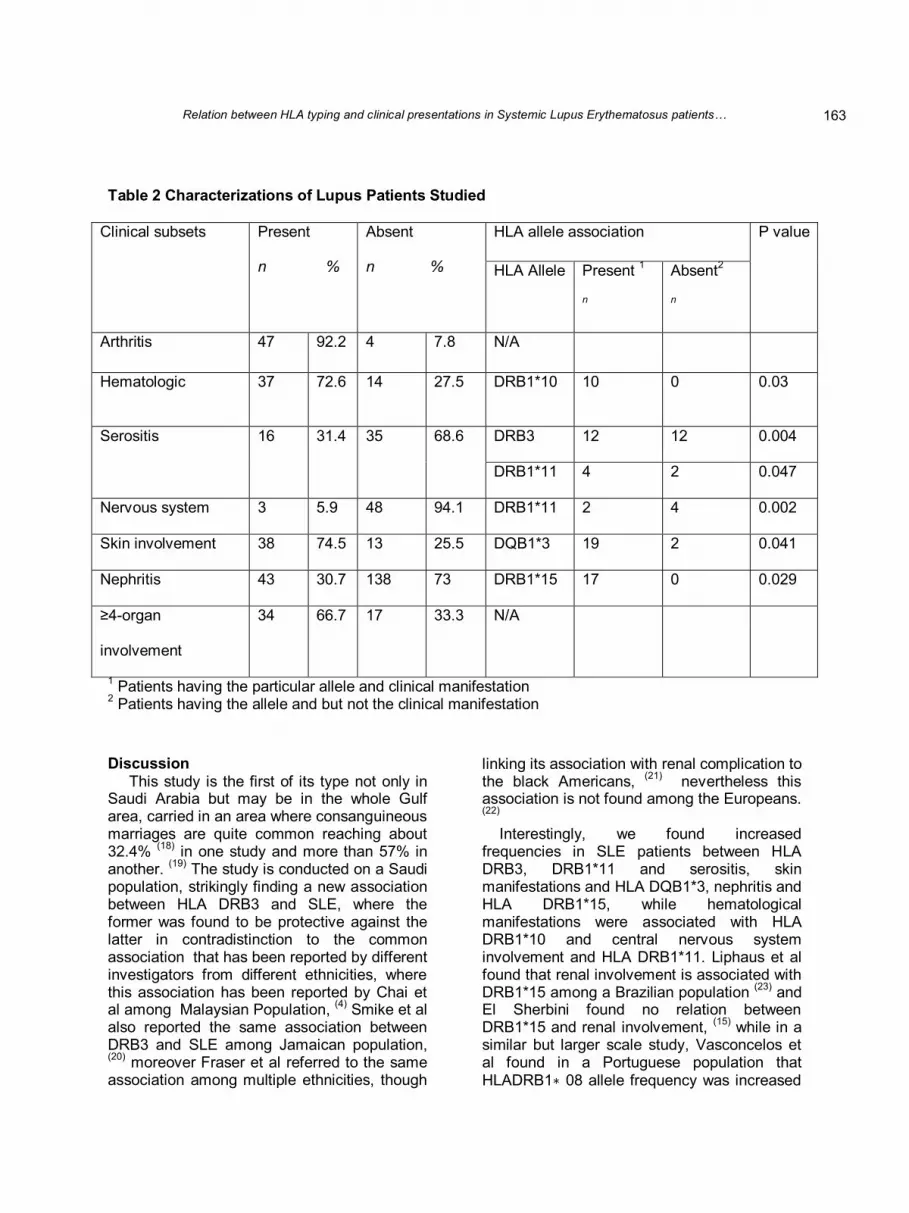
The most frequent DRB1 alleles found in the SLE group were HLA-DRB1 ∗ 07, DRB1 ∗ 015, DRB1 ∗3, and DRB1∗ 04. However, only the HLA DRB3 was significantly decreased in SLE patients (study group) when compared to controls (p value = 0.044), whereas the frequency of DRB4, DQB1*02, DRB3 where increased with non-significant difference when compared to controls, Only DRB3 (p value=0.044) was significantly decreased when compared to controls. It was found that HLA DRB3 frequency was increased in SLE patients with serositis, the number of which was 16 cases in comparison to those without serositis with (p value = 0.004), increased frequency of HLA DQB1*3 with p value (0.041) among SLE patients with skin manifestations, the number of which was 37 cases versus those without skin manifestations, an increased HLA DRB1*15 (p value 0.029) among SLE patients with nephritis, the number of which was 43 cases versus those without
nephritis, whereas HLA DRB1*10 was found to be increased among SLE patients with hematological manifestations the number of which was 37 cases versus those without hematological manifestations, (p value =0.03), moreover HLA DRB1*11 was found to have an increased frequency among patients with central nervous system involvement (p value = 0.002) along with an increased frequency among patients with serositis( p value = 0.047). See Table 2 on comparing Saudi patients to non-Saudi patients, the most striking finding was that only 42% of the Saudi patients have DRB3, moreover this allele was found to be protective against SLE (p value= 0.019), with the relative risk among those who carry DRB3 being around 0.65 (CI= 0.45, 0.94) in comparison to controls, while all the non-Saudi patients showed the presence of 100% presence of DRB3 though it was not found to be significant.
162
Relation between HLA typing and clinical presentations in Systemic Lupus Erythematosus patients…
Table 2 Characterizations of Lupus Patients Studied
Clinical subsets Present
n %
Absent
n %
HLA allele association P value
HLA Allele Present 1
n
Absent2
n
Arthritis 47 92.2 4 7.8 N/A
Hematologic 37 72.6 14 27.5 DRB1*10 10 0 0.03
Serositis 16 31.4 35 68.6 DRB3 12 12 0.004
DRB1*11 4 2 0.047
Nervous system 3 5.9 48 94.1 DRB1*11 2 4 0.002
Skin involvement 38 74.5 13 25.5 DQB1*3 19 2 0.041
Nephritis 43 30.7 138 73 DRB1*15 17 0 0.029
≥4-organ
involvement
34 66.7 17 33.3 N/A
1 Patients having the particular allele and clinical manifestation 2 Patients having the allele and but not the clinical manifestation
Discussion This study is the first of its type not only in Saudi Arabia but may be in the whole Gulf area, carried in an area where consanguineous marriages are quite common reaching about 32.4% (18) in one study and more than 57% in another. (19) The study is conducted on a Saudi population, strikingly finding a new association between HLA DRB3 and SLE, where the former was found to be protective against the latter in contradistinction to the common association that has been reported by different investigators from different ethnicities, where this association has been reported by Chai et al among Malaysian Population, (4) Smike et al also reported the same association between DRB3 and SLE among Jamaican population, (20) moreover Fraser et al referred to the same association among multiple ethnicities, though
linking its association with renal complication to the black Americans, (21) nevertheless this association is not found among the Europeans. (22) Interestingly, we found increased frequencies in SLE patients between HLA DRB3, DRB1*11 and serositis, skin manifestations and HLA DQB1*3, nephritis and HLA DRB1*15, while hematological manifestations were associated with HLA DRB1*10 and central nervous system involvement and HLA DRB1*11. Liphaus et al found that renal involvement is associated with DRB1*15 among a Brazilian population (23) and El Sherbini found no relation between DRB1*15 and renal involvement, (15) while in a similar but larger scale study, Vasconcelos et al found in a Portuguese population that HLADRB1∗ 08 allele frequency was increased
163

Walid Wadi et al…
among SLE patients with neurological involvement. (24) Conclusion: To sum up, this study highlights that, in contradistinction to what have been found among other populations DRB3 is a protective gene among Saudi’s against SLE. No evidence for any role for the HLA-DRB1, DRB4, DRB5, DQB1 alleles. There was an increased HLA DRB3 frequency with serositis, DQB1*3 skin manifestations, HLA DRB1*15 with nephritis, DRB1*10 with hematological manifestations and DRB1*11 with neurological manifestations. Results from this study are to be interpreted cautiously due to the small sample size. Conflicts of Interest The authors declare that they have no competing issues of interests References 1. Borchers AT, Naguwa SM, Shoenfeld Y,
Gershwin ME. The geoepidemiology of systemic lupus erythematosus. Autoimmun Rev 2010; 3:9:A277-87. 24.
2. Lindqvist AK, Alarcón-Riquelme ME. The genetics of systemic lupus erythematosus. Scand. J. Immunol1999; 50:6: 562–571.
3. Molokhia M, McKeigue P. Systemic lupus erythematosus: genes versus environment in high risk populations. Lupus 2006; 15: 827–832.
4. Chai HC, Phipps ME, Chua KH. Genetic Risk Factors of Systemic Lupus Erythematosus in the Malaysian Population: A Mini review. Clin. Dev. Immunol 2012;963730
5. Forabosco P, Gorman JD, Cleveland C, Kelly JA, Fisher SA, Ortmann WA, Johansson C, Johanneson B, Moser KL, Gaffney PM, Tsao BP, Cantor RM, Alarcón-Riquelme ME, Behrens TW, Harley JB, Lewis CM, Criswell LA. Meta-analysis of genomewide linkage studies of systemic lupus erythematosus. Genes Immun 2006; 7: 609–614.
6. Castro J, Balada E, Ordi-Ros J, Vilardell-Tarrés M.The complex immunogenetic basis of systemic lupus erythematosus. Autoimmun. Rev 2008; 7: 345–351.
7. Castaño-Rodríguez N, Diaz-Gallo LM, Pineda-Tamayo R, Rojas-Villarraga A, Anaya JM. Meta-analysis of HLA-DRB1 and
HLA-DQB1 polymorphisms in Latin American patients with systemic lupus erythematosus. Autoimmun. Rev 2008; 7:322-30.
8. Kelly JA, Moser KL, Harley JB. The genetics of systemic lupus erythematosus: putting the pieces together. Genes Immun 2002; 3: S71–S85.
9. Smerdel-Ramoya A, Finholt C, Lilleby V, Gilboe IM, Harbo HF, Maslinski S, Førre Ø, Thorsby E, Lie BA. Systemic lupus erythematosus and the extended major histocompatibility complex—evidence for several predisposing loci. Rheumatology (Oxford) 2005; 44:1368-73.
10. Rhodes B, Vyse TJ. General aspects of the genetics of SLE. Autoimmunity 2007; 40: 550–559.
11. López-Tello A, Rodríguez-Carreón AA, Jurado F, Yamamoto-Furusho JK, Castillo-Vázquez M, Chávez-Muñoz C, Salgado N, Arellano-Campos O, Vargas-Alarcón G, Granados J. Association of HLADRB1 ∗ 16 with chronic discoid lupus erythematosus in Mexican mestizo patients. ClinExpDermatol2007; 32:435-8.
12. Liphaus Bde L, Goldberg AC, Kiss MH, Silva CA. Analysis of human leukocyte antigens class II-DR in Brazilian children and adolescents with systemic lupus erythematosus. Rev Hosp Clin Fac Med Sao Paulo. 2002 Nov-Dec; 57(6):277-82.
13. Graham RR, Ortmann W, Rodine P, Espe K, Langefeld C, Lange E, Williams A, Beck S, Kyogoku C, Moser K, Gaffney P, Gregersen PK, Criswell LA, Harley JB, Behrens TW. Specific combinations of HLA-DR2 and DR3 class II haplotypes contribute graded risk for disease susceptibility and auto antibodies in human SLE. Eur. J. Hum. Genet 2007; 15: 823–830.
14. Rahman A, Isenberg DA.Systemic lupus erythematosus. N. Engl. J. Med 2008; 358: 929–939.
15. El Sherbini HM, El Garf AK, El Din Mahmoud SS. Human leukocyte antigen and autoantibodies association with juvenile systemic lupus erythematosus. Egypt J Immunol 2009; 16:107-14.
16. Miller SA, Dykes DD, Polesky HF. A simple salting out procedure for extracting DNA from human nucleated cells. Nucleic Acids Res 1988; 16(3): 1215.
164

Relation between HLA typing and clinical presentations in Systemic Lupus Erythematosus patients…
17. Olerup O, Zetterquist H. HLA-DR typing by PCR amplification with sequence-specific primers (PCR-SSP) in 2 hours: an alternative to serological DR typing in clinical practice including donor recipient matching in cadaveric transplantation. Tissue Antigens 1992; 39: 225–235.
18. Alzolibani A. Genetic epidemiology and heritability of vitiligo in the Qassim region of Saudi Arabia. ActaDermatovenerol Alp PanonicaAdriat2009; 18:119-25.
19. Hazmi MA, Al-Swailem AR, Warsy AS, Al-Swailem AM, Sulaimani R, Al-Meshari El-AA. Consanguinity among the Saudi Arabian population. J Med Genet 1995; 32:623-626
20. Smikle M, Christian N, DeCeulaer K, Barton E, Roye-Green K, Dowe G, Anderson N, Nicholson G. HLA-DRB alleles and systemic lupus erythematosus in Jamaicans. South Med J 2002; 95:717-9.
21. Fraser PA, Lu LY, DeCeulaer K, Schur PH, Fici D, Awdeh Z, Ding WZ, Levitan E, Lew R, Uko G, Gonzalez C. CD4 TCRBV CDR3 analysis in prevalent SLE cases from two ethnic groups. Lupus. 1999; 8(4):311-9.
22. Ramal LM, López-Nevot MA, Sabio JM, Jáimez L, Paco L, Sánchez J, de Ramón E, Fernández-Nebro A, Ortego N, Ruiz-Cantero A, Rivera F, Martín J, Jiménez-Alonso J; Grupo Lupus Virgen de las Nieves. Systemic lupus erythematosus in southern Spain.a comparative clinical and genetic study between Caucasian and Gypsy patients. Lupus 2004;13:934–40
23. Liphaus BL, Kiss MH, Goldberg AC. HLA-DRB1 alleles in juvenile-onset systemic lupus erythematosus: renal histologic class correlations. Braz J Med Biol Res. 2007 Apr; 40:591-7.
24. Vasconcelos C, Carvalho C, Leal B, Pereira C, Bettencourt A, Costa PP, Marinho A, Barbosa P, Almeida I, Farinha F, Mendonça T, Correia JA, Mendonça D, Martins B. HLA in Portuguese systemic lupus erythematosus patients and their relation to clinical features. Ann N Y Acad Sci. 2009 Sep; 1173:575-80.
165