immunosedimentation patterns in pathological human sera
TRANSCRIPT
CLINICA CHIMICA ACTA 61x
IMMUNOSEDIMENTATION PATTERNS IN PATHOLOGICAL HUMAN SERA
SUMMARY
The technique of immunosedimentation, a gel plate immunoprecipitation of antigens separated by centrifugation in sucrose gradients, was utilized for the analyses of some pathological human serum samples. Characteristic protein patterns were obtained with myeloma, macroglobulinemja and suggestive patterns with viral hepatitis, rheumatoid arth~tis, and others. A comparison of this technique with regular immu~oelectrophoretic plates was made. The resuhs and method are briefly discussed.
The determination of characteristic patterns of sera is a useful tool in compara- tive biology and in medical practice. This information can be obtained with various methods and techniques in current use : paper electrophoresis, disc electrophoresis, starch electrophoresis, immunoelectrophoresis.
We have recently described a method which takes advantage of electrophoretic separation of proteins but, in addition, uses cent~fugation in sucrose gradients to screen the antigens as a preliminary step in the procedure’. The second step is per- formed by direct immu7aoelectrophoresa’s 2, to develop the antigens as diffuse spots of specific precipitates on the agar plates.
In the present communication we present photographic and diagrammatic data of immunosedimentation plates obtained in a number of pathologic~ sera. Improvements of the original technique and a compa~son with immunoele~tro- phoretic analysis were made and the results are presented here.
MATERIALS AND METHODS
A g to 45% sucrose gradient in saline is prepared in a s-ml cellulose nitrate centrifuge tube containing a total of 4.6 ml. The gradient is prepared with the aid of a small two-vessel acrylic reservoir as described by Martin and Ames*. Samples of 0.1 ml of sera are placed on top of the sucrose solution and centrifuged in a swinging bucket rotor in a Spinco Model L preparative centrifuge at IOOOOO x g for I% h at 4”. After centrifugation the tubes are placed in a plastic stand and the contents frac- tionated into 25 consecutive fractions of 12 drops each and collected directly into small test tubes.
C&z. Chins. Acta, 't3(r966)6rr-625
612 F. C6RDOB.4 et d.
The antigens thus fractionated by their sedimentation properties are found in varied proportions in each tube. The second step in immunosedimentation resolves these mixtures by electrophoresis on agar gel plates. The procedure consists of pre- paring a 7 x ro-cm agar gel plate with 1.5% Difco “Noble” agar in the electrophoretic solution of Aronson5. A row of wells are punched on the agar with hypodermic needles No. 16 without the tips; the needles are first glued on a piece of plastic. Samples of 20-25 ,ul are removed from each of the fractions obtained by sedimenta- tion and transferred into the wells. Electrophoresis is performed under a constant voltage 150 V for 6 h. At the end of the run the wells are covered with freshly prepared warm agar solution. The agar plate is now flooded with the antiserum and set aside for a period of no less than 48 h to allow precipitation.
After the spots have formed, the excess antiserum is discarded and the plates are immersed-free of the glass trays-in 0.9% NaCl solution to remove the embedded soluble proteins. This is accomplished with 4 or 5 washes and the plates are then dried
and stained for proteins “. The plates presented here were stained with Ponceau Red, 50 mg/loo ml in 7% acetic acid as a first dye, washed in 3% acetic acid solution and dyed a second time with a 7% solution of acetic acid with 50 mg/roo ml of Amido Black. Finally, after a last wash with 10% glycerol in acetic acid the plates were dried and photographed.
Immunoelectrophoresis was performed on microscopic slides following the modification of Scheidegger 7. The buffer system and antiserum were the same as in immunosedimentation experiments.
Pathological human sera In Table I the serum samples are listed with the clinical diagnosis of the donors.
Most of the samples were obtained through the courtesy of Dr. Luis Landa, Chief
TABLE I
HUMAN SERUM SAMPLES ANALYSED BY IMMUNOSEDIMENTATION AND IMMUNOELECTROPHORESIS
WITH THE CORRESPONDING CLINICAL DIAGNOSES
Clinical condition of serum sample donor __-
Immunosedimentation analysis and immunoelectropherogram
Normal adult male (L.G.) Waldenstrijm macroglobulinemia Multiple myeloma (/3 and y type) Multiple myeloma (y type) Viral hepatitis with hypergammaglobulinemia Rheumatoid arthritis Hepatic cirrhosis Acute pancreatitis
Fig. I Fig. 2 Fig. 3 Fig. 4 Fig. 5 Fig. 6 Fig. 7 Fig. 8
of the Gastroenterological Service of the General Hospital, Medical Center, Instituto Mexican0 de1 Seguro Social, Mexico, D.F. The remaining samples were obtained from the Cardiology Institute, Mexico, D.F. thanks to the kindness of Dr. Carlos Biro, Head of the Immunology Department. Samples of normal human serum were ob- tained from the laboratory staff.
Rabbit antiserum, against pooled whole human serum, was produced by sub- cutaneous injections of I ml of human serum, 3 times a week, into six rabbits for a
Chile. Che’m. Acta, 13 (1966) 611-625
IMMUNOSEDIMENTATION IN PATHOLOGXCAL SERA 6r3
period of 4 months. Blee~gs from the mar~nal vein of the ear were done at one month’s intervals and the antiserum pooled according to a qualitative “ring test”.
Rabbit antiserum, against human y-globulin (HGG) was prepared by injecting adult rabbits with 5 mg of heat-aggregated HGG once a week, intravenousiy for 6 weeks.
RESULTS
Fig. I shows the immunosedimentation pattern of one of the samples (L.G.) of human normal serum. On top are photographs of the results of immunoelectrophoretic analysis of each sample as a control of the immunosedimentation runs. At the bottom, in each case, is a diagram drawn directly from the stained plates. In order to facilitate the description of the patterns the main spots, common to most of the serum samples investigated, were numbered with roman numerals. In some instances each spot be- longs to a single antigen but in most, they must be produced by mixtures of antigens with similar immunosedimentative characteristics.
The specific precipitate produced by albumin is marked III. It can be seen that the albumin occupies about half of the vertical length of the plate. This is in contrast to the pattern observed in sucrose gradient analysis of g-25% concentration employed before’. Correct sedimentation values (Svedberg units) for the estimated antigens in each spot are indicated by the position of the numerals in the diagrams. The agree- ment is good though in some cases the antigens became displaced to the bottom of the centrifuge tubes and the marks are not in the center of the spots. The calibration of the S values scale was obtained in control runs with purified proteins I.
Spot VIII corresponds to 7 S y,-globulin displaced below the albumin in all cases. Spot VI of plates appears to be composed with transferrin (siderophilin) ; this protein forms the main component of the antigens located in the p region. It has an S value of 5-5.3 and a molecular weight of goooo (ref. 8). Lipid-poor euglobin and p-lipoproteins should be approximately in this position.
Haptoglobins and glucoproteins appear to correspond to spot V, which is a prominent spot in normal human samples. Further down, in the same electrophoretic region, appears spot IX due to a,-macroglobulin, clearly demonstrated in Fig. 5_ Opposite to this, at the right hand side of the punches, is present a diffuse small spot produced by the macroglobulin of the y family of serum proteins (#l,n)-marked X. This spot which appears clearly in Fig. 5 is usually very faint in normal samples (Fig. I).
Spot IV, with the mobility of CL, could be produced by glycoproteins of Schultze, orosomucoid, ceruloplasmin and other globulins in trace amountss. Above this spot are present streaks of another component, cx-lipoprotein, demonstrated before by specific lipid dyes l, This component is marked as I, and extends in separate elongated spots from the top of the plate to about the middle.
Spot II at the left of albumin, could correspond to prealbumin but the spot is not always visible.
The region marked VII corresponds to pz- or y,-globulin in electrophoretic mobilities. In immunosedimentation this area is difficult to examine because of the smearing of the 7 S y component present in VIII.
Fig. 2 isfromase~msampleof apatient with Waldenstr~m’sma~roglobu~inemia.
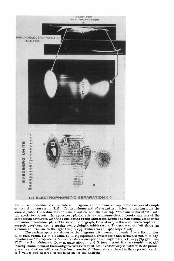
Fig. 1. Immunosedimentation plate and diagram, and immunoelectrophoretic analysis of sample of normal human serum (L.G.). Center: photograph of the pattern, below, a drawing from the stained plate. The sedimentation axis is vertical and the electrophoretic one is horizontal, with the anode to the left. The uppermost photograph is the immunoelectrophoretic analysis of the same serum developed with the same pooled rabbit antiserum, against human serum, used for the immunosedimentation plate. The second photograph, from above, is the immunoelectrophoretic analysis developed with a specific anti-y-globulin rabbit serum. The arrow to the left shows the albumin and the one to the right the 7 S y,-globulin arcs and spot respectively.
The antigen spots are shown in the diagrams with reman numerals: I = tc lipoproteins, II = prealbumin, III = albumin, IV = glycoproteins, orosomucoid and ceruloplasmin, V = hap- toglobins and glycoproteins, VI = transferrin and poor lipid euglobulin, VII = yr (/?a) globulins, VIII = 7 S y,-globulins, IX = cc,-macroglobulin and X (not present in this sample) = y1 (/&)- macroglobulin. Some of these antigens have been identified in control experiments with the purified proteins and others with specific colored reactions”. Numerals are placed in the expected position of S values and electrophoretic location for the antigens.
IMMUNOSEDIMENTATION IN PATHOLOGIC& &BRA 615
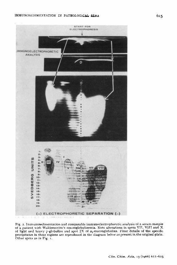
Fig. 2. Immunosedimentation and comparable immunoelectrophoretic analysis of a serum sample of a patient with WaldenstrCim’s macroglobulinemia. Note alterations in spots VII, VIII and X of light and heavy y-globulins and spot IX of cqmacroglobulins. Finer details of the specific precipitates in these regions are reproduced in the diagram below as present in the original plate. Other spots as in Fig. 1.
C&n. Chim. Acta, 13 (1966) 611-625
6r6 I?. C6RDOBA et ai.
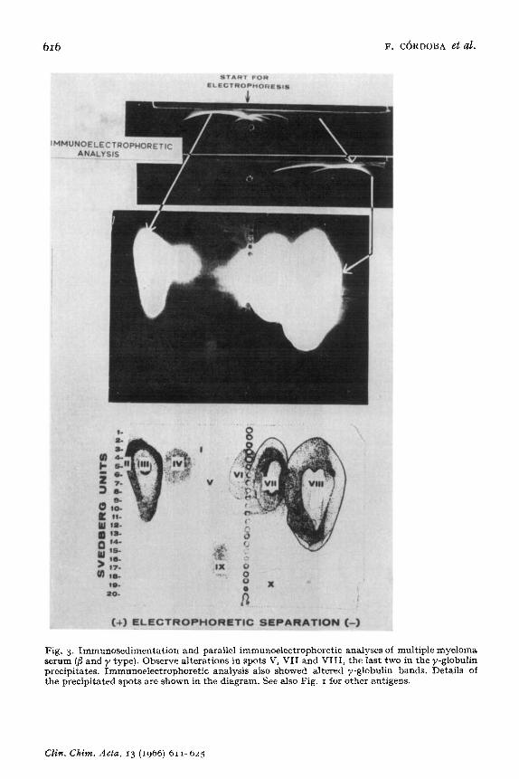
Fig. 3. Immunosedimentation and parallel immunoelectrophoretic analyses of multiple myeloma serum (@ and y type). Observe alterations in spots V, VII and VIII, the last two in the y-globulin precipitates. Immunoelectrophoretic analysis also showed altered y-globulin bands. Details of the precipitated spots are shown in the diagram. See also Fig. I for other antigens.
Clin.Chim. Acta, 13 (x966) 611-625
I~~UNOSEDI~E~TATlO~ IN PATIiQLOw $B?&# 6x7
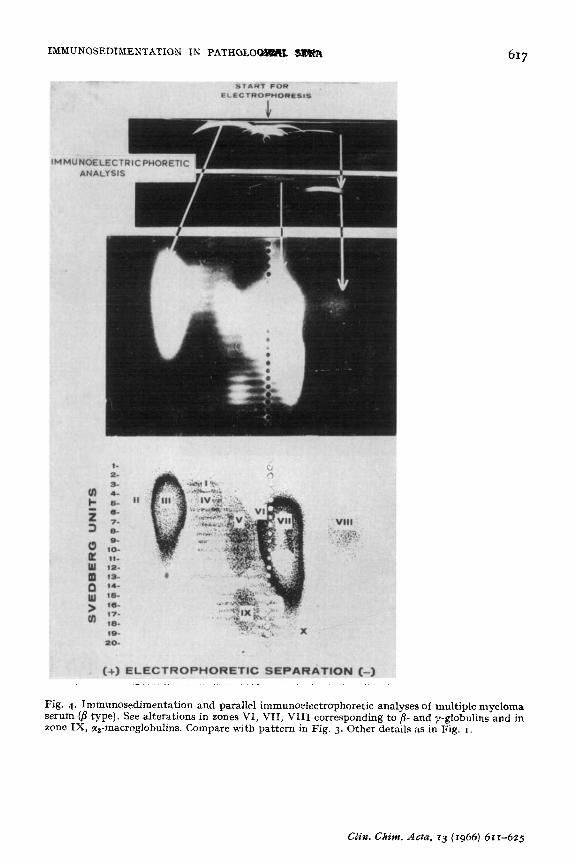
Fig. 4. Immunosedimentation and parallel immunoelectrophoretic analyses of multiple myeloma serum (4 type). See alterations in zones VI, VII, VIII corresponding to ,!?- and y-globulins and in zone IX, cc,-macroglobulins. Compare with pattern in Fig. 3. Other details as in Fig. I.
F. C6RDOBA et cd.
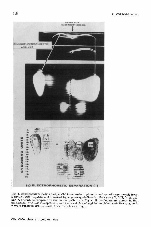
Fig. 5. Immunosedimentation and parallel immunoelectrophoretc analyses of sermn sample from a patient with hepatitis and transient hypergammaglobuli~emia. Kote spots V, VII, VIII, IX and X altered, as compared to the normal patterns in Fig. I. Haptoglobins are absent in the precipitate, with less glycoproteins and increased p- and y-globulins. Macroglobulins of Q and 1’ types appeared also increased. Other details as in Fig. 1.
C&z. Chinz. Acta, 13 (1966) 61x-625
IMb~UNOSE~IME~TATI~~ IN PATHOLOGICAL SEHR 619
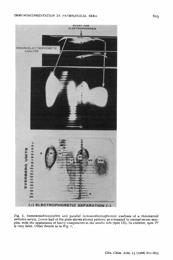
Fig. 6. Immunosedimentation and parallel immunoelectrophoretic analyses of a rkeumat arthritis serum. Lower half of the plate shows altered pattern. as compared to normal serum sa @es, with the appearance of heavy campcnents in the anodic side (spot IX). ln addition, spot is very faint. Other details as in Fig. 1.
oid bm- IV
F, c~RDOBA et al.
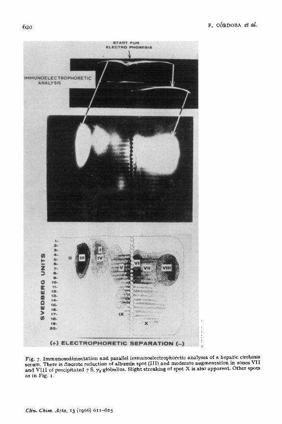
Fig. 7. Immu~osedimentation and parallel immunoelectrophoretic analyses serum. There is discrete reduction of albumin spot (III) and moderate augm and VIII of precipitated 7 S, y,-globulins. Slight streaking of spot X is also ( as in Fig. 1.
of a hepatic cirrh losis entation in zones VII apparent. Other s] pots
CZis. Claim. A&z, 13 (1966) 611-6~5
IMMUNOSEDIMENTATION IN PATIiUi_OG$G%% SERA 621
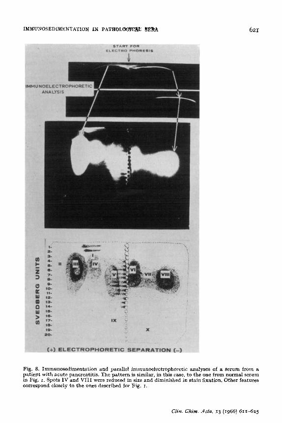
Fig. 8. Immunosedimentation and parallel immunodectrophoretic anal: patient with acute pancreatitis. The pattern is similar, in this case, to the in Fig. I. Spots IV and VIII were reduced in size and diminished in stain correspond closely to the ones described for Fig. I.
yses of a serum from a one from normal serum fixation. Other features
C&n. Chk. .&da, r3 (1966) 61~625
622 F. C6RDOBA et al.
The more prominent alterations are: heavy precipitation in the y-globulin side of the plate from the 7 S level to the bottom of the plate. The heterogeneity of the macroglobulin is apparent in the streaking of the spot extending to the anodic side. The 7 S component which is poorly defined due to intermixing with the heavy pro- teins, is nevertheless evident. In macroglobulinemia one expects more or less the same general pattern which is typical for the condition. The remaining antigens are not modified to the extent of the ones in the y region. There seems to be a doubtful augmentation of component IX as compared to normal human serum samples.
Macroglobulinemia serum was separated by immunoelectrophoresis and devel- oped with the same rabbit antiserum and with the rabbit anti-human y-globulin serum. The results of these tests are on top of Fig. 2. It can be seen that the diagnosis, on the basis of immunoelectrophoresis alone, could be difficult. By employing a more specific antiserum, like the anti-y-globulin serum, the problem becomes easier. In our view immunosedimentation gives a straightforward answer, plus additional data, with the usual rabbit anti-whole human serum.
Fig. 3 shows a case with multiple myeloma. Evident alterations are present in the y region with two large spots of precipitate clearly visible (double myeloma). One is in the yZ zone and the other in the y1 (8.J marked with numerals VII and VIII. The centers of the spots are clear, due to the dissolution of the precipitates by excess antigen. Since we never observed central dissolution of yZ 7 S globulin spots in normal serum samples, this characteristic together with the round shape and neatness of the
large spot is suggestive of myeloma. No spots were present in the 15 to 20 S range, thus no heavy components were present. Slight alterations of questionable value are an apparent decrease of proteins in zone V with a parallel increased penetration of those antigens in the centrifuge tube.
In Fig. 4 another case of myeloma is shown. In this case, a B myeloma in which a heavy precipitate, with neatly visible electrophoretic properties and very disperse sedimentation values, has formed. The pattern is of an elongated spot (center dis- solved) in region VII of immunosedimentation. The spot tails down to about the 18 S range. 7 S, y,-globulin is present in very discrete amounts (spot VIII). The remaining shadows are in their normal positions. By immunoelectrophoresis, there is a shortening of the 7 S, y,-globulin arc and with the more specific anti-y-globulin serum the /I precipitate of the myeloma protein can be visualized (top of Fig. 4). There was no evidence of macroglobulin either by immunoelectrophoresis or immunosedimentation.
Fig. 5 shows a case of hepatitis with transient hypergammaglobulinemia. Spot VIII is much broader and denser than normal serum samples. There is a marked increase of macroglobulins in the y region and in the aa region of the immunosedimen- tation plate (spots IX and X). No protein has accumulated in region V and the remaining antigens are in their expected positions. Immunoelectrophoresis of the sample with both antisera disclosed only an increased y-globulin precipitate (top of Fig. 5).
Fig. 6 was made from a case of rheumatoid arthritis. The appearance of high molecular weight proteins is reflected in the streaking at the lower half of the plate pattern on each side of the starting line. Region IV is very faint in comparison with similar spots in normal serum. Clear cut aberrations were not shown in the immuno- electrophoretic pattern (compare the y-globulin alterations in this case with the y-globulin shadows of macroglobulinemia and myeloma in Figs. 2, 3 and 4).
Clin. Chim. Acta, 13 (1966) 611625
IMMUNO~ED~~ENTATION IN PATHOLOG~C~ SERA 623
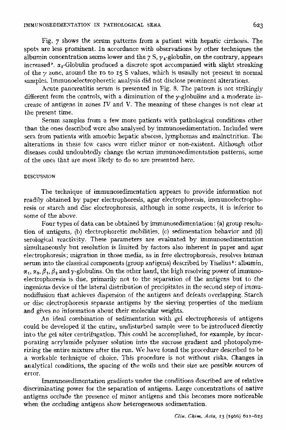
Fig. 7 shows the serum patterns from a patient with hepatic cirrhosis. The
spots are less prominent. In accordance with observations by other techniques the albumin concentration seems lower and the 7 S, ye-globulin, on the contrary, appears increaseda. a,-Globulin produced a discrete spot accompanied with slight streaking of the y zone, around the IO to 15 S values, which is usually not present in normal samples. Immunoelectrophoretic analysis did not disclose prominent alterations.
Acute pancreatitis serum is presented in Fig. 8. The pattern is not strikingly different from the controls, with a diminution of the y-globulins and a moderate in- crease of antigens in zones IV and V. The meaning of these changes is not clear at
the present time. Serum samples from a few more patients with patholo~c~ conditions other
than the ones described were also analysed by immunosedimentation. Included were sera from patients with amoebic hepatic abscess, lymphomas and malnutrition. The alterations in these few cases were either minor or non-existent. Although other diseases could undoubtedly change the serum immunose~mentation patterns, some of the ones that are most likely to do so are presented here.
DISCUSSION
The technique of immunosedimentation appears to provide information not readily obtained by paper electrophoresis, agar electrophoresis, immunoelectropho- resis or starch and disc electrophoresis, although in some respects, it is inferior to some of the above.
Four types of data can be obtained by immunosedimentation : (a) group resolu- tion of antigens, (b) electrophoretic mobilities, (c) sedimentation behavior and (d) serologic~ reactivity. These parameters are evaluated by immunosedimentation simultaneously but resolution is limited by factors also inherent in paper and agar electrophoresis; migration in those media, as in free electrophoresis, resolves human serum into the classical components (group antigens) described by Tiselius @ : albumin, a,, dcz, /ill, p2 and y-globulins. On the other hand, the high resolving power of immuno- electrophoresis is due, primarily not to the separation of the antigens but to the ingenious device of the lateral distribution of precipitates in the second step of immu- no~~usion that achieves dispersion of the antigens and defeats overlapping. Starch or disc electrophoresis separate antigens by the sieving properties of the medium and gives no information about their molecular weights.
An ideal combination of sedimentation with gel electrophoresis of antigens could be developed if the entire, undisturbed sample were to be introduced directly into the gel after centrifugation. This could be accomplished, for example, by incor- porating acrylamide polymer solution into the sucrose gradient and photopolyme- rizing the entire mixture after the run. We have found the procedure described to be a workable technique of choice. This procedure is not without risks. Changes in analytical conditions, the spacing of the wells and their size are possible sources of error.
Immunosedimentatian gradients under the conditions described are of relative ~scr~inating power for the separation of antigens. Large concentrations of native antigens occlude the presence of minor antigens and this becomes more noticeable when the occluding antigens show heterogeneous sedimentation.
CZin.Chim. Acta, 13 (1966) 611-62.5
624 F. C6RDOBA et al.
To diminish this effect, we attempted to use steeper gradients. The antigens precipitated within smaller areas, as compared to the precipitation areas observed with dilute sucrose gradientsI. The extent to which this last condition can be mani- pulated is practically unlimited, but for analyses of mixtures of antigens in body fluids a compromise should be reached between steep gradients and low sucrose con- centration gradients.
The antigens can be developed in the gel plates in a number of ways. We have chosen the immunological development for the following reasons: (a) to compare results with routine immunoelectrophoretic plates; (b) to keep samples down to O.I- ml volumes; (c) to improve visualization of the protein spots by the specific precipita- tion of the antibody; and (d) to spot integration (i.e. appearance of round, single spots of antigen originating from multiple round sources). This is effectively accom- plished by the same specific precipitate. High molecular weight antigens-with higher immune precipitation rates than the diffusion rate of the “macro-antigens” on the plate-give “streaked” instead of “spotted” precipitation patterns (see Figs. 2 and 5).
Like other analytical methods using antisera, immunosedimentation is not suited for detecting antigens for which there is not enough precipitating antibody in the antiserum utilized. If such were the case, one could set up another centrifuge tube with 0.3- to o.4-ml volumes of the antigen sample. After electrophoresis one could develop the protein by simple fixation and staining. The comparison of this new plate pattern with regular immunosedimentation plates could be of considerable advantage.
The usefulness of immunosedimentation was tested with human sera in a variety of pathological conditions, and the method was found of value in estimating electro- phoretic displacement and sedimentation behavior simultaneously in the same run. In the present work, patterns of pathological serum samples were visually compared with those of normal samples. This empirical approach gave in a significant number of cases, patterns which appeared characteristic of certain pathological conditions. The collection and comparison of such patterns from immunosedimentation analyses could become a valuable tool in laboratories performing clinical investigations.
A further potential application of the technique should also be mentioned. The two-dimensional separation of a complex mixture of antigens, yielding a flat, mul- tiple-spotted pattern appears suitable for quantification. Work on this direction-e.g. by densitometry-could be profitable in measuring specific antibodies to a number of antigens in the same biological fluid.
ACKNOWLEDGEMENTS
We gratefully acknowledge Dr. Carlos Biro for supplying the rabbit anti- human y-globulin serum used in this work and for allowing us to describe the prepara- tion procedure before publication.
This work was supported in part by a Rockefeller Foundation grant to this Department.
Clin. Chim. Acta, 13 (1966) 611-625

I~MUNOS~~I~ENTAT~ON IN PATHOLOGICAL SERA 62.5
REFERENCES
2 A. T. WILSON, J. ~~W6und, 92 (1964) 431. 3 P. GRABAR AND P. BURTIN, Ana&se Imrnun~~~l~lectvophor~t~*~e, M&son, Paris, 1960, 4 R. G. MARTIN AND B. N. ADS, J. Bid. Chm., 236 (fg6rf 1372. 5 T. AR~NSO~V AND A. GR~NWALL, Sci. Tools, 5 (1958) 2. 6 J. URIEL AND P. GRABAR, Asn. Zest. Pasteur, go (rgyjkq7. 7 J. J. SCHEIDEGGER, In&%. Arch. Allergy A$#. I~~u~o~, 7 (rg& rag. 8 R. A. PHELFS AND F. W. PUTNAM, in W. PUTNAM (Ed.), Ths F2aswa Proteins, Vol. X, Academic
Press, New York, 1960, p. ~43. g ‘4. TISELIUS AND E. A. KABAT, J. E.+tl. Med., 69 (1939) rrg.
Cl%. China. Acta, 13 (1966) 6rlt-G25