resilience of developing brain networks to interictal epileptiform discharges is associated with...
TRANSCRIPT
BRAINA JOURNAL OF NEUROLOGY
Resilience of developing brain networks tointerictal epileptiform discharges is associatedwith cognitive outcomeGeorge M. Ibrahim,1,2 Daniel Cassel,3 Benjamin R. Morgan,4 Mary Lou Smith,5 Hiroshi Otsubo,6
Ayako Ochi,6 Margot Taylor,3,4,5 James T. Rutka,1 O. Carter Snead III2,3,6 and Sam Doesburg2,3,4,5
1 Division of Neurosurgery, Hospital for Sick Children, Department of Surgery, University of Toronto, Toronto, Ontario, Canada
2 Institute of Medical Science, University of Toronto, Toronto, Ontario, Canada
3 Neuroscience and Mental Health Program, Hospital for Sick Children Research Institute, Toronto, Ontario, Canada
4 Department of Diagnostic Imaging, Hospital for Sick Children, Toronto, Ontario, Canada
5 Department of Psychology, University of Toronto, Toronto, Ontario, Canada
6 Division of Neurology, Hospital for Sick Children, University of Toronto, Toronto, Ontario, Canada
Correspondence to: George M. Ibrahim, MD, PhD,
Division of Neurosurgery,
The Hospital for Sick Children,
555 University Avenue,
Toronto ON, M5G 1Z8
E-mail: [email protected]
The effects of interictal epileptiform discharges on neurocognitive development in children with medically-intractable epilepsy
are poorly understood. Such discharges may have a deleterious effect on the brain’s intrinsic connectivity networks, which reflect
the organization of functional networks at rest, and in turn on neurocognitive development. Using a combined functional
magnetic resonance imaging–magnetoencephalography approach, we examine the effects of interictal epileptiform discharges
on intrinsic connectivity networks and neurocognitive outcome. Functional magnetic resonance imaging was used to determine
the location of regions comprising various intrinsic connectivity networks in 26 children (7–17 years), and magnetoencephalo-
graphy data were reconstructed from these locations. Inter-regional phase synchronization was then calculated across interictal
epileptiform discharges and graph theoretical analysis was applied to measure event-related changes in network topology in the
peri-discharge period. The magnitude of change in network topology (network resilience/vulnerability) to interictal epileptiform
discharges was associated with neurocognitive outcomes and functional magnetic resonance imaging networks using dual
regression. Three main findings are reported: (i) large-scale network changes precede and follow interictal epileptiform dis-
charges; (ii) the resilience of network topologies to interictal discharges is associated with stronger resting-state network
connectivity; and (iii) vulnerability to interictal discharges is associated with worse neurocognitive outcomes. By combining
the spatial resolution of functional magnetic resonance imaging with the temporal resolution of magnetoencephalography, we
describe the effects of interictal epileptiform discharges on neurophysiological synchrony in intrinsic connectivity networks and
establish the impact of interictal disruption of functional networks on cognitive outcome in children with epilepsy. The asso-
ciation between interictal discharges, network changes and neurocognitive outcomes suggests that it is of clinical importance to
suppress discharges to foster more typical brain network development in children with focal epilepsy.
Keywords: functional connectivity; resting-state functional MRI; IED; graph theoretical analysis
doi:10.1093/brain/awu214 Brain 2014: Page 1 of 13 | 1
Received April 23, 2014. Revised June 6, 2014. Accepted June 29, 2014.
� The Author (2014). Published by Oxford University Press on behalf of the Guarantors of Brain. All rights reserved.
For Permissions, please email: [email protected]
Brain Advance Access published August 7, 2014 at H
ospital for Sick Children H
ospital Library on A
ugust 13, 2014http://brain.oxfordjournals.org/
Dow
nloaded from
Abbreviations: BOLD = blood oxygen level-dependent level; ICN = intrinsic connectivity network; IED = interictal epileptiformdischarge
IntroductionThe human brain exhibits exquisite hierarchical organization during
rest, the maintenance of which consumes the majority of the
brain’s metabolic energy (Raichle and Mintun, 2006). The organ-
ization of functional brain activity at rest is readily measured using
resting-state functional MRI, wherein brain regions that demon-
strate covariance in spontaneous blood oxygen level-dependent
level (BOLD) signal oscillations are said to form intrinsic connect-
ivity network (ICNs; also known as resting-state networks) (Biswal
et al., 1995; Raichle et al., 2001; Raichle and Snyder, 2007).
Although these networks are conspicuous in infants (Fransson
et al., 2007), with typical development, there are increases in
the strength of long-range connections between brain regions
associated within the same ICN, together with increasing segrega-
tion of cortical and subcortical structures associated with different
ICNs (Dosenbach et al., 2010). These connectivity gradients reflect
patterns of grey matter growth and subsequent synaptic pruning
with age. Disruption of the organization of ICNs is an emerging
area of interest in the study of neurodevelopmental conditions and
has been proposed to impact childhood cognitive development
(Uddin et al., 2013; Washington et al., 2014).
Children with epilepsy exhibit impairments in the development
and segregation of these ICNs, which are related to the burden of
the disease and neurocognitive function (Vaessen et al., 2013;
Widjaja et al., 2013a, b; Ibrahim et al., 2014). Children who
have abnormal EEGs with the presence of interictal spikes or
sharp and/or slow waves also possess greater alterations in net-
work organization than those who do not (Mankinen et al.,
2012). The role of interictal epileptiform discharges (IEDs) is gain-
ing greater prominence as a putative mechanism by which epi-
lepsy interferes with normative organization of oscillatory brain
networks, thereby leading to cognitive impairments (Gotman
et al., 2005; Kobayashi et al., 2006; Fahoum et al., 2013).
Disruption of ICNs has been associated with focal IEDs arising
from multiple brain regions (Fahoum et al., 2013), highlighting
the relevance of alterations of ICNs to the understanding of local-
ization-related epilepsy. Furthermore, on a physiological level, the
importance of IEDs is buttressed by observations that electric fields
generated during these events are strong enough in intensity to
influence action potential firing threshold and network synchron-
ization (Grenier et al., 2003).
Understanding how IEDs interfere with ICNs and how these
interactions relate to neurocognitive outcomes is important for
several reasons. First, while the goal of medical and surgical treat-
ments for epilepsy is to achieve seizure-freedom with minimal
morbidity, the benefits of IED suppression are more controversial
(Kuruvilla and Flink, 2003). Second, an understanding of the
mechanisms of network impairments in this patient population
may inform management strategies and the implementation of
surgical treatments, including extent of resection of the irritative
zone. Finally, while various authors have identified associations
between disrupted ICNs and IEDs, the causal temporal relation-
ships underlying these interactions have not been previously
established.
Previous studies using BOLD-functional MRI have been limited
by the fact that functional MRI indirectly measures neural activity,
reflecting blood flow rather than direct neural processes.
Moreover, BOLD dynamics are expressed on a slower timescale
than are IEDs. Another modality that has been studied to charac-
terize ICNs is MEG, which directly records neural oscillations and
robustly reconstructs ICNs (de Pasquale et al., 2010, 2012;
Brookes et al., 2011a, b). The superior temporal resolution of
MEG has enabled mapping of oscillatory dynamics of ICN activity
(de Pasquale et al., 2010, 2012) and may allow precise determin-
ation of how pathological processes affect ICNs without modelling
these events using basis functions for haemodynamic responses.
Furthermore, using MEG, oscillatory activity may also be analysed
in physiologically-relevant frequency bands, an approach which
has the potential to provide novel insights into the underlying
neurophysiological processes involved in the development and dis-
ruption of ICNs.
In the present study, activity from brain regions comprising vari-
ous ICNs were reconstructed from MEG recordings using resting-
state functional MRI-informed coordinates to test the hypotheses
that (i) IEDs are associated with changes in neurophysiological
interactions within ICNs in children with focal epilepsy; (ii) that
resilience and vulnerability of ICNs to such neurophysiological con-
nectivity changes are associated with cognitive outcome; and
(iii) ICN integrity as measured by functional MRI. A graph theor-
etical approach was applied to study changes in network topolo-
gies occurring in the peri-IED period on a millisecond timescale.
The resilience or vulnerability of network topologies to change in
this period was correlated with neurocognitive outcomes and
functional MRI statistical parametric maps. Using a combined
functional MRI-MEG approach, the current study provides evi-
dence for disruption of neurophysiological connectivity in ICNs
by IEDs, and uniquely demonstrates that such network alterations
are associated with reduced ICN integrity and worse cognitive
outcomes.
Materials and methods
Subject demographicsA total of 71 children with medically-intractable, focal (localization-
related) epilepsy were recruited into this study from the Hospital for
Sick Children in Toronto, Canada. Of these, 26 children were included
in the current study. Children were excluded if (i) they required sed-
ation; (ii) did not meet criteria for motion parameters for both func-
tional MRI and MEG studies (as listed below); (iii) had a conspicuous
MRI lesion that precluded accurate image registration; or (iv) if no
2 | Brain 2014: Page 2 of 13 G. M. Ibrahim et al.
at Hospital for Sick C
hildren Hospital L
ibrary on August 13, 2014
http://brain.oxfordjournals.org/D
ownloaded from
IEDs were captured on MEG. All patients underwent functional MRI
and MEG studies without any sedation, which has been shown to alter
BOLD signal and cortical oscillations (Marcar et al., 2006). Only one
patient included had undergone a previous small cortical resection,
which did not affect registration and the ICN networks were all
adequately sampled. The children ranged in age from 7–17 years,
with 13 (50%) males and with mean epilepsy duration (� standard
deviation) of 4.56 � 3.76 years. Nine children (35%) had epileptic foci
localized to the temporal lobe, whereas 17 children (65%) had extra-
temporal epilepsy and 15 children (58%) presented with secondarily
generalized seizures (see Supplementary material for more details re-
garding subject demographics). The study complies with the Code of
Ethics of the World Medical Association (Declaration of Helsinki) and
was approved by the Research Ethics Board of the Hospital for Sick
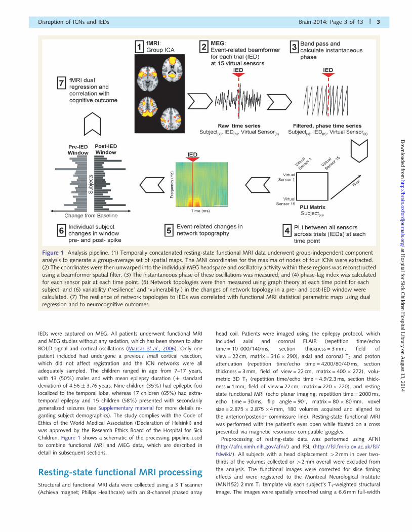
Children. Figure 1 shows a schematic of the processing pipeline used
to combine functional MRI and MEG data, which are described in
detail in subsequent sections.
Resting-state functional MRI processingStructural and functional MRI data were collected using a 3 T scanner
(Achieva magnet; Philips Healthcare) with an 8-channel phased array
head coil. Patients were imaged using the epilepsy protocol, which
included axial and coronal FLAIR (repetition time/echo
time = 10 000/140 ms, section thickness = 3 mm, field of
view = 22 cm, matrix = 316 � 290), axial and coronal T2 and proton
attenuation (repetition time/echo time = 4200/80/40 ms, section
thickness = 3 mm, field of view = 22 cm, matrix = 400 � 272), volu-
metric 3D T1 (repetition time/echo time = 4.9/2.3 ms, section thick-
ness = 1 mm, field of view = 22 cm, matrix = 220 � 220), and resting
state functional MRI (echo planar imaging, repetition time = 2000 ms,
echo time = 30 ms, flip angle = 90�, matrix = 80 � 80 mm, voxel
size = 2.875 � 2.875 �4 mm, 180 volumes acquired and aligned to
the anterior/posterior commissure line). Resting-state functional MRI
was performed with the patient’s eyes open while fixated on a cross
presented via magnetic resonance-compatible goggles.
Preprocessing of resting-state data was performed using AFNI
(http://afni.nimh.nih.gov/afni/) and FSL (http://fsl.fmrib.ox.ac.uk/fsl/
fslwiki/). All subjects with a head displacement 42 mm in over two-
thirds of the volumes collected or 42 mm overall were excluded from
the analysis. The functional images were corrected for slice timing
effects and were registered to the Montreal Neurological Institute
(MNI152) 2 mm T1 template via each subject’s T1-weighted structural
image. The images were spatially smoothed using a 6.6 mm full-width
Figure 1 Analysis pipeline. (1) Temporally concatenated resting-state functional MRI data underwent group-independent component
analysis to generate a group-average set of spatial maps. The MNI coordinates for the maxima of nodes of four ICNs were extracted.
(2) The coordinates were then unwarped into the individual MEG headspace and oscillatory activity within these regions was reconstructed
using a beamformer spatial filter. (3) The instantaneous phase of these oscillations was measured; and (4) phase-lag index was calculated
for each sensor pair at each time point. (5) Network topologies were then measured using graph theory at each time point for each
subject; and (6) variability (‘resilience’ and ‘vulnerability’) in the changes of network topology in a pre- and post-IED window were
calculated. (7) The resilience of network topologies to IEDs was correlated with functional MRI statistical parametric maps using dual
regression and to neurocognitive outcomes.
Disruption of ICNs and IEDs Brain 2014: Page 3 of 13 | 3
at Hospital for Sick C
hildren Hospital L
ibrary on August 13, 2014
http://brain.oxfordjournals.org/D
ownloaded from
at half-maximum kernel. Preprocessed data were then bandpass fil-
tered from 0.02 to 0.2 Hz. A general linear model was employed to
regress the time course of CSF, white matter, global signal and six-
parameter motion maximum displacement time courses from the data.
MEG acquisition and interictalepileptiform discharge markingMEG data were acquired from the same cohort of children who were
sleep-deprived the night before the acquisitions to accentuate focal
IEDs. A whole-head gradiometer-based CTF/MISL Omega system
(151 channels, VSM MedTech Ltd.) in a magnetically-shielded room
was used. Fifteen 2-min periods of spontaneous data were recorded
with simultaneous MEG and scalp EEG (International 10-20 system
placement) with a sampling rate of 625 Hz, as previously published
(Ochi et al., 2011). Data from 2-min segments with 45 mm head
displacement between the beginning and end of the recording were
discarded.
Interictal epileptiform discharges were identified based on the
recommended International Federation of Clinical Neurophysiology
recommendations: (i) sharp peak; (ii) duration between 20 and
200 ms; (iii) outstanding from ongoing background activity; and
(iv) involvement of two or more MEG channels (Cobb, 1983). IEDs
(spikes, polyspikes and sharp waves; median number: 82 events) were
reviewed by clinical electrophysiologists from the 151-channel raw
MEG waveforms with a bandpass filter of 1–70 Hz, which were
cross-referenced to simultaneous EEG recordings. Spikes superimposed
onto ECG tracings were excluded from the analysis. When polyspikes
or repetitive spikes occur, the earliest spike peak with a reasonable
magnetic field topography was selected (Ochi et al., 2011). This was
performed because it has been previously shown that the zone of the
earliest spike shows a high correlation with the seizure onset zone,
defined by intracranial recordings (Hufnagel et al., 2000). The earliest
peak of IEDs was marked as events for subsequent analysis.
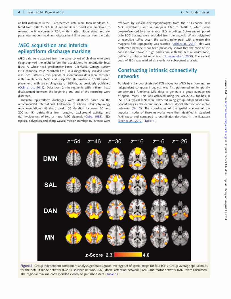
Constructing intrinsic connectivitynetworksTo identify the coordinates of ICN nodes for MEG beamforming, an
independent component analysis was first performed on temporally
concatenated functional MRI data to generate a group-average set
of spatial maps. This was achieved using the MELODIC toolbox in
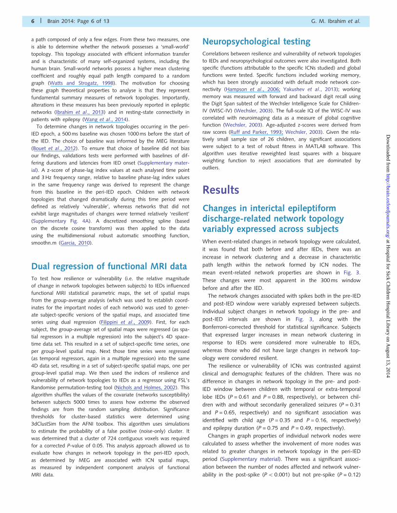
FSL. Four typical ICNs were extracted using group-independent com-
ponent analysis, the default mode, salience, dorsal attention and motor
networks (Fig. 2). The coordinates of the spatial maxima of the
important nodes of these networks were then identified in standard
MNI space and compared to coordinates described in the literature
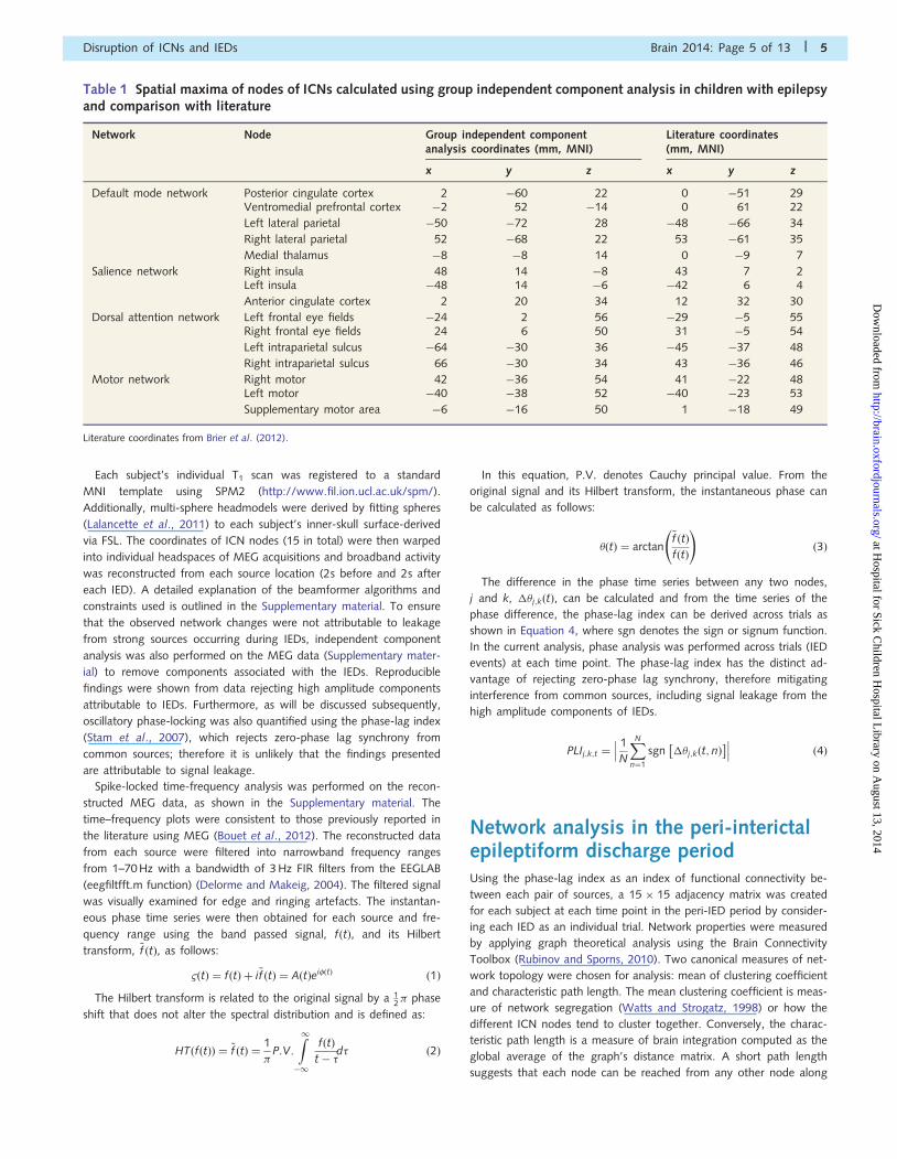
(Brier et al., 2012) (Table 1).
Figure 2 Group independent component analysis generates group-average set of spatial maps for four ICNs. Group-average spatial maps
for the default mode network (DMN), salience network (SN), dorsal attention network (DAN) and motor network (MN) were calculated.
The regional maxima corresponded closely to published data (Table 1).
4 | Brain 2014: Page 4 of 13 G. M. Ibrahim et al.
at Hospital for Sick C
hildren Hospital L
ibrary on August 13, 2014
http://brain.oxfordjournals.org/D
ownloaded from
Each subject’s individual T1 scan was registered to a standard
MNI template using SPM2 (http://www.fil.ion.ucl.ac.uk/spm/).
Additionally, multi-sphere headmodels were derived by fitting spheres
(Lalancette et al., 2011) to each subject’s inner-skull surface-derived
via FSL. The coordinates of ICN nodes (15 in total) were then warped
into individual headspaces of MEG acquisitions and broadband activity
was reconstructed from each source location (2s before and 2s after
each IED). A detailed explanation of the beamformer algorithms and
constraints used is outlined in the Supplementary material. To ensure
that the observed network changes were not attributable to leakage
from strong sources occurring during IEDs, independent component
analysis was also performed on the MEG data (Supplementary mater-
ial) to remove components associated with the IEDs. Reproducible
findings were shown from data rejecting high amplitude components
attributable to IEDs. Furthermore, as will be discussed subsequently,
oscillatory phase-locking was also quantified using the phase-lag index
(Stam et al., 2007), which rejects zero-phase lag synchrony from
common sources; therefore it is unlikely that the findings presented
are attributable to signal leakage.
Spike-locked time-frequency analysis was performed on the recon-
structed MEG data, as shown in the Supplementary material. The
time–frequency plots were consistent to those previously reported in
the literature using MEG (Bouet et al., 2012). The reconstructed data
from each source were filtered into narrowband frequency ranges
from 1–70 Hz with a bandwidth of 3 Hz FIR filters from the EEGLAB
(eegfiltfft.m function) (Delorme and Makeig, 2004). The filtered signal
was visually examined for edge and ringing artefacts. The instantan-
eous phase time series were then obtained for each source and fre-
quency range using the band passed signal, fðtÞ, and its Hilbert
transform, ~f ðtÞ, as follows:
&ðtÞ ¼ fðtÞ þ i~f ðtÞ ¼ AðtÞei�ðtÞ ð1Þ
The Hilbert transform is related to the original signal by a 12� phase
shift that does not alter the spectral distribution and is defined as:
HTðfðtÞÞ ¼ ~f ðtÞ ¼1
�P:V:
Z1�1
fðtÞ
t � �d� ð2Þ
In this equation, P.V. denotes Cauchy principal value. From the
original signal and its Hilbert transform, the instantaneous phase can
be calculated as follows:
�ðtÞ ¼ arctan~f ðtÞ
fðtÞ
!ð3Þ
The difference in the phase time series between any two nodes,
j and k, ��j;kðtÞ, can be calculated and from the time series of the
phase difference, the phase-lag index can be derived across trials as
shown in Equation 4, where sgn denotes the sign or signum function.
In the current analysis, phase analysis was performed across trials (IED
events) at each time point. The phase-lag index has the distinct ad-
vantage of rejecting zero-phase lag synchrony, therefore mitigating
interference from common sources, including signal leakage from the
high amplitude components of IEDs.
PLIj;k;t ¼��� 1
N
XN
n¼1
sgn ��j;kðt;nÞ� ���� ð4Þ
Network analysis in the peri-interictalepileptiform discharge periodUsing the phase-lag index as an index of functional connectivity be-
tween each pair of sources, a 15 � 15 adjacency matrix was created
for each subject at each time point in the peri-IED period by consider-
ing each IED as an individual trial. Network properties were measured
by applying graph theoretical analysis using the Brain Connectivity
Toolbox (Rubinov and Sporns, 2010). Two canonical measures of net-
work topology were chosen for analysis: mean of clustering coefficient
and characteristic path length. The mean clustering coefficient is meas-
ure of network segregation (Watts and Strogatz, 1998) or how the
different ICN nodes tend to cluster together. Conversely, the charac-
teristic path length is a measure of brain integration computed as the
global average of the graph’s distance matrix. A short path length
suggests that each node can be reached from any other node along
Table 1 Spatial maxima of nodes of ICNs calculated using group independent component analysis in children with epilepsyand comparison with literature
Network Node Group independent componentanalysis coordinates (mm, MNI)
Literature coordinates(mm, MNI)
x y z x y z
Default mode network Posterior cingulate cortex 2 �60 22 0 �51 29Ventromedial prefrontal cortex �2 52 �14 0 61 22
Left lateral parietal �50 �72 28 �48 �66 34
Right lateral parietal 52 �68 22 53 �61 35
Medial thalamus �8 �8 14 0 �9 7
Salience network Right insula 48 14 �8 43 7 2Left insula �48 14 �6 �42 6 4
Anterior cingulate cortex 2 20 34 12 32 30
Dorsal attention network Left frontal eye fields �24 2 56 �29 �5 55Right frontal eye fields 24 6 50 31 �5 54
Left intraparietal sulcus �64 �30 36 �45 �37 48
Right intraparietal sulcus 66 �30 34 43 �36 46
Motor network Right motor 42 �36 54 41 �22 48Left motor �40 �38 52 �40 �23 53
Supplementary motor area �6 �16 50 1 �18 49
Literature coordinates from Brier et al. (2012).
Disruption of ICNs and IEDs Brain 2014: Page 5 of 13 | 5
at Hospital for Sick C
hildren Hospital L
ibrary on August 13, 2014
http://brain.oxfordjournals.org/D
ownloaded from
a path composed of only a few edges. From these two measures, one
is able to determine whether the network possesses a ‘small-world’
topology. This topology associated with efficient information transfer
and is characteristic of many self-organized systems, including the
human brain. Small-world networks possess a higher mean clustering
coefficient and roughly equal path length compared to a random
graph (Watts and Strogatz, 1998). The motivation for choosing
these graph theoretical properties to analyse is that they represent
fundamental summary measures of network topologies. Importantly,
alterations in these measures has been previously reported in epileptic
networks (Ibrahim et al., 2013) and in resting-state connectivity in
patients with epilepsy (Wang et al., 2014).
To determine changes in network topologies occurring in the peri-
IED epoch, a 500 ms baseline was chosen 1000 ms before the start of
the IED. The choice of baseline was informed by the MEG literature
(Bouet et al., 2012). To ensure that choice of baseline did not bias
our findings, validations tests were performed with baselines of dif-
fering durations and latencies from IED onset (Supplementary mater-
ial). A z-score of phase-lag index values at each analysed time point
and 3 Hz frequency range, relative to baseline phase-lag index values
in the same frequency range was derived to represent the change
from this baseline in the peri-IED epoch. Children with network
topologies that changed dramatically during this time period were
defined as relatively ‘vulnerable’, whereas networks that did not
exhibit large magnitudes of changes were termed relatively ‘resilient’
(Supplementary Fig. 4A). A discretized smoothing spline (based
on the discrete cosine transform) was then applied to the data
using the multidimensional robust automatic smoothing function,
smoothn.m (Garcia, 2010).
Dual regression of functional MRI dataTo test how resilience or vulnerability (i.e. the relative magnitude
of change in network topologies between subjects) to IEDs influenced
functional MRI statistical parametric maps, the set of spatial maps
from the group-average analysis (which was used to establish coord-
inates for the important nodes of each network) was used to gener-
ate subject-specific versions of the spatial maps, and associated time
series using dual regression (Filippini et al., 2009). First, for each
subject, the group-average set of spatial maps were regressed (as spa-
tial regressors in a multiple regression) into the subject’s 4D space-
time data set. This resulted in a set of subject-specific time series, one
per group-level spatial map. Next those time series were regressed
(as temporal regressors, again in a multiple regression) into the same
4D data set, resulting in a set of subject-specific spatial maps, one per
group-level spatial map. We then used the indices of resilience and
vulnerability of network topologies to IEDs as a regressor using FSL’s
Randomise permutation-testing tool (Nichols and Holmes, 2002). This
algorithm shuffles the values of the covariate (networks susceptibility)
between subjects 5000 times to assess how extreme the observed
findings are from the random sampling distribution. Significance
thresholds for cluster-based statistics were determined using
3dClustSim from the AFNI toolbox. This algorithm uses simulations
to estimate the probability of a false positive (noise-only) cluster. It
was determined that a cluster of 724 contiguous voxels was required
for a corrected P-value of 0.05. This analysis approach allowed us to
evaluate how changes in network topology in the peri-IED epoch,
as determined by MEG are associated with ICN spatial maps,
as measured by independent component analysis of functional
MRI data.
Neuropsychological testingCorrelations between resilience and vulnerability of network topologies
to IEDs and neuropsychological outcomes were also investigated. Both
specific (functions attributable to the specific ICNs studied) and global
functions were tested. Specific functions included working memory,
which has been strongly associated with default mode network con-
nectivity (Hampson et al., 2006; Yakushev et al., 2013); working
memory was measured with forward and backward digit recall using
the Digit Span subtest of the Wechsler Intelligence Scale for Children-
IV (WISC-IV) (Wechsler, 2003). The full-scale IQ of the WISC-IV was
correlated with neuroimaging data as a measure of global cognitive
function (Wechsler, 2003). Age-adjusted z-scores were derived from
raw scores (Ruff and Parker, 1993; Wechsler, 2003). Given the rela-
tively small sample size of 26 children, any significant associations
were subject to a test of robust fitness in MATLAB software. This
algorithm uses iterative reweighted least squares with a bisquare
weighting function to reject associations that are dominated by
outliers.
Results
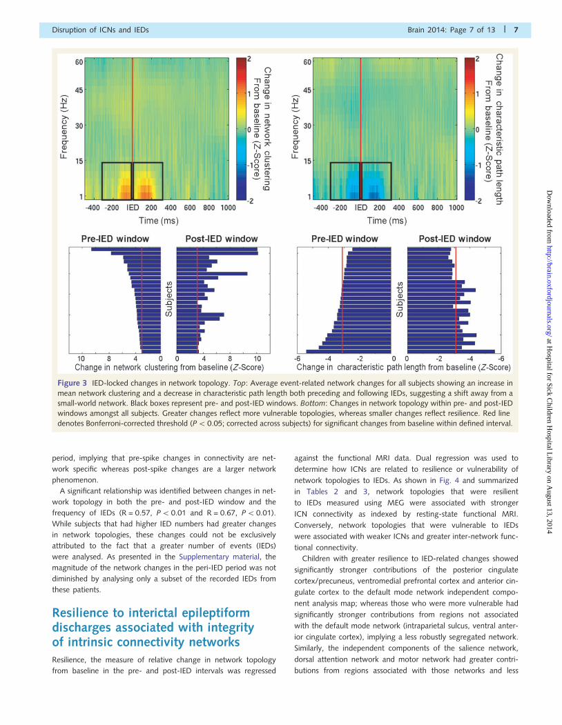
Changes in interictal epileptiformdischarge-related network topologyvariably expressed across subjectsWhen event-related changes in network topology were calculated,
it was found that both before and after IEDs, there was an
increase in network clustering and a decrease in characteristic
path length within the network formed by ICN nodes. The
mean event-related network properties are shown in Fig. 3.
These changes were most apparent in the 300 ms window
before and after the IED.
The network changes associated with spikes both in the pre-IED
and post-IED window were variably expressed between subjects.
Individual subject changes in network topology in the pre- and
post-IED intervals are shown in Fig. 3, along with the
Bonferroni-corrected threshold for statistical significance. Subjects
that expressed larger increases in mean network clustering in
response to IEDs were considered more vulnerable to IEDs,
whereas those who did not have large changes in network top-
ology were considered resilient.
The resilience or vulnerability of ICNs was contrasted against
clinical and demographic features of the children. There was no
difference in changes in network topology in the pre- and post-
IED window between children with temporal or extra-temporal
lobe IEDs (P = 0.61 and P = 0.88, respectively), or between chil-
dren with and without secondarily generalized seizures (P = 0.31
and P = 0.65, respectively) and no significant association was
identified with child age (P = 0.35 and P = 0.16, respectively)
and epilepsy duration (P = 0.75 and P = 0.49, respectively).
Changes in graph properties of individual network nodes were
calculated to assess whether the involvement of more nodes was
related to greater changes in network topology in the peri-IED
period (Supplementary material). There was a significant associ-
ation between the number of nodes affected and network vulner-
ability in the post-spike (P50.001) but not pre-spike (P = 0.12)
6 | Brain 2014: Page 6 of 13 G. M. Ibrahim et al.
at Hospital for Sick C
hildren Hospital L
ibrary on August 13, 2014
http://brain.oxfordjournals.org/D
ownloaded from
period, implying that pre-spike changes in connectivity are net-
work specific whereas post-spike changes are a larger network
phenomenon.
A significant relationship was identified between changes in net-
work topology in both the pre- and post-IED window and the
frequency of IEDs (R = 0.57, P50.01 and R = 0.67, P50.01).
While subjects that had higher IED numbers had greater changes
in network topologies, these changes could not be exclusively
attributed to the fact that a greater number of events (IEDs)
were analysed. As presented in the Supplementary material, the
magnitude of the network changes in the peri-IED period was not
diminished by analysing only a subset of the recorded IEDs from
these patients.
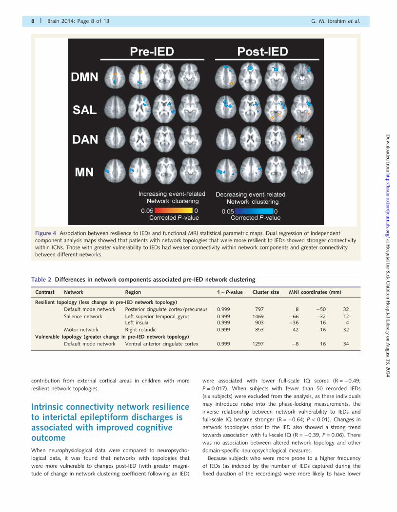
Resilience to interictal epileptiformdischarges associated with integrityof intrinsic connectivity networksResilience, the measure of relative change in network topology
from baseline in the pre- and post-IED intervals was regressed
against the functional MRI data. Dual regression was used to
determine how ICNs are related to resilience or vulnerability of
network topologies to IEDs. As shown in Fig. 4 and summarized
in Tables 2 and 3, network topologies that were resilient
to IEDs measured using MEG were associated with stronger
ICN connectivity as indexed by resting-state functional MRI.
Conversely, network topologies that were vulnerable to IEDs
were associated with weaker ICNs and greater inter-network func-
tional connectivity.
Children with greater resilience to IED-related changes showed
significantly stronger contributions of the posterior cingulate
cortex/precuneus, ventromedial prefrontal cortex and anterior cin-
gulate cortex to the default mode network independent compo-
nent analysis map; whereas those who were more vulnerable had
significantly stronger contributions from regions not associated
with the default mode network (intraparietal sulcus, ventral anter-
ior cingulate cortex), implying a less robustly segregated network.
Similarly, the independent components of the salience network,
dorsal attention network and motor network had greater contri-
butions from regions associated with those networks and less
Figure 3 IED-locked changes in network topology. Top: Average event-related network changes for all subjects showing an increase in
mean network clustering and a decrease in characteristic path length both preceding and following IEDs, suggesting a shift away from a
small-world network. Black boxes represent pre- and post-IED windows. Bottom: Changes in network topology within pre- and post-IED
windows amongst all subjects. Greater changes reflect more vulnerable topologies, whereas smaller changes reflect resilience. Red line
denotes Bonferroni-corrected threshold (P5 0.05; corrected across subjects) for significant changes from baseline within defined interval.
Disruption of ICNs and IEDs Brain 2014: Page 7 of 13 | 7
at Hospital for Sick C
hildren Hospital L
ibrary on August 13, 2014
http://brain.oxfordjournals.org/D
ownloaded from
contribution from external cortical areas in children with more
resilient network topologies.
Intrinsic connectivity network resilienceto interictal epileptiform discharges isassociated with improved cognitiveoutcomeWhen neurophysiological data were compared to neuropsycho-
logical data, it was found that networks with topologies that
were more vulnerable to changes post-IED (with greater magni-
tude of change in network clustering coefficient following an IED)
were associated with lower full-scale IQ scores (R = �0.49;
P = 0.017). When subjects with fewer than 50 recorded IEDs
(six subjects) were excluded from the analysis, as these individuals
may introduce noise into the phase-locking measurements, the
inverse relationship between network vulnerability to IEDs and
full-scale IQ became stronger (R = �0.64; P5 0.01). Changes in
network topologies prior to the IED also showed a strong trend
towards association with full-scale IQ (R = �0.39, P = 0.06). There
was no association between altered network topology and other
domain-specific neuropsychological measures.
Because subjects who were more prone to a higher frequency
of IEDs (as indexed by the number of IEDs captured during the
fixed duration of the recordings) were more likely to have lower
Figure 4 Association between resilience to IEDs and functional MRI statistical parametric maps. Dual regression of independent
component analysis maps showed that patients with network topologies that were more resilient to IEDs showed stronger connectivity
within ICNs. Those with greater vulnerability to IEDs had weaker connectivity within network components and greater connectivity
between different networks.
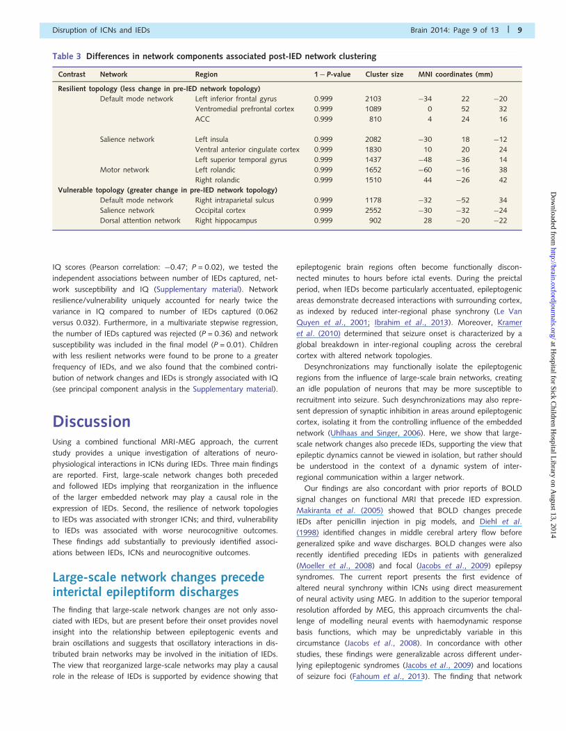
Table 2 Differences in network components associated pre-IED network clustering
Contrast Network Region 1� P-value Cluster size MNI coordinates (mm)
Resilient topology (less change in pre-IED network topology)
Default mode network Posterior cingulate cortex/precuneus 0.999 797 8 �50 32
Salience network Left superior temporal gyrus 0.999 1469 �66 �32 12Left insula 0.999 903 �36 16 4
Motor network Right rolandic 0.999 853 42 �16 32
Vulnerable topology (greater change in pre-IED network topology)
Default mode network Ventral anterior cingulate cortex 0.999 1297 �8 16 34
8 | Brain 2014: Page 8 of 13 G. M. Ibrahim et al.
at Hospital for Sick C
hildren Hospital L
ibrary on August 13, 2014
http://brain.oxfordjournals.org/D
ownloaded from
IQ scores (Pearson correlation: �0.47; P = 0.02), we tested the
independent associations between number of IEDs captured, net-
work susceptibility and IQ (Supplementary material). Network
resilience/vulnerability uniquely accounted for nearly twice the
variance in IQ compared to number of IEDs captured (0.062
versus 0.032). Furthermore, in a multivariate stepwise regression,
the number of IEDs captured was rejected (P = 0.36) and network
susceptibility was included in the final model (P = 0.01). Children
with less resilient networks were found to be prone to a greater
frequency of IEDs, and we also found that the combined contri-
bution of network changes and IEDs is strongly associated with IQ
(see principal component analysis in the Supplementary material).
DiscussionUsing a combined functional MRI-MEG approach, the current
study provides a unique investigation of alterations of neuro-
physiological interactions in ICNs during IEDs. Three main findings
are reported. First, large-scale network changes both preceded
and followed IEDs implying that reorganization in the influence
of the larger embedded network may play a causal role in the
expression of IEDs. Second, the resilience of network topologies
to IEDs was associated with stronger ICNs; and third, vulnerability
to IEDs was associated with worse neurocognitive outcomes.
These findings add substantially to previously identified associ-
ations between IEDs, ICNs and neurocognitive outcomes.
Large-scale network changes precedeinterictal epileptiform dischargesThe finding that large-scale network changes are not only asso-
ciated with IEDs, but are present before their onset provides novel
insight into the relationship between epileptogenic events and
brain oscillations and suggests that oscillatory interactions in dis-
tributed brain networks may be involved in the initiation of IEDs.
The view that reorganized large-scale networks may play a causal
role in the release of IEDs is supported by evidence showing that
epileptogenic brain regions often become functionally discon-
nected minutes to hours before ictal events. During the preictal
period, when IEDs become particularly accentuated, epileptogenic
areas demonstrate decreased interactions with surrounding cortex,
as indexed by reduced inter-regional phase synchrony (Le Van
Quyen et al., 2001; Ibrahim et al., 2013). Moreover, Kramer
et al. (2010) determined that seizure onset is characterized by a
global breakdown in inter-regional coupling across the cerebral
cortex with altered network topologies.
Desynchronizations may functionally isolate the epileptogenic
regions from the influence of large-scale brain networks, creating
an idle population of neurons that may be more susceptible to
recruitment into seizure. Such desynchronizations may also repre-
sent depression of synaptic inhibition in areas around epileptogenic
cortex, isolating it from the controlling influence of the embedded
network (Uhlhaas and Singer, 2006). Here, we show that large-
scale network changes also precede IEDs, supporting the view that
epileptic dynamics cannot be viewed in isolation, but rather should
be understood in the context of a dynamic system of inter-
regional communication within a larger network.
Our findings are also concordant with prior reports of BOLD
signal changes on functional MRI that precede IED expression.
Makiranta et al. (2005) showed that BOLD changes precede
IEDs after penicillin injection in pig models, and Diehl et al.
(1998) identified changes in middle cerebral artery flow before
generalized spike and wave discharges. BOLD changes were also
recently identified preceding IEDs in patients with generalized
(Moeller et al., 2008) and focal (Jacobs et al., 2009) epilepsy
syndromes. The current report presents the first evidence of
altered neural synchrony within ICNs using direct measurement
of neural activity using MEG. In addition to the superior temporal
resolution afforded by MEG, this approach circumvents the chal-
lenge of modelling neural events with haemodynamic response
basis functions, which may be unpredictably variable in this
circumstance (Jacobs et al., 2008). In concordance with other
studies, these findings were generalizable across different under-
lying epileptogenic syndromes (Jacobs et al., 2009) and locations
of seizure foci (Fahoum et al., 2013). The finding that network
Table 3 Differences in network components associated post-IED network clustering
Contrast Network Region 1� P-value Cluster size MNI coordinates (mm)
Resilient topology (less change in pre-IED network topology)
Default mode network Left inferior frontal gyrus 0.999 2103 �34 22 �20
Ventromedial prefrontal cortex 0.999 1089 0 52 32
ACC 0.999 810 4 24 16
Salience network Left insula 0.999 2082 �30 18 �12
Ventral anterior cingulate cortex 0.999 1830 10 20 24
Left superior temporal gyrus 0.999 1437 �48 �36 14
Motor network Left rolandic 0.999 1652 �60 �16 38
Right rolandic 0.999 1510 44 �26 42
Vulnerable topology (greater change in pre-IED network topology)
Default mode network Right intraparietal sulcus 0.999 1178 �32 �52 34
Salience network Occipital cortex 0.999 2552 �30 �32 �24
Dorsal attention network Right hippocampus 0.999 902 28 �20 �22
Disruption of ICNs and IEDs Brain 2014: Page 9 of 13 | 9
at Hospital for Sick C
hildren Hospital L
ibrary on August 13, 2014
http://brain.oxfordjournals.org/D
ownloaded from
changes were not present specifically at the time of the IED may
reflect a rejection of zero-phase lag synchrony by the phase-lag
index, given the high amplitude increase in neuromagnetic signal
during IEDs.
Interictal epileptiform discharges,intrinsic connectivity networks andneurocognitive outcomeIt has long been suspected that IEDs may contribute to impaired
neurocognitive function in patients with epilepsy and the inter-
action between IEDs and cognitively-salient oscillatory networks
is an area of increasing scientific inquiry. Previous studies
have reported spike-related BOLD decreases in default mode net-
work regions (Kobayashi et al., 2006; Laufs et al., 2007). Such
IED-associated deactivations accompany spikes of differing
morphologies and locations, including generalized spike and
wave discharges or focal interictal discharges of temporal, frontal
and posterior quadrant origins (Fahoum et al., 2012, 2013). In
patients with idiopathic generalized epilepsy, thalamocortical acti-
vation and suspension of regions of the default mode network
were hypothesized to contribute to reduced responsiveness
during IEDs (Gotman et al., 2005).
An understanding of the associations between IEDs, oscillatory
brain networks and cognition is critical to devise appropriate treat-
ment strategies aimed at improving outcomes in this patient popu-
lation. In the current report, changes in ICN topologies,
particularly following IEDs, were significantly associated with func-
tional MRI statistical parametric maps of networks and neurocog-
nitive outcomes. The segregation of ICNs is a hallmark of typical
development (Dosenbach et al., 2010). Connectivity gradients be-
tween task-positive and task-negative ICN hubs become stronger
during adolescence and early adulthood with sharpening of the
boundaries of the default mode network and decreased correlation
between the default mode and attention control networks
(Anderson et al., 2011). The fractionation of brain regions into
specific regional hubs also represents a transition from a local to
distributed network organization, which characterizes typical child-
hood and adolescent development (Fair et al., 2009). This intra-
network integration and inter-network segregation contributes
to the emergence of the more mature distributed, functionally-
specialized networks observed in adults.
The disruption of normative ICN organization in children with
network topologies that were vulnerable to IEDs provides novel
insights into the interactions between epileptic dynamics, oscilla-
tory synchrony and the organization of spontaneous network con-
nectivity in the developing brain. It was observed that the
physiological anti-correlation between task-positive networks
(salience network, dorsal attention network, motor network) and
a task-negative network (default mode network) was weaker in
children with greater vulnerability to IEDs. The loss of these con-
nectivity gradients has also been previously associated with worse
neurocognitive outcomes in a similar set of children with epilepsy
when compared with age-matched controls (Ibrahim et al., 2014).
Since the majority of resting-state studies do not differentiate
between baseline periods that are and are not IED-free, this
study underscores the importance of evaluating IEDs when study-
ing oscillatory networks in patients with epilepsy. We also provide
evidence supporting more aggressive suppression of discharges
through medical or surgical treatments, as these results indicate
that IEDs are not benign in regards to neurocognitive
development.
Neuromagnetic oscillations, BOLDintrinsic connectivity networks andfrequency specificityNeuromagnetic recordings were used to reconstruct oscillatory
activity from coordinates of ICN nodes, identified using functional
MRI. This approach capitalizes on recent advances in the use of
MEG for investigating neurophysiological oscillations in ICNs,
which are most accurately localized using functional MRI guid-
ance. ICNs have been previously reconstructed from band-limited
power (BLP; the power of envelope modulation of a relatively
narrow range of frequencies) correlations among MEG signals
reconstructed from various locations in the brain. BLP was
shown to demonstrate significant interhemispheric correlations be-
tween homologous brain regions, as would be expected from
spontaneous fluctuations in BOLD signal across multiple frequen-
cies (Liu et al., 2010). Furthermore, source space localization in
MEG demonstrates long-range temporal correlation between
spontaneous BLP signals from functionally-related regions
(Brookes et al., 2011b; Hipp et al., 2012). Correlations of BLP
within ICNs were strongest in these studies in the alpha and
beta frequency ranges, depending on the underlying oscillation
frequency. Furthermore, the spatial structure of several canonical
ICNs have been reconstructed from MEG (de Pasquale et al.,
2010, 2012; Brookes et al., 2011a). These studies again demon-
strated that ICNs are best captured by fluctuations in theta, alpha
and beta oscillations.
An important advantage of measuring ICNs using MEG is the
ability to analyse changes in network topologies in physiologically-
relevant frequency bands. In the current study, we found that the
strongest changes in network topology, both before and after
IEDs occurred in lower frequency bands (namely, delta and
theta). This is supported by previous findings that IEDs with
slow waves on EEG were more likely to cause default mode net-
work deactivation than those lacking slow waves (Kobayashi
et al., 2006). Recently, data from combined functional MRI-
intracranial EEG revealed that runs of IEDs and short electroence-
phalographic seizures were associated with decreased gamma
power and increases in the power of lower frequency oscillations,
although the findings were inconsistently expressed across the six
subjects studied (Fahoum et al., 2013). Although our study
differed considerably in methodology and techniques, time-
frequency analysis also revealed increases in the power of lower
frequencies along with some decreases in gamma power, although
as Fahoum et al. (2013) report, the latter were greatly exceeded
by the former. Finally, power increases in slower oscillations
in ICNs nodes has been previously linked to consciousness. For
instance, Blumenfeld et al. (2004) found that increased delta
frequency power in the default mode network, linked to impaired
10 | Brain 2014: Page 10 of 13 G. M. Ibrahim et al.
at Hospital for Sick C
hildren Hospital L
ibrary on August 13, 2014
http://brain.oxfordjournals.org/D
ownloaded from
consciousness in patients with complex partial seizures with intra-
cranial electrodes (Englot et al., 2010).
Limitations and future directionsAn important question is why some brains are more resilient, while
others more vulnerable to changes in network topology in the
peri-IED epoch. Using recent functional neuroimaging methods,
neuroscientists are able to improve classification of illness and
stratify patient populations based on specific resting- or event-
related network impairments, as performed in the current study.
Patients who are prone to having more frequent IEDs demonstrate
greater vulnerability to changes in network topology. We have
shown, however, that this increased vulnerability cannot be attrib-
uted to the greater number of IEDs analysed and explains more of
the variance in neurocognitive outcome than the number of IEDs
captured during the recording (Supplementary material). The find-
ings imply that the oscillatory organization of brain networks in
children who are more prone to IEDs is intrinsically less resilient
than those with fewer events. It is possible that the vulnerability
may be mediated by dysfunctional cortical or subcortical circuitry
in children who are more prone to having frequent IEDs. Notably,
the thalamus may play an important role in mediating oscillatory
network resilience as well as the expression of IEDs. We have
previously shown that more severe epilepsy syndromes are char-
acterized by a loss of centrality of the thalamus in a whole-brain
connectome (Ibrahim et al., 2014). It has also been shown that
synchronous spike-and-wave discharges reflect highly synchro-
nized oscillations in thalamocortical networks, although spike and
wave discharges originate in the cortex and initiate oscillations in
the thalamo-cortical-thalamic loop (Meeren et al., 2002). Multi-
modality studies evaluating effective connectivity or causal dir-
ected information flow are needed to disentangle these effects
and explain variability in resilience of oscillatory neural networks.
The primary limitation of the current study is the heterogeneity
in clinical syndromes of children with epilepsy, including the loca-
tion of seizure foci, epileptogenic substrates and antiepileptic
medications. Previous studies have, however, suggested that
large-scale network changes in the peri-IED period are generaliz-
able across different patient groups and, indeed, we found no
significant difference in resilience or vulnerability to IEDs across
different clinical variables. Furthermore, it is expected that if vari-
ability in the clinical population impacts the measured network
topologies, it would have an effect of diminishing the observed
findings. Another limitation is the visual marking of IEDs, where
imprecision in marking may introduce greater bias in the identifi-
cation of altered connectivity at higher frequencies. Future studies
with larger patient cohorts are indicated to better characterize
the heterogeneity and to correlate them to specific network
impairments.
ConclusionUsing a novel combined functional MRI-MEG approach, the cur-
rent study examined neurophysiological changes in ICN topologies
in the peri-IED epoch and correlated the resilience and
vulnerability to network changes with neurocognitive function in
children. It was found that changes in the topology of ICNs both
preceded and followed the IEDs. The resilience of ICNs to changes
in the peri-IED epoch, as measured by MEG was associated with
greater segregation of these networks on functional MRI as well
as improved neurocognitive outcomes in affected children. The
association between IEDs, network changes and neurocognitive
outcomes highlights their importance in understanding and treat-
ing the comorbidities of intractable childhood epilepsy.
FundingThis research was supported by the Canadian Institutes of Health
Research (CIHR) Vanier Canada Graduate Scholarship, CIHR Bisby
Fellowship, The Hospital for Sick Children Foundation Student
Scholarship Program, The Hospital for Sick Children Centre for
Brain and Behaviour, the Ontario Brain Institute, the Wiley
Family and Jack Beqaj Funds for Epilepsy Surgery Research, and
the University of Toronto Surgeon-Scientist Program.
Supplementary materialSupplementary material is available at Brain online.
ReferencesAnderson JS, Ferguson MA, Lopez-Larson M, Yurgelun-Todd D.
Connectivity gradients between the default mode and attention con-
trol networks. Brain Connect 2011; 1: 147–57.
Biswal B, Yetkin FZ, Haughton VM, Hyde JS. Functional connectivity in
the motor cortex of resting human brain using echo-planar MRI. Magn
Reson Med 1995; 34: 537–41.Blumenfeld H, Rivera M, McNally KA, Davis K, Spencer DD, Spencer SS.
Ictal neocortical slowing in temporal lobe epilepsy. Neurology 2004;
63: 1015–21.
Bouet R, Jung J, Delpuech C, Ryvlin P, Isnard J, Guenot M, et al.
Towards source volume estimation of interictal spikes in focal epilepsy
using magnetoencephalography. Neuroimage 2012; 59: 3955–66.
Brier MR, Thomas JB, Snyder AZ, Benzinger TL, Zhang D, Raichle ME,
et al. Loss of intranetwork and internetwork resting state functional
connections with Alzheimer’s disease progression. J Neurosci 2012; 32:
8890–9.
Brookes MJ, Hale JR, Zumer JM, Stevenson CM, Francis ST, Barnes GR,
et al. Measuring functional connectivity using MEG: methodology and
comparison with fcMRI. Neuroimage 2011a; 56: 1082–104.Brookes MJ, Woolrich M, Luckhoo H, Price D, Hale JR, Stephenson MC,
et al. Investigating the electrophysiological basis of resting state net-
works using magnetoencephalography. Proc Natl Acad Sci USA
2011b; 108: 16783–8.Cobb WA. Recommendations for the practice of clinical neurophysi-
ology. Amsterdam: Elsevier; 1983.
de Pasquale F, Della Penna S, Snyder AZ, Lewis C, Mantini D, Marzetti L,
et al. Temporal dynamics of spontaneous MEG activity in brain net-
works. Proc Natl Acad Sci USA 2010; 107: 6040–5.de Pasquale F, Della Penna S, Snyder AZ, Marzetti L, Pizzella V,
Romani GL, et al. A cortical core for dynamic integration of functional
networks in the resting human brain. Neuron 2012; 74: 753–64.
Delorme A, Makeig S. EEGLAB: an open source toolbox for analysis of
single-trial EEG dynamics including independent component analysis.
J Neurosci Methods 2004; 134: 9–21.
Disruption of ICNs and IEDs Brain 2014: Page 11 of 13 | 11
at Hospital for Sick C
hildren Hospital L
ibrary on August 13, 2014
http://brain.oxfordjournals.org/D
ownloaded from
Diehl B, Knecht S, Deppe M, Young C, Stodieck SR. Cerebral hemo-
dynamic response to generalized spike-wave discharges. Epilepsia
1998; 39: 1284–9.
Dosenbach NU, Nardos B, Cohen AL, Fair DA, Power JD, Church JA,
et al. Prediction of individual brain maturity using fMRI. Science 2010;
329: 1358–61.
Englot DJ, Yang L, Hamid H, Danielson N, Bai X, Marfeo A, et al.
Impaired consciousness in temporal lobe seizures: Role of cortical
slow activity. Brain 2010; 133: 3764–77.
Fahoum F, Zelmann R, Tyvaert L, Dubeau F, Gotman J. Epileptic dis-
charges affect the default mode network–FMRI and intracerebral EEG
evidence. PLoS One 2013; 8: e68038.
Fahoum F, Lopes R, Pittau F, Dubeau F, Gotman J. Widespread epileptic
networks in focal epilepsies: EEG-fMRI study. Epilepsia 2012; 53:
1618–27.
Fair DA, Cohen AL, Power JD, Dosenbach NU, Church JA, Miezin FM,
et al. Functional brain networks develop from a “local to distributed”
organization. PLoS Comput Biol 2009; 5: e1000381.
Filippini N, MacIntosh BJ, Hough MG, Goodwin GM, Frisoni GB,
Smith SM, et al. Distinct patterns of brain activity in young carriers
of the APOE-epsilon4 allele. Proc Natl Acad Sci USA 2009; 106:
7209–14.
Fransson P, Skiold B, Horsch S, Nordell A, Blennow M, Lagercrantz H,
et al. Resting-state networks in the infant brain. Proc Natl Acad Sci
USA 2007; 104: 15531–6.
Garcia D. Robust smoothing of gridded data in one and higher dimen-
sions with missing values. Comput Stat Data Anal 2010; 54: 1167–78.
Gotman J, Grova C, Bagshaw A, Kobayashi E, Aghakhani Y, Dubeau F.
Generalized epileptic discharges show thalamocortical activation and
suspension of the default state of the brain. Proc Natl Acad Sci USA
2005; 102: 15236–40.
Grenier F, Timofeev I, Crochet S, Steriade M. Spontaneous field poten-
tials influence the activity of neocortical neurons during paroxysmal
activities in vivo. Neuroscience 2003; 119: 277–91.
Hampson M, Driesen NR, Skudlarski P, Gore JC, Constable RT. Brain
connectivity related to working memory performance. J Neurosci
2006; 26: 13338–43.
Hipp JF, Hawellek DJ, Corbetta M, Siegel M, Engel AK. Large-scale cor-
tical correlation structure of spontaneous oscillatory activity. Nat
Neurosci 2012; 15: 884–90.
Hufnagel A, Dumpelmann M, Zentner J, Schijns O, Elger CE. Clinical
relevance of quantified intracranial interictal spike activity in presurgical
evaluation of epilepsy. Epilepsia 2000; 41: 467–78.
Ibrahim GM, Anderson R, Akiyama T, Ochi A, Otsubo H,
Singh-Cadieux G, et al. Neocortical pathological high frequency oscil-
lations are associated with frequency-dependent alterations in func-
tional network topology. J Neurophysiol 2013; 110: 2475–83.
Ibrahim GM, Morgan BR, Lee W, Smith ML, Donner EJ, Wang F, et al.
Impaired development of intrinsic connectivity networks in children
with medically intractable localization-related epilepsy. Hum Brain
Mapp 2014 [Epub ahead of print].
Jacobs J, Hawco C, Kobayashi E, Boor R, LeVan P, Stephani U, et al.
Variability of the hemodynamic response as a function of age and
frequency of epileptic discharge in children with epilepsy.
Neuroimage 2008; 40: 601–14.
Jacobs J, Levan P, Moeller F, Boor R, Stephani U, Gotman J, et al.
Hemodynamic changes preceding the interictal EEG spike in patients
with focal epilepsy investigated using simultaneous EEG-fMRI.
Neuroimage 2009; 45: 1220–31.
Kobayashi E, Bagshaw AP, Benar CG, Aghakhani Y, Andermann F,
Dubeau F, et al. Temporal and extratemporal BOLD responses to tem-
poral lobe interictal spikes. Epilepsia 2006; 47: 343–54.
Kramer MA, Eden UT, Kolaczyk ED, Zepeda R, Eskandar EN, Cash SS.
Coalescence and fragmentation of cortical networks during focal
seizures. J Neurosci 2010; 30: 10076–85.
Kuruvilla A, Flink R. Intraoperative electrocorticography in epilepsy
surgery: useful or not? Seizure 2003; 12: 577–84.
Lalancette M, Quraan M, Cheyne D. Evaluation of multiple-sphere head
models for MEG source localization. Phys Med Biol 2011; 56:
5621–35.
Laufs H, Hamandi K, Salek-Haddadi A, Kleinschmidt AK, Duncan JS,
Lemieux L. Temporal lobe interictal epileptic discharges affect cerebral
activity in “default mode” brain regions. Hum Brain Mapp 2007; 28:
1023–32.
Le Van Quyen M, Martinerie J, Navarro V, Baulac And M, Varela FJ.
Characterizing neurodynamic changes before seizures. J Clin
Neurophysiol 2001; 18: 191–208.
Liu Z, Fukunaga M, de Zwart JA, Duyn JH. Large-scale spontaneous
fluctuations and correlations in brain electrical activity observed with
magnetoencephalography. Neuroimage 2010; 51: 102–11.
Makiranta M, Ruohonen J, Suominen K, Niinimaki J, Sonkajarvi E,
Kiviniemi V, et al. BOLD signal increase preceeds EEG spike activity–
a dynamic penicillin induced focal epilepsy in deep anesthesia.
Neuroimage 2005; 27: 715–24.
Mankinen K, Jalovaara P, Paakki JJ, Harila M, Rytky S, Tervonen O, et al.
Connectivity disruptions in resting-state functional brain networks
in children with temporal lobe epilepsy. Epilepsy Res 2012; 100:
168–78.
Marcar VL, Schwarz U, Martin E, Loenneker T. How depth of anesthesia
influences the blood oxygenation level-dependent signal from the
visual cortex of children. AJNR Am J Neuroradiol 2006; 27: 799–805.
Meeren HK, Pijn JP, Van Luijtelaar EL, Coenen AM, Lopes da Silva FH.
Cortical focus drives widespread corticothalamic networks during
spontaneous absence seizures in rats. J Neurosci 2002; 22: 1480–95.
Moeller F, Siebner HR, Wolff S, Muhle H, Boor R, Granert O, et al.
Changes in activity of striato-thalamo-cortical network precede gen-
eralized spike wave discharges. Neuroimage 2008; 39: 1839–49.
Nichols TE, Holmes AP. Nonparametric permutation tests for functional
neuroimaging: a primer with examples. Hum Brain Mapp 2002; 15:
1–25.
Ochi A, Go CY, Otsubo H. Clinical MEG analyses for children with in-
tractable epilepsy. In: Pang E, editor. Magneoencephalography. Rijeka:
InTech; 2011.
Raichle ME, Mintun MA. Brain work and brain imaging. Annu Rev
Neurosci 2006; 29: 449–76.
Raichle ME, Snyder AZ. A default mode of brain function: a brief his-
tory of an evolving idea. Neuroimage 2007; 37: 1083–90; discussion
1097–9.
Raichle ME, MacLeod AM, Snyder AZ, Powers WJ, Gusnard DA,
Shulman GL. A default mode of brain function. Proc Natl Acad Sci
USA 2001; 98: 676–82.
Rubinov M, Sporns O. Complex network measures of brain connectivity:
uses and interpretations. Neuroimage 2010; 52: 1059–69.
Ruff RM, Parker SB. Gender- and age-specific changes in motor speed
and eye-hand coordination in adults: normative values for the finger
tapping and grooved pegboard tests. Percept Mot Skills 1993; 76:
1219–30.
Stam CJ, Nolte G, Daffertshofer A. Phase lag index: assessment of
functional connectivity from multi channel EEG and MEG with dimin-
ished bias from common sources. Hum Brain Mapp 2007; 28:
1178–93.
Uddin LQ, Supekar K, Lynch CJ, Khouzam A, Phillips J, Feinstein C, et al.
Salience network-based classification and prediction of symptom
severity in children with autism. JAMA Psychiatry 2013; 70: 869–79.
Uhlhaas PJ, Singer W. Neural synchrony in brain disorders: relevance for
cognitive dysfunctions and pathophysiology. Neuron 2006; 52:
155–68.
Vaessen MJ, Braakman HM, Heerink JS, Jansen JF, Debeij-van Hall MH,
Hofman PA, et al. Abnormal modular organization of functional net-
works in cognitively impaired children with frontal lobe epilepsy. Cereb
Cortex 2013; 23: 1997–2006.
Wang J, Qiu S, Xu Y, Liu Z, Wen X, Hu X, et al. Graph theoretical
analysis reveals disrupted topological properties of whole brain func-
tional networks in temporal lobe epilepsy. Clin Neurophysiol 2014.
12 | Brain 2014: Page 12 of 13 G. M. Ibrahim et al.
at Hospital for Sick C
hildren Hospital L
ibrary on August 13, 2014
http://brain.oxfordjournals.org/D
ownloaded from

Washington SD, Gordon EM, Brar J, Warburton S, Sawyer AT, Wolfe A,et al. Dysmaturation of the default mode network in autism. Hum
Brain Mapp 2014; 35: 1284–96.
Watts DJ, Strogatz SH. Collective dynamics of ‘small-world’ networks.
Nature 1998; 393: 440–2.Wechsler D. WISC-IV administration manual. San Antonio, TX: The
Psychological Corporation; 2003.
Widjaja E, Zamyadi M, Raybaud C, Snead OC, Smith ML. Impaired
default mode network on resting-state FMRI in children with
medically refractory epilepsy. AJNR Am J Neuroradiol 2013a; 34:552–7.
Widjaja E, Zamyadi M, Raybaud C, Snead OC, Smith ML. Abnormal func-
tional network connectivity among resting-state networks in children with
frontal lobe epilepsy. AJNR Am J Neuroradiol 2013b; 34: 2386–92.Yakushev I, Chetelat G, Fischer FU, Landeau B, Bastin C, Scheurich A,
et al. Metabolic and structural connectivity within the default mode
network relates to working memory performance in young healthy
adults. Neuroimage 2013; 79: 184–90.
Disruption of ICNs and IEDs Brain 2014: Page 13 of 13 | 13
at Hospital for Sick C
hildren Hospital L
ibrary on August 13, 2014
http://brain.oxfordjournals.org/D
ownloaded from