magnesium valproate in learning disabled children with interictal paroxysmal eeg patterns:...
TRANSCRIPT
ME
ETJa
3b
c
d
a
ARRA
KCEQL
CurSaotsa
fcEn
P
0d
Neuroscience Letters 492 (2011) 99–104
Contents lists available at ScienceDirect
Neuroscience Letters
journa l homepage: www.e lsev ier .com/ locate /neule t
agnesium valproate in learning disabled children with interictal paroxysmalEG patterns: Preliminary report
neida Porras-Kattza,∗, Thalía Harmonya, Josefina Ricardo-Garcell a, Lídice Galánb,halía Fernándeza, Roberto Prado-Alcaláa, Gloria Avecilla-Ramíreza, Liliana Sánchez-Morenoa,esús Barrera-Reséndiza, María Corsi-Cabrerac, Elizabeth Valencia-Solísd
Departamento de Neurobiología Conductual y Cognitiva, Instituto de Neurobiología, Universidad Nacional Autonoma de Mexico (UNAM), Campus Juriquilla, Boulevard Juriquilla001, Queretaro 76230, Qro.„ MexicoCentro de Neurociencias de Cuba, Avenida 25 y 158, Playa, La Habana, CubaFacultad de Psicología UNAM, Ciudad Universitaria, Mexico, D.F., MexicoHospital de Especialidades del Nino y la Mujer de Queretaro, Mexico
r t i c l e i n f o
rticle history:eceived 10 October 2010eceived in revised form 9 January 2011ccepted 25 January 2011
eywords:ognitive functionsEGEEG
a b s t r a c t
Previous studies have investigated whether routine use of antiepileptic drugs is adequate to improve cog-nitive abilities in children who are learning disabled not otherwise specified (LD NOS) and who displayinterictal paroxysmal patterns in the electroencephalogram (EEG) but do not have epilepsy, and the find-ings of these studies have been controversial. In the current study, 112 LD children without epilepsy wereassessed; however, only 18 met the strict inclusion/exclusion criteria in order to obtain a homogeneoussample. These children showed interictal paroxysmal patterns in the EEG, and a randomized, double-blind trial was carried out on them. The children were treated with either magnesium valproate (MgV;20 mg/kg/day) or a placebo for six months, and differences in WISC subtests, in a computerized reading
earning disorders skills battery (BTL) and EEG recordings were evaluated between groups before and after the treatmentperiod. Performance IQ score and several items of the BTL (rhymes and ordering of words) improved inchildren who received MgV, whereas no changes were observed in the placebo group. No changes in thenumber of interictal paroxysmal patterns were observed in any group; however increased EEG currentsat 10.92 and 12.87 Hz (alpha band) in posterior regions and decreased currents in frequencies within thetheta band (3.90, 4.29 and 5.07 Hz) in frontal regions and at 4.68 and 5.46 Hz in the parietal cortex were
mpro
observed, suggesting an ihildren with learning disorders (LD) are a heterogeneous pop-lation. Abnormal quantitative electroencephalogram (QEEG)ecordings were found in 25–45% of reported cases of LD [6,11,13].pecifically, increased absolute (AP) and relative power (RP) in deltand theta bands with decreased RP in the alpha band have beenbserved in the QEEGs of children with LD. In addition, interic-al paroxysmal pattern discharges, such as spikes, polyspikes, andharp waves have been observed in conventional EEG [4,19]. Thisctivity is rarely seen in typically developing children [1,13].
Studies measuring the effectiveness of antiepileptic treatment
or reducing attention and learning deficits associated with LD inhildren who do not experience clinical seizures but do presentEG interictal paroxysmal patterns have relied on very heteroge-eous samples without control groups [8,14]. The present study∗ Corresponding author. Tel.: +52 4421926101; fax: +52 442 2381005.E-mail addresses: [email protected], [email protected] (E.
orras-Kattz).
304-3940/$ – see front matter © 2011 Elsevier Ireland Ltd. All rights reserved.oi:10.1016/j.neulet.2011.01.065
vement in EEG maturation.© 2011 Elsevier Ireland Ltd. All rights reserved.
was designed to determine whether MgV decreases interictal EEGparoxysmal patterns and/or improves EEG background activity andperformance in a reading skills battery and the Wechsler [23] Intel-ligence Scale (WISC) in children with LD not otherwise specified(NOS) and who present interictal paroxysmal EEG patterns with-out epilepsy. Valproate was selected for administration because ithas been reported to be less likely than any other antiepileptic drugcurrently available to produce subjective cognitive side effects [3].Although more than one hundred children with LD were evaluatedthe low sample size due to the strict inclusion criteria designedto improve group homogeneity is an important limitation of thestudy; therefore is considered a pilot study that requires replica-tion.
This study was approved by the Ethics Committee of the Chil-
dren and Women’s Specialties Hospital in Queretaro, Mexico.All participants were volunteers and children and parents wereinformed of the goal and methods of the study, including the infor-mation that it was a blind control study and therefore some childrenwill receive placebo and others will receive MgV as well as the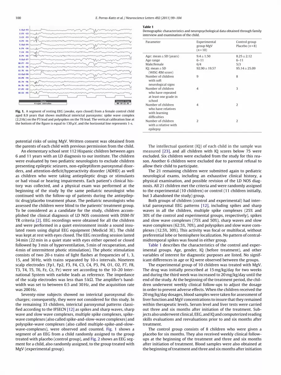
100 E. Porras-Kattz et al. / Neuroscience Letters 492 (2011) 99–104
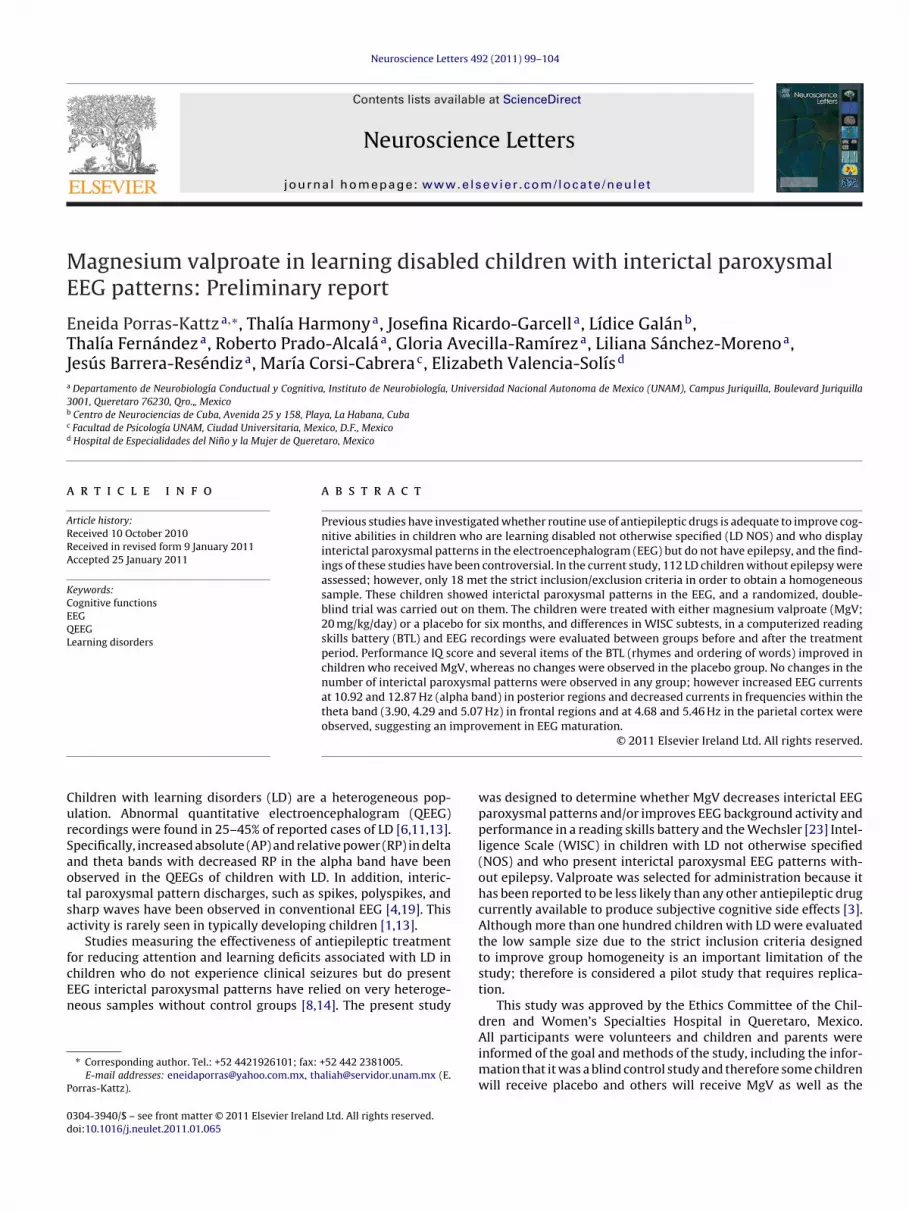
Fig. 1. A segment of resting EEG (awake, eyes closed) from a female control childaged 8.9 years that shows multifocal interictal paroxysms: spike wave complex(t
pt
6wpdaotbctaTpTalw3f4c1sTnoww
ctfiwwpwstmM
Table 1Demographic characteristics and neuropsychological data obtained through familyinterview and examination of the child.
Parameter Experimentalgroup MgV(n = 10)
Control groupPlacebo (n = 8)
Age: mean ± SD (years) 9.4 ± 1.50 8.25 ± 2.12Age range 6–11 6–11Male/female 6/4 5/3IQ: mean ± SD
(WISC-RM score)92.90 ± 19.57 95.14 ± 25.09
Number of childrenwith softneurological signs
9 8
Number of childrenwho have repeatedat least one grade inschool
4 3
Number of childrenwho have relativeswith learningdifficulties
4 2
Number of children 2 2
placebo for six months. They also received weekly clinical follow-
2.2 Hz) on the P3 lead and polyspikes on the T6 lead. The vertical calibration line athe bottom of the figure is equal to 130 �V, and the horizontal line represents 1-s.
otential risks of using MgV. Written consent was obtained fromhe parents of each child with previous permission from the child.
An elementary school sent 112 Hispanic children between agesand 11 years with an LD diagnosis to our institute. The childrenere evaluated by two pediatric neurologists to exclude childrenresenting epileptic seizures, non-epileptiform paroxysmal disor-ers, and attention-deficit/hyperactivity disorder (ADHD) as wells children who were taking antiepileptic drugs or stimulantsr had visual or hearing impairments. Each patient’s clinical his-ory was collected, and a physical exam was performed at theeginning of the study by the same pediatric neurologist whoontinued with the follow-up of patients during the antiepilep-ic drug/placebo treatment phase. The pediatric neurologists whossessed the children were blind to the patients’ treatment group.o be considered as a candidate for the study, children accom-lished the clinical diagnosis of LD NOS consistent with DSM-IVR criteria [2]. EEG recordings were obtained for all the childrennd were performed in a quiet environment inside a sound insu-ated room using digital EEG equipment (Medicid 3E). The child
as kept at rest with eyes closed. The EEG recording session lasted4 min (22 min in a quiet state with eyes either opened or closedollowed by 3 min of hyperventilation, 5 min of recuperation, andmin of intermittent photic stimulation). The photic stimulationonsists of two 20-s trains of light flashes at frequencies of 1, 3,5, and 30 Hz, with trains separated by 10-s intervals. Nineteencalp electrodes (Fp1, Fp2, F3, F4, C3, C4, P3, P4, O1, O2, F7, F8,3, T4, T5, T6, Fz, Cz, Pz) were set according to the 10–20 Inter-ational System with earlobe leads as reference. The impedancef the scalp electrodes was less than 5 k�. The amplifier’s band-idth was set to between 0.5 and 30 Hz, and the acquisition rateas 200 Hz.
Seventy-nine subjects showed no interictal paroxysmal dis-harges; consequently, they were not considered for this study. Inhe remaining 33 children, interictal paroxysmal patterns classi-ed according to the IFSECN [12] as spikes and sharp waves, sharpave and slow wave complexes, multiple spike complexes, spike-ave complexes (also called spike-and-slow-wave complexes) andolyspike-wave complexes (also called multiple-spike-and-slow-ave-complexes), were observed and counted. Fig. 1 shows a
egment of an EEG from a child randomly assigned to the group
reated with placebo (control group), and Fig. 2 shows an EEG seg-ent for a child, also randomly assigned, to the group treated withgV (experimental group).
with a relative withepilepsy
The intellectual quotient (IQ) of each child in the sample wasmeasured [23], and all children with IQ scores below 75 wereexcluded. Six children were excluded from the study for this rea-son. Another 6 children were excluded due to parental refusal toallow their child to participate.
The 21 remaining children were submitted again to pediatricneurological exams, including an exhaustive clinical history, aphysical examination, and possible revision of the LD NOS diag-nosis. All 21 children met the criteria and were randomly assignedto the experimental (10 children) or control (11 children initially,but 3 abandoned the study) group.
Both groups of children (control and experimental) had inter-ictal paroxysmal EEG patterns [12], including spikes and sharpwaves in all the children, multiple spike complexes (25% and30% of the control and experimental groups, respectively), spikesand slow wave complexes (75% and 50%), sharp waves and slowwave complexes (62.5%, 70%), and polyspikes and slow wave com-plexes (12.5%, 30%). This activity was focal or multifocal, withoutpreferential lobe or hemisphere localization. No pattern of central-midtemporal spikes was found in either group.
Table 1 describes the characteristics of the control and exper-imental groups. Age, gender, IQ (before treatment), and othervariables of interest for diagnostic purposes are listed. No signif-icant differences in age or IQ were observed between the groups.
The experimental group of 10 children was treated with MgV.The drug was initially prescribed at 15 mg/kg/day for two weeksand during the third week was increased to 20 mg/kg/day until theend of the study. At the beginning of the treatment period, the chil-dren underwent weekly clinical follow-ups to adjust the dosagein order to prevent adverse effects. When the children received the20 mg/kg/day dosages, blood samples were taken for assessment ofliver function and MgV concentrations to insure that they remainedwithin therapeutic levels. Serum level and liver tests were carriedout three and six months after initiation of the treatment. Sub-jects also underwent clinical, EEG, and IQ and computerized readingskills evaluations and reevaluations prior to and six months aftertreatment.
The control group consists of 8 children who were given a
ups at the beginning of the treatment and three and six monthsafter initiation of treatment. Blood samples were also obtained atthe beginning of treatment and three and six months after initiation

E. Porras-Kattz et al. / Neuroscience Letters 492 (2011) 99–104 101
F m a mc on lin1
oup
bfiiFlbar[I
rfmccwpqc
TS
ig. 2. A segment of resting EEG (eyes closed, second minute of hyperventilation) froomplex: on the Fp1 lead (3.2 Hz) and on the T6 lead (2.8 Hz). The vertical calibrati-s.
f the placebo. Subjects underwent clinical, EEG, and cognitive eval-ations and reevaluations prior to and six months after beginninglacebo treatment.
For QEEG analysis, an expert clinical neurophysiologist underlind conditions used visual editing to select twenty-four artifact-ree segments of 2.56 s during clear wakefulness and withoutnterictal paroxysmal patterns. These EEG segments were edited asn the normative database, and analyses were performed off-line.ast Fourier Transforms and the crosspectral matrices were calcu-ated each 0.39 Hz, and the AP and RP in each of four frequencyands (delta [0.5–3.5 Hz], theta [3.6–7.5 Hz], alpha [7.6–12.5 Hz],nd beta [12.6–19 Hz]) were obtained for each referential lead. Theanges of these bands were selected according to normative data21] provided by MEDICID 3E, a digital EEG equipment (Neuronicnc.).
Frequency domain variable resolution electromagnetic tomog-aphy (FD-VARETA) was used to calculate the distributed sourcesrom 0.78 to 19 Hz each 0.78 Hz. This technique was used to esti-
ate the source generators of EEG data, which are restricted toortical gray matter. In topographic maps, 3D color-coded imagesalled brain electromagnetic tomographies (BETs) are generated, inhich the color code reflects the magnitude of the current at eachoint of the grid or voxel. In this manner, BETs for each EEG fre-
uency band were obtained. Complete description of the procedurean be found in [5].(a) The IQ of each subject was evaluated using the WISC-RM scale[23] as well as both verbal and performance scores before and
able 2ignificant differences between groups in cognitive tests.
Scale Median group Differencesand after (p
Experimental Control
Before After Before
IQ 97 108 103Ordering of words (RT, ms) 670 352 400Rhymes (RT, s) 43 33 38
ale 6.4-year-old child in the experimental group that shows a multifocal spike wavee at the bottom of the figure is equal to 110 �V, and the horizontal line represents
six months after initiating MgV or placebo treatment. An inter-val of greater than six months between the two sessions assuredno retesting effects.
(b) Each child was administered a BTL for Hispanic children,designed to evaluate reading and pre-reading skills across eightdifferent tasks [17]. Tasks included comprehension, picturenaming, sentence completion, ordering of words into syntacti-cally correct sentences, phonological categorization (rhymes),and oral reading of texts, words, and pseudowords. Reac-tion times (RT) in milliseconds for both correct and incorrectanswers were measured.
Because the BTL and WISC-RM variable values did not followa normal distribution and because the sample size was small, theWilcoxon Test for intra-group comparisons and the Mann–WhitneyU Test for inter-group analysis were selected for data analysis. Bothwithin group (before vs. after treatment or placebo) and betweengroup (differences in after–before values were used to compare thechanges observed following MgV or placebo) comparisons wereanalysed.
Differences in number of EEG interictal paroxysmal patterns andEEG current sources obtained by FdVARETA for after–before treat-ment for each group were assessed by multivariate nonparametric
permutation tests [10].As previously described, all children included in this study hadinterictal paroxysmal patterns [12]. For each patient, the averagenumber of discharges was determined before and after treat-ment by dividing the number of discharges recorded by the total
between beforevalue)
Differences between groups ofthe difference before–after
After Exp Control
98.5 0.02 0.23 U = 12.00, p = 0.01420 0.001 0.82 U = 12.00, p = 0.01
42 0.01 0.62 U = 18.00, p = 0.05

102 E. Porras-Kattz et al. / Neuroscience Letters 492 (2011) 99–104
Fig. 3. FdVARETA of the experimental group. Both figures show probabilistic maps of the differences between before- and after-treatment. The scales show probability, withb ifferend quenc( rred to
lntgeSa
bstca
lue color indicating very significant differences. The upper figure corresponds to decreases. The lower figure corresponds to the differences at 10.92 Hz, and at this freFor interpretation of the references to color in this figure legend, the reader is refe
ength of the EEG recording. The two groups had a similar averageumber of interictal paroxysmal patterns at the beginning of thereatment (p > 0.36). No statistical differences were found withinroups between the beginning and the end of the treatment in thexperimental [before: (mean = 9.80; SD = 9.15), after: (mean = 8.89;D = 8.23) for p > 0.57] and control [before: (mean = 7.16; SD = 2.85),fter: (mean = 7.85; SD = 2.57) for p > 0.68] groups.
Z scores were not significantly different for AP and RP between
efore–after and after–before for any group. However, currentources showed significant decreases in magnitude (p < 0.001) inhe experimental group after treatment. Specifically, the frequen-ies that showed significant decreases at theta band were 3.90, 4.29nd 5.07 Hz in the frontal regions and at 4.68 and 5.46 Hz in the pari-ces at 3.90 Hz, and at this frequency, the left prefrontal region exhibited significanty the right occipito-temporal region, exhibited significant increases after treatment.
the web version of this article).
etal cortex. Also, significant increases (p < 0.001) were observed atalpha band (10.92 and 12.87 Hz) in posterior temporal and occipitalregions in the experimental group. In contrast, the control groupshowed no significant changes after receiving the placebo. Fig. 3shows the statistical parametric maps (probabilistic maps) of theafter–before treatment differences in the experimental group.
Prior to the MgV or placebo administration, no significant dif-ferences were observed between groups for any item. Comparisons
between before and after MgV treatment or placebo were onlysignificant in the experimental group (Table 2). Furthermore, com-parison of the after–before treatment difference between groupsrevealed significantly more improvement after treatment in theexperimental group than in the control group for performance
science
Itoc
tb
ttptwi
ohocppitwrstwoTqEoEttswo
imgw
aitbtsTitwfpri
accdat
[
[
[
[
[
[
[
E. Porras-Kattz et al. / Neuro
Q score (U = 12.000; Z = 2.487; p = 0.012) and information item ofhe verbal IQ score (U = 14.500; Z = −2.265; p = 0.023). Finally, webserved no significant differences in the within or between groupomparisons for the remaining items.
We found no significant differences in the non-parametric mul-ivariate permutation tests between groups with the BTL variablesefore MgV or placebo.
Comparison of the after–before difference values between thewo groups showed significantly greater improvement of reactionime in the experimental group in rhyming (U = 18.000; Z = −1.954;= 0.050) and ordering of words (U = 12.000; Z = −2.487; p = 0.012)
han in the control group (Table 2). No significant differences in theithin or between group comparisons were found for the remain-
ng items.This study is the first randomized, double-blind trial carried
ut with MgV or placebo in children diagnosed with LD NOS whoave EEG interictal paroxysmal patterns without epilepsy. Previ-us studies have attempted to determine the benefits of treatinghildren with LD or behavioral problems and interictal paroxysmalatterns in the EEG. Ronen and colleagues [18] evaluated cognitiveerformance and behavior in children with learning and behav-
oral problems associated with interictal paroxysmal patterns inhe EEG but without clinical seizures. These children were treatedith sodium valproate (VPA), and no clinical improvement was
eported. Therefore, the authors concluded that the data did notupport the use of VPA for this patient group. Importantly, however,he sample was a very heterogeneous group of patients. In otherords, some of the patients were diagnosed with pervasive devel-
pmental disorders and others had a clinical history of seizures.hus, the number of patients presenting with LD exclusively wasuite small, and these patients were not studied independently.tchepareborda [8] reported that neuropharmacological treatmentf patients with neurodevelopmental disorders and an abnormalEG pattern but no seizures was favorable in 80% of cases. Patientsreated by Etchepareborda received carbamazepine or MgV, buthe study did not include a control group. Other clinical trials withingle patients presenting ADHD associated with EEG focal sharpave discharges have reported a cognitive enhancing effect of VPA
r carbamazepine [16].Even though LD NOS criteria persisted at the end of the study
n all the evaluated children, our results showed greater improve-ents in performance in the experimental group than in the control
roup for Performance IQ score in the WISC-RM, RT in rhymes, andord ordering in the BTL.
Anti-epileptic drugs (AEDs) have a variety of mechanisms ofction which are reflected through different anticonvulsant activ-ties and behavioral effects. Even though mechanisms of action ofhe AEDs are only partially known, and behavioral effects coulde exerted by an unknown mechanism of action, MgV has beenypified as an attenuation of glutamate excitatory neurotransmis-ion agent with psychotropic profile and activating effects [7].he results in this study support the possible usefulness of MgVn treating children with LD NOS who present with EEG interic-al paroxysmal patterns but not epilepsy. However, no changesere observed in the number of paroxysmal discharges in the EEG
ollowing a six-month treatment period. Current treatments foratients with epilepsy are recommended for several years; thus theesults might be due to the short period of antiepileptic treatmentn our study.
By contrast, source EEG analysis showed significant changesfter treatment in the experimental group but no changes in the
ontrol group. These changes included both increases in frequen-ies within the alpha band in posterior regions as well as currentecreases at frequencies within the theta band in the left frontalnd parietal cortices, and these findings support the notion thathe EEG was more mature after treatment [6,11,13]. These results[
[
Letters 492 (2011) 99–104 103
may be interpreted as an improvement in background activity asso-ciated with improved cognitive performance [9,20,22]. An increaseat 10.92 and 12.87 Hz in right parietal and occipital areas may berelated to enhancement in attention and visual perception, as wellas in semantic memory processes, as a previous study have found[15].
An important limitation of the current study is the small samplesize for both groups. Therefore, this investigation can be considereda pilot study that requires replication.
Although this study requires replication with a larger sample,the results suggest that MgV may be useful in the treatment ofchildren with LD NOS who present interictal paroxysmal patternson the EEG.
Conflict of interest
The authors have no conflict of interest.
Acknowledgments
Partial support was provided by the UNAM program PAPIITIN209407. The authors acknowledge Héctor Belmont and RosaMaría Hernández for technical assistance.
References
[1] A. Alvarez, M.C. Pérez-Avalo, L. Morenza, Neuropsychological assessment oflearning-disorder children with EEG epileptiform discharges, New Issues Neu-rosci. 4 (1992) 40–75.
[2] American Psychiatric Association, Diagnostic and Statistical Manual of MentalDisorders, in: Text Revision (DSM-IV TR), fourth ed., Amazon Visa, Washington,D.C., 2000.
[3] H. Arif, R. Buchsbaum, D. Weintraub, J. Pierro, S.R. Resor Jr., L.J. Hirsch,Patient-reported cognitive side effects of antiepileptic drugs: predictors andcomparison of all commonly used antiepileptic drugs, Epilepsy Behav. 14(2009) 202–209.
[4] J. Becker, M. Velasco, T. Harmony, E. Marosi, A.M. Landázuri, Electroen-cephalographic characteristics of children with learning disabilities, Clin.Electroencephalogr. 18 (1987) 93–101.
[5] J. Bosch-Bayard, P. Valdes-Sosa, T. Virues-Alba, E. Aubert-Vázquez, E.R. John,T. Harmony, J. Riera-Díaz, N. Trujillo-Barreto, 3D statistical parametric map-ping of EEG source spectra by means of variable resolution electromagnetictomography (VARETA), Clin. Electroencephalogr. 32 (2001) 47–61.
[6] D.S. Cantor, R. Chabot, QEEG studies in the assessment and treatment of child-hood disorders, Clin. EEG Neurosci. 40 (2009) 113–121.
[7] A.E. Cavanna, F. Ali, H.E. Rickards, D. McCorry, Behavioral and cognitive effectsof anti-epileptic drugs, Discov. Med. 35 (2010) 138–144.
[8] M.C. Etchepareborda, Tratamiento de los ninos con electroencefalogramaparoxístico sin crisis, Revista Neurol. 37 (2003) 293–297.
[9] T. Fernández, T. Harmony, J. Silva, L. Galán, L. Díaz-Comas, J. Bosch, M. Rodríguez,A. Fernández-Bouzas, G. Yánez, G. Otero, E. Marosi, Relationship of specific EEGfrequencies at specific brain areas with performance, Neuroreport 9 (1998)3681–3687.
10] L. Galán, R. Biscay, J.L. Rodríguez, M.C. Pérez-Abalo, R. Rodríguez, Testingtopographic differences between event related brain potentials by usingnonparametric combinations of permutation test, Electroencephalogr. Clin.Neurophysiol. 102 (1997) 240–247.
11] T. Harmony, G. Hinojosa, E. Marosi, J. Becker, Correlation between EEG spectralparameters and an educational evaluation, Int. J. Neurosci. 54 (1990) 147–155.
12] IFSECN, A glossary of terms commonly used by clinical electroencephalogra-phers, Electroencephalogr. Clin. Neurophysiol. 37 (1974) 538–548.
13] E.R. John, L. Prichep, H. Ahn, P. Easton, J. Fridman, H. Kaye, Neurometric eval-uation of cognitive dysfunctions and neurological disorders in children, Prog.Neurobiol. 21 (1983) 239–290.
14] D.G. Kasteleijn-Nolst Trenité, Transient cognitive impairment during subclini-cal epileptiform electroencephalographic discharges, Semin. Pediatr. Neurol. 2(1995) 246–253.
15] W. Klimesch, M. Doppelmayr, D. Russegger, T. Pachinger, J. Schwaiger, Inducedalpha band power changes in the human EEG and attention, Neurosci. Lett. 244(1998) 73–76.
16] N. Laporte, G. Sébire, Y. Gillerto, R. Guerrini, S. Ghariani, Cognitive epilepsy:ADHD related to focal EEG discharges, Pediatr. Neurol. 27 (2002) 307–311.
17] V. Regiosa, M.C. Pérez, M. Manzano, M. Antelo, Sistema automatizado paraexplorar la lectura en escolares de habla hispana, Revista Latina de Pensamientoy Lenguaje 2 (1994) 141–160.
18] G.M. Ronen, J.E. Richards, C. Cunningham, M. Secord, D. Rosenbloom, Cansodium valproate improve learning in children with epileptiform bursts butwithout clinical seizures? Dev. Med. Child Neurol. 42 (2000) 751–755.

1 scienc
[
[
[
04 E. Porras-Kattz et al. / Neuro
19] A.L. Rugland, Sub-clinical epileptogenic activity, in: M. Sillanpaa, S.I. Johan-nessen, G. Blennow, M. Dam (Eds.), Paediatric Epilepsy, Wrightson, 1990.
20] W.W. Surwillo, The relation of simple response time to brain-wave frequencyand effects of age, Electroenceph. Clin. Neurophysiol. 15 (1963) 105–114.
21] P. Valdés, R. Biscay, L. Galán, J. Bosch, S. Zsava, T. Virués, High resolution spectralEEG norms topography, Brain Topogr. 3 (1990) 281–282.
[
[
e Letters 492 (2011) 99–104
22] D. Valentino, J.E. Arruda, S.M. Gold, Comparison of and response accuracy ingood vs. poorer performers during a vigilance task, Int. J. Psychophysiol. 15(1993) 123–133.
23] D. Wechsler, M. Gómez Palacios, E.R. Padilla, S. Roll, Manual del WISC-RM escalade inteligencia revisada para el nivel escolar, Ed Manual Moderno, México, D.F.,1984.