hippocampal interictal spikes disrupt cognition in rats
TRANSCRIPT
Hippocampal Interictal Spikes Disrupt Cognition in Rats
Jonathan K. Kleen, B.S.1, Rod C. Scott, M.D., Ph.D.1,2, Gregory L. Holmes, M.D.1, and PierrePascal Lenck-Santini, Ph.D.11Department of Neurology, Neuroscience Center at Dartmouth, Dartmouth Medical School,Hanover, New Hampshire2UCL, Institute of Child Health, London, United Kingdom
AbstractObjective—Cognitive impairment is common in epilepsy, particularly in memory function.Interictal spikes are thought to disrupt cognition, but it is difficult to delineate their contributionfrom general impairments in memory produced by etiology and seizures. We investigated thetransient impact of focal interictal spikes on the hippocampus, a structure crucial for learning andmemory and yet highly prone to interictal spikes in temporal lobe epilepsy.
Methods—Bilateral hippocampal depth electrodes were implanted into fourteen Sprague-Dawleyrats, followed by intrahippocampal pilocarpine or saline infusion unilaterally. Rats that developedchronic spikes were trained in a hippocampal-dependent operant behavior task, delayed-match-to-sample. Depth EEG was recorded during 5,562 trials among five rats, and within-subject analysesevaluated the impact of hippocampal spikes on short-term memory operations.
Results—Hippocampal spikes that occurred during memory retrieval strongly impairedperformance (p<0.001). However, spikes that occurred during memory encoding or memorymaintenance did not affect performance in those trials. Hippocampal spikes also affected responselatency, adding approximately 0.48 seconds to the time taken to respond (p<0.001).
Interpretation—We found that focal interictal spike-related interference in cognition extends tostructures in the limbic system, which required intrahippocampal recordings. Hippocampal spikesseem most harmful if they occur when hippocampal function is critical, extending human studiesshowing that cortical spikes are most disruptive during active cortical functioning. The cumulativeeffects of spikes could therefore impact general cognitive functioning. These results strengthen theargument that suppression of interictal spikes may improve memory and cognitive performance inpatients with epilepsy.
IntroductionOne of the most common yet troublesome co-morbidities in patients with temporal lobeepilepsy (TLE) is cognitive impairment 1-3. In particular, many patients have disturbances inmemory, an essential higher cognitive function for continuity in time, personal history, andawareness. While both etiology 4-5 and recurrent seizures 6 have been implicated, animportant controversy is whether interictal spikes (IIS) are a harmless biomarker of thedisease or contribute to this dysfunction.
IIS are transient, abnormal focal neural discharges seen on electroencephalogram (EEG)recordings during periods between seizures. Lasting 50-200 milliseconds, they are a result ofsynchronous, paroxysmal depolarizations of neurons producing a rapid succession of action
Correspondence to: Gregory L. Holmes Department of Neurology Neuroscience Center at Dartmouth Dartmouth Medical School OneMedical Center Drive Lebanon, New Hampshire 03756 Telephone: 603-650-4211 [email protected] .
NIH Public AccessAuthor ManuscriptAnn Neurol. Author manuscript; available in PMC 2011 February 1.
Published in final edited form as:Ann Neurol. 2010 February ; 67(2): 250–257. doi:10.1002/ana.21896.
NIH
-PA Author Manuscript
NIH
-PA Author Manuscript
NIH
-PA Author Manuscript

potentials 7-8. IIS usually occur close to the seizure focus 9-10 and they are one of the mostimportant factors in the diagnosis of epilepsy 11-12. However, IIS may interfere withcognitive functions that are already jeopardized by disease, which would be particularlytroublesome given the prominence of focal IIS in epileptic syndromes such as temporal lobeepilepsy and the epileptic encephalopathies 13.
The effect of IIS on cognition is likely ephemeral, coinciding with timing of the event on theEEG. The clinical term transitory cognitive impairment has been used to describe briefdisruptions in brain function concurrent with IIS 14. This term encompasses impairmentsobserved with both focal IIS and generalized spike-and-wave complexes. The latter haveproduced unquestionable demonstrations of transitory cognitive impairment, includingimpairments in reaction time 15-17, perception 16-19, verbal and spatial tasks 20-22, and evendriving behavior 23. Furthermore, some studies suggest that the neuroanatomical site andtiming of the EEG disturbance predicts the specific cognitive function affected. Forexample, disturbances localized to the occipital cortex briefly disrupt visual perception,particularly during a stimulus presentation 17, 19.
Focal IIS-induced cognitive disruption has been more difficult to identify than thatassociated with generalized spike-and-wave. This may be due to the use of scalp EEG in allstudies of transient IIS effects thus far. Scalp EEG does not allow precise neuroanatomicallocalization of IIS, which weakens the determination of which cognitive function might beaffected. Furthermore, non-cortical focal IIS, such as those in the hippocampus or other deepmedial temporal lobe structures, are not effectively detected by scalp EEG 24. This presentsproblems in testing for effects of IIS on learning and memory, for which these structures arecritical. Hence, mixed results in previous studies may be related to inconsistencies betweenIIS localization and the cognitive function tested.
Depth electrode studies can help to resolve these issues, through visualization of focal IIS insubcortical structures that play a central role in memory. However, patients with intracranialelectrodes present other difficulties for extended neuropsychological testing, mainly due topost-surgical discomfort and cognitive side effects of pain medication. We investigated theeffects of IIS on operant behavior, a particularly useful tool to examine IIS in an animalmodel which enables rigorous assessment of a fleeting and potentially subtle disruption.Pilocarpine-treated rats with chronic IIS were trained to perform a delayed-match-to-sampletask (DMTS), which relies largely on intact hippocampal function 25, and involves short-term memory encoding, maintenance, and retrieval. We used within-subject analysis tocompare trials with IIS versus trials without IIS from the same animal, isolating the specificeffect of IIS on cognition from differences that might otherwise vary between animals.
While cortical IIS have been shown to disrupt stimulus perception for later recall 19, patientswith TLE are not particularly prone to problems in perception 26, despite high incidence ofIIS in temporal lobe structures including the hippocampus 27. However, neuronal firing andneurophysiological rhythms in the hippocampus are closely related to short-term memory,particularly the retrieval phase, and may be vulnerable to the electrophysiological impact ofhippocampal IIS 28-30. Therefore we hypothesized that focal IIS in the hippocampusmomentarily disrupt short-term memory, specifically short-term memory retrieval.
We report that IIS in the hippocampus are associated with a transient disruption in functionand an increase in response latency. This evidence illustrates that focal IIS in limbicstructures, which are very common in epileptic conditions such as TLE, can provide anindependent contribution to cognitive impairments.
Kleen et al. Page 2
Ann Neurol. Author manuscript; available in PMC 2011 February 1.
NIH
-PA Author Manuscript
NIH
-PA Author Manuscript
NIH
-PA Author Manuscript

MethodsAnimals
All animal procedures were approved by the Dartmouth IACUC, under USDA andAAALAC-approved conditions, in accordance with National Institutes of Health guidelinesFourteen Adult male Sprague-Dawley rats, approximately eight weeks old, were housedindividually with a 12-hour light/dark cycle and ad libitum access to food and water. All ratsunderwent surgical implantations and infusion with either pilocarpine or saline, and werelater recorded to screen for IIS. Specifically, nine rats were implanted and infused withpilocarpine at least two weeks before behavioral training began, while two rats were trainedbefore surgery and pilocarpine infusion. Within-subject analyses accounted for anyindividual differences between rats such as the timing of training, therefore all behavioraldata was pooled together in the analysis (see Data Analysis section).
Three control rats were implanted and infused with saline at least two weeks before trainingto provide a comparison to pathological EEG phenomena in pilocarpine-treated rats, and toverify that the surgical and infusion techniques alone did not produce IIS. Control rats werenot involved in the behavioral analysis since this study aimed to use within-subjectcomparisons to assess the independent impact of IIS, which were not exhibited at any timefollowing a saline infusion. Female rats were not utilized in this study to prevent thepotential effects of fluctuating estrous cycle hormones on behavior and cognition31.
SurgeryRats were anesthetized with isoflurane (2-3% in oxygen) and custom electrodes wereimplanted in three sites 32 in each rat: bilateral ventral hippocampal CA1 (±5.7 mm lateral,5.4 mm posterior, and 6.0 mm ventral from the bregma skull fissure; Fig 1A), and prelimbicprefrontal cortex (0.6 mm lateral, 3.2 mm anterior, and 4.3 mm ventral). A 26-gauge guidecannula (Plastics One, Roanoke, VA) was bonded to the right ventral CA1 bipolar electrodeto allow close proximity (<1 mm) of EEG recording to the infusion focus. A ground wirewas soldered to a bone screw, and a reference wire was implanted in the cerebellum. Allwires were plugged into an interconnect socket array (Mill-Max Mfg. Corp., Oyster Bay,NY), and encased in dental cement (Dentsply International Inc., Milford, DE), leaving onlythe gold pin sockets exposed to allow connection to the amplifiers (see Electrophysiologysection).
Pilocarpine infusionOne week following surgery, pilocarpine dissolved in 0.9% saline (0.5 mg/μl; Sigma-Aldrich, St. Louis, MO) was infused into the hippocampus at a rate of 0.05 μl/minute withconcurrent depth-EEG recordings. To minimize mortality while ensuring IIS occurrence,pilocarpine infusions were terminated for a given animal when continuous EEG statusepilepticus (SE) was observed in the hippocampus. The amount of solution infused variedfrom 0.9 μL to 2.3 μl (0.45-1.15 mg pilocarpine). Importantly, individual differences ininfusion amount were accounted for by the within-subject design of the analysis (see DataAnalysis section). Control rats received an infusion of 1-2 μl of 0.9% saline, to provide laterqualitative comparison of their long-term EEG activity to that of pilocarpine-treated rats.Rats were monitored on EEG for one half hour after SE began, and behavior was monitoredfor another 12 hours. To assess IIS development, 30-60 minute EEG recordings were madeeach day after the infusion for two weeks, and once per week for an additional 3 weeks.
ElectrophysiologyElectrodes consisted of two insulated 25 μm nichrome wires (California Fine Wire, GroverBeach, CA), twisted and inserted into a 25-gauge stainless steel guide tube (Small Parts Inc.,
Kleen et al. Page 3
Ann Neurol. Author manuscript; available in PMC 2011 February 1.
NIH
-PA Author Manuscript
NIH
-PA Author Manuscript
NIH
-PA Author Manuscript

Miami Lakes, FL). Operational amplifiers plugged directly to the rat connector to reducemechanical artifacts. EEG signals, transmitted via a custom cable and a rotating commutator(Dragonfly Research & Development Inc., Ridgeley, WV), were amplified (10K) andacquired at 2.2kHz with an Axoscope analog/digital converter and software acquisition set-up (DigiData 1322, Axon Instruments, Foster City, CA).
ApparatusTwo operant conditioning chambers contained in sound-attenuating cubicles (LafayetteInstruments Inc., Lafayette, IN; Med Associates Inc., St. Albans, VT) were controlled withbehavior software packages (ABET II; MED-PC IV). Two retractable levers were separatedby a pellet dispenser on one of the walls. A stimulus light was located above each lever. Aninfrared photobeam nosepoke detector was placed on the opposite wall, with a stimulus lightabove it. A white incandescent house light bulb was illuminated during each trial andextinguished at its end. Three DC outputs from the behavioral software computer interfacewere sent directly to the EEG data acquisition system to additional input ports. Stimulus andresponse events were recorded as binary (on/off) combinations of these signals, enablingmicrosecond synchronization with EEG data.
ProcedureRats were food-restricted and maintained at 85% of their ad libitum weight throughoutoperant behavior study. They were trained in operant chambers to lever-press and poke theirnose into the infrared beam for food reward (45 mg Noyes food pellet; Research Diets,Incorporated, New Brunswick, NJ). Training sessions were run at approximately the sametime each day for each rat. Once the rats reliably pressed levers and broke the infrared beamfor food, the DMTS training paradigm commenced (Fig 1B). At the beginning of each trial,one lever (right or left) was randomly presented [Sample step]. Upon pressing the samplelever, rats were then required to poke their nose into an infrared beam [Delay step], whichwas positioned on the opposite wall of the chamber to prevent rats from relying on bodypositioning (non-hippocampal-dependent) strategies to gain reinforcers 33. Immediately afterthe beam was broken, both levers were presented [Match step]. A correct response wasdefined as pressing the same lever that was presented in the Sample Phase (i.e. match-to-sample), which produced a food reward. An incorrect response was pressing the lever thathad not been presented, which produced no food reward. Daily sessions consisted of 60-100trials, and when accuracy reached >85% for 3 continuous days, the delay period wasincreased. This was accomplished by designating nosepoke beam-breaks as unproductiveduring the delay step until a specified amount of time had elapsed. The delay length wasincreased for each rat until performance reached >80% accuracy on sessions with 6-15second variable delays, and EEG data was recorded for all sessions thereafter. Rats with atleast 10 IIS/hour were included in DMTS analysis. Five rats qualified under bothperformance and IIS prerequisites, generating a total of 5,562 trials for analysis.
Following spontaneous seizures, rats tended to return to normal behavioral activity levels5-15 minutes. Previous studies from our laboratory show that post-ictal effects are ratherminimal after three hours, in terms of hippocampal function and related cell firing34.Therefore, if an animal happened to have a spontaneous seizure before a DMTS session, thesession was delayed at least 3 hours to help minimizes influences of post-ictal impairmenton general performance. In addition, the within subject design of our analysis, whichcompared trials with and without IIS in the same session, was designed to negate suchinfluences.
Kleen et al. Page 4
Ann Neurol. Author manuscript; available in PMC 2011 February 1.
NIH
-PA Author Manuscript
NIH
-PA Author Manuscript
NIH
-PA Author Manuscript

Data AnalysisCustom IIS detection software, based on White et al. 35, was written using MATLAB (TheMathworks, Inc., Natick, MA). Briefly, potential IIS were detected each time the derivativeof the EEG recording (re-sampled at 222 Hz) was greater than five times the standarddeviation. Potential waveforms were then classified as IIS, artifacts, or false-detections ofnormal physiological waveforms36 based on comparison EEG sessions from saline-treatedrats (Fig 2C). IIS differentiation was based on morphology, by plotting the characteristicfeatures of the waveforms against one another (e.g. amplitude). IIS included in the analysishad a peak amplitude of at least 1 mV, and lasted less than 50 ms (examples in Fig 2B), withthe entire transient lasting less than 200 ms if including the subsequent slow wavecomponent. We evaluated the effectiveness of our IIS detection programs by comparingtheir performance to the evaluation of an experienced epileptologist (GLH) in threerandomly chosen sessions. Comparison of manually detected IIS to those detected by theprogram revealed a sensitivity of >95% and a specificity of >99%. Using the binary inputsignals from the behavioral software, the exact timings of IIS within trials were documentedrelative to behavioral events, to designate each trial as one with or without an IIS (Fig 1Cand D).
DMTS performance as a function of delay length and timing of IIS occurrence was analyzedusing logistic regression in Stata 10 (StataCorp LP, College Station, TX). Each DMTS trialwas treated as an individual observation and standard errors were adjusted for within animaleffects (N=5,562, clustered for five rats), to control for differences between animals such aspilocarpine dose, lesion extent, and general cognitive ability. Three stages of short-termmemory processing were inferred in the DMTS task: encoding (4 seconds surrounding thesample press), maintenance (2 seconds after the sample press to 1 second before the matchlever presentation), and retrieval (1 second before the match lever presentation to 1 secondbefore it was pressed). The timing of these epochs was based on rat behavior in short-termmemory tasks 29 and the pronounced effects in neuronal firing produced in the first secondfollowing a hippocampal IIS 30. Additionally, Pearson correlation coefficients were used toassess the timing of IIS within trials versus trial delay lengths, as well as test for anyrelationships between IIS frequency and general performance across sessions (defined aspercent correct in a given session).
The effect of hippocampal IIS on response latency was assessed by quantifying the amountof time taken by the rat to press one of the match levers, following their extension andstimulus light illumination after the last nosepoke. This length of time was compared intrials in which no IIS occurred versus trials in which an IIS occurred during this period,using a Mann-Whitney U test.
Hippocampal theta oscillations have been related to greater vigilance during behavioraltasks, and would likely associate with improved performance 28. Likewise, IIS occur largelyin the absence of hippocampal theta rhythm, i.e. during non-attentive states 37. To rule outthe possibility that vigilance could account for effects of IIS on DMTS performance, ameasure of vigilance ratio was calculated (sum of theta band (4-11Hz) power divided by thesum of delta band (0.5-3Hz) power)38-41, and compared between trials with versus withoutIIS, using a Mann-Whitney U test.
ResultsSeizures and IIS
During intrahippocampal pilocarpine infusion, all rats exhibited electrographic seizuresinitiating in the ipsilateral hippocampus and eventually generalizing to both the contralateralhippocampus and the ipsilateral prefrontal cortex leads. The initial SE lasted from 3-7 hours
Kleen et al. Page 5
Ann Neurol. Author manuscript; available in PMC 2011 February 1.
NIH
-PA Author Manuscript
NIH
-PA Author Manuscript
NIH
-PA Author Manuscript

for all rats, corresponding to Stage III-V behavioral seizures 42. In the subsequent weeksfollowing infusion, seven rats had witnessed spontaneous seizures. Because long-termmonitoring was not performed, we could not verify the presence of spontaneous seizures inthe other animals with IIS. It should be noted that our use of the term “interictal” whenreferring to IIS is based on the pathological waveform characteristics (deCurtis & Avanzini,2001), and not the apparition of previous and subsequent seizures. IIS developed within thefirst few days following infusion, subsequently increasing and reaching stable rates afterapproximately two weeks. IIS in the left hippocampus (contralateral to infusion site) tendedto be more frequent than right hippocampal IIS in the first two weeks (Fig 2A), whileprefrontal cortex IIS were seen in only one rat. Ten of eleven pilocarpine treated ratsdeveloped hippocampal IIS following pilocarpine infusion, ranging from 0.5 to 256.8 IIS perhour. IIS morphology and lateralization was variable between rats as illustrated in Figure 2Band C, although these features did not predict the degree of impairment induced. IISincluded in the analysis were distinct from normal physiological waveforms seen in bothpilocarpine- and saline-treated rats (Fig 2C). Electrode placements were verified in themajority of rats (N=9) to verify our surgical procedures, using thionin staining techniques ofcoronal sections43. Electrodes were found to be in the hippocampus, in or very near the CA1sub-region (Figure 1A). Although there was some variability in the amount of pilocarpine-induced cell loss, the hilus and CA1 appeared most affected by the pilocarpine. As shown inFigure 1A, cell loss tended to be greater ipsilateral to the injection site.
IIS-related impairmentIncreasing delays produced overall decreases in DMTS accuracy (odds ratio (OR) – 1.08;95% CI – 1.03 to 1.14, p<0.01), confirming findings by Hampson and colleagues 25.Furthermore, IIS were associated with additional impairments in performance in a trial-specific or transient manner (Fig 3). IIS that occurred during the retrieval epoch of trialswere related to markedly impaired performance, with rats more than 3 times as likely tomake an error in those trials (OR – 3.19; 95% CI – 2.34 to 4.35, p<0.001). However, IISduring the encoding epoch (OR - 0.97; 95% CI - 0.59 to 1.59, p=0.91) or the maintenanceepoch were not associated with impaired performance (OR – 1.06; 95% CI - 0.85 to 1.33,p=0.61). The number of IIS in a trial was not a significant predictor of accuracy, althoughthis was likely due to a statistical paucity of trials with multiple IIS. There was a greaterlikelihood of IIS occurring in longer delays rather than shorter delays (p<0.001), but therewas no relationship between the length of the delay and the epoch within which IIShappened (r=0.003, p=0.92). Vigilance ratio (see Methods) did not differ in trials with andwithout IIS (p=0.96). Finally, we found no relationships between the frequency of IIS andoverall accuracy across sessions (r=0.02, p=0.78), nor between the frequency of IIS and theaverage performances of individual rats (r=0.24, p=0.69).
Response latencyThe median response latency to press the match lever after it was extended into the chamberwas 2.28 seconds in trials without IIS, whereas if an IIS occurred anywhere during thisperiod, the median increased to 2.76 seconds (p<0.001; Fig 4). This addition in reaction timedid not differ between correct or incorrect trials.
DiscussionWe demonstrate that hippocampal IIS are associated with a dramatic alteration in on bothaccuracy and response latency in a rodent model. We were able to show this effect due toour use of depth electrodes, which have not previously been used to study the transientimpact of IIS. Therefore, the transitory effects from cortical IIS described previously have acorollary in a deep brain structure. Furthermore, transient impairments in short-term
Kleen et al. Page 6
Ann Neurol. Author manuscript; available in PMC 2011 February 1.
NIH
-PA Author Manuscript
NIH
-PA Author Manuscript
NIH
-PA Author Manuscript

memory were related to focal IIS in the hippocampus, illustrating that a more generalizeddischarge is not necessary to disrupt this cognitive function.
The hippocampus is required for accurate performance of the DMTS task, particularly intrials with long delays 25. Using this cognitive paradigm, we found that the effect of depth-detected hippocampal IIS is contrary to the effect of scalp-detected IIS 19. Specifically,hippocampal IIS had no effect on accuracy if they occurred during the encoding epoch(analogous to stimulus presentation in human neuropsychological studies), or during themaintenance epoch. However, hippocampal IIS during the memory retrieval epoch wereassociated with severe transient impairments, probably because of the intimate role of thehippocampus in this function 28-29. These selective effects reveal that hippocampaloperations may be devastated if hippocampal IIS occur during their neural processingwindows.
The specific effect of IIS on retrieval but not encoding or maintenance may also relate to thetransient nature of the hippocampal process involved in memory recall29. Memory encodingand maintenance may involve a longer time-span of processing, and may be buffered bystructures in addition to the hippocampus 44, and thus less vulnerable to a transient focaldisruption. Future studies will be required to delineate the mechanism of the process-specificimpact on hippocampal function, potentially utilizing multi-structure depth recordings.
Response latency was increased in trials with hippocampal IIS by approximately half asecond. These additions are a fraction of the total latency to respond, and might be easilyoverlooked in a clinical context. However, they may be cumulatively detrimental to patients,particularly those with frequent IIS 17. Similar increases in response latency were seen inboth correct and incorrect trials, suggesting that this additional time does not reflect ahesitancy of the rat due to faulty memory recollection. Rather, in addition to theirdeleterious effect on informational integrity, hippocampal IIS may mark a temporary offlinestate of the brain, manifested by this delay in responding.
IIS-related memory impairments may be a consequence of the incorporation of localneurons into a synchronized burst 45. Drawing neurons away from their normal processingmay disorder the representation of pertinent information within a hippocampal neuronalassembly 46, causing inaccurate recall of which lever was pressed just seconds ago. Inaddition, local changes in cellular environment immediately following an IIS 7 may besufficient to briefly depress neuron firing 30 and local oscillations47, and this delay in brainprocessing could manifest as an increase in response latency.
IIS were observed throughout the first two weeks following pilocarpine infusion andreached relatively stabile levels afterward, illustrating pathophysiological activity during thesupposed “latent period” before the generation of spontaneous seizures48-49. Thedevelopment of IIS contralateral to the infusion site was an unexpected finding, and mayindicate a bilateral epileptogenic process following unilateral intrahippocampal pilocarpineinfusion. The laterality of the IIS did not affect our results. Whether the spike occurred ineither hippocampus, or propagated from one to the other, impairments were still observed.
Correlations between the frequency of IIS and cognitive impairment in patients have beencontroversial, and the clinical significance of such findings has been questioned 13. Forexample, deficits in IQ and school performance among patients with Benign Epilepsy withCentro-Temporal Spikes have been correlated with the frequency of IIS but not seizurefrequency in some but not all studies 50-51. Differentiating the transient effects of IIS fromthe underlying pathological factors is challenging in clinical settings since factors such asanti-epileptic drugs and seizures may contribute to cognitive deficits. IIS rate and generalmeasures of performance were not related in the current study (see also Chauviere et al.,
Kleen et al. Page 7
Ann Neurol. Author manuscript; available in PMC 2011 February 1.
NIH
-PA Author Manuscript
NIH
-PA Author Manuscript
NIH
-PA Author Manuscript

200952). However, we show that the timing of IIS may be decisive in producing adversecognitive effects, since complex functions are often supported by multiple brain structureswith differential time windows of processing 44. Therefore, IIS in other brain structures maybe equally disruptive and clinically evident if both the timing of the IIS, and the putativeneurological substrate of the function tested, are taken into consideration.
While extrapolating results from rats to humans must be done cautiously, our study stronglysuggests that hippocampal IIS produce intermittent hippocampal dysfunction. It is thereforenot unreasonable to be concerned that cumulative influences of transient IIS-relateddisruptions may account for a portion of the cognitive impairment seen in epilepsy 14. Byextension, IIS-related disruptions could be devastating in children with epilepticencephalopathies, in whom high numbers of IIS may systematically obstruct learningfunctions in the brain, hence impeding normal cognitive development 19.
Further studies are required using other tasks that also require an intact hippocampus, sincethis brain area contributes to a multitude of neural processing functions 53. Human studies offocal hippocampal IIS are also warranted, to elucidate the extent of their impact on morecomplex cognitive functions, and to determine whether therapy improves those cognitivefunctions if IIS can be effectively suppressed.
AcknowledgmentsThis work was supported by the National Institutes of Health (5R01NS056170-02 and 1F30NS064624-01). Wethank Gregory Richard and Qian Zhao for assistance.
References1. Helmstaedter, C. Effects of chronic epilepsy on declarative memory systems. In: Thomas, S.; Asla,
P., editors. Progress in Brain Research. Vol. Volume 135. Elsevier; 2002. p. 439-453.2. Hermann BP, Seidenberg M, Schoenfeld J, Davies K. Neuropsychological characteristics of the
syndrome of mesial temporal lobe epilepsy. Arch Neurol. 1997; 54:369–376. [PubMed: 9109737]3. Jokeit H, Ebner A. Effects of chronic epilepsy on intellectual functions. Prog Brain Res. 2002;
135:455–463. [PubMed: 12143364]4. Theodore WH, Bhatia S, Hatta J, et al. Hippocampal atrophy, epilepsy duration, and febrile seizures
in patients with partial seizures. Neurology. 1999; 52:132–136. [PubMed: 9921860]5. Elger CE, Helmstaedter C, Kurthen M. Chronic epilepsy and cognition. Lancet Neurol. 2004;
3:663–672. [PubMed: 15488459]6. Cheung MC, Chan AS, Chan YL, et al. Effects of illness duration on memory processing of patients
with temporal lobe epilepsy. Epilepsia. 2006; 47:1320–1328. [PubMed: 16922876]7. de Curtis M, Avanzini G. Interictal spikes in focal epileptogenesis. Progress in Neurobiology. 2001;
63:541–567. [PubMed: 11164621]8. Prince DA, Connors BW. Mechanisms of interictal epileptogenesis. Adv Neurol. 1986; 44:275–299.
[PubMed: 3518347]9. Blume WT, Kaibara M. Localization of epileptic foci in children. Can J Neurol Sci. 1991; 18:570–
572. [PubMed: 1777871]10. Engel J Jr. A practical guide for routine EEG studies in epilepsy. J Clin Neurophysiol. 1984;
1:109–142. [PubMed: 6443570]11. Barry E, Sussman NM, O’Connor MJ, Harner RN. Presurgical electroencephalographic patterns
and outcome from anterior temporal lobectomy. Arch Neurol. 1992; 49:21–27. [PubMed:1728258]
12. Blume WT. Current trends in electroencephalography. Curr Opin Neurol. 2001; 14:193–197.[PubMed: 11262735]
13. Holmes GL, Lenck-Santini PP. Role of interictal epileptiform abnormalities in cognitiveimpairment. Epilepsy Behav. 2006; 8:504–515. [PubMed: 16540376]
Kleen et al. Page 8
Ann Neurol. Author manuscript; available in PMC 2011 February 1.
NIH
-PA Author Manuscript
NIH
-PA Author Manuscript
NIH
-PA Author Manuscript
14. Binnie CD. Cognitive impairment during epileptiform discharges: is it ever justifiable to treat theEEG? The Lancet Neurology. 2003; 2:725–730.
15. Hutt SJ, Newton J, Fairweather H. Choice reaction time and EEG activity in children withepilepsy. Neuropsychologia. 1977; 15:257–267. [PubMed: 846634]
16. Shewmon DA, Erwin RJ. The effect of focal interictal spikes on perception and reaction time. I.General considerations. Electroencephalogr Clin Neurophysiol. 1988; 69:319–337. [PubMed:2450731]
17. Shewmon DA, Erwin RJ. The effect of focal interictal spikes on perception and reaction time. II.Neuroanatomic specificity. Electroencephalogr Clin Neurophysiol. 1988; 69:338–352. [PubMed:2450732]
18. Shewmon DA, Erwin RJ. Focal spike-induced cerebral dysfunction is related to the after-comingslow wave. Ann Neurol. 1988; 23:131–137. [PubMed: 3377436]
19. Shewmon DA, Erwin RJ. Transient impairment of visual perception induced by single interictaloccipital spikes. J Clin Exp Neuropsychol. 1989; 11:675–691. [PubMed: 2808657]
20. Aarts JH, Binnie CD, Smit AM, Wilkins AJ. Selective cognitive impairment during focal andgeneralized epileptiform EEG activity. Brain. 1984; 107(Pt 1):293–308. [PubMed: 6421454]
21. Binnie CD. Significance and management of transitory cognitive impairment due to subclinicalEEG discharges in children. Brain Dev. 1993; 15:23–30. [PubMed: 8338208]
22. Rugland AL. Neuropsychological assessment of cognitive functioning in children with epilepsy.Epilepsia. 1990; 31(Suppl 4):S41–44. [PubMed: 2279482]
23. Trenite, DG Kasteleijn-Nolst; Riemersma, JB.; Binnie, CD., et al. The influence of subclinicalepileptiform EEG discharges on driving behaviour. Electroencephalogr Clin Neurophysiol. 1987;67:167–170. [PubMed: 2439294]
24. Nayak D, Valentin A, Alarcon G, et al. Characteristics of scalp electrical fields associated withdeep medial temporal epileptiform discharges. Clin Neurophysiol. 2004; 115:1423–1435.[PubMed: 15134711]
25. Hampson RE, Jarrard LE, Deadwyler SA. Effects of ibotenate hippocampal and extrahippocampaldestruction on delayed-match and -nonmatch-to-sample behavior in rats. J Neurosci. 1999;19:1492–1507. [PubMed: 9952425]
26. Grant AC, Donnelly KM, Chubb C, et al. Temporal lobe epilepsy does not impair visualperception. Epilepsia. 2008; 49:710–713. [PubMed: 18177359]
27. Niedermeyer E, Rocca U. The diagnostic significance of sleep electroencephalograms in temporallobe epilepsy. A comparison of scalp and depth tracings. Eur Neurol. 1972; 7:119–129. [PubMed:5019152]
28. Jones MW, Wilson MA. Theta Rhythms Coordinate Hippocampal–Prefrontal Interactions in aSpatial Memory Task. PLoS Biol. 2005; 3:e402. [PubMed: 16279838]
29. Montgomery SM, Buzsaki G. Gamma oscillations dynamically couple hippocampal CA3 and CA1regions during memory task performance. Proc Natl Acad Sci U S A. 2007; 104:14495–14500.[PubMed: 17726109]
30. Zhou JL, Lenck-Santini PP, Zhao Q, Holmes GL. Effect of interictal spikes on single-cell firingpatterns in the hippocampus. Epilepsia. 2007; 48:720–731. [PubMed: 17284294]
31. Daniel JM. Effects of oestrogen on cognition: what have we learned from basic research? JNeuroendocrinol. 2006; 18:787–795. [PubMed: 16965297]
32. Paxinos, G.; Watson, C. The rat brain in stereotaxic coordinates. 6th ed. Vol. 1 v. Academic Press/Elsevier; Amsterdam ; Boston: 2007. (unpaged)
33. Chudasama Y, Muir JL. A behavioural analysis of the delayed non-matching to position task: theeffects of scopolamine, lesions of the fornix and of the prelimbic region on mediating behavioursby rats. Psychopharmacology (Berl). 1997; 134:73–82. [PubMed: 9399369]
34. Boukhezra O, Riviello P, Fu DD, et al. Effect of the postictal state on visual-spatial memory inimmature rats. Epilepsy Res. 2003; 55:165–175. [PubMed: 12972171]
35. White AM, Williams PA, Ferraro DJ, et al. Efficient unsupervised algorithms for the detection ofseizures in continuous EEG recordings from rats after brain injury. J Neurosci Methods. 2006;152:255–266. [PubMed: 16337006]
Kleen et al. Page 9
Ann Neurol. Author manuscript; available in PMC 2011 February 1.
NIH
-PA Author Manuscript
NIH
-PA Author Manuscript
NIH
-PA Author Manuscript

36. Buzsaki G. Hippocampal sharp waves: their origin and significance. Brain Res. 1986; 398:242–252. [PubMed: 3026567]
37. Leung LW. Hippocampal interictal spikes induced by kindling: relations to behavior and EEG.Behav Brain Res. 1988; 31:75–84. [PubMed: 3228476]
38. Dzirasa K, Ribeiro S, Costa R, et al. Dopaminergic control of sleep-wake states. J Neurosci. 2006;26:10577–10589. [PubMed: 17035544]
39. Lee AK, Wilson MA. Memory of sequential experience in the hippocampus during slow wavesleep. Neuron. 2002; 36:1183–1194. [PubMed: 12495631]
40. Louie K, Wilson MA. Temporally structured replay of awake hippocampal ensemble activityduring rapid eye movement sleep. Neuron. 2001; 29:145–156. [PubMed: 11182087]
41. Santos LM, Dzirasa K, Kubo R, et al. Baseline hippocampal theta oscillation speeds correlate withrate of operant task acquisition. Behav Brain Res. 2008; 190:152–155. [PubMed: 18378012]
42. Racine RJ. Modification of seizure activity by electrical stimulation. II. Motor seizure.Electroencephalogr Clin Neurophysiol. 1972; 32:281–294. [PubMed: 4110397]
43. Kohane DS, Holmes GL, Chau Y, et al. Effectiveness of muscimol-containing microparticlesagainst pilocarpine-induced focal seizures. Epilepsia. 2002; 43:1462–1468. [PubMed: 12460246]
44. Lee I, Kesner RP. Time-dependent relationship between the dorsal hippocampus and the prefrontalcortex in spatial memory. J Neurosci. 2003; 23:1517–1523. [PubMed: 12598640]
45. Miles R, Wong RK. Single neurones can initiate synchronized population discharge in thehippocampus. Nature. 1983; 306:371–373. [PubMed: 6316152]
46. Pastalkova E, Itskov V, Amarasingham A, Buzsaki G. Internally generated cell assemblysequences in the rat hippocampus. Science. 2008; 321:1322–1327. [PubMed: 18772431]
47. Halasz P, Kelemen A, Clemens B, et al. The perisylvian epileptic network. A unifying concept.Ideggyogy Sz. 2005; 58:21–31. [PubMed: 15884395]
48. Williams PA, Hellier JL, White AM, et al. Development of spontaneous seizures afterexperimental status epilepticus: implications for understanding epileptogenesis. Epilepsia. 2007;48(Suppl 5):157–163. [PubMed: 17910596]
49. Williams PA, White AM, Clark S, et al. Development of spontaneous recurrent seizures afterkainate-induced status epilepticus. J Neurosci. 2009; 29:2103–2112. [PubMed: 19228963]
50. Fonseca LC, Tedrus GM, Tonelotto JM, et al. School performance in children with benignchildhood epilepsy with centrotemporal spikes. Arq Neuropsiquiatr. 2004; 62:459–462. [PubMed:15273844]
51. Weglage J, Demsky A, Pietsch M, Kurlemann G. Neuropsychological, intellectual, and behavioralfindings in patients with centrotemporal spikes with and without seizures. Dev Med Child Neurol.1997; 39:646–651. [PubMed: 9352724]
52. Chauviere L, Rafrafi N, Thinus-Blanc C, et al. Early deficits in spatial memory and theta rhythm inexperimental temporal lobe epilepsy. J Neurosci. 2009; 29:5402–5410. [PubMed: 19403808]
53. Andersen, P. The hippocampus book. Oxford University Press; Oxford ; New York: 2007. p. xxp.832
Kleen et al. Page 10
Ann Neurol. Author manuscript; available in PMC 2011 February 1.
NIH
-PA Author Manuscript
NIH
-PA Author Manuscript
NIH
-PA Author Manuscript

FIGURE 1.Electrophysiology and behavior setup. (A) Upper figures show an example of pilocarpine-induced cell loss in the hilus (arrows) and CA1 (arrowheads). Cell loss tended to be greateron the side ipsilateral to the pilocarpine infusion (right-hand frame) than the contralateralside. The lower figure shows an example of bilateral electrode placement in thehippocampus (arrows). Infusion area is seen adjacent to electrode track on right side (largerarrow). (B) Delayed-Match-To-Sample paradigm. In the Sample step, one of two levers wasrandomly presented (right or left) and was pressed by the rat. Then, in the Delay step, the rathad to poke its nose into a hole in the opposite wall for a random length of time (6–30seconds). After this time period had elapsed, the first nosepoke into the hole turned off thestimulus light above and extended both levers. Then, in the Match step, the rat had toremember which lever he pressed during the sample phase, and press that same lever againto procure a food reward. Hippocampal interictal spikes (IISs) are hypothesized to disruptshort-term memory in this task, manifested by an increased likelihood of pressing the wronglever if an IIS occurred during a trial. (C) Example of depth-electroencephalogram (EEG)traces recorded during one of the trials in which no IISs occurred (top trace: righthippocampus; bottom trace: left hippocampus). Trial events are marked with black verticallines, denoted as “S” for the moment the rat pressed the sample lever, “D” for the firstnosepoke following the delay simultaneously extending the match levers), and “M” for themoment of the match lever press. (D) Example of a trial in which an IIS occurred (dottedbox).
Kleen et al. Page 11
Ann Neurol. Author manuscript; available in PMC 2011 February 1.
NIH
-PA Author Manuscript
NIH
-PA Author Manuscript
NIH
-PA Author Manuscript

FIGURE 2.Interictal spikes (IISs). (A) Daily development of IIS in rats treated with pilocarpine (n = 11)over the first 14 days and 3 subsequent weeks post–status epilepticus (SE). IIS rate +/−standard error of the mean (SEM) is shown for the right (solid line) and left (dashed line)hippocampus. IIS rates stabilized before behavioral training, which began at least 2 weeksfollowing pilocarpine infusion. The majority of IISs developed in the left hippocampus,contralateral to the original pilocarpine infusion, although there was marked variabilitybetween the rats. IISs were apparent during most of the “latent period” before rats developedspontaneous seizures. A timeline is seen above the graph, indicating the periods of dailyEEG recordings for 14 days, followed by food restriction and behavioral training/testing inthe subsequent weeks. (B) Three examples of IIS in pilocarpine-treated rats, illustrated byconcurrent EEG recordings from the right (top) and left (bottom) hippocampi. The firstexample corresponds to the IIS outlined in Fig 1D. (C) Example of an IIS and a subsequentnormal physiological sharp wave, commonly seen in recordings from both pilocarpine-andsaline-treated animals.
Kleen et al. Page 12
Ann Neurol. Author manuscript; available in PMC 2011 February 1.
NIH
-PA Author Manuscript
NIH
-PA Author Manuscript
NIH
-PA Author Manuscript

FIGURE 3.Interictal spike (IIS) impact on accuracy in delayedmatch- to-sample (DMTS) trials ofvarying delays. Means +/− standard error of the mean (SEM) are shown, which are based onpredicted values using logistic regression analysis (n = 5,562 trials adjusted for fiveindividual rats). Error bars are corresponding SEM estimates for the representative pointsshown. An epoch-specific breakdown of trials with IIS is shown. Among trials in which anIIS occurred during the encoding or maintenance epoch of short-term memory, accuracy didnot differ from trials without IISs anywhere. However, IISs during the retrieval phaseproduced a marked decrease in accuracy. (p < 0.001). Increasing delays produced decreasesin accuracy, regardless of IIS epoch timing (p < 0.01). No IIS (solid line), IIS duringencoding (dotted line), IIS during maintenance (dash-dot-dash line), and IIS during retrieval(dashed line).
Kleen et al. Page 13
Ann Neurol. Author manuscript; available in PMC 2011 February 1.
NIH
-PA Author Manuscript
NIH
-PA Author Manuscript
NIH
-PA Author Manuscript
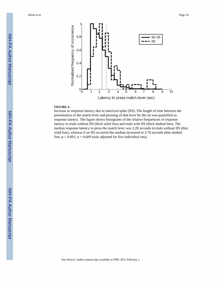
FIGURE 4.Increase in response latency due to interictal spike (IIS). The length of time between thepresentation of the match lever and pressing of that lever by the rat was quantified asresponse latency. The figure shows histograms of the relative frequencies of responselatency in trials without IIS (thick solid line) and trials with IIS (thick dashed line). Themedian response latency to press the match lever was 2.28 seconds in trials without IIS (thinsolid line), whereas if an IIS occurred the median increased to 2.76 seconds (thin dashedline; p < 0.001; n = 4,609 trials adjusted for five individual rats).
Kleen et al. Page 14
Ann Neurol. Author manuscript; available in PMC 2011 February 1.
NIH
-PA Author Manuscript
NIH
-PA Author Manuscript
NIH
-PA Author Manuscript