anterior semicircular canal benign paroxysmal positional vertigo and positional downbeating...
TRANSCRIPT
www.elsevier.com/locate/amjoto
American Journal of Otolaryngology–Head an
Anterior semicircular canal benign paroxysmal positional vertigo and
positional downbeating nystagmus
Jose A. Lopez-Escamez, PhDT, Maria I. Molina, MD, Maria J. Gamiz, PhDOtology and Neurotology Group, CTS495, Department of Surgery, Hospital de Poniente de Almerıa, El Ejido, Almerıa, Spain
Received 28 August 2005
Abstract Purpose: The aim of this study was to describe the clinical features and video-oculographic findings
0196-0709/$ – see fro
doi:10.1016/j.amjoto.2
T Corresponding
Department of Surg
Almerimar s/n, 04700
E-mail address: ja
in patients with anterior semicircular canal benign paroxysmal positional vertigo (BPPV).
Materials and methods: Study Design. This is a prospective case series. Setting. The study was set
at an outpatient clinic in a general hospital. Patients. Fourteen individuals with symptoms of BPPV
and positional downbeating nystagmus (pDBN) were included in the study. The diagnosis was based
on a history of brief episodes of vertigo and the presence of pDBN confirmed in the video-
oculographic examination during Dix-Hallpike test (DH) or head-hanging maneuver. Intervention.
Patients were treated by particle repositioning maneuver and the effectiveness was evaluated at 7, 30,
and 180 days posttreatment. The treatment was repeated up to 4 times if pDBN was persistent. Main
Outcome Measures. The main outcome measure is the number of patients without pDBN at 30 and
180 days.
Results: Video-oculography showed a predominant pDBN in response to DH. Of the 14 patients,
7 had arterial hypertension, and 5 of 14 cases presented abnormalities on the caloric test. Horizontal
spontaneous nystagmus was found in 3 of 14 individuals. Positional nystagmus at different
positional test was observed in 5 of 14 individuals, suggesting the involvement of several canals. Of
the 14 patients, 10 (71%) did not present vertigo, and the positional tests were negative at 30 days.
However, 3 cases presented a positive DH with persistence of BPPVepisodes and pDBN at 30 days,
and another developed a contralateral posterior canal affectation. One of the patients maintained a
persistent pDBN at 180 days despite the repeated maneuvers.
Conclusions: Video-oculography demonstrates that anterior canal BPPV is characterized by a
predominant downbeating nystagmus in response to DH. These individuals may show alterations in
the vestibular caloric, and they can have multicanal affectation.
D 2006 Elsevier Inc. All rights reserved.
1. Introduction
Benign paroxysmal positional vertigo (BPPV) is the
most common vestibular organ disorder that presents an
easy and effective treatment [1,2]. Benign paroxysmal
positional vertigo is defined by spells of vertigo and
positioning nystagmus of short duration that are elicited
by the turn of the head and produces the stimulation of the
semicircular canal [3]. This situation leads to the production
nt matter D 2006 Elsevier Inc. All rights reserved.
005.09.010
author. Otology and Neurotology Group CTS495,
ery, Hospital de Poniente de Almerıa, Ctra de
El Ejido, Almerıa, Spain. Tel.: +34 950 022653.
[email protected] (J.A. Lopez-Escamez).
of abnormal vestibulo-ocular reflexes, causing vertigo and
nystagmus with distinctive characteristics depending on the
canal affected. There are several clinical variants, posterior,
horizontal, and anterior canal, the most frequent being the
involvement of the posterior canal (PC) [4,5]. The
diagnosis of PV BPPV is based on the observation of a
characteristic positional nystagmus during the Dix-Hallpike
test (DH) [6,7].
The affectation of the horizontal or lateral semicircular
canal is less common, and it represents between 5% and 16%
of the cases of BPPV [3,8]. Different studies had described
large series of cases [9-11] differentiating 2 mechanisms
(canalithiasis or cupulolithiasis) that explain the features of
the positional nystagmus (geotropic or apogeotropic, latency,
d Neck Medicine and Surgery 27 (2006) 173–178
J.A. Lopez-Escamez et al. / American Journal of Otolaryngology–Head and Neck Medicine and Surgery 27 (2006) 173–178174
and duration). The intraoperative demonstration of the
presence of otoconia in the lumen of the semicircular canals
has been accepted as the underlying mechanism to explain
BPPV and the positional nystagmus [12,13]. Vestibular
lithiasis can theoretically affect any of the 3 semicircular
canals, and otoliths can be found free-floating in the canal
(canalolithiasis) or found adhered to the cupula of the crista
ampullaris (cupulolithiasis) [14].
The anterior canal variant of BPPV is considered a
rare form, and it is characterized by a predominant down-
beating nystagmus (pDBN) with a small torsional compo-
nent in response to DH [4,15]. The anterior canal projects to
the ipsilateral superior rectus muscle and to the contralateral
inferior oblique muscle; the nystagmus is downbeating
and torsional during the DH. If the downside ear is affected,
the direction of the torsional component will be the same
as in contralateral PC BPPV [15]. This variant has been
described in occasional reports estimating a frequency be-
tween 1% and 11% [3,4,16]. A recent study that clarified the
clinical significance of pDBN in cerebellar disorders
reported a series of 12 patients with possible anterior
semicircular canalithiasis [17]. Moreover, the parameters of
benign positional nystagmus, including 7 patients with
anterior canal BPPV and pDBN, have been measured with
3-dimensional, dual-search scleral coils in a 2-axis, whole-
body rotator [15].
In this report, a series of 14 cases of BPPV with pDBN
that were compatible with the diagnosis of anterior canal
BPPV is presented. Individuals were selected to define their
clinical features and the video-oculographic (VOG) param-
eters of the nystagmus.
2. Patients and methods
2.1. Patients
Individuals were outpatients that reported a history of
vertigo or dizziness. Eighty patients with BPPV and
positional nystagmus were diagnosed during the years
2003 to 2004. Physical examination included otoscopy,
Rinne test, Weber’s test, pure tone audiometry, and a basic
neurotologic examination (oculomotor, saccades, head-
impulse test, cranial nerve examination, Romberg’s test,
Barany’s test, and Fukuda test). Clinical diagnosis was
carried out by positional testing: standard DH maneuvers
were used to stimulate vertical canals [18] and head-hanging
maneuver was used for anterior canal, as previously
described [16]; while patients were in a supine position,
horizontal (yaw) rotations of the head were used to stimulate
horizontal canals.
2.2. Inclusion and exclusion criteria
The inclusion criteria for diagnosis of anterior canal
BPPV were (1) a positional test (DH to each side or the head-
hanging maneuver) after a short latency period that produced
vertigo that simulated the patient’s symptoms and simulta-
neous downbeating nystagmus (DBN); (2) the nystagmus
and vertigo habituate with the positional test; and (3) no
evidence of central nervous system (CNS) disease.
Differential diagnosis between posterior and anterior
canal involvement was based on the direction of the vertical
component of the fast phase of the nystagmus response
during DH, being upward in cases of PC involvement and
downward in cases of anterior canal disease [16]. Patients
with vertical nonpositional nystagmus (ie, spontaneous
DBN) were excluded from this study. All the individuals
were evaluated by a cranial magnetic resonance imaging
with gadolinium to rule out a CNS disease.
Written information was facilitated to the patients, and an
informed consent was obtained for all the individuals after
explaining the treatment. The Ethical and Research Com-
mittee of the hospital approved this research study.
2.3. Video-oculographic examination
The VOG recording included spontaneous nystagmus,
head-shaking nystagmus, positional testing (DH, horizontal
rotations of the head in supine and head-hanging maneuver
for the anterior canal) and water bithermal caloric testing.
The camera was adapted in the contralateral eye to the side
of testing to minimize the movement of the camera during
DH. Caloric test by using a Variotherm Plus model water
irrigator (Atmos, Berlin, Germany) was performed on each
subject, with a water flow of 250 mL/20 s at 308C and 448Cwith an interval of 10 minutes between successive irriga-
tions. All irrigations were performed with the eyes closed in
the Hallpike position.
The recording was performed by a VOG 2-dimensional
system with an infrared charge-coupled device camera
connected to the software (SMI, Berlin, Germany) to
analyze the eye response. Maximum slow-phase velocity
(SPV) was determined by the software, and SPV values
were compared with normal values of our laboratory. Eye
position in the orbit was controlled during testing because a
movement of the eyeball strongly influences the SPV and
the direction of positioning nystagmus.
2.4. Treatment
All patients were initially treated by a particle reposition-
ing maneuver (PRM) or Epley’s modified maneuver without
mastoid oscillator; individuals were recommended to avoid
fast head turnings, and they were allowed to sleep in
decubitus position. Patients were evaluated at 7 days
posttreatment, and if they presented a positional nystagmus,
PRM was repeated up to 4 times. Nonrespondent individ-
uals were treated by a Semont maneuver and finally with
Brandt-Daroff exercises.
3. Results
The mean age of the 14 individuals was 56F 17 (median,
53; range, 20–81) years, being 5 men and 9 women. Nine
patients presented idiopathic BPPV, and 5 cases presented
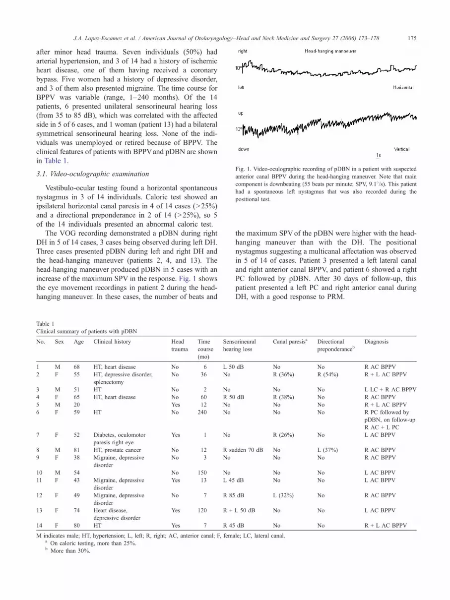
ig. 1. Video-oculographic recording of pDBN in a patient with suspected
nterior canal BPPV during the head-hanging maneuver. Note that main
omponent is downbeating (55 beats per minute; SPV, 9.18/s). This patientad a spontaneous left nystagmus that was also recorded during the
ositional test.
J.A. Lopez-Escamez et al. / American Journal of Otolaryngology–Head and Neck Medicine and Surgery 27 (2006) 173–178 175
after minor head trauma. Seven individuals (50%) had
arterial hypertension, and 3 of 14 had a history of ischemic
heart disease, one of them having received a coronary
bypass. Five women had a history of depressive disorder,
and 3 of them also presented migraine. The time course for
BPPV was variable (range, 1–240 months). Of the 14
patients, 6 presented unilateral sensorineural hearing loss
(from 35 to 85 dB), which was correlated with the affected
side in 5 of 6 cases, and 1 woman (patient 13) had a bilateral
symmetrical sensorineural hearing loss. None of the indi-
viduals was unemployed or retired because of BPPV. The
clinical features of patients with BPPVand pDBN are shown
in Table 1.
3.1. Video-oculographic examination
Vestibulo-ocular testing found a horizontal spontaneous
nystagmus in 3 of 14 individuals. Caloric test showed an
ipsilateral horizontal canal paresis in 4 of 14 cases (N25%)
and a directional preponderance in 2 of 14 (N25%), so 5
of the 14 individuals presented an abnormal caloric test.
The VOG recording demonstrated a pDBN during right
DH in 5 of 14 cases, 3 cases being observed during left DH.
Three cases presented pDBN during left and right DH and
the head-hanging maneuver (patients 2, 4, and 13). The
head-hanging maneuver produced pDBN in 5 cases with an
increase of the maximum SPV in the response. Fig. 1 shows
the eye movement recordings in patient 2 during the head-
hanging maneuver. In these cases, the number of beats and
Table 1
Clinical summary of patients with pDBN
No. Sex Age Clinical history Head
trauma
Time
course
(mo)
Sens
hear
1 M 68 HT, heart disease No 6 L 50
2 F 55 HT, depressive disorder,
splenectomy
No 36 No
3 M 51 HT No 2 No
4 F 65 HT, heart disease No 60 R 50
5 M 20 Yes 12 No
6 F 59 HT No 240 No
7 F 52 Diabetes, oculomotor
paresis right eye
Yes 1 No
8 M 81 HT, prostate cancer No 12 R su
9 F 38 Migraine, depressive
disorder
No 3 No
10 M 54 No 150 No
11 F 43 Migraine, depressive
disorder
Yes 13 L 45
12 F 49 Migraine, depressive
disorder
No 7 R 85
13 F 74 Heart disease,
depressive disorder
Yes 120 R +
14 F 80 HT Yes 7 R 45
M indicates male; HT, hypertension; L, left; R, right; AC, anterior canal; F, femaa On caloric testing, more than 25%.b More than 30%.
F
a
c
h
p
the maximum SPVof the pDBN were higher with the head-
hanging maneuver than with the DH. The positional
nystagmus suggesting a multicanal affectation was observed
in 5 of 14 of cases. Patient 3 presented a left lateral canal
and right anterior canal BPPV, and patient 6 showed a right
PC followed by pDBN. After 30 days of follow-up, this
patient presented a left PC and right anterior canal during
DH, with a good response to PRM.
orineural
ing loss
Canal paresisa Directional
preponderancebDiagnosis
dB No No R AC BPPV
R (36%) R (54%) R + L AC BPPV
No No L LC + R AC BPPV
dB R (38%) No R AC BPPV
No No R + L AC BPPV
No No R PC followed by
pDBN, on follow-up
R AC + L PC
R (26%) No L AC BPPV
dden 70 dB No L (37%) R AC BPPV
No No R AC BPPV
No No L AC BPPV
dB No No L AC BPPV
dB L (32%) No R AC BPPV
L 50 dB No No L AC BPPV
dB No No R + L AC BPPV
le; LC, lateral canal.
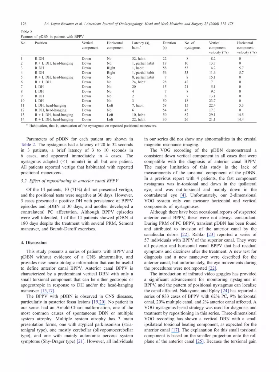
Table 2
Features of pDBN in patients with BPPV
No. Position Vertical
component
Horizontal
component
Latency (s),
habitaDuration
(s)
No. of
nystagmus
Vertical
component
velocity (8/s)
Horizontal
component
velocity (8/s)
1 R DH Down No 32, habit 22 8 8.2 0
2 R + L DH, head-hanging Down No 1, partial habit 18 10 13.7 0
3 R DH Down Right 1, habit 58 53 4.2 5.7
4 R DH Down Right 1, partial habit 56 53 11.6 5.7
5 R + L DH, head-hanging Down No 8, partial habit 7 8 15.1 0
6 R + L DH Down No 24, habit 28 42 7 0
7 L DH Down No 20 15 21 5.1 0
8 L DH Down No 4 7 8 9.5 0
9 R DH Down No 2 8 7 13.1 0
10 L DH Down No 3 50 18 23.7 0
11 L DH, head-hanging Down Left 7, habit 58 15 22.4 5.3
12 R DH, head-hanging Down No 6 54 45 17.3 0
13 R + L DH, head-hanging Down Left 10, habit 50 87 29.1 14.5
14 R + L DH, head-hanging Down Left 22, habit 30 26 21.1 14.4
a Habituation, that is, attenuation of the nystagmus on repeated positional maneuvers.
J.A. Lopez-Escamez et al. / American Journal of Otolaryngology–Head and Neck Medicine and Surgery 27 (2006) 173–178176
Parameters of pDBN for each patient are shown in
Table 2. The nystagmus had a latency of 20 to 32 seconds
in 3 patients, a brief latency of 3 to 10 seconds in
6 cases, and appeared immediately in 4 cases. The
nystagmus adapted (b1 minute) in all but one patient.
All patients reported vertigo that habituated with repeated
positional maneuvers.
3.2. Effect of repositioning in anterior canal BPPV
Of the 14 patients, 10 (71%) did not presented vertigo,
and the positional tests were negative at 30 days. However,
3 cases presented a positive DH with persistence of BPPV
episodes and pDBN at 30 days, and another developed a
contralateral PC affectation. Although BPPV episodes
were well tolerated, 1 of the 14 patients showed pDBN at
180 days despite the treatment with several PRM, Semont
maneuver, and Brandt-Daroff exercises.
4. Discussion
This study presents a series of patients with BPPV and
pDBN without evidence of a CNS abnormality, and
provides new neuro-otologic information that can be useful
to define anterior canal BPPV. Anterior canal BPPV is
characterized by a predominant vertical DBN with only a
small torsional component that can be either geotropic or
apogeotropic in response to DH and/or the head-hanging
maneuver [15,17].
The BPPV with pDBN is observed in CNS diseases,
particularly in posterior fossa lesions [19,20]. No patient in
our series had an Arnold-Chiari malformation, one of the
most common causes of spontaneous DBN or multiple
system atrophy. Multiple system atrophy has 3 main
presentation forms, one with atypical parkinsonism (stria-
tonigral type), one mostly cerebellar (olivopontocerebellar
type), and one with mainly autonomic nervous system
symptoms (Shy-Drager type) [21]. However, all individuals
in our series did not show any abnormalities in the cranial
magnetic resonance imaging.
The VOG recording of the pDBN demonstrated a
consistent down vertical component in all cases that were
compatible with the diagnosis of anterior canal BPPV.
The major limitation of this study is the lack of
measurements of the torsional component of the pDBN.
In a previous report with 4 patients, the fast component
nystagmus was in-torsional and down in the ipsilateral
eye, and was out-torsional and mainly down in the
contralateral eye [4]. Unfortunately, our 2-dimensional
VOG system only can measure horizontal and vertical
components of nystagmuses.
Although there have been occasional reports of suspected
anterior canal BPPV, these were not always concordant.
During PRM of PC BPPV, transient pDBN has been found
and attributed to invasion of the anterior canal by the
canalicular debris [22]. Rahko [23] reported a series of
57 individuals with BPPV of the superior canal. They were
all posterior and horizontal canal BPPV that had residual
symptoms and dizziness after the treatment. A new test for
diagnosis and a new maneuver were described for the
anterior canal, but unfortunately, the eye movements during
the procedures were not reported [22].
The introduction of infrared video goggles has provided
a significant advancement for monitoring nystagmus in
BPPV, and the pattern of positional nystagmus can localize
the canal affected. Nakayama and Epley [24] has reported a
series of 833 cases of BPPV with 62% PC, 9% horizontal
canal, 20% multiple canal, and 2% anterior canal affected. A
VOG nystagmus-based strategy was used for diagnosis and
treatment by repositioning in this series. Three-dimensional
VOG recording has shown a vertical DBN with a small
ipsilateral torsional beating component, as expected for the
anterior canal [17]. The explanation for this small torsional
component is based on the smaller projection onto the roll
plane of the anterior canal [25]. Because the torsional gain

J.A. Lopez-Escamez et al. / American Journal of Otolaryngology–Head and Neck Medicine and Surgery 27 (2006) 173–178 177
of the human vestibular ocular reflex is about 0.75 in
response to high-frequency (2 Hz) roll head impulses [26],
the torsional component would be smaller than the
horizontal and vertical components, and this can explain
the clinically observed pDBN [15].
The abnormal caloric vestibular tests were found in some
cases in our series (4 cases with unilateral canal paresis and
2 cases with increased directional preponderance). These
findings in caloric testing are similar to those observed in
patients with PC BPPV [3,27]. Moreover, horizontal spon-
taneous nystagmus was found in 3 of 14 individuals. These
individuals may show abnormalities in the caloric test.
The natural history of BPPV is incompletely understood.
Although spontaneous resolution of symptoms is observed
in PC BPPV, it is also clear that patients reporting symptoms
with a negative DH became positive DH after several
months of follow-up, suggesting a relapsing course. It is not
known whether repositioning can alter the natural history of
recurrences of BPPV. Furthermore, the abnormalities on
vestibular tests found in some patients with BPPV can
explain the relapsing course of the disease because the
causes that facilitate the shedding of otoliths from the
utricular macula probably persist.
In our series, 7 of the 14 patients had arterial hyper-
tension, suggesting that microvascular affectation could
be associated with anterior canal BPPV. As it has been
demonstrated, giant cell arteritis was associated with
BPPV [28].
We support that BPPV is a complex disorder that can
recur despite of short-term effectiveness of the PRM. In this
series, the positional nystagmus indicating possible multi-
canal affectation was observed in 5 of 14 cases, and those
patients with recurrent episodes can sometimes demonstrate
a new positional nystagmus, suggesting a change of the
canal affected. Bilateral pDBN triggered by the DH was
observed in 5 cases (2, 5, 6, 13, 14), being difficult to
localize the affected side, so this confirms that the head
rotation is unlikely to be critical for particle mobilization in
anterior canal lithiasis [16]. The only difference in the left
and right DH is that, during a contralateral DH, the head
rotates in the plane of the affected anterior canal, whereas
during the ipsilateral DH, the head rotates orthogonally to
the plane of the canal. These bilateral cases could be
suspected to be a bilateral PC BPPV, but here, the direction
of the vertical component recorded is essential for the
differential diagnosis.
Five individuals in our series had pDBN by the straight
head-hanging maneuver (2, 5, 11, 12, 13). As discussed
previously, the rotation in the canal plane is of less relevance
than the final low head-down position [16]. During the DH,
the head is rotated 458 in the horizontal plane previously, so
the head cannot reach such low vertex position as with the
head-hanging maneuver. This additional head angle may be
crucial for provoking anterior canal BPPV as the ampullary
segment will approach a vertical down-pointing position.
Although cervical ankylosis or obesity can difficult the
head-hanging maneuver, we should perform it in patients
with BPPV and a negative DH to rule out a pDBN.
The lower occurrence of lithiasis in the anterior canal is
attributed to the anatomical features of the labyrinth. The
debris within the anterior canal should be self-clearing,
mainly because the posterior arm of the anterior canal
descends directly into the common crus and the utricle.
Persistence of pDBN may be explained in 2 ways. First,
the canalith is incompletely returned into the utricle by a
single PRM and some particles remain in the PC. Why
this occurs is unknown, but it could be possible that the
canalith may be too large, a failure of disaggregation or a
stenotic common crus. This is consistent with the finding that
some patients require a second or third PRM to become DH
negative [29]. Second, another vestibular lesion (ie, ische-
mic, metabolic) that causes a continuous formation of debris
in patients with persistent positional nystagmus may exist.
5. Conclusions
Video-oculography demonstrates that anterior canal
BPPV is characterized by a predominant DBN in response
to DH. These individuals may show alterations in the ca-
loric response, and they can have multicanal involvement
in same cases.
Acknowledgments
This study was funded by a research project FIS
PI021394 from Instituto de Salud Carlos III (Madrid,
Spain). We thank Maria Jose Palma y Cristobal Zapata,
who performed the vestibular testing and the recording of the
positional nystagmus.
References
[1] Epley J. The canalith repositioning procedure for treatment of be-
nign paroxysmal positional vertigo. Otolaryngol Head Neck Surg
1992;107:399-404.
[2] Parnes L, Price-Jones R. Particle repositioning maneuver for benign
paroxysmal positional vertigo. Ann Otol Rhinol Laryngol 1993;
102:325-31.
[3] Brandt T. Positional and positioning vertigo and nystagmus. J Neurol
Sci 1990;95:3 -28.
[4] Honrubia V, Baloh RW, Harris MR, et al. Paroxysmal positional
vertigo syndrome. Am J Otol 1999;20:465 -70.
[5] Korres S, Balatsouras DG, Kaberos A, et al. Occurrence of
semicircular canal involvement in benign paroxysmal positional
vertigo. Otol Neurotol 2002;23:926 -32.
[6] Dix MT, Hallpike CS. The pathology, symptomatology and diagnosis
of certain common disorders of the vestibular system. Ann Otol
Rhinol Laryngol 1952;61:987-1016.
[7] Baloh RW, Sakala SM, Honrubia V. Benign paroxysmal positional
nystagmus. Am J Otolaryngol 1979;1:1 -6.
[8] Parnes LS, Agrawal SK, Atlas J. Diagnosis and management of
benign paroxysmal positional vertigo (BPPV). Can Med Assoc J
2003;169:681-93.
[9] McClure JA. Horizontal canal BPV. J Otolaryngol 1985;14:30-5.
[10] Baloh RW, Jacobson K, Honrubia V. Horizontal semicircular canal
variant of benign positional vertigo. Neurology 1993;43:2542-9.

J.A. Lopez-Escamez et al. / American Journal of Otolaryngology–Head and Neck Medicine and Surgery 27 (2006) 173–178178
[11] Nuti D, Vannucchi P, Pagnini P. Benign paroxysmal positional vertigo
of the horizontal canal: a form of canalolithiasis with variable clinical
features. J Vestib Res 1996;6:173-84.
[12] Parnes LS, McClure JA. Posterior semicircular canal occlusion in
the normal hearing ear. Otolaryngol Head Neck Surg 1991;104:
52 -7.
[13] Welling DB, Parnes LS, O’Brien B, et al. Particulate matter in the
posterior semicircular canal. Laryngoscope 1997;107:90-4.
[14] Brandt T, Steddin S. Current view of the mechanism of benign
paroxysmal positional vertigo: cupulolithiasis or canalolithiasis?
J Vestib Res 1993;3:373-82.
[15] Aw ST, Todd MJ, Aw GE, et al. Benign positional nystagmus. A study
of its three-dimensional spatio-temporal characteristics. Neurology
2005;64:1897-905.
[16] Crevits L. Treatment of anterior canal benign paroxysmal positional
vertigo by a prolonged forced position. J Neurol Neurosurg Psychiatry
2004;75:779-81.
[17] Bertholon P, Bronstein AM, Davies RA, et al. Positional down beating
nystagmus in 50 patients: cerebellar disorders and possible anterior
semicircular canalithiasis. J Neurol Neurosurg Psychiatry 2002;72:
366 -72.
[18] Dix MR, Hallpike C. The pathology, symptomatology and diagnosis
of certain common disorders of the vestibular system. Proc R Soc Med
1952;45:341-54.
[19] Barber HO. Positional nystagmus. Otolaryngol Head Neck Surg
1984;92:649-55.
[20] Kattah JC, Kolsky MP, Luessenhop AJ. Positional vertigo and the
cerebellar vermis. Neurology 1984;34:527 -9.
[21] Wenning GK, Ben Shlomo Y, Magalhes M, et al. Clinical features and
natural history of multiple system atrophy: an analysis of 100 cases.
Brain 1994;117:835-45.
[22] Herdman SJ, Tusa RJ. Complications of the canalith repositioning
procedure. Arch Otolaryngl Head Neck Surg 1996;122:281-6.
[23] Rahko T. The test and treatment methods of benign paroxysmal
positional vertigo and an addition to the management of vertigo due
to the superior vestibular canal (BPPV-SC). Clin Otolaryngol 2002;
27:392-5.
[24] Nakayama M, Epley JM. BPPV and variants: improved treatment
results with automated, nystagmus-based repositioning. Otolaryngol
Head Neck Surg 2005;133:107-12.
[25] Blanks RHI, Curthoys IS, Markham CH. Planar relationships of the
semicircular canals in man. Acta Otolaryngol 1975;80:185-96.
[26] Aw ST, Haslwanter T, Halmagyi GM, et al. Three-dimensional vector
analysis of the human vestibuloocular reflex in response to high-
acceleration head rotations. I. Responses in normal subjects.
J Neurophysiol 1996;76:4009-20.
[27] Baloh RW, Honrubia V, Jacobson K. Benign paroxysmal positioning
vertigo: clinical and oculographic features in 240 cases. Neurology
1987;37:371-8.
[28] Amor-Dorado JC, Llorca J, Costa-Ribas C, et al. Giant cell arteritis: a
new association with benign paroxysmal positional vertigo. Laryngo-
scope 2004;114:1420-5.
[29] Pollak L, Davies RA, Luxon LL. Effectiveness of the particle
repositioning maneuver in benign paroxysmal positional vertigo with
and without additional vestibular pathology. Otol Neurotol 2002;
23:79 -83.