rapid cardiovascular diagnostics - open research library
TRANSCRIPT
Chapter 2
Rapid Cardiovascular Diagnostics
David C. Gaze
Additional information is available at the end of the chapter
http://dx.doi.org/10.5772/63910
Abstract
Rapid cardiovascular diagnostics have played an integral role in the development of theclinical utility of cardiac biomarkers. Rapid qualitative and quantitative tests are used asan alternative to clinical chemistry laboratories, where urgent testing for cardiacbiomarkers has been unavailable. Biomarkers are clinically available for diagnosis andprognosis across the heart disease continuum. Early risk markers of plaque formationand destabilisation are non‐urgent and can be performed routinely as part of the pathologylaboratory repertoire. Rapid diagnostic tests are best suited for markers of ischaemia,necrosis and heart failure, where patients often present acutely to emergency medicalservices. This chapter reviews the evolution of rapid cardiovascular diagnostics both inthe pre‐hospital and emergency department setting, compared to the utility of central‐ised laboratory testing. Further, limitations to their use and improvements by noveltechnology are discussed.
Keywords: cardiovascular disease, cardiac markers, cardiac troponin, centralised lab‐oratory testing, ischemia, natriuretic peptides, rapid tests
1. Introduction
Cardiovascular disease (CVD) is the leading cause of death in the Western World with rates forcoronary heart disease and stroke‐related mortality exceeding that of any cancer. Patients whopresent to the emergency department (ED) with chest pain are often a complex clinical challenge.Classical electrocardiographic (ECG) changes of an acute cardiac episode occur in only 5–15%of ED presentations. Blood‐borne biomarkers offer an alternative diagnostic mode and havebeen part of the clinical testing repertoire since the 1950s.
Cardiac biomarker testing can be performed in a spectrum of clinical settings. Normally, thisoccurs in a centralised laboratory, where blood is drawn from the patient and transported
© 2016 The Author(s). Licensee InTech. This chapter is distributed under the terms of the Creative CommonsAttribution License (http://creativecommons.org/licenses/by/3.0), which permits unrestricted use, distribution,and reproduction in any medium, provided the original work is properly cited.
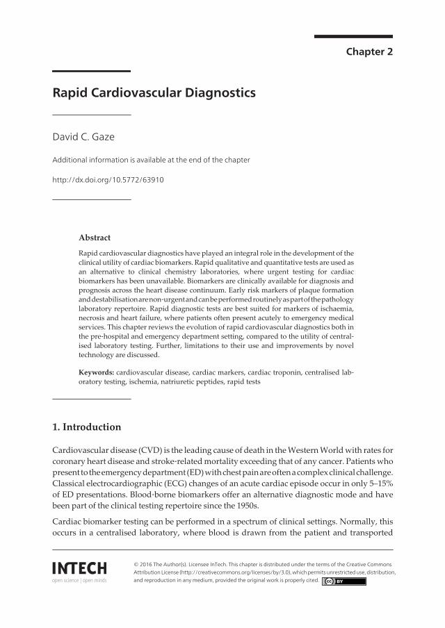
either by hand or pneumatic delivery chutes (Figure 1). The samples are processed in thelaboratory along with other patient samples received from inpatients, outpatient clinics orfrom primary care. The combination of samples and the utility of multidisciplinary pathologylaboratories can result in a time delay from drawing the sample to obtaining the results in theclinical setting (Figure 2).
Figure 1. Cycle of delivery of diagnostic testing results. TAT, turnaround time.
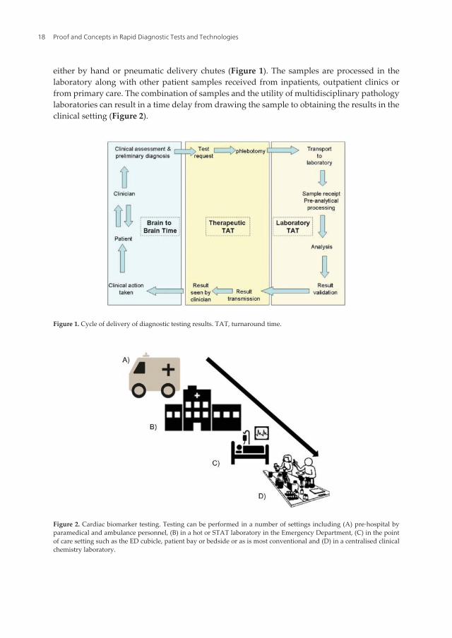
Figure 2. Cardiac biomarker testing. Testing can be performed in a number of settings including (A) pre‐hospital byparamedical and ambulance personnel, (B) in a hot or STAT laboratory in the Emergency Department, (C) in the pointof care setting such as the ED cubicle, patient bay or bedside or as is most conventional and (D) in a centralised clinicalchemistry laboratory.
Proof and Concepts in Rapid Diagnostic Tests and Technologies18
1.1. Point of care testing
The alternative to centralised laboratory testing is point of care testing (POCT). This has beenknown by many names and acronyms including near‐patient testing; bedside testing; physi‐cians’ office testing; extra‐laboratory, satellite ‘hot lab’; decentralised laboratory; ancillarylaboratory; or alternate site testing. Essentially, the aim of POCT analysis is to performdiagnostic laboratory tests with a shorter turnaround time (STAT) than that obtained by thecentral laboratory.
POCT is the immediate provision of a test at the point of healthcare delivery when the resultwill be used to make a decision and to take appropriate action, which leads to an improvedhealth outcome [1]. POCT is not a novel concept, mentioned early in the foundation of medicineby Hippocrates (c. 460‐370BC) and Galen (129‐c.200AD). It was also described in the seventhcentury by the Byzantine physician Theophilus Protospatharius, a forerunner of modernUrology. Theophilus used uroscopy, the practice of visually examining patients urine with aurine flask and determining disease using the urine wheel; a colour chart of 20 urine flasksaligned in a circle and acts as a reference to link urine colour to particular diseases.
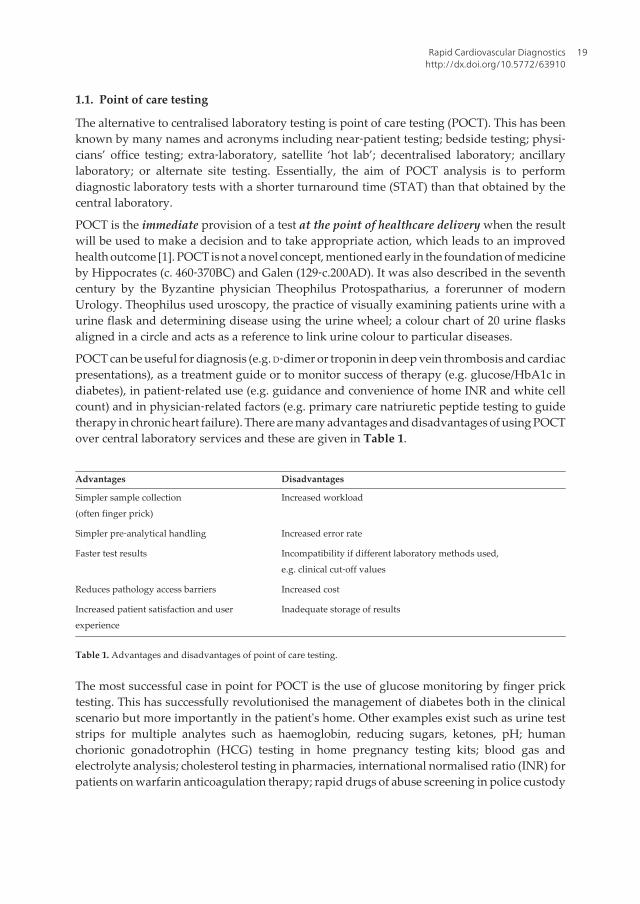
POCT can be useful for diagnosis (e.g. D‐dimer or troponin in deep vein thrombosis and cardiacpresentations), as a treatment guide or to monitor success of therapy (e.g. glucose/HbA1c indiabetes), in patient‐related use (e.g. guidance and convenience of home INR and white cellcount) and in physician‐related factors (e.g. primary care natriuretic peptide testing to guidetherapy in chronic heart failure). There are many advantages and disadvantages of using POCTover central laboratory services and these are given in Table 1.
Advantages Disadvantages
Simpler sample collection(often finger prick)
Increased workload
Simpler pre‐analytical handling Increased error rate
Faster test results Incompatibility if different laboratory methods used,e.g. clinical cut‐off values
Reduces pathology access barriers Increased cost
Increased patient satisfaction and userexperience
Inadequate storage of results
Table 1. Advantages and disadvantages of point of care testing.
The most successful case in point for POCT is the use of glucose monitoring by finger pricktesting. This has successfully revolutionised the management of diabetes both in the clinicalscenario but more importantly in the patient's home. Other examples exist such as urine teststrips for multiple analytes such as haemoglobin, reducing sugars, ketones, pH; humanchorionic gonadotrophin (HCG) testing in home pregnancy testing kits; blood gas andelectrolyte analysis; cholesterol testing in pharmacies, international normalised ratio (INR) forpatients on warfarin anticoagulation therapy; rapid drugs of abuse screening in police custody
Rapid Cardiovascular Diagnosticshttp://dx.doi.org/10.5772/63910
19
or the ED; procalcitonin and c‐reactive protein for rapid detection of sepsis; and humanimmunodeficiency virus in salivary samples.
The driver for adopting successful POCT is a balance between meeting realistic clinical needwith the appropriate technology at a sensible cost. The overwhelming utility for POCT is therapid delivery of results. This is appropriate for acute conditions, such as cardiac or respira‐tory events, ectopic pregnancy and sepsis. The other need is for diagnosis outside of stand‐ard clinical areas, such as monitoring in the patients home, testing in specialised walk‐incentres (HIV testing) or in social care (drugs of abuse).
1.2. Analytical actuary and precision of point of care testing
Methods employed for POCT need to demonstrate adequate analytical performance withacceptable accuracy and precision in comparison to centralised laboratory methods. Obtainingresults faster is of very limited value if the coefficient of variation (CV) for the analyte isexcessively large (e.g. 25%) compared to a central laboratory method with a CV of 5% at thesame concentration. It is generally accepted that the performance of POCT is limited com‐pared to central laboratory testing due to constraints such as the technology employed,knowledge and training of users who are often non‐laboratory trained and inadequate qualityassurance monitoring.
Performance has been well documented for blood gas analysis [2–5], cholesterol determina‐tion on eight POCT systems [6] and for INR [7] as determined on the popular CoaguCheck XSdevice, which was highly comparable (r = 0.91) to a laboratory‐based method.
1.3. Effect of point of care testing on turnaround time
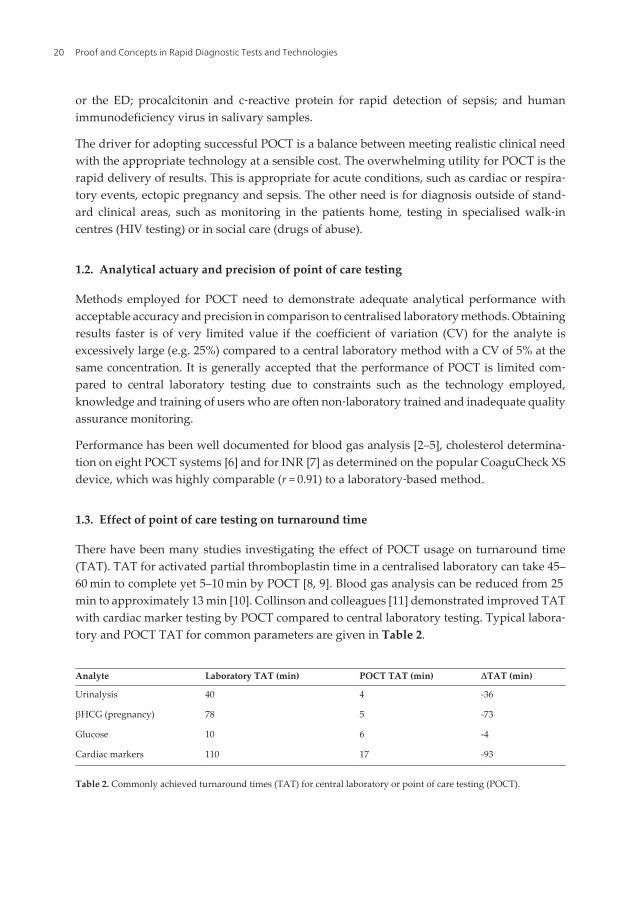
There have been many studies investigating the effect of POCT usage on turnaround time(TAT). TAT for activated partial thromboplastin time in a centralised laboratory can take 45–60 min to complete yet 5–10 min by POCT [8, 9]. Blood gas analysis can be reduced from 25 min to approximately 13 min [10]. Collinson and colleagues [11] demonstrated improved TATwith cardiac marker testing by POCT compared to central laboratory testing. Typical labora‐tory and POCT TAT for common parameters are given in Table 2.
Analyte Laboratory TAT (min) POCT TAT (min) ∆TAT (min)
Urinalysis 40 4 -36
βHCG (pregnancy) 78 5 -73
Glucose 10 6 -4
Cardiac markers 110 17 -93
Table 2. Commonly achieved turnaround times (TAT) for central laboratory or point of care testing (POCT).
Proof and Concepts in Rapid Diagnostic Tests and Technologies20

1.4. Impact on patient care
Having an analytically sound and faster method of determining an analyte is all well and good;however, the final piece of the jigsaw is the translation into improvement of patient manage‐ment and outcome over and above that of the central laboratory.
Parvin et al. [12] investigated the effect of routine use of POCT by non‐laboratory personnelin the ED on length of stay. A handheld device was used to determine Na, K, Cl, glucose andurea. There was no relevant decrease in length of stay in the ED. In a UK study of 1728 patients,changes in patient management are made earlier for those who received POCT haematologytesting (74 min earlier) and biochemical testing (21 min earlier) reducing the time to decisionaffecting patient management [13]. However, such changes did not affect clinical outcome orlength of stay in the ED.
Specifically, for cardiac markers, on the basis of 263 admissions with chest pain, Collinson etal. [11] have demonstrated that patients who received POCT had a reduced length of stay, bothin the coronary care unit and overall hospital stay compared to those who received conven‐tional biomarker testing provided by the central laboratory. This final important step hasplayed a pivotal role in the adoption (or lack of) of rapid cardiovascular diagnostics by POCTand is the focus of the remainder of this chapter.
2. Cardiovascular disease epidemiology
CVD is the leading cause of global death. World Heart Federation statistics demonstrate that17.1 million deaths globally each year are due to CVD, with 82% occurring in the developingworld. Such numbers are often difficult to comprehend. One in every five deaths in the USAis due to CVD. Thirty‐five per cent of UK people <65 years old die prematurely due to CVDdaily. Data prediction suggests that 23 million people will die annually from CVD by theyear 2030.
2.1. Acute chest pain
The largest category of patients admitted to UK hospitals is due to chest pain [14]. Patientswith chest pain are diagnostically challenging. The majority present with either stableischaemic heart disease (IHD) or no IHD [15]. Such admissions are either clinically inappro‐priate or are of short duration, lasting hours or a few days maximum. However, 2–7% ofpatients with acute myocardial infarction (AMI) are inappropriately discharged from the ED[16, 17]. Improvements to diagnosis have been made utilising risk scoring systems [18],computerised decision support [19, 20] and automated ECG interpretation [21]. Althoughclinical assessment is paramount in the assessment of chest pain, cardiac biomarker measure‐ments are now routinely used to aid diagnosis.
Rapid Cardiovascular Diagnosticshttp://dx.doi.org/10.5772/63910
21
2.2. Cardiac biomarkers: a historical perspective
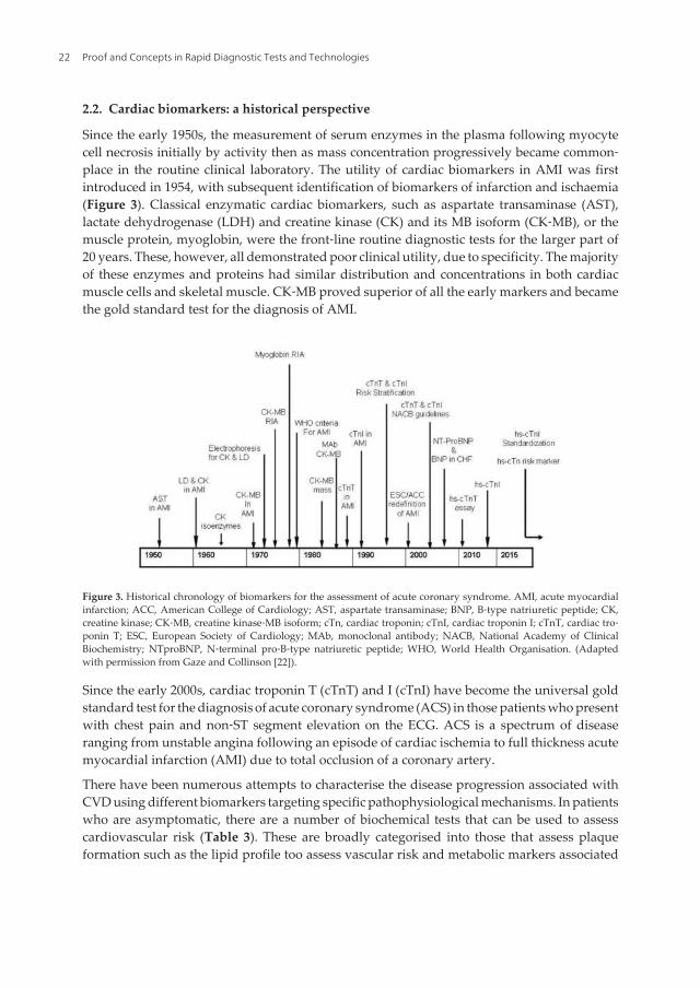
Since the early 1950s, the measurement of serum enzymes in the plasma following myocytecell necrosis initially by activity then as mass concentration progressively became common‐place in the routine clinical laboratory. The utility of cardiac biomarkers in AMI was firstintroduced in 1954, with subsequent identification of biomarkers of infarction and ischaemia(Figure 3). Classical enzymatic cardiac biomarkers, such as aspartate transaminase (AST),lactate dehydrogenase (LDH) and creatine kinase (CK) and its MB isoform (CK‐MB), or themuscle protein, myoglobin, were the front‐line routine diagnostic tests for the larger part of20 years. These, however, all demonstrated poor clinical utility, due to specificity. The majorityof these enzymes and proteins had similar distribution and concentrations in both cardiacmuscle cells and skeletal muscle. CK‐MB proved superior of all the early markers and becamethe gold standard test for the diagnosis of AMI.
Figure 3. Historical chronology of biomarkers for the assessment of acute coronary syndrome. AMI, acute myocardialinfarction; ACC, American College of Cardiology; AST, aspartate transaminase; BNP, B‐type natriuretic peptide; CK,creatine kinase; CK‐MB, creatine kinase‐MB isoform; cTn, cardiac troponin; cTnI, cardiac troponin I; cTnT, cardiac tro‐ponin T; ESC, European Society of Cardiology; MAb, monoclonal antibody; NACB, National Academy of ClinicalBiochemistry; NTproBNP, N‐terminal pro‐B‐type natriuretic peptide; WHO, World Health Organisation. (Adaptedwith permission from Gaze and Collinson [22]).
Since the early 2000s, cardiac troponin T (cTnT) and I (cTnI) have become the universal goldstandard test for the diagnosis of acute coronary syndrome (ACS) in those patients who presentwith chest pain and non‐ST segment elevation on the ECG. ACS is a spectrum of diseaseranging from unstable angina following an episode of cardiac ischemia to full thickness acutemyocardial infarction (AMI) due to total occlusion of a coronary artery.
There have been numerous attempts to characterise the disease progression associated withCVD using different biomarkers targeting specific pathophysiological mechanisms. In patientswho are asymptomatic, there are a number of biochemical tests that can be used to assesscardiovascular risk (Table 3). These are broadly categorised into those that assess plaqueformation such as the lipid profile too assess vascular risk and metabolic markers associated
Proof and Concepts in Rapid Diagnostic Tests and Technologies22
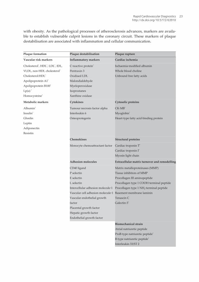
with obesity. As the pathological processes of atherosclerosis advances, markers are availa‐ble to establish vulnerable culprit lesions in the coronary circuit. These markers of plaquedestabilisation are associated with inflammation and cellular communication.
Plaque formation Plaque destabilisation Plaque rupture
Vascular risk markers Inflammatory markers Cardiac ischemia
Cholesterol*, HDL*, LDL*, IDL,VLDL, non‐HDL cholesterol*
Cholesterol:HDL*
Apolipoprotein A1*
Apolipoprotein B100*
Lp(a) *
Homocysteine*
C‐reactive protein*
Pentraxin 3Oxidised LDLMalondialdehydeMyeloperoxidaseIsoprostanesXanthine oxidase
Ischaemia‐modified albuminWhole blood cholineUnbound free fatty acids
Metabolic markers Cytokines Cytosolic proteins
Albumin*
Insulin*
GhrelinLeptinAdiponectinResistin
Tumour necrosis factor alphaInterleukin 6Osteoprotegerin
CK‐MB*
Myoglobin*
Heart‐type fatty acid‐binding protein
Chemokines Structural proteins
Monocyte chemoattractant factor Cardiac troponin T*
Cardiac troponin I*
Myosin light chain
Adhesion molecules Extracellular matrix turnover and remodelling
CD40 ligandP selectinE selectinL selectinIntercellular adhesion molecule‐1Vascular cell adhesion molecule‐1Vascular endothelial growthfactorPlacental growth factorHepatic growth factorEndothelial growth factor
Matrix metalloproteinases (MMP)Tissue inhibitors of MMPProcollagen III aminopeptideProcollagen type 1 COOH‐terminal peptideProcollagen type 1 NH2‐terminal peptideBasement membrane lamininTenascin CGalectin‐3*
Biomechanical strainAtrial natriuretic peptideProB‐type natriuretic peptide*
B‐type natriuretic peptide*
Interleukin 33/ST 2
Rapid Cardiovascular Diagnosticshttp://dx.doi.org/10.5772/63910
23
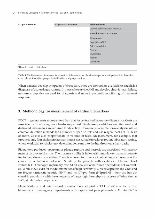
Plaque formation Plaque destabilisation Plaque rupture
Growth differentiation factor 15
Neurohormonal activation
Aldosterone*
Copeptin (ADH)AdrenomedullinApelinEndothelinRelaxinUrotensin
*Those in routine clinical use.
Table 3. Cardiovascular biomarkers for detection of the cardiovascular disease spectrum categorised into those thatdetect plaque formation, plaque destabilisation and plaque rupture.
When patients develop symptoms of chest pain, there are biomarkers available to establish adiagnosis of acute plaque rupture. In those who survive AMI and develop chronic heart failure,natriuretic peptides are used for diagnosis and more importantly monitoring of treatmentresponse.
3. Methodology for measurement of cardiac biomarkers
POCT in general costs more per test than that for centralised laboratory diagnostics. Costs areassociated with utilising more hardware per test. Single assay cartridges are often used anddedicated instruments are required for detection. Conversely, large platform analysers utilisecommon detection methods for a number of specific tests and use reagent packs of 100 testsor more. Cost is also proportionate to volume of tests. An instrument, for example, thatproduces only four cholesterol tests an hour is not suitable for a large routine laboratory setting,where workload for cholesterol determination runs into the hundreds on a daily basis.
Biomarkers produced upstream of plaque rupture and necrosis are associated with assess‐ment of cardiovascular risk. Their primary utility is in low‐risk ambulatory patients present‐ing to the primary care setting. There is no need for urgency in obtaining such results as theclinical presentation is not acute. Similarly, for patients with established Chronic HeartFailure (CHF) managed in primary care, STAT analysis of natriuretic peptides is not warrant‐ed. While POCT exist for the determination of high‐sensitivity C‐reactive protein (hs‐CRP) andfor B‐type natriuretic peptide (BNP) and its NT‐pro form (NTproBNP), their use has de‐clined in popularity with the emergence of large high throughput analysers offering similarTAT, at relatively cheaper cost.
Many National and International societies have adopted a TAT of <60 min for cardiacbiomarkers. In emergency departments with rapid chest pain protocols, a 30 min TAT is
Proof and Concepts in Rapid Diagnostic Tests and Technologies24
desirable. Centralised pathology provision is possible with pneumatic air tube delivery ofsamples, STAT processing of samples and host communication with instruments to priori‐tize samples for ‘urgent’ analysis over more routine tests that could be performed later. A studyin the USA of 159 hospitals, which audited 7020 cTn and 4368 CK‐MB determinations,demonstrated TAT of 90 min for cardiac biomarkers [23].
Due to patents, a single manufacturer (Roche Diagnostics) produces assays for cTnT on bothlarge‐scale immunoassay analysers and at the point of care. Cardiac troponin I, however, isunlicensed and currently not standardised. A multitude of in vitro diagnostic manufacturersproduce cTnI methods for both POCT and centralised laboratories. An in‐depth review of themeasurement of cTn has been published previously [24].
3.1. Point of care testing for cardiac troponin
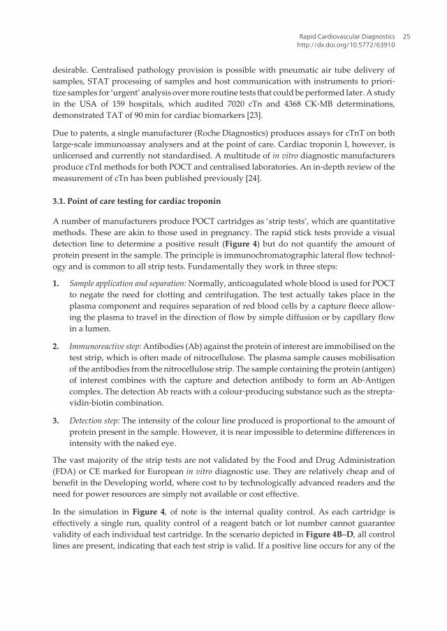
A number of manufacturers produce POCT cartridges as ‘strip tests’, which are quantitativemethods. These are akin to those used in pregnancy. The rapid stick tests provide a visualdetection line to determine a positive result (Figure 4) but do not quantify the amount ofprotein present in the sample. The principle is immunochromatographic lateral flow technol‐ogy and is common to all strip tests. Fundamentally they work in three steps:
1. Sample application and separation: Normally, anticoagulated whole blood is used for POCTto negate the need for clotting and centrifugation. The test actually takes place in theplasma component and requires separation of red blood cells by a capture fleece allow‐ing the plasma to travel in the direction of flow by simple diffusion or by capillary flowin a lumen.
2. Immunoreactive step: Antibodies (Ab) against the protein of interest are immobilised on thetest strip, which is often made of nitrocellulose. The plasma sample causes mobilisationof the antibodies from the nitrocellulose strip. The sample containing the protein (antigen)of interest combines with the capture and detection antibody to form an Ab‐Antigencomplex. The detection Ab reacts with a colour‐producing substance such as the strepta‐vidin‐biotin combination.
3. Detection step: The intensity of the colour line produced is proportional to the amount ofprotein present in the sample. However, it is near impossible to determine differences inintensity with the naked eye.
The vast majority of the strip tests are not validated by the Food and Drug Administration(FDA) or CE marked for European in vitro diagnostic use. They are relatively cheap and ofbenefit in the Developing world, where cost to by technologically advanced readers and theneed for power resources are simply not available or cost effective.
In the simulation in Figure 4, of note is the internal quality control. As each cartridge iseffectively a single run, quality control of a reagent batch or lot number cannot guaranteevalidity of each individual test cartridge. In the scenario depicted in Figure 4B–D, all controllines are present, indicating that each test strip is valid. If a positive line occurs for any of the
Rapid Cardiovascular Diagnosticshttp://dx.doi.org/10.5772/63910
25
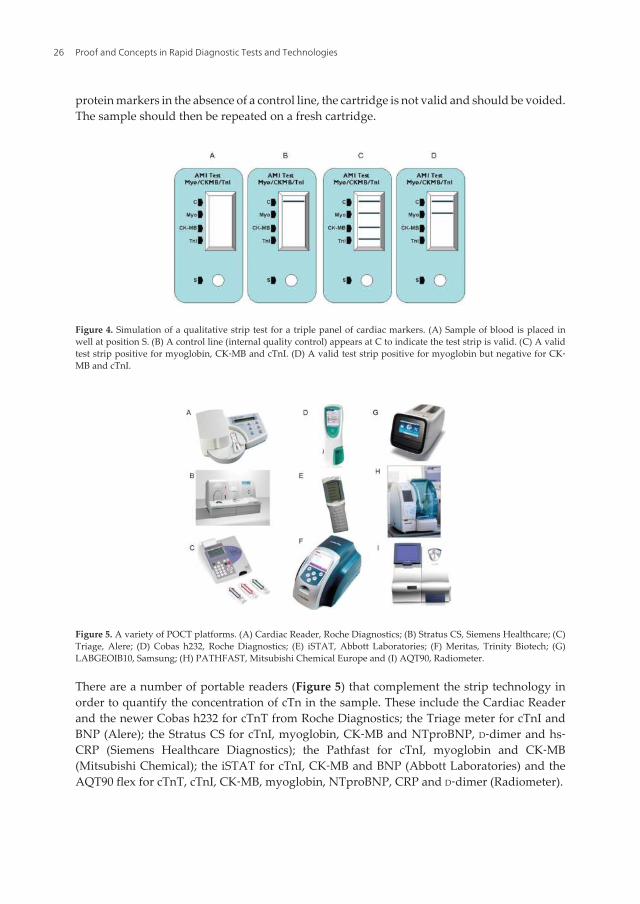
protein markers in the absence of a control line, the cartridge is not valid and should be voided.The sample should then be repeated on a fresh cartridge.
Figure 4. Simulation of a qualitative strip test for a triple panel of cardiac markers. (A) Sample of blood is placed inwell at position S. (B) A control line (internal quality control) appears at C to indicate the test strip is valid. (C) A validtest strip positive for myoglobin, CK‐MB and cTnI. (D) A valid test strip positive for myoglobin but negative for CK‐MB and cTnI.
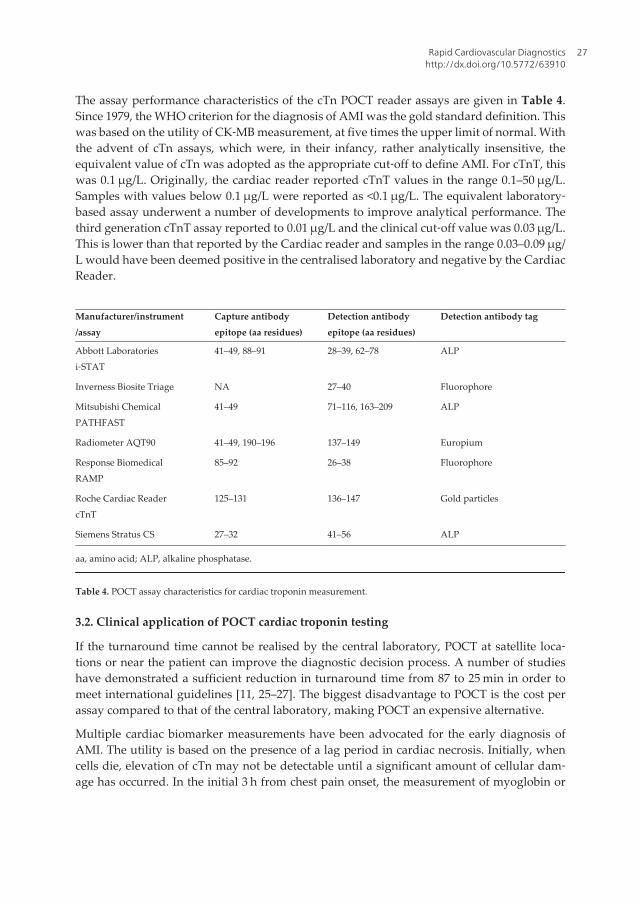
Figure 5. A variety of POCT platforms. (A) Cardiac Reader, Roche Diagnostics; (B) Stratus CS, Siemens Healthcare; (C)Triage, Alere; (D) Cobas h232, Roche Diagnostics; (E) iSTAT, Abbott Laboratories; (F) Meritas, Trinity Biotech; (G)LABGEOIB10, Samsung; (H) PATHFAST, Mitsubishi Chemical Europe and (I) AQT90, Radiometer.
There are a number of portable readers (Figure 5) that complement the strip technology inorder to quantify the concentration of cTn in the sample. These include the Cardiac Readerand the newer Cobas h232 for cTnT from Roche Diagnostics; the Triage meter for cTnI andBNP (Alere); the Stratus CS for cTnI, myoglobin, CK‐MB and NTproBNP, D‐dimer and hs‐CRP (Siemens Healthcare Diagnostics); the Pathfast for cTnI, myoglobin and CK‐MB(Mitsubishi Chemical); the iSTAT for cTnI, CK‐MB and BNP (Abbott Laboratories) and theAQT90 flex for cTnT, cTnI, CK‐MB, myoglobin, NTproBNP, CRP and D‐dimer (Radiometer).
Proof and Concepts in Rapid Diagnostic Tests and Technologies26
The assay performance characteristics of the cTn POCT reader assays are given in Table 4.Since 1979, the WHO criterion for the diagnosis of AMI was the gold standard definition. Thiswas based on the utility of CK‐MB measurement, at five times the upper limit of normal. Withthe advent of cTn assays, which were, in their infancy, rather analytically insensitive, theequivalent value of cTn was adopted as the appropriate cut‐off to define AMI. For cTnT, thiswas 0.1 μg/L. Originally, the cardiac reader reported cTnT values in the range 0.1–50 μg/L.Samples with values below 0.1 μg/L were reported as <0.1 μg/L. The equivalent laboratory‐based assay underwent a number of developments to improve analytical performance. Thethird generation cTnT assay reported to 0.01 μg/L and the clinical cut‐off value was 0.03 μg/L.This is lower than that reported by the Cardiac reader and samples in the range 0.03–0.09 μg/L would have been deemed positive in the centralised laboratory and negative by the CardiacReader.
Manufacturer/instrument/assay
Capture antibodyepitope (aa residues)
Detection antibodyepitope (aa residues)
Detection antibody tag
Abbott Laboratoriesi‐STAT
41–49, 88–91 28–39, 62–78 ALP
Inverness Biosite Triage NA 27–40 Fluorophore
Mitsubishi ChemicalPATHFAST
41–49 71–116, 163–209 ALP
Radiometer AQT90 41–49, 190–196 137–149 Europium
Response BiomedicalRAMP
85–92 26–38 Fluorophore
Roche Cardiac ReadercTnT
125–131 136–147 Gold particles
Siemens Stratus CS 27–32 41–56 ALP
aa, amino acid; ALP, alkaline phosphatase.
Table 4. POCT assay characteristics for cardiac troponin measurement.
3.2. Clinical application of POCT cardiac troponin testing
If the turnaround time cannot be realised by the central laboratory, POCT at satellite loca‐tions or near the patient can improve the diagnostic decision process. A number of studieshave demonstrated a sufficient reduction in turnaround time from 87 to 25 min in order tomeet international guidelines [11, 25–27]. The biggest disadvantage to POCT is the cost perassay compared to that of the central laboratory, making POCT an expensive alternative.
Multiple cardiac biomarker measurements have been advocated for the early diagnosis ofAMI. The utility is based on the presence of a lag period in cardiac necrosis. Initially, whencells die, elevation of cTn may not be detectable until a significant amount of cellular dam‐age has occurred. In the initial 3 h from chest pain onset, the measurement of myoglobin or
Rapid Cardiovascular Diagnosticshttp://dx.doi.org/10.5772/63910
27
the CK‐MB may be appropriate especially in the point of care setting and a protocol based ontriple cardiac marker testing has been suggested [27] and validated [28]. However, theintroduction of assays in the centralised laboratory capable of measuring troponin concentra‐tions within the reference interval [29] and the detection of very early rises in cTn [30, 31] inthe initial 3 h from presentation challenges this concept.
To date, there have been four randomised control trials of point of care testing [11, 32–34]. Twostudies report outcomes [11, 34] with no difference in the number of adverse events in patientsrandomised to either POCT or central pathology laboratory testing. One study reporteddiagnostic accuracy, observing that POCT and central laboratory testing were equivalent [11].In this study, a POCT cTnT method with the same decision limit as that in use by the centrallaboratory method of 0.1 μg/L. Although appropriate at the time of the study, the 0.1 μg/Ldecision limit is now considered too high for centralised cTnT determination. Further studiesare required to assess the utility of POCT for cardiac troponin in comparison to high‐sensi‐tive methodology available in the central laboratory.
In the recent multicentre, Randomised Assessment of Treatment, using Panel Assay of CardiacMarkers (RATPAC) study performed in the UK, Goodacre et al. demonstrated that POCTincreases successful discharge home of patients within 4 h of attendance and significantlyreduced the median length of stay (8.8 h in POCT arm compared to 14.2 h in the central labtesting arm). There was no effect in overall bed use [35]. The use of POCT was associated withhigher cost in the ED, coronary care and increased cardiac intervention costs but overall lowergeneral inpatient costs [36].
3.3. POCT cardiac troponin in the pre‐hospital setting
There are very limited data using point of care testing (POCT) for cTn in the pre‐hospitalsetting. The majority utilised the initial rapid Trop T assay from Roche Diagnostics [37–39],which was far less sensitive than we are used to in current laboratory diagnostics. The cut‐offfor AMI was 0.1 μg/L which equates to 100 ng/L in new units. By contrast, the current hs‐cTnTassay has a 99th percentile cut‐off value of 14 ng/L. There are even less published data usingPOCT cTnI [40–42].
What is important to remember is that the current POCT technology for cTn is not suitablyanalytically sensitive enough when compared to laboratory‐based immunoassay. To this end,they are reliable as rule in tests if patients are positive for cTn; however, their role as a rule outmarker is questionable due to the equivalent high cut‐off values employed.
In urban areas, where patients receive rapid response to emergency calls within minutes withsubsequent rapid transfer, the value of POCT is questionable. However, if a suitable sensi‐tive POCT could be developed, this may have major benefit in rural areas, where patients canbe triaged to appropriate cardiac centres that offer immediate primary coronary intervention‐al surgery. Furthermore, it is not common practice to draw venous blood samples in the pre‐hospital setting by paramedical staff. There are a number of companies that are investing innew technology with the hope of delivering a sensitive POCT troponin test with the addedbenefit of possibly using finger‐prick testing rather than venepuncture. The Samsung device
Proof and Concepts in Rapid Diagnostic Tests and Technologies28
1B10 cTnI assay has been studied in the Scottish Ambulance Service. The pilot study demon‐strated that patients with non‐ST segment elevation myocardial infarction who demonstrat‐ed elevated cTnI in the pre‐hospital setting benefited from disposition triage similar to thosewith ECG changes indicative of an ST elevation AMI. The pre‐hospital cTnI measurement givesa documented actual time zero from pain up to 2 h earlier than in‐hospital testing, leading toa reduction in time to second measurement if the initial sample was negative or detectable butbelow the 99th percentile cut‐off for positive [43].
The demand for POCT by finger‐prick testing is attractive across many disciplines and wouldbe of benefit for self‐monitoring at home by patients. This is already in common practice fordiabetic patients but has practical implications for others such as those with CHF. However,a recent publication by Bond and Richards‐Kortum has demonstrated vast differences inhaematological parameters when using capillary blood sampling. The coefficient of varia‐tion for platelets was 19% when using capillary blood compared to 4–5% when using ve‐nous blood samples. Higher CVs were also obtained from lymphocytes, granulocytes andhaemoglobin determination [44].
4. Conclusion
Point of care testing for cardiac biomarkers is only practical for the measurement of acutebiomarkers of necrosis such as the cardiac troponins. Risk markers are not required urgentlyand therefore can be performed in a more cost‐effective manner by centralised laboratorytesting. Recent advances in centralised laboratory testing have resulted in more sensitivemethods available with improved TAT. These cannot be met by the current POCT technolo‐gy. Alternative detection methodologies are required for providing robust low concentra‐tion analysis of proteins in small sample volumes at relatively cheap cost.
5. Executive summary
• Cardiac disease is the largest cause of morbidity and mortality in the world.
• Cardiac troponins T and I are considered the gold standard diagnostic test for the detec‐tion of acute coronary syndromes.
• POCT for risk markers, such as lipids and C‐reactive protein, is not cost effective.
• POCT technology for cTnT and cTnI cannot meet the current analytical performance ofcentralised laboratory testing.
• Novel technology is required to make POCT more sensitive analytically, which will have amajor impact on clinical performance.
Rapid Cardiovascular Diagnosticshttp://dx.doi.org/10.5772/63910
29
Author details
David C. Gaze*
Address all correspondence to: [email protected]
Department of Chemical Pathology Clinical Blood Sciences, St George's Healthcare NHSTrust, London, United Kingdom
References
[1] Price CP. Point‐of‐care testing, 2nd edn. Washington DC, USA: AACC Press, 2004.
[2] Jacobs E, Nowakowski M, Colman N. Performance of Gem Premier blood gas/electrolyte analyzer evaluated. Clin Chem. 1993;39:1890–1893.
[3] Lindemans J, Hoefkens P, Van Kessel AL et al. Portable blood gas and electrolyteanalyzer evaluated in a multiinstitutional study. Clin Chem. 1999;45:111–117.
[4] Wong RJ, Mahoney JJ, Van Kessel AL. Evaluation of the Ciba Corning 840 blood gasanalyzer. Respir Care. 1995;40:638–643.
[5] Wong RJ, Mahoney JJ, Harvey JA et al. StatPal II pH and Blood Gas Analysis Systemevaluated. Clin Chem. 1994;40:124–129.
[6] Gregory LC, Duh SH, Christenson RH. Eight compact analysis systems evaluated formeasuring total cholesterol. Clin Chem. 1994;40:579–585.
[7] Bereznicki LR, Jackson SL, Peterson GM et al. Accuracy and clinical utility of theCoaguChek XS portable international normalised ratio monitor in a pilot study ofwarfarin home‐monitoring. J Clin Pathol. 2007;60:311–314.
[8] Despotis GJ, Santoro SA, Spitznagel E et al. Prospective evaluation and clinical utilityof on‐site monitoring of coagulation in patients undergoing cardiac operation. J ThoracCardiovasc Surg. 1994;107:271–279.
[9] Tsai WW, Nash DB, Seamonds B et al. Point‐of‐care versus central laboratory testing:an economic analysis in an academic medical center. Clin Ther. 1994;16:898–910.
[10] Kilgore ML, Steindel SJ, Smith JA. Evaluating stat testing options in an academic healthcenter: therapeutic turnaround time and staff satisfaction. Clin Chem. 1998;44:1597–1603.
[11] Collinson PO, John C, Lynch S et al. A prospective randomized controlled trial of point‐of‐care testing on the coronary care unit. Ann Clin Biochem. 2004;41:397–404.
Proof and Concepts in Rapid Diagnostic Tests and Technologies30
[12] Parvin CA, Lo SF, Deuser SM et al. Impact of point‐of‐care testing on patients’ lengthof stay in a large emergency department. Clin Chem. 1996;42:711–717.
[13] Kendall J, Reeves B, Clancy M. Point of care testing: randomised controlled trial ofclinical outcome. BMJ. 1998;316:1052–1057.
[14] Collinson PO, Gaze DC, Bainbridge K et al. Utility of admission cardiac troponin and“Ischemia Modified Albumin” measurements for rapid evaluation and rule out ofsuspected acute myocardial infarction in the emergency department. Emerg Med J.2006;23:256–261.
[15] Collinson PO, Gaze DC. Ischaemia‐modified albumin: clinical utility and pitfalls inmeasurement. J Clin Pathol. 2008;61:1025–1028.
[16] Pope JH, Aufderheide TP, Ruthazer R et al. Missed diagnoses of acute cardiac ische‐mia in the emergency department. N Engl J Med. 2000;342:1163–1170.
[17] Collinson PO, Premachandram S, Hashemi K. Prospective audit of incidence ofprognostically important myocardial damage in patients discharged from emergencydepartment. BMJ. 2000;320:1702–1705.
[18] Pozen MW, D’Agostino RB, Selker HP et al. A predictive instrument to improvecoronary‐care‐unit admission practices in acute ischemic heart disease. A prospectivemulticenter clinical trial. N Engl J Med. 1984;310:1273–1278.
[19] de Dombal FT, Clamp SE, Softley A et al. Prediction of individual patient prognosis:value of computer‐aided systems. Med Decis Making. 1986;6:18–22.
[20] Goldman L, Cook EF, Brand DA et al. A computer protocol to predict myocardialinfarction in emergency department patients with chest pain. N Engl J Med.1988;318:797–803.
[21] Willems JL, Willems RJ, Bijnens I et al. Value of electrocardiographic scoring systemsfor the assessment of thrombolytic therapy in acute myocardial infarction. TheEuropean Cooperative Study Group for Recombinant Tissue Type PlasminogenActivator. Eur Heart J. 1991;12:378–388.
[22] Gaze DC, Collinson PO. Cardiac troponins as biomarkers of drug‐ and toxin‐inducedcardiac toxicity and cardioprotection. Expert Opin Drug Metab Toxicol. 2005;1:715–725.
[23] Novis DA, Jones BA, Dale JC et al. Biochemical markers of myocardial injury testturnaround time: a College of American Pathologists Q‐Probes study of 7020 tropo‐nin and 4368 creatine kinase‐MB determinations in 159 institutions. Arch Pathol LabMed. 2004;128:158–164.
[24] Collinson PO, Boa FG, Gaze DC. Measurement of cardiac troponins. Ann Clin Biochem.2001;38:423–449.
Rapid Cardiovascular Diagnosticshttp://dx.doi.org/10.5772/63910
31

[25] Caragher TE, Fernandez BB, Jacobs FL et al. Evaluation of quantitative cardiacbiomarker point‐of‐care testing in the emergency department. J Emerg Med. 2002;22:1–7.
[26] Lee‐Lewandrowski E, Corboy D, Lewandrowski K et al. Implementation of a point‐of‐care satellite laboratory in the emergency department of an academic medical center.Impact on test turnaround time and patient emergency department length of stay. ArchPathol Lab Med. 2003;127:456–460.
[27] McCord J, Nowak RM, McCullough PA et al. Ninety‐minute exclusion of acutemyocardial infarction by use of quantitative point‐of‐care testing of myoglobin andtroponin I. Circulation. 2001;104:1483–1488.
[28] Ng SM, Krishnaswamy P, Morrisey R et al. Mitigation of the clinical significance ofspurious elevations of cardiac troponin I in settings of coronary ischemia using serialtesting of multiple cardiac markers. Am J Cardiol. 2001;87:994–999.
[29] Collinson PO, Clifford‐Mobley O, Gaze D et al. Assay imprecision and 99th‐percen‐tile reference value of a high‐sensitivity cardiac troponin I assay. Clin Chem.2009;55:1433–1434.
[30] Apple FS. A new season for cardiac troponin assays: it's time to keep a scorecard. ClinChem. 2009;55:1303–1306.
[31] Kavsak PA, McQueen MJ. Sensitive and high sensitivity cardiac troponin I concentra‐tions in the Heart Outcomes Prevention Evaluation (HOPE) study—a high riskpopulation. Clin Chim Acta. 2010;411:1832.
[32] Loten C, Attia J, Hullick C et al. Point of care troponin decreases time in the emergen‐cy department for patients with possible acute coronary syndrome: a randomisedcontrolled trial. Emerg Med J. 2010;27:194–198.
[33] Ryan RJ, Lindsell CJ, Hollander JE et al. A multicenter randomized controlled trialcomparing central laboratory and point‐of‐care cardiac marker testing strategies: theDisposition Impacted by Serial Point of Care Markers in Acute Coronary Syndromes(DISPO‐ACS) trial. Ann Emerg Med. 2009;53:321–328.
[34] Renaud B, Maison P, Ngako A et al. Impact of point‐of‐care testing in the emergencydepartment evaluation and treatment of patients with suspected acute coronarysyndromes. Acad Emerg Med. 2008;15:216–224.
[35] Goodacre SW, Bradburn M, Cross E et al. The Randomised Assessment of Treatmentusing Panel Assay of Cardiac Markers (RATPAC) trial: a randomised controlled trialof point‐of‐care cardiac markers in the emergency department. Heart. 2011;97:190–196.
[36] Fitzgerald P, Goodacre SW, Cross E et al. Cost‐effectiveness of point‐of‐care biomark‐er assessment for suspected myocardial infarction: the randomized assessment oftreatment using panel assay of cardiac markers (RATPAC) trial. Acad Emerg Med.2011;18:488–495.
Proof and Concepts in Rapid Diagnostic Tests and Technologies32
[37] Newman J, Aulick N, Cheng T et al. Prehospital identification of acute coronaryischemia using a troponin T rapid assay. Prehosp Emerg Care. 1999;3:97–101.
[38] Schuchert A, Hamm C, Scholz J et al. Prehospital testing for troponin T in patients withsuspected acute myocardial infarction. Am Heart J. 1999;138:45–48.
[39] Sorensen JT, Terkelsen CJ, Steengaard C et al. Prehospital troponin T testing in thediagnosis and triage of patients with suspected acute myocardial infarction. Am JCardiol. 2011;107:1436–1440.
[40] Dumas F, Manzo‐Silberman S, Fichet J et al. Can early cardiac troponin I measure‐ment help to predict recent coronary occlusion in out‐of‐hospital cardiac arrestsurvivors? Crit Care Med. 2012;40:1777–1784.
[41] Venturini JM, Stake CE, Cichon ME. Prehospital point‐of‐care testing for troponin: arethe results reliable? Prehosp Emerg Care. 2013;17:88–91.
[42] Ezekowitz JA, Welsh RC, Weiss D et al. Providing Rapid Out of Hospital AcuteCardiovascular Treatment 4 (PROACT‐4). J Am Heart Assoc. 2015;4.
[43] Scotland S, Lunts P, Nicoll G, Barclay K, Baxter C, Archibald I, Miller G, Bluestein BI,Brennan E, Kim D, Grant J, Dean K. Evaluation of point of care (POC) Prehospitaltesting for troponin I (cTnI) while in Hospital Transit via the Scottish AmbulanceService (SAS)—a Preliminary Study using the Samsung LABGEOIB10 Analyzer. ClinChem. 2013;59(10): A182.
[44] Bond MM, Richards‐Kortum RR. Drop‐to‐drop variation in the cellular components offingerprick blood: implications for point‐of‐care diagnostic development. Am J ClinPathol. 2015;144:885–894.
Rapid Cardiovascular Diagnosticshttp://dx.doi.org/10.5772/63910
33
