radioaerosol lung scanning in chronic obstructive
TRANSCRIPT
88 XAOIOOIOO
Radioaerosol Lung Scanning in Chronic ObstructivePulmonary Disease (COPD) and Related Disorders
Yong Whee Bahk, M.D. and Soo Kyo Chung, M.D.
Introduction
As a coordinated research project of the International Atomic Energy Agency(IAEA) a multicentre joint study on radioaerosol lung scan using the BARC nebulizer[1] has prospectively been carried out during 1988-1992 with the participation of10 member countries in Asia [Bangladesh, China, India, Indonesia, Japan, Korea,Pakistan, Philippines, Singapore and Thailand]. The study was designed so that itwould primarily cover chronic obstructive pulmonary disease (COPD) and the otherrelated and common pulmonary diseases. The study also included normal controlsand asymptomatic smokers.
The purposes of this presentation are three fold: firstly, to document the useful-ness of the nebulizer and the validity of user's protocol in imaging COPD and otherlung diseases; secondly, to discuss scan features of the individual COPD and otherdisorders studied and thirdly, to correlate scan alterations with radiographie find-ings. Before proceeding with a systematic analysis of aerosol scan patterns in thedisease groups, we documented normal pattern. The next step was the assessmentof scan features in those who had been smoking for more than several years buthad no symptoms or signs referable to airways. The lung diseases we analyzedincluded COPD [emphysema, chronic bronchitis, asthma and bronchiectasis], bron-chial obstruction, compensatory overinflation and other common lung diseases suchas lobar pneumonia, tuberculosis, interstitial fibrosis, diffuse panbronchiolitis, lungedema and primary and metastatic lung cancers. Lung embolism, inhalation burnsand glue-sniffer's lung are seperately discussed by Dr. Sundram of Singapore else-where in this book. The larger portion of this chapter is allocated to the discussionof COPD with a special effort made in sorting out differential scan features. Diag-nostic criteria in individual COPD were defined for each category of disease andbasic clinical symptoms and signs and pertinent laboratory data as well as radio-graphic manifestations are described. In addition, where possible COPD was clas-sified into clinical subgroups according to the severity of disease for the purposeof testing whether any quantitative differences in their scan features exist.
89
Clinical Materials
Normal [nonsmoker] controls
Each of 10 participating institutions provided at least 10 aerosol scans obtainedfrom normal subjects for a concerted review and open discussion. The great majoritywere young and middle aged and no cases were sampled from the pediatric age group.
Asymptomatic smokers
Aerosol scans of asymptomatic smokers were also pooled together by the mem-ber institutions as in normal controls. However, only a limited number of 12 scansperformed under quality assessement in our institution were included because ofthe diversity of the image quality and clinical data in the pooled samples. All weremale volunteers in the third to sixth decades of life with a history of smoking formore than several years.
Chronic obstructive pulmonary diseases and related disorders
Aerosol scans of about 500 patients with COPD were accumulated by the par-ticipating institutions. However, again because of the imperative need to maintainthe uniformity [but not necessarily the quality] of scan for a satisfactory, objectiveimage analysis and photographic reproduction, the cases filed in our own institutionwere used for the most part. In addition, for the demonstration and archiving ofstandardized performance of aerosol scanning, an appropriate number of represen-tative cases were selected from each member institution to be illustrated in this book.The selection was worked out by the Editorial Committee convened on 14 Febru-ary, 1991, the next day after the Coordinated Research Programme Meeting hadbeen finished with great success in Singapore.
The total number of patients with COPD and the other diseases was 163. Theyincluded emphysema (n=21), uncomplicated chronic bronchitis (n=23), chronicbronchitis with asthma (n=12), asthma (n=18), bronchiectasis ( n = l l ) , bronchialstenosis or obstruction (n=13), compensatory lung overinflation (n=10) as wellas pneumonia, tuberculosis, interstitial fibrosis, diffuse panbronchiolitis, edema andprimary and metastatic cancers (n=55). Of these 136 were men and 24 were wom-en. The age ranged between 19 and 77 years with the mean being 50.
90 Aerosol Scanning in COPD
Diagnostic Criteria
For the inclusion in the present series the following clinical and radiologic diag-nostic criteria were to be met in each COPD patient at least in essential aspects[2-7]. Diagnostic guidelines for the related and miscellaneous diseases were freelyquoted from the standard textbooks [3,4,6,7].
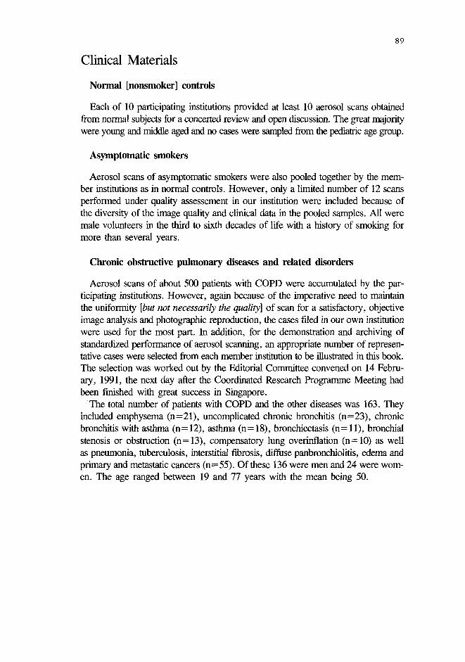
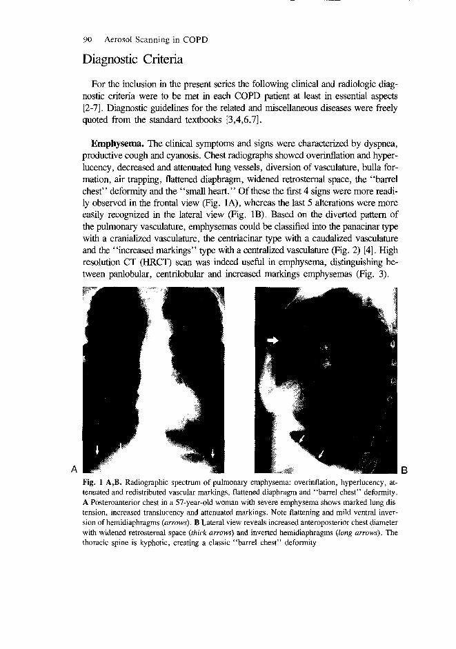
Emphysema. The clinical symptoms and signs were characterized by dyspnea,productive cough and cyanosis. Chest radiographs showed overinflation and hyper-lucency, decreased and attenuated lung vessels, diversion of vasculature, bulla for-mation, air trapping, flattened diaphragm, widened retrosternal space, the "barrelchest" deformity and the "small heart." Of these the first 4 signs were more readi-ly observed in the frontal view (Fig. 1A), whereas the last 5 alterations were moreeasily recognized in the lateral view (Fig. IB). Based on the diverted pattern ofthe pulmonary vasculature, emphysemas could be classified into the panacinar typewith a cranialized vasculature, the centriacinar type with a caudalized vasculatureand the "increased markings" type with a centralized vasculature (Fig. 2) [4]. Highresolution CT (HRCT) scan was indeed useful in emphysema, distinguishing be-tween panlobular, centrilobular and increased markings emphysemas (Fig. 3).
Fig. 1 A,B. Radiographic spectrum of pulmonary emphysema: overinflation, hyperlucency, at-tenuated and redistributed vascular markings, flattened diaphragm and "barrel chest" deformity.A Posteroanterior chest in a 57-year-old woman with severe emphysema shows marked lung dis-tension, increased translucency and attenuated markings. Note flattening and mild ventral inver-sion of hemidiaphragms {arrows). B Lateral view reveals increased anteroposterior chest diameterwith widened retrosternal space {thick arrows) and inverted hemidiaphragms {long arrows). Thethoracic spine is kyphotic, creating a classic "barrel chest" deformity
Y.W. Bahk and S.K. Chung 91
Fig. 2 A-C. Vascular redistribution in three different types of lung emphysema. A Posteroanteriorchest in a 74-year-old man with panacinar emphysema shows cranialized vascular markings {ar-rowheads) with air trap in the lower lungs. B Posteroanterior chest in a 69-year-old man withcentriacinar emphysema shows caudalized vascular markings {arrowheads) with air trap in the up-per lungs. C Posteroanterior chest in a 74-year-old man with "increased markings" type emphysemashows centralized vascular markings in midlungs with diffuse air trap {arrowheads)

Fig. 3 A-D. High resolution (HR) CT and histologic findings of centriacinar and panacinar em-physemas. A HRCT section through the upper lung level in a 69-year-old man with pulmonaryemphysema shows markedly dilated, ruptured acini, denoting centriacinar emphysema (see Fig.3C). B HRCT section through the midlung level shows dilated acini in panacinar emphysema (seeFig. 3D). C Histology of centriacinar emphysema reveals expanded, destroyed and coalesced aci-ni (a). D Histology of panacinar emphysema reveals dilated acini with relatively preserved ar-chitecture (a) and dilated respiratory bronchioli (RB)

Y.W. Bahk and S.K. Chung 93
Chronic bronchitis. Diagnostic criteria were productive cough for months formore than 2 consecutive years without wheezing or other lung diseases, normalor slightly increased IgE and normal or slightly decreased FEV. Radiographic changesincluded the "tram-line" shadow, the "bronchial cuff' sign, the "dirty lung" signand overinflation with vascular attenuation (Fig. 4), but these signs were not regu-larly seen. HRCT scan was very efficient in imaging thickened bronchial wall, thecharacteristic change that underlies the tram-line and bronchial cuff signs (Fig. 5)and bronchography demonstrated serrated appearance of the bronchial walls nearthe hilum, indicating glandular hypertrophy (Fig. 6).
Fig. 4. Radiographic manifestations of chronic bronchitis. Posteroanterior chest in a 45-year-old man with bronchitis shows a classic "tram line" shadow of thickened bronchi in the rightmedial basal lung (open arrows)
Fig. 5. CT scan manifestations of "tram line" and "bronchial cuff signs in chronic bronchitis.Transverse CT section through the hilum level in a 27-year-old woman with chronic bronchitisreveals thickened bronchi cut longitudinally (open arrow) and transsectionaJly (solid arrow), denotingthe "tram line" and "bronchial cuff signs, respectively
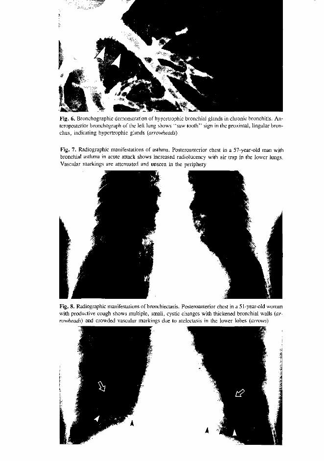
Fig. 6. Bronchographic demonstration of hypertrophic bronchial glands in chronic bronchitis. An-teroposterior bronchograph of the left lung shows "saw tooth" sign in the proximal, lingular bron-chus, indicating hypertrophic glands (arrowheads)
Fig. 7. Radiographic manifestations of asthma. Posteroanterior chest in a 57-year-old man withbronchial asthma in acute attack shows increased radiolucency with air trap in the lower lungs.Vascular markings are attenuated and unseen in the periphery
Fig. 8. Radiographic manifestations of bronchiectasis. Posteroanterior chest in a 51-year-old womanwith productive cough shows multiple, small, cystic changes with thickened bronchial walls (ar-rowheads) and crowded vascular markings due to atelectasis in the lower lobes (arrows)
Y.W. Bahk and S.K. Chung 95
Asthma. Typical symptoms and signs were paroxysms of dyspnea, cough andwheezing. They were not to be associated with a cardiac disease. Clinically, asth-ma was either in attack, in remission or resistant [status asthmaticus]. Radiographicalterations included hyperlucency and vascular attenuation (Fig. 7). During an at-tack and in resistant form, diaphragmatic flattening and retrosternal widening couldbe seen. There had to be no other lung diseases.
Bronchiectasis. The basic clinical symptoms and signs in this condition wereproductive cough with purulent or bloody sputum, chest pain and dyspnea. Radio-graphic alterations were bronchial dilatation, cystic formation and lung collapse,typically in the middle and lower lobes (Fig. 8). Bronchography and HRCT scanwere confirmatory, demonstrating bronchial dilatation and obstruction, glandularhypertrophy and lung collapse (Fig. 9).
Bronchia] obstruction. Only complete obstruction and marked bronchostenosiswere included in this series. It was either in the segmental, lobar or mainstem bronchiand their causes were chronic inflammation, tuberculosis and tumour.The diagno-sis was primarily based on the radiographic demonstration of obstruction inthe bronchus and/or lung collapse (Fig. 10) and on the conventional X-ray tomo-graphy, bronchoscopy or bronchography in some cases.
Overinflation or "compensatory emphysema". The condition was defined asan excessive yet essentially reversible or deflatable inflation [not irreversible as intrue emphysema that has alveolar destruction] of alveolar spaces in compensationfor a decrease in or the loss of lung volume due to bronchial obstruction, pulmo-nary fibrosis, lung resection or fibrothorax. In many the condition was long-standingand had associated COPD. The level of overinflation was segmental or lobar orin a lung. Radiographic alterations included, in addition to bronchial obstructionand the associated lung collapse, an increase in lung volume with hyperlucency andattenuated vascular markings (Fig. 10). Evidence of either lung collapse, missinglung or fibrothorax was present. CT scan was useful in detecting focal overinflation.
Miscellaneous diseases included acute pneumonia, tuberculosis, interstitial fibrosis,diffuse panbronchiolitis, edema and primary and metastatic cancers.

Fig. 9 A,B- Bronchographic and CT scan manifestations of bronchiectasis. A Posteroanterior bron-chograph in a 27-year-old woman shows irregularly dilated, clubbed bronchi in both middle andlower lungs. The bronchi appear crowded due to atelectasis and bronchial glands hypettrophic{arrowheads). B HRCT section through the lower lung level shows dilated, crowded bronchi inthe lower lungs {arrows) with some cystic changes on the left {arrowheads)
Fig. 10. Radiographic sign of bronchial obstruction. Posteroanterior chest in a 69-year-old manwith lung tuberculosis shows obstruction involving the right main stem bronchus {open arrow)with ipsilateral lung collapse

Y.W. Bahk and S.K. Chung 97
Scan Methods
Aerosol scan was performed according to the protocol provided, but with a minormodification so that more radioactive counts could be accumulated at the expenseof a longer scan time. Each subject inhaled, in a resting tidal breathing state, sub-micronic aerosol of 99mTc phytate through a mask for 5 min in a sitting position[1]. The radioactivity inhaled was approximately 3 mCi (111 MBq). Aerosol wasgenerated afresh each time by using a BARC nebulizer after the instillation of 15mCi (555 MBq) of 99mTc phytate. After gargling and rinsing the esophagus withdrinking water, scanning was performed with the subject lying on a scan couch,in the anterior, posterior and both lateral positions. The examination was supplementedby oblique views when necessary. A total of 600K counts was accumulated overa period of 10 min per view. It was twice as much total counts as the recommend-ed count of 300K. The aim of this increment in counts was to obtain aerosol scanswith improved image quality by maximally eliminating the background noise. Thesensitivity of detector was suppressed by setting the filter at the near baseline level.The gamma cameras we used were Siemens Scintiview II (Model ZLC 7500S) andOrbiter (Model 6601).
Observations on Scan Alterations
The following observations on scan alterations were made in terms of the gradeand pattern of the abnormal aerosol deposition in both the bronchial tree and alveo-lar air spaces. The abnormal deposition was increased in the bronchi and increased,decreased or absent in the air spaces. Abnormalities were graded arbitrarily intomild, moderate and marked. Altered bronchi were also assessed in terms of dilata-tion and obstruction. They were clubbed, bulbous or withered-bough-like in ap-pearance when dilated.
Semiquantitative assessment of scan alterations was possible using the traditionalcriteria: Grade I for the area of involvement less than 25% of the total lung, GradeII between 25% and 50% and Grade HI more than 50%. However, we found itmore practical to grade the extent by vertically dividing the lung into the central,middle and peripheral thirds by 2 laterally curved, equidistant lines drawn parallelto the innermost aspect of the lateral chest wall. The central or parahilar zone in-cluded the mainstem and large bronchi in and about the hila, the middle zone themedium-sized bronchi and the peripheral zone mainly the alveolar air spaces.
98 Aerosol Scanning in COPD
Results
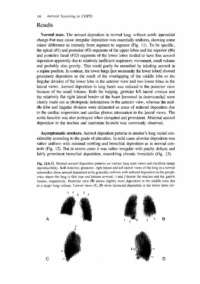
Normal scan. The aerosol deposition in normal lung without senile interstitialchange that may cause irregular deposition was essentially uniform, showing someminor difference in intensity from segment to segment (Fig. 11). To be specific,the apical (#1) and posterior (#3) segments of the upper lobes and the superior (#6)and posterior basal (#10) segments of the lower lobes tended to have less aerosoldeposition apparently due to relatively inefficient respiratory movement, small volumeand probably also gravity. This could partly be remedied by inhaling aerosol ina supine position. In contrast, the lower lungs [not necessarily the lower lobes] showedprominent deposition as the result of the overlapping of the middle lobe or thelingular division of the lower lobe in the anterior view and two lower lobes in thelateral views. Aerosol deposition in lung bases was reduced in the posterior viewbecause of the small volume. Both the bulging, globular left lateral contour andthe relatively flat right lateral border of the heart [reversed in dextrocardia] wereclearly made out as photopenic indentations in the anterior view, whereas the mid-dle lobe and lingular division were delineated as areas of reduced deposition dueto the cardiac impression and cardiac photon attenuation in the lateral views. Theaortic knuckle was also portrayed when elongated and prominent. Minimal aerosoldeposition in the trachea and mainstem bronchi was commonly observed.
Asymptomatic smokers. Aerosol deposition patterns in smoker's lung varied con-siderably according to the grade of alteration. In mild cases alveolar deposition wasrather uniform with minimal mottling and bronchial deposition as in normal con-trols (Fig. 12). But in severe cases it was rather irregular with patchy defects andfairly prominent bronchial deposition, resembling chronic bronchitis (Fig. 13).
Fig. 11A-G. Normal aerosol deposition patterns on various lung scan views and excellent imagereproducibility. A-D Anterior, posterior, right lateral and left lateral views of the lung in a normalnonsmoker show aerosol deposition to be generally uniform with reduced deposition in the periph-eries where the lung is thin (top and bottom arrows), t and/denote the trachea and the gastricfundus, respectively. Posterior view (B) shows slightly more deposition in the middle zone dueto a larger lung volume. Lateral views (C, D) show increased deposition in the lower lobes (ar-
B
D

Y.W. Bahk and S.K. Chung 99
l h r 3hrs
row). Note decreased deposition in the lingular division (/). E-G Serial scans of another asympto-matic nonsmoker's lung taken 30 min, 1 hr and 3 hr postinhalation show homogeneous depositionthroughout both lungs with reduced radioactivities in peripheries. /, 5, d denote the gastric fundus,stomach body and duodenum, respectively. Note excellent reproducibility of three images
A B
Fig. 12 A,B- Smoker's lung [mild change]. A Anterior lung scan in a 30-year-old asymptomaticman with a history of 5 pack year's smoking reveals very minimal bronchial deposition in theleft hilum (arrow). B Posterior lung scan reveals slightly more hilar deposition (arrows)
Fig. 13 A,B- Smoker's lung [moderate change]. A Anterior scan in a 46-year-old man with produc-tive cough and a history of 1/3 pack/day smoking for 30 years reveals prominent, tracheobronchi-al deposition in both hilar regions (arrows). Note strong resemblance to chronic bronchitis whichcan be the case. B Posterior lung scan reveals more prominent aerosol deposition (arrows)
B
100 Aerosol Scanning in COPD
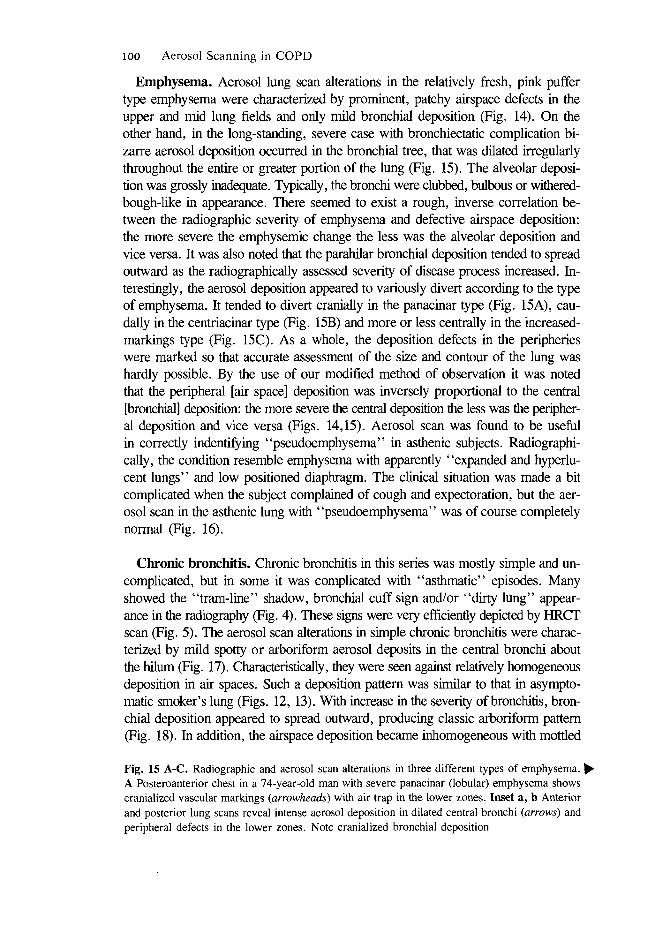
Emphysema. Aerosol lung scan alterations in the relatively fresh, pink puffertype emphysema were characterized by prominent, patchy airspace defects in theupper and mid lung fields and only mild bronchial deposition (Fig. 14). On theother hand, in the long-standing, severe case with bronchiectatic complication bi-zarre aerosol deposition occurred in the bronchial tree, that was dilated irregularlythroughout the entire or greater portion of the lung (Fig. 15). The alveolar deposi-tion was grossly inadequate. Typically, the bronchi were clubbed, bulbous or withered-bough-like in appearance. There seemed to exist a rough, inverse correlation be-tween the radiographic severity of emphysema and defective airspace deposition:the more severe the emphysemic change the less was the alveolar deposition andvice versa. It was also noted that the parahilar bronchial deposition tended to spreadoutward as the radiographically assessed severity of disease process increased. In-terestingly, the aerosol deposition appeared to variously divert according to the typeof emphysema. It tended to divert cranially in the panacinar type (Fig. 15A), cau-dally in the centriacinar type (Fig. 15B) and more or less centrally in the increased-markings type (Fig. 15C). As a whole, the deposition defects in the peripherieswere marked so that accurate assessment of the size and contour of the lung washardly possible. By the use of our modified method of observation it was notedthat the peripheral [air space] deposition was inversely proportional to the central[bronchial] deposition: the more severe the central deposition the less was the peripher-al deposition and vice versa (Figs. 14,15). Aerosol scan was found to be usefulin correctly indentifying "pseudoemphysema" in asthenic subjects. Radiographi-cally, the condition resemble emphysema with apparently "expanded and hyperlu-cent lungs" and low positioned diaphragm. The clinical situation was made a bitcomplicated when the subject complained of cough and expectoration, but the aer-osol scan in the asthenic lung with "pseudoemphysema" was of course completelynormal (Fig. 16).
Chronic bronchitis. Chronic bronchitis in this series was mostly simple and un-complicated, but in some it was complicated with "asthmatic" episodes. Manyshowed the "tram-line" shadow, bronchial cuff sign and/or "dirty lung" appear-ance in the radiography (Fig. 4). These signs were very efficiently depicted by HRCTscan (Fig. 5). The aerosol scan alterations in simple chronic bronchitis were charac-terized by mild spotty or arboriform aerosol deposits in the central bronchi aboutthe hilum (Fig. 17). Characteristically, they were seen against relatively homogeneousdeposition in air spaces. Such a deposition pattern was similar to that in asympto-matic smoker's lung (Figs. 12, 13). With increase in the severity of bronchitis, bron-chial deposition appeared to spread outward, producing classic arboriform pattern(Fig. 18). In addition, the airspace deposition became inhomogeneous with mottled
Fig. 15 A-C. Radiographic and aerosol scan alterations in three different types of emphysema. •A Posteroanterior chest in a 74-year-old man with severe panacinar (lobular) emphysema showscranialized vascular markings (arrowheads) with air trap in the lower zones. Inset a, b Anteriorand posterior lung scans reveal intense aerosol deposition in dilated central bronchi (arrows) andperipheral defects in the lower zones. Note cranialized bronchial deposition
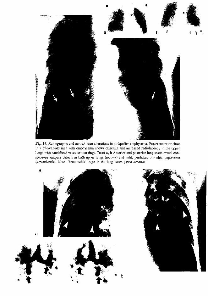
Fig. 14. Radiographic and aerosol scan alterations in pinkpuffer emphysema. Posteroanterior chestin a 62-year-old man with emphysema shows oligemia and increased radiolucency in the upperlungs with caudalized vascular markings. Inset a, b Anterior and posterior lung scans reveal con-spicuous airspace defects in both upper lungs (arrows) and mild, perihilar, bronchial deposition(arrowheads). Note "broomstick" sign in the Jung bases (open arrows)
B
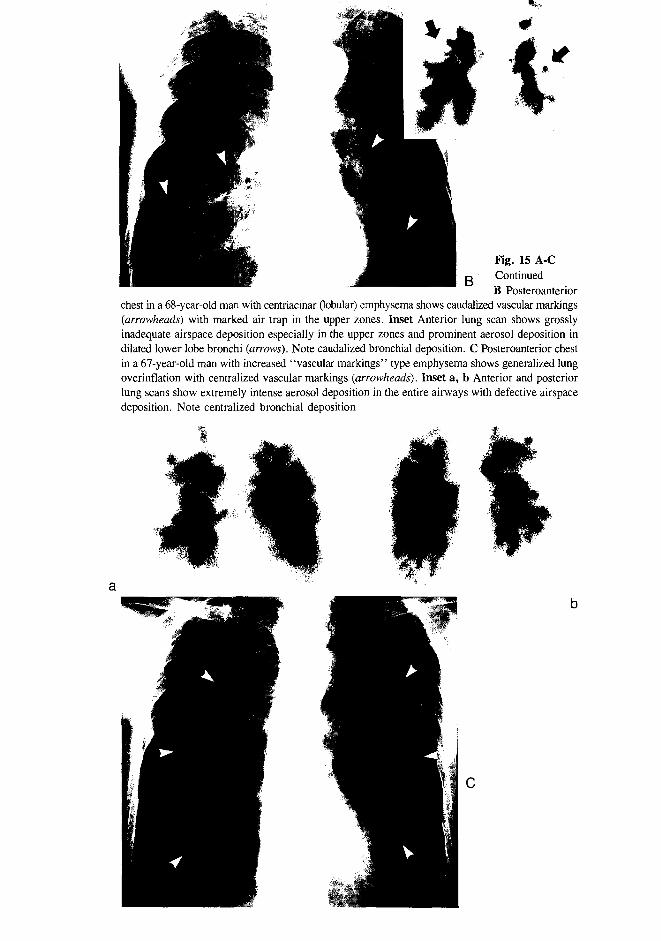
Fig. 15 A-CContinuedB Posteroanterior
chest in a 68-year-old man with centriacinar (lobular) emphysema shows caudalized vascular markings{arrowheads) with marked air trap in the upper zones. Inset Anterior lung scan shows grosslyinadequate airspace deposition especially in the upper zones and prominent aerosol deposition indilated lower lobe bronchi {arrows). Note caudalized bronchial deposition. C Posteroanterior chestin a 67-year-old man with increased "vascular markings" type emphysema shows generalized lungoverinflation with centralized vascular markings {arrowheads). Inset a, b Anterior and posteriorlung scans show extremely intense aerosol deposition in the entire airways with defective airspacedeposition. Note centralized bronchial deposition
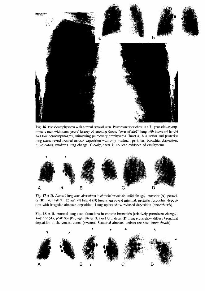
Fig. 16. Pseudoemphysema with normal aerosol scan. Posteroanterior chest in a 31-year-old, asymp-tomatic man with many years' history of smoking shows "overinflated" lung with increased heightand low hemidiaphragms, mimicking pulmonary emphysema. Inset a, b Anterior and posteriorlung scans reveal normal aerosol deposition with only minimal, perihilar, bronchial deposition,representing smoker's lung change. Clearly, there is no scan evidence of emphysema
Fig. 17 A-D. Aerosol lung scan alterations in chronic bronchitis [mild change]. Anterior (A), posteri-or (B), right lateral (C) and left lateral (D) lung scans reveal minimal, perihilar, bronchial deposi-tion with irregular airspace deposition. Lung apices show reduced deposition {arrowheads)
Fig. 18 A-D. Aerosol lung scan alterations in chronic bronchitis [relatively prominent change].Anterior (A), posterior (B), right lateral (C) and left lateral (D) lung scans show diffuse bronchialdeposition in the central zones {arrows). Scattered airspace defects are seen {arrowheads)
104 Aerosol Scanning in COPD
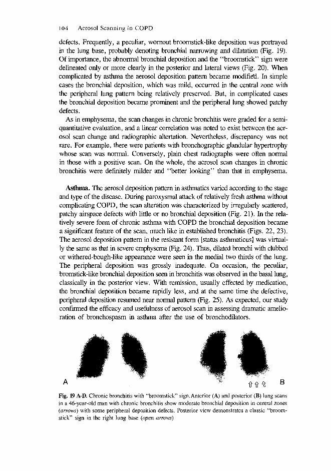
defects. Frequently, a peculiar, wornout broomstick-like deposition was portrayedin the lung base, probably denoting bronchial narrowing and dilatation (Fig. 19).Of importance, the abnormal bronchial deposition and the "broomstick" sign weredelineated only or more clearly in the posterior and lateral views (Fig. 20). Whencomplicated by asthma the aerosol deposition pattern became modifie'd. In simplecases the bronchial deposition, which was mild, occurred in the central zone withthe peripheral lung pattern being relatively preserved. But, in complicated casesthe bronchial deposition became prominent and the peripheral lung showed patchydefects.
As in emphysema, the scan changes in chronic bronchitis were graded for a semi-quantitative evaluation, and a linear correlation was noted to exist between the aer-osol scan change and radiographic altertation. Nevertheless, discrepancy was notrare. For example, there were patients with bronchographic glandular hypertrophywhose scan was normal. Conversely, plain chest radiographs were often normalin those with a positive scan. On the whole, the aerosol scan changes in chronicbronchitis were definitely milder and "better looking" than that in emphysema.
Asthma. The aerosol deposition pattern in asthmatics varied according to the stageand type of the disease. During paroxysmal attack of relatively fresh asthma withoutcomplicating COPD, the scan alteration was characterized by irregularly scattered,patchy airspace defects with little or no bronchial deposition (Fig. 21). In the rela-tively severe form of chronic asthma with COPD the bronchial deposition becamea significant feature of the scan, much like in established bronchitis (Figs. 22, 23).The aerosol deposition pattern in the resistant form [status asthmaticus] was virtual-ly the same as that in severe emphysema (Fig. 24). Thus, dilated bronchi with clubbedor withered-bough-like appearance were seen in the medial two thirds of the lung.The peripheral deposition was grossly inadequate. On occasion, the peculiar,bromstick-like bronchial deposition seen in bronchitis was observed in the basal lung,classically in the posterior view. With remission, usually effected by medication,the bronchial deposition became rapidly less, and at the same time the defective,peripheral deposition resumed near normal pattern (Fig. 25). As expected, our studyconfirmed the efficacy and usefulness of aerosol scan in assessing dramatic amelio-ration of bronchospasm in asthma after the use of bronchodilators.
B
Fig. 19 A-D. Chronic bronchitis with "broomstick" sign. Anterior (A) and posterior (B) lung scansin a 46-year-old man with chronic bronchitis show moderate bronchial deposition in central zones(arrows) with some peripheral deposition defects. Posterior view demonstrates a classic "broom-stick" sign in the right lung base (open arrows)
Y.W. Bahk and S.K. Chung 105
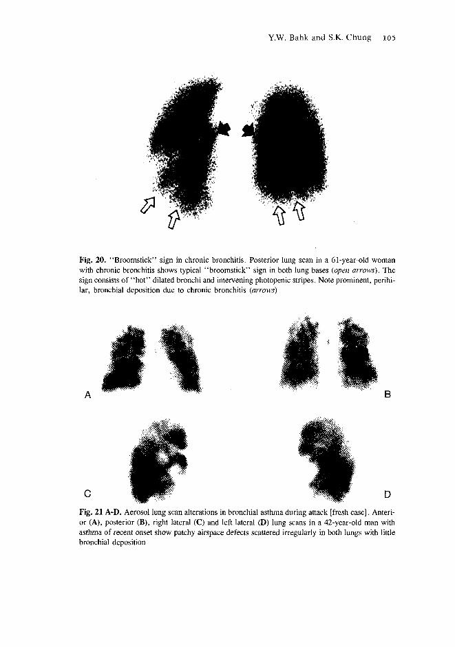
Fig. 20. "Broomstick" sign in chronic bronchitis. Posterior lung scan in a 61-year-old womanwith chronic bronchitis shows typical "broomstick" sign in both lung bases {open arrows). Thesign consists of "hot" dilated bronchi and intervening photopenic stripes. Note prominent, perihi-lar, bronchial deposition due to chronic bronchitis {arrows)
A B
D
Fig. 21 A-D. Aerosol lung scan alterations in bronchial asthma during attack [fresh case]. Anteri-or (A), posterior (B), right lateral (C) and left lateral (D) lung scans in a 42-year-old man withasthma of recent onset show patchy airspace defects scattered irregularly in both lungs with littlebronchial deposition
106 Aerosol Scanning in COPD
B
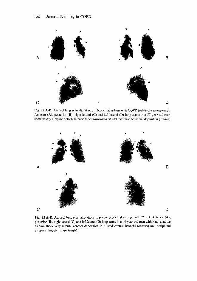
Fig. 22 A-D. Aerosol lung scan alterations in bronchial asthma with COPD [relatively severe case].Anterior (A), posterior (B), right lateral (C) and left lateral (D) lung scans in a 57-year-old manshow patchy airspace defects in peripheries (arrowheads) and moderate bronchial deposition (arrows)
B
D
Fig. 23 A-D. Aerosol lung scan alterations in severe bronchial asthma with COPD. Anterior (A),posterior (B), right lateral (C) and left lateral (D) lung scans in a 44-year-old man with long-standingasthma show very intense aerosol deposition in dilated central bronchi (arrows) and peripheralairspace defects (arrowheads)

Y.W. Bahk and S.K. Chung 107
A B
D
Fig. 24 A-D. Aerosol lung scan alterations in status asthmaticus. Anterior (A), posterior (B), rightlateral (C) and left lateral (D) lung scans in a 22-year-old woman with resistant asthma show promi-nent aerosol deposition in dilated bronchi throughout both lungs with grossly reduced airspace depo-sition. The scan changes are practically indistinguishable from those of emphysema
B
Fig. 25 A-D. Aerosol lung scan alterations in severe bronchial asthma during an attack and afterbronchodilator treatment. A, B Anterior and posterior lung scans in a 70-year-old man with long-standing bronchial asthma during an attack show intense aerosol deposition in dilated tracheobron-chial tree (arrows). Note grossly inadequate airspace deposition. C, D Anterior and posterior lungscans following the use of bronchodilators show remarkable improvement of scan alterations withmuch normalized airspace deposition
108 Aerosol Scanning in COPD
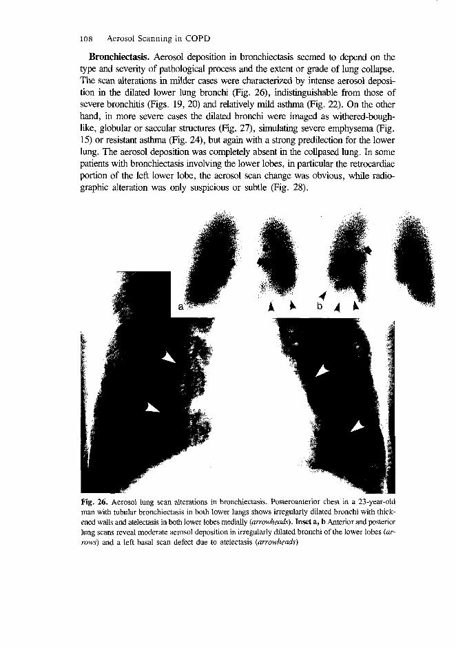
Bronchiectasis. Aerosol deposition in bronchiectasis seemed to depend on thetype and severity of pathological process and the extent or grade of lung collapse.The scan alterations in milder cases were characterized by intense aerosol deposi-tion in the dilated lower lung bronchi (Fig. 26), indistinguishable from those ofsevere bronchitis (Figs. 19, 20) and relatively mild asthma (Fig. 22). On the otherhand, in more severe cases the dilated bronchi were imaged as withered-bough-like, globular or saccular structures (Fig. 27), simulating severe emphysema (Fig.15) or resistant asthma (Fig. 24), but again with a strong predilection for the lowerlung. The aerosol deposition was completely absent in the collpased lung. In somepatients with bronchiectasis involving the lower lobes, in particular the retrocardiacportion of the left lower lobe, the aerosol scan change was obvious, while radio-graphic alteration was only suspicious or subtle (Fig. 28).
Fig. 26. Aerosol lung scan alterations in bronchiectasis. Posteroanterior chest in a 23-year-oldman with tubular bronchiectasis in both lower lungs shows irregularly dilated bronchi with thick-ened walls and atelectasis in both lower lobes medially (arrowheads). Inset a, b Anterior and posteriorlung scans reveal moderate aerosol deposition in irregularly dilated bronchi of the lower lobes (ar-rows) and a left basal scan defect due to atelectasis (arrowheads)

Fig. 27. Aerosol lung scan alterations in severe bronchiectasis. Posteroanterior chest in a 45-year-old man with cystic bronchiectasis shows extensive, variously sized, cystic shadows with air fluidlevels in some. Inset a, b Anterior and posterior lung scans reveal intense aerosol deposition inirregularly dilated bronchi along with multiple airspace deposition defects
Fig. 28. Obvious aerosol scan change in radiographically subtle bronchiectasis in the retrocardiacregion. Posteroanterior chest in a 31-year-old man with rather subtle bronchiectasis in the left low-er lobe (arrowheads). The change is obscured by the overlying heart shadow. Inset Posterior lungscan reveals prominent aerosol deposition in the dilated left lower lobe bronchi, clearly indicatingbronchiectasis (arrows)
110 Aerosol Scanning in COPD
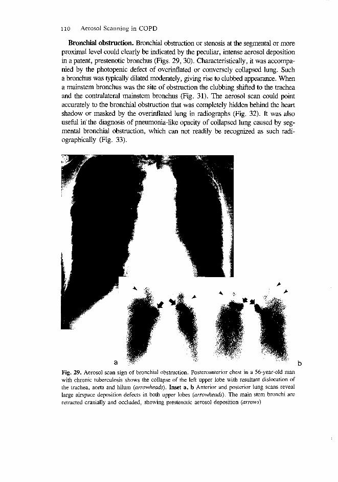
Bronchial obstruction. Bronchial obstruction or stenosis at the segmental or moreproximal level could clearly be indicated by the peculiar, intense aerosol depositionin a patent, prestenotic bronchus (Figs. 29, 30). Characteristically, it was accompa-nied by the photopenic defect of overinflated or conversely collapsed lung. Sucha bronchus was typically dilated moderately, giving rise to clubbed appearance. Whena mainstem bronchus was the site of obstruction the clubbing shifted to the tracheaand the contralateral mainstem bronchus (Fig. 31). The aerosol scan could pointaccurately to the bronchial obstruction that was completely hidden behind the heartshadow or masked by the overinflated lung in radiographs (Fig. 32). It was alsouseful in' the diagnosis of pneumonia-like opacity of collapsed lung caused by seg-mental bronchial obstruction, which can not readily be recognized as such radi-ographically (Fig. 33).
Fig. 29. Aerosol scan sign of bronchial obstruction. Posteroanterior chest in a 56-year-old manwith chronic tuberculosis shows the collapse of the left upper lobe with resultant dislocation ofthe trachea, aorta and hilum (arrowheads). Inset a, b Anterior and posterior lung scans reveallarge airspace deposition defects in both upper lobes {arrowheads). The main stem bronchi areretracted cranially and occluded, showing prestenotic aerosol deposition (arrows)
Y.W. Bahk and S.K. Chung 111
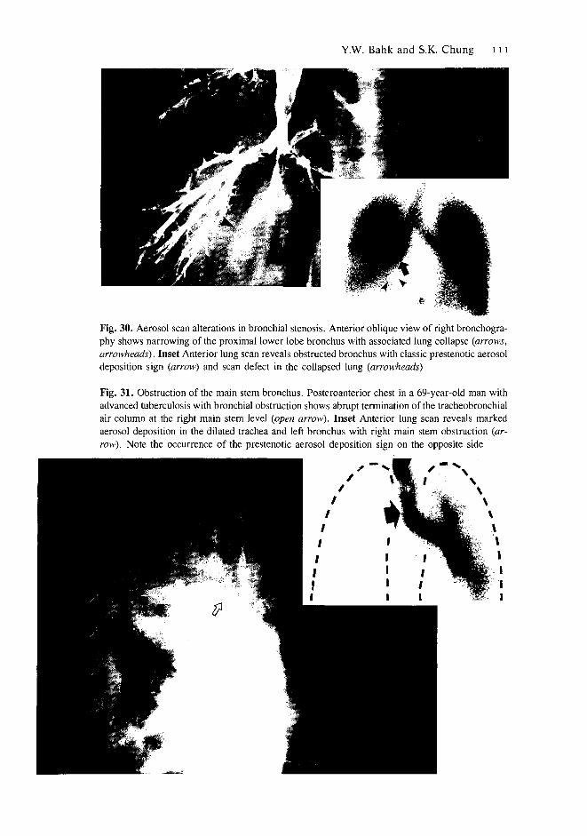
Fig. 30. Aerosol scan alterations in bronchial stenosis. Anterior oblique view of right bronchogra-phy shows narrowing of the proximal lower lobe bronchus with associated lung collapse (arrows,arrowheads). Inset Anterior lung scan reveals obstructed bronchus with classic prestenotic aerosoldeposition sign (arrow) and scan defect in the collapsed lung (arrowheads)
Fig. 31. Obstruction of the main stem bronchus. Posteroanterior chest in a 69-year-old man withadvanced tuberculosis with bronchial obstruction shows abrupt termination of the tracheobronchialair column at the right main stem level (open arrow). Inset Anterior lung scan reveals markedaerosol deposition in the dilated trachea and left bronchus with right main stem obstruction (ar-row). Note the occurrence of the prestenotic aerosol deposition sign on the opposite side

Fig. 32. The prestenotic aerosol deposition sign in obscure bronchial obstruction. Posteroanteriorchest in a 31-year-old man with right lower lobe collapse shows a paracardiac, triangular opacitythrough the heart shadow {arrowheads). Note also the upper mediastinal sign of lower lobe col-lapse (arrow). Inset Posterior lung scan reveals classic prestenotic aerosol deposition sign in theright lower lobe bronchus that is obstructed (arrow)
Fig. 33. The prestenotic aerosol deposition sign in segmental bronchial obstruction. Posteroanteri-or chest in a 61-year-old woman shows homogeneous opacity in the right lower lung, producingthe cardiac silhouette sign (arrowheads). The opacity is blurred by the overlying breast shadow.Inset a, b Anterior and right lateral lung scans reveal prestenotic aerosol deposition in the middlelobe bronchus (arrows). Distal lung is photopenic due to collapse. An area of increased aerosoldeposition is seen in the adjacent basal segment, denoting air trap (arrowheads)
a

Y.W. Bahk and S.K. Chung 113
Overinflation or compensatory emphysema. Overinflation was either simpleor complicated with COPD. Etiologically, the great majority of cases were relatedwith bronchial obstruction and lung collapse and some with focal pulmonary fibro-sis or diffuse fibrothorax. The aerosol deposition pattern in the overinflated lungwas characteristically uniform, but the intensity was variable according to the sizeof the lung overinflated or conversely collapsed. Thus, it appeared normal whenthe lung collapse was lobar or segmental (Fig. 34), increased when the collapsewas large- or multi-lobar (Fig. 35) and decreased when the collapse was more ex-tensive, e.g. one whole lung (Fig. 31). Little or no bronchial deposition was presentin the overinflated lung without COPD, but the lung with COPD showed bronchialdeposition, the degrees of which varied according to the type and severity of thedisease. The overinflated lung with chronic bronchitis showed minimal bronchialdeposition (Fig. 34), whereas the overinflated lung with severe COPD demonstrat-ed bizarre bronchial deposition with grossly defective airspace deposition (Fig. 31).The bronchial obstruction responsible for overinflation was often indicated clearlyby the aerosol deposited in a dilated, prestenotic bronchus (Fig. 34). We studied2 cases of compensatory overinflation occurring in one whole lung as a result offibrothorax with restrictive lung collapse in the contralateral hemithorax. The aero-sol deposition was homogeneously increased in airspaces but no deposition was seenin the bronchi (Fig. 36). Typically, the lung in the fibrosed hemithorax was con-stricted and small. With marked overinflation of one lung, its medial portion her-niated into the collapsed hemithorax through the anterior mediastinal space.
Fig. 34. Normal or near-normal aerosoldeposition pattern in lobar overinflation.Posteroanterior chest in a 57-year-oldman with tuberculosis in the right upperlobe shows atelectasis (a) and com-pensatory overinflation of the remaininglung (arrowheads). Inset Anterior lungscan reveals a large photopenic defectin the collapsed right upper lobe(arrowheads) and almost normal aerosoldeposition in theoverinflated lung.Note the classicprestenotic aerosoldeposition sign in theright upper lobebronchus that isobstructed (arrow)
114
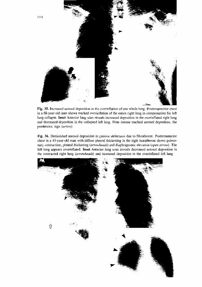
Fig. 35. Increased aerosol deposition in the overinflation of one whole lung. Posteroanterior chestin a 68-year-old man shows marked overinflation of the entire right lung in compensation for leftlung collapse. Inset Anterior lung scan reveals increased deposition in the overinflated right lungand decreased deposition in the collapsed left lung. Note intense tracheal aerosol deposition, theprestenotic sign (arrow)
Fig. 36. Diminished aerosol deposition in passive atelectasis due to fibrothorax. Posteroanteriorchest in a 43-year-old man with diffuse pleural thickening in the right hemithorax shows pulmo-nary contraction, pleural thickening (arrowheads) and diaphragmatic elevation (open arrow). Theleft lung appears overinflated. Inset Anterior lung scan reveals decreased aerosol deposition inthe contracted right lung (arrowheads) and increased deposition in the overinflated left lung
Y.W. Bahk and S.K. Chung 115
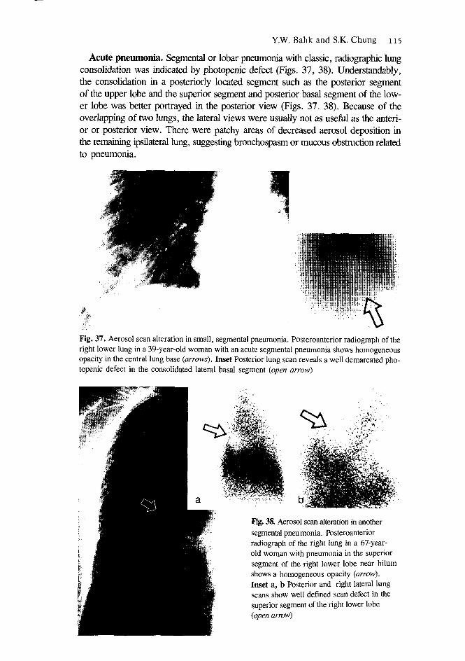
Acute pneumonia. Segmental or lobar pneumonia with classic, radiographic lungconsolidation was indicated by photopenic defect (Figs. 37, 38). Understandably,the consolidation in a posteriorly located segment such as the posterior segmentof the upper lobe and the superior segment and posterior basal segment of the low-er lobe was better portrayed in the posterior view (Figs. 37. 38). Because of theoverlapping of two lungs, the lateral views were usually not as useful as the anteri-or or posterior view. There were patchy areas of decreased aerosol deposition inthe remaining ipsilateral lung, suggesting bronchospasm or mucous obstruction relatedto pneumonia.
Fig. 37. Aerosol scan alteration in small, segmental pneumonia. Posteroanterior radiograph of theright lower lung in a 39-year-old woman with an acute segmental pneumonia shows homogeneousopacity in the central lung base {arrows). Inset Posterior lung scan reveals a well demarcated pho-topenic defect in the consolidated lateral basal segment {open arrow)
^ : |
Fig. 38. Aerosol scan alteration in anothersegmental pneumonia. Posteroanteriorradiograph of the right lung in a 67-year-old woman with pneumonia in the superiorsegment of the right lower lobe near hilumshows a homogeneous opacity (arrow).Inset a, b Posterior and right lateral lungscans show well defined scan defect in thesuperior segment of the right lower lobe{open arrow)

116 Aerosol Scanning in COPD
Tuberculosis. The spectrum of pulmonary tuberculosis was variable indeed, rang-ing from local pneumonia-like type with lung consolidation to chronic type withirregular fibrocystic, cavitary and atelectatic changes. The scan change in the form-er type was simple photopenic defect, whereas the changes were bizarre in the lat-ter type, showing clubbed or withered-bough-like bronchi with grossly defectiveperipheral deposition as in severe emphysema or bronchiectasis (Fig. 39). It wasimpressive that rather minimal, old, fibrocalcific tuberculosis was portayed as anobvious "cold" defect (Fig. 40).
Fig. 39. Aerosol scan alteration in fibrocystic, cavitary, atelectatic tuberculosis. Posteroanteriorchest in a 65-year-old man with tuberculosis shows fibrosis, cysts and cavities with atelectasis inboth upper lobes (arrows). Both hila are shifted cranially due to atelectasis. Inset a, b Anteriorand posterior lung scans show large aerosol deposition defects in the upper zones bilaterally. Moder-ate, bronchial deposition can be seen in irregularly dilated, central bronchi. Note the mismatchbetween radiographic and scintigraphic findings and the coexistence of deposition defects and bi-zarre bronchial deposition
Y.W. Bahk and S.K. Chung 117
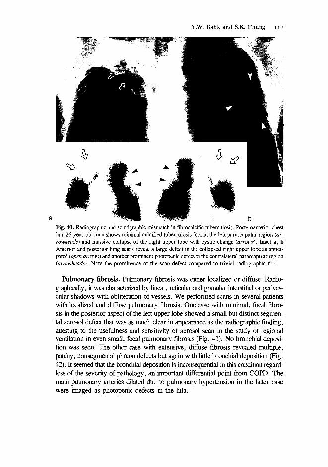
Fig. 40. Radiographic and scintigraphic mismatch in fibrocalcific tuberculosis. Posteroanterior chestin a 26-year-old man shows minimal calcified tuberculosis foci in the left parascapular region (ar-rowheads) and massive collapse of the right upper lobe with cystic change (arrows). Inset a, bAnterior and posterior lung scans reveal a large defect in the collapsed right upper lobe as antici-pated (open arrows) and another prominent photopenic defect in the contralateral parascapular region(arrowheads). Note the prominence of the scan defect compared to trivial radiographic foci
Pulmonary fibrosis. Pulmonary fibrosis was either localized or diffuse. Radio-graphically, it was characterized by linear, reticular and granular interstitial or perivas-cular shadows with obliteration of vessels. We performed scans in several patientswith localized and diffuse pulmonary fibrosis. One case with minimal, focal fibro-sis in the posterior aspect of the left upper lobe showed a small but distinct segmen-tal aerosol defect that was as much clear in appearance as the radiographic finding,attesting to the usefulness and sensitivity of aerosol scan in the study of regionalventilation in even small, focal pulmonary fibrosis (Fig. 41). No bronchial deposi-tion was seen. The other case with extensive, diffuse fibrosis revealed multiple,patchy, nonsegmental photon defects but again with little bronchial deposition (Fig.42). It seemed that the bronchial deposition is inconsequential in this condition regard-less of the severity of pathology, an important differential point from COPD. Themain pulmonary arteries dilated due to pulmonary hypertension in the latter casewere imaged as photopenic defects in the hila.

Fig. 41 A-E. Aerosol scan alterations in diffuse idiopathic pulmonary fibrosis. A Posteroanteriorchest of a 67-year-old man with diffuse interstitial fibrosis shows extensive linear, nodular, coarselyreticular shadows in the entire lung fields. B-E Anterior, posterior and both lateral lung scans re-veal patchy photopenic defects with little bronchial deposition. Note relatively well preserved scanpattern in the presence of overt radiographic alterations, distinguishing from COPD
Fig. 42. Aerosol scan alterations in another case of diffuse pulmonary fibrosis. Posteroanteriorchest of 47-year-old man with idiopathic interstitial fibrosis shows linear, nodular, reticular andmottled shadows in both lungs. Inset Anterior lung scan shows remarkably well preserved aerosoldeposition pattern with borderline hilar deposition (arrowheads), g denotes gastric fundus

Y.W. Bahk and S.K. Chung 119
Diffuse panbronchiolitis. This is a relatively new entity, clinically manifestingproductive cough and exertional dyspnea like COPD. The disease is progressivein nature and not infrequently fatal in outcome. The radiographic manifestationsincluded small nodular and mottled densities diffusely scattered throughout both lungswith some air trapping and occasional cor pulmonale (Fig. 43). The role of highresolution CT was decisive in making the diagnosis. Characteristically, it demon-strates centrilobular nodules and branched linear shadows of attenuation in theperipheral lung (Fig. 43). We performed aerosol lung scans in 2 cases with thisdisease, and could observe moderately severe, nonsegmental, airspace defects, resem-bling uncomplicated asthma and diffuse pulmonary fibrosis. In addition and impor-tantly, there were seen fairly intense aerosol deposition in the bronchi in the middleand peripheral lungs (Fig. 43). Unlike the central localization of the bronchial changesin COPD, middle and peripheral localization of the airway changes appeared tobe typical of this condition.
Lung edema. Radiographically, edema manifested homogeneous lung opacifi-cation due to airspace consolidation, typically in the lower two thirds of the lung.The lung edema of cardiac or renal origin was attended by cardiomegaly (Fig. 44).Aeorsol scan showed photon defects in the lung periphery which corresponded tothe lung opacities shown in radiograph when they were larger than a segment insize (Fig. 44). The aerosol scan in 2 cases showed extremely intense aerosol depositonin the trachea and the main stem bronchi. In one of them an aerosol scan was madebefore sudden development of lung edema, and the scan revealed the tracheobron-chial tree to be completely free from such deposition, implying that the subsequentdeposition was due to congestive failure (Fig. 45).
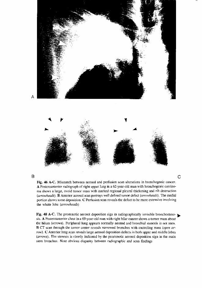
Bronchogenic carcinoma. The tumour in the peripheral lung having a sufficientdimension, say a few cm or larger, with patent bronchi about it could be depictedas a well demarcated photopenic defect (Fig. 46). More often than not, however,because of its bronchial origin, the bronchogenic carcinoma in the central lung tendedto occlude the bronchus at a relatively early stage, readily causing either lung em-physema or collapse. Scintigraphically, such a tumour could not be portrayed asseparate mass even when radiographically obvious. Instead, the entire complex ofthe tumour and either emphysemic or collapsed lung was delineated as a single pho-topenic defect that was much larger in size than each component disease (Figs. 47,48). The aerosol scan was able to indirectly but clearly indicate bronchial stenosiswith local obstructive emphysema or colllapse, a clinical situation that is not alwayseasy to assess by radiography (Figs. 47, 48). Thus, on the one hand the emphyse-ma produced by check-valve type bronchial obstruction was radiographically "wellaerated", but was paradoxically photopenic in the scan (Fig. 47). On the other hand,as in bronchostenosis of other causes, the stenosis was indicated by the aerosol in-tensely deposited in a prestenotic bronchus (Fig. 48).

Fig. 43 A-D. Aerosol scan alterations in diffuse panbronchiolitis (DPB). A Posteroanterior chestin a 57-year-old man with DPB shows extensive fibrocystic, nodular and emphysematous changesin both lungs. B High resolution CT section through the midlung reveals the classic findings ofthickened bronchioli, branching pattern and peripheral atelectasis in the transitional lung zone. C,D Anterior and posterior lung scans portray irregular aerosol deposition in the middle and outerzone. In addition, patchy peripheral defects are seen. Note the difference in the site of main pathologybetween DPB (transitional zone) and COPD (central and intermediate zone)
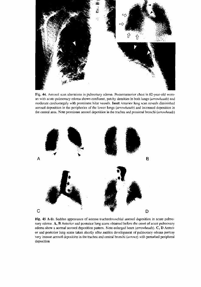
Fig. 44. Aerosol scan alterations in pulmonary edema. Posteroanterior chest in 62-year-old wom-an with acute pulmonary edema shows confluent, patchy densities in both lungs (arrowheads) andmoderate cardiomegaly with prominent hilar vessels. Inset Anterior lung scan reveals diminishedaerosol deposition in the peripheries of the lower lungs (arrowheads) and increased deposition inthe central area. Note prominent aerosol deposition in the trachea and proximal bronchi (arrowheads)
B
D
Fig. 45 A-D. Sudden appearance of intense tracheobronchial aerosol deposition in acute pulmo-nary edema. A, B Anterior and posterior lung scans obtained before the onset of acute pulmonaryedema show a normal aerosol deposition pattern. Note enlarged heart (arrowheads). C, D Anteri-or and posterior lung scans taken shortly after sudden development of pulmonary edema portrayvery intense aerosol deposition in the trachea and central bronchi (arrows) with perturbed peripheraldeposition
A
BFig. 46 A-C. Mismatch between aerosol and perfusion scan alterations in bronchogenic cancer.A Posteroanterior radiograph of right upper lung in a 62-year-old man with bronchogenic carcino-ma shows a large, ovoid tumor mass with marked regional pleural thickening and rib destruction(arrowheads). B Anterior aerosol scan portrays well defined tumor defect (arrowheads). The medialportion shows some deposition. C Perfusion scan reveals the defect to be more extensive involvingthe whole lobe (arrowheads)
Fig. 48 A-C. The prestenotic aerosol deposition sign in radiographically invisible bronchosteno-sis. A Posteroanterior chest in a 69-year-old man with right hilar cancer shows a tumor mass aboutthe hilum (arrows). Peripheral lung appears normally aerated and bronchial stenosis is not seen.B CT scan through the tumor center reveals narrowed bronchus with encircling mass (open ar-row). C Anterior lung scan reveals large aerosol deposition defects in both upper and middle lobes(arrows). The stenosis is clearly indicated by the prestenotic aerosol deposition sign in the mainstem bronchus. Note obvious disparity between radiographic and scan findings

Fig. 47. Bronchogenic carcinoma with lung collapse. Posteroanterior chest in a 69-year-old manwith lung cancer and associated ateiectasis shows a mass in the left suprahilar region (mass) withhomogeneous density with cranially concave lateral border in the left upper lobe, the " S " sign(arrowheads). Inset a, b Anterior and posterior lung scans reveal large, photopenic defect involv-ing the entire left upper lobe with the prestenotic aerosol deposition sign of bronchial obstruction(arrows)

124 Aerosol Scanning in COPD
aFig. 49. Aerosol scan manifestations of metastatic lung cancer. Posteroanterior chest in a 42-year-old man with extensive "cannon ball" metastases from hepatoma shows multiple, variously sized,roundish tumors. Inset a, b Anterior and posterior lung scans portray many, well-defined, round-ish defects
Y.W. Bahk and S.K. Chung 125
Metastatic lung cancer. The metastatic tumours of the lung, measuring a fewcm or more in size, were seen as photopenic masses (Fig. 49). Smaller metastatictumours could not be visulalized by this means.
Discussion
Normal and asymptomatic smoker's lung. The aerosol deposition in the al-veolar air spaces was indeed uniform throughout the entire lung fields in the nor-mal nonsmokers (Fig. 11). However in a small fraction of cases insignificant, focaldefect was noted. The defects tended to occur in the apical segments of the upperlobes and the superior and posterior-basal segments of the lower lobes, presumablydue to the combined effects of gravity and their small lung volume and relativelyrestricted respiratory movement. In contrast, the smoker's lung demonstrated minimal,patchy airspace defects with mild to moderate bronchial deposition, denoting bron-chitis, an anticipated scan manifestation of the smoker's lung (Figs. 12, 13). Wefound that the aerosol scanning is suited for the study of not only the parenchymalair spaces but also importantly the airways. An excellent ventilation scan study onboth normal life-long nonsmokers and smokers has been published by Barter et al[8]. Instead of aerosol, they used 81mKr gas which is excellent for the airspace scanbut not for the bronchial scan. Their study clearly showed the uniformity of theairspace scan pattern in 46 carefully recruited nonsmokers with minimal and focaldefects in 3, but significant scan defects in 19 (41%) of 46 current smokers. Thebronchial tree could not be assessed by their method.
Emphysema is defined as permanent abnormal distension of the air spaces distalto the terminal bronchioles accompanied by the destruction of alveolar septa, andwithout obvious fibrosis [3,9]. Histological characteristics of emphysema includeirreversible destruction and distension of the terminal air spaces with marked in-crease in airway resistance. The radiographic manifestations in well established casesinclude overinflation and hyperlucency of the lung with reduced, attenuated vascu-lar markings and flattened or ventrally retroverted diaphragm (Fig. 1). In the later-al view, widened retrosternal space and kyphotic thoracic spine produce the "barrelchest" deformity (Fig 1). When severe, the inflated lungs encroach upon both sidesof the heart. With the development of lung hypertension the hilar vessels becomedilated, protruding into the lung. As described by Heitzman [4] pulmonary em-physema can be classified into 3 categories according to the diversion pattern ofthe vasculature; the panacinar [panlobular] type with cranial diversion (Fig. 2A),the centriacinar [centrilobular] type with caudal diversion (Fig. 2B) and the "increased-markings" type with central diversion (Fig. 2C).
An early description of the aerosol scan changes in various categories of COPDwas published in 1970 by Isawa et al [10] and several years later Alderson et al
126 Aerosol Scanning in COPD
[11] provided clinical data on the sensitivity of 133Xe ventilation scan that was muchhigher than that of radiography in obstructive pulmonary disease. Recently, an ex-tensive study on COPD using 81mKr was presented by Lavender and Finn [12].The first of the above mentioned groups keenly observed that aerosol tended to depositin the central zone [bronchi] in emphysema and in the peripheral zone [air spaces]in bronchitis. Our observation clearly confirmed the characteristic scan feature ofemsphysema they described, i.e. the conspicuous bronchial deposition. Howeverthe aerosol scan changes of chronic bronchitis we noted were not in complete agree-ment with theirs as will be described below. In addition, our experience revealedanew that affected bronchi in emphysema were typically clubbed, bulbous or withered-bough-like in appearance, and such alterations appeared to progressively spread tothe periphery with increasing severity of emphysema. The scan manifestation inrelatively mild case was characterized by patchy defects in the periphery with minimalbronchial deposition, indeed resembling bronchitis (Fig. 14). The finding was muchsimilar to that of the 81mKr scan published by Lavender and Finn [12]. It ishowever to be emphasized that their radiogas scan did not provide any informationregarding the bronchial tree. The scan alterations frequently overlapped not onlyin emphysema and chronic bronchitis but in all four types of COPD. Yet, the aer-osol scan pattern in classic emphysema was characteristic enough to indicate thediagnosis. The large and medium sized, dilated bronchi in emphysema wereportrayed with intense aerosol deposition, and such bronchi showed deviated distri-bution patterns in the lung depending on the type of the disease, an exact mirrorimage of the radiographic vasculature diversion. Thus, it was noted that the bron-chial deposition tended to shift cranially in the panacinar emphysema (Fig. 15A),while it moved caudally in the cenrriacinar emphysema (Fig. 15B). In the third typewith increased markings, the bronchi were localized more or less in the midlungfield (Fig. 15C). A plausible account is that, like the pulmonary blood flow, theaerosol ingress in the overinflated lung may be facilitated through the airways thatare less resistant. Also it seemed that the intense aerosol deposition in the dilatedbronchi in this condition was largely structural, i.e. bronchiectatic. Interestingly,Cunningham and Lavender have also noted a "basal distribution of regional defectsin panacinar emphysema" in 81mKr inhalation scan [13].
Chronic bronchitis is defined as a condition associated with excessive tracheobron-chial mucus production, causing cough and expectoration [9,14]. The majority ofthe cases we studied was simple chronic bronchitis without complication. Clinical-ly, however, some of the cases had "wheezing" [15]. As anticipated chest radiog-raphy did play only an ancillary role in the diagnosis of chronic bronchitis [16,17].Nevertheless, the well-known radiographic signs of chronic bronchitis such as the"tram-line" shadow, the bronchial cuff and the "dirty lung" were seen in somepatients and air trapping with increased radiolucency and oligemia in others (Fig.4) [4,17-19]. In still others the diagnosis was established by HRCT (Fig. 5) or bron-

Y.W. Bahk and S.K. Chung 127
chography that demonstrated the classic serrated appearance of hypertrophied glandsin the bronchial wall (Fig. 6).
The high sensitivity of aerosol scan in the detection of mild peripheral air trap-ping in COPD has been described by Alderson et al in 1974 [11]. Our experienceconfirmed that aerosol scan is a sensitive indicator of bronchitis and the bronchiticchanges related to cigarette smoking. Thus, abnormal aerosol deposition occurrednot infrequently in the parahilar bronchi in the absence of radiographic abnormali-ty. The aerosol deposition in relatively mild cases was in the central bronchi andspotty (Fig. 17) and was arboriform in the middle and peripheral bronchi in severecases (Fig. 18). In general, mild bronchial deposition was seen in association withrelatively normal air space deposition, whereas prominent bronchial deposition wasseen in association with airspace defects. Occasionally, the "wornout broomstick"sign was portrayed in the basal lung (Figs. 19, 20). The sign was composed of"cold" and "hot" streaks, probably reflecting the spastic and dilated bronchi, respec-tively. Typically, the streaks appeared to be stretched out, imperceptibly blendinginto patchy photon defects in the periphery. The sign seemed to be seen in thosewith relatively severe scan alterations.
The chronic bronchitics having atypical "wheeze" showed fairly intense aerosoldeposition in the central bronchi that were mildly dilated as well as patchy airspacedefects (Fig. 18). The changes were indeed indistinguishable from those of asthma(see below). The discrepancy between the radiographic changes and lung scan al-terations was not rare.
Asthma is characterized by widespread, reversible, spastic narrowing of the air-ways in increased response to various stimuli. Clinically, the condition manifestswith paroxysms of dyspnea, wheezing and cough. The disease may be in attack,in remission or resistant defying treatment [status asthmaticus]. It must not be as-sociated with cardiovascular diseases. The severity of bronchial spasm may changerapidly over a short period of time spontaneously or in response to bronchodilator[9]. Pathologically, respiratory epithelial cells may appear in sputum. The paraffinsections of asthmatic sputum has been shown to contain Curschmann's spirals, whichare basically formed with nonmucoid elements [20]. The chest radiograph may revealair trapping that is diffuse in nature (Fig. 7). The lung vessels are attenuated andthe diaphragm may be depressed, but such changes are not regularly seen [21].The chest radiography is mandatory because it is the most efficient means to ex-clude pulmonary or cardiac diseases that may clinically simulate asthma.
According to Lavender and Finn [22] the aerosol deposition pattern in asthmais characterized by a well-defined, lobar or segmental defect with or without perfu-sion. Our observation confirmed their finding of a segmental or lobar defect in somecases, but on the whole the deposition patterns in asthma were much complicatedaccording to the stage, chronicity and severity of the disease. In the relatively fresh,"uncomplicated" cases without bronchitis or emphysema, the aerosol deposition
128 Aerosol Scanning in COPD
pattern during an attack was simply characterized by irregular, patchy, nonsegmental,airspace defects scattered throughout the entire lung field, especially in the upperzones (Fig. 21). With bronchial complications, asthma showed bronchial deposi-tion: in asthma with bronchitis it was confined to the central bronchi and spottyin appearance (Fig. 22), whereas in asthma with emphysema it was distributed inthe central and middle bronchi and withered-bough-like in appearance (Fig. 23).In the advanced or resistant asthma the bronchi became grossly dilated and distort-ed, manifesting very bizarre deposition (Fig. 24). Unlike the radiogas [113Xe and81mKr] scan that primarily visualizes the air spaces the radioaerosol scan is suitedfor the study of the bronchial tree. It is postulated that the scan abnormality in therelatively fresh asthma likely reflects bronchospasm and bronchial obstruction, whileit denotes bronchitic complication in the long-standing form and bronchiectaasis andemphysema in the resistant form. Thus, the patchy, nonsegmental, airspace defectsin the absence of abnormal bronchial deposition appears characteristic of uncompli-cated asthma, but the scan features in the long-standing and resistant forms are rathersimilar to other types of COPD. It was noted that the scan changes were rapidlyrestored to normal or near normal state or greatly improved in response to bron-chodilators (Fig. 25) and that the "broomstick" sign appeared in some milder cases,denoting underlying chronic bronchitis.
For a semiquantitative assessment of scan findings the aerosol deposition in bronchiand air spaces was graded as mild, moderate and marked. Using this criteria a recipro-cal correlation was noted to exist between the bronchial and airspace depositions;the more intense the bronchial deposition the less intense was the airspace deposition.
Bronchiectasis is characterized by abnormal and frequently permanent dilatationof the bronchi [3]. The dilatation is either cylindrical, varicose or cystic in appear-ance. It is a common sequela of respiratory tract infection, measles, pertussis andtuberculosis in childhood, and may also result from obstructive bronchial diseases.Clinical symptoms include coughing with profuse expectoration which is often foul-smelling or bloody. Radiographic manifestations include tubular, cystic or ring-likeshadows with or without air-fluid levels (Figs. 8, 9, 27). The occlusive nature ofthe disease makes lung collapse a constant accompaniment. Both bronchographyand CT scan provide conclusive evidence, but the latter examination is preferedbecause of its noninvasiveness (Fig. 9).
The aerosol deposition pattern in bronchiectasis also varied greatly according tothe type, extent and stage of the disease. In general, it was quite similar to thatof other COPD. It strongly resembled that of chronic bronchitis or asthma in mildcases (Fig. 26) and that of emphysema in severe cases (Fig. 27). There seemedto exist a good correlation between the radiographic changes and the lung scan al-terations. However when bronchiectasis was minimal or hidden behind the heart,aorta or diaphrgam, the simple radiography was unrevealing, while lung scan showedprominent alterations with intense bronchial deposition (Fig. 28). Thus, it appearedthat the aerosol scan is more sensitive than the radiography in such situations.

Y.W. Bahk and S.K. Chung 129
Bronchial obstruction can be caused by a number of diseases including inflam-mation, infections, tumours, trauma, foreign body and congenital malformations,with tuberculosis and tumour being the two most common conditions. The bron-chial obstruction may be either complete or incomplete with clinically significantinvolvement occurring at the segmental or higher level. Complete obstruction isinvariably associted with lung collapse, which manifests radiographically as airless-ness, reduced volume and crowded vascular markings. In well penetrated radio-graphs, the mainstem or major bronchial obstruction can clearly be made out (Fig.10), but most of the bronchial obstructions at the lobar or segmental level are notso easy to detect, especially when they are due to fibroproductive tuberculosis orbroncho-occlusive malignant tumours (Figs. 29-32). When obstruction is incom-plete a check-valve mechanism may develop, regionally creating segmental or lo-bar emphysema (Fig. 48). In some cases, atelectatic consolidation due to bronchialobstruction may strongly resemble pneumonic consolidation making differential dia-gnosis difficult radiographically (Fig. 33A). However the aerosol lung scan has definiteplace in this situation, accurately pointing to the presence of bronchial obstructionby the prestenotic bronchial deposition sign (Fig. 33B).
The value of the ventilation scan using 133Xe in obstructive pulmonary diseasehas been described by Alderson et al [11]. The method was superior to radiogra-phy in detecting regional bronchial obstruction. Lavender et al [23] also appliedventilation scanning with 81mKr to the study of bronchial obstruction, confirmingscan defect. Our study revealed that the aerosol scan is indeed useful in demon-strating bronchial stenosis or obstruction especially at the segmental or lobar level(Figs. 29-33). It was impressive that in virtually all of cases the characteristic scansign was observed, whereas the radiographic change was dubious, or indirect atmost (Fig. 32). Thus, the stenotic or obstruction site was clearly indicated by ashort, segmental deposition in the prestenotic bronchus that was slightly dilated (Figs.29-33). With the obstruction of a mainstem bronchus aerosol deposition moved tothe trachea and contralateral bronchial tree (Fig. 31). We named the phenomenonthe "prestenotic bronchial deposition" sign, which was indeed useful in detectinga relatively small obstruction in a bronchus of the middle, lingular and lower lobes.The phenomenon is probably the results of bronchial damage and derangedmucociliary movement with transient hold-up of aerosol. The aerosol lung scan playeda key role in the diagnosis of bronchial obstruction. Radiographically, the shrunkenlung is retracted to and concealed behind the mediastinal shadow, hence difficultto see (Figs. 32, 33). As described by Lavender et al [23], the collapsed lung distalto the obstruction was shown as a photopenic defect, but the compensatorily over-inflated lung showed more or less increased or decreased deposition according tothe size of the lung involved (see below).
130 Aerosol Scanning in COPD
Overinflation or compensatory emphysema is characterized by an excessiveinflation of air spaces without air trapping or alveolar destruction [3]. The condi-tion may occur at various levels of the airways; a segment, a lobe or a lung. Typi-cally, it is seen in a structurally normal lung in compensation for reduced or lostlung volume as in cicatrical or resorptive atelectasis or surgical resection. The mainradiographic manifestations include increased radiolucency and attenuated lung mark-ings, which, unlike in true emphysema with air trapping, become normalized dur-ing expiration, unless it is complicated by secondary COPD. The normal expiratorydeflation in compensatory overinflation can be confirmed readily by either fluoroscopicobservation or comparing inspiratory and expiratory radiographs, which may berecorded in one film by double exposure technique.
The aerosol scan of overinflated lung was characterized by an increase in lungsize with increased, normal or decreased aerosol deposition. The amount of aerosoldeposited in the compensatorily overinflated lung appeared to largely depend uponthe size of the collapsed lung or the magnitude of the overinflation therefrom resulted.In effect, the aerosol deposited in the overinflated lung was normal when obstruc-tive collapse was small lobar or segmental (Fig. 34); increased when collapse waslarge lobar or multilobar (Fig. 35) and drastically decreased when collapse was mul-tilobar or in a lung with intense bronchial dilatation (Fig. 31). It is presumed thatsuch a variation of the amount of aerosol deposited in the overinflated lung is thefunction of the amount of the aerosol remained available to the overinflated lungin bronchial obstruction. On the other hand, it was noted that the bronchial deposi-tion was not an essential scan feature in the overinflated lung, unless it was compli-cated with COPD. For example, minimal bronchial deposition was seen in thosewith chronic bronchitis (Fig. 34) and bizarre deposition in those with advanced COPD(Fig. 31). In 2 cases with overinflation of a whole lung resulted from diffuse, res-trictive fibrothorax of the contralateral hemithorax, aerosol deposition appeared uni-formly increased in the overinflated lung, while it was decreased in the restrictedside (Fig. 36). The bronchial obstruction responsible for lung collapse and overin-flation was again indicated clearly by the prestenotic bronchial deposition (Figs.34, 35). Atelectatic collapse and bronchial obstruction without collapse as in for-eign body aspiration have been reported to produce simple homogeneous defect in81mKr gas scan [12].
Pneumonia and tuberculosis are the two most common infectious lung diseasesthat can readily be diagnosed by radiography, usually not necessitating an aerosolscan. Nevertheless, we performed aerosol scans in a number of selected cases ofpneumonia and tuberculosis to document the aerosol scan pattern in pneumonia andtuberculosis, and to study the airway alterations such as bronchitis, bronchial ob-struction and emphysema and airspace change such as fibrosis and calcification intuberculosis.
Y.W. Bahk and S.K. Chung 131
As described by Lavender et al [23], the consolidation in pneumonia andpneumonia-like tuberculosis was photopenic (Figs. 37, 38). The size of consolida-tion in a segment appeared large enough for a scintigraphic portrayal. In general,the photopenic defect of pneumonic consoldiation was larger in area than the radio-graphic lesion, probably due to the contribution of bronchial inflammation and spasm.Understandably, tuberculosis with fibroproductive alteration, atelectasis and cavitymanifested as photopenic defect, while emphysema and bronchitis or bronchiecta-sis were indicated by reduced or defective airspace deposition and intense bronchialdeposition, respectively (Fig. 39). Most of cases with advanced tuberculosis wasassociated with a bronchial pathology, manifesting bizarre bronchial deposition (Fig.39). It was noted that even minimal fibrocalcific tuberculosis can produce an obvi-ous scan defect, that is disproportionately large in comparison to the radiographicalterations (Fig. 40). Mismatch with aborted ventilation and preserved perfusionwas not unusual.
Pulmonary fibrosis may be either localized or diffuse. The aerosol scan appearsnot only useful but also sensitive in showing regional ventilation change in pulmo-nary fibrosis. The causes of pulmonary fibrosis are many, including infection, cys-tic fibrosis, pneurnoconiosis, asbestosis, farmer's lung, radiation and others. Idiopathictype is not rare in the diffuse form. The radiographic manifestations are character-ized by the irregular meshwork of fine linear, reticular and nodular shadows thatobliterate the pulmonary and hilar vessels (Figs. 41, 42). In some diffuse form,the lung may show hyperlucency due to air trapping, but eventually becomes shrunkenwith the progression of the disease (Fig. 42). Cor pulmonale with the dilatationof the hilar arteries and right heart may ensue when pulmonary hypertension is se-vere and long-standing.
The aerosol defects in pulmonary fibrosis were rather distinct compared to theradiographic change, espeically in the localized form (Figs. 41, 42). Usually, thebronchial deposition is negligible or insignificant unless complicated with bronchialdisease as in the second case of cystic fibrosis studied with 99mTc-DTPA- aerosolby Loken et al [24]. That case showed prominent bronchial deposition along withairspace defects, denoting bronchial affection. Although the number of cases westudied is limited it is likely that bronchial deposition is not a central feature of sim-ple or uncomplicated interstitial fibrosis regardless of extent: an important differen-tial point from COPD in which the bronchial deposition is an essential feature. Thedilated hilar vessels due to pulmonary hypertension are depicted as photon defects.
Diffuse panbronchiolitis is a relatively new entity originally reported from Japan[25, 26] and recently also from Korea [27]. Its pathology is characterized by diffusechronic bronchiolitis and peribronchiolitis in the transitional zone of the airways,especially in the lower lungs. When advanced, it may cause secondary ectasia ofthe proximal bronchioli. The radiographic manifestations include small nodular
132 Aerosol Scanning in COPD
and irregularly mottled densities scattered throughout both lungs with air trappingand occasional cor pulmonale (Fig 43). High resolution CT scan has been shownto be extremely useful in delineating the pathognomonic signs of small centrilobu-lar nodules and branched linear shadows of attenuation in the peripheral lung (Fig,43) [26, 27]
Our experience with aerosol scans in this condition showed moderately severe,nonsegmental, airspace defects as in diffuse pulmonary fibrosis and uncomplicatedasthma. In addition and very significantly, fairly intense aerosol deposition was notedto occur in the peripheral and middle bronchi (Fig, 43). We consider the peripherallocalization to be characteristic of the disease in the transitional zone, sharply con-strasting with the central localization of the bronchial deposition in COPD (Figs15-17, 20, 24).
Lung edema is characterized by focal or diffuse replacement of the air in thealevoh with fluid. Causes include congestive heart failure, overhydration, azotemielung, aspiration and others The essential radiographic change is patchy or large,homogeneous opacification with occasional air bronchogram sign, typically in thelower lung (Fig 44). The "butterfly'1 or "bat-wing*1 appearance is a well-knownmanifestation of lung edema. Cardiomegaly accompanies the lung edema of cardi-ac or renal origin.
According to Lavender and Finn [12] the 81niKr-gas inhalation scan was not ef-ficient in revealing lung edema. However our experience showed that the aerosolscan could portray lung edema as a well-defined defect, well matching with theradiographic opacity. It appeared that an edema had to be at least segmental in sizefor its proper portrayal in the aerosol scan (Fig. 44). Muhtple, small, patchy ede-mas were indicated by irregularly mottled aerosol defects. Basically, the defect wasnonspecific and exactly the same as that of pneumonic consolidation or tumour mass,but its peripheral localization and presentation along with cardiomegaly were help-ful. It was interesting to observe an extremely intense aerosol deposition in the tracheaand mainstem bronchi in the 2 cases of acute lung edema we studied In one casean aerosol scan was obtained before the onset of acute lung edema, and it showedthe airways to be completely free from aerosol deposition, but with the develop-ment of diffuse lung edema an intense tracheobronchial deposition suddenly becamemanifest (Fig. 45). It is postulated that such a tracheobronchial deposition may haveresulted from perturbed mucociliary function and profuse secretion. The perfusionscan defects were larger in size and a bit more prominent than the ventilation scandefects
Lung cancer is not an ordinary indication for aerosol study. However it maybe applicable in some important situations. One good indication is the unresectablebronchogenic carcinomas whose response to radiation therapy is difficult to radio-graphically assess.. The combined inhalation and perfusion scans of the lung have
YW Bdhk and S k Chung 133
been shown to he a sensitive indicator of tumoral change with improved aerationand blood flow following irradiation [28]. The perfusion scan alone has also beenreported to be useful in screening the central lung cancer [29] and both inhalationand perfusion scans are valuable means to demonstrate that lung perfusion can moreseriosuly be impeded than the ventilation in bronchogenic carcinoma. The rumourin the lung periphery could be delineated as a well-demarcated scan defect whensufficiently large and surrounded by open bronchi and aerated lung (Fig 46). Thenext situation is probably the bronchial tumour with incomplete obstruction that maycreate focal emphysema through the check-valve mechanism This sort of emphysemawas denoted by little or no aerosol deposition in the radiographically well-aeratedlung, a paradox (Fig. 47). In addition, the causative bronchostenosis that is usuallynot easy to recognize in radiograph could accurately be indicated by the "prestenoticbronchial deposition" sign- The tumours with lung collapse were also portrayedas scan defect, but such a defect was much larger in size than the tumour itselfdue to the entering of the collapsed lung into the defect formation (Fig- 481.
Like primary carcinomas, metastatic tumours measuring a few cm or more insize could be portrayed as photopenic defects, typically with sharp demarcation (Pig49),
Conclusion
Our experience with radioaerosol inhalation scan in COPD and a number of otherbronchopulmonary conditions indicated that it is indeed a useful, inexpensive, practicaltool of clinical diagnosis and research Appropriately performed, spending adequateacquisition time, the aerosol scan can be refined into a state-of-the-art imaging mo-dality, which can provide very unique information concerning both the anatomicand functional situations of pulmonary airways and airspaces in both normal andpathologic conditions.
It appears that the assessment of bronchial aerosol deposition patterns in COPDand other related diseases is as much important as the assessment of alveolar depo-sition pattern In COPD, various combinations of the bronchial and alveolar airspace scan alterations were portrayed, helping to distinguish to some extent oneform of COPD from the other: for example, emphysema from simple bronchitis,asthma from bronchiectasis and bronchiectasis from bronchostenosis when the in-dividual diseases were not further complicated In addition, it was possible to sortout a few useful diagnostic features. They included the "wornout broomstick" signof spastic and dilated peripheral bronchi in bronchitis and asthma and the "witheredbough" sign of dilated bronchi in emphysema and bronchiectasis, Bronchial ob-struction or bronchostenosis could accurately be diagnosed by the peculiar, prestenotic,bronchial deposition. The over inflated lung in compensatory emphysema showedeither increased, normal or decreased aerosol deposition depending on the extent
134 Aerosol Scanning in COPD
of pathology. Pneumonic consolidation, tuberculosis and lung edema were portrayed
as photopenic defects. Pulmonary fibrosis, both segmental and diffuse, was rather
clearly indicated by airspace defect with little or no bronchial deposition unless com-
plicated with COPD. Diffuse panbronchiolitis was characterized by the aerosol depo-
sition in the middle and peripheral airways, accurately reflecting the transitional airway
localization of the disease. Aerosol scan was shown to be a useful noninvsive me-
ans to test bronchial patency, to detect obstruction or stenosis with lung collapse
or emphysema and to localize tumour in bronchogenic carcinoma. Mismatch of the
ventilation and perfiision scans was confirmed in pneumonia and primary lung cancer.
It seems warranted to state that with the refinement of technique and clearer dis-
play, the aerosol lung scan can be utilized for systematic investigation of not only
COPD but many other lung diseases. Finally, the value and importance of side-by-
side, correlative reading of aerosol scans with chest radiographs is reemphasized.
References
1. Kotrappa P, Raghunath B, Subramanyam PSS, Raiker UR, Sharma SM: Scintiphotographyof lungs with dry aerosol-generation and delivery system. J Nucl Med 1977; 18:1082-1085.
2. Welch MH: Obstructive diseases. [In] CA Guenter, MH Welch (eds) PulmonaryMedicine. 2nd edn. Lippincott, Philadelphia, pp 664-793, 1982.
3. Dunnill MS: Pulmonary Pathology. 2nd edn. Churchill, Edinburgh, pp 41-132, 1988.4. Heitzman ER: Chronic obstructive pulmonary disease. [In] The Lung. 2nd edn. Mosby, St
Louis, pp 422-456, 1984.5. Miiller NL: Obstructive lung disease. [In] Freundlich, DG Bragg (eds) A
Radiologic Approach to Diseases of the Chest. Williams & Wilkins, Baltimore, pp 455-465,1992.
6. Burrows B: Chronic airways disease. [In] JB Wyngarrden, LH Smith, Jr (eds) Cecil Text-book of Medicine. 16th edn. Saunders, Philadelphia, pp 363-371, 1982.
7. Frazer RG, Pare JAP: Tubular shadows. [In] Diagnosis of Diseases of the Chest. 2nd edn.Saunders, Philadelphia, pp 518-523, 1977.
8. Barter SJ, Cunningham DA, Lavender JP, et al: Abnormal ventilation scans in middle agedsmokers. Am Rev Respir Dis 1985; 132:148-151.
9. Ciba Guest Symposium 1959: Terminology, definition and classification of chronic pulmo-nary emphysema and related conditions. Thorax 1959; 14:286-299.
10. Isawa T, Wasserman K, Taplin GV: Lung scintigraphy and pulmonary function studies inobstructive airway disease. Am Rev Respir Dis 1970; 102:161-172.
11. Alderson PO, Seeker-Walker RH, Forrest JV: Detection of obstructive pulmonary disease.Radiology 1974; 111:643-648.
12. Lavender JP, Finn JP: V/Q patterns in nonthromboembolic lung diseases. [In] MK Loken(ed) Pulmonary Nuclear Medicine. Appleton & Lange, Norwalk, pp 101-131, 1987.
13. Cunningham DA, Lavender JP: Krypton-81m ventilation scanning in chronic obstructive air-ways disease. Br J Radiol 1981; 54:110-116.
14. Reid L: Pathology of chronic bronchitis. Lancet 1954; 1:275-278.15. Burrows B: Simple chronic bronchitis and asthmatic bronchitis. [In] JB Wyngaarden, LH
Smith, Jr (eds) Cecil Textbook of Medicine. 16th ed. Saunders, Philadelphia, pp 365-367, 1988.

Y.W. Bahk and S.K. Chung 135
16. Simon G: Chronic bronchitis and emphysema — a symposium. Br J Radiol 1959; 32:292-294.17. Bates DV, Gordon CA, Paul GI, Place REG, Sinidal DP, Woolf CR; Chronic bronchitis:
Report on the third and fourth stages of the coordinated study of chronic bronchitis in theDepartment of Veterans Affairs, Canada. Med Serv J Can 1966; 22:5.
18. Thurlbeck WM, Henderson JA, Fraser RG, Bates DV: Chronicobstructive lung disease: acomparison between clinical, roentgenologic, functional and morphologic criteria in chronicbronchitis, emphysema, asthma, and bronchiectasis. Medicine 1970; 136:935-940.
19. Fraser RG, Fraser RS, Renner JW, Berhard C, Fitzgerald PJ: The roentgenologic diagnosisof chronic bronchitis: a reassessment with emphasis on parahilar bronchi seen head-on. Radi-ology 1976; 120:1-9.
20. Sanerkin NG, Evans DMD: The sputum in bronchial asthma:pathognomonic patterns. J PatholBacteriol 1965; 89:535-541.
21. Blair DN, Coppage L, Shaw C: Medical imaging in asthma. J Thorac Imag 1986; 1:23-35.22. Lavender JP, Finn JP: Bronchial asthma. [In] MK Loken (ed) Pulmonary Nuclear Medi-
cine. Appleton & Lange, Norwalk, pp 106-108, 1987.23. Lavender JP, Irving H, Armstrong JD, II: Krypton-81m ventilation scanning: acute respira-
tory disease. AJR 1981; 136:309-316.24. Loken MK, Sirr SA, Boudreau RJ, Warwick WJ: Quantitative measurements of regional ven-
tilation in patients with cystic fibrosis. [In] MK Loken (ed) Pulmonary Nuclear Medicine.Appleton & Lange, Norwalk, pp 155-174, 1987.
25. Homma H, Yamanaka A, Tanimoto S, Tamura M, Chijimatsu Y, et al: Diffuse panbronchio-litis — A disease of the trantional zone of the lung. Chest 1983; 83:63-69.
26. Nishimura K, Kitaichi M, Izumi T, Itoh H: Diffuse panbronchiolitis: Correlation of high-resolution CT and pathologic findings. Radiology 1992; 184:779-785.
27. Choo SK, Im JG, Kim DY, Han MC: Diffuse panbronchiolitis: Chest radiograph and HRCTfindings in 8 patients. J Korean Radiol Soc 1992; 28:553-557.
28. Fazio F, Pratt TA, McKenzie CG, Steiner RE: Improvement in regional ventilation and per-fusion after radiotherapy for unresectable carcinoma of the bronchus. AJR 1979; 133:191-200.
29. Ernst H, Kriiger J, Vessal K: Lung scanning as a screening method for cancer of the lung.Cancer 1969; 23:508-512.