importance of mast cell prss31/transmembrane tryptase/tryptase-γ in lung function and experimental...
TRANSCRIPT

Richard L. StevensZhou, Paul M. Foster, Steven A. Krilis andG. William Wong, Sahar Hamadi, Saijun G. Jarnicki, Dominick Zheng, Sandra M. Frei,Michael Fricker, Shaan L. Gellatly, Andrew Philip M. Hansbro, Matthew J. Hamilton, Pulmonary Disease and ColitisExperimental Chronic Obstructive
in Lung Function andγTryptase/Tryptase-Prss31/Transmembrane Importance of Mast CellMolecular Bases of Disease:
published online May 12, 2014J. Biol. Chem.
10.1074/jbc.M114.548594Access the most updated version of this article at doi:
.JBC Affinity SitesFind articles, minireviews, Reflections and Classics on similar topics on the
Alerts:
When a correction for this article is posted•
When this article is cited•
to choose from all of JBC's e-mail alertsClick here
Supplemental material:
http://www.jbc.org/content/suppl/2014/05/12/M114.548594.DC1.html
http://www.jbc.org/content/early/2014/05/12/jbc.M114.548594.full.html#ref-list-1
This article cites 0 references, 0 of which can be accessed free at
by guest on May 15, 2014
http://ww
w.jbc.org/
Dow
nloaded from
by guest on May 15, 2014
http://ww
w.jbc.org/
Dow
nloaded from

Prss31 is pro-inflammatory in experimental COPD and colitis
1
Importance of Mast Cell Prss31/Transmembrane Tryptase/Tryptase-γ in Lung Function and Experimental
Chronic Obstructive Pulmonary Disease and Colitis*
Philip M. Hansbro,‡,1
Matthew J. Hamilton,§,1
Michael Fricker,‡ Shaan L. Gellatly,
‡
Andrew G. Jarnicki,‡ Dominick Zheng,
§ Sandra M. Frei,
§ G. William Wong,
¶ Sahar Hamadi,
§
Saijun Zhou,† Paul S. Foster,
‡ Steven A. Krilis,
† and Richard L. Stevens
§,2
From the ‡Centre for Asthma & Respiratory Disease, University of Newcastle and Hunter Medical
Research Institute, Newcastle, NSW 2308 Australia; §Department of Medicine, Brigham and Women's
Hospital and Harvard Medical School, Boston, MA 02115; ¶Department of Physiology, Johns Hopkins
University School of Medicine, Baltimore, MD 21205; and †Department of Infectious Disease,
Immunology, and Sexual Health, St. George Hospital and the University of New South Wales, Kogarah,
NSW 2217 Australia
*Running title: Prss31 is pro-inflammatory in experimental COPD and colitis
To whom correspondence should be addressed: Richard L. Stevens, Brigham and Women’s Hosp., Dept.
Med., Div. Rheumatol. Immunol. Allergy, Smith Bldg., Rm. 616B, 1 Jimmy Fund Way, Boston, MA
02115 USA. Tel: 617-525-1231; Fax: 617-525-1310; E-mail: [email protected].
Keywords: mast cell; protease; inflammation; inflammatory bowel disease; chronic obstructive
pulmonary disease
Background: Prss31/transmembrane
tryptase/tryptase-γ is a mast cell protease.
Results: A Prss31-null mouse was created to
evaluate the importance of this enzyme.
Conclusion: While Prss31 was found to hinder
airway reactivity to methacholine, it had
pro-inflammatory activity in experimental COPD
and colitis.
Significance: These data raise the possibility that
human Prss31 has beneficial and adverse roles in
the lung and colon.
ABSTRACT
Protease serine member S31
(Prss31)/transmembrane tryptase/tryptase-γ is
a mast cell (MC)-restricted protease of
unknown function that is retained on the outer
leaflet of the plasma membrane when MCs are
activated. We determined the nucleotide
sequences of the Prss31 gene in different mouse
strains, and then used a Cre/loxP homologous
recombination approach to create a novel
Prss31-/-
C57BL/6 mouse line. The resulting
animals exhibited no obvious developmental
abnormality, contained normal numbers of
granulated MCs in their tissues, and did not
compensate for their loss of the membrane
tryptase by increasing their expression of other
granule proteases. When Prss31-null MCs were
activated with calcium ionophore or by their
high-affinity IgE receptors, they degranulated
in a pattern similar to that of WT MCs.
Prss31-null mice had increased baseline airway
reactivity to methacholine, but markedly
reduced experimental chronic obstructive
pulmonary disease (COPD) and colitis, thereby
indicating both beneficial and adverse
functional roles for the tryptase. In a cigarette
smoke-induced model of COPD, WT mice had
more pulmonary macrophages, higher
histopathology scores, and more fibrosis in
their small airways than similarly treated
Prss31-null mice. In a dextran sodium sulfate-
induced acute colitis model, WT mice lost more
weight, had higher histopathology scores, and
contained more Cxcl-2 and IL-6 mRNA in their
colons than similarly treated Prss31-null mice.
The accumulated data raise the possibility that
inhibitors of this membrane tryptase may
provide additional therapeutic benefit in the
treatment of humans with these MC-dependent
inflammatory diseases.
Mouse mast cells (MCs)3 store varied
combinations of 15 serine proteases in their
secretory granules, three of which are the tryptases
http://www.jbc.org/cgi/doi/10.1074/jbc.M114.548594The latest version is at JBC Papers in Press. Published on May 12, 2014 as Manuscript M114.548594
Copyright 2014 by The American Society for Biochemistry and Molecular Biology, Inc.
by guest on May 15, 2014
http://ww
w.jbc.org/
Dow
nloaded from
Prss31 is pro-inflammatory in experimental COPD and colitis
2
known as mouse MC protease (mMCP)-6/Tpsb2,
mMCP-7/Tpsab1 (1-3), and protease serine
member S31 (Prss31)/transmembrane
tryptase/tryptase-γ (4). The corresponding TPSB2,
TPSAB1, and TPSG1/PRSS31 genes expressed in
human MCs reside on chromosome 16p13.3.
Although the constitutive MCs in the human lung
and gastrointestinal tract contain PRSS31 (4,5),
this tryptase is one of the most restricted proteases
in the body and only fifteen of the >8.7 million
expressed sequence tags (ESTs) in the GenBank
database originated from its gene (see UniGene
Hs.592076).
While the constitutive safranin+
MCs in all
examined WT mouse strains express mMCP-6, the
corresponding MCs in WT BALB/c and 129/Sv
mice have negligible amounts of Prss31 relative to
those in C57BL/6 (B6) mice (4). This
strain-dependent expression of Prss31 differs from
that of mMCP-7 whose gene has a loss-of-function
mutation in WT B6 mice (6).
Mouse Prss31 is initially translated as a
311-mer proteolytically inactive zymogen. Unlike
mMCP-6, mMCP-7, and their β-tryptase human
orthologs, human and mouse Prss31 has in each
instance a membrane-spanning domain near its C
terminus that causes the retention of the active
form of the enzyme on the outer leaflet of the
plasma membrane when MCs degranulate (7).
Despite structural differences, recombinant
mMCP-6, mMCP-7, human β-tryptases, and
human PRSS31 cleave many of the same trypsin-
susceptible low molecular weight chromogenic
substrates (7,8). Prss31 therefore has a substrate
specificity that overlaps that of its tetramer-
forming family members. While the function of
Prss31 is not known, the tetramer-forming
tryptases mMCP-6 and mMCP-7 have prominent
beneficial roles in innate immunity (9-11), but
substantial adverse roles in numerous
inflammatory diseases (12-14).
We recently developed a novel cigarette
smoke-induced 8-wk model of COPD in mice that
is dependent on pulmonary macrophages and
mMCP-6+ MCs (14) in order to advance our
knowledge of this devastating disease. A serine
protease•protease inhibitor imbalance in the lung
is a major risk factor in COPD (15), and humans
that are deficient in α1-anti-trypsin
(A1AT)/SERPINA1 (16) or α2-macroglobulin (17)
are at increased risk of developing emphysema.
Other than neutrophil elastase, the relevant
A1AT-susceptible serine proteases in human lung
that increase the risk of developing emphysema
and pulmonary inflammation have not been
identified. Macrophage accumulation, alveolar
enlargement, and varied aspects of lung function
were not reduced significantly in our smoke-
treated WT mice that were depleted of their
neutrophils (14). We therefore hypothesized that
an A1AT-regulated proinflammatory serine
protease other than neutrophil elastase probably
has a prominent adverse role in this pulmonary
disease. We also speculated that this enzyme
would have tryptic activity since A1AT inhibits
such serine proteases. In this regard, we previously
showed that recombinant Prss31 is rapidly
inhibited by A1AT in vitro (7).
Since tryptase redundancy occurs in some
inflammatory disease models (12), we concluded
that we probably would need to delete at least two
of the three MC-restricted tryptases in mice to
uncover the global importance of this family of
serine proteases in experimental COPD, colitis,
and other inflammatory disease models.
The adoptive transfer of in vitro-differentiated
mMCP-6+/mMCP-7
+ MCs from WBB6F1/J-+/+
mice into MC-deficient WBB6F1/J-KitW
/KitW-v
mice provided insights into the function of MCs
and their tetramer-forming tryptases in varied
experimental models (18). Unfortunately, because
the MCs in WBB6F1/J-KitW
/KitW-v
mice
constitutively contain low amounts of Prss31 (19),
this reconstitution approach cannot be used to
evaluate the importance of the latter membrane
tryptase in vivo. To deal with that obstacle, we
created in this study a novel inbred transgenic
mMCP-6+/+
/mMCP-7-/-
/Prss31-/-
(6+/7
-/31
-) B6
mouse line in which the functional Prss31 gene
was ablated in mMCP-7-null B6 mouse ES cells.
Using this transgenic mouse, we discovered that
Prss31 has beneficial roles in hindering airway
reactivity to methacholine but prominent adverse
roles in experimental COPD and colitis.
EXPERIMENTAL PROCEDURES
All experiments were carried out using animal
protocols approved by the Brigham and Women’s
Hospital (BWH)/Dana Farber Cancer Institute or
the University of Newcastle animal ethics
committees.
by guest on May 15, 2014
http://ww
w.jbc.org/
Dow
nloaded from

Prss31 is pro-inflammatory in experimental COPD and colitis
3
Sequence Analysis of the Prss31 Genes in
Different WT Mouse Strains—Purified A/J,
BALB/c, CBA, C3H/HeJ, and SWR mouse
genomic DNA were obtained from the Jackson
Laboratory (Bar Harbor, ME), and a standard PCR
approach was used to isolate the entire 3.9-kb
Prss31 gene from the five WT mouse strains
employing primers that resided at the beginning
(5ˈ-cccatactgataggcctggggctggg-3ˈ) and end
(5ˈ-atttgctttatgtcatccgttctac-3ˈ) of the tryptase
gene. The resulting 3.9-kb genomic fragments
were purified and sequenced in both directions
using 7 forward and 7 reverse primers spaced
every 500-600 nucleotides based on the sequence
of the 129/Sv mouse Prss31 gene (4).
Creation of a Novel Transgenic 6+/7
-/31
- B6
Mouse Line—Exon 1 of the endogenous 3.9-kb
mouse Prss31 gene encodes the translation-
initiation site of the expressed transcript and the
hydrophobic signal peptide of the translated
tryptase (4). Thus, no functional Prss31 protein
can be produced in mice if this exon is removed
using a Cre/loxP homologous recombination
approach. Another reason why the first exon of the
Prss31 gene was targeted was because the last 156
nucleotides of its fifth exon are shared with that of
the Cacna1h gene. Cacna1h is a calcium channel
protein important in brain and heart development,
and mutations in the human CACNA1H gene lead
to epilepsy and autism spectrum disorders (20).
Transgenic mice lacking just the sixth exon of the
latter gene (causing loss of amino acids 216 to 267
in this 2007-mer protein) have constricted
coronary arterioles and focal myocardial fibrosis
(21). We therefore concluded that targeted
removal of the 3' end of the mouse Prss31 gene
most likely would adversely affect the expression
of the physically linked Cacna1h gene which, in
turn, would almost certainly lead to serious
developmental abnormalities in the resulting
transgenic mice.
The Prss31-targeting vector employed to
create the transgenic mouse line used in this study
was generated with the assistance of Taconic-
Artemis using BAC clones from their B6 mouse
RPCIB-731 BAC library. A loxP site was placed
1.9 kb upstream of the gene’s first exon. The
second loxP site was placed in the gene’s first
intron. The puromycin resistance (PuroR) gene
flanked by F3 sites was inserted into intron 1
upstream of the second loxP site for positive
selection, whereas the thymidine kinase (Tk) gene
was inserted upstream of the mMCP-6/Tpsb2 gene
for negative selection. The B6 mouse ES cell line
was grown on a mitotically inactivated feeder
layer of mouse embryonic fibroblasts in DMEM
high glucose medium supplemented with 20%
FBS and 1,200 U/ml leukemia inhibitory factor
(Millipore ESG 1107). Thirty μg of the linearized
targeting DNA were electroporated into 106 ES
cells using a Gene Pulser (Bio-Rad) at 240 V and
500 μF. Puromycin (1 μg/ml) and gancyclovir (2
μM) positive and negative selection treatments
were commenced on days 2 and 5, respectively.
The resulting transfectants were isolated and
analyzed on day 8.
Assessed by DNA blot and/or qPCR analysis,
17 of the obtained 252 ES cell clones had
undergone homologous recombination at the
Prss31 genomic locus, as shown for clone C1.
After the transfer of this clone into mouse
blastocysts, chimera Prss31f/+
.FLpe mice were
obtained that had one allele of the mouse Prss31
gene floxed. After Flp-mediated removal of the
PuroR gene, the obtained Prss31f/+
heterozygote
mice were mated. The identified Prss31f/f
offspring
containing both alleles of the Prss31 gene floxed
were next bred with Taconic-Artemis’s CreDel
mouse B/6-Gt(ROSA)26Sortm9(cre/ESR1)Arte
. The
resulting Prss31-/+
.CreDel offspring were mated
with WT B6 mice to obtain Prss31-/+
B6 mice that
had one WT allele and one disrupted allele of the
targeted gene. These heterozygotes were bred to
obtain the final Prss31-/-
B6 mouse line that
contained both disrupted alleles of the Prss31
gene.
A standard PCR approach was carried out to
genotype our Prss31+/+
, Prss31+/-
, and Prss31-/-
B6
mice in which genomic DNA isolated from
proteinase K-digested tails was incubated with the
primers 5'-ctgttcattccctgtttgttgg-3' and
5'-cctgaggtagatgttcccatcc-3'. After an initial
denaturation step (94 °C for 10 min), 35 cycles of
PCR were carried out with denaturation (94 ºC for
15 sec), annealing (55 ºC for 30 sec), and
extension (72 ºC for 70 sec) steps. Since the MCs
in WT B6 mice cannot express mMCP-7 due to a
splice-site mutation in its gene (6,22), the MCs in
the created B6 mouse line lack both mMCP-7 and
Prss31.
Histochemistry and Enzyme Cytochemistry
Evaluation of the MCs in the Peritoneum and Ear
by guest on May 15, 2014
http://ww
w.jbc.org/
Dow
nloaded from
Prss31 is pro-inflammatory in experimental COPD and colitis
4
of the Prss31-null Mouse—To examine the in
vivo-differentiated MCs in the peritoneal cavity,
WT and Prss31-null B6 mice were euthanized
with CO2. Ten ml of PBS was injected into the
peritoneal cavity of each animal, and then
aspirated ~10 sec later. The resulting lavage fluid
was centrifuged at 450×g for 5 min, and the
obtained cell pellet in each instance was
resuspended in 0.5 ml of PBS. One hundred μl
samples were cytocentrifuged onto glass slides
which were then stained with toluidine blue or
with alcian blue followed by safranin (23) to
evaluate the number of MCs and their overall
granulation, as well as their expression and
granule accumulation of heparin-containing
serglycin proteoglycans. To examine the
constitutive MCs in Prss31-null B6 mice, the left
and right ear were harvested and placed overnight
in 4% paraformaldehyde. The fixed tissues were
cut, embedded in paraffin, sectioned, and the
resulting slides were subjected to H&E
histochemistry or chloroacetate esterase enzyme
cytochemistry (CAE) (24).
Generation and Characterization of Prss31-
null B6 Mouse Bone Marrow-derived MCs
(mBMMCs)—After euthanasia with CO2,
harvested bone marrow cells from the femora and
tibiae of WT and Prss31-null B6 mice were
cultured for >4 wk in IL-3-enriched WEHI-3
cell-conditioned medium, as previously described
(25). In two experiments to evaluate the IL-10
signaling pathway in Prss31-null MCs, the two
populations of IL-3-developed mBMMCs were
exposed to 10 ng/ml recombinant IL-10 (R&D
Systems) for 24 h, as previously described (26).
Based on our earlier analyses of the protease
composition of the MCs in WT 6+/7
+/31
- BALB/c
and WT 6+/7
-/31
+ B6 mice, the expression and
granule accumulation of the other proteases in the
cell’s secretory granules is not dependent on
Prss31. Nevertheless, RT-qPCR approaches were
used to confirm that targeted inactivation of the
Prss31 gene in our newly created 6+/7
-/31
- B6
mouse line did not cause the MCs in these animals
to increase their expression of another granule
protease. For these experiments, WT and
Prss31-null B6 mBMMCs were centrifuged, and
the resulting cell pellets were placed in Qiagen’s
cell lysis buffer. Each lysate was applied to an
RNeasy spin-column using the manufacturer’s
recommended protocols. Once RNA was isolated
and quantitated spectrophotometrically, Bio-Rad’s
iScript cDNA synthesis kit was used to convert
comparable amounts of the RNA from each
sample into cDNA.
The obtained cDNA samples were analyzed
by RT-qPCR using a Stratagene Mx3000P PCR
machine, a SYBR green PCR kit (Bio-Rad), and
Qiagen validated primers specific for the
transcripts that encode GAPDH, serglycin,
carboxypeptidase A3, mMCP-1, mMCP-2,
mMCP-4, mMCP-5, mMCP-6, and Prss31. The
obtained data for each transcript were then
normalized based on the level of the GAPDH
transcript in each sample.
The mMCP-4, mMCP-5, and mMCP-6 mRNA
data in IL-3-developed mBMMCs were confirmed
and extended by SDS-PAGE immunoblot analysis
using the relevant mMCP-specific anti-peptide
antibodies generated in rabbits. For these
experiments, in vitro-developed Prss31-null
mBMMCs and in vivo-developed peritoneal MCs
from the Prss31-null B6 mice were placed in ice
cold-PBS and sonicated in a cold room for ~5 sec.
SDS-PAGE buffer (2X) was quickly added, and
the resulting lysates were boiled for 5 min to
minimize proteolysis of the liberated cell’s granule
proteins. The resulting lysates were loaded onto
12% acrylamide gels (Bio-Rad). After a 90-min
electrophoresis at 90 V, each gel was transferred
overnight onto a polyvinylidene difluoride
membrane (Milipore) at 4ºC and at 35 V. The
resulting blots were blocked with a 5% solution of
non-fat dry milk in TBS for 1 h, and then washed
with 0.02% Tween-20 in TBS. The treated blots
were next incubated for 1 h with 1:200-1,000 fold
dilutions of affinity-purified anti-mMCP-4 (27),
anti-mMCP-5 (28), or anti-mMCP-6 (26) IgG. The
immunoblots were washed, incubated for 1 h with
horse radish peroxidase-labeled goat anti-rabbit
IgG (Bio-Rad), and washed again. The membranes
were developed with blot detection reagents (GE
Healthcare) in a 1:1 ratio for 1 min, and then
exposed in a dark room for 10-60 sec.
Activation of mBMMCs, and Release of the
Cell’s Granule Constituents and Newly Generated
TNF-α—WT mBMMCs can be induced to
degranulate and release β-hexosaminidase and
numerous cytokines and chemokines when
exposed to calcium ionophore A23187 or IgE
followed by antigen. Because Prss31 resides in the
secretory granules of MCs, this membrane tryptase
by guest on May 15, 2014
http://ww
w.jbc.org/
Dow
nloaded from

Prss31 is pro-inflammatory in experimental COPD and colitis
5
only becomes available to affect bystander cells in
the lung and gastrointestinal tract when MCs
degranulate (7). After MCs exocytose their
preformed mediators, they then increase their
expression and release of TNF-α and many other
cytokines and chemokines (29). The ability of
MCs to degranulate and release their varied
families of mediators in response to calcium
ionophore- and IgE/antigen signaling was
therefore evaluated.
For these experiments, WT and Prss31-null B6
mBMMCs (2 x 10⁵ cells/ml) were placed in 200 µl
of Tyrode’s buffer containing 1% dimethyl
sulfoxide with or without 4 μM calcium ionophore
A23187 (Sigma) for 30 min, as done in earlier
studies (30). The activated mBMMCs were
centrifuged and the supernatants and cell pellets
collected. The pelleted cells were resuspended in
Tyrode’s buffer and sonicated.
For FcεRI-dependent activation, WT and
Prss31-null B6 mBMMCs were placed in 10 ml of
fresh culture medium (1 x 10⁶ cells/ml)
supplemented with 0.3 μg/ml of anti-trinitrophenyl
IgE. After an overnight incubation at 37ºC, 1.5 ml
of each cell suspension was placed in a 2.0-ml
centrifuge tube, centrifuged, and washed once with
HBSS. The IgE-sensitized mBMMCs were
resuspended in 200 µl of fresh Tyrode’s buffer
with or without trinitrophenyl-BSA (10 ng/ml),
and the treated cell suspensions were incubated for
30 min at 37ºC. The FcεRI-activated mBMMCs
were once again centrifuged, and the supernatants
and cell pellets collected. The cell pellets were
resuspended in Tyrode’s buffer and sonicated.
The chromogenic assay developed by
Robinson and Stirling (31) was used to measure
the calcium ionophore- and FcεRI-mediated net
percent releases of the granule constituent
β-hexosaminidase. For evaluation of the FcεRI-
dependent expression of TNF-α protein, replicate
IgE-sensitized WT and Prss31-null mBMMCs
were exposed to the relevant antigen for 90 min.
The resulting supernatants were harvested, and the
levels of the cytokine were determined using an
ELISA kit from eBioscience.
Baseline Blood Cell Populations—WT and
Prss31-null B6 mice were sacrificed by an
overdose of sodium pentobarbital, followed by
cardiac puncture. In each instance, a single drop of
blood was smeared onto a microscope glass slide,
which was then stained with H&E. The differential
cell populations were enumerated based on their
morphology, as previously described (32).
Lung Function Before and After Exposure to
Methacholine—WT and Prss31-null B6 mice were
anesthetized and cannulated for invasive
plethysmography. Forced maneuver methods were
used to measure lung capacity (namely total lung,
inspiratory, and functional residual capacity),
whereas forced oscillation techniques were used to
assess lung function (namely transpulmonary and
airway-specific resistance and compliance,
dynamic elastance, inertance, and tissue damping)
before and after the animals were exposed to
methacholine (between 0-10 mg/ml in 15 µl PBS)
via nebulization. Lung measurements were taken
shortly after the mice were exposed to
methacholine. All of these methods have been
described previously (14,33,34).
Cigarette Smoke-induced Experimental
COPD—The conditions used to induce and then
gauge experimental COPD in WT and Prss31-null
B6 mice treated with cigarette smoke for 8 wk
have been described (14). Briefly, nose-only
exposure was used to deliver cigarette smoke into
the lungs of each animal for 1 h twice a day, 5
days/wk for 8 wk. Each day, program-controlled
30-sec breaks were interspersed with 30-sec puffs
of smoke to mimic human smoking. This regime
roughly equates to a human smoking one pack of
cigarettes per day, as we recently described (35).
Age-matched control WT and Prss31-null B6 mice
breathed normal air continually for 8 wk. Airway
inflammation was determined by collection of the
bronchoalveolar lavage fluid, and enumeration of
the inflammatory cells based on their morphology
at wk 8. Lungs were perfused, inflated, embedded
in paraffin, and sectioned. Parenchymal
inflammation was assessed by counting the
number of inflammatory cells in 10 randomized
fields of H&E-stained lung sections.
Pulmonary fibrosis was visualized
histochemically in formalin-fixed lung tissue
sections using a Verhoeff-Van Gieson staining kit.
Sections of lung on glass slides were sequentially
stained in Verhoeff’s solution for 1 h, rinsed with
tap water, immersed in 2% ferric chloride for 2
min, rinsed in tap water, immersed in 5% sodium
thiosulfate for 1 min, rinsed in tap water, and
immersed in Van Gieson’s solution for 5 min.
After the slides were dried, they were mounted in
Entellan medium. The area of fibrosis (stained
by guest on May 15, 2014
http://ww
w.jbc.org/
Dow
nloaded from

Prss31 is pro-inflammatory in experimental COPD and colitis
6
bright pink) surrounding small airways was
quantified for at least 4 airways per section. All
measurements were made using ImageJ software,
and quantification was expressed as the area of
fibrosis (μm2) divided by the perimeter (μm) of the
basement membrane of the evaluated small airway
(μm2/μm). Small airways were defined as having a
basement membrane perimeter of ≤ 1,000 μm.
Experimental Acute Colitis—For these
experiments, 8-10 wk old male and sibling WT
and Prss31-null B6 mice were used with
presumably similar intestinal microbiomes.
Employing a previously developed experimental
protocol (13,36), the two groups of animals were
exposed to dextran sodium sulfate (DSS) (36-50
kDa; MP Biomedicals) at a concentration of 2.0%
in their drinking water for five days. On days 6
and 7, the treated mice were given normal
drinking water. Exposure of WT B6 mice to these
experimental conditions results in substantial
inflammation and a 7-15% weight loss without
mortality (13).
After the DSS-treated mice were sacrificed at
day 7, their colons were harvested from terminal
ileum to distal rectum, measured in length, and
processed for H&E histochemistry. A 20-point
scoring system was used to assess the degree of
colitis (13,37). For quantitating the levels of
specific transcripts in the affected colon, ~1-cm
sections of distal colons were resected and placed
into RNAlater (Ambion) at 4˚C overnight to
inactivate RNases. The treated tissue samples were
homogenized, washed in PBS, and their RNA was
purified in each instance using an RNeasy column
(Qiagen). Once the isolated RNA was converted to
cDNA, the transcripts that encode GAPDH, IL-6,
Cxcl-1, Cxcl-2, and matrix metalloproteinase
(Mmp)-13 were quantified by RT-qPCR using
Qiagen’s validated primer sets since we previously
showed that these cytokine, chemokine, and MMP
transcripts are induced in DSS-treated B6 mice
(13).
Statistical Analyses—The obtained data were
analyzed using GraphPad Prism software (version
6.00). The airway methacholine reactivity data
were analyzed by the two-way ANOVA with
Dunnett’s multiple comparison test. The COPD
data were analyzed by one-way ANOVA with
post-hoc Tukey's test. The Student’s unpaired
t-test was performed on the colitis data to evaluate
means ± standard errors in order to compare
differences between WT and Prss31-null B6 mice
with regard to the blood cell type, weight change,
colon length, histopathology score, and RT-qPCR
data. Probability (p) values <0.05 were considered
statistically significant.
RESULTS
Nucleotide Sequence Analysis of the Prss31
Gene in Different Mouse Strains—The nucleotide
sequences of the A/J, BALB/c, CBA, C3H/HeJ,
and SWR mouse Prss31 genes were determined in
order to ensure that there was nothing abnormal
about the B6 mouse Prss31 gene and its translated
product, as well as to determine whether it was
better to inactivate the Prss31 gene in B6, 129/Sv,
or BALB/c mouse ES cells. The deduced
nucleotide sequences of the tryptase gene in the
five mouse strains [as well as the previously
determined 129/Sv mouse strain (4)] are compared
in Supplemental Figure S1 to that of the
corresponding B6 mouse gene. While 40
nucleotides were different in the Prss31 genes in
the genomes of BALB/c and B6 mice in this head-
to-head comparison, nearly all of these allelic
variations resided in their introns. One nucleotide
difference in exon 1 resulted in a Tyr-6/Asn-6
change in the signal peptide of the translated
zymogen. Since the signal peptide of translated
human PRSS31 lacks both amino acids (4), the
Tyr-6/Asn-6 allelic difference in mouse Prss31
most likely does not affect the ability of the mouse
MC to remove the signal peptide of the initially
translated zymogen in the cell’s endoplasmic
reticulum. Based on the nucleotide sequences of
the gene’s remaining exons, the propeptides and
catalytic domains of B6, 129/Sv, A/J, BALB/c,
CBA, C3H/HeJ, and SWR mouse Prss31 are
identical.
Creation of a Prss31-null B6 Mouse—Because
the tissue expression pattern of the Prss31
transcript in the WT B6 mouse is similar to that of
the human PRSS31 transcript (4) (also see the EST
data at GenBank UniGene Hs.592076 and
Mm.180108), we hypothesized that this mouse
strain could be used to obtain valuable insight as
to the function of its human ortholog. A transgenic
6+/7
-/31
- B6 mouse line (Fig. 1) therefore was
created using the homologous recombination and
animal mating approaches described in
Experimental Procedures. The resulting Prss31-
null B6 mice were viable and had no obvious
by guest on May 15, 2014
http://ww
w.jbc.org/
Dow
nloaded from

Prss31 is pro-inflammatory in experimental COPD and colitis
7
developmental abnormality. They also had a
normal life span.
The constitutive MCs in the connective tissues
of WT B6 mice express Prss31, and the histology
of every examined organ in the 6+/7
-/31
- B6 mouse
line was grossly normal, as shown for the ear (Fig.
2, A and C). When WT 6+/7
-/31
+ and transgenic
6+/7
-/31
- B6 mice were compared, there was no
significant difference in the number of MCs in the
ear [11.0 ± 0.7 versus 10.5 ± 0. 5 MCs/high power
fields for 10 examined high power fields for 2
mice for each strain] or peritoneal cavity (3.1 ± 0.5
versus 4.0 ± 0.3 MCs/100 total cells in the
exudates of 2 mice for each strain). In addition,
there was no noticeable difference in the
histochemical appearance of the MCs in the ear
(Fig. 2, B and D) and peritoneal cavity (Fig. 3).
The cytokine receptors Kit/CD117 and
ST-2/IL1RL1 and their respective ligands
KitL/stem cell factor and interleukin (IL)-33 are
essential for the development of the MCs in the
skin and other tissues of mice. Thus, the
observation that Prss31-null B6 mice contained
normal numbers of mature MCs in their skin and
peritoneal cavity indicated that Kit/KitL- and
IL-33/IL1RL1-signaling pathways were functional
in the MCs of Prss31-null mice.
Analysis of Prss31-null mBMMCs—As in WT
B6 mice, immature mBMMCs could be generated
by culturing the bone marrow cells from Prss31-
null B6 mice in IL-3-enriched medium (Fig. 4, A
and B). Thus, critical IL-3/IL3Rα signaling
pathways also were intact in Prss31-null B6
mBMMCs.
Because the mouse Prss31 gene resides only
2.3 kb downstream of the mMCP-6 gene (Fig. 1),
our biggest concern was that targeted inactivation
of the Prss31 gene using our Cre/loxP
homologous recombination approach could lead to
a positional defect on chromosome 17A3.3 which
indirectly caused a failure of the MCs in the newly
created Prss31-null mice to transcribe the
mMCP-6 gene. The SDS-PAGE immunoblot data
shown in Figure 4C revealed that the peritoneal
MCs in Prss31-null mice and the mBMMCs
developed from these transgenic mice contained
substantial amounts of mMCP-6 protein, as well as
mMCP-5 protein for both populations of MCs and
additionally mMCP-4 protein for peritoneal MCs.
In confirmation of these SDS-PAGE immunoblot
data, there was no significant difference in the
levels of the mMCP-6 transcript in the IL-3-
developed mBMMCs from WT and Prss31-null
B6 mice (Fig. 4D). Likewise, the levels of the
transcripts that encode mMCP-5, serglycin
proteoglycan, and carboxypeptidase A3 were all
abundant in Prss31-null mBMMCs, as assessed by
RT-qPCR analyses.
While IL-3-developed WT B6 mBMMCs
contain high levels of the transcripts that encode
four MC-restricted proteases, we previously
showed that these pluripotent immature MCs
constitutively contain low levels of the transcripts
that encode mMCP-1, mMCP-2, and mMCP-4 due
to a post-transcriptional pathway that catabolizes
these three protease transcripts as fast as they are
produced in IL-3-developed mBMMCs. As
assessed by RT-qPCR analysis, the levels of the
mMCP-1, mMCP-2, and mMCP-4 transcripts
were nearly below detection in IL-3-developed
mBMMCs from Prss31-null mice. However, like
WT mBMMCs, these Prss31-null mBMMCs
increased their levels of the mMCP-1 and
mMCP-2 transcripts >20 fold over that in
untreated mBMMCs (but not their levels of
mMCP-5) when exposed to IL-10 for 24 h (Fig.
5). The latter data indicated that IL-10-dependent
signaling pathways were functional in Prss31-null
mouse MCs.
Finally, when activated with calcium
ionophore A23187 or via their high-affinity IgE
receptors, WT and Prss31-null mBMMCs released
similar amounts of β-hexosaminidase and
generated similar amounts of TNF-α (data not
shown). Thus, the calcium ionophore- and FcεRI-
signaling pathways in these MCs also were
functional.
Baseline Blood Cell Populations, and Lung
Function in Naïve Prss31-null Mice Before and
After Exposure to Methacholine—As assessed
histochemically, no significant differences were
found in the numbers of monocytes, neutrophils,
or lymphocytes in the blood of untreated WT and
Prss31-null mice (Fig. 6A). The possible role of
Prss31 in the maintenance of lung function in
naïve mice before and after methacholine
challenge was next evaluated (Figs. 6, B-H). No
differences were found in baseline functional
residual, total lung inspiratory, or vital capacity in
Prss31-null B6 mice compared to WT B6 mice.
There also were no differences in baseline
transpulmonary or airway resistance, dynamic
by guest on May 15, 2014
http://ww
w.jbc.org/
Dow
nloaded from

Prss31 is pro-inflammatory in experimental COPD and colitis
8
compliance or elastance, inertance, or tissue
damping. In contrast, when challenged with
methacholine (Fig. 6, B-H), Prss31-null mice had
increased airway resistance, transpulmonary
resistance, hysteresivity, dynamic elastance, tissue
elastance, and tissue damping, but decreased
dynamic compliance relative to similarly treated
WT B6 mice.
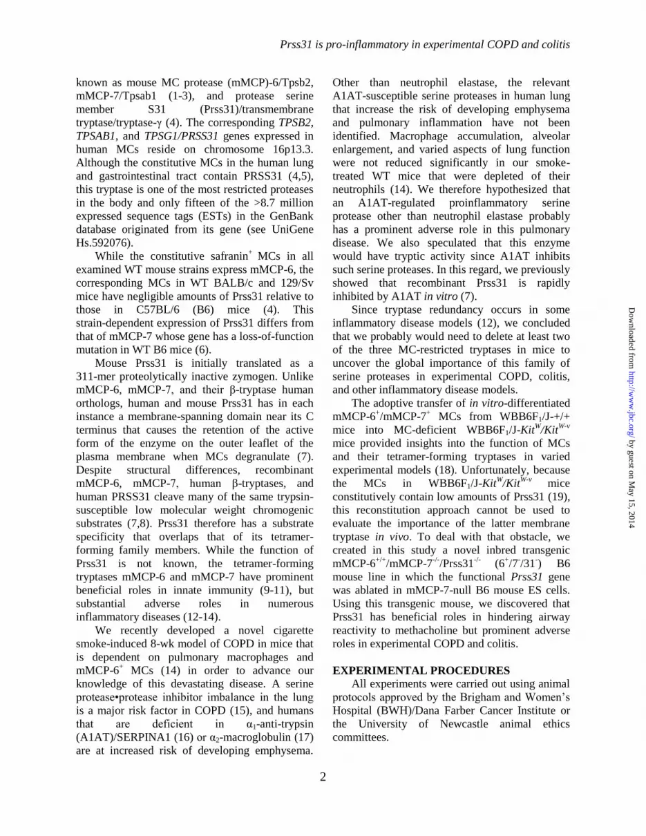
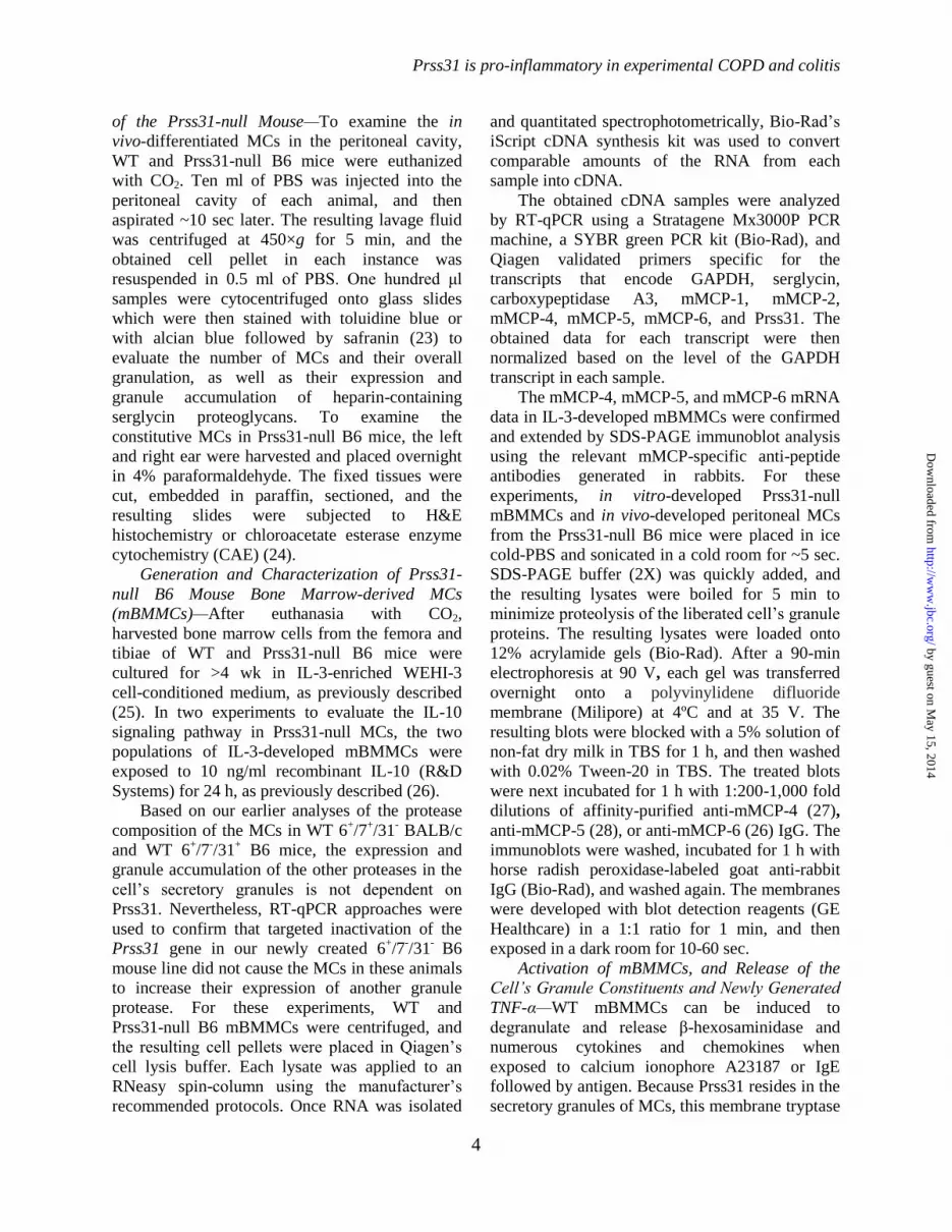
Cigarette Smoke-induced Experimental
COPD—mMCP-6 plays prominent roles in
cigarette smoke-induced COPD (14). Thus,
whether or not Prss31 also contributes to this
experimental model was investigated. As found in
our previous study carried out on WT and
mMCP-6-null B6 mice (14), the numbers of
leukocytes in the bronchial alveolar lavage fluid
(BALF) of smoke-treated WT B6 mice were
significantly greater than that of control WT B6
animals which breathed normal room air (Fig. 7A).
In support of these data, the numbers of
peribronchial, perivascular, and parenchymal
inflammatory cells were substantially increased in
the lungs of smoke-treated WT B6 mice (Fig. 7, C
and D). This cellular increase was markedly
attenuated in smoke-treated Prss31-null mice.
Differential cell counts in the BALF samples
showed that the increases in cellular infiltrates in
smoke-treated WT B6 mice were dominated by
macrophages with an associated increase in
neutrophils (Fig. 7B). In contrast, the numbers of
macrophages and neutrophils in the BALFs of
smoke-treated Prss31-null mice were not
significantly different than in the untreated
animals (Fig. 7B). In agreement with these data,
smoke-treated Prss31-null mice had reduced
histopathology scores in their lungs compared to
WT mice (Figs. 7, C and D).
While exposure to cigarette smoke resulted in
increased fibrosis around the small airways of WT
mice as assessed histochemically (Fig. 7, E and F),
this remodeling event was reduced in the lungs of
smoke-treated Prss31-null mice. The lack of
expression of Prss31 in the lungs of smoke-treated
B6 mice did not affect resistance, dynamic
compliance, tissue damping, or work of breathing.
Nevertheless, smoke-treated Prss31-null mice had
unchanged static compliance (Cst), in contrast to
smoke-treated WT mice (Fig. 7G).
Experimental Acute Colitis—In support of the
COPD data, DSS-induced acute colitis was
significantly reduced in Prss31-null B6 mice
relative to WT B6 mice (Fig. 8). Male
age-matched and sibling WT and Prss31-null mice
ingested 2% DSS in their drinking water for five
days. On days 6 and 7, the two groups of mice
were then given normal drinking water. The
animals were sacrificed on day 7, which is when
weight loss and inflammation are maximal after
WT B6 mice are subjected to this acute colitis
model. During the 7-day time period, Prss31-null
B6 mice lost only 5.8 ± 0.6% (n = 11) of their
original weight compared with WT B6 mice which
lost 11.1 ± 1.3% (n = 12, p<0.01) (Fig. 8A). At the
time of sacrifice, the colons of these same
DSS-treated WT mice were shortened to 6.6 ± 0.1
cm compared with the Prss31-null B6 mice colons
that were 7.2 ± 0.1 cm (p<0.001) (Fig. 8B). For
comparison, the colons of the WT B6 and Prss31-
null B6 mice that had not been exposed to DSS
were 8.1 ± 0.2 and 8.0 ± 0.2 cm, respectively.
Histopathology scores were determined to
assess the microscopic changes in DSS-induced
colitis that correlated with the weight and colon
length changes. A 20-point scoring system that
incorporates six different categories of mucosal
damage and inflammation was used. In these DSS
experiments, there was a significant difference in
the histopathology between the treated Prss31-null
B6 mice that had a less severe 9.5 ± 0.8
histopathology score compared with DSS-treated
WT mice that scored 14.5 ± 0.6 (p<0.0001) (Fig.
8C). Representative images are depicted in Figure
8D that demonstrate less epithelial cell crypt loss
and inflammatory cell infiltrate in the colonic
mucosa of DSS-treated Prss31-null relative to
treated WT B6 mice.
In regard to the mechanism(s) by which
Prss31 controls inflammation in the DSS-induced
colitis model, no significant differences in the
levels of the Mmp-13 and Cxcl-1 transcripts were
found in the colons of DSS-treated WT and
Prss31-null B6 mice (Fig. 9, C and D).
Nevertheless, in support of the weight loss and
histopathology data (Fig. 8), the levels of the
transcripts that encode the pro-inflammatory
cytokine IL-6 and the neutrophil-responsive
chemokine Cxcl-2 were significantly lower in the
colons of DSS-treated Prss31-null mice relative to
that in the colons of DSS-treated WT mice (Fig. 9,
A and B).
by guest on May 15, 2014
http://ww
w.jbc.org/
Dow
nloaded from

Prss31 is pro-inflammatory in experimental COPD and colitis
9
DISCUSSION
It has been known for >2 decades that the
three tryptases present in the secretory granules of
human and mouse MCs have
redundant/overlapping enzymatic activities in
vitro. Whether or not these in vitro data hold up in
vivo can only be established by the creation of
transgenic mice on a common genetic background
that lack different combinations of these enzymes.
Using our previously created 6-/7
-/31
+ B6 mice
(10), we discovered that the tetramer-forming
tryptases mMCP-6 and mMCP-7 have similar in
vivo bioactivities in blood coagulation (38,39) and
experimental arthritis (12). Because of the
possibility that mMCP-7 and Prss31 also might
have overlapping bioactivities in vivo, we
concluded that it was imperative that we knock out
the Prss31 gene in mMCP-7-/-
B6 mouse ES cells
rather than in mMCP-7+/+
129/Sv or BALB/c
mouse ES cells. Because that was done using our
Cre/loxP homologous recombination approach
(Fig. 1), the MCs in lung and other connective
tissues of our newly created mouse line have a
6+/7
-/31
- tryptase phenotype.
WT BALB/c and 129/Sv mice constitutively
lack Prss31, but express mMCP-7 (7). Thus, the
MCs in the peritoneal cavity, skin, and other
connective tissue sites of these two WT mouse
strains have a 6+/7
+/31
- tryptase phenotype.
Because WT BALB/c, 129/Sv, and B6 mice are
viable and have similar numbers of MCs in their
tissues, it was known before we began our studies
that Prss31 is not essential for the development of
mice and their tissue MCs. We also showed in the
1990s (1,2,40,41) that the MCs in the peritoneal
cavity, skin, and other connective tissues of the
WT 6+/7
+/31
- BALB/c mouse contain large
amounts of mMCP-4, mMCP-5, and mMCP-6
mRNA and protein. Thus, it also was known that
the expression and granule accumulation of the
latter three serine proteases in mouse peritoneal
MCs is not dependent on Prss31.
Our newly created Prss31-null B6 mice were
viable and had no obvious developmental
abnormality in their MCs (Figs. 2-4). We therefore
confirmed that Prss31 is not essential for the
homing, retention, and overall granule maturation
of the MCs that reside in varied tissues of the B6
mouse, including the granule storage of their other
protease•serglycin proteoglycan complexes. As
additionally anticipated, KitL-, IL-3-, IL-10-,
IL-33-, and FcεRI-signaling pathways were intact
in Prss31-null mouse MCs.
To deduce the in vivo function of Prss31, we
evaluated the susceptibility of our newly created
Prss31-null mice to methacholine. When our B6
mice were challenged with this synthetic choline
ester, we discovered that Prss31 had a physiologic
protective function in the lung by minimizing
airway responsiveness to the muscarinic receptor
agonist (Fig. 6).
Our earlier discovery (14) that cigarette
smoke-induced COPD was similar in WT 6+/7
-/31
+
B6 mice and WT 6+/7
+/31
- BALB/c mice raised
the possibility that mMCP-7 and Prss31 have
similar bioactivities in this experimental disease.
In support of that idea, cigarette smoke-induced
experimental COPD was more pronounced in WT
6+/7
-/31
+ B6 mice compared with our newly
created 6+/7
-/31
- B6 mice (Fig. 7). This finding
revealed for the first time that Prss31 has
prominent adverse inflammatory roles in
experimental COPD, as we previously showed for
mMCP-6 (14). Humans who have inactivating
mutations in the gene that encodes the serpin
A1AT are at increased risk of developing
emphysema even if they do not smoke (16),
thereby documenting the importance of a
A1AT/tryptic protease balance in the lungs. While
mMCP-6 and hTryptase-β are not susceptible to
A1AT, we previously showed that recombinant
human PRSS31 is rapidly inactivated by A1AT
(7). The fact that the genetic loss of a natural
inhibitor of Prss31 dramatically increases the risk
of lung inflammation in humans supports our
experimental mouse COPD data (Fig. 7).
We recently showed that mMCP-6 also has
prominent adverse roles in DSS-induced colitis
(13), and that this mouse ortholog of hTryptase-β
acts upstream of many of the pathologic factors in
patients with inflammatory bowel disease. Prss31
is expressed early in developing MCs (42), and the
intestine has the highest level of Prss31 mRNA in
the body (4). We therefore evaluated the
susceptibility of our Prss31-null B6 mice to
DSS-induced colitis. In confirmation of the
importance of Prss31 in a second MC-dependent
inflammatory disease model, experimental colitis
was significantly reduced in Prss31-null B6 mice
relative to WT B6 mice (Fig. 8).
The discovery that the levels of the
transcripts that encode the neutrophil-responsive
by guest on May 15, 2014
http://ww
w.jbc.org/
Dow
nloaded from

Prss31 is pro-inflammatory in experimental COPD and colitis
10
chemokine Cxcl-2 and the pro-inflammatory
cytokine IL-6 were both markedly reduced in the
colons of DSS-treated Prss31-null B6 mice
relative to treated WT B6 mice (Fig.9),
mechanistically explained, in part, why the
accumulation of neutrophils was markedly
reduced in the colons of the treated Prss31-null
mice (Fig. 8D). Our data therefore provide new
insights as to how MCs and specifically
exocytosed tryptase Prss31 participate in
inflammation and connective tissue remodeling in
two very different disease models.
The mechanisms at the cellular level as to
how Prss31 regulates baseline airway reactivity to
methacholine, DSS-induced colitis, and cigarette
smoke-induced COPD are likely to be complex, as
we discovered for the tryptase’s family members
mMCP-6 and mMCP-7. Nevertheless, we
previously showed that recombinant human
PRSS31 could induce peripheral blood T cells and
the Jurkat T cell line to quickly alter their
expression of hundreds of transcripts in vitro (7).
The putative receptor on the surfaces of T cells
that is susceptible to human and mouse Prss31
remains to be identified. However, one possibility
is a protease-activated receptor (PAR). All four of
these signaling proteins are activated by tryptic
proteases, and Jurkat cells express PARs (43). In
support of this hypothesis, PARs have been
implicated in airway responsiveness to
methacholine (44) and in experimental colitis (45).
MMPs are the primary neutral proteases that
participate in the remodeling of damaged
connective tissue. Cigarette smoke-induced COPD
is markedly attenuated in MMP-12-null mice (46),
and lung remodeling was reduced in smoke-treated
Prss31-null mice. That mMCP-6 and hTryptase-β
can activate varied pro-MMPs (47-49) raises the
additional possibility that Prss31 participates in
remodeling of the smoke-damaged lung by
activating pro-MMP-12 or another MMP
zymogen.
Whatever mechanisms are operative in
Prss31-dependent inflammation, it is now apparent
that one must knock out at least two of the three
tryptase genes in mouse MCs to undercover the
prominent adverse activities of this family of
serine proteases in experimental COPD and colitis.
Since human MCs express three functionally
similar tryptases encoded by the corresponding
TPSAB1, TPSB2, and TPSG1 genes, it is likely
that the primary reason why the tryptase locus on
human chromosome 16p13.3 has not been
identified in genome wide association studies of
patients with COPD or inflammatory bowel
disease is because of the in vivo redundancy of this
family of MC-restricted serine proteases.
Approximately ~1.5 and ~13.5 million people
in the United States alone have inflammatory
bowel disease and COPD, respectively. Moreover,
COPD is presently the third leading cause of death
in the United States, and carries an estimated ~2.1
trillion USDs yearly economic burden worldwide.
Thus, there is a critical need to identify and fully
understand novel inflammatory mediators and
their pathways so that more effective
pharmaceuticals can be developed. If our mouse
data are relevant to what occurs in humans with
COPD or colitis, the therapeutic potential of MC
tryptase inhibitors may only be realized when both
hTryptase-β and human PRSS31 are
inactivated.
Acknowledgements—We thank Michael Gurish (BWH) for the rabbit anti-mMCP-4 antibody used in the
SDS-PAGE immunoblot analysis of lysates of MCs from WT and Prss31-null B6 mice. We also thank
Kristy Wheeldon and Matthew Bowman for their technical assistance.
by guest on May 15, 2014
http://ww
w.jbc.org/
Dow
nloaded from

Prss31 is pro-inflammatory in experimental COPD and colitis
11
REFERENCES 1. Reynolds, D. S., Stevens, R. L., Lane, W. S., Carr, M. H., Austen, K. F., and Serafin, W. E. (1990)
Different mouse mast cell populations express various combinations of at least six distinct mast cell
serine proteases. Proc. Natl. Acad. Sci. USA 87, 3230-3234
2. Reynolds, D. S., Gurley, D. S., Austen, K. F., and Serafin, W. E. (1991) Cloning of the cDNA and
gene of mouse mast cell protease 6: transcription by progenitor mast cells and mast cells of the
connective tissue subclass. J. Biol. Chem. 266, 3847-3853
3. McNeil, H. P., Reynolds, D. S., Schiller, V., Ghildyal, N., Gurley, D. S., Austen, K. F., and
Stevens, R. L. (1992) Isolation, characterization, and transcription of the gene encoding mouse mast
cell protease 7. Proc. Natl. Acad. Sci. USA 89, 11174-11178
4. Wong, G. W., Tang, Y., Feyfant, E., Šali, A., Li, L., Li, Y., Huang, C., Friend, D. S., Krilis, S. A.,
and Stevens, R. L. (1999) Identification of a new member of the tryptase family of mouse and
human mast cell proteases that possesses a novel C-terminal hydrophobic extension. J. Biol. Chem.
274, 30784-30793
5. Caughey, G. H., Raymond, W. W., Blount, J. L., Hau, L. W., Pallaoro, M., Wolters, P. J., and
Verghese, G. M. (2000) Characterization of human γ tryptases, novel members of the chromosome
16p mast cell tryptase and prostasin gene families. J. Immunol. 164, 6566-6575
6. Hunt, J. E., Stevens, R. L., Austen, K. F., Zhang, J., Xia, Z., and Ghildyal, N. (1996) Natural
disruption of the mouse mast cell protease 7 gene in the C57BL/6 mouse. J. Biol. Chem. 271, 2851-
2855
7. Wong, G. W., Foster, P. S., Yasuda, S., Qi, J. C., Mahalingam, S., Mellor, E. A., Katsoulotos, G.,
Li, L., Boyce, J. A., Krilis, S. A., and Stevens, R. L. (2002) Biochemical and functional
characterization of human transmembrane tryptase (TMT)/tryptase γ: TMT is an exocytosed mast
cell protease that induces airway hyperresponsiveness in vivo via an IL-13/IL-4Rα/STAT6-
dependent pathway. J. Biol. Chem. 277, 41906-41915
8. Yuan, J., Beltman, J., Gjerstad, E., Nguyen, M. T., Sampang, J., Chan, H., Janc, J. W., and Clark, J.
M. (2006) Expression and characterization of recombinant γ-tryptase. Protein Expr. Purif. 49, 47-
54
9. Huang, C., De Sanctis, G. T., O'Brien, P. J., Mizgerd, J. P., Friend, D. S., Drazen, J. M., Brass, L.
F., and Stevens, R. L. (2001) Evaluation of the substrate specificity of human mast cell tryptase β1
and demonstration of its importance in bacterial infections of the lung. J. Biol. Chem. 276, 26276-
26284
10. Thakurdas, S. M., Melicoff, E., Sansores-Garcia, L., Moreira, D. C., Petrova, Y., Stevens, R. L.,
and Adachi, R. (2007) The mast cell-restricted tryptase mMCP-6 has a critical immunoprotective
role in bacterial infections. J. Biol. Chem. 282, 20809-20815
11. Shin, K., Watts, G. F., Oettgen, H. C., Friend, D. S., Pemberton, A. D., Gurish, M. F., and Lee, D.
M. (2008) Mouse mast cell tryptase mMCP-6 is a critical link between adaptive and innate
immunity in the chronic phase of Trichinella spiralis infection. J. Immunol. 180, 4885-4891
12. McNeil, H. P., Shin, K., Campbell, I. K., Wicks, I. P., Adachi, R., Lee, D. M., and Stevens, R. L.
(2008) The mouse mast cell-restricted tetramer-forming tryptases mouse mast cell protease 6 and
mouse mast cell protease 7 are critical mediators in inflammatory arthritis. Arthritis Rheum. 58,
2338-2346
13. Hamilton, M. J., Sinnamon, M. J., Lyng, G. D., Glickman, J. N., Wang, X., Xing, W., Krilis, S. A.,
Blumberg, R. S., Adachi, R., Lee, D. M., and Stevens, R. L. (2011) Essential role for mast cell
tryptase in acute experimental colitis. Proc. Natl. Acad. Sci. USA 108, 290-295
14. Beckett, E. L., Stevens, R. L., Jarnicki, A. G., Kim, R. Y., Hanish, I., Hansbro, N. G., Deane, A.,
Keely, S., Horvat, J. C., Yang, M., Oliver, B. G., van, R. N., Inman, M. D., Adachi, R., Soberman,
R. J., Hamadi, S., Wark, P. A., Foster, P. S., and Hansbro, P. M. (2013) A new short-term mouse
model of chronic obstructive pulmonary disease identifies a role for mast cell tryptase in
pathogenesis. J. Allergy Clin. Immunol. 131, 752-762
by guest on May 15, 2014
http://ww
w.jbc.org/
Dow
nloaded from

Prss31 is pro-inflammatory in experimental COPD and colitis
12
15. Keely, S., Talley, N. J., and Hansbro, P. M. (2012) Pulmonary-intestinal cross-talk in mucosal
inflammatory disease. Mucosal. Immunol 5, 7-18
16. Cohen, B. H., Ball, W. C., Jr., Brashears, S., Diamond, E. L., Kreiss, P., Levy, D. A., Menkes, H.
A., Permutt, S., and Tockman, M. S. (1977) Risk factors in chronic obstructive pulmonary disease
(COPD). Am. J. Epidemiol. 105, 223-232
17. Poller, W., Barth, J., and Voss, B. (1989) Detection of an alteration of the α2-macroglobulin gene in
a patient with chronic lung disease and serum α2-macroglobulin deficiency. Hum. Genet. 83, 93-96
18. Kitamura, Y., Go, S., and Hatanaka, K. (1978) Decrease of mast cells in W/Wv mice and their
increase by bone marrow transplantation. Blood 52, 447-452
19. Morii, E., Ogihara, H., Oboki, K., Kataoka, T. R., Jippo, T., and Kitamura, Y. (2001) Effect of
MITF on transcription of transmembrane tryptase gene in cultured mast cells of mice. Biochem.
Biophys. Res. Commun. 289, 1243-1246
20. Chen, Y., Lu, J., Pan, H., Zhang, Y., Wu, H., Xu, K., Liu, X., Jiang, Y., Bao, X., Yao, Z., Ding, K.,
Lo, W. H., Qiang, B., Chan, P., Shen, Y., and Wu, X. (2003) Association between genetic variation
of CACNA1H and childhood absence epilepsy. Ann. Neurol. 54, 239-243
21. Chen, C. C., Lamping, K. G., Nuno, D. W., Barresi, R., Prouty, S. J., Lavoie, J. L., Cribbs, L. L.,
England, S. K., Sigmund, C. D., Weiss, R. M., Williamson, R. A., Hill, J. A., and Campbell, K. P.
(2003) Abnormal coronary function in mice deficient in α1H T-type Ca2+
channels. Science 302,
1416-1418
22. Ghildyal, N., Friend, D. S., Freelund, R., Austen, K. F., McNeil, H. P., Schiller, V., and Stevens, R.
L. (1994) Lack of expression of the tryptase mouse mast cell protease 7 in mast cells of the
C57BL/6J mouse. J. Immunol. 153, 2624-2630
23. Enerbäck, L. (1966) Mast cells in rat gastrointestinal mucosa. 2. Dye-binding and metachromatic
properties. Acta Pathol. Microbiol. Scand. 66, 303-312
24. Friend, D. S., Ghildyal, N., Austen, K. F., Gurish, M. F., Matsumoto, R., and Stevens, R. L. (1996)
Mast cells that reside at different locations in the jejunum of mice infected with Trichinella spiralis
exhibit sequential changes in their granule ultrastructure and chymase phenotype. J. Cell Biol. 135,
279-290
25. Razin, E., Ihle, J. N., Seldin, D., Mencia-Huerta, J. M., Katz, H. R., LeBlanc, P. A., Hein, A.,
Caulfield, J. P., Austen, K. F., and Stevens, R. L. (1984) Interleukin-3: a differentiation and growth
factor for the mouse mast cell that contains chondroitin sulfate E proteoglycan. J. Immunol. 132,
1479-1486
26. Ghildyal, N., Friend, D. S., Nicodemus, C. F., Austen, K. F., and Stevens, R. L. (1993) Reversible
expression of mouse mast cell protease 2 mRNA and protein in cultured mast cells exposed to IL-
10. J. Immunol. 151, 3206-3214
27. Forsberg, E., Pejler, G., Ringvall, M., Lunderius, C., Tomasini-Johansson, B., Kusche-Gullberg,
M., Eriksson, I., Ledin, J., Hellman, L., and Kjellén, L. (1999) Abnormal mast cells in mice
deficient in a heparin-synthesizing enzyme. Nature 400, 773-776
28. McNeil, H. P., Frenkel, D. P., Austen, K. F., Friend, D. S., and Stevens, R. L. (1992) Translation
and granule localization of mouse mast cell protease 5: immunodetection with specific antipeptide
Ig. J. Immunol. 149, 2466-2472
29. Galli, S. J., Gordon, J. R., and Wershil, B. K. (1993) Mast cell cytokines in allergy and
inflammation. Agents Actions Suppl 43, 209-220
30. Razin, E., Mencia-Huerta, J. M., Lewis, R. A., Corey, E. J., and Austen, K. F. (1982) Generation of
leukotriene C4 from a subclass of mast cells differentiated in vitro from mouse bone marrow. Proc.
Natl. Acad. Sci. USA 79, 4665-4667
31. Robinson, D. and Stirling, J. L. (1968) N-acetyl-β-glucosaminidases in human spleen. Biochem. J.
107, 321-327
32. Essilfie, A. T., Simpson, J. L., Horvat, J. C., Preston, J. A., Dunkley, M. L., Foster, P. S., Gibson, P.
G., and Hansbro, P. M. (2011) Haemophilus influenzae infection drives IL-17-mediated
neutrophilic allergic airways disease. PLoS. Pathog. 7, e1002244
by guest on May 15, 2014
http://ww
w.jbc.org/
Dow
nloaded from

Prss31 is pro-inflammatory in experimental COPD and colitis
13
33. Horvat, J. C., Beagley, K. W., Wade, M. A., Preston, J. A., Hansbro, N. G., Hickey, D. K., Kaiko,
G. E., Gibson, P. G., Foster, P. S., and Hansbro, P. M. (2007) Neonatal chlamydial infection
induces mixed T-cell responses that drive allergic airway disease. Am. J Respir. Crit Care Med.
176, 556-564
34. Horvat, J. C., Starkey, M. R., Kim, R. Y., Phipps, S., Gibson, P. G., Beagley, K. W., Foster, P. S.,
and Hansbro, P. M. (2010) Early-life chlamydial lung infection enhances allergic airways disease
through age-dependent differences in immunopathology. J Allergy Clin. Immunol. 125, 617-25, 625
35. Fricker, M., Deane, A., and ansbro, P. M. (2014) Animal modes of chronic obstructive pulmonary
disease. Expert. Opin. Drug Disc., in press
36. Okayasu, I., Hatakeyama, S., Yamada, M., Ohkusa, T., Inagaki, Y., and Nakaya, R. (1990) A novel
method in the induction of reliable experimental acute and chronic ulcerative colitis in mice.
Gastroenterology 98, 694-702
37. Garrett, W. S., Lord, G. M., Punit, S., Lugo-Villarino, G., Mazmanian, S. K., Ito, S., Glickman, J.
N., and Glimcher, L. H. (2007) Communicable ulcerative colitis induced by T-bet deficiency in the
innate immune system. Cell 131, 33-45
38. Huang, C., Wong, G. W., Ghildyal, N., Gurish, M. F., Šali, A., Matsumoto, R., Qiu, W. T., and
Stevens, R. L. (1997) The tryptase, mouse mast cell protease 7, exhibits anticoagulant activity in
vivo and in vitro due to its ability to degrade fibrinogen in the presence of the diverse array of
protease inhibitors in plasma. J. Biol. Chem. 272, 31885-31893
39. Prieto-Garcia, A., Zheng, D., Adachi, R., Xing, W., Lane, W. S., Chung, K., Anderson, P.,
Hansbro, P. M., Castells, M., and Stevens, R. L. (2012) Mast cell restricted mouse and human
tryptase-heparin complexes hinder thrombin-induced coagulation of plasma and the generation of
fibrin by proteolytically destroying fibrinogen. J. Biol. Chem. 287, 7834-7844
40. McNeil, H. P., Austen, K. F., Somerville, L. L., Gurish, M. F., and Stevens, R. L. (1991) Molecular
cloning of the mouse mast cell protease 5 gene: a novel secretory granule protease expressed early
in the differentiation of serosal mast cells. J. Biol. Chem. 266, 20316-20322
41. Serafin, W. E., Sullivan, T. P., Conder, G. A., Ebrahimi, A., Marcham, P., Johnson, S. S., Austen,
K. F., and Reynolds, D. S. (1991) Cloning of the cDNA and gene for mouse mast cell protease 4:
demonstration of its late transcription in mast cell subclasses and analysis of its homology to
subclass-specific neutral proteases of the mouse and rat. J. Biol. Chem. 266, 1934-1941
42. Wong, G. W., Yasuda, S., Morokawa, N., Li, L., and Stevens, R. L. (2004) Mouse chromosome
17A3.3 contains thirteen genes that encode functional tryptic-like serine proteases with distinct
tissue and cell expression patterns. J. Biol. Chem. 279, 2438-2452
43. Bar-Shavit, R., Maoz, M., Yongjun, Y., Groysman, M., Dekel, I., and Katzav, S. (2002) Signalling
pathways induced by protease-activated receptors and integrins in T cells. Immunology 105, 35-46
44. De Campo, B. A. and Henry, P. J. (2005) Stimulation of protease-activated receptor-2 inhibits
airway eosinophilia, hyperresponsiveness and bronchoconstriction in a murine model of allergic
inflammation. Br. J Pharmacol. 144, 1100-1108
45. Fiorucci, S., Mencarelli, A., Palazzetti, B., Distrutti, E., Vergnolle, N., Hollenberg, M. D., Wallace,
J. L., Morelli, A., and Cirino, G. (2001) Proteinase-activated receptor 2 is an anti-inflammatory
signal for colonic lamina propria lymphocytes in a mouse model of colitis. Proc. Natl. Acad. Sci.
USA 98, 13936-13941
46. Hautamaki, R. D., Kobayashi, D. K., Senior, R. M., and Shapiro, S. D. (1997) Requirement for
macrophage elastase for cigarette smoke-induced emphysema in mice. Science 277, 2002-2004
47. Magarinos, N. J., Bryant, K. J., Fosang, A. J., Adachi, R., Stevens, R. L., and McNeil, H. P. (2013)
Mast cell-restricted, tetramer-forming tryptases induce aggrecanolysis in articular cartilage by
activating matrix metalloproteinase-3 and -13 zymogens. J. Immunol. 191, 1404-1412
48. Gruber, B. L., Schwartz, L. B., Ramamurthy, N. S., Irani, A. M., and Marchese, M. J. (1988)
Activation of latent rheumatoid synovial collagenase by human mast cell tryptase. J. Immunol. 140,
3936-3942
by guest on May 15, 2014
http://ww
w.jbc.org/
Dow
nloaded from

Prss31 is pro-inflammatory in experimental COPD and colitis
14
49. Gruber, B. L., Marchese, M. J., Suzuki, K., Schwartz, L. B., Okada, Y., Nagase, H., and
Ramamurthy, N. S. (1989) Synovial procollagenase activation by human mast cell tryptase
dependence upon matrix metalloproteinase 3 activation. J. Clin. Invest. 84, 1657-1662
by guest on May 15, 2014
http://ww
w.jbc.org/
Dow
nloaded from

Prss31 is pro-inflammatory in experimental COPD and colitis
15
FOOTNOTES
*This work was funded in part by grants from the National Institutes of Health (DK094971 and
AI059746), the National Health and Medical Research Council of Australia, the Harvard Digestive
Diseases Center, and the Saint George Medical Research Foundation, as well as by research fellowship
grants to SAK, RLS, PSF, and DZ from the Harvard Club of Australia Foundation.
The nucleotide and amino acid sequences reported in this paper for the BALB/c, CBA, SWR, A/J, and
C3H/HeJ mouse Prss31 genes determined in this study were deposited at GenBank. Their respective
accession numbers are GU810532, GU810531, GU810528, GU810529, and GU810533.
1Both authors contributed equally.
2To whom correspondence should be addressed: Richard L. Stevens, Brigham and Women’s Hosp., Dept.
Med., Div. Rheumatol. Immunol. Allergy, Smith Bldg., Rm. 616B, 1 Jimmy Fund Way, Boston, MA
02115 USA. Tel: 617-525-1231; Fax: 617-525-1310; E-mail: [email protected].
3The abbreviations used are: A1AT; α1-anti-trypsin; BALF, bronchial alveolar lavage fluid; CAE,
chloroacetate esterase; COPD, chronic obstructive pulmonary disease; DSS, dextran sodium sulfate; EST,
expressed sequence tag; HPF, high power field; MC, mast cell; mBMMC, mouse bone marrow-derived
MC; mMCP, mouse MC protease; MMP, matrix metalloproteinase; Prss31, protease serine member S31;
B6, C57BL/6; and 6+/7
-/31
-, mMCP-6
+/+/mMCP-7
-/-/Prss31
-/- .
by guest on May 15, 2014
http://ww
w.jbc.org/
Dow
nloaded from

Prss31 is pro-inflammatory in experimental COPD and colitis
16
LEGENDS
FIGURE 1. Creation of a transgenic Prss31-null B6 mouse. A-C, Shown in A is the genomic locus that
contains the B6 mouse Prss31 gene and its upstream mMCP-6/Tpsb2 and downstream Cacna1h genes.
Also shown in this panel is the Prss31-targeting vector with its loxP (red ►) and F3 (blue ►) sites, and
the coding (solid blue) and untranslated (hatched blue) portions of the exons of the mouse Prss31 gene.
The arrows indicate the orientations of the mMCP-6, Prss31, and Cacna1h genes on mouse chromosome
17A3.3. The sizes and locations of the long (LA), floxed (FA), and short (SA) arms of homology are
shown. After homologous recombination, the negative selection marker Tk is lost and the nucleotide
sequence of the targeting vector has replaced one of the alleles of the endogenous Prss31 gene in the B6
mouse ES cells. To demonstrate that homologous recombination took place in the C1 ES cell clone that
was injected into mouse blastocysts (B), genomic DNA was isolated form this clone and from untreated
ES cells. In both instances, the resulting DNA was digested with the restricted enzyme NsiI and subjected
to gel electrophoresis. The resulting DNA blot was then probed with a genomic fragment residing
upstream of the normal mouse Prss31 gene. Because an NsiI-susceptible sequence resides just after the
first loxP site in the targeting vector, a smaller genomic fragment (open arrow, lane 1) than the WT
Prss31 gene (solid arrow, lanes 1 and 2) is obtained if homologous recombination takes place at one of
the alleles of the mouse Prss31 gene. The DNA blot was next analyzed with a PuroR probe to confirm the
presence of the positive selection marker. Once chimeras and heterozygotes were generated, Flp-mediated
recombination was used to remove the positive selection marker PuroR. Cre-mediated recombination was
then employed to remove the first exon of the Prss31 gene. Heterozygotes containing one WT and one
disrupted allele of the Prss31 gene were bred to create the final Prss31-null B6 mouse line (C, lane 3).
Standard molecular weight 100-bp markers are shown in C, lane 1. The relevant DNA product (C, lane 3)
identified in the confirmatory PCR-based genotyping approach we used is 326-bp. The entire process to
create the final mouse line took >4 years of effort.
FIGURE 2. Histochemistry and enzyme cytochemistry of the cutaneous MCs in WT and Prss31-null
B6 mice. A-D, Identification of CAE+
cutaneous MCs. Sections of the ears of WT (A and B) and
Prss31-null (C and D) mice were subjected to H&E histochemistry (A and C) and enzyme cytochemistry
(B and D). No ultrastructural abnormality was detected in the ears of the Prss31-null mice, as also
occurred in the jejunum (data not shown). All MCs in WT B6 mice are CAE+
due to their granule storage
of large amounts of the chromosome 14C3 family members mMCPs 1-5 and 8-10. WT and Prss31-null
B6 mice constitutively had similar numbers of CAE+ MCs (red cells highlighted by black arrows) in their
ears (B and D). Thus, Prss31 is not essential for the recruitment of MC-committed progenitors into the ear
or for their development into mature MCs, as anticipated. For size comparison, the red arrows in B and D
point to the 10-µm magnification bars.
FIGURE 3. Histochemistry of the peritoneal MCs in WT and Prss31-null B6 mice. A-B, Peritoneal
lavage was performed on WT (A) and Prss31-null (B) B6 mice. Cytospins of the cells in the resulting
exudates were stained with toluidine blue or with alcian blue followed by safranin. Shown at lower power
(highlighted by black arrows), and at higher power in the bottom left inserts are the safranin+ MCs in the
exudates. Shown at higher power in the bottom right inserts are the corresponding peritoneal MCs in
these exudates stained with toluidine blue. Toluidine blue recognizes all types of negatively charged
serglycin proteoglycans in the granules of MCs, including those that contain chondroitin sulfate diB,
chondroitin sulfate E, and heparin glycosaminoglycans. In contrast, safranin preferentially recognizes
those serglycin proteoglycans that contain heparin glycosaminoglycans. The relative number of safranin+
and toluidine blue+ MCs in the peritoneal cavities of the two mouse strains were the same, as were their
degree of granulation and expression of heparin-containing serglycin proteoglycans.
by guest on May 15, 2014
http://ww
w.jbc.org/
Dow
nloaded from

Prss31 is pro-inflammatory in experimental COPD and colitis
17
FIGURE 4. Histochemistry of WT and Prss31-null mBMMCs, and protease expression in
mBMMCs and peritoneal MCs. A-B, Bone marrow cells harvested from WT (A) and Prss31-null (B) B6
mice were cultured for >4 wk in the presence of IL-3 enriched WEHI-3 cell conditioned medium. As
found in earlier studies (25), the resulting mBMMCs generated from WT B6 mice varied considerably in
their degree of granule maturation (A). Nevertheless, because similar numbers of histochemically
identical mBMMCs were generated from Prss31-null B6 mice (B), there was no defect in the IL-3/IL3Rα
signaling pathways in MCs developed from Prss31-null mice. For size comparison, the red arrow pointing
to the 10-µm magnification bar is shown in each panel. C, The levels of mMCP-4, mMCP-5, and
mMCP-6 protein in lysates of in vivo-differentiated Prss31-null mouse peritoneal MCs (mPMCs) and in
vitro-differentiated Prss31-null mBMMCs were determined by SDS-PAGE immunoblot analyses. Lysates
of the two populations of MCs were subjected to SDS-PAGE, and the resulting protein blots were stained
with affinity-purified anti-peptide antibodies that recognize mMCP-4, mMCP-5, and mMCP-6. As
previously found for WT B6 mice (1,41) and in agreement with the histochemistry and CEA enzyme
cytochemistry, mPMCs isolated from Prss31-null mice (lane 1) stored substantial amounts of all three
serine proteases in their secretory granules, whereas the levels of mMCP-4 protein were below detection
in IL-3-developed mBMMCs (lane 2). D, Since the mMCP-6 gene resides immediately upstream of the
Prss31 gene (Fig. 1), the level of the mMCP-6 transcript was quantitated in WT and Prss31-null B6
mBMMCs. Shown are the relative Ct expression levels of the mMCP-6 transcript normalized to that of
the GAPDH transcript (mean ± ½ range, n = 2). Similar RNA data were obtained in a second experiment
also carried out using mBMMCs obtained from two WT B6 mice and two Prss31-null mice. In agreement
with the SDS-PAGE immunoblot data (C), targeted inactivation of the Prss31 gene using our Cre/loxP
homologous recombination approach did not adversely affect transcription of the nearby mMCP-6 gene in
IL-3-developed mBMMCs.
FIGURE 5. IL-10 induces the expression of mMCP-1 and mMCP-2 in WT and Prss31-null
mBMMCs. A-C, WT 6+/7
-/31
+ and transgenic 6
+/7
-/31
- B6 mBMMCs were cultured in the presence (■)
or absence (□) of IL-10 (n = 3 per group) for 24 h. In each instance, RNA was isolated, converted to
cDNA, and the levels of the transcripts that encode mMCP-1 (A), mMCP-2 (B), and mMCP-5 (C) were
determined. The obtained data were then normalized to that of the ubiquitously expressed GAPDH
transcript. Results were expressed as fold-change differences relative to that in replicate mBMMCs that
had not been exposed to IL-10. As occurred in WT mBMMCs, Prss31-null mBMMCs markedly
increased their levels of mMCP-1 and mMCP-2 mRNA when exposed to IL-10. In contrast, the levels of
the mMCP-5 transcript were not significantly different. Similar findings were obtained in a second
experiment, also carried out in triplicate.
FIGURE 6. Baseline blood cell populations and airway hyperresponsiveness to methacholine. A,
Cardiac punctures were performed on naïve WT and Prss31-null mice, and in each instance a single drop
of blood was spread onto a microscope glass slide. Slides were stained with H&E and differential
leukocyte counts were obtained based on cellular morphology. There were no differences in baseline
blood cell populations between naïve WT and Prss31-null mice. B-H, Naïve WT (●) and Prss31-null (■)
B6 mice were cannulated, and attached to a forced maneuver (Buxco) or oscillation (Flexivent) system
and lung function parameters were assessed. When on the forced oscillation system, the mice were
challenged with 15-µl solutions containing 0-10 mg/ml methacholine. Relative to naïve WT mice, the
airways of naïve Prss31-null mice were more responsive to the muscarinic receptor agonist. The latter
transgenic mice had increased airway resistance (B), transpulmonary resistance (C), hysteresivity (D),
dynamic elastance (F), tissue elastance (G), and tissue damping (H), as well as reduced dynamic
compliance (E). The data are the mean ± SEM of 6-8 mice. In A, the NS above the compared columns
corresponds to p values that were not significant. In B-H, the *, ***, and **** symbols above curves
correspond to p values that were <0.05, <0.001, and <0.0001, respectively.
by guest on May 15, 2014
http://ww
w.jbc.org/
Dow
nloaded from

Prss31 is pro-inflammatory in experimental COPD and colitis
18
FIGURE 7. Experimental COPD. A-G, WT and Prss31-null B6 mice were exposed to cigarette smoke
for 8 wk. The BALF was collected in each instance, and cell counts and cytospins were performed to
enumerate the numbers of total leukocytes, as well as the numbers of macrophages, neutrophils, and
lymphocytes. Replicate lungs were perfused, inflated, paraffin embedded, sectioned, stained, and then
histopathology scores were determined. Greater numbers of macrophages and neutrophils were found in
the BALF of smoke-treated WT mice relative to smoke-treated Prss31-null mice (A, B). The
histopathology scores (C, D) also were reduced in smoke-treated Prss31-null mice. The black arrow in the
upper right panel of D highlights the inflammation in the lungs of smoke-treated WT mice. Baseline
fibrosis (presumably collagen accumulation) in the small airways and lung function of the mice were then
examined. As assessed histochemically (E, F), there was more fibrosis present in the small airways of
smoke-treated WT mice (bar in the insert of the upper right panel of E) than in the lungs of smoke-treated
Prss31-null mice (E, F). While exposure of WT B6 mice to cigarette smoke for 8 wk resulted in a reduced
pulmonary Cst (G), this did not occur in similarly treated Prss31-null mice. The data are the mean ± SEM
of 6-8 mice. The NS, *, **, and *** symbols above the compared columns correspond to p values that
were not significant, or were <0.05, <0.01, and <0.001, respectively.
FIGURE 8. Experimental colitis. WT and Prss31-null mice were exposed to DSS for 7 days. WT (○) and
age-matched Prss31-null (■,♦) B6 mice were untreated (♦) or were administered 2.0% DSS in their
drinking water for 5 days, and then normal drinking water on days 6 and 7 (○, ■). The resulting mice
were sacrificed at day 7. Relative to DSS-treated WT mice, DSS-treated Prss31-null mice lost less weight
(**, p<0.01) (A), had longer colons (***, p<0.001) (B), and lower histopathology scores (p<0.001) (C).
Assessed histochemically (D), more neutrophils and other inflammatory cells (yellow arrow, bottom left
panel) accumulated in the mucosa of the colons of DSS-treated WT mice. However, the extent of
inflammation of the submucosa and muscle layers of the DSS-treated Prss31-null and WT animals were
similar (black arrows). For size comparison, the 50-µm magnification bar (red) is indicated in each panel
of D.
FIGURE 9. Transcript levels in the colons of DSS-treated WT and Prss31-null B6 mice. A-D, After
colitis was induced in WT (n = 5) and Prss31-/-
(n = 5) B6 mice, the two groups of animals were sacrificed
and RNA was isolated to evaluate the levels of the transcripts that encode Cxcl-2 (A), IL-6 (B), Mmp-13
(C), and Cxcl-1 (D). The obtained data were first normalized to those of the GAPDH transcript and then
to untreated mice. In this colitis model, the colons of DSS-treated WT mice contained more Cxcl-2 (p =
0.017) and IL-6 (p = 0.055) mRNA than the colons of DSS-treated Prss31-/-
mice. In contrast, the levels of
the transcripts that encode Cxcl-1 and MMP-13 were not significantly different between the two groups
of DSS-treated animals, in contrast to what occurs in DSS-treated mMCP-6-null mice.
by guest on May 15, 2014
http://ww
w.jbc.org/
Dow
nloaded from


FIGURE 4
0.0
2.5
5.0
7.5
10.0
12.5
D
Rela
tive
mM
CP
-6
mR
NA
Le
ve
ls
Prss31
+/+ Prss31
-/-
mBMMCs mBMMCs
by guest on May 15, 2014
http://ww
w.jbc.org/
Dow
nloaded from


Importance of Mast Cell Prss31/Transmembrane Tryptase/Tryptase-γ in Lung Function and Experimental Chronic
Obstructive Pulmonary Disease and Colitis
Philip M. Hansbro, Matthew J. Hamilton, Michael Fricker, Shaan L. Gellatly,
Andrew G. Jarnicki, Dominick Zheng, Sandra M. Frei, G. William Wong, Sahar Hamadi, Saijun Zhou,
Paul S. Foster, Steven A. Krilis, and Richard L. Stevens
Supplemental FIGURE S1
|33
B6 1 cccatactga taggcctggg gctgggcagg ggcatggggt ggtcctgact ctgctgtggg
BALB/c a
CBA a
SWR a
A/J a
129/Sv a
C3H/HeJ a
|84
B6 61 aagacaccAT Ggctctgggg ccctactgtg gcatcctact gtttctggct gtttctggtg
BALB/c a(Tyr/Asn)
CBA a(Tyr/Asn)
SWR a(Tyr/Asn)
A/J a(Tyr/Asn)
129/Sv a(Tyr/Asn)
C3H/HeJ a(Tyr/Asn)
B6 121 agtgaactgg cttggggttg gtgtggcact ggggtgggag gggcctgctt cctctcagga
BALB/c
CBA
SWR
A/J
129/Sv
C3H/HeJ
|186
B6 181 acttcacctg catttattct attttccact ctgggctgga gctcagagaa atctgggagc
BALB/c c
CBA c
SWR c
A/J c
129/Sv c
C3H/HeJ c
B6 241 catgtaagag gggtccgggt accttgaagt ctggccttag acaagcaaag ccaagttctt
BALB/c
CBA
SWR
A/J
129/Sv
C3H/HeJ
B6 301 catggtggcc tctgctttgt ctgggctcct cagctgggca gcttggctgt cattactgga
BALB/c
CBA
SWR
A/J
129/Sv
C3H/HeJ

|417
B6 361 gacagaggct tgtgtgtatg tgtggggaca cctacaggac tagatcctgg aaggacacac
BALB/c c
CBA c
SWR c
A/J c
129/Sv c
C3H/HeJ c
B6 421 ctcaaggagg acctagaggt tgtgaggggc tgaggctcac tgcctcccag ctgttgttta
BALB/c
CBA
SWR
A/J
129/Sv
C3H/HeJ
B6 481 tggcccatgg ctggtcccac cctaccctgg aactaggatg cagttgacca ggccacaatc
BALB/c
CBA
SWR
A/J
129/Sv
C3H/HeJ
B6 541 aattagctgg gccccaccca gctgggcccc acctacctgg ctagatctag ctttagctgc
BALB/c
CBA
SWR
A/J
129/Sv
C3H/HeJ
649| |654
B6 601 ccaggatggg aacatctacc tcagggccac cttgggatct tccagggttg ggggcagtgc
BALB/c g a
CBA g a
SWR g a
A/J g a
129/Sv g a
C3H/HeJ g a
B6 661 ttaggcccac aggaccagag tcactcccac gggcagttcc cctaatccct ccgtctctat
BALB/c
CBA
SWR
A/J
129/Sv
C3H/HeJ
|742
B6 721 ccttcaggca tcgcccatga agctctacag tcaggtgcac ctccaacaga tatttcggga
BALB/c a
CBA a
SWR a
A/J a
129/Sv a
C3H/HeJ a

B6 781 ttagagacca gtcctggcag gcagacccta gaggggcagt gcctgtggtg ggagagagtg
BALB/c
CBA
SWR
A/J
129/Sv
C3H/HeJ
B6 841 acctggccca gggaggctat gatttgccac aggcttctcc tgggaagctg cacctctgcc
BALB/c
CBA
SWR
A/J
129/Sv
C3H/HeJ
|915
B6 901 ccatttgtat cttcgagagc cactttccat ctcctgcgac acagcctgct ggctctcaca
BALB/c t
CBA t
SWR t
A/J t
129/Sv t
C3H/HeJ t
B6 961 gttgatctct ttcttttgct gatgttgctc ttgggaaaga gactcaaagc atcttcctca
BALB/c
CBA
SWR
A/J
129/Sv
C3H/HeJ
B6 1021 ggacgtacct ggtgttctgg gtgggaagga tgtggtctct tccatttctt tttatcaatg
BALB/c
CBA
SWR
A/J
129/Sv
C3H/HeJ
|1130
B6 1081 tggacccaat ttcgggtttc attttgtagt acagattaag aggaaagcct tcccctgtgg
BALB/c c
CBA c
SWR c
A/J c
129/Sv c
C3H/HeJ c
B6 1141 gtgatatctc tagattttca tgagaaaaaa aattgaggtg aaatttacat aacatttact
BALB/c
CBA
SWR
A/J
129/Sv
C3H/HeJ

B6 1201 cacgatacag tgtagcattt gttgacattt aacacatccc cagtgttttg tgatatcaca
BALB/c
CBA
SWR
A/J
129/Sv
C3H/HeJ
B6 1261 tctatctagt tccaaacatc cttctaaaaa ggaaacccca ttctcactta ggggggcacc
BALB/c
CBA
SWR
A/J
129/Sv
C3H/HeJ
B6 1321 ccctaccccc agcctcctgg gcctccatag ctcccatcac ctcagccctt cctcacccct
BALB/c
CBA
SWR
A/J
129/Sv
C3H/HeJ
B6 1381 ttaattccag gcagtaagcc gtttgcccag ctccactgag catctttcta aggagcctgc
BALB/c
CBA
SWR
A/J
129/Sv
C3H/HeJ
B6 1441 tacgtccttt ggggtagtgt tcggaagcca agatccatga acttactgct tctagggtag
BALB/c
CBA
SWR
A/J
129/Sv
C3H/HeJ
B6 1501 cggttcccaa cctgtggggc gcaacccctt gggggagatc aaaccctttt acaggggtcc
BALB/c
CBA
SWR
A/J
129/Sv
C3H/HeJ
B6 1561 cctaacatta tttgcatatc aaatatttat attatgattc ataatagcaa agttacagtt
BALB/c
CBA
SWR
A/J
129/Sv
C3H/HeJ

B6 1621 atgaagtagc aacataaata attttatggt tgggggttac aacaacacag ggaactgtat
BALB/c
CBA
SWR
A/J
129/Sv
C3H/HeJ
|1704
B6 1681 gaatgggtcc cagtgttagg aagattgaaa accctagcat gcaaccacaa tgcttctgga
BALB/c g
CBA g
SWR g
A/J g
129/Sv g
C3H/HeJ g
|1741
B6 1741 aagagaccag cggatgggga gcagagacat ttgtagatgc cccaagatgc tagggctccc
BALB/c g
CBA g
SWR g
A/J g
129/Sv g
C3H/HeJ g
B6 1801 aagatgggag tcatggaggg ctcccagaga aagggcattg agcagaatgc cggtctccag
BALB/c
CBA
SWR
A/J
129/Sv
C3H/HeJ
|1869
B6 1861 attccctcgc caacagtgtc tcctctggat cagggtgtgg ccatccccag gtttcaaact
BALB/c a
CBA a
SWR a
A/J g
129/Sv a
C3H/HeJ a
B6 1921 cgggaagtcg aatcgtggga gggcatgctg ccccagcagg cacatggccg tggcaggcta
BALB/c
CBA
SWR
A/J
129/Sv
C3H/HeJ
B6 1981 gcctccgtct gcacaaggtg cacgtgtgtg gaggctccct gctcagtcca gaatgggtgc
BALB/c
CBA
SWR
A/J
129/Sv
C3H/HeJ

B6 2041 tcacagcagc ccactgcttc tctgggtgag tcacttcata aagcctggct gcctggcctt
BALB/c
CBA
SWR
A/J
129/Sv
C3H/HeJ
B6 2101 atggggcaat gactgcaaac aggaccttga ttagccgtga ctcacccttg agtccaaggg
BALB/c
CBA
SWR
A/J
129/Sv
C3H/HeJ
|2194
B6 2161 tccctttcag tagagaaaac ttcattaagg tccacccacc tgcaatggca gtgtacagcc
BALB/c g
CBA g
SWR g
A/J a
129/Sv g
C3H/HeJ g
7-mer (attgggc)deletion/16-mer insertion|||||||2261-2267
B6 2221 ccctctccca catctgccac gggaggagaa tgcaacctcc attgggctta agtgtgatgc
BALB/c -------cgttaagtgtgatgct
CBA -------cgttaagtgtgatgct
SWR -------cgttaagtgtgatgct
A/J attgggc
129/Sv -------cgttaagtgtgatgct
C3H/HeJ -------cgttaagtgtgatgct
B6 2281 tggccaccca tctagcttgt atcaggatgt atcttaaacc agccttaaca ggcccttcca
BALB/c
CBA
SWR
A/J
129/Sv
C3H/HeJ
2375| 4-mer (agcc) insert|2394-2395
B6 2341 cttgtctcga acacttctgg aactgccttt cccccagagt gccagccacc tcccaacgtc
BALB/c c agcc
CBA c agcc
SWR c agcc
A/J a agcc
129/Sv c agcc
C3H/HeJ c agcc
|2423 |2450
B6 2401 ccagctgttc ttgggccctt acgtgcgagg cactgccagg ttcaggcttt atgtacgcag
BALB/c a c
CBA a c
SWR a c
A/J a c
129/Sv a c
C3H/HeJ a c

B6 2461 tagctgtttc tgtgcctagc accgaggata gatgggacac tggctattgg caggggtggg
BALB/c
CBA
SWR
A/J
129/Sv
C3H/HeJ
B6 2521 cggtgggtgg acattggtgg tggtggtcat tgttggaggg ggacacactc acatctctcc
BALB/c
CBA
SWR
A/J
129/Sv
C3H/HeJ
B6 2581 ctgcctccca ggtctgtgaa ctcgtctgat tatcaggtgc acttgggaga gcttacggtc
BALB/c
CBA
SWR
A/J
129/Sv
C3H/HeJ
B6 2641 acactgtctc cccacttctc cactgtaaaa cggatcatca tgtacactgg ctctccagga
BALB/c
CBA
SWR
A/J
129/Sv
C3H/HeJ
B6 2701 ccaccggggt ccagtgggga cattgccctg gtgcagctgt cctccccggt ggccctttcc
BALB/c
CBA
SWR
A/J
129/Sv
C3H/HeJ
B6 2761 agccaggtcc agcctgtgtg cctcccagag gcctcagctg acttctaccc tgggatgcag
BALB/c
CBA
SWR
A/J
129/Sv
C3H/HeJ
B6 2821 tgctgggtga ctggctgggg ctatacaggg gagggaggta aggagcccct tctgaggtcc
BALB/c
CBA
SWR
A/J
129/Sv
C3H/HeJ

|2901
B6 2881 tcccccatgg ggataaggga gggtaagagg ggccacctgg ggaggagtgg gagccactgg
BALB/c a
CBA a
SWR a
A/J a
129/Sv a
C3H/HeJ a
2967||2968
B6 2941 tgccactgct ttgctcctag tggccacgcc tagaggccaa ccacttattg gcctccacca
BALB/c tg
CBA tg
SWR ca
A/J ca
129/Sv tg
C3H/HeJ tg
B6 3001 agatccacat ctgacccaca gaaagacagt gccagcacgg gtagggggtt gaaccccatg
BALB/c
CBA
SWR
A/J
129/Sv
C3H/HeJ
3119|
B6 3061 agggatgtgc ctgctgcgtg ttcactctca gcagtgagag gctggttact caacctccgc
BALB/c t
CBA t
SWR g
A/J t
129/Sv t
C3H/HeJ t
B6 3121 ctgaactttc ctttctctca gagcctctga agcccccata caaccttcag gaggccaaag
BALB/c
CBA
SWR
A/J
129/Sv
C3H/HeJ
B6 3181 tctctgtggt ggatgtaaag acctgcagcc aggcttacaa tagtcccaat ggcagcctca
BALB/c
CBA
SWR
A/J
129/Sv
C3H/HeJ
B6 3241 tccagccaga catgctatgc gcccggggcc ctggggatgc ctgccaggtg agtgggctgg
BALB/c
CBA
SWR
A/J
129/Sv
C3H/HeJ

|3327
B6 3301 gaatgtggag gaacggcgca ccatggagca cggtccttag aatcccatcc ttgttttgcc
BALB/c c
CBA c
SWR c
A/J c
129/Sv c
C3H/HeJ c
B6 3361 cttctgtcct gcaggatgac tctggagggc cactagtctg ccaggtggct ggaacctggc
BALB/c
CBA
SWR
A/J
129/Sv
C3H/HeJ
B6 3421 agcaggccgg cgttgtcagc tggggtgagg gctgtggccg ccctgaccgc cctggcgtct
BALB/c
CBA
SWR
A/J
129/Sv
C3H/HeJ
|3527
B6 3481 atgcccgggt tactgcctat gtaaactgga tccaccacca catcccagaa gcagggggct
BALB/c g(Pro/Pro)
CBA g(Pro/Pro)
SWR a(Pro/Pro)
A/J a(Pro/Pro)
129/Sv g(Pro/Pro)
C3H/HeJ g(Pro/Pro)
B6 3541 caggaatgca agggcttccc tgggctcctc tcctggctgc cctcttctgg ccaagcctct
BALB/c
CBA
SWR
A/J
129/Sv
C3H/HeJ
B6 3601 tcctgctgct ggtctctgga gtcctgatgg ccaagtactg gctgagctct ccctcccacg
BALB/c
CBA
SWR
A/J
129/Sv
C3H/HeJ
B6 3661 cggcctcgga actcTGAatg aggtgtagca accaacccaa gtgtctttct taaataagtt
BALB/c
CBA
SWR
A/J
129/Sv
C3H/HeJ

1-mer deletion (c)(3334)| 1-mer (a) insertion (3780)|
B6 3721 agtgtttatt cagctttgct ttgcccctcc cctcccctta gctttgactt aggaagccaa
BALB/c No insertion
CBA No insertion
SWR No insertion
A/J No insertion
129/Sv - A
C3H/HeJ No insertion
B6 3781 gttttctgca tcagattatt gcaacattta acctgaattt gtagaacgga tgacataaag
BALB/c
CBA
SWR
A/J
129/Sv
C3H/HeJ
B6 3841 caaatggatg tcaaatgaa
BALB/c
CBA
SWR
A/J
129/Sv
C3H/HeJ
FIGURE LEGEND
FIGURE S1. Strain-dependent variation of the mouse Prss31 gene. Shown are comparisons of the deduced
nucleotide sequences of the Prss31 genes in the WT B6, BALB/c, CBA, SWR, A/J, 129/Sv, and C3H/HeJ
mouse strains. The 5' and 3' untranslated regions of the reference B6 mouse Prss31 transcript are underlined and
in blue italics. The translation-initiation and -termination codons of the B6 mouse Prss31 gene are highlighted in
green, the exons in blue (not underlined), and introns in black. Although the 129/Sv mouse Prss31 gene was the
first sequenced in any mouse strain, the residue numbering in the depicted nucleotide sequence is based on that of
the 5-exon, 3,859-mer B6 mouse Prss31gene noted at GenBank accession number NC_000083 and GeneID
26945.