obstructive nephropathy - j-stage
TRANSCRIPT

REVIEW ARTICLE
Obstructive NephropathySaulo Klahr
Abstract
Obstructive nephropathy is a relatively commonentitythat is treatable and often reversible. It occurs at all agesfrom infancy to elderly subjects. Obstructive uropathy isclassified according to the degree, duration and site of theobstruction. It is the result of functional or anatomic le-sions located in the urinary tract. The causes of obstructiveuropathy are many. Obstruction of the urinary tract maydecrease renal blood flow and the glomerular filtration rate.Several abnormalities in tubular function may occur inobstructive nephropathy. These include decreased reab-sorption of solutes and water, inability to concentrate theurine and impaired excretion of hydrogen and potassium.Renal interstitial fibrosis is a commonfinding in patientswith long-term obstructive uropathy. Several factors: mac-rophages, growth factors, hypoxia, cytokines are involvedin the pathogenesis of interstitial fibrosis. It has been shownthat ACEinhibitors ameliorate the interstitial fibrosis inanimals with obstructive uropathy.(Internal Medicine 39: 355-361, 2000)
Key words: angiotensin II, acute renal failure, interstitialfibrosis
Introduction
Obstructive nephropathy is of great importance to cliniciansbecause it is a commonentity that is treatable and often revers-ible. Patients with obstructive nephropathy may be asymptom-atic or may exhibit a diversity of clinical syndromes (see Table1). Obstructive uropathy refers to the presence of structural orfunctional changes in the urinary tract that impede the normalflow of urine. Obstructive nephropathy is the renal diseasecaused by impaired flow of urine or tubular fluid. Hydroneph-rosis denotes dilation of the urinary tract.
Incidence and Prevalence of ObstructiveUrop athy
Obstructive uropathy is a commonentity that occurs at all
ages. The incidence of hydronephrosis reported by Bell (1) ina series of32,360 autopsies was 3.8% (3.9% in males, 3.6% infemales). The incidence of clinical manifestations of obstruc-tive uropathy prior to death was not reported, and it is likelythat hydronephrosis was an incidental finding in many of thesepatients. The incidence of hydronephrosis at autopsy is some-what lower in children than in adults, being 2%in one series of16, 100 autopsies (2). Over 80% of children with hydronephro-sis at autopsy were less than 1 year old, with the balance ofchildhood cases being distributed uniformly through the child-hood years. About 166 patients per 100,000 population had apresumptive diagnosis of obstructive uropathy on admissionto hospitals in the United States in 1985 (3). Among male pa-tients with kidney and urologic disorders, obstructive uropa-thy ranked fourth at discharge (242 patients/100,000 dis-charges). In females with kidney and urologic disorders, ob-structive uropathy ranked sixth as a diagnosis at discharge (94patients/100,000 discharges). In the United States in 1985, about387 visits per 100,000 population were related to obstructiveuropathy (3). Newultrasound techniques have made possiblethe diagnosis of obstructive uropathy in the fetus during preg-nancy (4, 5). In the adult, the incidence and causes of urinarytract obstruction vary with the age and sex of the patient. Inyoung and middle-aged males, acute obstruction from renalcalculi is commonbut temporary, and such cases would not beincluded in autopsy surveys. In females of this age group, onthe other hand, pelvic cancer is an important cause of obstruc-tive uropathy. In the older age group, urinary tract obstructionis more commonin the male, resulting from prostatic hyper-trophy or malignancy.In 1985, about 80%of the operations for benign prostatichyperplasia were performed on menage 65 years or older. From1989 to 1993, a 5-year span, 4,869 patients with the diagnosisof obstructive nephropathy began treatment for end-stage re-nal disease (ESRD) in the United States (3). During this pe-riod, obstructive nephropathy accounted for 2%of the patientsbeing treated under Medicare regulations for ESRD. Amongthe 4,869 patients with obstructive nephropathy being treatedfor ESRD, 6.9% were younger than 20 years of age, 35.7%were between the ages of 20 and 64 and 57.4% were older than64 years. Males comprised 73.8% of patients with obstructivenephropathy being treated with ESRD.
Simon Professor of Medicine, Department of Internal Medicine, Barnes-Jewish Hospital (North Campus) at Washington University School of Medicine, 216South Kingshighway Boulevard, St. Louis, Missouri 63 100- 1092
Internal Medicine Vol. 39, No. 5 (May 2000)355
Klahr
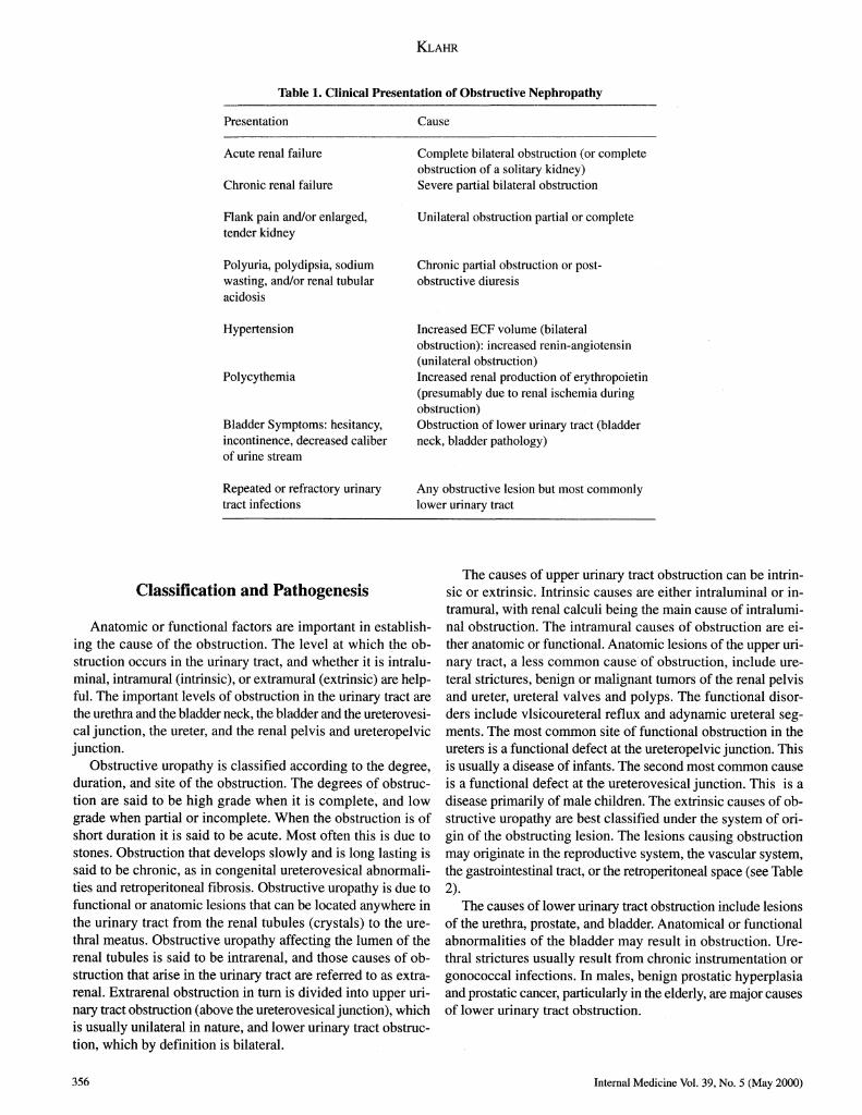
Table 1. Clinical Presentation of Obstructive NephropathyPre sentation C au se
Acute renal failure Complete bilateral obstruction (or completeobstruction of a solitary kidney)
Chronic renal failure Severe partial bilateral obstruction
Rank pain and/or enlarged, Unilateral obstruction partial or completetender kidney
P o ly uria, p olyd ip sia, so diu m Chronic partial obstruction or post-wasting, and/or renal tubular obstructive diuresisacido sis
H yp ert en sion Increased ECF volume (bilateralobstruction): increased renin-angiotensin(un ilateral o bstru ction )
P olycy th em ia Increased renal production of erythropoietin(presumably due to renal ischemia duringob structio n )
Bladder Symptoms: hesitancy, Obstruction of lower urinary tract (bladderincontinence, decreased caliber neck, bladder pathology)of urine stream
Repeated or refractory urinary Any obstructive lesion but most commonlytract in fectio ns lower urinary tract
Classification and Pathogenesis
Anatomicor functional factors are important in establish-ing the cause of the obstruction. Thelevel at whichthe ob-struction occurs in the urinary tract, and whether it is intralu-minal, intramural (intrinsic), or extramural (extrinsic) are help-ful. The important levels of obstruction in the urinary tract arethe urethra and the bladderneck, the bladderand the ureterovesi-cal junction, the ureter, and the renal pelvis and ureteropelvicjunction.
Obstructive uropathy is classified according to the degree,duration, and site of the obstruction. Thedegreesof obstruc-tion are said to be high grade whenit is complete, and lowgrade whenpartial or incomplete.Whenthe obstructionis ofshort duration it is said to be acute. Mostoften this is due tostones. Obstruction that develops slowly and is long lasting issaid to be chronic, as in congenital ureterovesical abnormali-ties and retroperitoneal fibrosis. Obstructive uropathy is due tofunctional or anatomic lesions that can be located anywhereinthe urinary tract from the renal tubules (crystals) to the ure-thral meatus. Obstructive uropathy affecting the lumenof therenal tubules is said to be intrarenal, and those causes of ob-struction that arise in the urinary tract are referred to as extra-renal. Extrarenal obstruction in turn is divided into upper uri-nary tract obstruction (above the ureterovesical junction), whichis usually unilateral in nature, and lower urinary tract obstruc-tion, which by definition is bilateral.
Thecauses of upperurinary tract obstruction can be intrin-sic or extrinsic. Intrinsic causesare either intraluminalor in-tramural, with renal calculi being the main cause of intralumi-nal obstruction. The intramural causes of obstruction are ei-ther anatomicor functional. Anatomiclesions of the upperuri-nary tract, a less commoncause of obstruction, include ure-teral strictures, benign or malignanttumorsof the renal pelvisand ureter, ureteral valves and polyps. The functional disor-ders include vlsicoureteral reflux and adynamicureteral seg-ments. Themost commonsite of functional obstruction in theureters is a functional defect at the ureteropelvic junction. Thisis usually a disease of infants. Thesecondmostcommoncauseis a functional defect at the ureterovesical junction. This is adisease primarily of male children. Theextrinsic causes of ob-structive uropathyare best classified under the systemof ori-gin of the obstructing lesion. The lesions causing obstructionmayoriginate in the reproductivesystem,the vascularsystem,the gastrointestinal tract, or the retroperitoneal space (see Table2 ) .
The causes of lower urinary tract obstruction include lesionsof the urethra, prostate, and bladder. Anatomicalor functionalabnormalitiesof the bladder mayresult in obstruction. Ure-thral strictures usually result fromchronicinstrumentationorgonococcal infections. In males, benign prostatic hyperplasiaand prostatic cancer, particularly in the elderly, are major causesof lower urinary tract obstruction.
3 5 6 Internal Medicine Vol. 39, No. 5 (May 2000)

Obstructive Nephropathy
Table 2. Causes of Urinary Tract ObstructionUpper urinary tract Lower urinary tract
Intrinsic causes 1. Phimosis, meatal stenosis, paraphimosis2. Urethra: strictures, stones, diverticulum,
1. Intralu m in al posterior or anterior ureteral valves, periure-1. Intratubular deposition of crystals (uric acid) thral abscess, urethral surgery2. Ureter: urolithiasis, blood clots, papillary tissue 3. Prostate: benign prostatic hyperplasia,
2. Intramural prostatic calculi, abscess, prostatic carcinoma1. Dysfunction at the ureteropelvic or ureterovesical 4. Bladder
ju n ction a. Neurogenic bladder: spinal cord defect or2. Ureteral valve, polyp, or stricture trauma, diabetes, multiple sclerosis,
cerebrovascular accidents, Parkinsons'sExtrinsic causes disea se
. Reproductive system b. Bladder neck dysfunctiona. Uterus: pregnancy, prolapse, tumors, endometriosis c. Bladder calculusb. Ovary: abscess, tumors, ovarian remnants d. Bladder cancerc. Gartner's duct cyst, tubuloovarian abscess 5. Tr auma
2. Vascular lesions a. Strad dle inju rya. Aneurysm: abdominal aorta, iliac vessels b. Pelvic fractureb. Aberrant vessels: ureteropelvic junctionc. Venous: retrocaval ureter, ovarian vein thrombophle-
b itisd. Fibrosis following vascular reconstructive surgery
3. Gastrointestinal tract: Crohn's disease, diverticulitis,ap p end iceal ab scess, tu m ors, p ancreatic tu m ors,
abscess, cysts4. Diseases of the retroperitoneum
a . R etro peritone al fibro sis (id io pathic, rad iation )b. Inflammatory: tuberculosis, sarcoidosisc. Hem atom asd. Primary retroperitoneal tumors (lymphoma, sarcoid,
etc.)e. Tumor metastatic to the retroperitoneum (cervix,
bladder, colon, prostate, etc.)f. Lymphoceleg . P elv ic lipo m ato sis
Blood Flow, Glomerularand Tabular Functionin Obstructive Nephropathy
In experimentalanimals,acute ureteral obstructioncauses atransient increase in blood flow to the affected kidney, followedby progressive vasoconstriction. Local production ofeicosanoids, especially prostacyclin and prostaglandin E mayaccount for the increased renal blood flow observed after theonset of obstruction. Usually 3 to 5 hours after the onset of theobstruction there is an increase in intrarenal resistance. Thisvasoconstrictionis mediatedby several vasoactivecompoundssuch as angiotensin II, (6) thromboxane A2 (7) and antidiuretichormone(8). In addition, decreased production of nitric oxidemayalso havea role in the increase in vascularresistance.Glomerular filtration rate (GFR) declines progressively af-ter the onset of completeureteral obstruction. Thereis an in-crease in pressure in the proximal tubule. This results in a de-
crease in the net hydraulic pressure gradient across glomerularcapillaries, resulting in a decline in GFR.The decrease in wholekidney GFRafter ureteral ligation is due to both a decrease insingle nephronGFRand a decrease in the numberof filteringnephrons.Several abnormalities in tubular function mayoccur in ob-structive nephropathy.Someof the abnormalities include: de-creased reabsorption of solutes and water, inability to concen-trate the urine, impaired excretion of hydrogenand potassium.Impairedability to concentratethe urine is evident after reliefof obstruction in both animals and humansubjects. Vasopressinadministration does not correct the defect (9, 10). A decreasein the solute content of the papillary interstitium maybe a majorfactor underlyingthis abnormality.A decreased expressionofaquoporins in the distal tubule may be also a contributory fac-tor .
The fractional excretion of potassium is decreased in pa-tients with obstructive uropathy. Thus, patients with this disor-
Internal Medicine Vol. 39, No. 5 (May 2000) 3 5 7

Klahr
der may develop hyperkalemic/hyperchloremic acidosis, par-ticularly those with chronic obstruction (1 1). An inability toacidify the urine is seen after release of obstruction in bothhumans and experimental animals ( 1 2-16). Although this acidi-fying defect is reversible in most instances, it may persist insome patients (12). In animals with ureteral obstruction, thereis a decreased number of H+ATPasepumpsin the apical sur-face of intercalated cells. This event mayaccount for the acidi-fying defect seen after release of ureteral obstruction (17).
Mechanisms of Interstitial Fibrosis inObstructive Nephropathy
Interstitial fibrosis is a commonfinding in the kidney withlong-term ureteral obstruction ( 1 8). The process of fibrosis, inpart, represents an imbalance betweenextracellular matrix pro-tein synthesis and degradation. An early event in the intersti-tial fibrosis process is the infiltration of the tubulointerstitial
compartment by macrophages (19). Since the macrophage is arich source of numerous peptide growth factors, it is not unex-pected that such compoundshave a pivotal role in initiatingand maintaining the fibrogenesis of the tubulointerstitium. Also,the cortical tubular cells in the kidney are capable of elaborat-ing a numberof peptide growth factors in response to the me-chanical perturbation that occurs after ureteral obstruction ( 1 8).Macrophagesand renal tubular cells are a source of a numberof growth factors and inflammatory agents such as, interleukin1 and interleukin 6, transforming growth factor p (TGF-p),tumor necrosis factor (TNF), platelet-derived growth factor(PDGF) and fibroblast growth factor (PGF).The deposition and accumulation of extracellular matrix(ECM)following tubulo-interstitial injury represents an im-balance between deposition and removal of collagen and othercompounds. There is a down-regulation of proteases which de-grade matrix components.Onthe other hand, the upregulationof TGF-p results in a variety of cellular responses that pro-mote fibrosis, including stimulation of ECMgenes, down regu-lation of degradative matrix metalloproteinases and up-regu-lation of tissue inhibitor of metalloproteinase (TIMP) 1 (20,21).
Recent studies have suggested that tension stress (22), hy-poxia (23), macrophage infiltration (24), and increased pro-duction of cytokines (25-27) mayaffect the redox state in vitro.To investigate the potential role of oxidative stress, Kawadaetal (28) examined the levels of distribution of NG carboxy-methyl-lysine (CML) in the kidney with ureteral obstruction.They found CMLin the interstitium of the obstructed kidney10 days after ligation of the ureter. Hemeoxygenase mRNAwas upregulated 12 hours after ureteral obstruction. These re-sults indicate the presence of oxidative stress in the intersti-tium of the obstructed kidney. Oxidative stress and the produc-tion of several active oxidative products may play an impor-tant role in interstitial inflammation and fibrosis.Reactive oxygenspecies stimulate nuclear factor-kappa B(NF-kB) and compounds that restore the "redox balance" ofthe cell ultimately inhibit NF-kB (29). The components of theNF-kB family of transcription factors are designated p50, p52,p65 (Rel A), c-Rel and Rel B; each derives from a separategene product (30). This family forms dimeric protein com-plexes, is constitutively present in the cytoplasm of cells, andupon activation, translocates to the nucleus, where it, in turncontrols genes contributing to tissue inflamation, cellular pro-liferation and cellular differentiation (30). NF-kB can be acti-vated by a number of stimuli including angiotensin II (3 1) andtumor necrosis factor alpha (TNF-cc). Transcription factors ofthe NF-kB family can directly or indirectly influence cellularevents leading to tissue fibrosis.
TYimor Necrosis Factor-ocThere are two members of the tumor necrosis factor (TNF)
family: TNF-a and TNF-p. The discussion in this article willbe confined to TNF-a. Activation of the human TNF-a geneleads to the production of a protein with 233 amino acids. The
Table 3. Factors whose Expression is Increased in theKidney with Ureteral Obstruction
Transforming growth factor p (TGF-(3,)Protein 53 (p53)Protein 21 [p21, (WAFl)]Tissue inhibitors of metalloproteinases- 1 (TIMP- 1 )
Decorin
Nuclear factor-KB (NF-kB)Tumor necrosis factor-a (TNF-a)Vasoactive compounds
AngiotensinogenAngiotensin II
Endothelin
Thromboxane A2Prostagl andi nsProto-oncogenesc-fos, c-jun, jun B, c-myc, cH-RasGrowthfactors
Interleukin-6 (IL-6)
Platelet activating factor (PAF)Basic fibroblast growth factor (BFG)Proteins involved in apotosis
Clusterin (SGP-2)Osteopontin
ChemoattractantsMonocyte chemoattractant peptide- 1 (MCP-1 )
OsteopontinAdhesion ProteinsIntercellular adhesion molecule 1 (ICAM-1)Vascular cell adhesion molecule 1 (VCAM-1)Fibronectin alternate splice formsMatrix/basement membraneproteinsCollagen types I, III and IV
Adapted from Klahr S. ; Obstructive Nephropathy (Neph-rology Forum) Used with permission from Kidney In-ternational 54: 286-300, 1998. Reference 1 8.
358 Internal Medicine Vol. 39, No. 5 (May 2000)
Obstructive Nephropathy
signal sequence of full length TNF-a is unusually long (76aminoacids). This signal sequence provides a mechanismbywhich TNF-oc can exist as an integral membraneprotein, withthe carboxy-terminus extracellular. MatureTNF-occonsists of157 amino acids with a molecular mass of 17 kDa. Fully ma-ture TNF-oc protein self-assemble as homotrimers with massesof 51 kDa. The macrophage is a major source ofTNF-cc, butTNF-oc is also produced by resident renal cells (32, 33). TwoTNF-ocreceptors have been described: one with a molecularweight of 55 kDa (TNFR1) and the other with a molecularweight of 75 kDa (TNFR2) (34). Binding ofTNF-a to its re-ceptors activates a number of signal transduction pathways thatresult in the expression of a variety of transcription factors,cytokines, growth factors, receptors, cell adhesion molecules,mediators of inflammatory processes, acute phase proteins, andmajor histocompatibility complex proteins (34). Lipopolysac-charide (LPS)-induced renal injury is associated with increasedexpression of TNF-aby renal cells. Proximal tubular cells ex-press TNF-a when stimulated with interleukin- la or LPS (35).Also, mRNAtranscripts of TNF-a are found in cortical tu-bules of mice injected with LPS. Thus, resident renal cells(glomerular mesangial cells and tubular epithelial cells) aresources of TNF-a production in renal injury. Wefound (36)that in normal rats TNF-a mRNAwas more abundant in glom-erulitis than in renal cortical tubules. Wemeasured TNF-amRNAin the renal cortex of rats at different times after theonset of unilateral ureteral obstruction and determined whetherangiotensin II inhibition or total body irradiation affected themRNAlevels of TNF-a (36). Cortical tubules obtained fromthe kidney with ureteral obstruction had a marked increase inTNF-a mRNAexpression, whereas the glomeruli obtained fromthe same kidneys did not. Thus, upregulation of TNF-a ex-pression was confined to renal tubular cells of the obstructedkidney. Levels of TNF-a mRNAare significantly increased inthe obstructed kidney at 1 hour (x 2), 2 hours (x 2.7), 4 hours(x 3.6), 24 hours (x 2.7), 72 hours (x 1.8) and 120 hours (x2.8) after ureteral ligation when compared to the contralateralkidney of the same rats or to the control kidney of normal rats.Administration of enalapril, an ACEinhibitor, before and dur-ing unilateral ureteral obstruction decreased TNF-a mRNAlevels in the obstructed kidney by about 40% at 4 hours afterthe onset of obstruction. Total body irradiation, which preventsthe migration of macrophages to the obstructed kidney, did notaffect the upregulation of TNF-a mRNAexpression at 4 hoursafter unilateral ureteral obstruction. Thus, TNF-a may have arole in initiating tubulointerstitial injury in the obstructed kid-ney. Leukocytes, infiltrating the renal interstitium of the ob-structed kidney, do not appear to contribute to the increasedexpression of TNF-amRNA.Angiotensin II seems to contrib-ute, at least in part, to the early increase in expression of TNF-a mRNAin the obstructed kidney.TNF-ahas a role in the recruitment of inflammatory cellsin animal models of glomerular injury. It stimulates the pro-duction of chemotactic factors by resident cells and upregulatesmonocyte chemoattractant protein-1 (MCP-1) mRNAin hu-man mesangial cells. Wolf et al (37) found that TNF-a increases
RANTESmRNAin a murine mesangial cell line and in vivo inrat kidneys perfused with TNF-a. Mulligan et al (38) reportedthat anti- TNF-a or soluble recombinant humanTNFR1 blockedthe upregulation of intercellular adhesion molecule 1 (ICAM-1), endothelial leukocyte adhesion molecule 1 (ELAM-1), andvascular adhesion molecule 1 (VCAM-1)in nephrotoxic ne-phritis. The above data support the concept that TNF-acon-tributes to the increased macrophage migration into the renalinterstitium of the affected kidney. Wefound that macroph-ages infiltrated the interstitium of the obstructed kidney cortexas early as 4 hours after the onset of unilateral ureteral obstruc-tion, and by 24 hours the influx was at a level approximately10-fold greater than normal (39). Taken together, these obser-vations suggest that an early increase in TNF-a after ureteralobstruction of the kidney upregulates the production of achemoattractant(s) for monocytes and contributes to the infil-tration of the obstructed kidney by leukocytes.The binding of TNF-a to a cell surface receptor results inintracellular metabolic changes that mediate apoptotic and ne-crotic cell death (40, 41). Although the mechanism of the cyto-cidal action of TNF-a has not been completely elucidated, theTNFR1receptor has been shown to have a cytoplasmic do-main sequence similar to the Fas antigen receptor, which isconsidered to mediate apoptosis (42). Some investigators havereported apoptosis of renal tubular cells in the obstructed kid-ney one week after ureteral ligation (43, 44).
Effect of Pharmacologic Interventions inObstructive Nephropathy
Administration of an ACEinhibitor or an angiotensin II re-ceptor (ATI ) antagonist, to rats with unilateral ureteral obstruc-tion, ameliorated the increase in interstitial volume and attenu-ated the increased expression of TGF-pj in tubular cells, theincreased production of extracellular matrix protein, the acti-vation of NF-kB, the proliferation of fibroblasts and the con-version of their phenotype to myofibroblasts (45). A mono-cyte/macrophage infiltrate was present in the obstructed kid-ney of untreated rats and in the obstructed kidney of rats treatedwith the angiotensin II receptor antagonist. By contrast, thisinfiltrate was markedly decreased in the obstructed kidney ofrats treated with an ACEinhibitor. This difference may be owingto greater generation of nitric oxide related to increased levelsof bradykinin during ACE inhibition (46). In fact, rats withunilateral obstruction given both an ACE inhibitor and (L-NAME)(an inhibitor of NOformation) had a substantial mac-rophage infiltrate. Administration of L-arginine in the drink-ing water significantly blunted the increases in interstitial vol-ume, monocyte infiltration, interstitial collagen IV and a-smooth muscle actin expression (47). However, in contrast toACEinhibitors, arginine administration did not decrease theexpression of TGF-(3, mRNAin the kidney with ureteral liga-tion. We also found a 10-fold increase in TIMP-1 mRNAin theobstructed kidney. Administration of an ACE inhibitor bluntedthis increase, by 40 % (p<0.001). The addition of L-NAMEtothe ACE inhibitor prevented the decrease in TIMP-1 mRNA.
Internal Medicine Vol. 39, No. 5 (May 2000) 359

Klahr
A commondenominator of the beneficial effects of ACEinhi-bition or arginine and the deleterious effects of L-NAMEdur-ing ACE inhibition may be a result of the increased or decreasedgeneration of NO (46, 47).In the above studies the ACEinhibitor was administered
before or concomitant with the onset of ureteral obstruction.Wealso have examined the effects of ACEinhibition after 3 or5 days of established unilateral ureteral obstruction in rats. De-layed administration of an ACEinhibitor slowed and in sev-eral instances halted the progression of fibrosis in thetubulointerstitium of the kidney with ureteral ligation (48).Pharmacological maneuvers that reduce angiotensin II (ACEinhibitors) or interfere with its action (ATI receptor antago-nists) blunt NF-kB activation and blunt the progression of dis-ease. Although AT2 receptor inhibition decreases NF-kB acti-vation the effect on NF-kBdimers may be different from thoseaffected through ATI receptors (45). Indeed, AT2 receptor in-hibition in rats (49) or AT2 receptor knockout in mice (50)exacerbates renal fibrosis consequent to ureteral obstruction.Pharmacological maneuversthat reduce the activation of cer-tain NF-kB isotypes may extrinsically blunt angiotensin II-mediated effects and also slow the progression of renal dis-ease.
In summary,angiotensin II regulates a numberof genes as-sociated with the progression of renal disease. The regulationof gene expression by angiotensin II occurs through specificreceptors that are ultimately linked to changes in the activityof transcription factors within the nucleus of target cells. Inparticular, members of the NF-kB family of transcription fac-tors are activated, which, in turn, fuels at least two autocrinereinforcing loops that amplify angiotensin II and TNF-oc for-mation. This amplification of the angiotensin II and TNF-asignals to the samecells in an autocrine manneror to adjacentcells in a paracrine manner further amplifies NF-kBactivationwithin the kidney. This leads to fibroblast proliferation andsubsequent differentiation into myofibroblasts. Furthermore,tubular epithelial cells are stimulated to produce chemoattrac-tants and adhesion proteins which cause an inflammatory re-sponse, leading to monocyte/macrophage infiltration. The tu-bule cells also produce profibrotic cytokines, leading to anoverproduction of extracellular matrix proteins by all cell types.The net result is fibrosis of the tubulointerstitium and progres-sion of renal disease.
References
1) Bell ET. Renal Disease. Philadelphia, Lea & Febiger, 1946.2) Campbell MR. Urinary Obstruction, in: MF. Campbell and J.H. Harrison
(eds.), Urology, Vol. I (3rd ed.). Philadelphia, Saunders, 1970.3) National Kidney and Urologic Diseases Advisory Board. The Scope andImpact of Kidney and Urologic Diseases, in: Long Range Plan, Chapter1. National Institutes of Health, Washington, D.C., NIH Publication #90-
583, 1990: 7-35.
4) Hadlock FP, Deter RL, Carpenter RJ. Sonography of the fetal genitouri-nary tract. Semin Ultrasound 5: 213-218, 1984.
5) Montana MA, Cyr DR, Lenke RR, Shuman WP, Mack LA. Sonographicdetection of fetal ureteral obstruction. AJR Am J Roentgenol 145: 595-
596, 1985.
6) Yarger WE, Schocken DD, Harris RH. Obstructive nephropathy in therat: possible roles for the renin-angiotensin system, prostaglandins, andthromboxanes in postobstructive renal function. J Clin Invest. 65: 400-
412, 1980.
7) Purkerson ML, Klahr S. Prior inhibition of vasoconstrictors normalizesGFR in postobstructed kidneys. Kidney Int. 35: 1305-1314, 1989.
8) Reyes AA, Robertson G, Klahr S. Role ofvasopressin in rats with bilat-eral ureteral obstruction. Proc Soc Exp Biol Med 197: 49-55, 1991.
9) Hanley MJ, Davidson K. Isolated nephron segments from rabbit modelsof obstructive nephropathy. J Clin Invest 69: 165-174, 1982.10) Campbell HT, Bello-Reuss E, Klahr S. Hydraulic water permeability andtransepithelial voltage in the isolated perfused rabbit cortical collectingtubule following acute unilateral ureteral obstruction. J Clin Invest 75:
219-225, 1985.
1 1 ) Batlle DC, Arruda JA, Kurtzman NA. Hyperkalemic distal renal tubularacidosis associated with obstructive uropathy. N Engl J Med 304: 373-
380, 1981.
12) Gillenwater JY, Westervelt FB Jr, Vaughan ED Jr, Howards SS. Renalfunction after release of chronic unilateral hydronephrosis in man. Kid-ney Int7: 179-186, 1975.
13) Better OS, ArieffAI, Massry SG, Kleeman CR, Maxwell MH. Studies onrenal function after relief of complete unilateral ureteral obstruction ofthree months' duration in man. AmJ Med 54: 234-240, 1973.
14) Ericsson NO, Wineberg J, Zetterstrom R. Renal function in infantile ob-structive uropathy. Acta Pediatr Scand 44: 444-459, 1955.1 5) Berlyne GM. Distal tubular function in chronic hydronepyhrosis. Q J Med
30: 339-355, 1961.
16) Massry SG, Schainuck LI, Goldsmith C, Schreiner GE. Studies on themechanismof diuresis after relief of urinary-tract obstruction. AnnInternMed 66: 149-158, 1967.
17) Purcell, H, Bastani, B, Harris, KP, Hemken, P, Klahr, S, Gluck, S. Cellu-lar distribution of H (+) -ATPase following acute unilateral ureteral ob-struction in the rat. Am. J Physiol 261: F365-F376, 1991.18) Klahr S. Obstructive nephropathy. Kidney Int 54: 286-300, 1998 (clini-
cal conference).19) Diamond, JR, Ricardo, SD, Klahr S. Mechanisms of interstitial fibrosis
in obstructive nephropathy. Semin Nephrol 18: 594-602, 1998.20) Jones CL, Buch S, Post M, McCulloch L, Liu E, Eddy AA. Renal extra-cellular matrix accumulation in acute puromycinaminonucleoside neph-rosis in rats. Am J Pathol 141: 1381-1396, 1992.
21) Sharma K, Ziyadeh FN. The emerging role of transforming growth fac-tor-beta in kidney diseases. Am J Physiol 266: F829-F842, 1994 (edito-
rial).
22) Ricardo SD, Ding G, Eufemio M, Diamond JR. Antioxidant expressionin experimental hydronephrosis: role of mechanical stretch and growthfactors. Am J Physiol 272: F789-F798, 1997.
23) Klahr S, Pukerson ML. The pathophysiology of obstructive nephropa-thy: the role of vasoactive compoundsin the hemodynamicand structuralabnormalities of the obstructed kidney. AmJ Kidney Dis 23: 219-223,
1994.
24) Ricardo SD, Levinson ME, DeJoseph MR, Diamond JR. Expression ofadhesion molecules in rat renal cortex during experimental hydronephro-sis. Kidney Int 50: 2002-2010, 1996.
25) Ohba M, Shibanuma M, Kuroki T, Nose K. Production of hydrogen per-oxide by transforming growth factor-bate 1 and its involvement in induc-tion of egr-1 in mouse osteoblastic cells. J Cell Biol 126: 1079-1088,
1994.
26) Meier B, Radeke HH, Selle S, et al. Humanfibroblasts release reactiveoxygen species in response to interleukin-1 or tumour necrosis factor-alpha. Biochem J 263: 539-545, 1989.
27) Sundaresan M, Yu ZX, Ferrans VJ, Irani K, Finkel T. Requirement forgeneration of H2O2for platelet-derived growth factor signal transduction.Science 270: 296-299, 1995.
28) Kawada N, Moriyama T, Ando A, et al. Increased oxidative stress in mousekidneys with unilateral ureteral obstruction. Kidney Int 56: 1004-1013,
1999.
29) Meyer M, Schreck R, Baeuerle PA. H2O2and antioxidants have opposite
360 Internal Medicine Vol. 39, No. 5 (May 2000)

Obstructive Nephropathy
effects on activation of NF-kappa B and AP-1 in intact cells: AP-1 assecondary antioxidant-responsive factor. EMBOJ 12: 2005-201 5, 1993.
30) Barnes PJ, Karin M. Nuclear factor-kappa B. A pivotal transcription fac-tor in chronic inflammatory diseases. N Engl J Med 336: 1066-1071,
1997.
3 1) Morrissey JJ, Klahr S. Rapid communication. Enalapril decreases nuclearfactor kappa B activation in the kidney with ureteral obstruction. KidneyInt 52: 926-933, 1997.
32) Egido J, Gomez-Chiarri M, Ortiz A, et al. Role of tumor necrosis factor-alpha in the pathogenesis of glomerular diseases. Kidney Int Suppl 39:
S59-S64, 1993.
33) Baud L, Fouqueray B, Philippe C, Amrani A. Tumor necrosis factor al-pha and mesangial cells. Kidney Int 41. 600-603, 1992.
34) Ortiz A, Bustos C, AlcazarA, et al. Involvement of tumor necrosis factor-ex in the pathogenesis of experimental and human glomerulonephritis. in:Advances in Nephrology, Grunfeld JP, Bach JF, Kreis H, Maxwell MH,Eds. Mosby, St. Louis, 1995: 53-77.
35) Wuthrich RP, Glimcher LH, Yui MA, Jevnikar AM, Dumas SE, KelleyVE. MHCclass II, antigen presentation and tumor necrosis factor in re-nal tubular epithelial cells. Kidney Int 37: 783-792, 1990.
36) Kaneto H, Morrissey J, McCracken R, Ishidoya S, Reyes A, Klahr S. Theexpression of rhRNAfor tumor necrosis factor-a increases in the ob-structed kidney of rats soon after unilateral ureteral ligation. Nephrology
2: 161-166, 1996.
37) WolfG, Aberle S, Thaiss F, et al. TNF alpha induces expression of thechemoattractant cytokine RANTESin cultured mouse mesangial cells.Kidney Int 44: 795-804, 1993.
38) Mulligan MS, Johnson KJ, Todd RF. et al. Requirements for leukocyteadhesion molecules in nephrotoxic nephritis. J Clin Invest 91: 577-587,
1993.
39) Schreiner GF, Harris KP, Purkerson ML, Klahr S. Immunological aspectsof acute ureteral obstruction: immunecell infiltrate in the kidney. KidneyInt 34: 487-493, 1988.
40) Larrick JW, Wright SC. Cytotoxic mechanism of tumor necrosis factor-alpha. FASEB J 4: 3215-3223, 1990.
41) Laster SM, Wood JG, Gooding LR. Tumor necrosis factor can induceboth apoptic and necrotic forms of cell lysis. J Immunol 141: 2629-2634,
1988.
42) Itoh N, Yonehara S, Ishii A, et al. The polypeptide encoded by the CDNAfor human cell surface antigen Fas can mediate apoptosis. Cell 66: 233-
243, 1991.
43) Gobe GC, Axelsen RA. Genesis of renal tubular atrophy in experimentalhydronephrosis in the rat. Role of apoptosis. Lab Invest 56: 273-281,
1987.
44) Kennedy WA, Stenberg A, Lackgren G, Hensle TW, Sawczuk IS. Renaltubular apoptosis after partial ureteral obstruction. J Urol 152: 658-664,
1994.
45) Klahr S, Morrissey JJ. Comparative study of ACEinhibitors and angio-tensin II receptor antagonists in interstitial scarring. Kidney Int Suppl 63:
SIll-114, 1997.
46) Morrissey JJ, Ishidoya S, McCracken R, Klahr S. Nitric oxide generationameliorates the tubulointerstitial fibrosis of obstructive nephropathy. JAm Soc Nephrol 7: 2202-2212, 1996.
47) Reyes AA, Porras BH, Chasalow FI, Klahr S. L-arginine decreases theinfiltration of the kidney by macrophages in obstructive nephropathy andpuromycin-induced nephrosis. Kidney Int 45: 1346-1354, 1994.
48) Ishidoya S, Morrissey J, McCracken R, Klahr S. Delayed treatment withenalapril halts tubulointerstitial fibrosis in rats with obstructive ne-phropathy. Kidney Int 49: 1110-1119, 1996.
49) Morrissey J, Duan L, Klahr S. Angiotensin Type II receptor antagonismin ureteral obstruction. J AmSoc Nephrol 7: 1761, 1996 (Abstract).
50) Ma J, Hideki H, Fogo A, Kon V, Magami T, Ichikawa I. Accelerated fi-brosis and collagen deposition develop in the renal interstitium of angio-tensin type II (AT2) receptor null mutant mice during ureteral obstruc-tion. J AmSoc Nephrol 8: 622A, 1997 (Abstract).
Internal Medicine Vol. 39, No. 5 (May 2000) 361