cytoprotective role of nitric oxide associated with hsp70 expression in neonatal obstructive...
TRANSCRIPT

1
2
3
4
567
89
10
11
12
13
14
15
16
17
18
19
20
21
22
23
24
25
2627
28
29
30
31
32
33
34
35
Available online at www.sciencedirect.com
YNIOX 856 No. of Pages 12, Model 5+
13 February 2008 Disk UsedARTICLE IN PRESS
www.elsevier.com/locate/yniox
Nitric Oxide xxx (2008) xxx–xxx
OO
F
Cytoprotective role of nitric oxide associated with Hsp70expression in neonatal obstructive nephropathy
Walter Manucha a,b, Patricia G. Valles a,b,*
a �Area de Fisiopatologıa, Departamento de Patologıa, Facultad de Ciencias Medicas, Universidad Nacional de Cuyo,
Avenida Libertador 80, Centro Universitario, CP: 5500 Mendoza, Argentinab IMBECU-CONICET (Consejo Nacional de Investigacion Ciencia y Tecnologica), Mendoza, Argentina
Received 1 September 2007; revised 8 January 2008
R
RR
EC
TED
PAbstract
Nitric oxide (NO) has emerged as an important endogenous inhibitor of apoptosis. In this study, we postulated that the mechanism ofapoptosis inhibition by NO would include stimulation of heat shock protein 70 (Hsp70) expression. Rats were subjected to unilateralureteral obstruction (UUO) or sham operation, and kidneys were harvested 5 and 14 days after obstruction. After 14 days of obstruction,decreased endogenous NO and lower inducible nitric oxide synthase (iNOS) expression at mRNA and protein levels associated withdownregulation of Hsp70 protein expression were shown in apoptosis induction, regulated by mitochondrial signal pathway, throughthe increased pro-apoptotic ratio Bax/BcL2 and consequently caspase 3 activity. Conversely, 5 days after kidney obstruction, increasedHsp70 expression linked to increase NO and iNOS expression at transcriptional and post-transcriptional levels with absence of apoptoticresponse, were demonstrated. In obstructed neonatal rats, in vivo administration of L-Arginine induced heat shock protein 70 (Hsp70)expression, which was associated with cytoprotection from apoptosis and transiently decreased nicotinamide adenine dinucleotide phos-phate reduced form (NADPH) oxidase activity. Opposite effects were obtained after nitro L-Arginine methyl ester (L-NAME) treatment.The interaction between B-cell lymphoma 2 anti-apoptotic members (BcL2) and Hsp70 in the presence of L-Arginine and L-NAME, wasdetermined by coimmunoprecipitation. Binding of BcL2 and Hsp70 increased after L-Arginine administration. These findings suggestthat NO can produce resistance to obstruction-induced cell death by mitochondrial apoptotic pathway, through the induction ofHsp70 expression, in neonatal unilateral ureteral obstruction.� 2008 Elsevier Inc. All rights reserved.
Keywords: Nitric oxide; BcL2; Neonatal unilateral ureteral obstruction; Apoptosis; Caspase 3; Hsp70
36
37
38
39
40
41
42
43
NC
OThe functional integrity of the kidney depends on thenormal development as well as on the physiological cellturnover, apoptosis induction being essential for thesemechanisms. Congenital obstructive nephropathy, a majorcause of chronic renal failure in infancy, is characterized bydecreased proliferation and increased apoptosis [1]. Pro-grammed cell death leads to renal tubular atrophy andtubular loss in neonatal unilateral ureteral obstruction
U44
45
46
47
48
49
1089-8603/$ - see front matter � 2008 Elsevier Inc. All rights reserved.
doi:10.1016/j.niox.2008.01.005
* Corresponding author. Address: Area de Fisiologıa Patologica,Departamento de Patologıa, Facultad de Ciencias Medicas, UniversidadNacional de Cuyo, Centro Universitario, CP: 5500 Mendoza, Argentina.Fax: +54 0261 4287370.
E-mail address: [email protected] (P.G. Valles).
Please cite this article in press as: W. Manucha, P.G. Valles, Cytopro(2008), doi:10.1016/j.niox.2008.01.005
(UUO) [2]. Moreover, the severity of the apoptoticresponse to unilateral ureteral obstruction is far greaterin the neonatal than in the adult rat, a factor that be likelycontribute to the impaired growth of the obstructed devel-opment kidney [3].
Nitric oxide (NO) has been implicated in apoptosis forUUO, being a controversial key. Effects of NO in apoptosisdepend on the dose, environment and/or redox state.Whereas excessive NO production induces cell death in sev-eral cell lines [4–7] conversely, protection against apoptosishad been shown in others [8,9]. Studies on the antiapopto-tic mechanism of NO have identified NO target interac-tions that range from indirect to direct interaction withthe apoptotic machinery. NO suppresses apoptosis through
tective role of nitric oxide associated with Hsp70 ..., Nitric Oxide

T
50
51
52
53
54
55
56
57
58
59
60
61
62
63
64
65
66
67
68
69
70
71
72
73
74
75
76
77
78
79
80
81
82
83
84
85
86
87
88
89
90
91
92
93
94
95
96
97
98
99
100
101
102
103
104
105
106
107
108
109
110
111
112
113
114
115
116117118119120121122123124125126127128129130131
132
133134135136137138139140141142143144145146147148149150151152153154155156157158159
160
161
162163
2 W. Manucha, P.G. Valles / Nitric Oxide xxx (2008) xxx–xxx
YNIOX 856 No. of Pages 12, Model 5+
13 February 2008 Disk UsedARTICLE IN PRESS
UN
CO
RR
EC
the direct caspase activity inhibition. Thiol group of cas-pase 3 (cys 163) susceptible to redox modification in thepresence of NO can be efficiently S-nitrosylated [10]depending on the abundance of these molecules. Moreover,recent studies have proposed that B-cell lymphoma 2 anti-apoptotic member (BcL2) cleavage can be inhibited by thecaspase-3-like inhibitor Ac-DEVD-cho and/or NO, sug-gesting that the activated caspase-3-like proteases areresponsible for the BcL2 protein cleavage and the inactiva-tion of the antiapoptotic function of BcL2 [11]. In neonatalUUO, we have reported an apoptotic response through thepro-apoptotic regulation of the BcL2 gene family and cas-pase 3 [12].
Due to the significant role of apoptosis in the pathogen-esis of the renal cellular injury resulting from urinary tractobstruction, the factors regulating the renal apoptoticresponse have been evaluated. Stretching of the renal tubu-lar cells by transmitted increased hydrostatic pressure canprovide a powerful mechanical stimulus to apoptosis inthe obstructed kidney [12,13]. Ischemia is another stimulusto apoptosis, and UUO induces a profound reduction inrenal blood flow and impairment of autoregulation of renalblood flow [13,14]. Moreover, reactive oxygen species areknown to reduce the threshold of tissues to undergo apop-tosis [15], and reactive oxygen species are significantlyincreased in the chronically obstructed kidney [16]. Theneonatal obstructed kidney may be particularly susceptibleto the generation of reactive oxygen species, becauseendogenous renal antioxidant enzymes, including superox-ide dismutase, are suppressed in the neonate [17].
Under normal physiological conditions, a balancebetween superoxide and nitric oxide exists in vivo.
NO and superoxide react together at a diffusion-con-trolled rate to yield peroxynitrite (ONOO�), which inflictscellular injury through oxidation of many biological mole-cules. Furthermore, ONOO� has also been implicated inthe inactivation of Mn and Fe superoxide dismutase [18].In contrast, NO may protect cells from reactive oxygen inter-mediate (ROI)-mediated cytotoxicity by scavenging super-oxide anions which are implicated in toxicity through theformation of hydrogen peroxide or hydroxyl radical [19].Nitric oxide has been shown to inhibit superoxide anion gen-eration. The mechanism for such inhibition is thought to bedue to the inactivation of nicotinamide adenine dinucleotidephosphate reduced form (NADPH) oxidase due to the scav-enging effects of NO on superoxide [20].
Induction of the stress response includes synthesis of heatshock proteins (HSPs) that have been well characterized incells injured from a variety of renal insults [21]. These pro-teins are generally classified into families according to theirapparent molecular weight and respective inducers and playessential roles in protein chaperoning and cellular protection[22]. In addition, certain HSPs (including Hsp70) confer cel-lular protection by modulating the engagement and/or pro-gression of apoptosis [23]. Recently, we have demonstratedthat after 24 h of UUO, protection against tubulointerstitialfibrosis by Losartan, independent from changes in blood
Please cite this article in press as: W. Manucha, P.G. Valles, Cytopro(2008), doi:10.1016/j.niox.2008.01.005
pressure, includes decreased oxidative stress linked to upreg-ulation of Hsp70 expression [24].
In this study, we examined the consequences of NO onobstruction-induced apoptosis in renal cortex from neona-tal UUO. We report that NO prevents obstruction-inducedcell death by mitochondrial apoptotic pathway, throughthe induction of heat shock protein 70 (Hsp70).
ED
PR
OO
F
Material and methods
Surgical procedure
Neonatal rats (Wistar Kyoto, males and females) were subjected tosham operation or complete UUO within the first 48 h of life. Under iso-flurane, the abdomen was surgically opened by a left lateral incision, theleft ureter was exposed and a 6.0 silk suture was used to place a ligature.The incision was closed in a single layer. The animals were allowed torecover from anesthesia and returned to their mothers. After 5 and 14 daysof obstruction, animals were sacrificed with a lethal injection of pentobar-bital and their obstructed and control kidneys were decapsulated,removed, and weighed. Successful ureteral ligation was confirmed at thetime of kidney removal by observation of important hydronephrosis. Leftkidney of sham group was also nephrectomized.
All the experimental procedures of this study have been previouslyapproved by the Laboratory Animal Ethical Committee of the Schoolof Medicine, Cuyo University, Mendoza (32/95 C.D.) The experimentswere conducted in accordance with guidelines of the CEEA (Ethical Com-mittee of Animal Experimentation of Argentina).
Identification of renal tubular cell apoptosis
TUNEL technique
Kidneys were then dehydrated, embedded in paraffin and serially sec-tioned (3–4 lm) on a microtome (Leica, Nussloch, Germany). Thereafter,kidney sections were deparaffined in xylene and rehydrated throughgraded ethanols to water. Endogenous peroxidase activity was quenchedby incubation with 2% (v/v) H2O2 in phosphate buffer saline (PBS) for5 min at room temperature (RT). Afterwards, staining and immunohisto-chemical techniques were performed.
After the digesting and quenching steps, equilibration buffer was applieddirectly to the sections for 5 min and working strength TdT enzyme (at aconcentration of 1:5 in reaction buffer) was then applied directly for 1 h at37 �C. A biotin-conjugated anti-digoxigenin antibody (Sigma) was used at1:1500 dilution in PBS, pH 7.4, to incubate the tissue sections overnight at4 �C. Then, the sections were incubated with biotinylated anti-mouse IgG(Dako, Carpinteria, CA, USA) at 1:100 dilution for 45 min at RT and laterwith peroxidase-labeled streptavidin (strept AB Complex/HRP, Dako) at1:100 dilution for 45 min at RT. After a brief wash, 3,30-diaminobenzidinetetrahydrochloride (0.5 mg/ml)/H2O2 (0.01%), a chromogen substrate wasincorporated. Tissue sections were lightly counterstained with 0.5% withhematoxylin to reveal nuclei, and the slides were observed with a Zeiss Axi-oskop 2 microscope. For positive control, we used paraffin sections frominvoluting prostates of castrated rats (n = 2).
For the quantification of apoptotic epithelial cells, 10 consecutive fieldswere randomly selected in each renal cortex and they were evaluated at400�, on a 10 � 10 grid, by using an image analyzer (Image Pro-Plus4.0, 1998, Maryland, USA). Results were expressed as the number ofapoptotic cells per mm2.
RT-PCR and semiquantification of mRNA for iNOS, BcL2 and
Bax
Total ribonucleic acid (RNA) was obtained by using Trizol reagent(Gibco BRL). Two micrograms of total RNA were denatured in the pres-
tective role of nitric oxide associated with Hsp70 ..., Nitric Oxide

164165166167168169170171172173174175176177
178
179180181182183184185186187188189190191192193194195196197198199200
201202203204205
206
207208209210211212
W. Manucha, P.G. Valles / Nitric Oxide xxx (2008) xxx–xxx 3
YNIOX 856 No. of Pages 12, Model 5+
13 February 2008 Disk UsedARTICLE IN PRESS
ence of 0.5 lg/50 lL Oligo (dT)15 primer and 40 U recombinant ribonucle-ase inhibitor RNasin (Promega, USA). Reverse transcription was per-formed in the presence of mixture by using 200 units of ReverseTranscriptase M-MLV RT in reaction buffer, 0.5 mM dNTPs each, andincubated for 60 min at 42 �C. The copy deoxy nucleic acid (cDNA)(10 lL) was amplified by polymerase chain reaction (PCR) by standardconditions. Each cDNA aliquot was amplified (30 cycles) for induciblenitric oxide synthase (iNOS), BcL2, BcL2–associated X protein (Bax)and b-actin, (primers designed, Table 1).
Densitometric analysis was performed by using National Institutes ofHealth Image 1.6 software (Rasband Wayne et al., Division of ComputerResearch and Technology NIH, Bethesda).
The iNOS, BcL2 and Bax signals were standardized against b-actin sig-nal for each sample and results were expressed as a ratio.
T
213214215216217218219220221
222
223224225226227228229230231232233234
CProtein determination for iNOS, Hsp70, BcL2 and pro-caspase 3
Cortex tissues were homogenized and protein concentrations werequantified by Bradford assay using bovine serum albumine (BSA)10 mg/ml as a standard. Protein samples were prepared in sodium dodecylsulphate (SDS) sample buffer (31.25 mM Tris–HCl, pH 6.8, 10% glycerol,0.0025% bromophenol blue, 10 mM dithiothreitol (DTT), 1% SDS). Atotal of 20–50 lg of proteins were electrophoresed in 0.1% SDS and 8%polyacrylamide gel with 4% stacking gel and electrophoretically trans-ferred to nitrocellulose. Gently removed blot from gel and placed it in asmall plastic container containing about 10 ml of Ponceau S protein stain-ing solution (to view extent of protein transfer or to ensure protein trans-fer) were performed.
Non-specific binding sites were blocked by incubating each membranein 5% non-fat dry milk in PBS plus 0.1% Tween for 1 h at RT, washed,and then incubated overnight in the primary antibodies against iNOS(dilution 1:3000), Hsp70 (dilution 1:3000), BcL2 (1:2000) and pro-caspase3 (1:3000), from Sigma Chemical Co., Chemicon and Santa Cruz Biotech-nology, respectively. Detection was accomplished with secondary antibod-ies (DAKO) and detected with enhanced chemiluminescence system (ECL,Amersham) and exposure to X-ray film (Amersham).
Densitometric analysis was carried out by image analysis software, thephotographs were digitalized by using a scanner (LACIE Silver Scannerfor Macintosh) and the Desk Scan software (Adobe Photo Shop) on a
UN
CO
RR
E 235
236237238239240241242243244245246247248249250251252253254255256257258259260261
Table 1Primers designed from rat sequences for RT-PCR
Primer Sequence Annealing(�C)
Predictedproductsize, (bp)
iNOSAntisense 50-GCTTCTGGTCGATGT
CATGAGCAA-3055 222
Sense 50-GCATGGACCAGTATAAGGCAAGCA-30
BcL2
Antisense 50-CTTGTGGCCCAGGTATGC-30
59 708
Sense 50-ATGGCGCAAGCCGGGAGAA-30
BaxAntisense 50-TCAGCCCATCTTCTT
CCAGAT-3059 550
Sense 50-ATGGACGGGTCCGGGGAGC-30
b-ActinAntisense 50-GTGCCACCAGACAG
CACTGTGTTG-3065 201
Sense 50-TGGAGAAGAGCTATGAGCTGCCTG-30
Please cite this article in press as: W. Manucha, P.G. Valles, Cytopro(2008), doi:10.1016/j.niox.2008.01.005
ED
PR
OO
F
desktop computer. Densitometric analysis was performed using the USNational Institute of Health Image 1.66 software (Rasband Wayneret al., Division of Computer Research and Technology NIH, Bethesda,USA). The magnitude of the immunosignal was standardized to 1 forthe corresponding control renal tissue values.
Caspase 3 activity assay
The activity of caspase 3 was determined by using the CaspACETM
Assay System (Promega, Madison, WI, USA).Aliquots of cytosolic homogenates (37.5 lL) were diluted in caspase
assay buffer (312.5 mM N-2-hydroxyethyl-piperazine-N0-2-ethanesulfonicacid HEPES, pH 7.5, 31.25% sucrose, 0.3125% CHAPS (3-(3-cholamido-propyl)-dimethyl ammonio)-1 propane-sulfonate) + dimethylsulphoxide(DMSO) 2 lL + DTT 100 mM 10 lL, and incubated for 30 min at 37 �C.After the addition of 2.5 mM of the substrate (CPP32 Substrate Ac-Asp-Glu-Val-Asp-7-amido-4-methyl coumarin (Ac-DEVD-AMC), incubationwas performed for 60 min at 37 �C. Peptide cleavage was measured over1 h at RT by using an spectrofluorometric fluorescent plate reader (FluoroCount TM; AF10001, Cambers Company, USA) at a wavelength of360 nm excitation and 460 nm emission. Specific caspase 3 activity wasexpressed as pmol of AMC liberated/min/lg protein. A reversible aldehydeinhibitor (CPP32 inhibitor Ac-DEVD-CHO) was used as negative control.
Determination of nitrite levels in homogenates from renal cortex
We measured nitrite levels from renal cortex kidney, as previouslydescribed [25] with minor modifications. Homogenates from renal cortextissue of obstructed and control kidneys were incubated with 10 mmo1/LL-Arginine in a buffer (pH 7.40) containing 25 mmol/L HEPES,140 mmol/L NaCl, 5.4 mmol/L KCl, 1.8 mmol/L CaCl2, 1 mmol/LMgCl2, and 5 mmol/L glucose at 37 �C for 24 h. After centrifugation at6400 rpm for 20 min, the supernatants were used for the assay of NO pro-duction and the amount of NO�2 was corrected by means of the proteinamount, measured according to the Bradford method. Nitrite wasmeasured by a spectrophotometer at 540 nm wavelength by using theGriess reaction. The NO�2 present was expressed as nmol of nitritegenerated per lg protein.
NADPH oxidase and superoxide dismutase activity assays
Cellular injury from oxidative stress occurs when reactive oxygen spe-cies (ROS) accumulate in excess on the host defense mechanisms. TheNADPH oxidase activity is one of the parameters highly involved in theapoptosis induction because it is anion superoxide producing.
NADPH oxidase activity was measured by luminol technique. Lumi-nol (5-amino-2,3-dihydro-1,4-phthalazine SIGMA) is widely used as achemioluminescence reagent [26].
Samples were homogenized and centrifuged at 6000 rpm for 30 min.The supernatant was separated and again centrifuged to 19,500 rpm andthe protein concentration of the membrane fraction lysate was determinedwith the Bradford protein assay (Bio-rad, Hercules, CA, USA).
Samples (100 lL) of the membrane fraction re-suspended in lysis bufferwere rapidly read in the spectrofluorometer (Fluoro Count TM; AF10001,Cambers Company, USA) in order to establish the basal value of eachsample. Then, 2 lL of b-NADPH (b-nicotinamide adenine dinucleotidephosphate, reduced form, SIGMA) 0.1 mmol/L and 2 lL of Luminol5 lmol/L in DMSO were incorporated and they were read during10 min (360 nm excitation and 460 nm emission). The values wereexpressed as relative fluorescence units by micrograms of protein andper minute of incubation.
Spectrophotometric assay for superoxide dismutase (SOD) activity wasperformed [27]. The assay is based on the SOD-mediated increase in the rateof autoxidation of 5,6,6a,11b-tetrahydro-3,9,10 trihydroxybenzo[c]fluorene(BXT-01050) in aqueous alkaline solution. This autoxidation yields a chro-mophore with a maximal absorbance wavelength of 525 nm. The SOD activ-ity was determined from the Vs/Vc ratio of the autoxidation rates measured
tective role of nitric oxide associated with Hsp70 ..., Nitric Oxide

TED
PR
OO
F
262263264
265
266
267268269270271272273274275276277278279280281282
283
284285286287288
289
290291292293294
Table 2Kidney weight/Body weight (mg/g) in obstructed and sham-operated rats
Days of obstruction CKW/BW OKW/BW
5 7.23 ± 0.48 8.47 ± 1.0014 6.52 ± 0.20 5.85 ± 0.20ª
After 5 and 14 days of obstruction, ratio of obstructed kidney weight/bodyweight (OKW/BW) mg/g and control kidney weight/body weight ratio(CKW/BW) mg/g are shown. Values are means ± SEM (n = 8).
a OKW/BW vs CKW/BW following 14 days of obstruction, *p < 0.05.
0
0.2
0.4
0.6
0.8
1
1.2
1.4
Rel
ativ
e O
ptic
Den
sity
**
5 days 14 days
Control (iNOS/B-Actin)
Obstructed (iNOS/B-Actin)
CC5 OC5 OC14
222 bp
201 bp
iNOS
B-Actin
CC5 OC5 OC14
iNOS130 kDa
**
CC14
CC14
4 W. Manucha, P.G. Valles / Nitric Oxide xxx (2008) xxx–xxx
YNIOX 856 No. of Pages 12, Model 5+
13 February 2008 Disk UsedARTICLE IN PRESS
C
in the presence (Vs) and in the absence (Vc) of sample. One SOD activity unit(U-525) has been defined as the activity that doubles the autoxidation back-ground (Vs/Vc = 2).
‘‘In vivo” administration of L-Arginine and L-NAME in UUO
neonatal rats: effects on apoptosis induction
In order to state if the mechanism of apoptosis inhibition by NO wouldinclude stimulation of Hsp70, nitro L-Arginine methyl ester L-NAME(50 mg/kg/day) [28], L-Arginine (100 mg/kg/day) [29] or deionized water(vehicle) were administrated by oral gavage for 14 days to neonatal rats pre-viously subjected to sham operation or complete UUO within the first 48 hof life.
The cortex from pretreated 14 day obstructed and control rats, waschopped in tiny pieces with razor blade and it was homogenized with adounce style tissue homogenizer on ice (buffer: 250 mM; sucrose, 20 mMTris–base, 5 mM ethylenediaminetetraacetic acid (EDTA), pH 7.4 prote-ase inhibitors were added: Soybean Tripsin inhibitor 5 lg/ml and phe-nylmethylsulphonyl fluoride (PMSF) 0.01%. After, centrifugation at7000 rpm (6000g) 15 min at 5 �C was performed.
In the supernatant, the measurement of nitrites, caspase 3 activity,Bax/BcL2 mRNA expression and iNOS. NADPH oxidase activity, BcL2
protein levels and Hsp70 protein levels were performed.
Hsp70 antibody
To evaluate the participation of Hsp70 upon apoptosis induction inneonatal UUO, cortex homogenates from obstructed kidney (OK) andcontrol kidney (CK) were incubated in the presence and in the absenceof anti-Hsp70 antibody. In the supernatant (cytosolic fraction) Hsp70 pro-tein expression and caspase 3 activity were evaluated in the same fraction.
BcL2 immunoprecipitation—Hsp70 coprecipitation
To evaluate the interaction between BcL2 and Hsp70 related to thenitric oxide bioavailability, coimmunoprecipitation was performed inobstructed and control cortex homogenates from rats previously treatedwith L-Arginine (100 mg/kg/day), responsible for NO production and aNO inhibitor L-NAME (50 mg/kg/day) for 14 days.
UN
CO
RR
E
Fig. 1. Endogenous NO generation and iNOS expression in kidney cortexafter 5 and 14 days of UUO. (A) Representative gel of iNOS mRNA incontrol and obstructed cortexes kidney after 5 days of obstruction, and incontrol and obstructed kidney for 14 days. Housekeeping gene b-actinexpression is shown in the line herebelow, in the same order as thedensitometry bars. Graphical representation of iNOS/b-actin mRNAratio showed an increase expression of iNOS isoform in obstructed cortex(OC) vs control cortex (CC) **p < 0.01 after 5 days of obstruction.Decreased iNOS expression from 14 days OC vs 5 days OC wasdemonstrated **p < 0.01. Results are means ± SEM of six independentobservations. (B) Representative Western blot and densitometric analysisof iNOS protein levels from cortex kidneys after 5 days of obstruction andfollowing 14 days of obstruction. Immunoblots were quantified for iNOSexpression. The relative amount of iNOS protein was determined afternormalization of the level of iNOS protein of the appropriate control: 1and was shown in histograms beneath the corresponding blots. Intensivedecreased in iNOS protein levels from kidneys obstructed for 14 dayscompared to CC ***p < 0.001. Light increase of iNOS protein levels in OCcompared to CC after 5 days of obstruction *p < 0.05. Results aremeans ± SEM of six separate experiments. (C) Measurement of nitritegenerated (nmol NO2 generated/lg protein) Following 14 days ofobstruction decreased NO in OC vs CC *p < 0.05, was shown. After 5days of obstruction, increased NO in OC vs CC *p < 0.05 was shown.Decreased NO in 14 days OC vs 5 days OC ***p < 0.001.
"
0
0.2
0.4
0.6
0.8
1
1.2
1.4
Rel
ativ
e O
ptic
Den
sity
***
*
5 days 14 days
Control Cortex
Obstructed Cortex
0
0.5
1
1.5
2
2.5
3
3.5
nmol
NO
2/g
prot
ein
5 days 14 days
*
Control Cortex
Obstructed Cortex*
Please cite this article in press as: W. Manucha, P.G. Valles, Cytoprotective role of nitric oxide associated with Hsp70 ..., Nitric Oxide(2008), doi:10.1016/j.niox.2008.01.005

TED
PR
OO
F
295296297298299300301302303304305306307308309310311312
313
314315316317318319320
321
322
323
324
325
326
327
328
329
330
331
332
333
334
335
336
337
338
339
340
341
342
343
344
345
346
347
348
349
350
0
0.2
0.4
0.6
0.8
1
1.2
1.4
1.6
Rel
ativ
e O
ptic
Den
sity
**Control Cortex (Bax/BcL2)
Obstructed Cortex (Bax/BcL2)
5 days 14 days
Bax
BcL2
550 pb
708 pb
CC5 OC5 CC14 OC14
β-Actin 201pb
*
CC5 OC14OC5 CC14
CC5 OC14OC5 CC14
CC5 OC14OC5
Rel
ativ
e O
ptic
Den
sity
CC14
Fig. 2. Mitochondrial apoptotic pathway induction after 5 and 14 days ofkidney obstruction. (A) Induction of mRNA expression for BcL2 and Baxand the ratio of mRNA Bax/mRNA BcL2 in kidney cortexes after UUOfor 5 and 14 days. Representative gels of BcL2 and Bax mRNA in cortexfrom OK and CK are shown. The corresponding housekeeping b-actin isincluded in below. Histograms show the relative concentration of mRNAsfor BcL2 and Bax to b-actin mRNA. Cortexes obstructed for 14 dayscompared with CC *p < 0.05. Data represent means ± SEM of sixindependent experiments. (B) Western blot analysis for 32 kDa pro-caspase 3 protein and caspase 3 activity in obstructed and control cortexkidneys. Upper panel: Total protein (50 lg) was extracted and equalamounts of protein were loaded and separated by molecular weight on12% SDS–polyacrylamide gel electrophoresis (PAGE). Blot represents oneout of six separate experiments. Densitometric analysis of pro-caspase 3protein has shown decreased protein level in 14 days OC vs CC***p < 0.001, n = 6. Lower panel: Caspase 3 activity was assessed by levelof Ac-DEVD-AMC cleavage release of fluorescence AMC tag. Activity isexpressed as pmol AMC/min/lg protein. Cortexes obstructed for 14 dayscompared with CC **p < 0.01. Caspase 3 activity and pro-caspase 3protein assay data were obtained from the same six independent samples.
W. Manucha, P.G. Valles / Nitric Oxide xxx (2008) xxx–xxx 5
YNIOX 856 No. of Pages 12, Model 5+
13 February 2008 Disk UsedARTICLE IN PRESS
UN
CO
RR
EC
The coimmunoprecipitation was carried out using the Dynabeads M-280 Tosylactivated (Dynal, Biotech). A concentration of 3 lg antibody/107 Dynabeads was used. Briefly, the antibody BcL2 was dissolved in a0.1 M borate buffer, pH 9.5, added to the Dynabeads and then vortexedfor 1 min. After 48 h incubation, rotating at 4 �C, sample were placed inthe magnet and the supernatants were removed and discarded. The coatedbeads were washed three times with a buffer containing PBS, pH 7.4 (phos-phate buffered saline) with 0.1% BSA one time and another containing0.2 M Tris, pH 8.5 with 0.1% BSA. Subsequently, equal volumes ofhomogenate supernatants, adjusted to contain equal quantities of proteinwere added to the coated beads. Following a 1 h incubation rotating at 2–8 �C, samples were placed in the magnet and the supernatants wereremoved and discarded. The beads were washed three times by using a0.1 M Na-phosphate, pH 7.4 and were suspended in an equal volume of2� sample buffer, and boiled for 3 min. The supernatant was removedand stored at �70 �C. Samples were boiled for 3 min before Western blot-ting. The Hsp70 level was standardized against BcL2 level for each exper-imental condition, and results were expressed as a ratio.
Statistical analysis
The results were assessed by one-way analysis of variance for compar-isons among groups.
Differences among groups were determined by Bonferroni post-test.A p < 0.05 was considered to be significant. Results are given as
means ± standard error medium (SEM).Statistical tests were performed by using GraphPad In Sat version 3.00
for Windows 95 (Graph Pad Software, Inc., San Diego, CA, USA).
Results
Kidney weight/body weight ratio
As shown in Table 2, after 5 days of obstruction therewere no differences in kidney weight/body weight ratiofrom OK related to left kidneys of the control group(CK). Decreased kidney weight/body weight ratio fromthe animals following 14 days of obstruction was demon-strated when it was compared to the one of the controlgroup (n = 8).
In vivo—apoptosis induction—is associated with diminished
NO
Western blot analysis of obstructed kidney cortex for14 days revealed decreased iNOS protein expression ascompared with control: 0.2 ± 0.05 vs 1 ± 0.8, p < 0.001;n = 6 (Fig. 1B). Lower nitrite generation (nmol NO�2 /lgprotein) in obstructed cortex (OC) related to control:0.90 ± 0.10 vs 1.50 ± 0.20, p < 0.05, n = 6 (Fig. 1C) anddecreased mRNA iNOS expression in OC for 14 dayscompared with OC for 5 days: 0.60 ± 0.03 vs1.25 ± 0.05, p < 0.01, n = 6 was shown (Fig. 1A). Linkedto decreased NO generation, apoptosis induction-depen-dent on intracellular mitochondrial pathway was shownin the same fraction in 14 days obstructed cortex com-pared with control through the decreased anti-apoptoticgen BcL2 expression 0.30 ± 0.03 vs 0.58 ± 0.01, p < 0.01and increased pro-apoptotic ratio Bax/BcL2 1.40 ± 0.10vs 0.85 ± 0.15, p < 0.05, n = 6, respectively, (Fig. 2A).Western blot analysis demonstrated an intensive decrease
Please cite this article in press as: W. Manucha, P.G. Valles, Cytopro(2008), doi:10.1016/j.niox.2008.01.005
in 32 kDa pro-caspase 3 protein expression due to itscleavage to an active protein 0.20 ± 0.05 vs 1.10 ± 0.05,
tective role of nitric oxide associated with Hsp70 ..., Nitric Oxide
351
352
353
354
355
356
357
358
359
360
361
362
363
364
365
366
367
368
369
370
371
372
373
374
375
376
377
378
379
380
381
382
383
384
385
386
387
388
389
6 W. Manucha, P.G. Valles / Nitric Oxide xxx (2008) xxx–xxx
YNIOX 856 No. of Pages 12, Model 5+
13 February 2008 Disk UsedARTICLE IN PRESS
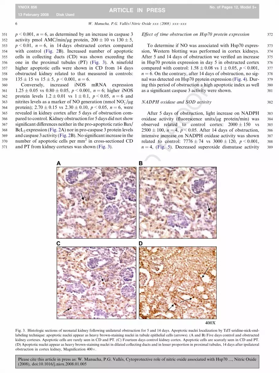
p < 0.001, n = 6, as determined by an increase in caspase 3activity pmol AMC/min/lg protein, 200 ± 10 vs 130 ± 5,p < 0.01, n = 6, in 14 days obstructed cortex comparedwith control (Fig. 2B). Increased number of apoptoticcells in collecting ducts (CD) was shown exceeding theone in the proximal tubules (PT) (Fig. 3). A ninefoldhigher apoptotic cells were shown in CD from 14 daysobstructed kidney related to that measured in controls:135 ± 15 vs 15 ± 5, p < 0.001, n = 6.
Conversely, increased iNOS mRNA expression1.25 ± 0.05 vs 0.80 ± 0.05, p < 0.001, n = 6; higher iNOSprotein levels 1.2 ± 0.01 vs 1 ± 0.1, p < 0.05, n = 6 andnitrites levels as a marker of NO generation (nmol NO�2 /lgprotein); 2.70 ± 0.15 vs 2.30 ± 0.10, p < 0.05, n = 6, wererevealed in kidney cortex after 5 days of obstruction com-pared to control. Kidney obstruction for 5 days did not showsignificant differences neither in the pro-apoptotic ratio Bax/BcL2 expression (Fig. 2A) nor in pro-caspase 3 protein levelsand caspase 3 activity (Fig. 2B). No significant increase in thenumber of apoptotic cells per mm2 in cross-sectioned CDand PT from kidney cortexes was shown (Fig. 3).
UN
CO
RR
EC
T
Fig. 3. Histologic sections of neonatal kidney following unilateral obstructionlabeling technique: apoptotic nuclei appear as heavy brown-staining nuclei in tkidney cortexes. Apoptotic cells are rarely seen in CD and PT. (C) Fourteen d(D) Apoptotic nuclei appear as heavy brown-staining nuclei in dilated collectingobstruction in cortex kidney, Magnification 400�.
Please cite this article in press as: W. Manucha, P.G. Valles, Cytopro(2008), doi:10.1016/j.niox.2008.01.005
RO
OF
Effect of time obstruction on Hsp70 protein expression
To determine if NO was associated with Hsp70 expres-sion, Western blotting was performed in cortex kidneys.After 5 and 14 days of obstruction we verified an increasein Hsp70 protein expression in day 5 in obstructed cortexcompared with control: 1.58 ± 0.08 vs 1 ± 0.05, p < 0.001,n = 6. On the contrary, after 14 days of obstruction, no sig-nal was detected on Hsp70 protein expression (Fig. 4). Dur-ing this period of obstruction a high apoptotic index as wellas a significant caspase 3 activity were shown.
NADPH oxidase and SOD activity
After 5 days of obstruction, light increase on NADPHoxidase activity (fluorescence units/lg protein/min) wasobserved related to control cortex: 2000 ± 150 vs2500 ± 100, n = 4, p < 0.05. After 14 days of obstruction,intensive increase on NADPH oxidase activity was shownrelated to control: 7776 ± 74 vs 3000 ± 120, p < 0.001,n = 4, (Fig. 5). Decreased superoxide dismutase activity
ED
P
for 5 and 14 days. Apoptotic nuclei localization by TdT-uridine-nick-end-ubule epithelial cells (arrows). (A and B) Five days control and obstructedays control kidney cortex. Apoptotic cells are scarcely seen in CD and PT.
ducts and in lesser proportion in proximal tubules, 14 days after ipsilateral
tective role of nitric oxide associated with Hsp70 ..., Nitric Oxide

NC
OR
REC
TED
PR
OO
390
391
392
393
394
395
396
397
398
399
400
401
402
403
404
405
406
407
408
409
CC5 OC5 CC14 OC14
70kDa Hsp70
0
0.2
0.4
0.6
0.8
1
1.2
1.4
1.6
1.8
Rel
ativ
e O
ptic
Den
sity
CC5 OC5 CC14 OC14
***
Fig. 4. Increased Hsp70 expression during early unilateral kidneyobstruction in neonatal rats. Western blot analysis of Hsp70 from cortexkidney following 5 and 14 days of obstruction. The relative amount ofHsp70 protein was determined after normalization of the level of Hsp70protein of the appropriate control. Densitometric analysis of Hsp70protein abundance showed a significant increase on Hsp70 expression inOC vs CC, ***p < 0.001 after 5 days of obstruction. No signal was detectedon Hsp70 protein expression after 14 days of obstruction. Data representmeans ± SEM of six separate experiments.
0
2000
4000
6000
8000
Rel
ativ
e flu
ores
cenc
e un
its
***
CC5 OC5 CC14 OC14
*
Fig. 5. Effect of obstruction for 5 and 14 days on NADPH oxidaseactivity in cortex kidney. NADPH oxidase activity was measured by achemioluminescence assay. Increased NADPH oxidase activity was shownafter 14 days of obstruction OC vs CC ***p < 0.001. Slight decreasedNADPH oxidase activity was shown after 5 days OC vs CC *p < 0.05.Each bar represents the mean ± SEM of four separate experiments.
0
2
4
6
8
10
12
Nitr
ites
(nm
ol/μ
g pr
otei
n)
Control Obstructed 14 days
L-arginine 100mg/Kg/dayL-NAME 50mg/Kg/dayWithout Treatment
***
***
***
**
iNOS130 kDa
Control Obstructed
Marker ------- L-NAME L-arginine ------- L-NAME L-arginine
0
0.2
0.4
0.6
0.8
1
1.2
1.4
Rel
ativ
e O
ptic
Den
sity
**
***
Control Obstructed 14 days
Without TreatmentL-NAME 50mg/Kg/dayL-arginine 100mg/Kg/day
Fig. 6. In vivo effect of L-Arginine and L-NAME treatment for 14 days, onendogenous NO levels and iNOS protein expression. (A) RepresentativeWestern blot and densitometry of iNOS in control and 14 days obstructedcortex homogenate from rats pretreated with L-Arginine or L-NAMEcontrol and obstructed cortexes from neonatal rats pretreated with L-Arginine resulted in higher iNOS protein levels related to non-treated rats,*p < 0.05 and **p < 0.01, respectively. On the contrary, control cortexhomogenates from animals treated with L-NAME for 14 days, showedlower iNOS abundance vs non-treated **p < 0.01. The relative amount ofiNOS protein was determined after normalization of the level of iNOSprotein of the appropriate control: 1. (B) Measurement of nitrite generated(nmol NO2 generated/lg protein) control and obstructed cortexes fromneonatal rats pretreated with L-Arginine resulted in higher NO levelsrelated to non-treated rats, ***p < 0.001 both. Nitrite levels were nearlycompletely inhibited after L-NAME treatment. Each bar represents themean ± SEM of four separate experiments.
W. Manucha, P.G. Valles / Nitric Oxide xxx (2008) xxx–xxx 7
YNIOX 856 No. of Pages 12, Model 5+
13 February 2008 Disk UsedARTICLE IN PRESS
Uwere demonstrated following 14 days of obstruction (U/mL) 10 ± 4 vs 60 ± 5, p < 0.001, n = 4. No differences werenoticed on the antioxidant level after 5 days of kidneyobstruction = 40 ± 8 vs 52 ± 7, n = 4.
‘‘In vivo” administration of L-Arginine and L-NAME in
UUO neonatal rats: effects on apoptosis induction
We examined the in vivo effect of L-Arginine (100 mg/kg/day) and L-NAME (50 mg/kg/day) pretreatment on the
Please cite this article in press as: W. Manucha, P.G. Valles, Cytopro(2008), doi:10.1016/j.niox.2008.01.005
F
apoptotic response induction, in control and obstructedcortex homogenates.
In vivo administration of L-Arginine for 14 days showedhigher endogenous NO (nmol/lg protein): 8.4 ± 0.45 vs3.2 ± 0.3, n = 4, p < 0.001, increased iNOS: 1.2 ± 0.05 vs1 ± 0.05, p < 0.05, n = 4, (Fig. 6A and B) and higherHsp70 protein levels: 1.9 ± 0.06 vs 1 ± 0.09, p < 0.01,n = 4 in control cortex homogenates (Fig. 7). Since ourdata showed that L-Arginine induced Hsp70 expression,we next examined whether treatment with L-Arginine inthe same control cortex homogenate, could result in resis-tance to apoptosis induction. Fig. 8A, showed that apopto-
tective role of nitric oxide associated with Hsp70 ..., Nitric Oxide

ED
PR
OO
F
410
411
412
413
414
415
416
417
418
419
420
421
422
423
424
425
426
427
428
429
430
431
432
433
434
435
436
437
438
Bax
BcL 2
B-actin
------- L-NAME ------- L-NAME L-arginine
550 bp
708 bp
201 bp
Control Obstructed
0
0.5
1
1.5
2
2.5
3
3.5
Rel
ativ
e O
ptic
Den
sity
(Bax
/BcL
2)
Control Obstructed 14 days
Without TreatmentL-NAME 50mg/Kg/dayL-arginine 100mg/Kg/day
***
**
BcL2
Control Obstructed------- L-NAME L-arginine ------- L-NAME L-arginine
25 kDa
20
30
40
50
60
70
ol A
MC
/μg
prot
/min L-arginine 100mg/Kg/day
L-NAME 50mg/Kg/dayWithout Treatment
***
**
L-arginine
8 W. Manucha, P.G. Valles / Nitric Oxide xxx (2008) xxx–xxx
YNIOX 856 No. of Pages 12, Model 5+
13 February 2008 Disk UsedARTICLE IN PRESS
sis was almost completely inhibited. Absence of the apop-totic index (1.52 ± 0.03 vs 1.00 ± 0.01, n = 4) as well asdecreased caspase 3 activity (10 ± 1 vs 15 ± 2, n = 4) wereshown (Fig. 8C). Overproduction of reactive oxygen inter-mediate (ROI) has previously been identified as a key com-ponent of apoptotic pathways. Therefore, after L-Argininetreatment lower NADPH oxidase activity was shown:1000 ± 120 vs 2500 ± 100, p < 0.001, n = 4 (Fig. 9).
Obstructed kidney homogenates from rats pretreatedwith L-Arginine, showed higher endogenous NO (nmol/lg protein): 9.7 ± 0.6 vs 1.8 ± 0.15, n = 4, p < 0.001,increased iNOS: 1.3 ± 0.07 vs 0.52 ± 0.04, n = 4, p < 0.01(Fig. 6B and A) and detectable Hsp70 protein levels(Fig. 7). Obstructed kidney homogenates from L-Argininepretreated rats compared to the one from rats without L-Arginine administration, showed decreased apoptoticindex Bax/BcL2 (0.9 ± 0.02 vs 1.90 ± 0.20, p < 0.01,n = 4) (Fig. 8A), increased BcL2 protein expression(Fig. 8B) and a light decrease in the caspase 3 activity(17 ± 3 vs 23 ± 3, n = 4) (Fig. 8C). Lower NADPH oxi-dase activity was demonstrated in 14 day obstructedhomogenates from L-Arginine pretreated rats comparedto the obstructed kidney homogenate from rats withouttreatment (3000 ± 119 vs 6000 ± 120, p < 0.001, n = 4)(Fig. 9).
Conversely, treatment of control rats with L-NAMEshowed lower iNOS: 0.5 ± 0.1 vs 1 ± 0.05, p < 0.01,n = 4, decreased endogenous NO(nmol/lg protein):0.33 ± 0.10 vs 3.2 ± 0.3, p < 0.001, n = 4 (Fig. 6A and B)
UN
CO
RR
EC
T
439
440
441
442
443
444
445
446
447
70kDa
Control Obstructed
Marker ------- L-NAME ------- L-NAME
-0.5
0
0.5
1
1.5
2
2.5
3
3.5
4
4.5
5
Rel
ativ
e O
ptic
Den
sity
Without TreatmentL-NAME 50mg/Kg/day
Control Obstructed 14 days
L-arginine 100mg/Kg/day
**
***
L-arginine L-arginine
Fig. 7. In vivo effect of L-Arginine and L-NAME treatment on Hsp70protein expression in neonatal rats. Representative Western blot anddensitometry of Hsp70 in control and 14 days obstructed renal cortexhomogenate from rats pretreated with L-Arginine or L-NAME. After L-Arginine-pre treatment for 14 days, a significant increase on inducibleHsp70 protein levels was noted in obstructed and control cortexhomogenates related to cortex tissue of non-treated rats, ***p < 0.01 and**p < 0.001, respectively. Absence of Hsp70 expression was demonstratedafter L-NAME treatment. Blot represents one out of four separateexperiments.
0
10
pm
Control Obstructed 14 days
Fig. 8. In vivo effect of L-Arginine and L-NAME treatment on apoptosisinduction. (A) mRNA expression for Bcl2 and Bax and the ratio ofmRNA Bax/mRNA Bcl2. (B) Representative Western blot of BcL2. (C)Caspase 3 activity was assessed by level of Ac-DEVD-AMC cleavagerelease of fluorescence AMC tag. Activity is expressed as pmol AMC/min/lg protein. Control and 14 day obstructed cortex homogenates from L-Arginine pretreated neonatal rats resulted in resistance to apoptosisinduction. After L-Arginine for 14 days, absence of apoptotic index,increased BcL2 expression as well as caspase 3 activity near control non-treatment were shown. Conversely, L-NAME treatment of control ratsshowed increased Bax/BcL2 apoptotic index as well as increased caspase 3activity, compared to non-treated control rats, ***p < 0.001 and***p < 0.001, respectively. After 14 days of obstruction, pretreated ratswith L-NAME increased caspase 3 activity compared to the one of thenon-treated animals **p < 0.01. Each bar represents the mean ± SEM offour separate experiments.
Please cite this article in press as: W. Manucha, P.G. Valles, Cytopro(2008), doi:10.1016/j.niox.2008.01.005
and detected no signal of Hsp70 expression in cortex(Fig. 7).
Increased Bax/BcL2 apoptotic index (2.50 ± 0.02 vs1.00 ± 0.01, p < 0.001, n = 4) (Fig. 8A), as well as increasedcaspase 3 activity (40 ± 5 vs 15 ± 2, p < 0.001, n = 4)(Fig. 8C) and higher NADPH oxidase activity (4500 ± 89vs 2500 ± 100, p < 0.001, n = 4) (Fig. 9), were demon-strated when control kidneys from L-NAME treated ratswere compared to non-treated control rats.
tective role of nitric oxide associated with Hsp70 ..., Nitric Oxide
TED
PR
OO
F
448
449
450
451
452
453
454
455
456
457
458
459
460
461
462
463
464
465
466
467
468
469
470
471
472
473
474
475
476
477
478
479
480
481
482
483
484
485
486
487
0
2000
4000
6000
8000
10000
Rel
ativ
e flu
ores
cenc
e un
its
Control Obstructed 14 days
Without Treatment
L-arginine 100mg/Kg/dayL-NAME 50mg/Kg/day
***
***
***
***
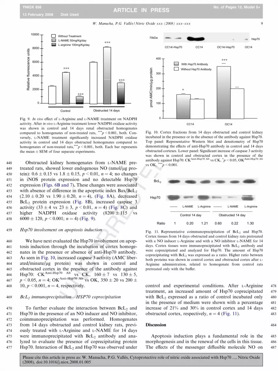
Fig. 9. In vivo effect of L-Arginine and L-NAME treatment on NADPHactivity. After in vivo L-Arginine treatment lower NADPH oxidase activitywas shown in control and 14 days renal obstructed homogenatescompared to homogenates of non-treated rats, ***p < 0.001, both. Con-versely, L-NAME treatment significantly increased NADPH oxidaseactivity in control and 14 days obstructed homogenates compared tohomogenates of non-treated rats,***p < 0.001, both. Each bar representsthe mean ± SEM of four separate experiments.
CC14/-Hsp70 CC14 OC14/-Hsp70 OC14
0
100
200
300
400
pmol
AM
C/m
in/μ
g pr
otei
n
CC14 OC14
***With Hsp70 Antibody
Without Hsp70 Antibody
*
70kDa Hsp70
Fig. 10. Cortex fractions from 14 days obstructed and control kidneyincubated in the presence or in the absence of the antibody against Hsp70.Top panel: Representative Western blot and densitometry of Hsp70demonstrating the effects of anti-Hsp70 antibody in control and 14 daysobstructed cortexes. Lower panel: Significant increase of caspase 3 activitywas shown in control and obstructed cortex in the presence of theantibody against Hsp70: CKAnti-Hsp70 Ab vs CK, *p < 0.05, OKAnti-Hsp70 Ab
vs OK, ***p < 0.001.
BcL2
Hsp70
-------- L-NAME L-Arginine --------- L-NAME L-Arginine
Control 14 day Obstructed 14 day
Ratio 1 0.20 1.21 0.60 0.22 1.30
Fig. 11. Representative coimmunoprecipitation of BcL2 and Hsp70.Cortex tissues from 14 days obstructed and control kidney rats pretreatedwith a NO inducer L-Arginine and with a NO inhibitor L-NAME for 14days. Cortex tissues were immunoprecipitated with BcL2 antibody andwere coprecipitated and analyzed for Hsp70. The amount of Hsp70coprecipitating with BcL2 was expressed as a ratio. Higher ratio betweenboth proteins was shown in control cortex and obstructed cortex after L-Arginine administration, related to homogenate from control ratspretreated only with the buffer.
W. Manucha, P.G. Valles / Nitric Oxide xxx (2008) xxx–xxx 9
YNIOX 856 No. of Pages 12, Model 5+
13 February 2008 Disk UsedARTICLE IN PRESS
UN
CO
RR
EC
Obstructed kidney homogenates from L-NAME pre-treated rats, showed lower endogenous NO (nmol/lg pro-tein): 0.6 ± 0.15 vs 1.8 ± 0.15, p < 0.01, n = 4; no changesin iNOS protein expression and no detectable Hsp70expression (Figs. 6B and 7). These changes were associatedwith absence of difference in the apoptotic index Bax/BcL2
(2.10 ± 0.20 vs 1.90 ± 0.20, n = 4), (Fig. 8A), decreasedBcL2 protein expression (Fig. 8B), increased caspase 3activity (33 ± 4 vs 23 ± 3, p < 0.01, n = 4) (Fig. 8C) andhigher NADPH oxidase activity (8200 ± 115 vs6000 ± 120, p < 0.001; n = 4) (Fig. 9).
Hsp70 involvement on apoptosis induction
We have next evaluated the Hsp70 involvement on apop-tosis induction through the incubation of cortex homoge-nates in the presence or absence of anti-Hsp70 antibody.As seen in Fig. 10, increased caspase 3 activity (AMC liber-ated/minute/lg protein) was shown in control andobstructed cortex in the presence of the antibody againstHsp70: CKAnti-Hsp70 Ab vs CK, 160 ± 7 vs 130 ± 5,p < 0.05, n = 4; OKAnti-Hsp70 Ab vs OK, 350 ± 20 vs 200 ±10, p < 0.001, n = 4, respectively.
BcL2 immunoprecipitation—HSP70 coprecipitation
To further evaluate the interaction between BcL2 andHsp70 in the presence of an NO inducer and NO inhibitor,coimmunoprecipitation was performed. Homogenatesfrom 14 days obstructed and control kidney rats, previ-ously treated with L-Arginine and L-NAME for 14 dayswere immunoprecipitated with BcL2 antibody and ana-lyzed to evaluate the presence of coprecipitating proteinHsp70. Interaction of BcL2 and Hsp70 was observed under
Please cite this article in press as: W. Manucha, P.G. Valles, Cytopro(2008), doi:10.1016/j.niox.2008.01.005
control and experimental conditions. After L-Argininetreatment, an increased amount of Hsp70 coprecipitatedwith BcL2 expressed as a ratio of control incubated onlyin the presence of medium were shown with a percentageincrease of 21% and 30% in control cortex and 14 daysobstructed cortex, respectively, n = 4 (Fig. 11).
Discussion
Apoptosis induction plays a fundamental role in themorphogenesis and in the renewal of the cells in this tissue.The effects of the messenger diffusible molecule NO on
tective role of nitric oxide associated with Hsp70 ..., Nitric Oxide

T
488
489
490
491
492
493
494
495
496
497
498
499
500
501
502
503
504
505
506
507
508
509
510
511
512
513
514
515
516
517
518
519
520
521
522
523
524
525
526
527
528
529
530
531
532
533
534
535
536
537
538
539
540
541
542
543
544
545
546
547
548
549
550
551
552
553
554
555
556
557
558
559
560
561
562
563
564
565
566
567
568
569
570
571
572
573
574
575
576
577
578
579Q1
580
581
582
583
584
585
586
587
588
589
590
591
592
593
594
595
596
597
598
599
600
601
10 W. Manucha, P.G. Valles / Nitric Oxide xxx (2008) xxx–xxx
YNIOX 856 No. of Pages 12, Model 5+
13 February 2008 Disk UsedARTICLE IN PRESS
UN
CO
RR
EC
apoptosis might be a NO level dependence and motive ofpermanent controversies because of its bifunctional role.UUO renal tubular apoptosis is related to renal tubularatrophy and renal tissue loss [30,31]. The study we presenthere provides evidence for cytoprotective effect of NOlinked to Hsp70 against the mitochondrial apoptosis path-way in early neonatal obstruction.
In the present study, we have shown that NO protectscortex tubular epithelial cells from obstructed cortex kid-ney-induced cytotoxicity and apoptosis. Studies after 5and 14 days of unilateral kidney obstruction revealed thatinduction of Hsp70 protein occurred in parallel to protec-tion from obstruction-induced apoptosis.
Both pro-apoptotic and anti-apoptotic effects of NOhave been demonstrated so far [32]. The capacity of NOto induce apoptosis was first appreciated by Albina et al.,who confirmed that NO-dependent death of murine perito-neal macrophages activated by interferon-c (IFN-c) andlipopolysaccharide (LPS) is mediated through apoptosis[33]. Notwithstanding, more recent studies have shown ananti-apoptotic effect of NO. Mannick et al. have shownthat endogenous iNOS expression or exposure to low dosesof NO donors inhibited apoptosis in human B lymphocytes[34]. Since then, similar findings have been reported in vitro
and in vivo [35,36]. From in vitro studies in stretchedepithelial cells and in vivo studies in obstructed kidney ofiNOS�/� mice support for an anti-apoptotic role to NOhave been provided [37].
The question of whether NO promotes or inhibitsapoptosis has been quite controversial, multiple mecha-nism for the inhibition of apoptosis by NO may exist ina single cell.
In our study, downregulation of Hsp70 protein expres-sion associated with decreased endogenous NO and loweriNOS at the level of gene expression and protein expressionwere shown in the induction of apoptosis following 14 daysof obstruction. A temporal relationship was shownbetween 14 days obstruction and apoptosis regulated bymitochondrial signal pathway, through the increased pro-apoptotic ratio Bax/BcL2 and, consequently caspase 3activity. Conversely, increased Hsp70 expression linked toincreased NO and iNOS expression at transcriptional andpost-transcriptional levels with absence of apoptotic tubu-lar cell response were shown after obstruction for 5 days.These results suggest that the presence of NO linked toHsp70 protein expression may serve to modulate apoptoticprocess in obstructed kidney. Hsp70 induction is an earlysurvival signal elaborated by stressed cells to counter cellu-lar damage and hasten recovery [38]. This chaperone isknown to bind to nascent and immature proteins, and toprevent premature and improper binding and folding.Hsp70 also confers cellular protection by modulating theengagement and/or progression of apoptosis [23]. Evidenceto support the hypothesis that apoptosis was associatedwith decreased NO joined to lower Hsp70 protein expres-sion was also established herein by in vivo manipulationof endogenous NO. Control cortex of L-NAME pretreated
Please cite this article in press as: W. Manucha, P.G. Valles, Cytopro(2008), doi:10.1016/j.niox.2008.01.005
ED
PR
OO
F
rats resulted in lower levels of Hsp70 and iNOS proteinexpression with downregulation of BcL2 at the level of geneexpression and protein expression together with increasedcaspase 3 activity. The cellular effects of apoptosis werereversed by L-Arginine treatment.
Moreover, to further demonstrate the association of NOwith Hsp70 in the apoptotic response, interaction betweenHsp70 and BcL2 in the presence of an NO inhibitor andNO inducer was performed. An antibody directed againstBcL2 was used to precipitate native BcL2 protein. Copre-cipitation of both proteins increased to 21% in controlhomogenates from rats pretreated with a NO inducerrelated to control rats pretreated with buffer. The mecha-nism by which NO stimulates the expression of Hsp70may involve the interaction of NO with thiol-containingmolecules. NO readily oxidizes the most abundant lowmolecular weight thiol glutathione, forming S-nitrosothiolsand disulfide. This action stimulates the Hsp70 which pro-tect cells from apoptotic cell death. [39]. In a previousreport, pretreatment of hepatocytes with NO altered redoxstate accompanied by oxidation of glutathione (GSH) andformation of S-nitrosoglutathione (GSNO), both beinginvolved in Hsp70 mRNA induction [8]. In our study wehave demonstrated that the apoptotic effect by lowerNO-mediated decreased Hsp70 expression was associatedwith the direct induction of apoptotic signal transductioninvolving the activation of caspase 3 by decreasing stabil-ization of BcL2. Given its BcL2 localization within mito-chondria and its role in preventing cytochrome c release,preservation of BcL2 by Hsp70 could account for the pro-tection of epithelial cells [40]. Nevertheless, Hsp70 mayintervene at several points to halt progression of the apop-totic cascade. Hsp70 may act by preventing cell death byinterfering with the ability of cytocrome c and Apaf-1 torecruit pro-caspase-9. Hsp70 therefore suppressing apopto-sis by directly associating with Apaf-1 and blocking theassembly of a functional apoptosome [41]. Production ofROS has been identified as a key component of apoptoticpathways involving activation of endogenous endonucle-ases [42] and direct DNA fragmentation [43]. In our study,after 14 days of obstruction, oxidative stress was elevatedthrough the increased NADPH oxidase activity as well asdecreased superoxide dismutase activity. These results leadto a pronounced increase in total oxidant activity inobstruction. Conversely, NADPH oxidase activity wastransiently suppressed when rats were pretreated with L-Arginine, similar to the results obtained after 5 days of kid-ney obstruction.
Higher renal endogenous NO levels in obstructed kid-neys for 5 days or after L-Arginine pretreatment, inducedHsp70 expression, which has been shown to have anti-apoptotic or cytoprotective effects. Thus, it is likely thatHsp70 expression by L-Arginine administration protectedthe cells from early obstruction-mediated apoptosis andcytotoxicity. These results allow us to suggest that upregu-lation of Hsp70 and increased endogenous NO may be anearly line of defense to cytoprotect cortex tubule cells in
tective role of nitric oxide associated with Hsp70 ..., Nitric Oxide
T
602
603
604
605
606
607
608
609
610
611
612
613
614
615
616
617
618
619
620
621
622
623
624
625
626
627
628
629
630
631
632
633
634
635636637638639640641642643644645646647648649650651652653654655656657658
659660661662663664665666667668669670671672673674675676677678679680681682683684685686687688689690691692693694695696697698699700701702703704705706707708709710711712713714715716717718719720721722723724725726
W. Manucha, P.G. Valles / Nitric Oxide xxx (2008) xxx–xxx 11
YNIOX 856 No. of Pages 12, Model 5+
13 February 2008 Disk UsedARTICLE IN PRESS
UN
CO
RR
EC
early kidney obstruction. Hsp70 expression induction pre-cedes conventional markers of renal injury.
In the present study we did not explore the mechanismsinvolved in ROS production inhibition by L-Arginine-induced Hsp70 expression. Previously, it has been sug-gested that Hsp70 may block signal transduction to themitochondria, resulting in the inhibition of mitochondrialreactive oxygen intermediate (ROI) production by inhibit-ing either second lipid messenger(s) to mitochondria [44].Alternatively, it has also been possible that Hsp70 mayenhance the chaperon-mediated import of precursor pro-teins into mitochondria which control mitochondrial func-tion [45,46] leading to decreased ROS formation.Pretreatment with the NO-generating compound S-nitroso,N-acetylpenicillamine (SNAP) have been shown to protectcultured rat hepatocytes from tumoral necrosis factor alfa(TNFa)-induced cytotoxicity and apoptosis through thestimulation of Hsp70 expression [8].
Taken together, our data demonstrate that the effect ofNO interacting with Hsp70 is a result of the capacity ofboth to prevent mitochondrial apoptotic pathway in neo-natal early kidney obstruction. Induction of Hsp70 pro-tects cells not only from damage due to apoptosisinduction but also from damage due to oxidative injury.These findings demonstrated that NO can induce cytopro-tection in early obstructed kidney cortex tubular epithelialcells, through the stimulation of Hsp70 expression.
Acknowledgments
This work was performed with financial support fromthe Research and Technology Council of Cuyo University(CIUNC) from Mendoza, Argentina/No. 631/02 and fromCONICET, PICT/2005 No. 05-33827 to P.G. Valles.
References
[1] R.L. Chevalier, Molecular and cellular pathophysiology of obstruc-tive nephropathy, Pediatr. Nephrol. 13 (1999) 612–619.
[2] R.L. Chevalier, K.H. Chung, C.D. Smith, M. Ficenec, R.A. Gomez,Renal apoptosis and clusterin following ureteral obstruction= therole of maturation, J. Urol. 156 (1996) 1474–1479.
[3] F. Cachat, B. Lange-Sperandio, A.Y. Chang, S.C. Kiley, B.A.Thornhill, M.S. Forbes, R.L. Chevalier, Ureteral obstruction inneonatal mice elicits segment-specific tubular cell responses leading tonephron loss, Kidney Int. 63 (2003) 564–575.
[4] U.K. Messmer, M. Ankarcrona, P. Nicotera, B. Brune, p53 expres-sion in nitric oxide-induced apoptosis, FEBS Lett. 355 (1994) 23–26.
[5] K. Fehsel, K.D. Kroncke, K.L. Meyer, H. Huber, V. Wahn, V. Kolb-Bachofen, Nitric oxide induces apoptosis in mouse thymocytes, J.Immunol. 155 (1995) 2858–2865.
[6] Y.S. Ho, Y.J. Wang, J.K. Lin, Induction of p53 and p21/WAF1/CIP1expression by nitric oxide and their association with apoptosis inhuman cancer cells, Mol. Carcinog. 16 (1996) 20–31.
[7] U.K. Messmer, B. Brune, Nitric oxide-induced apoptosis: p53-dependent and p53-independent signalling pathways, Biochem. J.319 (1996) 299–305.
[8] Y.M. Kim, M.E. de Vera, S.C. Watkins, T.R. Billiar, Nitric oxideprotects cultured rat hepatocytes from tumor necrosis factor-alpha-induced apoptosis by inducing heat shock protein 70 expression, J.Biol. Chem. 272 (1997) 1402–1411.
Please cite this article in press as: W. Manucha, P.G. Valles, Cytopro(2008), doi:10.1016/j.niox.2008.01.005
ED
PR
OO
F
[9] J.B. Mannick, X.Q. Miao, J.S. Stamler, Nitric oxide inhibits Fas-induced apoptosis, J. Biol. Chem. 272 (1997) 24125–24128.
[10] J. Li, T.R. Billiar, R.V. Talanian, Y.M. Kim, Nitric oxide reversiblyinhibits seven members of the caspase family via S-nitrosylation,Biochem. Biophys. Res. Commun. 240 (1997) 419–424.
[11] Y.M. Kim, T.H. Kim, D.W. Seol, R.V. Talanian, T.R. Billiar, Nitricoxide suppression of apoptosis occurs in association with aninhibition of BcL2 cleavage and cytochrome c release, J. Biol. Chem.273 (1998) 31437–31441.
[12] W. Manucha, L. Carrizo, C. Ruete, P. Valles, Apoptosis induction isassociated with decreased NHE1 expression in neonatal unilateralureteric obstruction, BJU Int. 1 (2007) 191–198.
[13] H.T. Nguyen, S.H. Bride, A.B. Badawy, R.M. Adam, J.Q. Lin, A.Orsola, P.D. Guthrie, M.R. Freeman, C.A. Peters, Heparin-bindingEGF-like growth factor is up-regulated in the obstructed kidney in acell- and region-specific manner and acts to inhibit apoptosis, Am. J.Pathol. 156 (2000) 889–898.
[14] R.L. Chevalier, B.A. Thornhill, Ureteral obstruction in the neonatalrat: renal nerves modulate hemodynamic effects, Pediatr. Nephrol. 9(1995) 447–450.
[15] Y. Kayanoki, J. Fujii, K.N. Islam, K. Suzuki, S. Kawata, Y.Matsuzawa, N. Taniguchi, The protective role of glutathione perox-idase in apoptosis induced by reactive oxygen species, J. Biochem.(Tokyo) 119 (1996) 817–822.
[16] N. Kawada, T. Moriyama, A. Ando, M. Fukunaga, T. Miyata, K.Kurokawa, E. Imai, M. Hori, Increased oxidative stress in mousekidneys with unilateral ureteral obstruction, Kidney Int. 56 (1999)1004–1013.
[17] A. Gupta, D. Nigam, G.S. Shukla, A.K. Agarwal, Profile of reactiveoxygen species generation and antioxidative mechanisms in thematuring rat kidney, J. Appl. Toxicol. 19 (1999) 55–59.
[18] H. Ischiropoulos, L. Zhu, J. Chen, M. Tsai, J.C. Martin, C.D. Smith,J.S. Beckman, Arch. Biochem. Biophys. 298 (1992) 431–437.
[19] A.P. Bautista, J.J. Spitzer, Inhibition of nitric oxide formation in vivoenhances superoxide release by the perfused liver, Am. J. Physiol. 266(1994) G783–G788.
[20] R.M. Clancy, J. Leszcznska-Piziak, S.B. Abramson, Nitric oxide, andendothelial cell relaxation factor inhibits neutrophils superoxideanion production via a direct action on the NADPH oxidase, J. Clin.Invest. 90 (1992) 1116–1121.
[21] R.I. Morimoto, A. Tissieres, C. Georgopoulos, The Biology of HeatShock Proteins and Molecular Chaperones, Cold Spring HarborLaboratory Press, Cold Spring Harbor, NY, 1994, pp. 395–416.
[22] F.X. Beck, N. Wolfgang, E. Muller, Molecular chaperones in thekidney: distribution, putative roles and regulation, Am. J. Physiol.Renal Physiol. 279 (2000) F203–F215.
[23] H.M. Beere, D.R. Green, Stress management -heat shock protein -70and the regulation of apoptosis, Trends Cell Biol. 11 (2001) 6–10.
[24] W. Manucha, L. Carrizo, C. Ruete, H. Molina, P. Valles, AngiotensinII type I antagonist on oxidative stress and heat shock protein 70(Hsp70) expression in obstructive nephropathy, Cell Mol. Biol. 6(2005) 547–555.
[25] J.P. Griess, On a new series of bodies in which nitrogen is substitutedfor hydrogen, Philos. Trans. R. Soc. Lond. 154 (1964) 667–731.
[26] A. Perner, L. Andresen, G. Pedersen, J. Rask-Madsen, Superoxideproduction and expression of NADPH oxidases by transformed andprimary human colonic epithelial cells, Gut 52 (2003) 231–236.
[27] C. Nebot, M. Moutet, P. Huet, J.Z. Xu, J.C. Yadan, Spectrophoto-metric assay of superoxide dismutase activity based on the activatedautoxidation of a tetracyclic catechol, J. Anal. Biochem. 214 (1993)442–451.
[28] M. Kawaguchi, K. Koshimura, Y. Murakami, M. Tsumori, T.Gonda, Y. Kato, Antihypertensive effect of insulin via nitric oxideproduction in the Zucker diabetic fatty rat, an animal model for non-insulin-dependent diabetes mellitus, Eur. J. Endocrinol. 140 (1999)341–349.
[29] M.P. Schlaich, S. Oehmer, M.P. Schneider, C. Delles, B.M. Schmidt,R.E. Schmieder, Effects of nitric oxide synthase inhibition and L-
tective role of nitric oxide associated with Hsp70 ..., Nitric Oxide
727728729730731732733734735736737738739740741742743744745746747748749750751752753
754755756757758759760761762763764765766767768769770771772773774775776777778779780
12 W. Manucha, P.G. Valles / Nitric Oxide xxx (2008) xxx–xxx
YNIOX 856 No. of Pages 12, Model 5+
13 February 2008 Disk UsedARTICLE IN PRESS
Arginine on renal haemodynamics in young patients at high cardio-vascular risk, Atherosclerosis 192 (2007) 155–160.
[30] I.S. Sawczuk, G. Hoke, C.A. Olsson, Gene expression in response toacute unilateral ureteral obstruction, Kidney Int. 35 (1989) 1315–1319.
[31] L.D. Truong, G. Petrusevska, G. Yang, Cell apoptosis and prolifer-ation in experimental chronic obstructive uropathy, Kidney Int. 50(1996) 200–207.
[32] S. Dimmeler, A.M. Zeiher, Nitric oxide and apoptosis: anotherparadigm for the double-edged role of nitric oxide, Nitric Oxide 1(1997) 275–281.
[33] J.E. Albina, S. Cui, R.B. Mateo, Nitric oxide-mediated apoptosis inmurine peritoneal macrophages, J. Immunol. 150 (1993) 5080–5085.
[34] J.B. Mannick, K. Asano, K. Izumi, Nitric oxide produced by humanB lymphocytes inhibits apoptosis and Epstein–Barr virus reactivation,Cell 79 (1994) 1137–1146.
[35] G.C. Gobe, R.A. Axelsen, Genesis of renal tubular atrophy inexperimental hydronephrosis in the rat: role of apoptosis, Lab. Invest.56 (1987) 273–281.
[36] Y.M. Kim, C.A. Bombeck, T.R. Billiar, Nitric oxide as a bifunctionalregulator of apoptosis, Circ. Res. 84 (1999) 253–256.
[37] A. Miyajima, J. Chen, D.P. Poppas, E.D. Darracott Vaughan Jr., D.Felsen, Role of nitric oxide in renal tubular apoptosis of unilateralureteral obstruction, Kidney Int. 59 (2001) 1290–1303.
[38] M.J. Gething, J. Sambrook, Protein folding in the cell, Nature 355(1992) 33–45.
[39] F. Li, H.P. Mao, K.L. Ruchalski, Y.H. Wang, W. Choy, J.H.Schwartz, S.C. Borkan, Heat stress prevents mitochondrial injury
UN
CO
RR
EC
T781
Please cite this article in press as: W. Manucha, P.G. Valles, Cytopro(2008), doi:10.1016/j.niox.2008.01.005
PR
OO
F
in ATP-depleted renal epithelial cells, Am. J. Physiol. 283 (2002)C917–C926.
[40] S.C. Borkan, A. Emami, J.H. Schwartz, Heat stress protein-associ-ated cytoprotection in inner medullary collecting duct cells from ratkidney, Am. J. Physiol. 265 (1993) F333–F341.
[41] H.M. Beere, B.B. Wolf, K. Cain, P. Tailor, R.I. Morimoto, G.M.Cohen, D.R. Green, Heat-shock protein 70 inhibits apoptosis bypreventing recruitment of procaspase-9 to the Apaf-1 apoptosome,Nat. Cell Biol. 2 (2000) 469–475.
[42] A. Fernandez, J. Kiefer, L. Fosdick, D.J. McConkey, Oxygen radicalproduction and thiol depletion are required for Ca(2+) -mediatedendogenous endonuclease activation in apoptotic thymocytes, J.Immunol. 155 (1995) 5133–5139.
[43] J.J. Mertens, N.W. Gibson, S.S. Lau, T.J. Monks, Reactive oxygenspecies and DNA damage in 2-bromo-(glutathion-S-yl) hydroquinone-mediated cytotoxicity, Arch. Biochem. Biophys. 320 (1995) 5851–5858.
[44] M.R. Jacquier-Sarlin, K. Fuller, A.T. Dinh-Xuan, M.J. Richard, B.S.Polla, Protective effects of hsp70 in inflammation, Experientia (Basel)50 (1994) 1031–1038.
[45] T.A.A. Harkness, F.E. Nargang, I. van der Klei, W. Neupert, R.Lill, A crucial role of the mitochondrial protein import receptorMOM19 for the biogenesis of mitochondria, J. Cell Biol. 124(1994) 637–648.
[46] S. Laloraya, P.J. Dekker, W. Voos, E.A. Craig, N. Pfanner, Mito-chondrial GrpE modulates the function of matrix Hsp70 in transloca-tion and maturation of preproteins, Mol. Cell. Biol. 15 (1995) 7098–7105.
ED
tective role of nitric oxide associated with Hsp70 ..., Nitric Oxide