qt64s245z6_nosplash_88d3e1b... - escholarship
TRANSCRIPT
ARTICLE
Molecular and clinical determinants of responseand resistance to rucaparib for recurrent ovariancancer treatment in ARIEL2 (Parts 1 and 2)Elizabeth M. Swisher 1,25✉, Tanya T. Kwan 2,25, Amit M. Oza3, Anna V. Tinker4, Isabelle Ray-Coquard 5,
Ana Oaknin6, Robert L. Coleman7, Carol Aghajanian8, Gottfried E. Konecny9, David M. O’Malley10,
Alexandra Leary11, Diane Provencher 12, Stephen Welch13, Lee-may Chen14, Andrea E. Wahner Hendrickson15,
Ling Ma16, Prafull Ghatage 17, Rebecca S. Kristeleit 18, Oliver Dorigo 19, Ashan Musafer20,
Scott H. Kaufmann 15, Julia A. Elvin21, Douglas I. Lin 21, Setsuko K. Chambers22, Erin Dominy2,
Lan-Thanh Vo2, Sandra Goble2, Lara Maloney2, Heidi Giordano2, Thomas Harding2, Alexander Dobrovic20,
Clare L. Scott 23, Kevin K. Lin2 & Iain A. McNeish 24
ARIEL2 (NCT01891344) is a single-arm, open-label phase 2 study of the PARP inhibitor
(PARPi) rucaparib in relapsed high-grade ovarian carcinoma. In this post hoc exploratory
biomarker analysis of pre- and post-platinum ARIEL2 samples, RAD51C and RAD51D muta-
tions and high-level BRCA1 promoter methylation predict response to rucaparib, similar to
BRCA1/BRCA2 mutations. BRCA1 methylation loss may be a major cross-resistance
mechanism to platinum and PARPi. Genomic scars associated with homologous recombi-
nation deficiency are irreversible, persisting even as platinum resistance develops, and
therefore are predictive of rucaparib response only in platinum-sensitive disease. The RAS,
AKT, and cell cycle pathways may be additional modulators of PARPi sensitivity.
https://doi.org/10.1038/s41467-021-22582-6 OPEN
1 University of Washington, Seattle, WA, USA. 2 Clovis Oncology, Inc., Boulder, CO, USA. 3 Princess Margaret Cancer Centre, University Health Network,Toronto, ON, Canada. 4 BC Cancer—Vancouver, Vancouver, BC, Canada. 5 GINECO, Centre Léon Bérard and University Claude Bernard, Lyon, France. 6 Valld’Hebron University Hospital, Vall d’Hebron Institute of Oncology (VHIO), Barcelona, Spain. 7 The University of Texas, MD Anderson Cancer Center,Houston, TX, USA. 8Memorial Sloan Kettering Cancer Center, New York, NY, USA. 9 University of California Los Angeles, Los Angeles, CA, USA. 10 The OhioState University, James Cancer Center, Columbus, OH, USA. 11 Gustave Roussy Cancer Center and INSERM U981, Villejuif, France. 12 l’Université de Montréal(CHUM), Montréal, QC, Canada. 13 Lawson Health Research Institute, London, ON, Canada. 14 University of California San Francisco Helen Diller FamilyComprehensive Cancer Center, San Francisco, CA, USA. 15Mayo Clinic, Rochester, MN, USA. 16 Rocky Mountain Cancer Centers, Lakewood, CO, USA.17 Tom Baker Cancer Center, Calgary, AB, Canada. 18 Guy’s and St. Thomas NHS Foundation Trust, London, UK. 19 Stanford University Cancer Center andStanford Cancer Institute, Palo Alto, CA, USA. 20University of Melbourne Department of Surgery, Austin Hospital, Heidelberg, VIC, Australia. 21 FoundationMedicine, Inc., Cambridge, MA, USA. 22 University of Arizona Cancer Center, Tucson, AZ, USA. 23 Royal Melbourne Hospital and Walter and Eliza HallInstitute of Medical Research, Parkville, VIC, Australia. 24 Imperial College London, London, UK. 25These authors contributed equally: Elizabeth M. Swisher,Tanya T. Kwan. ✉email: [email protected]
NATURE COMMUNICATIONS | (2021) 12:2487 | https://doi.org/10.1038/s41467-021-22582-6 | www.nature.com/naturecommunications 1
1234
5678
90():,;
Rucaparib is an inhibitor of poly(ADP-ribose) polymerase(PARP) 1, PARP2, and PARP3, DNA damage repairenzymes in the base excision repair pathway1. Homologous
recombination deficiency (HRD) sensitizes neoplasms to ruca-parib and other DNA-damaging agents (e.g., platinum-basedchemotherapy)2,3, and platinum sensitivity in high-grade ovariancarcinoma (HGOC) is a strong clinical predictor of benefit fromPARP inhibitors (PARPi)4–7.
Genetic, epigenetic, and genomic biomarkers can suggest thepresence of HRD and help identify patients most likely torespond to PARPi8–10. Germline and somatic BRCA1 or BRCA2(BRCA) mutations are well-defined biomarkers for PARPiresponse for a number of cancer types, including breast, ovarian,pancreatic, and prostate11–16. Alterations in other homologousrecombination repair (HRR) pathway genes, including PALB2,RAD51C, and RAD51D, have been associated with improvedresponses to rucaparib and other PARPi17–19. However, theextent to which many HRR genes contribute to PARPi sensitivityremains unclear20. BRCA1 and RAD51C promoter methylationresults in transcriptional silencing21 and commonly leadsto HRD in HGOC22. In preclinical studies, BRCA1 orRAD51C methylation led to increased sensitivity to PARPi andplatinum23–25, but establishing a consistent association ofmethylation with clinical responses has been elusive26,27.
Given the variety of mechanisms that can result in HRD,methods for identifying HRD cancers independent of themechanisms involved are desirable. HRD cancers exhibit highgenomic instability, characterized by deletions of large genomicsegments (genome-wide loss of heterozygosity [LOH]), amongother genomic aberrations28,29. Next-generation sequencing(NGS) can identify multiple patterns of genomic change, includingcopy number variations, single-nucleotide variations, insertions/deletions, rearrangements characteristic of HRD29,30, andothers31–34. HGOCs with a BRCA mutation or BRCA1/RAD51Cmethylation have high levels of genomic LOH25. Patients withBRCA wild-type (BRCAwt) HGOC with high LOH (≥16%) showgreater clinical benefit from rucaparib than those with low LOH7.High LOH is also correlated with improved response to platinumand other PARPi6,35. However, genomic scars accumulate in HRDcancer cells over time and do not disappear when HRR is restoredor other PARPi resistance mechanisms develop, making themimperfect biomarkers of PARPi sensitivity36. Therefore, under-standing the mechanisms leading to HRD and how they mightchange during prior therapies is important to predict outcomeswith PARPi.
ARIEL2 is an international, open-label, 2-part, phase 2 studyassessing the safety and efficacy of rucaparib as active treatmentin patients with relapsed HGOC. Part 1 enrolled patientswith platinum-sensitive disease; clinical results and moleculardata from this portion were published17,25,37,38. Part 2 enrolledpatients who had received three to four prior chemotherapies,including patients with platinum-sensitive or platinum-resistant/refractory disease. Here we present clinical results from Part 2and post hoc exploratory biomarker analyses using the richdataset of archival tissue samples and screening biopsies requiredfrom patients in both Parts 1 and 2.
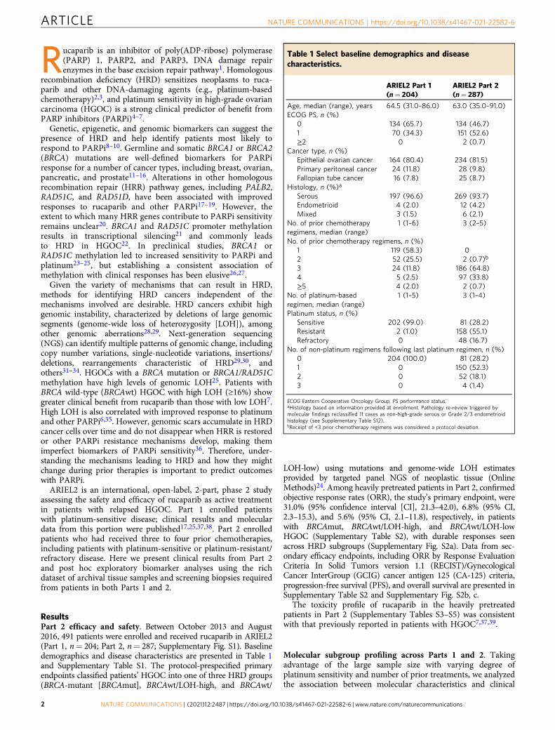
ResultsPart 2 efficacy and safety. Between October 2013 and August2016, 491 patients were enrolled and received rucaparib in ARIEL2(Part 1, n= 204; Part 2, n= 287; Supplementary Fig. S1). Baselinedemographics and disease characteristics are presented in Table 1and Supplementary Table S1. The protocol-prespecified primaryendpoints classified patients’ HGOC into one of three HRD groups(BRCA-mutant [BRCAmut], BRCAwt/LOH-high, and BRCAwt/
LOH-low) using mutations and genome-wide LOH estimatesprovided by targeted panel NGS of neoplastic tissue (OnlineMethods)24. Among heavily pretreated patients in Part 2, confirmedobjective response rates (ORR), the study’s primary endpoint, were31.0% (95% confidence interval [CI], 21.3–42.0), 6.8% (95% CI,2.3–15.3), and 5.6% (95% CI, 2.1–11.8), respectively, in patientswith BRCAmut, BRCAwt/LOH-high, and BRCAwt/LOH-lowHGOC (Supplementary Table S2), with durable responses seenacross HRD subgroups (Supplementary Fig. S2a). Data from sec-ondary efficacy endpoints, including ORR by Response EvaluationCriteria In Solid Tumors version 1.1 (RECIST)/GynecologicalCancer InterGroup (GCIG) cancer antigen 125 (CA-125) criteria,progression-free survival (PFS), and overall survival are presented inSupplementary Table S2 and Supplementary Fig. S2b, c.
The toxicity profile of rucaparib in the heavily pretreatedpatients in Part 2 (Supplementary Tables S3–S5) was consistentwith that previously reported in patients with HGOC7,37,39.
Molecular subgroup profiling across Parts 1 and 2. Takingadvantage of the large sample size with varying degree ofplatinum sensitivity and number of prior treatments, we analyzedthe association between molecular characteristics and clinical
Table 1 Select baseline demographics and diseasecharacteristics.
ARIEL2 Part 1(n= 204)
ARIEL2 Part 2(n= 287)
Age, median (range), years 64.5 (31.0–86.0) 63.0 (35.0–91.0)ECOG PS, n (%)
0 134 (65.7) 134 (46.7)1 70 (34.3) 151 (52.6)≥2 0 2 (0.7)
Cancer type, n (%)Epithelial ovarian cancer 164 (80.4) 234 (81.5)Primary peritoneal cancer 24 (11.8) 28 (9.8)Fallopian tube cancer 16 (7.8) 25 (8.7)
Histology, n (%)a
Serous 197 (96.6) 269 (93.7)Endometrioid 4 (2.0) 12 (4.2)Mixed 3 (1.5) 6 (2.1)
No. of prior chemotherapyregimens, median (range)
1 (1–6) 3 (2–5)
No. of prior chemotherapy regimens, n (%)1 119 (58.3) 02 52 (25.5) 2 (0.7)b
3 24 (11.8) 186 (64.8)4 5 (2.5) 97 (33.8)≥5 4 (2.0) 2 (0.7)
No. of platinum-basedregimen, median (range)
1 (1–5) 3 (1–4)
Platinum status, n (%)Sensitive 202 (99.0) 81 (28.2)Resistant 2 (1.0) 158 (55.1)Refractory 0 48 (16.7)
No. of non-platinum regimens following last platinum regimen, n (%)0 204 (100.0) 81 (28.2)1 0 150 (52.3)2 0 52 (18.1)3 0 4 (1.4)
ECOG Eastern Cooperative Oncology Group, PS performance status.aHistology based on information provided at enrollment. Pathology re-review triggered bymolecular findings reclassified 11 cases as non-high-grade serous or Grade 2/3 endometrioidhistology (see Supplementary Table S12).bReceipt of <3 prior chemotherapy regimens was considered a protocol deviation.
ARTICLE NATURE COMMUNICATIONS | https://doi.org/10.1038/s41467-021-22582-6
2 NATURE COMMUNICATIONS | (2021) 12:2487 | https://doi.org/10.1038/s41467-021-22582-6 | www.nature.com/naturecommunications
outcomes across both parts of ARIEL2 to identify molecularmechanisms resulting in rucaparib sensitivity and resistance.
Molecular analyses were used to characterize HGOC based onmutation, methylation, and molecular subgroup status. In these posthoc analyses, molecular subgroups were determined using a 16%LOH cutoff for LOH-high in the BRCAwt subgroup; thisrefined cutoff was validated within ARIEL2 Part 1 (ref. 40) andwas prospectively tested in the maintenance setting in ARIEL3(NCT01968213)7. To define the BRCAmut subgroup comprehen-sively, we utilized both available local testing data and an updatedBRCA missense mutation classifier to reclassify the HGOC from 14patients as BRCAmut, beyond those initially identified by targetedNGS (Supplementary Fig. S3, Online Methods). In total, 138 HGOCwere classified as BRCAmut, 156 were BRCAwt/LOH-high, 168were BRCAwt/LOH-low, and 29 were BRCAwt/LOH-unclassified.
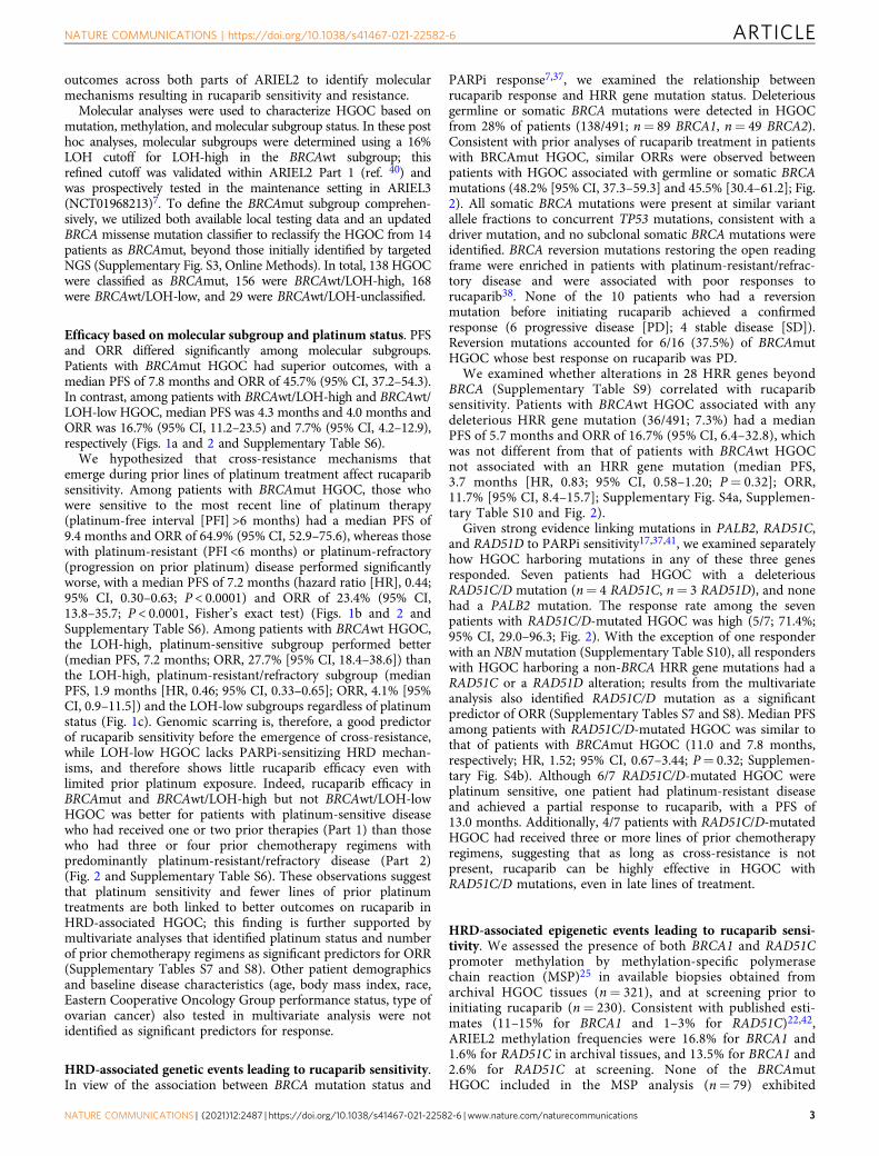
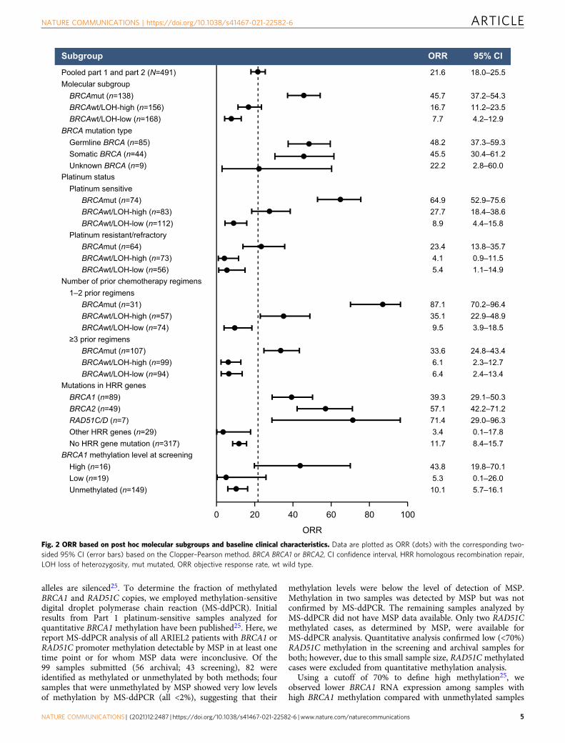
Efficacy based on molecular subgroup and platinum status. PFSand ORR differed significantly among molecular subgroups.Patients with BRCAmut HGOC had superior outcomes, with amedian PFS of 7.8 months and ORR of 45.7% (95% CI, 37.2–54.3).In contrast, among patients with BRCAwt/LOH-high and BRCAwt/LOH-low HGOC, median PFS was 4.3 months and 4.0 months andORR was 16.7% (95% CI, 11.2–23.5) and 7.7% (95% CI, 4.2–12.9),respectively (Figs. 1a and 2 and Supplementary Table S6).
We hypothesized that cross-resistance mechanisms thatemerge during prior lines of platinum treatment affect rucaparibsensitivity. Among patients with BRCAmut HGOC, those whowere sensitive to the most recent line of platinum therapy(platinum-free interval [PFI] >6 months) had a median PFS of9.4 months and ORR of 64.9% (95% CI, 52.9–75.6), whereas thosewith platinum-resistant (PFI <6 months) or platinum-refractory(progression on prior platinum) disease performed significantlyworse, with a median PFS of 7.2 months (hazard ratio [HR], 0.44;95% CI, 0.30–0.63; P < 0.0001) and ORR of 23.4% (95% CI,13.8–35.7; P < 0.0001, Fisher’s exact test) (Figs. 1b and 2 andSupplementary Table S6). Among patients with BRCAwt HGOC,the LOH-high, platinum-sensitive subgroup performed better(median PFS, 7.2 months; ORR, 27.7% [95% CI, 18.4–38.6]) thanthe LOH-high, platinum-resistant/refractory subgroup (medianPFS, 1.9 months [HR, 0.46; 95% CI, 0.33–0.65]; ORR, 4.1% [95%CI, 0.9–11.5]) and the LOH-low subgroups regardless of platinumstatus (Fig. 1c). Genomic scarring is, therefore, a good predictorof rucaparib sensitivity before the emergence of cross-resistance,while LOH-low HGOC lacks PARPi-sensitizing HRD mechan-isms, and therefore shows little rucaparib efficacy even withlimited prior platinum exposure. Indeed, rucaparib efficacy inBRCAmut and BRCAwt/LOH-high but not BRCAwt/LOH-lowHGOC was better for patients with platinum-sensitive diseasewho had received one or two prior therapies (Part 1) than thosewho had three or four prior chemotherapy regimens withpredominantly platinum-resistant/refractory disease (Part 2)(Fig. 2 and Supplementary Table S6). These observations suggestthat platinum sensitivity and fewer lines of prior platinumtreatments are both linked to better outcomes on rucaparib inHRD-associated HGOC; this finding is further supported bymultivariate analyses that identified platinum status and numberof prior chemotherapy regimens as significant predictors for ORR(Supplementary Tables S7 and S8). Other patient demographicsand baseline disease characteristics (age, body mass index, race,Eastern Cooperative Oncology Group performance status, type ofovarian cancer) also tested in multivariate analysis were notidentified as significant predictors for response.
HRD-associated genetic events leading to rucaparib sensitivity.In view of the association between BRCA mutation status and
PARPi response7,37, we examined the relationship betweenrucaparib response and HRR gene mutation status. Deleteriousgermline or somatic BRCA mutations were detected in HGOCfrom 28% of patients (138/491; n= 89 BRCA1, n= 49 BRCA2).Consistent with prior analyses of rucaparib treatment in patientswith BRCAmut HGOC, similar ORRs were observed betweenpatients with HGOC associated with germline or somatic BRCAmutations (48.2% [95% CI, 37.3–59.3] and 45.5% [30.4–61.2]; Fig.2). All somatic BRCA mutations were present at similar variantallele fractions to concurrent TP53 mutations, consistent with adriver mutation, and no subclonal somatic BRCA mutations wereidentified. BRCA reversion mutations restoring the open readingframe were enriched in patients with platinum-resistant/refrac-tory disease and were associated with poor responses torucaparib38. None of the 10 patients who had a reversionmutation before initiating rucaparib achieved a confirmedresponse (6 progressive disease [PD]; 4 stable disease [SD]).Reversion mutations accounted for 6/16 (37.5%) of BRCAmutHGOC whose best response on rucaparib was PD.
We examined whether alterations in 28 HRR genes beyondBRCA (Supplementary Table S9) correlated with rucaparibsensitivity. Patients with BRCAwt HGOC associated with anydeleterious HRR gene mutation (36/491; 7.3%) had a medianPFS of 5.7 months and ORR of 16.7% (95% CI, 6.4–32.8), whichwas not different from that of patients with BRCAwt HGOCnot associated with an HRR gene mutation (median PFS,3.7 months [HR, 0.83; 95% CI, 0.58–1.20; P= 0.32]; ORR,11.7% [95% CI, 8.4–15.7]; Supplementary Fig. S4a, Supplemen-tary Table S10 and Fig. 2).
Given strong evidence linking mutations in PALB2, RAD51C,and RAD51D to PARPi sensitivity17,37,41, we examined separatelyhow HGOC harboring mutations in any of these three genesresponded. Seven patients had HGOC with a deleteriousRAD51C/D mutation (n= 4 RAD51C, n= 3 RAD51D), and nonehad a PALB2 mutation. The response rate among the sevenpatients with RAD51C/D-mutated HGOC was high (5/7; 71.4%;95% CI, 29.0–96.3; Fig. 2). With the exception of one responderwith an NBN mutation (Supplementary Table S10), all responderswith HGOC harboring a non-BRCA HRR gene mutations had aRAD51C or a RAD51D alteration; results from the multivariateanalysis also identified RAD51C/D mutation as a significantpredictor of ORR (Supplementary Tables S7 and S8). Median PFSamong patients with RAD51C/D-mutated HGOC was similar tothat of patients with BRCAmut HGOC (11.0 and 7.8 months,respectively; HR, 1.52; 95% CI, 0.67–3.44; P= 0.32; Supplemen-tary Fig. S4b). Although 6/7 RAD51C/D-mutated HGOC wereplatinum sensitive, one patient had platinum-resistant diseaseand achieved a partial response to rucaparib, with a PFS of13.0 months. Additionally, 4/7 patients with RAD51C/D-mutatedHGOC had received three or more lines of prior chemotherapyregimens, suggesting that as long as cross-resistance is notpresent, rucaparib can be highly effective in HGOC withRAD51C/D mutations, even in late lines of treatment.
HRD-associated epigenetic events leading to rucaparib sensi-tivity. We assessed the presence of both BRCA1 and RAD51Cpromoter methylation by methylation-specific polymerasechain reaction (MSP)25 in available biopsies obtained fromarchival HGOC tissues (n= 321), and at screening prior toinitiating rucaparib (n= 230). Consistent with published esti-mates (11–15% for BRCA1 and 1–3% for RAD51C)22,42,ARIEL2 methylation frequencies were 16.8% for BRCA1 and1.6% for RAD51C in archival tissues, and 13.5% for BRCA1 and2.6% for RAD51C at screening. None of the BRCAmutHGOC included in the MSP analysis (n= 79) exhibited
NATURE COMMUNICATIONS | https://doi.org/10.1038/s41467-021-22582-6 ARTICLE
NATURE COMMUNICATIONS | (2021) 12:2487 | https://doi.org/10.1038/s41467-021-22582-6 | www.nature.com/naturecommunications 3
BRCA1 promoter methylation, suggesting that BRCA mutationsand BRCA1 promoter methylation are mutually exclusive HRDmechanisms.
Interestingly, we saw no difference in PFS based on eitherarchival or screening methylation status in patients with BRCAwt
HGOC, suggesting that the mere presence of methylation at thetwo promoters is not a biomarker for rucaparib outcomes(Supplementary Fig. S5).
MSP analysis detects the presence of methylation but does notestimate what fraction of the neoplastic cells is methylated or if all
c BRCAwt/LOH-high and BRCAwt/LOH-low HGOC
BRCAwt/LOH-low resistant/refractory 56 (0) 7 (45) 2 (50) 0 (52)BRCAwt/LOH-low sensitive 112 (0) 15 (87) 2 (99) 0 (101)
At risk (events)
BRCAwt/LOH-high resistant/refractory 73 (0) 8 (62) 3 (67) 0 (70)BRCAwt/LOH-high sensitive 83 (0) 26 (50) 10 (66) 2 (74) 2 (74) 2 (74) 0 (74)
BRCAwt/LOH-high resistant/refractory (n=73) 1.9 1.8–3.5BRCAwt/LOH-high sensitive (n=83) 7.2 5.5–8.2
Median,mo 95% CI
BRCAwt/LOH-low resistant/refractory (n=56) 1.8 1.8–3.7BRCAwt/LOH-low sensitive (n=112) 5.3 3.7–5.5
Months
Prob
abilit
y of
PFS
0
0.2
0.4
0.6
0.8
1.0
0 10 20 30 40 50 60
Months
Prob
abilit
y of
PFS
0
0.2
0.4
0.6
0.8
1.0
0 10 20 30 40 50 60
b BRCAmut HGOC
At risk (events)
Resistant/refractory 64 (0) 11 (51) 2 (60) 2 (60) 1 (61) 0 (61)Sensitive 74 (0) 34 (36) 16 (54) 8 (62) 7 (62) 3 (62) 0 (62)
Resistant/refractory (n=64) 7.2 5.4–7.4Sensitive (n=74) 9.4 9.0–13.0
Median,mo 95% CI
Sensitive vs Resistant/refractory:HR, 0.44; 95% CI, 0.30–0.63, P<0.0001
Months
Prob
abilit
y of
PFS
0
0.2
0.4
0.6
0.8
1.0
0 10 20 30 40 50 60
a BRCAmut, BRCAwt/LOH-high, and BRCAwt/LOH-low HGOC
At risk (events)BRCAmutBRCAwt/LOH-high
138 (0)156 (0)
45 (87)34 (112)
18 (114) 10 (122) 8 (123) 3 (123)
BRCAwt/LOH-low 168 (0) 22 (132)13 (133)4 (149)
2 (144)0 (153)
2 (144) 2 (144)0 (123)0 (144)
BRCAmut (n=138) 7.8 7.3–9.2BRCAwt/LOH-high (n=156) 4.3 3.5–5.7BRCAwt/LOH-low (n=168) 4.0 3.5–5.3
Median,mo 95% CI
BRCAmut vs BRCAwt/LOH-high:HR, 0.63; 95% CI, 0.49–0.80; P=0.0002BRCAmut vs BRCAwt/LOH-low:HR, 0.47; 95% CI, 0.37–0.60; P<0.0001BRCAwt/LOH-high vs BRCAwt/LOH-low:HR, 0.78; 95% CI, 0.62–0.98; P=0.037
Pairwise comparison HR 95% CI P value
BRCAwt/LOH-High, Sensitive vs BRCAwt/LOH-High, Resistant/Refractory 0.46 0.33–0.65 <0.0001
BRCAwt/LOH-Low, Sensitive vs BRCAwt/LOH-Low, Resistant/Refractory 0.82 0.58–1.16 0.26
BRCAwt/LOH-High, Sensitive vs BRCAwt/LOH-Low, Sensitive 0.56 0.41–0.76 0.0003
BRCAwt/LOH-High, Resistant/Refractory vs BRCAwt/LOH-Low, Resistant/Refractory 1.09 0.76–1.56 0.65
BRCAwt/LOH-High, Sensitive vs BRCAwt/LOH-Low, Resistant/Refractory 0.50 0.35–0.72 0.0002
Fig. 1 PFS by molecular subgroup and platinum-sensitivity status. a PFS in patients with BRCAmut (blue), BRCAwt/LOH-high (magenta), and BRCAwt/LOH-low (teal) HGOC. b PFS in patients with BRCAmut HGOC that are platinum resistant/refractory (blue) or platinum sensitive (magenta). c PFS inpatients with BRCAwt/LOH-high HGOC that are platinum resistant/refractory (blue) or platinum sensitive (magenta) and PFS in patients with BRCAwt/LOH-low HGOC that are platinum resistant/refractory (teal) or platinum sensitive (brown). P values were computed using a Cox proportional hazardmodel. The interaction between molecular subgroup and platinum status was also tested in the Cox proportional hazard model and found to be significant(P < 0.05). BRCA BRCA1 or BRCA2, CI confidence interval, HGOC high-grade ovarian carcinoma, HR hazard ratio, LOH loss of heterozygosity, mut mutated,PFS progression-free survival, wt wild type.
ARTICLE NATURE COMMUNICATIONS | https://doi.org/10.1038/s41467-021-22582-6
4 NATURE COMMUNICATIONS | (2021) 12:2487 | https://doi.org/10.1038/s41467-021-22582-6 | www.nature.com/naturecommunications
alleles are silenced25. To determine the fraction of methylatedBRCA1 and RAD51C copies, we employed methylation-sensitivedigital droplet polymerase chain reaction (MS-ddPCR). Initialresults from Part 1 platinum-sensitive samples analyzed forquantitative BRCA1 methylation have been published25. Here, wereport MS-ddPCR analysis of all ARIEL2 patients with BRCA1 orRAD51C promoter methylation detectable by MSP in at least onetime point or for whom MSP data were inconclusive. Of the99 samples submitted (56 archival; 43 screening), 82 wereidentified as methylated or unmethylated by both methods; foursamples that were unmethylated by MSP showed very low levelsof methylation by MS-ddPCR (all <2%), suggesting that their
methylation levels were below the level of detection of MSP.Methylation in two samples was detected by MSP but was notconfirmed by MS-ddPCR. The remaining samples analyzed byMS-ddPCR did not have MSP data available. Only two RAD51Cmethylated cases, as determined by MSP, were available forMS-ddPCR analysis. Quantitative analysis confirmed low (<70%)RAD51C methylation in the screening and archival samples forboth; however, due to this small sample size, RAD51C methylatedcases were excluded from quantitative methylation analysis.
Using a cutoff of 70% to define high methylation25, weobserved lower BRCA1 RNA expression among samples withhigh BRCA1 methylation compared with unmethylated samples
Fig. 2 ORR based on post hoc molecular subgroups and baseline clinical characteristics. Data are plotted as ORR (dots) with the corresponding two-sided 95% CI (error bars) based on the Clopper–Pearson method. BRCA BRCA1 or BRCA2, CI confidence interval, HRR homologous recombination repair,LOH loss of heterozygosity, mut mutated, ORR objective response rate, wt wild type.
NATURE COMMUNICATIONS | https://doi.org/10.1038/s41467-021-22582-6 ARTICLE
NATURE COMMUNICATIONS | (2021) 12:2487 | https://doi.org/10.1038/s41467-021-22582-6 | www.nature.com/naturecommunications 5
(P < 0.0001, Wilcoxon rank-sum test) (Supplementary Fig. S6a).However, samples with low methylation had intermediate BRCA1expression similar to unmethylated cases (P= 0.079, Wilcoxonrank-sum test). This analysis is consistent with BRCA1 promotermethylation repressing gene expression, with high and lowmethylation resulting in different levels of BRCA1 suppressionand, therefore, differential impact on HRR activity.
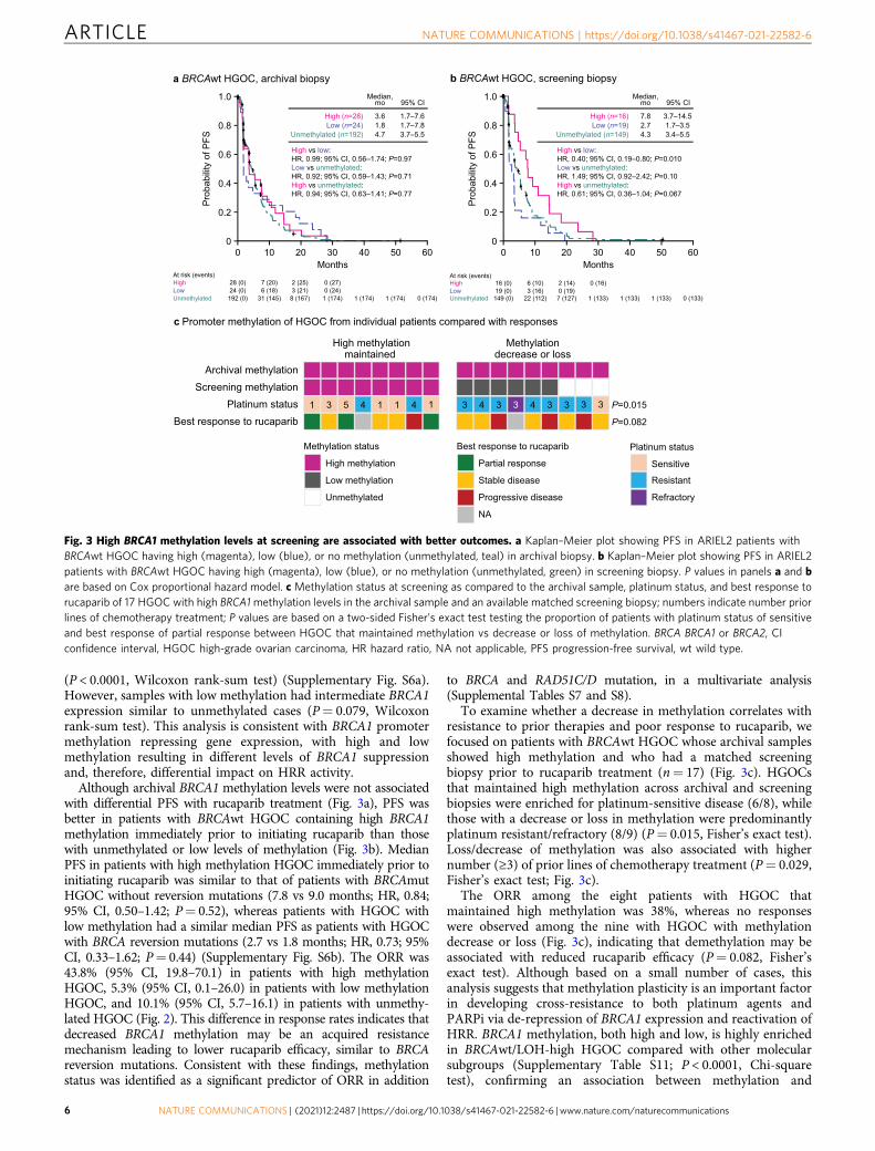
Although archival BRCA1 methylation levels were not associatedwith differential PFS with rucaparib treatment (Fig. 3a), PFS wasbetter in patients with BRCAwt HGOC containing high BRCA1methylation immediately prior to initiating rucaparib than thosewith unmethylated or low levels of methylation (Fig. 3b). MedianPFS in patients with high methylation HGOC immediately prior toinitiating rucaparib was similar to that of patients with BRCAmutHGOC without reversion mutations (7.8 vs 9.0 months; HR, 0.84;95% CI, 0.50–1.42; P= 0.52), whereas patients with HGOC withlow methylation had a similar median PFS as patients with HGOCwith BRCA reversion mutations (2.7 vs 1.8 months; HR, 0.73; 95%CI, 0.33–1.62; P= 0.44) (Supplementary Fig. S6b). The ORR was43.8% (95% CI, 19.8–70.1) in patients with high methylationHGOC, 5.3% (95% CI, 0.1–26.0) in patients with low methylationHGOC, and 10.1% (95% CI, 5.7–16.1) in patients with unmethy-lated HGOC (Fig. 2). This difference in response rates indicates thatdecreased BRCA1 methylation may be an acquired resistancemechanism leading to lower rucaparib efficacy, similar to BRCAreversion mutations. Consistent with these findings, methylationstatus was identified as a significant predictor of ORR in addition
to BRCA and RAD51C/D mutation, in a multivariate analysis(Supplemental Tables S7 and S8).
To examine whether a decrease in methylation correlates withresistance to prior therapies and poor response to rucaparib, wefocused on patients with BRCAwt HGOC whose archival samplesshowed high methylation and who had a matched screeningbiopsy prior to rucaparib treatment (n= 17) (Fig. 3c). HGOCsthat maintained high methylation across archival and screeningbiopsies were enriched for platinum-sensitive disease (6/8), whilethose with a decrease or loss in methylation were predominantlyplatinum resistant/refractory (8/9) (P= 0.015, Fisher’s exact test).Loss/decrease of methylation was also associated with highernumber (≥3) of prior lines of chemotherapy treatment (P= 0.029,Fisher’s exact test; Fig. 3c).
The ORR among the eight patients with HGOC thatmaintained high methylation was 38%, whereas no responseswere observed among the nine with HGOC with methylationdecrease or loss (Fig. 3c), indicating that demethylation may beassociated with reduced rucaparib efficacy (P= 0.082, Fisher’sexact test). Although based on a small number of cases, thisanalysis suggests that methylation plasticity is an important factorin developing cross-resistance to both platinum agents andPARPi via de-repression of BRCA1 expression and reactivation ofHRR. BRCA1 methylation, both high and low, is highly enrichedin BRCAwt/LOH-high HGOC compared with other molecularsubgroups (Supplementary Table S11; P < 0.0001, Chi-squaretest), confirming an association between methylation and
Fig. 3 High BRCA1 methylation levels at screening are associated with better outcomes. a Kaplan–Meier plot showing PFS in ARIEL2 patients withBRCAwt HGOC having high (magenta), low (blue), or no methylation (unmethylated, teal) in archival biopsy. b Kaplan–Meier plot showing PFS in ARIEL2patients with BRCAwt HGOC having high (magenta), low (blue), or no methylation (unmethylated, green) in screening biopsy. P values in panels a and bare based on Cox proportional hazard model. c Methylation status at screening as compared to the archival sample, platinum status, and best response torucaparib of 17 HGOC with high BRCA1methylation levels in the archival sample and an available matched screening biopsy; numbers indicate number priorlines of chemotherapy treatment; P values are based on a two-sided Fisher’s exact test testing the proportion of patients with platinum status of sensitiveand best response of partial response between HGOC that maintained methylation vs decrease or loss of methylation. BRCA BRCA1 or BRCA2, CIconfidence interval, HGOC high-grade ovarian carcinoma, HR hazard ratio, NA not applicable, PFS progression-free survival, wt wild type.
ARTICLE NATURE COMMUNICATIONS | https://doi.org/10.1038/s41467-021-22582-6
6 NATURE COMMUNICATIONS | (2021) 12:2487 | https://doi.org/10.1038/s41467-021-22582-6 | www.nature.com/naturecommunications
accumulation of genome scars43. In ARIEL2, 14% of BRCAwt/LOH-high HGOC (13/94) had high BRCA1 methylation atscreening, and 18% (17/94) had low BRCA1 methylation.Outcomes in the two groups were vastly different with ORRs of46.2% (95% CI, 19.2–74.9) and 5.9% (95% CI, 0.1–28.7; P=0.025, Fisher’s exact test) in patients with BRCAwt/LOH-highHGOC with high vs low screening methylation, respectively, andmedian PFS of 9.3 vs 3.3 months (HR, 0.44; 95% CI, 0.21–0.92;P= 0.015). Thus, acquired resistance by demethylation beforeinitiating rucaparib may account for a substantial fraction ofPARPi resistance among BRCAwt/LOH-high HGOC, a popula-tion expected to be PARPi responsive.
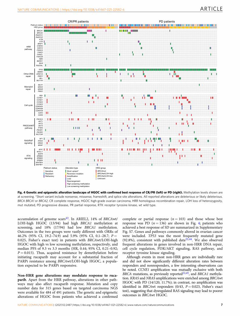
Non-HRR gene alterations may modulate response to ruca-parib. Apart from the HRR pathway, alterations in other path-ways may also affect rucaparib response. Mutation and copynumber data for 315 genes based on targeted carcinoma NGSwere available for 484 of 491 patients. The genetic and epigeneticalterations of HGOC from patients who achieved a confirmed
complete or partial response (n= 103) and those whose bestresponse was PD (n= 136) are shown in Fig. 4; patients whoachieved a best response of SD are summarized in SupplementaryFig. S7. Genes and pathways commonly altered in ovarian cancerwere included. TP53 was the most frequently mutated gene(92.8%), consistent with published data22,44. We also observedfrequent alterations in genes involved in non-HRR DNA repair,cell cycle regulation, PI3K/AKT signaling, RAS pathway, andreceptor tyrosine kinase signaling.
Although events in most non-HRR genes are individually rareand did not show significantly different alteration rates betweenresponders and nonresponders, a few interesting observations canbe noted. CCNE1 amplification was mutually exclusive with bothBRCA mutations, as previously reported22,45, and BRCA1 methyla-tion. KRAS and NRAS amplifications were enriched among BRCAwtHGOC with PD (14/120, 11.7%); in contrast, no amplification wasidentified in BRCAwt responders (0/43, P= 0.023, Fisher’s exacttest), suggesting that dysregulated RAS signaling may lead to pooreroutcomes in BRCAwt HGOC.
Fig. 4 Genetic and epigenetic alteration landscape of HGOC with confirmed best response of CR/PR (left) or PD (right). Methylation levels shown areat screening. *Short variant include nonsense, missense, frameshift, and splice site alterations. All reported alterations are deleterious or likely deleterious.BRCA BRCA1 or BRCA2, CR complete response, HGOC high-grade ovarian carcinoma, HRR homologous recombination repair, LOH loss of heterozygosity,mut mutated, PD progressive disease, PR partial response, RTK receptor tyrosine kinase, wt wild type.
NATURE COMMUNICATIONS | https://doi.org/10.1038/s41467-021-22582-6 ARTICLE
NATURE COMMUNICATIONS | (2021) 12:2487 | https://doi.org/10.1038/s41467-021-22582-6 | www.nature.com/naturecommunications 7
Several patients (n= 14) had a KRAS, NRAS, or BRAF activatingmutation in the absence of a TP53 mutation, a genetic profileassociated with low-grade serous carcinoma, mucinous carcinoma,and mesonephric-like adenocarcinoma46–48. A blinded pathologyre-review confirmed the presence of non-high-grade serous or non-grade 2/3 endometrioid histologies in 11 of the 14 cases, while allcancers with KRAS, NRAS, or BRAF mutations and a TP53alteration (n= 7) were confirmed to be high-grade serous(Supplementary Table S12 and Supplementary Fig. S8). Eleven of12 cancers with KRAS, NRAS, or BRAF activating mutations inTP53 wild-type background that could be classified into molecularsubgroups were LOH-low, consistent with the observation that lowgrade and mesonephric-like histologies are not driven by HRD49.Although none of the 14 patients achieved a clinical response torucaparib, indicating that PARPi are not likely to be active in theserare histologies, some experienced PFS longer than 200 days, anobservation that may be indicative of slower overall tumorgrowth47,50.
Amplification and overexpression in CCNE1/cell cycle andAKT pathway genes have been previously associated withplatinum resistance42,51. Among platinum-resistant/refractorypatients in ARIEL2, patients with BRCAwt HGOC andalterations (predominantly via copy number change) either inthe AKT1/2/3 genes or cell cycle pathway at screening had poorerPFS outcomes compared with other patients with BRCAwtHGOC (AKT: HR, 0.45; 95% CI, 0.24–0.85; P= 0.013; cell cycle:HR, 0.66; 95% CI, 0.44–1.00; P= 0.050; Supplementary Fig. S9),consistent with the hypothesis that certain platinum resistancemechanisms may modulate responses to other DNA-damagingagents, including PARPi.
DiscussionARIEL2 assessed the safety and efficacy of rucaparib in anunselected HGOC population. Platinum sensitivity was a strongclinical predictor of rucaparib response in this PARPi-naïvepopulation, especially in the BRCAmut and BRCAwt/LOH-highmolecular subgroups. As HRD is a common mechanism leadingto both platinum and PARPi sensitivity1, this finding suggeststhat cross-resistance mechanisms arising during platinumtherapies also impact responses to PARPi. Here, we describeHRD mechanisms leading to both platinum and rucaparib
sensitivity (BRCA mutation, RAD51C/D alterations, and highBRCA1 promoter methylation) and summarize two importantcross-resistance mechanisms: BRCA reversion mutations, whichwe have previously shown38, and loss of BRCA1 methylationdescribed here for the first time using archival and screeningclinical specimens. Other cross-resistance mechanisms toPARPi have been previously proposed, including BRCA1alternative splicing52, 53BP1 loss53,54, increased expression ofBRCA hypomorphs55–57, and ABCB1 gene fusions42,58. Thesemechanisms may also play a role in ARIEL2 but were notassessed here due to insufficient patient samples.
Alterations in RAD51C and RAD51D correlated with meaningfulclinical activity of rucaparib similar to that of BRCAmut HGOC.Therefore, we propose utilizing panels incorporating RAD51C/Dwhen considering targeted therapies. Importantly, we previouslyshowed that reversion mutations also occur in RAD51C/D as aresistance mechanism17, supporting their essential role in generat-ing synthetic lethality with PARPi.
The effect of mutations in other HRR genes on rucaparibsensitivity remains unclear given the low relative frequency ofeach gene in the ARIEL2 cohort. Although it has been hypo-thesized that ATM mutations are correlated with sensitivity toPARP inhibition, in a recent phase 3 trial, there was no survivalbenefit seen for patients with prostate cancer associated withATM mutations who were treated with olaparib vs androgen-receptor targeted therapy (HR, 0.93; 95% CI, 0.53–1.75)59. InARIEL2, no responses were observed among five patients withHGOC harboring an ATM mutation. However, three of thesepatients had platinum-resistant/refractory disease, and only oneof the remaining two was evaluable for response. Therefore, withthe limited data, the impact of ATM on rucaparib sensitivity inHGOC remains inconclusive.
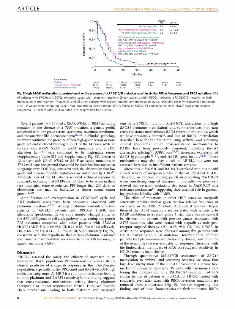
Through quantitative MS-ddPCR assessment of BRCA1methylation in archival and screening biopsies, we show thathigh-level methylation of the BRCA1 promoter is a strong bio-marker of rucaparib sensitivity. Patients with carcinomas har-boring this modification or a RAD51C/D mutation had PFSsimilar to that of patients with BRCAmut HGOC treated withrucaparib, even after cases with BRCA reversion mutations areremoved from comparison (Fig. 5). Further supporting thisfinding, each of these characteristics (methylation status, BRCA
Fig. 5 High BRCA1 methylation at pretreatment or the presence of a RAD51C/D mutation result in similar PFS as the presence of BRCA mutations. PFSof patients with BRCAmut HGOCs, excluding cases with reversion mutations (blue), patients with HGOCs harboring a RAD51C/D mutation or highmethylation at pretreatment (magenta), and all other patients with known mutation and methylation status, including cases with reversion mutations(teal). P values were computed using a Cox proportional hazard model. BRCA BRCA1 or BRCA2, CI confidence interval, HGOC high-grade ovariancarcinoma, HR hazard ratio, mut mutated, PFS progression-free survival.
ARTICLE NATURE COMMUNICATIONS | https://doi.org/10.1038/s41467-021-22582-6
8 NATURE COMMUNICATIONS | (2021) 12:2487 | https://doi.org/10.1038/s41467-021-22582-6 | www.nature.com/naturecommunications
mutation, or RAD51C/D mutation) were identified as a significantpredictor for ORR in multivariate analysis. Given strong pre-clinical data linking BRCA1 promoter methylation to HRD, andplatinum and PARPi sensitivity23,24, the previous lack of defini-tive clinical evidence associating BRCA1 methylation and therapyresponse has been perplexing60,61. The TNT trial (NCT00532727)found no association between BRCA1 methylation and responseto carboplatin vs docetaxel in patients with triple-negative breastcancer26. Discrepancies between these findings and our own mayhave both biological and technical explanations. First, most stu-dies analyze methylation in samples obtained at diagnosis, thusmissing methylation loss that may have occurred since samplecollection. Second, previous studies only report the binary pre-sence or absence of BRCA1 promoter methylation without con-sidering the levels of methylation observed. All BRCA1 alleleslikely need to be methylated to establish complete gene silencingand subsequent HRD26. Importantly, we show that methylationshould be quantitatively assessed immediately prior to treatmentto ensure high enough levels to result in sufficient promotersilencing, especially in later-line settings.
Relying on genomic scarring as evidence of HRD in later linesof treatment poses significant disadvantages. Once established,genomic scars persist and do not provide a real-time predictor ofsensitivity after multiple treatment lines. In the platinum-sensitiveARIEL2 Part 1 cases, high LOH was associated with a higher ORRamong BRCAwt HGOC. But among the more heavily pretreatedPart 2 patients, high LOH did not predict rucaparib sensitivity.Notably, all carcinomas with acquired platinum resistance (i.e.,BRCA reversion mutation and BRCA1 promoter demethylation)remained LOH-high. Thus, accumulation of genomic scarring is anirreversible process, persisting even as cancers re-acquire functionalHRR. In the QUADRA trial of the PARPi niraparib after three linesof therapy, which used the myChoice HRD test to determine HRDstatus, BRCAmut HGOC were included in the HRD group, drivingmuch of the 15% ORR62. When only considering the BRCAwt casesin QUADRA, HRD was associated with a higher ORR only forplatinum-sensitive disease (20% vs 4% for non-HRD) and was notpredictive for platinum-resistant cases (2.4% vs 3% non-HRD),similar to our findings. Therefore, although stratifying BRCAwtHGOC for PARPi treatment based on evidence of accumulatedgenomic scarring is useful early in the disease course, these tests loseutility in platinum-resistant/refractory cases.
We also examined alterations in non-HRR pathways that mayindirectly modulate response to rucaparib. Cell cycle gene altera-tions, including amplifications of cyclin E, cyclin D, and CDK4/6genes and deletions in the cell cycle inhibitors CDKN2A/B andCDKN1B, were common in BRCAwt HGOC in ARIEL2 and havebeen associated with platinum resistance22,63. AKT signalinghas also been linked to platinum resistance, and AKT inhibitionappears to reverse resistance in certain models64. In ARIEL2,platinum-resistant/refractory BRCAwt HGOC with alterations inthe cell cycle pathway and the AKT1/2/3 genes showed pooreroutcomes on rucaparib, implying that these pathways may berelevant to responses to multiple DNA-damaging agents. Furtherin vitro and in vivo studies would be essential to confirm theseclinical observations, describe the mechanistic connections betweenthese pathways and PARPi, and determine if combination thera-pies with PARPi and cell cycle or AKT inhibitors may be beneficialfor certain late-stage HGOC patients. Most of the cancers withKRAS, NRAS, or BRAF activating mutations in the absence of aTP53 mutation were subsequently found to be non-HGOC, andthe lack of response to rucaparib in these cancers indicatesthe importance of confirming histological subtype when thesemutations are detected.
Overall, our analysis highlights significant overlap betweenmolecular mechanisms resulting in platinum and PARPi
sensitivity and the extent of cross-resistance that exists betweenthese two drug classes. In addition to BRCA mutations,RAD51C/D mutations and high-level BRCA1 promotermethylation are strong predictors of sensitivity to a PARPi.Given its general tolerability and efficacy, especially in patientswith BRCAmut and BRCAwt/LOH-high HGOC, and datasupporting the use of PARPi as maintenance following primarytherapy8–10,65, we propose that administration of rucaparib,as active treatment, should be considered in earlier lines oftherapy, before the emergence of platinum resistance. Such anapproach would increase the likelihood of patients experiencingsignificant clinical benefit while maintaining the improvedquality of life associated with targeted vs systemic therapy.
MethodsStudy design and participants. ARIEL2 (CO-338-017; NCT01891344) was aninternational, multicenter, two-part, phase 2 open-label study conducted across64 sites in Australia, Canada, France, Spain, the United Kingdom, and the UnitedStates. The study design and clinical results from Part 1 were publishedpreviously37. In ARIEL2, eligible patients were aged 18 years or older with histo-logically confirmed, relapsed, high-grade serous or Grade 2 or Grade 3 endome-troid epithelial ovarian, fallopian tube, or primary peritoneal cancer. Patients wererequired to have measurable disease according to RECIST, an Eastern CooperativeOncology Group Performance Status of 0 or 1, and adequate organ function. Part 1enrolled patients with relapsed HGOC who had received at least one priorplatinum-based regimen and had platinum-sensitive disease (disease progression≥6 months after last platinum). Part 2 enrolled patients with relapsed HGOC whohad received three to four prior chemotherapies and had a treatment-free intervalof more than 6 months following first-line chemotherapy. Patients in Part 2 couldbe platinum sensitive, platinum resistant (disease progression <6 months after lastplatinum, with best response other than PD), or platinum refractory (best responseof PD on last platinum with progression-free interval <2 months). Both Part 1 andPart 2 enrolled patients regardless of their HRD status, with the exception that Part1 had a cap on the number of patients with a known germline BRCA mutation(n= 15) allowed, in order to enrich for the BRCAwt population.
Patients were ineligible if they previously received a PARPi in either thetreatment setting or as a maintenance therapy, had an active second malignancy,had central nervous system metastases, or received anticancer therapy 14 days orfewer before receiving their first dose of rucaparib.
The study was approved by national or local institutional review boards, asappropriate at each site (see Supplementary Information for full list), and wascarried out in accordance with the Declaration of Helsinki and Good ClinicalPractice Guidelines of the International Conference on Harmonization. Patientsprovided written informed consent before participation.
Procedures. Patients were treated with oral rucaparib at 600mg twice daily untildisease progression, unacceptable toxicity, or death. Supportive care (e.g., antiemeticsor analgesics for pain control) was permitted at the investigator’s discretion. Treat-ment interruptions and dose reductions (in decrements of 120mg formulation in Part1 and 100mg in Part 2) were permitted if a patient had a grade 3 or greater adverseevent. Treatment was discontinued if a dose interruption occurred for more than 14consecutive days (longer dose interruptions were permitted with sponsor approval).
Tumor response was assessed by the investigators using RECIST, withcomputed tomography scans at screening and every 8 weeks (±4 days) duringtreatment and after treatment for patients who discontinued for any reason otherthan disease progression. Patients who had been on study at least 18 months couldhave the frequency of disease assessment decreased to every 16 weeks (±2 days).Assessment continued until confirmed disease progression, death, start ofsubsequent treatment, or loss to follow-up. Serum CA-125 measures were taken atscreening, day 1 of each cycle, and the end of treatment, or when clinicallyindicated. Hematology, serum chemistry, and safety assessments were done atscreening, day 1 and day 15 of cycle 1, and day 1 of any subsequent cycles. Adverseevents were classified in accordance with the Medical Dictionary for DrugRegulatory Activities classification system version 19.1 and graded for severity inaccordance with the National Cancer Institute’s Common Terminology Criteria forAdverse Event version 4.03 (https://nciterms.nci.nih.gov/ncitbrowser/start.jsf). Part1 patients were treated and followed up until disease progression, death, ordiscontinuation of treatment due to other reasons. Part 2 patients were followed forsurvival, subsequent therapy, and secondary malignancy every 12 weeks untildeath, loss to follow-up, withdrawal of consent from study, or study closure,whichever happened first.
Molecular subgroup classification. Biomarker analysis was performed followingthe REMARK (Reporting Recommendations for Tumor Marker Prognostic Studies)guidelines66. A known HRR gene mutation was not required for enrollment inARIEL2. Patients were required to have adequate formalin-fixed paraffin-embedded
NATURE COMMUNICATIONS | https://doi.org/10.1038/s41467-021-22582-6 ARTICLE
NATURE COMMUNICATIONS | (2021) 12:2487 | https://doi.org/10.1038/s41467-021-22582-6 | www.nature.com/naturecommunications 9
(FFPE) archival tumor tissue available and to have undergone a pretreatment(screening) biopsy (optional in patients with HGOC having a known deleteriousgermline BRCA mutation in Part 2). For the ARIEL2 Part 1 and Part 2 prespecifiedanalyses, tumor BRCA status was determined centrally using the FoundationOneTM
NGS assay (Supplementary Fig. S3)37,67.Local BRCA test results were collected when available (Supplementary Fig. S3).
For our post hoc analysis of the pooled ARIEL2 patient population, we classified apatient as BRCAmut if a deleterious BRCA alteration was detected by either centraltumor testing or local testing (Supplementary Fig. S3). The majority of BRCAalterations that were detected by local testing but not by tissue testing were largerearrangements, known to be challenging to detect by NGS.
FoundationOneTM was also used to calculate the percentage of genomic LOH inarchival and screening biopsies and to identify alterations in genes other thanBRCA37,67.
To classify BRCAwt patients into LOH-high and LOH-low subgroups, we useda prespecified cutoff of 14% for patients in Part 1 and a prespecified cutoff of 18%for patients in Part 2 (Supplementary Fig. S3). For the post hoc, exploratorymolecular subgroup analyses, both the local and central test information for BRCAwas utilized. For BRCAwt, we utilized an optimized cutoff of 16%, which wasshown to be the optimal cutoff in ARIEL2 Part 1 (ref. 40) and was prospectivelyevaluated in the phase 3 ARIEL3 study (Supplementary Fig. S3)7.
BRCA1 and BRCA2 germline/somatic status was determined by a combinationof methods. Blood samples from all ARIEL2 Part 1 BRCAmut samples and a subsetof ARIEL2 Part 2 BRCAmut cases (n= 74) were analyzed using the BROCAhomologous recombination sequencing assay (University of Washington, SeattleWA, USA). Any alteration detected by FoundationOne™ NGS but not BROCA wasconsidered somatic. The remainder of the samples were assigned germline/somaticstatus based on local testing data provided by the study sites (n= 29) orcomputational inference method using the targeted NGS data (n= 26)68.Computational inference was not attempted for samples with tumor purity >80%;those samples were listed as Unknown. Nine BRCAmut cases remained withunknown germline/somatic status. Agreement between germline/somatic statusdetermined through germline sequencing (BROCA and local testing) and thecomputational inference method was very high for samples that had both dataavailable (n= 67, 95.5% agreement, kappa= 0.88, P < 0.0001, Cohen’s Kappastatistics).
HRR gene mutation subgroup was based on alterations in the genes listed inSupplementary Table S6. Germline/somatic status for ARIEL2 Part 1 non-BRCAHRR genes was determined by the BROCA homologous recombination sequencingassay (University of Washington, Seattle, WA, USA). Any alteration detected byFoundationOne™ NGS but not BROCA was considered somatic. BRCAwt ARIEL2Part 2 samples were not analyzed by central germline testing and germline/somaticstatus for these cases was determined by computational inference as describedabove68. The same computational approach was used to determine zygosity for allnon-BRCA HRR gene alterations68.
The detection of BRCA reversion mutations has been described in detailpreviously38. In short, for reversion mutations present in cell-free DNA (cfDNA),plasma samples collected from ARIEL2 patients were analyzed using theGuardant360 (ref. 69) or FoundationACT70 cfDNA assays. Central tumor tissueNGS data were also re-analyzed to determine if reversion mutations were present.BRCA reversion mutations were defined as: (i) a base substitution that changed anonsense mutation to a missense mutation, (ii) an insertion/deletion that restoredthe ORF, or (iii) a larger intragenic deletion that deleted the primary deleteriousmutation. Reversion mutations were detected in 10/112 (8.9%) BRCAmut HGOCpretreatment samples: nine by Guardant360 plasma analysis (eight previouslydescribed38 and one identified in post-publication analysis) and one by centraltumor tissue NGS analysis.
Patients were classified as having a cell cycle pathway alteration if an alterationin any of the following genes was detected in their screening cancer sample:CCNE1, CCND1, CCND2, CCND3, CDKN2A, CDKN2B, CDKN1B, CDK4, orCDK6. Patients were classified as having an AKT alteration if they had an alterationin AKT1, AKT2, or AKT3 in their screening cancer sample. Amplifications reportedwere high-copy gains (CN > 5).
Methylation analysis. The presence of methylation at the BRCA1 and RAD51Cpromoters was detected using MSP, as previously described37. In summary five10 μm sections of FFPE tissue were deparaffinized, rehydrated, and digested withProteinase K (Zymo Research, Irvine, CA, USA) overnight and bisulfite conversionof 10 μL of supernatant was performed in duplicates using the EZ DNAMethylation-Direct kit (Zymo Research). Following bisulfite conversion, thesamples underwent desulfonation and cleanup, and 2 μL of bisulfite-convertedDNA was evaluated with MSP for BRCA1 and RAD51C using primers listed inSupplementary Table S13.
Quantification of BRCA1 and RAD51C methylation levels in HGOC fromARIEL2 Part 1 and Part 2 patients was performed by quantitative MS-ddPCRmethodology as previously described25: DNA extracted from FFPE-preserved tissuesections was bisulfite converted using the EZ DNA Methylation-Lightning kit(Zymo Research). BRCA1 primers were designed for a 72 bp amplicon in theuntranslated region (UTR) (Supplementary Table S13). RAD51C methylation,primers were designed for a 142 bp amplicon in the RAD51C UTR. Minor groove
binder probes hybridizing to the fully unmethylated (2′-chloro-7′phenyl-1,4-dichloro-6-carboxyfluorescein [VIC] labeled) and the fully methylated sequences(6-carboxyfluorescein [FAM] labeled) were used. The ddPCR was performed onthe Bio-Rad QX-200 system. Methylation frequencies determined by MS-ddPCRwere normalized to neoplastic purity and BRCA1/RAD51C copy number estimatesusing the following formula: RM × (CN × TP+ (100− TP) × 2)/(TP × CN)/100,where RM is the raw fraction methylated copies detected, TP is the tumor purity,and CN is the BRCA1 copy number25. The tumor purity and copy number wereestimated based on the targeted NGS data68. Patient samples were classified ashaving high or low methylation levels based on a predefined cutoff of 70%25. In theabsence of contradicting MS-ddPCR data, biopsies with 0% BRCA1 promotermethylation by MS-ddPCR, or ones determined to be unmethylated by MSP, werelabeled as unmethylated.
BRCA1 expression analysis. For a subset of the samples analyzed for BRCA1promoter methylation status, we assessed the BRCA1 RNA expression levels byNanoString71.
Pathology analysis and histology reclassification. For all patients, hematoxylinand eosin (H&E) staining was performed following standard procedures onarchival and/or screening biopsy tissue corresponding to the tissue that wassubmitted for central NGS testing. The H&E slides for the patients harboringactivating mutations in the KRAS/NRAS/BRAF genes were re-reviewed bygynecologic pathologists (J.A.E. and D.I.L.), and a consensus diagnosis wasrendered. At time of re-review, the pathologists were blinded to the genomicfindings for these patients. Both archival and screening tissue H&E slides wereavailable for at least two patients each for mesonephric-like carcinomas, low-gradeserous carcinomas, and endometrioid adenocarcinomas, and the reclassificationwas confirmed in both samples. Representative fields were selected to describefeatures concidered for reclassification.
Statistical analysis. All analyses were performed using the safety population,which included all patients who received at least one dose of rucaparib. Theprimary endpoint in Part 1 was PFS by predefined HRD subgroups. PFS wasdefined as the number of days from the first dose of study drug to diseaseprogression by RECIST, as determined by the investigator, or death due to anycause, whichever occurred first. Patients without a documented event of progres-sion were censored on the date of their last adequate cancer assessment (i.e.,radiologic assessment) or the date of the first dose of the study drug if no cancerassessments were performed. In Part 2, the primary endpoint was ORR, defined asthe proportion of patients achieving a best response of complete or partial responseaccording to RECIST as assessed by the investigator by predefined HRD subgroups.Secondary endpoints included the proportion of patients achieving an objectiveresponse (according to RECIST and GCIG CA-125 criteria), duration of response(according to RECIST), and overall survival. Response endpoints were summarizedwith frequencies and percentage using Clopper–Pearson methodology to calculate95% CIs. Rates of response were compared with pair-wise comparison usingFisher’s exact test. The response (partial or complete response) by RECIST neededto be confirmed by a second assessment after at least 4 weeks. Duration ofconfirmed response (complete or partial response) was calculated from the initialdate a response was detected to the first date of PD. Patients without a documentedevent of progression were censored on the date of their last adequate cancerassessment (i.e., radiologic assessment) or date of response if no cancer assessmentswere performed. Overall survival was defined as the number of days from the dateof first dose of study drug to the date of death (due to any cause). Patients without aknown date of death were censored on the date the patient was last known to bealive. Duration of response, PFS, and overall survival were summarized withKaplan–Meier methodology, including median estimates and 95% CIs usinglog–log distribution. In addition, a Cox proportional hazard model was used tosummarize these endpoints and make comparisons between molecular subgroups.Here, we also present post hoc exploratory molecular biomarker analyses of PFSand ORR outcomes in order to further explore clinical benefit. Most of the post hocanalyses are based on univariate comparisons. In addition, a stepwise multivariatelogistics regression model was used to identify predictors of confirmed response(PR or CR) using baseline and molecular characteristic variables as predictors inthe model (Supplementary Tables S7 and S8). For all time-to-event analyses,the proportional hazards assumption was assessed visually with log–log plots(Supplementary Fig. S10); as the assumption of the model was valid in eachinstance, we provide HR and 95% CI for these endpoints. All P values for the posthoc exploratory analyses of molecular subgroups are presented for descriptivepurposes only.
Comparisons of proportions between methylation subgroups were summarizedusing Fisher’s exact test or chi-square tests; for the analysis of number prior lines ofchemotherapy treatment, patients were grouped in 1–2 and ≥3 prior linessubgroups. The distribution of BRCA1 gene expression levels was analyzed using anonparametric Wilcoxon rank-sum test to compare molecular subgroups.Agreement analyses were performed using non-weighted Cohen’s Kappa statistics.
Data analysis was performed using SAS v9.4 (SAS Institute, Cary, NC, USA)and Excel 2016 (Microsoft Corporation, Redmond, WA, USA).
ARTICLE NATURE COMMUNICATIONS | https://doi.org/10.1038/s41467-021-22582-6
10 NATURE COMMUNICATIONS | (2021) 12:2487 | https://doi.org/10.1038/s41467-021-22582-6 | www.nature.com/naturecommunications
Reporting summary. Further information on research design is available in the NatureResearch Reporting Summary linked to this article.
Data availabilityConsent was not obtained from patients to allow posting of the data to public repositories.Requests for de-identified datasets for the results reported in this publication will be madeavailable to qualified researchers following submission of a methodologically sound proposalto [email protected]. Data will be made available for such requests followingonline publication of this article and for 1 year thereafter in compliance with applicableprivacy laws, data protection, and requirements for consent and anonymization. Data will beprovided by Clovis Oncology. The redacted protocol for the ARIEL2 clinical study is availableon thelancet.com: https://ars.els-cdn.com/content/image/1-s2.0-S1470204516305599-mmc1.pdf. Clovis Oncology does not share identified participant data or a data dictionary.
Received: 18 December 2020; Accepted: 16 March 2021;
References1. Konstantinopoulos, P. A., Ceccaldi, R., Shapiro, G. I. & D’Andrea, A. D.
Homologous recombination deficiency: exploiting the fundamentalvulnerability of ovarian cancer. Cancer Discov. 5, 1137–1154 (2015).
2. Dal Molin, G. Z., Omatsu, K., Sood, A. K. & Coleman, R. L. Rucaparib inovarian cancer: an update on safety, efficacy and place in therapy. Ther. Adv.Med. Oncol. 10, 1758835918778483 (2018).
3. Pennington, K. P. et al. Germline and somatic mutations in homologousrecombination genes predict platinum response and survival in ovarian,fallopian tube, and peritoneal carcinomas. Clin. Cancer Res. 20, 764–775(2014).
4. Rafii, S. et al. Baseline clinical predictors of antitumor response to the PARPinhibitor olaparib in germline BRCA1/2 mutated patients with advancedovarian cancer. Oncotarget 8, 47154–47160 (2017).
5. Ledermann, J. A. et al. Overall survival in patients with platinum-sensitiverecurrent serous ovarian cancer receiving olaparib maintenance monotherapy:an updated analysis from a randomised, placebo-controlled, double-blind,phase 2 trial. Lancet Oncol. 17, 1579–1589 (2016).
6. del Campo, J. M. et al. Niraparib maintenance therapy in patients withrecurrent ovarian cancer after a partial response to the last platinum-basedchemotherapy in the ENGOT-OV16/NOVA trial. J. Clin. Oncol. 37,2968–2973 (2019).
7. Coleman, R. L. et al. Rucaparib maintenance treatment for recurrentovarian carcinoma after response to platinum therapy (ARIEL3): arandomised, double-blind, placebo-controlled, phase 3 trial. Lancet 390,1949–1961 (2017).
8. Coleman, R. L. et al. Veliparib with first-line chemotherapy and asmaintenance therapy in ovarian cancer. N. Engl. J. Med. 381, 2403–2415(2019).
9. Gonzalez-Martin, A. et al. Niraparib in patients with newly diagnosedadvanced ovarian cancer. N. Engl. J. Med. 381, 2391–2402 (2019).
10. Ray-Coquard, I. et al. Olaparib plus bevacizumab as first-line maintenance inovarian cancer. N. Engl. J. Med. 381, 2416–2428 (2019).
11. Robson, M. et al. Olaparib for metastatic breast cancer in patients with agermline BRCA mutation. N. Engl. J. Med. 377, 523–533 (2017).
12. Litton, J. K. et al. Talazoparib in patients with advanced breast cancer and agermline BRCA mutation. N. Engl. J. Med. 379, 753–763 (2018).
13. Golan, T. et al. Maintenance olaparib for germline BRCA-mutated metastaticpancreatic cancer. N. Engl. J. Med. 381, 317–327 (2019).
14. Mateo, J. et al. DNA-repair defects and olaparib in metastatic prostate cancer.N. Engl. J. Med. 373, 1697–1708 (2015).
15. de Bono, J. et al. Olaparib for metastatic castration-resistant prostate cancer.N. Engl. J. Med. 382, 2091–2102 (2020).
16. Abida, W. et al. Non-BRCA DNA damage repair gene alterations and responseto the PARP Inhibitor rucaparib in metastatic castration-resistant prostatecancer: analysis from the phase 2 TRITON2 study. Clin. Cancer Res. 28,2487–2496 (2020).
17. Kondrashova, O. et al. Secondary somatic mutations restoring RAD51C andRAD51D associated with acquired resistance to the PARP inhibitorrucaparib in high-grade ovarian carcinoma. Cancer Discov. 7, 984–998(2017).
18. Nikkila, J. et al. Heterozygous mutations in PALB2 cause DNA replication anddamage response defects. Nat. Commun. 4, 2578 (2013).
19. Kotsopoulos, J. et al. Frequency of germline PALB2 mutations among womenwith epithelial ovarian cancer. Fam. Cancer 16, 29–34 (2017).
20. Castaneda, A. et al. BRIP1 mutation does not confer sensitivity to PARPinhibition. Gynecol. Oncol. 154, 87 (2019).
21. Kawazu, M. et al. Integrative analysis of genomic alterations in triple-negativebreast cancer in association with homologous recombination deficiency. PLoSGenet. 13, e1006853 (2017).
22. Cancer Genome Atlas Research Network. Integrated genomic analyses ofovarian carcinoma. Nature 474, 609–615 (2011).
23. Ter Brugge, P. et al. Mechanisms of therapy resistance in patient-derivedxenograft models of BRCA1-deficient breast cancer. J. Natl. Cancer Inst. 108,https://doi.org/10.1093/jnci/djw148 (2016).
24. AlHilli, M. M. et al. In vivo anti-tumor activity of the PARP inhibitorniraparib in homologous recombination deficient and proficient ovariancarcinoma. Gynecol. Oncol. 143, 379–388 (2016).
25. Kondrashova, O. et al. Methylation of all BRCA1 copies predicts response tothe PARP inhibitor rucaparib in ovarian carcinoma. Nat. Commun. 9, 3970(2018).
26. Tutt, A. et al. Carboplatin in BRCA1/2-mutated and triple-negative breastcancer BRCAness subgroups: the TNT Trial. Nat. Med. 24, 628–637 (2018).
27. Lheureux, S. et al. Long-term responders on olaparib maintenance in high-grade serous ovarian cancer: clinical and molecular characterization. Clin.Cancer Res. 23, 4086–4094 (2017).
28. Abkevich, V. et al. Patterns of genomic loss of heterozygosity predicthomologous recombination repair defects in epithelial ovarian cancer. Br. J.Cancer 107, 1776–1782 (2012).
29. Davies, H. et al. HRDetect is a predictor of BRCA1 and BRCA2 deficiencybased on mutational signatures. Nat. Med. 23, 517–525 (2017).
30. Polak, P. et al. A mutational signature reveals alterations underlying deficienthomologous recombination repair in breast cancer. Nat. Genet. 49, 1476–1486(2017).
31. Nik-Zainal, S. et al. Mutational processes molding the genomes of 21 breastcancers. Cell 149, 979–993 (2012).
32. Menghi, F. et al. The tandem duplicator phenotype is a prevalent genome-wide cancer configuration driven by distinct gene mutations. Cancer Cell 34,197–210.e195 (2018).
33. Nik-Zainal, S. et al. Landscape of somatic mutations in 560 breast cancerwhole-genome sequences. Nature 534, 47 (2016).
34. Macintyre, G. et al. Copy number signatures and mutational processes inovarian carcinoma. Nat. Genet. 50, 1262–1270 (2018).
35. Telli, M. L. et al. Homologous recombination deficiency (HRD) score predictsresponse to platinum-containing neoadjuvant chemotherapy in patients withtriple-negative breast cancer. Clin. Cancer Res. 22, 3764–3773 (2016).
36. Watkins, J. A., Irshad, S., Grigoriadis, A. & Tutt, A. N. Genomic scars asbiomarkers of homologous recombination deficiency and drug response inbreast and ovarian cancers. Breast Cancer Res. 16, 211 (2014).
37. Swisher, E. M. et al. Rucaparib in relapsed, platinum-sensitive high-gradeovarian carcinoma (ARIEL2 Part 1): an international, multicentre, open-label,phase 2 trial. Lancet Oncol. 18, 75–87 (2017).
38. Lin, K. K. et al. BRCA reversion mutations in circulating tumor DNA predictprimary and acquired resistance to the PARP inhibitor rucaparib in high-grade ovarian carcinoma. Cancer Discov. 9, 210–219 (2019).
39. Oza, A. M. et al. Antitumor activity and safety of the PARP inhibitorrucaparib in patients with high-grade ovarian carcinoma and a germline orsomatic BRCA1 or BRCA2 mutation: integrated analysis of data from Study 10and ARIEL2. Gynecol. Oncol. 147, 267–275 (2017).
40. Coleman, R. L. et al. Refinement of prespecified cutoff for genomic loss ofheterozygosity (LOH) in ARIEL2 part 1: a phase II study of rucaparib inpatients (pts) with high grade ovarian carcinoma (HGOC). J. Clin. Oncol. 34,5540 (2016).
41. Goodall, J. et al. Circulating cell-free DNA to guide prostate cancer treatmentwith PARP inhibition. Cancer Discov. 7, 1006–1017 (2017).
42. Patch, A. M. et al. Whole-genome characterization of chemoresistant ovariancancer. Nature 521, 489–494 (2015).
43. Patel, J. N. et al. Characterisation of homologous recombination deficiency inpaired primary and recurrent high-grade serous ovarian cancer. Br. J. Cancer119, 1060–1066 (2018).
44. Ahmed, A. A. et al. Driver mutations in TP53 are ubiquitous in high gradeserous carcinoma of the ovary. J. Pathol. 221, 49–56 (2010).
45. Etemadmoghadam, D. et al. Synthetic lethality between CCNE1 amplificationand loss of BRCA1. Proc. Natl Acad. Sci. USA 110, 19489–19494 (2013).
46. Emmanuel, C. et al. Genomic classification of serous ovarian cancer withadjacent borderline differentiates RAS pathway and TP53-mutant tumors andidentifies NRAS as an oncogenic driver. Clin. Cancer Res. 20, 6618–6630(2014).
47. Kaldawy, A. et al. Low-grade serous ovarian cancer: a review. Gynecol. Oncol.143, 433–438 (2016).
48. Mirkovic, J. et al. Targeted genomic profiling reveals recurrent KRASmutations in mesonephric-like adenocarcinomas of the female genital tract.Am. J. Surg. Pathol. 42, 227–233 (2018).
49. Gershenson, D. M. et al. Recurrent low-grade serous ovarian carcinoma isrelatively chemoresistant. Gynecol. Oncol. 114, 48–52 (2009).
NATURE COMMUNICATIONS | https://doi.org/10.1038/s41467-021-22582-6 ARTICLE
NATURE COMMUNICATIONS | (2021) 12:2487 | https://doi.org/10.1038/s41467-021-22582-6 | www.nature.com/naturecommunications 11
50. Monk, B. J. et al. MILO/ENGOT-ov11: binimetinib versus physician’s choicechemotherapy in recurrent or persistent low-grade serous carcinomas of theovary, fallopian tube, or primary peritoneum. J. Clin. Oncol. 38, 3753–3762(2020).
51. Hahne, J. C. et al. Downregulation of AKT reverses platinumresistance of human ovarian cancers in vitro. Oncol. Rep. 28, 2023–2028(2012).
52. Wang, Y. et al. The BRCA1-delta11q alternative splice isoform bypassesgermline mutations and promotes therapeutic resistance to PARP inhibitionand cisplatin. Cancer Res. 76, 2778–2790 (2016).
53. Bunting, S. F. et al. 53BP1 inhibits homologous recombination inBrca1-deficient cells by blocking resection of DNA breaks. Cell 141, 243–254(2010).
54. Hurley, R. M. et al. 53BP1 as a potential predictor of response in PARPinhibitor-treated homologous recombination-deficient ovarian cancer.Gynecol. Oncol. 153, 127–134 (2019).
55. Drost, R. et al. BRCA1185delAG tumors may acquire therapy resistancethrough expression of RING-less BRCA1. J. Clin. Invest 126, 2903–2918(2016).
56. Park, P. H. et al. Amplification of the mutation-carrying BRCA2 allelepromotes RAD51 loading and PARP inhibitor resistance in the absence ofreversion mutations. Mol. Cancer Ther. 19, 602–613 (2020).
57. Johnson, N. et al. Stabilization of mutant BRCA1 protein confers PARPinhibitor and platinum resistance. Proc. Natl Acad. Sci. USA 110,17041–17046 (2013).
58. Christie, E. L. et al. Multiple ABCB1 transcriptional fusions in drugresistant high-grade serous ovarian and breast cancer. Nat. Commun. 10, 1295(2019).
59. Hussain, M. et al. Survival with olaparib in metastatic castration-resistantprostate cancer. N. Engl. J. Med. 383, 2345–2357 (2020).
60. Ignatov, T. et al. BRCA1 promoter methylation is a marker of better responseto anthracycline-based therapy in sporadic TNBC. Breast Cancer Res. Treat.141, 205–212 (2013).
61. Bernards, S. S. et al. Clinical characteristics and outcomes of patients withBRCA1 or RAD51C methylated versus mutated ovarian carcinoma. Gynecol.Oncol. 148, 281–285 (2018).
62. Moore, K. N. et al. Niraparib monotherapy for late-line treatment of ovariancancer (QUADRA): a multicentre, open-label, single-arm, phase 2 trial. LancetOncol. 20, 636–648 (2019).
63. Noel, E. E. et al. The association of CCND1 overexpression and cisplatinresistance in testicular germ cell tumors and other cancers. Am. J. Pathol. 176,2607–2615 (2010).
64. Whicker, M. E., Lin, Z. P., Hanna, R., Sartorelli, A. C. & Ratner, E. S. MK-2206sensitizes BRCA-deficient epithelial ovarian adenocarcinoma to cisplatin andolaparib. BMC Cancer 16, 550 (2016).
65. Moore, K. et al. Maintenance olaparib in patients with newly diagnosedadvanced ovarian cancer. N. Engl. J. Med. 379, 2495–2505 (2018).
66. McShane, L. M. et al. REporting recommendations for tumour MARKerprognostic studies (REMARK). Br. J. Cancer 93, 387–391 (2005).
67. Frampton, G. M. et al. Development and validation of a clinical cancergenomic profiling test based on massively parallel DNA sequencing. Nat.Biotechnol. 31, 1023–1031 (2013).
68. Sun, J. X. et al. A computational approach to distinguish somatic vs. germlineorigin of genomic alterations from deep sequencing of cancer specimenswithout a matched normal. PLoS Comput Biol. 14, e1005965 (2018).
69. Lanman, R. B. et al. Analytical and clinical validation of a digital sequencingpanel for quantitative, highly accurate evaluation of cell-free circulating tumorDNA. PLoS ONE 10, e0140712 (2015).
70. Clark, T. A. et al. Analytical validation of a hybrid capture–based next-generation sequencing clinical assay for genomic profiling of cell-freecirculating tumor DNA. J. Mol. Diagn. 20, 686–702 (2018).
71. Talhouk, A. et al. Single-patient molecular testing with NanoString nCounterdata using a reference-based strategy for batch effect correction. PLoS ONE 11,e0153844 (2016).
AcknowledgementsFunding from the National Breast Cancer Foundation of Australia (to A.D.) and theDepartment of Defense Ovarian Cancer Research Program (OC12056, to E.M.S. andS.H.K.) and a Stand Up To Cancer-Ovarian Cancer Research Fund Alliance-NationalOvarian Cancer Coalition Dream Team Translational Cancer Research Grant (SU2C-AACR-DT16-15, to E.M.S. and S.H.K.). Stand Up To Cancer is a division of theEntertainment Industry Foundation. Research grants are administered by the AmericanAssociation for Cancer Research, the Scientific Partner of SU2C. We thank Elaina Mann,Jennifer Borrow, Jen Lenore, Jeff Isaacson, and Lindsey Rolfe for clinical development,statistical guidance, and operational support of the ARIEL2 study. In addition, we thankVivian Chen and Mary Joyce for assistance in manuscript preparation. ARIEL2 wasdesigned by the funder and a subgroup of investigators. Data presented and herein werecollected by the funder; the funder and all authors interpreted and analyzed the data.
This article was written by the authors, with medical writing and copy editing supportpaid for by the funder and provided by Nathan Yardley and Frederique H. Evans ofAshfield MedComms, an Ashfield Health company. All authors had full access to all trialdata and had final responsibility for the decision to submit for publication.
Author contributionsJ.A.E. and D.I.L. conducted pathologist re-review of histology type of tumors withKRAS/NRAS/BRAF mutations with no TP53 mutations. E.M.S., C.L.S., K.K.L., and I.A.M.were involved in the study conception. E.M.S., S.H.K., C.L.S., K.K.L., and I.A.M. wereinvolved in the study design. E.M.S. and S.H.K. acquired funding. E.M.S., K.K.L., andI.A.M. were involved in the protocol development. E.M.S., T.T.K., K.K.L., and I.A.M.co-wrote the first draft of the manuscript. E.M.S., A.M.O, A.V.T., I.R.-C., A.O., R.L.C., C.A., G.E.K., D.M.O., A.L., D.P., S.W., L.-m.C., A.E.W.H., L. Ma, P.G., R.S.K., O.D., A.M.,S.H.K., S.K.C., A.D., C.L.S., and I.A.M. treated patients, acquired data, and obtainedsamples. E.M.S., T.T.K., I.R.C., J.A.E., D.I.L., C.L.S., K.K.L., and I.A.M. interpreted thedata. E.M.S., T.T.K., J.A.E., D.I.L., and K.K.L. analyzed data. E.M.S., T.T.K., A.M.O.,A.V.T., I.R.C., A.O., R.L.C., C.A., G.E.K., D.M.O., A.L., D.P., S.W., L.-m.C., A.E.W.H.,L.Ma, P.G., R.S.K., O.D., A.M., S.H.K., J.A.E., D.I.L, S.K.C., E.D., L.-T.V., S.G., L.Maloney, H.G., T.H., A.D., C.L.S., K.K.L., and I.A.M. contributed to manuscript revisionsand approved the final draft for submission.
Competing interestsE.M.S. has received funding for clinical trials from Clovis Oncology and TESARO(paid to her institution). T.T.K., E.D., L.-T.V., S.G., L. Maloney, H.G., T.H., and K.K.L.are employees of Clovis Oncology and may own stock or have stock options in thatcompany. A.M.O. has served on advisory boards for Clovis Oncology, Amgen,Immunovaccine, and Verastem; received support for travel or accommodation fromAstraZeneca; and received honoraria from WebRx. A.V.T. has served on an advisoryboard for and received grants from AstraZeneca. I.R.-C. has served on advisory boardsfor Clovis Oncology, AstraZeneca, Genmab/Seattle Genetics, ImmunoGen, MerckSharpe Dohme, PharmaMar, Roche, and Tesaro/GlaxoSmithKline and received sup-port for travel or accommodation from AstraZeneca, GlaxoSmithKline, and Roche.A.O. has served on advisory boards for Clovis Oncology, AstraZeneca, ImmunoGen,Genmab/Seattle Genetics, PharmaMar, Roche, and Tesaro and received support fortravel or accommodation from AstraZeneca, PharmaMar, Roche, and Tesaro. R.L.C.reports grants from Clovis Oncology, AbbVie, AstraZeneca, Esperance, Janssen,Merck, Millennium, OncoMed, and Roche/Genentech and has served as an advisor toClovis Oncology, AbbVie, AstraZeneca, Bayer, Esperance, GamaMabs, Genmab,Gradalis, Janssen, Millennium, Merck, OncoMed, Pfizer, Roche/Genentech, andTesaro. C.A. has served on steering committees for Clovis Oncology and MateonTherapeutics; served on advisory boards for Clovis Oncology, Bayer, CeruleanPharma, Tesaro, and VentiRx, and received honoraria from Clovis Oncology, Bayer,Cerulean Pharma, Mateon Therapeutics, Tesaro, and VentiRx. G.E.K. has receivedresearch funding (to the University of California outside the scope of this work)from Lilly, Merck, and Pfizer, and received honorarium from Clovis Oncology,AstraZeneca, and Tesaro. D.M.O. has served on advisory boards for ClovisOncology, AstraZeneca, Gynecologic Oncology Group, Janssen, Myriad, and Tesaro;has served on steering committees for Clovis Oncology, Amgen, and ImmunoGen;has served as a consultant to AbbVie, Ambry, AstraZeneca, Health Analytics, andTesaro, and his institution has received research support from Clovis Oncology,Agenus, Ajinomoto, Array BioPharma, AstraZeneca, Bristol-Myers Squibb,ERGOMED Clinical Research, Exelixis, Genentech, GlaxoSmithKline, GynecologicOncology Group, ImmunoGen, INC Research, inVentiv Health Clinical, JanssenResearch and Development, Ludwig Institute for Cancer Research, Novartis Phar-maceuticals, PRA International, Regeneron Pharmaceuticals, Serono, Stemcentrx,Tesaro, and TRACON Pharmaceuticals. A.L. has served on advisory boards for ClovisOncology, Ability, AstraZeneca, Biocad, Merck Serono, MSD, Pfizer, PharmaMar,Seattle Genetics, and Tesaro PharmaMar; reports institutional research grant supportfrom GamaMabs, Inivata, Merus, and Sanofi, and reports boarding and travel expensesfor congress activities from Clovis Oncology, AstraZeneca, Roche, and Tesaro. D.P.has served on advisory boards for AstraZeneca and GlaxoSmithKline, and receivedsupport for travel from AstraZeneca. S.W. has served on advisory boards for Astra-Zeneca and Roche; reports institutional research support from Clovis Oncology,AstraZeneca, Merck, Regeneron, and Tesaro, and has received honoraria fromAstraZeneca. P.G. has served on an advisory board for AstraZeneca, GSK, andMerck, and received support for travel expenses for congress activities from Astra-Zeneca. R.S.K. has served on advisory boards for Clovis Oncology, Roche, and Tesaro.O.D. has served on advisory boards for Clovis Oncology, IMV, Tesaro, and Merck,and has served on the speaker bureau for AstraZeneca and Tesaro. J.A.E. and D.I.L.are employees of Foundation Medicine, Inc., which is a wholly-owned subsidiary ofRoche, and may own stock or have stock options in Roche. A.D. has served onadvisory boards for AstraZeneca Australia and has received travel support from Bio-Rad. C.L.S. has served on advisory boards for Clovis Oncology, AstraZeneca,Takeda, Roche Australia, MSD, Eisai Co., has received travel support from AstraZe-neca, has received in kind research support from Clovis Oncology, Roche, Beigene andFunded research support from Sierra oncology and Eisai Co. I.A.M. has served onadvisory boards for Clovis Oncology, AstraZeneca, Roche, Takeda, and Tesaro and
ARTICLE NATURE COMMUNICATIONS | https://doi.org/10.1038/s41467-021-22582-6
12 NATURE COMMUNICATIONS | (2021) 12:2487 | https://doi.org/10.1038/s41467-021-22582-6 | www.nature.com/naturecommunications

receives institutional funding from AstraZeneca. The remaining authors declare nocompeting interests.
Additional informationSupplementary information The online version contains supplementary materialavailable at https://doi.org/10.1038/s41467-021-22582-6.
Correspondence and requests for materials should be addressed to E.M.S.
Peer review information Nature Communications thanks the anonymous reviewer(s) fortheir contribution to the peer review of this work.
Reprints and permission information is available at http://www.nature.com/reprints
Publisher’s note Springer Nature remains neutral with regard to jurisdictional claims inpublished maps and institutional affiliations.
Open Access This article is licensed under a Creative CommonsAttribution 4.0 International License, which permits use, sharing,
adaptation, distribution and reproduction in any medium or format, as long as you giveappropriate credit to the original author(s) and the source, provide a link to the CreativeCommons license, and indicate if changes were made. The images or other third partymaterial in this article are included in the article’s Creative Commons license, unlessindicated otherwise in a credit line to the material. If material is not included in thearticle’s Creative Commons license and your intended use is not permitted by statutoryregulation or exceeds the permitted use, you will need to obtain permission directly fromthe copyright holder. To view a copy of this license, visit http://creativecommons.org/licenses/by/4.0/.
© The Author(s) 2021
NATURE COMMUNICATIONS | https://doi.org/10.1038/s41467-021-22582-6 ARTICLE
NATURE COMMUNICATIONS | (2021) 12:2487 | https://doi.org/10.1038/s41467-021-22582-6 | www.nature.com/naturecommunications 13