proteomic analysis of the balance between survival and cell death responses in cisplatin-mediated...
TRANSCRIPT
Proteomic Analysis of the Balance between Survival and CellDeath Responses in Cisplatin-Mediated Ototoxicity
Samson Jamesdaniel†, Dalian Ding†, Mohammad Habiby Kermany†, Bruce A. Davidson‡,Paul R. Knight III§,∥, Richard Salvi†, and Donald E. Coling*,†Center for Hearing and Deafness, Departments of Anesthesiology, Pathology, and Microbiologyand Immunology, University at Buffalo, the State University of New York, Buffalo, New York 14214,and Department of Anesthesiology, Buffalo Veterans Administration Medical Center, Buffalo, NewYork 14215
AbstractCisplatin, a widely used anticancer drug, preferentially damages outer hair cells (OHCs) of the innerear. In this study, an antibody microarray was used to identify early changes in protein expressionin the rat cochlea induced by cisplatin. Only small changes in hearing thresholds (4−34 dB elevation)were detected two days after cisplatin treatment (12 mg/kg). OHC function, measured by otoacousticemissions, was slightly depressed (10 dB), and little or no receptor cell loss was observed. However,cisplatin induced large changes in the expression of 19 proteins involved in apoptosis, cell survival,or progression through the cell cycle. Fifteen of the proteins are novel to the study of the inner ear.Immunoblotting confirmed increases in the levels of the pro-survival activating transcription factor2 (ATF2), of pro-apoptotic serine-threonine protein kinase, receptor interacting protein, and a 70/75kDa nitrotyrosine bearing doublet of unknown function. Anti-nitrotyrosine antibodies localized theseoxidatively damaged proteins to the stereocilia of OHCs, the Golgi-centrosome region of Hensen'scells, nuclei of outer pillar cells, and tunnel crossing fibers innervating OHCs. The results of thisproteomic analysis reflect the commencement of ototoxic and cell survival responses before theobservation of a significant functional or anatomical loss.
Keywordsantibody microarray; cisplatin; cochlea; oxidative protein damage; protein tyrosine nitration
IntroductionThe platinum atom of cisplatin forms covalent bonds with DNA at the N7 positions of purinebases to form intrastrand and interstrand cross-links. Platinated DNA immunoreactivity hasbeen localized to the nuclei of outer hair cells and cells in the stria vascularis and spiral ligament.1 Cytotoxic effects of cisplatin have been suggested to occur primarily through apoptosis athigher doses and through DNA damage at lower doses.2 However, augmented pro-apoptoticsignaling has been demonstrated in the cochlea within 48 h of treatment with relatively lowdoses of cisplatin.3 Cisplatin targets three areas in the cochlea: the hair cells in the basal turn
* To whom correspondence should be addressed. Dr. Donald E. Coling, 137 Cary Hall, Center for Hearing and Deafness, University atBuffalo, State University of New York, Buffalo, NY 14214. Phone: 716 829 2001 ext 31. Fax: 716 829 2980. E-mail:[email protected]..†Center for Hearing and Deafness.‡Departments of Anesthesiology and Pathology.§Buffalo Veterans Administration Medical Center.∥Departments of Anesthesiology, Microbiology and Immunology.
NIH Public AccessAuthor ManuscriptJ Proteome Res. Author manuscript; available in PMC 2009 August 1.
Published in final edited form as:J Proteome Res. 2008 August ; 7(8): 3516–3524. doi:10.1021/pr8002479.
NIH
-PA Author Manuscript
NIH
-PA Author Manuscript
NIH
-PA Author Manuscript

of organ of Corti,4 the lateral wall tissues,5,6 and the spiral ganglion cells.1 Cisplatin-inducedgeneration of reactive oxygen species, including the superoxide anion7 is one of the importantfactors leading to cell death. Alternate death pathways implicate transcription factors, like NF-κB and high mobility group protein.8
Genomic studies, including DNA arrays, have also been used to investigate cochlear mRNAexpression in cisplatin-induced hearing loss. TNF-α was immunolocalized to the spiralligament, spiral limbus, and the organ of Corti, while mRNA expression increased in HEI-OCIcells treated with cisplatin.9 Cisplatin treatment induced kidney injury molecule-1 mRNAexpression and increased NOX-3 mRNA in rat cochlea and hair cell lines derived from mouse.10 Immunocytochemistry also revealed very strong expression of NF-κB p65 in cells of organof Corti, spiral ligament, and stria vascularis where cisplatin-induced TUNEL-positive stainingwas observed.9
However, mRNA often poorly correlates with protein expression either due to its degradationor inefficient translation.11 The proteome also differs from cell to cell and constantly changesthrough its biochemical interactions in response to stimuli. Therefore, proteomic analysis ofearly changes in cisplatin-induced ototoxicity is key to understanding the correspondingfunctional state of the cell, as well as the mechanisms associated with hearing loss. Thesimultaneous analysis of multiple proteins involved in cellular survival or apoptosis willidentify the pathogenic cellular pathways responsible for the evolution of ototoxicity associatedwith cisplatin treatment.
This study is the first to report the use of antibody microar-rays, a powerful new tool, forsimultaneously studying a multitude of cochlear proteins that change expression after cisplatintreatment. Antibody microarrays directly assay protein expressions and have been validatedas a reliable tool for identifyingandanalyzingproteinprofilesinbiologicalsystems.12,13 Themicroarray used for our studies was spotted with 725 antibodies against proteins from a broadspectrum of signaling pathways. Changes in expression of 19 proteins, repeatable in two strainsof rats, identified many novel ototoxic and cell survival responses before the observation of asignificant functional or anatomical loss.
Experimental Design and MethodsAnimals
Male Wistar and female Sprague—Dawley rats were obtained from Charles River Laboratories(Wilmington, MA). The experimental protocol was reviewed and approved by the Universityat Buffalo Institutional Animal Care and Use Committee. The animals were housed andmaintained in a temperature-controlled room with a 12-h light/dark cycle and allowed freeaccess to food and water.
Cisplatin AdministrationCisplatin (Sigma Aldrich Chemical Co., St. Louis, MO) was administered in a single dose of12 mg/kg body weight by slow intraperitoneal infusion of 1 mg/ mL in sterile saline (0.9%) at10 mL/hr. Control animals were infused with an equal volume of saline. All of the animalswere hydrated with 5 mL of subcutaneous injection of saline twice a day until they weresacrificed 48 h after cisplatin administration.
Physiological MeasurementsAuditory brainstem response (ABR) thresholds and distortion product otoacoustic emission(DPOAE) at 2f1-f2 were measured for each animal before and after cisplatin administration.The animals were anesthetized with isoflurane (4% induction, 1.5% maintenance with 1 L/min
Jamesdaniel et al. Page 2
J Proteome Res. Author manuscript; available in PMC 2009 August 1.
NIH
-PA Author Manuscript
NIH
-PA Author Manuscript
NIH
-PA Author Manuscript
O2) for ABR and DPOAE measurements. Subcutaneous differential needle electrodes wereplaced at the vertex (noninverting), below the test ear (inverting), and below the contralateralear (ground). The sound stimuli, 1 ms tone bursts (2.5, 5, 10, 20, or 40 kHz, 0.5 ms rise-falltimes) or 25 μs clicks, were generated using Tucker Davis Technologies (TDT, Alachua, FL)SigGen software and TDT hardware consisting of a real-time processor, programmableattenuator, electrostatic speaker driver, and electrostatic pressure field speaker. The stimuliwere presented to the external auditory meatus and the sound intensity varied in 5 dB intervalsfrom above to below threshold. Two hundred stimulus presentations, delivered at 21/s, wereaveraged using a TDT real-time processor controlled by BioSig software (TDT) to obtain theABR. Hearing threshold was defined as the lowest intensity of stimulation that yielded arepeatable waveform with identifiable peaks in the ABR waveform.
DPOAE stimuli were elicited with two primary tones f1 and f2 at an f2/f1 ratio of 1.2. The levelof f1 and f2 were set at L2 ) L1 + 10 dB. L1 level was varied from 70 to 25 dB SPL in 5 dBincrements. Two IHS-3738 high-frequency transducers (Intelligent Hearing System, Miami,FL, USA) were used to deliver f1 and f2 to the ear via separate flexible tubes connected to aprobe inserted into the ear canal. Sound pressure levels were measured at the cubic differencefrequency (2f1-f2) using an ER10B+ probe microphone (Etymotics Research, Inc., Elk GroveVillage, IL) and hardware and software from Smart Distortion Product Otoacoustic EmissionSystem version 4.53 (IHS). The output of the microphone was sampled at 40 kHz over a periodof 204 ms; the spectrum of each sweep was computed and averaged over 32 nonrejectedsweeps. The noise floor was measured in a 24 Hz band surrounding 2f1-f2 and f2 was variedfrom 1 to 16 kHz.
CochleogramAnimals were anesthetized by CO2 inspiration until there was no response to a toe pinch anddecapitated, and the temporal bones were quickly removed. The round and oval windows wereopened, and 4% paraformaldehyde in 0.1 M phosphate buffer (pH 7.2, 4 °C) was slowlyperfused through the round window for approximately one minute; then the cochlea wasimmersed in fixative for 3 h. Afterward, the specimens were stained with Harris' hematoxylinsolution. The organ of Corti was dissected and mounted as a flat surface preparation in glycerinon a glass slide. Specimens were examined under a light microscope (400×) and the numberof missing inner hair cells (IHC) and OHC were counted over 0.24 mm intervals along thelength of the cochlea. Cochleograms showing the percent hair cell loss as a function of percentdistance from the apex were constructed for each animal.14
Antibody MicroarrayCochlear protein samples from control and cisplatin injected rats were prepared 48 h aftercisplatin injection so that the protein expression in the cochlea would reflect an early evolvingstage of cisplatin-induced ototoxicity. Rats were anesthetized by CO2 inspiration, anddecapitated. Whole cochlea samples dissected from 4 cochleae, including the bony shell, lateralwall, basilar membrane, and modiolus, were homogenized with 150 μL lysis buffersupplemented with protease and phosphatase inhibitors, each provided with the “PanoramaAntibody Microarray - XPRESS Profiler 725 kit” (Sigma-Aldrich Corporation, St. Louis, MO).The protein concentration of the sample was determined using the BioRad (Bio-Rad, Hercules,CA) Bradford assay.15 Fluorescent dyes (Cy3 and Cy5 - GE Healthcare, Buckinghamshire,UK) were prepared as 800 μM stock solutions in 0.1 M carbonate/bicarbonate buffer, pH 9.3,at room temperature. Cochlear proteins from the samples were labeled using an N-hydroxysuccinimide ester linkage to lysines by adding 7.5 μL stock dye solution per 150 μgprotein adjusted to 1 mg/mL. Unlabeled dyes were removed by gel filtration using spin columnsprovided in the kit. Dye incorporation was determined spectrophotometrically at 552 nm (Cy3)and 650 nm (Cy5) using the molar extinction coefficient of 0.15 μM−1 cm−1 at 552 nm for Cy3
Jamesdaniel et al. Page 3
J Proteome Res. Author manuscript; available in PMC 2009 August 1.
NIH
-PA Author Manuscript
NIH
-PA Author Manuscript
NIH
-PA Author Manuscript

and 0.25 μM−1 cm−1 at 650 nm for Cy5. To optimize the signal-to-noise ratio, labeling wasrepeated as necessary to achieve a molar dye/protein ratio >2. Dye/protein molar ratio wascalculated assuming an average molecular weight of 60 kDa. To control for differences inlabeling stoichiometry, a dye-swapping paradigm16 was used. For each experiment, two slidesspotted with antibodies were incubated with labeled protein samples for 0.5 h each at roomtemperature. The first slide was incubated with Cy3-labeled control sample and Cy5-labeledcisplatin-treated sample. A second identical slide was incubated with the opposite labelingscheme, that is, Cy5-labeled control sample and Cy3-labeled cisplatin-treated sample.Fluorescent signal intensities from the binding of Cy3- or Cy5-labeled protein were thenrecorded for each antibody spot using a GenePix Professional 4200A Microarray Scanner(Molecular Devices Corporation, Sunnyvale, CA). All antibody spots that showed a negativesignal-to-noise ratio were deleted from further analysis. The data obtained was normalized bycalculating the geometric mean of 4 background-corrected fluorescence intensities fromduplicate antibody spots on each slide. The ratio of fluorescent intensity of cisplatin-treatedsamples to that of control samples (fold change induced by cisplatin) was calculated using thenormalized fluorescent intensities.
Western BlotChanges in protein level were quantified using Western blotting methods.17 Proteins wereseparated on 4−12% gradient NuPage gels (Invitrogen, Carlsbad, CA), transferred topolyvinylidene difluoride membranes, blocked with 0.1% I-Block (Applied Biosystems, FosterCity, CA), and probed with antibodies of interest using chemiluminescence detection (PierceChemical Co., Rockford, IL). A Fuji model LAS 1000 imaging system (Stamford, CT) wasused to visualize gel bands. Background corrected bands (NIH Image J software) werenormalized against bands obtained by stripping the membrane with 25 mM glycine (BioRad),pH 2.0, 1% lauryl sulfate and reprobing with an antibody against actin. Actin was used as ahousekeeping protein because cisplatin induced a change in the level of the widely usedhousekeeping enzyme, glyceraldehyde 3-phosphate dehydrogenase (GAPDH).
ImmunocytochemistryLocalization of nitrotyrosine in the cochlea, 48 h after cisplatin treatment was done byimmunocytochemistry using confocal microscopy.17 Cochlear tissue was fixed in 10%buffered formalin for 1 h, and the organ of Corti was permeabilised in PBS + 1% (v/v) TritonX-100 for 30 min. Then the tissue was placed in blocking solution (5% v/v goat serum, 2% w/v BSA in PBS) for 1 h and incubated overnight at 4 °C in primary antibody (1:400), followedby incubation with the secondary antibody at room temperature for 1 h. The tissue was thencolabeled with phalloidin that labels f-actin and TOPRO-3 that stains nuclei. The stainedspecimens were mounted on slides with ProLong Gold antifade reagent (P36934, InvitrogenMolecular Probes) and examined using the Carl Zeiss Laser Scanning Systems LSM 510.Images were captured and analyzed with Zeiss LSM Image Examiner (version 4,0,0,91, CarlZeiss GmbH Jena). The cellular and subcellular distribution was assessed by using 3D analysisof the organ of Corti. Secondary antibodies conjugated to Alexa Fluor 488 were obtained fromInvitrogen - Molecular Probes (Carlsbad, CA).
ResultsMeasures of Early Stage Ototoxicity
A goal of proteomics is to identify protein markers at an early stage in pathogenesis as potentialdrug targets for therapeutic intervention. A 12 mg/kg dose of cisplatin for rats will lead tosubstantial hearing loss within a weeks time.10 At the 48 h time point, physiological andmorphological measures indicated that pathogenesis was still at an early stage.
Jamesdaniel et al. Page 4
J Proteome Res. Author manuscript; available in PMC 2009 August 1.
NIH
-PA Author Manuscript
NIH
-PA Author Manuscript
NIH
-PA Author Manuscript

ABR for stimuli at 2.5, 5, 10, 20, 40 kHz and for clicks revealed threshold shifts ranging from10 to 34 dB for Wistar rats and 4 to 12 dB for Sprague—Dawley rats (Figure 1). In both strains,the level of cisplatin-induced hearing loss was greater at the higher (40 kHz) and lower (2.5kHz) frequencies, whereas a lesser degree of functional loss was sustained in the midfrequencies (10, 20 kHz). These changes are likely to be irreversible, as similar studies withlesser dosage of cisplatin (5 mg/kg) have shown significant shift in hearing threshold at 3, 7,30, and 90 days after cisplatin treatment.18 DPOAE amplitude decreased approximately 10dB at 8 and 16 kHz (Figure 2) with no change at lower frequencies (not shown).
Cochleograms indicated minimal or no hair cell loss in regions sensitive to mid frequencies inboth Wistar and Sprague—Dawley rats and minimal loss of outer hair cells at the base andapex (Figure 3). The small sizes of the lesions reflect a very early stage of cochlea pathology.
Protein Expression ProfileAnalysis by antibody microarrays resulted in the detection of 581 proteins in Wistar rats and626 proteins in Sprague—Dawley rats. Figure 4 shows the antibody array #1 labeled with Cy3for the control tissues and Cy5 for the cisplatin treated tissues (left panel). The dye-swappedarray #2 was labeled with Cy5 for the control tissues and Cy3 for the cisplatin treated tissues(right panel). The insets in the center panel show higher resolution images of respectivesegments of each array. Dye swapping minimizes bias due to labeling with two dyes. Thepseudocolor images depict the ratio of fluorescence intensities of protein in control vsexperimental samples.
Approximately 80% of the 725 antibodies on the arrays were detected for both strains of rats;302/309 proteins exhibited a ≥1.1 fold increase, 214/261 exhibited a ≥0.9 fold decrease, and65/56 remained unaltered (fold change was 1.0, e.g., actin) in Wistar/Sprague−Dawley rats(Figure 5). Levels of 15 proteins increased in both strains by ≥1.5 fold, whereas 4 proteinsdecreased by ≥0.6 fold (Table 1). Cisplatin-induced changes in 10 proteins were associatedwith a survival response (increased expression of ATF2, JAB1, Mdm2, Rsk1, SUMO-1,myosin VI, p21WAF1Cip1, PRMT4, and reelin and decreased expression of active caspase3). Increased expression of 4 proteins (Tal, Granzyme B, SLIPR/MAGI3, RIP) and decreasedexpression of 3 proteins (EGF - epidermal growth factor, p35, ubiquitin C-terminal hydrolaseL1) were linked to cell death responses. However, it is unclear if two cytoskeletal proteins,centrin and neurofilament 68, are associated with either survival or apoptosis responses.
ValidationImmunoblot analysis of cisplatin-treated rats confirmed the increased expression of ATF2,RIP, and nitrotyrosine (Figure 6). In contrast, actin levels remained constant in agreement withmicroarray results (1.0 ± 0.2 fold change for combined Wistar and Sprague—Dawley data).We used actin as a normalizing protein, rather than GAPDH, a typical housekeeping enzyme,because cisplatin induced a change in GAPDH levels in both microarray and immunoblotexperiments. Surprisingly, the anti-nitrotyrosine immunoblot was dominated by only twoprotein bands at 70 and 75 kDa.
Immunocytochemistry studies were carried out to localize the expression of nitrotyrosine inthe sensory epithelium. Immunofluorescent labeling of nitrotyrosine was present in some, butnot all, stereocilia (S), in Hensen's cells (H), in outer pillar cell nuclei (P), as well as in tunnelcrossing fibers (Figure 7F). Low-level labeling was observed in some, but not all, Deiters' celland inner pillar cell nuclei. Stereocilia labeling was not due to nonspecific binding of secondaryantibody because it was not observed in control specimen labeled with an unrelated primaryantibody (Figure 7D). Nitrotyrosine labeling in Hensen's cells was focal and appeared to be inthe Golgicentrosome perinuclear region (Figure 7, panel A and B, red perinuclear foci).
Jamesdaniel et al. Page 5
J Proteome Res. Author manuscript; available in PMC 2009 August 1.
NIH
-PA Author Manuscript
NIH
-PA Author Manuscript
NIH
-PA Author Manuscript

DiscussionThis study is the first to use a high-throughput antibody microarray to identify changes inprotein expression levels in the inner ear. The proteomic analysis of cisplatin ototoxicityrevealed the involvement of several novel proteins not previously reported in studies of eithernormal or pathological hearing. Cisplatin-induced strong expression changes in 19 proteinsthat either increased by ≥1.5 or decreased by ≥0.6 fold in both Wistar and Sprague—Dawleyrats (Table 1). Only 4 of these proteins have been reported in previous studies in the cochlea,myosin VI,19 neurofilament 68,20 caspase 3,21 and epidermal growth factor.22 Fifteenproteins have heretofore never been identified in cisplatin-induced ototoxicity. Review of theliterature indicates that the cisplatin-induced changes in expression of 10 proteins areassociated with a survival response, 7 indicate a cell death response, and 2 proteins have anundetermined role.
Of the 10 proteins that reflect a survival response, 5 are related to the p53 signaling pathway.p21, myosin VI, and Mdm2 are transcriptionally regulated by p53. p21 is a member of the cip/kip family of cyclin kinase inhibitors that function in cell cycle arrest. It acts as an anti-apoptoticand growth-promoting protein.23 Myosin VI also functions in p53 mediated cell survivalassociated with Golgi integrity.24 It is a minus-end-directed motor, abundant in hair cells andessential for development and maintenance of stereocilia.19 The Mdm2 (murine doubleminute) oncogene represses p53 transcriptional activity by binding to and blocking the N-terminal trans-activation domain of p53. It also acts as an E3 ubiquitin ligase targeting p53 bypoly ubiquitination for exit from the nucleus and degradation by the 26S proteasome.25Additionally, Mdm2 can initiate the intrinsic apoptotic pathway, because monoubiquitinationsignals transport p53 to the mitochondria.26 Like Mdm2, JAB1 (jun activation domain-bindingprotein 1) also functions as a nuclear export mediator in the degradation of p53.27 SUMOs(small ubiquitin-related modifiers) are reversible post-translational protein modifiers thatchange the localization, activity, and stability of the protein to which they are covalently bound.28 Increased expression of SUMO-1 48 h following cisplatin administration is consistent withan anti-apoptotic sumoylation that enhances Mdm2's ability to ubiquitnate p53.
Cisplatin-induced changes in 5 other proteins, not apparently linked to p53, also indicate asurvival response. Expression levels increased for 4 survival-associated proteins (ATF2, Rsk1,PRMT4, and reelin). ATF2 is a member of the ATF/cAMP response element-binding (CREB)protein family. DNA damage is one of the classic inducers of ATF2 transcriptional activity.29 ATF2 is implicated in the activation of a large set of genes important in drug resistance andin the regulation of ER stress regulatory protein Grp78.30 Rsk1 is a member of the p90ribosomal S6 kinase (Rsk) family which activates ATF/CREB family transcription factors andthe transcriptional coactivator CREB-binding protein.31 PRMT4 (Protein arginine N-methyltransferase-4) is a promoter-specific regulator of NF-κB recruitment to chromatin incell survival responses and also plays a key role in RNA transport and splicing.32 Reelin is anextracellular matrix protein that activates a survival response through p35/cyclin-dependentkinase 5 (Cdk5) and inactivates an apoptotic response through the Src-tyrosine kinase familymember Fyn.33 Expression levels decreased for active caspase 3, a major effector in neuronalapoptosis triggered by various stimuli,34 whereas Bcl-2 remained unaltered. However, amarked increment in active caspase 3 accompanied by a decrement in Bcl-2 has been reportedwith a lesser dosage (5 mg/kg) of cisplatin.3 It is not clear whether the dose difference causesthis discrepancy, even though an in vitro study that compared the effects of a higher and lowerdosage of cisplatin, reported a decline in caspase 3 activity with the higher dosage.35 Thisunexpected complexity must be resolved in future experiments, as the time point chosen forthe study may also have a critical role in determining the delicate balance between survival orapoptotic responses.
Jamesdaniel et al. Page 6
J Proteome Res. Author manuscript; available in PMC 2009 August 1.
NIH
-PA Author Manuscript
NIH
-PA Author Manuscript
NIH
-PA Author Manuscript

In contrast to changes promoting cell survival, increases observed in granzyme B, Tal, SLIPR/MAGI3, and RIP suggest an apoptotic response. Granzymes are structurally related serineproteases. Granzyme B is responsible for the rapid activation of pro-apoptotic protein Bid. Itcan induce cyto-chrome c release by cleavage and inactivation of the anti-apoptotic Bcl-2family member Mcl-1.36 Apoptosis from granzyme B mediated cytochrome c release can alsoproceed in a caspase-independent manner.37 It is interesting to speculate that such a responsemight be related to the cisplatin-induced decrease in active caspase 3. Tal (Tsg101-associatedligase) polyubiquitinates the tumor susceptibility gene 101 (Tsg101) product resulting inproteasomal degradation. Tsg 101 is essential for cell survival,38 endosomal sorting,membrane receptor degradation, and the final stages of cytokinesis.39 SLIPR/MAGI3 is amembrane-associated guanylate kinase protein, which localizes transmembrane proteins tospecific sites. SLIPR/MAGI3 interacts with protein phosphatase PTEN, a tumor suppressor,to antagonize the survival activity of protein kinase B/Akt.40 RIP (receptor interacting protein)is a 74 kDa Ser/Thr kinase. It interacts with other regulatory proteins in a signaling scaffoldassociated with the tumor necrosis factor receptor, which is capable of signaling either theinduction of apoptosis, through the activation of caspase 8, or the anti-apoptotic NF-κBpathway. It is a cell death domain adapter protein that can bind to the adapter proteins TRADD,RAID (CRADD), and TRAF2.41
The decrease in the expression of EGF, p35, and Ubiquitin C-terminal hydrolase L1 alsoindicates a cisplatin-induced cellular death response. EGF receptor activity can activate theextracellular signal-regulated kinase/mitogen-activated protein kinase cascade, whichfunctions in organogenesis and in tissue homeostasis.42 p35 is an activator of Cdk5, whichdisplays kinase activity in postmitotic neurons. Dysregulation of Cdk5 has been implicated inneurodegeneration.43 The expression of p35 is induced in differentiated neurons and isenhanced by extracellular stimuli such as neurotrophic factors or extra-cellular matrixmolecules.44 A decrease of p35 concomitant with increased levels of reelin would not beexpected33 and indicate a more complex inter-relationship than might have been predicted.Ubiquitin C-terminal hydrolase L1 is a member of the ubiquitin carboxy-terminal hydrolasefamily of deubiquitinating enzymes. It is a multifunctional protein in neurons that canhydrolyze bonds between ubiquitin and substrate proteins, thereby reversing the functionalstate of the substrate.45
Cytoskeletal proteins centrin and neurofilament 68 also increased, but their role in cisplatinototoxicity is unclear. Centrins are members of the EF-hand family of Ca2+-binding proteins.46 It has been suggested that Cen3 participates in centrosome reproduction and duplication,whereas Cen1/Cen2 play a role in centriole separation preceding centrosome duplication duringthe cell cycle.47 Cen2 also stimulates nucleotide excision repair and might therefore promotesurvival.48 Neurofilament 68 is one of the five major subunits of intermediate filamentsexpressed in neurons. Cisplatin upregulates the expression of NEFL encoding this 68 kDaneurofilament protein.49 During axonal growth, neurofilament subunits are incorporated allalong the axon in a dynamic process. Imbalances in subunit stoichiometry have been implicatedin the induction of neurodegeneration characterized by neurofilamentous aggregates.50
Nitrotyrosine expression indicates cisplatin-induced post-translational oxidative modification.Cisplatin-induced expression of nitrotyrosine (1.4−2.1 fold change) was observed by antibodymicroarray and confirmed by immunoblotting. Even though nitrotyrosine did not meet thecriterion of >1.5 fold increase for both strains, investigation of its expression was carried tothe level of subcellular localization before other proteins because nitration of tyrosine residuesresulting from the activity of either peroxynitrite or nitrogen dioxide is known to producecatastrophic effects on protein function. For example, nitration of tyrosine blocks the abilityof protein tyrosine kinases to activate certain transcription factors.51 Several nitrated proteinshave been reported in other systems,52 but none have been reported in the cochlea so far. The
Jamesdaniel et al. Page 7
J Proteome Res. Author manuscript; available in PMC 2009 August 1.
NIH
-PA Author Manuscript
NIH
-PA Author Manuscript
NIH
-PA Author Manuscript

strong expression of the 70/75 kDa protein(s) found in immunoblotting, as well as localizationof nitrotyrosine in the stereocilia of OHC, supporting cells, and tunnel crossing fibers, reflect,to our knowledge, the first report of evidence of stress-induced oxidative damage to cochlearproteins.
ConclusionIdentification of several novel proteins involved in cell death or survival responses during thisearly stage of ototoxicity indicates that multiplex microarrays are a sensitive and revealing toolfor understanding the molecular pathogenesis of cisplatin-induced hearing loss. These resultsilluminate the delicate balance between early survival and apoptotic responses in cisplatinototoxicity that take place before significant functional changes occur. Therapeuticinterventions that target proteins associated with the onset of ototoxicity, such as thoseidentified here, are likely to be far more effective than those that attempt to block the laterstages of cell death.
AcknowledgmentWe acknowledge support from the National Organization for Hearing Research Foundation (DC), Deafness ResearchFoundation (DC), and NIH (R01DC006630, RS).
References1. van Ruijven MW, de Groot JC, Klis SF, Smoorenburg GF. The cochlear targets of cisplatin: an
electrophysiological and morphological time-sequence study. Hear. Res 2005;205(1—2):241–8.[PubMed: 15953532]
2. Berndtsson M, Hagg M, Panaretakis T, Havelka AM, Shoshan MC, Linder S. Acute apoptosis bycisplatin requires induction of reactive oxygen species but is not associated with damage to nuclearDNA. Int. J. Cancer 2007;120(1):175–80. [PubMed: 17044026]
3. Garcia-Berrocal JR, Nevado J, Ramirez-Camacho R, Sanz R, Gonzalez-Garcia JA, Sanchez-RodriguezC, Cantos B, Espana P, Verdaguer JM, Trinidad Cabezas A. The anticancer drug cisplatin induces anintrinsic apoptotic pathway inside the inner ear. Br. J. Pharmacol 2007;152(7):1012–20. [PubMed:17906689]
4. Anniko M, Sobin A. Cisplatin: evaluation of its ototoxic potential. Am. J. Otolaryngol 1986;7(4):276–93. [PubMed: 3752388]
5. Meech RP, Campbell KC, Hughes LP, Rybak LP. A semiquantitative analysis of the effects of cisplatinon the rat stria vascularis. Hear. Res 1998;124(1—2):44–59. [PubMed: 9822901]
6. Ravi R, Somani SM, Rybak LP. Mechanism of cisplatin ototoxicity: antioxidant system. Pharmacol.Toxicol 1995;76(6):386–94. [PubMed: 7479581]
7. Dehne N, Lautermann J, Petrat F, Rauen U, de Groot H. Cisplatin ototoxicity: involvement of iron andenhanced formation of superoxide anion radicals. Toxicol. Appl. Pharmacol 2001;174(1):27–34.[PubMed: 11437646]
8. Rybak LP, Whitworth CA, Mukherjea D, Ramkumar V. Mechanisms of cisplatin-induced ototoxicityand prevention. Hear. Res 2007;226(1—2):157–67. [PubMed: 17113254]
9. So H, Kim H, Lee JH, Park C, Kim Y, Kim E, Kim JK, Yun KJ, Lee KM, Lee HY, Moon SK, LimDJ, Park R. Cisplatin cytotoxicity of auditory cells requires secretions of proinflammatory cytokinesvia activation of ERK and NF-kappaB. J. Assoc. Res. Otolaryngol 2007;8(3):338–55. [PubMed:17516123]
10. Mukherjea D, Whitworth CA, Nandish S, Dunaway GA, Rybak LP, Ramkumar V. Expression of thekidney injury molecule 1 in the rat cochlea and induction by cisplatin. Neuroscience 2006;139(2):733–40. [PubMed: 16464536]
11. Gygi SP, Rist B, Gerber SA, Turecek F, Gelb MH, Aebersold R. Quantitative analysis of complexprotein mixtures using isotope-coded affinity tags. Nat. Biotechnol 1999;17(10):994–9. [PubMed:10504701]
Jamesdaniel et al. Page 8
J Proteome Res. Author manuscript; available in PMC 2009 August 1.
NIH
-PA Author Manuscript
NIH
-PA Author Manuscript
NIH
-PA Author Manuscript

12. Kopf E, Shnitzer D, Zharhary D. Panorama Ab Microarray Cell Signaling kit: a unique tool for proteinexpression analysis. Proteomics 2005;5(9):2412–6. [PubMed: 15880767]
13. Smith L, Watson MB, O'Kane SL, Drew PJ, Lind MJ, Cawkwell L. The analysis of doxorubicinresistance in human breast cancer cells using antibody microarrays. Mol. Cancer Ther 2006;5(8):2115–20. [PubMed: 16928833]
14. Ding, D.; McFadden, S.; Salvi, RJ. Cochlear hair cell densities and inner ear staining techniques.. In:Willott, JF., editor. The Auditory Psychobiology of the Mouse. CRC Press: Boca Raton; FL: 2001.p. 189-204.
15. Bradford MM. A rapid and sensitive method for the quantitation of microgram quantities of proteinutilizing the principle of protein-dye binding. Anal. Biochem 1976;72:248–54. [PubMed: 942051]
16. Coling DE, Ding D, Young R, Lis M, Stofko E, Blumenthal KM, Salvi RJ. Proteomic analysis ofcisplatin-induced cochlear damage: methods and early changes in protein expression. Hear. Res2007;226(1—2):140–56. [PubMed: 17321087]
17. Coling DE, Espreafico EM, Kachar B. Cellular distribution of myosin-V in the guinea pig cochlea.J. Neurocytol 1997;26(2):113–20. [PubMed: 9181485]
18. Ramirez-Camacho R, Fernandez DE, Verdaguer JM, Gomez MM, Trinidad A, Garcia-Berrocal JR,Corvillo MA. Cisplatin-induced hearing loss does not correlate with intracellular platinumconcentration. Acta Otolaryngol 2008;128(5):505–9. [PubMed: 18421602]
19. Avraham KB, Hasson T, Steel KP, Kingsley DM, Russell LB, Mooseker MS, Copeland NG, JenkinsNA. The mouse Snell's waltzer deafness gene encodes an unconventional myosin required forstructural integrity of inner ear hair cells. Nat. Genet 1995;11(4):369–75. [PubMed: 7493015]
20. Dau J, Wenthold RJ. Immunocytochemical localization of neurofilament subunits in the spiralganglion of normal and neomycin-treated guinea pigs. Hear. Res 1989;42(2—3):253–63. [PubMed:2606806]
21. Hu BH, Henderson D, Nicotera TM. Involvement of apoptosis in progression of cochlear lesionfollowing exposure to intense noise. Hear. Res 2002;166(1—2):62–71. [PubMed: 12062759]
22. Malgrange B, Rogister B, Lefebvre PP, Mazy-Servais C, Welcher AA, Bonnet C, Hsu RY, Rigo JM,Van De Water TR, Moonen G. Expression of growth factors and their receptors in the postnatal ratcochlea. Neurochem. Res 1998;23(8):1133–8. [PubMed: 9704604]
23. Rossig L, Jadidi AS, Urbich C, Badorff C, Zeiher AM, Dimmeler S. Akt-dependent phosphorylationof p21(Cip1) regulates PCNA binding and proliferation of endothelial cells. Mol. Cell. Biol 2001;21(16):5644–57. [PubMed: 11463845]
24. Jung EJ, Liu G, Zhou W, Chen X. Myosin VI is a mediator of the p53-dependent cell survival pathway.Mol. Cell. Biol 2006;26(6):2175–86. [PubMed: 16507995]
25. Coates PJ. p53 and Mdm2: not all cells are equal. J. Pathol 2007;213(4):357–9. [PubMed: 17973240]26. Haupt S, Berger M, Goldberg Z, Haupt Y. Apoptosis - the p53 network. J. Cell Sci 2003;116(Pt 20):
4077–85. [PubMed: 12972501]27. Lee EW, Oh W, Song J. Jab1 as a mediator of nuclear export and cytoplasmic degradation of p53.
Mol. Cells 2006;22(2):133–40. [PubMed: 17085963]28. Geiss-Friedlander R, Melchior F. Concepts in sumoylation: a decade on. Nat. Rev. Mol. Cell. Biol
2007;8(12):947–56. [PubMed: 18000527]29. van Dam H, Wilhelm D, Herr I, Steffen A, Herrlich P, Angel P. ATF-2 is preferentially activated by
stress-activated protein kinases to mediate c-jun induction in response to genotoxic agents. EMBOJ 1995;14(8):1798–811. [PubMed: 7737130]
30. Bhoumik A, Lopez-Bergami P, Ronai Z. ATF2 on the double - activating transcription factor andDNA damage response protein. Pigment Cell Res 2007;20(6):498–506. [PubMed: 17935492]
31. Richards SA, Fu J, Romanelli A, Shimamura A, Blenis J. Ribosomal S6 kinase 1 (RSK1) activationrequires signals dependent on and independent of the MAP kinase ERK. Curr. Biol 1999;9(15):810–20. [PubMed: 10469565]
32. Covic M, Hassa PO, Saccani S, Buerki C, Meier NI, Lombardi C, Imhof R, Bedford MT, Natoli G,Hottiger MO. Arginine methyltransferase CARM1 is a promoter-specific regulator of NF-kappaB-dependent gene expression. EMBO J 2005;24(1):85–96. [PubMed: 15616592]
33. Fatemi SH. Reelin glycoprotein: structure, biology and roles in health and disease. Mol. Psychiatry2005;10(3):251–7. [PubMed: 15583703]
Jamesdaniel et al. Page 9
J Proteome Res. Author manuscript; available in PMC 2009 August 1.
NIH
-PA Author Manuscript
NIH
-PA Author Manuscript
NIH
-PA Author Manuscript

34. Yakovlev AG, Faden AI. Caspase-dependent apoptotic pathways in CNS injury. Mol. Neurobiol2001;24(1—3):131–44. [PubMed: 11831549]
35. Lieberthal W, Triaca V, Levine J. Mechanisms of death induced by cisplatin in proximal tubularepithelial cells: apoptosis vs. necrosis. Am. J. Physiol 1996;270(4 Pt 2):F700–8. [PubMed: 8967349]
36. Bots M, Medema JP. Granzymes at a glance. J. Cell Sci 2006;119(Pt 24):5011–4. [PubMed:17158907]
37. Heibein JA, Barry M, Motyka B, Bleackley RC. Granzyme B-induced loss of mitochondrial innermembrane potential (Delta Psi m) and cytochrome c release are caspase independent. J. Immunol1999;163(9):4683–93. [PubMed: 10528165]
38. Wagner KU, Krempler A, Qi Y, Park K, Henry MD, Triplett AA, Riedlinger G, Rucker IE,Hennighausen L. Tsg101 is essential for cell growth, proliferation, and cell survival of embryonicand adult tissues. Mol. Cell. Biol 2003;23(1):150–62. [PubMed: 12482969]
39. Amit I, Yakir L, Katz M, Zwang Y, Marmor MD, Citri A, Shtiegman K, Alroy I, Tuvia S, Reiss Y,Roubini E, Cohen M, Wides R, Bacharach E, Schubert U, Yarden Y. Tal, a Tsg101-specific E3ubiquitin ligase, regulates receptor endocytosis and retrovirus budding. Genes Dev 2004;18(14):1737–52. [PubMed: 15256501]
40. Wu Y, Dowbenko D, Spencer S, Laura R, Lee J, Gu Q, Lasky LA. Interaction of the tumor suppressorPTEN/MMAC with a PDZ domain of MAGI3, a novel membrane-associated guanylate kinase. J.Biol. Chem 2000;275(28):21477–85. [PubMed: 10748157]
41. Meylan E, Tschopp J. The RIP kinases: crucial integrators of cellular stress. Trends Biochem. Sci2005;30(3):151–9. [PubMed: 15752987]
42. Hsieh M, Conti M. G-protein-coupled receptor signaling and the EGF network in endocrine systems.Trends Endocrinol. Metab 2005;16(7):320–6. [PubMed: 16054836]
43. Dhavan R, Tsai LH. A decade of CDK5. Nat. Rev. Mol. Cell. Biol 2001;2(10):749–59. [PubMed:11584302]
44. Kesavapany S, Zheng YL, Amin N, Pant HC. Peptides derived from Cdk5 activator p35, specificallyinhibit deregulated activity of Cdk5. Biotechnol. J 2007;2(8):978–87. [PubMed: 17526058]
45. Setsuie R, Wada K. The functions of UCH-L1 and its relation to neurodegenerative diseases.Neurochem. Int 2007;51(2—4):105–11. [PubMed: 17586089]
46. Coling DE, Salisbury JL. Characterization of the calcium-binding contractile protein centrin fromTetraselmis striata (Pleurastrophyceae). J. Protozool 1992;39(3):385–91. [PubMed: 1640386]
47. Park JH, Pulvermuller A, Scheerer P, Rausch S, Giessl A, Hohne W, Wolfrum U, Hofmann KP, ErnstOP, Choe HW, Krauss N. Insights into functional aspects of centrins from the structure of N-terminally extended mouse centrin 1. Vision Res 2006;46(27):4568–74. [PubMed: 17027898]
48. Nishi R, Okuda Y, Watanabe E, Mori T, Iwai S, Masutani C, Sugasawa K, Hanaoka F. Centrin 2stimulates nucleotide excision repair by interacting with xeroderma pigmentosum group C protein.Mol. Cell. Biol 2005;25(13):5664–74. [PubMed: 15964821]
49. Kerley-Hamilton JS, Pike AM, Li N, DiRenzo J, Spinella MJ. A p53-dominant transcriptionalresponse to cisplatin in testicular germ cell tumor-derived human embryonal carcinoma. Oncogene2005;24(40):6090–100. [PubMed: 15940259]
50. Thyagarajan A, Strong MJ, Szaro BG. Post-transcriptional control of neurofilaments in developmentand disease. Exp. Cell Res 2007;313(10):2088–97. [PubMed: 17428473]
51. Kong SK, Yim MB, Stadtman ER, Chock PB. Peroxynitrite disables the tyrosine phosphorylationregulatory mechanism: Lymphocyte-specific tyrosine kinase fails to phosphorylate nitrated cdc2(6−20)NH2 peptide. Proc. Natl. Acad. Sci. U.S.A 1996;93(8):3377–82. [PubMed: 8622943]
52. Peluffo G, Radi R. Biochemistry of protein tyrosine nitration in cardiovascular pathology. Cardiovasc.Res 2007;75(2):291–302. [PubMed: 17544386]
Jamesdaniel et al. Page 10
J Proteome Res. Author manuscript; available in PMC 2009 August 1.
NIH
-PA Author Manuscript
NIH
-PA Author Manuscript
NIH
-PA Author Manuscript

Figure 1.Hearing threshold after cisplatin treatment. The ABR recorded before and 48 h after cisplatintreatment shows threshold shifts ranging from 10−34 dB in (A) Wistar and 4−12 dB in (B)Sprague—Dawley rats at 2.5, 5, 10, 20, 40 kHz and clicks. Measurements were done for n =6 rats for the pretreatment group and n = 3 for the cisplatin treated group. Results are expressedas mean ± standard deviation (SD).
Jamesdaniel et al. Page 11
J Proteome Res. Author manuscript; available in PMC 2009 August 1.
NIH
-PA Author Manuscript
NIH
-PA Author Manuscript
NIH
-PA Author Manuscript

Figure 2.Cisplatin-induced DPOAE changes in Wistar rats. DPOAEs showed a decrease ofapproximately 10 dB at 8 and 16 kHz in both (A) Wistar and (B) Sprague—Dawley rats 48 hafter cisplatin treatment. The pre-NF and post-cis-NF traces indicate the noise floor in DPOAErecordings before and after treatment with cisplatin. The measurements were done in n = 6 ratsfor the pretreatment group and n = 3 for the cisplatin treated group. The results are expressedas mean ± SD.
Jamesdaniel et al. Page 12
J Proteome Res. Author manuscript; available in PMC 2009 August 1.
NIH
-PA Author Manuscript
NIH
-PA Author Manuscript
NIH
-PA Author Manuscript
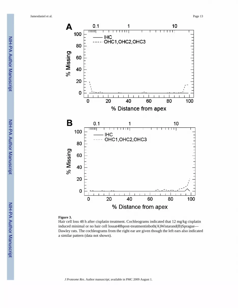
Figure 3.Hair cell loss 48 h after cisplatin treatment. Cochleograms indicated that 12 mg/kg cisplatininduced minimal or no hair cell lossat48hpost-treatmentinboth(A)Wistarand(B)Sprague—Dawley rats. The cochleograms from the right ear are given though the left ears also indicateda similar pattern (data not shown).
Jamesdaniel et al. Page 13
J Proteome Res. Author manuscript; available in PMC 2009 August 1.
NIH
-PA Author Manuscript
NIH
-PA Author Manuscript
NIH
-PA Author Manuscript

Figure 4.Antibody microarray of cisplatin-induced changes in cochlea. For array # 1, control proteinsfrom Sprague—Dawley rats were labeled with Cy3 and proteins from cisplatin-treated ratswith Cy5. For array # 2, the labeling dyes were swapped (controls labeled with Cy5 andcisplatin-treated with Cy3). Green spots indicate Cy5-labeled proteins outnumber Cy3-labeledproteins. Red spots indicate Cy-3 labeled proteins dominate. Similar results were obtained forsamples from Wistar rats (figure not shown).
Jamesdaniel et al. Page 14
J Proteome Res. Author manuscript; available in PMC 2009 August 1.
NIH
-PA Author Manuscript
NIH
-PA Author Manuscript
NIH
-PA Author Manuscript

Figure 5.Cochlear protein expression profile after cisplatin treatment. (A) Wistar and (B) Sprague—Dawley: plots of number of proteins (ordinate) versus fold changes in protein expression.Values greater than 1 indicate an increase in protein level in cisplatin treated group versuscontrol. Values less than 1 reflect a decrease in protein expression. The pattern of distributionin terms of the number of proteins that increased or decreased after cisplatin treatment wassimilar in both strains. Cut-off lines at fold changes of 0.6 and 1.5 indicate the selection criteriafor further protein analysis.
Jamesdaniel et al. Page 15
J Proteome Res. Author manuscript; available in PMC 2009 August 1.
NIH
-PA Author Manuscript
NIH
-PA Author Manuscript
NIH
-PA Author Manuscript

Figure 6.Validation of protein expression by immunoblotting; 60 μg of protein from Sprague—Dawleycochleae were loaded in each lane for control or cisplatin-treated rats. Immunoblotting resultsindicate cisplatin-dependent increases in the expression of (A) ATF2, (B) RIP, and (C)nitrotyrosine, whereas (D) shows the expression of actin. Numbers give apparent molecularweight in kDa.
Jamesdaniel et al. Page 16
J Proteome Res. Author manuscript; available in PMC 2009 August 1.
NIH
-PA Author Manuscript
NIH
-PA Author Manuscript
NIH
-PA Author Manuscript

Figure 7.Cisplatin-induced localization of nitrotyrosine in cochlea. Images, obtained using confocalmicroscopy, indicate the presence of nitrotyrosine in stereocilia (S), Hensen's cells (H), outerpillar cell nuclei (P), and tunnel crossing fibers (F). Sprague—Dawley sensory epithelium waslabeled with anti-nitrotyrosine, 48 h after cisplatin treatment. Red staining indicatesnitrotyrosine, green indicates f-actin, and blue indicates nuclei. The confocal sections are atthe level of (A) the stereocilia, (B) outer hair cell nuclei, and (C) Deiters' and pillar cell nuclei.The negative control (D), labeled with a different primary, rules out nonspecific labeling ofthe stereocilia by the secondary antibody.
Jamesdaniel et al. Page 17
J Proteome Res. Author manuscript; available in PMC 2009 August 1.
NIH
-PA Author Manuscript
NIH
-PA Author Manuscript
NIH
-PA Author Manuscript

NIH
-PA Author Manuscript
NIH
-PA Author Manuscript
NIH
-PA Author Manuscript
Jamesdaniel et al. Page 18Ta
ble
1Pr
otei
ns w
ith M
ajor
Cha
nges
afte
r Cis
plat
in T
reat
men
t
func
tion
antib
ody
mic
roar
ray
fold
cha
nge
prot
ein
apop
tosi
ssu
rviv
alce
ll cy
cle/
cyto
skel
eton
Wis
tar
SDa
mea
n
ATF
2Y
1.6
1.5
1.6
JAB
1Y
1.6
1.6
1.6
Tal
Y1.
51.
71.
6M
dm2
Y1.
71.
51.
6G
ranz
yme
BY
1.8
1.6
1.7
Rsk
1Y
1.7
1.7
1.7
Myo
sin
VI
Y1.
51.
91.
7SU
MO
-1Y
1.9
1.5
1.7
p21W
AF1
Cip
1Y
Y1.
61.
91.
7PR
MT4
Y1.
81.
71.
7SL
IPR
MA
GI3
Y1.
52.
11.
8R
eelin
Y2.
11.
61.
8R
ecep
tor I
nter
actin
g Pr
otei
nY
Y2
22.
0C
entri
nY
1.8
2.4
2.1
Neu
rofil
amen
t 68
Y1.
82.
42.
1C
aspa
se 3
Act
ive
Y0.
60.
30.
4Ep
ider
mal
Gro
wth
Fac
tor
Y0.
50.
50.
5p3
5 C
dk5
Reg
ulat
orY
0.3
0.6
0.5
Ubi
quiti
n C
term
inal
Hyd
rola
se L
1Y
0.6
0.6
0.6
Nitr
otyr
osin
ebY
1.4
2.1
1.8
a Cis
plat
in in
duce
d ch
ange
s in
19 p
rote
ins t
hat i
ncre
ased
by ≥1
.5 fo
ld o
r dec
reas
ed b
y ≥0
.6 fo
ld in
bot
h W
ista
r and
Spr
ague
-Daw
ley
(SD
).
b Even
thou
gh n
itrot
yros
ine
did
not m
eet t
he se
lect
ion
crite
ria fo
r the
tabl
e, it
was
incl
uded
due
to it
s fun
ctio
nal s
igni
fican
ce in
cis
plat
in-in
duce
d ot
otox
icity
.
J Proteome Res. Author manuscript; available in PMC 2009 August 1.