preventive effect of muscone against cisplatin nephrotoxicity
TRANSCRIPT
biomolecules
Article
Preventive Effect of Muscone against CisplatinNephrotoxicity in LLC-PK1 Cells
Hung Manh Phung 1,† , Sullim Lee 2,† , Ji Hye Hwang 3,* and Ki Sung Kang 1,*1 College of Korean Medicine, Gachon University, Seongnam 13120, Korea; [email protected] Department of Life Science, College of Bio-Nano Technology, Gachon University, Seongnam 13120, Korea;
[email protected] Department of Acupuncture and Moxibustion Medicine, College of Korean Medicine, Gachon University,
Seongnam 13120, Korea* Correspondence: [email protected] (J.H.H.); [email protected] (K.S.K.)† These authors contributed equally to this work.
Received: 1 September 2020; Accepted: 12 October 2020; Published: 15 October 2020�����������������
Abstract: Cisplatin, one of the most common antitumor agents, is widely applied to treat variouscancerous diseases and is included in the World Health Organization Model List of Essential Medicines.Cisplatin therapy is used to treat 10–20% of all cancerous cases, and its cure rate is especially high intesticular cancer (over 90%). However, a major side effect of this anticancer drug is nephrotoxicity,limiting treatment effect and reducing the quality of life in cancer patients. Muscone, an odoriferousconstituent of musk, was confirmed to inhibit cisplatin-induced LLC-PK1 kidney proximal tubulecell death in a dose-dependent manner. In term of renal protective mechanism, muscone inhibitedcisplatin oxidative toxicity by decreasing reactive oxygen species (ROS) level and stimulating HO-1expression. Muscone also exerted anti-inflammation effect through inhibition of p38 phosphorylation.Furthermore, muscone mitigated cisplatin-induced apoptosis in LLC-PK1 cells via both intrinsic andextrinsic pathways by inhibiting pro-apoptotic protein Bax expression, and cleaved caspase-3, 7, and 8;and increase of anti-apoptotic protein Bcl-2 level. In addition, the anti-apoptotic effect of musconealso was enhanced by preventing p53 expression and its phosphorylation. Our study showed thatmuscone may be a potential protective agent against cisplatin-induced nephrotoxicity.
Keywords: muscone; cisplatin; nephrotoxicity; acute kidney injury; LLC-PK1; chromatin condensation;apoptosis; oxidative stress; inflammation; p53
1. Introduction
Cisplatin, known as cis-diamminedichloroplatinum, is one of the most common antineoplasticagents used to treat various cancerous diseases, including ovarian, breast, testicular, brain, lung,and bladder cancer, and is included in the World Health Organization Model List of EssentialMedicines. Cisplatin inhibits carcinogenesis of malignant cells through the generation of intra- andinterstrand cross-links on DNA, which block DNA synthesis and replication [1–4]. The curing rate ofcisplatin is particularly high in testicular cancer (over 90%) [5] and 10–20% of all cancerous cases areprescribed cisplatin-based chemotherapy [6].
Although cisplatin is considered a cornerstone of cancer therapy, its clinical effectiveness is limitedowing to adverse reactions in normal tissues. As a primary organ involved in the excretion of cisplatin,the accumulation of cisplatin in the kidney is greater than that in other organs [7]. Thus, renal toxicityis considered one of the biggest challenges in cisplatin-based chemotherapy besides other commonside effects such as gastrointestinal toxicity, ototoxicity, neurotoxicity, and hematological toxicity.Approximately one-third of patients undergoing cisplatin treatment show symptoms of acute kidney
Biomolecules 2020, 10, 1444; doi:10.3390/biom10101444 www.mdpi.com/journal/biomolecules
Biomolecules 2020, 10, 1444 2 of 11
injury (AKI), including higher serum creatinine, lower glomerular filtration rate, and decreased serumpotassium and magnesium concentrations after 10-day drug administration [8].
The nephrotoxic mechanism of cisplatin remains unknown despite decades of research. Recent studieshave indicated that cisplatin induces renal failure by triggering complex intracellular signaling pathways.In brief, after cisplatin is transported through high-affinity copper uptake protein 1 (CTR1), organiccation transporters (OCTs), or passive diffusion via the cell membrane, it triggers a series of signalingpathways including those affecting the balance of cdk2 and p21, activation of mitogen-activatedprotein kinase (MAPK), tumor necrosis factor-α, tumor protein p53, nuclear factor-κB, mitochondrialdysfunction, DNA damage, reactive oxygen species (ROS) generation, and formation of toxicmetabolites. These upstream changes result in apoptosis or inflammation downstream, whichleads to renal cell death and AKI [8].
Although cisplatin therapy presents risks for kidneys and other organs, the clinical effectivenessof cisplatin is undeniable in cancer treatment. For years, various solutions have been proposed toreduce nephrotoxicity and conserve the therapeutic effect of cisplatin, such as the synthesis of newcisplatin-like drugs that express low toxicity on normal tissues (carboplatin) [9] or hydration of thepatients during cisplatin administration [10]. In addition, in recent years, the combination of cisplatintherapy with renal protective supplements originating from natural sources has been widely researchedin both in vitro and in vivo models [11–16].
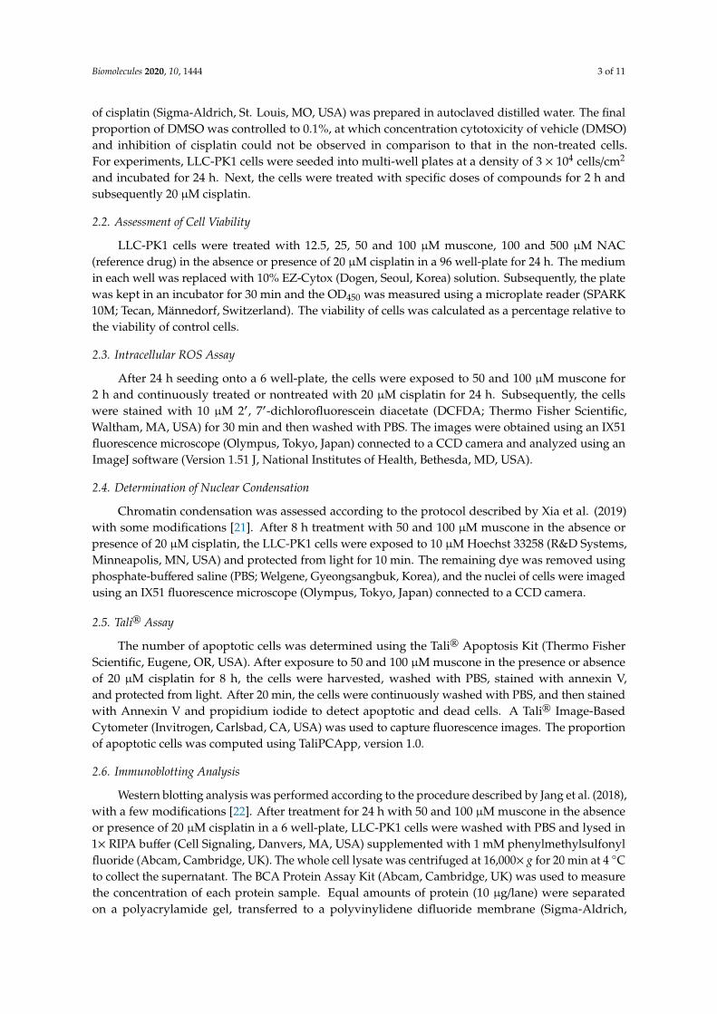
Musk, well known as a high-value crude drug in traditional Chinese medicine, is produced fromventral glandular secretion of the male musk deer, and is widely applied in pain relief, promotion ofblood flow, and resuscitation. In addition, muscone (3-methylcyclopentadecanone, Figure 1), the keycompound regulating the odor of musk, also possesses several valuable pharmaceutical activitiessuch as anti-early pregnancy, anti-cerebral ischemia, anti-cancer, neuroprotective, and cardioprotectiveeffects [17]. Previous reports have shown that muscone has a protective effect on various normalcell lines, including neurons and cardiac myocytes. The common feature in the cellular protectivemechanisms of muscone on these cell types is the inhibitory effect on diverse cell death pathwayssuch as oxidative stress, inflammation, and apoptosis, which share the pathway of cisplatin-inducednephrotoxicity [8,18,19]. Furthermore, Liu et al. [20] indicated that muscone strengthened the treatmenteffect of bone marrow stromal cells (BMSCs) therapy in AKI by stimulating the migration, survivability,and proliferation of BMSCs and inhibiting the expression of inflammatory cytokines and apoptosis inrenal tissues. Based on these studies, muscone may have a preventive effect against cisplatin-inducedkidney cell death. In this study, we assessed the cytoprotective effect of muscone against cisplatinnephrotoxicity in LLC-PK1 kidney proximal tubule cells.
Biomolecules 2020, 10, x FOR PEER REVIEW 2 of 11
symptoms of acute kidney injury (AKI), including higher serum creatinine, lower glomerular
filtration rate, and decreased serum potassium and magnesium concentrations after 10-day drug
administration [8].
The nephrotoxic mechanism of cisplatin remains unknown despite decades of research. Recent
studies have indicated that cisplatin induces renal failure by triggering complex intracellular
signaling pathways. In brief, after cisplatin is transported through high-affinity copper uptake
protein 1 (CTR1), organic cation transporters (OCTs), or passive diffusion via the cell membrane, it
triggers a series of signaling pathways including those affecting the balance of cdk2 and p21,
activation of mitogen-activated protein kinase (MAPK), tumor necrosis factor-α, tumor protein p53,
nuclear factor-κB, mitochondrial dysfunction, DNA damage, reactive oxygen species (ROS)
generation, and formation of toxic metabolites. These upstream changes result in apoptosis or
inflammation downstream, which leads to renal cell death and AKI [8].
Although cisplatin therapy presents risks for kidneys and other organs, the clinical effectiveness
of cisplatin is undeniable in cancer treatment. For years, various solutions have been proposed to
reduce nephrotoxicity and conserve the therapeutic effect of cisplatin, such as the synthesis of new
cisplatin-like drugs that express low toxicity on normal tissues (carboplatin) [9] or hydration of the
patients during cisplatin administration [10]. In addition, in recent years, the combination of cisplatin
therapy with renal protective supplements originating from natural sources has been widely
researched in both in vitro and in vivo models [11–16].
Musk, well known as a high-value crude drug in traditional Chinese medicine, is produced from
ventral glandular secretion of the male musk deer, and is widely applied in pain relief, promotion of
blood flow, and resuscitation. In addition, muscone (3-methylcyclopentadecanone, Figure 1), the key
compound regulating the odor of musk, also possesses several valuable pharmaceutical activities
such as anti-early pregnancy, anti-cerebral ischemia, anti-cancer, neuroprotective, and
cardioprotective effects [17]. Previous reports have shown that muscone has a protective effect on
various normal cell lines, including neurons and cardiac myocytes. The common feature in the
cellular protective mechanisms of muscone on these cell types is the inhibitory effect on diverse cell
death pathways such as oxidative stress, inflammation, and apoptosis, which share the pathway of
cisplatin-induced nephrotoxicity [8,18,19]. Furthermore, Liu et al. [20] indicated that muscone
strengthened the treatment effect of bone marrow stromal cells (BMSCs) therapy in AKI by
stimulating the migration, survivability, and proliferation of BMSCs and inhibiting the expression of
inflammatory cytokines and apoptosis in renal tissues. Based on these studies, muscone may have a
preventive effect against cisplatin-induced kidney cell death. In this study, we assessed the
cytoprotective effect of muscone against cisplatin nephrotoxicity in LLC-PK1 kidney proximal tubule
cells.
Figure 1. Chemical structure of muscone.
2. Materials and Methods
2.1. Cell Culture and Drug Treatment
LLC-PK1 cells (ATCC, Manassas, VA, USA) were propagated in Medium 199 (Welgene,
Gyeongsangbuk, Korea) containing 10% fetal bovine serum (Regeneration Biology, Ottawa, Canada)
and 1% streptomycin-penicillin (Corning, Manassas, VA, USA). The typical growth conditions were
set as 37 °C, 95% relative humidity, and 5% CO2. A 100 mM stock solution of muscone (The Nature
Network, Vestenbergsgreuth, Germany) and N-acetylcysteine (NAC; Abcam, Cambridge, UK) were
prepared in dimethyl sulfoxide (DMSO; Santa Cruz Biotechnology, TX, USA). A 2 mM stock solution
Figure 1. Chemical structure of muscone.
2. Materials and Methods
2.1. Cell Culture and Drug Treatment
LLC-PK1 cells (ATCC, Manassas, VA, USA) were propagated in Medium 199 (Welgene,Gyeongsangbuk, Korea) containing 10% fetal bovine serum (Regeneration Biology, Ottawa, Canada)and 1% streptomycin-penicillin (Corning, Manassas, VA, USA). The typical growth conditions wereset as 37 ◦C, 95% relative humidity, and 5% CO2. A 100 mM stock solution of muscone (The NatureNetwork, Vestenbergsgreuth, Germany) and N-acetylcysteine (NAC; Abcam, Cambridge, UK) wereprepared in dimethyl sulfoxide (DMSO; Santa Cruz Biotechnology, TX, USA). A 2 mM stock solution
Biomolecules 2020, 10, 1444 3 of 11
of cisplatin (Sigma-Aldrich, St. Louis, MO, USA) was prepared in autoclaved distilled water. The finalproportion of DMSO was controlled to 0.1%, at which concentration cytotoxicity of vehicle (DMSO)and inhibition of cisplatin could not be observed in comparison to that in the non-treated cells.For experiments, LLC-PK1 cells were seeded into multi-well plates at a density of 3 × 10⁴ cells/cm2
and incubated for 24 h. Next, the cells were treated with specific doses of compounds for 2 h andsubsequently 20 µM cisplatin.
2.2. Assessment of Cell Viability
LLC-PK1 cells were treated with 12.5, 25, 50 and 100 µM muscone, 100 and 500 µM NAC(reference drug) in the absence or presence of 20 µM cisplatin in a 96 well-plate for 24 h. The mediumin each well was replaced with 10% EZ-Cytox (Dogen, Seoul, Korea) solution. Subsequently, the platewas kept in an incubator for 30 min and the OD450 was measured using a microplate reader (SPARK10M; Tecan, Männedorf, Switzerland). The viability of cells was calculated as a percentage relative tothe viability of control cells.
2.3. Intracellular ROS Assay
After 24 h seeding onto a 6 well-plate, the cells were exposed to 50 and 100 µM muscone for2 h and continuously treated or nontreated with 20 µM cisplatin for 24 h. Subsequently, the cellswere stained with 10 µM 2′, 7′-dichlorofluorescein diacetate (DCFDA; Thermo Fisher Scientific,Waltham, MA, USA) for 30 min and then washed with PBS. The images were obtained using an IX51fluorescence microscope (Olympus, Tokyo, Japan) connected to a CCD camera and analyzed using anImageJ software (Version 1.51 J, National Institutes of Health, Bethesda, MD, USA).
2.4. Determination of Nuclear Condensation
Chromatin condensation was assessed according to the protocol described by Xia et al. (2019)with some modifications [21]. After 8 h treatment with 50 and 100 µM muscone in the absence orpresence of 20 µM cisplatin, the LLC-PK1 cells were exposed to 10 µM Hoechst 33258 (R&D Systems,Minneapolis, MN, USA) and protected from light for 10 min. The remaining dye was removed usingphosphate-buffered saline (PBS; Welgene, Gyeongsangbuk, Korea), and the nuclei of cells were imagedusing an IX51 fluorescence microscope (Olympus, Tokyo, Japan) connected to a CCD camera.
2.5. Tali® Assay
The number of apoptotic cells was determined using the Tali® Apoptosis Kit (Thermo FisherScientific, Eugene, OR, USA). After exposure to 50 and 100 µM muscone in the presence or absenceof 20 µM cisplatin for 8 h, the cells were harvested, washed with PBS, stained with annexin V,and protected from light. After 20 min, the cells were continuously washed with PBS, and then stainedwith Annexin V and propidium iodide to detect apoptotic and dead cells. A Tali® Image-BasedCytometer (Invitrogen, Carlsbad, CA, USA) was used to capture fluorescence images. The proportionof apoptotic cells was computed using TaliPCApp, version 1.0.
2.6. Immunoblotting Analysis
Western blotting analysis was performed according to the procedure described by Jang et al. (2018),with a few modifications [22]. After treatment for 24 h with 50 and 100 µM muscone in the absenceor presence of 20 µM cisplatin in a 6 well-plate, LLC-PK1 cells were washed with PBS and lysed in1× RIPA buffer (Cell Signaling, Danvers, MA, USA) supplemented with 1 mM phenylmethylsulfonylfluoride (Abcam, Cambridge, UK). The whole cell lysate was centrifuged at 16,000× g for 20 min at 4 ◦Cto collect the supernatant. The BCA Protein Assay Kit (Abcam, Cambridge, UK) was used to measurethe concentration of each protein sample. Equal amounts of protein (10 µg/lane) were separatedon a polyacrylamide gel, transferred to a polyvinylidene difluoride membrane (Sigma-Aldrich,
Biomolecules 2020, 10, 1444 4 of 11
St. Louis, MO, USA) presoaked in methyl alcohol, and blocked in 5% nonfat milk in tris-buffered salinesupplemented with 0.1% Tween 20 (TBS-T) for 1 h. Subsequently, the membranes were probed withthe first antibodies of interest overnight at 4 ◦C and secondary antibodies for 1 h. All antibodies weresupplied by Cell Signaling (Danvers, MA, USA). Next, the membranes were exposed to a mixture ofreagents A and B at a 1:1 ratio (ECL Western Blotting Substrate Kit, Abcam, Cambridge, UK), and theimmunoreactive bands were developed using the Fusion Solo Chemiluminescence System (PEQLABBiotechnologie GmbH, Erlangen, Germany).
2.7. Statistical Methods
The experimental results were described as the mean ± standard deviation (SD). Statisticaldifferences were assessed using one-way analysis of variance (ANOVA) with the Tukey’s range test.Statistical significance was set at p-values of less than 0.05. The IBM® SPSS® Statistics software(Version 25, IBM, Armonk, NY, USA) was used to conduct the statistical evaluation.
3. Results and Discussion
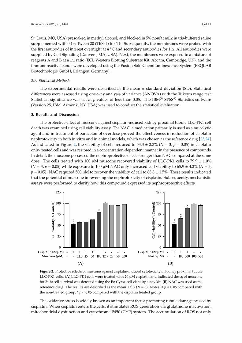
The protective effect of muscone against cisplatin-induced kidney proximal tubule LLC-PK1 celldeath was examined using cell viability assay. The NAC, a medication primarily is used as a mucolyticagent and in treatment of paracetamol overdose proved the effectiveness in reduction of cisplatinnephrotoxicity in both in vitro and in animal models, which was chosen as the reference drug [23,24].As indicated in Figure 2, the viability of cells reduced to 53.3 ± 2.3% (N = 3, p = 0.05) in cisplatinonly-treated cells and was restored in a concentration-dependent manner in the presence of compounds.In detail, the muscone possessed the nephroprotective effect stronger than NAC compared at the samedose. The cells treated with 100 µM muscone recovered viability of LLC-PK1 cells to 79.9 ± 1.0%(N = 3, p = 0.05) while exposure to 100 µM NAC only increased cell viability to 65.9 ± 4.2% (N = 3,p = 0.05). NAC required 500 µM to recover the viability of cell to 88.8 ± 1.5%. These results indicatedthat the potential of muscone in reversing the nephrotoxicity of cisplatin. Subsequently, mechanisticassays were performed to clarify how this compound expressed its nephroprotective effects.
Biomolecules 2020, 10, x FOR PEER REVIEW 4 of 11
in 5% nonfat milk in tris-buffered saline supplemented with 0.1% Tween 20 (TBS-T) for 1 h.
Subsequently, the membranes were probed with the first antibodies of interest overnight at 4 °C and
secondary antibodies for 1 h. All antibodies were supplied by Cell Signaling (Danvers, MA, USA).
Next, the membranes were exposed to a mixture of reagents A and B at a 1:1 ratio (ECL Western
Blotting Substrate Kit, Abcam, Cambridge, UK), and the immunoreactive bands were developed
using the Fusion Solo Chemiluminescence System (PEQLAB Biotechnologie GmbH, Erlangen,
Germany).
2.7. Statistical Methods
The experimental results were described as the mean ± standard deviation (SD). Statistical
differences were assessed using one-way analysis of variance (ANOVA) with the Tukey's range test.
Statistical significance was set at p-values of less than 0.05. The IBM® SPSS® Statistics software (Version
25, IBM, Armonk, NY, USA) was used to conduct the statistical evaluation.
3. Results and Discussion
The protective effect of muscone against cisplatin-induced kidney proximal tubule LLC-PK1 cell
death was examined using cell viability assay. The NAC, a medication primarily is used as a
mucolytic agent and in treatment of paracetamol overdose proved the effectiveness in reduction of
cisplatin nephrotoxicity in both in vitro and in animal models, which was chosen as the reference
drug [23,24]. As indicated in Figure 2, the viability of cells reduced to 53.3 ± 2.3% (N = 3, p = 0.05) in
cisplatin only-treated cells and was restored in a concentration-dependent manner in the presence of
compounds. In detail, the muscone possessed the nephroprotective effect stronger than NAC
compared at the same dose. The cells treated with 100 µM muscone recovered viability of LLC-PK1
cells to 79.9 ± 1.0% (N = 3, p = 0.05) while exposure to 100 µM NAC only increased cell viability to 65.9
± 4.2% (N = 3, p = 0.05). NAC required 500 µM to recover the viability of cell to 88.8 ± 1.5%. These
results indicated that the potential of muscone in reversing the nephrotoxicity of cisplatin.
Subsequently, mechanistic assays were performed to clarify how this compound expressed its
nephroprotective effects.
(A) (B)
Figure 2. Protective effects of muscone against cisplatin-induced cytotoxicity in kidney proximal
tubule LLC-PK1 cells. (A) LLC-PK1 cells were treated with 20 µM cisplatin and indicated doses of
muscone for 24 h; cell survival was detected using the Ez-Cytox cell viability assay kit. (B) NAC was
used as the reference drug. The results are described as the mean ± SD (N = 3). Notes: # p < 0.05
compared with the non-treated group, * p < 0.05 compared with the cisplatin treated group.
The oxidative stress is widely known as an important factor promoting tubule damage caused
by cisplatin. When cisplatin enters the cells, it stimulates ROS generation via glutathione inactivation,
mitochondrial dysfunction and cytochrome P450 (CYP) system. The accumulation of ROS not only
triggers various forms of cellular damage, consisting of lipid peroxidation, DNA damage, and protein
Figure 2. Protective effects of muscone against cisplatin-induced cytotoxicity in kidney proximal tubuleLLC-PK1 cells. (A) LLC-PK1 cells were treated with 20 µM cisplatin and indicated doses of musconefor 24 h; cell survival was detected using the Ez-Cytox cell viability assay kit. (B) NAC was used as thereference drug. The results are described as the mean ± SD (N = 3). Notes: # p < 0.05 compared withthe non-treated group, * p < 0.05 compared with the cisplatin treated group.
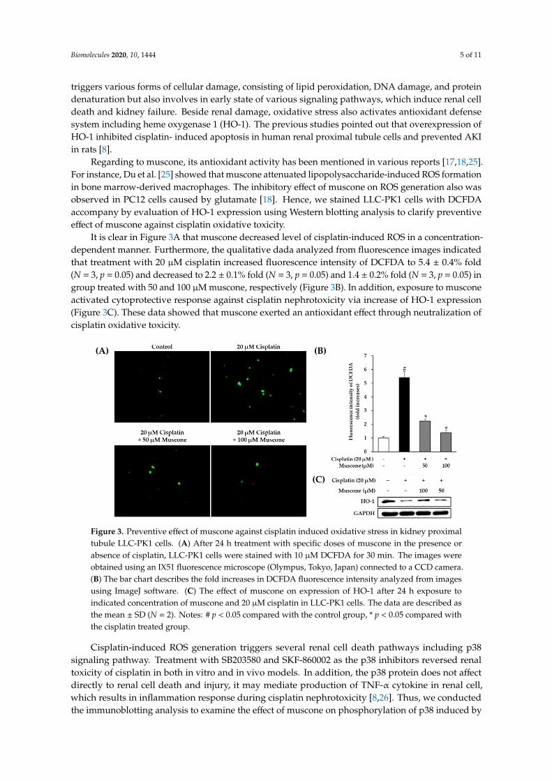
The oxidative stress is widely known as an important factor promoting tubule damage caused bycisplatin. When cisplatin enters the cells, it stimulates ROS generation via glutathione inactivation,mitochondrial dysfunction and cytochrome P450 (CYP) system. The accumulation of ROS not only
Biomolecules 2020, 10, 1444 5 of 11
triggers various forms of cellular damage, consisting of lipid peroxidation, DNA damage, and proteindenaturation but also involves in early state of various signaling pathways, which induce renal celldeath and kidney failure. Beside renal damage, oxidative stress also activates antioxidant defensesystem including heme oxygenase 1 (HO-1). The previous studies pointed out that overexpression ofHO-1 inhibited cisplatin- induced apoptosis in human renal proximal tubule cells and prevented AKIin rats [8].
Regarding to muscone, its antioxidant activity has been mentioned in various reports [17,18,25].For instance, Du et al. [25] showed that muscone attenuated lipopolysaccharide-induced ROS formationin bone marrow-derived macrophages. The inhibitory effect of muscone on ROS generation also wasobserved in PC12 cells caused by glutamate [18]. Hence, we stained LLC-PK1 cells with DCFDAaccompany by evaluation of HO-1 expression using Western blotting analysis to clarify preventiveeffect of muscone against cisplatin oxidative toxicity.
It is clear in Figure 3A that muscone decreased level of cisplatin-induced ROS in a concentration-dependent manner. Furthermore, the qualitative dada analyzed from fluorescence images indicatedthat treatment with 20 µM cisplatin increased fluorescence intensity of DCFDA to 5.4 ± 0.4% fold(N = 3, p = 0.05) and decreased to 2.2 ± 0.1% fold (N = 3, p = 0.05) and 1.4 ± 0.2% fold (N = 3, p = 0.05) ingroup treated with 50 and 100 µM muscone, respectively (Figure 3B). In addition, exposure to musconeactivated cytoprotective response against cisplatin nephrotoxicity via increase of HO-1 expression(Figure 3C). These data showed that muscone exerted an antioxidant effect through neutralization ofcisplatin oxidative toxicity.
Biomolecules 2020, 10, x FOR PEER REVIEW 5 of 11
denaturation but also involves in early state of various signaling pathways, which induce renal cell
death and kidney failure. Beside renal damage, oxidative stress also activates antioxidant defense
system including heme oxygenase 1 (HO-1). The previous studies pointed out that overexpression of
HO-1 inhibited cisplatin- induced apoptosis in human renal proximal tubule cells and prevented AKI
in rats [8].
Regarding to muscone, its antioxidant activity has been mentioned in various reports [17,18,25].
For instance, Du et al. [25] showed that muscone attenuated lipopolysaccharide-induced ROS
formation in bone marrow-derived macrophages. The inhibitory effect of muscone on ROS
generation also was observed in PC12 cells caused by glutamate [18]. Hence, we stained LLC-PK1
cells with DCFDA accompany by evaluation of HO-1 expression using Western blotting analysis to
clarify preventive effect of muscone against cisplatin oxidative toxicity.
It is clear in Figure 3A that muscone decreased level of cisplatin-induced ROS in a concentration-
dependent manner. Furthermore, the qualitative dada analyzed from fluorescence images indicated
that treatment with 20 µM cisplatin increased fluorescence intensity of DCFDA to 5.4 ± 0.4% fold (N
= 3, p = 0.05) and decreased to 2.2 ± 0.1% fold (N = 3, p = 0.05) and 1.4 ± 0.2% fold (N = 3, p = 0.05) in
group treated with 50 and 100 µM muscone, respectively (Figure 3B). In addition, exposure to
muscone activated cytoprotective response against cisplatin nephrotoxicity via increase of HO-1
expression (Figure 3C). These data showed that muscone exerted an antioxidant effect through
neutralization of cisplatin oxidative toxicity.
Figure 3. Preventive effect of muscone against cisplatin induced oxidative stress in kidney proximal
tubule LLC-PK1 cells. (A) After 24 h treatment with specific doses of muscone in the presence or
absence of cisplatin, LLC-PK1 cells were stained with 10 µM DCFDA for 30 min. The images were
obtained using an IX51 fluorescence microscope (Olympus, Tokyo, Japan) connected to a CCD
camera. (B) The bar chart describes the fold increases in DCFDA fluorescence intensity analyzed from
images using ImageJ software. (C) The effect of muscone on expression of HO-1 after 24 h exposure
to indicated concentration of muscone and 20 µM cisplatin in LLC-PK1 cells. The data are described
as the mean ± SD (N = 2). Notes: # p < 0.05 compared with the control group, * p < 0.05 compared with
the cisplatin treated group.
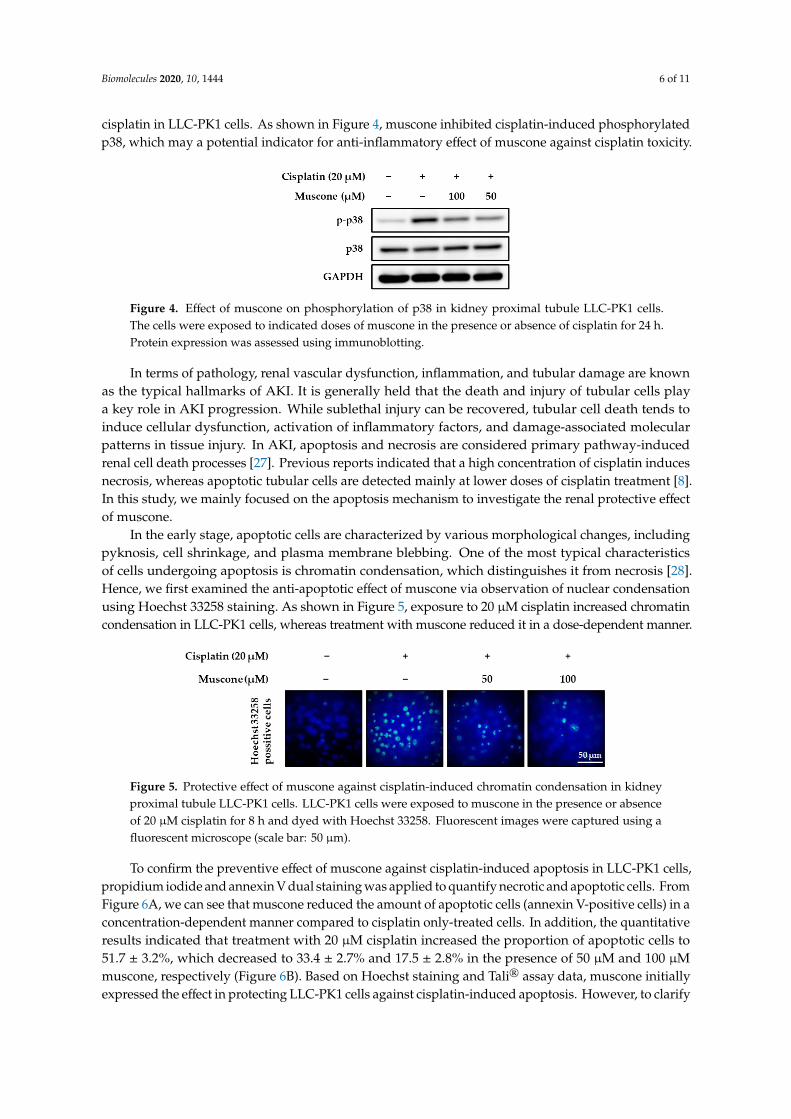
Cisplatin-induced ROS generation triggers several renal cell death pathways including p38
signaling pathway. Treatment with SB203580 and SKF-860002 as the p38 inhibitors reversed renal
toxicity of cisplatin in both in vitro and in vivo models. In addition, the p38 protein does not affect
directly to renal cell death and injury, it may mediate production of TNF-α cytokine in renal cell,
which results in inflammation response during cisplatin nephrotoxicity [8,26]. Thus, we conducted
the immunoblotting analysis to examine the effect of muscone on phosphorylation of p38 induced by
cisplatin in LLC-PK1 cells. As shown in Figure 4, muscone inhibited cisplatin-induced
phosphorylated p38, which may a potential indicator for anti-inflammatory effect of muscone against
cisplatin toxicity.
(A) (B)
(C)
Figure 3. Preventive effect of muscone against cisplatin induced oxidative stress in kidney proximaltubule LLC-PK1 cells. (A) After 24 h treatment with specific doses of muscone in the presence orabsence of cisplatin, LLC-PK1 cells were stained with 10 µM DCFDA for 30 min. The images wereobtained using an IX51 fluorescence microscope (Olympus, Tokyo, Japan) connected to a CCD camera.(B) The bar chart describes the fold increases in DCFDA fluorescence intensity analyzed from imagesusing ImageJ software. (C) The effect of muscone on expression of HO-1 after 24 h exposure toindicated concentration of muscone and 20 µM cisplatin in LLC-PK1 cells. The data are described asthe mean ± SD (N = 2). Notes: # p < 0.05 compared with the control group, * p < 0.05 compared withthe cisplatin treated group.
Cisplatin-induced ROS generation triggers several renal cell death pathways including p38signaling pathway. Treatment with SB203580 and SKF-860002 as the p38 inhibitors reversed renaltoxicity of cisplatin in both in vitro and in vivo models. In addition, the p38 protein does not affectdirectly to renal cell death and injury, it may mediate production of TNF-α cytokine in renal cell,which results in inflammation response during cisplatin nephrotoxicity [8,26]. Thus, we conductedthe immunoblotting analysis to examine the effect of muscone on phosphorylation of p38 induced by
Biomolecules 2020, 10, 1444 6 of 11
cisplatin in LLC-PK1 cells. As shown in Figure 4, muscone inhibited cisplatin-induced phosphorylatedp38, which may a potential indicator for anti-inflammatory effect of muscone against cisplatin toxicity.Biomolecules 2020, 10, x FOR PEER REVIEW 6 of 11
Figure 4. Effect of muscone on phosphorylation of p38 in kidney proximal tubule LLC-PK1 cells. The
cells were exposed to indicated doses of muscone in the presence or absence of cisplatin for 24 h.
Protein expression was assessed using immunoblotting.
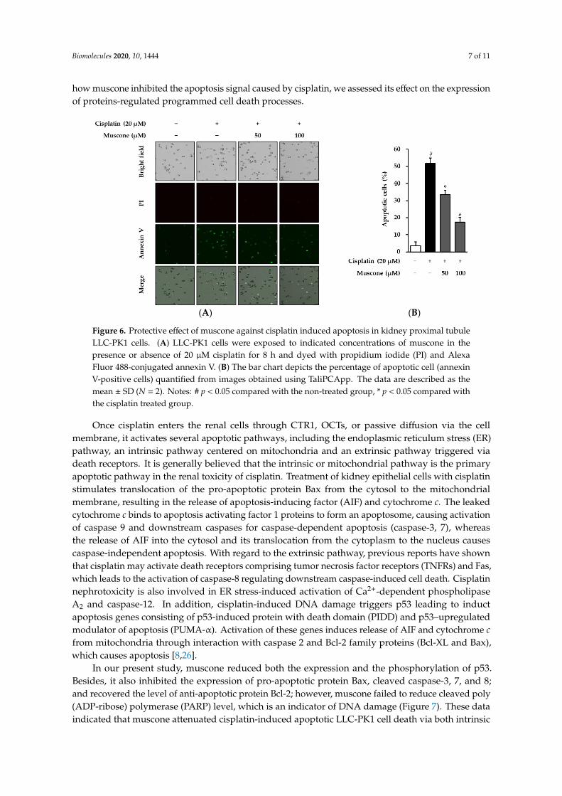
In terms of pathology, renal vascular dysfunction, inflammation, and tubular damage are known
as the typical hallmarks of AKI. It is generally held that the death and injury of tubular cells play a
key role in AKI progression. While sublethal injury can be recovered, tubular cell death tends to
induce cellular dysfunction, activation of inflammatory factors, and damage-associated molecular
patterns in tissue injury. In AKI, apoptosis and necrosis are considered primary pathway-induced
renal cell death processes [27]. Previous reports indicated that a high concentration of cisplatin
induces necrosis, whereas apoptotic tubular cells are detected mainly at lower doses of cisplatin
treatment [8]. In this study, we mainly focused on the apoptosis mechanism to investigate the renal
protective effect of muscone.
In the early stage, apoptotic cells are characterized by various morphological changes, including
pyknosis, cell shrinkage, and plasma membrane blebbing. One of the most typical characteristics of
cells undergoing apoptosis is chromatin condensation, which distinguishes it from necrosis [28].
Hence, we first examined the anti-apoptotic effect of muscone via observation of nuclear
condensation using Hoechst 33258 staining. As shown in Figure 5, exposure to 20 µM cisplatin
increased chromatin condensation in LLC-PK1 cells, whereas treatment with muscone reduced it in
a dose-dependent manner.
Figure 5. Protective effect of muscone against cisplatin-induced chromatin condensation in kidney
proximal tubule LLC-PK1 cells. LLC-PK1 cells were exposed to muscone in the presence or absence
of 20 µM cisplatin for 8 h and dyed with Hoechst 33258. Fluorescent images were captured using a
fluorescent microscope (scale bar: 50 µm).
To confirm the preventive effect of muscone against cisplatin-induced apoptosis in LLC-PK1
cells, propidium iodide and annexin V dual staining was applied to quantify necrotic and apoptotic
cells. From Figure 6A, we can see that muscone reduced the amount of apoptotic cells (annexin V-
positive cells) in a concentration-dependent manner compared to cisplatin only-treated cells. In
addition, the quantitative results indicated that treatment with 20 µM cisplatin increased the
proportion of apoptotic cells to 51.7 ± 3.2%, which decreased to 33.4 ± 2.7% and 17.5 ± 2.8% in the
presence of 50 µM and 100 µM muscone, respectively (Figure 6B). Based on Hoechst staining and
TaliⓇ assay data, muscone initially expressed the effect in protecting LLC-PK1 cells against cisplatin-
induced apoptosis. However, to clarify how muscone inhibited the apoptosis signal caused by
cisplatin, we assessed its effect on the expression of proteins-regulated programmed cell death
processes.
Figure 4. Effect of muscone on phosphorylation of p38 in kidney proximal tubule LLC-PK1 cells.The cells were exposed to indicated doses of muscone in the presence or absence of cisplatin for 24 h.Protein expression was assessed using immunoblotting.
In terms of pathology, renal vascular dysfunction, inflammation, and tubular damage are knownas the typical hallmarks of AKI. It is generally held that the death and injury of tubular cells playa key role in AKI progression. While sublethal injury can be recovered, tubular cell death tends toinduce cellular dysfunction, activation of inflammatory factors, and damage-associated molecularpatterns in tissue injury. In AKI, apoptosis and necrosis are considered primary pathway-inducedrenal cell death processes [27]. Previous reports indicated that a high concentration of cisplatin inducesnecrosis, whereas apoptotic tubular cells are detected mainly at lower doses of cisplatin treatment [8].In this study, we mainly focused on the apoptosis mechanism to investigate the renal protective effectof muscone.
In the early stage, apoptotic cells are characterized by various morphological changes, includingpyknosis, cell shrinkage, and plasma membrane blebbing. One of the most typical characteristicsof cells undergoing apoptosis is chromatin condensation, which distinguishes it from necrosis [28].Hence, we first examined the anti-apoptotic effect of muscone via observation of nuclear condensationusing Hoechst 33258 staining. As shown in Figure 5, exposure to 20 µM cisplatin increased chromatincondensation in LLC-PK1 cells, whereas treatment with muscone reduced it in a dose-dependent manner.
Biomolecules 2020, 10, x FOR PEER REVIEW 6 of 11
Figure 4. Effect of muscone on phosphorylation of p38 in kidney proximal tubule LLC-PK1 cells. The
cells were exposed to indicated doses of muscone in the presence or absence of cisplatin for 24 h.
Protein expression was assessed using immunoblotting.
In terms of pathology, renal vascular dysfunction, inflammation, and tubular damage are known
as the typical hallmarks of AKI. It is generally held that the death and injury of tubular cells play a
key role in AKI progression. While sublethal injury can be recovered, tubular cell death tends to
induce cellular dysfunction, activation of inflammatory factors, and damage-associated molecular
patterns in tissue injury. In AKI, apoptosis and necrosis are considered primary pathway-induced
renal cell death processes [27]. Previous reports indicated that a high concentration of cisplatin
induces necrosis, whereas apoptotic tubular cells are detected mainly at lower doses of cisplatin
treatment [8]. In this study, we mainly focused on the apoptosis mechanism to investigate the renal
protective effect of muscone.
In the early stage, apoptotic cells are characterized by various morphological changes, including
pyknosis, cell shrinkage, and plasma membrane blebbing. One of the most typical characteristics of
cells undergoing apoptosis is chromatin condensation, which distinguishes it from necrosis [28].
Hence, we first examined the anti-apoptotic effect of muscone via observation of nuclear
condensation using Hoechst 33258 staining. As shown in Figure 5, exposure to 20 µM cisplatin
increased chromatin condensation in LLC-PK1 cells, whereas treatment with muscone reduced it in
a dose-dependent manner.
Figure 5. Protective effect of muscone against cisplatin-induced chromatin condensation in kidney
proximal tubule LLC-PK1 cells. LLC-PK1 cells were exposed to muscone in the presence or absence
of 20 µM cisplatin for 8 h and dyed with Hoechst 33258. Fluorescent images were captured using a
fluorescent microscope (scale bar: 50 µm).
To confirm the preventive effect of muscone against cisplatin-induced apoptosis in LLC-PK1
cells, propidium iodide and annexin V dual staining was applied to quantify necrotic and apoptotic
cells. From Figure 6A, we can see that muscone reduced the amount of apoptotic cells (annexin V-
positive cells) in a concentration-dependent manner compared to cisplatin only-treated cells. In
addition, the quantitative results indicated that treatment with 20 µM cisplatin increased the
proportion of apoptotic cells to 51.7 ± 3.2%, which decreased to 33.4 ± 2.7% and 17.5 ± 2.8% in the
presence of 50 µM and 100 µM muscone, respectively (Figure 6B). Based on Hoechst staining and
TaliⓇ assay data, muscone initially expressed the effect in protecting LLC-PK1 cells against cisplatin-
induced apoptosis. However, to clarify how muscone inhibited the apoptosis signal caused by
cisplatin, we assessed its effect on the expression of proteins-regulated programmed cell death
processes.
Figure 5. Protective effect of muscone against cisplatin-induced chromatin condensation in kidneyproximal tubule LLC-PK1 cells. LLC-PK1 cells were exposed to muscone in the presence or absenceof 20 µM cisplatin for 8 h and dyed with Hoechst 33258. Fluorescent images were captured using afluorescent microscope (scale bar: 50 µm).
To confirm the preventive effect of muscone against cisplatin-induced apoptosis in LLC-PK1 cells,propidium iodide and annexin V dual staining was applied to quantify necrotic and apoptotic cells. FromFigure 6A, we can see that muscone reduced the amount of apoptotic cells (annexin V-positive cells) in aconcentration-dependent manner compared to cisplatin only-treated cells. In addition, the quantitativeresults indicated that treatment with 20 µM cisplatin increased the proportion of apoptotic cells to51.7 ± 3.2%, which decreased to 33.4 ± 2.7% and 17.5 ± 2.8% in the presence of 50 µM and 100 µMmuscone, respectively (Figure 6B). Based on Hoechst staining and Tali® assay data, muscone initiallyexpressed the effect in protecting LLC-PK1 cells against cisplatin-induced apoptosis. However, to clarify
Biomolecules 2020, 10, 1444 7 of 11
how muscone inhibited the apoptosis signal caused by cisplatin, we assessed its effect on the expressionof proteins-regulated programmed cell death processes.Biomolecules 2020, 10, x FOR PEER REVIEW 7 of 11
(A) (B)
Figure 6. Protective effect of muscone against cisplatin induced apoptosis in kidney proximal tubule
LLC-PK1 cells. (A) LLC-PK1 cells were exposed to indicated concentrations of muscone in the
presence or absence of 20 µM cisplatin for 8 h and dyed with propidium iodide (PI) and Alexa Fluor
488-conjugated annexin V. (B) The bar chart depicts the percentage of apoptotic cell (annexin V-
positive cells) quantified from images obtained using TaliPCApp. The data are described as the mean
± SD (N = 2). Notes: # p < 0.05 compared with the non-treated group, * p < 0.05 compared with the
cisplatin treated group.
Once cisplatin enters the renal cells through CTR1, OCTs, or passive diffusion via the cell
membrane, it activates several apoptotic pathways, including the endoplasmic reticulum stress (ER)
pathway, an intrinsic pathway centered on mitochondria and an extrinsic pathway triggered via
death receptors. It is generally believed that the intrinsic or mitochondrial pathway is the primary
apoptotic pathway in the renal toxicity of cisplatin. Treatment of kidney epithelial cells with cisplatin
stimulates translocation of the pro-apoptotic protein Bax from the cytosol to the mitochondrial
membrane, resulting in the release of apoptosis-inducing factor (AIF) and cytochrome c. The leaked
cytochrome c binds to apoptosis activating factor 1 proteins to form an apoptosome, causing
activation of caspase 9 and downstream caspases for caspase-dependent apoptosis (caspase-3, 7),
whereas the release of AIF into the cytosol and its translocation from the cytoplasm to the nucleus
causes caspase-independent apoptosis. With regard to the extrinsic pathway, previous reports have
shown that cisplatin may activate death receptors comprising tumor necrosis factor receptors
(TNFRs) and Fas, which leads to the activation of caspase-8 regulating downstream caspase-induced
cell death. Cisplatin nephrotoxicity is also involved in ER stress-induced activation of Ca2+-dependent
phospholipase A2 and caspase-12. In addition, cisplatin-induced DNA damage triggers p53 leading
to induct apoptosis genes consisting of p53-induced protein with death domain (PIDD) and p53–
upregulated modulator of apoptosis (PUMA-α). Activation of these genes induces release of AIF and
cytochrome c from mitochondria through interaction with caspase 2 and Bcl-2 family proteins (Bcl-
XL and Bax), which causes apoptosis [8,26].
In our present study, muscone reduced both the expression and the phosphorylation of p53.
Besides, it also inhibited the expression of pro-apoptotic protein Bax, cleaved caspase-3, 7, and 8; and
recovered the level of anti-apoptotic protein Bcl-2; however, muscone failed to reduce cleaved poly
(ADP‐ribose) polymerase (PARP) level, which is an indicator of DNA damage (Figure 7). These data
indicated that muscone attenuated cisplatin-induced apoptotic LLC-PK1 cell death via both intrinsic
and extrinsic pathways by inhibiting the cleavage of caspase-3, 7, and 8, and the protein Bax
expression; and increasing the level of protein Bcl-2. In addition, muscone also reversed p53 pathway
by preventing cisplatin-induced p53 expression and phosphorylation, which protects kidney
proximal tubule LLC-PK1 cells against apoptosis.
Figure 6. Protective effect of muscone against cisplatin induced apoptosis in kidney proximal tubuleLLC-PK1 cells. (A) LLC-PK1 cells were exposed to indicated concentrations of muscone in thepresence or absence of 20 µM cisplatin for 8 h and dyed with propidium iodide (PI) and AlexaFluor 488-conjugated annexin V. (B) The bar chart depicts the percentage of apoptotic cell (annexinV-positive cells) quantified from images obtained using TaliPCApp. The data are described as themean ± SD (N = 2). Notes: # p < 0.05 compared with the non-treated group, * p < 0.05 compared withthe cisplatin treated group.
Once cisplatin enters the renal cells through CTR1, OCTs, or passive diffusion via the cellmembrane, it activates several apoptotic pathways, including the endoplasmic reticulum stress (ER)pathway, an intrinsic pathway centered on mitochondria and an extrinsic pathway triggered viadeath receptors. It is generally believed that the intrinsic or mitochondrial pathway is the primaryapoptotic pathway in the renal toxicity of cisplatin. Treatment of kidney epithelial cells with cisplatinstimulates translocation of the pro-apoptotic protein Bax from the cytosol to the mitochondrialmembrane, resulting in the release of apoptosis-inducing factor (AIF) and cytochrome c. The leakedcytochrome c binds to apoptosis activating factor 1 proteins to form an apoptosome, causing activationof caspase 9 and downstream caspases for caspase-dependent apoptosis (caspase-3, 7), whereasthe release of AIF into the cytosol and its translocation from the cytoplasm to the nucleus causescaspase-independent apoptosis. With regard to the extrinsic pathway, previous reports have shownthat cisplatin may activate death receptors comprising tumor necrosis factor receptors (TNFRs) and Fas,which leads to the activation of caspase-8 regulating downstream caspase-induced cell death. Cisplatinnephrotoxicity is also involved in ER stress-induced activation of Ca2+-dependent phospholipaseA2 and caspase-12. In addition, cisplatin-induced DNA damage triggers p53 leading to inductapoptosis genes consisting of p53-induced protein with death domain (PIDD) and p53–upregulatedmodulator of apoptosis (PUMA-α). Activation of these genes induces release of AIF and cytochrome cfrom mitochondria through interaction with caspase 2 and Bcl-2 family proteins (Bcl-XL and Bax),which causes apoptosis [8,26].
In our present study, muscone reduced both the expression and the phosphorylation of p53.Besides, it also inhibited the expression of pro-apoptotic protein Bax, cleaved caspase-3, 7, and 8;and recovered the level of anti-apoptotic protein Bcl-2; however, muscone failed to reduce cleaved poly(ADP-ribose) polymerase (PARP) level, which is an indicator of DNA damage (Figure 7). These dataindicated that muscone attenuated cisplatin-induced apoptotic LLC-PK1 cell death via both intrinsic
Biomolecules 2020, 10, 1444 8 of 11
and extrinsic pathways by inhibiting the cleavage of caspase-3, 7, and 8, and the protein Bax expression;and increasing the level of protein Bcl-2. In addition, muscone also reversed p53 pathway by preventingcisplatin-induced p53 expression and phosphorylation, which protects kidney proximal tubule LLC-PK1cells against apoptosis.Biomolecules 2020, 10, x FOR PEER REVIEW 8 of 11
Figure 7. Effect of muscone on the expression of proteins-regulated in kidney proximal tubule LLC-
PK1 cells. The cells were treated with 20 µM cisplatin and specific concentration of muscone for 24 h.
Protein expression was determined by western blotting.
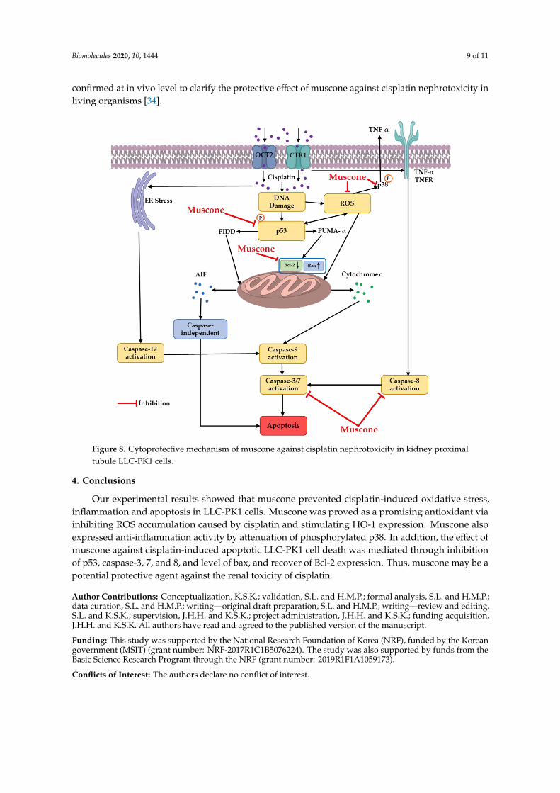
The cytoprotective mechanism of muscone targeted on 3 main pathways including oxidative
stress [29], inflammation and apoptosis [16]. In previous our reports under the same conditions, we
mainly assessed MAPK, cleaved caspase 3 and p53 to evaluate protective mechanism of compound
against cisplatin nephrotoxicity on LLC-PK1 cells. For instances, monoacetate from Poria cocos Wolf
inhibited oxidative stress-regulated apoptosis via reducing MAPK phosphorylation and cleaved
caspase 3 [30]. Ginsenosides Rg3, Rg5, and Rk1 found in processed ginseng prevented LLC-PK1 cells
against cisplatin-induced apoptosis and inflammation by inhibition of c-Jun N-terminal kinase (JNK)
phosphorylation, p53 expression and cleavage of caspase 3 [31]. In this study, the renal protective
mechanism of muscone against cisplatin toxicity in LLC-PK1 cells was focused on interaction among
p53, ROS, p38 and apoptosis pathways as shown in Figure 8. The inhibitory effect of muscone on
ROS generation and p53 expression and phosphorylation may inhibit intrinsic pathway of apoptosis
and p38 pathway inducing inflammation cytokine (TNFα).
In summary, muscone protected LLC-PK1 kidney proximal tubule cells against cisplatin
nephrotoxicity in a concentration-dependent manner. Muscone expressed the inhibitory effect
against cisplatin-induced oxidative stress by decrease of ROS accumulation and increase of HO-1
expression. Besides, muscone also displayed the anti-inflammatory activity via inhibiting the
phosphorylation of p38. Furthermore, muscone prevented LLC-PK1 cells from cisplatin-induced
apoptosis by decreasing chromatin condensation, the expression of the pro-apoptotic protein Bax and
cleavage of caspase-3, 7, and 8; and increasing level of anti-apoptotic protein Bcl-2. In addition, the
anti-apoptotic effect of muscone also was enhanced by preventing p53 expression and
phosphorylation. The protective mechanism of muscone against cisplatin nephrotoxicity in LLC-PK1
cells is summarized in Figure 8.
However, we are aware that our research may exist some limitations compared to previous
reports at the same scope. Firstly, the effect of muscone on other cell death pathways caused by
cisplatin including autophagy [32] and nuclear factor-κB activation [33] has not been investigated yet
in this study. Secondly, the experimental results are just at in vitro scope. This work should be further
confirmed at in vivo level to clarify the protective effect of muscone against cisplatin nephrotoxicity
in living organisms [34].
Figure 7. Effect of muscone on the expression of proteins-regulated in kidney proximal tubule LLC-PK1cells. The cells were treated with 20 µM cisplatin and specific concentration of muscone for 24 h. Proteinexpression was determined by western blotting.
The cytoprotective mechanism of muscone targeted on 3 main pathways including oxidativestress [29], inflammation and apoptosis [16]. In previous our reports under the same conditions,we mainly assessed MAPK, cleaved caspase 3 and p53 to evaluate protective mechanism of compoundagainst cisplatin nephrotoxicity on LLC-PK1 cells. For instances, monoacetate from Poria cocos Wolfinhibited oxidative stress-regulated apoptosis via reducing MAPK phosphorylation and cleavedcaspase 3 [30]. Ginsenosides Rg3, Rg5, and Rk1 found in processed ginseng prevented LLC-PK1 cellsagainst cisplatin-induced apoptosis and inflammation by inhibition of c-Jun N-terminal kinase (JNK)phosphorylation, p53 expression and cleavage of caspase 3 [31]. In this study, the renal protectivemechanism of muscone against cisplatin toxicity in LLC-PK1 cells was focused on interaction amongp53, ROS, p38 and apoptosis pathways as shown in Figure 8. The inhibitory effect of muscone on ROSgeneration and p53 expression and phosphorylation may inhibit intrinsic pathway of apoptosis andp38 pathway inducing inflammation cytokine (TNFα).
In summary, muscone protected LLC-PK1 kidney proximal tubule cells against cisplatinnephrotoxicity in a concentration-dependent manner. Muscone expressed the inhibitory effect againstcisplatin-induced oxidative stress by decrease of ROS accumulation and increase of HO-1 expression.Besides, muscone also displayed the anti-inflammatory activity via inhibiting the phosphorylationof p38. Furthermore, muscone prevented LLC-PK1 cells from cisplatin-induced apoptosis bydecreasing chromatin condensation, the expression of the pro-apoptotic protein Bax and cleavage ofcaspase-3, 7, and 8; and increasing level of anti-apoptotic protein Bcl-2. In addition, the anti-apoptoticeffect of muscone also was enhanced by preventing p53 expression and phosphorylation. The protectivemechanism of muscone against cisplatin nephrotoxicity in LLC-PK1 cells is summarized in Figure 8.
However, we are aware that our research may exist some limitations compared to previous reportsat the same scope. Firstly, the effect of muscone on other cell death pathways caused by cisplatinincluding autophagy [32] and nuclear factor-κB activation [33] has not been investigated yet in thisstudy. Secondly, the experimental results are just at in vitro scope. This work should be further
Biomolecules 2020, 10, 1444 9 of 11
confirmed at in vivo level to clarify the protective effect of muscone against cisplatin nephrotoxicity inliving organisms [34].Biomolecules 2020, 10, x FOR PEER REVIEW 9 of 11
Figure 8. Cytoprotective mechanism of muscone against cisplatin nephrotoxicity in kidney proximal
tubule LLC-PK1 cells.
4. Conclusions
Our experimental results showed that muscone prevented cisplatin-induced oxidative stress,
inflammation and apoptosis in LLC-PK1 cells. Muscone was proved as a promising antioxidant via
inhibiting ROS accumulation caused by cisplatin and stimulating HO-1 expression. Muscone also
expressed anti-inflammation activity by attenuation of phosphorylated p38. In addition, the effect of
muscone against cisplatin-induced apoptotic LLC-PK1 cell death was mediated through inhibition of
p53, caspase-3, 7, and 8, and level of bax, and recover of Bcl-2 expression. Thus, muscone may be a
potential protective agent against the renal toxicity of cisplatin.
Author Contributions: Conceptualization, K.S.K.; validation, S.L. and H.M.P.; formal analysis, S.L. and H.M.P.;
data curation, S.L. and H.M.P.; writing—original draft preparation, S.L. and H.M.P.; writing—review and
editing, S.L. and K.S.K.; supervision, J.H.H. and K.S.K.; project administration, J.H.H. and K.S.K.; funding
acquisition, J.H.H. and K.S.K. All authors have read and agreed to the published version of the manuscript.
Funding: This study was supported by the National Research Foundation of Korea (NRF), funded by the Korean
government (MSIT) (grant number: NRF-2017R1C1B5076224). The study was also supported by funds from the
Basic Science Research Program through the NRF (grant number: 2019R1F1A1059173).
Conflicts of Interest: The authors declare no conflicts of interest.
References
1. Organization, W.H. World Health Organization Model List of Essential Medicines: 21st List 2019; World Health
Organization: Geneva, Switzerland, 2019.
2. Daud, N.N.N.N.M.; Septama, A.W.; Simbak, N.; Bakar, N.H.A.; Rahmi, E.P. Synergistic Effect of Flavonoids
from Artocarpus heterophyllus Heartwoods on Anticancer Activity of Cisplatin Against H460 and MCF-7
Cell Lines. Nat. Prod. Sci. 2019, 25, 311–316.
Figure 8. Cytoprotective mechanism of muscone against cisplatin nephrotoxicity in kidney proximaltubule LLC-PK1 cells.
4. Conclusions
Our experimental results showed that muscone prevented cisplatin-induced oxidative stress,inflammation and apoptosis in LLC-PK1 cells. Muscone was proved as a promising antioxidant viainhibiting ROS accumulation caused by cisplatin and stimulating HO-1 expression. Muscone alsoexpressed anti-inflammation activity by attenuation of phosphorylated p38. In addition, the effect ofmuscone against cisplatin-induced apoptotic LLC-PK1 cell death was mediated through inhibitionof p53, caspase-3, 7, and 8, and level of bax, and recover of Bcl-2 expression. Thus, muscone may be apotential protective agent against the renal toxicity of cisplatin.
Author Contributions: Conceptualization, K.S.K.; validation, S.L. and H.M.P.; formal analysis, S.L. and H.M.P.;data curation, S.L. and H.M.P.; writing—original draft preparation, S.L. and H.M.P.; writing—review and editing,S.L. and K.S.K.; supervision, J.H.H. and K.S.K.; project administration, J.H.H. and K.S.K.; funding acquisition,J.H.H. and K.S.K. All authors have read and agreed to the published version of the manuscript.
Funding: This study was supported by the National Research Foundation of Korea (NRF), funded by the Koreangovernment (MSIT) (grant number: NRF-2017R1C1B5076224). The study was also supported by funds from theBasic Science Research Program through the NRF (grant number: 2019R1F1A1059173).
Conflicts of Interest: The authors declare no conflict of interest.
Biomolecules 2020, 10, 1444 10 of 11
References
1. World Health Organization. World Health Organization Model List of Essential Medicines: 21st List 2019;World Health Organization: Geneva, Switzerland, 2019.
2. Daud, N.N.N.N.M.; Septama, A.W.; Simbak, N.; Bakar, N.H.A.; Rahmi, E.P. Synergistic Effect of Flavonoidsfrom Artocarpus heterophyllus Heartwoods on Anticancer Activity of Cisplatin Against H460 and MCF-7Cell Lines. Nat. Prod. Sci. 2019, 25, 311–316. [CrossRef]
3. Dasari, S.; Tchounwou, P.B. Cisplatin in cancer therapy: Molecular mechanisms of action. Eur. J. Pharmacol.2014, 740, 364–378. [CrossRef] [PubMed]
4. Chen, C.; Zhang, H.; Xu, H.; Zheng, Y.; Wu, T.; Lian, Y. Ginsenoside Rb1 ameliorates cisplatin-inducedlearning and memory impairments. J. Ginseng Res. 2019, 43, 499–507. [CrossRef] [PubMed]
5. Bucher-Johannessen, C.; Page, C.M.; Haugen, T.B.; Wojewodzic, M.W.; Fosså, S.D.; Grotmol, T.; Haugnes, H.S.;Rounge, T.B. Cisplatin treatment of testicular cancer patients introduces long-term changes in the epigenome.Clin. Epigenetics 2019, 11, 179. [CrossRef]
6. Holditch, S.J.; Brown, C.N.; Lombardi, A.M.; Nguyen, K.N.; Edelstein, C.L. Recent advances in models,mechanisms, biomarkers, and interventions in cisplatin-induced acute kidney injury. Int. J. Mol. Sci. 2019,20, 3011. [CrossRef]
7. Hayati, F.; Hossainzadeh, M.; Shayanpour, S.; Abedi-Gheshlaghi, Z.; Mousavi, S.S.B. Prevention of cisplatinnephrotoxicity. J. Nephropharmacolo. 2016, 5, 57.
8. Qi, L.; Luo, Q.; Zhang, Y.; Jia, F.; Zhao, Y.; Wang, F. Advances in toxicological research of the anticancer drugcisplatin. Chem. Res. Toxicol. 2019, 32, 1469–1486. [CrossRef]
9. Perazella, M.A. Onco-nephrology: Renal toxicities of chemotherapeutic agents. Clin. J. Am. Soc. Nephrol.2012, 7, 1713–1721. [CrossRef]
10. Hase, T.; Miyazaki, M.; Ichikawa, K.; Yogo, N.; Ozawa, N.; Hatta, T.; Ando, M.; Sato, M.; Kondo, M.;Yamada, K. Short hydration with 20 mEq of magnesium supplementation for lung cancer patients receivingcisplatin-based chemotherapy: A prospective study. Int. J. Clin. Oncol. 2020, 1–8. [CrossRef]
11. Ojha, S.; Venkataraman, B.; Kurdi, A.; Mahgoub, E.; Sadek, B.; Rajesh, M. Plant-derived agents forcounteracting cisplatin-induced nephrotoxicity. Oxid. Med. Cell. Longev. 2016, 2016. [CrossRef]
12. Heidari-Soreshjani, S.; Asadi-Samani, M.; Yang, Q.; Saeedi-Boroujeni, A. Phytotherapy of nephrotoxicity-induced by cancer drugs: An updated review. J. Nephropathol. 2017, 6, 254. [CrossRef] [PubMed]
13. Ma, Z.-N.; Li, Y.-Z.; Li, W.; Yan, X.-T.; Yang, G.; Zhang, J.; Zhao, L.-C.; Yang, L.-M. Nephroprotective effects ofsaponins from leaves of Panax quinquefolius against cisplatin-induced acute kidney injury. Int. J. Mol. Sci.2017, 18, 1407. [CrossRef] [PubMed]
14. Nematbakhsh, M.; Pezeshki, Z.; Jazi, F.E.; Mazaheri, B.; Moeini, M.; Safari, T.; Azarkish, F.; Moslemi, F.;Maleki, M.; Rezaei, A. Cisplatin-induced nephrotoxicity; protective supplements and gender differences.Asian Pac. J. Cancer Prev. 2017, 18, 295.
15. Lee, D.; Lee, D.-S.; Jung, K.; Hwang, G.S.; Lee, H.L.; Yamabe, N.; Lee, H.-J.; Eom, D.-W.; Kim, K.H.;Kang, K.S. Protective effect of ginsenoside Rb1 against tacrolimus-induced apoptosis in renal proximaltubular LLC-PK1 cells. J. Ginseng Res. 2018, 42, 75–80. [CrossRef] [PubMed]
16. Lee, D.; Kim, K.H.; Lee, W.Y.; Kim, C.-E.; Sung, S.H.; Kang, K.B.; Kang, K.S. Multiple targets of3-dehydroxyceanothetric acid 2-methyl ester to protect against cisplatin-induced cytotoxicity in kidneyepithelial LLC-PK1 cells. Molecules 2019, 24, 878. [CrossRef]
17. Wang, J.; Xing, H.; Qin, X.; Ren, Q.; Yang, J.; Li, L. Pharmacological effects and involved mechanisms ofmuscone. J. Ethnopharmacol. 2020, 262, 113120. [CrossRef]
18. Yu, L.; Wang, N.; Zhang, Y.; Wang, Y.; Li, J.; Wu, Q.; Liu, Y. Neuroprotective effect of muscone onglutamate-induced apoptosis in PC12 cells via antioxidant and Ca2+ antagonism. Neurochem. Int. 2014,70, 10–21. [CrossRef]
19. Wu, Q.; Li, H.; Wu, Y.; Shen, W.; Zeng, L.; Cheng, H.; He, L. Protective effects of muscone on ischemia–reperfusion injury in cardiac myocytes. J. Ethnopharmacol. 2011, 138, 34–39. [CrossRef]
20. Liu, P.; Feng, Y.; Dong, C.; Yang, D.; Li, B.; Chen, X.; Zhang, Z.; Wang, Y.; Zhou, Y.; Zhao, L. Administration ofBMSCs with muscone in rats with gentamicin-induced AKI improves their therapeutic efficacy. PLoS ONE2014, 9, e97123. [CrossRef]
Biomolecules 2020, 10, 1444 11 of 11
21. Xia, T.; Zhang, J.; Zhou, C.; Li, Y.; Duan, W.; Zhang, B.; Wang, M.; Fang, J. 20 (S)-Ginsenoside Rh2displays efficacy against T-cell acute lymphoblastic leukemia through the PI3K/Akt/mTOR signal pathway.J. Ginseng Res. 2019. [CrossRef]
22. Jang, M.G.; Ko, H.C.; Kim, S.J. Effect of sasa quelpaertensis Nakai extracts and its constituent p-coumaricacid on the apoptosis of human cancer cell lines. Nat. Prod. Sci. 2018, 24, 293–297. [CrossRef]
23. Visacri, M.B.; Quintanilha, J.C.; de Sousa, V.M.; Amaral, L.S.; de FL Ambrósio, R.; Calonga, L.; Curi, S.F.;de T Leme, M.F.; Chone, C.T.; Altemani, J.M. Can acetylcysteine ameliorate cisplatin-induced toxicities andoxidative stress without decreasing antitumor efficacy? A randomized, double-blind, placebo-controlledtrial involving patients with head and neck cancer. Cancer Med. 2019, 8, 2020–2030. [CrossRef]
24. Huang, S.; You, J.; Wang, K.; Li, Y.; Zhang, Y.; Wei, H.; Liang, X.; Liu, Y. N-acetylcysteine attenuatescisplatin-induced acute kidney injury by inhibiting the C5a receptor. Biomed Res. Int. 2019, 2019. [CrossRef][PubMed]
25. Du, Y.; Gu, X.; Meng, H.; Aa, N.; Liu, S.; Peng, C.; Ge, Y.; Yang, Z. Muscone improves cardiac function inmice after myocardial infarction by alleviating cardiac macrophage-mediated chronic inflammation throughinhibition of NF-κB and NLRP3 inflammasome. Am. J. Transl. Res. 2018, 10, 4235.
26. Miller, R.P.; Tadagavadi, R.K.; Ramesh, G.; Reeves, W.B. Mechanisms of cisplatin nephrotoxicity. Toxins 2010,2, 2490–2518. [CrossRef]
27. Linkermann, A.; Chen, G.; Dong, G.; Kunzendorf, U.; Krautwald, S.; Dong, Z. Regulated cell death in AKI.J. Am. Soc. Nephrol. 2014, 25, 2689–2701. [CrossRef]
28. Kang, B.; Lee, S.; Seo, C.-S.; Kang, K.S.; Choi, Y.-K. Analysis and Identification of Active Compoundsfrom Salviae miltiorrhizae Radix Toxic to HCT-116 Human Colon Cancer Cells. Appl. Sci. 2020, 10, 1304.[CrossRef]
29. Lee, H.; Lee, D.; Kang, K.S.; Song, J.H.; Choi, Y.-K. Inhibition of intracellular ROS accumulation byformononetin attenuates cisplatin-mediated apoptosis in LLC-PK1 cells. Int. J. Mol. Sci. 2018, 19, 813.[CrossRef]
30. Lee, D.; Lee, S.; Shim, S.H.; Lee, H.-J.; Choi, Y.; Jang, T.S.; Kim, K.H.; Kang, K.S. Protective effect of lanostanetriterpenoids from the sclerotia of Poria cocos Wolf against cisplatin-induced apoptosis in LLC-PK1 cells.Bioorg Med. Chem. Lett. 2017, 27, 2881–2885. [CrossRef]
31. Park, J.Y.; Choi, P.; Kim, T.; Ko, H.; Kim, H.-K.; Kang, K.S.; Ham, J. Protective effects of processed ginseng andits active ginsenosides on cisplatin-induced nephrotoxicity: In vitro and in vivo studies. J. Agric. Food Chem.2015, 63, 5964–5969. [CrossRef]
32. Lee, D.; Kang, K.B.; Kim, H.W.; Park, J.S.; Hwang, G.S.; Kang, K.S.; Choi, S.; Yamabe, N.; Kim, K.H. UniqueTriterpenoid of Jujube Root Protects Cisplatin-induced Damage in Kidney Epithelial LLC-PK1 Cells viaAutophagy Regulation. Nutrients 2020, 12, 677. [CrossRef] [PubMed]
33. Song, J.; Liu, D.; Feng, L.; Zhang, Z.; Jia, X.; Xiao, W. Protective effect of standardized extract of Ginkgobiloba against cisplatin-induced nephrotoxicity. Evid. Based Complementary Altern. Med. 2013, 2013, 846126.[CrossRef] [PubMed]
34. Sanchez-Gonzalez, P.D.; Lopez-Hernandez, F.J.; Perez-Barriocanal, F.; Morales, A.I.; Lopez-Novoa, J.M.Quercetin reduces cisplatin nephrotoxicity in rats without compromising its anti-tumour activity.Nephrol. Dial. Transplant. 2011, 26, 3484–3495. [CrossRef] [PubMed]
Publisher’s Note: MDPI stays neutral with regard to jurisdictional claims in published maps and institutionalaffiliations.
© 2020 by the authors. Licensee MDPI, Basel, Switzerland. This article is an open accessarticle distributed under the terms and conditions of the Creative Commons Attribution(CC BY) license (http://creativecommons.org/licenses/by/4.0/).