physiology in sleep
TRANSCRIPT
LL
199
Why do doctors and scientists need to understand sleep physiology? A thorough knowledge of sleep physiology is central to improved accuracy and validity in the develop-ment of diagnostic tools, to making innovations in patient management, and to keeping abreast of leading edge devel-opments in sleep medicine. For some clinicians, reading chapters in sleep physiology may recall early years of train-ing, but these days physiology is an integral part of many advancements in clinical practice (e.g., breathing and car-diovascular measurements, brain imaging), and it forms the basis of translating genetics and proteomics into inno-vation in diagnosis and therapeutics.
The impact of poor sleep and of several sleep disorders on societal and economic health is interrelated1; health governmental agencies are extremely sensitive to discover-ies that may improve the population’s quality of life and reduce health care costs. A greater understanding of sleep
physiology has resulted in innovations in the pharma-ceutical industry and in the design of diagnostic and therapeutic devices (e.g., recording and scoring systems, continuous positive airway pressure, mandibular advance-ment appliances). But governmental agencies only grant permission to market a given product following epide-miological findings, explorations of physiological and pathological mechanisms, and randomized control trials demonstrating efficacy and safety. The absence of objec-tive and valid measures to assess sleep improvement and safety can prevent developments and innovation from being integrated into sleep medicine practices. Although questionnaires are used to screen patients for many sleep disorders, it is the physiological (e.g., hormonal-endocrine release, heart rate by electrocardiogram, brain activity by electroencephalography) and psychophysiological (reac-tion time, multiple sleep latency test, sensory perception)
AbstractThe physiology section of this volume covers a wide spectrum of very precise concepts from molecular and behavioral genet-ics to system physiology (temperature control, cardiovascular and respiratory physiology, immune and endocrine functions, sensory motor neurophysiology), integrating functions such as mental performance, memory, mood, and wake time physical functioning. An important focus has been to highlight the relevance of these topics to the practice of sleep medicine. A keener understanding of physiological dysfunction helps clini-cians to explain to patients how to cope with a sleep disorder,
Physiology in SleepGilles Lavigne
Section
417 Relevance of Sleep
Physiology for Sleep Medicine Clinicians
18 What Brain Imaging Reveals about Sleep Generation and Maintenance
19 Cardiovascular Physiology: Central and Autonomic Regulation
20 Cardiovascular Physiology: Autonomic Control in Health and in Sleep Disorders
21 Respiratory Physiology: Central Neural Control of Respiratory Neurons and Motoneurons during Sleep
22 Respiratory Physiology: Understanding the Control of Ventilation
23 Normal Physiology of the Upper and Lower Airways
24 Respiratory Physiology: Sleep at High Altitudes
25 Sleep and Host Defense
26 Endocrine Physiology in Relation to Sleep and Sleep Disturbances
27 Gastrointestinal Physiology in Relation to Sleep
28 Body Temperature, Sleep, and Hibernation
29 Memory Processing in Relation to Sleep
30 Sensory and Motor Processing during Sleep and Wakefulness
Relevance of Sleep Physiology for Sleep Medicine CliniciansGilles Lavigne
Chapter
17a process that is integral to patient satisfaction and well being. A wider knowledge of physiology will also assist clini-cians in clarifying new and relevant research priorities for basic scientists or public health investigators. Overall, the development of enhanced communication between health workforces will promote the rapid transfer of relevant clinical issues to scientists, of new findings to the benefit of patients. At the same time, good communication will keep clinicians in step with the expanding field of sleep medicine and will make them well prepared to face the growing challenges in public health issues.
L
200 PART I / Section 4 • Physiology in Sleep
measures that confirm the accuracy and validity of the concepts.
Clinical science progresses sequentially. For example, using questionnaires, the prevalence of nonrestorative sleep has been reported at 10% in the general population.2 With polygraphy, the consequences of that nonrestorative sleep consequences are characterized,3,4 interindividual trait differences may be identified,5 and phenotypic deter-minants of vulnerability also recognized.6
The identification of gene polymorphism related to spe-cific sleep disorders is another domain of intense interest (see Section 3 of this volume). We are already in the post-genomic era with the advent of proteomics: the science of protein characterization in relation to biological activity or disorder/disease.7 Most sleep disorders, such as insomnia, sleep breathing disorders (e.g., sleep apnea), parasomnia (e.g., sleepwalking, enuresis, REM behavior disorder [RBD]), sleep-related movement disorders (e.g., restless leg syndrome/periodic limb movement), and circadian rhythm sleep disorders have genetic and/or molecular targets that provide possible new avenues in therapeutics.8,9 Genetic epidemiology is a growing field that integrates all aspects of physiology to further identify targets (gene loci), and variance related to individuals and environments.10,11 Physiology provides the tools with which sleep disorders may be phenotyped and genetics advances the clinical domain by identifying risk or vulnerability factors. The combination makes for innovative approaches to therapy.
This section updates some chapters from the previous edition related to cardiovascular, respiratory, immune, endocrine, gastrointestinal, and thermoregulatory mecha-nisms. In addition there are new chapters on the contribu-tion of brain imaging to the understanding of sleep physiology (Chapter 18), a description of the relevance of autonomic-cardiovascular measures and their meaning in sleep medicine (Chapter 20), the mechanisms that regulate breathing and the relevance of respiratory measures in sleep medicine (Chapters 21 and 23), the extension of endocrinology to obesity and women’s sleep issues (Chapter 26), the potential impact of thermoregulation on nonrestorative sleep management (Chapter 28), the circu-lar relationship between sleep and memory-learning (Chapter 29), the role of sensory-motor integration on the control of breathing and sleep breathing disorders, on motor parasomnia or movement disorders, and on ways in which sensory feedback interferes with sleep in relation to periodic limb movement, RBD, pain, and bruxism (Chapter 30).
These new chapters will prepare the reader to under-stand the pathophysiological mechanisms relevant in
understanding the clinical disorders described in this volume. In other words, we hope they will provide some answers about how disease affects physiology, how inter-ventions work, why they do not work, and what remains to be done. Future editions of this volume will probably integrate more knowledge on the relevance of nano infor-mation, such as molecular and synaptic homeostasis,12 micro information, such as how tractography imaging is used to assess cortical and brainstem networking activity during sleep or using mathematical modelling,13 and macro information that integrates behavior with sleep disorders and addresses issues related to cognition and placebo influ-ence on sleep.14-17
REFERENCES1. Léger D, Pandi-Perumal SR, editors. Sleep disorders—their impact
on public health. London, UK: Informa Healthcare; 2007.2. Ohayon M. Prevalence and risk factors of morning headaches in the
general population. Arch Intern Med 2004;164:97-102.3. Bonnet MH, Arand DL. Clinical effects of sleep fragmentation
versus sleep deprivation. Sleep Med Rev 2003;7:297-310.4. Stone KC, Taylor DJ, McCrae CS, et al. Nonrestorative sleep. Sleep
Med Rev 2008;12:275-288.5. Tucker AM, Dinges DF, Van Dongen HP. Trait interindividual
differences in the sleep physiology of healthy young adults. J Sleep Res 2007;16:170-180.
6. Galliaud E, Taillard J, Saqaspe P, et al. Sharp and sleepy: evidence for dissociation between sleep pressure and nocturnal performance. J Sleep Res 2008;17:11-15.
7. Choudhary J, Grant SG. Proteomics in postgenomic neuroscience: the end of the beginning. Nat Neurosci 2004;7(5):440-445.
8. Tafti M, Maret S, Dauvilliers Y. Genes for normal sleep and sleep disorders. Ann Med 2005;37:580-589.
9. Wisor JP, Kilduff TS. Molecular genetic advances in sleep research and their relevance to sleep medicine. Sleep 2005;28:357-367.
10. Hublin C, Kaprio J. Genetic aspects and genetic epidemiology of parasomnias. Sleep Med Rev 2003;7:413-421.
11. Koskenvuo M, Hublin C, Partinen M, et al. Heritability of diurnal type: a nationwide study of 8753 adult twin pairs. J Sleep Res 2007;16:156-162.
12. Tononi G, Cirelli C. Sleep function and synaptic homeostasis. Sleep Med Rev 2006;10:49-62.
13. Phillips AJK, Robinson PA. A quantitative model of sleep-wake dynamics based on the physiology of the brainstem ascending arousal system. J Biol Rhythms 2007;22:167-179.
14. Gagnon JF, Postuma RB, Mazza S, et al. Rapid-eye-movement sleep behaviour disorder and neurodegenerative diseases. Lancet Neurol 2006;5:424-432.
15. Boelmans K, Kaufmann J, Bodammer N, et al. Involvement of motor pathways in corticobasal syndrome detected by diffusion tensor trac-tography. Mov Disord 2009;24:168-175.
16. Scott DJ, Stohler CS, Egnatuk CM, et al. Placebo and nocebo effects are defined by opposite opioid and dopaminergic responses. Arch Gen Psychiatry 2008;65:220-231.
17. Laverdure-Dupont D, Rainville P, Montplaisir J, et al. Changes in rapid eye movement sleep associated with placebo-induced expecta-tions and analgesia. J Neurosci 2009;29:11745-11752.
LL
201
Abstract
The development of neuroimaging techniques has made it possible to characterize regional cerebral function in humans under a variety of sleep-related conditions. These techniques were first used to characterize brain activity throughout the sleep−wake cycle in normal human subjects. It was shown that regional brain activity during sleep was segregated and inte-grated within cortical and subcortical areas differently in
sleep than during wakefulness. Regional brain activity was also shown to be influenced by incoming stimuli as well as by previous waking experience. Functional neuroimaging also probed the neural correlates of sleep−wake regulation by homeostatic sleep pressure and the nonvisual effects of light. Finally, functional imaging of patients with sleep disorders indicated reliable changes in neural systems across the sleep−wake cycle in primary sleep disorders and in response to treatment interventions.
Functional neuroimaging consists of all techniques that can generate images of brain activity. In humans, they usually include single photon emission computed tomog-raphy (SPECT), positron emission tomography (PET), functional magnetic resonance imaging (fMRI), optical imaging, multichannel electroencephalography (EEG), and magnetoencephalography (MEG). Each technique has its own advantages and drawbacks in terms of spatial and temporal resolutions, accessibility, safety, and cost. For instance, EEG and MEG record brain oscillations with an excellent temporal resolution (usually on the order of a millisecond) but their localizing capacity is limited by our ability to accurately model the electric or magnetic sources of the signal. In contrast, PET and MRI have an excellent spatial resolution (a few millimeters) but they are based on the measure of hemodynamic or metabolic parameters, which reduces their temporal resolution from a few seconds to many minutes. For this reason, a comprehensive under-standing of brain function probably requires human brain function to be characterized using as many techniques as possible.
In this review, we summarize the main advances made using functional neuroimaging in our understanding of human sleep and its intimate relationships with waking performance and cognition, both in normal healthy sleep-ers and in patients with sleep disorders. This chapter is organized in four sections that address the characterization of regional brain activity during normal human sleep, the neural correlates of the regulation of sleep–wake cycle by circadian influences and nonclassical photoreception, the regional brain function in conditions of increased sleep pressure, and some application to sleep disorders.
FUNCTIONAL SEGREGATION AND INTEGRATION DURING NORMAL HUMAN SLEEPPreclinical research has identified the basic circuits in the brain that are responsible for promoting arousal. In general, reduction in activity in these systems is essential for generating and maintaining sleep. A major component of this network is the brainstem reticular core, a diffuse network of predominantly glutamatergic long-projecting neurons and a smaller collection of presumed local circuit
gamma-aminobutyric acid (GABA) neurons. Additional components include a collection of nuclei in the brainstem tegmentum dorsal to the reticular formation that include cholinergic and monoaminergic (serotoninergic, norad-renergic, and dopaminergic) neurons. These nuclei send rostral projections that parallel and are interconnected with those of the brainstem reticular core. Cholinergic nuclei are also clustered rostrally in the basal forebrain, including septal nuclei and the diagonal band of Broca.
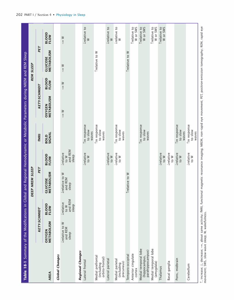
Research attention has focused on the hypothalamus playing a significant role in arousal and in regulating transitions between sleep and waking states. Specifically, the tuberomamillary histaminergic neurons and the peri-fornical hypocretin neurons in the posterior hypothalamus have extensive interconnections and interactions with the basic arousal systems (for more information see Chapters 7, 8, 21, and 33). Activity changes in these primary regulating areas result in profound modifications in activity patterns in thalamocortical circuits and associ-ated structures, such as basal ganglia or cerebellum. A primary aim of functional imaging studies has been to characterize this reorganization of regional brain func-tion during normal human sleep as well as the responses to external stimuli and the influence of previous waking experience on regional brain activity during sleep. The results detailed below are summarized in Table 18-1 (Fig. 18-1).
NON–RAPID EYE MOVEMENT SLEEPNeurophysiologic recordings in sleeping animals indicate that during non–rapid eye movement (NREM) sleep, the neural activity of the brain is shaped by a slow rhythm (<1 Hz), characterized by a fundamental oscillation of membrane potential made up of a depolarizing phase, associated with important neuronal firing (up state), fol-lowed by a hyperpolarizing phase, during which cortical neurons remain silent for a few hundred milliseconds (down state).1,2 The slow oscillation occurs synchronously in large neuronal populations in such a way that it can be reflected on EEG recordings as high-amplitude low- frequency waves.1,3-5 The slow rhythm entrains other sleep oscillations in a coalescence of multiple rhythms.6 Among the latter, spindles are associated with burst firing in thala-mocortical populations. They arise from a cyclic inhibition
What Brain Imaging Reveals about Sleep Generation and MaintenanceEric A. Nofzinger and Pierre Maquet
Chapter
18
L
202 PART I / Section 4 • Physiology in Sleep
Tab
le 1
8-1
Sum
mar
y of
the
Modifi
cati
ons
in G
lobal
and R
egio
nal
Hem
odyn
amic
or
Met
abolic
Par
amet
ers
duri
ng N
REM
and R
EM S
leep
AR
EA
DEEP N
REM
SLEEP
REM
SLEEP
KET
Y-S
CH
MID
TPET
fMR
IK
ET
Y-S
CH
MID
TPET
OX
YG
EN
M
ETA
BO
LIS
MBLO
OD
FLO
WG
LU
CO
SE
META
BO
LIS
MBLO
OD
FLO
WBO
LD
SIG
NA
LO
XY
GEN
M
ETA
BO
LIS
MBLO
OD
FLO
WG
LU
CO
SE
META
BO
LIS
MBLO
OD
FLO
W
Glo
bal
Changes
↓rel
ativ
e to
W
and R
EM
slee
p
↓rel
ativ
e to
W
and R
EM
slee
p
↓rel
ativ
e to
W
and R
EM
slee
p
↓rel
ativ
e to
W
and R
EM
slee
p
~=
W~
= W
~=
W~
= W
Regio
nal
Changes
Late
ral
fronta
l↓r
elat
ive
to W
↑in r
esponse
to
slo
w
wav
es
↓rel
ativ
e to
W
Med
ial
pre
fronta
l (incl
udin
g
orb
ito-f
ronta
l)
↑in r
esponse
to
slo
w
wav
es
↑rel
ativ
e to
W
Late
ral
par
ieta
l↓r
elat
ive
to W
↓rel
ativ
e to
W
Med
ial
par
ieta
l (incl
udin
g
pre
cuneu
s)
↓rel
ativ
e to
W↑i
n r
esponse
to
slo
w
wav
es
↓rel
ativ
e to
W
Tem
poro
-occ
ipit
al↑r
elat
ive
to W
↑rel
ativ
e to
W
Ante
rior
cingula
te
cort
ex↑r
elat
ive
to
W o
r SW
S
Med
ial
tem
pora
l lo
be
(hip
poca
mpus,
par
ahip
poca
mpus)
↑in r
esponse
to
slo
w
wav
es
↑rel
ativ
e to
W
or
SWS
Med
ial
tem
pora
l lo
be
(am
ygdal
a)↑r
elat
ive
to
W o
r SW
S
Thal
amus
↓rel
ativ
e to
W↑r
elat
ive
to
W o
r SW
S
Basa
l gan
glia
↓rel
ativ
e to
W
Pons,
mid
bra
in↓r
elat
ive
to W
↑in r
esponse
to
slo
w
wav
es
Cer
ebel
lum
↓rel
ativ
e to
W↑i
n r
esponse
to
slo
w
wav
es
↑, i
ncr
ease
; ↓,
dec
reas
e; ~
=,
about
sam
e ac
tivi
ty;
fMR
I, f
unct
ional
mag
net
ic r
esonan
ce i
mag
ing;
NR
EM,
non–r
apid
eye
move
men
t; P
ET,
posi
tron-e
mis
sion t
om
ogra
phy;
REM
, ra
pid
eye
m
ove
men
t; S
WS,
slo
w-w
ave
slee
p;
W,
wak
efuln
ess.
L
CHAPTER 18 • What Brain Imaging Reveals about Sleep Generation and Maintenance 203
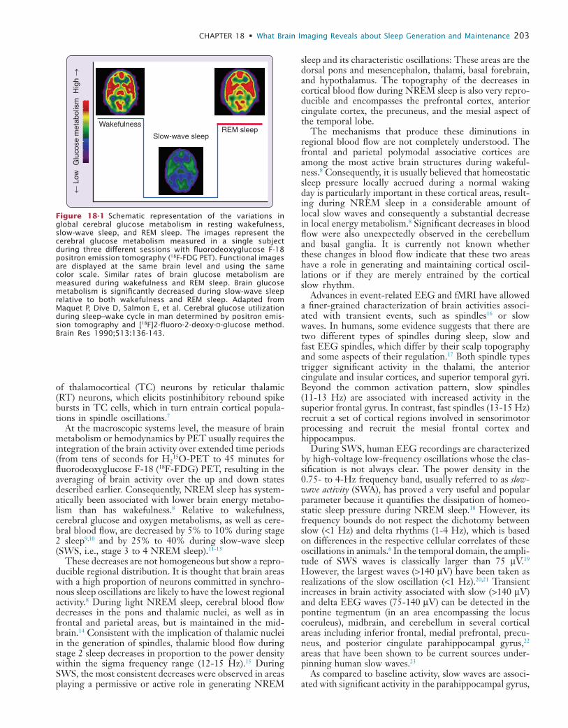
sleep and its characteristic oscillations: These areas are the dorsal pons and mesencephalon, thalami, basal forebrain, and hypothalamus. The topography of the decreases in cortical blood flow during NREM sleep is also very repro-ducible and encompasses the prefrontal cortex, anterior cingulate cortex, the precuneus, and the mesial aspect of the temporal lobe.
The mechanisms that produce these diminutions in regional blood flow are not completely understood. The frontal and parietal polymodal associative cortices are among the most active brain structures during wakeful-ness.8 Consequently, it is usually believed that homeostatic sleep pressure locally accrued during a normal waking day is particularly important in these cortical areas, result-ing during NREM sleep in a considerable amount of local slow waves and consequently a substantial decrease in local energy metabolism.8 Significant decreases in blood flow were also unexpectedly observed in the cerebellum and basal ganglia. It is currently not known whether these changes in blood flow indicate that these two areas have a role in generating and maintaining cortical oscil-lations or if they are merely entrained by the cortical slow rhythm.
Advances in event-related EEG and fMRI have allowed a finer-grained characterization of brain activities associ-ated with transient events, such as spindles16 or slow waves. In humans, some evidence suggests that there are two different types of spindles during sleep, slow and fast EEG spindles, which differ by their scalp topography and some aspects of their regulation.17 Both spindle types trigger significant activity in the thalami, the anterior cingulate and insular cortices, and superior temporal gyri. Beyond the common activation pattern, slow spindles (11-13 Hz) are associated with increased activity in the superior frontal gyrus. In contrast, fast spindles (13-15 Hz) recruit a set of cortical regions involved in sensorimotor processing and recruit the mesial frontal cortex and hippocampus.
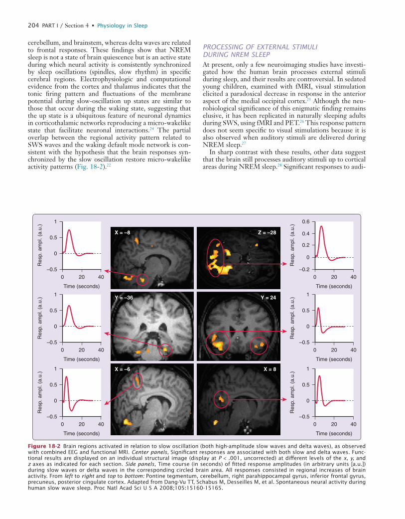
During SWS, human EEG recordings are characterized by high-voltage low-frequency oscillations whose the clas-sification is not always clear. The power density in the 0.75- to 4-Hz frequency band, usually referred to as slow-wave activity (SWA), has proved a very useful and popular parameter because it quantifies the dissipation of homeo-static sleep pressure during NREM sleep.18 However, its frequency bounds do not respect the dichotomy between slow (<1 Hz) and delta rhythms (1-4 Hz), which is based on differences in the respective cellular correlates of these oscillations in animals.6 In the temporal domain, the ampli-tude of SWS waves is classically larger than 75 µV.19 However, the largest waves (>140 µV) have been taken as realizations of the slow oscillation (<1 Hz).20,21 Transient increases in brain activity associated with slow (>140 µV) and delta EEG waves (75-140 µV) can be detected in the pontine tegmentum (in an area encompassing the locus coeruleus), midbrain, and cerebellum in several cortical areas including inferior frontal, medial prefrontal, precu-neus, and posterior cingulate parahippocampal gyrus,22 areas that have been shown to be current sources under-pinning human slow waves.23
As compared to baseline activity, slow waves are associ-ated with significant activity in the parahippocampal gyrus,
of thalamocortical (TC) neurons by reticular thalamic (RT) neurons, which elicits postinhibitory rebound spike bursts in TC cells, which in turn entrain cortical popula-tions in spindle oscillations.7
At the macroscopic systems level, the measure of brain metabolism or hemodynamics by PET usually requires the integration of the brain activity over extended time periods (from tens of seconds for H2
15O-PET to 45 minutes for fluorodeoxyglucose F-18 (18F-FDG) PET, resulting in the averaging of brain activity over the up and down states described earlier. Consequently, NREM sleep has system-atically been associated with lower brain energy metabo-lism than has wakefulness.8 Relative to wakefulness, cerebral glucose and oxygen metabolisms, as well as cere-bral blood flow, are decreased by 5% to 10% during stage 2 sleep9,10 and by 25% to 40% during slow-wave sleep (SWS, i.e., stage 3 to 4 NREM sleep).11-13
These decreases are not homogeneous but show a repro-ducible regional distribution. It is thought that brain areas with a high proportion of neurons committed in synchro-nous sleep oscillations are likely to have the lowest regional activity.8 During light NREM sleep, cerebral blood flow decreases in the pons and thalamic nuclei, as well as in frontal and parietal areas, but is maintained in the mid-brain.14 Consistent with the implication of thalamic nuclei in the generation of spindles, thalamic blood flow during stage 2 sleep decreases in proportion to the power density within the sigma frequency range (12-15 Hz).15 During SWS, the most consistent decreases were observed in areas playing a permissive or active role in generating NREM
Slow-wave sleep
WakefulnessREM sleep
← L
owH
igh
→G
luco
se m
etab
olis
m
Figure 18-1 Schematic representation of the variations in global cerebral glucose metabolism in resting wakefulness, slow-wave sleep, and REM sleep. The images represent the cerebral glucose metabolism measured in a single subject during three different sessions with fluorodeoxyglucose F-18 positron emission tomography (18F-FDG PET). Functional images are displayed at the same brain level and using the same color scale. Similar rates of brain glucose metabolism are measured during wakefulness and REM sleep. Brain glucose metabolism is significantly decreased during slow-wave sleep relative to both wakefulness and REM sleep. Adapted from Maquet P, Dive D, Salmon E, et al. Cerebral glucose utilization during sleep–wake cycle in man determined by positron emis-sion tomography and [18F]2-fluoro-2-deoxy-D-glucose method. Brain Res 1990;513:136-143.
L
204 PART I / Section 4 • Physiology in Sleep
PROCESSING OF EXTERNAL STIMULI DURING NREM SLEEPAt present, only a few neuroimaging studies have investi-gated how the human brain processes external stimuli during sleep, and their results are controversial. In sedated young children, examined with fMRI, visual stimulation elicited a paradoxical decrease in response in the anterior aspect of the medial occipital cortex.25 Although the neu-robiological significance of this enigmatic finding remains elusive, it has been replicated in naturally sleeping adults during SWS, using fMRI and PET.26 This response pattern does not seem specific to visual stimulations because it is also observed when auditory stimuli are delivered during NREM sleep.27
In sharp contrast with these results, other data suggest that the brain still processes auditory stimuli up to cortical areas during NREM sleep.28 Significant responses to audi-
cerebellum, and brainstem, whereas delta waves are related to frontal responses. These findings show that NREM sleep is not a state of brain quiescence but is an active state during which neural activity is consistently synchronized by sleep oscillations (spindles, slow rhythm) in specific cerebral regions. Electrophysiologic and computational evidence from the cortex and thalamus indicates that the tonic firing pattern and fluctuations of the membrane potential during slow-oscillation up states are similar to those that occur during the waking state, suggesting that the up state is a ubiquitous feature of neuronal dynamics in corticothalamic networks reproducing a micro-wakelike state that facilitate neuronal interactions.24 The partial overlap between the regional activity pattern related to SWS waves and the waking default mode network is con-sistent with the hypothesis that the brain responses syn-chronized by the slow oscillation restore micro-wakelike activity patterns (Fig. 18-2).22
X = –8 Z = –28
Y = –36
X = –6
Y = 24
X = 8
0
0.2
0.4
0.6
–0.2
Res
p. a
mpl
. (a.
u.)
0 20 40
Time (seconds)
0
0.5
1
–0.5
Res
p. a
mpl
. (a.
u.)
0 20 40
Time (seconds)
0
0.5
1
–0.5
Res
p. a
mpl
. (a.
u.)
0 20 40
Time (seconds)
0
0.5
1
–0.5
Res
p. a
mpl
. (a.
u.)
0 20 40
Time (seconds)
0
0.5
1
–0.5
Res
p. a
mpl
. (a.
u.)
0 20 40
Time (seconds)
0
0.5
1
–0.5
Res
p. a
mpl
. (a.
u.)
0 20 40
Time (seconds)
Figure 18-2 Brain regions activated in relation to slow oscillation (both high-amplitude slow waves and delta waves), as observed with combined EEG and functional MRI. Center panels, Significant responses are associated with both slow and delta waves. Func-tional results are displayed on an individual structural image (display at P < .001, uncorrected) at different levels of the x, y, and z axes as indicated for each section. Side panels, Time course (in seconds) of fitted response amplitudes (in arbitrary units [a.u.]) during slow waves or delta waves in the corresponding circled brain area. All responses consisted in regional increases of brain activity. From left to right and top to bottom: Pontine tegmentum, cerebellum, right parahippocampal gyrus, inferior frontal gyrus, precuneus, posterior cingulate cortex. Adapted from Dang-Vu TT, Schabus M, Desseilles M, et al. Spontaneous neural activity during human slow wave sleep. Proc Natl Acad Sci U S A 2008;105:15160-15165.
L
CHAPTER 18 • What Brain Imaging Reveals about Sleep Generation and Maintenance 205
Although not reported in all studies, posterior cortices in temporooccipital areas are typically activated during REM sleep.11 In contrast, the dorsolateral prefrontal cortex, parietal cortex, posterior cingulate cortex, and pre-cuneus are the least active brain regions.11,29 Although early animal studies had already mentioned the high limbic activity during REM sleep, functional neuroimaging in humans highlighted the contrast between the activation of limbic, paralimbic, and posterior cortical areas on the one hand and the relative quiescence of the associative frontal and parietal cortices on the other hand.
Regional functional integration is also modified during REM sleep, relative to wakefulness. For instance, the functional interactions between striate and extrastriate cortices, which are positive during wakefulness, become negative during REM sleep.31 Likewise, the functional connectivity between the amygdala and temporooccipital areas is tighter during REM sleep than during resting wakefulness.32
The organization of human brain function during REM sleep somehow relates to some of the characteristics of dreaming activity.29,33,34 The perceptual aspects of dreams
tory stimuli were detected in bilateral auditory cortex, thalamus, and caudate nuclei during wakefulness and light NREM sleep. In addition, the left amygdala and the left prefrontal cortex are recruited by stimuli having particular affective significance for the individual subject. (More information on how the brain process sensory input during sleep is found in Chapter 30).
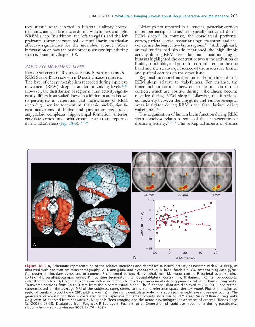
RAPID EYE MOVEMENT SLEEPReorganization of Regional Brain Function during REM Sleep: Relation with Dream CharacteristicsThe level of energy metabolism recorded during rapid eye movement (REM) sleep is similar to waking levels.12,13 However, the distribution of regional brain activity signifi-cantly differs from wakefulness. In addition to areas known to participate in generation and maintenance of REM sleep (e.g., pontine tegmentum, thalamic nuclei), signifi-cant activations of limbic and paralimbic areas (e.g., amygdaloid complexes, hippocampal formation, anterior cingulate cortex, and orbitofrontal cortex) are reported during REM sleep (Fig. 18-3).11,29,30
A
B
A
PH
T-O
F
Ca CpO
ThB
PTA,H
T-O
P F
–4 mm –2 mm 0 mm
–15
–10
–5
0
5
10
rCB
F [1
6 –2
8 –2
]
REMs density
–20 0 20 40 60
Figure 18-3 A, Schematic representation of the relative increases and decreases in neural activity associated with REM sleep, as observed with positron emission tomography. A,H, amygdala and hyppocampus; B, basal forebrain; Ca, anterior cingulate gyrus; Cp, posterior cingulate gyrus and precuneus; F, prefrontal cortex; H, hypothalamus; M, motor cortex; P, parietal supramarginal cortex; PH, parahippocampic gyrus; PT, pontine tegmentum; O, occipital-lateral cortex; Th, thalamus; T-O, temporooccipital extrastriate cortex; B, Cerebral areas more active in relation to rapid eye movements during paradoxical sleep than during wake. Transverse sections from 24 to 0 mm from the bicommissural plane. The functional data are displayed at P < .001 uncorrected, superimposed on the average MRI of the subjects, coregistered to the same reference space. Bottom panel, Plot of the adjusted regional cerebral blood flow (rCBF; arbitrary units) in the right geniculate body in relation to the rapid eye movement counts. The geniculate cerebral blood flow is correlated to the rapid eye movement counts more during REM sleep (in red) than during wake (in green). (A adapted from Schwartz S, Maquet P. Sleep imaging and the neuro-psychological assessment of dreams. Trends Cogn Sci 2002;6:23-30. B adapted from Peigneux P, Laureys S, Fuchs S, et al. Generation of rapid eye movements during paradoxical sleep in humans. Neuroimage 2001;14:701-708.)
L
206 PART I / Section 4 • Physiology in Sleep
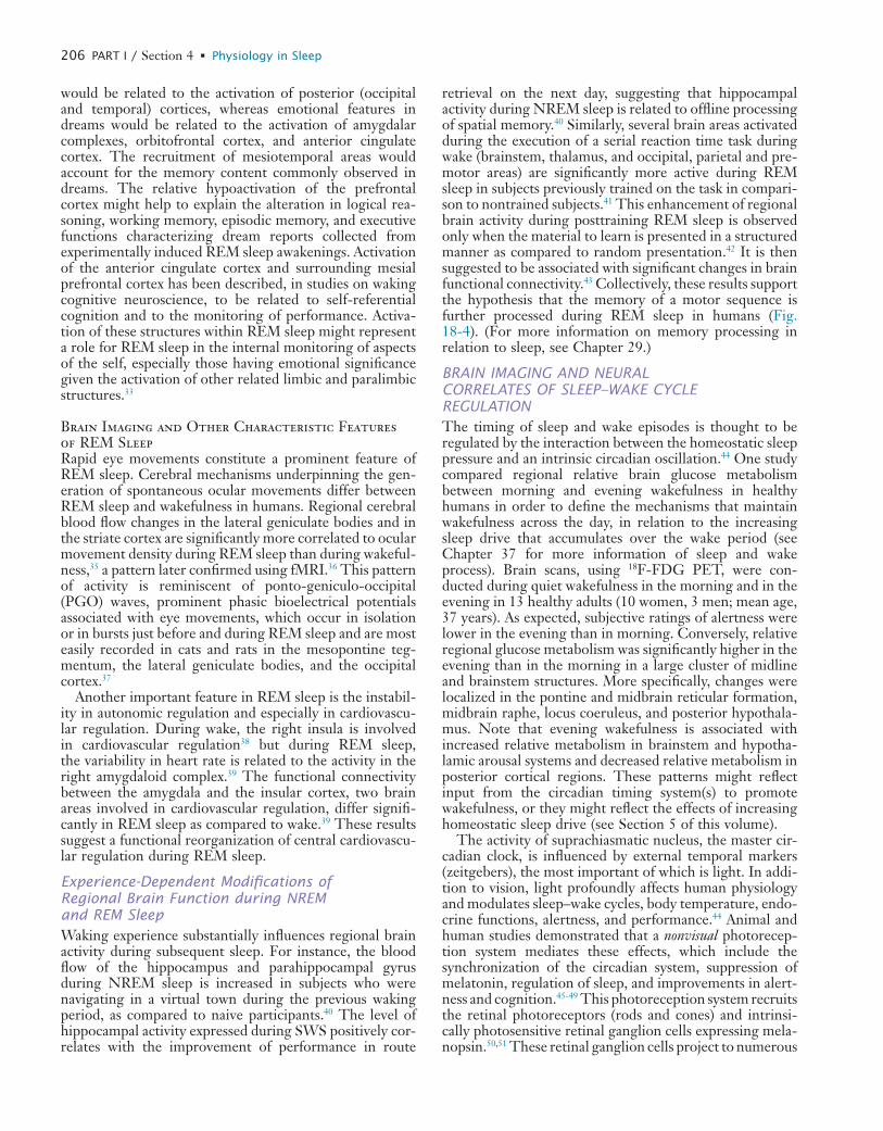
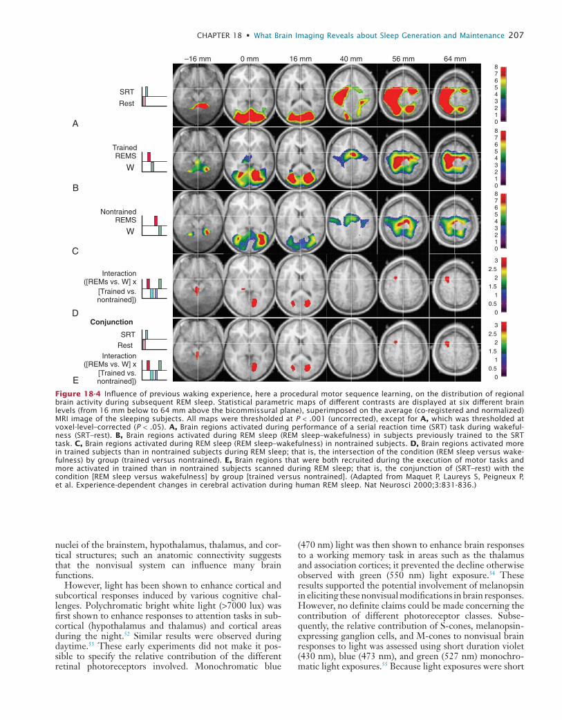
retrieval on the next day, suggesting that hippocampal activity during NREM sleep is related to offline processing of spatial memory.40 Similarly, several brain areas activated during the execution of a serial reaction time task during wake (brainstem, thalamus, and occipital, parietal and pre-motor areas) are significantly more active during REM sleep in subjects previously trained on the task in compari-son to nontrained subjects.41 This enhancement of regional brain activity during posttraining REM sleep is observed only when the material to learn is presented in a structured manner as compared to random presentation.42 It is then suggested to be associated with significant changes in brain functional connectivity.43 Collectively, these results support the hypothesis that the memory of a motor sequence is further processed during REM sleep in humans (Fig. 18-4). (For more information on memory processing in relation to sleep, see Chapter 29.)
BRAIN IMAGING AND NEURAL CORRELATES OF SLEEP–WAKE CYCLE REGULATIONThe timing of sleep and wake episodes is thought to be regulated by the interaction between the homeostatic sleep pressure and an intrinsic circadian oscillation.44 One study compared regional relative brain glucose metabolism between morning and evening wakefulness in healthy humans in order to define the mechanisms that maintain wakefulness across the day, in relation to the increasing sleep drive that accumulates over the wake period (see Chapter 37 for more information of sleep and wake process). Brain scans, using 18F-FDG PET, were con-ducted during quiet wakefulness in the morning and in the evening in 13 healthy adults (10 women, 3 men; mean age, 37 years). As expected, subjective ratings of alertness were lower in the evening than in morning. Conversely, relative regional glucose metabolism was significantly higher in the evening than in the morning in a large cluster of midline and brainstem structures. More specifically, changes were localized in the pontine and midbrain reticular formation, midbrain raphe, locus coeruleus, and posterior hypothala-mus. Note that evening wakefulness is associated with increased relative metabolism in brainstem and hypotha-lamic arousal systems and decreased relative metabolism in posterior cortical regions. These patterns might reflect input from the circadian timing system(s) to promote wakefulness, or they might reflect the effects of increasing homeostatic sleep drive (see Section 5 of this volume).
The activity of suprachiasmatic nucleus, the master cir-cadian clock, is influenced by external temporal markers (zeitgebers), the most important of which is light. In addi-tion to vision, light profoundly affects human physiology and modulates sleep–wake cycles, body temperature, endo-crine functions, alertness, and performance.44 Animal and human studies demonstrated that a nonvisual photorecep-tion system mediates these effects, which include the synchronization of the circadian system, suppression of melatonin, regulation of sleep, and improvements in alert-ness and cognition.45-49 This photoreception system recruits the retinal photoreceptors (rods and cones) and intrinsi-cally photosensitive retinal ganglion cells expressing mela-nopsin.50,51 These retinal ganglion cells project to numerous
would be related to the activation of posterior (occipital and temporal) cortices, whereas emotional features in dreams would be related to the activation of amygdalar complexes, orbitofrontal cortex, and anterior cingulate cortex. The recruitment of mesiotemporal areas would account for the memory content commonly observed in dreams. The relative hypoactivation of the prefrontal cortex might help to explain the alteration in logical rea-soning, working memory, episodic memory, and executive functions characterizing dream reports collected from experimentally induced REM sleep awakenings. Activation of the anterior cingulate cortex and surrounding mesial prefrontal cortex has been described, in studies on waking cognitive neuroscience, to be related to self-referential cognition and to the monitoring of performance. Activa-tion of these structures within REM sleep might represent a role for REM sleep in the internal monitoring of aspects of the self, especially those having emotional significance given the activation of other related limbic and paralimbic structures.33
Brain Imaging and Other Characteristic Features of REM SleepRapid eye movements constitute a prominent feature of REM sleep. Cerebral mechanisms underpinning the gen-eration of spontaneous ocular movements differ between REM sleep and wakefulness in humans. Regional cerebral blood flow changes in the lateral geniculate bodies and in the striate cortex are significantly more correlated to ocular movement density during REM sleep than during wakeful-ness,35 a pattern later confirmed using fMRI.36 This pattern of activity is reminiscent of ponto-geniculo-occipital (PGO) waves, prominent phasic bioelectrical potentials associated with eye movements, which occur in isolation or in bursts just before and during REM sleep and are most easily recorded in cats and rats in the mesopontine teg-mentum, the lateral geniculate bodies, and the occipital cortex.37
Another important feature in REM sleep is the instabil-ity in autonomic regulation and especially in cardiovascu-lar regulation. During wake, the right insula is involved in cardiovascular regulation38 but during REM sleep, the variability in heart rate is related to the activity in the right amygdaloid complex.39 The functional connectivity between the amygdala and the insular cortex, two brain areas involved in cardiovascular regulation, differ signifi-cantly in REM sleep as compared to wake.39 These results suggest a functional reorganization of central cardiovascu-lar regulation during REM sleep.
Experience-Dependent Modifications of Regional Brain Function during NREM and REM SleepWaking experience substantially influences regional brain activity during subsequent sleep. For instance, the blood flow of the hippocampus and parahippocampal gyrus during NREM sleep is increased in subjects who were navigating in a virtual town during the previous waking period, as compared to naive participants.40 The level of hippocampal activity expressed during SWS positively cor-relates with the improvement of performance in route
L
CHAPTER 18 • What Brain Imaging Reveals about Sleep Generation and Maintenance 207
(470 nm) light was then shown to enhance brain responses to a working memory task in areas such as the thalamus and association cortices; it prevented the decline otherwise observed with green (550 nm) light exposure.54 These results supported the potential involvement of melanopsin in eliciting these nonvisual modifications in brain responses. However, no definite claims could be made concerning the contribution of different photoreceptor classes. Subse-quently, the relative contribution of S-cones, melanopsin-expressing ganglion cells, and M-cones to nonvisual brain responses to light was assessed using short duration violet (430 nm), blue (473 nm), and green (527 nm) monochro-matic light exposures.55 Because light exposures were short
nuclei of the brainstem, hypothalamus, thalamus, and cor-tical structures; such an anatomic connectivity suggests that the nonvisual system can influence many brain functions.
However, light has been shown to enhance cortical and subcortical responses induced by various cognitive chal-lenges. Polychromatic bright white light (>7000 lux) was first shown to enhance responses to attention tasks in sub-cortical (hypothalamus and thalamus) and cortical areas during the night.52 Similar results were observed during daytime.53 These early experiments did not make it pos-sible to specify the relative contribution of the different retinal photoreceptors involved. Monochromatic blue
–16 mm 0 mm 16 mm 40 mm 56 mm 64 mm876543210
876543210876543210
3
2
2.5
1.5
1
0.5
0
3
2
2.5
1.5
1
0.5
0
A
B
C
D
E
SRT
Rest
TrainedREMS
W
NontrainedREMS
W
Interaction([REMs vs. W] x
[Trained vs.nontrained])
Interaction([REMs vs. W] x
[Trained vs.nontrained])
Conjunction
SRT
Rest
Figure 18-4 Influence of previous waking experience, here a procedural motor sequence learning, on the distribution of regional brain activity during subsequent REM sleep. Statistical parametric maps of different contrasts are displayed at six different brain levels (from 16 mm below to 64 mm above the bicommissural plane), superimposed on the average (co-registered and normalized) MRI image of the sleeping subjects. All maps were thresholded at P < .001 (uncorrected), except for A, which was thresholded at voxel-level–corrected (P < .05). A, Brain regions activated during performance of a serial reaction time (SRT) task during wakeful-ness (SRT–rest). B, Brain regions activated during REM sleep (REM sleep–wakefulness) in subjects previously trained to the SRT task. C, Brain regions activated during REM sleep (REM sleep–wakefulness) in nontrained subjects. D, Brain regions activated more in trained subjects than in nontrained subjects during REM sleep; that is, the intersection of the condition (REM sleep versus wake-fulness) by group (trained versus nontrained). E, Brain regions that were both recruited during the execution of motor tasks and more activated in trained than in nontrained subjects scanned during REM sleep; that is, the conjunction of (SRT–rest) with the condition [REM sleep versus wakefulness] by group [trained versus nontrained]. (Adapted from Maquet P, Laureys S, Peigneux P, et al. Experience-dependent changes in cerebral activation during human REM sleep. Nat Neurosci 2000;3:831-836.)
L
208 PART I / Section 4 • Physiology in Sleep
before and after 32 hours of sleep deprivation. They noted prominent decreases in metabolism in the thalamus, basal ganglia, temporal lobes, and cerebellum and increases in visual cortex. Whole-brain absolute metabolic rate was not different.
Thomas and coworkers57,58 described the effects of 24 hours, 48 hours, and 72 hours of sleep deprivation on waking regional cerebral metabolism assessed via 18F-FDG PET, as well as alertness and cognitive performance. Sleep deprivation was associated with global declines in absolute cerebral metabolism. Regionally, these declines were most notable in frontoparietal cortex and in the thalamus. This is consistent with studies showing that the effects of sleep deprivation on SWS are greatest in frontal EEG leads. Alertness and cognitive performance on a sleep depriva-tion–sensitive serial addition and subtraction test declined in association with the sleep-deprivation–associated regional deactivations.
Paus and colleagues59-61 have demonstrated that blood flow in the thalamus and pontomesencephalic tegmentum as assessed by H2
15O PET positively correlates with arous-als in sleep,59 with performance on vigilance tasks60 and with loss of consciousness associated with anesthesia.61 In
lasting, this protocol allowed the detection of subcortical brain structures involved in early nonvisual responses to light, such as the thalamus and the brainstem.
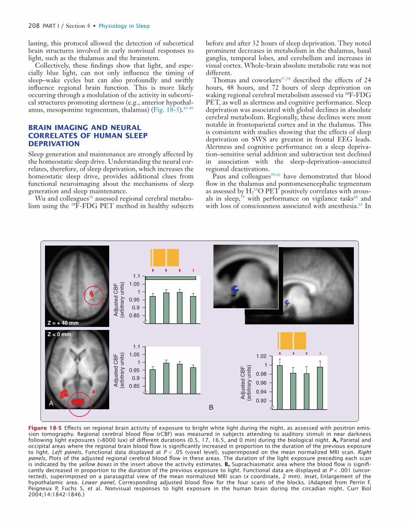
Collectively, these findings show that light, and espe-cially blue light, can not only influence the timing of sleep−wake cycles but can also profoundly and swiftly influence regional brain function. This is more likely occurring through a modulation of the activity in subcorti-cal structures promoting alertness (e.g., anterior hypothal-amus, mesopontine tegmentum, thalamus) (Fig. 18-5).45-49
BRAIN IMAGING AND NEURAL CORRELATES OF HUMAN SLEEP DEPRIVATIONSleep generation and maintenance are strongly affected by the homeostatic sleep drive. Understanding the neural cor-relates, therefore, of sleep deprivation, which increases the homeostatic sleep drive, provides additional clues from functional neuroimaging about the mechanisms of sleep generation and sleep maintenance.
Wu and colleagues56 assessed regional cerebral metabo-lism using the 18F-FDG PET method in healthy subjects
AB
Z = + 40 mm
Z = 0 mm
1.1
1.05
1
0.95
0.9
0.85Adj
uste
d C
BF
(arb
itrar
y un
its)
1.1
1.05
1
0.95
0.9
0.85Adj
uste
d C
BF
(arb
itrar
y un
its)
1.02
0.98
1
0.96
0.94
0.92Adj
uste
d C
BF
(arb
itrar
y un
its)
Figure 18-5 Effects on regional brain activity of exposure to bright white light during the night, as assessed with positron emis-sion tomography. Regional cerebral blood flow (rCBF) was measured in subjects attending to auditory stimuli in near darkness following light exposures (>8000 lux) of different durations (0.5, 17, 16.5, and 0 min) during the biological night. A, Parietal and occipital areas where the regional brain blood flow is significantly increased in proportion to the duration of the previous exposure to light. Left panels, Functional data displayed at P < .05 (voxel level), superimposed on the mean normalized MRI scan. Right panels, Plots of the adjusted regional cerebral blood flow in these areas. The duration of the light exposure preceding each scan is indicated by the yellow boxes in the insert above the activity estimates. B, Suprachiasmatic area where the blood flow is signifi-cantly decreased in proportion to the duration of the previous exposure to light. Functional data are displayed at P < .001 (uncor-rected), superimposed on a parasagittal view of the mean normalized MRI scan (x coordinate, 2 mm). Inset, Enlargement of the hypothalamic area. Lower panel, Corresponding adjusted blood flow for the four scans of the blocks. (Adapted from Perrin F, Peigneux P, Fuchs S, et al. Nonvisual responses to light exposure in the human brain during the circadian night. Curr Biol 2004;14:1842-1846.)
L
CHAPTER 18 • What Brain Imaging Reveals about Sleep Generation and Maintenance 209
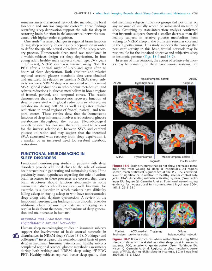
did insomnia subjects. The two groups did not differ on any measure of visually scored or automated measure of sleep. Grouping by state-interaction analysis confirmed that insomnia subjects showed a smaller decrease than did healthy subjects in relative glucose metabolism from waking to NREM sleep in the brainstem reticular core and in the hypothalamus. This study supports the concept that persistent activity in this basic arousal network may be responsible for the impaired objective and subjective sleep in insomnia patients (Figs. 18-6 and 18-7).
In terms of interventions, the action of sedative–hypnot-ics may be primarily on these basic arousal systems. For
some instances this arousal network also included the basal forebrain and anterior cingulate cortex.60 These findings regarding sleep deprivation support the role for sleep in restoring brain function in thalamocortical networks asso-ciated with higher-order cognition.
One study62 assessed changes in regional brain function during sleep recovery following sleep deprivation in order to define the specific neural correlates of the sleep recov-ery process. Homeostatic sleep need was modulated in a within-subjects design via sleep deprivation. In four young adult healthy male subjects (mean age, 24.9 years ± 1.2 years), NREM sleep was assessed using 18F-FDG PET after a normal night of sleep and again after 36 hours of sleep deprivation. Both absolute and relative regional cerebral glucose metabolic data were obtained and analyzed. In relation to baseline NREM sleep, sub-jects’ recovery NREM sleep was associated with increased SWA, global reductions in whole-brain metabolism, and relative reductions in glucose metabolism in broad regions of frontal, parietal, and temporal cortex. The results demonstrate that the homeostatic recovery function of sleep is associated with global reductions in whole-brain metabolism during NREM as well as greater relative reductions in broad regions of frontal, parietal, and tem-poral cortex. These results show that the homeostatic function of sleep in humans involves a reduction of glucose metabolism throughout the cortex. Neurobiological models of sleep homeostasis, therefore, need to account for the inverse relationship between SWA and cerebral glucose utilization and may suggest that the increased SWA associated with recovery from sleep deprivation is a marker of an increased need for cerebral metabolic restoration.
FUNCTIONAL NEUROIMAGING IN SLEEP DISORDERSFunctional neuroimaging studies in patients with sleep disorders provide additional clues to the role of various brain structures in generating and maintaining sleep. If the previously stated hypotheses regarding the role of various brain structures in these processes are correct, then these brain structures should function abnormally in some manner in patients who do not sleep well. Insomnia, for example, is a disorder in which patients have difficulty falling asleep or staying asleep or who have nonrestorative sleep along with daytime dysfunction. A review of the functional neuroimaging findings in this disorder provides additional clues, because new data are emerging on a regular basis about the neural mechanisms of sleep genera-tion and maintenance in humans.
Insomnia and Brainstem and Hypothalamic Arousal NetworksHuman sleep neuroimaging studies in insomnia subjects support the involvement of basic arousal networks in disturbances in NREM sleep (Video 18-1). Nofzinger and colleagues63 investigated the neurobiological basis of poor sleep in insomnia. Insomnia patients and healthy subjects completed regional cerebral glucose metabolic assessments during both waking and NREM sleep using 18F-FDG PET. Healthy subjects reported better sleep quality than
ARAS Hypothalamus
Mesial temporal cortex
Thalamus
ARAS
ARAS
Cingulate
Hypothalamus
Insular cortex
Mesial temporal cortex
Figure 18-6 Brain structures that do not show decreased meta-bolic rate from waking to sleep in insomniacs. All regions shown reach statistical significance at the P < .05, corrected, level of significance in relation to healthy sleeper control sub-jects. ARAS, Ascending reticular activating system. (From Nofz-inger EA, Buysse DJ, Germain A, et al. Functional neuroimaging evidence for hyperarousal in insomnia. Am J Psychiatry 2004; 161:2126-2131.)
Pontinebrainstem
ACC, medialprefrontal cortex
Thalamus Diffusethalamocortical network
Figure 18-7 Brain structures where metabolism during NREM sleep correlates with wakefulness after sleep onset in insomnia patients. ACC, anterior cingulate cortex. (From Nofzinger EA, Nissen C, Germain A, et al. Regional cerebral metabolic corre-lates of WASO during NREM sleep in insomnia. J Clin Sleep Med 2006;2(3):316-322.)
L
210 PART I / Section 4 • Physiology in Sleep
ventrally located system, with important contributions from the amygdala, has been shown to be fundamental to the initial experience of emotions and to the automatic generation of emotional responses. The function of this system is a reactive one in response to emotional stimuli. Other structures related to this system include the anterior insula, ventral striatum, and ventral regions of the anterior cingulate cortex and ventral prefrontal cortex. A more dor-sally located system, with important contributions from the dorsolateral prefrontal cortex, has been shown to be fundamental to the conscious, planned regulation of emo-tional behavior in light of future behavior. The function of this system is one of planning behavior in response to emotional stimuli. Other structures related to this system include the hippocampus and the dorsal regions of the anterior cingulate cortex. A primary structure in the ventral system is the amygdala. It has been shown to participate in the sensory component of emotional behavior and in the initial organization of a reactive emotional response. In humans, the amygdala shows increased activation in response to a variety of emotional stimuli including fearful faces, sad faces, threatening words, and fearful vocaliza-tions. A reactive motor role for the amygdala includes the recruitment and coordinating of cortical arousal and vigi-lant attention for optimizing sensory and perceptual processing of stimuli associated with underdetermined contingencies.
Recent work shows that the amygdala is anatomically connected with and functionally modulates effects on the brainstem centers involved in arousal and sleep regulation. Similarly, other components of the ventral emotional system such as the ventral striatum, the subgenual anterior cingulate cortex, and the ventromedial prefrontal cortex are known to have anatomic and functional relationships with brainstem centers that are thought to play a role in behavioral state regulation in addition to the primary roles they each play in cortical arousal.
Human sleep neuroimaging studies support the role for components of the ventral emotional system in pathological sleep associated with both depression and insomnia. Nofz-inger and colleagues66 used 18F-FDG PET to define
example, Kajimura’s group64 assessed regional cerebral blood flow, as a correlate of neuronal activity, during NREM sleep in response to triazolam, a short-acting ben-zodiazepine sedative–hypnotic. They found that blood flow in the basal forebrain was lower during NREM sleep following administration of triazolam than following administration of placebo.
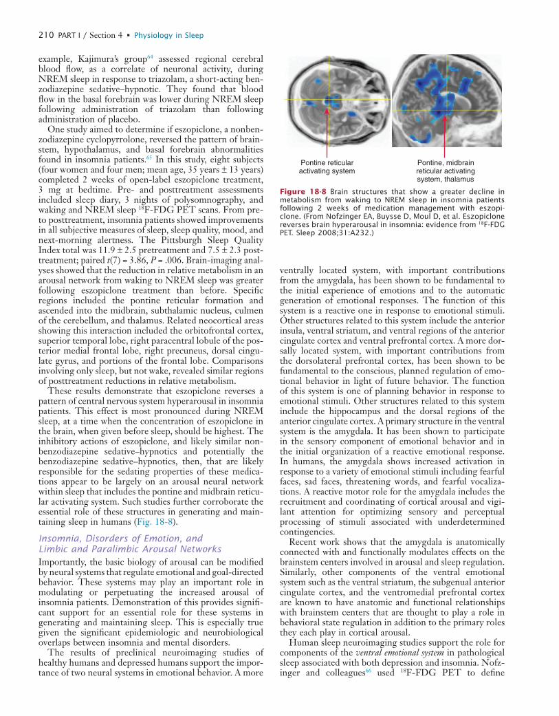
One study aimed to determine if eszopiclone, a nonben-zodiazepine cyclopyrrolone, reversed the pattern of brain-stem, hypothalamus, and basal forebrain abnormalities found in insomnia patients.65 In this study, eight subjects (four women and four men; mean age, 35 years ± 13 years) completed 2 weeks of open-label eszopiclone treatment, 3 mg at bedtime. Pre- and posttreatment assessments included sleep diary, 3 nights of polysomnography, and waking and NREM sleep 18F-FDG PET scans. From pre- to posttreatment, insomnia patients showed improvements in all subjective measures of sleep, sleep quality, mood, and next-morning alertness. The Pittsburgh Sleep Quality Index total was 11.9 ± 2.5 pretreatment and 7.5 ± 2.3 post-treatment; paired t(7) = 3.86, P = .006. Brain-imaging anal-yses showed that the reduction in relative metabolism in an arousal network from waking to NREM sleep was greater following eszopiclone treatment than before. Specific regions included the pontine reticular formation and ascended into the midbrain, subthalamic nucleus, culmen of the cerebellum, and thalamus. Related neocortical areas showing this interaction included the orbitofrontal cortex, superior temporal lobe, right paracentral lobule of the pos-terior medial frontal lobe, right precuneus, dorsal cingu-late gyrus, and portions of the frontal lobe. Comparisons involving only sleep, but not wake, revealed similar regions of posttreatment reductions in relative metabolism.
These results demonstrate that eszopiclone reverses a pattern of central nervous system hyperarousal in insomnia patients. This effect is most pronounced during NREM sleep, at a time when the concentration of eszopiclone in the brain, when given before sleep, should be highest. The inhibitory actions of eszopiclone, and likely similar non-benzodiazepine sedative–hypnotics and potentially the benzodiazepine sedative–hypnotics, then, that are likely responsible for the sedating properties of these medica-tions appear to be largely on an arousal neural network within sleep that includes the pontine and midbrain reticu-lar activating system. Such studies further corroborate the essential role of these structures in generating and main-taining sleep in humans (Fig. 18-8).
Insomnia, Disorders of Emotion, and Limbic and Paralimbic Arousal NetworksImportantly, the basic biology of arousal can be modified by neural systems that regulate emotional and goal-directed behavior. These systems may play an important role in modulating or perpetuating the increased arousal of insomnia patients. Demonstration of this provides signifi-cant support for an essential role for these systems in generating and maintaining sleep. This is especially true given the significant epidemiologic and neurobiological overlaps between insomnia and mental disorders.
The results of preclinical neuroimaging studies of healthy humans and depressed humans support the impor-tance of two neural systems in emotional behavior. A more
Pontine reticularactivating system
Pontine, midbrainreticular activatingsystem, thalamus
Figure 18-8 Brain structures that show a greater decline in metabolism from waking to NREM sleep in insomnia patients following 2 weeks of medication management with eszopi-clone. (From Nofzinger EA, Buysse D, Moul D, et al. Eszopiclone reverses brain hyperarousal in insomnia: evidence from 18F-FDG PET. Sleep 2008;31:A232.)
L
CHAPTER 18 • What Brain Imaging Reveals about Sleep Generation and Maintenance 211
jects during both waking and NREM sleep using 18F-FDG PET. Insomnia subjects scored worse on measures of daytime concentration and fatigue consistent with prefron-tal cortex impairment. Insomnia patients showed increased global cerebral glucose metabolism during sleep and wake, suggesting an increased vigilant or attentive function of the neocortex consistent with hyperarousal. A group by state interaction analysis confirmed that insomnia subjects showed a smaller decrease than did healthy subjects in relative metabolism from waking to NREM sleep in the thalamus, the anterior cingulate, and medial prefrontal cortices, suggesting a persistence of thalamocortical arousal even within sleep in insomnia patients. While awake, in relation to healthy subjects, insomnia subjects showed rela-tive hypometabolism in a broad region of the frontal cortex bilaterally; left hemispheric superior temporal, parietal, and occipital cortices; and the thalamus. Their daytime fatigue might reflect decreased activity in prefrontal cortex that results from inefficient sleep.
Several interventions may alter activity in the prefrontal cortex in a beneficial manner for insomnia patients. For example, Lou and colleagues69 assessed regional brain function associated with Yoga Nidra, a meditative state in which there is a loss of conscious control and an increased awareness of sensory experience. In their study, they found reduced blood flow during meditation in an attentional network that included the dorsolateral prefrontal cortex and anterior cingulate cortex, as well as increased blood flow in posterior sensory and associative cortex associated with visual imagery. Cognitive approaches to the treat-ment of insomnia may have similar mechanisms of action in prefrontal areas.
Serotoninergically active antidepressants also increase brain function (blood flow or metabolism) in dorsal paralimbic and dorsolateral prefrontal cortex. Increasing activity in prefrontal cortex might reverse prefrontal defi-cits in insomnia patients, leading to improved daytime cognitive function. Alternatively, further increase of an already metabolically overactive prefrontal cortex might increase attentive and vigilant functions, thereby produc-ing further insomnia, a not uncommon side effect of selective serotonin reuptake inhibitor (SSRI) therapy in depressed or insomnia patients.
One study has documented prefrontal hypoactivation in insomnia as predicted from the background review previously mentioned. This study investigated functional brain activation differences as a possible result of chronic insomnia, and the reversibility of these differences after nonmedicated sleep therapy. Twenty-one insomniac sub-jects and 12 carefully matched controls underwent fMRI scanning during the performance of a category and a letter-fluency task. Insomniac subjects were randomly assigned to either a 6-week period of nonpharmacologic sleep therapy or a wait-list period, after which fMRI scanning was repeated using parallel tasks. Task-related brain activation and number of generated words were considered as outcome measures. Compared to controls, insomnia patients showed hypoactivation of the medial and inferior prefrontal cortical areas (Brodmann areas 9, 44-45), which recovered after sleep therapy but not after a wait-list period. These studies support the hypothesis that insomnia interferes in a reversible fashion with
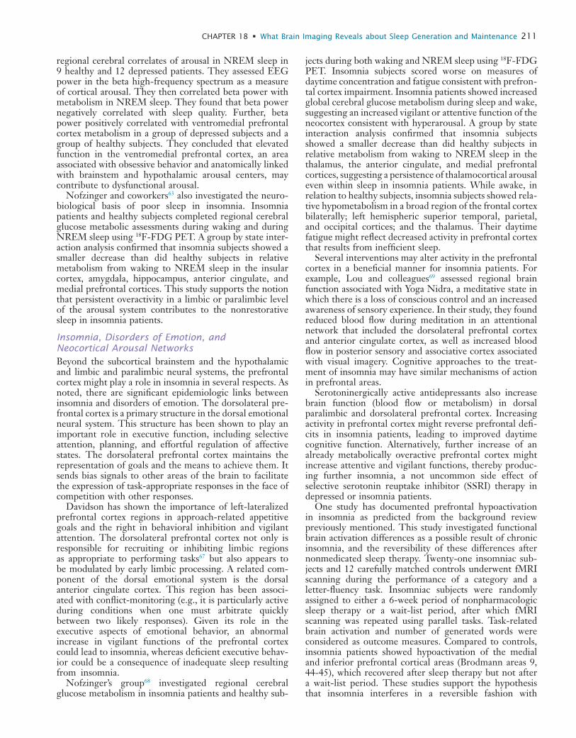
regional cerebral correlates of arousal in NREM sleep in 9 healthy and 12 depressed patients. They assessed EEG power in the beta high-frequency spectrum as a measure of cortical arousal. They then correlated beta power with metabolism in NREM sleep. They found that beta power negatively correlated with sleep quality. Further, beta power positively correlated with ventromedial prefrontal cortex metabolism in a group of depressed subjects and a group of healthy subjects. They concluded that elevated function in the ventromedial prefrontal cortex, an area associated with obsessive behavior and anatomically linked with brainstem and hypothalamic arousal centers, may contribute to dysfunctional arousal.
Nofzinger and coworkers63 also investigated the neuro-biological basis of poor sleep in insomnia. Insomnia patients and healthy subjects completed regional cerebral glucose metabolic assessments during waking and during NREM sleep using 18F-FDG PET. A group by state inter-action analysis confirmed that insomnia subjects showed a smaller decrease than did healthy subjects in relative metabolism from waking to NREM sleep in the insular cortex, amygdala, hippocampus, anterior cingulate, and medial prefrontal cortices. This study supports the notion that persistent overactivity in a limbic or paralimbic level of the arousal system contributes to the nonrestorative sleep in insomnia patients.
Insomnia, Disorders of Emotion, and Neocortical Arousal NetworksBeyond the subcortical brainstem and the hypothalamic and limbic and paralimbic neural systems, the prefrontal cortex might play a role in insomnia in several respects. As noted, there are significant epidemiologic links between insomnia and disorders of emotion. The dorsolateral pre-frontal cortex is a primary structure in the dorsal emotional neural system. This structure has been shown to play an important role in executive function, including selective attention, planning, and effortful regulation of affective states. The dorsolateral prefrontal cortex maintains the representation of goals and the means to achieve them. It sends bias signals to other areas of the brain to facilitate the expression of task-appropriate responses in the face of competition with other responses.
Davidson has shown the importance of left-lateralized prefrontal cortex regions in approach-related appetitive goals and the right in behavioral inhibition and vigilant attention. The dorsolateral prefrontal cortex not only is responsible for recruiting or inhibiting limbic regions as appropriate to performing tasks67 but also appears to be modulated by early limbic processing. A related com-ponent of the dorsal emotional system is the dorsal anterior cingulate cortex. This region has been associ-ated with conflict-monitoring (e.g., it is particularly active during conditions when one must arbitrate quickly between two likely responses). Given its role in the executive aspects of emotional behavior, an abnormal increase in vigilant functions of the prefrontal cortex could lead to insomnia, whereas deficient executive behav-ior could be a consequence of inadequate sleep resulting from insomnia.
Nofzinger’s group68 investigated regional cerebral glucose metabolism in insomnia patients and healthy sub-
L
212 PART I / Section 4 • Physiology in Sleep
with a mutation at codon 178 of the prion protein gene. Thalamic hypometabolism was found in all cases, and more widespread nonspecific cortical hypometabolism was noted in some. Perani and colleagues suggest that the thalamic dysfunction is consistent with the neuropatho-logic findings in the disorder and is a hallmark of the disease.
Kloppel’s group71 reported the results of a [123I] β-CIT SPECT study in two cases of fatal familial insomnia. They showed a 57% and 73% reduced availability of serotonin transporters in a thalamus-hypothalamus region in the two patients in relation to age-expected control values. Although the interpretation is not entirely clear, they suggest that this might reflect altered serotoninergic func-tion in regions of the brain thought to be important in sleep–wake regulation in this patient group.
SUMMARYThe application of functional neuroimaging methods to the study of sleep in health and disease in human subjects has provided unique insights into the neural mechanisms of sleep generation and maintenance. In many instances, these studies provide secondary support for the neural mechanisms of sleep generation and maintenance that have been discovered in preclinical research. They also provide unique insights into the involvement and interaction of broad neural networks at subcortical and cortical levels in a defined and regular manner to produce the final experi-ence of sleep in humans. Brain imaging studies are con-tributing to the understanding of how wake and sleep networks can behave pathologically to produce various sleep disorders and where treatments can reverse these abnormalities.
AcknowledgementsThe research data reported in this paper were collected with the help of grant from by the Belgian Fonds National de la Recherche Scientifique (FNRS), Fondation Médicale Reine Elisabeth (FMRE), Research Fund of the University of Liège, “Interuniversity Attraction Poles Programme—Belgian State—Belgian Science Policy,” and the United States National Institutes of Health (grants MH24652, AG00972, AG20677, RR00056, RR024153, MH30915).
activation of the prefrontal cortical system during daytime task performance.
REM Sleep in DepressionGiven that REM sleep activates limbic and anterior paralimbic cortex in healthy subjects, the increased REM sleep in depressed patients may reflect a greater re-activa-tion of these structures in REM sleep. Nofzinger and coworkers68 tested this hypothesis in 24 depressed patients and 14 healthy subjects. They underwent EEG sleep studies and regional cerebral glucose metabolism assess-ments during both waking and REM sleep using 18F-FDG PET. Depressed patients showed greater REM sleep per-centage. Consistent with the hypothesis that depressed patients would show increased activation in limbic and anterior limbic structures from waking to REM, depressed patients showed greater increases in relative metabolism from waking to REM sleep than healthy subjects in the midbrain reticular formation, including the pretectal area, and in a larger region of anterior paralimbic cortex. Addi-tionally, depressed patients showed greater increases in relative metabolism from waking to REM sleep than healthy subjects in a broadly distributed region of pre-dominantly left hemispheric dorsolateral prefrontal, pari-etal, and temporal cortex. This area included the frontal and parietal eye fields.
Increased activation of the brainstem reticular formation from waking to REM sleep in depressed patients is consis-tent with the model of an altered balance in brainstem monoaminergic (norepinephrine and serotonin) systems and brainstem acetylcholine neuronal systems in depressed patients. A second important finding in this study was the increased activation of limbic and anterior paralimbic (hip-pocampus, basal forebrain/ventral pallidum, anterior cin-gulate, and medial prefrontal) cortex from waking to REM sleep in the depressed patients. The highest density of cholinergic axons is in core limbic structures such as the hippocampus and amygdala. Limbic and anterior paralim-bic cortices also have high densities of inhibitory 5-hydroxy-tryptamine1A (5-HT1A) postsynaptic receptors in relation to other areas of cortex. Behaviorally, increased activation of limbic and paralimbic cortex in depressed patients may reflect a susceptibility of depressed patients to experience stimuli in a more affectively intense, negative context, given the increased activation of these structures in response to negatively valenced stimuli or increased affec-tive states. A third major finding in this study is the rela-tively greater activation of executive cortex from waking to REM sleep in depressed patients. This may reflect a change in modulation of cortical function from monoaminergic during waking to cholinergic in REM sleep, coupled with a monoaminergic or cholinergic imbalance in depressed patients. Behaviorally, this might also reflect a greater involvement of executive function during REM sleep in depressed patients, perhaps in response to the increased affective state produced by the abnormal re-activation of limbic and paralimbic cortex during REM sleep in depressed patients.
Fatal Familial InsomniaPerani and colleagues70 assessed cerebral metabolism in four patients with fatal familial insomnia, a prion disease
� Clinical Pearl
Neural systems related to sleep–wake regulation and the function of sleep overlap extensively with neural systems involved in essential aspects of waking cog-nitive and emotional behavior. Disruptions in sleep in patients with sleep disorders, therefore, can be associated with alterations in these neural systems, and, in turn, altered sleep leads to fundamental changes in these neural systems that impair waking behavior.
REFERENCES1. Steriade M, Contreras D, Curro-Dossi R, et al. The slow (<1 Hz)
oscillation in reticular thalamic and thalamocortical neurons: sce-nario of sleep rhythm generation in interacting thalamic and neocor-tical networks. J Neurosci 1993;13:3284-3299.
L
CHAPTER 18 • What Brain Imaging Reveals about Sleep Generation and Maintenance 213
28. Portas CM, Krakow K, Allen P, et al. Auditory processing across the sleep–wake cycle: simultaneous EEG and fMRI monitoring in humans. Neuron 2000;28:991-999.
29. Maquet P, Peters J, Aerts J, et al. Functional neuroanatomy of human rapid-eye-movement sleep and dreaming. Nature 1996;383:163- 166.
30. Nofzinger EA, Mintun MA, Wiseman MB, et al. Forebrain activation in REM sleep: an FDG PET study. Brain Res 1997;770:192-201.
31. Braun AR, Balkin TJ, Wesensten NJ, et al. Dissociated pattern of activity in visual cortices and their projections during human rapid eye movement sleep. Science 1998;279:91-95.
32. Maquet P, Phillips C. Functional brain imaging of human sleep. J Sleep Res 1998;7:42-47.
33. Maquet P, Ruby P, Maudoux A, et al. Human cognition during REM sleep and the activity profile within the frontal and parietal cortices: a reappraisal of functional neuroimaging data. Prog Brain Res 2005; 150:219-227.
34. Hobson JA, Pace-Schott EF, Stickgold R. Dreaming and the brain: towards a cognitive neuroscience of conscious states. Behav Brain Sci 2000;23:793-842.
35. Peigneux P, Laureys S, Fuchs S, et al. Generation of rapid eye move-ments during paradoxical sleep in humans. Neuroimage 2001;14: 701-708.
36. Wehrle R, Czisch M, Kaufmann C, et al. Rapid eye movement-related brain activation in human sleep: a functional magnetic reso-nance imaging study. Neuroreport 2005;16:853-857.
37. Callaway CW, Lydic R, Baghdoyan HA, et al. Pontogeniculooccipi-tal waves: spontaneous visual system activity during rapid eye move-ment sleep. Neurobiology 1987;7:105-149.
38. Critchley HD, Corfield DR, Chandler MP, et al. Cerebral correlates of autonomic cardiovascular arousal: a functional neuroimaging investigation in humans. J Physiol 2000;523:259-270.
39. Desseilles M, Dang-Vu T, Laureys S, et al. A prominent role for amygdaloid complexes in the variability in heart rate (VHR) during rapid eye movement (REM) sleep relative to wakefulness. Neuroim-age 2006;32:1008-1015.
40. Peigneux P, Laureys S, Fuchs S, et al. Are spatial memories strength-ened in the human hippocampus during slow wave sleep? Neuron 2004;44:535-545.
41. Maquet P, Laureys S, Peigneux P, et al. Experience-dependent changes in cerebral activation during human REM sleep. Nat Neu-rosci 2000;3:831-836.
42. Peigneux P, Laureys S, Fuchs S, et al. Learned material content and acquisition level modulate cerebral reactivation during posttraining rapid-eye-movements sleep. Neuroimage 2003;20:125-134.
43. Laureys S, Peigneux P, Phillips C, et al. Experience-dependent changes in cerebral functional connectivity during human rapid eye movement sleep. Neuroscience 2001;105:521-525.
44. Dijk DJ, Lockley SW. Integration of human sleep–wake regulation and circadian rhythmicity. J Appl Physiol 2002;92:852-862.
45. Cajochen C, Munch M, Kobialka S, et al. High sensitivity of human melatonin, alertness, thermoregulation, and heart rate to short wave-length light. J Clin Endocrinol Metab 2005;90:1311-1316.
46. Lockley SW, Brainard GC, Czeisler CA. High sensitivity of the human circadian melatonin rhythm to resetting by short wavelength light. J Clin Endocrinol Metab 2003;88:4502-4505.
47. Lockley SW, Evans EE, Scheer FA, et al. Short-wavelength sen-sitivity for the direct effects of light on alertness, vigilance, and the waking electroencephalogram in humans. Sleep 2006;29: 161-168.
48. Munch M, Kobialka S, Steiner R, et al. Wavelength-dependent effects of evening light exposure on sleep architecture and sleep EEG power density in men. Amer J Physiol Regul Integr Comp Physiol 2006;290:R1421-R1428.
49. Brainard GC, Hanifin JP, Greeson JM, et al. Action spectrum for melatonin regulation in humans: evidence for a novel circadian pho-toreceptor. J Neurosci 2001;21:6405-6412.
50. Panda S, Provencio I, Tu DC, Pires SS, et al. Melanopsin is required for non–image-forming photic responses in blind mice. Science 2003;301:525-527.
51. Berson DM, Dunn FA, Takao M. Phototransduction by retinal gan-glion cells that set the circadian clock. Science 2002;295:1070-1073.
52. Perrin F, Peigneux P, Fuchs S, et al. Nonvisual responses to light exposure in the human brain during the circadian night. Curr Biol 2004;14:1842-1846.
2. Steriade M, Timofeev I, Grenier F. Natural waking and sleep states: a view from inside neocortical neurons. J Neurophysiol 2001;85: 1969-1985.
3. Steriade M, McCormick DA, Sejnowski TJ. Thalamocortical oscillations in the sleeping and aroused brain. Science 1993;262: 679-685.
4. Steriade M, Nunez A, Amzica F. Intracellular analysis of relations between the slow (<1 Hz) neocortical oscillation and other sleep rhythms of the electroencephalogram. J Neurosci 1993;13:3266- 3283.
5. Steriade M, Nunez A, Amzica F. A novel slow (<1 Hz) oscillation of neocortical neurons in vivo: depolarizing and hyperpolarizing com-ponents. J Neurosci 1993;13:3252-3265.
6. Steriade M, McCarley RW. Brain control of wakefulness and sleep. New York: Kluwer Academic; 2005.
7. Steriade M. Grouping of brain rhythms in corticothalamic systems. Neuroscience 2006;137:1087-1106.
8. Maquet P. Functional neuroimaging of normal human sleep by posi-tron emission tomography. J Sleep Res 2000;9:207-231.
9. Madsen PL, Schmidt JF, Holm S, et al. Cerebral oxygen metabolism and cerebral blood flow in man during light sleep (stage 2). Brain Res 1991;557:217-220.
10. Maquet P, Dive D, Salmon E, et al. Cerebral glucose utilization during stage 2 sleep in man. Brain Res 1992;571:149-153.
11. Braun AR, Balkin TJ, Wesenten NJ, et al. Regional cerebral blood flow throughout the sleep–wake cycle. An H2
15O PET study. Brain 1997;120:1173-1197.
12. Madsen PL, Schmidt JF, Wildschiodtz G. Cerebral oxygen metabo-lism and cerebral blood flow in humans during deep and rapid-eye movement sleep. J Appl Physiol 1991;70:2597-2601.
13. Maquet P, Dive D, Salmon E, et al. Cerebral glucose utilization during sleep–wake cycle in man determined by positron emission tomography and [18F]2-fluoro-2-deoxy-d-glucose method. Brain Res 1990;513:136-143.
14. Kajimura N, Uchiyama M, Takayama Y, et al. Activity of midbrain reticular formation and neocortex during the progression of human non–rapid eye movement sleep. J Neurosci 1999;19:10065-10073.
15. Hofle N, Paus T, Reutens D, et al. Regional cerebral blood flow changes as a function of delta and spindle activity during slow wave sleep in humans. J Neurosci 1997;17:4800-4808.
16. Schabus M, Dang-Vu TT, Albouy G, et al. Hemodynamic cerebral correlates of sleep spindles during human non–rapid eye movement sleep. Proc Natl Acad Sci U S A 2007;104:13164-13169.
17. DeGennaro L, Ferrara M. Sleep spindles: an overview. Sleep Med Rev 2003;7:423-440.
18. Borbély AA. From slow waves to sleep homeostasis: new perspectives. Arch Ital Biol 2001;139:53-61.
19. Rechtschaffen A, Kales A. A manual of standardized terminology, techniques and scoring system for sleep stages of human subjects. NIH Publication 204. Washington, DC: U.S. Government Printing Office, Department of Health, Education and Welfare; 1968.
20. Massimini M, Huber R, Ferrarelli F, et al. The sleep slow oscillation as a traveling wave. J Neurosci 2004;24:6862-6870.
21. Molle M, Marshall L, Gais S, et al. Grouping of spindle activity during slow oscillations in human non–rapid eye movement sleep. J Neurosci 2002;22:10941-10947.
22. Dang-Vu TT, Schabus M, Desseilles M, et al. Spontaneous neural activity during human slow wave sleep. Proc Natl Acad Sci U S A 2008;105:15160-15165.
23. Murphy M, Riedner BA, Huber R, et al. Source modeling sleep slow waves. Proc Natl Acad Sci U S A 2009;106:1608-1613.
24. Destexhe A, Hughes SW, Rudolph M, et al. Are corticothalamic “up” states fragments of wakefulness? Trends Neurosci 2007; 30:334-342.
25. Born AP, Rostrup E, Miranda MJ, et al. Visual cortex reactivity in sedated children examined with perfusion MRI (FAIR). Magn Reson Imaging 2002;20:199-205.
26. Born AP, Law I, Lund TE, Rostrup E, et al. Cortical deactivation induced by visual stimulation in human slow-wave sleep. Neuroim-age 2002;17:1325-1335.
27. Czisch M, Wetter TC, Kaufmann C, et al. Altered processing of acoustic stimuli during sleep: reduced auditory activation and visual deactivation detected by a combined fMRI/EEG study. Neuroimage 2002;16:251-258.
L
214 PART I / Section 4 • Physiology in Sleep
62. Johnson J, Nissen C, Germain A, et al. Modulation of sleep homeo-stasis via sleep deprivation leads to reductions in glucose metabolism in the cerebral cortex during recovery sleep in humans: a repeated measures PET FDG study. Sleep 2006;29:A150.
63. Nofzinger EA, Buysse DJ, Germain A, et al. Functional neuroimag-ing evidence for hyperarousal in insomnia. Am J Psychiatry 2004;161:2126-2131.
64. Kajimura N, Nishikawa M, Uchiyama M, et al. Deactivation by benzodiazepine of the basal forebrain and amygdala in normal humans during sleep: a placebo-controlled 15H2 PET study. Am J Psychiatry 2004;161:748-751.
65. Nofzinger EA, Buysse D, Moul D, et al. Eszopiclone reverses brain hyperarousal in insomnia: evidence from 18F-FDG PET. Sleep 2008;31:A232.
66. Nofzinger EA, Price JC, Meltzer CC, et al. Towards a neurobiology of dysfunctional arousal in depression: the relationship between beta EEG power and regional cerebral glucose metabolism during NREM sleep. Psychiatry Res 2000;98:71-91.
67. Davidson RJ, Kabat-Zinn J, Schumacher J, et al. Alterations in brain and immune function produced by mindfulness meditation. Psycho-som Med 2003;65:564-570.
68. Nofzinger EA, Buysse DJ, Miewald JM, et al. Human regional cere-bral glucose metabolism during non–rapid eye movement sleep in relation to waking. Brain 2002;125:1105-1115.
69. Lou HC, Kjaer TW, Friberg L, et al. A 15O-H2O PET study of meditation and the resting state of normal consciousness. Hum Brain Map 1999;7:98-105.
70. Perani D, Cortelli P, Lucignani G, et al. 18F-FDG PET in fatal familial insomnia: the functional effects of thalamic lesions. Neurol-ogy 1993;43:2565-2569.
71. Kloppel S, Pirker W, Brucke T, et al. Beta-CIT SPECT demon-strates reduced availability of serotonin transporters in patients with fatal familial insomnia. J Neural Transmitters 2002;109:1105-1110.
53. Vandewalle G, Balteau E, Phillips C, et al. Daytime light exposure dynamically enhances brain responses. Curr Biol 2006;16:1616- 1621.
54. Vandewalle G, Gais S, Schabus M, et al. Wavelength-dependent modulation of brain responses to a working memory task by daytime light exposure. Cereb Cortex 2007;17:2788-2795.
55. Vandewalle G, Schmidt C, Albouy G, et al. Brain responses to violet, blue, and green monochromatic light exposures in humans: prominent role of blue light and the brainstem. PloS One 2007; 2:e1247
56. Wu R, Bao J, Zhang C, et al. Comparison of sleep condition and sleep-related psychological activity after cognitive-behavior and pharmacological therapy for chronic insomnia. Psychother Psycho-som 2006;75:220-228.
57. Thomas M, Sing H, Belenky G, et al. Neural basis of alertness and cognitive performance impairments during sleepiness. I. Effects of 24 h of sleep deprivation on waking human regional brain activity. J Sleep Res 2000;9:335-352.
58. Thomas ML, Sing HC, Belenky G, et al. Neural basis of alert-ness and cognitive performance impairments during sleepiness II. Effects of 48 and 72 h of sleep deprivation on waking human regional brain activity. Thalamus and Related Systems 2003;2: 199-229.
59. Paus T, Jech R, Thompson CJ, et al. Transcranial magnetic stimula-tion during positron emission tomography: a new method for study-ing connectivity of the human cerebral cortex. J Neurosci 1997; 17:3178-3184.
60. Paus T, Koski L, Caramanos Z, et al. Regional differences in the effects of task difficulty and motor output on blood flow response in the human anterior cingulate cortex: a review of 107 PET activation studies. Neuroreport 1998;9:R37-R47.
61. Paus T. Functional anatomy of arousal and attention systems in the human brain. Prog Brain Res 2000;126:65-77.
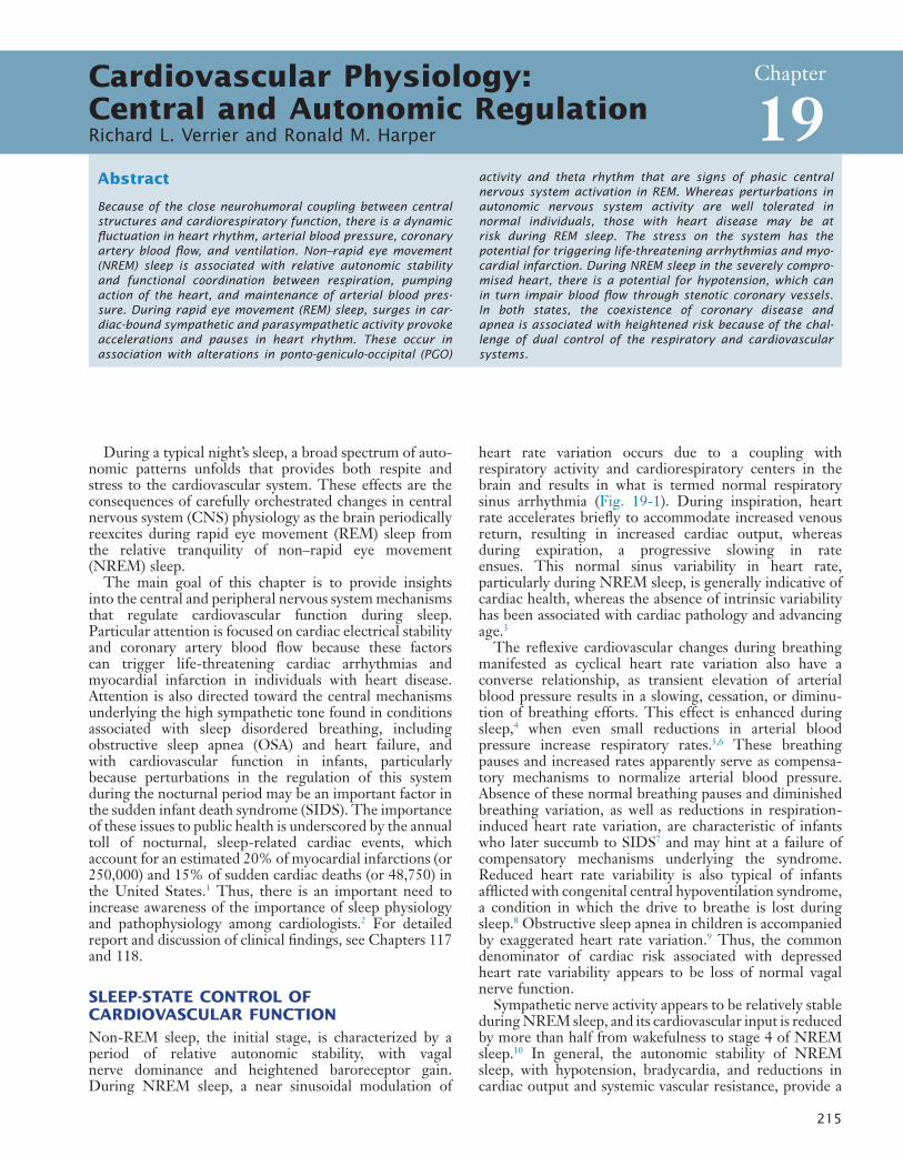
LL
215
Abstract
Because of the close neurohumoral coupling between central structures and cardiorespiratory function, there is a dynamic fluctuation in heart rhythm, arterial blood pressure, coronary artery blood flow, and ventilation. Non–rapid eye movement (NREM) sleep is associated with relative autonomic stability and functional coordination between respiration, pumping action of the heart, and maintenance of arterial blood pres-sure. During rapid eye movement (REM) sleep, surges in car-diac-bound sympathetic and parasympathetic activity provoke accelerations and pauses in heart rhythm. These occur in association with alterations in ponto-geniculo-occipital (PGO)
activity and theta rhythm that are signs of phasic central nervous system activation in REM. Whereas perturbations in autonomic nervous system activity are well tolerated in normal individuals, those with heart disease may be at risk during REM sleep. The stress on the system has the potential for triggering life-threatening arrhythmias and myo-cardial infarction. During NREM sleep in the severely compro-mised heart, there is a potential for hypotension, which can in turn impair blood flow through stenotic coronary vessels. In both states, the coexistence of coronary disease and apnea is associated with heightened risk because of the chal-lenge of dual control of the respiratory and cardiovascular systems.
During a typical night’s sleep, a broad spectrum of auto-nomic patterns unfolds that provides both respite and stress to the cardiovascular system. These effects are the consequences of carefully orchestrated changes in central nervous system (CNS) physiology as the brain periodically reexcites during rapid eye movement (REM) sleep from the relative tranquility of non–rapid eye movement (NREM) sleep.
The main goal of this chapter is to provide insights into the central and peripheral nervous system mechanisms that regulate cardiovascular function during sleep. Particular attention is focused on cardiac electrical stability and coronary artery blood flow because these factors can trigger life-threatening cardiac arrhythmias and myocardial infarction in individuals with heart disease. Attention is also directed toward the central mechanisms underlying the high sympathetic tone found in conditions associated with sleep disordered breathing, including obstructive sleep apnea (OSA) and heart failure, and with cardiovascular function in infants, particularly because perturbations in the regulation of this system during the nocturnal period may be an important factor in the sudden infant death syndrome (SIDS). The importance of these issues to public health is underscored by the annual toll of nocturnal, sleep-related cardiac events, which account for an estimated 20% of myocardial infarctions (or 250,000) and 15% of sudden cardiac deaths (or 48,750) in the United States.1 Thus, there is an important need to increase awareness of the importance of sleep physiology and pathophysiology among cardiologists.2 For detailed report and discussion of clinical findings, see Chapters 117 and 118.
SLEEP-STATE CONTROL OF CARDIOVASCULAR FUNCTIONNon-REM sleep, the initial stage, is characterized by a period of relative autonomic stability, with vagal nerve dominance and heightened baroreceptor gain. During NREM sleep, a near sinusoidal modulation of
heart rate variation occurs due to a coupling with respiratory activity and cardiorespiratory centers in the brain and results in what is termed normal respiratory sinus arrhythmia (Fig. 19-1). During inspiration, heart rate accelerates briefly to accommodate increased venous return, resulting in increased cardiac output, whereas during expiration, a progressive slowing in rate ensues. This normal sinus variability in heart rate, particularly during NREM sleep, is generally indicative of cardiac health, whereas the absence of intrinsic variability has been associated with cardiac pathology and advancing age.3
The reflexive cardiovascular changes during breathing manifested as cyclical heart rate variation also have a converse relationship, as transient elevation of arterial blood pressure results in a slowing, cessation, or diminu-tion of breathing efforts. This effect is enhanced during sleep,4 when even small reductions in arterial blood pressure increase respiratory rates.5,6 These breathing pauses and increased rates apparently serve as compensa-tory mechanisms to normalize arterial blood pressure. Absence of these normal breathing pauses and diminished breathing variation, as well as reductions in respiration-induced heart rate variation, are characteristic of infants who later succumb to SIDS7 and may hint at a failure of compensatory mechanisms underlying the syndrome. Reduced heart rate variability is also typical of infants afflicted with congenital central hypoventilation syndrome, a condition in which the drive to breathe is lost during sleep.8 Obstructive sleep apnea in children is accompanied by exaggerated heart rate variation.9 Thus, the common denominator of cardiac risk associated with depressed heart rate variability appears to be loss of normal vagal nerve function.
Sympathetic nerve activity appears to be relatively stable during NREM sleep, and its cardiovascular input is reduced by more than half from wakefulness to stage 4 of NREM sleep.10 In general, the autonomic stability of NREM sleep, with hypotension, bradycardia, and reductions in cardiac output and systemic vascular resistance, provide a
Cardiovascular Physiology: Central and Autonomic RegulationRichard L. Verrier and Ronald M. Harper
Chapter
19
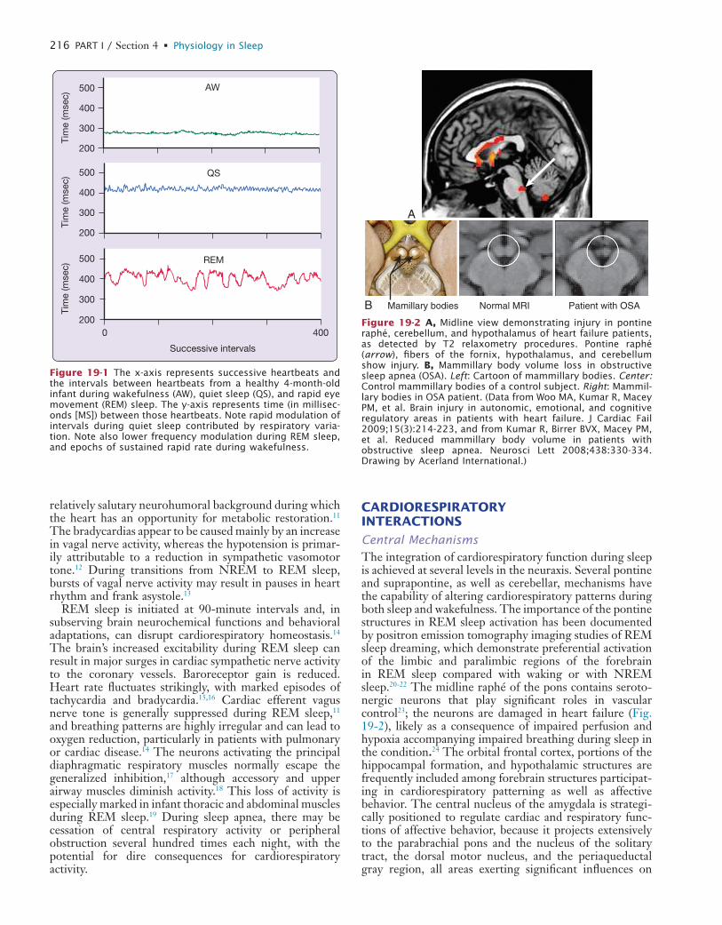
L
216 PART I / Section 4 • Physiology in Sleep
CARDIORESPIRATORY INTERACTIONS
Central MechanismsThe integration of cardiorespiratory function during sleep is achieved at several levels in the neuraxis. Several pontine and suprapontine, as well as cerebellar, mechanisms have the capability of altering cardiorespiratory patterns during both sleep and wakefulness. The importance of the pontine structures in REM sleep activation has been documented by positron emission tomography imaging studies of REM sleep dreaming, which demonstrate preferential activation of the limbic and paralimbic regions of the forebrain in REM sleep compared with waking or with NREM sleep.20-22 The midline raphé of the pons contains seroto-nergic neurons that play significant roles in vascular control23; the neurons are damaged in heart failure (Fig. 19-2), likely as a consequence of impaired perfusion and hypoxia accompanying impaired breathing during sleep in the condition.24 The orbital frontal cortex, portions of the hippocampal formation, and hypothalamic structures are frequently included among forebrain structures participat-ing in cardiorespiratory patterning as well as affective behavior. The central nucleus of the amygdala is strategi-cally positioned to regulate cardiac and respiratory func-tions of affective behavior, because it projects extensively to the parabrachial pons and the nucleus of the solitary tract, the dorsal motor nucleus, and the periaqueductal gray region, all areas exerting significant influences on
relatively salutary neurohumoral background during which the heart has an opportunity for metabolic restoration.11 The bradycardias appear to be caused mainly by an increase in vagal nerve activity, whereas the hypotension is primar-ily attributable to a reduction in sympathetic vasomotor tone.12 During transitions from NREM to REM sleep, bursts of vagal nerve activity may result in pauses in heart rhythm and frank asystole.13
REM sleep is initiated at 90-minute intervals and, in subserving brain neurochemical functions and behavioral adaptations, can disrupt cardiorespiratory homeostasis.14 The brain’s increased excitability during REM sleep can result in major surges in cardiac sympathetic nerve activity to the coronary vessels. Baroreceptor gain is reduced. Heart rate fluctuates strikingly, with marked episodes of tachycardia and bradycardia.15,16 Cardiac efferent vagus nerve tone is generally suppressed during REM sleep,11 and breathing patterns are highly irregular and can lead to oxygen reduction, particularly in patients with pulmonary or cardiac disease.14 The neurons activating the principal diaphragmatic respiratory muscles normally escape the generalized inhibition,17 although accessory and upper airway muscles diminish activity.18 This loss of activity is especially marked in infant thoracic and abdominal muscles during REM sleep.19 During sleep apnea, there may be cessation of central respiratory activity or peripheral obstruction several hundred times each night, with the potential for dire consequences for cardiorespiratory activity.
0 400
500
400
300
200
Tim
e (m
sec)
500
400
300
200
Tim
e (m
sec)
500
400
300
200
Tim
e (m
sec)
REM
QS
AW
Successive intervals
Figure 19-1 The x-axis represents successive heartbeats and the intervals between heartbeats from a healthy 4-month-old infant during wakefulness (AW), quiet sleep (QS), and rapid eye movement (REM) sleep. The y-axis represents time (in millisec-onds [MS]) between those heartbeats. Note rapid modulation of intervals during quiet sleep contributed by respiratory varia-tion. Note also lower frequency modulation during REM sleep, and epochs of sustained rapid rate during wakefulness.
A
B Mamillary bodies Normal MRI Patient with OSA
Figure 19-2 A, Midline view demonstrating injury in pontine raphé, cerebellum, and hypothalamus of heart failure patients, as detected by T2 relaxometry procedures. Pontine raphé (arrow), fibers of the fornix, hypothalamus, and cerebellum show injury. B, Mammillary body volume loss in obstructive sleep apnea (OSA). Left: Cartoon of mammillary bodies. Center: Control mammillary bodies of a control subject. Right: Mammil-lary bodies in OSA patient. (Data from Woo MA, Kumar R, Macey PM, et al. Brain injury in autonomic, emotional, and cognitive regulatory areas in patients with heart failure. J Cardiac Fail 2009;15(3):214-223, and from Kumar R, Birrer BVX, Macey PM, et al. Reduced mammillary body volume in patients with obstructive sleep apnea. Neurosci Lett 2008;438:330-334. Drawing by Acerland International.)
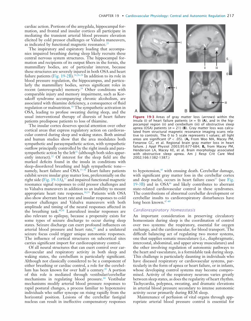
L
CHAPTER 19 • Cardiovascular Physiology: Central and Autonomic Regulation 217
to hypotension,45 with ensuing death. Cerebellar damage, with significant gray matter loss in the cerebellar cortex and deep nuclei, occurs in heart failure cases37 (see Fig. 19-3B) and in OSA46 and likely contributes to aberrant state-related cardiovascular control in these syndromes. The contributions of abnormal cerebellar development or cerebellar insults to cardiorespiratory disturbances have long been known.47-49
Cardiorespiratory HomeostasisAn important consideration in preserving circulatory homeostasis during sleep is the coordination of control over two systems: the respiratory, essential for oxygen exchange, and the cardiovascular, for blood transport. The difficult balancing act of regulating two motor systems, one that supplies somatic musculature (i.e., diaphragmatic, intercostal, abdominal, and upper airway musculature) and the other involving regulation of autonomic pathways to the heart and vasculature, is a formidable task during sleep. This challenge is particularly daunting in individuals who have diseased respiratory or cardiovascular systems, par-ticularly in the form of apnea or heart failure, or in infants, whose developing control systems may become compro-mised. Activity of the respiratory neurons varies greatly between sleep states, as does the regularity of heart rhythm. Tachycardia, polypnea, sweating, and dramatic elevations in arterial blood pressure secondary to intense autonomic activity occur primarily during REM sleep.
Maintenance of perfusion of vital organs through app-ropriate arterial blood pressure control is essential for
cardiac action. Portions of the amygdala, hippocampal for-mation, and frontal and insular cortices all participate in mediating the transient arterial blood pressure elevation elicited by cold pressor challenges or Valsalva maneuvers, as indicated by functional magnetic resonance.25
The inspiratory and expiratory loading that accompa-nies impaired breathing during sleep likely recruits these central nervous system structures. The hippocampal for-mation and recipients of its output fibers in the fornix, the mammillary bodies, are of particular interest, because these structures are severely injured in both OSA and heart failure patients (Fig. 19-2B).24,26-28 In addition to its role in blood pressure regulation, the hippocampus, and particu-larly the mammillary bodies, serves significant roles in recent (antereograde) memory.29 Other conditions with comparable injury and memory impairment, such as Kor-sakoff syndrome accompanying chronic alcoholism, are associated with thiamine deficiency, a consequence of fluid regulation or malnutrition.30 The sympathetic activation in OSA, leading to profuse sweating during sleep, and the usual interventional therapy of diuresis of heart failure patients predispose patients to loss of thiamine.
The insular cortex deserves special attention over other cortical areas that express regulatory action on cardiovas-cular control during sleep and waking states. Both animal and human studies show that the area modulates both sympathetic and parasympathetic action, with sympathetic outflow principally controlled by the right insula and para-sympathetic action by the left31 (although both sides appar-ently interact).32 Of interest for the sleep field are the marked deficits found in the insula in conditions with sleep-disordered breathing and high sympathetic tone—namely, heart failure and OSA.33-36 Heart failure patients exhibit severe insular gray matter loss, preferentially on the right side (Fig. 19-3A),37 and impaired functional magnetic resonance signal responses to cold pressor challenges and to Valsalva maneuvers in addition to an inability to mount appropriate heart rate responses.38,39 Patients with OSA also show aberrant heart rate and insular responses to cold pressor challenges and Valsalva maneuvers with both amplitude and timing of the neural responses affected in the breathing task.40,41 Lateralized insular responses are also relevant to epilepsy, because a propensity exists for some types of seizure discharge to occur during sleep states. Seizure discharge can exert profound influences on arterial blood pressure and heart rate,42 and a unilateral seizure focus could trigger unique autonomic responses. The influence of cortical structures on subcortical sites carries significant import for cardiorespiratory control.
Of all neural structures that can exert control over car-diovascular and respiratory activity in both sleep and waking states, the cerebellum is particularly significant. Although not classically considered to be a component of either breathing or cardiac control, a role for the cerebel-lum has been known for over half a century.43 A portion of this role is mediated through vestibular/cerebellar mechanisms in regulating blood pressure.44 Vestibular mechanisms modify arterial blood pressure responses to rapid postural changes, a process familiar to hypotensive individuals who suffer syncope on rising rapidly from the horizontal position. Lesions of the cerebellar fastigial nucleus can result in ineffective compensatory responses
i
ii iii
5
0
iii
i
A BFigure 19-3 Areas of gray matter loss (arrows) within the insula (i) of heart failure patients (n = 9) (A), and in the hip-pocampal region (ii) and cerebellum (iii) of obstructive sleep apnea (OSA) patients (n = 21) (B). Gray matter loss was calcu-lated from structural magnetic resonance imaging scans rela-tive to controls. The 0 to 5 scale represents t values; all light areas are significant (P < .05). (A, From Woo MA, Macey PM, Fonarow GC, et al. Regional brain gray matter loss in heart failure. J Appl Physiol 2003;95:677-684; B, from Macey PM, Henderson LA, Macey KE, et al. Brain morphology associated with obstructive sleep apnea. Am J Resp Crit Care Med 2002;166:1382-1387.)
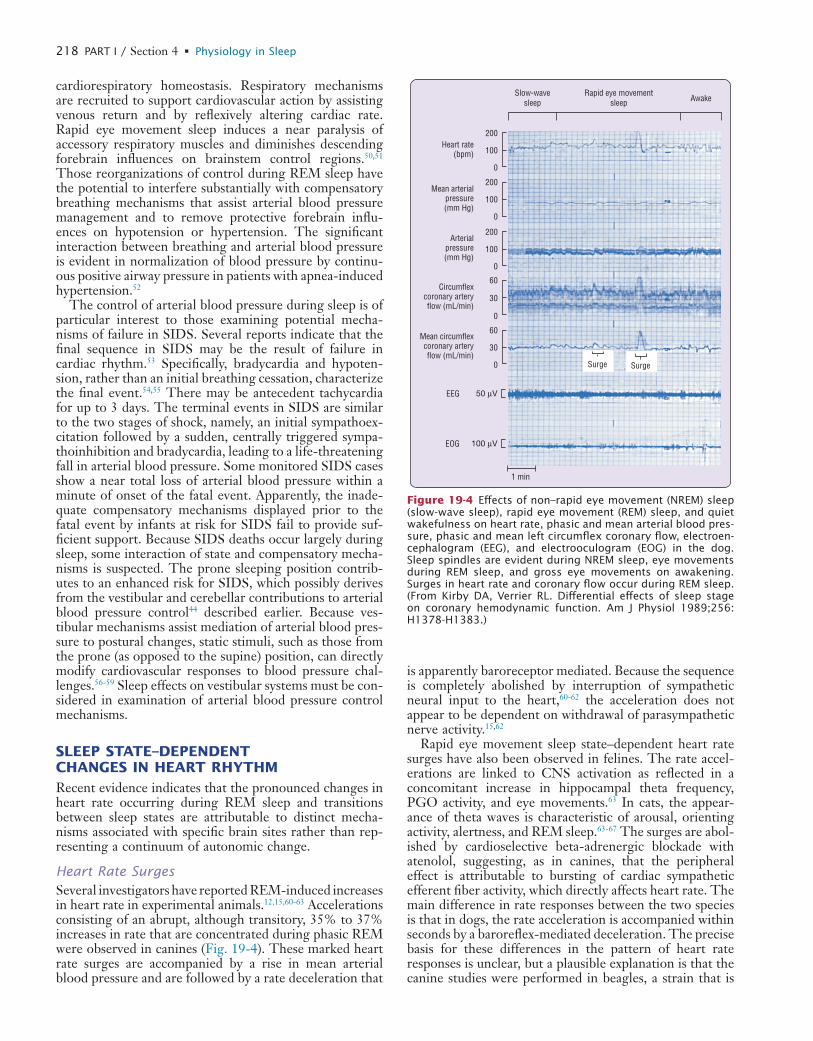
L
218 PART I / Section 4 • Physiology in Sleep
is apparently baroreceptor mediated. Because the sequence is completely abolished by interruption of sympathetic neural input to the heart,60-62 the acceleration does not appear to be dependent on withdrawal of parasympathetic nerve activity.15,62
Rapid eye movement sleep state–dependent heart rate surges have also been observed in felines. The rate accel-erations are linked to CNS activation as reflected in a concomitant increase in hippocampal theta frequency, PGO activity, and eye movements.63 In cats, the appear-ance of theta waves is characteristic of arousal, orienting activity, alertness, and REM sleep.63-67 The surges are abol-ished by cardioselective beta-adrenergic blockade with atenolol, suggesting, as in canines, that the peripheral effect is attributable to bursting of cardiac sympathetic efferent fiber activity, which directly affects heart rate. The main difference in rate responses between the two species is that in dogs, the rate acceleration is accompanied within seconds by a baroreflex-mediated deceleration. The precise basis for these differences in the pattern of heart rate responses is unclear, but a plausible explanation is that the canine studies were performed in beagles, a strain that is
cardiorespiratory homeostasis. Respiratory mechanisms are recruited to support cardiovascular action by assisting venous return and by reflexively altering cardiac rate. Rapid eye movement sleep induces a near paralysis of accessory respiratory muscles and diminishes descending forebrain influences on brainstem control regions.50,51 Those reorganizations of control during REM sleep have the potential to interfere substantially with compensatory breathing mechanisms that assist arterial blood pressure management and to remove protective forebrain influ-ences on hypotension or hypertension. The significant interaction between breathing and arterial blood pressure is evident in normalization of blood pressure by continu-ous positive airway pressure in patients with apnea-induced hypertension.52
The control of arterial blood pressure during sleep is of particular interest to those examining potential mecha-nisms of failure in SIDS. Several reports indicate that the final sequence in SIDS may be the result of failure in cardiac rhythm.53 Specifically, bradycardia and hypoten-sion, rather than an initial breathing cessation, characterize the final event.54,55 There may be antecedent tachycardia for up to 3 days. The terminal events in SIDS are similar to the two stages of shock, namely, an initial sympathoex-citation followed by a sudden, centrally triggered sympa-thoinhibition and bradycardia, leading to a life-threatening fall in arterial blood pressure. Some monitored SIDS cases show a near total loss of arterial blood pressure within a minute of onset of the fatal event. Apparently, the inade-quate compensatory mechanisms displayed prior to the fatal event by infants at risk for SIDS fail to provide suf-ficient support. Because SIDS deaths occur largely during sleep, some interaction of state and compensatory mecha-nisms is suspected. The prone sleeping position contrib-utes to an enhanced risk for SIDS, which possibly derives from the vestibular and cerebellar contributions to arterial blood pressure control44 described earlier. Because ves-tibular mechanisms assist mediation of arterial blood pres-sure to postural changes, static stimuli, such as those from the prone (as opposed to the supine) position, can directly modify cardiovascular responses to blood pressure chal-lenges.56-59 Sleep effects on vestibular systems must be con-sidered in examination of arterial blood pressure control mechanisms.
SLEEP STATE–DEPENDENT CHANGES IN HEART RHYTHMRecent evidence indicates that the pronounced changes in heart rate occurring during REM sleep and transitions between sleep states are attributable to distinct mecha-nisms associated with specific brain sites rather than rep-resenting a continuum of autonomic change.
Heart Rate SurgesSeveral investigators have reported REM-induced increases in heart rate in experimental animals.12,15,60-63 Accelerations consisting of an abrupt, although transitory, 35% to 37% increases in rate that are concentrated during phasic REM were observed in canines (Fig. 19-4). These marked heart rate surges are accompanied by a rise in mean arterial blood pressure and are followed by a rate deceleration that
Surge
Heart rate(bpm)
Slow-wavesleep
Rapid eye movementsleep Awake
Mean arterialpressure(mm Hg)
Arterialpressure(mm Hg)
Circumflexcoronary arteryflow (mL/min)
Mean circumflexcoronary arteryflow (mL/min)
EEG
EOG
1 min
200
100
0
200
100
0
200
100
0
60
30
0
60
30
0
50 µV
100 µV
Surge
Figure 19-4 Effects of non–rapid eye movement (NREM) sleep (slow-wave sleep), rapid eye movement (REM) sleep, and quiet wakefulness on heart rate, phasic and mean arterial blood pres-sure, phasic and mean left circumflex coronary flow, electroen-cephalogram (EEG), and electrooculogram (EOG) in the dog. Sleep spindles are evident during NREM sleep, eye movements during REM sleep, and gross eye movements on awakening. Surges in heart rate and coronary flow occur during REM sleep. (From Kirby DA, Verrier RL. Differential effects of sleep stage on coronary hemodynamic function. Am J Physiol 1989;256: H1378-H1383.)
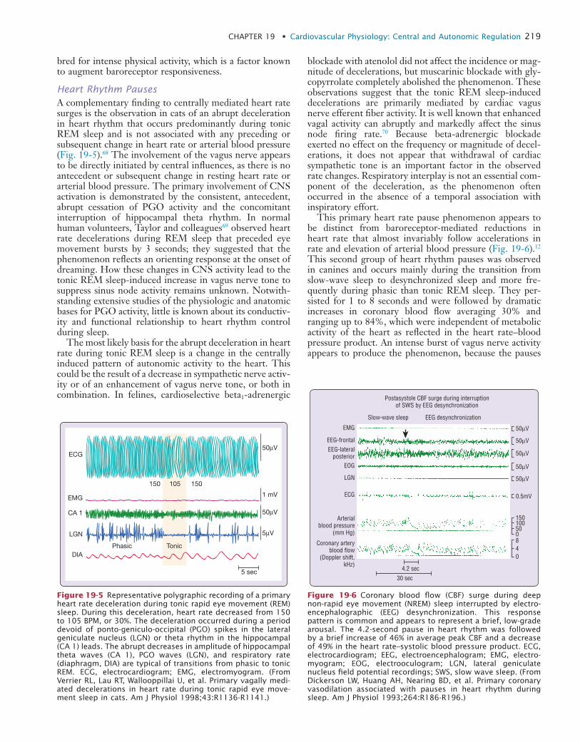
L
CHAPTER 19 • Cardiovascular Physiology: Central and Autonomic Regulation 219
blockade with atenolol did not affect the incidence or mag-nitude of decelerations, but muscarinic blockade with gly-copyrrolate completely abolished the phenomenon. These observations suggest that the tonic REM sleep-induced decelerations are primarily mediated by cardiac vagus nerve efferent fiber activity. It is well known that enhanced vagal activity can abruptly and markedly affect the sinus node firing rate.70 Because beta-adrenergic blockade exerted no effect on the frequency or magnitude of decel-erations, it does not appear that withdrawal of cardiac sympathetic tone is an important factor in the observed rate changes. Respiratory interplay is not an essential com-ponent of the deceleration, as the phenomenon often occurred in the absence of a temporal association with inspiratory effort.
This primary heart rate pause phenomenon appears to be distinct from baroreceptor-mediated reductions in heart rate that almost invariably follow accelerations in rate and elevation of arterial blood pressure (Fig. 19-6).12 This second group of heart rhythm pauses was observed in canines and occurs mainly during the transition from slow-wave sleep to desynchronized sleep and more fre-quently during phasic than tonic REM sleep. They per-sisted for 1 to 8 seconds and were followed by dramatic increases in coronary blood flow averaging 30% and ranging up to 84%, which were independent of metabolic activity of the heart as reflected in the heart rate–blood pressure product. An intense burst of vagus nerve activity appears to produce the phenomenon, because the pauses
bred for intense physical activity, which is a factor known to augment baroreceptor responsiveness.
Heart Rhythm PausesA complementary finding to centrally mediated heart rate surges is the observation in cats of an abrupt deceleration in heart rhythm that occurs predominantly during tonic REM sleep and is not associated with any preceding or subsequent change in heart rate or arterial blood pressure (Fig. 19-5).68 The involvement of the vagus nerve appears to be directly initiated by central influences, as there is no antecedent or subsequent change in resting heart rate or arterial blood pressure. The primary involvement of CNS activation is demonstrated by the consistent, antecedent, abrupt cessation of PGO activity and the concomitant interruption of hippocampal theta rhythm. In normal human volunteers, Taylor and colleagues69 observed heart rate decelerations during REM sleep that preceded eye movement bursts by 3 seconds; they suggested that the phenomenon reflects an orienting response at the onset of dreaming. How these changes in CNS activity lead to the tonic REM sleep-induced increase in vagus nerve tone to suppress sinus node activity remains unknown. Notwith-standing extensive studies of the physiologic and anatomic bases for PGO activity, little is known about its conductiv-ity and functional relationship to heart rhythm control during sleep.
The most likely basis for the abrupt deceleration in heart rate during tonic REM sleep is a change in the centrally induced pattern of autonomic activity to the heart. This could be the result of a decrease in sympathetic nerve activ-ity or of an enhancement of vagus nerve tone, or both in combination. In felines, cardioselective beta1-adrenergic
ECG
EMG
CA 1
LGN
DIA
1 mV
50µV
50µV
5µV
150 150105
Phasic Tonic
5 sec
Figure 19-5 Representative polygraphic recording of a primary heart rate deceleration during tonic rapid eye movement (REM) sleep. During this deceleration, heart rate decreased from 150 to 105 BPM, or 30%. The deceleration occurred during a period devoid of ponto-geniculo-occipital (PGO) spikes in the lateral geniculate nucleus (LGN) or theta rhythm in the hippocampal (CA 1) leads. The abrupt decreases in amplitude of hippocampal theta waves (CA 1), PGO waves (LGN), and respiratory rate (diaphragm, DIA) are typical of transitions from phasic to tonic REM. ECG, electrocardiogram; EMG, electromyogram. (From Verrier RL, Lau RT, Wallooppillai U, et al. Primary vagally medi-ated decelerations in heart rate during tonic rapid eye move-ment sleep in cats. Am J Physiol 1998;43:R1136-R1141.)
Postasystole CBF surge during interruptionof SWS by EEG desynchronization
EMG
EEG-frontalEEG-lateral
posteriorEOG
LGN
ECG
Arterialblood pressure
(mm Hg)
Coronary arteryblood flow
(Doppler shift,kHz)
4.2 sec
0.5mV
50µV
50µV
50µV
50µV
50µV
Slow-wave sleep EEG desynchronization
30 sec
840
150100500
Figure 19-6 Coronary blood flow (CBF) surge during deep non-rapid eye movement (NREM) sleep interrupted by electro-encephalographic (EEG) desynchronization. This response pattern is common and appears to represent a brief, low-grade arousal. The 4.2-second pause in heart rhythm was followed by a brief increase of 46% in average peak CBF and a decrease of 49% in the heart rate–systolic blood pressure product. ECG, electrocardiogram; EEG, electroencephalogram; EMG, electro-myogram; EOG, electrooculogram; LGN, lateral geniculate nucleus field potential recordings; SWS, slow wave sleep. (From Dickerson LW, Huang AH, Nearing BD, et al. Primary coronary vasodilation associated with pauses in heart rhythm during sleep. Am J Physiol 1993;264:R186-R196.)
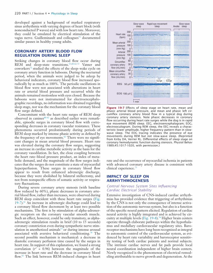
L
220 PART I / Section 4 • Physiology in Sleep
rate and the occurrence of myocardial ischemia in patients with advanced coronary artery disease is consistent with clinical experience.75
IMPACT OF SLEEP ON ARRHYTHMOGENESIS
Central Nervous System Sites Influencing Cardiac Electrical StabilityExtensive investigation of CNS-induced cardiac arrhyth-mias has provided evidence that triggering of arrhythmias by the CNS is not only the consequence of intense activa-tion of the autonomic nervous system, but also is a function of the specific neural pattern elicited. Regulation of cardiac neural activity is highly integrated and is achieved by cir-cuitry at multiple levels (Fig. 19-8).76 Higher brain centers operate through elaborate pathways within the hypothala-mus and medullary cardiovascular regulatory sites. Baro-receptor mechanisms have long been recognized as integral to autonomic control of the cardiovascular system, as evi-denced by heart rate variability and baroreceptor sensitiv-ity testing of both cardiac patients and normal subjects. The intrinsic cardiac nerves and fat pads provide local neural coordination independent of higher brain centers. Newly recognized is the phenomenon of electrical remod-eling attributable to nerve growth and degeneration. At the
developed against a background of marked respiratory sinus arrhythmia with varying degrees of heart block (with nonconducted P waves) and with low heart rate. Moreover, they could be emulated by electrical stimulation of the vagus nerve. Guilleminault and colleagues71 documented similar pauses in healthy young adults.
CORONARY ARTERY BLOOD FLOW REGULATION DURING SLEEPStriking changes in coronary blood flow occur during REM and sleep-state transitions.13,60-62,72 Vatner and coworkers72 studied the effects of the sleep–wake cycle on coronary artery function in baboons. During the nocturnal period, when the animals were judged to be asleep by behavioral indicators, coronary blood flow increased spo-radically by as much as 100%. The periodic oscillations in blood flow were not associated with alterations in heart rate or arterial blood pressure and occurred while the animals remained motionless with eyes closed. Because the baboons were not instrumented for electroencephalo-graphic recordings, no information was obtained regarding sleep stage, nor was the mechanism for the coronary blood flow surge defined.
Concomitant with the heart rate surges of REM sleep observed in canines60-62 as described earlier were remark-able, episodic surges in coronary blood flow with corre-sponding decreases in coronary vascular resistance. These phenomena occurred predominantly during periods of REM sleep marked by intense phasic activity as defined by the frequency of eye movements.62 There were no signifi-cant changes in mean arterial blood pressure. Heart rate was elevated during the coronary flow surges, suggesting an increase in cardiac metabolic activity as the basis for the coronary vasodilation. In fact, the close coupling between the heart rate–blood pressure product, an index of meta-bolic demand, and the magnitude of the flow surges indi-cates that the surges do not constitute a state of myocardial hyperperfusion. These surges in coronary blood flow appear to result from enhanced adrenergic discharge, because they were abolished by bilateral stellectomy, and not from nonspecific effects of somatic activity or respira-tory fluctuations.
During severe coronary artery stenosis (with baseline flow reduced by 60%), phasic decreases in coronary arte-rial blood flow, rather than increases, were observed during REM sleep coincident with these heart rate surges (Fig. 19-7).61 An increase in adrenergic discharge could lead to a coronary blood flow decrement by at least two possible mechanisms. The first is by stimulation of alpha-adrener-gic receptors on the coronary vascular smooth muscle. Such an effect, however, could be only transitory, as alpha-adrenergic stimulation results in brief (10 to 15 seconds) coronary constriction even during sympathetic nerve stim-ulation in anesthetized animals73 or during intense arousal associated with aversive behavioral conditioning.74 The second possible mechanism is mechanical: a decrease in diastolic coronary perfusion time caused by the surges in heart rate. In support of this explanation, we found a strong correlation (r2 = 0.96) between the magnitude of the increase in heart rate and the decrease in coronary blood flow.61 The link between REM-induced changes in heart
200100
0200100
0200100
060300
60300
Heart rate(bpm)
Mean arterialpressure(mm Hg)
Arterialpressure(mm Hg)
Circumflexcoronary arteryflow (mL/min)
Mean circumflexcoronary arteryflow (mL/min)
EEG
EOG
Slow-wave Slow-waveRapid eye movementAwake
1 min
50 µV
100 µV
sleep sleep sleep
Surge Surge
Figure 19-7 Effects of sleep stage on heart rate, mean and phasic arterial blood pressure, and mean and phasic left cir-cumflex coronary artery blood flow in a typical dog during coronary artery stenosis. Note phasic decreases in coronary flow occurring during heart rate surges while the dog is in rapid eye movement (REM) sleep. EEG, electroencephalogram; EOG, electrooculogram. During REM sleep, the EEG reveals a charac-teristic lower amplitude, higher frequency pattern than in slow-wave sleep. The EOG tracing indicates the presence of eye movements during REM but not slow-wave sleep. (Reprinted from Kirby DA, Verrier RL. Differential effects of sleep stage on coronary hemodynamic function during stenosis. Physiol Behav 1989;45:1017-1020, with permission.)
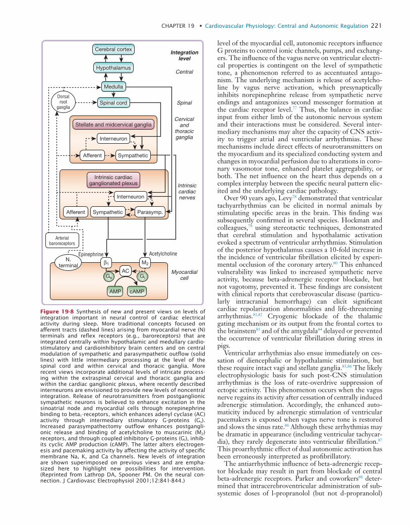
L
CHAPTER 19 • Cardiovascular Physiology: Central and Autonomic Regulation 221
level of the myocardial cell, autonomic receptors influence G proteins to control ionic channels, pumps, and exchang-ers. The influence of the vagus nerve on ventricular electri-cal properties is contingent on the level of sympathetic tone, a phenomenon referred to as accentuated antago-nism. The underlying mechanism is release of acetylcho-line by vagus nerve activation, which presynaptically inhibits norepinephrine release from sympathetic nerve endings and antagonizes second messenger formation at the cardiac receptor level.77 Thus, the balance in cardiac input from either limb of the autonomic nervous system and their interactions must be considered. Several inter-mediary mechanisms may alter the capacity of CNS activ-ity to trigger atrial and ventricular arrhythmias. These mechanisms include direct effects of neurotransmitters on the myocardium and its specialized conducting system and changes in myocardial perfusion due to alterations in coro-nary vasomotor tone, enhanced platelet aggregability, or both. The net influence on the heart thus depends on a complex interplay between the specific neural pattern elic-ited and the underlying cardiac pathology.
Over 90 years ago, Levy78 demonstrated that ventricular tachyarrhythmias can be elicited in normal animals by stimulating specific areas in the brain. This finding was subsequently confirmed in several species. Hockman and colleagues,79 using stereotactic techniques, demonstrated that cerebral stimulation and hypothalamic activation evoked a spectrum of ventricular arrhythmias. Stimulation of the posterior hypothalamus causes a 10-fold increase in the incidence of ventricular fibrillation elicited by experi-mental occlusion of the coronary artery.80 This enhanced vulnerability was linked to increased sympathetic nerve activity, because beta-adrenergic receptor blockade, but not vagotomy, prevented it. These findings are consistent with clinical reports that cerebrovascular disease (particu-larly intracranial hemorrhage) can elicit significant cardiac repolarization abnormalities and life-threatening arrhythmias.81,82 Cryogenic blockade of the thalamic gating mechanism or its output from the frontal cortex to the brainstem83 and of the amygdala84 delayed or prevented the occurrence of ventricular fibrillation during stress in pigs.
Ventricular arrhythmias also ensue immediately on ces-sation of diencephalic or hypothalamic stimulation, but these require intact vagi and stellate ganglia.85,86 The likely electrophysiologic basis for such post-CNS stimulation arrhythmias is the loss of rate-overdrive suppression of ectopic activity. This phenomenon occurs when the vagus nerve regains its activity after cessation of centrally induced adrenergic stimulation. Accordingly, the enhanced auto-maticity induced by adrenergic stimulation of ventricular pacemakers is exposed when vagus nerve tone is restored and slows the sinus rate.86 Although these arrhythmias may be dramatic in appearance (including ventricular tachycar-dia), they rarely degenerate into ventricular fibrillation.87 This proarrhythmic effect of dual autonomic activation has been erroneously interpreted as profibrillatory.
The antiarrhythmic influence of beta-adrenergic recep-tor blockade may result in part from blockade of central beta-adrenergic receptors. Parker and coworkers88 deter-mined that intracerebroventricular administration of sub-systemic doses of l-propranolol (but not d-propranolol)
Cerebral cortex
Hypothalamus
Medulla
Spinal cord
Stellate and midcervical ganglia
Interneuron
Afferent Sympathetic
Intrinsic cardiacganglionated plexus
Interneuron
Afferent
Arterialbaroreceptors
Dorsalroot
ganglia
Sympathetic Parasymp.
N.terminal
Epinephrine Acetylcholine
AC
AMP cAMP
M2
Gs Gi
β1
Myocardialcell
Intrinsiccardiacnerves
Cervicaland
thoracicganglia
Spinal
Central
Integrationlevel
Figure 19-8 Synthesis of new and present views on levels of integration important in neural control of cardiac electrical activity during sleep. More traditional concepts focused on afferent tracts (dashed lines) arising from myocardial nerve (N) terminals and reflex receptors (e.g., baroreceptors) that are integrated centrally within hypothalamic and medullary cardio-stimulatory and cardioinhibitory brain centers and on central modulation of sympathetic and parasympathetic outflow (solid lines) with little intermediary processing at the level of the spinal cord and within cervical and thoracic ganglia. More recent views incorporate additional levels of intricate process-ing within the extraspinal cervical and thoracic ganglia and within the cardiac ganglionic plexus, where recently described interneurons are envisioned to provide new levels of noncentral integration. Release of neurotransmitters from postganglionic sympathetic neurons is believed to enhance excitation in the sinoatrial node and myocardial cells through norepinephrine binding to beta1-receptors, which enhances adenyl cyclase (AC) activity through intermediary stimulatory G-proteins (Gs). Increased parasympathectomy outflow enhances postgangli-onic release and binding of acetylcholine to muscarinic (M2) receptors, and through coupled inhibitory G-proteins (Gi), inhib-its cyclic AMP production (cAMP). The latter alters electrogen-esis and pacemaking activity by affecting the activity of specific membrane Na, K, and Ca channels. New levels of integration are shown superimposed on previous views and are empha-sized here to highlight new possibilities for intervention. (Reprinted from Lathrop DA, Spooner PM. On the neural con-nection. J Cardiovasc Electrophysiol 2001;12:841-844.)
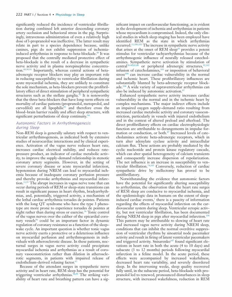
L
222 PART I / Section 4 • Physiology in Sleep
nificant impact on cardiovascular functioning, as is evident in the development of ischemia and arrhythmias in patients whose myocardium is compromised. Indeed, the only clin-ical studies in which sleep staging has been employed have identified REM as the state in which arrhythmias occurred.11,100,101 The increase in sympathetic nerve activity that arises at the onset of REM sleep10 provides a potent stimulus for ventricular tachyarrhythmias because of the arrhythmogenic influence of neurally released catechol-amines. Sympathetic nerve activation by stimulation of central78-80,85,86 or peripheral adrenergic structures,92,102 infusion of catecholamines,103 or imposition of behavioral stress104 can increase cardiac vulnerability in the normal and ischemic heart. These profibrillatory influences are substantially blunted by beta-adrenergic receptor block-ade.104 A wide variety of supraventricular arrhythmias can also be induced by autonomic activation.87
Enhanced sympathetic nerve activity increases cardiac vulnerability in the normal and in the ischemic heart by complex mechanisms. The major indirect effects include an impaired oxygen supply–demand ratio resulting from increased cardiac metabolic activity and coronary vasocon-striction, particularly in vessels with injured endothelium and in the context of altered preload and afterload. The direct profibrillatory effects on cardiac electrophysiologic function are attributable to derangements in impulse for-mation or conduction, or both.87 Increased levels of cate-cholamines activate beta-adrenergic receptors, which in turn alter adenylate cyclase activity and intracellular calcium flux. These actions are probably mediated by the cyclic nucleotide and protein kinase regulatory cascade, which can alter spatial heterogeneity of calcium transients and consequently increase dispersion of repolarization. The net influence is an increase in susceptibility to ven-tricular fibrillation.74,105 Conversely, reduction of cardiac sympathetic drive by stellectomy has proved to be antifibrillatory.
Notwithstanding the evidence that autonomic factors have the potential for significantly altering susceptibility to arrhythmias, the observation that the heart rate surges of REM sleep are conducive to myocardial ischemia, and the epidemiologic data in humans on the extent of sleep-induced cardiac events,1 there is a paucity of information regarding the effects of myocardial infarction on the car-diovascular system during sleep. Ventricular ectopic activ-ity, but not ventricular fibrillation, has been documented during NREM sleep in pigs after myocardial infarction.106 This pattern may be attributable to slowing of heart rate and increased vagus nerve activity during NREM sleep, conditions that can inhibit the normal overdrive suppres-sion of ventricular rhythms by sinoatrial node pacemaker activity and result in firing of latent ventricular pacemakers and triggered activity. Snisarenko107 found significant ele-vations in heart rate in both the acute (4 to 10 days) and subacute (3 to 12 months) periods following myocardial infarction in a feline model. In the acute period, these effects were accompanied by increased wakefulness, decreased heart rate variability, and severely disordered sleep. In the intervening weeks, sleep quality recovered fully until, in the subacute period, beta-blockade with pro-pranolol led to renewed, pronounced disturbances in sleep structure, with increased wakefulness, reduction in REM
significantly reduced the incidence of ventricular fibrilla-tion during combined left anterior descending coronary artery occlusion and behavioral stress in the pig. Surpris-ingly, intravenous administration of even a relatively high dose of l-propranolol was ineffective. The latter result may relate in part to a species dependence because, unlike canines, pigs do not exhibit suppression of ischemia-induced arrhythmias in response to beta-blockade.89 It was proposed that the centrally mediated protective effect of beta-blockade is the result of a decrease in sympathetic nerve activity and in plasma norepinephrine concentra-tion.88,90,91 Importantly, whereas central actions of beta-adrenergic receptor blockers may play an important role in reducing susceptibility to ventricular fibrillation during acute myocardial ischemia, they are unlikely to constitute the sole mechanism, as beta-blockers prevent the profibril-latory effect of direct stimulation of peripheral sympathetic structures such as the stellate ganglia.92 It is noteworthy that the three beta-blockers that have long-term effects on mortality of cardiac patients (propranolol, metoprolol, and carvedilol) are all lipophilic93 and therefore cross the blood–brain barrier readily and affect sleep structure, with significant perturbations of sleep continuity.94
Autonomic Factors in Arrhythmogenesis during SleepNon-REM sleep is generally salutary with respect to ven-tricular arrhythmogenesis, as indicated both by extensive studies of neurocardiac interactions and by clinical experi-ence. Activation of the vagus nerve reduces heart rate, increases cardiac electrical stability, and reduces rate- pressure product, an indicator of cardiac metabolic activ-ity, to improve the supply-demand relationship in stenotic coronary artery segments. However, in the setting of severe coronary disease or acute myocardial infarction, hypotension during NREM can lead to myocardial isch-emia because of inadequate coronary perfusion pressure and thereby provoke arrhythmias and myocardial infarc-tion.11,95 The abrupt increases in vagus nerve tone that can occur during periods of REM or sleep-state transitions can result in significant pauses in heart rhythm, bradyarrhyth-mias, and, potentially, triggered activity, a mechanism of the lethal cardiac arrhythmia torsades de pointes. Patients with the long QT syndrome who have the type 3 pheno-type are more prone to experience torsades de pointes at night rather than during stress or exercise.56 Tonic control of the vagus nerves over the caliber of the epicardial coro-nary vessels96 could be an important factor in dynamic regulation of coronary resistance as a function of the sleep–wake cycle. An important question is whether tonic vagus nerve activity exerts a protective or a deleterious influence on myocardial perfusion and arrhythmogenesis in indi-viduals with atherosclerotic disease. In these patients, noc-turnal surges in vagus nerve activity could precipitate myocardial ischemia and arrhythmias as a result of coro-nary vasoconstriction rather than dilation in atheroscle-rotic segments, in patients with impaired release of endothelium-derived relaxing factor.97
Because of the attendant surges in sympathetic nerve activity and in heart rate, REM sleep has the potential for triggering ventricular arrhythmias.98,99 The striking vari-ability of heart rate and breathing pattern can have a sig-
L
CHAPTER 19 • Cardiovascular Physiology: Central and Autonomic Regulation 223
REFERENCES1. Lavery CE, Mittleman MA, Cohen MC, Muller E, et al. Nonuni-
form nighttime distribution of acute cardiac events: a possible effect of sleep states. Circulation 1997;96:3321-3327.
2. Malhotra A, Loscalzo J. Sleep and cardiovascular disease: an over-view. Prog Cardiovasc Dis 2009;51:279-284.
3. Task Force of the European Society of Cardiology and the North American Society of Pacing and Electrophysiology. Heart rate vari-ability: standards of measurement, physiological interpretation and clinical use. Circulation 1996;93:1043-1065.
4. Trelease RB, Sieck GC, Marks JD, Harper RM. Respiratory inhibi-tion induced by transient hypertension during sleep. Exp Neurol 1985;90:173-186.
5. Ohtake PJ, Jennings DB. Ventilation is stimulated by small reduc-tions in arterial pressure in the awake dog. J Appl Physiol 1992; 73:1549-1557.
6. Harper RM, Gozal D, Forster HV, Ohtake PJ, et al. Imaging of ventral medullary surface activity during blood pressure challenges in awake and anesthetized goats. Am J Physiol 1996;39:R182- R191.
7. Schechtman VL, Harper RM, Wilson AJ, Southall DP. Sleep apnea in infants who succumb to the sudden infant death syndrome. Pediatrics 1991;87:841-846.
8. Woo MS, Woo MA, Gozal D, Jansen MT, et al. Heart rate vari-ability in congenital central hypoventilation syndrome. Pediatr Res 1992;31:291-296.
9. Aljadeff G, Gozal D, Schechtman VL, Burrell B, et al. Heart rate variability in children with obstructive sleep apnea. Sleep 1997;20:151-157.
10. Somers VK, Dyken ME, Mark AL, Abboud FM. Sympathetic nerve activity during sleep in normal subjects. N Engl J Med 1993;328: 303-307.
11. Mancia G. Autonomic modulation of the cardiovascular system during sleep. N Engl J Med 1993;328:347-349.
12. Baccelli G, Guazzi M, Mancia G, Zanchetti A. Neural and non-neural mechanisms influencing circulation during sleep. Nature 1969;223:184-185.
13. Dickerson LW, Huang AH, Nearing BD, Verrier RL. Primary coronary vasodilation associated with pauses in heart rhythm during sleep. Am J Physiol 1993;264:R186-196.
14. Harper RM, Frysinger RC, Zhang J, Trelease RB, et al. Cardiac and respiratory interactions maintaining homeostasis during sleep. In: Lydic R, Biebuyck JF, editors. Clinical physiology of sleep. Bethesda, Md: American Physiological Society; 1988.
15. Gassel MM, Ghelarducci B, Marchiafava PL, Pompeiano O. Phasic changes in blood pressure and heart rate during the rapid eye move-ment episodes of desynchronized sleep in unrestrained cats. Arch Ital Biol 1964;102:530-544.
16. Snyder F, Hobson JA, Morrison DF, Goldfrank F. Changes in respiration, heart rate, and systolic blood pressure in human sleep. J Appl Physiol 1964;19:417-422.
17. Lydic R, Baghdoyan HA. Microdialysis of cat pons reveals enhanced acetylcholine release during state-dependent respiratory depression. Am J Physiol 1991;261:R766-R770.
18. Sauerland EK, Harper RM. The human tongue during sleep: Elec-tromyographic activity of the genioglossus muscle. Exp Neurol 1976;51:160-170.
19. Henderson-Smart DJ, Read DJC. Reduced lung volume during behavioral active sleep in the newborn. J Appl Physiol 1979; 46:1081-1085.
20. Maquet P, Peters J, Aerts J, Delfiore G, et al. Functional neuro-anatomy of human rapid-eye-movement sleep and dreaming. Nature 1996;383:163-166.
21. Braun AR, Balkin TJ, Wesenten NJ, Carson RE, et al. Regional cerebral blood flow throughout the sleep-wake cycle: an H2(15)O PET study. Brain 1997;120:1173-1197.
22. Nofzinger EA, Mintun MA, Wiseman M, Kupfer DJ, et al. Fore-brain activation in REM sleep: an FDG PET study. Brain Res 1997;770:192-201.
23. Dedeoglu A, Fisher LA. CNS actions of serotonin on cardiovascular function: nonadrenergic, noncholinergic mechanisms Am J Physiol Regul Integr Comp Physiol 1996;271:R569-R578.
24. Woo MA, Kumar R, Macey PM, Fonarow GC, et al. Brain injury in autonomic, emotional, and cognitive regulatory areas in patients with heart failure. J Cardiac Fail, 2009;15(3):214-223.
sleep, and prolongation of stages 1 and 2 of NREM sleep. He attributed these results to reflex activation of adrener-gic, noradrenergic, and dopaminergic nerves in several brain structures after coronary artery ligation.108
SUMMARYSleep states exert a major impact on cardiorespiratory function. This is a direct consequence of the significant variations in brain states that occur in the normal cycling between NREM and REM sleep. Dynamic fluctuations in CNS variables influence heart rhythm, arterial blood pres-sure, coronary artery blood flow, and ventilation. Whereas REM-induced surges in sympathetic and parasympathetic nerve activity with accompanying significant surges and pauses in heart rhythm are well tolerated in normal people, patients with heart disease may be at heightened risk for life-threatening arrhythmias and myocardial ischemia and infarction.75,99 During NREM sleep, in the severely com-promised heart, a potential for hypotension exists that can impair blood flow through stenotic coronary vessels to trigger myocardial ischemia or infarction.11 Damage to central brain areas that regulate autonomic activity and coordinate upper airway and diaphragmatic action can lead to enhanced sympathetic outflow, increasing risk in heart failure and contributing to hypertension in OSA. Coordi-nation of cardiorespiratory control is especially pivotal in infancy, when developmental immaturity can compromise function and pose special risks. Throughout sleep, the coexistence of coronary disease and apnea is associated with heightened risk of cardiovascular events109 resulting from the challenge of dual control of the respiratory and cardiovascular systems.
� Clinical Pearl
REM sleep is characterized by surges in sympathetic and vagus nerve activity, which are well tolerated in normal individuals but which may result in cardiac arrhythmias, myocardial ischemia, and myocardial infarction in those with heart disease. During NREM sleep, systemic blood pressure may fall, potentially reducing flow through stenotic coronary vessels, which may precipitate cardiac ischemia or infarction. In essence, sleep constitutes an autonomic stress test for the heart, and nighttime monitoring of cardiore-spiratory function is of considerable diagnostic value.110
AcknowledgmentsThis work was supported by National Institutes of Health grant MH13923 from the National Institute of Mental Health, and HD22695, HL22418, and HL60296 from the National Institute of Child Health and Human Develop-ment and the National Heart, Lung, and Blood Institute, Bethesda, Maryland. Additional support was provided by the Mind-Body Network of the John D. and Catherine T. MacArthur Foundation. The authors thank Sandra S. Verrier for her editorial contributions.
L
224 PART I / Section 4 • Physiology in Sleep
52. Somers VK, Dyken ME, Clary MP, Abboud FM. Sympathetic neural mechanisms in obstructive sleep apnea. J Clin Invest 1995; 96:1897-1904.
53. Schwartz PJ, Stramba-Badiale M, Segantini A, Austoni P, et al. Prolongation of the QT interval and the sudden infant death syn-drome. N Engl J Med 1998;338:1709-1714.
54. Meny RG, Carroll JL, Carbone MT, Kelly DH. Cardiorespiratory recordings from infants dying suddenly and unexpectedly at home. Pediatrics 1994;93:43-49.
55. Harper RM, Bandler R. Finding the failure mechanism in the sudden infant death syndrome. Nat Med 1998;4:157-158.
56. Sahni R, Schulze KF, Kashyap S, Ohira-Kist K, et al. Body position, sleep states, and cardiorespiratory activity in developing low birth weight infants. Early Hum Dev 1999;54:197-206.
57. Sahni R, Schulze KF, Kashyap S, Ohira-Kist K, et al. Postural dif-ferences in cardiac dynamics during quiet and active sleep in low birthweight infants. Acta Paediatr 1999;88:1396-1401.
58. Galland BC, Taylor BJ, Bolton DP, Sayers RM. Vasoconstriction following spontaneous sighs and head-up tilts in infants sleeping prone and supine. Early Hum Dev 2000;58:119-132.
59. Galland BC, Hayman RM, Taylor BJ, Bolton DP, et al. Factors affecting heart rate variability and heart rate responses to tilting in infants aged 1 and 3 months. Pediatr Res 2000;48:360-368.
60. Kirby DA, Verrier RL. Differential effects of sleep stage on coro-nary hemodynamic function. Am J Physiol 1989;256:H1378- H1383.
61. Kirby DA, Verrier RL. Differential effects of sleep stage on coro-nary hemodynamic function during stenosis. Physiol Behav 1989;45:1017-1020.
62. Dickerson LW, Huang AH, Thurnher MM, Nearing BD, et al. Relationship between coronary hemodynamic changes and the phasic events of rapid eye movement sleep. Sleep 1993;16:550-557.
63. Rowe K, Moreno R, Lau RT, Wallooppillai U, et al. Heart rate surges during REM sleep are associated with theta rhythm and PGO activity in the cat. Am J Physiol 1999;277:R843-R849.
64. Sakai K, Sano K, Iwahara S. Eye movements and hippocampal theta activity in cats. Electroencephalogr Clin Neurophysiol 1973;34: 547-549.
65. Kemp IR, Kaada BR. The relation of hippocampal theta activity to arousal, attentive behaviour and somato-motor movements in unre-strained cats. Brain Res 1975;95:323-342.
66. Lerma J, Garcia-Austt E. Hippocampal theta rhythm during para-doxical sleep: Effects of afferent stimuli and phase relationships with phasic events. Electroencephalogr Clin Neurophysiol 1985; 60:46-54.
67. Sei H, Morita Y. Acceleration of EEG theta wave precedes the phasic surge of arterial pressure during REM sleep in the rat. Neu-roreport 1996;7:3059-3062.
68. Verrier RL, Lau RT, Wallooppillai U, Quattrochi J, et al. Primary vagally mediated decelerations in heart rate during tonic rapid eye movement sleep in cats. Am J Physiol 1998;43:R1136-R1141.
69. Taylor WB, Moldofsky H, Furedy JJ. Heart rate deceleration in REM sleep: An orienting reaction interpretation. Psychophysiology 1985;22:110-115.
70. Pappano AJ. Modulation of the heartbeat by the vagus nerve. In: Zipes DP, Jalife J, editors. Cardiac electrophysiology: from cell to bedside. Philadelphia: Saunders; 1995, p. 411-422.
71. Guilleminault C, Pool P, Motta J, Gillis AM. Sinus arrest during REM sleep in young adults. N Engl J Med 1984;311: 1006-1010.
72. Vatner SF, Franklin D, Higgins CB, Patrick T, et al. Coronary dynamics in unrestrained conscious baboons. Am J Physiol 1971; 221:1396-1401.
73. Feigl EO. Coronary physiology. Physiol Rev 1983;63:1-205.74. Billman GE, Randall DC. Mechanisms mediating the coronary
vascular response to behavioral stress in the dog. Circ Res 1981;48: 214-223.
75. Nowlin JB, Troyer WG Jr, Collins WS, Silverman G, et al. The association of nocturnal angina pectoris with dreaming. Ann Intern Med 1965;63:1040-1046.
76. Lathrop DA, Spooner PM. On the neural connection. J Cardiovasc Electrophysiol 2001;12:841-844.
77. Verrier RL, Antzelevitch C. Autonomic aspects of arrhythmogen-esis: the enduring and the new. Curr Opin Cardiol 2004;19:2-11.
78. Levy AG. The exciting causes of ventricular fibrillation in animals under chloroform anesthesia. Heart 1912;4:319-378.
25. Harper RM, Bandler R, Spriggs D, Alger JR. Lateralized and wide-spread brain activation during transient blood pressure elevation revealed by magnetic resonance imaging. J Comp Neurol 2000;417: 195-204.
26. Kumar R, Birrer BV, Macey PM, Woo MA, et al. Reduced mam-millary body volume in patients with obstructive sleep apnea. Neu-rosci Lett 2008;438:330-334.
27. Kumar R, Woo MA, Birrer BV, Macey PM, et al. Mammillary body volumes are diminished in heart failure. Neurobiol Dis 2009; 33:236-242.
28. Macey PM, Kumar R, Woo MA. Brain structural changes in obstructive sleep apnea. Sleep 2008;31:967-977.
29. Aggleton JP, Brown MW. Episodic memory, amnesia, and the hip-pocampal anterior thalamic axis. Behav Brain Sci 1999;22:425-444.
30. Harper C. Thiamine (vitamin B1) deficiency and associated brain damage is still common throughout the world and prevention is simple and safe. Eur J Neurol 2006;13;1078-1082.
31. Oppenheimer SM, Gelb A, Girvin JP, Hachinski VC. Cardiovascu-lar effects of human insular cortex stimulation. Neurology 1992; 42:1727-1732.
32. Zhang Z, Oppenheimer SM. Characterization, distribution and lat-eralization of baroreceptor-related neurons in the rat insular cortex. Brain Res 1997;760:243-250.
33. Bradley TD, Floras JS. Obstructive sleep apnoea and its cardiovas-cular consequences. Lancet 2009;373:82-93.
34. Leung RS. Sleep-disordered breathing: autonomic mechanisms and arrhythmias. Prog Cardiovasc Dis 2009;51:324-338.
35. Xie A, Skatrud JB, Barczi SR, Reichmuth K, et al. Influence of cerebral blood flow on breathing stability. J Appl Physiol 2009; 106(3):850-856.
36. Naughton MT, Lorenzi-Filho G. Sleep in heart failure. Prog Car-diovasc Dis 2009;51:339-349.
37. Woo MA, Macey PM, Fonarow GC, Hamilton MA, et al. Regional brain gray matter loss in heart failure. J Appl Physiol 2003;95: 677-684.
38. Woo MA, Macey PM, Keens PT, Kumar R, et al. Functional abnormalities in brain areas that mediate autonomic nervous system control in advanced heart failure. J Cardiac Fail 2005;11:437-446.
39. Woo MA, Macey PM, Keens PT. Aberrant central nervous system responses to the Valsalva maneuver in heart failure. Congest Heart Fail 2007;13:29-35.
40. Henderson LA, Woo MA, Macey PM, Macey KE, et al. Neural responses during Valsalva maneuvers in obstructive sleep apnea syndrome. J Appl Physiol 2003;94:1063-1074.
41. Harper RM, Macey PM, Henderson LA, Woo MA, et al. FMRI responses to cold pressor challenges in control and obstructive sleep apnea subjects. J Appl Physiol 2003;94:1583-1595.
42. Frysinger RC, Harper RM, Hackel RJ. State-dependent cardiac and respiratory changes associated with complex partial epilepsy. In: Engel J Jr, Ojemann GA, Lüders HO, editors. Fundamental mecha-nisms of human brain function. New York: Raven Press; 1987, pp. 219-226.
43. Moruzzi G. Paleocerebellar inhibition of vasomotor and respiratory carotid sinus reflexes. J Neurophysiol 1940;3:20-32.
44. Yates BJ. Vestibular influences on the autonomic nervous system. Ann N Y Acad Sci 1996:781:458-473.
45. Lutherer LO, Lutherer BC, Dormer KJ, Janssen HF, et al. Bilateral lesions of the fastigial nucleus prevent the recovery of blood pres-sure following hypotension induced by hemorrhage or administra-tion of endotoxin. Brain Res 1983;269:251-257.
46. Macey PM, Henderson LA, Macey KE, Alger JR, et al. Brain mor-phology associated with obstructive sleep apnea. Am J Resp Crit Care Med 2002;166:1382-1387.
47. Martin R, Roessmann U, Fanaroff A. Massive intracerebellar hem-orrhage in low-birth-weight infants. J Pediatr 1976;89:290-293.
48. Elisevich K, Redekop G. The fastigial pressor response. J Neuro-surg 1991;74:147-151.
49. Waters KA, Forbes P, Morielli A, Hum C, et al. Sleep-disordered breathing in children with myelomeningocele. J Pediatr 1998;132: 672-681.
50. Parmegianni PL, Franzini C, Lenzi P. Respiratory frequency as a function of preoptic temperature during sleep. Brain Res 1976;111:253-260.
51. Frysinger RC, Marks JD, Trelease RB, Schechtman VL, et al. Sleep states attenuate the pressor response to central amygdala stimula-tion. Exp Neurol 1984;83:604-617.

L
CHAPTER 19 • Cardiovascular Physiology: Central and Autonomic Regulation 225
94. Kostis JB, Rosen RC. Central nervous system effects of beta- adrenergic blocking drugs: the role of ancillary properties. Circula-tion 1987;75:204-212.
95. Broughton R, Baron R. Sleep patterns in the intensive care unit and on the ward after acute myocardial infarction. Electroencephalogr Clin Neurophysiol 1978;45:348-360.
96. Kovach JA, Gottdiener JS, Verrier RL. Vagal modulation of epicar-dial coronary artery size in dogs. A two-dimensional intravascular ultrasound study. Circulation 1995;92:2291-2298.
97. Ludmer PL, Selwyn AP, Shook TL, Wayne RR, et al. Paradoxical vasoconstriction induced by acetylcholine in atherosclerotic arter-ies. N Engl J Med 1986;315:1046-1051.
98. Lown B, Verrier RL. Neural activity and ventricular fibrillation. N Engl J Med 1976;294:1165-1170.
99. Andrews TC, Fenton T, Toyosaki N, Glasser SP, et al. Subsets of ambulatory myocardial ischemia based on heart rate activity: circa-dian distribution and response to anti-ischemic medication. Circu-lation 1993;88:92-100.
100. Smith R, Johnson L, Rothfeld D, Zir L, et al. Sleep and cardiac arrhythmias. Arch Intern Med 1972;130:751-753.
101. Rosenblatt G, Hartman E, Zwilling GR. Cardiac irritability during sleep and dreaming. J Psychosom 1973;17:129-134.
102. Verrier RL, Thompson PL, Lown B. Ventricular vulnerability during sympathetic stimulation: role of heart rate and blood pres-sure. Cardiovasc Res 1974;8:602-610.
103. Han J, Moe GK. Nonuniform recovery of excitability in ventricular muscle. Circ Res 1964;14:44-60.
104. Kovach JA, Nearing BD, Verrier RL. An angerlike behavioral state potentiates myocardial ischemia-induced T-wave alternans in canines. J Am Coll Cardiol 2001;37:1719-1725.
105. Levy MN. Role of calcium in arrhythmogenesis. Circulation 1989;80(6 Suppl):IV23-IV30.
106. Skinner JE, Mohr DN, Kellaway P. Sleep-stage regulation of ventricular arrhythmias in the unanesthetized pig. Circ Res 1975;37:342-349.
107. Snisarenko AA. Cardiac rhythm in cats during physiological sleep in experimental myocardial infarction and beta-adrenergic receptor blockade. Cor Vasa 1986;38:306-314.
108. Sole MJ, Hussain MN, Lixfeld W. Activation of brain catechol-aminergic neurons by cardiac vagal afferents during acute myocar-dial ischemia in the rat. Circ Res 1980;47:166-172.
109. Shah NA, Yaggi HK, Concato J, Mohsenin V. Obstructive sleep apnea as a risk factor for coronary events or cardiovascular death. Sleep Breath 2010;14:131-136.
110. Verrier RL, Josephson ME. Impact of sleep on arrhythmogenesis. Circ Arrhythmia Electrophysiol 2009;2:450-459.
79. Hockman CH, Mauck HP, Hoff EC. ECG changes resulting from cerebral stimulation: II. A spectrum of ventricular arrhythmias of sympathetic origin. Am Heart J 1966;71:695-700.
80. Verrier RL, Calvert A, Lown B. Effect of posterior hypothalamic stimulation on the ventricular fibrillation threshold. Am J Physiol 1975;228:923-927.
81. Cropp GJ, Manning GW. Electrocardiographic changes simulating myocardial ischemia and infarction associated with spontaneous intracranial hemorrhage. Circulation 1960;22:25-38.
82. Hugenholtz PG. Electrocardiographic abnormalities in cerebral disorders: report of six cases and review of the literature. Am Heart J 1962;63:451-461.
83. Skinner JE, Reed JC. Blockade of frontocortical-brain stem pathway prevents ventricular fibrillation of ischemic heart. Am J Physiol 1981;240:H156-H163.
84. Carpeggiani C, Landisman C, Montaron M-F, Skinner JE. Cryob-lockade in limbic brain (amygdala) prevents or delays ventricular fibrillation after coronary artery occlusion in psychologically stressed pigs. Circ Res 1992;70:600-606.
85. Korteweg GCJ, Boeles JTF, Ten Cate J. Influence of stimulation of some subcortical areas on electrocardiogram. J Neurophysiol 1957;20:100-107.
86. Manning JW de V, Cotten M. Mechanism of cardiac arrhythmias induced by diencephalic stimulation. Am J Physiol 1962;203:1120- 1124.
87. Janse MJ, Wit AL. Electrophysiological mechanisms of ventricular arrhythmias resulting from myocardial ischemia and infarction. Physiol Rev 1989;69:1049-1169.
88. Parker GW, Michael LH, Hartley CJ, Skinner JE, et al. Central beta-adrenergic mechanisms may modulate ischemic ventricular fibrillation in pigs. Circ Res 1990;66:259-270.
89. Benfey BG, Elfellah MS, Ogilvie RI, Varma DR. Antiarrhythmic effects of prazosin and propranolol during coronary artery occlusion and reperfusion in dogs and pigs. Br J Pharmacol 1984;82:717-725.
90. Lewis PJ, Haeusler G. Reduction in sympathetic nervous activity as a mechanism for the hypotensive action of propranolol. Nature 1975;256:440.
91. Privitera PJ, Webb JG, Walle T. Effect of centrally administered propranolol on plasma renin activity, plasma norepinephrine, and arterial pressure. Eur J Pharmacol 1979;54:51-60.
92. Schwartz PJ, Vanoli E, Zaza A, Zuanetti G. The effect of antiar-rhythmic drugs on life-threatening arrhythmias induced by the interaction between acute myocardial ischemia and sympathetic hyperactivity. Am Heart J 1985;109:937-948.
93. Hjalmarson A, Olsson G. Myocardial infarction: effects of beta-blockade. Circulation 1991;84(Suppl 6):VI-101-107.
LL
226
Abstract
Autonomic control of circulation is pivotal in ensuring an adequate cardiac output to the vital organs through continu-ous and rapid adjustments of heart rate (HR), arterial blood
pressure (BP), and redistribution of blood flow. In the longer term, neural circulatory control appears to be coupled with the circadian rhythm, the sleep−wake cycle, and ultradian rhythms, including rapid eye movement (REM) and non−rapid eye movement (NREM) sleep processes.
hemodynamic variables, including heart rate, arterial blood pressure, and peripheral blood flow, on a beat-by-beat basis. The cardiovascular autonomic nervous system appears to be intimately linked to sleep and circadian phys-iology, as demonstrated by the disrupted autonomic control that accompanies sleep loss and sleep apnea. Sleep disruption may lead to altered neural circulatory control. On the other hand, whether primary alterations in auto-nomic function may translate into sleep disturbances is not known.
This chapter is introduced by a general overview of autonomic cardiovascular regulation and of its central and peripheral controllers, followed by a description of the methods used to explore cardiovascular neural control during sleep in humans and their advantages and limita-tions. We then seek to outline some of the current knowl-edge of neural circulatory control during normal sleep and how it may change as a consequence of sleep deprivation, sleep apnea, and autonomic dysfunction such as may occur in diabetes.
THE CARDIOVASCULAR AUTONOMIC NERVOUS SYSTEM: DEFINITION AND FUNCTIONSThe cardiovascular autonomic nervous system is a highly integrated network that controls visceral functions, which on a short timescale (seconds to hours), adjusts the circula-tion in keeping with behavior, the environment, and emo-tions. Its primary role is to ensure an adequate cardiac output to the vital organs through continuous and rapid adjustments of HR, arterial BP, and redistribution of blood flow. In the longer term, this neural circulatory regulation appears to be coupled with the circadian rhythm, the sleep–wake cycle, and some ultradian rhythms, including rapid eye movement (REM) and non–rapid eye movement (NREM) sleep processes, as well as hormones implicated in long-term BP regulation.
Neural control of circulation operates via parasympa-thetic neurons to the heart and sympathetic neuronal effer-ents to the heart, blood vessels, kidneys, and adrenal medulla. Parasympathetic stimulation of the cardiovascu-lar system is mediated primarily through the vagus nerve by means of the activation of muscarinic receptors and results in bradycardia. Sympathetic stimulation of the heart acts through activation of beta1 adrenoreceptors at the sinoatrial node (the cardiac pacemaker) and in the myocardium (the cardiac muscle) and results in tachycardia
Autonomic circulatory control operates via parasympa-thetic neurons to the heart and sympathetic neuronal effer-ents to the heart, blood vessels, kidneys and adrenal medulla. Parasympathetic stimulation of the heart, through the activation of cardiac muscarinic receptors, results in bradycardia, whereas sympathetic stimulation of the heart, through activation of beta1 adrenoreceptors, results in tachycardia and increased contractility. Sympathetic acti-vation in the vascular bed induces both vasoconstriction, by stimulating alpha1 adrenoreceptors, and vasodilation by stimulating beta2 adrenoreceptors. Several reflexes, includ-ing the arterial baroreflex, cardiopulmonary reflexes, and chemoreflexes are also important in the rapid adjustments of circulation that occur in association with postural changes, hypoxia, temperature changes, and perhaps sleep.
Heart rate (HR) and blood pressure (BP) have a 24-hour rhythm characterized by a significant reduction during nighttime hours, secondary to changes in activity and posture, as well as sleep and circadian influences. This physiological pattern can be altered by sleep loss and sleep disorders, with important implications for cardiovascular health.
Autonomic neural output to the cardiovascular system is also sleep-stage–dependent, with a global reduction in sym-pathetic drive to the heart and vessels, and a shift toward cardiac parasympathetic dominance with deepening stages of non-REM sleep. On the other hand, REM sleep is dominated by remarkable fluctuations between parasympa-thetic and sympathetic activity, leading to sudden changes in HR and BP. Finally, significant autonomic changes accompany electrocortical arousal from sleep as well as periodic leg movements during sleep. Heart rate changes seemingly precede cortical activation and motor activity.
The critical role of the autonomic nervous system in maintaining homeostasis during sleep is demonstrated by disorders that are associated with autonomic dysregula-tion, such as obstructive sleep apnea (see Chapter 119). Obstructive sleep apnea is associated with increases in sympathetic neural activity both during sleep and wakefulness, and may be mediated through heightened chemoreflex sensitivity. Sympathetic drive may play a role in the higher prevalence of cardiovascular disease in these patients.
INTRODUCTIONThe cardiovascular autonomic nervous system seeks to maintain homeostasis through precise control of numerous
Cardiovascular Physiology: Autonomic Control in Health and in Sleep DisordersPaola A. Lanfranchi and Virend K. Somers
Chapter
20
L
CHAPTER 20 • Cardiovascular Physiology: Autonomic Control in Health and in Sleep Disorders 227
Arterial and cardiopulmonary reflexes are implicated in BP regulation during postural changes. Assumption of the upright position produces a caudal shift in blood volume and acutely reduces stroke volume and BP. The circulatory adjustment to this orthostatic stress is rapid and is charac-terized by a reflexive increase in HR and peripheral vascu-lar resistance, followed by enhanced secretion of antidiuretic hormone and renin-angiotensin. Recumbency, as occurs during sleep, produces an increased volume load in the cardiac chambers and elicits the opposite effects.
The ChemoreflexesThe chemoreflexes mediate the ventilatory response to hypoxia and hypercapnia, and they also exert important cardiovascular effects.2 The peripheral arterial chemore-ceptors, the most important of which are located in the carotid bodies, respond primarily to changes in the partial pressure of oxygen. Hypoxemic stimulation elicits an increase in respiratory muscle output, inducing hyper-ventilation, and an increase in sympathetic outflow to peripheral blood vessels, resulting in vasoconstriction. Hyperventilation in turn activates pulmonary stretch receptors, which buffer the increases in sympathetic and vagal outflow, thereby maintaining homeostasis under normal conditions. During apnea, when hyperventilation is absent or prevented, vasoconstriction is potentiated and occurs simultaneously with activation of cardiac vagal drive resulting in bradycardia, collectively termed the “diving reflex,” a protective mechanism which helps pre-serve blood flow to the heart and brain while limiting cardiac oxygen demand.3,4
The central chemoreceptors are located in the brain-stem and respond to changes in pH mediated primarily by carbon dioxide tension. Stimulation of central chemore-ceptors by hypercapnia also elicits sympathetic and respi-ratory activation, but without the cardiovagal effects seen with hypoxia.2
MEASURES TO EXPLORE AUTONOMIC CHANGES DURING SLEEP AND THEIR PHYSIOLOGICAL SIGNIFICANCE
Heart Rate, Arterial Blood Pressure, and Their VariabilityRR interval, the time elapsed between two successive R-waves of the QRS signal on the electrocardiogram (and its reciprocal, the HR) is a function of intrinsic properties of the sinus node as well as autonomic influences. Blood pressure is a function of vascular resistance (an expression of arterial constriction or dilation) and cardiac output (the blood volume being pumped by the heart in 1 minute), which is a function of HR, cardiac contractility, and dia-stolic blood volume, all components controlled in part by the autonomic nervous system.
Autonomic cardiovascular regulation can be investigated through the quantification of average HR and BP as assessed in steady conditions (wakefulness and sleep, for instance) or in their responses to endogenous or exogenous challenges (e.g., changes in posture, response to respira-tory changes, and arousal from sleep).
and increased contractility. Sympathetic activation in the vascular bed induces vasoconstriction by stimulating alpha1 adrenoreceptors (in the skin and splanchnic districts) and vasodilation by stimulating beta2 adrenoreceptors (in the heart and skeletal muscles). Parasympathetic and sympa-thetic efferent activity to the heart may also modulate cardiac electrophysiological properties that can be relevant to the genesis of several types of arrhythmias, particularly in the presence of a proarrhythmic substrate.
Central organization of the autonomic nervous system and its relationship with sleep modulating mechanisms are detailed in Chapter 19. Briefly, autonomic impulses to the vasculature and heart originate from the vasomotor center in the brainstem, located bilaterally in the reticular sub-stance of the medulla and pons. The vasomotor center is in turn modulated by higher nervous system regions in the pons, mesencephalon, and diencephalon, including the hypothalamus and many portions of the cerebral cortex. Several cardiovascular reflexes are also important in the rapid adjustments of blood pressure occurring in associa-tion with postural changes, hypoxia, exercise, and moder-ate temperature changes, and may also be implicated in cardiovascular changes observed during sleep. These include the arterial baroreflex, cardiopulmonary reflexes, and chemoreflexes. The renin-angiotensin-aldosterone system, vasopressin, and other vasoactive mechanisms may also contribute to cardiovascular regulation during sleep. More information on the interaction between cardiovas-cular disorders and sleep can be found in Section 14 of this book.
Arterial BaroreflexThe arterial baroreflex is an important regulator of BP in the short term.1 The baroreceptors are sensory receptors in the aortic arch and carotid sinuses that relay in the medullary regions of the brain. Changes in arterial baro-receptor afferent discharge trigger reflex adjustments that buffer or oppose the changes in BP. For instance, increas-ing increments in BP stretch the receptors, resulting in heightened afferent traffic to the brainstem neuronal network. This inhibits efferent sympathetic outflow to cardiac and vascular smooth muscle and increases para-sympathetic cardiac tone, resulting in slowing of HR, reduction in contractility, and increased peripheral vasodi-lation with subsequent decrease in BP. A decrease in BP has opposite effects, and elicits reflex tachycardia, increased contractility and peripheral vasoconstriction with subse-quent increase in BP.
Cardiopulmonary ReflexesThese reflexes are triggered by the stimulation of low-pressure receptors located in the atria, ventricles, and pul-monary arteries. Cardiopulmonary receptors are volume receptors that serve to mitigate changes in BP in response to changes in blood volume. The firing pattern of these receptors parallels the pressure changes within the cardiac chambers or vessels and helps to regulate blood volume. Cardiopulmonary reflex activation results in peripheral vasodilation, reduction in sympathetic outflow to the kidney, and activation of the posterior pituitary gland to inhibit the release of antidiuretic hormone, resulting in increased urine excretion.
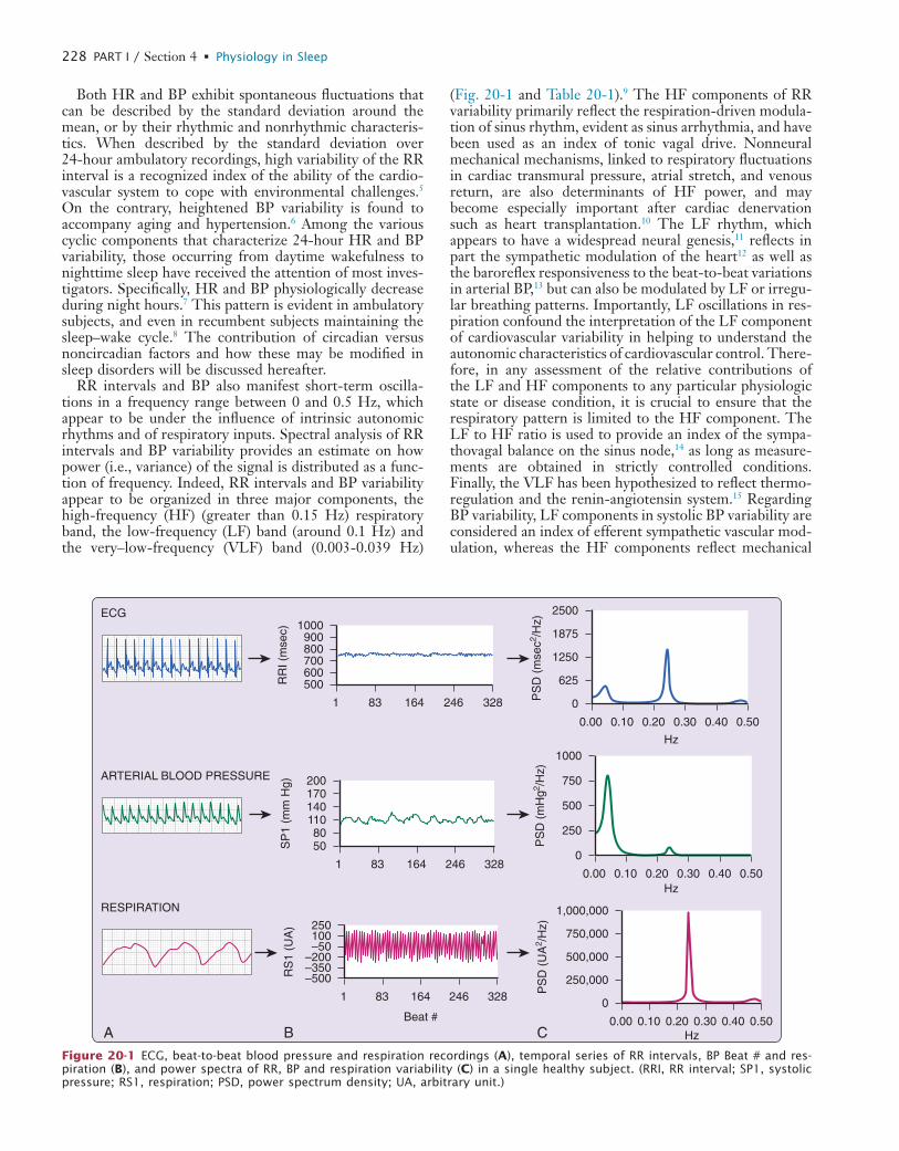
L
228 PART I / Section 4 • Physiology in Sleep
(Fig. 20-1 and Table 20-1).9 The HF components of RR variability primarily reflect the respiration-driven modula-tion of sinus rhythm, evident as sinus arrhythmia, and have been used as an index of tonic vagal drive. Nonneural mechanical mechanisms, linked to respiratory fluctuations in cardiac transmural pressure, atrial stretch, and venous return, are also determinants of HF power, and may become especially important after cardiac denervation such as heart transplantation.10 The LF rhythm, which appears to have a widespread neural genesis,11 reflects in part the sympathetic modulation of the heart12 as well as the baroreflex responsiveness to the beat-to-beat variations in arterial BP,13 but can also be modulated by LF or irregu-lar breathing patterns. Importantly, LF oscillations in res-piration confound the interpretation of the LF component of cardiovascular variability in helping to understand the autonomic characteristics of cardiovascular control. There-fore, in any assessment of the relative contributions of the LF and HF components to any particular physiologic state or disease condition, it is crucial to ensure that the respiratory pattern is limited to the HF component. The LF to HF ratio is used to provide an index of the sympa-thovagal balance on the sinus node,14 as long as measure-ments are obtained in strictly controlled conditions. Finally, the VLF has been hypothesized to reflect thermo-regulation and the renin-angiotensin system.15 Regarding BP variability, LF components in systolic BP variability are considered an index of efferent sympathetic vascular mod-ulation, whereas the HF components reflect mechanical
Both HR and BP exhibit spontaneous fluctuations that can be described by the standard deviation around the mean, or by their rhythmic and nonrhythmic characteris-tics. When described by the standard deviation over 24-hour ambulatory recordings, high variability of the RR interval is a recognized index of the ability of the cardio-vascular system to cope with environmental challenges.5 On the contrary, heightened BP variability is found to accompany aging and hypertension.6 Among the various cyclic components that characterize 24-hour HR and BP variability, those occurring from daytime wakefulness to nighttime sleep have received the attention of most inves-tigators. Specifically, HR and BP physiologically decrease during night hours.7 This pattern is evident in ambulatory subjects, and even in recumbent subjects maintaining the sleep–wake cycle.8 The contribution of circadian versus noncircadian factors and how these may be modified in sleep disorders will be discussed hereafter.
RR intervals and BP also manifest short-term oscilla-tions in a frequency range between 0 and 0.5 Hz, which appear to be under the influence of intrinsic autonomic rhythms and of respiratory inputs. Spectral analysis of RR intervals and BP variability provides an estimate on how power (i.e., variance) of the signal is distributed as a func-tion of frequency. Indeed, RR intervals and BP variability appear to be organized in three major components, the high-frequency (HF) (greater than 0.15 Hz) respiratory band, the low-frequency (LF) band (around 0.1 Hz) and the very–low-frequency (VLF) band (0.003-0.039 Hz)
Beat #
RR
I (m
sec)
500
1000900800700600
83
ECG
ARTERIAL BLOOD PRESSURE
RESPIRATION
164 246 3281 PS
D (
mse
c2 /Hz)
0
2500
1875
1250
625
0.10 0.20
Hz
Hz
Hz
0.30 0.40 0.500.00
PS
D (
mH
g2 /Hz)
0
1000
750
500
250
0.10 0.20 0.30 0.40 0.500.00
PS
D (
UA
2 /Hz)
0
1,000,000
750,000
500,000
250,000
0.10 0.20 0.30 0.40 0.500.00CBA
SP
1 (m
m H
g)
50
20017014011080
83 164 246 3281
RS
1 (U
A)
–500
250100–50
–200–350
83 164 246 3281
Figure 20-1 ECG, beat-to-beat blood pressure and respiration recordings (A), temporal series of RR intervals, BP Beat # and res-piration (B), and power spectra of RR, BP and respiration variability (C) in a single healthy subject. (RRI, RR interval; SP1, systolic pressure; RS1, respiration; PSD, power spectrum density; UA, arbitrary unit.)

L
CHAPTER 20 • Cardiovascular Physiology: Autonomic Control in Health and in Sleep Disorders 229
in blood pressure.22 Two techniques have been mainly used in sleep research to assess spontaneous baroreflex modula-tion of heart rate: the sequence technique and the spectral analysis tecnique. The first identifies sequences of con-secutive beats in which progressive increases in systolic BP are followed by a progressive lengthening in RR (or vice versa). The slope of the regression line between RR inter-vals and systolic BP within these sequences is taken as the magnitude of the reflex gain. The second is based on cross-spectral analysis of short segments of SBP and RR and relies on the assumption that a certain frequency band of RR variability, between 0.04 and 0.35 Hz, is modulated by the baroreflex. Baroreflex sensitivity is expressed by the gain of the transfer function relating changes in blood pressure to coherent changes in RR or muscle sympathetic nerve activity (MSNA).
Preejection PeriodThe preejection period (PEP) is the time elapsed between the electrical depolarization of the left ventricle (QRS on the ECG) and the beginning of ventricular ejection and represents the time the left ventricle contracts with the cardiac valves closed. The PEP is influenced by sym-pathetic activity acting via beta1 adrenoreceptors and shortens under stimulation. The PEP can be derived non-invasively from impedance cardiography, which converts changes in thoracic impedance (as measured by electrodes on the chest and neck) to changes in volume over time and allows tracking of volumetric changes such as those occur-ring during the cardiac cycle. This method has been applied, although not intensively, to assess cardiac sympa-thetic influences in steady state conditions during sleep.23,24 The application to transient sympathetic responses is unfortunately limited because errors can occur in interpre-tation in the presence of BP increases, which can induce a lengthening of the PEP (instead of the expected shorten-ing) due to the longer time required to overcome the external pressure.
Microneurographic Recording of Sympathetic Nerve ActivityMicroneurography provides direct information on sympa-thetic vasomotor and sudomotor activity to muscle and skin. Muscle sympathetic nerve activity (MSNA), usually
effects of respiration on blood pressure changes.12 Mea-surements of HF, LF, and VLF are usually made in abso-lute values (msec2), but LF and HF are often presented in normalized units (NU), which represent the relative value of each power component in proportion to the total power minus the VLF components (see Table 20-1). Normaliza-tion allows minimizing the effect of changes in total power on LF and HF components.
Traditional spectral analysis techniques include fast Fourier Transform algorithms and autoregressive model-ing, which in most instances provide comparable results.16 These techniques require stationarity of the signal being processed, and therefore cannot be applied to processes in which there is significant transient activity (e.g., sleep onset, arousals, sleep stages transition, and awakening). In addition, such methods have to be used with caution in association with respiratory or motor events (e.g., periodic limb movement, bruxism). More advanced algorithms of signal processing can be used to overcome this limitation and permit the assessment of dynamic changes in auto-nomic cardiovascular control during transient events (e.g., sleep onset, arousal, bruxism, etc.)17 and help define the temporal relationship between dynamic changes occurring in different systems, such as electroencephalogram (EEG) and electrocardiogram (ECG).18,19 The most commonly used algorithms include short-time Fourier Transform, Wigner-Ville distribution, Time Varying Autoregressive models, and Wavelets and Wavelet Packets.17
Finally, in addition to the periodic oscillatory behavior observed in RR intervals and arterial BP, a less specific variability occurs with nonperiodic behavior, which can be described by methods based on nonlinear system theory (“chaos theory and fractal analysis”).20 The physiological basis for this nonharmonic beat-to-beat behavior, which extends over a wide time range (seconds to hours), is still unsettled, although it has been proposed that it is under higher central modulation.21 The application of this type of analysis to sleep cardiovascular physiology is still limited.
Baroreflex SensitivityThe arterial baroreflex is important in buffering short term changes in BP. The gain of the arterial baroreflex, or baroreflex sensitivity, is measured by the degree of change in heart rate or sympathetic traffic for a given unit change
Table 20-1 Spectral Components of RR Variability in the Short Term (∼5 minutes)
VARIABLE UNITSDESCRIPTION ANALYSIS OF SHORT-TERM RECORDINGS (5 MIN) FREQUENCY RANGE
Total power msec2 The variance of RR intervals over the temporal series analyzed
Approximately ≤0.4 Hz
VLF msec2 Power in the very–low-frequency range ≤0.04 Hz
LF msec2 Power in the low-frequency range 0.04-0.15 Hz
LF norm (NU) % LF power in normalized unitsLF/(Total power—VLF) × 100
HF msec2 Power in high-frequency range 0.15-0.4 Hz
HF norm (NU) % HF power in normalized unitsHF/(Total power—VLF) × 100
LF/HF Ratio LF [msec2]/ HF [msec2]
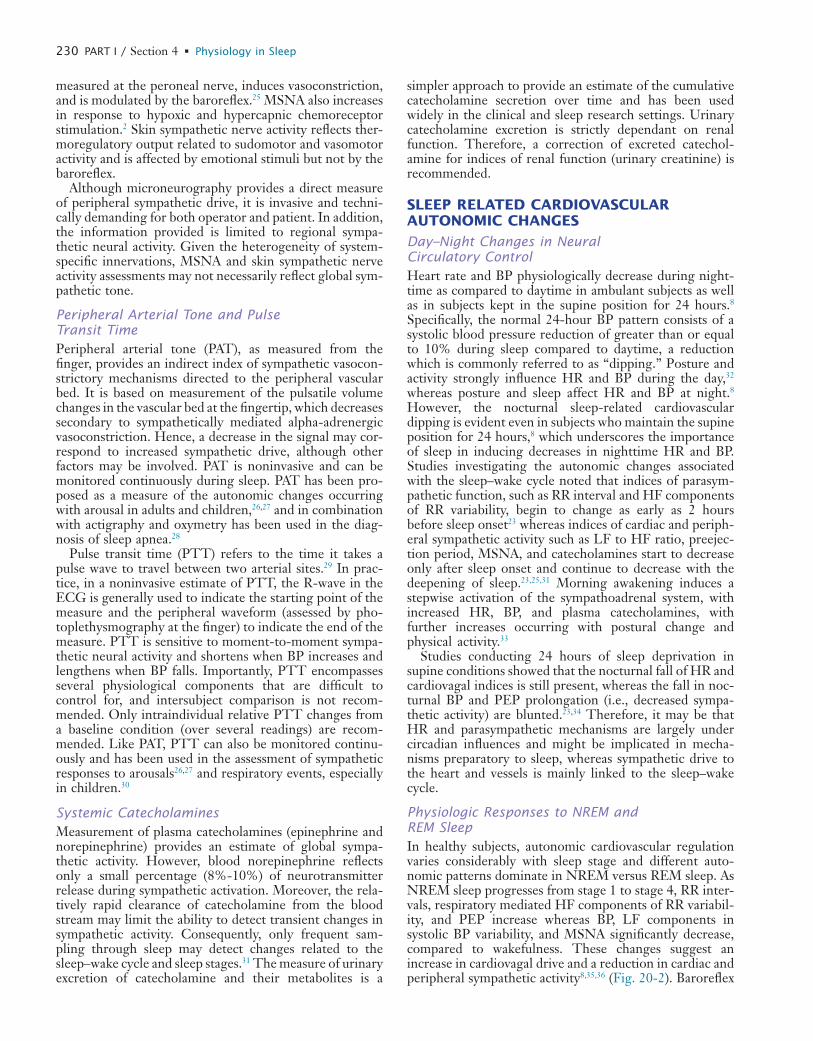
L
230 PART I / Section 4 • Physiology in Sleep
simpler approach to provide an estimate of the cumulative catecholamine secretion over time and has been used widely in the clinical and sleep research settings. Urinary catecholamine excretion is strictly dependant on renal function. Therefore, a correction of excreted catechol-amine for indices of renal function (urinary creatinine) is recommended.
SLEEP RELATED CARDIOVASCULAR AUTONOMIC CHANGES
Day–Night Changes in Neural Circulatory ControlHeart rate and BP physiologically decrease during night-time as compared to daytime in ambulant subjects as well as in subjects kept in the supine position for 24 hours.8 Specifically, the normal 24-hour BP pattern consists of a systolic blood pressure reduction of greater than or equal to 10% during sleep compared to daytime, a reduction which is commonly referred to as “dipping.” Posture and activity strongly influence HR and BP during the day,32 whereas posture and sleep affect HR and BP at night.8 However, the nocturnal sleep-related cardiovascular dipping is evident even in subjects who maintain the supine position for 24 hours,8 which underscores the importance of sleep in inducing decreases in nighttime HR and BP. Studies investigating the autonomic changes associated with the sleep–wake cycle noted that indices of parasym-pathetic function, such as RR interval and HF components of RR variability, begin to change as early as 2 hours before sleep onset23 whereas indices of cardiac and periph-eral sympathetic activity such as LF to HF ratio, preejec-tion period, MSNA, and catecholamines start to decrease only after sleep onset and continue to decrease with the deepening of sleep.23,25,31 Morning awakening induces a stepwise activation of the sympathoadrenal system, with increased HR, BP, and plasma catecholamines, with further increases occurring with postural change and physical activity.33
Studies conducting 24 hours of sleep deprivation in supine conditions showed that the nocturnal fall of HR and cardiovagal indices is still present, whereas the fall in noc-turnal BP and PEP prolongation (i.e., decreased sympa-thetic activity) are blunted.23,34 Therefore, it may be that HR and parasympathetic mechanisms are largely under circadian influences and might be implicated in mecha-nisms preparatory to sleep, whereas sympathetic drive to the heart and vessels is mainly linked to the sleep–wake cycle.
Physiologic Responses to NREM and REM SleepIn healthy subjects, autonomic cardiovascular regulation varies considerably with sleep stage and different auto-nomic patterns dominate in NREM versus REM sleep. As NREM sleep progresses from stage 1 to stage 4, RR inter-vals, respiratory mediated HF components of RR variabil-ity, and PEP increase whereas BP, LF components in systolic BP variability, and MSNA significantly decrease, compared to wakefulness. These changes suggest an increase in cardiovagal drive and a reduction in cardiac and peripheral sympathetic activity8,35,36 (Fig. 20-2). Baroreflex
measured at the peroneal nerve, induces vasoconstriction, and is modulated by the baroreflex.25 MSNA also increases in response to hypoxic and hypercapnic chemoreceptor stimulation.2 Skin sympathetic nerve activity reflects ther-moregulatory output related to sudomotor and vasomotor activity and is affected by emotional stimuli but not by the baroreflex.
Although microneurography provides a direct measure of peripheral sympathetic drive, it is invasive and techni-cally demanding for both operator and patient. In addition, the information provided is limited to regional sympa-thetic neural activity. Given the heterogeneity of system-specific innervations, MSNA and skin sympathetic nerve activity assessments may not necessarily reflect global sym-pathetic tone.
Peripheral Arterial Tone and Pulse Transit TimePeripheral arterial tone (PAT), as measured from the finger, provides an indirect index of sympathetic vasocon-strictory mechanisms directed to the peripheral vascular bed. It is based on measurement of the pulsatile volume changes in the vascular bed at the fingertip, which decreases secondary to sympathetically mediated alpha-adrenergic vasoconstriction. Hence, a decrease in the signal may cor-respond to increased sympathetic drive, although other factors may be involved. PAT is noninvasive and can be monitored continuously during sleep. PAT has been pro-posed as a measure of the autonomic changes occurring with arousal in adults and children,26,27 and in combination with actigraphy and oxymetry has been used in the diag-nosis of sleep apnea.28
Pulse transit time (PTT) refers to the time it takes a pulse wave to travel between two arterial sites.29 In prac-tice, in a noninvasive estimate of PTT, the R-wave in the ECG is generally used to indicate the starting point of the measure and the peripheral waveform (assessed by pho-toplethysmography at the finger) to indicate the end of the measure. PTT is sensitive to moment-to-moment sympa-thetic neural activity and shortens when BP increases and lengthens when BP falls. Importantly, PTT encompasses several physiological components that are difficult to control for, and intersubject comparison is not recom-mended. Only intraindividual relative PTT changes from a baseline condition (over several readings) are recom-mended. Like PAT, PTT can also be monitored continu-ously and has been used in the assessment of sympathetic responses to arousals26,27 and respiratory events, especially in children.30
Systemic CatecholaminesMeasurement of plasma catecholamines (epinephrine and norepinephrine) provides an estimate of global sympa-thetic activity. However, blood norepinephrine reflects only a small percentage (8%-10%) of neurotransmitter release during sympathetic activation. Moreover, the rela-tively rapid clearance of catecholamine from the blood stream may limit the ability to detect transient changes in sympathetic activity. Consequently, only frequent sam-pling through sleep may detect changes related to the sleep–wake cycle and sleep stages.31 The measure of urinary excretion of catecholamine and their metabolites is a
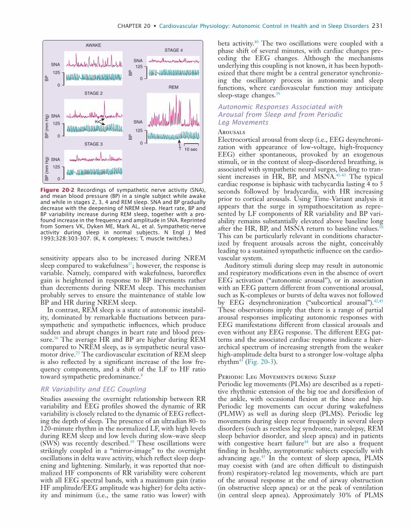
L
CHAPTER 20 • Cardiovascular Physiology: Autonomic Control in Health and in Sleep Disorders 231
beta activity.40 The two oscillations were coupled with a phase shift of several minutes, with cardiac changes pre-ceding the EEG changes. Although the mechanisms underlying this coupling is not known, it has been hypoth-esized that there might be a central generator synchroniz-ing the oscillatory process in autonomic and sleep functions, where cardiovascular function may anticipate sleep-stage changes.39
Autonomic Responses Associated with Arousal from Sleep and from Periodic Leg MovementsArousalsElectrocortical arousal from sleep (i.e., EEG desynchroni-zation with appearance of low-voltage, high-frequency EEG) either spontaneous, provoked by an exogenous stimuli, or in the context of sleep-disordered breathing, is associated with sympathetic neural surges, leading to tran-sient increases in HR, BP, and MSNA.41-43 The typical cardiac response is biphasic with tachycardia lasting 4 to 5 seconds followed by bradycardia, with HR increasing prior to cortical arousals. Using Time-Variant analysis it appears that the surge in sympathoexcitation as repre-sented by LF components of RR variability and BP vari-ability remains substantially elevated above baseline long after the HR, BP, and MSNA return to baseline values.18 This can be particularly relevant in conditions character-ized by frequent arousals across the night, conceivably leading to a sustained sympathetic influence on the cardio-vascular system.
Auditory stimuli during sleep may result in autonomic and respiratory modifications even in the absence of overt EEG activation (“autonomic arousal”), or in association with an EEG pattern different from conventional arousal, such as K-complexes or bursts of delta waves not followed by EEG desynchronization (“subcortical arousal”).42,43 These observations imply that there is a range of partial arousal responses implicating autonomic responses with EEG manifestations different from classical arousals and even without any EEG response. The different EEG pat-terns and the associated cardiac response indicate a hier-archical spectrum of increasing strength from the weaker high-amplitude delta burst to a stronger low-voltage alpha rhythm43 (Fig. 20-3).
Periodic Leg Movements during SleepPeriodic leg movements (PLMs) are described as a repeti-tive rhythmic extension of the big toe and dorsiflexion of the ankle, with occasional flexion at the knee and hip. Periodic leg movements can occur during wakefulness (PLMW) as well as during sleep (PLMS). Periodic leg movements during sleep recur frequently in several sleep disorders (such as restless leg syndrome, narcolepsy, REM sleep behavior disorder, and sleep apnea) and in patients with congestive heart failure44 but are also a frequent finding in healthy, asymptomatic subjects especially with advancing age.45 In the context of sleep apnea, PLMS may coexist with (and are often difficult to distinguish from) respiratory-related leg movements, which are part of the arousal response at the end of airway obstruction (in obstructive sleep apnea) or at the peak of ventilation (in central sleep apnea). Approximately 30% of PLMS
sensitivity appears also to be increased during NREM sleep compared to wakefulness37; however, the response is variable. Namely, compared with wakefulness, baroreflex gain is heightened in response to BP increments rather than decrements during NREM sleep. This mechanism probably serves to ensure the maintenance of stable low BP and HR during NREM sleep.
In contrast, REM sleep is a state of autonomic instabil-ity, dominated by remarkable fluctuations between para-sympathetic and sympathetic influences, which produce sudden and abrupt changes in heart rate and blood pres-sure.38 The average HR and BP are higher during REM compared to NREM sleep, as is sympathetic neural vaso-motor drive.25 The cardiovascular excitation of REM sleep is also reflected by a significant increase of the low fre-quency components, and a shift of the LF to HF ratio toward sympathetic predominance.8
RR Variability and EEG CouplingStudies assessing the overnight relationship between RR variability and EEG profiles showed the dynamic of RR variability is closely related to the dynamic of EEG reflect-ing the depth of sleep. The presence of an ultradian 80- to 120-minute rhythm in the normalized LF, with high levels during REM sleep and low levels during slow-wave sleep (SWS) was recently described.39 These oscillations were strikingly coupled in a “mirror-image” to the overnight oscillations in delta wave activity, which reflect sleep deep-ening and lightening. Similarly, it was reported that nor-malized HF components of RR variability were coherent with all EEG spectral bands, with a maximum gain (ratio HF amplitude/EEG amplitude was higher) for delta activ-ity and minimum (i.e., the same ratio was lower) with
125
SNA
AWAKE
STAGE 2
STAGE 4
REM
STAGE 3
SNA
SNA
BP
(m
m H
g)B
PB
P (
mm
Hg)
0
125 K
T 10 sec
0
125
0
BP
125
SNA
SNA
0
BP
125
0
Figure 20-2 Recordings of sympathetic nerve activity (SNA), and mean blood pressure (BP) in a single subject while awake and while in stages 2, 3, 4 and REM sleep. SNA and BP gradually decrease with the deepening of NREM sleep. Heart rate, BP and BP variability increase during REM sleep, together with a pro-found increase in the frequency and amplitude in SNA. Reprinted from Somers VK, Dyken ME, Mark AL, et al. Sympathetic-nerve activity during sleep in normal subjects. N Engl J Med 1993;328:303-307. (K, K complexes; T, muscle twitches.)
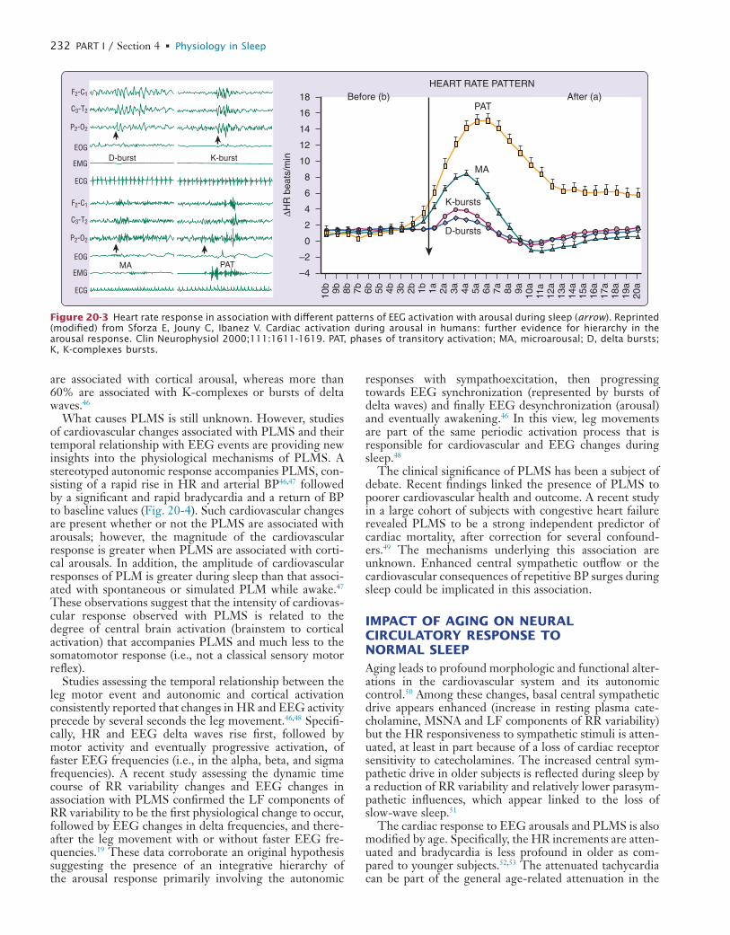
L
232 PART I / Section 4 • Physiology in Sleep
responses with sympathoexcitation, then progressing towards EEG synchronization (represented by bursts of delta waves) and finally EEG desynchronization (arousal) and eventually awakening.46 In this view, leg movements are part of the same periodic activation process that is responsible for cardiovascular and EEG changes during sleep.48
The clinical significance of PLMS has been a subject of debate. Recent findings linked the presence of PLMS to poorer cardiovascular health and outcome. A recent study in a large cohort of subjects with congestive heart failure revealed PLMS to be a strong independent predictor of cardiac mortality, after correction for several confound-ers.49 The mechanisms underlying this association are unknown. Enhanced central sympathetic outflow or the cardiovascular consequences of repetitive BP surges during sleep could be implicated in this association.
IMPACT OF AGING ON NEURAL CIRCULATORY RESPONSE TO NORMAL SLEEPAging leads to profound morphologic and functional alter-ations in the cardiovascular system and its autonomic control.50 Among these changes, basal central sympathetic drive appears enhanced (increase in resting plasma cate-cholamine, MSNA and LF components of RR variability) but the HR responsiveness to sympathetic stimuli is atten-uated, at least in part because of a loss of cardiac receptor sensitivity to catecholamines. The increased central sym-pathetic drive in older subjects is reflected during sleep by a reduction of RR variability and relatively lower parasym-pathetic influences, which appear linked to the loss of slow-wave sleep.51
The cardiac response to EEG arousals and PLMS is also modified by age. Specifically, the HR increments are atten-uated and bradycardia is less profound in older as com-pared to younger subjects.52,53 The attenuated tachycardia can be part of the general age-related attenuation in the
are associated with cortical arousal, whereas more than 60% are associated with K-complexes or bursts of delta waves.46
What causes PLMS is still unknown. However, studies of cardiovascular changes associated with PLMS and their temporal relationship with EEG events are providing new insights into the physiological mechanisms of PLMS. A stereotyped autonomic response accompanies PLMS, con-sisting of a rapid rise in HR and arterial BP46,47 followed by a significant and rapid bradycardia and a return of BP to baseline values (Fig. 20-4). Such cardiovascular changes are present whether or not the PLMS are associated with arousals; however, the magnitude of the cardiovascular response is greater when PLMS are associated with corti-cal arousals. In addition, the amplitude of cardiovascular responses of PLM is greater during sleep than that associ-ated with spontaneous or simulated PLM while awake.47 These observations suggest that the intensity of cardiovas-cular response observed with PLMS is related to the degree of central brain activation (brainstem to cortical activation) that accompanies PLMS and much less to the somatomotor response (i.e., not a classical sensory motor reflex).
Studies assessing the temporal relationship between the leg motor event and autonomic and cortical activation consistently reported that changes in HR and EEG activity precede by several seconds the leg movement.46,48 Specifi-cally, HR and EEG delta waves rise first, followed by motor activity and eventually progressive activation, of faster EEG frequencies (i.e., in the alpha, beta, and sigma frequencies). A recent study assessing the dynamic time course of RR variability changes and EEG changes in association with PLMS confirmed the LF components of RR variability to be the first physiological change to occur, followed by EEG changes in delta frequencies, and there-after the leg movement with or without faster EEG fre-quencies.19 These data corroborate an original hypothesis suggesting the presence of an integrative hierarchy of the arousal response primarily involving the autonomic
18F2-C1
C3-T2
P2-O2
EOG
EMG
ECG
F2-C1
C3-T2
P2-O2
EOG
EMG
ECG
D-burst
MA PAT
K-burst
16
14
12
10
8
6
4
MA
PAT
HEART RATE PATTERN
K-bursts
D-bursts
Before (b) After (a)
2
0
–2
–4∆H
R b
eats
/min
10b 9b 8b 7b 6b 5b 4b 3b 2b 1b 1a 2a 3a 4a 5a 6a 7a 8a 9a 10a
11a
12a
13a
14a
15a
16a
17a
18a
19a
20a
Figure 20-3 Heart rate response in association with different patterns of EEG activation with arousal during sleep (arrow). Reprinted (modified) from Sforza E, Jouny C, Ibanez V. Cardiac activation during arousal in humans: further evidence for hierarchy in the arousal response. Clin Neurophysiol 2000;111:1611-1619. PAT, phases of transitory activation; MA, microarousal; D, delta bursts; K, K-complexes bursts.
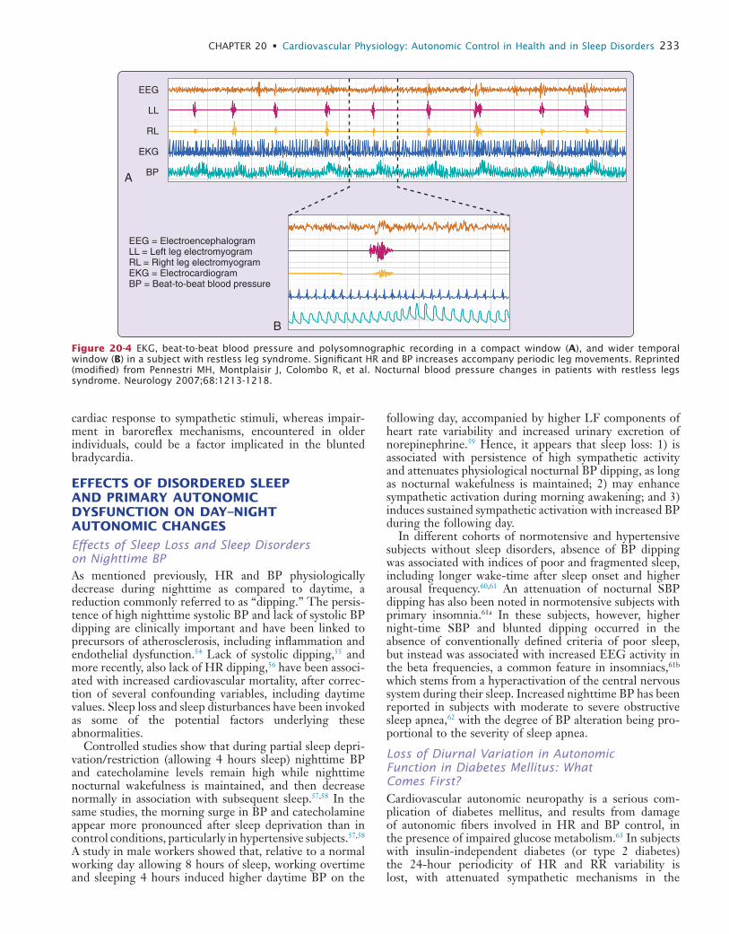
L
CHAPTER 20 • Cardiovascular Physiology: Autonomic Control in Health and in Sleep Disorders 233
following day, accompanied by higher LF components of heart rate variability and increased urinary excretion of norepinephrine.59 Hence, it appears that sleep loss: 1) is associated with persistence of high sympathetic activity and attenuates physiological nocturnal BP dipping, as long as nocturnal wakefulness is maintained; 2) may enhance sympathetic activation during morning awakening; and 3) induces sustained sympathetic activation with increased BP during the following day.
In different cohorts of normotensive and hypertensive subjects without sleep disorders, absence of BP dipping was associated with indices of poor and fragmented sleep, including longer wake-time after sleep onset and higher arousal frequency.60,61 An attenuation of nocturnal SBP dipping has also been noted in normotensive subjects with primary insomnia.61a In these subjects, however, higher night-time SBP and blunted dipping occurred in the absence of conventionally defined criteria of poor sleep, but instead was associated with increased EEG activity in the beta frequencies, a common feature in insomniacs,61b which stems from a hyperactivation of the central nervous system during their sleep. Increased nighttime BP has been reported in subjects with moderate to severe obstructive sleep apnea,62 with the degree of BP alteration being pro-portional to the severity of sleep apnea.
Loss of Diurnal Variation in Autonomic Function in Diabetes Mellitus: What Comes First?Cardiovascular autonomic neuropathy is a serious com-plication of diabetes mellitus, and results from damage of autonomic fibers involved in HR and BP control, in the presence of impaired glucose metabolism.63 In subjects with insulin-independent diabetes (or type 2 diabetes) the 24-hour periodicity of HR and RR variability is lost, with attenuated sympathetic mechanisms in the
cardiac response to sympathetic stimuli, whereas impair-ment in baroreflex mechanisms, encountered in older individuals, could be a factor implicated in the blunted bradycardia.
EFFECTS OF DISORDERED SLEEP AND PRIMARY AUTONOMIC DYSFUNCTION ON DAY–NIGHT AUTONOMIC CHANGES
Effects of Sleep Loss and Sleep Disorders on Nighttime BPAs mentioned previously, HR and BP physiologically decrease during nighttime as compared to daytime, a reduction commonly referred to as “dipping.” The persis-tence of high nighttime systolic BP and lack of systolic BP dipping are clinically important and have been linked to precursors of atherosclerosis, including inflammation and endothelial dysfunction.54 Lack of systolic dipping,55 and more recently, also lack of HR dipping,56 have been associ-ated with increased cardiovascular mortality, after correc-tion of several confounding variables, including daytime values. Sleep loss and sleep disturbances have been invoked as some of the potential factors underlying these abnormalities.
Controlled studies show that during partial sleep depri-vation/restriction (allowing 4 hours sleep) nighttime BP and catecholamine levels remain high while nighttime nocturnal wakefulness is maintained, and then decrease normally in association with subsequent sleep.57,58 In the same studies, the morning surge in BP and catecholamine appear more pronounced after sleep deprivation than in control conditions, particularly in hypertensive subjects.57,58 A study in male workers showed that, relative to a normal working day allowing 8 hours of sleep, working overtime and sleeping 4 hours induced higher daytime BP on the
EEG
LL
RL
EKG
BPA
B
EEG = ElectroencephalogramLL = Left leg electromyogramRL = Right leg electromyogramEKG = ElectrocardiogramBP = Beat-to-beat blood pressure
Figure 20-4 EKG, beat-to-beat blood pressure and polysomnographic recording in a compact window (A), and wider temporal window (B) in a subject with restless leg syndrome. Significant HR and BP increases accompany periodic leg movements. Reprinted (modified) from Pennestri MH, Montplaisir J, Colombo R, et al. Nocturnal blood pressure changes in patients with restless legs syndrome. Neurology 2007;68:1213-1218.

L
234 PART I / Section 4 • Physiology in Sleep
SYMPATHETIC ACTIVATION IN OBSTRUCTIVE SLEEP APNEA
The sympathetic nervous system appears to play a key role in the cardiac pathophysiology of sleep apnea (see also Chapter 119 in this volume). Even when patients with obstructive sleep apnea are awake and breathing normally and in the absence of any overt cardiovascular disease such as hypertension or heart failure, they have evidence for impaired sympathetic cardiovascular regulation. Specifi-cally, they have high levels of muscle sympathetic nerve activity, increased catecholamines, faster heart rates, and attenuated heart rate variability.69 Furthermore, even though they are normotensive, they have excessive blood pressure variability.70
During sleep, because of activation of the peripheral and central chemoreflexes by hypoxia and hypercapnia, sympa-thetic activity increases even further. In the setting of apnea, the inhibitory effect of the thoracic afferents is absent, thus resulting in further potentiation of sympa-thetic activation. The consequent vasoconstriction results in surges in blood pressure as noted earlier. Sym-pathetic activity abruptly ceases at onset of breathing due to the inhibitory effect of the thoracic afferents (Fig. 20-5).71 This may explain how chronic intermittent hypoxia in normals causes hypertension.72
In a minority of patients with obstructive sleep apnea, the diving reflex, noted earlier, is activated. Therefore these patients may have marked bradyarrhythmias in asso-ciation with the obstructive apnea even though they do not have any intrinsic conduction system abnormality.4 The bradycardia is secondary to cardiac vagal activation due to the combination of hypoxia and apnea.
daytime and blunted parasympathetic function during the night.64 In subjects with different degrees of glucose abnormalities without overt diabetes, RR variability and its spectral components appear similar to controls in the daytime, but are significantly altered during sleep, with strikingly higher normalized LF and lower HF, proportional to the degree of insulin resistance.65 Insulin resistance (a state where there is a reduced biologic effect of insulin) and sympathetic overactivity are known to be linked and possibly potentiate each other, with insulin increasing sympathetic activity and neuroadren-ergic mechanisms acting to increase plasma glucose avail-ability and to reduce peripheral insulin sensitivity. These data suggest that a primary alteration in the autonomic nervous system may occur during sleep in these subjects before overt diabetes is evident and is linked to the level of insulin resistance. However, one study also observed that selectively altered nighttime autonomic function was also present in nondiabetic offspring of type 2 diabetic parents, whether or not they had insulin resistance,66 suggesting that nighttime impaired parasym-pathetic mechanisms, likely of genetic origin, may precede metabolic abnormalities.
Type 2 diabetes is a complex disease that derives from the interaction of environmental factors on a background of a genetic susceptibility. Chronic sleep debt, either due to sleep restriction or sleep apnea, has been shown to be one factor that can alter glucose handling67 and increases the likelihood of developing type 2 diabetes.68 Little is known about the relationship and interactions between these sleep disturbances and early autonomic dysfunction in subjects with differing severities of glucose abnormali-ties and their healthy offspring.
0
100
200
RESP
SNA
EKG
EMG
EEG
EOG
BP
(m
m H
g)
20 sec
OSAOSA OSA
Figure 20-5 Sympathetic nerve activity (SNA) and blood pressure (BP) recordings in association with obstructive sleep apnea (OSA).SNA increases progressively during the apnea because the activation of the peripheral and central chemoreflexes by hypoxia and hypercapnia. The consequent vasoconstriction results in marked surges in BP, which reaches the peak during the hyperventilation. SNA abruptly ceases at onset of breathing due to the inhibitory effect of the thoracic afferents. The arrows indicate increases in muscle tone (i.e., EMG) toward the end of the apneaic phases in relation to arousals from REM sleep. Reprinted from Somers et al.71 with permission of the publisher.
L
CHAPTER 20 • Cardiovascular Physiology: Autonomic Control in Health and in Sleep Disorders 235
5. Lombardi F, Parati G. An update on: cardiovascular and respiratory changes during sleep in normal and hypertensive subjects. Cardiovasc Res 2000;45:200-211.
6. Mancia G. Autonomic modulation of the cardiovascular system during sleep. N Engl J Med 1993;328:347-349.
7. Millar-Craig MW, Bishop CN, Raftery EB. Circadian variation of blood-pressure. Lancet 1978;1:795-797.
8. van de Borne P, Nguyen H, Biston P, et al. Effects of wake and sleep stages on the 24-h autonomic control of blood pressure and heart rate in recumbent men. Am J Physiol 1994;266:H548-H554.
9. Heart rate variability: standards of measurement, physiological inter-pretation and clinical use. Task Force of the European Society of Cardiology and the North American Society of Pacing and Electro-physiology. Circulation 1996;93:1043-1065.
10. Bernardi L, Salvucci F, Suardi R, et al. Evidence for an intrinsic mechanism regulating heart rate variability in the transplanted and the intact heart during submaximal dynamic exercise? Cardiovasc Res 1990;24:969-981.
11. Montano N, Porta A, Malliani A. Evidence for central organization of cardiovascular rhythms. Ann N Y Acad Sci 2001;940:299-306.
12. Pagani M, Lombardi F, Guzzetti S, et al. Power spectral analysis of heart rate and arterial pressure variabilities as a marker of sympatho-vagal interaction in man and conscious dog. Circ Res 1986;59: 178-193.
13. Sleight P, La Rovere MT, Mortara A, et al. Physiology and patho-physiology of heart rate and blood pressure variability in humans: is power spectral analysis largely an index of baroreflex gain? Clin Sci Lond 1995;88:103-109.
14. Malliani A, Pagani M, Lombardi F, et al. Cardiovascular neural regu-lation explored in the frequency domain. Circulation 1991;84: 482-492.
15. Akselrod S, Gordon D, Ubel FA, et al. Power spectrum analysis of heart rate fluctuation: a quantitative probe of beat-to-beat cardiovas-cular control. Science 1981;213:220-222.
16. Parati G, Saul JP, Di Rienzo M, et al. Spectral analysis of blood pressure and heart rate variability in evaluating cardiovascular regula-tion. A critical appraisal. Hypertension 1995;25:1276-1286.
17. Cerutti S, Bianchi AM, Mainardi LT. Advanced spectral methods for detecting dynamic behaviour. Auton Neurosci 2001;90:3-12.
18. Blasi A, Jo J, Valladares E, et al. Cardiovascular variability after arousal from sleep: time-varying spectral analysis. J Appl Physiol 2003;95:1394-1404.
19. Guggisberg AG, Hess CW, Mathis J. The significance of the sym-pathetic nervous system in the pathophysiology of periodic leg move-ments in sleep. Sleep 2007;30:755-766.
20. Lombardi F. Chaos theory, heart rate variability, and arrhythmic mortality. Circulation 2000;101:8-10.
21. Togo F, Yamamoto Y. Decreased fractal component of human heart rate variability during non-REM sleep. Am J Physiol Heart Circ Physiol 2001;280:H17-H21.
22. Persson PB, DiRienzo M, Castiglioni P, et al. Time versus frequency domain techniques for assessing baroreflex sensitivity. J Hypertens 2001;19:1699-1705.
23. Burgess HJ, Trinder J, Kim Y, et al. Sleep and circadian influences on cardiac autonomic nervous system activity. Am J Physiol 1997;273:H1761-H1768.
24. Burgess HJ, Trinder J, Kim Y. Cardiac autonomic nervous system activity during presleep wakefulness and stage 2 NREM sleep. J Sleep Res 1999;8:113-122.
25. Somers VK, Dyken ME, Mark AL, et al. Sympathetic-nerve activity during sleep in normal subjects. N Engl J Med 1993;328: 303-307.
26. Pillar G, Bar A, Shlitner A, et al. Autonomic arousal index: an auto-mated detection based on peripheral arterial tonometry. Sleep 2002;25:543-549.
27. O’Brien LM, Gozal D. Potential usefulness of noninvasive auto-nomic monitoring in recognition of arousals in normal healthy chil-dren. J Clin Sleep Med 2007;3:41-47.
28. Pittman SD, MacDonald MM, Fogel RB, et al. Assessment of auto-mated scoring of polysomnographic recordings in a population with suspected sleep-disordered breathing. Sleep 2004;27:1394-1403.
29. Foo JY, Lim CS. Pulse transit time as an indirect marker for varia-tions in cardiovascular related reactivity. Technol Health Care 2006;14:97-108.
30. Foo JY. Pulse transit time in paediatric respiratory sleep studies. Med Eng Phys 2007;29:17-25.
SUMMARY
The autonomic nervous system is intimately linked to central neural state changes. This is especially true for physiologic sleep and sleep disorders. It is clear that although the different stages of physiologic sleep result in structured changes in neural circulatory control, disturbed sleep, such as is seen in patients with obstructive sleep apnea, with PLMS, or in sleep deprivation, acts to disrupt the sleep-related physiologic variations in autonomic regu-lation of heart rate and blood pressure. Our knowledge of this general area is limited by the tools available for com-prehensive and direct assessment of the autonomic nervous system in humans. Although microneurography provides a direct measurement of sympathetic neural activity to the peripheral blood vessels, this measurement itself has limi-tations. The other options available are primarily those that monitor blood and urine levels of catecholamines. Measurements such as heart rate and blood pressure vari-ability, while providing some insight, provide only indirect information on autonomic cardiovascular control, and are limited due to problems with regard to acquisition of data, confounding effects of medications and abnormal breathing patterns, and inconsistencies with regard to interpretation. Rigorous methods in line with the standard recommendations9 are mandatory. Therefore, although this chapter seeks to address some of the current knowl-edge in the area of neural circulatory control during normal and disordered sleep, the available data are limited in part because of methodological shortcomings and also because of the obvious difficulties inherent in nighttime studies of sleep physiology in humans.
� Clinical Pearl
The autonomic nervous system is the mediator of central–cardiovascular interactions occurring during sleep and its normal function appears to be impor-tant in preserving health. Despite the recognized methodological limitations (technically demanding and cautious interpretation of outcome of interest as described in Table 20-1), broadening sleep poly-graphic monitoring to include heart rate and blood pressure recordings may contribute to a better under-standing of the physiology and pathology of sleep-related cardiovascular autonomic modulation. These considerations suggest an important potential avenue for innovation in the management of medical disor-ders that may, in part, be sleep related (e.g., hyper-tension, diabetes, periodic limb movement, sleep disordered breathing).
REFERENCES1. Sleight P, Ekberg DL. Human baroreflexes in health and disease.
Oxford: Clarendon; 1992.2. Somers VK, Mark AL, Abboud FM. Sympathetic activation by
hypoxia and hypercapnia—implications for sleep apnea. Clin Exp Hypertens A 1988;10(Suppl. 1):413-422.
3. Daly MD, Angell-James JE, Elsner R. Role of carotid-body chemo-receptors and their reflex interactions in bradycardia and cardiac arrest. Lancet 1979;1:764-767.
4. Somers VK, Dyken ME, Mark AL, et al. Parasympathetic hyperre-sponsiveness and bradyarrhythmias during apnoea in hypertension. Clin Auton Res 1992;2:171-176.
L
236 PART I / Section 4 • Physiology in Sleep
53. Gosselin N, Lanfranchi P, Michaud M, et al. Age and gender effects on heart rate activation associated with periodic leg movements in patients with restless legs syndrome. Clin Neurophysiol 2003; 114:2188-2195.
54. von Kanel R, Jain S, Mills PJ, et al. Relation of nocturnal blood pressure dipping to cellular adhesion, inflammation and hemostasis. J Hypertens 2004;22:2087-2093.
55. Fagard RH, Celis H, Thijs L, et al. Daytime and nighttime blood pressure as predictors of death and cause-specific cardiovascular events in hypertension. Hypertension 2008;51:55-61.
56. Ben-Dov IZ, Kark JD, Ben-Ishay D, et al. Blunted heart rate dip during sleep and all-cause mortality. Arch Intern Med 2007;167: 2116-2121.
57. Lusardi P, Mugellini A, Preti P, et al. Effects of a restricted sleep regimen on ambulatory blood pressure monitoring in normotensive subjects. Am J Hypertens 1996;9:503-505.
58. Lusardi P, Zoppi A, Preti P, et al. Effects of insufficient sleep on blood pressure in hypertensive patients: a 24-h study. Am J Hyper-tens 1999;12:63-68.
59. Tochikubo O, Ikeda A, Miyajima E, et al. Effects of insufficient sleep on blood pressure monitored by a new multibiomedical recorder. Hypertension 1996;27:1318-1324.
60. Loredo JS, Nelesen R, Ancoli-Israel S, Dimsdale JE. Sleep quality and blood pressure dipping in normal adults. Sleep 2004;27: 1097-1103.
61. Pedulla M, Silvestri R, Lasco A, et al. Sleep structure in essential hypertensive patients: differences between dippers and non-dippers. Blood Press 1995;4:232-237.
61a. Lanfranchi PA, Pennestri MH, Fradette L, et al. Nighttime blood pressure in normotensive subjects with chronic insomnia: implica-tions for cardiovascular risk. Sleep 2009;32:760-766.
61b. Perlis ML, Merica H, Smith MT, et al. EEG activity and insomnia. Sleep Med Rev 2001;5:363-374.
62. Pankow W, Nabe B, Lies A, et al. Influence of sleep apnea on 24-hour blood pressure. Chest 1997;112:1253-1258.
63. Vinik AI, Ziegler D. Diabetic cardiovascular autonomic neuropathy. Circulation 2007;115:387-397.
64. Bernardi L, Ricordi L, Lazzari P, et al. Impaired circadian modula-tion of sympathovagal activity in diabetes. A possible explanation for altered temporal onset of cardiovascular disease. Circulation 1992; 86:1443-1452.
65. Perciaccante A, Fiorentini A, Paris A, et al. Circadian rhythm of the autonomic nervous system in insulin resistant subjects with normo-glycemia, impaired fasting glycemia, impaired glucose tolerance, type 2 diabetes mellitus. BMC Cardiovasc Disord 2006;6:19.
66. Fiorentini A, Perciaccante A, Paris A, et al. Circadian rhythm of autonomic activity in non diabetic offsprings of type 2 diabetic patients. Cardiovasc Diabetol 2005;4:15.
67. Spiegel K, Leproult R, Van CE. Impact of sleep debt on metabolic and endocrine function. Lancet 1999;354:1435-1439.
68. Zizi F, Jean-Louis G, Brown CD, et al. Sleep duration and the risk of diabetes mellitus: epidemiologic evidence and pathophysiologic insights. Curr Diab Rep 2010;10(1):43-47.
69. Narkiewicz K, Somers VK. Sympathetic nerve activity in obstructive sleep apnoea. Acta Physiol Scand 2003;177:385-390.
70. Narkiewicz K, Somers VK. Cardiovascular variability characteristics in obstructive sleep apnea. Auton Neurosci 2001;90:89-94.
71. Somers VK, Dyken ME, Clary MP, et al. Sympathetic neural mecha-nisms in obstructive sleep apnea. J Clin Invest 1995;96:1897-1904.
72. Gilmartin GS, Lynch M, Tamisier R, Weiss JW. Chronic intermit-tent hypoxia in humans during 28 nights results in blood pressure elevation and increased muscle sympathetic nerve activity. Am J Physiol Heart Circ Physiol 2010 Jun 25. (Epub ahead of print)
31. Dodt C, Breckling U, Derad I, et al. Plasma epinephrine and nor-epinephrine concentrations of healthy humans associated with night-time sleep and morning arousal. Hypertension 1997;30:71-76.
32. Kario K, Schwartz JE, Pickering TG. Ambulatory physical activity as a determinant of diurnal blood pressure variation. Hypertension 1999;34:685-691.
33. Linsell CR, Lightman SL, Mullen PE, et al. Circadian rhythms of epinephrine and norepinephrine in man. J Clin Endocrinol Metab 1985;60:1210-1215.
34. Kerkhof GA, Van Dongen HP, Bobbert AC. Absence of endogenous circadian rhythmicity in blood pressure? Am J Hypertens 1998; 11:373-377.
35. Somers VK, Dyken ME, Mark AL, et al. Sympathetic-nerve activity during sleep in normal subjects. N Engl J Med 1993;328:303-307.
36. Bonnet MH, Arand DL. Heart rate variability: sleep stage, time of night, and arousal influences. Electroencephalogr Clin Neurophysiol 1997;102:390-396.
37. Legramante JM, Marciani MG, Placidi F, et al. Sleep-related changes in baroreflex sensitivity and cardiovascular autonomic modulation. J Hypertens 2003;21:1555-1561.
38. Guilleminault C, Pool P, Motta J, et al. Sinus arrest during REM sleep in young adults. N Engl J Med 1984;311:1006-1010.
39. Brandenberger G, Ehrhart J, Piquard F, et al. Inverse coupling between ultradian oscillations in delta wave activity and heart rate variability during sleep. Clin Neurophysiol 2001;112:992-996.
40. Jurysta F, van de Borne P, Migeotte PF, et al. A study of the dynamic interactions between sleep EEG and heart rate variability in healthy young men. Clin Neurophysiol 2003;114:2146-2155.
41. Davies RJ, Belt PJ, Roberts SJ, et al. Arterial blood pressure responses to graded transient arousal from sleep in normal humans. J Appl Physiol 1993;74:1123-1130.
42. Morgan BJ, Crabtree DC, Puleo DS, et al. Neurocirculatory conse-quences of abrupt change in sleep state in humans. J Appl Physiol 1996;80:1627-1636.
43. Sforza E, Jouny C, Ibanez V. Cardiac activation during arousal in humans: further evidence for hierarchy in the arousal response. Clin Neurophysiol 2000;111:1611-1619.
44. Javaheri S. Sleep disorders in systolic heart failure: a prospective study of 100 male patients. The final report. Int J Cardiol 2006; 106:21-28.
45. Pennestri MH, Whittom S, Adam B, et al. PLMS and PLMW in healthy subjects as a function of age: prevalence and interval distribu-tion. Sleep 2006;29:1183-1187.
46. Sforza E, Nicolas A, Lavigne G, et al. EEG and cardiac activation during periodic leg movements in sleep: support for a hierarchy of arousal responses. Neurology 1999;52:786-791.
47. Pennestri MH, Montplaisir J, Colombo R, et al. Nocturnal blood pressure changes in patients with restless legs syndrome. Neurology 2007;68:1213-1218.
48. Ferrillo F, Beelke M, Canovaro P, et al. Changes in cerebral and autonomic activity heralding periodic limb movements in sleep. Sleep Med 2004;5:407-412.
49. Yumino D, Wang H, Floras JF, et al. Association between periodic limb movements in sleep and increased mortality risk in patients with heart failure. Circulation 2008;118:796.
50. Lakatta EG. Cardiovascular regulatory mechanisms in advanced age. Physiol Rev 1993;73:413-467.
51. Crasset V, Mezzetti S, Antoine M, et al. Effects of aging and cardiac denervation on heart rate variability during sleep. Circulation 2001;103:84-88.
52. Goff EA, O’Driscoll DM, Simonds AK, et al. The cardiovascular response to arousal from sleep decreases with age in healthy adults. Sleep 2008;31:1009-1017.
LL
237
Abstract
Any anatomical or physiological abnormality that compro-mises the respiratory system in wakefulness can predispose a person to significant breathing disorders during sleep. Such abnormalities include an anatomically small upper airspace that increases the reliance on the pharyngeal muscles to main-tain adequate airflow and to prevent obstructive sleep apnea. Patients with restrictive lung diseases or neuromuscular weakness rely, to varying degrees, on the activation of nondia-phragmatic respiratory muscles to help maintain adequate ventilation in wakefulness, but this compensation can be reduced or absent in sleep, leading to severe hypoventilation. These and other examples highlight that sleep is a state of vulnerability for the respiratory system, and that loss of a “wakefulness stimulus” that is sufficient to sustain adequate breathing in wakefulness is central to the pathogenesis of a variety of respiratory disorders during sleep. This simple concept has been an enduring principle in respiratory medi-cine because, ultimately, it is the root mechanism to under-
stand sleep effects on breathing. To this end there have been significant recent developments in identifying the neurochemi-cal substrates underlying this wakefulness stimulus, central to which is an understanding of the neurobiology of sleep, its impact on central respiratory neurons and motoneurons, and the important role of tonic excitatory (nonrespiratory) drives in contributing to the overall level of excitability in the respira-tory system. Moreover, like the realization that sleep onset is not simply the passive withdrawal of wakefulness, breathing during sleep is not simply due to the passive withdrawal of the “wakefulness stimulus.” For example, non–rapid eye move-ment (NREM) sleep and rapid eye movement (REM) sleep are fundamentally different neurobiological states, which have distinct effects on the control of respiratory neurons and motoneurons. Accordingly, NREM and REM sleep pose different problems to breathing during sleep in different individuals with different pathologies, and understanding these mecha-nisms is necessary for physiological understanding of the spectrum of sleep-related breathing disorders and their appropriate clinical management.
(predominantly inspiratory) neurons and caudal retroam-bigualis (predominantly expiratory) neurons (see Fig. 21-1).1,2 The VRG and DRG contain both bulbospinal respiratory premotoneurons (i.e., neurons that project to spinal motoneurons, which in turn innervate the respec-tive respiratory pump and abdominal muscles of breath-ing), and propriobulbar neurons (i.e., neurons that project to, and influence the activity of, other medullary respira-tory neurons but themselves do not project to motoneu-rons) (see Fig. 21-1).1,2 The hypoglossal, trigeminal, and facial motor nuclei also innervate muscles important to pharyngeal motor control and the maintenance of upper airway patency (see Fig. 21-1).4 It is important to stress, however, that the expression of respiratory-related activity is not restricted to neurons of the DRG, VRG, and cranial motoneurons innervating the pharyngeal and laryngeal muscles. For example, neurons expressing respiratory-related activity in the pons (i.e., the pontine respiratory group; PRG in Fig. 21-1) are thought to play an impor-tant role in shaping the activity of medullary respiratory neurons during breathing.3
Pre-Bötzinger ComplexPre-Bötzinger complex neurons have pacemaker-like properties that are thought important to the generation of the basic respiratory rhythm and the expression of rhyth-mic neuronal activity elsewhere in the respiratory network (see Fig. 21-1).5,6 Respiratory rhythm-generating pre-Bötzinger complex neurons coexpress mu-opioid and neu-rokinin-1 receptors (i.e., the receptors for Substance P) that slow and increase respiratory rate respectively.6 The presence of mu-opioid receptors on pre-Bötzinger complex
RESPIRATORY NEUROBIOLOGY: BASIC OVERVIEW
Medullary Respiratory Neurons and MotoneuronsThere are bilateral columns of neurons in the medulla with activity patterns that vary in phase with some com-ponent of the respiratory cycle. The dorsal respiratory group (DRG) is located in the dorsomedial medulla, spe-cifically in the ventrolateral nucleus of the solitary tract, and contains predominantly inspiratory neurons (Fig. 21-1).1,2 The DRG and the other subnuclei of the solitary tract are also the primary projection sites for vagal affer-ents from the lung, and afferents from the carotid and aortic chemoreceptors and baroreceptors, that exert important reflex influences on breathing. These projec-tions indicate that the nuclei of the solitary tract, including the DRG, are key sites of integration of sensory informa-tion from the lung, as well as information regarding the prevailing levels of arterial Pco2, Po2, pH, and systemic blood pressure. The ventral respiratory group (VRG) extends from the facial nucleus to the first cervical segment of the spinal cord and contains both inspiratory and expi-ratory neurons (see Fig. 21-1).1,2 The nucleus ambiguus also consists of a rostrally to caudally extending column neurons expressing respiratory-related activity, with sub-regions regions containing motoneurons that innervate the muscles of the larynx and pharynx that are not con-sidered part of the VRG.3 In addition to the nucleus ambiguus, rostrally to caudally the VRG is comprised of Bötzinger complex (expiratory) neurons, pre-Bötzinger complex (inspiratory) neurons, rostral retroambigualis
Respiratory Physiology: Central Neural Control of Respiratory Neurons and Motoneurons during SleepRichard L. Horner
Chapter
21
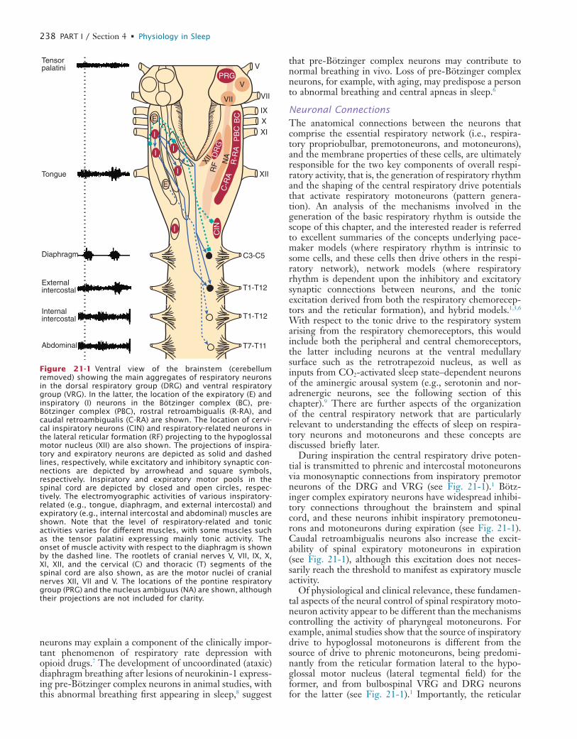
L
238 PART I / Section 4 • Physiology in Sleep
that pre-Bötzinger complex neurons may contribute to normal breathing in vivo. Loss of pre-Bötzinger complex neurons, for example, with aging, may predispose a person to abnormal breathing and central apneas in sleep.6
Neuronal ConnectionsThe anatomical connections between the neurons that comprise the essential respiratory network (i.e., respira-tory propriobulbar, premotoneurons, and motoneurons), and the membrane properties of these cells, are ultimately responsible for the two key components of overall respi-ratory activity, that is, the generation of respiratory rhythm and the shaping of the central respiratory drive potentials that activate respiratory motoneurons (pattern genera-tion). An analysis of the mechanisms involved in the generation of the basic respiratory rhythm is outside the scope of this chapter, and the interested reader is referred to excellent summaries of the concepts underlying pace-maker models (where respiratory rhythm is intrinsic to some cells, and these cells then drive others in the respi-ratory network), network models (where respiratory rhythm is dependent upon the inhibitory and excitatory synaptic connections between neurons, and the tonic excitation derived from both the respiratory chemorecep-tors and the reticular formation), and hybrid models.1,3,6 With respect to the tonic drive to the respiratory system arising from the respiratory chemoreceptors, this would include both the peripheral and central chemoreceptors, the latter including neurons at the ventral medullary surface such as the retrotrapezoid nucleus, as well as inputs from CO2-activated sleep state–dependent neurons of the aminergic arousal system (e.g., serotonin and nor-adrenergic neurons, see the following section of this chapter).9 There are further aspects of the organization of the central respiratory network that are particularly relevant to understanding the effects of sleep on respira-tory neurons and motoneurons and these concepts are discussed briefly later.
During inspiration the central respiratory drive poten-tial is transmitted to phrenic and intercostal motoneurons via monosynaptic connections from inspiratory premotor neurons of the DRG and VRG (see Fig. 21-1).1 Bötz-inger complex expiratory neurons have widespread inhibi-tory connections throughout the brainstem and spinal cord, and these neurons inhibit inspiratory premotoneu-rons and motoneurons during expiration (see Fig. 21-1). Caudal retroambigualis neurons also increase the excit-ability of spinal expiratory motoneurons in expiration (see Fig. 21-1), although this excitation does not neces-sarily reach the threshold to manifest as expiratory muscle activity.
Of physiological and clinical relevance, these fundamen-tal aspects of the neural control of spinal respiratory moto-neuron activity appear to be different than the mechanisms controlling the activity of pharyngeal motoneurons. For example, animal studies show that the source of inspiratory drive to hypoglossal motoneurons is different from the source of drive to phrenic motoneurons, being predomi-nantly from the reticular formation lateral to the hypo-glossal motor nucleus (lateral tegmental field) for the former, and from bulbospinal VRG and DRG neurons for the latter (see Fig. 21-1).1 Importantly, the reticular
neurons may explain a component of the clinically impor-tant phenomenon of respiratory rate depression with opioid drugs.7 The development of uncoordinated (ataxic) diaphragm breathing after lesions of neurokinin-1 express-ing pre-Bötzinger complex neurons in animal studies, with this abnormal breathing first appearing in sleep,8 suggest
V
VII
IXXXI
XII
C3-C5
PRGV
VII
BC
PB
CR
-RA
C-R
AN
ADR
G
XII
T1-T12
T1-T12
T7-T11
Diaphragm
External intercostal
Abdominal
Internalintercostal
Tongue
Tensorpalatini
CIN
RF
I
I
I
I
I
E
E
Figure 21-1 Ventral view of the brainstem (cerebellum removed) showing the main aggregates of respiratory neurons in the dorsal respiratory group (DRG) and ventral respiratory group (VRG). In the latter, the location of the expiratory (E) and inspiratory (I) neurons in the Bötzinger complex (BC), pre- Bötzinger complex (PBC), rostral retroambigualis (R-RA), and caudal retroambigualis (C-RA) are shown. The location of cervi-cal inspiratory neurons (CIN) and respiratory-related neurons in the lateral reticular formation (RF) projecting to the hypoglossal motor nucleus (XII) are also shown. The projections of inspira-tory and expiratory neurons are depicted as solid and dashed lines, respectively, while excitatory and inhibitory synaptic con-nections are depicted by arrowhead and square symbols, respectively. Inspiratory and expiratory motor pools in the spinal cord are depicted by closed and open circles, respec-tively. The electromyographic activities of various inspiratory-related (e.g., tongue, diaphragm, and external intercostal) and expiratory (e.g., internal intercostal and abdominal) muscles are shown. Note that the level of respiratory-related and tonic activities varies for different muscles, with some muscles such as the tensor palatini expressing mainly tonic activity. The onset of muscle activity with respect to the diaphragm is shown by the dashed line. The rootlets of cranial nerves V, VII, IX, X, XI, XII, and the cervical (C) and thoracic (T) segments of the spinal cord are also shown, as are the motor nuclei of cranial nerves XII, VII and V. The locations of the pontine respiratory group (PRG) and the nucleus ambiguus (NA) are shown, although their projections are not included for clarity.
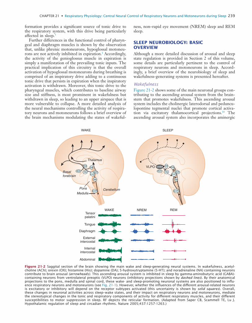
L
CHAPTER 21 • Respiratory Physiology: Central Neural Control of Respiratory Neurons and Motoneurons during Sleep 239
ness, non–rapid eye movement (NREM) sleep and REM sleep.
SLEEP NEUROBIOLOGY: BASIC OVERVIEWAlthough a more detailed discussion of arousal and sleep state regulation is provided in Section 2 of this volume, some details are particularly pertinent to the control of respiratory neurons and motoneurons in sleep. Accord-ingly, a brief overview of the neurobiology of sleep and wakefulness-generating systems is presented hereafter.
WakefulnessFigure 21-2 shows some of the main neuronal groups con-tributing to the ascending arousal system from the brain-stem that promotes wakefulness. This ascending arousal system includes the cholinergic laterodorsal and peduncu-lopontine tegmental nuclei that promote cortical activa-tion via excitatory thalamocortical projections.10 The ascending arousal system also incorporates the aminergic
formation provides a significant source of tonic drive to the respiratory system, with this drive being particularly affected in sleep.3
Further differences in the functional control of pharyn-geal and diaphragm muscles is shown by the observation that, unlike phrenic motoneurons, hypoglossal motoneu-rons are not actively inhibited in expiration.1 Accordingly, the activity of the genioglossus muscle in expiration is simply a manifestation of the prevailing tonic inputs. The practical implication of this circuitry is that the overall activation of hypoglossal motoneurons during breathing is comprised of an inspiratory drive adding to a continuous tonic drive that persists in expiration when the inspiratory activation is withdrawn. Moreover, this tonic drive to the pharyngeal muscles, which contributes to baseline airway size and stiffness, is most prominent in wakefulness but withdrawn in sleep, so leading to an upper airspace that is more vulnerable to collapse. A more detailed analysis of the neural mechanisms controlling the activity of respira-tory neurons and motoneurons follows a brief overview of the brain mechanisms modulating the states of wakeful-
Diaphragm
Externalintercostal
Abdominal
Internalintercostal
Tensorpalatini
Basalforebrain
CerebellumMedulla
Pons
Thalamus
WAKE
VLPO
SLEEP
Tongue
WAKE NREM REM
5-HT
DAOX
ACh
RF
ACh5 HT
His
NA
5-HT
DAOX
ACh
RF
ACh5-HT
His
NA
GABA
Figure 21-2 Saggital section of the brain showing the main wake and sleep-generating neural systems. In wakefulness, acetyl-choline (ACh); orexin (OX); histamine (His); dopamine (DA); 5-hydroxytryptamine (5-HT); and noradrenaline (NA) containing neurons contribute to brain arousal (arrowheads). This ascending arousal system is inhibited in sleep by gamma-aminobutyric acid (GABA)-containing neurons from ventrolateral preoptic (VLPO) neurons (inhibitory projections shown by dashed lines). By their anatomical projections to the pons, medulla and spinal cord, these wake- and sleep-promoting neuronal systems are also positioned to influ-ence respiratory neurons and motoneurons (see Fig. 21-1). However, whether the influences of the different arousal-related neurons is excitatory or inhibitory will depend on the receptor subtypes activated (this uncertainty is shown by solid squares). Overall, these changes in neuronal activities across sleep–wake states, and their impact on respiratory neurons and motoneurons, mediate the stereotypical changes in the tonic and respiratory components of activity for different respiratory muscles, and their different susceptibilities to motor suppression in sleep. RF depicts the reticular formation. (Adapted from Saper CB, Scammell TE, Lu J. Hypothalamic regulation of sleep and circadian rhythms. Nature 2005;437:1257-1263.)
L
240 PART I / Section 4 • Physiology in Sleep
mation is used to mimic this process experimentally in animal studies, that is, the “carbachol model of REM sleep.”17,18 A significant component of the motor sup-pression of REM sleep is mediated by descending path-ways involving activation of medullary reticular formation relay neurons19 that are inhibitory to spinal motoneurons via release of glycine.20
Despite the strong experimental support for the inter-action of pontine monoaminergic and cholinergic neurons as being primarily responsible for the initiation and main-tenance of REM sleep, recent evidence has implicated a glutamatergic-GABAergic mechanism.21,22 One of the key differences between the aminergic-cholinergic and the glutamatergic-GABAergic hypotheses of REM sleep gen-eration is that the motor atonia is produced by different pathways, that is, the latter framework does not require a relay in the medullary reticular formation.22 Rather, in the glutamatergic-GABAergic mechanism of REM sleep induction, the REM sleep-active pontine neurons are thought to lead to suppression of spinal motoneuron activity via long glutamatergic projections to the ventral horn of the spinal cord, which then activate local gly-cinergic interneurons to inhibit motor activity.22 Such a mechanism is likely involved in the strong inhibition of spinal intercostal motoneurons in REM sleep, but whether collaterals from these specific long descending glutama-tergic projections also synapse onto glycinergic inhibitory interneurons in the hypoglossal motor pool is not established.16
In summary, a number of neural systems show changes in activity across sleep–wake states and project to respira-tory neurons and motoneurons. Given that motoneurons are the final common output pathway for the influence of the central nervous system on motor activity, this chapter will initially focus on the control of respiratory motoneu-rons across sleep–wake states before addressing the control of the central respiratory neurons that ultimately drive breathing via those motoneurons.
CONTROL OF RESPIRATORY MOTONEURONSA characteristic and defining feature of mammalian motor activity is that postural muscle tone is highest in wakeful-ness, decreased in NREM sleep, and minimal in REM sleep, with the hypotonia of REM sleep punctuated by occasional muscle twitches that are associated with vigor-ous eye movements and phasic REM sleep events.20 Whether respiratory muscle activity is affected in the same way as postural muscle activity across sleep–wake states is somewhat complicated by the interaction of the primary influence of sleep state (e.g., producing suppression of muscle tone) and any subsequent respiratory response (e.g., to compensate for any hypoventilation). On balance, however, the overall stereotyped pattern of suppression of postural muscle activity across sleep–wake states also typi-cally occurs in respiratory muscles, with the degree of sleep state–dependent modulation being most readily apparent in those muscles that combine respiratory and nonrespira-tory (e.g., postural and behavioral) functions such as the intercostal and pharyngeal muscles.23 In these respiratory muscles, decreases in activity typically occur immediately
arousal system that originates from brainstem neuronal groups principally containing serotonin (dorsal raphé nuclei), noradrenaline (locus coeruleus), histamine (tuber-omammillary nucleus), and dopamine (ventral periaque-ductal grey). Orexin neurons from the perifornical region of the hypothalamus and cholinergic neurons from the basal forebrain also contribute to this ascending arousal system.10 Overall, multiple neuronal systems contribute to cortical arousal and wakefulness. These neuronal systems are also positioned to influence respiratory neurons and motoneurons via their anatomical projections to the pons, medulla, and spinal cord (see Fig. 21-2).
NREM SleepSleep is actively generated by neurons in the ventrolateral preoptic area, anterior hypothalamus, and basal forebrain (see Fig. 21-2).10 These neurons become active in NREM sleep, an effect influenced by the thermal stimulus that accompanies the circadian-mediated decline in body tem-perature at normal bedtime.11 This circadian-mediated decline in body temperature is mediated by a change in the set-point of hypothalamic temperature-regulating neurons, which initially leads to a relative “warm stimulus” as body temperature is at first higher than the new set-point, that is, before heat loss occurs. This warm stimulus activates NREM sleep-active hypothalamic neurons and so pro-motes sleep onset. This effect of internal body temperature on sleep is distinct from the influences of ambient envi-ronmental temperature on sleep regulation. Activation of ventrolateral preoptic neurons leads to a direct suppression of cortical arousal, this via ascending inhibitory cortical projections. Ventrolateral preoptic neurons also promote sleep by descending inhibition of the aforementioned brainstem arousal neurons via release of gamma-aminobu-tyric acid (GABA) and galanin.11,12 This effect of GABA explains the sedative-hypnotic effects of barbiturates, ben-zodiazepines, and imidazopyridine compounds that enhance GABA-mediated neuronal inhibition via interac-tions with binding sites on the GABAA receptor.13 GABAA receptors are also strongly implicated in respiratory control and are present throughout the respiratory network,14 excessive stimulation of which can promote respiratory depression.15
In summary, sleep onset is triggered by increased GABA-ergic neuronal activity, and this is accompanied by a massed and coordinated withdrawal of activity of brainstem arousal neurons comprising serotonergic, noradrenergic, hista-minergic and cholinergic neurons. Given the widespread projections of these sleep state–dependent neuronal groups, these changes in neuronal activity in sleep are also positioned to influence respiratory neurons and motoneu-rons (see Fig. 21-2).16
REM SleepDecreased serotonergic and noradrenergic activity pre-ceding and during REM sleep withdraws inhibition of the laterodorsal and pedunculopontine tegmental nuclei.10,12 This effect leads to increased acetylcholine release into the pontine reticular formation to trigger REM sleep.17,18 Exogenous application of cholinergic agonists or acetylcholinesterase inhibitors (to increase endogenous acetylcholine) into the pontine reticular for-

L
CHAPTER 21 • Respiratory Physiology: Central Neural Control of Respiratory Neurons and Motoneurons during Sleep 241
DETERMINANTS OF RESPIRATORY MOTONEURON ACTIVITY
Tonic and Respiratory-Related Inputs to Respiratory MotoneuronsThe changes in muscle tone across sleep–wake states ulti-mately result from the impact of sleep neural mechanisms on the electrical properties and membrane potential of individual motoneurons located in the respective motor pools in the central nervous system. In turn, the excitability of individual motoneurons changes across sleep–wake states because of varying degrees of excitatory and inhibi-tory inputs to those motoneurons from sleep–wake related regions in the brain, and from neurons activated during specific behaviors such as purposeful motor acts in
at sleep onset23 indicating a primary suppressant effect of sleep neural mechanisms on the activity of respiratory motoneurons, that is, before any compensatory increase in activity takes place in response to altered blood gases, mechanical loads, or sleep-disordered breathing. In con-trast to those respiratory muscles with both respiratory and nonrespiratory functions, the diaphragm has an almost sole respiratory function and undergoes lesser suppression of activity in NREM sleep and is largely spared the motor inhibition of REM sleep (see Fig. 21-2).24 Chapters 22 and 23 provide more detail regarding clinical aspects of the control of breathing and upper airway function during sleep, whereas in this chapter the fundamental mechanisms underlying these effects of sleep on the respiratory system are presented.
}}
}
} }
Tonic Respiratory
Motoneuron
Premotorinput
Muscle
Presynapticmodulation
Respiratory-relatedTonic
Respiratory drive potentials
Tonic membrane potentialTonic
Respiratory
Respiratory Apnea
Respiratory “Respiratory”
A B C
D E
Sum of inputsat motoneuron
Electromyographicactivity
Electromyographic activity
Threshold for neural activity
Figure 21-3 Schema to show how converging tonic (e.g., postural, nonrespiratory) and respiratory inputs to a motoneuron summate to produce the tonic and respiratory components of electromyographic activity. These premotor tonic and respiratory inputs can be excitatory or inhibitory, but in this figure they are shown as excitatory for simplicity. Panels A to E further show how changes in the tonic and respiratory components of respiratory muscle activity can result from independent changes in either tonic drives affecting tonic membrane potential (A, B, C, and E, e.g., as may occur upon transitions from wakefulness to NREM and REM sleep), or the magnitude of the respiratory drive potential (B vs. D, e.g., as may occur in NREM and REM sleep compared to wake-fulness). Changes in respiratory drive potential at the motoneuron can result from decreases in the input from respiratory neurons, presynaptic modulation of that input and/or changes in input resistance of the motoneuron membrane (see text for further details). In the examples shown in A to E, respiratory drive is indicated as three depolarizing potentials, each associated with the genera-tion of motoneuron action potentials when the membrane potential exceeds threshold (dashed line). Panel E also shows that time-varying alterations in membrane potential, e.g., as occurs in REM sleep, can produce respiratory muscle activation unrelated to the prevailing respiratory input (see E, lower red line). Therefore, from peripheral measurements of diaphragm activity or airflow there appears to be five “breaths,” although there are only three respiratory drive potentials generated by the central respiratory oscillator.
L
242 PART I / Section 4 • Physiology in Sleep
nisms producing phasic muscle twitches in limb muscles), can produce sporadic activation of respiratory muscle and contribute to the expression of rapid and irregular breath-ing in REM sleep (see Fig. 21-3E), even in the presence of low CO2 levels that are otherwise sufficient to produce central apnea in NREM sleep.25,26
Electrical Properties of MotoneuronsThe electrical properties of the motoneuron membrane also significantly affect the responses of that motoneuron to a given synaptic input. For example, reduced motoneu-ron responses to an incoming respiratory drive potential could be due to the aforementioned effects of reduced tonic drives and consequent membrane hyperpolarization (see Fig. 21-3), but also due to the electrical resistance of the motoneuron membrane itself. The input resistance of a membrane is defined as its voltage response to a given synaptic current, with a decrease in input resistance result-ing in less membrane depolarization for a given synaptic drive, that is, a decrease in cell excitability (see Fig. 21-3). This electrical property of excitable membranes has clear physiological relevance because there is a large (~44%) decrease in input resistance of motoneurons in REM sleep compared to NREM sleep and wakefulness.20 In addition, there are transient fluctuations in input resistance that occur throughout REM sleep episodes, such as the decreased input resistance of somatic motoneurons that occurs in temporal association with the phasic events of REM sleep. Such an effect likely contributes to the periods of marked suppression of inspiratory upper airway muscle activity in humans during phasic REM sleep compared to tonic REM sleep.27
In summary, an increase in motoneuron input resistance in REM sleep, especially in association with eye move-ments, can contribute to decreased motor outflow to the pharyngeal and respiratory pump muscles, and so to periods of increased upper airway resistance and hypoven-tilation. Moreover, such decreases in respiratory motoneu-ron activity can occur despite the persistence of a continuing, and even heightened, activity of the central respiratory neurons that innervate those motoneurons in REM sleep (Fig. 21-4, and see later section, Control of Respiratory Neurons).3,26 This observation highlights that a powerful inhibition or disfacilitation (i.e., withdrawal of excitation) must be taking place at respiratory motoneu-rons to explain the periods of reduced motor output despite continuing, and even heightened, inputs from respiratory neurons in REM sleep (see Fig. 21-4).3,28
Presynaptic ModulationThe control of respiratory motoneuron activity by changes in sleep state–dependent neuromodulators or inputs from respiratory neurons often emphasizes the postsynaptic effects of released neurotransmitters (discussed previ-ously). However, such postsynaptic effects do not fully account for the control of motoneuron activity as pre-synaptic modulation of the prevailing inputs is also impor-tant in motor control (see Fig. 21-3). For example, inhibitory inputs arriving at a nerve terminal prior to the subsequent arrival of a descending excitatory drive can lead to marked reductions in the release of excitatory neurotransmitters, so leading to the suppression of moto-
wakefulness.16 At each individual motoneuron, therefore, the relative strengths and balance between time-varying excitatory and inhibitory inputs ultimately determines net motor output, with neural activity being generated when the membrane potential rises above threshold for the pro-duction of action potentials (Fig. 21-3). In addition to the excitatory and inhibitory nonrespiratory inputs to a moto-neuron that alter membrane potential across sleep–wake states, a respiratory motoneuron also receives additional inputs (excitatory and inhibitory) that alter membrane excitability and neural activity in phase with the inspiratory or expiratory phases of the respiratory cycle. Simply put, a respiratory motoneuron resembles a postural motoneu-ron in its control except that it receives an additional rhythmic drive related to respiration, that is, the central respiratory drive potential. Figure 21-3 highlights that the electromyographic activity recorded in a given respiratory muscle is critically dependent on the overall sum of the respiratory and nonrespiratory (i.e., tonic) inputs to the motoneurons innervating that muscle.
Appreciating the importance of both the tonic- and respiratory-related inputs to a motoneuron in determining overall motor output is critical to any interpretation of the changes in respiratory muscle activity observed across sleep–wake states. Indeed, periods of hypoventilation, apparent central apnea, and even the sporadic respiratory muscle activations that occur during REM sleep can all result from independent effects of sleep neural processes on the tonic and respiratory-related inputs to a respiratory motoneuron (see Fig. 21-3A to E). For example, the appar-ent absence of activity recorded in a respiratory muscle cannot be taken as evidence that the controlling circuitry is inactive, that is, an apparent apnea may not be truly due to a “central” cessation of respiratory drive. Indeed, a simple withdrawal of tonic drive in sleep may be sufficient to take a population of motoneurons close to, or below, the threshold for the generation of motor activity, such that any excitatory respiratory inputs to the motoneurons are subthreshold for the generation of action potentials, and so are not revealed as respiratory muscle activity (see Fig. 21-3C).
In summary, nonrespiratory tonic drives exert important influences on the resting membrane potential of respira-tory motoneurons, and so significantly modulate the excit-ability of motoneurons in response to the incoming central respiratory drive potential. This significant effect of non-respiratory tonic drives on the activity of respiratory moto-neurons has clear physiological relevance because when identified experimentally, the tonic drive to respiratory motoneurons is typically reduced from wakefulness to NREM sleep, and this will importantly contribute to sleep-related reductions in respiratory muscle activity and consequent hypoventilation (see Fig. 21-3A to B).25,26 Tonic drive to respiratory motoneurons can also be further reduced in REM sleep, although time-varying fluctuations in this tonic drive can produce transient increases or decreases in respiratory muscle activity and contribute to changes in lung ventilation in REM sleep by a mechanism independent of effects on the respiratory-related inputs (see Fig. 21-3E). Indeed, the presence of endogenous excitatory inputs to respiratory motoneurons in REM sleep (i.e., unrelated to breathing and akin to the mecha-
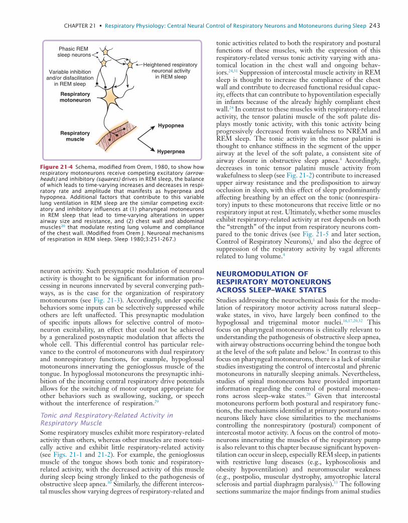
L
CHAPTER 21 • Respiratory Physiology: Central Neural Control of Respiratory Neurons and Motoneurons during Sleep 243
tonic activities related to both the respiratory and postural functions of these muscles, with the expression of this respiratory-related versus tonic activity varying with ana-tomical location in the chest wall and ongoing behav-iors.24,31 Suppression of intercostal muscle activity in REM sleep is thought to increase the compliance of the chest wall and contribute to decreased functional residual capac-ity, effects that can contribute to hypoventilation especially in infants because of the already highly compliant chest wall.24 In contrast to these muscles with respiratory-related activity, the tensor palatini muscle of the soft palate dis-plays mostly tonic activity, with this tonic activity being progressively decreased from wakefulness to NREM and REM sleep. The tonic activity in the tensor palatini is thought to enhance stiffness in the segment of the upper airway at the level of the soft palate, a consistent site of airway closure in obstructive sleep apnea.4 Accordingly, decreases in tonic tensor palatini muscle activity from wakefulness to sleep (see Fig. 21-2) contribute to increased upper airway resistance and the predisposition to airway occlusion in sleep, with this effect of sleep predominantly affecting breathing by an effect on the tonic (nonrespira-tory) inputs to these motoneurons that receive little or no respiratory input at rest. Ultimately, whether some muscles exhibit respiratory-related activity at rest depends on both the “strength” of the input from respiratory neurons com-pared to the tonic drives (see Fig. 21-5 and later section, Control of Respiratory Neurons),3 and also the degree of suppression of the respiratory activity by vagal afferents related to lung volume.4
NEUROMODULATION OF RESPIRATORY MOTONEURONS ACROSS SLEEP–WAKE STATESStudies addressing the neurochemical basis for the modu-lation of respiratory motor activity across natural sleep–wake states, in vivo, have largely been confined to the hypoglossal and trigeminal motor nuclei.16,17,20,32 This focus on pharyngeal motoneurons is clinically relevant to understanding the pathogenesis of obstructive sleep apnea, with airway obstructions occurring behind the tongue both at the level of the soft palate and below.4 In contrast to this focus on pharyngeal motoneurons, there is a lack of similar studies investigating the control of intercostal and phrenic motoneurons in naturally sleeping animals. Nevertheless, studies of spinal motoneurons have provided important information regarding the control of postural motoneu-rons across sleep–wake states.20 Given that intercostal motoneurons perform both postural and respiratory func-tions, the mechanisms identified at primary postural moto-neurons likely have close similarities to the mechanisms controlling the nonrespiratory (postural) component of intercostal motor activity. A focus on the control of moto-neurons innervating the muscles of the respiratory pump is also relevant to this chapter because significant hypoven-tilation can occur in sleep, especially REM sleep, in patients with restrictive lung diseases (e.g., kyphoscoliosis and obesity hypoventilation) and neuromuscular weakness (e.g., postpolio, muscular dystrophy, amyotrophic lateral sclerosis and partial diaphragm paralysis).33 The following sections summarize the major findings from animal studies
neuron activity. Such presynaptic modulation of neuronal activity is thought to be significant for information pro-cessing in neurons innervated by several converging path-ways, as is the case for the organization of respiratory motoneurons (see Fig. 21-3). Accordingly, under specific behaviors some inputs can be selectively suppressed while others are left unaffected. This presynaptic modulation of specific inputs allows for selective control of moto-neuron excitability, an effect that could not be achieved by a generalized postsynaptic modulation that affects the whole cell. This differential control has particular rele-vance to the control of motoneurons with dual respiratory and nonrespiratory functions, for example, hypoglossal motoneurons innervating the genioglossus muscle of the tongue. In hypoglossal motoneurons the presynaptic inhi-bition of the incoming central respiratory drive potentials allows for the switching of motor output appropriate for other behaviors such as swallowing, sucking, or speech without the interference of respiration.29
Tonic and Respiratory-Related Activity in Respiratory MuscleSome respiratory muscles exhibit more respiratory-related activity than others, whereas other muscles are more toni-cally active and exhibit little respiratory-related activity (see Figs. 21-1 and 21-2). For example, the genioglossus muscle of the tongue shows both tonic and respiratory-related activity, with the decreased activity of this muscle during sleep being strongly linked to the pathogenesis of obstructive sleep apnea.30 Similarly, the different intercos-tal muscles show varying degrees of respiratory-related and
Heightened respiratory neuronal activity
in REM sleep
Respiratorymotoneuron
Respiratorymuscle
Variable inhibitionand/or disfacilitation
in REM sleep
Hypopnea
Hyperpnea
Phasic REM sleep neurons
Figure 21-4 Schema, modified from Orem, 1980, to show how respiratory motoneurons receive competing excitatory (arrow-heads) and inhibitory (squares) drives in REM sleep, the balance of which leads to time-varying increases and decreases in respi-ratory rate and amplitude that manifests as hyperpnea and hypopnea. Additional factors that contribute to this variable lung ventilation in REM sleep are the similar competing excit-atory and inhibitory influences at (1) pharyngeal motoneurons in REM sleep that lead to time-varying alterations in upper airway size and resistance, and (2) chest wall and abdominal muscles20 that modulate resting lung volume and compliance of the chest wall. (Modified from Orem J. Neuronal mechanisms of respiration in REM sleep. Sleep 1980;3:251-267.)

L
244 PART I / Section 4 • Physiology in Sleep
sleep and minimal activity in REM sleep, that is, a pattern that may contribute to reduced respiratory muscle activity in sleep via withdrawal of excitation.3,16,17
Animal studies show that an endogenous noradrenergic drive to the hypoglossal motor nucleus contributes to both the respiratory and tonic components of genioglossus muscle activation in wakefulness, and the residual expres-sion of respiratory-related activity that persists in NREM sleep as the tonic drive is withdrawn.34 Moreover, this noradrenergic contribution to genioglossus muscle tone was minimal in REM sleep, so explaining, at least in part, the periods of genioglossus muscle hypotonia during REM sleep.34,35 The identification of an endogenous excitatory noradrenergic drive that contributes to genioglossus muscle activation in wakefulness, but with this drive being withdrawn in sleep, is particularly significant because since the first clinical description of obstructive sleep apnea, this was the first identification of a neural drive contributing to the sleep state–dependent activity of a muscle that is central to this disorder. The location of the central nor-adrenergic neurons that may provide this drive to hypo-
addressing the sleep state–dependent modulation of respiratory motor activity. These data derive, in large part, from studies at the hypoglossal motor nucleus, a model motor pool with dual respiratory and nonrespiratory functions.16
Excitatory Influences across Sleep–Wake StatesThe concept of a tonic drive activating respiratory muscle in wakefulness but not in sleep (i.e., the wakefulness stimu-lus for breathing) has been an important and enduring notion in respiratory medicine,3,24 not least because it is useful in modeling sleep effects on breathing and under-standing the pathogenesis of sleep-related breathing dis-orders. Neurons of the aminergic arousal system provide an important source of tonic drive to the respiratory system (see Fig. 21-2).3,16,17 Serotonin and noradrenaline-contain-ing neurons have been of particular attention experimen-tally because these neurons send excitatory projections to respiratory motoneurons, and these neurons show their highest activity in wakefulness, reduced activity in NREM
1 sec
η2 = 0.90
η2 = 0.75
η2 = 0.54
η2 = 0.44
η2 = 0.15
Discharge of respiratory neuron
Airflow (inspiration ↓)
RespiratoryNeuron
NonrespiratoryInput
RespiratoryInput
High η2 activity
NonrespiratoryInput
RespiratoryInput
Low η2 activity
Figure 21-5 Five different medullary respiratory neurons recorded in intact cats in NREM sleep. These neurons vary in the strength of their relationship to breathing, an effect that is quantified by the η2 statis-tic with values ranging from 0 (weak relationship) to 1.0 (strong relationship). High η2 cells are considered to be more strongly influenced by respiratory inputs than nonrespiratory inputs, and vice versa for low η2 cells. (Modified from Orem J, Kubin L. Respiratory physiology: central neural control. In: Kryger MH, Roth T, Dement WC, editors. Principles and practice of sleep medicine, 3nd ed. Philadelphia: Saunders; 2000. pp. 205-220.)
L
CHAPTER 21 • Respiratory Physiology: Central Neural Control of Respiratory Neurons and Motoneurons during Sleep 245
and both these classes of sedatives promote sleep by enhancing GABA-mediated neuronal inhibition via inter-actions with binding sites on GABAA receptors.13 However, the presence of lorazepam and zolpidem at the hypoglos-sal motor nucleus also leads to inhibition of genioglossus muscle activity.16 This inhibitory effect of sedative hyp-notics at respiratory motor nuclei may underlie a com-ponent of the respiratory depression observed clinically with excessive GABAA receptor stimulation, and the pre-disposition of some individuals to obstructive sleep apnea with sedative hypnotics.15
Large inhibitory glycinergic potentials appear to play an important role in the inhibition of spinal motoneuron activity in REM sleep,20 and this likely explains the inhibi-tion of intercostal respiratory muscle activity in this sleep state.24 However, glycine and GABAA receptor antagonism at the hypoglossal and trigeminal motoneuron pools fails to reverse the profound tonic suppression of genioglossus or masseter muscle activity in REM sleep, although in both cases this antagonism increases the amount or magnitude of the phasic motor activations observed during REM sleep.16,32 Nevertheless, these increases in motor activity during REM sleep with glycine and GABAA receptor antagonism does implicate a functional role for inhibitory neurotransmission in the control hypoglossal and trigemi-nal motor activity in REM sleep,16 although this may not be as profound as the inhibition demonstrated at spinal motoneurons.20
The degree of suppression observed in a variety of respi-ratory pump muscles in REM sleep appears strongly cor-related with the muscle spindle density of these different muscles.31 The diaphragm has few, if any, spindles and little inhibition in REM sleep, whereas different intercostal muscles (especially the external inspiratory intercostals) have significant numbers of muscle spindles and profound suppression of activity in REM sleep, with the degree of suppression varying with muscle spindle density.24,31 Of clinical relevance, acute diaphragm paralysis leads to increased reliance on the intercostal and accessory muscle to maintain effective lung ventilation, but this compensa-tion is lost in REM sleep as the motoneurons innervating these muscles with dual respiratory and postural functions are inhibited.43 Interestingly, however, patients with chronic bilateral diaphragm paralysis are able to recruit nondiaphragmatic inspiratory muscle activity during REM sleep, and so lessen any attendant hypoventilation. This compensation suggests that the central nervous system of these patients is able to functionally reorganize the drives controlling the accessory respiratory muscles such that activity is less suppressed by REM sleep mechanisms in the long term.44,45
In summary, current information from sleeping animals indicate that reduced excitation, largely via withdrawal of endogenous noradrenergic and glutamatergic inputs, is principally responsible for reductions in pharyngeal muscle tone from wakefulness to NREM and REM sleep.16,17,39 In comparison, an endogenous serotonergic drive plays a lesser role.16 Increased inhibitory neurotransmission via glycine and GABA also contributes to suppression of pha-ryngeal motor activity in REM sleep, but the contribution of this mechanism appears much less than expected16,17,39 from studies at spinal motoneurons.46
glossal and other respiratory motoneurons is reviewed elsewhere.16 Given the widespread projections of brain-stem aminergic neurons, they are also positioned to provide an endogenous input to other respiratory neurons and motoneurons, and so influence respiratory pump muscle activity and ventilation across sleep–wake states.36,37 Recent data also implicate a role for endogenous glutamatergic inputs in the tonic excitatory drive that increases pharyn-geal muscle activity in wakefulness, the withdrawal of which contributes to reduced activity in sleep.16,38,39 In con-trast to these functionally active tonic inputs, endogenous levels of serotonin at the hypoglossal motor nucleus con-tribute less to the changes in genioglossus muscle activity in sleeping animals.16 It remains to be determined if this minimal influence of endogenous serotonin on genioglos-sus muscle activity also applies to humans. If so, it may explain (at least in part) the lack of clinically significant effects on pharyngeal muscle activity and obstructive sleep apnea severity with administration of selective serotonin reuptake inhibitors in humans.16,40,41
Local application of serotonergic, noradrenergic and glutamatergic agonists to the hypoglossal or trigeminal motor nuclei produces robust motor activation in wakeful-ness and NREM sleep.16,39 These observations provide “proof of principle” to the notion that it may be possible to develop pharmacological strategies to increase respira-tory muscle activity in sleep, for example, as a potential treatment for obstructive sleep apnea. Importantly, however, a major component of the motor activation observed in response to these agonists in NREM sleep is overcome in REM sleep.16,39 There is an important practi-cal implication of this differential modulation of pharyn-geal motor responses to otherwise potent excitatory neuromodulators between NREM and REM sleep. For example, even if it is possible to effectively target pharyn-geal motoneurons with directed pharmacological manipu-lations, (e.g., as a potential treatment for obstructive sleep apnea) then different strategies may be required to produce sustained pharyngeal muscle activation throughout both the NREM and REM sleep stages, as the neurobiology of motor control is fundamentally different between these two states.16
Inhibitory Influences across Sleep–Wake StatesGlycine and GABA are the main inhibitory neurotrans-mitters in the central nervous system. Glycine and GABAA receptor stimulation at the hypoglossal motor nucleus in vivo produces the expected depression of genioglossus muscle activity, whereas antagonism of these receptors increases genioglossus activity across all sleep–wake states.16 The augmentation of respiratory-related motor activity with application of antagonists for these inhibi-tory neurotransmitters fits with the notion of a tonic inhibitory tone that constrains the rhythmic activation of respiratory neurons and motoneurons via gain modula-tion.42 The inhibitory effects of GABA at respiratory neurons14 and motoneurons16 is also clinically relevant given the widespread use of sedative hypnotic drugs in modern society. For example, benzodiazepine and imid-azopyridine drugs are commonly prescribed as sedative hypnotics (e.g., lorazepam and zolpidem, respectively),
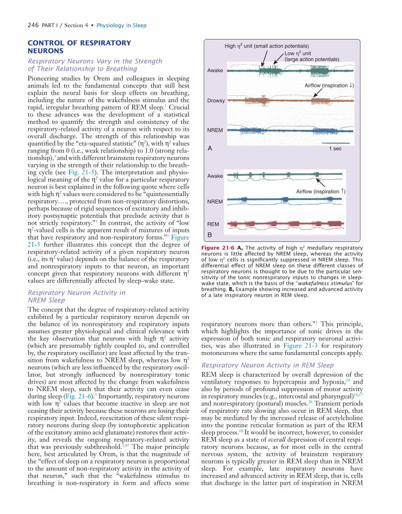
L
246 PART I / Section 4 • Physiology in Sleep
respiratory neurons more than others.”3 This principle, which highlights the importance of tonic drives in the expression of both tonic and respiratory neuronal activi-ties, was also illustrated in Figure 21-3 for respiratory motoneurons where the same fundamental concepts apply.
Respiratory Neuron Activity in REM SleepREM sleep is characterized by overall depression of the ventilatory responses to hypercapnia and hypoxia,24 and also by periods of profound suppression of motor activity in respiratory muscles (e.g., intercostal and pharyngeal)16,17 and nonrespiratory (postural) muscles.20 Transient periods of respiratory rate slowing also occur in REM sleep, that may be mediated by the increased release of acetylcholine into the pontine reticular formation as part of the REM sleep process.18 It would be incorrect, however, to consider REM sleep as a state of overall depression of central respi-ratory neurons because, as for most cells in the central nervous system, the activity of brainstem respiratory neurons is typically greater in REM sleep than in NREM sleep. For example, late inspiratory neurons have increased and advanced activity in REM sleep, that is, cells that discharge in the latter part of inspiration in NREM
CONTROL OF RESPIRATORY NEURONS
Respiratory Neurons Vary in the Strength of Their Relationship to BreathingPioneering studies by Orem and colleagues in sleeping animals led to the fundamental concepts that still best explain the neural basis for sleep effects on breathing, including the nature of the wakefulness stimulus and the rapid, irregular breathing pattern of REM sleep.3 Crucial to these advances was the development of a statistical method to quantify the strength and consistency of the respiratory-related activity of a neuron with respect to its overall discharge. The strength of this relationship was quantified by the “eta-squared statistic” (η2), with η2 values ranging from 0 (i.e., weak relationship) to 1.0 (strong rela-tionship),3 and with different brainstem respiratory neurons varying in the strength of their relationship to the breath-ing cycle (see Fig. 21-5). The interpretation and physio-logical meaning of the η2 value for a particular respiratory neuron is best explained in the following quote where cells with high η2 values were considered to be “quintessentially respiratory…., protected from non-respiratory distortions, perhaps because of rigid sequences of excitatory and inhib-itory postsynaptic potentials that preclude activity that is not strictly respiratory.”3 In contrast, the activity of “low η2-valued cells is the apparent result of mixtures of inputs that have respiratory and non-respiratory forms.”3 Figure 21-5 further illustrates this concept that the degree of respiratory-related activity of a given respiratory neuron (i.e., its η2 value) depends on the balance of the respiratory and nonrespiratory inputs to that neuron, an important concept given that respiratory neurons with different η2 values are differentially affected by sleep–wake state.
Respiratory Neuron Activity in NREM SleepThe concept that the degree of respiratory-related activity exhibited by a particular respiratory neuron depends on the balance of its nonrespiratory and respiratory inputs assumes greater physiological and clinical relevance with the key observation that neurons with high η2 activity (which are presumably tightly coupled to, and controlled by, the respiratory oscillator) are least affected by the tran-sition from wakefulness to NREM sleep, whereas low η2 neurons (which are less influenced by the respiratory oscil-lator, but strongly influenced by nonrespiratory tonic drives) are most affected by the change from wakefulness to NREM sleep, such that their activity can even cease during sleep (Fig. 21-6).3 Importantly, respiratory neurons with low η2 values that become inactive in sleep are not ceasing their activity because these neurons are losing their respiratory input. Indeed, reexcitation of these silent respi-ratory neurons during sleep (by iontophoretic application of the excitatory amino acid glutamate) restores their activ-ity, and reveals the ongoing respiratory-related activity that was previously subthreshold.3,47 The major principle here, best articulated by Orem, is that the magnitude of the “effect of sleep on a respiratory neuron is proportional to the amount of non-respiratory activity in the activity of that neuron,” such that the “wakefulness stimulus to breathing is non-respiratory in form and affects some
1 sec
Awake
High η2 unit (small action potentials)Low η2 unit(large action potentials)
Drowsy
NREM
A
B
Awake
NREM
REM
Airflow (inspiration ↓)
Airflow (inspiration ↑)
Figure 21-6 A, The activity of high η2 medullary respiratory neurons is little affected by NREM sleep, whereas the activity of low η2 cells is significantly suppressed in NREM sleep. This differential effect of NREM sleep on these different classes of respiratory neurons is thought to be due to the particular sen-sitivity of the tonic nonrespiratory inputs to changes in sleep-wake state, which is the basis of the “wakefulness stimulus” for breathing. B, Example showing increased and advanced activity of a late inspiratory neuron in REM sleep.
L
CHAPTER 21 • Respiratory Physiology: Central Neural Control of Respiratory Neurons and Motoneurons during Sleep 247
REM sleep,3,19 that is, a pattern similar to the changes in respiratory neuron activity. Electrical stimulation of the midbrain reticular formation can convert the activity of several respiratory muscles or motor nerves from a sleep-like pattern to one more like wakefulness.3 Indeed, it has been postulated that one important source of the tonic (nonrespiratory) input to medullary respiratory neurons when awake (i.e., the wakefulness stimulus) arises from the reticular formation.3 The source(s) of the drive(s) activat-ing central respiratory neurons in REM sleep, however, has not been determined.
Neurons of the aminergic arousal system (serotonergic, histaminergic, and noradrenergic), and other sleep state–dependent neuronal groups, are also positioned to provide a source of tonic (nonrespiratory) drive to respiratory neurons across sleep–wake states. However, whether these tonic drives would be excitatory or inhibitory to respira-tory neurons depends on the receptor subtypes activated, and the pre- or postsynaptic location of these receptors (see Figs. 21-2 and 21-3 for reference). This lack of knowl-edge of the sleep state-dependent neuromodulation of respiratory neurons needs to be addressed by further research, which may also identify specific pharmacological approaches that may be appropriate to preserve respiratory neuron activity in sleep and so minimize respiratory depression. The various brain structures that exert behav-ioral control of the respiratory system should also be con-sidered as a source of the wakefulness stimulus for breathing3 but it is not known if this collection of inputs shares the same neurochemicals as the aforementioned inputs from the reticular formation and sleep state–depen-dent neuronal systems.
sleep can be active throughout inspiration in REM sleep (see Fig. 21-6).3
Nevertheless, there is considerable variation around the mean level of discharge of respiratory neurons in REM sleep associated with tonic and phasic events.3 For example, increased ponto-geniculo-occiptal wave activity, a defining feature of phasic REM sleep events, is associated with increased activity of medullary respiratory neurons. This latter observation has been taken to suggest that the activ-ity of respiratory neurons in REM sleep is strongly influ-enced by processes peculiar to the neurobiology of REM sleep rather than intrinsic to the respiratory network.3 This concept of nonrespiratory inputs having significant influ-ences on the activity of neurons and motoneurons in the respiratory network has similarities to the major influence of tonic drives that were discussed in the context of the wakefulness stimulus to breathing. Together, these con-cepts serve to highlight that the activity levels of central respiratory neurons and motoneurons is due to the interac-tion of their respiratory and nonrespiratory inputs, with the latter having major influences on overall respiratory activity and being particularly sensitive to changes in sleep–wake state.
As discussed previously for respiratory motoneurons, this fundamental effect of REM sleep in activating central respiratory neurons can lead to periods of increased respi-ratory rate and respiratory muscle activity unrelated to homeostatic feedback regulation and to prevailing blood gas tensions.3,25,26 This increased activity of central respi-ratory neurons in REM sleep would most likely produce periods of increased respiratory rate and higher respira-tory muscle activity at times when the normally time-varying inhibition of respiratory motoneurons is briefly weakened or withdrawn (see Figs. 21-3 and 21-4). That REM sleep can lead to periods of heightened diaphragm activity unrelated to prevailing blood gas tensions has particular relevance to the clinical observation that hypo-capnic central apneas most commonly occur in NREM sleep, but can be absent in REM sleep when breathing is characteristically erratic.48,49 Figure 21-4 illustrates how this balance of excitatory and inhibitory influences at respiratory motoneurons can underlie the highly variable respiratory activity in REM sleep, including periods of respiratory depression despite activation of central respira-tory neurons.
Neuromodulation of Respiratory Neurons across Sleep–Wake StatesUnlike the studies performed at respiratory motor pools,16,17 there have been no studies addressing the neurochemicals that may mediate the control of respiratory neurons in vivo as a function of sleep–wake states. Nevertheless, it is a reasonable working hypothesis that the neuronal groups involved in the modulation of respiratory motoneurons across sleep–wake states also likely affect respiratory neurons. Accordingly, influences from brainstem reticular formation neurons (likely glutamatergic) are positioned to provide a source of tonic drive to respiratory neurons, with this influence altering from wakefulness to NREM and REM sleep.3,16,17,39 Neurons of the brainstem reticular for-mation system generally show decreased activity in NREM sleep compared to wakefulness, with increased activity in
� Clinical Pearl
The withdrawal of the wakefulness stimulus to breathing at the transition from wakefulness to sleep is the principal mechanism that precipitates the major clinical sleep-related breathing disorders. Current evi-dence identifies neurons of the aminergic arousal system and reticular formation as providing the key components of this wakefulness stimulus. Withdrawal of this tonic excitatory drive to the muscles of the upper airway is thought to underlie the normal sleep-related increase in upper airway resistance, and the hypoventilation, flow-limitation, and obstructive sleep apnea observed in susceptible individuals (e.g., those with already anatomically narrow upper airways). Patients with restrictive lung diseases and neuromuscular weakness rely, to varying degrees, on the activation of nondiaphragmatic respiratory muscles to help maintain adequate ventilation in wakefulness, but this compensation can be reduced or absent in sleep, leading to severe hypoventilation as the essential tonic excitatory drive that is present in wakefulness is withdrawn. REM sleep mechanisms can also lead to inhibition of respiratory motoneu-rons, so explaining the typically worse severity of abnormal breathing events in REM sleep compared to NREM sleep.
L
248 PART I / Section 4 • Physiology in Sleep
21. Luppi PH, Gervasoni D, Verret L, et al. Paradoxical (REM) sleep genesis: the switch from an aminergic-cholinergic to a GABAergic-glutamatergic hypothesis. J Physiol Paris 2006;100: 271-283.
22. Fuller PM, Saper CB, Lu J. The pontine REM switch: past and present. J Physiol 2007;584:735-741.
23. Worsnop C, Kay A, Pierce R, et al. Activity of respiratory pump and upper airway muscles during sleep onset. J Appl Physiol 1998;85: 908-920.
24. Phillipson EA, Bowes G. Control of breathing during sleep. In: Cherniack NS, Widdicombe JG, editors. Handbook of physiol-ogy, sect 3, the respiratory system, vol II, Control of breathing, part 2. Bethesda, Md: American Physiological Society; 1986. p. 649-689.
25. Horner RL, Kozar LF, Kimoff RJ, et al. Effects of sleep on the tonic drive to respiratory muscle and the threshold for rhythm generation in the dog. J Physiol 1994;474:525-537.
26. Orem J, Lovering AT, Dunin-Barkowski W, et al. Endogenous excit-atory drive to the respiratory system in rapid eye movement sleep in cats. J Physiol 2000;527:365-376.
27. Wiegand L, Zwillich CW, Wiegand D, et al. Changes in upper airway muscle activation and ventilation during phasic REM sleep in normal men. J Appl Physiol 1991;71:488-497.
28. Orem J. Central respiratory activity in rapid eye movement sleep: augmenting and late inspiratory cells. Sleep 1994;17:665- 673.
29. Bellingham MC, Berger AJ. Presynaptic depression of excitatory synaptic inputs to rat hypoglossal motoneurons by muscarinic M2 receptors. J Neurophysiol 1996;76:3758-3770.
30. Remmers JE, deGroot WJ, Sauerland EK, et al. Pathogenesis of upper airway occlusion during sleep. J Appl Physiol 1978;44: 931-938.
31. Duron B, Marlot D. Intercostal and diaphragmatic electrical activity during wakefulness and sleep in normal unrestrained adult cats. Sleep 1980;3:269-280.
32. Brooks PL, Peever JH. Glycinergic and GABAA-mediated inhibition of somatic motoneurons does not mediate REM sleep motor atonia. J Neurosci 2008;28:3535-3545.
33. Goldstein RS. Hypoventilation: neuromuscular and chest wall disor-ders. Clin Chest Med 1992;13:507-521.
34. Chan E, Steenland HW, Liu H, et al. Endogenous excitatory drive modulating respiratory muscle activity across sleep-wake states. Am J Respir Crit Care Med 2006;174:1264-1273.
35. Fenik VB, Davies RO, Kubin L. REM sleep-like atonia of hypo-glossal (XII) motoneurons is caused by loss of noradrenergic and serotonergic inputs. Am J Respir Crit Care Med 2005;172:1322- 1330.
36. Fenik V, Marchenko V, Janssen P, et al. A5 cells are silenced when REM sleep-like signs are elicited by pontine carbachol. J Appl Physiol 2002;93:1448-1456.
37. Li A, Nattie E. Catecholamine neurones in rats modulate sleep, breathing, central chemoreception and breathing variability. J Physiol 2006;570:385-396.
38. Steenland HW, Liu H, Horner RL. Endogenous glutamatergic control of rhythmically active mammalian respiratory motoneurons in vivo. J Neurosci 2008;28:6826-6835.
39. Burgess C, Lai D, Siegel J, et al. An endogenous glutamatergic drive onto somatic motoneurons contributes to the stereotypical pattern of muscle tone across the sleep-wake cycle. J Neurosci 2008:28: 4649-4660.
40. Smith IE, Quinnell TG. Pharmacotherapies for obstructive sleep apnoea: where are we now? Drugs 2004;64:1385-1399.
41. Veasey SC. Pharmacotherapies for obstructive sleep apnea: how close are we? Curr Opin Pulm Med 2001;7:399-403.
42. Zuperku EJ, McCrimmon DR. Gain modulation of respiratory neurons. Respir Physiol Neurobiol 2002;131:121-133.
43. Stradling JR, Kozar LF, Dark J, et al. Effect of acute diaphragm paralysis on ventilation in awake and sleeping dogs. Am Rev Respir Dis 1987;136:633-637.
44. Bennett JR, Dunroy HM, Corfield DR, et al. Respiratory muscle activity during REM sleep in patients with diaphragm paralysis. Neurol 2004;62:134-137.
45. Arnulf I, Similowski T, Salachas F, et al. Sleep disorders and dia-phragmatic function in patients with amyotrophic lateral sclerosis. Am J Respir Crit Care Med 2000;161:849-856.
Acknowledgments
RLH holds a Tier 1 Canada Research Chair in Sleep and Respiratory Neurobiology. The author’s research is sup-ported by grants from the Canadian Institutes of Health Research and Ontario Thoracic Society. The author also acknowledges development grants from the Canada Foun-dation for Innovation and the Ontario Research and Development Challenge Fund.
REFERENCES1. Duffin J. Functional organization of respiratory neurones: a brief
review of current questions and speculations. Exp Physiol 2004; 89:517-529.
2. Rekling JC, Feldman JL. PreBotzinger complex and pacemaker neurons: hypothesized site and kernel for respiratory rhythm genera-tion. Annu Rev Physiol 1998;60:385-405.
3. Orem J, Kubin L. Respiratory physiology: central neural control. In: Kryger MH, Roth T, Dement WC, editors. Principles and practice of sleep medicine, 3rd ed. Philadelphia: Saunders; 2000. p. 205-220.
4. Horner RL. Motor control of the pharyngeal musculature and impli-cations for the pathogenesis of obstructive sleep apnea. Sleep 1996;19:827-853.
5. Smith JC, Ellenberger HH, Ballanyi K, et al. Pre-Botzinger complex: a brainstem region that may generate respiratory rhythm in mammals. Science 1991;254:726-729.
6. Feldman JL, Del Negro CA. Looking for inspiration: new perspectives on respiratory rhythm. Nat Rev Neurosci 2006;7: 232-241.
7. McCrimmon DR, Alheid GF. On the opiate trail of respiratory depression. Am J Physiol Regul Integr Comp Physiol 2003;285: R1274-R1275.
8. McKay LC, Janczewski WA, Feldman JL. Sleep-disordered breath-ing after targeted ablation of preBotzinger complex neurons. Nat Neurosci 2005;8:1142-1144.
9. Mitchell GS. Back to the future: carbon dioxide chemoreceptors in the mammalian brain. Nat Neurosci 2004;7:1288-1290.
10. Jones BE. Basic mechanisms of sleep-wake states. In: Kryger MH, Roth T, Dement WC, editors. Principles and practice of sleep medi-cine, 3rd ed. Philadelphia: Saunders; 2000. p. 134-154.
11. McGinty D, Szymusiak R. The sleep-wake switch: a neuronal alarm clock. Nat Med 2000;6:510-511.
12. Saper CB, Scammell TE, Lu J. Hypothalamic regulation of sleep and circadian rhythms. Nature 2005;437:1257-1263.
13. Mendelson WB. Hypnotics: basic mechanisms and pharmacology. In: Kryger MH, Roth T, Dement WC, editors. Principles and practice of sleep medicine, 3rd ed. Philadelphia: Saunders; 2000. p. 407-413.
14. McCrimmon DR, Mitchell GS, Dekin MS. Glutamate, GABA, and serotonin in ventilatory control. In: Dempsey JA, Pack AI, editors. Regulation of breathing. New York: Dekker; 1995. p. 151-218.
15. Robinson RW, Zwillich CW. Medications, sleep and breathing. In: Kryger MH, Roth T, Dement WC, editors. Principles and prac-tice of sleep medicine, 3rd ed. Philadelphia: Saunders; 2000. p. 797-812.
16. Horner RL. Neuromodulation of hypoglossal motoneurons during sleep. Respir Physiol Neurobiol 2008;164:179-196.
17. Kubin L, Davies RO, Pack L. Control of upper airway motoneurons during REM sleep. News Physiol Sci 1998;13:637-656.
18. Lydic R, Baghdoyan HA. Sleep, anesthesiology, and the neuro-biology of arousal state control. Anesthesiology 2005;103:1268- 1295.
19. Siegel JM. Brainstem mechanisms generating REM sleep. In: Kryger MH, Roth T, Dement WC, editors. Principles and practice of sleep medicine, 3rd ed. Philadelphia: Saunders; 2000. p. 112-133.
20. Chase MH, Morales FR. Control of motoneurons during sleep. In: Kryger MH, Roth T, Dement WC, editors. Principles and practice of sleep medicine, 3rd ed. Philadelphia: Saunders; 2000. p. 155-168.

L
CHAPTER 21 • Respiratory Physiology: Central Neural Control of Respiratory Neurons and Motoneurons during Sleep 249
46. Chase MH, Morales FR. The control of motoneurons during sleep. In: Kryger MH, Roth T, Dement WC, editors. Principles and practice of sleep medicine, 2nd ed. Philadelphia: Saunders; 1994. p. 163-175.
47. Foutz AS, Boudinot E, Morin-Surun MP, et al. Excitability of “silent” respiratory neurons during sleep-waking states: an iontopho-retic study in undrugged chronic cats. Brain Res 1987;404:10-20.
48. Yumino D, Bradley TD. Central sleep apnea and Cheyne-Stokes respiration. Pro Am Thorac Soc 2008;5:226-236.
49. Xi L, Smith CA, Saupe KW, et al. Effects of rapid-eye-movement sleep on the apneic threshold in dogs. J Appl Physiol 1993;75: 1129-1139.
50. Orem J. Neuronal mechanisms of respiration in REM sleep. Sleep 1980;3:251-267.
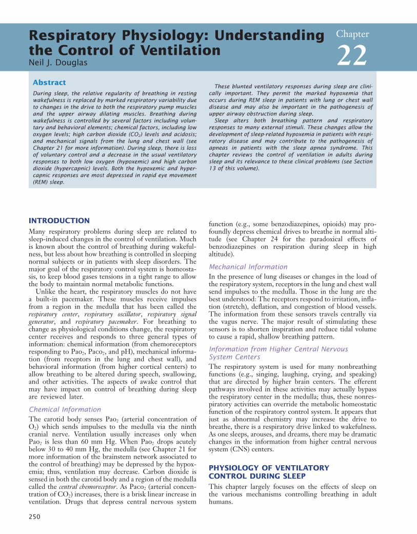
LL
250
AbstractDuring sleep, the relative regularity of breathing in resting wakefulness is replaced by marked respiratory variability due to changes in the drive to both the respiratory pump muscles and the upper airway dilating muscles. Breathing during wakefulness is controlled by several factors including volun-tary and behavioral elements; chemical factors, including low oxygen levels; high carbon dioxide (CO2) levels and acidosis; and mechanical signals from the lung and chest wall (see Chapter 21 for more information). During sleep, there is loss of voluntary control and a decrease in the usual ventilatory responses to both low oxygen (hypoxemic) and high carbon dioxide (hypercapnic) levels. Both the hypoxemic and hyper-capnic responses are most depressed in rapid eye movement (REM) sleep.
function (e.g., some benzodiazepines, opioids) may pro-foundly depress chemical drives to breathe in normal alti-tude (see Chapter 24 for the paradoxical effects of benzodiazepines on respiration during sleep in high altitude).
Mechanical InformationIn the presence of lung diseases or changes in the load of the respiratory system, receptors in the lung and chest wall send impulses to the medulla. Those in the lung are the best understood: The receptors respond to irritation, infla-tion (stretch), deflation, and congestion of blood vessels. The information from these sensors travels centrally via the vagus nerve. The major result of stimulating these sensors is to shorten inspiration and reduce tidal volume to cause a rapid, shallow breathing pattern.
Information from Higher Central Nervous System CentersThe respiratory system is used for many nonbreathing functions (e.g., singing, laughing, crying, and speaking) that are directed by higher brain centers. The efferent pathways involved in these activities may actually bypass the respiratory center in the medulla; thus, these nonres-piratory activities can override the metabolic homeostatic function of the respiratory control system. It appears that just as abnormal chemistry may increase the drive to breathe, there is a respiratory drive linked to wakefulness. As one sleeps, arouses, and dreams, there may be dramatic changes in the information from higher central nervous system (CNS) centers.
PHYSIOLOGY OF VENTILATORY CONTROL DURING SLEEPThis chapter largely focuses on the effects of sleep on the various mechanisms controlling breathing in adult humans.
INTRODUCTION
Many respiratory problems during sleep are related to sleep-induced changes in the control of ventilation. Much is known about the control of breathing during wakeful-ness, but less about how breathing is controlled in sleeping normal subjects or in patients with sleep disorders. The major goal of the respiratory control system is homeosta-sis, to keep blood gases tensions in a tight range to allow the body to maintain normal metabolic functions.
Unlike the heart, the respiratory muscles do not have a built-in pacemaker. These muscles receive impulses from a region in the medulla that has been called the respiratory center, respiratory oscillator, respiratory signal generator, and respiratory pacemaker. For breathing to change as physiological conditions change, the respiratory center receives and responds to three general types of information: chemical information (from chemoreceptors responding to Pao2, Paco2, and pH), mechanical informa-tion (from receptors in the lung and chest wall), and behavioral information (from higher cortical centers) to allow breathing to be altered during speech, swallowing, and other activities. The aspects of awake control that may have impact on control of breathing during sleep are reviewed later.
Chemical InformationThe carotid body senses Pao2 (arterial concentration of O2) which sends impulses to the medulla via the ninth cranial nerve. Ventilation usually increases only when Pao2 is less than 60 mm Hg. When Pao2 drops acutely below 30 to 40 mm Hg, the medulla (see Chapter 21 for more information of the brainstem network associated to the control of breathing) may be depressed by the hypox-emia; thus, ventilation may decrease. Carbon dioxide is sensed in both the carotid body and a region of the medulla called the central chemoreceptor. As Paco2 (arterial concen-tration of CO2) increases, there is a brisk linear increase in ventilation. Drugs that depress central nervous system
Respiratory Physiology: Understanding the Control of VentilationNeil J. Douglas
Chapter
22These blunted ventilatory responses during sleep are clini-
cally important. They permit the marked hypoxemia that occurs during REM sleep in patients with lung or chest wall disease and may also be important in the pathogenesis of upper airway obstruction during sleep.
Sleep alters both breathing pattern and respiratory responses to many external stimuli. These changes allow the development of sleep-related hypoxemia in patients with respi-ratory disease and may contribute to the pathogenesis of apneas in patients with the sleep apnea syndrome. This chapter reviews the control of ventilation in adults during sleep and its relevance to these clinical problems (see Section 13 of this volume).
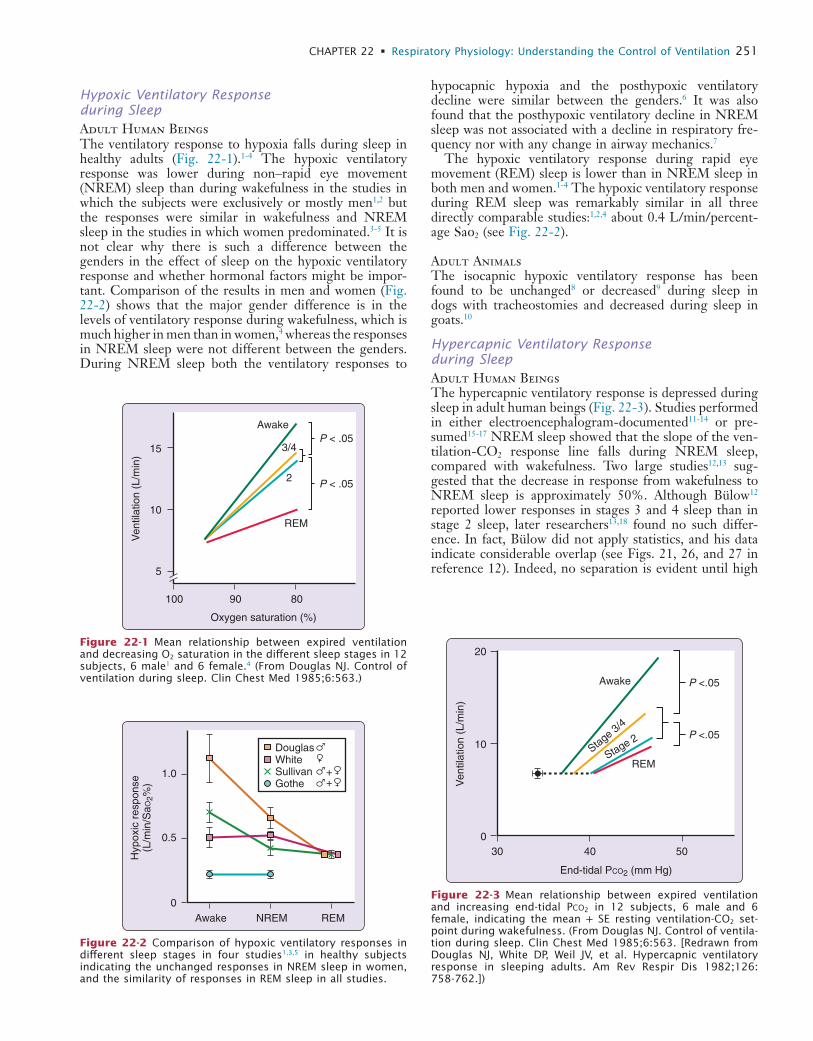
L
CHAPTER 22 • Respiratory Physiology: Understanding the Control of Ventilation 251
hypocapnic hypoxia and the posthypoxic ventilatory decline were similar between the genders.6 It was also found that the posthypoxic ventilatory decline in NREM sleep was not associated with a decline in respiratory fre-quency nor with any change in airway mechanics.7
The hypoxic ventilatory response during rapid eye movement (REM) sleep is lower than in NREM sleep in both men and women.1-4 The hypoxic ventilatory response during REM sleep was remarkably similar in all three directly comparable studies:1,2,4 about 0.4 L/min/percent-age Sao2 (see Fig. 22-2).
Adult AnimalsThe isocapnic hypoxic ventilatory response has been found to be unchanged8 or decreased9 during sleep in dogs with tracheostomies and decreased during sleep in goats.10
Hypercapnic Ventilatory Response during SleepAdult Human BeingsThe hypercapnic ventilatory response is depressed during sleep in adult human beings (Fig. 22-3). Studies performed in either electroencephalogram-documented11-14 or pre-sumed15-17 NREM sleep showed that the slope of the ven-tilation-CO2 response line falls during NREM sleep, compared with wakefulness. Two large studies12,13 sug-gested that the decrease in response from wakefulness to NREM sleep is approximately 50%. Although Bülow12 reported lower responses in stages 3 and 4 sleep than in stage 2 sleep, later researchers13,18 found no such differ-ence. In fact, Bülow did not apply statistics, and his data indicate considerable overlap (see Figs. 21, 26, and 27 in reference 12). Indeed, no separation is evident until high
Hypoxic Ventilatory Response during SleepAdult Human BeingsThe ventilatory response to hypoxia falls during sleep in healthy adults (Fig. 22-1).1-4 The hypoxic ventilatory response was lower during non–rapid eye movement (NREM) sleep than during wakefulness in the studies in which the subjects were exclusively or mostly men1,2 but the responses were similar in wakefulness and NREM sleep in the studies in which women predominated.3-5 It is not clear why there is such a difference between the genders in the effect of sleep on the hypoxic ventilatory response and whether hormonal factors might be impor-tant. Comparison of the results in men and women (Fig. 22-2) shows that the major gender difference is in the levels of ventilatory response during wakefulness, which is much higher in men than in women,4 whereas the responses in NREM sleep were not different between the genders. During NREM sleep both the ventilatory responses to
5
10
15
100 90
Awake
REM
3/4
2
80
P < .05
P < .05
Ven
tilat
ion
(L/m
in)
Oxygen saturation (%)
Figure 22-1 Mean relationship between expired ventilation and decreasing O2 saturation in the different sleep stages in 12 subjects, 6 male1 and 6 female.4 (From Douglas NJ. Control of ventilation during sleep. Clin Chest Med 1985;6:563.)
0
0.5
1.0
Awake NREM REM
Hyp
oxic
res
pons
e(L
/min
/SaO
2%)
DouglasWhiteSullivanGothe
++
Figure 22-2 Comparison of hypoxic ventilatory responses in different sleep stages in four studies1,3,5 in healthy subjects indicating the unchanged responses in NREM sleep in women, and the similarity of responses in REM sleep in all studies.
0
10
20
30 40
End-tidal PCO2 (mm Hg)
50
Ven
tilat
ion
(L/m
in)
Awake
Stage 3/4
Stage 2
REM
P <.05
P <.05
Figure 22-3 Mean relationship between expired ventilation and increasing end-tidal PCO2 in 12 subjects, 6 male and 6 female, indicating the mean + SE resting ventilation-CO2 set-point during wakefulness. (From Douglas NJ. Control of ventila-tion during sleep. Clin Chest Med 1985;6:563. [Redrawn from Douglas NJ, White DP, Weil JV, et al. Hypercapnic ventilatory response in sleeping adults. Am Rev Respir Dis 1982;126: 758-762.])
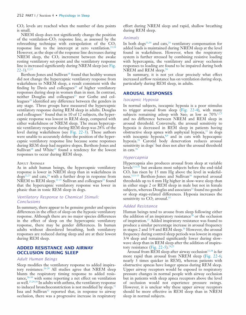
L
252 PART I / Section 4 • Physiology in Sleep
effort during NREM sleep and rapid, shallow breathing during REM sleep.
AnimalsIn both dogs33,34 and cats,24 ventilatory compensation for added loads is maintained during NREM sleep at the level found in wakefulness. However, when the respiratory system is further stressed by combining resistive loading with hypercapnia, the ventilatory and airway occlusion responses to loading are found to be impaired during both NREM and REM sleep.24
In summary, it is not yet clear precisely what effect increased airflow resistance has on ventilation during sleep, particularly during REM sleep, in adults.
AROUSAL RESPONSES
Isocapnic HypoxiaIn normal subjects, isocapnic hypoxia is a poor stimulus to trigger arousal from sleep (Fig. 22-4), with many subjects remaining asleep with Sao2 as low as 70%1,2,5 and no difference between NREM and REM sleep in arousal threshold. Conversely, the arousal sensitivity to hypoxia is decreased in REM sleep in patients having obstructive sleep apnea with asphyxial hypoxia,35 in dogs with isocapnic hypoxia,8,9 and in cats with hypocapnic hypoxia.36 Carotid body denervation reduces arousal sensitivity in dogs7 but does not alter the arousal threshold in cats.36
HypercapniaHypercapnia also produces arousal from sleep at variable levels3,11,13 but awakens most subjects before the end-tidal CO2 has risen by 15 mm Hg above the level in wakeful-ness.3,11-13 Berthon-Jones and Sullivan16 reported arousal thresholds up to 6 mm Hg higher in slow-wave sleep than in either stage 2 or REM sleep in male but not in female subjects, whereas Douglas and associates13 found no gender or sleep stage-related differences. Hypoxia increases the sensitivity to CO2 arousal.35
Added ResistanceHuman beings tend to arouse from sleep following either the addition of an inspiratory resistance28 or the occlusion of inspiration.37 Added inspiratory resistance was found to produce a similar percentage increase in arousal frequency in stages 2 and 3/4 and REM sleep.26 However, the arousal frequency during control sleep periods was lowest in stages 3/4 sleep and remained significantly lower during slow-wave sleep than in REM sleep after the addition of inspira-tory resistance (Fig. 22-5).24,25
Arousal from REM sleep after airway occlusion32,38 is far more rapid than arousal from NREM sleep (Fig. 22-6; nearly 3 times quicker in REM), whereas patients with obstructive apneas have longer apneas during REM sleep. Upper airway receptors would be exposed to respiratory pressure changes in normal people with airway occlusion but in patients with sleep apnea receptors above the level of occlusion would not experience pressure swings. However, it is unclear why these upper airway receptors should be more effective in REM sleep than in NREM sleep in normal subjects.
CO2 levels are reached when the number of data points is small.
NREM sleep does not significantly change the position of the ventilation-CO2 response line, as assessed by the rebreathing technique with extrapolation of the CO2 response line to the intercept at zero ventilation.13,18 However, as the slope of the response line decreases during NREM sleep, the CO2 increment between the awake resting ventilatory set-point and the ventilatory response line is increased significantly during NREM sleep (see Fig. 22-3).12,13
Berthon-Jones and Sullivan18 found that healthy women did not change the hypercapnic ventilatory response from wakefulness to NREM sleep, a result consistent with the finding by Davis and colleagues19 of higher ventilatory responses during sleep in women than in men. In contrast, neither Douglas and colleagues13 nor Gothe and col-leagues20 identified any difference between the genders in any stage. Three groups have measured the hypercapnic ventilatory response during REM sleep in adults. Douglas and colleagues13 found that in 10 of 12 subjects, the hyper-capnic response was lowest in REM sleep, compared with either wakefulness or NREM sleep. The mean hypercap-nic ventilatory response during REM sleep was 28% of the level during wakefulness (see Fig. 22-3). These authors were unable to accurately define the position of the hyper-capnic ventilatory response line because some responses during REM sleep had negative slopes. Berthon-Jones and Sullivan18 and White21 found a tendency for the lowest responses to occur during REM sleep.
Adult AnimalsAs in adult human beings, the hypercapnic ventilatory response is lower in NREM sleep than in wakefulness in dogs22,23 and cats,24 with a further drop in response from NREM to REM sleep.22-25 Sullivan and colleagues23 found that the hypercapnic ventilatory response was lower in phasic than in tonic REM sleep in dogs.
Ventilatory Response to Chemical Stimuli: ConclusionsIn summary, there appear to be genuine gender and species differences in the effect of sleep on the hypoxic ventilatory response. Although there are no major species differences in the effect of sleep on the hypercapnic ventilatory response, there may be gender differences. In human adults without disordered breathing, both ventilatory responses are reduced during sleep and are at their lowest during REM sleep.
ADDED RESISTANCE AND AIRWAY OCCLUSION DURING SLEEP
Adult Human BeingsSleep modifies the ventilatory response to added inspira-tory resistance.26-29 All studies agree that NREM sleep blunts the respiratory timing response to added resis-tance,26-30 with some reporting a net effect on ventilation as well.27,29,30 In adults with asthma, the ventilatory response to induced bronchoconstriction is not modified by sleep.31 Issa and Sullivan32 reported that, in response to airway occlusion, there was a progressive increase in respiratory
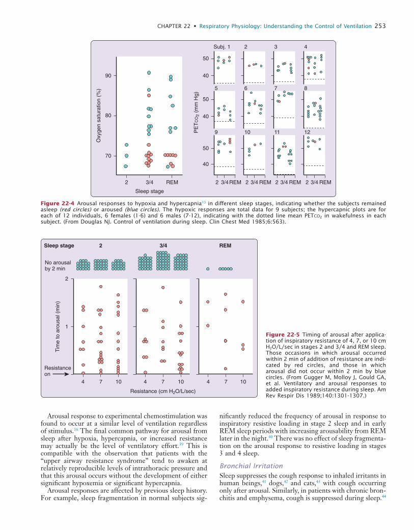
L
CHAPTER 22 • Respiratory Physiology: Understanding the Control of Ventilation 253
nificantly reduced the frequency of arousal in response to inspiratory resistive loading in stage 2 sleep and in early REM sleep periods with increasing arousability from REM later in the night.40 There was no effect of sleep fragmenta-tion on the arousal response to resistive loading in stages 3 and 4 sleep.
Bronchial IrritationSleep suppresses the cough response to inhaled irritants in human beings,41 dogs,42 and cats,43 with cough occurring only after arousal. Similarly, in patients with chronic bron-chitis and emphysema, cough is suppressed during sleep.44
Arousal response to experimental chemostimulation was found to occur at a similar level of ventilation regardless of stimulus.16 The final common pathway for arousal from sleep after hypoxia, hypercapnia, or increased resistance may actually be the level of ventilatory effort.39 This is compatible with the observation that patients with the “upper airway resistance syndrome” tend to awaken at relatively reproducible levels of intrathoracic pressure and that this arousal occurs without the development of either significant hypoxemia or significant hypercapnia.
Arousal responses are affected by previous sleep history. For example, sleep fragmentation in normal subjects sig-
70
80
90
2 3/4
Sleep stage
REM 2 3/4
Subj. 1 2 3 4
6 7 85
10 11 129
REM 2 3/4 REM 2 3/4 REM 2 3/4 REM
Oxy
gen
satu
ratio
n (%
)
40
50
40
50
40
50
PE
TC
O2
(mm
Hg)
Figure 22-4 Arousal responses to hypoxia and hypercapnia13 in different sleep stages, indicating whether the subjects remained asleep (red circles) or aroused (blue circles). The hypoxic responses are total data for 9 subjects; the hypercapnic plots are for each of 12 individuals, 6 females (1-6) and 6 males (7-12), indicating with the dotted line mean PETCO2 in wakefulness in each subject. (From Douglas NJ. Control of ventilation during sleep. Clin Chest Med 1985;6:563).
1
2
4 7
Resistance (cm H2O/L/sec)
Sleep stage
No arousalby 2 min
Resistanceon
10 4 7 10 4 7 10
2 3/4 REM
Tim
e to
aro
usal
(m
in)
Figure 22-5 Timing of arousal after applica-tion of inspiratory resistance of 4, 7, or 10 cm H2O/L/sec in stages 2 and 3/4 and REM sleep. Those occasions in which arousal occurred within 2 min of addition of resistance are indi-cated by red circles, and those in which arousal did not occur within 2 min by blue circles. (From Gugger M, Molloy J, Gould GA, et al. Ventilatory and arousal responses to added inspiratory resistance during sleep. Am Rev Respir Dis 1989;140:1301-1307.)
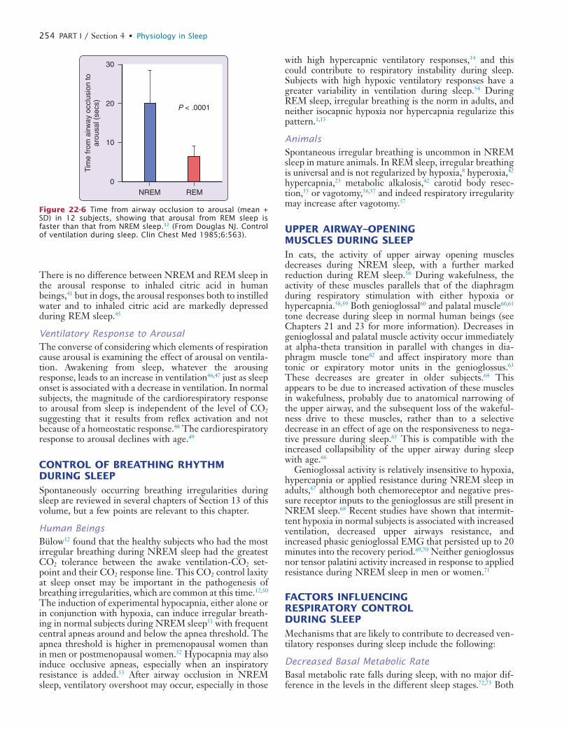
L
254 PART I / Section 4 • Physiology in Sleep
with high hypercapnic ventilatory responses,14 and this could contribute to respiratory instability during sleep. Subjects with high hypoxic ventilatory responses have a greater variability in ventilation during sleep.54 During REM sleep, irregular breathing is the norm in adults, and neither isocapnic hypoxia nor hypercapnia regularize this pattern.1,13
AnimalsSpontaneous irregular breathing is uncommon in NREM sleep in mature animals. In REM sleep, irregular breathing is universal and is not regularized by hypoxia,8 hyperoxia,42 hypercapnia,23 metabolic alkalosis,42 carotid body resec-tion,55 or vagotomy,56,57 and indeed respiratory irregularity may increase after vagotomy.57
UPPER AIRWAY–OPENING MUSCLES DURING SLEEPIn cats, the activity of upper airway opening muscles decreases during NREM sleep, with a further marked reduction during REM sleep.56 During wakefulness, the activity of these muscles parallels that of the diaphragm during respiratory stimulation with either hypoxia or hypercapnia.58,59 Both genioglossal60 and palatal muscle60,61 tone decrease during sleep in normal human beings (see Chapters 21 and 23 for more information). Decreases in genioglossal and palatal muscle activity occur immediately at alpha-theta transition in parallel with changes in dia-phragm muscle tone62 and affect inspiratory more than tonic or expiratory motor units in the genioglossus.63 These decreases are greater in older subjects.64 This appears to be due to increased activation of these muscles in wakefulness, probably due to anatomical narrowing of the upper airway, and the subsequent loss of the wakeful-ness drive to these muscles, rather than to a selective decrease in an effect of age on the responsiveness to nega-tive pressure during sleep.65 This is compatible with the increased collapsibility of the upper airway during sleep with age.66
Genioglossal activity is relatively insensitive to hypoxia, hypercapnia or applied resistance during NREM sleep in adults,67 although both chemoreceptor and negative pres-sure receptor inputs to the genioglossus are still present in NREM sleep.68 Recent studies have shown that intermit-tent hypoxia in normal subjects is associated with increased ventilation, decreased upper airways resistance, and increased phasic genioglossal EMG that persisted up to 20 minutes into the recovery period.69,70 Neither genioglossus nor tensor palatini activity increased in response to applied resistance during NREM sleep in men or women.71
FACTORS INFLUENCING RESPIRATORY CONTROL DURING SLEEPMechanisms that are likely to contribute to decreased ven-tilatory responses during sleep include the following:
Decreased Basal Metabolic RateBasal metabolic rate falls during sleep, with no major dif-ference in the levels in the different sleep stages.72,73 Both
There is no difference between NREM and REM sleep in the arousal response to inhaled citric acid in human beings,41 but in dogs, the arousal responses both to instilled water and to inhaled citric acid are markedly depressed during REM sleep.45
Ventilatory Response to ArousalThe converse of considering which elements of respiration cause arousal is examining the effect of arousal on ventila-tion. Awakening from sleep, whatever the arousing response, leads to an increase in ventilation46,47 just as sleep onset is associated with a decrease in ventilation. In normal subjects, the magnitude of the cardiorespiratory response to arousal from sleep is independent of the level of CO2 suggesting that it results from reflex activation and not because of a homeostatic response.48 The cardiorespiratory response to arousal declines with age.49
CONTROL OF BREATHING RHYTHM DURING SLEEPSpontaneously occurring breathing irregularities during sleep are reviewed in several chapters of Section 13 of this volume, but a few points are relevant to this chapter.
Human BeingsBülow12 found that the healthy subjects who had the most irregular breathing during NREM sleep had the greatest CO2 tolerance between the awake ventilation-CO2 set-point and their CO2 response line. This CO2 control laxity at sleep onset may be important in the pathogenesis of breathing irregularities, which are common at this time.12,50 The induction of experimental hypocapnia, either alone or in conjunction with hypoxia, can induce irregular breath-ing in normal subjects during NREM sleep51 with frequent central apneas around and below the apnea threshold. The apnea threshold is higher in premenopausal women than in men or postmenopausal women.52 Hypocapnia may also induce occlusive apneas, especially when an inspiratory resistance is added.53 After airway occlusion in NREM sleep, ventilatory overshoot may occur, especially in those
0
10
20
30
NREM REM
Tim
e fr
om a
irway
occ
lusi
on to
arou
sal (
secs
)
P < .0001
Figure 22-6 Time from airway occlusion to arousal (mean + SD) in 12 subjects, showing that arousal from REM sleep is faster than that from NREM sleep.32 (From Douglas NJ. Control of ventilation during sleep. Clin Chest Med 1985;6:563).
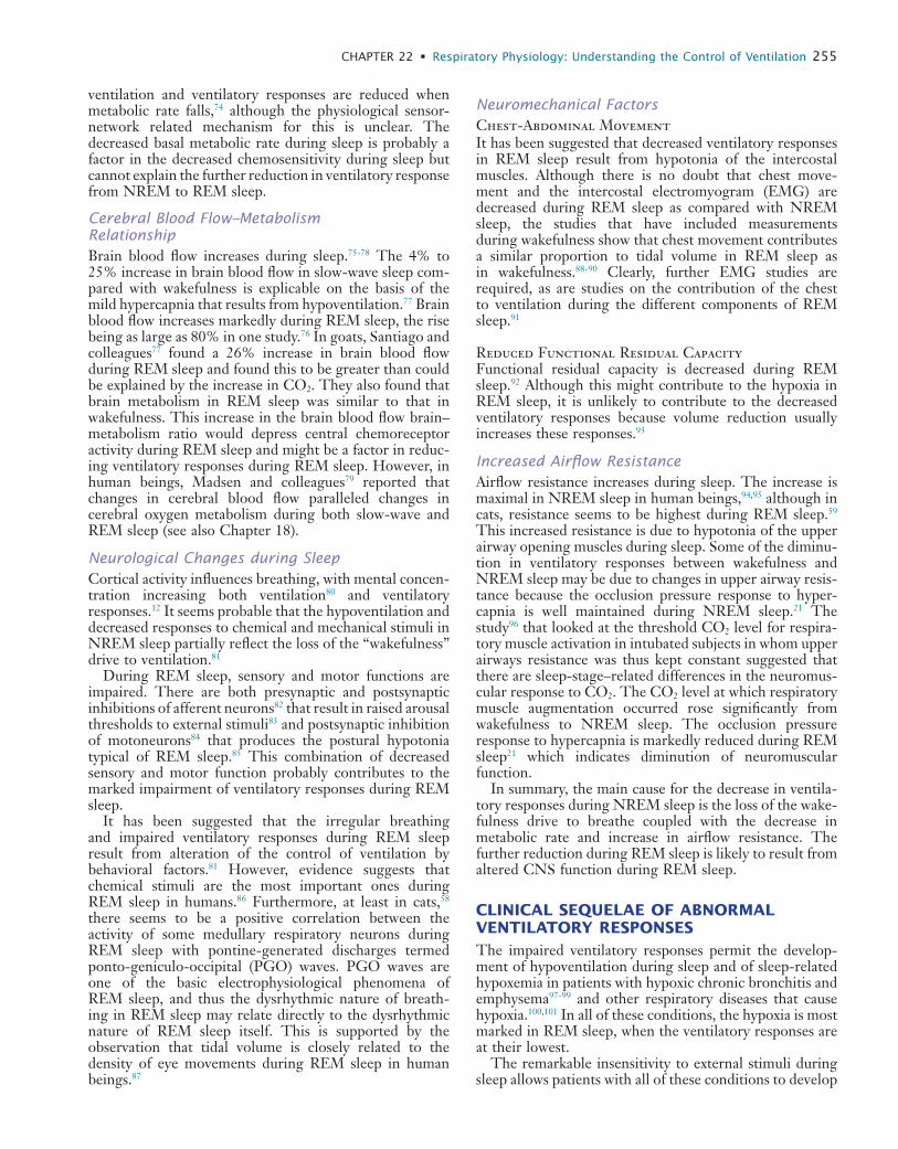
L
CHAPTER 22 • Respiratory Physiology: Understanding the Control of Ventilation 255
Neuromechanical FactorsChest-Abdominal MovementIt has been suggested that decreased ventilatory responses in REM sleep result from hypotonia of the intercostal muscles. Although there is no doubt that chest move-ment and the intercostal electromyogram (EMG) are decreased during REM sleep as compared with NREM sleep, the studies that have included measurements during wakefulness show that chest movement contributes a similar proportion to tidal volume in REM sleep as in wakefulness.88-90 Clearly, further EMG studies are required, as are studies on the contribution of the chest to ventilation during the different components of REM sleep.91
Reduced Functional Residual CapacityFunctional residual capacity is decreased during REM sleep.92 Although this might contribute to the hypoxia in REM sleep, it is unlikely to contribute to the decreased ventilatory responses because volume reduction usually increases these responses.93
Increased Airflow ResistanceAirflow resistance increases during sleep. The increase is maximal in NREM sleep in human beings,94,95 although in cats, resistance seems to be highest during REM sleep.59 This increased resistance is due to hypotonia of the upper airway opening muscles during sleep. Some of the diminu-tion in ventilatory responses between wakefulness and NREM sleep may be due to changes in upper airway resis-tance because the occlusion pressure response to hyper-capnia is well maintained during NREM sleep.21 The study96 that looked at the threshold CO2 level for respira-tory muscle activation in intubated subjects in whom upper airways resistance was thus kept constant suggested that there are sleep-stage–related differences in the neuromus-cular response to CO2. The CO2 level at which respiratory muscle augmentation occurred rose significantly from wakefulness to NREM sleep. The occlusion pressure response to hypercapnia is markedly reduced during REM sleep21 which indicates diminution of neuromuscular function.
In summary, the main cause for the decrease in ventila-tory responses during NREM sleep is the loss of the wake-fulness drive to breathe coupled with the decrease in metabolic rate and increase in airflow resistance. The further reduction during REM sleep is likely to result from altered CNS function during REM sleep.
CLINICAL SEQUELAE OF ABNORMAL VENTILATORY RESPONSESThe impaired ventilatory responses permit the develop-ment of hypoventilation during sleep and of sleep-related hypoxemia in patients with hypoxic chronic bronchitis and emphysema97-99 and other respiratory diseases that cause hypoxia.100,101 In all of these conditions, the hypoxia is most marked in REM sleep, when the ventilatory responses are at their lowest.
The remarkable insensitivity to external stimuli during sleep allows patients with all of these conditions to develop
ventilation and ventilatory responses are reduced when metabolic rate falls,74 although the physiological sensor-network related mechanism for this is unclear. The decreased basal metabolic rate during sleep is probably a factor in the decreased chemosensitivity during sleep but cannot explain the further reduction in ventilatory response from NREM to REM sleep.
Cerebral Blood Flow–Metabolism RelationshipBrain blood flow increases during sleep.75-78 The 4% to 25% increase in brain blood flow in slow-wave sleep com-pared with wakefulness is explicable on the basis of the mild hypercapnia that results from hypoventilation.77 Brain blood flow increases markedly during REM sleep, the rise being as large as 80% in one study.76 In goats, Santiago and colleagues77 found a 26% increase in brain blood flow during REM sleep and found this to be greater than could be explained by the increase in CO2. They also found that brain metabolism in REM sleep was similar to that in wakefulness. This increase in the brain blood flow brain–metabolism ratio would depress central chemoreceptor activity during REM sleep and might be a factor in reduc-ing ventilatory responses during REM sleep. However, in human beings, Madsen and colleagues79 reported that changes in cerebral blood flow paralleled changes in cerebral oxygen metabolism during both slow-wave and REM sleep (see also Chapter 18).
Neurological Changes during SleepCortical activity influences breathing, with mental concen-tration increasing both ventilation80 and ventilatory responses.12 It seems probable that the hypoventilation and decreased responses to chemical and mechanical stimuli in NREM sleep partially reflect the loss of the “wakefulness” drive to ventilation.81
During REM sleep, sensory and motor functions are impaired. There are both presynaptic and postsynaptic inhibitions of afferent neurons82 that result in raised arousal thresholds to external stimuli83 and postsynaptic inhibition of motoneurons84 that produces the postural hypotonia typical of REM sleep.85 This combination of decreased sensory and motor function probably contributes to the marked impairment of ventilatory responses during REM sleep.
It has been suggested that the irregular breathing and impaired ventilatory responses during REM sleep result from alteration of the control of ventilation by behavioral factors.81 However, evidence suggests that chemical stimuli are the most important ones during REM sleep in humans.86 Furthermore, at least in cats,58 there seems to be a positive correlation between the activity of some medullary respiratory neurons during REM sleep with pontine-generated discharges termed ponto-geniculo-occipital (PGO) waves. PGO waves are one of the basic electrophysiological phenomena of REM sleep, and thus the dysrhythmic nature of breath-ing in REM sleep may relate directly to the dysrhythmic nature of REM sleep itself. This is supported by the observation that tidal volume is closely related to the density of eye movements during REM sleep in human beings.87

L
256 PART I / Section 4 • Physiology in Sleep
6. Tarbichi AG, Rowley JA, Shkoukani MA, et al. Lack of gender difference in ventilatory chemoresponsiveness and post-hypoxic ventilatory decline. Respir Physiol Neurobiol 2003;137:41-50.
7. Halker RB, Pierchala LA, Badr MS. Upper airway mechanics and post-hypoxic ventilatory decline during NREM sleep. Sleep Breath 2007;11:165-170.
8. Phillipson EA, Sullivan CE, Read DJC, et al. Ventilatory and waking responses to hypoxia in sleeping dogs. J Appl Physiol 1978;44:512-520.
9. Bowes G, Townsend ER, Kozar LF, et al. Effect of carotid body denervation on arousal response to hypoxia in sleeping dogs. J Appl Physiol 1981;51:40-45.
10. Santiago TV, Scardella AT, Edelman NH. Determinants of the ventilatory response to hypoxia during sleep. Am Rev Respir Dis 1984;130:179-182.
11. Birchfield RI, Sieker HO, Heyman A. Alterations in respiratory function during natural sleep. J Lab Clin Med 1959;54:216-222.
12. Bülow K. Respiration and wakefulness in man. Acta Physiol Scand 1963;59(Suppl. 209):1-110.
13. Douglas NJ, White DP, Weil JV, et al. Hypercapnic ventilatory response in sleeping adults. Am Rev Respir Dis 1982;126:758-762.
14. Gleeson K, Zwillich CW, White DP. Chemosensitivity and the ventilatory response to airflow obstruction during sleep. J Appl Physiol 1989;67:1630-1637.
15. Magnussen G. Studies on the respiration during sleep: a contribu-tion to the physiology of the sleep function. London: HK Lewis; 1944.
16. Ostergaard T. The excitability of the respiratory centre during sleep and during Evipan anaesthesia. Acta Physiol Scand 1944;8:1-15.
17. Robin ED, Whaley RD, Crump CH, et al. Alveolar gas tensions, pulmonary ventilation and blood pH during physiological sleep in normal subjects. J Clin Invest 1958;37:981-989.
18. Berthon-Jones M, Sullivan CE. Ventilation and arousal responses to hypercapnia in normal sleeping adults. J Appl Physiol 1984; 57:59-67.
19. Davis JN, Loh L, Nodal J, et al. Effects of sleep on the pattern of CO2 stimulated breathing in males and females. Adv Exp Med Biol 1978;99:79-83.
20. Gothe B, Altose MD, Goldman MD, et al. Effect of quiet sleep on resting and CO2-stimulated breathing in humans. J Appl Physiol 1981;50:724-730.
21. White DP. Occlusion pressure and ventilation during sleep in normal humans. J Appl Physiol 1986;61:1279-1287.
22. Phillipson EA, Kozar LF, Rebuck AS, et al. Ventilatory and waking responses to CO2 in sleeping dogs. Am Rev Respir Dis 1977;115:251-259.
23. Sullivan CE, Murphy E, Kozar LF, et al. Ventilatory responses to CO2 and lung inflation in tonic versus phasic REM sleep. J Appl Physiol 1979;47:1304-1310.
24. Santiago TV, Sinha AK, Edelman NH. Respiratory flow-resistive load compensation during sleep. Am Rev Respir Dis 1981;123: 382-387.
25. Netick A, Dugger WJ, Symmons RA. Ventilatory response to hypercapnia during sleep and wakefulness in cats. J Appl Physiol 1984;56:1347-1354.
26. Gugger M, Molloy J, Gould GA, et al. Ventilatory and arousal responses to added inspiratory resistance during sleep. Am Rev Respir Dis 1989;140:1301-1307.
27. Hudgel DW, Mulholland M, Hendricks C. Neuromuscular and mechanical responses to inspiratory resistive loading during sleep. J Appl Physiol 1987;63:603-608.
28. Iber C, Berssenbrugge A, Skatrud JB, et al. Ventilatory adaptations to resistive loading during wakefulness and non-REM sleep. J Appl Physiol 1982;52:607-614.
29. Wiegand L, Zwillich CW, White DP. Sleep and the ventilatory response to resistive loading in normal men. J Appl Physiol 1988;64:1186-1195.
30. Gora J, Kay A, Colrain IM, et al. Load compensation as a function of state during sleep onset. J Appl Physiol 1998;84:2123-2131.
31. Ballard RD, Tan WC, Kelly PL, et al. Effect of sleep and sleep deprivation on ventilatory response to broncho-constriction. J Appl Physiol 1990;69:490-497.
32. Issa FG, Sullivan CE. Arousal and breathing responses to airway occlusion in healthy sleeping adults. J Appl Physiol 1983;55: 1113-1119.
clinically significant hypoxia and hypercapnia before arousal occurs.
Instability of the control of breathing during sleep may contribute to the development of the obstructive sleep apnea-hypopnea syndrome in some patients.102 Paradoxi-cally, enhanced ventilatory responses may be present in some patients, which may destabilize ventilation and lead to periodic breathing.103 This may be an important factor in the development of Cheyne-Stokes respiration in left ventricular heart failure.104 In heart failure, central apnea during sleep may be initiated by an acute increase in ven-tilation and reduction in Paco2 following a spontaneous arousal.105 When Paco2 falls below the threshold level required to stimulate breathing, the drive to respiratory muscles and airflow cease, and central apnea result. Apnea persists until Paco2 rises above the threshold required to stimulate ventilation. Prolonged circulation time appears not to play a key role in initiating central apneas, and indeed cardiac transplantation with normalisation of cardiac function does not always prevent Cheyne-Stokes respiration.105 The major influence of circulation time appears to be on the lengths of the hyperpneic phase and of the total periodic breathing cycle.106 Once triggered, the pattern of alternating hyperventilation and apnea is sus-tained by the combination of increased respiratory chemo-receptor drive, pulmonary congestion, arousals, and apnea-induced hypoxia, which cause oscillations in Paco2 above and below the apneic threshold. Inhalation of a CO2-enriched gas to raise Paco2 abolishes Cheyne-Stokes respiration in heart failure.107 The clinical effectiveness of continuous positive airway pressure therapy in this situa-tion is debated.108,109 For more details on sleep disorders breathing, Section 13 of this volume can be consulted.
� Clinical Pearl
Decreased ventilatory responses to hypoxia, hyper-capnia, and inspiratory resistance during sleep, par-ticularly in REM sleep, permit REM hypoxemia in patients with chronic obstructive pulmonary disease, chest wall disease, and neuromuscular abnormalities that affect the respiratory muscles. Decreased venti-latory responses may also contribute to the develop-ment of hypoventilation syndromes and the sleep apnea/hypopnea syndrome.
REFERENCES1. Douglas NJ, White DP, Weil JV, et al. Hypoxic ventilatory response
decreases during sleep in normal men. Am Rev Respir Dis 1982;125:286-289.
2. Berthon-Jones M, Sullivan CE. Ventilatory and arousal responses to hypoxia in sleeping humans. Am Rev Respir Dis 1982;125: 632-639.
3. Hedemark LL, Kronenberg RS. Ventilatory and heart rate responses to hypoxia and hypercapnia during sleep in adults. J Appl Physiol 1982;53:307-312.
4. White DP, Douglas NJ, Pickett CK, et al. Hypoxic ventilatory response during sleep in normal women. Am Rev Respir Dis 1982;126:530-533.
5. Gothe B, Goldman MD, Cherniack NS, et al. Effect of progressive hypoxia on breathing during sleep. Am Rev Respir Dis 1982;126: 97-102.
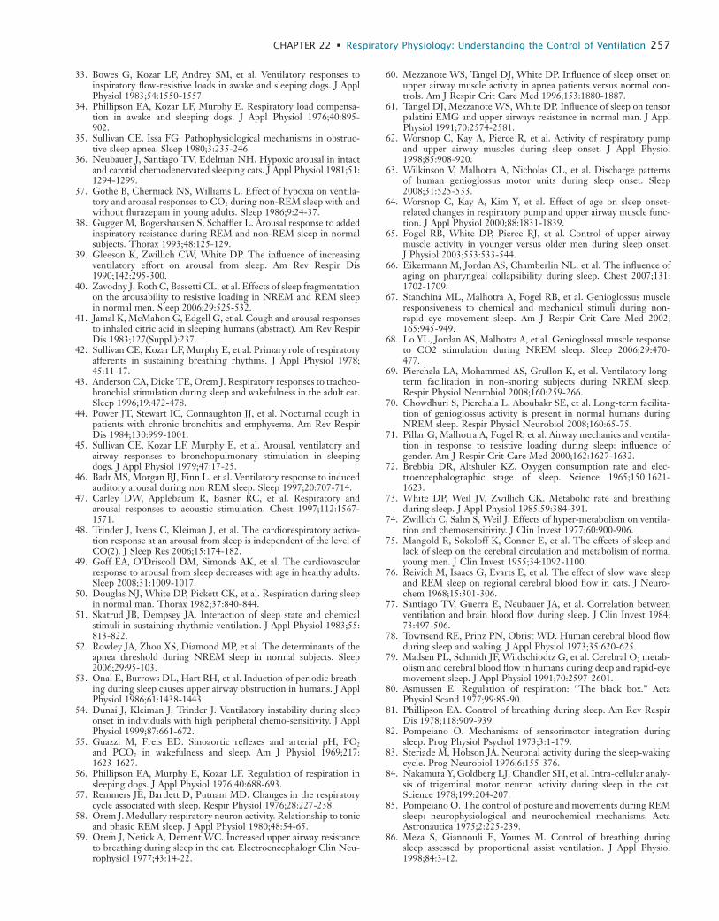
L
CHAPTER 22 • Respiratory Physiology: Understanding the Control of Ventilation 257
60. Mezzanote WS, Tangel DJ, White DP. Influence of sleep onset on upper airway muscle activity in apnea patients versus normal con-trols. Am J Respir Crit Care Med 1996;153:1880-1887.
61. Tangel DJ, Mezzanote WS, White DP. Influence of sleep on tensor palatini EMG and upper airways resistance in normal man. J Appl Physiol 1991;70:2574-2581.
62. Worsnop C, Kay A, Pierce R, et al. Activity of respiratory pump and upper airway muscles during sleep onset. J Appl Physiol 1998;85:908-920.
63. Wilkinson V, Malhotra A, Nicholas CL, et al. Discharge patterns of human genioglossus motor units during sleep onset. Sleep 2008;31:525-533.
64. Worsnop C, Kay A, Kim Y, et al. Effect of age on sleep onset-related changes in respiratory pump and upper airway muscle func-tion. J Appl Physiol 2000;88:1831-1839.
65. Fogel RB, White DP, Pierce RJ, et al. Control of upper airway muscle activity in younger versus older men during sleep onset. J Physiol 2003;553:533-544.
66. Eikermann M, Jordan AS, Chamberlin NL, et al. The influence of aging on pharyngeal collapsibility during sleep. Chest 2007;131: 1702-1709.
67. Stanchina ML, Malhotra A, Fogel RB, et al. Genioglossus muscle responsiveness to chemical and mechanical stimuli during non-rapid eye movement sleep. Am J Respir Crit Care Med 2002; 165:945-949.
68. Lo YL, Jordan AS, Malhotra A, et al. Genioglossal muscle response to CO2 stimulation during NREM sleep. Sleep 2006;29:470- 477.
69. Pierchala LA, Mohammed AS, Grullon K, et al. Ventilatory long-term facilitation in non-snoring subjects during NREM sleep. Respir Physiol Neurobiol 2008;160:259-266.
70. Chowdhuri S, Pierchala L, Aboubakr SE, et al. Long-term facilita-tion of genioglossus activity is present in normal humans during NREM sleep. Respir Physiol Neurobiol 2008;160:65-75.
71. Pillar G, Malhotra A, Fogel R, et al. Airway mechanics and ventila-tion in response to resistive loading during sleep: influence of gender. Am J Respir Crit Care Med 2000;162:1627-1632.
72. Brebbia DR, Altshuler KZ. Oxygen consumption rate and elec-troencephalographic stage of sleep. Science 1965;150:1621- 1623.
73. White DP, Weil JV, Zwillich CK. Metabolic rate and breathing during sleep. J Appl Physiol 1985;59:384-391.
74. Zwillich C, Sahn S, Weil J. Effects of hyper-metabolism on ventila-tion and chemosensitivity. J Clin Invest 1977;60:900-906.
75. Mangold R, Sokoloff K, Conner E, et al. The effects of sleep and lack of sleep on the cerebral circulation and metabolism of normal young men. J Clin Invest 1955;34:1092-1100.
76. Reivich M, Isaacs G, Evarts E, et al. The effect of slow wave sleep and REM sleep on regional cerebral blood flow in cats. J Neuro-chem 1968;15:301-306.
77. Santiago TV, Guerra E, Neubauer JA, et al. Correlation between ventilation and brain blood flow during sleep. J Clin Invest 1984; 73:497-506.
78. Townsend RE, Prinz PN, Obrist WD. Human cerebral blood flow during sleep and waking. J Appl Physiol 1973;35:620-625.
79. Madsen PL, Schmidt JF, Wildschiodtz G, et al. Cerebral O2 metab-olism and cerebral blood flow in humans during deep and rapid-eye movement sleep. J Appl Physiol 1991;70:2597-2601.
80. Asmussen E. Regulation of respiration: “The black box.” Acta Physiol Scand 1977;99:85-90.
81. Phillipson EA. Control of breathing during sleep. Am Rev Respir Dis 1978;118:909-939.
82. Pompeiano O. Mechanisms of sensorimotor integration during sleep. Prog Physiol Psychol 1973;3:1-179.
83. Steriade M, Hobson JA. Neuronal activity during the sleep-waking cycle. Prog Neurobiol 1976;6:155-376.
84. Nakamura Y, Goldberg LJ, Chandler SH, et al. Intra-cellular analy-sis of trigeminal motor neuron activity during sleep in the cat. Science 1978;199:204-207.
85. Pompeiano O. The control of posture and movements during REM sleep: neurophysiological and neurochemical mechanisms. Acta Astronautica 1975;2:225-239.
86. Meza S, Giannouli E, Younes M. Control of breathing during sleep assessed by proportional assist ventilation. J Appl Physiol 1998;84:3-12.
33. Bowes G, Kozar LF, Andrey SM, et al. Ventilatory responses to inspiratory flow-resistive loads in awake and sleeping dogs. J Appl Physiol 1983;54:1550-1557.
34. Phillipson EA, Kozar LF, Murphy E. Respiratory load compensa-tion in awake and sleeping dogs. J Appl Physiol 1976;40:895- 902.
35. Sullivan CE, Issa FG. Pathophysiological mechanisms in obstruc-tive sleep apnea. Sleep 1980;3:235-246.
36. Neubauer J, Santiago TV, Edelman NH. Hypoxic arousal in intact and carotid chemodenervated sleeping cats. J Appl Physiol 1981;51: 1294-1299.
37. Gothe B, Cherniack NS, Williams L. Effect of hypoxia on ventila-tory and arousal responses to CO2 during non-REM sleep with and without flurazepam in young adults. Sleep 1986;9:24-37.
38. Gugger M, Bogershausen S, Schaffler L. Arousal response to added inspiratory resistance during REM and non-REM sleep in normal subjects. Thorax 1993;48:125-129.
39. Gleeson K, Zwillich CW, White DP. The influence of increasing ventilatory effort on arousal from sleep. Am Rev Respir Dis 1990;142:295-300.
40. Zavodny J, Roth C, Bassetti CL, et al. Effects of sleep fragmentation on the arousability to resistive loading in NREM and REM sleep in normal men. Sleep 2006;29:525-532.
41. Jamal K, McMahon G, Edgell G, et al. Cough and arousal responses to inhaled citric acid in sleeping humans (abstract). Am Rev Respir Dis 1983;127(Suppl.):237.
42. Sullivan CE, Kozar LF, Murphy E, et al. Primary role of respiratory afferents in sustaining breathing rhythms. J Appl Physiol 1978; 45:11-17.
43. Anderson CA, Dicke TE, Orem J. Respiratory responses to tracheo-bronchial stimulation during sleep and wakefulness in the adult cat. Sleep 1996;19:472-478.
44. Power JT, Stewart IC, Connaughton JJ, et al. Nocturnal cough in patients with chronic bronchitis and emphysema. Am Rev Respir Dis 1984;130:999-1001.
45. Sullivan CE, Kozar LF, Murphy E, et al. Arousal, ventilatory and airway responses to bronchopulmonary stimulation in sleeping dogs. J Appl Physiol 1979;47:17-25.
46. Badr MS, Morgan BJ, Finn L, et al. Ventilatory response to induced auditory arousal during non REM sleep. Sleep 1997;20:707-714.
47. Carley DW, Applebaum R, Basner RC, et al. Respiratory and arousal responses to acoustic stimulation. Chest 1997;112:1567- 1571.
48. Trinder J, Ivens C, Kleiman J, et al. The cardiorespiratory activa-tion response at an arousal from sleep is independent of the level of CO(2). J Sleep Res 2006;15:174-182.
49. Goff EA, O’Driscoll DM, Simonds AK, et al. The cardiovascular response to arousal from sleep decreases with age in healthy adults. Sleep 2008;31:1009-1017.
50. Douglas NJ, White DP, Pickett CK, et al. Respiration during sleep in normal man. Thorax 1982;37:840-844.
51. Skatrud JB, Dempsey JA. Interaction of sleep state and chemical stimuli in sustaining rhythmic ventilation. J Appl Physiol 1983;55: 813-822.
52. Rowley JA, Zhou XS, Diamond MP, et al. The determinants of the apnea threshold during NREM sleep in normal subjects. Sleep 2006;29:95-103.
53. Onal E, Burrows DL, Hart RH, et al. Induction of periodic breath-ing during sleep causes upper airway obstruction in humans. J Appl Physiol 1986;61:1438-1443.
54. Dunai J, Kleiman J, Trinder J. Ventilatory instability during sleep onset in individuals with high peripheral chemo-sensitivity. J Appl Physiol 1999;87:661-672.
55. Guazzi M, Freis ED. Sinoaortic reflexes and arterial pH, PO2 and PCO2 in wakefulness and sleep. Am J Physiol 1969;217:1623-1627.
56. Phillipson EA, Murphy E, Kozar LF. Regulation of respiration in sleeping dogs. J Appl Physiol 1976;40:688-693.
57. Remmers JE, Bartlett D, Putnam MD. Changes in the respiratory cycle associated with sleep. Respir Physiol 1976;28:227-238.
58. Orem J. Medullary respiratory neuron activity. Relationship to tonic and phasic REM sleep. J Appl Physiol 1980;48:54-65.
59. Orem J, Netick A, Dement WC. Increased upper airway resistance to breathing during sleep in the cat. Electroencephalogr Clin Neu-rophysiol 1977;43:14-22.
L
258 PART I / Section 4 • Physiology in Sleep
99. Koo KW, Sax DS, Snider GL. Arterial blood gases and pH during sleep in chronic obstructive pulmonary disease. Am J Med 1975;58:663-670.
100. Mezon BL, West P, Israels J, et al. Sleep breathing abnormalities in kyphoscoliosis. Am Rev Respir Dis 1980;122:617-621.
101. Muller NL, Francis PW, Gurwitz D, et al. Mechanism of hemo-globin desaturation during rapid-eye-movement sleep in normal subjects and in patients with cystic fibrosis. Am Rev Respir Dis 1980;121:463-469.
102. Younes M, Ostrowski M, Thompson W, et al. Chemical control stability in patients with obstructive sleep apnea. Am J Respir Crit Care Med 2001;163:1181-1190
103. Cherniack NS. Apnea and periodic breathing during sleep. N Engl J Med 1999;341:985-987.
104. Javaheri S. A mechanism of central sleep apnea in patients with heart failure. N Engl J Med 1999;341:949-954.
105. Mansfield DR, Solin P, Roebuck T, et al. The effect of successful heart transplant treatment of heart failure on central sleep apnea. Chest 2003;124:1675-1681.
106. Bradley TD, Floras JS. Sleep apnea and heart failure: Part II. Central sleep apnea. Circulation 2003;107:1822-1826.
107. Lorenzi-Filho G, Rankin F, Bies I, et al. Effects of inhaled carbon dioxide and oxygen on Cheyne-Stokes respiration in patients with heart failure. Am J Respir Crit Care Med 1999;159: 1490-1498.
108. Arzt M, Floras JS, Logan AG, et al. Suppression of central sleep apnea by continuous positive airway pressure and transplant-free survival in heart failure: a post hoc analysis of the Canadian Con-tinuous Positive Airway Pressure for Patients with Central Sleep Apnea and Heart Failure Trial (CANPAP). Circulation 2007;115: 3173-3180.
109. Kasai T, Usui Y, Yoshioka T, JASV Investigators. Effect of flow-triggered adaptive servo-ventilation compared with continuous positive airway pressure in patients with chronic heart failure with coexisting obstructive sleep apnea and Cheyne-Stokes respiration. Circ Heart Fail 2010;3:140-148.
87. Gould GA, Gugger M, Molloy J, et al. Breathing pattern and eye movement density during REM sleep in man. Am Rev Respir Dis 1988;138:874-877.
88. Mortola JP, Anch AM. Chest configuration in supine man: wakeful-ness and sleep. Respir Physiol 1978;35:201-213.
89. Stradling JR, Chadwick GA, Frew AJ. Changes in ventilation and its components in normal subjects during sleep. Thorax 1985; 40:364-370.
90. Tabachnik E, Muller NL, Bryan AC, et al. Changes in ventilation and chest wall mechanics during sleep in normal adults. J Appl Physiol 1981;51:557-564.
91. Millman RP, Knight H, Chung DC, et al. Changes in compartmen-tal ventilation in association with eye movements during REM sleep. J Appl Physiol 1988;65:1196-1202.
92. Hudgel DW, Devadatta P. Decrease in functional residual capacity during sleep in normal humans. J Appl Physiol 1984;57:1319- 1322.
93. Cherniack NS, Stanley NN, Tuteur PG, et al. Effect of lung volume changes on respiratory drive during hypoxia and hypercapnia. J Appl Physiol 1973;35:635-641.
94. Hudgel DW, Martin RJ, Johnson B, et al. Mechanics of the respira-tory system and breathing pattern during sleep in normal humans. J Appl Physiol 1984;56:133-137.
95. Lopes JM, Tabachnik E, Muller NL, et al. Total airway resistance and respiratory muscle activity during sleep. J Appl Physiol 1983;54:773-777.
96. Ingrassia TS 3rd, Nelson SB, Harris CD, et al. Influence of sleep state on carbon dioxide responsiveness: a study of the unloaded respiratory pump in man. Am Rev Respir Dis 1991;144:1125- 1129.
97. Douglas NJ, Calverley PMA, Leggett RJE, et al. Transient hypox-aemia during sleep in chronic bronchitis and emphysema. Lancet 1979;1:1-4.
98. Fleetham JA, Mezon B, West P, et al. Chemical control of ventila-tion and sleep arterial oxygen desaturation in patients with COPD. Am Rev Respir Dis 1980;122:583-589.
LL
269
AbstractSleep disturbance is a common cause of discomfort among the constellation of symptoms after ascent to high altitude. Subjectively, the sensation is that of a restless or sleepless night. Individuals commonly experience awakening from sleep with a feeling of suffocation, taking a deep breath, feeling much improved, and falling back to sleep. Objective observa-tions show associated periodic breathing and frequent awak-enings but relatively little change in total sleep time. Sleep stages are generally shifted from deeper toward lighter sleep stages.
Periodic breathing at altitude seems to reflect the respi-ratory dilemma of acute altitude ascent in which the stimu-latory effects of hypoxia are opposed by the inhibitory action of hypocapnic alkalosis. The outcome is respiratory oscillation. Hypocapnic alkalosis induces apnea, which in turn lessens alkalotic inhibition and augments hypoxic stimu-lation. This triggers hyperpnea, which lessens respiratory stimulation by decreasing hypoxia and increasing alkalosis, leading to recurrent apnea. The occurrence of altitude related apnea with lessening of ventilatory stimuli is enhanced during sleep.
tion is lessening hypoxia, which is the presumed stimulus to breathing, and increasing hypocapnic alkalosis, which is a ventilatory inhibitor. This is the phenomenon of ventila-tory acclimatization to high altitude, which is manifested as a progressive decrease in arterial Pco2 (Paco2) with increasing ventilation over several days. Although the mechanism of acclimatization is debated, studies in humans and animals suggest that increased hypoxic sensitivity of the carotid body may be a major contributor.2 In any case, it is during the early phase of altitude adjustment, shortly after ascent, that sleep disturbances appear to be most marked; they tend to improve during the period of acclimatization.
Sleep DisturbancesThe earliest systematic study of sleep at altitude was a report in 1970 of studies of subjects working in Antarctica, where a combination of geographic elevation and terres-trial spin produces decreased barometric pressure ranging from 485 to 525 mm Hg, equivalent to an altitude of 4500 to 5000 meters. The men stationed there experienced a sleep disturbance termed polar red-eye, and electroen-cephalographic studies showed major disruption of sleep with a marked decrease in slow-wave sleep (stages 3 and 4).3 Although these changes were ascribed to hypobaric hypoxia, the effects of isolation and disturbances of light–dark cycle that are typical in persons living at the South Pole clouded their interpretation.
As research at low altitude began to elucidate the close links between breathing and sleep and showed that sleep disruption was often closely linked to changes in respira-tory rhythm, such as occurs in sleep apnea, studies were performed at high altitude with simultaneous monitoring
PHYSIOLOGIC ADJUSTMENT TO HIGH ALTITUDE
Primary among the changes in physical environment that attend the ascent to altitude is a decrease in barometric pressure such that although the fractional concentration of O2 is similar to that at sea level, O2 tension—the product of fractional concentration and barometric pressure—is reduced (Fig. 24-1). This decreased O2 tension of ambient air presents a threat to arterial and tissue oxygenation and elicits a series of responses that may act to minimize tissue hypoxia. These consist of early increases in ventilation and cardiac output, and, during more prolonged exposure, rises in circulating red cell concentration and adaptive changes in peripheral tissue, including increased spatial density of capillaries and mitochondria. Reading Chapters 21 and 22 may help one to further understand the physiological changes associated with sleep and breathing in high altitude.
Increased VentilationProbably the earliest, best studied, and one of the most important of these responses is increased ventilation, which acts to minimize the extent of alveolar hypoxia and arterial hypoxemia in the face of a decrease in ambient O2 tension. The magnitude of the ventilatory response increases with increasing altitude, but it also varies consid-erably among individuals at a fixed altitude. This variability in part reflects intrinsic, interindividual differences in the strength of the basal (preascent) ventilatory response to hypoxia.1 In addition, ventilation progressively increases over several days after ascent to high altitude. This gradual increase occurs despite the fact that the increasing ventila-
Respiratory Physiology: Sleep at High AltitudesJohn V. Weil
Chapter
24On balance, the poor subjective quality of sleep seems to
reflect the fragmentation of sleep by frequent arousals linked to the marked changes in respiratory pattern of periodic breathing. Arousals commonly occur at the transition from the end of apnea to the onset of hyperpnea. Subsequent accli-matization to altitude is associated with lessening of periodic breathing and better-quality sleep.
The most common treatment is prophylactic administration of acetazolamide, an inhibitor of carbonic anhydrase, which likely works by reducing alkalotic ventilatory inhibition. The results of recent studies suggest that benzodiazepines and other sleep-promoting agents may improve sleep quality without apparent adverse effects.
In this chapter, relevant features of acute physiologic adjust-ment to high altitude are briefly summarized; then the char-acteristics of the sleep disturbance at high altitude, its pathogenesis, and therapeutic interventions are reviewed. Although much of the focus is on sleep after acute ascent to high altitude, alterations in sleep during long-term altitude exposure are also briefly mentioned. Information in this chapter may also be relevant to the pathophysiology of central sleep apnea at low altitude (see Section 13).
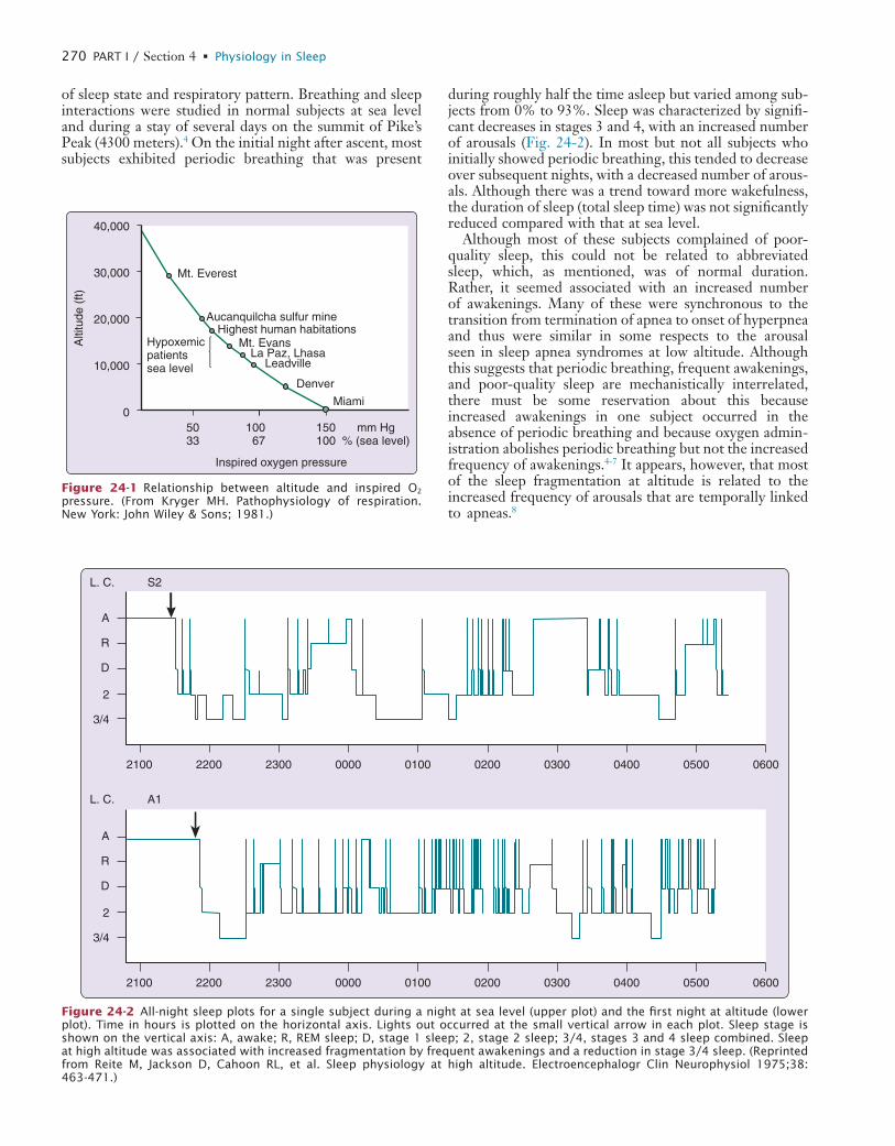
L
270 PART I / Section 4 • Physiology in Sleep
during roughly half the time asleep but varied among sub-jects from 0% to 93%. Sleep was characterized by signifi-cant decreases in stages 3 and 4, with an increased number of arousals (Fig. 24-2). In most but not all subjects who initially showed periodic breathing, this tended to decrease over subsequent nights, with a decreased number of arous-als. Although there was a trend toward more wakefulness, the duration of sleep (total sleep time) was not significantly reduced compared with that at sea level.
Although most of these subjects complained of poor-quality sleep, this could not be related to abbreviated sleep, which, as mentioned, was of normal duration. Rather, it seemed associated with an increased number of awakenings. Many of these were synchronous to the transition from termination of apnea to onset of hyperpnea and thus were similar in some respects to the arousal seen in sleep apnea syndromes at low altitude. Although this suggests that periodic breathing, frequent awakenings, and poor-quality sleep are mechanistically interrelated, there must be some reservation about this because increased awakenings in one subject occurred in the absence of periodic breathing and because oxygen admin-istration abolishes periodic breathing but not the increased frequency of awakenings.4-7 It appears, however, that most of the sleep fragmentation at altitude is related to the increased frequency of arousals that are temporally linked to apneas.8
of sleep state and respiratory pattern. Breathing and sleep interactions were studied in normal subjects at sea level and during a stay of several days on the summit of Pike’s Peak (4300 meters).4 On the initial night after ascent, most subjects exhibited periodic breathing that was present
Inspired oxygen pressure
40,000
30,000
20,000
10,000
010067
5033
150100
mm Hg% (sea level)
Alti
tude
(ft)
Mt. Everest
Aucanquilcha sulfur mineHighest human habitations
Mt. EvansLa Paz, Lhasa
Leadville
Denver
Miami
Hypoxemicpatientssea level
Figure 24-1 Relationship between altitude and inspired O2 pressure. (From Kryger MH. Pathophysiology of respiration. New York: John Wiley & Sons; 1981.)
A
R
D
2
3/4
2100
S2L. C.
2200 2300 0000 0100 0200 0300 0400 0500 0600
A
R
D
2
3/4
2100
A1L. C.
2200 2300 0000 0100 0200 0300 0400 0500 0600
Figure 24-2 All-night sleep plots for a single subject during a night at sea level (upper plot) and the first night at altitude (lower plot). Time in hours is plotted on the horizontal axis. Lights out occurred at the small vertical arrow in each plot. Sleep stage is shown on the vertical axis: A, awake; R, REM sleep; D, stage 1 sleep; 2, stage 2 sleep; 3/4, stages 3 and 4 sleep combined. Sleep at high altitude was associated with increased fragmentation by frequent awakenings and a reduction in stage 3/4 sleep. (Reprinted from Reite M, Jackson D, Cahoon RL, et al. Sleep physiology at high altitude. Electroencephalogr Clin Neurophysiol 1975;38: 463-471.)
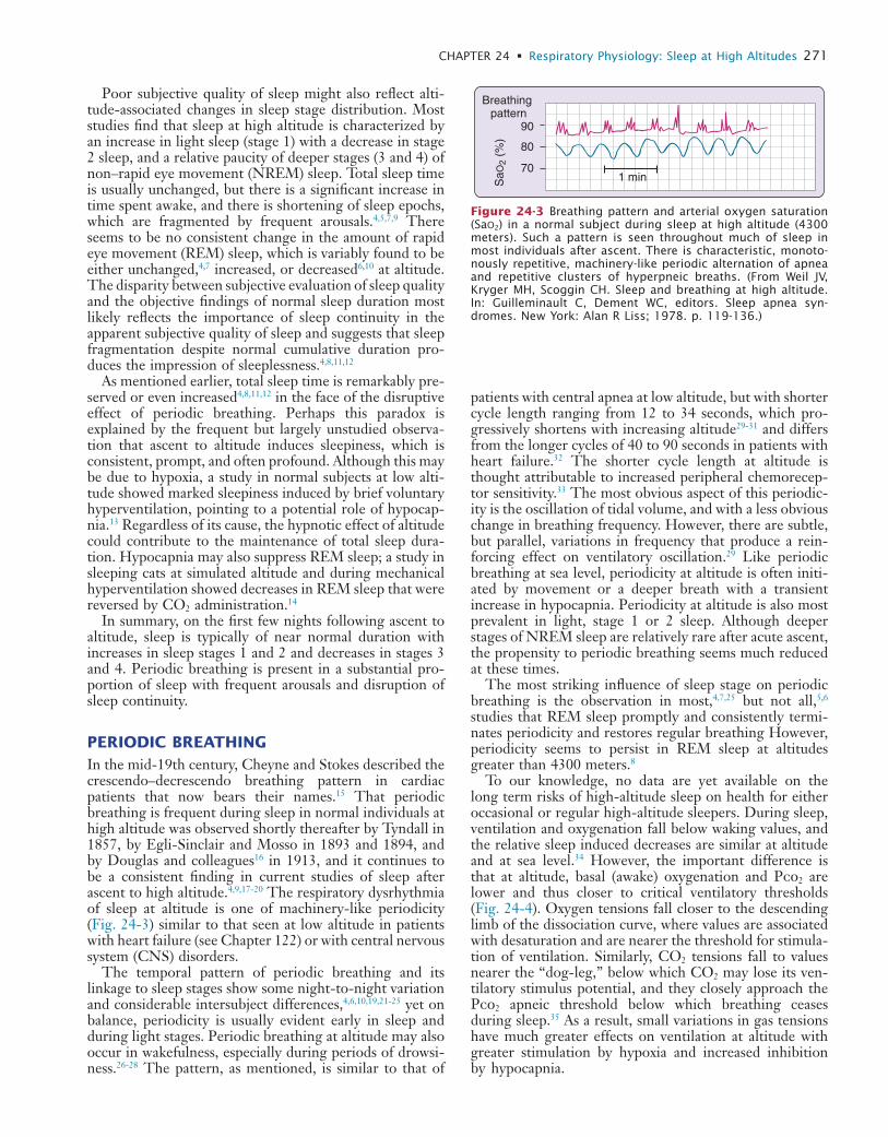
L
CHAPTER 24 • Respiratory Physiology: Sleep at High Altitudes 271
patients with central apnea at low altitude, but with shorter cycle length ranging from 12 to 34 seconds, which pro-gressively shortens with increasing altitude29-31 and differs from the longer cycles of 40 to 90 seconds in patients with heart failure.32 The shorter cycle length at altitude is thought attributable to increased peripheral chemorecep-tor sensitivity.33 The most obvious aspect of this periodic-ity is the oscillation of tidal volume, and with a less obvious change in breathing frequency. However, there are subtle, but parallel, variations in frequency that produce a rein-forcing effect on ventilatory oscillation.29 Like periodic breathing at sea level, periodicity at altitude is often initi-ated by movement or a deeper breath with a transient increase in hypocapnia. Periodicity at altitude is also most prevalent in light, stage 1 or 2 sleep. Although deeper stages of NREM sleep are relatively rare after acute ascent, the propensity to periodic breathing seems much reduced at these times.
The most striking influence of sleep stage on periodic breathing is the observation in most,4,7,25 but not all,5,6 studies that REM sleep promptly and consistently termi-nates periodicity and restores regular breathing However, periodicity seems to persist in REM sleep at altitudes greater than 4300 meters.8
To our knowledge, no data are yet available on the long term risks of high-altitude sleep on health for either occasional or regular high-altitude sleepers. During sleep, ventilation and oxygenation fall below waking values, and the relative sleep induced decreases are similar at altitude and at sea level.34 However, the important difference is that at altitude, basal (awake) oxygenation and Pco2 are lower and thus closer to critical ventilatory thresholds (Fig. 24-4). Oxygen tensions fall closer to the descending limb of the dissociation curve, where values are associated with desaturation and are nearer the threshold for stimula-tion of ventilation. Similarly, CO2 tensions fall to values nearer the “dog-leg,” below which CO2 may lose its ven-tilatory stimulus potential, and they closely approach the Pco2 apneic threshold below which breathing ceases during sleep.35 As a result, small variations in gas tensions have much greater effects on ventilation at altitude with greater stimulation by hypoxia and increased inhibition by hypocapnia.
Poor subjective quality of sleep might also reflect alti-tude-associated changes in sleep stage distribution. Most studies find that sleep at high altitude is characterized by an increase in light sleep (stage 1) with a decrease in stage 2 sleep, and a relative paucity of deeper stages (3 and 4) of non–rapid eye movement (NREM) sleep. Total sleep time is usually unchanged, but there is a significant increase in time spent awake, and there is shortening of sleep epochs, which are fragmented by frequent arousals.4,5,7,9 There seems to be no consistent change in the amount of rapid eye movement (REM) sleep, which is variably found to be either unchanged,4,7 increased, or decreased6,10 at altitude. The disparity between subjective evaluation of sleep quality and the objective findings of normal sleep duration most likely reflects the importance of sleep continuity in the apparent subjective quality of sleep and suggests that sleep fragmentation despite normal cumulative duration pro-duces the impression of sleeplessness.4,8,11,12
As mentioned earlier, total sleep time is remarkably pre-served or even increased4,8,11,12 in the face of the disruptive effect of periodic breathing. Perhaps this paradox is explained by the frequent but largely unstudied observa-tion that ascent to altitude induces sleepiness, which is consistent, prompt, and often profound. Although this may be due to hypoxia, a study in normal subjects at low alti-tude showed marked sleepiness induced by brief voluntary hyperventilation, pointing to a potential role of hypocap-nia.13 Regardless of its cause, the hypnotic effect of altitude could contribute to the maintenance of total sleep dura-tion. Hypocapnia may also suppress REM sleep; a study in sleeping cats at simulated altitude and during mechanical hyperventilation showed decreases in REM sleep that were reversed by CO2 administration.14
In summary, on the first few nights following ascent to altitude, sleep is typically of near normal duration with increases in sleep stages 1 and 2 and decreases in stages 3 and 4. Periodic breathing is present in a substantial pro-portion of sleep with frequent arousals and disruption of sleep continuity.
PERIODIC BREATHINGIn the mid-19th century, Cheyne and Stokes described the crescendo–decrescendo breathing pattern in cardiac patients that now bears their names.15 That periodic breathing is frequent during sleep in normal individuals at high altitude was observed shortly thereafter by Tyndall in 1857, by Egli-Sinclair and Mosso in 1893 and 1894, and by Douglas and colleagues16 in 1913, and it continues to be a consistent finding in current studies of sleep after ascent to high altitude.4,9,17-20 The respiratory dysrhythmia of sleep at altitude is one of machinery-like periodicity (Fig. 24-3) similar to that seen at low altitude in patients with heart failure (see Chapter 122) or with central nervous system (CNS) disorders.
The temporal pattern of periodic breathing and its linkage to sleep stages show some night-to-night variation and considerable intersubject differences,4,6,10,19,21-25 yet on balance, periodicity is usually evident early in sleep and during light stages. Periodic breathing at altitude may also occur in wakefulness, especially during periods of drowsi-ness.26-28 The pattern, as mentioned, is similar to that of
SaO
2 (%
)
Breathing pattern
90
80
701 min
Figure 24-3 Breathing pattern and arterial oxygen saturation (SaO2) in a normal subject during sleep at high altitude (4300 meters). Such a pattern is seen throughout much of sleep in most individuals after ascent. There is characteristic, monoto-nously repetitive, machinery-like periodic alternation of apnea and repetitive clusters of hyperpneic breaths. (From Weil JV, Kryger MH, Scoggin CH. Sleep and breathing at high altitude. In: Guilleminault C, Dement WC, editors. Sleep apnea syn-dromes. New York: Alan R Liss; 1978. p. 119-136.)
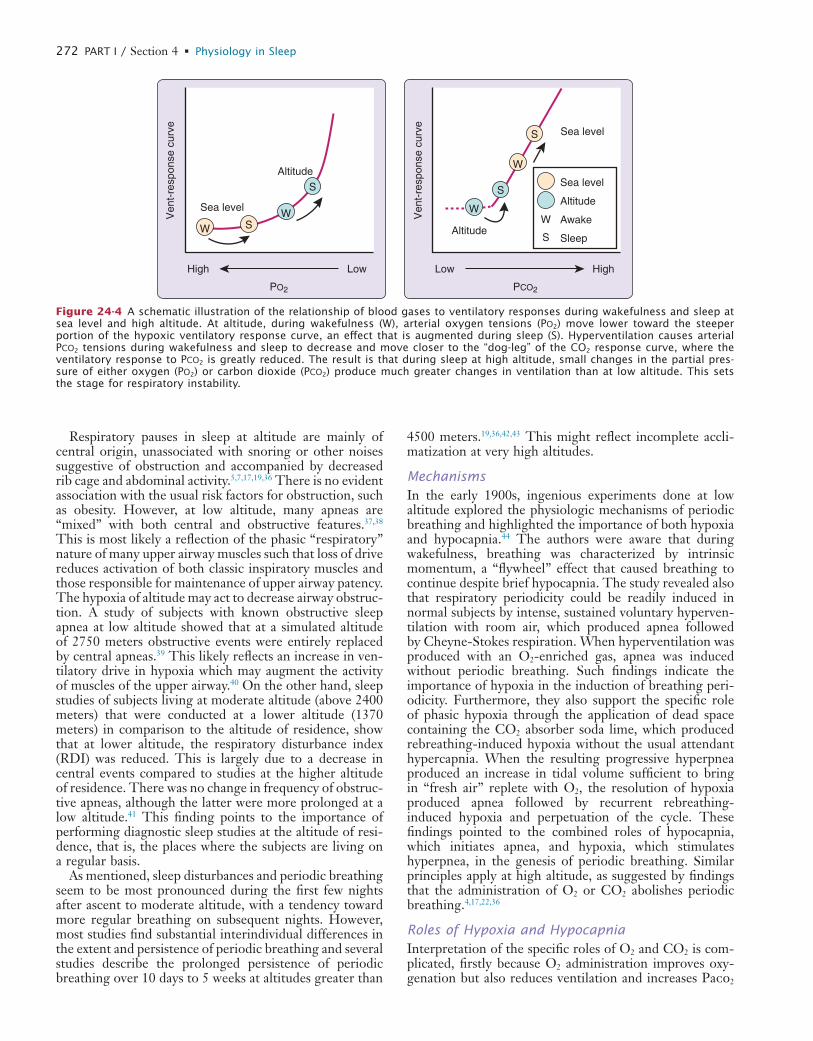
L
272 PART I / Section 4 • Physiology in Sleep
4500 meters.19,36,42,43 This might reflect incomplete accli-matization at very high altitudes.
MechanismsIn the early 1900s, ingenious experiments done at low altitude explored the physiologic mechanisms of periodic breathing and highlighted the importance of both hypoxia and hypocapnia.44 The authors were aware that during wakefulness, breathing was characterized by intrinsic momentum, a “flywheel” effect that caused breathing to continue despite brief hypocapnia. The study revealed also that respiratory periodicity could be readily induced in normal subjects by intense, sustained voluntary hyperven-tilation with room air, which produced apnea followed by Cheyne-Stokes respiration. When hyperventilation was produced with an O2-enriched gas, apnea was induced without periodic breathing. Such findings indicate the importance of hypoxia in the induction of breathing peri-odicity. Furthermore, they also support the specific role of phasic hypoxia through the application of dead space containing the CO2 absorber soda lime, which produced rebreathing-induced hypoxia without the usual attendant hypercapnia. When the resulting progressive hyperpnea produced an increase in tidal volume sufficient to bring in “fresh air” replete with O2, the resolution of hypoxia produced apnea followed by recurrent rebreathing-induced hypoxia and perpetuation of the cycle. These findings pointed to the combined roles of hypocapnia, which initiates apnea, and hypoxia, which stimulates hyperpnea, in the genesis of periodic breathing. Similar principles apply at high altitude, as suggested by findings that the administration of O2 or CO2 abolishes periodic breathing.4,17,22,36
Roles of Hypoxia and HypocapniaInterpretation of the specific roles of O2 and CO2 is com-plicated, firstly because O2 administration improves oxy-genation but also reduces ventilation and increases Paco2
Respiratory pauses in sleep at altitude are mainly of central origin, unassociated with snoring or other noises suggestive of obstruction and accompanied by decreased rib cage and abdominal activity.5,7,17,19,36 There is no evident association with the usual risk factors for obstruction, such as obesity. However, at low altitude, many apneas are “mixed” with both central and obstructive features.37,38 This is most likely a reflection of the phasic “respiratory” nature of many upper airway muscles such that loss of drive reduces activation of both classic inspiratory muscles and those responsible for maintenance of upper airway patency. The hypoxia of altitude may act to decrease airway obstruc-tion. A study of subjects with known obstructive sleep apnea at low altitude showed that at a simulated altitude of 2750 meters obstructive events were entirely replaced by central apneas.39 This likely reflects an increase in ven-tilatory drive in hypoxia which may augment the activity of muscles of the upper airway.40 On the other hand, sleep studies of subjects living at moderate altitude (above 2400 meters) that were conducted at a lower altitude (1370 meters) in comparison to the altitude of residence, show that at lower altitude, the respiratory disturbance index (RDI) was reduced. This is largely due to a decrease in central events compared to studies at the higher altitude of residence. There was no change in frequency of obstruc-tive apneas, although the latter were more prolonged at a low altitude.41 This finding points to the importance of performing diagnostic sleep studies at the altitude of resi-dence, that is, the places where the subjects are living on a regular basis.
As mentioned, sleep disturbances and periodic breathing seem to be most pronounced during the first few nights after ascent to moderate altitude, with a tendency toward more regular breathing on subsequent nights. However, most studies find substantial interindividual differences in the extent and persistence of periodic breathing and several studies describe the prolonged persistence of periodic breathing over 10 days to 5 weeks at altitudes greater than
High Low
PO2
Ven
t-re
spon
se c
urve
Altitude
Sea level
Low High
PCO2
Ven
t-re
spon
se c
urve
Altitude
Sea level
W
S
Sea level
Altitude
Awake
Sleep
W
S
SW
W
S
S
W
Figure 24-4 A schematic illustration of the relationship of blood gases to ventilatory responses during wakefulness and sleep at sea level and high altitude. At altitude, during wakefulness (W), arterial oxygen tensions (PO2) move lower toward the steeper portion of the hypoxic ventilatory response curve, an effect that is augmented during sleep (S). Hyperventilation causes arterial PCO2 tensions during wakefulness and sleep to decrease and move closer to the “dog-leg” of the CO2 response curve, where the ventilatory response to PCO2 is greatly reduced. The result is that during sleep at high altitude, small changes in the partial pres-sure of either oxygen (PO2) or carbon dioxide (PCO2) produce much greater changes in ventilation than at low altitude. This sets the stage for respiratory instability.

L
CHAPTER 24 • Respiratory Physiology: Sleep at High Altitudes 273
breathing during acclimatization and its prevention by the use of inhibitors of carbonic anhydrase. In both instances, hypocapnia is, if anything, accentuated whereas alkalosis is reduced, which suggests a dominant role for decreased pH in the genesis of apnea.
with lessening of alkalosis. Secondly, the administration of CO2 corrects respiratory alkalosis but also increases venti-lation and improves oxygenation. Despite this difficulty, considerable evidence suggests that although hypoxia may be contributory, hypocapnic alkalosis may have a particu-larly important role. Respiratory alkalosis is typical in patients with classic Cheyne-Stokes respiration at low alti-tude45 and occurs in normal subjects in whom apneas during sleep have been noted to occur after transient epi-sodes of decreased end-tidal Pco2 and presumed increase in blood pH.26 The critical role of hypocapnia in periodic breathing in sleep at altitude became clearly evident when periodic breathing in NREM sleep at simulated altitude (barometric pressure of 455 mmHg equivalent to an alti-tude of 4300 meters) was abolished by a selective increase in Paco2.17 This was achieved through the administration of low levels of CO2 with added nitrogen to prevent the increase in oxygen saturation (Sao2) that would be antici-pated as a result of the stimulation of ventilation by a rising Paco2. In one subject, during NREM sleep, the appearance of periodic breathing during the induction of hypoxia was temporally related closely to the fall in end-tidal CO2 rather to the decrease in Po2. Similarly, this study showed that breathing was regular in NREM sleep in hypoxia when CO2 was added to maintain isocapnia. However, when CO2 administration was terminated, periodic breath-ing occurred when the end-tidal CO2 fell, although the extent of hypoxia remained constant.
The Apneic ThresholdAn experimental study showed that apneas could be con-sistently produced at low altitude in NREM sleep through the induction of hypocapnia by passive positive-pressure hyperventilation against a background of either hypoxia or hyperoxia.46 Apnea tended to occur when the end-tidal CO2 was lowered 1 to 3 mm Hg below levels observed during wakefulness (the apneic threshold). Thus, hypoxia alone seems insufficient to produce periodic breathing, for which a fall in Pco2, below an apneic threshold, seems important (Fig. 24-5). Just as apneas at low altitude fre-quently follow sighs or breaths of increased tidal volume with a drop in Pco2,26 breaths of increased tidal volume with decreased end-tidal Pco2 often trigger periodic breathing at altitude.17,36
It thus appears that the events leading to periodic breath-ing at altitude begin with hypoxia, which stimulates hyper-ventilation, and the resulting hypocapnic alkalosis induces apnea, as illustrated in Figure 24-6. Apnea, in turn leads to enhanced ventilatory stimulation by increasing hypox-emia and lessening hypocapnic alkalosis, which promotes subsequent hyperpnea and arousal. The ensuing increase in ventilation lessens hypoxia and restores hypocapnic alkalosis, promoting the recurrence of apnea. Indeed, another study suggested that a stable and slightly elevated Pco2 is necessary for stable rhythmic breathing during low-altitude sleep.26
Role of Decreased PCO2 versus Increased pH in the Generation of ApneaIt is clear that apneas at altitude are reversed by the admin-istration of CO2, but this also reverses alkalosis. Observa-tions described later show the resolution of periodic
PaCO2 mm Hg
100
90
80
70
60
SaO
2 (%
)
4535 40
Periodicbreathing
Regularbreathing
O2 administration Normoxia
CO2 administration
Hypoxia
Figure 24-5 Relationship of respiratory pattern to arterial partial pressure of CO2 (PaCO2) and oxygen saturation (SaO2). This figure is a schematic representation of data of Berssen-brugge et al.17 and demonstrates that reduction in PCO2 is a critical determinant of periodic breathing in normal human sub-jects during hypoxia. Even a small increase in PCO2 after admin-istration of either O2 or CO2 restores normal breathing. This effect seems largely independent of changes in SaO2. (From Weil JV. Sleep at high altitude. Clin Chest Med 1985;6:615-621.)
Altitude ascenthypoxia
Hyperventilationarousal
Ventilatorystimulation
Ventilatoryinhibition
HyperventilationHypocapnic alkalosis
Apnea
Sleep
↑PCO2 /↓ PO2
↓PCO2 /↑ PO2
Figure 24-6 Schematic summary of mechanisms responsible for periodic breathing during sleep at altitude. Altitude-induced hypoxia stimulates increased ventilation. The resulting hypo-capnic alkalosis together with the effects of sleep promotes apnea. During altitude-related apnea, increasing hypoxemia and lessening of hypocapnic alkalosis stimulate resumption of ventilation and arousal. This augments oxygenation and lessens hypocapnic alkalosis, permitting recurrence of apnea.
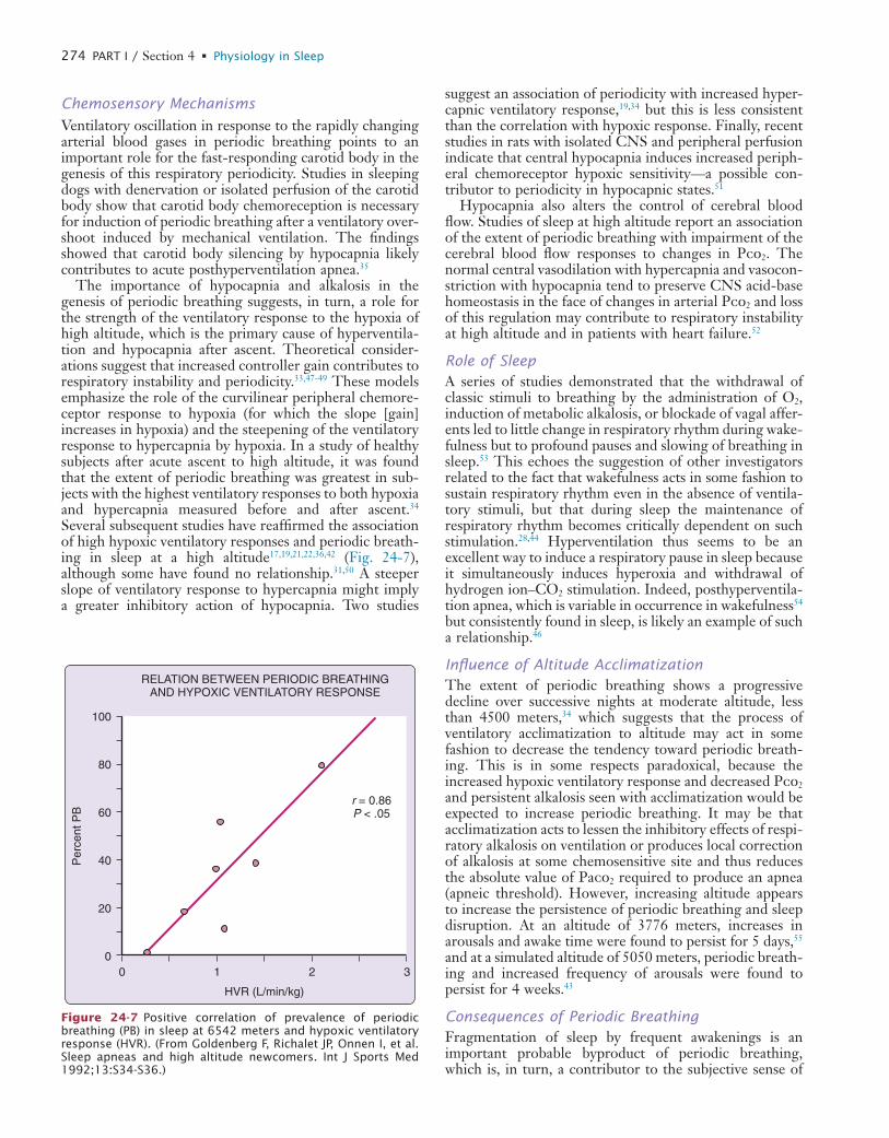
L
274 PART I / Section 4 • Physiology in Sleep
suggest an association of periodicity with increased hyper-capnic ventilatory response,19,34 but this is less consistent than the correlation with hypoxic response. Finally, recent studies in rats with isolated CNS and peripheral perfusion indicate that central hypocapnia induces increased periph-eral chemoreceptor hypoxic sensitivity—a possible con-tributor to periodicity in hypocapnic states.51
Hypocapnia also alters the control of cerebral blood flow. Studies of sleep at high altitude report an association of the extent of periodic breathing with impairment of the cerebral blood flow responses to changes in Pco2. The normal central vasodilation with hypercapnia and vasocon-striction with hypocapnia tend to preserve CNS acid-base homeostasis in the face of changes in arterial Pco2 and loss of this regulation may contribute to respiratory instability at high altitude and in patients with heart failure.52
Role of SleepA series of studies demonstrated that the withdrawal of classic stimuli to breathing by the administration of O2, induction of metabolic alkalosis, or blockade of vagal affer-ents led to little change in respiratory rhythm during wake-fulness but to profound pauses and slowing of breathing in sleep.53 This echoes the suggestion of other investigators related to the fact that wakefulness acts in some fashion to sustain respiratory rhythm even in the absence of ventila-tory stimuli, but that during sleep the maintenance of respiratory rhythm becomes critically dependent on such stimulation.28,44 Hyperventilation thus seems to be an excellent way to induce a respiratory pause in sleep because it simultaneously induces hyperoxia and withdrawal of hydrogen ion–CO2 stimulation. Indeed, posthyperventila-tion apnea, which is variable in occurrence in wakefulness54 but consistently found in sleep, is likely an example of such a relationship.46
Influence of Altitude AcclimatizationThe extent of periodic breathing shows a progressive decline over successive nights at moderate altitude, less than 4500 meters,34 which suggests that the process of ventilatory acclimatization to altitude may act in some fashion to decrease the tendency toward periodic breath-ing. This is in some respects paradoxical, because the increased hypoxic ventilatory response and decreased Pco2 and persistent alkalosis seen with acclimatization would be expected to increase periodic breathing. It may be that acclimatization acts to lessen the inhibitory effects of respi-ratory alkalosis on ventilation or produces local correction of alkalosis at some chemosensitive site and thus reduces the absolute value of Paco2 required to produce an apnea (apneic threshold). However, increasing altitude appears to increase the persistence of periodic breathing and sleep disruption. At an altitude of 3776 meters, increases in arousals and awake time were found to persist for 5 days,55 and at a simulated altitude of 5050 meters, periodic breath-ing and increased frequency of arousals were found to persist for 4 weeks.43
Consequences of Periodic BreathingFragmentation of sleep by frequent awakenings is an important probable byproduct of periodic breathing, which is, in turn, a contributor to the subjective sense of
Chemosensory Mechanisms
Ventilatory oscillation in response to the rapidly changing arterial blood gases in periodic breathing points to an important role for the fast-responding carotid body in the genesis of this respiratory periodicity. Studies in sleeping dogs with denervation or isolated perfusion of the carotid body show that carotid body chemoreception is necessary for induction of periodic breathing after a ventilatory over-shoot induced by mechanical ventilation. The findings showed that carotid body silencing by hypocapnia likely contributes to acute posthyperventilation apnea.35
The importance of hypocapnia and alkalosis in the genesis of periodic breathing suggests, in turn, a role for the strength of the ventilatory response to the hypoxia of high altitude, which is the primary cause of hyperventila-tion and hypocapnia after ascent. Theoretical consider-ations suggest that increased controller gain contributes to respiratory instability and periodicity.33,47-49 These models emphasize the role of the curvilinear peripheral chemore-ceptor response to hypoxia (for which the slope [gain] increases in hypoxia) and the steepening of the ventilatory response to hypercapnia by hypoxia. In a study of healthy subjects after acute ascent to high altitude, it was found that the extent of periodic breathing was greatest in sub-jects with the highest ventilatory responses to both hypoxia and hypercapnia measured before and after ascent.34 Several subsequent studies have reaffirmed the association of high hypoxic ventilatory responses and periodic breath-ing in sleep at a high altitude17,19,21,22,36,42 (Fig. 24-7), although some have found no relationship.31,50 A steeper slope of ventilatory response to hypercapnia might imply a greater inhibitory action of hypocapnia. Two studies
HVR (L/min/kg)
100
80
60
40
20
010 2 3
Per
cent
PB
RELATION BETWEEN PERIODIC BREATHINGAND HYPOXIC VENTILATORY RESPONSE
r = 0.86P < .05
Figure 24-7 Positive correlation of prevalence of periodic breathing (PB) in sleep at 6542 meters and hypoxic ventilatory response (HVR). (From Goldenberg F, Richalet JP, Onnen I, et al. Sleep apneas and high altitude newcomers. Int J Sports Med 1992;13:S34-S36.)
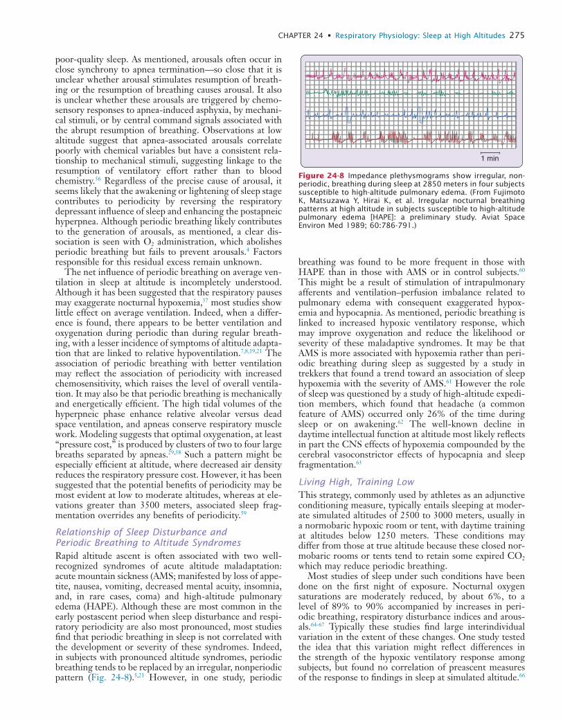
L
CHAPTER 24 • Respiratory Physiology: Sleep at High Altitudes 275
breathing was found to be more frequent in those with HAPE than in those with AMS or in control subjects.60 This might be a result of stimulation of intrapulmonary afferents and ventilation–perfusion imbalance related to pulmonary edema with consequent exaggerated hypox-emia and hypocapnia. As mentioned, periodic breathing is linked to increased hypoxic ventilatory response, which may improve oxygenation and reduce the likelihood or severity of these maladaptive syndromes. It may be that AMS is more associated with hypoxemia rather than peri-odic breathing during sleep as suggested by a study in trekkers that found a trend toward an association of sleep hypoxemia with the severity of AMS.61 However the role of sleep was questioned by a study of high-altitude expedi-tion members, which found that headache (a common feature of AMS) occurred only 26% of the time during sleep or on awakening.62 The well-known decline in daytime intellectual function at altitude most likely reflects in part the CNS effects of hypoxemia compounded by the cerebral vasoconstrictor effects of hypocapnia and sleep fragmentation.63
Living High, Training LowThis strategy, commonly used by athletes as an adjunctive conditioning measure, typically entails sleeping at moder-ate simulated altitudes of 2500 to 3000 meters, usually in a normobaric hypoxic room or tent, with daytime training at altitudes below 1250 meters. These conditions may differ from those at true altitude because these closed nor-mobaric rooms or tents tend to retain some expired CO2 which may reduce periodic breathing.
Most studies of sleep under such conditions have been done on the first night of exposure. Nocturnal oxygen saturations are moderately reduced, by about 6%, to a level of 89% to 90% accompanied by increases in peri-odic breathing, respiratory disturbance indices and arous-als.64-67 Typically these studies find large interindividual variation in the extent of these changes. One study tested the idea that this variation might reflect differences in the strength of the hypoxic ventilatory response among subjects, but found no correlation of preascent measures of the response to findings in sleep at simulated altitude.66
poor-quality sleep. As mentioned, arousals often occur in close synchrony to apnea termination—so close that it is unclear whether arousal stimulates resumption of breath-ing or the resumption of breathing causes arousal. It also is unclear whether these arousals are triggered by chemo-sensory responses to apnea-induced asphyxia, by mechani-cal stimuli, or by central command signals associated with the abrupt resumption of breathing. Observations at low altitude suggest that apnea-associated arousals correlate poorly with chemical variables but have a consistent rela-tionship to mechanical stimuli, suggesting linkage to the resumption of ventilatory effort rather than to blood chemistry.56 Regardless of the precise cause of arousal, it seems likely that the awakening or lightening of sleep stage contributes to periodicity by reversing the respiratory depressant influence of sleep and enhancing the postapneic hyperpnea. Although periodic breathing likely contributes to the generation of arousals, as mentioned, a clear dis-sociation is seen with O2 administration, which abolishes periodic breathing but fails to prevent arousals.4 Factors responsible for this residual excess remain unknown.
The net influence of periodic breathing on average ven-tilation in sleep at altitude is incompletely understood. Although it has been suggested that the respiratory pauses may exaggerate nocturnal hypoxemia,57 most studies show little effect on average ventilation. Indeed, when a differ-ence is found, there appears to be better ventilation and oxygenation during periodic than during regular breath-ing, with a lesser incidence of symptoms of altitude adapta-tion that are linked to relative hypoventilation.7,8,19,21 The association of periodic breathing with better ventilation may reflect the association of periodicity with increased chemosensitivity, which raises the level of overall ventila-tion. It may also be that periodic breathing is mechanically and energetically efficient. The high tidal volumes of the hyperpneic phase enhance relative alveolar versus dead space ventilation, and apneas conserve respiratory muscle work. Modeling suggests that optimal oxygenation, at least “pressure cost,” is produced by clusters of two to four large breaths separated by apneas.29,58 Such a pattern might be especially efficient at altitude, where decreased air density reduces the respiratory pressure cost. However, it has been suggested that the potential benefits of periodicity may be most evident at low to moderate altitudes, whereas at ele-vations greater than 3500 meters, associated sleep frag-mentation overrides any benefits of periodicity.59
Relationship of Sleep Disturbance and Periodic Breathing to Altitude SyndromesRapid altitude ascent is often associated with two well-recognized syndromes of acute altitude maladaptation: acute mountain sickness (AMS; manifested by loss of appe-tite, nausea, vomiting, decreased mental acuity, insomnia, and, in rare cases, coma) and high-altitude pulmonary edema (HAPE). Although these are most common in the early postascent period when sleep disturbance and respi-ratory periodicity are also most pronounced, most studies find that periodic breathing in sleep is not correlated with the development or severity of these syndromes. Indeed, in subjects with pronounced altitude syndromes, periodic breathing tends to be replaced by an irregular, nonperiodic pattern (Fig. 24-8).5,21 However, in one study, periodic
1 min
Figure 24-8 Impedance plethysmograms show irregular, non-periodic, breathing during sleep at 2850 meters in four subjects susceptible to high-altitude pulmonary edema. (From Fujimoto K, Matsuzawa Y, Hirai K, et al. Irregular nocturnal breathing patterns at high altitude in subjects susceptible to high-altitude pulmonary edema [HAPE]: a preliminary study. Aviat Space Environ Med 1989; 60:786-791.)
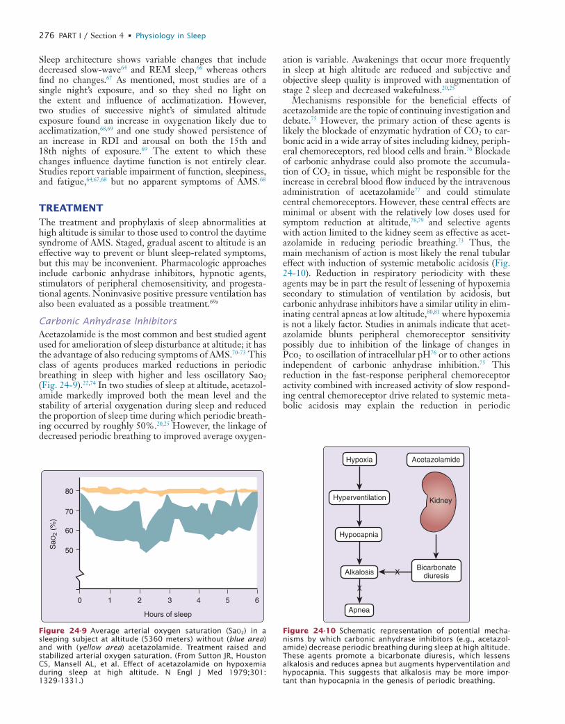
L
276 PART I / Section 4 • Physiology in Sleep
ation is variable. Awakenings that occur more frequently in sleep at high altitude are reduced and subjective and objective sleep quality is improved with augmentation of stage 2 sleep and decreased wakefulness.20,25
Mechanisms responsible for the beneficial effects of acetazolamide are the topic of continuing investigation and debate.75 However, the primary action of these agents is likely the blockade of enzymatic hydration of CO2 to car-bonic acid in a wide array of sites including kidney, periph-eral chemoreceptors, red blood cells and brain.76 Blockade of carbonic anhydrase could also promote the accumula-tion of CO2 in tissue, which might be responsible for the increase in cerebral blood flow induced by the intravenous administration of acetazolamide77 and could stimulate central chemoreceptors. However, these central effects are minimal or absent with the relatively low doses used for symptom reduction at altitude,78,79 and selective agents with action limited to the kidney seem as effective as acet-azolamide in reducing periodic breathing.73 Thus, the main mechanism of action is most likely the renal tubular effect with induction of systemic metabolic acidosis (Fig. 24-10). Reduction in respiratory periodicity with these agents may be in part the result of lessening of hypoxemia secondary to stimulation of ventilation by acidosis, but carbonic anhydrase inhibitors have a similar utility in elim-inating central apneas at low altitude,80,81 where hypoxemia is not a likely factor. Studies in animals indicate that acet-azolamide blunts peripheral chemoreceptor sensitivity possibly due to inhibition of the linkage of changes in Pco2 to oscillation of intracellular pH76 or to other actions independent of carbonic anhydrase inhibition.75 This reduction in the fast-response peripheral chemoreceptor activity combined with increased activity of slow respond-ing central chemoreceptor drive related to systemic meta-bolic acidosis may explain the reduction in periodic
Sleep architecture shows variable changes that include decreased slow-wave64 and REM sleep,66 whereas others find no changes.67 As mentioned, most studies are of a single night’s exposure, and so they shed no light on the extent and influence of acclimatization. However, two studies of successive night’s of simulated altitude exposure found an increase in oxygenation likely due to acclimatization,68,69 and one study showed persistence of an increase in RDI and arousal on both the 15th and 18th nights of exposure.69 The extent to which these changes influence daytime function is not entirely clear. Studies report variable impairment of function, sleepiness, and fatigue,64,67,68 but no apparent symptoms of AMS.68
TREATMENTThe treatment and prophylaxis of sleep abnormalities at high altitude is similar to those used to control the daytime syndrome of AMS. Staged, gradual ascent to altitude is an effective way to prevent or blunt sleep-related symptoms, but this may be inconvenient. Pharmacologic approaches include carbonic anhydrase inhibitors, hypnotic agents, stimulators of peripheral chemosensitivity, and progesta-tional agents. Noninvasive positive pressure ventilation has also been evaluated as a possible treatment.69a
Carbonic Anhydrase InhibitorsAcetazolamide is the most common and best studied agent used for amelioration of sleep disturbance at altitude; it has the advantage of also reducing symptoms of AMS.70-73 This class of agents produces marked reductions in periodic breathing in sleep with higher and less oscillatory Sao2 (Fig. 24-9).22,74 In two studies of sleep at altitude, acetazol-amide markedly improved both the mean level and the stability of arterial oxygenation during sleep and reduced the proportion of sleep time during which periodic breath-ing occurred by roughly 50%.20,25 However, the linkage of decreased periodic breathing to improved average oxygen-
SaO
2 (%
)
1 2 3 4 5 6
Hours of sleep
0
80
70
60
50
Figure 24-9 Average arterial oxygen saturation (SaO2) in a sleeping subject at altitude (5360 meters) without (blue area) and with (yellow area) acetazolamide. Treatment raised and stabilized arterial oxygen saturation. (From Sutton JR, Houston CS, Mansell AL, et al. Effect of acetazolamide on hypoxemia during sleep at high altitude. N Engl J Med 1979;301: 1329-1331.)
Bicarbonatediuresis
Hypoxia Acetazolamide
Hyperventilation
Hypocapnia
Alkalosis
Apnea
Kidney
X
X
Figure 24-10 Schematic representation of potential mecha-nisms by which carbonic anhydrase inhibitors (e.g., acetazol-amide) decrease periodic breathing during sleep at high altitude. These agents promote a bicarbonate diuresis, which lessens alkalosis and reduces apnea but augments hyperventilation and hypocapnia. This suggests that alkalosis may be more impor-tant than hypocapnia in the genesis of periodic breathing.
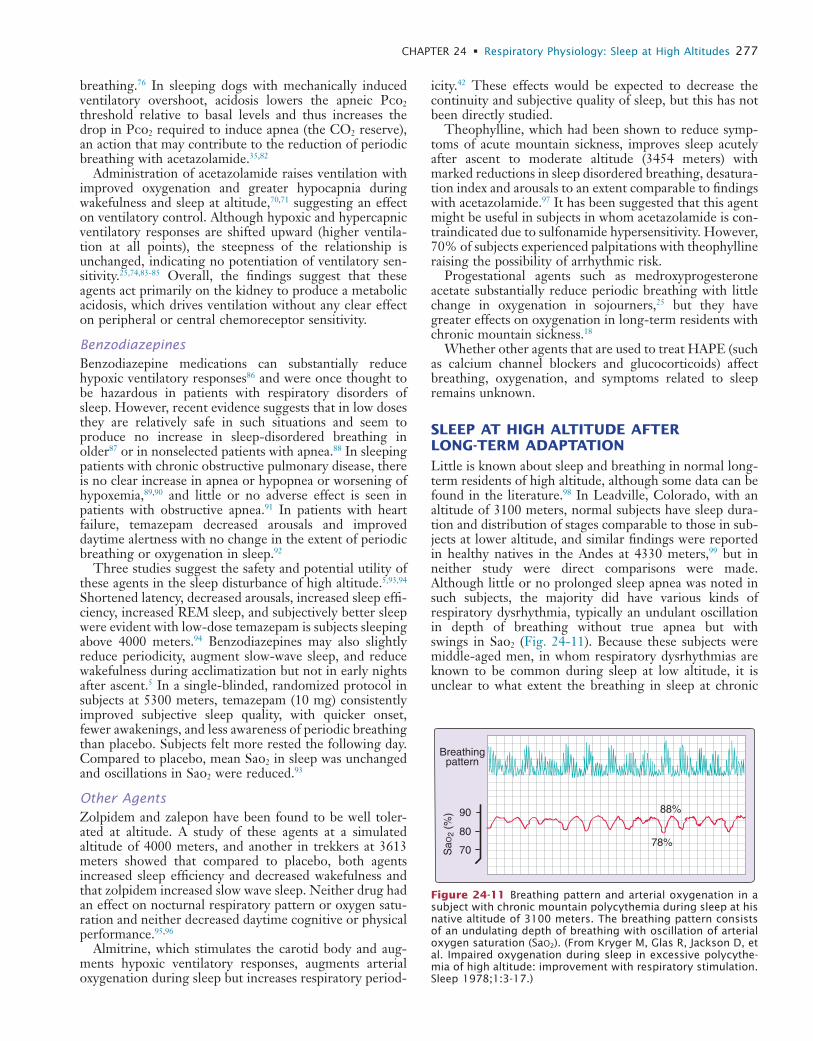
L
CHAPTER 24 • Respiratory Physiology: Sleep at High Altitudes 277
icity.42 These effects would be expected to decrease the continuity and subjective quality of sleep, but this has not been directly studied.
Theophylline, which had been shown to reduce symp-toms of acute mountain sickness, improves sleep acutely after ascent to moderate altitude (3454 meters) with marked reductions in sleep disordered breathing, desatura-tion index and arousals to an extent comparable to findings with acetazolamide.97 It has been suggested that this agent might be useful in subjects in whom acetazolamide is con-traindicated due to sulfonamide hypersensitivity. However, 70% of subjects experienced palpitations with theophylline raising the possibility of arrhythmic risk.
Progestational agents such as medroxyprogesterone acetate substantially reduce periodic breathing with little change in oxygenation in sojourners,25 but they have greater effects on oxygenation in long-term residents with chronic mountain sickness.18
Whether other agents that are used to treat HAPE (such as calcium channel blockers and glucocorticoids) affect breathing, oxygenation, and symptoms related to sleep remains unknown.
SLEEP AT HIGH ALTITUDE AFTER LONG-TERM ADAPTATIONLittle is known about sleep and breathing in normal long-term residents of high altitude, although some data can be found in the literature.98 In Leadville, Colorado, with an altitude of 3100 meters, normal subjects have sleep dura-tion and distribution of stages comparable to those in sub-jects at lower altitude, and similar findings were reported in healthy natives in the Andes at 4330 meters,99 but in neither study were direct comparisons were made. Although little or no prolonged sleep apnea was noted in such subjects, the majority did have various kinds of respiratory dysrhythmia, typically an undulant oscillation in depth of breathing without true apnea but with swings in Sao2 (Fig. 24-11). Because these subjects were middle-aged men, in whom respiratory dysrhythmias are known to be common during sleep at low altitude, it is unclear to what extent the breathing in sleep at chronic
breathing.76 In sleeping dogs with mechanically induced ventilatory overshoot, acidosis lowers the apneic Pco2 threshold relative to basal levels and thus increases the drop in Pco2 required to induce apnea (the CO2 reserve), an action that may contribute to the reduction of periodic breathing with acetazolamide.35,82
Administration of acetazolamide raises ventilation with improved oxygenation and greater hypocapnia during wakefulness and sleep at altitude,70,71 suggesting an effect on ventilatory control. Although hypoxic and hypercapnic ventilatory responses are shifted upward (higher ventila-tion at all points), the steepness of the relationship is unchanged, indicating no potentiation of ventilatory sen-sitivity.25,74,83-85 Overall, the findings suggest that these agents act primarily on the kidney to produce a metabolic acidosis, which drives ventilation without any clear effect on peripheral or central chemoreceptor sensitivity.
BenzodiazepinesBenzodiazepine medications can substantially reduce hypoxic ventilatory responses86 and were once thought to be hazardous in patients with respiratory disorders of sleep. However, recent evidence suggests that in low doses they are relatively safe in such situations and seem to produce no increase in sleep-disordered breathing in older87 or in nonselected patients with apnea.88 In sleeping patients with chronic obstructive pulmonary disease, there is no clear increase in apnea or hypopnea or worsening of hypoxemia,89,90 and little or no adverse effect is seen in patients with obstructive apnea.91 In patients with heart failure, temazepam decreased arousals and improved daytime alertness with no change in the extent of periodic breathing or oxygenation in sleep.92
Three studies suggest the safety and potential utility of these agents in the sleep disturbance of high altitude.5,93,94 Shortened latency, decreased arousals, increased sleep effi-ciency, increased REM sleep, and subjectively better sleep were evident with low-dose temazepam is subjects sleeping above 4000 meters.94 Benzodiazepines may also slightly reduce periodicity, augment slow-wave sleep, and reduce wakefulness during acclimatization but not in early nights after ascent.5 In a single-blinded, randomized protocol in subjects at 5300 meters, temazepam (10 mg) consistently improved subjective sleep quality, with quicker onset, fewer awakenings, and less awareness of periodic breathing than placebo. Subjects felt more rested the following day. Compared to placebo, mean Sao2 in sleep was unchanged and oscillations in Sao2 were reduced.93
Other AgentsZolpidem and zalepon have been found to be well toler-ated at altitude. A study of these agents at a simulated altitude of 4000 meters, and another in trekkers at 3613 meters showed that compared to placebo, both agents increased sleep efficiency and decreased wakefulness and that zolpidem increased slow wave sleep. Neither drug had an effect on nocturnal respiratory pattern or oxygen satu-ration and neither decreased daytime cognitive or physical performance.95,96
Almitrine, which stimulates the carotid body and aug-ments hypoxic ventilatory responses, augments arterial oxygenation during sleep but increases respiratory period-
Breathingpattern
90
SaO
2 (%
)
80
7078%
88%
Figure 24-11 Breathing pattern and arterial oxygenation in a subject with chronic mountain polycythemia during sleep at his native altitude of 3100 meters. The breathing pattern consists of an undulating depth of breathing with oscillation of arterial oxygen saturation (SaO2). (From Kryger M, Glas R, Jackson D, et al. Impaired oxygenation during sleep in excessive polycythe-mia of high altitude: improvement with respiratory stimulation. Sleep 1978;1:3-17.)
L
278 PART I / Section 4 • Physiology in Sleep
drase, which likely works by reducing alkalotic ventilatory inhibition. Recent studies suggest that benzodiazepines and other hypnotic agents may improve sleep quality without apparent adverse effects.107
high altitude truly differs from that at sea level. It is pos-sible that arterial desaturation induced by high altitude shifts Sao2 during sleep to the steeper portion of the dis-sociation curve and thereby amplifies the influence of ven-tilatory dysrhythmia on Sao2.
Ventilatory and Sao2 oscillations similar to those observed by Kryger and colleagues98 have also been described at high altitude by Lahiri and colleagues36 in Sherpas native to high altitude but not in Sherpas native to low altitude. The potential contribution of ethnic and/or genetic differences is suggested by a study comparing Tibetan and Chinese Han residents of 4000 meters. Sleep was studied in a hypobaric chamber at simulated altitudes of 2261 and 5000 meters. At the higher altitude, Tibetans had more periodic breathing, higher Sao2, and better sleep structure than did the Han subjects.100
Although decreased hypoxic ventilatory response during wakefulness has been observed in natives of Leadville101 and in Sherpas native to high altitude,102 it is unclear whether this contributes to respiratory dysrhythmia and hypoxemia in highlanders during sleep at altitude. However, improvement in hypoxemia during wakefulness and sleep in long-term residents at high altitude with use of the ventilatory stimulant medroxyprogesterone acetate suggests that decreased ventilatory drive may have a per-missive role.98,103 Similar findings are reported for acetazol-amide which over a three week’s treatment in residents in the Andes at 4300 m increased oxygen saturation and markedly reduced the apnea-hypopnea index during sleep.104
Natives and long-term residents of high altitude exhibit chronic mountain sickness or Monge’s disease, a syndrome of excessive polycythemia with headache, dizziness, breath-lessness, and sleep disturbance. The pathophysiology of the syndrome is debated, but likely is induced by increased hypoxemia reflecting the combined effects of altitude, decreased ventilatory drive and lung dysfunction. Com-pared to normal subjects, individuals with chronic moun-tain sickness exhibit exaggerated hypoxemia during sleep without an increase in RDI.104-106 These subjects also exhibit greater daytime hypoxemia and thus the role of sleep-associated desaturation remains uncertain.
CONCLUSIONSThe sensation of disrupted sleep after ascent to high alti-tude is associated with frequent awakenings, which in part probably reflect sleep fragmentation by respiratory dys-rhythmia typically consisting of monotonously repetitive periodic breathing. This periodicity is produced by venti-latory inhibition by hypocapnic alkalosis alternating with stimulation by hypoxia, which terminates apnea and initi-ates hyperpnea with consequent hypocapnia, leading to perpetuation of periodicity. Sleep disruption and periodic breathing decrease with time at moderate altitude and are also considerably reduced by pretreatment with acetazol-amide, which reduces alkalosis. In long-term residents of high altitude, less-distinctive, undulating respiratory dys-rhythmias are described, with unstable and decreased arte-rial oxygenation.
The most common treatment is prophylactic adminis-tration of acetazolamide, an inhibitor of carbonic anhy-
� Clinical Pearl
Sleep at altitude is disturbed by the opposing influ-ences of hypoxic stimulation and alkalotic inhibition which lead to periodic breathing and frequent associ-ated arousals. Effective treatments include correction of alkalosis with acetazolamide or blunting of hypoxic stimulation with some benzodiazepines.
REFERENCES1. Moore LG, Harrison GL, McCullough RE, et al. Low acute
hypoxic ventilatory response and hypoxic depression in acute alti-tude sickness. J Appl Physiol 1986;60:1407-1412.
2. Bisgard GE. Carotid body mechanisms in acclimatization to hypoxia. Respir Physiol 2000;121:237-246.
3. Joern AT, Shurley JT, Brooks RE, et al. Short-term changes in sleep patterns on arrival at the South Polar Plateau. Arch Intern Med 1970;125:649-654.
4. Reite M, Jackson D, Cahoon RL, et al. Sleep physiology at high altitude. Electroencephalogr Clin Neurophysiol 1975;38: 463-471.
5. Goldenberg F, Richalet JP, Onnen I, et al. Sleep apneas and high altitude newcomers. Int J Sports Med 1992;13:S34-S36.
6. Mizuno K, Asano K, Okudaira N. Sleep and respiration under acute hypobaric hypoxia. Jpn J Physiol 1993;43:161-175.
7. Normand H, Barragan M, Benoit O, et al. Periodic breathing and O2 saturation in relation to sleep stages at high altitude. Aviat Space Environ Med 1990;61:229-235.
8. Wickramasinghe H, Anholm JD. Sleep and breathing at high alti-tude. Sleep Breath 1999;3:89-102.
9. Miller JC, Horvath SM. Sleep at altitude. Aviat Space Environ Med 1977;48:615-620.
10. Anholm JD, Powles AC, Downey RD, et al. Operation Everest II: arterial oxygen saturation and sleep at extreme simulated altitude. Am Rev Respir Dis 1992;145:817-826.
11. Bonnet MH. Performance and sleepiness as a function of frequency and placement of sleep disruption. Psychophysiology 1986;23: 263-271.
12. Netzer NC, Strohl KP. Sleep and breathing in recreational climbers at an altitude of 4200 and 6400 meters: observational study of sleep and patterning of respiration during sleep in a group of recreational climbers. Sleep Breath 1999;3:75-82.
13. Ohi M, Chin K, Hirai M, et al. Oxygen desaturation following voluntary hyperventilation in normal subjects. Am J Respir Crit Care Med 1994;149:731-738.
14. Lovering AT, Fraigne JJ, Dunin-Barkowski WL, et al. Hypocapnia decreases the amount of rapid eye movement sleep in cats. Sleep 2003;26:961-967.
15. Ward M. Periodic respiration. A short historical note. Ann R Coll Surg Engl 1973;52:330-334.
16. Douglas C, Haldane J, Henderson Y, et al. Physiological observa-tions made of Pike’s Peak, Colorado, with special reference to adaptation to low barometric pressures. Philosophical Trans R Soc Lond 1913;203:185-381.
17. Berssenbrugge A, Dempsey J, Iber C, et al. Mechanisms of hypoxia-induced periodic breathing during sleep in humans. J Physiol 1983;343:507-526.
18. Kryger M, McCullough RE, Collins D, et al. Treatment of exces-sive polycythemia of high altitude with respiratory stimulant drugs. Am Rev Respir Dis 1978;117:455-464.
19. Masuyama S, Kohchiyama S, Shinozaki T, et al. Periodic breathing at high altitude and ventilatory responses to O2 and CO2. Jpn J Physiol 1989;39:523-535.
20. Sutton JR, Houston CS, Mansell AL, et al. Effect of acetazolamide on hypoxemia during sleep at high altitude. N Engl J Med 1979; 301:1329-1331.
L
CHAPTER 24 • Respiratory Physiology: Sleep at High Altitudes 279
and man. Bethesda, Md: American Physiological Society; 1984. p. 129-140.
48. Khoo MC, Anholm JD, Ko SW, et al. Dynamics of periodic breath-ing and arousal during sleep at extreme altitude. Respir Physiol 1996;103:33-43.
49. Khoo MC, Kronauer RE, Strohl KP, et al. Factors inducing peri-odic breathing in humans: a general model. J Appl Physiol 1982; 53:644-659.
50. Powles AC, Sutton JR, Gray GW, et al. Sleep hypoxemia at altitude: its relationship to acute mountain sickness and ventilatory respon-siveness to hypoxia and hypercapnia. In: Folinsbee LJ, editor. Environmental stress: individual human adaptations. New York: Academic Press; 1978. p. 373-381.
51. Day TA, Wilson RJ. Brainstem PCO2 modulates phrenic responses to specific carotid body hypoxia in an in situ dual perfused rat preparation. J Physiol 2007;578:843-857.
52. Ainslie PN, Burgess K, Subedi P, et al. Alterations in cerebral dynamics at high altitude following partial acclimatization in humans: wakefulness and sleep. J Appl Physiol 2007;102:658-664.
53. Sullivan C, Kosar L, Murphy E, et al. Primary role of respiratory afferents in sustaining breathing rhythm. J Appl Physiol 1978; 45:11-17.
54. Tawadrous FD, Eldridge FL. Posthyperventilation breathing patterns after active hyperventilation in man. J Appl Physiol 1974; 37:353-356.
55. Mizuno K, Asano K, Inoue Y, et al. Consecutive monitoring of sleep disturbance for four nights at the top of Mt Fuji (3776 m). Psychia-try Clin Neurosci 2005;59:223-225.
56. Berry RB, Gleeson K. Respiratory arousal from sleep: mechanisms and significance. Sleep 1997;20:654-675.
57. Milledge JS. The ventilatory response to hypoxia: how much is good for a mountaineer? Postgrad Med J 1987;63:169-172.
58. Ghazanshahi SD, Khoo MC. Optimal ventilatory patterns in peri-odic breathing. Ann Biomed Eng 1993;21:517-530.
59. Kupper T, Schoffl V, Netzer N. Cheyne Stokes breathing at high altitude: a helpful response or a troublemaker? Sleep Breath 2008;12:123-127.
60. Eichenberger U, Weiss E, Riemann D, et al. Nocturnal periodic breathing and the development of acute high altitude illness. Am J Respir Crit Care Med 1996;154:1748-1754.
61. Burgess KR, Johnson P, Edwards N, et al. Acute mountain sickness is associated with sleep desaturation at high altitude. Respirology 2004;9:485-492.
62. Silber E, Sonnenberg P, Collier DJ, et al. Clinical features of head-ache at altitude: a prospective study. Neurology 2003;60:1167-1171.
63. Bonnet MH. [Sleep fragmentation as the cause of daytime sleepi-ness and reduced performance]. Wien Med Wochenschr 1996;146: 332-334.
64. Hoshikawa M, Uchida S, Sugo T, et al. Changes in sleep quality of athletes under normobaric hypoxia equivalent to 2,000-m altitude: a polysomnographic study. J Appl Physiol 2007;103:2005-2011.
65. Kinsman TA, Hahn AG, Gore CJ, et al. Respiratory events and periodic breathing in cyclists sleeping at 2,650-m simulated altitude. J Appl Physiol 2002;92:2114-2118.
66. Kinsman TA, Townsend NE, Gore CJ, et al. Sleep disturbance at simulated altitude indicated by stratified respiratory disturbance index but not hypoxic ventilatory response. Eur J Appl Physiol 2005;94:569-575.
67. Pedlar C, Whyte G, Emegbo S, et al. Acute sleep responses in a normobaric hypoxic tent. Med Sci Sports Exerc 2005;37:1075- 1079.
68. Brugniaux JV, Schmitt L, Robach P, et al. Living high-training low: tolerance and acclimatization in elite endurance athletes. Eur J Appl Physiol 2006;96:66-77.
69. Kinsman TA, Gore CJ, Hahn AG, et al. Sleep in athletes undertak-ing protocols of exposure to nocturnal simulated altitude at 2650 m. J Sci Med Sport 2005;8:222-232.
69a. Johnson PL, Popa DA, Prisk GK, et al. Non-invasive positive pressure ventilation during sleep at 3800 m: relationship to acute mountain sickness and sleeping oxyhaemoglobin saturation. Respi-rology 2010;15(2):277-282.
70. Cain SM, Dunn JD. Low doses of acetazolamide to aid accommoda-tion of men to altitude. J Appl Physiol 1966;21:1195-2000.
71. Forwand SA, Landowne M, Follansbee JN, et al. Effect of acetazol-amide on acute mountain sickness. N Engl J Med 1968;279: 839-845.
21. Fujimoto K, Matsuzawa Y, Hirai K, et al. Irregular nocturnal breathing patterns high altitude in subjects susceptible to high-altitude pulmonary edema (HAPE): a preliminary study. Aviat Space Environ Med 1989;60:786-791.
22. Lahiri S, Barnard P. Role of arterial chemoreflex in breathing during sleep at high altitude. Prog Clin Biol Res 1983;136:75-85.
23. Lahiri S, Data PG. Chemosensitivity and regulation of ventilation during sleep at high altitudes. Intern J Sports Med 1992;13: S31-S33.
24. Normand H, Vargas E, Bordachar J, et al. Sleep apneas in high altitude residents (3,800 m). Intern J Sports Med 1992;13: S40-S42.
25. Weil JV, Kryger MH, Scoggin CH. Sleep and breathing at high altitude. In: Guilleminault C, Dement WC, editors. Sleep apnea syndromes. New York: Alan R Liss; 1978. p. 119-136.
26. Bulow K. Respiration and wakefulness in man. Acta Physiol Scand Suppl 1963;59:1-110.
27. Douglas C, Haldane J. The causes of periodic or Cheyne-Stokes breathing. J Physiol 1909;38:401-419.
28. Douglas CG, Haldane JS. The regulation of normal breathing. J Physiol 1909;38:420-440.
29. Brusil PJ, Waggener TB, Kronauer RE, et al. Methods for identify-ing respiratory oscillations disclose altitude effects. J Appl Physiol 1980;48:545-556.
30. Waggener TB, Brusil PJ, Kronauer RE, et al. Strength and cycle time of high-altitude ventilatory patterns in unacclimatized humans. J Appl Physiol 1984;56:576-581.
31. West JB, Peters R Jr, Aksnes G, et al. Nocturnal periodic breathing at altitudes of 6,300 and 8,050 m. J Appl Physiol 1986;61: 280-287.
32. Naughton M, Benard D, Tam A, et al. Role of hyperventilation in the pathogenesis of central sleep apneas in patients with congestive heart failure [see comments]. Am Rev Respir Dis 1993;148:330-338.
33. Topor ZL, Vasilakos K, Younes M, et al. Model based analysis of sleep disordered breathing in congestive heart failure. Respir Physiol Neurobiol 2007;155:82-92.
34. White DP, Gleeson K, Pickett CK, et al. Weil JV. Altitude accli-matization: influence on periodic breathing and chemoresponsive-ness during sleep. J Appl Physiol 1987;63:401-412.
35. Smith CA, Nakayama H, Dempsey JA. The essential role of carotid body chemoreceptors in sleep apnea. Can J Physiol Pharmacol 2003;81:774-779.
36. Lahiri S, Maret K, Sherpa MG. Dependence of high altitude sleep apnea on ventilatory sensitivity to hypoxia. Respir Physiol 1983; 52:281-301.
37. Badr MS, Toiber F, Skatrud JB, et al. Pharyngeal narrowing/ occlusion during central sleep apnea. J Appl Physiol 1995;78: 1806-1815.
38. Fletcher EC. Recurrence of sleep apnea syndrome following tracheostomy. A shift from obstructive to central apnea. Chest 1989;95:205-209.
39. Burgess KR, Cooper J, Rice A, et al. Effect of simulated altitude during sleep on moderate-severity OSA. Respirology 2006;11:62- 69.
40. Warner G, Skatrud JB, Dempsey JA. Effect of hypoxia-induced periodic breathing on upper airway obstruction during sleep. J Appl Physiol 1987;62:2201-2211.
41. Patz D, Spoon M, Corbin R, et al. The effect of altitude descent on obstructive sleep apnea. Chest 2006;130:1744-1750.
42. Hackett PH, Roach RC, Harrison GL, et al. Respiratory stimulants and sleep periodic breathing at high altitude. Almitrine versus acet-azolamide. Am Rev Respir Dis 1987;135:896-898.
43. Salvaggio A, Insalaco G, Marrone O, et al. Effects of high-altitude periodic breathing on sleep and arterial oxyhaemoglobin saturation. Eur Respir J 1998;12:408-413.
44. Douglas CG, Haldane JS, Henderson Y, et al. The physiological effects of low atmospheric pressures as observed on Pike’s Peak, Colorado. J Physiol 1912;LXXXV:65-67.
45. Gotoh F, Meyer JS, Takagi Y. Cerebral venous and arterial blood gases during Cheyne-Stokes respiration. Am J Med 1969;47: 534-545.
46. Skatrud JB, Dempsey JA. Interaction of sleep state and chemical stimuli in sustaining rhythmic ventilation. J Appl Physiol 1983; 55:813-822.
47. Cherniack N, Gothe B, Strohl K. Mechanisms for recurrent apneas at altitude. In: West JB, Lahiri S, editors. High altitude
L
280 PART I / Section 4 • Physiology in Sleep
90. Steens RD, Pouliot Z, Millar TW, et al. Effects of zolidem and triazolam on sleep and respiration in mild to moderate chronic obstructive pulmonary disease. Sleep 1993;16:318-326.
91. Berry RB, Kouchi K, Bower J, et al. Triazolam in patients with obstructive apnea. Am J Resp Crit Care Med 1995;151:450-454.
92. Biberdorf DJ, Steens R, Miller TW, et al. Benzodiazepines in con-gestive heart failure: effects of temazepam on arousability and Cheyne-Stokes respiration. Sleep 1993;16:529-538.
93. Dubowitz G. Effect of temazepam on oxygen saturation and sleep quality at high altitude: randomised placebo controlled crossover trial. BMJ 1998;316:587-589.
94. Nicholson AN, Smith PA, Stone BM, et al. Altitude insomnia: studies during an expedition to the Himalayas. Sleep 1988;11: 354-361.
95. Beaumont M, Batejat D, Coste O, et al. Effects of zolpidem and zaleplon on sleep, respiratory patterns and performance at a simulated altitude of 4,000 m. Neuropsychobiology 2004;49: 154-162.
96. Beaumont M, Batejat D, Pierard C, et al. Zaleplon and zolpidem objectively alleviate sleep disturbances in mountaineers at a 3,613 meter altitude. Sleep 2007;30:1527-1533.
97. Fischer R, Lang SM, Leitl M, et al. Theophylline and acetazolamide reduce sleep-disordered breathing at high altitude. Eur Respir J 2004;23:47-52.
98. Kryger M, Glas R, Jackson D, et al. Impaired oxygenation during sleep in excessive polycythemia of high altitude: improvement with respiratory stimulation. Sleep 1978;1:3-17.
99. Coote JH, Stone BM, Tsang G. Sleep of Andean high altitude natives. Eur J Appl Physiol Occup Physiol 1992;64:178-181.
100. Plywaczewski R, Wu TY, Wang XQ, et al. Sleep structure and periodic breathing in Tibetans and Han at simulated altitude of 5000 m. Respir Physiol Neurobiol 2003;136:187-197.
101. Weil JV, Byrne-Quinn E, Sodal IE, et al. Acquired attenuation of chemoreceptor function in chronically hypoxic man at high altitude. J Clin Invest 1971;50:186-195.
102. Hackett PH, Reeves JT, Reeves CD, et al. Control of breathing in Sherpas at low and high altitude. J Appl Physiol 1980;49: 374-379.
103. Kryger M, Weil J, Grover R. Chronic mountain polycythemia: a disorder of the regulation of breathing during sleep? Chest 1978;73:303-304.
104. Richalet JP, Rivera-Ch M, Maignan M, et al. Acetazolamide for Monge’s disease: efficiency and tolerance of 6-month treatment. Am J Respir Crit Care Med 2008;177:1370-1376.
105. Reeves JT, Weil JV. Chronic mountain sickness. A view from the crow’s nest. Adv Exp Med Biol 2001;502:419-437.
106. Spicuzza L, Casiraghi N, Gamboa A, et al. Sleep-related hypoxae-mia and excessive erythrocytosis in Andean high-altitude natives. Eur Respir J 2004;23:41-46.
107. Luks AM. Which medications are safe and effective for improving sleep at high altitude? High Alt Med Biol 2008;9:195-198.
72. Hackett PH, Rennie D. The incidence, importance, and prophy-laxis of acute mountain sickness. Lancet 1976;2:1149-1155.
73. Swenson ER. Carbonic anhydrase inhibitors and ventilation: a complex interplay of stimulation and suppression. Eur Respir J 1998;12:1242-1247.
74. Sutton JR, Gray GW, Houston CS, et al. Effects of duration at altitude and acetazolamide on ventilation and oxygenation during sleep. Sleep 1980;3:455-464.
75. Swenson ER, Teppema LJ. Prevention of acute mountain sickness by acetazolamide: as yet an unfinished story. J Appl Physiol 2007;102:1305-1307.
76. Leaf DE, Goldfarb DS. Mechanisms of action of acetazolamide in the prophylaxis and treatment of acute mountain sickness. J Appl Physiol 2007;102:1313-1322.
77. Hauge A, Nicolaysen G, Thoresen M. Acute effects of acetazol-amide on cerebral blood flow in man. Acta Physiol Scand 1983; 117:233-239.
78. Huang SY, McCullough RE, McCullough RG, et al. Usual clinical dose of acetazolamide does not alter cerebral blood. Respir Physiol 1988;72:315-326.
79. Teppema LJ, Balanos GM, Steinback CD, et al. Effects of acetazol-amide on ventilatory, cerebrovascular, and pulmonary vascular responses to hypoxia. Am J Respir Crit Care Med 2007;175:277-281.
80. DeBacker WA, Verbraecken J, Willemen M, et al. Central apnea index decreases after prolonged treatment with acetazolamide. Am J Respir Crit Care Med 1995;151:87-91.
81. White DP, Zwillich CW, Pickett CK, et al. Central sleep apnea. Improvement with acetazolamide therapy. Arch Intern Med 1982;142:1816-1819.
82. Nakayama H, Smith CA, Rodman JR, et al. Effect of ventilatory drive on carbon dioxide sensitivity below eupnea during sleep. Am J Respir Crit Care Med 2002;165:1251-1260.
83. Bashir Y, Kann M, Stradling JR. The effect of acetazolamide on hypercapnic and eucapnic/poikilocapnic hypoxic ventilatory responses in normal subjects. Pulm Pharmacol 1990;3:151-154.
84. Swenson ER, Leatham KL, Roach RC, et al. Renal carbonic anhy-drase inhibition reduces high altitude sleep periodic breathing. Respir Physiol 1991;86:333-343.
85. Swenson ER, Hughes JM. Effects of acute and chronic acetazol-amide on resting ventilation and ventilatory responses in men. J Appl Physiol 1993;74:230-237.
86. Lakshminarayan S, Sahn SA, Hudson LD, et al. Effect of diazepam on ventilatory responses. Clin Pharmacol Ther 1976;20:178-183.
87. Camacho ME, Morin CM. The effect of temazepam of respiraton in elderly insomniacs with mild sleep apnea. Sleep 1995;18:644-645.
88. Hoijer U, Hedner J, Ejnell H, et al. Nitrazepam in patients with sleep apnoea: a double-blind placebo-controlled study. Eur Resp J 1994;7:2011-2015.
89. Gentil B, Tehindrazanarivelo A, Lienhart A, et al. Respiratory effects of midazolam in patients with obstructive sleep apnea syn-dromes. Ann Fran Anes Reanim 1994;13:275-279.
LL
281
Abstract
Sleepiness, like fever, is commonly experienced at the onset of an infection or other cause of systemic inflammation. Changes in sleep in response to microbes appear to be one facet of the acute phase response. Typically, soon after infec-tious challenge, animals exhibit excess time spent in non–rapid eye movement sleep and decreased total rapid eye movement sleep. The exact time course of sleep responses depends upon the infectious agent used, the route of admin-istration, and the time of day the infectious challenge is given.
There is a common perception that sleep loss can render one vulnerable to infection. Many studies have combined sleep deprivation with measurement of one or more parameters of the immune response. A few studies have combined sleep deprivation with infectious challenge. After mild sleep depriva-
tion, several immune system parameters (such as natural killer cell activity) change, and resistance to a viral challenge is decreased in individuals that spontaneously sleep less. After prolonged (2 to 3 weeks) sleep deprivation, rats become sep-ticemic. Studies have not yet been done to determine the effects of sleep deprivation on recuperation from an infection. Small amounts of sleep loss may promote host defenses, whereas prolonged sleep loss is devastating.
The molecular mechanisms responsible for the changes in sleep associated with infection appear to be an amplification of a physiological sleep regulatory biochemical cascade. This chain of events includes well-known immune response modi-fiers such as interleukin-1, tumor necrosis factor-α, prosta-glandins, nitric oxide, and adenosine. All these substances are normal constituents of the brain and are involved in physio-logical sleep regulation.
response1 that follows any inflammatory challenge, such as an infection or traumatic injury. Local inflammatory chal-lenges, for example, minor cuts or bruises, may activate a low-level APR that is not perceived by the victim. But when the challenge is severe enough, it activates a full-blown systemic APR within 24 hours and the patient feels sick. In the case of infections, the function of the APR is to wall off and destroy invading microorganisms, to remove tissue debris, and to alert the host to the invasion so that systemic protective responses are mobilized. The systemic inflammatory response activates the brain, liver, and bone marrow to react in a stereotypic manner. The APR has both behavioral (fever, excess sleep, anorexia, etc.) and biochemical markers (C-reactive protein, serum amyloid A, mannose binding protein, etc.). Increased secretion of a broad array of endocrine hormones, including the stress hormones, also occurs. This complex of responses leads to host protective behaviors (such as social withdrawal that avoids predation),2 physiological responses (such as fever, which can thermally inhibit growth of some microorgan-isms),3 and immune responses (such as mobilization of leukocytes and natural killer (NK) cells.)1 Hormonal changes (such as prolactin regulation of antimicrobial nitric oxide levels)4 and biochemical changes (such as potentiation of microbial phagocytosis)5 also contribute to host defense. The APR is the first line of defense against infections and the trigger for acquired immunity mediated by specific antibodies and cytotoxic T lymphocytes needed to clear the infection.6
A major class of protein mediators, the cytokines, initi-ates the APR.7 Cytokines are generally associated with immune cells, but they are made by all cells. Over 100 of these intercellular signaling molecules have been identi-fied, and the complexity of their interactions rivals that of the nervous system. Cytokines induce themselves and other cytokines, and they form biochemical cascades
INTRODUCTION
Most individuals have experienced the intense desire to sleep that may occur at the onset of more severe infections. Further, many have received the advice from loving parents and grandparents to get plenty of rest to prevent, or help recover from, infectious diseases. These experiences suggest a connection between sleep and host defense systems. Indeed, Hippocrates, Aristotle, and many of our predecessors acknowledged such a relationship. But only within the past 15 years have modern science and medicine systematically investigated whether sleep has anything to do with host defense systems. This chapter is organized around four main sections related to this issue. They are: 1) the acute phase response and host defense; 2) sleep changes after infectious challenge; 3) sleep loss effects on immune function; 4) sleep and immunity share common regulatory molecules; and finally, in the Clinical Pearl section: sleep and recuperation. The available evidence, thus far, is consistent with a role for sleep in maintaining resistance to infections.
THE ACUTE PHASE RESPONSE AND HOST DEFENSEA major theme of this chapter is that excess sleep as a host defense is part of the acute phase response (APR) to inflam-matory challenge and shares numerous immunoregulatory molecules with other APRs. Related concepts and termi-nology are found in the following section.
Enhanced sleep in response to infections occurs in the context of dozens of other responses, collectively termed the APR. Recent advances in innate immunity research provide a framework for understanding many of the shared mechanisms underlying the APR in general and sleep reg-ulation specifically. The APR is a critical innate immune
Sleep and Host DefenseJames M. Krueger and Jeannine A. Majde
Chapter
25
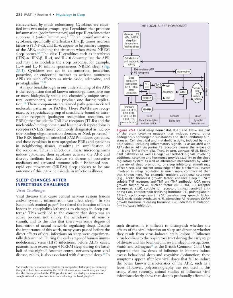
L
282 PART I / Section 4 • Physiology in Sleep
such diseases, it is difficult to distinguish whether the effects of the viral infection on sleep are direct or whether they result from virus-induced brain lesions.13 Influenza virus localizes to the respiratory tract during the early stage of disease and has been used in several sleep investigations. Smith and colleagues18 at the British Common Cold Unit reported that low doses of influenza in humans induce excess behavioral sleep and cognitive dysfunction; these symptoms appear after low viral doses that fail to induce the better known characteristics of the APR, such as a fever. However, polysomnography was not used in this study. More recently, animal studies of influenza viral infections clearly show that sleep is profoundly affected by
characterized by much redundancy. Cytokines are classi-fied into two major groups, type I cytokines that promote inflammation (proinflammatory) and type II cytokines that suppress it (antiinflammatory).8 Three proinflammatory cytokines, specifically interleukin (IL)-1β, tumor necrosis factor-α (TNF-α), and IL-6, appear to be primary triggers of the APR, including the situation when excess NREM sleep occurs.7,9 The class II cytokines such as interferon (IFN)-α, IFN-β, IL-4, and IL-10 downregulate the APR and may also modulate the sleep response; for example, IL-4 and IL-10 inhibit spontaneous NREM sleep (Fig. 25-1). Cytokines can act in an autocrine, juxtacrine, paracrine, or endocrine manner to activate numerous APRs via such effectors as nitric oxide, adenosine, and prostaglandins.7,9,10
A major breakthrough in our understanding of the APR is the recognition that all known microorganisms have one or more biologically stable and chemically unique struc-tural components, or they produce one during replica-tion.11 These components are termed pathogen-associated molecular patterns, or PAMPs. These PAMPs are recog-nized by a specialized group of membrane-bound or intra-cellular receptors (pathogen recognition receptors, or PRRs)6 that include the Toll-like receptors (TLRs) and the nucleotide-binding domain and leucine-rich repeat domain receptors (NLRs) (more commonly designated as nucleo-tide-binding oligomerization domain, or Nod, proteins.)12 The PRR binding of microbial PAMPs induces cytokines, and these cytokines in turn upregulate PRRs and cytokines in neighboring tissues, resulting in amplification of the response. Thus in infectious illness, microorganisms induce cytokines, then cytokines activate the APR and thereby facilitate host defense via dozens of protective mediators and activated immune cells.1,7 Enhanced non–rapid eye movement (NREM) sleep appears to be one outcome of this cytokine cascade in infectious illness.
SLEEP CHANGES AFTER INFECTIOUS CHALLENGE
Viral ChallengeViral diseases that cause central nervous system lesions and/or systemic inflammation can affect sleep.13 In von Economo’s seminal paper14 he related the location of brain lesions in encephalitis lethargica to changes in sleep pat-terns.* This work led to the concept that sleep was an active process, not simply the withdrawal of sensory stimuli, and to the idea that there was some degree of localization of neural networks regulating sleep. Despite the importance of this work, many years passed before the direct effects of viral infections on sleep were experimen-tally determined. During the early stages of human immu-nodeficiency virus (HIV) infections, before AIDS onset, patients have excess stage 4 NREM sleep during the latter half of the night.16 Another central nervous system viral disease, rabies, is also associated with disrupted sleep.17 In
THE LOCAL SLEEP HOMEOSTAT
Cell electricaland metabolic
activity
Extracellular ATPP2 receptors Adenosine
Microbes, LPS,MPs, dsRNA,
sleep loss,temperature,
feeding, stress
SleepregulatorysubstancesIL1β, TNFα,
othercytokines
Inhibitors(e.g., IL4, IL10,
IL13, CRH,sTNFR, sIL1R,
IL1RA, TGF,glucocorticoids)
NF-κB NO
Adenosine
PGsCOX-2
GHRH
NOS
A1ARExcitatory
(glu)and
inhibitory(GABA)
NR
EM
S
Hours
Gene transcriptionand translation
Seconds
Blood flow
msec
Synaptictransmission
Figure 25-1 Local sleep homeostat. IL-1β and TNF-α are part of the brain cytokine network that includes several other endogenous somnogenic substances and sleep inhibitory sub-stances. Cell electrical and metabolic activity, induced by mul-tiple stimuli including inflammatory signals, is associated with ATP release. ATP via purine P2 receptors causes the release of IL-1β and TNF-α from glia. They, in turn, activate NFκB. Redun-dant pathways as well as negative feedback signals involving additional cytokine and hormones provide stability to the sleep regulatory system as well as alternative mechanisms by which a variety of sleep promoting, or sleep inhibitory, stimuli may affect sleep. Our current knowledge of the biochemical events involved in sleep regulation is much more complicated than that shown here. For example, multiple additional cytokines (e.g., acidic fibroblast growth factor) enhance sleep.10 TNFR, soluble TNF receptor; anti-TNF, anti-TNF antibody; NGF, nerve growth factor; NFκB, nuclear factor κB; IL1RA, IL1 receptor antagonist; sILIR, soluble IL1 receptor; anti-IL1, anti-IL1 anti-body; CRH, corticotropin releasing hormone; PG, prostaglandin; COX-2, cyclooxygenase-2; TGF, transforming growth factor; NOS, nitric oxide synthase; A1R, adenosine A1 receptor; GHRH, growth hormone releasing hormone; (→) indicates stimulation; (—) indicates inhibition.
*Although von Economo’s encephalitis (or encephalitis lethargica) is commonly thought to have been caused by the 1918 influenza virus, recent analyses reveal that the disease preceded the 1918 pandemic and is probably an autoimmune complication of streptococcal infections affecting the basal ganglia.15
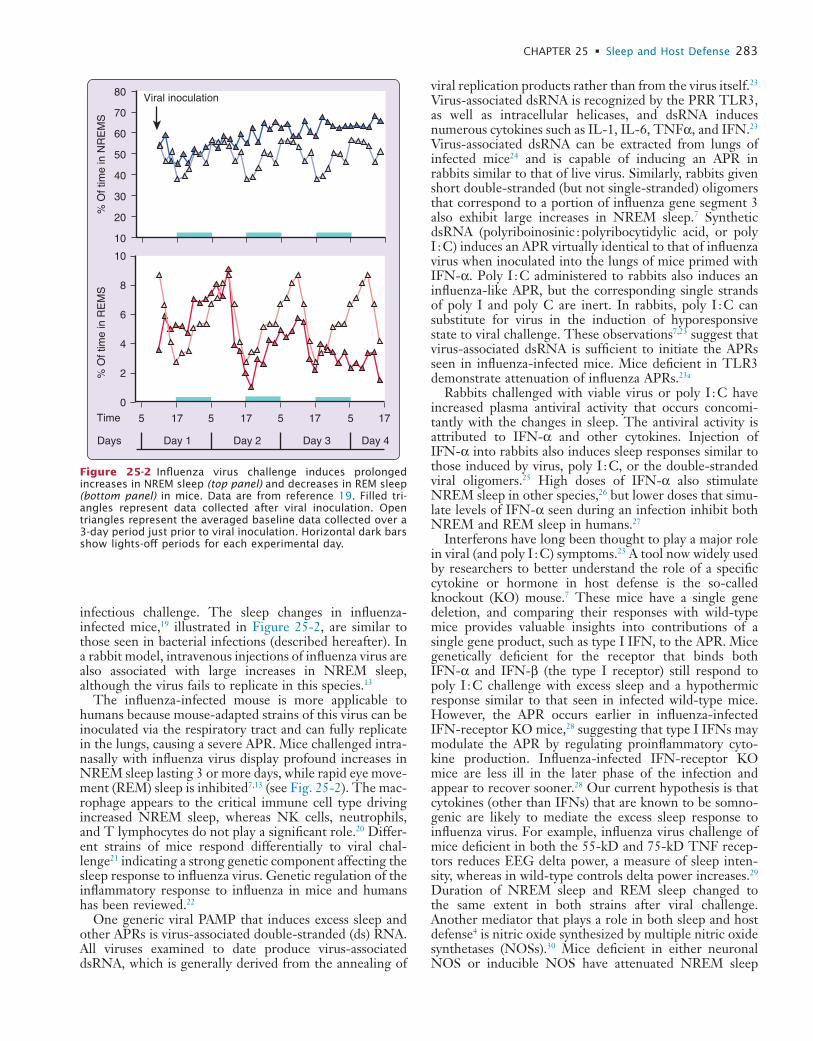
L
CHAPTER 25 • Sleep and Host Defense 283
viral replication products rather than from the virus itself.23 Virus-associated dsRNA is recognized by the PRR TLR3, as well as intracellular helicases, and dsRNA induces numerous cytokines such as IL-1, IL-6, TNFα, and IFN.23 Virus-associated dsRNA can be extracted from lungs of infected mice24 and is capable of inducing an APR in rabbits similar to that of live virus. Similarly, rabbits given short double-stranded (but not single-stranded) oligomers that correspond to a portion of influenza gene segment 3 also exhibit large increases in NREM sleep.7 Synthetic dsRNA (polyriboinosinic : polyribocytidylic acid, or poly I : C) induces an APR virtually identical to that of influenza virus when inoculated into the lungs of mice primed with IFN-α. Poly I : C administered to rabbits also induces an influenza-like APR, but the corresponding single strands of poly I and poly C are inert. In rabbits, poly I : C can substitute for virus in the induction of hyporesponsive state to viral challenge. These observations7,23 suggest that virus-associated dsRNA is sufficient to initiate the APRs seen in influenza-infected mice. Mice deficient in TLR3 demonstrate attenuation of influenza APRs.23a
Rabbits challenged with viable virus or poly I : C have increased plasma antiviral activity that occurs concomi-tantly with the changes in sleep. The antiviral activity is attributed to IFN-α and other cytokines. Injection of IFN-α into rabbits also induces sleep responses similar to those induced by virus, poly I : C, or the double-stranded viral oligomers.25 High doses of IFN-α also stimulate NREM sleep in other species,26 but lower doses that simu-late levels of IFN-α seen during an infection inhibit both NREM and REM sleep in humans.27
Interferons have long been thought to play a major role in viral (and poly I : C) symptoms.23 A tool now widely used by researchers to better understand the role of a specific cytokine or hormone in host defense is the so-called knockout (KO) mouse.7 These mice have a single gene deletion, and comparing their responses with wild-type mice provides valuable insights into contributions of a single gene product, such as type I IFN, to the APR. Mice genetically deficient for the receptor that binds both IFN-α and IFN-β (the type I receptor) still respond to poly I : C challenge with excess sleep and a hypothermic response similar to that seen in infected wild-type mice. However, the APR occurs earlier in influenza-infected IFN-receptor KO mice,28 suggesting that type I IFNs may modulate the APR by regulating proinflammatory cyto-kine production. Influenza-infected IFN-receptor KO mice are less ill in the later phase of the infection and appear to recover sooner.28 Our current hypothesis is that cytokines (other than IFNs) that are known to be somno-genic are likely to mediate the excess sleep response to influenza virus. For example, influenza virus challenge of mice deficient in both the 55-kD and 75-kD TNF recep-tors reduces EEG delta power, a measure of sleep inten-sity, whereas in wild-type controls delta power increases.29 Duration of NREM sleep and REM sleep changed to the same extent in both strains after viral challenge. Another mediator that plays a role in both sleep and host defense4 is nitric oxide synthesized by multiple nitric oxide synthetases (NOSs).30 Mice deficient in either neuronal NOS or inducible NOS have attenuated NREM sleep
infectious challenge. The sleep changes in influenza-infected mice,19 illustrated in Figure 25-2, are similar to those seen in bacterial infections (described hereafter). In a rabbit model, intravenous injections of influenza virus are also associated with large increases in NREM sleep, although the virus fails to replicate in this species.13
The influenza-infected mouse is more applicable to humans because mouse-adapted strains of this virus can be inoculated via the respiratory tract and can fully replicate in the lungs, causing a severe APR. Mice challenged intra-nasally with influenza virus display profound increases in NREM sleep lasting 3 or more days, while rapid eye move-ment (REM) sleep is inhibited7,13 (see Fig. 25-2). The mac-rophage appears to the critical immune cell type driving increased NREM sleep, whereas NK cells, neutrophils, and T lymphocytes do not play a significant role.20 Differ-ent strains of mice respond differentially to viral chal-lenge21 indicating a strong genetic component affecting the sleep response to influenza virus. Genetic regulation of the inflammatory response to influenza in mice and humans has been reviewed.22
One generic viral PAMP that induces excess sleep and other APRs is virus-associated double-stranded (ds) RNA. All viruses examined to date produce virus-associated dsRNA, which is generally derived from the annealing of
Viral inoculation
5 17 17 17 175 5 5Time
Day 1Days Day 2 Day 3 Day 4
80
70
60
50
40
30
20
10
10
8
6
4
2
0
% O
f tim
e in
NR
EM
S%
Of t
ime
in R
EM
S
Figure 25-2 Influenza virus challenge induces prolonged increases in NREM sleep (top panel) and decreases in REM sleep (bottom panel) in mice. Data are from reference 19. Filled tri-angles represent data collected after viral inoculation. Open triangles represent the averaged baseline data collected over a 3-day period just prior to viral inoculation. Horizontal dark bars show lights-off periods for each experimental day.
L
284 PART I / Section 4 • Physiology in Sleep
relatively long periods. In contrast, if the gram-negative bacterium Pasteurella multocida is given intranasally, a dif-ferent time course of sleep responses is observed. In this case, the increased NREM sleep responses occur after a longer latency and the magnitude of the increases in NREM sleep is less than the effects of this pathogen given by other routes of administration. P. multocida is a natural respiratory pathogen in rabbits.
The intestinal lumen of mammals contains large amounts of bacteria, some of which leak into the intestinal lymphat-ics under normal conditions. Changes in intestinal perme-ability by inflammatory challenges increase the release of bacterial products into the lymphatics. Local lymph node macrophages phagocytose and digest these bacterial prod-ucts,9,35 releasing somnogenic PAMPs as described later. This mechanism is viewed as operating at a low basal rate under normal conditions and is amplified greatly during systemic inflammation. It is also likely to be involved in sleep responses induced by sleep deprivation and excess food intake (discussed hereafter). Reduction of bacterial populations in the intestine is associated with a reduction of sleep.36
A specific muramyl peptide derived from bacterial cell wall peptidoglycans was isolated from the brain and urine of sleep-deprived subjects.37 This muramyl peptide was the first bacterial PAMP to be associated with sleep regula-tion.37 Peptidoglycan components are recognized by certain NLRs7 and appear to play a major role in the pathogenesis of inflammatory mucosal diseases.7 The sleep promoting activity of muramyl peptides is dependent upon their chemical structure.38 Many muramyl peptides are also immune adjuvants and pyrogenic, although the structural requirements for these biological activities are distinct from those required for sleep-promoting activity.7,38
Another bacterial cell wall product that is involved in sleep responses that are induced by gram-negative bacteria is the lipopolysaccharide component of cell wall endotoxin. Lipopolysaccharide is the dominant PAMP associated with endotoxin, it binds to TLR4,7 and has been intensively studied with respect to sleep effects in both animal models10 and humans.39 Lipopolysaccharide and its toxic moiety, lipid A, are somnogenic in animals and man.7,38 Modifica-tion of the lipid A structure alters somnogenic activity (e.g., conversion of diphosphoryl lipid A to monophospho-ryl lipid A) and reduces its somnogenicity. Humans inocu-lated with lipopolysaccharide and followed for 12 hours manifest sleep changes, fever, cytokine expression, and hormonal changes39 somewhat similar to those seen in animals, though EEG changes are distinct from those seen in rabbits or rats and NREM sleep increases require a higher dose of lipopolysaccharide than does REM sleep reduction.
Other microbes, for example, fungal organisms such as Candida albicans or protozoans such as Trypanosoma brucei brucei express their own PAMPs, bind to specific TLRs, and also have the capacity to induce sleep responses.40 Some of these microbe-induced sleep responses are quite interesting. Trypanosomiasis in rabbits is associated with recurrent bouts of enhanced NREM sleep occurring about every 7 days. Trypanosomes undergo antigenic variations in the host; the proliferating new antigenic variants stimu-late the host immune response, and such periods are
responses to influenza challenge compared to infected wild-type controls.30
Mice and rats with natural mutations of the growth hormone releasing hormone (GHRH) receptor express a dwarf phenotype and altered spontaneous NREM sleep.31 The GHRH receptor is a candidate gene for regulation of NREM sleep increases in response to influenza virus,13 and its expression is upregulated in the hypothalamus by IL-1β. If dwarf mice with nonfunctional GHRH receptors (called lit/lit mice) are infected with influenza virus, then they fail to show the increase in NREM sleep or EEG delta power seen in heterozygous controls.32 Instead, infected lit/lit mice manifest a pathological state with EEG slow waves, enhanced muscle tone and increased mortality.32 Such results indicate that single genes can substantially modify sleep responses to infectious challenge and suggest that the sleep responses forming part of the APR at least correlate with survival.
Influenza virus has been a popular model for acute phase studies because it has been assumed that the virus does not invade the brain or lead to the complications associated with neurovirulent viruses. Recent studies in our labora-tory, however, demonstrate that the strain of influenza most commonly employed in acute phase studies rapidly invades the olfactory bulb of the mouse brain following intranasal inoculation.33 The virus activates microglia in the outer layer of the bulb and induces significant IL-1β and TNF-α upregulation at the same time (15 hours post-infection) that the APR (hypothermia, in this case) begins.33 These studies suggest that cytokines made in the olfactory bulb could impact the APR to influenza virus.
Bacterial ChallengeSleep responses are also observed after bacterial challenge. Indeed, results obtained after challenging rabbits with the gram-positive bacteria Staphylococcus aureus were the first to suggest that NREM sleep responses formed part of the APR.34 In those experiments rabbits were given S. aureus intravenously to induce septicemia; within a few hours of the inoculation large excesses in NREM sleep were observed. Associated with the increase in NREM sleep were increases in amplitude of EEG slow waves. EEG slow wave ( 1
2 to 4 Hz) amplitudes are thought to be indicative of the intensity of NREM sleep. This initial phase of increased duration and intensity of NREM sleep lasted about 20 hours; it was followed by a more prolonged phase of decreased duration of NREM sleep and decreased EEG slow wave amplitudes.34 During both phases of the NREM sleep changes, REM sleep was inhibited and animals were febrile. Other changes characteristic of the APR, for example, fibrinogenemia and neutrophilia, occurred con-currently with the changes in sleep.34 In subsequent studies in which gram-negative bacteria and other routes of administration were used, a similar general pattern of biphasic NREM sleep responses and REM sleep inhibition was observed.7,13 However, the timing of sleep responses depends upon both the bacterial species and the route of administration. For example, after intravenous administra-tion of Escherichia coli, NREM sleep responses are rapid in onset, but the excess NREM sleep phase lasts only 4 to 6 hours. The subsequent phase of reduced NREM sleep and reduced amplitude of EEG slow waves is sustained for
L
CHAPTER 25 • Sleep and Host Defense 285
deprivation protocols, endpoints analyzed, and influenza models employed.7
The effects of long-term sleep deprivation on host defenses of experimental animals are more striking. If rats obtain only about 20% of their normal sleep, after a period of 2 to 3 weeks they die.45 Yoked control rats, which manage to keep about 80% of their normal sleep during the deprivation period, survive. The experimental rats, but not the yoked controls, develop septicemia.45 Bacteria cul-tured from the blood were primarily facultative anaerobes indigenous to the host and environment. These results clearly suggest that innate host defenses are compromised by long-term sleep loss. In another model of sleep depriva-tion in which rats are placed on a small pedestal in a pool of water, similar results were obtained.46 After 72 hours of REM sleep loss, they could culture bacteria from mesen-teric lymph nodes. These results suggest that sleep loss likely amplifies the normally-occurring process of gut per-meability to bacteria and bacterial products. As discussed later, food intake affects gut permeability to bacteria and bacterial products47 and also affects sleep.
An independent literature clearly indicates that sleep loss is associated with changes in parameters normally associated with the immune response. Cytokines, such as IFN, IL-1β, and TNF-α, are well known for their roles as immune response modifiers as discussed previously. These substances are affected by sleep deprivation.31 More than 30 years ago, Palmblad and colleagues48 showed that after sleep deprivation, the ability of human lymphocytes to produce antiviral activity is enhanced. More recently, many laboratories have obtained similar results. For example, sleep deprivation enhances TNF-α production in streptococcal-stimulated white blood cells. Other stress-ors, unlike sleep deprivation, fail to prime for systemic production of TNF-α, whereas sleep loss increases the ability of lipopolysaccharide-stimulated monocytes to produce TNF-α.38 The ability of cultures of whole blood to produce IL-1β and IFN γ in response to lipopolysac-charide is maximal at the time of sleep onset.31 In humans or animals, sleep deprivation leads to enhanced nocturnal plasma levels of IL-1–like activity.31
There are several reports that show in normal people that plasma levels of cytokines are related to the sleep–wake cycle. Such relationships were first described in humans showing that plasma IL-1 activity was related to the onset of slow wave sleep.49 Plasma levels of TNF vary in phase with EEG slow-wave amplitudes.50 There is also a temporal relationship between sleep and IL-1β activity.51 Several clinical conditions associated with sleepiness, such as sleep apnea, chronic fatigue syndrome, chronic insom-nia, preeclampsia, postdialysis fatigue, psychoses, rheuma-toid arthritis, and AIDS are associated with enhanced plasma levels of TNF and other cytokines.31 Only those sleep apnea patients showing elevated TNF activity experi-ence fatigue.52
Other facets of the immune response are also linked to sleep. Over 25 years ago, altered antigen uptake after sleep deprivation was reported.53 Studies carried out in the 1970s also showed a decrease in lymphocyte DNA synthesis after 48 hours of sleep deprivation and a decrease in phagocy-tosis after 72 hours of sleep deprivation.48,54 Sleep depriva-tion also induces changes in mitogen responses.7 Circulating
accompanied by excess NREM sleep.40 Like bacteria and viruses, fungi and protozoans have the capacity to enhance cytokine production by the host.
In summary, infectious challenge is associated with pro-found changes in sleep. As mentioned in the overview of the APR, PRRs such as the TLR and NLR receptor fami-lies detect the various somnogenic PAMPs described pre-viously and provide an overarching theory that explains why diverse microbial factors activate stereotypic host defense responses such as fever, anorexia, and excess sleep.7 These microbe-induced sleep responses are now consid-ered part of the APR, and, like the other components of this response, sleep may be adaptive.
SLEEP LOSS EFFECTS ON IMMUNE FUNCTIONAnswering the question of whether sleep loss affects immune function is difficult. For example, what measure-ment should one use to assess immune function? The important question is whether sleep loss renders the animal more vulnerable to infectious challenge, tumor formation, or systemic inflammatory diseases. (We already know that sleep loss renders one more vulnerable to accidental injury.) Unfortunately, only a very few studies have directly measured host outcomes; instead the approach most often used is to pick one or more parameters associated with the immune system, for example, NK cell activity or plasma cytokine levels, and determine whether they change after sleep deprivation. Often such results leave the reader unin-formed as to whether the outcome is adverse or beneficial for the host. In addition, it is very difficult in sleep depriva-tion studies to isolate sleep loss, per se, as the independent variable. Sleep deprivation can be associated with stress, increased locomotor activity, changes in feeding patterns, hormonal changes, and changes in body temperature. Each of these variables is known to affect immune function. Despite these limitations, a picture is emerging that sug-gests that sleep loss does indeed influence the immune system. Paradoxically, short-term sleep deprivation may enhance host defenses, whereas long-term sleep loss is detrimental in animals and humans.
Animal studies in which short-term sleep deprivation is used are consistent with most human studies. Toth and colleagues challenged rabbits with E. coli before or after 4 hours of sleep deprivation. They concluded that sleep deprivation failed to exacerbate the E. coli-induced clinical illness.41 In rats partially deprived of sleep for several days and challenged with a subdermal allogeneic carcinoma, host defenses were improved by sleep deprivation because rats deprived of sleep had smaller tumors than those in control animals.42 In contrast, in mice deprived of sleep for 7 hours that were immunized against influenza virus and then rechallenged with influenza virus just prior to sleep deprivation, the sleep-deprived, immunized mice (but not the immunized control mice) failed to clear the virus from their lungs.43 These results strongly suggest that sleep loss is detrimental to host defenses. However, in a similar study,44 sleep loss failed to alter preexisting mucosal and humoral immunity in either young or senescent mice. The variation in the effects of sleep deprivation on mouse influ-enza outcomes is likely due to the variation in the sleep
L
286 PART I / Section 4 • Physiology in Sleep
variables (including circadian rhythm disruption and stress) confound the interpretation of these results. Recently a study was conducted in subjects in whom self reports of sleep duration and sleep efficiency were acquired daily for two weeks prior to controlled challenge with a “cold” virus.65 Individuals with less than 7 hours of sleep per night or those with sleep efficiency below 92% were more likely to develop colds.65 This study was carefully conducted in a substantial number of subjects, but with this method of sleep quantification neither the duration of spontaneous sleep deprivation prior to the study, nor its cause (e.g., stress), is known.
In summary, analysis of sleep deprivation effects on immune function is confounded by stress and other coin-cident physiological responses in animals. Concurrent physiological changes (other than stress) also complicate sleep deprivation studies in humans.7 Sleep deprivation protocols are not standardized in animal or human studies, making comparison of results difficult. Despite the prob-lems with available data, collectively the extensive litera-ture on sleep deprivation and immune changes suggests that short-term deprivation potentiates immune function whereas long-term deprivation leads to functional immune suppression.
SLEEP AND IMMUNITY SHARE COMMON REGULATORY MOLECULESSubstantial evidence now suggests that IL-1β and TNF-α are involved in physiological sleep regulation and levels change in sleep associated with pathology.31 Sleep depriva-tion is associated with enhanced sleepiness, sleep rebound, sensitivity to kindling and pain stimuli, cognitive and memory impairments, performance impairments, depres-sion and fatigue. Administration of exogenous IL-1β or TNF-α induces all of these sleep loss-induced symp-toms.10,31 Further, chronic sleep loss-associated patholo-gies such as metabolic syndrome, chronic inflammation, and cardiovascular disease are also associated with changes in IL-1β and TNF-α activity10,31 or in some cases blocked if these cytokines are inhibited.66-68 Clinically available inhibitors of either IL-1β67 (e.g., the IL-1–receptor antag-onist, anakinra) or TNF-α68 (e.g. the TNF-α soluble receptor, etanercept) reduce spontaneous sleep in experi-mental animals and alleviate fatigue and excess sleepiness in humans with pathologies such as sleep apnea and rheu-matoid arthritis. The IL1-receptor antagonist and TNF soluble receptor are normal gene products found in blood and brain and their levels are altered by sleep.10
In addition to being immunocyte products whose pro-duction is amplified by viral and bacterial products, both IL-1β and TNF-α are also found in normal brain.10,31 Both IL-1β mRNA and TNF-α mRNA have diurnal rhythms in the brain with the highest values being associated with periods of maximum sleep. TNF-α protein levels also have a sleep-associated diurnal rhythm in several brain areas and IL-1β cerebrospinal fluid levels vary with the sleep–wake cycle.69 Cortical expression of TNF-α in layers II through IV is enhanced by afferent activity,70 and this may be part of the process that is responsible for local use-dependent sleep.10
immune complexes fall during sleep and rise again just before getting out of bed and in mice sleep deprivation reduces IgG catabolism, resulting in elevated IgG levels.7 In contrast, one study failed to show an effect of sleep deprivation on spleen cell counts, lymphocyte proliferation or plaque-forming cell responses to antigens in rats.55 In an extensive elegant study of humans deprived of sleep for 64 hours, there was also a failure to show an effect of sleep deprivation on proliferative responses to mitogens.56 However, they did show a depression of CD4, CD16, CD56 and CD57 lymphocytes after one night of sleep loss, although the number of CD56 and CD57 lymphocytes increased after two nights of sleep loss. Another group57 also showed that a night of sustained wakefulness reduced counts of all lymphocyte subsets measured. Finally, 8 hours of sleep deprivation suppressed the secondary antibody response to sheep red blood cells in rats.58
Changes in NK cell activity in conjunction with sleep and sleep loss have been measured by several laboratories.7 In several studies, NK cell activity decreased after sleep deprivation. In contrast, increased NK cell activity after sleep deprivation is reported in other studies. In normal men and women, NK cell activity may decrease with sleep.59 However, insomnia is associated with a reduction of NK cell activity.60 It is likely that circulating NK cell activity, as well as NK cell activity in a variety of tissue compartments, is sensitive to sleep though the exact tissue distribution of NK cell activity is likely dependent upon the specific experimental conditions.
Three clinical studies have examined the effects of sleep deprivation on functional immunity by following antibody responses in human subjects to viral vaccines. Acute sleep deprivation for one whole night following immunization with hepatitis A vaccine at 9:00 am (0900h) results in an antibody response at 4 weeks that is about half that seen in subjects that slept regularly from 11:00 pm (2300h) to 7:00 am (0700h) on the night fol-lowing vaccination.61 Sleeping subjects show increases in growth hormone, prolactin, and dopamine, but decreases in thyrotropin, norepinephrine, and epinephrine in com-parison to sleep-deprived subjects.61 Another vaccination study was conducted in subjects restricted to 4 hours of sleep for 4 nights and then immunized with influenza vaccine.62 Sleep deprivation continued for 2 more nights after the vaccination. Then subjects were allowed to sleep for 12 hours over 7 days to recover.62 Antibody deter-minations at 10 days following vaccination reveal that sleep-deprived subjects express influenza antibodies at less than half the level seen in controls.62 However, by 3 weeks following immunization, antibody titers are similar in both groups.62 Analysis of the antibody response to influenza vaccine in moderate to severe obstructive sleep apnea revealed no differences in immune responsiveness.63 No analyses of resistance to infection, cellular immunity, cytokine expression or other immune parameters were conducted in these studies.
Susceptibility to infection has been used as an end point in two human studies. Shift workers are considered chroni-cally sleep deprived, and a large study of shift workers revealed an increased incidence of infections in those who experienced the most shift changes64; however, sleep time was not quantified in these individuals, and many other
L
CHAPTER 25 • Sleep and Host Defense 287
sleep. Finally, as mentioned previously, the GHRH recep-tor seems necessary for an effective response to viral challenge.32
The mechanisms by which sleep regulatory substances (SRSs) are regulated and induce sleep are beginning to be understood. TNF and IL-1 neuronal expressions are enhanced in response to afferent activity. For instance, excessive stimulation of rat facial whiskers for 2 hours enhances IL-1 and TNF immunoreactivity in cortical layers II through IV of the somatosensory cortical columns receiving the enhanced afferent input.70
What is it about neuronal activity or wakefulness that causes the enhanced SRS activity? Neuronal activity mani-fests as presynaptic and postsynaptic events that act in both the short and long term. Neuronal activity in presynaptic neurons results in the release of transmitters and ATP.73 In turn, some of that ATP is converted to adenosine and some ATP acts on purine P2X7 receptors on glia to release TNF and IL-1.10 In immunocytes, ATP performs a similar cytokine releasing action.74 The extracellular adenosine derived from ATP interacts with neurons via the adenosine A1 receptor (A1AR); this action results in hyperpolariza-tion via K+ channels. The ATP-released TNF activates NFκB in postsynaptic and presynaptic neurons.10 NFκB enhances the A1AR, thereby rendering the cell more sensi-tive to adenosine. NFκB also enhances production of pro-duction a subunit of the AMPA receptor gluR1 mRNA. AMPA receptors make the postsynaptic neuron more sen-sitive to glutamate.10,75 The time courses of enhanced receptor or ligand mRNA are much slower than the direct actions of adenosine or TNF; the subsequent production of protein offers a way for the brain to keep track of prior neuronal network activity and translate that activity into a greater sleep propensity. The various time courses of action of the neurotransmitters (msec), the conversions of ATP to adenosine and its actions (sec) and the actions of ATP-induced release of cytokines and their subsequent actions on gene transcription and translation (min-hours) provide a mechanism for activity-dependent oscillations of neuronal assembly sleep.75
There is a growing literature linking the somnogenic cytokines to more classical sleep mechanisms. IL-1β and TNF-α interact with a variety of neurotransmitter systems. The list includes glutamate, serotonin, acetylcholine, gamma amino butyric acid, histamine, and dopamine.10,31 Little is known about the specificity of these interactions for sleep, although there are some promising investiga-tions. For example, the depletion of brain serotonin blocks muramyl dipeptide–induced NREM sleep responses and attenuates IL-1–induced sleep responses.76 In another report, the same group directly measured medial preoptic serotonin metabolism after IL-1β treatment and concluded that serotonergic activation could play a role in mediating the effects of IL-1 on sleep.77 Within the hypothalamus, IL-1β activates sleep-active neurons and inhibits wake-active neurons.78 TNF-α promotes sleep if microinjected into the anterior hypothalamus while injection of a soluble TNF receptor into this area reduces sleep.79 TNF-α is also somnogenic if injected into the locus coeruleus.79 This latter observation likely relates to TNF-α interactions with α2 adrenergic receptive mechanisms and norepineph-rine release.80 Interestingly, TNF-α or IL-1β, if applied
Administration of either IL-1β or TNF-α (and IL-1α and TNF-β, as well) promotes NREM sleep.10,31 The excessive NREM sleep occurring after either IL-1β or TNF-α injection appears to be physiological in the sense that sleep remains episodic, sleep-cycle length remains normal and sleep is readily reversible if animals are dis-turbed. Further, IL-1β or TNF-α enhance NREM sleep intensity, as measured by the amplitude of EEG delta waves. The effects of IL-1 on sleep depend upon dose and the time of day it is given. IL-1 and TNF inhibit the binding of the BMAL/CLOCK complex in the suprachi-asmatic nucleus71; this action may be responsible for the differential effects of these cytokines at different times of the day. Finally, knockout strains of mice that lack either the type I IL- receptor or the 55 kD TNF receptor sleep less than strain controls.10,31
Sleep deprivation, excessive food intake and acute mild increases in ambient temperature are effective somnogens. The somnogenic actions of each of these manipulations are associated with enhanced production of either IL-1 or TNF. After sleep deprivation, brain levels of IL-1β mRNA increase.10,31 The NREM sleep rebound that would nor-mally occur after sleep deprivation is greatly attenuated if either IL-1 or TNF is blocked using antibodies or soluble receptors. In humans and rabbits, IL-1 plasma levels increase during sleep deprivation.
The actions of excessive feeding on NREM sleep and liver and brain production of IL-1 represent physiological change yet they likely involve the actions of bacterial cell wall products. Gut permeability to bacteria and bacterial products is influenced by dietary factors47 and the gram-negative bacteria cell wall product, endotoxin, is a normal constituent of portal blood.72 Endotoxin stimulates IL-1 production in liver and elsewhere. Other bacterial cell wall products, for example, muramyl peptides, also have the capacity to stimulate IL-1 and TNF production38 and cross the intestinal wall into lymph. NREM sleep responses induced by muramyl dipeptide are attenuated if animals are pretreated with either blockers of IL-1 or TNF.10,31,38 As mentioned previously, prolonged sleep deprivation results in bacteremia. It thus seems likely that the interac-tion of those bacteria with liver macrophages results in the amplification of the physiological processes that are also associated with excessive food intake.
IL-1 and TNF act within a cascade of events (see Fig. 25-1). For example, both IL-1 and TNF stimulate nuclear factor kappa B (NFκB) production. NFκB is a DNA-bind-ing protein involved in transcription. Other somnogenic cytokines, such as acidic fibroblast growth factor, epider-mal growth factor, and nerve growth factor also stimulate NFκB production. NFκB promotes IL-1 and TNF pro-duction and thus forms a positive feedback loop (see Fig. 25-1). Activation of NFκB also promotes IL-2, IL-6, IL-8, IL-15 and IL-18 production; all of these cytokines promote sleep in rats.10,31,38 Sleep deprivation is associated with the activation of NFκB in the cerebral cortex, basal forebrain cholinergic neurons, and the lateral hypothalamus.10,31,38
Growth hormone–releasing hormone (GHRH) is likely involved in IL-1 promotion of NREM sleep. There is an independent literature implicating GHRH in sleep regula-tion.31 Administration of GHRH promotes NREM sleep and inhibition of GHRH inhibits spontaneous NREM
L
288 PART I / Section 4 • Physiology in Sleep
2. Hart BL. Biological basis of the behavior of sick animals. Neurosci Biobehav Rev 1988;12:123-137.
3. Mackowiak PA. Direct effects of hyperthermia on pathogenic micro-organisms: teleologic implications with regard to fever. Rev Infect Dis 1981;3:508-520.
4. Meli R, Raso GM, Bentivoglio C, et al. Recombinant human prolactin induces protection against Salmonella typhimurium infection in the mouse: role of nitric oxide. Immunopharmacol 1996; 34:1-7.
5. van Vliet SJ, Dunnen JD, Gringhuis SI, et al. Innate signaling and regulation of dendritic cell immunity. Curr Opin Immunol 2007;19:435-440.
6. Germain RN. An innately interesting decade of research in immu-nology. Nat Med 2004;10:1307-1320.
7. Majde JA, Krueger JM. Links between the innate immune system and sleep. J Allergy Clin Immunol 2005;116:1188-1198.
8. Gadina M, Hilton D, Johnston JA, et al. Signaling by type I and II cytokine receptors: ten years after. Curr Opin Immunol 2001;13: 363-373.
9. Opp MR. Cytokines and sleep. Sleep Med Rev 2005;9:355- 364.
10. Krueger JM. The role of cytokines in sleep regulation. Cur Pharm Design 2008;14:3408-3416.
11. Akira S, Uematsu S, Takeuchi O. Pathogen recognition and innate immunity. Cell 2006;124:783-801.
12. Kaparakis M, Philpott DJ, Ferrero RL. Mammalian NLR proteins; discriminating foe from friend. Immunol Cell Biol 2007;85: 495-502.
13. Toth LA. Microbial modulation of sleep. In: Lydic R, Bagh-doyan H, editors. Handbook of behavioral state control: cellular and molecular mechanisms. Boca Raton, Fla: CRC Press; 1999. p. 641-657.
14. Von Economo C. Sleep as a problem of localization. J Nerv Ment Dis 1930;71:249-259.
15. Kristensson K. Avian influenza and the brain—comments on the occasion of resurrection of the Spanish flu virus. Brain Res Bull 2006;68:406-413.
16. Norman SE, Chediak AD, Freeman C, et al. Sleep disturbances in men with asymptomatic human immunodeficiency (HIV) infection. Sleep 1992;15:150-155.
17. Gourmelon P, Biet D, Clarencon D, et al. Sleep alterations in experi-mental street rabies virus infection occur in the absence of major EEG abnormalities. Brain Res 1991;554:159-165.
18. Smith A. Sleep, colds, and performance. In: Broughton RJ, Ogilvie RD, editors. Sleep, arousal and performance. Boston: Birkhauser; 1992. p. 233-242.
19. Fang J, Sanborn CK, Renegar KB, et al. Influenza viral infections enhance sleep in mice. Proc Soc Exp Med Biol 1995;210:242- 252.
20. Toth LA, Hughes LF. Macrophage participation in influenza-induced sleep enhancement in C57BL/6J mice. Brain Behav Immun 2004;18:375-389.
21. Toth LA, Verhulst SJ. Strain differences in sleep patterns of healthy and influenza-infected inbred mice. Behav Genet 2003; 33:325-336.
22. Trammell RA, Toth LA. Genetic susceptibility and resistance to influenza infection and disease in humans and mice. Expert Rev Mol Diagn 2008;8:515-529.
23. Majde JA. Viral double-stranded RNA, cytokines and the flu. J Inter-feron Cytokine Res 2000;20:259-272.
23a. Majde JA, Kapas L, Bohnet SG, et al. Attenuation of the influenza virus sickness behavior in mice deficient in Toll-like receptor 3. Brain Behav Immun 2010;24:306-315.
24. Majde JA, Brown RK, Jones MW, et al. Detection of toxic viral-associated double-stranded RNA (dsRNA) in influenza-infected lung. Microb Pathol 1991;10:105-115.
25. Kimura M, Majde JA, Toth LA, et al. Somnogenic effects of rabbit and human recombinant interferons in rabbits. Am J Physiol 1994;267:R53-R61.
26. DeSarro GB, Masuda Y, Ascioti C, et al. Behavioral and ECoG spectrum changes induced by intracerebral infusion of interferons and interleukin-2 in rats are antagonized by naloxone. Neurophar-macology 1990;29:167-179.
27. Späth-Schwalbe E, Lange T, Perras P, et al. Interferon-α acutely impairs sleep in healthy humans. Cytokine 2000;12:518-521.
locally onto the surface of the cerebral cortex unilaterally, enhances EEG delta activity on the side to which it is applied but not the contralateral side.81,82 Conversely, application of the TNF soluble receptor unilaterally onto the cortex of sleep-deprived rats attenuates sleep loss-induced EEG delta activity on the side injected, but not on the opposite side. These latter data suggest that TNF-α acts locally within the cortex (in addition to its somnogenic actions in the hypothalamus) to enhance EEG synchroni-zation and possibly sleep intensity. In fact, application of TNF-α directly to the cortex enhances the probability of individual cortical columns entering into a sleeplike state.70
In summary, sleep control mechanisms and the immune system share regulatory molecules.83 The best character-ized are IL-1β and TNF-α. These substances are involved in physiological NREM sleep regulation and are key players in the development of the acute phase response induced by infectious agents. During the initial response to infectious challenge these proinflammatory cytokines are upregulated and thereby amplify physiological sleep mechanisms leading to the acute phase sleep response.
� Clinical Pearl
Although physicians have always prescribed bed rest to aid in recuperation from infections and other mala-dies, as yet there is no direct evidence that sleep aids in recuperation. Such studies are difficult to perform as the recovery from an infection, for instance, will be influenced by the baseline severity of the infection (i.e., differences in exposure or innate resistance that determine the replication level and dissemination of the invading microbe) as well as by what the patient does during the infection. Physicians will continue to prescribe bed rest and often this is just what the patient wishes to do. It seems likely that such advice is beneficial, as enhanced sleep is clearly part of the adaptive APR. The only evidence of which we are aware that is relevant to this issue is consistent with the concept that sleep aids in recuperation. After infectious challenge, animals that have robust NREM sleep responses have a higher probability of survival than animals that fail to exhibit NREM sleep responses.84 Although this evidence is strictly cor-relative in nature, it behooves those of us interested in sleep and sleep disorders to investigate this a little further. Perhaps our grandmothers’ folk wisdom per-taining to the preventative and curative attributes of sleep is correct.
Acknowledgments
This work was suggested, in part, by grants from the National Institutes of Health, NS-25378, NS-27250, NS-31453, and HD 36520.
REFERENCES1. Kapetanovic R, Cavaillon JM. Early events in innate immunity in the
recognition of microbial pathogens. Expert Opin Biol Therapy 2007;7:907-918.
L
CHAPTER 25 • Sleep and Host Defense 289
54. Palmblad J, Petrini B, Wasserman J, et al. Lymphocyte and granu-locyte reactions during sleep deprivation. Psychosom Med 1979; 41:273-278.
55. Benca RM, Kushida CA, Everson CA, et al. Sleep deprivation in the rat: VII. Immune function. Sleep 1989;12:47-52.
56. Dinges DF, Douglas SD, Zaugg L, et al. Leukocytosis and natural killer cell function parallel neurobehavioral fatigue induced by 64 hours of sleep deprivation. J Clin Invest 1994;93:1930- 1939.
57. Born J, Lange T, Hansen K, et al. Effects of sleep and circadian rhythm on human circulating immune cells. J Immunol 1997; 158:4454-4464.
58. Brown R, Price RJ, King MG, et al. Interleukin-1β and muramyl dipeptide can prevent decreased antibody response associated with sleep deprivation. Brain Behav Immun 1989;3:320-330.
59. Moldofsky H, Lue FA, Davidson J, et al. Comparison of sleep- wake circadian immune functions in women vs men. Sleep Res 1989;18:431.
60. Irwin M, Smith TL, Gillin JC. Electroencephalographic sleep and natural killer activity in depressed patients and control subjects. Pschosom Med 1992;54:10-21.
61. Lange T, Perras B, Fehm HL, et al. Sleep enhances the human antibody response to hepatitis A vaccination. Psychosom Med 2003;65:831-835.
62. Spiegel K, Sheridan JF, Van Cauter E. Effect of sleep deprivation on response to immunization. JAMA 2002;288:1471-1472.
63. Dopp JM, Wiegert NA, Moran JJ, et al. Humoral immune responses to influenza vaccination in patients with obstructive sleep apnea. Pharmacotherapy 2007;27:1483-1489.
64. Mohren DC, Jansen NW, Kant IJ, et al. Prevalence of common infections among employees in different work schedules. J Occup Environ Med 2002;44:1003-1011.
65. Cohen S, Doyle WJ, Alper CM, et al. Sleep habits and susceptibility to the common cold. Arch Intern Med 2009;169:62-67.
66. Omdal R, Gunnarsson R. The effect of interleukin-1 blockade on fatigue in rheumatoid arthritis—a pilot study. Rheumatol Int 2005;25:481-484.
67. Franklin CM. Clinical experience with soluble TNF p75 receptor in rheumatoid arthritis. Semin Arthrit Rheum 1999;29:171-181.
68. Larsen CM, Faulenbach M, Vaag A, et al. Interleukin-1-receptor antagonist in type 2 diabetes mellitus. N Engl J Med 2007;356: 1517-1526.
69. Lue FA, Bail M, Jephthah-Ochola J, et al. Sleep and cerebrospinal fluid interleukin-1-like activity in the cat. Int J Neurosci 1988; 42:179-183.
70. Churchill L, Rector DM, Yasuda K, et al. Tumor necrosis factor alpha: activity dependent expression and promotion of cortical column sleep in rats. Neurosci 2008;156:71-80.
71. Cavadini G, Petrzilka S, Kohler P, et al. TNF-alpha suppresses the expression of clock genes by interfering with E-box-mediated transcription. Proc Natl Acad Sci USA 2007;104:12843-12848.
72. Jacob AI, Goldberg PK, Bloom N, et al. Endotoxin and bacteria in portal blood. Gastroenterology 1977;72:1268-1270.
73. Burnstock G. Physology and pathophysiology of purinergic neuro-transmission. Physiol Rev 2007;87:659-797.
74. Ferrari D, Pizzirani C, Adinolfi E, et al. The P2X7 receptor: a key player in IL-1 processing and release. J Immunol 2006;176: 3877-3883.
75. Krueger JM, Rector DM, Roy S, et al. Sleep as a fundamental prop-erty of neuronal assemblies. Nature Rev Neurosci 2008;9:910-919.
76. Imeri L, Bianchi S, Mancia M. Muramyl dipeptide and IL1 effects on sleep and brain temperature following inhibition of serotonin synthesis. Am J Physiol 1997; 273:R1663-R1668.
77. Gemma C, Imeri L, Grazia De Simoni M, et al. Interleukin-1 induces changes in sleep, brain temperature, and serotonergic metab-olism. Am J Physiol 1997;272:R601-R606.
78. Alam N, McGinty D, Bashir T, et al. Interleukin-1 beta modulates state-dependent discharge activity of preoptic area and basal fore-brain neurons: Role in sleep regulation. Eur J Neurosci 2004;20: 207-216.
79. Kubota T, Li N, Guan Z, et al. Intrapreoptic microinjection of TNFα enhances non-REM sleep in rats. Brain Res 2002;932:37-44.
80. Elenkov IJ, Kovacs K, Duda E, et al. Presynaptic inhibitory effect of TNFα on the release of noradrenaline in isolated median eminence. J Neuroimmunol 1992;41:117-120.
28. Traynor TR, Majde JA, Bohnet SG, et al. Interferon type I receptor-deficient mice have altered disease symptoms in response to influenza virus. Brain Behav Immun 2007;21:311-322.
29. Kapas L, Bohnet SG, Traynor TR, et al. Spontaneous and influenza virus-induced sleep are altered in TNFα double-receptor deficient mice. J Appl Physiol 2008;105:1185-1198.
30. Chen L, Duricka D, Nelson S, et al. Influenza virus-induced sleep responses in mice with targeted disruptions in neuronal or inducible nitric oxide synthases. J Appl Physiol 2004;97:17-28.
31. Obal F Jr, Krueger JM. Biochemical regulation of sleep. Frontiers Biosci 2003;8:520-550.
32. Alt JA, Obal F Jr, Traynor TR, et al. Alterations in EEG activity and sleep after influenza viral infection in GHRH receptor-deficient mice. J Appl Physiol 2003;95:460-468.
33. Majde JA, Bohnet SG, Ellis GA, et al. Detection of mouse-adapted human influenza virus in the olfactory bulb of mice within hours after intranasal infection. J Neurovirol 2007;13:399-409.
34. Toth LA, Krueger JM. Alterations in sleep during Staphylococcus aureus infection in rabbits. Infect Immun 1988;56:1785-1791.
35. Johannsen L, Wecke J, Obál F Jr, et al. Macrophages produce som-nogenic and pyrogenic muramyl peptides during digestion of staphy-lococci. Am J Physiol 1991;260:R126-R133.
36. Brown R, Price RJ, King MG, et al. Are antibiotic effects on sleep behavior in the rat due to modulation of gut bacteria? Physiol Behav 1990;48:561-565.
37. Krueger JM, Karnovsky ML, Martin SA, et al. Peptidoglycans as promoters of slow-wave sleep. II. Somnogenic and pyrogenic activities of some naturally occurring muramyl peptides; correlations with mass spectrometric structure determination. J Biol Chem 1984;259:12659-12662.
38. Pabst MJ, Beranova S, Krueger JM. A review of the effects of muramyl peptides on macrophages, monokines and sleep. Neuro Immuno Modulation 1999;6:261-283.
39. Mullington J, Korth C, Hermann DM, et al. Dose-dependent effects of endotoxin on human sleep. Am J Physiol Regul Integr Compar Physiol 2000;278:R947-R955.
40. Toth LA, Tolley EA, Broady R, et al. Sleep during experimental trypanosomiasis in rabbits. Proc Soc Exp Biol Med 1994;205: 174-181.
41. Toth LA, Opp MR, Mao L. Somnogenic effects of sleep deprivation and Escherichia coli inoculation in rabbits. J Sleep Res 1995;4:30-40.
42. Bergmann BM, Rechtschaffen A, Gilliland MA, et al. Effect of extended sleep deprivation on tumor growth in rats. Am J Physiol 1996;271:R1460-R1464.
43. Brown R, Pang G, Husband AJ, et al. Suppression of immunity to influenza virus infection in the respiratory tract following sleep dis-turbance. Reg Immunol 1989;2:321-325.
44. Renegar KB, Floyd RA, Krueger JM. Effects of short-term sleep deprivation on murine immunity to influenza virus in young adult and senescent mice. Sleep 1998;21:241-248.
45. Everson CA, Toth LA. Systemic bacterial invasion induced by sleep deprivation. Am J Physiol 2000;278:R905-R916.
46. Landis C, Pollack S, Helton WS. Microbial translocation and NK cell cytotoxicity in female rats sleep deprived on small platforms. Sleep Res 1997;26:619.
47. Deitch EA. Bacterial translocation: the influence of dietary variables. Gut 1994;35:S23-S27.
48. Palmblad J, Cantell K, Strander H, et al. Stressor exposure and immunological response in man: interferon producing capacity and phagocytosis. Psychosom Res 1976;20:193-199.
49. Moldofsky H, Lue FA, Eisen J, et al. The relationship of interleu-kin-1 and immune functions to sleep in humans. Psychosom Med 1986;48:309-318.
50. Darko DF, Miller JC, Gallen C, et al. Sleep electroencephalogram delta frequency amplitude, night plasma levels of tumor necrosis factor α and human immunodeficiency virus infection. Proc Natl Acad Sci USA 1995;92:12080-12086.
51. Covelli V, D’Andrea L, Savastano S, et al. Interleukin-1 beta plasma secretion during diurnal spontaneous and induced sleeping in healthy volunteers. Acta Neurol (Napoli) 1994;16:79-86.
52. Mills P, Kim JH, Bardwell W, et al. Predictors of fatigue in obstruc-tive sleep apnea. Sleep Breath 2008;12:397-399.
53. Casey FB, Eisenberg J, Peterson D, et al. Altered antigen uptake and distribution due to exposure to extreme environmental temperatures or sleep deprivation. Reticuloendothelial Soc J 1974;15:87-90.

L
290 PART I / Section 4 • Physiology in Sleep
83. Thomas KS, Motivala S, Olmstead R, Irwin MR. Sleep depth and fatigue: role of cellular inflammatory activation. Brain Behav Immun 2010 (in press).
84. Toth LA, Tolley EA, Krueger JM. Sleep as a prognostic indicator during infectious disease in rabbits. Proc Soc Exp Biol Med 1993;203:179-192.
81. Yoshida H, Peterfi Z, Garcia-Garcia F, et al. State-specific asym-metries in EEG slow wave activity induced by local application of TNF alpha. Brain Res 2004;1009:129-136.
82. Yasuda T, Yoshida H, Garcia-Garcia F, et al. Interleukin-1β has a role in cerebral cortical state-dependent electroencephalographic slow-wave activity. Sleep 2005;28:177-184.
LL
291
Abstract
Sleep exerts important modulatory effects on most compo-nents of the endocrine system. Pathways mediating the impact of sleep on peripheral endocrine function and metabolism include the activity of the hypothalamic releasing and inhibit-ing factors on pituitary hormone release and the autonomous nervous system control of endocrine organs. Modulatory effects of sleep on endocrine release are not limited to the hormones of the hypothalamic-pituitary axes; these effects are also observed for the hormones controlling carbohydrate metabolism, appetite regulation, and water and electrolyte balance. Sleep loss is associated with disturbances of hormone
secretion and metabolism, which may have clinical relevance, particularly as voluntary partial sleep curtailment has become a highly prevalent behavior in modern society. Reduced sleep quality also adversely affects endocrine release and metabo-lism. Evidence suggests that part of the constellation of hor-monal and metabolic alterations that characterize normal aging may reflect the deterioration of sleep quality. Major metabolic diseases such as obesity, type 2 diabetes, and poly-cystic ovary syndrome are all associated with sleep distur-bances, which may promote the development or exacerbate the severity of the condition. Strategies to reverse decrements in sleep quality may have beneficial effects on endocrine and metabolic function.
To differentiate between effects of circadian rhythmicity and those subserving sleep–wake homeostasis, researchers have used experimental strategies that take advantage of the fact that the central circadian pacemaker takes several days to adjust to a large sudden shift of sleep–wake and light–dark cycles (such as occur in jet lag and shift work). Such strategies allow for the effects of circadian modula-tion to be observed in the absence of sleep and for the effects of sleep to be observed at an abnormal circadian time. Figure 26-1 illustrates mean profiles of hormonal concentrations, glucose levels, and insulin-secretion rates (ISR) observed in healthy subjects who were studied before and during an abrupt 12-hour delay of the sleep–wake and dark–light cycles. To eliminate the effects of feeding, fasting, and postural changes, the subjects remained recumbent throughout the study, and the normal meal schedule was replaced by intravenous glucose infusion at a constant rate.5
As shown in Figure 26-1, this drastic manipulation of sleep had only modest effects on the wave shape of the cortisol profile, in sharp contrast with the immediate shift of the growth hormone (GH) and prolactin (PRL) rhythms that followed the shift of the sleep–wake cycle. The tem-poral organization of thyroid-stimulating hormone (TSH) secretion appears to be influenced equally by circadian and sleep-dependent processes. Indeed, the evening elevation of TSH levels occurs well before sleep onset and has been shown to reflect circadian phase. During sleep, an inhibi-tory process prevents TSH concentrations from rising further. Consequently, in the absence of sleep, the noctur-nal TSH elevation is markedly amplified. Both sleep and time of day clearly modulated glucose levels and ISR. Noc-turnal elevations of glucose and ISR occurred even when the subjects were sleep deprived, and recovery sleep at an abnormal circadian time was also associated with elevated glucose and ISR. This pattern of changes in glucose levels and ISR reflected changes in glucose use because, when glucose is infused exogenously, endogenous glucose pro-duction is largely inhibited.
MODULATION OF ENDOCRINE FUNCTION BY SLEEP–WAKE HOMEOSTASIS AND CIRCADIAN RHYTHMICITY
In healthy adults, reproducible changes of essentially all hormonal and metabolic variables occur during sleep and around wake–sleep and sleep–wake transitions. These daily events reflect the interaction of central circadian rhythmic-ity and sleep–wake homeostasis. Thus, the dual control of sleep timing and quality by circadian processes (i.e., Process C) and sleep–wake homeostasis (i.e., Process S) is readily reflected in the modulatory effects exerted by sleep on endocrine and metabolic function. Pathways by which central circadian rhythmicity and sleep-wake homeostasis affect peripheral endocrine function and metabolism include the modulation of the activity of the hypothalamic releasing and inhibiting factors and the autonomous nervous system control of endocrine organs. Findings from genome-wide association studies also support a role of circulating melatonin levels on specific endocrine targets, including the pancreatic beta cells.1-3 The relative contri-butions of circadian timing compared with homeostatic control in the regulation of the temporal organization of hormone release vary from one endocrine axis to another. Similarly, modulatory effects of the transitions between wake and sleep (and vice versa) and between non–rapid eye movement (NREM) and rapid eye movement (REM) stages also vary from one hormone to another.
Circadian oscillations can be generated in many periph-eral organs, including tissues that release endocrine signals such as adipocytes and pancreatic beta cells.4 These “local” oscillators appear to be under the control of the central pacemaker in the suprachiasmatic nuclei either directly via neural and endocrine signals, or indirectly via its control of behavioral rhythms such as the sleep–wake cycle and the rhythm of feeding. The possible involvement of these peripheral oscillators on the temporal organization of endocrine release and metabolic function during waking and sleeping remains to be investigated.
Endocrine Physiology in Relation to Sleep and Sleep DisturbancesEve Van Cauter and Esra Tasali
Chapter
26
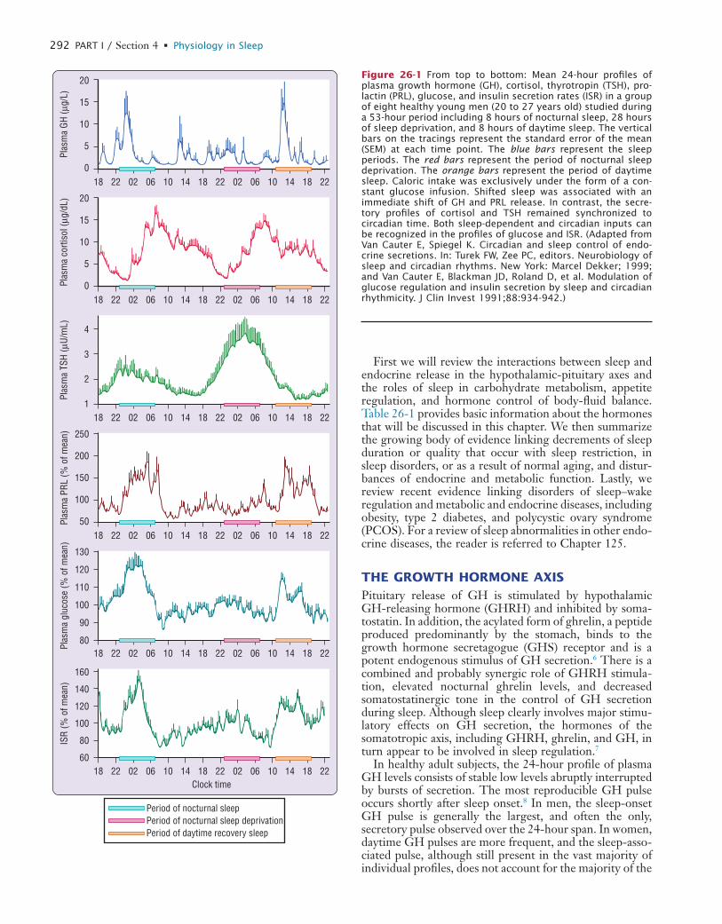
L
292 PART I / Section 4 • Physiology in Sleep
20
15
10
5
006 1018 0222 14 2218
Plas
ma
GH (µ
g/L)
06 1002 14 2218
4
3
2
106 1018 0222 14 2218
Plas
ma
TSH
(µU/
mL)
06 1002 14 2218
250
200
150
100
5006 1018 0222 14 2218
Plas
ma
PRL
(% o
f mea
n)
06 1002 14 2218
130
120
110
100
90
8006 1018 0222 14 2218
Plas
ma
gluc
ose
(% o
f mea
n)
06 1002 14 2218
160
140
120
100
80
6006 1018 0222 14 22
Clock time
Period of nocturnal sleepPeriod of nocturnal sleep deprivationPeriod of daytime recovery sleep
18
ISR
(% o
f mea
n)
06 1002 14 2218
20
15
10
5
006 1018 0222 14 2218
Plas
ma
corti
sol (
µg/d
L)
06 1002 14 2218
Figure 26-1 From top to bottom: Mean 24-hour profiles of plasma growth hormone (GH), cortisol, thyrotropin (TSH), pro-lactin (PRL), glucose, and insulin secretion rates (ISR) in a group of eight healthy young men (20 to 27 years old) studied during a 53-hour period including 8 hours of nocturnal sleep, 28 hours of sleep deprivation, and 8 hours of daytime sleep. The vertical bars on the tracings represent the standard error of the mean (SEM) at each time point. The blue bars represent the sleep periods. The red bars represent the period of nocturnal sleep deprivation. The orange bars represent the period of daytime sleep. Caloric intake was exclusively under the form of a con-stant glucose infusion. Shifted sleep was associated with an immediate shift of GH and PRL release. In contrast, the secre-tory profiles of cortisol and TSH remained synchronized to circadian time. Both sleep-dependent and circadian inputs can be recognized in the profiles of glucose and ISR. (Adapted from Van Cauter E, Spiegel K. Circadian and sleep control of endo-crine secretions. In: Turek FW, Zee PC, editors. Neurobiology of sleep and circadian rhythms. New York: Marcel Dekker; 1999; and Van Cauter E, Blackman JD, Roland D, et al. Modulation of glucose regulation and insulin secretion by sleep and circadian rhythmicity. J Clin Invest 1991;88:934-942.)
First we will review the interactions between sleep and endocrine release in the hypothalamic-pituitary axes and the roles of sleep in carbohydrate metabolism, appetite regulation, and hormone control of body-fluid balance. Table 26-1 provides basic information about the hormones that will be discussed in this chapter. We then summarize the growing body of evidence linking decrements of sleep duration or quality that occur with sleep restriction, in sleep disorders, or as a result of normal aging, and distur-bances of endocrine and metabolic function. Lastly, we review recent evidence linking disorders of sleep–wake regulation and metabolic and endocrine diseases, including obesity, type 2 diabetes, and polycystic ovary syndrome (PCOS). For a review of sleep abnormalities in other endo-crine diseases, the reader is referred to Chapter 125.
THE GROWTH HORMONE AXISPituitary release of GH is stimulated by hypothalamic GH-releasing hormone (GHRH) and inhibited by soma-tostatin. In addition, the acylated form of ghrelin, a peptide produced predominantly by the stomach, binds to the growth hormone secretagogue (GHS) receptor and is a potent endogenous stimulus of GH secretion.6 There is a combined and probably synergic role of GHRH stimula-tion, elevated nocturnal ghrelin levels, and decreased somatostatinergic tone in the control of GH secretion during sleep. Although sleep clearly involves major stimu-latory effects on GH secretion, the hormones of the somatotropic axis, including GHRH, ghrelin, and GH, in turn appear to be involved in sleep regulation.7
In healthy adult subjects, the 24-hour profile of plasma GH levels consists of stable low levels abruptly interrupted by bursts of secretion. The most reproducible GH pulse occurs shortly after sleep onset.8 In men, the sleep-onset GH pulse is generally the largest, and often the only, secretory pulse observed over the 24-hour span. In women, daytime GH pulses are more frequent, and the sleep-asso-ciated pulse, although still present in the vast majority of individual profiles, does not account for the majority of the
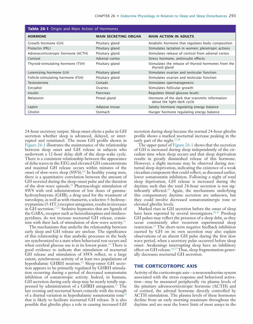
L
CHAPTER 26 • Endocrine Physiology in Relation to Sleep and Sleep Disturbances 293
secretion during sleep because the normal 24-hour ghrelin profile shows a marked nocturnal increase peaking in the early part of the night.17,18
The upper panel of Figure 26-1 shows that the secretion of GH is increased during sleep independently of the cir-cadian time when sleep occurs and that sleep deprivation results in greatly diminished release of this hormone. However, a slight increase may be observed during noc-turnal sleep deprivation, indicating the existence of a weak circadian component that could reflect, as discussed earlier, lower somatostatin inhibition. Following a night of total sleep deprivation, GH release is increased during the daytime such that the total 24-hour secretion is not sig-nificantly affected.19 Again, the mechanisms underlying this compensatory daytime secretion are unknown, but they could involve decreased somatostatinergic tone or elevated ghrelin levels.
Marked rises in GH secretion before the onset of sleep have been reported by several investigators.20-22 Presleep GH pulses may reflect the presence of a sleep debt, as they occur consistently after recurrent experimental sleep restriction.23 The short-term negative feedback inhibition exerted by GH on its own secretion may also explain observations of an absent GH pulse during the first slow wave period, when a secretory pulse occurred before sleep onset. Awakenings interrupting sleep have an inhibitory effect on GH release.24,25 Thus, sleep fragmentation gener-ally decreases nocturnal GH secretion.
THE CORTICOTROPIC AXISActivity of the corticotropic axis—a neuroendocrine system associated with the stress response and behavioral activa-tion—may be measured peripherally via plasma levels of the pituitary adrenocorticotropic hormone (ACTH) and of cortisol, the adrenal hormone directly controlled by ACTH stimulation. The plasma levels of these hormones decline from an early morning maximum throughout the daytime and are near the lower limit of most assays in the
24-hour secretory output. Sleep onset elicits a pulse in GH secretion whether sleep is advanced, delayed, or inter-rupted and reinitiated. The mean GH profile shown in Figure 26-1 illustrates the maintenance of the relationship between sleep onset and GH release in subjects who underwent a 12-hour delay shift of the sleep–wake cycle. There is a consistent relationship between the appearance of delta waves in the EEG and elevated GH concentrations and maximal GH release occurs within minutes of the onset of slow-wave sleep (SWS).8,9 In healthy young men, there is a quantitative correlation between the amount of GH secreted during the sleep-onset pulse and the duration of the slow-wave episode.10 Pharmacologic stimulation of SWS with oral administration of low doses of gamma-hydroxybutyrate (GHB), a drug used for the treatment of narcolepsy, as well as with ritanserin, a selective 5-hydroxy-tryptamine (5-HT2) receptor antagonist, results in increases in GH secretion.11,12 Sedative hypnotics that are ligands of the GABAA receptor such as benzodiazepines and imidazo-pyridines, do not increase nocturnal GH release, consis-tent with their lack of stimulation of slow-wave activity.13
The mechanisms that underlie the relationship between early sleep and GH release are unclear. The significance of this relationship is that anabolic processes in the body are synchronized to a state when behavioral rest occurs and when cerebral glucose use is at its lowest point.14 There is good evidence to indicate that stimulation of nocturnal GH release and stimulation of SWS reflect, to a large extent, synchronous activity of at least two populations of hypothalamic GHRH neurons.14 Sleep-onset GH secre-tion appears to be primarily regulated by GHRH stimula-tion occurring during a period of decreased somatostatin inhibition of somatotropic activity. Indeed, in humans, GH secretion during early sleep may be nearly totally sup-pressed by administration of a GHRH antagonist.15 The late evening and nocturnal hours coincide with the trough of a diurnal variation in hypothalamic somatostatin tone16 that is likely to facilitate nocturnal GH release. It is also possible that ghrelin plays a role in causing increased GH
Table 26-1 Origin and Main Action of Hormones
HORMONE MAIN SECRETING ORGAN MAIN ACTION IN ADULTS
Growth hormone (GH) Pituitary gland Anabolic hormone that regulates body composition
Prolactin (PRL) Pituitary gland Stimulates lactation in women; pleiotropic actions
Adrenocorticotropic hormone (ACTH) Pituitary gland Stimulates release of cortisol from adrenal cortex
Cortisol Adrenal cortex Stress hormone, antiinsulin effects
Thyroid-stimulating hormone (TSH) Pituitary gland Stimulates the release of thyroid hormones from the thyroid gland
Luteinizing hormone (LH) Pituitary gland Stimulates ovarian and testicular function
Follicle-stimulating hormone (FSH) Pituitary gland Stimulates ovarian and testicular function
Testosterone Gonads Stimulates spermatogenesis
Estradiol Ovaries Stimulates follicular growth
Insulin Pancreas Regulates blood glucose levels
Melatonin Pineal gland Hormone of the dark that transmits information about the light–dark cycle
Leptin Adipose tissue Satiety hormone regulating energy balance
Ghrelin Stomach Hunger hormone regulating energy balance

L
294 PART I / Section 4 • Physiology in Sleep
secretion may be seen during sleep deprivation (clearly seen in Fig. 26-1), when nocturnal TSH secretion is increased by as much as 200% over the levels observed during nocturnal sleep. Thus, sleep exerts an inhibitory influence on TSH secretions, and sleep deprivation relieves this inhibition.30,34
Interestingly, when sleep occurs during daytime hours, TSH secretion is not suppressed significantly below normal daytime levels. Thus, the inhibitory effect of sleep on TSH secretion appears to be operative when the nighttime ele-vation has taken place, indicating once again the interac-tion of the effects of circadian time and the effects of sleep. When the depth of sleep at the habitual time is increased by prior sleep deprivation, the nocturnal TSH rise is more markedly inhibited, suggesting that SWS is probably the primary determinant of the sleep-associated fall.30 Awak-enings interrupting nocturnal sleep appear to relieve the inhibition of TSH and are consistently associated with a short-term TSH elevation.
Circadian and sleep-related variations in thyroid hor-mones have been difficult to demonstrate, probably because these hormones are bound to serum proteins and thus their peripheral concentrations are affected by diurnal variations in hemodilution caused by postural changes. However, under conditions of sleep deprivation, the increased ampli-tude of the TSH rhythm may result in a detectable increase in plasma triiodothyronine (T3) levels, paralleling the noc-turnal TSH rise.35 If sleep deprivation is continued for a second night, then the nocturnal rise of TSH is markedly diminished as compared with that occurring during the first night.35,36 It is likely that following the first night of sleep deprivation, the elevated thyroid hormone levels, which persist during the daytime period because of the prolonged half-life of these hormones, limit the subse-quent TSH rise at the beginning of the next nighttime period. Data from a study of 64 hours of sleep deprivation suggest that prolonged sleep loss may be associated with an upregulation of the thyroid axis, with lower levels of TSH and higher levels of thyroid hormones.37 As discussed in the section on chronic sleep restriction, this was indeed the case in a study of the endocrine and metabolic effects of a sleep debt resulting from bedtime curtailment to 4 hours per night for 6 nights.38,39
The inhibitory effects of sleep on TSH secretion are time dependent, and that may cause, under specific cir-cumstances, elevations of plasma TSH levels that reflect the misalignment of sleep and central circadian timing. In a study examining the course of adaptation to an abrupt 8-hour advance of the sleep–dark period in healthy young men,40 TSH levels increased progressively because daytime sleep failed to inhibit TSH and nighttime wakefulness was associated with large circadian-dependent TSH elevations. As a result, mean TSH levels following awakening from the second shifted sleep period were more than twofold higher than during the same time interval following normal nocturnal sleep. This overall elevation of TSH levels was paralleled by a small increase in T3 concentrations.40 This study demonstrated that the subjective discomfort and fatigue often referred to as “jet-lag syndrome” are associ-ated not only with a desynchronization of bodily rhythms but also with a prolonged elevation of a hormone concen-tration in the peripheral circulation.
late evening and early part of the sleep period. Thus, sleep is normally initiated when corticotropic activity is quies-cent. Reactivation of ACTH and cortisol secretion occurs abruptly a few hours before the usual waking time.
The mean cortisol profile shown in Figure 26-1 illus-trates the remarkable persistence of this diurnal variation when sleep is manipulated. Indeed, the overall waveshape of the profile is not markedly affected by the absence of sleep or by sleep at an unusual time of day. Thus, the 24-hour periodicity of corticotropic activity is primarily controlled by circadian rhythmicity.
Nevertheless, modulatory effects of sleep or wake have been clearly demonstrated. Indeed, a number of studies have indicated that sleep onset is reliably associated with a short-term inhibition of cortisol secretion,5,26 although this effect may not be detectable when sleep is initiated at the time of the daily maximum of corticotropic activity, that is, in the morning.27 Under normal conditions, because cortisol secretion is already quiescent in the late evening, this inhibitory effect of sleep, which is temporally associ-ated with the occurrence of slow-wave sleep,28-30 results in a prolongation of the quiescent period. Therefore, under conditions of sleep deprivation, the nadir of cortisol secre-tion is less pronounced and occurs earlier than under normal conditions of nocturnal sleep. Conversely, awaken-ing at the end of the sleep period is consistently followed by a pulse of cortisol secretion.5,25,31
During sleep deprivation, the rapid effects of sleep onset and sleep offset on corticotropic activity are obviously absent, and, as may be seen in the profiles shown in Figure 26-1, the nadir of cortisol levels is higher than during nocturnal sleep (because of the absence of the inhibitory effects of the first hours of sleep), and the morning maximum is lower (because of the absence of the stimulat-ing effects of morning awakening). Overall, the amplitude of the rhythm is reduced by approximately 15% during sleep deprivation as compared to normal conditions.
In addition to the immediate modulatory effects of sleep–wake transitions on cortisol levels, nocturnal sleep deprivation, even partial deprivation, results in an eleva-tion of cortisol levels on the following evening.32 Sleep loss thus appears to delay the normal return to evening quies-cence of the corticotropic axis. This endocrine alteration is remarkably similar to that occurring in normal aging, where increases in evening cortisol levels of similar mag-nitude are consistently observed. This interpretation is consistent with findings in normal subjects submitted to recurrent partial sleep restriction, as discussed later.
THE THYROID AXISDaytime levels of plasma TSH are low and relatively stable and are followed by a rapid elevation starting in the early evening and culminating in a nocturnal maximum occur-ring around the beginning of the sleep period.30,33 The later part of sleep is associated with a progressive decline in TSH levels, and daytime values resume shortly after morning awakening. The first 24 hours of the study illus-trated in Figure 26-1 are typical of the diurnal TSH rhythm. Because the nocturnal rise of TSH occurs well before the time of sleep onset, it probably reflects a circa-dian effect. However, a marked effect of sleep on TSH
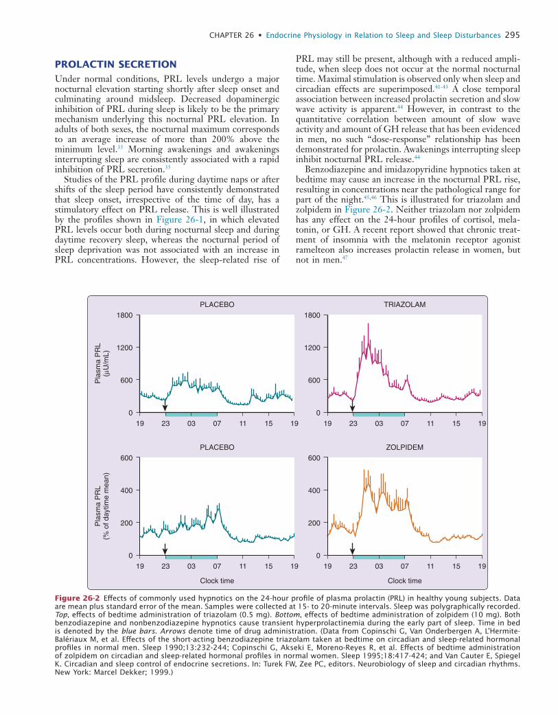
L
CHAPTER 26 • Endocrine Physiology in Relation to Sleep and Sleep Disturbances 295
PRL may still be present, although with a reduced ampli-tude, when sleep does not occur at the normal nocturnal time. Maximal stimulation is observed only when sleep and circadian effects are superimposed.41-43 A close temporal association between increased prolactin secretion and slow wave activity is apparent.44 However, in contrast to the quantitative correlation between amount of slow wave activity and amount of GH release that has been evidenced in men, no such “dose-response” relationship has been demonstrated for prolactin. Awakenings interrupting sleep inhibit nocturnal PRL release.44
Benzodiazepine and imidazopyridine hypnotics taken at bedtime may cause an increase in the nocturnal PRL rise, resulting in concentrations near the pathological range for part of the night.45,46 This is illustrated for triazolam and zolpidem in Figure 26-2. Neither triazolam nor zolpidem has any effect on the 24-hour profiles of cortisol, mela-tonin, or GH. A recent report showed that chronic treat-ment of insomnia with the melatonin receptor agonist ramelteon also increases prolactin release in women, but not in men.47
PROLACTIN SECRETION
Under normal conditions, PRL levels undergo a major nocturnal elevation starting shortly after sleep onset and culminating around midsleep. Decreased dopaminergic inhibition of PRL during sleep is likely to be the primary mechanism underlying this nocturnal PRL elevation. In adults of both sexes, the nocturnal maximum corresponds to an average increase of more than 200% above the minimum level.35 Morning awakenings and awakenings interrupting sleep are consistently associated with a rapid inhibition of PRL secretion.35
Studies of the PRL profile during daytime naps or after shifts of the sleep period have consistently demonstrated that sleep onset, irrespective of the time of day, has a stimulatory effect on PRL release. This is well illustrated by the profiles shown in Figure 26-1, in which elevated PRL levels occur both during nocturnal sleep and during daytime recovery sleep, whereas the nocturnal period of sleep deprivation was not associated with an increase in PRL concentrations. However, the sleep-related rise of
PLACEBO
1800
1200
600
0
Pla
sma
PR
L(µ
U/m
L)
19 23 03 07 11 15 19
TRIAZOLAM
1800
1200
600
0
19 23 03 07 11 15 19
PLACEBO
600
400
200
0
Pla
sma
PR
L(%
of d
aytim
e m
ean)
19 23 03 07 11 15 19
ZOLPIDEM
Clock time Clock time
600
400
200
0
19 23 03 07 11 15 19
Figure 26-2 Effects of commonly used hypnotics on the 24-hour profile of plasma prolactin (PRL) in healthy young subjects. Data are mean plus standard error of the mean. Samples were collected at 15- to 20-minute intervals. Sleep was polygraphically recorded. Top, effects of bedtime administration of triazolam (0.5 mg). Bottom, effects of bedtime administration of zolpidem (10 mg). Both benzodiazepine and nonbenzodiazepine hypnotics cause transient hyperprolactinemia during the early part of sleep. Time in bed is denoted by the blue bars. Arrows denote time of drug administration. (Data from Copinschi G, Van Onderbergen A, L’Hermite-Balériaux M, et al. Effects of the short-acting benzodiazepine triazolam taken at bedtime on circadian and sleep-related hormonal profiles in normal men. Sleep 1990;13:232-244; Copinschi G, Akseki E, Moreno-Reyes R, et al. Effects of bedtime administration of zolpidem on circadian and sleep-related hormonal profiles in normal women. Sleep 1995;18:417-424; and Van Cauter E, Spiegel K. Circadian and sleep control of endocrine secretions. In: Turek FW, Zee PC, editors. Neurobiology of sleep and circadian rhythms. New York: Marcel Dekker; 1999.)
L
296 PART I / Section 4 • Physiology in Sleep
(GnRH) release. Awakenings interrupting sleep are usually associated with a pulse of LH concentration.60 In the midfollicular phase, pulse amplitude is decreased, pulse frequency is increased, and the frequency modulation of LH pulsatility by sleep is less apparent. Pulse amplitude increases again by the late-follicular phase. In the early luteal phase, the pulse amplitude is markedly increased, the pulse frequency is decreased, and nocturnal slowing of pulsatility is again evident. In the mid- and late-luteal phase, pulse amplitude and frequency are decreased, and there is no modulation by sleep.
In older men, the amplitude of LH pulses is decreased but the frequency is increased and no significant diurnal pattern can be detected.61-63 The circadian variation of testosterone persists, although markedly dampened.63 The sleep-related rise is still apparent in older men, but its amplitude is lower and the relationship to REM latency is no longer apparent.64 It is likely that decreased sleep quality as occurs in aging as well as in sleep disorders (e.g., obstruc-tive sleep apnea) plays a role in the dampening of the sleep-related testosterone rise.
In postmenopausal women, gonadotropin levels are elevated, but they show no consistent circadian pattern.65 A number of studies66-68 have indicated that estrogen replacement therapy has modest beneficial effects on sub-jective and objective sleep quality, particularly in the pres-ence of environmental disturbance69 or sleep-disordered breathing.66,67,70
GLUCOSE REGULATIONThe consolidation of human sleep in a single 7- to 9-hour period implies that an extended period of fast must be maintained overnight. Despite the prolonged fasting con-dition, glucose levels remain stable or fall only minimally across the night. In contrast, if subjects are awake and fasting in a recumbent position, in the absence of any physical activity, glucose levels fall by an average of 0.5 to 1.0 mmol/L (± 10 to 20 mg/dL) over a 12-hour period.71 Thus, a number of mechanisms that operate during noc-turnal sleep must intervene to maintain stable glucose levels during the overnight fast.
The lower panels of Figure 26-1 show profiles of blood glucose and ISR obtained in normal subjects who were studied under conditions of constant glucose infusion,76 a condition that results in a marked inhibition of endoge-nous glucose production. Thus, when subjects receive a constant glucose infusion, changes in plasma glucose levels reflect mainly changes in glucose use. A marked decrease in glucose tolerance (reflected in higher plasma glucose levels) is apparent during nighttime as well as daytime sleep. A smaller elevation of glucose and insulin also occurs during nocturnal sleep deprivation, indicating an effect of circadian-dependent mechanisms.
During nocturnal sleep, the overall increase in plasma glucose ranged from 20% to 30%, despite the maintenance of rigorously constant rates of caloric intake. Maximal levels occur around the middle of the sleep period. During the later part of the night (i.e., at the time of the so-called dawn phenomenon), glucose tolerance begins to improve, and glucose levels progressively decrease toward morning values. The mechanisms underlying these robust variations
There is evidence from animal studies that PRL is involved in the humoral regulation of REM sleep.48 The primary effect is a stimulation of REM sleep, which appears to be dependent on time of day. Recent findings indicate that prolactin deficient mice have decreased REM sleep.49
THE GONADAL AXISThe relationship between the 24-hour patterns of gonado-tropin release and gonadal steroid levels varies according to the stage of maturation, and it is gender dependent in young adulthood (for review see Van Cauter et al.35).
Prior to puberty, luteinizing hormone (LH) and follicle-stimulating hormone are secreted in a pulsatile pattern, and an augmentation of pulsatile activity is associated with sleep onset in a majority of both girls and boys. The increased amplitude of gonadotropin release during sleep is one of the hallmarks of puberty. Both sleep and circadian rhythmicity contribute to the nocturnal elevation of gonadotropin pulses in pubertal children. As the pubescent child enters adulthood, the daytime pulse amplitude increases as well, eliminating or diminishing the diurnal rhythm. In pubertal girls, a diurnal variation of circulating estradiol levels, with higher concentrations during the daytime instead of the nighttime, becomes apparent. It has been suggested that the lack of parallelism between gonadotropin and estradiol levels reflects an approximate 10-hour delay between gonadotropin stimulation and the subsequent ovarian response. In pubertal boys, a nocturnal rise of testosterone coincides with the elevation of gonadotropins.
In adult men, the day–night variation of plasma LH levels is dampened or even undetectable. During the sleep period, LH pulses appear to be temporally related to the REM–NREM cycle.50 Despite the low amplitude of the nocturnal increase in gonadotropin release, a marked diurnal rhythm in circulating testosterone levels is present, with minimal levels in the late evening, a robust rise fol-lowing sleep onset and maximal levels in the early morning.51,52 Thus, the robust circadian rhythm of plasma testosterone may be partially controlled by factors other than LH. The nocturnal rise of testosterone appears tem-porally linked to the latency of the first REM episode,53 as plasma levels continue to rise until the first REM episode occurs. A robust rise of testosterone may also be observed during daytime sleep, suggesting that sleep, irrespective of time of day, stimulates gonadal hormone release.54 Experi-mental sleep fragmentation in young men resulted in attenuation of the nocturnal rise of testosterone, particu-larly in subjects who did not achieve REM sleep.55 Andro-gen concentrations in young adults decline significantly during periods of total sleep deprivation and recover promptly once the sleep is restored.54,56 In contrast, phar-macological suppression of testosterone in healthy men appears to have no effect on the total amount and overall architecture of nighttime sleep.57
In women, the 24-hour variation in plasma LH is markedly modulated by the menstrual cycle.58,59 In the early follicular phase, LH pulses are large and infrequent, and a marked slowing of the frequency of secretory pulses occurs during sleep, suggestive of inhibitory effect of sleep on pulsatile gonadotropin-releasing hormone
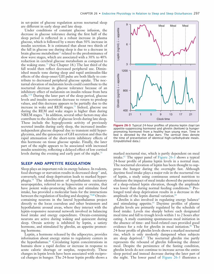
L
CHAPTER 26 • Endocrine Physiology in Relation to Sleep and Sleep Disturbances 297
marked nocturnal rise, which is partly dependent on meal intake.82 The upper panel of Figure 26-3 shows a typical 24-hour profile of plasma leptin levels in a normal man. The nocturnal elevation of leptin has been thought to sup-press the hunger during the overnight fast. Although daytime food intake plays a major role in the nocturnal rise of leptin, a study using continuous enteral nutrition to eliminate the impact of meal intake showed the persistence of a sleep-related leptin elevation, though the amplitude was lower than during normal feeding conditions.83 Pro-longed total sleep deprivation results in a decrease in the amplitude of the leptin diurnal variation.84
Ghrelin is also involved in regulating energy balance6 and stimulating appetite.85 Daytime profiles of plasma ghrelin levels are primarily regulated by the schedule of food intake: Levels rise sharply before each designated meal time and fall to trough levels within 1 to 2 hours after eating. A study examining spontaneous meal initiation in the absence of time- and food-related cues provided good evidence for a role for ghrelin in meal initiation.86 The 24-hour profile of ghrelin levels shows a marked nocturnal rise, which is only modestly dampened when subjects are sleep deprived.17 The nocturnal ghrelin rise partly represents the rebound of ghrelin following the dinner meal. Despite the persistence of the fasting condition, ghrelin levels do not continue to increase across the entire sleep period and instead decrease during the later part of the night. The lower panel of Figure 26-3 illustrates a
in set-point of glucose regulation across nocturnal sleep are different in early sleep and late sleep.
Under conditions of constant glucose infusion, the decrease in glucose tolerance during the first half of the sleep period is reflected in a robust increase in plasma glucose, which is followed by a more than 50% increase in insulin secretion. It is estimated that about two thirds of the fall in glucose use during sleep is due to a decrease in brain glucose metabolism72 related to the predominance of slow wave stages, which are associated with a 30% to 40% reduction in cerebral glucose metabolism as compared to the waking state.73 (See Chapter 18.) The last third of the fall would then reflect decreased peripheral use. Dimin-ished muscle tone during sleep and rapid antiinsulin-like effects of the sleep-onset GH pulse are both likely to con-tribute to decreased peripheral glucose uptake. The noc-turnal elevation of melatonin levels could contribute to the nocturnal decrease in glucose tolerance because of an inhibitory effect of melatonin on insulin release from beta cells.2,74 During the later part of the sleep period, glucose levels and insulin secretion decrease to return to presleep values, and this decrease appears to be partially due to the increase in wake and REM stages.75 Indeed, glucose use during the REM and wake stages is higher than during NREM stages.73 In addition, several other factors may also contribute to the decline of glucose levels during late sleep. These include the hypoglycemic activity of previously secreted insulin during early sleep, the increased insulin-independent glucose disposal due to transient mild hyper-glycemia, and the quiescence of GH secretion and thus the rapid attenuation of the short-term inhibitory effects of this hormone on tissue glucose uptake. Finally, the later part of the night appears to be associated with increased insulin sensitivity, reflecting a delayed effect of low cortisol levels during the evening and early part of the night.76
SLEEP AND APPETITE REGULATIONSleep plays an important role in energy balance. In rodents, food shortage or starvation results in decreased sleep77 and, conversely, total sleep deprivation leads to marked hyper-phagia.78 The identification of hypothalamic excitatory neuropeptides, referred to as hypocretins or orexins, that have potent wake-promoting effects and stimulate food intake, has provided a molecular basis for the interactions between the regulation of feeding and sleeping.79,80 Orexin-containing neurons in the lateral hypothalamus project directly to the locus coeruleus and other brainstem and hypothalamic arousal areas, where they interact with the leptin-responsive neuronal network involved in balancing food intake and energy expenditure. Orexin-containing neurons are active during waking and quiescent during sleep. Orexin activity is inhibited by leptin, a satiety hormone, and stimulated by ghrelin, an appetite promot-ing hormone.
Leptin, a hormone released by the adipocytes, provides information about energy status to regulatory centers in the hypothalamus.81 Circulating leptin concentrations in humans show a rapid decline or increase in response to acute caloric shortage or surplus, respectively. These changes in leptin levels have been associated with recipro-cal changes in hunger. The 24-hour leptin profile shows a
Lept
in (
ng/m
l) ↓
– ap
petit
e
9
6
3
1200
800
400
Clock timeG
hrel
in (
pg/m
l) ↓
+ a
ppet
ite5 9 13 17 2121 1
Meal Meal Meal
Figure 26-3 Typical 24-hour profiles of plasma leptin (top) (an appetite-suppressing hormone) and ghrelin (bottom) (a hunger-promoting hormone) from a healthy lean young man. Time in bed is denoted by the blue bars. The vertical lines denote the time of presentation of identical high-carbohydrate meals. (Unpublished data.)
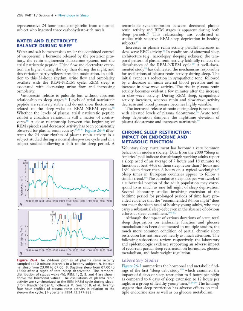
L
298 PART I / Section 4 • Physiology in Sleep
remarkable synchronization between decreased plasma renin activity and REM stages is apparent during both sleep periods.92 This relationship was confirmed in studies with selective REM-sleep deprivation in healthy subjects.93
Increases in plasma renin activity parallel increases in slow-wave EEG activity.94 In conditions of abnormal sleep architecture (e.g., narcolepsy, sleeping sickness), the tem-poral pattern of plasma renin activity faithfully reflects the disturbances of the REM–NREM cycle.87 A well-docu-mented study95 has delineated the mechanisms responsible for oscillations of plasma renin activity during sleep. The initial event is a reduction in sympathetic tone, followed by a decrease in mean arterial blood pressure and an increase in slow-wave activity. The rise in plasma renin activity becomes evident a few minutes after the increase in slow-wave activity. During REM sleep, sympathetic activity increases, whereas renin and slow-wave activity decrease and blood pressure becomes highly variable.
The increased release of renin during sleep is associated with elevated levels of plasma aldosterone.96 Acute total sleep deprivation dampens the nighttime elevation of plasma aldosterone and increases natriuresis.97
CHRONIC SLEEP RESTRICTION: IMPACT ON ENDOCRINE AND METABOLIC FUNCTIONVoluntary sleep curtailment has become a very common behavior in modern society. Data from the 2008 “Sleep in America” poll indicate that although working adults report a sleep need of an average of 7 hours and 18 minutes to function at best, 44% of them sleep fewer than 7 hours and 16% sleep fewer than 6 hours on a typical weeknight.98 Sleep times in European countries appear to follow a similar trend.99 The cumulative sleep loss per workweek of a substantial portion of the adult population may corre-spond to as much as one full night of sleep deprivation. Several laboratory studies involving extension of the bedtime period for prolonged periods of time have pro-vided evidence that the “recommended 8-hour night” does not meet the sleep need of healthy young adults, who may carry a substantial sleep debt even in the absence of obvious efforts at sleep curtailment.100-102
Although the impact of various durations of acute total sleep deprivation on endocrine function and glucose metabolism has been documented in multiple studies, the much more common condition of partial chronic sleep restriction has not received nearly as much attention. The following subsections review, respectively, the laboratory and epidemiologic evidence supporting an adverse impact of recurrent partial sleep restriction on hormones, glucose metabolism, and body weight regulation.
Laboratory StudiesFigure 26-5 summarizes the hormonal and metabolic find-ings of the first “sleep debt study”39 which examined the impact of 6 days of sleep restriction to 4 hours per night as compared to 6 days of sleep extension to 12 hours per night in a group of healthy young men.23,38,39 The findings suggest that sleep restriction has adverse effects on mul-tiple endocrine axes as well as on glucose metabolism.
representative 24-hour profile of ghrelin from a normal subject who ingested three carbohydrate-rich meals.
WATER AND ELECTROLYTE BALANCE DURING SLEEPWater and salt homeostasis is under the combined control of vasopressin, a hormone released by the posterior pitu-itary, the renin-angiotensin-aldosterone system, and the atrial natriuretic peptide. Urine flow and electrolyte excre-tion are higher during the day than during the night, and this variation partly reflects circadian modulation. In addi-tion to this 24-hour rhythm, urine flow and osmolarity oscillate with the REM–NREM cycle. REM sleep is associated with decreasing urine flow and increasing osmolarity.
Vasopressin release is pulsatile but without apparent relationship to sleep stages.87 Levels of atrial natriuretic peptide are relatively stable and do not show fluctuations related to the sleep–wake or REM–NREM cycles.88 Whether the levels of plasma atrial natriuretic peptide exhibit a circadian variation is still a matter of contro-versy.88 A close relationship between the beginning of REM episodes and decreased activity has been consistently observed for plasma renin activity.87,89-91 Figure 26-4 illus-trates the 24-hour rhythm of plasma renin activity in a subject studied during a normal sleep–wake cycle and in a subject studied following a shift of the sleep period. A
Time
4
3
2
1
0
23:00 01:00 03:00 05:00 07:00 09:00 11:00 13:00 15:00 17:00 19:00 21:00 23:00
Time
4
3
2
1
0PRA
(ng
• mL–1
-hr–1
)PR
A (n
g • m
L–1-h
r–1)
23:00 01:00 03:00 05:00 07:00 09:00 11:00 13:00 15:00 17:00 19:00 21:00 23:00
WREM
1234
WREM
1234
A
B
Figure 26-4 The 24-hour profiles of plasma renin activity sampled at 10-minute intervals in a healthy subject. A, Noctur-nal sleep from 23:00 to 07:00. B, Daytime sleep from 07:00 to 15:00 after a night of total sleep deprivation. The temporal distribution of stages wake (W); REM; 1, 2, 3, and 4 are shown above the hormonal values. The oscillations of plasma renin activity are synchronized to the REM–NREM cycle during sleep. (From Brandenberger G, Follenius M, Goichot B, et al. Twenty-four hour profiles of plasma renin activity in relation to the sleep-wake cycle. J Hypertens 1994;12:277-283.)
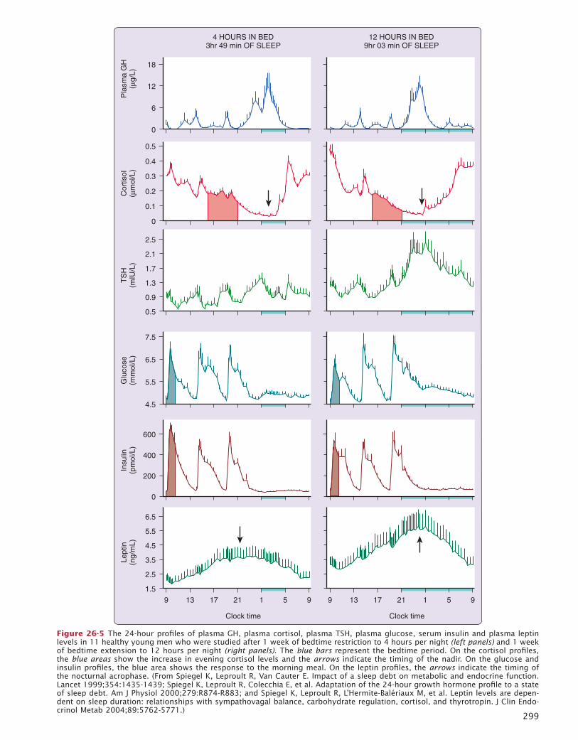
LL
299
4 HOURS IN BED3hr 49 min OF SLEEP
18
12
6
0P
lasm
a G
H(µ
g/L)
12 HOURS IN BED9hr 03 min OF SLEEP
6.5
3.5
2.5
4.5
5.5
1.5
Lept
in(n
g/m
L)
600
200
400
0
Insu
lin(p
mol
/L)
7.5
5.5
6.5
4.5
Glu
cose
(mm
ol/L
)
2.5
1.3
0.9
1.7
2.1
0.5
TS
H(m
IU/L
)
0.5
0.2
0.1
0.3
0.4
0
Cor
tisol
(µm
ol/L
)
9 13 17 21 1 5 9
Clock time
9 13 17 21 1 5 9
Clock time
Figure 26-5 The 24-hour profiles of plasma GH, plasma cortisol, plasma TSH, plasma glucose, serum insulin and plasma leptin levels in 11 healthy young men who were studied after 1 week of bedtime restriction to 4 hours per night (left panels) and 1 week of bedtime extension to 12 hours per night (right panels). The blue bars represent the bedtime period. On the cortisol profiles, the blue areas show the increase in evening cortisol levels and the arrows indicate the timing of the nadir. On the glucose and insulin profiles, the blue area shows the response to the morning meal. On the leptin profiles, the arrows indicate the timing of the nocturnal acrophase. (From Spiegel K, Leproult R, Van Cauter E. Impact of a sleep debt on metabolic and endocrine function. Lancet 1999;354:1435-1439; Spiegel K, Leproult R, Colecchia E, et al. Adaptation of the 24-hour growth hormone profile to a state of sleep debt. Am J Physiol 2000;279:R874-R883; and Spiegel K, Leproult R, L’Hermite-Balériaux M, et al. Leptin levels are depen-dent on sleep duration: relationships with sympathovagal balance, carbohydrate regulation, cortisol, and thyrotropin. J Clin Endo-crinol Metab 2004;89:5762-5771.)
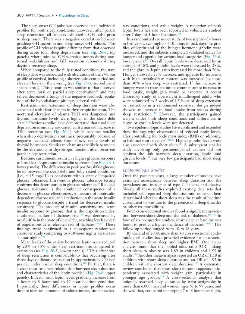
L
300 PART I / Section 4 • Physiology in Sleep
tary conditions, and stable weight. A reduction of peak leptin levels has also been reported in volunteers studied after 7 days of 4-hour bedtimes.108
In a randomized crossover study of two nights of 4 hours in bed versus two nights of 10 hours in bed, daytime pro-files of leptin and of the hunger hormone ghrelin were measured, and the subjects completed validated scales for hunger and appetite for various food categories (Fig. 26-6, lower panel).109 Overall leptin levels were decreased by an average of 18% and ghrelin levels were increased by 28%, and the ghrelin : leptin ratio increased by more than 70%. Hunger showed a 23% increase, and appetite for nutrients with high carbohydrate content was increased by more than 30% when sleep was restricted. If this increase in hunger were to translate into a commensurate increase in food intake, weight gain would be expected. A recent laboratory study of overweight middle-aged adults who were submitted to 2 weeks of 1.5 hour of sleep extension or restriction in a randomized crossover design indeed showed an increase in food intake from snacks during sleep restriction.110 However, the participants gained weight under both sleep conditions and differences in leptin or ghrelin levels were not detected.
Two epidemiologic studies have confirmed and extended these findings with observations of reduced leptin levels, after controlling for body mass index (BMI) or adiposity, in habitual short sleepers.111,112 Higher ghrelin levels were also associated with short sleep.111 A subsequent smaller study involving only postmenopausal women did not confirm the link between sleep duration, leptin, and ghrelin levels,113 but very few participants had short sleep durations.
Epidemiologic StudiesOver the past ten years, a large number of studies have examined associations between sleep duration and the prevalence and incidence of type 2 diabetes and obesity. Nearly all these studies explored existing data sets that included self reported sleep duration and none of them determined whether short sleep was the result of bedtime curtailment or was due to the presence of a sleep disorder or other co-morbidities.
Four cross-sectional studies found a significant associa-tion between short sleep and the risk of diabetes.114-117 In four of six prospective studies, short sleep at baseline was found to predict a higher incidence of diabetes.118-123 The follow-up period ranged from 10 to 18 years.
By the end of 2008, more than 40 cross-sectional epide-miological studies have provided evidence for an associa-tion between short sleep and higher BMI. One meta- analysis found that the pooled odds ratio (OR) linking short sleep to obesity was 1.89 in children and 1.55 in adults.124 Another meta-analysis reported an OR of 1.58 in children with short sleep duration and an OR of 1.92 in children with the shortest sleep duration.125 A systematic review concluded that short sleep duration appears inde-pendently associated with weight gain, particularly in younger age groups.126 A cross-sectional analysis that uniquely assessed sleep duration by wrist actigraphy in more than 6,000 men and women, ages 67 to 99 years, and showed that, compared to sleeping 7 to 8 hours per night,
The sleep-onset GH pulse was observed in all individual profiles for both sleep conditions. However, after partial sleep restriction, all subjects exhibited a GH pulse prior to sleep onset. There was a negative correlation between presleep GH secretion and sleep-onset GH release. This profile of GH release is quite different from that observed during acute total sleep deprivation (see Fig. 26-1, top panels), where minimal GH secretion occurs during noc-turnal wakefulness and GH secretion rebounds during daytime recovery sleep.
When compared to the fully rested condition, the state of sleep debt was associated with alterations of the 24-hour profile of cortisol, including a shorter quiescent period and elevated levels in the evening (see Fig. 26-5, second panel shaded areas). This alteration was similar to that observed after acute total or partial sleep deprivation32 and may reflect decreased efficacy of the negative feedback regula-tion of the hypothalamic-pituitary-adrenal axis.39
Restriction and extension of sleep duration were also associated with clear changes in thyrotropic function. The nocturnal elevation of plasma TSH was dampened and thyroid hormone levels were higher in the sleep debt state.39 Previous studies have demonstrated that total sleep deprivation is initially associated with a marked increase in TSH secretion (see Fig. 26-1), which becomes smaller when sleep deprivation continues, presumably because of negative feedback effects from slowly rising levels of thyroid hormones. Similar mechanisms are likely to under-lie the alterations in thyrotropic function after recurrent partial sleep restriction.
Bedtime curtailment results in a higher glucose response to breakfast despite similar insulin secretion (see Fig. 26-5, lower panels). The difference in peak postbreakfast glucose levels between the sleep debt and fully rested conditions (i.e., ± 15 mg/dL) is consistent with a state of impaired glucose tolerance. Intravenous glucose tolerance testing confirms this deterioration in glucose tolerance.39 Reduced glucose tolerance is the combined consequence of a decrease in glucose effectiveness, a measure of noninsulin dependent glucose use, and a reduction in the acute insulin response to glucose despite a trend for decreased insulin sensitivity. The product of insulin sensitivity and acute insulin response to glucose, that is, the disposition index, a validated marker of diabetes risk,103 was decreased by nearly 40% in the state of sleep debt, reaching levels typical of populations at an elevated risk of diabetes.104,105 These findings were confirmed in a subsequent randomized crossover study comparing two 10-hour nights versus two 4-hour nights.106
Mean levels of the satiety hormone leptin were reduced by 20% to 30% under sleep restriction as compared to extension (see Fig. 26-5, lowest panels).38 This effect size of sleep restriction is comparable to that occurring after three days of dietary restriction by approximately 900 kcal per day under normal sleep conditions.107 Further, there is a clear dose-response relationship between sleep duration and characteristics of the leptin profile38 (Fig. 26-6, upper panels). Indeed, mean leptin levels gradually increase from 4 hours to 8 hours and to 12-hour bedtime condition. Importantly, these differences in leptin profiles occur despite identical amounts of caloric intake, similar seden-
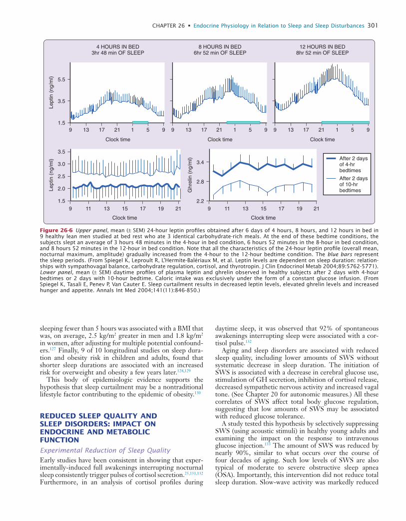
L
CHAPTER 26 • Endocrine Physiology in Relation to Sleep and Sleep Disturbances 301
daytime sleep, it was observed that 92% of spontaneous awakenings interrupting sleep were associated with a cor-tisol pulse.132
Aging and sleep disorders are associated with reduced sleep quality, including lower amounts of SWS without systematic decrease in sleep duration. The initiation of SWS is associated with a decrease in cerebral glucose use, stimulation of GH secretion, inhibition of cortisol release, decreased sympathetic nervous activity and increased vagal tone. (See Chapter 20 for autonomic measures.) All these correlates of SWS affect total body glucose regulation, suggesting that low amounts of SWS may be associated with reduced glucose tolerance.
A study tested this hypothesis by selectively suppressing SWS (using acoustic stimuli) in healthy young adults and examining the impact on the response to intravenous glucose injection.133 The amount of SWS was reduced by nearly 90%, similar to what occurs over the course of four decades of aging. Such low levels of SWS are also typical of moderate to severe obstructive sleep apnea (OSA). Importantly, this intervention did not reduce total sleep duration. Slow-wave activity was markedly reduced
sleeping fewer than 5 hours was associated with a BMI that was, on average, 2.5 kg/m2 greater in men and 1.8 kg/m2 in women, after adjusting for multiple potential confound-ers.127 Finally, 9 of 10 longitudinal studies on sleep dura-tion and obesity risk in children and adults, found that shorter sleep durations are associated with an increased risk for overweight and obesity a few years later.128,129
This body of epidemiologic evidence supports the hypothesis that sleep curtailment may be a nontraditional lifestyle factor contributing to the epidemic of obesity.130
REDUCED SLEEP QUALITY AND SLEEP DISORDERS: IMPACT ON ENDOCRINE AND METABOLIC FUNCTION
Experimental Reduction of Sleep QualityEarly studies have been consistent in showing that exper-imentally-induced full awakenings interrupting nocturnal sleep consistently trigger pulses of cortisol secretion.25,131,132 Furthermore, in an analysis of cortisol profiles during
Lept
in (
ng/m
l) 5.5
3.5
1.5
Clock time
Lept
in (
ng/m
l)
3.5
3.0
2.5
2.0
1.5
Clock time
13 15 17 19 219 11
Ghr
elin
(ng
/ml)
3.4
2.8
2.2
Clock time
13 15 17 19 219 11
179 13 5 921 1
4 HOURS IN BED3hr 48 min OF SLEEP
Clock time
179 13 5 921 1
8 HOURS IN BED6hr 52 min OF SLEEP
Clock time
179 13 5 921 1
12 HOURS IN BED8hr 52 min OF SLEEP
After 2 daysof 4-hrbedtimes
After 2 daysof 10-hrbedtimes
Figure 26-6 Upper panel, mean (± SEM) 24-hour leptin profiles obtained after 6 days of 4 hours, 8 hours, and 12 hours in bed in 9 healthy lean men studied at bed rest who ate 3 identical carbohydrate-rich meals. At the end of these bedtime conditions, the subjects slept an average of 3 hours 48 minutes in the 4-hour in bed condition, 6 hours 52 minutes in the 8-hour in bed condition, and 8 hours 52 minutes in the 12-hour in bed condition. Note that all the characteristics of the 24-hour leptin profile (overall mean, nocturnal maximum, amplitude) gradually increased from the 4-hour to the 12-hour bedtime condition. The blue bars represent the sleep periods. (From Spiegel K, Leproult R, L’Hermite-Balériaux M, et al. Leptin levels are dependent on sleep duration: relation-ships with sympathovagal balance, carbohydrate regulation, cortisol, and thyrotropin. J Clin Endocrinol Metab 2004;89:5762-5771). Lower panel, mean (± SEM) daytime profiles of plasma leptin and ghrelin observed in healthy subjects after 2 days with 4-hour bedtimes or 2 days with 10-hour bedtime. Caloric intake was exclusively under the form of a constant glucose infusion. (From Spiegel K, Tasali E, Penev P, Van Cauter E. Sleep curtailment results in decreased leptin levels, elevated ghrelin levels and increased hunger and appetite. Annals Int Med 2004;141(11):846-850.)
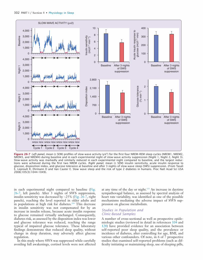
L
302 PART I / Section 4 • Physiology in Sleep
at any time of the day or night.133 An increase in daytime sympathovagal balance, as assessed by spectral analysis of heart rate variability, was identified as one of the possible mechanisms mediating the adverse impact of SWS sup-pression on glucose metabolism.
Studies in Population and Clinic-Based SamplesA number of cross-sectional as well as prospective epide-miologic studies (reviewed in detail in references 104 and 128) have provided evidence for an association between self-reported poor sleep quality, and the prevalence or incidence of diabetes, after controlling for age, BMI, and various other confounders. Of note, in 6 of 7 prospective studies that examined self-reported problems (such as dif-ficulty initiating or maintaining sleep, use of sleeping pills,
in each experimental night compared to baseline (Fig. 26-7, left panels). After 3 nights of SWS suppression, insulin sensitivity was decreased by ∼25% (Fig. 26-7, right panels), reaching the level reported in older adults and in populations at high risk for diabetes.134 This decrease in insulin sensitivity was not compensated for by an increase in insulin release, because acute insulin response to glucose remained virtually unchanged. Consequently, diabetes risk, as assessed by the disposition index was lower and glucose tolerance was reduced, reaching the range typical of impaired glucose tolerance. These laboratory findings demonstrate that reduced sleep quality, without change in sleep duration, may adversely affect glucose regulation.
In this study where SWS was suppressed while carefully avoiding full awakenings, cortisol levels were not affected
Bas
elin
e
Insu
lin s
ensi
tivity
(m
U/I–
1 ⋅m
in–1
)
4,000
3,000
2,000
1,000
0
NREM REM
Cycle 1 Cycle 2 Cycle 3 Cycle 4
NREM REM NREM REM NREM REM
SLOW-WAVE ACTIVITY (µv2)
After 3 nightsof SWS
suppression
Baseline
Nig
ht 1
4,000
3,000
2,000
1,000
0
Nig
ht 2
4,000
3,000
2,000
1,000
0
Nig
ht 3
4,000
3,000
2,000
1,000
0
10
8
6
4
2
0
Acu
te in
sulin
res
pons
e to
gluc
ose
(mU
/I–1 ⋅
min
)
After 3 nightsof SWS
suppression
Baseline
400
300
200
100
0
Dis
posi
tion
inde
x
After 3 nightsof SWS
suppression
Baseline
2,800
2,100
1,400
700
0
Glu
cose
tole
ranc
e(%
⋅min
–1)
After 3 nightsof SWS
suppression
Baseline
2
1
0
Figure 26-7 Left panel, mean (± SEM) profiles of slow-wave activity (µV2) for the first four NREM–REM sleep cycles (NREM1, NREM2, NREM3, and NREM4) during baseline and in each experimental night of slow-wave activity suppression (Night 1, Night 2, Night 3). Slow-wave activity was markedly and similarly reduced in each experimental night compared to baseline, and the largest reduc-tions were achieved during the first two NREM cycles. Right panel, mean (± SEM) insulin sensitivity, acute insulin response to glucose, disposition index, and glucose tolerance at baseline and after 3 nights of slow-wave sleep (SWS) suppression. (From Tasali E, Leproult R, Ehrmann D and Van Cauter E. Slow wave sleep and the risk of type 2 diabetes in humans. Proc Natl Acad Sci USA 2008;105(3):1044-1049).
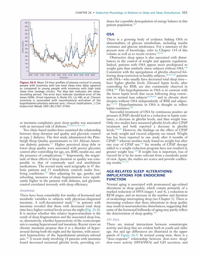
L
CHAPTER 26 • Endocrine Physiology in Relation to Sleep and Sleep Disturbances 303
dence for a possible dysregulation of energy balance in this patient population.141
OSAThere is a growing body of evidence linking OSA to abnormalities of glucose metabolism, including insulin resistance and glucose intolerance. For a summary of the present state of knowledge, refer to Chapter 114 of this volume as well as to recent reviews.142,143
Obstructive sleep apnea is also associated with distur-bances in the control of weight and appetite regulation. Indeed, patients with OSA appear more predisposed to weight gain than similarly obese subjects without OSA.144 Consistent with the upregulation of ghrelin observed fol-lowing sleep restriction in healthy subjects,109,111,145 patients with OSA—who usually have decreased total sleep time—have higher ghrelin levels. Elevated leptin levels, after controlling for BMI, are also consistently observed in OSA.144 This hyperleptinemia in OSA is in contrast with the lower leptin levels that occur following sleep restric-tion in normal lean subjects38,108,109 and in chronic short sleepers without OSA independently of BMI and adipos-ity.111,112 Hyperleptinemia in OSA is thought to reflect leptin resistance.144
Successful treatment of OSA by continuous positive air pressure (CPAP) should lead to a reduction in leptin resis-tance, a decrease in ghrelin levels, and thus weight loss. Only two studies have measured ghrelin levels after CPAP treatment and both reported a decrease in ghrelin levels.146,147 However, the findings on the effect of CPAP on body weight and visceral adiposity are mixed. Weight loss has been reported in one study after 6 months of CPAP,148 whereas another study found no weight loss after one year of CPAP use.149 Six months of CPAP therapy added to a weight reduction program have not resulted in greater weight loss.150 If weight loss is important, loss of visceral fat is by far more relevant from a metabolic point of view. Again, the studies are scarce and provide conflict-ing results.151-153
AGE-RELATED SLEEP ALTERATIONS: IMPLICATIONS FOR ENDOCRINE FUNCTIONNormal aging is associated with pronounced age-related alterations in sleep quality, which consist primarily of a marked reduction of SWS (stages 3 and 4), a reduction in REM stages, and an increase in the number and duration of awakenings interrupting sleep (see Chapter 3). There is increasing evidence that these alterations in sleep quality may result in neuroendocrine disturbances, suggesting that some of the hormonal hallmarks of aging may partly reflect the deterioration of sleep quality.154
GH AxisThere are mutual interactions between somatotropic activity and sleep that are evident both in youth and older age. Sex and age differences are illustrated in the upper panels of Figure 26-9. In normal young men, there is a “dose-response” relationship between slow-wave sleep/slow-wave activity (SWS/SWA) and GH secretion, and
or insomnia complaint), poor sleep quality was associated with an increased risk of diabetes.119,120,123,135-138
Two clinic-based studies have examined the relationship between sleep duration and quality and glycemic control in type 2 diabetes. The first study administered the Pitts-burgh Sleep Quality questionnaire to 161 African-Ameri-can diabetic patients.115 Higher perceived sleep debt or lower sleep quality were associated with poorer glycemic control after controlling for age, sex, BMI, insulin use, and the presence of complications.115 Importantly, the magni-tude of these effects of sleep duration or quality was com-parable to that of commonly used oral antidiabetic medications. The second study used actigraphy in 47 dia-betic patients and 23 nondiabetic controls under free- living conditions.170 After adjusting for age, gender, and schooling, measures of sleep fragmentation were signifi-cantly higher in the patients with diabetes, and glycemic control correlated inversely with sleep efficiency.
InsomniaThere have been remarkably few studies of hormonal and metabolic variables in subjects with physician-diagnosed insomnia. A well-documented study139 in patients with insomnia revealed that those with decreased total sleep time have higher cortisol levels across the night (Fig. 26-8). It is unclear whether this relative hypercortisolism is the result of sleep fragmentation and the associated sleep loss, or, alternatively, whether hyperactivity of the corticotropic axis is causing hyperarousal and insomnia. Recent views on chronic insomnia propose that it is a disorder of hyper-arousal during both the night and the daytime, with associ-ated hyperactivity of the hypothalamic-pituitary-adrenal axis.140 A recent study involving 14 patients with insomnia found decreased nocturnal ghrelin levels, providing evi-
650
600
550
500
450
400
350
300
250
200
150
100
50
0
Corti
sol (
nmol
/L)
06:0004:0002:0000:0022:0020:0018:00Clock hours
16:0014:0012:0010:0008:00
Figure 26-8 Mean 24-hour profiles of plasma cortisol in young people with insomnia with low total sleep time (blue squares) as compared to young people with insomnia with high total sleep time (orange circles). The blue bar indicates the sleep-recording period. The error bars indicate standard error of the mean (SEM). (From Vgontzas A, Bixler EO, Lin HM, et al. Chronic insomnia is associated with neurohumoral activation of the hypothalamo-pituitary-adrenal axis: clinical implications. J Clin Endocrinol Metab 2001;86:3787-3794).
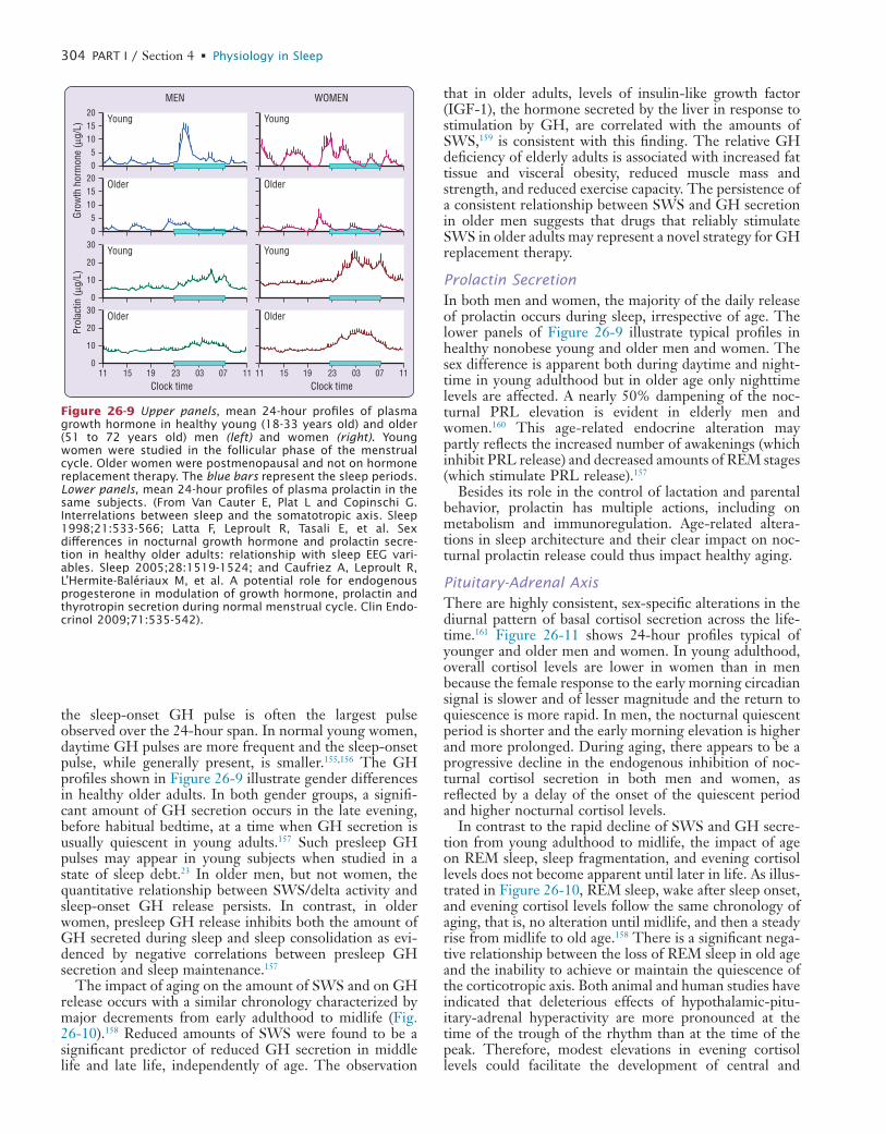
L
304 PART I / Section 4 • Physiology in Sleep
that in older adults, levels of insulin-like growth factor (IGF-1), the hormone secreted by the liver in response to stimulation by GH, are correlated with the amounts of SWS,159 is consistent with this finding. The relative GH deficiency of elderly adults is associated with increased fat tissue and visceral obesity, reduced muscle mass and strength, and reduced exercise capacity. The persistence of a consistent relationship between SWS and GH secretion in older men suggests that drugs that reliably stimulate SWS in older adults may represent a novel strategy for GH replacement therapy.
Prolactin SecretionIn both men and women, the majority of the daily release of prolactin occurs during sleep, irrespective of age. The lower panels of Figure 26-9 illustrate typical profiles in healthy nonobese young and older men and women. The sex difference is apparent both during daytime and night-time in young adulthood but in older age only nighttime levels are affected. A nearly 50% dampening of the noc-turnal PRL elevation is evident in elderly men and women.160 This age-related endocrine alteration may partly reflects the increased number of awakenings (which inhibit PRL release) and decreased amounts of REM stages (which stimulate PRL release).157
Besides its role in the control of lactation and parental behavior, prolactin has multiple actions, including on metabolism and immunoregulation. Age-related altera-tions in sleep architecture and their clear impact on noc-turnal prolactin release could thus impact healthy aging.
Pituitary-Adrenal AxisThere are highly consistent, sex-specific alterations in the diurnal pattern of basal cortisol secretion across the life-time.161 Figure 26-11 shows 24-hour profiles typical of younger and older men and women. In young adulthood, overall cortisol levels are lower in women than in men because the female response to the early morning circadian signal is slower and of lesser magnitude and the return to quiescence is more rapid. In men, the nocturnal quiescent period is shorter and the early morning elevation is higher and more prolonged. During aging, there appears to be a progressive decline in the endogenous inhibition of noc-turnal cortisol secretion in both men and women, as reflected by a delay of the onset of the quiescent period and higher nocturnal cortisol levels.
In contrast to the rapid decline of SWS and GH secre-tion from young adulthood to midlife, the impact of age on REM sleep, sleep fragmentation, and evening cortisol levels does not become apparent until later in life. As illus-trated in Figure 26-10, REM sleep, wake after sleep onset, and evening cortisol levels follow the same chronology of aging, that is, no alteration until midlife, and then a steady rise from midlife to old age.158 There is a significant nega-tive relationship between the loss of REM sleep in old age and the inability to achieve or maintain the quiescence of the corticotropic axis. Both animal and human studies have indicated that deleterious effects of hypothalamic-pitu-itary-adrenal hyperactivity are more pronounced at the time of the trough of the rhythm than at the time of the peak. Therefore, modest elevations in evening cortisol levels could facilitate the development of central and
the sleep-onset GH pulse is often the largest pulse observed over the 24-hour span. In normal young women, daytime GH pulses are more frequent and the sleep-onset pulse, while generally present, is smaller.155,156 The GH profiles shown in Figure 26-9 illustrate gender differences in healthy older adults. In both gender groups, a signifi-cant amount of GH secretion occurs in the late evening, before habitual bedtime, at a time when GH secretion is usually quiescent in young adults.157 Such presleep GH pulses may appear in young subjects when studied in a state of sleep debt.23 In older men, but not women, the quantitative relationship between SWS/delta activity and sleep-onset GH release persists. In contrast, in older women, presleep GH release inhibits both the amount of GH secreted during sleep and sleep consolidation as evi-denced by negative correlations between presleep GH secretion and sleep maintenance.157
The impact of aging on the amount of SWS and on GH release occurs with a similar chronology characterized by major decrements from early adulthood to midlife (Fig. 26-10).158 Reduced amounts of SWS were found to be a significant predictor of reduced GH secretion in middle life and late life, independently of age. The observation
Grow
th h
orm
one
(µg/
L)
20151050
Clock time1911 15 07 1123 03
Clock time1911 15 07 1123 03
MEN
Young Young
20151050
Older Older
Prol
actin
(µg/
L)
30
20
10
0
Young Young
30
20
10
0
Older Older
WOMEN
Figure 26-9 Upper panels, mean 24-hour profiles of plasma growth hormone in healthy young (18-33 years old) and older (51 to 72 years old) men (left) and women (right). Young women were studied in the follicular phase of the menstrual cycle. Older women were postmenopausal and not on hormone replacement therapy. The blue bars represent the sleep periods. Lower panels, mean 24-hour profiles of plasma prolactin in the same subjects. (From Van Cauter E, Plat L and Copinschi G. Interrelations between sleep and the somatotropic axis. Sleep 1998;21:533-566; Latta F, Leproult R, Tasali E, et al. Sex differences in nocturnal growth hormone and prolactin secre-tion in healthy older adults: relationship with sleep EEG vari-ables. Sleep 2005;28:1519-1524; and Caufriez A, Leproult R, L’Hermite-Balériaux M, et al. A potential role for endogenous progesterone in modulation of growth hormone, prolactin and thyrotropin secretion during normal menstrual cycle. Clin Endo-crinol 2009;71:535-542).
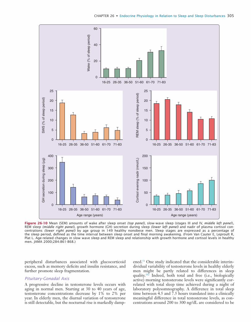
L
CHAPTER 26 • Endocrine Physiology in Relation to Sleep and Sleep Disturbances 305
ened.51 One study indicated that the considerable interin-dividual variability of testosterone levels in healthy elderly men might be partly related to differences in sleep quality.162 Indeed, both total and free (i.e., biologically active) morning testosterone levels were significantly cor-related with total sleep time achieved during a night of laboratory polysomnography. A difference in total sleep time between 4.5 and 7.5 hours translated into a clinically meaningful difference in total testosterone levels, as con-centrations around 200 to 300 ng/dL are considered to be
peripheral disturbances associated with glucocorticoid excess, such as memory deficits and insulin resistance, and further promote sleep fragmentation.
Pituitary-Gonadal AxisA progressive decline in testosterone levels occurs with aging in normal men. Starting at 30 to 40 years of age, testosterone concentrations decrease by 1% to 2% per year. In elderly men, the diurnal variation of testosterone is still detectable, but the nocturnal rise is markedly damp-
60
40
20
0
26-3516-25 36-50 51-60
Wak
e (%
of s
leep
per
iod)
61-70 71-83
25
20
15
10
5
0
26-3516-25 36-50 51-60
RE
M s
leep
(%
of s
leep
per
iod)
61-70 71-83
25
20
15
10
5
0
26-3516-25 36-50 51-60
SW
S (
% o
f sle
ep p
erio
d)
61-70 71-83
26-3516-25 36-50 51-60
Cor
tisol
eve
ning
nad
ir (n
mol
/L)
61-70 71-83
400
300
200
100
0
200
150
100
50
026-3516-25 36-50 51-60
Age range (years) Age range (years)
GH
sec
retio
n du
ring
slee
p (µ
g)
61-70 71-83
Figure 26-10 Mean (SEM) amounts of wake after sleep onset (top panel), slow-wave sleep (stages III and IV, middle left panel ), REM sleep (middle right panel), growth hormone (GH) secretion during sleep (lower left panel) and nadir of plasma cortisol con-centrations (lower right panel) by age group in 149 healthy nonobese men. Sleep stages are expressed as a percentage of the sleep period, defined as the time interval between sleep onset and final morning awakening. (From Van Cauter E, Leproult R, Plat L. Age-related changes in slow wave sleep and REM sleep and relationship with growth hormone and cortisol levels in healthy men. JAMA 2000;284:861-868.)
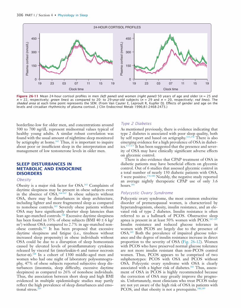
L
306 PART I / Section 4 • Physiology in Sleep
Type 2 DiabetesAs mentioned previously, there is evidence indicating that type 2 diabetes is associated with poor sleep quality, both by self report and based on actigraphy.115,170 There is also emerging evidence for a high prevalence of OSA in diabet-ics.171-175 It has been suggested that the presence and sever-ity of OSA may have clinically significant adverse effects on glycemic control.
There is also evidence that CPAP treatment of OSA in diabetic patients may have beneficial effects on glycemic control. Out of 6 studies that assessed glycemic control in a total number of nearly 150 diabetic patients with OSA, 5 were positive.176-180 Notably, the negative study reported an average nightly therapeutic CPAP use of only 3.6 hours.181
Polycystic Ovary SyndromePolycystic ovary syndrome, the most common endocrine disorder of premenopausal women, is characterized by hyperandrogenism, obesity, insulin resistance, and an ele-vated risk of type 2 diabetes. Insulin resistance is often referred to as a hallmark of PCOS. Obstructive sleep apnea is present in at least 50% women with PCOS.182-187 Insulin resistance and reduced glucose tolerance in women with PCOS are largely due to the presence of OSA.185 Both the prevalence of impaired glucose toler-ance and the degree of insulin resistance increase in direct proportion to the severity of OSA (Fig. 26-12). Women with PCOS who have preserved normal glucose tolerance are not more insulin resistant than non-PCOS control women. Thus, PCOS appears to be comprised of two subphenotypes: PCOS with OSA and PCOS without OSA. Polycystic ovary syndrome with OSA is clearly associated with a higher risk of diabetes.185 Thus, assess-ment of OSA in PCOS is highly recommended because the correction of OSA may greatly improve the progno-sis. Unfortunately, most clinicians who treat PCOS today are not yet aware of the high risk of OSA in patients with PCOS, and that obesity is not a prerequisite.188,189
borderline-low for older men, and concentrations around 500 to 700 ng/dL represent midnormal values typical of healthy young adults. A similar robust correlation was found with the usual amount of nighttime sleep monitored by actigraphy at home.162 Thus, it is important to inquire about poor or insufficient sleep in the interpretation and management of low testosterone levels in older men.
SLEEP DISTURBANCES IN METABOLIC AND ENDOCRINE DISORDERS
ObesityObesity is a major risk factor for OSA.163 Complaints of daytime sleepiness may be present in obese subjects even in the absence of OSA.164-167 In obese subjects without OSA, there may be disturbances in sleep architecture, including lighter and more fragmented sleep as compared to nonobese controls.165 Severely obese patients without OSA may have significantly shorter sleep latencies than lean age-matched controls.164 Excessive daytime sleepiness has been found in 35% of obese subjects (BMI 40 ± 6 kg/m2) without OSA compared to 2.7% in age-matched non-obese controls.167 It has been proposed that excessive daytime sleepiness and fatigue (i.e., tiredness without increased sleep propensity) in obese individuals without OSA could be due to a disruption of sleep homeostasis caused by elevated levels of proinflammatory cytokines released by visceral fat (interleukin-6 and tumor necrosis factor-α).168 In a cohort of 1300 middle-aged men and women who had one night of laboratory polysomnogra-phy, 47% of obese subjects reported subjective sleep dis-turbances (insomnia, sleep difficulty, excessive daytime sleepiness) as compared to 26% of nonobese individuals. Thus, the association between short sleep and high BMI evidenced in multiple epidemiologic studies may partly reflect the high prevalence of sleep disturbances and emo-tional stress.169
Pla
sma
cort
isol
(nm
ol/L
)
450
300
150
0
Clock time
0319 23 15 1907 11
Pla
sma
cort
isol
(nm
ol/L
)
450
300
150
0
Clock time
0319 23 15 1907 11
24-HOUR CORTISOL PROFILES
Figure 26-11 Mean 24-hour cortisol profiles in men (left panel) and women (right panel) 50 years of age and older (n = 25 and n = 22, respectively; green lines) as compared to 20- to 29-year-old subjects (n = 29 and n = 20, respectively; red lines). The shaded area at each time point represents the SEM. (From Van Cauter E, Leproult R, Kupfer DJ. Effects of gender and age on the levels and circadian rhythmicity of plasma cortisol. J Clin Endocrinol Metab 1996;81:2468-2473.)
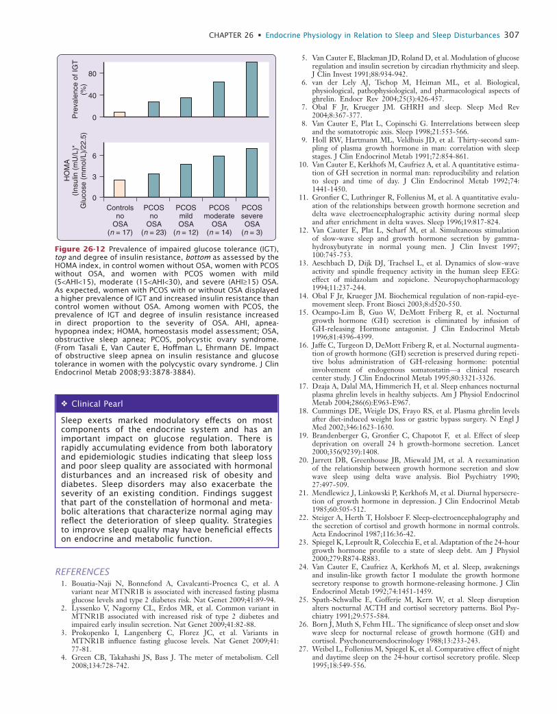
L
CHAPTER 26 • Endocrine Physiology in Relation to Sleep and Sleep Disturbances 307
5. Van Cauter E, Blackman JD, Roland D, et al. Modulation of glucose regulation and insulin secretion by circadian rhythmicity and sleep. J Clin Invest 1991;88:934-942.
6. van der Lely AJ, Tschop M, Heiman ML, et al. Biological, physiological, pathophysiological, and pharmacological aspects of ghrelin. Endocr Rev 2004;25(3):426-457.
7. Obal F Jr, Krueger JM. GHRH and sleep. Sleep Med Rev 2004;8:367-377.
8. Van Cauter E, Plat L, Copinschi G. Interrelations between sleep and the somatotropic axis. Sleep 1998;21:553-566.
9. Holl RW, Hartmann ML, Veldhuis JD, et al. Thirty-second sam-pling of plasma growth hormone in man: correlation with sleep stages. J Clin Endocrinol Metab 1991;72:854-861.
10. Van Cauter E, Kerkhofs M, Caufriez A, et al. A quantitative estima-tion of GH secretion in normal man: reproducibility and relation to sleep and time of day. J Clin Endocrinol Metab 1992;74: 1441-1450.
11. Gronfier C, Luthringer R, Follenius M, et al. A quantitative evalu-ation of the relationships between growth hormone secretion and delta wave electroencephalographic activity during normal sleep and after enrichment in delta waves. Sleep 1996;19:817-824.
12. Van Cauter E, Plat L, Scharf M, et al. Simultaneous stimulation of slow-wave sleep and growth hormone secretion by gamma-hydroxybutyrate in normal young men. J Clin Invest 1997; 100:745-753.
13. Aeschbach D, Dijk DJ, Trachsel L, et al. Dynamics of slow-wave activity and spindle frequency activity in the human sleep EEG: effect of midazolam and zopiclone. Neuropsychopharmacology 1994;11:237-244.
14. Obal F Jr, Krueger JM. Biochemical regulation of non-rapid-eye-movement sleep. Front Biosci 2003;8:d520-550.
15. Ocampo-Lim B, Guo W, DeMott Friberg R, et al. Nocturnal growth hormone (GH) secretion is eliminated by infusion of GH-releasing Hormone antagonist. J Clin Endocrinol Metab 1996;81:4396-4399.
16. Jaffe C, Turgeon D, DeMott Friberg R, et al. Nocturnal augmenta-tion of growth hormone (GH) secretion is preserved during repeti-tive bolus administration of GH-releasing hormone: potential involvement of endogenous somatostatin—a clinical research center study. J Clin Endocrinol Metab 1995;80:3321-3326.
17. Dzaja A, Dalal MA, Himmerich H, et al. Sleep enhances nocturnal plasma ghrelin levels in healthy subjects. Am J Physiol Endocrinol Metab 2004;286(6):E963-E967.
18. Cummings DE, Weigle DS, Frayo RS, et al. Plasma ghrelin levels after diet-induced weight loss or gastric bypass surgery. N Engl J Med 2002;346:1623-1630.
19. Brandenberger G, Gronfier C, Chapotot F, et al. Effect of sleep deprivation on overall 24 h growth-hormone secretion. Lancet 2000;356(9239):1408.
20. Jarrett DB, Greenhouse JB, Miewald JM, et al. A reexamination of the relationship between growth hormone secretion and slow wave sleep using delta wave analysis. Biol Psychiatry 1990; 27:497-509.
21. Mendlewicz J, Linkowski P, Kerkhofs M, et al. Diurnal hypersecre-tion of growth hormone in depression. J Clin Endocrinol Metab 1985;60:505-512.
22. Steiger A, Herth T, Holsboer F. Sleep-electroencephalography and the secretion of cortisol and growth hormone in normal controls. Acta Endocrinol 1987;116:36-42.
23. Spiegel K, Leproult R, Colecchia E, et al. Adaptation of the 24-hour growth hormone profile to a state of sleep debt. Am J Physiol 2000;279:R874-R883.
24. Van Cauter E, Caufriez A, Kerkhofs M, et al. Sleep, awakenings and insulin-like growth factor I modulate the growth hormone secretory response to growth hormone-releasing hormone. J Clin Endocrinol Metab 1992;74:1451-1459.
25. Spath-Schwalbe E, Gofferje M, Kern W, et al. Sleep disruption alters nocturnal ACTH and cortisol secretory patterns. Biol Psy-chiatry 1991;29:575-584.
26. Born J, Muth S, Fehm HL. The significance of sleep onset and slow wave sleep for nocturnal release of growth hormone (GH) and cortisol. Psychoneuroendocrinology 1988;13:233-243.
27. Weibel L, Follenius M, Spiegel K, et al. Comparative effect of night and daytime sleep on the 24-hour cortisol secretory profile. Sleep 1995;18:549-556.
Pre
vale
nce
of IG
T(%
)80
40
0
6
3
0
HO
MA
(Ins
ulin
(m
U/L
)*G
luco
se (
mm
ol/L
)/22
.5)
PCOSno
OSA(n = 23)
PCOSmildOSA
(n = 12)
PCOSmoderate
OSA(n = 14)
PCOSsevereOSA
(n = 3)
Controlsno
OSA(n = 17)
Figure 26-12 Prevalence of impaired glucose tolerance (IGT), top and degree of insulin resistance, bottom as assessed by the HOMA index, in control women without OSA, women with PCOS without OSA, and women with PCOS women with mild (5<AHI<15), moderate (15<AHI<30), and severe (AHI≥15) OSA. As expected, women with PCOS with or without OSA displayed a higher prevalence of IGT and increased insulin resistance than control women without OSA. Among women with PCOS, the prevalence of IGT and degree of insulin resistance increased in direct proportion to the severity of OSA. AHI, apnea- hypopnea index; HOMA, homeostasis model assessment; OSA, obstructive sleep apnea; PCOS, polycystic ovary syndrome. (From Tasali E, Van Cauter E, Hoffman L, Ehrmann DE. Impact of obstructive sleep apnea on insulin resistance and glucose tolerance in women with the polycystic ovary syndrome. J Clin Endocrinol Metab 2008;93:3878-3884).
� Clinical Pearl
Sleep exerts marked modulatory effects on most components of the endocrine system and has an important impact on glucose regulation. There is rapidly accumulating evidence from both laboratory and epidemiologic studies indicating that sleep loss and poor sleep quality are associated with hormonal disturbances and an increased risk of obesity and diabetes. Sleep disorders may also exacerbate the severity of an existing condition. Findings suggest that part of the constellation of hormonal and meta-bolic alterations that characterize normal aging may reflect the deterioration of sleep quality. Strategies to improve sleep quality may have beneficial effects on endocrine and metabolic function.
REFERENCES1. Bouatia-Naji N, Bonnefond A, Cavalcanti-Proenca C, et al. A
variant near MTNR1B is associated with increased fasting plasma glucose levels and type 2 diabetes risk. Nat Genet 2009;41:89-94.
2. Lyssenko V, Nagorny CL, Erdos MR, et al. Common variant in MTNR1B associated with increased risk of type 2 diabetes and impaired early insulin secretion. Nat Genet 2009;41:82-88.
3. Prokopenko I, Langenberg C, Florez JC, et al. Variants in MTNR1B influence fasting glucose levels. Nat Genet 2009;41: 77-81.
4. Green CB, Takahashi JS, Bass J. The meter of metabolism. Cell 2008;134:728-742.
L
308 PART I / Section 4 • Physiology in Sleep
50. Fehm HL, Clausing J, Kern W, et al. Sleep-associated augmentation and synchronization of luteinizing hormone pulses in adult men. Neuroendocrinology 1991;54:192-195.
51. Bremner WJ, Vitiello MV, Prinz PN. Loss of circadian rhythmicity in blood testosterone levels with aging in normal men. J Clin Endo-crinol Metab 1983;56:1278-1280.
52. Lejeune-Lenain C, Van Cauter E, Desir D, et al. Control of cir-cadian and episodic variations of adrenal androgens secretion in man. J Endocrinol Invest 1987;10:267-276.
53. Luboshitzky R, Herer P, Levi M, et al. Relationship between rapid eye movement sleep and testosterone secretion in normal men. J Androl 1999;20:731-737.
54. Axelsson J, Ingre M, Akerstedt T, et al. Effects of acutely displaced sleep on testosterone. J Clin Endocrinol Metab 2005;90:4530- 4535.
55. Luboshitzky R, Zabari Z, Shen-Orr Z, et al. Disruption of the nocturnal testosterone rhythm by sleep fragmentation in normal men. J Clin Endocrinol Metab 2001;86:1134-1139.
56. Akerstedt T, Palmblad J, de la Torre B, et al. Adrenocortical and gonadal steroids during sleep deprivation. Sleep 1980;3:23-30.
57. Leibenluft E, Schmidt PJ, Turner EH, et al. Effects of leuprolide-induced hypogonadism and testosterone replacement on sleep, melatonin, and prolactin secretion in men. J Clin Endocrinol Metab 1997;82:3203-3207.
58. Reame N, Sauder SE, Kelch RP, et al. Pulsatile gonadotropin secretion during the human menstrual cycle: evidence for altered frequency of gonadotropin-releasing hormone secretion. J Clin Endocrinol Metab 1984;59:328-337.
59. Filicori M, Santoro N, Merriam GR, et al. Characterization of the physiological pattern of episodic gonadotropin secretion through-out the menstrual cycle. J Clin Endocrinol Metab 1986;62: 1136-1144.
60. Hall JE, Sullivan JP, Richardson GS. Brief wake episodes modulate sleep-inhibited luteinizing hormone secretion in the early follicular phase. J Clin Endocrinol Metab 2005;90:2050-2055.
61. Vermeulen A, Deslypere JP, Kaukman JM. Influence of antiopioids on luteinizing hormone pulsatility in aging men. J Clin Endocrinol Metab 1989;68:68-72.
62. Mulligan T, Iranmanesh A, Gheorghiu S, et al. Amplified nocturnal luteinizing hormone (LH) secretory burst frequency with selective attenuation of pulsatile (but not basal) testosterone secretion in healthy aged men: possible Leydig cell desensitization to endoge-nous LH signaling—a clinical research center study. J Clin Endo-crinol Metab 1995;80:3025-3031.
63. Tenover JS, Matsumoto AM, Clifton DK, et al. Age-related altera-tions in the circadian rhythms of pulsatile luteinizing hormone and testosterone secretion in healthy men. J Gerontol 1988;43: M163-M169.
64. Luboshitzky R, Shen-Orr Z, Herer P. Middle-aged men secrete less testosterone at night than young healthy men. J Clin Endocrinol Metab 2003;88:3160-3166.
65. Turek FW, Van Cauter E. Rhythms in reproduction. In: Knobil E, Neill JD, editors. The physiology of reproduction. New York: Raven Press; 1993. p. 1789-1830.
66. Saletu-Zyhlarz G, Anderer P, Gruber G, et al. Insomnia related to postmenopausal syndrome and hormone replacement therapy: sleep laboratory studies on baseline differences between patients and con-trols and double-blind, placebo-controlled investigations on the effects of a novel estrogen-progestogen combination (Climodien, Lafamme) versus estrogen alone. J Sleep Res 2003;12:239-254.
67. Hays J, Ockene JK, Brunner RL, et al. Effects of estrogen plus progestin on health-related quality of life. N Engl J Med 2003; 348:1839-1854.
68. Antonijevic IA, Stalla GK, Steiger A. Modulation of the sleep elec-troencephalogram by estrogen replacement in postmenopausal women. Am J Obstet Gynecol 2000;182:277-282.
69. Moe KE, Larsen LH, Vitiello MV, et al. Estrogen replacement therapy moderates the sleep disruption associated with nocturnal blood sampling. Sleep 2001;24:886-894.
70. Shahar E, Redline S, Young T, et al. Hormone replacement therapy and sleep-disordered breathing. Am J Respir Crit Care Med 2003;167:1186-1192.
71. Van Cauter E, Polonsky KS, Scheen AJ. Roles of circadian rhyth-micity and sleep in human glucose regulation. Endocr Rev 1997; 18:716-738.
28. Gronfier C, Luthringer R, Follenius M, et al. Temporal relation-ships between pulsatile cortisol secretion and electroencephalo-graphic activity during sleep in man. Electroencephalogr Clin Neurophysiol 1997;103:405-408.
29. Bierwolf C, Struve K, Marshall L, et al. Slow wave sleep drives inhibition of pituitary-adrenal secretion in humans. J Neuroendo-crinol 1997;9:479-484.
30. Brabant G, Prank K, Ranft U, et al. Physiological regulation of circadian and pulsatile thyrotropin secretion in normal man and woman. J Clin Endocrinol Metab 1990;70:403-409.
31. Pruessner JC, Wolf OT, Hellhammer DH, et al. Free cortisol levels after awakening: a reliable biological marker for the assessment of adrenocortical activity. Life Sci 1997;61:2539-2549.
32. Leproult R, Copinschi G, Buxton O, et al. Sleep loss results in an elevation of cortisol levels the next evening. Sleep 1997;20: 865-870.
33. Veldhuis JD, Iranmanesh A, Johnson ML, et al. Twenty-four-hour rhythms in plasma concentrations of adenohypophyseal hormones are generated by distinct amplitude and/or frequency modulation of underlying pituitary secretory bursts. J Clin Endocrinol Metab 1990;71:1616-1623.
34. Parker DC, Rossman LG, Pekary AE, et al. Effect of 64-hour sleep deprivation on the circadian waveform of thyrotropin (TSH): further evidence of sleep-related inhibition of TSH release. J Clin Endocrinol Metab 1987;64:157-161.
35. Van Cauter E, Copinschi G. Endocrine and other biological rhythms. In: DeGroot LJ, Jameson JL, editors. Endocrinology. Philadelphia: Elsevier Saunders; 2006. p. 341-372.
36. Allan JS, Czeisler CA. Persistence of the circadian thyrotropin rhythm under constant conditions and after light-induced shifts of circadian phase. J Clin Endocrinol Metab 1994;79:508-512.
37. Gary KA, Winokur A, Douglas SD, et al. Total sleep deprivation and the thyroid axis: effects of sleep and waking activity. Aviat Space Environ Med 1996;67:513-519.
38. Spiegel K, Leproult R, L’Hermite-Baleriaux M, et al. Leptin levels are dependent on sleep duration: relationships with sympathovagal balance, carbohydrate regulation, cortisol, and thyrotropin. J Clin Endocrinol Metab 2004;89:5762-5771.
39. Spiegel K, Leproult R, Van Cauter E. Impact of sleep debt on metabolic and endocrine function. Lancet 1999;354:1435- 1439.
40. Hirschfeld U, Moreno-Reyes R, Akseki E, et al. Progressive eleva-tion of plasma thyrotropin during adaptation to simulated jet lag: effects of treatment with bright light or zolpidem. J Clin Endocrinol Metab 1996;81:3270-3277.
41. Spiegel K, Follenius M, Simon C, et al. Prolactin secretion and sleep. Sleep 1994;17:20-27.
42. Waldstreicher J, Duffy JF, Brown EN, et al. Gender differences in the temporal organization of prolactin (PRL) secretion: evidence fort a sleep-independent circadian rhythm of circulating PRL levels—a clinical research Center study. J Clin Endocrinol Metab 1996;81:1483-1487.
43. Désir D, Van Cauter E, L’Hermite M, et al. Effects of “jet lag” on hormonal patterns. III. Demonstration of an intrinsic circadian rhythmicity in plasma prolactin. J Clin Endocrinol Metab 1982; 55:849-857.
44. Spiegel K, Luthringer R, Follenius M, et al. Temporal relationship between prolactin secretion and slow-wave electroencephalographic activity during sleep. Sleep 1995;18:543-548.
45. Copinschi G, Van Onderbergen A, L’Hermite-Balériaux M, et al. Effects of the short-acting benzodiazepine triazolam, taken at bedtime, on circadian and sleep-related hormonal profiles in normal men. Sleep 1990;13:232-244.
46. Copinschi G, Akseki E, Moreno-Reyes R, et al. Effects of bedtime administration of zolpidem on circadian and sleep-related hormonal profiles in normal women. Sleep 1995;18:417-424.
47. Richardson G, Wang-Weigand S. Effects of long-term exposure to ramelteon, a melatonin receptor agonist, on endocrine function in adults with chronic insomnia. Hum Psychopharmacol 2009;24: 103-111.
48. Roky R, Obal F, Valatx JL, et al. Prolactin and rapid eye movement sleep regulation. Sleep 1995;18:536-542.
49. Obal F Jr, Garcia-Garcia F, Kacsoh B, et al. Rapid eye movement sleep is reduced in prolactin-deficient mice. J Neurosci 2005; 25(44):10282-10289.
L
CHAPTER 26 • Endocrine Physiology in Relation to Sleep and Sleep Disturbances 309
98. National Sleep Foundation. 2008 “Sleep in America” Poll. www.sleepfoundation.org.
99. Institut National de Prévention et d’Education pour la Santé. Les français et leur sommeil. http://www.inpes.sante.fr/70000/dp/08/dp080310.pdf 2008.
100. Wehr T, Moul D, Barbato G, et al. Conservation of photoperiod-responsive mechanisms in humans. Am J Physiol 1993;265: R846-R857.
101. Roehrs T, Shore E, Papineau K, et al. A two-week sleep extension in sleepy normals. Sleep 1996:576-582.
102. Harrison Y, Horme JA. Long-term extension to sleep—are we really chronically sleep deprived? Psychophysiology 1996;33:22-30.
103. Bergman RN. Minimal model: perspective from 2005. Horm Research 2005;64:8-15.
104. Knutson KL, Van Cauter E. Associations between sleep loss and increased risk of obesity and diabetes. Ann N Y Acad Sci 2008;1129:287-304.
105. Spiegel K, Tasali E, Leproult R, et al. Effects of poor and short sleep on glucose metabolism and obesity risk. Nat Rev Endocrinol 2009;5:253-261.
106. Spiegel K, Knutson K, Leproult R, et al. Sleep loss: a novel risk factor for insulin resistance and Type 2 diabetes. J Appl Physiol 2005;99:2008-2019.
107. Chin-Chance C, Polonsky KS, Schoeller D. Twenty-four hour leptin levels respond to cumulative short-term energy imbalance and predict subsequent intake. J Clin Endocrinol Metab 2000;85:2685-2691.
108. Guilleminault C, Powell NB, Martinez S, et al. Preliminary obser-vations on the effects of sleep time in a sleep restriction paradigm. Sleep Med 2003;4:177-184.
109. Spiegel K, Tasali E, Penev P, et al. Brief communication: sleep curtailment in healthy young men is associated with decreased leptin levels, elevated ghrelin levels, and increased hunger and appe-tite. Ann Intern Med 2004;141:846-850.
110. Nedeltcheva AV, Kilkus JM, Imperial J, et al. Sleep curtailment is accompanied by increased intake of calories from snacks. Am J Clin Nutr 2009;89:126-133.
111. Taheri S, Lin L, Austin D, et al. Short sleep duration is associated with reduced leptin, elevated ghrelin, and increased body mass index. PLoS Med 2004;1:e62.
112. Chaput JP, Despres JP, Bouchard C, et al. Short sleep duration is associated with reduced leptin levels and increased adiposity: results from the Quebec family study. Obesity (Silver Spring) 2007;15: 253-261.
113. Littman AJ, Vitiello MV, Foster-Schubert K, et al. Sleep, ghrelin, leptin and changes in body weight during a 1-year moderate- intensity physical activity intervention. Int J Obes (Lond) 2007;31: 466-475.
114. Gottlieb DJ, Punjabi NM, Newman AB, et al. Association of sleep time with diabetes mellitus and impaired glucose tolerance. Arch Intern Med 2005;165:863-867.
115. Knutson KL, Ryden AM, Mander BA, et al. Role of sleep duration and quality in the risk and severity of type 2 diabetes mellitus. Arch Intern Med 2006;166:1768-1774.
116. Tuomilehto H, Peltonen M, Partinen M, et al. Sleep duration is associated with an increased risk for the prevalence of type 2 diabe-tes in middle-aged women—the FIN-D2D Survey. Sleep Med 2008;9:221-227.
117. Chaput JP, Despres JP, Bouchard C, et al. Association of sleep duration with type 2 diabetes and impaired glucose tolerance. Dia-betologia 2007;50:2298-2304.
118. Ayas NT, White DP, Al-Delaimy WK, et al. A prospective study of self-reported sleep duration and incident diabetes in women. Diabetes Care 2003;26:380-384.
119. Mallon L, Broman JE, Hetta J. High incidence of diabetes in men with sleep complaints or short sleep duration: a 12-year follow-up study of a middle-aged population. Diabetes Care 2005;28: 2762-2767.
120. Bjorkelund C, Bondyr-Carlsson D, Lapidus L, et al. Sleep distur-bances in midlife unrelated to 32-year diabetes incidence: the pro-spective population study of women in Gothenburg. Diabetes Care 2005;28:2739-2744.
121. Yaggi HK, Araujo AB, McKinlay JB. Sleep duration as a risk factor for the development of type 2 diabetes. Diabetes Care 2006;29: 657-661.
72. Boyle PJ, Scott JC, Krentz AJ, et al. Diminished brain glucose metabolism is a significant determinant for falling rates of systemic glucose utilization during sleep in normal humans. J Clin Invest 1994;93:529-535.
73. Maquet P. Functional neuroimaging of normal human sleep by positron emission tomography. J Sleep Research 2000;9:207-231.
74. Peschke E. Melatonin, endocrine pancreas and diabetes. J Pineal Res 2008;44:26-40.
75. Scheen AJ, Byrne MM, Plat L, et al. Relationships between sleep quality and glucose regulation in normal humans. Am J Physiol 1996;271:E261-E270.
76. Plat L, Byrne MM, Sturis J, et al. Effects of morning cortisol eleva-tion on insulin secretion and glucose regulation in humans. Am Journal Physiol 1996;270:E36-E42.
77. Danguir J, Nicolaidis S. Dependence of sleep on nutrients’ avail-ability. Physiol Behav 1979;22:735-740.
78. Rechtschaffen A, Bergmann BM. Sleep deprivation in the rat by the disk-over-water method. Behav Brain Res 1995;69:55-63.
79. Sakurai T. The neural circuit of orexin (hypocretin): maintaining sleep and wakefulness. Nat Rev 2007;8:171-181.
80. Adamantidis A, de Lecea L. The hypocretins as sensors for metabo-lism and arousal. J Physiol 2009;587(Pt 1):33-40.
81. Ahima RS, Lazar MA. Adipokines and the peripheral and neural control of energy balance. Mol Endocrinol 2008;22:1023-1031.
82. Schoeller DA, Cella LK, Sinha MK, et al. Entrainment of the diurnal rhythm of plasma leptin to meal timing. J Clin Invest 1997;100:1882-1887.
83. Simon C, Gronfier C, Schlienger JL, et al. Circadian and ultradian variations of leptin in normal man under continuous enteral nutri-tion: relationship to sleep and body temperature. J Clin Endocrinol Metab 1998;83:1893-1899.
84. Mullington JM, Chan JL, Van Dongen HP, et al. Sleep loss reduces diurnal rhythm amplitude of leptin in healthy men. J Neuroendo-crinol 2003;15:851-854.
85. Havel PJ. Peripheral signals conveying metabolic information to the brain: short-term and long-term regulation of food intake and energy homeostasis. Exp Biol Med (Maywood) 2001;226: 963-977.
86. Cummings DE, Frayo RS, Marmonier C, et al. Plasma ghrelin levels and hunger scores in humans initiating meals voluntarily without time- and food-related cues. Am J Physiol Endocrinol Metab 2004;287:E297-E304.
87. Brandenberger G, Charloux A, Grongier C, et al. Ultradian rhythms in hydromineral hormones. Horm Res 1998;49:131-135.
88. Follenius M, Brandenberger G, Saini J. Lack of diurnal rhythm in plasma atrial natriuretic peptide. Life Sci 1992;51:143-149.
89. Brandenberger G, Krauth MO, Ehrhart J, et al. Modulation of episodic renin release during sleep in humans. Hypertension 1990;15:370-375.
90. Brandenberger G, Follenius M, Muzet A, et al. Ultradian oscilla-tions in plasma renin activity: their relationship to meals and sleep stages. J Clin Endocrinol Metab 1985;61:280-284.
91. Portaluppi F, Bagni B, Degli UB, et al. Circadian rhythms of atrial natriuretic peptide, renin, aldosterone, cortisol, blood pressure, and heart rate in normal and hypertensive subjects. J Hypertens 1990;8:85-95.
92. Brandenberger G, Follenius M, Goichot B, et al. Twenty-four hour profiles of plasma renin activity in relation to the sleep-wake cycle. J Hypertens 1994;12:277-283.
93. Brandenberger G, Follenius M, Simon C, et al. Nocturnal oscilla-tions in plasma renin activity and REM—NREM sleep cycles in man: a common regulatory mechanism? Sleep 1988;11:242-250.
94. Luthringer R, Brandenberger G, Schaltenbrand N, et al. Slow wave electroencephalographic activity parallels renin oscillations during sleep in humans. Electroencephalogr Clin Neurophysiol 1995; 95:318-322.
95. Charloux A, Piquard F, Ehrhart J, et al. Time-courses in renin and blood pressure during sleep in humans. J Sleep Res 2002; 11:73-79.
96. Charloux A, Gronfier C, Lonsdorfer-Wolf E, et al. Aldosterone release during the sleep-wake cycle in humans. Am J Physiol 1998;276:E43-E49.
97. Charloux A, Gronfier C, Chapotot F, et al. Sleep deprivation blunts the night time increase in aldosterone release in humans. J Sleep Res 2001;10:27-33.
L
310 PART I / Section 4 • Physiology in Sleep
146. Harsch IA, Konturek PC, Koebnick C, et al. Leptin and ghrelin levels in patients with obstructive sleep apnoea: effect of CPAP treatment. Eur Respir J 2003;22:251-257.
147. Takahashi K, Chin K, Akamizu T, et al. Acylated ghrelin level in patients with OSA before and after nasal CPAP treatment. Respirol-ogy 2008;13:810-816.
148. Loube DI, Loube AA, Erman MK. Continuous positive airway pressure treatment results in weight less in obese and overweight patients with obstructive sleep apnea. J Am Diet Assoc 1997;97: 896-897.
149. Redenius R, Murphy C, O’Neill E, et al. Does CPAP lead to change in BMI? J Clin Sleep Med 2008;4:205-209.
150. Kajaste S, Brander PE, Telakivi T, et al. A cognitive-behavioral weight reduction program in the treatment of obstructive sleep apnea syndrome with or without initial nasal CPAP: a randomized study. Sleep Med 2004;5:125-131.
151. Chin K, Shimizu K, Nakamura T, et al. Changes in intra-abdominal visceral fat and serum leptin levels in patients with obstructive sleep apnea syndrome following nasal continuous positive airway pressure therapy. Circulation 1999;100:706-712.
152. Trenell MI, Ward JA, Yee BJ, et al. Influence of constant positive airway pressure therapy on lipid storage, muscle metabolism and insulin action in obese patients with severe obstructive sleep apnoea syndrome. Diabetes Obes Metab 2007;9:679-687.
153. Vgontzas AN, Zoumakis E, Bixler EO, et al. Selective effects of CPAP on sleep apnoea-associated manifestations. Eur J Clin Invest 2008;38:585-595.
154. Van Cauter E, Leproult R. Sleep and diabesity in older adults. In: Pandi-Perumal SR, Monti JM, Monjan AA, editors. Principles and practice of geriatric sleep medicine. Cambridge, UK: Cambridge University Press; 2009. p. 101-121.
155. Copinschi G, Van Cauter E. Effects of ageing on modulation of hormonal secretions by sleep and circadian rhythmicity. Hormone Res 1995;43:20-24.
156. Van Cauter E. Hormones and sleep. In: Kales A, editor. The phar-macology of sleep. Berlin: Springer Verlag; 1995. p. 279-306.
157. Latta F, Leproult R, Tasali E, et al. Sex differences in nocturnal growth hormone and prolactin secretion in healthy older adults: relationships with sleep EEG variables. Sleep 2005;28:1519- 1524.
158. Van Cauter E, Leproult R, Plat L. Age-related changes in slow wave sleep and REM sleep and relationship with growth hormone and cortisol levels in healthy men. JAMA 2000;284:861-868.
159. Prinz P, Moe K, Dulberg E, et al. Higher plasma IGF-1 levels are associated with increased delta sleep in healthy older men. J Geron-tol 1995;50A:M222-M226.
160. van Coevorden A, Mockel J, Laurent E, et al. Neuroendocrine rhythms and sleep in aging men. Am J Physiol 1991;260:E651- E661.
161. Van Cauter E, Leproult R, Kupfer DJ. Effects of gender and age on the levels and circadian rhythmicity of plasma cortisol. J Clin Endocrinol Metab 1996;81:2468-2473.
162. Penev PD. Association between sleep and morning testosterone levels in older men. Sleep 2007;30:427-432.
163. Young T, Peppard PE, Taheri S. Excess weight and sleep-disor-dered breathing. J Appl Physiol 2005;99:1592-1599.
164. Vgontzas AN, Bixler EO, Tan TL, et al. Obesity without sleep apnea is associated with daytime sleepiness. Arch Intern Med 1998;158:1333-1337.
165. Vgontzas AN, Tan TL, Bixler EO, et al. Sleep apnea and sleep disruption in obese patients. Arch Internal Med 1994;154:1705- 1711.
166. Resta O, Foschino Barbaro MP, Bonfitto P, et al. Low sleep quality and daytime sleepiness in obese patients without obstructive sleep apnoea syndrome. J Intern Med 2003;253:536-543.
167. Resta O, Foschino-Barbaro MP, Legari G, et al. Sleep-related breathing disorders, loud snoring and excessive daytime sleepiness in obese subjects. Int J Obes Relat Metab Disord 2001;25: 669-675.
168. Vgontzas AN, Bixler EO, Chrousos GP. Obesity-related sleepiness and fatigue: the role of the stress system and cytokines. Ann N Y Acad Sci 2006;1083:329-344.
169. Vgontzas AN, Lin HM, Papaliaga M, et al. Short sleep duration and obesity: the role of emotional stress and sleep disturbances. Int J Obes 2008;32:801-809.
122. Gangwisch JE, Heymsfield SB, Boden-Albala B, et al. Sleep dura-tion as a risk factor for diabetes incidence in a large U.S. sample. Sleep 2007;30:1667-1673.
123. Hayashino Y, Fukuhara S, Suzukamo Y, et al. Relation between sleep quality and quantity, quality of life, and risk of developing diabetes in healthy workers in Japan: the High-risk and Population Strategy for Occupational Health Promotion (HIPOP-OHP) Study. BMC Public Health 2007;7:129.
124. Cappuccio FP, Taggart FM, Kandala NB, et al. Meta-analysis of short sleep duration and obesity in children and adults. Sleep 2008;31:619-626.
125. Chen X, Beydoun MA, Wang Y. Is sleep duration associated with childhood obesity? A systematic review and meta-analysis. Obesity (Silver Spring) 2008;16:265-274.
126. Patel SR, Hu FB. Short sleep duration and weight gain: a systematic review. Obesity (Silver Spring) 2008;16:643-653.
127. Patel SR, Blackwell T, Redline S, et al. The association between sleep duration and obesity in older adults. Int J Obes (Lond) 2008;32:1825-1834.
128. Van Cauter E, Knutson K. Sleep and the epidemic of obesity in children and adults. Eur J Endocrinol 2008;159:S59-S66.
129. Berkey CS, Rockett HR, Colditz GA. Weight gain in older adoles-cent females: the internet, sleep, coffee, and alcohol. J Pediatr 2008;153:635-639.
130. Keith SW, Redden DT, Katzmarzyk PT, et al. Putative contribu-tors to the secular increase in obesity: exploring the roads less traveled. Int J Obes 2006;30:1585-1594.
131. Follenius M, Brandenberger G, Bardasept J, et al. Nocturnal cortisol release in relation to sleep structure. Sleep 1992;15: 21-27.
132. Van Cauter E, van Coevorden A, Blackman JD. Modulation of neuroendocrine release by sleep and circadian rhythmicity. In: Yen S, Vale W, editors. Advances in neuroendocrine regulation of reproduction. Norwell, Mass: Serono Symposia USA; 1990. p. 113-122.
133. Tasali E, Leproult R, Ehrmann DA, et al. Slow-wave sleep and the risk of type 2 diabetes in humans. Proc Natl Acad Sci U S A 2008; 105:1044-1049.
134. Bergman RN. Toward physiological understanding of glucose tol-erance. Minimal model approach. Diabetes 1989;38:1512-1527.
135. Kawakami N, Takatsuka N, Shimizu H. Sleep disturbance and onset of type 2 diabetes. Diabetes Care 2004;27:282-283.
136. Nilsson PM, Roost M, Engstrom G, et al. Incidence of diabetes in middle-aged men is related to sleep disturbances. Diabetes Care 2004;27:2464-2469.
137. Meisinger C, Heier M, Loewel H. Sleep disturbance as a predictor of type 2 diabetes mellitus in men and women from the general population. Diabetologia 2005;48:235-241.
138. Eriksson AK, Ekbom A, Granath F, et al. Psychological distress and risk of pre-diabetes and Type 2 diabetes in a prospective study of Swedish middle-aged men and women. Diabet Med 2008;25: 834-842.
139. Vgontzas AN, Bixler EO, Lin HM, et al. Chronic insomnia is associated with nyctohemeral activation of the hypothalamic-pitu-itary-adrenal axis: clinical implications. J Clin Endocrinol Metab 2001;86:3787-3794.
140. Basta M, Chrousos GP, Vela-Bueno A, et al. Chronic insomnia and stress system. Sleep Med Clin 2007;2:279-291.
141. Motivala SJ, Tomiyama AJ, Ziegler M, et al. Nocturnal levels of ghrelin and leptin and sleep in chronic insomnia. Psychoneuroen-docrinology 2009;34:540-545.
142. Tasali E, Ip MS. Obstructive sleep apnea and metabolic syndrome: alterations in glucose metabolism and inflammation. Proc Am Thorac Soc 2008;5:207-217.
143. Tasali E, Mokhlesi B, Van Cauter E. Obstructive sleep apnea and type 2 diabetes: interacting epidemics. Chest 2008;133:496-506.
143a. Romero-Corral A, Caples SM, Lopez-Jimenez F, Somers VK. Interactions between obesity and obstructive sleep apnea: implica-tions for treatment. Chest 2010;137(3):711-719.
144. Phillips BG, Kato M, Narkiewicz K, et al. Increases in leptin levels, sympathetic drive, and weight gain in obstructive sleep apnea. Am J Physiol Heart Circ Physiol 2000;279:H234-H237.
145. Schmid SM, Hallschmid M, Jauch-Chara K, et al. A single night of sleep deprivation increases ghrelin levels and feelings of hunger in normal-weight healthy men. J Sleep Res 2008;17:331-334.
L
CHAPTER 26 • Endocrine Physiology in Relation to Sleep and Sleep Disturbances 311
170. Trento M, Broglio F, Riganti F, et al. Sleep abnormalities in type 2 diabetes may be associated with glycemic control. Acta Diabetol 2008;45:225-229.
171. Einhorn D, Stewart DA, Erman MK, et al. Prevalence of sleep apnea in a population of adults with type 2 diabetes mellitus. Endocr Pract 2007;13:355-362.
172. West SD, Nicoll DJ, Stradling JR. Prevalence of obstructive sleep apnoea in men with type 2 diabetes. Thorax 2006;61:945-950.
173. Resnick HE, Redline S, Shahar E, et al. Diabetes and sleep distur-bances: findings from the Sleep Heart Health Study. Diabetes Care 2003;26:702-709.
174. Katsumata K, Okada T, Miyao M, et al. High incidence of sleep apnea syndrome in a male diabetic population. Diabetes Res Clin Pract 1991;13:45-51.
175. Elmasry A, Lindberg E, Berne C, et al. Sleep-disordered breathing and glucose metabolism in hypertensive men: a population-based study. J Intern Med 2001;249:153-161.
176. Dawson A, Abel SL, Loving RT, et al. CPAP therapy of obstructive sleep apnea in type 2 diabetics improves glycemic control during sleep. J Clin Sleep Med 2008;4:538-542.
177. Hassaballa HA, Tulaimat A, Herdegen JJ, et al. The effect of con-tinuous positive airway pressure on glucose control in diabetic patients with severe obstructive sleep apnea. Sleep Breath 2005; 9:176-180.
178. Babu AR, Herdegen J, Fogelfeld L, et al. Type 2 diabetes, glycemic control, and continuous positive airway pressure in obstructive sleep apnea. Arch Intern Med 2005;165:447-452.
179. Harsch IA, Schahin SP, Bruckner K, et al. The effect of continuous positive airway pressure treatment on insulin sensitivity in patients with obstructive sleep apnoea syndrome and type 2 diabetes. Res-piration 2004;71:252-259.
180. Brooks B, Cistulli PA, Borkman M, et al. Obstructive sleep apnea in obese noninsulin-dependent diabetic patients: effects of continu-ous positive airway pressure treatment on insulin responsiveness. J Clin Endocrinol Metab 1994;79:1681-1685.
181. West SD, Nicoll DJ, Wallace TM, et al. Effect of CPAP on insulin resistance and HbA1c in men with obstructive sleep apnoea and type 2 diabetes. Thorax 2007;62:969-974.
182. Vgontzas AN, Legro R, Bixler EO, et al. Polycystic ovary syndrome is associated with obstructive sleep apnea and daytime sleepiness: role of insulin resistance. J Clin Endocrinol Metab 2001;86:517-520.
183. Gopal M, Duntley S, Uhles M, et al. The role of obesity in the increased prevalence of obstructive sleep apnea syndrome in patients with polycystic ovarian syndrome. Sleep Med 2002;3:401-404.
184. Fogel RB, Malhotra A, Pillar G, et al. Increased prevalence of obstructive sleep apnea syndrome in obese women with polycystic ovary syndrome. J Clin Endocrinol Metab 2001;86:1175-1180.
185. Tasali E, Van Cauter E, Ehrmann D. Polycystic ovary syndrome and obstructive sleep apnea. Sleep Med Clin 2008;3:37-46.
186. Tasali E, Van Cauter E, Ehrmann DA. Relationships between sleep disordered breathing and glucose metabolism in polycystic ovary syndrome. J Clin Endocrinol Metab 2006;91:36-42.
187. Tasali E, Van Cauter E, Hoffman L, et al. Impact of obstructive sleep apnea on insulin resistance and glucose tolerance in women with polycystic ovary syndrome. J Clin Endocrinol Metab 2008; 93:3878-3884.
188. Subramanian S, Desai A, Joshipura M, et al. Practice patterns of screening for sleep apnea in physicians treating PCOS patients. Sleep Breath 2007;11:233-237.
189. Yang HP, Kang JH, Su HY, et al. Apnea-hypopnea index in non-obese women with polycystic ovary syndrome. Int J Gynaecol Obstet 2009;105:226-229.
LL
312
ABSTRACT
The gastrointestinal system is regulated by both the auto-nomic nervous system and the enteric nervous system (ENS), a complex network of neurons located within the entire luminal gastrointestinal tract.
Esophageal peristalsis is largely regulated by the ENS and, to a much lesser extent, by vagal input. There is a diminution in peristaltic amplitude and a substantial decrease in the swal-lowing rate and concomitant prolongation of esophageal acid clearance during sleep. Transient lower esophageal sphincter relaxations have been shown to be decreased during sleep.
The basic myoelectric activity of the stomach is regulated by a gastric pacemaker located in brainstem that emits a basic electrical rhythm of approximately three cycles per minute and forms the basis for the production of gastric con-tractile activity. There is also periodic electrical spike activity associated with gastric contractions. This basic electrical cycle can be measured noninvasively with appropriately placed elec-
trodes on the surface of the abdomen. This basic pacemaker activity has been shown to be altered during non-REM sleep (e.g., decrease activity). There is, in normal individuals, a peak in acid secretion occurring at approximately midnight, whereas patients with duodenal ulcer disease have increased acid secretion throughout the circadian cycle unrelated to sleep stage.
Intestinal motility is largely composed of the cyclic (every 90 min) occurrence of a sequence of contractions, the migrat-ing motor complex (MMC), which is regulated almost com-pletely by the ENS. This activity is not correlated with rapid eye movement sleep. With sleep, the MMC cycle becomes shorter. During sleep, the colon is relatively quiescent. The high amplitude peristaltic contractions are associated with defecation. Anal rectal functioning, on the other hand, has an endogenous oscillation that is most evident during sleep, resulting in primarily retrograde propagation. This likely serves a protective function against the involuntary loss of rectal contents during sleep.
taneous esophageal function is markedly reduced during sleep because there is no need for this organ to function except in the event of food being transmitted from the upper esophageal area. This rarely occurs without voli-tional swallowing, which is diminished significantly during sleep. On the other hand, rectal motor activity persists during sleep, which appears to be a mechanism necessary to preserve continence during sleep.
The ultimate description of GI functioning and its complex physiology requires that autonomic and central control be separated from the intrinsic control generated by the ENS. Because sleep essentially represents a relative state of reversible isolation from cortical influences, the study of GI motor functioning during sleep allows an assessment of autonomous activity without higher cortical influence. Thus, the study of GI motility during sleep allows the separation of CNS and ENS processes and a more clear understanding of what has been referred to as the brain-gut axis. More information on the interaction between gastrointestinal disorders and sleep is described by this author in Chapter 127 of this volume.
HISTORICAL ASPECTSInterest in GI physiology during sleep preceded the devel-opment of sophisticated techniques for monitoring GI function by at least 100 years. For example, Friedenwald,1 in 1906, described the secretory function of the stomach during sleep and reported that it was not appreciably altered. In 1924, Johnson and Washeim2 reported a decrease in the volume of gastric juice secreted during sleep, and, in addition, they reported an increase in the acidity of the gastric juice. Henning and Norpoth3 mea-sured acidity hourly during sleep, and they reported that sleep was associated with a cessation of acid secretion and,
INTRODUCTION
A description of the normal physiology of any organ system rarely includes the normal alterations of that system during sleep. It would seem inappropriate that nearly one third of the functioning time of an organ system would be ignored in describing its functioning. Profound alterations have been documented during sleep in describing normal physi-ology, yet these changes do not become part of the litany in describing normal physiology. Sleep-related, or circadian changes, have been most commonly addressed in describ-ing blood pressure and the definition of hypertension, but this has not been accomplished with either the description of or clinical application of sleep-related changes in gas-trointestinal (GI) physiology. This can be largely attrib-uted to the inaccessibility of the luminal GI tract to easy measurement. Monitoring any of the basic functions of the GI system requires invading an orifice, which is an unpleasant experience and one that would generally be assumed to be disruptive of normal sleep.
The functioning of the luminal GI tract (to which this review is confined) reflects the final common pathway of a complex interaction of the central nervous system (CNS) with both the autonomic nervous system (ANS) and the ENS. The latter is a complex neuronal network that is woven throughout the subserosal layers of the luminal GI tract. The ENS provides an autonomous source of GI motor activity. The movement through the GI tract is controlled and regulated by the ENS and the ANS, and the interaction of these two influences ultimately deter-mines that appropriate movement, at an appropriate rate, occurs throughout the passage from mouth to anus.
Alterations in GI functioning during sleep appear to be quite different depending on the particular organ studied and its function within the GI system. For example, spon-
Gastrointestinal Physiology in Relation to SleepWilliam C. Orr
Chapter
27

L
CHAPTER 27 • Gastrointestinal Physiology in Relation to Sleep 313
low secreters were consistently low. These studies paved the way for more extensive investigations into the role of nocturnal acid secretion in the pathogenesis of DU disease. This study is at variance with the study by Sandweiss and colleagues,7 which did not find a substantial difference in nocturnal acid secretion between normal subjects and patients with DU. These discrepant results may be due to varying degrees of ulcer activity in the patients studied. As previously noted, perhaps the most influential work in nocturnal gastric acid secretion has been done by Dragst-edt.11 Using hourly collections of overnight acid secretion in normal subjects and in patients with DU disease, he reported that the nocturnal acid secretion in patients with DU disease is 3 to 20 times greater than that in his normal subjects. He reported that this greater secretion was abol-ished by vagotomy, which invariably produced prompt ulcer healing. These data were interpreted to be strong evidence in favor of a “nervous” origin for DU disease.
The decade of the 1950s ushered in the “golden age” of sleep investigation. The realization that sleep is not a unitary, passive state of consciousness focused considerable interest on its unique physiology. The first study to describe gastric acid secretion during conditions of poly-somnographic (PSG) monitoring was reported in 1960 by Reichsman and colleagues.13 Using relatively crude deter-minations of stages of sleep, they found no correlation between gastric acid secretion and sleep stages in normal subjects. A subsequent study by Armstrong and colleagues14 reported the rather startling finding of a hypersecretion of acid during REM sleep in patients with DU disease. Both of these studies suffered from a major methodological flaw in that they used drugs to induce sleep. Another problem in these studies was the relatively small data sample per subject, in most cases only a single night. Earlier studies documented the considerable night-to-night variability in acid secretion. Thus, studying a patient or a normal vol-unteer for a single night with a nasogastric tube would undoubtedly result in numerous awakenings and generally poor sleep, with correspondingly more variable acid secretion.
Stacher and colleagues15 studied gastric acid secretion in a group of normal volunteers during PSG-monitored natural sleep. They reported no significant differences in acid secretion during the non–rapid eye movement (NREM) stages of sleep; however, they reported that REM sleep was associated with an inhibition of acid secretion. This study, although avoiding the use of drugs to induce sleep, used a rather cumbersome technique for determin-ing acid output. It was measured by an intragastric titration from a telemetric pH capsule and involved numerous disruptions in the patients’ sleep. Thus, although it was natural sleep, it was at best fragmented sleep.
The variability of these results and the various method-ological problems of previous studies prompted a concur-rent study of both normal subjects and patients with DU disease. Orr and colleagues16 studied five normal volun-teers and five patients with DU disease, and each subject was studied for 5 consecutive nights in the sleep labora-tory. The study involved continuous aspiration of gastric contents divided into 20-minute aliquots for gastric analy-sis as well as complete PSG monitoring for the determina-tion of sleep stages. In addition, serum gastrin levels were
conversely, patients with gastric ulcers did appear to main-tain continuous acid secretion. These results were con-firmed nearly 20 years later.4
In 1915, Luckhardt5 described the inhibition of gastric motility during periods of sleep, which, from his descrip-tion, sounded like rapid eye movement (REM) sleep. His observations were made in dogs and were described in association with “sonorous respiration” (snoring?), irregu-lar breathing, movements of the forelimbs and hindlimbs, and occasional “abortive yelps.” Luckhardt stated, “I assumed that the dog was experiencing during sleep a form of cerebral excitation akin to or identical with the dream-ing state in man.”
A few years later, in 1922, Wada6 reported an extensive study of hunger and its relation to gastric and somatic activity by use of simultaneous records of gastric contrac-tions and body movements during sleep. Gastric contrac-tions were reported that were often associated with body movements and reports of dreaming. The techniques used in these studies were obviously somewhat crude, but it is clear that interest in GI function during sleep was not a development of the more modern era of sleep investigation.
GASTRIC FUNCTION DURING WAKE AND SLEEP
Acid SecretionGastric acid secretion has been shown to exhibit a clear circadian rhythm, which was initially described by Sand-weiss and colleagues.7 It remained, however, for Moore and Englert to provide a definitive description of the cir-cadian oscillation of gastric acid secretion in normal sub-jects.8 These investigators described a peak in acid secretion occurring generally between 10:00 pm and 2:00 am while confirming already established data that indicated that basal acid secretion in the waking state is minimal in the absence of meal stimulation. Similar results have been described in patients who have duodenal ulcer (DU) disease with levels of acid secretion markedly enhanced through-out the circadian cycle.9,10 It is clear from these results that there is an endogenous circadian rhythm of unstimulated basal acid secretion, but it is not clear that this is specifi-cally altered in any way by sleep.
Initial interest in the role of sleep in gastric acid produc-tion was stimulated by the work of the eminent University of Chicago surgeon Dr. Lester Dragstedt.11 An interest in the pathogenesis of DU disease and the possible role of nocturnal gastric acid secretion in this process was further stimulated by the studies of Levin and colleagues.9,12 They analyzed hourly samples of acid secretion in both normal subjects and patients with DU disease, and they identified nearly continuous acid secretion throughout the night in normal volunteers. They described a considerable degree of night-to-night and subject-to-subject variation. In con-trast to the normal subjects, there were increases in both volume and acid concentration during sleep in patients with DU disease as suggested earlier for premier study. Although emphasizing night-to-night variation, they found that the patients with DU disease who tended to be high secreters were consistently high, and those who were
L
314 PART I / Section 4 • Physiology in Sleep
by sleep stage were noted in a study in unanesthetized and unrestrained cats.22
Hall and colleagues23 reported data on gastric emptying during sleep. This study required the patient to sleep with a nasogastric tube through which 750 ml of 10% glucose was administered. After 30 minutes, the gastric contents were aspirated, and the residual volume was determined. Aspiration was followed by a washout meal of 150 ml of saline. This process was done during the presleep waking interval, during NREM and REM sleep, and during post-sleep waking. These data suggested a more rapid gastric emptying during REM sleep and a slower emptying during the postsleep–waking state. The emptying of a hypertonic solution is controlled by vagally mediated osmoreceptors in the duodenum. These data, therefore, would suggest a possible anticholinergic action during REM sleep and a cholinergic process during the postsleep–waking state. These data represent only an approximation of the altera-tions in gastric emptying during sleep because these mea-surement techniques are relatively crude, and gastric emptying is a complex process.
Dubois and colleagues24 have described a technique that permits the simultaneous assessment of acid secretion, water secretion, and the fractional rate of emptying. These techniques have been applied to the assessment of gastric functioning during sleep; the results indicate that in normal subjects, acid secretion, water secretion, and the fractional rate of emptying all showed significant decrements during sleep.25 There did not appear to be any differences between REM and NREM sleep, but all of these measures demon-strated a significant difference between presleep waking and REM sleep. Data obtained by use of radionuclide emptying assessments suggest that this difference may be a circadian, rather than a sleep-dependent, effect. Studies by Goo and colleagues26 have shown a marked delay in gastric emptying of solids in the evening, compared with the morning.
Gastric motor functioning is characterized by an endog-enous electrical cycle generated by the gastric smooth muscle. The electrical rhythm is generated by a pacemaker located in the proximal portion of the greater curvature of the stomach.27 The electrical cycle occurs at a frequency of approximately three per minute and represents the pre-cursor to contractile activity of the stomach, which allows movement of gastric contents to the antrum and subse-quent emptying into the duodenum.
The gastric electrical rhythm can be measured by surface electrodes placed in the periumbilical area. The identifica-tion of this basic motor function of the stomach requires highly sophisticated measurement, digital filtering, and spectral analysis to describe the parameters of this oscilla-tion.28 The noninvasive measurement of the gastric electri-cal rhythm is called electrogastrography (EGG). The progressive sophistication of the measurement and analytic techniques has allowed a reliable noninvasive technique to measure an important function of the GI system.
The sophisticated analysis of gastric electrical activity has documented three fundamental characteristics of this electrical rhythm that determine its normal activity. First, the power in the frequency band is approximately three per minute. This is essentially a method of quantifying the extent to which the wave approximates a sinusoidal rhythm,
assessed at 20-minute intervals throughout the study. The results did not reveal any significant correlation between the sleep stage (REM vs. NREM) and acid concentration or total acid secretion. Furthermore, there was no relation-ship between any of these variables and serum gastrin levels. These data did show, however, that the patients with DU failed to inhibit acid secretion during the first 2 hours of sleep, which was consistent with the previously reported studies by Levin and colleagues,12 which sug-gested that acid secretion is poorly inhibited during sleep in patients with DU disease.
In conclusion, the data would support the presence of a clear-cut circadian rhythm in basal acid secretion, with a peak occurring in the early part of the normal sleep interval. However, there are no definitive data at the present time that would suggest a major effect of sleep stages on this process. If one conclusion can be drawn from the various studies that have been done assessing gastric acid secretion during sleep, it would be that acid secretion is extremely variable from night to night and from person to person, and for this reason definitive con-clusions require numerous replications across and within a large number of subjects and patients. The extremely demanding logistics of sleep studies, as well as the obvious aversive aspects of probe placement via the nasal passages or anal canal make the acquisition of such data exceed-ingly difficult.
GASTRIC MOTOR FUNCTION DURING SLEEPThe motor function of the stomach serves to empty solids and liquids into the duodenum at an appropriate rate and pH. The stomach is functionally divided into two sections: the fundus of the stomach functions primarily to control liquid emptying into the duodenum, whereas the antrum controls emptying of solids.17 Because liquids and solids are handled differently by the stomach, the regulation of gastric emptying is correspondingly complicated, involv-ing intrinsic regulation of motor activity as well as specific alterations associated with the ingestion of liquids and solids. Thus, although gastric emptying itself can be regarded as the final common pathway reflecting the motor activity of the stomach, one must keep in mind that the processes of liquid and solid emptying are regulated by quite different mechanisms.
There have been many reports of gastric motility during sleep, with sometimes contradictory results. Inhibition of gastric motility during sleep was documented in a study by Scantlebury and colleagues,18 in which they implicated the “dream mechanism” as a part of this inhibitory process. A cortical inhibitory mechanism acting through the splanch-nic nerve is the postulated mechanism of this inhibition. Nearly 30 years later, using somewhat more sophisticated measurement techniques, Bloom and colleagues19 found that gastric motility was enhanced during sleep, compared with waking. Baust and Rohrwasser20 studied gastric motil-ity during PSG-monitored sleep, and they also described a marked enhancement in gastric motility during sleep, but their findings were restricted to REM sleep. Only a year later, a decrease in gastric motility during REM sleep was reported.21 No consistent findings concerning alterations
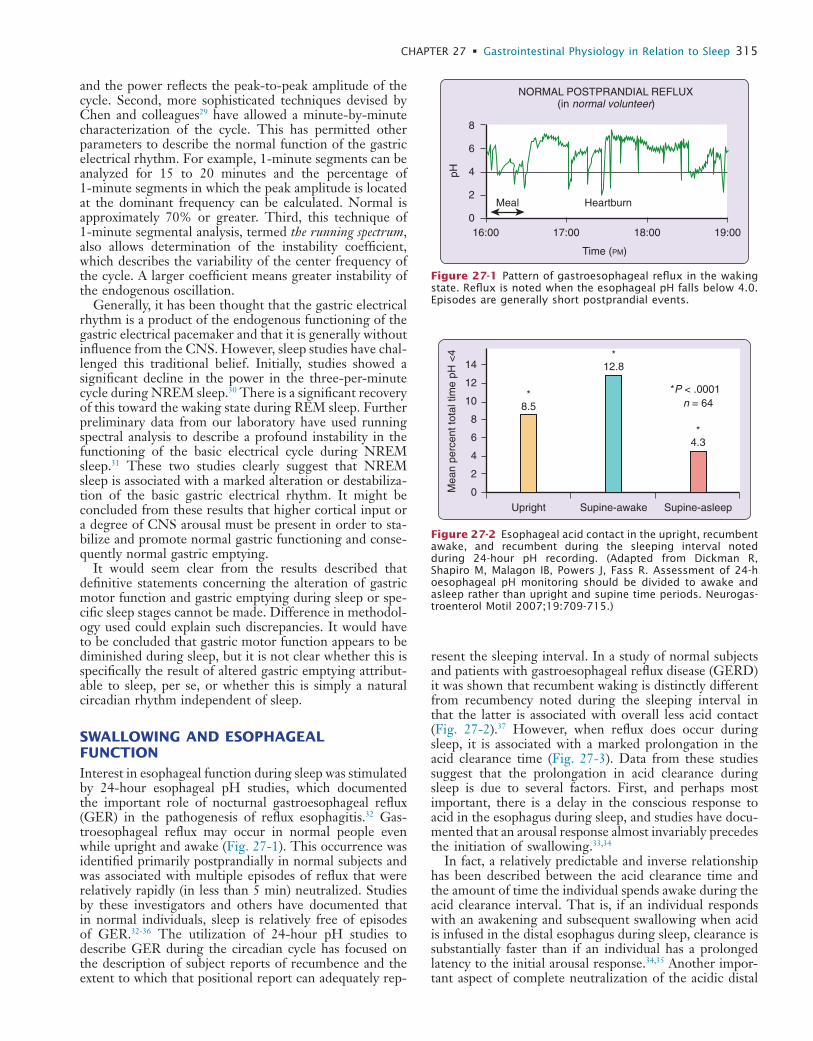
L
CHAPTER 27 • Gastrointestinal Physiology in Relation to Sleep 315
resent the sleeping interval. In a study of normal subjects and patients with gastroesophageal reflux disease (GERD) it was shown that recumbent waking is distinctly different from recumbency noted during the sleeping interval in that the latter is associated with overall less acid contact (Fig. 27-2).37 However, when reflux does occur during sleep, it is associated with a marked prolongation in the acid clearance time (Fig. 27-3). Data from these studies suggest that the prolongation in acid clearance during sleep is due to several factors. First, and perhaps most important, there is a delay in the conscious response to acid in the esophagus during sleep, and studies have docu-mented that an arousal response almost invariably precedes the initiation of swallowing.33,34
In fact, a relatively predictable and inverse relationship has been described between the acid clearance time and the amount of time the individual spends awake during the acid clearance interval. That is, if an individual responds with an awakening and subsequent swallowing when acid is infused in the distal esophagus during sleep, clearance is substantially faster than if an individual has a prolonged latency to the initial arousal response.34,35 Another impor-tant aspect of complete neutralization of the acidic distal
and the power reflects the peak-to-peak amplitude of the cycle. Second, more sophisticated techniques devised by Chen and colleagues29 have allowed a minute-by-minute characterization of the cycle. This has permitted other parameters to describe the normal function of the gastric electrical rhythm. For example, 1-minute segments can be analyzed for 15 to 20 minutes and the percentage of 1-minute segments in which the peak amplitude is located at the dominant frequency can be calculated. Normal is approximately 70% or greater. Third, this technique of 1-minute segmental analysis, termed the running spectrum, also allows determination of the instability coefficient, which describes the variability of the center frequency of the cycle. A larger coefficient means greater instability of the endogenous oscillation.
Generally, it has been thought that the gastric electrical rhythm is a product of the endogenous functioning of the gastric electrical pacemaker and that it is generally without influence from the CNS. However, sleep studies have chal-lenged this traditional belief. Initially, studies showed a significant decline in the power in the three-per-minute cycle during NREM sleep.30 There is a significant recovery of this toward the waking state during REM sleep. Further preliminary data from our laboratory have used running spectral analysis to describe a profound instability in the functioning of the basic electrical cycle during NREM sleep.31 These two studies clearly suggest that NREM sleep is associated with a marked alteration or destabiliza-tion of the basic gastric electrical rhythm. It might be concluded from these results that higher cortical input or a degree of CNS arousal must be present in order to sta-bilize and promote normal gastric functioning and conse-quently normal gastric emptying.
It would seem clear from the results described that definitive statements concerning the alteration of gastric motor function and gastric emptying during sleep or spe-cific sleep stages cannot be made. Difference in methodol-ogy used could explain such discrepancies. It would have to be concluded that gastric motor function appears to be diminished during sleep, but it is not clear whether this is specifically the result of altered gastric emptying attribut-able to sleep, per se, or whether this is simply a natural circadian rhythm independent of sleep.
SWALLOWING AND ESOPHAGEAL FUNCTIONInterest in esophageal function during sleep was stimulated by 24-hour esophageal pH studies, which documented the important role of nocturnal gastroesophageal reflux (GER) in the pathogenesis of reflux esophagitis.32 Gas-troesophageal reflux may occur in normal people even while upright and awake (Fig. 27-1). This occurrence was identified primarily postprandially in normal subjects and was associated with multiple episodes of reflux that were relatively rapidly (in less than 5 min) neutralized. Studies by these investigators and others have documented that in normal individuals, sleep is relatively free of episodes of GER.32-36 The utilization of 24-hour pH studies to describe GER during the circadian cycle has focused on the description of subject reports of recumbence and the extent to which that positional report can adequately rep-
Time (PM)
8
6
4
2
0
NORMAL POSTPRANDIAL REFLUX(in normal volunteer)
Meal Heartburn
pH
16:00 17:00 18:00 19:00
Figure 27-1 Pattern of gastroesophageal reflux in the waking state. Reflux is noted when the esophageal pH falls below 4.0. Episodes are generally short postprandial events.
Upright Supine-awake Supine-asleep
Mea
n pe
rcen
t tot
al ti
me
pH <
4
14
12
10
8
6
4
2
0
*8.5
*12.8
*4.3
n = 64*P < .0001
Figure 27-2 Esophageal acid contact in the upright, recumbent awake, and recumbent during the sleeping interval noted during 24-hour pH recording. (Adapted from Dickman R, Shapiro M, Malagon IB, Powers J, Fass R. Assessment of 24-h oesophageal pH monitoring should be divided to awake and asleep rather than upright and supine time periods. Neurogas-troenterol Motil 2007;19:709-715.)
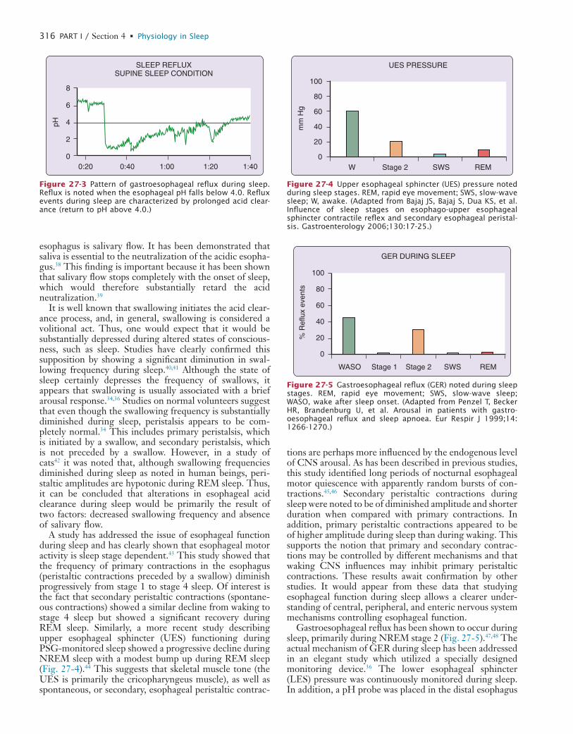
L
316 PART I / Section 4 • Physiology in Sleep
tions are perhaps more influenced by the endogenous level of CNS arousal. As has been described in previous studies, this study identified long periods of nocturnal esophageal motor quiescence with apparently random bursts of con-tractions.45,46 Secondary peristaltic contractions during sleep were noted to be of diminished amplitude and shorter duration when compared with primary contractions. In addition, primary peristaltic contractions appeared to be of higher amplitude during sleep than during waking. This supports the notion that primary and secondary contrac-tions may be controlled by different mechanisms and that waking CNS influences may inhibit primary peristaltic contractions. These results await confirmation by other studies. It would appear from these data that studying esophageal function during sleep allows a clearer under-standing of central, peripheral, and enteric nervous system mechanisms controlling esophageal function.
Gastroesophageal reflux has been shown to occur during sleep, primarily during NREM stage 2 (Fig. 27-5).47,48 The actual mechanism of GER during sleep has been addressed in an elegant study which utilized a specially designed monitoring device.36 The lower esophageal sphincter (LES) pressure was continuously monitored during sleep. In addition, a pH probe was placed in the distal esophagus
esophagus is salivary flow. It has been demonstrated that saliva is essential to the neutralization of the acidic esopha-gus.38 This finding is important because it has been shown that salivary flow stops completely with the onset of sleep, which would therefore substantially retard the acid neutralization.39
It is well known that swallowing initiates the acid clear-ance process, and, in general, swallowing is considered a volitional act. Thus, one would expect that it would be substantially depressed during altered states of conscious-ness, such as sleep. Studies have clearly confirmed this supposition by showing a significant diminution in swal-lowing frequency during sleep.40,41 Although the state of sleep certainly depresses the frequency of swallows, it appears that swallowing is usually associated with a brief arousal response.34,36 Studies on normal volunteers suggest that even though the swallowing frequency is substantially diminished during sleep, peristalsis appears to be com-pletely normal.34 This includes primary peristalsis, which is initiated by a swallow, and secondary peristalsis, which is not preceded by a swallow. However, in a study of cats42 it was noted that, although swallowing frequencies diminished during sleep as noted in human beings, peri-staltic amplitudes are hypotonic during REM sleep. Thus, it can be concluded that alterations in esophageal acid clearance during sleep would be primarily the result of two factors: decreased swallowing frequency and absence of salivary flow.
A study has addressed the issue of esophageal function during sleep and has clearly shown that esophageal motor activity is sleep stage dependent.43 This study showed that the frequency of primary contractions in the esophagus (peristaltic contractions preceded by a swallow) diminish progressively from stage 1 to stage 4 sleep. Of interest is the fact that secondary peristaltic contractions (spontane-ous contractions) showed a similar decline from waking to stage 4 sleep but showed a significant recovery during REM sleep. Similarly, a more recent study describing upper esophageal sphincter (UES) functioning during PSG-monitored sleep showed a progressive decline during NREM sleep with a modest bump up during REM sleep (Fig. 27-4).44 This suggests that skeletal muscle tone (the UES is primarily the cricopharyngeus muscle), as well as spontaneous, or secondary, esophageal peristaltic contrac-
0:20 0:40 1:00 1:20 1:40
8
6
4
2
0
pHSLEEP REFLUX
SUPINE SLEEP CONDITION
Figure 27-3 Pattern of gastroesophageal reflux during sleep. Reflux is noted when the esophageal pH falls below 4.0. Reflux events during sleep are characterized by prolonged acid clear-ance (return to pH above 4.0.)
W Stage 2 SWS REM
UES PRESSURE
mm
Hg
100
80
60
40
20
0
Figure 27-4 Upper esophageal sphincter (UES) pressure noted during sleep stages. REM, rapid eye movement; SWS, slow-wave sleep; W, awake. (Adapted from Bajaj JS, Bajaj S, Dua KS, et al. Influence of sleep stages on esophago-upper esophageal sphincter contractile reflex and secondary esophageal peristal-sis. Gastroenterology 2006;130:17-25.)
% R
eflu
x ev
ents
100
80
60
40
20
0
WASO Stage 1 Stage 2 SWS REM
GER DURING SLEEP
Figure 27-5 Gastroesophageal reflux (GER) noted during sleep stages. REM, rapid eye movement; SWS, slow-wave sleep; WASO, wake after sleep onset. (Adapted from Penzel T, Becker HR, Brandenburg U, et al. Arousal in patients with gastro-oesophageal reflux and sleep apnoea. Eur Respir J 1999;14: 1266-1270.)
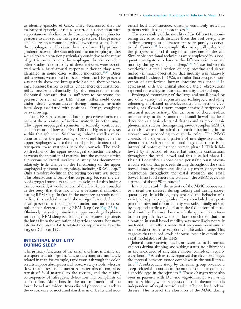
L
CHAPTER 27 • Gastrointestinal Physiology in Relation to Sleep 317
turnal fecal incontinence, which is commonly noted in patients with ileoanal anastomosis.50
The accessibility of the motility of the GI tract to moni-toring decreases with distance from the oral cavity. The earliest attempts at measurement were purely observa-tional. Cannon,51 for example, fluoroscopically observed the progress of food through the intestines of the cat. Similar observational techniques were employed by subse-quent investigators to describe the differences in intestinal motility during waking and sleep.52,53 These individuals exteriorized a small section of dog intestine and deter-mined via visual observation that motility was relatively unaffected by sleep. In 1926, a similar fluoroscopic obser-vation of exteriorized human intestine was made.54 In agreement with the animal studies, these observations reported no change in intestinal motility during sleep.
Prolonged monitoring of the large and small bowel by use of a variety of sophisticated techniques, including telemetry, implanted microelectrodes, and suction elec-trodes, has allowed a more comprehensive description of intestinal motor activity. On the basis of these studies, tonic activity in the stomach and small bowel has been described as a basic electrical rhythm and as more phasic phenomena, such as the migrating motor complex (MMC), which is a wave of intestinal contraction beginning in the stomach and proceeding through the colon. The MMC consists of a dependent pattern of interdigestive motor phenomena. Subsequent to food ingestion there is an interval of motor quiescence termed phase I. This is fol-lowed by a period of somewhat random contractions throughout the small bowel and this is called phase II. Phase III describes a coordinated peristaltic burst of con-tractile activity that proceeds distally throughout the small bowel. Food ingestion establishes a pattern of vigorous contraction throughout the distal stomach and small bowel. If no food enters the stomach, the MMC cycle has a period of about 90 minutes.55
In a recent study55 the activity of the MMC subsequent to a meal was assessed during waking and during subse-quent sleep. In addition, they assessed the behavior of a variety of regulatory peptides. They concluded that post-prandial intestinal motor activity was substantially altered by sleep, primarily a reduction in the fed pattern of intes-tinal motility. Because there was little appreciable altera-tion in peptide levels, the authors concluded that the alteration in small bowel motility was most likely neurally mediated. The authors noted that responses were similar to those described after vagotomy in the waking state. This suggests that reduced levels of arousal result in diminished vagal modulation of the ENS.
Jejunal motor activity has been described in 20 normal subjects during sleeping and waking states; no differences in the incidence of migrating motor complexes activity were found.56 Another study reported that sleep prolonged the interval between motor complexes in the small intes-tine.57 A subsequent study by the same group revealed a sleep-related diminution in the number of contractions of a specific type in the jejunum.58 These changes were also seen in patients with DU and vagotomies as well as in normal subjects, which suggests that this phenomenon is independent of vagal control and unaffected by duodenal disease. The issue of the alteration of the MMC during
to identify episodes of GER. They determined that the majority of episodes of reflux occurred in association with a spontaneous decline in the lower esophageal sphincter pressure to close to the intragastric pressure. This pressure decline creates a common cavity between the stomach and the esophagus, and because there is a 5-mm Hg pressure gradient between the stomach and the midesophagus, this would create a situation particularly conducive to the reflux of gastric contents into the esophagus. As also noted in other studies, the majority of these episodes were associ-ated with a brief arousal response, although they were identified in some cases without movement.47,48 Other reflux events were noted to occur when the LES pressure was clearly above the intragastric baseline, thereby creat-ing a pressure barrier to reflux. Under these circumstances, reflux occurs mechanically, by the creation of intra- abdominal pressure that is sufficient to overcome the pressure barrier in the LES. Thus, reflux could occur under these circumstances during transient arousals from sleep associated with positional change, coughing, or swallowing.
The UES serves as an additional protective barrier to prevent the aspiration of noxious material into the lungs. The upper esophageal sphincter is tonically contracted, and a pressure of between 40 and 80 mm Hg usually exists within this sphincter. Swallowing induces a reflex relax-ation to allow the positioning of food and liquids in the upper esophagus, where the normal peristaltic mechanism transports these materials into the stomach. The tonic contraction of the upper esophageal sphincter therefore prevents the ingestion of material into the esophagus with a previous volitional swallow. A study has documented relatively little change in the functioning of the upper esophageal sphincter during sleep, including REM sleep.49 Only a modest decline in the resting pressure was noted. This observation is somewhat surprising because the cri-copharyngeal muscle is a skeletal muscle, and if this finding can be verified, it would be one of the few skeletal muscles in the body that does not show a substantial inhibition during REM sleep. In fact, in the more recent study noted earlier, this skeletal muscle shows significant decline in basal pressure in the upper sphincter, and an increase, rather than decrease during REM sleep (see Fig. 27-3).44 Obviously, persisting tone in the upper esophageal sphinc-ter during REM sleep is advantageous because it protects the lungs from the aspiration of gastric contents. For more information on the GER related to sleep disorder breath-ing, see Chapter 127.
INTESTINAL MOTILITY DURING SLEEPThe primary functions of the small and large intestine are transport and absorption. These functions are intimately related in that, for example, rapid transit through the colon results in poor absorption and loose, watery stools, whereas slow transit results in increased water absorption, slow transit of fecal material to the rectum, and the clinical consequence of infrequent defecation and complaints of constipation. Alterations in the motor function of the lower bowel are evident from clinical phenomena, such as the occurrence of nocturnal diarrhea in diabetics and noc-
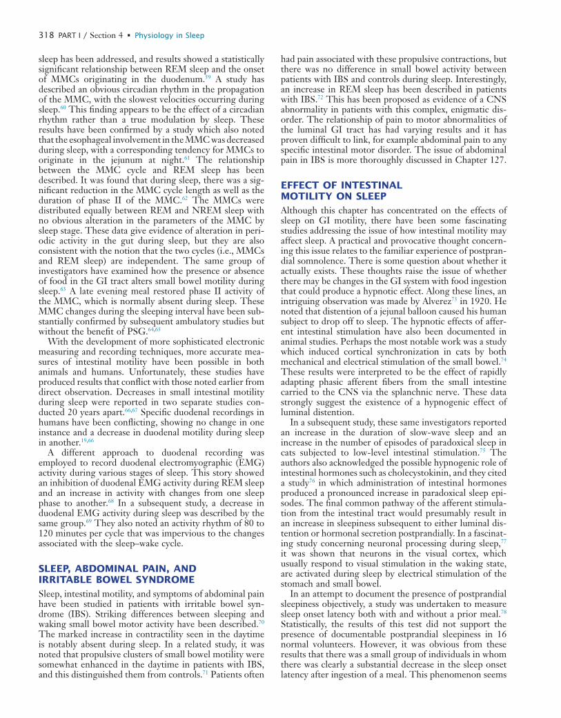
L
318 PART I / Section 4 • Physiology in Sleep
had pain associated with these propulsive contractions, but there was no difference in small bowel activity between patients with IBS and controls during sleep. Interestingly, an increase in REM sleep has been described in patients with IBS.72 This has been proposed as evidence of a CNS abnormality in patients with this complex, enigmatic dis-order. The relationship of pain to motor abnormalities of the luminal GI tract has had varying results and it has proven difficult to link, for example abdominal pain to any specific intestinal motor disorder. The issue of abdominal pain in IBS is more thoroughly discussed in Chapter 127.
EFFECT OF INTESTINAL MOTILITY ON SLEEPAlthough this chapter has concentrated on the effects of sleep on GI motility, there have been some fascinating studies addressing the issue of how intestinal motility may affect sleep. A practical and provocative thought concern-ing this issue relates to the familiar experience of postpran-dial somnolence. There is some question about whether it actually exists. These thoughts raise the issue of whether there may be changes in the GI system with food ingestion that could produce a hypnotic effect. Along these lines, an intriguing observation was made by Alverez73 in 1920. He noted that distention of a jejunal balloon caused his human subject to drop off to sleep. The hypnotic effects of affer-ent intestinal stimulation have also been documented in animal studies. Perhaps the most notable work was a study which induced cortical synchronization in cats by both mechanical and electrical stimulation of the small bowel.74 These results were interpreted to be the effect of rapidly adapting phasic afferent fibers from the small intestine carried to the CNS via the splanchnic nerve. These data strongly suggest the existence of a hypnogenic effect of luminal distention.
In a subsequent study, these same investigators reported an increase in the duration of slow-wave sleep and an increase in the number of episodes of paradoxical sleep in cats subjected to low-level intestinal stimulation.75 The authors also acknowledged the possible hypnogenic role of intestinal hormones such as cholecystokinin, and they cited a study76 in which administration of intestinal hormones produced a pronounced increase in paradoxical sleep epi-sodes. The final common pathway of the afferent stimula-tion from the intestinal tract would presumably result in an increase in sleepiness subsequent to either luminal dis-tention or hormonal secretion postprandially. In a fascinat-ing study concerning neuronal processing during sleep,77 it was shown that neurons in the visual cortex, which usually respond to visual stimulation in the waking state, are activated during sleep by electrical stimulation of the stomach and small bowel.
In an attempt to document the presence of postprandial sleepiness objectively, a study was undertaken to measure sleep onset latency both with and without a prior meal.78 Statistically, the results of this test did not support the presence of documentable postprandial sleepiness in 16 normal volunteers. However, it was obvious from these results that there was a small group of individuals in whom there was clearly a substantial decrease in the sleep onset latency after ingestion of a meal. This phenomenon seems
sleep has been addressed, and results showed a statistically significant relationship between REM sleep and the onset of MMCs originating in the duodenum.59 A study has described an obvious circadian rhythm in the propagation of the MMC, with the slowest velocities occurring during sleep.60 This finding appears to be the effect of a circadian rhythm rather than a true modulation by sleep. These results have been confirmed by a study which also noted that the esophageal involvement in the MMC was decreased during sleep, with a corresponding tendency for MMCs to originate in the jejunum at night.61 The relationship between the MMC cycle and REM sleep has been described. It was found that during sleep, there was a sig-nificant reduction in the MMC cycle length as well as the duration of phase II of the MMC.62 The MMCs were distributed equally between REM and NREM sleep with no obvious alteration in the parameters of the MMC by sleep stage. These data give evidence of alteration in peri-odic activity in the gut during sleep, but they are also consistent with the notion that the two cycles (i.e., MMCs and REM sleep) are independent. The same group of investigators have examined how the presence or absence of food in the GI tract alters small bowel motility during sleep.63 A late evening meal restored phase II activity of the MMC, which is normally absent during sleep. These MMC changes during the sleeping interval have been sub-stantially confirmed by subsequent ambulatory studies but without the benefit of PSG.64,65
With the development of more sophisticated electronic measuring and recording techniques, more accurate mea-sures of intestinal motility have been possible in both animals and humans. Unfortunately, these studies have produced results that conflict with those noted earlier from direct observation. Decreases in small intestinal motility during sleep were reported in two separate studies con-ducted 20 years apart.66,67 Specific duodenal recordings in humans have been conflicting, showing no change in one instance and a decrease in duodenal motility during sleep in another.19,66
A different approach to duodenal recording was employed to record duodenal electromyographic (EMG) activity during various stages of sleep. This story showed an inhibition of duodenal EMG activity during REM sleep and an increase in activity with changes from one sleep phase to another.68 In a subsequent study, a decrease in duodenal EMG activity during sleep was described by the same group.69 They also noted an activity rhythm of 80 to 120 minutes per cycle that was impervious to the changes associated with the sleep–wake cycle.
SLEEP, ABDOMINAL PAIN, AND IRRITABLE BOWEL SYNDROMESleep, intestinal motility, and symptoms of abdominal pain have been studied in patients with irritable bowel syn-drome (IBS). Striking differences between sleeping and waking small bowel motor activity have been described.70 The marked increase in contractility seen in the daytime is notably absent during sleep. In a related study, it was noted that propulsive clusters of small bowel motility were somewhat enhanced in the daytime in patients with IBS, and this distinguished them from controls.71 Patients often
L
CHAPTER 27 • Gastrointestinal Physiology in Relation to Sleep 319
First, it appears that spontaneous awakening does induce high-amplitude peristaltic contractions as described by Narducci and noted earlier, and this appears to be some-what different than colonic motor activity induced by a sudden awakening from sleep. In one study, sudden awak-enings from sleep induced a pattern of segmental colonic contractions,88 rather than the propagating high-amplitude peristaltic contractions noted in a previous study.82 These data are of considerable interest in that they demonstrate not only the influence of higher cortical functions on colonic motility but also the fact that the state of con-sciousness can affect the colon in rather subtle ways in terms of the induction of different patterns of colonic motility.
The striated muscle of the anal canal was evaluated during sleep in a 1953 study that included EEG docu-mentation of sleep.89 These investigators described a marked reduction in EMG activity during sleep. They concluded that this muscle is under voluntary control. In another study,90 anal canal pressure was measured continu-ously during sleep, but without PSG monitoring. The results indicated a decrease in the minute-to-minute varia-tion and the amplitude of spontaneous decreases in anal canal pressure during sleep. These results have been largely confirmed in a study involving the ambulatory monitoring of anorectal activity that noted a decrease in anal canal resting pressure during sleep, as well as altera-tions in rectal motor complexes (RMC) that were noted to be similar in waking and sleep in the midrectum.91 However, in the distal rectum, this motor pattern was found to be more prevalent during sleep. They also noted that during sleep, the anal canal resting pressure exceeded that of the rectal pressure, which would seem to be impor-tant in maintaining rectal continence during sleep. The structures of the rectum and anal canal are vital in main-taining normal bowel continence and ensuring normal defecation. In general, normal defecation is associated with sensory responses to rectal distention and appropriate motor responses of the muscles of the anal canal. These responses would include a contraction of the external anal sphincter and a transient decrease in the internal anal sphincter pressure associated with rectal distention. It is thought that the high resting basal pressure in the internal anal sphincter of the anal canal, as well as the response of the external anal sphincter to rectal distention, is critical in maintaining continence.
For assessment of the effect of sleep on these anorectal sensorimotor responses, 10 normal volunteers were studied during sleep with an anorectal probe in place.92 This probe permits the transient distention of the rectum via a rectal balloon while the responses of the internal and external anal sphincters can be simultaneously monitored. This study documented a marked decrease—and, in most sub-jects, an abolition—of the external anal sphincter response to rectal distention. The internal anal sphincter response remained unaltered. In addition, there was no evidence of an arousal response with up to 50 mL of rectal distention during sleep. The normal threshold of response in the waking state is approximately 10 to 15 mL. These results confirm that the external anal sphincter response to rectal distention is most likely a learned response, whereas the internal anal sphincter response is clearly a reflex response
to be affected by many variables including the volume of the meal, the meal constituents, and the circadian cycle of the individual. In a follow-up study,79 this same group tested the hypothesis that afferent stimulation from the gastric antrum would enhance postprandial sleepiness. This was tested by comparing an equal volume distention of the stomach with water to an equal caloric solid meal and liquid meal. Sleep onset latency was determined sub-sequent to each of these conditions. Because antral stimu-lation results from the digestion of a solid meal, sleep onset latency should be shorter subsequent to the consumption of the solid meal. This was confirmed in this study in that the sleep onset latency after the solid meal was significantly shorter than the equal volume water condition. These results are compatible with the animal studies cited earlier and lend further support to the notion that contraction of the lumen of the GI tract produces afferent stimulation, which induces drowsiness.
COLONIC AND ANORECTAL FUNCTION DURING SLEEPThe colon has two main functions: transport and absorp-tion. These are critically determined by the motor activity of the colon, which determines the rate of transport and, therefore, indirectly, the rate of absorption from the colonic lumen. Thus, alterations in colonic motility will have significant consequences in terms of transit through the colon and water absorption and ultimately clinical con-sequences such as constipation and diarrhea.
In the early 1940s a study described a decrease in colonic function during sleep.80 These results have been confirmed by two other studies that included measurements of the transverse, descending, and sigmoid colon.81,82 In one of these studies,82 a clear inhibition of colonic motility index is evident during sleep in the transverse, descending, and sigmoid colon segments, with a marked increase in activity on awakening. Certainly, this explains the common urge to defecate on awakening in the morning. Neither of these studies attempted to document sleep with standard PSG. However, in a subsequent study,83 colonic activity from cecum to rectum was monitored continuously for 32 hours, and sleep was monitored via PSG. This study again noted a rather marked decrease in colonic motor activity during sleep, but it also described an interesting abolition in propagating waves during slow-wave sleep. During REM sleep, the frequency of propagating events rose substan-tially. Other studies do not provide evidence for any sig-nificant change in colonic motility or variability in the rectosigmoid colon during sleep.84,85 However, a study of colonic myoelectrical activity in the human being sug-gested a decrease in spike activity during sleep.86 Again, this study does not determine whether the results are accounted for on the basis of true physiological sleep or simply reflect a circadian variation in colonic activity inde-pendent of sleep.
Collectively, these studies would suggest an inhibition of colonic contractile and myoelectric activity during sleep, and other studies have documented the fact that there is diminished colonic tone during sleep.87 Resumption of the waking state, and consequently increased CNS arousal, would suggest two different effects on colonic motility.
L
320 PART I / Section 4 • Physiology in Sleep
REFERENCES1. Friedenwald J. On the influence of rest, exercise and sleep on gastric
digestion. Am J Med 1906;1:249-255.2. Johnson RL, Washeim H. Studies in gastric secretion. Am J Physiol
1924;70:247-253.3. Henning N, Norpoth L. Die Magensekretion waehrend des Schlafes.
Dtsch Arch Klin Med 1932;172:558-562.4. Banche M. La secrezione gastric notturna durante il sonno. Minerva
Med 1950;1:428-434.5. Luckhardt AB. Contributions to the physiology of the empty
stomach. Am J Physiol 1915;39:330-333.6. Wada T. Experimental study of hunger in its relation to activity.
Arch Psychol 1922;8:1-65.7. Sandweiss DJ, Friedman HF, Sugarman MH, et al. Nocturnal gastric
secretion. Gastroenterology 1946;1:38-54.8. Moore JG, Englert E. Circadian rhythm of gastric acid secretion in
man. Nature 1970;226:1261-1262.9. Levin E, Kirsner JB, Palmer WL, et al. A comparison of the noctur-
nal gastric secretion in patients with duodenal ulcer and in normal individuals. Gastroenterology 1948;10:952-964.
10. Feldman M, Richardson CT. Total 24-hour gastric acid secretion in patients with duodenal ulcer: comparison with normal subjects and effects of cimetidine and parietal cell vagotomy. Gastroenterology 1986;90:540-544.
11. Dragstedt LR. A concept of the etiology of gastric and duodenal ulcers. Gastroenterology 1956;30:208-220.
12. Levin E, Kirsner JB, Palmer WL, et al. The variability and periodic-ity of the nocturnal gastric secretion in normal individuals. Gastro-enterology 1948;10:939-951.
13. Reichsman F, Cohen J, Colwill J, et al. Natural and histamine-induced gastric secretion during waking and sleeping states. Psycho-som Med 1960;1:14-24.
14. Armstrong RH, Burnap D, Jacobson A, et al. Dreams and acid secre-tions in duodenal ulcer patients. New Physician 1965;33:241-243.
15. Stacher G, Presslich B, Starker H. Gastric acid secretion and sleep stages during natural night sleep. Gastroenterology 1975;68:1449- 1455.
16. Orr WC, Hall WH, Stahl ML, et al. Sleep patterns and gastric acid secretion in duodenal ulcer disease. Arch Intern Med 1976;136: 655-660.
17. Dubois A, Castell DO. Abnormal gastric emptying response to pen-tagastrin in duodenal ulcer disease. Dig Dis Sci 1981;26:292-296.
18. Scantlebury RE, Frick HL, Patterson TL. The effect of normal and hypnotically induced dreams on the gastric hunger movements of man. J Appl Physiol 1942;26:682-691.
19. Bloom PB, Ross DL, Stunkard AJ, et al. Gastric and duodenal motil-ity, food intake and hunger measured in man during a 24-hour period. Dig Dis Sci 1970;15:719-725.
20. Baust W, Rohrwasser W. Das Verhalten von pH und Motilitat des Megens in naturlichen Schlaf des Menschen. Pflugers Arch 1969;305:229-240.
21. Yaryura-Tobias HA, Hutcheson JS, White L. Relationship between stages of sleep and gastric motility. Behav Neuropsychiatry 1970;2:22-24.
22. Fujitani Y, Hosogai M. Circadian rhythm of electrical activity and motility of the stomach in cats and their relation to sleep-wakefulness states. Tohoku J Exp Med 1983;141:275-285.
23. Hall WH, Orr WC, Stahl ML. Gastric function during sleep. In: Brooks FP, Evers PW, editors. Nerves and the gut. Thorofare, NJ: Slack; 1977. p. 495-502.
24. Dubois A, Van Eerdewegh P, Gardner JD. Gastric emptying and secretion in Zollinger-Ellison syndrome. J Clin Invest 1977;59: 255-263.
25. Orr WC, Dubois A, Stahl ML, et al. Gastric function during sleep (Abstract). Sleep Res 1978;7:72.
to rectal distention because it persists during sleep. It also raises certain clinical questions with regard to the phenom-enon of nocturnal diarrhea and the maintenance of fecal continence during sleep. In an ambulatory study of ano-rectal functioning, it was demonstrated that external anal sphincter contractions occurred periodically during sleep, and these periodic bursts of activity were followed by motor quiescence.93 These spontaneous contractions were associated with a rise in the anal canal pressure, but inter-nal anal sphincter contractions were shown to occur inde-pendently of external anal sphincter activity. The sampling reflex, which is a spontaneous relaxation of the internal anal sphincter, occurred frequently in the waking state but was markedly reduced during sleep.
An important study has shed light on intrinsic anal-rectal functioning, which is altered during sleep.94 These investigators confirmed the presence of an endogenous oscillation in rectal motor activity, and they have specifi-cally noted that these bursts of cyclic rectal motor activity occupied approximately 44% of the overall recording time at night. They described the incidence of this motor activ-ity to be nearly twofold greater at night than during the daytime. Of particular importance is the finding that the majority of contractions were propagated in a retrograde direction. Other studies have shown that rectal motor activity is not altered by REM sleep.90 Two other studies have shown that anal canal pressure is decreased during sleep.95,96 However, of particular interest is the fact that even though there was a diminution in anal canal pressure during sleep, anal canal pressure was always greater than rectal pressure even in the presence of cyclic rectal motor activity.95
These studies collectively shed important light on the mechanisms of rectal continence during sleep. There appear to be at least two mechanisms that prevent the passive escape of rectal contents during sleep. First, rectal motor activity increases substantially during sleep, but the propagation is retrograde rather than anterograde. Fur-thermore, these physiologic studies have shown that, even under the circumstances of periodic rectal contractions, the anal canal pressure is consistently above that of the rectum. Both of these mechanisms would tend to protect against rectal leakage during sleep, and alterations in these mechanisms would explain loss of rectal continence during sleep in individuals with diabetes or who have undergone ileal-anal anastomosis.
CONCLUSIONSThere are marked alterations in the GI system during sleep, and these have numerous consequences in terms of normal digestive processes as well as digestive disease (reviewed in Chapter 127). Our understanding of the mod-ulation of GI function by sleep has increased. In the future, research will focus on the direct effects on the GI physiol-ogy of orexins A and B and other neuropeptides that play a role in arousal and sleep as well as on appetite, regulation of feeding, and energy homeostasis.97 Clearly, the past 50 years have shown a marked increase in interest in sleep and GI physiology, and this will undoubtedly continue as the importance of sleep physiology and pathophysiology is further revealed.
� Clinical Pearl
Suppression of nocturnal acid secretion is an impor-tant element of healing duodenal ulcers and remains a mainstay in the treatment of GERD. This in turn can reduce the pulmonary complications of GERD.
L
CHAPTER 27 • Gastrointestinal Physiology in Relation to Sleep 321
52. Barcroft J, Robinson CS. A study of some factors influencing intes-tinal movements. Am J Physiol 1929;67:211-220.
53. Douglas DM, Mann FG. An experimental study of the rhythmic contractions in the small intestine of the dog. Am J Dig Dis 1977;6:318-322.
54. Hines LE, Mead HCA. Peristalsis in a loop of small intestine. Arch Intern Med 1926;38:536-543.
55. Soffer EE, Adrian TE, Launspach J, et al. Meal-induced secretion of gastrointestinal regulatory peptides is not affected by sleep. Neuro-gastroenterol Motil 1997;9:7-12.
56. Archer L, Benson MJ, Green WJ, et al. Radiotelemetric measure-ment of normal human small bowel motor activity during prolonged fasting. J Physiol 1979;296:53P.
57. Thompson DG, Wingate DL. Characterisation of interdigestive and digestive motor activity in the normal human jejunum (Abstract). Gut 1979;20:A943.
58. Ritchie HD, Thompson DG, Wingate DL. Diurnal variation in human jejunal fasting motor activity. In: Proceedings of the American Physiological Society. March 1980; p. 54-55.
59. Finch P, Ingram D, Henstridge J, et al. The relationship of sleep stage to the migrating gastrointestinal complex of man. In: Chris-tensen J, editor. Gastrointestinal motility. New York: Raven Press; 1980. p. 261-265.
60. Kumar D, Wingate D, Ruckebusch Y. Circadian variation in the propagation velocity of the migrating motor complex. Gastroenterol-ogy 1986;91:926-930.
61. Kellow JE, Borody TJ, Phillips SF, et al. Human interdigestive motility: variations in patterns from esophagus to colon. Gastroen-terology 1986;91:386-395.
62. Kumar D, Idzikowski C, Wingate DL, et al. Relationship between enteric migrating motor complex and the sleep cycle. Am J Physiol Gastrointest Liver Physiol 1990;259(6):G983-G990.
63. Kumar D, Soffer EE, Wingate DL, et al. Modulation of the duration of human postprandial motor activity by sleep. Am J Physiol 1989;256(5):G851-G855.
64. Wilson P, Perdikis G, Hinder RA, et al. Prolonged ambulatory antroduodenal manometry in humans. Am J Gastroenterol 1994; 89:1489-1495.
65. Wilmer A, Andrioli A, Coremans G, et al. Ambulatory small intes-tinal manometry. Detailed comparison of duodenal and jejunal motor activity in healthy man. Dig Dis Sci 1997;42:1618-1627.
66. Helm JD, Kramer P, MacDonald RM, et al. Changes in motility of the human small intestine during sleep. Gastroenterology 1948; 10:135-137.
67. Sadler HH, Orten AU. The complementary relationship between the emotional state and the function of the ileum in a human subject. Am J Psychiatry 1968;124:1377-1381.
68. Spire JP, Tassinari CA. Duodenal EMG activity during sleep. Elec-troencephalogr Clin Neurophysiol 1971;31:179-183.
69. Tassinari CA, Coccagna G, Mantovani M, et al. Duodenal EMG activity during sleep in man. In: Jovanovic UJ, editor. The nature of sleep. Stuttgart, Germany: Gustav Fischer Verlag; 1973.
70. Kellow JE, Gill RC, Wingate DL. Prolonged ambulant recordings of small bowel motility demonstrate abnormalities in the irritable bowel syndrome. Gastroenterology 1990;98:1208-1218.
71. Kellow JE, Phillips SF. Altered small bowel motility in irritable bowel syndrome is correlated with symptoms. Gastroenterology 1987;92:1885-1893.
72. Kumar D, Thompson PD, Wingate DL, et al. Abnormal REM sleep in the irritable bowel syndrome. Gastroenterology 1992; 103:12-17.
73. Alverez WC. Physiologic studies on the motor activities of the stomach and bowel in man. Am J Physiol 1920;88:658-660.
74. Kukorelli T, Juhasz G. Sleep induced by intestinal stimulation in cats. Physiol Behav 1976;19:355-358.
75. Juhasz G, Kukorelli T. Modifications of visceral evoked potentials during sleep in cats. Act Nerv Super (Praha) 1977;19:212-214.
76. Rubenstein EH, Sonnenschein RR. Sleep cycles and feeding behavior in the cat: role of gastrointestinal hormones. Acta Cient Venez 1971;22:125-128.
77. Pigarev IN. Neurons of visual cortex respond to visceral stimulation during slow wave sleep. Neuroscience 1994;62:1237-1243.
78. Stahl ML, Orr WC, Bollinger C. Postprandial sleepiness: objective documentation via polysomnography. Sleep 1983;6:29-35.
79. Orr WC, Shadid G, Harnish MJ, et al. Meal composition and its effect on postprandial sleepiness. Physiol Behav 1997;62:709-712.
26. Goo RH, Moore JG, Greenburg E, et al. Circadian variation in gastric emptying of meals in humans. Gastroenterology 1987;93: 515-518.
27. Chen JZ, McCallum RW, Familoni BO. Validity of the cutaneous electrogastrogram. In: Chen JZ, McCallum RW, editors. Electrogas-trography: principles and applications. New York: Raven Press; 1994. p. 103-125.
28. Chen JZ, McCallum RW, Smout AJPM, et al. Acquisition and analy-sis of electrogastrographic data. In: Chen JZ, McCallum RW, editors. Electrogastrography: principles and applications. New York: Raven Press; 1994. p. 3-30.
29. Chen JZ, McCallum RW, Lin Z. Comparison of three running spectral analysis methods. In: Chen JZ, McCallum RW, editors. Electrogastrography: principles and applications. New York: Raven Press; 1994. p. 75-99.
30. Orr WC, Crowell MD, Lin B, et al. Sleep and gastric function in irritable bowel syndrome: derailing the brain-gut axis. Gut 1997;41:390-393.
31. Elsenbruch S, Harnish MJ, Orr WC, et al. Disruption of normal gastric myoelectric functioning by sleep. Sleep 1999;22:453-458.
32. Johnson LF, DeMeester TR. Twenty-four-hour pH monitoring of the distal esophagus: a quantitative measure of gastroesophageal reflux. Am J Gastroenterol 1974;62:325-332.
33. DeMeester TR, Johnson LF, Joseph CJ, et al. Patterns of gastro-esophageal reflux in health and disease. Ann Surg 1976;184: 459-470.
34. Orr WC, Johnson LF, Robinson MG. The effect of sleep on swal-lowing, esophageal peristalsis, and acid clearance. Gastroenterology 1984;86:814-819.
35. Orr WC, Robinson MG, Johnson LF. Acid clearing during sleep in patients with esophagitis and controls. Dig Dis Sci 1981;26:423.
36. Dent J, Dodds WJ, Friedman RH, et al. Mechanism of gastroesopha-geal reflux in recumbent asymptomatic human subjects. J Clin Invest 1980;65:256-257.
37. Dickman R, Shapiro M, Malagon IB, et al. Assessment of 24-h oesophageal pH monitoring should be divided to awake and asleep rather than upright and supine time periods. Neurogastroenterol Motil 2007;19:709-715.
38. Helm JF, Dodds WJ, Hogan WJ, et al. Acid neutralizing capacity of human saliva. Gastroenterology 1982;83:69-74.
39. Schneyer LH, Pigman W, Hanahan L, et al. Rate of flow of human parotid, sublingual, and submaxillary secretions during sleep. J Dent Res 1956;35:109-114.
40. Lear CSC, Flanagan JB Jr, Moorees CFA. The frequency of degluti-tion in man. Arch Oral Biol 1965;10:83-96.
41. Lichter J, Muir RC. The pattern of swallowing during sleep. Elec-troencephalogr Clin Neurophysiol 1975;38:427-432.
42. Anderson CA, Dick TE, Orem J. Swallowing in sleep and wakeful-ness in adult cats. Sleep 1995;18:325-329.
43. Castiglione F, Emde C, Armstrong D, et al. Nocturnal oesophageal motor activity is dependent on sleep stage. Gut 1993;34:1653- 1659.
44. Bajaj JS, Bajaj S, Dua KS, et al. Influence of sleep stages on esophago-upper esophageal sphincter contractile reflex and secondary esopha-geal peristalsis. Gastroenterology 2006:130:17-25.
45. Armstrong D, Emde C, Bumm R, et al. Twenty-four-hour pattern of esophageal motility in asymptomatic volunteers. Dig Dis Sci 1990;35:1190-1197.
46. Smout AJPM, Breedijk M, van der Zouw C, et al. Physiological gastroesophageal reflux and esophageal motor activity studied with a new system for 24-hour recording and automated analysis. Dig Dis Sci 1989;34:372-378.
47 Freidin N, Fisher MJ, Taylor W, et al. Sleep and nocturnal acid reflux in normal subjects and patients with reflux oesophagitis. Gut 1991;32:1275-1279.
48. Penzel T, Becker HR, Brandenburg U, et al. Arousal in patients with gastro-oesophageal reflux and sleep apnoea. Eur Respir J 1999; 14:1266-1270.
49. Kahrilas PJ, Dodds WJ, Dent J, et al. Effect of sleep, spontaneous gastroesophageal reflux, and a meal on upper esophageal sphincter pressure in normal human volunteers. Gastroenterology 1987;92: 466-467.
50. Metcalf AM, Dozois RR, Kelly KA, et al. Ileal J pouch-anal anasto-mosis: clinical outcome. Ann Surg 1985;202:735-739.
51. Cannon WB. The movements of the intestine studied by means of the roentgen rays. Am J Physiol 1902;6:275-276.
L
322 PART I / Section 4 • Physiology in Sleep
89. Floyd WF, Walls EW. Electromyography of the sphincter and exter-nus in man. J Physiol 1953;122:599-609.
90. Orkin BA, Hanson RB, Kelly KA, et al. Human anal motility while fasting, after feeding, and during sleep. Gastroenterology 1991;100: 1016-1023.
91. Ronholt C, Rasmussen O, Christiansen J. Ambulatory manometric recording of anorectal activity. Dis Colon Rectum 1999;12: 1551-1559.
92. Whitehead WE, Orr WC, Engel BT, et al. External anal sphincter response to rectal distention: learned response or reflex. Psycho-physiology 1981;19:57-62.
93. Kumar D, Waldron D, Williams NS, et al. Prolonged anorectal manometry and external anal sphincter electromyography in ambu-lant human subjects. Dig Dis Sci 1990;35:641-648.
94. Rao SS, Welcher K. Periodic rectal motor activity: the intrinsic colonic gatekeeper? Am J Gastroenterol 1996;91:890-897.
95. Ferrara A, Pemberton JH, Levin KE, et al. Relationship between anal canal tone and rectal motor activity. Dis Colon Rectum 1993; 36:337-342.
96. Enck P, Eggers E, Koletzko S, et al. Spontaneous variation of anal “resting” pressure in healthy humans. Am J Physiol 1991;261: G823-G826.
97. Baccari BC. Orexins and gastrointestinal functions. Curr Protein Pept Sci 2010;11:148-155.
80. Adler HF, Atkinson AJ, Ivy AC. A study of the motility of the human colon: an explanation of dyssynergia of the colon, or of the unstable colon. Am J Dig Dis 1941;8:197-202.
81. Rosenblum MJ, Cummins AJ. The effect of sleep and of Amytal on the motor activity of the human sigmoid colon. Gastroenterology 1954;27:445-450.
82. Narducci F, Bassotti G, Gaburri M, et al. Twenty four hour mano-metric recording of colonic motor activity in healthy man. Gut 1987;28:17-25.
83. Furukawa V, Cook IJ, Panagopoulos V, et al. Relationship between sleep patterns and human colonic motor patterns. Gasteroenerology 1994;107:1372-1381.
84. Kerlin P, Zinsmeister A, Phillips S. Motor responses to food of the ileum, proximal colon, and distal colon of healthy humans. Gastro-enterology 1983;84:762-770.
85. Posey EL, Bargen JA. Observations of normal and abnormal human intestinal motor function. Am J Med Sci 1951;221:10-20.
86. Frexinos J, Bueno L, Fioramonti J. Diurnal changes in myoelectric spiking activity of the human colon. Gastroenterology 1985;88: 1104-1110.
87. Steadman CJ, Phillips SF, Camilleri M, et al. Variation of muscle tone in the human colon. Gastroenterology 1991;101:373-381.
88. Bassotti G, Bucaneve G, Betti C, et al. Sudden awakening from sleep: effects on proximal and distal colonic contractile activity in humans. Eur J Gastroenterol Hepatol 1990;2:475-478.