modafinil and γ-hydroxybutyrate have sleep state-specific pharmacological actions on hypocretin-1...
TRANSCRIPT
Original article 643
Modafinil and c-hydroxybutyrate have sleep state-specificpharmacological actions on hypocretin-1 physiologyin a primate model of human sleepJamie M. Zeitzera,c, Christine L. Buckmastera, Hans-Peter Landoltd,David M. Lyonsa and Emmanuel Mignota,b
Hypocretin-1 is a hypothalamic neuropeptide that is
important in the regulation of wake and the lack of which
results in the sleep disorder narcolepsy. Using a monkey
that has consolidated wake akin to humans, we examined
pharmacological manipulation of sleep and wake and its
effects on hypocretin physiology. Monkeys were given
the sleep-inducing c-hydroxybutyrate (GHB) and the
wake-inducing modafinil both in the morning and in the
evening. Cerebrospinal fluid hypocretin-1 concentrations
changed significantly in response to the drugs only when
accompanied by a behavioral change (GHB-induced
sleep in the morning or modafinil-induced wake in the
evening). We also found that there was a large (180-fold)
interindividual variation in GHB pharmacokinetics that
explains variability in sleep induction in response to
the drug. Our data indicate that the neurochemical
concomitants of sleep and wake are capable of changing
the physiological output of hypocretin neurons. Sleep
independent of circadian timing is capable of decreasing
cerebrospinal fluid hypocretin-1 concentrations.
Furthermore, hypocretin neurons do not seem to respond
to an ‘effort’ to remain awake, but rather keep track
of time spent awake as a wake-promoting counterbalance
to extended wakefulness. Behavioural Pharmacology
20:643–652 �c 2009 Wolters Kluwer Health | Lippincott
Williams & Wilkins.
Behavioural Pharmacology 2009, 20:643–652
Keywords: gamma-hydroxybutyrate, hypocretin, modafinil, orexin,pharmacology, sleep
aDepartment of Psychiatry and Behavioral Sciences, bHoward Hughes MedicalInstitute, Stanford University, Stanford, cPsychiatry Service, VA Palo Alto HealthCare System, Palo Alto, California, USA and dInstitute of Pharmacology andToxicology and Zurich Center for Integrative Human Physiology, University ofZurich, Zurich, Switzerland
Correspondence to Jamie M. Zeitzer, PhD, 3801 Miranda Avenue (151Y),Palo Alto, CA 94304, USAE-mail: [email protected]
Received 3 December 2008 Accepted as revised 26 July 2009
IntroductionHypocretin-1 is a neuropeptide produced in the lateral
hypothalamus. Although it is likely involved in the regu-
lation of multiple behaviors (Berridge and Espana, 2005),
a major function across species may be the maintenance
of extended wake, most notably in wake-consolidated
mammals (Zeitzer and Mignot, 2006). The release of
hypocretin-1 is partially controlled by the circadian clock
(Deboer et al., 2004; Zhang et al., 2004), with elevated
release normally occurring late in the wake period in
both nocturnal (Yoshida et al., 2001) and diurnal (Zeitzer
et al., 2003) mammals. Hypocretin is wake promoting
when injected into the central nervous system (Hagan
et al., 1999; Yoshimichi et al., 2001) and increased wake
transitions occur following optogenic stimulation of
hypocretin neurons in vivo (Adamantidis et al., 2007).
Forced wake can also cause an increase in hypocretin-1
concentrations in hypothalamic dialysate (Yoshida et al.,2001) or cerebrospinal fluid (CSF) (Deboer et al., 2004;
Zeitzer et al., 2007). It is hypothesized that this increase
contributes to the actual extension of wake, opposing the
mounting sleep debt. Similarly, hypocretin decreases
upon sleep onset, with minimal levels late in the inactive
period, possibly helping to maintain sleep in the face
of reduced sleep debt. Problematically, however, it is not
known whether the increased hypocretin levels that have
been reported with sleep deprivation are a reflection of
the need for higher hypocretin tone to further stimulate
wake, or may simply be invoked by the sleep deprivation
procedure and its behavioral correlates. It is also unknown
whether the decline in CSF hypocretin-1 concentrations,
that is, observed during the time of normal sleep is
because of sleep itself or a circadian signal.
The study of hypocretin-1 physiology is difficult in
humans because significant concentrations of this pep-
tide are only found in the CSF and brain tissue. Lumbar
CSF concentrations of hypocretin-1 exhibit a diurnal
rhythm (Salomon et al., 2003), but the amplitude is quite
small and the rhythm is likely delayed by several hours
because of the transit time between brain release and
equilibration in the lumbar sac. Most animal models (e.g.
rats, cats, dogs, mice) of hypocretin-1 physiology are also
problematic, because the relationship between sleep
and circadian rhythms is fundamentally different in
humans compared with most other mammals. Human
sleep typically occurs as a single daily episode, whereas in
most other mammals sleep is polyphasic, in that it occurs
in brief bouts throughout the day and night with group-
ing of the bouts during daytime (diurnal), night time
(nocturnal), or at dawn/dusk (crepuscular). Given that
hypocretin is likely involved in the maintenance of
0955-8810 �c 2009 Wolters Kluwer Health | Lippincott Williams & Wilkins DOI: 10.1097/FBP.0b013e328331b9db
Copyright © Lippincott Williams & Wilkins. Unauthorized reproduction of this article is prohibited.

consolidated daytime wake in humans, it is critical to
examine its physiology in an animal model that has
an equivalent maintenance of wake. As such, we have
developed a primate model to study the normal
physiology of hypocretin-1 – the squirrel monkey (Zeitzer
et al., 2003, 2004, 2007). These New World primates
have consolidated sleep and wake akin to humans, unlike
most other mammals, and CSF is readily accessible
through a percutaneous tap of the cerebellomedullary
cistern (cisterna magna).
In this study, we tested the hypothesis that sleep itself
can cause a decrease in CSF hypocretin-1 concentrations.
Furthermore, we investigated whether the increase in
CSF hypocretin-1 concentrations in response to sleep
deprivation (Yoshida et al., 2001; Deboer et al., 2004;
Zeitzer et al., 2007) occurs because of an extension of
wake or whether this increase only reflects a compensa-
tory effort to remain awake when forced to do so by
an experimental protocol (e.g. secondary to cortisol or
other neurochemical changes). To induce sleep, we used
sodium oxybate, also called g-hydroxybutyrate (GHB).
This compound is known to greatly increase slow wave
sleep (Crunelli et al., 2006), which is in contrast to most
other currently used hypnotics, such as benzodiazepines.
GHB is typically used at night (bi-nightly administration)
to consolidate sleep in hypocretin-deficient humans
with narcolepsy, an effect that subsequently improves
daytime wake (and other symptoms) in these individuals.
To produce wake, we used modafinil, a stimulant that
seems to have fewer unwanted effects than other stimu-
lant compounds and does not strongly affect sleep state
distribution in subsequent sleep (Shelton et al., 1995).
Modafinil is a common wake-promoting agent used in the
treatment of narcolepsy and other conditions associated
with excessive daytime sleepiness. Of interest, there have
been reports linking the mechanism of action modafinil
to activation of hypocretin-producing neurons (Chemelli
et al., 1999; Scammell et al., 2000). As controls, we also
administered GHB at night, during which time it would
be expected to have no effect on the amount of sleep
observed, and we administered modafinil during the day
during which time it would be expected to have no effect
on the amount of wake observed. We also have examined
changes in plasma cortisol as we wanted to ensure that
changes we observed were not because of the nonspecific
stress of maintaining wake, as occurs in every study of
behavioral sleep deprivation (e.g. gentle handling, forced
locomotion) in nonhuman mammals. These controls allow
us to discriminate the direct effects of the drugs per seand the indirect effects of the drugs mediated through
changes in sleep and wake.
MethodsSubjects
Six adult, male squirrel monkeys (Saimiri sciureus sciureus)from the Stanford University squirrel monkey colony
were examined in this study. Monkeys were group housed
in two cages in the same room under constant humidity
and temperature, with a 12/12 h light/dark cycle (room
lights on at 07.00 h, room lights off at 19.00 h). Dim
yellow lighting was always on during hours of sche-
duled darkness, wherein yellow light does not interfere
with circadian entrainment in these monkeys (Zeitzer
and Buckmaster, unpublished observations). Food (New
World Primate Diet 5040, PMI Nutrition International,
Brentwood MO; fresh fruits and vegetables) was freely
available and replenished daily at 10.00 h, except on
experimental days, when this was done at 13.00 h. All
husbandry and experimental procedures were reviewed
and approved by the Stanford University Administrative
Panel on Laboratory Animal Care and carried out in
accordance with the Guide for the Care and Use of
Laboratory Animals as adopted and promulgated by the
US National Institutes of Health.
General protocol
Each monkey had CSF and blood obtained on six separate
occasions, with at least 2 weeks between each occasion.
Three samples were obtained at 13.00 h – one after
placebo administration, one after GHB administration,
and one after modafinil administration. The other three
samples were obtained at 01.00 h – one after placebo
injections, one after GHB administration, and one after
modafinil administration. All samples were assayed in
duplicate for CSF hypocretin-1 and cortisol. Blood samples
obtained after GHB administration were assayed for
GHB concentrations and those obtained after modafinil
administration were assayed for modafinil concentrations.
Activity patterns were monitored throughout the protocol;
in earlier studies, these have been shown to accurately
reflect sleep (Zeitzer et al., 2003).
Drug administration
In preliminary testing, based on doses used to treat
humans with narcolepsy and concentrations administered
in behavioral studies of rhesus monkeys (Snead, 1978;
Nakamura et al., 1987; Beardsley et al., 1996; Woolverton
et al., 1999), we determined the dose of GHB needed to
induce sleep in monkeys from 07.00 to 13.00 h (con-
firmed by visual observation). In the Xyrem formulation
(Jazz Pharmaceuticals, California, USA), GHB is in
suspension at 500 mg/ml. Although administered orally
in humans, we were unable to orally administer GHB in
this formulation because the monkeys refused to ingest
GHB that had been mixed with any of their favorite treats
(e.g. marshmallows, peanut butter, jelly). We, therefore,
administered GHB in the Xyrem formulation using
intramuscular injections into the hip flexor. A single dose
of GHB did not induce continuous sleep from 07.00
to 13.00 h, likely because of the short half-life of the
compound (0.5–1 h in humans). Therefore, we adminis-
tered 200 mg/kg of GHB at 06.00 h and 175 mg/kg
of GHB at 10.00 h (for CSF and blood collection at
644 Behavioural Pharmacology 2009, Vol 20 No 7
Copyright © Lippincott Williams & Wilkins. Unauthorized reproduction of this article is prohibited.

13.00 h) and 200 mg/kg of GHB at 18.00 h and 175 mg/kg
of GHB at 22.00 h (for CSF and blood collection at
01.00 h). This dosing schedule did not induce significant
side effects (e.g. nausea and ataxia).
In preliminary testing, based on doses typically adminis-
tered to human children and administered to rhesus
monkeys and marmosets in behavioral studies (Lagarde
and Milhaud, 1990; Herman et al., 1991; Ivanenko et al.,2003; Van Vliet et al., 2008), we determined the dose of
modafinil needed to keep monkeys awake from 19.00 to
01.00 h. We found that 16 mg/kg of modafinil kept the
monkeys awake for 3 h and repeated dosing could keep
them awake for 6 h. We packaged 16 mg/kg of modafinil
(Lafon, Maisons-Alfort, France) into gelatin capsules and
placed the capsules inside a miniature marshmallow, a
favorite treat of our squirrel monkeys. The marshmallows
capsules were given to the monkeys at 07.00 and 10.00 h
(for CSF and blood collection at 13.00 ) or at 19.00 and
22.00 h (for CSF and blood collection at 01.00 h).
In the third condition (placebo), we administered 0.4 ml
sterile water into the hip flexor at 07.00 and 10.00 h (for
CSF and blood collection at 13.00 h) or at 19.00 and
22.00 h (for CSF and blood collection at 01.00 h).
Activity monitoring
All monkeys wore a collar-mounted actigraph (Actiwatch-
64; MiniMitter, Bend, Oregon, USA), a small (17 g)
device capable of detecting movement in three dimen-
sions through the use of an accelerometer. Data were
stored as integrated (total) movement (arbitrary units)
occurring over 1-min intervals. Actigraphs were worn
throughout the protocol and at least 1 week of adaptation
occurred before the collection of analyzable data. Sleep
and wake were estimated from movement data using
Sleepwatch software (v.2.82, Cambridge Neurotechnology,
Cambridge, UK) set on low sensitivity, which has been
shown to accurately reflect sleep and wake in squirrel
monkeys (Zeitzer et al., 2003). Data were integrated
using the trapezoidal method from 19.00 to 01.00 h (for
CSF collection at 01.00 h) or 07.00 to 13.00 h (for CSF
collection at 13.00 h). Actigraphy was unsuccessful in
three of 36 collection intervals (one 13.00 h baseline
collection, one 13.00 h modafinil collection, one 01.00 h
modafinil collection).
CSF and blood collection
CSF was obtained through a percutaneous tap from the
cisterna magna (cerebellomedullary cistern) from anes-
thetized monkeys using a siliconized syringe (Zeitzer
et al., 2003). Anesthesia was induced by intrasaphenous
injection of 10.0 mg/kg ketamine hydrochloride with
0.5 mg/kg diazepam, and supplemented as needed
with an intramuscular injection of 5.0 mg/kg ketamine
hydrochloride. CSF (200 ml) was collected, placed in a
siliconized tube on ice, and later stored at – 801C. Blood
was obtained through puncture of the femoral vein. Blood
was spun in a centrifuge (4900 rpm, 41C, 14 min) with the
resulting supernatant being collected and stored at – 801C.
Assays
Blood concentrations of GHB were determined by
Covance Laboratories (Madison, Wisconsin, USA) using
liquid chromatography with tandem mass spectrometric
detection. Blood concentrations of modafinil were deter-
mined using liquid chromatography/mass spectrometry
(limit of detection: 0.7 ng/ml; limit of quantification:
2.4 ng/ml; signal-to-noise ratio: 3 and 10, respectively)
(Bodenmann et al., in preparation). Hypocretin-1 concen-
trations were determined using a commercially available
radioimmunoassay kit and a custom hypocretin-1 anti-
body, provided by Dr Shahrad Taheri and Dr Ling Lin
(detection limit = 10 pg/ml, 5% intra-assay variability, inter-
assay variability adjusted with internal standard, Phoenix
Pharmaceuticals, Belmont, California, USA) (Zeitzer et al.,2003). We selected the two CSF sampling time points of
13.00 and 01.00 h, because our earlier research (Zeitzer
et al., 2003) showed that CSF hypocretin-1 values are
intermediate at these times and, thus, could be observed to
increase or decrease without a concern of a ceiling or
floor effect. Plasma concentrations of cortisol were deter-
mined using a commercially available radioimmunoassay
(Diagnostic Products Inc., Los Angeles, California, USA).
All samples were assayed in duplicate.
Statistics
Student’s t-tests and basic calculations were carried
out using Microsoft Excel (v. 11.8211.8202; Redmond,
Washington, USA). Curve fitting of concentration–
response data was performed using Microcal Origin
(v. 6.1052; Northampton, Massahusetts, USA). Repeated-
measure two-factor analyses of variance and Spearman’s
rank correlation analyses were performed using SAS
Statview (v. 5.0.1; Cary, North Carolina, USA). To deter-
mine significance of change in the amount of movement
or wake, data were compared as change from time-
matched baseline to respective drug condition. The
absorbance and body distribution of some drugs are
affected by either body mass index (BMI) or body surface
area (BSA). BMI was calculated as kg/m2. Height was
measured as the crown–rump length and this distance
was used in the BMI calculation. BMI calculated with
crown–rump length had a better correlation with plasma
leptin levels, a fat cell-produced hormone known to
correlate with BMI in humans, than when other measures
of height were used to calculate BMI (Zeitzer et al.,2007). BSA was calculated as (cm� kg� 0.0002778)0.5
(Mosteller, 1987). All data are presented as mean ±
standard error of the mean.
ResultsAs expected, morning administration of the sleep-
promoting compound GHB increased the amount of
Effects of GHB and modafinil on hypocretin-1 Zeitzer et al. 645
Copyright © Lippincott Williams & Wilkins. Unauthorized reproduction of this article is prohibited.

sleep, whereas administration of the wake-promoting
compound modafinil had negligible effects on wake
during the daytime. After morning administration of
GHB, there was a significant 57 ± 17% decline in the
amount of movement, as compared with the control
condition (P < 0.05, paired t-test) (Fig. 1). In deriving
estimated sleep/wake from actigraphy data, there was also
a corresponding significant 65 ± 11% decrease in the
amount of wake after morning administration of GHB
compared with the control condition (P < 0.01, paired
t-test). Confirming earlier reports (Wisor et al., 2001),
after morning administration of modafinil, there was no
significant change in the amount of movement occurring
07.00–13.00 h, as compared with the control condition
(P = 0.83, paired t-test) (Fig. 1). There was also no
significant change in the amount of wake after exposure
to modafinil as compared with the control condition
(P = 0.12, paired t-test). Thus, morning administration of
GHB significantly decreased the amount of wake,
whereas morning administration of modafinil did not
significantly change the amount of activity or wake.
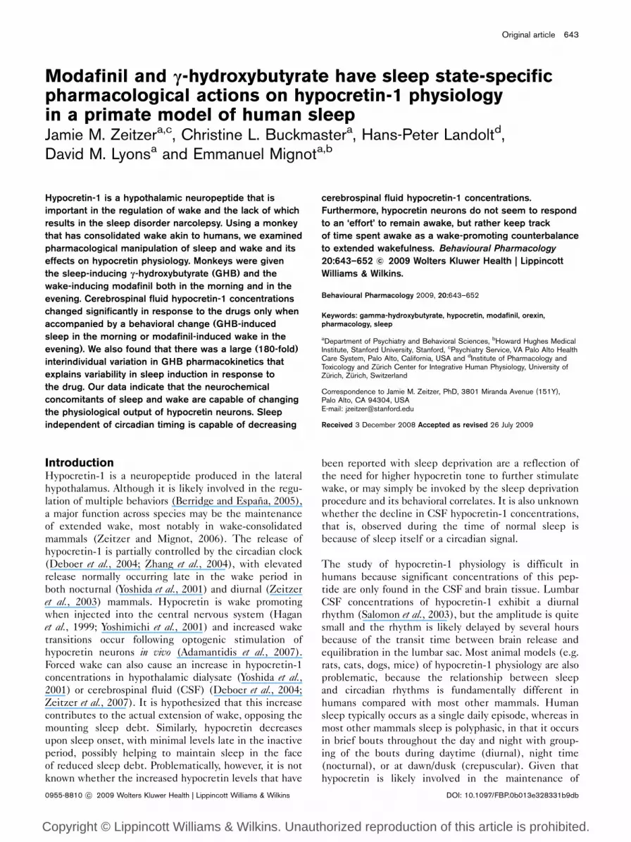
As expected and in contrast to daytime administration,
evening administration of the same dose of modafinil
increased the amount of wake and administration of
the sleep-promoting GHB had negligible effects on
sleep. After evening administration of GHB, there was
a nonsignificant decrease ( – 22 ± 4%) in the amount of
movement, as compared with the control condition
(P = 0.07, paired t-test) (Fig. 2). There was also no
significant change in the amount of wake ( – 15 ± 19%)
after evening administration of GHB, compared with
the control conditions (P = 0.09, paired t-test). After
evening administration of modafinil, there was a non-
significant increase (410 ± 272%) in the integrated
amount of movement (20.00–01.00 h), as compared with
the control conditions (P = 0.16, paired t-test) (Fig. 2). In
deriving estimated sleep/wake from actigraphy data, there
was, however, a significant 859 ± 525% increase in the
amount of wake after exposure to modafinil as compared
with control conditions (P < 0.01, paired t-test). Thus,
evening administration of modafinil significantly in-
creased the amount of wake, whereas evening adminis-
tration of GHB had no significant influence on the
amount of wake.
We next examined the relationship between plasma
concentrations of GHB and changes in wake. Surprisingly,
we found a 180-fold difference in plasma GHB concen-
trations, ranging 0.521–95.0 mg/ml. This was not because
of inaccurate experimental procedure as rank ordered
circulating concentrations of GHB after daytime or night-
time administration correlated highly within monkeys
(r = 0.94, P < 0.05, Spearman’s rank correlation coeffi-
cient). There existed a significant concentration–effect
relationship between morning administration of GHB
Fig. 1
3500400000
∗
320000
240000
160000
80000
0
Inte
grat
ed u
nits
(min
)
Control GHB ModafinilG C, M C, G, M
3000
2500
2000
1500
1000
500
0
05.00 06.00 07.00 08.00 09.00
Time of day (h)
Act
igra
ph u
nits
(Arb
)
10.00 11.00 12.00 13.00
Average movement from 05.00 h until 13.00 h in animals exposed to placebo (black), modafinil (M, light grey), or g-hydroxybutyrate (GHB, G, darkgrey). Data were averaged over an hour within and then between monkeys and are presented as mean ± SEM. The time of darkness is illustrated bya dark bar above the x-axis. The time of drug/placebo administration is indicated by arrows. Integrated actigraphy data (07.00–13.00 h) is presentedin the inset. Significance difference from control, *P < 0.05. Arb, arbitrary units; C, control.
646 Behavioural Pharmacology 2009, Vol 20 No 7
Copyright © Lippincott Williams & Wilkins. Unauthorized reproduction of this article is prohibited.
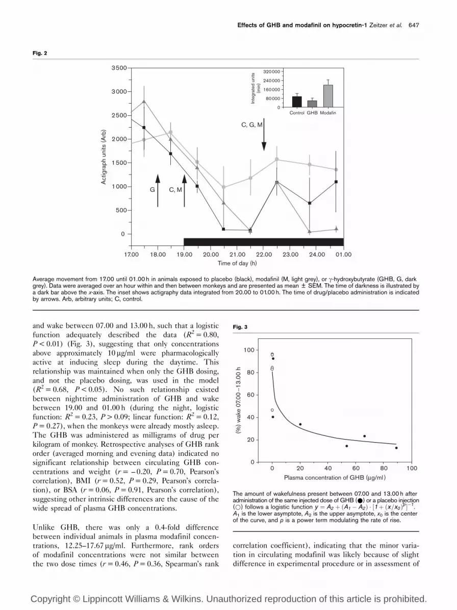
and wake between 07.00 and 13.00 h, such that a logistic
function adequately described the data (R2 = 0.80,
P < 0.01) (Fig. 3), suggesting that only concentrations
above approximately 10 mg/ml were pharmacologically
active at inducing sleep during the daytime. This
relationship was maintained when only the GHB dosing,
and not the placebo dosing, was used in the model
(R2 = 0.68, P < 0.05). No such relationship existed
between nighttime administration of GHB and wake
between 19.00 and 01.00 h (during the night, logistic
function: R2 = 0.23, P > 0.09; linear function: R2 = 0.12,
P = 0.27), when the monkeys were already mostly asleep.
The GHB was administered as milligrams of drug per
kilogram of monkey. Retrospective analyses of GHB rank
order (averaged morning and evening data) indicated no
significant relationship between circulating GHB con-
centrations and weight (r = – 0.20, P = 0.70, Pearson’s
correlation), BMI (r = 0.52, P = 0.29, Pearson’s correla-
tion), or BSA (r = 0.06, P = 0.91, Pearson’s correlation),
suggesting other intrinsic differences are the cause of the
wide spread of plasma GHB concentrations.
Unlike GHB, there was only a 0.4-fold difference
between individual animals in plasma modafinil concen-
trations, 12.25–17.67 mg/ml. Furthermore, rank orders
of modafinil concentrations were not similar between
the two dose times (r = 0.46, P = 0.36, Spearman’s rank
correlation coefficient), indicating that the minor varia-
tion in circulating modafinil was likely because of slight
difference in experimental procedure or in assessment of
Fig. 2
3500320000
240000
160000
80000
0Control
C, G, M
C, MG
GHB Modafin
3000
2500
2000
1500
1000
500
0
17.00 18.00 19.00 20.00 21.00Time of day (h)
Act
igra
ph u
nits
(Arb
)
22.00 23.00 24.00 01.00In
tegr
ated
uni
ts(m
in)
Average movement from 17.00 until 01.00 h in animals exposed to placebo (black), modafinil (M, light grey), or g-hydroxybutyrate (GHB, G, darkgrey). Data were averaged over an hour within and then between monkeys and are presented as mean ± SEM. The time of darkness is illustrated bya dark bar above the x-axis. The inset shows actigraphy data integrated from 20.00 to 01.00 h. The time of drug/placebo administration is indicatedby arrows. Arb, arbitrary units; C, control.
Fig. 3
100
(%) w
ake
07.0
0−1
3.00
h 80
60
40
20
00 20 40 60 80
Plasma concentration of GHB (μg/ml )
100
The amount of wakefulness present between 07.00 and 13.00 h afteradministration of the same injected dose of GHB (*) or a placebo injection(*) follows a logistic function y ¼ A2 þ ðA1 � A2 Þ � ½1þ ðx=x0 Þp ��1.A1 is the lower asymptote, A2 is the upper asymptote, x0 is the centerof the curve, and p is a power term modulating the rate of rise.
Effects of GHB and modafinil on hypocretin-1 Zeitzer et al. 647
Copyright © Lippincott Williams & Wilkins. Unauthorized reproduction of this article is prohibited.

drug plasma concentrations. As expected, modafinil was
dosed to be equivalent based on body weight, there
were no significant correlations between plasma modafinil
concentrations and weight (r = – 0.79, P = 0.06), BMI
(r = 0.00, P = 1.00), or BSA (r = – 0.41, P = 0.41).
Analysis of variance indicated that there was a significant
effect of both time of day (P < 0.05) and drug con-
dition (P < 0.01) on CSF concentrations of hypocretin-1
(Fig. 4). Post-hoc analyses with paired t-tests showed
that CSF hypocretin-1 concentrations were significantly
lower after GHB administration during the daytime
(795.6 ± 42.9 vs. 640.0 ± 35.2 pg/ml, baseline vs. GHB,
P < 0.05) when sleep-inducing effects occurred, but
not during the night (618.6 ± 42.3 vs. 585.9 ± 26.3 pg/ml,
baseline vs. GHB, P = 0.51) when sleep-inducing effects
were masked by natural sleep. We found no relationship
between the percentage change in GHB-induced sleep
at 13.00 h and the percentage change in hypocretin-1
concentrations (r = 0.52, P = 0.37, Pearson’s correlation;
r = 0.74, P = 0.09 logistic function). After daytime moda-
finil administration, there was a nonsignificant increase in
CSF hypocretin-1 concentrations, to 917.6 ± 51.7 pg/ml
(P = 0.18), but the changes after administration at
night to 738.1 ± 35.1 pg/ml were significant (P < 0.05).
Unexpectedly, we found that the larger the percentage
change in modafinil-induced wake at 01.00 h, the smaller
the percentage change in hypocretin-1 concentration
(r = 0.87, P < 0.01, Pearson’s correlation; r = 0.99, P < 0.01
logistic function).
As with hypocretin-1, we also examined plasma cortisol
concentrations at 13.00 and 01.00 h. At 13.00 h, cor-
tisol concentrations were 177.3 ± 73.7 mg/dl (placebo),
180.1 ± 44.9 mg/dl (GHB), and 252.2 ± 35.3 (modafinil),
whereas at 01.00 h they were 96.6 ± 48.0 mg/dl (placebo),
87.6 ± 20.9 mg/dl (GHB), and 152.7 ± 62.7 mg/dl (modafi-
nil), showing the expected diurnal variation. There was
a significant effect of time of day (P < 0.01) and of
drug administration (P < 0.02) (Fig. 5). There were no
significant differences in plasma cortisol following GHB
administration during the daytime (P = 0.94) or at night
(P = 0.63). Post-hoc analyses using paired t-tests indicated
that there was a significant increase in plasma cortisol
concentrations following modafinil administration during
the daytime (P < 0.01), but not at night (P = 0.25).
DiscussionUsing an animal that has consolidated sleep and wake, we
examined the impact of sleep and wake on hypocretin
regulation through their pharmacological induction with
GHB and modafinil. We found that CSF hypocretin-1
concentrations declined after exposure to the sleep-
promoting GHB and increased after exposure to the
wake-promoting modafinil. These effects, however, are
not because of the direct action of these drugs on
hypocretin activity, as the effects were only present when
there was a concomitant change in behavior (i.e. an
increase or a decrease in sleep amounts, respectively).
Thus, the neurochemical concomitants of sleep and
Fig. 4
30 Samples obtained at 13.00 h Samples obtained at 01.00 h
25
20
15
10
GHB Modafinil
CS
F hy
pocr
etin
-1 c
hang
e fro
m p
lace
bo (%
)
GHB
∗
∗
Modafinil
5
0
− 5
− 10
− 15
− 20
− 25
− 30
30
25
20
15
10
5
0
− 5
− 10
− 15
− 20
− 25
− 30
Cerebrospinal fluid (CSF) hypocretin-1 concentration change scores from placebo treatment at 13.00 h (left panel) and 01.00 h (right panel) afteradministration of g-hydroxybutyrate (GHB) or modafinil. Hypocretin-1 is significantly lower after daytime administration of GHB, when there is anincrease in sleep, and is significantly elevated after nighttime modafinil administration, when there is an increase in wakefulness. Hypocretin-1 isincreased at 13.00 h after modafinil treatment, but this change did not reach statistical significance (P = 0.18). Data are plotted as average ± SDpercent change in hypocretin-1 concentrations in the drug conditions, as compared with placebo. Percentage data were log transformed beforeaveraging and retransformed following averaging. *P < 0.05.
648 Behavioural Pharmacology 2009, Vol 20 No 7
Copyright © Lippincott Williams & Wilkins. Unauthorized reproduction of this article is prohibited.

wake, rather than the direct effects of the drugs, are
capable of changing the physiological output of hypo-
cretin neurons.
Sleep induced by GHB lowers CSF hypocretin-1 concen-
trations. GHB in and of itself, however, does not have a
significant effect on CSF hypocretin-1 concentrations.
By the nature of the model, earlier studies in polyphasic
mammals have been unable to establish whether the
decline of CSF hypocretin-1 concentrations during the
sleep portion of the daily cycle is because of a sleep
or time of day effect. Our data indicate that sleep is
independently able to reduce CSF hypocretin-1 con-
centrations. This conforms well to electrophysiological
recordings of hypocretin neurons in rodents that show
modest decrease in firing of hypocretin neurons during
times of sleep (Lee et al., 2005; Mileykovskiy et al., 2005).
As our data are limited, in that we did not record electro-
encephalic activity to confirm sleep, we have shown
earlier that behavioral quiescence during wake does not
induce changes in CSF hypocretin-1 concentrations
(Zeitzer et al., 2004), thus confirming that the GHB-
induced changes we observed are mediated through sleep
and are not because of a reduction in overall activity. The
relative contribution of sleep, specific phases of sleep,
specific electroencephalographic characteristics of sleep,
and the circadian clock, however, remain to be elucidated.
GHB is an endogenous metabolite of GABA (Doherty
et al., 1978) that can be converted to GABA through
transamination. There are specific GHB receptors in the
squirrel monkey brain (Castelli et al., 2000), but it is likely
that most, if not all, of the soporific actions of GHB
are mediated by GABA-B receptors (Crunelli et al., 2006;
Carai et al., 2008). Our data showed that there were
extremely large variations in circulating GHB concentra-
tions despite the monkeys each being dosed with the
same number of milligram of drug per kilogram body
weight. The dose was always given as an injection and the
monkeys were injected in a random order. This variation
seemed to be a trait of the monkeys, as the rank order
of concentrations was similar at both dosing times.
Importantly, this variation was functionally significant,
as circulating concentrations of GHB predicted the
amount of drug-induced sleep that the monkey experi-
enced. We examined whether the variation could be
because of common predictors of drug concentrations in
plasma, such as weight, BMI, and BSA, but none of these
explained the variance we observed. We could find no
published studies that have described variation in GHB
pharmacokinetics. It is possible that interindividual varia-
tion in absorption of the GHB suspension may account for
some of the interindividual variation in plasma concentra-
tions of GHB. As this finding may be idiosyncratic for the
squirrel monkey, further examination of GHB pharma-
cokinetics in this animal model is warranted.
We show here that an extension of wake can increase CSF
hypocretin-1 concentrations and that this increase is not
dependent upon the monkeys being forced to maintain
wake through interactions with human observers. We have
Fig. 5
120
Samples obtained at 13.00 h
Pla
sma
cort
isol
cha
nge
from
pla
cebo
(%)
Samples obtained at 01.00 h
100
80
60∗
40
20
0
120
100
80
60
40
20
0
GHB Modafinil GHB Modafinil
Plasma cortisol concentration change scores from placebo treatment at 13.00 h (left panel) and 01.00 h (right panel) after administration ofg-hydroxybutyrate (GHB) or modafinil. Cortisol was significantly elevated after daytime administration of modafinil, but the increase in cortisol afternighttime administration did not reach statistical significance (P = 0.25). There was no significant effect of GHB on cortisol. Data are plotted asaverage ± SD percent change in cortisol concentrations in the drug conditions, as compared with placebo. Percentage data were log transformedbefore averaging and retransformed following averaging. *P < 0.05.
Effects of GHB and modafinil on hypocretin-1 Zeitzer et al. 649
Copyright © Lippincott Williams & Wilkins. Unauthorized reproduction of this article is prohibited.
shown earlier in squirrel monkeys that forcing the
monkeys to remain awake for up to 7 h past their normal
wake time, by gently shaking the cage or perch on
which the monkey is resting, is sufficient to maintain
hypocretin-1 concentrations at an elevated value (Zeitzer
et al., 2007). Others have shown a similar phenomenon in
other species (Yoshida et al., 2001; Wu et al., 2002; Deboer
et al., 2004). None of these nonhuman mammals stays
awake volitionally as can humans. To keep a rat or a
monkey awake, it must be provided with external
motivation. Thus, the effects on hypocretin-1 physiology
could be secondary to physiologic changes that occur in
response to this motivation. We have shown earlier and
confirmed here that cortisol does not have a significant
contribution to the hypocretin-1 signal in squirrel
monkeys (Zeitzer et al., 2007), but there may be other
factors that do contribute. In this study, after monkeys
were administered modafinil, they were left undisturbed
until they were gathered for CSF sampling. On account of
the pharmacological properties of modafinil, they were
able to maintain wake for much of this time. Therefore, it
is unlikely that the monkeys were expending any ‘effort’
to remain awake nor were they stressed into a state of
wakefulness, as indicated by the lack of a change in
plasma cortisol. Despite this, hypocretin-1 concentrations
were elevated over control conditions. This implies that
hypocretin-1 changes during sleep deprivation are not
because of the motivated actions of the monkey to remain
awake or to changes in behavior induced by physical
intervention, but are primarily because of being awake for
an extended length of time. The changes in hypocretin-1
concentrations are likely not directly because of the
action of the drug, as when modafinil was administered
during the daytime, there was no significant, conse-
quent effect on CSF hypocretin-1 concentrations. In this
manner, hypocretin-1 seems to act more as a measure
of the length of time awake than it does as a reactive
mechanism to offset sleepiness. This result is also
supported by the data of Rao et al. (2007), who showed
that prolonged wake in mice, induced by gentle hand-
ling or modafinil, increases presynaptic glutamatergic
input to hypocretin neurons. As reported for other
glutamatergic systems in the brain (Tononi and Cirelli,
2007), hypocretin neurons may thus increase activity
with extended wake, resulting in long-term potentiation.
In the process, hypocretin neurons, therefore, may be
acting as a counter for daily wake until the process
becomes somehow unsustainable, then reducing activity
and inducing sleep.
There have been many theories as to the mechanism of
action of modafinil, including activation of hypocretin
neurons, but there is no consensus agreement. Its main
target for the promotion of wake is likely to be the
dopamine transporter (Wisor et al., 2001), potentially acting
through downstream dopaminergic (Qu et al., 2008) or
noradrenergic mechanisms (Wisor and Eriksson, 2005).
Modafinil does selectively increase Fos expression in
mouse (Chemelli et al., 1999) and rat (Scammell et al.,2000) hypocretin neurons and we did observe an increase
in CSF hypocretin-1 during the daytime after modafinil
administration, but this did not reach the level of
statistical significance. Our results suggest, rather, that
modafinil-induced increases in Fos expression in rodent
hypocretin neurons are indirectly because of a change
in wake and not because of a direct action of the
drug. The observation that bath-applied modafinil does
not change the firing activity of hypocretin neurons
in murine hypothalamic slices (Rao et al., 2007) also
supports this hypothesis. In addition, importantly, hypo-
cretin does not seem necessary for the wake-promoting
actions of modafinil, as mice genetically constructed
to not produce hypocretins have a robust modafinil-
induced wake response (Willie et al., 2005). Further
confirmation with electroencephalographic recording of
sleep will be important.
Another unexpected finding was the possible state-
dependent effect of modafinil on cortisol. As GHB had
no effects on cortisol, modafinil increased cortisol con-
centrations when the drug was administered in the
morning, when there were no obvious behavioral changes
after modafinil administration, but not in the evening,
when the monkeys experienced a significant increase in
their amount of wake. There have been few studies on
the effect of modafinil on cortisol (the main stress-related
hormone primates) or corticosterone (the main stress-
related hormone in rodents). One study in humans
reported that modafinil (300 mg, approximately 4.5 mg/kg
for a 66 kg human) had no effect on plasma cortisol
concentrations (Brun et al., 1998). Another study in mice
reported that 16 or 32 mg/kg did not affect plasma
corticosterone concentrations (Beracochea et al., 2008),
but the same group reported in a separate study that both
16 and 32 mg/kg of modafinil increased plasma cortico-
sterone concentrations, whereas 8 mg/kg of modafinil
did not (Pierard et al., 2006). Both of these studies in
mice examined corticosterone during the normal time of
sleep in mice (daytime) and we only saw effects during
the normal time of wake in monkeys (daytime) in our
study. Given the lower dose of modafinil used in the one
human study, it is possible that only at higher doses and
during time of normal wake are the effects of modafinil
on cortisol observed. More research is needed to explore
the possible state-dependent effects of modafinil on
plasma cortisol.
This study also has implication for diagnostic studies of
CSF in human narcolepsy and for the interpretation of
pharmacology studies that should examine sleep as a
confounder. The fact that the same compound adminis-
tered at the same dose at two different times of the
day has strikingly differential effects is important, as it
suggests that studies looking at the neurochemical effects
650 Behavioural Pharmacology 2009, Vol 20 No 7
Copyright © Lippincott Williams & Wilkins. Unauthorized reproduction of this article is prohibited.

of various drugs must control not only for circadian time,
but also for sleep history when studying effects on
neurotransmitters known to be involved in sleep regula-
tion. CSF hypocretin-1 concentration is often used as a
diagnostic tool in sleep medicine, notably to confirm
unusual presentations of narcolepsy (Mignot et al., 2002).
Our findings suggest that if patients were using either
GHB (typically given at night) or modafinil (typically
given during the daytime), it is unlikely that these
would have a significant impact on the clinical utility of
CSF hypocretin-1 measurement, as changes these drugs
evoked in hypocretin-1 concentrations through modula-
tion of sleep and wake activity would likely be minimal
in comparison with diagnostic criteria (<110 pg/ml are
indicative of narcolepsy and >200 pg/ml is normal).
We have shown in this study that sleep is independently
capable of causing a decrease in CSF hypocretin-1
concentrations, and that an extension of wake, rather
than efforts related to maintaining wake, is sufficient to
cause an increase in CSF hypocretin-1 concentrations.
Determination of the upstream pathways that control the
effects of sleep and wake on hypocretin neuron activity
will be crucial to better understand the physiology of
this system.
AcknowledgementsThe authors thank Dr Seiji Nishino for providing the
modafinil, S. Bodenmann and Dr M. Arand for help with
modafinil quantification, and Dr William Houghton for
early advice on GHB dosing.
This study was funded by NARSAD (J.M.Z.), Jazz
Pharmaceuticals (J.M.Z.), Department of Veterans Affairs
Sierra-Pacific Mental Illness Research, Education, and
Clinical Center (J.M.Z.), Howard Hughes Medical
Institute (E.M.), Swiss National Science Foundation
(H.-P.L.), and NIH-NS23724 (E.M.).
ReferencesAdamantidis AR, Zhang F, Aravanis AM, Deisseroth K, de Lecea L (2007). Neural
substrates of awakening probed with optogenetic control of hypocretinneurons. Nature 450:420–424.
Beardsley PM, Balster RL, Harris LS (1996). Evaluation of the discriminativestimulus and reinforcing effects of gammahydroxybutyrate (GHB). Psycho-pharmacol 127:315–322.
Beracochea D, Liscia P, Tronche C, Chauveau F, Jouanin J-C, Pierard C (2008).Stress modulation of the memory retrograde-enhancing effects of theawakening drug modafinil in mice. Psychopharmacol 196:1–13.
Berridge CW, Espana RA (2005). Hypocretins: waking, arousal, or action?Neuron 46:696–698.
Brun J, Chamba G, Khalfallah Y, Girard P, Boissy I, Bastuji H, et al. (1998). Effectof modafinil on plasma melatonin, cortisol and growth hormone rhythms,rectal temperature and performance in healthy subjects during a 36 h sleepdeprivation. J Sleep Res 7:105–114.
Carai MA, Lobina C, Maccioni P, Cabras C, Colombo G, Gessa GL (2008).Gamma-aminobutyric acidB (GABAB)-receptor mediation of different in vivoeffects of gamma-butyrolactone. J Pharmacol Sci 106:199–207.
Castelli MP, Mocci I, Langlois X, Gommeren W, Luyten WHML, Leysen JE, Gessa GL(2000). Quantitative autoradiographic distribution of g-hydroxybutyric acidbinding sites in human and monkey brain. Mol Brain Res 78:91–99.
Chemelli RM, Willie JT, Sinton CM, Elmquist JK, Scammell T, Lee C, et al. (1999).Narcolepsy in orexin knockout mice: molecular genetics of sleep regulation.Cell 98:437–451.
Crunelli V, Emri Z, Leresche N (2006). Unravelling the brain targets ofg-hydroxybutyric acid. Curr Opinon Pharmacol 6:44–52.
Deboer T, Overeem S, Visser NAH, Duindam H, Frolich M, Lammers GJ, Meijer JH(2004). Convergence of circadian and sleep regulatory mechanisms onhypocretin-1. Neurosci 129:727–732.
Doherty JD, Hattox SE, Snead OC, Roth RH (1978). Identification of endogenousg-hydroxybutyrate in human and bovine brain and its regional distributionin human, guinea pig and rhesus monkey brain. J Pharmacol Exp Ther207:130–139.
Hagan JJ, Leslie RA, Patel S, Evans ML, Wattam TA, Holmes S, et al. (1999).Orexin A activates locus coeruleus cell firing and increases arousal in the rat.Proc Natl Acad Sci U S A 96:10911–10916.
Hermant J-F, Rambert FA, Duteil J (1991). Awakening properties of modafinil:effect on nocturnal activity in monkeys (Macaca mulatta) after acute andrepeated administration. Psychopharmacol 103:28–32.
Ivanenko A, Tauman R, Gozal D (2003). Modafinil in the treatment of excessivedaytime sleepiness in children. Sleep Med 4:579–582.
Lagarde D, Milhaud C (1990). Electroencephalographic effects of modafinil, analpha-1-adrenergic psychostimulant, on the sleep of rhesus monkeys. Sleep13:441–448.
Lee MG, Hassani OK, Jones BE (2005). Discharge of identified orexin/hypocretinneurons across the sleep-waking cycle. J Neurosci 25:6716–6720.
Mignot E, Lammers GJ, Ripley B, Okun M, Nevsimalova S, Overeem S, et al.(2002). The role of cerebrospinal fluid hypocretin measurement in thediagnosis of narcolepsy and other hypersomnias. Arch Neurol 59:1553–1562.
Mileykovskiy BY, Kiyashchenko LI, Siegel JM (2005). Behavioral correlatesof activity in identified hypocretin/orexin neurons. Neuron 46:787–798.
Mosteller RD (1987). Simplified calculation of body-surface area. N Engl J Med317:1098.
Nakamura RK, Myslobodsky MS, Coppola R, Johannesen-Conway J, Mirsky AF(1987). Effects of g-hydroxybutyrate on the performance of monkeys in aGo/No-go visual discrimination task. Beh Brain Res 26:19–27.
Pierard C, Liscia P, Valleau M, Drouet I, Chauveau F, Huart B, et al. (2006).Modafinil-induced modulation of working memory and plasma corticosteronein chronically-stressed mice. Pharmacol Biochem Behavior 83:1–8.
Qu WM, Huang ZL, Xu XH, Matsumoto N, Urade Y (2008). Dopaminergic D1and D2 receptors are essential for the arousal effect of modafinil. J Neurosci28:8462–8469.
Rao Y, Liu Z-W, Borok E, Rabenstein RL, Shanabrough M, Lu M, et al. (2007).Prolonged wakefulness induces experience-dependent synaptic plasticity inmouse hypocretin/orexin neurons. J Clin Invest 117:4022–4033.
Salomon RM, Ripley B, Kennedy JS, Johnson B, Zeitzer JM, Nishino S, Mignot E(2003). Diurnal variation of CSF hypocretin-1 (orexin-A) levels in control anddepressed subjects. Biol Psychiatry 54:96–104.
Scammell TE, Estabrooke IV, McCarthy MT, Chemelli RM, Yanagisawa M,Miller MS, Saper CB (2000). Hypothalamic arousal regions are activatedduring modafinil-induced wakefulness. J Neurosci 20:8620–8628.
Shelton J, Nishino S, Vaught J, Dement WC, Mignot E (1995). Comparativeeffects of modafinil and amphetamine on daytime sleepiness and cataplexy ofnarcoleptic dogs. Sleep 18:817–826.
Snead OC III (1978). Gamma hydroxybutyrate in the monkey. I. Electroencephalo-graphic, behavioral, and pharmacokinetic studies. Neurol 28:636–642.
Tononi G, Cirelli C (2007). Staying awake puts pressure on brain arousalsystems. J Clin Invest 117:3648–3650.
van Vliet SA, Jongsma MJ, Vanwersch RA, Olivier B, Philippens IH (2008).Efficacy of caffeine and modafinil in counteracting sleep deprivation in themarmoset monkey. Psychopharmacol (Berl) 197:59–66.
Willie JT, Renthal W, Chemelli RM, Miller MS, Scammell TE, Yanagisawa M,Sinton CM (2005). Modafinil more effectively induces wakefulness inorexin-null mice than in wild-type littermates. Neurosci 130:983–995.
Wisor JP, Eriksson KS (2005). Dopaminergic-adrenergic interactions in the wakepromoting mechanism of modafinil. Neurosci 132:1027–1034.
Wisor JP, Nishino S, Sora I, Uhl GH, Mignot E, Edgar DM (2001). Dopaminergicrole in stimulant-induced wakefulness. J Neurosci 21:1787–1794.
Woolverton WL, Rowlett JK, Winger G, Woods JH, Gerak LR, France CP(1999). Evaluation of the reinforcing and discriminative stimulus effects ofg-hydroxybutyrate in rhesus monkeys. Drug Alcohol Depend 54:137–143.
Wu M-F, John J, Maidment N, Lam HA, Siegel JM (2002). Hypocretin release innormal and narcoleptic dogs after food and sleep deprivation, eating, andmovement. Am J Physiol 283:R1079–R1086.
Yoshida Y, Fujiki N, Nakajima T, Ripley B, Matsumura H, Yoneda H, et al. (2001).Fluctuation of extracellular hypocretin-1 (orexin A) levels in the rat in relation tothe light-dark cycle and sleep-wake activities. Eur J Neurosci 14:1075–1081.
Effects of GHB and modafinil on hypocretin-1 Zeitzer et al. 651
Copyright © Lippincott Williams & Wilkins. Unauthorized reproduction of this article is prohibited.

Yoshimichi G, Yoshimatsu H, Masaki T, Sakata T (2001). Orexin-A regulatesbody 40-temperature in coordination with arousal status. Exp Biol Med226:468–476.
Zeitzer JM, Mignot E (2006). Role of hypocretin/orexin in the neurobiology ofsleep and alertness. In: Bassetti C, Billiard M, Mignot E, editors. Narcolepsyand hypersomnia. New York: Informa Healthcare. pp. 359–374.
Zeitzer JM, Buckmaster CL, Parker KJ, Hauck CM, Lyons DM, Mignot E (2003).Circadian and homeostatic regulation of hypocretin in a primate model:implications for the consolidation of wakefulness. J Neurosci 23:3555–3560.
Zeitzer JM, Buckmaster CL, Lyons DM, Mignot E (2004). Locomotor-dependentand independent components to hypocretin-1 (orexin A) regulation in sleep-wake consolidating monkeys. J Physiol 557:1045–1053.
Zeitzer JM, Buckmaster CL, Lyons DM, Mignot E (2007). Effects of increasinglength of wakefulness on hypocretin-1 in the wake-consolidating squirrelmonkey. Am J Physiol 293:R1736–R1742.
Zhang S, Zeitzer JM, Yoshida Y, Wisor JP, Nishino S, Edgar DM, Mignot E (2004).Lesions of the suprachiasmatic nucleus eliminate the daily rhythm ofhypocretin-1 release. Sleep 27:619–627.
652 Behavioural Pharmacology 2009, Vol 20 No 7
Copyright © Lippincott Williams & Wilkins. Unauthorized reproduction of this article is prohibited.