n-methyl-d-aspartic acid receptor expression in the dorsolateral prefrontal cortex of elderly...
TRANSCRIPT

Article
1400 Am J Psychiatry 158:9, September 2001
N-Methyl-D-Aspartic Acid Receptor Expressionin the Dorsolateral Prefrontal Cortex
of Elderly Patients With Schizophrenia
Stella Dracheva, Ph.D.
Salvatore A.E. Marras, M.S.
Sharif L. Elhakem, B.S.
Fred R. Kramer, Ph.D.
Kenneth L. Davis, M.D.
Vahram Haroutunian, Ph.D.
Objective: The N-methyl-D-aspartic acid(NMDA) class of glutamate receptors hasreceived attention in the pathophysiologyof schizophrenia because of the similaritybetween some schizophrenic symptomsand symptoms caused by NMDA antago-nists. To determine if NMDA receptor ab-normalities were present at the mRNAlevel, expression of NMDA receptor (NR)subunits NR1, NR2A, and NR2B was mea-sured in specimens from the dorsolateralprefrontal cortex and the occipital cortexof elderly patients with schizophrenia andnormal elderly subjects.
Method: Postmortem specimens fromantemortem assessed and diagnosed eld-erly patients with schizophrenia (N=26)were compared with those from a neuro-pathologically and neuropsychiatricallynormal elderly comparison group (N=13)and from patients with Alzheimer’s disease(N=10). The mRNA expression of the NR1,NR2A, and NR2B subunits and of postsynap-tic density 95 (PSD-95), a protein associatedwith postsynaptic NMDA receptors, wasstudied with quantitative real-time reversetranscriptase polymerase chain reaction.
Results: Expression of NR1 and NR2A butnot NR2B subunits was higher in the dor-solateral prefrontal cortex and the occipi-tal cortex of patients with schizophreniathan in the normal and Alzheimer’s dis-ease groups. In contrast, NR1 expressionwas significantly lower in the Alzheimer’sdisease group. Occipital cortex expressionof PSD-95 was higher in the schizophrenicsubjects and correlated strongly with theexpression of NR2A and NR2B in both cor-tical regions and with expression of NR1 inthe occipital cortex. These results werenot influenced by neuroleptic exposurehistory, postmortem interval, or age ofthe subject.
Conclusions: NMDA receptor subunitsare abnormally expressed in elderly pa-tients with schizophrenia. The dispropor-tionate expression of the NR1 and NR2A
subunits relative to NR2B expression mayhave implications for the pathophysiol-ogy of schizophrenia and the sensitivity ofschizophrenic patients to glutamate andglutamatergic drugs.
(Am J Psychiatry 2001; 158:1400–1410)
Several neurochemical hypotheses have been pro-posed to explain the origin of schizophrenia, including ab-normal dopamine, serotonin (5-HT), γ-aminobutyric acid(GABA), and/or glutamate neurotransmission in differentregions of the brain (1–8). Abnormalities in the dorsolat-eral prefrontal cortex have figured prominently in many ofthese hypotheses, in part due to results of in vivo imagingand neuroanatomical studies (9–11), although there is ev-idence for structural, metabolic, and neurochemical ab-normalities in many other brain regions, including thethalamus, the hippocampus, and the cingulate and en-torhinal cortices (1, 12–19).
Strong evidence supporting an association betweenglutamatergic hypofunction and schizophrenia has comefrom pharmacological studies showing that N-methyl-D-aspartic acid (NMDA) receptor antagonists such as phen-cyclidine and ketamine can induce many of the psychoticsigns and symptoms of schizophrenia in normal subjects,as well as exacerbate these signs and symptoms in sub-
jects with schizophrenia (20–27). Observations of abnor-malities in markers of glutamatergic neurotransmission inthe hippocampus and the entorhinal, cingulate, orbital,and prefrontal cortices of patients with schizophrenia (3,13, 28–43), the critical role of glutamatergic systems inlearning and memory (44, 45), and the clear evidence forthe neurotoxicity of glutamate (28, 46, 47) have given fur-ther credence to the potential involvement of the gluta-matergic system in schizophrenia.
Glutamate receptors comprise four different receptorfamilies: NMDA, kainate, α-amino-3-hydroxy-5-methyl-isoxazole-4-propionate (AMPA), and metabotropic recep-tors (48, 49). Because glutamate is the principal excitatoryneurotransmitter in the brain, its receptors are ubiqui-tously but relatively discretely distributed throughout theneuraxis (50–53). Although there is evidence suggestingthat kainate receptors are predominantly presynaptic andthat AMPA and NMDA receptors are predominantly post-synaptic and often coexpressed, AMPA and NMDA recep-

Am J Psychiatry 158:9, September 2001 1401
DRACHEVA, MARRAS, ELHAKEM, ET AL.
tors can also be localized to presynaptic sites (51, 54). Eachreceptor is assembled from multiple subunits that are en-coded by different genes. The NMDA receptor is assem-bled from a combination of four–five subunits designatedNR1 and NR2A–D. The NR1 subunit is often consideredobligatory for functional NMDA receptor assemblies, butthe remaining subunits can vary. Additional heterogeneityis conferred by eight alternative splice variants of the NR1
subunit that have different expression patterns within thehuman brain (55). The pattern of subunits assembled toform specific glutamate receptors varies from brain regionto brain region (55). In the cerebral cortex and the hippo-campus, NMDA receptors are assembled from NR1, NR2A,and NR2B subunits, and the NR2C and NR2D subunits aregenerally thought not to be expressed at appreciable levels(54, 56) (but see reference 38). Since the various combina-tions of NMDA receptor subunits confer different sensitiv-ities to various endogenous and exogenous glutamatergicligands and since NMDA receptors can be localized pre-and postsynaptically (57–59), the release of glutamatefrom any given neuron or a pharmacologic glutamateligand can have a wide range of effects in different brainregions and on glutamatergic function and activity.
At a postsynaptic level, the clustering, assemblage, andanchoring of NMDA receptors is governed in part by a pro-tein family known as postsynaptic density 95/synapse-as-sociated protein 90 (PSD-95/SAP-90) that appears also toplay an important role in the binding and assemblage ofsignal transduction complexes (60–65). PSD-95 is exclu-sively localized with postsynaptic NMDA receptor-relateddensities (62, 66), with significantly greater associationwith the NR2A and NR2B subunits than with the NR1 sub-unit, although it does co-localize and associate with somesplice variants of the NR1 subunit (60, 62). PSD-95 alsoplays an important role in signal transduction, nitric oxideneurotoxicity linked to NMDA receptor activation (67, 68),and NMDA-induced long-term potentiation (69). Becauseof these characteristics, PSD-95 is important in providinga functional scaffold for postsynaptic NMDA receptorsand in mediating intracellular NMDA receptor functions.
The study described here sought to determine whetherthe expression of genes encoding for NR1, NR2A, and NR2B
subunits of the NMDA receptor and their postsynaptic an-choring protein PSD-95 are specifically altered in the dor-solateral prefrontal cortex of patients with schizophrenia.The dorsolateral prefrontal cortex was studied in postmor-tem brains of elderly schizophrenic subjects who had beenantemortem diagnosed with DSM-IV criteria (70–72), hadno other neuropsychiatric disease, had died of naturalnonviolent causes without coma, had no evidence of sig-nificant neuropathology (73), and had clear documenta-tion of neuroleptic drug exposure during the months andweeks before death. Specimens of the dorsolateral prefron-tal cortex from these subjects were compared to identicallytreated and dissected specimens from normal elderly sub-jects who were found by chart review and caregiver inter-views to have no neuropsychiatric or neurological diseasesand who had no significant neuropathology. Specificity toschizophrenia was tested by the inclusion of specimensfrom patients with Alzheimer’s disease. To determine thespecificity of findings to the dorsolateral prefrontal cortex,specimens from the primary occipital cortex (Brodmann’sarea 17) from the same subjects were dissected and as-sessed for NMDA receptor and PSD-95 transcript mRNAabundance by using identical procedures.
Method
Human Postmortem Tissue
Frozen postmortem brain samples from subjects diagnosedwith DSM-IV schizophrenia (N=26), normal elderly comparisonsubjects (N=13), and subjects with Alzheimer’s disease (N=10)were obtained from the Brain Bank, Department of Psychiatry,Mount Sinai/Bronx Veterans Administration Medical Center. Thesex distribution and mean age, postmortem interval, and tissuepH of the cohorts are shown in Table 1. All schizophrenic subjectshad been hospitalized for the long term at Pilgrim PsychiatricCenter (New York). Complete medical charts were available for allsubjects, and 16 of the 26 schizophrenic subjects had been pro-spectively diagnosed and neuropsychiatrically assessed by a teamof research clinicians (70–72). Patients who died before the ante-mortem assessment by the research team were diagnosed by the
TABLE 1. Characteristics of Postmortem Brain Tissue From Patients With Schizophrenia, Normal Comparison Subjects, andPatients With Alzheimer’s Disease
Tissue From Patients With Schizophrenia Tissue From Normal Comparison Subjects
Tissue FromPatients WithAlzheimer’s
Disease (N=10)CharacteristicAll Patients
(N=26)
Patients Matched for Age With
Normal Comparison Subjects (N=10)
All Subjects(N=13)
Subjects Matchedfor Age WithPatients With
Schizophrenia (N=10)N N N N N
Subjects’ genderMale 17 4 5 2 4Female 9 6 8 8 6
Mean SD Mean SD Mean SD Mean SD Mean SD
Subjects’ age at death (years) 72.3 12.0 81.1 9.3 82.8 10.0 81.5 11.1 79.8 9.8Postmorten interval (hours) 14.6 9.5 14.2 8.4 8.0 5.5 8.0 5.7 10.1 9.3pH 6.33 0.32 6.36 0.31 6.29 0.31 6.23 0.34 6.27 0.37Storage time interval (days) 2128.0 1029.5 1998.6 995.7 1759.4 1006.2 1815.9 1129.6 2198.9 676.2
1402 Am J Psychiatry 158:9, September 2001
NMDA RECEPTOR EXPRESSION
same team of research clinicians who conducted diagnostic re-views of all medical charts (73). The reliability of these postmor-tem diagnostic procedures was confirmed by assessing an inde-pendent group of 35 subjects from the same institution by usingstructured interviews and blindly by using chart review. Inter-rater/interassessment reliability was 0.86. All assessment andpostmortem evaluations and procedures were approved by theinstitutional review boards of the Pilgrim Psychiatric Center, theMount Sinai School of Medicine, and the Bronx VA Medical Cen-ter. All patients had thorough neuropathologic characterizationto rule out associated neurologic complications such as Alzhei-mer’s disease and multi-infarct dementia (73). Normal compari-son subjects had no history of any psychiatric or neurologic dis-orders and no discernible neuropathologic lesions. Nine of theschizophrenic subjects had not received neuroleptic medicationsfor at least 6 weeks before death (range=0–124 weeks). For com-parison purposes and to enable assessment of specificity of thefindings to schizophrenia, brain tissue from 10 subjects with a di-agnosis of definite Alzheimer’s disease (according to criteria ofthe Consortium to Establish a Registry for Alzheimer’s Disease)(74) was also studied. The overall characteristics and diagnosticprocedures for these Alzheimer’s disease subjects have been de-scribed extensively (75, 76).
Each brain was divided midsagittally at the time of extraction.The left half was sectioned in 6–8-mm coronal slabs, immedi-ately snap-frozen in liquid nitrogen-cooled isopentane, andstored at –80°C. Gray matter from the frozen dorsolateral pre-
frontal cortex (77, 78) (Brodmann’s area 46) and occipital cortex(Brodmann’s area 17) was dissected from coronal sections of fro-zen brain (–80°C). Brodmann’s area 17 was identified as the areacontaining the band of Gennari in the coronal section from ap-proximately 2 cm rostral to the occipital pole. The dissected tis-sues were pulverized at –190°C into a fine powder, aliquoted intoindividual Eppendorf tubes, and stored at –80°C until use.
Quantitation of NMDA Receptor Expression
RNA isolation. Total RNA was isolated from 50 mg of tissuewith the guanidinium isothiocyanate method (79) by using theToTALLY RNA kit (Ambion, Austin, Tex.) according to the manu-facturer’s protocol. To remove genomic DNA contamination, iso-lated RNA samples were then treated with 40 units of DNase I(Ambion) in the presence of 120 units of RNaseOUT (GibcoBRL,Invitrogen, Carlsbad, Calif.) for 1 hour at 37°C. The yield of totalRNA determined by absorbance at 260 nM ranged from 15 to 30µg per 50 mg of brain tissue. The 260/280 nM ratios of the sampleswere >2.1. The yield and quality of total RNA was also analyzed byusing agarose gel electrophoresis.
Reverse transcriptase reaction. Total RNA (∼ 2 µg) was used in20 µl of reverse transcriptase reaction to synthesize cDNA, by us-ing a ThermoScript RT-PCR System kit (GibcoBRL) and randomhexamers as primers. The cDNA was diluted 50 times with water,and 5 µl of the diluted cDNA was amplified in 25 µl of polymerasechain reaction (PCR) mix.
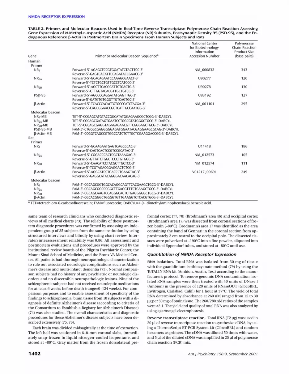
TABLE 2. Primers and Molecular Beacons Used in Real-Time Reverse Transcriptase Polymerase Chain Reaction AssessingGene Expression of N-Methyl-D-Aspartic Acid (NMDA) Receptor (NR) Subunits, Postsynaptic Density 95 (PSD-95), and the En-dogenous Reference β-Actin in Postmortem Brain Specimens From Human Subjects and Rats
Gene Primer or Molecular Beacon Sequencea
National Center for Biotechnology
InformationAccession Number
Polymerase Chain Reaction
Product Size (base pairs)
HumanPrimer
NR1 Forward-5′-AGAGCTCCGTGGATATCTACTTCC-3′ NM_000832 343Reverse-5′-GAGTCACATTCCAGATACCGAACC-3′
NR2A Forward-5′-GCACAGAATCCAAAGCGAACT-3′ U90277 120Reverse-5′-TCTCTGCTGTTGCCTCATCCC-3′
NR2B Forward-5′-AGCTTCACGCATTCTGACTG-3′ U90278 130Reverse-5′-CTTGGTACACGTTGCTGTCC-3′
PSD-95 Forward-5′-AGCCCCAGGATATGAGTTGC-3′ U83192 127Reverse-5′-GATGTGTGGGTTGTCAGTGC-3′
β-Actin Forward-5′-TCACCCACACTGTGCCCATCTACGA-3′ NM_001101 295Reverse-5′-CAGCGGAACCGCTCATTGCCAATGG-3′
Molecular beaconNR1-MB TET-5′-CCGAGCATGTACCGGCATATGGAGAAGCGCTCGG-3′-DABCYLNR2A-MB TET-5′-CGCAGCGATAGTGAATCCTGGCGTATGGGCTGCG-3′-DABCYLNR2B-MB TET-5′-CGCAGCGAGGTAGAGAGAACGTTCGGGAGCTGCG-3′-DABCYLPSD-95-MB FAM-5′-CTGCGCGAGGGGGAGATGGAATACGAGGAAGCGCAG-3′-DABCYLβ-Actin-MB FAM-5′-CCGGTCAGCCGTGGCCATCTCTTGCTCGAAGGACCGG-3′-DABCYL
RatPrimer
NR1 Forward-5′-GCAAGAATGAGTCAGCCCAC-3′ U11418 186Reverse-5′-CAGTCACTCCGTCCGCATAC-3′
NR2A Forward-5′-CGGACCCACTCGCTAAAGAG-3′ NM_012573 105Reverse-5′-GTTATCTGGCTCCCTGTGGC-3′
NR2B Forward-5′-CAACATCCTACGCTTGCTCC-3′ NM_012574 111Reverse-5′-TCGTAGACGGAGGACTCTCG-3′
β-Actin Forward-5′-AGGCATCCTGACCCTGAAGTAC-3′ V01217 J00691 249Reverse-5′-GAGGCATACAGGGACAACACAG-3′
Molecular beaconNR1 FAM-5′-CGCAGCGGTGGCACAGGCAGTTCACGAAGCTGCG-3′-DABCYLNR2A FAM-5′-CGCAGCGGCCCGGCTTGAGGTTTCTGAAGCTGCG-3′-DABCYLNR2B FAM-5′-CGCAGCAAGTCCAGGGCACTCTGAGGGGGCTGCG-3′-DABCYLβ-Actin FAM-5′-CGCACGGGCTGGGGTGTTGAAGGTCTCACGTGCG-3′-DABCYL
a TET=tetrachloro-6-carboxyfluorescein; FAM=fluorescein; DABCYL=4-(4′-dimethylaminophenylazo) benzoic acid.

Am J Psychiatry 158:9, September 2001 1403
DRACHEVA, MARRAS, ELHAKEM, ET AL.
Primer and molecular beacon design. Real-t im e revers etranscriptase polymerase chain reaction (RT-PCR) was used forquantitation of NMDA receptor and PSD-95 expression. PCRprimers were designed by using Vector NTI software (InforMax,North Bethesda, Md.). NR1 primers were designed to ignore dis-tinctions between known splicing variants of this NMDA recep-tor subunit. Molecular beacons were used as fluorogenic probesin the real-time PCR. Molecular beacons are hairpin-shapedmolecules with an internally quenched fluorophore whose fluo-rescence is restored when they bind to their target nucleic acids(80–83). The molecular beacons were designed by using a DNAfolding program (available at http://www.ibc.wustl.edu/∼ zuker)to estimate the stability of the hairpin stem. The molecular bea-cons used in the experiments contained probe sequences thatwere 22–25 nucleotides long and arm sequences that were sixnucleotides long. The melting temperature of the hairpin stemsand probe sequences was 64–66°C. Fluorescein (FAM) or tetra-chloro 6-carboxyfluorescein (TET) fluorophores were covalentlylinked to the 5′ end of the molecular beacons, and the quencher4-(4′-dimethylaminophenylazo) benzoic acid (DABCYL) was co-valently linked to the 3′ end. The primers and molecular beaconswere synthesized either commercially (IDT, Coralville, Iowa) orby one of us (S.A.E.M.). The primer pair and molecular beaconsequences that were used to detect each of the mRNAs are shownin Table 2.
Real-time PCR. Real-time PCR analysis was performed by usingan ABI Prism 7700 Sequence Detector (Applied Biosystems, Fos-ter City, Calif.). Each 25 µl PCR reaction contained 5 µl of the rele-vant cDNA, 200 nM of molecular beacon, 500 nM of each primer,1 unit of AmpliTaq Gold DNA polymerase (Applied Biosystems),250 µM of each deoxy-nucleotide triphosphate (dNTP), 4 mMMgCl2, 50 mM KCl, and 10 mM Tris-HCl (pH 8.3). The thermal cy-cling program consisted of 10 minutes at 95°C to activate thepolymerase, followed by 10 cycles of 15 seconds at 95°C and 45seconds at 70–61°C (touch-down PCR, annealing temperaturewas decreased 1°C after each cycle). This touch-down step wasfollowed by 35 cycles of 15 seconds at 95°C and 1 minute at 60°C.Fluorescence was monitored during the 60°C annealing-exten-sion steps. The reactions were quantitated by selecting the ampli-fication cycle when the PCR product of interest was first detected(threshold cycle [Ct]).
To determine sensitivity of the assays, the amplification of eachmRNA in serial dilutions of cDNA derived from pooling of humancortical specimens from 10 randomly selected subjects (pooledsample) was measured. Figure 1 shows the amplification of NR1mRNA in 10-fold dilutions of pooled cDNA and the threshold cy-cle values of these amplifications plotted against the log of the rel-ative initial amount of cDNA. In assays that use PCR to amplify atarget sequence exponentially, there is an inverse linear relation-ship between the threshold cycle and the logarithm of the num-ber of target molecules that were present initially (84). Theoreti-cally, the slope of this linear curve is expected to be –3.32. In theresults shown on Figure 1, a linear relationship between thresholdcycle and the initial amount of NR1 mRNA was demonstrated forfour orders of magnitude. The slope of the curve was –3.23, whichwas very close to theoretically expected value. These data showthe broad dynamic range of the NR1 mRNA quantitation. Similarresults were obtained for each amplification assay performed inthis study.
To account for different degrees of RNA degradation and othertechnical artifacts, relative quantitations of the expression levelsof NR1, NR2A, NR2B and PSD-95 genes were performed as de-scribed in User Bulletin #2 for the ABI PRISM 7700 Sequence De-tection System: Relative Quantitation of Gene Expression; Com-parative Ct Method: Separate Tubes, product #4303859 (AppliedBiosystems). The expression level of each gene of interest wasnormalized to the expression level of the endogenous reference
(β-actin) in each sample. This relative value was further normal-ized to the relative expression of the same gene in the pooledsample (see the preceding paragraph). Pooled cDNAs were run inevery plate simultaneously with experimental samples. To avoidcompetition, only one mRNA was amplified in each PCR (mono-plex). All samples were run in triplicate.
To measure the level of contamination with chromosomalDNA, all RNA samples that were not treated with reverse tran-scriptase were subjected to PCR by using NR1 or NR2A primers.The products of the PCRs were analyzed on EtBr-stained agarosegels. In contrast to the respective cDNA templates, the RNA sam-ples showed no PCR products. Random RNA samples (N=10) werealso subjected to real-time PCR by using β-actin primers and amolecular beacon. The RNA samples showed at least 4.5 orders ofmagnitude fewer initial template molecules than the respectivecDNAs templates (the difference in threshold cycle betweenRNAs and cDNA in each sample was at least 15 cycles), demon-strating negligible amounts of genomic DNA contamination(data not shown).
NMDA Receptor Expression in Neuroleptic-Treated Rats
To assess the effects of neuroleptic exposure on NMDA receptormRNA, groups of six male Sprague-Dawley rats (6–8 months of age)received daily subcutaneous injections of haloperidol (2 mg/kg) or
FIGURE 1. Dynamic Range of the mRNA Quantitation Assayfor NMDA Receptor 1 (NR1)a
a Panel A: Amplification of NR1 mRNA in 10-fold dilutions of pooledcDNA. Panel B: Threshold values of amplifications of NR1 mRNAplotted against the log of the relative initial amount of cDNA. Simi-lar results were obtained for the amplification assays performed forNR2A, NR2B, PSD-95 and β-actin.
5000
4000
3000
2000
1000
25
20
15
0Threshold
1:10
15 20 25 30
Flu
ore
scen
ce (arb
itra
ry u
nit
s)
Cycle Number
A
B
1 2 3 4Log Initial Amount of cDNA
(1:10,000–1:10 cDNA dilutions)
Th
resh
old
Cyc
le
1:100
1:1,0001:10,000

1404 Am J Psychiatry 158:9, September 2001
NMDA RECEPTOR EXPRESSION
saline vehicle for 21 days. The specific haloperidol dosing parame-ters were selected because previous studies (85) have shown themto be effective in regulating dopamine receptor mRNA expressionin the rat brain. The rats were sacrificed by decapitation 24 hoursafter the last injection, and their brains were rapidly removed. Cor-tices were dissected and immediately frozen on dry ice. NMDA re-ceptor expression was assessed by using the procedures describedearlier except that primers and molecular beacons specific to ratNMDA receptor subtypes were used (Table 2).
Statistical Analysis
Analysis of variance (ANOVA) and covariance (ANCOVA) fol-lowed by Newman-Keuls tests and t tests were used to analyze theresults of these studies. ANOVA was used for analyses of data fromthe entire cohort. ANCOVA was used for analyses of data from theentire cohort, with age of the subject at the time of death as a co-variate. T tests were used to compare differences between groupswhen the groups had been matched for age at the time of death.Because each brain region and each NMDA receptor subunit wasmeasured independently with different probes and in differentexperiments, differences between groups for the expression ofeach gene were assessed with independent tests of significance.Pearson product moment correlations were used to assess the re-lationship between continuously distributed variables. Statisticalanalyses were performed with Statistica for Windows (release 5.5,Statsoft Inc., Tulsa, Okla.) or SPSS for Windows (version 10, SPSSInc., Chicago).
Results
The relative abundance of NMDA receptor subunit ex-pression in the dorsolateral prefrontal cortex and the oc-cipital cortex for each group is shown in Figure 2. NR1 sub-unit expression in the dorsolateral prefrontal cortex wassignificantly higher in the schizophrenia group and signif-icantly lower in the Alzheimer’s disease group, comparedwith the normal elderly group (F=12.75, df=2, 46, p=0.00004) (Newman-Keuls tests: schizophrenia versus nor-mal elderly, p=0.009; Alzheimer’s disease versus normalelderly, p=0.05; Alzheimer’s disease versus schizophrenia,p=0.0002). Higher levels of NR1 subunit expression wereevident in the occipital cortex of the schizophrenia sub-jects than in the normal subjects, but not in the Alzhei-mer’s disease subjects (F=6.79, df=2, 46, p=0.003) (New-man-Keuls tests: schizophrenia versus normal elderly, p=0.02; Alzheimer’s disease versus normal elderly, p=0.94).Expression of NR2A in the dorsolateral prefrontal cortexwas not significantly different in either the schizophrenicor Alzheimer’s disease subjects relative to normal elderlysubjects (p>0.14, Newman-Keuls). However, the level ofNR2A expression in the occipital cortex of the schizo-phrenic subjects was significantly higher than that of thenormal elderly subjects (F=5.5, df=2, 46, p=0.007; p=0.04,Newman-Keuls). The lower level of NR2A expression in theoccipital cortex of the Alzheimer’s disease group, relativeto the normal comparison group, did not reach statisticalsignificance (p=0.40, Newman-Keuls). The expression ofthe NR2B subunit was not significantly altered in anygroup in either of the brain regions examined (F<0.9, df=2,46, p>0.4). The expression of the mRNA for PSD-95 wasalso significantly higher in the schizophrenia subjectsthan in the normal elderly subjects (Figure 2). The schizo-phrenia-related difference in the expression of PSD-95was most evident in the occipital cortex (F=4.31, df=2, 46,p=0.02; schizophrenia versus normal elderly, p=0.04, New-man-Keuls) but did not reach statistical significance in thedorsolateral prefrontal cortex (p=0.31, Newman-Keuls).PSD-95 gene expression was unchanged in the brains ofthe Alzheimer’s disease cohort.
FIGURE 2. Relative Gene Expression of N-Methyl-D-AsparticAcid (NMDA) Receptor Subunits NR1, NR2A, and NR2B andof Postsynaptic Density 95 (PSD-95) in the Dorsolateral Pre-frontal Cortex and the Occipital Cortex of Normal Compar-ison Subjects, Patients With Schizophrenia, and PatientsWith Alzheimer’s Disease
a Significantly different from comparison group (p<0.05). Newman-Keuls tests were used for comparisons of entire groups. T tests (df=18) were used for comparisons of age-matched groups (10 patientswith schizophrenia and 10 normal comparison subjects).
Rela
tive
mR
NA
Exp
ress
ion
DorsolateralPrefrontal Cortex
Occipital Cortex
NR1
NR2A
NR2B
PSD-95
0.75
Comparisonsubjects (N=13)
Entiregroups
Age-matched
Entiregroups
Age-matched
Patients withschizophrenia (N=26)Patients with Alzheimer'sdisease (N=10)
0.50
0.25
1.00
0.75
0.50
0.25
12.5
10.0
7.5
5.0
2.5
1.5
1.0
0.5
a
a
a
a a
aa
aa

Am J Psychiatry 158:9, September 2001 1405
DRACHEVA, MARRAS, ELHAKEM, ET AL.
The schizophrenic cohort was significantly youngerthan the normal elderly group (Table 1), and age at deathcorrelated significantly with the expression of the NR1 andNR2A subunits of the NMDA receptor (r<−0.31, df=47,p<0.04) when the entire cohort was considered. The ex-pression of the NMDA receptor subunits and PSD-95mRNA did not correlate significantly with age within theschizophrenia cohort despite a relatively broad age rangeof 52–97 years. Two approaches were taken to determinewhether age at death affected the differences betweengroups. First, ANCOVAs, with age as a covariate, weredone. The inclusion of age as a covariate did not alter thepattern or the statistical significance of the results in anyway (e.g., the ANCOVA comparing NR1 expression in thedorsolateral prefrontal cortex was significant [F=9.6, df=2,45, p=0.0003]). Second, the schizophrenic and normalelderly groups were subgrouped into two groups of 10subjects each, matched for age to within 1 year. The differ-ences in the expression of the NMDA receptor subunitsand PSD-95 were reassessed by using t tests. As for theANCOVA, the same significant group differences (t<−2.2,df=18, p<0.04) found when analyzing the entire cohortwere observed when comparing the age-matched schizo-phrenic and normal elderly subjects.
Table 3 shows the correlation of expression of the differ-ent NMDA receptor subunit mRNAs with each other andwith PSD-95 in each brain region for the entire cohort.Nearly identical results were obtained when separate cor-relational analysis was performed for the schizophreniasubjects, the Alzheimer’s disease subjects, and the normalelderly subjects. Similarly, these correlations were not un-duly influenced by differences in the age of the subjects,since the results were nearly identical when the contribu-tion of age was factored out by using partial correlationanalyses. PSD-95 gene expression correlated best with theexpression of NR2A and NR2B in the dorsolateral prefron-tal cortex and with NR1 gene expression in the occipitalcortex but not with NR1 gene expression in the dorsolat-eral prefrontal cortex. Comparison of the correlations ofPSD-95 with NR1, NR2A, and NR2B expression in the dor-solateral prefrontal cortex versus the occipital cortex (us-ing Fisher’s r-to-z transformation [86]) showed that thecorrelations of PSD-95 with NR1 and NR2A were signifi-cantly stronger in the occipital cortex than in the dorso-lateral prefrontal cortex (NR1: t=5.5, df=47, p<0.001; NR2A:t=4.1, df=47, p<0.001).
All of the schizophrenic subjects had been exposed toneuroleptics for decades. As mentioned previously, the his-tory of neuroleptic exposure for each subject was assessedin detail by examining his or her medical chart. Of the 26schizophrenic subjects, 13 had been exposed to neurolep-tics to within 1 week of death, while neuroleptic medica-tions had been discontinued for the remaining 13 subjectsfrom 1 week before death to as long as 124 weeks beforedeath. The neuroleptic-free interval did not correlate withthe expression of any of the genes studied (r=–0.16 to 0.11,df=24, p>0.42). To further assess the possible influence ofacute neuroleptic exposure on NMDA and PSD-95 gene ex-pression, the schizophrenic group was subdivided intothose who had been exposed to neuroleptics to within 6weeks of death (N=16) and those who had been neurolep-tic free more than 6 weeks (N=9) (data missing for one sub-ject). Comparison of NR1, NR2A, NR2B, and PSD-95 gene ex-pression in the dorsolateral prefrontal cortex and occipitalcortex of these two groups did not reveal any significantdifferences (t tests, df=23, p>0.19, data not shown). As a fur-ther test of the potential influence of neuroleptic exposureon the expression of the genes of interest, NR1, NR2A, andNR2B gene expression was compared in the cortices of ratsthat had been treated with a daily 2-mg/kg dose of halo-peridol for 3 weeks. No significant differences in NR1, NR2A,and NR2B gene expression were detected in the cortices ofrats treated with haloperidol versus saline-treated rats(t<1.4, df=14, p>0.19).
Discussion
The results of this series of studies have shown that theexpression of genes encoding for the predominant corti-cally expressed subunits of the NMDA receptor is ab-normal in the dorsolateral prefrontal cortex and occipitalcortex of schizophrenic subjects hospitalized for the longterm who died of natural causes in old age. The expressionof the NR1 subunit was consistently higher in both regionsof the cortex in the schizophrenic subjects than in normalelderly comparison subjects, and the expression of theNR2A subunit was nominally higher in the dorsolateralprefrontal cortex and significantly higher in the occipitalcortex in the schizophrenic subjects. The expression of theNR2B subunit was not significantly altered in either regionin the schizophrenic subjects. In the schizophrenic sub-jects, the higher level of NMDA receptor subunit gene ex-
TABLE 3. Correlation of Relative Gene Expression of N-Methyl-D-Aspartic Acid (NMDA) Receptor Subunits and PostsynapticDensity 95 (PSD-95) in the Dorsolateral Prefrontal Cortex and the Occipital Cortex of Patients With Schizophrenia, NormalComparison Subjects, and Patients With Alzheimer’s Disease (N=49)
NMDA Receptor Subunit
Gene Expression in Dorsolateral Prefrontal Cortex Gene Expression in Occipital Cortex
NR2A NR2B PSD-95 NR2A NR2B PSD-95
ra p ra p ra p ra p ra p ra pNR1 0.54 0.0001 0.18 0.23 0.25 0.10 0.82 0.0001 0.36 0.01 0.81 0.0001NR2A 0.72 0.0001 0.47 0.001 0.72 0.0001 0.89 0.0001NR2B 0.51 0.0001 0.61 0.0001a df=47

1406 Am J Psychiatry 158:9, September 2001
NMDA RECEPTOR EXPRESSION
pression was accompanied by comparable higher levels ofcortical-region-specific expression of PSD-95, which isassociated with postsynaptic NDMA receptor special-izations. This dysregulation in NMDA receptor subunitexpression was specific to the schizophrenic subjects, in-sofar as identically treated and studied specimens fromAlzheimer’s disease patients showed lower than normallevels of NR1 expression, as expected from previous stud-ies (87, 88). Replication of the finding of lower levels of NR1
expression in Alzheimer’s disease confirms the validity ofthe methods used in this study and adds to the reliabilityof the findings in the schizophrenic tissues.
The altered expression pattern of the NMDA receptorsubunits is unlikely to have been a result of the youngerage of the schizophrenia group, because group differencespersisted in analyses that included age as a covariate andwere evident even when subgroups of schizophrenics andnormal elderly subjects were matched closely for age. Inaddition, age did not correlate with the expression patternof any of the genes in the schizophrenia cohort, despitethose subjects’ relatively broad age range. It is possiblethat the observed differences between the normal elderlysubjects and schizophrenic subjects resulted not onlyfrom schizophrenia but from an interaction betweenschizophrenia and the age of the subjects. Only replicationof the study in a younger cohort can address this question.Similarly, it is noteworthy that the schizophrenic subjectsin this study were chronically and severely ill and had re-quired hospitalization for most of their lives. Whether theobserved changes in NMDA receptor subunit and PSD-95gene expression will generalize to less severely affectedsubjects is another question that must await replication ofthe study in a less severely affected cohort.
The possibility that the observed differences in NMDAand PSD-95 gene expression were influenced by exposureto neuroleptics cannot be excluded, but it is unlikely thatthe higher levels of NMDA receptor subunit gene expres-sion were due to acute neuroleptic effects. Gene expres-sion did not correlate with the amount of time subjectshad been medication free before death; it was not differ-entially affected when the schizophrenia group was strati-fied into subgroups who had taken neuroleptics until thetime of death versus those who had been neuroleptic freefor 6–124 weeks; and it was not observed in the cortices ofrats treated subchorionically with haloperidol for 3 weeks.That the observed NMDA receptor subunit gene expres-sion was unlikely to have been directly influenced by neu-roleptic exposure is supported further by other studiesthat have failed to find increases in cortical NMDA recep-tor gene expression after neuroleptic treatment (89–91).
The complexity of the glutamate system and its recep-tors is paralleled by the complexity of findings with re-spect to the expression of glutamate receptors in the brainin schizophrenia. Different studies have reported differentfindings in different regions of the brains of schizophrenicsubjects. One study, which used in situ hybridization tech-
niques and specimens from the same collection used here,reported higher levels of NR1 and NR2A expression in theprefrontal cortex of schizophrenic subjects (37), substanti-ating the results reported here with different detectiontechniques and different molecular probes. Other studieshave reported higher levels of glutamate receptor bindingin the orbital frontal cortex and the superior temporal gy-rus that are in general agreement with the current findings(34, 35, 41). Results from another in situ hybridizationstudy found that while the overall abundance of NMDA re-ceptor subunits did not differ in the frontal cortices ofschizophrenic subjects relative to comparison subjects,there was a shift toward increased abundance of the NR2
subunit class, especially NR2D subunits (38). Recently, Gaoet al. (42) reported lower NR1 expression and higher NR2B
expression in several subregions of the hippocampus ofschizophrenic subjects, whereas the expression of theNR2A subunit was unchanged in the same regions. Theseand other studies (59) all support the conclusion thatwhile the expression of glutamate receptors may be com-plex, it is nevertheless significantly affected in schizophre-nia, and the expression of different subunits of NMDA re-ceptors is significantly altered in different brain regions.
The observation of higher levels of NMDA receptor NR1
and NR2A subunit gene expression associated with schizo-phrenia raises the question of the functional consequencesof this apparent change and its relationship to gluta-matergic dysfunction hypotheses of schizophrenia. Knowl-edge of a possible disequilibrium in subunit expressiondoes not directly provide an understanding of the atten-dant functional consequences, but the literature suggeststhat functional consequences are a likely result of alteredNMDA gene expression (59). For example, receptors as-sembled in vitro from the NR1 subunit alone bind glycineantagonists, but the assembly of both NR1 and NR2A sub-units is required for binding to glutamate antagonists andchannel-blocking agents (92). Similarly, the channel prop-erties and antagonist affinities of NMDA receptors assem-bled from combinations of NR1 and NR2A are differentfrom receptors assembled from the NR1 and NR2B subunits(57, 58). The subunit composition of NMDA receptors canalso significantly influence their susceptibility to neurotox-icity and to cell death. Cell lines transfected with NR1/NR2A
subunits are more susceptible to cell death than thosetransfected with combinations of NR1/NR2B, which aremore susceptible than cells transfected with NR1/NR2C
subunits (47).
The potential functional, and perhaps deleterious, con-sequences of higher levels of NR1 and NR2A gene expres-sion in schizophrenia are further suggested by the clearevidence for increased PSD-95 gene expression and thecorrelation of PSD-95 gene expression with NMDA recep-tor subunit expression. PSD-95 was significantly overex-pressed in the occipital cortex, and its expression corre-lated most strongly with the expression of NMDA receptorsubunit mRNA in this region, although correlations be-

Am J Psychiatry 158:9, September 2001 1407
DRACHEVA, MARRAS, ELHAKEM, ET AL.
tween PSD-95 mRNA and NR2A and NR2B mRNA in thedorsolateral prefrontal cortex were also significant andrelatively high. Given the importance of the C-terminaldomains of NR2A and NR2B to receptor function and to in-teraction with PSD-95 (93, 94), it is likely that the NMDAreceptors formed by the higher levels of NR1 and NR2A
subunits were appropriately clustered and anchored withat least some functional integrity. The coexistence of nitricoxide immunoreactivity and NMDA receptors on corticalspiny neurons (63, 68) and PSD-95 mediation of nitric ox-ide neurotoxicity induced by NMDA receptor activation(67) raise the possibility that higher than normal levels ofNMDA receptor subunit expression have detrimentalconsequences.
The relationship between NR1, NR2A, and NR2B subunitsand PSD-95 in the dorsolateral prefrontal cortex was dif-ferent than that in the occipital cortex. PSD-95 expressioncorrelated exceptionally strongly with NR1, NR2A, andNR2B expression in the occipital cortex, but it did notcorrelate significantly with NR1 subunit expression in thedorsolateral prefrontal cortex despite significantly higherlevels of NR1 expression in both brain regions of schizo-phrenic subjects. Furthermore, PSD-95 correlated lessstrongly with NR2A and NR2B expression in the dorsolat-eral prefrontal cortex than in the occipital cortex. Theseresults suggest that NMDA receptors are expressed pre-dominantly at postsynaptic sites in the occipital cortexbut that their distribution or composition may be differentin the dorsolateral prefrontal cortex. These results alsoraise the possibility of local regulation of glutamatergicneurotransmission and suggest that different componentsof the glutamatergic systems may be affected differentiallyin different brain regions.
The divergent correlations between the NMDA receptorsubunit and PSD-95 expression in the dorsolateral prefron-tal cortex and occipital cortex could imply that the pre- andpostsynaptic distribution of NMDA receptors is different inthe two regions. Thus, if NMDA receptors were distributedboth pre- and postsynaptically in the dorsolateral prefron-tal cortex, but predominantly postsynaptically in the oc-cipital cortex, then one would expect the correlations be-tween the NMDA receptor subunits and PSD-95 to besignificantly higher in the occipital cortex than in the dor-solateral prefrontal cortex. The lack of significantly higherlevels of PSD-95 expression in the dorsolateral prefrontalcortex of the schizophrenic subjects would then suggestthat the observed overexpression of the NR1 subunit in thatregion is likely presynaptic in origin. An alteration in thebalance of pre- and postsynaptic NMDA receptors in thedorsolateral prefrontal cortex of schizophrenic subjectscould have broad implications, not only with respect toglutamatergic function but also relative to the responsivityto glutamatergic agonists and antagonists.
A parsimonious, yet perhaps simplistic overall interpre-tation of the results of this study is that some NMDA re-
ceptors are more abundant in the dorsolateral prefrontalcortex and occipital cortex of schizophrenic subjects thanin those regions in comparison subjects. This interpreta-tion is concordant with a hypoglutamatergic state hy-pothesis of schizophrenia, especially given the strongpossibility that the increased expression of at least someof these receptor subunits is at postsynaptic sites. A tradi-tional pharmacological interpretation would suggest thatlower levels of glutamatergic activity would lead to higherlevels of expression of postsynaptic glutamate receptors.In fact, animal studies have shown that NMDA receptorantagonism with phencylclidine can increase the expres-sion of NR1 mRNA (95). At first glance, however, this inter-pretation of the results is at odds with pharmacologicalstudies of schizophrenia with NMDA receptor antago-nists. Studies of the psychotomimetic effects of uncom-petitive NMDA receptor antagonists have been instru-mental in the development of hypotheses that posit ahypofunctional postsynaptic glutamatergic system inschizophrenia. Some recent evidence suggests that theinterpretation of the results of studies with uncompetitiveNMDA antagonists may be more complex and that thesymptoms induced by ketamine could be a result of in-creased glutamate release and/or increased activation ofpostsynaptic glutamate receptors (24). Because of thepresynaptic localization of some NMDA receptors (51,54), a hyperglutamatergic state and increased glutamaterelease could result from ketamine administration and itsinteraction with presynaptic NMDA receptor elements(54, 96). The increased glutamate release could then acton postsynaptic glutamatergic receptors (e.g., AMPA) andprovoke the psychotomimetic symptoms observed withglutamate receptor antagonists. If this interpretation ofoverexpressed NMDA receptors at presynaptic sites in thedorsolateral prefrontal cortex of schizophrenic patients iscorrect, then it would be reasonable to assume that NMDAantagonists would exacerbate the symptoms of schizo-phrenia. Thus, although the results of the current studyand those cited earlier support the view that corticalglutamatergic systems are significantly affected and ab-normal in schizophrenia, they highlight the need for fur-ther and more detailed studies to elucidate the precisenature of the glutamatergic abnormality.
Received Nov. 16, 2000; revision received March 20, 2001; ac-cepted March 27, 2001. From the Department of Psychiatry, MountSinai School of Medicine and Bronx Veterans Affairs Medical Center;and the Department of Molecular Genetics, Public Health ResearchInstitute, New York. Address reprint requests to Dr. Haroutunian, Psy-chiatry Research (3F-02), Bronx VA Medical Center, 130 West Kings-bridge Rd., Bronx, NY 10468; [email protected] (e-mail).
Supported by the Schizophrenia Brain Bank, Merit Review andMental Illness Research, Education, and Clinical Center awards fromthe Department of Veterans Affairs to Dr. Haroutunian, NIMH grantMH-45212 to Dr. Davis, and grant HL-43521 from the National Heart,Lung, and Blood Institute to Dr. Kramer.

1408 Am J Psychiatry 158:9, September 2001
NMDA RECEPTOR EXPRESSION
References
1. Benes FM: Emerging principles of altered neural circuitry inschizophrenia. Brain Res Brain Res Rev 2000; 31:251–269
2. Tamminga CA: Glutamatergic aspects of schizophrenia. Br JPsychiatry Suppl 1999; 37:12–15
3. Carlsson A, Hansson LO, Waters N, Carlsson ML: A glutamatergicdeficiency model of schizophrenia. Br J Psychiatry Suppl 1999;37:2–6
4. Aghajanian GK, Marek GJ: Serotonin model of schizophrenia:emerging role of glutamate mechanisms. Brain Res Brain ResRev 2000; 31:302–312
5. Roth BL, Meltzer HY: The role of serotonin in schizophrenia, inPsychopharmacology: The Fourth Generation of Progress. Ed-ited by Bloom FE, Kupfer DJ. New York, Raven Press, 1995, pp1215–1227
6. Kahn RS, Davis KL: New developments in dopamine andschizophrenia. Ibid, pp 1193–1203
7. Chan Palay V, Lang W, Allen YS, Haesler U, Polak JM: Corticalneurons immunoreactive with antisera against neuropeptide Yare altered in Alzheimer’s-type dementia. J Comp Neurol 1985;238:390–400
8. Tamminga CA: Schizophrenia and glutamatergic transmission.Crit Rev Neurobiol 1998; 12:21–36
9. Bunney WE, Bunney BG: Evidence for a compromised dorsolat-eral prefrontal cortical parallel circuit in schizophrenia. BrainRes Brain Res Rev 2000; 31:138–146
10. Berman KF, Illowsky BP, Weinberger DR: Physiological dysfunc-tion of dorsolateral prefrontal cortex in schizophrenia, IV: fur-ther evidence for regional and behavioral specificity. Arch GenPsychiatry 1988; 45:616–622
11. Casanova MF, Goldberg TE, Suddath RL, Daniel DG, Rawlings R,Lloyd DG, Loats HL, Kleinman JE, Weinberger DR: Quantitativeshape analysis of the temporal and prefrontal lobes of schizo-phrenic patients: a magnetic resonance image study. J Neuro-psychiatry Clin Neurosci 1990; 2:363–372
12. Haroutunian V, Davis KL: Neuropathology of schizophrenia, inCurrent Issues in the Psychopharmacology of Schizophrenia.Edited by Breier A, Tran PV, Herrera J, Bymaster F, Tollefson GD.Baltimore, Williams & Wilkins, 2000, pp 57–70
13. Harrison PJ: The neuropathology of schizophrenia: a critical re-view of the data and their interpretation. Brain 1999; 122:593–624
14. Hazlett EA, Buchsbaum MS, Byne W, Wei T-C, Spiegel-Cohen J,Geneve C, Kinderlehrer R, Haznedar MM, Shihabuddin L, SieverLJ: Three-dimensional analysis with MRI and PET of the size,shape, and function of the thalamus in the schizophrenia spec-trum. Am J Psychiatry 1999; 156:1190–1199
15. Weinberger DR: Cell biology of the hippocampal formation inschizophrenia. Biol Psychiatry 1999; 45:395–402
16. Krimer LS, Herman MM, Saunders RC, Boyd JC, Hyde TM, CarterJM, Kleinman JE, Weinberger DR: A qualitative and quantitativeanalysis of the entorhinal cortex in schizophrenia. Cereb Cortex1997; 7:732–739
17. Pakkenberg B: Pronounced reduction of total neuron numberin mediodorsal thalamic nucleus and nucleus accumbens inschizophrenics. Arch Gen Psychiatry 1990; 47:1023–1028
18. Clinton SM, Haroutunian V, Davis KL, Meador-Woodruff JH: Al-tered expression of NMDA receptor-associated post-synapticdensity proteins in the thalamus in schizophrenia. Proc NatlAcad Sci USA (in press)
19. Tamminga CA, Vogel M, Gao X, Lahti AC, Holcomb HH: The lim-bic cortex in schizophrenia: focus on the anterior cingulate.Brain Res Brain Res Rev 2000; 31:364–370
20. Javitt DC, Zukin SR: Recent advances in the phencyclidinemodel of schizophrenia. Am J Psychiatry 1991; 148:1301–1308
21. Krystal JH, Karper LP, Seibyl JP, Freeman GK, Delaney R, Brem-mer JD, Heninger GR, Bowers MB, Charney DS: Subanestheticeffects of the noncompetitive NMDA antagonist, ketamine, inhumans: psychotomimetic, perceptual, cognitive and neu-roendocrine responses. Arch Gen Psychiatry 1994; 51:199–214
22. Krystal JH, D’Souza DC, Petrakis IL, Belger A, Berman RM, Char-ney DS, Abi-Saab W, Madonick S: NMDA agonists and antago-nists as probes of glutamatergic dysfunction and pharmaco-therapies in neuropsychiatric disorders. Harv Rev Psychiatry1999; 7:125–143
23. Krystal JH, Moghaddam B, Breier A, Goldman-Rakic PS,McElvey J: Glutamate, dopamine, the frontal cortex, andschizophrenia (abstract). Biol Psychiatry 1998; 43(suppl):60S
24. Anand A, Charney DS, Oren DA, Berman RM, Hu XS, CappielloA, Krystal JH: Attenuation of the neuropsychiatric effects of ket-amine with lamotrigine: support for hyperglutamatergic ef-fects of N-methyl-D-aspartate receptor antagonists. Arch GenPsychiatry 2000; 57:270–276
25. Halberstadt AL: The phencyclidine-glutamate model of schizo-phrenia. Clin Neuropharmacol 1995; 18:237–249
26. Lahti AC, Holcomb HH, Medoff DR, Tamminga CA: Ketamineactivates psychosis and alters limbic blood flow in schizophre-nia. Neuroreport 1995; 6:869–872
27. Lahti AC, Koffel B, LaPorte D, Tamminga CA: Subanestheticdoses of ketamine stimulate psychosis in schizophrenia. Neu-ropsychopharmacology 1995; 13:9–19
28. Olney JW, Farber NB: Glutamate receptor dysfunction andschizophrenia. Arch Gen Psychiatry 1995; 52:998–1007
29. Carlsson A, Waters N, Carlsson ML: Neurotransmitter interac-tions in schizophrenia—therapeutic implications. Biol Psychia-try 1999; 46:1388–1395
30. Nishikawa T, Takashima M, Toru M: Increased [3H] kainic acidbinding in the prefrontal cortex in schizophrenia. Neurosci Lett1983; 40:245–250
31. Toru M, Watanabe S, Shibuya H, Nishikawa T, Noda K, Mit-sushio H, Ichikawa H, Kurumaji A, Takashima M, Mataga N,Ogawa A: Neurotransmitters, receptors and neuropeptides inpost-mortem brains of chronic schizophrenic patients. ActaPsychiatr Scand 1988; 78:121–137
32. Kornhuber J, Mack Burkhardt F, Riederer P, Hebenstreit GF,Reynolds GP, Andrews HB, Beckmann H: [3H]MK-801 bindingsites in postmortem brain regions of schizophrenic patients. JNeural Transm 1989; 77:231–236
33. Byerley W, Bailey ME, Hicks AA, Riley BP, Darlison MG, Holik J,Hoff M, Umar F, Reimherr F, Wender P: Schizophrenia andGABAA receptor subunit genes. Psychiatr Genet 1995; 5:23–29
34. Grimwood S, Slater P, Deakin JF, Hutson PH: NR2B-containingNMDA receptors are up-regulated in temporal cortex in schizo-phrenia. Neuroreport 1999; 10:461–465
35. Simpson MD, Slater P, Deakin JF: Comparison of glutamate andgamma-aminobutyric acid uptake binding sites in frontal andtemporal lobes in schizophrenia. Biol Psychiatry 1998; 44:423–427
36. Longson D, Deakin JF, Benes FM: Increased density of entorhi-nal glutamate-immunoreactive vertical fibers in schizophrenia.J Neural Transm 1996; 103:503–507
37. Meador-Woodruff JH, Haroutunian V, Davis KL, Watson SJ:NMDA receptor dysregulation in schizophrenic prefrontal cor-tex (abstract). Biol Psychiatry 1998; 43(suppl):83S
38. Akbarian S, Sucher N, Bradley D, Tafazzoli A, Trinh D, Hetrick W,Potkin S, Sandman C, Bunney W, Jones E: Selective alterationsin gene expression for NMDA receptor subunits in prefrontalcortex of schizophrenics. J Neurosci 1996; 16:19–30
39. Korpi ER, Kleinman JE, Goodman SI, Wyatt RJ: Neurotransmit-ter amino acids in post-mortem brains of chronic schizo-phrenic patients. Psychiatry Res 1987; 22:291–301

Am J Psychiatry 158:9, September 2001 1409
DRACHEVA, MARRAS, ELHAKEM, ET AL.
40. Sherman AD, Hegwood TS, Baruah S, Waziri R: Deficient NMDA-mediated glutamate release from synaptosomes of schizo-phrenics. Biol Psychiatry 1991; 30:1191–1198
41. Deakin JF, Slater P, Simpson MD, Gilchrist AC, Skan WJ, RoystonMC, Reynolds GP, Cross AJ: Frontal cortical and left temporalglutamatergic dysfunction in schizophrenia. J Neurochem1989; 52:1781–1786
42. Gao X-M, Sakai K, Roberts RC, Conley RR, Dean B, TammingaCA: Ionotropic glutamate receptors and expression of N-me-thyl-D-aspartate receptor subunits in subregions of humanhippocampus: effects of schizophrenia. Am J Psychiatry 2000;157:1141–1149
43. Simpson MDC, Slater P, Royston MC, Deakin JFW: Alterations inphencyclidine and sigma binding sites in schizophrenia brains:effects of disease process and neuroleptic medication.Schizophr Res 1992; 6:41–48
44. Breier A: Cognitive deficit in schizophrenia and its neurochem-ical basis. Br J Psychiatry Suppl 1999; 37:16–18
45. Haroutunian V, Santucci AC: Pharmacological animal modelsof dementia, in Neurobiology of Mental Illness. Edited by Char-ney DS, Nestler EJ, Bunney BS. New York, Oxford UniversityPress, 1999, pp 669–678
46. Olney JW, Newcomer JW, Farber NB: NMDA receptor hypofunc-tion model of schizophrenia. J Psychiatr Res 1999; 33:523–533
47. Anegawa NJ, Guttmann RP, Grant ER, Anand R, Lindstrom J,Lynch DR: N-Methyl-D-aspartate receptor mediated toxicity innonneuronal cell lines: characterization using fluorescentmeasures of cell viability and reactive oxygen species produc-tion. Brain Res Mol Brain Res 2000; 77:163–175
48. Trist DG: Excitatory amino acid agonists and antagonists: phar-macology and therapeutic applications. Pharm Acta Helv2000; 74:221–229
49. Hollmann M, Heinemann S: Cloned glutamate receptors, inAnnual Review of Neuroscience. Edited by Cowan W, Shooter E,Stevens C, Thompson R. Palo Alto, Calif, Annual Reviews, 1994,pp 31–108
50. Gao XM, Tamminga CA: Human brain receptors, VIII: the distri-bution of ionotropic glutamate receptors in the human hippo-campus (image). Am J Psychiatry 1994; 151:1271
51. Huntley GW, Vickers JC, Morrison JH: Cellular and synaptic lo-calization of NMDA and non-NMDA receptor subunits in neo-cortex: organizational features related to cortical circuitry,function and disease. Trends Neurosci 1994; 17:536–543
52. Wenzel A, Scheurer L, Kunzi R, Fritschy JM, Mohler H, Benke D:Distribution of NMDA receptor subunit proteins NR2A, 2B, 2Cand 2D in rat brain. Neuroreport 1995; 7:45–48
53. Dodt HU, Frick A, Kampe K, Zieglgansberger W: NMDA andAMPA receptors on neocortical neurons are differentially dis-tributed. Eur J Neurosci 1998; 10:3351–3357
54. Conti F, Minelli A, DeBiasi S, Melone M: Neuronal and glial lo-calization of NMDA receptors in the cerebral cortex. Mol Neu-robiol 1997; 14:1–18
55. Rigby M, Le Bourdelles B, Heavens RP, Kelly S, Smith D, ButlerA, Hammans R, Hills R, Xuereb JH, Hill RG, Whiting PJ, Siri-nathsinghji DJ: The messenger RNAs for the N-methyl-D-aspar-tate receptor subunits show region-specific expression of dif-ferent subunit composition in the human brain. Neuroscience1996; 73:429–447
56. Kaczmarek L, Kossut M, Skangiel-Kramska J: Glutamate recep-tors in cortical plasticity: molecular and cellular biology. Phys-iol Rev 1997; 77:217–255
57. Kendrick SJ, Lynch DR, Pritchett DB: Characterization of gluta-mate binding sites in receptors assembled from transfectedNMDA receptor subunits. J Neurochem 1996; 67:608–616
58. Lynch DR, Lawrence JJ, Lenz S, Anegawa NJ, Dichter M, PritchettDB: Pharmacological characterization of heterodimeric NMDAreceptors composed of NR 1a and 2B subunits: differences
with receptors formed from NR 1a and 2A. J Neurochem 1995;64:1462–1468
59. Meador-Woodruff JH, Healy DJ: Glutamate receptor expressionin schizophrenic brain. Brain Res Brain Res Rev 2000; 31:288–294
60. Kennedy MB: The postsynaptic density at glutamatergic syn-apses. Trends Neurosci 1997; 20:264–268
61. Ehlers MD: Synapse structure: glutamate receptors connectedby the shanks. Curr Biol 1999; 9:R848–R850
62. Kornau HC, Schenker LT, Kennedy MB, Seeburg PH: Domain in-teraction between NMDA receptor subunits and the postsyn-aptic density protein PSD-95. Science 1995; 269:1737–1740
63. Christopherson KS, Hillier BJ, Lim WA, Bredt DS: PSD-95 assem-bles a ternary complex with the N-methyl-D-aspartic acid re-ceptor and a bivalent neuronal NO synthase PDZ domain. JBiol Chem 1999; 274:27467–27473
64. Cattabeni F, Gardoni F, Di Luca M: Pathophysiological implica-tions of the structural organization of the excitatory synapse.Eur J Pharmacol 1999; 375:339–347
65. Sheng M, Pak DT: Glutamate receptor anchoring proteins andthe molecular organization of excitatory synapses. Ann NYAcad Sci 1999; 868:483–493
66. Hunt CA, Schenker LJ, Kennedy MB: PSD-95 is associated withthe postsynaptic density and not with the presynaptic mem-brane at forebrain synapses. J Neurosci 1996; 16:1380–1388
67. Sattler R, Xiong Z, Lu WY, Hafner M, MacDonald JF, TymianskiM: Specific coupling of NMDA receptor activation to nitric ox-ide neurotoxicity by PSD-95 protein. Science 1999; 284:1845–1848
68. Aoki C, Bredt DS, Fenstemaker S, Lubin M: The subcellular dis-tribution of nitric oxide synthase relative to the NR1 subunit ofNMDA receptors in the cerebral cortex. Prog Brain Res 1998;118:83–97
69. Migaud M, Charlesworth P, Dempster M, Webster LC, WatabeAM, Makhinson M, He Y, Ramsay MF, Morris RG, Morrison JH,O’Dell TJ, Grant SG: Enhanced long-term potentiation and im-paired learning in mice with mutant postsynaptic density-95protein. Nature 1998; 396:433–439
70. Davidson M, Harvey PD, Powchik P, Parrella M, White L, Kno-bler HY, Losonczy MF, Keefe RS, Katz S, Frecska E: Severity ofsymptoms in chronically institutionalized geriatric schizo-phrenic patients. Am J Psychiatry 1995; 152:197–207
71. Harvey PD, Howanitz E, Parrella M, White L, Davidson M, MohsRC, Hoblyn J, Davis KL: Symptoms, cognitive functioning, andadaptive skills in geriatric patients with lifelong schizophrenia:a comparison across treatment sites. Am J Psychiatry 1998;155:1080–1086
72. Harvey PD, Parrella M, White L, Mohs RC, Davidson M, Davis KL:Convergence of cognitive and adaptive decline in late-lifeschizophrenia. Schizophr Res 1999; 35:77–84
73. Purohit DP, Perl DP, Haroutunian V, Powchik P, Davidson M,Davis K: Alzheimer’s disease and related neurodegenerativediseases in elderly schizophrenic patients. Arch Gen Psychiatry1998; 55:205–211
74. Mirra SS, Heyman A, McKeel D, Sumi SM, Crain BJ, BrownleeLM, Vogel FS, Hughes JP, van Belle G, Berg L: The Consortium toEstablish a Registry for Alzheimer’s Disease (CERAD), part II:standardization of the neuropathologic assessment of Alzhei-mer’s disease. Neurology 1991; 41:479–486
75. Davis KL, Mohs RC, Marin DB, Purohit DP, Perl DP, Lantz M, Aus-tin G, Haroutunian V: Cholinergic markers are not decreased inearly Alzheimer’s disease. JAMA 1999; 281:1401–1406
76. Haroutunian V, Perl DP, Purohit DP, Marin DB, Khan K, Lantz M,Davis KL, Mohs RC: Regional distribution of neuritic plaques innondemented elderly and cases of very mild Alzheimer’s dis-ease. Arch Neurol 1998; 55:1185–1191

1410 Am J Psychiatry 158:9, September 2001
NMDA RECEPTOR EXPRESSION
77. Rajkowska G, Goldman-Rakic PS: Cytoarchitectonic definitionof prefrontal areas in the normal human cortex, 1: remappingof areas 9 and 46 using quantitative criteria. Cereb Cortex1995; 5:307–322
78. Daviss SR, Lewis DA: Local circuit neurons of the prefrontal cor-tex in schizophrenia: selective increase in the density of calbin-din-immunoreactive neurons. Psychiatry Res 1995; 59:81–96
79. Chomczynski P, Sacchi N: Single-step method of RNA isolationby acid guanidinium thiocyanate-phenol-chloroform extrac-tion. Anal Biochem 1987; 162:156–159
80. Vet JA, Majithia AR, Marras SA, Tyagi S, Dube S, Poiesz BJ,Kramer FR: Multiplex detection of four pathogenic retrovirusesusing molecular beacons. Proc Natl Acad Sci USA 1999; 96:6394–6399
81. Bonnet G, Tyagi S, Libchaber A, Kramer FR: Thermodynamicbasis of the enhanced specificity of structured DNA probes.Proc Natl Acad Sci USA 1999; 96:6171–6176
82. Marras SA, Kramer FR, Tyagi S: Multiplex detection of single-nu-cleotide variations using molecular beacons. Genet Anal 1999;14:151–156
83. Tyagi S, Kramer FR: Molecular beacons: probes that fluoresceupon hybridization. Nat Biotechnol 1996; 14:303–308
84. Higuchi R, Fockler C, Dollinger G, Watson R: Kinetic PCR analy-sis: real-time monitoring of DNA amplification reactions. Bio-technology (NY) 1993; 11:1026–1030
85. Ritter LM, Meador-Woodruff JH: Antipsychotic regulation ofhippocampal dopamine receptor messenger RNA expression.Biol Psychiatry 1997; 42:155–164
86. McNemar Q: Psychological Statistics. New York, John Wiley &Sons, 1969
87. Ozawa S, Kamiya H, Tsuzuki K: Glutamate receptors in themammalian central nervous system. Prog Neurobiol 1998; 54:581–618
88. Wang Y, TesFaye E, Yasuda RP, Mash DC, Armstrong DM, WolfeBB: Effects of post-mortem delay on subunits of ionotropicglutamate receptors in human brain. Brain Res Mol Brain Res2000; 80:123–131
89. Ossowska K, Pietraszek M, Wardas J, Nowak G, Wolfarth S:Chronic haloperidol and clozapine administration increasesthe number of cortical NMDA receptors in rats. NaunynSchmiedebergs Arch Pharmacol 1999; 359:280–287
90. Tarazi FI, Florijn WJ, Creese I: Regulation of ionotropic gluta-mate receptors following subchronic and chronic treatmentwith typical and atypical antipsychotics. Psychopharmacology(Berl) 1996; 128:371–379
91. Brene S, Messer C, Nestler EJ: Expression of messenger RNAsencoding ionotropic glutamate receptors in rat brain: regula-tion by haloperidol. Neuroscience 1998; 84:813–823
92. Lynch DR, Anegawa NJ, Verdoorn T, Pritchett DB: N-Methyl-D-aspartate receptors: different subunit requirements for bind-ing of glutamate antagonists, glycine antagonists, and chan-nel-blocking agents. Mol Pharmacol 1994; 45:540–545
93. Sprengel R, Suchanek B, Amico C, Brusa R, Burnashev N, RozovA, Hvalby O, Jensen V, Paulsen O, Andersen P, Kim JJ, ThompsonRF, Sun W, Webster LC, Grant SG, Eilers J, Konnerth A, Li J, Mc-Namara JO, Seeburg PH: Importance of the intracellular do-main of NR2 subunits for NMDA receptor function in vivo. Cell1998; 92:279–289
94. Steigerwald F, Schulz TW, Schenker LT, Kennedy MB, SeeburgPH, Kohr G: C-Terminal truncation of NR2A subunits impairssynaptic but not extrasynaptic localization of NMDA receptors.J Neurosci 2000; 20:4573–4581
95. Wang C, Showalter VM, Hillman GR, Johnson KM: Chronicphencyclidine increases NMDA receptor NR1 subunit mRNA inrat forebrain. J Neurosci Res 1999; 55:762–769
96. Conti F: Localization of NMDA receptors in the cerebral cortex:a schematic overview. Braz J Med Biol Res 1997; 30:555–560