monitoring harmful microalgae by using a molecular biological technique
TRANSCRIPT

2
Monitoring Harmful Microalgae by Using a Molecular Biological Technique
Tomotaka Shiraishi1, Ryoma Kamikawa2, Yoshihiko Sako3 and Ichiro Imai4
1Wakayama Research Center of Agriculture, Forestry and Fisheries,
2University of Tsukuba, 3Kyoto University,
4Hokkaido University, Japan
1. Introduction
In recent years, cultivation of fish and shellfish possesses an important portion for securing enough seafood all over the world. While, fisheries industry handling fish and shellfish derived from cultivation in addition to natural seafood are exposed to the danger of mass mortality of the reared and toxicities of bivalves, sometimes resulting in serious economic losses and physiological damages by seafood poisoning.
Certain microalgal species have been clearly demonstrated relationships with a mass
mortality of fish and shellfish and certain symptoms of people which are caused by
consumption of seafood contaminated with toxins. Occurrences of paralytic shellfish
poisoning (PSP), neurotoxic shellfish poisoning (NSP), diarrheic shellfish poisoning (DSP),
amnesic shellfish poisoning (ASP) and ciguatera fish poisoning (CFP) are caused through a
food chain from toxin-producing microalgae to fish or shellfish (Hallegraeff 1995).
Otherwise, some microalgal species cause a red tide, the name commonly used for the
occurrence of harmful algal blooms (HABs) that result from local or regional accumulation
of a unicellular phytoplankton species and exert a negative effect on the environment
(Anderson 1994; Smayda 1997). Of the 5000 species of extant marine phytoplankton,
approximately 300 algal species can form red tides, and the distribution of these HAB
species is increasing globally. HABs therefore continue to receive attention in coastal regions
all over the world (Hallegraeff 1993).
The canonical method monitoring HABs is that by observation of morphological features under a light microscope. This method requires labour, time, expert knowledge on morphologies of microalgae, and technical skills to observe the species-specific morphological features. In addition, morphology of microalgae is sometimes changed, depending on the environmental conditions or their growth phases (Imai 2000). Therefore, identification of HAB species with ambiguous morphology is quite difficult and sometimes subjective, and henceforth, problematic particularly in genera comprising both toxic and

Food Quality
16
non-toxic species which have similar morphology. The difficulty of monitoring HAB by light microscopy has indicated necessity of a more objective, rapid and accurate identification method for HAB species.
In the last decade, to address the above issue, molecular biological techniques have been
developed for monitoring HAB species (Godhe et al. 2002; Sako et al. 2004; Hosoi-Tanabe
and Sako 2005a). Many of such newly developed methods focus on genetic diversity of a
certain gene that does not change in a short term depending on environmental conditions or
the algal growth phase. This implies such molecular biological techniques can distinguish a
HAB species from a morphologically similar but non-toxic species if the two species have
gene sequences different from each other. Additionally, these assays appear to be time-
saving, accurate, simple, and effective for the mass investigation of samples. So, polymerase
chain reaction (PCR) assay, one of representative molecular biological techniques, is an
indispensable tool in the fields regarding HAB-monitoring, since a PCR-based method
allows us to identify or detect HAB cells more objectively even if they have morphology
difficult to be defined their taxonomy under general microscopic methods (Adachi et al.
1994). Further, real-time PCR assay was later developed which allows us not only to detect
and identify HAB species but also to quantify HAB cells (Bowers et al. 2000).
This is especially useful for resting cysts of certain HAB species. Some HAB species have a
resting stage like a seed as one of their life cycles, and resting cysts of many HAB species are
spheroid or ovate and with neither species-specific colour nor ornament (e.g., Alexandrium; ).
In the chapter, we will introduce the principle of real-time PCR itself at first, and subsequently focus on several applications of the real-time PCR assay which have been developed to monitor dynamics of HAB species: e.g., neurotoxin-producing dinoflagellate Alexandrium species, red tide-forming dinoflagellates Karenia mikimotoi and Cochlodinium polykrikoides, red tide-forming raphidophytes Chattonella species and Heterosigma akashiwo, and bivalve-specific killer dinoflagellate Heterocapsa circularisquama.
Further, we introduce a method I and my coworkers have recently developed to process a
lot of environmental seawater samples by using a filtration assay and the simplest protocol
of DNA extraction (Shiraishi et al. 2009.). The simple method allows us to investigate many
seawater samples for monitoring HAB species smoothly by using a real-time PCR assay
with HAB species specific oligonucleotide primers and a probe.
2. Principle and application of real-time PCR assay for harmful algal blooms
In 1991, Holland et al. (1991) developed the new method called “Taq man real-time PCR
assay”, which is based on the 5’ to 3’-exonuclease activity of Taq polymerases, for
monitoring the quantity of PCR product in real-time. Subsequently, the method was
improved by Heid et al. (1996). The feature of the real-time PCR assay is requirement of a
fluorogenic oligonucleotide probe in addition to reagents used in general PCR-based assay.
The emission of 6-carboxy-tetramethyl-rhodamine (TAMRA) attached at the 3’-termini of
probes as a quencher dye suppresses that of 6-carboxy-fluorescein (FAM) attached at the 5’-
termini as a reporter dye due to the proximity between the emissions of two dyes.
Describing the mechanisms of quantification briefly, the labeled probes hybridize with
target DNA or PCR products and subsequently are deleted by the exonuclease activity in

Monitoring Harmful Microalgae by Using a Molecular Biological Technique
17
each PCR-cycle, resulting in release of emission of the reporter dye. A fluorometer, which is
generally equipped with a thermal cycler, detects the released emission of the reporter dye
and quantifies the PCR products. Due to the utility, high-sensitivity, and accuracy of
quantification, real-time PCR assay has been applied to development of a method for
monitoring several HABs.
The first application of Taq man real-time PCR assay to HAB species was performed by Bowers et al. (2000) to quantitatively detect the toxic dinoflagellate Pfiesteria piscicida and its close relative Pfiesteria shumwayae. Bowers et al. (2000) designed primers-Taq man probe sets specifically hybridizes 18S rRNA gene of either P. piscicida or P. shumwayae. The real-time PCR assay using the primers-probe sets demonstrated high specificity even for single cells. Similar trials were carried out for the toxic dinoflagellate Alexandrium species (Galluzzi et al., 2004; Hosoi-Tanabe and Sako, 2005; Dyhrman et al. 2006), Karenia brevis (Gray et al. 2003), Pfiesteria spp. (Zhang and Lin, 2005), the naked harmful dinoflagellate Cochlodinium polykrikoides, Karenia mikimotoi (Kamikawa et al., 2006), and harmful raphidophytes (Handy et al., 2005; Bowers et al. 2006; Kamikawa et al., 2006).
Especially, Taq man real-time PCR assay was applied to resting cysts of Alexandrium species
in marine sediments (Kamikawa et al. 2005, 2007; Erdner et al. 2011). The cyst densities
calculated by the real-time PCR assay for Alexandrium cysts were almost identical to those
by the canonical method to monitoring the cysts called primulin-staining (Yamaguchi et al.
1995, ; Kamikawa et al. 2007). However, it is notable that the cyst density calculated by the
real-time PCR assay tends to be lower than that by the primulin method when sediment
samples collected from 1-3cm depth were used (Erdner et al. 2011). This difference between
the real-time PCR assay and the primulin method suggests that the real-time PCR assay may
be influenced by cyst condition and viability (Erdner et al. 2011). Otherwise, there are
unknown species that produce resting cysts with the similar morphology and that are
stained with primulin as well.
3. The noxious dinoflagellate Heterocapsa circularisquama
The dinoflagellate Heterocapsa circularisquama is one of the most noxious phytoplankton in Japanese coastal areas and causes mass mortalities of both natural and cultured bivalves such as oyster, manila clam and pearl oyster in Japan (Nagai et al., 1996, 2000; Matsuyama, 1999). Blooms of this species have had significant negative impacts on the shellfish aquaculture especially in western coastal area of Japan (Matsuyama et al., 1997; Tamai, 1999). H. circularisquama was discovered for the first time in Uranouchi Inlet, Kochi Prefecture, Japan in 1988, and since that time, bloom occurrences have expanded throughout the western area of Japan (Matsuyama et al., 2001; Imai et al., 2006).
Monitoring the population dynamics of this species is essential for forecast of the red tide occurrences, and hence, for the mitigation of the damages, following to early countermeasures. Generalized seasonal occurrence of this species in summer and autumn could be determined using conventional optical microscopy (Matsuyama et al., 1996; Nakanishi et al., 1999; Shiraishi et al., 2007). However, precise identification and enumeration are difficult because this species is rather smaller than other red tide species (<30 µm), and there are numerous co-occurring dinoflagellates with similar morphology, implying that it is difficult to distinguish them from H. circularisquama (Horiguchi, 1995;
Food Quality
18
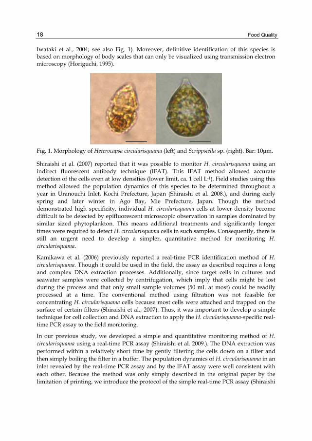
Iwataki et al., 2004; see also Fig. 1). Moreover, definitive identification of this species is based on morphology of body scales that can only be visualized using transmission electron microscopy (Horiguchi, 1995).
Fig. 1. Morphology of Heterocapsa circularisquama (left) and Scrippsiella sp. (right). Bar: 10μm.
Shiraishi et al. (2007) reported that it was possible to monitor H. circularisquama using an
indirect fluorescent antibody technique (IFAT). This IFAT method allowed accurate
detection of the cells even at low densities (lower limit, ca. 1 cell L-1). Field studies using this
method allowed the population dynamics of this species to be determined throughout a
year in Uranouchi Inlet, Kochi Prefecture, Japan (Shiraishi et al. 2008.), and during early
spring and later winter in Ago Bay, Mie Prefecture, Japan. Though the method
demonstrated high specificity, individual H. circularisquama cells at lower density become
difficult to be detected by epifluorescent microscopic observation in samples dominated by
similar sized phytoplankton. This means additional treatments and significantly longer
times were required to detect H. circularisquama cells in such samples. Consequently, there is
still an urgent need to develop a simpler, quantitative method for monitoring H.
circularisquama.
Kamikawa et al. (2006) previously reported a real-time PCR identification method of H.
circularisquama. Though it could be used in the field, the assay as described requires a long
and complex DNA extraction processes. Additionally, since target cells in cultures and
seawater samples were collected by centrifugation, which imply that cells might be lost
during the process and that only small sample volumes (50 mL at most) could be readily
processed at a time. The conventional method using filtration was not feasible for
concentrating H. circularisquama cells because most cells were attached and trapped on the
surface of certain filters (Shiraishi et al., 2007). Thus, it was important to develop a simple
technique for cell collection and DNA extraction to apply the H. circularisquama-specific real-
time PCR assay to the field monitoring.
In our previous study, we developed a simple and quantitative monitoring method of H.
circularisquama using a real-time PCR assay (Shiraishi et al. 2009.). The DNA extraction was
performed within a relatively short time by gently filtering the cells down on a filter and
then simply boiling the filter in a buffer. The population dynamics of H. circularisquama in an
inlet revealed by the real-time PCR assay and by the IFAT assay were well consistent with
each other. Because the method was only simply described in the original paper by the
limitation of printing, we introduce the protocol of the simple real-time PCR assay (Shiraishi

Monitoring Harmful Microalgae by Using a Molecular Biological Technique
19
et al. 2009.) in detail in the following sections. This protocol will be helpful for the studies on
many other dinoflagellate species by real-time PCR assay.
4. Materials and methods
4.1 Organisms and culture conditions
The algal strains of H. circularisquama, K. mikimotoi and Skeletonema sp. were obtained from
the National Research Institute of Fisheries and Environment of Inland Sea, Fisheries
Research Agency. Strains of Heterocapsa triquetra and Heterosigma akashiwo were isolated by
G. Nishitani from Maizuru Bay, Kyoto Prefecture, Japan in 1998 and by I. Imai from
Hiroshima Bay, Hiroshima Prefecture, Japan in 1989, respectively. These strains were
cultured at a temperature of 20 ºC on a 14-h light: 10-h dark photo-cycle under an
illumination at 180 µmol photons m-2 s-1 in modified SWM-3 medium (Chen et al., 1969; Imai
et al., 1996).
4.2 Cell collection and DNA extraction
The most effective method of extracting H. circularisquama DNA from cell pellets was
evaluated using six different protocols, the simplest of which was a TE boiling method
modified from the procedure of Kamikawa et al. (2006). For the basic boiling procedure,
either one cell or 100 cells of cultured H. circularisquama cells were collected on the
Nuclepore polycarbonate membrane filters (pore size 3.0 µm) (Whatman, Maidstone, UK)
by filtration, respectively. Extractions at both cell concentrations were done in triplicate to
assess assay variability. The filter was placed in a 1.5-mL microtube without folding, and
then 750 µL of TE buffer (10 mM Tris-HCl: pH 8.0, 1 mM EDTA: pH 8.0) was added. After
the boiling for 10 min in the TE buffer, the filter was immediately removed. The extracted
DNA sample was stored at -60 ºC until the real-time PCR assay was performed. DNA
extraction efficiency using the TE (Tris-HCl/EDTA) boiling method was compared with that
by the modified CTAB (Cetyltrimethylammonium Bromide) method (Zhou et al., 1999;
Kamikawa et al., 2005) and the proteinase K method (Kamikawa et al., 2005), both of which
are commonly used. The same protocol as described above for the TE extractions was
followed.
To choose the most suitable filter for the DNA extraction of H. circularisquama, the cells were collected on 6 different filters and the DNA extraction was performed on each filter by the TE boiling method which was found to be the efficient method from the study described above. Specifically, one cell and 100 cells of cultured H. circularisquama cells were collected by filtration onto membrane filters composed of either polycarbonate membranes (Nuclepore, mesh size 3.0 µm), glass-fibers (GF/C, pore size 1.2 µm) (Whatman, Maidstone, UK), cellulose mixed esters (pore size 3.0 µm) (Millipore, Tokyo, Japan), cellulose acetate (pore size 3.0 µm) (ADVANTEC, Tokyo, Japan), polytetrafluoroethylene (PTFE) (pore size 3.0 µm) (ADVANTEC, Tokyo, Japan), or hydrophilic polyvinylidene difluoride (PVDF) (pore size 5.0 µm) (Millipore, Tokyo, Japan), respectively. The DNA sample was stored at -60 ºC until the real-time PCR assay was performed.
Based on the results of the extraction efficiency tests on the various tests, a standard curves
consisting of eight-fold serial dilutions (104 to 1 cells) of cultured cells were prepared. Each

Food Quality
20
number of the cells was collected on the Nuclepore filter (pore size 3.0 µm) (Whatman,
Maidstone, UK) by filtration. The DNA extraction was performed by the TE boiling method,
and the real-time PCR assay was carried out in triplicate. The standard curve was
constructed based on the correlation between the threshold cycle (Ct value) and the number
of cells.
A major concern when designing a real-time PCR assay for HABs is whether other co-
occurring microalgae adversely affect the amplification efficiency either by introducing
inhibitors or due to cross-reactivity problems. This possibility was explored in an
examination where H. circularisquama cells (104 to 1 cells) were filtered on the Nuclepore
filters (pore size 3.0 µm) at 20 cm Hg with 105 cells each of H. triquetra, H. akashiwo, K.
mikimotoi and Skeletonema sp. which are frequently co-dominated in western coastal areas of
Japan. A previous study also showed that the primers and probe used in this study are
species-specific and do not react DNA from H. triquetra, H. akashiwo or K. mikimotoi
(Kamikawa et al., 2006). The DNA extraction was performed by the TE boiling method, and
the real-time PCR assay was carried out as follows in triplicate. Obtained Ct values at each
number of cells were compared with those of the control experiment where only H.
circularisquama was used.
4.3 Real-time PCR
The primer set and probe used in this study were based on unique species-specific DNA
sites identified by aligning the D1/D2 LSU rDNA sequence of H. circularisquama
(DDBJ/EMBL/GenBank accession number AB049709) with the correponding dinoflagellate
sequences in GenBank. Primers specific to H. circularisquama were HcirF (5’-
GTTTGCCTATGGGTGAGC-3’) and HcirR (5’-CATTGTGTCAGGGAGGAG-3’) and the
probe was HcirTaqMan (5’-FAM-CACCACAAGGTCATGAGGACACA-TAMRA-3’) that
was labeled at the 5’-end with FAM (carboxyfluorescein) and the 3’-end with TAMRA
(carboxytetramethylrhodamine) (Kamikawa et al., 2006).
Thermal cycling was performed with a Rotor-Gene 3000 (Corbett Research, Mortlake,
Australia) in 200-µL PCR tubes of commodity type. PCR was carried out in 25-µL volumes
comprising 1×PCR EX Taq buffer (containing 20mM Mg2+), 200 µM dATP, 200 µM dTTP,
200 µM dGTP, 200 µM dCTP, 0.3 µM forward and reverse primers, 0.4 µM fluorogenic
probe, and 1.25 U of Taq DNA polymerase (Takara EX TaqTM, TaKaRa Bio Inc., Shiga,
Japan). The PCR conditions were as follows according to Kamikawa et al. (2006): one
heating cycle at 95 ºC for 2 min, followed by 45 cycles at 95 ºC for 10 sec and 54 ºC for 30 sec.
The Ct value was calculated as a cycle number that an amplification curve reached at the
most suitable threshold value.
5. Results and discussion
5.1 Development of a DNA extraction method
In order to examine the most efficient method for DNA extraction, three kinds of DNA
extraction methods were subjected to H. circularisquama cells (100 cells and 1 cell) trapped on
the filter. Figure 2 shows obtained Ct values for one and 100 cells by real-time PCR assay

Monitoring Harmful Microalgae by Using a Molecular Biological Technique
21
with each DNA extraction method. For 100 cells, the DNA extracted with the TE boiling
method was as efficient as with the CTAB method and the proteinase K method (t-test, df =
4, p > 0.05). For 1 cell, the DNA extraction efficiency with the TE boiling method was higher
than that with the modified CTAB method (t-test, df = 4, p < 0.05) and similar to that of the
proteinase K method (t-test, df = 4, p > 0.05). Thus, we can consider that the three methods
are similarly efficient for H. circularisquama cells with high density. Given the importance of
detection the HAB species at low density, the TE boiling method appeared to be the most
useful technique for monitoring H. circularisquama by real-time PCR assay. In addition to its
higher detection efficiency, the TE boiling method is more suitable in simplicity, ease of
execution, lower cost, and shorter execution time than the other two methods.
Fig. 2. Comparison of Ct (Threshold cycle) values obtained with three DNA extraction
methods. TE boiling method, CTAB method and proteinase K and SDS method were
subjected to 100 cells (left) and one cell (right) of H. circularisquama on
Nucleporepolycarbonate membrane filters. Ct values were obtained by using the real-time
PCR assay in triplicate. The bars show the standard deviations.
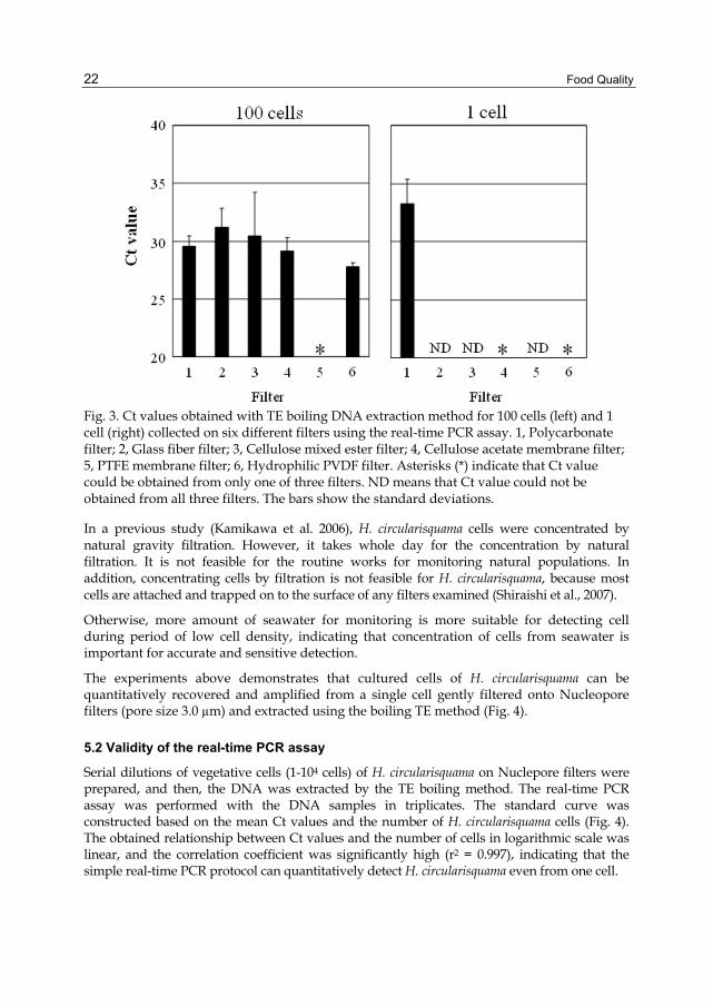
In order to select the filter which would yield the highest and most consistent recovery of
DNA, samples containing either 1 or 100 cells of H. circularisquama cells were filtered onto
six different types of filter. DNA was then extracted using the TE boiling method and
subjected to real-time PCR-amplification. In the case of 100 cells, the real-time PCR assay
successfully amplified H. circularisquama DNA from all the filters with the exception of the
polytetra fluoroethylene (PTFE) membrane filter (Fig. 3). In contrast, for the 1 cell samples,
the qPCR assay failed to reliably amplify the DNA from all the filters with the exception of
the Nuclepore polycarbonate membrane filter, which could be detected in triplicate. Only
one of three DNA samples extracted from either the cellulose acetate or hydrophilic
polyvinylidine difluoride (PVDF) membrane filters were detected. Therefore, we concluded
that the best filter for extraction and detection of H. circularisquama was Nuclepore
polycarbonate membrane filter.
Food Quality
22
Fig. 3. Ct values obtained with TE boiling DNA extraction method for 100 cells (left) and 1 cell (right) collected on six different filters using the real-time PCR assay. 1, Polycarbonate filter; 2, Glass fiber filter; 3, Cellulose mixed ester filter; 4, Cellulose acetate membrane filter; 5, PTFE membrane filter; 6, Hydrophilic PVDF filter. Asterisks (*) indicate that Ct value could be obtained from only one of three filters. ND means that Ct value could not be obtained from all three filters. The bars show the standard deviations.
In a previous study (Kamikawa et al. 2006), H. circularisquama cells were concentrated by natural gravity filtration. However, it takes whole day for the concentration by natural filtration. It is not feasible for the routine works for monitoring natural populations. In addition, concentrating cells by filtration is not feasible for H. circularisquama, because most cells are attached and trapped on to the surface of any filters examined (Shiraishi et al., 2007).
Otherwise, more amount of seawater for monitoring is more suitable for detecting cell during period of low cell density, indicating that concentration of cells from seawater is important for accurate and sensitive detection.
The experiments above demonstrates that cultured cells of H. circularisquama can be quantitatively recovered and amplified from a single cell gently filtered onto Nucleopore filters (pore size 3.0 µm) and extracted using the boiling TE method (Fig. 4).
5.2 Validity of the real-time PCR assay
Serial dilutions of vegetative cells (1-104 cells) of H. circularisquama on Nuclepore filters were prepared, and then, the DNA was extracted by the TE boiling method. The real-time PCR assay was performed with the DNA samples in triplicates. The standard curve was constructed based on the mean Ct values and the number of H. circularisquama cells (Fig. 4). The obtained relationship between Ct values and the number of cells in logarithmic scale was linear, and the correlation coefficient was significantly high (r2 = 0.997), indicating that the simple real-time PCR protocol can quantitatively detect H. circularisquama even from one cell.

Monitoring Harmful Microalgae by Using a Molecular Biological Technique
23
Fig. 4. Real-time PCR assay using eight-fold serial dilutions (104 to 1 cell). The result for each cell number was represented by each symbol shown in the figure.
In order to examine the effects of the existence of other microalgae on the DNA extraction or subsequent PCR-based quantification, H. circularisquama cells (104 to 1 cells) were collected together with 105 cells of H. triquetra, H. akashiwo, K. mikimotoi and Skeletonema sp. on the Nuclepore membrane filters by filtration. The DNA extraction and the real-time PCR assay were carried out as described above. The standard curve was constructed based on the mean Ct values and the number of cells (Fig. 5a). At each number of H. circularisquama examined, there was no significant difference between the Ct value obtained from H. circularisquama cells in spite of presence or absence of the other microalgae (t-test, df = 4, p > 0.05, Fig. 5b). The correlation between Ct values and the number of cells in logarithmic scale was linear, and the correlation coefficient was extremely significant (r2 = 0.991, Fig. 5a). It was confirmed that the DNA extraction and subsequent PCR-based quantification of H. circularisquama cells were not inhibited even when other microalgae such as H. triquetra, H. akashiwo, K. mikimotoi and Skeletonema sp. coexist with H. circularisquama.
The constructed standard curve showed linearity (Fig. 4), and the protocol including concentration of cells, DNA extraction, and the real-time PCR was not inhibited by the existence of other microalgae even at 105 cells of H. triquetra, H. akashiwo, K. mikimotoi and Skeletonema sp. (Fig. 5b). It was clearly demonstrated that the presence of closely related species (e.g., H. triquetra) and/or many other common red tide species did not affect the efficiency of DNA extraction and subsequent PCR-based quantification of H. circularisquama cells.
When there are H. circularisquama cells in addition to much higher abundance of similar sized phytoplankton in the field, the detection of H. circularisquama cells by the canonical IFAT method with epifluorescent signal is obscured by the presence of numerous other microalgal cells. Similarly, an underestimation of cell abundance can be occurred when a large number of particles such as detritus not only inhibit epifluorescence microscopy

Food Quality
24
observation but also blocks the antibody reaction trapped within the detritus, indicating that the IFAT method is difficult to be applied to sediments and detritus-rich samples. The real-time PCR assay described in this study appears to be more feasible and practical for environmental samples than the IFAT method.
Fig. 5. Relationship between Ct values and the log number of cells. a. Standard curve for H. circularisquama cells constructed from DNA that was extracted from H. circularisquama cells plus several microalgae. b. Comparison of detection and quantification efficiency between DNA that was extracted from H. circularisquama cells (closed bars) and from H. circularisquama cells plus several microalgae (open bars). The bars show standard deviations.
6. Application to environmental samples
The procedure described above was applied to environmental samples in order to monitor successively H. circularisquama cells in addition to IFAT assay (Shiraishi et al. 2009.). The cell densities obtained by the real-time PCR assay were almost identical to the results obtained by the IFAT method. Hence, it was clearly demonstrated that H. circularisquama could be quantified by this simple real-time PCR assay as sensitively and precisely as the IFAT method in the field. It is notable that the detection limit of the real-time PCR assay was 1 cell/L: The most sensitive level currently with real-time PCR assay (Shiraishi et al. 2009.). It

Monitoring Harmful Microalgae by Using a Molecular Biological Technique
25
should be also mentioned that the real-time PCR assay sometimes reacted to some environmental samples which the IFAT assay did not (Shiraishi et al. 2009.). This incongruence can be explained by that the real-time PCR assay is more sensitive than the IFAT assay. Otherwise, the real-time PCR assay might react to cell-free DNA derived from broken, dead cells of H. circularisquama. Since we have no idea which is true, it is better to use both methods for monitoring H. circularisquama cells in order to grasp precise dynamics of the HAB species, escaping both underestimation and overestimation.
In this chapter, it has been demonstrated that the real-time PCR assay can be applied to monitoring various HABs in field waters. If other HABs can be quantified by the same manner to present method with slight modification, those microalgae would be easily monitored with the similar procedures of the DNA extraction at the same time. The conventional methods for monitoring HABs with optical microscopy might be replaced by the simple real-time PCR assay in the near future, when the costs of machines and reagents are lowered to become reasonable.
In addition to seawater samples, real-time PCR assay has been applied for the detection of the cysts of the toxic Alexandrium species from marine sediments (Kamikawa et al., 2005, 2007, Erdner et al. 2011). Furthermore, the PCR method was also used for the detection of the cells of Alexandrium species in the tissue of mussels (Galluzzi et al., 2005) in order to investigate the possibility that the HAB cells are propagated to other areas by transport of bivalves. When H. circularisquama forms temporary cysts in water columns, those temporary cysts possibly sink down and survive some periods at the surface of the sea bottom. There are some reports that H. circularisquama could proliferate in water columns of a new area after the transportation of bivalves which accompany temporary cysts (Honjo et al., 1998; Honjo and Imada, 1999; Imada et al., 2001). Given the possibility of the temporary cysts as a seed-population, the detection of H. circularisquama cells is an urgent need from sediments, and tissues and fecal pellets of bivalves.
7. Acknowledgment
We would like to thank Drs. T. Uchida, M. Yamaguchi, and Y. Matsuyama (the National Research Institute of Fisheries and Environment of Inland Sea, Fisheries Research Agency), and G. Nishitani (Tohoku University) for their kind donation of the culture strains of H. circularisquama, K. mikimotoi and Skeletonema sp. for this study. RK is a research fellow supported by the Japan Society for Promotion of Sciences (no. 210528). This work wa supported in part by a grant from the Fishery Agency of Japan.
8. References
Adachi, M.; Sako, Y. & Ishida, Y. (1994). Restriction fragment length polymorphism of ribosomal DNA internal transcribed spacer and 5.8S regions in Japanese Alexandrium species (Dinophyceae). Journal of Phycology, Vol.30, pp.857-863.
Anderson, D.M. (1994). Red tides. Scientific American, Vol.271, pp.52-58. Bowers, H.A.; Tengs, T., Glasgow, H.B.Jr, Burkholder, J.M., Rublee, P.A. & Oldach, D.W.
(2000). Development of real-time PCR assays for rapid detection of Pfiesteria piscicida and related dinoflagellates. Applied and Environmental Microbiology, Vol.66, pp.4641-4648

Food Quality
26
Bowers, H.A.; Tomas, C., Tengs, T., Kempton, J.W., Lewitus, A.J. & Oldach, D.W. (2006). Raphidophyceae [Chadefaud ex Silva] systematic and rapid identification: sequence analyses and real-time PCR assays. Journal of Phycology, Vol.42, pp.1333-1348.
Chen, L.C.M.; Edelstein, T. & McLachlan, J. (1969). Bonnemaisonia hamifera Hariot in nature and in culture. Journal of Phycology, Vol.5, pp.211-220.
Dyhrman, S.T.; Erdner, D., Du, J.L., Galac, M. & Anderson, D.M. (2006). Molecular quantification of toxic Alexandrium fundyense in the gulf of Maine using real-time PCR. Harmful Algae, Vol.5, pp.242-250.
Erdner, D.; Percy, L., Keafer, B., Lewis, J. & Anderson, D.M. (2011). A quantitative real-time PCR assay for the identification and enumeration of Alexandrium cysts in marine sediments. Deep Sea Research Part II: Topical Studies in Oceanography, Vol.57, pp.279-287.
Galluzzi, L.; Penna, A., Bertozzini, E., Vila, M., Garcés, E. & Magnani, M. (2004). Development of a real-time PCR assay for rapid detection and quantification of Alexandrium minutum (a Dinoflagellate). Applied and Environmental Microbiology, Vol.70, pp.1199-1206.
Galluzzi, L.; Penna, A., Bertozzini, E., Giacobbe, M.G., Vila, M., Garcés, E., Prioli, S. & Magnani, M. (2005). Development of a quantitative PCR method for the Alexandrium spp. (Dinophyceae) detection in contaminated mussels (Mytilus galloprovincialis). Harmful Algae, Vol.4, pp.973-983.
Godhe, A.; Rehnstam-Holm, A.S., Karunasagar, I. & Karunasagar, I. (2002). PCR detection of dinoflagellate cysts in field sediment samples from tropic and temperate environments. Harmful Algae, Vol.1, pp.361-373.
Gray, M.; Wawrik, B., Paul, J. & Casper, E. (2003). Molecular detection and quantitation of the red tide dinoflagellate Karenia brevis in the marine environment. Applied and Environmental Microbiology, Vol.69, pp.5726-5730.
Hallegraeff, G.M. (1993). A review of harmful algal blooms and their apparent global increase. Phycologia, Vol.32, pp.79-99.
Hallegraeff, G.M. (1995). Harmful algal blooms: a global overview. In: G.M. Hallegraeff, D.M. Anderson and A.D. Cembella, Editors, Manual on Harmful Marine Microalgae, IOC Manuals and Guides. UNESCO. Vol.33, pp.1–22.
Handy, S.M.; Coyne, K.J., Portune, K.J., Demir, E., Doblin, M.A., Hare, C.E., Cary, S.C. & Hutchins, D.A. (2005). Evaluating vertical migration behavior of harmful raphidophytes in the Delaware Inland Bays utilizing quantitative real-time PCR. Aquatic Microbial Ecology, Vol.40, pp.121-132.
Heid, C.A.; Stevens, J., Livak, K.J. & Williams, P.M. (1996). Real time quantitative PCR. Genome Research, Vol.6, pp.986-994.
Holland, P.M.; Abramson, R.D., Watson, R. & Gelfand D.H. (1991). Detection of specific polymerase chain reaction product by utilizing the 5'-3' exonuclease activity of Thermus aquaticus DNA polymerase. Proceedings of the National Academy of Sciences of the United States of America, Vol.88, pp.7276-7280.
Honjo, T.; Imada, N., Ohshima, Y., Maema, Y., Nagai, K., Matsuyama, Y. & Uchida, T. (1998). Potential transfer of Heterocapsa circularisquama with pearl oyster consignments. In: Reguera, B., Blanco, J., Fernandez, M.L., Wyatt, T. (Eds), Harmful Algae. Xunta de Galicia and IOC of UNESCO, Santiago de Compostela, pp.224-226.
Monitoring Harmful Microalgae by Using a Molecular Biological Technique
27
Honjo, T. & Imada, N. (1999). Future attention-Spread of Heterocapsa circularisquama red tides and its preventive measure. Bulltin of the Plankton Society of Japan, Vo.46, pp.180-181 (in Japanese).
Horiguchi, T. (1995). Heterocapsa circularisquama sp. nov. (Peridiniales, Dinophyceae): a new marine dinoflagellate causing mass mortality of bivalves in Japan. Phycological Research, Vol.43, pp.129-136.
Hosoi-Tanabe, S. & Sako, Y. (2005a). Rapid detection of natural cells of Alexandrium tamarense and A. catenella (Dinophyceae) by fluorescence in situ hybridization. Harmful Algae, Vol.4, pp.319-328.
Hosoi-Tanabe, S. & Sako, Y. (2005b). Species-specific detection and quantification of toxic marine dinoflagellates Alexandrium tamarense and A. catenella by real-time PCR assay. Marine Biotechnology, Vol.7, pp.506-514.
Imada, N.; Honjo, T., Shibata, H., Oshima, Y., Nagai, K., Matsuyama, Y. & Uchida, T. (2001). The quantities of Heterocapsa circularisquama cells transferred with shellfish consignments and the possibility of its establishment in new areas. In: Hallegraeff, G.M., Blackburn, S.I., Bolch, C.J., Lewis, R.J. (Eds), Harmful Algal Blooms 2000. IOC of UNESCO, Paris, pp.474-476.
Imai, I.; Itakura, S., Matsuyama, Y. & Yamaguchi, M. (1996). Selenium requirement for growth of a novel red tide flagellate Chattonella verruculosa (Raphidophyceae) in culture. Fisheries Science, Vol.62, pp.834-835.
Imai, I. (2000). Current problems in classification and identification of marine raphidoflagellates (raphidophycean flagellates): from the view point of ecological study. Bulltin of the Plankton Society of Japan, Vol.47, pp.55–64 (in Japanese).
Imai, I.; Yamaguchi, M. & Hori, Y. (2006). Eutrophication and occurrences of harmful algal blooms in the Seto Inland Sea, Japan. Plankton and Benthos Ressearch, Vol.1, pp.71-84.
Iwataki, M.; Hansen, G., Sawaguchi, T., Hiroishi, S. & Fukuyo, Y. (2004). Investigations of body scales in twelve Heterocapsa species (Peridiniales, Dinophyceae), including a new species H. pseudotriquetra sp. nov. Phycologia Vol.43, pp.394-403.
Kamikawa, R.; Hosoi-Tanabe, S., Nagai, S., Itakura, S. & Sako, Y. (2005). Development of a quantification assay for the cysts of the toxic dinoflagellate Alexandrium tamarense using real-time polymerase chain reaction. Fisheries Science, Vol.71, pp.987-991.
Kamikawa, R.; Asai, J., Miyahara, T., Murata, K., Oyama, K., Yoshimatsu, S., Yoshida, T. & Sako, Y. (2006). Application of a real-time PCR assay to a comprehensive method of monitoring harmful algae. Microbes and Environments, Vol.21, pp.163-173.
Kamikawa, R.; Nagai, S., Hosoi-Tanabe, S., Itakura, S., Yamaguchi, M., Uchida, Y., Baba, T. & Sako, Y. (2007). Application of real-time PCR assay for detection and quantification of Alexandrium tamarense and Alexandrium catenella cysts from marine sediments. Harmful Algae, Vol.6, pp.413-420.
Matsuyama, Y.; Uchida, T., Nagai, K., Ishimura, M., Nishimura, A., Yamaguchi, M. & Honjo, T. (1996). Biological and environmental aspects of noxious dinoflagellate red tides by Heterocapsa circularisquama in the west Japan. In: Yasumoto, T., Oshima, Y., Fukuyo, Y. (Eds), Harmful and Toxic Algal Blooms. UNESCO, Paris, pp.247-250.
Matsuyama, Y.; Kimura, A., Fujii, H., Takayama, H. & Uchida, T. (1997). Occurrence of a Heterocapsa circularisquama red tide and subsequent damages to shellfish in western Hiroshima Bay, Seto Inland Sea, Japan in 1995. Bulletin of the Nansei National Fisheries Research Institute, Vol.30, pp.189-207 (in Japanese, with English abstract).

Food Quality
28
Matsuyama, Y. (1999). Harmful effect of dinoflagellate Heterocapsa circularisquama on shellfish aquaculture in Japan. Japan Agricultural Research Quarterly, Vol.33, pp.283-293.
Matsuyama, Y.; Uchida, T., Honjo, T. & Shumway, S.E. (2001). Impacts of the harmful dinoflagellate, Heterocapsa circularisquama, on shellfish aquaculture in Japan. Journal of Shellfish Research, Vol.20, pp.1269-1272.
Nagai, K.; Matsuyama, Y., Uchida, T., Yamaguchi, M., Ishimura, M., Nishimura, A., Akamatsu, S. & Honjo, T. (1996). Toxicity and LD50 levels of the red tide dinoflagellate Heterocapsa circularisquama on juvenile pearl oysters. Aquaculture, Vol.144, pp.149-154.
Nagai, K.; Matsuyama, Y., Uchida, T., Akamatsu, S. & Honjo, T. (2000). Effect of a natural population of the harmful dinoflagellate Heterocapsa circularisquama on the survival of the pearl oyster Pinctada fucata. Fisheries Science, Vol.66, pp.995-997.
Nakanishi, K.; Onaka, S., Kobayashi, T. & Masuda, K. (1999). Bloom dynamics of Heterocapsa circularisquama in Ago Bay, Japan. Bulltin of the Plankton Society of Japan, Vol.46, pp.161-164 (in Japanese).
Sako, Y.; Hosoi-Tanabe, S. & Uchida, A. (2004). Fluorescence in situ hybridization using rRNA-targeted probes for simple and rapid identification of the toxic dinoflagellates Alexandrium tamarense and Alexandrium catenella. Journal of Phycology, Vol.40, pp.598-605.
Shiraishi, T.; Hiroishi, S., Nagai, K., Go, J., Yamamoto, T. & Imai, I. (2007). Seasonal distribution of the shellfish-killing dinoflagellate Heterocapsa circularisquama in Ago Bay monitored by an indirect fluorescent antibody technique using monoclonal antibodies. Plankton Benthos Research, Vol.2, pp.49-62.
Shiraishi, T.; Hiroishi, S., Kamikawa, R., Sako, Y., Taino, S., Ishikawa, T., Hayashi, Y. & Imai, I. (2009). Population dynamics of the shellfish-killing dinoflagellate Heterocapsa circularisquama monitored by an indirect fluorescent antibody technique and a real-time PCR assay in Uranouchi Inlet, Kochi Prefecture, Japan. In Proceedings of 5th World Fisheries Congress, 6c_1006_200, TerraPub, Tokyo.
Shiraishi, T.; Hiroishi, S., Taino, S., Ishikawa, T., Hayashi, Y., Sakamoto, S., Yamaguchi, M. & Imai, I. (2008). Identification of overwintering vegetative cells of the bivalve-killing dinoflagellate Heterocapsa circularisquama in Uranouchi Inlet, Kochi Prefecture, Japan. Fisheries Science, Vol.74, pp.128-136.
Smayda, T.J. (1997). Bloom dynamics: physiology, behaviour, trophic effects. Limnology and Oceanography, Vol.42, pp.1132-1136.
Tamai, K. (1999). Current status of outbreaks and fisheries damages due to Heterocapsa circularisquama. Bulltin of the Plankton Society of Japan, Vol.46, pp.153-154 (in Japanese).
Yamaguchi, M.; Itakura, S., Imai, I. & Ishida, Y. (1995). A rapid and precise technique for enumeration of resting cysts of Alexandrium spp. (Dinophyceae) in natural sediments. Phycologia, Vo.34, pp.207–214.
Zhang, H. & Lin, S. (2005). Development of a cob-18S rRNA gene real-time PCR assay for quantifying Pfiesteria shumwayae in the natural environment. Applied and Environmental Microbiology, Vo.71, pp. 7053-7063.
Zhou, Z.; Miwa, M. & Hogetsu, T. (1999). Analysis of genetic structure of a Suillus grevillei population in a Larix kaempferi stand by polymorphism of inter-simple sequence repeat (ISSR). New Phytologists, Vo.144, pp. 55-63.