molecular detection, quantification, and diversity evaluation of microalgae
TRANSCRIPT
REVIEWARTICLE
Molecular Detection, Quantification, and DiversityEvaluation of Microalgae
Vinitha Ebenezer & Linda K. Medlin & Jang-Seu Ki
Received: 28 July 2011 /Accepted: 2 December 2011# Springer Science+Business Media, LLC 2011
Abstract This study reviews the available molecular meth-ods and new high-throughput technologies for their practicaluse in the molecular detection, quantification, and diversityassessment of microalgae. Molecular methods applied to othergroups of organisms can be adopted for microalgal studiesbecause they generally detect universal biomolecules, such asnucleic acids or proteins. These methods are primarily relatedto species detection and discrimination among various micro-algae. Among current molecular methods, some moleculartools are highly valuable for small-scale detection [e.g.,single-cell polymerase chain reaction (PCR), quantitativereal-time PCR (qPCR), and biosensors], whereas others aremore useful for large-scale, high-throughput detection [e.g.,terminal restriction length polymorphism, isothermal nucleicacid sequence-based amplification, loop-mediated isothermalamplification, microarray, and next generation sequencing(NGS) techniques]. Each molecular technique has its ownstrengths in detecting microalgae, but they may sometimeshave limitations in terms of detection of other organisms.Among current technologies, qPCR may be considered thebest method for molecular quantification of microalgae. Meta-genomic microalgal diversity can easily be achieved by 454pyrosequencing rather than by the clone library method.Current NGS, third and fourth generation technologies pavethe way for the high-throughput detection and quantification
of microalgal diversity, and have significant potential forfuture use in field monitoring.
Keywords Microalgae .Molecular techniques . Detection .
Quantification . Diversity
Introduction
Microalgae are microscopic, unicellular species that existsolitarily or in chains and are typically found in aquaticsystems. They play a vital role in primary production inthe aquatic environments and contribute to global atmo-spheric carbon dioxide acquisition. The commercial valueof microalgae is increasing day by day; for example, goodnutritional values (Becker 2007), large utilization in aqua-cultures (Brown 2002), and a possible use for biofuel pro-duction (Chisti 2007). Moreover, some microalgae, such asdinoflagellates and diatoms, can form harmful algal blooms(HABs) and contain biotoxins that affect humans and manyother organisms that consume these algae (e.g., bivalves,which can filter the toxic species). Microalgae are incrediblydiverse, and their species are estimated to amount to ca.200,000–800,000, of which only about 35,000 are described(Cheng and Ogden 2011). Microalgal species have tradi-tionally been discriminated by morphological observationsand pigment profiles. Molecular discrimination methods aresometimes very effective for their identification, especiallyfor the pico-sized fractions that have very few morpholog-ical features that can be used for identification (Not et al.2007). From a historical viewpoint, the advent of the use ofmolecular technology in phycology began in the 1970s.During this time, phycologists developed molecular techni-ques as indirect detection methods; they had been limited bythe traditional methodologies for microalgal discrimination
V. Ebenezer : J.-S. Ki (*)Department of Green Life Science, College of Convergence,Sangmyung University,Seoul 110-743, South Koreae-mail: [email protected]
L. K. MedlinParis 6, CNRS, UMR 7621, LOMIC, ObservatoireOcéanologique, Université Pierre et Marie Curie,66651, Banyuls-sur-Mer, France
Mar BiotechnolDOI 10.1007/s10126-011-9427-y

and identification. These indirect discrimination methodsinclude the detection of carbohydrates, nucleic acids, pro-teins, and toxins from microalgae. Since then, DNA-basedapproaches have been extensively developed, and thesefindings have greatly expanded our understanding of geneticdiversity, molecular systematics, evolution, and even adap-tive responses, from an ecological basis, for all organismsand not just the microalgae (Bott et al. 2010; Kudela et al.2010; Medlin and Kooistra 2010).
Molecular techniques used for planktonic microalgae (orphytoplankton) have been developed or modified by biologistsfor a long time, and these methods have been reviewed over thelast decade (Medlin et al. 2000, 2006, 2007; de Bruin et al.2003). Recently, Bott et al. (2010) extensively compared rou-tine DNA-based detection methods for the development ofpractical, specific, sensitive, and rapid diagnostics of marinepests, including harmful algae (HA). In addition, Kudela et al.(2010) provided a detailed outline of the molecular tools avail-able for comparative harmful algal bloom programs in upwell-ing systems, focusing on cell enumeration and identification,molecular phylogenetics, and applications of high-throughputsequencingmethods.Most of themolecular methods have beenprimarily developed for the discrimination of planktonic micro-algae, particularly HA, and tested under laboratory conditions.The lack of rigorous field testing of the applicability of thesemolecular techniques often poses a problem. Factors that needto be addressed include the selection of suitable geneticmarkers for the identification, quantification, diversity analysis,and isolation of genomic DNA from environmental samples.Currently available methods do not really place an emphasis onthe quantification of microalgal cells, but enumeration of cellsplays a crucial role in field assessment. On the other hand,routine molecular techniques and more recent advanced tech-nologies, such as next-generation sequencing (NGS) technolo-gies, should be focused on molecular quantification, which isusually a tedious process and also on the large-scale microalgaldetection from the environment.
In the present paper, we review molecular technologies,including recent advanced techniques [e.g., DNA chip,loop-mediated isothermal amplification (LAMP), and NGStechnologies], for accurate, rapid detection and environmen-tal monitoring, as well as for the quantification and large-scale diversity assessment of microalgae.
Molecular Technologies for Microalgae Detection
In general, all kinds of detection techniques used in molecularbiology can be applied to microalgal studies because theprinciple of molecular detection is to target universal biomo-lecules, such as nucleic acids and proteins. Table 1 provides anoutline of various techniques used to identify and detectmicroalgae, especially the harmful bloom-forming species.
Overall, these methods can be categorized into five groupsof detection tools according to the target molecule: toxin,proteins, carbohydrates, RNA, and DNA. Toxin profiles areonly used as discrimination markers for biotoxin-producingspecies (Cembella et al. 1987); these methods are thus rare andcan vary even among strains of a single species. On the otherhand, proteins and nucleic acids have frequently been thetarget molecules of choice. Protein-based methods includeallozyme electrophoresis and immunoassay (or ELISA) withantibodies (Shapiro et al. 1989; Sako et al. 1990). Most meth-ods are DNA-targeting technologies, including restriction frag-ment length polymorphism (RFLP), denaturing gradient gelelectrophoresis (DGGE), single-stranded conformation poly-morphism (SSCP), random amplification of polymorphicDNA (RAPD), amplified fragment length polymorphism(AFLP), microsatellites, and polymerase chain reaction(PCR), among others (see Table 1). DNA-based methods havebeen developed for the taxonomic identification of species orstrains of microalgae and are often used for phylogeneticanalyses. Microsatellites and internal short sequence repeat(ISSR) analysis are often used in population genetics of ani-mals and higher plants and have recently been applied tomicroalgae (Nagai et al. 2007; Evans et al. 2009; Casteleynet al. 2010; see later reviews). These studies have shown thatthe oceanic environment is highly fragmented and that micro-algal populations are highly diverse, even on a local level.
Molecular tools that have been developed for environmen-tal samples are mostly designed to detect certain harmfulmicroalgae (e.g., Alexandrium, Heterosigma, Gyrodinium,Gymnodinium, Karenia, Pfiesteria, Pseudo-nitzschia, etc.)rather than to quantify these cells. However, quantitativePCR (qPCR) has recently been applied to some species inenvironmental samples (e.g., Dyhrman et al. 2006, 2010). Ofthe available methods, fluorescently labeled lectins, antibodies(immunoassays), and oligonucleotide probes were first usedto bind or hybridize molecules located on the cell’s surface(Lindquist 1997; Simon et al. 2000), and internally to ribo-somes (Hosoi-Tanabe and Sako 2005, 2006). Among themicroalgae, ribosomal RNA (rRNA) probes have routinelybeen applied to several harmful algal species, such as Alexan-drium tamarense, Pseudo-nitzschia, Heterosigma akashiwo,and Fibrocapsa japonica (Scholin and Anderson 1993;Scholin et al. 1994; Tyrrell et al. 2002), and also for thedetection of some picoeukaryotoc microalgae, such asMicro-monas pusilla and Bathycoccus prasinos (Not et al. 2004;Lepère et al. 2009; Shi et al. 2011). Whereas, in bacterialresearch, they have been widely used to characterize the entirecommunity (Britschgi and Giovannoni 1991; Amann et al.1995). Probe-labeled cells are observed by light microscopyand counted, but they are limited in their use because only oneor two unique probes with different fluorochromes can beused at one time. Fluorescent in situ hybridization (FISH)using rRNA-targeted probes has been used as a method for
Mar Biotechnol

Table 1 Molecular technologies involved in the detection of microalgae, their advantages, and limitations
Techniques Advantages Limitations
▪Toxin profile -Detect toxic species -Toxin difference among strain, difficultto analysis
▪Fluorescent-conjugated lectin probe -Characterization of closely related species -Lectins bind non-covalently withpolysaccharides on cell surfaces
▪Allozyme (or isozyme) electrophoresis -Easy to develop and cost-effective -Underestimates the level of genetic variation
▪Antibody probe -Rapid quantification and identification -Variation in cell protein caused by externalfactors, cross-reactivity with antigens
▪Sandwich hybridization assay (SHA) -Variation in surface protein can be characterized,minimize false negative signals
-Requires homogenization of cells, specificity,and sensitivity of the probe in field studies is aquestion
▪Fluorescence in situ hybridization (FISH) -Highly specific, rapid identification, analysis ofnatural samples
-Labor intensive, time consumption, expensiveand requires technical expertise to manufacturespecies-specific probe
▪Oligonucleotide array -Rapid, sensitive, simultaneous detection andallows the enumeration of species
-Expensive, longer hybridization incubationtime, non-flexible platform, expensiveequipments
▪Restriction fragment lengthpolymorphism (RFLP)
-Discrimination of closely related species -Requires large volume of pure, high molecularweight genomic DNA
▪Random amplification of polymorphicDNA (RAPD)
-Discrimination of closely related species -Low reproducibility rate in genotyping andsensitive to reaction conditions
▪Amplified fragment length polymorphism(AFLP)
-Genetic variation between species, highlyreproducible
-Expensive, requires more technical expertise
▪Single-strand conformationpolymorphism (SSCP)
-Identification of species in complex assemblages -Time consuming
▪DNA sequencing -Most accurate comparison, easy to use -Require comparable data
▪Massively parallel signature sequencing(MPSS)
-Discriminate closely related species -Species-specific sequence bias, loss of specificsequences
▪Automated ribosomal intergenic spaceranalysis (ARISA)
-Determining the presence of an organism formenvironmental samples
-Require comparable data; intra-speciesvariation
▪Isothermal nucleic acid sequence-basedamplification (NASBA)
-Rapid and reliable identification of species insamples containing low concentration of cells
–
▪Loop-mediated isothermal amplificationmethod (LAMP)
-Simple operation, rapid reaction and ease ofdetection
–
▪Single cell PCR -Detection of non-culturable species, applicable topreserved samples rapid
-Labor intensive in single cell isolation
▪Denaturing gradient gel electrophoresis(DGGE) and temperature gradient gelelectrophoresis (TGGE)
-Differentiating similar strains, comparison ofcomposition of natural population
-Non-quantitative detection, expensiveequipment
▪Melting curve analysis -Easy to handle, cost-effective -Low resolution for discrimination
▪Inter simple sequence repeat (ISSR) -Simple to use and inexpensive -Few marker available, difficult markerdevelopments from large number of isolates
▪Microsatellites/single sequence repeats -Largely used to study population structure andintra-specific genetic diversity
-Few marker available, difficult markerdevelopments from large number of isolates
▪Multiplex/semi-multiplex PCR -Highly specific, sensitive, can determine multiplespecies
-Decreases the sensitivity
▪PCR coupled dotblot/low densitymicroarray hybridization
-Rapid and highly specific, requires less quantity ofgenome
-Unsuitable for field applications
▪High throughput microarray -Accurate, inexpensive maintenance, goodsensitivity and specificity, rapid, easy to use, easyconfiguration of assay platform
-Unsuitable for field applications
▪Terminal restriction fragment lengthpolymorphism (T-RFLP)
-Highly reproducible results for repeated samples -Possible appearance of false (pseudo) T-RFs
▪PCR assay -Simple to use, cost-effective -Require specific primers
▪Real-time PCR -Highly specific, sensitive, cost-effective, andquantitative, applicable to preservedenvironmental samples
-Gives information one species or few targetspecies
Mar Biotechnol
easy identification of microalgal cells (Hosoi-Tanabe andSako 2005). Oligonucletotide probe methods were later mod-ified for application in sandwich hybridization assays andbiosensors (e.g., DNA biosensor, sensor chip), in a cell-freeformat where many probes could be used simultaneously.Protein-targeting methods have been questioned, however, be-cause the nature and abundance of cell surface proteins varieswith environmental conditions, different stages of growth, andthe physiological state of the cells (Anderson 1995).
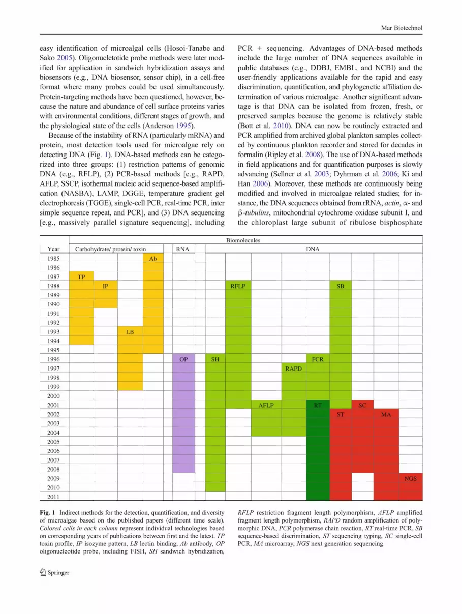
Because of the instability of RNA (particularly mRNA) andprotein, most detection tools used for microalgae rely ondetecting DNA (Fig. 1). DNA-based methods can be catego-rized into three groups: (1) restriction patterns of genomicDNA (e.g., RFLP), (2) PCR-based methods [e.g., RAPD,AFLP, SSCP, isothermal nucleic acid sequence-based amplifi-cation (NASBA), LAMP, DGGE, temperature gradient gelelectrophoresis (TGGE), single-cell PCR, real-time PCR, intersimple sequence repeat, and PCR], and (3) DNA sequencing[e.g., massively parallel signature sequencing], including
PCR + sequencing. Advantages of DNA-based methodsinclude the large number of DNA sequences available inpublic databases (e.g., DDBJ, EMBL, and NCBI) and theuser-friendly applications available for the rapid and easydiscrimination, quantification, and phylogenetic affiliation de-termination of various microalgae. Another significant advan-tage is that DNA can be isolated from frozen, fresh, orpreserved samples because the genome is relatively stable(Bott et al. 2010). DNA can now be routinely extracted andPCR amplified from archived global plankton samples collect-ed by continuous plankton recorder and stored for decades informalin (Ripley et al. 2008). The use of DNA-based methodsin field applications and for quantification purposes is slowlyadvancing (Sellner et al. 2003; Dyhrman et al. 2006; Ki andHan 2006). Moreover, these methods are continuously beingmodified and involved in microalgae related studies; for in-stance, the DNA sequences obtained from rRNA, actin,α- andβ-tubulins, mitochondrial cytochrome oxidase subunit I, andthe chloroplast large subunit of ribulose bisphosphate
Year Biomolecules
Carbohydrate/ protein/ toxin RNA DNA
1985 Ab
1986
1987 TP
1988 IP RFLP SB
1989
1990
1991
1992
1993 LB
1994
1995
1996 OP SH PCR
1997 RAPD
1998
1999
2000
2001 AFLP RT SC
2002 ST MA
2003
2004
2005
2006
2007
2008
2009 NGS
2010
2011
Fig. 1 Indirect methods for the detection, quantification, and diversityof microalgae based on the published papers (different time scale).Colored cells in each column represent individual technologies basedon corresponding years of publications between first and the latest. TPtoxin profile, IP isozyme pattern, LB lectin binding, Ab antibody, OPoligonucleotide probe, including FISH, SH sandwich hybridization,
RFLP restriction fragment length polymorphism, AFLP amplifiedfragment length polymorphism, RAPD random amplification of poly-morphic DNA, PCR polymerase chain reaction, RT real-time PCR, SBsequence-based discrimination, ST sequencing typing, SC single-cellPCR, MA microarray, NGS next generation sequencing
Mar Biotechnol

carboxylase genes are being used for detection and phyloge-netic studies in microalgae.
Small- and Medium-Scale (Depending on Speciesor Sample Size) Detection Techniques
DNA-based detection methods for microalgae are generallyconducted by PCR amplification of a target gene or DNAmarkers. PCR methods can amplify minute amounts of tem-plate DNA and multiply even a single copy of a given DNAsequence by a factor of 1012; its high specificity makes thistool highly effective for species and strain identification over awide range of organisms (Bott et al. 2010). The relatively lowcost of the equipment and reagents makes PCR accessible tosmall laboratories. In microalgal studies, PCR-based assayshave largely been used for the identification and characteriza-tion of HAB species with the use of species-specific PCRprimers and genetic markers (Godhe et al. 2008; Penna andGalluzzi 2008; Wang et al. 2008).
Multiple species detection is achieved by a complex PCRwith a mixture of many primers or run, in parallel or series, asa large number of individual PCRs (Anthony et al. 2000). Inaddition, a multiplex PCR that uses species-specific primersmay detect several species and has been tested for detection ofharmful microalgae (Oldach et al. 2000; Rublee et al. 2001).Multiple primer sets within a single PCR mixture produceamplicons of varying sizes that are specific to different DNAsequences. This method can detect several target species;however, multiple primers used in the same multiplex PCRreaction decrease the sensitivity and increase the chance oftwo unrelated primers producing spurious products (Anthonyet al. 2000). Annealing temperatures for each of the specificprimer sets must be optimized to work correctly within asingle reaction, and amplicon sizes should be sufficientlydifferent to form distinct bands when visualized by gel elec-trophoresis. For these reasons, the method has several limita-tions when detecting more species in natural assemblages.
In addition, conventional PCR methods have been used todetect specific single cells by single-cell PCR. Single-cell PCRmakes it possible to amplify DNA fragments by PCR from 1 ora few cells of an organism. It has enabled the detection anddetermination of DNA sequences from non-culturable micro-algae (Edvardsen et al. 2003; Ki et al. 2004; Ki and Han 2005;Takano and Horiguchi 2006) and is applicable to samplesthat are frozen or preserved in solvents, such as formalin(Bertozzini et al. 2005; Richlen and Barber 2005). In spite ofthe fact that the single-cell PCR assay is very useful fordetermination of DNA sequences from uncultured microalgaecollected from the environment, it is still labor intensive toisolate these target cells from natural microalgal assemblages.In addition, pico-size cells cannot be isolated by capillarymethods for single-cell isolations prior to cell extraction and
subsequent PCR amplification. For this reason, initially it wasa very useful tool to isolate and detect comparatively largecells, such as armored dinoflagellates because they were easyto isolate by capillary tubes using the inverted light micro-scope. Recently, pico-size eukaryotic cells in environmentalsamples can be isolated by flow cytometry, followed by wholegenome amplification or clone library (Man-Aharonovich etal. 2010; Shi et al. 2011). This technological progress promisesthat single-cell PCR detection can be applied for various sizemicroalgae.
Large-Scale (High-Throughput) Detection Techniques
Conventional PCR detection with species-specific primersmay only detect a single species, rather than the multiplemicroalgal species present in a given body of water. The useof species-specific PCR is therefore impractical for the routineanalysis of field samples that may contain many differentmicroalgal species. Multiple microalgal species detections areessential for monitoring harmful algal species in coastal waters.In the present study, we tentatively assigned some molecularmethods that can simultaneously detect more than 50 speciesto the large-scale (or high-throughput) detection categories.Several advanced techniques have been developed for high-speed and high-throughput detection and monitoring of envi-ronmental microbes. These are based on cell-free systems andinclude terminal restriction length polymorphism (T-RFLP) orsometimes referred to as TRFLP, real-time PCR assay, andlow-density microarray (DNA chip or phylochip). TheT-RFLP method is a molecular detection technique for profil-ing microbial communities based on the position of a restric-tion site closest to a labeled end of an amplified gene. Thismethod has been widely used in studies of bacterial diversity(Liu et al. 1997), but few studies have been attempted formicroalgal detection (Countway et al. 2005; Joo et al. 2010).Recently, automated ribosomal intergenic spacer analysis,which is based on length variation in the internal transcribedspacer region (ITS) of rRNA, has been applied to microalgaldetection (Hubbard et al. 2008).
Real-time PCR or qPCR is used to amplify and simulta-neously quantify target DNA molecules. The procedure fol-lows the same principle as standard PCR technique, but thekey distinction is that the amplified DNA is detected as thereaction progresses in real time; therefore, it is more advanta-geous than conventional PCR because of its linearity, sensi-tivity, specificity, and the speed at which a large number ofsamples can be processed. This method has been applied tothe detection and quantification of certain microalgal species(Galluzzi et al. 2004, 2010; Zhu et al. 2005; Godhe et al. 2008;Diaz et al. 2010). However, DNA contamination can lead tofalse positives and negative signals when using PCR-basedassays.
Mar Biotechnol

Microarray (or DNA chip) technology has emerged as amethod that allows parallel analysis of large numbers of bio-molecules, such as DNA. The method was initially designedfor gene expression analysis (Guo et al. 1994; Yershov et al.1996); however, recently, it has been applied to the detectionof bacteria and DNA-based typing of specific pathogenicbacterial strains for clinical diagnostics (Anthony et al. 2000;Wu et al. 2003; Mitterer et al. 2004). Microarrays withspecies-specific oligonucleotide probes have been used tosimultaneously detect several harmful microalgae (Ki andHan 2006). In addition, Ellison and Burton (2005) have useda bead array technology for the simultaneous identificationand quantification of many taxa in phytoplankton communi-ties. Metfies et al. (2005, 2006), Metfies and Medlin (2008),and Gescher et al. (2008) used low-density microarrays todetect marine phytoplankton by using a hierarchical probeapproach. These studies illustrate that themicroarray techniqueconstitutes a significant breakthrough for the high-throughputdetection of microalgal species in complex samples. Fiber-optic microarray and bead array technology are multiplexedmicroarray technologies that have been developed for thedetection of microalgae. With these methods, hundreds ofspecies can be detected using a single optical fiber or bead-based assay (Ahn et al. 2006; Scorzetti et al. 2009; Diaz et al.2010). The advantages of this method are a high sensor-packing density, smaller sample volumes, increased reuse ofarrays, flexible assay design, and a reduction in false signals.
Other high-throughput detection techniques, includingNASBA and LAMP, are DNA amplification technologies thatare recently gaining importance in microalgal research. LAMPamplifies the target sequence at a constant temperature of 60–65°C using several primers and Bst DNA polymerase, whichhas high strand-displacement activity (Notomi et al. 2000).The amount of DNA produced in LAMP is considerablyhigher than that produced in PCR-based amplification. More-over, amplification products can be detected by photometry forthe turbidity caused by the increasing quantity of magnesiumpyrophosphate in solution (Mori et al. 2001), or even by thenaked eye with the addition of SYBRGreen. To date, LAMP iswidely studied for detecting infectious diseases, such as tuber-culosis, malaria, and sleeping sickness; it has yet to be exten-sively validated for the detection of microalgae. Recent workon the application of NASBA and LAMP for microalgaldetection (Casper et al. 2007; Wang et al. 2008; Ulrich et al.2010) shows the potential for its use in the large-scale detectionof microalgae. These techniques can also be used for theestimation of microalgal diversity and species composition.
Microalgae Diversity by Molecular Techniques
Microalgae have enormous diversity in aquatic ecosystems,and some species (e.g., diatoms and dinoflagellates) have
been used as bioindicator species to assess water quality andenvironmental changes (Vaulot et al. 2008). In general,microscopic observation and analysis of the diversity ofmicroalgae in environmental samples is considered the goldstandard; however, this method is only applicable for nano-size and larger plankton (5~200 μm in body length), be-cause of the microscope resolution. Other smaller micro-algae (pico-size cells), however, have remained elusivebecause they lack morphological features for identification;sometimes, they are even ignored in cell counting by mi-croscopy. Alternatively, metagenomic analysis have beenapplied to study the microalgal diversity, by comparing the18S rRNA sequences as DNA taxonomic markers, becausethere is significant sequence data in public databases, suchas GenBank. In addition, the 18S rRNA is universallypresent in living organisms and contains regions that arewell conserved within a species and generally differentbetween species. Indeed, microeukaryote metagenomicshave shown a remarkably high diversity of microalgae andother protists from environmental samples (Díez et al. 2001;Countway et al. 2005; Amaral-Zettler et al. 2009; Burki etal. 2010). In addition, large numbers of undescribed micro-algae in natural environments have been identified (Moreiraand López-García 2002; Cheung et al. 2010). However, notall microalgae have been sequenced, but despite the obviousbias that recovered sequences may represent known species,the magnitude of novel sequences, even in well-knownlineages, has revealed many cryptic species (Sarno et al.2005). Molecular technologies have greatly expanded ourunderstanding of the diversity of microalgae that are notdetected by microscopy.
Table 2 lists some molecular tools that have been appliedfor studies on microalgal diversity. Comparative diversityanalysis in a larger number of samples has been achievedwith DNA fingerprinting methods, such as DGGE, TGGE,AFLP, and T-RFLP (Widmer et al. 2006; Kumari et al. 2009;Alpermann et al. 2010; Joo et al. 2010). Distinction ofindividuals below the species level can be obtained by usinghighly variable molecular markers (e.g., ITS sequences andmicrosatellites). Microsatellite sequences generally comprise2–4 bp sequences occurring as tandem repeats in nuclear andorganelle DNA. They are used as the method of choice forresolving intra-specific diversity because they provide aunique marker indicative of intra-species variability (Nagaiet al. 2007; Cho et al. 2009; Evans et al. 2009). A detailedreview of the methods available to estimate diversity inmarineprotists was published recently (Medlin and Kooistra 2010).
Clone libraries are well established to provide informa-tion for both the phylogenetic identity and, to some extent,the relative abundance of community operational taxonomicunits (OTUs)—a term used to describe the diversity, orspecies richness, of a sample (Stackebrandt 2006). This isachieved by DNA sequencing of a clone library constructed
Mar Biotechnol

from PCR amplicons of environmental DNA. This methodwas initially developed for bacterial diversity studies (Laneet al. 1985; Britschgi and Giovannoni 1991; Amann et al.1995), and it also has been widely applied for diversitystudies on microalgae (Countway et al. 2005; Medlin et al.2006; Potvin and Lovejoy 2009; Shi et al. 2011). Thesemetagenomic studies have shown that the vast majority ofmicroalgal diversity was not detected by conventional mi-croscopic methods, particularly the nano- and pico-sizemicroalgae (Delong and Pace 1991; Stoeck and Epstein2003). In the clone library approach, minor species or pop-ulations may be ignored if too few clones are selected forsequence analysis. One reason is that this method is laborintensive and the possibility of less number of clones beingsampled. In addition, a PCR bias exists because primerspreferentially bind to the dominant template in a sample, thusmasking minor species and rare organisms. Moreover, thisanalysis is expensive, which makes it difficult to analyzemultiple samples with replicates and high frequency. Recentpyrosequencing technologies (e.g., 454 Life Sciences) arerapidly replacing clone library methods because high-throughput reads do not require cloning. However, it requiresextensive computer analyses for a large data process, andprecise phylogenetic affinities are not always possible. It isdiscussed in detail in “NGS techniques for diversity ofmicroalgae.”
Quantification of Microalgae Using MolecularTechniques
Molecular quantification, or enumeration, of algal biomass andother important species regularly in an aquatic environment isa relevant parameter for the assessment of water quality andmonitoring for possible algal bloom incidents. Most moleculartools have focused on the detection and discrimination ofmicroalgae but are not commonly used in the field for quanti-fication. Of the many techniques, those methods that are rapidand simple are preferred as replacements for the traditional
methods (e.g., direct cell counting or chlorophyll estimation).Table 3 describes the molecular techniques available for thequantification of microalgae from environmental samples.Among current technologies (see Table 1), qPCR may beconsidered the best method for the molecular quantificationof some targeting microalgal species. qPCR employs twodifferent methods: (1) sequence-specific oligonucleotideprobes that are labeled with a fluorescent reporter, such asTaqMan, and (2) non-specific fluorescent dyes (e.g., SYBRGreen) that intercalate with any double-stranded DNA. Dataare collected over the entire series of PCR cycles by usingfluorescent markers that are incorporated into the ampliconproduct during amplification and directly in the exponentialphase where PCR is precise and linear. In order to quantifycells, the parameters of qPCR are optimized using differentstandard curves (plasmid dilution or pure algal cultures). qPCRgenerates a standard curve of cycle thresholds (Ct) with knownconcentrations, and thus cell density can be compared. Bycomparison with known standards, cell density is calculatedfrom the Ct value in qPCR. TaqMan-based qPCR assay, theprinciple of which is described elsewhere (Liu et al. 2006), hasbeen tested for the quantification of harmful microalgae (Parket al. 2007; Handy et al. 2008). SYBR Green-based qPCR,commonly considered a relatively easy and cost-effectivemethod, has been attempted for the detection and quantifica-tion of harmful microalgae, such as Pfiesteria sp., Chattonellasubsala, Pseudo-nitzchia sp., Ostreopsis sp., and Aureococcussp., (Galluzzi et al. 2010, 2011; Godhe et al. 2008; Andree etal. 2011; Perini et al. 2011). TaqMan-based qPCR is consid-ered more accurate than intercalating dye methods. The formerrequires additional dye-labeled probes; however, double strandDNA (dsDNA) dyes, such as SYBR Green, will bind to alldsDNA PCR products, including non-specific PCR productslike primer dimers. This can potentially interfere with, orprevent, accurate quantification of the intended target se-quence. By applying this method to environmental samples,autofluorescence caused by microalgal pigments can interferewith an accurate quantification in SYBR Green-based qPCR.EvaGreen is a new DNA-binding dye that shows both a
Table 2 Microalgal diversity studies with molecular technologies
Techniques Descriptions Target genes studied
▪Clone library -Discrimination of closely related species, highly reproducible,allows detection of non-culturable species, recognizes geneticindividuality, rapid identification in field and laboratory
-18S rRNA
▪DGGE/TGGE -Easy discrimination in natural samples, high reproducibility,and reliability rates
-18S rRNA, rDNA ITS
▪T-RFLP/Flu-RFLP -Provides excellent resolution -18S rRNA, rDNA ITS
▪qPCR/in situ hybridization -Sensitive and rapid -18S rRNA
▪High throughput sequencing -Rapid, simultaneous sequencing, Reliable and cost-effective -18S rRNA, COI
▪NGS techniques -Large-scale sequencing, high sequence reads, eliminates therequirement to clone DNA fragments avoiding cloning bias
-18S rRNA, COI
Mar Biotechnol

relatively low PCR inhibition and a relatively low tendency fornon-specific amplicon interaction (Mao et al. 2007). Erdner etal. (2010) used EvaGreen dye for the quantification of plas-mids and the enumeration of cysts in Alexandrium sp. Simi-larly, SYTO9 another intercalating dye used in qPCR was alsoused for the detection and quantification of the toxic dinofla-gellate Karlodinium veneficum (Park et al. 2009). The mainadvantage of using qPCR is that it is highly sensitive, specific,accurate, and cost-effective and can be applied to preservedenvironmental samples (Galluzzi et al. 2004; Toyoda et al.2010).
The rRNA molecules, particularly the non-coding ITSregions, are quite useful for developing specific primersfor PCR amplification, but the eukaryotic rRNA gene hasa high copy number—up to 104 copies—and are tandemlyorganized (Schlötterer 1998). When a single-copy gene isdetected by PCR, it represents a single cell. Similarly,multiple-copy genes within a cell are detected by the samemanner, they may represent multiple cells, and there is apossibility for overestimation of real cell numbers. RecentqPCR techniques use real cells as standards, and these resultsare thus comparable with direct cell counts using microscopy,provided this technique can detect undetectable and fragilecells accurately (Park BS, personal communication).
As noted previously, microarray (DNA chip) methods arevery effective for the simultaneous large-scale detection ofmicroalgae (Gescher et al. 2008; Anderson and Walt 2009).Furthermore, this method is recommended as one of thehigh-throughput molecular quantification techniques. Thistechnique detects labeled rRNA or labeled DNA ampliconsfrom target regions, such as rRNA from genomic DNA. Inthis assay the fluorescence labeled DNA or RNA moleculesare quantified and is proportional to cell numbers (Anderson
et al. 2006) Hybridization of the labeled products is per-formed by exposing the microarray to signal probes, and thehybridization signal is recorded (Metfies and Medlin 2008).For quantification, the arrays have been hybridized usingdifferent concentrations of target cells, and the cells weresubsequently enumerated from the signals generated (Ahn etal. 2006; Anderson et al. 2006; Scorzetti et al. 2009; Diaz et al.2010). The microarray-based method has several advantages,such as low detection limit, shorter analysis time, and minimalfalse positive signals. The microarray has been tested in pilotstudies to quantify absolutely certain specific algal species; ithas great potential to quantify microalgal cells and alter thestandard procedures used for microalgae monitoring pro-grams. However, problems regarding signal acquisition needsto be well addressed.
FISH uses short fluorescently labeled, synthesized probesthat are complementary to a target sequence within the targetcells. FISH usually utilizes oligonucleotide sequences that bindto the ribosomes in target cells, although peptide nucleic acidprobes are sometimes used (Litaker and Tester 2006). Thetarget molecules in the cells will fluoresce, and in the case ofribosomes, the entire cell can be made fluorescent, makingthem easy to enumerate (Kudela et al. 2010). The advantagesof FISH are that it is inexpensive, more rapid than electronmicroscopy, and reduces false positives and allows the charac-terization of the entire phytoplankton population, although onlywith 1 probe (1 fluorochrome) at a time. The use of signalamplification tools like tyramide signal amplification and cat-alyzed reported deposition have also been used along withFISH for the easy discrimination of microalgal species (Biegalaet al. 2002; Töbe et al. 2006).
Non-molecular techniques, such as flow cytometry andadvanced microscopy, can be combined with DNA-based
Table 3 Molecular tools for the quantification of microalgae
Tools Species tested Field applicability
▪qRT-PCR assay -Alexandrium catenella, A. fundyense,A. minutum, A. tamarense, Aureococcussp., Chattonella subsalsa, Coscinodiscussp., Gymnodinium sp., Heterosigmaakashiwo, Osteropsis sp., Pfiesteriapiscicida, Protoceratium sp., Prymenesiumparvum, Thalassiosira pseudonana
-High accuracy and sensitivity, reductionin material used, applicable to field samples
▪Fluorescence in situ hybridization (FISH) -Symbiodinium, A. tamarense, A. catenella,Lingulodinium polyedrum
-More precise and sensitive
▪DNA chip (microarray) -Alexandrium sp., Dinophysis heterocapsasp., Karenia sp., Micromonas sp.,Prochlorococcus, Protocentrum sp.,Synechococcus
-Potential to generate data rapidly, cost-effective,adaptable, and comprehensive
▪Locked nucleic acid (LNA) probes -A. miminum, A. ostenfieldii, Karenia brevis,K. mikimotoi, Prorocentrum sp.
-LNA probes increases thermal duplex stability,extremely reliable
▪High throughput sequecing -Amphidinium, Gymnodinium, Phaeodactylumsp., Peridinium, Prorocentrum,Prochlorococcus, Thalassiosira pseudonana
-Linear quantification over a wide dynamic rangeno post PCR handling
Mar Biotechnol

probes and dyes (nucleic acid-specific dyes, specific oligo-nucleotide probes, etc.) for the quantification of species inenvironmental samples. It has been observed that with theuse of DNA-specific dyes, such as SYBR Green and Eva-Green, microalgal cells can be enumerated using fluores-cence microscopy, and the results are comparable to thoseobtained by hemocytometer-based counting (Soto et al.2005). This technique is more advantageous than normalmicroscope-based enumeration because it can specificallydistinguish cells based on their shape and other morpholog-ical parameters and is less time-consuming. FISH has beencombined with solid-phase cytometry for the enumerationof microalgae (Töbe et al. 2006); this method is rapid andhas a lower detection limit, approximately one cell per filter.
Next-Generation Sequencing Technologies for Diversityof Microalgae
Next-generation sequencing technologies have recently in-spired almost all life science studies using techniques, suchas full genome sequencing (de novo sequencing and rese-quencing), amplicon sequencing, transcriptome sequencing,and metagenomics. NGS techniques with pyrosequencinggenerate much higher throughput data, by which millions tobillions of sequencing reactions take place at the same time,in small reaction volumes (Metzker 2010; Nowrousian2010). Table 4 summarizes the NGS technologies availableand their major features. In field sample studies, NGS tech-nologies are facilitating the gathering of DNA data fromboth environmental DNA and PCR products amplified fromenvironmental DNA. These NGS applications differ fromthe clone library method because they do not require thecloning of template DNA into bacterial vectors; alternative-ly, DNA templates are bound to substrates and amplified byPCR to generate clonal representatives, and hence no clon-ing bias is imposed for metagenomics (e.g., Shendure and Ji2008; Metzker 2010). In addition, the number of sequencereads by the NGS methods have been extremely high,revealing a high diversity of microbes that were not detectedfrom clone library methods (Stoeck et al. 2010).
The development of NGS has made it possible to directlysequence a huge number of genomic fragments extracted fromenvironmental samples (Rothberg and Leamon 2008), hencemaking NGS a potential tool for the identification and detec-tion of microbes from environmental samples (Medinger et al.2010). In 2006, Edwards et al. (2006) published, for the firsttime, sequences of environmental samples generated with thechip-based pyrosequencing developed by 454 Life Sciences.NGS techniques have enabled the discovery of novel genesfrom environmental samples for the massive characterizationof functional genes and enabled study of the metagenomicdiversity of unculturable bacteria and archaea in various T
able
4Nextgeneratio
nsequ
encing
(NGS)techno
logies
andtheirfeatures
NGSTechn
olog
y(Com
pany
/platform)
Techn
olog
yAmplification
approach
Reads
(million)
Length(bp)
Throu
ghpu
tAdv
antages
▪Roche/GS-FLX
(454
)a-Pyrosequencing
-Emulsion
PCR
1.0
600bp
600Mb
-Highqu
ality
reads,long
read
leng
ths,andcapacity
tohand
lerepetitiveregion
s,identification,
and
quantificationof
sign
ificantgenes
▪Illu
mina/Genom
eAnalyzerIIx(G
AIIx)
-Polym
erase-based
sequ
encing
-by-synthesis
-Bridg
eam
plification
640
-Singleandpaired
end,
36–50
0bp
95Gb
-Aplatform
forgeno
mic
discov
eryandvalid
ation
▪Illu
mina/HiSeq20
00-Sequencingby
synthesis(SBS)
-Bridg
eam
plification
2,00
0-Singleandpaired
end
50–10
0bp
200Gb
-Mostaccuratedataforabroadrang
eof
applications
▪ABI/SOLiD
v.4
-Ligation-basedsequ
encing
-Emulsion
PCR
1,40
0-Fragm
entandpaired
endandmatepair
35–50
bp
100Gb
-Providesbettersequ
encing
fidelity
▪Pacific
Biosciences/
PacBio
RS
-Pho
spho
linkedfluo
rescent
nucleotid
es,real-tim
esequ
encing
-Singlemolecule
templates
800–
1,00
0–
–-H
asthegreatestpo
tentialforreadsexceeding1kb
aThe
GS-FLX
hadrecently
been
upgraded
toGS-FLX+system
s(perform
ance:read
leng
th,up
to1,00
0bp
;mod
eread
leng
th,70
0bp
;typicalthroug
hput,70
0Mb;
readsperrun,
1,00
0,00
0,respectiv
ely)
Mar Biotechnol

environmental samples (Roesch et al. 2007; Shi et al. 2009).NGS techniques have recently been applied for diversity eval-uation and phylogenetic studies in protists and microalgae,such as diatoms, dinoflagellates, and haptophytes (Amaral-Zettler et al. 2009; Burki et al. 2010; Shalchian-Tabrizi et al.2011).
With the rapid progress in NGS technologies in recentyears, various NGS platforms are now available, such as 454pyrosequencing, HiSeq2000, GAIIx, and SOLiD, PacBio RS(see Table 4); however, their application to environmentaldiversity studies is restricted by sequence length per individ-ual read. Sequence length of each read is usually less than150 bp, though NGS tools generate a huge amount of se-quence data with billions of sequencing reactions in a singlerun. However, the 454 pyrosequencing easily produces readsof 500 bp, and recently this technology has been upgraded asthe GS FLX+ system, which enhances the read length up to1,000 bp in optimum conditions with new reagents (http://www.my454.com/products/gs-flx-system/index.asp). Inmetagenomic diversity studies, individual sequences withoutassembly of sequence reads are subjected to phylogeneticanalysis by comparison with well-defined DNA sequencesas taxonomic markers or signatures. NGS-based metagenom-ics mostly targets the 18S rRNA molecules, as DNA taxo-nomic markers (e.g., Amaral-Zettler et al. 2009; Bråte et al.2010), because of highly conserved sequences within a spe-cies, but generally different between species (Ki 2011), andcomparably large dataset available in GenBank. Because ofthe relatively long reads (up to 1,000 bp) in 454 pyrosequenc-ing, the Genome Sequencer FLX System is usually employedin metagenomic diversity studies using PCR amplicons(Amaral-Zettler et al. 2009; Medinger et al. 2010; Burki et al.2010; Edgcomb et al. 2011; Mccliment et al. 2011; Shalchian-Tabrizi et al. 2011; Tai et al. 2011). The NGS system thereforeenables a more comprehensive view into the diversity ofvarious environmental habitats. Techniques, such as 454 pyro-sequencing, Illumina, and SOLiD, are largely used to identifyand detect malfunctioning cells and microbiota of the humanbody (Petrosino et al. 2009; Roesch et al. 2009). There is greatpotential for their use in diversity studies on microbes, such asbacteria, archaea, and microeukaryotes, including microalgae.In addition, these techniques can be used to count environmen-tal gene tags, or PCR amplicons, to analyze the relative abun-dance of microalgal species under varying environmentalconditions, although the NGS-based quantification is not com-pletely proven at this stage, because of some possible biases(e.g., PCR-amplification bias, NGS reads, etc.).
Conclusions and Remarks
Microalgae are major components of the aquatic ecosystem, andtheir diversity is strongly modified by rapid and accelerating
environmental changes (Elmqvist et al. 2003). They areunicellular eukaryotes [excluding blue-green algae (cyanobac-teria)] with distinct features, such as morphology, pigments,and photosynthetic activity, and can be extremely small in size(e.g., nano- and pico-size plankton). Hence, it is necessary forthe correct identification, continuous monitoring, and enumer-ation of these microorganisms. Beyond the traditional micro-scopic methods, many molecular techniques have beendeveloped as alternative methods to discriminate microalgalspecies. Even molecular methods developed by other molec-ular biologists can be used for microalgal studies because thetarget biomolecules (DNA, RNA, and protein) are universal.Each molecular technique has its own particular strengths indetecting microalgae but may have limitations when appliedto other species. For example, single-cell PCR is considered agood molecular tool for studying uncultured microalgal cellsfrom environments but is difficult to apply to pico-size cells;but by involving flow cytometry, this can also be made pos-sible. To date, there are several reviews of molecular methodsthat consider different aspects, applications, and organisms.This paper highlights the practical molecular tools availablefor species detection, quantification, and diversity analysis ofmicroalgae.
Despite rapid advances and many examples highlightingthe application of molecular methods to a broad spectrum ofapplications, there are very few methodologies for quantifi-cation and diversity studies in microalgae. Molecular tech-nologies ranging from automated Sanger sequencing toNGS technologies have opened doors for the easy and rapiddetection and quantification of cells. Among the currentlyavailable methods, qPCR offers the most cost-effective,sensitive, and rapid analysis for the detection and quantifi-cation of microalgae in both laboratory and field situations.Isothermal DNA amplification techniques, such as LAMPand NASBA, are relatively new methods, which, because oftheir simplicity, ruggedness, and low cost, could providemajor advantages. These methods therefore have the poten-tial to be used as large-scale, simple screening assays for thedetection and diversity estimation of microalgae. In addi-tion, NGS techniques have been applied to various genomicresearch studies and have significant potential for future usein both discrimination and quantification of microalgaeworldwide. At present, the operational cost and data analy-sis tools associated with NGS technology support the wide-range use in molecular monitoring and quantification ofenvironmental microalgae. In the case of 454 pyrosequenc-ing, costs can be greatly reduced by employing MultiplexIdentifier-containing adaptors, which allow users to havegreater multiplexing capabilities with the GS FLX Titaniumsequencing chemistry. Moreover, the coming third- andfourth-generation technologies that are using techniques likesingle molecule sequencing (Pacific Biosciences Inc.) andsingle molecule electrical detection (Genia Technologies,
Mar Biotechnol

Inc.) will increase throughput and decrease the cost and timeof acquiring results. This shows that NGS tools offer greatpotential to alter the manner in which researchers monitormicroalgae.
Molecular metagenomic techniques have greatly expandedour understanding of the diversity of microalgae in environ-mental samples. In particular, NGS-based metagenomicsshow a remarkably high diversity of microalgae; however, alarge portion of sequence data can be unassigned to molecularoperational taxonomic units, because of an insufficient DNAtaxonomic database (e.g., DNA barcoding, DNA reference, orsignature sequences). Most data available in public databaseshave been derived from cultured strains of microalgae. In thefuture, we have to construct well-defined DNA databases ofmicroalgae in order to achieve a comprehensive understand-ing of the molecular diversity of environmental microalgae. Inthis regard, the newest technology of single-cell genomeanalysis, in which a single cell can be isolated from theenvironment by flow cytometry and its genome can be ampli-fied and sequenced, may be the only way that we can achievesuch a goal (Yoon et al. 2011).
Acknowledgments This work was supported by both the Marine andExtreme Genome Research Center Program of the Ministry of Land,Transportation and Maritime Affairs, Republic of Korea, and by theNational Research Foundation of Korea Grant funded by the KoreanGovernment (MEST) (NRF-C1ABA001-2011-0018573).
References
Ahn S, Kulis DM, Erdner DL, Anderson DM, Walt DR (2006) Fiber-optic for the simultaneous detection of multiple harmful algalbloom species. Appl Environ Microb 72(9):5742–5749
Alpermann TJ, Tillmann U, Beszteri B, Cembella AD, John U (2010)Phenotypic variation and genotypic diversity in a planktonicpopulation of the toxigenic marine dinoflagellate Alexandriumtamarense (Dinophyceae). J Phycol 46:18–32
Amann RI, Ludwig W, Schleifer KH (1995) Phylogenetic identifica-tion and in situ detection of individual microbial cells withoutcultivation. Microbiol Rev 59(1):143–169
Amaral-Zettler LA, McCliment EA, Ducklow HW, Huse SM (2009) Amethod for studying protistan diversity using massively parallelsequencing of V9 hypervariable regions of small-subunit ribo-somal RNA genes. PLoS One 4(7):e6372
Anderson DM (1995) Identification of harmful algal species usingmolecular probes: an emerging technology. In: Lassus P, ArzulG, Erard E, Gentien P, Marcaillou C (eds) Harmful marine algalblooms. Lavoiser Science Publishers, Paris, pp 3–13
Anderson DM, Walt DW (2009) A fiber optic microarray for thedetection and enumeration of harmful algal bloom (HAB) species.Internal report: The NOAA/UNH cooperative institute for coastaland estuarine environmental technology (CICEET). http://ciceet.unh.edu/news/releases/spring09_reports/pdf/anderson_FR.pdf
Anderson DM, Kulis DM, Erdner D, Ahn S, Walt DR (2006) Fibreoptic microarrays for the detection and enumeration of harmfulalgal bloom. Afr J Mar Sci 28(2):231–235
Andree KB, Fernández-Tejedor M, Elandaloussi LM, Quijano-Scheggia S, Sampedro N, Garcés E, Camp J, Diogéne J (2011)
Quantitative PCR coupled with melt curve analysis for detectionof selected Pseudo-nitzschia spp. (Bacillariophyceae) from theNorth Western Mediterranean sea. Appl Environ Microb 77(5):1651–1659
Anthony RM, Brown TJ, French GL (2000) Rapid diagnosis of bacter-emia by universal amplification of 23S ribosomal DNA followed byhybridization to an oligonucleotide array. J Clin Microbiol 38(2):781–788
Becker EW (2007) Microalgae as a source of protein. Biotenol Adv 25(2):207–210
Bertozzini E, PennaA PE, Bruce I, Magnani M (2005) Development ofnew procedures for the isolation of phytoplankton DNA fromfixed samples. J Appl Phycol 17:223–229
Biegala IC, Kennaway G, Alverca E, Lennon JF, Vaulot D, Simon N(2002) Identification of bacteria associated with dinoflagellates(Dinophyceae) Alexandrium spp using tyramide signalamplification-fluorescent in situ hybridisation and confocal micros-copy. J Phycol 38(2):404–411
Bott NJ, Ophel-Kellener KM, Sierp MT, Rowling KP, Mckay AC, LooMGK, Tanner JE, DeveneyMR (2010) Toward routine, DNA-baseddetection methods for marine pests. Biotechnol Adv 28:706–714
Bråte J, Klaveness D, Rygh T, Jakobsen KS, Shalchian-Tabrizi K(2010) Telonemia-specific environmental 18S rDNA PCR revealsunknown diversity and multiple marine-freshwater colonizations.BMC Microbiol 10:168. doi:10.1186/1471-2180-10-168
Britschgi TB, Giovannoni SJ (1991) Phylogenetic analysis of a naturalmarine bacterioplankton population by rRNA gene cloning andsequencing. Appl Environ Microbiol 57(6):1707–1713
Brown MR (2002) Nutritional value of microalgae for aquculture. In:Cruz-Suárez LE, Ricque-Marie D, Tapia-Salazar M, Gaxiola-Cortés MG, Simoes N (eds) Avances en Nutrición Acuícola VI.Memorias del VI Simposium Internacional de Nutrición Acuícola.3 al 6 de Septiembre del 2002. Cancún, Quintana Roo, México
Burki F, Kudryavtsev A, Matz MV, Aglyamova GV, Bulman S, FiersM, Keeling PJ, Pawlowski J (2010) Evolution of Rhizaria: newinsights from phylogenomic analysis of uncultivated protists.BMC Evol Biol 10:377
Casper ET, Patterson SS, Bhanushali P, Farmer A, Smith M, Fries DP,Paul JH (2007) A handheld NASBA analyzer for the field detec-tion and quantification of Karenia brevis. Harmful Algae 6(1):112–118
Casteleyn G, Leliaert F, Backeljau T, Debeer AE, Kotaki Y, Rhodes L,Lundholm N, Sabbe K, Vyverman W (2010) Limits to gene flowin a cosmopolitan marine planktonic diatom. Proc Natl Acad SciUSA 107(29):12952–12957
Cembella AD, Sullivan JJ, Boyer GL, Taylor FJR, Anderson RJ (1987)Variations in paralytic shellfish toxin composition within theProtogonyaulax tamarensis/catenella species complex: red tidedinoflagellates. Biochem Syst Ecol 15:171–186
Cheng KC, Ogden KL (2011) Algal biofuels: the research. AmericanInstitute of Chemical Engineers (AICHE). http://www.aiche.org
CheungMK,AuCH, ChuKH,KwanHS,WongCK (2010) Compositionand genetic diversity of pico eukaryotes in subtropical coastal watersas revealed by 454 pyrosequencing. ISME J 4:1053–1059
Chisti Y (2007) Biodiesel from microalgae. Biotechnol Adv 25(3):294–306
Cho SY, Nagai S, Nishitani G, Han MS (2009) Development ofcompound microsatellite markers in red-tide-causing dinoflagel-late Akashiwo sanguinea (Dinophyceae). Mol Ecol Resour 9:915–917
Countway P, Gast RJ, Savala P, Caron DA (2005) Protistan diversityestimates based on 18S rDNA from seawater incubations in theWestern North Atlantic. J Eukaryot Microbiol 52(2):95–106
de Bruin A, Ibelings BW, Donk EV (2003) Molecular techniques inphytoplankton research: from allozyme electrophoresis to genomics.Hydrobiologia 491(1–3):47–63
Mar Biotechnol

Delong EF, Pace NR (1991) Analysis of a marine picoplankton commu-nity by 16S rRNA gene cloning and sequencing. J Bacteriol 173(14):4371–4378
Diaz MR, Jacobson JW, Goodwin KD, Dubar SA, Fell JW (2010)Molecular detection of harmful algal blooms (HABs) usinglocked nucleic acids and bead array technology. Limnol OceanogrMethods 8:269–284
Díez B, Pedrós-Alió C, Marsh TL, Massana R (2001) Application ofdenaturing gradient gel electrophoresis (DGGE) to study thediversity of marine Pico eukaryotic assemblages and comparisonof DGGE with other molecular techniques. Appl Environ Microbiol67(7):2942–2951
Dyhrman ST, Erdner D, La Du J, Galac M, Anderson DM (2006)Molecular quantification of toxic Alexandrium fundyense inthe Gulf of Maine using real-time PCR. Harmful Algae 5(3):242–250
Dyhrman ST, Haley ST, Borchert JA, Lona B, Kollars N, Erdner DL(2010) Parallel analyses of Alexandrium catenella cell concentra-tions and shellfish toxicity in the Puget Sound. Appl EnvironMicrob 76(14):4647–4654
Edgcomb V, Orsi W, Bunge J, Jeon S, Christen R, Leslin C, Holder M,Taylor GT, Suarez P, Varela R, Epstein S (2011) Protistan micro-bial observatory in the Cariaco Basin, Caribbean I. pyrosequenc-ing vs sanger insights in to species richness. ISME J 5:1344–1356. doi:10.1038/ismej.2011.6
Edvardsen B, Shalchian-Tabrizi K, Jakobsen KS, Medlin LK, Dahl E,Brubak S, Paasche E (2003) Genetic variability and molecularphylogeny of Dinophysis species (Dinophyceae) from Norwegianwaters inferred from single cells analysis of rDNA. J Phycol 39(2):395–408
Edwards RA, Rodriguez-Brito B, Wegley L, Haynes M, Breitbart M,Peterson DM, Saar MO, Alexander S, Alexander EC, Rohwer F(2006) Using pyrosequencing to shed light on deep mine micro-bial ecology. BMC Genomics 7:57
Ellison CK, Burton RS (2005) Application of bead array technology tocommunity dynamics of marine phytoplankton. Mar Ecol ProgSer 288:75–85
Elmqvist T, Folke C, Nyström M, Peterson G, Bengtsson J, Walker B,Norberg J (2003) Response diversity, ecosystem change, ecosystemchange, and resilience. Fron Ecol Environ 1:488–494
Erdner DL, Percy L, Keafer B, Lewis J, Anderson DM (2010) Aquantitative real-time PCR assay for the identification and enu-meration of Alexandrium cysts in marine sediments. Deep SeaRes PT II 57:279–287
Evans KM, Chepurnov VA, Mann DG (2009) Ten microsatellitemarkers for the freshwater diatom Sellaphora capitata. Mol EcolResour 9:216–218
Galluzzi L, Penna A, Bertozzini E, Vila M, Garces E, Magnani M(2004) Development of a real-time PCR assay for rapid detectionand quantification of Alexandrium minutum (a dinoflagellate).Appl Environ Microbiol 70(2):1199–1206
Galluzzi L, Bertozzini E, Penna A, Perini F, Garcés E, Magnani M(2010) Analysis of rRNA gene content in the Mediterraneandinoflagellate Alexandrium catenella and Alexandrium taylori:implications for the quantitative real-time PCR-based monitoringmethods. J Appl Phycol 22(1):1–9
Galluzzi L, Cegna A, Casabianca S, Penna A, Saunders N, Magnani M(2011) Development of an oligonucleotide microarray for thedetection and monitoring of marine dinoflagellates. J MicrobiolMeth 84(2):234–242
Gescher C, Metfies K, Frickenhaus S, Knefelkamp B, Wiltshire KH,Medlin LK (2008) Feasibility of assessing the community com-position of Prasinophytes at the Helgoland Roads sampling sitewith a DNA microarray. Appl Env Microbiol 74:5305–5316
GodheA, AsplundME, HärnströmK, Saravanan V, Tyagi A, KarunasagarI (2008) Quantification of diatom and dinoflagellate biomasses in
coastal marine seawater samples by real-time PCR. Appl EnvironMicrobiol 74(23):7174–7182
Guo Z, Guilfoyle RA, Thiel AJ, Wang R, Smith LM (1994) Directfluorescence analysis of genetic polymorphisms by hybridizationwith oligonucleotides arrays on glass supports. Nucleic Acids Res22:5456–5465
Handy SM, Demir E, Hutchins DA, Portune KJ, Whereat EB, HareCE, Rose JM, Warner M, Farestad M, Cary S, Coyne KJ (2008)Using quantitative real-time PCR to study competition and com-munity dynamics among Delaware Inland Bays harmful algae infield and laboratory studies. Harmful Algae 7(5):599–613
Hosoi-Tanabe S, Sako Y (2005) Rapid detection of natural cells ofAlexandrium tamarense and A. catenella (Dinophyceae) by fluo-rescence in situ hybridization. Harmful Algae 4(2):319–328
Hosoi-Tanabe S, Sako Y (2006) Development and application offluorescence in situ hybridization (FISH) method for simple andrapid identification of the toxic dinoflagellates Alexandrium tamar-ense and Alexandrium catenella in cultured and natural seawater.Fish Sci 72(1):77–82
Hubbard KA, Rocap G, Armbrust EV (2008) Inter- and intraspecificcommunity structure within the diatom genus Pseudo-nitzschia(Bacillariophyceae). J Phycol 44(3):637–649
Joo S, Lee S-R, Park S (2010) Monitoring of phytoplankton commu-nity structure using terminal restriction fragment length polymor-phism (T-RFLP). J Microbiol Meth 81(1):61–68
Ki J-S (2011) Hypervariable regions (V1-V9) of the dinoflagellate 18SrRNA using a large dataset for marker considerations. J ApplPhycol. doi:10.1007/s10811-011-9730-z
Ki J-S, Han M-S (2005) Sequence-based diagnostics and phylogeneticapproach of uncultured freshwater dinoflagellate Peridinium(Dinophyceae) species, based on single-cell sequencing of rDNA.J Appl Phycol 17(2):147–153
Ki J-S, Han M-S (2006) A low-density oligonucleotide array study forparallel detection of harmful algal species using hybridization ofconsensus PCR products of LSU rDNA D2 domain. BiosensBioelectron 21(9):1812–1821
Ki J-S, Jang GY, Han M-S (2004) Integrated method for single-cellDNA extraction, PCR amplification, and sequencing of ribosomalDNA from harmful dinoflagellates Cochlodinium polykrikoidesand Alexandrium catenella. Mar Biotechnol 6(6):587–593
Kudela RM, Howard MDA, Jenkins BD, Miller PE, Smith GJ (2010)Using the molecular toolbox to compare harmful algal blooms inupwelling systems. Prog Oceanogr 85(1–2):108–121
Kumari N, Srivastava AK, Bhargava P, Rai LK (2009) Molecularapproaches towards assessment of cyanobacterial biodiversity.Afr J Biotechnol 8(18):4284–4298
Lane DJ, Pace B, Olsen GJ, Stahl DA, Sogin ML, Pace NR (1985)Rapid determination of 16S ribosomal RNA sequences for phy-logenetic analyses. Proc Natl Acad Sci USA 82(20):6955–6959
Lepère C, Vaulot D, Scanlan DJ (2009) Photosynthetic picoeukaryoticcommunity structure in the South East Pacific Ocean encompass-ing the most oligotrophic waters on Earth. Environ Microbiol 11(12):3105–3117
Lindquist HDA (1997) Probes for the specific detection of Cryptospo-ridium parvum. Water Res 31(10):2668–2671
Litaker RW, Tester PA (2006) Molecular approaches to the study ofphytoplankton life cycles: implications for harmful algal bloomecology. In: Granéli E, Turner T (eds) Ecology of harmful algae,ecological studies, vol 189. Springer, Heidelberg, pp 299–309
Liu W, Marsh T, Cheng H, Forney L (1997) Characterization ofmicrobial diversity by determining terminal restriction fragmentlength polymorphisms of genes encoding 16S rRNA. Appl EnvironMicrobiol 63:4516–4522
Liu H, Wang H, Shi Z, Wang H, Yang C, Silke S, Tan W, Li Z (2006)TaqMan probe array for quantitative detection of DNA targets.Nucleic Acids Res 34(1):1–8
Mar Biotechnol

Man-Aharonovich D, Philosof A, Kirkup BC, Gal FL, Yogev T,Berman-Frank I, Polz MF, Vaulot D, Béjà O (2010) Diversity ofactive marine picoeukaryotes in the Eastern Mediterranean Seaunveiled using photosystem-II psbA transcripts. ISME J 4:1044–1052
Mao F, Leung W-Y, Xin X (2007) Characterization of EvaGreen andthe implication of its physicochemical properties for qPCR appli-cations. BMC Biotechnol 7:76
McCliment EA, Nelson CE, Carlson CA, Alldredge AL, Witting J,Amaral-Zettler LA (2011) An all-taxon microbial inventory of theMoorea coral reef ecosystem. ISME J. doi:10.1038/ismej.2011.108
Medinger R, Nolte V, Pandey RV (2010) Diversity in a hidden world:potential and limitation of next generation sequencing for surveysof molecular diversity of eukaryotic microorganisms. Mol Ecol 19(1):32–40
Medlin LK, Kooistra WHCF (2010) Methods to estimate the diversityin the marine photosynthetic protist community with illustrationsfrom case studies: a review. Diversity 2:973–1014
Medlin LK, Lange M, Noethig EV (2000) Genetic diversity in themarine phytoplankton: a review and a consideration of Antarcticphytoplankton. Antarct Sc 12:325–331
Medlin LK, Metfies K, Mehl H, Wiltshire K, Valentin K (2006) Pico-plankton diversity at the Helgoland Time Series Site as assessed bythree molecular methods. Microb Ecol 167:1432–1451
Medlin LK, Metfies K, John U, Olsen J (2007) Algal molecularsystematics: a review of the past and prospects for the future. In:Broadie J, Lewis J (eds) Unravelling the algae: the past, presentand future of algal systematics. Sys Assn Special Vol Ser 75. CRCPress, Taylor & Francis Group, London, pp 341–353
Metfies K, Medlin LK (2008) Feasibility of transferring fluorescent in situhybridization probes to an 18S rRNA gene phylochip and mappingof signal intensities. Appl Envron Microbiol 74:2814–2821
Metfies K, Huljic S, Lange M, Medlin LK (2005) Electrochemicaldetection of the toxic dinoflagellate Alexandrium ostenfeldii witha DNA-biosensor. Biosens Bioelectron 20:1349–1357
Metfies K, Töbe K, Scholin CA, Medlin LK (2006) Laboratory and fieldapplications of ribosomal RNA probes to aid the detection andmonitoring of Harmful Algae. In: Granéli E, Turner JT (eds) Ecologyof harmful algae. Springer Verlag, Berlin, Heidelberg, pp 11–325
Metzker ML (2010) Sequencing technologies—the next generation.Nat Rev 11:31–46
Mitterer G, Huber M, Leidinger E, Kiristis C, Lubitz W, Mueller MW,Schmidt WM (2004) Microarray-based identification of bacteriain clinical samples by solid-phase PCR amplification of 23Sribosomal DNA sequences. J Clin Microbiol 42(2):1048–1057
Moreira D, López-García P (2002) The molecular ecology of microbialeukaryotes unveils a hidden world. Trends Microbiol 10:31–38
Mori Y, Nagamine K, Tomita N, Notomi T (2001) Detection of loop-mediated isothermal amplification reaction by turbidity derivedfrom magnesium pyrophosphate formation. Biochem Bioph ResCo 289:150–154
Nagai SC, Lian S, Yamaguchi M, Hamaguchi Y, Matsuyama S, ItakuraH, Shimada S, Kaga H, Yamauchi Y, Sonda T, Kim C, Hogetsu T(2007) Microsatellite markers reveal population genetic structureof the toxic dinoflagellate Alexandrium tamarense (Dinophyceae)in Japanese coastal waters. J Phycol 43:43–54
Not F, Latasa M, Marie D, Cariou T, Vaulot D, Simon N (2004) Asingle species, Micromonas pusilla (Prasinophyceae), dominatesthe eukaryotic picoplankton in the Western English Channel.Appl Environ Microbiol 70(7):4064–4072
Not F, Gausling R, Azam F, Heidelberg JF, Worden AZ (2007) Verticaldistribution of picoeukaryotic diversity in the open ocean. EnvironMicrobiol 9:1233–1252
Notomi T, Okayama H, Masubuchi H, Yonekawa T, Watanabe K,Amino N, Hase T (2000) Loop-mediated isothermal amplificationof DNA. Nucleic Acids Res 28:E63
Nowrousian M (2010) Net-generation sequencing techniques for eu-karyotic microorganisms: sequencing based solutions to biologi-cal problems. Eukaryot Cell 9(9):1300–1310
Oldach DW, Delwiche CF, Jakobsen KS, Tengs T, Brown EG, KemptonJW, Schaefer EF, Bowers HA, Glasgow HB Jr, Burkholder JM,Steidinger KA, Rublee PA (2000) Heteroduplex mobility assay-guided sequence discovery: elucidation of the small subunit (18S)rDNA sequences of Pfiesteria piscicida and related dinoflagellatesfrom complex algal culture and environmental sample DNA pools.Proc Natl Acad Sci USA 97(8):4303–4308
Park TG, Salas MF, Bolch CJS, Hallegraeff GM (2007) Developmentof a realtime PCR probe for quantification of the heterotro-phic dinoflagellate Cryptoperidiniopsis brodyi (Dinophyceae)in environmental samples. Appl Environ Microbiol 73:2552–2560
Park TG, Park YT, Lee Y (2009) Development of a SYTO9 based real-time PCR probe for detection and quantification of toxic dinofla-gellate Karlodinium veneficum (Dinophyceae) in environmentalsamples. Phycologia 48(1):32–43
Penna A, Galluzzi L (2008) PCR techniques a diagnostic tool for theidentification and enumeration of toxic marine phytoplanktonspecies. In: Evangelista V, Barsanti L, Frassanito AM, PassarelliV, Gualtieri P (eds) Algal toxins: nature, occurrence, effect anddetection. Springer Science + Business media BV, pp 261–284
Perini F, Casabianca A, Battocchi C, Accoroni S, Totti C, Penna A(2011) New approaches using the real-time PCR method forestimation of the toxic marine dinoflagellate Ostreopsis cf. ovatain marine environmental. PLoS One 6(3):e17699. doi:10.1371/journal.pone.0017699
Petrosino JF, Highlander S, Luna RA, Gibbs RA, Versalovic MD(2009) Metagenomic pyrosequencing and microbial identification.Clin Chem 55(5):856–866
Potvin M, Lovejoy C (2009) PCR-based diversity estimates of artificialand environmental 18s rRNA gene libraries. J Eukaryot Microbiol56(2):174–181
Richlen ML, Barber PH (2005) A technique for the rapid extraction ofmicroalgal DNA from single live and preserved cells. Mol EcolNotes 5:688–691
Ripley SJ, Baker AS, Miller PI, Walne AW, Schroeder DC (2008)Development and validation of a molecular technique for theanalysis of archived formalin-preserved phytoplankton samplespermits retrospective assessment of Emiliania huxleyi communi-ties. J Microbiol Meth. doi:10.1016/j.mimet.2008.02.001
Roesch LFW, Fulthorpe RR, Riva A, Casella G, Hadwin AK, KentAD, Daroub SH, Camargo FA, Farmerie WG, Triplett EW (2007)Pyrosequencing enumerates and contrasts soil microbial diversity.ISME J 1(4):283–290
Roesch LFW, Lorca GL, Casella G, Giongo A, Naranjo A, Pionzio AM(2009) Culture-independent identification of gut bacteria correlatedwith the onset of diabetes in a rat model. ISME J 3:536–548
Rothberg JM, Leamon JH (2008) The development and impact of 454sequencing. Nat Biotechnol 26:1117–1124
Rublee PA, Kempton JW, Schaefer EF, Allen C, Harris J, Oldach DW,Bowers H, Tengs T, Burkholder JM, Glasgow HB (2001) Use ofmolecular probes to assess geographic distribution of Pfiesteriaspecies. Environ Health Perspect 109(5):765–767
Sako Y, Kim CH, Ninomiya H, Adachi M, Ishida Y (1990) Isozymeand cross analysis of mating populations in the Alexandriumcatenella/tamarense species complex. In: Granéli E, SundstromB, Edler L, Anderson DM (eds) Toxic marine phytoplankton.Elsevier, New York, pp 320–323
Sarno D, Kooistra WHCF, Medlin LK, Percopo I, Zingone A (2005)Diversity in the genus Skeletonema (Bacillariophyceae): Skeleto-nema costatum (Bacillariophyceae) consists of several geneticallyand morphologically distinct species with the description of fournew species. J Phycol 41:151–176
Mar Biotechnol

Schlötterer C (1998) Ribosomal DNA probes and primers. In: Karp A,Isaac PG, Ingram DS (eds) Molecular tools for screening biodi-versity. Chapman & Hall, London, pp 267–276
Scholin CA, Anderson DA (1993) Population analysis of toxic andnon-toxic Alexandrium species using ribosomal RNA signaturesequences. In: Smayda TJ, Shimizu Y (eds) Toxic phytoplanktonblooms in the sea. Elsevier, Amsterdam, pp 95–102
Scholin CA, Herzog M, Sogin ML, Anderson DM (1994) Identifica-tion of group and strain-specific genetic markers for globallydistributed Alexandrium (Dinophyceae). II. Sequence analysis ofa fragment of the LSU rRNA gene. J Phycol 30:999–1011
Scorzetti G, Brand LE, Hitchcock GL, Rein KS, Sinigalliano CD, FellJW (2009) Multiple simultaneous detection of harmful algalblooms (HABs) through a high throughput bead array technology,with potential use in phytoplankton community analysis. HarmfulAlgae 8:196–211
Sellner KG, Gregory E, Doucette J, Kirkpatric GJ (2003) Harmful algalblooms: causes, impacts and detection. J Ind Microbiol Biotechnol30:383–440
Shalchian-Tabrizi K, Reier-Røberg K, Ree DK, Klaveness D, Brate J(2011) Marine-freshwater colonizations of haptophytes inferredfrom phylogeny of environmental 18S rDNA sequences. J EukaryotMicrobiol 58(11):315–318
Shapiro LP, Campbell L, Haugen EM (1989) Immunochemical recog-nition of phytoplankton species. Mar Ecol Prog Ser 57:219–224
Shendure J, Ji H (2008) Next generation DNA sequencing. NatBiotechnol 26:1135–1145
Shi Y, TysonGW,DeLong EF (2009)Metatranscriptomics reveals uniquemicrobial small RNAs in the ocean’s water column. Nature 459(7244):266–269
Shi XL, Lepère C, Scanlan DJ, Vaulot D (2011) Plastid 16S rRNAgene diversity among eukaryotic picophytoplankton sorted byflow cytometry from the South Pacific Ocean. PLoS One 6(4):e18979. doi:10.1371/journal.pone.0018979
Simon N, Campbell L, Örn lfsd ttir E, Groben R, Guillou L, Lange M,Medlin LK (2000) Oligonucleotide probes for the identification ofthree algal groups by dot blot and Fluorescent Whole-Cell Hybrid-ization. J Eukaryot Microbiol 47(1):76–84
Soto K, Collantes G, ZahrM, Kuzhar J (2005) Simultaneous enumerationof Phaeodactylum tricornutum (MCB292) and bacteria growing inmixed communities. Invest Mar Valparaiso 33(2):143–149
Stackebrandt E (2006) Molecular identification, systematics, and popu-lation structure of prokaryotes. Springer-Verlag Berlin Heidelberg,Germany, pp 51–80
Stoeck T, Epstein S (2003) Novel eukaryotic lineages inferred fromsmall-subunit rRNA analyses of oxygen-depleted marine environ-ments. Appl Environ Microbiol 69(5):2657–2663
Stoeck T, Bass D, Nebel M, Christen R, Jones MD, Breiner HW,Richard TA (2010) Multiple marker parallel tag environmental
DNA sequencing reveals a highly complex eukaryotic communityin marine anoxic water. Mol Ecol 19(1):21–31
Tai V, Poon AF, Paulsen IT, Palenik B (2011) Selection in coastalSynechococcus (cyanobacteria) populations eva;uated from envi-ronmental metagenomics. PLoS One 6(9):e24249
Takano Y, Horiguchi T (2006) Acquiring scanning electron microscop-ical. Light microscopical and multiple gene sequence data from asingle dinoflagellate cell. J Phycol 42:251–256
Töbe K, Eller G, Medlin LK (2006) Automated detection and enumer-ation for toxic algae by solid-phase cytometry and the introduc-tion of a new probe for Prymnesium parvum (Haptophyta:Prymnesiophyceae). J Plank Res 28(7):643–657
Toyoda K, Nagasaki K, Tomaru Y (2010) Application of real-timePCR assay for detection and quantification of bloom-formingdiatom Chaetoceros tenuissimus Meunier. Plankton Benthos Res5(2):56–61
Tyrrell JV, Connell LB, Scholin CA (2002) Monitoring for Hetero-sigma akashiwo using a sandwich hybridization assay. HarmfulAlgae 1:205–214
Ulrich RM, Casper ET, Campbell L, Richardson B, Heil CA, Paul JH(2010) Detection and quantification of Karenia mikimotoi usingreal-time nucleic acid sequence-based amplification with internalcontrol RNA (IC-NASBA). Harmful Algae 9(1):116–122
Vaulot D, Eikrem W, Viprey M, Moreau H (2008) The diversity ofsmall eukaryotic phytoplankton (3 mm) in marine ecosystems.FEMS Microbiol Rev 32:795–820
Wang L, Li MJ, Alam Y, Geng Z, Yamasaki S, Shi L (2008) Loop-mediated isothermal amplification method for rapid detection ofthe toxic dinoflagellate Alexandrium, which causes algal bloomsand poisoning of shellfish. FEMS Microbiol Lett 282:15–21
Widmer F, Hartmann M, Frey B, Kölliker B (2006) A novel strategy toextract specific phylogenetics sequence information from com-munity T-RFLP. J Microbiol Meth 66:512–520
Wu Z, Irizarry RA, Gentleman R, Murillo FM, Spencer F (2003) Amodel based background adjustment for oligonucleotide expres-sion arrays. Technical report, Johns Hopkins University, dept ofbiostatistics working papers. http://www.bepress.com/jhubiostat/paper1
Yershov G, Barsky V, Kirillov E, Kreindlin K, Ivanov I, Parinov S,Guschin D, Drobishev A, Dubiley S (1996) DNA analysis anddiagnostics on oligonucleotide microchips. Proc Natl Acad SciUSA 93(10):4913–4918
Yoon HS, Price DC, Stepanauskas R, Rajah VD, Sieracki ME, WilsonWH, Yang EC, Duffy S, Bhattacharya D (2011) Single-cellgenomics reveals organismal interactions in uncultivated marineprotists. Science 332:714–717
Zhu F, Massana R, Not F, Marie D, Vaulot D (2005) Mapping ofpicoeukaryotes in marine ecosystems with quantitative PCR ofthe 18S rRNA gene. FEMS Microbiol Ecol 52:79–92
Mar Biotechnol