harmful and beneficial role of ros - hindawi.com
TRANSCRIPT
Oxidative Medicine and Cellular Longevity
Harmful and Beneficial Role of ROS
Guest Editors: Sergio Di Meo, Tanea T. Reed, Paola Venditti, and Victor M. Victor

Harmful and Beneficial Role of ROS
Oxidative Medicine and Cellular Longevity
Harmful and Beneficial Role of ROS
Guest Editors: Sergio Di Meo, Tanea T. Reed, Paola Venditti,and Victor M. Victor

Copyright © 2016 Hindawi Publishing Corporation. All rights reserved.
This is a special issue published in “OxidativeMedicine and Cellular Longevity.” All articles are open access articles distributed under theCreative Commons Attribution License, which permits unrestricted use, distribution, and reproduction in any medium, provided theoriginal work is properly cited.
Editorial Board
Mohammad Abdollahi, IranAntonio Ayala, SpainNeelam Azad, USAPeter Backx, CanadaDamian Bailey, UKConsuelo Borrás, SpainVittorio Calabrese, ItalyAngel Catalá, ArgentinaShao-Yu Chen, USAZhao Zhong Chong, USAGiuseppe Cirillo, ItalyMassimo Collino, ItalyMark J. Crabtree, UKManuela Curcio, ItalyAndreas Daiber, GermanyFelipe Dal Pizzol, BrazilFrancesca Danesi, ItalyDomenico D’Arca, ItalyYolanda de Pablo, SwedenJames Duce, UKGrégory Durand, FranceJavier Egea, SpainAmina El Jamali, USAErsin Fadillioglu, TurkeyQingping Feng, CanadaGiuseppe Filomeni, ItalySwaran J. S. Flora, IndiaRodrigo Franco, USAJosé Luís García-Giménez, SpainJanusz Gebicki, AustraliaHusam Ghanim, USALaura Giamperi, Italy
Daniela Giustarini, ItalySaeid Golbidi, CanadaTilman Grune, GermanyHunjoo Ha, Republic of KoreaNikolas Hodges, UKTim Hofer, NorwaySilvana Hrelia, ItalyMaria G. Isaguliants, SwedenVladimir Jakovljevic, SerbiaPeeter Karihtala, FinlandRaouf A. Khalil, USAKum Kum Khanna, AustraliaNeelam Khaper, CanadaThomas Kietzmann, FinlandMike Kingsley, UKRon Kohen, IsraelWerner Koopman, NetherlandsJean-Claude Lavoie, CanadaChristopher H. Lillig, GermanyPaloma B. Liton, USANageswara Madamanchi, USAKenneth Maiese, USATullia Maraldi, ItalyReiko Matsui, USASteven McAnulty, USABruno Meloni, AustraliaTrevor A. Mori, AustraliaRyuichi Morishita, JapanAnge Mouithys-Mickalad, BelgiumHassan Obied, AustraliaPál Pacher, USAValentina Pallottini, Italy
David Pattison, AustraliaSerafina Perrone, ItalyTiziana Persichini, ItalyVincent Pialoux, FranceChiara Poggi, ItalyAurel Popa-Wagner, GermanyAda Popolo, ItalyJosé L. Quiles, SpainWalid Rachidi, FranceKota V. Ramana, USAPranela Rameshwar, USASidhartha D. Ray, USAAlessandra Ricelli, ItalyFrancisco J. Romero, SpainVasantha Rupasinghe, CanadaGabriele Saretzki, UKHonglian Shi, USACinzia Signorini, ItalyDinender K. Singla, USARichard Siow, UKShaneThomas, AustraliaRosa Tundis, ItalyGiuseppe Valacchi, ItalyJeannette Vasquez-Vivar, USAVictor M. Victor, SpainMichal Wozniak, PolandSho-ichi Yamagishi, JapanLiang-Jun Yan, USAGuillermo Zalba, SpainJacek Zielonka, USA
Contents
Harmful and Beneficial Role of ROSSergio Di Meo, Tanea T. Reed, Paola Venditti, and Victor M. VictorVolume 2016, Article ID 7909186, 3 pages
Functionalized Fullerene Increases NF-𝜅B Activity and Blocks Genotoxic Effect of Oxidative Stress inSerum-Starving Human Embryo Lung Diploid FibroblastsE. S. Ershova, V. A. Sergeeva, V. J. Tabakov, L. A. Kameneva, L. N. Porokhovnik, I. I. Voronov, E. A. Khakina,P. A. Troshin, S. I. Kutsev, N. N. Veiko, and S. V. KostyukVolume 2016, Article ID 9895245, 17 pages
Role of ROS and RNS Sources in Physiological and Pathological ConditionsSergio Di Meo, Tanea T. Reed, Paola Venditti, and Victor Manuel VictorVolume 2016, Article ID 1245049, 44 pages
Influence of Insulin Resistance and TNF-𝛼 on the Inflammatory Process, Oxidative Stress, and DiseaseActivity in Patients with Rheumatoid ArthritisNeide Tomimura Costa, Tatiana Mayumi Veiga Iriyoda, Ana Paula Kallaur, Francieli Delongui,Daniela Frizon Alfieri, Marcell Alysson Batisti Lozovoy, Ricardo Braga Amin,Vinicius Daher Alvares Delfino, Isaias Dichi, and Andréa Name Colado SimãoVolume 2016, Article ID 8962763, 9 pages
Antidiabetic and Antinephritic Activities of Aqueous Extract of Cordyceps militaris Fruit Body inDiet-Streptozotocin-Induced Diabetic Sprague Dawley RatsChungang Liu, Jingjing Song, Meiyu Teng, Xiaoyi Zheng, Xiangmei Li, Yue Tian, Minlian Pan, Yuhuan Li,Robert J. Lee, and Di WangVolume 2016, Article ID 9685257, 11 pages
TheRegulatory Role of Nuclear Factor Kappa B in the Heart of Hereditary Hypertriglyceridemic RatStanislava Vranková, Andrej Barta, Jana Klimentová, Ima Dovinová, Silvia Líšková, Zdenka Dobešová,Ol’ga Pecháňová, Jaroslav Kuneš, and Josef ZichaVolume 2016, Article ID 9814038, 6 pages
Widening and Elaboration of Consecutive Research intoTherapeutic Antioxidant Enzyme DerivativesAlexander V. MaksimenkoVolume 2016, Article ID 3075695, 9 pages
Induction Effect of Bisphenol A on Gene Expression Involving Hepatic Oxidative Stress in RatSohrab Kazemi, Seydeh Narges Mousavi, Fahimeh Aghapour, Boshra Rezaee, Farzin Sadeghi,and Ali Akbar MoghadamniaVolume 2016, Article ID 6298515, 5 pages
TheReactive Oxygen Species in Macrophage Polarization: Reflecting Its Dual Role in Progression andTreatment of Human DiseasesHor-Yue Tan, Ning Wang, Sha Li, Ming Hong, Xuanbin Wang, and Yibin FengVolume 2016, Article ID 2795090, 16 pages
ROS-Mediated NLRP3 Inflammasome Activation in Brain, Heart, Kidney, and TestisIschemia/Reperfusion InjuryLetteria Minutoli, Domenico Puzzolo, Mariagrazia Rinaldi, Natasha Irrera, Herbert Marini,Vincenzo Arcoraci, Alessandra Bitto, Giovanni Crea, Antonina Pisani, Francesco Squadrito,Vincenzo Trichilo, Daniele Bruschetta, Antonio Micali, and Domenica AltavillaVolume 2016, Article ID 2183026, 10 pages
Antioxidant Effects of SheepWhey Protein on Endothelial CellsEfthalia Kerasioti, Dimitrios Stagos, Vasiliki Georgatzi, Erinda Bregou, Alexandros Priftis,Ioannis Kafantaris, and Dimitrios KouretasVolume 2016, Article ID 6585737, 10 pages
TheRole of Mitochondrial Functional Proteins in ROS Production in Ischemic Heart DiseasesHaifeng Pei, Yi Yang, Heng Zhao, Xiuchuan Li, Dachun Yang, De Li, and Yongjian YangVolume 2016, Article ID 5470457, 8 pages
Redox NanoparticleTherapeutics for Acetaminophen-Induced Hepatotoxicity in MicePhetcharat Boonruamkaew, Pennapa Chonpathompikunlert, and Yukio NagasakiVolume 2016, Article ID 4984597, 10 pages
TheDual Function of Reactive Oxygen/Nitrogen Species in Bioenergetics and Cell Death:The Role ofATP SynthaseNina Kaludercic and Valentina GiorgioVolume 2016, Article ID 3869610, 17 pages
Magnesium Supplementation Diminishes Peripheral Blood Lymphocyte DNA Oxidative Damage inAthletes and Sedentary Young ManJelena Petrović, Dušanka Stanić, Gordana Dmitrašinović, Bosiljka Plećaš-Solarović, Svetlana Ignjatović,Bojan Batinić, Dejana Popović, and Vesna PešićVolume 2016, Article ID 2019643, 7 pages
Endogenous Generation of Singlet Oxygen and Ozone in Human and Animal Tissues: Mechanisms,Biological Significance, and Influence of Dietary ComponentsArnold N. OnyangoVolume 2016, Article ID 2398573, 22 pages
Bach1 Induces Endothelial Cell Apoptosis and Cell-Cycle Arrest through ROS GenerationXinhong Wang, Junxu Liu, Li Jiang, Xiangxiang Wei, Cong Niu, Rui Wang, Jianyi Zhang, Dan Meng,and Kang YaoVolume 2016, Article ID 6234043, 13 pages
Dual Role of ROS as Signal and Stress Agents: Iron Tips the Balance in favor of Toxic EffectsElena Gammella, Stefania Recalcati, and Gaetano CairoVolume 2016, Article ID 8629024, 9 pages
Lung Neutrophilia in Myeloperoxidase Deficient Mice during the Course of Acute PulmonaryInflammationSilvie Kremserova, Tomas Perecko, Karel Soucek, Anna Klinke, Stephan Baldus, Jason P. Eiserich,and Lukas KubalaVolume 2016, Article ID 5219056, 13 pages
Protective Effect of Decursin Extracted from Angelica gigas in Male Infertility via Nrf2/HO-1 SignalingPathwayWoong Jin Bae, U. Syn Ha, Jin Bong Choi, Kang Sup Kim, Su Jin Kim, Hyuk Jin Cho, Sung Hoo Hong,Ji Youl Lee, Zhiping Wang, Sung Yeoun Hwang, and Sae Woong KimVolume 2016, Article ID 5901098, 9 pages
Periodic Exposure of Keratinocytes to Cold Physical Plasma: An In VitroModel for Redox-RelatedDiseases of the SkinAnke Schmidt, Thomas von Woedtke, and Sander BekeschusVolume 2016, Article ID 9816072, 17 pages
Use of Carnosine for Oxidative Stress Reduction in Different PathologiesV. D. Prokopieva, E. G. Yarygina, N. A. Bokhan, and S. A. IvanovaVolume 2016, Article ID 2939087, 8 pages
Beneficial Effects of Physical Exercise on Functional Capacity and Skeletal Muscle Oxidative Stress inRats with Aortic Stenosis-Induced Heart FailureMariana Janini Gomes, Paula Felippe Martinez, Dijon Henrique Salomé Campos, Luana Urbano Pagan,Camila Bonomo, Aline Regina Ruiz Lima, Ricardo Luiz Damatto, Marcelo D. M. Cezar,Felipe Cezar Damatto, Camila Moreno Rosa, Camila Marchiolli Garcia, David Rafael Abreu Reyes,Ana Angélica Henrique Fernandes, Denise Castro Fernandes, Francisco Rafael Laurindo, Katashi Okoshi,and Marina Politi OkoshiVolume 2016, Article ID 8695716, 12 pages
Effect of Carnosine in Experimental Arthritis and on Primary Culture ChondrocytesS. Ponist, F. Drafi, V. Kuncirova, D. Mihalova, L. Rackova, L. Danisovic, O. Ondrejickova, I. Tumova,O. Trunova, T. Fedorova, and K. BauerovaVolume 2016, Article ID 8470589, 11 pages
TheRole of the ReactiveOxygen Species andOxidative Stress in the Pathomechanism of the Age-RelatedOcular Diseases and Other Pathologies of the Anterior and Posterior Eye Segments in AdultsMałgorzata Nita and Andrzej GrzybowskiVolume 2016, Article ID 3164734, 23 pages
Evaluation of Lasting Effects of Heat Stress on Sperm Profile and Oxidative Status of Ram Semen andEpididymal SpermThais Rose dos Santos Hamilton, Camilla Mota Mendes, Letícia Signori de Castro, Patrícia Monken de Assis,Adriano Felipe Perez Siqueira, Juliana de Carvalho Delgado, Marcelo Demarchi Goissis,Teresa Muiño-Blanco, José Álvaro Cebrián-Pérez, Marcílio Nichi, José Antonio Visintin,and Mayra Elena Ortiz D’Ávila AssumpçãoVolume 2016, Article ID 1687657, 12 pages
Lipids and Oxidative Stress Associated with Ethanol-Induced Neurological DamageJosé A. Hernández, Rosa C. López-Sánchez, and Adela Rendón-RamírezVolume 2016, Article ID 1543809, 15 pages
Xanthine Oxidoreductase-Derived Reactive Species: Physiological and Pathological EffectsMaria Giulia Battelli, Letizia Polito, Massimo Bortolotti, and Andrea BolognesiVolume 2016, Article ID 3527579, 8 pages
Relationship between Oxidative Stress, Circadian Rhythms, and AMDMaría Luisa Fanjul-Moles and Germán Octavio López-RiquelmeVolume 2016, Article ID 7420637, 18 pages
Inhibition of the RhoAGTPase Activity Increases Sensitivity of Melanoma Cells to UVRadiation EffectsGisele Espinha, Juliana Harumi Osaki, Erico Tosoni Costa, and Fabio Luis FortiVolume 2016, Article ID 2696952, 14 pages
Activation of Antioxidative Functions by Radon Inhalation Enhances the Mitigation Effects ofPregabalin on Chronic Constriction Injury-Induced Neuropathic Pain in MiceTakahiro Kataoka, Shunsuke Horie, Reo Etani, Norie Kanzaki, Kaori Sasaoka, Yusuke Kobashi,Katsumi Hanamoto, and Kiyonori YamaokaVolume 2016, Article ID 9853692, 8 pages
Characteristics of Skeletal Muscle Fibers of SOD1 Knockout MiceHiroshi Nagahisa, Kazuma Okabe, Yoshihito Iuchi, Junichi Fujii, and Hirofumi MiyataVolume 2016, Article ID 9345970, 8 pages
Obestatin Accelerates the Healing of Acetic Acid-Induced Colitis in RatsAleksandra Matuszyk, Piotr Ceranowicz, Zygmunt Warzecha, Jakub Cieszkowski, Joanna Bonior,Jolanta Jaworek, Beata Kuśnierz-Cabala, Peter Konturek, Tadeusz Ambroży, and Artur DembińskiVolume 2016, Article ID 2834386, 7 pages
EditorialHarmful and Beneficial Role of ROS
Sergio Di Meo,1 Tanea T. Reed,2 Paola Venditti,1 and Victor M. Victor3
1Dipartimento di Biologia, Università di Napoli “Federico II”, 80126 Napoli, Italy2Department of Chemistry, Eastern Kentucky University, Richmond, KY 40475, USA3Service of Endocrinology, Dr. Peset University Hospital, Foundation for the Promotion of Health and Biomedical Research inthe Valencian Region (FISABIO), 46010 Valencia, Spain
Correspondence should be addressed to Sergio Di Meo; [email protected]
Received 20 June 2016; Accepted 20 June 2016
Copyright © 2016 Sergio Di Meo et al. This is an open access article distributed under the Creative Commons Attribution License,which permits unrestricted use, distribution, and reproduction in any medium, provided the original work is properly cited.
Reactive oxygen species (ROS) are an unavoidable byproductof oxygen metabolism and their cellular concentrations aredetermined by the balance between their rates of productionand their rates of clearance by various antioxidant com-pounds and enzymes. For a long time ROS were thoughtto cause exclusively toxic effects which were associated withvarious pathologies, including carcinogenesis, neurodegen-eration, atherosclerosis, diabetes, and aging. However, todate, it is known that while prolonged exposure to highROS concentrations may lead to various disorders, lowROS concentrations exert beneficial effects regulating cellsignaling cascades.
The papers reported in this issue focus attention on someaspects of ROS biology including the impact of ROS produc-tion on various body districts and the defense arising fromendogenous and exogenous antioxidants, beneficial effects ofROS production, and ROS regulation of signaling pathways.However, the examination of the manuscripts clearly showsthat the passage of time has partly changed the approach tovarious topics. For example, although in someworks differentsubstances, including antioxidants, have yet been used, inother works different procedures and substances, includingproducts or extracts, have been successfully used in thetreatment of oxidative stress and related disorders due to thecomplex bioactive compounds they contain.
Thus,A.V.Maksimenko studied the effects of intravenousinjection of the superoxide dismutase-chondroitin sulfate-catalase (SOD-CHS-CAT) conjugate in a rat model of endo-toxin shock. In this way he demonstrated the effectivenessof the conjugate in prevention and medication of oxidativestress damage, which is only partly due to prevention of NOconversion in peroxynitrite.
The review of V. D. Prokopieva et al. examined proper-ties and biological effects of the antioxidant carnosine andpresented data on successful use of carnosine in differentpathologies. Such data show that carnosine is an effectiveantioxidant able to protect tissues against various adversefactors inducing development of oxidative stress.
S. Ponist et al. evaluated the therapeutic potential ofcarnosine in rat adjuvant arthritis. The results obtained ontwo animal models (model of local acute inflammatory reac-tion and subchronic model of rodent polyarthritis) showedthat carnosine had systemic anti-inflammatory activity andprotected rat brain and chondrocytes from oxidative stress.
A.Matuszyk et al. found that administration of exogenousobestatin accelerates the healing of acetic acid-induced col-itis, an effect partly due to anti-inflammatory properties ofobestatin that reduces IL-1𝛽 concentration and myeloperoxi-dase activity in colonic mucosa.
C. Liu et al. used the aqueous extract of Cordyceps mil-itaris fruit body in streptozotocin-induced diabetic rats andfound that the extract displays antidiabetic and antinephroticactivity due to its ability to attenuate oxidative stress.
W. J. Bae et al. examined the effects of decursin extractedfrom Angelica gigas Nakai (AG) on antioxidant activityin vitro and in a cryptorchidism-induced infertility ratmodel. Their study suggests that decursin is able to reduceoxidative stress by Nrf2-mediated upregulation of hemeoxygenase-1 (HO-1) in rat experimentally induced unilateralcryptorchidism and may improve cryptorchidism-inducedinfertility.
E. Kerasioti et al. studied the protective effect ofsheep whey protein (SWP) against tert-butyl hydroperoxide-(tBHP-) induced oxidative stress in endothelial cells. Their
Hindawi Publishing CorporationOxidative Medicine and Cellular LongevityVolume 2016, Article ID 7909186, 3 pageshttp://dx.doi.org/10.1155/2016/7909186
2 Oxidative Medicine and Cellular Longevity
findings demonstrate that SWP protects endothelial cellsfrom oxidative stress increasing GSH levels and decreasingGSSG, lipid peroxidation, protein oxidation, and ROS levels.
P. Boonruamkaew et al. studied the effect of an antiox-idative nanoparticle (RNPN) that they recently developedagainst APAP-induced hepatotoxicity in mice. Their findingslead to concluding that RNPN possesses effective hepatopro-tective properties and does not exhibit the notable adverseeffects associated with NAC treatment.
M. J. Gomes et al. evaluated the influence of exerciseon functional capacity, cardiac remodeling, and skeletalmuscle oxidative stress in rats with aortic stenosis- (AS-)induced heart failure (HF).They found that exercise improvesfunctional capacity in rats regardless of echocardiographicparameter changes. In soleus, exercise reduces oxidativestress, preserves antioxidant enzyme activity, and modulatesmitogen-activated protein kinases (MAPK) expression
S. Kremserova et al. evaluated the role ofmyeloperoxidase(MPO) in the regulation of acute lung inflammation andinjury. They showed that MPO deficiency enhances neu-trophilia during LPS-induced airway inflammation due toaltered accumulation of proinflammatory cytokine RANTES(regulated on activation, normal T cell expressed andsecreted) and reduces cell death of MPO deficient neu-trophils. The role of MPO in the regulation of the course ofpulmonary inflammation, independent of its putative micro-bicidal functions, can be potentially linked to its ability tomodulate the life span of neutrophils and affect accumulationof chemotactic factors at the site of inflammation.
J. Petrović et al. investigated whether magnesium supple-mentation in sedentary and rugby players young men couldprotect peripheral blood lymphocytes (PBL) from hydrogenperoxide-inducedDNAdamage.They found thatmagnesiumsupplementation has marked effects in protecting the DNAfrom oxidative damage in both men with different lifestyles.
T. Kataoka et al. compared the mitigating effects onchronic constriction injury- (CCI-) induced neuropathicpain of radon inhalation and pregabalin administration andexamined the combination effects of the treatments. Theyfound that combined effect of radon and pregabalin is anadditive effect because it has mitigative effect similar to theeffects of remarkably higher dose of pregabalin. The possi-ble mechanism is the activation of antioxidative functionsinduced by radon inhalation.
G. Espinha et al. found that the inhibition of RhoAGTPase, an enzyme overexpressed in highly aggressivemetastatic tumors, increases sensitivity of melanoma cells toUV radiation effects, suggesting that this GTPase representsa potential inhibitory target for metastatic melanomas.
Some works have addressed the problem of the dual roleplayed by ROS or ROS producing enzymes.
N. Kaludercic and V. Giorgio described mitochondriaas a major site of production and as a target of ROS/RNSand discussed how the posttranslational modifications ofATP synthase due to ROS/RNS generation might play a dualrole by promoting cell death or survival depending on theirrelative effects on mitochondrial ATP synthase catalysis andPTP.
H. Pei et al. summarized the present understanding ofthe role played by mitochondrial functional proteins such
as electron transport chain complexes, uncoupling proteins,mitochondrial dynamic proteins, translocases of outer mem-brane complex, and mitochondrial permeability transitionpore in ROS production and in protection of mitochondrialintegrity and function in ischemic heart diseases.
M. G. Battelli et al. reviewed the physiological andpathological roles of xanthine oxidoreductase- (XOR-)derived oxidant molecules showing that they may resultin either harmful or beneficial outcomes. Indeed, XORgenerates free radicals which are responsible for tissuedamage in hypoxia/reoxygenation and ischemia/reperfusion,have proinflammatory activity, are involved in cancerpathogenesis, and favor the progression to malignancy byinducing angiogenesis and cell migration. On the otherhand, XOR products may activate the expression of theproapoptotic protein p53 and transcription factors withantitumorigenic and antiproliferative activity.
H.-Y. Tan et al. reviewed the role of ROS in maintainingthe homeostatic functions of macrophage and in particularmacrophage polarization. They also reviewed the biology ofmacrophage polarization and the disturbance of the balanceof the different functional phenotypes in human diseases.
J. A. Hernández et al. reviewed the role of lipids in theneuronal damage induced by ethanol-related oxidative stressand in the related compensatory or defense mechanisms.They showed that ethanol-induced neurodegeneration is atleast partly the result of the equilibrium between the toxicityof signaling lipids and the protection that some lipids, suchphosphatidylethanolamine and cholesterol, confer to the cell.
A. Schmidt et al. used a HaCaT keratinocyte cell culturemodel to investigate redox regulation and inflammation toperiodic, low-dose oxidative challenge generated by recurrentincubation with cold physical plasma-treated cell culturemedium. They investigated the HaCaT keratinocyte globaltranscriptomic profile over three months to identify genesresponsible for adaptations to periodic oxidative stress as seenin redox-related diseases of the skin.Their results suggest thatall keratinocytes may have adapted to redox stress over time,significantly altering their basal gene expression profile.
E. Ershova et al. studied the influence of a water-solublefullerene derivative (F828) on serum-starving human embryolung diploid fibroblasts HELFs.They found that F828 exerts ablock on genotoxic effect of oxidative stress in serum-starvingHELFs. The decrease in the number of double strand breaksand apoptosis was maximum at concentrations 0.2–0.25𝜇M,whereas, at concentrations higher than 0.5𝜇M, excessive ROSscavenging was accompanied by increased cell death rate.
The problem of the role of reactive species sources inhealth anddiseasewas examined by S.DiMeo et al.They, afterexamining the cellular localization and supposed involve-ment of such sources in tissue dysfunction and protection,examined experimental evidence concerning their harmfuland protective effects in a normal physiological activity, suchas exercise, and in pathologic conditions, such as diabetes andneurodegenerative diseases.
Some works have faced different problems that are stillunited by the oxidative stress impact on various pathologicalconditions and the factors involved in the signaling pathways
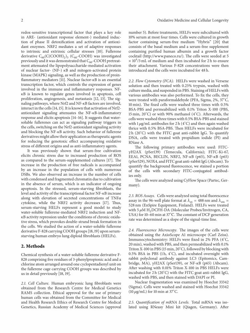
N. T. Costa et al. evaluated the involvement of TNF-𝛼 andinsulin resistance (IR) in the inflammatory process, oxidative
Oxidative Medicine and Cellular Longevity 3
stress, and disease activity in patients with rheumatoidarthritis (RA). They demonstrated that IR and TNF-𝛼 areimportant factors involved in redox imbalance in patientswith RA which seems to be due to the maintenance ofinflammatory state and disease activity.
S. Vranková et al. studied the effects of NF-kB inhibitionon ROS and NO generation and blood pressure (BP) reg-ulation in hereditary hypertriglyceridemic rats. They foundthat NF-kB inhibition leads to decreased ROS degradation bySOD followed by increased heart oxidative damage and BPelevation despite the increase in eNOS protein.
L. Minutoli et al. described the current knowledge on therole of NLRP3 inflammasome in some organs (brain, heart,kidney, and testis) after I/R injury, with particular regard tothe role played by ROS in its activation. They conclude that adefinite comprehension of the role of NLRP3 inflammasomein the host responses to different danger signals is still lacking.
S. Kazemi et al. investigated the effects of the xenoe-strogenic chemical Bisphenol A (BPA) on hepatic oxidativestress-related gene expression in rats. Their finding demon-strated that BPA generates ROS and increases the expressionof HO-1 and Gadd45b genes that cause hepatotoxicity.
A. N. Onyango reviewed the potential pathways of theendogenous formation of singlet oxygen and ozone, theirrelevance to human health, and how dietary factors affectgeneration and activity of such oxidants.
E. Gammella et al. described the multifaceted systemsregulating cellular and body iron homeostasis and discussedhow altered iron balance may lead to oxidative damage insome pathophysiological settings.
X.Wang et al. found that the transcription factor BTB andCNC homology 1 (Bach1) induces endothelial cell apoptosisand cell cycle arrest through ROS generation and, conse-quently, that functional downregulation of Bach1 may be apromising target for the treatment of vascular diseases.
M. Nita and A. Grzybowski, examining the currentliterature, showed that excessive production of reactive oxy-gen species and oxidative stress play important role in thepathogenesis of many age-related ocular diseases and otherpathologies of the anterior and posterior eye segment inadults. ROS stimulate cells’ death via apoptosis process,participate in the activation of proinflammatory and proan-giogenic pathways, and are associated with the autophagyprocess.
T. R. S. Hamilton et al. evaluated lasting effects of heatstress-induced oxidative stress on ejaculated and epididymalspermand found that it leads to rescuable alterations after onespermatic cycle in ejaculated sperm and also after 30 days inepididymal sperm.
M. L. Fanjul-Moles and G. O. López-Riquelme exam-ined the relationships between oxidative stress, circadianrhythms, and retinal damage in humans, particularly thoserelated to light and photodamage. Their review highlightsthe role of oxidative stress as one of the main causes of age-related macular degeneration (AMD) etiologies, a disease towhich, in addition to genetic predispositions, at least fourprocesses contribute: lipofuscinogenesis, drusogenesis, localinflammation and neovascularization, and immunologicalmechanisms.
H. Nagahisa et al. studied structural and functionalchanges induced by SOD1 deficiency and their results sug-gested that muscle is damaged by ROS produced in theTypeIIx/b fibers of the gastrocnemius muscle, acceleratingthe proliferation and differentiation of satellite cells in SOD1KO mice.
It is our opinion that the articles included in this specialissue, despite dealing with so different topics, represent animportant contribution to the knowledge of the harmful andbeneficial effects of ROS in living organisms.
Sergio Di MeoTanea T. ReedPaola Venditti
Victor M. Victor
Research ArticleFunctionalized Fullerene Increases NF-𝜅B Activity and BlocksGenotoxic Effect of Oxidative Stress in Serum-Starving HumanEmbryo Lung Diploid Fibroblasts
E. S. Ershova,1,2 V. A. Sergeeva,1 V. J. Tabakov,1 L. A. Kameneva,1
L. N. Porokhovnik,1 I. I. Voronov,3 E. A. Khakina,3 P. A. Troshin,3,4
S. I. Kutsev,1,5 N. N. Veiko,1,2 and S. V. Kostyuk1
1Research Centre for Medical Genetics (RCMG), Moscow 115478, Russia2V. A. Negovsky Research Institute of General Reanimatology, Moscow 107031, Russia3Institute for Problems of Chemical Physics of Russian Academy of Sciences, Moscow Region 142432, Russia4Skolkovo Institute of Science and Technology, Skolkovo Innovation Center, Nobel Street 3, Moscow 143026, Russia5N. I. Pirogov Russian National Research Medical University, Moscow 117997, Russia
Correspondence should be addressed to L. N. Porokhovnik; lp [email protected]
Received 25 December 2015; Revised 19 May 2016; Accepted 25 May 2016
Academic Editor: Javier Egea
Copyright © 2016 E. S. Ershova et al. This is an open access article distributed under the Creative Commons Attribution License,which permits unrestricted use, distribution, and reproduction in any medium, provided the original work is properly cited.
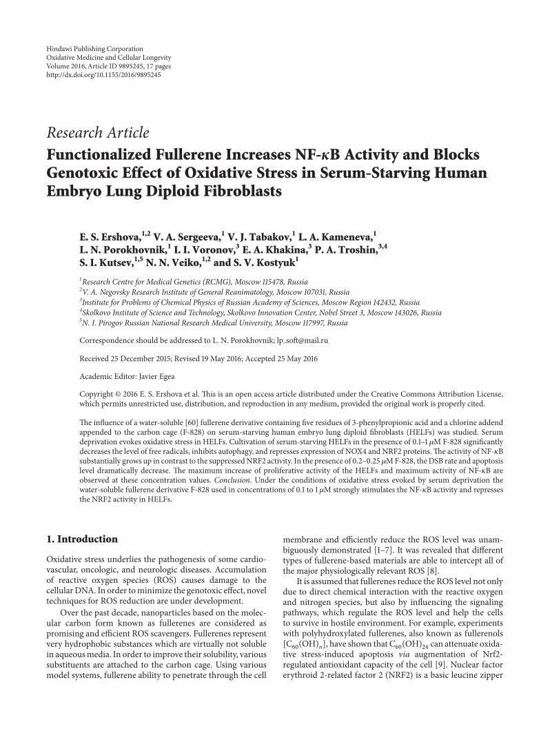
The influence of a water-soluble [60] fullerene derivative containing five residues of 3-phenylpropionic acid and a chlorine addendappended to the carbon cage (F-828) on serum-starving human embryo lung diploid fibroblasts (HELFs) was studied. Serumdeprivation evokes oxidative stress in HELFs. Cultivation of serum-starving HELFs in the presence of 0.1–1 𝜇M F-828 significantlydecreases the level of free radicals, inhibits autophagy, and represses expression of NOX4 and NRF2 proteins.The activity of NF-𝜅Bsubstantially grows up in contrast to the suppressedNRF2 activity. In the presence of 0.2–0.25 𝜇MF-828, theDSB rate and apoptosislevel dramatically decrease. The maximum increase of proliferative activity of the HELFs and maximum activity of NF-𝜅B areobserved at these concentration values. Conclusion. Under the conditions of oxidative stress evoked by serum deprivation thewater-soluble fullerene derivative F-828 used in concentrations of 0.1 to 1𝜇M strongly stimulates the NF-𝜅B activity and repressesthe NRF2 activity in HELFs.
1. Introduction
Oxidative stress underlies the pathogenesis of some cardio-vascular, oncologic, and neurologic diseases. Accumulationof reactive oxygen species (ROS) causes damage to thecellularDNA. In order tominimize the genotoxic effect, noveltechniques for ROS reduction are under development.
Over the past decade, nanoparticles based on the molec-ular carbon form known as fullerenes are considered aspromising and efficient ROS scavengers. Fullerenes representvery hydrophobic substances which are virtually not solublein aqueousmedia. In order to improve their solubility, varioussubstituents are attached to the carbon cage. Using variousmodel systems, fullerene ability to penetrate through the cell
membrane and efficiently reduce the ROS level was unam-biguously demonstrated [1–7]. It was revealed that differenttypes of fullerene-based materials are able to intercept all ofthe major physiologically relevant ROS [8].
It is assumed that fullerenes reduce the ROS level not onlydue to direct chemical interaction with the reactive oxygenand nitrogen species, but also by influencing the signalingpathways, which regulate the ROS level and help the cellsto survive in hostile environment. For example, experimentswith polyhydroxylated fullerenes, also known as fullerenols[C60(OH)n], have shown that C60(OH)
24can attenuate oxida-
tive stress-induced apoptosis via augmentation of Nrf2-regulated antioxidant capacity of the cell [9]. Nuclear factorerythroid 2-related factor 2 (NRF2) is a basic leucine zipper
Hindawi Publishing CorporationOxidative Medicine and Cellular LongevityVolume 2016, Article ID 9895245, 17 pageshttp://dx.doi.org/10.1155/2016/9895245
2 Oxidative Medicine and Cellular Longevity
redox-sensitive transcriptional factor that plays a key rolein ARE- (antioxidant response element-) mediated induc-tion of phase II detoxification and activation of antioxi-dant enzymes. NRF2 mediates a set of adaptive responsesto intrinsic and extrinsic cellular stresses [10]. Fullerenederivative C
60(C(COOH)
2)3(C60-COOH) was investigated
previously and it was demonstrated that C60-COOHpretreat-
ment attenuated the lipopolysaccharide-mediated activationof nuclear factor- (NF-) 𝜅B and mitogen-activated proteinkinase (MAPK) signaling, as well as the production of proin-flammatory mediators [11]. Nuclear factor-𝜅B is an essentialtranscription factor, which controls the expression of genesinvolved in the immune and inflammatory responses. NF-𝜅B is known to regulate genes involved in apoptosis, cellproliferation, angiogenesis, and metastasis [12, 13]. The sig-naling pathways, where Nrf2 and NF-𝜅B factors are involved,interact in the cells [14, 15]. It is known that activation ofNrf2-antioxidant signaling attenuates the NF-𝜅B-inflammatoryresponse and elicits apoptosis [14–16]. It suggests that water-soluble fullerenes can act as signaling pathway triggers inthe cells, switching on the Nrf2-antioxidant signaling activityand blocking the NF-𝜅B activity. Such behavior of fullerenederivativesmight allow their application as therapeutic agentsfor reducing the genotoxic effect accompanying oxidativestress of different origins and as anti-inflammatory agents.
It was previously shown that serum-free cultivationelicits chronic stress due to increased production of ROSas compared to the serum-supplemented cultures [17]. Theincrease in the production of free radicals is accompaniedby an increase in the population of cells with numerousDSBs. We also observed an increase in the number of cellswith condensed and fragmented chromatin due to cultivationin the absence of serum, which is an indicator of ongoingapoptosis. In the stressed, serum-starving fibroblasts, thelevel and activity of the transcriptional factor NF-𝜅B increasealong with elevation of secreted concentrations of TNF𝛼cytokine, while the NRF2 activity decreases [17]. Thus,serum-starving HELFs represent a good model to studywater-soluble fullerene-mediated NRF2 induction and NF-𝜅B activity repression under the conditions of chronic oxida-tive stress, which provokes double-strand break formation inthe cells. We studied the action of a water-soluble fullerenederivative F-828 carryingCOOHgroups [18, 19] upon serum-starving human embryo lung diploid fibroblasts (HELFs).
2. Methods
Chemical synthesis of a water-soluble fullerene derivative F-828 comprising five residues of 3-phenylpropionic acid and achlorine atom arranged around one cyclopentadienyl unit onthe fullerene cage carrying COOH groups was described byus in detail previously [18, 19].
2.1. Cell Culture. Human embryonic lung fibroblasts wereobtained from the Research Centre for Medical GeneticsRAMS collection. Ethical approval for the use of primaryhuman cells was obtained from the Committee for Medicaland Health Research Ethics of Research Centre for MedicalGenetics, Russian Academy of Medical Sciences (approval
number 5). Before treatments, HELFs were subcultured with10% serum at most four times. Cells were cultured in growthfactor containing serum-free medium “Hybris” [20] thatconsists of the basal medium and a serum-free supplementcontaining purified human albumin and a growth factorcocktail (http://www.paneco.ru/). The cells were seeded at 5× 10⁴/3mL of medium and then incubated for 2 h to ensuretheir attachment. Various F-828 concentrations were thenintroduced and the cells were incubated for 48 h.
2.2. Flow Cytometry (FCA). HELFs were washed in Versenesolution and then treated with 0.25% trypsin, washed withculturemedia, and suspended in PBS. Staining ofHELFswithvarious antibodies was also performed. To fix the cells, theywere treated with paraformaldehyde (PFA, Sigma, 2%, 37∘C,10min). The fixed cells were washed three times with 0.5%BSA-PBS and permeabilized with 0.1% Triton X-100 (PBS,15min, 20∘C) or with 90% methanol (4∘C). Afterwards, thecells were washed three timeswith 0.5%BSA-PBS and stainedwith 1 𝜇g/mL antibodies for 2 h (4∘C) and then again washedthrice with 0.5% BSA-PBS. Then HELFs were incubated for2 h (20∘C) with the FITC goat anti-rabbit IgG. To quantifyDNA, cells were treated with propidium iodide (PI) andRNase A.
The following primary antibodies were used: FITC-𝛾H2AX (pSer139) (Temecula, California); FITC-Ki-67,EEA1, PCNA, BECLIN, NRF2, NF-𝜅B (p65), NF-𝜅B (p65)(pSer529),NOX4, and FITCgoat anti-rabbit IgG (Abcam). Toquantify the background fluorescence, we stained a portionof the cells with secondary FITC-conjugated antibod-ies only.
The cells were analyzed using CyFlow Space (Partec, Ger-many).
2.3. ROS Assays. Cells were analyzed using total fluorescenceassay in the 96-well plate format at 𝜆ex = 488 nm and 𝜆em =528 nm (EnSpire Equipment, Finland). HELFs were treatedwith 5 𝜇MH
2DCFH-DA (Molecular Probes/Invitrogen, CA,
USA) for 10–60min at 37∘b. The constant of DCF generationrate was determined as a slope of the signal-time line.
2.4. Fluorescence Microscopy. The images of the cells wereobtained using the AxioScope A1 microscope (Carl Zeiss).Immunocytochemistry: HELFs were fixed in 2% PFA (4∘C,20min), washed with PBS, and then permeabilized with 0.1%TritonX-100 in PBS (15min, 20∘C), followed by blockingwith0.5% BSA in PBS (1 h, 4∘C), and incubated overnight withrabbit polyclonal antibody against LC3 (Epitomics, Cam-bridge, MA), 𝛾H2AX (pSer139), or NF-𝜅B (p65) (Abcam).After washing with 0.01% Triton X-100 in PBS HELFs wereincubated for 2 h (20∘C) with the FITC goat anti-rabbit IgG,washed with PBS, and then stained with DAPI or PI.
Nuclear fragmentation was examined by Hoechst 33342(Sigma). Cells were washed and stained with Hoechst 33342(10 𝜇g/mL) for 10min at 37∘C.
2.5. Quantification of mRNA Levels. Total mRNA was iso-lated using RNeasy Mini kit (Qiagen, Germany). After
Oxidative Medicine and Cellular Longevity 3
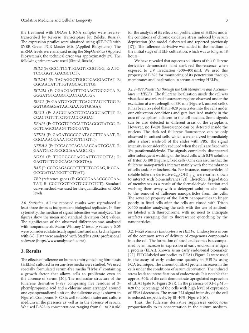
the treatment with DNAse I, RNA samples were reverse-transcribed by Reverse Transcriptase kit (Sileks, Russia).The expression profiles were obtained using qRT-PCR withSYBR Green PCR Master Mix (Applied Biosystems). ThemRNA levels were analyzed using the StepOnePlus (AppliedBiosystems); the technical error was approximately 2%. Thefollowing primers were used (Sintol, Russia):
BCL2 (F: GCCTTCTTTGAGTTCGGTGG, R: ATC-TCCCGGTTGACGCTCT);BCL2A1 (F: TACAGGCTGGCTCAGGACTAT R:CGCAACATTTTGTAGCACTCTG);BCL2L1 (F: CGACGAGTTTGAACTGCGGTA R:GGGATGTCAGGTCACTGAATG);BIRC2 (F: GAATCTGGTTTCAGCTAGTCTGG R:GGTGGGAGATAATGAATGTGCAA);BIRC3 (F: AAGCTACCTCTCAGCCTACTTT R:CCACTGTTTTCTGTACCCGGA);KEAP1 (F: GTGGTGTCCATTGAGGGTATCC, R:GCTCAGCGAAGTTGGCGAT);NFKB1 (F: CAGATGGCCCATACCTTCAAAT, R:CGGAAACGAAATCCTCTCTGTT);NFE2L2 (F: TCCAGTCAGAAACCAGTGGAT, R:GAATGTCTGCGCCAAAAGCTG);NOX4 (F: TTGGGGCTAGGATTGTGTCTA; R:GAGTGTTCGGCACATGGGTA);BAX (F: CCCGAGAGGTCTTTTTCCGAG,R:CCA-GCCCATGATGGTTCTGAT);TBP (reference gene) (F: GCCCGAAACGCCGAA-TAT, R: CCGTGGTTCGTGGCTCTCT). Standardcurve method was used for the quantification of RNAlevels.
2.6. Statistics. All the reported results were reproduced atleast three times as independent biological replicates. In flowcytometry, the median of signal intensities was analyzed.Thefigures show the mean and standard deviation (SD) values.The significance of the observed differences was analyzedwith nonparametric Mann-Whitney 𝑈 tests. 𝑝 values < 0.05were considered statistically significant andmarked in figureswith (∗). Data were analyzed with StatPlus 2007 professionalsoftware (http://www.analystsoft.com/).
3. Results
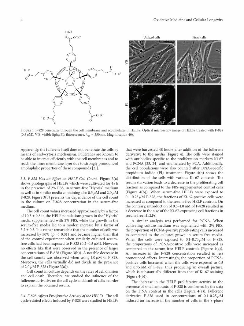
The effects of fullerene on human embryonic lung fibroblasts(HELFs) cultured in serum-freemedia were studied.We usedspecially formulated serum-free media “Hybris” containinga growth factor that allows cells to proliferate even inthe absence of serum [20]. The molecular structure of thefullerene derivative F-828 comprising five residues of 3-phenylpropionic acid and a chlorine atom arranged aroundone cyclopentadienyl unit on the fullerene cage is shown inFigure 1. Compound F-828 is well soluble inwater and culturemedium in the presence as well as in the absence of serum.We used F-828 in concentrations ranging from 0.1 to 2.0 𝜇M
for the analysis of its effects on proliferation of HELFs underthe conditions of chronic oxidative stress induced by serumdeprivation (the model elaborated and reported previously[17]). The fullerene derivative was added to the medium atthe initial stage of HELF cultivation, which was as long as 48hours.
We have revealed that aqueous solutions of this fullerenederivative demonstrate faint dark-red fluorescence whenexposed to UV irradiation (300–400 nm). We used thisproperty of F-828 for monitoring of its penetration throughmembranes and localization in serum-starving HELFs.
3.1. F-828 Penetrates through the Cell Membrane and Accumu-lates in HELFs. The fullerene localization inside the cell wasvisualized as dark-red fluorescent regions observed under theexcitation at a wavelength of 350 nm (Figure 1, unfixed cells).It has been revealed that F-828 penetrates into the cells underour cultivation conditions and gets localized mainly in thearea of cytoplasm adjacent to the cell nucleus. Some signalscan be also detected in different areas of the cytoplasm.However, no F-828 fluorescence can be detected inside thenucleus. The dark-red fullerene fluorescence can be onlyobserved in unfixed cells, which were analyzed immediatelyafter a short wash-off of the slides with PBS. The signalintensity is considerably reduced when the cells are fixed with3% paraformaldehyde. The signals completely disappearedafter subsequent washing of the fixed cells with 0.1% solutionof TritonH-100 (Figure 1, fixed cells). One can assume that thefullerene nanoparticles interact mainly with the membranesof cells and/or mitochondria. For instance, nanoparticles ofsoluble fullerene derivative C
60(OH)18–22 were earlier shown
to interact with biomembranes [21]. Therefore, degradationof membranes as a result of the formaldehyde fixation andwashing them away with a detergent solution also leadsto the removal of fullerene nanoparticles from the cells.The revealed property of the F-828 nanoparticles to lingerpoorly in fixed cells after the cells are rinsed with TritonX-100 enables analyzing the cells with the use of antibod-ies labeled with fluorochrome, with no need to anticipateartefacts emerging due to fluorescence quenching by thenanoparticles.
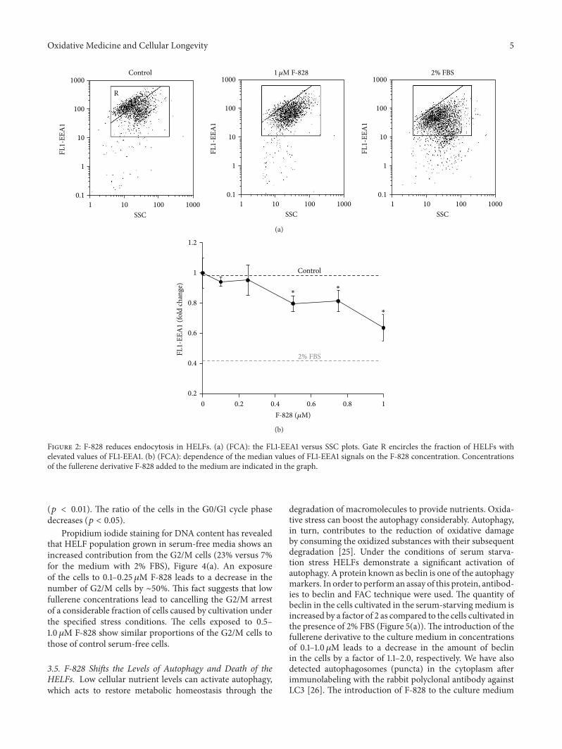
3.2. F-828 Reduces Endocytosis in HELFs. Endocytosis is oneof the common ways of delivery of exogenous compoundsinto the cell. The formation of novel endosomes is accompa-nied by an increase in expression of early endosome antigen1 protein (EEA1), known as an early endosomal biomarker[22]. FITC-labeled antibodies to EEA1 (Figure 2) were usedin the assay of early endosome quantity in HELFs usingFbA technique.The amount of@@01 protein increases in thecells under the conditions of serum deprivation.The inducedstress leads to intensification of endocytosis. It is notable thatapprox. 60% of the cells demonstrate upregulated expressionof @@01 (gate R, Figure 2(a)). In the presence of 0.1–1𝜇M F-828 the percentage of the cells with high level of expressionof @@01 decreases. The mean FL1 signal intensity of the cellis reduced, respectively, by 10–40% (Figure 2(b)).
Thus, the fullerene derivative suppresses endocytosisproportionally to its concentration in the culture medium.
4 Oxidative Medicine and Cellular Longevity
Unfixed cells Fixed cells
VIS
FL
F-828
+K−O
+K−O
O−K+
O−K+
O−K+
Cl
O
O
OO
O
Figure 1: F-828 penetrates through the cell membrane and accumulates in HELFs. Optical microscopy image of HELFs treated with F-828(0.5 𝜇M). VIS: visible light; FL: fluorescence, 𝜆ex = 350 nm. Magnification 40x.
Apparently, the fullerene itself does not penetrate the cells bymeans of endocytosis mechanism. Fullerenes are known tobe able to interact efficiently with the cell membranes and toreach the inner membrane layer due to strongly pronouncedamphiphilic properties of these compounds [21].
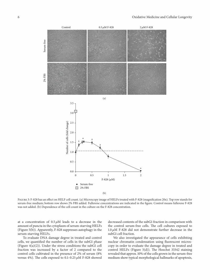
3.3. F-828 Has an Effect on HELF Cell Count. Figure 3(a)shows photographs of HELFs which were cultivated for 48 hin the presence of 2% FBS, in serum-free “Hybris” mediumas well as in similar media containing also 0.5 𝜇Mand 2.0 𝜇MF-828. Figure 3(b) presents the dependence of the cell countin the culture on F-828 concentration in the serum-freemedium.
The cell count values increased approximately by a factorof 10.3 ± 0.8 in the HELF populations grown in the “Hybris”media supplemented with 2% FBS, while the growth in theserum-free media led to a modest increase by a factor of3.2 ± 0.3. It is rather remarkable that the number of cells wasincreased by 50% (𝑝 < 0.01) and became higher than thatof the control experiment when similarly cultured serum-free cells had been exposed to F-828 (0.2–0.5𝜇M). However,no effects like that were observed in the presence of largerconcentrations of F-828 (Figure 3(b)). A notable decrease inthe cell counts was observed when using 1.0 𝜇M of F-828.Moreover, the cells virtually did not divide in the presenceof 2.0 𝜇M F-828 (Figure 3(a)).
Cell count in culture depends on the rates of cell divisionand cell death. Therefore, we studied the influence of thefullerene derivative on the cell cycle and death of cells in orderto explain the obtained results.
3.4. F-828 Affects Proliferative Activity of the HELFs. The cellcycle-related effects induced by F-828 were studied in HELFs
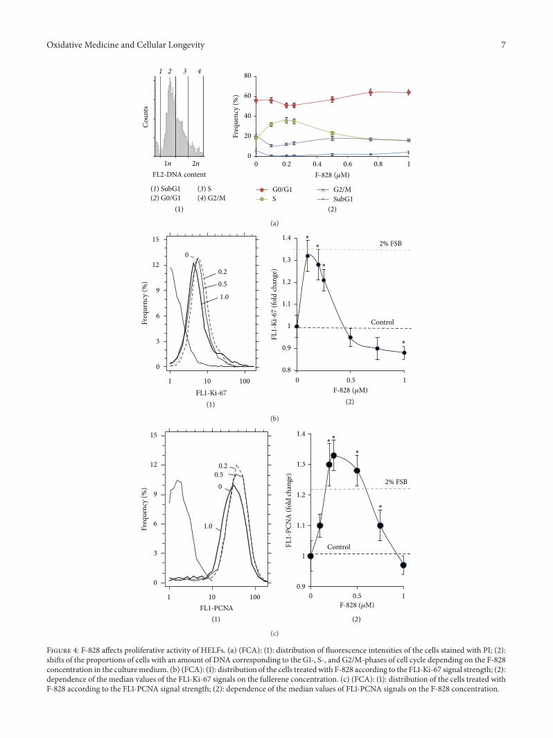
that were harvested 48 hours after addition of the fullerenederivative to the media (Figure 4). The cells were stainedwith antibodies specific to the proliferation markers Ki-67and PCNA [23, 24] and enumerated by FCA. Additionally,the cell populations were also counted after DNA-specificpropidium iodide (PI) treatment. Figure 4(b) shows thedistribution of the cells with various Ki-67 contents. Theserum starvation leads to a decrease in the proliferating cellfraction as compared to the FBS-supplemented control cells(Figure 4(b)). When serum-free HELFs were exposed to0.1–0.25 𝜇M F-828, the fractions of Ki-67-positive cells wereincreased as compared to the serum-free HELF controls. Onthe contrary, introduction of 0.5–1.0 𝜇M of F-828 resulted ina decrease in the size of the Ki-67-expressing cell fractions inserum-free HELFs.
A similar analysis was performed for PCNA. Whencultivating culture medium was augmented with 2% FBS,the proportion of PCNA-positive proliferating cells increasedas compared to the cultures grown in serum-free media.When the cells were exposed to 0.1–0.75 𝜇M of F-828,the proportions of PCNA-positive cells were increased ascompared to the serum-free HELF controls (Figure 4(c)).An increase in the F-828 concentration resulted in lesspronounced effects. Interestingly, the proportion of PCNA-positive cells increased when the cells were exposed to 0.5and 0.75 𝜇M of F-828, thus producing an overall picture,which is substantially different from that of Ki-67 staining(Figure 4(b)).
The increase in the HELF proliferative activity in thepresence of small amounts of F-828 is confirmed by the dataon the DNA content in the cells (Figure 4(a)). Fullerenederivative F-828 used in concentrations of 0.1–0.25𝜇Minduced an increase in the number of cells in the S-phase
Oxidative Medicine and Cellular Longevity 5
Control
SSC
R
1000
100
10
1
1000100101
0.1
FL1-
EEA
11𝜇M F-828 2% FBS
SSC
1000
100
10
1
1000100101
0.1
FL1-
EEA
1SSC
1000
100
10
1
1000100101
0.1
FL1-
EEA
1
(a)
0.2
0.4
0.6
0.8
1
1.2
0 0.2 0.4 0.6 0.8 1
FL1-
EEA
1 (fo
ld ch
ange
)
Control
2% FBS
∗∗
∗
F-828 (𝜇M)
(b)
Figure 2: F-828 reduces endocytosis in HELFs. (a) (FCA): the FL1-EEA1 versus SSC plots. Gate R encircles the fraction of HELFs withelevated values of FL1-EEA1. (b) (FCA): dependence of the median values of FL1-EEA1 signals on the F-828 concentration. Concentrationsof the fullerene derivative F-828 added to the medium are indicated in the graph.
(𝑝 < 0.01). The ratio of the cells in the G0/G1 cycle phasedecreases (𝑝 < 0.05).
Propidium iodide staining for DNA content has revealedthat HELF population grown in serum-free media shows anincreased contribution from the G2/M cells (23% versus 7%for the medium with 2% FBS), Figure 4(a). An exposureof the cells to 0.1–0.25 𝜇M F-828 leads to a decrease in thenumber of G2/M cells by ∼50%. This fact suggests that lowfullerene concentrations lead to cancelling the G2/M arrestof a considerable fraction of cells caused by cultivation underthe specified stress conditions. The cells exposed to 0.5–1.0 𝜇M F-828 show similar proportions of the G2/M cells tothose of control serum-free cells.
3.5. F-828 Shifts the Levels of Autophagy and Death of theHELFs. Low cellular nutrient levels can activate autophagy,which acts to restore metabolic homeostasis through the
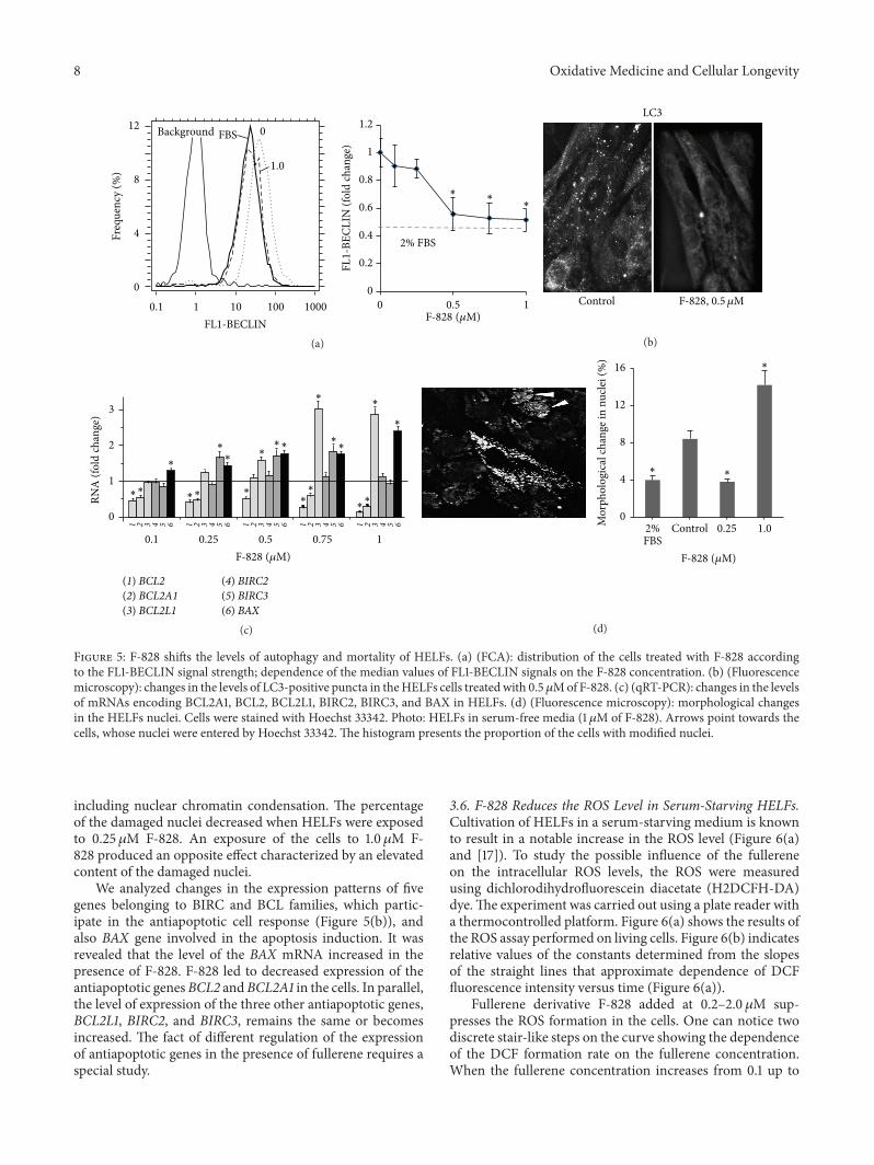
degradation of macromolecules to provide nutrients. Oxida-tive stress can boost the autophagy considerably. Autophagy,in turn, contributes to the reduction of oxidative damageby consuming the oxidized substances with their subsequentdegradation [25]. Under the conditions of serum starva-tion stress HELFs demonstrate a significant activation ofautophagy. A protein known as beclin is one of the autophagymarkers. In order to performan assay of this protein, antibod-ies to beclin and FAC technique were used. The quantity ofbeclin in the cells cultivated in the serum-starving medium isincreased by a factor of 2 as compared to the cells cultivated inthe presence of 2% FBS (Figure 5(a)).The introduction of thefullerene derivative to the culture medium in concentrationsof 0.1–1.0 𝜇M leads to a decrease in the amount of beclinin the cells by a factor of 1.1–2.0, respectively. We have alsodetected autophagosomes (puncta) in the cytoplasm afterimmunolabeling with the rabbit polyclonal antibody againstLC3 [26]. The introduction of F-828 to the culture medium
6 Oxidative Medicine and Cellular Longevity
Seru
m-fr
ee2%
FBS
Control 0.5 𝜇M F-828 2𝜇M F-828
(a)
0.0
0.5
1.0
1.5
2.0
2.5
3.0
3.5
0 0.5 1 1.5 2
Num
ber o
f cel
ls (fo
ld ch
ange
)
F-828 (𝜇M)
Serum-free2% FBS
∗∗ ∗
∗
∗
(b)
Figure 3: F-828 has an effect on HELF cell count. (a) Microscopy image of HELFs treated with F-828 (magnification 20x). Top row stands forserum-free medium; bottom row shows 2% FBS added. Fullerene concentrations are indicated in the figure. Control means fullerene F-828was not added. (b) Dependence of the cell count in the culture on the F-828 concentration.
at a concentration of 0.5 𝜇M leads to a decrease in theamount of puncta in the cytoplasm of serum-starving HELFs(Figure 5(b)). Apparently, F-828 suppresses autophagy in theserum-starving HELFs.
To evaluate DNA damage degree in treated and controlcells, we quantified the number of cells in the subG1 phase(Figure 4(a)(2)). Under the stress conditions the subG1 cellfraction was increased by a factor of 2 compared to thecontrol cells cultivated in the presence of 2% of serum (8%versus 4%). The cells exposed to 0.1–0.25𝜇M F-828 showed
decreased contents of the subG1 fraction in comparison withthe control serum-free cells. The cell cultures exposed to1.0 𝜇M F-828 did not demonstrate further decrease in thesubG1 cell fraction.
We also investigated the appearance of cells exhibitingnuclear chromatin condensation using fluorescent micros-copy in order to evaluate the damage degree in treated andcontrol HELFs (Figure 5(d)). The Hoechst 33342 stainingrevealed that approx. 10% of the cells grown in the serum-freemedium show typical morphological hallmarks of apoptosis,
Oxidative Medicine and Cellular Longevity 7
(1) (2)
0
20
40
60
80
0 0.2 0.4 0.6 0.8 1
G0/G1S
G2/MSubG1
F-828 (𝜇M)
1 2 3 4
Cou
nts
1n 2n
(1) SubG1(2) G0/G1
(3) S(4) G2/M
FL2-DNA content
Freq
uenc
y (%
)
(a)
(1) (2)
0.8
0.9
1
1.1
1.2
1.3
1.4
0 0.5 1
Freq
uenc
y (%
)
FL1-
Ki-6
7 (fo
ld ch
ange
)
15
12
9
6
3
0
1 10 100
FL1-Ki-67
0
0.2
0.5
1.0
2% FSB
Control
∗∗
∗
∗
F-828 (𝜇M)
(b)
(1) (2)
0.9
1
1.1
1.2
1.3
1.4
0 0.5 1
2% FSB
Control
Freq
uenc
y (%
)
15
12
9
6
3
0
FL1-PCNA
0
0.20.5
1.0
1 10 100
∗
∗
∗∗
FL1-
PCN
A (f
old
chan
ge)
F-828 (𝜇M)
(c)
Figure 4: F-828 affects proliferative activity of HELFs. (a) (FCA): (1): distribution of fluorescence intensities of the cells stained with PI; (2):shifts of the proportions of cells with an amount of DNA corresponding to the G1-, S-, and G2/M-phases of cell cycle depending on the F-828concentration in the culturemedium. (b) (FCA): (1): distribution of the cells treated with F-828 according to the FL1-Ki-67 signal strength; (2):dependence of the median values of the FL1-Ki-67 signals on the fullerene concentration. (c) (FCA): (1): distribution of the cells treated withF-828 according to the FL1-PCNA signal strength; (2): dependence of the median values of FL1-PCNA signals on the F-828 concentration.
8 Oxidative Medicine and Cellular Longevity
0
0.2
0.4
0.6
0.8
1
1.2
0 0.5 1
FL1-
BECL
IN (f
old
chan
ge)
2% FBS
F-828 (𝜇M)
Freq
uenc
y (%
)
FL1-BECLIN
12
8
4
0
0.1 1 10 100 1000
FBSBackground 0
1.0
∗ ∗∗
(a)
LC3
Control F-828, 0.5 𝜇M
(b)
0.1 0.25 0.5 0.75 1F-828 (𝜇M)
RNA
(fol
d ch
ange
) 3
2
1
0
1 2 3 4 5 6 1 2 3 4 5 6 1 2 3 4 5 6 1 2 3 4 5 6 1 2 3 4 5 6
∗∗
∗
∗∗
∗∗
∗
∗∗∗
∗
∗
∗
∗
∗
∗∗∗
∗
(1) BCL2(2) BCL2A1(3) BCL2L1
(4) BIRC2(5) BIRC3(6) BAX
(c)
0
4
8
12
16
Mor
phol
ogic
al ch
ange
in n
ucle
i (%
)
F-828 (𝜇M)
Control 0.25 1.0
∗
∗
∗
2%FBS
(d)
Figure 5: F-828 shifts the levels of autophagy and mortality of HELFs. (a) (FCA): distribution of the cells treated with F-828 accordingto the FL1-BECLIN signal strength; dependence of the median values of FL1-BECLIN signals on the F-828 concentration. (b) (Fluorescencemicroscopy): changes in the levels of LC3-positive puncta in theHELFs cells treatedwith 0.5𝜇Mof F-828. (c) (qRT-PCR): changes in the levelsof mRNAs encoding BCL2A1, BCL2, BCL2L1, BIRC2, BIRC3, and BAX in HELFs. (d) (Fluorescence microscopy): morphological changesin the HELFs nuclei. Cells were stained with Hoechst 33342. Photo: HELFs in serum-free media (1 𝜇M of F-828). Arrows point towards thecells, whose nuclei were entered by Hoechst 33342. The histogram presents the proportion of the cells with modified nuclei.
including nuclear chromatin condensation. The percentageof the damaged nuclei decreased when HELFs were exposedto 0.25 𝜇M F-828. An exposure of the cells to 1.0𝜇M F-828 produced an opposite effect characterized by an elevatedcontent of the damaged nuclei.
We analyzed changes in the expression patterns of fivegenes belonging to BIRC and BCL families, which partic-ipate in the antiapoptotic cell response (Figure 5(b)), andalso BAX gene involved in the apoptosis induction. It wasrevealed that the level of the BAX mRNA increased in thepresence of F-828. F-828 led to decreased expression of theantiapoptotic genesBCL2 andBCL2A1 in the cells. In parallel,the level of expression of the three other antiapoptotic genes,BCL2L1, BIRC2, and BIRC3, remains the same or becomesincreased. The fact of different regulation of the expressionof antiapoptotic genes in the presence of fullerene requires aspecial study.
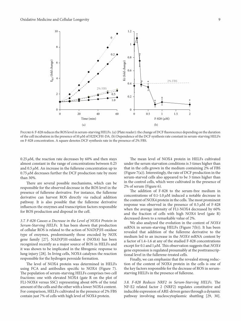
3.6. F-828 Reduces the ROS Level in Serum-Starving HELFs.Cultivation of HELFs in a serum-starving medium is knownto result in a notable increase in the ROS level (Figure 6(a)and [17]). To study the possible influence of the fullereneon the intracellular ROS levels, the ROS were measuredusing dichlorodihydrofluorescein diacetate (H2DCFH-DA)dye.The experiment was carried out using a plate reader witha thermocontrolled platform. Figure 6(a) shows the results ofthe ROS assay performed on living cells. Figure 6(b) indicatesrelative values of the constants determined from the slopesof the straight lines that approximate dependence of DCFfluorescence intensity versus time (Figure 6(a)).
Fullerene derivative F-828 added at 0.2–2.0𝜇M sup-presses the ROS formation in the cells. One can notice twodiscrete stair-like steps on the curve showing the dependenceof the DCF formation rate on the fullerene concentration.When the fullerene concentration increases from 0.1 up to
Oxidative Medicine and Cellular Longevity 9
0
2
4
6
8
10
0 20 40 60Time (min)
0
0.2
2% FBS
0.5
1.0
I dcf
(fol
d ch
ange
)
(a)
0
0.2
0.4
0.6
0.8
1
1.2
0 0.5 1 1.5 2
Ki (f
old
chan
ge)
2% FBS
∗∗
∗
∗
∗
∗
F-828 (𝜇M)
(b)
Figure 6: F-828 reduces theROS level in serum-starvingHELFs. (a) (Plate reader): the change ofDCFfluorescence depending on the durationof the cell incubation in the presence of 10𝜇MofH2DCFH-DA. (b) Dependence of the DCF synthesis rate constant in serum-starvingHELFson F-828 concentration. A square denotes DCF synthesis rate in the presence of 2% FBS.
0.25 𝜇M, the reaction rate decreases by 60% and then staysalmost constant in the range of concentrations between 0.25and 0.5 𝜇M. An increase in the fullerene concentration up to0.75 𝜇M decreases further the DCF production rate by morethan 30%.
There are several possible mechanisms, which can beresponsible for the observed decrease in the ROS level in thepresence of fullerene derivative. For instance, the fullerenederivative can harvest ROS directly via radical additionpathway. It is also possible that the fullerene derivativeinfluences the enzymes and transcription factors responsiblefor ROS production and disposal in the cell.
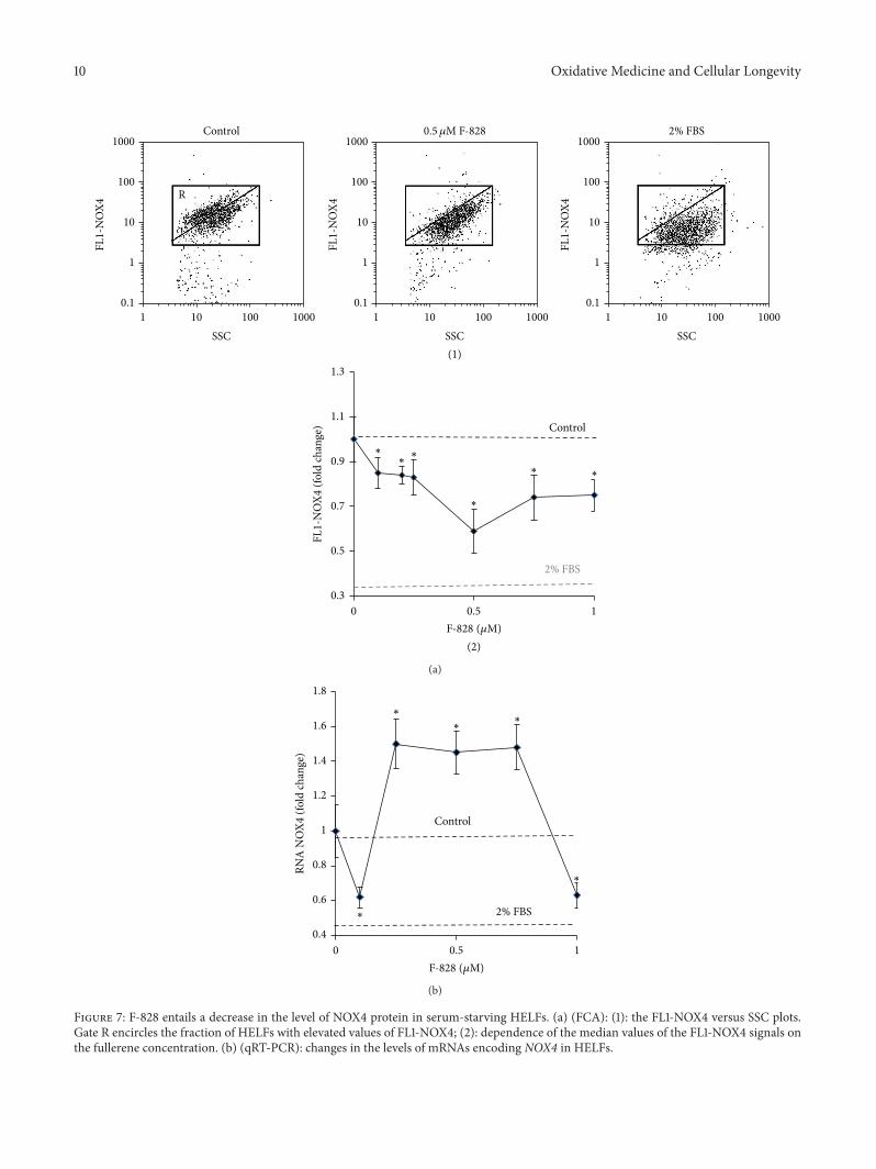
3.7. F-828 Causes a Decrease in the Level of NOX4 Protein inSerum-Starving HELFs. It has been shown that productionof cellular ROS is related to the action of NAD(P)H-oxidasetype of enzymes, predominantly those encoded by NOXgene family [27]. NAD(P)H-oxidase 4 (NOX4) has beenrecognized recently as a major source of ROS in HELFs andit was shown to be implicated in the fibrogenic response tolung injury [28]. In living cells, NOX4 catalyzes the reactionresponsible for the hydrogen peroxide formation.
The level of NOX4 protein was determined in HELFsusing FCA and antibodies specific to NOX4 (Figure 7).The population of serum-starving HELFs comprises two cellfractions: one with elevated NOX4 (gate R on the plot ofFL1-NOX4 versus SSC) representing about 60% of the totalamount of the cells and the other with a lower NOX4 content.For comparison, HELFs cultivated in the presence of 2% FBScontain just 7% of cells with high level of NOX4 protein.
The mean level of NOX4 protein in HELFs cultivatedunder the serum starvation conditions is 3 times higher thanthat in the cells grown in the medium containing 2% of FBS(Figure 7(a)). Interestingly, the rate of DCF production in theserum-starved cells also appeared to be 3 times higher thanin the control cells, which were cultivated in the presence of2% of serum (Figure 6).
The addition of F-828 to the serum-free medium inconcentrations of 0.1–1.0𝜇M induced a notable decrease inthe content of NOX4 protein in the cells.Themost prominentresponse was observed in the presence of 0.5 𝜇M of F-828when the average intensity of FL1-NOX4 decreased by 60%and the fraction of cells with high NOX4 level (gate R)decreased down to a remarkable value of 2%.
We also analyzed the evolution in the content of NOX4mRNA in serum-starving HELFs (Figure 7(b)). It has beenrevealed that addition of the fullerene derivative to themedium led to an increase in the NOX4 mRNA content bya factor of 1.4–1.6 at any of the studied F-828 concentrationsexcept for 0.1 and 1 𝜇M.This observation suggests thatNOX4gene expression is regulated presumably at the posttranscrip-tional level in the fullerene-treated cells.
Finally, we can emphasize that the revealed strong reduc-tion of the content of NOX4 protein in the cells is one ofthe key factors responsible for the decrease of ROS in serum-starving HELFs in the presence of fullerene.
3.8. F-828 Reduces NRF2 in Serum-Starving HELFs. TheNF-E2 related factor 2 (NRF2) regulates constitutive andinducible expression of ARE-driven genes through a dynamicpathway involving nucleocytoplasmic shuttling [29, 30].
10 Oxidative Medicine and Cellular Longevity
(2)
0.3
0.5
0.7
0.9
1.1
1.3
0 0.5 1
Control
2% FBS
F-828 (𝜇M)
∗∗
∗
∗∗
∗
FL1-
NO
X4 (f
old
chan
ge)
(1)
FL1-
NO
X4
Control1000
100
10
1
0.1
2% FBS0.5 𝜇M F-828
R
1000100101
SSCFL
1-N
OX4
1000
100
10
1
0.1
1000100101
SSC
FL1-
NO
X4
1000
100
10
1
0.1
1000100101
SSC
(a)
0.4
0.6
0.8
1
1.2
1.4
1.6
1.8
0 0.5 1
RNA
NO
X4 (f
old
chan
ge)
Control
2% FBS
∗
∗∗
∗
∗
F-828 (𝜇M)
(b)
Figure 7: F-828 entails a decrease in the level of NOX4 protein in serum-starving HELFs. (a) (FCA): (1): the FL1-NOX4 versus SSC plots.Gate R encircles the fraction of HELFs with elevated values of FL1-NOX4; (2): dependence of the median values of the FL1-NOX4 signals onthe fullerene concentration. (b) (qRT-PCR): changes in the levels of mRNAs encoding NOX4 in HELFs.
Oxidative Medicine and Cellular Longevity 11
(1)
Control1000
100
10
1
0.1
R
1000100101
SSC
FL1-
NRF
20.5 𝜇M
1000
100
10
1
0.1
1000100101
SSC
FL1-
NRF
2
(2)
0.20.30.40.50.60.70.80.9
11.11.2
0 0.2 0.4 0.6 0.8 1F-828 (𝜇M)
∗
∗
∗
FL1-
NRF
2 (fo
ld ch
ange
)
(a)
0.3
0.5
0.7
0.9
1.1
1.3
1.5
1.7
1.9
0 0.2 0.4 0.6 0.8 1
RNA
(fol
d ch
ange
)
F-828 (𝜇M)
∗
∗
∗∗
NRF2KEAP1
(b)
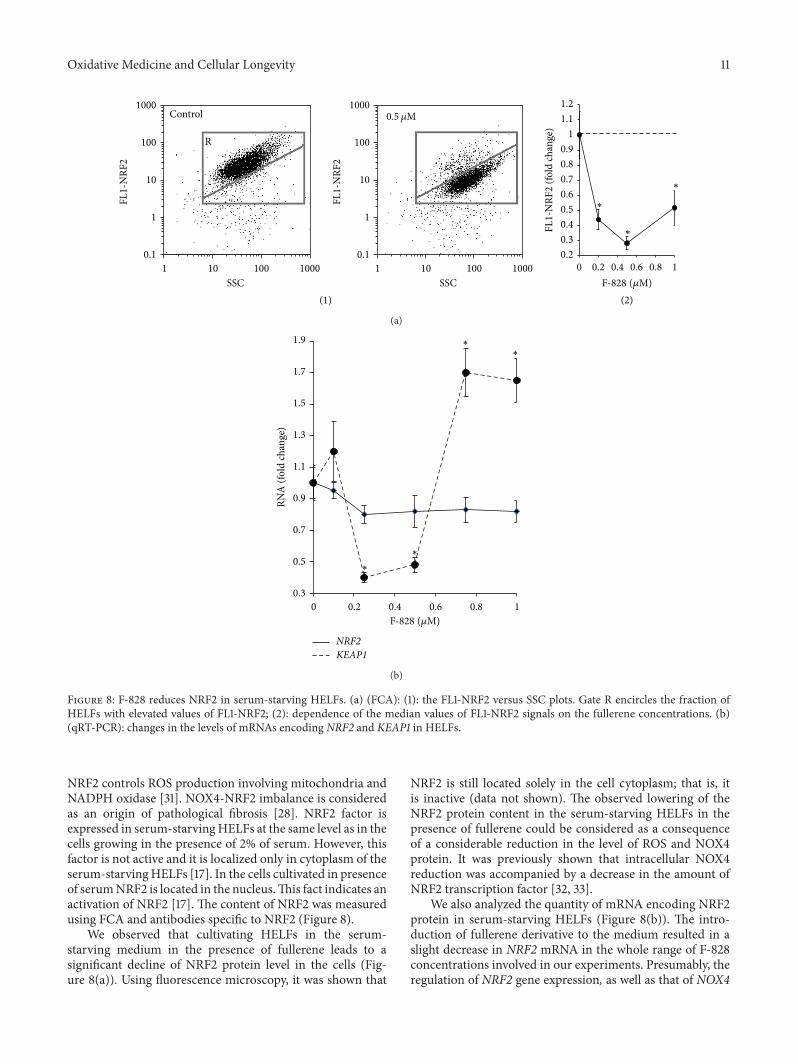
Figure 8: F-828 reduces NRF2 in serum-starving HELFs. (a) (FCA): (1): the FL1-NRF2 versus SSC plots. Gate R encircles the fraction ofHELFs with elevated values of FL1-NRF2; (2): dependence of the median values of FL1-NRF2 signals on the fullerene concentrations. (b)(qRT-PCR): changes in the levels of mRNAs encoding NRF2 and KEAP1 in HELFs.
NRF2 controls ROS production involving mitochondria andNADPH oxidase [31]. NOX4-NRF2 imbalance is consideredas an origin of pathological fibrosis [28]. NRF2 factor isexpressed in serum-starvingHELFs at the same level as in thecells growing in the presence of 2% of serum. However, thisfactor is not active and it is localized only in cytoplasm of theserum-starvingHELFs [17]. In the cells cultivated in presenceof serumNRF2 is located in the nucleus.This fact indicates anactivation of NRF2 [17]. The content of NRF2 was measuredusing FCA and antibodies specific to NRF2 (Figure 8).
We observed that cultivating HELFs in the serum-starving medium in the presence of fullerene leads to asignificant decline of NRF2 protein level in the cells (Fig-ure 8(a)). Using fluorescence microscopy, it was shown that
NRF2 is still located solely in the cell cytoplasm; that is, itis inactive (data not shown). The observed lowering of theNRF2 protein content in the serum-starving HELFs in thepresence of fullerene could be considered as a consequenceof a considerable reduction in the level of ROS and NOX4protein. It was previously shown that intracellular NOX4reduction was accompanied by a decrease in the amount ofNRF2 transcription factor [32, 33].
We also analyzed the quantity of mRNA encoding NRF2protein in serum-starving HELFs (Figure 8(b)). The intro-duction of fullerene derivative to the medium resulted in aslight decrease in NRF2 mRNA in the whole range of F-828concentrations involved in our experiments. Presumably, theregulation of NRF2 gene expression, as well as that of NOX4
12 Oxidative Medicine and Cellular Longevity
gene, occurs at some posttranscriptional level in serum-starving cells treated with the fullerene derivative. NRF2 isknown to be negatively regulated by cytoplasmic Kelch-likeECH-associated protein 1 (Keap1). We observed an increasein the concentration of mRNA for KEAP1 protein at 0.75 and1 𝜇M of F-828.
Thus, NRF2 transcription factor is not active in theserum-starvingHELFs, whichwere cultivated in the presenceof fullerene derivative.Therefore, NRF2 is neither a cause nora factor of the observed significant decrease in ROS level inserum-starvingHELFs cultivated in the presence of fullerene.
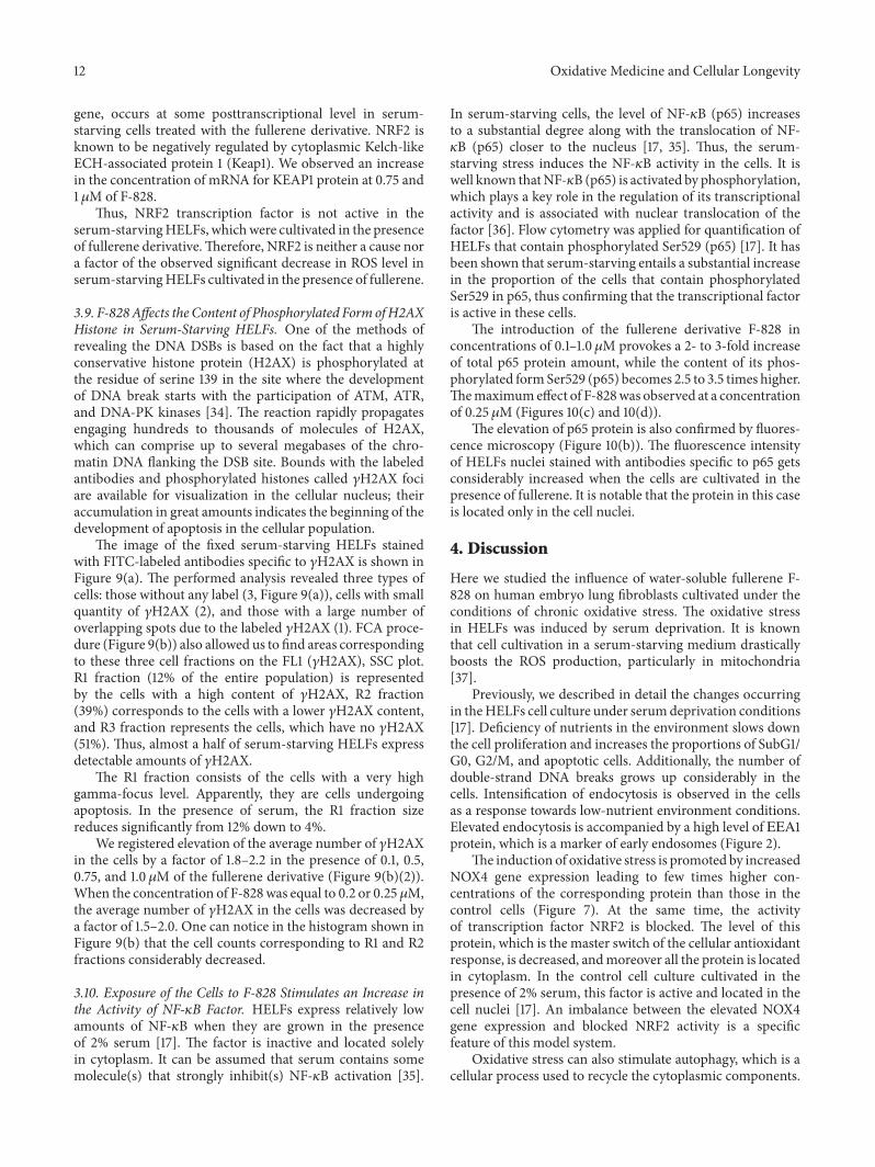
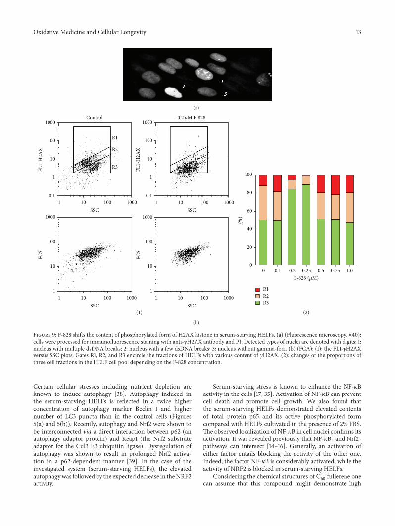
3.9. F-828Affects the Content of Phosphorylated FormofH2AXHistone in Serum-Starving HELFs. One of the methods ofrevealing the DNA DSBs is based on the fact that a highlyconservative histone protein (H2AX) is phosphorylated atthe residue of serine 139 in the site where the developmentof DNA break starts with the participation of ATM, ATR,and DNA-PK kinases [34]. The reaction rapidly propagatesengaging hundreds to thousands of molecules of H2AX,which can comprise up to several megabases of the chro-matin DNA flanking the DSB site. Bounds with the labeledantibodies and phosphorylated histones called 𝛾H2AX fociare available for visualization in the cellular nucleus; theiraccumulation in great amounts indicates the beginning of thedevelopment of apoptosis in the cellular population.
The image of the fixed serum-starving HELFs stainedwith FITC-labeled antibodies specific to 𝛾H2AX is shown inFigure 9(a). The performed analysis revealed three types ofcells: those without any label (3, Figure 9(a)), cells with smallquantity of 𝛾H2AX (2), and those with a large number ofoverlapping spots due to the labeled 𝛾H2AX (1). FCA proce-dure (Figure 9(b)) also allowed us to find areas correspondingto these three cell fractions on the FL1 (𝛾H2AX), SSC plot.R1 fraction (12% of the entire population) is representedby the cells with a high content of 𝛾H2AX, R2 fraction(39%) corresponds to the cells with a lower 𝛾H2AX content,and R3 fraction represents the cells, which have no 𝛾H2AX(51%). Thus, almost a half of serum-starving HELFs expressdetectable amounts of 𝛾H2AX.
The R1 fraction consists of the cells with a very highgamma-focus level. Apparently, they are cells undergoingapoptosis. In the presence of serum, the R1 fraction sizereduces significantly from 12% down to 4%.
We registered elevation of the average number of 𝛾H2AXin the cells by a factor of 1.8–2.2 in the presence of 0.1, 0.5,0.75, and 1.0 𝜇M of the fullerene derivative (Figure 9(b)(2)).When the concentration of F-828 was equal to 0.2 or 0.25 𝜇M,the average number of 𝛾H2AX in the cells was decreased bya factor of 1.5–2.0. One can notice in the histogram shown inFigure 9(b) that the cell counts corresponding to R1 and R2fractions considerably decreased.
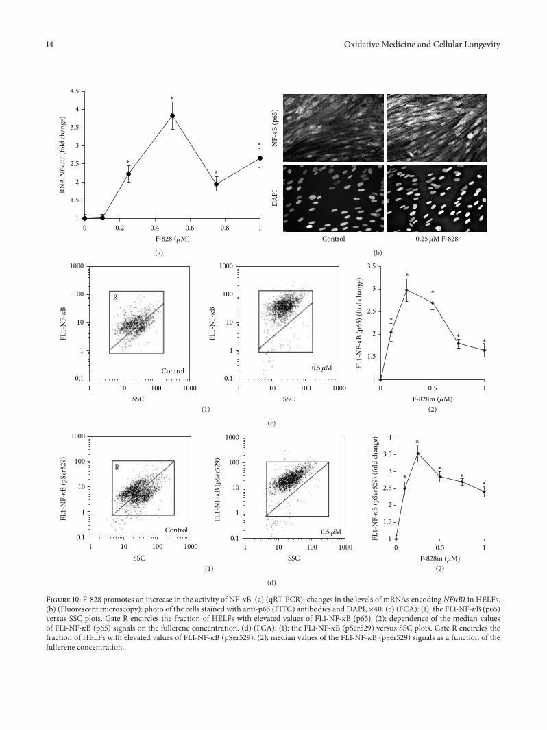
3.10. Exposure of the Cells to F-828 Stimulates an Increase inthe Activity of NF-𝜅B Factor. HELFs express relatively lowamounts of NF-𝜅B when they are grown in the presenceof 2% serum [17]. The factor is inactive and located solelyin cytoplasm. It can be assumed that serum contains somemolecule(s) that strongly inhibit(s) NF-𝜅B activation [35].
In serum-starving cells, the level of NF-𝜅B (p65) increasesto a substantial degree along with the translocation of NF-𝜅B (p65) closer to the nucleus [17, 35]. Thus, the serum-starving stress induces the NF-𝜅B activity in the cells. It iswell known thatNF-𝜅B (p65) is activated by phosphorylation,which plays a key role in the regulation of its transcriptionalactivity and is associated with nuclear translocation of thefactor [36]. Flow cytometry was applied for quantification ofHELFs that contain phosphorylated Ser529 (p65) [17]. It hasbeen shown that serum-starving entails a substantial increasein the proportion of the cells that contain phosphorylatedSer529 in p65, thus confirming that the transcriptional factoris active in these cells.
The introduction of the fullerene derivative F-828 inconcentrations of 0.1–1.0𝜇M provokes a 2- to 3-fold increaseof total p65 protein amount, while the content of its phos-phorylated formSer529 (p65) becomes 2.5 to 3.5 times higher.Themaximumeffect of F-828was observed at a concentrationof 0.25𝜇M (Figures 10(c) and 10(d)).
The elevation of a65 protein is also confirmed by fluores-cence microscopy (Figure 10(b)). The fluorescence intensityof HELFs nuclei stained with antibodies specific to a65 getsconsiderably increased when the cells are cultivated in thepresence of fullerene. It is notable that the protein in this caseis located only in the cell nuclei.
4. Discussion
Here we studied the influence of water-soluble fullerene F-828 on human embryo lung fibroblasts cultivated under theconditions of chronic oxidative stress. The oxidative stressin HELFs was induced by serum deprivation. It is knownthat cell cultivation in a serum-starving medium drasticallyboosts the ROS production, particularly in mitochondria[37].
Previously, we described in detail the changes occurringin theHELFs cell culture under serumdeprivation conditions[17]. Deficiency of nutrients in the environment slows downthe cell proliferation and increases the proportions of SubG1/G0, G2/M, and apoptotic cells. Additionally, the number ofdouble-strand DNA breaks grows up considerably in thecells. Intensification of endocytosis is observed in the cellsas a response towards low-nutrient environment conditions.Elevated endocytosis is accompanied by a high level of @@01protein, which is a marker of early endosomes (Figure 2).
The induction of oxidative stress is promoted by increasedNOX4 gene expression leading to few times higher con-centrations of the corresponding protein than those in thecontrol cells (Figure 7). At the same time, the activityof transcription factor NRF2 is blocked. The level of thisprotein, which is the master switch of the cellular antioxidantresponse, is decreased, andmoreover all the protein is locatedin cytoplasm. In the control cell culture cultivated in thepresence of 2% serum, this factor is active and located in thecell nuclei [17]. An imbalance between the elevated NOX4gene expression and blocked NRF2 activity is a specificfeature of this model system.
Oxidative stress can also stimulate autophagy, which is acellular process used to recycle the cytoplasmic components.
Oxidative Medicine and Cellular Longevity 13
(a)
(1) (2)
0
20
40
60
80
100
R1R2R3
(%)
F-828 (𝜇M)
0 0.1 0.2 0.25 0.5 0.75 1.0
FCS
FCS
R1
R2
R3
0.2 𝜇M F-828Control1000
100
10
1
0.1
1000
100
10
1
1000
100
10
1
1000
100
10
1
0.1
1000100101
SSC
10001000
1000
100101
SSC100101
SSC
100101
SSC
FL1-
H2A
X
FL1-
H2A
X
(b)
Figure 9: F-828 shifts the content of phosphorylated form of H2AX histone in serum-starving HELFs. (a) (Fluorescence microscopy, ×40):cells were processed for immunofluorescence staining with anti-𝛾H2AX antibody and PI. Detected types of nuclei are denoted with digits: 1:nucleus with multiple dsDNA breaks; 2: nucleus with a few dsDNA breaks; 3: nucleus without gamma-foci. (b) (FCA): (1): the FL1-𝛾H2AXversus SSC plots. Gates R1, R2, and R3 encircle the fractions of HELFs with various content of 𝛾H2AX. (2): changes of the proportions ofthree cell fractions in the HELF cell pool depending on the F-828 concentration.
Certain cellular stresses including nutrient depletion areknown to induce autophagy [38]. Autophagy induced inthe serum-starving HELFs is reflected in a twice higherconcentration of autophagy marker Beclin 1 and highernumber of LC3 puncta than in the control cells (Figures5(a) and 5(b)). Recently, autophagy and Nrf2 were shown tobe interconnected via a direct interaction between p62 (anautophagy adaptor protein) and Keap1 (the Nrf2 substrateadaptor for the Cul3 E3 ubiquitin ligase). Dysregulation ofautophagy was shown to result in prolonged Nrf2 activa-tion in a p62-dependent manner [39]. In the case of theinvestigated system (serum-starving HELFs), the elevatedautophagywas followed by the expected decrease in theNRF2activity.
Serum-starving stress is known to enhance the NF-𝜅Bactivity in the cells [17, 35]. Activation of NF-𝜅B can preventcell death and promote cell growth. We also found thatthe serum-starving HELFs demonstrated elevated contentsof total protein a65 and its active phosphorylated formcompared with HELFs cultivated in the presence of 2% FBS.The observed localization of NF-𝜅B in cell nuclei confirms itsactivation. It was revealed previously that NF-𝜅B- and Nrf2-pathways can intersect [14–16]. Generally, an activation ofeither factor entails blocking the activity of the other one.Indeed, the factor NF-𝜅B is considerably activated, while theactivity of NRF2 is blocked in serum-starving HELFs.
Considering the chemical structures of b60fullerene one
can assume that this compound might demonstrate high
14 Oxidative Medicine and Cellular Longevity
1
1.5
2
2.5
3
3.5
4
4.5
0 0.2 0.4 0.6 0.8 1
RNA
NF𝜅
B1 (f
old
chan
ge)
F-828 (𝜇M)
∗
∗
∗
∗
(a)
NF-𝜅
B (p
65)
DA
PI
Control 0.25 𝜇M F-828
(b)
SSC
1000
100
10
1
0.1
1000100101
SSC
0.5 𝜇M
FL1-
NF-𝜅
B
(1)
Control
1000
100
10
1
0.1
1000100101
SSC
R
FL1-
NF-𝜅
B
(2)
1
1.5
2
2.5
3
3.5
0 0.5 1
FL1-
NF-𝜅
B (p
65) (
fold
chan
ge)
F-828m (𝜇M)
∗
∗
∗
∗∗
(c)
FL1-
NF-𝜅
B (p
Ser5
29)
1000
100
10
1
0.1
1000100101
SSC
0.5 𝜇M
(1)
FL1-
NF-𝜅
B (p
Ser5
29)
Control
1
1000
100
10
0.1
1000100101
SSC
R
(2)
1
1.5
2
2.5
3
3.5
4
0 0.5 1
FL1-
NF-𝜅
B (p
Ser5
29) (
fold
chan
ge)
F-828m (𝜇M)
∗∗
∗
∗∗
(d)
Figure 10: F-828 promotes an increase in the activity of NF-𝜅B. (a) (qRT-PCR): changes in the levels of mRNAs encoding NF𝜅B1 in HELFs.(b) (Fluorescent microscopy): photo of the cells stained with anti-p65 (FITC) antibodies and DAPI, ×40. (c) (FCA): (1): the FL1-NF-𝜅B (p65)versus SSC plots. Gate R encircles the fraction of HELFs with elevated values of FL1-NF-𝜅B (p65). (2): dependence of the median valuesof FL1-NF-𝜅B (p65) signals on the fullerene concentration. (d) (FCA): (1): the FL1-NF-𝜅B (pSer529) versus SSC plots. Gate R encircles thefraction of HELFs with elevated values of FL1-NF-𝜅B (pSer529). (2): median values of the FL1-NF-𝜅B (pSer529) signals as a function of thefullerene concentration.
Oxidative Medicine and Cellular Longevity 15
antioxidant capacity. The ability of the fullerene to bind andneutralize ROS directly can lead to the reduction of the ROSlevel in the serum-starving HELFs and improvement of cellculture’s viability. Indeed, serum-starving HELFs appeared tobe very sensitive to the low fullerene concentrations (0.2 to1.0 𝜇M), which indeed decreased the ROS level remarkably.The most pronounced effects were observed at a F-828concentration above 0.5𝜇M(Figure 6). It is rather remarkablethat the fullerene derivative not only binds ROS directly, butalso affects signaling pathways involved in ROS productionand scavenging. In particular, introduction of F-828 reducedthe level of the NOX4 protein (Figure 7(a)).
The fullerene derivative reduced even to a greater extentthe content of NRF2 factor, which is the master switch ofantioxidant response (Figure 8(a)). These findings are verydistinct from the previous report where a mixture of water-soluble fullerenols was shown to increase the activity ofNRF2 factor [9]. It was assumed that C
60(OH)24
attenuatesoxidative stress-induced apoptosis via augmentation of Nrf2-regulated cellular antioxidant capacity [9]. We have alsoshown that a decrease in the NRF2 activity is accompaniedby reduced autophagy as concluded from the evolution of theBeclin 1 and LC3 puncta content in the cells. This is ratherunexpectable result, which warrants further investigations. Itwas shown previously that suppression of autophagy underthe stress conditions is accompanied by an activation ofNRF2-signaling pathway [39, 40].
The observed fullerene-induced decrease of ROS levelin serum-starving HELFs considerably enhances the cell’sviability. For instance, a 1.5-fold increase in the cell count wasobservedwhen F-828was introduced in the culture of serum-starving HELFs in concentrations of 0.2–0.5 𝜇M. This effectis caused by both enhancement of proliferative activity of thecells and lowering the level of the cell death. An exposureto F-828 at concentrations of 0.2 and 0.25𝜇M showed theminimum double-strand DNA break occurrence and thelowest cell mortality. It is notable that these positive effectswere less pronounced at all the other concentrations of thefullerene derivative F-828 we used in the tests. Considerableimprovement in the viability and survivability of the serum-starving HELFs in the presence of fullerene F-828 wasaccompanied by even more pronounced effects such as astrong increase in the activity.
It is important to note that elevated survival rate of serum-starving HELFs can be observed at F-828 concentrationsfrom 0.2 up to 0.5 𝜇M. At these fullerene concentrations,the ROS level found in serum-starving HELFs decreaseddown to the level registered in control cells (2% FBS). Moreeffective ROS scavenging at a concentration of F-828 higherthan 0.5 𝜇M resulted in an increased cell death rate incomparison with the rate found in serum-starving HELFswithout fullerene. A decrease in ROS level is accompaniedby a decrease in the level of NF-𝜅B factor and an increase inthe number of DSBs and in the dead cell count. The findingssuggest that even in oxidative stress conditions surplus ROSelimination may be associated with a decrease in the cellsurvival rate. The harmful effect could be due to a different,ROS-unrelated mechanism(s). This interesting fact warrantsfurther investigation.
Additional Points
Highlights
(i) The influence of a water-soluble [60] fullerene deriva-tive (F-828) used in concentrations of 0.1 𝜇M to1 𝜇M on serum-starving human embryo lung diploidfibroblasts (HELFs) was studied.
(ii) Within a range of concentrations between 0.2 𝜇Mand1 𝜇M F-828 induced a decrease in the ROS level inHELFs.
(iii) In concentrations of 0.2–0.25𝜇M F-828 enhancedproliferation of HELFs and decreased the DSB rateand apoptosis level.
(iv) F-828 strongly stimulated NF-𝜅B activity in serum-starving HELFs.
(v) F-828 repressed NRF2 activity and NOX4 expressionin serum-starving HELFs.
(vi) In concentrations higher than 0.5 𝜇M F-828 inducedexcessive ROS scavenging that resulted in anincreased cell death rate as compared to the rate inserum-starving (stressed) HELFs without fullerene.
Disclosure
The authors approved the submission.
Competing Interests
The authors do not have any competing interests.
Acknowledgments
This work was supported by Russian Foundation for BasicResearch (Grant no. 15-29-01200 OFI-M).
References
[1] R. Bakry, R. M. Vallant, M. Najam-ul-Haq et al., “Medicinalapplications of fullerenes,” International Journal ofNanomedicine, vol. 2, no. 4, pp. 639–649, 2007.
[2] R. Qiao, A. P. Roberts, A. S. Mount, S. J. Klaine, and P. C. Ke,“Translocation of C
60and its derivatives across a lipid bilayer,”
Nano Letters, vol. 7, no. 3, pp. 614–619, 2007.[3] Z. Markovic and V. Trajkovic, “Biomedical potential of the
reactive oxygen species generation and quenching by fullerenes(C60),” Biomaterials, vol. 29, no. 26, pp. 3561–3573, 2008.
[4] F. Lao, L. Chen, W. Li et al., “Fullerene nanoparticles selectivelyenter oxidation-damaged cerebral microvessel endothelial cellsinhibit JNK-related apoptosis,” ACS Nano, vol. 3, no. 11, pp.3358–3368, 2009.
[5] S. S. Ali, J. I. Hardt, and L. L. Dugan, “SOD Activity of carboxy-fullerenes predicts their neuroprotective efficacy: a structure-activity study,” Nanomedicine, vol. 4, no. 4, pp. 283–294, 2008.
[6] F. Lao, W. Li, D. Han et al., “Fullerene derivatives protectendothelial cells against NO-induced damage,”Nanotechnology,vol. 20, no. 22, Article ID 225103, 2009.
16 Oxidative Medicine and Cellular Longevity
[7] M. S. Misirkic, B. M. Todorovic-Markovic, L. M. Vucicevicet al., “The protection of cells from nitric oxide-mediatedapoptotic death by mechanochemically synthesized fullerene(C60) nanoparticles,” Biomaterials, vol. 30, no. 12, pp. 2319–2328,
2009.[8] J.-J. Yin, F. Lao, P. P. Fu et al., “The scavenging of reactive oxygen
species and the potential for cell protection by functionalizedfullerene materials,” Biomaterials, vol. 30, no. 4, pp. 611–621,2009.
[9] S. Ye,M. Chen, Y. Jiang et al., “Polyhydroxylated fullerene atten-uates oxidative stress-induced apoptosis via a fortifying Nrf2-regulated cellular antioxidant defence system,” InternationalJournal of Nanomedicine, vol. 9, no. 1, pp. 2073–2087, 2014.
[10] T. W. Kensler, N. Wakabayashi, and S. Biswal, “Cell survivalresponses to environmental stresses via the Keap1-Nrf2-AREpathway,” Annual Review of Pharmacology and Toxicology, vol.47, pp. 89–116, 2007.
[11] S. Ye, T. Zhou, K. Cheng et al., “Carboxylic acid fullerene (C60)
derivatives attenuated neuroinflammatory responses by modu-latingmitochondrial dynamics,”Nanoscale Research Letters, vol.10, article 246, 2015.
[12] G. Gloire, S. Legrand-Poels, and J. Piette, “NF-𝜅B activationby reactive oxygen species: fifteen years later,” BiochemicalPharmacology, vol. 72, no. 11, pp. 1493–1505, 2006.
[13] M. S. Hayden and S. Ghosh, “NF-𝜅B, the first quarter-century:remarkable progress and outstanding questions,” Genes &Development, vol. 26, no. 3, pp. 203–234, 2012.
[14] L. M. Pedruzzi, M. B. Stockler-Pinto, M. Leite Jr., and D. Mafra,“Nrf2-keap1 system versus NF-𝜅B: the good and the evil inchronic kidney disease?” Biochimie, vol. 94, no. 12, pp. 2461–2466, 2012.
[15] N. Wakabayashi, S. L. Slocum, J. J. Skoko, S. Shin, and T. W.Kensler, “When NRF2 talks, who’s listening?” Antioxidants &Redox Signaling, vol. 13, no. 11, pp. 1649–1663, 2010.
[16] W. Li, T. O. Khor, C. Xu et al., “Activation of Nrf2-antioxidantsignaling attenuates NF𝜅B-inflammatory response and elicitsapoptosis,” Biochemical Pharmacology, vol. 76, no. 11, pp. 1485–1489, 2008.
[17] S. V. Kostyuk, V. J. Tabakov, V. V. Chestkov et al., “OxidizedDNA induces an adaptive response in human fibroblasts,”Mutation Research/Fundamental and Molecular Mechanisms ofMutagenesis, vol. 747-748, pp. 6–18, 2013.
[18] A. B. Kornev, E. A. Khakina, S. I. Troyanov et al., “Facilepreparation of amine and amino acid adducts of [60]fullereneusing chlorofullerene C
60Cl6as a precursor,” Chemical Commu-
nications, vol. 48, no. 44, pp. 5461–5463, 2012.[19] O. A. Troshina, P. A. Troshin, A. S. Peregudov, V. I. Kozlovskiy,
J. Balzarini, and R. N. Lyubovskaya, “Chlorofullerene C60C𝑙6:
a precursor for straightforward preparation of highly water-soluble polycarboxylic fullerene derivatives active against HIV,”Organic and Biomolecular Chemistry, vol. 5, no. 17, pp. 2783–2791, 2007.
[20] R. I. A. Podchernyaeva, O. V. Baklanova, L. A. Glushchenko etal., “Influenza virus reproduction in theMDCK cells adapted togrowth in serum-free Hybris-2 medium,” Voprosy Virusologii,vol. 55, no. 5, pp. 47–49, 2010.
[21] S. M. Santos, A. M. Dinis, F. Peixoto, L. Ferreira, A. S. Jurado,and R. A. Videira, “Interaction of fullerene nanoparticles withbiomembranes: from the partition in lipidmembranes to effectsonmitochondrial bioenergetics,”Toxicological Sciences, vol. 138,no. 1, Article ID kft327, pp. 117–129, 2014.
[22] J. J. Dumas, E. Merithew, E. Sudharshan et al., “Multivalentendosome targeting by homodimeric EEA1,”Molecular Cell, vol.8, no. 5, pp. 947–958, 2001.
[23] P. Guillaud, S. du Manoir, and D. Seigneurin, “Quantificationand topographical description of Ki-67 antibody labelling dur-ing the cell cycle of normal fibroblastic (MRC-5) andmammarytumour cell lines (MCF-7),”Analytical Cellular Pathology, vol. 1,no. 1, pp. 25–39, 1989.
[24] S. N. Naryzhny, “Proliferating cell nuclear antigen: a proteomicsview,” Cellular and Molecular Life Sciences, vol. 65, no. 23, pp.3789–3808, 2008.
[25] L. Li, J. Tan, Y.Miao, P. Lei, andQ. Zhang, “ROS and autophagy:interactions and molecular regulatory mechanisms,” Cellularand Molecular Neurobiology, vol. 35, no. 5, pp. 615–621, 2015.
[26] D. J. Klionsky, K. Abdelmohsen, A. Abe et al., “Guidelines forthe use and interpretation of assays for monitoring autophagy(3rd edition),” Autophagy, vol. 12, no. 1, pp. 1–222, 2016.
[27] A. C. Cave, A. C. Brewer, A. Narayanapanicker et al., “NADPHoxidases in cardiovascular health and disease,”Antioxidants andRedox Signaling, vol. 8, no. 5-6, pp. 691–728, 2006.
[28] L. Hecker, N. J. Logsdon, D. Kurundkar et al., “Reversal ofpersistent fibrosis in aging by targetingNox4-Nrf2 redox imbal-ance,” Science Translational Medicine, vol. 6, no. 231, Article ID231ra47, 2014.
[29] T. Nguyen, P. J. Sherratt, P. Nioi, C. S. Yang, and C. B.Pickett, “Nrf2 controls constitutive and inducible expressionof ARE-driven genes through a dynamic pathway involvingnucleocytoplasmic shuttling by Keap1,” Journal of BiologicalChemistry, vol. 280, no. 37, pp. 32485–32492, 2005.
[30] H. Dreger, K. Westphal, A. Weller et al., “Nrf2-dependentupregulation of antioxidative enzymes: a novel pathway for pro-teasome inhibitor-mediated cardioprotection,” CardiovascularResearch, vol. 83, no. 2, pp. 354–361, 2009.
[31] S. Kovac, P. R. Angelova, K. M. Holmström, Y. Zhang, A. T.Dinkova-Kostova, and A. Y. Abramov, “Nrf2 regulates ROSproduction by mitochondria and NADPH oxidase,” Biochimicaet Biophysica Acta (BBA)—General Subjects, vol. 1850, no. 4, pp.794–801, 2015.
[32] S. Pendyala, J. Moitra, S. Kalari et al., “Nrf2 regulates hyperoxia-induced Nox4 expression in human lung endothelium: identifi-cation of functional antioxidant response elements on the Nox4promoter,” Free Radical Biology andMedicine, vol. 50, no. 12, pp.1749–1759, 2011.
[33] A. C. Brewer, T. V. A. Murray, M. Arno et al., “Nox4 regulatesNrf2 and glutathione redox in cardiomyocytes in vivo,” FreeRadical Biology and Medicine, vol. 51, no. 1, pp. 205–215, 2011.
[34] M. Löbrich, A. Shibata, A. Beucher et al., “𝛾H2AX foci analysisfor monitoring DNA double-strand break repair: strengths,limitations and optimization,” Cell Cycle, vol. 9, no. 4, pp. 662–669, 2010.
[35] T. Kohno, Y. Kubo, K. Yasui et al., “Serum starvation activatesNF-𝜅B through G protein 𝛽2 subunit-mediated signal,” DNAand Cell Biology, vol. 31, no. 11, pp. 1636–1644, 2012.
[36] D.Wang and A. S. Baldwin Jr., “Activation of nuclear factor-𝜅B-dependent transcription by tumor necrosis factor-𝛼 is mediatedthrough phosphorylation of RelA/p65 on serine 529,” Journal ofBiological Chemistry, vol. 273, no. 45, pp. 29411–29416, 1998.
[37] A. V. Kuznetsov, I. Kehrer, A. V. Kozlov et al., “MitochondrialROS production under cellular stress: comparison of differentdetectionmethods,”Analytical andBioanalytical Chemistry, vol.400, no. 8, pp. 2383–2390, 2011.
Oxidative Medicine and Cellular Longevity 17
[38] C. Liu, E. P. DeRoo, C. Stecyk,M.Wolsey, M. Szuchnicki, and E.G.Hagos, “Impaired autophagy inmouse embryonic fibroblastsnull for Krüppel-like Factor 4 promotes DNA damage andincreases apoptosis upon serum starvation,” Molecular Cancer,vol. 14, no. 1, article 101, 2015.
[39] T. M. Stępkowski and M. K. Kruszewski, “Molecular cross-talkbetween the NRF2/KEAP1 signaling pathway, autophagy, andapoptosis,” Free Radical Biology and Medicine, vol. 50, no. 9, pp.1186–1195, 2011.
[40] N. Chen, L. Wu, H. Yuan, and J. Wang, “ROS/autophagy/Nrf2pathway mediated low-dose radiation induced radio-resistancein human lung adenocarcinoma A549 cell,” International Jour-nal of Biological Sciences, vol. 11, no. 7, pp. 833–844, 2015.
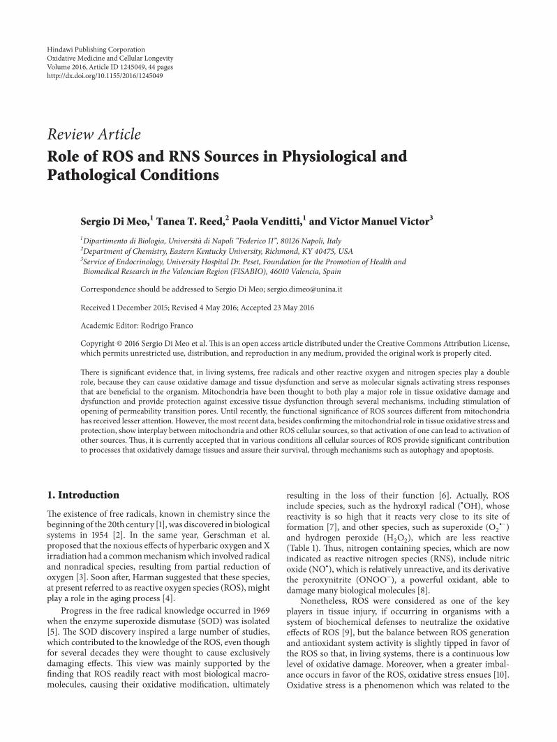
Review ArticleRole of ROS and RNS Sources in Physiological andPathological Conditions
Sergio Di Meo,1 Tanea T. Reed,2 Paola Venditti,1 and Victor Manuel Victor3
1Dipartimento di Biologia, Università di Napoli “Federico II”, 80126 Napoli, Italy2Department of Chemistry, Eastern Kentucky University, Richmond, KY 40475, USA3Service of Endocrinology, University Hospital Dr. Peset, Foundation for the Promotion of Health andBiomedical Research in the Valencian Region (FISABIO), 46010 Valencia, Spain
Correspondence should be addressed to Sergio Di Meo; [email protected]
Received 1 December 2015; Revised 4 May 2016; Accepted 23 May 2016
Academic Editor: Rodrigo Franco
Copyright © 2016 Sergio Di Meo et al. This is an open access article distributed under the Creative Commons Attribution License,which permits unrestricted use, distribution, and reproduction in any medium, provided the original work is properly cited.
There is significant evidence that, in living systems, free radicals and other reactive oxygen and nitrogen species play a doublerole, because they can cause oxidative damage and tissue dysfunction and serve as molecular signals activating stress responsesthat are beneficial to the organism. Mitochondria have been thought to both play a major role in tissue oxidative damage anddysfunction and provide protection against excessive tissue dysfunction through several mechanisms, including stimulation ofopening of permeability transition pores. Until recently, the functional significance of ROS sources different from mitochondriahas received lesser attention. However, themost recent data, besides confirming themitochondrial role in tissue oxidative stress andprotection, show interplay between mitochondria and other ROS cellular sources, so that activation of one can lead to activation ofother sources. Thus, it is currently accepted that in various conditions all cellular sources of ROS provide significant contributionto processes that oxidatively damage tissues and assure their survival, through mechanisms such as autophagy and apoptosis.
1. Introduction
The existence of free radicals, known in chemistry since thebeginning of the 20th century [1], was discovered in biologicalsystems in 1954 [2]. In the same year, Gerschman et al.proposed that the noxious effects of hyperbaric oxygen and Xirradiation had a commonmechanismwhich involved radicaland nonradical species, resulting from partial reduction ofoxygen [3]. Soon after, Harman suggested that these species,at present referred to as reactive oxygen species (ROS), mightplay a role in the aging process [4].
Progress in the free radical knowledge occurred in 1969when the enzyme superoxide dismutase (SOD) was isolated[5]. The SOD discovery inspired a large number of studies,which contributed to the knowledge of the ROS, even thoughfor several decades they were thought to cause exclusivelydamaging effects. This view was mainly supported by thefinding that ROS readily react with most biological macro-molecules, causing their oxidative modification, ultimately
resulting in the loss of their function [6]. Actually, ROSinclude species, such as the hydroxyl radical (∙OH), whosereactivity is so high that it reacts very close to its site offormation [7], and other species, such as superoxide (O
2
∙−)and hydrogen peroxide (H
2
O2
), which are less reactive(Table 1). Thus, nitrogen containing species, which are nowindicated as reactive nitrogen species (RNS), include nitricoxide (NO∙), which is relatively unreactive, and its derivativethe peroxynitrite (ONOO−), a powerful oxidant, able todamage many biological molecules [8].
Nonetheless, ROS were considered as one of the keyplayers in tissue injury, if occurring in organisms with asystem of biochemical defenses to neutralize the oxidativeeffects of ROS [9], but the balance between ROS generationand antioxidant system activity is slightly tipped in favor ofthe ROS so that, in living systems, there is a continuous lowlevel of oxidative damage. Moreover, when a greater imbal-ance occurs in favor of the ROS, oxidative stress ensues [10].Oxidative stress is a phenomenon which was related to the
Hindawi Publishing CorporationOxidative Medicine and Cellular LongevityVolume 2016, Article ID 1245049, 44 pageshttp://dx.doi.org/10.1155/2016/1245049

2 Oxidative Medicine and Cellular Longevity
Table 1: Main reactive oxygen (ROS) and nitrogen (RNS) species.
Radicals
O2
∙− Superoxide anion∙OH HydroxylNO∙ Nitric oxideNO2
∙ Nitrogen dioxide
NonradicalsH2
O2
Hydrogen peroxideHOCl Hypochlorous acid
ONOO− Peroxynitrite
development of many pathological conditions. Pathologieswhere ROS were identified as causal factors include cardio-vascular disease, diabetes, rheumatoid arthritis, cancer, andneurodegenerative disorders [11] and the use of exogenousantioxidants was proposed for their treatment [12].
The finding that ∙OH radical stimulated formation ofcyclic guanosine monophosphate (cGMP) [13] led to anopposing view about the ROS role in biological systems. Sincethen, it became clear that living systems not only adapted to acoexistence with free radicals but also developed methods toturn these toxicants to their own advantage utilizing them incritical physiological processes.This view has been supportedby the observation that, at the cellular level, ROS regulategrowth, apoptosis, and other signaling, while, at the systemlevel, they contribute to complex functions, including bloodpressure regulation, cognitive function, and immune func-tion [14]. It has also been shown that whereas accumulationof oxidative damage results in organism death [15], severallongevity-promoting interventions increase generation ofROS that activate stress responses that are beneficial to theorganism and extend lifespan [16].
Like ROS, RNS play a dual role since they can be eitherharmful or beneficial to living systems. Nitric oxide, earlyidentified as a signaling molecule in blood vessel modulation[17] and now known as a regulator of important physiolog-ical processes [18], can mediate cellular toxicity damagingmetabolic enzymes and generating, by reaction with super-oxide, peroxynitrite [19].
Although the role of ROS andRNS in cellular damage andsignal transduction is well-established, several controversialquestions remain open. At low concentrations, ROS and RNSplay an important role as regulatory mediators in signalingprocesses, whereas, at moderate or high concentrations,they are harmful for living organisms inactivating importantcellular molecules. This suggests that the concentrations ofreactive species determine the shift from their advantageousto detrimental effects, but the concentrations to which thisshift happens are not generally known. Moreover, given thewide range of possible targets and the general reactivity ofcellular oxidants, it is unclear how any specificity in theiropposite actions can be achieved. It has been suggestedthat contributing factors may include cell type, durationof oxidant production, reactive species produced, and thelocalization of their source and their targets [20], but ourinformation about such amatter is still scarce and discordant.For example, it has often been assumed that mitochondriaare the main cellular source of ROS in physiological and
pathological conditions [21], so that these organelles havebeen thought to play a crucial role in several human diseasesand aging [22]. However, in the light of available data itseems that evidence thatmitochondria are themain source ofcellular ROS is lacking [23]. The issue is complicated by theexistence of a strong interaction among the various sourcesof ROS generation in the cell [24, 25], whichmakes it difficultto establish what source of reactive species plays a main rolein different physiological and pathological conditions.
To contribute to the understanding of the role of reactivespecies sources in health and disease, the present review, afterexamining the cellular localization and supposed involve-ment of such sources in tissue dysfunction and protection,examines experimental evidence concerning their harmfuland protective effects on a normal physiological activity, suchas exercise, and on pathologic conditions, such as diabetesand neurodegenerative diseases.
2. Cellular ROS and RNS Sources
In the living organisms, ROS are generated in several cellularsystems localized on the plasma membrane, in the cytosol,in the peroxisomes, and on membranes of mitochondria andendoplasmic reticulum (Figure 1).
2.1. ROS Production in the Cytosol. Several soluble cell com-ponents, including thiols, hydroquinones, catecholamines,and flavins, can contribute to intracellular ROS productionas they are able to undergo redox reactions [26]. Moreover,several cytosolic enzymes produce ROS during their catalyticactivity. Probably the most studied ROS producing enzymeis xanthine oxidase (XO). In healthy tissues, the enzymecatalyzing oxidation of hypoxanthine to xanthine and xan-thine to uric acid is xanthine dehydrogenase (XDH), whichuses NADP+ as an electron acceptor. Conversely, in damagedtissues, by either reversible oxidation of cysteine residuesor irreversible Ca2+-stimulated proteolysis, the enzyme isconverted from the dehydrogenase form into the oxidaseform, which transfers electrons to molecular oxygen produc-ing the superoxide radical during xanthine or hypoxanthineoxidation [27].
2.2. ROS Production by Mitochondria. In aerobic cells, mito-chondria are necessary for numerous fundamental functions,including respiration and oxidative energy production, regu-lation of the intracellular calcium concentration, and controlof the fatty acid 𝛽-oxidation.
For a long time, mitochondria have only been consideredfor their role in energy production.They utilize about 95% ofthe oxygen assumed from aerobic animals to obtain energyby oxidizing substances contained in food by transfer ofelectrons to electron carriers such as NAD+, FMN, andFAD.The reduced forms of these molecules, in turn, transferelectrons to the components of the respiratory chain andfinally to oxygen in a process which happens in a series ofsubsequent steps, so that the energy, resulting by the fall in theelectron potential energy, is gradually released. In the process,several redox centers, in great part organized in four protein
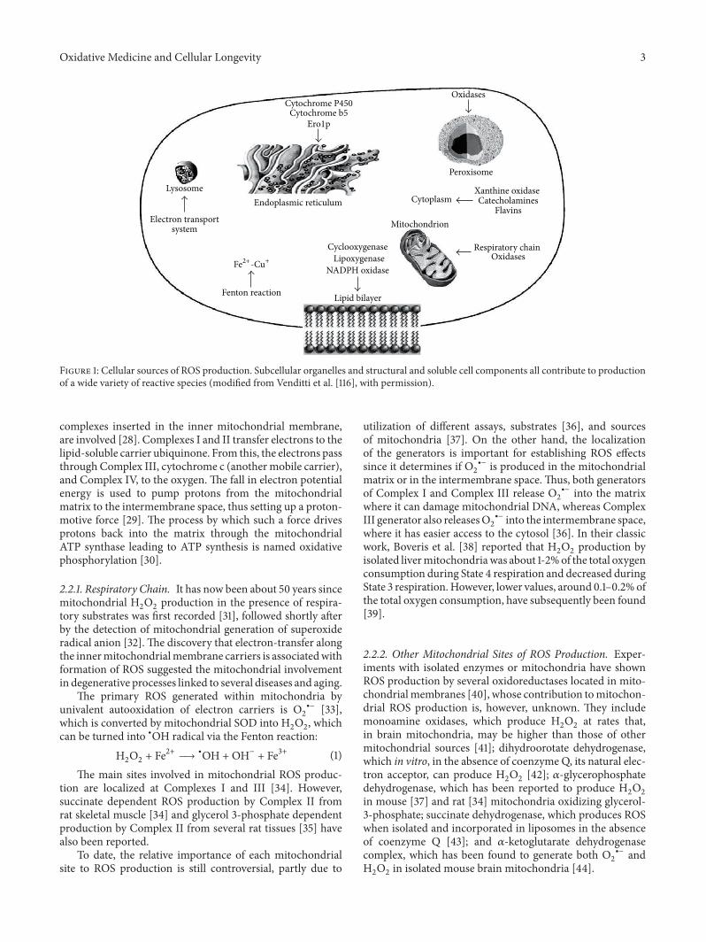
Oxidative Medicine and Cellular Longevity 3
Cytochrome P450
Cytoplasm
Peroxisome
Oxidases
Mitochondrion
Respiratory chainOxidases
CyclooxygenaseLipoxygenase
NADPH oxidase
Lipid bilayer
Xanthine oxidaseCatecholamines
Flavins
Cytochrome b5Ero1p
Endoplasmic reticulum
Electron transportsystem
Fenton reaction
Lysosome
Fe2+-Cu+
Figure 1: Cellular sources of ROS production. Subcellular organelles and structural and soluble cell components all contribute to productionof a wide variety of reactive species (modified from Venditti et al. [116], with permission).
complexes inserted in the inner mitochondrial membrane,are involved [28]. Complexes I and II transfer electrons to thelipid-soluble carrier ubiquinone. From this, the electrons passthrough Complex III, cytochrome c (another mobile carrier),and Complex IV, to the oxygen. The fall in electron potentialenergy is used to pump protons from the mitochondrialmatrix to the intermembrane space, thus setting up a proton-motive force [29]. The process by which such a force drivesprotons back into the matrix through the mitochondrialATP synthase leading to ATP synthesis is named oxidativephosphorylation [30].
2.2.1. Respiratory Chain. It has now been about 50 years sincemitochondrial H
2
O2
production in the presence of respira-tory substrates was first recorded [31], followed shortly afterby the detection of mitochondrial generation of superoxideradical anion [32]. The discovery that electron-transfer alongthe innermitochondrialmembrane carriers is associatedwithformation of ROS suggested the mitochondrial involvementin degenerative processes linked to several diseases and aging.
The primary ROS generated within mitochondria byunivalent autooxidation of electron carriers is O
2
∙− [33],which is converted by mitochondrial SOD into H
2
O2
, whichcan be turned into ∙OH radical via the Fenton reaction:
H2
O2
+ Fe2+ → ∙OH +OH− + Fe3+ (1)The main sites involved in mitochondrial ROS produc-
tion are localized at Complexes I and III [34]. However,succinate dependent ROS production by Complex II fromrat skeletal muscle [34] and glycerol 3-phosphate dependentproduction by Complex II from several rat tissues [35] havealso been reported.
To date, the relative importance of each mitochondrialsite to ROS production is still controversial, partly due to
utilization of different assays, substrates [36], and sourcesof mitochondria [37]. On the other hand, the localizationof the generators is important for establishing ROS effectssince it determines if O
2
∙− is produced in the mitochondrialmatrix or in the intermembrane space.Thus, both generatorsof Complex I and Complex III release O
2
∙− into the matrixwhere it can damage mitochondrial DNA, whereas ComplexIII generator also releasesO
2
∙− into the intermembrane space,where it has easier access to the cytosol [36]. In their classicwork, Boveris et al. [38] reported that H
2
O2
production byisolated livermitochondriawas about 1-2%of the total oxygenconsumption during State 4 respiration and decreased duringState 3 respiration.However, lower values, around 0.1–0.2% ofthe total oxygen consumption, have subsequently been found[39].
2.2.2. Other Mitochondrial Sites of ROS Production. Exper-iments with isolated enzymes or mitochondria have shownROS production by several oxidoreductases located in mito-chondrialmembranes [40], whose contribution tomitochon-drial ROS production is, however, unknown. They includemonoamine oxidases, which produce H
2
O2
at rates that,in brain mitochondria, may be higher than those of othermitochondrial sources [41]; dihydroorotate dehydrogenase,which in vitro, in the absence of coenzyme Q, its natural elec-tron acceptor, can produce H
2
O2
[42]; 𝛼-glycerophosphatedehydrogenase, which has been reported to produce H
2
O2
in mouse [37] and rat [34] mitochondria oxidizing glycerol-3-phosphate; succinate dehydrogenase, which produces ROSwhen isolated and incorporated in liposomes in the absenceof coenzyme Q [43]; and 𝛼-ketoglutarate dehydrogenasecomplex, which has been found to generate both O
2
∙− andH2
O2
in isolated mouse brain mitochondria [44].
4 Oxidative Medicine and Cellular Longevity
2.3. ROS Production by Peroxisomes. Although peroxisomeshave long been known as organelles involved in cellularmetabolismofH
2
O2
[45], it is now clear that they are involvedin several metabolic pathways [46]. Important functions per-formed by peroxisomes include fatty acid𝛽- and 𝛼-oxidation,amino acid and glyoxylate metabolism, and synthesis oflipidic compounds [47], and most enzymes catalyzing theseprocesses produce ROS during their activity [48].
Early studies indicated that about 35% of the peroxideformed in rat liver is derived from peroxisomal oxidases [38].Moreover, the observation that about 20–60% of the H
2
O2
generated inside peroxisomes diffused to the surroundingmedium demonstrated that H
2
O2
can readily cross theperoxisomal membrane. The peroxide diffuses through thePxmp2 channel permeable to small solutes [49], even thoughH2
O2
generated by urate oxidase localized in the peroxisomecore can be directly released in the cytosol through crystalloidcore tubules [50]. In any case, it is apparent that, despite highcontent of catalase (CAT), peroxisomes are unable to preventthe H
2
O2
release.Peroxisomes also contain xanthine oxidase [51] and the
inducible form of nitric oxide synthase (see below) [52],which produce O
2
∙− and NO∙, respectively. Because suchspecies react rapidly forming ONOO and H
2
O2
gives riseto ∙OH radicals via Fenton reaction, peroxisomes are apotential source of such highly reactive species. Because, ifuncontrolled, ROS andRNS are very damaging, peroxisomes,in addition to catalase, contain other antioxidant enzymes[50]. However, in the light of their capacity to producemembrane permeant reactive species, such as H
2
O2
andNO∙, it is highly likely that, under some physiological orpathological conditions, peroxisomes may act as a source ofH2
O2
and NO∙ in living cells [50].
2.4. ROS Production by Endoplasmic Reticulum. The endo-plasmic reticulum (ER) is involved in multiple functions,such as synthesis, folding, and transport of Golgi, lysosomal,secretory, and cell-surface proteins [53], calcium storage [54],lipid metabolism, and, in some cell types, drug detoxification[55].
Smooth endoplasmic reticulum presents a chain of elec-tron transport, constituted by two systems devoted to xeno-biotic metabolism and introduction of double bonds in fattyacids, which are also able to produce ROS. Another micro-somal system, which shares this ability, provides oxidativeprotein folding.
2.4.1. Xenobiotic Metabolism. Themetabolism of xenobioticsgenerally occurs in two phases. Phase I reactions introducea polar group in a lipophilic substrate (AH) using O
2
and areducing agent (RH
2
):
AH +O2
+ RH2
→ AOH + R +H2
O (2)
In the reaction, known as a monooxygenase reaction, it isinvolved in a system constituted by a flavoprotein (NADPH-cytochrome P450 reductase) and cytochromes known collec-tively as cytochromes P450 (CYPs).
The Phase II reactions are conjugation reactions in whichan endogenous molecule is added to the Phase I reactionproduct or sometimes directly to the xenobiotic.
In whole, the process detoxifies xenobiotics convertingthem into species that are more water soluble and easier toexcrete in urine or can be conjugated with substances whichmake their urinary or biliary excretion further easier.
The membranes of the endoplasmic reticulum systemwere recognized as a source of H
2
O2
in 1957 by Gillette et al.[56] that assumed that the NADPH-cytochrome c reductasemight be the microsomal H
2
O2
generator. However, it wassubsequently reported that O
2
∙− [57] and H2
O2
[58] could beformed by decay of two intermediates of the catalytic cycle.
Themicrosomal cytochrome P450-dependent monooxy-genase system is one of the major producers of ROS in theliver cell [59]. Indeed, estimates performed using NADPHas substrate indicated that microsomes contribute to H
2
O2
production in rat liver by 45% [38].
2.4.2. Unsaturated Fatty Acid Production. Inmost organisms,unsaturated fatty acids are produced by desaturases, whichconvert a single bond between two carbon atoms in a fattyacyl chain into a double bond [60]. In the ER system,which allows for fatty acid desaturation, cytochrome b5acts as an electron-transfer component, with two possiblemodes of action. First, desaturation can be carried out bya multienzyme system, composed of desaturase, NADHcytochromeb5 reductase, and cytochromeb5. In the reaction,cytochrome b5 transfers electrons by lateral diffusion, fromNADH cytochrome b5 reductase to the desaturase whichintroduces carbon-carbon double bonds into fatty acids usingone molecule of O
2
and forming two molecules of H2
O [61].Secondly, many desaturases are modular proteins that arecomposed of desaturase and cytochrome b5 modules [62].The fusion of the desaturase and cytochrome b5 domainsmakes the NADH cytochrome b5 reductase able to directlytransfer electrons to the catalytic site of the cytochromeb5 fusion desaturases via the cytochrome b5-like domainwithout the requirement of an independent cytochromeb5 [63]. This can increase the rate of electron-transfer bypresenting a correctly oriented heme group with respect tothe dioxo-iron cluster, eliminating the need for diffusion andreorientation of the reduced cytochrome b5 [63].
The two electron transport systems do not act indepen-dently of each other and cytochrome b5 may play a rolein the NADPH-dependent oxidation of xenobiotics. Indeed,cytochrome b5 exhibits a positive action on cytochrome P450monooxygenase reaction, which is due to the transfer of thesecond of the two electrons necessary for molecular oxygenactivation to cytochrome P450 [64]. This transfer makes thecatalysis faster and reduces the time for formation of sideproducts, such as H
2
O2
and O2
∙− [64]. Recently, it has alsobeen demonstrated that NADH cytochrome b5 reductasecan leak electrons to O
2
to make O2
∙− and this can be anadditional source of O
2
∙− in vivo [65].
2.4.3. Protein Folding. Most proteins synthesized in ER arestabilized by formation of intramolecular disulfide bonds,process that requires oxidation of free sulphydryl groups.
Oxidative Medicine and Cellular Longevity 5
The feasibility of protein oxidative folding in intraluminal ERmilieu is ensured by an oxidized (GSSG) to reduced (GSH)glutathione ratio higher than that within the cytosol [66].Theformation of disulfide bonds in proteins is driven by proteindisulfide isomerase (PDI), a member of the thioredoxinfamily, and endoplasmic reticulum-resident protein (Ero1p),which is tightly associated with FAD moiety [66]. Ero1pfunctions as an oxidase for PDI: oxidizing equivalents flowfrom Ero1p to substrate proteins via PDI, through directdithiol-disulfide exchange between PDI and Ero1p [67].
The process of oxidative protein folding uses molecularoxygen as the source of oxidizing equivalents. Indeed, Ero1ptransfers electrons from PDI to molecular oxygen by aFAD-dependent reaction, resulting in ER protein folding-induced oxidative stress [68]. By theoretical calculation, itwas estimated that protein thiol oxidation via PDI and Ero1could account for up to 25% of cellular ROS produced duringprotein synthesis [69].
2.5. ROSProduction by PlasmaMembrane. Theplasmamem-brane is involved in several cellular processes such as celladhesion, ion conductivity, and cell signaling. It is also akey site of free radical reactions because it is generallyexposed to an oxidizing environment. ROS, which in tissuescould be generated from dysfunctional cells [70], causeoxidative damage of membrane components unless efficientantioxidant systems are operative.The increase in membranepermeability, caused by oxidation of lipids or structurallyimportant proteins, can result in a decrease in transmem-brane ion gradients, loss of secretory functions, and inhibi-tion of cellular metabolic processes.
Free radicals can be produced during the conversionof arachidonic acid into products, such as prostaglandins,thromboxanes, and leukotrienes, by membrane associatedenzymes such as lipoxygenase and cyclooxygenase [71]. Suchenzymes metabolize arachidonic acid released from mem-brane phospholipids via phospholipase A2 activity and gen-erate ROS as by-products during arachidonic acid oxidation.
However, the main source of ROS is represented byO2
∙− production by the membrane-bound enzyme NADPHoxidases. O
2
∙− production by the phagocyte enzyme is a well-known phenomenon, which helps to kill bacterial intruders[72].
The phagocyte NADPH oxidase is composed of twomembrane proteins gp91phox (cytochromeb558 heavy chain,later designated as NOX2, which is the catalytic subunit ofthe enzyme) and p22phox, three cytosolic proteins p67phox,p47phox, and p40phox, and a small GTP binding proteinRac [73]. In resting cells, the enzyme is dormant, and itscomponents are distributed between the cytosol and plasmamembrane. Bacterial infection induces translocation of thecytosolic components to the phagosome membrane wherethey associate with cytochrome b558 and give rise to thecatalytically active NADPH oxidase [73].
The presence of NOX2 homologs was firstly suggestedby the observation that O
2
∙− is produced in a NADPH-dependent manner in nonphagocytic cells, in which NOX2is not expressed [74]. To date, five NOX isoforms (NOX1,
NOX2, NOX3, NOX4, and NOX5) and two related enzymes,DUOX1 andDUOX2, have been reported andmost, if not all,isoforms were targeted to cellular membranes.
TheNOXproteins constitute the only enzyme family withthe sole function of producing ROS. These proteins havedifferent regulation and specific subcellular localization andgenerate distinct ROS [75]. NOX1, present in smooth musclecells and other vascular cells, NOX2, present in endothelialand phagocytic cells, andNOX3, found in the brain and innerear, generate O
2
∙−. NOX4, constitutively expressed and activein vascular smooth muscle and endothelial cells, is respon-sible for the basal production of H
2
O2
. NOX5, identifiedin human immature lymphatic tissues and endothelial cells,producesH
2
O2
in aCa2+ dependent fashion.TheDUOX1 andDUOX2, originally isolated from the thyroid, produce H
2
O2
that oxidizes iodide during thyroid hormone synthesis.The phagocyte NADPH oxidase, when activated, gener-
ates quantities of O2
∙− and H2
O2
accounting for a significantfraction (10–90%) of total oxygen consumption of neu-trophils, macrophages, and microglia, but the contributionof such and other NADPH oxidases to total cellular ROSproduction in resting or during activation is less clear [23].
It has also been reported that arachidonic acid [76] andits metabolites generated by lipoxygenase [77] and cyclooxy-genase [78] stimulate the generation of ROS by NOXs, thusrevealing the existence of an interconnecting signaling systembetween eicosanoids and NOXs.
2.6. ROS Production by Lysosomes. Although lysosomes areinvolved in several functions, until recently they were consid-ered as pure sites for terminal degradation ofmacromolecules[79].
On rat livermembranes, flavins, ubiquinone, and a b-typecytochrome [80] form a functional electron transport system,starting with the donor NADH and ending to acceptorO2
through the sequence FAD→cytochrome b→ubiquinone[81]. The role of this redox chain is to support proton accu-mulation within lysosomes [81] to maintain an optimal pHfor the acidic hydrolases [79]. The electron transport chainappeared to give rise to ∙OHradical, which required the trans-fer of three electrons to molecular oxygen, whereas O
2
∙− wasnot detected.This does not exclude the intermediate existenceof O2
∙−, because the acid pH-milieu inside lysosomes favorsspontaneous dismutation of O
2
∙− to H2
O2
, which is cleavedby intralysosomal ferrous iron into ∙OH [82].
2.7. RNS Production. NO∙ is produced from the metabolismof the amino acid, L-arginine. The enzymes catalyzing thisprocess, known as nitric oxide synthases (NOS), convert L-arginine into L-citrulline and NO∙ by a 5-electron oxidationof a guanidine nitrogen of L-arginine [83].
To date, three isoforms of nitric oxide synthase have beenidentified. Two isoforms, neuronal NOS (nNOS; type I NOS)and endothelial NOS (eNOS; type III NOS), are expressedconstitutively and regulated by the interaction of Ca2+ withcalmodulin [84]. The other isoform, inducible-NOS (iNOS;type II NOS), is induced in response to infection, inflamma-tion, or trauma and is not regulated byCa2+ because it forms a
6 Oxidative Medicine and Cellular Longevity
complex with calmodulin at very low concentrations of Ca2+[84].
NO∙ generated by the NOS isoforms located in differentcell types plays different roles. NO∙ generated by nNOSin neurons serves in communication between nerve cells,whereas the free radical generated by iNOS in macrophagesand smooth muscle cells contributes to their killing mech-anism, and NO∙ generated by eNOS in endothelium, brain,and heart relaxes blood vessels and maintains normal bloodpressure [84].
The subcellular distribution of NOS is dynamically reg-ulated so that the enzymes are exposed to different concen-trations of ROS depending on where in the cell they arelocalized. For example, eNOS is mainly found in plasmamembranes of cardiac and endothelial cells and, in both cells,it is localized at the caveolae of the sarcolemma andT tubules,where it is associated with caveolin, the structural protein ofcaveolae [85]. However, as a result of different stimuli, eNOSshuttles between caveolae and distinct intracellular sites andit is likely that the selective movement of eNOS serves todetermine specific responses to the agonists [86].
Interestingly, the study of the subcellular localization ofiNOS showed that during sepsis a substantial amount of theenzyme of the rat hepatocytes localizes to peroxisomes [52],but subsequently it was found that only monomeric iNOSis associated with peroxisomes. Peroxisomal iNOS could bereactivated in vitro, but it had a lower specific activity thaniNOS in the soluble pool [87]. Thus, uncoupled or deficientiNOS may be targeted to the peroxisome, even though itis possible that iNOS plays a role in the regulation of per-oxisomal function. Furthermore, there is growing evidencesupporting the existence of mitochondrial NOS (mtNOS)and its involvement in the regulation ofmitochondrial as wellas cellular functions in several tissues [88].
Some functions of NO∙ in signaling and regulation ofcell function are performed through cGMP-independentpathways including those involving mitochondria [89]. Atphysiological concentrations, most mitochondrial effects ofNO∙ are exerted on the respiratory chain. First, NO∙ com-petes with O
2
for the binding site at the binuclear centerof cytochrome 𝑐 oxidoreductase, leading to a reversibleinhibition of cytochrome 𝑐 oxidase activity [90]. Secondly,NO∙, reactingwith respiratoryComplex III, inhibits electron-transfer and enhances O
2
∙− production [91]. NO∙ also givesrise to protein nitrosation, reacting reversibly with the nucle-ophilic centers in protein thiol residues [92], and mitochon-dria, treated with NO∙ donors, exhibit S-nitrosation andinhibition of Complex I [93]. Moreover, the reaction of NO∙with O
2
∙−, which is formed by the mitochondrial respiratorychain, leads to the switch from reversible inhibition ofcellular respiration by NO∙ to the pathological inhibition ofmitochondrial function by ONOO− [94].
3. Role of ROS Sources in Oxidative Stress andTissue Dysfunction
Tissue oxidative damage and consequent dysfunction shownin various pathological conditions depend on increased
cellular production of ROS and RNS or on impaired removalof such species. Extensive information is available on mito-chondrial ROS production and its relationship with mito-chondrial oxidative damage and dysfunction. Because mito-chondria are required for oxidative energy production andmultiple biosynthetic reaction pathways in aerobic cells, it isunderstandable that a disturbance of mitochondrial functioncan lead to impaired cell function and development of severalpathologies [22].
However, the larger available information and relevanceofmitochondrial function for cell viability do notmakemito-chondria the main source responsible for tissue dysfunctionin conditions of oxidative stress. Indeed, even increases inreactive species production by other cellular sources cancause cellular alterations. On the other hand, available dataabout ROS and RNS production have mostly been obtainedusing isolated organelles or cellular cultures and, untilrecently, the direct measurement of ROS in living systemsremained difficult due to the lack of adequate methodology.The recently introduced fluorescent protein-based probes forH2
O2
and GSH/GSSG redox state [95] will likely facilitatereliable organelle-specific ROS measurements, but presentlywe are not able to provide a well-founded answer to thequestion concerning the role played by the various sourcesof reactive species in tissue dysfunction.
3.1. Mitochondria. Normally, the rate of mitochondrial ROSgeneration is rather low and results in minimal damage,because mitochondria have a highly efficient antioxidantdefense system able to scavenge a large number of theROS produced. However, in several circumstances, highrates of ROS production occur, so that a substantial part ofoxidantsmay escape the scavenging systems and compromiseimportant mitochondrial functions. Moreover, even thoughit is extremely unlikely that ∙OH radicals can be released bymitochondria, oxidative damage to components of cytoplasmand other cellular structures can result from mitochondrialleakage of other ROS, such asH
2
O2
that is able to readily crossmitochondrial membranes and reach such structures where,in the presence of Fe2+ ligands, it can generate ∙OH radical.
Information on the role of an increased ROS productionin decline of mitochondrial function and cellular derange-ment is supplied by experimental work dealing with myocar-dial ischemia-reperfusion (IR) injury.
Myocardial ischemia occurs when myocardial oxygendemand exceeds oxygen supply. Unless reversed, this sit-uation results in irreversible tissue injury and myocardialinfarction. Although restoration of blood flow is necessaryto salvage ischemic tissues, it may create another form ofmyocardial damage, termed “reperfusion injury” [96], whichis partly due to the generation of toxic oxygen radicals [97].
Initially, O2
∙− was postulated to be the species responsible[98], but now it is clear that the several ROS and RNS areinvolved in reperfusion injury [99]. ROS are produced inreperfused myocytes from several sources, including xan-thine oxidase, NADPH oxidase, andmitochondria. ROSmaybe produced by xanthine oxidase which is activated duringhypoxia [98]. NADPH oxidases account for an importantpart of the ROS formed during ischemia-reperfusion [100].
Oxidative Medicine and Cellular Longevity 7
However, there is now strong evidence that mitochondrialROS generation plays a critical role in damaging cellularcomponents and initiating cell death.
The proposal that the respiratory chain is a major sourceof ROS during reperfusion of ischemic myocardium [97]was supported by the observation that a generation ofoxygen radicals was induced in vitro upon reoxygenation ofmitochondria isolated from hearts that had been subjected toischemia [101]. Further support was obtained demonstratingby electron paramagnetic resonance that resumption ofmitochondrial oxidative phosphorylation upon postischemicreflow can be a source of oxygen radicals in intact rabbithearts [102].
Because mitochondrial ROS generation depends on thedegree of reduction of the autoxidizable electron carriers,the increased reduction of the respiratory chain associatedwith ischemia promotes ROS generation upon the respirationresumption. It was proposed that ROS generation is inducedby interaction with ubisemiquinone, which accumulates inmitochondria during ischemia because of respiratory chaininhibition [103]. This ROS generation ends rapidly whenthe mitochondrial components of the respiratory chain arereoxidized.However, it is long-lastingmitochondria that haveaccumulated large amounts of reducing equivalents, so thatthe severity of reperfusion-induced oxidative damage andmitochondrial dysfunction should increase with ischemiaduration. In fact, it was determined that mitochondrialfunction impairmentwas enhancedwhen coronary occlusionperiods increased [104] and mitochondria lipid peroxidationincreased gradually with ischemia duration [105]. Thesechanges were well related to a gradual decline in mito-chondrial respiration, which reflected damage to electrontransport chain components. It was also shown that, afterreperfusion of ischemic heart, functional recovery of thetissue was inversely correlated to mitochondrial derange-ment [105], supporting the idea that heart performance isstrongly conditioned bymitochondrial functionality. Furthersupport was provided by the observation that the antiox-idant protection of mitochondrial function was associatedwith decreased impairment of cardiac function followingischemia-reperfusion [106]. Thus, mitochondria are a siteof reperfusion-induced oxidative damage, whose severityincreases with ischemia duration.
It is likely that mitochondrial oxidative damage and dys-function is due to ∙OH radicals produced within mitochon-dria.These oxyradicals are highly reactive, short-lived speciesand are expected to cause damage at or near the site offormation.Therefore, they may inactivate components of therespiratory chain, enzymes of theKrebs cycle, and othermito-chondrial proteins, leading to mitochondrial dysfunction.
NOS stimulation [107] and inhibition of mitochondrialfunction by both NO∙ [108] and ONOO− [109] upon ische-mia-reperfusion were reported, suggesting that the reductionin mitochondrial respiration induced by ischemia-reper-fusion also depends on increased RNS production.
RNS involvement in reperfusion-linked alteration inmitochondrial and tissue function was demonstrated study-ing ischemia-reperfusion in the presence of the NOS inhib-itor, N𝜔-nitro-L-arginine (L-NNA) [110]. Indeed, L-NNA
improved heart functional recovery and mitochondrial res-piration protecting mitochondria from the oxidative andnitrosative damage.
3.2. Strengthening of Mitochondrial Oxidative Damage. Inlight of the aforementioned results, the mechanism which,during reperfusion, causes tissue damage and dysfunctionappears to be a positive feedback loop. Indeed, the con-comitance of reflow-mediated perturbations, such as NOSactivation and increased ROS production, strengthens mito-chondrial damage and dysfunction thus leading to increasedtissue derangement. Furthermore, there is evidence thatother mechanisms, involving mitochondria, are able to alterthe tissue susceptibility to stressful conditions, leading topathological consequences.
A mechanism of ROS production strengthening is theprocess named ROS-induced ROS release (RIRR) [111].RIRR is generated by circuits requiring mitochondrial mem-brane channels, including nonspecific mitochondrial chan-nels called the mitochondrial permeability transition (MPT)pores [112] and the inner membrane anion channel (IMAC)[113].
A condition that leads to RIRR is the exposure to highoxidative stress resulting by an increase in ROS that reaches athreshold level that triggers the opening of MPT pore. Underoxidative stress, mitochondrial Ca2+ overload takes place,which depresses mitochondrial function [114] and triggersseveral processes, including MPT pore opening [111]. This, inturn, causes collapse of mitochondrial membrane potentialand transient increase in ROS generation [111]. In addition toROS effects in mitochondria where the RIRR is originated,the ROS release into cytosol, which seems to occur throughIMAC [115], can lead to RIRR activation in the neighboringmitochondria. ROS trafficking between mitochondria couldconstitute a positive feedback mechanism for enhanced ROSproduction potentially leading to significant mitochondrialand cellular injury.
Although externally generatedO2
∙− andH2
O2
can readilycross mitochondrial membranes, it is likely that H
2
O2
is themessenger molecule leading to whole cell RIRR because ofits longer lifetime in the cytosol and higher permeabilityin membrane lipids [116]. In addition, it is conceivable thata phenomenon similar to RIRR can also depend on NO∙.Indeed, NO∙ diffuses frommitochondria to cytosol, as well asfrom cytosol tomitochondria, a process calledmitochondria-cytosol NO∙ cross talk [117]. Within mitochondria, NO∙ isable to act as an inducer of permeability transition [118],through a direct effect on theMPT pore and an indirect effectsecondary to oxidative phosphorylation inhibition [119].
Excess oxidants may also augmentmitochondrial ROS byupregulating the expression of the lifespan regulator, the 66-kDa isoform of growth factor adaptor Shc (p66Shc) protein,which has been implicated in the development of aging andaging-related diseases [120]. The protein resides mainly inthe cytosol [121], with a small fraction localized in the mito-chondrial intermembrane space [122]. The protein is kept bythioredoxin (TRX) 1 and glutathione in the inactive reducedstate. However, stress factors, including ROS, can increase the
8 Oxidative Medicine and Cellular Longevity
expression of the protein that is activated by thiol oxidationwhich causes a dimer-tetramer transition [123]. Activatedp66Shc translocates to the mitochondrial intermembranespace where it associates with cytochrome c producingH
2
O2
,which can trigger MPT pore opening [121]. The importanceof p66Shc in vivo has been demonstrated by observationthat knockout of p66Shc increases lifespan, reduces H
2
O2
generation, and enhances survival to oxidant stress [120].Mitochondrial ROS production can be increased by
mitochondrial fission. As it is known, mitochondrial shapecan be modified by fusion and fission, resulting in elongated,interconnectedmitochondrial networks and fragmented, dis-continuous mitochondria, respectively [124].
Specific changes in mitochondrial shape suggest thatmorphology and function of mitochondria are closely linked,so that loss of fusion or division activity results in dysfunc-tionalmitochondria [125]. An explanation for the importanceof mitochondrial fusion could be the need for exchange ofintermembrane space and matrix contents between mito-chondria, so that defects and transient stresses may bepartially buffered. On the other hand, mitochondrial divisionshould create organelles of the appropriate size for transportalong actin or microtubule networks [125].
The fission process seems to have a remarkable impacton ROS metabolism. Indeed, it seems that oxidative stresscauses mitochondrial fragmentation via differential modula-tion ofmitochondrial fission-fusion proteins [126], leading toreduced respiratory capacity and enhanced ROS production[127]. It has also been suggested that changes in mitochon-drial network structure provide an example of ROS-mediatedROS generation where ROS play a role in mitochondrialfission to augment ROS generation from restructured mito-chondria [128].
NO∙ appears to play opposite roles in mitochondrialfission-fusion. Indeed, it may enhance mitochondrial frag-mentation and cell death in neurodegenerative diseases by itseffects on dynamin-related protein-1 (Drp1), which promotesmitochondrial fission [129]. In myogenesis, NO∙ has theopposite effect promoting the fusion of mitochondria into anelongated network by inhibiting Drp1-mediated fission [130].
3.3. Other Cellular ROS Sources. Until recently, the functionalsignificance of ROS sources different from mitochondria hasreceived lesser attention. However, in recent years, greaterattention has been turned to the potential role of ROSproduced by outer sources in cell signaling and dysfunction.
Moreover, there are reasons to think that ROS, releasedby mitochondria, interact not only with other mitochondriabut also with other sources of ROS. It is now apparent thatthere is a substantial interplay between ROS sources, so thatactivation of one can lead to activation of the others, resultingin RIRR that further increases ROS production and oxidativestress (Figure 2).
3.3.1. Peroxisomes. Normally, peroxisomes display mecha-nisms to maintain the equilibrium between production andscavenging of ROS, but in some situations antioxidant systemcapacity is overwhelmed. One such situation is the increase in
peroxisome numbers stimulated by a heterogeneous class ofchemicals, known as peroxisome proliferators, whose effectsare mediated by peroxisome proliferator activated receptors(PPARs) which belong to the family of nuclear transcriptionfactors [131]. Whereas the expression of the genes of the lipid𝛽-oxidation, particularly of acyl-CoA oxidase, is induced 10–30-fold depending on compound and dosage, the catalasedoes not exceed 1-2-fold induction [132]. The disproportion-ate increase of H
2
O2
-generating oxidases in comparison toH2
O2
-scavenging catalase was suggested to be responsible foroxidative stress leading to the development of hepatic tumorsin rodents treated with peroxisome proliferating compounds[133].
The central event in the carcinogenesis seems to be theactivation of PPAR𝛼, because PPAR𝛼−/− mice, fed a dietcontaining a potent nongenotoxic carcinogen, are refractoryto both peroxisome proliferating effect and carcinogenesis[134]. This is consistent with resistance to the carcinogeniceffect of peroxisome proliferators of primates and humans,which have low hepatic levels of PPAR𝛼 [135].
Peroxisomes rely heavily on cross talk with other subcel-lular organelles, notably mitochondria, to further metabolizethe end products of their metabolism [136]. Peroxisomesand mitochondria also share an intricate redox sensitiverelationship [137] and seem to cooperate in the maintenanceof cellular ROS homeostasis. It has been suggested that whenmitochondrial H
2
O2
generation increases and the systemconstituted by glutathione peroxidase (GPX) and glutathionereductase (GR), limited by GSH andNADPH levels, is unableto cope with the increased H
2
O2
, the peroxide that escapesacross the mitochondrial membrane may be degraded bycatalase in the peroxisomes [138].
Actually, the very high content of catalase inside peroxi-somes suggests that these organelles may serve as an intracel-lular sink for H
2
O2
. This idea is supported by the finding thatoverexpression of catalase in pancreatic islets of transgenicmice produces a marked protection of islet insulin secretionagainst H
2
O2
[139]. However, other studies indicate thatperoxisomes represent a potential source of oxidative stress,causing cell damage or modulating redox sensitive pathways[140], and disturbances in peroxisomal redox metabolismlead to mitochondrial oxidative stress [141]. For example,human fibroblasts treated with a catalase inhibitor not onlyexhibit high levels of cellular H
2
O2
, protein carbonyls, andperoxisomal numbers but also increase mitochondrial ROSlevels and decrease mitochondrial aconitase activity andinner membrane potential, demonstrating that peroxisomeoxidative imbalance elicits oxidative damage throughout thecell and in particular to mitochondria [142]. Peroxisome dys-function also has a profound impact on mitochondrial func-tion, as demonstrated by the observation that Pex5p (peroxi-somal cycling receptor) knockoutmice possess increased lev-els ofmitochondria, which show structural abnormalities andalterations in the expression and activity of respiratory chaincomplexes [143]. Mitochondrial oxidative phosphorylationis also impaired, with consequent increase in mitochondrialROS generation, in X-linked adrenoleukodystrophy, themostcommon peroxisomal disorder [144].
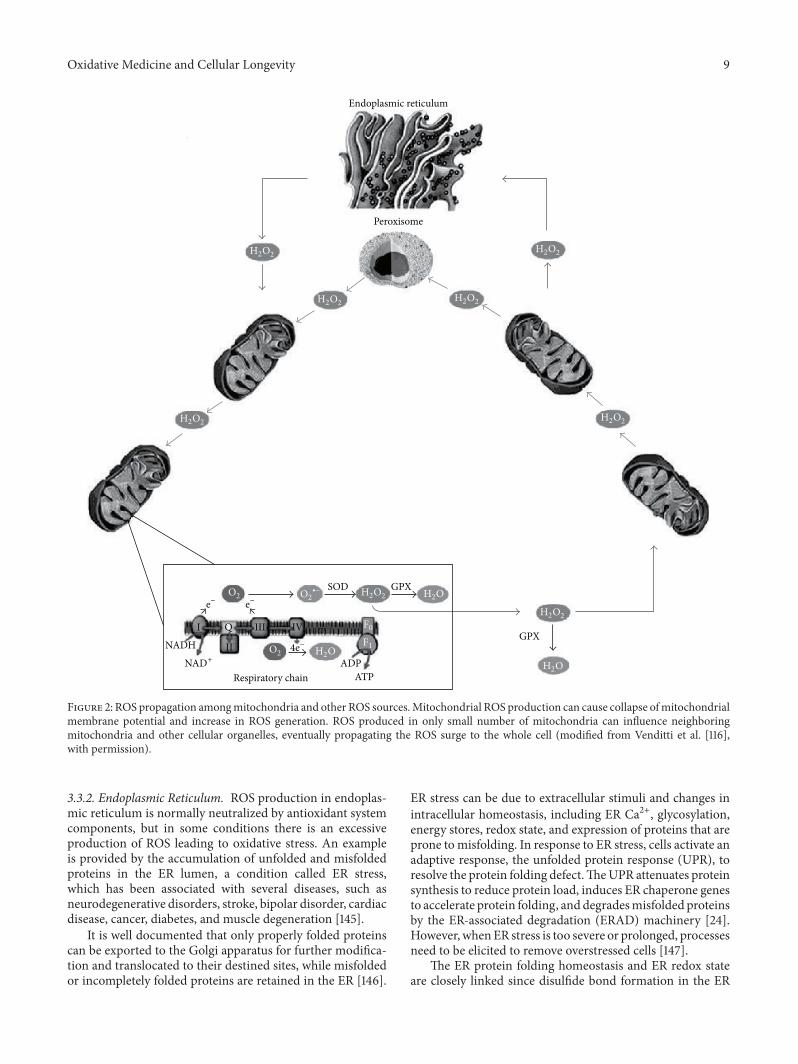
Oxidative Medicine and Cellular Longevity 9
Peroxisome
GPX
GPX
ATP
I Q III
II
IV
Respiratory chainADP
NADH
SOD
Endoplasmic reticulum
H2O
H2O2 H2O
O2
O2e− e−
4e−
H2O2
H2O
H2O2
H2O2
H2O2H2O2
H2O2
H2O2
NAD+
O2∙−
F0
F1
Figure 2: ROS propagation amongmitochondria and other ROS sources.Mitochondrial ROS production can cause collapse ofmitochondrialmembrane potential and increase in ROS generation. ROS produced in only small number of mitochondria can influence neighboringmitochondria and other cellular organelles, eventually propagating the ROS surge to the whole cell (modified from Venditti et al. [116],with permission).
3.3.2. Endoplasmic Reticulum. ROS production in endoplas-mic reticulum is normally neutralized by antioxidant systemcomponents, but in some conditions there is an excessiveproduction of ROS leading to oxidative stress. An exampleis provided by the accumulation of unfolded and misfoldedproteins in the ER lumen, a condition called ER stress,which has been associated with several diseases, such asneurodegenerative disorders, stroke, bipolar disorder, cardiacdisease, cancer, diabetes, and muscle degeneration [145].
It is well documented that only properly folded proteinscan be exported to the Golgi apparatus for further modifica-tion and translocated to their destined sites, while misfoldedor incompletely folded proteins are retained in the ER [146].
ER stress can be due to extracellular stimuli and changes inintracellular homeostasis, including ER Ca2+, glycosylation,energy stores, redox state, and expression of proteins that areprone tomisfolding. In response to ER stress, cells activate anadaptive response, the unfolded protein response (UPR), toresolve the protein folding defect.TheUPR attenuates proteinsynthesis to reduce protein load, induces ER chaperone genesto accelerate protein folding, anddegradesmisfolded proteinsby the ER-associated degradation (ERAD) machinery [24].However, whenER stress is too severe or prolonged, processesneed to be elicited to remove overstressed cells [147].
The ER protein folding homeostasis and ER redox stateare closely linked since disulfide bond formation in the ER
10 Oxidative Medicine and Cellular Longevity
lumen is highly sensitive to altered redox balance so that bothreducing and oxidizing reagents disrupt protein folding andcause ER stress [148]. During oxidative protein folding, thethiol groups on cysteines of substrate peptides are oxidizedand H
2
O2
is generated as a by-product. During ER stress,dysregulated disulfide bond formation and breakagemay leadto oxidative stress by generating large amounts of H
2
O2
anddepleting ER GSH [149].
Studies have indicated that ROS, produced in the ERduring ER stress, subsequently caused mitochondrial dys-function, impairing the respiratory chain, and increase mito-chondrial ROS production particularly when ER stress issevere or sustained. Indeed, the increase in ROS levels causesa Ca2+ influx from the ER into the cytoplasm through theER-localized channels and a large portion of the ion is takenup by the mitochondria resulting in ROS production [145].Mitochondrial ROS can in turn increase further the ER stressresponse thereby amplifying ROS accumulation [150]. Thus,it seems that the ER is placed in a vicious cyclewhere ER stresscan be caused by oxidative stress and increases the perturbedredox state. This process is likely favored by the existenceof close ER-mitochondria contacts [151], which facilitate theROS shift between organelle compartments.
ROS production can also be amplified when exposureto a variety of foreign compounds, such as phenobarbital(PB), increases the P450 levels. Induction by PB causesproliferation of the smooth endoplasmic reticulum withincorporation of the structural gene products (the P450s) intomicrosomal membrane [152]. In rat liver, increases in P450levels, microsomal and mitochondrial protein content, andalteration of mitochondrial composition were reported [153].PB-induced increase in P450 levels enhances H
2
O2
produc-tion by NADPH-supplemented liver microsomes [154] andgives rise to an increase in peroxidative reactions, involvingthe whole cell [153].
Although these results suggest a damaging role of P450induction in hepatic tissue, a consequence of the high ratesof production of ROS by P450 is its own labilization andsubsequent rapid inactivation. Indeed, ROS, generated bysome cytochrome P450 forms, are able to lead to oxidativeinactivation of the P450 themselves modifying apoenzymesor oxidizing the heme groups. Attack on sulfur-containingamino acids results in the conversion of P450 into P420 [155],whereas attack on the heme moiety leads to its breakdownand loss of microsomal P450 [156]. Such a loss was observedduring microsome catalytic turnover under the action ofoxygen species generated at the hemoprotein active center[157]. The cytochrome P450 self-inactivation is importantin situations in which increases in P450 levels occur due touptake by the organism of enhanced amounts of xenobiotics.
Although P450 induction serves to help “detoxication”of xenobiotics, it can be considered an adaptive responsethat has survival value for the organism, as it can alsoincrease “toxification” [158]. First, cytochrome P450 gener-ates toxic chemically reactive intermediates from relativelyunreactive compounds. Moreover, because cytochrome P450inducibility is generally higher than the conjugating enzymeinducibility [159], there is a potential imbalance betweenthe rate of generation of chemically reactive intermediates
and their rate of inactivation and removal. The reactivemetabolites that are not conjugated and the ROS released byP450may attack proteins, membrane components, or nucleicacids, leading to cytotoxicity, mutations, and cancer [160].Therefore, P450 inactivationmay be considered amechanismto prevent cellular accumulation of high levels of the enzyme.Moreover, it appears to be part of a concerted adaptiveresponse to oxidative stress, consisting of repression of ROS-generating systems and induction of antioxidant defenses, asobserved in human hepatoma HepG2 cell lines, expressingCYP2E, the ethanol-inducible cytochrome P450 [161].
3.3.3. Lysosomes. Lysosomes contain the major pool of lowmass redox-active intracellular iron, arising from the intral-ysosomal degradation of iron-containing proteins, such asferritin, and iron-rich organelles, such as mitochondria[162]. Iron accumulation predisposes lysosomes to oxidant-induced damage and rupture with consequent cellular injury.Indeed, someH
2
O2
, formed outside the organelles and escap-ing the extralysosomal degradation, can diffuse into lyso-somes and, together with that formed directly in the lysos-omes, can generate ∙OH radicals by Fenton reaction. Theensuing oxidative damage on the lysosomalmembranes leadsto membrane permeabilization with release to cytosol ofhydrolytic enzymes and low mass iron. This can relocate toother cellular sites, causing site-specific ∙OH production andoxidative damage in conditions of H
2
O2
production.It seems that H
2
O2
formation and lysosomal destabiliza-tion are important for the radiation-induced cellular injuryand death [163], which for a long time have been consideredto depend on ∙OH formation due to radiolytic cleavageof water. Indeed, radiation, besides ∙OH radicals, producessignificant amounts of H
2
O2
[164], which can enter thelysosomal compartment and cause membrane permeabiliza-tion. Employing a model of irradiated murine histiocyticlymphoma (J774) cells [163], it was found that the cellsirradiated twice demonstrated progressive lysosomal damagefrom 2 h after the second irradiation, which in turn resultedin extensive cell death [163]. Irradiation-induced lysosomaldisruption and cell death were significantly reduced by incu-bation with desferrioxamine conjugated with starch whichforms a stable complex with iron and renders the metalinactive [163]. The protective effect of iron chelators againstionizing radiation damage was subsequently confirmed onseveral cell lines previously exposed to salicylaldehyde isoni-cotinoyl hydrazone, a lysosome targeted iron chelator [165].
3.3.4. NADH-Oxidase. Although multiple sources of ROSduring ischemia-reperfusion have been identified, convinc-ing evidence supports NADPH oxidases as important con-tributors to oxidant generation in several tissues, includingcardiac tissue [166]. However, depending on the phase of IRinjury, NADPH oxidases can be either detrimental or pro-tective, shown to have a double-edged role. Low ROS levelsare cardioprotective in pre- and postconditioning therapies,while high ROS levels are deleterious and lead to cardiomy-ocyte death [167]. In addition to the ROS level, the typesof NOX expressed in cardiomyocytes and their localization
Oxidative Medicine and Cellular Longevity 11
are also important in determining the cell fate. In cardiomy-ocytes, threeNOXs are expressed,NOX1 andNOX2, predom-inantly localized to the plasmamembrane, and NOX4, whichis constitutively active and is localized on the intracellularmembranes of organelles [168]. Although the role of NOXisoforms in ischemia-reperfusion has not yet clarified, a studyshowed that deleterious effects of NOX1 and NOX2 occurredduring reperfusion phase in agreement with the idea thatoxygen supply during reperfusion provides substrate forNOX-mediated ROS generation [169]. Moreover, indicationswere also obtained that NOX2 generated higher levels of ROSthan NOX1, leading to direct myocardial damage [169].
Several examples of interplay betweenmitochondrial andNOX-derived ROS have been reported [170]. In the whole,they suggest the presence of a feedforward cycle in whichNADPH oxidases increase mitochondrial ROS that furtheractivate cytoplasmic NADPH oxidases and increase cellularROS production.
The NADPH oxidase involvement in the ROS-inducedROS production is very well documented. For example,exposure of smoothmuscle cells and fibroblasts to exogenousH2
O2
activates these cells to produce O2
∙− via a NADPH oxi-dase [171]. This mechanism could help to explain why micro-molar concentrations ofH
2
O2
cause oxidant-mediated injuryto many different types of cells during chronic oxidativestress. Moreover, H
2
O2
-induced NADPH oxidase activationin nonphagocytic cells could be an important mechanism bywhich the degree of oxidative stress, as well as the subsequentcellular damage, is amplified during inflammatory disorders.
It was also observed that endothelial levels of xanthineoxidase, a source of H
2
O2
and O2
∙−, depend on NADPHoxidase [172].The study of the mechanism showed that H
2
O2
was able to induce transformation of xanthine dehydrogenaseinto xanthine oxidase [173]. The observation that increase inO2
∙− production was a late effect of cell exposure to H2
O2
suggested that the peroxide not only stimulated conversionof xanthine dehydrogenase into xanthine oxidase but alsoactivated the NADPH oxidase, leading to prolonged H
2
O2
production and sustained xanthine dehydrogenase conver-sion [173].
4. ROS as Signaling Molecules
ROS are now appreciated as signalingmolecules that regulatea wide variety of physiological functions. Indeed, they playcrucial roles in gene activation [174], cellular growth [175],and modulation of chemical reactions in the cell [176]. Theyalso participate in blood pressure control [177], are mediatorsin the biosynthesis of prostaglandins [178], function inembryonic development [179], and act as signalingmoleculeswithin the individual cell and among cells during theirlifespan [180].
An important development in the field of ROS beneficialeffects was the discovery that, in organisms from simplebacteria to complex mammals, ROS are able to induce redoxsensitive signal cascades leading to increased expression ofantioxidant enzymes. The increase in effectiveness of theantioxidant defense system provided by this genetic response
enables cells to survive an oxidant exposure that wouldnormally be lethal.
Inmammals, gene transcription determining cell survivalcan be activated by ROS in two ways: either via transcriptionfactors, which can interact directly with specific DNAmotifson promoters of target genes, or via activation of mitogen-activated protein kinase cascades, which in turn activatetranscription factors that trigger target gene transcription[181].
There are two ideas concerning the mechanism by whichROS initiate cellular signaling, namely, modification of targetprotein molecules and changes of intracellular redox state[182], even though the distinction between them is not easy.
While ROS, such as ∙OH, may cause irreversible damageto macromolecules with low specificity, the main target of amild oxidant, such asH
2
O2
, is thiol groups of protein cysteineresidues. Oxidation of these residues forms reactive sulfenicacid (-SOH) that can form disulfide bonds with nearbycysteines (-S-S-) or undergo further oxidation to sulfinic(-SO2
H) or sulfonic (-SO3
H) acid.Thesemodifications resultin changes in structure and/or function of the protein. Withthe exception of sulfonic acid and to a lesser degree sulfinicacid the modifications are reversible by reducing systemssuch as thioredoxin and peroxiredoxins [183].
The cytosol redox state is normally achieved by the“redox-buffering” capacity of intracellular thiols, such asGSH and thioredoxin, which counteract cellular oxidativestress by reducing H
2
O2
. The high ratios of reduced tooxidized forms are maintained by the activity of GSH reduc-tase and TRX reductase, respectively. Accumulating evidencesuggests that GSH and TRX participate in cell signalingprocesses. GSH can regulate redox signaling by alterationsboth in the level of total GSH and in the ratio of its reduced tooxidized forms, while TRX can regulate the activity of someproteins by directly binding to them [182].
Whatever the mechanism by which ROS initiate cellsignaling, there is increasing evidence that ROS cellular levelsare strongly linked to the regulation of cellular antioxidantlevels. A well-known example of this phenomenon is Nrf2.This transcription factor regulates the expression of severalantioxidant and detoxifying genes by binding to promotersequences containing a consensus antioxidant response ele-ment [184]. In turn, the subcellular localization and henceactivity of Nrf2 are at least in part regulated by its interactionwith specific reactive cysteine residues on an inhibitoryprotein called Keap1 [185].
Interestingly, ROS not only are involved in the regulationof the expression of antioxidant genes but also interactwith critical signaling molecules such as MAP kinases, PI3kinase, PTEN, and protein tyrosine phosphatases, to initiatesignaling in several cellular processes, including proliferationand survival [186].
4.1. ROS Role in Protection against Tissue Excessive Dysfunc-tion. An enhanced ROS production in a cellular site caninvolve further ROS production by other cellular sourcesgiving rise to a self-destructive phenomenon as well as thepropagation of a fire in a room containing inflammablematerial. If, as in the fire example, it occurs to confine the
12 Oxidative Medicine and Cellular Longevity
more dangerous focuses and, above all, avoid that the firereaches the other rooms and the entire house catches fire,the cell exposed to oxidative stress should be equipped withmechanisms able to avoid its death or that of surroundingcells, which should lead to excessive tissue dysfunction.
Several results indicate that ROS-linked mechanismsare operative to provide tissue protection against excessivedysfunction, in which mitochondrial systems seem to play amajor role.
4.1.1. Mitoptosis. High mitochondrial ROS production trig-gers opening of the MPT pore leading to potentially signif-icant mitochondrial and cellular injury. However, in manycases mitochondrial swelling due to megachannel openingis a signal for programed mitochondrial destruction [187], aphenomenon called mitoptosis [188], which was proposed torepresent a line of the antioxidant defense [70].
It is well-established that the mitochondrial populationis heterogeneous with regard to its characteristics because itconsists of fractions with different properties [189]. Studieson liver mitochondrial fractions, resolved by differentialcentrifugation, suggested that the light fractions, with lowrespiratory activity, contained transitional forms in the pro-cess of development into the heavy mitochondrial structureswith high respiratory activity [190]. The heavy fractionalso exhibited the lowest antioxidant level [190, 191] andthe highest rates of H
2
O2
production and susceptibility toCa2+-induced swelling [190]. It is interesting that conditionsleading to increased ROS production, such as exercise [192]and cold exposure [193, 194], decrease the amount of heavymitochondria and increase that of light mitochondria in ratmuscle and liver, respectively.This suggests that, in rat tissues,conditions of increased ROS production favor the substi-tution of the oldest ROS-overproducing mitochondria withneoformed mitochondria endowed with a smaller capacityto produce free radicals [190]. If so, the mechanism that,during oxidative stress, enhances the swelling of Ca2+-loadedmitochondria constitutes a negative feedback loop. In fact,the perturbation itself, represented by an enhancement inROS generation, should lead to accelerated mitoptosis, thuslimiting tissue oxidative damage during oxidative stress.
4.1.2. Autophagy. Autophagy is a cytoprotective process bywhich organelles and bits of cytoplasm are sequesteredin double-membrane vesicles, called autophagosomes, andsubsequently delivered to lysosomes for hydrolytic digestion[195]. Autophagy serves as an adaptation strategy for stressconditions, such as amino acid starvation, unfolded proteinresponse, or viral infection, and it is widely accepted thatit regulates cell homeostasis by adjusting organelle numberand clearing damaged structures. However, if autophagyprevents or promotes cell death and if it is a selective ornonelective process are still controversy questions. It is likelythat autophagy can be involved in both survival and death.It assures cell survival when it removes damaged organellesthat might activate programed cell death as apoptosis (typeI cell death) [196]. On the other hand, it promotes cell deathwhen it is excessive and deregulated, since enzymes leaking
from lysosomes, such as cathepsins and other hydrolases,can initiate mitochondrial permeabilization and, eventually,apoptosis [197].
Clearly, autophagy leading to cell death is not selective,whereas the controversy persists about autophagy develop-ing as a survival process. The observation that autophago-somes often contain various cytoplasmic elements, includ-ing cytosolic proteins and organelles [198], suggested thatautophagy is a nonspecific form of lysosomal degrada-tion. However, subsequent observations showed that theautophagy can be a selective process in which specific pro-teins or organelles are delivered to the autophagosome fordegradation [197].
Such selective types of autophagy include selective degra-dation of mitochondria (mitophagy) [197], peroxisomes(pexophagy) [199], endoplasmic reticulum (reticulophagy)[200], or even nucleophagy during which parts of the nucleusare specifically degraded by an autophagic process [201].
Mitochondria subjected over time to multiple attacksbecome damaged and possibly dangerous to the cell, sothat elimination of such mitochondria is essential to protectcells from the harm due to their disordered metabolism.The view that autophagic processes can remove damagedand dysfunctional mitochondria was directly confirmed byexperiments in which selected mitochondria inside livinghepatocytes were subjected to laser-induced photodamage[202]. Mitochondrial depolarization and inner membranepermeabilization seemed to be required for autophagy signal-ing [202] suggesting involvement of MPT pore opening andswelling in the mitophagy.
A growing body of evidence now suggests that pro-cesses of autophagy and/or apoptosis involving other cellularorganelles are able to protect tissues in conditions leading tooxidative stress.
Peroxisomes were the first organelles for which selectiveorganelle degradation by autophagy was described [203].Studies on selective degradation of peroxisomes in methy-lotrophic yeasts showed that when grown on methanolas the carbon source, yeast species included several largeperoxisomes containing the enzymes necessary to assimilatemethanol. When methanol grown cells were shifted to adifferent carbon source, such as glucose or ethanol, whosemetabolism did not involve these peroxisomal enzymes,pexophagy occurred [199]. Hence, this autophagic processoccurred during a rapid intracellular remodeling process toremove organelles containing enzymes no longer needed formethanol utilization.
More recent studies indicate that autophagic degradationof peroxisomes in yeast also occurs as part of cellular house-keeping [204]. It was observed that, during chemostat culti-vation of wild type H. polymorpha cells, entire peroxisomeswere constitutively degraded by autophagy during normalvegetative growth, likely to enable the cells to rejuvenate theperoxisome population.
There is convincing evidence that ER stress and reticu-lophagy induction are strongly linked. For example, it wasreported that, during unfolded protein response to ER stress,induced by DTT, portions of the ER were sequestered bydouble-membrane vesicles, similar to autophagosomes [200].
Oxidative Medicine and Cellular Longevity 13
The presence of ribosomes on the outer membrane of thesestructures suggested a role of the ER as a membrane sourcefor this type of autophagosomes, which subsequently fusedwith the vacuole releasing its content for degradation. Thisprocess was highly selective as cytosol and other organelleswere not included into the vesicles [200]. Not much is knownabout ER degradation and its role in ER maintenance, butit is possible that this selective pathway serves to degradedamaged portions of the ER or resize it after the folding stressinduced enlargement.
Although numerous factors and signaling pathways con-tribute to autophagy induction in different cellular contexts,ROS are indicated to be essential signals to activate autophagystress by several stimulating conditions [205]. ROS appearto be implicated in the regulation of autophagy through dis-tinct mechanisms, depending on cell types and stimulation.Autophagy, in turn, can reduce ROS production removingROS-overproducing organelles.
It seems that ROS, generated from both mitochondriaand NADPH oxidases, activate autophagy to protect cellsfrom nutrient starvation, dysfunctional mitochondria, celldeath, and invading pathogens [206]. The involvement ofROS as signaling molecules in starvation-induced autophagywas demonstrated for the first time showing that starva-tion triggered accumulation of ROS, most probably H
2
O2
,which was necessary for autophagosome formation and theresulting degradation pathway. The oxidative signal is par-tially dependent on phosphatidyl inositol 3 kinase (PI3K),which plays a critical role in the early stages of autophago-some formation. Furthermore, a direct target for oxidationby H2
O2
, the cysteine protease Atg4, has been identified[207]. Whereas mitochondria were found to be involved instarvation-induced autophagy [208], activation of antibacte-rial autophagy is due to NADPH oxidase that generates ROSnecessary for targeting of the autophagic protein LC3 to thephagosome [205].
4.1.3. Autophagic and Apoptotic Cell Death. Autophagy usu-ally promotes cell survival but when cellular damage ispronounced and survival mechanisms fail, death programsare activated in response to oxidative stress. The activationof the autophagic pathway beyond a certain threshold hasrecently been found to directly promote cell death by causingthe collapse of cellular functions as a result of cellularatrophy (autophagic or type II cell death) [209]. This formof autophagic cell death, which is necessary under certainconditions, such as in apoptosis-defective cells [210], isactivated in response to oxidative stress in nervous cells, asshown by oxidative stress and autophagic death induced inhuman neuroblastoma SH-SY5Y cells [211] and sympatheticneurons [212] by dopamine and growth factor deprivation,respectively.
Autophagy can also lead to the execution of apoptotic ornecrotic (type III cell death) programs, likely via commonregulators such as proteins from the Bcl-2 family [196].Apoptotic death is an adaptive process, allowing for renewalof the organism constituents and life maintenance, which,
differently from necrosis, is a form of cell death that causesminimal damage to surrounding cells.
Apoptosis plays an important role in the elimination ofunnecessary, damaged, or diseased cells during the wholelifespan and particularly during embryogenesis, when amajority of newly formed cells undergo programed celldeath. Although both types of cell death have been linked toautophagy, cell death does not necessarily result from a pre-vious autophagic process. Depending on the cellular contextand death trigger, apoptosis and necrosis either cooperate ina balanced interplay involving autophagy or are employed bycells in a complementaryway to facilitate cellular destruction.A further complication results by the observation that apop-tosis, necrosis, and autophagy are often regulated by similarpathways, engage the same subcellular sites and organelles,and share initiator and effector molecules.
An example is supplied by the cellular response to MPTpore opening. This process provides a common pathwayleading to mitophagy, apoptosis, and necrosis [213]. Withlow intensity stress, limited MPT leads to mitoptosis, whichis followed by the elimination of degraded mitochondria.Recent studies have shown that removal can occur throughtwo different processes which rid cells of damaged mito-chondria, the formation of mitoptotic bodies which arethen extruded from the cell [214] or selective autophagy[215]. With increasing stress MPT involves an increasingproportion of mitochondria and cellular response progressesfrommitoptosis to apoptosis driven bymitochondrial releaseof cytochrome c and other proapoptotic factors. This eventoccurs if permeabilization affects a great number of mito-chondria, in the absence of excessive reduction in ATP levels,and can purify tissue from cells that produce large amountsof ROS [70]. Lastly, when extreme stress causes MPT poreopening in virtually all cellular mitochondria, ATP levelsdrop, and, because of bioenergetic failure, neither autophagynor apoptosis can progress, and only necrotic cell death,which is not dependent on energy supply, ensues [197].
Another example involves cellular responses to ER stress,which include the activation of UPR, autophagy, and celldeath [216]. These processes are not mutually exclusive, andthere is significant cross talk between these cellular stressresponses. Autophagy upregulation during ER stress is a pro-survival response directed to removal of unfolded proteins,protein aggregates, and damaged organelles, which is trig-gered in order to relieve the stress and restore ER homeostasis[217]. However, prolonged or unresolved ER stress results inapoptotic program activation [218]. Sometimes, the effect ofautophagy on cell survival in ER stress also depends on thetissue type. In colon and prostate cancer cells, ER-inducedautophagy protects against cell death, whereas in normalcolon cells autophagy does not alleviate ER stress but rathercontributes to ER-induced apoptosis [219].
Recent studies have shown several pathways that mediatethe interplay between autophagy and apoptosis providingmechanistic insight into the network regulating both pro-cesses [220]. However, the mechanism by which ER stressinduces apoptosis is not fully delineated, even though itis clear that mitochondria-dependent and independent cell
14 Oxidative Medicine and Cellular Longevity
death pathways can mediate apoptosis in response to ERstress.
In ER-mitochondria-independent cell death pathway,caspases are required for apoptosis, and some membersof this family of proteases are associated with the ER. Inrodents, caspase-12 is ER-membrane proapoptotic proteasespecifically activated upon prolonged UPR. Several pathwaysinvolve caspase-12 activation in cell apoptosis after ER-induced stress. A pathway triggers apoptosis by ER stress-specific caspase cascade leading to the caspase-9 and caspase-3 activation in cytochrome c and apoptotic protease activat-ing factor-1 (Apaf-1) independent manner [221].
ER stress can also trigger apoptosis by mitochondria-dependent pathways [222] which can share actors withmitochondria-independent pathways. The phosphorylationof inositol-requiring enzyme 1 (IRE1) leads to the phospho-rylation of c-Jun N-terminal kinase (JNK) that, in turn, canactivate p53 transcription factor.The p53-induced expressionof a protein, the Bcl-2 interactingmediator of cell death (Bim)[223], leads to the formation of the BAX/BAK pore on theouter mitochondrial membrane and cytochrome c releaseresulting in activation of the canonical apoptosis pathway.
Whereas the relationship between mitochondrial oxida-tive stress and cell death is well-established [224], that of per-oxisomes in cell death pathways is just beginning to emerge.In light of the strong impact of peroxisome dysfunction onmitochondria, it is possible that peroxisome-derived medi-ators of oxidative stress have an influence on mitochondriathat would have profound implications for cell fate.
In fact, it has been reported that excess ROS generatedinside peroxisomes quickly perturbs themitochondrial redoxbalance and leads to remarkable mitochondrial fragmenta-tion [225]. In a recent study using targeted variants of thephotosensitizer Killer Red it has been showed that the photo-toxic effects of peroxisomal Killer Red induce mitochondria-mediated apoptotic death and that this process is inhibitedby targeted overexpression of antioxidant enzymes, includingperoxisomal glutathione S-transferase kappa 1, superoxidedismutase 1, and mitochondrial catalase [226].
Over the last decades, the intricate molecular eventsunderlying the process of apoptosis have been elucidated[227]. It is apparent that crucial steps involve mitochondrialrelease of proapoptotic factors, although the exact mech-anisms involved in this release are less well understood.In this regard, it seems that, in some circumstances, therelease into the cytosol of lysosomal constituents may be aninitiating event in apoptosis and that mitochondrial releaseof proapoptotic factors might be a consequence of earlierlysosomal destabilization [228].
The destabilization of the lysosomal membrane is dueto its vulnerability to oxidative stress, because agents, whichinduce oxidative stress, such as H
2
O2
[229] and radiation[230], also promote lysosomal membrane rupture. Differentcell types but also cells of the same type and lysosomesof individual cells exhibit different lysosomal resistance tooxidative stress [231]. It has been suggested that such a resis-tancemainly depends on the capacity to degradeH
2
O2
beforeit reaches the acidic vacuolar compartment, resistance to ROS
of lysosomal membranes, and lysosomal content of redox-active iron [231]. Lysosomal membrane permeabilization perse triggers intracellular formation of ROS, a process whichcan be mediated by the action of lysosomal proteases, such ascathepsins B and D, which leak into the cytosol [208]. Theseenzymes affect mitochondria inducing further cytochrome crelease and activation of caspase-mediated cell death [208,228]. The involvement of lysosomes and their iron content inradiation-induced cell death is supported by the observationthat cells are significantly protected from radiation damage ifexposed to iron chelators [163, 165]. Conversely, the involve-ment of lysosomal proteases in cell death is supported bythe observation that apoptosis inhibition by cyclosporin A-induced block of MPT pore opening favors the developmentof a necrotic form of cell death, which is attenuated by ROSscavengers and inhibition of cathepsin D activity [232].
5. Harmful and Beneficial Effects of ROSduring Exercise
It is well documented that acute physical exercise can producesignificant damage, including alterations in membranes ofmitochondria and sarcoplasmic and endoplasmic reticulum[233–236] in skeletal muscles and other tissues. The contrac-tion form most damaging to skeletal muscle is that in whichthe muscle is contracting while being lengthened (eccen-tric contraction). During such a contraction, disruption ofcytoskeletal structures, loss of muscle force generation, andinflux of phagocytic cells and neutrophils into the damagedfibers occur [237].
Different from acute exercise, aerobic physical activityregularly performed (training) induces adaptive responsesin the whole organism and particularly in the cardiorespi-ratory and musculoskeletal systems [238], which lead to anincreased ability to perform prolonged strenuous exercise[235]. Moreover, it has several healthy effects, includingthe maintenance of insulin sensitivity and cardiorespiratoryfitness, so that it is able to prevent type 2 diabetes [239]and coronary heart diseases [240], and can also be used asadjunctive therapy in the treatment of patients with diabetes[241] and chronic heart failure [242].
To date, the idea that the opposite effects of acute exerciseand training are in great part due to the ability of ROS to playa dual role in animal organisms is widely shared. In fact, it iswell documented that, during a single session of prolongedaerobic exercise, ROS production increases and this can leadto cellular damage and dysfunction. On the other hand, someresults suggest that the ROS produced during each sessionof a training program can act as factors inducing cellularadaptations to exercise.
During physical activity several ROS sources can beactivated contributing to the oxidative damage and/or tothe adaptive processes. It is reasonable to imagine that aninitial source of ROS can activate the ROS release fromother sources, inducing a positive feedback loop. In the nextsections, we will point out our attention to what is knownabout production, sources, and double action of ROS duringacute exercise and training.
Oxidative Medicine and Cellular Longevity 15
6. Acute Exercise
6.1. ROS Production. ROS involvement in the tissue damagefound after prolonged aerobic exercise was suggested in thelate 1970s [243], but there is still no direct evidence thatROS production increases during exercise. Electron spinresonance (ESR) spectroscopy was able to furnish directinformation of the presence of free radical species, butsuch a technique allows obtaining information on the ROSproduction after exercise. Davies et al. [244] first reportedthat signals of free radicals were enhanced in rat muscleand liver after a bout of exhaustive running. Subsequently,increased generation of free radical signals was observedin rat heart after an acute bout of exhaustive enduranceexercise [245]. The idea that free radical activity mightincrease after extensive muscular activity was also confirmedon skeletal muscles subjected to electrical stimulation [246].Using ESR spectroscopy in conjunction with the spin tappingtechnique, enhanced free radical concentration in humanserum following exhaustive exercise was found [247, 248]which was prevented by ascorbic acid supplementation [249].
Indirect information on ROS production during acuteexercise has been obtained by the changes in indexes of oxida-tive damage to lipids, proteins, and DNA and in the cellularredox state. A plethora of information exists concerning theincrease in such indexes in various animal species, includinghuman, but here we focus our attention on what is availablein literature on rat.
Lipid oxidation can be evaluated measuring tissue lev-els of oxidized lipids, such as lipid hydroperoxides, orthose coming from their degradation, such as malondialde-hyde (MDA) and 4-hydroxyl-2-nonenal. Numerous studiesshowed increase in lipid oxidation markers in skeletal [244,250–254] and cardiac muscle [251–253, 255], liver [244, 250–254, 256], brain [257], erythrocytes [258], and kidney [254]of untrained rats after acute exercise.
Exercise-induced increases in protein carbonyl content,a marker of protein oxidative damage, were reported in ratskeletal [253] and cardiac muscle [253, 255], liver [253, 256],and plasma [259]. Furthermore, increases in 8-hydroxy-2-deoxyguanosine (8-OH-dG), amarker of oxidative damage toDNA, were also found in rat skeletal [260] and cardiac [255]muscle, liver, and lung [260] after acute exercise.
Some studies reported that the extent of oxidative damagedepends on duration or intensity of exercise. One such studyshowed that exhaustive maximal exercise caused plasmaMDA increase while short periods of submaximal exercise(less than 70% VO2max) reduced lipid peroxidation [261].Liver oxidative damage indexes did not change after 5 h ofswimming and increased after 8 h of exercise [192]. Anotherstudy showed that an acute bout of a moderate as well ashigh intensity exercise led to an increase in malonaldehydeand lipid hydroperoxide levels in red vastus, white vastus,and soleus muscle [262]. The study also showed that whenintensity of exercise was considerably decreased, lower MDAlevels were found [262].
Reduced glutathione (GSH), a thiol-containing tripeptideplaying a vital role in maintaining cells in the reducedstate and in protecting tissues from oxidative stress [263],
is involved in reducing radicals arising from a variety ofantioxidants, such as 𝛼-tocopherol and ascorbic acid, to thenative structure [9]. GSH is oxidized to glutathione disulfide(GSSG) donating a pair of hydrogen atoms. The ratio ofGSH to GSSG is used as an indicator of intracellular GSHredox status. A decrease in GSH/GSSG ratio suggests thatthe production of ROS goes over the reducing capacity ofGSH and other antioxidants.Therefore, change of GSH redoxstatus has been used as a footprint of oxidative stress duringexercise. Several studies showed that prolonged exhaustiveexercise promoted oxidation of GSH to form GSSG in theblood [264], liver [253, 264], heart [253, 265], and skeletalmuscle [253, 264] of rats, even though it is possible thatthe GSH redox state in the muscle is not altered by shortterm exercise. Indeed, it was reported that physical exerciseat submaximal level determined a progressive depletion ofliver GSH to about 20% of the levels found in sedentaryrats which persisted for several hours following the cessationof exercise [266]. However, skeletal muscle appeared to bespared by this severe depletion phenomenon, whereas thelevels of plasma glutathione exhibited a transient increaseat the beginning of the exercise bout followed by a lineardecrease with increased running time [266]. In liver, theearly decrease in GSH level during exercise did not dependon increased ROS production, even though it could be oneof the factors inducing liver oxidative stress. Evidence thata decrease in liver GSH content precedes oxidative stresswas supplied by the finding that prolonged exercise led toincreased lipid peroxidation and decreased GSH content,while exercise of shorter duration was not able to induceoxidative stress in liver, although it reduced GSH content[192].The liver GSH reduction can be due to several factors. Ithas been suggested that the exercise linked increase in plasmalevels of glucagon, vasopressin, and epinephrine stimulateshepatic efflux of GSH which is delivered to tissues, such asskeletalmuscle that necessitates a larger tripeptide supply andits uptake from the plasma [267, 268].
6.2. Sources of ROS during Physical Activity. Notwithstandingmost work indicates that exercise increases ROS productionin rat tissues, the debate about the cellular sources of suchROS is still open. Several intracellular sources of free radicalshave been identified and it is possible that all contribute tothe increased ROS production during exercise even thoughthe extent of their contribution can depend on several factorsincluding the type of tissue and exercise.
Mitochondria,NADPHoxidase, and xanthine oxidase areconsidered the main ROS sources during exercise but it isnecessary to point out that reports concerning other cellularsources are scarce or lacking.
6.2.1. Mitochondria. Mitochondria were long considered themain source of ROS in the cell during physical activity.Initially, this belief stemmed from the widespread idea thatin tissue free radical production was closely related to oxygenconsumption [269]. Since over 90% of the oxygen consumedby a mammal is utilized in the mitochondria, which appearto generate free radicals in all tissues studied [270], it wasspeculated that the increase inmuscular oxygen consumption
16 Oxidative Medicine and Cellular Longevity
during exercise was associated with an increase in free radicalproduction by the respiratory chain localized in the innermitochondrial membrane [269].
Duringmuscle contraction, ADP concentration increasesand stimulates mitochondrial oxidative phosphorylation[271], shifting the mitochondrial respiration nearer to State3 than to State 4 oxygen consumption. Therefore, the closelink between ROS production and O
2
consumption shouldrequire that the percentage of total electron flow escapingfrom the respiratory chain to reduce O
2
to superoxide radical(the mitochondrial free radical leak) is not modified duringthe transition from State 4 to State 3 happening duringmuscle contraction. However, this idea is theoretically andexperimentally inconsistent. The rate of the mitochondrialROS generation is related to the degree of reduction ofelectron carriers able to donate electrons to oxygen, andsuch a degree of reduction decreases during transition fromState 4 to State 3 [272]. In agreement with such prediction,measurements performed on isolated mitochondria showthat ROS mitochondrial release is higher in State 4 and in allconditionswhen the rate of electron-transfer is lowered [272].
Nevertheless, results obtained measuring ROS releasefrom mitochondria isolated from tissues of animals thatexercised suggest that mitochondrial ROS release mightincrease during prolonged aerobic exercise not only inmusclebut also in other tissues, such as liver and heart [272].
An increased release of ROS during basal and stimulatedrespiration, with respiratory substrates linked to ComplexI (pyruvate plus malate) or Complex II (succinate), wasreported for mitochondria isolated from the muscles ofthe hind limbs [273], gastrocnemius (red portion), heart,and liver [253] of rats subjected to prolonged swimming.Increased ROS release was also reported following prolongedtreadmill running [274] in heartmitochondria.The increasedROS release was associated with an increase in the levelsof oxidative stress markers in mitochondria isolated fromcardiac and skeletal muscles and liver [253] of rats subjectedto prolonged aerobic exercise. It was also accompanied byalterations in mitochondrial functionality. Indeed, exerciseincreased State 4 respiration in liver, muscle [253], and heart[253, 274] mitochondria and decreased State 3 respiration inliver andmuscle mitochondria [253]. Such results suggest thepossibility that, whatever the initial source of ROS duringexercise is, such ROS can damagemitochondrial componentsinducing both functionality impairment and increasing ROSrelease. It was initially proposed that the ROS formationduring exercise could involve the loss in the cytochromeoxidase activity and a consequent increase in the electronpressure within the respiratory chain [275]. The finding thatin hind limb muscle mitochondria exercise-induced changein mitochondrial respiration is not associated with changesin the cytochrome oxidase activity [273] seems to excludesuch a possibility suggesting that oxidative modifications ofother components of mitochondrial membrane are involved.Oxidative modifications of lipids and proteins located in theinner mitochondrial membrane could be responsible for theincrease in State 4 respiration.The increase in State 4 respira-tion rate represents a compensatory response to the increasedleak of protons back in the mitochondrial matrix. It has
been proposed that adenine nucleotide translocase (ANT)and uncoupling proteins (UCPs) are involved in protonconductance of mitochondrial membrane [276]. However,the observation that in skeletal muscle ANT expression isnot affected by acute exercise [277] excludes that ANT isresponsible for the increase in State 4 respiration induced byexercise. Moreover, it is known that an uncoupling protein1 (UCP1) catalyzes inducible proton conductance in brownadipose tissue (BAT) [278], and exercise upregulates UCP1homologue expression in skeletal [279] and cardiac muscle[274]. However, whether in tissues differently from BATUCP1 homologues are responsible for mitochondrial basal orinducible proton conductance is yet controversial [280].
More support is available for the idea that the observedenhancement in State 4 respiration is due to high productionof ROS and RNS, which seem to be able to affect pro-ton leak through an indirect mechanism. Both O
2
∙− [281]and ONOO− [282] increase mitochondrial proton leak byenhancing the extent of peroxidative processes [282, 283].Therefore, it is conceivable that the increase in State 4 respira-tion induced by exercise may be due to a lipid peroxidation-mediated increase in proton leak.
The decrease observed in State 3 respiration can be dueto a direct action of ROS and/or RNS. Indeed, damage torespiratory chain components by ROS [284] and inhibition ofmitochondrial function byONOO− [109] have been reported.The decline of the respiration rate in mitochondria from ratsthat exercised is likely to involve ONOO−, which duringexercise could be formed in greater amount and causeirreversible inhibition of many mitochondrial componentsdifferent from cytochrome aa3 [285].
Inhibition ofmitochondrial function and increase in ROSrelease could also be due to the increase in mitochondrialCa2+ content, which occurs in skeletal muscle after prolongedexercise [286] and leads to oxidative phosphorylation inhibi-tion [287]. The Ca2+ effect on mitochondrial function resultsfrom induction of mitochondrial permeability transition(MPT) [114], which leads to degradation of the heaviest mito-chondrial subpopulation characterized by high respiratorycapacity and susceptibility to Ca2+-induced swelling [190].
Following long-lasting exercise, a transfer of damagedmitochondria in the lightest fraction happened in rat liver,as demonstrated by the increase in protein percent contentin such a fraction and its decrease in the heaviest one[192], whereas a similar transfer did not happen in skeletalmuscle [273]. This agrees with the observation that acuteexercise enhances mitochondrial resistance to Ca2+ overloadin human skeletal muscle [277]. The Ca2+ tolerance ofmitochondria after exercise could be due to the maintenanceof relatively high levels of GSH, which should prevent mito-chondrial membrane potential collapse [288] which precedesMPT pore opening. Although this can appear as a protectivemechanism, it slows removal of ROS-overproducing mito-chondria thus enhancing exercise-induced muscle oxidativedamage and dysfunction.
6.2.2. NADPH Oxidases. In the cells of skeletal muscle, theisoform NOX2 is expressed [289] and protein subunits have
Oxidative Medicine and Cellular Longevity 17
been identified in transverse tubules and triads obtainedfrom rabbit skeletal muscle but not in sarcoplasmic reticulumvesicles [290]. Some authors suggested that this is one of themain sources of ROS during in vitro muscle contraction orelectrical stimulation [291, 292]. Moreover, it was demon-strated that muscle contraction increased O
2
∙− in cytosol andsubsequently in mitochondria, suggesting that NADPH oxi-dase could be a potential primary source of ROS productionduringmuscle contraction [293]. It is conceivable that duringmuscular activity the increased activity of NADPH oxidasecan produce excessive ROS release which can contribute todamage cellular components. However, it is also possible thatsuch a release can have beneficial roles. The increased levelof ROS induces changes in intracellular calcium levels whichare the result of oxidativemodification of calcium channels orother proteins involved in calcium signaling [294] and it hasbeen suggested that NOX2ROS productionmay be necessaryfor the excitation-contraction coupling process [290].
The mechanism of activation of NADPH oxidase has notbeen completely defined but a recent paper suggests that thiscan involve the increase in ATP release. Electrical stimulationof adult muscular fibers isolated from the muscle flexor digi-torum brevis activates a voltage gated L-type calcium channel(Cav1.1) with each depolarizing event.This activation inducesATP release via pannexin-1 (PnX1) channel which colocalizesin the transverse tubules with Cav1.1 [295]. These events, inturn, trigger a signaling cascade where, through ATP acti-vation of a purinergic receptor (P2Y), phosphatidylinositol3-kinase-𝛾 and phospholipase C and consequently proteinkinase C, which activates NOX2 oxidase and ROS release[296], are activated. It is conceivable that a similar activationcan happen also in in vivomuscle because an increase in ATPconcentration has been found in the interstitial muscularspace after exercise and contraction [297, 298].
6.2.3. XanthineOxidases. Studies performed usingXO inhib-itors suggested a potential role of enzyme XO as a sourceof oxidative stress, during ischemia-reperfusion in varioustissues, such as intestine, heart, lung, kidney, and liver [98,299, 300]. In particular, XDH/XO activity in liver has beenreported to be relatively higher with respect to other organs[301, 302]. On the other hand, it has been demonstrated thatliver blood flow during severe exercise may be reduced tohalf of normal, indicating that exercise can induce ischemiaor hypoxia in the hepatic tissue [303]. These findings wouldsuggest that XO produces free radicals which may influencethe function of hepatic cells during and/or after strenuousexercise. Furthermore, inosine and hypoxanthine producedby the skeletal muscle during severe exercise have beenreported to be taken up by the liver via the blood stream andoxidized to uric acid mainly after exercise [304]. Accordingto these observations, it was shown that the liver has asubstantially higher risk of oxidative stress following a singlebout of exhausting exercise, rather than during the exerciseitself [305].
It has been reported that xanthine oxidase produces O2
∙−
in the contracting rat [306] and human skeletal muscles[307]. However, there is controversy about the role xanthine
oxidase plays in O2
∙− production of human skeletal mus-cle during contraction, because such a muscle appears topossess low amounts of xanthine dehydrogenase or oxidase[308], even though these enzymes are present in associatedendothelial cells. It has been speculated that postexerciseoxidative stress in mouse skeletal muscles may be due tothe conversion of XDH into XO in capillary endothelialcells [309] and enhanced adenosine 5-triphosphate (ATP)degradation together [310].
More recently, it was found that the administration ofallopurinol, XO inhibitor, did not prevent the increase inprotein and lipid oxidative stress markers in rat plasma,erythrocytes, and gastrocnemius muscle due to swimmingexhaustion [311]. Moreover, in rats treated with allopurinolalone, a similar increase in protein and lipid oxidative stressmarkers in erythrocytes and gastrocnemius muscle wasfound.The combination of allopurinol and exercise appearedto increase protein oxidative damage in plasma and proteinand lipid oxidative damage in erythrocytes. Interestingly,allopurinol provoked a marked reduction in physical perfor-mance as demonstrated by the 35% decrease in the swimmingtime to exhaustion [311].This result is in contrast with anotherwork reporting a lack of effects of allopurinol administrationon the time to exhaustion in rats performing treadmillrunning [305]. The discrepancy can be due to the differenttype of exercise and/or to the different doses of allopurinol.However, it is interesting that notwithstanding allopurinolseems to have intrinsic antioxidant properties being a potenthydroxyl radical scavenger [312, 313], it either did not changeor decreased the time to exhaustion, differently from whatwas found administering antioxidants before an exhaustiveexercise [314].
Another study [315] reported that, following acuteexercise (60min treadmill running 27m/min, 5% incline),skeletal muscle oxidized glutathione (GSSG) significantlyincreased in allopurinol- and vehicle-treated rats despite XOactivity and uric acid levels were unaltered.This suggests thatXO was not the source of ROS during exercise. In the whole,the available data obtained by XO inhibition suggest that theenzyme can be a source of ROS during exercise but not themain.
7. Training
7.1. Effects of Training on Tissue Oxidative Damage. Con-trary to acute exercise, aerobic exercise training inducesadaptations which reduce liver [316] and skeletal [317] andcardiac muscle [318] oxidative damage of lipids and proteins.Such adaptations also render tissues less susceptible to theoxidative damage induced by conditions leading to oxidativedamage. Indeed, training attenuates lipid and protein oxida-tive damage and glutathione depletion in rat heart subjectedto ischemia-reperfusion [319]. Moreover, training preventslipid peroxidation increase induced by moderate intensityexercise in rat liver and muscle [250]. However, other studiessuggested that training does not affect the extent of lipidperoxidation due to exhaustive swimming but, delaying therate of the peroxidative reactions, allows trained animals to
18 Oxidative Medicine and Cellular Longevity
sustain the activity for a longer period before the fatiguebecomes limiting [251, 252].
It is clear that training exerts protective effects reducingoxidative damage of tissues and increasing their resistanceto oxidative challenges. Such effects seem to be associatedwith increased cellular antioxidant defenses. Several studiesexamined the effect of training on the activities of antioxidantenzymes.Much of these studiesmay not be directly comparedto each other because of the differences in experimentaldesign, animal model, and analytical procedures. However,in the whole they show that training results in an increasein skeletal [320] and cardiac [321] activity of antioxidantenzymes, such as superoxide dismutase, glutathione peroxi-dase, glutathione reductase, and catalase, even though somestudies failed to find enhanced antioxidant activity aftertraining.
The training effect on liver antioxidant enzymes hasbeen less studied and the results are rather contrasting. Onthe other hand, it was previously reported that liver totalantioxidant capacity was increased by training as well as thoseof skeletal and cardiac muscle [251, 252].
Surprisingly, there are few studies concerning the effectof training on antioxidant enzyme expression. However,increases induced by training in CuZnSOD mRNA abun-dance in rat liver and heart [322] and Cu, ZnSOD, andMnSOD protein level only in some muscles but not in others[322, 323] were reported.
It is likely that training exerts its protective effects alsodecreasing H
2
O2
production, even though scarce informa-tion is available on training impact on cellular ROS sources.The rate of H
2
O2
release was decreased in liver [316], skeletalmuscle [317, 324], and heart [318] mitochondria from ratstrained to swim and in heart mitochondria [325] from ratstrained to run. Conversely, no effect on H
2
O2
release wasfound in skeletal muscle mitochondria following voluntarywheel training [326].
Measurements of H2
O2
release rate in the presence ofrespiratory inhibitors suggested that training reduces theconcentration of the autoxidizable electron carriers locatedat Complexes I and III in the liver [316] and muscle [317]mitochondria and that of the autoxidizable electron carrierlocated at Complex III in the heart mitochondria [318].However, other swim training-induced adaptations can con-tribute to the reduction of the H
2
O2
release rate found inthe mitochondria isolated from liver heart and muscle.Thesecould include the increased activity of theH
2
O2
metabolizingenzyme GPX, which is coupled with the increase in GRactivity, in the mitochondria of the three tissues [316–318].Conversely, it is unlikely that the decrease in H
2
O2
releaseis due to increased uncoupling of the inner mitochondrialmembrane, since that training reduces mRNA expressionof uncoupling protein 3 in skeletal muscle and uncouplingprotein 2 in skeletal and cardiac muscles [327].
The effects of exercise training on NADPH oxidase activ-ity are limited but some data suggest that exercise trainingis able to modulate NADPH oxidase activity. For example, ithas been reported that exercise training mitigates age-relatedupregulation of NOX2 subunits gp91phox and p47phox in ratheart [328], reduces microvascular endothelial NOX content
inmuscle biopsies fromvastus lateralis of obesemen [329] butnot of lean men [330], and downregulates NADPH oxidaseexpression in obese rats [331]. Studies using XO inhibition byallopurinol show that such an inhibition produces complexeffects that are not yet fully elucidated so that the relativedata are not conclusive and do not allow us to point outthe relevance of the ROS produced by XO for the trainingadaptations.
7.2. Mitochondrial Biogenesis. The improved cardiovascularfunction and aerobic capacity elicited by aerobic exercisetraining require an increased tissue metabolic activity. Thefirst proof that the increase induced by the training inmetabolic capacity was due to the increased tissue mitochon-drial protein content was obtained in the skeletal muscle[332, 333]. Subsequent studies confirmed this finding [317]and showed that swim training increased mitochondrialprotein content also in other tissues such as liver [316]and heart [318]. However, the changes in mitochondrialprotein content seemed to differ in various tissues andwere associated with different changes in the metaboliccapacity of tissues and mitochondria. Indeed, in the skeletalmuscle swim training increased tissue metabolic capacity byinducing a moderate increase in the tissue content of themitochondrial population characterized by a lightly reducedaerobic capacity [317]. In liver, swim training did not induceincreases in tissue metabolic capacity because the modestincrease in mitochondrial population content was balancedby the reduction in mitochondrial metabolic capacity [316].In heart, swim training increased tissue metabolic capacityby enhancing lightly both mitochondrial population contentand mitochondrial respiratory capacity [318]. These differentresponses are consistent with the different functions of liverand muscles as energy supplier and consumers, respectively[334], during aerobic long-lasting exercise.
7.3. Mechanisms of Adaptive Response to Exercise. Mitochon-drial biogenesis requires that expression of themitochondrialgenome and nuclear genes encoding mitochondrial proteinsis finely organized. The process is controlled by the per-oxisome proliferator-activated-receptor-gamma coactivator1 (PGC-1) which regulates the expression of transcriptionfactors such as nuclear respiratory factors 1 and 2 (NRF-1 andNRF-2) [335]. NRF-1 and NRF-2 control the expression ofmany genes, among which are those implicated in mitochon-drial biogenesis, adaptive thermogenesis, glucose and fattyacid metabolism, fiber type switching in skeletal muscle, andheart development [336].
PGC-1 expression increases quickly in muscle cells stim-ulated to contract [337], in rat skeletal muscle after a singlebout of exercise [338] and in human skeletal muscle afterendurance training [339]. A study using PGC-1𝛼 knockoutmice showed that in liver PGC-1𝛼 plays a pivotal role inregulation of cytochrome c and cytochrome oxidase subunitI expression in response to a single bout of treadmill exerciseand prolonged exercise training, which suggests that theexercise-induced changes in tissue oxidative capacity areregulated by PGC-1𝛼 [340].
Oxidative Medicine and Cellular Longevity 19
Increases in PGC-1𝛼 expression were also found in heartafter short term training to treadmill run [341] and in liver[316], skeletal muscle [317], and heart [318] after 10 weeksof training to swimming. In these tissues, the increases inPGC-1𝛼 expression were associated with an increase in theexpression of NRF-1 and NRF-2.
Interestingly, in addition to regulating mitochondrialbiogenesis, PGC-1 is able to regulate endogenous antioxidantexpression, such as Cu, ZnSOD, MnSOD, and GPX, in skele-tal muscle [342, 343].This coordination of the proliferation ofROS producing organelles with increase in antioxidant levelslikely helps to maintain redox homeostasis. In addition, it hasbeen shown that PGC-1𝛼 promotes mSIRT3 gene expression,which is mediated by ER-binding element mapped to theSIRT3 promoter region [344]. In turn, SIRT3 binds to, dea-cetylates, and activates mitochondrial enzymes, includingMnSOD, through a posttranslational mechanism [345].
PGC-1𝛼 is also able to regulate the mRNA expression ofuncoupling proteins 2 and 3 in cell culture [346], suggestingthat PGC-1𝛼 may also increase the uncoupling capacity andconcomitantly reduce ROS production in the mitochondria[343].
Several initiating stimuli, activated during exercise, cancontribute to eliciting the PGC-1 gene response. Theseinclude (i) increase in cytosolic calcium concentration, whichactivates various signaling pathways regulated by the cal-cineurin phosphatase and the calmodulin-modulated kinase,(ii) the decrease in levels of high-energy phosphates, leadingto the activation of the AMP-sensitive kinase (AMPK), and(iii) stimulation of the adrenergic system, leading to cyclicAMP synthesis, and activation of protein kinase A and otherkinases, such as mitogen-activated protein kinase (MAPK)[342]. However, it is necessary to point out that PGC-1𝛼regulation is not only due to variation in expression butalso caused by covalent modifications among which arephosphorylation, acetylation methylation, and ubiquitina-tion. Indeed, in vitro experiments showed that p38 MAPKand AMPK phosphorylate PGC-1𝛼 producing a more activeprotein [342].
It seems that PGC-1 expression is also upregulated byROS. Indeed, the observation that antioxidant incubationprevents the increase in PGC-1𝛼mRNA induced by electricalstimulation in rat skeletal muscle cell culture [347] indicatesthat increases in ROS may contribute to exercise-inducedincreases in skeletal muscle PGC-1𝛼 mRNA content. Thus,the observation that theH
2
O2
-induced increase in themRNAcontent of SOD, catalase, and GPX in PGC-1𝛼 KO fibroblastsis lower than that inwild type fibroblasts [348] indicates a roleof PGC-1𝛼 in the upregulation of ROS removing enzymes inresponse to increases in ROS.
Moreover, notwithstanding conflicting results exist inliterature [349], it was reported that antioxidant supplemen-tation attenuates the PGC-1𝛼 expression increase due totraining [316–318, 350–352]. It was also reported that vitaminE supplementation prevents the increase in activator andcoactivator levels and mitochondrial population adaptationto physical training [316–318]. These results suggest that theROS produced during each session of exercise training areable to regulate cellular functions acting as signals regulating
molecular events crucial for adaptive responses of liver, mus-cle, and heart.
The role of ROS as signaling molecules in the tissueadaptation induced by training seems to contrast with theoxidative damage and dysfunction elicited by acute exercise.However, this can be explained by differences in extentand temporal pattern of ROS generation. Thus, a moderate,intermittent ROS production during short time periods ina program of graduate aerobic training can activate signal-ing pathways leading to cellular adaptation and protectionagainst future stresses. In contrast, moderate levels of ROSproduction over long time periods (e.g., hours) or high levelsproduced during brief exercise at high intensity may result instructural and functional tissue damage.
8. ROS Production and Type 2 Diabetes
Type 2 diabetes and other related diseases, such as metabolicsyndrome and coronary heart disease, are a serious prob-lem worldwide [353]. Specifically, diabetes is very closelyrelated to microvascular and macrovascular complicationsthat seriously affect the quality of life and life expectancyof patients. At present, there are around 350 million peopleworldwide with diabetes, a figure that will rise to 500–600million over the following years. Diabetes, insulin resistance,and cardiometabolic diseases are associated and constitute anactive field of research [354, 355].
Mitochondria are known to produce ATP after metab-olization of nutrients and are capable of generating energy.In this sense, it has been demonstrated that mitochondrialdysfunction is characterized by decreased levels of ATP,inhibition of mitochondrial O
2
consumption, enhanced ROSproduction, a decrease in the antioxidant content, and alter-ations in mitochondrial membrane potential (ΔΨ
𝑚
). Theseeffects are due mainly to an imbalance between energy intakeand expenditure [356]. In fact, a decrease in the activity ofthe electron transport chain (ETC) complexes or increaseduncoupling produced by the activity of uncoupling proteinsor the ADP/ATP translocator (also called adenine nucleotidetranslocase, ANT) can induce changes inΔΨ
𝑚
that eventuallylead to apoptosis [354, 355].
Different factors, both genetic and environmental (diet,exercise, and stress), have been shown to modulate mito-chondrial function and alter insulin sensitivity [357, 358]. Inthis context, the presence of mitochondrial impairment hasbeen demonstrated in different types of leukocytes [359] andtissues such as liver, lung, skeletal muscle, spleen, or heartin type 2 diabetes [360, 361], confirming the relationshipbetween this condition and mitochondrial dysfunction.
Mitochondria are the main source of reactive oxygenspecies (ROS), as mentioned previously. ROS are key tothe development of diabetic complications [362–364], andstudies have demonstrated that the use of antioxidantssuch as lipoic acid (LA) can reduce insulin resistance andROS production (by improving mitochondrial function) andprevent CVD in humans [365]. Recently, Faid et al. havedemonstrated that resveratrol can alleviate diabetes-inducedapoptosis bymodulating caspase-3 activities, oxidative stress,and JNK signaling [366].
20 Oxidative Medicine and Cellular Longevity
Electron transport chain (ETC) dysfunction is directlyrelated to diabetes and its complications, including retinopa-thy, nephropathy, and neuropathy [355]. Furthermore, somestudies have documented that deleterious genetic mutationsrelated to a reduction in the activity of Complex I can leadtomitochondrial impairment and enhanced ROS production[367]. Therefore, mitochondria-targeted antioxidant therapyhas been proposed as a beneficial tool in the treatment ofmitochondria-related diseases [368].
Victor’s group has performed several studies of type 2diabetes patients which have shown that oxidative stressand mitochondrial dysfunction occur due to a decrease inO2
consumption, Complex I activity, membrane potential,and glutathione levels and an increase in ROS production[359, 369–371], thus confirming mitochondria as a key targetfor diabetes treatment. In other related pathologies, suchas polycystic ovary syndrome (PCOS), in which patientscan develop insulin resistance, there is also an impairmentof mitochondrial Complex I and an increase in leukocyte-endothelium interactions [372].This mitochondrial dysfunc-tion increases ROS production, reduces ATP and Ca2+,and alters membrane potential and mitochondrial mor-phology. However, in an animal model of diabetes (db/dbmice), mitochondrial and renal function are improved inthe presence of mitochondria-targeted antioxidants, suchas CoQ10, highlighting the crucial role of mitochondria inthe development and pathogenesis of diabetic nephropathy[373]. In addition, Victor’s group has demonstrated thatmitochondrial dysfunction and, especially, mitochondrialROS production are related to the development of silentmyocardial ischemia and endothelial dysfunction due toincreased leukocyte/endothelium interactions [374].
Mitochondria-targeted antioxidants have been shown tohave beneficial effects on conditions of oxidative stress. Inthis sense, MitoQ is an antioxidant which, due to a covalentattachment to the lipophilic triphenylphosphonium cation,is selectively taken up 1000-fold by mitochondria [363, 375].Chacko et al. demonstrated an example of the beneficialeffects of MitoQ on diabetes when they reported that MitoQdecreased urinary albumin levels to the same level as those ofnondiabetic controls in amousemodel of diabetic nephropa-thy (Ins2(+/)-(AkitaJ) mice) [376]. Furthermore, glomerulardamage and interstitial fibrosis were significantly reduced inthe treated animals, and there was a nuclear accumulation ofthe profibrotic transcription factors 𝛽-catenin and phospho-Smad2/3, which was prevented by MitoQ treatment. Theseresults support the hypothesis that mitochondrially targetedtherapies could be beneficial for the treatment of diabeticnephropathy.
8.1. Insulin Resistance and Mitochondrial Dysfunction. Glu-cose homeostasis is regulated by insulin. In addition, insulinhas important cardiovascular, renal, and neural functions,which may explain why insulin resistance is a risk factor formicrovascular complications such as retinopathy, nephropa-thy, hypertension, and CVD [25].
A series of conditions are related to the developmentof insulin resistance, such as obesity, changes in lipid andglucose metabolism, chronic inflammation, stress, or other
oxidative factors. In these conditions, the appearance ofinsulin resistance is frequently associated with a diminishedcapacity of tissues or cells to respond to levels of insulin [377].This process is related to mitochondrial dysfunction, changesin mitochondrial dynamics, and enhanced ROS production.
In relation to this theory, it has been demonstrated thatmitochondrial impairment, oxidative stress, excess energyintake, and lipodystrophy can enhance circulating free fattyacids (FFAs), which can lead to the accumulation of triglyc-erides, FFAs, and diacylglycerol (DG) in different tissularlocations, including liver, skeletal muscle, heart, kidney, and𝛽-cells. Furthermore, alterations in cholesterol subfractions,such as an increase in the atherogenic potential of small denseLDL, may be related to several metabolic properties of theseparticles, facilitating their transport into the subendothelialspace [378], reducing LDL receptor affinity [379, 380], andincreasing susceptibility to oxidativemodifications [378, 381].The cited studies feed into the idea that small dense LDLare related to arterial damage in patients with dyslipidemiaassociated with diabetes.
Insulin resistance, mitochondrial dysfunction, and en-hanced production of ROS, which act as secondary mes-sengers by activating serine kinases that phosphorylate IRSproteins, modulate the insulin response [377]. In addition,ROS can trigger the inflammatory process by activatingIKKb, which phosphorylate IRS-1 [382]. Several studieshave demonstrated that insulin sensitivity andmitochondrialfunction can be modulated by antioxidants, with a subse-quent decrease in ROS production and an increase in theexpression of UCP2/3 and a decrease in ROS levels [382].However, the results of the said studies have generated somecontroversy [383].
Mitochondrial impairment and insulin resistance havealso been shown to be related to diminishing levels of mito-chondrial oxidative enzymes, which reduce mitochondrialcomplex activity, alter mitochondrial morphology, and limitmitochondrial number [384]. For example, mitochondrialoxidative capacity can correlate negatively with insulin sensi-tivity after the accumulation of intramyocellular lipids [385].
During obesity, there is an increase of triglyceridesin adipose tissue, and consequently glucose metabolism isaltered in other nonadipose tissues. In this sense, it has beenspeculated that lipodystrophy induces insulin resistance,mitochondrial dysfunction, and type 2 diabetes [386]. Indiabetes and obesity, adipocytes can release high amounts ofadipokines, such as resistin, leptin, adiponectin, and TNF-𝛼,which can regulate metabolic pathways [387]. Furthermore,the number and morphology of mitochondria, as well as theexpression of genes involved in mitochondrial biogenesis,are significantly decreased by the energetic alterations thatappear as a result [388]. All of these studies support the ideathat insulin resistance is present in lipodystrophy, obesity,and type 2 diabetes. This action leads to the accumulation ofintracellular fatty acid metabolites (e.g., diacylglycerol, fattyacyl CoAs) in muscle and liver, which triggers the activationof a serine kinase cascade and finally induces defects ininsulin signaling and insulin action in these tissues [389].
Oxidative stress, mitochondrial-endothelial dysfunction,and insulin resistance are very common in cardiovascular
Oxidative Medicine and Cellular Longevity 21
diseases such as stroke, silent myocardial ischemia, coronaryartery disease, or hypertension [390]. In this sense, type 2 dia-betic patients exhibit high blood pressure, whose appearanceis related to hyperglycaemia [391]. In relationship to this idea,Katz et al. have highlighted that diabetes is associated witha higher prevalence of calcified atherosclerotic plaque in thethoracic arteries [392]. Furthermore, endothelial impairmenthas been associated with intramyocardial lipid accumulationand glucose intolerance and, eventually, heart failure [393].
Not all organs are specifically protected against oxidativestress. For example, the heart, which has a highmetabolic rateand high beta oxidation and ROS production, contains lowlevels of antioxidants, making it particularly susceptible tooxidative stress, mitochondrial dysfunction, and subsequentstructural and functional abnormalities [394].
Mitochondrial oxidative stress damage and changes in themorphology/function ofmitochondria have been reported inan animal model of obesity, namely, insulin-resistant obeseZucker rats [395]. Obesity and lipotoxicity can also enhancemitochondrial damage during the development of diabeticretinopathy [396]. Experiments involving transmission elec-tron microscopic analysis of myocardial tissue have demon-strated an increase of abnormal mitochondria in an insulin-resistant rat model [397]. Another study showed an increasein the number of mitochondria in hypertrophied rat heartsunder oxidative stress conditions [398]. In disagreementwith these studies, others have failed to find changes in thenumber of mitochondria and their DNA content, while somehave even reported reduced numbers in patients and animalmodels of pathological hypertrophy [399]. In conclusion,these results point out the importance of mitochondria in theheart and would suggest that they enhance CVD, includingheart failure, stroke, cardiomyopathy, coronary heart disease,silent myocardial ischemia, and hypertension.
Insulin resistance is related to endothelial dysfunction[400], but the underlying mechanisms are yet to be con-firmed. In the endothelium, mitochondria play an essen-tial role by acting as sensors of local alterations in theconcentration of O
2
and as regulators of intracellular Ca2+concentrations [401]. Taking into account that mitochondrialdysfunction is related to endothelial dysfunction, differentstudies have demonstrated that blockade of ROS generationcan improve endothelial function under hyperglycemia con-ditions [362, 402].
It is generally recognized that endothelial nitric oxidesynthase (eNOS) plays a key role in the maintenance ofvascular tone and insulin-stimulated NO∙ production in theendothelium [403]. In fact, poor eNOS activity has beenrelated to insulin resistance, hypertension, and dyslipidemia[404]. Therefore, there is an impairment in the NO∙ produc-tion under insulin resistance conditions, which is related tothe appearance of CVD such as coronary artery disease, heartfailure, stroke, or silent myocardial ischemia.
Insulin-resistant patients can develop type 2 diabeteswhen there is impairment in 𝛽-cells, the result of which is anincapacity to sense glucose properly and release insulin andfailed glucose homeostasis. In this sense,mitochondrial activ-ity can modulate the potassium channels (𝐾ATP) modulating
ATP/ADP ratio. 𝛽-cell function and mitochondrial functionare related through the ATP/ADP ratio [405]. Furthermore,mitochondrial Ca2+ levels are crucial to the maintenance ofinsulin secretion, as demonstrated by Han et al. who showedthat taurine can enhance the glucose sensitivity of UCP2-overexpressing 𝛽-cells, probably by enhancing mitochon-drial Ca2+ influx and subsequently increasing the ATP/ADPratio and mitochondrial function [406]. Other studies havehighlighted the importance of mitochondrial function inglucose homeostasis by using knockout Tfam (TranscriptionFactor A, Mitochondrial) mice, Tfam being a nuclear DNA-encoded mitochondrial protein, which results in a dramaticmtDNA depletion, a decreased of insulin secretion, reduced𝛽-cell mass, and the development of diabetes [407]. Inconclusion, all of the abovementioned studies highlight thefact that preserving mitochondrial function is essential for𝛽-cell function under oxidative stress conditions and thatmitochondrial impairment contributes to the pathogenesisof type 2 diabetes by interfering with insulin action andsecretion. Furthermore, high levels of fatty acids can inducemitochondrial dysfunction and impair insulin signaling dueto oxidative stress and enhanced ROS production. In sum-mary, we consider that mitochondria should be considereda key target in therapy for insulin resistance in general anddiabetes in particular.
Finally, we would like to mention that the possiblebeneficial effects of RNS and ROS can occur at low levels andcan exert different physiological functions. Cells can produceH2
O2
, O2
∙−, or NO∙ at physiological levels, but, in the case ofdiabetes, basal levels of ROS are elevated, and so these ROSand RNS are generally harmful. For example, in basal condi-tions, leukocytes kill pathogens by phagocytosis after whichROS are released. Basal levels of ROS can also trigger energyproduction by mitochondria, induce mitogenic responses,or activate the release of cytokines or nuclear transcriptionfactors.
NO∙ can also modulate vascular pressure, leukocyteadhesion, and angiogenesis. Furthermore, NO∙ is an impor-tant neurotransmitter and is a key mediator of the immuneresponse when generated by activated macrophages.Although different studies suggest that ROS act as secondarymessengers, it is clear that they can be harmful when theyaccumulate and disrupt molecules and tissues [12, 408–410].
9. ROS and Neurodegeneration
The brain is composed of two main types of cells: glia andneurons. Glial cells encompass a wide variety of cells includ-ing astrocytes, microglia, and oligodendrocytes. Glia cells actas a neuronal support system and are the most abundant cellsin the nervous system. Astrocytes support neurons in thebrain and regulate the chemical and extracellular environ-ment. They maintain low levels of ammonia and glutamateand produce neuroprotective enzymes. Upon activation,astrocytes repair cellular damage, mount an inflammatoryresponse, and activate microglia. Microglia, the smallestglial cells, repair damage due to injury via phagocyto-sis. Oligodendrocytes are responsible for axon myelination,
22 Oxidative Medicine and Cellular Longevity
a protective layer required for propagation of action poten-tials and maintenance of intracellular communication.
Neurons (nerve cells) are the basic structural elements ofthe nervous system.Their primary function is to transmit andreceive information through nerve impulses, electrochemicalsignals that travel down the neuron. Neurons “sense” changesin environmental conditions and respond to such changesvia neurotransmission. Neurons consist of three major com-ponents: the axon, dendrite, and perikaryon (or soma). Theaxon, typically ending at a specialized structure called thesynapse, carries impulses to distant locations. The dendriteacts as the neuronal “receiver” but can also communicatevia neurotransmitters to adjacent neurons. Dendrites becomedenser during neurogenesis. Small dendrites do not containorganelles, but large dendrites have neurofilaments (onlyfound in neurons), microtubules, ribosomes, and endoplas-mic reticulum. The perikaryon is the “metabolic hub” of theneuron. It houses mitochondria, ribosomes, Golgi apparatus,and endoplasmic reticulum, essential machinery in proteinsynthesis and energy production. Neurons are essential incontributing to emotions, perceptions, and memory andlearning. Neuronal damage can alter these functions andultimately lead to overall brain dysfunction and decline inthe cognitive functions previously described. Neurodegener-ation, an example of neuronal damage, is the loss of neuronalstructures or function. During the aging process, myelinand neuronal loss occur, dendritic length and branching aredecreased, and global brain volume is reduced. Consequencesof these events include but are not limited to age associatedcognitive decline, memory loss, epigenetic changes, reducedautophagy, and synaptic plasticity. Excessive neuronal deathresulting in accelerated cognitive decline and memory losshas been observed in those suffering from neurodegenerativedisorders. Recent findings from Villeda demonstrated asignificant improvement in hippocampal learning exercisesand contextual fear conditioning tasks when old mice weregiven plasma from young mice [411]. Other findings fromthis study showed an increase in dendritic spine number andsynaptic plasticity in the old mice associated with reversal ofcognitive decline.
Astrocytes are highly abundant throughout the centralnervous system. Due to their extension-like end feet, theycover the free surfaces of neuronal dendrites and soma.Astrocytes also cover the inner surface of the one of thebrain’s most important meningeal membranes, the pia mater,and all blood vessels in the CNS. More importantly, theseend feet surround the brain’s capillary endothelial cells ofthe blood-brain barrier. They participate in neurotransmittermetabolism and play a pivotal role in glutamate uptaketo prevent excitotoxicity. Glutamate is then converted intoglutamine via glutamine synthetase, in which the basicamino acid is transported into the neuron. Furthermore,astrocytes maintain the pH of the extracellular space andionic environment. In neurodegeneration, these cells canrelease cytokines which regulate the inflammatory response.
Microglia act as macrophages in the brain. Phenotypiccharacteristics include short spiny projections, which becomeenlarged under ROS conditions. Neurodegeneration is a
key promoter of microglial activation; therefore this phe-nomenon is observed in neurodegenerative disorders includ-ing Alzheimer’s disease, Parkinson’s disease, amyotrophiclateral sclerosis, and Huntington’s disease [412]. Microgliaalso play a role in generating reactive nitrogen species asnitric oxide synthases, iNOS andNOX2, andNADPHoxidaseare induced in these glial cells [413]. This NOX2 activationcan lead to a respiratory burst of superoxide flooding themitochondria further contributing to neurodegeneration.In addition to their inflammatory response, microglia alsoexpress multiple ion channels, namely, the sodium, proton,voltage gatedCa2+ andCl−, and potassium channels. Changesin ionic concentrations may play a role in depolarization andaction potential initiation, which can trigger inflammationand neuronal activation.
Activation of these glial cells can have dramatic cellulareffects including prompting an inflammatory response. Thisoccurs in the normal aging process due to mitochondrialderived ROS production, which promotes inflammationand cytokine production [414]. This consequence can beobserved as cognitive deficits as NF𝜅-B is generated andneuroinflammation occurs. NF𝜅-B serves as a proinflam-matory agent and a prosurvival molecule by regulating theinflammatory response. This is an age-dependent process.If the NF𝜅-B pathway is blocked in old mice, a reversalof gene expression occurs [415]. Interestingly, NF𝜅-B ishighly associated with RNS as this family of proteins inducesnitric oxide synthase, further promoting nitrosative stress.Prostaglandin synthesis is also initiated through the NF𝜅-B signaling cascade, propagating the inflammatory process.Neuroinflammation can be defined as the increased produc-tion of a multitude of proinflammatory molecules, mostlynotably interleukin-1𝛽 (IL-1𝛽), tumor necrosis factor𝛼 (TNF-𝛼), and transforming growth factor 𝛽 (TGF-𝛽). IL-1𝛽 isa proinflammatory agent that recruits neutrophils as partof the inflammatory response. This cytokine is frequentlyobserved in several neurodegenerative disorders [416]. TNF-𝛼 is a key regulator of the immune system. It has beenshown to be increased in microglia from aged mice ina lipopolysaccharide induced mouse model [417]. TNF-𝛼is found predominantly in macrophages. It is released byactivated microglia and astrocytes which perpetuates neu-rodegeneration and neuroinflammation by increasing levelsof reactive oxygen species, specifically superoxide. The morethe neuronal damage, the more frequent the neuroinflam-mation as microglia and astrocytes are constantly activated.In addition to the inflammatory properties of TNF-𝛼, itdirectly activates NADPH oxidase which increases levels ofsuperoxide in the cells similar to NF𝜅-B. NOX2 is highlyexpressed via TNF-𝛼, which is linked to excess levels of RNS[418]. TGF-𝛽 regulates neuroinflammation and apoptosis byreleasing inflammatory cytokines and reactive oxygen species[419]. TGF-𝛽 is mediated by the Smad3 pathway, whichinhibits the production of free radicals which are normallypromoted in the inflammatory pathway. This pathway isimpaired during neurodegeneration and could contribute tothe disease progression as neuronal loss and neuroinflam-mation are observed [420]. As these cytokines have been
Oxidative Medicine and Cellular Longevity 23
tested as biomarkers of oxidative stress, a link between ROS,neurodegeneration, and neurodegenerative disorders can befurther bolstered.
Oligodendrocytes. Oligodendrocytes are vulnerable to oxida-tive damage as they have a higher ATP requirement thanother glial cells, contain low levels of glutathione, a potentantioxidant, and have a high intracellular iron level which canform prooxidants through Fenton chemistry [421]. The mainfunction of oligodendrocytes is myelination. The myelinsheath is a necessary axonal component that increases salta-tory conduction of action potentials, thereby stimulatingneurotransmission.Demyelinating disorders such asmultiplesclerosis exhibit motor function decline due to disruption ofaction potential propagation from the loss of myelin.
It has been well-established that, in neurodegenerativedisease, neurodegeneration can occur as the result of oxida-tive stress, the imbalance of antioxidant and prooxidant levels[422]. Reactive oxygen species levels increase as a functionof age and are even higher in age-related neurodegenerativedisorders [423]; therefore oxidative stress can also occur ifthere is an excess of ROS/RNS production or an antioxidantdeficiency [424]. This section of the review will focus onthe interconnection between oxidative stress, reactive oxygenand nitrogen species, and neurodegeneration in the afore-mentioned cellular systems and research directed at neuro-protection, the delay or prevention of neurodegeneration.
9.1. Oxidative Stress. Mitochondria are the key source for freeradicals [425, 426]. A minute amount of electrons leaks fromthe mitochondria and reacts with molecular oxygen to formsuperoxide. Other sources for free radicals can include envi-ronmental toxins [427], metal catalyzed reactions, certainenzymatic reactions (e.g., xanthine/xanthine oxidase), andcellular processes. During phagocytosis, oxidants are neededto ingest bacteria, viruses, and other pathogens [428].
9.1.1. Hypochlorous Acid. Hydrogen peroxide can react witha chloride anion to form hypochlorous acid (HOCl) viamyeloperoxidase (MPO). Myeloperoxidase is largely presentin neutrophils [429] but has also been located in neuronalcells under certain conditions [430]. Hypochlorous acid canfurther react with nitrogen dioxide (NO
2
∙) to form nitrylchloride (NO
2
Cl), a potent chlorinating and nitrating oxidant[431]. Phagocytes use HOCl as one of their agents [432].Hydrogen sulfide (H
2
S) is an inhibitor of HOCl from Cl−.Myeloperoxidase in the presence of H
2
O converts Cl− intoCl+; therefore hydrogen sulfide may have a protective effect.H2
S levels are lowered in Alzheimer’s disease, a neurologicaldisorder [433]:
H2
O2
+ Cl− MPO→ HClO (3)
9.1.2. Reactive Nitrogen Species. Similar to ROS, reactivenitrogen species are highly toxic [434]. Some examples ofRNS, nitric oxide, and nitrogen dioxide will be discussedbelow. Reactive nitrogen species cause protein nitration byvariousmethods, which can result in protein dysfunction andneuronal loss.
NO∙ has been shown to play a role in neurodegenerativediseases by acting as a neurotoxin when excessively produced[435]. Hara has shown that glyceraldehyde-3-phosphatedehydrogenase (GAPDH) acts as NO∙ sensor [436]. NO∙ isinvolved in cell signaling pathways immune response andvasodilation. NO∙ activates protein kinase C (𝜀 isoform),which activates a specific family of tyrosine kinases that canstimulate apoptosis [437]. NO∙ can also bind to glutamatechannels and indirectly to calcium and potassium channels[438]. As glutamate is an excitatory neurotransmitter in thecell, depolarization of themembrane occurs. Glutamate bindsto the NMDA receptor and an influx of Ca2+ enters thecell and causes a disruption in calcium homeostasis. Thisdisruption can eventually lead to cell death.
NO2
∙ acts as an outdoor and indoor air pollutant fromcar emissions, fossil fuels, cigarette smoke, heaters, and gasstoves, just to name a few [439]. NO
2
∙ is primarily foundin the airways of the terminal bronchi; however, NO
2
∙ maybe found in other areas of the respiratory tract. Minimalexposure to NO
2
∙ results in long morphological changesresulting in possible inflammation, pulmonary edema, andcellular injury [440]. Nitrogen dioxide exposure also leadsto an increase in TBARS, a marker for lipid peroxidationin lung tissue, and vitamin E treatment showed a reducedpulmonary injury [441]. NO
2
∙ serves as an oxidant in inflam-mation mediated by the peroxidases, eosinophil peroxidase,and myeloperoxidase [442–446]. This gas can oxidize theantioxidant glutathione and increase activity of glutathionereductase and glutathione peroxidase [447, 448]. The deple-tion of glutathione shifts the balance to the side of nitrosativestress. NO
2
∙ can also be formed by the oxidation of ONOO−,another potent reactive nitrogen species.
10. An Overview of SomeNeurodegenerative Diseases
Neurodegenerative diseases are a classification of disordersin which neuronal loss and progressive cognitive decline areobserved.These two consequences contribute to the memoryloss exhibited by those with neurodegenerative disorders.This level of decline is accelerated in contrast to the normalaging process. Depending on the specific disease othercharacteristics and symptoms include dementia, a declinein motor function, and depression may be evident. Thephenomenon of oxidative stress has been well-establishedin such neurodegenerative disorders as Huntington’s disease[449, 450], Parkinson’s disease [451], amyotrophic lateralsclerosis [452], and Alzheimer’s disease [453–455]. This sec-tion will only highlight the correlation between autophagy,apoptosis, and oxidative stress in the following neurodegen-erative diseases: Alzheimer’s disease, Parkinson’s disease, andamyotrophic lateral sclerosis.
10.1. Alzheimer’s Disease. Alzheimer’s disease (AD), the mostcommon form of dementia, is a neurodegenerative diseasecurrently affecting millions of people worldwide. Age isone risk factor of AD, as the onset of disease typicallyoccurs at about 65 years old. Although AD is an age-related
24 Oxidative Medicine and Cellular Longevity
neurodegenerative disease, it can be inherited (familial AD)and sporadic. Familial AD (FAD) is very rare, as it affects lessthan 10% of those afflicted with the disease. There are fourdistinct types of FAD caused by the genetic factors describedbelow [456–458]. Sporadic ADusually occurs in the late stageof AD and is associated with apolipoprotein E4 (APOE4)allele. Apolipoprotein E (APOE) helps transport cholesterolinto the bloodstream. APOE4 is also involved in learning andmemory as persons with the APOE4 allele showed a declinein processing new information [459]. APOE4 shows anincrease of amyloid beta peptide and neurofibrillary tangles(NFT) [460, 461]. Increased oxidative stress is prevalent inAD and also linked to APOE4 [462, 463]. Ramassamy et al.demonstrated that tissue from patients with the APOE4 alleleshowed a decrease in activity of the antioxidant enzymesglutathione peroxidase and catalase [463]. Other risk factorsinclude family history of disease, reduced brain volume,traumatic brain injury, and low education and mental abilityearly in life [464–468].
Extreme neurofibrillary tangles and senile plaques are thehallmarks of AD. These NFT are composed of paired helicalfilaments, which consist of hyperphosphorylated tau protein.The senile plaques are composed of amyloid beta (A𝛽) pep-tide. Amyloid precursor protein (APP) is a transmembraneprotein that plays a role in neuronal plasticity, long termpotentiation, and memory loss [469]. APP is proteolyticallycleaved by the enzymes 𝛽-secretase and 𝛾-secretase to form a40–42 amino acids’ peptide, amyloid beta (A𝛽) peptide. Thetwo major forms of A𝛽 found in human brain are A𝛽(1–40)and A𝛽(1–42). A𝛽(1–42) is the more toxic form of A𝛽 andis the primary component of the senile plaque [470, 471].A𝛽(1–42) induces oxidative stress in vivo [472, 473] and invitro [474]. After this toxic peptide is formed, it can aggregateand these aggregates can accumulate outside or inside the cellcontributing to the pathogenesis of AD.
There are three human causative genetic alterations inAD (APP; PS1; and PS2) and several genes associated withAD (APOE4, 𝛼-2 macroglobulin, chromosome 10, and chro-mosome 12). APP is a precursor to amyloid beta peptide.There are several APPmutations (i.e., Artic, Flemish London,Dutch, Italian, Indiana, Iranian, etc.) that cause AD bymany different mechanisms (i.e., protofibril formation, densesenile plaques, and increased A𝛽(1–42) production) [457,475–478]. The highly characterized Swedish mutant Tg2576transgenic mouse model for Alzheimer’s disease containshumanAPP and has A𝛽 plaques deposits as early as 9monthsold [479, 480], leading to increased memory decline withage. Presenilin 1 (PS1) and presenilin 2 (PS2) are catalyticcomponents of 𝛾-secretase and are highly involved in APPprocessing. Mutations in PS1 or PS2 show an increase inA𝛽(1–42) production and are the cause of most FAD cases[456, 481–484]. Mutations in APP, PS1, and PS2 have beenfound in the first clinical stage of AD and MCI [485], earlystage AD (EAD) [486], and late stage AD [487].The APP/PS1mutant mouse is a common model for AD as it exhibitsearly amyloid deposition and increased oxidative stress [488,489]. A significant increase in levels of protein carbonyls,4-hydroxynonenal, and 3-nitrotyrosine levels, markers ofoxidative stress, was exhibited in APP/PS1 double mutant
neurons compared to wild type [489]. A triple transgenic(3XTg-AD) mouse model has been recently used to study thepathogenesis of Alzheimer disease. Similarly to the doubleAPP/PS1 mouse, the 3XTg-AD mouse model exhibits oxida-tive imbalance as antioxidant levels are reduced and lipidperoxidation and overall brain oxidation are increased [490].
These mice have mutations in the PS1 gene and arehomozygous for the Swedish APP mutation and tau P301Lmutation, making them highly representative of this disorderand an excellent model to use as amyloid deposition isobserved at 3 months, hippocampal hyperphosphorylatedtau appears at 12–15 months, and synaptic transmission isimpaired at an early age, making them ideal to study thisneurodegenerative disorder.
Chromosome 10 codes several particular genes of interestas possible risk factors of AD [491]. Insulin degrading enzyme(IDE) is one such gene. IDE degrades and clears A𝛽 in thebrain [492, 493]. IDE levels are reduced in hippocampus[494] and this protein’s catalytic activity is lowered in AD aswell [495]. IDE, as the name infers, degrades insulin. Statindrugs have been shown to promote astrocytic IDE secretionin AD model to stimulate autophagy [496]. Abnormalitiesin insulin metabolism are associated with APOE status. ADpatients with the APOE4 allele had higher insulin levels thanpatients without E4 allele [497] Elevated plasma insulin levelscorrelate to increased A𝛽 levels, which as previously men-tioned has detrimental effects [498, 499].Therefore, chromo-some 10 mutations may be a possible risk factor in AD.𝛼-2-Macroglobulin (𝛼
2
M) is encoded by chromosome 12and is thought to be a possible risk factor for AD aswell [500].𝛼2
Mprotein binds to A𝛽 and transports it from neurons intocells for degradation using the LRP receptor [501]. APOEusesthis same receptor to enter into cells. Therefore, APOE4 orexcess APOE may prevent the A𝛽/𝛼
2
M complex from bind-ing to the receptor and clearing for the neuron. 𝛼
2
Mhas beenshown to be neuroprotective against 𝛽 amyloid fibrils in vitro[502] and in vivo [503] inhibiting toxicity. Mutations in thisprotein may result in A𝛽 deposition and neuron death [504].
Microglial activation has been linked to A𝛽 plaques inAlzheimer’s disease. This activation results in a neuroinflam-mation and phagocytic impairment [505].This inflammatoryresponse leads to the release of TNF-𝛼 and interleukin-1𝛽[506]. Once the A𝛽 peptide activates the microglia, nitricoxide is released [413], thereby promoting an oxidative stressvia peroxynitrite elevation which can alter calcium home-ostasis and promote cellular apoptosis [507].
10.2. Parkinson’s Disease. Parkinson disease (PD), the secondmost prevalent age-dependent neurodegenerative disorder, isclassified as a tauopathy, a neurological disorder that exhibitsexcessive levels of phosphorylated tau.
Tau is a microtubule-associated protein that is respon-sible for stabilizing microtubules. Microtubules are neuron-resident, cylindrically shaped, dynamic structures composedof alternating rows of 𝛼- and 𝛽-tubulin. Microtubules play apivotal role in facilitating intracellular transport by assistingin motor protein-driven transport of vesicles, mitochondria,and other cargos in neurons. If tau is damaged, it canno longer stabilize these microtubules, thereby reducing
Oxidative Medicine and Cellular Longevity 25
transport of critical factors to the neuron due to cytoskeletaldisintegration. Ultimately, neurons become energy starvedleading to apoptosis. Parkinson’s disease is strongly correlatedwith neuroinflammation which is demonstrated by activatedastrocytes and microglia in the central nervous system. Theactivation of these glial cells is detrimental to neurons andpromotes neuronal loss.
Parkinson’s disease presents as a decline in motor func-tion in the form of resting tremors, muscle rigidity, anddyskinesia. This disease is attributed to protein aggregates of𝛼-synuclein, a protein whose main function is mitochondrialfunctionality, vesicle trafficking, and synaptic vesicle forma-tion [508, 509]. Alpha synuclein is predominantly locatedin the presynaptic terminals; thereby accumulation resultsin poor neurotransmission. These aggregates are the majorcomponent of Lewy bodies located primarily in the putamenand substantia nigra of the brain. These two regions arelargely involved in motor movement and learning. Activatedmicroglia are found largely in the substantia nigra andstriatum of PD animals [510]. Oxidative stress in PD brainis evident by DNA damage [511] and increased levels ofcarbonylated proteins [512]. Neuronal dopamine, a key neu-rotransmitter involved in motor function, levels are signifi-cantly diminished in the substantia nigra causing substantialneuronal death. Aggregation also leads to Complex I impair-ment in dopaminergic neurons [513]. Protein aggregation incombination with dopamine loss causes a profound effect onthe physical capabilities of persons suffering from Parkinsondisease and late in the disease cognitive dysfunction is oftenobserved. Posttranslational nitration of alpha synuclein [514]andComplex I [508] lead to altered energymetabolismwhichis evident in neurodegenerative disorders that are associatedwith oxidative stress.
The most commonly used models of PD include treat-ment with rotenone, 6-OHDA, or 1-methyl-4-phenyl-1,2,5,6-tetrahydropyridine (MPTP) induction. As muscle rigidityis a hallmark of PD, mitochondria are highly impacted bythis neurodegenerative disease. Both the Krebs cycle andoxidative phosphorylation are the major energy generatingprocesses of the mitochondria. When combined, both pro-cesses yield approximately 40 ATP molecules, providing thenecessary energy for the body’s daily function. Rotenone is aninhibitor to Complex I in themitochondria, thereby reducingthe proton gradient and energy production of the cell.Neurons treated with as little as 0.3 𝜇Mof rotenone generatedsuperoxide radicals, thereby promoting oxidative stress [515].
1-Methyl-4-phenyl-4-propionoxy-piperidine (MPPP)and 1-methyl-4-phenyl-1,2,5,6-tetrahydropyridine (MPTP)have been shown to induce PD pathology [516]. Originally,they were discovered in heroin patients who developedParkinsonian-like symptoms following use of a “syntheticheroin” distributed in California in the 1980s. This syntheticblend was a mixture of MPTP and MPPP. MPTP easilycrosses the blood-brain barrier and is taken up by astrocytesandmetabolized into 1-methyl-4-phenylpyridinium (MPP+),which further breaks down into toxic compounds including3,4-dihydroxyphenylacetaldehyde (DOPAL) [517], a com-pound that removes dopamine from synaptic vesicles. Theoxidized product, MPP+, inhibits Complex I of the electron
transport chain working in a similar mechanism as rotenone.MPTP induces a slow progression of the nonmotor andmotor symptoms of PD, allowing for longitudinal studiesthat demonstrate a realistic progression of PD. This ispreferred by PD researchers over other toxins such as 6-hy-droxydopamine (6-OHDA) [517].
Recent research has investigated the use of gliptins toprevent neuronal loss in animal models of Parkinson’s dis-ease. Gliptins are peptides that prevent neurodegenerationby preventing apoptosis and neuroinflammation by alteringGLP-1, a glucagon-like peptide-1. Abdelsalam has shown thatvildagliptin, a dipeptidyl peptidase, reduced iNOS, caspase-3,and myeloperoxidase levels. The antioxidant potential of thisdrug also blocked the RAGE/NFK-𝛽 cascade, thereby lessen-ing neuroinflammation in the rodent rotenone Parkinsonianmodel [518]. Additionally, activation of aldehyde dehydroge-nase, a mitochondrial enzyme responsible for detoxificationof toxic aldehydes, has been shown to protect SH-SY5Ycells against rotenone-induced apoptotic cell death, therebylowering oxidative stress levels [519]. Other studies involvingthe upregulation of antioxidants to promote cell survival inrotenone cell mediated toxicity and MPTP-induced Parkin-son disease include ferulic acid (Ojha, DDT, 2015), catalase[520], a sirtuin, SIRT5 [521], pyrroloquinoline quinone [522],and the phenylpropanoid glycoside salidroside [523]. Theseresults are promising in the development of a potentialParkinson’s disease strategy.
The neurotoxin 6-hydroxydopamine (6-OHDA) is alsocapable of inducing Parkinson’s-like symptoms, and its usein animal models of PD predates the discovery of MPTP’sneurotoxic properties [524]. This destroys tyrosine hydrox-ylase (TH) containing neurons [517]. TH catalyzes the rate-limiting step for the production of dopamine, a pivotal neu-rotransmitter involved in motor functions. This neurotoxinplays a key role in modifying the nigrostriatal pathway inwhich dopaminergic neurons are produced.
Recently, Kostrzewa et al. have shown that early admin-istration of 6-OHPA promotes a lifelong model for severeParkinson’s disease [525]. In this study, bilateral intracister-nal or intracerebroventricular administration of 6-hydrox-ydopamine (6-OHDA) was given to perinatal rats. Ele-vated levels of hydroxyl radical and extreme loss of stri-atal dopaminergic neurons were observed. However, therewere no differences in lifespan, feeding behavior, or motorfunction. This novel model promotes lifelong Parkinsoniansymptoms without the characteristic motor deficits observedin PD, providing a truly unique system to study Parkin-son’s disease progression. Other studies aimed at reduc-ing the oxidative stress promoted by 6-OHDA inductioninclude the administration of antioxidants that also haveanti-inflammatory properties such as echinacoside [526],PEG conjugated recombinant human FGF-2 [527], madopar(a combination of the dopamine precursor, levodopa, andbenserazide, a decarboxylase inhibitor) [528], guanosine, anMPP+ antagonist [529], and carnosine [530]. A safe, effective,combinatorial therapy of GAD and AAV2 vector whichcodes for the dopamine synthetic enzyme, aromatic-l-aminodecarboxylase (AADC), has yielded encouraging results in
26 Oxidative Medicine and Cellular Longevity
a small number of PD patients by improving motor perfor-mance [531, 532]. This drug is currently in Phase II clinicaltrial testing. These treatment strategies bolster the theoryof oxidative stress as a contributor to neurodegenerationobserved in neurodegenerative diseases.
10.3. Amyotrophic Lateral Sclerosis. Amyotrophic lateral scle-rosis (ALS), commonly known as Lou Gehrig’s disease, is aprogressive neurodegenerative disorder in which all volun-tary muscle movement is lost within 1–5 years after diseasediagnosis. This disorder rapidly progresses to death within2–5 years after the first symptoms are observed. ALS hastwo forms: sporadic or inherited (familial), with approx-imately 90% of all ALS cases classified as sporadic. LikePD, motor neurons are greatly affected in ALS resulting inmuscle weakness, spasticity, and atrophy [533]. In the motorcortex, cerebellum, and parietal cortex, both forms of ALSdisplay protein oxidation, DNA damage, and MDAmodifiedproteins. Patients suffering from sporadic ALS showed asignificant increase in these aforementioned oxidative insultscompared to familial ALS subjects, thereby supporting therole of oxidative stress in amyotrophic lateral sclerosis [452].Twenty percent of persons with inherited ALS have a muta-tion in the antioxidant enzyme superoxide dismutase, whichleads to cellular toxicity [534]. This mutation lowers theability to combat potentially harmful free radicals, therebyincreasing levels of oxidative stress [535]. Mutant SOD1 canaggregate in the cytoplasm in motor neurons of inheritedALS patients and in various mouse models. These aggregatesare capable of inducing apoptosis in cortical neurons of theG93A-SOD1 mouse, which has been widely studied as it haspathology similar to that of ALS. In the G93A-SOD1 mousemodel, Ala for Gly substitution occurs at position 93. Thistransgenic mouse overexpresses mutant human SOD1 andexhibits the age-dependent motor neuronal characteristicsassociated with amyotrophic lateral sclerosis. In familialALS patients with SOD1 mutations, a significant increasein oxidative stress, as indexed by protein carbonyls, wasobserved compared to control subjects [536].
It has been speculated that autophagy may play a directrole in neurodegeneration via this glutamate mechanism.This prevents the accumulation of proteins thereby reducingthe risk of several neurodegenerative disorders includingAlzheimer’s disease (accumulation of A𝛽), Parkinson’s dis-ease (accumulation of alpha synuclein), and Huntington’sdisease (accumulation of the huntingtin protein). Interest-ingly, Manchon has shown that the huntingtin protein isdegraded by sphingosine kinase 1 in the sphingosine-1-phosphate pathway to promote cell survival via autophagy[537]. These results show a novel target for treatment ofneurodegenerative disease. Defects in this process have beenstrongly associated with neurodegeneration [538, 539].
As ALS is a motor neuron disorder, the motor neuronsof the central nervous system are greatly affected. Rojaset al. have shown that, in astrocytes of conditioned mediathat express human SOD1 in the G93A mouse model, c-Ablwas activated [540]. This activation also caused opening ofthe mitochondrial permeability transition pore. C-Abl is a
tyrosine kinase that promotes apoptosis and ROS generation.Administration of the antioxidants trolox, esculetin, andtiron prevented c-Abl activation, thereby reducing oxidativestress and neuronal loss. In cell culture, it has been demon-strated that cells from the spinal cords of G93A mice overex-press transcription factor EB, which regulates autophagy.Theexpression of beclin-1 and LC3-II, both crucial autophagicmarkers, was observed as well in this study [541].
10.4. Mitochondrial Effect. Mitochondria are the powerhouseof ATP production in the cell. Mitochondria travel onmicrotubules with assistance of motor proteins (kinesin anddynein) to the presynaptic terminal and return to the soma.Mitochondrial dysfunction is a classic event observed in neu-rodegeneration as increased oxidative stress has deleteriouseffects on the mitochondria. Reduced energy metabolismhas been observed in most neurodegenerative disorders.Dysregulation of mitochondria may also lead to alterationsin the mitochondrial membrane potential which is an earlycharacteristic of apoptosis. One leading theory to preventneurodegeneration is reversing mitochondrial dysfunction.Several antioxidant therapies have been strategically targetedto the mitochondria to support this notion. Coenzyme Q
10
(CoQ10
) plays a key role in oxidative phosphorylation andhas neuroprotective properties. It has been widely investi-gated in the treatment of neurodegenerative disorders [542,543]. This quinone delays functional decline but does notincrease dopamine levels. Although coenzymeQ
10
has shownpromising results, its transport across the blood-brain barrieris poor. A new family of mitochondrial antioxidants, theSzeto-Schiller (SS) peptides, have shown an increase inmotor function and cell survival [544]. These antioxidantshave also protected dopaminergic neurons against MPTPneurotoxicity, thereby reducing effects of Parkinson’s diseaseand stimulating the field of Parkinson’s disease research.Another avenue of treatment includes stimulation of the highmobility group box 1 protein (HMGB-1). The high mobilitygroup box 1 protein is a chromatin binding protein thatrecognizes DNA damage and promotes binding to p53 tostimulate an oxidative stress response, namely, autophagy orapoptosis. Studies investigating the correlation of HMGB-1 and mitochondrial dysfunction in 3-nitropropionic acidtreated animals have been conducted to study the roleHGMB-1 may have on striatal neurodegeneration in vivoand in vitro [545]. They demonstrated that HMGB-1 bindsto beclin-1 to regulate autophagy, thereby establishing anew mechanism to study in striatal neurodegeneration viaautophagy and apoptosis.
Oxidative stress is a well-established phenomenon thatoccurs in neurodegenerative disease. This coupled with anincrease in apoptosis and autophagy contributes to the neu-rodegeneration and memory loss observed in Alzheimer’sdisease, Parkinson’s disease, and amyotrophic lateral sclero-sis. Reactive oxygen and nitrogen species are highly abundantin these disorders aswell. Newantioxidant andmitochondrialbased therapies showpromise to reduce neuronal cell loss andpromote neuroprotection, which will have a positive effect onpatient outcomes.
Oxidative Medicine and Cellular Longevity 27
11. Conclusions
In the whole, available data indicate that mitochondria area significant source of ROS, but evidence for or againstmitochondria being the main source of cellular ROS islacking. However, there is convincing evidence that increasesin mitochondrial ROS production can lead to mitochondrialand cellular oxidative stress and dysfunction, even thoughthe same ROS can trigger mechanisms of tissue protectionagainst excessive oxidative stress. Less information is avail-able on role played by other cellular ROS sources in theseprocesses. However, accumulating evidence favors the ideathat, in many cells and conditions, such sources play a role incellular oxidative damage as well as in survival mechanismsactivated by oxidative stress, contributing to tissue rescuefrom excessive damage and dysfunction.
Competing Interests
The authors declare that there is no conflict of interestsregarding the publication of this paper.
Authors’ Contributions
Sergio Di Meo, Tanea T. Reed, Paola Venditti, and VictorManuel Victor contributed equally to this work.
Acknowledgments
This study was financed by Grants PI13/1025 and UGP-14-95and by the European Regional Development Fund (ERDF “Away to build Europe”). Victor Manuel Victor is a recipientof contract from the Ministry of Health of the Valen-cian Regional Government and Carlos III Health Institute(CES10/030).
References
[1] M. Gomberg, “An instance of trivalent carbon: triphenyl-methyl,” Journal of the American Chemical Society, vol. 22, no.2, pp. 757–771, 1900.
[2] B. Commoner, J. Townsend, and G. E. Pake, “Free radicals inbiological materials,” Nature, vol. 174, no. 4432, pp. 689–691,1954.
[3] R. Gerschman, D. L. Gilbert, S. W. Nye, P. Dwyer, and W. O.Fenn, “Oxygen poisoning and X-irradiation: a mechanism incommon,” Science, vol. 119, no. 3097, pp. 623–626, 1954.
[4] D. Harman, “Aging: a theory based on free radical and radiationchemistry,” Journal of Gerontology, vol. 11, no. 3, pp. 298–300,1956.
[5] J. M. McCord and I. Fridovich, “Superoxide dismutase. Anenzymic function for erythrocuprein (hemocuprein),”The Jour-nal of Biological Chemistry, vol. 244, no. 22, pp. 6049–6055, 1969.
[6] G. Bartosz, “Reactive oxygen species: destroyers or messen-gers?” Biochemical Pharmacology, vol. 77, no. 8, pp. 1303–1315,2009.
[7] B.Halliwell, “Oxidants andhumandisease: somenew concepts,”The FASEB Journal, vol. 1, no. 5, pp. 358–364, 1987.
[8] R. Radi, “Peroxynitrite, a stealthy biological oxidant,”The Jour-nal of Biological Chemistry, vol. 288, no. 37, pp. 26464–26472,2013.
[9] B. P. Yu, “Cellular defenses against damage from reactive oxygenspecies,” Physiological Reviews, vol. 74, no. 1, pp. 139–162, 1994.
[10] H. Sies, Oxidative Stress, Oxidants and Antioxidants, AcademicPress, London, UK, 1991.
[11] M. Valko, D. Leibfritz, J. Moncol, M. T. D. Cronin, M. Mazur,and J. Telser, “Free radicals and antioxidants in normal physi-ological functions and human disease,” International Journal ofBiochemistry & Cell Biology, vol. 39, no. 1, pp. 44–84, 2007.
[12] H. Y. Aboul-Enein, P. Berczyński, and I. Kruk, “Phenolic com-pounds: the role of redox regulation in neurodegenerativedisease and cancer,” Mini-Reviews in Medicinal Chemistry, vol.13, no. 3, pp. 385–398, 2013.
[13] C. K. Mittal and F. Murad, “Activation of guanylate cyclaseby superoxide dismutase and hydroxyl radical: a physiologicalregulator of guanosine 3,5-monophosphate formation,” Pro-ceedings of the National Academy of Sciences of the United Statesof America, vol. 74, no. 10, pp. 4360–4364, 1977.
[14] K. Brieger, S. Schiavone, F. J. Miller Jr., and K.-H. Krause,“Reactive oxygen species: from health to disease,” Swiss MedicalWeekly, vol. 142, Article ID w13659, 14 pages, 2012.
[15] D. Harman, “Origin and evolution of the free radical theory ofaging: a brief personal history, 1954–2009,” Biogerontology, vol.10, no. 6, pp. 773–781, 2009.
[16] M. Ristow and S. Schmeisser, “Extending life span by increasingoxidative stress,” Free Radical Biology and Medicine, vol. 51, no.2, pp. 327–336, 2011.
[17] L. J. Ignarro, G. M. Buga, K. S. Wood, R. E. Byrns, and G.Chaudhuri, “Endothelium-derived relaxing factor producedand released from artery and vein is nitric oxide,” Proceedings ofthe National Academy of Sciences of the United States of America,vol. 84, no. 24, pp. 9265–9269, 1987.
[18] C. Bogdan, “Nitric oxide and the regulation of gene expression,”Trends in Cell Biology, vol. 11, no. 2, pp. 66–75, 2001.
[19] P. Pacher, J. S. Beckman, and L. Liaudet, “Nitric oxide andperoxynitrite in health and disease,” Physiological Reviews, vol.87, no. 1, pp. 315–424, 2007.
[20] T. Finkel and N. J. Holbrook, “Oxidants, oxidative stress and thebiology of ageing,”Nature, vol. 408, no. 6809, pp. 239–247, 2000.
[21] A. J. Lambert and M. D. Brand, “Reactive oxygen speciesproduction by mitochondria,” Methods in Molecular Biology,vol. 554, pp. 165–181, 2009.
[22] D.-F. Dai, Y. A. Chiao, D. J. Marcinek, H. H. Szeto, and P.S. Rabinovitch, “Mitochondrial oxidative stress in aging andhealthspan,” Longevity & Healthspan, vol. 3, no. 1, article 6, 22pages, 2014.
[23] G. C. Brown and V. Borutaite, “There is no evidence that mito-chondria are the main source of reactive oxygen species inmammalian cells,”Mitochondrion, vol. 12, no. 1, pp. 1–4, 2012.
[24] F. Camões, N. A. Bonekamp, H. K. Delille, and M. Schrader,“Organelle dynamics and dysfunction: a closer link betweenperoxisomes and mitochondria,” Journal of Inherited MetabolicDisease, vol. 32, no. 2, pp. 163–180, 2009.
[25] K. Vannuvel, P. Renard, M. Raes, and T. Arnould, “Functionaland morphological impact of ER stress on mitochondria,”Journal of Cellular Physiology, vol. 228, no. 9, pp. 1802–1818, 2013.
[26] B. A. Freeman and J. D. Crapo, “Biology of disease: free radicalsand tissue injury,” Laboratory Investigation, vol. 47, no. 5, pp.412–426, 1982.
28 Oxidative Medicine and Cellular Longevity
[27] J. M. McCord, R. S. Roy, and S. W. Schaffer, “Free radicals andmyocardial ischemia.The role of xanthine oxidase,”Advances inMyocardiology, vol. 5, pp. 183–189, 1985.
[28] A. Navarro and A. Boveris, “The mitochondrial energy trans-duction system and the aging process,” American Journal ofPhysiology—Cell Physiology, vol. 292, no. 2, pp. C670–C686,2007.
[29] P. Mitchell and J. Moyle, “Stoichiometry of proton translocationthrough the respiratory chain and adenosine triphosphatasesystems of rat liver mitochondria,” Nature, vol. 208, no. 5006,pp. 147–151, 1965.
[30] J. E. Walker, I. R. Collinson, M. J. Van Raaij, and M. J. Run-swick, “Structural analysis of ATP synthase from bovine heartmitochondria,” Methods in Enzymology, vol. 260, pp. 163–190,1995.
[31] P. K. Jensen, “Antimycin-insensitive oxidation of succinateand reduced nicotinamide-adenine dinucleotide in electron-transport particles. I. pH dependency and hydrogen peroxideformation,” Biochimica et Biophysica Acta (BBA)—Enzymologyand Biological Oxidation, vol. 122, no. 2, pp. 157–166, 1966.
[32] G. Loschen, A. Azzi, C. Richter, and L. Flohé, “Superoxideradicals as precursors of mitochondrial hydrogen peroxide,”FEBS Letters, vol. 42, no. 1, pp. 68–72, 1974.
[33] J. F. Turrens and A. Boveris, “Generation of superoxide anionby the NADH dehydrogenase of bovine heart mitochondria,”Biochemical Journal, vol. 191, no. 2, pp. 421–427, 1980.
[34] C. L. Quinlan, I. V. Perevoshchikova, M. Hey-Mogensen, A.L. Orr, and M. D. Brand, “Sites of reactive oxygen speciesgeneration by mitochondria oxidizing different substrates,”Redox Biology, vol. 1, no. 1, pp. 304–312, 2013.
[35] A. L.Orr, C. L.Quinlan, I. V. Perevoshchikova, andM.D. Brand,“A refined analysis of superoxide production by mitochondrialsn-glycerol 3-phosphate dehydrogenase,”The Journal of Biolog-ical Chemistry, vol. 287, no. 51, pp. 42921–42935, 2012.
[36] M.D. Brand, “The sites and topology ofmitochondrial superox-ide production,” Experimental Gerontology, vol. 45, no. 7-8, pp.466–472, 2010.
[37] L. K. Kwong and R. S. Sohal, “Substrate and site specificityof hydrogen peroxide generation in mouse mitochondria,”Archives of Biochemistry and Biophysics, vol. 350, no. 1, pp. 118–126, 1998.
[38] A. Boveris, N. Oshino, and B. Chance, “The cellular productionof hydrogen peroxide,” Biochemical Journal, vol. 128, no. 3, pp.617–630, 1972.
[39] J. St-Pierre, J. A. Buckingham, S. J. Roebuck, and M. D. Brand,“Topology of superoxide production from different sites in themitochondrial electron transport chain,” Journal of BiologicalChemistry, vol. 277, no. 47, pp. 44784–44790, 2002.
[40] A. Y. Andreyev, Y. E. Kushnareva, andA.A. Starkov, “Mitochon-drial metabolism of reactive oxygen species,” Biochemistry, vol.70, no. 2, pp. 200–214, 2005.
[41] N. Hauptmann, J. Grimsby, J. C. Shih, and E. Cadenas, “Themetabolism of tyramine by monoamine oxidase A/B causesoxidative damage to mitochondrial DNA,”Archives of Biochem-istry and Biophysics, vol. 335, no. 2, pp. 295–304, 1996.
[42] M. Löffler, C. Becker, E. Wegerle, and G. Schuster, “Catalyticenzyme histochemistry and biochemical analysis of dihydroor-otate dehydrogenase/oxidase and succinate dehydrogenase inmammalian tissues, cells and mitochondria,” Histochemistryand Cell Biology, vol. 105, no. 2, pp. 119–128, 1996.
[43] L. Zhang, L. Yu, and C.-A. Yu, “Generation of superoxide anionby succinate-cytochrome c reductase from bovine heart mito-chondria,” The Journal of Biological Chemistry, vol. 273, no. 51,pp. 33972–33976, 1998.
[44] A. A. Starkov, G. Fiskum, C. Chinopoulos et al., “Mitochon-drial𝛼-ketoglutarate dehydrogenase complex generates reactiveoxygen species,”The Journal of Neuroscience, vol. 24, no. 36, pp.7779–7788, 2004.
[45] C. De Duve and P. Baudhuin, “Peroxisomes (microbodies andrelated particles),” Physiological Reviews, vol. 46, no. 2, pp. 323–357, 1966.
[46] M. Fransen, “Peroxisome dynamics: molecular players, mecha-nisms, and (Dys)functions,” ISRNCell Biology, vol. 2012, ArticleID 714192, 24 pages, 2012.
[47] I. Singh, “Biochemistry of peroxisomes in health and disease,”Molecular and Cellular Biochemistry, vol. 167, no. 1-2, pp. 1–29,1997.
[48] V. D. Antonenkov, S. Grunau, S. Ohlmeier, and J. K. Hiltunen,“Peroxisomes are oxidative organelles,” Antioxidants and RedoxSignaling, vol. 13, no. 4, pp. 525–537, 2010.
[49] A. Rokka, V. D. Antonenkov, R. Soininen et al., “Pxmp2 isa channel-forming protein in mammalian peroxisomal mem-brane,” PLoS ONE, vol. 4, no. 4, Article ID e5090, 2009.
[50] R. Fritz, J. Bol, U. Hebling et al., “Compartment-dependentmanagement ofH
2
O2
by peroxisomes,” Free Radical Biology andMedicine, vol. 42, no. 7, pp. 1119–1129, 2007.
[51] S. Angermüller, G. Bruder, A. Völkl, H. Wesch, and H. D.Fahimi, “Localization of xanthine oxidase in crystalline cores ofperoxisomes. A cytochemical and biochemical study,” EuropeanJournal of Cell Biology, vol. 45, no. 1, pp. 137–144, 1987.
[52] D. B. Stolz, R. Zamora, Y. Vodovotz et al., “Peroxisomal local-ization of inducible nitric oxide synthase in hepatocytes,”Hepatology, vol. 36, no. 1, pp. 81–93, 2002.
[53] J. Groenendyk and M. Michalak, “Endoplasmic reticulumquality control and apoptosis,”Acta Biochimica Polonica, vol. 52,no. 2, pp. 381–395, 2005.
[54] G. L. E. Koch, “The endoplasmic reticulum and calcium stor-age,” BioEssays, vol. 12, no. 11, pp. 527–531, 1990.
[55] A. E. Crib,M. Peyrou, S.Muruganandan, and L. Schneider, “Theendoplasmic reticulum in xenobiotic toxicity,”DrugMetabolismReviews, vol. 37, no. 3, pp. 405–442, 2005.
[56] J. R. Gillette, B. B. Brodie, and B. N. La Du, “The oxidation ofdrugs by liver microsomes: on the role of TPNH and oxygen,”The Journal of Pharmacology and Experimental Therapeutics,vol. 119, no. 4, pp. 532–540, 1957.
[57] R. W. Estabrook, S. Kawano, J. Werringloer et al., “Oxycy-tochrome P-450: its breakdown to superoxide for the formationof hydrogen peroxide,”Acta Biologica etMedica Germanica, vol.38, no. 2-3, pp. 423–434, 1979.
[58] H. Kuthan and V. Ullrich, “Oxidase and oxygenase functionof the microsomal cytochrome P450 monooxygenase system,”European Journal of Biochemistry, vol. 126, no. 3, pp. 583–588,1982.
[59] E. R. Stadtman, “Oxidation of proteins by mixed-function oxi-dation systems: implication in protein turnover, ageing andneutrophil function,” Trends in Biochemical Sciences, vol. 11, no.1, pp. 11–12, 1986.
[60] P. S. Aguilar and D. de Mendoza, “Control of fatty acid desat-uration: a mechanism conserved from bacteria to humans,”Molecular Microbiology, vol. 62, no. 6, pp. 1507–1514, 2006.
Oxidative Medicine and Cellular Longevity 29
[61] M. A. Thiede, J. Ozols, and P. Strittmatter, “Construction andsequence of cDNA for rat liver stearyl coenzyme A desaturase,”The Journal of Biological Chemistry, vol. 261, no. 28, pp. 13230–13235, 1986.
[62] J. A. Napier, L. V. Michaelson, and O. Sayanova, “The role ofcytochrome b5 fusion desaturases in the synthesis of polyun-saturated fatty acids,” Prostaglandins Leukotrienes and EssentialFatty Acids, vol. 68, no. 2, pp. 135–143, 2003.
[63] A. G. Mitchell and C. E. Martin, “A novel cytochrome b5-likedomain is linked to the carboxyl terminus of the SaccharomycescerevisiaeΔ-9 fatty acid desaturase,” The Journal of BiologicalChemistry, vol. 270, no. 50, pp. 29766–29772, 1995.
[64] J. B. Schenkman and I. Jansson, “Themany roles of cytochromeb5,” Pharmacology & Therapeutics, vol. 97, no. 2, pp. 139–152,2003.
[65] A. K. Samhan-Arias and C. Gutierrez-Merino, “PurifiedNADH-cytochrome b5 reductase is a novel superoxide anionsource inhibited by apocynin: sensitivity to nitric oxide andperoxynitrite,” Free Radical Biology and Medicine, vol. 73, pp.174–189, 2014.
[66] C. Hwang, A. J. Sinskey, and H. F. Lodish, “Oxidized redox stateof glutathione in the endoplasmic reticulum,” Science, vol. 257,no. 5076, pp. 1496–1502, 1992.
[67] A. R. Frand and C. A. Kaiser, “Ero1p oxidizes protein disulfideisomerase in a pathway for disulfide bond formation in theendoplasmic reticulum,” Molecular Cell, vol. 4, no. 4, pp. 469–477, 1999.
[68] B. P. Tu and J. S. Weissman, “The FAD- and O2
-dependentreaction cycle of Ero1-mediated oxidative protein folding in theendoplasmic reticulum,”Molecular Cell, vol. 10, no. 5, pp. 983–994, 2002.
[69] B. P. Tu and J. S. Weissman, “Oxidative protein folding ineukaryotes: mechanisms and consequences,”The Journal of CellBiology, vol. 164, no. 3, pp. 341–346, 2004.
[70] V. P. Skulachev, “Why are mitochondria involved in apoptosis?Permeability transition pores and apoptosis as selective mech-anisms to eliminate superoxide-producing mitochondria andcell,” FEBS Letters, vol. 397, no. 1, pp. 7–10, 1996.
[71] K.-J. Cho, J.-M. Seo, and J.-H. Kim, “Bioactive lipoxygenasemetabolites stimulation of NADPH oxidases and reactive oxy-gen species,”Molecules and Cells, vol. 32, no. 1, pp. 1–5, 2011.
[72] B. M. Babior, “NADPH oxidase: an update,” Blood, vol. 93, no.5, pp. 1464–1476, 1999.
[73] S. J. Chanock, J. El Benna, R. M. Smith, and B. M. Babior, “Therespiratory burst oxidase,” The Journal of Biological Chemistry,vol. 269, no. 40, pp. 24519–24522, 1994.
[74] Y.-A. Suh, R. S. Arnold, B. Lassegue et al., “Cell transformationby the superoxide-generating oxidase Mox1,” Nature, vol. 401,no. 6748, pp. 79–82, 1999.
[75] M. Katsuyama, “NOX/NADPH oxidase, the superoxide-gener-ating enzyme: its transcriptional regulation and physiologicalroles,” Journal of Pharmacological Sciences, vol. 114, no. 2, pp.134–146, 2010.
[76] C. Kim and M. C. Dinauer, “Impaired NADPH oxidase activityin Rac2-deficient murine neutrophils does not result fromdefective translocation of p47phox and p67 phox and canbe rescued by exogenous arachidonic acid,” The Journal ofLeukocyte Biology, vol. 79, no. 1, pp. 223–234, 2006.
[77] S. V. K. Mahipal, J. Subhashini, M. C. Reddy et al., “Effect of15-lipoxygenase metabolites, 15-(S)-HPETE and 15-(S)-HETEon chronic myelogenous leukemia cell line K-562: reactive
oxygen species (ROS) mediate caspase-dependent apoptosis,”Biochemical Pharmacology, vol. 74, no. 2, pp. 202–214, 2007.
[78] H.-Y.Hong,W.-K. Jeon, and B.-C. Kim, “Up-regulation of hemeoxygenase-1 expression through the Rac1/NADPH oxidase/ROS/p38 signaling cascade mediates the anti-inflammatoryeffect of 15-deoxy-Δ12,14-prostaglandin J2 in murine macro-phages,” FEBS Letters, vol. 582, no. 6, pp. 861–868, 2008.
[79] G.-H. Sun-Wada, Y. Wada, and M. Futai, “Lysosome and lyso-some-related organelles responsible for specialized functionsin higher organisms, with special emphasis on vacuolar-typeproton ATPase,” Cell Structure and Function, vol. 28, no. 5, pp.455–463, 2003.
[80] K. Arai, T. Kanaseki, and S. Ohkuma, “Isolation of highly puri-fied lysosomes from rat liver: identification of electron carriercomponents on lysosomalmembranes,” Journal of Biochemistry,vol. 110, no. 4, pp. 541–547, 1991.
[81] L. Gille and H. Nohl, “The existence of a lysosomal redoxchain and the role of ubiquinone,” Archives of Biochemistry andBiophysics, vol. 375, no. 2, pp. 347–354, 2000.
[82] H.Nohl and L. Gille, “The bifunctional activity of ubiquinone inlysosomal membranes,” Biogerontology, vol. 3, no. 1-2, pp. 125–131, 2002.
[83] B. Mayer and B. Hemmens, “Biosynthesis and action of nitricoxide in mammalian cells,” Trends in Biochemical Sciences, vol.22, no. 12, pp. 477–481, 1997.
[84] C. Nathan and Q.-W. Xie, “Nitric oxide synthases: roles, tolls,and controls,” Cell, vol. 78, no. 6, pp. 915–918, 1994.
[85] O. Feron, L. Belhassen, L. Kobzik, T.W. Smith, R. A. Kelly, andT.Michel, “Endothelial nitric oxide synthase targeting to caveolae.Specific interactions with caveolin isoforms in cardiacmyocytesand endothelial cells,” The Journal of Biological Chemistry, vol.271, no. 37, pp. 22810–22814, 1996.
[86] F. A. Sánchez, N. B. Savalia, R. G. Durán, B. K. Lal, M. P.Boric, andW. N. Durán, “Functional significance of differentialeNOS translocation,”American Journal of Physiology-Heart andCirculatory Physiology, vol. 291, no. 3, pp. H1058–H1064, 2006.
[87] P. A. Loughran, D. B. Stolz, Y. Vodovotz, S. C. Watkins, R. L.Simmons, and T. R. Billiar, “Monomeric inducible nitric oxidesynthase localizes to peroxisomes in hepatocytes,” Proceedingsof the National Academy of Sciences of the United States ofAmerica, vol. 102, no. 39, pp. 13837–13842, 2005.
[88] M. S. Parihar, R. R. Nazarewicz, E. Kincaid, U. Bringold,and P. Ghafourifar, “Association of mitochondrial nitric oxidesynthase activity with respiratory chain complex I,” Biochemicaland Biophysical Research Communications, vol. 366, no. 1, pp.23–28, 2008.
[89] N. Paolocc, U. E. G. Ekelund, T. Isoda et al., “cGMP-inde-pendent inotropic effects of nitric oxide and peroxynitritedonors: potential role for nitrosylation,” American Journal ofPhysiology—Heart and Circulatory Physiology, vol. 279, no. 4,pp. H1982–H1988, 2000.
[90] M.W. J. Cleeter, J. M. Cooper, V.M. Darley-Usmar, S. Moncada,and A. H. V. Schapira, “Reversible inhibition of cytochrome coxidase, the terminal enzyme of the mitochondrial respiratorychain, by nitric oxide. Implications for neurodegenerativediseases,” FEBS Letters, vol. 345, no. 1, pp. 50–54, 1994.
[91] J. J. Poderoso, J. G. Peralta, C. L. Lisdero et al., “Nitric oxideregulates oxygen uptake and hydrogen peroxide release by theisolated beating rat heart,”American Journal of Physiology—CellPhysiology, vol. 274, no. 1, pp. C112–C119, 1998.
[92] D. I. Simon, M. E. Mullins, L. Jia, B. Gaston, D. J. Singel,and J. S. Stamler, “Polynitrosylated proteins: characterization,
30 Oxidative Medicine and Cellular Longevity
bioactivity, and functional consequences,” Proceedings of theNational Academy of Sciences of the United States of America,vol. 93, no. 10, pp. 4736–4741, 1996.
[93] V. Borutaite, A. Budriunaite, and G. C. Brown, “Reversal ofnitric oxide-, peroxynitrite- and S-nitrosothiol-induced inhibi-tion of mitochondrial respiration or complex I activity by lightand thiols,” Biochimica et Biophysica Acta—Bioenergetics, vol.1459, no. 2-3, pp. 405–412, 2000.
[94] R. Radi, A. Cassina, R. Hodara, C. Quijano, and L. Castro, “Per-oxynitrite reactions and formation in mitochondria,” FreeRadical Biology andMedicine, vol. 33, no. 11, pp. 1451–1464, 2002.
[95] K. A. Lukyanov and V. V. Belousov, “Genetically encoded flu-orescent redox sensors,”Biochimica et BiophysicaActa—GeneralSubjects, vol. 1840, no. 2, pp. 745–756, 2014.
[96] D. J. Hearse, “Reperfusion of the ischemic myocardium,” Jour-nal of Molecular and Cellular Cardiology, vol. 9, no. 8, pp. 605–616, 1977.
[97] M. L. Hess and N. H. Manson, “Molecular oxygen: friend andfoe. The role of the oxygen free radical system in the calciumparadox, the oxygen paradox and ischemia/reperfusion injury,”Journal of Molecular and Cellular Cardiology, vol. 16, no. 11, pp.969–985, 1984.
[98] J. M. McCord, “Oxygen-derived free radicals in postischemictissue injury,”TheNew England Journal of Medicine, vol. 312, no.3, pp. 159–163, 1985.
[99] J. S. Beckman, T. W. Beckman, J. Chen, P. A. Marshall, andB. A. Freeman, “Apparent hydroxyl radical production byperoxynitrite: implications for endothelial injury from nitricoxide and superoxide,” Proceedings of the National Academy ofSciences of the United States of America, vol. 87, no. 4, pp. 1620–1624, 1990.
[100] V. Braunersreuther, F. Montecucco, M. Asrih et al., “Roleof NADPH oxidase isoforms NOX1, NOX2 and NOX4 inmyocardial ischemia/reperfusion injury,” Journal of Molecularand Cellular Cardiology, vol. 64, pp. 99–107, 2013.
[101] H. Otani, H. Tanaka, T. Inoue et al., “In vitro study on con-tribution of oxidative metabolism of isolated rabbit heartmitochondria to myocardial reperfusion injury,” CirculationResearch, vol. 55, no. 2, pp. 168–175, 1984.
[102] G. Ambrosio, J. L. Zweier, C. Duilio et al., “Evidence thatmitochondrial respiration is a source of potentially toxic oxygenfree radicals in intact rabbit hearts subjected to ischemia andreflow,”The Journal of Biological Chemistry, vol. 268, no. 25, pp.18532–18541, 1993.
[103] A. P. Halestrap, E. J. Griffiths, and C. P. Connern, “Mitochon-drial calciumhandling and oxidative stress,”Biochemical SocietyTransactions, vol. 21, no. 2, pp. 353–358, 1993.
[104] J. J. Kane, M. L. Murphy, J. K. Bisset, N. deSoyza, J. E.Doherty, and K. D. Straub, “Mitochondrial function, oxygenextraction, epicardial S-T segment changes and tritiated digoxindistribution after reperfusion of ischemic myocardium,” TheAmerican Journal of Cardiology, vol. 36, no. 2, pp. 218–224, 1975.
[105] P. Venditti, P. Masullo, and S. Di Meo, “Effects of myocar-dial ischemia and reperfusion on mitochondrial function andsusceptibility to oxidative stress,” Cellular and Molecular LifeSciences, vol. 58, no. 10, pp. 1528–1537, 2001.
[106] P. S. Brookes, S. B. Digerness, D. A. Parks, and V. Darley-Usmar,“Mitochondrial function in response to cardiac ischemia-reperfusion after oral treatment with quercetin,” Free RadicalBiology and Medicine, vol. 32, no. 11, pp. 1220–1228, 2002.
[107] P. Liu, C. E. Hock, R. Nagele, and P. Y.-K. Wong, “Forma-tion of nitric oxide, superoxide, and peroxynitrite in myocar-dial ischemia-reperfusion injury in rats,” American Journal ofPhysiology—Heart and Circulatory Physiology, vol. 272, no. 5,pp. H2327–H2336, 1997.
[108] V. Borutaité and G. C. Brown, “Rapid reduction of nitric oxideby mitochondria, and reversible inhibition of mitochondrialrespiration by nitric oxide,” Biochemical Journal, vol. 315, no. 1,pp. 295–299, 1996.
[109] R. Radi, M. Rodriguez, L. Castro, and R. Telleri, “Inhibition ofmitochondrial electron transport by peroxynitrite,” Archives ofBiochemistry and Biophysics, vol. 308, no. 1, pp. 89–95, 1994.
[110] P. Venditti, R. De Rosa, L. Cigliano, C. Agnisola, and S. Di Meo,“Role of nitric oxide in the functional response to ischemia-reperfusion of heart mitochondria from hyperthyroid rats,”Cellular and Molecular Life Sciences, vol. 61, no. 17, pp. 2244–2252, 2004.
[111] D. B. Zorov, C. R. Filburn, L.-O. Klotz, J. L. Zweier, andS. J. Sollott, “Reactive oxygen species (ROS)-induced ROSrelease: a new phenomenon accompanying induction of themitochondrial permeability transition in cardiacmyocytes,”TheJournal of Experimental Medicine, vol. 192, no. 7, pp. 1001–1014,2000.
[112] A. E. Vercesi, A. J. Kowaltowski, M. T. Grijalba, A. R. Meinicke,and R. F. Castilho, “The role of reactive oxygen species inmitochondrial permeability transition,” Bioscience Reports, vol.17, no. 1, pp. 43–52, 1997.
[113] K. D. Garlid and A. D. Beavis, “Evidence for the existence of aninnermembrane anion channel inmitochondria,” Biochimica etBiophysica Acta (BBA)—Reviews on Bioenergetics, vol. 853, no.3-4, pp. 187–204, 1986.
[114] J. G. McCormack, A. P. Halestrap, and R. M. Denton, “Role ofcalcium ions in regulation of mammalian intramitochondrialmetabolism,” Physiological Reviews, vol. 70, no. 2, pp. 391–425,1990.
[115] M. A. Aon, S. Cortassa, E. Marbán, and B. O’Rourke, “Syn-chronized whole cell oscillations in mitochondrial metabolismtriggered by a local release of reactive oxygen species in cardiacmyocytes,”The Journal of Biological Chemistry, vol. 278, no. 45,pp. 44735–44744, 2003.
[116] P. Venditti, G.Napolitano, and S.DiMeo, “Role ofmitochondriaand other ROS sources in hyperthyroidism-linked oxidativestress,” Immunology, Endocrine &Metabolic Agents inMedicinalChemistry, vol. 15, no. 1, pp. 5–36, 2015.
[117] S. Moncada and J. D. Erusalimsky, “Does nitric oxide modu-late mitochondrial energy generation and apoptosis?” NatureReviews Molecular Cell Biology, vol. 3, no. 3, pp. 214–220, 2002.
[118] M. Yu. Balakirev, V. V. Khramtsov, andG. Zimmer, “Modulationof the mitochondrial permeability transition by nitric oxide,”European Journal of Biochemistry, vol. 246, no. 3, pp. 710–718,1997.
[119] H.Vieira andG.Kroemer, “Mitochondria as targets of apoptosisregulation by nitric oxide,” IUBMB Life, vol. 55, no. 10-11, pp.613–616, 2003.
[120] E. Migliaccio, M. Giogio, S. Mele et al., “The p66(shc) adaptorprotein controls oxidative stress response and life span inmammals,” Nature, vol. 402, no. 6759, pp. 309–313, 1999.
[121] M. Giorgio, E. Migliaccio, F. Orsini et al., “Electron transferbetween cytochrome c and p66Shc generates reactive oxygenspecies that trigger mitochondrial apoptosis,” Cell, vol. 122, no.2, pp. 221–233, 2005.
Oxidative Medicine and Cellular Longevity 31
[122] S. Nemoto, C. A. Combs, S. French et al., “The mammalianlongevity-associated gene product p66shc regulates mitochon-drial metabolism,”The Journal of Biological Chemistry, vol. 281,no. 15, pp. 10555–10560, 2006.
[123] M.Gertz, F. Fischer, D.Wolters, andC. Steegborn, “Activation ofthe lifespan regulator p66Shc through reversible disulfide bondformation,” Proceedings of the National Academy of Sciences ofthe United States of America, vol. 105, no. 15, pp. 5705–5709,2008.
[124] A. E. Frazier, C. Kiu, D. Stojanovski, N. J. Hoogenraad, andM. T. Ryan, “Mitochondrial morphology and distribution inmammalian cells,”Biological Chemistry, vol. 387, no. 12, pp. 1551–1558, 2006.
[125] J. Nunnari and A. Suomalainen, “Mitochondria: in sickness andin health,” Cell, vol. 148, no. 6, pp. 1145–1159, 2012.
[126] S. Wu, F. Zhou, Z. Zhang, and D. Xing, “Mitochondrial oxida-tive stress causes mitochondrial fragmentation via differentialmodulation ofmitochondrial fission-fusion proteins,”TheFEBSJournal, vol. 278, no. 6, pp. 941–954, 2011.
[127] T. Yu, S.-S. Sheu, J. L. Robotham, and Y. Yoon, “Mitochondrialfission mediates high glucose-induced cell death through ele-vated production of reactive oxygen species,” CardiovascularResearch, vol. 79, no. 2, pp. 341–351, 2008.
[128] D. E. Handy and J. Loscalzo, “Redox regulation of mitochon-drial function,”Antioxidants and Redox Signaling, vol. 16, no. 11,pp. 1323–1367, 2012.
[129] D.-H. Cho, T. Nakamura, J. Fang et al., “S-Nitrosylation of Drp1mediates𝛽-amyloid-relatedmitochondrial fission andneuronalinjury,” Science, vol. 324, no. 5923, pp. 102–105, 2009.
[130] C. De Palma, S. Falcone, S. Pisoni et al., “Nitric oxide inhibitionof Drp1-mediated mitochondrial fission is critical for myogenicdifferentiation,” Cell Death & Differentiation, vol. 17, no. 11, pp.1684–1696, 2010.
[131] I. Issemann and S. Green, “Activation of amember of the steroidhormone receptor superfamily by peroxisome proliferators,”Nature, vol. 347, no. 6294, pp. 645–650, 1990.
[132] S. M. Rao and J. K. Reddy, “Peroxisome proliferation and hepat-ocarcinogenesis,” Carcinogenesis, vol. 8, no. 5, pp. 631–636, 1987.
[133] J. K. Reddy, N. D. Lalvvai, and E. Farber, “Carcinogenesisby hepatic peroxisome proliferators: evaluation of the risk ofhypolipidemic drugs and industrial plasticizers to humans,”Critical Reviews in Toxicology, vol. 12, no. 1, pp. 1–58, 1983.
[134] F. J. Gonzalez, J. M. Peters, and R. C. Cattley, “Mechanism ofaction of the nongenotoxic peroxisome proliferators: role ofthe peroxisome proliferator-activated receptor 𝛼,” Journal of theNational Cancer Institute, vol. 90, no. 22, pp. 1702–1709, 1998.
[135] J. M. Peters, C. Cheung, and F. J. Gonzalez, “Peroxisome pro-liferator-activated receptor-𝛼 and liver cancer: where do westand?” Journal of Molecular Medicine, vol. 83, no. 10, pp. 774–785, 2005.
[136] R. J. A. Wanders, “Peroxisomes, lipid metabolism, and perox-isomal disorders,” Molecular Genetics and Metabolism, vol. 83,no. 1-2, pp. 16–27, 2004.
[137] M. Fransen, M. Nordgren, B. Wang, and O. Apanasets, “Role ofperoxisomes in ROS/RNS-metabolism: Implications for humandisease,” Biochimica et Biophysica Acta - Molecular Basis ofDisease, vol. 1822, no. 9, pp. 1363–1373, 2012.
[138] M. Schrader and Y. Yoon, “Mitochondria and peroxisomes: arethe ‘Big Brother’ and the ‘Little Sister’ closer than assumed?”BioEssays, vol. 29, no. 11, pp. 1105–1114, 2007.
[139] B. Xu, J. T.Moritz, and P.N. Epstein, “Overexpression of catalaseprovides partial protection to transgenic mouse beta cells,” FreeRadical Biology andMedicine, vol. 27, no. 7-8, pp. 830–837, 1999.
[140] S. Mueller, A. Weber, R. Fritz et al., “Sensitive and real-time determination of H
2
O2
release from intact peroxisomes,”Biochemical Journal, vol. 363, no. 3, pp. 483–491, 2002.
[141] M. Nordgren and M. Fransen, “Peroxisomal metabolism andoxidative stress,” Biochimie, vol. 98, no. 1, pp. 56–62, 2014.
[142] P. A. Walton and M. Pizzitelli, “Effects of peroxisomal catalaseinhibition on mitochondrial function,” Frontiers in Physiology,vol. 3, article 108, 2012.
[143] R. Dirkx, I. Vanhorebeek, K. Martens et al., “Absence of per-oxisomes in mouse hepatocytes causes mitochondrial and ERabnormalities,” Hepatology, vol. 41, no. 4, pp. 868–878, 2005.
[144] J. López-Erauskin, J. Galino, M. Ruiz et al., “Impaired mito-chondrial oxidative phosphorylation in the peroxisomal diseaseX-linked adrenoleukodystrophy,” Human Molecular Genetics,vol. 22, no. 16, pp. 3296–3305, 2013.
[145] B. Bhandary, A. Marahatta, H.-R. Kim, and H.-J. Chae, “Aninvolvement of oxidative stress in endoplasmic reticulum stressand its associated diseases,” International Journal of MolecularSciences, vol. 14, no. 1, pp. 434–456, 2013.
[146] M. H. Smith, H. L. Ploegh, and J. S. Weissman, “Road toruin: targeting proteins for degradation in the endoplasmicreticulum,” Science, vol. 334, no. 6059, pp. 1086–1090, 2011.
[147] I. Tabas and D. Ron, “Integrating the mechanisms of apoptosisinduced by endoplasmic reticulum stress,” Nature Cell Biology,vol. 13, no. 3, pp. 184–190, 2011.
[148] J. D. Malhotra and R. J. Kaufman, “Endoplasmic reticulumstress and oxidative stress: a vicious cycle or a double-edgedsword?”Antioxidants & Redox Signaling, vol. 9, no. 12, pp. 2277–2293, 2007.
[149] C. M. Haynes, E. A. Titus, and A. A. Cooper, “Degradation ofmisfolded proteins prevents ER-derived oxidative stress and celldeath,”Molecular Cell, vol. 15, no. 5, pp. 767–776, 2004.
[150] H. Yoon, D. Kim, G. Lee, K. Kim, H. Kim, and H. Chae, “Apop-tosis induced by manganese on neuronal SK-N-MC cell line:Endoplasmic Reticulum (ER) stress andmitochondria dysfunc-tion,” Environmental Health and Toxicology, vol. 26, Article IDe2011017, 7 pages, 2011.
[151] C. Franzini-Armstrong, “ER-mitochondria communication.How privileged?” Physiology, vol. 22, no. 4, pp. 261–268, 2007.
[152] A. H. Conney, “Pharmacological implications of microsomalenzyme induction,” Pharmacological Reviews, vol. 19, no. 3, pp.317–366, 1967.
[153] P. Venditti, C. M. Daniele, T. De Leo, and S. Di Meo, “Effect ofphenobarbital treatment on characteristics determining suscep-tibility to oxidants of homogenates, mitochondria and micro-somes from rat liver,” Cellular Physiology and Biochemistry, vol.8, no. 6, pp. 328–338, 1998.
[154] A.G.Hildebrandt and I. Roots, “Reduced nicotinamide adeninedinucleotide phosphate (NADPH)-dependent formation andbreakdown of hydrogen peroxide during mixed function oxi-dation reactions in liver microsomes,” Archives of Biochemistryand Biophysics, vol. 171, no. 2, pp. 385–397, 1975.
[155] C. A. Yu and I. C. Gunsalus, “Cytochrome P-450𝑐𝑎𝑚
. II. Inter-conversion with P-420,”The Journal of Biological Chemistry, vol.249, no. 1, pp. 102–106, 1974.
[156] W. Levin, A. Y. H. Lu, M. Jacobson, R. Kuntzman, J. L. Poyer,and P. B. McCay, “Lipid peroxidation and the degradationof cytochrome P-450 heme,” Archives of Biochemistry andBiophysics, vol. 158, no. 2, pp. 842–852, 1973.
32 Oxidative Medicine and Cellular Longevity
[157] I. I. Karuzina and A. I. Archakov, “The oxidative inactivationof cytochrome P450 in monooxygenase reactions,” Free RadicalBiology and Medicine, vol. 16, no. 1, pp. 73–97, 1994.
[158] J. R. Mitchell, C. V. Smith, B. H. Lauterburg, H. Hughes, J. B.Corcoran, and E. C. Horning, “Reactive metabolites and thepathophysiology of acutelethal injury,” inDrug Metabolism andDrug Toxicity, J. R. Mitchell and M. G. Horning, Eds., pp. 301–319, Raven Press, New York, NY, USA, 1984.
[159] P. Talalay, H. J. Prochaska, and S. R. Spencer, “Regulationof enzymes that detoxify the electrophilic forms of chemicalcarcinogens,” Princess Takamatsu symposia, vol. 21, pp. 177–187,1990.
[160] A. H. Conney, “Induction of microsomal enzymes by foreignchemicals and carcinogenesis by polycyclic aromatic hydrocar-bons,” Cancer Research, vol. 42, no. 12, pp. 4875–4917, 1982.
[161] M.Maŕı andA. I. Cederbaum, “Induction of catalase, alpha, andmicrosomal glutathione S-transferase in CYP2E1 overexpress-ing HepG2 cells and protection against short-term oxidativestress,” Hepatology, vol. 33, no. 3, pp. 652–661, 2001.
[162] T. Kurz, A. Terman, B. Gustafsson, andU. T. Brunk, “Lysosomesin iron metabolism, ageing and apoptosis,” Histochemistry andCell Biology, vol. 129, no. 4, pp. 389–406, 2008.
[163] H. L. Persson, T. Kurz, J. W. Eaton, and U. T. Brunk, “Radiation-induced cell death: importance of lysosomal destabilization,”Biochemical Journal, vol. 389, no. 3, pp. 877–884, 2005.
[164] E. Scherer, C. Streffer, and K. R. Trott, Radiopathology of Organsand Tissues, Springer, New York, NY, USA, 1991.
[165] C. Berndt, T. Kurz, M. Selenius, A. P. Fernandes, M. R. Edgren,and U. T. Brunk, “Chelation of lysosomal iron protects againstionizing radiation,” Biochemical Journal, vol. 432, no. 2, pp. 295–301, 2010.
[166] T. Kahles and R. P. Brandes, “Which NADPH oxidase isoformis relevant for ischemic stroke? the case for Nox 2,”Antioxidants& Redox Signaling, vol. 18, no. 12, pp. 1400–1417, 2013.
[167] V. Braunersreuther and V. Jaquet, “Reactive oxygen speciesin myocardial reperfusion injury: from physiopathology totherapeutic approaches,”Current Pharmaceutical Biotechnology,vol. 13, no. 1, pp. 97–114, 2012.
[168] B. Lassègue, A. San Mart́ın, and K. K. Griendling, “Biochem-istry, physiology, and pathophysiology of NADPH oxidases inthe cardiovascular system,”Circulation Research, vol. 110, no. 10,pp. 1364–1390, 2012.
[169] V. Braunersreuther, F. Montecucco, M. Asrih et al., “Roleof NADPH oxidase isoforms NOX1, NOX2 and NOX4 inmyocardial ischemia/reperfusion injury,” Journal of Molecularand Cellular Cardiology, vol. 64, pp. 99–107, 2013.
[170] A. Daiber, “Redox signaling (cross-talk) from and tomitochon-dria involves mitochondrial pores and reactive oxygen species,”Biochimica et Biophysica Acta (BBA)—Bioenergetics, vol. 1797,no. 6-7, pp. 897–906, 2010.
[171] W.-G. Li, F. J. Miller Jr., H. J. Zhang, D. R. Spitz, L. W. Oberley,andN. L.Weintraub, “H
2
O2
-inducedO2
—production by a non-phagocytic NAD(P)H Oxidase Causes Oxidant Injury,” TheJournal of Biological Chemistry, vol. 276, no. 31, pp. 29251–29256,2001.
[172] J. S. McNally, A. Saxena, H. Cai, S. Dikalov, and D. G. Harrison,“Regulation of xanthine oxidoreductase protein expression byhydrogen peroxide and calcium,” Arteriosclerosis, Thrombosis,and Vascular Biology, vol. 25, no. 8, pp. 1623–1628, 2005.
[173] J. S. McNally, M. E. Davis, D. P. Giddens et al., “Role of xanthineoxidoreductase and NAD(P)H oxidase in endothelial superox-ide production in response to oscillatory shear stress,”American
Journal of Physiology—Heart and Circulatory Physiology, vol.285, no. 6, pp. H2290–H2297, 2003.
[174] B. Kaltschmidt, T. Sparna, and C. Kaltschmidt, “Activation ofNF-𝜅B by reactive oxygen intermediates in the nervous system,”Antioxidants & Redox Signaling, vol. 1, no. 2, pp. 129–144, 1999.
[175] R. E. Shackelford,W. K. Kaufmann, and R. S. Paules, “Oxidativestress and cell cycle checkpoint function,” Free Radical Biologyand Medicine, vol. 28, no. 9, pp. 1387–1404, 2000.
[176] C. K. Sen, “Cellular thiols and redox-regulated signal transduc-tion,” Current Topics in Cellular Regulation, vol. 36, no. C, pp.1–30, 2001.
[177] M. S. Wolin, “Interactions of oxidants with vascular signalingsystems,”Arteriosclerosis,Thrombosis, and Vascular Biology, vol.20, no. 6, pp. 1430–1442, 2000.
[178] F. A. Kuehl Jr. and R. W. Egan, “Prostaglandins, arachidonicacid, and inflammation,” Science, vol. 210, no. 4473, pp. 978–984,1980.
[179] O. D. Saugstad, “Update on oxygen radical disease in neonatol-ogy,” Current Opinion in Obstetrics and Gynecology, vol. 13, no.2, pp. 147–153, 2001.
[180] M. Benhar, D. Engelberg, and A. Levitzki, “ROS, stress-activated kinases and stress signaling in cancer,” EMBOReports,vol. 3, no. 5, pp. 420–425, 2002.
[181] J. G. Scandalios, “Oxidative stress: molecular perception andtransduction of signals triggering antioxidant gene defenses,”Brazilian Journal of Medical and Biological Research, vol. 38, no.7, pp. 995–1014, 2005.
[182] V. J. Thannickal and B. L. Fanburg, “Reactive oxygen species incell signaling,” American Journal of Physiology—Lung CellularandMolecular Physiology, vol. 279, no. 6, pp. L1005–L1028, 2000.
[183] G. Roos and J. Messens, “Protein sulfenic acid formation: fromcellular damage to redox regulation,” Free Radical Biology andMedicine, vol. 51, no. 2, pp. 314–326, 2011.
[184] S. Singh, S. Vrishni, B. K. Singh, I. Rahman, and P. Kakkar,“Nrf2-ARE stress response mechanism: a control point inoxidative stress-mediated dysfunctions and chronic inflamma-tory diseases,” Free Radical Research, vol. 44, no. 11, pp. 1267–1288, 2010.
[185] D. D. Zhang and M. Hannink, “Distinct cysteine residues inKeap1 are required for Keap1-dependent ubiquitination of Nrf2and for stabilization of Nrf2 by chemopreventive agents andoxidative stress,”Molecular and Cellular Biology, vol. 23, no. 22,pp. 8137–8151, 2003.
[186] P. D. Ray, B.-W. Huang, and Y. Tsuji, “Reactive oxygen species(ROS) homeostasis and redox regulation in cellular signaling,”Cellular Signalling, vol. 24, no. 5, pp. 981–990, 2012.
[187] M. Zoratti and I. Szabò, “The mitochondrial permeability tran-sition,” Biochimica et Biophysica Acta (BBA)—Reviews on Biom-embranes, vol. 1241, no. 2, pp. 139–176, 1995.
[188] V. P. Skulachev, “Uncoupling: new approaches to an old problemof bioenergetics,” Biochimica et Biophysica Acta—Bioenergetics,vol. 1363, no. 2, pp. 100–124, 1998.
[189] E. L. Kuff and W. C. Schneider, “Intracellular distribution ofenzymes. XII. Biochemical heterogeneity of mitochondria,”TheJournal of Biological Chemistry, vol. 206, no. 2, pp. 677–685,1954.
[190] P. Venditti, I. R. Costagliola, and S. Di Meo, “H2
O2
productionand response to stress conditions by mitochondrial fractionsfrom rat liver,” Journal of Bioenergetics and Biomembranes, vol.34, no. 2, pp. 115–125, 2002.
Oxidative Medicine and Cellular Longevity 33
[191] P. Venditti, M. C. Daniele, P. Masullo, and S. Di Meo, “Anti-oxidant-sensitive triiodothyronine effects on characteristics ofrat liver mitochondrial population,” Cellular Physiology andBiochemistry, vol. 9, no. 1, pp. 38–52, 1999.
[192] P. Venditti, P. Masullo, and S. Di Meo, “Effect of exerciseduration on characteristics of mitochondrial population fromrat liver,”Archives of Biochemistry and Biophysics, vol. 368, no. 1,pp. 112–120, 1999.
[193] P. Venditti, R. De Rosa, G. Caldarone, and S. Di Meo, “Func-tional and biochemical characteristics of mitochondrial frac-tions from rat liver in cold-induced oxidative stress,” Cellularand Molecular Life Sciences, vol. 61, no. 24, pp. 3104–3116, 2004.
[194] P. Venditti, G. Napolitano, L. Di Stefano, and S. Di Meo,“Effect of vitamin E on characteristics of liver mitochondrialfractions from cold-exposed rats,” Journal of Bioenergetics andBiomembranes, vol. 43, no. 4, pp. 387–397, 2011.
[195] B. Levine and D. J. Klionsky, “Development by self-digestion:molecular mechanisms and biological functions of autophagy,”Developmental Cell, vol. 6, no. 4, pp. 463–477, 2004.
[196] J. Debnath, E. H. Baehrecke, and G. Kroemer, “Does autophagycontribute to cell death?” Autophagy, vol. 1, no. 2, pp. 66–74,2005.
[197] I. Kim, S. Rodriguez-Enriquez, and J. J. Lemasters, “Selectivedegradation of mitochondria by mitophagy,” Archives of Bio-chemistry and Biophysics, vol. 462, no. 2, pp. 245–253, 2007.
[198] J. Kopitz, G. Ø. Kisen, P. B. Gordon, P. Bohley, and P. O. Seglen,“Nonselective autophagy of cytosolic enzymes by isolated rathepatocytes,”The Journal of Cell Biology, vol. 111, no. 3, pp. 941–953, 1990.
[199] W. A. Dunn Jr., J. M. Cregg, J. A. K. W. Kiel et al., “Pexophagy:the selective autophagy of peroxisomes,” Autophagy, vol. 1, no.2, pp. 75–83, 2005.
[200] S. Bernales, S. Schuck, and P. Walter, “ER-phagy: selectiveautophagy of the endoplasmic reticulum,”Autophagy, vol. 3, no.3, pp. 285–287, 2007.
[201] P. Roberts, S. Moshitch-Moshkovitz, E. Kvam, E. O’Toole, M.Winey, and D. S. Goldfarb, “Piecemeal microautophagy ofnucleus in Saccharomyces cerevisiae,” Molecular Biology of theCell, vol. 14, no. 1, pp. 129–141, 2003.
[202] I. Kim and J. J. Lemasters, “Mitophagy selectively degrades indi-vidual damaged mitochondria after photoirradiation,” Antioxi-dants & Redox Signaling, vol. 14, no. 10, pp. 1919–1928, 2011.
[203] C. Bormann and H. Sahm, “Degradation of microbodies inrelation of activities of alcohol oxidase and catalase in Candidaboidinii,”Archives of Microbiology, vol. 117, no. 1, pp. 67–72, 1978.
[204] E. B. Aksam, A. Koek, J. A. K. W. Kiel, S. Jourdan, M. Veenhuis,and I. J. Van Der Klei, “A peroxisomal Lon protease andperoxisome degradation by autophagy play key roles in vitalityofHansenula polymorpha cells,”Autophagy, vol. 3, no. 2, pp. 96–105, 2007.
[205] J. Huang, G. Y. Lam, and J. H. Brumell, “Autophagy signalingthrough reactive oxygen species,” Antioxidants & Redox Signal-ing, vol. 14, no. 11, pp. 2215–2231, 2011.
[206] R. Scherz-Shouval and Z. Elazar, “ROS, mitochondria and theregulation of autophagy,”Trends in Cell Biology, vol. 17, no. 9, pp.422–427, 2007.
[207] R. Scherz-Shouval, E. Shvets, E. Fass, H. Shorer, L. Gil, and Z.Elazar, “Reactive oxygen species are essential for autophagy andspecifically regulate the activity of Atg4,” The EMBO Journal,vol. 26, no. 7, pp. 1749–1760, 2007.
[208] A.-C. Johansson, H. Steen, K. Öllinger, and K. Roberg, “Cathep-sin D mediates cytochrome c release and caspase activationin human fibroblast apoptosis induced by staurosporine,” CellDeath and Differentiation, vol. 10, no. 11, pp. 1253–1259, 2003.
[209] L. Galluzzi, J. M. Vicencio, O. Kepp, E. Tasdemir, M. C. Maiuri,and G. Kroemer, “To die or not to die: that is the autophagicquestion,” Current Molecular Medicine, vol. 8, no. 2, pp. 78–91,2008.
[210] S. Shimizu, T. Kanaseki, N. Mizushima et al., “Role of Bcl-2 family proteins in a non-apoptotic programmed cell deathdependent on autophagy genes,” Nature Cell Biology, vol. 6, no.12, pp. 1221–1228, 2004.
[211] C. Gómez-Santos, I. Ferrer, A. F. Santidrián, M. Barrachina, J.Gil, and S. Ambrosio, “Dopamine induces autophagic cell deathand 𝛼-synuclein increase in human neuroblastoma SH-SY5Ycells,” Journal of Neuroscience Research, vol. 73, no. 3, pp. 341–350, 2003.
[212] R. A. Kirkland, R. M. Adibhatla, J. F. Hatcher, and J. L. Franklin,“Loss of cardiolipin and mitochondria during programmedneuronal death: evidence of a role for lipid peroxidation andautophagy,” Neuroscience, vol. 115, no. 2, pp. 587–602, 2002.
[213] J. J. Lemasters, A.-L. Nieminen, T. Qian et al., “The mito-chondrial permeability transition in cell death: a commonmechanism in necrosis, apoptosis and autophagy,” Biochimicaet Biophysica Acta—Bioenergetics, vol. 1366, no. 1-2, pp. 177–196,1998.
[214] J. R. Jangamreddy andM. J. Los, “Mitoptosis, a novel mitochon-drial death mechanism leading predominantly to activation ofautophagy,” Hepatitis Monthly, vol. 12, no. 8, Article ID e6159, 3pages, 2012.
[215] K. G. Lyamzaev, O. K. Nepryakhina, V. B. Saprunova et al.,“Novel mechanism of elimination of malfunctioning mito-chondria (mitoptosis): formation of mitoptotic bodies andextrusion of mitochondrial material from the cell,” Biochimicaet Biophysica Acta (BBA)—Bioenergetics, vol. 1777, no. 7-8, pp.817–825, 2008.
[216] A. M. Gorman, S. J. M. Healy, R. Jäger, and A. Samali, “Stressmanagement at the ER: regulators of ER stress-induced apopto-sis,” Pharmacology andTherapeutics, vol. 134, no. 3, pp. 306–316,2012.
[217] K. M. Doyle, D. Kennedy, A. M. Gorman, S. Gupta, S. J. M.Healy, and A. Samali, “Unfolded proteins and endoplasmicreticulum stress in neurodegenerative disorders,” Journal ofCellular and Molecular Medicine, vol. 15, no. 10, pp. 2025–2039,2011.
[218] E. Szegezdi, S. E. Logue, A. M. Gorman, and A. Samali,“Mediators of endoplasmic reticulum stress-induced apopto-sis,” EMBO Reports, vol. 7, no. 9, pp. 880–885, 2006.
[219] W.-X. Ding, H.-M. Ni, W. Gao et al., “Differential effectsof endoplasmic reticulum stress-induced autophagy on cellsurvival,”The Journal of Biological Chemistry, vol. 282, no. 7, pp.4702–4710, 2007.
[220] V. Nikoletopoulou, M. Markaki, K. Palikaras, and N. Taver-narakis, “Crosstalk between apoptosis, necrosis and autophagy,”Biochimica et Biophysica Acta—Molecular Cell Research, vol.1833, no. 12, pp. 3448–3459, 2013.
[221] N. Morishima, K. Nakanishi, H. Takenouchi, T. Shibata, andY. Yasuhiko, “An endoplasmic reticulum stress-specific caspasecascade in apoptosis. Cytochrome c-independent activation ofcaspase-9 by caspase-12,” The Journal of Biological Chemistry,vol. 277, no. 37, pp. 34287–34294, 2002.
34 Oxidative Medicine and Cellular Longevity
[222] M. S. Ola, M. Nawaz, and H. Ahsan, “Role of Bcl-2 family pro-teins and caspases in the regulation of apoptosis,”Molecular andCellular Biochemistry, vol. 351, no. 1-2, pp. 41–58, 2011.
[223] L. O’Connor, A. Strasser, L. A. O’Reilly et al., “Bim: a novelmember of the Bcl-2 family that promotes apoptosis,” TheEMBO Journal, vol. 17, no. 2, pp. 384–395, 1998.
[224] M. Ott, V. Gogvadze, S. Orrenius, and B. Zhivotovsky, “Mito-chondria, oxidative stress and cell death,” Apoptosis, vol. 12, no.5, pp. 913–922, 2007.
[225] O. Ivashchenko, P. P. Van Veldhoven, C. Brees, Y.-S. Ho, S.R. Terlecky, and M. Fransen, “Intraperoxisomal redox balancein mammalian cells: oxidative stress and interorganellar cross-talk,”Molecular Biology of the Cell, vol. 22, no. 9, pp. 1440–1451,2011.
[226] B. Wang, P. P. Van Veldhoven, C. Brees et al., “Mitochondriaare targets for peroxisome-derived oxidative stress in culturedmammalian cells,” Free Radical Biology and Medicine, vol. 65,pp. 882–894, 2013.
[227] S. H. Kaufmann and M. O. Hengartner, “Programmed celldeath: alive and well in the new millennium,” Trends in CellBiology, vol. 11, no. 12, pp. 526–534, 2001.
[228] M. Zhao, F. Antunes, J. W. Eaton, and U. T. Brunk, “Lysosomalenzymes promote mitochondrial oxidant production, cyto-chrome c release and apoptosis,” European Journal of Biochem-istry, vol. 270, no. 18, pp. 3778–3786, 2003.
[229] F. Antunes, E. Cadenas, and U. T. Brunk, “Apoptosis inducedby exposure to a low steady-state concentration of H
2
O2
is aconsequence of lysosomal rupture,” Biochemical Journal, vol.356, no. 2, pp. 549–555, 2001.
[230] Y. Ogawa, T. Kobayashi, A. Nishioka et al., “Reactive oxy-gen species-producing site in radiation-induced apoptosis ofhuman peripheral T cells: involvement of lysosomal membranedestabilization,” International Journal of Molecular Medicine,vol. 13, no. 1, pp. 69–73, 2004.
[231] E. Nilsson, R. Ghassemifar, and U. T. Brunk, “Lysosomal het-erogeneity between and within cells with respect to resistanceagainst oxidative stress,” Histochemical Journal, vol. 29, no. 11-12, pp. 857–865, 1997.
[232] M.-A. Raymond, L. Mollica, N. Vigneault et al., “Blockadeof the apoptotic machinery by cyclosporin A redirects celldeath toward necrosis in arterial endothelial cells: regulation byreactive oxygen species and cathepsin D,” The FASEB Journal,vol. 17, no. 3, pp. 515–517, 2003.
[233] P. D. Gollnick and D. W. King, “Effect of exercise and trainingon mitochondria of rat skeletal muscle,” The American Journalof Physiology, vol. 216, no. 6, pp. 1502–1509, 1969.
[234] D. W. King and P. D. Gollnick, “Ultrastructure of rat heartand liver after exhaustive exercise,” The American Journal ofPhysiology, vol. 218, no. 4, pp. 1150–1155, 1970.
[235] C. B. Ebbeling and P. M. Clarkson, “Exercise-induced muscledamage and adaptation,” Sports Medicine, vol. 7, no. 4, pp. 207–234, 1989.
[236] L. J. McCutcheon, S. K. Byrd, and D. R. Hodgson, “Ultrastruc-tural changes in skeletal muscle after fatiguing exercise,” Journalof Applied Physiology, vol. 72, no. 3, pp. 1111–1117, 1992.
[237] P. M. Clarkson, “Eccentric exercise and muscle damage,” Inter-national Journal of Sports Medicine, Supplement, vol. 18, no. 4,pp. S314–S317, 1997.
[238] D. E. R. Warburton, C. W. Nicol, and S. S. D. Bredin, “Healthbenefits of physical activity: the evidence,” Canadian MedicalAssociation Journal, vol. 174, no. 6, pp. 801–809, 2006.
[239] S. R. Colberg, “Physical activity, insulin action, and diabetesprevention and control,” Current Diabetes Reviews, vol. 3, no.3, pp. 176–184, 2007.
[240] S. G. Wannamethee, A. G. Shaper, and M. Walker, “Changesin physical activity, mortality, and incidence of coronary heartdisease in older men,” The Lancet, vol. 351, no. 9116, pp. 1603–1608, 1998.
[241] D. J. O’Gorman and A. Krook, “Exercise and the treatment ofdiabetes and obesity,” Endocrinology and Metabolism Clinics ofNorth America, vol. 37, no. 4, pp. 887–903, 2008.
[242] F. Edelmann, V. Grabs, andM. Halle, “Exercise training in heartfailure,” Internist, vol. 55, no. 6, pp. 669–675, 2014.
[243] C. J. Dillard, R. E. Litov, W. M. Savin, E. E. Dumelin, and A. L.Tappel, “Effects of exercise, vitamin E, and ozone on pulmonaryfunction and lipid peroxidation,” Journal of Applied PhysiologyRespiratory Environmental and Exercise Physiology, vol. 45, no.6, pp. 927–932, 1978.
[244] K. J. A. Davies, A. T. Quintanilha, G. A. Brooks, and L. Packer,“Free radicals and tissue damage produced by exercise,” Bio-chemical and Biophysical Research Communications, vol. 107, no.4, pp. 1198–1205, 1982.
[245] C. T. Kumar, V. K. Reddy, M. Prasad, K. Thyagaraju, and P.Reddanna, “Dietary supplementation of vitamin E protectsheart tissue from exercise-induced oxidant stress,” Molecularand Cellular Biochemistry, vol. 111, no. 1-2, pp. 109–115, 1992.
[246] M. J. Jackson, R. H. T. Edwards, andM. C. R. Symons, “Electronspin resonance studies of intact mammalian skeletal muscle,”Biochimica et Biophysica Acta (BBA)—Molecular Cell Research,vol. 847, no. 2, pp. 185–190, 1985.
[247] T. Ashton, C. C. Rowlands, E. Jones et al., “Electron spin res-onance spectroscopic detection of oxygen-centred radicals inhuman serum following exhaustive exercise,” European Journalof Applied Physiology and Occupational Physiology, vol. 77, no.6, pp. 498–502, 1998.
[248] S.Mrakic-Sposta, M. Gussoni, S. Porcelli et al., “Training effectson ROS production determined by electron paramagnetic res-onance in master swimmers,” Oxidative Medicine and CellularLongevity, vol. 2015, Article ID 804794, 8 pages, 2015.
[249] T.Ashton, I. S. Young, J. R. Peters et al., “Electron spin resonancespectroscopy, exercise, and oxidative stress: an ascorbic acidintervention study,” Journal of Applied Physiology, vol. 87, no. 6,pp. 2032–2036, 1999.
[250] H.M.Alessio andA.H.Goldfarb, “Lipid peroxidation and scav-enger enzymes during exercise: adaptive response to training,”Journal of Applied Physiology, vol. 64, no. 4, pp. 1333–1336, 1988.
[251] P. Venditti and S. Di Meo, “Antioxidants, tissue damage, andendurance in trained and untrained young male rats,” Archivesof Biochemistry and Biophysics, vol. 331, no. 1, pp. 63–68, 1996.
[252] P. Venditti and S. Di Meo, “Effect of training on antioxidantcapacity, tissue damage, and endurance of adult male rats,”International Journal of Sports Medicine, vol. 18, no. 7, pp. 497–502, 1997.
[253] S. Carfagna, G. Napolitano, D. Barone, G. Pinto, A. Pollio, andP. Venditti, “Dietary supplementation with the microalga Gal-dieria sulphuraria (Rhodophyta) reduces prolonged exercise-induced oxidative stress in rat tissues,” Oxidative Medicine andCellular Longevity, vol. 2015, Article ID 732090, 11 pages, 2015.
[254] C.-C. Huang, T.-J. Lin, Y.-F. Lu, C.-C. Chen, C.-Y. Huang, andW.-T. Lin, “Protective effects of L-arginine supplementationagainst exhaustive exercise-induced oxidative stress in youngrat tissues,”Chinese Journal of Physiology, vol. 52, no. 5, pp. 306–315, 2009.
Oxidative Medicine and Cellular Longevity 35
[255] W. Ziolkowski, D. J. Flis, M. Halon et al., “Prolonged swimmingpromotes cellular oxidative stress and p66Shc phosphorylation,but does not induce oxidative stress in mitochondria in the ratheart,” Free Radical Research, vol. 49, no. 1, pp. 7–16, 2015.
[256] J. Liu, H. C. Yeo, E. Övervik-Douki et al., “Chronically andacutely exercised rats: biomarkers of oxidative stress andendogenous antioxidants,” Journal of Applied Physiology, vol. 89,no. 1, pp. 21–28, 2000.
[257] M. Suzuki, S. Katamine, and S. Tatsumi, “Exercise-inducedenhancement of lipid peroxide metabolism in tissues and theirtransference into the brain in rat,” Journal of Nutritional Scienceand Vitaminology, vol. 29, no. 2, pp. 141–151, 1983.
[258] Ü. K. Sentürk, F. Gündüz, O. Kuru et al., “Exercise-inducedoxidative stress affects erythrocytes in sedentary rats but notexercise-trained rats,” Journal of Applied Physiology, vol. 91, no.5, pp. 1999–2004, 2001.
[259] D. Ramos, E. G. Martins, D. Viana-Gomes, G. Casimiro-Lopes, and V. P. Salerno, “Biomarkers of oxidative stress andtissue damage released by muscle and liver after a singlebout of swimming exercise,” Applied Physiology, Nutrition andMetabolism, vol. 38, no. 5, pp. 507–511, 2013.
[260] S. Asami, T. Hirano, R. Yamaguchi, Y. Tsurudome, H. Itoh, andH. Kasai, “Effects of forced and spontaneous exercise on 8-hydroxydeoxyguanosine levels in rat organs,” Biochemical andBiophysical Research Communications, vol. 243, no. 3, pp. 678–682, 1998.
[261] R. Lovlin, W. Cottle, I. Pyke, M. Kavanagh, and A. N. Belcastro,“Are indices of free radical damage related to exercise intensity,”European Journal of Applied Physiology and Occupational Phys-iology, vol. 56, no. 3, pp. 313–316, 1987.
[262] H. M. Alessio, A. H. Goldfarb, and R. G. Cutler, “MDA contentincreases in fast- and slow-twitch skeletal muscle with intensityof exercise in a rat,”TheAmerican Journal of Physiology, vol. 255,no. 6, part 1, pp. C874–C877, 1988.
[263] A. Meister and M. E. Anderson, “Glutathione,” Annual Reviewof Biochemistry, vol. 52, pp. 711–760, 1983.
[264] H. Lew, S. Pyke, and A. Quintanilha, “Changes in the glu-tathione status of plasma, liver andmuscle following exhaustiveexercise in rats,” FEBS Letters, vol. 185, no. 2, pp. 262–266, 1985.
[265] C. K. Sen, M. Atalay, and O. Hanninen, “Exercise-inducedoxidative stress: glutathione supplementation and deficiency,”Journal of Applied Physiology, vol. 77, no. 5, pp. 2177–2187, 1994.
[266] S. Pyke, H. Lew, and A. Quintanilha, “Severe depletion inliver glutathione during physical exercise,” Biochemical andBiophysical Research Communications, vol. 139, no. 3, pp. 926–931, 1986.
[267] S. C. Lu, C.Garcia-Ruiz, J. Kuhlenkamp,M.Ookhtens,M. Salas-Prato, and N. Kaplowitz, “Hormonal regulation of glutathioneefflux,” Journal of Biological Chemistry, vol. 265, no. 27, pp.16088–16095, 1990.
[268] H.Galbo,HormonalAdaptations to Exercise,Thieme,NewYork,NY, USA, 1983.
[269] G. B. de Quiroga, “Brown fat thermogenesis and exercise: twoexamples of physiological oxidative stress?” Free Radical Biology& Medicine, vol. 13, no. 4, pp. 325–340, 1992.
[270] B. Chance, H. Sies, andA. Boveris, “Hydroperoxidemetabolismin mammalian organs,” Physiological Reviews, vol. 59, no. 3, pp.527–605, 1979.
[271] H. J. Green, T. A. Duhamel, I. C. Smith et al., “Musclemetabolic,enzymatic and transporter responses to a session of prolongedcycling,” European Journal of Applied Physiology, vol. 111, no. 5,pp. 827–837, 2011.
[272] S. Di Meo and P. Venditti, “Mitochondria in exercise-inducedoxidative stress,” Biological Signals and Receptors, vol. 10, no. 1-2, pp. 125–140, 2001.
[273] P. Venditti, A. Bari, L. Di Stefano, and S. Di Meo, “Role ofmitochondria in exercise-induced oxidative stress in skeletalmuscle from hyperthyroid rats,” Archives of Biochemistry andBiophysics, vol. 463, no. 1, pp. 12–18, 2007.
[274] H. Bo, N. Jiang, G. Ma et al., “Regulation of mitochondrialuncoupling respiration during exercise in rat heart: role ofreactive oxygen species (ROS) and uncoupling protein 2,” FreeRadical Biology andMedicine, vol. 44, no. 7, pp. 1373–1381, 2008.
[275] B. Sjödin, Y. Hellsten Westing, and F. S. Apple, “Biochemicalmechanisms for oxygen free radical formation during exercise,”Sports Medicine, vol. 10, no. 4, pp. 236–254, 1990.
[276] M.D. Brand, “Uncoupling to survive?The role ofmitochondrialinefficiency in ageing,” Experimental Gerontology, vol. 35, no. 6-7, pp. 811–820, 2000.
[277] M. Fernström, M. Tonkonogi, and K. Sahlin, “Effects of acuteand chronic endurance exercise on mitochondrial uncouplingin human skeletal muscle,” The Journal of Physiology, vol. 554,no. 3, pp. 755–763, 2004.
[278] D. G. Nicholls and R. M. Locke, “Thermogenic mechanisms inbrown fat,” Physiological Reviews, vol. 64, no. 1, pp. 1–64, 1984.
[279] M. Zhou, B.-Z. Lin, S. Coughlin, G. Vallega, and P. F. Pilch,“UCP-3 expression in skeletal muscle: effects of exercise,hypoxia, and AMP-activated protein kinase,” American Journalof Physiology—Endocrinology and Metabolism, vol. 279, no. 3,pp. E622–E629, 2000.
[280] D. Ricquier and F. Bouillaud, “The uncoupling protein homo-logues: UCP1, UCP2, UCP3, StUCP and AtUCP,” BiochemicalJournal, vol. 345, no. 2, pp. 161–179, 2000.
[281] D. A. Talbot, A. J. Lambert, and M. D. Brand, “Production ofendogenous matrix superoxide from mitochondrial complex Ileads to activation of uncoupling protein 3,” FEBS Letters, vol.556, no. 1–3, pp. 111–115, 2004.
[282] P. S. Brookes, J. M. Land, J. B. Clark, and S. J. R. Heales,“Peroxynitrite and brain mitochondria: evidence for increasedproton leak,” Journal of Neurochemistry, vol. 70, no. 5, pp. 2195–2202, 1998.
[283] K. S. Echtay, T. C. Esteves, J. L. Pakay et al., “A signallingrole for 4-hydroxy-2-nonenal in regulation of mitochondrialuncoupling,” The EMBO Journal, vol. 22, no. 16, pp. 4103–4110,2003.
[284] Y. Zhang, O.Marcillat, C. Giulivi, L. Ernster, and K. J. A. Davies,“The oxidative inactivation of mitochondrial electron transportchain components and ATPase,” The Journal of BiologicalChemistry, vol. 265, no. 27, pp. 16330–16336, 1990.
[285] A. Cassina and R. Radi, “Differential inhibitory action of nitricoxide and peroxynitrite on mitochondrial electron transport,”Archives of Biochemistry and Biophysics, vol. 328, no. 2, pp. 309–316, 1996.
[286] K. Madsen, P. Ertbjerg, M. S. Djurhuus, and P. K. Pedersen,“Calcium content and respiratory control index of skeletalmuscle mitochondria during exercise and recovery,” AmericanJournal of Physiology—Endocrinology and Metabolism, vol. 271,no. 6, pp. E1044–E1050, 1996.
[287] T. E. Gunter and D. R. Pfeiffer, “Mechanisms by which mito-chondria transport calcium,” American Journal of Physiology—Cell Physiology, vol. 258, no. 5, pp. C755–C786, 1990.
[288] S. Vesce, M. B. Jekabsons, L. I. Johnson-Cadwell, and D. G.Nicholls, “Acute glutathione depletion restricts mitochondrial
36 Oxidative Medicine and Cellular Longevity
ATP export in cerebellar granule neurons,” The Journal ofBiological Chemistry, vol. 280, no. 46, pp. 38720–38728, 2005.
[289] Q.-A. Sun, B. Wang, M. Miyagi, D. T. Hess, and J. S. Stamler,“Oxygen-coupled redox regulation of the skeletal muscle ryan-odine receptor/Ca2+ release channel (RyR1): sites and nature ofoxidativemodification,”The Journal of Biological Chemistry, vol.288, no. 32, pp. 22961–22971, 2013.
[290] C. Hidalgo, G. Sánchez, G. Barrientos, and P. Aracena-Parks, “Atransverse tubule NADPH oxidase activity stimulates calciumrelease from isolated triads via ryanodine receptor type 1 S-glutathionylation,”The Journal of Biological Chemistry, vol. 281,no. 36, pp. 26473–26482, 2006.
[291] G. K. Sakellariou, A. Vasilaki, J. Palomero et al., “Studies ofmitochondrial and nonmitochondrial sources implicate nicoti-namide adenine dinucleotide phosphate oxidase(s) in theincreased skeletal muscle superoxide generation that occursduring contractile activity,”Antioxidants & Redox Signaling, vol.18, no. 6, pp. 603–621, 2013.
[292] A. Espinosa, A. Leiva, M. Peña et al., “Myotube depolarizationgenerates reactive oxygen species through NAD(P)H oxidase;ROS-elicited Ca2+ stimulates ERK, CREB, early genes,” Journalof Cellular Physiology, vol. 209, no. 2, pp. 379–388, 2006.
[293] T. Pearson, T. Kabayo, R. Ng, J. Chamberlain, A. McArdle,and M. J. Jackson, “Skeletal muscle contractions induce acutechanges in cytosolic superoxide, but slower responses in mito-chondrial superoxide and cellular hydrogen peroxide,” PLoSONE, vol. 9, no. 5, Article ID e96378, 2014.
[294] W. Dröge, “Free radicals in the physiological control of cellfunction,” Physiological Reviews, vol. 82, no. 1, pp. 47–95, 2002.
[295] G. Jorquera, F. Altamirano, A. Contreras-Ferrat et al., “Cav1.1controls frequency-dependent events regulating adult skeletalmuscle plasticity,” Journal of Cell Science, vol. 126, no. 5, pp. 1189–1198, 2013.
[296] A. Dı́az-Vegas, C. A. Campos, A. Contreras-Ferrat et al., “ROSproduction via P2Y
1
-PKC-NOX2 is triggered by extracellularATP after electrical stimulation of skeletal muscle cells,” PLoSONE, vol. 10, no. 6, Article ID e0129882, 14 pages, 2015.
[297] S. P. Mortensen, J. González-Alonso, J.-J. Nielsen, B. Saltin,and Y. Hellsten, “Muscle interstitial ATP and norepinephrineconcentrations in the human leg during exercise and ATPinfusion,” Journal of Applied Physiology, vol. 107, no. 6, pp. 1757–1762, 2009.
[298] Y. Hellsten, D. Maclean, G. Rådegran, B. Saltin, and J. Bangsbo,“Adenosine concentrations in the interstitium of resting andcontracting human skeletal muscle,” Circulation, vol. 98, no. 1,pp. 6–8, 1998.
[299] T. G. McKelvey, M. E. Hollwarth, D. N. Granger, T. D. Enger-son, U. Landler, and H. P. Jones, “Mechanisms of conversionof xanthine dehydrogenase to xanthine oxidase in ischemicrat liver and kidney,” The American Journal of Physiology—Gastrointestinal and Liver Physiology, vol. 254, no. 5, part 1, pp.G753–G760, 1988.
[300] D. A. Parks, T. K. Williams, and J. S. Beckman, “Conversion ofxanthine dehydrogenase to oxidase in ischemic rat intestine: areevaluation,” American Journal of Physiology—Gastrointestinaland Liver Physiology, vol. 254, no. 5, pp. G767–G774, 1988.
[301] E.-D. Jarasch, C. Grund, G. Bruder, H. W. Heid, T. W.Keenan, andW.W. Franke, “Localization of xanthine oxidase inmammary-gland epithelium and capillary endothelium,” Cell,vol. 25, no. 1, pp. 67–82, 1981.
[302] A. Kooij, M. Schijns, W. M. Frederiks, C. J. F. Van Noorden,and J. James, “Distribution of xanthine oxidoreductase activity
in human tissues: a histochemical and biochemical study,” Vir-chows Archiv B: Cell Pathology Including Molecular Pathology,vol. 63, no. 1, pp. 17–23, 1992.
[303] P. Felig and J. Wahren, “Amino acid metabolism in exercisingman,” The Journal of Clinical Investigation, vol. 50, no. 12, pp.2703–2714, 1971.
[304] Y. Hellsten-Westing, L. Kaijser, B. Ekblom, and B. Sjödin,“Exchange of purines in human liver and skeletal muscle withshort-term exhaustive exercise,”American Journal of Physiology,vol. 266, no. 1, pp. R81–R86, 1994.
[305] K. Koyama,M. Kaya, T. Ishigaki et al., “Role of xanthine oxidasein delayed lipid peroxidation in rat liver induced by acuteexhausting exercise,” European Journal of Applied Physiologyand Occupational Physiology, vol. 80, no. 1, pp. 28–33, 1999.
[306] M.-C. Gomez-Cabrera, C. Borrás, F. V. Pallardo, J. Sastre, L. L. Ji,and J. Viña, “Decreasing xanthine oxidase-mediated oxidativestress prevents useful cellular adaptations to exercise in rats,”Journal of Physiology, vol. 567, no. 1, pp. 113–120, 2005.
[307] M.-C. Gómez-Cabrera, F. V. Pallardó, J. Sastre, J. Viña, and L.Garcia-Del-Moral, “Allopurinol and markers of muscle damageamong participants in the tour de France,” Journal of theAmerican Medical Association, vol. 289, no. 19, pp. 2503–2504,2003.
[308] Y. Hellsten, F. S. Apple, and B. Sjödin, “Effect of sprint cycletraining on activities of antioxidant enzymes in human skeletalmuscle,” Journal of Applied Physiology, vol. 81, no. 4, pp. 1484–1487, 1996.
[309] J. A. R. Duarte, H.-J. Appell, F. Carvalho, M. L. Bastos, andJ. M. C. Soares, “Endothelium-derived oxidative stress maycontribute to exercise-induced muscle damage,” InternationalJournal of Sports Medicine, vol. 14, no. 8, pp. 440–443, 1993.
[310] R. B. Armstrong, G. L. Warren, and J. A. Warren, “Mechanismsof exercise-inducedmuscle fibre injury,” SportsMedicine, vol. 12,no. 3, pp. 184–207, 1991.
[311] A. S. Veskoukis, M. G. Nikolaidis, A. Kyparos et al., “Effectsof xanthine oxidase inhibition on oxidative stress and swim-ming performance in rats,” Applied Physiology, Nutrition andMetabolism, vol. 33, no. 6, pp. 1140–1154, 2008.
[312] D. K. Das, R.M. Engelman, R. Clement, H. Otani, M. R. Prasad,and P. S. Rao, “Role of xanthine oxidase inhibitor as free radicalscavenger: a novel mechanism of action of allopurinol andoxypurinol in myocardial salvage,” Biochemical and BiophysicalResearch Communications, vol. 148, no. 1, pp. 314–319, 1987.
[313] S. Lindsay, T.-H. Liu, J. Xu et al., “Role of xanthine dehydro-genase and oxidase in focal cerebral ischemic injury to rat,”The American Journal of Physiology—Heart and CirculatoryPhysiology, vol. 261, no. 6, pp. H2051–H2057, 1991.
[314] G. P. Novelli, G. Bracciotti, and S. Falsini, “Spin-trappers andvitamin E prolong endurance to muscle fatigue in mice,” FreeRadical Biology and Medicine, vol. 8, no. 1, pp. 9–13, 1990.
[315] G. D. Wadley, M. A. Nicolas, D. S. Hiam, and G. K. McConell,“Xanthine oxidase inhibition attenuates skeletal muscle signal-ing following acute exercise but does not impair mitochon-drial adaptations to endurance training,” American Journal ofPhysiology—Endocrinology and Metabolism, vol. 304, no. 8, pp.E853–E862, 2013.
[316] P. Venditti, G. Napolitano, D. Barone, and S. Di Meo, “Effectof training and vitamin e administration on rat liver oxidativemetabolism,” Free Radical Research, vol. 48, no. 3, pp. 322–332,2014.
[317] P. Venditti, G. Napolitano, D. Barone, and S. Di Meo, “VitaminE supplementation modifies adaptive responses to training in
Oxidative Medicine and Cellular Longevity 37
rat skeletal muscle,” Free Radical Research, vol. 48, no. 10, pp.1179–1189, 2014.
[318] P. Venditti, G. Napolitano, D. Barone, E. Pervito, and S. Di Meo,“Vitamin E-enriched diet reduces adaptive responses to train-ing determining respiratory capacity and redox homeostasis inrat heart,” Free Radical Research, vol. 50, no. 1, pp. 56–67, 2016.
[319] P. Venditti, A. Bari, L. Di Stefano, C. Agnisola, and S. Di Meo,“Effect of T3 treatment on the response to ischemia-reperfusionof heart preparations from sedentary and trained rats,” PflugersArchiv European Journal of Physiology, vol. 455, no. 4, pp. 667–676, 2008.
[320] S. K. Powers, L. L. Ji, and C. Leeuwenburgh, “Exercise training-induced alterations in skeletal muscle antioxidant capacity: abrief review,” Medicine and Science in Sports and Exercise, vol.31, no. 7, pp. 987–997, 1999.
[321] M. Atalay and C. K. Sen, “Physical exercise and antioxidantdefenses in the heart,” Annals of the New York Academy ofSciences, vol. 874, pp. 169–177, 1999.
[322] M. Gore, R. Fiebig, J. Hollander, C. Leeuwenburgh, H. Ohno,and L. L. Ji, “Endurance training alters antioxidant enzymegene expression in rat skeletal muscle,” Canadian Journal ofPhysiology and Pharmacology, vol. 76, no. 12, pp. 1139–1145, 1998.
[323] J. Hollander, R. Fiebig, M. Gore et al., “Superoxide dismu-tase gene expression in skeletal muscle: fiber-specific adapta-tion to endurance training,” American Journal of Physiology—Regulatory Integrative and Comparative Physiology, vol. 277, no.3, pp. R856–R862, 1999.
[324] P. Venditti, P. Masullo, and S. Di Meo, “Effect of trainingon H
2
O2
release by mitochondria from rat skeletal muscle,”Archives of Biochemistry and Biophysics, vol. 372, no. 2, pp. 315–320, 1999.
[325] J. W. Starnes, B. D. Barnes, and M. E. Olsen, “Exercise trainingdecreases rat heart mitochondria free radical generation butdoes not prevent Ca2+-induced dysfunction,” Journal of AppliedPhysiology, vol. 102, no. 5, pp. 1793–1798, 2007.
[326] S. Servais, K. Couturier, H. Koubi et al., “Effect of voluntaryexercise on H
2
O2
release by subsarcolemmal and intermyofib-rillar mitochondria,” Free Radical Biology and Medicine, vol. 35,no. 1, pp. 24–32, 2003.
[327] O. Boss, S. Samec, D. Desplanches et al., “Effect of endurancetraining on mRNA expression of uncoupling proteins 1, 2, and3 in the rat,”The FASEB Journal, vol. 12, no. 3, pp. 335–339, 1998.
[328] Y. Lee, H.-B. Kwak, J. Hord, J.-H. Kim, and J. M. Lawler, “Exer-cise training attenuates age-dependent elevation of angiotensinII type 1 receptor and Nox2 signaling in the rat heart,” Experi-mental Gerontology, vol. 70, pp. 163–173, 2015.
[329] M. Cocks, C. S. Shaw, S. O. Shepherd et al., “Sprint intervalandmoderate-intensity continuous training have equal benefitson aerobic capacity, insulin sensitivity, muscle capillarisationand endothelial eNOS/NAD(P)Hoxidase protein ratio in obesemen,” Journal of Physiology, vol. 594, no. 8, pp. 2307–2321, 2016.
[330] M. Cocks, C. S. Shaw, S. O. Shepherd et al., “Sprint interval andendurance training are equally effective in increasing musclemicrovascular density and eNOS content in sedentary males,”Journal of Physiology, vol. 591, no. 3, pp. 641–656, 2013.
[331] S. Touati, A. C. Montezano, F. Meziri, C. Riva, R. M. Touyz,and P. Laurant, “Exercise training protects against atheroscle-rotic risk factors through vascular NADPH oxidase, extracel-lular signal-regulated kinase 1/2 and stress-activated proteinkinase/c-Jun N-terminal kinase downregulation in obese rats,”Clinical and Experimental Pharmacology andPhysiology, vol. 42,no. 2, pp. 179–185, 2015.
[332] K. M. Baldwin, G. H. Klinkerfuss, R. L. Terjung, P. A. Molé, andJ. O. Holloszy, “Respiratory capacity of white, red, and inter-mediate muscle: adaptative response to exercise,”The AmericanJournal of Physiology, vol. 222, no. 2, pp. 373–378, 1972.
[333] K. J. A. Davies, L. Packer, and G. A. Brooks, “Biochemical adap-tation of mitochondria, muscle, and whole-animal respirationto endurance training,” Archives of Biochemistry and Biophysics,vol. 209, no. 2, pp. 539–554, 1981.
[334] D. H. Wasserman and A. D. Cherrington, “Hepatic fuel metab-olism during muscular work: role and regulation,” AmericanJournal of Physiology—Endocrinology and Metabolism, vol. 260,no. 6, pp. E811–E824, 1991.
[335] R. C. Scarpulla, “Nuclear activators and coactivators in mam-malian mitochondrial biogenesis,” Biochimica et BiophysicaActa (BBA)—Gene Structure and Expression, vol. 1576, no. 1-2,pp. 1–14, 2002.
[336] H. Liang and W. F. Ward, “PGC-1𝛼: a key regulator of energymetabolism,” Advances in Physiology Education, vol. 30, no. 4,pp. 145–151, 2006.
[337] I. Irrcher, P. J. Adhihetty, T. Sheehan, A.-M. Joseph, and D. A.Hood, “PPAR𝛾 coactivator-1𝛼 expression during thyroid hor-mone- and contractile activity-induced mitochondrial adap-tations,” American Journal of Physiology—Cell Physiology, vol.284, no. 6, pp. C1669–C1677, 2003.
[338] K. Baar, A. R. Wende, T. E. Jones et al., “Adaptations of skel-etal muscle to exercise: rapid increase in the transcriptionalcoactivator PGC-1,”The FASEB Journal, vol. 16, no. 14, pp. 1879–1886, 2002.
[339] L. Wang, H. Mascher, N. Psilander, E. Blomstrand, and K.Sahlin, “Resistance exercise enhances the molecular signalingof mitochondrial biogenesis induced by endurance exercise inhuman skeletal muscle,” Journal of Applied Physiology, vol. 111,no. 5, pp. 1335–1344, 2011.
[340] T. N. Haase, S. Ringholm, L. Leick et al., “Role of PGC-1𝛼in exercise and fasting-induced adaptations in mouse liver,”TheAmerican Journal of Physiology—Regulatory Integrative andComparative Physiology, vol. 301, no. 5, pp. R1501–R1509, 2011.
[341] A. N. Kavazis, A. J. Smuder, and S. K. Powers, “Effects ofshort-term endurance exercise training on acute doxorubicin-induced FoxO transcription in cardiac and skeletal muscle,”Journal of Applied Physiology, vol. 117, no. 3, pp. 223–230, 2014.
[342] C. Kang and L. L. Ji, “Role of PGC-1𝛼 signaling in skeletalmuscle health and disease,” Annals of the New York Academy ofSciences, vol. 1271, no. 1, pp. 110–117, 2012.
[343] J. Olesen, K. Kiilerich, and H. Pilegaard, “PGC-1𝛼-mediatedadaptations in skeletal muscle,” Pflugers Archiv European Jour-nal of Physiology, vol. 460, no. 1, pp. 153–162, 2010.
[344] X. Kong, R. Wang, Y. Xue et al., “Sirtuin 3, a new target ofPGC-1𝛼, plays an important role in the suppression of ROS andmitochondrial biogenesis,” PLoS ONE, vol. 5, no. 7, Article IDe11707, 2010.
[345] T. Shi, F. Wang, E. Stieren, and Q. Tong, “SIRT3, a mito-chondrial sirtuin deacetylase, regulates mitochondrial functionand thermogenesis in brown adipocytes,” Journal of BiologicalChemistry, vol. 280, no. 14, pp. 13560–13567, 2005.
[346] J. St-Pierre, J. Lin, S. Krauss et al., “Bioenergetic analysis ofperoxisome proliferator-activated receptor 𝛾 coactivators 1𝛼and 1𝛽 (PGC-1𝛼 and PGC-1𝛽) in muscle cells,” The Journal ofBiological Chemistry, vol. 278, no. 29, pp. 26597–26603, 2003.
[347] L. R. Silveira, H. Pilegaard, K. Kusuhara, R. Curi, and Y.Hellsten, “The effect of reactive oxygen species and antioxidants
38 Oxidative Medicine and Cellular Longevity
on basal and contraction-induced gene expression of PGC-1𝛼, UCP3 and HKII in primary rat skeletal muscle cells,”Biochemical and Biophysical Acta, vol. 1763, pp. 969–976, 2006.
[348] J. St-Pierre, S. Drori, M. Uldry et al., “Suppression of reactiveoxygen species and neurodegeneration by the PGC-1 transcrip-tional coactivators,” Cell, vol. 127, no. 2, pp. 397–408, 2006.
[349] K. Higashida, S. H. Kim, M. Higuchi, J. O. Holloszy, and D.-H.Han, “Normal adaptations to exercise despite protection againstoxidative stress,” American Journal of Physiology—Endocri-nology and Metabolism, vol. 301, no. 5, pp. E779–E784, 2011.
[350] M.-C. Gomez-Cabrera, E. Domenech, M. Romagnoli et al.,“Oral administration of vitamin C decreases muscle mitochon-drial biogenesis and hampers training-induced adaptations inendurance performance,” American Journal of Clinical Nutri-tion, vol. 87, no. 1, pp. 142–149, 2008.
[351] M. Ristow, K. Zarsea, A. Oberbach et al., “Antioxidants preventhealth-promoting effects of physical exercise in humans,” Pro-ceedings of the National Academy of Sciences of the United Statesof America, vol. 106, no. 21, pp. 8665–8670, 2009.
[352] G. Paulsen, K. T. Cumming, G. Holden et al., “Vitamin C andE supplementation hampers cellular adaptation to endurancetraining in humans: a double-blind, randomised, controlledtrial,” The Journal of Physiology, vol. 592, no. 8, pp. 1887–1901,2014.
[353] B. M. Popkin, “Global nutrition dynamics: the world is shiftingrapidly toward a diet linked with noncommunicable diseases,”The American Journal of Clinical Nutrition, vol. 84, no. 2, pp.289–298, 2006.
[354] M. Rocha, N. Apostolova, J. R. Herance, S. Rovira-Llopis,A. Hernandez-Mijares, and V. M. Victor, “Perspectives andpotential applications of mitochondria-targeted antioxidantsin cardiometabolic diseases and type 2 diabetes,” MedicinalResearch Reviews, vol. 34, no. 1, pp. 160–189, 2014.
[355] N. Apostolova and V. M. Victor, “Molecular strategies for tar-geting antioxidants to mitochondria: therapeutic implications,”Antioxidants&Redox Signaling, vol. 22, no. 8, pp. 686–729, 2015.
[356] A. Brehm, M. Krssak, A. I. Schmid, P. Nowotny, W. Waldhäusl,and M. Roden, “Increased lipid availability impairs insulin-stimulated ATP synthesis in human skeletal muscle,” Diabetes,vol. 55, no. 1, pp. 136–140, 2006.
[357] P. Ritz and G. Berrut, “Mitochondrial function, energy expen-diture, aging and insulin resistance,” Diabetes and Metabolism,vol. 31, no. 2, pp. S67–S73, 2005.
[358] M. Frisard and E. Ravussin, “Energy metabolism and oxidativestress: impact on the metabolic syndrome and the agingprocess,” Endocrine, vol. 29, no. 1, pp. 27–32, 2006.
[359] A. Hernandez-Mijares, M. Rocha, N. Apostolova et al., “Mito-chondrial complex I impairment in leukocytes from type 2diabetic patients,” Free Radical Biology & Medicine, vol. 50, no.10, pp. 1215–1221, 2011.
[360] M. Brownlee, “The pathobiology of diabetic complications: aunifying mechanism,” Diabetes, vol. 54, no. 6, pp. 1615–1625,2005.
[361] H. Ashrafian, M. P. Frenneaux, and L. H. Opie, “Metabolicmechanisms in heart failure,” Circulation, vol. 116, no. 4, pp.434–448, 2007.
[362] T. Nishikawa, D. Edelstein, X. L. Du et al., “Normalizing mito-chondrial superoxide production blocks three pathways ofhyperglycaemic damage,” Nature, vol. 404, no. 6779, pp. 787–790, 2000.
[363] V.M. Victor, M. Rocha, R. Herance, and A. Hernandez-Mijares,“Oxidative stress and mitochondrial dysfunction in type 2diabetes,” Current Pharmaceutical Design, vol. 17, no. 36, pp.3947–3958, 2011.
[364] E. Tatsch, J. A. M. D. Carvalho, B. S. Hausen et al., “OxidativeDNA damage is associated with inflammatory response, insulinresistance and microvascular complications in type 2 diabetes,”Mutation Research/Fundamental and Molecular Mechanisms ofMutagenesis, vol. 782, pp. 17–22, 2015.
[365] I. Padmalayam, “Targeting mitochondrial oxidative stressthrough lipoic acid synthase: a novel strategy to manage dia-betic cardiovascular disease,”Cardiovascular andHematologicalAgents in Medicinal Chemistry, vol. 10, no. 3, pp. 223–233, 2012.
[366] I. Faid, H. Al-Hussaini, and N. Kilarkaje, “Resveratrol alleviatesdiabetes-induced testicular dysfunction by inhibiting oxidativestress and c-Jun N-terminal kinase signaling in rats,” Toxicologyand Applied Pharmacology, vol. 289, no. 3, pp. 482–494, 2015.
[367] S. Verkaart, W. J. H. Koopman, J. Cheek et al., “Mitochondrialand cytosolic thiol redox state are not detectably altered inisolated human NADH:ubiquinone oxidoreductase deficiency,”Biochimica et Biophysica Acta (BBA)—Molecular Basis of Dis-ease, vol. 1772, no. 9, pp. 1041–1051, 2007.
[368] M. P. Murphy, “Antioxidants as therapies: can we improve onnature?” Free Radical Biology and Medicine, vol. 66, pp. 20–23,2014.
[369] S. Rovira-Llopis,M. Rocha, R. Falcon et al., “Ismyeloperoxidasea key component in the ROS-induced vascular damage relatedto nephropathy in type 2 diabetes?” Antioxidants & RedoxSignaling, vol. 19, no. 13, pp. 1452–1458, 2013.
[370] S. Rovira-Llopis, N. Diáz-Morales, C. Bañuls et al., “Is auto-phagy altered in the leukocytes of type 2 diabetic patients?”Antioxidants & Redox Signaling, vol. 23, no. 13, pp. 1050–1056,2015.
[371] S. Rovira-Llopis, C. Bañuls, N. Apostolova et al., “Is glycemiccontrol modulating endoplasmic reticulum stress in leukocytesof type 2 diabetic patients?” Antioxidants and Redox Signaling,vol. 21, no. 12, pp. 1759–1765, 2014.
[372] V. M. Victor, M. Rocha, C. Bañuls et al., “Induction of oxidativestress and human leukocyte/endothelial cell interactions inpolycystic ovary syndrome patients with insulin resistance,”Journal of Clinical Endocrinology and Metabolism, vol. 96, no.10, pp. 3115–3122, 2011.
[373] M. F. Persson, S. Franzén, S.-B. Catrina et al., “Coenzyme Q10prevents GDP-sensitive mitochondrial uncoupling, glomerularhyperfiltration and proteinuria in kidneys from db/dbmice as amodel of type 2 diabetes,” Diabetologia, vol. 55, no. 5, pp. 1535–1543, 2012.
[374] A. Hernandez-Mijares, M. Rocha, S. Rovira-Llopis et al.,“Human leukocyte/endothelial cell interactions and mitochon-drial dysfunction in type 2 diabetic patients and their associa-tion with silent myocardial ischemia,”Diabetes Care, vol. 36, no.6, pp. 1695–1702, 2013.
[375] K. Green, M. D. Brand, and M. P. Murphy, “Prevention ofmitochondrial oxidative damage as a therapeutic strategy indiabetes,” Diabetes, vol. 53, no. 1, pp. S110–S118, 2004.
[376] B. K. Chacko, C. Reily, A. Srivastava et al., “Prevention of dia-betic nephropathy in Ins2+/−AkitaJ mice by the mitochondria-targeted therapy MitoQ,” The Biochemical Journal, vol. 432, no.1, pp. 9–19, 2010.
[377] K.Morino, K. F. Petersen, and G. I. Shulman, “Molecular mech-anisms of insulin resistance in humans and their potential links
Oxidative Medicine and Cellular Longevity 39
with mitochondrial dysfunction,” Diabetes, vol. 55, supplement2, pp. S9–S15, 2006.
[378] T. Björnheden, A. Babyi, G. Bondjers, and O. Wiklund, “Accu-mulation of lipoprotein fractions and subfractions in the arterialwall, determined in an in vitro perfusion system,” Atherosclero-sis, vol. 123, no. 1-2, pp. 43–56, 1996.
[379] H. Campos, K. S. Arnold, M. E. Balestra, T. L. Innerarity, andR. M. Krauss, “Differences in receptor binding of LDL subfrac-tions,”Arteriosclerosis,Thrombosis, andVascular Biology, vol. 16,no. 6, pp. 794–801, 1996.
[380] N. F. Galeano, R. Milne, Y. L. Marcel et al., “Apoprotein Bstructure and receptor recognition of triglyceride-rich lowdensity lipoprotein (LDL) is modified in small LDL but not intriglyceride-rich LDL of normal size,” The Journal of BiologicalChemistry, vol. 269, no. 1, pp. 511–519, 1994.
[381] D. L. Tribble, L. G. Holl, P. D. Wood, and R. M. Krauss,“Variations in oxidative susceptibility among six low densitylipoprotein subfractions of differing density and particle size,”Atherosclerosis, vol. 93, no. 3, pp. 189–199, 1992.
[382] T. Nishikawa and E. Araki, “Impact of mitochondrial ROSproduction in the pathogenesis of diabetes mellitus and itscomplications,”Antioxidants & Redox Signaling, vol. 9, no. 3, pp.343–353, 2007.
[383] M. Khodaeian, O. Tabatabaei-Malazy, M. Qorbani, F. Farzadfar,P. Amini, and B. Larijani, “Effect of vitamins C and E oninsulin resistance in diabetes: AMeta-Analysis Study,”EuropeanJournal of Clinical Investigation, vol. 45, no. 11, pp. 1161–1174,2015.
[384] K. F. Petersen, S. Dufour, D. Befroy, R. Garcia, and G. I. Shul-man, “Impaired mitochondrial activity in the insulin-resist-ant offspring of patients with type 2 diabetes,”TheNew EnglandJournal of Medicine, vol. 350, no. 7, pp. 664–671, 2004.
[385] S. Oseid, H. Beck Nielsen, O. Pedersen, and O. Sovik,“Decreased binding of insulin to its receptor in patients withcongenital generalized lipodystrophy,” New England Journal ofMedicine, vol. 296, no. 5, pp. 245–248, 1977.
[386] R. R. Banerjee, S. M. Rangwala, J. S. Shapiro et al., “Regulationof fasted blood glucose by resistin,” Science, vol. 303, no. 5661,pp. 1195–1198, 2004.
[387] T. Yamauchi, J. Kamon, H. Waki et al., “The fat-derived hor-mone adiponectin reverses insulin resistance associated withboth lipoatrophy and obesity,”Nature Medicine, vol. 7, no. 8, pp.941–946, 2001.
[388] I. Bogacka, H. Xie, G. A. Bray, and S. R. Smith, “Pioglitazoneinduces mitochondrial biogenesis in human subcutaneous adi-pose tissue in vivo,”Diabetes, vol. 54, no. 5, pp. 1392–1399, 2005.
[389] R. Parish and K. F. Petersen, “Mitochondrial dysfunction andtype 2 diabetes,” Current Diabetes Reports, vol. 5, no. 3, pp. 177–183, 2005.
[390] J. B. Kostis and M. Sanders, “The association of heart failurewith insulin resistance and the development of type 2 diabetes,”American Journal of Hypertension, vol. 18, no. 5, pp. 731–737,2005.
[391] E. Ferrannini andW. C. Cushman, “Diabetes and hypertension:the bad companions,”TheLancet, vol. 380, no. 9841, pp. 601–610,2012.
[392] R. Katz, M. J. Budoff, K. D. O’Brien, N. D. Wong, andK. Nasir, “The metabolic syndrome and diabetes mellitus aspredictors of thoracic aortic calcification as detected by non-contrast computed tomography in the Multi-Ethnic Study ofAtherosclerosis,” Diabetic Medicine, vol. 33, no. 7, pp. 912–919,2016.
[393] J. M. McGavock, I. Lingvay, I. Zib et al., “Cardiac steatosis indiabetesmellitus: a 1H-magnetic resonance spectroscopy study,”Circulation, vol. 116, no. 10, pp. 1170–1175, 2007.
[394] A. Whaley-Connell, G. Govindarajan, J. Habibi et al., “Angi-otensin II-mediated oxidative stress promotesmyocardial tissueremodeling in the transgenic (mRen2) 27 Ren2 rat,” TheAmerican Journal of Physiology—ndocrinology and Metabolism,vol. 293, no. 1, pp. E355–E363, 2007.
[395] P. V. G. Katakam, J. E. Jordan, J. A. Snipes, C. D. Tulbert, A. W.Miller, and D. W. Busija, “Myocardial preconditioning againstischemia-reperfusion injury is abolished in Zucker obese ratswith insulin resistance,” American Journal of Physiology—Regulatory Integrative and Comparative Physiology, vol. 292, no.2, pp. R920–R926, 2007.
[396] B. Kumar, A. Kowluru, and R. A. Kowluru, “Lipotoxicity aug-ments glucotoxicity-induced mitochondrial damage in thedevelopment of diabetic retinopathy,” Investigative Ophthalmol-ogy & Visual Science, vol. 56, no. 5, pp. 2985–2992, 2015.
[397] Y. Nishio, A. Kanazawa, Y. Nagai, H. Inagaki, and A. Kashiwagi,“Regulation and role of the mitochondrial transcription factorin the diabetic rat heart,” Annals of the New York Academy ofSciences, vol. 1011, pp. 78–85, 2004.
[398] N. Suematsu,H. Tsutsui, J.Wen et al., “Oxidative stressmediatestumor necrosis factor-𝛼-induced mitochondrial DNA damageand dysfunction in cardiac myocytes,” Circulation, vol. 107, no.10, pp. 1418–1423, 2003.
[399] B. N. Finck and D. P. Kelly, “Peroxisome proliferator-activatedreceptor 𝛾 coactivator-1 (PGC-1) regulatory cascade in cardiacphysiology and disease,” Circulation, vol. 115, no. 19, pp. 2540–2548, 2007.
[400] J.-A. Kim, M. Montagnani, K. K. Kwang, and M. J. Quon,“Reciprocal relationships between insulin resistance and endo-thelial dysfunction: molecular and pathophysiological mecha-nisms,” Circulation, vol. 113, no. 15, pp. 1888–1904, 2006.
[401] S. M. Davidson and M. R. Duchen, “Endothelial mitochondria:contributing to vascular function and disease,” CirculationResearch, vol. 100, no. 8, pp. 1128–1141, 2007.
[402] Z. Zheng, H. Chen, G. Ke et al., “Protective effect of perindoprilon diabetic retinopathy is associated with decreased vascu-lar endothelial growth factor-to-pigment epithelium-derivedfactor ratio: involvement of a mitochondria-reactive oxygenspecies pathway,” Diabetes, vol. 58, no. 4, pp. 954–964, 2009.
[403] M. Montagnani, H. Chen, V. A. Barr, and M. J. Quon, “Insulin-stimulated activation of eNOS is independent of Ca2+ butrequires phosphorylation by Akt at Ser1179,” The Journal ofBiological Chemistry, vol. 276, no. 32, pp. 30392–30398, 2001.
[404] H. Duplain, Ŕ. Burcelin, C. Sartori et al., “Insulin resistance,hyperlipidemia, and hypertension in mice lacking endothelialnitric oxide synthase,” Circulation, vol. 104, no. 3, pp. 342–345,2001.
[405] P. Maechler and C. B. Wollheim, “Mitochondrial function innormal and diabetic 𝛽-cells,”Nature, vol. 414, no. 6865, pp. 807–812, 2001.
[406] J. Han, J. H. Bae, S.-Y. Kim et al., “Taurine increases glucosesensitivity of UCP2-overexpressing 𝛽-cells by amelioratingmitochondrial metabolism,” American Journal of Physiology—Endocrinology andMetabolism, vol. 287, no. 5, pp. E1008–E1018,2004.
[407] A. Soejima, K. Inoue, D. Takai et al., “Mitochondrial DNA isrequired for regulation of glucose-stimulated insulin secretionin a mouse pancreatic beta cell line, MIN6,” The Journal ofBiological Chemistry, vol. 271, no. 42, pp. 26194–26199, 1996.
40 Oxidative Medicine and Cellular Longevity
[408] T. P. A. Devasagayam, J. C. Tilak, K. K. Boloor, K. S. Sane, S.S. Ghaskadbi, and R. D. Lele, “Free radicals and antioxidants inhuman health: current status and future prospects,”The Journalof Association of Physicians of India, vol. 52, pp. 794–804, 2004.
[409] Y.-Z. Fang, S. Yang, andG.Wu, “Free radicals, antioxidants, andnutrition,” Nutrition, vol. 18, no. 10, pp. 872–879, 2002.
[410] H. M. Lander, “An essential role for free radicals and derivedspecies in signal transduction,” FASEB Journal, vol. 11, no. 2, pp.118–124, 1997.
[411] S. A. Villeda, K. E. Plambeck, J. Middeldorp et al., “Youngblood reverses age-related impairments in cognitive functionand synaptic plasticity in mice,” Nature Medicine, vol. 20, no.6, pp. 659–663, 2014.
[412] D. K. Kaushik andA. Basu, “A friend in needmay not be a friendindeed: role of microglia in neurodegenerative diseases,” CNS& Neurological Disorders—Drug Targets, vol. 12, no. 6, pp. 726–740, 2013.
[413] R. N. Saha and K. Pahan, “Regulation of inducible nitric oxidesynthase gene in glial cells,” Antioxidants and Redox Signaling,vol. 8, no. 5-6, pp. 929–947, 2006.
[414] A. Aguzzi, B. A. Barres, and M. L. Bennett, “Microglia: scape-goat, saboteur, or something else?” Science, vol. 339, no. 6116, pp.156–161, 2013.
[415] A. S. Adler, T. L. A. Kawahara, E. Segal, and H. Y. Chang,“Reversal of aging by NF𝜅B blockade,” Cell Cycle, vol. 7, no. 5,pp. 556–559, 2008.
[416] A. K. Chauhan, N. Mittra, V. Kumar, D. K. Patel, and C.Singh, “Inflammation and B-cell lymphoma-2 associated X pro-tein regulate zinc-induced apoptotic degeneration of rat nigros-triatal dopaminergic neurons,”Molecular Neurobiology, 2015.
[417] A. Sierra, A. C. Gottfried-Blackmore, B. S. Mcewen, and K.Bulloch, “Microglia derived from aging mice exhibit an alteredinflammatory profile,” Glia, vol. 55, no. 4, pp. 412–424, 2007.
[418] R. P. Brandes, N. Weissmann, and K. Schröder, “Nox familyNADPH oxidases: molecular mechanisms of activation,” FreeRadical Biology and Medicine, vol. 76, pp. 208–226, 2014.
[419] K. Saud, R. Herrera-Molina, and R. Bernhardi, “Pro- and anti-inflammatory cytokines regulate the ERK pathway: implicationof the timing for the activation ofmicroglial cells,”NeurotoxicityResearch, vol. 8, no. 3-4, pp. 277–287, 2005.
[420] I. Tesseur and T. Wyss-Coray, “A role for TGF-𝛽 signalingin neurodegeneration: evidence from genetically engineeredmodels,” Current Alzheimer Research, vol. 3, no. 5, pp. 505–513,2006.
[421] L. Fellner, K. A. Jellinger, G. K. Wenning, and N. Stefanova,“Glial dysfunction in the pathogenesis of 𝛼-synucleinopathies:emerging concepts,” Acta Neuropathologica, vol. 121, no. 6, pp.675–693, 2011.
[422] B. Halliwell, “Oxidative stress and neurodegeneration: whereare we now?” Journal of Neurochemistry, vol. 97, no. 6, pp. 1634–1658, 2006.
[423] O. I. Aruoma, H. Kaur, and B. Halliwell, “Oxygen free radicalsand human diseases,” Journal of the Royal Society of Health, vol.111, no. 5, pp. 172–177, 1991.
[424] L. A. S. Brown, F. L. Harris, and D. P. Jones, “Ascorbatedeficiency and oxidative stress in the alveolar type II cell,”American Journal of Physiology—Lung Cellular and MolecularPhysiology, vol. 273, no. 4, pp. L782–L788, 1997.
[425] J. Miquel, A. C. Economos, J. Fleming, and J. E. Johnson Jr.,“Mitochondrial role in cell aging,” Experimental Gerontology,vol. 15, no. 6, pp. 575–591, 1980.
[426] J. Sastre, F. V. Pallardó, J. G. de la Asunción, and J. Viña, “Mito-chondria, oxidative stress and aging,” Free Radical Research, vol.32, no. 3, pp. 189–198, 2000.
[427] C. E. Thomas and S. D. Aust, “Free radicals and environmentaltoxins,” Annals of Emergency Medicine, vol. 15, no. 9, pp. 1075–1083, 1986.
[428] E. Niki, “Free radicals in the 1900’s: from in vitro to in vivo,” FreeRadical Research, vol. 33, no. 6, pp. 693–704, 2000.
[429] H. Schempp, E. Albrecht-Goepfert, and E. F. Elstner, “Detectionof the production of reactive oxygen species by neutrophils inwhole blood: modulation by adamantanes and triggering byFe3+-ions,” Zeitschrift für Naturforschung, vol. 54, no. 7-8, pp.562–568, 1999.
[430] B. S. van der, M. P. Veen, and P. Heeringa, “Myeloperoxidase:molecular mechanisms of action and their relevance to humanhealth and disease,” Antioxidants & Redox Signaling, vol. 11, pp.2899–2937, 2009.
[431] M. Whiteman, J. P. E. Spencer, A. Jenner, and B. Halliwell,“Hypochlorous acid-induced DNA base modification: potenti-ation by nitrite: biomarkers of DNA damage by reactive oxygenspecies,” Biochemical and Biophysical Research Communica-tions, vol. 257, no. 2, pp. 572–576, 1999.
[432] M. Saran, I. Beck-Speier, B. Fellerhoff, and G. Bauer, “Phago-cytic killing of microorganisms by radical processes: conse-quences of the reaction of hydroxyl radicals with chlorideyielding chlorine atoms,” Free Radical Biology andMedicine, vol.26, no. 3-4, pp. 482–490, 1999.
[433] K. Eto, T. Asada, K. Arima, T.Makifuchi, andH. Kimura, “Brainhydrogen sulfide is severely decreased in Alzheimer’s disease,”Biochemical and Biophysical Research Communications, vol. 293,no. 5, pp. 1485–1488, 2002.
[434] L. Bergendi, L. Beneš, Z. Ďuracková, and M. Ferenčik, “Chem-istry, physiology and pathology of free radicals,” Life Sciences,vol. 65, no. 18-19, pp. 1865–1874, 1999.
[435] J. B. Schulz, R. T. Matthews, andM. F. Beal, “Role of nitric oxidein neurodegenerative diseases,” Current Opinion in Neurology,vol. 8, no. 6, pp. 480–486, 1995.
[436] M. R. Hara, M. B. Cascio, and A. Sawa, “GAPDH as a sensorof NO stress,” Biochimica et Biophysica Acta (BBA)—MolecularBasis of Disease, vol. 1762, no. 5, pp. 502–509, 2006.
[437] A. Castrillo, D. J. Pennington, F. Otto, P. J. Parker, M. J. Owen,and L. Boscá, “Protein kinase C𝜀 is required for macrophageactivation and defense against bacterial infection,” The Journalof Experimental Medicine, vol. 194, no. 9, pp. 1231–1242, 2001.
[438] L. Fagni and J. Bockaert, “Effects of nitric oxide on glutamate-gated channels and other ionic channels,” Journal of ChemicalNeuroanatomy, vol. 10, no. 3-4, pp. 231–240, 1996.
[439] R. L. Persinger, M. E. Poynter, K. Ckless, and Y. M. W. Jans-sen-Heininger, “Molecular mechanisms of nitrogen dioxideinduced epithelial injury in the lung,” Molecular and CellularBiochemistry, vol. 234-235, pp. 71–80, 2002.
[440] P. Moldéus, “Toxicity induced by nitrogen dioxide in experi-mental animals and isolated cell systems,” Scandinavian Journalof Work, Environment & Health, vol. 19, supplement 2, pp. 28–36, 1993.
[441] M. Sagai, T. Ichinose, and K. Kubota, “Studies on the biochemi-cal effects of nitrogen dioxide. IV. Relation between the changeof lipid peroxidation and the antioxidative protective systemin rat lungs upon life span exposure to low levels of NO
2
,”Toxicology and Applied Pharmacology, vol. 73, no. 3, pp. 444–456, 1984.
Oxidative Medicine and Cellular Longevity 41
[442] J. Byun, D. M. Mueller, J. S. Fabjan, and J. W. Heinecke, “Nitro-gen dioxide radical generated by the myeloperoxidase-hydro-gen peroxide-nitrite system promotes lipid peroxidation of lowdensity lipoprotein,” FEBS Letters, vol. 455, no. 3, pp. 243–246,1999.
[443] E. A. Podrez, D. Schmitt, H. F. Hoff, and S. L. Hazen, “Mye-loperoxidase-generated reactive nitrogen species convert LDLinto an atherogenic form in vitro,” The Journal of ClinicalInvestigation, vol. 103, no. 11, pp. 1547–1560, 1999.
[444] J. H. Wang, J. Duddle, J. L. Devalia, and R. J. Davies, “Nitro-gen dioxide increases eosinophil activation in the early-phaseresponse to nasal allergen provocation,” International Archivesof Allergy and Immunology, vol. 107, no. 1–3, pp. 103–105, 1995.
[445] P. C. Dedon and S. R. Tannenbaum, “Reactive nitrogen speciesin the chemical biology of inflammation,” Archives of Biochem-istry and Biophysics, vol. 423, no. 1, pp. 12–22, 2004.
[446] R. Zhang,M.-L. Brennan, Z. Shen et al., “Myeloperoxidase func-tions as a major enzymatic catalyst for initiation of lipid per-oxidation at sites of inflammation,” The Journal of BiologicalChemistry, vol. 277, no. 48, pp. 46116–46122, 2002.
[447] C. K. Chow and A. L. Tappel, “Response of glutathione perox-idase to dietary selenium in rats,” The Journal of Nutrition, vol.104, no. 4, pp. 444–451, 1974.
[448] P. J. Smith, A. L. Tappel, and C. K. Chow, “Glutathione per-oxidase activity as a function of dietary selenomethionine,”Nature, vol. 247, no. 5440, pp. 392–393, 1974.
[449] M. F. Beal, “Aging, energy, and oxidative stress in neurodegen-erative diseases,”Annals of Neurology, vol. 38, no. 3, pp. 357–366,1995.
[450] A. H. Sharp and C. A. Ross, “Neurobiology of Huntington’sdisease,” Neurobiology of Disease, vol. 3, no. 1, pp. 3–15, 1996.
[451] A. C. Bowling andM. F. Beal, “Bioenergetic and oxidative stressin neurodegenerative diseases,” Life Sciences, vol. 56, no. 14, pp.1151–1171, 1995.
[452] R. J. Ferrante, S. E. Browne, L. A. Shinobu et al., “Evidenceof increased oxidative damage in both sporadic and familialamyotrophic lateral sclerosis,” Journal of Neurochemistry, vol.69, no. 5, pp. 2064–2074, 1997.
[453] D. A. Butterfield, J. Drake, C. Pocernich, and A. Castegna, “Evi-dence of oxidative damage in Alzheimer’s disease brain: centralrole for amyloid 𝛽-peptide,” Trends in Molecular Medicine, vol.7, no. 12, pp. 548–554, 2001.
[454] X. Zhu, H.-G. Lee, G. Casadesus et al., “Oxidative imbalance inAlzheimer’s disease,” Molecular Neurobiology, vol. 31, no. 1, pp.205–217, 2005.
[455] P. F. Good, P. Werner, A. Hsu, C. W. Olanow, and D. P.Perl, “Evidence of neuronal oxidative damage in Alzheimer’sdisease,” The American Journal of Pathology, vol. 149, no. 1, pp.21–28, 1996.
[456] O. Nelson, H. Tu, T. Lei, M. Bentahir, B. De Strooper, and I.Bezprozvanny, “Familial Alzheimer disease-linked mutationsspecifically disrupt Ca2+ leak function of presenilin 1,” TheJournal of Clinical Investigation, vol. 117, no. 5, pp. 1230–1239,2007.
[457] J. R. Murrell, A. M. Hake, K. A. Quaid, M. R. Farlow, and B.Ghetti, “Early-onset Alzheimer disease caused by a new muta-tion (V717L) in the amyloid precursor protein gene,”Archives ofNeurology, vol. 57, no. 6, pp. 885–887, 2000.
[458] Y. Ji, Y. Gong, W. Gan, T. Beach, D. M. Holtzman, and T.Wisniewski, “Apolipoprotein E isoform-specific regulation ofdendritic spine morphology in apolipoprotein E transgenic
mice and Alzheimer’s disease patients,” Neuroscience, vol. 122,no. 2, pp. 305–315, 2003.
[459] L. C. Baxter, R. J. Caselli, S. C. Johnson, E. Reiman, andD. Osborne, “Apolipoprotein E 𝜀4 affects new learning incognitively normal individuals at risk for Alzheimer’s disease,”Neurobiology of Aging, vol. 24, no. 7, pp. 947–952, 2003.
[460] M. M. M. Wilhelmus, I. Otte-Höller, J. Davis, W. E. Van Nos-trand, R. M. W. De Waal, and M. M. Verbeek, “ApolipoproteinE genotype regulates amyloid-𝛽 cytotoxicity,” The Journal ofNeuroscience, vol. 25, no. 14, pp. 3621–3627, 2005.
[461] S. Ye, Y. Huang, K. Müllendorff et al., “Apolipoprotein (apo)E4 enhances amyloid beta peptide production in culturedneuronal cells: apoE structure as a potential therapeutic target,”in Proceedings of the National Academy of Sciences of the UnitedStates of America, vol. 102, pp. 18700–18705, 2005.
[462] C. M. Lauderback, J. Kanski, J. M. Hackett, N. Maeda, M.S. Kindy, and D. A. Butterfield, “Apolipoprotein E modulatesAlzheimer’s A𝛽(1-42)-induced oxidative damage to synapto-somes in an allele-specificmanner,” Brain Research, vol. 924, no.1, pp. 90–97, 2002.
[463] C. Ramassamy, D. Averill, U. Beffert et al., “Oxidative insultsare associated with apolipoprotein E genotype in Alzheimer’sdisease brain,” Neurobiology of Disease, vol. 7, no. 1, pp. 23–37,2000.
[464] K. Blennow, M. J. de Leon, and H. Zetterberg, “Alzheimer’sdisease,”The Lancet, vol. 368, no. 9533, pp. 387–403, 2006.
[465] K. A. Jellinger, “Traumatic brain injury as a risk factor for Alz-heimer’s disease,” Journal of Neurology, Neurosurgery and Psy-chiatry, vol. 75, no. 3, pp. 511–512, 2004.
[466] K. A. Jellinger, W. Paulus, C. Wrocklage, and I. Litvan, “Trau-matic brain injury as a risk factor for Alzheimer disease. Com-parison of two retrospective autopsy cohorts with evaluation ofApoE genotype,” BMC Neurology, vol. 1, no. 1, article 3, 2001.
[467] J. A.Mortimer, A. R. Borenstein, K.M.Gosche, andD. A. Snow-don, “Very early detection of Alzheimer neuropathology andthe role of brain reserve in modifying its clinical expression,”Journal of Geriatric Psychiatry and Neurology, vol. 18, no. 4, pp.218–223, 2005.
[468] J. A. Mortimer, C. M. Van Duijn, V. Chandra et al., “Headtrauma as a risk factor for Alzheimer’s disease: a collaborativere-analysis of case-control studies,” International Journal ofEpidemiology, vol. 20, no. 2, pp. S28–S35, 1991.
[469] P. R. Turner, K.O’Connor,W. P. Tate, andW.C.Abraham, “Rolesof amyloid precursor protein and its fragments in regulatingneural activity, plasticity and memory,” Progress in Neurobiol-ogy, vol. 70, no. 1, pp. 1–32, 2003.
[470] G.G.Glenner andC.W.Wong, “Alzheimer’s disease andDown’ssyndrome: sharing of a unique cerebrovascular amyloid fibrilprotein,” Biochemical and Biophysical Research Communica-tions, vol. 122, no. 3, pp. 1131–1135, 1984.
[471] G. G. Glenner, C. W. Wong, V. Quaranta, and E. D. Eanes,“The amyloid deposits in Alzheimer’s disease: their nature andpathogenesis,”Applied Pathology, vol. 2, no. 6, pp. 357–369, 1984.
[472] D. Boyd-Kimball, A. Castegna, R. Sultana et al., “Proteomicidentification of proteins oxidized by A𝛽(1–42) in synapto-somes: implications for Alzheimer’s disease,” Brain Research,vol. 1044, no. 2, pp. 206–215, 2005.
[473] D. Boyd-Kimball, R. Sultana, H. Fai Poon et al., “Proteomicidentification of proteins specifically oxidized by intracerebralinjection of amyloid 𝛽-peptide (1–42) into rat brain: implica-tions for Alzheimer’s disease,” Neuroscience, vol. 132, no. 2, pp.313–324, 2005.
42 Oxidative Medicine and Cellular Longevity
[474] D. A. Butterfield and D. Boyd-Kimball, “Amyloid 𝛽-peptide(1-42) contributes to the oxidative stress and neurodegenerationfound in Alzheimer disease brain,” Brain Pathology, vol. 14, no.4, pp. 426–432, 2004.
[475] I. Dewachter, J. Van Dorpe, L. Smeijers et al., “Aging increasedamyloid peptide and caused amyloid plaques in brain of oldAPP/V717I transgenic mice by a different mechanism thanmutant presenilin1,” Journal of Neuroscience, vol. 20, no. 17, pp.6452–6458, 2000.
[476] S. Kumar-Singh, P. Cras, R. Wang et al., “Dense-core senileplaques in the Flemish variant of Alzheimer’s disease are vaso-centric,” The American Journal of Pathology, vol. 161, no. 2, pp.507–520, 2002.
[477] P. Pasalar, H. Najmabadi, A. R. Noorian et al., “An IranianfamilywithAlzheimer’s disease caused by a novel APPmutation(THr714ALa),” Neurology, vol. 58, no. 10, pp. 1574–1575, 2002.
[478] S. Tsubuki, Y. Takaki, and T. C. Saido, “Dutch, Flemish, Ital-ian, and Arctic mutations of APP and resistance of A𝛽 to phys-iologically relevant proteolytic degradation,” The Lancet, vol.361, no. 9373, pp. 1957–1958, 2003.
[479] K. Hsiao, “Transgenic mice expressing Alzheimer amyloidprecursor proteins,” Experimental Gerontology, vol. 33, no. 7-8,pp. 883–889, 1998.
[480] M. A. Westerman, D. Cooper-Blacketer, A. Mariash et al., “Therelationship between A𝛽 and memory in the Tg2576 mousemodel of Alzheimer’s disease,”The Journal of Neuroscience, vol.22, no. 5, pp. 1858–1867, 2002.
[481] J. L. Jankowsky, D. J. Fadale, J. Anderson et al., “Mutantpresenilins specifically elevate the levels of the 42 residue 𝛽-amyloid peptide in vivo: evidence for augmentation of a 42-specific 𝛾 secretase,” Human Molecular Genetics, vol. 13, no. 2,pp. 159–170, 2004.
[482] W. D. Knight, J. Kennedy, S. Mead et al., “A novel presenilin1 deletion (p.L166del) associated with early onset familialAlzheimer’s disease,” European Journal of Neurology, vol. 14, no.7, pp. 829–831, 2007.
[483] M. A. Leissring, I. Parker, and F. M. LaFerla, “Presenilin-2mutations modulate amplitude and kinetics of inositol 1,4,5-trisphosphate-mediated calcium signals,” Journal of BiologicalChemistry, vol. 274, no. 46, pp. 32535–32538, 1999.
[484] A. E. Shrimpton, R. L. Schelper, R. P. Linke et al., “A presenilin1 mutation (L420R) in a family with early onset Alzheimerdisease, seizures and cotton wool plaques, but not spasticparaparesis,” Neuropathology, vol. 27, no. 3, pp. 228–232, 2007.
[485] O. Almkvist, K. Axelman, H. Basun et al., “Clinical findingsin nondemented mutation carriers predisposed to Alzheimer’sdisease: a model of mild cognitive impairment,” Acta Neurolog-ica Scandinavica. Supplementum, vol. 179, pp. 77–82, 2003.
[486] C. Zekanowski, M. Styczyńska, B. Pepłońska et al., “Mutationsin presenilin 1, presenilin 2 and amyloid precursor protein genesin patients with early-onset Alzheimer’s disease in Poland,”Experimental Neurology, vol. 184, no. 2, pp. 991–996, 2003.
[487] A. P. Grigorenko and E. I. Rogaev, “Molecular basics of Alz-heimer’s disease,” Molekuliarnaia Biologiia, vol. 41, no. 2, pp.331–345, 2007.
[488] V. Calabrese, G. Scapagnini, C. Colombrita et al., “Redox reg-ulation of heat shock protein expression in aging and neu-rodegenerative disorders associated with oxidative stress: anutritional approach,” Amino Acids, vol. 25, no. 3-4, pp. 437–444, 2003.
[489] H. Mohmmad Abdul, R. Sultana, J. N. Keller, D. K. St Clair, W.R. Markesbery, and D. A. Butterfield, “Mutations in amyloid
precursor protein and presenilin-1 genes increase the basaloxidative stress in murine neuronal cells and lead to increasedsensitivity to oxidative stressmediated by amyloid𝛽-peptide (1–42),H
2
O2
and kainic acid: implications forAlzheimer’s disease,”Journal of Neurochemistry, vol. 96, no. 5, pp. 1322–1335, 2006.
[490] R. Resende, P. I. Moreira, T. Proença et al., “Brain oxidativestress in a triple-transgenicmousemodel of Alzheimer disease,”Free Radical Biology & Medicine, vol. 44, no. 12, pp. 2051–2057,2008.
[491] L. Bertram, D. Blacker, K. Mullin et al., “Evidence for geneticlinkage of Alzheimer’s disease to chromosome 10q,” Science, vol.290, no. 5500, pp. 2302–2303, 2000.
[492] R. H. Fabian, J. R. Perez-Polo, and T. A. Kent, “Electrochemicalmonitoring of superoxide anion production and cerebral bloodflow: effect of interleukin-1𝛽 pretreatment in a model of focalischemia and reperfusion,” Journal of Neuroscience Research,vol. 60, no. 6, pp. 795–803, 2000.
[493] W. Q. Qiu, D. M. Walsh, Z. Ye et al., “Insulin-degradingenzyme regulates extracellular levels of amyloid 𝛽-protein bydegradation,” The Journal of Biological Chemistry, vol. 273, no.49, pp. 32730–32738, 1998.
[494] D. G. Cook, J. B. Leverenz, P. J. McMillan et al., “Reduced hip-pocampal insulin-degrading enzyme in late-onset Alzheimer’sdisease is associated with the apolipoprotein E-𝜀4 allele,” TheAmerican Journal of Pathology, vol. 162, no. 1, pp. 313–319, 2003.
[495] Y. O. Kim, H.-J. Kim, G. S. Kim et al., “Panax ginseng protectsagainst global ischemia injury in rat hippocampus,” Journal ofMedicinal Food, vol. 12, no. 1, pp. 71–76, 2009.
[496] S. M. Son, S. Kang, H. Choi, and I. Mook-Jung, “Statinsinduce insulin-degrading enzyme secretion from astrocytesvia an autophagy-based unconventional secretory pathway,”Molecular Neurodegeneration, vol. 10, article 56, 2015.
[497] S. Craft, S. Asthana, G. Schellenberg et al., “Insulin metabolismin Alzheimer’s disease differs according to apolipoprotein Egenotype and gender,” Neuroendocrinology, vol. 70, no. 2, pp.146–152, 1999.
[498] S. Craft, “Insulin resistance and Alzheimer’s disease patho-genesis: potential mechanisms and implications for treatment,”Current Alzheimer Research, vol. 4, no. 2, pp. 147–152, 2007.
[499] N. Ertekin-Taner, N. Graff-Radford, L. H. Younkin et al., “Link-age of plasmaA𝛽42 to a quantitative locus on chromosome 10 inlate-onset Alzheimer’s disease pedigrees,” Science, vol. 290, no.5500, pp. 2303–2304, 2000.
[500] E. Van Uden, Y. Sagara, J. Van Uden et al., “A protective roleof the low density lipoprotein receptor-related protein againstamyloid 𝛽-protein toxicity,” Journal of Biological Chemistry, vol.275, no. 39, pp. 30525–30530, 2000.
[501] R. D. Moir and R. E. Tanzi, “LRP-mediated clearance ofA𝛽 is inhibited by KPI-containing isoforms of APP,” CurrentAlzheimer Research, vol. 2, no. 2, pp. 269–273, 2005.
[502] Y. Du, K. R. Bales, R. C. Dodel et al., “𝛼2-Macroglobulin atten-uates 𝛽-amyloid peptide 1-40 fibril formation and associatedneurotoxicity of cultured fetal rat cortical neurons,” Journal ofNeurochemistry, vol. 70, no. 3, pp. 1182–1188, 1998.
[503] S. R. Hughes, O. Khorkova, S. Goyal et al., “𝛼2
-macroglobulinassociates with 𝛽-amyloid peptide and prevents fibril forma-tion,” Proceedings of the National Academy of Sciences of theUnited States of America, vol. 95, no. 6, pp. 3275–3280, 1998.
[504] R. C. Dodel, Y. Du, K. R. Bales et al., “𝛼2Macroglobulin and therisk of Alzheimer’s disease,” Neurology, vol. 54, no. 2, pp. 438–442, 2000.
Oxidative Medicine and Cellular Longevity 43
[505] G. J. Harry, C. Lefebvre d’Hellencourt, A. Bruccoleri, and D.Schmechel, “Age-dependent cytokine responses: trimethyltinhippocampal injury in wild-type, APOE knockout, and APOE4mice,”Brain, Behavior, and Immunity, vol. 14, no. 4, pp. 288–304,2000.
[506] S. Chakraborty, D. K. Kaushik, M. Gupta, and A. Basu, “Inflam-masome signaling at the heart of central nervous systempathology,” Journal of Neuroscience Research, vol. 88, no. 8, pp.1615–1631, 2010.
[507] L. Qin, Y. Liu, C. Cooper, B. Liu, B. Wilson, and J.-S. Hong,“Microglia enhance 𝛽-amyloid peptide-induced toxicity in cor-tical and mesencephalic neurons by producing reactive oxygenspecies,” Journal of Neurochemistry, vol. 83, no. 4, pp. 973–983,2002.
[508] S. J. Chinta, J. K. Mallajosyula, A. Rane, and J. K. Andersen,“Mitochondrial alpha-synuclein accumulation impairs complexI function in dopaminergic neurons and results in increasedmitophagy in vivo,”Neuroscience Letters, vol. 486, no. 3, pp. 235–239, 2010.
[509] N. M. Bonini and B. I. Giasson, “Snaring the function of 𝛼-synuclein,” Cell, vol. 123, no. 3, pp. 359–361, 2005.
[510] A. Pisanu, D. Lecca, G. Mulas et al., “Dynamic changes inpro-and anti-inflammatory cytokines inmicroglia after PPAR-𝛾agonist neuroprotective treatment in the MPTPp mouse modelof progressive Parkinson’s disease,”Neurobiology of Disease, vol.71, no. 1, pp. 280–291, 2014.
[511] Z. I. Alam,A. Jenner, S. E. Daniel et al., “OxidativeDNAdamagein the parkinsonian brain: an apparent selective increase in S-hydroxyguanine levels in substanzia nigra,” Journal of Neuro-chemistry, vol. 69, pp. 1196–1203, 1997.
[512] Z. I. Alam, S. E. Daniel, A. J. Lees, D. C. Marsden, P. Jenner,and B. Halliwell, “A generalised increase in protein carbonyls inthe brain in Parkinson’s but not incidental Lewy body disease,”Journal of Neurochemistry, vol. 69, no. 3, pp. 1326–1329, 1997.
[513] D. S. Harischandra, H. Jin, V. Anantharam, A. Kanthasamy, andA. G. Kanthasamy, “𝛼-Synuclein protects against manganeseneurotoxic insult during the early stages of exposure in adopaminergic cell model of Parkinson’s disease,” ToxicologicalSciences, vol. 143, no. 2, pp. 454–468, 2015.
[514] B. I. Giasson, J. E. Duda, I. V. J. Murray et al., “Oxidative damagelinked to neurodegeneration by selective 𝛼-synuclein nitrationin synucleinopathy lesions,” Science, vol. 290, no. 5493, pp. 985–989, 2000.
[515] M. Tiwari, M. Lopez-Cruzan, W. W. Morgan, and B. Herman,“Loss of caspase-2-dependent apoptosis induces autophagyafter mitochondrial oxidative stress in primary cultures ofyoung adult cortical neurons,” The Journal of Biological Chem-istry, vol. 286, no. 10, pp. 8493–8506, 2011.
[516] J. W. Langston, P. Ballard, J. W. Tetrud, and I. Irwin, “Chronicparkinsonism in humans due to a product of meperidine-analog synthesis,” Science, vol. 219, no. 4587, pp. 979–980, 1983.
[517] V. Jackson-Lewis, J. Blesa, and S. Przedborski, “Animal modelsof Parkinson’s disease,” Parkinsonism and Related Disorders, vol.18, supplement 1, pp. S183–S185, 2012.
[518] R. M. Abdelsalam and M. M. Safar, “Neuroprotective effectsof vildagliptin in rat rotenone Parkinson’s disease model: roleof RAGE-NF𝜅B and Nrf2-antioxidant signaling pathways,”Journal of Neurochemistry, vol. 133, no. 5, pp. 700–707, 2015.
[519] C.-C. Chiu, T.-H. Yeh, S.-C. Lai et al., “Neuroprotective effectsof aldehyde dehydrogenase 2 activation in rotenone-inducedcellular and animal models of parkinsonism,” ExperimentalNeurology, vol. 263, pp. 244–253, 2015.
[520] S. A. Eom, D. W. Kim, M. J. Shin et al., “Protective effects ofPEP-1-Catalase on stress-induced cellular toxicity and MPTP-induced Parkinson’s disease,” BMB Reports, vol. 48, no. 7, pp.395–400, 2015.
[521] L. Liu, C. Peritore, J. Ginsberg, J. Shih, S. Arun, and G. Don-mez, “Protective role of SIRT5 againstmotor deficit and dopam-inergic degeneration in MPTP-induced mice model of Parkin-son’s disease,” Behavioural Brain Research, vol. 281, pp. 215–221,2015.
[522] J. Qin,M.Wu, S. Yu et al., “Pyrroloquinoline quinone-conferredneuroprotection in rotenone models of Parkinson’s disease,”Toxicology Letters, vol. 238, no. 3, pp. 70–82, 2015.
[523] S.Wang,H.He, L. Chen,W.Zhang, X. Zhang, and J. Chen, “Pro-tective effects of salidroside in theMPTP/MPP+-inducedmodelof Parkinson’s disease throughROS-NO-relatedmitochondrionpathway,” Molecular Neurobiology, vol. 51, no. 2, pp. 718–728,2015.
[524] J. Bové, D. Prou, C. Perier, and S. Przedborski, “Toxin-inducedmodels of Parkinson’s disease,” NeuroRx, vol. 2, no. 3, pp. 484–494, 2005.
[525] J. P. Kostrzewa, R. A. Kostrzewa, R. M. Kostrzewa, R. Brus,and P. Nowak, “Perinatal 6-hydroxydopamine to produce alifelong model of severe Parkinson’s disease,” Current Topics inBehavioral Neurosciences, 2015.
[526] Y.-H. Wang, Z.-H. Xuan, S. Tian, and G.-H. Du, “Echinacosideprotects against 6-hydroxydopamine-induced mitochondrialdysfunction and inflammatory responses in PC12 cells viareducingROSproduction,”Evidence-BasedComplementary andAlternative Medicine, vol. 2015, Article ID 189239, 9 pages, 2015.
[527] G. Zhu, G. Chen, L. Shi et al., “PEGylated rhFGF-2 conveyslong-term neuroprotection and improves neuronal function ina rat model of Parkinson’s disease,”Molecular Neurobiology, vol.51, no. 1, pp. 32–42, 2014.
[528] X. Pan, C. Chen, J. Huang, H. Wei, and Q. Fan, “Neuropro-tective effect of combined therapy with hyperbaric oxygen andmadopar on 6-hydroxydopamine-induced Parkinson’s diseasein rats,” Neuroscience Letters, vol. 600, pp. 220–225, 2015.
[529] P. Giuliani, P. Ballerini, S. Buccella et al., “Guanosine protectsglial cells against 6-hydroxydopamine toxicity,” Advances inExperimental Medicine and Biology, vol. 837, pp. 23–33, 2015.
[530] S. Afshin-Majd,M.Khalili,M. Roghani, N.Mehranmehr, andT.Baluchnejadmojarad, “Carnosine exerts neuroprotective effectagainst 6-hydroxydopamine toxicity in hemiparkinsonian rat,”Molecular Neurobiology, vol. 51, no. 3, pp. 1064–1070, 2015.
[531] M. G. Kaplitt, A. Feigin, C. Tang et al., “Safety and tolerabilityof gene therapy with an adeno-associated virus (AAV) borneGAD gene for Parkinson’s disease: an open label, phase I trial,”The Lancet, vol. 369, no. 9579, pp. 2097–2105, 2007.
[532] P. A. LeWitt, A. R. Rezai, M. A. Leehey et al., “AAV2-GAD genetherapy for advanced Parkinson’s disease: a double-blind, sham-surgery controlled, randomised trial,”TheLancetNeurology, vol.10, no. 4, pp. 309–319, 2011.
[533] D. W. Cleveland and J. D. Rothstein, “From Charcot to LouGehrig: deciphering selective motor neuron death in ALS,”Nature Reviews Neuroscience, vol. 2, no. 11, pp. 806–819, 2001.
[534] N. Nirmalananthan and L. Greensmith, “Amyotrophic lateralsclerosis: recent advances and future therapies,” Current Opin-ion in Neurology, vol. 18, no. 6, pp. 712–719, 2005.
[535] H. Mitsumoto, R. Santella, X. Liu et al., “Oxidative stress bio-markers in sporadic ALS,” Amyotrophic Lateral Sclerosis, vol. 9,no. 3, pp. 177–183, 2008.
44 Oxidative Medicine and Cellular Longevity
[536] A. C. Bowling, J. B. Schulz, R. H. Brown Jr., and M. F. Beal,“Superoxide dismutase activity, oxidative damage, and mito-chondrial energy metabolism in familial and sporadic amy-otrophic lateral sclerosis,” Journal of Neurochemistry, vol. 61, no.6, pp. 2322–2325, 1993.
[537] J. F. Moruno Manchon, N.-E. Uzor, Y. Dabaghian, E. E. Furr-Stimming, S. Finkbeiner, and A. S. Tsvetkov, “Cytoplasmicsphingosine-1-phosphate pathway modulates neuronal auto-phagy,” Scientific Reports, vol. 5, Article ID 15213, 2015.
[538] D. C. Rubinsztein, “The roles of intracellular protein-deg-radation pathways in neurodegeneration,” Nature, vol. 443, no.7113, pp. 780–786, 2006.
[539] E. Wong and A. M. Cuervo, “Autophagy gone awry in neurode-generative diseases,”Nature Neuroscience, vol. 13, no. 7, pp. 805–811, 2010.
[540] F. Rojas, D. Gonzalez, N. Cortes et al., “Reactive oxygen speciestrigger motoneuron death in non-cell-autonomous models ofals through activation of c-Abl signaling,” Frontiers in CellularNeuroscience, vol. 9, article A203, 2015.
[541] Y. Chen, H. Liu, Y. Guan et al., “The altered autophagymediatedby TFEB in animal and cell models of amyotrophic lateralsclerosis,”American Journal of Translational Research, vol. 7, no.9, pp. 1574–1587, 2015.
[542] J.-Y. Hwang, S.-W. Min, Y.-T. Jeon et al., “Effect of coenzymeQ10
on spinal cord ischemia-reperfusion injury,” Journal ofNeurosurgery: Spine, vol. 22, no. 4, pp. 432–438, 2015.
[543] L. Jing, M.-T. He, Y. Chang et al., “Coenzyme Q10 protectsastrocytes from ROS-Induced damage through inhibition ofMitochondria-Mediated cell death pathway,” International Jour-nal of Biological Sciences, vol. 11, no. 1, pp. 59–66, 2015.
[544] P. I. Moreira, X. Zhu, X. Wang et al., “Mitochondria: a ther-apeutic target in neurodegeneration,” Biochimica et BiophysicaActa—Molecular Basis of Disease, vol. 1802, no. 1, pp. 212–220,2010.
[545] L. Qi, X. Sun, F.-E. Li et al., “Hmgb1 promotes mitochondrialdysfunction-triggered striatal neurodegeneration via auto-phagy and apoptosis activation,” PLoS ONE, vol. 10, no. 11,Article ID e0142901, 2015.
Clinical StudyInfluence of Insulin Resistance and TNF-𝛼 on theInflammatory Process, Oxidative Stress, and DiseaseActivity in Patients with Rheumatoid Arthritis
Neide Tomimura Costa,1 Tatiana Mayumi Veiga Iriyoda,1
Ana Paula Kallaur,2 Francieli Delongui,3 Daniela Frizon Alfieri,3
Marcell Alysson Batisti Lozovoy,3 Ricardo Braga Amin,1 Vinicius Daher Alvares Delfino,4
Isaias Dichi,4 and Andréa Name Colado Simão3
1Department of Rheumatology, University of Londrina (UEL), Londrina, PR, Brazil2Department of Clinical Analysis, University North of Paraná (UNOPAR), Londrina, PR, Brazil3Department of Clinical Pathology, University of Londrina, Robert Koch Avenue No. 60 Bairro Cervejaria,Londrina 86038-440, PR, Brazil4Department of Internal Medicine, University of Londrina, Londrina, PR, Brazil
Correspondence should be addressed to Andréa Name Colado Simão; [email protected]
Received 8 December 2015; Accepted 19 April 2016
Academic Editor: Victor M. Victor
Copyright © 2016 Neide Tomimura Costa et al.This is an open access article distributed under the Creative Commons AttributionLicense, which permits unrestricted use, distribution, and reproduction in any medium, provided the original work is properlycited.
The aim of this study was to evaluate the involvement of TNF-𝛼 and insulin resistance (IR) in the inflammatory process, oxidativestress, and disease activity in patients with rheumatoid arthritis (RA). This cross-sectional study included 270 subjects (controlgroup, 𝑛 = 97) and RA patients (𝑛 = 173). RA patients were divided into four groups: the first group without IR and not usingantitumor necrosis factor-𝛼 (TNF−) (G1, IR−TNF−); the second groupwithout IR and using anti-TNF-𝛼 (G2, IR−TNF+); the thirdgroup with IR and not using anti-TNF-𝛼 (G3, IR+ TNF−); and the fourth group with IR and using anti-TNF-𝛼 (G4, IR+ TNF+). G3and G4 had higher (𝑝 < 0.05) advanced oxidation protein products (AOPPs) and oxidative stress index (OSI) compared to G1. G4group presented higher (𝑝 < 0.05) AOPPs and OSI than G2. TRAP was significantly lower in G3 compared to G1. Plasma TNF-𝛼levels were significantly higher in G4 and G2 compared to G1 (𝑝 < 0.0001) and G3 (𝑝 < 0.0001 and 𝑝 < 0.01, resp.). The presenceof insulin resistance was robustly associated with both oxidative stress and TNF-𝛼 levels. More studies are warranted to verify if IRcan be involved in therapeutic failure with TNF-𝛼 inhibitors. This trial is registered with Brazilian Clinical Trials Registry Registernumber RBR-2jvj92.
1. Introduction
Rheumatoid arthritis (RA) is a chronic inflammatory diseasethat leads to severe joint destruction. In addition, RA patientshave higher risk of developing cardiovascular disease (CVD)and this is related to chronic inflammation [1] and corti-costeroids treatment [2, 3]. Systemic chronic inflammationand proinflammatory cytokines have been proposed asmajorprotagonists in the pathogenesis of insulin resistance (IR),an important factor for CVD [4, 5]. TNF-𝛼 plays a central
role in the pathogenesis of RA [6, 7] and has also beenimplicated in the development of IR [4, 8]. In addition, singleinfusion of the anti-TNF-𝛼 monoclonal antibody decreasedinsulin resistance inRApatients [9]. Abnormalities in glucosemetabolism have been well documented in RA patients andmay also correlate with Disease Activity Score evaluating 28joints (DAS 28) [9].
Oxidative stress has a prominent role in the etiology andpathogenesis of joint tissue injury and chronic inflammationin patients with RA, which may lead to connective tissue
Hindawi Publishing CorporationOxidative Medicine and Cellular LongevityVolume 2016, Article ID 8962763, 9 pageshttp://dx.doi.org/10.1155/2016/8962763
2 Oxidative Medicine and Cellular Longevity
degradation and joint and periarticular deformities [10].Reactive oxygen species (ROS) have been considered anenhancer factor for autoimmune disease risk [11]. ROS areimportant intracellular signaling molecules in the cells ofthe immune system that amplify the synovial inflammatory-proliferative response [12]. Previous studies showed thatelevated levels of lipoperoxidation and decreased antioxidantsystem in RA are positively correlated with DAS 28 andhigh sensitivity C-reactive protein (hsCRP) [13, 14]. Tumornecrosis factor-alpha (TNF-𝛼) can induce higher oxidativestress by initiators of the nuclear factor kappa B activationcascade and is under its transcriptional control, constitutinga positive feedback loop [11]. Moreover, anti-TNF-𝛼 therapycan reduce oxidative stress in patients with RA [15, 16].
Our group has investigated the development of IR andthe metabolic syndrome in chronic inflammatory diseases[17–20] and these reports have found an important role ofoxidative stress in the development andmaintenance of theseconditions. Therefore, it seems that chronic inflammationand oxidative stress contribute to the pathogenesis of bothRA and IR. Furthermore, previous studies have shown that IR[8, 21–23] and oxidative stress [15, 16, 24–26], independently,may impair disease activity in patients with RA.
Therefore, the aim of the present study was to verify theinfluence of insulin resistance and TNF-𝛼 on the inflamma-tory process, oxidative stress, and disease activity in patientswith RA.
2. Patients and Methods
2.1. Subjects. This cross-sectional study included 270 sub-jects, healthy individuals (control group, 𝑛 = 97) and RApatients (𝑛 = 173), aged between 18 and 70 years. Thecontrol group was selected from among blood donors of theUniversityHospital whodid not present autoimmunedisease,and RA patients were selected from among the Ambulatoryof Rheumatology of the University Hospital of Londrina,Paraná, Brazil. RA patients were initially divided into twogroups: the first group without IR (IR−, 𝑛 = 91) and thesecond group with IR (IR+, 𝑛 = 82). After that, to verifythe influence of insulin resistance and also of anti-TNF-𝛼 therapy on anthropometric, biochemical, immunological,and oxidative stress parameters in patients with RA, theywere divided into four groups: the first group (control group)without IR andnot using anti-TNF-𝛼 therapy (G1, IR− TNF−,𝑛 = 71); the second group without IR and using anti-TNF-𝛼therapy (G2, IR− TNF+, 𝑛 = 20); the third group with IRand not using anti-TNF-𝛼 therapy (G3, IR+ TNF−, 𝑛 = 63);and the fourth group with IR and using anti-TNF-𝛼 therapy(G4, IR+ TNF+, 𝑛 = 19). RA patients (G2 and G4) wereusing anti-TNF-𝛼 therapy at least for sixmonths. Sex, age, andethnicity were controlled. RA was classified according to the2010 rheumatoid arthritis classification criteria [27].
Disease activity status was determined using DAS 28 [9]and patientswere classified into four different groups, namely,(1) remission group: DAS 28 ≤ 2.6; (2) low disease activitygroup: 2.6<DAS 28≤ 3.2; (3)moderate disease activity group
3.2 < DAS 28 ≤ 5.1; and (4) high disease activity group: DAS28 > 5.1.
None of the subjects was receiving a specific diet. Theindividuals of both groups (control and RA) did not smokeand did not drink alcohol regularly. None of the participantsin the study presented heart, thyroid, renal, hepatic, gastroin-testinal, or oncological diseases, and none were receivingestrogen replacement therapy or drugs for hyperlipidemiaor hyperglycemia or antioxidant supplements. This studywas conducted according to the guidelines laid down inthe Declaration of Helsinki and the Ethical Committee ofthe University of Londrina, Paraná, Brazil, approved allprocedures involving human subjects and patients. Writteninformed consent was obtained from all subjects/patients.
2.2. Anthropometric Measurements. Body weight was mea-sured in the morning to the nearest 0.1 kg by using anelectronic scale with individuals wearing light clothing andwithout shoes; height was measured to the nearest 0.1 cmby using a stadiometer. Body mass index was calculated asweight (kg) divided by height (m) squared. Waist circum-ference (WC) was measured on standing subjects midwaybetween the lowest rib and the iliac crest.
2.3. Biochemical, Immunological, andHematological Biomark-ers. After fasting for 12 hours, serum or plasma samples wereobtained and the patients underwent the following laboratoryblood analysis: glucose and uric acid (UA) were evaluatedby a biochemical autoanalyzer (Dimension Dade AR, DadeBehring�, Deerfield, IL, USA), using Dade Behring kits;plasma insulin level and anticyclic citrullinated peptide (anti-CCP) antibody were determined by chemiluminescencemicroparticle immunoassay (Architect, Abbott Laboratory,Abbott Park, IL, USA). The homeostasis model assessment-IR (HOMA-IR) was used as a surrogate measurement ofinsulin resistance [28]. Consider the following: HOMA-IR =insulin fasting (𝜇U/mL) × glucose fasting (nmol/L)/22.5. IRwas considered when HOMA-IR ≥ 2.114 [8]. Serum highsensitivity CRP (hsCRP) and rheumatoid factor (RF) weremeasured using a nephelometric assay (Behring Nephelome-ter II, Dade Behring, Marburg, Germany). TNF-𝛼 levels weremeasured by a sandwich enzyme-linked immunosorbentassay (ELISA) using a commercial immunoassay ELISA(Ready-SET-Go! Set, e-Bioscience, San Diego, California,USA). Erythrocyte sedimentation rate (ESR) was obtainedby automated kinetic-photometricmethod (Ves-Matic CUBE30, DIESSE, Siena, Italy).
2.4. Oxidative Stress Measurements. Samples for evaluatingoxidative stress and total antioxidant capacity were per-formed with EDTA as anticoagulant and antioxidant. Allsamples were centrifuged at 3.000 rpm for 15 minutes andplasma aliquots stored at −70∘C until assayed.
2.5. Tert-Butyl Hydroperoxide-Initiated Chemiluminescence(CL-LOOH). The CL-LOOH in plasma was evaluated asdescribed previously by Gonzalez Flecha et al. [29]. Forchemiluminescence (CL) measurement, reaction mixtures
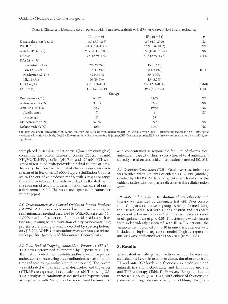
Oxidative Medicine and Cellular Longevity 3
Table 1: Clinical and laboratory data in patients with rheumatoid arthritis with (IR+) or without (IR−) insulin resistance.
IR− (𝑛 = 91) IR+ (𝑛 = 82) 𝑝
Disease duration (years) 11.0 (5.0–18.3) 8.0 (4.0–20.3) NSRF (IU/mL) 48.3 (0.0–125.0) 26.9 (0.0–118.2) NSAnti-CCP (U/mL) 25.55 (0.13–120.10) 6.65 (0.50–131.40) NSDAS 28 3.51 (2.39–4.49) 3.76 (2.85–4.78) 0.043DAS 28, 𝑛 (%)
Remission (<2.6) 27 (29.7% ) 16 (19.5%)Low (2.6–3.2) 12 (13.2%) 11 (13.4%) 0.001Moderate (3.2–5.1) 42 (46.1%) 39 (47.6%)High (>5.1) 10 (10.0%) 16 (19.5%)
CPR (mg/L) 3.52 (1.31–12.38) 6.35 (2.51–11.08) 0.040ESR (mm) 14.0 (6.0–22.0) 19.5 (9.3–35.5) 0.023
TherapyPrednisone (Y/N) 64/27 54/28 NSAntimalarials (Y/N) 38/53 32/50 NSAnti-TNF-𝛼 (Y/N) 20/71 19/63 NS
Adalimumab 7 6 NSEtanercept 13 13
Methotrexate (Y/N) 57/34 62/20 NSLeflunomide (Y/N) 40/51 35/47 NSChi-square test with Yates correction. Mann-Whitney test. Data are expressed as median (25–75%). Y, yes; N, no; RF, rheumatoid factor; anti-CCP, anti-cycliccitrullinated peptide antibody; DAS 28, Disease Activity Score evaluating 28 joints; CRP, C-reactive protein; ESR, erythrocyte sedimentation rate; and NS, notsignificant.
were placed in 20mL scintillation vials (low-potassium glass)containing final concentrations of plasma (250𝜇L), 30mMKH2PO4/K2HPO4buffer (pH 7.4), and 120mM KCl with
3mM of tert-butyl hydroperoxide in a final volume of 2mL.Tert-butyl hydroperoxide-initiated chemiluminescence wasmeasured in Beckman LS 6000 Liquid Scintillation Counterset to the out-of-coincidence mode, with a response rangefrom 300 to 620 nm. The vials were kept in the dark up tothe moment of assay, and determination was carried out ina dark room at 30∘C. The results are expressed in counts perminute (cpm).
2.6. Determination of Advanced Oxidation Protein Products(AOPPs). AOPPs were determined in the plasma using thesemiautomatedmethod described byWitko-Sarsat et al. [30].AOPPs results of oxidation of amino acid residues such astyrosine, leading to the formation of dityrosine-containingprotein cross-linking products detected by spectrophotom-etry [17, 30]. AOPPs concentrations were expressed as micro-moles per liter (𝜇mol/L) of chloramines-T equivalents.
2.7. Total Radical-Trapping Antioxidant Parameter (TRAP).TRAP was determined as reported by Repetto et al. [31].This method detects hydrosoluble and/or liposoluble plasmaantioxidants bymeasuring the chemiluminescence inhibitiontime induced by 2,2-azobis(2-amidinopropane). The systemwas calibrated with vitamin E analog Trolox, and the valuesof TRAP are expressed in equivalent of 𝜇M Trolox/mg UA.TRAP analysis in conditions associated with hyperuricemia,as in patients with MetS, may be jeopardized because uric
acid concentration is responsible for 60% of plasma totalantioxidant capacity. Thus, a correction of total antioxidantcapacity based on uric acid concentration is needed [32, 33].
2.8. Oxidative Stress Index (OSI). Oxidative stress imbalancewas verified when OSI was calculated as AOPPs (𝜇mol/L)divided by TRAP (𝜇M Trolox/mg UA), which indicates theoxidant-antioxidant ratio as a reflection of the cellular redoxstate.
2.9. Statistical Analysis. Distribution of sex, ethnicity, andtherapy was analyzed by chi-square test with Yates correc-tion. Comparisons between groups were performed usingthe Kruskal-Wallis test with Dunn’s posttest and data wereexpressed as the median (25–75%). The results were consid-ered significant when 𝑝 < 0.05. To determine which factorswere independently associated with IR in RA patients, thevariables that presented 𝑝 < 0.10 in univariate analyses wereincluded in logistic regression model. Logistic regressionanalyses were performed with SPSS v20.0 (IBM, USA).
3. Results
Rheumatoid arthritis patients with or without IR were notstatistically different in relation to disease duration and serumRF and anti-CCP levels and frequency in prednisone andantimalarials and methotrexate and leflunomide use andanti-TNF-𝛼 therapy (Table 1). However, IR+ group had anincreased DAS 28 (𝑝 = 0.043) with enhanced frequency inpatients with high disease activity. In addition, IR+ group
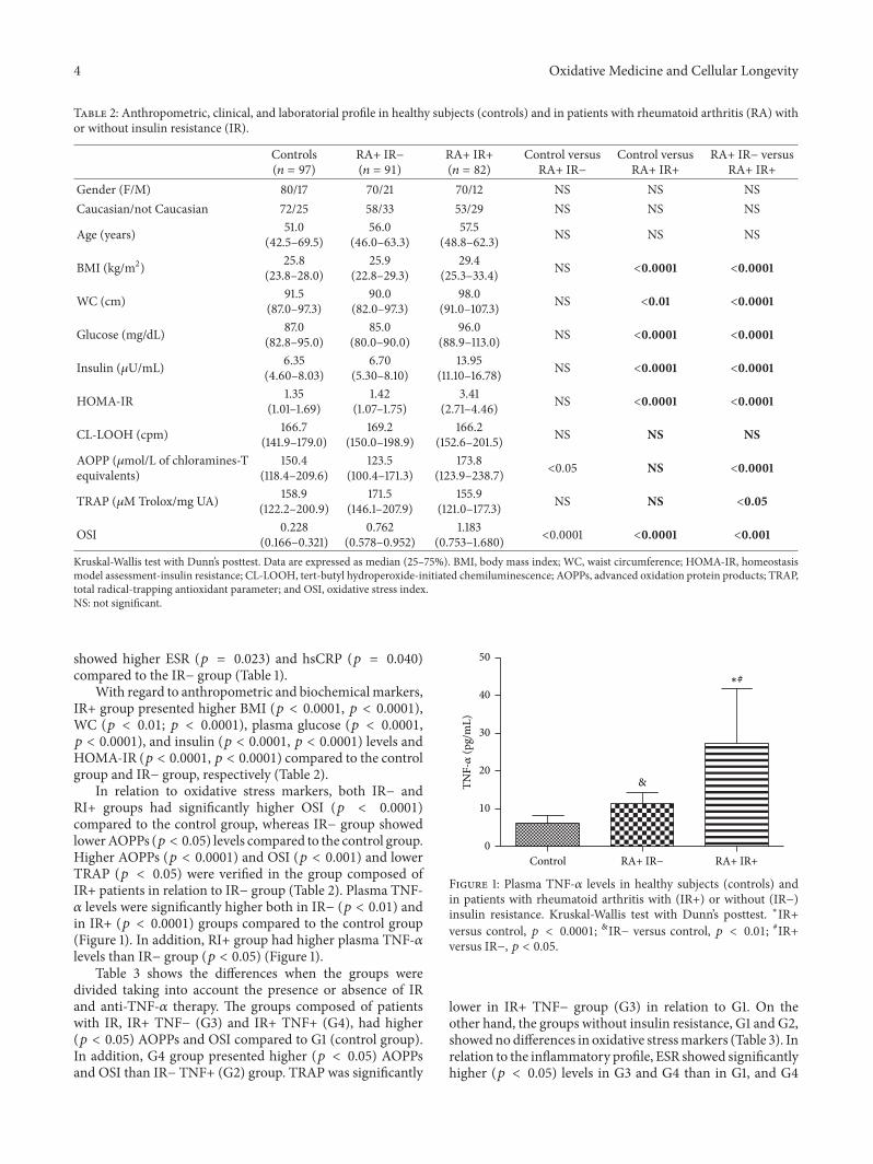
4 Oxidative Medicine and Cellular Longevity
Table 2: Anthropometric, clinical, and laboratorial profile in healthy subjects (controls) and in patients with rheumatoid arthritis (RA) withor without insulin resistance (IR).
Controls(𝑛 = 97)
RA+ IR−(𝑛 = 91)
RA+ IR+(𝑛 = 82)
Control versusRA+ IR−
Control versusRA+ IR+
RA+ IR− versusRA+ IR+
Gender (F/M) 80/17 70/21 70/12 NS NS NSCaucasian/not Caucasian 72/25 58/33 53/29 NS NS NS
Age (years) 51.0(42.5–69.5)
56.0(46.0–63.3)
57.5(48.8–62.3) NS NS NS
BMI (kg/m2) 25.8(23.8–28.0)
25.9(22.8–29.3)
29.4(25.3–33.4) NS <0.0001 <0.0001
WC (cm) 91.5(87.0–97.3)
90.0(82.0–97.3)
98.0(91.0–107.3) NS <0.01 <0.0001
Glucose (mg/dL) 87.0(82.8–95.0)
85.0(80.0–90.0)
96.0(88.9–113.0) NS <0.0001 <0.0001
Insulin (𝜇U/mL) 6.35(4.60–8.03)
6.70(5.30–8.10)
13.95(11.10–16.78) NS <0.0001 <0.0001
HOMA-IR 1.35(1.01–1.69)
1.42(1.07–1.75)
3.41(2.71–4.46) NS <0.0001 <0.0001
CL-LOOH (cpm) 166.7(141.9–179.0)
169.2(150.0–198.9)
166.2(152.6–201.5) NS NS NS
AOPP (𝜇mol/L of chloramines-Tequivalents)
150.4(118.4–209.6)
123.5(100.4–171.3)
173.8(123.9–238.7) <0.05 NS <0.0001
TRAP (𝜇M Trolox/mg UA) 158.9(122.2–200.9)
171.5(146.1–207.9)
155.9(121.0–177.3) NS NS <0.05
OSI 0.228(0.166–0.321)
0.762(0.578–0.952)
1.183(0.753–1.680) <0.0001 <0.0001 <0.001
Kruskal-Wallis test with Dunn’s posttest. Data are expressed as median (25–75%). BMI, body mass index; WC, waist circumference; HOMA-IR, homeostasismodel assessment-insulin resistance; CL-LOOH, tert-butyl hydroperoxide-initiated chemiluminescence; AOPPs, advanced oxidation protein products; TRAP,total radical-trapping antioxidant parameter; and OSI, oxidative stress index.NS: not significant.
showed higher ESR (𝑝 = 0.023) and hsCRP (𝑝 = 0.040)compared to the IR− group (Table 1).
With regard to anthropometric and biochemicalmarkers,IR+ group presented higher BMI (𝑝 < 0.0001, 𝑝 < 0.0001),WC (𝑝 < 0.01; 𝑝 < 0.0001), plasma glucose (𝑝 < 0.0001,𝑝 < 0.0001), and insulin (𝑝 < 0.0001, 𝑝 < 0.0001) levels andHOMA-IR (𝑝 < 0.0001, 𝑝 < 0.0001) compared to the controlgroup and IR− group, respectively (Table 2).
In relation to oxidative stress markers, both IR− andRI+ groups had significantly higher OSI (𝑝 < 0.0001)compared to the control group, whereas IR− group showedlowerAOPPs (𝑝 < 0.05) levels compared to the control group.Higher AOPPs (𝑝 < 0.0001) and OSI (𝑝 < 0.001) and lowerTRAP (𝑝 < 0.05) were verified in the group composed ofIR+ patients in relation to IR− group (Table 2). Plasma TNF-𝛼 levels were significantly higher both in IR− (𝑝 < 0.01) andin IR+ (𝑝 < 0.0001) groups compared to the control group(Figure 1). In addition, RI+ group had higher plasma TNF-𝛼levels than IR− group (𝑝 < 0.05) (Figure 1).
Table 3 shows the differences when the groups weredivided taking into account the presence or absence of IRand anti-TNF-𝛼 therapy. The groups composed of patientswith IR, IR+ TNF− (G3) and IR+ TNF+ (G4), had higher(𝑝 < 0.05) AOPPs and OSI compared to G1 (control group).In addition, G4 group presented higher (𝑝 < 0.05) AOPPsand OSI than IR− TNF+ (G2) group. TRAP was significantly
&
∗#
0
10
20
30
40
50
RA+ IR− RA+ IR+Control
TNF-𝛼
(pg/
mL)
Figure 1: Plasma TNF-𝛼 levels in healthy subjects (controls) andin patients with rheumatoid arthritis with (IR+) or without (IR−)insulin resistance. Kruskal-Wallis test with Dunn’s posttest. ∗IR+versus control, 𝑝 < 0.0001; &IR− versus control, 𝑝 < 0.01; #IR+versus IR−, 𝑝 < 0.05.
lower in IR+ TNF− group (G3) in relation to G1. On theother hand, the groups without insulin resistance, G1 and G2,showed no differences in oxidative stressmarkers (Table 3). Inrelation to the inflammatory profile, ESR showed significantlyhigher (𝑝 < 0.05) levels in G3 and G4 than in G1, and G4
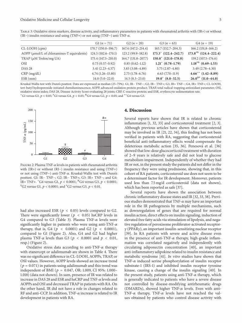
Oxidative Medicine and Cellular Longevity 5
Table 3: Oxidative stress markers, disease activity, and inflammatory parameters in patients with rheumatoid arthritis with (IR+) or without(IR−) insulin resistance and using (TNF+) or not using (TNF−) anti-TNF-𝛼.
G1 (𝑛 = 71) G2 (𝑛 = 20) G3 (𝑛 = 63) G4 (𝑛 = 19)CL-LOOH (cpm) 170.7 (150.0–196.7) 167.4 (147.2–214.4) 165.7 (152.7–204.3) 166.2 (151.8–166.2)AOPP (𝜇mol/L of chloramines-T equivalents) 124.5 (102.6–170.1) 123.2 (99.9–182.8) 173.3∗ (122.4–242.7) 173.8#& (124.4–222.4)TRAP (𝜇M Trolox/mg UA) 175.4 (147.3–210.0) 164.7 (131.8–207.7) 150.8∗ (121.0–178.8) 159.2 (107.5–176.6)OSI 0.73 (0.57–0.92) 0.85 (0.62–1.12) 1.21∗ (0.78–1.79) 1.18#& (0.69–1.53)DAS 28 3.41 (2.23–4.57) 3.83 (3.08–4.89) 3.75 (2.87–4.80) 3.49 (2.78–4.30)CRP (mg/dL) 4.74 (1.26–15.80) 2.75 (1.78–6.76) 6.63 (7.70–11.9) 4.66∗∗ (1.42–8.89)ESR (mm) 14.0 (5.0–22.0) 14.5 (8.3–23.0) 19.0∗ (8.0–32.5) 26.0&# (11.0–44.0)Kruskal-Wallis test with Dunn’s posttest. Data are expressed as median (25–75%). G1, IR− TNF−; G2, IR− TNF+; G3, IR+ TNF−; G4, IR+ TNF+; CL-LOOH,tert-butyl hydroperoxide-initiated chemiluminescence; AOPP, advanced oxidation protein product; TRAP, total radical-trapping antioxidant parameter; OSI,oxidative stress index; DAS 28, Disease Activity Score evaluating 28 joints; CRP, C-reactive protein; and ESR, erythrocyte sedimentation rate.∗G1 versus G3, 𝑝 < 0.05; #G1 versus G4, 𝑝 < 0.05; &G4 versus G2, 𝑝 < 0.05; and ∗∗G4 versus G3.
&∗
$
#
TNF-𝛼
(pg/
mL)
0
50
100
150
200
G2 G3 G4G1
Figure 2: Plasma TNF-𝛼 levels in patients with rheumatoid arthritiswith (IR+) or without (RI−) insulin resistance and using (TNF+)or not using (TNF−) anti-TNF-𝛼. Kruskal-Wallis test with Dunn’sposttest. G1: IR− TNF−; G2: IR− TNF+; G3: IR+ TNF−; and G4:IR+ TNF+. ∗G4 versus G1, 𝑝 < 0.0001; &G4 versus G3, 𝑝 < 0.0001;$G2 versus G1, 𝑝 < 0.0001; and #G2 versus G3, 𝑝 < 0.01.
had also increased ESR (𝑝 < 0.05) levels compared to G2.There were significantly lower (𝑝 < 0.05) hsCRP levels inG4 compared to G3 (Table 3). Plasma TNF-𝛼 levels weresignificantly higher in patients who were using anti-TNF-𝛼therapy, that is, G4 (𝑝 < 0.0001) and G2 (𝑝 < 0.0001),compared to G1 (Figure 2). Also, G4 and G2 had higherplasma TNF-𝛼 levels than G3 (𝑝 < 0.0001 and 𝑝 < 0.01,resp.) (Figure 2).
Oxidative stress data according to anti-TNF-𝛼 therapywith etanercept or adalimumab are shown in Table 4. Therewas no significant difference in CL-LOOH, AOPPs, TRAP, orOSI values. However, AOPP levels showed an increase trend(𝑝 = 0.071) in patients using adalimumab and this trend wasindependent of BMI (𝑝 = 0.047, OR: 1.009, CI 95%: 1.000–1.018) (data not shown). In sum, presence of IR was related toincrease inDAS 28 and ESR and hsCRP andTNF-𝛼 levels andAOPPs andOSI and decreased TRAP in patients with RA.Onthe other hand, IR did not have a role in changes related toRF and anti-CCP. In addition, TNF-𝛼 increase is related to IRdevelopment in patients with RA.
4. Discussion
Several reports have shown that IR is related to chronicinflammation [1, 32, 33] and corticosteroid treatment [2, 3].Although previous articles have shown that corticosteroidmay be involved in IR [21, 22, 34], this finding has not beenverified in patients with RA, suggesting that corticosteroidbeneficial anti-inflammatory effects would compensate thedeleterious metabolic action [35, 36]. Penesová et al. [36]showed that low-dose glucocorticoid treatmentwith durationof 2–9 years is relatively safe and did not lead to glucosemetabolism impairment. Independently of whether they hadIR or not, in the present study the patients did not differ in thefrequency they were using prednisone, showing that, in thiscohort of RA patients, corticosteroid use does not seem to bea determinant factor for IR development. Moreover, patientsused less than 7.5mg/d corticosteroid (data not shown),which has been reported as safe [37].
Several reports have shown the association betweenchronic inflammatory disease states and IR [32, 33, 38]. Previ-ous studies demonstrated that TNF-𝛼may have an importantrole in the IR pathogenesis by multiple mechanisms, suchas downregulation of genes that are required for normalinsulin action, direct effects on insulin signaling, induction ofelevated free fatty acids via stimulation of lipolysis, and nega-tive regulation of peroxisome proliferator-activated receptor-𝛾 (PPAR𝛾), an important insulin-sensitizing nuclear receptor[39]. In RA patients with severe and active disease evenin the presence of anti-TNF-𝛼 therapy, high-grade inflam-mation was correlated negatively and independently withcirculating adiponectin concentration [40], an importantanti-inflammatory adipokine related to insulin resistance andmetabolic syndrome [41]. In vitro studies have shown thatTNF-𝛼 induced serine phosphorylation of insulin receptorsubstrate-1 (IRS-1) and inhibited insulin receptor tyrosinekinase, causing a change of the insulin signaling [40]. Inthe present study, patients using anti-TNF-𝛼 therapy, whichis generally indicated to patients who have a severe diseasenot controlled by disease-modifying antirheumatic drugs(DMARDs), showed higher TNF-𝛼 levels. Even with anti-TNF-𝛼 therapy, TNF-𝛼 levels have not reached the val-ues obtained by patients who control disease activity with
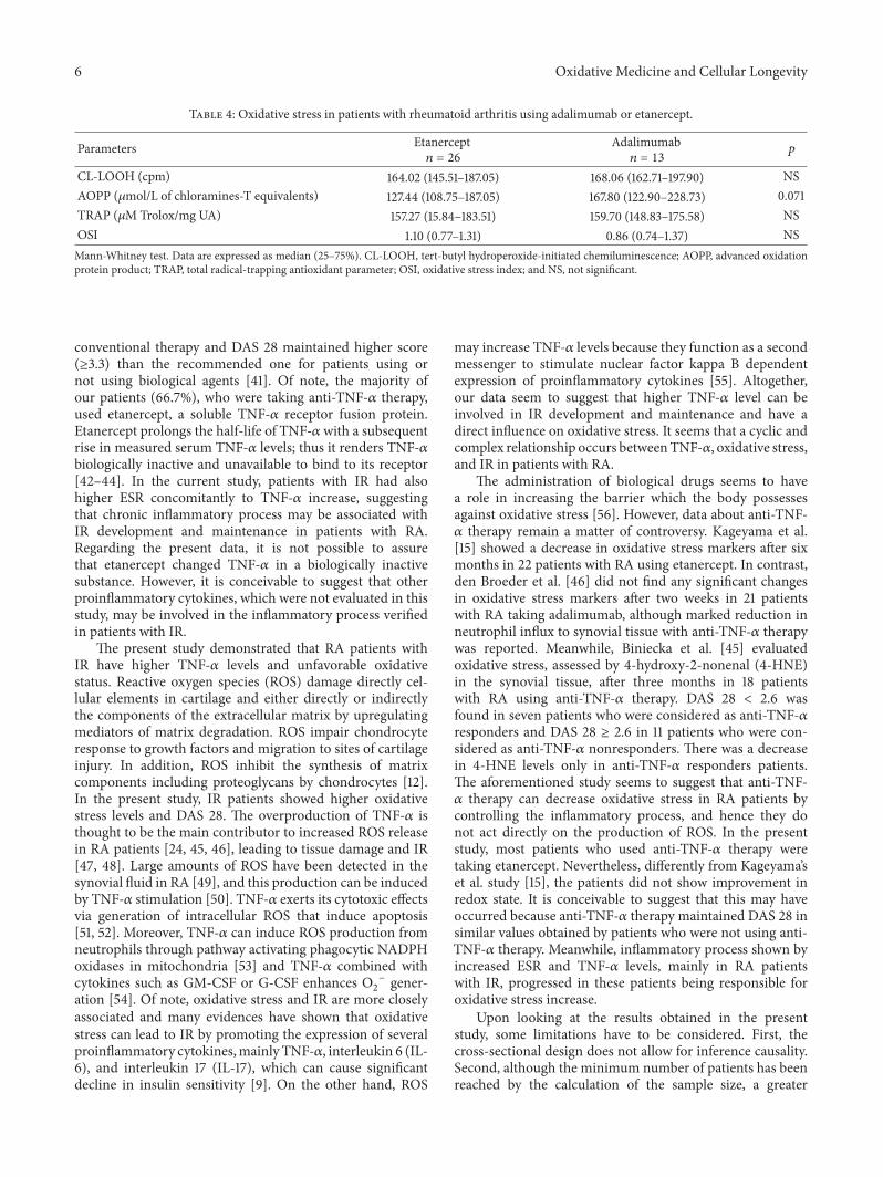
6 Oxidative Medicine and Cellular Longevity
Table 4: Oxidative stress in patients with rheumatoid arthritis using adalimumab or etanercept.
Parameters Etanercept𝑛 = 26
Adalimumab𝑛 = 13
𝑝
CL-LOOH (cpm) 164.02 (145.51–187.05) 168.06 (162.71–197.90) NSAOPP (𝜇mol/L of chloramines-T equivalents) 127.44 (108.75–187.05) 167.80 (122.90–228.73) 0.071TRAP (𝜇M Trolox/mg UA) 157.27 (15.84–183.51) 159.70 (148.83–175.58) NSOSI 1.10 (0.77–1.31) 0.86 (0.74–1.37) NSMann-Whitney test. Data are expressed as median (25–75%). CL-LOOH, tert-butyl hydroperoxide-initiated chemiluminescence; AOPP, advanced oxidationprotein product; TRAP, total radical-trapping antioxidant parameter; OSI, oxidative stress index; and NS, not significant.
conventional therapy and DAS 28 maintained higher score(≥3.3) than the recommended one for patients using ornot using biological agents [41]. Of note, the majority ofour patients (66.7%), who were taking anti-TNF-𝛼 therapy,used etanercept, a soluble TNF-𝛼 receptor fusion protein.Etanercept prolongs the half-life of TNF-𝛼with a subsequentrise in measured serum TNF-𝛼 levels; thus it renders TNF-𝛼biologically inactive and unavailable to bind to its receptor[42–44]. In the current study, patients with IR had alsohigher ESR concomitantly to TNF-𝛼 increase, suggestingthat chronic inflammatory process may be associated withIR development and maintenance in patients with RA.Regarding the present data, it is not possible to assurethat etanercept changed TNF-𝛼 in a biologically inactivesubstance. However, it is conceivable to suggest that otherproinflammatory cytokines, which were not evaluated in thisstudy, may be involved in the inflammatory process verifiedin patients with IR.
The present study demonstrated that RA patients withIR have higher TNF-𝛼 levels and unfavorable oxidativestatus. Reactive oxygen species (ROS) damage directly cel-lular elements in cartilage and either directly or indirectlythe components of the extracellular matrix by upregulatingmediators of matrix degradation. ROS impair chondrocyteresponse to growth factors and migration to sites of cartilageinjury. In addition, ROS inhibit the synthesis of matrixcomponents including proteoglycans by chondrocytes [12].In the present study, IR patients showed higher oxidativestress levels and DAS 28. The overproduction of TNF-𝛼 isthought to be the main contributor to increased ROS releasein RA patients [24, 45, 46], leading to tissue damage and IR[47, 48]. Large amounts of ROS have been detected in thesynovial fluid in RA [49], and this production can be inducedby TNF-𝛼 stimulation [50]. TNF-𝛼 exerts its cytotoxic effectsvia generation of intracellular ROS that induce apoptosis[51, 52]. Moreover, TNF-𝛼 can induce ROS production fromneutrophils through pathway activating phagocytic NADPHoxidases in mitochondria [53] and TNF-𝛼 combined withcytokines such as GM-CSF or G-CSF enhances O
2
− gener-ation [54]. Of note, oxidative stress and IR are more closelyassociated and many evidences have shown that oxidativestress can lead to IR by promoting the expression of severalproinflammatory cytokines,mainly TNF-𝛼, interleukin 6 (IL-6), and interleukin 17 (IL-17), which can cause significantdecline in insulin sensitivity [9]. On the other hand, ROS
may increase TNF-𝛼 levels because they function as a secondmessenger to stimulate nuclear factor kappa B dependentexpression of proinflammatory cytokines [55]. Altogether,our data seem to suggest that higher TNF-𝛼 level can beinvolved in IR development and maintenance and have adirect influence on oxidative stress. It seems that a cyclic andcomplex relationship occurs betweenTNF-𝛼, oxidative stress,and IR in patients with RA.
The administration of biological drugs seems to havea role in increasing the barrier which the body possessesagainst oxidative stress [56]. However, data about anti-TNF-𝛼 therapy remain a matter of controversy. Kageyama et al.[15] showed a decrease in oxidative stress markers after sixmonths in 22 patients with RA using etanercept. In contrast,den Broeder et al. [46] did not find any significant changesin oxidative stress markers after two weeks in 21 patientswith RA taking adalimumab, although marked reduction inneutrophil influx to synovial tissue with anti-TNF-𝛼 therapywas reported. Meanwhile, Biniecka et al. [45] evaluatedoxidative stress, assessed by 4-hydroxy-2-nonenal (4-HNE)in the synovial tissue, after three months in 18 patientswith RA using anti-TNF-𝛼 therapy. DAS 28 < 2.6 wasfound in seven patients who were considered as anti-TNF-𝛼responders and DAS 28 ≥ 2.6 in 11 patients who were con-sidered as anti-TNF-𝛼 nonresponders. There was a decreasein 4-HNE levels only in anti-TNF-𝛼 responders patients.The aforementioned study seems to suggest that anti-TNF-𝛼 therapy can decrease oxidative stress in RA patients bycontrolling the inflammatory process, and hence they donot act directly on the production of ROS. In the presentstudy, most patients who used anti-TNF-𝛼 therapy weretaking etanercept. Nevertheless, differently from Kageyama’set al. study [15], the patients did not show improvement inredox state. It is conceivable to suggest that this may haveoccurred because anti-TNF-𝛼 therapy maintained DAS 28 insimilar values obtained by patients who were not using anti-TNF-𝛼 therapy. Meanwhile, inflammatory process shown byincreased ESR and TNF-𝛼 levels, mainly in RA patientswith IR, progressed in these patients being responsible foroxidative stress increase.
Upon looking at the results obtained in the presentstudy, some limitations have to be considered. First, thecross-sectional design does not allow for inference causality.Second, although the minimum number of patients has beenreached by the calculation of the sample size, a greater
Oxidative Medicine and Cellular Longevity 7
number of patients would probably confer more strength tothe statistical results.
This study corroborates with Binieckas et al.’s [45],which suggested that inflammatory state maintenance can beresponsible for oxidative stress found in patients with RA.On the other hand, the data of the present study show thatIR is involved in an unbalanced redox state, which possiblycontributes to maintaining a vicious circle of high-gradeinflammation.
5. Conclusions
This study demonstrates that IR and TNF-𝛼 are importantfactors involved in redox imbalance in patients with RA andit seems to be due to the maintenance of inflammatory stateand disease activity. The data from the present study suggesta complex interaction of TNF-𝛼, oxidative stress, and IR,but the presence of insulin resistance seems to be directlyassociated with both oxidative stress and TNF-𝛼 levels. Thedifferences in oxidative stress markers in RA patients with orwithout IR could contribute to a better design for future drugsand/or nutritional interventional studies in this population.In addition, more studies are warranted to verify if IR can beinvolved in therapeutic failure with TNF-𝛼 inhibitors.
Abbreviations
Anti-CCP: Anticyclic citrullinated peptideAOPPs: Advanced oxidation protein productsCL-LOOH: Tert-butyl hydroperoxide-initiated
chemiluminescencehsCRP: Highly sensitive C-reactive proteinDAS 28: Disease Activity Score evaluating 28 joints4-HNE: 4-Hidroxy-2-nonenalCVD: Cardiovascular diseaseDMARDs: Disease-modifying antirheumatic drugsESR: Erythrocyte sedimentation rateHOMA-IR: Homeostasis model assessment-insulin
resistanceIL-6: Interleukin 6IL-17: Interleukin 17IR: Insulin resistanceOSI: Oxidative stress indexPPAR𝛾: Peroxisome proliferator-activated
receptor-𝛾RA: Rheumatoid arthritisRLU: Relative luminescence unitsRF: Rheumatoid factorROS: Reactive oxygen speciesTRAP: Total radical-trapping antioxidant
parameterTNF-𝛼: Tumor necrosis factor-alphaUA: Uric acidWC: Waist circumference.
Competing Interests
The authors declare that they have no competing interests.
Authors’ Contributions
NeideTomimuraCosta helped to analyze the data and to draftthe paper. Andréa Name Colado Simão performed data anal-ysis and together with Isaias Dichi helped to design the studyand draft the paper and critically revised the paper for impor-tant intellectual content. Vinicius Daher Alvares Delfinohelped to analyze the data and critically revised the paper.Tatiana Mayumi Veiga Iriyoda and Ricardo Braga Aminparticipated in clinical assessments. Francieli Delongui, AnaPaula Kallaur, Marcell Alysson Batisti Lozovoy, and DanielaFrizon Alfieri helped in laboratory analyses. All authors havegiven final approval of the version to be published and agreeto be accountable for all aspects of the work in ensuring thatquestions related to the accuracy or integrity of any part ofthe work are appropriately investigated and resolved.
Acknowledgments
This research was supported by the National Council ofBrazilian Research (CNPq) and Araucaria Foundation fromParaná, Brazil.
References
[1] I. D. del Rincón, K. Williams, M. P. Stern, G. L. Freeman, andA. Escalante, “High incidence of cardiovascular events in arheumatoid arthritis cohort not explained by traditional cardiacrisk factors,”Arthritis and Rheumatism, vol. 44, no. 12, pp. 2737–2745, 2001.
[2] S. E. Gabriel, “Cardiovascular morbidity and mortality inrheumatoid arthritis,” The American Journal of Medicine, vol.121, no. 10, pp. S9–S14, 2008.
[3] S. Haque, H. Mirjafari, and I. N. Bruce, “Atherosclerosis inrheumatoid arthritis and systemic lupus erythematosus,” Cur-rent Opinion in Lipidology, vol. 19, no. 4, pp. 338–343, 2008.
[4] C. De Luca and J. M. Olefsky, “Inflammation and insulinresistance,” FEBS Letters, vol. 582, no. 1, pp. 97–105, 2008.
[5] A. M. El-Barbary, E. M. Kassem, M. A. S. El-Sergany, S. A.-M. Essa, and M. A. Eltomey, “Association of anti-modifiedcitrullinated vimentin with subclinical atherosclerosis in earlyrheumatoid arthritis compared with anti-cyclic citrullinatedpeptide,” The Journal of Rheumatology, vol. 38, no. 5, pp. 828–834, 2011.
[6] T. Saxne, M. A. Palladino Jr., D. Heinegard, N. Talal, and F.A. Wollheim, “Detection of tumor necrosis factor 𝛼 but nottumor necrosis factor 𝛽 in rheumatoid arthritis synovial fluidand serum,” Arthritis and Rheumatism, vol. 31, no. 8, pp. 1041–1045, 1988.
[7] I. B. McInnes and G. Schett, “The pathogenesis of rheumatoidarthritis,”TheNew England Journal of Medicine, vol. 365, no. 23,pp. 2205–2219, 2011.
[8] C. P. Chung, A. Oeser, J. F. Solus et al., “Inflammation-associated insulin resistance: differential effects in rheumatoidarthritis and systemic lupus erythematosus define potentialmechanisms,”Arthritis and Rheumatism, vol. 58, no. 7, pp. 2105–2112, 2008.
[9] M. L. L. Prevoo, M. A. van’t Hof, H. H. Kuper, M. A. vanLeeuwen, L. B. A. van de Putte, and P. L. C. M. van Riel,“Modified disease activity scores that include twenty-eight-joint
8 Oxidative Medicine and Cellular Longevity
counts development and validation in a prospective longitu-dinal study of patients with rheumatoid arthritis,” Arthritis &Rheumatism, vol. 38, no. 1, pp. 44–48, 1995.
[10] P. Vasanthi, G. Nalini, and G. Rajasekhar, “Status of oxida-tive stress in rheumatoid arthritis,” International Journal ofRheumatic Diseases, vol. 12, no. 1, pp. 29–33, 2009.
[11] L. I. Filippin, R. Vercelino, N. P. Marroni, and R. M. Xavier,“Redox signalling and the inflammatory response in rheuma-toid arthritis,” Clinical and Experimental Immunology, vol. 152,no. 3, pp. 415–422, 2008.
[12] C. A.Hitchon andH. S. El-Gabalawy, “Oxidation in rheumatoidarthritis,” Arthritis Research and Therapy, vol. 6, no. 6, pp. 265–278, 2004.
[13] S. Taysi, F. Polat, M. Gul, R. Sari, and E. Bakan, “Lipid peroxida-tion, some extracellular antioxidants, and antioxidant enzymesin serum of patients with rheumatoid arthritis,” RheumatologyInternational, vol. 21, no. 5, pp. 200–204, 2002.
[14] S. Z. Hassan, T. A. Gheita, S. A. Kenawy, A. T. Fahim, I. M. El-Sorougy, and M. S. Abdou, “Oxidative stress in systemic lupuserythematosus and rheumatoid arthritis patients: relationshipto disease manifestations and activity,” International Journal ofRheumatic Diseases, vol. 14, no. 4, pp. 325–331, 2011.
[15] Y. Kageyama, M. Takahashi, T. Nagafusa, E. Torikai, andA. Nagano, “Etanercept reduces the oxidative stress markerlevels in patients with rheumatoid arthritis,” RheumatologyInternational, vol. 28, no. 3, pp. 245–251, 2008.
[16] S. Shahmohamadnejad, A. Vaisi-Raygani, Y. Shakiba et al.,“Association between butyrylcholinesterase activity and phe-notypes, paraoxonase192 rs662 gene polymorphism and theirenzymatic activity with severity of rheumatoid arthritis: corre-lationwith systemic inflammatorymarkers and oxidative stress,preliminary report,” Clinical Biochemistry, vol. 48, no. 1-2, pp.63–69, 2015.
[17] M. A. B. Lozovoy, A. N. C. Simão, M. S. N. Hohmann et al.,“Inflammatory biomarkers and oxidative stress measurementsin patients with systemic lupus erythematosus with or withoutmetabolic syndrome,” Lupus, vol. 20, no. 13, pp. 1356–1364, 2011.
[18] M. A. B. Lozovoy, A. N. C. Simão, S. R. Oliveira et al.,“Relationship between iron metabolism, oxidative stress, andinsulin resistance in patients with systemic lupus erythemato-sus,” Scandinavian Journal of Rheumatology, vol. 42, no. 4, pp.303–310, 2013.
[19] S. R. Oliveira, A. N. Colado Simão, A. P. Kallaur et al.,“Disability in patients with multiple sclerosis: influence ofinsulin resistance, adiposity, and oxidative stress,”Nutrition, vol.30, no. 3, pp. 268–273, 2014.
[20] H. K. Morimoto, A. N. C. Simão, E. R. D. D. Almeida etal., “Role of metabolic syndrome and antiretroviral therapyin adiponectin levels and oxidative stress in HIV-1 infectedpatients,” Nutrition, vol. 30, no. 11-12, pp. 1324–1330, 2014.
[21] P. H. Dessein and B. I. Joffe, “Insulin resistance and impairedbeta cell function in rheumatoid arthritis,” Arthritis andRheumatism, vol. 54, no. 9, pp. 2765–2775, 2006.
[22] G. La Montagna, F. Cacciapuoti, R. Buono et al., “Insulinresistance is an independent risk factor for atherosclerosis inrheumatoid arthritis,” Diabetes and Vascular Disease Research,vol. 4, no. 2, pp. 130–135, 2007.
[23] G. Arcaro, “Insulin causes endothelial dysfunction in humans:sites and mechanisms,” Circulation, vol. 105, no. 5, pp. 576–582,2002.
[24] Y. Kageyama, M. Takahashi, T. Ichikawa, E. Torikai, andA. Nagano, “Reduction of oxidative stress marker levels by
anti-TNF-𝛼 antibody, infliximab, in patients with rheumatoidarthritis,” Clinical and Experimental Rheumatology, vol. 26, no.1, pp. 73–80, 2008.
[25] O. Altindag, M. Karakoc, A. Kocyigit, H. Celik, and N. Soran,“Increased DNA damage and oxidative stress in patients withrheumatoid arthritis,” Clinical Biochemistry, vol. 40, no. 3-4, pp.167–171, 2007.
[26] A. Nakajima, Y. Aoki, Y. Shibata et al., “Identification of clinicalparameters associated with serum oxidative stress in patientswith rheumatoid arthritis,” Modern Rheumatology, vol. 24, no.6, pp. 926–930, 2014.
[27] D. Aletaha, T. Neogi, and A. J. Silman, “2010 rheumatoid arthri-tis classification criteria: An American College of Rheumatol-ogy/European League against rheumatism collaborative initia-tive,” Annals of the Rheumatic Diseases, vol. 69, no. 10, pp. 1580–1588, 2010.
[28] S. M. Haffner, H. Miettinen, and M. P. Stern, “The homeostasismodel in the San Antonio Heart Study,” Diabetes Care, vol. 20,no. 7, pp. 1087–1092, 1997.
[29] B. Gonzalez Flecha, S. Llesuy, and A. Boveris, “Hydroperoxide-initiated chemiluminescence: an assay for oxidative stress inbiopsies of heart, liver, and muscle,” Free Radical Biology andMedicine, vol. 10, no. 2, pp. 93–100, 1991.
[30] V. Witko-Sarsat, M. Friedlander, T. N. Khoa et al., “Advancedoxidation protein products as novel mediators of inflammationand monocyte activation in chronic renal failure,” The Journalof Immunology, vol. 161, no. 5, pp. 2524–2532, 1998.
[31] M. Repetto, C. Reides, M. L. Gomez Carretero, M. Costa, G.Griemberg, and S. Llesuy, “Oxidative stress in blood of HIVinfected patients,” Clinica Chimica Acta, vol. 255, no. 2, pp. 107–117, 1996.
[32] G. S. Hotamisligil, “Molecular mechanisms of insulin resistanceand the role of the adipocyte,” International Journal of Obesity,vol. 24, supplement 4, pp. S23–S27, 2000.
[33] C. Popa, M. G. Netea, P. L. C. M. Van Riel, J. W. M. VanDer Meer, and A. F. H. Stalenhoef, “The role of TNF-𝛼 inchronic inflammatory conditions, intermediary metabolism,and cardiovascular risk,” Journal of Lipid Research, vol. 48, no.4, pp. 751–752, 2007.
[34] P. H. Dessein, B. I. Joffe, A. E. Stanwix, B. F. Christian, and M.Veller, “Glucocorticoids and insulin sensitivity in rheumatoidarthritis,” The Journal of Rheumatology, vol. 31, no. 5, pp. 867–874, 2004.
[35] K. L. G. Svenson, G. Lundqvist, L. Wide, and R. Hällgren,“Impaired glucose handling in active rheumatoid arthri-tis: effects of corticosteroids and antirheumatic treatment,”Metabolism, vol. 36, no. 10, pp. 944–948, 1987.
[36] A. Penesová, Z. Rádiková, M. Vlček et al., “Chronic inflamma-tion and low-dose glucocorticoid effects on glucosemetabolismin premenopausal females with rheumatoid arthritis free ofconventional metabolic risk factors,” Physiological Research, vol.62, no. 1, pp. 75–83, 2013.
[37] J. M. Sabio, J. A. Vargas-Hitos, N. Navarrete, C. Hidalgo-Tenorio, and J. Jiménez-Alonso, “Effects of low ormedium-doseof prednisone on insulin resistance in patients with systemiclupus erythematosus,”Clinical and Experimental Rheumatology,vol. 28, no. 4, pp. 483–489, 2010.
[38] L.-S. Tam, B. Tomlinson, T. T. Chu, T. K. Li, and E. K. Li,“Impact of TNF inhibition on insulin resistance and lipids levelsin patients with rheumatoid arthritis,” Clinical Rheumatology,vol. 26, no. 9, pp. 1495–1498, 2007.
Oxidative Medicine and Cellular Longevity 9
[39] D. E. Moller, “Potential role of TNF-𝛼 in the pathogenesis ofinsulin resistance and type 2 diabetes,” Trends in Endocrinologyand Metabolism, vol. 11, no. 6, pp. 212–217, 2000.
[40] T. Hayakawa, Y. Nagai, M. Taniguchi et al., “Tumor necrosisfactor-𝛽 gene NcoI polymorphism decreases insulin resistancein Japanese men,” Metabolism: Clinical and Experimental, vol.49, no. 11, pp. 1506–1509, 2000.
[41] J. A. Singh, D. E. Furst, A. Bharat et al., “2012 update of the2008AmericanCollege of Rheumatology recommendations forthe use of disease-modifying antirheumatic drugs and biologicagents in the treatment of rheumatoid arthritis,” Arthritis Care& Research, vol. 64, no. 5, pp. 625–639, 2012.
[42] J. Lo, L. E. Bernstein, B. Canavan et al., “Effects of TNF-𝛼neutralization on adipocytokines and skeletal muscle adiposityin the metabolic syndrome,” American Journal of Physiology—Endocrinology and Metabolism, vol. 293, no. 1, pp. E102–E109,2007.
[43] K. J. Grattendick, J. M. Nakashima, L. Feng, S. N. Giri, andS. B. Margolin, “Effects of three anti-TNF-𝛼 drugs: etanercept,infliximab and pirfenidone on release of TNF-𝛼 in mediumand TNF-𝛼 associated with the cell in vitro,” InternationalImmunopharmacology, vol. 8, no. 5, pp. 679–687, 2008.
[44] A. Bhatia and R. E. Kast, “Tumor Necrosis Factor (TNF) canparadoxically increase on etanercept treatment, occasionallycontributing to TNF-mediated disease,” Journal of Rheumatol-ogy, vol. 34, no. 2, pp. 447–449, 2007.
[45] M. Biniecka, A. Kennedy, C. T. Ng et al., “Successful tumournecrosis factor (TNF) blocking therapy suppresses oxidativestress and hypoxia-induced mitochondrial mutagenesis ininflammatory arthritis,” Arthritis Research and Therapy, vol. 13,no. 4, article R121, 2011.
[46] A. A. den Broeder, G. J. A. Wanten, W. J. G. Oyen, T. Naber, P.L. C. M. Van Riel, and P. Barrera, “Neutrophil migration andproduction of reactive oxygen species during treatment with afully human anti-tumor necrosis factor-𝛼monoclonal antibodyin patients with rheumatoid arthritis,” Journal of Rheumatology,vol. 30, no. 2, pp. 232–237, 2003.
[47] I. Stagakis, G. Bertsias, S. Karvounaris et al., “Anti-tumornecrosis factor therapy improves insulin resistance, beta cellfunction and insulin signaling in active rheumatoid arthritispatients with high insulin resistance,” Arthritis Research andTherapy, vol. 14, no. 3, article R141, 2012.
[48] A. Stavropoulos-Kalinoglou, G. S. Metsios, V. F. Panoulas, P.Nightingale, Y. Koutedakis, and G. D. Kitas, “Anti-tumournecrosis factor alpha therapy improves insulin sensitivity innormal-weight but not in obese patients with rheumatoidarthritis,” Arthritis Research and Therapy, vol. 14, no. 4, articleR160, 2012.
[49] P. Biemond, A. J. G. Swaak, and J. F. Koster, “Protectivefactors against oxygen free radicals and hydrogen peroxide inrheumatoid arthritis synovial fluid,” Arthritis and Rheumatism,vol. 27, no. 7, pp. 760–765, 1984.
[50] C.-H. Woo, T.-H. Kim, J.-A. Choi et al., “Inhibition of receptorinternalization attenuates the TNF𝛼-induced ROS generationin non-phagocytic cells,” Biochemical and Biophysical ResearchCommunications, vol. 351, no. 4, pp. 972–978, 2006.
[51] K. Schulze-Osthoff, P. H. Krammer, and W. Dröge, “Divergentsignalling via APO-1/Fas and the TNF receptor, two homolo-gousmolecules involved in physiological cell death,”TheEMBOJournal, vol. 13, no. 19, pp. 4587–4596, 1994.
[52] K. Hirose, D. L. Longo, J. J. Oppenheim, and K. Matsushima,“Overexpression of mitochondrial manganese superoxide dis-mutase promotes the survival of tumor cells exposed tointerleukin-1, tumor necrosis factor, selected anticancer drugs,and ionizing radiation,” FASEB Journal, vol. 7, no. 2, pp. 361–368,1993.
[53] R. Miesel, M. P. Murphy, and H. Kröger, “Enhanced mitochon-drial radical production in patients with rheumatoid arthritiscorrelates with elevated levels of tumor necrosis factor alpha inplasma,” Free Radical Research, vol. 25, no. 2, pp. 161–169, 1996.
[54] E. Mur, A. Zabernigg, W. Hilbe, W. Eisterer, W. Halder, and J.Thaler, “Oxidative burst of neutrophils in patients with rheuma-toid arthritis: influence of various cytokines and medication,”Clinical and Experimental Rheumatology, vol. 15, no. 3, pp. 233–237, 1997.
[55] G. Bonizzi, J. Piette, M.-P. Merville, and V. Bours, “Cell type-specific role for reactive oxygen species in nuclear factor-kappaB activation by interleukin-1,” Biochemical Pharmacology,vol. 59, no. 1, pp. 7–11, 2000.
[56] S. Coaccioli, A. Panaccione, R. Biondi et al., “Evaluationof oxidative stress in rheumatoid and psoriatic arthritis andpsoriasis,”Clinica Terapeutica, vol. 160, no. 6, pp. 467–472, 2009.
Research ArticleAntidiabetic and Antinephritic Activities of AqueousExtract of Cordyceps militaris Fruit Body inDiet-Streptozotocin-Induced Diabetic Sprague Dawley Rats
Chungang Liu,1 Jingjing Song,1 Meiyu Teng,1 Xiaoyi Zheng,2 Xiangmei Li,1
Yue Tian,1 Minlian Pan,1 Yuhuan Li,1 Robert J. Lee,1,3 and Di Wang1
1School of Life Sciences, Jilin University, Changchun 130012, China2Division of Nephrology, School of Medicine, Stanford University, Palo Alto, CA 94305, USA3Division of Pharmaceutics, College of Pharmacy, The Ohio State University, Columbus, OH 43210, USA
Correspondence should be addressed to Robert J. Lee; [email protected] and Di Wang; [email protected]
Received 23 December 2015; Revised 16 March 2016; Accepted 7 April 2016
Academic Editor: Victor M. Victor
Copyright © 2016 Chungang Liu et al. This is an open access article distributed under the Creative Commons Attribution License,which permits unrestricted use, distribution, and reproduction in any medium, provided the original work is properly cited.
Cordyceps militaris has long been used as a crude drug and folk tonic food in East Asia. The present study aims to evaluate theantidiabetic and antinephritic effects of the aqueous extract of theCordycepsmilitaris fruit body (CM) in diet-streptozotocin- (STZ-) induced diabetic rats. During four weeks of continuous oral administration of CM at doses of 0.5, 1.0, and 2.0 g/kg andmetforminat 100mg/kg, the fasting blood glucose and bodyweight of each rat were monitored. Hypoglycemic effects of CM on diabetic ratswere indicated by decreases in plasma glucose, food and water intake, and urine output. The hypolipidemic activity of CM wasconfirmed by the normalization of total cholesterol, triglycerides, and low- and high-density lipoprotein cholesterol in diabeticrats. Inhibitory effects on albuminuria, creatinine, urea nitrogen, and n-acetyl-𝛽-d-glucosaminidase verified CM’s renal protectiveactivity in diabetic rats. Furthermore, CMexerted beneficialmodulation of inflammatory factors and oxidative enzymes. Comparedwith untreated diabetic rats, CM decreased the expression of phosphor-AKT and phosphor-GSK-3𝛽 in the kidneys. Altogether, viaattenuating oxidative stress, CM displayed antidiabetic and antinephritic activities in diet-STZ-induced diabetic rats.
1. Introduction
Theprevalence of diabetes andmetabolic disease is increasingrapidly worldwide and has become a major health problem[1]. Currently, 387 million people are diagnosed with dia-betes mellitus, 90% with type 2 diabetes mellitus (T2DM).A deficiency of insulin secretion leads to increased bloodglucose levels and organ damage, which further disruptsthe metabolism of the three major nutrients, namely, lipids,carbohydrates, and proteins [2, 3]. Various complicationsincluding nephropathy, neuropathy, retinopathy, and hyper-lipemia are observed in most diabetic patients [4]. Thelongitudinal data predict that patients with T2DM will havea much more aggressive course of disease with greater riskof early hypertension and nephropathy compared with type 1(T1DM) patients [5].
Diabetic nephropathy is a major cause of end-stage renaldisease with high mortality and morbidity [6]. During thepathogenic process, microalbuminuria follows macroalbu-minuria, leading to renal dysfunction. Multiple and complexmechanisms are involved in the pathogenesis of diabeticnephropathy, which is characterized by persistent albumin-uria, elevated arterial blood pressure, and a decline inthe glomerular filtration rate (GFR) [7]. In 2013, diabeticnephropathy accounted for over 25% of the incidence of end-stage renal disease (ESRD) in the UK, while over 40% ofdiabetic nephropathic patients in the United States receivedialysis [8].
Current therapy for diabetes focuses only on the recoveryof pancreatic islet function and regulation of blood glucose,most ofwhich fails to improve the symptomsof complications[9]. Poorly controlled blood pressure and cholesterol activate
Hindawi Publishing CorporationOxidative Medicine and Cellular LongevityVolume 2016, Article ID 9685257, 11 pageshttp://dx.doi.org/10.1155/2016/9685257
2 Oxidative Medicine and Cellular Longevity
inflammatory mediators, and genetic predisposition helpspatients progress to an advanced stage of nephropathy.Insulin injection and commonly prescribed drugs such asmetformin and pioglitazone produce adverse side effects,including insulin resistance, hypoglycemia, and gastrointesti-nal disturbances [10]. Due to the limited and unsatisfactorytherapeutic effects of antidiabetic agents, alternative medi-cations to treat diabetes and related nephropathy are highlydesirable.
Herbs are a source of novel pharmaceuticals not only dueto their potent efficacy with fewer side effects, but also due tothe complex bioactive compounds they contain [11]. About1,200 plants have been claimed to have antidiabetic prop-erties, and over 400 plants and their bioactive compoundshave been scientifically evaluated for T2DM treatment [12].Due to its anti-inflammatory, antioxidant, and antitumoractivities, Cordyceps militaris has been extensively used asa crude drug and folk tonic food in East Asia [13]. In ourresearch group, Cordyceps militaris mycelium obtained viasubmerged fermentation has shown excellent antidiabeticand antinephropathic activities [14]. Aqueous extracts ofCordyceps militaris enhance insulin secretion and cholinergicactivation in normal Wistar rats [15].
We therefore hypothesized that the polysaccharide-richaqueous extract of the Cordyceps militaris fruit body (CM)may possess antidiabetic and antinephritic properties. Ahigh-fat diet and streptozotocin- (STZ-) induced rat modelwas used to investigate the effects of CM on diabetes, renalinjury, and other underlying mechanisms related to inflam-matory factors and oxidative stress.
2. Materials and Methods
2.1. Cordyceps militaris Extract Preparation. Cordyceps mil-itaris fruit body (purchased from Qianxiang Co., Ltd.,Shenyang, China) was extracted with 10 volumes of doubledistilled (DD) water at 45∘C for 3 h. After centrifugation, theresidue was extracted at 80∘C for another 3.5 h. After thetwo extractswere combined, the supernatantwas sequentiallyconcentrated in an evaporator under reduced pressure andthen freeze-dried to produce a solid aqueous extract (CM).CM contained 29.1% polysaccharides, 20.5% total proteins,6.1% cordycepic acid, 0.2% adenosine, and 0.4% cordycepin.The concentrations of adenosine and cordycepin were deter-mined using HPLC methods and the results were shownin Figure 1S (in Supplementary Material available online athttp://dx.doi.org/10.1155/2016/9685257).
2.2. Animal Care. The experimental animal protocol used inthe study was approved by the Institutional Animal EthicsCommittee of Jilin University. Male Sprague Dawley ratsweighing 180–220 g (SCXK(JI)-2014-0003) (purchased fromthe Norman Bethune College of Medicine, Jilin University,China) were maintained on a 12 h light/dark cycle (lights on07:00–19:00) at 23 ± 1∘C with water and food available adlibitum. All efforts were made to minimize animal sufferingand reduce the number of animals used.
2.3. The Diet-Streptozotocin-Induced Diabetic Rat Model andDrug Administration Procedure. Rats were randomly dividedinto two groups and fed with either the standard control diet(normal control group, 𝑛 = 6) or a high-fat diet (HFHSD,12% protein, 5% fat, 67% carbohydrate, 5% cholesterol, and5% other additives) (𝑛 = 30) for 8 weeks. HFHSD-treated ratswere further intraperitoneally injected with 25mg/kg STZagent dissolved in a citrate buffer (0.1mol/L sodium citrateand 0.1mol/L citric acid, pH 4.5) for one week (once a day).Rats were defined as diabetic if their blood glucose levels 72 hafter the last STZ injection were over 11.1mmol/L.
Diabetic rats were randomly divided into five groups andorally treatedwith 2.0mL/kg sterile saline (HFHSD+STZdia-betic model group, 𝑛 = 6), 0.10 g/kg metformin hydrochlo-ride (Met; from Beijing Jingfeng Zhiyao Co., Ltd, Beijing,China) (Met+HFHSD+STZ group, 𝑛 = 6), and 0.5, 1.0, and2.0 g/kg CM (CM+HFHSD+STZ group, 𝑛 = 6). Normal rats,which received 2.0mL/kg sterile saline, served as the normalcontrol group. Over the four-week drug delivery period,bodyweight and blood glucose were recorded weekly. At theend of the experiment, the daily food intake, water intake, and24 h urine output of each rat were recorded using the diuresisand metabolic cage method.
2.4. Oral Glucose Tolerance Test (OGTT) in Rats. After thelast drug administration, the rats were fasted for 16 h, beforeundergoing a glucose tolerance test. Briefly, the rats wereweighed and then orally given glucose (2.0 g/kg). Tail-veinblood samples were collected at intervals from 0 to 240minand assayed via a fast blood glucose meter [16]. The areaunder the blood glucose curve (AUC) was calculated usingthe following [17]:
AUC = (basal glycaemia + glycaemia 0.5 h) × 0.25
+ (glycaemia 0.5 h + glycaemia 1 h) × 0.25
+ (glycaemia 1 h + glycaemia 2 h) × 0.5.
(1)
2.5. Sample Collection and Biochemical Analysis. Before sac-rifice, blood was sampled from the heart of each rat underanesthesia. The blood samples were centrifuged at 3000 g for10min, and the serum was frozen at −80∘C. After sacrifice,the kidneys were collected, and one part was homogenizedin DD water (or RIPA buffer) with three washes in ice-coldphysiological saline, while the other part was placed in 4%paraformaldehyde for histopathological examination.
The levels were then determined for serum pyru-vate kinase (PK), total cholesterol (TC), triglyceride (TG),low-density lipoprotein cholesterol (LDL-C), high-densitylipoprotein cholesterol (HDL-C), creatinine (Scr), urea nitro-gen (BUN), glutathione peroxidase (GSH-Px), superoxidedismutase (SOD), and n-acetyl-𝛽-d-glucosaminidase (NAG)and for albuminuria in urine, malondialdehyde (MDA) inserum and kidneys, and reactive oxygen species (ROS) inkidneys, using commercial kits (Nanjing Biotechnology Co.,Ltd., Nanjing, China).
The serum levels of insulin (INS), interleukin-2 (IL-2),interleukin-6 (IL-6), tumor necrosis factor-𝛼 (TNF-𝛼), and
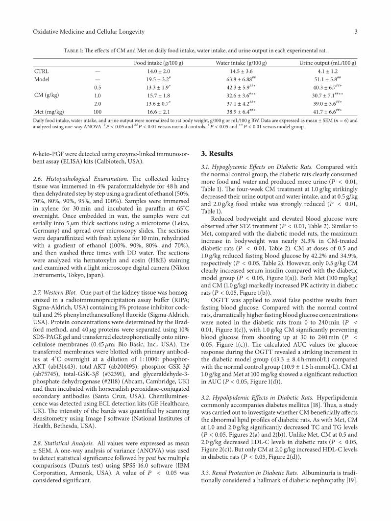
Oxidative Medicine and Cellular Longevity 3
Table 1: The effects of CM and Met on daily food intake, water intake, and urine output in each experimental rat.
Food intake (g/100 g) Water intake (g/100 g) Urine output (mL/100 g)CTRL — 14.0 ± 2.0 14.5 ± 3.6 4.1 ± 1.2
Model — 19.5 ± 3.2# 63.8 ± 6.88## 51.1 ± 5.8##
CM (g/kg)0.5 13.3 ± 1.9∗ 42.3 ± 5.9##∗ 40.3 ± 6.7##∗
1.0 15.7 ± 1.8 32.6 ± 3.6#∗∗ 30.7 ± 7.1##∗∗
2.0 13.6 ± 0.7∗ 37.1 ± 4.2##∗ 39.0 ± 3.6##∗
Met (mg/kg) 100 16.6 ± 2.1 38.9 ± 6.4##∗ 41.7 ± 6.6##∗
Daily food intake, water intake, and urine output were normalized to rat body weight, g/100 g or mL/100 g BW. Data are expressed as mean ± SEM (𝑛 = 6) andanalyzed using one-way ANOVA. #𝑃 < 0.05 and ##
𝑃 < 0.01 versus normal controls. ∗𝑃 < 0.05 and ∗∗𝑃 < 0.01 versus model group.
6-keto-PGFwere detected using enzyme-linked immunosor-bent assay (ELISA) kits (Calbiotech, USA).
2.6. Histopathological Examination. The collected kidneytissue was immersed in 4% paraformaldehyde for 48 h andthen dehydrated step by step using a gradient of ethanol (50%,70%, 80%, 90%, 95%, and 100%). Samples were immersedin xylene for 30min and incubated in paraffin at 65∘Covernight. Once embedded in wax, the samples were cutserially into 5𝜇m thick sections using a microtome (Leica,Germany) and spread over microscopy slides. The sectionswere deparaffinized with fresh xylene for 10min, rehydratedwith a gradient of ethanol (100%, 90%, 80%, and 70%),and then washed three times with DD water. The sectionswere analyzed via hematoxylin and eosin (H&E) stainingand examined with a light microscope digital camera (NikonInstruments, Tokyo, Japan).
2.7. Western Blot. One part of the kidney tissue was homog-enized in a radioimmunoprecipitation assay buffer (RIPA;Sigma-Aldrich, USA) containing 1% protease inhibitor cock-tail and 2% phenylmethanesulfonyl fluoride (Sigma-Aldrich,USA). Protein concentrations were determined by the Brad-ford method, and 40 𝜇g proteins were separated using 10%SDS-PAGE gel and transferred electrophoretically onto nitro-cellulose membranes (0.45𝜇m; Bio Basic, Inc., USA). Thetransferred membranes were blotted with primary antibod-ies at 4∘C overnight at a dilution of 1 : 1000: phosphor-AKT (ab131443), total-AKT (ab200195), phosphor-GSK-3𝛽(ab75745), total-GSK-3𝛽 (#32391), and glyceraldehyde-3-phosphate dehydrogenase (#2118) (Abcam, Cambridge, UK)and then incubated with horseradish peroxidase-conjugatedsecondary antibodies (Santa Cruz, USA). Chemilumines-cence was detected using ECL detection kits (GE Healthcare,UK). The intensity of the bands was quantified by scanningdensitometry using Image J software (National Institutes ofHealth, Bethesda, USA).
2.8. Statistical Analysis. All values were expressed as mean± SEM. A one-way analysis of variance (ANOVA) was usedto detect statistical significance followed by post hocmultiplecomparisons (Dunn’s test) using SPSS 16.0 software (IBMCorporation, Armonk, USA). A value of 𝑃 < 0.05 wasconsidered significant.
3. Results
3.1. Hypoglycemic Effects on Diabetic Rats. Compared withthe normal control group, the diabetic rats clearly consumedmore food and water and produced more urine (𝑃 < 0.01,Table 1). The four-week CM treatment at 1.0 g/kg strikinglydecreased their urine output and water intake, and at 0.5 g/kgand 2.0 g/kg food intake was strongly reduced (𝑃 < 0.01,Table 1).
Reduced bodyweight and elevated blood glucose wereobserved after STZ treatment (𝑃 < 0.01, Table 2). Similar toMet, compared with the diabetic model rats, the maximumincrease in bodyweight was nearly 31.3% in CM-treateddiabetic rats (𝑃 < 0.01, Table 2). CM at doses of 0.5 and1.0 g/kg reduced fasting blood glucose by 42.2% and 34.9%,respectively (𝑃 < 0.05, Table 2). However, only 0.5 g/kg CMclearly increased serum insulin compared with the diabeticmodel group (𝑃 < 0.05, Figure 1(a)). Both Met (100mg/kg)and CM (1.0 g/kg) markedly increased PK activity in diabeticrats (𝑃 < 0.05, Figure 1(b)).
OGTT was applied to avoid false positive results fromfasting blood glucose. Compared with the normal controlrats, dramatically higher fasting blood glucose concentrationswere noted in the diabetic rats from 0 to 240min (𝑃 <0.01, Figure 1(c)), with 1.0 g/kg CM significantly preventingblood glucose from shooting up at 30 to 240min (𝑃 <0.05, Figure 1(c)). The calculated AUC values for glucoseresponse during the OGTT revealed a striking increment inthe diabetic model group (43.3 ± 8.4 h⋅mmol/L) comparedwith the normal control group (10.9 ± 1.5 h⋅mmol/L). CM at1.0 g/kg and Met at 100mg/kg showed a significant reductionin AUC (𝑃 < 0.05, Figure 1(d)).
3.2. Hypolipidemic Effects in Diabetic Rats. Hyperlipidemiacommonly accompanies diabetes mellitus [18]. Thus, a studywas carried out to investigate whether CM beneficially affectsthe abnormal lipid profiles of diabetic rats. As with Met, CMat 1.0 and 2.0 g/kg significantly decreased TC and TG levels(𝑃 < 0.05, Figures 2(a) and 2(b)). Unlike Met, CM at 0.5 and2.0 g/kg decreased LDL-C levels in diabetic rats (𝑃 < 0.05,Figure 2(c)). But only CM at 2.0 g/kg increased HDL-C levelsin diabetic rats (𝑃 < 0.05, Figure 2(d)).
3.3. Renal Protection in Diabetic Rats. Albuminuria is tradi-tionally considered a hallmark of diabetic nephropathy [19].
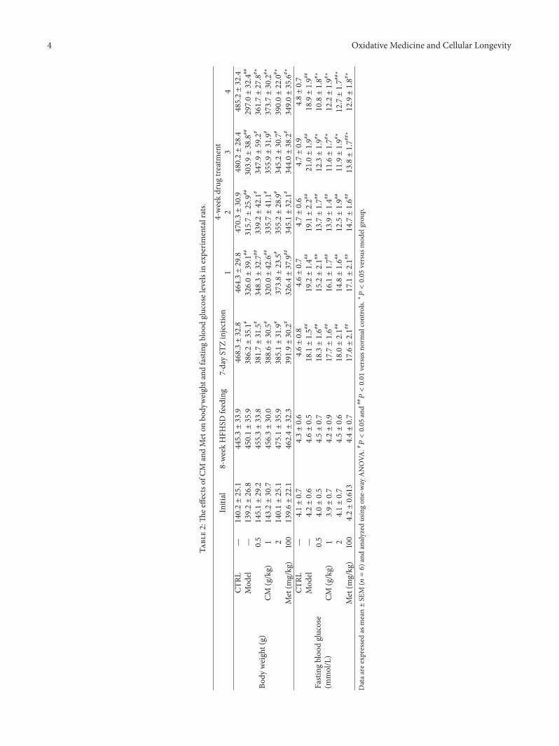
4 Oxidative Medicine and Cellular Longevity
Table2:Th
eeffectso
fCM
andMeton
body
weightand
fasting
bloo
dglucoselevels
inexperim
entalrats.
Initial
8-weekHFH
SDfeeding
7-dayST
Zinjection
4-weekdrug
treatment
12
34
Body
weight(g)
CTRL
—140.2±25.1
445.3±33.9
468.3±32.8
464.3±29.8470.3±30.9480.2±28.4485.2±32.4
Mod
el—139.2±26.8
450.1±35.9
386.2±35.1
#326.0±39.1
##315.7±25.9
##303.9±38.8
##297.0±32.4
##
CM(g/kg)
0.5145.1±29.2
455.3±33.8
381.7±31.5
#348.3±32.7
##339.2±42.1
#347.9±59.2
#361.7±27.8
#∗
1143.2±30.7
456.3±30.0
388.6±30.5
#320.0±42.6
##335.7±41.1
#355.9±31.9
#373.7±30.2
#∗
2140.1±25.1
475.1±35.9
385.1±31.9
#373.8±23.5
#355.2±28.9
#345.2±30.7
#390.0±22.0
#∗
Met(m
g/kg)
100139.6±22.1
462.4±32.3
391.9±30.2
#326.4±37.9
##345.1±32.1
#344.0±38.2
#349.0±35.6
#∗
Fastingbloo
dglucose
(mmol/L)
CTRL
—4.1±0.7
4.3±0.6
4.6±0.8
4.6±0.7
4.7±0.6
4.7±0.9
4.8±0.7
Mod
el—
4.2±0.6
4.6±0.5
18.1±1.5#
#19.2±1.4#
#19.1±2.2#
#21.0±1.9#
#18.9±1.9#
#
CM(g/kg)
0.54.0±0.5
4.5±0.7
18.3±1.6#
#15.2±2.1#
#13.7±1.7#
#12.3±1.9#∗
10.8±1.8#∗
13.9±0.7
4.2±0.9
17.7±1.6#
#16.1±1.7#
#13.9±1.4#
#11.6±1.7#∗
12.2±1.9#∗
24.1±0.7
4.5±0.6
18.0±2.1#
#14.8±1.6#
#12.5±1.9#
#11.9±1.9#∗
12.7±1.7#
#∗
Met(m
g/kg)
1004.2±0.613
4.4±0.7
17.6±2.1#
#17.1±2.1#
#14.7±1.6#
#13.8±1.7#
#∗12.9±1.8#∗
Dataa
reexpressedas
mean±SE
M(𝑛=6)a
ndanalyzed
usingon
e-way
ANOVA
.#𝑃<0.05and
##𝑃<0.01versus
norm
alcontrols.∗
𝑃<0.05versus
mod
elgrou
p.
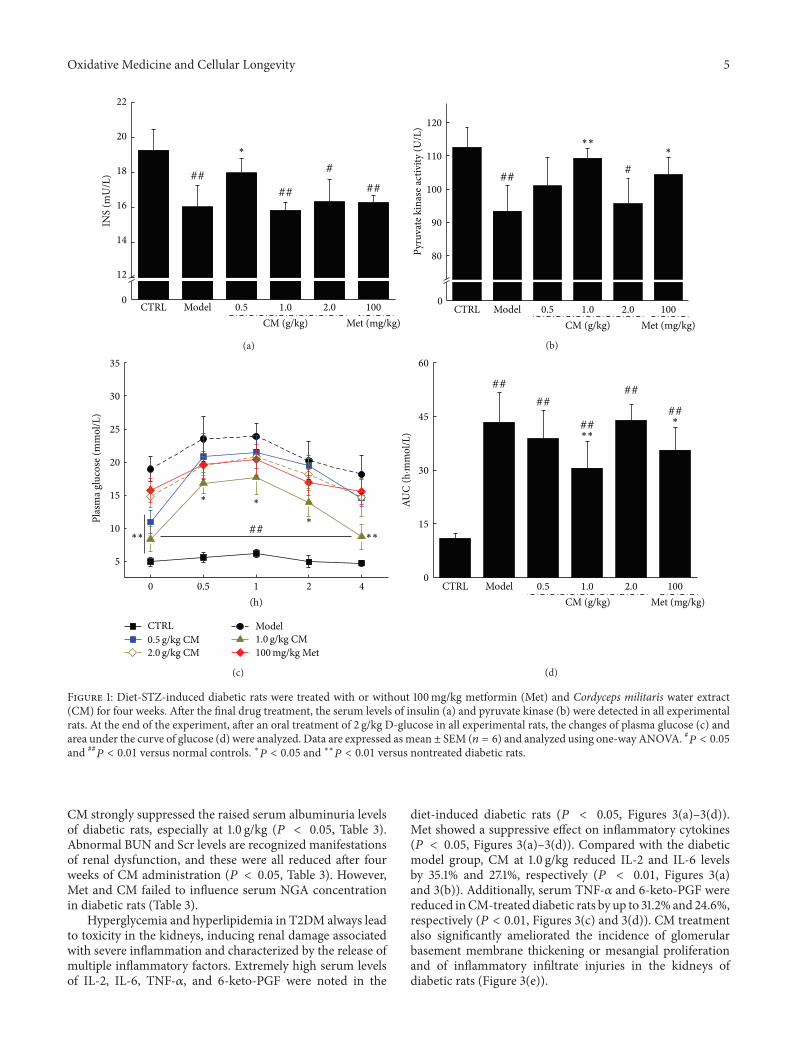
Oxidative Medicine and Cellular Longevity 5
INS
(mU
/L)
0
12
14
16
18
20
22
##
## ##
#
CTRL Model 0.5 1.0 2.0 100
∗
CM (g/kg) Met (mg/kg)
(a)
###
∗∗∗
CTRL Model 0.5 1.0 2.0 100
CM (g/kg) Met (mg/kg)
Pyru
vate
kin
ase a
ctiv
ity (U
/L)
0
80
90
100
110
120
(b)
5
10
15
20
25
30
35
Plas
ma g
luco
se (m
mol
/L)
0 0.5 1 2 4
(h)
CTRL Model0.5 g/kg CM2.0 g/kg CM
1.0 g/kg CM100mg/kg Met
∗∗∗∗
∗ ∗
∗##
(c)
##
##
##
##
##∗
∗∗
CTRL Model 0.5 1.0 2.0 100
CM (g/kg) Met (mg/kg)
AUC
(h·m
mol
/L)
0
15
30
45
60
(d)
Figure 1: Diet-STZ-induced diabetic rats were treated with or without 100mg/kg metformin (Met) and Cordyceps militaris water extract(CM) for four weeks. After the final drug treatment, the serum levels of insulin (a) and pyruvate kinase (b) were detected in all experimentalrats. At the end of the experiment, after an oral treatment of 2 g/kg D-glucose in all experimental rats, the changes of plasma glucose (c) andarea under the curve of glucose (d) were analyzed. Data are expressed asmean ± SEM (𝑛 = 6) and analyzed using one-way ANOVA. #𝑃 < 0.05and ##𝑃 < 0.01 versus normal controls. ∗𝑃 < 0.05 and ∗∗𝑃 < 0.01 versus nontreated diabetic rats.
CM strongly suppressed the raised serum albuminuria levelsof diabetic rats, especially at 1.0 g/kg (𝑃 < 0.05, Table 3).Abnormal BUN and Scr levels are recognized manifestationsof renal dysfunction, and these were all reduced after fourweeks of CM administration (𝑃 < 0.05, Table 3). However,Met and CM failed to influence serum NGA concentrationin diabetic rats (Table 3).
Hyperglycemia and hyperlipidemia in T2DM always leadto toxicity in the kidneys, inducing renal damage associatedwith severe inflammation and characterized by the release ofmultiple inflammatory factors. Extremely high serum levelsof IL-2, IL-6, TNF-𝛼, and 6-keto-PGF were noted in the
diet-induced diabetic rats (𝑃 < 0.05, Figures 3(a)–3(d)).Met showed a suppressive effect on inflammatory cytokines(𝑃 < 0.05, Figures 3(a)–3(d)). Compared with the diabeticmodel group, CM at 1.0 g/kg reduced IL-2 and IL-6 levelsby 35.1% and 27.1%, respectively (𝑃 < 0.01, Figures 3(a)and 3(b)). Additionally, serum TNF-𝛼 and 6-keto-PGF werereduced inCM-treated diabetic rats by up to 31.2% and 24.6%,respectively (𝑃 < 0.01, Figures 3(c) and 3(d)). CM treatmentalso significantly ameliorated the incidence of glomerularbasement membrane thickening or mesangial proliferationand of inflammatory infiltrate injuries in the kidneys ofdiabetic rats (Figure 3(e)).
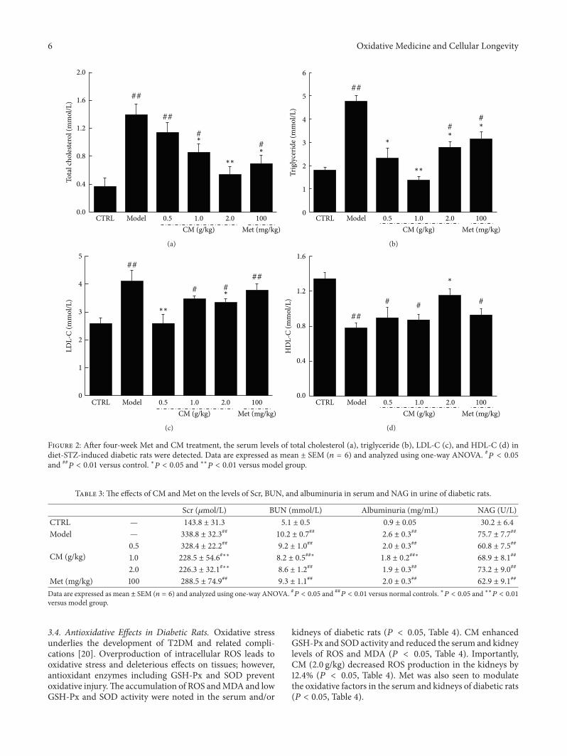
6 Oxidative Medicine and Cellular Longevity
#
#
##
##
CTRL Model 0.5 1.0 2.0 100
∗
∗
∗∗
CM (g/kg) Met (mg/kg)
Tota
l cho
leste
rol (
mm
ol/L
)
0.0
0.4
0.8
1.2
1.6
2.0
(a)
#
##
∗
#∗
∗
∗∗
CTRL Model 0.5 1.0 2.0 100
CM (g/kg) Met (mg/kg)
Trig
lyce
ride (
mm
ol/L
)
0
1
2
3
4
5
6
(b)
#
##
##
#∗
∗∗
CTRL Model 0.5 1.0 2.0 100
CM (g/kg) Met (mg/kg)
LDL-
C (m
mol
/L)
0
1
2
3
4
5
(c)
##
#
##
∗
CTRL Model 0.5 1.0 2.0 100
CM (g/kg) Met (mg/kg)
HD
L-C
(mm
ol/L
)
0.0
0.4
0.8
1.2
1.6
(d)
Figure 2: After four-week Met and CM treatment, the serum levels of total cholesterol (a), triglyceride (b), LDL-C (c), and HDL-C (d) indiet-STZ-induced diabetic rats were detected. Data are expressed as mean ± SEM (𝑛 = 6) and analyzed using one-way ANOVA. #𝑃 < 0.05and ##𝑃 < 0.01 versus control. ∗𝑃 < 0.05 and ∗∗𝑃 < 0.01 versus model group.
Table 3: The effects of CM and Met on the levels of Scr, BUN, and albuminuria in serum and NAG in urine of diabetic rats.
Scr (𝜇mol/L) BUN (mmol/L) Albuminuria (mg/mL) NAG (U/L)CTRL — 143.8 ± 31.3 5.1 ± 0.5 0.9 ± 0.05 30.2 ± 6.4
Model — 338.8 ± 32.3## 10.2 ± 0.7## 2.6 ± 0.3## 75.7 ± 7.7##
CM (g/kg)0.5 328.4 ± 22.2## 9.2 ± 1.0## 2.0 ± 0.3## 60.8 ± 7.5##
1.0 228.5 ± 54.6#∗∗ 8.2 ± 0.5##∗ 1.8 ± 0.2##∗ 68.9 ± 8.1##
2.0 226.3 ± 32.1#∗∗ 8.6 ± 1.2## 1.9 ± 0.3## 73.2 ± 9.0##
Met (mg/kg) 100 288.5 ± 74.9## 9.3 ± 1.1## 2.0 ± 0.3## 62.9 ± 9.1##
Data are expressed as mean ± SEM (𝑛 = 6) and analyzed using one-way ANOVA. #𝑃 < 0.05 and ##𝑃 < 0.01 versus normal controls. ∗𝑃 < 0.05 and ∗∗𝑃 < 0.01
versus model group.
3.4. Antioxidative Effects in Diabetic Rats. Oxidative stressunderlies the development of T2DM and related compli-cations [20]. Overproduction of intracellular ROS leads tooxidative stress and deleterious effects on tissues; however,antioxidant enzymes including GSH-Px and SOD preventoxidative injury.The accumulation of ROS andMDA and lowGSH-Px and SOD activity were noted in the serum and/or
kidneys of diabetic rats (𝑃 < 0.05, Table 4). CM enhancedGSH-Px and SOD activity and reduced the serum and kidneylevels of ROS and MDA (𝑃 < 0.05, Table 4). Importantly,CM (2.0 g/kg) decreased ROS production in the kidneys by12.4% (𝑃 < 0.05, Table 4). Met was also seen to modulatethe oxidative factors in the serum and kidneys of diabetic rats(𝑃 < 0.05, Table 4).
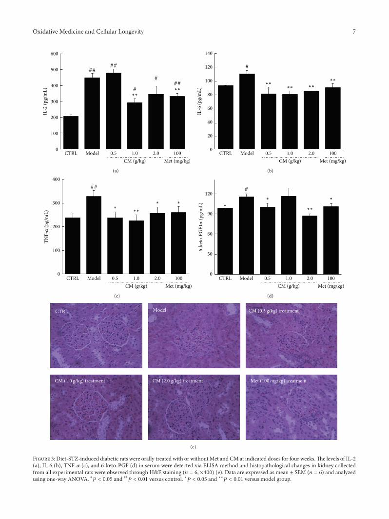
Oxidative Medicine and Cellular Longevity 7
####
#
###
∗∗∗∗
CTRL Model 0.5 1.0 2.0 100
CM (g/kg) Met (mg/kg)
IL-2
(pg/
mL)
0
100
200
300
400
500
600
(a)
#
∗∗
∗∗∗∗∗∗
CTRL Model 0.5 1.0 2.0 100
CM (g/kg) Met (mg/kg)
IL-6
(pg/
mL)
0
20
40
60
80
100
120
140
(b)
##
∗∗∗
∗ ∗
CTRL Model 0.5 1.0 2.0 100
CM (g/kg) Met (mg/kg)
TNF-𝛼
(pg/
mL)
0
100
200
300
400
(c)
#
∗ ∗
∗∗
CTRL Model 0.5 1.0 2.0 100
CM (g/kg) Met (mg/kg)
6-k
eto-
PGF1𝛼
(pg/
mL)
0
30
60
90
120
(d)
CTRL Model CM (0.5 g/kg) treatment
CM (1.0 g/kg) treatment CM (2.0 g/kg) treatment Met (100mg/kg) treatment
(e)
Figure 3: Diet-STZ-induced diabetic rats were orally treated with or withoutMet and CM at indicated doses for four weeks.The levels of IL-2(a), IL-6 (b), TNF-𝛼 (c), and 6-keto-PGF (d) in serum were detected via ELISA method and histopathological changes in kidney collectedfrom all experimental rats were observed through H&E staining (𝑛 = 6, ×400) (e). Data are expressed as mean ± SEM (𝑛 = 6) and analyzedusing one-way ANOVA. #𝑃 < 0.05 and ##
𝑃 < 0.01 versus control. ∗𝑃 < 0.05 and ∗∗𝑃 < 0.01 versus model group.
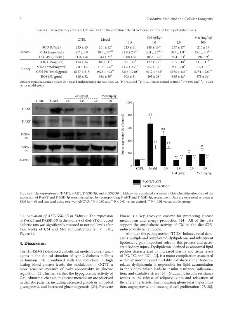
8 Oxidative Medicine and Cellular Longevity
Table 4: The regulatory effects of CM and Met on the oxidation related factors in serum and kidney of diabetic rats.
CTRL Model CM (g/kg) Met (mg/kg)0.5 1.0 2.0 100
SerumSOD (U/mL) 245 ± 15 201 ± 12# 223 ± 11 248 ± 16∗∗ 237 ± 17∗ 215 ± 17
MDA (nmol/mL) 8.7 ± 0.8 28.0 ± 0.7## 21.9 ± 2.7## 12.4 ± 2.7#∗∗ 10.7 ± 1.8∗∗ 15.0 ± 2.5#∗∗
GSH-Px (𝜇mol/L) 1116 ± 41 944 ± 37# 1000 ± 51 1053 ± 25∗ 994 ± 33# 994 ± 9#
Kidney
SOD (U/mgprot) 134 ± 14 84 ± 11## 110 ± 18# 122 ± 13∗∗ 105 ± 14# 111 ± 11#∗
MDA (nmol/mgprot) 7.0 ± 1.4 11.3 ± 2.6## 11.2 ± 2.7## 8.2 ± 1.2∗ 9.3 ± 2.0# 8.5 ± 1.5∗
GSH-Px (𝜇mol/gprot) 6987 ± 318 4925 ± 402## 5236 ± 210# 6012 ± 462∗ 5985 ± 433∗ 5784 ± 223#∗
ROS (FI/gprot) 815 ± 32 988 ± 55# 903 ± 31 895 ± 28∗ 865 ± 48∗ 875 ± 36∗
Data are expressed as mean ± SEM (𝑛 = 6) and analyzed using one-way ANOVA. #𝑃 < 0.05 and ##𝑃 < 0.01 versus normal controls. ∗𝑃 < 0.05 and ∗∗𝑃 < 0.01
versus model group.
GAPDH
P-AKT
T-AKT
P-GSK-3𝛽
T-GSK-3𝛽
CTRL 1002.01.00.5ModelCM (g/kg) Met (mg/kg)
##
##
##
###
∗∗ ∗∗
∗∗
∗∗
∗∗
∗∗
CTRL Model 0.5 1.0 2.0 100
CM (g/kg) Met (mg/kg)
Prot
ein
expr
essio
n (%
of c
ontro
l)
0
200
400
600
800
1000
P-AKT/T-AKTP-GSK-3𝛽/T-GSK-3𝛽
Figure 4: The expressions of T-AKT, P-AKT, T-GSK-3𝛽, and P-GSK-3𝛽 in kidney were analyzed via western blot. Quantification data of theexpression of P-AKT and P-GSK-3𝛽 were normalized by corresponding T-AKT and T-GSK-3𝛽, respectively. Data are expressed as mean ±SEM (𝑛 = 6) and analyzed using one-way ANOVA. #𝑃 < 0.05 and ##
𝑃 < 0.01 versus control. ∗∗𝑃 < 0.01 versus model group.
3.5. Activation of AKT/GSK-3𝛽 in Kidneys. The expressionof P-AKT and P-GSK-3𝛽 in the kidneys of diet-STZ-induceddiabetic rats was significantly restored to normal levels afterfour weeks of CM and Met administration (𝑃 < 0.01,Figure 4).
4. Discussion
TheHFHSD-STZ-induced diabetic rat model is closely anal-ogous to the clinical situation of type 2 diabetes mellitusin humans [21]. Combined with the reduction in highfasting blood glucose levels, the modulation of OGTT, amore sensitive measure of early abnormality in glucoseregulation [22], further verifies the hypoglycemic activity ofCM. Abnormal changes in glucose metabolism are observedin diabetic patients, including decreased glycolysis, impededglycogenesis, and increased gluconeogenesis [23]. Pyruvate
kinase is a key glycolytic enzyme for promoting glucosemetabolism and energy production [24]. All of the datasupport the antidiabetic activity of CM in the diet-STZ-induced diabetic rat model.
Although the pathogenesis of T2DM-induced renal dam-age ismultiple and complicated, dyslipidemia and subsequentlipotoxicity play important roles in this process and accel-erate kidney injury. Dyslipidemia, defined as abnormal lipidprofiles characterized by increased plasma and tissue levelsof TG, TC, and LDL [21], is a major complication associatedwith highmorbidity andmortality in diabetics [25].Diabetes-related dyslipidemia is responsible for lipid accumulationin the kidney, which leads to insulin resistance, inflamma-tion, and oxidative stress [26]. Gradually, insulin resistanceresults in the release of adipocytokines and relaxation ofthe afferent arteriole, finally causing glomerular hyperfiltra-tion, angiogenesis, and mesangial cell proliferation [27, 28].
Oxidative Medicine and Cellular Longevity 9
The antilipemic effect of CM plays an important role in renalprotection in HFHSD-STZ-induced diabetic rats.
Oxidative stress has been singled out as a major cause ofdiabetic complications, especially nephropathy [29]. O2− andnitric oxide (NO) levels are important in kidney and vascularfunction [30]. ROS, which is responsible for oxidative dam-age, degrades membrane polyunsaturated fatty acids throughsequential peroxidation processes [31] and elevates MDAlevels, which serve as biomarkers of tissue oxidative stress[32]. Excessive generation of ROS and MDA in the kidneysleads to tubular obstruction, back-leakage of renal tubules,and contraction of the mesangial cells, finally resulting inthe abnormal expression of renal function markers suchas Scr, BUN, albuminuria, and NAG [33]. However, cellsdefend themselves against oxidative stress via the activa-tion of antioxidant enzymes. Antioxidant compounds are acommon and effective way to prevent or inhibit pancreaticbeta-cell destruction caused by alloxan [34]. SOD catalyzesthe conversion of superoxides into hydrogen peroxide andoxygen, while GSH-Px scavenges the hydroxyl radicals [35].The enhanced activity of SOD and GSH-Px in the serumand kidneys of CM-treated diabetic rats helps to maintain abalance of oxidants and antioxidants by causing the excretionof ROS. Therefore, CM improves renal function by scaveng-ing free radicals, especially ROS and MDA. It is well knownthat inhibition of AKT phosphorylation downregulates GSK-3𝛽 phosphorylation [36]. Gardenamide A is reported toattenuate ROS levels by promoting the phosphorylation ofAKT, an effect that can be completely abrogated by the AKTinhibitor [37]. In CM-treated diet-STZ-reduced diabetic rats,decreased AKT and GSK-3𝛽 phosphorylation is responsiblefor the transcriptional expression of multiple antioxidants toprevent diabetes-related oxidative damage.
Oxidative stress in T2DM favors the appearance of endo-thelial dysfunction, and oxidative production is an importantstep in inflammation [38]. Interleukins have important rolesduring inflammatory development, and the overexpressionof IL-2 activates proinflammatory CD4+ T cells, exacerbat-ing the glomerular damage by recruiting macrophages andneutrophils [39]. IL-6, secreted by the glomerular membranesystem, is responsible for the proliferation of mesangialcells and the release of inflammatory mediators, includingsuperoxide anions [40]. As reported, TNF-𝛼 upregulates IL-6 release by the podocytes in coculture with glomerularendothelial cells [41]. Previous studies have reported thatoxidative stress is mediated in podocyte apoptosis in theprocess of diabetic nephropathy [42] and that the progressionof renal interstitial fibrosis can be inhibited by suppressingoxidative stress [43]. Thus, CM exerts renal protection indiabetic rats via the regulation of inflammatory factors thatare modulated by oxidative stress.
All of the data suggest that CM targets manymolecules inthe signaling of hyperglycemia, inflammation, and oxidativestress. This “systemic targeting” will completely eliminatethe symptoms of diabetes and diabetic nephropathy in amuch natural way, so that less adverse effect is expected.As a folk tonic food in China, CM has been emphasizingits safety with few adverse effects. Our subchronic toxic
test provides experimental basis for its safety indicatingthat CM showed no influences on bodyweights (Table 1S),organ indexes (Table 2S), and kidney structures in mice(Figure 2S). On the other hand, the crude drug nature ofCM suggests multieffective components, which may showsynergistic effect on the disease. It may explain that non-dose-dependent manner was the common way of action ofsome herbal medicines. Amount of natural productions isreported to show various pharmacological activities via non-dose-dependent manner [44, 45].
There is still a limitation in our present study. Althoughwe confirmed the regulatory effects of Met and CM oninflammatory factors in serum, we failed to detect the relatedchanges in kidney tissues. As reported, Met successfullyregulates inflammatory cytokines associated with nephritisbut shows no influences on kidney structure [46].Our furtherstudywill focus on the effects of drugs on biochemical indicesand pathological changes of kidney.
In summary, we successfully explored the antidiabeticand antinephritic effects of CM in diet-STZ-induced diabeticrats. During the experiment, CM exhibited the ability toreduce blood glucose, decrease blood lipids, reduce renalinjury, and lower inflammatory factors through enhancedantioxidant expression and the attenuation of oxidative stress.Cordyceps militaris fruit body extract, a safe pharmaceuticalagent, thus has great potential as a new treatment for diabeticpatients, especially those with nephritis.
Competing Interests
The authors declare that they have no competing interests.
Acknowledgments
This work was supported by the Science and Technology KeyProject in Jilin Province of China (Grant no. 20130201006ZYand 20150203002NY), the “Twelfth Five-Year” Science andTechnology Planning Project of Jilin Province in China(Grant no. 2014B033), and Science and Technology Devel-opment Program of Jilin Province in China (Grant no.20160520036JH).
References
[1] S. M. Kabadi, B. K. Lee, and L. Liu, “Joint effects of obesityand vitamin D insufficiency on insulin resistance and type 2diabetes: results from the NHANES 2001–2006,” Diabetes Care,vol. 35, no. 10, pp. 2048–2054, 2012.
[2] Q. You, F. Chen, X. Wang, P. G. Luo, and Y. Jiang, “Inhibitoryeffects of muscadine anthocyanins on 𝛼-glucosidase and pan-creatic lipase activities,” Journal of Agricultural and Food Chem-istry, vol. 59, no. 17, pp. 9506–9511, 2011.
[3] W. Kerner and J. Bruckel, “Definition, classification and diagno-sis of diabetes mellitus,” Experimental and Clinical Endocrinol-ogy & Diabetes, vol. 122, no. 7, pp. 384–386, 2014.
[4] G. Winkler, T. Hidvégi, G. Vándorfi, and G. Jermendy, “Risk-stratified screening for diabetes in adult subjects: results of thefirst investigation in Hungary ,” Orvosi Hetilap, vol. 151, no. 17,pp. 691–696, 2010.
10 Oxidative Medicine and Cellular Longevity
[5] C. Solis-Herrera, C. L. Triplitt, and J. L. Lynch, “Nephropathy inyouth and young adults with type 2 diabetes,” Current DiabetesReports, vol. 14, no. 2, p. 456, 2014.
[6] K. Zhu, T. Kakehi,M.Matsumoto et al., “NADPHoxidaseNOX1is involved in activation of protein kinase C and prematuresenescence in early stage diabetic kidney,” Free Radical Biologyand Medicine, vol. 83, pp. 21–30, 2015.
[7] G. L. Bakris, “Recognition, pathogenesis, and treatment ofdifferent stages of nephropathy in patients with type 2 diabetesmellitus,” Mayo Clinic Proceedings, vol. 86, no. 5, pp. 444–456,2011.
[8] J. Gilg, R. Pruthi, andD. Fogarty, “UK renal registry 17th annualreport: chapter 1 UK renal replacement therapy incidence in2013: national and centre-specific analyses,” Nephron, vol. 129,supplement 1, pp. 1–29, 2015.
[9] D. S. Kania, J. D. Gonzalvo, and Z. A. Weber, “Saxagliptin: aclinical review in the treatment of type 2 diabetes mellitus,”Clinical Therapeutics, vol. 33, no. 8, pp. 1005–1022, 2011.
[10] A. J. Scheen, “Antidiabetic agents in subjects with mild dys-glycaemia: prevention or early treatment of type 2 diabetes?”Diabetes and Metabolism, vol. 33, no. 1, pp. 3–12, 2007.
[11] T. Salihu Shinkafi, L. Bello, S. Wara Hassan, and S. Ali, “An eth-nobotanical survey of antidiabetic plants used by Hausa-Fulanitribes in Sokoto, Northwest Nigeria,” Journal of Ethnopharma-cology, vol. 172, pp. 91–99, 2015.
[12] J. Singh, E. Cumming, G. Manoharan, H. Kalasz, and E.Adeghate, “Medicinal chemistry of the anti-diabetic effects ofMomordica charantia: active constituents andmodes of actions,”OpenMedicinal Chemistry Journal, vol. 5, supplement 2, pp. 70–77, 2011.
[13] S. K. Das, M. Masuda, A. Sakurai, and M. Sakakibara, “Medici-nal uses of the mushroom Cordyceps militaris: current state andprospects,” Fitoterapia, vol. 81, no. 8, pp. 961–968, 2010.
[14] Y. Dong, T. Jing, Q. Meng et al., “Studies on the antidiabeticactivities of Cordyceps militaris extract in diet-streptozotocin-induced diabetic Sprague-Dawley rats,” BioMed Research Inter-national, vol. 2014, Article ID 160980, 11 pages, 2014.
[15] Y.-W. Cheng, Y.-I. Chen, C.-Y. Tzeng et al., “Extracts ofCordyceps militaris lower blood glucose via the stimulation ofcholinergic activation and insulin secretion in normal rats,”Phytotherapy Research, vol. 26, no. 8, pp. 1173–1177, 2012.
[16] C.-H. Ma, L.-L. Kang, H.-M. Ren, D.-M. Zhang, and L.-D.Kong, “Simiao pill ameliorates renal glomerular injury viaincreasing Sirt1 expression and suppressing NF-𝜅B/NLRP3inflammasome activation in high fructose-fed rats,” Journal ofEthnopharmacology, vol. 172, pp. 108–117, 2015.
[17] Y. Dong, T. Jing, Q. Meng et al., “Studies on the antidiabeticactivities of cordyceps militaris extract in diet-streptozotocin-induced diabetic sprague-dawley rats,” BioMed Research Inter-national, vol. 2014, Article ID 160980, 11 pages, 2014.
[18] S. O. Adewole, T. Adenowo, T. Naicker, and J. A. Ojewole,“Hypoglycaemic and hypotensive effects of Ficus exasperatavahl. (Moraceae) leaf aqueous extract in rats,”The African Jour-nal of Traditional, Complementary and Alternative Medicines,vol. 8, no. 3, pp. 275–283, 2011.
[19] M. E.Williams, “Diabetic nephropathy: the proteinuria hypoth-esis,” American Journal of Nephrology, vol. 25, no. 2, pp. 77–94,2005.
[20] S. Tangvarasittichai, “Oxidative stress, insulin resistance, dys-lipidemia and type 2 diabetes mellitus,” World Journal ofDiabetes, vol. 6, no. 3, pp. 456–480, 2015.
[21] M. Shao, X. Lu, W. Cong et al., “Multiple low-dose radiationprevents type 2 diabetes-induced renal damage through atten-uation of dyslipidemia and insulin resistance and subsequentrenal inflammation and oxidative stress,” PLoS ONE, vol. 9, no.3, Article ID e92574, 2014.
[22] B. A. Sheikh, L. Pari, A. Rathinam, and R. Chandramohan,“Trans-anethole, a terpenoid ameliorates hyperglycemia byregulating key enzymes of carbohydrate metabolism in strep-tozotocin induced diabetic rats,” Biochimie, vol. 112, pp. 57–65,2015.
[23] B. Huang, Z. Wang, J. H. Park et al., “Anti-diabetic effectof purple corn extract on C57BL/KsJ db/db mice,” NutritionResearch and Practice, vol. 9, no. 1, pp. 22–29, 2015.
[24] H. Wang, W. Chu, S. K. Das, Q. Ren, S. J. Hasstedt, and S. C.Elbein, “Liver pyruvate kinase polymorphisms are associatedwith type 2 diabetes in northern European Caucasians,” Dia-betes, vol. 51, no. 9, pp. 2861–2865, 2002.
[25] N. K. Sambu, R. T. Kashinath, and J. G. Ambekar, “Effect ofdiallyl disulphide on diabetes induced dyslipidemia in malealbino rats,” Journal of Clinical and Diagnostic Research, vol. 9,no. 4, pp. BF01–BF03, 2015.
[26] I. M. Wahba and R. H. Mak, “Obesity and obesity-initiatedmetabolic syndrome: mechanistic links to chronic kidney dis-ease,”Clinical Journal of the American Society of Nephrology, vol.2, no. 3, pp. 550–562, 2007.
[27] A. W. Miller, C. Dimitropoulou, G. Han, R. E. White, D. W.Busija, and G. O. Carrier, “Epoxyeicosatrienoic acid-inducedrelaxation is impaired in insulin resistance,” American Journalof Physiology—Heart and Circulatory Physiology, vol. 281, no. 4,pp. H1524–H1531, 2001.
[28] L. Piqueras, A. R. Reynolds, K. M. Hodivala-Dilke et al.,“Activation of PPAR𝛽/𝛿 induces endothelial cell proliferationand angiogenesis,” Arteriosclerosis, Thrombosis, and VascularBiology, vol. 27, no. 1, pp. 63–69, 2007.
[29] A. P. Rolo and C. M. Palmeira, “Diabetes and mitochondrialfunction: role of hyperglycemia and oxidative stress,”Toxicologyand Applied Pharmacology, vol. 212, no. 2, pp. 167–178, 2006.
[30] A. Ragheb, A. Attia, W. S. Eldin, F. Elbarbry, S. Gazarin, and A.Shoker, “The protective effect of thymoquinone, an anti-oxidantand anti-inflammatory agent, against renal injury: a review,”Saudi Journal of Kidney Diseases and Transplantation, vol. 20,no. 5, pp. 741–752, 2009.
[31] O. R. Ayepola, M. E. Cerf, N. L. Brooks, and O. O. Oguntibeju,“Kolaviron, a biflavonoid complex of Garcinia kola seeds mod-ulates apoptosis by suppressing oxidative stress and inflamma-tion in diabetes-induced nephrotoxic rats,” Phytomedicine, vol.21, no. 14, pp. 1785–1793, 2014.
[32] A. Goel, V. Dani, and D. K. Dhawan, “Protective effects ofzinc on lipid peroxidation, antioxidant enzymes and hepatichistoarchitecture in chlorpyrifos-induced toxicity,” Chemico-Biological Interactions, vol. 156, no. 2-3, pp. 131–140, 2005.
[33] A. Y. Nasr and H. A. M. Saleh, “Aged garlic extract protectsagainst oxidative stress and renal changes in cisplatin-treatedadult male rats,” Cancer Cell International, vol. 14, no. 1, article92, 2014.
[34] H. Sebai, S. Selmi, K. Rtibi, A. Souli, N. Gharbi, and M.Sakly, “Lavender (Lavandula stoechas L.) essential oils attenuatehyperglycemia and protect against oxidative stress in alloxan-induced diabetic rats,” Lipids in Health and Disease, vol. 12, no.1, article 189, 2013.
[35] P. Borges, B. Oliveira, S. Casal, J. Dias, L. Conceição, and L.M. P. Valente, “Dietary lipid level affects growth performance
Oxidative Medicine and Cellular Longevity 11
and nutrient utilisation of Senegalese sole (Solea senegalensis)juveniles,” British Journal of Nutrition, vol. 102, no. 7, pp. 1007–1014, 2009.
[36] C. Zhang, X. Lu, Y. Tan et al., “Diabetes-induced hepaticpathogenic damage, inflammation, oxidative stress, and insulinresistancewas exacerbated in zinc deficientmousemodel,”PLoSONE, vol. 7, no. 12, Article ID e49257, 2012.
[37] R. Wang, L. Peng, J. Zhao et al., “Gardenamide a protects RGC-5 cells from H
2
O2
-induced oxidative stress insults by activatingPI3K/Akt/eNOS signaling pathway,” International Journal ofMolecular Sciences, vol. 16, no. 9, pp. 22350–22367, 2015.
[38] A. Ceriello, A. Novials, E. Ortega et al., “Vitamin C furtherimproves the protective effect of glucagon-like peptide-1 onacute hypoglycemia-induced oxidative stress, inflammation,and endothelial dysfunction in type 1 diabetes,” Diabetes Care,vol. 36, no. 12, pp. 4104–4108, 2013.
[39] R. Bertelli, A. Di Donato, M. Cioni et al., “LPS nephropathy inmice is ameliorated by IL-2 independently of regulatory T cellsactivity,” PLoS ONE, vol. 9, no. 10, Article ID e111285, 2014.
[40] L. Ma, Y. Gao, G. Chen et al., “Relationships of urinaryVEGF/CR and IL-6/CR with glomerular pathological injuryin asymptomatic hematuria patients,”Medical Science Monitor,vol. 21, pp. 356–362, 2015.
[41] S. J. Kuravi, H. M. McGettrick, S. C. Satchell et al., “Podocytesregulate neutrophil recruitment by glomerular endothelial cellsvia IL-6-mediated crosstalk,” The Journal of Immunology, vol.193, no. 1, pp. 234–243, 2014.
[42] W. Hua, H.-Z. Huang, L.-T. Tan et al., “CD36 mediated fattyacid-induced podocyte apoptosis via oxidative stress,” PLoSONE, vol. 10, no. 5, Article ID e0127507, 2015.
[43] J. Qin, W.-J. Mei, Y.-Y. Xie et al., “Fluorofenidone attenuatesoxidative stress and renal fibrosis in obstructive nephropa-thy via blocking NOX2 (gp91phox) expression and inhibit-ing erk/mapk signaling pathway,” Kidney and Blood PressureResearch, vol. 40, no. 1, pp. 89–99, 2014.
[44] J. Wei, S. Wang, G. Liu et al., “Polysaccharides from Entero-morpha prolifera enhance the immunity of normal mice,”International Journal of Biological Macromolecules, vol. 64, pp.1–5, 2014.
[45] L. Ma, S. Zhang, and M. Du, “Cordycepin from Cordycepsmilitaris prevents hyperglycemia in alloxan-induced diabeticmice,” Nutrition Research, vol. 35, no. 5, pp. 431–439, 2015.
[46] B. D. Sahu, M. Kuncha, U. K. Putcha, and R. Sistla, “Effect ofmetformin against cisplatin induced acute renal injury in rats:a biochemical and histoarchitectural evaluation,” Experimentaland Toxicologic Pathology, vol. 65, no. 6, pp. 933–940, 2013.
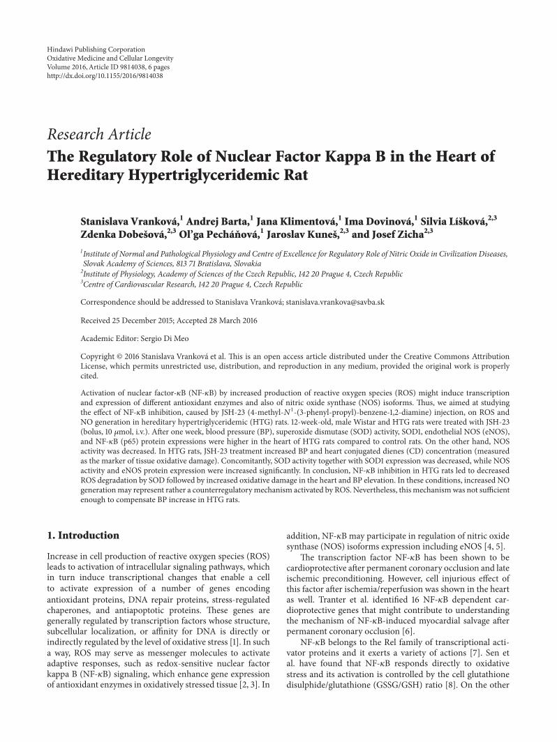
Research ArticleThe Regulatory Role of Nuclear Factor Kappa B in the Heart ofHereditary Hypertriglyceridemic Rat
Stanislava Vranková,1 Andrej Barta,1 Jana Klimentová,1 Ima Dovinová,1 Silvia Líšková,2,3
Zdenka Dobešová,2,3 O8ga Pechá^ová,1 Jaroslav Kuneš,2,3 and Josef Zicha2,3
1 Institute of Normal and Pathological Physiology and Centre of Excellence for Regulatory Role of Nitric Oxide in Civilization Diseases,Slovak Academy of Sciences, 813 71 Bratislava, Slovakia2Institute of Physiology, Academy of Sciences of the Czech Republic, 142 20 Prague 4, Czech Republic3Centre of Cardiovascular Research, 142 20 Prague 4, Czech Republic
Correspondence should be addressed to Stanislava Vranková; [email protected]
Received 25 December 2015; Accepted 28 March 2016
Academic Editor: Sergio Di Meo
Copyright © 2016 Stanislava Vranková et al. This is an open access article distributed under the Creative Commons AttributionLicense, which permits unrestricted use, distribution, and reproduction in any medium, provided the original work is properlycited.
Activation of nuclear factor-𝜅B (NF-𝜅B) by increased production of reactive oxygen species (ROS) might induce transcriptionand expression of different antioxidant enzymes and also of nitric oxide synthase (NOS) isoforms. Thus, we aimed at studyingthe effect of NF-𝜅B inhibition, caused by JSH-23 (4-methyl-𝑁1-(3-phenyl-propyl)-benzene-1,2-diamine) injection, on ROS andNO generation in hereditary hypertriglyceridemic (HTG) rats. 12-week-old, male Wistar and HTG rats were treated with JSH-23(bolus, 10 𝜇mol, i.v.). After one week, blood pressure (BP), superoxide dismutase (SOD) activity, SOD1, endothelial NOS (eNOS),and NF-𝜅B (p65) protein expressions were higher in the heart of HTG rats compared to control rats. On the other hand, NOSactivity was decreased. In HTG rats, JSH-23 treatment increased BP and heart conjugated dienes (CD) concentration (measuredas the marker of tissue oxidative damage). Concomitantly, SOD activity together with SOD1 expression was decreased, while NOSactivity and eNOS protein expression were increased significantly. In conclusion, NF-𝜅B inhibition in HTG rats led to decreasedROS degradation by SOD followed by increased oxidative damage in the heart and BP elevation. In these conditions, increased NOgenerationmay represent rather a counterregulatorymechanism activated by ROS. Nevertheless, this mechanismwas not sufficientenough to compensate BP increase in HTG rats.
1. Introduction
Increase in cell production of reactive oxygen species (ROS)leads to activation of intracellular signaling pathways, whichin turn induce transcriptional changes that enable a cellto activate expression of a number of genes encodingantioxidant proteins, DNA repair proteins, stress-regulatedchaperones, and antiapoptotic proteins. These genes aregenerally regulated by transcription factors whose structure,subcellular localization, or affinity for DNA is directly orindirectly regulated by the level of oxidative stress [1]. In sucha way, ROS may serve as messenger molecules to activateadaptive responses, such as redox-sensitive nuclear factorkappa B (NF-𝜅B) signaling, which enhance gene expressionof antioxidant enzymes in oxidatively stressed tissue [2, 3]. In
addition, NF-𝜅B may participate in regulation of nitric oxidesynthase (NOS) isoforms expression including eNOS [4, 5].
The transcription factor NF-𝜅B has been shown to becardioprotective after permanent coronary occlusion and lateischemic preconditioning. However, cell injurious effect ofthis factor after ischemia/reperfusion was shown in the heartas well. Tranter et al. identified 16 NF-𝜅B dependent car-dioprotective genes that might contribute to understandingthe mechanism of NF-𝜅B-induced myocardial salvage afterpermanent coronary occlusion [6].
NF-𝜅B belongs to the Rel family of transcriptional acti-vator proteins and it exerts a variety of actions [7]. Sen etal. have found that NF-𝜅B responds directly to oxidativestress and its activation is controlled by the cell glutathionedisulphide/glutathione (GSSG/GSH) ratio [8]. On the other
Hindawi Publishing CorporationOxidative Medicine and Cellular LongevityVolume 2016, Article ID 9814038, 6 pageshttp://dx.doi.org/10.1155/2016/9814038
2 Oxidative Medicine and Cellular Longevity
hand, in vitro inhibition of the transcriptional activity ofNF-𝜅B may lead to accumulation of reactive oxygen speciesfollowing oxidative damage [9]. The contradictory findingson the role of NF-𝜅B signaling reflect the diversity of cellularprocesses on molecular level and should be taken intoaccount in different therapeutic settings.
It is evident that themodel of nonobese hereditary hyper-triglyceridemic (HTG) rats selected from the Wistar strain[10] represents a suitable model for the study of metabolicdisturbances in relation to blood pressure as well as in thesearch for genetic determinants of these abnormalities [11].Hereditary HTG rats exhibit insulin resistance, hyperinsu-linemia, disturbances in glucose metabolism, hypertension,and different signs of oxidative stress, for example, increasedlipoprotein oxidability and lipid peroxidation [12]. Accordingto our knowledge, the role of NF-𝜅B signaling in response toincreased oxidative damage in HTG rats was not studied asyet.
Shin et al. [13] showed that aromatic diamine, 4-methyl-𝑁1-(3-phenyl-propyl)-benzene-1,2-diamine (JSH-23), had
an inhibitory effect on NF-𝜅B transcriptional activity inlipopolysaccharide- (LPS-) stimulated macrophages RAW264.7. JSH-23 had inhibitory effects, in parallel, on LPS-induced DNA binding activity and nuclear translocation ofNF-𝜅B p65. However, the compound JSH-23 did not influ-ence LPS-induced inhibitory kappa B alpha protein (I𝜅B𝛼)degradation. These results indicate that the JSH-23 couldinhibit nuclear translocation of NF-𝜅B p65 without affectingI𝜅B𝛼 degradation, which is a very rare mode of action,lending JSH-23 a specific character of NF-𝜅B inhibition.
In this study we investigated the effect of NF-𝜅B inhibi-tion (caused by JSH-23 injection) on heart reactive oxygenspecies level, superoxide dismutase and nitric oxide synthaseactivities, and blood pressure regulation in hereditary hyper-triglyceridemic rats.
2. Material and Methods
2.1. Animals and Treatment. Male 12-week-old normoten-sive Wistar rats and Prague hereditary hypertriglyceridemic(HTG) rats (bred in the Institute of Physiology AS CR,Prague) were used in this study. All animals were kept understandard laboratory conditions (12 h light, 12 h darkness, 23±1∘C, pelleted ST-1 diet, drinking ad libitum). All proceduresand experimental protocols were approved by the AnimalCare Ethical Committee of the Institute of Physiology AS CRin Prague and conformed to the European Convention onAnimal Protection and Guidelines on Research Animal Use.Adult 12-week-old Wistar (𝑛 = 18) and HTG (𝑛 = 18) ratswere included in the study. Nine Wistar rats and 9 HTG ratswere taken as controls, whereas the remaining rats (9 Wistarand 9 HTG) were injected with JSH-23 (bolus, 10 𝜇mol,i.v.).
At the end of the experiment, one week after JSH-23injection, blood pressure was measured by a direct punctureof the carotid artery under light ether anesthesia. Heart wasdissected and left ventricle (LV) was taken for determinationof biochemical parameters.
2.2. Biochemical Parameters. The concentration of con-jugated dienes (CD) was measured in lipid extracts ofheart homogenates [14]. After chloroform evaporation underinert atmosphere and addition of cyclohexane, conjugateddiene concentrations were determined spectrophotometri-cally (𝜆 = 233 nm, GBC 911A, Bio-Rad Laboratories).
Reduced glutathione (GSH) level was determined accord-ing to Ellman [15]. Samples of LV were homogenized in 1mLof ice-cold 3% sulphosalicylic acid and, after centrifugationat 3.000×g for 5min, GSH concentration was determinedspectrophotometrically in the acid-soluble fractions (𝜆 =412 nm, GBC 911A, Bio-Rad Laboratories).
Total NO synthase (NOS) activity was determined incrude LV homogenates by measuring L-[3H]citrulline for-mation from L-[3H]arginine (Amersham, UK) as previouslydescribed by Bredt and Snyder [16] with minor modifications[17, 18].
Superoxide dismutase (SOD) activity was analyzed in LVhomogenates spectrophotometrically using the SOD assaykit (Fluka, Switzerland). The absorbance was measured at450 nm using a microplate reader (Thermo Scientific Multi-scan FC, Finland). SOD activity was expressed in U/mg ofprotein in the tissues.
For Western blot analysis, samples of the LV were usedand probed with polyclonal rabbit anti-eNOS (Santa CruzBiotechnology, USA), anti-SOD1 (Santa Cruz Biotechnology,USA), anti-NF𝜅B (p65) (BioLegend, USA), and anti-𝛽-actin(Santa Cruz Biotechnology, USA) antibodies.
2.3. Statistical Analysis. Results are expressed as means ±SEM. One-way ANOVA and Bonferroni test were used forstatistical analysis. 𝑝 < 0.05 value was considered statisticallysignificant.
3. Results
3.1. Biometric Parameters. At the end of experiment, meanblood pressure of control HTG rats was significantlyincreased (by 40%) in comparison with control Wistarrats. After JSH-23 treatment, blood pressure was increasedsignificantly only in HTG rats compared with age-matcheduntreated rats (Table 1).
HTG rats had lower body weight and heart weight incomparison with normotensive Wistar rats. In HTG rats,HW/BW ratio was significantly higher than in Wistar rats.JSH-23 administration did not affect body weight, heartweight, and relative heart weight of Wistar as well as HTGrats (Table 1).
3.2. Biochemical Parameters
3.2.1. NF-𝜅B (p65) Expression and CD and GSH Concentra-tion. The protein expression of NF-𝜅B (subunit p65) wassignificantly higher in HTG rats than in Wistar rats. JSH-23treatment had no effect on NF-𝜅B protein expression in HTGas well as Wistar rats (Figure 1(a)).
The levels of conjugated dienes were increased signif-icantly in HTG rats as compared to Wistar rats. JSH-23
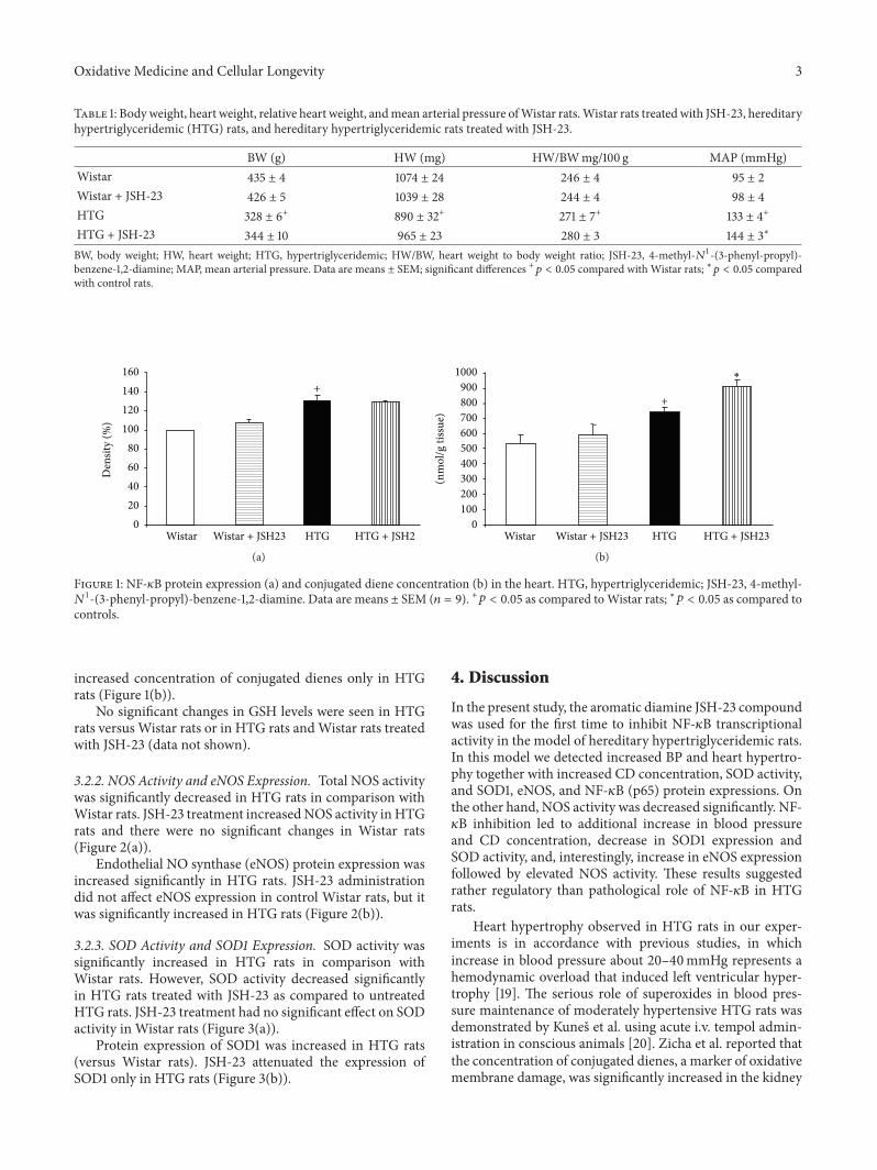
Oxidative Medicine and Cellular Longevity 3
Table 1: Bodyweight, heart weight, relative heart weight, andmean arterial pressure ofWistar rats.Wistar rats treated with JSH-23, hereditaryhypertriglyceridemic (HTG) rats, and hereditary hypertriglyceridemic rats treated with JSH-23.
BW (g) HW (mg) HW/BWmg/100 g MAP (mmHg)Wistar 435 ± 4 1074 ± 24 246 ± 4 95 ± 2Wistar + JSH-23 426 ± 5 1039 ± 28 244 ± 4 98 ± 4HTG 328 ± 6+ 890 ± 32+ 271 ± 7+ 133 ± 4+
HTG + JSH-23 344 ± 10 965 ± 23 280 ± 3 144 ± 3∗
BW, body weight; HW, heart weight; HTG, hypertriglyceridemic; HW/BW, heart weight to body weight ratio; JSH-23, 4-methyl-𝑁1-(3-phenyl-propyl)-benzene-1,2-diamine; MAP, mean arterial pressure. Data are means ± SEM; significant differences +𝑝 < 0.05 compared with Wistar rats; ∗𝑝 < 0.05 comparedwith control rats.
Den
sity
(%)
020406080
100120140160
Wistar Wistar + JSH23 HTG HTG + JSH2
+
(a)
(nm
ol/g
tiss
ue)
0100200300400500600700800900
1000
Wistar Wistar + JSH23 HTG HTG + JSH23
+
∗
(b)
Figure 1: NF-𝜅B protein expression (a) and conjugated diene concentration (b) in the heart. HTG, hypertriglyceridemic; JSH-23, 4-methyl-𝑁1-(3-phenyl-propyl)-benzene-1,2-diamine. Data are means ± SEM (𝑛 = 9). +𝑝 < 0.05 as compared toWistar rats; ∗𝑝 < 0.05 as compared to
controls.
increased concentration of conjugated dienes only in HTGrats (Figure 1(b)).
No significant changes in GSH levels were seen in HTGrats versus Wistar rats or in HTG rats andWistar rats treatedwith JSH-23 (data not shown).
3.2.2. NOS Activity and eNOS Expression. Total NOS activitywas significantly decreased in HTG rats in comparison withWistar rats. JSH-23 treatment increasedNOS activity inHTGrats and there were no significant changes in Wistar rats(Figure 2(a)).
Endothelial NO synthase (eNOS) protein expression wasincreased significantly in HTG rats. JSH-23 administrationdid not affect eNOS expression in control Wistar rats, but itwas significantly increased in HTG rats (Figure 2(b)).
3.2.3. SOD Activity and SOD1 Expression. SOD activity wassignificantly increased in HTG rats in comparison withWistar rats. However, SOD activity decreased significantlyin HTG rats treated with JSH-23 as compared to untreatedHTG rats. JSH-23 treatment had no significant effect on SODactivity in Wistar rats (Figure 3(a)).
Protein expression of SOD1 was increased in HTG rats(versus Wistar rats). JSH-23 attenuated the expression ofSOD1 only in HTG rats (Figure 3(b)).
4. Discussion
In the present study, the aromatic diamine JSH-23 compoundwas used for the first time to inhibit NF-𝜅B transcriptionalactivity in the model of hereditary hypertriglyceridemic rats.In this model we detected increased BP and heart hypertro-phy together with increased CD concentration, SOD activity,and SOD1, eNOS, and NF-𝜅B (p65) protein expressions. Onthe other hand, NOS activity was decreased significantly. NF-𝜅B inhibition led to additional increase in blood pressureand CD concentration, decrease in SOD1 expression andSOD activity, and, interestingly, increase in eNOS expressionfollowed by elevated NOS activity. These results suggestedrather regulatory than pathological role of NF-𝜅B in HTGrats.
Heart hypertrophy observed in HTG rats in our exper-iments is in accordance with previous studies, in whichincrease in blood pressure about 20–40mmHg represents ahemodynamic overload that induced left ventricular hyper-trophy [19]. The serious role of superoxides in blood pres-sure maintenance of moderately hypertensive HTG rats wasdemonstrated by Kuneš et al. using acute i.v. tempol admin-istration in conscious animals [20]. Zicha et al. reported thatthe concentration of conjugated dienes, a marker of oxidativemembrane damage, was significantly increased in the kidney
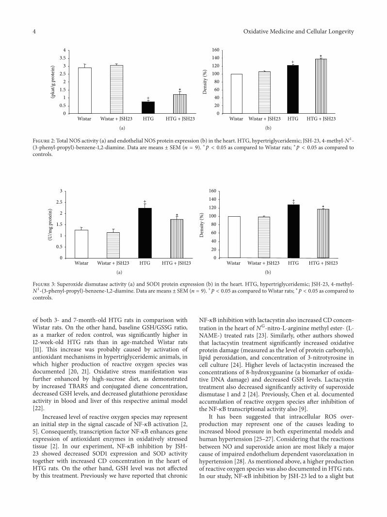
4 Oxidative Medicine and Cellular Longevity
(pka
t/g p
rote
in)
00.5
11.5
22.5
33.5
4
Wistar Wistar + JSH23 HTG HTG + JSH23
+
∗
(a)
Den
sity
(%)
020406080
100120140160
Wistar Wistar + JSH23 HTG HTG + JSH23
+
∗
(b)
Figure 2: Total NOS activity (a) and endothelial NOS protein expression (b) in the heart. HTG, hypertriglyceridemic; JSH-23, 4-methyl-𝑁1-(3-phenyl-propyl)-benzene-1,2-diamine. Data are means ± SEM (𝑛 = 9). +𝑝 < 0.05 as compared to Wistar rats; ∗𝑝 < 0.05 as compared tocontrols.
(U/m
g pr
otei
n)
0
0.5
1
1.5
2
2.5
3
Wistar Wistar + JSH23 HTG HTG + JSH23
+
∗
(a)
Den
sity
(%)
020406080
100120140160
Wistar Wistar + JSH23 HTG HTG + JSH23
+
∗
(b)
Figure 3: Superoxide dismutase activity (a) and SOD1 protein expression (b) in the heart. HTG, hypertriglyceridemic; JSH-23, 4-methyl-𝑁1-(3-phenyl-propyl)-benzene-1,2-diamine. Data are means ± SEM (𝑛 = 9). +𝑝 < 0.05 as compared toWistar rats; ∗𝑝 < 0.05 as compared to
controls.
of both 3- and 7-month-old HTG rats in comparison withWistar rats. On the other hand, baseline GSH/GSSG ratio,as a marker of redox control, was significantly higher in12-week-old HTG rats than in age-matched Wistar rats[11]. This increase was probably caused by activation ofantioxidant mechanisms in hypertriglyceridemic animals, inwhich higher production of reactive oxygen species wasdocumented [20, 21]. Oxidative stress manifestation wasfurther enhanced by high-sucrose diet, as demonstratedby increased TBARS and conjugated diene concentration,decreased GSH levels, and decreased glutathione peroxidaseactivity in blood and liver of this respective animal model[22].
Increased level of reactive oxygen species may representan initial step in the signal cascade of NF-𝜅B activation [2,5]. Consequently, transcription factor NF-𝜅B enhances geneexpression of antioxidant enzymes in oxidatively stressedtissue [2]. In our experiment, NF-𝜅B inhibition by JSH-23 showed decreased SOD1 expression and SOD activitytogether with increased CD concentration in the heart ofHTG rats. On the other hand, GSH level was not affectedby this treatment. Previously we have reported that chronic
NF-𝜅B inhibition with lactacystin also increased CD concen-tration in the heart of𝑁G-nitro-L-arginine methyl ester- (L-NAME-) treated rats [23]. Similarly, other authors showedthat lactacystin treatment significantly increased oxidativeprotein damage (measured as the level of protein carbonyls),lipid peroxidation, and concentration of 3-nitrotyrosine incell culture [24]. Higher levels of lactacystin increased theconcentrations of 8-hydroxyguanine (a biomarker of oxida-tive DNA damage) and decreased GSH levels. Lactacystintreatment also decreased significantly activity of superoxidedismutase 1 and 2 [24]. Previously, Chen et al. documentedaccumulation of reactive oxygen species after inhibition ofthe NF-𝜅B transcriptional activity also [9].
It has been suggested that intracellular ROS over-production may represent one of the causes leading toincreased blood pressure in both experimental models andhuman hypertension [25–27]. Considering that the reactionsbetween NO and superoxide anion are most likely a majorcause of impaired endothelium dependent vasorelaxation inhypertension [28]. As mentioned above, a higher productionof reactive oxygen species was also documented in HTG rats.In our study, NF-𝜅B inhibition by JSH-23 led to a slight but
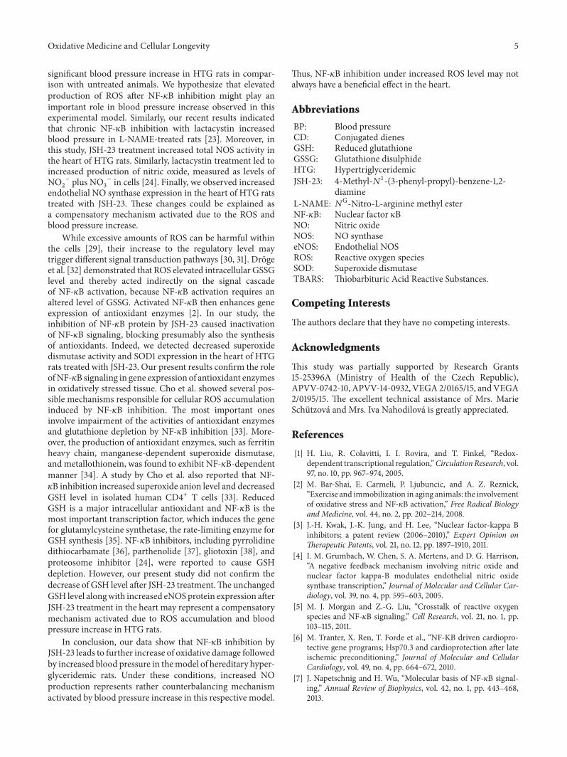
Oxidative Medicine and Cellular Longevity 5
significant blood pressure increase in HTG rats in compar-ison with untreated animals. We hypothesize that elevatedproduction of ROS after NF-𝜅B inhibition might play animportant role in blood pressure increase observed in thisexperimental model. Similarly, our recent results indicatedthat chronic NF-𝜅B inhibition with lactacystin increasedblood pressure in L-NAME-treated rats [23]. Moreover, inthis study, JSH-23 treatment increased total NOS activity inthe heart of HTG rats. Similarly, lactacystin treatment led toincreased production of nitric oxide, measured as levels ofNO2
− plus NO3
− in cells [24]. Finally, we observed increasedendothelial NO synthase expression in the heart of HTG ratstreated with JSH-23. These changes could be explained asa compensatory mechanism activated due to the ROS andblood pressure increase.
While excessive amounts of ROS can be harmful withinthe cells [29], their increase to the regulatory level maytrigger different signal transduction pathways [30, 31]. Drögeet al. [32] demonstrated that ROS elevated intracellular GSSGlevel and thereby acted indirectly on the signal cascadeof NF-𝜅B activation, because NF-𝜅B activation requires analtered level of GSSG. Activated NF-𝜅B then enhances geneexpression of antioxidant enzymes [2]. In our study, theinhibition of NF-𝜅B protein by JSH-23 caused inactivationof NF-𝜅B signaling, blocking presumably also the synthesisof antioxidants. Indeed, we detected decreased superoxidedismutase activity and SOD1 expression in the heart of HTGrats treated with JSH-23. Our present results confirm the roleofNF-𝜅B signaling in gene expression of antioxidant enzymesin oxidatively stressed tissue. Cho et al. showed several pos-sible mechanisms responsible for cellular ROS accumulationinduced by NF-𝜅B inhibition. The most important onesinvolve impairment of the activities of antioxidant enzymesand glutathione depletion by NF-𝜅B inhibition [33]. More-over, the production of antioxidant enzymes, such as ferritinheavy chain, manganese-dependent superoxide dismutase,and metallothionein, was found to exhibit NF-𝜅B-dependentmanner [34]. A study by Cho et al. also reported that NF-𝜅B inhibition increased superoxide anion level and decreasedGSH level in isolated human CD4+ T cells [33]. ReducedGSH is a major intracellular antioxidant and NF-𝜅B is themost important transcription factor, which induces the genefor glutamylcysteine synthetase, the rate-limiting enzyme forGSH synthesis [35]. NF-𝜅B inhibitors, including pyrrolidinedithiocarbamate [36], parthenolide [37], gliotoxin [38], andproteosome inhibitor [24], were reported to cause GSHdepletion. However, our present study did not confirm thedecrease of GSH level after JSH-23 treatment.The unchangedGSH level alongwith increased eNOSprotein expression afterJSH-23 treatment in the heart may represent a compensatorymechanism activated due to ROS accumulation and bloodpressure increase in HTG rats.
In conclusion, our data show that NF-𝜅B inhibition byJSH-23 leads to further increase of oxidative damage followedby increased blood pressure in themodel of hereditary hyper-glyceridemic rats. Under these conditions, increased NOproduction represents rather counterbalancing mechanismactivated by blood pressure increase in this respective model.
Thus, NF-𝜅B inhibition under increased ROS level may notalways have a beneficial effect in the heart.
Abbreviations
BP: Blood pressureCD: Conjugated dienesGSH: Reduced glutathioneGSSG: Glutathione disulphideHTG: HypertriglyceridemicJSH-23: 4-Methyl-𝑁1-(3-phenyl-propyl)-benzene-1,2-
diamineL-NAME: 𝑁G-Nitro-L-arginine methyl esterNF-𝜅B: Nuclear factor 𝜅BNO: Nitric oxideNOS: NO synthaseeNOS: Endothelial NOSROS: Reactive oxygen speciesSOD: Superoxide dismutaseTBARS: Thiobarbituric Acid Reactive Substances.
Competing Interests
The authors declare that they have no competing interests.
Acknowledgments
This study was partially supported by Research Grants15-25396A (Ministry of Health of the Czech Republic),APVV-0742-10, APVV-14-0932, VEGA 2/0165/15, andVEGA2/0195/15. The excellent technical assistance of Mrs. MarieSchützová and Mrs. Iva Nahodilová is greatly appreciated.
References
[1] H. Liu, R. Colavitti, I. I. Rovira, and T. Finkel, “Redox-dependent transcriptional regulation,”CirculationResearch, vol.97, no. 10, pp. 967–974, 2005.
[2] M. Bar-Shai, E. Carmeli, P. Ljubuncic, and A. Z. Reznick,“Exercise and immobilization in aging animals: the involvementof oxidative stress and NF-𝜅B activation,” Free Radical Biologyand Medicine, vol. 44, no. 2, pp. 202–214, 2008.
[3] J.-H. Kwak, J.-K. Jung, and H. Lee, “Nuclear factor-kappa Binhibitors; a patent review (2006–2010),” Expert Opinion onTherapeutic Patents, vol. 21, no. 12, pp. 1897–1910, 2011.
[4] I. M. Grumbach, W. Chen, S. A. Mertens, and D. G. Harrison,“A negative feedback mechanism involving nitric oxide andnuclear factor kappa-B modulates endothelial nitric oxidesynthase transcription,” Journal of Molecular and Cellular Car-diology, vol. 39, no. 4, pp. 595–603, 2005.
[5] M. J. Morgan and Z.-G. Liu, “Crosstalk of reactive oxygenspecies and NF-𝜅B signaling,” Cell Research, vol. 21, no. 1, pp.103–115, 2011.
[6] M. Tranter, X. Ren, T. Forde et al., “NF-ΚB driven cardiopro-tective gene programs; Hsp70.3 and cardioprotection after lateischemic preconditioning,” Journal of Molecular and CellularCardiology, vol. 49, no. 4, pp. 664–672, 2010.
[7] J. Napetschnig and H. Wu, “Molecular basis of NF-𝜅B signal-ing,” Annual Review of Biophysics, vol. 42, no. 1, pp. 443–468,2013.
6 Oxidative Medicine and Cellular Longevity
[8] C. K. Sen, S. Khanna, A. Z. Reznick, S. Roy, and L. Packer, “Glu-tathione regulation of tumor necrosis factor-𝛼-induced NF-𝜅Bactivation in skeletal muscle-derived L6 cells,” Biochemical andBiophysical Research Communications, vol. 237, no. 3, pp. 645–649, 1997.
[9] F. Chen, V. Castranova, Z. Li, M. Karin, and X. Shi, “Inhibitorof nuclear factor kappa B kinase deficiency enhances oxidativestress and prolongs c-Jun NH2-terminal kinase activationinduced by arsenic,” Cancer Research, vol. 63, pp. 7689–7693,2003.
[10] A. Vrána and L. Kazdová, “The hereditary hypertriglyceridemicnonobese rat: an experimental model of human hypertriglyc-eridemia,” Transplantation Proceedings, vol. 22, no. 6, p. 2579,1990.
[11] J. Zicha, O. Pecháňová, S. Čačányiová et al., “Hereditaryhypertriglyceridemic rat: a suitable model of cardiovasculardisease and metabolic syndrome?” Physiological Research, vol.55, supplement 1, pp. S49–S63, 2006.
[12] L. Kazdová, A. Žák, and A. Vrána, “Increased lipoproteinoxidability and aortic lipid peroxidation in an experimentalmodel of insulin resistance syndrome,” Annals of the New YorkAcademy of Sciences, vol. 827, pp. 521–525, 1997.
[13] H.-M. Shin, M.-H. Kim, B. H. Kim et al., “Inhibitory actionof novel aromatic diamine compound on lipopolysaccharide-induced nuclear translocation of NF-𝜅B without affecting I𝜅Bdegradation,” FEBS Letters, vol. 571, no. 1–3, pp. 50–54, 2004.
[14] K. Kogure, B. D. Watson, R. Busto, and K. Abe, “Potentiationof lipid peroxides by ischemia in rat brain,” NeurochemicalResearch, vol. 7, no. 4, pp. 437–454, 1982.
[15] G. L. Ellman, “Tissue sulfhydryl groups,” Archives of Biochem-istry and Biophysics, vol. 82, no. 1, pp. 70–77, 1959.
[16] D. S. Bredt and S. H. Snyder, “Isolation of nitric oxide syn-thetase, a calmodulin-requiring enzyme,” Proceedings of theNational Academy of Sciences of the United States of America,vol. 87, no. 2, pp. 682–685, 1990.
[17] O. Pecháňová, I. Bernátová, V. Pelouch, and F. Šimko, “Proteinremodelling of the heart in NO-deficient hypertension: theeffect of captopril,” Journal ofMolecular andCellular Cardiology,vol. 29, no. 12, pp. 3365–3374, 1997.
[18] O. Pecháňová, J. Zicha, S. Kojšová, Z. Dobešová, L. Jendeková,and J. Kuneš, “Effect of chronic N-acetylcysteine treatment onthe development of spontaneous hypertension,”Clinical Science,vol. 110, no. 2, pp. 235–242, 2006.
[19] F. Simko, I. Luptak, J. Matuskova et al., “Heart remodeling inthe hereditary hypertriglyceridemic rat: Effect of captopril andnitric oxide deficiency,” Annals of the New York Academy ofSciences, vol. 967, pp. 454–462, 2002.
[20] J. Kuneš, Z. Dobešová, and J. Zicha, “Altered balance of mainvasopressor and vasodepressor systems in rats with genetichypertension and hypertriglyceridaemia,” Clinical Science, vol.102, no. 3, pp. 269–277, 2002.
[21] M. Žourek, P. Kyselová, J. Mudra et al., “The relationshipbetween glycemia, insulin and oxidative stress in hereditaryhypertriglyceridemic rat,” Physiological Research, vol. 57, no. 4,pp. 531–538, 2008.
[22] N. Škottová, L. Kazdová, O. Oliyarnyk, R. Večeřa, L. Sobolová,and J. Ulrichová, “Phenolics-rich extracts from Silybum mari-anum and Prunella vulgaris reduce a high-sucrose diet inducedoxidative stress in hereditary hypertriglyceridemic rats,” Phar-macological Research, vol. 50, no. 2, pp. 123–130, 2004.
[23] S. Vranková, J. Parohová, A. Barta, P. Janega, F. Šimko, and O.Pecháňová, “Effect of nuclear factor kappa B inhibition on L-NAME-induced hypertension and cardiovascular remodelling,”Journal of Hypertension, vol. 28, no. 1, pp. S45–S49, 2010.
[24] M. H. Lee, D.-H. Hyun, P. Jenner, and B. Halliwell, “Effect ofproteasome inhibition on cellular oxidative damage, antioxi-dant defences and nitric oxide production,” Journal of Neuro-chemistry, vol. 78, no. 1, pp. 32–41, 2001.
[25] N. C.Ward andK.D. Croft, “Hypertension and oxidative stress,”Clinical and Experimental Pharmacology and Physiology, vol. 33,no. 9, pp. 872–876, 2006.
[26] S. Kojšová, L. Jendeková, J. Zicha, J. Kuneš, R. Andriantsito-haina, and O. Pecháňová, “The effect of different antioxidantson nitric oxide production in hypertensive rats,” PhysiologicalResearch, vol. 55, supplement 1, pp. S3–S16, 2006.
[27] C. S. Wilcox and A. Pearlman, “Chemistry and antihyperten-sive effects of tempol and other nitroxides,” PharmacologicalReviews, vol. 60, no. 4, pp. 418–469, 2008.
[28] B. Skibska andA. Goraca, “The protective effect of lipoic acid onselected cardiovascular diseases caused by age-related oxidativestress,” Oxidative Medicine and Cellular Longevity, vol. 2015,Article ID 313021, 11 pages, 2015.
[29] P. X. Shaw, G. Werstuck, and Y. Chen, “Oxidative stress andaging diseases,” Oxidative Medicine and Cellular Longevity, vol.2014, Article ID 569146, 2 pages, 2014.
[30] M. Valko, D. Leibfritz, J. Moncol, M. T. D. Cronin, M. Mazur,and J. Telser, “Free radicals and antioxidants in normal physi-ological functions and human disease,” International Journal ofBiochemistry and Cell Biology, vol. 39, no. 1, pp. 44–84, 2007.
[31] M. Woodward, K. D. Croft, T. A. Mori et al., “Associationbetween both lipid and protein oxidation and the risk of fatalor non-fatal coronary heart disease in a human population,”Clinical Science, vol. 116, no. 1, pp. 53–60, 2009.
[32] W. Dröge, K. Schulze-Osthoff, S. Mihm et al., “Functionsof glutathione and glutathione disulfide in immunology andimmunopathology,” The FASEB Journal, vol. 8, no. 14, pp. 1131–1138, 1994.
[33] M.-L. Cho, Y.-M.Moon, Y.-J. Heo et al., “NF-𝜅B inhibition leadsto increased synthesis and secretion of MIF in human CD4+ Tcells,” Immunology Letters, vol. 123, no. 1, pp. 21–30, 2009.
[34] T. Sasazuki, T. Okazaki, K. Tada et al., “Genome wide analysisof TNF-inducible genes reveals that antioxidant enzymes areinduced by TNF and responsible for elimination of ROS,”Molecular Immunology, vol. 41, no. 5, pp. 547–551, 2004.
[35] D.M. Townsend, K. D. Tew, andH. Tapiero, “The importance ofglutathione in human disease,” Biomedicine and Pharmacother-apy, vol. 57, no. 3, pp. 145–155, 2003.
[36] M.C. Santos-Silva,M. S.D. Freitas, and J. Assreuy, “Involvementof NF-𝜅B and glutathione in cytotoxic effects of nitric oxide andtaxol on human leukemia cells,” Leukemia Research, vol. 30, no.2, pp. 145–152, 2006.
[37] S. Zhang, C.-N. Ong, and H.-M. Shen, “Critical roles of intra-cellular thiols and calcium in parthenolide-induced apoptosisin human colorectal cancer cells,” Cancer Letters, vol. 208, no. 2,pp. 143–153, 2004.
[38] J. G. Orr, V. Leel, G. A. Cameron et al., “Mechanism of actionof the antifibrogenic compound gliotoxin in rat liver cells,”Hepatology, vol. 40, no. 1, pp. 232–242, 2004.
Review ArticleWidening and Elaboration of Consecutive Research intoTherapeutic Antioxidant Enzyme Derivatives
Alexander V. Maksimenko
Institute of Experimental Cardiology, Russian Cardiology Research-and-Production Complex, Ministry of Health Care ofRussian Federation, 15a 3rd Cherepkovskaya Street, Moscow 121552, Russia
Correspondence should be addressed to Alexander V. Maksimenko; [email protected]
Received 22 December 2015; Revised 16 February 2016; Accepted 16 March 2016
Academic Editor: Tanea T. Reed
Copyright © 2016 Alexander V. Maksimenko. This is an open access article distributed under the Creative Commons AttributionLicense, which permits unrestricted use, distribution, and reproduction in any medium, provided the original work is properlycited.
Undiminishing actuality of enzyme modification for therapeutic purposes has been confirmed by application of modifiedenzymes in clinical practice and numerous research data on them. Intravenous injection of the superoxide dismutase-chondroitinsulfate-catalase (SOD-CHS-CAT) conjugate in preventive and medicative regimes in rats with endotoxin shock induced with alipopolysaccharide bolus has demonstrated that antioxidant agents not only effectively prevent damage caused by oxidative stress(as believed previously) but also can be used for antioxidative stress therapy. The results obtained emphasize the importance ofinvestigation into the pathogenesis of vascular damage and the role of oxidative stress in it. The effects of intravenous medicativeinjection of SOD-CHS-CAT in a rat model of endotoxin shock have demonstrated a variety in the activity of this conjugatein addition to prevention of NO conversion in peroxynitrite upon interaction with O
2
∙− superoxide radical. Together with theliterature data, these findings offer a prospect for the study of NO-independent therapeutic effects of SOD-CHS-CAT, implying theimportance of a better insight into themechanisms of the conjugate activity inmodeled cardiovascular damage involving vasoactiveagents other than NO.
1. Introduction
A considerable role of enzymes in human body has promptedan extensive research of their derivatives for medical appli-cation. The intense initial period of the research promotedthe development of “new biology” [1] and biopharmacology[2]. A decrease in the number of studies of proteins for ther-apeutic purposes was followed by great diversity of enzymemodification methods. Some enzyme derivatives have foundsuccessful clinical application. For instance, investigation anddevelopment of plasminogen activators provided a currentset of protein preparations for clinical thrombolysis [3].Theireffectiveness urges further improvements, such as a decreasein molecular mass (compared with parent molecules) or anincrease in it upon construction of new modified agents[4]. Fast development of glycobiology [5, 6] leads to newapproaches and targets for the future protein therapy. Nan-otechnologies allowed large biological conjugates for furtherinvestigation [3, 7–9], and “smart” biopolymers respondingto temperature, aX, and effects of biomolecules [10] were
developed. New conceptions contributed to further diversifi-cation of protein agents, new protocols of their clinical trials,and appearance of novel aspects in the search for medicalenzyme derivatives.
2. Improvement of Enzyme Agentsby Modification
Modification of enzymes to overcome their disadvantages formedical use (low stability in the body, short retention periodin the bloodstream, low resistance to inhibitors, etc.) and toadd to them new properties (ability to concentrate in thedamaged area, reduce toxicity, etc.) has been successfully usedfor a long time [3, 4]. Covalent binding of low-molecular-mass heparin to Cu, Zn-superoxide dismutase increased theenzyme stability in acid and alkaline media as well as itsresistance to trypsinolysis and temperature [11]. Conjugationof bovine pancreatic ribonuclease with poly[N-(2-hydrox-ypropyl)methacrylamide] prolongs the enzyme retention in
Hindawi Publishing CorporationOxidative Medicine and Cellular LongevityVolume 2016, Article ID 3075695, 9 pageshttp://dx.doi.org/10.1155/2016/3075695
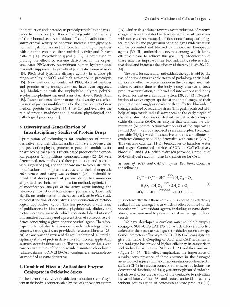
2 Oxidative Medicine and Cellular Longevity
the circulation and increases its proteolytic stability and resis-tance to inhibitors [12], thus enhancing antitumor activityof the ribonuclease. Antioxidant effect of ovalbumin andantimicrobial activity of lysozyme increase after glycosyla-tion with galactomannan [13]. Covalent binding of peptideswith albumin enhances their antiviral activity and in vivohalf-life [14]. Polyethylene glycol (PEG) is often used toprolong the effects of enzyme derivatives in the organ-ism. After PEGylation, recombinant human hyaluronidasemarkedly suppresses the growth of pancreatic tumor in mice[15]. PEGylated lysozyme displays activity in a wide aXrange, stability at 50∘b, and high resistance to proteolysis[16]. New methods for controlled PEGylation of peptidesand proteins using transglutaminase have been suggested[17]. Modification with the amphiphilic polymer poly(N-acryloylmorpholine)was used as an alternative to PEGylation[18]. Recent evidence demonstrates the diversity and effec-tiveness of protein modifications for the development of newmedical protein derivatives [3, 4, 19, 20] and evaluates therole of protein modifications in various physiological andpathological processes [21].
3. Diversity and Generalization ofInterdisciplinary Studies of Protein Drugs
Optimization of technologies for production of proteinderivatives and their clinical application have broadened theprospects of employing proteins as potential candidates forpharmacological agents. Protein-based products for biomed-ical purposes (compositions, combined drugs) [22, 23] weredetermined, new methods of their production and isolationwere suggested [24], and the concordance between structuralmodifications of biopharmaceutics and their therapeuticeffectiveness and safety was evaluated [25]. It should benoted that development of protein drugs has numerousstages, such as choice of modification method, optimizationof modification, analysis of the active agent binding andrelease, cytotoxicity and toxicological parameters, statisticallysignificant confirmation of therapeutic effects in vivo, studyof biodistribution of derivatives, and evaluation of techno-logical approaches [4, 10]. This has provoked a vast arrayof publications in biochemical, physiological, clinical, andbiotechnological journals, which accelerated distribution ofinformation but hampered a presentation of consecutive evi-dence concerning a given pharmaceutical agent. Therefore,papers selected due to semantic search technology (for aconcrete test object) were provided by electron libraries [26–28]. An analysis and review of the results obtained in interdis-ciplinary study of protein derivatives for medical applicationseems relevant in this situation.The present review deals withconsecutive studies of the superoxide dismutase-chondroitinsulfate-catalase (SOD-CHS-CAT) conjugate, a supramolecu-lar modified enzyme derivative.
4. Combined Effect of Antioxidant EnzymeConjugate in Oxidative Stress
In the norm the activity of oxidation-reduction (redox) sys-tem in the body is countervailed by that of antioxidant system
[29]. Shift in this balance towards overproduction of reactiveoxygen species facilitates the development of oxidative stresswith nonselective structural and functional damage to biolog-ical molecules and progression of pathology. Oxidative stresscan be prevented and blocked by antioxidant therapeuticagents [30, 31], antioxidant enzymes among which beingeffective means to achieve this goal [32]. Modification ofthese enzymes improves their bioavailability, reduces effec-tive dose, and increases the efficacy of therapy [4, 29, 30, 32–34].
The basis for successful antioxidant therapy is laid by theuse of antioxidants at early stages of pathology, their local-ization and effective concentration in the damaged area, suf-ficient retention time in the body, safety, absence of toxicproduct accumulation, and beneficial interactions with bodysystems, for instance, immune system [29, 30, 32]. Neutral-ization of active oxygen species at the initial stages of theirproduction is strongly associatedwith an effective blockade ofdamage induced by oxidative stress.This goal is achievedwithhelp of superoxide radical scavengers at the early stages ofchain transformations associatedwith oxidative stress. Super-oxide dismutase (SOD), an enzyme that catalyzes the dis-mutation (or neutralization/partitioning) of the superoxideradical (\
2
∙−), can be employed as an interceptor. Hydrogenperoxide (X
2\2) which in excessive amounts contributes to
oxidative damage should be detoxified with catalase (CAT).This enzyme catalyzes X
2\2breakdown to harmless water
and oxygen. Connected activities of SOD andCAT effectivelyblock \
2
∙− and X2\2, when hydrogen peroxide, a product of
SOD-catalyzed reaction, turns into substrate for CAT.
Schemes of SOD- and CAT-Catalyzed Reactions. Considerthe following:
\2
∙−+ \2
∙−+ 2H+ SOD→ H
2O2+O2
H2O2+H2O2
CAT→ 2H
2O +O
2
4\2
∙−+ 4H+
SOD/CAT→ 2H
2O + 3O
2
(1)
It is noteworthy that these conversions should be effectivelyrealized in the damaged area which is often confined to thevascular wall. Antioxidant agents, including enzyme deriv-atives, have been used to prevent oxidative damage to bloodvessels.
We have developed a covalent water-soluble bienzymeconjugate SOD-CHS-CAT [35, 36] which offers an effectivedefense of the vascular wall against oxidative stress damage.Some parameters of bienzyme SOD-CHS-CAT conjugate aregiven in Table 1. Coupling of SOD and CAT activities inthe conjugate has provided higher efficiency in comparisonwith individual activities of SOD andCAT and their mixtures(Figure 1) [37]. This effect emphasizes the importance ofsimultaneous presence of these enzymes in the damagedarea (focus of injury). Enhanced accumulation of chondroitinsulfate (CHS) in vascular zones of atherosclerotic lesions hasdetermined the choice of this glycosaminoglycan of endothe-lial glycocalyx for preparation of the conjugate to potentiateits vasodilatory effect [38] and rooted antioxidant activitywithout accumulation of concomitant toxic products [37].
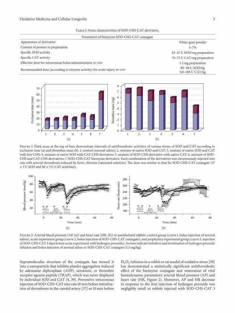
Oxidative Medicine and Cellular Longevity 3
Table 1: Some characteristics of SOD-CHS-CAT derivative.
Parameters of bienzyme SOD-CHS-CAT conjugateAppearance of derivative White-gray powderContent of protein in preparation 5–7%Specific SOD activity 43–47U SOD/mg preparationSpecific CAT activity 70–75U CAT/mg preparationEffective dose for intravenous bolus administration in vivo 1-2mg preparation
Recommended dose (according to enzyme activity) for acute injury in vivo 80–90U SOD/kg145–185U CAT/kg
0
10
20
30
40
50
60
70
Occ
lusio
n tim
e (m
in)
1 2 3 4 5 6 7(a)
1 2 3 4 5 6 70
1
2
3
4
5
6
7
8
Thro
mbu
s mas
s (m
g)
(b)
Figure 1: Dark areas at the top of bars demonstrate intervals of antithrombotic activities of various forms of SOD and CAT according toocclusion time (a) and thrombus mass (b). 1, control (normal saline); 2, mixture of native SOD and CAT; 3, mixture of native SOD and CATwith free CHS; 4, mixture of native SOD with CAT-CHS derivative; 5, mixture of SOD-CHS derivative with native CAT; 6, mixture of SOD-CHS and CAT-CHS derivatives; 7, SOD-CHS-CAT bienzyme derivative. Each combination of the derivatives was intravenously injected intorats with arterial thrombosis induced by ferric chlorine (saturated solution). The dose was similar to that for SOD-CHS-CAT conjugate (37± 3U SOD and 80 ± 3U CAT activities).
0 20 40 60 80
Time (min)
20
40
60
80
100
Bloo
d pr
essu
re (m
mH
g)
Infusionof H2O2
Infusionof H2O2
Bolussaline or
SOD-CHS-CAT
1
2
3
(a)
0 20 40 60 80
Time (min)
180
200
220
240
260
280
Hea
rt ra
te (b
eats/
min
)
Infusionof H2O2
Infusionof H2O2
Bolussaline or
SOD-CHS-CAT
1
2
3
(b)
Figure 2: Arterial blood pressure (AP, (1)) and heart rate (HR, (b)) in anesthetized rabbits: control group (curve 1, bolus injection of normalsaline), acute experiment group (curve 2, bolus injection of SOD-CHS-CAT conjugate), and prophylaxy experiment group (curve 3, injectionof SOD-CHS-CAT 3 days before acute experiment with hydrogen peroxide). Arrows indicate initiation and termination of hydrogen peroxideinfusion and bolus injection of normal saline or SOD-CHS-CAT conjugate (1.5mg/kg).
Supramolecular structure of the conjugate has turned itinto a nanoparticle that inhibits platelet aggregation inducedby adenosine diphosphate (ADP), serotonin, or thrombinreceptor agonist peptide (TRAP), which was never displayedby individual SOD and CAT [4, 39]. Preventive intravenousinjection of SOD-CHS-CAT into rats 10min before initializa-tion of thrombosis in the carotid artery [37] or 10min before
H2O2infusion in a rabbit or rat model of oxidative stress [39]
has demonstrated a statistically significant antithromboticeffect of the bienzyme conjugate and restoration of vitalhemodynamic parameters: arterial blood pressure (AP) andheart rate (HR, Figure 2). Moreover, AP and HR decreasein response to the first injection of hydrogen peroxide wasnegligibly small in rabbits injected with SOD-CHS-CAT 3
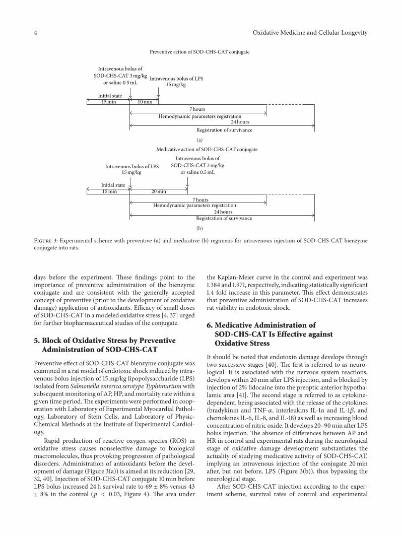
4 Oxidative Medicine and Cellular Longevity
Preventive action of SOD-CHS-CAT conjugate
Intravenous bolus ofSOD-CHS-CAT 3mg/kg
or saline 0.5mL
Initial state15min 10min
Intravenous bolus of LPS15mg/kg
7hoursHemodynamic parameters registration
24hoursRegistration of survivance
(a)
Intravenous bolus ofSOD-CHS-CAT 3mg/kg
or saline 0.5mL
Initial state15min 20min
Intravenous bolus of LPS15mg/kg
7hoursHemodynamic parameters registration
24hoursRegistration of survivance
Medicative action of SOD-CHS-CAT conjugate
(b)
Figure 3: Experimental scheme with preventive (1) and medicative (b) regimens for intravenous injection of SOD-CHS-CAT bienzymeconjugate into rats.
days before the experiment. These findings point to theimportance of preventive administration of the bienzymeconjugate and are consistent with the generally acceptedconcept of preventive (prior to the development of oxidativedamage) application of antioxidants. Efficacy of small dosesof SOD-CHS-CAT in amodeled oxidative stress [4, 37] urgedfor further biopharmaceutical studies of the conjugate.
5. Block of Oxidative Stress by PreventiveAdministration of SOD-CHS-CAT
Preventive effect of SOD-CHS-CAT bienzyme conjugate wasexamined in a ratmodel of endotoxic shock induced by intra-venous bolus injection of 15mg/kg lipopolysaccharide (LPS)isolated from Salmonella enterica serotype Typhimuriumwithsubsequent monitoring of AP, HP, andmortality rate within agiven time period.The experiments were performed in coop-eration with Laboratory of Experimental Myocardial Pathol-ogy, Laboratory of Stem Cells, and Laboratory of Physic-Chemical Methods at the Institute of Experimental Cardiol-ogy.
Rapid production of reactive oxygen species (ROS) inoxidative stress causes nonselective damage to biologicalmacromolecules, thus provoking progression of pathologicaldisorders. Administration of antioxidants before the devel-opment of damage (Figure 3(a)) is aimed at its reduction [29,32, 40]. Injection of SOD-CHS-CAT conjugate 10min beforeLPS bolus increased 24 h survival rate to 69 ± 8% versus 43± 8% in the control (𝑝 < 0.03, Figure 4). The area under
the Kaplan-Meier curve in the control and experiment was1.384 and 1.971, respectively, indicating statistically significant1.4-fold increase in this parameter. This effect demonstratesthat preventive administration of SOD-CHS-CAT increasesrat viability in endotoxic shock.
6. Medicative Administration ofSOD-CHS-CAT Is Effective againstOxidative Stress
It should be noted that endotoxin damage develops throughtwo successive stages [40]. The first is referred to as neuro-logical. It is associated with the nervous system reactions,develops within 20min after LPS injection, and is blocked byinjection of 2% lidocaine into the preoptic anterior hypotha-lamic area [41]. The second stage is referred to as cytokine-dependent, being associated with the release of the cytokines(bradykinin and TNF-𝛼, interleukins IL-1𝛼 and IL-1𝛽, andchemokines IL-6, IL-8, and IL-18) as well as increasing bloodconcentration of nitric oxide. It develops 20–90min after LPSbolus injection. The absence of differences between AP andHR in control and experimental rats during the neurologicalstage of oxidative damage development substantiates theactuality of studying medicative activity of SOD-CHS-CAT,implying an intravenous injection of the conjugate 20minafter, but not before, LPS (Figure 3(b)), thus bypassing theneurological stage.
After SOD-CHS-CAT injection according to the exper-iment scheme, survival rates of control and experimental
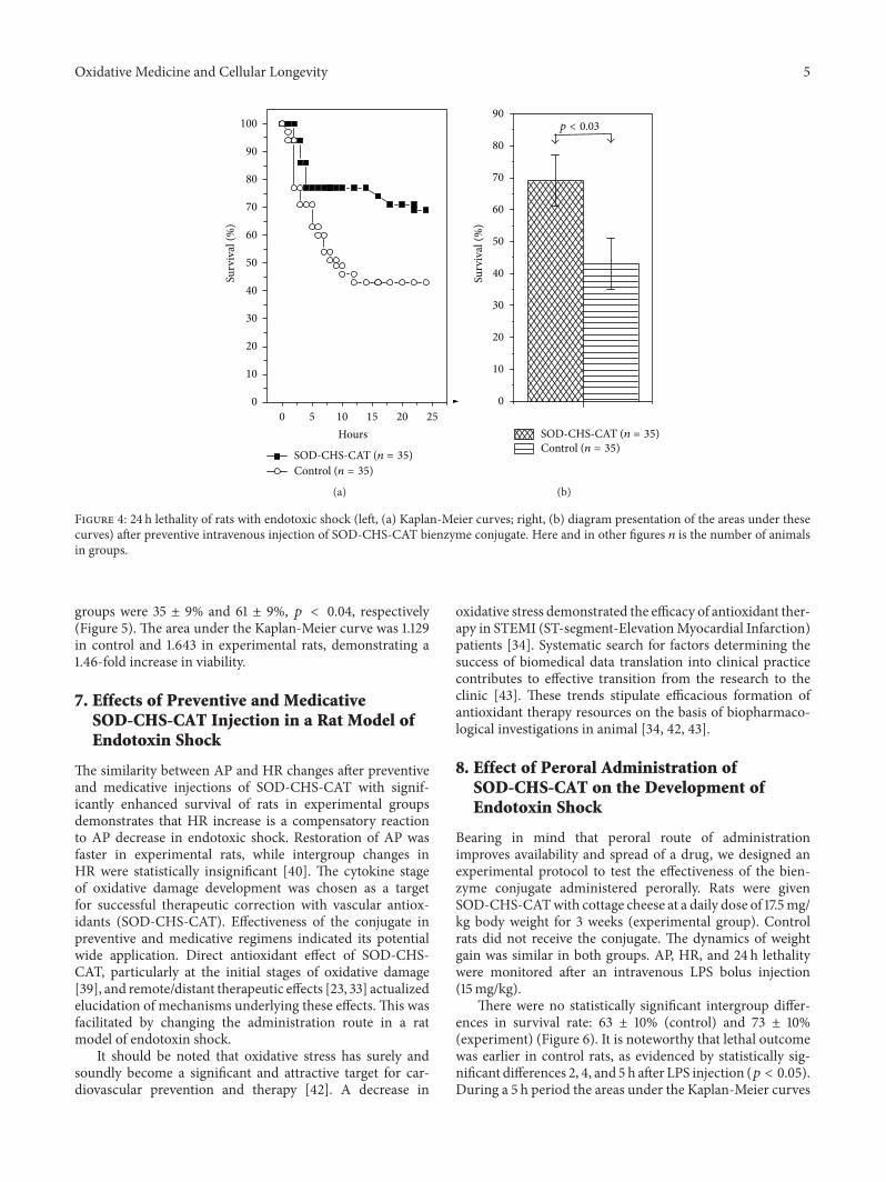
Oxidative Medicine and Cellular Longevity 5
0 5 10 15 20 250
10
20
30
40
50
60
70
80
90
100
Surv
ival
(%)
Hours
Control (n = 35)SOD-CHS-CAT (n = 35)
(a)
0
10
20
30
40
50
60
70
80
90
Surv
ival
(%)
Control (n = 35)SOD-CHS-CAT (n = 35)
p < 0.03
(b)
Figure 4: 24 h lethality of rats with endotoxic shock (left, (a) Kaplan-Meier curves; right, (b) diagram presentation of the areas under thesecurves) after preventive intravenous injection of SOD-CHS-CAT bienzyme conjugate. Here and in other figures 𝑛 is the number of animalsin groups.
groups were 35 ± 9% and 61 ± 9%, 𝑝 < 0.04, respectively(Figure 5). The area under the Kaplan-Meier curve was 1.129in control and 1.643 in experimental rats, demonstrating a1.46-fold increase in viability.
7. Effects of Preventive and MedicativeSOD-CHS-CAT Injection in a Rat Model ofEndotoxin Shock
The similarity between AP and HR changes after preventiveand medicative injections of SOD-CHS-CAT with signif-icantly enhanced survival of rats in experimental groupsdemonstrates that HR increase is a compensatory reactionto AP decrease in endotoxic shock. Restoration of AP wasfaster in experimental rats, while intergroup changes inHR were statistically insignificant [40]. The cytokine stageof oxidative damage development was chosen as a targetfor successful therapeutic correction with vascular antiox-idants (SOD-CHS-CAT). Effectiveness of the conjugate inpreventive and medicative regimens indicated its potentialwide application. Direct antioxidant effect of SOD-CHS-CAT, particularly at the initial stages of oxidative damage[39], and remote/distant therapeutic effects [23, 33] actualizedelucidation of mechanisms underlying these effects. This wasfacilitated by changing the administration route in a ratmodel of endotoxin shock.
It should be noted that oxidative stress has surely andsoundly become a significant and attractive target for car-diovascular prevention and therapy [42]. A decrease in
oxidative stress demonstrated the efficacy of antioxidant ther-apy in STEMI (ST-segment-ElevationMyocardial Infarction)patients [34]. Systematic search for factors determining thesuccess of biomedical data translation into clinical practicecontributes to effective transition from the research to theclinic [43]. These trends stipulate efficacious formation ofantioxidant therapy resources on the basis of biopharmaco-logical investigations in animal [34, 42, 43].
8. Effect of Peroral Administration ofSOD-CHS-CAT on the Development ofEndotoxin Shock
Bearing in mind that peroral route of administrationimproves availability and spread of a drug, we designed anexperimental protocol to test the effectiveness of the bien-zyme conjugate administered perorally. Rats were givenSOD-CHS-CATwith cottage cheese at a daily dose of 17.5mg/kg body weight for 3 weeks (experimental group). Controlrats did not receive the conjugate. The dynamics of weightgain was similar in both groups. AP, HR, and 24 h lethalitywere monitored after an intravenous LPS bolus injection(15mg/kg).
There were no statistically significant intergroup differ-ences in survival rate: 63 ± 10% (control) and 73 ± 10%(experiment) (Figure 6). It is noteworthy that lethal outcomewas earlier in control rats, as evidenced by statistically sig-nificant differences 2, 4, and 5 h after LPS injection (𝑝 < 0.05).During a 5 h period the areas under the Kaplan-Meier curves
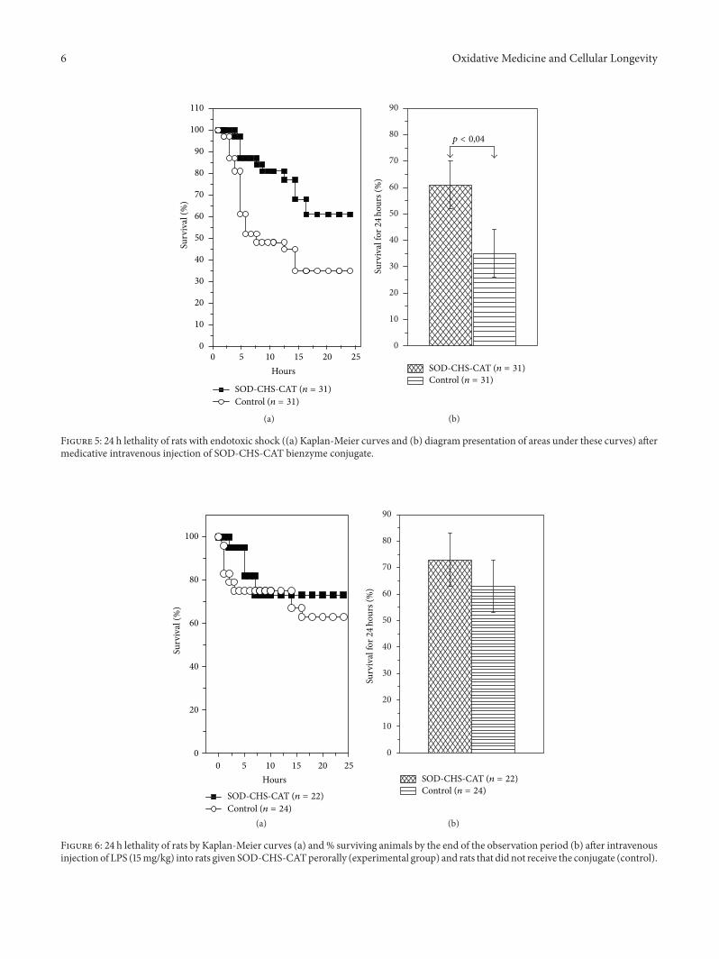
6 Oxidative Medicine and Cellular Longevity
Surv
ival
(%)
Hours
Control (n = 31)SOD-CHS-CAT (n = 31)
0 5 10 15 20 250
10
20
30
40
50
60
70
80
90
110
100
(a)
0
10
20
30
40
50
60
70
80
90
Control (n = 31)SOD-CHS-CAT (n = 31)
Surv
ival
for2
4ho
urs (
%)
p < 0,04
(b)
Figure 5: 24 h lethality of rats with endotoxic shock ((1) Kaplan-Meier curves and (b) diagram presentation of areas under these curves) aftermedicative intravenous injection of SOD-CHS-CAT bienzyme conjugate.
0
20
40
60
80
100
Surv
ival
(%)
Hours
Control (n = 24)SOD-CHS-CAT (n = 22)
0 5 10 15 20 25
(a)
0
10
20
30
40
50
60
70
80
90
Control (n = 24)SOD-CHS-CAT (n = 22)
Surv
ival
for2
4ho
urs (
%)
(b)
Figure 6: 24 h lethality of rats by Kaplan-Meier curves (1) and % surviving animals by the end of the observation period (b) after intravenousinjection of LPS (15mg/kg) into rats given SOD-CHS-CATperorally (experimental group) and rats that did not receive the conjugate (control).
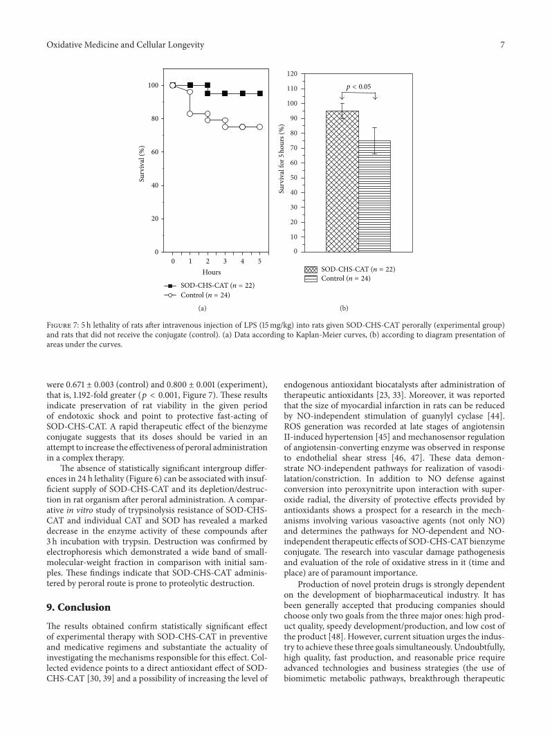
Oxidative Medicine and Cellular Longevity 7
0 1 2 3 4 50
20
40
60
80
100
Surv
ival
(%)
Hours
Control (n = 24)SOD-CHS-CAT (n = 22)
(a)
10
0
20
30
40
50
60
70
80
90
100
110
120
Control (n = 24)SOD-CHS-CAT (n = 22)
Surv
ival
for5
hour
s (%
)
p < 0.05
(b)
Figure 7: 5 h lethality of rats after intravenous injection of LPS (15mg/kg) into rats given SOD-CHS-CAT perorally (experimental group)and rats that did not receive the conjugate (control). (1) Data according to Kaplan-Meier curves, (b) according to diagram presentation ofareas under the curves.
were 0.671 ± 0.003 (control) and 0.800 ± 0.001 (experiment),that is, 1.192-fold greater (𝑝 < 0.001, Figure 7). These resultsindicate preservation of rat viability in the given periodof endotoxic shock and point to protective fast-acting ofSOD-CHS-CAT. A rapid therapeutic effect of the bienzymeconjugate suggests that its doses should be varied in anattempt to increase the effectiveness of peroral administrationin a complex therapy.
The absence of statistically significant intergroup differ-ences in 24 h lethality (Figure 6) can be associated with insuf-ficient supply of SOD-CHS-CAT and its depletion/destruc-tion in rat organism after peroral administration. A compar-ative in vitro study of trypsinolysis resistance of SOD-CHS-CAT and individual CAT and SOD has revealed a markeddecrease in the enzyme activity of these compounds after3 h incubation with trypsin. Destruction was confirmed byelectrophoresis which demonstrated a wide band of small-molecular-weight fraction in comparison with initial sam-ples. These findings indicate that SOD-CHS-CAT adminis-tered by peroral route is prone to proteolytic destruction.
9. Conclusion
The results obtained confirm statistically significant effectof experimental therapy with SOD-CHS-CAT in preventiveand medicative regimens and substantiate the actuality ofinvestigating the mechanisms responsible for this effect. Col-lected evidence points to a direct antioxidant effect of SOD-CHS-CAT [30, 39] and a possibility of increasing the level of
endogenous antioxidant biocatalysts after administration oftherapeutic antioxidants [23, 33]. Moreover, it was reportedthat the size of myocardial infarction in rats can be reducedby NO-independent stimulation of guanylyl cyclase [44].ROS generation was recorded at late stages of angiotensinII-induced hypertension [45] and mechanosensor regulationof angiotensin-converting enzyme was observed in responseto endothelial shear stress [46, 47]. These data demon-strate NO-independent pathways for realization of vasodi-latation/constriction. In addition to NO defense againstconversion into peroxynitrite upon interaction with super-oxide radial, the diversity of protective effects provided byantioxidants shows a prospect for a research in the mech-anisms involving various vasoactive agents (not only NO)and determines the pathways for NO-dependent and NO-independent therapeutic effects of SOD-CHS-CAT bienzymeconjugate. The research into vascular damage pathogenesisand evaluation of the role of oxidative stress in it (time andplace) are of paramount importance.
Production of novel protein drugs is strongly dependenton the development of biopharmaceutical industry. It hasbeen generally accepted that producing companies shouldchoose only two goals from the three major ones: high prod-uct quality, speedy development/production, and low cost ofthe product [48]. However, current situation urges the indus-try to achieve these three goals simultaneously. Undoubtfully,high quality, fast production, and reasonable price requireadvanced technologies and business strategies (the use ofbiomimetic metabolic pathways, breakthrough therapeutic
8 Oxidative Medicine and Cellular Longevity
schemes, competition in the most promising areas, etc.). Ithas been suggested that this will be facilitated by smallermar-kets and small-scale products of biopharmaceutical industryfor next ten years [48]. Such a development can accelerateproduction of drugs based on antioxidant enzymes.
Our findings demonstrate the effectiveness of SOD-CHS-CAT conjugate in prevention and medication of oxidativestress damage, attract attention to the elucidation of mech-anism of its action (probably, especially via NO-independentpathway), and emphasize the increasing actuality of theresearch into pathogenesis of cardiovascular disorders andcontribution of oxidative stress to them.
Abbreviations
ADP: Adenosine diphosphateAP: Arterial pressureCAT: CatalaseCHS: Chondroitin sulfateHR: Heart rateLPS: LipopolysaccharidePEG: Polyethylene glycolROS: Reactive oxygen speciesSOD: Superoxide dismutaseSOD-CHS-CAT: Superoxide
dismutase-chondroitinsulfate-catalaseconjugate
TRAP: Thrombin receptoragonist peptide.
Competing Interests
The author declares that there are no competing interests.
Acknowledgments
The author is most grateful to Professors V. I. Kapelko, E.K. Ruuge, and V. N. Smirnov (Cardiology Research-and-Production Complex, Moscow, Russia), Drs. V. L. Lakomkin,0. 0. Abramov,V. 0. Zvyagintseva, 0. 0. Timoshin, and 0.V. Vavaeva (Institute of Experimental Cardiology, Moscow,Russia). The study was supported by Russian Foundation forBasic Research (Grant 15-04-03584) and Ministry of HealthCare of Russian Federation.
References
[1] B. E. Sobel, “Coronary thrombolysis and the new biology,” Jour-nal of the American College of Cardiology, vol. 14, no. 4, pp. 850–860, 1989.
[2] A. V. Maksimenko and E. G. Tischenko, “New thrombolyticstrategy: bolus administration of tPA and urokinase-fibrinogenconjugate,” Journal ofThrombosis andThrombolysis, vol. 7, no. 3,pp. 307–312, 1999.
[3] A. V. Maksimenko, “Development and application of targetedtherapeutic protein conjugates,” Russian Journal of GeneralChemistry, vol. 84, no. 2, pp. 357–363, 2014.
[4] A. V. Maksimenko, “Cardiological biopharmaceuticals in theconception of drug targeting delivery: practical results andresearch perspectives,” Acta Naturae, vol. 4, no. 3, pp. 72–81,2012.
[5] Z. L. Wu, “Time of molecular glycobiology,” Glycobiology Jour-nal, vol. 1, no. 2, article e106, 2012.
[6] C. M. West, “Latest advances in glycobiology highlighted andold challenges revisited at the 2013 annual conference of thesociety for glycobiology,” Glycobiology, vol. 24, no. 3, pp. 218–219, 2014.
[7] E. D. Hood, M. Chorny, C. F. Greineder, I. Alferiev, R. J. Levy,and V. R. Muzykantov, “Endothelial targeting of nanocarriersloadedwith antioxidant enzymes for protection against vascularoxidative stress and inflammation,” Biomaterials, vol. 35, no. 11,pp. 3708–3715, 2014.
[8] P.U. Richard, J. T.Duskey, S. Stolarov,M. Spulber, andC.G. Pali-van, “New concepts to fight oxidative stress: nanosized three-dimensional supramolecular antioxidant assemblies,” ExpertOpinion on Drug Delivery, vol. 12, no. 9, pp. 1527–1545, 2015.
[9] R. Sandhir, A. Yadav, A. Sunkaria, and N. Singhal, “Nano-anti-oxidants: an emerging strategy for intervention against neuro-degenerative conditions,” Neurochemistry International, vol. 89,pp. 209–226, 2015.
[10] S. Kim, J.-H. Kim, O. Jeon, I. C. Kwon, andK. Park, “Engineeredpolymers for advanced drug delivery,” European Journal ofPharmaceutics andBiopharmaceutics, vol. 71, no. 3, pp. 420–430,2009.
[11] H. W. Zhang, F. S. Wang, W. Shao et al., “Characterization andstability investigation of Cu,Zn-superoxide dismutase cova-lently modified by lowmolecular weight heparin,” Biochemistry(Moscow), vol. 71, supplement 1, pp. S96–S100, 2006.
[12] J. Soucek, P. Poucková, J. Strohalm et al., “Poly[N-(2-hydroxy-propyl)methacrylamide] conjugates of bovine pancreatic ribo-nuclease (RNaseA) inhibit growth of humanmelanoma in nudemice,” Journal of Drug Targeting, vol. 10, no. 3, pp. 175–183, 2002.
[13] S. Nakamura and A. Kato, “Multi-functional biopolymer pre-pared by covalent attachment of galactomannan to egg-whiteproteins through naturally occurring Maillard reaction,” Nah-rung, vol. 44, no. 3, pp. 201–206, 2000.
[14] D. Xie, C. Yao, L. Wang et al., “An albumin-conjugated peptideexhibits potent anti-HIV activity and long in vivo half-life,”Antimicrobial Agents and Chemotherapy, vol. 54, no. 1, pp. 191–196, 2010.
[15] A. Kultti, C. Zhao, N. C. Singha et al., “Accumulation of extra-cellular hyaluronan by hyaluronan synthase 3 promotes tumorgrowth and modulates the pancreatic cancer microenviron-ment,” BioMed Research International, vol. 2014, Article ID817613, 15 pages, 2014.
[16] D. da Silva Freitas and J. Abrahão-Neto, “Biochemical and bio-physical characterization of lysozymemodified by PEGylation,”International Journal of Pharmaceutics, vol. 392, no. 1-2, pp. 111–117, 2010.
[17] A. Mero, M. Schiavon, F. M. Veronese, and G. Pasut, “A newmethod to increase selectivity of transglutaminase mediatedPEGylation of salmon calcitonin and human growth hormone,”Journal of Controlled Release, vol. 154, no. 1, pp. 27–34, 2011.
[18] O. Schiavon, P. Caliceti, P. Ferruti, and F. M. Veronese, “Thera-peutic proteins: a comparison of chemical and biological prop-erties of uricase conjugated to linear or branched poly(ethyleneglycol) and poly(N-acryloylmorpholine),” II Farmaco, vol. 55,no. 4, pp. 264–269, 2000.
Oxidative Medicine and Cellular Longevity 9
[19] M. Werle and A. Bernkop-Schnürch, “Strategies to improveplasma half life time of peptide and protein drugs,”AminoAcids,vol. 30, no. 4, pp. 351–367, 2006.
[20] M. Schlapschy, U. Binder, C. Börger et al., “PASylation: a bio-logical alternative to PEGylation for extending the plasma half-life of pharmaceutically active proteins,” Protein Engineering,Design and Selection, vol. 26, no. 8, pp. 489–501, 2013.
[21] J. Wu, “New stories on protein modification,” Journal of Molec-ular Cell Biology, vol. 8, no. 1, pp. 1–1, 2016.
[22] A. Mire-Sluis, D. French, J. Mercer, G. Kleemann, and J.Dougherty, “Drug products for biological medicines: noveldelivery devices, challenging formulations, and combinationproducts, part 1,” BioProcess International, vol. 11, no. 4, pp. 48–62, 2013.
[23] Working Group on the Summit on Combination Therapy forCardiovascular Diseases, “Combination pharmacotherapy toprevent cardiovascular disease: present status and challenges,”European Heart Journal, vol. 35, no. 6, pp. 353–364, 2014.
[24] T. Muller-Spath, N. Ulmar, L. Aumann et al., “Purifying com-mon light-chain bispecific antibodies. A twin-column, counter-current chromatography platform process,” BioProcess Interna-tional, vol. 11, no. 5, pp. 36–44, 2013.
[25] Z. Wei, E. Shacter, M. Schenerman, J. Dougherty, and L. D.McLeod, “The role of higher-order structure in defining bio-pharmaceutical quality,” BioProcess International, vol. 9, no. 4,pp. 58–66, 2011.
[26] I. F. Cooper and M. S. Siadaty, “Amino acids, peptides, or pro-teins associated with glycosaminoglycans binding,” BioMedLibReview, vol. 705, pp. 864–909, 2014.
[27] I. F. Cooper and M. S. Siadaty, “Therapeutic or preventiveprocedures associated with prourokinase,” BioMedLib Review,vol. 705, pp. 305–490, 2014.
[28] I. F. Cooper and M. S. Siadaty, “Enzymes associated with cellcoat,” BioMedLib Review, vol. 705, pp. 371–894, 2014.
[29] A. V. Maksimenko, “Extracellular oxidative damage of vascularwalls and their protection using antioxidant enzymes,” Pharma-ceutical Chemistry Journal, vol. 41, no. 5, pp. 235–243, 2007.
[30] A. V. Maksimenko and A. V. Vavaev, “Antioxidant enzymesas potential targets in cardioprotection and treatment of car-diovascular diseases. Enzyme antioxidants: the next stage ofpharmacological counterwork to the oxidative stress,” HeartInternational, vol. 7, no. 1, pp. 14–19, 2012.
[31] H. N. Siti, Y. Kamisah, and J. Kamsiah, “The role of oxidativestress, antioxidants and vascular inflammation in cardiovascu-lar disease (a review),” Vascular Pharmacology, vol. 71, pp. 40–56, 2015.
[32] A. V. Maksimenko, “Experimental antioxidant biotherapy forprotection of the vascular wall by modified forms of superoxidedismutase and catalase,” Current Pharmaceutical Design, vol. 11,no. 16, pp. 2007–2016, 2005.
[33] J. Carillon, J.-M. Rouanet, J.-P. Cristol, and R. Brion, “Super-oxide dismutase administration, a potential therapy againstoxidative stress related diseases: several routes of supplemen-tation and proposal of an original mechanism of action,” Phar-maceutical Research, vol. 30, no. 11, pp. 2718–2728, 2013.
[34] S. Ekelof, S. E. Jensen, J. Rosenberg, and I. Gogenur, “Reducedoxidative stress in STEMI patients treated by primary percu-taneous coronary intervention and with antioxidant therapy: asystematic review,” Cardiovascular Drugs and Therapy, vol. 28,no. 2, pp. 173–181, 2014.
[35] A. V. Maksimenko and E. G. Tischenko, “Covalent modifica-tion of superoxide dismutase subunits by chondroitin sulfate,”Biochemistry (Moscow), vol. 62, no. 10, pp. 1163–1166, 1997.
[36] A. V. Maksimenko and E. G. Tischenko, “Modification of cata-lase by chondroitin sulfate,” Biochemistry (Moscow), vol. 62, no.10, pp. 1167–1170, 1997.
[37] A. V. Maksimenko, V. L. Golubykh, and E. G. Tischenko, “Thecombination of modified antioxidant enzymes for anti-throm-botic protection of the vascular wall: the significance of covalentconnection of superoxide dismutase and catalase activities,”Journal of Pharmacy and Pharmacology, vol. 56, no. 11, pp. 1463–1468, 2004.
[38] A. V. Vavaev, E. G. Tischenko, V. P. Mokh, and A. V. Maksi-menko, “Effect of hydrogen peroxide on the tone of rat arterialfragment and its antioxidant protection with derivatives ofcatalase and superoxide dismutase,” Technol Living Systems, vol.6, no. 3, pp. 26–32, 2009 (Russian).
[39] A. V. Maksimenko, A. V. Vavaev, L. I. Bouryachkovskaya etal., “Biopharmacology of enzyme conjugates: vasoprotectiveactivity of supramolecular superoxide dismutase-chondroitinsulfate-catalase derivative,” Acta Naturae, vol. 2, no. 4, pp. 82–94, 2010.
[40] A. V. Maksimenko, A. V. Vavaeva, A. A. Abramov, A. V.Vavaev, and V. L. Lakomkin, “Medicative and preventive actionof bienzyme superoxide dismutase-chondroitin sulfate-catalaseconjugate at endotoxic shock,” Living Systems Technology, vol.11, no. 2, pp. 35–44, 2014 (Russian).
[41] M. S. Yilmaz, W. R. Millington, and C. Feleder, “The preopticanterior hypothalamic area mediates initiation of the hypoten-sive response induced by LPS in male rats,” Shock, vol. 29, no. 2,pp. 232–237, 2008.
[42] T. Münzel, T. Gori, R. M. Bruno, and S. Taddei, “Is oxidativestress a therapeutic target in cardiovascular disease?” EuropeanHeart Journal, vol. 31, no. 22, pp. 2741–2749, 2010.
[43] G. P. van Hout, S. J. Jansen of Lorkeers, K. E. Wever etal., “Translational failure of anti-inflammatory compounds formyocardial infarction: a meta-analysis of large animal models,”Cardiovascular Research, vol. 109, no. 2, pp. 240–248, 2016.
[44] J. S. Bice, Y. Keim, J.-P. Stasch, and G. F. Baxter, “NO-inde-pendent stimulation or activation of soluble guanylyl cyclaseduring early reperfusion limits infarct size,” CardiovascularResearch, vol. 101, no. 2, pp. 220–228, 2014.
[45] S. Kimura, G.-X. Zhang, and Y. Abe, “Malfunction of vas-cular control in lifestyle-related diseases: oxidative stress ofangiotensin II-induced hypertension: mitogen-activated pro-tein kinases and blood pressure regulation,” Journal of Pharma-cological Sciences, vol. 96, no. 4, pp. 406–410, 2004.
[46] V. G. Barauna, L. C. G. Campos, A. A. Miyakawa, and J. E.Krieger, “ACE as a mechanosensor to shear stress influences thecontrol of its own regulation via phosphorylation of cytoplas-mic ser1270,” PLoS ONE, vol. 6, no. 8, Article ID e22803, 2011.
[47] I. Fleming,K.Kohlstedt, andR. Busse, “New fACEs to the renin-angiotensin system,” Physiology, vol. 20, no. 2, pp. 91–95, 2005.
[48] C. Scott, “Speeding development and lowering costs whileenhancing quality: a BPI theater roundtable at the 2015 BIOconvention,” BioProcess International, vol. 13, supplement 4, pp.23–25, 2015.
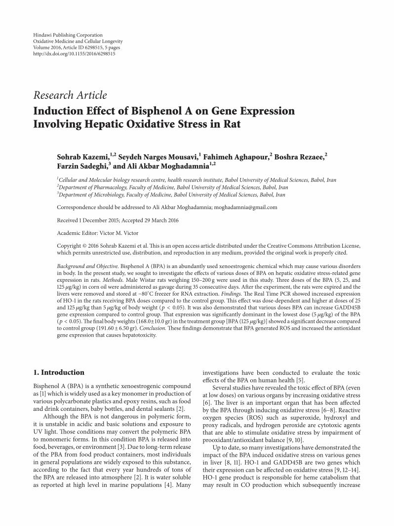
Research ArticleInduction Effect of Bisphenol A on Gene ExpressionInvolving Hepatic Oxidative Stress in Rat
Sohrab Kazemi,1,2 Seydeh Narges Mousavi,1 Fahimeh Aghapour,2 Boshra Rezaee,2
Farzin Sadeghi,3 and Ali Akbar Moghadamnia1,2
1Cellular and Molecular biology research centre, health research institute, Babol University of Medical Sciences, Babol, Iran2Department of Pharmacology, Faculty of Medicine, Babol University of Medical Sciences, Babol, Iran3Department of Microbiology, Faculty of Medicine, Babol University of Medical Sciences, Babol, Iran
Correspondence should be addressed to Ali Akbar Moghadamnia; [email protected]
Received 1 December 2015; Accepted 29 March 2016
Academic Editor: Victor M. Victor
Copyright © 2016 Sohrab Kazemi et al.This is an open access article distributed under the Creative Commons Attribution License,which permits unrestricted use, distribution, and reproduction in any medium, provided the original work is properly cited.
Background and Objective. Bisphenol A (BPA) is an abundantly used xenoestrogenic chemical which may cause various disordersin body. In the present study, we sought to investigate the effects of various doses of BPA on hepatic oxidative stress-related geneexpression in rats. Methods. Male Wistar rats weighing 150–200 g were used in this study. Three doses of the BPA (5, 25, and125𝜇g/kg) in corn oil were administered as gavage during 35 consecutive days. After the experiment, the rats were expired and thelivers were removed and stored at −80∘C freezer for RNA extraction. Findings. The Real Time PCR showed increased expressionof HO-1 in the rats receiving BPA doses compared to the control group. This effect was dose-dependent and higher at doses of 25and 125 𝜇g/kg than 5𝜇g/kg of body weight (𝑝 < 0.05). It was also demonstrated that various doses BPA can increase GADD45Bgene expression compared to control group. That expression was significantly dominant in the lowest dose (5 𝜇g/kg) of the BPA(𝑝 < 0.05).Thefinal bodyweights (168.0±10.0 gr) in the treatment group [BPA (125𝜇g/kg)] showed a significant decrease comparedto control group (191.60 ± 6.50 gr). Conclusion.These findings demonstrate that BPA generated ROS and increased the antioxidantgene expression that causes hepatotoxicity.
1. Introduction
Bisphenol A (BPA) is a synthetic xenoestrogenic compoundas [1] which is widely used as a keymonomer in production ofvarious polycarbonate plastics and epoxy resins, such as foodand drink containers, baby bottles, and dental sealants [2].
Although the BPA is not dangerous in polymeric form,it is unstable in acidic and basic solutions and exposure toUV light. Those conditions may convert the polymeric BPAto monomeric forms. In this condition BPA is released intofood, beverages, or environment [3]. Due to long-term releaseof the PBA from food product containers, most individualsin general populations are widely exposed to this substance,according to the fact that every year hundreds of tons ofthe BPA are released into atmosphere [2]. It is water solubleas reported at high level in marine populations [4]. Many
investigations have been conducted to evaluate the toxiceffects of the BPA on human health [5].
Several studies have revealed the toxic effect of BPA (evenat low doses) on various organs by increasing oxidative stress[6]. The liver is an important organ that has been affectedby the BPA through inducing oxidative stress [6–8]. Reactiveoxygen species (ROS) such as superoxide, hydroxyl andproxy radicals, and hydrogen peroxide are cytotoxic agentsthat are able to stimulate oxidative stress by impairment ofprooxidant/antioxidant balance [9, 10].
Up to date, somany investigations have demonstrated theimpact of the BPA induced oxidative stress on various genesin liver [8, 11]. HO-1 and GADD45B are two genes whichtheir expression can be affected on oxidative stress [9, 12–14].HO-1 gene product is responsible for heme catabolism thatmay result in CO production which subsequently increase
Hindawi Publishing CorporationOxidative Medicine and Cellular LongevityVolume 2016, Article ID 6298515, 5 pageshttp://dx.doi.org/10.1155/2016/6298515
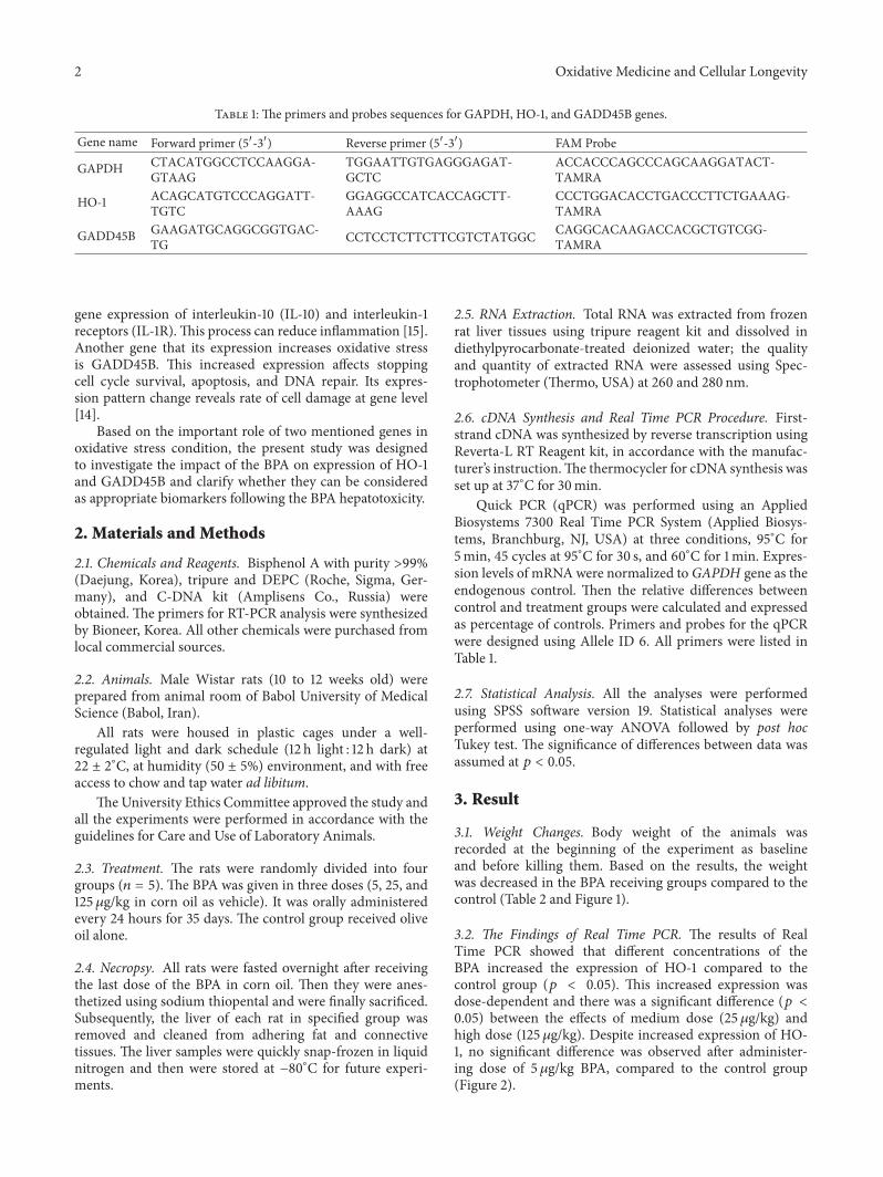
2 Oxidative Medicine and Cellular Longevity
Table 1: The primers and probes sequences for GAPDH, HO-1, and GADD45B genes.
Gene name Forward primer (5-3) Reverse primer (5-3) FAM Probe
GAPDH CTACATGGCCTCCAAGGA-GTAAG
TGGAATTGTGAGGGAGAT-GCTC
ACCACCCAGCCCAGCAAGGATACT-TAMRA
HO-1 ACAGCATGTCCCAGGATT-TGTC
GGAGGCCATCACCAGCTT-AAAG
CCCTGGACACCTGACCCTTCTGAAAG-TAMRA
GADD45B GAAGATGCAGGCGGTGAC-TG CCTCCTCTTCTTCGTCTATGGC CAGGCACAAGACCACGCTGTCGG-
TAMRA
gene expression of interleukin-10 (IL-10) and interleukin-1receptors (IL-1R).This process can reduce inflammation [15].Another gene that its expression increases oxidative stressis GADD45B. This increased expression affects stoppingcell cycle survival, apoptosis, and DNA repair. Its expres-sion pattern change reveals rate of cell damage at gene level[14].
Based on the important role of two mentioned genes inoxidative stress condition, the present study was designedto investigate the impact of the BPA on expression of HO-1and GADD45B and clarify whether they can be consideredas appropriate biomarkers following the BPA hepatotoxicity.
2. Materials and Methods
2.1. Chemicals and Reagents. Bisphenol A with purity >99%(Daejung, Korea), tripure and DEPC (Roche, Sigma, Ger-many), and C-DNA kit (Amplisens Co., Russia) wereobtained. The primers for RT-PCR analysis were synthesizedby Bioneer, Korea. All other chemicals were purchased fromlocal commercial sources.
2.2. Animals. Male Wistar rats (10 to 12 weeks old) wereprepared from animal room of Babol University of MedicalScience (Babol, Iran).
All rats were housed in plastic cages under a well-regulated light and dark schedule (12 h light : 12 h dark) at22 ± 2
∘C, at humidity (50 ± 5%) environment, and with freeaccess to chow and tap water ad libitum.
The University Ethics Committee approved the study andall the experiments were performed in accordance with theguidelines for Care and Use of Laboratory Animals.
2.3. Treatment. The rats were randomly divided into fourgroups (𝑛 = 5). The BPA was given in three doses (5, 25, and125 𝜇g/kg in corn oil as vehicle). It was orally administeredevery 24 hours for 35 days. The control group received oliveoil alone.
2.4. Necropsy. All rats were fasted overnight after receivingthe last dose of the BPA in corn oil. Then they were anes-thetized using sodium thiopental and were finally sacrificed.Subsequently, the liver of each rat in specified group wasremoved and cleaned from adhering fat and connectivetissues. The liver samples were quickly snap-frozen in liquidnitrogen and then were stored at −80∘C for future experi-ments.
2.5. RNA Extraction. Total RNA was extracted from frozenrat liver tissues using tripure reagent kit and dissolved indiethylpyrocarbonate-treated deionized water; the qualityand quantity of extracted RNA were assessed using Spec-trophotometer (Thermo, USA) at 260 and 280 nm.
2.6. cDNA Synthesis and Real Time PCR Procedure. First-strand cDNA was synthesized by reverse transcription usingReverta-L RT Reagent kit, in accordance with the manufac-turer’s instruction.The thermocycler for cDNA synthesis wasset up at 37∘C for 30min.
Quick PCR (qPCR) was performed using an AppliedBiosystems 7300 Real Time PCR System (Applied Biosys-tems, Branchburg, NJ, USA) at three conditions, 95∘C for5min, 45 cycles at 95∘C for 30 s, and 60∘C for 1min. Expres-sion levels of mRNA were normalized to GAPDH gene as theendogenous control. Then the relative differences betweencontrol and treatment groups were calculated and expressedas percentage of controls. Primers and probes for the qPCRwere designed using Allele ID 6. All primers were listed inTable 1.
2.7. Statistical Analysis. All the analyses were performedusing SPSS software version 19. Statistical analyses wereperformed using one-way ANOVA followed by post hocTukey test. The significance of differences between data wasassumed at 𝑝 < 0.05.
3. Result
3.1. Weight Changes. Body weight of the animals wasrecorded at the beginning of the experiment as baselineand before killing them. Based on the results, the weightwas decreased in the BPA receiving groups compared to thecontrol (Table 2 and Figure 1).
3.2. The Findings of Real Time PCR. The results of RealTime PCR showed that different concentrations of theBPA increased the expression of HO-1 compared to thecontrol group (𝑝 < 0.05). This increased expression wasdose-dependent and there was a significant difference (𝑝 <0.05) between the effects of medium dose (25 𝜇g/kg) andhigh dose (125 𝜇g/kg). Despite increased expression of HO-1, no significant difference was observed after administer-ing dose of 5 𝜇g/kg BPA, compared to the control group(Figure 2).
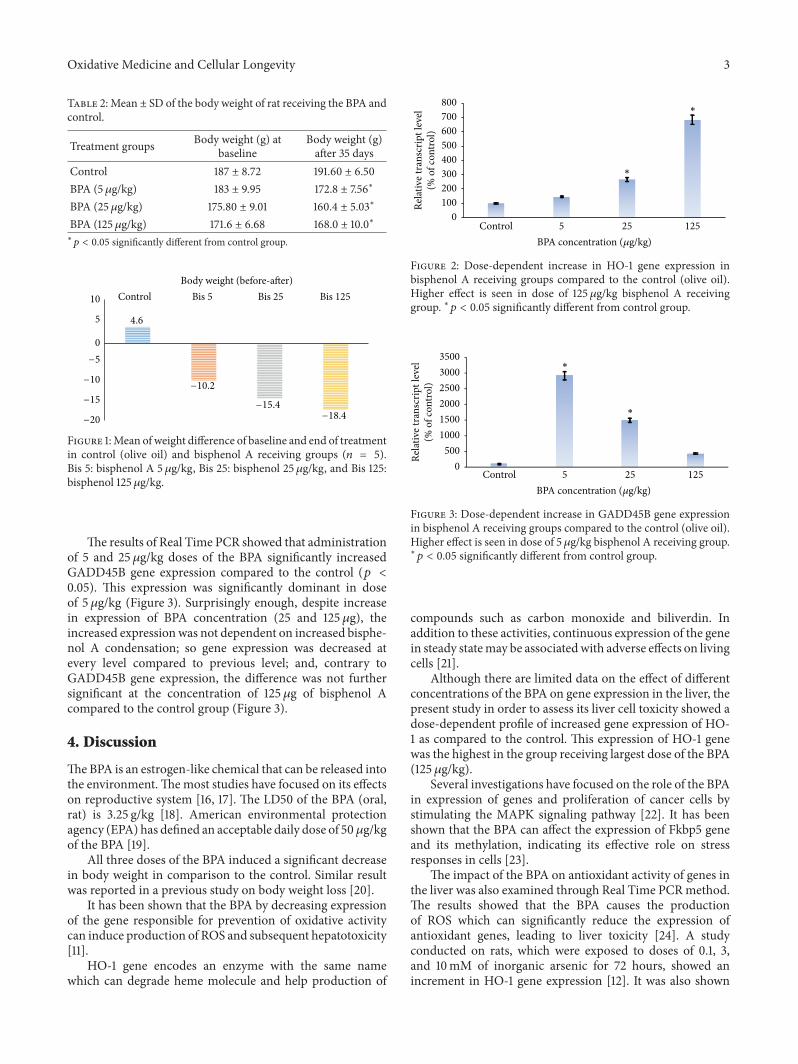
Oxidative Medicine and Cellular Longevity 3
Table 2: Mean ± SD of the body weight of rat receiving the BPA andcontrol.
Treatment groups Body weight (g) atbaseline
Body weight (g)after 35 days
Control 187 ± 8.72 191.60 ± 6.50BPA (5𝜇g/kg) 183 ± 9.95 172.8 ± 7.56∗
BPA (25 𝜇g/kg) 175.80 ± 9.01 160.4 ± 5.03∗
BPA (125𝜇g/kg) 171.6 ± 6.68 168.0 ± 10.0∗∗
𝑝 < 0.05 significantly different from control group.
Control Bis 5 Bis 25 Bis 125
4.6
−10.2
−15.4−18.4−20
−15
−10
−5
0
5
10
Body weight (before-after)
Figure 1:Mean of weight difference of baseline and end of treatmentin control (olive oil) and bisphenol A receiving groups (𝑛 = 5).Bis 5: bisphenol A 5 𝜇g/kg, Bis 25: bisphenol 25 𝜇g/kg, and Bis 125:bisphenol 125 𝜇g/kg.
The results of Real Time PCR showed that administrationof 5 and 25 𝜇g/kg doses of the BPA significantly increasedGADD45B gene expression compared to the control (𝑝 <0.05). This expression was significantly dominant in doseof 5 𝜇g/kg (Figure 3). Surprisingly enough, despite increasein expression of BPA concentration (25 and 125𝜇g), theincreased expressionwas not dependent on increased bisphe-nol A condensation; so gene expression was decreased atevery level compared to previous level; and, contrary toGADD45B gene expression, the difference was not furthersignificant at the concentration of 125 𝜇g of bisphenol Acompared to the control group (Figure 3).
4. Discussion
TheBPA is an estrogen-like chemical that can be released intothe environment.Themost studies have focused on its effectson reproductive system [16, 17]. The LD50 of the BPA (oral,rat) is 3.25 g/kg [18]. American environmental protectionagency (EPA) has defined an acceptable daily dose of 50 𝜇g/kgof the BPA [19].
All three doses of the BPA induced a significant decreasein body weight in comparison to the control. Similar resultwas reported in a previous study on body weight loss [20].
It has been shown that the BPA by decreasing expressionof the gene responsible for prevention of oxidative activitycan induce production of ROS and subsequent hepatotoxicity[11].
HO-1 gene encodes an enzyme with the same namewhich can degrade heme molecule and help production of
Control 5 25 125
∗
∗
0100200300400500600700800
Rela
tive t
rans
crip
t lev
el(%
of c
ontro
l)
BPA concentration (𝜇g/kg)
Figure 2: Dose-dependent increase in HO-1 gene expression inbisphenol A receiving groups compared to the control (olive oil).Higher effect is seen in dose of 125 𝜇g/kg bisphenol A receivinggroup. ∗𝑝 < 0.05 significantly different from control group.
Control 5 25 125
∗
∗
BPA concentration (𝜇g/kg)
0500
100015002000250030003500
Rela
tive t
rans
crip
t lev
el(%
of c
ontro
l)
Figure 3: Dose-dependent increase in GADD45B gene expressionin bisphenol A receiving groups compared to the control (olive oil).Higher effect is seen in dose of 5 𝜇g/kg bisphenol A receiving group.∗
𝑝 < 0.05 significantly different from control group.
compounds such as carbon monoxide and biliverdin. Inaddition to these activities, continuous expression of the genein steady statemay be associatedwith adverse effects on livingcells [21].
Although there are limited data on the effect of differentconcentrations of the BPA on gene expression in the liver, thepresent study in order to assess its liver cell toxicity showed adose-dependent profile of increased gene expression of HO-1 as compared to the control. This expression of HO-1 genewas the highest in the group receiving largest dose of the BPA(125 𝜇g/kg).
Several investigations have focused on the role of the BPAin expression of genes and proliferation of cancer cells bystimulating the MAPK signaling pathway [22]. It has beenshown that the BPA can affect the expression of Fkbp5 geneand its methylation, indicating its effective role on stressresponses in cells [23].
The impact of the BPA on antioxidant activity of genes inthe liver was also examined through Real Time PCRmethod.The results showed that the BPA causes the productionof ROS which can significantly reduce the expression ofantioxidant genes, leading to liver toxicity [24]. A studyconducted on rats, which were exposed to doses of 0.1, 3,and 10mM of inorganic arsenic for 72 hours, showed anincrement in HO-1 gene expression [12]. It was also shown
4 Oxidative Medicine and Cellular Longevity
that HO-1 is a good biomarker to detect and define arseniccell toxicity [25]. In another study on rats treated withvarious doses of the BPA (0.1, 1, 10, and 50mg/kg), thecompound led to changes in expression levels of antioxidantgenes of glutathione, peroxidase (GSHPx), catalase (CAT),glutathione transferase (GST), and glutathione reductase(GR) in liver tissue. Expression of these antioxidant geneswas decreasedwith increasing doses of the BPA.However, theimpact of these doses of the BPA on biomarkers of oxidativestress showed a different result. With increasing doses of theBPA, the levels of TBARS and NO (𝑥) were increased andGSH and SOD decreased [24].
Another result obtained in the present study showed thatdifferent doses of the BPA increased GADD45B gene expres-sion compared to the control. This increased gene expressionwas significantly evident at the lowest doses (5 𝜇g/kg), butthe gene expression was decreased in larger doses of the BPAcompared to the low doses. Although an increased expressionof GADD45B gene was shown in comparison to the controlgroup, this increment was not statistically significantly thehighest doses of the BPA. One of the results of this study isthe positive effect of expression of this gene on cell apoptosis.When the rats received higher doses of the BPA, Gadd45gene expression is increased first. Increased expression ofGadd45 gene stimulates apoptosis and the percent of viablecells for expression of this gene is being decreased gradually.As a result, with increasing doses of the BPA, gene expressionfollowed a decreased trend. In the case of HO-1 gene, nostudy has reported changes in GADD45B gene expressionafter administering BPA in rats.
5. Conclusion
According to the results, it is concluded that bisphenol A(BPA) increased expression of HO-1 gene more than Gadd45gene at high doses. The BPA can induce dose-dependentliver damage. Increased reactive oxygen species (ROS) andoxidative reactions may be responsible for the toxicity.
Competing Interests
The authors declare that there are no competing interests.
Acknowledgments
The authors wish to thank Professor Hossein Najafzadeh forhis advisory cooperation. Research Affairs Division of BabolUniversity of Medical Sciences has financially supported thisinvestigation.
References
[1] N. Ben-Jonathan and R. Steinmetz, “Xenoestrogens: theemerging story of bisphenol A,” Trends in Endocrinology &Metabolism, vol. 9, no. 3, pp. 124–128, 1998.
[2] C. Liu, W. Duan, R. Li et al., “Exposure to bisphenol Adisrupts meiotic progression during spermatogenesis in adultrats through estrogen-like activity,” Cell Death and Disease, vol.4, article e676, 2013.
[3] C. E. Talsness, A. J. M. Andrade, S. N. Kuriyama, J. A. Taylor,and F. S. V. Saal, “Components of plastic: experimental studiesin animals and relevance for human health,” PhilosophicalTransactions of the Royal Society B: Biological Sciences, vol. 364,no. 1526, pp. 2079–2096, 2009.
[4] S. Mortazavi, A. Riyahi Bakhtiari, A. E. Sari, N. Bahramifar,and F. Rahbarizade, “Phenolic endocrine disrupting chemicals(EDCs) in Anzali Wetland, Iran: elevated concentrations of 4-nonylphenol, octhylphenol and bisphenol A,”Marine PollutionBulletin, vol. 64, no. 5, pp. 1067–1073, 2012.
[5] M. A. Kamrin, “Bisphenol A: a scientific evaluation,”MedGen-Med, vol. 6, no. 3, article 7, 2004.
[6] M. K. Moon, M. J. Kim, I. K. Jung et al., “Bisphenol a impairsmitochondrial function in the liver at doses below the noobserved adverse effect level,” Journal of KoreanMedical Science,vol. 27, no. 6, pp. 644–652, 2012.
[7] A. Kourouma, C. Quan, P. Duan et al., “Bisphenol A inducesapoptosis in liver cells through induction of ROS,” Advances inToxicology, vol. 2015, Article ID 901983, 10 pages, 2015.
[8] V. Bindhumol, K. C. Chitra, and P. P. Mathur, “Bisphenol Ainduces reactive oxygen species generation in the liver of malerats,” Toxicology, vol. 188, no. 2-3, pp. 117–124, 2003.
[9] M. McMillian, A. Y. Nie, J. B. Parker et al., “A gene expressionsignature for oxidant stress/reactive metabolites in rat liver,”Biochemical Pharmacology, vol. 68, no. 11, pp. 2249–2261, 2004.
[10] L. A. Videla, “Oxidative stress signaling underlying liver diseaseand hepatoprotective mechanisms,” World Journal of Hepatol-ogy, vol. 1, no. 1, pp. 72–78, 2009.
[11] Z. K. Hassan, M. A. Elobeid, P. Virk et al., “Bisphenol A induceshepatotoxicity through oxidative stress in rat model,” OxidativeMedicine and Cellular Longevity, vol. 2012, Article ID 194829, 6pages, 2012.
[12] J. Liu, L. Yu, E. J. Tokar et al., “Arsenic-induced aberrant geneexpression in fetal mouse primary liver-cell cultures,” Annals ofthe New York Academy of Sciences, vol. 1140, no. 1, pp. 368–375,2008.
[13] M. Iida, C. H. Anna, W. M. Holliday et al., “Unique patterns ofgene expression changes in liver after treatment of mice for 2weeks with different known carcinogens and non-carcinogens,”Carcinogenesis, vol. 26, no. 3, pp. 689–699, 2005.
[14] J.-H. Kim, A. Qu, J. K. Reddy, B. Gao, and F. J. Gonzalez,“Hepatic oxidative stress activates the Gadd45b gene by way ofdegradation of the transcriptional repressor STAT3,” Hepatol-ogy, vol. 59, no. 2, pp. 695–704, 2014.
[15] C. A. Piantadosi, C. M. Withers, R. R. Bartz et al., “Hemeoxygenase-1 couples activation of mitochondrial biogenesis toanti-inflammatory cytokine expression,” Journal of BiologicalChemistry, vol. 286, no. 18, pp. 16374–16385, 2011.
[16] C. C. Willhite, G. L. Ball, and C. J. McLellan, “Derivationof a bisphenol A oral reference dose (RfD) and drinking-water equivalent concentration,” Journal of Toxicology andEnvironmental Health Part B: Critical Reviews, vol. 11, no. 2, pp.69–146, 2008.
[17] T. E. Haavisto, N. A. Adamsson, S. A.Myllymäki, J. Toppari, andJ. Paranko, “Effects of 4-tert-octylphenol, 4-tert-butylphenol,and diethylstilbestrol on prenatal testosterone surge in the rat,”Reproductive Toxicology, vol. 17, no. 5, pp. 593–605, 2003.
[18] R. E. Chapin, J. Adams, K. Boekelheide et al., “NTP-CERHRexpert panel report on the reproductive and developmentaltoxicity of bisphenol A,” Birth Defects Research Part B: Develop-mental and Reproductive Toxicology, vol. 83, no. 3, pp. 157–395,2008.
Oxidative Medicine and Cellular Longevity 5
[19] B. S. Rubin, M. K. Murray, D. A. Damassa, J. C. King, and A.M. Soto, “Perinatal exposure to low doses of bisphenol A affectsbodyweight, patterns of estrous cyclicity, and plasma LH levels,”Environmental Health Perspectives, vol. 109, no. 7, pp. 675–680,2001.
[20] K. Yamasaki, M. Sawaki, S. Noda, N. Imatanaka, and M.Takatsuki, “Subacute oral toxicity study of ethynylestradiol andbisphenol A, based on the draft protocol for the ‘EnhancedOECD Test Guideline no. 407’,” Archives of Toxicology, vol. 76,no. 2, pp. 65–74, 2002.
[21] Y. L. Tang, Y. Tang, Y. C. Zhang, K. Qian, L. Shen, and M. I.Phillips, “Improved graft mesenchymal stem cell survival inischemic heart with a hypoxia-regulated heme oxygenase-1vector,” Journal of the American College of Cardiology, vol. 46,no. 7, pp. 1339–1350, 2005.
[22] H.-C. Lan, I.-W. Lin, Z.-J. Yang, and J.-H. Lin, “Low-dosebisphenol A activates Cyp11a1 gene expression and corticos-terone secretion in adrenal gland via the JNK signaling path-way,” Toxicological Sciences, vol. 148, no. 1, pp. 26–34, 2015.
[23] E. Kitraki, I. Nalvarte, A. Alavian-Ghavanini, and J. Rüegg,“Developmental exposure to bisphenol A alters expression andDNAmethylation of Fkbp5, an important regulator of the stressresponse,” Molecular and Cellular Endocrinology, vol. 417, pp.191–199, 2015.
[24] Z. K. Hassan, M. A. Elobeid, P. Virk et al., “Bisphenol A induceshepatotoxicity through oxidative stress in rat model,” OxidativeMedicine and Cellular Longevity, vol. 2012, Article ID 194829, 6pages, 2012.
[25] J. Liu, M. B. Kadiiska, Y. Liu, T. Lu, W. Qu, and M. P. Waalkes,“Stress-related gene expression in mice treated with inorganicarsenicals,” Toxicological Sciences, vol. 61, no. 2, pp. 314–320,2001.
Review ArticleThe Reactive Oxygen Species in Macrophage Polarization:Reflecting Its Dual Role in Progression and Treatment ofHuman Diseases
Hor-Yue Tan,1 Ning Wang,1 Sha Li,1 Ming Hong,1 Xuanbin Wang,2,3 and Yibin Feng1,2,3
1School of Chinese Medicine, Li Ka Shing Faculty of Medicine, The University of Hong Kong, Pokfulam, Hong Kong2Laboratory of Chinese Herbal Pharmacology, Renmin Hospital, Hubei 442000, China3Hubei Key Laboratory of Wudang Local Chinese Medicine Research, Hubei University of Medicine, Hubei 442000, China
Correspondence should be addressed to Yibin Feng; [email protected]
Received 24 December 2015; Revised 13 March 2016; Accepted 15 March 2016
Academic Editor: Paola Venditti
Copyright © 2016 Hor-Yue Tan et al. This is an open access article distributed under the Creative Commons Attribution License,which permits unrestricted use, distribution, and reproduction in any medium, provided the original work is properly cited.
High heterogeneity of macrophage is associated with its functions in polarization to different functional phenotypes dependingon environmental cues. Macrophages remain in balanced state in healthy subject and thus macrophage polarization may becrucial in determining the tissue fate. The two distinct populations, classically M1 and alternatively M2 activated, representingthe opposing ends of the full activation spectrum, have been extensively studied for their associations with several diseaseprogressions. Accumulating evidences have postulated that the redox signalling has implication in macrophage polarization andthe key roles of M1 andM2macrophages in tissue environment have provided the clue for the reasons of ROS abundance in certainphenotype. M1 macrophages majorly clearing the pathogens and ROS may be crucial for the regulation of M1 phenotype, whereasM2 macrophages resolve inflammation which favours oxidative metabolism. Therefore how ROS play its role in maintainingthe homeostatic functions of macrophage and in particular macrophage polarization will be reviewed here. We also reviewthe biology of macrophage polarization and the disturbance of M1/M2 balance in human diseases. The potential therapeuticopportunities targeting ROS will also be discussed, hoping to provide insights for development of target-specific delivery systemor immunomodulatory antioxidant for the treatment of ROS-related diseases.
1. Introduction
Accumulating studies have implied the physiological role ofreactive oxygen species (ROS) in various biological processesat distinct levels, for example, gene expression, proteintranslation, posttranslationalmodification, and protein inter-actions. Instead of merely being a harmful byproduct ofmetabolism, cell-derived ROS majorly derived from hydro-gen peroxide (H
2O2), superoxide anions (O
2
−), and hydroxylradicals (OH−), is an independent or cooperative regulatorfor cellular signalling in response to environmental cues.For instance, H
2O2, due to its long-lived nature and ability
to easily pass through cellular membrane, is an importantsecondarymessenger inmaintaining the cellular homeostasisunder different conditions [1]. It may result in activation orshutdown of diverse cellular processes such as cell cycling,
chromatin remodeling, DNA repair, cell differentiation, andself-renewal [2].The balance of intracellular ROS is thereforeextremely important in maintaining normal physiology ofhuman beings. While mitochondrial respiratory chain isthe major component that cells used to produce intracel-lular ROS, cells develop a series of antioxidant enzymesto reduce redundant ROS. These enzymes, including cata-lase, superoxide dismutase (SOD), glutathione peroxidase(GPx), and glutathione reductase (GR), can either grosslyor specifically catalyze different types of ROS. The presenceof other endogenous antioxidants such as peroxiredoxinsand thioredoxins ensures critical monitoring of cellular ROSlevel and conserves the normal redox homeostasis. In somepathological conditions, the redox balance is disturbed whenthe intracellular redox system is shifted to oxidized state,which is defined as oxidative stress. During oxidative stress,
Hindawi Publishing CorporationOxidative Medicine and Cellular LongevityVolume 2016, Article ID 2795090, 16 pageshttp://dx.doi.org/10.1155/2016/2795090
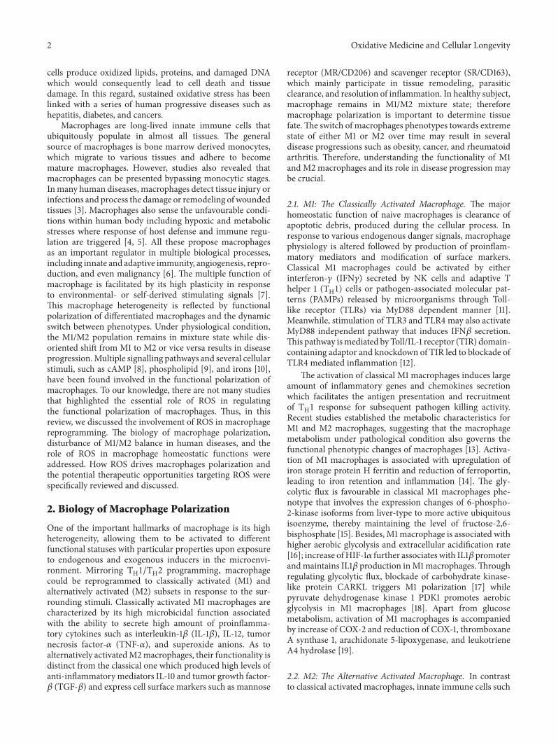
2 Oxidative Medicine and Cellular Longevity
cells produce oxidized lipids, proteins, and damaged DNAwhich would consequently lead to cell death and tissuedamage. In this regard, sustained oxidative stress has beenlinked with a series of human progressive diseases such ashepatitis, diabetes, and cancers.
Macrophages are long-lived innate immune cells thatubiquitously populate in almost all tissues. The generalsource of macrophages is bone marrow derived monocytes,which migrate to various tissues and adhere to becomemature macrophages. However, studies also revealed thatmacrophages can be presented bypassing monocytic stages.Inmany human diseases, macrophages detect tissue injury orinfections and process the damage or remodeling of woundedtissues [3]. Macrophages also sense the unfavourable condi-tions within human body including hypoxic and metabolicstresses where response of host defense and immune regu-lation are triggered [4, 5]. All these propose macrophagesas an important regulator in multiple biological processes,including innate and adaptive immunity, angiogenesis, repro-duction, and even malignancy [6]. The multiple function ofmacrophage is facilitated by its high plasticity in responseto environmental- or self-derived stimulating signals [7].This macrophage heterogeneity is reflected by functionalpolarization of differentiated macrophages and the dynamicswitch between phenotypes. Under physiological condition,the M1/M2 population remains in mixture state while dis-oriented shift from M1 to M2 or vice versa results in diseaseprogression.Multiple signalling pathways and several cellularstimuli, such as cAMP [8], phospholipid [9], and irons [10],have been found involved in the functional polarization ofmacrophages. To our knowledge, there are not many studiesthat highlighted the essential role of ROS in regulatingthe functional polarization of macrophages. Thus, in thisreview, we discussed the involvement of ROS in macrophagereprogramming. The biology of macrophage polarization,disturbance of M1/M2 balance in human diseases, and therole of ROS in macrophage homeostatic functions wereaddressed. How ROS drives macrophages polarization andthe potential therapeutic opportunities targeting ROS werespecifically reviewed and discussed.
2. Biology of Macrophage Polarization
One of the important hallmarks of macrophage is its highheterogeneity, allowing them to be activated to differentfunctional statuses with particular properties upon exposureto endogenous and exogenous inducers in the microenvi-ronment. Mirroring TH1/TH2 programming, macrophagecould be reprogrammed to classically activated (M1) andalternatively activated (M2) subsets in response to the sur-rounding stimuli. Classically activated M1 macrophages arecharacterized by its high microbicidal function associatedwith the ability to secrete high amount of proinflamma-tory cytokines such as interleukin-1𝛽 (IL-1𝛽), IL-12, tumornecrosis factor-𝛼 (TNF-𝛼), and superoxide anions. As toalternatively activatedM2macrophages, their functionality isdistinct from the classical one which produced high levels ofanti-inflammatorymediators IL-10 and tumor growth factor-𝛽 (TGF-𝛽) and express cell surface markers such as mannose
receptor (MR/CD206) and scavenger receptor (SR/CD163),which mainly participate in tissue remodeling, parasiticclearance, and resolution of inflammation. In healthy subject,macrophage remains in M1/M2 mixture state; thereforemacrophage polarization is important to determine tissuefate.The switch of macrophages phenotypes towards extremestate of either M1 or M2 over time may result in severaldisease progressions such as obesity, cancer, and rheumatoidarthritis. Therefore, understanding the functionality of M1and M2 macrophages and its role in disease progression maybe crucial.
2.1. M1: The Classically Activated Macrophage. The majorhomeostatic function of naive macrophages is clearance ofapoptotic debris, produced during the cellular process. Inresponse to various endogenous danger signals, macrophagephysiology is altered followed by production of proinflam-matory mediators and modification of surface markers.Classical M1 macrophages could be activated by eitherinterferon-𝛾 (IFN𝛾) secreted by NK cells and adaptive Thelper 1 (TH1) cells or pathogen-associated molecular pat-terns (PAMPs) released by microorganisms through Toll-like receptor (TLRs) via MyD88 dependent manner [11].Meanwhile, stimulation of TLR3 and TLR4 may also activateMyD88 independent pathway that induces IFN𝛽 secretion.This pathway ismediated byToll/IL-1 receptor (TIR) domain-containing adaptor and knockdown of TIR led to blockade ofTLR4 mediated inflammation [12].
The activation of classical M1 macrophages induces largeamount of inflammatory genes and chemokines secretionwhich facilitates the antigen presentation and recruitmentof TH1 response for subsequent pathogen killing activity.Recent studies established the metabolic characteristics forM1 and M2 macrophages, suggesting that the macrophagemetabolism under pathological condition also governs thefunctional phenotypic changes of macrophages [13]. Activa-tion of M1 macrophages is associated with upregulation ofiron storage protein H ferritin and reduction of ferroportin,leading to iron retention and inflammation [14]. The gly-colytic flux is favourable in classical M1 macrophages phe-notype that involves the expression changes of 6-phospho-2-kinase isoforms from liver-type to more active ubiquitousisoenzyme, thereby maintaining the level of fructose-2,6-bisphosphate [15]. Besides, M1macrophage is associated withhigher aerobic glycolysis and extracellular acidification rate[16]; increase ofHIF-1𝛼 further associates with IL1𝛽 promoterandmaintains IL1𝛽 production inM1macrophages.Throughregulating glycolytic flux, blockade of carbohydrate kinase-like protein CARKL triggers M1 polarization [17] whilepyruvate dehydrogenase kinase 1 PDK1 promotes aerobicglycolysis in M1 macrophages [18]. Apart from glucosemetabolism, activation of M1 macrophages is accompaniedby increase of COX-2 and reduction of COX-1, thromboxaneA synthase 1, arachidonate 5-lipoxygenase, and leukotrieneA4 hydrolase [19].
2.2. M2: The Alternative Activated Macrophage. In contrastto classical activated macrophages, innate immune cells such
Oxidative Medicine and Cellular Longevity 3
as basophils and mast cells and other adaptive cells produceIL-4 and IL-13 priming M2 alternative phenotype. IL-4induced M2 macrophages expressed high concentration ofIL-10, decoy receptor IL-1R, IL-1R antagonist, chemokinesCCL22 and CCL17, and intracellular enzyme arginase-1.All of these ensue the recruitment and activation of TH2immune response and immune-suppressive function of M2macrophages. In addition to TH2 immune response, IL-4induced macrophages stimulate arginase activity by con-verting arginine to polyamines and collagen precursors thatare crucial for tissue modeling and wound healing. M2macrophages also produce VEGFA, EGF, and IL-8 thatare responsible for angiogenesis and lymphangiogenesis [7].Apart from IL-4 induced phenotype, different schemes ofM2macrophages classification have been proposed due to theoverlapping properties of alternative activated macrophages.The activation of M2 macrophages stimulated by additionof either IL-4 and IL-13, TGF𝛽, immune complexes, glu-cocorticoids, or IL-10 may yield distinct activation profiles[20].
Cellular metabolism especially lipid metabolism alsoincurs important role in providing energy fuel for activa-tion of alternative M2 macrophages. In iron metabolism,M2 macrophages increased ferroportin expressions, whichfurther induced iron export [14]. As opposed to M1 classicalactivation, M2-regulated gene transcription occurs in con-dition favouring of mitochondrial metabolism and oxidativeglucose metabolism, in which the M2 phenotype tends to beswitched towards proinflammatory state under low oxygencondition [16]. In lipid metabolism, M2 macrophage activa-tion is associatedwith fatty acid oxidation and its uptake.M2-secreted lysosomal acid lipase as well as its scavenger receptorCD36 facilitates uptake of triglycerols, LDL, and VLDL for 𝛽-oxidation and fatty acid generation [21]. Apart from LDL andVLDL, high density lipoproteins promote anti-inflammatoryfunction through ATF3 dependent pathway [22]. Study byPrieur et al. [23] also postulated that short and saturatedfatty acids favour M1 polarization, while longer and unsat-urated fatty acids induce M2 anti-inflammatory phenotype.Therefore, M2 activation is accompanied by upregulation ofarachidonic acid [19], omega-3 polyunsaturated fatty acids[24], sphingosine 1-phosphate [25], eicosapentaenoic acid,and docosahexaenoic acid [26]. All the correlation of alterna-tive M2 activation with lipid metabolism also provides cluefor the important role of M2 activation in atherosclerosis,obesity, and insulin sensitivity (as discussed later).
2.3. Disruption of M1/M2 Balance in Human Diseases. Clas-sical M1 macrophages elicit major role in inducing inflam-mation and clearing of pathogens, whereas alternative M2macrophages resolve inflammation and are crucial for thefunctions of tissue modeling and wound healing.They repre-sent the opposing ends of the activation spectrum, and eitheraccumulation of M1/M2 signals may lead to deteriorationof the host. Extensive production of IL1, IL6, and IL23 byM1macrophages incurs activation of TH17 immune responseand leads to autoimmune disorder progression [27]. Earlierstudiesmay postulate thatmacrophages populations promote
disease progression but many recent studies have proposedthat the proportions of different macrophage phenotypescontribute to disease progression. There is no absoluteanswer on which phenotype is “good” or “bad” as theswitch of resident or recruited macrophages towards M2phenotype may trigger tumor progression while accumu-lation of M1 macrophages leads to insulin resistance andatherosclerosis. Therefore, continuous activation of eitherstates brings harm to the host and the understanding of howdisruption of M1/M2 in regulating diseases may be essen-tial. The involvement of M1/M2 in obesity, atherosclerosis,and cancer is briefly discussed below; the comprehensivereviews on human diseases have been published elsewhere[28–30].
2.3.1. Obesity and Insulin Resistance. Adipose tissue macro-phages (ATM) are predominantly alternative M2 phenotypein lean subjects, and the ATM are reprogrammed to classi-cally activatedM1 phenotype with proinflammatory functionupon exposure to free saturated fatty acids produced by adi-pose tissue, resulting in reduced insulin sensitivity in whiteadipose tissue. IL-10 secreted byM2macrophages may be theresponsible cytokine that protects adipocytes from obesityleading insulin resistance [31]. The free fatty acids will alsotrigger production of CCL2 by adipocytes and ATM in orderto promote the recruitment of proinflammatory monocytesto the inflamed tissue [32]. Knockout of CCR2 in murinemodel effectively ameliorated the adipocyte inflammation,macrophage infiltration, and insulin resistance in obese mice[33].
2.3.2. Atherosclerosis. Atherosclerosis develops with theaccumulation and trapping of low density lipoproteins (LDL)in the intima of the arteries, and the modified LDL-inducedsecretion of adhesion molecules by endothelial cells recruitsmonocytes with proinflammatory phenotype to atheroscle-rotic lesion, which eventually differentiate into lipid-ladenfoam cells [34]. Cholesterol crystal, IFN𝛾, LPS, and oxidizedLDL are known to stimulate M1 activation and M1-activatedcytokines further support the monocyte recruitment andmacrophage retention at the plaque area [35]. Due to theinvolvement in plaque destabilization, the proatherogenicM1populations are predominantly accumulated at the plaqueswhere they are prone to rupture. As forM2macrophages, theymostly reside at stable-cell enriched plaque and adventitia.M2 macrophages tend to prevent foam cell formation andprotect against atherosclerosis.
2.3.3. Cancer. Thegrowth and expansion ofmalignant cells iscomplex, which involves gene manipulation as well as estab-lishment of microenvironment that favours tumor progres-sion. Many recent studies have highlighted the role of tumor-associated macrophages in promoting cancer growth, angio-genesis, invasion, migration, and T cell suppression [36].In most cancers, macrophages residing in tumor microen-vironment exhibit M2 phenotype with immunosuppressiveproperty. In contact with tumor cells, M2 macrophages tendto derive certain substances such as various growth factors,
4 Oxidative Medicine and Cellular Longevity
chemokines, and proteases that maintain tumor growth andexpansion [37]. Apart from mediating tumor growth andprogression, M2 macrophages interact with other immunecells and suppress innate and adaptive antitumor immuneresponse. Depending on the tumor environment, phenotypeof tumor-associated macrophages would be reprogrammedto M1-like phenotype, which is characterized by the antigenpresenting property and tumoricidal function that favourstumor regression [38]. M1 macrophages activation increasesexpressions of mediators that are responsible for antigenpresentation and costimulation; this may further promoteinfiltration of neutrophils to the tumor area leading toneutrophil-targeted tumor regression [39].
3. Reactive Oxygen Species on HomeostasisFunction of Macrophages
3.1. ROS in Regulating Phagocytosis of Macrophages. Numer-ous studies have unveiled the diverse regulations of ROSon the phagocytosis function of macrophage. The regulationmay be controversial, but the discrete role of ROS onmacrophages may be impacted by the different sources ofROS as well as plasticity of macrophage itself, which wouldbe discussed in detail below. Asmacrophages are endogenousscavengers for dying cells in various pathological conditions,interaction between macrophages with compartments deter-mines the phagocytic function of macrophages. Dying cellsproduce high levels of ROS, which are released into extracel-lular area when cellular membrane is degraded during celldeath. Attachment of dying cells to macrophages requiresintercellular communication in which ROS may play a role.On the other hand, extracellular and intracellular ROS maydifferentially control the phagocytosis process ofmacrophageby regulating the ability and capacity of macrophages inthe uptake and degradation of dying compartments. Inthis regard, ROS plays a critical regulatory role in deter-mining the initiation and outcome of cellular phagocyto-sis.
3.1.1. The Role of ROS from Dying Cells during Phagocytosis.Engulfment of cells undergoing apoptotic programmed celldeath (PCD) by macrophage is initiated by the presentationof membrane signals to phagocytes that allows recognitionof dying compartments. Inmacrophage-driven phagocytosis,these molecules include scavenger receptors, for example,CD36, immunoglobulin super-familymolecules, CD31, com-plement receptors, such as C91, sugar and phospholipid-engaging molecules, for example, lectins and PSR, and someintegrins such as 𝛼v𝛽3. The corresponding components oncellular membrane of apoptotic cells were less identified, butstructures encompass lipid, carbohydrate, and protein whichare exposed as molecules presenting find-me signals towardsextracellular area during cell apoptosis [40]. These lipids andproteins presented on membranes of apoptotic cells are gen-erally regarded as substrates of oxidation reaction containingdying cell-produced ROS. Indeed, experimental evidencehas shown that oxidation of lipids and proteins by ROSconfers recognition and attachment of macrophage towards
apoptotic cells. By using monoclonal antibody that blocksthe epitopes of oxLDL of apoptotic cells, scientists observedfailure of engulfment of dying cells by murine peritonealmacrophages [41]. It was further observed that surface pre-sentation of oxidative modified phosphatidylserines (PS) onapoptotic cells is essential for macrophage engulfment [42].The phagocytosis was blocked upon presence of lipoprotein-associated phospholipase A2 (Lp-PLA2), a secreted enzymewith high specificity towards PS metabolites. Oxidation ofPS is often observed during cell apoptosis [43]. This givesrise to the mechanism underlying attenuated phagocytosisof etoposide-treated cells by macrophage as etoposide isable to suppress oxidation and externalization of PS of theapoptotic cells [44]. In addition, some phosphatidylcholine(PC) species, which are oxidized during early- to late-apoptotic process, present to the surface of apoptotic cellsare recognized by C-reactive proteins, facilitating clearanceof apoptotic cells by macrophage engulfment [41]. Studyrevealed that oxidation of PC (oxPC) was majorly processedby ROS, in virus-infected cells, and scavenge of ROS by NACabrogated oxPC production [45]. Although macrophagereceptors correspond to particular type of oxidized lipids andproteins have not been fully unveiled, all these studies havepaved the importance of ROS-driven oxidation reaction indying cells during initiation of phagocytosis.
3.1.2.The Role of Macrophage ROS in Regulating Phagocytosis.Besides dying cells, macrophage itself also produces intracel-lular ROS that is involved in the phagocytic process. It is anotion that ROS in macrophage is essential for uptake andclearance of apoptotic cells; however, maintaining high levelof ROS may be harmful to macrophage as, in some studies,inducible ROS is sufficient to cause macrophage apoptosis[46]. Hypothesis of adaptive mechanism underlying survivalof macrophage in high ROS condition was ever discussed,which included increase expressions of DNA repair proteins[47] and endogenous antioxidative enzymes [48] duringmonocyte-macrophage differentiation and classical activa-tion of macrophage. Production of ROS by macrophagemajorly relies onNox2 gene, and study showed that activationof Nox2 gene in murine macrophage cell line RAW264.7as well as primary peritoneal macrophages by carotenoidlutein induced ROS production that was responsible forthe increased phagocytic activity [49]. Additionally, pro-duction of mitochondrial ROS (mROS) is able to increasephagocytosis ofmacrophages. InmROS-driven phagocytosis,intracellular fatty acids are utilized as fuel for oxidative phos-phorylation by mitochondria-localizing enzyme encodedby Immunoresponsive gene 1 (IRG1). 𝛽-oxidation of fattyacids is associated with mROS production and augmentsthe bactericidal activity of macrophages [50]. These obser-vations give rise to the critical role of intracellular ROSin clearance of apoptotic cells by phagocyte. Besides, NOproduced by phagocytosing macrophages is important forPS externalization of dying cells [51]. And this was furtherproved by the notion that phagocytosis entry requires classI PI3K product phosphatidylinositol 3,4,5-trisphosphate-induced ROS production in murine macrophages [52]. Infact, phagocytosis of macrophages requires Nox2-dependent
Oxidative Medicine and Cellular Longevity 5
production of extracellular ROS [53], and clinical evi-dence of essential role of ROS was noted in chronicgranulomatous disease patients, who lack Nox2, owningmacrophages that failed to efficiently engulf apoptotic cells[54].
Additionally, oxidative burst is also required in the clear-ance of apoptotic cells by alternatively activatedmacrophagesduring wound healing process [53]. However, the increaseof ROS in macrophage during early stage of apoptotic cellclearance is followed by attenuation of oxidative burst byPPAR𝛾 activation. In PPAR𝛾-mutantmacrophages, ROS levelwas restored due to constituting activation of PKC pathwayduring apoptotic cell clearance [55]. Further study revealedthat resolvin D1 (RvD1), one of endogenous proresolvinglipid mediators derived from docosahexaenoic acid, is able toinactivate Nox2 and inhibits production of ROS after engulf-ment of apoptotic cells by macrophages, which preventsmacrophage from apoptotic cell death [56]. This indicatesthat ROS increase in macrophage is transient during phago-cytic process of apoptotic cells. Indeed, intracellular level ofROS within macrophages can be triggered by cells undergo-ing apoptosis or necrosis. This oxidative burst system helpsmacrophages to recognize necrotic cells whose clearancerequests an inflammatory reaction. In necrosis-related celldeath, dying cells release high concentration of high-mobilitygroup box1 (HMGB1), which triggers inflammatory responsein macrophages [57]. Other molecules like calgranulins andadenosine triphosphate derived from necrotic cells triggerNox2 activation in macrophage and produce more ROS [58,59]. On the contrary, engulfment of apoptotic cells inhibitspersistent ROS production thereby preventing activation ofa secondary inflammation that is harmful to any bystandercells.This indicates a host response of engulfed compartmentin the extent of oxidative burst. On the other hand, pathogensare able to developmechanism in responding tomacrophage-derived ROS, whichwas observed from the induced arginine-biosynthetic genes in C. albicans [60]. In summary, thephagocytosis process of macrophage can be regulated byROS, which involves responses of host and engulfed cellstowards oxidative burst.
3.2. ROS in the Control of Death of Macrophage. Increaseof ROS during the differentiation and phagocytosis ofmacrophagesmay be harmful to the cells. Although increasedlevels of DNA repair proteins and ROS reductase inmacrophage make it become highly ROS-resistant in fightingagainst overwhelmed intracellular ROS level [47, 48], it isstill not completely prevented from ROS-associated death.Death of macrophage may not be favourable during diseasestreatment, as dying macrophages may induce secondaryresponse of necrotic cell death, which includes release ofproinflammatory cytokines and proteolytic factors that fur-ther activate inflammation [61]. High level of circulatingheme has been demonstrated to induce necrotic cell death ofmacrophage, which is associated with increased intracellularROS level. And such macrophage death further affects theintracellular infection control of, for example, malaria andsepsis [62]. Study has revealed that a tissue damaging agentmethemoglobin has toxicity towards murine macrophages
by increasing the ROS production. In this case, peripheralpresentation of methemoglobin may lead to multiple tissuesdamage as well as immunosuppression [63]. These observa-tions reveal that an overwhelmed ROS level in macrophagemay lead to diseases-associated cell death. In fact, induc-tion of cell death by ROS can be primarily due to Nox2activity on phagosomes [64], as well as secondary responsestowards several extracellular and intracellular factors. It wasfound that extracellular oxidatively modified high densitylipoprotein induces ROS level in human-derivedmacrophagelineage cells, which is associated withmacrophage death [65].This was similarly observed in human-derived macrophagestreated with oxidatively modified low density lipoproteins(oxLDL) [66], the mechanism of which may involve acti-vation of Nox by lysophosphatidylcholine, a side productof LDL oxidation [67]. Interestingly, the cell death-inducingeffect is not observed in either naive HDL or LDL. Thephenomenon is commonly observed in atherogenesis, inwhich the persistent macrophage foam cell death and effe-rocytosis drive the formation of advanced lesions. Otherfactors that could result in ROS-associated cell death inmacrophage include cytokines [68] and free cholesterol [69].Additionally, some studies revealed that ROS-associated ERstress may lead to macrophage death upon being challengedby Mycobacterium tuberculosis and Mycobacterium kansasii[70, 71], whichmay be related to the presence of their heparin-binding haemagglutinin antigen [72], indicating that the ERstress may be the downstream event of elevated ROS indying macrophage. Increased ER stress subsequently alteredcalcium homeostasis and activated Nox-mediated ROS for-mation, which eventually led to death of macrophages[61].
3.3. ROS on Monocyte Recruitment. The circulating mono-cytes, which derive from hematopoietic stem cells in thebone marrow and migrate to peripheral blood, have thecapacity of differentiating into tissue macrophages. This isgenerally considered as themajor population ofmacrophagesinvolved in pathophysiological development of human dis-eases. However, tissue macrophages have distinct mecha-nisms of hematopoiesis, and embryonic macrophages evenbypassed monocytic stages [73]. The diversity in strain ofmacrophages was reflected by their name in particular organsand tissues, for example, Kupffer cells in the liver andmicroglia in the brain. It is a notion that, during diseaseprogression that involves inflammatory response, inflam-matory monocytes are developed and exhibited migratorycapacity towards primary sites of the diseases. Deficiencyof CX
3CR1in mice suppressed the activation of monocyte-
derived macrophages in periphery and reduced macrophageinfiltration and microglia proliferation, which subsequentlyattenuated brain ROS level and neuron apoptosis [74]. Thisgives rise to the notion that oxidative stress may havecorrelation with monocyte recruitment in human diseases.Indeed, it was found that H
2O2may serve as chemoat-
tractant to monocytes, as evidenced by the observation inzebrafish larvae whose wound produced H
2O2to facili-
tate rapid macrophage recruitment [75]. Overexpression ofUCP2, which relieved oxidative stress and intracellular ROS
6 Oxidative Medicine and Cellular Longevity
level of THP1 human monocytes, further reduced monocytemigration and adhesion through cellular monolayer [76].Oxidative burst is therefore regarded as a favouring environ-ment for monocyte activation. Previous study showed thatROS triggered CCL2-induced hyperalgesia in rats, which isattenuated in the presence of SOD confirming the role ofROS as facilitator in monocyte recruitment [77]. The authorsalso found that ROS level is not elicited in response toCCL2 in monocytes/macrophages with fewer expressions ofCCL2 receptor, which indicated that intracellular ROS levelmay have an independent role in triggering recruitment ofmonocytes.
Moreover, metabolic stress may trigger expression ofNox4 in monocytes, increased intracellular H
2O2produc-
tion, and thereby accelerated THP-1 monocyte migration[78]. The study also further explained why high fat diet-induced metabolic stress in mice had higher rate in mono-cyte chemotactic activity. The localization of Nox4 aroundfocal adhesion and actin cytoskeleton of human-derivedmacrophages, together with the association with adhesionrelated proteins, supports the role of ROS in mediatingmacrophage motility [79]. The study also further postulatedthat ER stress stimulated THP-1 monocytes have augmentedadhesion ability, and deletion of Nox4 blocked this activity.This further claimed that ER stress induced ROS produc-tion may be an endogenous factor in facilitating monocytechemotaxis. Using Nox inhibitor apocynin or antioxidantcatalase, recruitment ofmonocytes into atherosclerotic lesionwas reduced, which suggested the central role of Nox-derived ROS in monocyte recruitment in atherosclerosis[80]. Very interestingly, ROS is associated with macrophagedeath (as discussed previously) driven by oxLDL and HDL inatherosclerosis; and also monocyte accumulation in diseasedmice model proposes ROS as an important regulator foratherosclerosis progression.
Although the reported studies have highlighted ROS as acritical mediator of monocyte recruitment, the contributionof ROS as a chemoattractant of monocytes compared toothers, as well as the factors that raise intracellular ROSin monocytes during pathophysiological process, remainsunanswered. Previous study proposed that ROS-sensitizedmonocytes may have higher chemotactic response towardschemoattractants CCL5, CCL2, and PDGF-𝛽 [78], yet thestudy may not directly delineate the role of ROS in monocyterecruitment. There are few explanations for the difficultiesof this investigation in deriving the cause-effect relationship:first, the reported models are insufficient to show the correla-tion of ROS and monocyte recruitment. Blocking ROS maydrive the inhibition of other chemoattractants that furtherreduced monocyte motility; second, it is not clear whetherincreased monocyte recruitment is the consequences ofenhanced monocyte proliferation and accumulation; thusthe role of ROS in monocyte recruitment, accumulation,and proliferation needs to be established; third, monocyteis hypersensitive to ROS. Monocytes undergo extensive celldeath in response to ROS in dose- and time-dependentmanner [47]; proposed high levels of ROS are fatal to mono-cytes themselves. Therefore, the role of ROS in monocyterecruitment needs to be further deciphered.
3.4. The Role of ROS on Monocytes-to-Macrophages Differen-tiation. Studies have revealed that ROS drives monocytes-to-macrophages differentiation in in vitro culture of var-ious types of monocytic cells, regardless of mouse orhuman origin. It was observed in human promyelocyticleukemia cell lines that production of ROS was inducedin line with increased expression of macrophage markerCD14 during 1 alpha, 25-dihydroxyvitamin D (3) (VD3)induced differentiation. Regulation of monocyte differenti-ation is mediated through induction of ROS that furtherincreases 5-lipoxygenase along with p38 MAPK activation[81]. In human promonocytic cell line U937, productionof ROS was accelerated during macrophage differentiationinduced by phorbol ester (PMA). This event was driven byNADPH oxidase, and it was observed that the persistentinduction of Cox-2 along with monocyte differentiationwas blocked by Nox inhibitors, suggesting the critical roleof Nox in functional activation of proinflammatory geneCox-2 [82]. Generation of ROS which serves as defenseagainst invading microbes is regarded as the hallmark ofmonocyte/macrophage activation, though it is still not clearif ROS production directly results in macrophage maturation[47]. It was observed in the latter study that eliminationof ROS by butylated hydroxyanisole (BHA) and other ROSinhibitors completely blocked monocyte/macrophage differ-entiation [83]. NADPH oxidase, in particular, is the moleculethat primed to produce more ROS during macrophagedifferentiation. It is a typical ROS generator, and its expres-sion as well as translocation to plasma membrane wasinduced by 1,25-dihydroxyvitamin D3 in human myeloidleukemia PLB-985 cell in line with increased surface markersCD11b and CD36 during macrophage differentiation [84].The study also included in vivo intraperitoneal thioglycol-late injection-induced mouse peritoneal macrophages modeland findings showed that increased macrophage maturationis associated with enhanced cellular capacity in oxidizingLDL.
In lipopolysaccharide- (LPS-) induced macrophage dif-ferentiation model, it was observed that ROS productionis essential in THP-1 differentiated macrophages for acti-vation of HIF-1𝛼 and acquired adaptive ability in hypoxicmicroenvironment of inflammatory site [85]. It is thereforereasonable that high ROS level is associated with diseasedenvironment, mainly because ROS facilitates the differen-tiated macrophages survival under hypoxic condition. Asimilar observation is made on 12-O-tetradecanoylphorbol-13-acetate- (TPA-) induced THP-1 cells in which activa-tion of NADPH oxidase-derived ROS is associated withmonocytic differentiation [86]. Interestingly, TPA was foundto decrease expression of endogenous antioxidant enzymecatalase during human U937 macrophage differentiation. Byapplying catalase in TPA-treated monocytes, the differenti-ation process as well as ROS production was blocked [87],further confirming the role of ROS in mediating macrophagedifferentiation. By silencing the important endogenous redoxhomeostasis regulator NF-E2-related factor 2 (NRF2) inPMA-induced U937 cells, the ROS level is maintainedat high level during macrophage differentiation and thiswas followed by higher expressions of proinflammatory
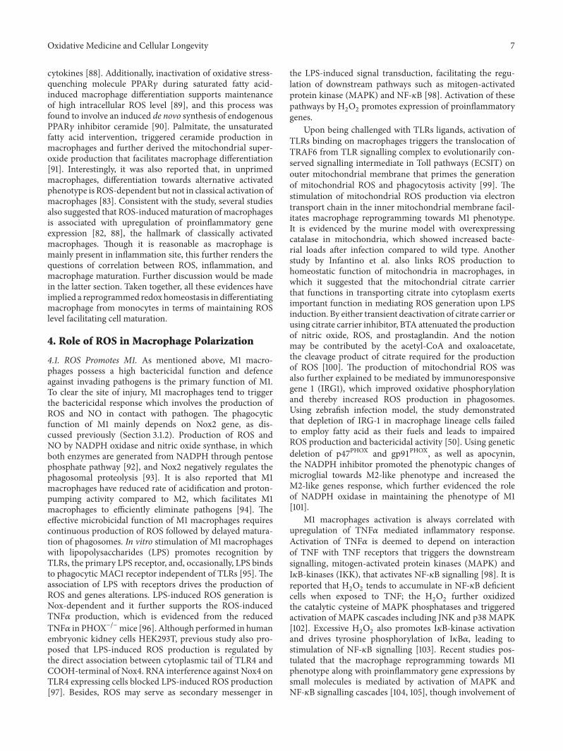
Oxidative Medicine and Cellular Longevity 7
cytokines [88]. Additionally, inactivation of oxidative stress-quenching molecule PPAR𝛾 during saturated fatty acid-induced macrophage differentiation supports maintenanceof high intracellular ROS level [89], and this process wasfound to involve an induced de novo synthesis of endogenousPPAR𝛾 inhibitor ceramide [90]. Palmitate, the unsaturatedfatty acid intervention, triggered ceramide production inmacrophages and further derived the mitochondrial super-oxide production that facilitates macrophage differentiation[91]. Interestingly, it was also reported that, in unprimedmacrophages, differentiation towards alternative activatedphenotype is ROS-dependent but not in classical activation ofmacrophages [83]. Consistent with the study, several studiesalso suggested that ROS-inducedmaturation of macrophagesis associated with upregulation of proinflammatory geneexpression [82, 88], the hallmark of classically activatedmacrophages. Though it is reasonable as macrophage ismainly present in inflammation site, this further renders thequestions of correlation between ROS, inflammation, andmacrophage maturation. Further discussion would be madein the latter section. Taken together, all these evidences haveimplied a reprogrammed redoxhomeostasis in differentiatingmacrophage from monocytes in terms of maintaining ROSlevel facilitating cell maturation.
4. Role of ROS in Macrophage Polarization
4.1. ROS Promotes M1. As mentioned above, M1 macro-phages possess a high bactericidal function and defenceagainst invading pathogens is the primary function of M1.To clear the site of injury, M1 macrophages tend to triggerthe bactericidal response which involves the production ofROS and NO in contact with pathogen. The phagocyticfunction of M1 mainly depends on Nox2 gene, as dis-cussed previously (Section 3.1.2). Production of ROS andNO by NADPH oxidase and nitric oxide synthase, in whichboth enzymes are generated from NADPH through pentosephosphate pathway [92], and Nox2 negatively regulates thephagosomal proteolysis [93]. It is also reported that M1macrophages have reduced rate of acidification and proton-pumping activity compared to M2, which facilitates M1macrophages to efficiently eliminate pathogens [94]. Theeffective microbicidal function of M1 macrophages requirescontinuous production of ROS followed by delayed matura-tion of phagosomes. In vitro stimulation of M1 macrophageswith lipopolysaccharides (LPS) promotes recognition byTLRs, the primary LPS receptor, and, occasionally, LPS bindsto phagocytic MAC1 receptor independent of TLRs [95]. Theassociation of LPS with receptors drives the production ofROS and genes alterations. LPS-induced ROS generation isNox-dependent and it further supports the ROS-inducedTNF𝛼 production, which is evidenced from the reducedTNF𝛼 in PHOX−/−mice [96]. Although performed in humanembryonic kidney cells HEK293T, previous study also pro-posed that LPS-induced ROS production is regulated bythe direct association between cytoplasmic tail of TLR4 andCOOH-terminal of Nox4. RNA interference against Nox4 onTLR4 expressing cells blocked LPS-induced ROS production[97]. Besides, ROS may serve as secondary messenger in
the LPS-induced signal transduction, facilitating the regu-lation of downstream pathways such as mitogen-activatedprotein kinase (MAPK) and NF-𝜅B [98]. Activation of thesepathways by H
2O2promotes expression of proinflammatory
genes.Upon being challenged with TLRs ligands, activation of
TLRs binding on macrophages triggers the translocation ofTRAF6 from TLR signalling complex to evolutionarily con-served signalling intermediate in Toll pathways (ECSIT) onouter mitochondrial membrane that primes the generationof mitochondrial ROS and phagocytosis activity [99]. Thestimulation of mitochondrial ROS production via electrontransport chain in the inner mitochondrial membrane facil-itates macrophage reprogramming towards M1 phenotype.It is evidenced by the murine model with overexpressingcatalase in mitochondria, which showed increased bacte-rial loads after infection compared to wild type. Anotherstudy by Infantino et al. also links ROS production tohomeostatic function of mitochondria in macrophages, inwhich it suggested that the mitochondrial citrate carrierthat functions in transporting citrate into cytoplasm exertsimportant function in mediating ROS generation upon LPSinduction. By either transient deactivation of citrate carrier orusing citrate carrier inhibitor, BTA attenuated the productionof nitric oxide, ROS, and prostaglandin. And the notionmay be contributed by the acetyl-CoA and oxaloacetate,the cleavage product of citrate required for the productionof ROS [100]. The production of mitochondrial ROS wasalso further explained to be mediated by immunoresponsivegene 1 (IRG1), which improved oxidative phosphorylationand thereby increased ROS production in phagosomes.Using zebrafish infection model, the study demonstratedthat depletion of IRG-1 in macrophage lineage cells failedto employ fatty acid as their fuels and leads to impairedROS production and bactericidal activity [50]. Using geneticdeletion of p47PHOX and gp91PHOX, as well as apocynin,the NADPH inhibitor promoted the phenotypic changes ofmicroglial towards M2-like phenotype and increased theM2-like genes response, which further evidenced the roleof NADPH oxidase in maintaining the phenotype of M1[101].
M1 macrophages activation is always correlated withupregulation of TNF𝛼 mediated inflammatory response.Activation of TNF𝛼 is deemed to depend on interactionof TNF with TNF receptors that triggers the downstreamsignalling, mitogen-activated protein kinases (MAPK) andI𝜅B-kinases (IKK), that activates NF-𝜅B signalling [98]. It isreported that H
2O2tends to accumulate in NF-𝜅B deficient
cells when exposed to TNF; the H2O2further oxidized
the catalytic cysteine of MAPK phosphatases and triggeredactivation of MAPK cascades including JNK and p38 MAPK[102]. Excessive H
2O2also promotes I𝜅B-kinase activation
and drives tyrosine phosphorylation of I𝜅B𝛼, leading tostimulation of NF-𝜅B signalling [103]. Recent studies pos-tulated that the macrophage reprogramming towards M1phenotype along with proinflammatory gene expressions bysmall molecules is mediated by activation of MAPK andNF-𝜅B signalling cascades [104, 105], though involvement of
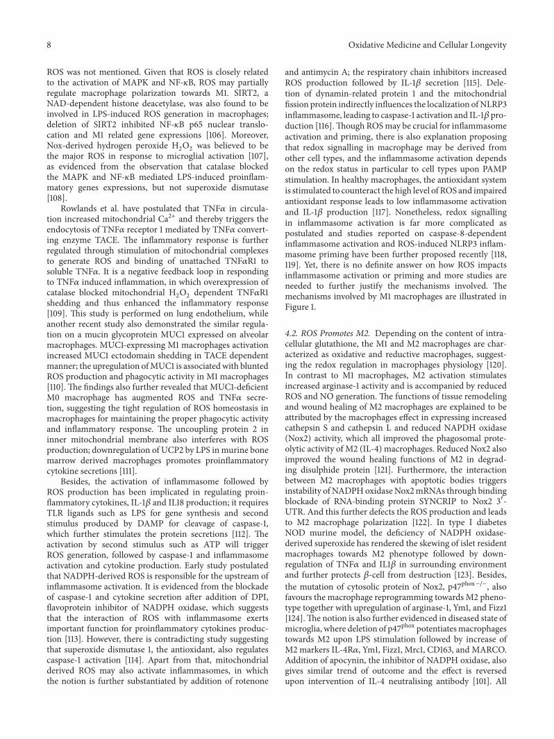
8 Oxidative Medicine and Cellular Longevity
ROS was not mentioned. Given that ROS is closely relatedto the activation of MAPK and NF-𝜅B, ROS may partiallyregulate macrophage polarization towards M1. SIRT2, aNAD-dependent histone deacetylase, was also found to beinvolved in LPS-induced ROS generation in macrophages;deletion of SIRT2 inhibited NF-𝜅B p65 nuclear translo-cation and M1 related gene expressions [106]. Moreover,Nox-derived hydrogen peroxide H
2O2was believed to be
the major ROS in response to microglial activation [107],as evidenced from the observation that catalase blockedthe MAPK and NF-𝜅B mediated LPS-induced proinflam-matory genes expressions, but not superoxide dismutase[108].
Rowlands et al. have postulated that TNF𝛼 in circula-tion increased mitochondrial Ca2+ and thereby triggers theendocytosis of TNF𝛼 receptor 1 mediated by TNF𝛼 convert-ing enzyme TACE. The inflammatory response is furtherregulated through stimulation of mitochondrial complexesto generate ROS and binding of unattached TNF𝛼R1 tosoluble TNF𝛼. It is a negative feedback loop in respondingto TNF𝛼 induced inflammation, in which overexpression ofcatalase blocked mitochondrial H
2O2dependent TNF𝛼R1
shedding and thus enhanced the inflammatory response[109]. This study is performed on lung endothelium, whileanother recent study also demonstrated the similar regula-tion on a mucin glycoprotein MUC1 expressed on alveolarmacrophages. MUC1-expressing M1 macrophages activationincreased MUC1 ectodomain shedding in TACE dependentmanner; the upregulation ofMUC1 is associatedwith bluntedROS production and phagocytic activity in M1 macrophages[110]. The findings also further revealed that MUC1-deficientM0 macrophage has augmented ROS and TNF𝛼 secre-tion, suggesting the tight regulation of ROS homeostasis inmacrophages for maintaining the proper phagocytic activityand inflammatory response. The uncoupling protein 2 ininner mitochondrial membrane also interferes with ROSproduction; downregulation of UCP2 by LPS inmurine bonemarrow derived macrophages promotes proinflammatorycytokine secretions [111].
Besides, the activation of inflammasome followed byROS production has been implicated in regulating proin-flammatory cytokines, IL-1𝛽 and IL18 production; it requiresTLR ligands such as LPS for gene synthesis and secondstimulus produced by DAMP for cleavage of caspase-1,which further stimulates the protein secretions [112]. Theactivation by second stimulus such as ATP will triggerROS generation, followed by caspase-1 and inflammasomeactivation and cytokine production. Early study postulatedthat NADPH-derived ROS is responsible for the upstream ofinflammasome activation. It is evidenced from the blockadeof caspase-1 and cytokine secretion after addition of DPI,flavoprotein inhibitor of NADPH oxidase, which suggeststhat the interaction of ROS with inflammasome exertsimportant function for proinflammatory cytokines produc-tion [113]. However, there is contradicting study suggestingthat superoxide dismutase 1, the antioxidant, also regulatescaspase-1 activation [114]. Apart from that, mitochondrialderived ROS may also activate inflammasomes, in whichthe notion is further substantiated by addition of rotenone
and antimycin A; the respiratory chain inhibitors increasedROS production followed by IL-1𝛽 secretion [115]. Dele-tion of dynamin-related protein 1 and the mitochondrialfission protein indirectly influences the localization ofNLRP3inflammasome, leading to caspase-1 activation and IL-1𝛽 pro-duction [116].Though ROSmay be crucial for inflammasomeactivation and priming, there is also explanation proposingthat redox signalling in macrophage may be derived fromother cell types, and the inflammasome activation dependson the redox status in particular to cell types upon PAMPstimulation. In healthy macrophages, the antioxidant systemis stimulated to counteract the high level of ROS and impairedantioxidant response leads to low inflammasome activationand IL-1𝛽 production [117]. Nonetheless, redox signallingin inflammasome activation is far more complicated aspostulated and studies reported on caspase-8-dependentinflammasome activation and ROS-induced NLRP3 inflam-masome priming have been further proposed recently [118,119]. Yet, there is no definite answer on how ROS impactsinflammasome activation or priming and more studies areneeded to further justify the mechanisms involved. Themechanisms involved by M1 macrophages are illustrated inFigure 1.
4.2. ROS Promotes M2. Depending on the content of intra-cellular glutathione, the M1 and M2 macrophages are char-acterized as oxidative and reductive macrophages, suggest-ing the redox regulation in macrophages physiology [120].In contrast to M1 macrophages, M2 activation stimulatesincreased arginase-1 activity and is accompanied by reducedROS and NO generation. The functions of tissue remodelingand wound healing of M2 macrophages are explained to beattributed by the macrophages effect in expressing increasedcathepsin S and cathepsin L and reduced NAPDH oxidase(Nox2) activity, which all improved the phagosomal prote-olytic activity of M2 (IL-4) macrophages. Reduced Nox2 alsoimproved the wound healing functions of M2 in degrad-ing disulphide protein [121]. Furthermore, the interactionbetween M2 macrophages with apoptotic bodies triggersinstability of NADPHoxidaseNox2mRNAs through bindingblockade of RNA-binding protein SYNCRIP to Nox2 3-UTR. And this further defects the ROS production and leadsto M2 macrophage polarization [122]. In type I diabetesNOD murine model, the deficiency of NADPH oxidase-derived superoxide has rendered the skewing of islet residentmacrophages towards M2 phenotype followed by down-regulation of TNF𝛼 and IL1𝛽 in surrounding environmentand further protects 𝛽-cell from destruction [123]. Besides,the mutation of cytosolic protein of Nox2, p47phox−/−, alsofavours the macrophage reprogramming towards M2 pheno-type together with upregulation of arginase-1, Ym1, and Fizz1[124].The notion is also further evidenced in diseased state ofmicroglia, where deletion of p47phox potentiatesmacrophagestowards M2 upon LPS stimulation followed by increase ofM2 markers IL-4R𝛼, Ym1, Fizz1, Mrc1, CD163, and MARCO.Addition of apocynin, the inhibitor of NADPH oxidase, alsogives similar trend of outcome and the effect is reversedupon intervention of IL-4 neutralising antibody [101]. All
Oxidative Medicine and Cellular Longevity 9
(a)
NADPH oxidase
mt.complex I-TLR Direct interaction
LPS NADPH
Citrate
Pyruvate
LPS
TCA
IRG1FA
M1
NADPH G6P
R5PPPP
Pathways in between
Cleavage of caspase-1 inflammasomes activation
ATP
IL18 iNOS
(b)
↑ phagocytic activity
TRAF6 + ECSIT
IL1𝛽TNF𝛼
TNF𝛼R1
TNF𝛼R1 sheddingMAPK and NF-𝜅B
H2O2
mt-H2O2 ↑
Inflammatory response ↑
ROS ↑ ROS ↑
Figure 1: Involvement of ROS in regulatingM1 responsible phagocytic activity and inflammatory response. (a)Multiple pathways are involvedin generating NADPH, followed by ROS production by NADPH oxidase. The high ROS level mainly used to mediate the phagocytic activityofM1macrophages. (b) ROS serves as secondmessengermediating the inflammatory response ofM1macrophages, primarily throughMAPKandNF-𝜅B as well as inflammasome activation.Mt, mitochondrial; FA, fatty acid; G6P, glucose-6-phosphate; R5P, ribulose-5-phosphate; PPP,pentose phosphate pathway; TCA, tricarboxylic acid cycle.
these further propose depletion of Nox2 accompanied byROS reduction which is important for reprogramming of M1to M2 phenotype.
Apart from the suggested role of NADPH oxidase inregulating M2 macrophages, superoxide dismutase (SOD)enzyme, which catalyses superoxide dismutation and wasassociated with H
2O2production, also contributed to M2
macrophages activation. Study byHe et al. postulated that Cu,Zn-SOD−/− mice had abundance of alveolar macrophagesin M1 phenotype, while Cu, Zn-SODTG mice predominantlyexpressed M2 macrophage markers. The function of Cu,Zn-SOD in modulating M2 alternative activation is regu-lated by redox-dependent STAT6 translocation [125]. Indeed,previous study revealed the absence of IL-4 related genesexpressions in STAT6 deficient mice upon stimulation by IL-4. Silencing of STAT6 in T lymphocytes showed incapabilityto polarize to Th2 phenotype in the presence of IL-13 or IL-4. This also further postulated that STAT6 is implicated forM2 macrophage polarization, given that M2 macrophageshave the same differentiation manner as Th2 lymphocytes[126].Moreover, as opposed toM1macrophages, extracellularATP blocks IL-1𝛽 in M2 macrophages. This was claimedto be mediated by two mechanisms, the direct blockadeof ROS and inflammasome trapping through clustering ofactin filaments, which are both associated with reducedATP plasma membrane ion channel, P2X
7R [127]. Recent
study also postulated the glucose metabolism related proteincarbohydrate kinase-like protein (CARKL) promotes M2activation. Reduced pentose phosphate pathway (PPP) fluxin M2macrophages is mediated by CARKL that catalyses theformation of sedoheptulose-7-phosphate, the intermediateof PPP. The reduced glucose metabolism activated M2 wasevidenced by addition of metformin and rotenone whichblocked (IL-4) M2 genes expressions. The study also furtherobserved reduction in NAD by not NAD+ levels whichsuggested that CARKL may be important in regulating theredox balance in glucose metabolism [17].
Although impact of redox signalling or ROS productionin M1 macrophages activation seems to interfere with M2macrophage priming, study by Zhang et al. postulated thatROS production is also important for M2 macrophage dif-ferentiation. Intervention of antioxidant butylated hydrox-yanisole BHA by inhibiting Nox-mediated O2− productionbefore differentiation by M-CSF treatment blocked mono-cyte differentiation to M2, which suggests that ROS mayimplicate the early stage of M2 macrophage differentiation.Further intervention of BHA in urethane-induced murinelung cancer model also attenuated the occurrence of tumor-associated macrophages and thereby reduced tumor progres-sion [83]. Also, study revealed that KLF4 triggers MCP-1-induced protein (MCPIP) to stimulate ROS production in IL-4-inducedM2macrophages andROS attenuation blocked ERstress inM2macrophages. Removal of theMCPIP shifted themacrophage phenotype towards M1 with increasing phago-cytic function [128]. These studies may render questionsfor activation and priming of alternative M2 macrophages,whether ROS may impact different stages of M2 macrophagemanifestation, which need to be further investigated. Themechanisms involved by M2 macrophages are illustrated inFigure 2.
5. Discussion
5.1. ROS-Controlled Macrophage Polarization in Disease Pro-gression: A Potential Drug Target? More evidences postulatedthat macrophage polarization played critical role in initiationand progression of multiple human diseases, as describedabove. In some cases, ROS plays a critical part in triggeringdisease-specific skewing of macrophages. This has beenparticularly observed in tumorigenesis and atherosclerosisin which the dysregulation caused by oxidative stress andinflammation have been extensively studied. However, theinvolvement of oxidative stress in diseases progression maybe very broad and results in pathogenesis linking variety
10 Oxidative Medicine and Cellular Longevity
ATP
PPPsedo
S7P
Pathways in between
M2Cu, Zn-SOD
STAT6
FIZZ-1
Arg1
YM-1
SYNCRIP
Anti-inflammatory
Wound healing
H2O2
NAD ↓
ROS ↓
O2∙
CARKL ↑
IL1𝛽 ↓
Disulphide protein degradation ↑
NADPH oxidase ↓
Nox2 3UTR
P2X7R ↓
Figure 2: Involvement of ROS in regulating M2 responsible inflammation resolving and wound healing activities. Multiple pathwaysare involved in reducing NADPH and its oxidase followed by reduced ROS generation. The low ROS level was accompanied byreduced inflammatory mediators; increased M2-regulated genes responsible for inflammation resolution; and increased disulphide proteindegradation which enhanced wound healing effect of M2. S7P, sedoheptulose-7-phosphate; sedo, sedoheptulose; PPP, pentose phosphatepathway; SYNCRIP, synaptotagmin binding, cytoplasmic RNA interacting protein; SOD, superoxide dismutase.
levels of biological processes; direct cues on ROS-directedmacrophage polarization further dominate diseases pro-gression which are still lacking. This is also not justifieddue to the nature of macrophages as effectors cells, whosefunction in part is antigen presenting. In this case, ROS-drivenmacrophage polarization is thus far difficult to becomeputative drug target owing to the lack of specificity. Thisshortage is reflected in multiple levels, and pharmaceuticalcompanies have to develop systems that particularly targetmacrophage ROS as well as its polarization.This includes notonly drug target study but also development of drug deliv-ery system. Interestingly, recent studies have revealed thatliposome specifically delivered molecules to macrophages[129], which allows molecules to be specifically targetedon macrophages in the body. And development of high-resolution transcriptome analysis allows differentiation ofM1 macrophages from M2 phenotype by specific surfacemarkers [130]. Development of particular antibodies alsoallows recognition of individual subtype of macrophages inhuman body. Potential molecules targeting ROS signallingand macrophage reprogramming could be further enrichedin order to discover the target treatment. More experimentalinvestigations as well as clinical trials shall be conducted toprove the hypothesis.
5.2. Antioxidative Herbal Supplements RegulatingMacrophagePolarization: Any Clues for Diseases Treatment? There area plenty of herbal supplements available for the indicationof health improvement. Herbal supplements are commonlyemployed, by people not only in Asian countries withtradition of using herbal remedy for diseases treatment, butalso in countries where use of herbal products is understrict control and closed monitoring, for example, Europeand US. There are an increasing number of populations whofavour herbal products as dietary supplements due to theconcrete health-improving effect of herbal supplements asdemonstrated from the scientific evidences by both benchtop
and clinical studies. Most of these herbal supplements, suchas Ginkgo biloba, lingzhi mushroom, baicalin, and somecomposite herbal formulae, exhibit excellent antioxidativeeffect in laboratory studies. Indeed, herbal supplements gen-erally contain a series of flavone and phenol-like compoundsthat work as effective scavengers of ROS and that conferthe health-improving effects. Interestingly, recent evidencesdemonstrated that some herbal supplements could regulatemacrophage polarization in preclinical models of humandiseases. Study by Lam et al. proved that PHY906, anherbal adjuvant derived from ancient Chinese Medicineformula Huangqin Decoction, has beneficial effect to cancertreatment. The tumor regression effect of PHY906 may beassociated with its regulation on polarization of macrophagewithin tumormicroenvironment. Tumor frommice receivingPHY906 showed more infiltrated M1 macrophages whichfacilitates tumor cells killing activity [131]. Our previous studyon baicalin, which is the major compound in PHY906 and isused as calming and soothing supplement, could reskew M2polarized macrophages towards M1 phenotype. This effectwas reflected in tumor microenvironment, with more M1 butreduced M2 macrophages observed after baicalin treatment.Removal of macrophages attenuated tumor inhibition bybaicalin [132]. Interestingly, baicalin is generally used toreduce inflammation. The property of baicalin and baicalin-containing herbal supplement in activating proinflammatoryphenotype of tumor-associated macrophages may under-score a variety of mechanisms underlying effect of baicalinon macrophages. In fact, baicalin has been reported as aROS generator as well as scavenger in macrophages [133,134]. Given the complicated role of ROS as we discussedabove, the dual role of baicalin in ROS homeostasis may beinvolved in baicalin-mediated proinflammatory macrophageskewing. More scientific evidences have to be acquired inorder to prove if ROS involves baicalin-mediatedmacrophagefunctions, but this also further raises the consideration onthe scientific scrutiny in consumption of herbal products as
Oxidative Medicine and Cellular Longevity 11
daily dietary supplements, assuming that they may impactROS-driven macrophages polarization. Similar cases arereported in other antioxidative herbal supplements, includ-ing bitter mushroom [135] and lingzhi mushroom [136],which facilitates the regulation of macrophage polarizationtowards particular phenotype. Nonetheless, the functionof polarized macrophages with particular phenotype variesacross different diseases, and there is no justifiable con-clusion on the function of each individual phenotype ofmacrophages in all diseases. In this case, herbal supplementsconsumption without concerning their use in particulardiseases may cause severe consequences, at least in thecontext of macrophage polarization. As we still could notdeny the drug-like effect of many herbal supplements, theirproper use shall be further supported by more scientificevidences.
6. Conclusion
In summary, macrophages are majorly diversified intotwo distinct phenotypes. A variety of peripheral and resi-dence factors may determine the dominant phenotypes ofmacrophages within tissues. Although it is not yet concludedwhether this polarization of macrophages is in situ reversible,it has been shown that the overall shift of macrophagephenotypes plays a critical role in the progression of varioushuman diseases. Instead ofmerely being a harmful byproductof metabolism, ROS has been shown to get involved in thefunctional and phenotypic regulation of macrophages. ROSis able to control the cell death, proliferation, motility, andphagocytic ability of macrophages. Intriguingly, it is recentlyobserved that ROS may play a complicated role in regu-lating macrophage polarization, which implies the potentfuture therapeutic approaches for life-threatening diseases.Development of target-specific delivery system has beensupportive for drug development, and natural antioxidantswith immunomodulatory function shall be considered. Morescientific evidences as well as clinical trials are imperative andemerging.
Competing Interests
The authors declare no competing interests.
Authors’ Contributions
Hor-Yue Tan and Ning Wang drafted the paper; Sha Li,Ming Hong, and Xuanbin Wang revised the paper. YibinFeng conceived, designed, revised, and finalized the paper. Allauthors discussed and confirmed the final paper.
Acknowledgments
This research was partially supported by the ResearchCouncil of the University of Hong Kong (Project codes:104002889 and 104003422), Wong’s donation (Project code:200006276), the donation of Gaia Family Trust, New Zealand(Project code: 200007008), the donation of Vita Green
Health Products Co., Ltd. (Project code: 200007477), andthe Open Project of Hubei Key Laboratory of Wudang LocalChinese Medicine Research, Hubei University of Medicine(WDCM001).
References
[1] K. M. Holmström and T. Finkel, “Cellular mechanisms andphysiological consequences of redox-dependent signalling,”Nature ReviewsMolecular Cell Biology, vol. 15, no. 6, pp. 411–421,2014.
[2] C. L. Bigarella, R. Liang, and S. Ghaffari, “Stem cells and theimpact of ROS signaling,” Development, vol. 141, no. 22, pp.4206–4218, 2014.
[3] D. Zhou, C. Huang, Z. Lin et al., “Macrophage polarization andfunction with emphasis on the evolving roles of coordinatedregulation of cellular signaling pathways,” Cellular Signalling,vol. 26, no. 2, pp. 192–197, 2014.
[4] D. Lecca, M. L. Trincavelli, P. Gelosa et al., “The recentlyidentified P2Y-like receptor GPR17 is a sensor of brain damageand a new target for brain repair,” PLoS ONE, vol. 3, no. 10,Article ID e3579, 2008.
[5] M. E. Rausch, S. Weisberg, P. Vardhana, and D. V. Tortoriello,“Obesity in C57BL/6J mice is characterized by adipose tissuehypoxia and cytotoxic T-cell infiltration,” International Journalof Obesity, vol. 32, no. 3, pp. 451–463, 2008.
[6] G. Dhabekar, R. Dandekar, and A. Kingaonkar, “Role ofmacrophages in malignancy,” Annals of Maxillofacial Surgery,vol. 1, no. 2, pp. 150–154, 2011.
[7] S. K. Biswas and A. Mantovani, “Macrophage plasticity andinteraction with lymphocyte subsets: cancer as a paradigm,”Nature Immunology, vol. 11, no. 10, pp. 889–896, 2010.
[8] A. Sica and A.Mantovani, “Macrophage plasticity and polariza-tion: in vivo veritas,” The Journal of Clinical Investigation, vol.122, no. 3, pp. 787–795, 2012.
[9] M. J. Rauh, V. Ho, C. Pereira et al., “SHIP represses thegeneration of alternatively activated macrophages,” Immunity,vol. 23, no. 4, pp. 361–374, 2005.
[10] A. Kroner, A. D. Greenhalgh, J. G. Zarruk, R. PassosdosSantos,M. Gaestel, and S. David, “TNF and increased intracellular ironalter macrophage polarization to a detrimental M1 phenotypein the injured spinal cord,” Neuron, vol. 83, no. 5, pp. 1098–1116,2014.
[11] K. Takeda, T. Kaisho, and S. Akira, “Toll-like receptors,”AnnualReview of Immunology, vol. 21, pp. 335–376, 2003.
[12] M.Yamamoto, S. Sato,H.Hemmi et al., “Role of adaptorTRIF inthe MyD88-independent toll-like receptor signaling pathway,”Science, vol. 301, no. 5633, pp. 640–643, 2003.
[13] S. K. Biswas and A. Mantovani, “Orchestration of metabolismby macrophages,” Cell Metabolism, vol. 15, no. 4, pp. 432–437,2012.
[14] S. Recalcati, M. Locati, A. Marini et al., “Differential regulationof iron homeostasis during human macrophage polarizedactivation,” European Journal of Immunology, vol. 40, no. 3, pp.824–835, 2010.
[15] J.-C. Rodŕıguez-Prados, P. G. Través, J. Cuenca et al., “Substratefate in activated macrophages: a comparison between innate,classic, and alternative activation,” The Journal of Immunology,vol. 185, no. 1, pp. 605–614, 2010.
[16] E. Izquierdo, V. D. Cuevas, S. Fernández-Arroyo et al., “Reshap-ing of human macrophage polarization through modulation of
12 Oxidative Medicine and Cellular Longevity
glucose catabolic pathways,” Journal of Immunology, vol. 195, no.5, pp. 2442–2451, 2015.
[17] A. Haschemi, P. Kosma, L. Gille et al., “The sedoheptulosekinase CARKL directs macrophage polarization through con-trol of glucose metabolism,” Cell Metabolism, vol. 15, no. 6, pp.813–826, 2012.
[18] Z. Tan, N. Xie, H. Cui et al., “Pyruvate dehydrogenase kinase 1participates in macrophage polarization via regulating glucosemetabolism,” The Journal of Immunology, vol. 194, no. 12, pp.6082–6089, 2015.
[19] F. O. Martinez, S. Gordon, M. Locati, and A. Mantovani, “Tran-scriptional profiling of the human monocyte-to-macrophagedifferentiation and polarization: new molecules and patterns ofgene expression,” Journal of Immunology, vol. 177, no. 10, pp.7303–7311, 2006.
[20] A.Mantovani, A. Sica, S. Sozzani, P. Allavena, A. Vecchi, andM.Locati, “The chemokine system in diverse forms of macrophageactivation and polarization,” Trends in Immunology, vol. 25, no.12, pp. 677–686, 2004.
[21] S. C.-C. Huang, B. Everts, Y. Ivanova et al., “Cell-intrinsiclysosomal lipolysis is essential for alternative activation ofmacrophages,” Nature Immunology, vol. 15, no. 9, pp. 846–855,2014.
[22] D. De Nardo, L. I. Labzin, H. Kono et al., “High-densitylipoprotein mediates anti-inflammatory reprogramming ofmacrophages via the transcriptional regulator ATF3,” NatureImmunology, vol. 15, no. 2, pp. 152–160, 2014.
[23] X. Prieur, C. Y. L. Mok, V. R. Velagapudi et al., “Differentiallipid partitioning between adipocytes and tissue macrophagesmodulates macrophage lipotoxicity and M2/M1 polarization inobese mice,” Diabetes, vol. 60, no. 3, pp. 797–809, 2011.
[24] D.-S. Im, “Functions of omega-3 fatty acids and FFA4 (GPR120)in macrophages,” European Journal of Pharmacology, 2015.
[25] S.-J. Park, K.-P. Lee, S. Kang et al., “Sphingosine 1-phosphateinduced anti-atherogenic and atheroprotectiveM2macrophagepolarization through IL-4,”Cellular Signalling, vol. 26, no. 10, pp.2249–2258, 2014.
[26] M. Masoodi, O. Kuda, M. Rossmeisl, P. Flachs, and J. Kopecky,“Lipid signaling in adipose tissue: connecting inflammation &metabolism,” Biochimica et Biophysica Acta (BBA)—Molecularand Cell Biology of Lipids, vol. 1851, no. 4, pp. 503–518, 2015.
[27] A. Shiomi and T. Usui, “Pivotal roles of GM-CSF in autoimmu-nity and inflammation,” Mediators of Inflammation, vol. 2015,Article ID 568543, 13 pages, 2015.
[28] M. J. Kraakman, A. J. Murphy, K. Jandeleit-Dahm, and H.L. Kammoun, “Macrophage polarization in obesity and type2 diabetes: weighing down our understanding of macrophagefunction?” Frontiers in Immunology, vol. 5, article 470, 2014.
[29] G. Chinetti-Gbaguidi, S. Colin, and B. Staels, “Macrophagesubsets in atherosclerosis,” Nature Reviews Cardiology, vol. 12,no. 1, pp. 10–17, 2015.
[30] B. Ruffell, N. I. Affara, and L.M. Coussens, “Differential macro-phage programming in the tumor microenvironment,” Trendsin Immunology, vol. 33, no. 3, pp. 119–126, 2012.
[31] C. N. Lumeng, J. L. Bodzin, and A. R. Saltiel, “Obesity induces aphenotypic switch in adipose tissue macrophage polarization,”The Journal of Clinical Investigation, vol. 117, no. 1, pp. 175–184,2007.
[32] A. Chawla, K. D. Nguyen, and Y. P. S. Goh, “Macrophage-mediated inflammation in metabolic disease,” Nature ReviewsImmunology, vol. 11, no. 11, pp. 738–749, 2011.
[33] S. P. Weisberg, D. Hunter, R. Huber et al., “CCR2 modulatesinflammatory and metabolic effects of high-fat feeding,” TheJournal of Clinical Investigation, vol. 116, no. 1, pp. 115–124, 2006.
[34] S. Colin, G. Chinetti-Gbaguidi, and B. Staels, “Macrophagephenotypes in atherosclerosis,” Immunological Reviews, vol.262, no. 1, pp. 153–166, 2014.
[35] K. M. Botham and C. P. D. Wheeler-Jones, “Postprandiallipoproteins and the molecular regulation of vascular home-ostasis,” Progress in Lipid Research, vol. 52, no. 4, pp. 446–464,2013.
[36] B.-Z. Qian and J. W. Pollard, “Macrophage diversity enhancestumor progression and metastasis,” Cell, vol. 141, no. 1, pp. 39–51, 2010.
[37] H. Yang, C. Kim, M.-J. Kim et al., “Soluble vascular endothelialgrowth factor receptor-3 suppresses lymphangiogenesis andlymphatic metastasis in bladder cancer,”Molecular Cancer, vol.10, article 36, 2011.
[38] X. Tang, C. Mo, Y. Wang, D. Wei, and H. Xiao, “Anti-tumourstrategies aiming to target tumour-associated macrophages,”Immunology, vol. 138, no. 2, pp. 93–104, 2013.
[39] F. Eriksson, P. Tsagozis, K. Lundberg et al., “Tumor-specificbacteriophages induce tumor destruction through activationof tumor-associated macrophages,” Journal of Immunology, vol.182, no. 5, pp. 3105–3111, 2009.
[40] C. D. Gregory and A. Devitt, “The macrophage and the apop-totic cell: an innate immune interaction viewed simplistically?”Immunology, vol. 113, no. 1, pp. 1–14, 2004.
[41] M.-K. Chang, C. Bergmark, A. Laurila et al., “Monoclonalantibodies against oxidized low-density lipoprotein bind toapoptotic cells and inhibit their phagocytosis by elicitedmacrophages: evidence that oxidation-specific epitopesmediatemacrophage recognition,” Proceedings of the National Academyof Sciences of the United States of America, vol. 96, no. 11, pp.6353–6358, 1999.
[42] V. A. Tyurin, K. Balasubramanian, D. Winnica et al., “Oxida-tively modified phosphatidylserines on the surface of apop-totic cells are essential phagocytic ‘eat-me’ signals: cleavageand inhibition of phagocytosis by Lp-PLA
2,” Cell Death and
Differentiation, vol. 21, no. 5, pp. 825–835, 2014.[43] V. E. Kagan, G. G. Borisenko, B. F. Serinkan et al., “Appe-
tizing rancidity of apoptotic cells for macrophages: oxidation,externalization, and recognition of phosphatidylserine,” TheAmerican Journal of Physiology—Lung Cellular and MolecularPhysiology, vol. 285, no. 1, pp. L1–L17, 2003.
[44] Y. Y. Tyurina, F. B. Serinkan, V. A. Tyurin et al., “Lipid antioxi-dant, etoposide, inhibits phosphatidylserine externalization andmacrophage clearance of apoptotic cells by preventing phos-phatidylserine oxidation,” The Journal of Biological Chemistry,vol. 279, no. 7, pp. 6056–6064, 2004.
[45] S. Hoshino, M. Konishi, M. Mori et al., “HIV-1 Vpr inducesTLR4/MyD88-mediated IL-6 production and reactivates viralproduction from latency,” Journal of Leukocyte Biology, vol. 87,no. 6, pp. 1133–1143, 2010.
[46] N. Dey, M. Sinha, S. Gupta et al., “Caspase-1/ASC inflam-masome-mediated activation of IL-1𝛽–ROS–NF-𝜅B pathwayfor control of Trypanosoma cruzi replication and survival isdispensable in NLRP3−/− macrophages,” PLoS ONE, vol. 9, no.11, Article ID e111539, 2014.
[47] M. Bauer, M. Goldstein, M. Christmann, H. Becker, D. Heyl-mann, and B. Kaina, “Human monocytes are severely impairedin base and DNA double-strand break repair that renders
Oxidative Medicine and Cellular Longevity 13
them vulnerable to oxidative stress,” Proceedings of the NationalAcademy of Sciences of the United States of America, vol. 108, no.52, pp. 21105–21110, 2011.
[48] S. Essler, N. Dehne, and B. Brüne, “Role of sestrin2 in peroxidesignaling in macrophages,” FEBS Letters, vol. 583, no. 21, pp.3531–3535, 2009.
[49] H.-M. Lo, C.-L. Chen, C.-M. Yang et al., “The carotenoidlutein enhances matrix metalloproteinase-9 production andphagocytosis through intracellular ROSgeneration andERK1/2,p38 MAPK, and RAR𝛽 activation in murine macrophages,”Journal of Leukocyte Biology, vol. 93, no. 5, pp. 723–735, 2013.
[50] C. J. Hall, R. H. Boyle, J. W. Astin et al., “Immunoresponsivegene 1 augments bactericidal activity of macrophage-lineagecells by regulating 𝛽-oxidation-dependent mitochondrial rosproduction,” Cell Metabolism, vol. 18, no. 2, pp. 265–278, 2013.
[51] Y. Y. Tyurina, L. V. Basova, N. V. Konduru et al., “Nitrosativestress inhibits the aminophospholipid translocase resulting inphosphatidylserine externalization and macrophage engulf-ment: implications for the resolution of inflammation,” TheJournal of Biological Chemistry, vol. 282, no. 11, pp. 8498–8509,2007.
[52] T. Harada, T. Tanikawa, Y. Iwasaki, M. Yamada, Y. Imai,and M. Miyake, “Phagocytic entry of Legionella pneumophilainto macrophages through phosphatidylinositol 3,4,5-trisphos-phate-independent pathway,” Biological and PharmaceuticalBulletin, vol. 35, no. 9, pp. 1460–1468, 2012.
[53] K. L. Brown, K. Christenson, A. Karlsson, C. Dahlgren, andJ. Bylund, “Divergent effects on phagocytosis by macrophage-derived oxygen radicals,” Journal of Innate Immunity, vol. 1, no.6, pp. 592–598, 2009.
[54] D. Sanmun, E. Witasp, S. Jitkaew et al., “Involvement of afunctional NADPH oxidase in neutrophils and macrophagesduring programmed cell clearance: Implications for chronicgranulomatous disease,” American Journal of Physiology—CellPhysiology, vol. 297, no. 3, pp. C621–C631, 2009.
[55] A. M. Johann, A. von Knethen, D. Lindemann, and B. Brüne,“Recognition of apoptotic cells by macrophages activates theperoxisome proliferator-activated receptor-𝛾 and attenuates theoxidative burst,”Cell Death and Differentiation, vol. 13, no. 9, pp.1533–1540, 2006.
[56] H.-N. Lee and Y.-J. Surh, “Resolvin D1-mediated NOX2 inac-tivation rescues macrophages undertaking efferocytosis fromoxidative stress-induced apoptosis,” Biochemical Pharmacology,vol. 86, no. 6, pp. 759–769, 2013.
[57] P. Scaffidi, T. Misteli, and M. E. Bianchi, “Release of chro-matin protein HMGB1 by necrotic cells triggers inflammation,”Nature, vol. 418, no. 6894, pp. 191–195, 2002.
[58] S. Y. Lim, M. J. Raftery, J. Goyette, K. Hsu, and C. L. Geczy,“Oxidativemodifications of S100 proteins: functional regulationby redox,” Journal of Leukocyte Biology, vol. 86, no. 3, pp. 577–587, 2009.
[59] S. F. Moore and A. B. MacKenzie, “NADPH oxidase NOX2mediates rapid cellular oxidation following ATP stimulation ofendotoxin-primed macrophages,” Journal of Immunology, vol.183, no. 5, pp. 3302–3308, 2009.
[60] C. Jiménez-López, J. R. Collette, K. M. Brothers et al., “Candidaalbicans induces arginine biosynthetic genes in response tohost-derived reactive oxygen species,” Eukaryotic Cell, vol. 12,no. 1, pp. 91–100, 2013.
[61] S. Tavakoli and R. Asmis, “Reactive oxygen species and thiolredox signaling in the macrophage biology of atherosclerosis,”
Antioxidants and Redox Signaling, vol. 17, no. 12, pp. 1785–1795,2012.
[62] G. B. Fortes, L. S. Alves, R. de Oliveira et al., “Heme inducesprogrammed necrosis on macrophages through autocrine TNFand ROS production,” Blood, vol. 119, no. 10, pp. 2368–2375,2012.
[63] R. Deshmukh and V. Trivedi, “Methemoglobin exposure pro-duces toxicological effects in macrophages due to multiple ROSspike induced apoptosis,” Toxicology in Vitro, vol. 27, no. 1, pp.16–23, 2013.
[64] J. L. Miller, K. Velmurugan, M. J. Cowan, and V. Briken,“The type I NADH dehydrogenase of Mycobacterium tuber-culosis counters phagosomal NOX2 activity to inhibit TNF-𝛼-mediated host cell apoptosis,” PLoS Pathogens, vol. 6, no. 4,Article ID e1000864, 2010.
[65] V. S. Soumyarani and N. Jayakumari, “Oxidatively modifiedhigh density lipoprotein promotes inflammatory response inhuman monocytes-macrophages by enhanced production ofROS, TNF-𝛼, MMP-9, and MMP-2,” Molecular and CellularBiochemistry, vol. 366, no. 1-2, pp. 277–285, 2012.
[66] K. Clare, S. J. Hardwick, K. L. H. Carpenter, N.Weeratunge, andM. J. Mitchinson, “Toxicity of oxysterols to human monocyte-macrophages,” Atherosclerosis, vol. 118, no. 1, pp. 67–75, 1995.
[67] U. Rueckschloss, J. Galle, J. Holtz, H.-R. Zerkowski, and H.Morawietz, “Induction of NAD(P)H oxidase by oxidized low-density lipoprotein in human endothelial cells: antioxidativepotential of hydroxymethylglutaryl coenzyme A reductaseinhibitor therapy,” Circulation, vol. 104, no. 15, pp. 1767–1772,2001.
[68] E. Lombardo, A. Alvarez-Barrientos, B. Maroto, L. Boscá, andU. G. Knaus, “TLR4-mediated survival of macrophages isMyD88 dependent and requires TNF-𝛼 autocrine signalling,”The Journal of Immunology, vol. 178, no. 6, pp. 3731–3739, 2007.
[69] S.-D. Ha, S. Park, C. Y. Han, M. L. Nguyen, and S. O. Kim,“Cellular adaptation to anthrax lethal toxin-inducedmitochon-drial cholesterol enrichment, hyperpolarization, and reactiveoxygen species generation through downregulating MLN64 inmacrophages,” Molecular and Cellular Biology, vol. 32, no. 23,pp. 4846–4860, 2012.
[70] Y.-J. Lim, J.-A. Choi, H.-H. Choi et al., “Endoplasmic reticulumstress pathway-mediated apoptosis in macrophages contributesto the survival of Mycobacterium tuberculosis,” PLoS ONE, vol.6, no. 12, Article ID e28531, 2011.
[71] Y.-J. Lim,H.-H.Choi, J.-A. Choi et al., “Mycobacteriumkansasii-induced death of murine macrophages involves endoplasmicreticulum stress responses mediated by reactive oxygen speciesgeneration or calpain activation,” Apoptosis, vol. 18, no. 2, pp.150–159, 2013.
[72] J.-A. Choi, Y.-J. Lim, S.-N. Cho et al., “Mycobacterial HBHAinduces endoplasmic reticulum stress-mediated apoptosisthrough the generation of reactive oxygen species and cytosolicCa2+ in murine macrophage RAW 264.7 cells,” Cell Death andDisease, vol. 4, article e957, 2013.
[73] J. Y. Bertrand, A. Jalil, M. Klaine, S. Jung, A. Cumano, and I.Godin, “Three pathways to mature macrophages in the earlymouse yolk sac,” Blood, vol. 106, no. 9, pp. 3004–3011, 2005.
[74] Z. Tang, Y. Gan, Q. Liu et al., “CX3CR1 deficiency suppressesactivation and neurotoxicity ofmicroglia/macrophage in exper-imental ischemic stroke,” Journal of Neuroinflammation, vol. 11,article 26, 2014.
[75] P. Niethammer, C. Grabher, A. T. Look, and T. J. Mitchison,“A tissue-scale gradient of hydrogen peroxide mediates rapid
14 Oxidative Medicine and Cellular Longevity
wound detection in zebrafish,” Nature, vol. 459, no. 7249, pp.996–999, 2009.
[76] J.-W. Ryu, K. H. Hong, J. H. Maeng et al., “Overexpression ofuncoupling protein 2 in THP1 monocytes inhibits 𝛽2 integrin-mediated firm adhesion and transendothelial migration,” Arte-riosclerosis, Thrombosis, and Vascular Biology, vol. 24, no. 5, pp.864–870, 2004.
[77] D. Hackel, D. Pflücke, A. Neumann et al., “The connection ofmonocytes and reactive oxygen species in pain,” PLoS ONE, vol.8, no. 5, Article ID e63564, 2013.
[78] S. Ullevig, Q. Zhao, C. F. Lee, H. Seok Kim, D. Zamora, andR. Asmis, “NADPH oxidase 4 mediates monocyte primingand accelerated chemotaxis induced by metabolic stress,” Arte-riosclerosis, Thrombosis, and Vascular Biology, vol. 32, no. 2, pp.415–426, 2012.
[79] C. F. Lee, S. Ullevig, H. S. Kim, and R. Asmis, “Regulation ofmonocyte adhesion and migration by Nox4,” PLoS ONE, vol. 8,no. 6, Article ID e66964, 2013.
[80] D. Zhang, X. Jiang, P. Fang et al., “Hyperhomocysteinemiapromotes inflammatory monocyte generation and acceleratesatherosclerosis in transgenic cystathionine 𝛽-synthase- defi-cient mice,” Circulation, vol. 120, no. 19, pp. 1893–1902, 2009.
[81] L. Stixová, J. Procházková, K. Souček, J. Hofmanová, andA. Kozubı́k, “5-Lipoxygenase inhibitors potentiate 1𝛼,25-dihy-droxyvitaminD3-inducedmonocytic differentiation by activat-ing p38 MAPK pathway,” Molecular and Cellular Biochemistry,vol. 330, no. 1-2, pp. 229–238, 2009.
[82] S. S. Barbieri, S. Eligini, M. Brambilla, E. Tremoli, and S. Colli,“Reactive oxygen species mediate cyclooxygenase-2 inductionduring monocyte to macrophage differentiation: critical role ofNADPH oxidase,” Cardiovascular Research, vol. 60, no. 1, pp.187–197, 2003.
[83] Y. Zhang, S. Choksi, K. Chen, Y. Pobezinskaya, I. Linnoila,and Z.-G. Liu, “ROS play a critical role in the differentiationof alternatively activated macrophages and the occurrence oftumor-associatedmacrophages,”Cell Research, vol. 23, no. 7, pp.898–914, 2013.
[84] B. Fuhrman, M. Shiner, N. Volkova, and M. Aviram, “Cell-induced copper ion-mediated low density lipoprotein oxidationincreases during in vivo monocyte-to-macrophage differentia-tion,” Free Radical Biology and Medicine, vol. 37, no. 2, pp. 259–271, 2004.
[85] K. Nishi, T. Oda, S. Takabuchi et al., “LPS induces hypoxia-inducible factor 1 activation in macrophage-differentiated cellsin a reactive oxygen species-dependent manner,” Antioxidantsand Redox Signaling, vol. 10, no. 5, pp. 983–995, 2008.
[86] J. Makino, T. Kamiya, H. Hara, and T. Adachi, “TPA inducesthe expression of EC-SOD in human monocytic THP-1 cells:involvement of PKC, MEK/ERK and NOX-derived ROS,” FreeRadical Research, vol. 46, no. 5, pp. 637–644, 2012.
[87] T. Yamamoto, N. Sakaguchi, M. Hachiya, F. Nakayama, M.Yamakawa, and M. Akashi, “Role of catalase in monocyticdifferentiation of U937 cells by TPA: hydrogen peroxide as asecond messenger,” Leukemia, vol. 23, no. 4, pp. 761–769, 2009.
[88] M.-G. Song, I.-G. Ryoo, H.-Y. Choi et al., “NRF2 signalingnegatively regulates phorbol-12-myristate-13-acetate (PMA)-induced differentiation of human monocytic U937 cells intopro-inflammatory macrophages,” PLoS ONE, vol. 10, no. 7,Article ID e0134235, 2015.
[89] C. Pararasa, J. Ikwuobe, S. Shigdar et al., “Age-associatedchanges in long-chain fatty acid profile during healthy aging
promote pro-inflammatory monocyte polarization via PPAR𝛾,”Aging Cell, vol. 15, no. 1, pp. 128–139, 2016.
[90] C. Pararasa, C. Bailey, and H. Griffiths, “Macrophage polari-sation by fatty acids is PPARgamma-dependent,” Free RadicalBiology and Medicine, vol. 75, supplement 1, pp. S31–S32, 2014.
[91] J. D. Schilling, H. M. Machkovech, L. He et al., “Palmitate andlipopolysaccharide trigger synergistic ceramide production inprimarymacrophages,” Journal of Biological Chemistry, vol. 288,no. 5, pp. 2923–2932, 2013.
[92] B. Ghesquière, B. W. Wong, A. Kuchnio, and P. Carmeliet,“Metabolism of stromal and immune cells in health anddisease,” Nature, vol. 511, no. 7508, pp. 167–176, 2014.
[93] J. M. Rybicka, D. R. Balce, M. F. Khan, R. M. Krohn, andR. M. Yates, “NADPH oxidase activity controls phagosomalproteolysis in macrophages throughmodulation of the lumenalredox environment of phagosomes,” Proceedings of the NationalAcademy of Sciences of the United States of America, vol. 107, no.23, pp. 10496–10501, 2010.
[94] J. Canton, R. Khezri, M. Glogauer, and S. Grinstein, “Contrast-ing phagosomepH regulation andmaturation in humanM1 andM2 macrophages,”Molecular Biology of the Cell, vol. 25, no. 21,pp. 3330–3341, 2014.
[95] Z. Pei, H. Pang, L. Qian et al., “MAC1 mediates LPS-inducedproduction of superoxide by microglia: the role of patternrecognition receptors in dopaminergic neurotoxicity,” Glia, vol.55, no. 13, pp. 1362–1373, 2007.
[96] L. Qin, Y. Liu, T. Wang et al., “NADPH oxidase mediateslipopolysaccharide-induced neurotoxicity and proinflamma-tory gene expression in activated microglia,” Journal of Biologi-cal Chemistry, vol. 279, no. 2, pp. 1415–1421, 2004.
[97] H. S. Park, H. Y. Jung, E. Y. Park, J. Kim, W. J. Lee, and Y. S.Bae, “Cutting edge: Direct interaction of TLR4 with NAD(P)Hoxidase 4 isozyme is essential for lipopolysaccharide-inducedproduction of reactive oxygen species and activation of NF-𝜅B,”Journal of Immunology, vol. 173, no. 6, pp. 3589–3593, 2004.
[98] C. Kohchi, H. Inagawa, T. Nishizawa, andG.-I. Soma, “ROS andinnate immunity,” Anticancer Research, vol. 29, no. 3, pp. 817–821, 2009.
[99] A. P. West, I. E. Brodsky, C. Rahner et al., “TLR signalling aug-ments macrophage bactericidal activity through mitochondrialROS,” Nature, vol. 472, no. 7344, pp. 476–480, 2011.
[100] V. Infantino, P. Convertini, L. Cucci et al., “The mitochondrialcitrate carrier: a new player in inflammation,” BiochemicalJournal, vol. 438, no. 3, pp. 433–436, 2011.
[101] S.-H. Choi, S. Aid, H.-W. Kim, S. H. Jackson, and F. Bosetti,“Inhibition of NADPH oxidase promotes alternative and anti-inflammatory microglial activation during neuroinflamma-tion,” Journal of Neurochemistry, vol. 120, no. 2, pp. 292–301,2012.
[102] H. Kamata, S.-I. Honda, S. Maeda, L. Chang, H. Hirata, andM. Karin, “Reactive oxygen species promote TNF𝛼-induceddeath and sustained JNK activation by inhibiting MAP kinasephosphatases,” Cell, vol. 120, no. 5, pp. 649–661, 2005.
[103] Y. Takada, A. Mukhopadhyay, G. C. Kundu, G. H. Maha-beleshwar, S. Singh, and B. B. Aggarwal, “Hydrogen peroxideactivates NF-𝜅B through tyrosine phosphorylation of I𝜅B𝛼 andserine phosphorylation of p65: evidence for the involvement ofI𝜅B𝛼 kinase and Syk protein-tyrosine kinase,” The Journal ofBiological Chemistry, vol. 278, no. 26, pp. 24233–24241, 2003.
[104] S. Hucke, M. Eschborn, M. Liebmann et al., “Sodium chloridepromotes pro-inflammatory macrophage polarization thereby
Oxidative Medicine and Cellular Longevity 15
aggravating CNS autoimmunity,” Journal of Autoimmunity, vol.67, pp. 90–101, 2016.
[105] Y. Zhou, T. Zhang, X. Wang et al., “Curcumin modulatesmacrophage polarization through the inhibition of the toll-like receptor 4 expression and its signaling pathways,” CellularPhysiology and Biochemistry, vol. 36, no. 2, pp. 631–641, 2015.
[106] A. S. Lee, Y. J. Jung, D. Kim et al., “SIRT2 ameliorates lipopol-ysaccharide-induced inflammation in macrophages,” Biochem-ical and Biophysical Research Communications, vol. 450, no. 4,pp. 1363–1369, 2014.
[107] E. A. Bordt and B. M. Polster, “NADPH oxidase- and mito-chondria-derived reactive oxygen species in proinflammatorymicroglial activation: a bipartisan affair?” Free Radical Biologyand Medicine, vol. 76, no. 1, pp. 34–46, 2014.
[108] S. Pawate, Q. Shen, F. Fan, and N. R. Bhat, “Redox regula-tion of glial inflammatory response to lipopolysaccharide andinterferon𝛾,” Journal of Neuroscience Research, vol. 77, no. 4, pp.540–551, 2004.
[109] D. J. Rowlands, M. N. Islam, S. R. Das et al., “Activation ofTNFR1 ectodomain shedding by mitochondrial Ca2+ deter-mines the severity of inflammation inmouse lungmicrovessels,”The Journal of Clinical Investigation, vol. 121, no. 5, pp. 1986–1999, 2011.
[110] K. Kato, R. Uchino, E. P. Lillehoj, K. Knox, Y. Lin, and K.C. Kim, “Membrane-tethered MUC1 mucin counter-regulatesthe phagocytic activity of macrophages,” American Journal ofRespiratory Cell and Molecular Biology, vol. 54, no. 4, pp. 515–523, 2016.
[111] Y. Emre, C. Hurtaud, T. Nübel, F. Criscuolo, D. Ricquier,and A.-M. Cassard-Doulcier, “Mitochondria contribute to LPS-induced MAPK activation via uncoupling protein UCP2 inmacrophages,” Biochemical Journal, vol. 402, no. 2, pp. 271–278,2007.
[112] T. Strowig, J. Henao-Mejia, E. Elinav, and R. Flavell, “Inflam-masomes in health and disease,” Nature, vol. 481, no. 7381, pp.278–286, 2012.
[113] C. M. Cruz, A. Rinna, H. J. Forman, A. L. M. Ventura, P. M.Persechini, and D. M. Ojcius, “ATP activates a reactive oxygenspecies-dependent oxidative stress response and secretion ofproinflammatory cytokines in macrophages,” The Journal ofBiological Chemistry, vol. 282, no. 5, pp. 2871–2879, 2007.
[114] F. Meissner, K. Molawi, and A. Zychlinsky, “Superoxide dis-mutase 1 regulates caspase-1 and endotoxic shock,” NatureImmunology, vol. 9, no. 8, pp. 866–872, 2008.
[115] R. Zhou, A. S. Yazdi, P. Menu, and J. Tschopp, “A role formitochondria inNLRP3 inflammasome activation,”Nature, vol.469, no. 7329, pp. 221–225, 2011.
[116] S. Park, J.-H. Won, I. Hwang, S. Hong, H. K. Lee, andJ.-W. Yu, “Defective mitochondrial fission augments NLRP3inflammasome activation,” Scientific Reports, vol. 5, Article ID15489, 2015.
[117] A. Rubartelli, M. Gattorno, M. G. Netea, and C. A. Dinarello,“Interplay between redox status and inflammasome activation,”Trends in Immunology, vol. 32, no. 12, pp. 559–566, 2011.
[118] F. Bauernfeind, E. Bartok, A. Rieger, L. Franchi, G. Núñez, andV. Hornung, “Cutting edge: reactive oxygen species inhibitorsblock priming, but not activation, of the NLRP3 inflamma-some,” The Journal of Immunology, vol. 187, no. 2, pp. 613–617,2011.
[119] R. Allam, K. E. Lawlor, E. C.-W. Yu et al., “Mitochondrialapoptosis is dispensable for NLRP3 inflammasome activation
but non-apoptotic caspase-8 is required for inflammasomepriming,” EMBO Reports, vol. 15, no. 9, pp. 982–990, 2014.
[120] Y. Murata, T. Shimamura, and J. Hamuro, “The polarizationof Th1/Th2 balance is dependent on the intracellular thiolredox status of macrophages due to the distinctive cytokineproduction,” International Immunology, vol. 14, no. 2, pp. 201–212, 2002.
[121] D. R. Balce, B. Li, E. R. O. Allan, J. M. Rybicka, R. M. Krohn,and R. M. Yates, “Alternative activation of macrophages by IL-4enhances the proteolytic capacity of their phagosomes throughsynergistic mechanisms,” Blood, vol. 118, no. 15, pp. 4199–4208,2011.
[122] L. Kuchler, A. K. Giegerich, L. K. Sha et al., “SYNCRIP-dependent Nox2mRNAdestabilization impairs ROS formationin M2-polarized macrophages,” Antioxidants and Redox Signal-ing, vol. 21, no. 18, pp. 2483–2497, 2014.
[123] L. E. Padgett, A. R. Burg,W. Lei, andH.M.Tse, “Loss ofNADPHoxidase-derived superoxide skews macrophage phenotypes todelay type 1 diabetes,”Diabetes, vol. 64, no. 3, pp. 937–946, 2015.
[124] L. Yi, Q. Liu, M. S. Orandle et al., “P47(phox) directs murinemacrophage cell fate decisions,” American Journal of Pathology,vol. 180, no. 3, pp. 1049–1058, 2012.
[125] C. He, A. J. Ryan, S. Murthy, and A. B. Carter, “Acceler-ated development of pulmonary fibrosis via Cu,Zn-superoxidedismutase-induced alternative activation of macrophages,”Journal of Biological Chemistry, vol. 288, no. 28, pp. 20745–20757, 2013.
[126] M. H. Kaplan, U. Schindler, S. T. Smiley, and M. J. Grusby,“Stat6 is required for mediating responses to IL-4 and for thedevelopment of Th2 cells,” Immunity, vol. 4, no. 3, pp. 313–319,1996.
[127] P. Pelegrin and A. Surprenant, “Dynamics of macrophagepolarization reveal new mechanism to inhibit IL-1𝛽 releasethrough pyrophosphates,”TheEMBO Journal, vol. 28, no. 14, pp.2114–2127, 2009.
[128] N. Kapoor, J. Niu, Y. Saad et al., “Transcription factors STAT6and KLF4 implement macrophage polarization via the dualcatalytic powers ofMCPIP,” Journal of Immunology, vol. 194, no.12, pp. 6011–6023, 2015.
[129] C. Kelly, C. Jefferies, and S. A. Cryan, “Targeted liposomaldrug delivery to monocytes and macrophages,” Journal of DrugDelivery, vol. 2011, Article ID 727241, 11 pages, 2011.
[130] M. Beyer, M. R. Mallmann, J. Xue et al., “High-resolutiontranscriptome of human macrophages,” PLoS ONE, vol. 7, no.9, Article ID e45466, 2012.
[131] W. Lam, Z. Jiang, F. Guan et al., “PHY906(KD018), an adjuvantbased on a 1800-year-old Chinese medicine, enhanced the anti-tumor activity of Sorafenib by changing the tumor microenvi-ronment,” Scientific Reports, vol. 5, article 9384, 2015.
[132] H. Y. Tan, N. Wang, K. Man, S. W. Tsao, C. M. Che, andY. Feng, “Autophagy-induced RelB/p52 activation mediatestumour-associated macrophage repolarisation and suppressionof hepatocellular carcinoma by natural compound baicalin,”Cell Death and Disease, vol. 6, no. 10, Article ID e1942, 2015.
[133] H.-Y. Lin, S.-C. Shen, C.-W. Lin, L.-Y. Yang, and Y.-C. Chen,“Baicalein inhibition of hydrogen peroxide-induced apopto-sis via ROS-dependent heme oxygenase 1 gene expression,”Biochimica et Biophysica Acta—Molecular Cell Research, vol.1773, no. 7, pp. 1073–1086, 2007.
[134] L.-L. Liu, L.-K. Gong, H. Wang et al., “Baicalin inhibitsmacrophage activation by lipopolysaccharide and protects mice
16 Oxidative Medicine and Cellular Longevity
from endotoxin shock,” Biochemical Pharmacology, vol. 75, no.4, pp. 914–922, 2008.
[135] F. Sheu, P.-J. Chien, K.-Y. Hsieh et al., “Purification, cloning,and functional characterization of a novel immunomodulatoryprotein from Antrodia camphorata (Bitter Mushroom) thatexhibits TLR2-dependent NF-𝜅B activation and M1 polariza-tion within murine macrophages,” Journal of Agricultural andFood Chemistry, vol. 57, no. 10, pp. 4130–4141, 2009.
[136] Y.-K. Zhang, J. Wang, L. Liu, R. C.-C. Chang, K.-F. So, andG. Ju, “The effect of Lycium barbarum on spinal cord injury,particularly its relationship with M1 and M2 macrophage inrats,” BMC Complementary and Alternative Medicine, vol. 13,article 67, 2013.
Review ArticleROS-Mediated NLRP3 Inflammasome Activation in Brain,Heart, Kidney, and Testis Ischemia/Reperfusion Injury
Letteria Minutoli,1 Domenico Puzzolo,2 Mariagrazia Rinaldi,1 Natasha Irrera,1
Herbert Marini,1 Vincenzo Arcoraci,1 Alessandra Bitto,1 Giovanni Crea,3
Antonina Pisani,2 Francesco Squadrito,1 Vincenzo Trichilo,1 Daniele Bruschetta,2
Antonio Micali,2 and Domenica Altavilla2
1Department of Clinical and Experimental Medicine, University of Messina, 98125 Messina, Italy2Department of Biomedical and Dental Sciences and Morphofunctional Imaging, University of Messina, 98125 Messina, Italy3Department of Human Pathology, University of Messina, 98125 Messina, Italy
Correspondence should be addressed to Francesco Squadrito; [email protected]
Received 22 December 2015; Revised 3 March 2016; Accepted 10 March 2016
Academic Editor: Tanea T. Reed
Copyright © 2016 Letteria Minutoli et al. This is an open access article distributed under the Creative Commons AttributionLicense, which permits unrestricted use, distribution, and reproduction in any medium, provided the original work is properlycited.
Ischemia and reperfusion (I/R) causes a reduction in arterial blood supply to tissues, followed by the restoration of perfusionand consequent reoxygenation. The reestablishment of blood flow triggers further damage to the ischemic tissue through reactiveoxygen species (ROS) accumulation, interference with cellular ion homeostasis, and inflammatory responses to cell death. Innormal conditions, ROS mediate important beneficial responses. When their production is prolonged or elevated, harmful eventsare observed with peculiar cellular changes. In particular, during I/R, ROS stimulate tissue inflammation and induce NLRP3inflammasome activation. The mechanisms underlying the activation of NLRP3 are several and not completely elucidated. It wasrecently shown that NLRP3 might sense directly the presence of ROS produced by normal or malfunctioning mitochondria orindirectly by other activators of NLRP3. Aim of the present review is to describe the current knowledge on the role of NLRP3 insome organs (brain, heart, kidney, and testis) after I/R injury, with particular regard to the role played by ROS in its activation.Furthermore, as no specific therapy for the prevention or treatment of the high mortality and morbidity associated with I/R isavailable, the state of the art of the development of novel therapeutic approaches is illustrated.
1. Introduction
The term ischemia and reperfusion (I/R) indicates a reduc-tion of arterial blood supply to tissues followed by therestoration of perfusion and consequent reoxygenation [1]. Inhumans, ischemia, with the consequent fall in blood supply,is generally induced by the presence of an arterial embolus,which induces a severe tissue hypoxia in a coexistent inflam-matory environment secondary to different risk factors, suchas diabetes, hyperlipidemia, and aging. Experimental studiesexamining the mechanisms and the consequences of I/Ruse surgical methods to block specific vessels in otherwisehealthy animals [2], therefore, these models are indicatedto better understand the mechanisms involved in the injuryinduced by I/R.
The restoration of blood flow causes further damage tothe ischemic tissue through neutrophil infiltration, reactiveoxygen species (ROS) accumulation, deregulation of cellularion homeostasis, and cell death with consequent inflamma-tory responses. Furthermore, in addition to local damage,I/R can also induce deleterious remote effects, resulting inthe development of systemic inflammatory responses andmultiple organ dysfunction syndrome [3].
Reperfusion is characterized by an early and a late phase,during which, due to reduced adenylate cyclase activity andintracellular cAMP levels, free radicals, such as ROS, aregenerated [4], and themechanisms of cell death are triggered.
ROS are free radicals containing the oxygen atom, amongwhich there are hydrogen peroxide (H
2O2), superoxide
anion (O2
∙−), and hydroxyl radical (OH∙). They originate
Hindawi Publishing CorporationOxidative Medicine and Cellular LongevityVolume 2016, Article ID 2183026, 10 pageshttp://dx.doi.org/10.1155/2016/2183026
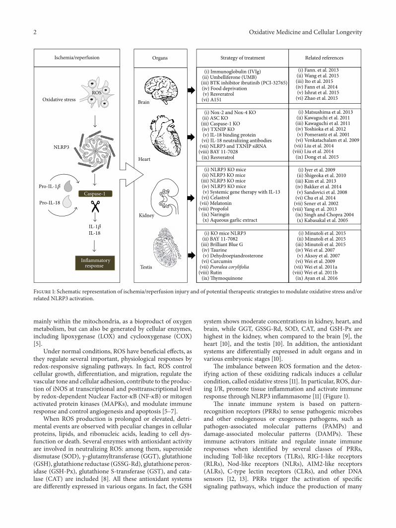
2 Oxidative Medicine and Cellular Longevity
ROS
Strategy of treatmentIschemia/reperfusion Organs Related references
BrainOxidative stress
NLRP3
Heart
Testis
Kidney
Caspase-1
Inflammatory response
Pro-IL-18
Pro-IL-1𝛽
IL-1𝛽IL-18
(i) Immunoglobulin (IVIg)(ii) Umbelliferone (UMB)
(iii) BTK inhibitor ibrutinib (PCI-32765)(iv) Food deprivation(v) Resveratrol
(vi) A151
(i) Nox-2 and Nox-4 KO(ii) ASC KO
(iii) Caspase-1 KO(iv) TXNIP KO(v) IL-18 binding protein
(vi) IL-18 neutralizing antibodies(vii) NLRP3 and TXNIP siRNA
(viii) BAY 11-7028(ix) Resveratrol
(i) NLRP3 KO mice(ii) NLRP3 KO mice
(iii) NLRP3 KO mice(iv) NLRP3 KO mice(v) Systemic gene therapy with IL-13
(vi) Celastrol(vii) Melatonin
(viii) Propofol(ix) Naringin(x) Aqueous garlic extract
(i) KO mice NLRP3(ii) BAY 11-7082
(iii) Brilliant Blue G(iv) Taurine(v) Dehydroepiandrosterone
(vi) Curcumin(vii) Psoralea corylifolia
(viii) Rutin(ix) Thymoquinone
(i) Fann. et al. 2013(ii) Wang et al. 2015(iii) Ito et al. 2015(iv) Fann et al. 2014(v) Ishrat et al. 2015
(vi) Zhao et al. 2015
(i) Matsushima et al. 2013(ii) Kawaguchi et al. 2011(iii) Kawaguchi et al. 2011(iv) Yoshioka et al. 2012(v) Pomerantz et al. 2001
(vi) Venkatachalam et al. 2009(vii) Liu et al. 2014
(viii) Liu et al. 2014(ix) Dong et al. 2015
(i) Iyer et al. 2009(ii) Shigeoka et al. 2010
(iii) Kim et al. 2013(iv) Bakker et al. 2014(v) Sandovici et al. 2008
(vi) Chu et al. 2014(vii) Sener et al. 2002
(viii) Yang et al. 2013(ix) Singh and Chopra 2004(x) Kabasakal et al. 2005
(i) Minutoli et al. 2015(ii) Minutoli et al. 2015
(iii) Minutoli et al. 2015(iv) Wei et al. 2007(v) Aksoy et al. 2007
(vi) Wei et al. 2009(vii) Wei et al. 2011a
(viii) Wei et al. 2011b(ix) Ayan et al. 2016
Figure 1: Schematic representation of ischemia/reperfusion injury and of potential therapeutic strategies to modulate oxidative stress and/orrelated NLRP3 activation.
mainly within the mitochondria, as a bioproduct of oxygenmetabolism, but can also be generated by cellular enzymes,including lipoxygenase (LOX) and cyclooxygenase (COX)[5].
Under normal conditions, ROS have beneficial effects, asthey regulate several important, physiological responses byredox-responsive signaling pathways. In fact, ROS controlcellular growth, differentiation, and migration, regulate thevascular tone and cellular adhesion, contribute to the produc-tion of iNOS at transcriptional and posttranscriptional levelby redox-dependent Nuclear Factor-𝜅B (NF-𝜅B) or mitogenactivated protein kinases (MAPKs), and modulate immuneresponse and control angiogenesis and apoptosis [5–7].
When ROS production is prolonged or elevated, detri-mental events are observed with peculiar changes in cellularproteins, lipids, and ribonucleic acids, leading to cell dys-function or death. Several enzymes with antioxidant activityare involved in neutralizing ROS: among them, superoxidedismutase (SOD), 𝛾-glutamyltransferase (GGT), glutathione(GSH), glutathione reductase (GSSG-Rd), glutathione perox-idase (GSH-Px), glutathione S-transferase (GST), and cata-lase (CAT) are included [8]. All these antioxidant systemsare differently expressed in various organs. In fact, the GSH
system shows moderate concentrations in kidney, heart, andbrain, while GGT, GSSG-Rd, SOD, CAT, and GSH-Px arehighest in the kidney, when compared to the brain [9], theheart [10], and the testis [10]. In addition, the antioxidantsystems are differentially expressed in adult organs and invarious embryonic stages [10].
The imbalance between ROS formation and the detox-ifying action of these oxidizing radicals induces a cellularcondition, called oxidative stress [11]. In particular, ROS, dur-ing I/R, promote tissue inflammation and activate immuneresponse through NLRP3 inflammasome [11] (Figure 1).
The innate immune system is based on pattern-recognition receptors (PRRs) to sense pathogenic microbesand other endogenous or exogenous pathogens, such aspathogen-associated molecular patterns (PAMPs) anddamage-associated molecular patterns (DAMPs). Theseimmune activators initiate and regulate innate immuneresponses when identified by several classes of PRRs,including Toll-like receptors (TLRs), RIG-I-like receptors(RLRs), Nod-like receptors (NLRs), AIM2-like receptors(ALRs), C-type lectin receptors (CLRs), and other DNAsensors [12, 13]. PRRs trigger the activation of specificsignaling pathways, which induce the production of many
Oxidative Medicine and Cellular Longevity 3
proinflammatory cytokines and chemokines and presentantigens to the adaptive immune system for long-lastingprotection.
NLRs form a large protein family of intracellular sensors,the members of which share a conserved central nucleotide-binding, oligomerization domain (NOD), a leucine-richrepeat (LRR) region, and a variable N-terminal effectordomain [14, 15]. The family members of inflammasome arenumerous [16], but NLRP3 inflammasome is the best charac-terized.
In a series of experimental models in vitro [17, 18],it was shown that two different steps, controlled by twodifferent mechanisms, are required for the activation ofNLRP3. The former is driven at transcriptional level by NF-𝜅B, whose increased production in I/R injury is activated viaTLR signaling throughMyd88-dependent pathways, with thesubsequent stimulation of interleukin- (IL-) 1𝛽 and IL-18 geneexpression [19, 20]. The latter is induced at posttranscrip-tional level and consists in the activation of NLRP3, drivenby many activators, such as ROS in I/R injury [21].
When activated, the NLRP3 inflammasome is formedby the NLRP3 protein belonging to the family of NLRs, bythe adapter protein apoptosis-associated speck-like protein(ASC), and by the procaspase-1 [15]. The assembly of NLRP3moves procaspase-1 molecules near enough to transformthem into active fragments [21], so that the conversion ofthe immature proinflammatory cytokines IL-1𝛽 and IL-18 totheir active forms is induced [22, 23]. These cytokines theninitiate or amplify diverse downstream signaling pathwaysand drive proinflammatory processes [24], leading to cel-lular damage, such as autophagy and pyroptosis [22]. Theformer is a process of self-degradation of parts of the cellsthrough sequestering of organelles or parts of the cytoplasmand subsequent fusion with lysosomes [25]. Pyroptosis is aprogrammed cell death with loss of the cellular membraneintegrity, differently from apoptosis, associated with IL-1𝛽and IL-18 secretion; for this reason, it is considered aninflammatory form of cell death [26]. NLRP3 activation isable to promote also the initiation and the progression ofdifferent autoimmune and autoinflammatory diseases, suchas metabolic disorders, inflammatory bowel syndrome [15],obesity, and cognitive diseases [27].
The mechanisms driving the activation of NLRP3, thatis, NLRP3 oligomerization, ASC recruitment, and caspase-1 activation, are generally classified as noncanonical andcanonical.The noncanonical pathway involves caspase-4 andcaspase-5 in human cells and caspase-11 in mice and isactivated by the identification of cell wall ligands, such asLPS, from phagocytized bacteria [28].The canonical pathwayis based on the recognition of general cellular stress, suchas the oxidative stress induced by I/R, bacterial toxins,and particulate substances [28]. Recent studies showed thatNLRP3 might sense the presence of ROS produced into thesame cell by normal or malfunctioning mitochondria [29].In particular, it was proposed that increased ROS are sensedby a complex of TRX and TRX-interacting protein (TXNIP)and induce the dissociation of the complex. In normal cells,TXNIP is constitutively connected to and kept in the reducedstate by the ubiquitous TRX [30]. Following an increase
in cellular ROS concentration, this complex dissociates andTXNIP binds to the LRR region of NLRP3, leading to NLRP3activation [31].
Given the growing evidence that NLRP3 inflammasomeactivation is involved in many systemic diseases [32, 33] andconsidering that conflicting data exist with respect to I/Rinjury, the present review was aimed to evaluate the currentknowledge on the role ofNLRP3 in some organs (brain, heart,kidney, and testis) after I/R injury, with particular regard tothe role played by ROS in its activation. Furthermore, as nospecific therapy exists for the prevention or treatment of thegreat mortality andmorbidity associated with I/R, the state ofthe art of the development of therapeutic strategies was alsoevaluated.
2. ROS and NLRP3 in I/R Injury of the Brain
It is well known that reperfusion may exacerbate the braininjury initially caused by ischemia, producing an I/R injury[3]. Among the various underlying mechanisms of stroke,inflammation and oxidative stress are implicated in thepathogenesis of brain I/R, and an adequate regulation ofinflammatory level may play a critical role in the preventionand treatment of stroke [34]. In the last years, the role ofinflammasomes, particularly NLRP3, has been recognized inpostischemic inflammation after stroke. Since the inflamma-some initiates inflammation, modulation of NLRP3 inflam-masome can regulate inflammatory response. However, froma molecular point of view, NLRP3 inflammasome pathwaycan be activated by a variety ofmolecular signaling systems inI/R brain and many mechanisms have not been fully definedyet.
IL-1𝛽 is a crucial contributor to excitotoxic and ischemicbrain injury and central inflammatory responses [35, 36].Specifically, this cytokine is produced during a central ner-vous system disease or after a brain injury by macrophages ormicroglial cells [37] and molecular mediators as mitochon-dria-derived ROS and lysosomal protease cathepsin B arenecessary for the microglial cell production of interleukin-1𝛽[38].
So far, NLRP3 inflammasome dependent responses invitro are linked to an initial stimulus by a pathogen ordamage-associated molecular pattern (DAMP) [18, 39, 40].Recently, it has been shown that DAMPs could induceinflammatory responses through the production of IL-6 andchemokine (C-X-Cmotif) ligand 1 (CXCL1) and the release ofcathepsin B, in the absence of any bacterial infection or prod-ucts in culturedmousemixed glia [41]. Savage and coworkers[41] also revealed that IL-1 production contributed to increaseof IL-6 and CXCL1 levels following cerebral ischemia bymiddle cerebral artery occlusion in mice, confirming thatDAMPs amplify brain inflammation by directly stimulatingproduction of glial derived inflammatory mediators. In thiscontext, acute phase protein serum amyloid A could act asa priming stimulus on glial cells [41]. Together, these dataadd new helpful information on molecular pathways of braininflammation and/or ischemic brain damage that involve glialcells and NLRP3 inflammasome-activating DAMPs, in theabsence of cell priming and in the presence of a relevant
4 Oxidative Medicine and Cellular Longevity
endogenous priming stimulus, leading thus to new interest-ing perspectives from a therapeutic point of view.
So far, a number of recent studies focused on pathophys-iological mechanism and/or potential therapeutic strategyto modulate inflammatory and oxidative stress pathwaysinvolving NLRP3 inflammasome in stroke brain damage(Figure 1).
Indeed, the expression pattern of the NLRP3 inflamma-some in primary cortical neurons subjected to simulatedischemia, in a mouse model of focal ischemic stroke, and inbrain tissue samples from stroke patients has been described[42]. Interestingly, these authors showed that intravenousimmunoglobulin treatment protected brain cells in experi-mental stroke models by a mechanism involving suppressionof NLRP3 activity [42].
Likewise, another interesting paper of Fann et al. [43]reported that 16 hours of food deprivation, daily, for 4months, can attenuate the inflammatory response and tissuedamage following focal ischemic stroke inmice through inhi-bition of NLRP3 inflammasome activity. Moreover, NLRP3deficiency ameliorates neurovascular damage in experimen-tal ischemic stroke [44].
Bruton’s tyrosine kinase (BTK) is a tyrosine kinaseinvolved inNLRP3 inflammasome activation leading, in turn,to caspase-1 activation and mature IL-1𝛽 production in theischemic brain [45]. Interestingly, Ito and coworkers [45]showed that ibrutinib (PCI-32765), a potent BTK inhibitor[46], suppresses NLRP3 inflammasome signal in a focalbrain I/Rmodel. Specifically, ibrutinib exerts neuroprotectiveeffects through the suppression of IL-1𝛽 maturation in infil-trating macrophages and neutrophils in the infarcted area.
Asmentioned before, oxidative stress is a crucial hallmarkin the pathophysiology of brain damage after stroke. Tothis purpose, it has been indicated that ROS are proximalsignals for NLRP3 inflammasome activation in inflamma-tory diseases [47]. Experimental evidences demonstrate thatan increase in ROS concentration following cellular stressleads to TRX oxidation, TXNIP recruitment of NLRP3,and consequent NLRP3 activation [48]. In fact, the actionof curcumin in the hippocampus subjected to glutamateneurotoxicity was recently demonstrated [48]; in vitro andin vivo results showed that curcumin attenuated glutamateneurotoxicity by inhibiting endoplasmic reticulum stress-associated TXNIP/NLRP3 activation via the regulation ofAMP-Activated Protein Kinase and thereby protected thehippocampus from ischemic insult [48].
Umbelliferone (UMB) is a natural compound belongingto the coumarin family with antioxidant properties. Asa matter of fact, in an interesting paper of Wang et al.[49], pretreatment with UMB ameliorated the neurologicaloutcomes, the infarct volume, and the brain edema in arat model of focal cerebral ischemia induced by middlecerebral artery I/R. These results indicate that UMB exertspartly these neuroprotective effects through the inhibitionof TXNIP/NLRP3 signal. Moreover, another study suggeststhat TXNIP plays a critical role in acute ischemic strokebecause it is directly linked to redox imbalance and NLRP3activation. The latter also suggests the importance of theantioxidant effect of resveratrol on the TRX/TXNIP system
inmice subjected to embolicmiddle cerebral artery occlusion[50].
Recently, it has been shown that A151, a synthetic oli-godeoxynucleotide containing multiple telemeric TTAGGGmotifs, reduces ischemic brain damage and NLRP3 mRNAlevels in Stroke-Prone Spontaneously Hypertensive rats sub-mitted to permanent middle cerebral artery occlusion [51].
Collectively, these data strongly confirmed that NLRP3represents a potential therapeutic target in the manage-ment of ischemic stroke. Therefore, an appropriate treat-ment of brain I/R injury with compounds showing anti-inflammatory/antioxidant activity and targeting different andcomplex molecular pathways, also including NLRP3 acti-vation, remains a big therapeutic challenge in translationalmedicine. In fact, it is nowadays difficult to establish theappropriate timing about their use and/or the duration oftreatment to counteract the brain parenchymal damage in thesetting of I/R injury.
3. ROS and NLRP3 in I/R Injury of the Heart
Myocardial I/R injury is a pathological process causingcardiac cells necrosis and apoptosis, in particular when thecoronary perfusion is restored [52].
A large number of studies have demonstrated anincreased ROS formation either during the ischemic phaseor during the reperfusion period. Excessive ROS induce cellinjury by disrupting cellular signaling transduction, activat-ing inflammation factors, and inducing lipid peroxidation[53] and even cell death [54]. Additionally, ROS have beenidentified as an important NLRP3 inflammasome activator incardiac diseases [55].
When activated, NLRP3 forms an inflammasome com-plex with the adaptor molecule ASC, thus controlling theactivation of caspase-1; the latter cleaves pro-IL-1𝛽 and pro-IL-18 into the biologically active forms, thus initiating thesterile inflammatory disease [56]. In a recent work [52], KOmice for NLRP3 showed larger infarct size than wild type,so a protective role of NLRP3 inflammasome was suggested.However, the results of the study were critically discussed anddisproved by Toldo et al. [57].
IL-1𝛽 and ASC are key players in I/R injury as theyare important and early mediators of the inflammatoryresponse in myocardial I/R injury (Figure 1). In fact, ASCdeletion and IL-1𝛽 inhibition protect the myocardium fromI/R injury in mice [58, 59]. In myocardial I/R injury, animportant role is played also by IL-18, whose expression isstimulated in cardiomyocytes by ROS [60]. Specifically, IL-18 may induce myocardial injury through the induction ofinflammation, increased apoptosis, and changes in calciumoverload [61]. The administration of human myocardiocytesof IL-18 binding protein, a potent inhibitor of IL-18 activity,improved contractile function [58] and showed a protectiverole in the cardiac inflammatory response against I/R injuryin mice [62]. Similarly, the treatment of mice with IL-18neutralizing antibodies prior to I/R injury reduced the infarctsize [60].
As to the mechanisms by which ROS induce inflamma-some activation in the heart, many doubts are still present.
Oxidative Medicine and Cellular Longevity 5
TXNIP is ubiquitously expressed in normal tissues and is anendogenous inhibitor of TRX as, when directly connected,it prevents TRX activity to scavenge ROS [63]. Therefore,TXNIP KO mice showed a protection from I/R injury incardiomyocytes [64]. However, the mechanism by whichTXNIP mediates cardiac injury is still not clear. Recently, inmyocardial I/R injury a role of TXNIP in the activation ofthe NLRP3 inflammasome was proposed through a directinteraction in cardiac microvascular endothelial cells, afterintramyocardial administration ofNLRP3 andTXNIP siRNAand of BAY 11-7028, an inflammasome inhibitor [63] (Fig-ure 1).
Many clinical studies examined the role of antioxidantsin ROS-mediated I/R injury through the administration ofantioxidant drugs after thrombolysis, but the results were notpositive in reducing infarct size or enhancing heart function[65]. On the contrary, inmiceKO forNADPHoxidases (Nox)2 and 4, contributing to part of ROS production during I/Rinjury, a reduction in ROS production and a decrease of theinfarct size were observed after I/R [66]. However, it wassuggested that a total inhibition of Nox is not positive as itis involved in the physiological and beneficial production ofROS [66]. Among antioxidant drugs, resveratrol protects theheart during I/R injury by inhibition of NALP3 inflamma-some and ROS production [67] (Figure 1).
4. ROS and NLRP3 in I/R Injury of the Kidney
I/R injury is one of the common causes of acute renalfailure, thus playing a significant impact on patient morbidityand mortality [33]. Kidneys are particularly vulnerable toischemia; therefore, I/R injury may cause early graft rejectionin renal transplantation and induce structural damage aftersuprarenal aneurysm repair, renal artery reconstruction, con-trast agent-induced nephropathy, cardiac arrest, and shock[68]. Even if ischemia initiates a complex, organized seriesof events, resulting in damage and death of renal cells due tothe dramatic decrease in oxygen and nutrition, reperfusion,though essential for tissue survival, determines an exacerba-tion of tissue injury and a profound inflammatory response,leading to renal dysfunction [1]. Increasing ischemia timeworsens the histological changes, which are particularlysevere at 24–72 hours after reperfusion.
Epithelial cells, particularly those of the S3 segment of theproximal tubule in the outer renal medulla, are particularlyexposed to both ischemia and reperfusion phases of I/Rinjury, which can lead to acute tubular necrosis [69, 70]; onthe contrary, glomerular vessels degeneration was describedonly in the reperfusion phase [69]. Histopathological eval-uation revealed the presence of extensive vascular dilata-tion, slight interstitial edema, tubular dilatation, tubular cellswelling, brush border, and nuclear loss [71]. Furthermore,I/R injury induces an early infiltration of inflammatorycells, mainly neutrophils [72], in addition to the rapidtubular necrosis determining an acute renal dysfunction [73].However, it was observed that the less I/R-sensitive cells ofthe distal tubules might have a protective role, leading toreepithelialization of the injured tubules and preventing theprogression to chronic kidney disease [74].
There is now substantial evidence that ROS and NLRP3inflammasome have a key role, even if not coincident, in theprimary mechanism through which I/R induces the aboveindicated kidney damage.
In fact, Iyer et al. [75] showed that nonlethal renal I/Rinjury resulted in a significant upregulation of NLRP3 geneexpression, which was accompanied by pronounced acutetubular necrosis. It was suggested that specific forms ofcellular injury result in the release of viablemitochondria intothe extracellular space, triggering the activation of theNLRP3inflammasome, in part through the release of ATP.
Furthermore, upregulation of NLRP3 activates caspase-1 and, subsequently, IL-1𝛽 and IL-18; in addition, caspase-1induces pyroptosis, a proinflammatory form of programmedcell death, characterized by pores in the plasma membrane atearly time points [33].
Several pharmacological approaches were proposed toreduce the functional and morphological damage inducedby I/R in kidney. In particular, the pathways of oxidativestress and NLRP3 inflammasome were examined by theadministration of antioxidant drugs or by the study of NLRP3knock-out (KO) animals (Figure 1).
As to the drugs acting on the ROS production and/orscavenging in the kidney, the pretreatment with the pinealhormone melatonin had a protective effect against oxidativedamage caused by free radicals in a number of modelsboth in vivo and in vitro of I/R injury: in particular, whenadministered prior to ischemia and immediately before thereperfusion, melatonin reduced the renal structural changesand limited the neutrophils infiltration [69].
Similar results were obtained from the pretreatmentwith naringin [76], aqueous garlic extract [77], rutin [78],propofol [79], celastrol [71], and allopurinol and apocynin,administered alone or together [80]; all these therapeuticstrategies were based on the antioxidant properties of thesesubstances, which are able to positively act on the exaggeratedinflammatory responses and tissue damage dependent onthe free radicals production and the inflammatory cellsinfiltration [53].
The pretreatment with modified adenovirus expressingIL-13, which is known to display antioxidant properties,diminished renal tubulointerstitial damage and inflammationinduced by I/R [81].
Important data on the mechanisms involved in the I/Rof kidney were obtained from the study of KO animals. Infact, in IL-18 KO mice, a reduced tubular damage and higherprotection against I/R injury [82] were observed. In NLRP3KOmice, an increased proliferation of tubular epithelial cellswas observed, thus indicating that NLRP3 is detrimentalto the repair response after the reperfusion phase of theI/R injury [83]. Similarly, in NLRP3 KO mice following I/Rinjury, an increased protection from lethal ischemic injury[56] and a reduced tubular necrosis and apoptosis, withconsequent repopulation of the tubular epithelium, wereobserved after the reperfusion phase [84]. On the contrary,when ASC KO mice were examined, the protection fromlethal ischemic injury was less pronounced [32, 75] and thetubular necrosis and apoptosis were higher and similar tothose observed in WT mice [84]. It was evident that, at least
6 Oxidative Medicine and Cellular Longevity
in the kidney, the NLRP3 protein, the essential component ofthe inflammasome, may play an independent role in injurysignaling, different from that of the other components of theinflammasome (ASC, caspase-1).
5. ROS and NLRP3 in I/R Injury of the Testis
Testis torsion is a testicular lesion typical of the pediatricpopulation and it is representative of the I/R injury observedin other organs [1, 85]. Under these circumstances, thetestis produces several proinflammatory cytokines, whoseincreased levels can be considered an indirect evidence oftissue inflammation [86].
In normal conditions, enzymatic antioxidant defensesystems, such as SOD, GPx, and CAT, protect testicularsomatic and germinal cells from free radical damage. On thecontrary, malondialdehyde (MDA) is an important indicatorof lipid peroxidation induced by ROS [87].
I/R induces early tissue injuries, such as reactive oxygenspecies (ROS) generation [4], and a damaged barrier func-tion of endothelial cells, resulting from reduced adenylatecyclase activity and intracellular cAMP levels. In particular,during I/R, nucleotides in the form of ATP stimulate tissueinflammation and trigger NLRP3 inflammasome [88]. Thispathological cascade causes a decreased number of germ cellsinduced by an enhanced apoptosis, the vacuolization of theseminiferous epithelium, a reduced number of spermatozoa,and a recruitment of neutrophils [89]; at a later stage, testicu-lar atrophy and impaired spermatogenesis are observed [90–92].
In order to prevent testicular I/R injury, the effects ofdifferent substances with antioxidant activities and the roleof NLRP3 inflammasome have been investigated (Figure 1).
Among antioxidant substances, lipoic acid (LA) hasROS scavenging and metal chelating ability and regeneratesendogenous antioxidants, such as glutathione and vitamins Eand C [87]. The pretreatment with LA induced an increaseof SOD and GPx activity, reduced MDA levels, and abatedcellular damage.
Similar results have been obtained by the pre- orposttreatment with different antioxidant pharmacologicalapproaches, including taurine [93], dehydroepiandrosterone[94], curcumin [95], Psoralea corylifolia [96], rutin [97], thy-moquinone [98], and apocynin [99].
As to the role of NLRP3 inflammasome, specificinhibitors, such as BAY 11-7082 [100] and Brilliant Blue G(BBG) [101], are able to inhibit its effects in a testicular I/Rmodel [102]. In fact, BAY 11-7082, an I-𝜅B kinase-𝛽 inhibitor,and BBG, blocking the membrane-bound purinergic P2X7receptor, showed a significant reduction of IL-1𝛽 andIL-18 mRNA expression, blunted caspase-1 and caspase-3expression, minor histological damage, low TUNEL activity,and preserved spermatogenesis, indicating a selectivelyreduced NLRP3 inflammasome activity [102]. It was alsoobserved that NLRP3 KO mice responded to I/R insult witha lower activation of the inflammatory and apoptosis cascadethan WT animals.
Therefore, NLRP3 can be considered an interesting targetfor innovative drugs aimed at treating I/R injury after testic-ular torsion.
6. Conclusions
The restoration of blood flow as soon as possible is withoutany doubt the primary therapeutic approach to ischemia,even if reperfusion, although essential to restore oxygen andnutrients supply and to remove potentially harmful prod-ucts of cellular metabolism, can induce further pathologicalprocesses in the same organ and tissue injuries in otherorgans.
The main points to be stressed as a conclusion of thisreview can be summarized as follows:
(i) ROS have beneficial effects, as they regulate severalimportant, physiological responses by redox-respon-sive signaling pathways, but, during I/R, they promotetissue inflammation and activate immune responsethrough different pathways, includingNLRP3 inflam-masome.
(ii) Not all organs demonstrate equal sensitivity toischemia [2], the brain being the most sensitive toreductions in its blood supply; all organs demonstratesimilar sensitivity to reperfusion injury, whose keyevents are inflammation and oxidative stress.
(iii) The effects of ROS-mediated NLRP3 inflammasomeactivation in course of I/R injury in other experi-mental models are strongly suggested; in fact, by theexamination of the existing literature, no data on limb,intestine, and ovary ischemia are present.
(iv) In some of the already available experimental models,such as the heart, the testis, and the kidney, furtherstudies using KO mice (NLRP3 or ASC) or antag-onists of the NLRP3 cascade are needed to betterunderstand the physiopathological events during I/Rinjury.
In conclusion, despite the increased literature of the pastdecade, a definite comprehension of the role of NLRP3inflammasome in the host responses to different danger sig-nals is still lacking. A detailed examination of the molecularmechanisms driving NLRP3 inflammasome transcription,assembly, and activation is needed to elucidate these pro-cesses in the different organs. This experimental approach,given the role of NLRP3 in several sterile inflammatorydiseases, could be the basis for the design and elaborationof novel NLRP3 inflammasome inhibitors, thus avoidingthe exclusive use of substances with antioxidant activity inpatients with ischemic damage.
Abbreviations
I/R: Ischemia and reperfusionROS: Reactive oxygen speciesNLRP3: NOD-like receptor family pyrin domain
containing 3
Oxidative Medicine and Cellular Longevity 7
LOX: LipoxygenaseCOX: CyclooxygenaseTRX: ThioredoxinCAT: CatalasePRRs: Pattern-recognition receptorsPAMPs: Pathogen-associated molecular patternsDAMPs: Damage-associated molecular patternsTLRs: Toll-like receptorsRLRs: RIG-I-like receptorsNF-𝜅B: Nuclear Factor kappa-light-chain-enhancer
of activated B cellsNLRs: Nod-like receptorsALRs: AIM2-like receptorsCLRs: C-type lectin receptorsIL-1𝛽: Interleukin-1𝛽IL-18: Interleukin-18TXNIP: TRX-interacting proteinSOD: Superoxide dismutaseGPx: Glutathione peroxidaseMDA: MalondialdehydeKO: Knock-outBBG: Brilliant Blue G.
Competing Interests
The authors declare that there are no competing interestsregarding the publication of this paper.
References
[1] H. K. Eltzschig and T. Eckle, “Ischemia and reperfusion—frommechanism to translation,” Nature Medicine, vol. 17, no. 11, pp.1391–1401, 2011.
[2] T. Kalogeris, C. P. Baines, M. Krenz, and R. J. Korthuis, “Cellbiology of ischemia/reperfusion injury,” International Review ofCell and Molecular Biology, vol. 298, pp. 229–317, 2012.
[3] D. L. Carden and D. N. Granger, “Pathophysiology ofischaemia-reperfusion injury,”TheJournal of Pathology, vol. 190,no. 3, pp. 255–266, 2000.
[4] X. Lei, H. Chao, Z. Zhang et al., “Neuroprotective effects ofquercetin in a mouse model of brain ischemic/reperfusioninjury via anti-apoptotic mechanisms based on the Akt path-way,”Molecular Medicine Reports, vol. 12, no. 3, pp. 3688–3696,2015.
[5] F. Martinon, “Signaling by ROS drives inflammasome activa-tion,” European Journal of Immunology, vol. 40, no. 3, pp. 616–619, 2010.
[6] P. D. Ray, B.-W. Huang, and Y. Tsuji, “Reactive oxygen species(ROS) homeostasis and redox regulation in cellular signaling,”Cellular Signalling, vol. 24, no. 5, pp. 981–990, 2012.
[7] M. Valko, D. Leibfritz, J. Moncol, M. T. D. Cronin, M. Mazur,and J. Telser, “Free radicals and antioxidants in normal physi-ological functions and human disease,” International Journal ofBiochemistry and Cell Biology, vol. 39, no. 1, pp. 44–84, 2007.
[8] J. Limón-Pacheco and M. E. Gonsebatt, “The role of an-tioxidants and antioxidant-related enzymes in protectiveresponses to environmentally induced oxidative stress,”Mutation Research/Genetic Toxicology and EnvironmentalMutagenesis, vol. 674, no. 1-2, pp. 137–147, 2009.
[9] R. Dringen, “Metabolism and functions of glutathione in brain,”Progress in Neurobiology, vol. 62, no. 6, pp. 649–671, 2000.
[10] J. Jurado, M.-J. Prieto-Álamo, J. Madrid-Rı́squez, and C.Pueyo, “Absolute gene expression patterns of thioredoxin andglutaredoxin redox systems in mouse,”The Journal of BiologicalChemistry, vol. 278, no. 46, pp. 45546–45554, 2003.
[11] C. Bryant and K. A. Fitzgerald, “Molecular mechanismsinvolved in inflammasome activation,” Trends in Cell Biology,vol. 19, no. 9, pp. 455–464, 2009.
[12] C. Bourgeois and K. Kuchler, “Fungal pathogens-a sweet andsour treat for toll-like receptors,” Frontiers in Cellular andInfection Microbiology, vol. 2, p. 142, 2012.
[13] J. Cui, Y. Chen, H. Y. Wang, and R.-F. Wang, “Mechanisms andpathways of innate immune activation and regulation in healthand cancer,”HumanVaccines& Immunotherapeutics, vol. 10, no.11, pp. 3270–3285, 2014.
[14] F. Martinon, K. Burns, and J. Tschopp, “The inflammasome:a molecular platform triggering activation of inflammatorycaspases and processing of proIL-𝛽,”Molecular Cell, vol. 10, no.2, pp. 417–426, 2002.
[15] B. Z. Shao, Z. Q. Xu, B. Z. Han, D. F. Su, and C. Liu,“NLRP3 inflammasome and its inhibitors: a review,” Frontiersin Pharmacology, vol. 6, article 262, 2015.
[16] E. Ozaki, M. Campbell, and S. L. Doyle, “Targeting the NLRP3inflammasome in chronic inflammatory diseases: current per-spectives,” Journal of Inflammation Research, vol. 8, pp. 15–27,2015.
[17] J. M. Kahlenberg, K. C. Lundberg, S. B. Kertesy, Y. Qu, and G.R. Dubyak, “Potentiation of caspase-1 activation by the P2X7receptor is dependent on TLR signals and requires NF-𝜅B-driven protein synthesis,” Journal of Immunology, vol. 175, no.11, pp. 7611–7622, 2005.
[18] F. G. Bauernfeind, G. Horvath, A. Stutz et al., “Cutting edge:NF-𝜅B activating pattern recognition and cytokine receptorslicense NLRP3 inflammasome activation by regulating NLRP3expression,” Journal of Immunology, vol. 183, no. 2, pp. 787–791,2009.
[19] H. Zhao, J. S. Perez, K. Lu, A. J. T. George, and D. Ma, “Role oftoll-like receptor-4 in renal graft ischemia-reperfusion injury,”American Journal of Physiology—Renal Physiology, vol. 306, no.8, pp. F801–F811, 2014.
[20] G. Vilahur and L. Badimon, “Ischemia/reperfusion activatesmyocardial innate immune response: the key role of the toll-likereceptor,” Frontiers in Physiology, vol. 5, article 496, 2014.
[21] A. Rubartelli, “Redox control of NLRP3 inflammasome activa-tion in health and disease,” Journal of Leukocyte Biology, vol. 92,no. 5, pp. 951–958, 2012.
[22] K. Schroder and J. Tschopp, “The inflammasomes,”Cell, vol. 140,no. 6, pp. 821–832, 2010.
[23] Q. Zheng, Y. Ren, P. S. Reinach et al., “Reactive oxygen speciesactivated NLRP3 inflammasomes prime environment-inducedmurine dry eye,” Experimental Eye Research, vol. 125, pp. 1–8,2014.
[24] L. Niu, S. Zhang, J. Wu, L. Chen, and Y.Wang, “Upregulation ofNLRP3 inflammasome in the tears and ocular surface of dry eyepatients,” PLoS ONE, vol. 10, no. 5, Article ID e0126277, 2015.
[25] C.He andD. J. Klionsky, “Regulationmechanisms and signalingpathways of autophagy,” Annual Review of Genetics, vol. 43, pp.67–93, 2009.
[26] H.-J. Anders and D. A.Muruve, “The inflammasomes in kidneydisease,” Journal of the American Society of Nephrology, vol. 22,no. 6, pp. 1007–1018, 2011.
8 Oxidative Medicine and Cellular Longevity
[27] A. J. S. Choi and S. W. Ryter, “Inflammasomes: molecular reg-ulation and implications for metabolic and cognitive diseases,”Molecules and Cells, vol. 37, no. 6, pp. 441–448, 2014.
[28] J. Rivers-Auty and D. Brough, “Potassium efflux fires the canon:potassium efflux as a common trigger for canonical and non-canonical NLRP3 pathways,” European Journal of Immunology,vol. 45, no. 10, pp. 2758–2761, 2015.
[29] O. Gross, C. J. Thomas, G. Guarda, and J. Tschopp, “Theinflammasome: an integrated view,” Immunological Reviews,vol. 243, no. 1, pp. 136–151, 2011.
[30] J. Tschopp and K. Schroder, “NLRP3 inflammasome activation:the convergence of multiple signalling pathways on ROS pro-duction?” Nature Reviews Immunology, vol. 10, no. 3, pp. 210–215, 2010.
[31] J. Zhou and W.-J. Chng, “Roles of thioredoxin binding protein(TXNIP) in oxidative stress, apoptosis and cancer,” Mitochon-drion, vol. 13, no. 3, pp. 163–169, 2013.
[32] H.-J. Kim, D. W. Lee, K. Ravichandran et al., “NLRP3 Inflam-masome knockout mice are protected against ischemic but notcisplatin-induced acute kidney injury,” Journal of Pharmacologyand Experimental Therapeutics, vol. 346, no. 3, pp. 465–472,2013.
[33] C. M. Turner, N. Arulkumaran, M. Singer, R. J. Unwin, and F.W. Tam, “Is the inflammasome a potential therapeutic target inrenal disease?” BMC Nephrology, vol. 15, article 21, 2014.
[34] M.Ahmad,N. J. Dar, Z. S. Bhat et al., “Inflammation in ischemicstroke: mechanisms, consequences and possible drug targets,”CNS and Neurological Disorders—Drug Targets, vol. 13, no. 8,pp. 1378–1396, 2014.
[35] H. Marini, D. Altavilla, M. Bellomo et al., “Modulation of IL-1𝛽 gene expression by lipid peroxidation inhibition after kainicacid-induced rat brain injury,” Experimental Neurology, vol. 188,no. 1, pp. 178–186, 2004.
[36] B. Fogal and S. J. Hewett, “Interleukin-1𝛽: a bridge betweeninflammation and excitotoxicity?” Journal of Neurochemistry,vol. 106, no. 1, pp. 1–23, 2008.
[37] A. Denes, P.Thornton, N. J. Rothwell, and S. M. Allan, “Inflam-mation and brain injury: acute cerebral ischaemia, peripheral,and central inflammation,” Brain, Behavior, and Immunity, vol.24, no. 5, pp. 708–723, 2010.
[38] R. von Bernhardi, L. Eugenı́n-von Bernhardi, and J. Eugenı́n,“Microglial cell dysregulation in brain aging and neurodegen-eration,” Frontiers in Aging Neuroscience, vol. 7, article 124, 2015.
[39] D. Brough, R. A. Le Feuvre, Y. Iwakura, and N. J. Rothwell,“Purinergic (P2X7) receptor activation ofmicroglia induces celldeath via an interleukin-1-independent mechanism,”Molecularand Cellular Neuroscience, vol. 19, no. 2, pp. 272–280, 2002.
[40] V. Hornung and E. Latz, “Critical functions of priming andlysosomal damage for NLRP3 activation,” European Journal ofImmunology, vol. 40, no. 3, pp. 620–623, 2010.
[41] C. D. Savage, G. Lopez-Castejon, A. Denes, and D. Brough,“NLRP3-inflammasome activating DAMPs stimulate aninflammatory response in glia in the absence of priming whichcontributes to brain inflammation after injury,” Frontiers inImmunology, vol. 3, article 288, 2012.
[42] D. Yang-Wei Fann, S.-Y. Lee, S. Manzanero et al., “In-travenous immunoglobulin suppresses NLRP1 and NLRP3inflammasome-mediated neuronal death in ischemic stroke,”Cell Death & Disease, vol. 4, no. 9, article e790, 2013.
[43] D. Y.-W. Fann, T. Santro, S. Manzanero et al., “Intermittentfasting attenuates inflammasome activity in ischemic stroke,”Experimental Neurology, vol. 257, pp. 114–119, 2014.
[44] F. Yang, Z. Wang, X. Wei et al., “NLRP3 deficiency amelioratesneurovascular damage in experimental ischemic stroke,” Jour-nal of Cerebral Blood Flow&Metabolism, vol. 34, no. 4, pp. 660–667, 2014.
[45] M. Ito, T. Shichita, M. Okada et al., “Bruton’s tyrosine kinase isessential for NLRP3 inflammasome activation and contributesto ischaemic brain injury,” Nature Communications, vol. 6,article 7360, 2015.
[46] A. F. Herrera and E. D. Jacobsen, “Ibrutinib for the treatment ofmantle cell lymphoma,” Clinical Cancer Research, vol. 20, no. 21,pp. 5365–5371, 2014.
[47] A. Abderrazak, T. Syrovets, D. Couchie et al., “NLRP3 inflam-masome: from a danger signal sensor to a regulatory node ofoxidative stress and inflammatory diseases,” Redox Biology, vol.4, pp. 296–307, 2015.
[48] Y. Li, J. Li, S. Li et al., “Curcumin attenuates glutamate neurotox-icity in the hippocampus by suppression of ER stress-associatedTXNIP/NLRP3 inflammasome activation in a manner depen-dent onAMPK,”Toxicology andApplied Pharmacology, vol. 286,no. 1, pp. 53–63, 2015.
[49] X.Wang, R. Li, X.Wang, Q. Fu, and S.Ma, “Umbelliferone ame-liorates cerebral ischemia-reperfusion injury via upregulatingthe PPAR gamma expression and suppressing TXNIP/NLRP3inflammasome,” Neuroscience Letters, vol. 600, pp. 182–187,2015.
[50] T. Ishrat, I. N. Mohamed, B. Pillai et al., “Thioredoxin-interacting protein: a novel target for neuroprotection in exper-imental thromboembolic stroke in mice,”Molecular Neurobiol-ogy, vol. 51, no. 2, pp. 766–778, 2015.
[51] J. Zhao, Y. Mou, J. D. Bernstock et al., “Synthetic oligodeoxynu-cleotides containing multiple telemeric TTAGGG motifs sup-press inflammasome activity in macrophages subjected tooxygen and glucose deprivation and reduce ischemic braininjury in stroke-prone spontaneously hypertensive rats,” PLoSONE, vol. 10, no. 10, Article ID e0140772, 2015.
[52] Ø. Sandanger, E. Gao, T. Ranheim et al., “NLRP3 inflammasomeactivation during myocardial ischemia reperfusion is cardio-protective,” Biochemical and Biophysical Research Communica-tions, vol. 469, no. 4, pp. 1012–1020, 2016.
[53] S. M. Davidson, D.M. Yellon, M. P. Murphy, andM. R. Duchen,“Slow calcium waves and redox changes precede mitochondrialpermeability transition pore opening in the intact heart duringhypoxia and reoxygenation,” Cardiovascular Research, vol. 93,no. 3, pp. 445–453, 2012.
[54] K. Zhan, P. Yu, C. Liu, J. Luo, and W. Yang, “Detrimental orbeneficial: the role of TRPM2 in ischemia/reperfusion injury,”Acta Pharmacologica Sinica, vol. 37, no. 1, pp. 4–12, 2016.
[55] Y. Wang, Y. Wu, J. Chen, S. Zhao, and H. Li, “Pirfenidone atten-uates cardiac fibrosis in a mouse model of TAC-induced leftventricular remodeling by suppressing NLRP3 Inflammasomeformation,” Cardiology, vol. 126, no. 1, pp. 1–11, 2013.
[56] M. Takahashi, “Role of the inflammasome inmyocardial infarc-tion,”Trends in CardiovascularMedicine, vol. 21, no. 2, pp. 37–41,2011.
[57] S. Toldo, C. Marchetti, and A. Abbate, “Re. ‘NLRP3 inflam-masome activation during myocardial ischemia reperfusion iscardioprotective’,” Biochemical and Biophysical Research Com-munications, vol. 470, no. 4, pp. 811–812, 2016.
[58] B. J. Pomerantz, L. L. Reznikov, A. H. Harken, and C. A.Dinarello, “Inhibition of caspase 1 reduces human myocardial
Oxidative Medicine and Cellular Longevity 9
ischemic dysfunction via inhibition of IL-18 and IL-1𝛽,” Pro-ceedings of the National Academy of Sciences of the United Statesof America, vol. 98, no. 5, pp. 2871–2876, 2001.
[59] M. Kawaguchi, M. Takahashi, T. Hata et al., “Inflammasomeactivation of cardiac fibroblasts is essential for myocardialischemia/reperfusion injury,” Circulation, vol. 123, no. 6, pp.594–604, 2011.
[60] K. Venkatachalam, S. D. Prabhu, V. S. Reddy, W. H. Boyl-ston, A. J. Valente, and B. Chandrasekar, “Neutralizationof interleukin-18 ameliorates ischemia/reperfusion-inducedmyocardial injury,”The Journal of Biological Chemistry, vol. 284,no. 12, pp. 7853–7865, 2009.
[61] M. Wang, T. A. Markel, and D. R. Meldrum, “Interleukin 18 inthe heart,” Shock, vol. 30, no. 1, pp. 3–10, 2008.
[62] H. Gu, M. Xie, L. Xu, X. Zheng, Y. Yang, and X. Lv, “The protec-tive role of interleukin-18 binding protein in a murine model ofcardiac ischemia/reperfusion injury,” Transplant International,vol. 28, no. 12, pp. 1436–1444, 2015.
[63] Y. Liu, K. Lian, L. Zhang et al., “TXNIP mediates NLRP3inflammasome activation in cardiac microvascular endothelialcells as a novel mechanism in myocardial ischemia/reperfusioninjury,” Basic Research in Cardiology, vol. 109, no. 5, p. 415, 2014.
[64] J. Yoshioka, W. A. Chutkow, S. Lee et al., “Deletion ofthioredoxin-interacting protein in mice impairs mitochon-drial function but protects the myocardium from ischemia-reperfusion injury,”The Journal of Clinical Investigation, vol. 122,no. 1, pp. 267–279, 2012.
[65] D. M. Yellon and D. J. Hausenloy, “Myocardial reperfusioninjury,” The New England Journal of Medicine, vol. 357, no. 11,pp. 1074–1135, 2007.
[66] S. Matsushima, J. Kuroda, T. Ago et al., “Broad suppressionof NADPH oxidase activity exacerbates ischemia/reperfusioninjury through inadvertent downregulation of hypoxia-inducible factor-1𝛼 and upregulation of peroxisome prolifera-tor-activated receptor-𝛼,” Circulation Research, vol. 112, no. 8,pp. 1135–1149, 2013.
[67] W. Dong, R. Yang, J. Yang et al., “Resveratrol pretreatmentprotects rat hearts from ischemia/reperfusion injury partlyvia a NALP3 inflammasome pathway,” International Journal ofClinical and Experimental Pathology, vol. 8, no. 8, pp. 8731–8741,2015.
[68] L. F. Tirapelli, D. F. Barione, B. F.M.Trazzi et al., “Comparison oftwomodels for evaluation histopathology of experimental renalischemia,” Transplantation Proceedings, vol. 41, no. 10, pp. 4083–4087, 2009.
[69] G. Sener, A. Ö. Sehirli, M. Keyer-Uysal, S. Arbak, Y. Ersoy,and B. Ç. Yeğen, “The protective effect of melatonin onrenal ischemia-reperfusion injury in the rat,” Journal of PinealResearch, vol. 32, no. 2, pp. 120–126, 2002.
[70] R. W. Schrier, W. Wang, B. Poole, and A. Mitra, “Acute renalfailure: definitions, diagnosis, pathogenesis, and therapy,” TheJournal of Clinical Investigation, vol. 114, no. 1, pp. 5–14, 2004.
[71] C. Chu,W.He, Y. Kuang, K. Ren, andX.Gou, “Celastrol protectskidney against ischemia-reperfusion-induced injury in rats,”Journal of Surgical Research, vol. 186, no. 1, pp. 398–407, 2014.
[72] J. J. Friedewald and H. Rabb, “Inflammatory cells in ischemicacute renal failure,”Kidney International, vol. 66, no. 2, pp. 486–491, 2004.
[73] J. V. Bonventre and A. Zuk, “Ischemic acute renal failure: aninflammatory disease?” Kidney International, vol. 66, no. 2, pp.480–485, 2004.
[74] G. C. Gobe and D.W. Johnson, “Distal tubular epithelial cells ofthe kidney: potential support for proximal tubular cell survivalafter renal injury,” International Journal of Biochemistry and CellBiology, vol. 39, no. 9, pp. 1551–1561, 2007.
[75] S. S. Iyer, W. P. Pulskens, J. J. Sadler et al., “Necrotic cellstrigger a sterile inflammatory response through the Nlrp3inflammasome,”Proceedings of theNational Academy of Sciencesof theUnited States of America, vol. 106, no. 48, pp. 20388–20393,2009.
[76] D. Singh and K. Chopra, “The effect of naringin, a bioflavonoidon ischemia-reperfusion induced renal injury in rats,” Pharma-cological Research, vol. 50, no. 2, pp. 187–193, 2004.
[77] L. Kabasakal, Ö. Şehirli, Ş. Çetinel, E. Cikler, N. Gedik, and G.Şener, “Protective effect of aqueous garlic extract against renalischemia/reperfusion injury in rats,” Journal of Medicinal Food,vol. 8, no. 3, pp. 319–326, 2005.
[78] A. Korkmaz andD. Kolankaya, “Protective effect of rutin on theischemia/reperfusion induced damage in rat kidney,” Journal ofSurgical Research, vol. 164, no. 2, pp. 309–315, 2010.
[79] S. Yang, W.-P. Chou, and L. Pei, “Effects of propofol onrenal ischemia/reperfusion injury in rats,” Experimental andTherapeutic Medicine, vol. 6, no. 5, pp. 1177–1183, 2013.
[80] E. K. Choi, H. Jung, K. H. Kwak et al., “Effects of allopurinoland apocynin on renal ischemia-reperfusion injury in rats,”Transplantation Proceedings, vol. 47, no. 6, pp. 1633–1638, 2015.
[81] M. Sandovici, R. H. Henning, H. van Goor, W. Helfrich, D.de Zeeuw, and L. E. Deelman, “Systemic gene therapy withinterleukin-13 attenuates renal ischemia-reperfusion injury,”Kidney International, vol. 73, no. 12, pp. 1364–1373, 2008.
[82] H. Wu, M. L. Craft, P. Wang et al., “IL-18 contributes to renaldamage after ischemia-reperfusion,” Journal of the AmericanSociety of Nephrology, vol. 19, no. 12, pp. 2331–2341, 2008.
[83] P. J. Bakker, L. M. Butter, N. Claessen et al., “A tissue-specific role for Nlrp3 in tubular epithelial repair after renalischemia/reperfusion,” The American Journal of Pathology, vol.184, no. 7, pp. 2013–2022, 2014.
[84] A. A. Shigeoka, J. L. Mueller, A. Kambo et al., “Aninflammasome-independent role for epithelial-expressedNlrp3 in renal ischemia-reperfusion injury,” The Journal ofImmunology, vol. 185, no. 10, pp. 6277–6285, 2010.
[85] L. Minutoli, P. Antonuccio, F. Polito et al., “Mitogen-activatedprotein kinase 3/mitogen-activated protein kinase 1 activatesapoptosis during testicular ischemia-reperfusion injury in anuclear factor-𝜅B-independent manner,” European Journal ofPharmacology, vol. 604, no. 1–3, pp. 27–35, 2009.
[86] J. J. Lysiak, “The role of tumor necrosis factor-alpha andinterleukin-1 in the mammalian testis and their involvementin testicular torsion and autoimmune orchitis,” ReproductiveBiology and Endocrinology, vol. 2, article 9, 2004.
[87] S. Ozbal, B. U. Ergur, G. Erbil, I. Tekmen, A. Bagryank, and Z.Cavdar, “The effects of 𝛼-lipoic acid against testicular ischemia-reperfusion injury in rats,” The Scientific World Journal, vol.2012, Article ID 489248, 8 pages, 2012.
[88] B. McDonald, K. Pittman, G. B. Menezes et al., “Intravasculardanger signals guide neutrophils to sites of sterile inflamma-tion,” Science, vol. 330, no. 6002, pp. 362–366, 2010.
[89] J. G. Reyes, J. G. Farias, S. Henŕıquez-Olavarrieta et al., “Thehypoxic testicle: physiology and pathophysiology,” OxidativeMedicine and Cellular Longevity, vol. 2012, Article ID 929285,15 pages, 2012.
10 Oxidative Medicine and Cellular Longevity
[90] P. Antonuccio, L. Minutoli, C. Romeo et al., “Lipid peroxida-tion activates mitogen-activated protein kinases in testicularischemia-reperfusion injury,” The Journal of Urology, vol. 176,no. 4, part 1, pp. 1666–1672, 2006.
[91] A. K. Srinivasan, J. Freyle, J. S. Gitlin, and L. S. Palmer, “Climaticconditions and the risk of testicular torsion in adolescentmales,”The Journal of Urology, vol. 178, no. 6, pp. 2585–2588, 2007.
[92] L. Minutoli, P. Antonuccio, F. Squadrito et al., “Effects ofpolydeoxyribonucleotide on the histological damage and thealtered spermatogenesis induced by testicular ischaemia andreperfusion in rats,” International Journal of Andrology, vol. 35,no. 2, pp. 133–144, 2012.
[93] S.-M. Wei, Z.-Z. Yan, and J. Zhou, “Beneficial effect of taurineon testicular ischemia-reperfusion injury in rats,” Urology, vol.70, no. 6, pp. 1237–1242, 2007.
[94] H. Aksoy, T. Yapanoglu, Y. Aksoy, I. Özbey, H. Turhan, and N.Gursan, “Dehydroepiandrosterone treatment attenuates reper-fusion injury after testicular torsion and detorsion in rats,”Journal of Pediatric Surgery, vol. 42, no. 10, pp. 1740–1744, 2007.
[95] S.-M. Wei, Z.-Z. Yan, and J. Zhou, “Curcumin attenuatesischemia-reperfusion injury in rat testis,” Fertility and Sterility,vol. 91, no. 1, pp. 271–277, 2009.
[96] S.-M. Wei, Z.-Z. Yan, and J. Zhou, “Psoralea corylifolia protectsagainst testicular torsion/detorsion-induced ischemia/reperfu-sion injury,” Journal of Ethnopharmacology, vol. 137, no. 1, pp.568–574, 2011.
[97] S.-M. Wei, Z.-Z. Yan, and J. Zhou, “Protective effect of rutinon testicular ischemia-reperfusion injury,” Journal of PediatricSurgery, vol. 46, no. 7, pp. 1419–1424, 2011.
[98] M. Ayan, U. Tas, E. Sogut et al., “Protective effect of thymo-quinone against testicular torsion induced oxidative injury,”Andrologia, vol. 48, no. 2, pp. 143–151, 2016.
[99] T. E. Şener, M. Yüksel, N. Özyılmaz-Yay et al., “Apocynin atten-uates testicular ischemia-reperfusion injury in rats,” Journal ofPediatric Surgery, vol. 50, no. 8, pp. 1382–1387, 2015.
[100] C. Juliana, T. Fernandes-Alnemri, J. Wu et al., “Anti-inflam-matory compounds parthenolide and bay 11-7082 are directinhibitors of the inflammasome,” Journal of Biological Chem-istry, vol. 285, no. 13, pp. 9792–9802, 2010.
[101] J. Zhao, H. Wang, C. Dai et al., “P2X7 blockade attenu-ates murine lupus nephritis by inhibiting activation of theNLRP3/ASC/caspase 1 pathway,” Arthritis & Rheumatism, vol.65, no. 12, pp. 3176–3185, 2013.
[102] L. Minutoli, P. Antonuccio, N. Irrera et al., “NLRP3 Inflam-masome involvement in the organ damage and impaired sper-matogenesis induced by testicular ischemia and reperfusion inmice,” Journal of Pharmacology and Experimental Therapeutics,vol. 355, no. 3, pp. 370–380, 2015.
Research ArticleAntioxidant Effects of Sheep Whey Protein on Endothelial Cells
Efthalia Kerasioti, Dimitrios Stagos, Vasiliki Georgatzi, Erinda Bregou,Alexandros Priftis, Ioannis Kafantaris, and Dimitrios Kouretas
Department of Biochemistry and Biotechnology, University of Thessaly, Ploutonos 26 & Aiolou, 41221 Larissa, Greece
Correspondence should be addressed to Dimitrios Kouretas; [email protected]
Received 24 December 2015; Revised 11 March 2016; Accepted 14 March 2016
Academic Editor: Tanea T. Reed
Copyright © 2016 Efthalia Kerasioti et al. This is an open access article distributed under the Creative Commons AttributionLicense, which permits unrestricted use, distribution, and reproduction in any medium, provided the original work is properlycited.
Excessive production of reactive oxygen species (ROS) may cause endothelial dysfunction and consequently vascular disease. Inthe present study, the possible protective effects of sheep whey protein (SWP) from tert-butyl hydroperoxide- (tBHP-) inducedoxidative stress in endothelial cells (EA.hy926) were assessed using oxidative stress biomarkers. These oxidative stress biomarkerswere glutathione (GSH) and ROS levels determined by flow cytometry.Moreover, thiobarbituric acid-reactive substances (TBARS),protein carbonyls (CARB), and oxidized glutathione (GSSG)were determined spectrophotometrically.The results showed that SWPat 0.78, 1.56, 3.12, and 6.24mg of protein mL−1 increased GSH up to 141%, while it decreased GSSG to 46.7%, ROS to 58.5%, TBARSto 52.5%, and CARB to 49.0%. In conclusion, the present study demonstrated for the first time that SWP protected endothelialcells from oxidative stress. Thus, SWP may be used for developing food supplements or biofunctional foods to attenuate vasculardisturbances associated with oxidative stress.
1. Introduction
Free radicals such as reactive oxygen species (ROS) can begenerated in a wide variety of chemical and biological sys-tems. ROS play an important role in body’s immune response[1], redox regulation of gene transcription [2], and cell signal-ing [1]. On the other hand, the ensuing cascade of ROS canresult in cellular damage including apoptosis, protein oxida-tion, DNA modification, and lipid peroxidation [3]. Undernormal conditions ROS are controlled by antioxidant sys-tems. When there is a disturbance between the prooxidantand antioxidant balance in favor of the former that leadsto oxidative stress which can cause damage to all moleculartargets [1], a range of antioxidants are active in the bodyincluding enzymatic and nonenzymatic antioxidants [4].Antioxidant enzymes include superoxide dismutase (SOD),catalase (CAT), and glutathione peroxidase (GPX) [4]. Non-enzymatic antioxidants include vitamin A, vitamin C, vita-min E, flavonoids, glutathione (GSH), uric acid, and bilirubin[5].
The endothelium lines the entire vascular system andis composed of a monolayer of endothelial cells. Endothe-lial cell structure and functional integrity are important in
the maintenance of the vessel wall and circulatory function.In addition to its role as a selective permeability barrier, endo-thelial cells are dynamic and are capable of conducting avariety of metabolic and synthetic functions and regulatinghomeostasis, immune, and inflammatory responses [6]. Endo-thelial cell injury or dysfunction is a hallmark of many patho-logic conditions including atherosclerosis and thrombosis[6]. Excessive production of ROS may exceed the capacity ofantioxidant mechanisms, thus contributing to vascular dis-ease by induction of endothelial dysfunction through severalpathways [6]. Endothelial dysfunction is considered largelyas endothelial activation, which may eventually contributeto arterial disease [6]. Inflammatory cytokines, growth factors,and the interaction of the endothelium with leukocytesmay induce ROS signaling in endothelial cells. Moreover,interaction between ROS and NO may cause a vicious circleleading to more endothelial activation and inflammation [6].In addition, superoxide dismutasemay use superoxide radical(O2
∙−) for producing hydrogen peroxide which can diffuse tothe endothelial cells and damage proteins through reactionwith cysteine groups [7]. Thus, continuous ROS signaling inendothelial cells can cause loss of integrity, progression tosenescence, and detachment into the circulation [8].
Hindawi Publishing CorporationOxidative Medicine and Cellular LongevityVolume 2016, Article ID 6585737, 10 pageshttp://dx.doi.org/10.1155/2016/6585737
2 Oxidative Medicine and Cellular Longevity
Thus, there is a great interest for natural sources of antiox-idants in order to enhance antioxidant mechanisms and pro-tect the organism from the harmful effects of oxidative stress.For example, whey protein is a widely consumed supplementthat is considered to increase the antioxidant defense [9,10]. Whey protein is a by-product of cheese manufacturing,but it is used as a functional food with nutritional applica-tions [11, 12]. The main components of whey include beta-lactoglobulin, alpha-lactalbumin, bovine serum albumin,lactoferrin, immunoglobulins, lactoperoxidase enzymes, gly-comacropeptides, and lactose [13]. Some of these componentsact as antioxidants. For example, alpha-lactalbumin canchelate iron and thus result in the reduction of oxidativestress [14]. Moreover, whey protein has a high content inthe sulphur-containing amino acids cysteine andmethioninethat enhance antioxidant mechanisms through intracellularconversion to glutathione [11].
In our previous studies, we have shown that a cake con-taining sheep whey protein (SWP) had antioxidant and anti-inflammatory activities in subjects submitted to intense exer-cise [9, 15].We have also shown that SWP exerted antioxidanteffects on C2C12 muscle cells [16]. The aim of the presentstudy was to examine the possible protective effects of SWPagainst tert-butyl hydroperoxide- (tBHP-) induced oxidativestress in EA.hy926 endothelial cells.
2. Materials and Methods
2.1. Chemicals, Reagents, and Culture Medium. Dulbecco’smodified Eagle’s medium (DMEM), fetal bovine serum(FBS), phosphate buffered saline (PBS), and L-glutamine andtrypsinwere purchased fromGibco (Grand Island,NY). Tert-butyl hydroperoxide (tBHP), 2,4-dinitrophenylhydrazine(DNPH), urea, oxidized glutathione (GSSG), nicotinamideadenine di-nucleotide phosphate (NADPH), 5,5-dithiobis(2-nitrobenzoic acid) (DTNB), 2-vinyl pyridine, glutathionereductase, ethyl acetate, Bradford reagent, mercury orange,and 2,7-dichlorofluorescein diacetate (DCF-DA) were obtainedfrom Sigma-Aldrich (St. Louis, MO, USA). Trichloroaceticacid (TCA), sodium hydroxide (NaOH), 2-thiobarbituricacid (TBA), and ethanol were purchased fromMerck (Darm-stadt, Germany). Cell proliferation kit II (XTT) was pur-chased from Roche Diagnostics (Mannheim, Germany).
2.2. Cell Culture. EA.hy926 endothelial cells were cultured asdescribed previously in tissue culture flasks at 37∘C in 5%CO
2
[17]. The medium used was DMEM, containing 10% (v/v)FBS, 2mML-glutamine, 100 units mL−1 of penicillin, and 100units mL−1 of streptomycin (Gibco, UK).
2.3. Cell Viability Assay. Cell viability was assessed usingthe XTT assay kit (Roche, Germany) as described previously[17]. Briefly, EA.hy926 cells were subcultured into a 96-wellplate with 1 × 10⁴ cells per well in DMEM medium. After24 h of incubation, the cells were treated with increasingconcentrations of SWP (0.78, 1.56, 3.12, and 6.24mgof proteinmL−1) in serum-free DMEMmedium for 24 h or tBHP (0.15,0.3, 0.6, and 1.2mM) for 1 h. Then, following manufacturer’sinstructions absorbance was measured at 450 nm and also
at 690 nm as a reference wavelength in a Bio-Tek ELx800microplate reader (Winooski, VT, USA). Cell cultures inDMEM serum-free medium were used as a negative control.The absorbance of each SWP concentration alone in DMEMserum-free medium and XTT test solution was also testedat 450 nm. The absorbance values shown by the proteinsalone were subtracted from those derived from EA.hy926 celltreated with proteins. Data were calculated as percentage ofinhibition by the following formula:
inhibition (%) = [(O.D.control −O.D.sample)
O.D.control] × 100, (1)
where O.D.control and O.D.sample indicated the optical densityof the negative control and the tested compounds, respec-tively. All samples were measured in triplicate and at least inthree independent experiments.
2.4. Determination of Conditions for the Treatment ofEA.hy926 Cells with tBHP. In order to find out the appro-priate conditions (i.e., dose, incubation time) at which tBHP-induced oxidative stress in EA.hy926 cells, the cells wereseeded in 25 cm2 culture flasks, and when cell confluency was70–80% incubatedwith tBHP for 1/2 or 1 h at 0.15 and 0.3mM.Then, oxidative stress markers (i.e., ROS and GSH levels)were evaluated using flow cytometry for assessing oxidativestress induction.
2.5. Treatment of EA.hy926 Cells with SWP. EA.hy926 cellswere seeded in 25 cm2 culture flasks for GSH and ROS deter-mination and 75 cm2 culture flasks for TBARS, protein car-bonyls, and GSSG determination and were incubated for 24 hat 37∘C in 5% CO
2
. Then, at a cell confluency of 70–80%, themediumwas removed and replaced with serum-freemediumcontaining SWP at different concentrations (0–6.24mg ofproteinmL−1), followed by incubation for 24 h.The untreatedcells were considered as controls. After incubation, SWP wasremoved and tBHP (0.3mM)was added for 1 h.Then, the cellswere trypsinized, collected, and centrifuged twice at 300 gfor 10 minutes at 5∘C. Each centrifugation was followed bysupernatant dismissal and resuspension of cellular pellet inPBS. After the last centrifugation the cellular suspension wasused for themeasurement of oxidative stressmarkers, namely,GSH, ROS, TBARS, protein carbonyls, and GSSG.
2.6. Flow Cytometric Analysis of GSH and ROS Levels. Theintracellular GSH and ROS levels were assessed by flowcytometry using mercury orange and DCF-DA, respectively,as described previously [17]. In particular, the fluorescentmercury orange binds directly toGSH,whileDCF-DAwithincells is deacetylated by esterases and further converted tofluorescent DCF by oxidative action of ROS. A 400𝜇M stocksolution of mercury orange was made up in acetone andstored at 4∘C, while a fresh 400𝜇M stock solution of DCF-DA was prepared in methanol. To assess the GSH and ROSlevels, the cells were resuspended in PBS at 1 × 10⁶ cellsper mL and incubated in the presence of mercury orange(40 𝜇M) or DCF-DA (10 𝜇M) in the dark at 37∘C for 30min.Then, the cells were washed, resuspended in PBS, and
Oxidative Medicine and Cellular Longevity 3
submitted to flow cytometric analysis using a FACScanflow cytometer (Becton Dickinson, NJ, USA) with excitationand emission at 488 and 530 nm for ROS and at 488 and580 nm for GSH. Also, forward angle and right angle lightscattering showing the cells size and cell internal complexity,respectively, were measured. Cells were analyzed at a flowrate of 1000 events per second. Analyses were performedon 10000 cells per sample and fluorescence intensities weremeasured on a logarithmic scale of four decades of log offluorescence. Data were analyzed by using BD Cell Questsoftware (Becton Dickinson). Each experiment was repeatedat least three times.
2.7. Assessment of GSSG Levels. For the assessment of GSSGlevels, cellular suspension was homogenized by sonicationon ice. The resulting lysate was then centrifuged at 10,000×gfor 10min at 4∘C. Afterwards, GSSG was measured inthe supernatant according to the method of Reddy et al.[18]. Briefly, 50 𝜇L of supernatant was neutralized to pH7.0–7.5 with NaOH. Then, 5𝜇L of 2-vinyl pyridine (1 : 100diluted) was added and the samples were incubated at roomtemperature for 2 h. Ten 𝜇L of the sample treated with 2-vinyl pyridine was mixed with 600𝜇L of 143mM sodiumphosphate (6.3mMEDTA, pH 7.5), 100 𝜇L of 3mMNADPH,100 𝜇L of 10mM DTNB, and 189 𝜇L of H
2
O. The sampleswere incubated for 10min at room temperature. After theaddition of 1 𝜇L of glutathione reductase, the change inabsorbance at 412 nm was read for 3min. The assay requiresmore than 2–4 𝜇g absolute amount of protein in the testsample. Total protein in cellular suspensionwas assayed usinga Bradford reagent from Sigma-Aldrich. GSSG concentrationwas calculated using a standard sample containing 75𝜇L of10 𝜇mol L−1 oxidized glutathione.
2.8. Assessment of TBARS Levels. For the assessment ofTBARS levels, cellular suspension was homogenized by son-ication on ice. Then, TBARS were measured in the resultinghomogenate spectrophotometrically as previously described[16]. 400𝜇L of cellular suspension or 400 𝜇L of PBS forblank was mixed with 500𝜇L of 35% TCA and 500𝜇L oftrishydroxymethylaminomethane hydrochloride (Tris-HCl)(200mM, pH 7.4) and incubated for 10min at room tem-perature. Afterwards, 1mL of 2M Na
2
SO4
and 55mM TBAsolution was added and the samples were incubated at 95∘Cfor 45min.The samples were cooled on ice for 5min andwerevortexed after adding 1mL of 70% TCA. Then, the sampleswere centrifuged at 15,000 g for 3min and the absorbance ofthe supernatant was read at 530 nm. The assay requires morethan 30 𝜇g absolute amount of protein in the test sample.Total protein in cellular suspension was assayed using aBradford reagent from Sigma-Aldrich. Calculation of TBARSconcentration was based on the molar extinction coefficientof malondialdehyde (MDA).
2.9. Assessment of Protein Carbonyl Levels. For the assess-ment of protein carbonyl levels, cellular suspension washomogenized by sonication on ice. Then, protein carbonylswere measured in the homogenate spectrophotometrically aspreviously described [17]. In this assay, 200𝜇L of 20% TCA
was added to 200𝜇L of cellular suspension and this mixturewas incubated in an ice bath for 15min and centrifuged at15,000 g for 5min at 4∘C.The supernatant was discarded and500𝜇L of DNPH [in 2.5N hydrochloride (HCL)] for thesample or 500 𝜇L 2.5N HCL for the blank was added in thepellet. The samples were incubated in the dark for 1 h, withintermittent vortexing every 15min and were centrifuged at15,000 g for 5min at 4∘C.The supernatant was discarded and1mL of 10% TCA was added, vortexed, and centrifuged at15,000 g for 5min at 4∘C.The supernatant was discarded and1mL of ethanol-ethyl acetate (1 : 1 v/v) was added, vortexed,and centrifuged at 15,000 g for 5min at 4∘C.Thiswashing stepwas repeated twice. The supernatant was discarded and 1mLof 5M urea (pH 2.3) was added, vortexed, and incubated at37∘C for 15min. The samples were centrifuged at 15,000 g for3min at 4∘Cand the absorbancewas read at 375 nm.The assayrequires more than 30 𝜇g absolute amount of protein in thetest sample. Total protein in cellular suspension was assayedusing a Bradford reagent from Sigma-Aldrich. Calculationof protein carbonyl concentration was based on the molarextinction coefficient of DNPH.
2.10. Statistical Analysis. Data were analyzed by one-wayANOVA followed by Tukey’s test for multiple pairwise com-parisons. The level of statistical significance was set at 𝑃 <0.05. For all statistical analyses SPSS, version 13.0 (SPSS Inc.,Chicago, IL), was used. Data are presented as mean ± SEM.
3. Results
3.1. Determination of the Cytotoxic Activity of SWP and tBHP.In the present study, the SWPwas examined at concentrations(0.78, 1.56, 3.12, and 6.24mg protein/mL) that exhibitedantioxidant activity in vitro [16]. The cytotoxic activity ofSWP in EA.hy926 cells was examined using the XTT assay.The results showed that SWP had no cytotoxic effect at theexamined concentrations (Figure 1(a)). Regarding tBHP, theresults showed that there was no cytotoxicity at concentra-tions below 0.6mM (Figure 1(b)).
3.2. Determination of the Conditions for the Treatment ofEA.hy926 Cells with tBHP. In a previous study, we havefound that tBHP-induced oxidative stress in mouse C2C12myoblastoma cells at 0.3mM after 1/2 h incubation time.Based on these results, for finding the appropriate conditions(i.e., incubation time and dose) for the treatment of EA.hy926cells with tBHP, the following methodology was followed.At first, as mentioned above, tBHP’s effects on viabilityof EA.hy926 cells were examined, so as the noncytotoxicconcentrations to be used. The results showed that therewas no cytotoxicity at concentrations below 0.6mM of tBHP(Figure 1(b)). Then, noncytotoxic concentrations (i.e., 0.15and 0.3mM) of tBHP were used at two different incubationtimes, 1/2 and 1 h. At these incubation times, the effect oftBHP at different doses on GSH and ROS levels was assessed,so as to find out the appropriate concentration at whichtBHP-induced oxidative stress. The results showed that therewas a tBHP-induced decrease in GSH levels at 0.3mM after1 h of incubation (Figures 1(c), 1(d), 1(e), and 1(f)). For this
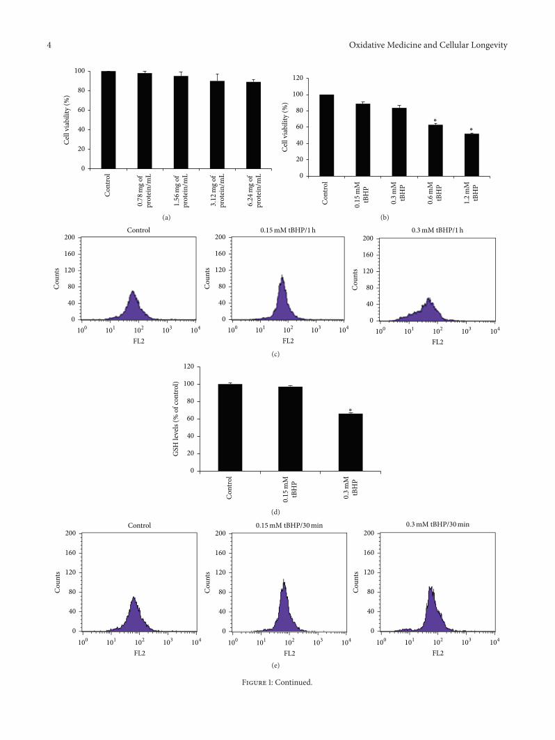
4 Oxidative Medicine and Cellular Longevity
Con
trol
0
20
40
60
80
100C
ell v
iabi
lity
(%)
prot
ein/
mL
6.24
mg
of
prot
ein/
mL
3.12
mg
of
prot
ein/
mL
1.56
mg
of
prot
ein/
mL
0.78
mg
of
(a)
Con
trol
0
20
40
60
80
100
120
Cel
l via
bilit
y (%
)
tBH
P1.2
mM
tBH
P0.6
mM
∗
∗
tBH
P0.3
mM
tBH
P0.15
mM
(b)
Control 0.15mM tBHP/1h 0.3mM tBHP/1h
101
102
103
104
100
FL210
110
210
310
410
0
FL210
110
210
310
410
0
FL2
0
40
80
120
160
200
Cou
nts
0
40
80
120
160
200
Cou
nts
0
40
80
120
160
200
Cou
nts
(c)
Con
trol
∗
tBH
P0.3
mM
tBH
P0.15
mM
0
20
40
60
80
100
120
GSH
leve
ls (%
of c
ontro
l)
(d)
0.15mM tBHP/30min 0.3mM tBHP/30minControl
101
102
103
104
100
FL2
0
40
80
120
160
200
Cou
nts
0
40
80
120
160
200
Cou
nts
101
102
103
104
100
FL2
0
40
80
120
160
200
Cou
nts
101
102
103
104
100
FL2(e)
Figure 1: Continued.
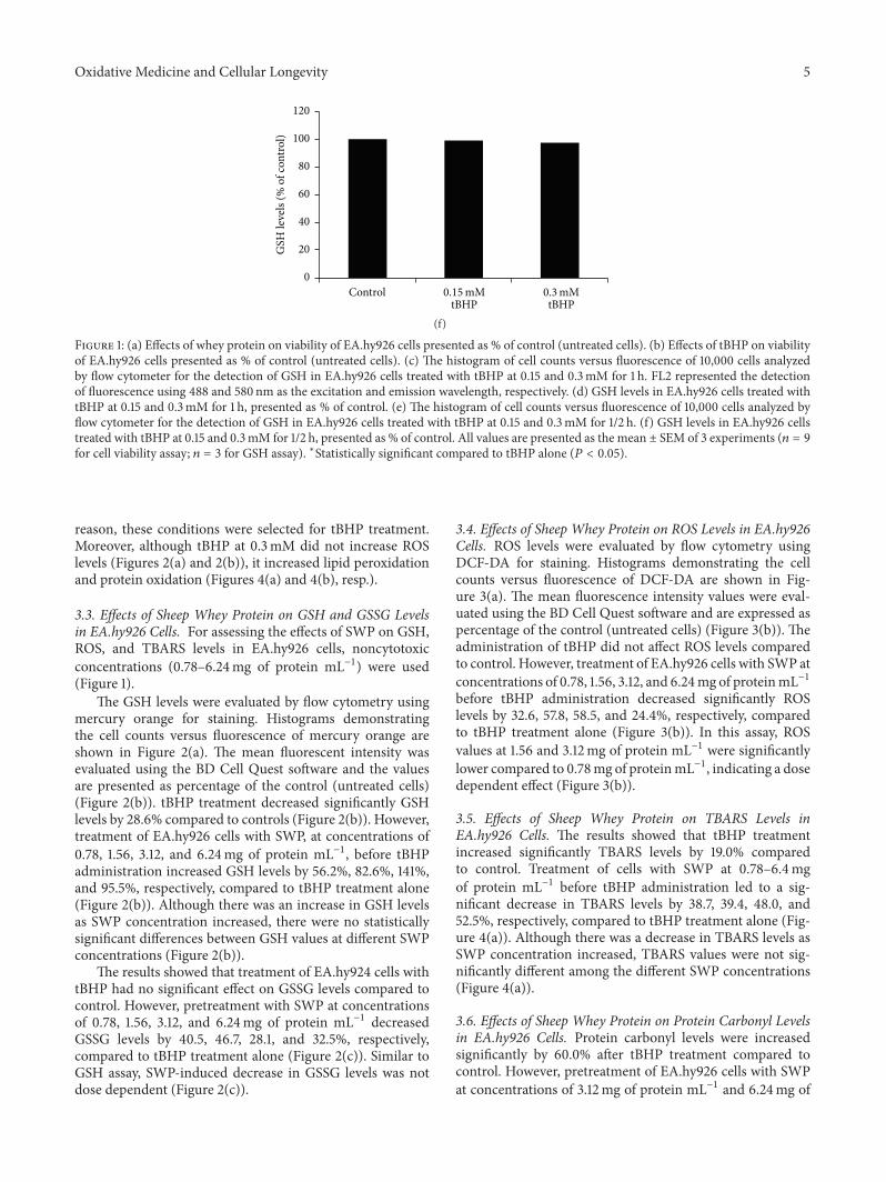
Oxidative Medicine and Cellular Longevity 5
ControltBHP0.3mM
tBHP0.15mM
0
20
40
60
80
100
120
GSH
leve
ls (%
of c
ontro
l)
(f)
Figure 1: (a) Effects of whey protein on viability of EA.hy926 cells presented as % of control (untreated cells). (b) Effects of tBHP on viabilityof EA.hy926 cells presented as % of control (untreated cells). (c) The histogram of cell counts versus fluorescence of 10,000 cells analyzedby flow cytometer for the detection of GSH in EA.hy926 cells treated with tBHP at 0.15 and 0.3mM for 1 h. FL2 represented the detectionof fluorescence using 488 and 580 nm as the excitation and emission wavelength, respectively. (d) GSH levels in EA.hy926 cells treated withtBHP at 0.15 and 0.3mM for 1 h, presented as % of control. (e) The histogram of cell counts versus fluorescence of 10,000 cells analyzed byflow cytometer for the detection of GSH in EA.hy926 cells treated with tBHP at 0.15 and 0.3mM for 1/2 h. (f) GSH levels in EA.hy926 cellstreated with tBHP at 0.15 and 0.3mM for 1/2 h, presented as % of control. All values are presented as the mean ± SEM of 3 experiments (𝑛 = 9for cell viability assay; 𝑛 = 3 for GSH assay). ∗Statistically significant compared to tBHP alone (𝑃 < 0.05).
reason, these conditions were selected for tBHP treatment.Moreover, although tBHP at 0.3mM did not increase ROSlevels (Figures 2(a) and 2(b)), it increased lipid peroxidationand protein oxidation (Figures 4(a) and 4(b), resp.).
3.3. Effects of Sheep Whey Protein on GSH and GSSG Levelsin EA.hy926 Cells. For assessing the effects of SWP on GSH,ROS, and TBARS levels in EA.hy926 cells, noncytotoxicconcentrations (0.78–6.24mg of protein mL−1) were used(Figure 1).
The GSH levels were evaluated by flow cytometry usingmercury orange for staining. Histograms demonstratingthe cell counts versus fluorescence of mercury orange areshown in Figure 2(a). The mean fluorescent intensity wasevaluated using the BD Cell Quest software and the valuesare presented as percentage of the control (untreated cells)(Figure 2(b)). tBHP treatment decreased significantly GSHlevels by 28.6% compared to controls (Figure 2(b)). However,treatment of EA.hy926 cells with SWP, at concentrations of0.78, 1.56, 3.12, and 6.24mg of protein mL−1, before tBHPadministration increased GSH levels by 56.2%, 82.6%, 141%,and 95.5%, respectively, compared to tBHP treatment alone(Figure 2(b)). Although there was an increase in GSH levelsas SWP concentration increased, there were no statisticallysignificant differences between GSH values at different SWPconcentrations (Figure 2(b)).
The results showed that treatment of EA.hy924 cells withtBHP had no significant effect on GSSG levels compared tocontrol. However, pretreatment with SWP at concentrationsof 0.78, 1.56, 3.12, and 6.24mg of protein mL−1 decreasedGSSG levels by 40.5, 46.7, 28.1, and 32.5%, respectively,compared to tBHP treatment alone (Figure 2(c)). Similar toGSH assay, SWP-induced decrease in GSSG levels was notdose dependent (Figure 2(c)).
3.4. Effects of Sheep Whey Protein on ROS Levels in EA.hy926Cells. ROS levels were evaluated by flow cytometry usingDCF-DA for staining. Histograms demonstrating the cellcounts versus fluorescence of DCF-DA are shown in Fig-ure 3(a). The mean fluorescence intensity values were eval-uated using the BD Cell Quest software and are expressed aspercentage of the control (untreated cells) (Figure 3(b)). Theadministration of tBHP did not affect ROS levels comparedto control. However, treatment of EA.hy926 cells with SWP atconcentrations of 0.78, 1.56, 3.12, and 6.24mg of proteinmL−1before tBHP administration decreased significantly ROSlevels by 32.6, 57.8, 58.5, and 24.4%, respectively, comparedto tBHP treatment alone (Figure 3(b)). In this assay, ROSvalues at 1.56 and 3.12mg of protein mL−1 were significantlylower compared to 0.78mg of proteinmL−1, indicating a dosedependent effect (Figure 3(b)).
3.5. Effects of Sheep Whey Protein on TBARS Levels inEA.hy926 Cells. The results showed that tBHP treatmentincreased significantly TBARS levels by 19.0% comparedto control. Treatment of cells with SWP at 0.78–6.4mgof protein mL−1 before tBHP administration led to a sig-nificant decrease in TBARS levels by 38.7, 39.4, 48.0, and52.5%, respectively, compared to tBHP treatment alone (Fig-ure 4(a)). Although there was a decrease in TBARS levels asSWP concentration increased, TBARS values were not sig-nificantly different among the different SWP concentrations(Figure 4(a)).
3.6. Effects of Sheep Whey Protein on Protein Carbonyl Levelsin EA.hy926 Cells. Protein carbonyl levels were increasedsignificantly by 60.0% after tBHP treatment compared tocontrol. However, pretreatment of EA.hy926 cells with SWPat concentrations of 3.12mg of protein mL−1 and 6.24mg of
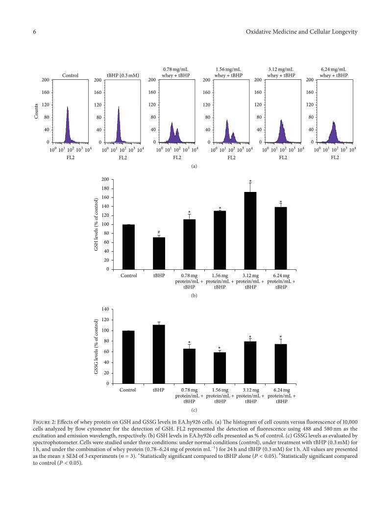
6 Oxidative Medicine and Cellular Longevity
10110
210
310
410
0
FL2
0
40
80
120
160
200
Cou
nts
10110
210
310
410
0
FL2
0
40
80
120
160
200
10110
210
310
410
0
FL2
0
40
80
120
160
200
10110
210
310
410
0
FL2
0
40
80
120
160
200
10110
210
310
410
0
FL2
0
40
80
120
160
200
10110
210
310
410
0
FL2
0
40
80
120
160
200
Control tBHP (0.3mM)0.78mg/mL
whey + tBHP1.56mg/mL
whey + tBHP3.12mg/mL
whey + tBHP6.24mg/mL
whey + tBHP
(a)
Control tBHP
#
protein/mL +tBHP
6.24mgprotein/mL +
tBHP
3.12mgprotein/mL +
tBHP
1.56mgprotein/mL +
tBHP
0.78mg
∗
∗
∗∗
0
20
40
60
80
100
120
140
160
180
200
GSH
leve
ls (%
of c
ontro
l)
(b)
Control tBHP
∗∗
∗
∗
protein/mL +tBHP
6.24mgprotein/mL +
tBHP
3.12mgprotein/mL +
tBHP
1.56mgprotein/mL +
tBHP
0.78mg0
20
40
60
80
100
120
140
GSS
G le
vels
(% o
f con
trol)
(c)
Figure 2: Effects of whey protein on GSH and GSSG levels in EA.hy926 cells. (a) The histogram of cell counts versus fluorescence of 10,000cells analyzed by flow cytometer for the detection of GSH. FL2 represented the detection of fluorescence using 488 and 580 nm as theexcitation and emission wavelength, respectively. (b) GSH levels in EA.hy926 cells presented as % of control. (c) GSSG levels as evaluated byspectrophotometer. Cells were studied under three conditions: under normal conditions (control), under treatment with tBHP (0.3mM) for1 h, and under the combination of whey protein (0.78–6.24mg of protein mL−1) for 24 h and tBHP (0.3mM) for 1 h. All values are presentedas the mean ± SEM of 3 experiments (𝑛 = 3). ∗Statistically significant compared to tBHP alone (𝑃 < 0.05). #Statistically significant comparedto control (𝑃 < 0.05).
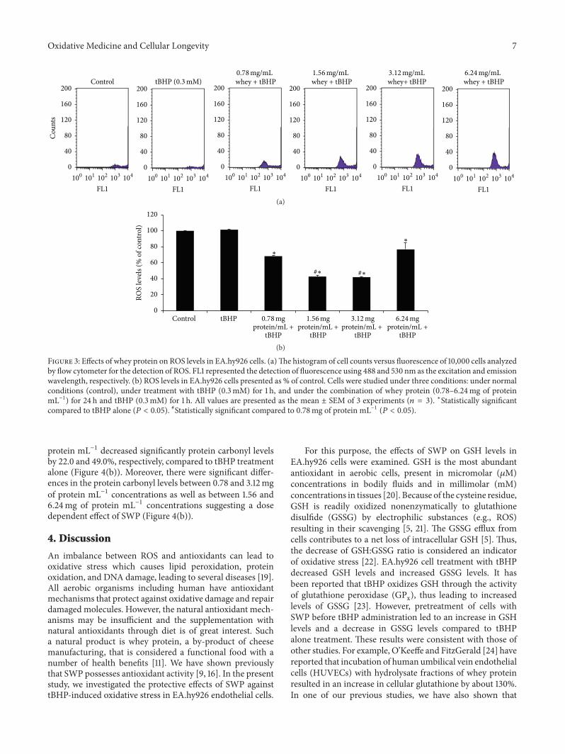
Oxidative Medicine and Cellular Longevity 7
10110
210
310
410
0
FL1
0
40
80
120
160
200
Cou
nts
10110
210
310
410
0
FL1
0
40
80
120
160
200
10110
210
310
410
0
FL1
0
40
80
120
160
200
10110
210
310
410
0
FL1
0
40
80
120
160
200
10110
210
310
410
0
FL1
0
40
80
120
160
200
10110
210
310
410
0
FL1
0
40
80
120
160
200
Control tBHP (0.3mM)0.78mg/mLwhey + tBHP
1.56mg/mLwhey + tBHP
3.12mg/mLwhey+ tBHP
6.24mg/mLwhey + tBHP
(a)
Control tBHP
#∗#∗
0
20
40
60
80
100
120
ROS
leve
ls (%
of c
ontro
l)
∗
∗
protein/mL +tBHP
6.24mgprotein/mL +
tBHP
3.12mgprotein/mL +
tBHP
1.56mgprotein/mL +
tBHP
0.78mg
(b)
Figure 3: Effects of whey protein on ROS levels in EA.hy926 cells. (a)The histogram of cell counts versus fluorescence of 10,000 cells analyzedby flow cytometer for the detection of ROS. FL1 represented the detection of fluorescence using 488 and 530 nm as the excitation and emissionwavelength, respectively. (b) ROS levels in EA.hy926 cells presented as % of control. Cells were studied under three conditions: under normalconditions (control), under treatment with tBHP (0.3mM) for 1 h, and under the combination of whey protein (0.78–6.24mg of proteinmL−1) for 24 h and tBHP (0.3mM) for 1 h. All values are presented as the mean ± SEM of 3 experiments (𝑛 = 3). ∗Statistically significantcompared to tBHP alone (𝑃 < 0.05). #Statistically significant compared to 0.78mg of protein mL−1 (𝑃 < 0.05).
protein mL−1 decreased significantly protein carbonyl levelsby 22.0 and 49.0%, respectively, compared to tBHP treatmentalone (Figure 4(b)). Moreover, there were significant differ-ences in the protein carbonyl levels between 0.78 and 3.12mgof protein mL−1 concentrations as well as between 1.56 and6.24mg of protein mL−1 concentrations suggesting a dosedependent effect of SWP (Figure 4(b)).
4. Discussion
An imbalance between ROS and antioxidants can lead tooxidative stress which causes lipid peroxidation, proteinoxidation, and DNA damage, leading to several diseases [19].All aerobic organisms including human have antioxidantmechanisms that protect against oxidative damage and repairdamaged molecules. However, the natural antioxidant mech-anisms may be insufficient and the supplementation withnatural antioxidants through diet is of great interest. Sucha natural product is whey protein, a by-product of cheesemanufacturing, that is considered a functional food with anumber of health benefits [11]. We have shown previouslythat SWP possesses antioxidant activity [9, 16]. In the presentstudy, we investigated the protective effects of SWP againsttBHP-induced oxidative stress in EA.hy926 endothelial cells.
For this purpose, the effects of SWP on GSH levels inEA.hy926 cells were examined. GSH is the most abundantantioxidant in aerobic cells, present in micromolar (𝜇M)concentrations in bodily fluids and in millimolar (mM)concentrations in tissues [20]. Because of the cysteine residue,GSH is readily oxidized nonenzymatically to glutathionedisulfide (GSSG) by electrophilic substances (e.g., ROS)resulting in their scavenging [5, 21]. The GSSG efflux fromcells contributes to a net loss of intracellular GSH [5]. Thus,the decrease of GSH:GSSG ratio is considered an indicatorof oxidative stress [22]. EA.hy926 cell treatment with tBHPdecreased GSH levels and increased GSSG levels. It hasbeen reported that tBHP oxidizes GSH through the activityof glutathione peroxidase (GPx), thus leading to increasedlevels of GSSG [23]. However, pretreatment of cells withSWP before tBHP administration led to an increase in GSHlevels and a decrease in GSSG levels compared to tBHPalone treatment. These results were consistent with those ofother studies. For example, O’Keeffe and FitzGerald [24] havereported that incubation of human umbilical vein endothelialcells (HUVECs) with hydrolysate fractions of whey proteinresulted in an increase in cellular glutathione by about 130%.In one of our previous studies, we have also shown that
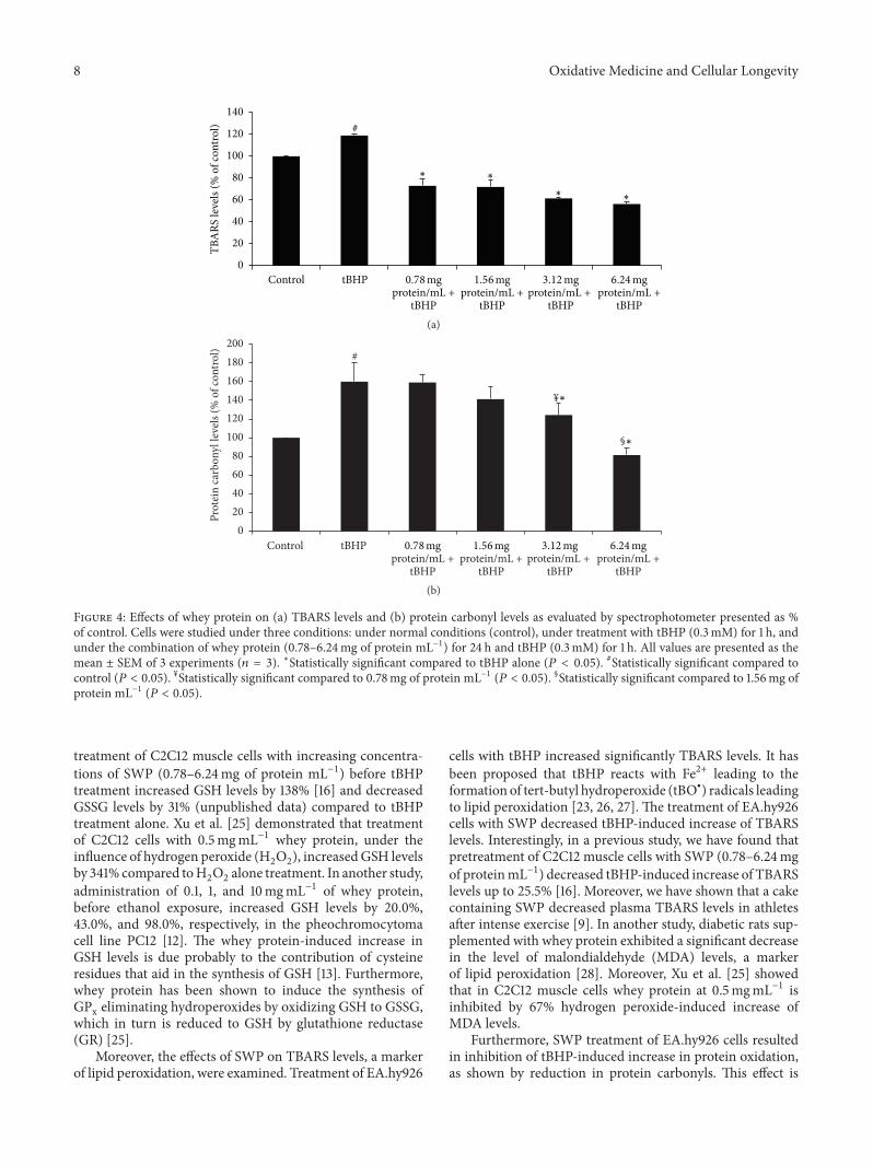
8 Oxidative Medicine and Cellular Longevity
Control tBHP
#
0
20
40
60
80
100
120
140
TBA
RS le
vels
(% o
f con
trol)
∗ ∗
∗∗
protein/mL +tBHP
6.24mgprotein/mL +
tBHP
3.12mgprotein/mL +
tBHP
1.56mgprotein/mL +
tBHP
0.78mg
(a)
Control tBHP
#
§
¥
020406080
100120140160180200
Prot
ein
carb
onyl
leve
ls (%
of c
ontro
l)
∗
∗
protein/mL +tBHP
6.24mgprotein/mL +
tBHP
3.12mgprotein/mL +
tBHP
1.56mgprotein/mL +
tBHP
0.78mg
(b)
Figure 4: Effects of whey protein on (a) TBARS levels and (b) protein carbonyl levels as evaluated by spectrophotometer presented as %of control. Cells were studied under three conditions: under normal conditions (control), under treatment with tBHP (0.3mM) for 1 h, andunder the combination of whey protein (0.78–6.24mg of protein mL−1) for 24 h and tBHP (0.3mM) for 1 h. All values are presented as themean ± SEM of 3 experiments (𝑛 = 3). ∗Statistically significant compared to tBHP alone (𝑃 < 0.05). #Statistically significant compared tocontrol (𝑃 < 0.05). ¥Statistically significant compared to 0.78mg of protein mL−1 (𝑃 < 0.05). §Statistically significant compared to 1.56mg ofprotein mL−1 (𝑃 < 0.05).
treatment of C2C12 muscle cells with increasing concentra-tions of SWP (0.78–6.24mg of protein mL−1) before tBHPtreatment increased GSH levels by 138% [16] and decreasedGSSG levels by 31% (unpublished data) compared to tBHPtreatment alone. Xu et al. [25] demonstrated that treatmentof C2C12 cells with 0.5mgmL−1 whey protein, under theinfluence of hydrogen peroxide (H
2
O2
), increasedGSH levelsby 341% compared toH
2
O2
alone treatment. In another study,administration of 0.1, 1, and 10mgmL−1 of whey protein,before ethanol exposure, increased GSH levels by 20.0%,43.0%, and 98.0%, respectively, in the pheochromocytomacell line PC12 [12]. The whey protein-induced increase inGSH levels is due probably to the contribution of cysteineresidues that aid in the synthesis of GSH [13]. Furthermore,whey protein has been shown to induce the synthesis ofGPx eliminating hydroperoxides by oxidizing GSH to GSSG,which in turn is reduced to GSH by glutathione reductase(GR) [25].
Moreover, the effects of SWP on TBARS levels, a markerof lipid peroxidation, were examined. Treatment of EA.hy926
cells with tBHP increased significantly TBARS levels. It hasbeen proposed that tBHP reacts with Fe2+ leading to theformation of tert-butyl hydroperoxide (tBO∙) radicals leadingto lipid peroxidation [23, 26, 27]. The treatment of EA.hy926cells with SWP decreased tBHP-induced increase of TBARSlevels. Interestingly, in a previous study, we have found thatpretreatment of C2C12 muscle cells with SWP (0.78–6.24mgof proteinmL−1) decreased tBHP-induced increase of TBARSlevels up to 25.5% [16]. Moreover, we have shown that a cakecontaining SWP decreased plasma TBARS levels in athletesafter intense exercise [9]. In another study, diabetic rats sup-plemented with whey protein exhibited a significant decreasein the level of malondialdehyde (MDA) levels, a markerof lipid peroxidation [28]. Moreover, Xu et al. [25] showedthat in C2C12 muscle cells whey protein at 0.5mgmL−1 isinhibited by 67% hydrogen peroxide-induced increase ofMDA levels.
Furthermore, SWP treatment of EA.hy926 cells resultedin inhibition of tBHP-induced increase in protein oxidation,as shown by reduction in protein carbonyls. This effect is
Oxidative Medicine and Cellular Longevity 9
important, since oxidative stress-induced carbonylation ofproteins leads to the loss of their physiological function [29].It is believed that there is an association between lipid andprotein oxidation [30]. For example, tBHPhas been suggestedto lead to the formation of tBO∙ radicals that in turn leadto protein oxidation either directly by attacking the aminoacyl side chains or indirectly by leading to lipid peroxidation[26]. Thus, the SWP-induced decrease in lipid peroxidationmay also account, at least in part, for inhibiting tBHP-inducedincrease in protein oxidation. Moreover, Haraguchi et al. [31]have shown that whey protein precluded increases in muscleprotein carbonyl content in exercised and sedentary animals.
Intriguingly, tBHP treatment did not affect ROS levels.As we have suggested previously, it seems that althoughtBHP produces free radicals, their “free form” cannot beobserved [17]. This may be attributed to the reaction offree radicals with other molecules in the cells. For instance,the decrease in GSH and the increase in lipid peroxidationand protein oxidation after tBHP treatment suggested thatfree radicals may react with GSH, lipids, and proteins,respectively. However, treatment of EA.hy926 cells with SWPbefore tBHP administration led to a decrease in ROS levelsup to 58.5% compared to tBHP alone treatment. In one ofour previous studies, we have also found that pretreatment ofC2C12muscle cells with SWP (0.78–6.24mg of proteinmL−1)decreased ROS levels to 41.3% [16]. Moreover, in anotherstudy, whey protein isolate (pWPI) and whey protein nativehydrolysates (nWPI) at 2mgmL−1 inhibited H
2
O2
-inducedROS formation by 76.0% and 32.5%, respectively, in humancolonic adenocarcinoma (Caco-2) cell line [32]. Likewise,whey protein has been shown to decrease significantly ROSlevels in diabetic rats [28]. The decrease in ROS levels maybe explained by the SWP-induced increase in antioxidantdefense mechanisms such as GSH. Interestingly, recent clin-ical studies have shown that a whey protein formulationreduced by almost twofold inflammatory oxidative damage(IOD) levels [33] and improved vascular functions [33, 34].
Although SWP treatment enhanced antioxidant capacityof EA.hy926 cells by either increasing antioxidant mecha-nisms or reducing ROS levels and oxidative stress-induceddamage, these effects were not always dose dependent. Thus,SWP exhibited dose dependent decrease in ROS and CARBlevels but SWP-induced increase inGSH or decrease inGSSGand TBARS levels was not dose dependent. This may beexplained by the different mechanisms through which SWPaffects each of the tested oxidative stress markers, and so itspotency differs among the different assays. It seems that inGSH, GSSG, and TBARS assays, SWP’s activity has alreadyreached a plateau at the concentrations used and for thisreason a dose dependent effect was not observed. Namely, iflower than the tested concentrations were used, it may also bea dose dependent effect in these assays.
In conclusion, the present study showed that SWP waseffective to protect endothelial cells from oxidative stress-induced damage. SWP exerted its protective activity againstoxidative stress, by increasing GSH levels and decreasingGSSG, lipid peroxidation, protein oxidation, and ROS levelsin EA.hy926 cells. It should also be mentioned that SWPconcentrations (i.e., from 0.78 to 6.24mg/mL) used were not
cytotoxic. Moreover, in a previous study we have shown thatat these concentrations SWP exhibited strong free radicalscavenging activity and reducing power and enhanced theantioxidant capacity in mouse myoblastoma C2C12 cells [16].Also, these concentrations are assimilated to the concentra-tions found in food. For example, the highest concentrationof SWP was 6.24mg/mL in the cell culture medium. Takeninto account that the total plasma volume in human organ-ism is about 3.5 L, then the concentration of 6.24mg/mLof SWP would be achieved theoretically if about 20 g ofSWP is consumed. This amount is within the range of therecommended intake doses of whey protein.Thus, since thesewhey protein concentrations can be found in blood, thenthe endothelial cells are possible to be exposed to them,since they are the main cells of the blood vessel walls. Thus,the findings of the present study suggest that SWP may beused as food supplement to attenuate vascular disturbancesassociated with oxidative stress.
Competing Interests
The authors declare that there are no competing interests.
Acknowledgments
This work was cofinanced by the European Union (EuropeanSocial Fund (ESF)) and Greek national funds through theOperational Program “Education and Lifelong Learning”of the National Strategic Reference Framework (NSRF),Research Funding Program: “Heracleitus II. Investing inknowledge society through the European Social Fund.” Thiswork was also funded by the MSc programme “Biotech-nology-Nutrition & Environment” in the Department ofBiochemistry & Biotechnology at University of Thessaly.
References
[1] B. Halliwell, “Free radicals and other reactive species in disease,”in Encyclopedia of Life Sciences, John Wiley & Sons, New York,NY, USA, 2001.
[2] K. T. Turpaev, “Reactive oxygen species and regulation of geneexpression,” Biochemistry, vol. 67, no. 3, pp. 281–292, 2002.
[3] B. P. Chew and J. S. Park, “Carotenoid action on the immuneresponse,” Journal of Nutrition, vol. 134, no. 1, pp. 257S–261S,2004.
[4] S. K. Powers and M. J. Jackson, “Exercise-induced oxidativestress: cellularmechanisms and impact onmuscle force produc-tion,” Physiological Reviews, vol. 88, no. 4, pp. 1243–1276, 2008.
[5] G.Wu, Y.-Z. Fang, S. Yang, J. R. Lupton, andN. D. Turner, “Glu-tathione metabolism and its implications for health,” Journal ofNutrition, vol. 134, no. 3, pp. 489–492, 2004.
[6] J. E. Deanfield, J. P. Halcox, and T. J. Rabelink, “Endothelialfunction and dysfunction: testing and clinical relevance,” Cir-culation, vol. 115, no. 10, pp. 1285–1295, 2007.
[7] S. G. Rhee, “H2
O2
, a necessary evil for cell signaling,” Science,vol. 312, no. 5782, pp. 1882–1883, 2006.
[8] A. Woywodt, F. H. Bahlmann, K. De Groot, H. Haller, and M.Haubitz, “Circulating endothelial cells: life, death, detachmentand repair of the endothelial cell layer,” Nephrology DialysisTransplantation, vol. 17, no. 10, pp. 1728–1730, 2002.
10 Oxidative Medicine and Cellular Longevity
[9] E. Kerasioti, A. Kiskini, A. Veskoukis et al., “Effect of a specialcarbohydrate-protein cake on oxidative stress markers afterexhaustive cycling in humans,” Food and Chemical Toxicology,vol. 50, no. 8, pp. 2805–2810, 2012.
[10] W. J. Bartfay, M. T. Davis, J. M. Medves, and S. Lugowski,“Milk whey protein decreases oxygen free radical productionin a murine model of chronic iron-overload cardiomyopathy,”Canadian Journal of Cardiology, vol. 19, no. 10, pp. 1163–1168,2003.
[11] K. Marshall, “Therapeutic applications of whey protein,” Alter-native Medicine Review, vol. 9, no. 2, pp. 136–156, 2004.
[12] Y.-M. Tseng, S.-K. Lin, J.-K. Hsiao et al., “Whey protein con-centrate promotes the production of glutathione (GSH) byGSHreductase in the PC12 cell line after acute ethanol exposure,”Food and Chemical Toxicology, vol. 44, no. 4, pp. 574–578, 2006.
[13] R. L. Walzem, C. J. Dillard, and J. B. German, “Whey compo-nents: millennia of evolution create functionalities for mam-malian nutrition: what we know and what we may be overlook-ing,” Critical Reviews in Food Science and Nutrition, vol. 42, no.4, pp. 353–375, 2002.
[14] E. Ha and M. B. Zemel, “Functional properties of whey, wheycomponents, and essential amino acids: mechanisms under-lying health benefits for active people,” Journal of NutritionalBiochemistry, vol. 14, no. 5, pp. 251–258, 2003.
[15] E. Kerasioti, D. Stagos, A. Jamurtas et al., “Anti-inflammatoryeffects of a special carbohydrate-whey protein cake after exhaus-tive cycling in humans,” Food and Chemical Toxicology, vol. 61,pp. 42–46, 2013.
[16] E. Kerasioti, D. Stagos, A. Priftis et al., “Antioxidant effects ofwhey protein on muscle C2C12 cells,” Food Chemistry, vol. 155,pp. 271–278, 2014.
[17] N. Goutzourelas, D. Stagos, N. Demertzis et al., “Effects of poly-phenolic grape extract on the oxidative status of muscle andendothelial cells,” Human and Experimental Toxicology, vol. 33,no. 11, pp. 1099–1112, 2014.
[18] Y. N. Reddy, S. V. Murthy, D. R. Krishna, and M. C. Prabhakar,“Role of free radicals and antioxidants in tuberculosis patients,”Indian Journal of Tuberculosis, vol. 51, pp. 213–218, 2004.
[19] M. Valko, D. Leibfritz, J. Moncol, M. T. D. Cronin, M. Mazur,and J. Telser, “Free radicals and antioxidants in normal physi-ological functions and human disease,” International Journal ofBiochemistry and Cell Biology, vol. 39, no. 1, pp. 44–84, 2007.
[20] J. B. Owen and D. A. Butterfield, “Measurement of oxidized/reduced glutathione ratio,” Methods in Molecular Biology, vol.648, pp. 269–277, 2010.
[21] Y.-Z. Fang, S. Yang, andG.Wu, “Free radicals, antioxidants, andnutrition,” Nutrition, vol. 18, no. 10, pp. 872–879, 2002.
[22] K. Aquilano, S. Baldelli, and M. R. Ciriolo, “Glutathione: newroles in redox signalling for an old antioxidant,” Frontiers inPharmacology, vol. 5, p. 196, 2014.
[23] C. F. Lima,M. Fernandes-Ferreira, andC. Pereira-Wilson, “Phe-nolic compounds protect HepG2 cells from oxidative damage:relevance of glutathione levels,” Life Sciences, vol. 79, no. 21, pp.2056–2068, 2006.
[24] M. B. O’Keeffe and R. J. FitzGerald, “Antioxidant effects ofenzymatic hydrolysates ofwhey protein concentrate on culturedhuman endothelial cells,” International Dairy Journal, vol. 36,no. 2, pp. 128–135, 2014.
[25] R. Xu, N. Liu, X. Xu, and B. Kong, “Antioxidative effects of wheyprotein on peroxide-induced cytotoxicity,” Journal of DairyScience, vol. 94, no. 8, pp. 3739–3746, 2011.
[26] S. Hix, M. B. Kadiiska, R. P. Mason, and O. Augusto, “In vivometabolism of tert-butyl hydroperoxide to methyl radicals.EPR spin-trapping and DNA methylation studies,” ChemicalResearch in Toxicology, vol. 13, no. 10, pp. 1056–1064, 2000.
[27] M. Aĺıa, S. Ramos, R. Mateos, L. Bravo, and L. Goya, “Responseof the antioxidant defense system to tert-butyl hydroperox-ide and hydrogen peroxide in a human hepatoma cell line(HepG2),” Journal of Biochemical andMolecular Toxicology, vol.19, no. 2, pp. 119–128, 2005.
[28] H. Ebaid, A. Salem, A. Sayed, and A. Metwalli, “Whey proteinenhances normal inflammatory responses during cutaneouswound healing in diabetic rats,” Lipids in Health and Disease,vol. 10, article 235, 2011.
[29] M. F. Beal, “Oxidativelymodified proteins in aging and disease,”Free Radical Biology and Medicine, vol. 32, no. 9, pp. 797–803,2002.
[30] Y. V. Vasil’ev, S.-C. Tzeng, L. Huang, and C. S. Maier, “Proteinmodifications by electrophilic lipoxidation products: adductformation, chemical strategies and tandem mass spectrome-try for their detection and identification,” Mass SpectrometryReviews, vol. 33, no. 3, pp. 157–182, 2014.
[31] F. K. Haraguchi, M. E. Silva, L. X. Neves, R. C. Dos Santos, andM. L. Pedrosa, “Whey protein precludes lipid and protein oxi-dation and improves body weight gain in resistance-exercisedrats,” European Journal of Nutrition, vol. 50, no. 5, pp. 331–339,2011.
[32] A. F. Piccolomini, M. M. Iskandar, L. C. Lands, and S. Kubow,“High hydrostatic pressure pre-treatment of whey proteinsenhanceswhey protein hydrolysate inhibition of oxidative stressand IL-8 secretion in intestinal epithelial cells,” Food andNutrition Research, vol. 56, 2012.
[33] I. M. Petyaev, P. Y. Dovgalevsky, V. A. Klochkov, N. E. Chalyk,and N. Kyle, “Whey protein lycosome formulation improvesvascular functions and plasma lipids with reduction of markersof inflammation and oxidative stress in prehypertension,” TheScientific World Journal, vol. 2012, Article ID 269476, 7 pages,2012.
[34] K. D. Ballard, B. R. Kupchak, B. M. Volk et al., “Acute effects ofingestion of a novel whey-derived extract on vascular endothe-lial function in overweight, middle-aged men and women,”British Journal of Nutrition, vol. 109, no. 5, pp. 882–893, 2013.
Review ArticleThe Role of Mitochondrial Functional Proteins inROS Production in Ischemic Heart Diseases
Haifeng Pei,1,2 Yi Yang,1 Heng Zhao,3 Xiuchuan Li,1 Dachun Yang,1
De Li,1 and Yongjian Yang1,2
1Department of Cardiology, Chengdu Military General Hospital, Chengdu 610083, China2Department of Cardiology, Third Military Medical University, Chongqing 400042, China3Department of Ultrasonography, Chengdu Military General Hospital, Chengdu 610083, China
Correspondence should be addressed to Yongjian Yang; [email protected]
Received 23 December 2015; Revised 27 January 2016; Accepted 28 January 2016
Academic Editor: Sergio Di Meo
Copyright © 2016 Haifeng Pei et al. This is an open access article distributed under the Creative Commons Attribution License,which permits unrestricted use, distribution, and reproduction in any medium, provided the original work is properly cited.
Ischemic heart diseases (IHD) have become the leading cause of death around the world, killing more than 7 million peopleannually. In IHD, the blockage of coronary vessels will cause irreversible cell injury and even death. As the “powerhouse” and“apoptosis center” in cardiomyocytes, mitochondria play critical roles in IHD. Ischemia insult can reduce myocardial ATP content,resulting in energy stress and overproduction of reactive oxygen species (ROS). Thus, mitochondrial abnormality has beenidentified as a hallmark of multiple cardiovascular disorders. To date, many studies have suggested that these mitochondrialproteins, such as electron transport chain (ETC) complexes, uncoupling proteins (UCPs), mitochondrial dynamic proteins,translocases of outermembrane (Tom) complex, andmitochondrial permeability transition pore (MPTP), can directly or indirectlyinfluence mitochondria-originated ROS production, consequently determining the degree of mitochondrial dysfunction andmyocardial impairment. Here, the focus of this review is to summarize the present understanding of the relationship betweensome mitochondrial functional proteins and ROS production in IHD.
1. Introduction
As the leading cause of fatality worldwide, ischemic heartdiseases (IHD) give rise to widespread loss of cardiomyocytesand subsequent adverse cardiac remodeling [1]. More than17 million people had succumbed to IHD in 2008, andthe number was estimated to be 23.6 million by 2030 [2].Ischemia starves cardiomyocytes of vital oxygen, resultingin severe or irreversible injury to heart [3]. Abundant linesof evidence indicate that reactive oxygen species (ROS)overload that originates from mitochondria is closely linkedwith the pathogenesis of cardiovascular diseases, such asatherosclerosis, myocardial infarction, and heart failure [4].In myocytes, mitochondria comprise more than 30% cellvolume and generate about 90% of the ATP [5]. Accordingly,mitochondria are also the major source of ROS in the cardio-vascular diseases [5, 6]. Numerous experimental and clinicalstudies have shown that ROS accumulation is significantly
exacerbated in the failingmyocardium [7]. Chronic exposureofmyocytes to ROSwill lead to apoptosis, necrosis, and fibro-sis, finally causing arrhythmias, impairment of excitation-contraction coupling, and cardiac remodeling [8–10]. Giventhat ROS affect virtually all aspects of cardiac myocyte physi-ology and that those lines of evidence support the notion thatROS are increased in IHD, the underlyingmechanisms aboutROS overproduction induced by mitochondrial abnormalityshould be further explored.
2. The Electron Transport Chain (ETC)Complexes and Uncoupling Proteins (UCPs)
Mitochondrial oxidative phosphorylation is the basic mech-anism of ATP synthesis, in which the five enzyme complexesI–V in mitochondria are composed of multiple subunits.The respiratory chain, apart from energy production, playsa pivotal role in regulation of cardiac oxidative stress [11].
Hindawi Publishing CorporationOxidative Medicine and Cellular LongevityVolume 2016, Article ID 5470457, 8 pageshttp://dx.doi.org/10.1155/2016/5470457
2 Oxidative Medicine and Cellular Longevity
ETC damage will lead to a robust increase of ROS productionin mitochondria [12]. In ischemic heart, oxygen deliveryto myocardium is not sufficient to meet the demand ofmitochondrial oxidation. Reintroduction of oxygen inducedby reperfusion markedly enhances electron leakage from theETC, resulting in overproduction of superoxide (O
2
−) andO2
−-derived oxidants [12, 13]. Indeed, reduced activity ofETC subunits in heart failure patients has been confirmedindependently of the etiology, notably of complex I [14],complex III [15], and complex IV [16]. Within the ETC,complexes I/III are regarded as the key contributors toROS generation under stressful conditions [17, 18]. In thesetting of myocardial ischemia/reperfusion (MI/R), oxidativeinjury of complex I can weaken complex interaction andenhance ROS production by complex III [19, 20]. Althoughthe contribution of intact complex II seems to be negligible,somemutations in subunits of complex II have been linked tospecific forms of cancer or neurodegeneration with increasedROS generation [21, 22]. Aldakkak et al. [23] revealed thatpharmacologic blockade of electron transport attenuatesROS-induced cardiac injury. Consistently, nitrite can mod-ulate mitochondrial resilience to reperfusion injury, perhapsvia inhibiting complex I by posttranslational s-nitrosation toreduce ROS generation [24, 25]. Moreover, ROS scavengingability may be impaired as an evident decrease of MnSODactivity in failing heart [26]. Coenzyme Q (CoQ) encom-passes a collection of homologous molecules which consistof a benzoquinone ring linked to an isoprenoid side chain[27]. In the quinone form, CoQ serves as an electron carrierto transfer electrons in the mitochondrial ETC. In the quinolform (the reduced form of quinone), CoQ represents anefficient antioxidant in the body [28]. CoQ10 at high doses hasalso been proved to regulate gene transcription, including thegenes important in lipid metabolism and the specific genesinduced by ROS-sensitive intracellular pathways [29]. It isclaimed that CoQ10 supplementation to obese rats attenuatesage-related oxidative stress and preserves mitochondrialfunction in myocardium [30]. Moreover, CoQ10 deficiencyis detrimental to the prognosis of chronic heart failure, andplasma concentration of CoQ10may serve as an independentpredictor of mortality [31]. Long-term supplementation ofCoQ10 for patients with heart failure improves symptoms andreduces major adverse cardiovascular events [32].
In contrast, uncoupling proteins (UCPs) that are locatedin mitochondrial inner membrane exhibit opposite func-tions as complexes mentioned above. UCPs can reducemitochondrial ROS production by dissipating the electro-chemical gradient [33–36]. UCPs overexpression was foundto salvage cardiomyocytes by preserving mitochondrialintegrity [33, 36]. Specifically, mild to moderate mitochon-drial uncoupling mediated by UCPs protects against MI/Rinjury by reducing ROS generation [37]. UCP1, character-ized in brown adipocytes [38], has close relationship withendogenous CoQ to influence H+ transport [39]. In thesetting of hypoxia/reoxygenation, UCP1 overexpression con-fers cardioprotection [33]. The presence of UCP1 mitigatesreperfusion-induced damage, probably by reducing mito-chondrial hyperpolarization [40]. Moreover, UCP2 overex-pression can exert cardioprotection perhaps by preventing
mitochondrial Ca2+ overload and attenuating ROS genera-tion [41]. UCP3 has also been reported to play a critical rolein cardioprotection during oxidative stress by suppressingdetrimental ROS generation and maintaining myocardialhigh-energy phosphates [37]. In view of what was mentionedabove, UCPs may be developed as protective targets for IHDpatients.
3. Mitochondrial Dynamic Proteins
In order to adapt to stressful conditions, mitochondriadynamically undergo fission and fusion [42], which isbasically regulated by some functional proteins [42, 43].Mitofusin 1/2 (Mfn1/2) and optic atrophy 1 (Opa1) are knownto induce mitochondrial fusion [44, 45], while dynamin-related protein 1 (Drp1) is identified to interact with fissionprotein 1 (Fis1) to promote mitochondrial fission [46, 47]. Inmammalian cells, mitochondrial function is largely governedby mitochondrial fission and fusion which is an importantfactor for the integrity, structure, and function of healthymitochondria. Abnormal fission or fusion will impair mito-chondrial function, for example, resulting in overload ofmitochondrial Ca2+, overproduction of free radicals, alter-ation of mitochondrial enzymatic activities, and reduction ofATP production [48].
Recent studies have highlighted the notion that defectsin mitochondrial dynamics are relevant to various humandisorders, including MI/R, heart failure, diabetes, and aging[49, 50]. Inhibition of mitochondrial fission has been con-sidered to rescue myocardial infarction/heart failure [7].The deletion of Mfn1/2 appears to render the heart lessresistant to MPTP opening [51], whereas overexpression ofMfn1/2 prevents the opening of MPTP and reduces celldeath following ischemia/reperfusion (I/R) [52, 53]. Opa1mutation in myocardium results in mitochondrial dysfunc-tion and increases ROS [54], while transfection of Opa1in vivo protects mice from denervation-induced ischemicheart damage [55]. Moreover, inhibition of Drp1 significantlydecreased I/R-inducedmitochondrial fragmentation and car-diomyocytes apoptosis [52, 56]. Cumulative lines of evidencesuggest that impaired mitochondrial dynamics are an earlyevent in the progression of IHD that involve excessive mito-chondrial fission and mitochondrial dysfunction, indicatingthat dynamics related proteins may be developed as newtherapeutic targets against IHD.
4. The Translocases of OuterMembrane (Tom) Complex
The translocases of mitochondrial outer membrane (Tom)complex is a general entry gate for the importing of allmitochondrial preproteins. It comprises a central channel(formed by Tom40, Tom5, Tom6, and Tom7) and threereceptors (Tom70, Tom20, and Tom22) [57]. Tom20 andTom70 can recognize mitochondrial-targeting sequences onthe precursor proteins and transfer them to Tom22 and thecentral channel [57, 58]. Tom20 and Tom22 mainly recog-nize the presequences of cleavable precursor proteins, while
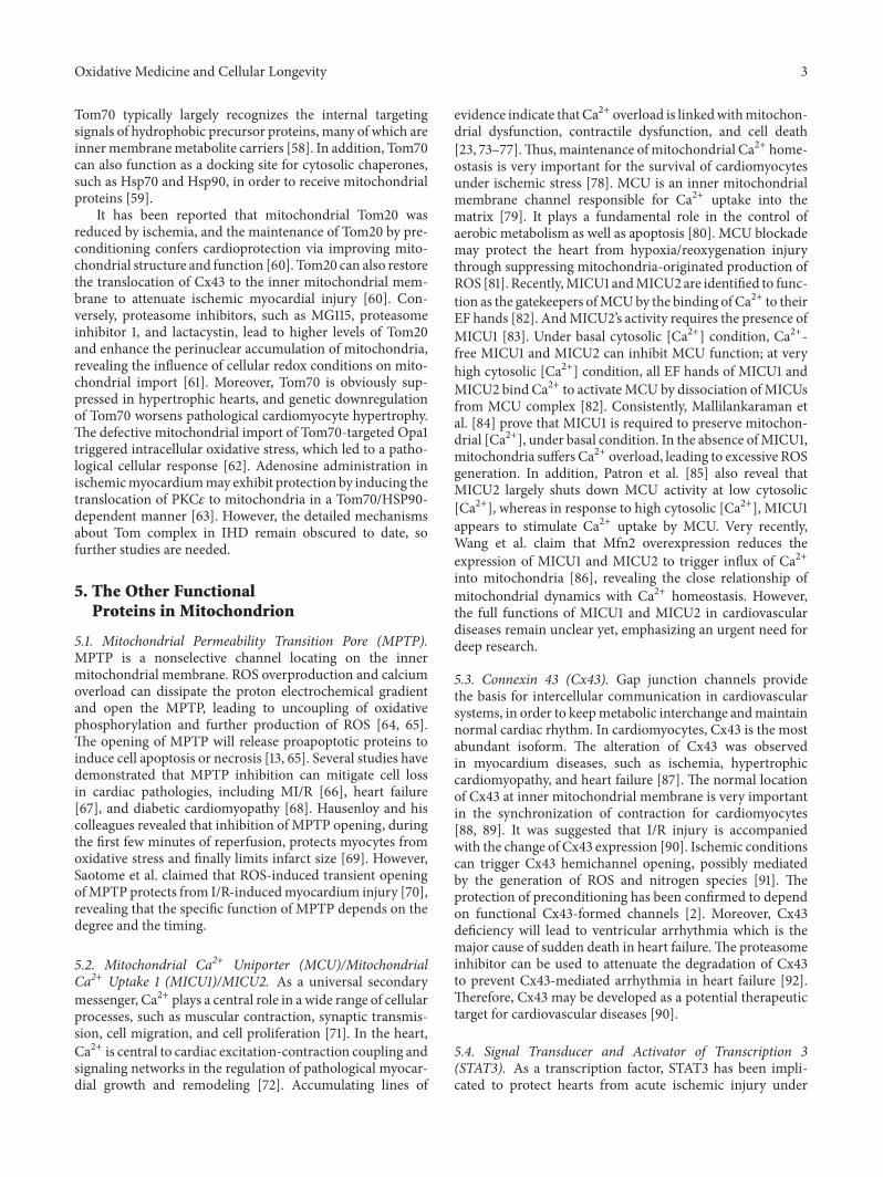
Oxidative Medicine and Cellular Longevity 3
Tom70 typically largely recognizes the internal targetingsignals of hydrophobic precursor proteins, many of which areinnermembranemetabolite carriers [58]. In addition, Tom70can also function as a docking site for cytosolic chaperones,such as Hsp70 and Hsp90, in order to receive mitochondrialproteins [59].
It has been reported that mitochondrial Tom20 wasreduced by ischemia, and the maintenance of Tom20 by pre-conditioning confers cardioprotection via improving mito-chondrial structure and function [60]. Tom20 can also restorethe translocation of Cx43 to the inner mitochondrial mem-brane to attenuate ischemic myocardial injury [60]. Con-versely, proteasome inhibitors, such as MG115, proteasomeinhibitor 1, and lactacystin, lead to higher levels of Tom20and enhance the perinuclear accumulation of mitochondria,revealing the influence of cellular redox conditions on mito-chondrial import [61]. Moreover, Tom70 is obviously sup-pressed in hypertrophic hearts, and genetic downregulationof Tom70 worsens pathological cardiomyocyte hypertrophy.The defective mitochondrial import of Tom70-targeted Opa1triggered intracellular oxidative stress, which led to a patho-logical cellular response [62]. Adenosine administration inischemicmyocardiummay exhibit protection by inducing thetranslocation of PKC𝜀 to mitochondria in a Tom70/HSP90-dependent manner [63]. However, the detailed mechanismsabout Tom complex in IHD remain obscured to date, sofurther studies are needed.
5. The Other FunctionalProteins in Mitochondrion
5.1. Mitochondrial Permeability Transition Pore (MPTP).MPTP is a nonselective channel locating on the innermitochondrial membrane. ROS overproduction and calciumoverload can dissipate the proton electrochemical gradientand open the MPTP, leading to uncoupling of oxidativephosphorylation and further production of ROS [64, 65].The opening of MPTP will release proapoptotic proteins toinduce cell apoptosis or necrosis [13, 65]. Several studies havedemonstrated that MPTP inhibition can mitigate cell lossin cardiac pathologies, including MI/R [66], heart failure[67], and diabetic cardiomyopathy [68]. Hausenloy and hiscolleagues revealed that inhibition of MPTP opening, duringthe first few minutes of reperfusion, protects myocytes fromoxidative stress and finally limits infarct size [69]. However,Saotome et al. claimed that ROS-induced transient openingofMPTP protects from I/R-inducedmyocardium injury [70],revealing that the specific function of MPTP depends on thedegree and the timing.
5.2. Mitochondrial Ca2+ Uniporter (MCU)/MitochondrialCa2+ Uptake 1 (MICU1)/MICU2. As a universal secondarymessenger, Ca2+ plays a central role in a wide range of cellularprocesses, such as muscular contraction, synaptic transmis-sion, cell migration, and cell proliferation [71]. In the heart,Ca2+ is central to cardiac excitation-contraction coupling andsignaling networks in the regulation of pathological myocar-dial growth and remodeling [72]. Accumulating lines of
evidence indicate that Ca2+ overload is linkedwithmitochon-drial dysfunction, contractile dysfunction, and cell death[23, 73–77].Thus, maintenance ofmitochondrial Ca2+ home-ostasis is very important for the survival of cardiomyocytesunder ischemic stress [78]. MCU is an inner mitochondrialmembrane channel responsible for Ca2+ uptake into thematrix [79]. It plays a fundamental role in the control ofaerobic metabolism as well as apoptosis [80]. MCU blockademay protect the heart from hypoxia/reoxygenation injurythrough suppressing mitochondria-originated production ofROS [81]. Recently,MICU1 andMICU2 are identified to func-tion as the gatekeepers ofMCUby the binding of Ca2+ to theirEF hands [82]. AndMICU2’s activity requires the presence ofMICU1 [83]. Under basal cytosolic [Ca2+] condition, Ca2+-free MICU1 and MICU2 can inhibit MCU function; at veryhigh cytosolic [Ca2+] condition, all EF hands of MICU1 andMICU2 bind Ca2+ to activateMCU by dissociation ofMICUsfrom MCU complex [82]. Consistently, Mallilankaraman etal. [84] prove that MICU1 is required to preserve mitochon-drial [Ca2+], under basal condition. In the absence ofMICU1,mitochondria suffers Ca2+ overload, leading to excessive ROSgeneration. In addition, Patron et al. [85] also reveal thatMICU2 largely shuts down MCU activity at low cytosolic[Ca2+], whereas in response to high cytosolic [Ca2+], MICU1appears to stimulate Ca2+ uptake by MCU. Very recently,Wang et al. claim that Mfn2 overexpression reduces theexpression of MICU1 and MICU2 to trigger influx of Ca2+into mitochondria [86], revealing the close relationship ofmitochondrial dynamics with Ca2+ homeostasis. However,the full functions of MICU1 and MICU2 in cardiovasculardiseases remain unclear yet, emphasizing an urgent need fordeep research.
5.3. Connexin 43 (Cx43). Gap junction channels providethe basis for intercellular communication in cardiovascularsystems, in order to keepmetabolic interchange andmaintainnormal cardiac rhythm. In cardiomyocytes, Cx43 is the mostabundant isoform. The alteration of Cx43 was observedin myocardium diseases, such as ischemia, hypertrophiccardiomyopathy, and heart failure [87]. The normal locationof Cx43 at inner mitochondrial membrane is very importantin the synchronization of contraction for cardiomyocytes[88, 89]. It was suggested that I/R injury is accompaniedwith the change of Cx43 expression [90]. Ischemic conditionscan trigger Cx43 hemichannel opening, possibly mediatedby the generation of ROS and nitrogen species [91]. Theprotection of preconditioning has been confirmed to dependon functional Cx43-formed channels [2]. Moreover, Cx43deficiency will lead to ventricular arrhythmia which is themajor cause of sudden death in heart failure.The proteasomeinhibitor can be used to attenuate the degradation of Cx43to prevent Cx43-mediated arrhythmia in heart failure [92].Therefore, Cx43 may be developed as a potential therapeutictarget for cardiovascular diseases [90].
5.4. Signal Transducer and Activator of Transcription 3(STAT3). As a transcription factor, STAT3 has been impli-cated to protect hearts from acute ischemic injury under
4 Oxidative Medicine and Cellular Longevity
ROS
MCU/MICU1/MICU2
Tom complex
Dynamic proteinsETC
MPTP
Ischemic heart
Cardiomyocyte
Mitochondrion
UCPs/Cx43/STAT3/others
Figure 1: Schematic diagram indicatingmitochondrial proteins’ role in ROS generation in ischemic heart diseases. Under ischemic insult, thegeneration of ROS significantly increases inmitochondria of cardiomyocytes, which can be directly or indirectly influenced bymitochondrialfunctional proteins, including ETC complexes, UCPs, mitochondrial dynamic proteins, Tom complex, MPTP, MCU/MICU1/MICU2, Cx43,and STAT3. ROS: reactive oxygen species; ETC: electron transport chain; UCPs: uncoupling proteins; Tom: translocases of outer membrane;MPTP: mitochondrial permeability transition pore; MCU: mitochondrial Ca2+ uniporter; MICU1: mitochondrial Ca2+ uptake 1; MICU2:mitochondrial Ca2+ uptake 2; Cx43: Connexin 43; STAT3: signal transducer and activator of transcription 3.
stressful conditions [93], and loss of STAT3 will result incardiomyopathy [94]. Accumulative lines of evidence alsodemonstrated that STAT3 serves as a protective moleculefor the heart in hypertension and advanced age [95]. STAT3is now known to be present in cardiac mitochondria andbe able to regulate ROS generation and MPTP opening[96]. Wegrzyn and colleagues have proved that STAT3deficiency can reduce the activities of complexes I and II[97]. Szczepanek et al. further revealed that mutation in theDNA binding domain of mitochondrial-targeted STAT3 candisrupt ETC activity in the heart [96]. Consistently, STAT3transgene reduces the vulnerability of cardiac mitochondriato ischemia by restoring complex I activity and suppressingROS generation [98]. Thus, in pathologic settings suchas ischemia or early reperfusion, STAT3 may be an idealtherapeutic target to protect cardiac mitochondria.
6. Conclusion
In summary, mitochondrial functional proteins play criticalroles in the production of ROS in IHD: (1) the defectiveETC activity, notably of decreased activity of complex I,may form the pathological foundation for mitochondria-derived ROS overload; (2) the disruption of mitochondrialdynamics, especially depressed mitochondrial fusion, willaggravate mitochondrial ROS production; (3) Tom complexmay possess important property in regulating oxidativestress, perhaps via influencing the translocation of mito-chondrial proteins; (4) the other functional factors, suchas MPTP, MCU/MICU1/MICU2, Cx43, and STAT3, playimportant roles in preserving mitochondrial integrity andfunction, directly or indirectly through inhibiting ROS over-load (Figure 1).
In recent decades, there has been great progress in screen-ing, identifying, and developing molecules as therapeutictargets to preserve mitochondrial integrity and prevent ROS
overload. As we understand more of the distinct abnor-malities occurring in the mitochondria with IHD, the goalbecomes to develop new methods to mitigate the mitochon-drial abnormalities. Current medicines, such beta blockers,statins, and nitrates, have improved the symptoms of IHDpatients; however, these medicines’ effects on mitochondrialimpairment and mitochondrial functional proteins in IHDare far from clear yet. Undoubtedly, more work is neededto explore the fundamental roles of mitochondrial proteinsin IHD, because they are attractive mechanistic targets forcardioprotection.
Conflict of Interests
The authors declare that they have no conflict of interests.
Authors’ Contribution
Haifeng Pei and Yi Yang contributed equally to this study.
Acknowledgments
This work was supported by grants from the National Sci-ence Funds of China (nos. 81500208 and 81470396), YouthBreeding Project for Medical Scientific Research Program ofPLA (no. 14QNP050), and Science & Technology Project ofSichuan Province (no. 2015JY0277).
References
[1] V. L. Roger, A. S. Go, D. M. Lloyd-Jones et al., “Executivesummary: heart disease and stroke statistics—2012 update: areport from the American Heart Association,” Circulation, vol.125, no. 1, pp. 188–197, 2012.
[2] H. He, N. Li, Z. Zhao, F. Han, X. Wang, and Y. Zeng, “Ischemicpostconditioning improves the expression of cellularmembraneconnexin 43 and attenuates the reperfusion injury in rat acute
Oxidative Medicine and Cellular Longevity 5
myocardial infarction,” Biomedical Reports, vol. 3, pp. 668–674,2015.
[3] W. H. Townley-Tilson, X. Pi, and L. Xie, “The role of oxygensensors, hydroxylases, and hif in cardiac function and disease,”Oxidative Medicine and Cellular Longevity, vol. 2015, Article ID676893, 10 pages, 2015.
[4] Z. W. Zhang, X. C. Xu, T. Liu, and S. Yuan, “Mitochondrion-permeable antioxidants to treat ROS-burst-mediated acutediseases,” Oxidative Medicine and Cellular Longevity, vol. 2016,Article ID 6859523, 10 pages, 2016.
[5] E. Murphy and C. Steenbergen, “Preconditioning: the mito-chondrial connection,”Annual Review of Physiology, vol. 69, pp.51–67, 2007.
[6] D. B. Zorov, C. R. Filburn, L.-O. Klotz, J. L. Zweier, andS. J. Sollott, “Reactive oxygen species (ROS)-induced ROSrelease: a new phenomenon accompanying induction of themitochondrial permeability transition in cardiacmyocytes,”TheJournal of Experimental Medicine, vol. 192, no. 7, pp. 1001–1014,2000.
[7] A. A. Knowlton, L. Chen, and Z. A. Malik, “Heart failureand mitochondrial dysfunction: the role of mitochondrialfission/fusion abnormalities and new therapeutic strategies,”Journal of Cardiovascular Pharmacology, vol. 63, no. 3, pp. 196–206, 2014.
[8] F. J. Giordano, “Oxygen, oxidative stress, hypoxia, and heartfailure,” The Journal of Clinical Investigation, vol. 115, no. 3, pp.500–508, 2005.
[9] C. Maack, E. R. Dabew, M. Hohl, H.-J. Schäfers, and M.Böhm, “Endogenous activation of mitochondrial KATP chan-nels protects human failingmyocardium fromhydroxyl radical-induced stunning,” Circulation Research, vol. 105, no. 8, pp. 811–817, 2009.
[10] F. G. Akar, M. A. Aon, G. F. Tomaselli, and B. O’Rourke, “Themitochondrial origin of postischemic arrhythmias,”The Journalof Clinical Investigation, vol. 115, no. 12, pp. 3527–3535, 2005.
[11] K. Schwarz,N. Siddiqi, S. Singh, C. J. Neil, D. K.Dawson, andM.P. Frenneaux, “The breathing heart—mitochondrial respiratorychain dysfunction in cardiac disease,” International Journal ofCardiology, vol. 171, no. 2, pp. 134–143, 2014.
[12] T. Ide, H. Tsutsui, S. Kinugawa et al., “Mitochondrial electrontransport complex I is a potential source of oxygen free radicalsin the failing myocardium,” Circulation Research, vol. 85, no. 4,pp. 357–363, 1999.
[13] E. Murphy and C. Steenbergen, “Mechanisms underlying acuteprotection from cardiac ischemia-reperfusion injury,” Physio-logical Reviews, vol. 88, no. 2, pp. 581–609, 2008.
[14] R. J. Scheubel, M. Tostlebe, A. Simm et al., “Dysfunction ofmitochondrial respiratory chain complex I in human failingmyocardium is not due to disturbedmitochondrial gene expres-sion,” Journal of the American College of Cardiology, vol. 40, no.12, pp. 2174–2181, 2002.
[15] D. Jarreta, J. Orús, A. Barrientos et al., “Mitochondrial functionin heart muscle from patients with idiopathic dilated cardiomy-opathy,” Cardiovascular Research, vol. 45, no. 4, pp. 860–865,2000.
[16] E. Arbustini, M. Diegoli, R. Fasani et al., “MitochondrialDNA mutations and mitochondrial abnormalities in dilatedcardiomyopathy,” American Journal of Pathology, vol. 153, no. 5,pp. 1501–1510, 1998.
[17] M. P. Murphy, “How mitochondria produce reactive oxygenspecies,”The Biochemical Journal, vol. 417, no. 1, pp. 1–13, 2009.
[18] M.D. Brand, “The sites and topology ofmitochondrial superox-ide production,” Experimental Gerontology, vol. 45, no. 7-8, pp.466–472, 2010.
[19] Q. Chen, A. K. S. Camara, D. F. Stowe, C. L. Hoppel, andE. J. Lesnefsky, “Modulation of electron transport protectscardiac mitochondria and decreases myocardial injury duringischemia and reperfusion,” American Journal of Physiology—Cell Physiology, vol. 292, no. 1, pp. C137–C147, 2007.
[20] Q. Chen, C. L.Hoppel, and E. J. Lesnefsky, “Blockade of electrontransport before cardiac ischemia with the reversible inhibitoramobarbital protects rat heart mitochondria,” The Journal ofPharmacology and ExperimentalTherapeutics, vol. 316, no. 1, pp.200–207, 2006.
[21] V. Yankovskaya, R. Horsefield, S. Törnroth et al., “Architectureof succinate dehydrogenase and reactive oxygen species gener-ation,” Science, vol. 299, no. 5607, pp. 700–704, 2003.
[22] T. M. Iverson, E. Maklashinas, and G. Cecchinis, “Structuralbasis for malfunction in complex II,” The Journal of BiologicalChemistry, vol. 287, no. 42, pp. 35430–35438, 2012.
[23] M. Aldakkak, D. F. Stowe, Q. Chen, E. J. Lesnefsky, and A.K. S. Camara, “Inhibited mitochondrial respiration by amo-barbital during cardiac ischaemia improves redox state andreduces matrix Ca2+ overload and ROS release,” CardiovascularResearch, vol. 77, no. 2, pp. 406–415, 2008.
[24] S. Shiva, M. N. Sack, J. J. Greer et al., “Nitrite augmentstolerance to ischemia/reperfusion injury via the modulation ofmitochondrial electron transfer,” The Journal of ExperimentalMedicine, vol. 204, no. 9, pp. 2089–2102, 2007.
[25] S. M. Nadtochiy, L. S. Burwell, and P. S. Brookes, “Car-dioprotection and mitochondrial S-nitrosation: effects of S-nitroso-2-mercaptopropionyl glycine (SNO-MPG) in cardiacischemia-reperfusion injury,” Journal of Molecular and CellularCardiology, vol. 42, no. 4, pp. 812–825, 2007.
[26] F. Sam, D. L. Kerstetter, D. R. Pimental et al., “Increasedreactive oxygen species production and functional alterationsin antioxidant enzymes in human failing myocardium,” Journalof Cardiac Failure, vol. 11, no. 6, pp. 473–480, 2005.
[27] J. L. Tarry-Adkins, H. L. Blackmore, M. S. Martin-Gronert etal., “Coenzyme Q10 prevents accelerated cardiac aging in a ratmodel of poor maternal nutrition and accelerated postnatalgrowth,”Molecular Metabolism, vol. 2, no. 4, pp. 480–490, 2013.
[28] M. Bentinger, K. Brismar, and G. Dallner, “The antioxidant roleof coenzymeQ,”Mitochondrion, vol. 7, supplement, pp. S41–S50,2007.
[29] C. Schmelzer, M. Kitano, K. Hosoe, and F. Döring, “Ubiquinolaffects the expression of genes involved in PPAR𝛼 signallingand lipid metabolism without changes in methylation of CpGpromoter islands in the liver of mice,” Journal of ClinicalBiochemistry and Nutrition, vol. 50, no. 2, pp. 119–126, 2012.
[30] J. J. Ochoa, J. L. Quiles, J. R. Huertas, and J. Mataix, “CoenzymeQ10 protects from aging-related oxidative stress and improvesmitochondrial function in heart of rats fed a polyunsaturatedfatty acid (PUFA)-rich diet,” The Journals of Gerontology SeriesA: Biological Sciences and Medical Sciences, vol. 60, no. 8, pp.970–975, 2005.
[31] S. L. Molyneux, C. M. Florkowski, P. M. George et al., “Coen-zyme Q10: an independent predictor of mortality in chronicheart failure,” Journal of the American College of Cardiology, vol.52, no. 18, pp. 1435–1441, 2008.
[32] S. A. Mortensen, F. Rosenfeldt, A. Kumar et al., “The effectof coenzyme Q
10on morbidity and mortality in chronic heart
6 Oxidative Medicine and Cellular Longevity
failure: results from Q-SYMBIO: a randomized double-blindtrial,” JACC: Heart Failure, vol. 2, no. 6, pp. 641–649, 2014.
[33] M. Bienengraeber, C. Ozcan, and A. Terzic, “Stable transfectionof UCP1 confers resistance to hypoxia/reoxygenation in a heart-derived cell line,” Journal of Molecular and Cellular Cardiology,vol. 35, no. 7, pp. 861–865, 2003.
[34] C. J. McLeod, A. Aziz, R. F. Hoyt Jr., J. P. McCoy Jr., and M.N. Sack, “Uncoupling proteins 2 and 3 function in concert toaugment tolerance to cardiac ischemia,”The Journal of BiologicalChemistry, vol. 280, no. 39, pp. 33470–33476, 2005.
[35] S. M. Nadtochiy, A. J. Tompkins, and P. S. Brookes,“Different mechanisms of mitochondrial proton leakin ischaemia/reperfusion injury and preconditioning:implications for pathology and cardioprotection,” TheBiochemical Journal, vol. 395, no. 3, pp. 611–618, 2006.
[36] M. N. Sack, “Mitochondrial depolarization and the role ofuncoupling proteins in ischemia tolerance,” CardiovascularResearch, vol. 72, no. 2, pp. 210–219, 2006.
[37] C. Ozcan, M. Palmeri, T. L. Horvath, K. S. Russell, and R.R. Russell III, “Role of uncoupling protein 3 in ischemia-reperfusion injury, arrhythmias, and preconditioning,” TheAmerican Journal of Physiology—Heart and Circulatory Physi-ology, vol. 304, no. 9, pp. H1192–H1200, 2013.
[38] S. Krauss, C.-Y. Zhang, and B. B. Lowell, “The mitochondrialuncoupling-protein homologues,” Nature Reviews MolecularCell Biology, vol. 6, no. 3, pp. 248–261, 2005.
[39] K. S. Echtay, E. Winkler, and M. Klingenberg, “Coenzyme Q isan obligatory cofactor for uncoupling protein function,”Nature,vol. 408, no. 6812, pp. 609–613, 2000.
[40] J. Hoerter, M.-D. Gonzalez-Barroso, E. Couplan et al., “Mito-chondrial uncoupling protein 1 expressed in the heart oftransgenicmice protects against ischemic-reperfusion damage,”Circulation, vol. 110, no. 5, pp. 528–533, 2004.
[41] Y. Teshima, M. Akao, S. P. Jones, and E. Marbán, “Uncouplingprotein-2 overexpression inhibits mitochondrial death pathwayin cardiomyocytes,” Circulation Research, vol. 93, no. 3, pp. 192–200, 2003.
[42] R. J. Youle and A. M. van der Bliek, “Mitochondrial fission,fusion, and stress,” Science, vol. 337, no. 6098, pp. 1062–1065,2012.
[43] M. Liesa,M. Palaćın, andA. Zorzano, “Mitochondrial dynamicsin mammalian health and disease,” Physiological Reviews, vol.89, no. 3, pp. 799–845, 2009.
[44] T. Koshiba, S. A. Detmer, J. T. Kaiser, H. Chen, J. M. McCaffery,and D. C. Chan, “Structural basis of mitochondrial tethering bymitofusin complexes,” Science, vol. 305, no. 5685, pp. 858–862,2004.
[45] S. Cipolat, O. Martins de Brito, B. Dal Zilio, and L. Scorrano,“OPA1 requires mitofusin 1 to promote mitochondrial fusion,”Proceedings of the National Academy of Sciences of the UnitedStates of America, vol. 101, no. 45, pp. 15927–15932, 2004.
[46] E. Smirnova, L. Griparic, D.-L. Shurland, and A. M. van derBliek, “Dynamin-related proteinDrp1 is required formitochon-drial division inmammalian cells,”Molecular Biology of the Cell,vol. 12, no. 8, pp. 2245–2256, 2001.
[47] Y. Yoon, E. W. Krueger, B. J. Oswald, and M. A. McNiven, “Themitochondrial protein hFis1 regulates mitochondrial fission inmammalian cells through an interaction with the dynamin-likeproteinDLP1,”Molecular andCellular Biology, vol. 23, no. 15, pp.5409–5420, 2003.
[48] P.H. Reddy, “Inhibitors ofmitochondrial fission as a therapeuticstrategy for diseases with oxidative stress and mitochondrialdysfunction,” Journal of Alzheimer’s Disease, vol. 40, no. 2, pp.245–256, 2014.
[49] W. W. Sharp, Y. H. Fang, M. Han et al., “Dynamin-relatedprotein 1 (Drp1)-mediated diastolic dysfunction in myocardialischemia-reperfusion injury: therapeutic benefits of Drp1 inhi-bition to reduce mitochondrial fission,”The FASEB Journal, vol.28, no. 1, pp. 316–326, 2014.
[50] J. Maŕın-Garćıa, A. T. Akhmedov, and G. W. Moe, “Mito-chondria in heart failure: the emerging role of mitochondrialdynamics,” Heart Failure Reviews, vol. 18, no. 4, pp. 439–456,2013.
[51] L. Chen, Q. Gong, J. P. Stice, and A. A. Knowlton, “Mito-chondrial OPA1, apoptosis, and heart failure,” CardiovascularResearch, vol. 84, no. 1, pp. 91–99, 2009.
[52] S.-B. Ong, S. Subrayan, S. Y. Lim, D. M. Yellon, S. M. Davidson,and D. J. Hausenloy, “Inhibiting mitochondrial fission protectsthe heart against ischemia/reperfusion injury,” Circulation, vol.121, no. 18, pp. 2012–2022, 2010.
[53] K. N. Papanicolaou, R. J. Khairallah, G. A. Ngoh et al.,“Mitofusin-2 maintains mitochondrial structure and con-tributes to stress-induced permeability transition in cardiacmyocytes,” Molecular and Cellular Biology, vol. 31, no. 6, pp.1309–1328, 2011.
[54] L. Chen, T. Liu, A. Tran et al., “OPA1 mutation and late-onset cardiomyopathy:mitochondrial dysfunction andmtDNAinstability,” Journal of the American Heart Association, vol. 1,Article ID e003012, 2012.
[55] T. Varanita, M. E. Soriano, V. Romanello et al., “The OPA1-dependent mitochondrial cristae remodeling pathway con-trols atrophic, apoptotic, and ischemic tissue damage,” CellMetabolism, vol. 21, no. 6, pp. 834–844, 2015.
[56] M. Sumida, K. Doi, E. Ogasawara et al., “Regulation of mito-chondrial dynamics by dynamin-related protein-1 in acutecardiorenal syndrome,” Journal of the American Society ofNephrology, vol. 26, no. 10, pp. 2378–2387, 2015.
[57] A. Chacinska, C. M. Koehler, D. Milenkovic, T. Lithgow, and N.Pfanner, “Importing mitochondrial proteins: machineries andmechanisms,” Cell, vol. 138, no. 4, pp. 628–644, 2009.
[58] O. Schmidt, N. Pfanner, and C. Meisinger, “Mitochondrialprotein import: from proteomics to functional mechanisms,”Nature Reviews Molecular Cell Biology, vol. 11, no. 9, pp. 655–667, 2010.
[59] J. C. Young, N. J. Hoogenraad, and F. U. Hartl, “Molecularchaperones Hsp90 and Hsp70 deliver preproteins to the mito-chondrial import receptor Tom70,”Cell, vol. 112, no. 1, pp. 41–50,2003.
[60] K. Boengler, P. Gres, A. Cabestrero et al., “Prevention of theischemia-induced decrease inmitochondrial Tom20 content byischemic preconditioning,” Journal of Molecular and CellularCardiology, vol. 41, no. 3, pp. 426–430, 2006.
[61] G. Wright, K. Terada, M. Yan, I. Sergeev, and M. Mori, “Oxida-tive stress inhibits the mitochondrial import of preproteins andleads to their degradation,” Experimental Cell Research, vol. 263,no. 1, pp. 107–117, 2001.
[62] J. Li, M. Qi, C. Li et al., “Tom70 serves as a molecular switch todetermine pathological cardiac hypertrophy,”Cell Research, vol.24, no. 8, pp. 977–993, 2014.
Oxidative Medicine and Cellular Longevity 7
[63] Z. Yang, W. Sun, and K. Hu, “Molecular mechanism underlyingadenosine receptor-mediated mitochondrial targeting of pro-tein kinase C,” Biochimica et Biophysica Acta (BBA)—MolecularCell Research, vol. 1823, no. 4, pp. 950–958, 2012.
[64] D. J. Hausenloy and D. M. Yellon, “The mitochondrial perme-ability transition pore: Its fundamental role in mediating celldeath during ischaemia and reperfusion,” Journal of Molecularand Cellular Cardiology, vol. 35, no. 4, pp. 339–341, 2003.
[65] C. P. Baines, “The molecular composition of the mitochondrialpermeability transition pore,” Journal of Molecular and CellularCardiology, vol. 46, no. 6, pp. 850–857, 2009.
[66] J. Zhu, M. J. Rebecchi, P. S. A. Glass, P. R. Brink, and L. Liu,“Cardioprotection of the aged rat heart by GSK-3𝛽 inhibitor isattenuated: age-related changes in mitochondrial permeabilitytransition pore modulation,” American Journal of Physiology—Heart and Circulatory Physiology, vol. 300, no. 3, pp. H922–H930, 2011.
[67] E. N. Dedkova, L. K. Seidlmayer, and L. A. Blatter,“Mitochondria-mediated cardioprotection by trimetazidinein rabbit heart failure,” Journal of Molecular and CellularCardiology, vol. 59, pp. 41–54, 2013.
[68] P. J. Oliveira, R. Seiça, P. M. Coxito et al., “Enhanced perme-ability transition explains the reduced calciumuptake in cardiacmitochondria from streptozotocin-induced diabetic rats,” FEBSLetters, vol. 554, no. 3, pp. 511–514, 2003.
[69] D. J. Hausenloy, M. R. Duchen, and D. M. Yellon, “Inhibitingmitochondrial permeability transition pore opening at reperfu-sion protects against ischaemia-reperfusion injury,” Cardiovas-cular Research, vol. 60, no. 3, pp. 617–625, 2003.
[70] M. Saotome, H. Katoh, Y. Yaguchi et al., “Transient openingof mitochondrial permeability transition pore by reactive oxy-gen species protects myocardium from ischemia-reperfusioninjury,” American Journal of Physiology—Heart and CirculatoryPhysiology, vol. 296, no. 4, pp. H1125–H1132, 2009.
[71] M. J. Berridge, P. Lipp, andM. D. Bootman, “The versatility anduniversality of calcium signalling,” Nature Reviews MolecularCell Biology, vol. 1, no. 1, pp. 11–21, 2000.
[72] A. B. Aurora, A. I.Mahmoud, X. Luo et al., “MicroRNA-214 pro-tects the mouse heart from ischemic injury by controlling Ca2+overload and cell death,” The Journal of Clinical Investigation,vol. 122, no. 4, pp. 1222–1232, 2012.
[73] T. Liu, E. Takimoto, V. L. Dimaano et al., “Inhibiting mitochon-drial Na+/Ca2+ exchange prevents sudden death in a Guinea pigmodel of heart failure,” Circulation Research, vol. 115, no. 1, pp.44–54, 2014.
[74] P. S. Brookes, Y. Yoon, J. L. Robotham, M. W. Anders, and S.-S. Sheu, “Calcium, ATP, and ROS: a mitochondrial love-hatetriangle,” American Journal of Physiology—Cell Physiology, vol.287, no. 4, pp. C817–C833, 2004.
[75] M. Aldakkak, A. K. S. Camara, J. S. Heisner, M. Yang, andD. F. Stowe, “Ranolazine reduces Ca2+ overload and oxidativestress and improves mitochondrial integrity to protect againstischemia reperfusion injury in isolated hearts,” PharmacologicalResearch, vol. 64, no. 4, pp. 381–392, 2011.
[76] M. L. Riess, A. K. S. Camara, L. G. Kevin, J. An, and D. F.Stowe, “Reduced reactive O
2species formation and preserved
mitochondrial NADH and [Ca2+] levels during short-term 17∘C ischemia in intact hearts,” Cardiovascular Research, vol. 61,no. 3, pp. 580–590, 2004.
[77] M. L. Riess, A. K. S. Camara, E. Novalija, Q. Chen, S. S.Rhodes, andD. F. Stowe, “Anesthetic preconditioning attenuates
mitochondrial Ca2+ overload during ischemia in Guinea pigintact hearts: reversal by 5-hydroxydecanoic acid,” Anesthesiaand Analgesia, vol. 95, no. 6, pp. 1540–1546, 2002.
[78] R. Ferrari, P. Pedersini, M. Bongrazio et al., “Mitochondrialenergy production and cation control in myocardial ischaemiaand reperfusion,” Basic Research in Cardiology, vol. 88, no. 5, pp.495–512, 1993.
[79] D. De Stefani, A. Raffaello, E. Teardo, I. Szabó, and R. Rizzuto,“A forty-kilodalton protein of the inner membrane is themitochondrial calcium uniporter,” Nature, vol. 476, no. 7360,pp. 336–340, 2011.
[80] D. De Stefani, M. Patron, and R. Rizzuto, “Structure andfunction of the mitochondrial calcium uniporter complex,”Biochimica et Biophysica Acta (BBA)—Molecular Cell Research,vol. 1853, no. 9, pp. 2006–2011, 2015.
[81] T. M. Ye, S. Z. Zhang, and Q. Xia, “Role of mitochondrial cal-cium uniporter in myocardial hypoxia/reoxygenation inducedinjury,” Chinese Journal of Applied Physiology, vol. 22, pp. 136–140, 2006.
[82] M. Ahuja and S. Muallem, “The gatekeepers of mitochondrialcalcium influx:MICU1 andMICU2,” EMBOReports, vol. 15, no.3, pp. 205–206, 2014.
[83] K. J. Kamer and V. K. Mootha, “MICU1 and MICU2 playnonredundant roles in the regulation of the mitochondrialcalcium uniporter,” EMBO Reports, vol. 15, no. 3, pp. 299–307,2014.
[84] K. Mallilankaraman, P. Doonan, C. Cárdenas et al., “MICU1 isan essential gatekeeper for mcu-mediated mitochondrial Ca2+uptake that regulates cell survival,” Cell, vol. 151, no. 3, pp. 630–644, 2012.
[85] M. Patron, V. Checchetto, A. Raffaello et al., “MICU1 andMICU2 finely tune the mitochondrial Ca2+ uniporter by exert-ing opposite effects onMCUactivity,”Molecular Cell, vol. 53, no.5, pp. 726–737, 2014.
[86] W. Wang, Q. Xie, X. Zhou et al., “Mitofusin-2 triggers mito-chondria Ca2+ influx from the endoplasmic reticulum toinduce apoptosis in hepatocellular carcinoma cells,” CancerLetters, vol. 358, no. 1, pp. 47–58, 2015.
[87] P. Michela, V. Velia, P. Aldo, and P. Ada, “Role of connexin 43in cardiovascular diseases,” European Journal of Pharmacology,vol. 768, pp. 71–76, 2015.
[88] A. Rodriguez-Sinovas, K. Boengler, A. Cabestrero et al.,“Translocation of connexin 43 to the inner mitochondrialmembrane of cardiomyocytes through the heat shock protein90-dependent TOM pathway and its importance for cardiopro-tection,” Circulation Research, vol. 99, no. 1, pp. 93–101, 2006.
[89] K.-G. Shyu, B.-W. Wang, W.-P. Cheng, and H.-M. Lo,“MicroRNA-208a increases myocardial endoglin expressionand myocardial fibrosis in acute myocardial infarction,” Cana-dian Journal of Cardiology, vol. 31, no. 5, pp. 679–690, 2015.
[90] R. Schulz, P. M. Görge, A. Görbe, P. Ferdinandy, P. D. Lampe,and L. Leybaert, “Connexin 43 is an emerging therapeutictarget in ischemia/reperfusion injury, cardioprotection andneuroprotection,” Pharmacology & Therapeutics, vol. 153, pp.90–106, 2015.
[91] N. Wang, E. De Vuyst, R. Ponsaerts et al., “Selective inhibitionof Cx43 hemichannels by Gap19 and its impact on myocardialischemia/reperfusion injury,” Basic Research in Cardiology, vol.108, article 309, 2013.
[92] G. Chen, J. Zhao, C. Liu, Y. Zhang, Y. Huo, and L. Zhou, “MG132proteasome inhibitor upregulates the expression of connexin
8 Oxidative Medicine and Cellular Longevity
43 in rats with adriamycin-induced heart failure,” MolecularMedicine Reports, vol. 12, no. 5, pp. 7595–7602, 2015.
[93] G. Heusch, J. Musiolik, N. Gedik, and A. Skyschally,“Mitochondrial STAT3 activation and cardioprotection byischemic postconditioning in pigs with regional myocardialischemia/reperfusion,” Circulation Research, vol. 109, no. 11, pp.1302–1308, 2011.
[94] K. Szczepanek, E. J. Lesnefsky, and A. C. Larner, “Multi-tasking: nuclear transcription factors with novel roles in themitochondria,” Trends in Cell Biology, vol. 22, no. 8, pp. 429–437, 2012.
[95] F. A. Zouein, R. Altara, Q. Chen, E. J. Lesnefsky, M. Kurdi,and G. W. Booz, “Pivotal importance of STAT3 in protectingthe heart from acute and chronic stress: new advancement andunresolved issues,” Frontiers in Cardiovascular Medicine, vol. 2,article 36, 2015.
[96] K. Szczepanek, Q. Chen, A. C. Larner, and E. J. Lesnefsky,“Cytoprotection by the modulation of mitochondrial electrontransport chain: the emerging role of mitochondrial STAT3,”Mitochondrion, vol. 12, no. 2, pp. 180–189, 2012.
[97] J. Wegrzyn, R. Potla, Y.-J. Chwae et al., “Function of mitochon-drial Stat3 in cellular respiration,” Science, vol. 323, no. 5915, pp.793–797, 2009.
[98] K. Szczepanek, Q. Chen, M. Derecka et al., “Mitochondrial-targeted signal transducer and activator of transcription 3(STAT3) protects against ischemia-induced changes in theelectron transport chain and the generation of reactive oxygenspecies,”The Journal of Biological Chemistry, vol. 286, no. 34, pp.29610–29620, 2011.
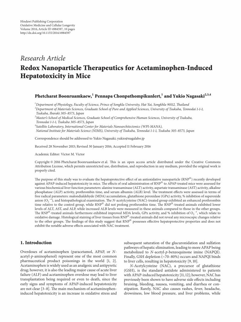
Research ArticleRedox Nanoparticle Therapeutics for Acetaminophen-InducedHepatotoxicity in Mice
Phetcharat Boonruamkaew,1 Pennapa Chonpathompikunlert,1 and Yukio Nagasaki2,3,4
1Department of Physiology, Faculty of Science, Prince of Songkla University, Hat Yai, Songkhla 90112, Thailand2Department of Materials Sciences, Graduate School of Pure and Applied Sciences, University of Tsukuba, Tennodai 1-1-1,Tsukuba, Ibaraki 305–8573, Japan3Master’s School of Medical Sciences, Graduate School of Comprehensive Human Sciences, University of Tsukuba,Tennodai 1-1-1, Tsukuba 305–8573, Japan4Satellite Laboratory, International Center for Materials Nanoarchitectonics (WPI-MANA),National Institute for Materials Science (NIMS), University of Tsukuba, Tennodai 1-1-1, Tsukuba 305–8573, Japan
Correspondence should be addressed to Yukio Nagasaki; [email protected]
Received 28 November 2015; Revised 30 January 2016; Accepted 11 February 2016
Academic Editor: Victor M. Victor
Copyright © 2016 Phetcharat Boonruamkaew et al. This is an open access article distributed under the Creative CommonsAttribution License, which permits unrestricted use, distribution, and reproduction in any medium, provided the original work isproperly cited.
The purpose of this study was to evaluate the hepatoprotective effect of an antioxidative nanoparticle (RNPN) recently developedagainst APAP-induced hepatotoxicity in mice. The effects of oral administration of RNPN to APAP-treated mice were assessed forvarious biochemical liver function parameters: alanine transaminase (ALT) activity, aspartate transaminase (AST) activity, alkalinephosphatase (ALP) activity, prothrombin time, and serum albumin (ALB) level. The treatment effects were assessed in terms offree radical parameters: malondialdehyde (MDA) accumulation, glutathione peroxidase (GPx) activity, % inhibition of superoxideanion (O
2
−∙), and histopathological examination. The N-acetylcysteine (NAC)-treated group exhibited an enhanced prothrombintime relative to the control group, while RNPN did not prolong prothrombin time. The RNPN-treated animals exhibited lowerlevels of ALT, AST, and ALP, while increased ALB levels were measured in these animals compared to those in the other groups.The RNPN-treated animals furthermore exhibited improved MDA levels, GPx activity, and % inhibition of O
2
−∙, which relate tooxidative damage. Histological staining of liver tissues from RNPN-treated animals did not reveal any microscopic changes relativeto the other groups. The findings of this study suggest that RNPN possesses effective hepatoprotective properties and does notexhibit the notable adverse effects associated with NAC treatment.
1. Introduction
Overdoses of acetaminophen (paracetamol, APAP, or N-acetyl-p-aminophenol) represent one of the most commonpharmaceutical product poisonings in the world [1, 2].Acetaminophen is widely used as an analgesic and antipyreticdrug; however, it is also the leading major cause of acute liverfailure (ALF) and acetaminophen overdose may lead to livertransplantation being required or even to death, since theearly signs and symptoms of APAP-induced hepatotoxicityare not clear [3–8]. The main mechanism of acetaminophen-induced hepatotoxicity is an increase in oxidative stress and
subsequent saturation of the glucuronidation and sulfationpathways of hepatic elimination, leading tomoreAPAP beingmetabolized to N-acetyl-p-benzoquinone imine (NAPQI).Finally, GSH depletion (∼70–80%) occurs and NAPQI bindsto liver cells, resulting in hepatotoxicity [9, 10].
N-Acetylcysteine (NAC), a precursor of glutathione(GSH), is the standard antidote administered to patientswithAPAP-induced hepatotoxicity [11, 12]; however, NAChaspreviously been shown to have adverse side effects includingbruising, bleeding, nausea, vomiting, and diarrhea or con-stipation. Rarely, NAC also causes rashes, fever, headache,drowsiness, low blood pressure, and liver problems, while
Hindawi Publishing CorporationOxidative Medicine and Cellular LongevityVolume 2016, Article ID 4984597, 10 pageshttp://dx.doi.org/10.1155/2016/4984597
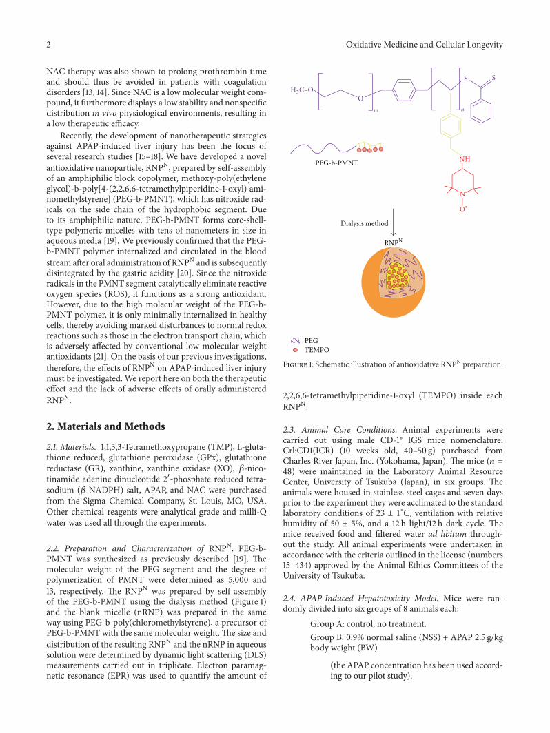
2 Oxidative Medicine and Cellular Longevity
NAC therapy was also shown to prolong prothrombin timeand should thus be avoided in patients with coagulationdisorders [13, 14]. Since NAC is a low molecular weight com-pound, it furthermore displays a low stability and nonspecificdistribution in vivo physiological environments, resulting ina low therapeutic efficacy.
Recently, the development of nanotherapeutic strategiesagainst APAP-induced liver injury has been the focus ofseveral research studies [15–18]. We have developed a novelantioxidative nanoparticle, RNPN, prepared by self-assemblyof an amphiphilic block copolymer, methoxy-poly(ethyleneglycol)-b-poly[4-(2,2,6,6-tetramethylpiperidine-1-oxyl) ami-nomethylstyrene] (PEG-b-PMNT), which has nitroxide rad-icals on the side chain of the hydrophobic segment. Dueto its amphiphilic nature, PEG-b-PMNT forms core-shell-type polymeric micelles with tens of nanometers in size inaqueous media [19]. We previously confirmed that the PEG-b-PMNT polymer internalized and circulated in the bloodstream after oral administration of RNPN and is subsequentlydisintegrated by the gastric acidity [20]. Since the nitroxideradicals in the PMNT segment catalytically eliminate reactiveoxygen species (ROS), it functions as a strong antioxidant.However, due to the high molecular weight of the PEG-b-PMNT polymer, it is only minimally internalized in healthycells, thereby avoiding marked disturbances to normal redoxreactions such as those in the electron transport chain, whichis adversely affected by conventional low molecular weightantioxidants [21]. On the basis of our previous investigations,therefore, the effects of RNPN on APAP-induced liver injurymust be investigated. We report here on both the therapeuticeffect and the lack of adverse effects of orally administeredRNPN.
2. Materials and Methods
2.1. Materials. 1,1,3,3-Tetramethoxypropane (TMP), L-gluta-thione reduced, glutathione peroxidase (GPx), glutathionereductase (GR), xanthine, xanthine oxidase (XO), 𝛽-nico-tinamide adenine dinucleotide 2-phosphate reduced tetra-sodium (𝛽-NADPH) salt, APAP, and NAC were purchasedfrom the Sigma Chemical Company, St. Louis, MO, USA.Other chemical reagents were analytical grade and milli-Qwater was used all through the experiments.
2.2. Preparation and Characterization of RNPN. PEG-b-PMNT was synthesized as previously described [19]. Themolecular weight of the PEG segment and the degree ofpolymerization of PMNT were determined as 5,000 and13, respectively. The RNPN was prepared by self-assemblyof the PEG-b-PMNT using the dialysis method (Figure 1)and the blank micelle (nRNP) was prepared in the sameway using PEG-b-poly(chloromethylstyrene), a precursor ofPEG-b-PMNT with the same molecular weight. The size anddistribution of the resulting RNPN and the nRNP in aqueoussolution were determined by dynamic light scattering (DLS)measurements carried out in triplicate. Electron paramag-netic resonance (EPR) was used to quantify the amount of
PEG TEMPO
Dialysis method
NH
N
O∙
n
S
O
S
m
H3C–O
RNPN
PEG-b-PMNT
Figure 1: Schematic illustration of antioxidative RNPN preparation.
2,2,6,6-tetramethylpiperidine-1-oxyl (TEMPO) inside eachRNPN.
2.3. Animal Care Conditions. Animal experiments werecarried out using male CD-1® IGS mice nomenclature:Crl:CD1(ICR) (10 weeks old, 40–50 g) purchased fromCharles River Japan, Inc. (Yokohama, Japan). The mice (𝑛 =48) were maintained in the Laboratory Animal ResourceCenter, University of Tsukuba (Japan), in six groups. Theanimals were housed in stainless steel cages and seven daysprior to the experiment they were acclimated to the standardlaboratory conditions of 23 ± 1∘C, ventilation with relativehumidity of 50 ± 5%, and a 12 h light/12 h dark cycle. Themice received food and filtered water ad libitum through-out the study. All animal experiments were undertaken inaccordance with the criteria outlined in the license (numbers15–434) approved by the Animal Ethics Committees of theUniversity of Tsukuba.
2.4. APAP-Induced Hepatotoxicity Model. Mice were ran-domly divided into six groups of 8 animals each:
Group A: control, no treatment.Group B: 0.9% normal saline (NSS) + APAP 2.5 g/kgbody weight (BW)
(the APAP concentration has been used accord-ing to our pilot study).
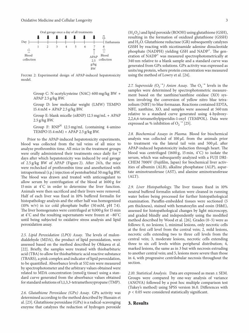
Oxidative Medicine and Cellular Longevity 3
87Day
APAP 2.5
g/kg BW
1 2 3 4 5 6
Oral gavage once a day of all treatments
Euthanasia
Blood collection
Blood collection
Figure 2: Experimental design of APAP-induced hepatotoxicitymodel.
Group C: N-acetylcysteine (NAC) 600mg/kg BW +APAP 2.5 g/kg BW.Group D: low molecular weight (LMW) TEMPO15.4mM + APAP 2.5 g/kg BW.Group E: blank micelle (nRNP) 12.5mg/mL + APAP2.5 g/kg BW.Group F: RNPN 12.5mg/mL (containing 4-aminoTEMPO 15.4mM) + APAP 2.5 g/kg BW.
Prior to the APAP-induced hepatotoxicity experiments,blood was collected from the tail veins of all mice toanalyze prothrombin time. All mice in the treatment groupswere orally administered their treatments once daily for 7days after which hepatotoxicity was induced by oral gavageof 2.5 g/kg BW of APAP (Figure 2). After 24 h, the micewere rechecked of prothrombin time and anesthetized withintraperitoneal (i.p.) injection of pentobarbital 50mg/kg BW.The blood was drawn and treated with anticoagulant toallow serum by centrifugation of the blood at 1600 g for15min at 4∘C in order to determine the liver function.Animals were then sacrificed and their livers were removed.Half of each liver was fixed in 10% buffered formalin forhistopathology analysis and the other half was homogenized(10% w/v) in ice cold phosphate buffer (50mM, pH 7.4).The liver homogenates were centrifuged at 9,000 g for 15minat 4∘C and the resulting supernatants were frozen at −80∘Cuntil being subjected to oxidative stress analysis and lipidperoxidation assay.
2.5. Lipid Peroxidation (LPO) Assay. The levels of malon-dialdehyde (MDA), the product of lipid peroxidation, wereassessed based on the method described by Ohkawa et al.[22]. Briefly, the samples were treated with thiobarbituricacid (TBA) to allow for thiobarbituric acid reactive substance(TBARS), a pink complex and indicator of lipid peroxidation,to be quantified. Absorbance levels at 532 nm were measuredby spectrophotometer and the arbitrary values obtained wererelated to MDA concentration (nmol/g tissue) using a stan-dard curve generated from the absorbance values obtainedfor standard solutions of 1,1,3,3-tetramethoxypropane (TMP).
2.6. Glutathione Peroxidase (GPx) Assay. GPx activity wasdetermined according to the method described by Hussain etal. [23]. Glutathione peroxidase (GPx) is a radical-scavengingenzyme that catalyzes the reduction of hydrogen peroxide
(H2O2) and lipid peroxide (ROOH) using glutathione (GSH),
resulting in the formation of oxidized glutathione (GSSH)and H
2O. Glutathione reductase (GR) subsequently catalyzes
GSSH by reacting with nicotinamide adenine dinucleotidephosphate (NADPH) yielding GSH and NADP+. The gen-eration of NADP+ was measured spectrophotometrically at340 nm relative to a blank sample and a standard curve wasgenerated from GPx solutions. GPx activity was expressed asunits/mg protein, where protein concentrationwasmeasuredusing the method of Lowry et al. [24].
2.7. Superoxide (𝑂2−∙) Anion Assay. The O2
−∙ levels in thesamples were determined by spectrophotometric measure-ment based on the xanthine/xanthine oxidase (XO) sys-tem involving the conversion of yellow nitro blue tetra-zolium (NBT) to blue formazan. Reactions contained EDTA,NBT, xanthine, XO, and samples were assessed at 560 nmrelative to a standard curve generated using 4-hydroxy-2,2,6,6-tetramethylpiperidin-1-oxyl (TEMPOL). Data wereexpressed as % inhibition of O
2
−∙ [25].
2.8. Biochemical Assays in Plasma. Blood for biochemicalanalysis was collected of 100𝜇L from the animals priorto treatment via the lateral tail vein and 500 𝜇L afterAPAP-induced hepatotoxicity induction through heart. Theblood was centrifuged (1600 g, 15min, 4∘C) to isolate theserum, which was subsequently analyzed with a FUJI DRI-CHEM 7000V (Fujifilm, Japan) for biochemical liver activ-ities of albumin (ALB), alkaline phosphatase (ALP), aspar-tate aminotransferase (AST), and alanine aminotransferase(ALT).
2.9. Liver Histopathology. The liver tissues fixed in 10%neutral buffered formalin solution were cleaned in runningwater before being processed for further histopathologicalexamination. Paraffin-embedded tissues were sectioned (5𝜇m thickness), stained with hematoxylin and eosin (H&E),assessed for histopathological changes by light microscopy,and graded blindly and independently using the modifiedmethod described by Wood et al. [26]. Grades (0–5) were asfollows: 0, no lesions; 1, minimal lesions, only necrotic cellsat the first cell level from the central vein; 2, mild lesions,necrotic cells extending two to three cell levels from thecentral vein; 3, moderate lesions, necrotic cells extendingthree to six cell levels within peripheral distribution; 4,marked lesions, the same as in 3 but with necrosis extrudingto another central vein; and 5, lesions more severe than thosein 4, with progressive centrilobular necrosis throughout thesection.
2.10. Statistical Analysis. Data are expressed as mean ± SEM.Groups were compared by one-way analysis of variance(ANOVA) followed by a post hoc multiple comparison test(Tukey’s method) using SPSS version 16.0. Differences with𝑝 < 0.05 were considered statistically significant.
3. Results
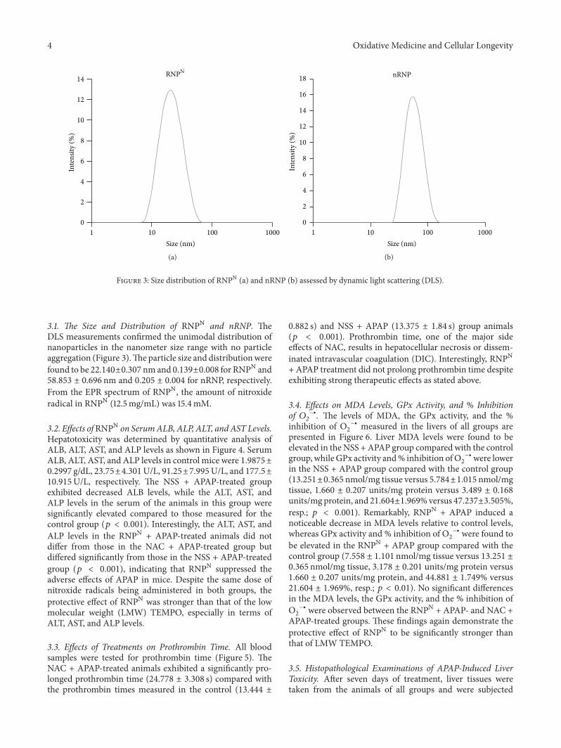
4 Oxidative Medicine and Cellular Longevity
0
2
4
6
8
10
12
14
1 10 100 1000
Inte
nsity
(%)
Size (nm)
RNPN
(a)
0
2
4
6
8
10
12
14
16
18
1 10 100 1000
Inte
nsity
(%)
Size (nm)
nRNP
(b)
Figure 3: Size distribution of RNPN (a) and nRNP (b) assessed by dynamic light scattering (DLS).
3.1. The Size and Distribution of RNPN and nRNP. TheDLS measurements confirmed the unimodal distribution ofnanoparticles in the nanometer size range with no particleaggregation (Figure 3).Theparticle size and distributionwerefound to be 22.140±0.307 nmand 0.139±0.008 for RNPN and58.853 ± 0.696 nm and 0.205 ± 0.004 for nRNP, respectively.From the EPR spectrum of RNPN, the amount of nitroxideradical in RNPN (12.5mg/mL) was 15.4mM.
3.2. Effects ofRNPN on SerumALB, ALP, ALT, andAST Levels.Hepatotoxicity was determined by quantitative analysis ofALB, ALT, AST, and ALP levels as shown in Figure 4. SerumALB, ALT, AST, and ALP levels in control mice were 1.9875±0.2997 g/dL, 23.75±4.301U/L, 91.25±7.995U/L, and 177.5±10.915U/L, respectively. The NSS + APAP-treated groupexhibited decreased ALB levels, while the ALT, AST, andALP levels in the serum of the animals in this group weresignificantly elevated compared to those measured for thecontrol group (𝑝 < 0.001). Interestingly, the ALT, AST, andALP levels in the RNPN + APAP-treated animals did notdiffer from those in the NAC + APAP-treated group butdiffered significantly from those in the NSS + APAP-treatedgroup (𝑝 < 0.001), indicating that RNPN suppressed theadverse effects of APAP in mice. Despite the same dose ofnitroxide radicals being administered in both groups, theprotective effect of RNPN was stronger than that of the lowmolecular weight (LMW) TEMPO, especially in terms ofALT, AST, and ALP levels.
3.3. Effects of Treatments on Prothrombin Time. All bloodsamples were tested for prothrombin time (Figure 5). TheNAC + APAP-treated animals exhibited a significantly pro-longed prothrombin time (24.778 ± 3.308 s) compared withthe prothrombin times measured in the control (13.444 ±
0.882 s) and NSS + APAP (13.375 ± 1.84 s) group animals(𝑝 < 0.001). Prothrombin time, one of the major sideeffects of NAC, results in hepatocellular necrosis or dissem-inated intravascular coagulation (DIC). Interestingly, RNPN+ APAP treatment did not prolong prothrombin time despiteexhibiting strong therapeutic effects as stated above.
3.4. Effects on MDA Levels, GPx Activity, and % Inhibitionof 𝑂2−∙. The levels of MDA, the GPx activity, and the %inhibition of O
2
−∙ measured in the livers of all groups arepresented in Figure 6. Liver MDA levels were found to beelevated in theNSS +APAP group compared with the controlgroup, whileGPx activity and% inhibition ofO
2
−∙ were lowerin the NSS + APAP group compared with the control group(13.251±0.365 nmol/mg tissue versus 5.784±1.015 nmol/mgtissue, 1.660 ± 0.207 units/mg protein versus 3.489 ± 0.168units/mg protein, and 21.604±1.969%versus 47.237±3.505%,resp.; 𝑝 < 0.001). Remarkably, RNPN + APAP induced anoticeable decrease in MDA levels relative to control levels,whereas GPx activity and % inhibition of O
2
−∙ were found tobe elevated in the RNPN + APAP group compared with thecontrol group (7.558 ± 1.101 nmol/mg tissue versus 13.251 ±0.365 nmol/mg tissue, 3.178 ± 0.201 units/mg protein versus1.660 ± 0.207 units/mg protein, and 44.881 ± 1.749% versus21.604 ± 1.969%, resp.; 𝑝 < 0.01). No significant differencesin the MDA levels, the GPx activity, and the % inhibition ofO2
−∙ were observed between the RNPN + APAP- and NAC +APAP-treated groups. These findings again demonstrate theprotective effect of RNPN to be significantly stronger thanthat of LMW TEMPO.
3.5. Histopathological Examinations of APAP-Induced LiverToxicity. After seven days of treatment, liver tissues weretaken from the animals of all groups and were subjected
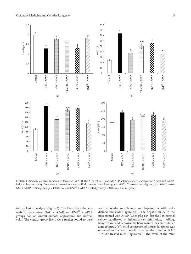
Oxidative Medicine and Cellular Longevity 5
# #
RNPN
+ A
PAP
Con
trol
NSS
+ A
PAP
NAC
+ A
PAP
TEM
PO +
APA
P
nRN
P +
APA
P
∗∗
0
0.5
1
1.5
2
2.5Le
vel (
g/dL
)
(a)
RNPN
+ A
PAP
Con
trol
NSS
+ A
PAP
NAC
+ A
PAP
TEM
PO +
APA
P
nRN
P +
APA
P
∗, o∗
∗
# #
0
10
20
30
40
50
60
70
80
90
Leve
l (U
/L)
(b)
RNPN
+ A
PAP
Con
trol
NSS
+ A
PAP
NAC
+ A
PAP
TEM
PO +
APA
P
nRN
P +
APA
P
∗, o
##
∗∗
0
20
40
60
80
100
120
140
160
180
200
Leve
l (U
/L)
(c)
RNPN
+ A
PAP
Con
trol
NSS
+ A
PAP
NAC
+ A
PAP
TEM
PO +
APA
P
nRN
P +
APA
P
∗∗, o# #
∗∗
0
50
100
150
200
250
300
Leve
l (U
/L)
(d)
Figure 4: Biochemical liver function in terms of (a) ALB, (b) ALT, (c) AST, and (d) ALP activities after treatment for 7 days and APAP-induced hepatotoxicity. Data were expressed as mean ± SEM, ∗versus control group, 𝑝 < 0.001; ∗∗versus control group, 𝑝 < 0.01; #versusNSS + APAP-treated group, 𝑝 < 0.001; oversus RNPN + APAP-treated group, 𝑝 < 0.05; 𝑛 = 8mice/group.
to histological analysis (Figure 7). The livers from the ani-mals in the control, NAC + APAP, and RNPN + APAPgroups had an overall smooth appearance and normalcolor. The control group livers were further found to have
normal lobular morphology and hepatocytes with well-defined sinusoids (Figure 7(a)). The hepatic injury in themice treated with APAP (2.5mg/kg BW dissolved in normalsaline) manifested as inflammatory infiltration, swelling,hemorrhage, and necrosis involving mainly the centrilobularzone (Figure 7(b)). Mild congestion of sinusoidal spaces wasobserved in the centrilobular area of the livers of NAC+ APAP-treated mice (Figure 7(c)). The livers of the mice
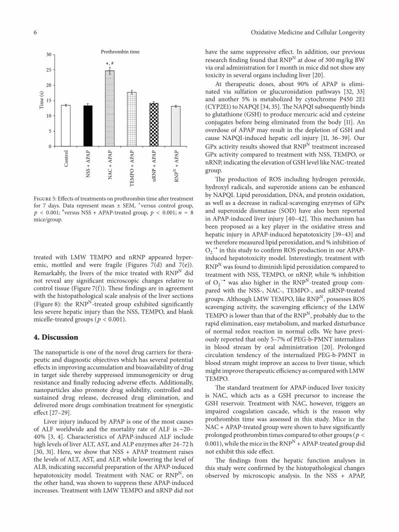
6 Oxidative Medicine and Cellular Longevity
0
5
10
15
20
25
30
Tim
e (s)
Prothrombin time
RNPN
+ A
PAP
Con
trol
NSS
+ A
PAP
NAC
+ A
PAP
TEM
PO +
APA
P
nRN
P +
APA
P
∗, #
Figure 5: Effects of treatments on prothrombin time after treatmentfor 7 days. Data represent mean ± SEM, ∗versus control group,𝑝 < 0.001; #versus NSS + APAP-treated group, 𝑝 < 0.001; 𝑛 = 8mice/group.
treated with LMW TEMPO and nRNP appeared hyper-emic, mottled and were fragile (Figures 7(d) and 7(e)).Remarkably, the livers of the mice treated with RNPN didnot reveal any significant microscopic changes relative tocontrol tissue (Figure 7(f)). These findings are in agreementwith the histopathological scale analysis of the liver sections(Figure 8): the RNPN-treated group exhibited significantlyless severe hepatic injury than the NSS, TEMPO, and blankmicelle-treated groups (𝑝 < 0.001).
4. Discussion
The nanoparticle is one of the novel drug carriers for thera-peutic and diagnostic objectives which has several potentialeffects in improving accumulation and bioavailability of drugin target side thereby suppressed immunogenicity or drugresistance and finally reducing adverse effects. Additionally,nanoparticles also promote drug solubility, controlled andsustained drug release, decreased drug elimination, anddelivered more drugs combination treatment for synergisticeffect [27–29].
Liver injury induced by APAP is one of the most causesof ALF worldwide and the mortality rate of ALF is ∼20–40% [3, 4]. Characteristics of APAP-induced ALF includehigh levels of liver ALT, AST, and ALP enzymes after 24–72 h[30, 31]. Here, we show that NSS + APAP treatment raisesthe levels of ALT, AST, and ALP, while lowering the level ofALB, indicating successful preparation of the APAP-inducedhepatotoxicity model. Treatment with NAC or RNPN, onthe other hand, was shown to suppress these APAP-inducedincreases. Treatment with LMW TEMPO and nRNP did not
have the same suppressive effect. In addition, our previousresearch finding found that RNPN at dose of 300mg/kg BWvia oral administration for 1 month in mice did not show anytoxicity in several organs including liver [20].
At therapeutic doses, about 90% of APAP is elimi-nated via sulfation or glucuronidation pathways [32, 33]and another 5% is metabolized by cytochrome P450 2E1(CYP2E1) toNAPQI [34, 35].TheNAPQI subsequently bindsto glutathione (GSH) to produce mercuric acid and cysteineconjugates before being eliminated from the body [11]. Anoverdose of APAP may result in the depletion of GSH andcause NAPQI-induced hepatic cell injury [11, 36–39]. OurGPx activity results showed that RNPN treatment increasedGPx activity compared to treatment with NSS, TEMPO, ornRNP, indicating the elevation of GSH level like NAC-treatedgroup.
The production of ROS including hydrogen peroxide,hydroxyl radicals, and superoxide anions can be enhancedby NAPQI. Lipid peroxidation, DNA, and protein oxidation,as well as a decrease in radical-scavenging enzymes of GPxand superoxide dismutase (SOD) have also been reportedin APAP-induced liver injury [40–42]. This mechanism hasbeen proposed as a key player in the oxidative stress andhepatic injury in APAP-induced hepatotoxicity [39–43] andwe thereforemeasured lipid peroxidation, and% inhibition ofO2
−∙ in this study to confirm ROS production in our APAP-induced hepatotoxicity model. Interestingly, treatment withRNPN was found to diminish lipid peroxidation compared totreatment with NSS, TEMPO, or nRNP, while % inhibitionof O2
−∙ was also higher in the RNPN-treated group com-pared with the NSS-, NAC-, TEMPO-, and nRNP-treatedgroups. Although LMW TEMPO, like RNPN, possesses ROSscavenging activity, the scavenging efficiency of the LMWTEMPO is lower than that of the RNPN, probably due to therapid elimination, easy metabolism, and marked disturbanceof normal redox reaction in normal cells. We have previ-ously reported that only 5–7% of PEG-b-PMNT internalizesin blood stream by oral administration [20]. Prolongedcirculation tendency of the internalized PEG-b-PMNT inblood stream might improve an access to liver tissue, whichmight improve therapeutic efficiency as comparedwith LMWTEMPO.
The standard treatment for APAP-induced liver toxicityis NAC, which acts as a GSH precursor to increase theGSH reservoir. Treatment with NAC, however, triggers animpaired coagulation cascade, which is the reason whyprothrombin time was assessed in this study. Mice in theNAC + APAP-treated group were shown to have significantlyprolonged prothrombin times compared to other groups (𝑝 <0.001), while themice in the RNPN +APAP-treated group didnot exhibit this side effect.
The findings from the hepatic function analyses inthis study were confirmed by the histopathological changesobserved by microscopic analysis. In the NSS + APAP,
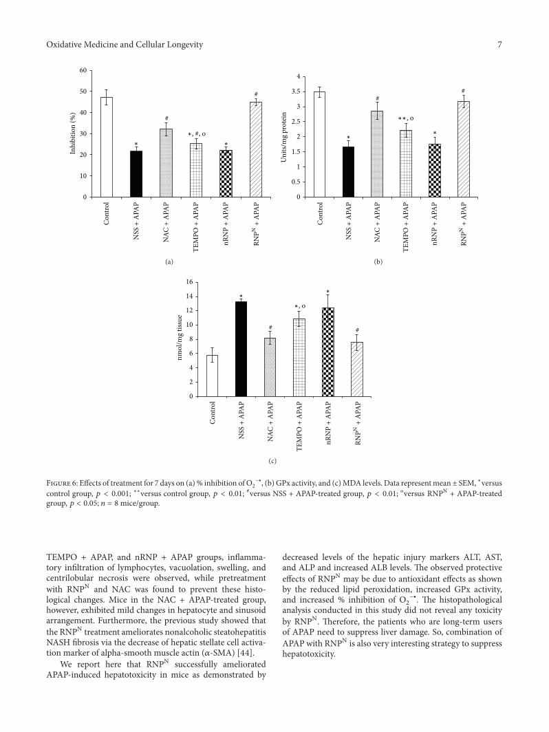
Oxidative Medicine and Cellular Longevity 7
0
10
20
30
40
50
60In
hibi
tion
(%)
RNPN
+ A
PAP
Con
trol
NSS
+ A
PAP
NAC
+ A
PAP
TEM
PO +
APA
P
nRN
P +
APA
P
∗, #, o
#
#
∗ ∗
(a)
0
0.5
1
1.5
2
2.5
3
3.5
4
Uni
ts/m
g pr
otei
n
RNPN
+ A
PAP
Con
trol
NSS
+ A
PAP
NAC
+ A
PAP
TEM
PO +
APA
P
nRN
P +
APA
P
∗∗, o
##
∗∗
(b)
0
2
4
6
8
10
12
14
16
nmol
/mg
tissu
e
RNPN
+ A
PAP
Con
trol
NSS
+ A
PAP
NAC
+ A
PAP
TEM
PO +
APA
P
nRN
P +
APA
P
∗, o
##
∗∗
(c)
Figure 6: Effects of treatment for 7 days on (a)% inhibition ofO2
−∙, (b) GPx activity, and (c)MDA levels. Data representmean± SEM, ∗versuscontrol group, 𝑝 < 0.001; ∗∗versus control group, 𝑝 < 0.01; #versus NSS + APAP-treated group, 𝑝 < 0.01; oversus RNPN + APAP-treatedgroup, 𝑝 < 0.05; 𝑛 = 8mice/group.
TEMPO + APAP, and nRNP + APAP groups, inflamma-tory infiltration of lymphocytes, vacuolation, swelling, andcentrilobular necrosis were observed, while pretreatmentwith RNPN and NAC was found to prevent these histo-logical changes. Mice in the NAC + APAP-treated group,however, exhibited mild changes in hepatocyte and sinusoidarrangement. Furthermore, the previous study showed thatthe RNPN treatment ameliorates nonalcoholic steatohepatitisNASH fibrosis via the decrease of hepatic stellate cell activa-tion marker of alpha-smooth muscle actin (𝛼-SMA) [44].
We report here that RNPN successfully amelioratedAPAP-induced hepatotoxicity in mice as demonstrated by
decreased levels of the hepatic injury markers ALT, AST,and ALP and increased ALB levels. The observed protectiveeffects of RNPN may be due to antioxidant effects as shownby the reduced lipid peroxidation, increased GPx activity,and increased % inhibition of O
2
−∙. The histopathologicalanalysis conducted in this study did not reveal any toxicityby RNPN. Therefore, the patients who are long-term usersof APAP need to suppress liver damage. So, combination ofAPAP with RNPN is also very interesting strategy to suppresshepatotoxicity.
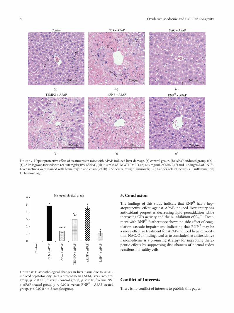
8 Oxidative Medicine and Cellular Longevity
Control
S CV
KC
200𝜇m
(a)
NSS + APAP
H
N
I
200𝜇m
(b)
NAC + APAP
200𝜇m
(c)
TEMPO + APAP
200𝜇m
(d)
nRNP + APAP
200𝜇m
(e)
RNPN + APAP
200𝜇m
(f)
Figure 7: Hepatoprotective effect of treatments in mice with APAP-induced liver damage. (a) control group. (b) APAP-induced group. ((c)–(f)) APAP group treatedwith (c) 600mg/kgBWofNAC, (d) 15.4mMof LMWTEMPO, (e) 12.5mg/mLof nRNP, (f) and 12.5mg/mLof RNPN.Liver sections were stained with hematoxylin and eosin (×400). CV: central vein; S: sinusoids; KC; Kupffer cell; N: necrosis; I: inflammation;H: hemorrhage.
0
1
2
3
4
5
6 Histopathological grade
RNPN
+ A
PAP
Con
trol
NSS
+ A
PAP
NAC
+ A
PAP
TEM
PO +
APA
P
nRN
P +
APA
P
∗, o
∗∗, ##
∗∗
Figure 8: Histopathological changes in liver tissue due to APAP-induced hepatotoxicity. Data representmean± SEM, ∗versus controlgroup, 𝑝 < 0.001; ∗∗versus control group, 𝑝 < 0.05; #versus NSS+ APAP-treated group, 𝑝 < 0.001; oversus RNPN + APAP-treatedgroup, 𝑝 < 0.001; 𝑛 = 5 samples/group.
5. Conclusion
The findings of this study indicate that RNPN has a hep-atoprotective effect against APAP-induced liver injury viaantioxidant properties decreasing lipid peroxidation whileincreasing GPx activity and the % inhibition of O
2
−∙. Treat-ment with RNPN furthermore shows no side effect of coag-ulation cascade impairment, indicating that RNPN may bea more effective treatment for APAP-induced hepatotoxicitythanNAC.Our findings lead us to conclude that antioxidativenanomedicine is a promising strategy for improving thera-peutic effects by suppressing disturbances of normal redoxreactions in healthy cells.
Conflict of Interests
There is no conflict of interests to publish this paper.
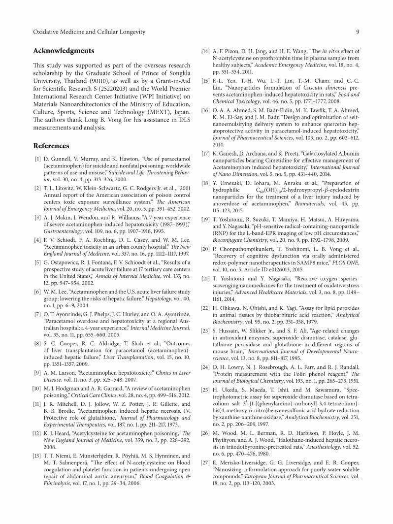
Oxidative Medicine and Cellular Longevity 9
Acknowledgments
This study was supported as part of the overseas researchscholarship by the Graduate School of Prince of SongklaUniversity, Thailand (90110), as well as by a Grant-in-Aidfor Scientific Research S (25220203) and the World PremierInternational Research Center Initiative (WPI Initiative) onMaterials Nanoarchitectonics of the Ministry of Education,Culture, Sports, Science and Technology (MEXT), Japan.The authors thank Long B. Vong for his assistance in DLSmeasurements and analysis.
References
[1] D. Gunnell, V. Murray, and K. Hawton, “Use of paracetamol(acetaminophen) for suicide andnonfatal poisoning:worldwidepatterns of use andmisuse,” Suicide and Life-Threatening Behav-ior, vol. 30, no. 4, pp. 313–326, 2000.
[2] T. L. Litovitz, W. Klein-Schwartz, G. C. Rodgers Jr. et al., “2001Annual report of the American association of poison controlcenters toxic exposure surveillance system,” The AmericanJournal of Emergency Medicine, vol. 20, no. 5, pp. 391–452, 2002.
[3] A. J. Makin, J. Wendon, and R. Williams, “A 7-year experienceof severe acetaminophen-induced hepatotoxicity (1987–1993),”Gastroenterology, vol. 109, no. 6, pp. 1907–1916, 1995.
[4] F. V. Schiødt, F. A. Rochling, D. L. Casey, and W. M. Lee,“Acetaminophen toxicity in an urban county hospital,”TheNewEngland Journal of Medicine, vol. 337, no. 16, pp. 1112–1117, 1997.
[5] G. Ostapowicz, R. J. Fontana, F. V. Schioødt et al., “Results of aprospective study of acute liver failure at 17 tertiary care centersin the United States,” Annals of Internal Medicine, vol. 137, no.12, pp. 947–954, 2002.
[6] W.M. Lee, “Acetaminophen and theU.S. acute liver failure studygroup: lowering the risks of hepatic failure,”Hepatology, vol. 40,no. 1, pp. 6–9, 2004.
[7] O. T. Ayonrinde, G. J. Phelps, J. C. Hurley, and O. A. Ayonrinde,“Paracetamol overdose and hepatotoxicity at a regional Aus-tralian hospital: a 4-year experience,” Internal Medicine Journal,vol. 35, no. 11, pp. 655–660, 2005.
[8] S. C. Cooper, R. C. Aldridge, T. Shah et al., “Outcomesof liver transplantation for paracetamol (acetaminophen)-induced hepatic failure,” Liver Transplantation, vol. 15, no. 10,pp. 1351–1357, 2009.
[9] A. M. Larson, “Acetaminophen hepatotoxicity,” Clinics in LiverDisease, vol. 11, no. 3, pp. 525–548, 2007.
[10] M. J. Hodgman and A. R. Garrard, “A review of acetaminophenpoisoning,”Critical CareClinics, vol. 28, no. 4, pp. 499–516, 2012.
[11] J. R. Mitchell, D. J. Jollow, W. Z. Potter, J. R. Gillette, andB. B. Brodie, “Acetaminophen induced hepatic necrosis. IV.Protective role of glutathione,” Journal of Pharmacology andExperimental Therapeutics, vol. 187, no. 1, pp. 211–217, 1973.
[12] K. J. Heard, “Acetylcysteine for acetaminophen poisoning,”TheNew England Journal of Medicine, vol. 359, no. 3, pp. 228–292,2008.
[13] T. T. Niemi, E. Munsterhjelm, R. Pöyhiä, M. S. Hynninen, andM. T. Salmenperä, “The effect of N-acetylcysteine on bloodcoagulation and platelet function in patients undergoing openrepair of abdominal aortic aneurysm,” Blood Coagulation &Fibrinolysis, vol. 17, no. 1, pp. 29–34, 2006.
[14] A. F. Pizon, D. H. Jang, and H. E. Wang, “The in vitro effect ofN-acetylcysteine on prothrombin time in plasma samples fromhealthy subjects,” Academic Emergency Medicine, vol. 18, no. 4,pp. 351–354, 2011.
[15] F.-L. Yen, T.-H. Wu, L.-T. Lin, T.-M. Cham, and C.-C.Lin, “Nanoparticles formulation of Cuscuta chinensis pre-vents acetaminophen-induced hepatotoxicity in rats,” Food andChemical Toxicology, vol. 46, no. 5, pp. 1771–1777, 2008.
[16] O. A. A. Ahmed, S. M. Badr-Eldin, M. K. Tawfik, T. A. Ahmed,K. M. El-Say, and J. M. Badr, “Design and optimization of self-nanoemulsifying delivery system to enhance quercetin hep-atoprotective activity in paracetamol-induced hepatotoxicity,”Journal of Pharmaceutical Sciences, vol. 103, no. 2, pp. 602–612,2014.
[17] K. Ganesh, D. Archana, and K. Preeti, “Galactosylated Albuminnanoparticles bearing Cimetidine for effective management ofAcetaminophen induced hepatotoxicity,” International Journalof Nano Dimension, vol. 5, no. 5, pp. 431–440, 2014.
[18] Y. Umezaki, D. Iohara, M. Anraku et al., “Preparation ofhydrophilic C
60(OH)
10/2-hydroxypropyl-𝛽-cyclodextrin
nanoparticles for the treatment of a liver injury induced byanoverdose of acetaminophen,” Biomaterials, vol. 45, pp.115–123, 2015.
[19] T. Yoshitomi, R. Suzuki, T. Mamiya, H. Matsui, A. Hirayama,and Y. Nagasaki, “pH-sensitive radical-containing-nanoparticle(RNP) for the L-band-EPR imaging of low pH circumstances,”Bioconjugate Chemistry, vol. 20, no. 9, pp. 1792–1798, 2009.
[20] P. Chonpathompikunlert, T. Yoshitomi, L. B. Vong et al.,“Recovery of cognitive dysfunction via orally administeredredox-polymer nanotherapeutics in SAMP8 mice,” PLOS ONE,vol. 10, no. 5, Article ID e0126013, 2015.
[21] T. Yoshitomi and Y. Nagasaki, “Reactive oxygen species-scavenging nanomedicines for the treatment of oxidative stressinjuries,” Advanced Healthcare Materials, vol. 3, no. 8, pp. 1149–1161, 2014.
[22] H. Ohkawa, N. Ohishi, and K. Yagi, “Assay for lipid peroxidesin animal tissues by thiobarbituric acid reaction,” AnalyticalBiochemistry, vol. 95, no. 2, pp. 351–358, 1979.
[23] S. Hussain, W. Slikker Jr., and S. F. Ali, “Age-related changesin antioxidant enzymes, superoxide dismutase, catalase, glu-tathione peroxidase and glutathione in different regions ofmouse brain,” International Journal of Developmental Neuro-science, vol. 13, no. 8, pp. 811–817, 1995.
[24] O. H. Lowry, N. J. Rosebrough, A. L. Farr, and R. J. Randall,“Protein measurement with the Folin phenol reagent,” TheJournal of Biological Chemistry, vol. 193, no. 1, pp. 265–275, 1951.
[25] H. Ukeda, S. Maeda, T. Ishii, and M. Sawamura, “Spec-trophotometric assay for superoxide dismutase based on tetra-zolium salt 3-{1-[(phenylamino)-carbonyl]-3,4-tetrazolium}-bis(4-methoxy-6-nitro)benzenesulfonic acid hydrate reductionby xanthine-xanthine oxidase,”Analytical Biochemistry, vol. 251,no. 2, pp. 206–209, 1997.
[26] M. Wood, M. L. Berman, R. D. Harbison, P. Hoyle, J. M.Phythyon, and A. J. Wood, “Halothane-induced hepatic necro-sis in triiodothyronine-pretreated rats,” Anesthesiology, vol. 52,no. 6, pp. 470–476, 1980.
[27] E. Merisko-Liversidge, G. G. Liversidge, and E. R. Cooper,“Nanosizing: a formulation approach for poorly-water-solublecompounds,” European Journal of Pharmaceutical Sciences, vol.18, no. 2, pp. 113–120, 2003.
10 Oxidative Medicine and Cellular Longevity
[28] L. Zhang, F. X. Gu, J. M. Chan, A. Z. Wang, R. S. Langer, and O.C. Farokhzad, “Nanoparticles in medicine: therapeutic applica-tions and developments,”Clinical Pharmacology&Therapeutics,vol. 83, no. 5, pp. 761–769, 2008.
[29] M. Ferrari, “Frontiers in cancer nanomedicine: directing masstransport through biological barriers,” Trends in Biotechnology,vol. 28, no. 4, pp. 181–188, 2010.
[30] D. G. Davidson and W. N. Eastham, “Acute liver necrosisfollowing overdose of paracetamol,”TheBritishMedical Journal,vol. 5512, pp. 497–499, 1966.
[31] D. C. Dahlin, G. T. Miwa, A. Y. Lu, and S. D. Nelson, “N-acetyl-p-benzoquinone imine: a cytochrome P-450-mediated oxida-tion product of acetaminophen,” Proceedings of the NationalAcademy of Sciences of the United States of America, vol. 81, no.5, pp. 1327–1331, 1984.
[32] D. J. Jollow, S. S. Thorgeirsson, W. Z. Potter, M. Hashimoto,and J. R. Mitchell, “Acetaminophen induced hepatic necrosis.VI. Metabolic disposition of toxic and nontoxic doses ofacetaminophen,” Pharmacology, vol. 12, no. 4-5, pp. 251–271,1974.
[33] J. T. Slattery and G. Levy, “Acetaminophen kinetics in acutelypoisoned patients,” Clinical Pharmacology & Therapeutics, vol.25, no. 2, pp. 184–195, 1979.
[34] S. D. Nelson, “Molecular mechanisms of the hepatotoxicitycaused by acetaminophen,” Seminars in Liver Disease, vol. 10,no. 4, pp. 267–278, 1990.
[35] J. D. Gibson, N. R. Pumford, V. M. Samokyszyn, and J. A.Hinson, “Mechanism of acetaminophen-induced hepatotoxic-ity: covalent binding versus oxidative stress,” Chemical Researchin Toxicology, vol. 9, no. 3, pp. 580–585, 1996.
[36] W. Z. Potter, S. S. Thorgeirsson, D. J. Jollow, and J. R. Mitchell,“Acetaminophen-induced hepatic necrosis. V. Correlation ofhepatic necrosis, covalent binding and glutathione depletion inhamsters,” Pharmacology, vol. 12, no. 3, pp. 129–143, 1974.
[37] D. J. Miner and P. T. Kissinger, “Evidence for the involvementof N-acetyl-p-quinoneimine in acetaminophen metabolism,”Biochemical Pharmacology, vol. 28, no. 22, pp. 3285–3290, 1979.
[38] J. G. M. Bessems and N. P. E. Vermeulen, “Paracetamol(acetaminophen)-induced toxicity: molecular and biochemicalmechanisms, analogues and protective approaches,” CriticalReviews in Toxicology, vol. 31, no. 1, pp. 55–138, 2001.
[39] L. P. James, P. R. Mayeux, and J. A. Hinson, “Acetaminophen-induced hepatotoxicity,” Drug Metabolism and Disposition, vol.31, no. 12, pp. 1499–1506, 2003.
[40] S. L. Michael, N. R. Pumford, P. R. Mayeux, M. R. Niesman,and J. A. Hinson, “Pretreatment of mice with macrophageinactivators decreases acetaminophen hepatotoxicity and theformation of reactive oxygen and nitrogen species,”Hepatology,vol. 30, no. 1, pp. 186–195, 1999.
[41] J. A. Hinson, T. J. Bucci, L. K. Irwin, S. L. Michael, and P.R. Mayeux, “Effect of inhibitors of nitric oxide synthase onacetaminophen-induced hepatotoxicity in mice,” Nitric Oxide,vol. 6, no. 2, pp. 160–167, 2002.
[42] J. A. Hinson, A. B. Reid, S. S. McCullough, and L. P. James,“Acetaminophen-induced hepatotoxicity: role of metabolicactivation, reactive oxygen/nitrogen species, andmitochondrialpermeability transition,” Drug Metabolism Reviews, vol. 36, no.3-4, pp. 805–822, 2004.
[43] H. Jaeschke, T. R. Knight, and M. L. Bajt, “The role ofoxidant stress and reactive nitrogen species in acetaminophenhepatotoxicity,” Toxicology Letters, vol. 144, no. 3, pp. 279–288,2003.
[44] A. Eguchi, T. Yoshitomi, M. Lazic et al., “Redox nanoparticlesas a novel treatment approach for inflammation and fibrosisassociated with nonalcoholic steatohepatitis,” Nanomedicine,vol. 10, no. 17, pp. 2697–2708, 2015.
Review ArticleThe Dual Function of Reactive Oxygen/Nitrogen Species inBioenergetics and Cell Death: The Role of ATP Synthase
Nina Kaludercic and Valentina Giorgio
Neuroscience Institute, National Research Council of Italy (CNR), Via Ugo Bassi 58/B, 35131 Padova, Italy
Correspondence should be addressed to Valentina Giorgio; [email protected]
Received 23 December 2015; Accepted 15 February 2016
Academic Editor: Tanea T. Reed
Copyright © 2016 N. Kaludercic and V. Giorgio.This is an open access article distributed under the Creative Commons AttributionLicense, which permits unrestricted use, distribution, and reproduction in anymedium, provided the originalwork is properly cited.
Reactive oxygen species (ROS) and reactive nitrogen species (RNS) targeting mitochondria are major causative factors in diseasepathogenesis. The mitochondrial permeability transition pore (PTP) is a mega-channel modulated by calcium and ROS/RNSmodifications and it has been described to play a crucial role in many pathophysiological events since prolonged channelopening causes cell death. The recent identification that dimers of ATP synthase form the PTP and the fact that posttranslationalmodifications caused by ROS/RNS also affect cellular bioenergetics through the modulation of ATP synthase catalysis reveal adual function of these modifications in the cells. Here, we describe mitochondria as a major site of production and as a target ofROS/RNS and discuss the pathophysiological conditions in which oxidative and nitrosative modifications modulate the catalyticand pore-forming activities of ATP synthase.
1. Introduction
Reactive oxygen species (ROS) and reactive nitrogen species(RNS) play important physiological functions but can alsocause extensive cellular damage, in a balance that is deter-mined by their relative rates of formation and removal.Usually, these species are removed rapidly before they causecell dysfunction and death. Oxidative/nitrosative stress gen-erated by an imbalance between formation of ROS/RNSand antioxidant defense capacity can affect major cellularcomponents, including lipids, proteins, carbohydrates, andDNA.Mitochondria are recognized as a critical site in the cellfor the formation of ROS/RNS and as their target.
Mitochondrial processes are highly compartmentalizedbecause of the existence of two limiting membranes allowingthe selective localization of proteins, nucleotides, and coen-zymes in the intermembrane and matrix spaces. The outermitochondrial membrane (OMM) is the interface betweenmitochondria and the cell components and its permeabi-lization is essential to allow the release of mitochondrialproteins involved in apoptosis such as cytochrome c [1]. Theinner mitochondrial membrane (IMM), whose permeabilityto solutes is controlled by highly specific transporters andtightly regulated channels, is the site of coupling between
substrate oxidation and ATP synthesis in the process ofoxidative phosphorylation. Mitochondria operate a sequenceof energy conversion processes through which the exergonicflow of electrons along the respiratory complexes supportsthe endergonic pumping of protons from the matrix to theintermembrane space. The resulting proton motive forcedrives the rotation of the FO sector of ATP synthase leadingto the synthesis of ATP in the F
1sector, but the electron
flow through the respiratory chain also generates ROS/RNS.In addition, the mitochondrial permeability transition pore(PTP), a large-conductance channel, is also located at the levelof the IMM and prolonged opening of this channel leads tomitochondrial swelling, rupture of the OMM, and cell death[2]. PTP opening is dependent on the presence of matrixcalcium, but the threshold calcium load which is requiredis modulated by inducers of the pore such as oxidants [3].Although the existence of the PTP was established as earlyas the 1970s [4–6], its molecular nature has been the subjectof controversy as many potential components were ruledout by the use of targeted gene deletion in mice [2]. Theonly candidate remaining is cyclophilin D (CyPD), whichwas found to act not as a structural component of the porebut as a modulator whose binding to the PTP decreasesthe threshold calcium concentration necessary to induce
Hindawi Publishing CorporationOxidative Medicine and Cellular LongevityVolume 2016, Article ID 3869610, 17 pageshttp://dx.doi.org/10.1155/2016/3869610
2 Oxidative Medicine and Cellular Longevity
permeability transition [7–10]. CyPD was shown to interactwith the lateral stalk of the ATP synthase in mammals[11], a finding which was the basis for the characterizationof the molecular structure of the PTP as formed by ATPsynthase itself [12–16]. Genetic ablation of the Ppif gene(which encodes for CyPD) in the mouse or its displacementfrom the PTP by the treatment with cyclosporin A (CsA), aknown inhibitor of the PTP, has been also used to demon-strate the important role of PTP in the pathophysiologicalmechanism of several diseases such as neurodegenerativediseases, muscular dystrophies, ischemia/reperfusion (I/R),and diabetes [2, 17, 18]. Besides the PTP, mitochondrialfunction and bioenergetics (including the modulation of thecatalytic activity of ATP synthase) are also affected in mostof these pathophysiological conditions and ROS/RNS arepresumably involved as causative factors.
While numerous mechanisms of oxidant-induced injuryhave been identified, the impact of oxidants on themitochon-drial proteome has been investigated only recently. Oxidativeor nitrosative stressmay not only alter levels ofmitochondrialproteins, but also induce posttranslational modifications ofproteins. These modifications involve reversible changes atthe level of cysteine, tyrosine, methionine, histidine, andtryptophan residues and irreversible protein carbonylation[19]. Thiol groups can be S-nitrosylated by nitric oxide (NO)or reversibly oxidized by ROS to form disulfide bonds orsulfenic acid; the latter can be further oxidized to sulfinicand sulfonic acids [20]. Sulfenic acid can also interact withglutathione to become glutathionylated. Tyrosine residuesinstead are target for peroxynitrite (ONOO−) which leads toirreversible formation of 3-nitrotyrosine. All these modifi-cations lead to changes in protein structure and/or activity,thereby affecting their roles in cell function. In this reviewwe discuss the role of mitochondria as a source and target ofROS/RNS during the switch between cell death and survival.Wewill focus on the effects of these reactive species on criticalATP synthase amino acid residues as part of the mechanismaffecting ATP synthesis and/or permeability transition.
2. Mitochondria Are Sources of ROS/RNS
Anumber of sites responsible for ROS/RNS formationwithinthe cell have been identified and shown to play a role indifferent pathologies. NADPH oxidases, xanthine oxidase,uncoupled nitric oxide synthase (NOS), and mitochondriaare all relevant sources of ROS that in certain pathologicalconditions contribute to oxidative damage of cells and tissues.Indeed, these processes have been identified as disease-relevant enzymatic sources of ROS and their inhibition hasbeen shown to afford protection in a number of diseases bothin experimental models and in patients [21, 22].
2.1. Mitochondrial Sources of ROS. It is well accepted thatmitochondria are a major source of ROS in the cell. Theseorganelles contain several enzymes that catalyze ROS for-mation either as the obligatory product or as the result ofan occasional, possibly undesired, reaction [23]. The bestexample of “accidental” ROS formation is represented bythe mitochondrial respiratory chain. A small fraction of the
electrons (about 0.1%) flowing through the respiratory chainis diverted causing the partial reduction ofO
2into superoxide
[24]. This process occurs at the level of the first three com-plexes where flavins or quinones are able to act as single elec-tron donors.The electron detour at these sites is favoredwhenflow is hampered downstream as a result of either proteinalterations in respiratory complexes or their inhibition.Othermitochondrial enzymes, such as flavin containing glycerol-3-phosphate-, proline-, and dihydroorotate-dehydrogenase,and the electron transferring flavoprotein/ETF:Q oxidore-ductase system of fatty acid 𝛽-oxidation have also beendescribed as potential ROS producers [25]. Nevertheless, allthese enzymes and respiratory complexes normally catalyzereactions required for energy metabolism, cell function, andviability maintenance, making it difficult to envision theirinhibition as a potential therapeutic tool.
Mitochondria also contain other enzymes that may gen-erate hydrogen peroxide (H
2O2) as a direct and obligatory
product. One such example is p66Shc, cytosolic adaptorprotein that upon stress translocates to mitochondria whereit catalyzes electron transfer from cytochrome c to oxygen, aprocess that can result in the formation of ROS [37]. Geneticdeletion of p66Shc protects against I/R injury in mice [38, 39],obesity [40], and diabetic complications such as cardiomy-opathy [41], nephropathy [42], delayed wound healing [43],pancreatic cell death [44], and endothelial dysfunction [45].On the other hand, the observation that mice lacking p66Shcactually live shorter when exposed to natural conditions(low temperatures and food competition) [46] may suggestthat p66Shc dependent ROS also serve a physiological role.NADPH oxidase 4 (Nox4), an enzyme belonging to theNADPH oxidase family, is another ROS generating enzymethat has been reported to localize in the plasma membraneand apparently also in the mitochondria, focal adhesions,nucleus, and endoplasmic reticulum [47]. This enzyme asso-ciates with p22phox for its activation; it is constitutively active,and, unlike othermembers of theNox family, generates H
2O2
rather than superoxide [47, 48]. However, the relevance ofNox4 for mitochondrial ROS generation remains controver-sial. A recent study has shown that Nox4 can (in principle)interact with complex I subunits, but under physiologicalconditions Nox4 protein or ROS formation could not bedetected in kidney or heart mitochondria [49]. Other studieshave shown that cardiac-specific targeting of Nox4 can beboth protective and harmful in different models of cardiacpressure overload [50, 51]. Moreover, while certain studiesreported Nox4 to be deleterious, contributing to mitochon-drial dysfunction and several pathologies such as ischemicstroke [52], diabetic cardiomyopathy [53], vascular inflam-mation, and remodeling [54], others concluded that Nox4might be vascular-protective rather than vascular-damaging[55].These apparently contradictory findings reflect the needfor further investigation in order to address the pathophysi-ological role and regulation of Nox4. Another major sourceof H2O2in the mitochondria is monoamine oxidase (MAO).
Activation of this enzyme, localized at the level of the OMM,leads to H
2O2formation and has been shown to contribute to
neuronal disorders, such as Parkinson’s (PD) or Alzheimer’s
Oxidative Medicine and Cellular Longevity 3
disease (AD), most likely due to formation of ROS respon-sible for oxidative damage to neurons [56]. In recent years,the contribution of MAO to oxidative stress that ultimatelyleads to mitochondrial dysfunction and cell damage has beendemonstrated also in the cardiovascular field where MAOinhibition has been shown to be protective in I/R injury,pressure overload, vascular damage, and diabetes [23, 57–62].
2.2. Mitochondrial Sources of RNS. In addition to ROS, cellsare also capable of generating RNS. NOS exist in three iso-forms (endothelial (eNOS), neuronal (nNOS), and inducible(iNOS)) which catalyze the conversion of L-arginine intocitrulline and NO. This reaction also requires flavin adeninedinucleotide, flavin mononucleotide, tetrahydrobiopterin(BH4), heme, and calmodulin.These cofactors are important(e.g., when BH4 levels are limited) because NOSmay becomeuncoupled and generate superoxide instead of NO [63, 64].Another possibility is that, in conditions of high oxidativestress, NO and superoxide interact to generate ONOO−, avery reactive species capable of nitrating tyrosine residues,thus amplifying oxidative damage.
It is generally accepted that NO has protective effectson mitochondria although it remains unclear whether mito-chondria actually possess a mitochondrial NOS [65–69].Nevertheless, in addition to being generated by NOS, NO canbe formed from dietary inorganic nitrite (NO
2
−) and nitrate(NO3
−) [70]. Several in vitro studies have demonstrated thatmitochondria are able to metabolize NO
2
− into NO, sincecytochrome c and respiratory chain complexes III and IVpossess nitrite reductase activities that can be stimulatedunder hypoxic or acidic conditions [71–73]. Either way, thebioactivity of newly synthesized NO is rapidly terminated byits oxidation into NO
2
− and NO3
−, thus completing the NOcycle. The protective effects exerted by NO on mitochondriaare due to S-nitrosylation of critical cysteine residues. Onesuch example is represented by the mechanism throughwhich MitoSNO, a mitochondria selective S-nitrosylatingagent, affords protection in cardiac I/R injury in vivo [74].Administration of MitoSNO within the first minutes ofreperfusion temporarily inhibits complex I and thus blocksthe reverse electron flow that leads to generation of ROS,oxidative damage, and tissue necrosis [74, 75]. The inhibitionof complex I is due to S-nitrosylation of a critical cysteineresidue (Cys39) within the ND3 subunit, underlining theimportance of (i) the reversibility of cysteine modificationand (ii) mitochondria as both source and target of ROS/RNS.This latter notion is further supported by the observationsthat mice lacking Mn superoxide dismutase (SOD), enzymeresponsible for conversion of superoxide into H
2O2, develop
ROS toxicity and dilated cardiomyopathy [76] and that tar-geting catalase expression in mitochondria affords beneficialeffects in a number of pathologies [77–80].
3. The Role of ATP Synthase inMitochondrial ROS Formation
Recent evidence suggests that ATP synthase activity canmodulate ROS formation in mitochondria. Although nota direct source of ROS, ATP synthase is able to regulate
energy metabolism and modulate pathways leading to ROSformation, cell death, and survival. A recent study by Niet al. [81] very elegantly showed that 𝛼 subunit is a targetfor calpain-1 in diabetic hearts, leading to its proteolyticdegradation, reduction in ATP synthase activity, increase inmitochondrial superoxide formation, and diabetic cardiomy-opathy in mice. Indeed, genetic inhibition of calpain-1 orupregulation of 𝛼 subunit increased ATP synthase activityand reduced mitochondrial ROS generation and all thedownstream changes occurring in diabetic hearts. It is likelythat, besides insufficient ATP production that can directlycontribute to myocardial dysfunction, disruption in ATPsynthase activity leads to the accumulation of the electronsin the upstream complexes of the respiratory chain, pro-moting superoxide generation through complexes I and III[81].
Whereas inhibition of ATP synthase in normal cells leadstomitochondrial oxidative stress and cell death [81–83], mostcancer cells are highly glycolytic and have adopted severalmolecular strategies to reduce oxidative phosphorylationincluding ATP synthase activity inhibition [84, 85], thusmodulating the level of ROS in mitochondria. This does notonly occur through downregulation of ATP synthase, butalso through overexpression of its inhibitor protein (IF
1)
[86]. IF1is the endogenous inhibitor of ATP synthase that
reversibly binds to the enzyme. Its binding is promoted bylow matrix pH and membrane potential, thereby limitingATP hydrolysis and energy dissipation [87, 88]. In fact, itsrole has been extensively studied in myocardial ischemia andischemic preconditioning (IPC), since IF
1seems to mediate
ATP synthase inhibition and thus sparing of ATP duringmyocardial preconditioning [89–92]. Moreover, IF
1seems to
regulate the oligomeric state of ATP synthase, increasing thedensity of cristae and formation of ATP synthase dimers [93–96]. Mitochondria from different types of carcinoma showa remarkable increase in IF
1expression [86] that has been
linked to the inhibition of ATP synthase, glycolytic switchin energy metabolism, and production of ROS [97–99]. Acritical role of the phosphorylation status of IF
1has been
recently suggested in the modulation of ATP synthesis andrespiration in tumors and heart [100]. ROS generated whenATP synthesis is inhibited are used as nuclear signals toinitiate transcription of genes necessary to support tumordevelopment [86, 97]. Indeed, IF
1triggers ROS-induced
activation of transcription factors (such as NFkB and HIF1𝛼)in cancer cells causing enhanced proliferation, invasion, andsurvival [97]. These findings highlight how cancer cells, asopposed to normal cells, rely on a subtle mechanism of redoxequilibrium: on the one hand, ROS enhance proliferationand favor genomic instability by damaging DNA, while onthe other hand, excess ROS is harmful as it could promotePTP opening and cell death [101]. Moreover, it is temptingto speculate that PTP might be differently modulated byIF1and CyPD in cancer cells, as observed for CyPD in
osteosarcoma and prostatic cell models [102]. This suggeststhat this different PTP modulation might render cancer cellsmore resistant to cell death. Nevertheless, the ROS signalingpathways triggered by IF
1overexpression in many other
cancer types still remain to be elucidated.
4 Oxidative Medicine and Cellular Longevity
4. Mitochondrial ROS/RNS InducePosttranslational Modification of ATPSynthase Residues
ROS/RNS induced posttranslational modifications areknown to modulate the catalytic activity of ATP synthase inseveral pathophysiological conditions. Matrix calcium levelsand oxidative/nitrosative stress play a crucial role also in themodulation of PTP. Recent finding that ATP synthase dimersform the PTP [12] makes the identification of the sitesinvolved in the two different functions of ATP synthasemore feasible (Figure 1). The most frequently modified sitesof ATP synthase possibly involved in mitochondrial patho-physiology are listed in Table 1.
4.1. Matrix Residues. Critical thiols facing the matrix areinvolved in the modulation of ATP synthase catalysis. H
2O2
inactivates F1-ATPase activity from bovine heart through
formation of iron-protein adducts [103]. ROS- and RNS-mediated modifications responsible for ATPase inactivationare mostly localized at the level of cysteine residues in the 𝛼and 𝛾 subunits [104]. A special feature of higher plant CF
1-
ATPase is a regulatory domain in the 𝛾 subunit consistingof three methionines and a cysteine that were identified bymass spectrometry to be oxidized by ROS. Reduction of thedisulfide bonds of 𝛾 subunit elevates ATP hydrolysis andsynthesis [105].
Although in mammalian cells the chloroplast redox-sensitive 𝛾 subunit region is not present, mitochondrialATP synthase F
1domain might be a major site for oxida-
tive/nitrosative posttranslational modifications. Garcia et al.[27] have reported that 𝛼 subunit is S-glutathionylated underoxidative stress in rat brain or liver mitochondria leadingto decreased ATPase activity. The same inhibitory effect dueto 𝛼 subunit S-glutathionylation was observed in transgenicmouse hearts overexpressing iNOS [28]. Sun et al. [29], on theother hand, reported inhibition of ATPase activity which isprotective in preconditionedmouse hearts duringmyocardialischemia due to 𝛼 subunit S-nitrosylation and an additivecardioprotective effect of this posttranslational modificationwhich causes desensitization of the PTP [30]. An age-associated decline of ATPase activity due to carbonylation of𝛼 subunit was also observed inmouse skeletalmuscle [31]. Anindirect effect of ROS on 𝛼 subunit was also found follow-ing H
2O2treatment of mitochondria that caused tyrosine-
phosphorylation of 𝛼 (but not 𝛽) chain [32].This is likely dueto the inactivation of phosphatases mediated by H
2O2[106].
Two disulfide bonds were identified between cysteinesof ATP synthase 𝛼 and 𝛾 subunits and between those of 𝛼subunits in heart failure [26] and both S-glutathionylationand S-nitrosylation of 𝛼-cysteine 294 (corresponding tobovine Cys251, Figures 1 and 2) can prevent the formation ofthese disulfide bridges. Other selective targets of ROS in thematrix are tryptophan residues of d subunit of ATP synthaseinmammals [36] or tryptophan 503 of𝛼 subunit in Podosporaanserina [107]. In bovine heart mitochondria treated withONOO−, 3-nitrotyrosine modified residues were identifiedat the level of 𝛽 and d subunits of ATP synthase [108, 109].Although it is not yet known which of the tyrosine residues
present in this subunit were modified, one might speculatethat Tyr345 and Tyr368 in the 𝛽 subunit are involved, sincethey have been suggested to be a major target for nitrosativestress in rat liver under in vivo conditions [35] (Figure 3).Carbonylation at the level of 𝛽 subunit was observed inEscherichia coli after treatment with H
2O2[110]. Whether
sites targeted by ROS/RNS that modulate ATP synthase arealso involved in PTP formation/modulation has not beenclarified yet.
The first indication of the presence of ROS-sensitivemodulatory thiols on the matrix-facing side of the PTP wasrevealed in isolated mitochondria through the use of chemi-cal thiol-oxidants, such as diamide, arsenite anion (AsO),copper-o-phenanthroline, phenylarsine oxide (PhAsO), and𝛽-hydroxybutyrate, or thiol-modifiers, such as N-ethylmal-eimide (NEM) or monobromobimane (MBM) [111–114]. Inthese studies the authors proposed that the PTP can bemodulated by oxidation-reduction effectors at two sites thatcan be distinguished experimentally. One site (which wascalled the “P site”) appears to be modulated through theoxidation-reduction state of pyridine nucleotides even whenglutathione is fully reduced and can be blocked by NEM,but not by MBM. The other site (which was called the“S site”) coincides with the oxidation/reduction-sensitivedithiol(s) [111], and it can be activated by reaction with AsOor PhAsO. The S site can be blocked by both NEM andMBM. Irrespective of the precise mechanism by which glu-tathione and pyridine nucleotides affect the PTP, oxidativestress causes an increased probability of pore opening whenthe concentrations of reduced glutathione and pyridinenucleotides decrease.
Some of the above-mentioned oxidants and thiol-modi-fiers of the PTP sites are also known to be reagents effectiveon ATP synthase [115] and since the PTP is formed by ATPsynthase dimers, an accurate analysis of the amino acidresidues of ATP synthase might clarify the molecular locali-zation for the S and P sensitive sites of the PTP. Moreover,the characterization of these residues could be useful todiscriminate between the two effects of ROS/RNS on ATPsynthase: the modulation of its catalytic activity and theinduction of the PTP.
Upon treatment of beef heart mitochondria with radio-active NEM and dithiobis(nitrobenzoate), these radiola-beled compounds were incorporated into the oligomycin-sensitivity conferring protein (OSCP) subunit, which facesthe matrix side, through its only cysteine (OSCP-Cys118in Figure 1) without any detectable effect on the ATPaseactivity [34, 116]. Intriguingly, the OSCP subunit has beenidentified as the binding site for CyPD to ATP synthase[12], an interaction favored by phosphate [11]. In mouseliver mitochondria high phosphate (5mM) protects PTPfrom the oxidative stress, something that is not observed inCyPD knockout (KO) mitochondria [8], suggesting that thebinding of CyPD on OSCP, which is favored by phosphate,might affect PTP thiols availability. Taken together, theseresults suggest that the OSCP subunit in mammals mightplay a crucial modulatory role for PTP opening and ROSsensitivity. This hypothesis might explain the previous andnever fully understood finding that overexpression of CyPD
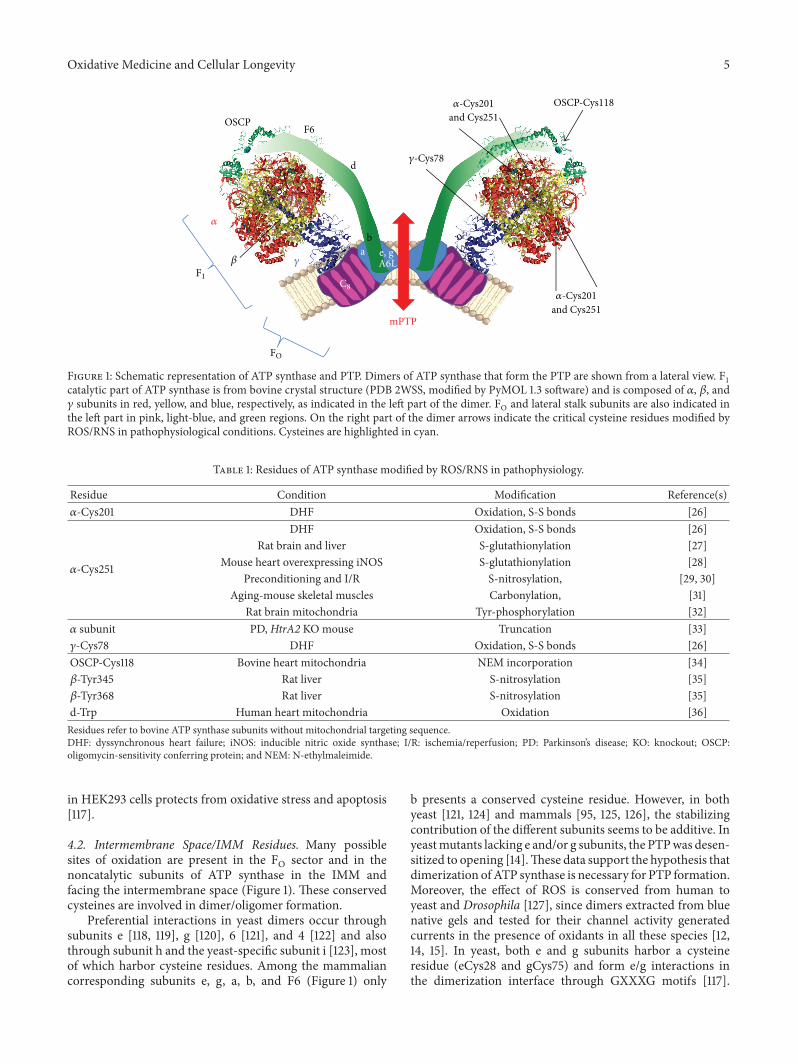
Oxidative Medicine and Cellular Longevity 5
mPTP
e, g a A6L
b
d
OSCPF6
OSCP-Cys118and Cys251
and Cys251
𝛼-Cys201
𝛼-Cys201
𝛾-Cys78
𝛾
𝛼
𝛽
F1
FO
C8
Figure 1: Schematic representation of ATP synthase and PTP. Dimers of ATP synthase that form the PTP are shown from a lateral view. F1
catalytic part of ATP synthase is from bovine crystal structure (PDB 2WSS, modified by PyMOL 1.3 software) and is composed of 𝛼, 𝛽, and𝛾 subunits in red, yellow, and blue, respectively, as indicated in the left part of the dimer. FO and lateral stalk subunits are also indicated inthe left part in pink, light-blue, and green regions. On the right part of the dimer arrows indicate the critical cysteine residues modified byROS/RNS in pathophysiological conditions. Cysteines are highlighted in cyan.
Table 1: Residues of ATP synthase modified by ROS/RNS in pathophysiology.
Residue Condition Modification Reference(s)𝛼-Cys201 DHF Oxidation, S-S bonds [26]
𝛼-Cys251
DHF Oxidation, S-S bonds [26]Rat brain and liver S-glutathionylation [27]
Mouse heart overexpressing iNOS S-glutathionylation [28]Preconditioning and I/R S-nitrosylation, [29, 30]
Aging-mouse skeletal muscles Carbonylation, [31]Rat brain mitochondria Tyr-phosphorylation [32]
𝛼 subunit PD, HtrA2 KOmouse Truncation [33]𝛾-Cys78 DHF Oxidation, S-S bonds [26]OSCP-Cys118 Bovine heart mitochondria NEM incorporation [34]𝛽-Tyr345 Rat liver S-nitrosylation [35]𝛽-Tyr368 Rat liver S-nitrosylation [35]d-Trp Human heart mitochondria Oxidation [36]Residues refer to bovine ATP synthase subunits without mitochondrial targeting sequence.DHF: dyssynchronous heart failure; iNOS: inducible nitric oxide synthase; I/R: ischemia/reperfusion; PD: Parkinson’s disease; KO: knockout; OSCP:oligomycin-sensitivity conferring protein; and NEM: N-ethylmaleimide.
in HEK293 cells protects from oxidative stress and apoptosis[117].
4.2. Intermembrane Space/IMM Residues. Many possiblesites of oxidation are present in the FO sector and in thenoncatalytic subunits of ATP synthase in the IMM andfacing the intermembrane space (Figure 1). These conservedcysteines are involved in dimer/oligomer formation.
Preferential interactions in yeast dimers occur throughsubunits e [118, 119], g [120], 6 [121], and 4 [122] and alsothrough subunit h and the yeast-specific subunit i [123], mostof which harbor cysteine residues. Among the mammaliancorresponding subunits e, g, a, b, and F6 (Figure 1) only
b presents a conserved cysteine residue. However, in bothyeast [121, 124] and mammals [95, 125, 126], the stabilizingcontribution of the different subunits seems to be additive. Inyeastmutants lacking e and/or g subunits, the PTPwas desen-sitized to opening [14].These data support the hypothesis thatdimerization ofATP synthase is necessary for PTP formation.Moreover, the effect of ROS is conserved from human toyeast and Drosophila [127], since dimers extracted from bluenative gels and tested for their channel activity generatedcurrents in the presence of oxidants in all these species [12,14, 15]. In yeast, both e and g subunits harbor a cysteineresidue (eCys28 and gCys75) and form e/g interactions inthe dimerization interface through GXXXG motifs [117].
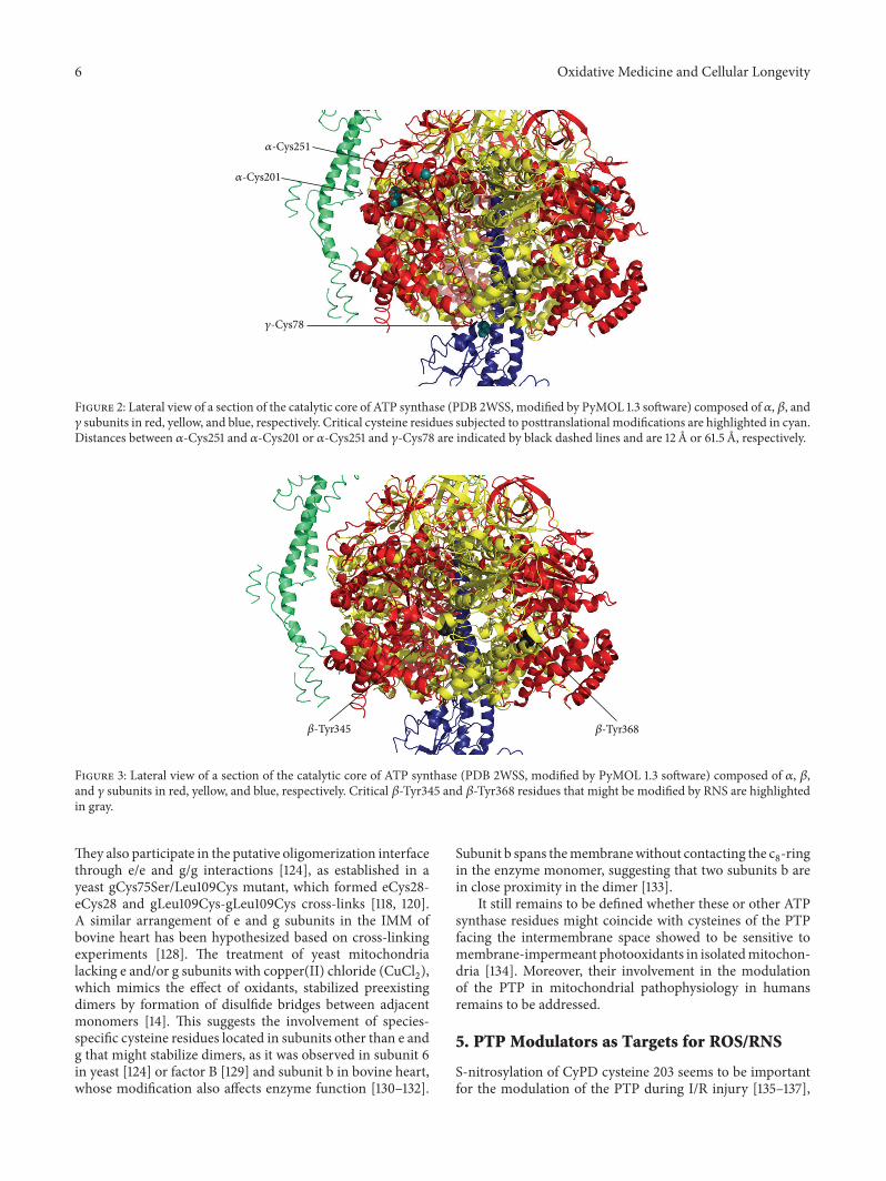
6 Oxidative Medicine and Cellular Longevity
𝛼-Cys201
𝛼-Cys251
𝛾-Cys78
Figure 2: Lateral view of a section of the catalytic core of ATP synthase (PDB 2WSS, modified by PyMOL 1.3 software) composed of 𝛼, 𝛽, and𝛾 subunits in red, yellow, and blue, respectively. Critical cysteine residues subjected to posttranslational modifications are highlighted in cyan.Distances between 𝛼-Cys251 and 𝛼-Cys201 or 𝛼-Cys251 and 𝛾-Cys78 are indicated by black dashed lines and are 12 Å or 61.5 Å, respectively.
𝛽-Tyr345 𝛽-Tyr368
Figure 3: Lateral view of a section of the catalytic core of ATP synthase (PDB 2WSS, modified by PyMOL 1.3 software) composed of 𝛼, 𝛽,and 𝛾 subunits in red, yellow, and blue, respectively. Critical 𝛽-Tyr345 and 𝛽-Tyr368 residues that might be modified by RNS are highlightedin gray.
They also participate in the putative oligomerization interfacethrough e/e and g/g interactions [124], as established in ayeast gCys75Ser/Leu109Cys mutant, which formed eCys28-eCys28 and gLeu109Cys-gLeu109Cys cross-links [118, 120].A similar arrangement of e and g subunits in the IMM ofbovine heart has been hypothesized based on cross-linkingexperiments [128]. The treatment of yeast mitochondrialacking e and/or g subunits with copper(II) chloride (CuCl
2),
which mimics the effect of oxidants, stabilized preexistingdimers by formation of disulfide bridges between adjacentmonomers [14]. This suggests the involvement of species-specific cysteine residues located in subunits other than e andg that might stabilize dimers, as it was observed in subunit 6in yeast [124] or factor B [129] and subunit b in bovine heart,whose modification also affects enzyme function [130–132].
Subunit b spans themembranewithout contacting the c8-ring
in the enzyme monomer, suggesting that two subunits b arein close proximity in the dimer [133].
It still remains to be defined whether these or other ATPsynthase residues might coincide with cysteines of the PTPfacing the intermembrane space showed to be sensitive tomembrane-impermeant photooxidants in isolatedmitochon-dria [134]. Moreover, their involvement in the modulationof the PTP in mitochondrial pathophysiology in humansremains to be addressed.
5. PTP Modulators as Targets for ROS/RNS
S-nitrosylation of CyPD cysteine 203 seems to be importantfor the modulation of the PTP during I/R injury [135–137],
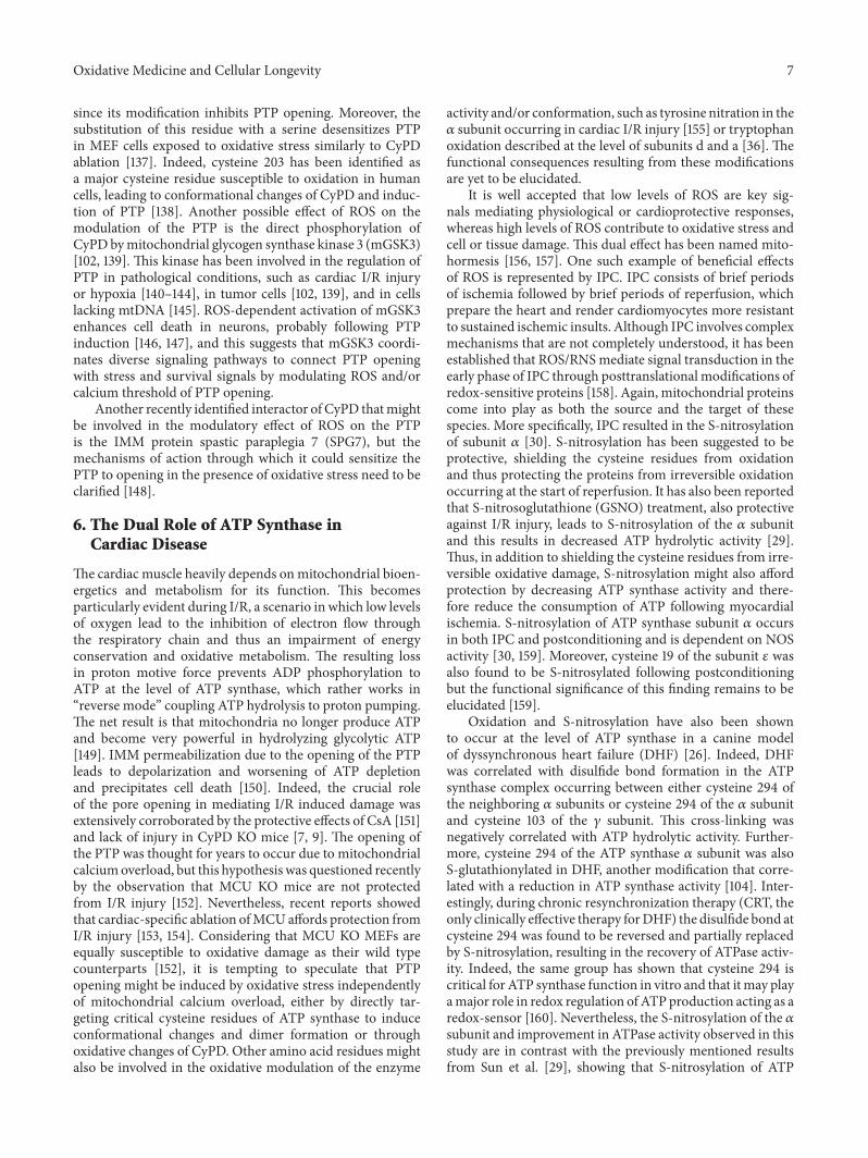
Oxidative Medicine and Cellular Longevity 7
since its modification inhibits PTP opening. Moreover, thesubstitution of this residue with a serine desensitizes PTPin MEF cells exposed to oxidative stress similarly to CyPDablation [137]. Indeed, cysteine 203 has been identified asa major cysteine residue susceptible to oxidation in humancells, leading to conformational changes of CyPD and induc-tion of PTP [138]. Another possible effect of ROS on themodulation of the PTP is the direct phosphorylation ofCyPD bymitochondrial glycogen synthase kinase 3 (mGSK3)[102, 139]. This kinase has been involved in the regulation ofPTP in pathological conditions, such as cardiac I/R injuryor hypoxia [140–144], in tumor cells [102, 139], and in cellslacking mtDNA [145]. ROS-dependent activation of mGSK3enhances cell death in neurons, probably following PTPinduction [146, 147], and this suggests that mGSK3 coordi-nates diverse signaling pathways to connect PTP openingwith stress and survival signals by modulating ROS and/orcalcium threshold of PTP opening.
Another recently identified interactor of CyPD thatmightbe involved in the modulatory effect of ROS on the PTPis the IMM protein spastic paraplegia 7 (SPG7), but themechanisms of action through which it could sensitize thePTP to opening in the presence of oxidative stress need to beclarified [148].
6. The Dual Role of ATP Synthase inCardiac Disease
The cardiac muscle heavily depends onmitochondrial bioen-ergetics and metabolism for its function. This becomesparticularly evident during I/R, a scenario in which low levelsof oxygen lead to the inhibition of electron flow throughthe respiratory chain and thus an impairment of energyconservation and oxidative metabolism. The resulting lossin proton motive force prevents ADP phosphorylation toATP at the level of ATP synthase, which rather works in“reverse mode” coupling ATP hydrolysis to proton pumping.The net result is that mitochondria no longer produce ATPand become very powerful in hydrolyzing glycolytic ATP[149]. IMM permeabilization due to the opening of the PTPleads to depolarization and worsening of ATP depletionand precipitates cell death [150]. Indeed, the crucial roleof the pore opening in mediating I/R induced damage wasextensively corroborated by the protective effects of CsA [151]and lack of injury in CyPD KO mice [7, 9]. The opening ofthe PTP was thought for years to occur due to mitochondrialcalciumoverload, but this hypothesis was questioned recentlyby the observation that MCU KO mice are not protectedfrom I/R injury [152]. Nevertheless, recent reports showedthat cardiac-specific ablation ofMCUaffords protection fromI/R injury [153, 154]. Considering that MCU KO MEFs areequally susceptible to oxidative damage as their wild typecounterparts [152], it is tempting to speculate that PTPopening might be induced by oxidative stress independentlyof mitochondrial calcium overload, either by directly tar-geting critical cysteine residues of ATP synthase to induceconformational changes and dimer formation or throughoxidative changes of CyPD. Other amino acid residues mightalso be involved in the oxidative modulation of the enzyme
activity and/or conformation, such as tyrosine nitration in the𝛼 subunit occurring in cardiac I/R injury [155] or tryptophanoxidation described at the level of subunits d and a [36]. Thefunctional consequences resulting from these modificationsare yet to be elucidated.
It is well accepted that low levels of ROS are key sig-nals mediating physiological or cardioprotective responses,whereas high levels of ROS contribute to oxidative stress andcell or tissue damage. This dual effect has been named mito-hormesis [156, 157]. One such example of beneficial effectsof ROS is represented by IPC. IPC consists of brief periodsof ischemia followed by brief periods of reperfusion, whichprepare the heart and render cardiomyocytes more resistantto sustained ischemic insults. Although IPC involves complexmechanisms that are not completely understood, it has beenestablished that ROS/RNSmediate signal transduction in theearly phase of IPC through posttranslational modifications ofredox-sensitive proteins [158]. Again, mitochondrial proteinscome into play as both the source and the target of thesespecies. More specifically, IPC resulted in the S-nitrosylationof subunit 𝛼 [30]. S-nitrosylation has been suggested to beprotective, shielding the cysteine residues from oxidationand thus protecting the proteins from irreversible oxidationoccurring at the start of reperfusion. It has also been reportedthat S-nitrosoglutathione (GSNO) treatment, also protectiveagainst I/R injury, leads to S-nitrosylation of the 𝛼 subunitand this results in decreased ATP hydrolytic activity [29].Thus, in addition to shielding the cysteine residues from irre-versible oxidative damage, S-nitrosylation might also affordprotection by decreasing ATP synthase activity and there-fore reduce the consumption of ATP following myocardialischemia. S-nitrosylation of ATP synthase subunit 𝛼 occursin both IPC and postconditioning and is dependent on NOSactivity [30, 159]. Moreover, cysteine 19 of the subunit 𝜀 wasalso found to be S-nitrosylated following postconditioningbut the functional significance of this finding remains to beelucidated [159].
Oxidation and S-nitrosylation have also been shownto occur at the level of ATP synthase in a canine modelof dyssynchronous heart failure (DHF) [26]. Indeed, DHFwas correlated with disulfide bond formation in the ATPsynthase complex occurring between either cysteine 294 ofthe neighboring 𝛼 subunits or cysteine 294 of the 𝛼 subunitand cysteine 103 of the 𝛾 subunit. This cross-linking wasnegatively correlated with ATP hydrolytic activity. Further-more, cysteine 294 of the ATP synthase 𝛼 subunit was alsoS-glutathionylated in DHF, another modification that corre-lated with a reduction in ATP synthase activity [104]. Inter-estingly, during chronic resynchronization therapy (CRT, theonly clinically effective therapy forDHF) the disulfide bond atcysteine 294 was found to be reversed and partially replacedby S-nitrosylation, resulting in the recovery of ATPase activ-ity. Indeed, the same group has shown that cysteine 294 iscritical for ATP synthase function in vitro and that itmay playamajor role in redox regulation ofATPproduction acting as aredox-sensor [160]. Nevertheless, the S-nitrosylation of the 𝛼subunit and improvement in ATPase activity observed in thisstudy are in contrast with the previously mentioned resultsfrom Sun et al. [29], showing that S-nitrosylation of ATP
8 Oxidative Medicine and Cellular Longevity
synthase 𝛼 subunit in IPC or after GSNO treatment leads to areduction of its hydrolytic activity. One cannot exclude thatdifferent cysteine residues were modified in the two afore-mentioned studies, a fact that could explain this discrepancy.Another possible explanation is that cross-link reversal andrecovery ofATPase activity observed inCRToccur at a higherlevel compared to the extent of S-nitrosylation, suggestingthat the majority of cysteine residues are actually free in CRThearts [26]. On the other hand, the distances between the twocysteines 294 of neighboring𝛼 subunits or between𝛼 cysteine294 and 𝛾 cysteine 103 aremore than 5 Å (corresponding to𝛼-Cys251 and 𝛾-Cys78, respectively, in Figures 1 and 2), makingit unlikely that these disulfide bonds actually occur in theassembled complex. Rather, these bonds could form in themisfolded/aggregated enzyme, suggesting that the reversalof cysteine cross-links observed in CRT may promote thecorrect assembly of the complex, thus resulting in improvedATPase activity. Of note, CuCl
2treatment of isolated ATP
synthase complex from ratmitochondria in vitro showed thatdisulfide bonds can form also between 𝛼 and OSCP subunitsas well as between the two 𝛾 subunits and 𝛾 and OSCP [26].The effect of these disulfide crossbridges on ATP synthaseactivity was not addressed, and whether these modificationscould also occur in vivo still needs to be assessed. Furtherefforts should also be put in addressing the effect of theseoxidative posttranslational modifications on PTP forma-tion.
7. Neurodegenerative Diseases
Mitochondria have a central role in aging-related neurode-generative diseases. Oxidative stress generated by mitochon-dria has been inversely correlated with longevity in modelorganisms [161, 162] and defects inmitochondrial bioenerget-ics have been implicated in a number of neurodegenerativediseases [163].
In vitro, ATP synthase is susceptible to ROS [103, 164, 165]and to oxidative/nitrosative stress associated with disordersof the central nervous system [166, 167] and aging [35, 168].The fact that ROS/RNSmodifications can alter themitochon-drial oxidative phosphorylation efficiency may explain themitochondrial involvement in neurological diseases.
Moreover, the involvement of PTP in neurodegenerativediseases has been demonstrated by the use of CsA bothin vitro and in vivo and the ablation in mouse models ofits target, CyPD [17, 18]. CyPD plays an important dualfunction on the modulation of ATP synthase; on the onehand, it sensitizes PTP to matrix calcium, while, on the otherhand, it inhibits both ATP hydrolysis and synthesis by 30%[11]. In cellular mechanisms of neurodegenerative diseases,when mitochondria are hydrolyzing ATP, CyPD binding andinhibition of ATP synthase can be an advantage avoidingATP consumption and cell death, or a disadvantage in casesin which PTP is sensitized to calcium and the displacementof CyPD would promote cell survival. This is only one ofthe complex relationships between ATP synthase and PTPin neurodegeneration, and in this paragraph we attempt tosummarize possible mechanisms linking ATP synthase/PTPand ROS/RNS modifications.
7.1. Amyotrophic Lateral Sclerosis. Amyotrophic lateral scle-rosis (ALS) is a progressive degeneration ofmotor neurons. Infamilial ALS, mutations have been found in Cu,Zn-SOD thatare suggested to increase generation of ROS [169, 170], RNS,and nitrosylation [171]. Furthermore, Beal et al. [172] detectedincreased levels of nitrotyrosine staining in motor neuronsof both sporadic ALS and familial ALS, suggesting thatONOO− mediated oxidative damage may play a role in bothforms of the disease. Of note, as already mentioned, tyrosinemodifications at the level of the 𝛽 subunit of ATP synthasehave been described to bemodulatory of the catalytic ATPaseactivity in the presence of ROS/RNS [108, 109], even if theirdirect involvement in ALS remains to be addressed.
In light of the susceptibility of the mitochondrial res-piratory chain to nitrosative stress, it is not surprising thatmitochondrial function is impaired in ALS [173]. Spinalcord mitochondria in ALS mouse model display decreasedcalcium retention capacity long before the onset of motorweakness and neuronal death [174], and this was correctedby ablation of the Ppif gene which encodes CyPD [175]. Inthese mice, an improved mitochondrial ATP synthesis wasmatched by PTP inhibition and significant suppression ofmotor neuron death throughout disease, although survivalwas not improved confirming the role of ATP synthesisand permeability transition in ALS neuron cell death [176,177]. Increased ATP synthesis in this mouse ALS modellacking CyPD could also be explained by our observation thatablation of CyPD increases the catalysis of ATP synthase [11].
7.2. Alzheimer’s Disease. AD, the most common form ofdementia in aged people, is characterized by deposition ofamyloid plaques formed by the amyloid 𝛽 peptide [178].Amyloid 𝛽 can be imported in mitochondria [179]. Severalstudies have observed activity changes in key mitochondrialenzymes in AD. While the exact mechanism for this loss ofactivity is unclear, evidence suggests that ROS/RNS produc-tion is increased in AD. Nitrotyrosine residues have beendetected in postmortem Alzheimer’s tissue but not in age-matched controls [180], indicating the presence of ONOO−.The induction of iNOS in cultured rat astrocytes causes NO-mediated neuronal death in a coculture system [181].
Amyloid 𝛽 is also reported to stimulate glial NO produc-tion [182–184] and it has been shown to inhibit both purifiedcomplex IV [185] and complex IV in isolated brain mito-chondria [186]. Additionally, in neuronal cultures amyloid 𝛽caused a loss of activity of all the mitochondrial complexesand a loss of mitochondrial integrity, due to PTP opening[187]. It has been shown that amyloid 𝛽 oligomers altercalcium homeostasis [188].
Upon import in mitochondria, amyloid 𝛽 interacts withCyPD and enhances PTP induction, since neurons fromCyPD KO mice are protected from cell death induced byamyloid 𝛽 dependent PTP opening [189]. Interestingly, anovel association with AD risk has been recently identifiedin the ATP synthase ATP5H locus, which encodes subunitd of the lateral stalk [190]. Intriguingly, in a proteomicanalysis of human heart mitochondria in normal condition,a tryptophan residue at the level of d subunit was identifiedas a “hot spot” for oxidation [36], the latter being another
Oxidative Medicine and Cellular Longevity 9
important pathophysiological factor in AD. Moreover, in thefilamentous fungus Podospora anserina, a model with a cleardefined mitochondrial etiology of aging, another tryptophanresidue on the 𝛼 subunit of ATP synthase (Trp503) has beendescribed to be crucial for the selective targeting of oxidativedamage [107]. Although this residue is not conserved inunicellular fungi and higher eukaryotes, the example of P.anserina is instructive because the authors proposed thatoxidized cysteine and methionine can be efficiently reducedby repair mechanisms, whereas tryptophan oxidation prod-ucts are irreversible and have the potential to form markersdetected by the mitochondrial quality control system.
7.3. Multiple Sclerosis. In multiple sclerosis (MS), the myelinsheath of neurons in the central nervous system is destroyedleading to axonal degeneration [191]. This is associated withmitochondrial calcium overload and bioenergetic dysfunc-tion [192]. CyPD KOmice with autoimmune encephalomye-litis display a marked protection from axonal degenerationand a milder clinical picture despite a normal inflammatoryresponse, thus suggesting that PTP might be involved indisease pathogenesis [193], even if other effects on bioener-getics caused byCyPD ablation cannot be excluded. A furtherindication of the important role played by the PTP in MSand its activation by ROS is provided by the observation thataxonal damage of mice undergoing experimental autoim-mune encephalomyelitis is reduced by genetic ablation ofp66Shc [194]. Importantly, a proteomic study of experimentalautoimmune encephalomyelitis identified the ATP synthasedimer-forming subunit e differentially expressed [195].More-over, mutations at the level of the genes encoding subunits aand A6L were observed in MS patients [196].
A large body of evidence exists implicating increased gen-eration of RNS inMS.The observation that the concentrationof NO
2
− plus NO3
− in cerebrospinal fluid is elevated by 70%in MS patients supports this hypothesis [197]. Furthermore,increased iNOS activity and iNOS mRNA have been demon-strated in astrocytes associated with demyelinating lesions inpostmortem MS brain [198] and in experimental models ofdemyelination [199, 200]. Nitrotyrosine residues indicatingthe presence of ONOO− have also been detected in MSbrain [201]. Whether NO-mediated mitochondrial damage isthe cause of the disease remains to be established; however,the first direct evidence of altered mitochondrial functionin MS came from a study by Lu et al. [202]. These authorsdescribed a decrease of respiratory complex I activity and acompensatory increase in complex IV in MS. Major targetsfor nitrosative stress in rat liver under in vivo conditionsare tyrosine 345 and tyrosine 368 in the 𝛽 subunit of themitochondrial ATP synthase that is suggested as an earlymarker of nitrosative stress and aging [35]. A comparisonbetween 𝛼 and 𝛽 subunits allowed the speculation that thelatter is more accessible for RNS modification and that thecatalytic conformation of this subunit also affects tyrosineresidues exposure to nitration (higher exposure to RNSmodifications in the absence of bound nucleotide).Moreover,Ding et al. [203] demonstrated inND4mice (an in vivomodelforMS) an impaired transport of ATP5bmRNA (which gene-rates subunit 𝛽) to mitochondria.This caused decreased ATP
synthesis in MS mice due to lower levels of this subunit andnot to impaired ATP synthase assembly, as suggested by theauthors. However, the possibility that tyrosine modificationsin this subunit might affect enzyme activity cannot beexcluded.
7.4. Parkinson’s Disease. PD is caused by death of dopamin-ergic neurons in the substantia nigra pars compacta. Asensitization to PTP opening has been proposed as a majorcause of neurodegeneration in several models of the diseasecharacterized by altered homeostasis of intracellular calcium[204–206], including the forms caused by complex I inhi-bition [207, 208] and by inactivation of the Ser/Thr kinasePINK1 [209], in which changes in calcium storage capacity[210, 211], impairment of respiratory complex I [212], andaltered mitophagy [213] are observed. Moreover, the PTPcan be induced in dopaminergic neurons because of theinability to buffer increased intracellular ROS levels [214].In a PD mouse model induced by parkin deficiency, state3 and state 4 respiration rates were both affected indicatinga more likely direct modulation of the respiratory chaincompared to an effect on ATP synthase [215]. On the otherhand, a mouse model lacking chaperone protein HtrA2 andshowing a parkinsonian phenotype displays mitochondrialuncoupling at the level of ATP synthase and a truncated𝛼 subunit that might be involved in the neurodegenerationobserved in these mice [33].
Postsynaptic density protein 95 (PSD-95) binds to neu-ronal nNOS and the neuroprotective effects of small-molecule inhibitors of this interaction were tested in anin vitro PD model. The observed protective effects wereassociated with suppressed mitochondrial dysfunction, asevidenced by decreased ROS generation, preserved ATPsynthesis, and PTP inhibition [216]. Moreover, Darios et al.[217] demonstrated that in PC12 cells the overexpression ofparkin protects from ceramide-induced swelling suggestingthat parkin may act directly to prevent PTP mediated celldeath, but the exact mechanism remains to be addressed.
8. Conclusions
Posttranslational modifications of ATP synthase due toROS/RNS generation might play a dual role by promotingcell death or survival depending on their relative effects onmitochondrial ATP synthase catalysis and PTP.We have illus-trated howATP synthase is the target for oxidative/nitrosativemodifications that affect its activity and might promote for-mation of the PTP. Identification of specific residues involvedin the latter event is still lacking and will help to elucidatethe mechanisms that mediate the role of ATP synthase inmodulating cell survival or death. Finally, we discussed howchanges in the ATP synthase activity regulate mitochondrialROS formation and thus may represent an attractive strategyfor the treatment of pathologies such as cancer.
Abbreviations
AD: Alzheimer’s diseaseALS: Amyotrophic lateral sclerosis
10 Oxidative Medicine and Cellular Longevity
AsO: Arsenite anionBH4: TetrahydrobiopterinCRT: Chronic resynchronization therapyCsA: Cyclosporin ACuCl2: Copper(II) chloride
CyPD: Cyclophilin DDHF: Dyssynchronous heart failureeNOS: Endothelial nitric oxide synthaseGSNO: S-NitrosoglutathioneH2O2: Hydrogen peroxide
IF1: Inhibitor protein
IMM: Inner mitochondrial membraneiNOS: Inducible nitric oxide synthaseIPC: Ischemic preconditioningI/R: Ischemia/reperfusionKO: KnockoutMAO: Monoamine oxidaseMBM: MonobromobimanemGSK3: Mitochondrial glycogen synthase kinase 3PTP: Mitochondrial permeability transition
poreMS: Multiple sclerosisNEM: N-EthylmaleimidenNOS: Neuronal nitric oxide synthaseNO: Nitric oxideNO2
−: NitriteNO3
−: NitrateNox4: NADPH oxidase 4OMM: Outer mitochondrial membraneONOO−: PeroxynitriteOSCP: Oligomycin-sensitivity conferring proteinPD: Parkinson’s diseasePhAsO: Phenylarsine oxideRNS: Reactive nitrogen speciesROS: Reactive oxygen speciesSOD: Superoxide dismutaseSPG7: Spastic paraplegia 7.
Conflict of Interests
The authors declare that there is no conflict of interestsregarding the publication of this paper.
Acknowledgments
The authors thank Paolo Bernardi, Fabio Di Lisa, and AndreaRasola for the critical reading of the paper and helpful discus-sions. The authors also thank Manuela Antoniel, Universityof Padova, for the generous help in the use of PyMOL 1.3software. Valentina Giorgio’s research activities are supportedby the Telethon Grant GGP14187.
References
[1] Å. B. Gustafsson and R. A. Gottlieb, “Bcl-2 family members andapoptosis, taken to heart,”TheAmerican Journal of Physiology—Cell Physiology, vol. 292, no. 1, pp. C45–C51, 2007.
[2] P. Bernardi, A. Rasola, M. Forte, and G. Lippe, “The mito-chondrial permeability transition pore: channel formation by
F-ATP synthase, integration in signal transduction, and role inpathophysiology,” Physiological Reviews, vol. 95, no. 4, pp. 1111–1155, 2015.
[3] K. Le-Quoc and D. Le-Quoc, “Crucial role of sulfhydryl groupsin the mitochondrial inner membrane structure,”The Journal ofBiological Chemistry, vol. 260, no. 12, pp. 7422–7428, 1985.
[4] R. A. Haworth andD. R. Hunter, “TheCa2+-inducedmembranetransition in mitochondria. II. Nature of the Ca2+ trigger site,”Archives of Biochemistry and Biophysics, vol. 195, no. 2, pp. 460–467, 1979.
[5] D. R.Hunter and R. A.Haworth, “TheCa2+-inducedmembranetransition in mitochondria. I. The protective mechanisms,”Archives of Biochemistry and Biophysics, vol. 195, no. 2, pp. 453–459, 1979.
[6] D. R.Hunter and R. A.Haworth, “TheCa2+-inducedmembranetransition in mitochondria. III. Transitional Ca2+ release,”Archives of Biochemistry and Biophysics, vol. 195, no. 2, pp. 468–477, 1979.
[7] C. P. Baines, R. A. Kaiser, N.H. Purcell et al., “Loss of cyclophilinD reveals a critical role for mitochondrial permeability transi-tion in cell death,”Nature, vol. 434, no. 7033, pp. 658–662, 2005.
[8] E. Basso, L. Fante, J. Fowlkes, V. Petronilli, M. A. Forte, andP. Bernardi, “Properties of the permeability transition pore inmitochondria devoid of cyclophilin D,”The Journal of BiologicalChemistry, vol. 280, no. 19, pp. 18558–18561, 2005.
[9] T. Nakagawa, S. Shimizu, T. Watanabe et al., “CyclophilinD-dependent mitochondrial permeability transition regulatessome necrotic but not apoptotic cell death,”Nature, vol. 434, no.7033, pp. 652–658, 2005.
[10] A. C. Schinzel, O. Takeuchi, Z. Huang et al., “Cyclophilin Dis a component of mitochondrial permeability transition andmediates neuronal cell death after focal cerebral ischemia,” Pro-ceedings of the National Academy of Sciences of the United Statesof America, vol. 102, no. 34, pp. 12005–12010, 2005.
[11] V. Giorgio, E. Bisetto, M. E. Soriano et al., “Cyclophilin Dmodulates mitochondrial F0F1-ATP synthase by interactingwith the lateral stalk of the complex,” The Journal of BiologicalChemistry, vol. 284, no. 49, pp. 33982–33988, 2009.
[12] V. Giorgio, S. von Stockum,M. Antoniel et al., “Dimers of mito-chondrial ATP synthase form the permeability transition pore,”Proceedings of the National Academy of Sciences of the UnitedStates of America, vol. 110, no. 15, pp. 5887–5892, 2013.
[13] M. Bonora, A. Bononi, E. De Marchi et al., “Role of the csubunit of the FO ATP synthase in mitochondrial permeabilitytransition,” Cell Cycle, vol. 12, no. 4, pp. 674–683, 2013.
[14] M. Carraro, V. Giorgio, J. Sileikyte et al., “Channel formationby yeast F-ATP synthase and the role of dimerization in themitochondrial permeability transition,”The Journal of BiologicalChemistry, vol. 289, no. 23, pp. 15980–15985, 2014.
[15] S. von Stockum, V. Giorgio, E. Trevisan et al., “F-ATPase ofDrosophila melanogaster forms 53-picosiemen (53-pS) channelsresponsible for mitochondrial Ca2+-induced Ca2+ release,” TheJournal of Biological Chemistry, vol. 290, no. 8, pp. 4537–4544,2015.
[16] K.N.Alavian, S. I. Dworetzky, L. Bonanni et al., “Themitochon-drial complex V—associated large-conductance inner mem-brane current is regulated by cyclosporine and dexpramipex-ole,”Molecular Pharmacology, vol. 87, no. 1, pp. 1–8, 2015.
[17] V. Giorgio, M. E. Soriano, E. Basso et al., “Cyclophilin D inmitochondrial pathophysiology,” Biochimica et Biophysica Acta(BBA)—Bioenergetics, vol. 1797, no. 6-7, pp. 1113–1118, 2010.
Oxidative Medicine and Cellular Longevity 11
[18] J.W. Elrod and J. D.Molkentin, “Physiologic functions of cyclo-philin D and the mitochondrial permeability transition pore,”Circulation Journal, vol. 77, no. 5, pp. 1111–1122, 2013.
[19] D. A. Butterfield and I. Dalle-Donne, “Redox proteomics: fromproteinmodifications to cellular dysfunction and disease,”MassSpectrometry Reviews, vol. 33, no. 1, pp. 1–6, 2014.
[20] S. F. Steinberg, “Oxidative stress and sarcomeric proteins,” Cir-culation Research, vol. 112, no. 2, pp. 393–405, 2013.
[21] A. I. Casas, V. T. Dao, A. Daiber et al., “Reactive oxygen-relateddiseases: therapeutic targets and emerging clinical indications,”Antioxidants & Redox Signaling, vol. 23, no. 14, pp. 1171–1185,2015.
[22] V. T. Dao, A. I. Casas, G. J. Maghzal et al., “Pharmacology andclinical drug candidates in redox medicine,” Antioxidants &Redox Signaling, vol. 23, no. 14, pp. 1113–1129, 2015.
[23] N. Kaludercic, J. Mialet-Perez, N. Paolocci, A. Parini, and F. DiLisa, “Monoamine oxidases as sources of oxidants in the heart,”Journal of Molecular and Cellular Cardiology, vol. 73, pp. 34–42,2014.
[24] M. P. Murphy, “How mitochondria produce reactive oxygenspecies,” Biochemical Journal, vol. 417, no. 1, pp. 1–13, 2009.
[25] M.D. Brand, “The sites and topology ofmitochondrial superox-ide production,” Experimental Gerontology, vol. 45, no. 7-8, pp.466–472, 2010.
[26] S.-B. Wang, D. B. Foster, J. Rucker, B. O’Rourke, D. A. Kass,and J. E. Van Eyk, “Redox regulation ofmitochondrial ATP syn-thase: implications for cardiac resynchronization therapy,” Cir-culation Research, vol. 109, no. 7, pp. 750–757, 2011.
[27] J. Garcia, D. Han, H. Sancheti, L.-P. Yap, N. Kaplowitz, and E.Cadenas, “Regulation ofmitochondrial glutathione redox statusand protein glutathionylation by respiratory substrates,” Journalof Biological Chemistry, vol. 285, no. 51, pp. 39646–39654, 2010.
[28] M. B. West, B. G. Hill, Y.-T. Xuan, and A. Bhatnagar, “Proteinglutathiolation by nitric oxide: an intracellular mechanismregulating redox protein modification,”The FASEB Journal, vol.20, no. 10, pp. 1715–1717, 2006.
[29] J. Sun, M. Morgan, R.-F. Shen, C. Steenbergen, and E. Murphy,“Preconditioning results in S-nitrosylation of proteins involvedin regulation of mitochondrial energetics and calcium trans-port,” Circulation Research, vol. 101, no. 11, pp. 1155–1163, 2007.
[30] J. Sun, T. Nguyen, A.M. Aponte et al., “Ischaemic precondition-ing preferentially increases protein S-nitrosylation in subsar-colemmal mitochondria,” Cardiovascular Research, vol. 106, no.2, pp. 227–236, 2015.
[31] N. Das and C. K. Jana, “Age-associated oxidative modificationsof mitochondrial 𝛼-subunit of F1 ATP synthase from mouseskeletal muscles,” Free Radical Research, vol. 49, no. 8, pp. 954–961, 2015.
[32] O. Augereau, S. Claverol, N. Boudes et al., “Identification oftyrosine-phosphorylated proteins of the mitochondrial oxida-tive phosphorylation machinery,” Cellular and Molecular LifeSciences, vol. 62, no. 13, pp. 1478–1488, 2005.
[33] H. Plun-Favreau, V. S. Burchell, K. M. Holmström et al.,“HtrA2 deficiency causes mitochondrial uncoupling throughthe F(1)F(0)-ATP synthase and consequent ATP depletion,”CellDeath and Disease, vol. 3, article e335, 2012.
[34] A. Dupuis, J. Lunardi, J.-P. Issartel, and P. V. Vignais, “Inter-actions between the oligomycin sensitivity conferring protein(OSCP) and beef heart mitochondrial F1ATPase. 2. Identifica-tion of the interacting F1 subunits by cross-linking,” Biochem-istry, vol. 24, no. 3, pp. 734–739, 1985.
[35] V. Haynes, N. J. Traaseth, S. Elfering, Y. Fujisawa, and C. Giulivi,“Nitration of specific tyrosines in FoF1 ATP synthase and acti-vity loss in aging,” American Journal of Physiology—Endocri-nology and Metabolism, vol. 298, no. 5, pp. E978–E987, 2010.
[36] S. W. Taylor, E. Fahy, J. Murray, R. A. Capaldi, and S. S.Ghosh, “Oxidative post-translational modification of trypto-phan residues in cardiac mitochondrial proteins,” Journal ofBiological Chemistry, vol. 278, no. 22, pp. 19587–19590, 2003.
[37] M. Giorgio, E. Migliaccio, F. Orsini et al., “Electron transferbetween cytochrome c and p66Shc generates reactive oxygenspecies that trigger mitochondrial apoptosis,” Cell, vol. 122, no.2, pp. 221–233, 2005.
[38] A. Carpi, R. Menabò, N. Kaludercic, P. Pelicci, F. Di Lisa, andM. Giorgio, “The cardioprotective effects elicited by p66Shcablation demonstrate the crucial role of mitochondrial ROSformation in ischemia/reperfusion injury,” Biochimica et Bio-physica Acta—Bioenergetics, vol. 1787, no. 7, pp. 774–780, 2009.
[39] R. D. Spescha, Y. Shi, S. Wegener et al., “Deletion of the ageinggene p66Shc reduces early stroke size following ischaemia/reper-fusion brain injury,” European Heart Journal, vol. 34, no. 2, pp.96–103, 2013.
[40] S. Ciciliot, M. Albiero, L. Menegazzo et al., “p66Shc deletion ordeficiency protects from obesity but not metabolic dysfunctionin mice and humans,” Diabetologia, vol. 58, no. 10, pp. 2352–2360, 2015.
[41] M. Rota, N. LeCapitaine, T. Hosoda et al., “Diabetes promotescardiac stem cell aging and heart failure, which are prevented bydeletion of the p66shc gene,” Circulation Research, vol. 99, no. 1,pp. 42–52, 2006.
[42] S. Menini, L. Amadio, G. Oddi et al., “Deletion of p66Shclongevity gene protects against experimental diabetic glomeru-lopathy by preventing diabetes-induced oxidative stress,” Dia-betes, vol. 55, no. 6, pp. 1642–1650, 2006.
[43] G. P. Fadini,M. Albiero, L.Menegazzo et al., “The redox enzymep66Shc contributes to diabetes and ischemia-induced delay incutaneous wound healing,” Diabetes, vol. 59, no. 9, pp. 2306–2314, 2010.
[44] A. Natalicchio, F. Tortosa, R. Labarbuta et al., “The p66(Shc)redox adaptor protein is induced by saturated fatty acidsand mediates lipotoxicity-induced apoptosis in pancreatic betacells,” Diabetologia, vol. 58, no. 6, pp. 1260–1271, 2015.
[45] G. G. Camici, M. Schiavoni, P. Francia et al., “Genetic deletionof p66Shc adaptor protein prevents hyperglycemia-inducedendothelial dysfunction and oxidative stress,” Proceedings of theNational Academy of Sciences of the United States of America,vol. 104, no. 12, pp. 5217–5222, 2007.
[46] M.Giorgio, A. Berry, I. Berniakovich et al., “Thep66Shc knockedout mice are short lived under natural condition,” Aging Cell,vol. 11, no. 1, pp. 162–168, 2012.
[47] R. M. Touyz and A. C. Montezano, “Vascular Nox4: a multifar-ious NADPH oxidase,” Circulation Research, vol. 110, no. 9, pp.1159–1161, 2012.
[48] K. D. Martyn, L. M. Frederick, K. von Loehneysen, M. C.Dinauer, and U. G. Knaus, “Functional analysis of Nox4 revealsunique characteristics compared to other NADPH oxidases,”Cellular Signalling, vol. 18, no. 1, pp. 69–82, 2006.
[49] C. Hirschhäuser, J. Bornbaum, A. Reis et al., “NOX4 in mito-chondria: yeast two-hybrid-based interaction with complex Iwithout relevance for basal reactive oxygen species?” Antioxi-dants & Redox Signaling, vol. 23, no. 14, pp. 1106–1112, 2015.
[50] M. Zhang, A. C. Brewer, K. Schröder et al., “NADPH oxidase-4 mediates protection against chronic load-induced stress in
12 Oxidative Medicine and Cellular Longevity
mouse hearts by enhancing angiogenesis,” Proceedings of theNational Academy of Sciences of the United States of America,vol. 107, no. 42, pp. 18121–18126, 2010.
[51] J. Kuroda, T. Ago, S. Matsushima, P. Zhai, M. D. Schneider, andJ. Sadoshima, “NADPH oxidase 4 (Nox4) is a major source ofoxidative stress in the failing heart,” Proceedings of the NationalAcademy of Sciences of the United States of America, vol. 107, no.35, pp. 15565–15570, 2010.
[52] C. Kleinschnitz, H. Grund, K. Wingler et al., “Post-strokeinhibition of induced NADPH Oxidase type 4 prevents oxida-tive stress and neurodegeneration,” PLoS Biology, vol. 8, no. 9,Article ID e1000479, 2010.
[53] R.M.Maalouf, A. A. Eid, Y. C. Gorin et al., “Nox4-derived reac-tive oxygen speciesmediate cardiomyocyte injury in early type 1diabetes,”The American Journal of Physiology—Cell Physiology,vol. 302, no. 3, pp. C597–C604, 2012.
[54] D. Sorescu, D.Weiss, B. Lassègue et al., “Superoxide productionand expression of Nox family proteins in human atherosclero-sis,” Circulation, vol. 105, no. 12, pp. 1429–1435, 2002.
[55] K. Schröder, M. Zhang, S. Benkhoff et al., “Nox4 is a protectivereactive oxygen species generating vascular NADPH oxidase,”Circulation Research, vol. 110, no. 9, pp. 1217–1225, 2012.
[56] M. B. H. Youdim, D. Edmondson, and K. F. Tipton, “The ther-apeutic potential of monoamine oxidase inhibitors,” NatureReviews Neuroscience, vol. 7, no. 4, pp. 295–309, 2006.
[57] P. Bianchi, O. Kunduzova, E. Masini et al., “Oxidative stress bymonoamine oxidase mediates receptor-independent cardiomy-ocyte apoptosis by serotonin and postischemic myocardialinjury,” Circulation, vol. 112, no. 21, pp. 3297–3305, 2005.
[58] D. Pchejetski, O. Kunduzova, A. Dayon et al., “Oxidative stress-dependent sphingosine kinase-1 inhibition mediates mono-amine oxidase A-associated cardiac cell apoptosis,” CirculationResearch, vol. 100, no. 1, pp. 41–49, 2007.
[59] N. Kaludercic, A. Carpi, T. Nagayama et al., “Monoamine oxi-dase B prompts mitochondrial and cardiac dysfunction inpressure overloaded hearts,” Antioxidants and Redox Signaling,vol. 20, no. 2, pp. 267–280, 2014.
[60] N. Kaludercic, E. Takimoto, T. Nagayama et al., “Monoamineoxidase A-mediated enhanced catabolism of norepinephrinecontributes to adverse remodeling and pump failure in heartswith pressure overload,” Circulation Research, vol. 106, no. 1, pp.193–202, 2010.
[61] A. Sturza, M. S. Leisegang, A. Babelova et al., “Monoamine oxi-dases are mediators of endothelial dysfunction in the mouseaorta,” Hypertension, vol. 62, no. 1, pp. 140–146, 2013.
[62] P. Umbarkar, S. Singh, S. Arkat, S. L. Bodhankar, S. Lohidasan,and S. L. Sitasawad, “Monoamine oxidase-A is an importantsource of oxidative stress and promotes cardiac dysfunction,apoptosis, and fibrosis in diabetic cardiomyopathy,” Free RadicalBiology and Medicine, vol. 87, pp. 263–273, 2015.
[63] E. Takimoto, H. C. Champion, M. Li et al., “Oxidant stress fromnitric oxide synthase-3 uncoupling stimulates cardiac patho-logic remodeling from chronic pressure load,” The Journal ofClinical Investigation, vol. 115, no. 5, pp. 1221–1231, 2005.
[64] E. Takimoto and D. A. Kass, “Role of oxidative stress in cardiachypertrophy and remodeling,” Hypertension, vol. 49, no. 2, pp.241–248, 2007.
[65] P. S. Brookes, “Mitochondrial nitric oxide synthase,”Mitochon-drion, vol. 3, no. 4, pp. 187–204, 2004.
[66] P. Venkatakrishnan, E. S. Nakayasu, I. C. Almeida, and R. T.Miller, “Absence of nitric-oxide synthase in sequentially puri-fied rat liver mitochondria,”The Journal of Biological Chemistry,vol. 284, no. 30, pp. 19843–19855, 2009.
[67] P. Venkatakrishnan, E. S. Nakayasu, I. C. Almeida, and R.T. Miller, “Arginase activity in mitochondria—an interferingfactor in nitric oxide synthase activity assays,” Biochemical andBiophysical Research Communications, vol. 394, no. 3, pp. 448–452, 2010.
[68] S. L. Elfering, T. M. Sarkela, and C. Giulivi, “Biochemistry ofmitochondrial nitric-oxide synthase,” The Journal of BiologicalChemistry, vol. 277, no. 41, pp. 38079–38086, 2002.
[69] F. Forner, C. Kumar, C. A. Luber, T. Fromme, M. Klingenspor,andM.Mann, “Proteome differences between brown and whitefat mitochondria reveal specialized metabolic functions,” CellMetabolism, vol. 10, no. 4, pp. 324–335, 2009.
[70] T. R. Figueira, M. H. Barros, A. A. Camargo et al., “Mitochon-dria as a source of reactive oxygen and nitrogen species: frommolecular mechanisms to human health,” Antioxidants andRedox Signaling, vol. 18, no. 16, pp. 2029–2074, 2013.
[71] A. V. Kozlov, K. Staniek, andH. Nohl, “Nitrite reductase activityis a novel function of mammalian mitochondria,” FEBS Letters,vol. 454, no. 1-2, pp. 127–130, 1999.
[72] H. Nohl, K. Staniek, and A. V. Kozlov, “The existence andsignificance of amitochondrial nitrite reductase,” Redox Report,vol. 10, no. 6, pp. 281–286, 2005.
[73] S. Basu, N. A. Azarova, M. D. Font et al., “Nitrite reductase acti-vity of cytochrome c,” The Journal of Biological Chemistry, vol.283, no. 47, pp. 32590–32597, 2008.
[74] E. T. Chouchani, C.Methner, S.M.Nadtochiy et al., “Cardiopro-tection by S-nitrosation of a cysteine switch on mitochondrialcomplex I,” Nature Medicine, vol. 19, no. 6, pp. 753–759, 2013.
[75] E. T. Chouchani, V. R. Pell, E. Gaude et al., “Ischaemic accumu-lation of succinate controls reperfusion injury through mito-chondrial ROS,” Nature, vol. 515, no. 7527, pp. 431–435, 2014.
[76] Y. Li, T.-T. Huang, E. J. Carlson et al., “Dilated cardiomyopathyand neonatal lethality inmutantmice lackingmanganese super-oxide dismutase,” Nature Genetics, vol. 11, no. 4, pp. 376–381,1995.
[77] D.-F. Dai, T. Chen, J. Wanagat et al., “Age-dependent car-diomyopathy in mitochondrial mutator mice is attenuated byoverexpression of catalase targeted tomitochondria,”AgingCell,vol. 9, no. 4, pp. 536–544, 2010.
[78] D.-F. Dai, L. F. Santana, M. Vermulst et al., “Overexpressionof catalase targeted to mitochondria attenuates murine cardiacaging,” Circulation, vol. 119, no. 21, pp. 2789–2797, 2009.
[79] H.-Y. Lee, C. S. Choi, A. L. Birkenfeld et al., “Targeted expres-sion of catalase to mitochondria prevents age-associated reduc-tions in mitochondrial function and insulin resistance,” CellMetabolism, vol. 12, no. 6, pp. 668–674, 2010.
[80] S. E. Schriner, N. J. Linford, G. M. Martin et al., “Extensionof murine life span by overexpression of catalase targeted tomitochondria,” Science, vol. 308, no. 5730, pp. 1909–1911, 2005.
[81] R. Ni, D. Zheng, S. Xiong et al., “Mitochondrial calpain-1disrupts ATP synthase and induces superoxide generation intype-1 diabetic hearts: a novel mechanism contributing to dia-betic cardiomyopathy,” Diabetes, vol. 65, no. 1, pp. 255–268,2016.
[82] K. M. Johnson, X. Chen, A. Boitano, L. Swenson, A. W.Opipari Jr., and G. D. Glick, “Identification and validation ofthe mitochondrial F1F0-ATPase as the molecular target of the
Oxidative Medicine and Cellular Longevity 13
immunomodulatory benzodiazepine Bz-423,” Chemistry andBiology, vol. 12, no. 4, pp. 485–496, 2005.
[83] G. Santamaŕıa, M.Mart́ınez-Diez, I. Fabregat, and J.M. Cuezva,“Efficient execution of cell death in non-glycolytic cells requiresthe generation of ROS controlled by the activity of mitochon-drial H+-ATP synthase,” Carcinogenesis, vol. 27, no. 5, pp. 925–935, 2006.
[84] E. Gaude and C. Frezza, “Defects in mitochondrial metabolismand cancer,” Cancer & Metabolism, vol. 2, article 10, 2014.
[85] I. Mart́ınez-Reyes and J. M. Cuezva, “The H(+)-ATP synthase:a gate to ROS-mediated cell death or cell survival,” Biochimicaet Biophysica Acta—Bioenergetics, vol. 1837, no. 7, pp. 1099–1112,2014.
[86] M. Sánchez-Aragó, L. Formentini, I. Mart́ınez-Reyes et al.,“Expression, regulation and clinical relevance of the ATPaseinhibitory factor 1 in human cancers,”Oncogenesis, vol. 2, articlee46, 2013.
[87] E. Cabezon, P. J. G. Butler, M. J. Runswick, and J. E. Walker,“Modulation of the oligomerization state of the bovine F1-ATPase inhibitor protein, IF1, by pH,” The Journal of BiologicalChemistry, vol. 275, no. 33, pp. 25460–25464, 2000.
[88] J. R. Gledhill, M. G. Montgomery, A. G. W. Leslie, andJ. E. Walker, “How the regulatory protein, IF1, inhibits F1-ATPase from bovine mitochondria,” Proceedings of the NationalAcademy of Sciences of the United States of America, vol. 104, no.40, pp. 15671–15676, 2007.
[89] F. Di Pancrazio, I. Mavelli, M. Isola et al., “In vitro and in vivostudies of F
0F1ATP synthase regulation by inhibitor protein IF
1
in goat heart,” Biochimica et Biophysica Acta, vol. 1659, no. 1, pp.52–62, 2004.
[90] K. Ylitalo, A. Ala-Rämi, K. Vuorinen et al., “Reversible ischemicinhibition of F1F0-ATPase in rat and human myocardium,”Biochimica et Biophysica Acta—Bioenergetics, vol. 1504, no. 2-3,pp. 329–339, 2001.
[91] D. W. Green and G. J. Grover, “The IF1inhibitor protein of
the mitochondrial F1F0-ATPase,” Biochimica et Biophysica Acta
(BBA)—Bioenergetics, vol. 1458, no. 2-3, pp. 343–355, 2000.[92] W. Rouslin and C.W. Broge, “Mechanisms of ATP conservation
during ischemia in slow and fast heart rate hearts,” The Amer-ican Journal of Physiology—Cell Physiology, vol. 264, no. 1, pp.C209–C216, 1993.
[93] M. Campanella, E. Casswell, S. Chong et al., “Regulation ofmitochondrial structure and function by the F
1F𝑜-ATPase inhi-
bitor protein, IF1,”Cell Metabolism, vol. 8, no. 1, pp. 13–25, 2008.
[94] J. J. Garćıa, E. Morales-Rı́os, P. Cortés-Hernández, and J.S. Rodŕıguez-Zavala, “The inhibitor protein (IF1) promotesdimerization of the mitochondrial F1F0-ATP synthase,” Bio-chemistry, vol. 45, no. 42, pp. 12695–12703, 2006.
[95] E. Bisetto, M. Comelli, A. M. Salzano et al., “Proteomic ana-lysis of F1F0-ATP synthase super-assembly in mitochondria ofcardiomyoblasts undergoing differentiation to the cardiac line-age,” Biochimica et Biophysica Acta, vol. 1827, no. 7, pp. 807–816,2013.
[96] E. Cabezón, I. Arechaga, P. Jonathan, G. Butler, and J. E.Walker,“Dimerization of bovine F1-ATPase by binding the inhibitorprotein, IF1,” Journal of Biological Chemistry, vol. 275, no. 37, pp.28353–28355, 2000.
[97] L. Formentini, M. Sánchez-Aragó, L. Sánchez-Cenizo, and J. M.C. Cuezva, “The mitochondrial ATPase inhibitory factor 1 trig-gers a ROS-mediated retrograde prosurvival and proliferativeresponse,”Molecular Cell, vol. 45, no. 6, pp. 731–742, 2012.
[98] M. Sánchez-Aragó, L. Formentini, and J. M. Cuezva, “Mito-chondria-mediated energy adaption in cancer: the H+-ATPsynthase-geared switch of metabolism in human tumors,”Antioxidants & Redox Signaling, vol. 19, no. 3, pp. 285–298, 2013.
[99] L. Sánchez-Cenizo, L. Formentini, M. Aldea et al., “Up-regu-lation of the ATPase inhibitory factor 1 (IF1) of the mito-chondrial H+-ATP synthase in human tumors mediates themetabolic shift of cancer cells to a warburg phenotype,” Journalof Biological Chemistry, vol. 285, no. 33, pp. 25308–25313, 2010.
[100] J. Garcia-Bermudez, M. Sanchez-Arago, B. Soldevilla, A. DelArco, C. Nuevo-Tapioles, and J. M. Cuezva, “PKA phosphory-lates the ATPase inhibitory factor 1 and inactivates its capacityto bind and inhibit the mitochondrial H+-ATP synthase,” CellReports, vol. 12, no. 12, pp. 2143–2155, 2015.
[101] C. Gorrini, I. S. Harris, and T.W.Mak, “Modulation of oxidativestress as an anticancer strategy,”Nature Reviews DrugDiscovery,vol. 12, no. 12, pp. 931–947, 2013.
[102] A. Rasola, M. Sciacovelli, F. Chiara, B. Pantic, W. S. Brusilow,and P. Bernardi, “Activation of mitochondrial ERK protectscancer cells from death through inhibition of the permeabilitytransition,” Proceedings of the National Academy of Sciences ofthe United States of America, vol. 107, no. 2, pp. 726–731, 2010.
[103] G. Lippe,M. Comelli, D.Mazzilis, F. D. Sala, and I.Mavelli, “Theinactivation of mitochondrial F1 ATPase by H
2O2is mediated
by iron ions not tightly bound in the protein,” Biochemical andBiophysical Research Communications, vol. 181, no. 2, pp. 764–770, 1991.
[104] S.-B.Wang, C. I.Murray, H. S. Chung, and J. E. Van Eyk, “Redoxregulation of mitochondrial ATP synthase,” Trends in Cardio-vascular Medicine, vol. 23, no. 1, pp. 14–18, 2013.
[105] F. Buchert, Y. Schober, A. Römpp, M. L. Richter, and C. Forre-iter, “Reactive oxygen species affect ATP hydrolysis by target-ing a highly conserved amino acid cluster in the thylakoid ATPsynthase 𝛾 subunit,” Biochimica et Biophysica Acta, vol. 1817, no.11, pp. 2038–2048, 2012.
[106] N. Kaludercic, S. Deshwal, and F. Di Lisa, “Reactive oxygenspecies and redox compartmentalization,” Frontiers in Physiol-ogy, vol. 5, article 285, 2014.
[107] S. Rexroth, A. Poetsch, M. Rögner et al., “Reactive oxygenspecies target specific tryptophan site in themitochondrial ATPsynthase,” Biochimica et Biophysica Acta, vol. 1817, no. 2, pp. 381–387, 2012.
[108] J. Murray, S.W. Taylor, B. Zhang, S. S. Ghosh, and R. A. Capaldi,“Oxidative damage to mitochondrial complex I due to per-oxynitrite: identification of reactive tyrosines by mass spec-trometry,” Journal of Biological Chemistry, vol. 278, no. 39, pp.37223–37230, 2003.
[109] S. Nesci, V. Ventrella, F. Trombetti, M. Pirini, and A. Pagliarani,“Preferential nitrite inhibition of the mitochondrial FF-ATPaseactivities when activated by Ca in replacement of the naturalcofactor Mg,” Biochimica et Biophysica Acta, vol. 1860, no. 2, pp.345–353, 2015.
[110] J. Tamarit, E. Cabiscol, and J. Ros, “Identification of the majoroxidatively damaged proteins in Escherichia coli cells exposedto oxidative stress,” Journal of Biological Chemistry, vol. 273, no.5, pp. 3027–3032, 1998.
[111] V. Petronilli, P. Costantini, L. Scorrano, R. Colonna, S. Passa-monti, and P. Bernardi, “The voltage sensor of the mitochon-drial permeability transition pore is tuned by the oxidation-reduction state of vicinal thiols. Increase of the gating potentialby oxidants and its reversal by reducing agents,” Journal ofBiological Chemistry, vol. 269, no. 24, pp. 16638–16642, 1994.
14 Oxidative Medicine and Cellular Longevity
[112] P. Costantini, B. V. Chernyak, V. Petronilli, and P. Bernardi,“Modulation of the mitochondrial permeability transition poreby pyridine nucleotides and dithiol oxidation at two separatesites,” Journal of Biological Chemistry, vol. 271, no. 12, pp. 6746–6751, 1996.
[113] P. Costantini, B. V. Chernyak, V. Petronilli, and P. Bernardi,“Selective inhibition of the mitochondrial permeability transi-tion pore at the oxidation-reduction sensitive dithiol by mono-bromobimane,” FEBS Letters, vol. 362, no. 2, pp. 239–242, 1995.
[114] P. Costantini, R. Colonna, and P. Bernardi, “Induction of themitochondrial permeability transition by N-ethylmaleimidedepends on secondary oxidation of critical thiol groups. Poten-tiation by copper-ortho-phenanthroline without dimerizationof the adenine nucleotide translocase,” Biochimica et BiophysicaActa, vol. 1365, no. 3, pp. 385–392, 1998.
[115] S. Hong and P. L. Pedersen, “ATP synthase and the actions ofinhibitors utilized to study its roles in human health, disease,and other scientific areas,” Microbiology and Molecular BiologyReviews, vol. 72, no. 4, pp. 590–641, 2008.
[116] A. Dupuis, J.-P. Issartel, J. Lunardi, M. Satre, and P. V. Vignais,“Interactions between the oligomycin sensitivity conferringprotein (OSCP) and beef heart mitochondrial F1-ATPase. 1.Study of the binding parameters with a chemically radiolabeledOSCP,” Biochemistry, vol. 24, no. 3, pp. 728–733, 1985.
[117] D.-T. Lin and J. D. Lechleiter, “Mitochondrial targeted cyclo-philin D protects cells from cell death by peptidyl prolyl iso-merization,”The Journal of Biological Chemistry, vol. 277, no. 34,pp. 31134–31141, 2002.
[118] G. Arselin, M.-F. Giraud, A. Dautant et al., “The GxxxG motifof the transmembrane domain of subunit e is involved inthe dimerization/oligomerization of the yeast ATP synthasecomplex in the mitochondrial membrane,” European Journal ofBiochemistry, vol. 270, no. 8, pp. 1875–1884, 2003.
[119] V. Everard-Gigot, C. D. Dunn, B. M. Dolan, S. Brunner, R.E. Jensen, and R. A. Stuart, “Functional analysis of subunite of the F1Fo-ATP synthase of the yeast Saccharomyces cere-visiae: importance of theN-terminalmembrane anchor region,”Eukaryotic Cell, vol. 4, no. 2, pp. 346–355, 2005.
[120] D.M. Bustos and J. Velours, “Themodification of the conservedGXXXGmotif of themembrane-spanning segment of subunit gdestabilizes the supramolecular species of yeast ATP synthase,”The Journal of Biological Chemistry, vol. 280, no. 32, pp. 29004–29010, 2005.
[121] I. Wittig, J. Velours, R. Stuart, and H. Schägger, “Characteri-zation of domain interfaces in monomeric and dimeric ATPsynthase,” Molecular and Cellular Proteomics, vol. 7, no. 5, pp.995–1004, 2008.
[122] C. Spannagel, J. Vaillier, G. Arselin, P.-V. Graves, X. Grandier-Vazeille, and J. Velours, “Evidence of a subunit 4 (subunit b)dimer in favor of the proximity of ATP synthase complexes inyeast inner mitochondrial membrane,” Biochimica et BiophysicaActa—Biomembranes, vol. 1414, no. 1-2, pp. 260–264, 1998.
[123] P. Paumard, G. Arselin, J. Vaillier et al., “Two ATP synthasescan be linked through subunits i in the inner mitochondrialmembrane of Saccharomyces cerevisiae,” Biochemistry, vol. 41,no. 33, pp. 10390–10396, 2002.
[124] J. Habersetzer, W. Ziani, I. Larrieu et al., “ATP synthaseoligomerization: from the enzymemodels to the mitochondrialmorphology,” International Journal of Biochemistry and CellBiology, vol. 45, no. 1, pp. 99–105, 2013.
[125] J. Habersetzer, I. Larrieu, M. Priault et al., “Human F1F0 ATPsynthase, mitochondrial ultrastructure and OXPHOS impair-ment: a (super-)complex matter?” PLoS ONE, vol. 8, no. 10,Article ID e75429, 2013.
[126] I. Wittig, B. Meyer, H. Heide et al., “Assembly and oligomer-ization of human ATP synthase lacking mitochondrial subunitsa and A6L,” Biochimica et Biophysica Acta—Bioenergetics, vol.1797, no. 6-7, pp. 1004–1011, 2010.
[127] L. Azzolin, S. von Stockum, E. Basso, V. Petronilli, M. A. Forte,and P. Bernardi, “The mitochondrial permeability transitionfromyeast tomammals,”FEBS Letters, vol. 584, no. 12, pp. 2504–2509, 2010.
[128] G. I. Belogrudov, J. M. Tomich, and Y. Hatefi, “Membranetopography and near-neighbor relationships of the mitochon-drial ATP synthase subunits e, f, and g,”The Journal of BiologicalChemistry, vol. 271, no. 34, pp. 20340–20345, 1996.
[129] S. Joshi and J. B. Hughes, “Inhibition of coupling factor Bactivity by cadmium ion, arsenite-2,3-dimercaptopropanol, andphenylarsine oxide, and preferential reactivation by dithiols,”The Journal of Biological Chemistry, vol. 256, no. 21, pp. 11112–11116, 1981.
[130] F. Dabbeni-Sala, G. Lippe, and M. C. Sorgato, “Structural andfunctional modifications induced by diamide on the F0 sectorof themammalian ATP synthase,” FEBS Letters, vol. 281, no. 1-2,pp. 47–50, 1991.
[131] G. Lippe, F. Dabbeni Sala, and M. C. Sorgato, “ATP synthasecomplex from beef heart mitochondria. Role of the thiol groupof the 25-kDa subunit of F(o) in the coupling mechanismbetween F(o) and F1,” The Journal of Biological Chemistry, vol.263, no. 35, pp. 18627–18634, 1988.
[132] F. Zanotti, F. Guerrieri, G. Capozza, M. Fiermonte, J. Berden,and S. Papa, “Role of F0 and F1 subunits in the gating and coupl-ing function of mitochondrial H+-ATP synthase. The effect ofdithiol reagents,” European Journal of Biochemistry, vol. 208, no.1, pp. 9–16, 1992.
[133] L. A. Baker, I. N. Watt, M. J. Runswick, J. E. Walker, and J.L. Rubinstein, “Arrangement of subunits in intact mammalianmitochondrial ATP synthase determined by cryo-EM,”Proceed-ings of the National Academy of Sciences of the United States ofAmerica, vol. 109, no. 29, pp. 11675–11680, 2012.
[134] V. Petronilli, J. Šileikyte, A. Zulian et al., “Switch from inhi-bition to activation of the mitochondrial permeability transi-tion during hematoporphyrin-mediated photooxidative stress.Unmasking pore-regulating external thiols,” Biochimica et Bio-physica Acta—Bioenergetics, vol. 1787, no. 7, pp. 897–904, 2009.
[135] M. J. Kohr, A. M. Aponte, J. Sun et al., “Characterization ofpotential S-nitrosylation sites in the myocardium,” The Amer-ican Journal of Physiology—Heart and Circulatory Physiology,vol. 300, no. 4, pp. H1327–H1335, 2011.
[136] M. J. Kohr, J. Sun, A. Aponte et al., “Simultaneous measure-ment of protein oxidation and S-nitrosylation during precon-ditioning and ischemia/reperfusion injury with resin-assistedcapture,” Circulation Research, vol. 108, no. 4, pp. 418–426, 2011.
[137] T. T. Nguyen, M. V. Stevens, M. Kohr, C. Steenbergen, M. N.Sack, and E. Murphy, “Cysteine 203 of cyclophilin D is criticalfor cyclophilin D activation of the mitochondrial permeabilitytransition pore,” The Journal of Biological Chemistry, vol. 286,no. 46, pp. 40184–40192, 2011.
[138] D. Linard, A. Kandlbinder, H. Degand, P. Morsomme, K.-J. Dietz, and B. Knoops, “Redox characterization of human
Oxidative Medicine and Cellular Longevity 15
cyclophilin D: identification of a new mammalian mitochon-drial redox sensor?” Archives of Biochemistry and Biophysics,vol. 491, no. 1-2, pp. 39–45, 2009.
[139] J. Traba, A. Del Arco, M. R. Duchen, G. Szabadkai, and J.Satrústegui, “SCaMC-1 promotes cancer cell survival by desen-sitizing mitochondrial permeability transition via ATP/ADP-mediatedmatrix Ca2+ buffering,”Cell Death andDifferentiation,vol. 19, no. 4, pp. 650–660, 2012.
[140] G. Chanoit, S. Lee, J. Xi et al., “Exogenous zinc protectscardiac cells from reperfusion injury by targetingmitochondrialpermeability transition pore through inactivation of glycogensynthase kinase-3𝛽,”TheAmerican Journal of Physiology—Heartand Circulatory Physiology, vol. 295, no. 3, pp. H1227–H1233,2008.
[141] M. Juhaszova, D. B. Zorov, Y. Yaniv, H. B. Nuss, S. Wang, andS. J. Sollott, “Role of glycogen synthase kinase-3𝛽 in cardio-protection,” Circulation Research, vol. 104, no. 11, pp. 1240–1252,2009.
[142] T. Miura and T. Miki, “GSK-3𝛽, a therapeutic target for cardio-myocyte protection,”Circulation Journal, vol. 73, no. 7, pp. 1184–1192, 2009.
[143] T. Miura and M. Tanno, “Mitochondria and GSK-3𝛽 in cardio-protection against ischemia/reperfusion injury,”CardiovascularDrugs and Therapy, vol. 24, no. 3, pp. 255–263, 2010.
[144] K. Ohori, T. Miura, M. Tanno et al., “Ser9 phosphorylation ofmitochondrial GSK-3𝛽 is a primary mechanism of cardiomy-ocyte protection by erythropoietin against oxidant-inducedapoptosis,” The American Journal of Physiology—Heart andCirculatory Physiology, vol. 295, no. 5, pp. H2079–H2086, 2008.
[145] J. A. McCubrey, L. S. Steelman, W. H. Chappell et al., “Rolesof the Raf/MEK/ERK pathway in cell growth, malignant trans-formation and drug resistance,” Biochimica et Biophysica Acta—Molecular Cell Research, vol. 1773, no. 8, pp. 1263–1284, 2007.
[146] T. D. King, B. Clodfelder-Miller, K. A. Barksdale, and G. N.Bijur, “Unregulated mitochondrial GSK3𝛽 activity results inNADH: ubiquinone oxidoreductase deficiency,” NeurotoxicityResearch, vol. 14, no. 4, pp. 367–382, 2008.
[147] A. Petit-Paitel, F. Brau, J. Cazareth, and J. Chabry, “Involv-ment of cytosolic and mitochondrial GSK-3𝛽 in mitochondrialdysfunction and neuronal cell death of MPTP/MPP+-treatedneurons,” PLoS ONE, vol. 4, no. 5, Article ID e5491, 2009.
[148] S. Shanmughapriya, S. Rajan, N. Hoffman et al., “SPG7 is anessential and conserved component of the mitochondrial per-meability transition pore,”Molecular Cell, vol. 60, no. 1, pp. 47–62, 2015.
[149] P. Bernardi, F. Di Lisa, F. Fogolari, and G. Lippe, “From ATPto PTP and back: a dual function for the mitochondrial ATPsynthase,” Circulation Research, vol. 116, no. 11, pp. 1850–1862,2015.
[150] F. Di Lisa and P. Bernardi, “Modulation of mitochondrial per-meability transition in ischemia-reperfusion injury of the heart.Advantages and limitations,” Current Medicinal Chemistry, vol.22, no. 20, pp. 2480–2487, 2015.
[151] E. J. Griffiths and A. P. Halestrap, “Protection by Cyclosporin Aof ischemia/reperfusion-induced damage in isolated rat hearts,”Journal of Molecular and Cellular Cardiology, vol. 25, no. 12, pp.1461–1469, 1993.
[152] X. Pan, J. Liu, T. Nguyen et al., “The physiological role of mito-chondrial calcium revealed by mice lacking the mitochondrialcalcium uniporter,”Nature Cell Biology, vol. 15, no. 12, pp. 1464–1472, 2013.
[153] J. Q. Kwong, X. Lu, R. N. Correll et al., “The mitochondrialcalciumuniporter selectivelymatchesmetabolic output to acutecontractile stress in the heart,” Cell Reports, vol. 12, no. 1, pp. 15–22, 2015.
[154] T. S. Luongo, J. P. Lambert, A. Yuan et al., “The mitochondrialcalcium uniporter matches energetic supply with cardiac work-load during stress and modulates permeability transition,” CellReports, vol. 12, no. 1, pp. 23–34, 2015.
[155] B. Liu, A. K. Tewari, L. Zhang et al., “Proteomic analysis ofprotein tyrosine nitration after ischemia reperfusion injury:mitochondria as the major target,” Biochimica et BiophysicaActa, vol. 1794, no. 3, pp. 476–485, 2009.
[156] M. Ristow andK. Schmeisser, “Mitohormesis: promoting healthand lifespan by increased levels of reactive oxygen species(ROS),” Dose-Response, vol. 12, no. 2, pp. 288–341, 2014.
[157] J. Yun and T. Finkel, “Mitohormesis,” Cell Metabolism, vol. 19,no. 5, pp. 757–766, 2014.
[158] H. Otani, “Reactive oxygen species as mediators of signal trans-duction in ischemic preconditioning,” Antioxidants and RedoxSignaling, vol. 6, no. 2, pp. 449–469, 2004.
[159] G. Tong, A. M. Aponte, M. J. Kohr, C. Steenbergen, E. Murphy,and J. Sun, “Postconditioning leads to an increase in proteinS-nitrosylation,” American Journal of Physiology—Heart andCirculatory Physiology, vol. 306, no. 6, pp. H825–H832, 2014.
[160] H. S. Chung, S.-B. Wang, V. Venkatraman, C. I. Murray, and J.E. Van Eyk, “Cysteine oxidative posttranslationalmodifications:emerging regulation in the cardiovascular system,” CirculationResearch, vol. 112, no. 2, pp. 382–392, 2013.
[161] R. S. Sohal and R. Weindruch, “Oxidative stress, caloric restric-tion, and aging,” Science, vol. 273, no. 5271, pp. 59–63, 1996.
[162] R. S. Balaban, S. Nemoto, and T. Finkel, “Mitochondria, oxi-dants, and aging,” Cell, vol. 120, no. 4, pp. 483–495, 2005.
[163] M. F. Beal, B. T. Hyman, andW. Koroshetz, “Do defects inmito-chondrial energy metabolism underlie the pathology of neu-rodegenerative diseases?” Trends in Neurosciences, vol. 16, no.4, pp. 125–131, 1993.
[164] M. Comelli, G. Lippe, and I. Mavelli, “Differentiation potenti-ates oxidant injury to mitochondria by hydrogen peroxide inFriend’s erythroleukemia cells,” FEBS Letters, vol. 352, no. 1, pp.71–75, 1994.
[165] Y. Zhang, O.Marcillat, C. Giulivi, L. Ernster, and K. J. A. Davies,“The oxidative inactivation of mitochondrial electron transportchain components andATPase,” Journal of Biological Chemistry,vol. 265, no. 27, pp. 16330–16336, 1990.
[166] H. F. Poon,V. Calabrese,M.Calvani, andD.A. Butterfield, “Pro-teomics analyses of specific protein oxidation and proteinexpression in aged rat brain and its modulation by l-acetyl-carnitine: insights into the mechanisms of action of this pro-posed therapeutic agent for CNS disorders associated withoxidative stress,” Antioxidants and Redox Signaling, vol. 8, no.3-4, pp. 381–394, 2006.
[167] R. Sultana, H. F. Poon, J. Cai et al., “Identification of nitratedproteins in Alzheimer’s disease brain using a redox proteomicsapproach,” Neurobiology of Disease, vol. 22, no. 1, pp. 76–87,2006.
[168] K. Groebe, F. Krause, B. Kunstmann et al., “Differential pro-teomic profiling of mitochondria from Podospora anserina, ratand human reveals distinct patterns of age-related oxidativechanges,” Experimental Gerontology, vol. 42, no. 9, pp. 887–898,2007.
16 Oxidative Medicine and Cellular Longevity
[169] J. Grosskreutz, L. Van Den Bosch, and B. U. Keller, “Calciumdysregulation in amyotrophic lateral sclerosis,” Cell Calcium,vol. 47, no. 2, pp. 165–174, 2010.
[170] H. Kawamata and G. Manfredi, “Mitochondrial dysfunctionand intracellular calcium dysregulation in ALS,”Mechanisms ofAgeing and Development, vol. 131, no. 7-8, pp. 517–526, 2010.
[171] J. S. Beckman, M. Carson, C. D. Smith, and W. H. Koppenol,“ALS, SOD and peroxynitrite,”Nature, vol. 364, no. 6438, p. 584,1993.
[172] M. F. Beal, R. J. Ferrante, S. E. Browne, R. T. Matthews, N. W.Kowall, and R. H. Brown Jr., “Increased 3-nitrotyrosine in bothsporadic and familial amyotrophic lateral sclerosis,” Annals ofNeurology, vol. 42, no. 4, pp. 644–654, 1997.
[173] J.-F. Collard, F. Cote, and J.-P. Julien, “Defective axonal transportin a transgenic mouse model of amyotrophic lateral sclerosis,”Nature, vol. 375, no. 6526, pp. 61–64, 1995.
[174] M. Damiano, A. A. Starkov, S. Petri et al., “Neural mitochon-drial Ca2+ capacity impairment precedes the onset of motorsymptoms inG93ACu/Zn-superoxide dismutasemutantmice,”Journal of Neurochemistry, vol. 96, no. 5, pp. 1349–1361, 2006.
[175] P. A. Parone, S. Da Cruz, J. S. Han et al., “Enhancing mito-chondrial calcium buffering capacity reduces aggregation ofmisfolded SOD1 and motor neuron cell death without extend-ing survival in mouse models of inherited amyotrophic lateralsclerosis,” Journal of Neuroscience, vol. 33, no. 11, pp. 4657–4671,2013.
[176] P. Pasinelli andR.H. Brown, “Molecular biology of amyotrophiclateral sclerosis: insights from genetics,” Nature Reviews Neuro-science, vol. 7, no. 9, pp. 710–723, 2006.
[177] S. Boillée, C. Vande Velde, and D. W. Cleveland, “ALS: a diseaseof motor neurons and their nonneuronal neighbors,” Neuron,vol. 52, no. 1, pp. 39–59, 2006.
[178] C. Haass and D. J. Selkoe, “Soluble protein oligomers in neuro-degeneration: lessons from the Alzheimer’s amyloid 𝛽-peptide,”Nature Reviews Molecular Cell Biology, vol. 8, no. 2, pp. 101–112,2007.
[179] C. A. Hansson Petersen, N. Alikhani, H. Behbahani et al.,“The amyloid 𝛽-peptide is imported into mitochondria viathe TOM import machinery and localized to mitochondrialcristae,” Proceedings of the National Academy of Sciences of theUnited States of America, vol. 105, no. 35, pp. 13145–13150, 2008.
[180] M. A. Smith, P. L. Richey Harris, L. M. Sayre, J. S. Beckman,and G. Perry, “Widespread peroxynitrite-mediated damage inAlzheimer’s disease,” Journal of Neuroscience, vol. 17, no. 8, pp.2653–2657, 1997.
[181] J. Hu, F. Castets, J. L. Guevara, and L. J. Van Eldiki, “S100𝛽stimulates inducible nitric oxide synthase activity and mRNAlevels in rat cortical astrocytes,” The Journal of BiologicalChemistry, vol. 271, no. 5, pp. 2543–2547, 1996.
[182] F. Rossi and E. Bianchini, “Synergistic induction of nitric oxideby 𝛽-amyloid and cytokines in astrocytes,” Biochemical andBiophysical Research Communications, vol. 225, no. 2, pp. 474–478, 1996.
[183] M. N.Wallace, J. G. Geddes, D. A. Farquhar, andM. R. Masson,“Nitric oxide synthase in reactive astrocytes adjacent to 𝛽-amyloid plaques,” Experimental Neurology, vol. 144, no. 2, pp.266–272, 1997.
[184] K. T. Akama, C. Albanese, R. G. Pestell, and L. J. Van Eldik,“Amyloid 𝛽-peptide stimulates nitric oxide production in astro-cytes through an NF𝜅B-dependent mechanism,” Proceedings ofthe National Academy of Sciences of the United States of America,vol. 95, no. 10, pp. 5795–5800, 1998.
[185] C. S. Casley, L. Canevari, J. M. Land, J. B. Clark, and M. A.Sharpe, “𝛽-Amyloid inhibits integrated mitochondrial respira-tion and key enzyme activities,” Journal of Neurochemistry, vol.80, no. 1, pp. 91–100, 2002.
[186] L. Canevari, J. B. Clark, and T. E. Bates, “𝛽-Amyloid fragment25-35 selectively decreases complex IV activity in isolatedmitochondria,” FEBS Letters, vol. 457, no. 1, pp. 131–134, 1999.
[187] C. S. Casley, J. M. Land,M. A. Sharpe, J. B. Clark,M. R. Duchen,and L. Canevari, “𝛽-Amyloid fragment 25-35 causes mitochon-drial dysfunction in primary cortical neurons,” Neurobiology ofDisease, vol. 10, no. 3, pp. 258–267, 2002.
[188] A. Demuro, I. Parker, and G. E. Stutzmann, “Calcium signalingand amyloid toxicity in Alzheimer disease,” The Journal ofBiological Chemistry, vol. 285, no. 17, pp. 12463–12468, 2010.
[189] H. Du, L. Guo, F. Fang et al., “Cyclophilin D deficiency attenu-ates mitochondrial and neuronal perturbation and ameliorateslearning and memory in Alzheimer’s disease,”Nature Medicine,vol. 14, no. 10, pp. 1097–1105, 2008.
[190] M. Boada, C. Antúnez, R. Ramı́rez-Lorca et al., “ATP5H/KCTD2 locus is associated with Alzheimer’s disease risk,”Molecular Psychiatry, vol. 19, no. 6, pp. 682–687, 2014.
[191] P. K. Stys, G. W. Zamponi, J. van Minnen, and J. J. G. Geurts,“Will the real multiple sclerosis please stand up?” NatureReviews Neuroscience, vol. 13, no. 7, pp. 507–514, 2012.
[192] E. M. Frohman, M. K. Racke, and C. S. Raine, “Multiple sclero-sis—the plaque and its pathogenesis,”The New England Journalof Medicine, vol. 354, no. 9, pp. 942–955, 2006.
[193] M. Forte, B. G. Gold, G. Marracci et al., “Cyclophilin D inac-tivation protects axons in experimental autoimmune encephal-omyelitis, an animal model of multiple sclerosis,” Proceedings ofthe National Academy of Sciences of the United States of America,vol. 104, no. 18, pp. 7558–7563, 2007.
[194] C. Savino, P. Pelicci, and M. Giorgio, “The P66Shc/mitochon-drial permeability transition pore pathway determines neu-rodegeneration,”OxidativeMedicine andCellular Longevity, vol.2013, Article ID 719407, 7 pages, 2013.
[195] S. Azzam, L. Broadwater, S. Li, E. J. Freeman, J. McDonough,and R. B. Gregory, “A SELDI mass spectrometry study of exper-imental autoimmune encephalomyelitis: sample preparation,reproducibility, and differential protein expression patterns,”Proteome Science, vol. 11, no. 1, article 19, 2013.
[196] S. E. Ahari, M. Houshmand, M. S. Panahi, S. Kasraie, M. Moin,and M. A. Bahar, “Investigation on mitochondrial tRNA(Leu/Lys), NDI andATPase 6/8 in Iranianmultiple sclerosis patients,”Cellular andMolecular Neurobiology, vol. 27, no. 6, pp. 695–700,2007.
[197] A. W. Johnson, J. M. Land, E. J. Thompson, J. P. Bolanos, J. E.Clark, and J. R. Heales, “Evidence for increased nitric oxideproduction in multiple sclerosis,” Journal of Neurology Neuro-surgery and Psychiatry, vol. 58, no. 1, p. 107, 1995.
[198] L. Bö, T. M. Dawson, S. Wesselingh et al., “Induction of nitricoxide synthase in demyelinating regions of multiple sclerosisbrains,” Annals of Neurology, vol. 36, no. 5, pp. 778–786, 1994.
[199] Y. Okuda, Y. Nakatsuji, H. Fujimura et al., “Expression ofthe inducible isoform of nitric oxide synthase in the centralnervous system of mice correlates with the severity of activelyinduced experimental allergic encephalomyelitis,” Journal ofNeuroimmunology, vol. 62, no. 1, pp. 103–112, 1995.
[200] D.M.Grzybicki, K. Kwack, S. Perlman, and S. P.Murphy, “Nitricoxide synthase type II expression by different cell types inMHV-JHM encephalitis suggests distinct roles for nitric oxide
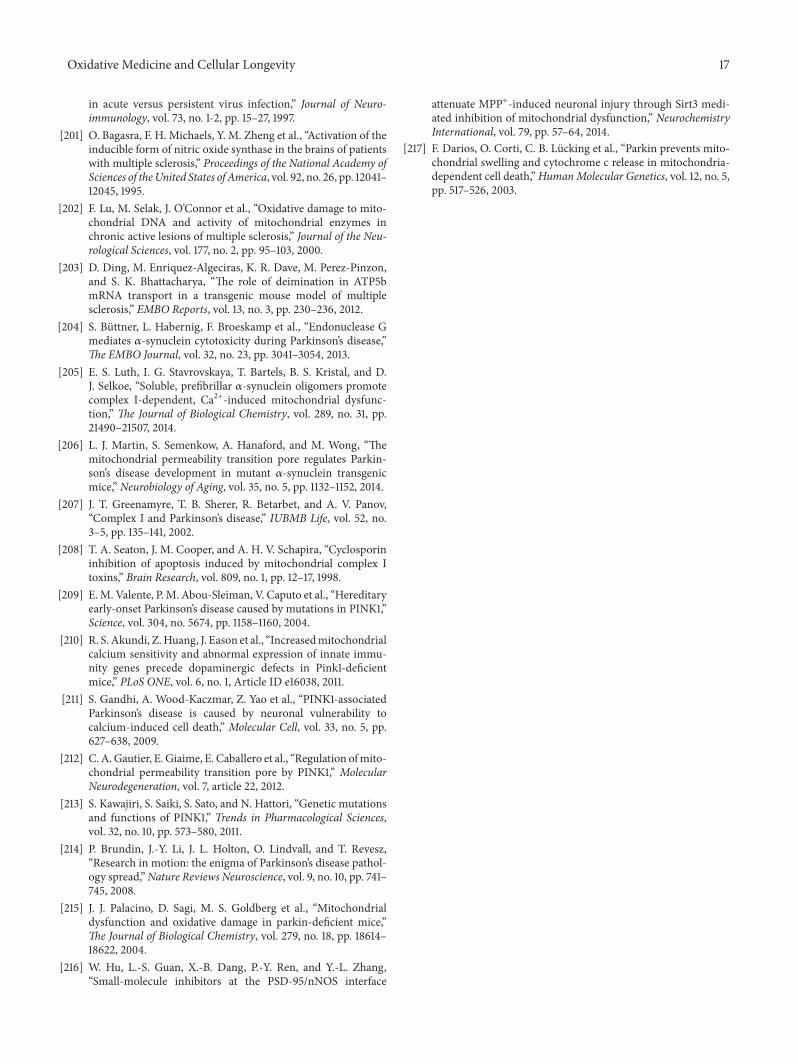
Oxidative Medicine and Cellular Longevity 17
in acute versus persistent virus infection,” Journal of Neuro-immunology, vol. 73, no. 1-2, pp. 15–27, 1997.
[201] O. Bagasra, F. H. Michaels, Y. M. Zheng et al., “Activation of theinducible form of nitric oxide synthase in the brains of patientswith multiple sclerosis,” Proceedings of the National Academy ofSciences of theUnited States of America, vol. 92, no. 26, pp. 12041–12045, 1995.
[202] F. Lu, M. Selak, J. O’Connor et al., “Oxidative damage to mito-chondrial DNA and activity of mitochondrial enzymes inchronic active lesions of multiple sclerosis,” Journal of the Neu-rological Sciences, vol. 177, no. 2, pp. 95–103, 2000.
[203] D. Ding, M. Enriquez-Algeciras, K. R. Dave, M. Perez-Pinzon,and S. K. Bhattacharya, “The role of deimination in ATP5bmRNA transport in a transgenic mouse model of multiplesclerosis,” EMBO Reports, vol. 13, no. 3, pp. 230–236, 2012.
[204] S. Büttner, L. Habernig, F. Broeskamp et al., “Endonuclease Gmediates 𝛼-synuclein cytotoxicity during Parkinson’s disease,”The EMBO Journal, vol. 32, no. 23, pp. 3041–3054, 2013.
[205] E. S. Luth, I. G. Stavrovskaya, T. Bartels, B. S. Kristal, and D.J. Selkoe, “Soluble, prefibrillar 𝛼-synuclein oligomers promotecomplex I-dependent, Ca2+-induced mitochondrial dysfunc-tion,” The Journal of Biological Chemistry, vol. 289, no. 31, pp.21490–21507, 2014.
[206] L. J. Martin, S. Semenkow, A. Hanaford, and M. Wong, “Themitochondrial permeability transition pore regulates Parkin-son’s disease development in mutant 𝛼-synuclein transgenicmice,” Neurobiology of Aging, vol. 35, no. 5, pp. 1132–1152, 2014.
[207] J. T. Greenamyre, T. B. Sherer, R. Betarbet, and A. V. Panov,“Complex I and Parkinson’s disease,” IUBMB Life, vol. 52, no.3–5, pp. 135–141, 2002.
[208] T. A. Seaton, J. M. Cooper, and A. H. V. Schapira, “Cyclosporininhibition of apoptosis induced by mitochondrial complex Itoxins,” Brain Research, vol. 809, no. 1, pp. 12–17, 1998.
[209] E.M. Valente, P.M. Abou-Sleiman, V. Caputo et al., “Hereditaryearly-onset Parkinson’s disease caused by mutations in PINK1,”Science, vol. 304, no. 5674, pp. 1158–1160, 2004.
[210] R. S. Akundi, Z.Huang, J. Eason et al., “Increasedmitochondrialcalcium sensitivity and abnormal expression of innate immu-nity genes precede dopaminergic defects in Pink1-deficientmice,” PLoS ONE, vol. 6, no. 1, Article ID e16038, 2011.
[211] S. Gandhi, A. Wood-Kaczmar, Z. Yao et al., “PINK1-associatedParkinson’s disease is caused by neuronal vulnerability tocalcium-induced cell death,” Molecular Cell, vol. 33, no. 5, pp.627–638, 2009.
[212] C.A.Gautier, E. Giaime, E. Caballero et al., “Regulation ofmito-chondrial permeability transition pore by PINK1,” MolecularNeurodegeneration, vol. 7, article 22, 2012.
[213] S. Kawajiri, S. Saiki, S. Sato, and N. Hattori, “Genetic mutationsand functions of PINK1,” Trends in Pharmacological Sciences,vol. 32, no. 10, pp. 573–580, 2011.
[214] P. Brundin, J.-Y. Li, J. L. Holton, O. Lindvall, and T. Revesz,“Research in motion: the enigma of Parkinson’s disease pathol-ogy spread,”Nature Reviews Neuroscience, vol. 9, no. 10, pp. 741–745, 2008.
[215] J. J. Palacino, D. Sagi, M. S. Goldberg et al., “Mitochondrialdysfunction and oxidative damage in parkin-deficient mice,”The Journal of Biological Chemistry, vol. 279, no. 18, pp. 18614–18622, 2004.
[216] W. Hu, L.-S. Guan, X.-B. Dang, P.-Y. Ren, and Y.-L. Zhang,“Small-molecule inhibitors at the PSD-95/nNOS interface
attenuate MPP+-induced neuronal injury through Sirt3 medi-ated inhibition of mitochondrial dysfunction,” NeurochemistryInternational, vol. 79, pp. 57–64, 2014.
[217] F. Darios, O. Corti, C. B. Lücking et al., “Parkin prevents mito-chondrial swelling and cytochrome c release in mitochondria-dependent cell death,”HumanMolecular Genetics, vol. 12, no. 5,pp. 517–526, 2003.
Clinical StudyMagnesium Supplementation Diminishes PeripheralBlood Lymphocyte DNA Oxidative Damage in Athletes andSedentary Young Man
Jelena PetroviT,1 Dušanka StaniT,1 Gordana DmitrašinoviT,2 Bosiljka PleTaš-SolaroviT,1
Svetlana IgnjatoviT,2 Bojan BatiniT,1 Dejana PopoviT,1 and Vesna PešiT1
1Department of Physiology, University of Belgrade, Faculty of Pharmacy, 11121 Belgrade, Serbia2Department of Medical Biochemistry, University of Belgrade, Faculty of Pharmacy, 11121 Belgrade, Serbia
Correspondence should be addressed to Vesna Pešić; [email protected]
Received 24 September 2015; Accepted 11 February 2016
Academic Editor: Paola Venditti
Copyright © 2016 Jelena Petrović et al. This is an open access article distributed under the Creative Commons Attribution License,which permits unrestricted use, distribution, and reproduction in any medium, provided the original work is properly cited.
Sedentary lifestyle is highly associated with increased risk of cardiovascular disease, obesity, and type 2 diabetes. It is known thatregular physical activity has positive effects on health; however several studies have shown that acute and strenuous exercise caninduce oxidative stress and lead to DNA damage. As magnesium is essential in maintaining DNA integrity, the aim of this studywas to determine whether four-week-long magnesium supplementation in students with sedentary lifestyle and rugby playerscould prevent or diminish impairment of DNA. By using the comet assay, our study demonstrated that the number of peripheralblood lymphocytes (PBL) with basal endogenous DNA damage is significantly higher in rugby players compared to students withsedentary lifestyle. On the other hand, magnesium supplementation significantly decreased the number of cells with high DNAdamage, in the presence of exogenous H
2O2, in PBL from both students and rugby players, and markedly reduced the number
of cells with medium DNA damage in rugby players compared to corresponding control nonsupplemented group. Accordingly,the results of our study suggest that four-week-long magnesium supplementation has marked effects in protecting the DNA fromoxidative damage in both rugby players and in young men with sedentary lifestyle. Clinical trial is registered at ANZCTR Trial Id:ACTRN12615001237572.
1. Introduction
Modern age has brought upon a life style that is, in youngpeople particularly, accompanied by lack of sleep, imbalanceddiet including fast food consumption, excessive amountof stress, reduced physical activity, and alcohol abuse. Allthese factors contribute to excess inflammation and oxidativestress and exhibit detrimental influence on human genome[1–4]. Each day, the human genome suffers approximatelyone million lesions, including adducts, modifications, orfragmentation of the sugar phosphate backbone of DNA [5].If left unrepaired, DNA damage can cause mutations suchas base substitutions and chromosomal translocations thatdisrupt normal gene expression or create abnormal proteinsthat are detrimental to cellular function or viability [6].
Both endogenous and exogenous factors can increaseproduction of reactive oxygen species (ROS) and induceDNA damage [7]. Endogenous factors include products ofcellular metabolism, such as ROS created via mitochondrialoxidative respiration, or produced by lipid peroxidation, andduring processes such as phagocytosis. Endogenous damagemay also occur due to errors arising from normal cell repli-cation [8, 9]. Exogenous factors, on the other hand, includeimproper diet, alcohol, cigarette smoking, and environmentaltoxins [8, 10]. Evidence indicates that oxidative stress inducedDNA damage and impaired DNA repair mechanisms areinvolved in the pathogenesis of cancer, atherosclerosis, neu-rodegenerative disorders, and chronic lung diseases [11, 12].
Even though physical exercise is considered to havebeneficial effects on health, many, but not all studies on this
Hindawi Publishing CorporationOxidative Medicine and Cellular LongevityVolume 2016, Article ID 2019643, 7 pageshttp://dx.doi.org/10.1155/2016/2019643
2 Oxidative Medicine and Cellular Longevity
topic, gave evidence that acute and strenuous exercise caninduce oxidative stress [13]. Acute and strenuous exerciselead to oxidative stress by excessive production of ROS andreactive nitrogen species (RNS), superoxide (O
2
∙−), hydrogenperoxide (H
2O2), hydroxyl radical (OH∙), hypochlorous acid
(HOCl), and nitric oxide (NO∙) that exhibit detrimentaleffects on macromolecules such as lipids and proteins, butmainly the DNA [13, 14]. During exercise, oxygen consump-tion can increase up to 10- to 15-fold above resting levels, thustemporarily increasing the rate of mitochondrial free radicalsproduction. Exercisemay also induce inflammatory reactionssimilar to the acute phase response occurring in injury orinfection [15].
Rugby matches are considered to be very demanding andcause both physical and psychological stress in participants[16, 17]. Various studies evaluated the changes in immu-noendocrine and inflammatorymarkers, neutrophil function(production of ROS and phagocytic activity), and the recov-ery time-course of neuromuscular function, concentrationsof testosterone and cortisol, mood impairments, and muscledamage after intense competitive physical performance, suchas a rugby game [16, 18–21]. Nevertheless, there are fewevidences concerning the correlation between rugby-inducedstress and the level of oxidative damage [18, 21].
On the other hand, magnesium as an essential element isinvolved in regulation of cell cycle and apoptosis, stabilizesthe structure of nucleic acids [22], protects DNA fromalkylation, and has a role as a cofactor of the enzymes ofnucleic acid metabolism: it is involved in DNA replication,DNA repair, and gene expression [23, 24].
Having all abovementioned in mind, the aim of thisstudy was to investigate whether four-week-long magnesiumsupplementation in young men, with either sedentary lifestyle such as students or athletes involved in strenuousexercise such as rugby players, could protect peripheral bloodlymphocytes (PBL) from hydrogen peroxide-induced DNAdamage evaluated by alkaline Comet test.
2. Materials and Methods
2.1. Subjects. Twenty-three healthy young male subjects vol-unteered for this study, thirteen were members of the sameamateur rugby team, and the other ten were students ofthe University of Belgrade with sedentary life style. Theparticipants were divided into four groups, as follows:
Group 1: students with a sedentary life style, withoutmagnesium supplementation (𝑛 = 5).
Group 2: students with a sedentary life style, receiving500mg of magnesium per day divided in two dosesseparated by 12 h interval for 28 days (𝑛 = 5).
Group 3: rugby players, without magnesium supple-mentation (𝑛 = 5).
Group 4: rugby players receiving 500mg of magne-sium per day divided in two doses (one tablet ofmagnesium 250mg�. Natural Wealth, NBTY Inc.)separated by 12 h interval for 28 days (𝑛 = 8).
Students had not been involved in any regular exercise forat least six months and they remained sedentary during thewhole study. Rugby players trained three to four times aweek for 2 hours and played a match on each Sunday in theearly afternoon. Three months before and during the study,none of the participants was taking other vitamin or mineralsupplements. All participants gave their written consent toparticipate after being fully informed of all experimentalprocedures. The investigation was carried out according tothe guidelines and study protocol that has been approvedby the Ethical Committee for Clinical trials of University ofBelgrade, Faculty of Pharmacy, number 199/2.
2.2. Anthropometric Data. Rugby players involved in thisstudy were 23.30 ± 0.93 years old and had average height181.26 ± 1.30 cm, body mass 85.30 ± 2.31 kg, and body massindex (BMI) 25.91 ± 0.51 kg/m2 (Mean ± SD). Mean age ofstudents was 22.6 ± 0.52 years, height was 186.9 ± 6.42 cm,bodymasswas 84.4±6.60 kg, andBMIwas 24.16±1.41 kg/m2.
2.3. Blood Collection. On the 29th day of experiment (firstday after 28 days of supplementation) peripheral blood sam-ples were collected from the participants of this study. In themorning from 09.00–10.00 a.m. (about 20 h after the rugbymatch for athletes), blood was drawn from an antecubitalvein in sitting position into 2mL EDTAVacutainer tubes andcomet assay was performed.
2.4. Comet Assay (Single Cell Gel Electrophoresis (SCGE)).The comet assay is highly reproducible, rapid, and sen-sitive method for measuring DNA damage [25, 26]. Thecomet test was conducted as described by Singh et al.[25]. Microscope slides were coated with a layer of 1%normalmelting point agarose (Sigma-Aldrich, St. Louis,MO)and left to dry on room temperature. The 6 𝜇L of wholeblood samples was suspended in 0.67% low melting point(LMP) agarose (Sigma-Aldrich, St. Louis, MO) in phosphatebuffered saline (PBS, Torlak Institute of Immunology andVirology, Belgrade, Serbia), applied onto microscope slides(prepared on previously described way) and maintained on4∘C for 5min, to solidify. Then a second layer of 0.5%LMP agarose was pipetted onto microscope slides. Everysample had a matching positive control that was treated withhydrogen peroxide (1.5mM H
2O2) and then after 5min on
4∘C with second layer of 0.5% LMP agarose. The followingstep included immersing all slides in lysing solution (2.5MNaCl, 100mM ethylene-diaminetetra-acetic acid, 10mMTrisat pH 10, 1% Triton X-100 and 10% dimethylsulfoxide, pH 10adjusted with NaOH) on 4∘C and leaving them overnight.After that, DNA in lysed cells was allowed to unwind in alkalibuffer (300mM NaOH, 1mM EDTA) for 30min. Sampleswere then subjected to electrophoresis for another 30minat 215mA, 25V, washed three times (for 5min each slide)with a neutralizing buffer (0.4M Tris, pH 7.5), and stainedwith ethidium bromide (20𝜇g/mL). Cover slips were thenplaced on top of microscope slides and DNA damage wasvisually analyzed. Analyses were performed on Olympus BX50 microscope (Olympus Optical Co., GmbH, Hamburg,
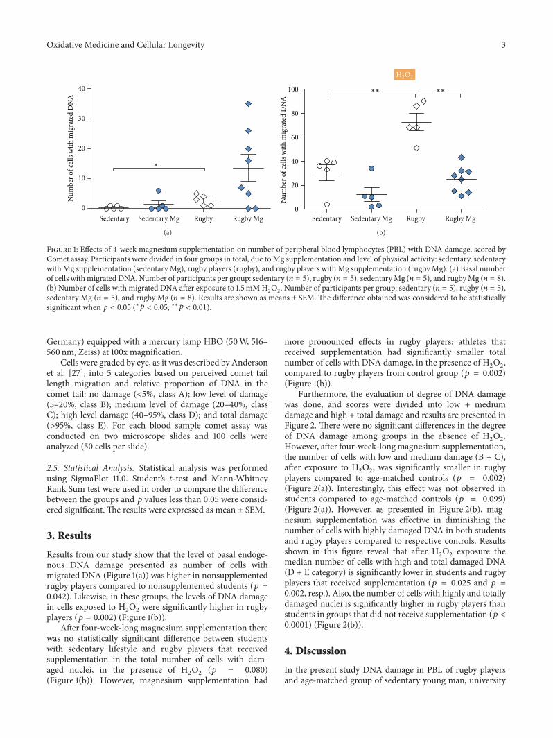
Oxidative Medicine and Cellular Longevity 3
Num
ber o
f cel
ls w
ith m
igra
ted
DN
A
40
30
20
10
0
Sedentary Sedentary Mg Rugby Rugby Mg
∗
(a)
Num
ber o
f cel
ls w
ith m
igra
ted
DN
A
Sedentary Sedentary Mg Rugby Rugby Mg
∗∗∗∗
H2O2
100
80
60
40
20
0
(b)
Figure 1: Effects of 4-week magnesium supplementation on number of peripheral blood lymphocytes (PBL) with DNA damage, scored byComet assay. Participants were divided in four groups in total, due to Mg supplementation and level of physical activity: sedentary, sedentarywith Mg supplementation (sedentary Mg), rugby players (rugby), and rugby players with Mg supplementation (rugbyMg). (a) Basal numberof cells withmigrated DNA. Number of participants per group: sedentary (𝑛 = 5), rugby (𝑛 = 5), sedentaryMg (𝑛 = 5), and rugbyMg (𝑛 = 8).(b) Number of cells with migrated DNA after exposure to 1.5mMH
2O2. Number of participants per group: sedentary (𝑛 = 5), rugby (𝑛 = 5),
sedentary Mg (𝑛 = 5), and rugby Mg (𝑛 = 8). Results are shown as means ± SEM. The difference obtained was considered to be statisticallysignificant when 𝑝 < 0.05 (∗𝑝 < 0.05; ∗∗𝑝 < 0.01).
Germany) equipped with a mercury lamp HBO (50W, 516–560 nm, Zeiss) at 100x magnification.
Cells were graded by eye, as it was described by Andersonet al. [27], into 5 categories based on perceived comet taillength migration and relative proportion of DNA in thecomet tail: no damage (<5%, class A); low level of damage(5–20%, class B); medium level of damage (20–40%, classC); high level damage (40–95%, class D); and total damage(>95%, class E). For each blood sample comet assay wasconducted on two microscope slides and 100 cells wereanalyzed (50 cells per slide).
2.5. Statistical Analysis. Statistical analysis was performedusing SigmaPlot 11.0. Student’s 𝑡-test and Mann-WhitneyRank Sum test were used in order to compare the differencebetween the groups and 𝑝 values less than 0.05 were consid-ered significant. The results were expressed as mean ± SEM.
3. Results
Results from our study show that the level of basal endoge-nous DNA damage presented as number of cells withmigrated DNA (Figure 1(a)) was higher in nonsupplementedrugby players compared to nonsupplemented students (𝑝 =0.042). Likewise, in these groups, the levels of DNA damagein cells exposed to H
2O2were significantly higher in rugby
players (𝑝 = 0.002) (Figure 1(b)).After four-week-long magnesium supplementation there
was no statistically significant difference between studentswith sedentary lifestyle and rugby players that receivedsupplementation in the total number of cells with dam-aged nuclei, in the presence of H
2O2
(𝑝 = 0.080)(Figure 1(b)). However, magnesium supplementation had
more pronounced effects in rugby players: athletes thatreceived supplementation had significantly smaller totalnumber of cells with DNA damage, in the presence of H
2O2,
compared to rugby players from control group (𝑝 = 0.002)(Figure 1(b)).
Furthermore, the evaluation of degree of DNA damagewas done, and scores were divided into low + mediumdamage and high + total damage and results are presented inFigure 2. There were no significant differences in the degreeof DNA damage among groups in the absence of H
2O2.
However, after four-week-longmagnesium supplementation,the number of cells with low and medium damage (B + C),after exposure to H
2O2, was significantly smaller in rugby
players compared to age-matched controls (𝑝 = 0.002)(Figure 2(a)). Interestingly, this effect was not observed instudents compared to age-matched controls (𝑝 = 0.099)(Figure 2(a)). However, as presented in Figure 2(b), mag-nesium supplementation was effective in diminishing thenumber of cells with highly damaged DNA in both studentsand rugby players compared to respective controls. Resultsshown in this figure reveal that after H
2O2exposure the
median number of cells with high and total damaged DNA(D + E category) is significantly lower in students and rugbyplayers that received supplementation (𝑝 = 0.025 and 𝑝 =0.002, resp.). Also, the number of cells with highly and totallydamaged nuclei is significantly higher in rugby players thanstudents in groups that did not receive supplementation (𝑝 <0.0001) (Figure 2(b)).
4. Discussion
In the present study DNA damage in PBL of rugby playersand age-matched group of sedentary young man, university
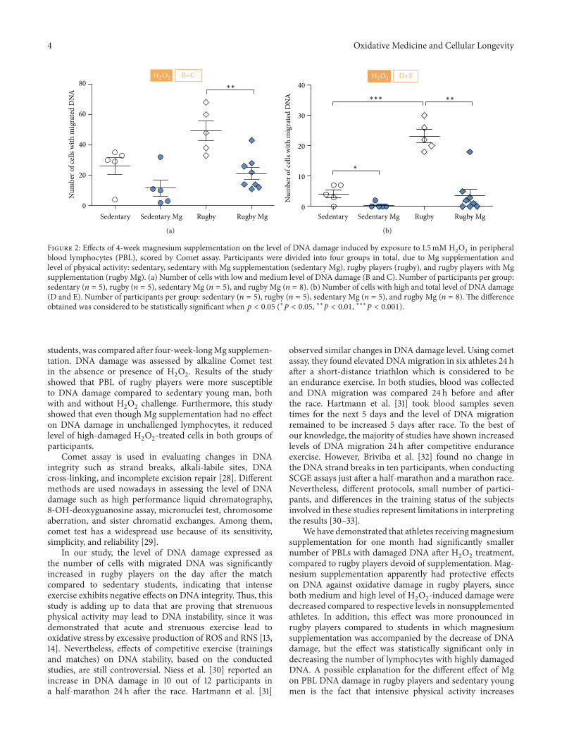
4 Oxidative Medicine and Cellular Longevity
H2O2
Sedentary Sedentary Mg Rugby Rugby Mg
∗∗
Num
ber o
f cel
ls w
ith m
igra
ted
DN
A80
60
40
20
0
B+C
(a)
H2O2
Sedentary Sedentary Mg Rugby Rugby Mg
∗∗
∗
∗∗∗
Num
ber o
f cel
ls w
ith m
igra
ted
DN
A
40
30
20
10
0
D+E
(b)
Figure 2: Effects of 4-week magnesium supplementation on the level of DNA damage induced by exposure to 1.5mM H2O2in peripheral
blood lymphocytes (PBL), scored by Comet assay. Participants were divided into four groups in total, due to Mg supplementation andlevel of physical activity: sedentary, sedentary with Mg supplementation (sedentary Mg), rugby players (rugby), and rugby players with Mgsupplementation (rugby Mg). (a) Number of cells with low and medium level of DNA damage (B and C). Number of participants per group:sedentary (𝑛 = 5), rugby (𝑛 = 5), sedentary Mg (𝑛 = 5), and rugby Mg (𝑛 = 8). (b) Number of cells with high and total level of DNA damage(D and E). Number of participants per group: sedentary (𝑛 = 5), rugby (𝑛 = 5), sedentary Mg (𝑛 = 5), and rugby Mg (𝑛 = 8). The differenceobtained was considered to be statistically significant when 𝑝 < 0.05 (∗𝑝 < 0.05, ∗∗𝑝 < 0.01, ∗∗∗𝑝 < 0.001).
students, was compared after four-week-longMg supplemen-tation. DNA damage was assessed by alkaline Comet testin the absence or presence of H
2O2. Results of the study
showed that PBL of rugby players were more susceptibleto DNA damage compared to sedentary young man, bothwith and without H
2O2challenge. Furthermore, this study
showed that even though Mg supplementation had no effecton DNA damage in unchallenged lymphocytes, it reducedlevel of high-damaged H
2O2-treated cells in both groups of
participants.Comet assay is used in evaluating changes in DNA
integrity such as strand breaks, alkali-labile sites, DNAcross-linking, and incomplete excision repair [28]. Differentmethods are used nowadays in assessing the level of DNAdamage such as high performance liquid chromatography,8-OH-deoxyguanosine assay, micronuclei test, chromosomeaberration, and sister chromatid exchanges. Among them,comet test has a widespread use because of its sensitivity,simplicity, and reliability [29].
In our study, the level of DNA damage expressed asthe number of cells with migrated DNA was significantlyincreased in rugby players on the day after the matchcompared to sedentary students, indicating that intenseexercise exhibits negative effects on DNA integrity.Thus, thisstudy is adding up to data that are proving that strenuousphysical activity may lead to DNA instability, since it wasdemonstrated that acute and strenuous exercise lead tooxidative stress by excessive production of ROS and RNS [13,14]. Nevertheless, effects of competitive exercise (trainingsand matches) on DNA stability, based on the conductedstudies, are still controversial. Niess et al. [30] reported anincrease in DNA damage in 10 out of 12 participants ina half-marathon 24 h after the race. Hartmann et al. [31]
observed similar changes in DNA damage level. Using cometassay, they found elevated DNAmigration in six athletes 24 hafter a short-distance triathlon which is considered to bean endurance exercise. In both studies, blood was collectedand DNA migration was compared 24 h before and afterthe race. Hartmann et al. [31] took blood samples seventimes for the next 5 days and the level of DNA migrationremained to be increased 5 days after race. To the best ofour knowledge, the majority of studies have shown increasedlevels of DNA migration 24 h after competitive enduranceexercise. However, Briviba et al. [32] found no change inthe DNA strand breaks in ten participants, when conductingSCGE assays just after a half-marathon and a marathon race.Nevertheless, different protocols, small number of partici-pants, and differences in the training status of the subjectsinvolved in these studies represent limitations in interpretingthe results [30–33].
We have demonstrated that athletes receivingmagnesiumsupplementation for one month had significantly smallernumber of PBLs with damaged DNA after H
2O2treatment,
compared to rugby players devoid of supplementation. Mag-nesium supplementation apparently had protective effectson DNA against oxidative damage in rugby players, sinceboth medium and high level of H
2O2-induced damage were
decreased compared to respective levels in nonsupplementedathletes. In addition, this effect was more pronounced inrugby players compared to students in which magnesiumsupplementation was accompanied by the decrease of DNAdamage, but the effect was statistically significant only indecreasing the number of lymphocytes with highly damagedDNA. A possible explanation for the different effect of Mgon PBL DNA damage in rugby players and sedentary youngmen is the fact that intensive physical activity increases
Oxidative Medicine and Cellular Longevity 5
magnesium requirement. Evidence indicates that both short-term high-intensity and long-term strenuous exercise causesignificantly increased loss of magnesium through urineand sweat [34]. Also, during exercise, redistribution ofmagnesium to certain body compartments, with increasedenergy andROS production, occurs [34–36].Therefore, someauthors propose that short- and long-term strenuous exerciseshould be accompanied with 10–20% higher magnesiumintake compared to daily intake recommendations for thepersons of the same age and sex with sedentary lifestyle [34].
Both animal and human studies indicated that magne-sium deficiency has a negative impact on physical perfor-mance [34, 37]. However, that is not the only negative conse-quence of decreased level of magnesium in the body: studieson experimental animals demonstrated thatmagnesium defi-ciency leads to swollen mitochondria and disorganized sar-coplasmic reticulum in skeletal muscles, increases formationof lipid radicals and nitric oxide, and impairs endogenousprotective mechanisms such as glutathione [38, 39]. It hasalso been shown that vigorous exercise induces inflammatoryresponse because of tissue injury, consequently increasing theproduction of ROS, especially in activated phagocytes [15,40–42]. Takahashi et al. [18] reported no significant changein ROS production in neutrophils right after a rugby game,but they also noticed that 4 h after the end of the game it hadincreased significantly. Nevertheless, some authors pointedout that chronic moderate exercise may induce adaptiveresponses in human organism by enhancing the expressionof antioxidant enzymes such as Cu/Zn superoxide dismutase(SOD) and by reducing mitochondrial hydrogen peroxide[43, 44] in the muscle tissue.The effectiveness of the adaptivemechanisms in oxidative stress induced by regular physicalactivity also depends on the individual’s lifestyle, nutrition,and expression of genes involved in DNA repair systems[29, 45]. Future studies investigating the effects of strenuousexercise on DNA stability should include longer observationperiods and monitor the DNA integrity for at least fivedays after the match as some authors claim that for majoralterations in DNA repair mechanisms take more than 24hours [15, 33, 46, 47].
Furthermore, it might be taken into account that greaterdegree of magnesium deficiency which influences antiox-idant activity in PBLs makes them more susceptible tooxidative stress caused by hydrogen peroxide, since previousdata showed the harmful effect of magnesium deficiency onlipid peroxidation in the cardiovascular system [48], and thattissue homogenates from magnesium deficient animals weremore susceptible to lipid peroxidation than animals fed dietsadequate in magnesium [48]. Nevertheless, this assumptionshould be explored and possibly confirmed by estimatinglevel of magnesium in PBLs of rugby players and sedentarystudents, in the future investigation on the larger population.
Results of this study point to the conclusion that stren-uous exercise sensitizes PBLs to oxidative stress and thatmagnesium supplementation shows protective effects inreducing the level of H
2O2-induced PBL DNA damage thus
indicating the importance of adequate magnesium intake inboth students with sedentary life style and physically activeindividuals.
Competing Interests
The authors stated that there is no conflict of interestswhatsoever regarding the publication of this paper.
Acknowledgments
The work was supported entirely by a grant from the Min-istry of Education, Science and Technological Development,Republic of Serbia, Grant no. 175036.
References
[1] M. E. Cogswell, P.Weisberg, and C. Spong, “Cigarette smoking,alcohol use and adverse pregnancy outcomes: implications formicronutrient supplementation,” The Journal of Nutrition, vol.133, no. 5, pp. 1722S–1731S, 2003.
[2] B.-T. Ji, X.-O. Shu, M. S. Linet et al., “Paternal cigarette smokingand the risk of childhood cancer among offspring of nonsmok-ing mothers,” Journal of the National Cancer Institute, vol. 89,no. 3, pp. 238–244, 1997.
[3] Y.-J. Kim, Y.-C. Hong, K.-H. Lee et al., “Oxidative stress inpregnant women and birth weight reduction,” ReproductiveToxicology, vol. 19, no. 4, pp. 487–492, 2005.
[4] S. Raimondi, S. Garte, R. J. Sram et al., “Effects of diet onbiomarkers of exposure and effects, and on oxidative damage,”Mutation Research/Fundamental and Molecular Mechanisms ofMutagenesis, vol. 620, no. 1-2, pp. 93–102, 2007.
[5] J. M. Skoner, J. Sigmon, and L. L. Larcom, “Suppressed DNArepair capacity of peripheral lymphocytes in pregnant women,”Molecular and Cellular Endocrinology, vol. 108, no. 1-2, pp. 179–183, 1995.
[6] D. L. F. Furness, G. A. Dekker, and C. T. Roberts, “DNA damageand health in pregnancy,” Journal of Reproductive Immunology,vol. 89, no. 2, pp. 153–162, 2011.
[7] F. J. Jenkins, B. Van Houten, and D. H. Bovbjerg, “Effects onDNAdamage and/or repair processes as biological mechanismslinking psychological stress to cancer risk,” Journal of AppliedBiobehavioral Research, vol. 19, no. 1, pp. 3–23, 2014.
[8] Y. Gidron, K. Russ, H. Tissarchondou, and J. Warner, “Therelation between psychological factors and DNA-damage: acritical review,” Biological Psychology, vol. 72, no. 3, pp. 291–304,2006.
[9] J. H. J. Hoeijmakers, “Genome maintenance mechanisms forpreventing cancer,”Nature, vol. 411, no. 6835, pp. 366–374, 2001.
[10] P. Møller, H. Wallin, and L. E. Knudsen, “Oxidative stressassociated with exercise, psychological stress and life-style fac-tors,” Chemico-Biological Interactions, vol. 102, no. 1, pp. 17–36,1996.
[11] B. Halliwell and J. M. Gutteridge, Free Radicals in Biology andMedicine, Oxford University Press, London, UK, 3rd edition,1999.
[12] L. L. Wu, C.-C. Chiou, P.-Y. Chang, and J. T. Wu, “Urinary 8-OHdG: a marker of oxidative stress to DNA and a risk factorfor cancer, atherosclerosis and diabetics,” Clinica Chimica Acta,vol. 339, no. 1-2, pp. 1–9, 2004.
[13] K.-H. Wagner, S. Reichhold, C. Hölzl et al., “Well-trained,healthy triathletes experience no adverse health risks regardingoxidative stress and DNA damage by participating in an ultra-endurance event,” Toxicology, vol. 278, no. 2, pp. 211–216, 2010.
6 Oxidative Medicine and Cellular Longevity
[14] L. Packer, E. Cadenas, and K. J. A. Davies, “Free radicals andexercise: an introduction,” Free Radical Biology &Medicine, vol.44, no. 2, pp. 123–125, 2008.
[15] K. Tsai, T.-G. Hsu, K.-M. Hsu et al., “Oxidative DNA damagein human peripheral leukocytes induced by massive aerobicexercise,” Free Radical Biology & Medicine, vol. 31, no. 11, pp.1465–1472, 2001.
[16] B. Cunniffe, A. J. Hore, D.M.Whitcombe, K. P. Jones, J. S. Baker,and B. Davies, “Time course of changes in immuneoendocrinemarkers following an international rugby game,” EuropeanJournal of Applied Physiology, vol. 108, no. 1, pp. 113–122, 2010.
[17] T. Mashiko, T. Umeda, S. Nakaji, and K. Sugawara, “Positionrelated analysis of the appearance of and relationship betweenpost-match physical and mental fatigue in university rugbyfootball players,” British Journal of Sports Medicine, vol. 38, no.5, pp. 617–621, 2004.
[18] I. Takahashi, T. Umeda, T. Mashiko et al., “Effects of rugbysevens matches on human neutrophil-related non-specificimmunity,” British Journal of Sports Medicine, vol. 41, no. 1, pp.13–18, 2007.
[19] D. J. West, C. V. Finn, D. J. Cunningham et al., “Neuromuscularfunction, hormonal, and mood responses to a professionalrugby union match,” Journal of Strength and ConditioningResearch, vol. 28, no. 1, pp. 194–200, 2014.
[20] M. Elloumi, F. Maso, O. Michaux, A. Robert, and G. Lac,“Behaviour of saliva cortisol [C], testosterone [T] and the T/Cratio during a rugby match and during the post-competitionrecovery days,” European Journal of Applied Physiology, vol. 90,no. 1-2, pp. 23–28, 2003.
[21] A. Lindsay, J. Healy, W. Mills et al., “Impact-induced muscledamage and urinary pterins in professional rugby: 7,8-dihy-droneopterin oxidation by myoglobin,” Scandinavian Journal ofMedicine & Science in Sports, vol. 26, no. 3, pp. 329–337, 2016.
[22] J. Anastassopoulou and T. Theophanides, “Magnesium-DNAinteractions and the possible relation of magnesium to car-cinogenesis. Irradiation and free radicals,” Critical Reviews inOncology/Hematology, vol. 42, no. 1, pp. 79–91, 2002.
[23] A. Klungland, I. Rosewell, S. Hollenbach et al., “Accumulationof premutagenic DNA lesions in mice defective in removal ofoxidative base damage,” Proceedings of the National Academyof Sciences of the United States of America, vol. 96, no. 23, pp.13300–13305, 1999.
[24] P. Møller and H. Wallin, “Adduct formation, mutagenesis andnucleotide excision repair of DNA damage produced by reac-tive oxygen species and lipid peroxidation product,” MutationResearch/Reviews in Mutation Research, vol. 410, no. 3, pp. 271–290, 1998.
[25] N. P. Singh, M. T. McCoy, R. R. Tice, and E. L. Schneider, “Asimple technique for quantitation of low levels of DNA damagein individual cells,” Experimental Cell Research, vol. 175, no. 1,pp. 184–191, 1988.
[26] M. Neri, D. Milazzo, D. Ugolini et al., “Worldwide interest inthe comet assay: a bibliometric study,”Mutagenesis, vol. 30, no.1, pp. 155–163, 2015.
[27] D.Anderson, T.-W.Yu, B. J. Phillips, andP. Schmezer, “The effectof various antioxidants and other modifying agents on oxygen-radical-generated DNA damage in human lymphocytes in theCOMET assay,” Mutation Research, vol. 307, no. 1, pp. 261–271,1994.
[28] S.-Y. Park, E. Cho, E. Oh, and D. Sul, “Comet assay analysis ofDNA damage in T- and B-lymphocytes separated by MACS for
human biomonitoring studies,” Toxicology in Vitro, vol. 26, no.2, pp. 369–372, 2012.
[29] B. Tomasello, S. Grasso, G. Malfa, S. Stella, M. Favetta, andM. Renis, “Double-face activity of resveratrol in voluntaryrunners: assessment of DNA damage by comet assay,” Journal ofMedicinal Food, vol. 15, no. 5, pp. 441–447, 2012.
[30] A. M. Niess, M. Baumann, K. Roecker, T. Horstmann, F. Mayer,andH.-H.Dickhuth, “Effects of intensive endurance exercise onDNA damage in leucocytes,”The Journal of Sports Medicine andPhysical Fitness, vol. 38, no. 2, pp. 111–115, 1998.
[31] A. Hartmann, S. Pfuhler, C. Dennog, D. Germadnik, A. Pilger,and G. Speit, “Exercise-induced DNA effects in human leuko-cytes are not accompanied by increased formation of 8-hydroxy-2-deoxyguanosine or induction of micronuclei,” FreeRadical Biology and Medicine, vol. 24, no. 2, pp. 245–251, 1998.
[32] K. Briviba, B. Watzl, K. Nickel et al., “A half-marathon and amarathon run induce oxidative DNA damage, reduce antiox-idant capacity to protect DNA against damage and modifyimmune function in hobby runners,” Redox Report, vol. 10, no.6, pp. 325–331, 2005.
[33] S. Reichhold, O. Neubauer, C. Hoelzl et al., “DNA damage inresponse to an Ironman triathlon,” Free Radical Research, vol.43, no. 8, pp. 753–760, 2009.
[34] F. H. Nielsen and H. C. Lukaski, “Update on the relationshipbetweenmagnesium and exercise,”MagnesiumResearch, vol. 19,no. 3, pp. 180–189, 2006.
[35] K. Madsen, P. K. Pedersen, M. S. Djurhuus, and N. A. Klitgaard,“Effects of detraining on endurance capacity and metabolicchanges during prolonged exhaustive exercise,” Journal ofApplied Physiology, vol. 75, no. 4, pp. 1444–1451, 1993.
[36] A. Córdova, J. F. Escanero, and M. Gimenez, “Magnesiumdistribution in rats after maximal exercise in air and underhypoxic conditions,” Magnesium Research, vol. 5, no. 1, pp. 23–27, 1992.
[37] H. C. Lukaski and F. H. Nielsen, “Dietary magnesium depletionaffects metabolic responses during submaximal exercise inpostmenopausal women,” Journal of Nutrition, vol. 132, no. 5,pp. 930–935, 2002.
[38] E. Rock, C. Astier, C. Lab et al., “Dietary magnesium deficiencyin rats enhances free radical production in skeletal muscle,”TheJournal of Nutrition, vol. 125, no. 5, pp. 1205–1210, 1995.
[39] W. B. Weglicki, I. T. Mak, B. F. Dickens et al., “Neuropeptides,free radical stress and antioxidants in models of Mg-deficientcardiomyopathy,” inMagnesium: Current Status andNewDevel-opments, T.Theophanide and J. Anastassopoulou, Eds., pp. 169–178, Kluwer Academic, Dordrecht, Netherlands, 1997.
[40] B. Sjodin, Y. Hellsten Westing, and F. S. Apple, “Biochemicalmechanisms for oxygen free radical formation during exercise,”Sports Medicine, vol. 10, no. 4, pp. 236–254, 1990.
[41] E. Zerba, T. E. Komorowski, and J. A. Faulkner, “Free radicalinjury to skeletal muscles of young, adult, and old mice,” TheAmerican Journal of Physiology—Cell Physiology, vol. 258, no. 3,pp. C429–C435, 1990.
[42] N. Zamzami, P. Marchetti, M. Castedo et al., “Sequential reduc-tion of mitochondrial transmembrane potential and generationof reactive oxygen species in early programmed cell death,”Journal of Experimental Medicine, vol. 182, no. 2, pp. 367–377,1995.
[43] C. A. Delaney, I. C. Green, J. E. Lowe et al., “Use of thecomet assay to investigate possible interactions of nitric oxideand reactive oxygen species in the induction of DNA damage
Oxidative Medicine and Cellular Longevity 7
and inhibition of function in an insulin-secreting cell line,”Mutation Research/Fundamental and Molecular Mechanisms ofMutagenesis, vol. 375, no. 2, pp. 137–146, 1997.
[44] D. A. Wink and J. Laval, “The Fpg protein, a DNA repairenzyme, is inhibited by the biomediator nitric oxide in vitro andin vivo,” Carcinogenesis, vol. 15, no. 10, pp. 2125–2129, 1994.
[45] S. Goto, H. Naito, T. Kaneko, H. Chung, and Z. Radák,“Hormetic effects of regular exercise in aging: correlation withoxidative stress,” Applied Physiology, Nutrition and Metabolism,vol. 32, no. 5, pp. 948–953, 2007.
[46] S. Reichhold, O. Neubauer, V. Ehrlich, S. Knasmüller, andK.-H. Wagner, “No acute and persistent DNA damage afteran ironman triathlon,” Cancer Epidemiology Biomarkers &Prevention, vol. 17, no. 8, pp. 1913–1919, 2008.
[47] A. Mastaloudis, T.-W. Yu, R. P. O’Donnell, B. Frei, R. H.Dashwood, and M. G. Traber, “Endurance exercise results inDNA damage as detected by the comet assay,” Free RadicalBiology & Medicine, vol. 36, no. 8, pp. 966–975, 2004.
[48] J. Busserolles, E. Gueux, E. Rock, A. Mazur, and Y. Rayssiguier,“High fructose feeding ofmagnesiumdeficient rats is associatedwith increased plasma triglyceride concentration and increasedoxidative stress,” Magnesium Research, vol. 16, no. 1, pp. 7–12,2003.
Review ArticleEndogenous Generation of Singlet Oxygen and Ozone inHuman and Animal Tissues: Mechanisms, BiologicalSignificance, and Influence of Dietary Components
Arnold N. Onyango
Department of Food Science and Technology, Jomo Kenyatta University of Agriculture and Technology, P.O. Box 62000,Nairobi 00200, Kenya
Correspondence should be addressed to Arnold N. Onyango; [email protected]
Received 27 December 2015; Accepted 8 February 2016
Academic Editor: Sergio Di Meo
Copyright © 2016 Arnold N. Onyango.This is an open access article distributed under the Creative Commons Attribution License,which permits unrestricted use, distribution, and reproduction in any medium, provided the original work is properly cited.
Recent studies have shown that exposing antibodies or amino acids to singlet oxygen results in the formation of ozone (or an ozone-like oxidant) and hydrogen peroxide and that human neutrophils produce both singlet oxygen and ozone during bacterial killing.There is also mounting evidence that endogenous singlet oxygen productionmay be a common occurrence in cells through variousmechanisms. Thus, the ozone-producing combination of singlet oxygen and amino acids might be a common cellular occurrence.This paper reviews the potential pathways of formation of singlet oxygen and ozone in vivo and also proposes some new pathwaysfor singlet oxygen formation. Physiological consequences of the endogenous formation of these oxidants in human tissues arediscussed, as well as examples of how dietary factors may promote or inhibit their generation and activity.
1. Introduction
Singlet oxygen (1O2) is an electronically excited form of oxy-
gen which is well known to be formed when photosensitizerssuch as chlorophyll or the aromatic dye rose bengal absorblight energy and transfer some of that energy to molecularoxygen [1, 2]. Various nonphotosensitizedmechanisms for itsformation have also been reported and suggested to occur inbiological systems, but the importance of such endogenoussinglet oxygen formation has had a controversial history [1,3]. Ozone (O
3) is best known as occurring in the stratosphere
where it shields organisms on earth from ultraviolet C andmuch of ultraviolet B radiations, which are the most damag-ingUV components of solar radiations because they are read-ily absorbed by DNA [4, 5]. It is also known as a respiratorysystem-damaging pollutant in the troposphere and ironicallyas a therapeutic agent in alternative medicine [6]. Morerecently, it was shown that antibodies or amino acids catalyzethe conversion of singlet oxygen (1O
2) to ozone (O
3) and that
this reaction occurs during the killing of bacteria by activatedneutrophils [7, 8]. Since both singlet oxygen and ozone arehighly reactive oxygen species, a full understanding of their
mechanisms of formation and action in vivo is necessary.Hence, this paper reviews the various reported mechanismsof the endogenous formation of these reactive oxygen species(ROS), the potential relevance of such pathways in humanphysiology, and how dietary factors affect the generation andactivity of these oxidants.
2. Radiation-Induced Formation ofSinglet Oxygen
Human beings are frequently exposed to natural and artificialradiation, and most of this interacts primarily with theskin. The spectrum of solar radiation at the earth’s surfaceconsists of ultraviolet (UV) radiation (UVB: 290–320 nm andUVA: 320–400 nm), visible radiation (VIS: 400–760 nm), andnear infrared radiation (IRA: 760–1440 nm and IRB: 1440–3000 nm) [9]. UV, VIS, and IR contribute 7%, 39%, and 54%of the solar energy reaching the skin [10]. Direct absorptionof UVB by cellular DNA leads to formation of cyclobu-tane pyrimidine dimers and pyrimidine (6–4) pyrimidoneproducts, while UVA is not readily absorbed by DNA, andits direct damage to DNA is therefore not important [5].
Hindawi Publishing CorporationOxidative Medicine and Cellular LongevityVolume 2016, Article ID 2398573, 22 pageshttp://dx.doi.org/10.1155/2016/2398573
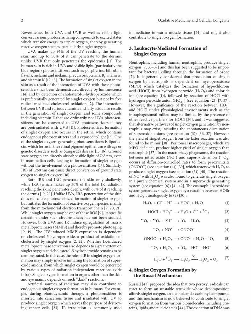
2 Oxidative Medicine and Cellular Longevity
Nevertheless, both UVA and UVB as well as visible lightconvert various photosensitizing compounds to excited stateswhich transfer energy to triplet oxygen, thereby generatingreactive oxygen species, particularly singlet oxygen.
UVA makes up 95% of the UV reaching the humanskin, and up to 50% of it can penetrate to the dermis,unlike UVB that only penetrates the epidermis [11]. Thehuman skin is rich in UVA and visible light (particularly theblue region) photosensitizers such as porphyrins, bilirubin,flavins,melanin andmelanin precursors, pterins, B
6vitamers,
and vitamin K [12, 13].The formation of singlet oxygen in theskin as a result of the interaction of UVA with these photo-sensitizers has been demonstrated directly by luminescence[14] and by detection of cholesterol-5-hydroperoxide whichis preferentially generated by singlet oxygen but not by freeradical mediated cholesterol oxidation [2]. The interactionbetweenUVBand various vitamins and fatty acids also resultsin the generation of singlet oxygen, and some compoundsincluding vitamin E that are ordinarily not UVA photosen-sitizers can be converted to UVA photosensitizers if theyare preirradiated with UVB [11]. Photosensitized formationof singlet oxygen also occurs in the retina, which containsendogenous photosensitizers and is exposed to light [15]. Oneof the singlet oxygen-generating photosensitizers is lipofus-cin, which forms in the retinal pigment epitheliumwith age orgenetic disorders such as Stargardt’s disease [15, 16]. Groundstate oxygen can directly absorb visible light of 765 nm, evenin mammalian cells, leading to formation of singlet oxygenwithout the involvement of a photosensitizer [17]. Similarly,IRB of 1268 nm can cause direct conversion of ground stateoxygen to singlet oxygen [18].
Both IRB and IRC penetrate the skin only shallowly,while IRA (which makes up 30% of the total IR radiationreaching the skin) penetrates deeply, with 65% of it reachingthe dermis [19, 20]. Unlike UVA, IRA penetration of the skindoes not cause photosensitized formation of singlet oxygenbut initiates the formation of reactive oxygen species, mainlyfrom the mitochondrial electron transport chain [9, 20, 21].While singlet oxygenmay be one of these ROS [9], its specificdetection under such circumstances has not been studied.However, both UVA and IR induce upregulation of matrixmetalloproteinases (MMPs) and thereby promote photoaging[9, 19]. The UV-induced MMP expression is dependenton cholesterol-5 hydroperoxide, a product of oxidation ofcholesterol by singlet oxygen [2, 22]. Whether IR-inducedmetalloproteinase activation also depends to a great extent onsinglet oxygen and cholesterol-5 hydroperoxide remains to bedemonstrated. In this case, the role of IR in singlet oxygen for-mation may simply involve initiating the formation of super-oxide anions, from which singlet oxygen would be generatedby various types of radiation-independent reactions (videinfra). Singlet oxygen formation in organs other than the skinand eye mainly depends on such “dark” reactions.
Artificial sources of radiation may also contribute toendogenous singlet oxygen formation in humans. For exam-ple, during photodynamic therapy, a photosensitizer isinserted into cancerous tissue and irradiated with UV toproduce singlet oxygen which serves the purpose of destroy-ing cancer cells [23]. IR irradiation is commonly used
in medicine to warm muscle tissue [24] and might alsocontribute to singlet oxygen formation.
3. Leukocyte-Mediated Formation ofSinglet Oxygen
Neutrophils, including human neutrophils, produce singletoxygen [7, 35–37] and this has been suggested to be impor-tant for bacterial killing through the formation of ozone[7]. It is generally considered that production of singletoxygen by neutrophils is dependent on myeloperoxidase(MPO) which catalyzes the formation of hypochlorousacid (HOCl) from hydrogen peroxide (H
2O2) and chloride
ion (see equation (1)), followed by reaction of HOCl withhydrogen peroxide anion (HO
2
−) (see equation (2)) [7, 37].However, the significance of the reaction between HO
2
−
and HOCl under physiological environments such as theintraphagosomal milieu may be limited by the presence ofother reactive partners for HOCl [36], and it was suggestedthat alternative pathways of singlet oxygen generation by neu-trophils may exist, including the spontaneous dismutationof superoxide anions (see equation (3)) [36, 37]. However,the yield of singlet oxygen from the latter reaction was alsofound to be minor [38]. Peritoneal macrophages, which areMPO deficient, produce higher yield of singlet oxygen thanneutrophils [37]. In themacrophage phagosome, the reactionbetween nitric oxide (NO∙) and superoxide anion (∙−O
2)
occurs at diffusion-controlled rates to form peroxynitrite(ONOO−) (see equation (4)) [39], which reacts with H
2O2to
produce singlet oxygen (see equation (5)) [40]. The reactionof NO∙ with H
2O2was also found to generate singlet oxygen
in a purely chemical system and in a superoxide generatingsystem (see equation (6)) [41, 42]. The eosinophil peroxidasesystem generates singlet oxygen by a reaction between HOBrand HO
2
−, analogously to (2) [30]:
H2O2+ Cl− +H+ → HOCl +H
2O (1)
HOCl +HO2
−→ H
2O + Cl− + 1O
2(2)
∙−O2+∙−O2+ 2H+ → 1O
2+H2O2
(3)∙−O2+NO∙ → ONOO− (4)
ONOO− +H2O2→ ONO− +H
2O + 1O
2(5)
∙−O2+H2O2→1O2+HO∙ +HO− (6)
H2O + 1O
2→ H
2O3
1O2
→ H2O2+O3
(7)
4. Singlet Oxygen Formation bythe Russel Mechanism
Russell [43] proposed the idea that two peroxyl radicals canreact to form an unstable tetroxide whose decompositionaffords singlet oxygen, an alcohol, and a carbonyl compound,and this mechanism is now believed to contribute to singletoxygen formation from various biomolecules including pro-teins, lipids, andnucleic acids [44].Theoxidation ofDNAwas
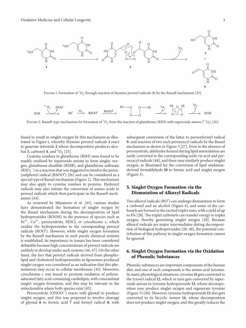
Oxidative Medicine and Cellular Longevity 3
NH
NH
O
O
NH
NH
O
O NH
NH
O
O
NH
NH
O
O
NH
NH
O
O
HO
O
O
HN
NH
O
O
H
O
O
H
O
1 2
3
4
1O2
+
∙OO∙OO
Figure 1: Formation of 1O2through reaction of thymine peroxyl radicals (1) by the Russell mechanism [25].
RSH
RS O
O O
O SR RS
O O
SR
RSSR
∙−O21O21O2
O2
HOO−
RS∙ RSOO∙
RSOO∙
Figure 2: Russell-type mechanism for formation of 1O2from the reaction of glutathione (RSH) with superoxide anions (∙−O
2) [26].
found to result in singlet oxygen by this mechanism as illus-trated in Figure 1, whereby thymine peroxyl radicals 1 reactto generate tetroxide 2 whose decomposition produces alco-hol 3, carbonyl 4, and 1O
2[25].
Cysteine residues in glutathione (RSH) were found to bereadily oxidized by superoxide anions to form singlet oxy-gen, glutathione disulfide (RSSR), and glutathione sulfonate(RSO3
−) in a reaction that was suggested to involve the perox-ysulphenyl radical (RSOO∙) [26] and can be considered as aspecial type of Russelmechanism (Figure 2).Thismechanismmay also apply to cysteine residues in proteins. Hydroxylradicals may also initiate the conversion of amino acids toperoxyl radicals which then participate in the Russell mech-anism [44].
As reviewed by Miyamoto et al. [45], various studieshave demonstrated the formation of singlet oxygen bythe Russel mechanism during the decomposition of lipidhydroperoxides (ROOH) in the presence of species such asFe3+, Cu2+, peroxynitrite, HOCl, or cytochrome c, whichoxidize the hydroperoxides to the corresponding peroxylradicals (ROO∙). However, while singlet oxygen formationby the Russell mechanism in such purely chemical systemsis established, its importance in tissues has been considereddebatable because high concentrations of peroxyl radicals areunlikely to develop under such systems [46, 47]. On the otherhand, the fact that peroxyl radicals derived from phospho-lipid and cholesterol hydroperoxides in liposomes producedsinglet oxygen was considered as an indication that this phe-nomenon may occur in cellular membranes [45]. Moreover,cytochrome c was found to promote oxidation of polyun-saturated fatty acid-containing cardiolipin, with concomitantsinglet oxygen formation, and this may be relevant in themitochondria where both species exist [45].
Peroxynitrite (ONOO−) reacts with glyoxal to producesinglet oxygen, and this was proposed to involve cleavageof glyoxal 6 to formic acid 7 and formyl radical 8, with
subsequent conversion of the latter to peroxyformyl radical9, and reaction of two such peroxyacyl radicals by the Russelmechanism as shown in Figure 3 [27]. Even in the absence ofperoxynitrite, aldehydes formed during lipid autoxidation areeasily converted to the corresponding acids via acyl and per-oxyacyl radicals [48], and thesemay similarly produce singletoxygen, as illustrated for the conversion of lipid oxidation-derived formaldehyde 10 to formic acid and singlet oxygen(Figure 3).
5. Singlet Oxygen Formation via theDismutation of Alkoxyl Radicals
Two alkoxyl radicals (RO∙) can undergo dismutation to forma carbonyl and an alcohol (Figure 4), and some of the car-bonyls are formed in the excited triplet state, with a yield of upto 8% [28].The triplet carbonyls can transfer energy to tripletoxygen, thereby generating singlet oxygen [28]. Becausealkoxyl radicals are major intermediates during decomposi-tion of biological hydroperoxides [28, 48], the potential con-tribution of this pathway to singlet oxygen formation cannotbe ignored.
6. Singlet Oxygen Formation via the Oxidationof Phenolic Substances
Phenolic substances are important components of the humandiet, and one of such compounds is the amino acid tyrosine.Inmany physiological situations, tyrosine 11 gets converted tothe tyrosyl radical 12, which in turn gets converted by super-oxide anions to tyrosine hydroperoxide 13, whose decompo-sition may produce singlet oxygen and regenerate tyrosine(Figure 5) [40]. However, tyrosine hydroperoxide 13 also getsconverted to its bicyclic isomer 14, whose decompositiondoes not produce singlet oxygen, and this greatly reduces the

4 Oxidative Medicine and Cellular Longevity
O
O
H
H
OH
O
O H OH O
ROOH
H
H O
O
OO
H
O
H
O
O
6
7
8 9
10
7
Lipid oxidation
O2
CO2
1O2
∙
ONOO−+
OO∙
OO∙
∙NO2
ROO∙
Figure 3: Russell-type mechanism for formation of 1O2upon reaction of glyoxal (6) with peroxynitrite (ONOO−) via formyl radical (8) [27]
and proposed occurrence of such a reaction via formyl radicals formed during lipid oxidation.
HOH
O O
HO
HO
R1 R1R2 R2
R1 R2 R1 R2
R1 R2
∗
O21O2
∙∙
+
Figure 4: Formation of 1O2via dismutation of alkoxyl radicals [28]. The asterisk indicates that the carbonyl is in excited state.
CH C OHO
O
HOO
OOH
HOO
OH
HN
C OHO
1O2
11
12
13 14
∙
H2N
CH2
CH C OHO
H2N
CH2
CH C OHO
H2N
CH2
∙−O2, H+
Figure 5: Formation of 1O2during the superoxide-dependent oxidation of tyrosine 11 [29].
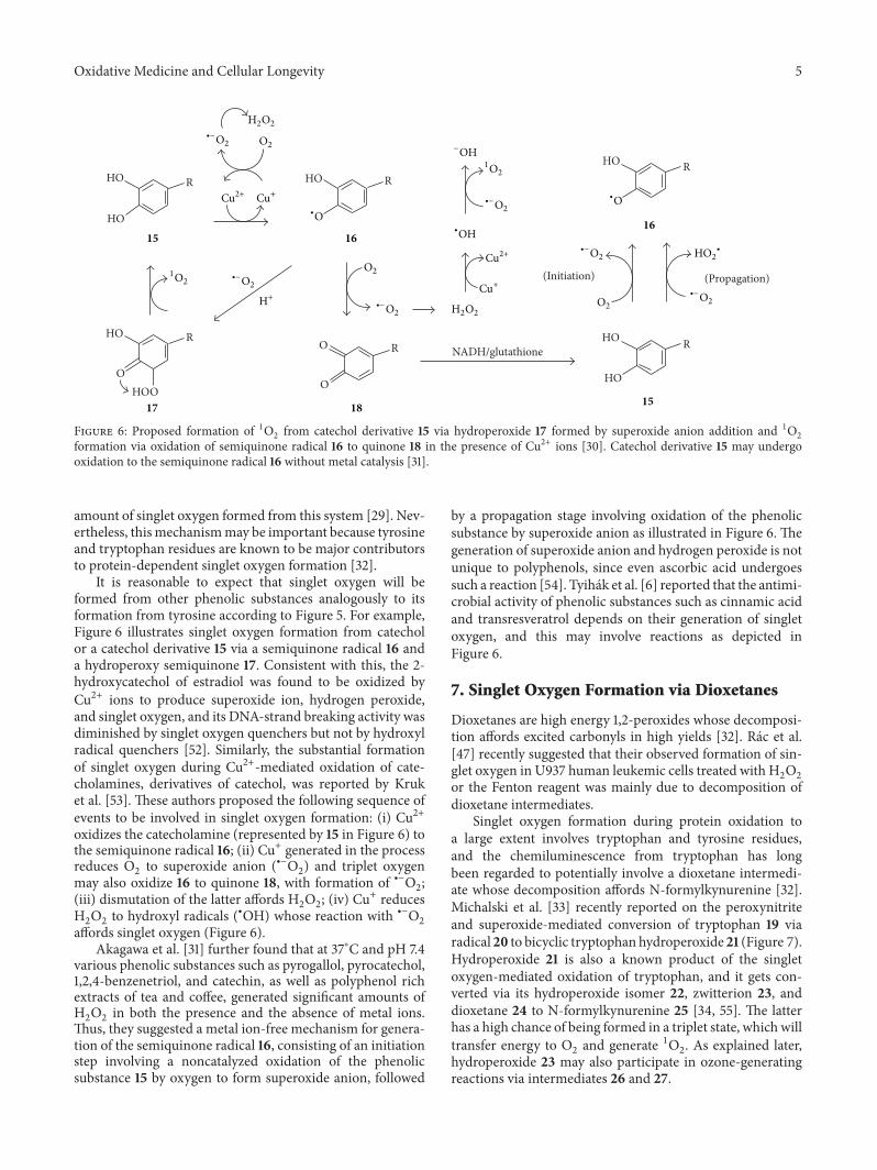
Oxidative Medicine and Cellular Longevity 5
HO
HO
HO
O
R
R HO
O R
R
NADH/glutathione
OHOO
HO
HO
HO
R
R
(Initiation) (Propagation)
15 16
18 15
16
17
∙−O2
∙−O2
∙−O2
∙−O2
∙−O2
∙−O2H+
Cu+
Cu+
O2
O2
O2
H2O2
H2O2
−OH
1O2
1O2
Cu2+
Cu2+
∙O∙O
∙OHHO2
∙
Figure 6: Proposed formation of 1O2from catechol derivative 15 via hydroperoxide 17 formed by superoxide anion addition and 1O
2
formation via oxidation of semiquinone radical 16 to quinone 18 in the presence of Cu2+ ions [30]. Catechol derivative 15 may undergooxidation to the semiquinone radical 16 without metal catalysis [31].
amount of singlet oxygen formed from this system [29]. Nev-ertheless, this mechanismmay be important because tyrosineand tryptophan residues are known to be major contributorsto protein-dependent singlet oxygen formation [32].
It is reasonable to expect that singlet oxygen will beformed from other phenolic substances analogously to itsformation from tyrosine according to Figure 5. For example,Figure 6 illustrates singlet oxygen formation from catecholor a catechol derivative 15 via a semiquinone radical 16 anda hydroperoxy semiquinone 17. Consistent with this, the 2-hydroxycatechol of estradiol was found to be oxidized byCu2+ ions to produce superoxide ion, hydrogen peroxide,and singlet oxygen, and its DNA-strand breaking activity wasdiminished by singlet oxygen quenchers but not by hydroxylradical quenchers [52]. Similarly, the substantial formationof singlet oxygen during Cu2+-mediated oxidation of cate-cholamines, derivatives of catechol, was reported by Kruket al. [53]. These authors proposed the following sequence ofevents to be involved in singlet oxygen formation: (i) Cu2+oxidizes the catecholamine (represented by 15 in Figure 6) tothe semiquinone radical 16; (ii) Cu+ generated in the processreduces O
2to superoxide anion (∙−O
2) and triplet oxygen
may also oxidize 16 to quinone 18, with formation of ∙−O2;
(iii) dismutation of the latter affords H2O2; (iv) Cu+ reduces
H2O2to hydroxyl radicals (∙OH) whose reaction with ∙−O
2
affords singlet oxygen (Figure 6).Akagawa et al. [31] further found that at 37∘C and pH 7.4
various phenolic substances such as pyrogallol, pyrocatechol,1,2,4-benzenetriol, and catechin, as well as polyphenol richextracts of tea and coffee, generated significant amounts ofH2O2in both the presence and the absence of metal ions.
Thus, they suggested a metal ion-free mechanism for genera-tion of the semiquinone radical 16, consisting of an initiationstep involving a noncatalyzed oxidation of the phenolicsubstance 15 by oxygen to form superoxide anion, followed
by a propagation stage involving oxidation of the phenolicsubstance by superoxide anion as illustrated in Figure 6. Thegeneration of superoxide anion and hydrogen peroxide is notunique to polyphenols, since even ascorbic acid undergoessuch a reaction [54]. Tyihák et al. [6] reported that the antimi-crobial activity of phenolic substances such as cinnamic acidand transresveratrol depends on their generation of singletoxygen, and this may involve reactions as depicted inFigure 6.
7. Singlet Oxygen Formation via Dioxetanes
Dioxetanes are high energy 1,2-peroxides whose decomposi-tion affords excited carbonyls in high yields [32]. Rác et al.[47] recently suggested that their observed formation of sin-glet oxygen in U937 human leukemic cells treated with H
2O2
or the Fenton reagent was mainly due to decomposition ofdioxetane intermediates.
Singlet oxygen formation during protein oxidation toa large extent involves tryptophan and tyrosine residues,and the chemiluminescence from tryptophan has longbeen regarded to potentially involve a dioxetane intermedi-ate whose decomposition affords N-formylkynurenine [32].Michalski et al. [33] recently reported on the peroxynitriteand superoxide-mediated conversion of tryptophan 19 viaradical 20 to bicyclic tryptophan hydroperoxide 21 (Figure 7).Hydroperoxide 21 is also a known product of the singletoxygen-mediated oxidation of tryptophan, and it gets con-verted via its hydroperoxide isomer 22, zwitterion 23, anddioxetane 24 to N-formylkynurenine 25 [34, 55]. The latterhas a high chance of being formed in a triplet state, which willtransfer energy to O
2and generate 1O
2. As explained later,
hydroperoxide 23 may also participate in ozone-generatingreactions via intermediates 26 and 27.
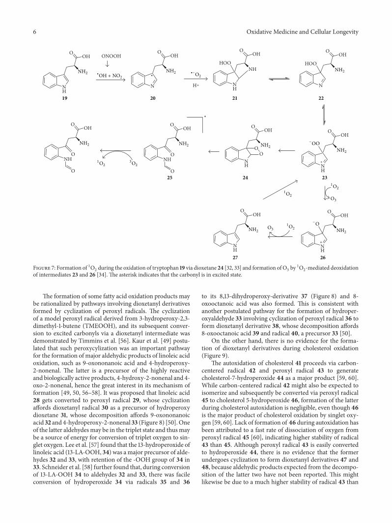
6 Oxidative Medicine and Cellular Longevity
O
NH
OOH
O
NH
OOH ONOOH
O
NH
OOH
O
N
OOH
NH
OOH
OO
NH
OOH
NH
OOH
HOONH
NH
OOH
NH
OOH
HOO
N
OOH
19 20 21 22
232425
2627
NH2 NH2 NH2
NH2NH2
NH2NH2
NH2 NH2
−OO
−O
O3
O3
∙−O2
∗
+
+
1O21O2
1O2
1O23O2
∙
H+
∙OH + NO2
Figure 7: Formation of 1O2during the oxidation of tryptophan 19 via dioxetane 24 [32, 33] and formation of O
3by 1O
2-mediated deoxidation
of intermediates 23 and 26 [34]. The asterisk indicates that the carbonyl is in excited state.
The formation of some fatty acid oxidation products maybe rationalized by pathways involving dioxetanyl derivativesformed by cyclization of peroxyl radicals. The cyclizationof a model peroxyl radical derived from 3-hydroperoxy-2,3-dimethyl-1-butene (TMEOOH), and its subsequent conver-sion to excited carbonyls via a dioxetanyl intermediate wasdemonstrated by Timmins et al. [56]. Kaur et al. [49] postu-lated that such peroxycylization was an important pathwayfor the formation of major aldehydic products of linoleic acidoxidation, such as 9-oxononanoic acid and 4-hydroperoxy-2-nonenal. The latter is a precursor of the highly reactiveand biologically active products, 4-hydroxy-2-nonenal and 4-oxo-2-nonenal, hence the great interest in its mechanism offormation [49, 50, 56–58]. It was proposed that linoleic acid28 gets converted to peroxyl radical 29, whose cyclizationaffords dioxetanyl radical 30 as a precursor of hydroperoxydioxetane 31, whose decomposition affords 9-oxononanoicacid 32 and 4-hydroperoxy-2-nonenal 33 (Figure 8) [50].Oneof the latter aldehydesmay be in the triplet state and thusmaybe a source of energy for conversion of triplet oxygen to sin-glet oxygen. Lee et al. [57] found that the 13-hydroperoxide oflinoleic acid (13-LA-OOH, 34) was amajor precursor of alde-hydes 32 and 33, with retention of the -OOH group of 34 in33. Schneider et al. [58] further found that, during conversionof 13-LA-OOH 34 to aldehydes 32 and 33, there was facileconversion of hydroperoxide 34 via radicals 35 and 36
to its 8,13-dihydroperoxy-derivative 37 (Figure 8) and 8-oxooctanoic acid was also formed. This is consistent withanother postulated pathway for the formation of hydroper-oxyaldehyde 33 involving cyclization of peroxyl radical 36 toform dioxetanyl derivative 38, whose decomposition affords8-oxooctanoic acid 39 and radical 40, a precursor 33 [50].
On the other hand, there is no evidence for the forma-tion of dioxetanyl derivatives during cholesterol oxidation(Figure 9).
The autoxidation of cholesterol 41 proceeds via carbon-centered radical 42 and peroxyl radical 43 to generatecholesterol-7-hydroperoxide 44 as a major product [59, 60].While carbon-centered radical 42 might also be expected toisomerize and subsequently be converted via peroxyl radical45 to cholesterol 5-hydroperoxide 46, formation of the latterduring cholesterol autoxidation is negligible, even though 46is the major product of cholesterol oxidation by singlet oxy-gen [59, 60]. Lack of formation of 46 during autoxidation hasbeen attributed to a fast rate of dissociation of oxygen fromperoxyl radical 45 [60], indicating higher stability of radical43 than 45. Although peroxyl radical 43 is easily convertedto hydroperoxide 44, there is no evidence that the formerundergoes cyclization to form dioxetanyl derivatives 47 and48, because aldehydic products expected from the decompo-sition of the latter two have not been reported. This mightlikewise be due to a much higher stability of radical 43 than
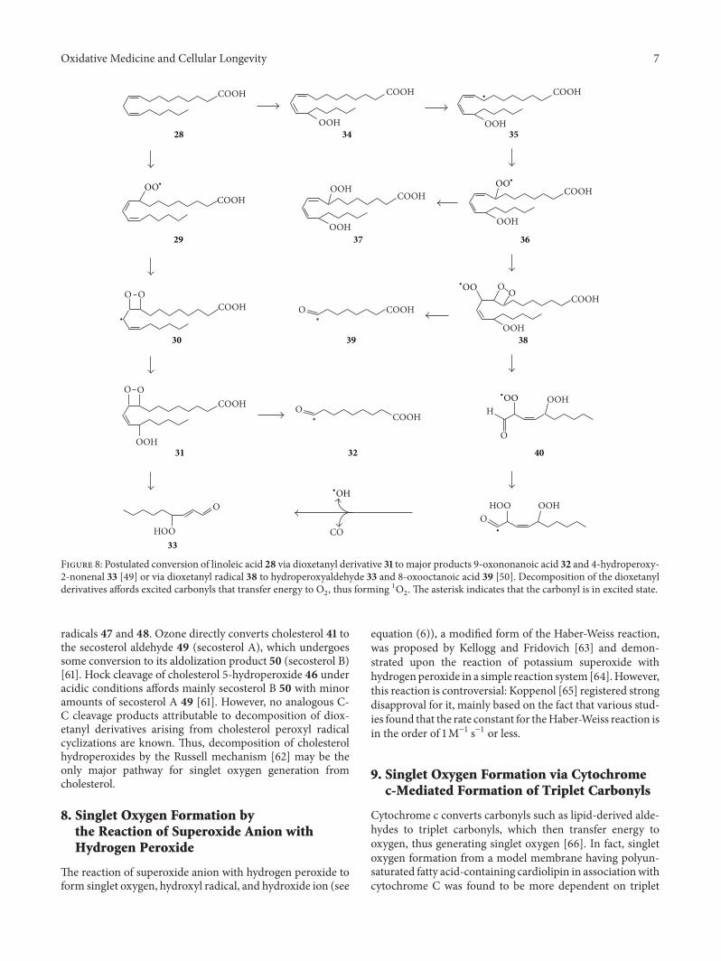
Oxidative Medicine and Cellular Longevity 7
COOH
COOH
COOH
OOH
OOH
OOH
OOH
O
OOH
O
HOO
O
H
O
COOH
COOH
COOH
COOH
OOH
OOH
OOH
O
O
CO
COOH
COOH
COOH
COOH
O
HOO
OOH
O
O
O
O
28
29
30
31
33
32
34 35
3637
38
40
39
∗
∗
∙
∙
∙
OO∙
OO∙
∙OO
∙OO
∙OH
Figure 8: Postulated conversion of linoleic acid 28 via dioxetanyl derivative 31 to major products 9-oxononanoic acid 32 and 4-hydroperoxy-2-nonenal 33 [49] or via dioxetanyl radical 38 to hydroperoxyaldehyde 33 and 8-oxooctanoic acid 39 [50]. Decomposition of the dioxetanylderivatives affords excited carbonyls that transfer energy to O
2, thus forming 1O
2. The asterisk indicates that the carbonyl is in excited state.
radicals 47 and 48. Ozone directly converts cholesterol 41 tothe secosterol aldehyde 49 (secosterol A), which undergoessome conversion to its aldolization product 50 (secosterol B)[61]. Hock cleavage of cholesterol 5-hydroperoxide 46 underacidic conditions affords mainly secosterol B 50 with minoramounts of secosterol A 49 [61]. However, no analogous C-C cleavage products attributable to decomposition of diox-etanyl derivatives arising from cholesterol peroxyl radicalcyclizations are known. Thus, decomposition of cholesterolhydroperoxides by the Russell mechanism [62] may be theonly major pathway for singlet oxygen generation fromcholesterol.
8. Singlet Oxygen Formation bythe Reaction of Superoxide Anion withHydrogen Peroxide
The reaction of superoxide anion with hydrogen peroxide toform singlet oxygen, hydroxyl radical, and hydroxide ion (see
equation (6)), a modified form of the Haber-Weiss reaction,was proposed by Kellogg and Fridovich [63] and demon-strated upon the reaction of potassium superoxide withhydrogen peroxide in a simple reaction system [64].However,this reaction is controversial: Koppenol [65] registered strongdisapproval for it, mainly based on the fact that various stud-ies found that the rate constant for theHaber-Weiss reaction isin the order of 1M−1 s−1 or less.
9. Singlet Oxygen Formation via Cytochromec-Mediated Formation of Triplet Carbonyls
Cytochrome c converts carbonyls such as lipid-derived alde-hydes to triplet carbonyls, which then transfer energy tooxygen, thus generating singlet oxygen [66]. In fact, singletoxygen formation from a model membrane having polyun-saturated fatty acid-containing cardiolipin in associationwithcytochrome C was found to be more dependent on triplet
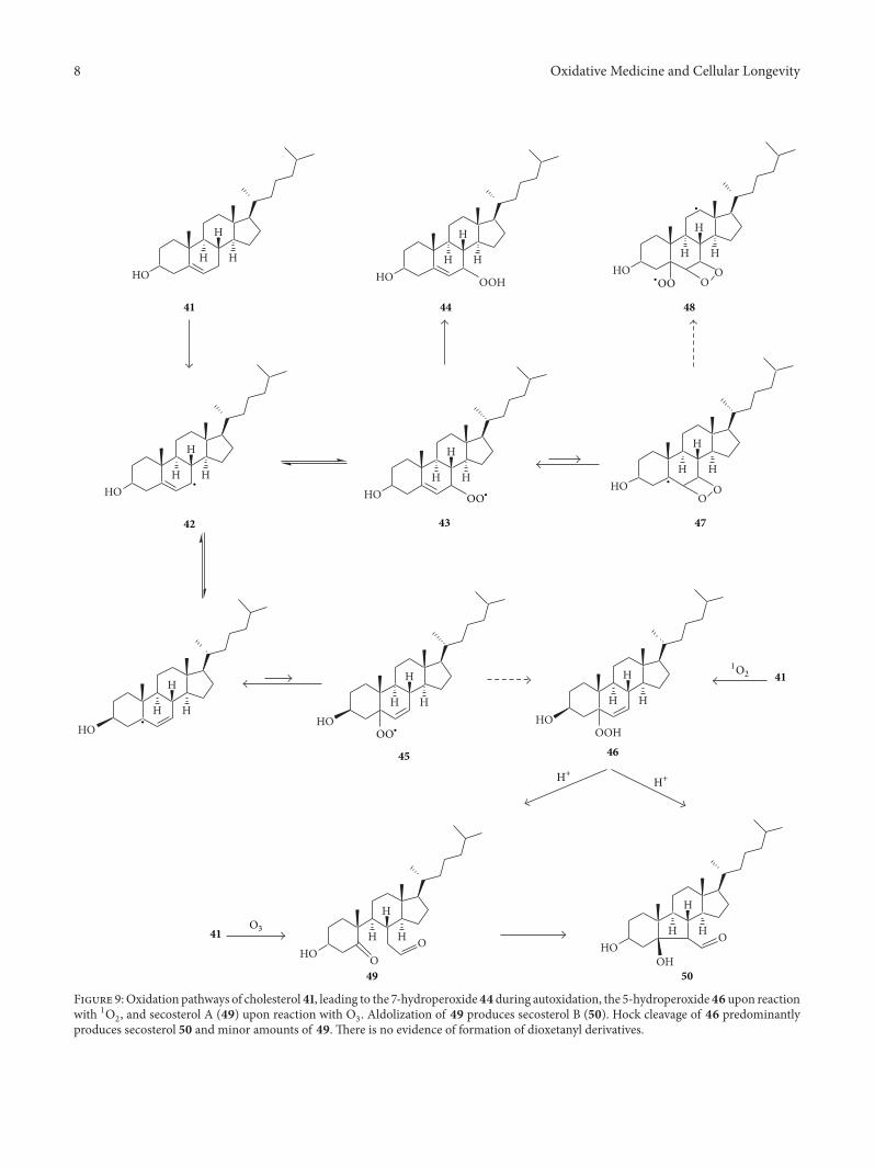
8 Oxidative Medicine and Cellular Longevity
HOH
H
HO
O
HOH
H
H
OO
HOH
H
H
OOH
H
H
H
OOH
HOH
H
H
HO
HOH
H
H O
OH
H
H
H
HOH
H
H
O
HOH
H
H
HO
HOH
H
H
O
H
H
HHO
41
41
42 43
44 48
45 46
49 50
41
47
O3
1O2
∙
∙
∙
∙
H+H+
∙OO
OO∙
OO∙
Figure 9:Oxidation pathways of cholesterol41, leading to the 7-hydroperoxide44 during autoxidation, the 5-hydroperoxide46 upon reactionwith 1O
2, and secosterol A (49) upon reaction with O
3. Aldolization of 49 produces secosterol B (50). Hock cleavage of 46 predominantly
produces secosterol 50 and minor amounts of 49. There is no evidence of formation of dioxetanyl derivatives.
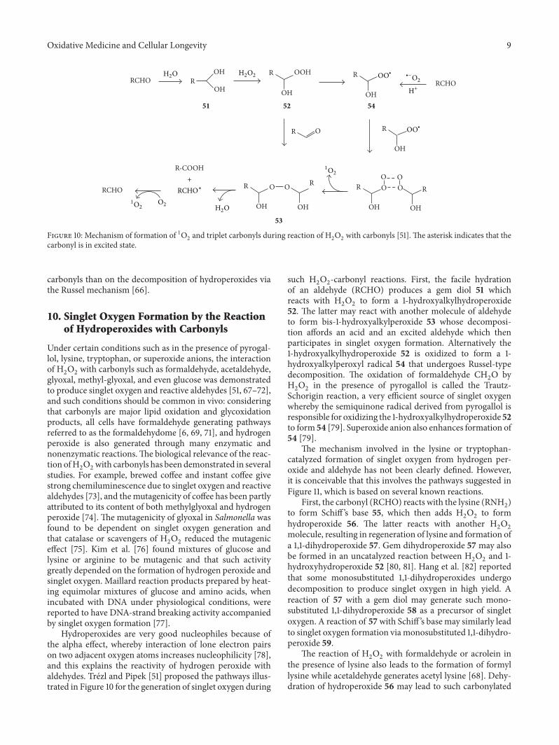
Oxidative Medicine and Cellular Longevity 9
OH OOHR RRCHO R RCHO
OH OH OH
ROR
OH
R-COOHO O
RR O O R O O RRCHO
OH OH OH OH
51 52
53
54
RCHO∗
H2O
H2O
O2
∙−O2
+
1O2
1O2
H+
H2O2 OO∙
OO∙
Figure 10: Mechanism of formation of 1O2and triplet carbonyls during reaction of H
2O2with carbonyls [51]. The asterisk indicates that the
carbonyl is in excited state.
carbonyls than on the decomposition of hydroperoxides viathe Russel mechanism [66].
10. Singlet Oxygen Formation by the Reactionof Hydroperoxides with Carbonyls
Under certain conditions such as in the presence of pyrogal-lol, lysine, tryptophan, or superoxide anions, the interactionof H2O2with carbonyls such as formaldehyde, acetaldehyde,
glyoxal, methyl-glyoxal, and even glucose was demonstratedto produce singlet oxygen and reactive aldehydes [51, 67–72],and such conditions should be common in vivo: consideringthat carbonyls are major lipid oxidation and glycoxidationproducts, all cells have formaldehyde generating pathwaysreferred to as the formaldehydome [6, 69, 71], and hydrogenperoxide is also generated through many enzymatic andnonenzymatic reactions.The biological relevance of the reac-tion ofH
2O2with carbonyls has been demonstrated in several
studies. For example, brewed coffee and instant coffee givestrong chemiluminescence due to singlet oxygen and reactivealdehydes [73], and themutagenicity of coffee has been partlyattributed to its content of both methylglyoxal and hydrogenperoxide [74]. The mutagenicity of glyoxal in Salmonella wasfound to be dependent on singlet oxygen generation andthat catalase or scavengers of H
2O2reduced the mutagenic
effect [75]. Kim et al. [76] found mixtures of glucose andlysine or arginine to be mutagenic and that such activitygreatly depended on the formation of hydrogen peroxide andsinglet oxygen. Maillard reaction products prepared by heat-ing equimolar mixtures of glucose and amino acids, whenincubated with DNA under physiological conditions, werereported to have DNA-strand breaking activity accompaniedby singlet oxygen formation [77].
Hydroperoxides are very good nucleophiles because ofthe alpha effect, whereby interaction of lone electron pairson two adjacent oxygen atoms increases nucleophilicity [78],and this explains the reactivity of hydrogen peroxide withaldehydes. Trézl and Pipek [51] proposed the pathways illus-trated in Figure 10 for the generation of singlet oxygen during
such H2O2-carbonyl reactions. First, the facile hydration
of an aldehyde (RCHO) produces a gem diol 51 whichreacts with H
2O2to form a 1-hydroxyalkylhydroperoxide
52. The latter may react with another molecule of aldehydeto form bis-1-hydroxyalkylperoxide 53 whose decomposi-tion affords an acid and an excited aldehyde which thenparticipates in singlet oxygen formation. Alternatively the1-hydroxyalkylhydroperoxide 52 is oxidized to form a 1-hydroxyalkylperoxyl radical 54 that undergoes Russel-typedecomposition. The oxidation of formaldehyde CH
2O by
H2O2in the presence of pyrogallol is called the Trautz-
Schorigin reaction, a very efficient source of singlet oxygenwhereby the semiquinone radical derived from pyrogallol isresponsible for oxidizing the 1-hydroxyalkylhydroperoxide 52to form 54 [79]. Superoxide anion also enhances formation of54 [79].
The mechanism involved in the lysine or tryptophan-catalyzed formation of singlet oxygen from hydrogen per-oxide and aldehyde has not been clearly defined. However,it is conceivable that this involves the pathways suggested inFigure 11, which is based on several known reactions.
First, the carbonyl (RCHO) reacts with the lysine (RNH2)
to form Schiff ’s base 55, which then adds H2O2to form
hydroperoxide 56. The latter reacts with another H2O2
molecule, resulting in regeneration of lysine and formation ofa 1,1-dihydroperoxide 57. Gem dihydroperoxide 57 may alsobe formed in an uncatalyzed reaction between H
2O2and 1-
hydroxyhydroperoxide 52 [80, 81]. Hang et al. [82] reportedthat some monosubstituted 1,1-dihydroperoxides undergodecomposition to produce singlet oxygen in high yield. Areaction of 57 with a gem diol may generate such mono-substituted 1,1-dihydroperoxide 58 as a precursor of singletoxygen. A reaction of 57with Schiff ’s base may similarly leadto singlet oxygen formation viamonosubstituted 1,1-dihydro-peroxide 59.
The reaction of H2O2with formaldehyde or acrolein in
the presence of lysine also leads to the formation of formyllysine while acetaldehyde generates acetyl lysine [68]. Dehy-dration of hydroperoxide 56 may lead to such carbonylated
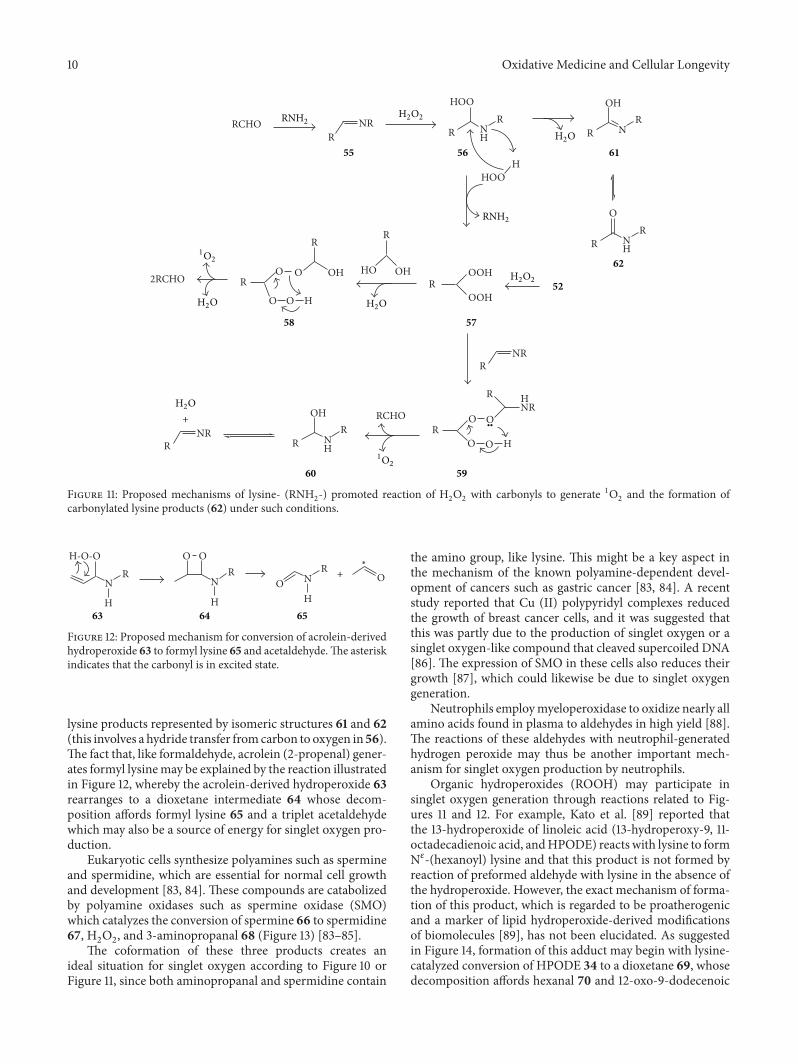
10 Oxidative Medicine and Cellular Longevity
R
R
NH
N
OH
O
R
R
HNR
NR
H
R
OOH
OOH
HOO
O
R
R
NH
O
HOO
R
R
R
OH
R
HO
RCHO
OH
R
HOO
H
NR
O
O
R
O
O
R
NH
OH
R
R
RCHO
NRR
2RCHO
55 56
5758
5960
61
62
52H2O
H2O
H2O
H2O
+
H2O2
H2O2
∙∙
1O2
1O2
RNH2
RNH2
Figure 11: Proposed mechanisms of lysine- (RNH2-) promoted reaction of H
2O2with carbonyls to generate 1O
2and the formation of
carbonylated lysine products (62) under such conditions.
R R
H
NO O
H
O
N
OR
H
N
H-O-O
63 64 65
+
∗
Figure 12: Proposed mechanism for conversion of acrolein-derivedhydroperoxide 63 to formyl lysine 65 and acetaldehyde.The asteriskindicates that the carbonyl is in excited state.
lysine products represented by isomeric structures 61 and 62(this involves a hydride transfer fromcarbon to oxygen in 56).The fact that, like formaldehyde, acrolein (2-propenal) gener-ates formyl lysinemay be explained by the reaction illustratedin Figure 12, whereby the acrolein-derived hydroperoxide 63rearranges to a dioxetane intermediate 64 whose decom-position affords formyl lysine 65 and a triplet acetaldehydewhich may also be a source of energy for singlet oxygen pro-duction.
Eukaryotic cells synthesize polyamines such as spermineand spermidine, which are essential for normal cell growthand development [83, 84]. These compounds are catabolizedby polyamine oxidases such as spermine oxidase (SMO)which catalyzes the conversion of spermine 66 to spermidine67, H
2O2, and 3-aminopropanal 68 (Figure 13) [83–85].
The coformation of these three products creates anideal situation for singlet oxygen according to Figure 10 orFigure 11, since both aminopropanal and spermidine contain
the amino group, like lysine. This might be a key aspect inthe mechanism of the known polyamine-dependent devel-opment of cancers such as gastric cancer [83, 84]. A recentstudy reported that Cu (II) polypyridyl complexes reducedthe growth of breast cancer cells, and it was suggested thatthis was partly due to the production of singlet oxygen or asinglet oxygen-like compound that cleaved supercoiled DNA[86]. The expression of SMO in these cells also reduces theirgrowth [87], which could likewise be due to singlet oxygengeneration.
Neutrophils employmyeloperoxidase to oxidize nearly allamino acids found in plasma to aldehydes in high yield [88].The reactions of these aldehydes with neutrophil-generatedhydrogen peroxide may thus be another important mech-anism for singlet oxygen production by neutrophils.
Organic hydroperoxides (ROOH) may participate insinglet oxygen generation through reactions related to Fig-ures 11 and 12. For example, Kato et al. [89] reported thatthe 13-hydroperoxide of linoleic acid (13-hydroperoxy-9, 11-octadecadienoic acid, andHPODE) reacts with lysine to formN𝜀-(hexanoyl) lysine and that this product is not formed byreaction of preformed aldehyde with lysine in the absence ofthe hydroperoxide. However, the exact mechanism of forma-tion of this product, which is regarded to be proatherogenicand a marker of lipid hydroperoxide-derived modificationsof biomolecules [89], has not been elucidated. As suggestedin Figure 14, formation of this adduct may begin with lysine-catalyzed conversion of HPODE 34 to a dioxetane 69, whosedecomposition affords hexanal 70 and 12-oxo-9-dodecenoic
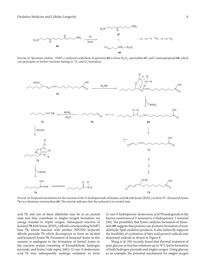
Oxidative Medicine and Cellular Longevity 11
NH
HN
SMO
O
NH
66
67
68
H2N
H2N
NH2
NH2
O3O2
+
1O2
NH2 + H2O2
Figure 13: Spermine oxidase- (SMO-) catalyzed catabolism of spermine 66 to form H2O2, spermidine 67, and 3-aminopropanal 68, which
can participate in further reactions leading to 1O2and O
3formation.
HOO OH
O
NR
ROOH
ROH
NR
OH
N-R
HO
O
NH
OO
R
H
R
O
O
O O
OO
H
H
OOH
N
H
R
OH
O
OH
OH
O
O
OH
O34
697072
73
74 75
71
H2NR
RNH2
RNH2
∗
−H2O
∙∙
Figure 14: Proposedmechanism for the reaction of the 13-hydroperoxide of linoleic acid 34with lysine (RNH2) to formN𝜀–(hexanoyl) lysine
74 via a dioxetane intermediate 69. The asterisk indicates that the carbonyl is in excited state.
acid 71, and one of these aldehydes may be in an excitedstate and thus contribute to singlet oxygen formation viaenergy transfer to triplet oxygen. Subsequent reaction ofhexanal 70with lysine (RNH
2) affords corresponding Schiff ’s
base 72, whose reaction with another HPODE moleculeaffords peroxide 73 which decomposes to form an alcoholand hexanoyl-lysine 74. Formation of hexanoyl-lysine in thismanner is analogous to the formation of formyl lysine inthe reaction system consisting of formaldehyde, hydrogenperoxide, and lysine (vide supra, [68]). 12-oxo-9-dodecenoicacid 71 may subsequently undergo oxidation to form
12-oxo-9-hydroperoxy-dodecenoic acid 75 analogously to theknown conversion of 3-nonenal to 4-hydroperoxy-2-nonenal[90]. The possibility that lysine catalyzes formation of dioxe-tane 69 suggests that proteins can promote formation of toxicaldehydic lipid oxidation products. It also indirectly supportsthe feasibility of cyclization of fatty acid peroxyl radicals intodioxetanyl radicals as shown in Figure 8.
Wang et al. [91] recently found that thermal treatment ofpure glucose or fructose solutions up to 70∘C led to formationof both hydrogen peroxide and singlet oxygen. Using glucoseas an example, the potential mechanism for singlet oxygen
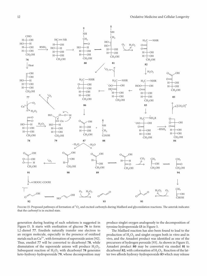
12 Oxidative Medicine and Cellular Longevity
OHHOHH
OH
O
O
OHHOHH
OH
NHR
OHOO
OHHOHH
OH
NHR
HO
OHHOHH
NHR
OO
OHOHHOHH
OH
NHR
OO
HOOHHOHH
HO
NH
R
HHOOHHOHH
O
OH
OHHOHH
OOO
OH
OHHOHH
O
R
NH
COOH
OH
OHHOHH
OO
HO
HHOOHHOHH
OOHOHH
OHO
HOH
OHO
OHCHOHO
O
OH
O
OHH
O
OHHOOHHOHH
OH
HOH
O
OO
O O H
HOOC-COOH
OO
OOH OH
HO
NH
R
OHHHOOHHOHH
OHHHHOOHHOHH
NR
OH
HCCHO
OHHHHOOHHOHH
Heat
76
77
78 79
80 81 82
83
84
85
86
87 88
899091
92 93
94
95
[CH2O]∗
CH2CH2
CH2
CH2
H2C
H2C H2C H2C
H2C
CH2OHCH2OHCH2OHCH2OH
CH2OH
CH2OH
CH2OH CH2OH CH2OH
CH2OH
CH2OHCH2OHCH2OHCH2OHCH2OH
CH2OH CH2OH CH2OH CH2OH CH2OH CH2OH
RNH2
O2
O2O2H2O2
O2 H2O2
H2O2
H2O2
H2O2
H2O2
1O2
1O2
1O2
Cu2+
+
+
+
−H2O−H2O
−OO
−OO
CH3
CH3
∗
CO2
−RNH2
NH2R
∙∙
Figure 15: Proposed pathways of formation of 1O2and excited carbonyls during Maillard and glycoxidation reactions. The asterisk indicates
that the carbonyl is in excited state.
generation during heating of such solutions is suggested inFigure 15. It starts with enolization of glucose 76 to form1,2-dienol 77. Enediols naturally transfer one electron toan oxygen molecule, especially in the presence of oxidizedmetals such as Cu2+, with formation of superoxide anion [92].Thus, enediol 77 will be converted to dicarbonyl 78, whiledismutation of the superoxide anions will produce H
2O2.
Subsequent reaction of H2O2with dicarbonyl 78 generates
keto-hydroxy-hydroperoxide 79, whose decomposition may
produce singlet oxygen analogously to the decomposition oftyrosine hydroperoxide 13 in Figure 5.
The Maillard reaction has also been found to lead to theproduction of H
2O2and singlet oxygen both in vitro and in
vivo, and the Amadori product was identified as one of theprecursors of hydrogen peroxide [93]. As shown in Figure 15,Amadori product 80 may be converted via enediol 81 todicarbonyl 82, with coformation of H
2O2. Reaction of the lat-
ter two affords hydroxy-hydroperoxide 83 which may release
Oxidative Medicine and Cellular Longevity 13
1O2and convert back to enediol 81. Alternatively, 83 may
release lysine and may be converted to dioxetane 84, whosedecomposition affords ketoacid 85 and formaldehyde, whichmay be in the excited state and thus contribute to singletoxygen formation. Enediol 81 may also react with singletoxygen to form dioxetane 86whose decomposition produceserythronic acid 87 and carboxymethyllysine 88. The latteris one of the most commonly formed advanced glycationend products(AGEs) that are known to contribute to variousphysiological disorders [94]. Dehydration of erythronic acid87 may produce two regioisomeric deoxy-ketonic acids 89and 90. The latter may react with singlet oxygen to producedioxetane 91 as a precursor of oxalic acid (HOOC-COOH),glycolaldehyde 92, and glyoxal 93, all of which are knownproducts of glycoxidation [95]. Deoxy-ketonic acid 89 may,via its ketoform, decarboxylate to formmonohydroxyacetone94 (via the enol form of the latter), whose oxidation producesmethylglyoxal 95. Since the formation of the highly reactiveglyoxal 93 and methylglyoxal 95 is accompanied by H
2O2
formation, singlet oxygen formation according to Figures 10and 11 will thus also occur during Maillard reaction.
Treatment of cultured U937 human leukemic cells orhumanmultiplemyeloma cells withH
2O2was found to cause
singlet oxygen from both the cells and medium components,and this was not dependent on lipid oxidation [47, 96]. Themechanisms in Figures 10, 11, and 15 may be involved in suchsystems.
11. Evidence for Endogenous Ozone Formationand the Potential Mechanisms Involved
Wentworth et al. [7] were the first to suggest the possibilityof the formation of ozone (O
3) in biological systems. One
of their key pieces of evidence was that, in solutions of anti-bodies exposed to singlet oxygen, there was generation of alarge amount of hydrogen peroxide, the occurrence of higherbactericidal activity than what could be attributed exclusivelyto H2O2, as well as the oxidation of cholesterol to secosterol
aldehyde A (49 in Figure 9), a well-known product of theozonolysis of cholesterol. They referred to the generation ofH2O2and ozone under such circumstances as the antibody-
catalyzedwater oxidation pathway and proposed the idea thatthis involves an initial reaction of water with 1O
2to form
dihydrogen trioxide (H2O3) and that decomposition of the
latter affords H2O2and O
3(see equation (7)). This reaction
was suggested to occur in a hydrophobic site in the antibodymolecule, where theH
2O3would be shielded from hydrolysis
and facilitated to undergo the conversion to H2O2and O
3
[97]. Although antibodies produce much more H2O2and O
3
than other proteins [7], Yamashita et al. [8] reported that anti-body catalysis is not essential for this reaction, but rather thepresence of one of four amino acids: histidine, tryptophan,cysteine, or methionine. On the other hand, various authorshave expressed reservations concerning the generation ofozone under such systems, for example, based on the fact thatthe catalytic mechanisms for the antibody- or amino acid-catalyzed water oxidation remain ill-defined [1, 3, 35, 37].Others have reported that cholesterol 5-hydroperoxide 46, aproduct of the oxidation of cholesterol by singlet oxygen,
can also decompose to generate secosterol aldehydes [98,99]. On the other hand, it has been shown that reactionof cholesterol with ozone predominantly generates secos-terol A while decomposition of cholesterol-5 hydroperoxidepredominantly generates secosterol B [61]. The fact thatsecosterol A is the predominant secosterol detected in humantissues and is formed by neutrophils in vitro thus supportsthe formation of endogenous ozone [61, 100]. There is alsoindirect evidence consistent with the formation of ozone inplant leaves or in the cyanobacterium SynechocystisPCC6803during light-induced damage to their PS II, because singletoxygen and tryptophan or histidine residues, respectively, areinvolved [34]. Unlike other commonly generated ROS thatonly generate single strand breaks in DNA, ozone generatesboth single strand and double strand breaks [101, 102]. Theaddition of L-histidine to cultured mammalian cells exposedto H2O2results in DNA double strand breaks [103–105], and
this might be related to histidine-mediated ozone generationin the presence of singlet oxygen.
A BioArena system is an overpressured layer chromatog-raphy (OPLC) systemwhich enables observation of biochem-ical interactions between microorganisms and biologicallyactive compounds in an adsorbent layer covered with themicroorganisms [106]. Using such a system, it was foundthat formaldehyde, singlet oxygen, and ozone are formedin the interaction between microorganisms and antibioticsubstances such as resveratrol and cinnamic acid and thatozone-trapping compounds greatly reduce the antimicrobialeffect [6, 69, 71, 72]. Based on results from the BioArena sys-tem, ozone is considered to be an indispensable endogenousmolecule that can be detected and measured in practically allbiological systems [6, 71, 72]. Moreover, in directly detectingozone formation by plant leaves through GC-MS-SIM, Ballaand Tyihák [4] have added direct proof for the formation ofozone in biological systems. Thus, the current evidence forendogenous ozone is enough towarrant further studies on themechanisms of its formation and biological effects.
As already mentioned, a number of questions remainunanswered regarding the antibody-/amino acid-catalyzedwater oxidation pathway for ozone generation. Moreover,decomposition of dihydrogen trioxide (H
2O3), the proposed
key precursor of ozone in both aqueous and organic solvents,has only been shown to produce singlet oxygen and waterrather than hydrogen peroxide and ozone [107]. In responseto these challenges, a new concept for ozone formationwas recently suggested, involving the oxidation of organicsubstrates such as aldehydes or amino acids to form oxidizedintermediates, and the subsequent singlet oxygen-mediateddeoxidation of the oxidized intermediates to produce ozone,whose subsequent decomposition in water affords hydrogenperoxide [34]. For example, an aldehyde (RCHO) may beconverted to a peroxyacid [RC(O)OOH] through a radicalpathway [48] or by reacting with singlet oxygen [108], andthe peroxyacid may undergo a Bayer-Villiger type reactionwith singlet oxygen to produce an acid and O
3(Figure 16)
[34]. The significance of this mechanism is that it mightgenerate ozone inmany of the situationswhere singlet oxygenis formed in the presence of aldehydic compounds such as inFigures 10–15. Detailed potential mechanisms for histidine-,
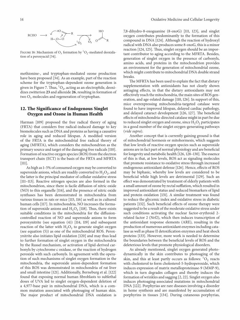
14 Oxidative Medicine and Cellular Longevity
RCHOO
R
O OH O
HO
RO31O2
∙∙
Figure 16: Mechanism of O3formation by 1O
2-mediated deoxida-
tion of a peroxyacid [34].
methionine-, and tryptophan-mediated ozone productionhave been proposed [34]. As an example, part of the reactionscheme for the tryptophan-dependent ozone generation isgiven in Figure 7. Thus, 1O
2, acting as an electrophile, deoxi-
dizes zwitterion 23 and alkoxide 26, resulting in formation oftwo O
3molecules and regeneration of tryptophan.
12. The Significance of Endogenous SingletOxygen and Ozone in Human Health
Harman [109] proposed the free radical theory of aging(FRTA) that considers free radical-induced damage to keybiomolecules such as DNA and proteins as having a causativerole in aging and reduced lifespan. A modified versionof the FRTA is the mitochondrial free radical theory ofaging (MFRTA), which considers the mitochondrion as theprimary source and target of the damaging free radicals [110].Formation of reactive oxygen species (ROS) from the electrontransport chain (ECT) is the basis of the FRTA and MFRTA[111].
As high as 1–5% of consumed oxygenmay be converted tosuperoxide anions, which are readily converted to H
2O2, and
the latter is the principal mediator of cellular oxidative stress[111–113]. Reactive nitrogen species are also formed in themitochondrion, since there is facile diffusion of nitric oxide(NO) to this organelle [114], and the presence of nitric oxidesynthases has been demonstrated in mitochondria fromvarious tissues in rats or mice [115, 116] as well as in culturedhuman cells [117]. In mitochondria, NO increases the forma-tion of superoxide anions and H
2O2[118]. Thus, there exists
suitable conditions in the mitochondria for the diffusion-controlled reaction of NO and superoxide anions to formperoxynitrite (see equation (4)) [114, 119] and subsequentreaction of the latter with H
2O2to generate singlet oxygen
(see equation (5)) as one of the mitochondrial ROS. Perox-ynitrite also initiates lipid oxidation [120] and may thus leadto further formation of singlet oxygen in the mitochondriaby the Russel mechanism, or activation of lipid-derived car-bonyls by cytochrome c oxidase, or the reaction of hydrogenperoxide with such carbonyls. In agreement with the opera-tion of such mechanisms of singlet oxygen formation in themitochondria, the superoxide anion-dependent formationof this ROS was demonstrated in mitochondria of rat liverand small intestine [121]. Additionally, Berneburg et al. [122]found that exposing normal human fibroblasts to sublethaldoses of UVA led to singlet oxygen-dependent deletion ofa 4,977-base pair in mitochondrial DNA, which is a com-mon mutation associated with photoaging of human skin.The major product of mitochondrial DNA oxidation is
7,8-dihydro-8-oxoguanine (8-oxoG) [111, 123], and singletoxygen contributes predominantly to the formation of thiscompound in DNA [124]. Although the reaction of hydroxylradical with DNA also produces some 8-oxoG, this is a minorreaction [124, 125]. Thus, singlet oxygen should be an impor-tant contributor to aging according to the MFRTA. Besides,generation of singlet oxygen in the presence of carbonyls,amino acids, and proteins in the mitochondrion providesan environment for the generation of mitochondrial ozone,whichmight contribute tomitochondrial DNAdouble strandbreaks.
TheMFRTA has been used to explain the fact that dietarysupplementation with antioxidants has not clearly shownantiaging effects, in that the dietary antioxidants may noteffectively reach themitochondria, themain sites of ROS gen-eration, and age-related damage [110, 126]. In support of this,mice overexpressing mitochondria-targeted catalase werefound to have improved lifespan, delayed cardiac pathology,and delayed cataract development [126, 127]. The beneficialeffects ofmitochondria-directed catalasemight in part be dueto reduced singlet oxygen and ozone, since H
2O2participates
in a good number of the singlet oxygen-generating pathways(vide supra).
Another concept that is currently gaining ground is thatof mitochondrial hormesis or mitohormesis, which suggeststhat low levels of reactive oxygen species such as superoxideanions are in fact part of normal physiology and are beneficialfor longevity andmetabolic health [126, 128–130].The essenceof this is that, at low levels, ROS act as signaling moleculesthat promote resistance to oxidative stress through increasedendogenous antioxidant defense [126]. Hence, effects of ROSmay be biphasic, whereby low levels are considered to bebeneficial while high levels are detrimental [129]. Such aneffect was demonstrated by treatment of elderly patients witha small amount of ozone by rectal sufflation, which resulted inimproved antioxidant status and reduced biomarkers of lipidand protein oxidation [131]. A similar treatment was foundto reduce the glycemic index and oxidative stress in diabeticpatients [132]. Such beneficial effects of ozone therapy weresuggested to be a result of themoderate oxidative stress undersuch conditions activating the nuclear factor-erythroid 2-related factor 2 (Nrf2), which then induces transcription ofthe antioxidant response elements (ARE), resulting in theproduction of numerous antioxidant enzymes including cata-lase as well as phase II detoxification enzymes and heat shockproteins [133]. However, more work is needed to determinethe boundaries between the beneficial levels of ROS and thedeleterious levels that promote physiological disorders.
As already mentioned, singlet oxygen generated photo-dynamically in the skin contributes to photoaging of theskin, and this at least partly occurs as follows: 1O
2reacts
with cholesterol to form cholesterol-5-hydroperoxide, whichinduces expression of matrix metalloproteinase-9 (MMP-9),which in turn degrades collagen and thereby induces theformation of wrinkles and sagging [2, 22]. Singlet oxygen alsoinduces photoaging-associated mutations in mitochondrialDNA [122]. Porphyrias are rare diseases involving a disorderin heme synthesis and are manifested by accumulation ofporphyrins in tissues [134]. During cutaneous porphyrias,
Oxidative Medicine and Cellular Longevity 15
there is enhanced photosensitivity due to singlet oxygenformation [134].
Singlet oxygen-specific linoleic acid oxidation products,10-hydroperoxy-8(E), 12(Z)-octadecadienoic acid (10-(E,Z)-HPODE), and 12-hydroperoxy-9(Z), 13(E)-octadecadienoicacid (12-(Z,E)-HPODE) have been found to be suitablebiomarkers for the evaluation of the early stages of diabetes,underscoring the role of singlet oxygen in the pathogenesisof this disorder [135, 136]. Further, fasting levels of 10-(E,Z-HPODE) and 12-(Z,E)-HPODE, together with insulin andleptin/adiponectin, are excellent predictors of the risk for typeII diabetes, glucose intolerance, and insulin resistance [137,138]. Patients with diabetes are prone to other diseases andphysiological disorders including atherosclerosis, cardiovas-cular diseases, chronic kidney disease, retinopathy, and skindisorders [139].Therefore, by contributing to glucose intoler-ance, insulin resistance, and type II diabetes, singlet oxygenmay be contributing to a much wider range of chronicdiseases.
Recently, the catabolism of polyamines has been linkedto the development of some cancers such as gastric cancer,prostate cancer, and colon cancer [84, 85, 140]. Humanspermine oxidase isoforms were found to be localized in thenucleus where they generate reactive oxygen species close toDNA and nuclear proteins [85]. Spermine oxidase-catalyzedcatabolism of spermine thus generates products that can reactto produce singlet oxygen and ozone in the nucleus. Hence,singlet oxygen and ozone might play a key role in spermineoxidase-dependent cancers.
Since singlet oxygen is a precursor of endogenous ozone,some of the biological activities attributed to singlet oxygenmight be directly mediated by ozone or ozonolysis prod-ucts. High levels of cholesterol secosterols A and B havebeen detected in human atherosclerotic tissues and shownto be proatherogenic [141]. The fact that secosterol A isthe predominant secosterol in human tissues [61, 62, 100]indicates a much greater role for ozone than singlet oxygenin the in vivo formation of the secosterols because even thesecosterol B detected in such systems is at least partly formedby aldolization of secosterol A. A possible reason for the invivo predominance of secosterol A is that, while cholesterolhydroperoxide-5, a direct precursor of secosterol B, may beeasily formed in tissues, its Hock cleavage is favored by acidicconditions, which may not be common in most tissues. Thefact that Tomono et al. [142] observed a time-dependentincrease of the secosterols in plasma of mice after injectionof lipopolysaccharide indicates that ozone formation is easilyinitiated during inflammation.
The secosterols A and B induce endothelial cell dys-function by promoting apoptosis and inhibiting endothelial-dependent arterial relaxation [143]. These products are ele-vated in brain tissues of Alzheimer’s disease patients, and itwas demonstrated that they may trigger the disease by induc-ing proteinmisfolding [144]. Elevated levels of the secosterolsare found in Lewy body dementia brain tissues, where theyaccelerate alpha-synuclein fibrillization [145].The secosterolsbind to the p53 protein and induce it to misfold and lose theability to bind to a consensus DNA sequence, signifying thatthey may contribute to cancers where wild-type inactivation
of p53 occurs, such as breast cancer, colon cancer, colonadenoma, and neuroblastoma [146]. The secosterols andtheir acidic oxidation products are strongly cytotoxic againstvarious human cell lines, and it was concluded that, duringinflammation, cell death caused by them may contribute tofurther tissue damage and development of disease [147]. For-mation of Schiff ’s bases between the secosterols and myelinbasic protein (MBP) induces myelin instability and mightcontribute to the onset and progression of multiple sclerosis[148].
Despite the generally harmful effects of high levels ofROS, organisms employ high ROS levels in certain patholog-ical situations in an attempt to restore normal physiologicalconditions. For example, singlet oxygen and ozone are pro-duced by activated humanneutrophils, where they are impor-tant for the destruction of bacteria during acute infections [7,149].The use of photodynamic therapy (PDT) in cancer treat-ment relies on the generation of singlet oxygen in the tissuesto destroy cancer cells, and when PDT involves use of sen-sitizers conjugated to antibodies, ozone generation might beequally important [34].
13. The Influence of Dietary Factors onthe Formation and Effects of EndogenousSinglet Oxygen and Ozone
Some dietary factors promote inflammation and oxidativestress, while others are anti-inflammatory and protectiveagainst oxidative stress. Generally, the proinflammatory fac-tors may promote while the anti-inflammatory factors mayinhibit the formation of singlet oxygen and ozone. Morespecifically, some of the dietary factors, such as carotenoids,act as quenchers of singlet oxygen. Such substances willprevent ozone formation and dysfunctions associated withsinglet oxygen. Dietary carotenoids have been shown toeasily accumulate in the skin, where they prevent the singletoxygen-associated photoaging [2, 22, 150]. Just as singletoxygen plays a role in the development of diabetes (videsupra), adequate dietary carotenoids have been reportedto reduce the risk for type II diabetes and the metabolicsyndrome [151–153]. Besides their singlet oxygen-quenchingability, carotenoids also have anti-inflammatory activity byinterfering with the NF-𝜅B pathway [154]. Beta carotenewas reported to prevent ozone-induced proinflammatorymarkers in murine skin [155].
On the other hand, alcohol consumption lowers the levelsof carotenoids in the human skin and increases the riskfor sunburn and potentially skin cancer [156]. Alcohol con-sumption and cigarette smoking also lower serum carotenoidlevels [157]. By lowering carotenoids, alcohol consumptionand cigarette smoking reduce singlet oxygen quenching andmay promote ozone formation. Moreover, since ethanol ismetabolized to acetaldehyde, there are increased blood levelsof acetaldehyde after alcohol consumption [158] and thismay promote production of ROS. Alcohol consumption isalso associated with oroesophageal squamous cell carcinoma,gastric cancer, and colorectal cancer, whichmay be partly dueto induction of ROS production by ethanol or its metabolite,acetaldehyde [159–161].
16 Oxidative Medicine and Cellular Longevity
Helicobacter strains expressing the virulence factorcytotoxin-associated geneA (Cag-A) stimulate the expressionof spermine oxidase in gastric epithelial cells and promoteROS generation and DNA damage in these cells, therebyincreasing gastric cancer risk [83]. SMO expression also con-tributes to ulcerative colitis, an inflammatory condition thatis associated with colon cancer [162]. The bacterium Entero-coccus faecalis produces large amounts of superoxide anionsand hydrogen peroxide in the colon and this causes DNAdamage in luminal cells of the colon [163].The large amountsof H2O2may promote 1O
2andO
3formation in that environ-
ment. In addition, some colon microorganisms convert bilecomponents into metabolites that are carcinogenic, partly byinducing ROS generation [164]. Consumption of foods con-taining probioticmicroorganismsmay reduce oxidative stressin various ways including inhibiting the growth of oxidativestress-inducing microorganisms [164, 165].
High fat and high carbohydrate diets are associated withincreased postprandial inflammation and oxidative stress[166]. Hence, such diets may promote the generation of sin-glet oxygen and ozone, besides other ROS. The consumptionof foods rich in Maillard oxidation products may also con-tribute to endogenous singlet oxygen and ozone generationbecause dietary advanced glycation end products promoteoxidative stress and inflammation through interaction withthe receptor for advanced glycation products in a variety ofcells, resulting in oxidative stress involving superoxide anionand H
2O2formation [167–169].
Various dietary phenolic substances are recognized aspowerful antioxidants mainly by their scavenging of freeradicals. On the other hand, these phenolic substances alsohave prooxidant activity in that they can react with oxygen togenerate superoxide anions, hydrogen peroxide, and singletoxygen [31, 52, 53].Thus, consumption of coffee or tea, whichis rich in these phytochemicals, also means consumption ofsubstantial amounts of hydrogen peroxide and perhaps sin-glet oxygen [73, 74]. Nevertheless, the consumption of foodsrich in such substances, such as fruits and vegetables, andeven tea and coffee hasmainly been associatedwith beneficialeffects [170–172]. This is because, apart from direct antiox-idant or prooxidant activity, these compounds also affectcellular physiology by signaling pathways. For example, theymay inhibit redox-sensitive transcription factors and proox-idant enzymes such as xanthine oxidase or nitric oxide syn-thase [171] and/or induce expression of antioxidant enzymesand phase II detoxification enzymes [170–172]. Phytochem-icals in virgin olive oil were found to lower postprandialinflammation by reducing postprandial plasma lipopolysac-charide levels [173].
14. Conclusion
Singlet oxygen may be commonly generated in tissuesthrough a range of enzymatic and nonenzymatic reactions,and, at least based on the in vivo formation of cholesterol sec-osterol aldehydes, ozone formation also seems to be impor-tant. Endogenous overproduction of these two oxidantslikely plays important roles in the pathogenesis of physio-logical disorders such as diabetes, cardiovascular diseases,
skin photoaging, and some cancers. Consumption of foodsrich in singlet oxygen quenchers and components withanti-inflammatory activities, including probiotics, may helpreduce the negative effects of high levels of these oxidants.Phytochemicals that generate low levels of these oxidantsmight also be useful for cellular adaptation to oxidative stressand prevention of physiological disorders.
Conflict of Interests
The author declares that there is no conflict of interestsregarding the publication of this paper.
References
[1] W. A. Pryor, K. N. Houk, C. S. Foote et al., “Free radicalbiology and medicine: it’s a gas, man!,”The American Journal ofPhysiology—Regulatory Integrative and Comparative Physiology,vol. 291, no. 3, pp. R491–R511, 2006.
[2] J. Terao, Y. Minami, and N. Bando, “Singlet molecular oxygen-quenching activity of carotenoids: relevance to protection ofthe skin from photoaging,” Journal of Clinical Biochemistry andNutrition, vol. 48, no. 1, pp. 57–62, 2011.
[3] L. L. Smith, “Oxygen, oxysterols, ouabain, and ozone: a caution-ary tale,” Free Radical Biology and Medicine, vol. 37, no. 3, pp.318–324, 2004.
[4] J. Balla and E. Tyihák, “Direct measurement of emissionof endogenous ozone from plants by GC-MS-SIM,” Chro-matographia, vol. 71, supplement 1, pp. 87–91, 2010.
[5] R. P. Rastogi, Richa, A. Kumar, M. B. Tyagi, and R. P. Sinha,“Molecular mechanisms of ultraviolet radiation-induced DNAdamage and repair,” Journal of Nucleic Acids, vol. 2010, ArticleID 592980, 32 pages, 2010.
[6] E. Tyihák, Á. M. Móricz, and P. G. Ott, “BioArena studies:unique function of endogenous formaldehyde and ozone in theantibiotic effect—a review,” Medicinal Chemistry, vol. 8, no. 1,pp. 75–84, 2012.
[7] P. Wentworth Jr., J. E. McDunn, A. D. Wentworth et al.,“Evidence for antibody-catalyzed ozone formation in bacterialkilling and inflammation,” Science, vol. 298, no. 5601, pp. 2195–2199, 2002.
[8] K. Yamashita, T. Miyoshi, T. Arai et al., “Ozone production byamino acids contributes to killing of bacteria,” Proceedings of theNational Academy of Sciences of theUnited States of America, vol.105, no. 44, pp. 16912–16917, 2008.
[9] M. Y. Akhalaya, G. V. Maksimov, A. B. Rubin, J. Lademann, andM. E. Darvin, “Molecular action mechanisms of solar infraredradiation and heat on human skin,” Ageing Research Reviews,vol. 16, no. 1, pp. 1–11, 2014.
[10] I. E. Kochevar, M. A. Pathak, and J. A. Parrish, “Photophysics,photochemistry, and phobiology,” in Fitzpatrick’s Dermatologyin General Medicine, I. M. Freedberg, A. Z. Eisen, K. Wolff etal., Eds., pp. 220–229, McGraw-Hill, New York, NY, USA, 1999.
[11] A.Knak, J. Regensburger, T.Maisch, andW.Bäumler, “Exposureof vitamins to UVB and UVA radiation generates singletoxygen,” Photochemical and Photobiological Sciences, vol. 13, no.5, pp. 820–829, 2014.
[12] G. T. Wondrak, M. K. Jacobson, and E. L. Jacobson, “Endoge-nous UVA-photosensitizers: mediators of skin photodamageand novel targets for skin photoprotection,” Photochemical andPhotobiological Sciences, vol. 5, no. 2, pp. 215–237, 2006.
Oxidative Medicine and Cellular Longevity 17
[13] W. Bäumler, J. Regensburger, A. Knak, A. Felgenträger, and T.Maisch, “UVAand endogenous photosensitizers—the detectionof singlet oxygen by its luminescence,” Photochemical & Photo-biological Sciences, vol. 11, no. 1, pp. 107–117, 2012.
[14] J. Baier, T. Maisch, M. Maier, M. Landthaler, and W. Bäumler,“Direct detection of singlet oxygen generated by UVA irradia-tion in human cells and skin,” Journal of Investigative Dermatol-ogy, vol. 127, no. 6, pp. 1498–1506, 2007.
[15] M. Sun, S. C. Finnemann, M. Febbraio et al., “Light-inducedoxidation of photoreceptor outer segment phospholipids gen-erates ligands for CD36-mediated phagocytosis by retinal pig-ment epithelium,” Journal of Biological Chemistry, vol. 281, no. 7,pp. 4222–4230, 2006.
[16] M. J. Niedre, M. S. Patterson, A. Giles, and B. C. Wilson,“Imaging of photodynamically generated singlet oxygen lumi-nescence in vivo,” Photochemistry and Photobiology, vol. 81, no.4, pp. 941–943, 2005.
[17] M. Bregnhøj, A. Blázquez-Castro, M.Westberg, T. Breitenbach,and P. R. Ogilby, “Direct 765 nm optical excitation of molecularoxygen in solution and in single mammalian cells,”The Journalof Physical Chemistry B, vol. 119, no. 17, pp. 5422–5429, 2015.
[18] S. G. Sokolovski, S. A. Zolotovskaya, A. Goltsov, C. Pourreyron,A. P. South, and E. U. Rafailov, “Infrared laser pulse triggersincreased singlet oxygen production in tumour cells,” ScientificReports, vol. 3, article 3484, 2013.
[19] A. M. Holzer, M. Athar, and C. A. Elmets, “The other end ofthe rainbow: infrared and skin,” Journal of Investigative Derma-tology, vol. 130, no. 6, pp. 1496–1499, 2010.
[20] P. Schroeder, C. Calles, T. Benesova, F. MacAluso, and J.Krutmann, “Photoprotection beyond ultraviolet radiation—effective sun protection has to include protection againstinfrared a radiation-induced skin damage,” Skin Pharmacologyand Physiology, vol. 23, no. 1, pp. 15–17, 2010.
[21] P. Schroeder, J. Lademann, M. E. Darvin et al., “Infraredradiation-induced matrix metalloproteinase in human skin:implications for protection,” Journal of Investigative Dermatol-ogy, vol. 128, no. 10, pp. 2491–2497, 2008.
[22] M. E. Darvin, W. Sterry, J. Lademann, and T. Vergou, “The roleof carotenoids in human skin,” Molecules, vol. 16, no. 12, pp.10491–10506, 2011.
[23] T. Rodrigues, L. P. de França, C. Kawai et al., “Protective roleof mitochondrial unsaturated lipids on the preservation of theapoptotic ability of cytochrome c exposed to singlet oxygen,”Thew Journal of Biological Chemistry, vol. 282, no. 35, pp. 25577–25587, 2007.
[24] M. E. Darvin, I. Gersonde, H. Albrecht, L. Zastrow, W. Sterry,and J. Lademann, “In vivo Raman spectroscopic analysis ofthe influence of IR radiation on the carotenoid antioxidantsubstances beta-carotene and lycopene in the human skin.Formation of free radicals,” Laser Physics Letters, vol. 4, no. 4,pp. 318–321, 2007.
[25] F. M. Prado, M. C. B. Oliveira, S. Miyamoto et al., “Thyminehydroperoxide as a potential source of singlet molecular oxygenin DNA,” Free Radical Biology and Medicine, vol. 47, no. 4, pp.401–409, 2009.
[26] H. Wefers and H. Sies, “Oxidation of glutathione by the super-oxide radical to the disulfide and the sulfonate yielding singletoxygen,” European Journal of Biochemistry, vol. 137, no. 1-2,pp. 29–36, 1983.
[27] J. Massari, R. Tokikawa, D. B. Medinas et al., “Generation ofsinglet oxygen by the glyoxal-peroxynitrite system,” Journal of
the American Chemical Society, vol. 133, no. 51, pp. 20761–20768,2011.
[28] G.D.Mendenhall, X. C. Sheng, andT.Wilson, “Yields of excitedcarbonyl species from alkoxyl and from alkylperoxyl radicaldismutations,” Journal of the American Chemical Society, vol.113, no. 23, pp. 8976–8977, 1991.
[29] P. Nagy, A. J. Kettle, and C. C. Winterbourn, “Superoxide-mediated formation of tyrosine hydroperoxides and methio-nine sulfoxide in peptides through radical addition andintramolecular oxygen transfer,”The Journal of Biological Chem-istry, vol. 284, no. 22, pp. 14723–14733, 2009.
[30] J. R. Kanofsky, H. Hoogland, R. Wever, and S. J. Weiss, “Singletoxygen production by human eosinophils,” The Journal ofBiological Chemistry, vol. 263, no. 20, pp. 9692–9696, 1988.
[31] M. Akagawa, T. Shigemitsu, and K. Suyama, “Production ofhydrogen peroxide by polyphenols and polyphenol rich bev-erages under quasi-physiological conditions,” Bioscience, Bio-technology and Biochemistry, vol. 67, no. 12, pp. 2632–2640,2003.
[32] G. F. Fedorova, A.V. Trofimoz, R. F. Vasilev, andT. L. Veprinstev,“Peroxy-radical-mediated chemiluminescence: mechanisticdiversity and fundamentals for antioxidant assay,” ARKIVOC,vol. Peroxy-radical-mediated chemiluminescence: mechanisticdiversity and fundamentals for antioxidant assay, no. 8, pp. 163–215, 2007.
[33] R. Michalski, J. Zielonka, E. Gapys, A. Marcinek, J. Joseph,and B. Kalyanaraman, “Real-time measurements of amino acidand protein hydroperoxides using coumarin boronic acid,”TheJournal of Biological Chemistry, vol. 289, no. 32, pp. 22536–22553, 2014.
[34] A. N. Onyango, “Alternatives to the ‘water oxidation pathway’of biological ozone formation,” Journal of Chemical Biology, vol.9, no. 1, pp. 1–8, 2016.
[35] M. J. Steinbeck, A. U. Khan, and M. J. Karnovsky, “Intracellularsinglet oxygen generation by phagocytosing neutrophils inresponse to particles coated with a chemical trap,” The Journalof Biological Chemistry, vol. 267, no. 19, pp. 13425–13433, 1992.
[36] C. C. Winterbourn and A. J. Kettle, “Redox reactions andmicrobial killing in the neutrophil phagosome,” Antioxidants &Redox Signaling, vol. 18, no. 6, pp. 642–660, 2013.
[37] J. K. Hurst, “What really happens in the neutrophil phago-some?” Free Radical Biology and Medicine, vol. 53, no. 3, pp.508–520, 2012.
[38] C. S. Foote, F. C. Shook, and R. A. Abakerli, “Chemistry ofsuperoxide ion. 4. Singlet oxygen is not a major product ofdismutation,” Journal of the American Chemical Society, vol. 102,no. 7, pp. 2503–2504, 1980.
[39] M. J. Steinbeck, A. U. Khan, and M. J. Karnovsky, “Extracel-lular production of singlet oxygen by stimulated macrophagesquantified using 9,10-diphenylanthracene and perylene in apolystyrene film,” The Journal of Biological Chemistry, vol. 268,no. 21, pp. 15649–15654, 1993.
[40] C. Prolo, M. N. Álvarez, and R. Radi, “Peroxynitrite, a potentmacrophage-derived oxidizing cytotoxin to combat invadingpathogens,” BioFactors, vol. 40, no. 2, pp. 215–225, 2014.
[41] P. Di Mascio, E. J. H. Bechara, M. H. G. Medeiros, K. Briviba,and H. Sies, “Singlet molecular oxygen production in the reac-tion of peroxynitrite with hydrogen peroxide,” FEBS Letters, vol.355, no. 3, pp. 287–289, 1994.
18 Oxidative Medicine and Cellular Longevity
[42] A. A. Noronha-Dutra, M. M. Epperlein, and N. Woolf, “Reac-tion of nitric oxide with hydrogen peroxide to produce poten-tially cytotoxic singlet oxygen as a model for nitric oxide-mediated killing,” FEBS Letters, vol. 321, no. 1, pp. 59–62, 1993.
[43] G. A. Russell, “Deuterium-isotope effects in the autoxidation ofaralkyl hydrocarbons—mechanism of the interaction of peroxyradicals,” Journal of the American Chemical Society, vol. 79, no.14, pp. 3871–3877, 1957.
[44] S. Miyamoto, G. E. Ronsein, F. M. Prado et al., “Biologi-cal hydroperoxides and singlet molecular oxygen generation,”IUBMB Life, vol. 59, no. 4-5, pp. 322–331, 2007.
[45] S. Miyamoto, G. R. Martinez, M. H. G. Medeiros, and P.Di Mascio, “Singlet molecular oxygen generated by biologicalhydroperoxides,” Journal of Photochemistry and Photobiology B:Biology, vol. 139, pp. 24–33, 2014.
[46] K. M. Schaich, “Lipid oxidation: theoretical aspects,” in BaileysIndustrial oil and Fat Products, F. Shahidi, Ed., vol. 1, pp. 269–355, Edible Oil and Fat Products: Chemistry, Properties andHealth Effects, 6th edition, 2005.
[47] M. Rác, M. Křupka, S. Binder et al., “Oxidative damage of U937human leukemic cells caused by hydroxyl radical results insinglet oxygen formation,” PLOS ONE, vol. 10, no. 3, Article IDe0116958, 2015.
[48] A. N. Onyango and N. Baba, “New hypotheses on the pathwaysof formation of malondialdehyde and isofurans,” Free RadicalBiology and Medicine, vol. 49, no. 10, pp. 1594–1600, 2010.
[49] K. Kaur, R. G. Salomon, J. O’Neil, and H. F. Hoff, “(Car-boxyalkyl) pyrroles in human plasma and oxidized low-densitylipoproteins,”Chemical Research in Toxicology, vol. 10, no. 12, pp.1387–1396, 1997.
[50] A. N. Onyango and N. Baba, “A non-radical mechanism for therearrangement of linoleic acid dihydroperoxides,” New Journalof Chemistry, vol. 33, no. 8, pp. 1635–1636, 2009.
[51] L. Trézl and J. Pipek, “Formation of excited formaldehyde inmodel reactions simulating real biological systems,” Journal ofMolecular Structure: THEOCHEM, vol. 170, pp. 213–223, 1988.
[52] Y. Li, M. A. Trush, and J. D. Yager, “DNA damage caused byreactive oxygen species originating from a copper-dependentoxidation of the 2-hydroxy catechol of estradiol,” Carcinogene-sis, vol. 15, no. 7, pp. 1421–1427, 1994.
[53] I. Kruk, K. Lichszteld, T. Michalska, J. Wrońska, and M.Bounias, “The formation of singlet oxygen during oxidation ofcatechol amines as detected by infrared chemiluminescence andspectrophotometric method,” Zeitschrift fur Naturforschung C,vol. 44, no. 11-12, pp. 895–900, 1989.
[54] S. Ikeuci, Y. Miyamoto, T. Katoh, and K. Nishimura, “Action ofascorbic acid on a myosin molecule derived from carp,” Bio-science, Biotechnology and Biochemistry, vol. 71, no. 8, pp. 2091–2094, 2007.
[55] G. E. Ronsein, M. C. B. de Oliveira, M. H. G. de Medeiros,and P. di Mascio, “Mechanism of dioxindolylalanine formationby singlet molecular oxygen-mediated oxidation of tryptophanresidues,” Photochemical and Photobiological Sciences, vol. 10,no. 11, pp. 1727–1730, 2011.
[56] G. S. Timmins, R. E. dos Santos, A. C. Whitwood et al., “Lipidperoxidation-dependent chemiluminescence from the cycliza-tion of alkylperoxyl radicals to dioxetane radical intermediates,”Chemical Research in Toxicology, vol. 10, no. 10, pp. 1090–1096,1997.
[57] S. H. Lee, T. Oe, and I. A. Blair, “Vitamin C-induced decom-position of lipid hydroperoxides to endogenous genotoxins,”Science, vol. 292, no. 5524, pp. 2083–2086, 2001.
[58] C. Schneider, K. A. Tallman, N. A. Porter, and A. R. Brash,“Two distinct pathways of formation of 4-hydroxy-2-nonenal,”The Journal of Biological Chemistry, vol. 276, no. 24, pp. 20831–20838, 2001.
[59] R. C. Murphy and K. M. Johnson, “Cholesterol, reactive oxygenspecies, and the formation of biologically active mediators,”TheJournal of Biological Chemistry, vol. 283, no. 23, pp. 15521–15525,2008.
[60] H. Yin, L. Xu, and N. A. Porter, “Free radical lipid peroxidation:mechanisms and analysis,”Chemical Reviews, vol. 111, no. 10, pp.5944–5972, 2011.
[61] A. D. Wentworth, B.-D. Song, J. Nieva, A. Shafton, S. Tripure-nani, and P. Wentworth Jr., “The ratio of cholesterol 5,6-secosterols formed from ozone and singlet oxygen offers insightinto the oxidation of cholesterol in vivo,” Chemical Communi-cations, no. 21, pp. 3098–3100, 2009.
[62] M. Uemi, G. E. Ronsein, F. M. Prado et al., “Cholesterolhydroperoxides generate singlet molecular oxygen [O
2(1Δ𝑔)]:
near-IR emission, 18O-labeled hydroperoxides, and mass spec-trometry,” Chemical Research in Toxicology, vol. 24, no. 6, pp.887–895, 2011.
[63] E.W. Kellogg and I. Fridovich, “Superoxide, hydrogen peroxide,and singlet oxygen in lipid peroxidation by a xanthine oxidasesystem,”The Journal of Biological Chemistry, vol. 250, no. 22, pp.8812–8817, 1975.
[64] A. U. Khan and M. Kasha, “Singlet molecular oxygen in theHaber-Weiss reaction,” Proceedings of the National Academy ofSciences of theUnited States of America, vol. 91, no. 26, pp. 12365–12367, 1994.
[65] W.H. Koppenol, “TheHaber-Weiss cycle—70 years later,”RedoxReport, vol. 6, no. 4, pp. 229–234, 2001.
[66] S. Miyamoto, I. L. Nantes, P. A. Faria et al., “Cytochromec-promoted cardiolipin oxidation generates singlet molecularoxygen,” Photochemical and Photobiological Sciences, vol. 11, no.10, pp. 1536–1546, 2012.
[67] E. E. Boh, W. H. Baricos, C. Bernofsky, and R. H. Steele,“Mitochondrial chemiluminescence elicited by acetaldehyde,”Journal of Bioenergetics and Biomembranes, vol. 14, no. 2, pp.115–133, 1982.
[68] L. Trezl, G. Torok, G. Vasvari, J. Pipek, and L. Hullan,“Formation of burst of chemiluminescence, excited aldehydes,and singlet oxygen in model reactions and from carcinogeniccompounds in rat liver S9 fractions,” Periodica Polytechnica-Chemical Engineering, vol. 36, pp. 236–239, 1992.
[69] E. Tyihák, S. Rozsnyay, É. Sárdi, G. Gullner, L. Trézl, and R.Gáborjányi, “Possibility of formation of excited formaldehydeand singlet oxygen in biotic and abiotic stress situations.,” ActaBiologica Hungarica, vol. 45, no. 1, pp. 3–10, 1994.
[70] L. Trézl, L. Hullán, T. Szarvas, A. Csiba, and J. Pipek, “Analogiesand differences in the excited reactions of formaldehyde and D-glucose,”Acta BiologicaHungarica, vol. 49, no. 2–4, pp. 437–447,1998.
[71] E. Tyihak, A. M. Moricz, P. G. Ott, G. Katay, and E. Mincso-vics, “Biological characterization of ingredients in OPLC-BioArenagreenhouse-system: unique reactions of endogenousHCHO and O
3in in vitro and in vivo conditions,” Chro-
matographia, vol. 75, no. 17, pp. 983–990, 2012.[72] E. Tyihák, Á. M. Móricz, P. G. Ott, Z. Király-Véghely, G. Kátay,
and E. Mincsovics, “BioArena system for knowing and under-standing the biological world: a review with new experimentalresults,” Journal of AOAC International, vol. 96, no. 6, pp.1189–1199, 2013.
Oxidative Medicine and Cellular Longevity 19
[73] T. Kato, K. Hiramoto, and K. Kikugawa, “Possible occurrenceof new mutagens with the DNA breaking activity in coffee,”Mutation Research/Fundamental and Molecular Mechanisms ofMutagenesis, vol. 306, no. 1, pp. 9–17, 1994.
[74] Y. Fujita, K. Wakabayashi, M. Nagao, and T. Sugimura, “Impli-cation of hydrogen peroxide in the mutagenicity of coffee,”Mutation Research Letters, vol. 144, no. 4, pp. 227–230, 1985.
[75] H. Ueno, K. Nakamuro, Y. Sayato, and S. Okada, “Characteris-tics of mutagenesis by glyoxal in Salmonella typhimurium: con-tribution of singlet oxygen,” Mutation Research/Fundamentaland Molecular Mechanisms of Mutagenesis, vol. 251, no. 1, pp.99–107, 1991.
[76] S.-B. Kim, I.-S. Kim, D.-M. Yeum, and Y.-H. Park, “Mutagenic-ity of Maillard reaction products from d-glucose-amino acidmixtures and possible roles of active oxygens in the muta-genicity,” Mutation Research/DNA Repair, vol. 254, no. 1, pp.65–69, 1991.
[77] K. Hiramoto, T. Kato, and K. Kikugawa, “Generation of DNA-breaking activity in theMaillard reaction of glucose-amino acidmixtures in a solid system,” Mutation Research/Fundamentaland Molecular Mechanisms of Mutagenesis, vol. 285, no. 2, pp.191–198, 1993.
[78] J. Clayden, N. Greeves, and S. Warren, Organic Chemistry,Oxford University Press, Oxford, UK, 2nd edition, 2012.
[79] I. Kruk, Environmental Toxicology and Chemistry of OxygenSpecies, vol. 2, Springer, part 1, 1998.
[80] N. Tada, L. Cui, H. Okubo, T. Miura, and A. Itoh, “A facilecatalyst-free synthesis of gem-dihydroperoxides with aqueoushydrogen peroxide,” Chemical Communications, vol. 46, no. 10,pp. 1772–1774, 2010.
[81] K. S. Renar, S. Pečar, and J. Iskra, “Activation of aqueoushydrogen peroxide for non-catalyzed dihydroperoxidation ofketones by azeotropic removal of water,” Organic & Biomolec-ular Chemistry, vol. 13, no. 36, pp. 9369–9372, 2015.
[82] J. Hang, P. Ghorai, S. A. Finkenstaedt-Quinn et al., “Generationof singlet oxygen from fragmentation of monoactivated 1,1-dihydroperoxides,” Journal of Organic Chemistry, vol. 77, no. 3,pp. 1233–1243, 2012.
[83] R. Chaturvedi, T. de Sablet, R. Peek, and K. Wilson, “Spermineoxidase, a polyamine catabolic enzyme that links Helicobacterpylori CagA and gastric cancer risk,” Gut Microbes, vol. 3, no. 1,pp. 48–56, 2012.
[84] V. Battaglia, C. DeStefano Shields, T. Murray-Stewart, and R. A.Casero Jr., “Polyamine catabolism in carcinogenesis: potentialtargets for chemotherapy and chemoprevention,” Amino Acids,vol. 46, no. 3, pp. 511–519, 2014.
[85] T.Murray-Stewart, Y.Wang,A.Goodwin,A.Hacker,A.Meeker,and R. A. Casero Jr., “Nuclear localization of human spermineoxidase isoforms-possible implications in drug response anddisease etiology,” FEBS Journal, vol. 275, no. 11, pp. 2795–2806,2008.
[86] M. Salimi, K. Abdi, H. M. Kandelous et al., “Antiproliferativeeffects of copper(II)–polypyridyl complexes in breast cancercells through inducing apoptosis,” BioMetals, vol. 28, no. 2, pp.267–278, 2015.
[87] M. Cervelli, G. Bellavia, E. Fratini et al., “Spermine oxidase(SMO) activity in breast tumor tissues and biochemical analysisof the anticancer spermine analogues BENSpm and CPEN-Spm,” BMC Cancer, vol. 10, article 555, 2010.
[88] S. L. Hazen, A. d’Avignon, M. M. Anderson, F. F. Hsu, and J. W.Heinecke, “Human neutrophils employ the myeloperoxidase-hydrogen peroxide-chloride system to oxidize 𝛼-amino acids to
a family of reactive aldehydes. Mechanistic studies identifyinglabile intermediates along the reaction pathway,” Journal of Bio-logical Chemistry, vol. 273, no. 9, pp. 4997–5005, 1998.
[89] Y. Kato, Y. Mori, Y. Makino et al., “Formation of N𝜀-(hexanonyl)lysine in protein exposed to lipid hydroperoxide. Aplausible marker for lipid hydroperoxide-derived protein mod-ification,” The Journal of Biological Chemistry, vol. 274, no. 29,pp. 20406–20414, 1999.
[90] W. Jian, S. H. Lee, J. S. Arora, M. V. S. Elipe, and I. A. Blair,“Unexpected formation of etheno-2-deoxyguanosine adductsfrom 5(S)-hydroperoxyeicosatetraenoic acid:evidence for a bis-hydroperoxide intermediate,” Chemical Research in Toxicology,vol. 18, no. 3, pp. 599–610, 2004.
[91] Q. Wang, E. Durand, R. J. Elias, and R. V. Tikekar, “Generationof reactive oxidative species from thermal treatment of sugarsolutions,” Food Chemistry, vol. 196, pp. 301–308, 2016.
[92] S. Kawakishi, Y. Okawa, and K. Uchida, “Oxidative damageof protein induced by the Amadori compound-copper ionsystem,” Journal of Agricultural and Food Chemistry, vol. 38, no.1, pp. 13–17, 1990.
[93] A. Elgawish, M. Glomb, M. Friedlander, and V. M. Monnier,“Involvement of hydrogen peroxide in collagen cross-linkingby high glucose in vitro and in vivo,” The Journal of BiologicalChemistry, vol. 271, no. 22, pp. 12964–12971, 1996.
[94] S. Arena, A. M. Salzano, G. Renzone, C. D’Ambrosio, and A.Scaloni, “Non-enzymatic glycation and glycoxidation proteinproducts in foods and diseases: an interconnected, complexscenario fully open to innovative proteomic studies,” MassSpectrometry Reviews, vol. 33, no. 1, pp. 49–77, 2014.
[95] O. Novotný, K. Cejpek, and J. Veĺıšek, “Formation of carboxylicacids during degradation ofmonosaccharides,”Czech Journal ofFood Sciences, vol. 26, no. 2, pp. 117–131, 2008.
[96] M. Rác, M. Sedlářová, and P. Pospı́šil, “The formation ofelectronically excited species in the human multiple myelomacell suspension,” Scientific Reports, vol. 5, article 8882, 2015.
[97] X. Zhu, P. Wentworth Jr., A. D. Wentworth, A. Eschenmoser,R. A. Lerner, and I. A. Wilson, “Probing the antibody-catalyzedwater-oxidation pathway at atomic resolution,” Proceedings ofthe National Academy of Sciences of the United States of America,vol. 101, no. 8, pp. 2247–2252, 2004.
[98] J. Brinkhorst, S. J. Nara, and D. A. Pratt, “Hock cleavage ofcholesterol-5𝛼-hydroperoxide: an ozone-free pathway to thecholesterol ozonolysis products identified in arterial plague andbrain tissue,” Journal of the American Chemical Society, vol. 130,no. 37, pp. 12224–12225, 2008.
[99] M.Uemi, G. E. Ronsein, S.Miyamoto,M.H.G.Medeiros, and P.Di Mascio, “Generation of cholesterol carboxyaldehyde by thereaction of singletmolecular oxygen [O2 (1Δg)] as well as ozonewith cholesterol,”Chemical Research in Toxicology, vol. 22, no. 5,pp. 875–884, 2009.
[100] N. Miyoshi, L. Iuliano, S. Tomono, and H. Ohshima, “Implica-tions of cholesterol autoxidation products in the pathogenesisof inflammatory diseases,”Biochemical and Biophysical ResearchCommunications, vol. 446, no. 3, pp. 702–708, 2014.
[101] C. Hamelin, “Production of single- and double-strand breaksin plasmid dna by ozone,” International Journal of RadiationOncology, Biology, Physics, vol. 11, no. 2, pp. 253–257, 1985.
[102] C. Hamelin and Y. S. Chung, “Repair of ozone-inducedDNA lesions in Escherichia coli B cells,” Mutation Research/Fundamental and Molecular Mechanisms of Mutagenesis, vol.214, no. 2, pp. 253–255, 1989.
20 Oxidative Medicine and Cellular Longevity
[103] O. Cantoni, P. Sestili, and F. Cattabeni, “Induction of DNAdouble strand breaks in cultured mammalian cells exposed tohydrogen peroxide and histidine,” Cytotechnology, vol. 5, no. 1,pp. 80–81, 1991.
[104] O. Cantoni, P. Sestili, G. Brandi, and F. Cattabeni, “Thel-histidine-mediated enhancement of hydrogen peroxide-induced cytotoxicity is a general response in culturedmammalian cell lines and is always associated with theformation of DNA double strand breaks,” FEBS Letters, vol. 353,no. 1, pp. 75–78, 1994.
[105] P. Sestili, O. Cantoni, F. Cattabeni, and D. Murray, “Evidencefor separate mechanisms of cytotoxicity in mammalian cellstreated with hydrogen peroxide in the absence or presenceof L-histidine,” Biochimica et Biophysica Acta—Molecular CellResearch, vol. 1268, no. 2, pp. 130–136, 1995.
[106] I. Choma andW. Jesionek, “TLC-Direct bioautography as a highthroughput method for detection of antimicrobials in plants,”Chromatography, vol. 2, no. 2, pp. 225–238, 2015.
[107] J. Cerkovnik and B. Plesničar, “Recent advances in the chem-istry of hydrogen trioxide (HOOOH),” Chemical Reviews, vol.113, no. 10, pp. 7930–7951, 2013.
[108] M. Hajimohammadi, N. Safari, H.Mofakham, and A. Shaabani,“A new and efficient aerobic oxidation of aldehydes to car-boxylic acids with singlet oxygen in the presence of porphyrinsensitizers and visible light,” Tetrahedron Letters, vol. 51, no. 31,pp. 4061–4065, 2010.
[109] D. Harman, “Aging: a theory based on free radical and radiationchemistry,” Journal of Gerontology, vol. 11, no. 3, pp. 298–300,1956.
[110] D. Harman, “The biologic clock: the mitochondria?” Journal ofthe American Geriatrics Society, vol. 20, no. 4, pp. 145–147, 1972.
[111] I. Shokolenko, N. Venediktova, A. Bochkareva, G. I. Wilson,and M. F. Alexeyev, “Oxidative stress induces degradation ofmitochondrial DNA,” Nucleic Acids Research, vol. 37, no. 8, pp.2539–2548, 2009.
[112] A. Boveris, N. Oshino, and B. Chance, “The cellular productionof hydrogen peroxide,” Biochemical Journal, vol. 128, no. 3, pp.617–630, 1972.
[113] G. Loschen and A. Azzi, “On the formation of hydrogenperoxide and oxygen radicals in heart mitochondria,” RecentAdvances in Studies on Cardiac Structure and Metabolism, vol.7, pp. 3–12, 1975.
[114] V. Valez, A. Cassina, I. Batinic-Haberle, B. Kalyanaraman, G.Ferrer-Sueta, and R. Radi, “Peroxynitrite formation in nitricoxide-exposed submitochondrial particles: detection, oxidativedamage and catalytic removal by Mn-porphyrins,” Archives ofBiochemistry and Biophysics, vol. 529, no. 1, pp. 45–54, 2013.
[115] C. Giulivi, J. J. Poderoso, and A. Boveris, “Production of nitricoxide bymitochondria,”The Journal of Biological Chemistry, vol.273, no. 18, pp. 11038–11043, 1998.
[116] M. Kirca, P. Kleinbongard, D. Soetkamp et al., “Interactionbetween Connexin 43 and nitric oxide synthase in mice heartmitochondria,” Journal of Cellular and Molecular Medicine, vol.19, no. 4, pp. 815–825, 2015.
[117] G. B. Stefano, K. J. Mantione, L. Capellan et al., “Morphinestimulates nitric oxide release in humanmitochondria,” Journalof Bioenergetics and Biomembranes, vol. 47, no. 5, pp. 409–417,2015.
[118] T. Rőszer, “Nitric oxide synthesis in the mitochondria of animalcells,” in The Biology of Subcellular Nitric Oxide, pp. 169–178,Springer, Amsterdam, The Netherlands, 2012.
[119] M. R. Ramis, S. Esteban, A.Miralles, D. Tan, and R. Reiter, “Pro-tective effects of melatonin and mitochondria-targeted antiox-idants against oxidative stress: a review,” Current MedicinalChemistry, vol. 22, no. 22, pp. 2690–2711, 2015.
[120] R. Radi, J. S. Beckman, K. M. Bush, and B. A. Freeman,“Peroxynitrite-inducedmembrane lipid peroxidation: the cyto-toxic potential of superoxide and nitric oxide,” Archives of Bio-chemistry and Biophysics, vol. 288, no. 2, pp. 481–487, 1991.
[121] E. D. Kerver, I. M. C. Vogels, K. S. Bosch, H. Vreeling-Sindelárová, R. J. M. Van Den Munckhof, and W. M. Frederiks,“In situ detection of spontaneous superoxide anion and singletoxygen production by mitochondria in rat liver and smallintestine,”TheHistochemical Journal, vol. 29, no. 3, pp. 229–237,1997.
[122] M. Berneburg, S. Grether-Beck, V. Kürten et al., “Singlet oxygenmediates the UVA-induced generation of the photoaging-associatedmitochondrial commondeletion,”The Journal of Bio-logical Chemistry, vol. 274, no. 22, pp. 15345–15349, 1999.
[123] R. De Bont and N. van Larebeke, “Endogenous DNA damage inhumans: a review of quantitative data,”Mutagenesis, vol. 19, no.3, pp. 169–185, 2004.
[124] J. Cadet, T. Douki, and J.-L. Ravanat, “Oxidatively generateddamage to cellular DNA by UVB and UVA radiation,” Photo-chemistry and Photobiology, vol. 91, no. 1, pp. 140–155, 2015.
[125] A. Kumar, V. Pottiboyina, and M. D. Sevilla, “Hydroxyl radical(OH∙) reactionwith guanine in an aqueous environment: aDFTstudy,” The Journal of Physical Chemistry B, vol. 115, no. 50, pp.15129–15137, 2011.
[126] D. Dai, Y. Chiao, D. J. Marcinek, H. H. Szeto, and P. S.Rabinovitch, “Mitochondrial oxidative stress in aging andhealthspan,” Longevity & Healthspan, vol. 3, article 6, 2014.
[127] S. E. Schriner, N. J. Linford, G. M. Martin et al., “Medecine:extension of murine life span by overexpression of catalasetargeted to mitochondria,” Science, vol. 308, no. 5730, pp. 1909–1911, 2005.
[128] P. C. Tapia, “Sublethal mitochondrial stress with an attendantstoichiometric augmentation of reactive oxygen species mayprecipitate many of the beneficial alterations in cellular physiol-ogy produced by caloric restriction, intermittent fasting, exer-cise and dietary phytonutrients: ‘mitohormesis’ for health andvitality,”Medical Hypotheses, vol. 66, no. 4, pp. 832–843, 2006.
[129] M. Ristow andK. Schmeisser, “Mitohormesis: promoting healthand lifespan by increased levels of reactive oxygen species(ROS),” Dose-Response, vol. 12, no. 2, pp. 288–341, 2014.
[130] K. Sharma, “Mitochondrial hormesis and diabetic complica-tions,” Diabetes, vol. 64, no. 3, pp. 663–672, 2015.
[131] I. Wilkins-Peter, L. Delgado-Roche, J. M. Barrios, and G.B. Fabregas-Popoco, “Rectal insufflation of ozone attenuateschronic oxidative stress in elderly patients with cardiovasculardiseases,” Oxidants and Antioxidants in Medical Science, vol. 4,no. 1, pp. 23–27, 2015.
[132] G. Mart́ınez-Sánchez, S. M. Al-Dalain, S. Menéndez et al.,“Therapeutic efficacy of ozone in patients with diabetic foot,”European Journal of Pharmacology, vol. 523, no. 1–3, pp. 151–161,2005.
[133] M. Sagai andV. Bocci, “Mechanisms of action involved in ozonetherapy: is healing induced via a mild oxidative stress?”MedicalGas Research, vol. 1, article 29, 2011.
[134] R. P. E. Sarkany, “Making sense of the porphyrias,” Photoderma-tology Photoimmunology and Photomedicine, vol. 24, no. 2, pp.102–108, 2008.
Oxidative Medicine and Cellular Longevity 21
[135] A. Umeno, M. Shichiri, N. Ishida et al., “Singlet oxygeninduced products of linoleates, 10- and 12-(Z,E)-hydroxy-octadecadienoic acids (HODE), can be potential biomarkers forearly detection of type 2 Diabetes,” PLoS ONE, vol. 8, no. 5,Article ID e63542, 2013.
[136] K. Murotomi, A. Umeno, M. Yasunaga et al., “Switching fromsinglet-oxygen-mediated oxidation to free-radical-mediatedoxidation in the pathogenesis of type 2 diabetes in modelmouse,” Free Radical Research, vol. 49, no. 2, pp. 133–138, 2015.
[137] A. Umeno, K. Yoshino, Y. Hashimoto et al., “Multi-biomarkersfor early detection of type 2 diabetes, including 10- and12-(Z,E)-hydroxyoctadecadienoic acids, insulin, leptin, andadiponectin,”PLoSONE, vol. 10, no. 7, Article ID e0130971, 2015.
[138] A. Umeno, H. Nagai, Y. Hagihara, and Y. Yoshida, “Demand forthe early detection of diabetic risk at annual health examina-tions and a probable solution,” Journal of Diabetes and Meta-bolis, vol. 6, article 589, 2015.
[139] S. M. Grundy, “Pre-diabetes, metabolic syndrome, and cardio-vascular risk,” Journal of the American College of Cardiology, vol.59, no. 7, pp. 635–643, 2012.
[140] S. L. Nowotarski, P.M.Woster, andR. A. Casero Jr., “Polyaminesand cancer: implications for chemotherapy and chemopreven-tion,” Expert Reviews in Molecular Medicine, vol. 15, article e3,2013.
[141] P.Wentworth Jr., J. Nieva, C. Takeuchi et al., “Evidence for ozoneformation in human atherosclerotic arteries,” Science, vol. 302,no. 5647, pp. 1053–1056, 2003.
[142] S. Tomono, N. Miyoshi, H. Shiokawa et al., “Formationof cholesterol ozonolysis products in vitro and in vivothrough a myeloperoxidase-dependent pathway,” Journal ofLipid Research, vol. 52, no. 1, pp. 87–97, 2011.
[143] F. Luchetti, B. Canonico, E. Cesarini et al., “7-ketocholesteroland 5,6-secosterol induce human endothelial cell dysfunctionby differential mechanisms,” Steroids, vol. 99, pp. 204–211, 2015.
[144] Q. Zhang, E. T. Powers, J. Nieva et al., “Metabolite-initiated pro-tein misfolding may trigger Alzheimer’s disease,” Proceedings ofthe National Academy of Sciences of the United States of America,vol. 101, no. 14, pp. 4752–4757, 2004.
[145] D. A. Bosco, D. M. Fowler, Q. Zhang et al., “Elevated levels ofoxidized cholesterol metabolites in Lewy body disease brainsaccelerate 𝛼-synuclein fibrilization,” Nature Chemical Biology,vol. 2, no. 5, pp. 249–253, 2006.
[146] J. Nieva, B.-D. Song, J. K. Rogel et al., “Cholesterol secosterolaldehydes induce amyloidogenesis and dysfunction of wild-type tumor protein p53,” Chemistry & Biology, vol. 18, no. 7, pp.920–927, 2011.
[147] S. Tomono, Y. Yasue, N. Miyoshi, and H. Ohshima, “Cytotoxiceffects of secosterols and their derivatives on several culturedcells,” Bioscience, Biotechnology and Biochemistry, vol. 77, no. 3,pp. 651–653, 2013.
[148] N. K. Cygan, J. C. Scheinost, T. D. Butters, and P. Went-worth, “Adduction of cholesterol 5,6-secosterol aldehyde tomembrane-bound myelin basic protein exposes an immun-odominant epitope,”Biochemistry, vol. 50, no. 12, pp. 2092–2100,2011.
[149] Y. Nishinaka, T. Arai, S. Adachi, A. Takaori-Kondo, and K.Yamashita, “Singlet oxygen is essential for neutrophil extra-cellular trap formation,” Biochemical and Biophysical ResearchCommunications, vol. 413, no. 1, pp. 75–79, 2011.
[150] J. Lademann, W. Köcher, R. Yu et al., “Cutaneous carotenoids:the mirror of lifestyle?” Skin Pharmacology and Physiology, vol.27, no. 4, pp. 201–207, 2014.
[151] I. Sluijs, E. Cadier, J. W. J. Beulens, D. L. van der A, A. M.W. Spijkerman, and Y. T. van der Schouw, “Dietary intake ofcarotenoids and risk of type 2 diabetes,” Nutrition, Metabolismand Cardiovascular Diseases, vol. 25, no. 4, pp. 376–381, 2015.
[152] I. Sluijs, J. W. J. Beulens, D. E. Grobbee, and Y. T. Van DerSchouw, “Dietary carotenoid intake is associated with lowerprevalence of metabolic syndrome in middle-aged and elderlymen,” Journal of Nutrition, vol. 139, no. 5, pp. 987–992, 2009.
[153] M. A. Beydoun,M. R. Shroff, X. Chen, H. A. Beydoun, Y.Wang,and A. B. Zonderman, “Serum antioxidant status is associatedwith metabolic syndrome among U.S. adults in recent nationalsurveys,” The Journal of Nutrition, vol. 141, no. 5, pp. 903–913,2011.
[154] A. Kaulmann and T. Bohn, “Carotenoids, inflammation, andoxidative stress-implications of cellular signaling pathways andrelation to chronic disease prevention,” Nutrition Research, vol.34, no. 11, pp. 907–929, 2014.
[155] G. Valacchi, A. Pecorelli, M. Mencarelli, E. Maioli, and P. A.Davis, “Beta-carotene prevents ozone-induced proinflamma-tory markers in murine skin,” Toxicology and Industrial Health,vol. 25, no. 4-5, pp. 241–247, 2009.
[156] M. E. Darvin, W. Sterry, J. Lademann, and A. Patzelt, “Alcoholconsumption decreases the protection efficiency of the antioxi-dant network and increases the risk of sunburn in human skin,”Skin Pharmacology and Physiology, vol. 26, no. 1, pp. 45–51, 2012.
[157] K. Aoki, Y. Ito, R. Sasaki, M. Ohtani, N. Hamajima, and A.Asano, “Smoking, alcohol drinking and serum carotenoidslevels,” Japanese Journal of Cancer Research, vol. 78, no. 10, pp.1049–1056, 1987.
[158] S. Balbo, L. Meng, R. L. Bliss, J. A. Jensen, D. K. Hatsukami,and S. S. Hecht, “Time course of DNA adduct formation inperipheral blood granulocytes and lymphocytes after drinkingalcohol,”Mutagenesis, vol. 27, no. 4, pp. 485–490, 2012.
[159] M. Tamura, H. Matsui, T. Kaneko, and I. Hyodo, “Alcohol isan oxidative stressor for gastric epithelial cells: detection ofsuperoxide in living cells,” Journal of Clinical Biochemistry andNutrition, vol. 53, no. 2, pp. 75–80, 2013.
[160] M. Tamura, H. Ito, H.Matsui, and I. Hyodo, “Acetaldehyde is anoxidative stressor for gastric epithelial cells,” Journal of ClinicalBiochemistry and Nutrition, vol. 55, no. 1, pp. 26–31, 2014.
[161] Y. Liu,H.Chen, Z. Sun, andX.Chen, “Molecularmechanisms ofethanol-associated oro-esophageal squamous cell carcinoma,”Cancer Letters, vol. 361, no. 2, pp. 164–173, 2015.
[162] S.-K. S. Hong, R. Chaturvedi, M. B. Piazuelo et al., “Increasedexpression and cellular localization of spermine oxidase inulcerative colitis and relationship to disease activity,” Inflamma-tory Bowel Diseases, vol. 16, no. 9, pp. 1557–1566, 2010.
[163] M. M. Huycke, V. Abrams, and D. R. Moore, “Enterococcus fae-calis produces extracellular superoxide and hydrogen peroxidethat damages colonic epithelial cell DNA,” Carcinogenesis, vol.23, no. 3, pp. 529–536, 2002.
[164] M. A. Azcárate-Peril, M. Sikes, and J. M. Bruno-Bárcena,“The intestinal microbiota, gastrointestinal environment andcolorectal cancer: a putative role for probiotics in prevention ofcolorectal cancer?” The American Journal of Physiology—Gas-trointestinal and Liver Physiology, vol. 301, no. 3, pp.G401–G424,2011.
[165] A. Mohammadi, S. Jazayeri, K. Khosravi-Darani et al., “Effectsof probiotics on biomarkers of oxidative stress and inflamma-tory factors in petrochemical workers: a randomized, double-blind, placebo-controlled trial,” International Journal of Preven-tive Medicine, vol. 6, no. 1, p. 82, 2015.
22 Oxidative Medicine and Cellular Longevity
[166] C. Patel, H. Ghanim, S. Ravishankar et al., “Prolonged reactiveoxygen species generation andnuclear factor-𝜅Bactivation aftera high-fat, high-carbohydrate meal in the obese,”The Journal ofClinical Endocrinology & Metabolism, vol. 92, no. 11, pp. 4476–4479, 2007.
[167] M.W. Poulsen,M. J. Bak, J. M. Andersen et al., “Effect of dietaryadvanced glycation end products on postprandial appetite,inflammation, and endothelial activation in healthy overweightindividuals,” European Journal of Nutrition, vol. 53, no. 2, pp.661–672, 2014.
[168] S. I. Yamagishi and T. Matsui, “Pathologic role of dietaryadvanced glycation end products in cardiometabolic disorders,and therapeutic intervention,” Nutrition, vol. 32, no. 2, pp. 157–165, 2015.
[169] G. Daffu, C. H. del Pozo, K. M. O’Shea, R. Ananthakrishnan, R.Ramasamy, and A. M. Schmidt, “Radical roles for RAGE in thepathogenesis of oxidative stress in cardiovascular diseases andbeyond,” International Journal of Molecular Sciences, vol. 14, no.10, pp. 19891–19910, 2013.
[170] K. B. Pandey and S. I. Rizvi, “Plant polyphenols as dietaryantioxidants in human health and disease,” Oxidative Medicineand Cellular Longevity, vol. 2, no. 5, pp. 270–278, 2009.
[171] M. S. Butt, A. Imran, M. K. Sharif et al., “Black tea polyphenols:a mechanistic treatise,” Critical Reviews in Food Science andNutrition, vol. 54, no. 8, pp. 1002–1011, 2014.
[172] V. Calabrese, C. Cornelius, A. Trovato-Salinaro et al., “Thehormetic role of dietary antioxidants in free radical-relateddiseases,” Current Pharmaceutical Design, vol. 16, no. 7, pp. 877–883, 2010.
[173] A. Camargo, O. A. Rangel-Zuñiga, C. Haro et al., “Olive oilphenolic compounds decrease the postprandial inflammatoryresponse by reducing postprandial plasma lipopolysaccharidelevels,” Food Chemistry, vol. 162, pp. 161–171, 2014.
Research ArticleBach1 Induces Endothelial Cell Apoptosis and Cell-CycleArrest through ROS Generation
Xinhong Wang,1 Junxu Liu,1 Li Jiang,1 Xiangxiang Wei,1 Cong Niu,1 Rui Wang,2
Jianyi Zhang,3 Dan Meng,1 and Kang Yao4
1Department of Physiology and Pathophysiology, School of Basic Medical Sciences, Fudan University, Shanghai 200032, China2Laurentian University, Sudbury, ON, Canada P3E 2C63Division of Cardiology, Department of Medicine, Stem Cell Institute, University of Minnesota Medical School, MN 55455, USA4Shanghai Institute of Cardiovascular Disease, Department of Cardiology, Zhongshan Hospital, Fudan University,Shanghai 200032, China
Correspondence should be addressed to Dan Meng; [email protected] and Kang Yao; [email protected]
Received 9 December 2015; Revised 29 January 2016; Accepted 4 February 2016
Academic Editor: Sergio Di Meo
Copyright © 2016 Xinhong Wang et al.This is an open access article distributed under the Creative Commons Attribution License,which permits unrestricted use, distribution, and reproduction in any medium, provided the original work is properly cited.
The transcription factor BTB and CNC homology 1 (Bach1) regulates genes involved in the oxidative stress response and cell-cycleprogression. We have recently shown that Bach1 impairs cell proliferation and promotes apoptosis in cultured endothelial cells(ECs), but the underlying mechanisms are largely uncharacterized. Here we demonstrate that Bach1 upregulation impaired theblood flow recovery from hindlimb ischemia and this effect was accompanied both by increases in reactive oxygen species (ROS)and cleaved caspase 3 levels and by declines in the expression of cyclin D1 in the injured tissues.We found that Bach1 overexpressioninduced mitochondrial ROS production and caspase 3-dependent apoptosis and its depletion attenuated H
2O2-induced apoptosis
in cultured human microvascular endothelial cells (HMVECs). Bach1-induced apoptosis was largely abolished when the cells werecultured with N-acetyl-l-cysteine (NAC), a ROS scavenger. Exogenous expression of Bach1 inhibited the cell proliferation and theexpression of cyclin D1, induced an S-phase arrest, and increased the expression of cyclin E2, which were partially blocked by NAC.Taken together, our results suggest that Bach1 suppresses cell proliferation and induces cell-cycle arrest and apoptosis by increasingmitochondrial ROS production, suggesting that Bach1 may be a promising treatment target for the treatment of vascular diseases.
1. Introduction
The transcription factor BTB and CNC homology 1 (Bach1)regulates genes involved in apoptosis, the oxidative stressresponse, mitotic chromatin dynamics, and the cell-cycle [1–5]. Previous studies suggest that Bach1 deficiencymay protectagainst oxidative tissue damage in murine models of lung,liver, intestine, pancreas, and cardiovascular disease [6–11],and we have shown that Bach1 suppresses angiogenesis inmice with surgically induced hindlimb ischemia (HLI) [2].Our results also indicate that Bach1 disrupts Wnt/𝛽-cateninsignaling and that this disruption reduces the proliferation,migration, and tube formation activity of endothelial cells(ECs) [12]. Bach1 upregulation also increased apoptosis incultured ECs, but themechanisms involved in Bach1-inducedEC apoptosis are largely uncharacterized.
Physiological levels of reactive oxygen species (ROS),such as superoxide, hydrogen peroxide, and hydroxyl radical,are required for normal endothelial activity [13]; for example,we have shown that inhibition of NADPH oxidase subunit4, which is among the most prominent sources of ROSproduction in the endothelium [14], impairs the migration,proliferation, and tube formation of human microvascularendothelial cells (HMVECs) [15]. However, the high ROSlevels caused by inflammation and the response to ischemicinjury appeared to impair neovascularization in the ischemiclimbs ofmice [16, 17] by inducing endothelial dysfunction andapoptosis [17]. The role of ROS in Bach1-induced apoptosishas not been investigated. Thus, in the present study, weconducted a series of analyses in a murine HLI model andin cultured HMVECs to determine whether the proapoptoticactivity of Bach1 in ECs is mediated by ROS production.
Hindawi Publishing CorporationOxidative Medicine and Cellular LongevityVolume 2016, Article ID 6234043, 13 pageshttp://dx.doi.org/10.1155/2016/6234043
2 Oxidative Medicine and Cellular Longevity
2. Materials and Methods
2.1. Reagents. N-Acetyl-L-cysteine (A7250), H2O2(88597),
and the 𝛽-actin antibody (A5316) were purchased fromSigma-Aldrich (St. Louis, MO). Dihydroethidium (DHE,D1168) and MitoSOX Red (M36008) were obtained fromInvitrogen (Carlsbad, CA). Antibodies against Bach1 (sc-14700), Bcl2 (sc-7382), Bcl-xL (sc-8392), HO-1 (sc-1796),cyclin D1 (sc-20044), cyclin E2 (sc-28351), cyclin B1 (sc-595), CDK2 (sc-163), CDK4 (sc-601), CDK6 (sc-177), p21(sc-397), p53 (sc-126), the human Bach1 siRNA (sc-37064),and the control siRNA were obtained from Santa CruzBiotechnology (Santa Cruz, CA). Antibodies against cleavedcaspase 3 (9664) and cyclin A2 (4656) were obtained fromCell Signaling (Beverly, MA). The recombinant adenovirusescoding for human Bach1 (Ad-GFP-Bach1) or GFP (Ad-GFP)were purchased from GenePharma (Shanghai, China).
2.2. Cells. Human microvascular endothelial cells(HMVECs) were immortalized with the human telomerasecatalytic protein (hTERT) [18] and obtained as a kindgift from Dr. Rong Shao (University of Massachusetts,Springfield, MA). The cells were cultured as describedpreviously.
2.3. Mice. Eight-week-old male C57BL/6J mice wereobtained from the Nanjing Biomedical Research Instituteof Nanjing University (Nanjing, China) and housed in astandard, pathogen-free facility. All experimental procedureswere approved by the Ethics Committee of ExperimentalResearch at Fudan University Shanghai Medical College andwere consistent with US National Institutes of Health “Guidefor the Care and Use of Laboratory Animals.”
2.4. Transfection and Viral Transduction of ECs. HMVECswere transfected with the Bach1 siRNA (100 nmol/L) or thenontargeting negative control siRNA (100 nmol/L) using theLipofectamine 2000 transfection reagent according to themanufacturer’s instructions (Invitrogen, Carlsbad, CA). Thetransfection medium was replaced after six hours by EBM-2medium and the cells were incubated for another 72 hours.Transfection efficiency was verified using reverse transcrip-tion polymerase chain reaction andWestern blotting. For theviral transduction, cells were infected with the adenoviruses(Ad-Bach1 or Ad-GFP) at an MOI of 25. At 72 h after infec-tion, cell viability was assessed. No detectable cellular toxicitywas observed. Transduction was verified via GFP expression.
2.5. Cell Proliferation. EC proliferation was determined bybromodeoxyuridine (BrdU) incorporation into newly syn-thesized DNA. HMVECs were seeded in a 96-well plate inculture medium; then BrdU was added to each well andincubated with the cells for six hours. BrdU incorporationwas evaluated according to the manufacturer’s instructions(Calbiochem, San Diego, CA).The absorbance was measuredat 450 nmusing a spectrophotometermicroplate reader. Eachexperiment was performed in triplicate, and each assay wasperformed three independent times.
2.6. Cell-Cycle Profile. HMVECs were harvested and fixed in70% ethanol for 1 h at 4∘C and then stained with a solutioncontaining 0.05mg/mL propidium iodide, 1mg/mLRNaseA,and 0.3% Triton X-100 in the dark for 1 h. The percentageof cells in different phases of the cell-cycle was examinedby measuring the DNA content (propidium iodide intensity)with a flow cytometer (Beckman Coulter, Brea, CA), andpopulations of G1, S, and G2/M phase cells were determinedwith the ModFIT software. Each experiment was repeatedthree independent times.
2.7. Apoptosis Analysis. Cells were incubated with propidiumiodide and annexinV-PE for 15min at 37∘C; then, the sampleswere analyzed with a flow cytometer (Beckman Coulter), andthe percentage of cells that were positive for annexin V wascalculated. Each experiment was performed in triplicate, andeach assay was performed three times. To evaluate apoptosisin ischemic muscles, cleaved caspase 3 levels in skeletalmuscle tissue homogenates were measured using Westernblotting.
2.8. Measurement of Intracellular ROS Levels. ROS produc-tion was monitored via the dihydroethidium (DHE) assayas described previously [19]. Briefly, the cells were incubatedwith DHE (5 𝜇M) for 20min in a light-protected humidifiedchamber at 37∘C and then harvested and resuspended in PBS;then, DHE fluorescence was excited at 535 nm andmonitoredat 610 nmwith a flow cytometer (BeckmanCoulter).TheROSlevels were expressed as the fold change relative to the control.MitoSOX Red was used to measure mitochondrial ROSproduction [20]. HMVECs were loaded with MitoSOX Red(5 𝜇M) for 15min in a light-protected humidified chamber at37∘C, followed by washout. Cells were then incubated withHoechst 33258 before image acquisition. Confocal imageswere obtained by excitation at 510 nm and measuring theemitted light at 580 nm with identical parameters for allsamples. MitoSOX Red fluorescence was measured in fivedifferent areas in each sample. Immunofluorescence intensityof MitoSOX was analyzed by ImageJ software (NIH). Flu-orescent levels were expressed as percent increase over thecontrol. Each experiment was performed three times in threereplicate wells.
2.9. Western Blot Analysis. Western blot analysis was per-formed as described previously [21]. Briefly, cells or tissuehomogenates were lysed with RIPA buffer; then, the proteinswere separated via SDS-PAGE, labeled with the correspond-ing antibodies and detected via chemiluminescence with aTanon-5500 Imaging System (Tanon Science & TechnologyLtd., Shanghai, China). Band intensities were quantified withthe ImageJ software and normalized to the control. Eachexperiment was performed three times.
2.10. Murine Ischemic Hindlimb Model. Unilateral hindlimbischemia was induced via femoral artery ligation as describedpreviously [12], and then the animals were randomly assignedto treatment with intramuscular injections of Ad-Bach1 orAd-GFP (2 × 10⁸ plaque-forming units in 40 𝜇L per mouse;
Oxidative Medicine and Cellular Longevity 3
𝑛 = 12 per experimental group). Injections were adminis-tered in the gastrocnemius muscle and the adductor muscleimmediately after HLI induction.
Blood flow measurements were performed at the indi-cated time points with a MoorLDI2-2 laser Doppler imagingsystem (Moor Instruments, Devon, UK); the mice were euth-anized with sodium pentobarbital (50mg/kg i.p.) and main-tained at 37∘C on a heating plate to minimize temperaturevariation. Measurements in the ischemic limb were normal-ized to measurements in the nonischemic, contralateral limb.
Mice were sacrificed on day seven or 14 postsurgery andadductor muscles were harvested and snap frozen in OCTcompound for cryosectioning. ROS levels were determinedvia DHE fluorescence. Briefly, the unfixed tissues were cutinto 10 𝜇M thick sections and incubated with DHE (2𝜇M)at 37∘C for 30min in a light-protected humidified chamber.Pictures from five random fields of each section and four sec-tions per mouse were taken using a fluorescence microscope.Positive staining was quantified by measuring the percentageof positive staining/mm2 using the ImageJ software.
2.11. Statistical Analysis. Data are presented as the mean ±SEM. Comparisons between 2 groups were evaluated forsignificance with the t-test. Differences among groups weredetermined with one-way analysis of variance followed byBonferroni post hoc test. Differences between groups wereconsidered significant when 𝑃 < 0.05. Analyses wereperformed using GRAPHPAD Prism Version 5.0 (GraphPadSoftware, La Jolla, CA).
3. Results
3.1. Bach1 Overexpression Promotes ROS Production andApoptosis in the Ischemic Limbs of Mice. Recently, we haveshown that Bach1 disrupts Wnt/𝛽-catenin signaling andimpedes angiogenesis in a murine model of hindlimbischemia (HLI) [12]; however, Bach1 is also known to partici-pate in the oxidative stress response [6], cell-cycle regulation[3, 5], and apoptosis [8].Thus, we investigatedwhethermech-anisms involved in ROS production, cell-cycle progression,and apoptosis can be altered by upregulating Bach1 in theischemic limbs ofmice.UnilateralHLIwas surgically inducedby ligating the femoral artery in one hind limb, and thenthe injured limb was treated with intramuscular injectionsof adenoviruses coding for Bach1 (Ad-Bach1) or GFP (Ad-GFP) or an equal volume of normal saline (NS); two weekslater, perfusionmeasurements confirmed that blood flowwassignificantly lower in the injured limbs of Ad-Bach1 micethan in the injured limbs of mice treated with Ad-GFP or NS(Figure 1(a)). Cells with elevatedROS levels were significantlymore common after treatment with Ad-Bach1 than after Ad-GFP or NS injection (Figures 1(b) and 1(c)), and Ad-Bach1treatment was associated with significantly greater levels ofcleaved caspase 3 (Figure 1(d)), which promotes apoptosis,and with lower levels of cyclin D1 (Figure 1(d)), which isrequired for progression through the G1 phase of the cellcycle. Thus, Bach1 overexpression appears to impede theangiogenic response to ischemic injury by increasing cellular
ROS production, promoting apoptosis, and disrupting cell-cycle progression.
3.2. Bach1 Promotes Mitochondrial ROS Production andApoptosis in Cultured ECs. The results from our previousinvestigation suggested that when Ad-Bach1 was deliveredto the ischemic limbs of mice, the vectors tended to beexpressed by ECs [12].Thus, we performed a series of in vitroexperiments to determine whether ROS levels, apoptosis, andcell-cycle progression can be altered in ECs by manipulatingBach1 expression. The effect of Bach1 upregulation wasevaluated by comparing assessments in Ad-Bach1 infectedandAd-GFP infectedHMVECs, while Bach1 downregulationwas evaluated by performing experiments in HMVECs thathad been transfected with Bach1 siRNA (Bach1siRNA) or acontrol siRNA (Con siRNA).
Bach 1 overexpression appeared to promote apop-totic nuclear condensation (Figure 2(a)) and cell apopto-sis (Figure 2(b)), and cleaved caspase 3 levels (Figure 2(c))and intracellular ROS levels (Figure 3(a)) were significantlyhigher in populations of Ad-Bach1 HMVECs than in Ad-GFPHMVECs. MitoSOX Red, a redox fluorophore detectingselectively mitochondrial superoxide, was used to evaluatemitochondrial ROS generation in HMVECs. The fluores-cence intensity of MitoSOX Red was significantly higherin Ad-Bach1 HMVECs than that in Ad-GFP HMVECs(Figure 3(b)), indicating that Bach1 increases mitochondrialROS levels. The higher levels of Bach1 expression wasalso associated with declines in expression of the apop-tosis inhibitors Bcl2, Bcl-xL, and heme oxygenase 1 (HO-1) (Figure 2(c)). However, when Ad-Bach1 HMVECs werecultured with the ROS scavenger N-acetyl-L-cysteine (NAC;10mM), mitochondrial ROS levels (Figure 3(b)), cell apop-tosis (Figure 3(c)), and cleaved caspase 3 levels (Figure 3(d))declined significantly.
Measurements in Bach1siRNA- and Con siRNA-trans-fected cells were generally similar under normal cultureconditions, but when oxidative stress was induced by cul-turing the cells with 500 𝜇M hydrogen peroxide, Bach1silencing was associated with significant declines in ROSlevels (Figure 4(a)), cell apoptosis (Figure 4(b)), and cleavedcaspase 3 levels (Figure 4(c)). Collectively, these observationssuggest that Bach1 activates ROS-dependent apoptotic signal-ing pathways in ECs.
3.3. Bach1 Disrupts Cell-Cycle Progression and Limits Prolifer-ation in Cultured ECs. Bach1 overexpression was associatedwith a significant decline in cell proliferation (Figure 5(a)),and this decline was partially abolished by culturing thecells with NAC (Figure 5(b)). However, cell proliferationwas significantly greater in Bach1siRNA than in Con siRNAHMVECs (Figure 5(c)), even though apoptosis rates for thetwo cell populations were similar (Figure 4(b)). Bach1 over-expression also caused a decrease of cells in the G0/G1 phaseand an increase of cells in the S phase, and this S-phase cell-cycle arrest was partially blocked by concurrent treatmentwith NAC (Figures 5(d) and 5(e)). Furthermore, cyclin D1levels were significantly reduced in Ad-Bach1 HMVECs but
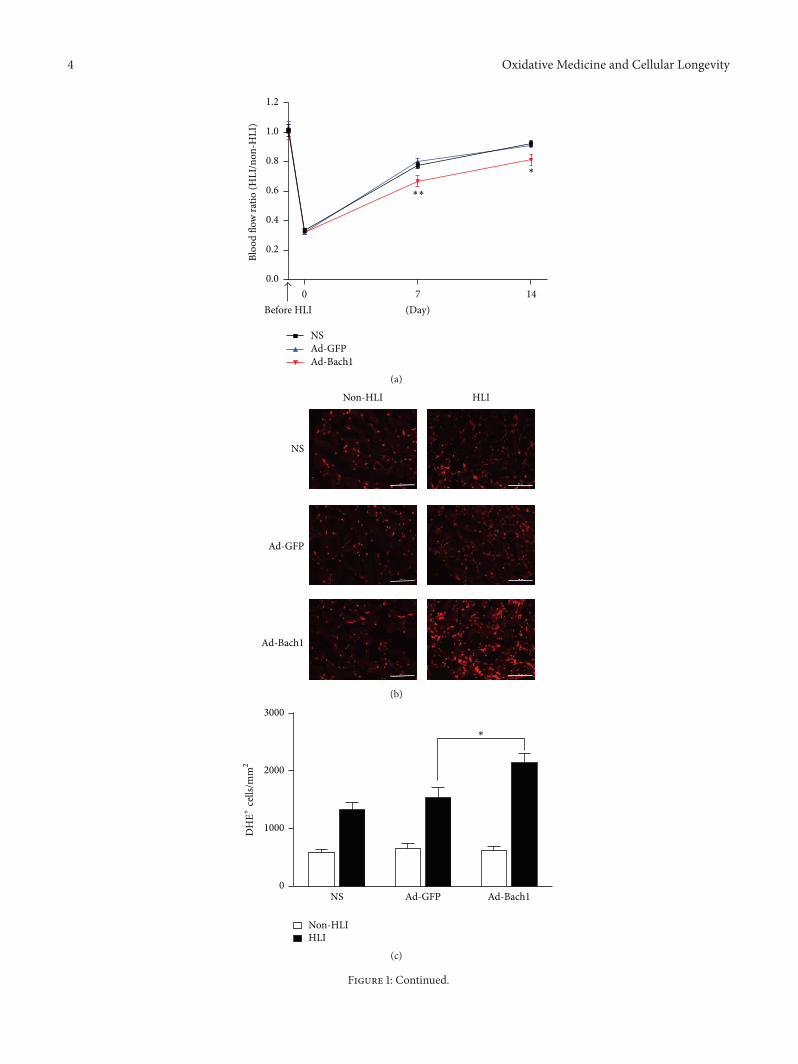
4 Oxidative Medicine and Cellular Longevity
Bloo
d flo
w ra
tio (H
LI/n
on-H
LI)
0 7 140.0
0.2
0.4
0.6
0.8
1.0
1.2
Before HLI (Day)
Ad-GFPNS
Ad-Bach1
∗
∗∗
(a)
Ad-GFP
HLI
NS
Ad-Bach1
Non-HLI
(b)
NS Ad-GFP Ad-Bach10
1000
2000
3000
∗
Non-HLIHLI
DH
E+ce
lls/m
m2
(c)
Figure 1: Continued.
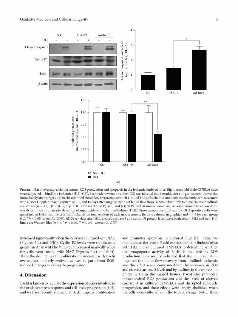
Oxidative Medicine and Cellular Longevity 5
NS Ad-GFP Ad-Bach10
1
2
3
4
∗
Clea
ved
casp
ase 3
pro
tein
leve
l(n
orm
aliz
ed, N
S, H
IL =
1)
Cleaved caspase 3
Cyclin D1
HLI + + +
Bach1
NS Ad-GFP Ad-Bach1
𝛽-actin
− − −
0.00
0.25
0.50
0.75
1.00
1.25
NS Ad-GFP Ad-Bach1
∗∗
Cycli
n D
1 pr
otei
n le
vel
(nor
mal
ized
, NS,
non
-HIL
=1
)
Non-HLIHLI
(d)
Figure 1: Bach1 overexpression promotes ROS production and apoptosis in the ischemic limbs of mice. Eight-week-old male C57BL/6 micewere subjected to hindlimb ischemia (HLI). GFP, Bach1 adenovirus, or saline (NS) was injected into the adductor and gastrocnemius musclesimmediately after surgery. (a) Bach1 inhibited blood flow restoration after HLI. Blood flows of ischemic and nonischemic limbweremeasuredwith a laser Doppler imaging system at 0, 7, and 14 days after surgery. Ratios of blood flow from ischemic hindlimb to nonischemic hindlimbare shown (𝑛 = 12; ∗𝑃 < 0.05, ∗∗𝑃 < 0.01 versus Ad-GFP). ((b) and (c)) ROS level in nonischemic and ischemic muscle tissue on day 7was determined by an in situ detection of superoxide with dihydroethidium (DHE) fluorescence. Bars, 100𝜇m (b). DHE-positive cells werequantified as DHE-positive cells/mm2. Data from four sections of each mouse muscle tissue are shown in graphics and 𝑛 = 6 for each group((c), ∗𝑃 < 0.05 versus Ad-GFP). (d) Seven days after HLI, cleaved caspase 3 and cyclin D1 protein levels were evaluated in HLI and non-HLIlimbs via Western blot (𝑛 = 6; ∗𝑃 < 0.05, ∗∗𝑃 < 0.01 versus Ad-GFP).
increased significantlywhen the cellswere culturedwithNAC(Figures 6(a) and 6(b)). Cyclin E2 levels were significantlygreater in Ad-Bach1 HMVECs but decreased markedly whenthe cells were treated with NAC (Figures 6(a) and 6(b)).Thus, the decline in cell proliferation associated with Bach1overexpression likely evolved, at least in part, from ROS-induced changes in cell-cycle progression.
4. Discussion
Bach1 is known to regulate the expression of genes involved inthe oxidative stress response and cell-cycle progression [1–5],and we have recently shown that Bach1 impairs proliferation
and promotes apoptosis in cultured ECs [12]. Thus, wemanipulated the level of Bach1 expression in the limbs ofmicewith HLI and in cultured HMVECs to determine whetherthe proapoptotic activity of Bach1 is mediated by ROSproduction. Our results indicated that Bach1 upregulationimpaired the blood flow recovery from hindlimb ischemiaand this effect was accompanied both by increases in ROSand cleaved caspase 3 levels and by declines in the expressionof cyclin D1 in the injured tissues. Bach1 also promotedmitochondrial ROS production and the levels of cleavedcaspase 3 in cultured HMVECs and disrupted cell-cycleprogression, and these effects were largely abolished whenthe cells were cultured with the ROS scavenger NAC. Thus,
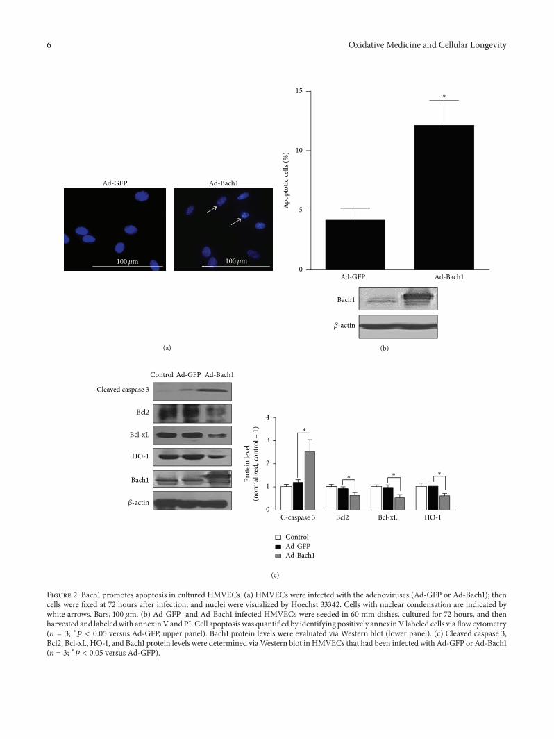
6 Oxidative Medicine and Cellular Longevity
Ad-GFP Ad-Bach1
100𝜇m 100𝜇m
(a)
Bach1
𝛽-actin
Ad-GFP Ad-Bach10
5
10
15
Apop
totic
cells
(%)
∗
(b)
Cleaved caspase 3
Bcl2
HO-1
Bach1
Ad-GFP Ad-Bach1Control
𝛽-actin
C-caspase 3 Bcl2 Bcl-xL HO-10
1
2
3
4
Ad-GFPAd-Bach1
Control
Prot
ein
leve
l(n
orm
aliz
ed, c
ontro
l =1
)
∗ ∗ ∗
∗Bcl-xL
(c)
Figure 2: Bach1 promotes apoptosis in cultured HMVECs. (a) HMVECs were infected with the adenoviruses (Ad-GFP or Ad-Bach1); thencells were fixed at 72 hours after infection, and nuclei were visualized by Hoechst 33342. Cells with nuclear condensation are indicated bywhite arrows. Bars, 100𝜇m. (b) Ad-GFP- and Ad-Bach1-infected HMVECs were seeded in 60 mm dishes, cultured for 72 hours, and thenharvested and labeledwith annexinV andPI. Cell apoptosis was quantified by identifying positively annexinV labeled cells via flow cytometry(𝑛 = 3; ∗𝑃 < 0.05 versus Ad-GFP, upper panel). Bach1 protein levels were evaluated via Western blot (lower panel). (c) Cleaved caspase 3,Bcl2, Bcl-xL, HO-1, and Bach1 protein levels were determined viaWestern blot in HMVECs that had been infected with Ad-GFP or Ad-Bach1(𝑛 = 3; ∗𝑃 < 0.05 versus Ad-GFP).
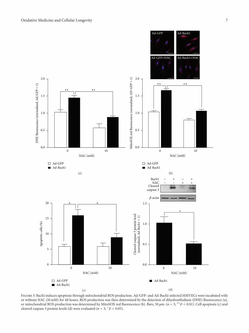
Oxidative Medicine and Cellular Longevity 7
0.0
0.5
1.0
1.5
2.0
Ad-Bach1
NAC (mM)0 10
Ad-GFP
∗∗∗∗
DH
E flu
ores
cenc
e (no
rmal
ized
, Ad-
GFP
=1
)
(a)Ad-Bach1Ad-GFP
NAC (mM)0 10
0.0
0.5
1.0
1.5
2.0
Mito
SOX
red
fluor
esce
nce (
norm
aliz
ed, A
D-G
FP =
1)
∗∗∗∗
Ad-GFP Ad-Bach1
Ad-GFP+NAC Ad-Bach1+NAC
(b)
0
5
10
15
20
Apop
totic
cells
(%)
Ad-Bach1Ad-GFP
NAC (mM)0 10
∗∗
(c)
Cleavedcaspase 3
Bach1NAC
𝛽-actin
− −−− + +
++
0.0
0.5
1.0
1.5
Ad-Bach1
NAC (mM)0 10
∗
Clea
ved
casp
ase 3
pro
tein
leve
l(n
orm
aliz
ed, A
d-Ba
ch1
=1
)
(d)
Figure 3: Bach1 induces apoptosis through mitochondrial ROS production. Ad-GFP- and Ad-Bach1-infected HMVECs were incubated withor without NAC (10mM) for 48 hours, ROS production was then determined by the detection of dihydroethidium (DHE) fluorescence (a),or mitochondrial ROS production was determined by MitoSOX red fluorescence (b). Bars, 50 𝜇m. (𝑛 = 3; ∗∗𝑃 < 0.01). Cell apoptosis (c) andcleaved caspase 3 protein levels (d) were evaluated (𝑛 = 3; ∗𝑃 < 0.05).
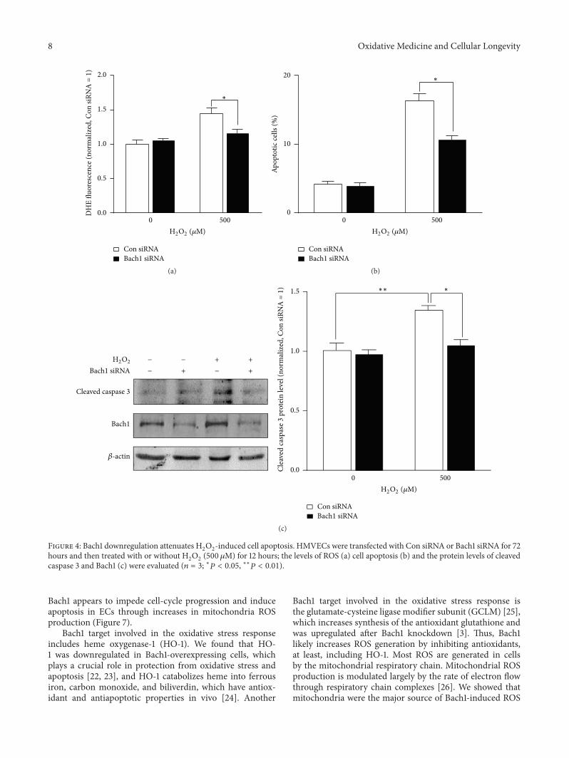
8 Oxidative Medicine and Cellular Longevity
Bach1 siRNACon siRNA
DH
E flu
ores
cenc
e(no
rmal
ized
, Con
siRN
A =
1)
H2O2 (𝜇M)
2.0
0 500
1.5
1.0
0.5
0.0
∗
(a)Ap
opto
tic ce
lls (%
)
Bach1 siRNACon siRNA
20
10
0
H2O2 (𝜇M)0 500
∗
(b)
Cleaved caspase 3
Bach1
Bach1 siRNA
Bach1 siRNACon siRNA
Clea
ved
casp
ase 3
pro
tein
leve
l (no
rmal
ized
, Con
siRN
A =
1)
𝛽-actin
−
− −
− + +
++
1.5
1.0
0.5
0.0
H2O2 (𝜇M)
H2O2
0 500
∗∗∗
(c)
Figure 4: Bach1 downregulation attenuates H2O2-induced cell apoptosis. HMVECs were transfected with Con siRNA or Bach1 siRNA for 72
hours and then treated with or without H2O2(500 𝜇M) for 12 hours; the levels of ROS (a) cell apoptosis (b) and the protein levels of cleaved
caspase 3 and Bach1 (c) were evaluated (𝑛 = 3; ∗𝑃 < 0.05, ∗∗𝑃 < 0.01).
Bach1 appears to impede cell-cycle progression and induceapoptosis in ECs through increases in mitochondria ROSproduction (Figure 7).
Bach1 target involved in the oxidative stress responseincludes heme oxygenase-1 (HO-1). We found that HO-1 was downregulated in Bach1-overexpressing cells, whichplays a crucial role in protection from oxidative stress andapoptosis [22, 23], and HO-1 catabolizes heme into ferrousiron, carbon monoxide, and biliverdin, which have antiox-idant and antiapoptotic properties in vivo [24]. Another
Bach1 target involved in the oxidative stress response isthe glutamate-cysteine ligase modifier subunit (GCLM) [25],which increases synthesis of the antioxidant glutathione andwas upregulated after Bach1 knockdown [3]. Thus, Bach1likely increases ROS generation by inhibiting antioxidants,at least, including HO-1. Most ROS are generated in cellsby the mitochondrial respiratory chain. Mitochondrial ROSproduction is modulated largely by the rate of electron flowthrough respiratory chain complexes [26]. We showed thatmitochondria were the major source of Bach1-induced ROS
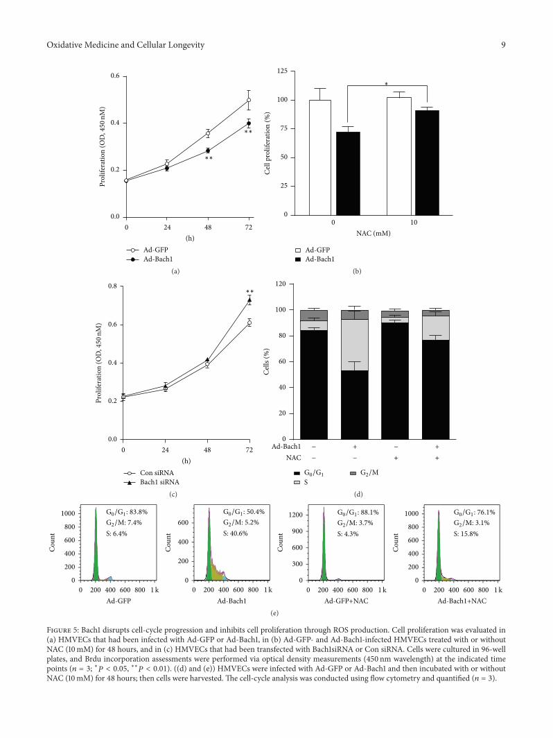
Oxidative Medicine and Cellular Longevity 9
0 24 48 720.0
0.2
0.4
0.6
Ad-GFPAd-Bach1
(h)
∗∗
∗∗
Prol
ifera
tion
(OD
,450
nM)
(a)
Ad-GFPAd-Bach1
0 100
25
50
75
100
125
NAC (mM)
Cel
l pro
lifer
atio
n (%
)
∗
(b)
Con siRNABach1 siRNA
0 24 48 72(h)
0.0
0.2
0.4
0.6
0.8∗∗
Prol
ifera
tion
(OD
,450
nM)
(c)
Cell
s (%
)
+ ++ +
Ad-Bach1NAC
0
40
60
20
80
100
120
− −
− −
G0/G1
SG2/M
(d)
Ad-GFP
S: 6.4%G2/M: 7.4%G0/G1: 83.8%
0 200 400 600 800 1k
1000
800
600
400
200
0
Cou
nt
Ad-Bach1
S: 40.6%G2/M: 5.2%G0/G1: 50.4%
0 200 400 600 800 1k
Cou
nt
600
400
200
0
Ad-GFP+NAC
S: 4.3%G2/M: 3.7%G0/G1: 88.1%
Cou
nt
1200
900
600
300
0
0 200 400 600 800 1kAd-Bach1+NAC
S: 15.8%G2/M: 3.1%G0/G1: 76.1%
Cou
nt
1000
800
600
400
200
0
0 200 400 600 800 1k
(e)
Figure 5: Bach1 disrupts cell-cycle progression and inhibits cell proliferation through ROS production. Cell proliferation was evaluated in(a) HMVECs that had been infected with Ad-GFP or Ad-Bach1, in (b) Ad-GFP- and Ad-Bach1-infected HMVECs treated with or withoutNAC (10mM) for 48 hours, and in (c) HMVECs that had been transfected with Bach1siRNA or Con siRNA. Cells were cultured in 96-wellplates, and Brdu incorporation assessments were performed via optical density measurements (450 nm wavelength) at the indicated timepoints (𝑛 = 3; ∗𝑃 < 0.05, ∗∗𝑃 < 0.01). ((d) and (e)) HMVECs were infected with Ad-GFP or Ad-Bach1 and then incubated with or withoutNAC (10mM) for 48 hours; then cells were harvested. The cell-cycle analysis was conducted using flow cytometry and quantified (𝑛 = 3).
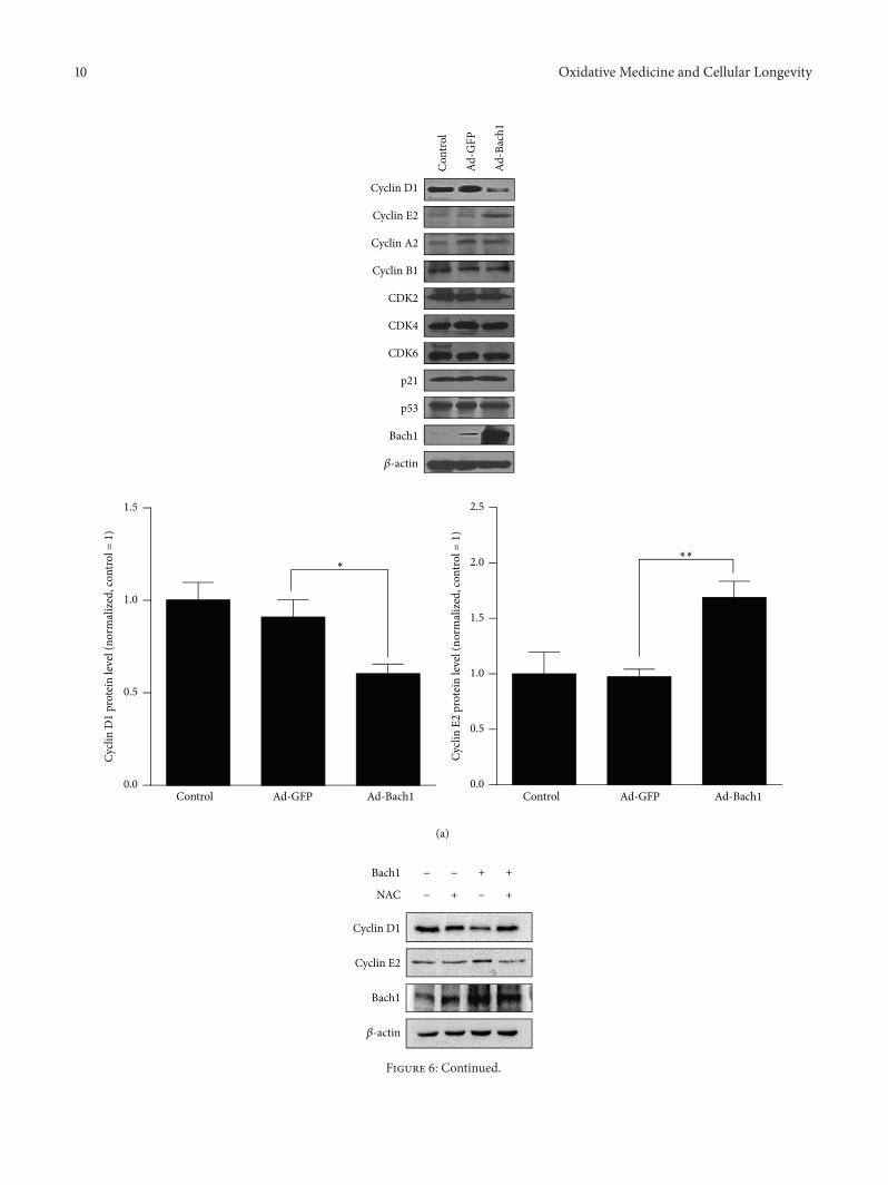
10 Oxidative Medicine and Cellular Longevity
Cyclin E2
Bach1
NAC + +
++
Cyclin D1
Bach1
𝛽-actin
−
− −
−
(a)
0.0
0.5
1.0
1.5
Cycli
n D
1 pr
otei
n le
vel (
norm
aliz
ed, c
ontro
l = 1
)
∗
Control Ad-Bach1Ad-GFP0.0
0.5
1.0
2.5
2.0
1.5
Cycli
n E2
pro
tein
leve
l (no
rmal
ized
, con
trol =
1)
∗∗
Control Ad-Bach1Ad-GFP
Cyclin D1
p21
p53
Cyclin A2
Cyclin B1
CDK2
CDK4
CDK6
Cyclin E2
Bach1
𝛽-actin
Con
trol
Ad-B
ach1
Ad-G
FP
Figure 6: Continued.
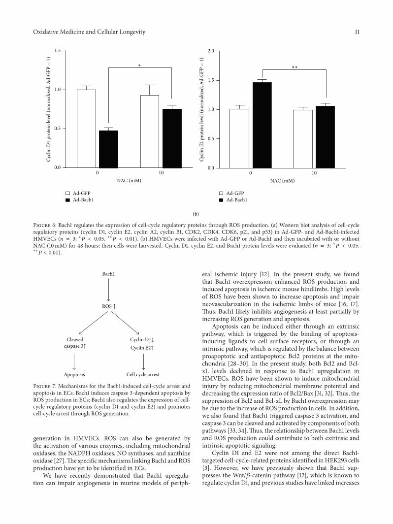
Oxidative Medicine and Cellular Longevity 11
(b)
Cycli
n D
1 pr
otei
n le
vel (
norm
aliz
ed, A
d-G
FP =
1)
0.00 10
0.5
1.0
1.5
NAC (mM)
Ad-GFPAd-Bach1
∗
Cycli
n E2
pro
tein
leve
l (no
rmal
ized
, Ad-
GFP
= 1
)
0 10NAC (mM)
∗∗
Ad-GFPAd-Bach1
0.0
0.5
1.0
2.0
1.5
Figure 6: Bach1 regulates the expression of cell-cycle regulatory proteins through ROS production. (a) Western blot analysis of cell-cycleregulatory proteins (cyclin D1, cyclin E2, cyclin A2, cyclin B1, CDK2, CDK4, CDK6, p21, and p53) in Ad-GFP- and Ad-Bach1-infectedHMVECs (𝑛 = 3; ∗𝑃 < 0.05, ∗∗𝑃 < 0.01). (b) HMVECs were infected with Ad-GFP or Ad-Bach1 and then incubated with or withoutNAC (10mM) for 48 hours; then cells were harvested. Cyclin D1, cyclin E2, and Bach1 protein levels were evaluated (𝑛 = 3; ∗𝑃 < 0.05,∗∗𝑃 < 0.01).
Bach1
ROS ↑
Cleavedcaspase 3↑
Cyclin D1↓
Cyclin E2↑
Apoptosis Cell cycle arrest
Figure 7: Mechanisms for the Bach1-induced cell-cycle arrest andapoptosis in ECs. Bach1 induces caspase 3-dependent apoptosis byROS production in ECs; Bach1 also regulates the expression of cell-cycle regulatory proteins (cyclin D1 and cyclin E2) and promotescell-cycle arrest through ROS generation.
generation in HMVECs. ROS can also be generated bythe activation of various enzymes, including mitochondrialoxidases, the NADPH oxidases, NO synthases, and xanthineoxidase [27].The specificmechanisms linking Bach1 andROSproduction have yet to be identified in ECs.
We have recently demonstrated that Bach1 upregula-tion can impair angiogenesis in murine models of periph-
eral ischemic injury [12]. In the present study, we foundthat Bach1 overexpression enhanced ROS production andinduced apoptosis in ischemic mouse hindlimbs. High levelsof ROS have been shown to increase apoptosis and impairneovascularization in the ischemic limbs of mice [16, 17].Thus, Bach1 likely inhibits angiogenesis at least partially byincreasing ROS generation and apoptosis.
Apoptosis can be induced either through an extrinsicpathway, which is triggered by the binding of apoptosis-inducing ligands to cell surface receptors, or through anintrinsic pathway, which is regulated by the balance betweenproapoptotic and antiapoptotic Bcl2 proteins at the mito-chondria [28–30]. In the present study, both Bcl2 and Bcl-xL levels declined in response to Bach1 upregulation inHMVECs. ROS have been shown to induce mitochondrialinjury by reducing mitochondrial membrane potential anddecreasing the expression ratio of Bcl2/Bax [31, 32].Thus, thesuppression of Bcl2 and Bcl-xL by Bach1 overexpression maybe due to the increase of ROS production in cells. In addition,we also found that Bach1 triggered caspase 3 activation, andcaspase 3 can be cleaved and activated by components of bothpathways [33, 34].Thus, the relationship between Bach1 levelsand ROS production could contribute to both extrinsic andintrinsic apoptotic signaling.
Cyclin D1 and E2 were not among the direct Bach1-targeted cell-cycle-related proteins identified inHEK293 cells[3]. However, we have previously shown that Bach1 sup-presses the Wnt/𝛽-catenin pathway [12], which is known toregulate cyclin D1, and previous studies have linked increases
12 Oxidative Medicine and Cellular Longevity
in cyclin E expression with S-phase arrest [35]. Thus, Bach1appears to inhibit cell-cycle progression and proliferation inECs through a network of at least three overlapping mech-anisms: (1) declines in Wnt-mediated cyclin D1 expression,(2) increases in ROS-mediated cyclin D1 repression, and (3)upregulation of cyclin-E2-induced S-phase arrest.
In conclusion, our results indicate that Bach1 inhibitscell proliferation and induces cell-cycle arrest and apoptosisby increasing mitochondria derived ROS production and,consequently, that functional downregulation of Bach1 maybe a promising treatment target for the treatment of vasculardiseases.
Disclosure
Xinhong Wang and Junxu Liu share first authorship.
Conflict of Interests
The authors declare that they have no conflict of interests.
Authors’ Contribution
Dan Meng and Kang Yao designed this study. XinhongWang, Junxu Liu, and Li Jiang performed the experiments.Xiangxiang Wei, Cong Niu, Rui Wang, and Jianyi Zhanganalyzed the data. DanMengwrote the paper. XinhongWangand Junxu Liu contributed equally to this paper.
Acknowledgments
The authors thank Dr. Xianglin Shi for providing HMVECs.The authors thank Dr. W. Kevin Cukier-Meisner for editorialassistance. This work was supported by General Programs ofthe National Natural Science Foundation of China (81170298and 81270410 toD.Meng, 81201468 to X.Wang, and 81470386to K. Yao) and US Public Health Service Grants RO1 HL67828 and HL 95077 (J. Zhang).
References
[1] T. Oyake, K. Itoh, H. Motohashi et al., “Bach proteins belong toa novel family of BTB-basic leucine zipper transcription factorsthat interact with MafK and regulate transcription through theNF-E2 site,” Molecular and Cellular Biology, vol. 16, no. 11, pp.6083–6095, 1996.
[2] K. Igarashi and J. Sun, “The heme-Bach1 pathway in the regula-tion of oxidative stress response and erythroid differentiation,”Antioxidants &Redox Signaling, vol. 8, no. 1-2, pp. 107–118, 2006.
[3] H.-J. Warnatz, D. Schmidt, T. Manke et al., “The BTB and CNChomology 1 (BACH1) target genes are involved in the oxidativestress response and in control of the cell cycle,” The Journal ofBiological Chemistry, vol. 286, no. 26, pp. 23521–23532, 2011.
[4] K. Ogawa, J. Sun, S. Taketani et al., “Heme mediates dere-pression of Maf recognition element through direct binding totranscription repressor Bach1,” The EMBO Journal, vol. 20, no.11, pp. 2835–2843, 2001.
[5] J. Li, T. Shiraki, and K. Igarashi, “Transcription-independentrole of Bach1 in mitosis through a nuclear exporter Crm1-dependent mechanism,” FEBS Letters, vol. 586, no. 4, pp. 448–454, 2012.
[6] K. Kondo, Y. Ishigaki, J. Gao et al., “Bach1 deficiency protectspancreatic 𝛽-cells from oxidative stress injury,” American Jour-nal of Physiology—Endocrinology and Metabolism, vol. 305, no.5, pp. E641–E648, 2013.
[7] M. Inoue, S. Tazuma, K. Kanno, H. Hyogo, K. Igarashi, andK. Chayama, “Bach1 gene ablation reduces steatohepatitis inmouse MCD diet model,” Journal of Clinical Biochemistry andNutrition, vol. 48, no. 2, pp. 161–166, 2011.
[8] A. Harusato, Y. Naito, T. Takagi et al., “Suppression ofindomethacin-induced apoptosis in the small intestine due toBach1 deficiency,” Free Radical Research, vol. 45, no. 6, pp. 717–727, 2011.
[9] T. Tanimoto, N. Hattori, T. Senoo et al., “Genetic ablation of theBach1 gene reduces hyperoxic lung injury in mice: role of IL-6,”Free Radical Biology and Medicine, vol. 46, no. 8, pp. 1119–1126,2009.
[10] Y. Watari, Y. Yamamoto, A. Brydun et al., “Ablation of theBach1 gene leads to the suppression of atherosclerosis in Bach1and apolipoprotein E double knockout mice,” HypertensionResearch, vol. 31, no. 4, pp. 783–792, 2008.
[11] Y. Yano, R. Ozono, Y. Oishi et al., “Genetic ablation of thetranscription repressor Bach1 leads to myocardial protectionagainst ischemia/reperfusion inmice,”Genes to Cells, vol. 11, no.7, pp. 791–803, 2006.
[12] L. Jiang,M.Yin, X.Wei et al., “Bach1 repressesWnt/beta-cateninsignaling and angiogenesis,” Circulation Research, vol. 117, no. 4,pp. 364–375, 2015.
[13] R. S. Frey, M. Ushio-Fukai, and A. B. Malik, “NADPH oxidase-dependent signaling in endothelial cells: role in physiology andpathophysiology,” Antioxidants and Redox Signaling, vol. 11, no.4, pp. 791–810, 2009.
[14] J. Streeter, W.Thiel, K. Brieger, and F. J. Miller Jr., “Opportunitynox: the future of NADPH oxidases as therapeutic targets incardiovascular disease,”CardiovascularTherapeutics, vol. 31, no.3, pp. 125–137, 2013.
[15] D. Meng, A. Mei, J. Liu et al., “NADPH oxidase 4 medi-ates insulin-stimulated HIF-1alpha and VEGF expression, andangiogenesis in vitro,” PLoS ONE, vol. 7, no. 10, Article IDe48393, 2012.
[16] T. G. Ebrahimian, C. Heymes, D. You et al., “NADPH oxidase-derived overproduction of reactive oxygen species impairspostischemic neovascularization in mice with type 1 diabetes,”American Journal of Pathology, vol. 169, no. 2, pp. 719–728, 2006.
[17] H. W. Kim, A. Lin, R. E. Guldberg, M. Ushio-Fukai, andT. Fukai, “Essential role of extracellular SOD in reparativeneovascularization induced by hindlimb ischemia,” CirculationResearch, vol. 101, no. 4, pp. 409–419, 2007.
[18] R. Shao and X. Guo, “Human microvascular endothelial cellsimmortalized with human telomerase catalytic protein: amodelfor the study of in vitro angiogenesis,”Biochemical and Biophysi-cal Research Communications, vol. 321, no. 4, pp. 788–794, 2004.
[19] D. Meng, D.-D. Lv, and J. Fang, “Insulin-like growth factor-Iinduces reactive oxygen species production and cell migrationthrough Nox4 and Rac1 in vascular smooth muscle cells,”Cardiovascular Research, vol. 80, no. 2, pp. 299–308, 2008.
Oxidative Medicine and Cellular Longevity 13
[20] J. Fauconnier, D. C. Andersson, S.-J. Zhang et al., “Effects ofpalmitate on Ca2+ handling in adult control and ob/ob car-diomyocytes: impact of mitochondrial reactive oxygen species,”Diabetes, vol. 56, no. 4, pp. 1136–1142, 2007.
[21] D. Meng, X. Shi, B.-H. Jiang, and J. Fang, “Insulin-like growthfactor-I (IGF-I) induces epidermal growth factor receptortransactivation and cell proliferation through reactive oxygenspecies,” Free Radical Biology and Medicine, vol. 42, no. 11, pp.1651–1660, 2007.
[22] C. D. Ferris, S. R. Jaffrey, A. Sawa et al., “Haem oxygenase-1 prevents cell death by regulating cellular iron,” Nature CellBiology, vol. 1, no. 3, pp. 152–157, 1999.
[23] N. Yun, H.-A. Eum, and S.-M. Lee, “Protective role of hemeoxygenase-1 against liver damage caused by hepatic ischemiaand reperfusion in rats,” Antioxidants and Redox Signaling, vol.13, no. 10, pp. 1503–1512, 2010.
[24] L. E. Otterbein, M. P. Soares, K. Yamashita, and F. H. Bach,“Heme oxygenase-1: unleashing the protective properties ofheme,” Trends in Immunology, vol. 24, no. 8, pp. 449–455, 2003.
[25] A.K.MacLeod,M.McMahon, S.M. Plummer et al., “Character-ization of the cancer chemopreventive NRF2-dependent genebattery in human keratinocytes: demonstration that theKEAP1-NRF2 pathway, and not the BACH1-NRF2 pathway, controlscytoprotection against electrophiles as well as redox-cyclingcompounds,” Carcinogenesis, vol. 30, no. 9, pp. 1571–1580, 2009.
[26] R. O. Poyton, K. A. Ball, and P. R. Castello, “Mitochondrialgeneration of free radicals and hypoxic signaling,” Trends inEndocrinology & Metabolism, vol. 20, no. 7, pp. 332–340, 2009.
[27] P. L. Hordijk, “Regulation of NADPH oxidases: the role of Racproteins,”Circulation Research, vol. 98, no. 4, pp. 453–462, 2006.
[28] L. Coultas and A. Strasser, “The role of the Bcl-2 protein familyin cancer,” Seminars in Cancer Biology, vol. 13, no. 2, pp. 115–123,2003.
[29] N. N. Danial and S. J. Korsmeyer, “Cell death: critical controlpoints,” Cell, vol. 116, no. 2, pp. 205–219, 2004.
[30] D. R. Green and G. Kroemer, “The pathophysiology of mito-chondrial cell death,” Science, vol. 305, no. 5684, pp. 626–629,2004.
[31] T. Adachi, H. Tanaka, S. Nonomura, H. Hara, S.-I. Kondo, andM. Hori, “Plasma-activated medium induces A549 cell injuryvia a spiral apoptotic cascade involving the mitochondrial-nuclear network,” Free Radical Biology andMedicine, vol. 79, pp.28–44, 2015.
[32] P. Wyrsch, C. Blenn, J. Bader, and F. R. Althaus, “Cell death andautophagy under oxidative stress: roles of poly(ADP-Ribose)polymerases and Ca2+,”Molecular and Cellular Biology, vol. 32,no. 17, pp. 3541–3553, 2012.
[33] E. Frejlich, J. Rudno-Rudzińska, K. Janiszewski et al., “Caspasesand their role in gastric cancer,” Advances in Clinical andExperimental Medicine, vol. 22, no. 4, pp. 593–602, 2013.
[34] J.-H. Zhang, Y. Zhang, andB.Herman, “Caspases, apoptosis andaging,”Ageing Research Reviews, vol. 2, no. 4, pp. 357–366, 2003.
[35] J.-W. Park, Y.-J. Choi, M.-A. Jang et al., “Chemopreventive agentresveratrol, a natural product derived from grapes, reversiblyinhibits progression through S and G2 phases of the cell cyclein U937 cells,” Cancer Letters, vol. 163, no. 1, pp. 43–49, 2001.
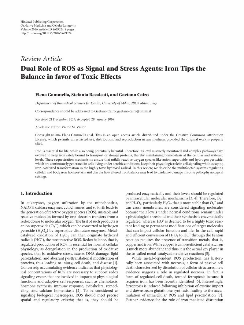
Review ArticleDual Role of ROS as Signal and Stress Agents: Iron Tips theBalance in favor of Toxic Effects
Elena Gammella, Stefania Recalcati, and Gaetano Cairo
Department of Biomedical Sciences for Health, University of Milan, 20133 Milan, Italy
Correspondence should be addressed to Gaetano Cairo; [email protected]
Received 21 December 2015; Accepted 28 January 2016
Academic Editor: Victor M. Victor
Copyright © 2016 Elena Gammella et al. This is an open access article distributed under the Creative Commons AttributionLicense, which permits unrestricted use, distribution, and reproduction in any medium, provided the original work is properlycited.
Iron is essential for life, while also being potentially harmful. Therefore, its level is strictly monitored and complex pathways haveevolved to keep iron safely bound to transport or storage proteins, thereby maintaining homeostasis at the cellular and systemiclevels. These sequestration mechanisms ensure that mildly reactive oxygen species like anion superoxide and hydrogen peroxide,which are continuously generated in cells living under aerobic conditions, keep their physiologic role in cell signalingwhile escapingiron-catalyzed transformation in the highly toxic hydroxyl radical. In this review, we describe the multifaceted systems regulatingcellular and body iron homeostasis and discuss how altered iron balance may lead to oxidative damage in some pathophysiologicalsettings.
1. Introduction
In eukaryotes, oxygen utilization by the mitochondria,NADPHoxidase enzymes, cytochromes, and so forth leads tothe generation of reactive oxygen species (ROS), unstable andreactive molecules formed by one-electron transfers from aredox donor tomolecular oxygen.Thefirst of such products isanion superoxide (O
2
−), which can be converted to hydrogenperoxide (H
2O2) by superoxide dismutase enzymes. Metal-
catalyzed oxidation of H2O2can then originate hydroxyl
radicals (HO∙), themost reactive ROS. Redox balance, that is,regulated production of ROS, is essential for normal cellularphysiology, as deregulation in the production of oxidativespecies, that is, oxidative stress, causes DNA damage, lipidperoxidation, and aberrant posttranslational modification ofproteins, thus leading to injury, cell death, and disease [1].Conversely, accumulating evidence indicates that physiolog-ical concentrations of ROS are necessary to support redoxsignaling events that are involved in important physiologicalfunctions and adaptive cell responses, such as chemotaxis,hormone synthesis, immune response, cytoskeletal remod-eling, and calcium homeostasis [2]. To be considered assignaling biological messengers, ROS should meet precisespatial and regulatory criteria; that is, they should be
produced enzymatically and their levels should be regulatedby intracellular molecular mechanisms [3, 4]. Therefore, O
2
−
andH2O2, particularlyH
2O2that ismore stable thanO
2
− andcan cross membranes, are considered signaling moleculesbecause their levels under normal conditions remain undera physiological threshold and their synthesis is enzymaticallyregulated, whereas HO∙ is deemed to be a highly toxic reac-tant leading to permanent modifications of target moleculesthat can impact cellular function and life. In the cell, rapidand efficient conversion of H
2O2to HO∙ through the Fenton
reaction requires the presence of transition metals, that is,copper and iron.While copper is amore efficient catalyst, ironis much more abundant and thus it is the actual key player inthe so-called metal-catalyzed oxidative reactions [5].
While metal-dependent ROS production has histori-cally been associated with necrosis, a form of passive celldeath characterized by dissolution of cellular structures, newevidence suggests a role in regulated necrosis. In fact, aform of regulated cell death, termed ferroptosis because itrequires iron, has been recently identified [6]. Interestingly,ferroptosis is induced following inhibition of cystine importand downstream glutathione synthesis, leading to the accu-mulation of intracellular ROS and lipid peroxidation [7].Further evidence for the role of iron-mediated disruption
Hindawi Publishing CorporationOxidative Medicine and Cellular LongevityVolume 2016, Article ID 8629024, 9 pageshttp://dx.doi.org/10.1155/2016/8629024
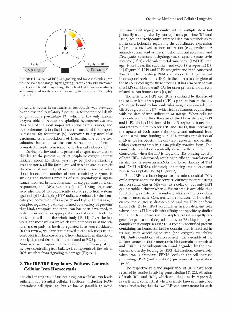
2 Oxidative Medicine and Cellular Longevity
Signaling Damage
FeFe
FeH2O2
H2O2 H2O2 → HO∙
Figure 1: Dual role of ROS as signaling and toxic molecules, irontips the scale for damage. By triggering Fenton chemistry, increasediron (Fe) availability may change the role of H
2O2from a relatively
safe compound involved in cell signaling to a source of the highlytoxic HO∙.
of cellular redox homeostasis in ferroptosis was providedby the essential regulatory function in ferroptotic cell deathof glutathione peroxidase [8], which is the only knownenzyme able to reduce phospholipid hydroperoxides andthus one of the most important antioxidant enzymes, andby the demonstration that transferrin-mediated iron importis essential for ferroptosis [9]. Moreover, in hepatocellularcarcinoma cells, knockdown of H ferritin, one of the twosubunits that compose the iron storage protein ferritin,promoted ferroptosis in response to classical inducers [10].
During the slow and long process of oxygen accumulationthat led to the present 20.9% atmospheric oxygen contentinitiated about 1.5 billion years ago by photosynthesizingcyanobacteria, all life forms evolved mechanisms to exploitthe chemical reactivity of iron for efficient aerobic reac-tions. Indeed, the number of iron-containing enzymes isstriking and includes proteins of vital physiological signif-icance involved in functions such as oxygen transport, cellrespiration, and DNA synthesis [11, 12]. Living organismswere also forced to concurrently evolve protection systemsagainst highly damaging HO∙ radicals produced by the iron-catalyzed conversion of superoxide and H
2O2. To this aim, a
complex regulatory pathway formed by a variety of proteinsthat bind, transport, and store iron has been developed, inorder to maintain an appropriate iron balance in both theindividual cells and the whole body [13, 14]. Over the lastyears, the mechanisms by which iron homeostasis at the cel-lular and organismal levels is regulated have been elucidated.In this review, we have summarized recent advances in thecontrol of iron homeostasis and how changes in availability ofpoorly liganded ferrous iron are related to ROS production.Moreover, we propose that whenever the efficiency of thenetwork controlling iron balance is compromised, the role ofROS switches from signaling to damage (Figure 1).
2. The IRE/IRP Regulatory Pathway ControlsCellular Iron Homeostasis
The challenging task of maintaining intracellular iron levelssufficient for essential cellular functions, including ROS-dependent cell signaling, but as low as possible to avoid
ROS-mediated injury, is controlled at multiple steps butprimarily accomplished by iron regulatory proteins (IRP1 andIRP2), which strictly control intracellular ironmetabolism byposttranscriptionally regulating the coordinated expressionof proteins involved in iron utilization (e.g., erythroid 5aminolevulinic acid synthase, mitochondrial aconitase, andDrosophila succinate dehydrogenase), uptake (transferrinreceptor (TfR1) and divalentmetal transporter (DMT1)), stor-age (H and L ferritin subunits), and export (ferroportin) [15,16] (Figure 2). IRP1 and IRP2 recognize and bind conserved25–30 nucleotides-long RNA stem-loop structures namediron responsive elements (IREs) in the untranslated regions ofthe mRNAs coding for these proteins. It has also been shownthat IRPs can bind the mRNAs for other proteins not directlyrelated to iron homeostasis [15, 16].
The activity of IRP1 and IRP2 is dictated by the size ofthe cellular labile iron pool (LIP), a pool of iron in the low𝜇M range bound to low molecular weight compounds likecitrate or glutathione [17], which is in continuous equilibriumwith the sites of iron utilization or storage. When cells areiron deficient and thus the size of the LIP is shrunk, IRP1and IRP2 bind to IREs located in the 3 region of transcriptsand stabilize the mRNA for TfR1 and DMT1, thus increasingthe uptake of both transferrin-bound and unbound iron.At the same time, binding to 5 IRE impairs translation ofmRNAs for ferroportin, the only iron exporter, and ferritin,which sequesters iron in a catalytically inactive form. Thiscoordinate regulation eventually expands the cellular LIP.Conversely, when the LIP is large, the IRE-binding activityof both IRPs is decreased, resulting in efficient translation offerritin and ferroportin mRNAs and lower stability of TfR1and DMT1 mRNAs, ultimately enhancing iron storage andrelease over uptake [15, 16] (Figure 2).
Both IRPs are homologous to the mitochondrial TCAcycle enzyme aconitase that converts citrate to isocitrate usingan iron sulfur cluster (4Fe–4S) as a cofactor, but only IRP1can assemble a cluster when sufficient iron is available, thusfunctioning as cytosolic aconitase, which is the prevailingform in most cells. Conversely, in conditions of iron defi-ciency, the cluster is disassembled and the IRP1 apoformbinds IRE [15, 16]. IRP2 accumulates in iron-deficient cellswhere it binds IRE motifs with affinity and specificity similarto that of IRP1, whereas in iron-replete cells it is rapidly tar-geted for proteasomal degradation by an E3 ubiquitin ligasecomplex that comprises FBXL5, a recently identified proteincontaining an hemerythrin-like domain that is involved inits regulation according to iron (and oxygen) availability[18]. Under conditions of iron scarcity, the assembly of thedi-iron center in the hemerythrin-like domain is impairedand FBXL5 is polyubiquitinated and degraded by the pro-teasome, thereby leading to IRP2 stabilization. Conversely,when iron is abundant, FBXL5 levels in the cell increasepromoting IRP2 (and apo-IRP1) proteasomal degradation[19, 20].
The respective role and importance of IRPs have beenrevealed by studies involving gene deletion [21, 22]. Ablationof both IRP1 and IRP2, which are ubiquitously expressed,is early embryonic lethal whereas single knockout mice areviable, indicating that the two IRPs can compensate for each
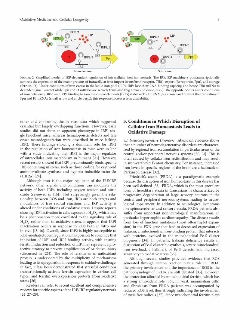
Oxidative Medicine and Cellular Longevity 3
Abundant iron Scarce iron
FtFpn
TfR1
Tf
Tf
Tf
LIPIRP1/2
IRP1/2
IRP1/2IRE
IRE
IRE
Ft Fpn
TfR1
IRP1/2
IRP1/2
IRP1/2
Tf
TfTf
LIP
IRE
IRE
IRE
Figure 2: Simplified model of IRP-dependent regulation of intracellular iron homeostasis. The IRE/IRP machinery posttranscriptionallycontrols the expression of the major proteins of intracellular iron import (transferrin receptor, TfR1), export (ferroportin, Fpn), and storage(ferritin, Ft). Under conditions of iron excess in the labile iron pool (LIP), IRPs lose their RNA-binding capacity, and hence TfR1 mRNA isdegraded (small arrow) while Fpn and Ft mRNAs are actively translated (big arrow and circle, resp.). The opposite occurs under conditionsof iron deficiency: IRP1 and IRP2 binding to iron responsive elements (IREs) stabilize TfR1 mRNA (big arrow) and prevent the translation ofFpn and Ft mRNAs (small arrow and circle, resp.); this response increases iron availability.
other and confirming the in vitro data which suggestedessential but largely overlapping functions. However, earlystudies did not show an apparent phenotype in IRP1 sin-gle knockout mice, whereas hematopoietic defects and lateonset neurodegeneration were described in mice lackingIRP2. These findings showing a dominant role for IRP2in the regulation of iron homeostasis in mice were in linewith a study indicating that IRP2 is the major regulatorof intracellular iron metabolism in humans [23]. However,recent results showed that IRP1 predominantly binds specificIRE-containing mRNAs, such as those coding for erythroidaminolevulinate synthase and hypoxia inducible factor 2𝛼(HIF2𝛼) [24].
Although iron is the major regulator of the IRE/IRPnetwork, other signals and conditions can modulate theactivity of both IRPs, including oxygen tension and nitricoxide (reviewed in [16]). Not surprisingly given the rela-tionship between ROS and iron, IRPs are both targets andmodulators of free radical reactions and IRP activity isaltered under conditions of oxidative stress. Despite reportsshowing IRP1 activation in cells exposed to H
2O2, whichmay
be a phenomenon more correlated to the signaling role ofH2O2rather than to oxidative stress, it appears that IRP1
inactivation occurs in response to ROS both in vitro andin vivo [15, 16]. Overall, since IRP2 is highly susceptible toROS-mediated downregulation, it is possible to conclude thatinhibition of IRP1 and IRP2 binding activity, with ensuingferritin induction and reduction of LIP, may represent a pro-tective strategy to prevent amplification of oxidative injury(discussed in [25]). The role of ferritin as an antioxidantprotein is underscored by the multiplicity of mechanismsleading to its upregulation in response to oxidative challenge;in fact, it has been demonstrated that stressful conditionstranscriptionally activate ferritin expression in various celltypes, and ferritin overexpression protects from oxidativestress [26].
Readers can refer to recent excellent and comprehensivereviews for specific aspects of the IRE/IRP regulatory network[24, 27–29].
3. Conditions in Which Disruption ofCellular Iron Homeostasis Leads toOxidative Damage
3.1. Neurodegenerative Disorders. Abundant evidence showsthat a number of neurodegenerative disorders are character-ized by regional iron accumulation in particular areas of thecentral and/or peripheral nervous systems [30, 31]. This isoften caused by cellular iron redistribution and may resultin iron-catalyzed Fenton chemistry. For instance, increasediron levels in specific regions of the brain are a hallmark ofParkinson disease [32].
Friedrich’s ataxia (FRDA) is a paradigmatic examplebecause the disruption of iron homeostasis in this disease hasbeen well defined [33]. FRDA, which is the most prevalentform of hereditary ataxia in Caucasians, is characterized byprogressive degeneration of large sensory neurons in thecentral and peripheral nervous systems leading to neuro-logical impairment. In addition to neurological symptomslike spinocerebellar and sensory ataxia, FRDA patients alsosuffer from important nonneurological manifestations, inparticular hypertrophic cardiomyopathy. The disease resultsfrom loss of function mutations (most often triplet expan-sion) in the FXN gene that lead to decreased expression offrataxin, a mitochondrial iron-binding protein that interactswith proteins involved in the mitochondrial Fe-S clusterbiogenesis [34]. In patients, frataxin deficiency results indisruption of Fe-S cluster biosynthesis, severe mitochondrialiron overload, a hallmark of Fe-S defects, and increasedsensitivity to oxidative stress [35].
Although several studies provided evidence that ROSgenerated through Fenton reaction play a role in FRDA,the primary involvement and the importance of ROS in thepathophysiology of FRDA are still debated [33]. However,the protection afforded by mitochondrial ferritin, which hasa strong antioxidant role [36], in yeast, mammalian cells,and fibroblasts from FRDA patients was accompanied byreduced ROS level, thus strongly indicating the involvementof toxic free radicals [37]. Since mitochondrial ferritin plays
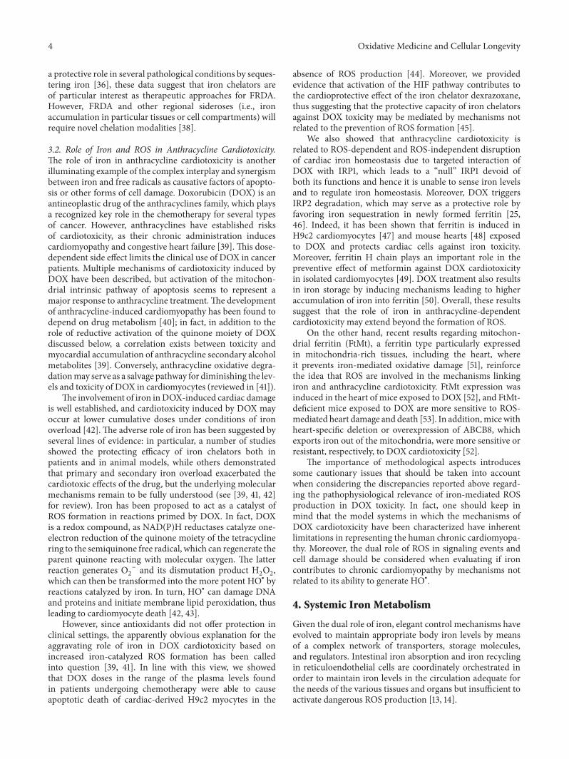
4 Oxidative Medicine and Cellular Longevity
a protective role in several pathological conditions by seques-tering iron [36], these data suggest that iron chelators areof particular interest as therapeutic approaches for FRDA.However, FRDA and other regional sideroses (i.e., ironaccumulation in particular tissues or cell compartments) willrequire novel chelation modalities [38].
3.2. Role of Iron and ROS in Anthracycline Cardiotoxicity.The role of iron in anthracycline cardiotoxicity is anotherilluminating example of the complex interplay and synergismbetween iron and free radicals as causative factors of apopto-sis or other forms of cell damage. Doxorubicin (DOX) is anantineoplastic drug of the anthracyclines family, which playsa recognized key role in the chemotherapy for several typesof cancer. However, anthracyclines have established risksof cardiotoxicity, as their chronic administration inducescardiomyopathy and congestive heart failure [39]. This dose-dependent side effect limits the clinical use of DOX in cancerpatients. Multiple mechanisms of cardiotoxicity induced byDOX have been described, but activation of the mitochon-drial intrinsic pathway of apoptosis seems to represent amajor response to anthracycline treatment.The developmentof anthracycline-induced cardiomyopathy has been found todepend on drug metabolism [40]; in fact, in addition to therole of reductive activation of the quinone moiety of DOXdiscussed below, a correlation exists between toxicity andmyocardial accumulation of anthracycline secondary alcoholmetabolites [39]. Conversely, anthracycline oxidative degra-dationmay serve as a salvage pathway for diminishing the lev-els and toxicity of DOX in cardiomyocytes (reviewed in [41]).
The involvement of iron inDOX-induced cardiac damageis well established, and cardiotoxicity induced by DOX mayoccur at lower cumulative doses under conditions of ironoverload [42]. The adverse role of iron has been suggested byseveral lines of evidence: in particular, a number of studiesshowed the protecting efficacy of iron chelators both inpatients and in animal models, while others demonstratedthat primary and secondary iron overload exacerbated thecardiotoxic effects of the drug, but the underlying molecularmechanisms remain to be fully understood (see [39, 41, 42]for review). Iron has been proposed to act as a catalyst ofROS formation in reactions primed by DOX. In fact, DOXis a redox compound, as NAD(P)H reductases catalyze one-electron reduction of the quinone moiety of the tetracyclinering to the semiquinone free radical, which can regenerate theparent quinone reacting with molecular oxygen. The latterreaction generates O
2
− and its dismutation product H2O2,
which can then be transformed into the more potent HO∙ byreactions catalyzed by iron. In turn, HO∙ can damage DNAand proteins and initiate membrane lipid peroxidation, thusleading to cardiomyocyte death [42, 43].
However, since antioxidants did not offer protection inclinical settings, the apparently obvious explanation for theaggravating role of iron in DOX cardiotoxicity based onincreased iron-catalyzed ROS formation has been calledinto question [39, 41]. In line with this view, we showedthat DOX doses in the range of the plasma levels foundin patients undergoing chemotherapy were able to causeapoptotic death of cardiac-derived H9c2 myocytes in the
absence of ROS production [44]. Moreover, we providedevidence that activation of the HIF pathway contributes tothe cardioprotective effect of the iron chelator dexrazoxane,thus suggesting that the protective capacity of iron chelatorsagainst DOX toxicity may be mediated by mechanisms notrelated to the prevention of ROS formation [45].
We also showed that anthracycline cardiotoxicity isrelated to ROS-dependent and ROS-independent disruptionof cardiac iron homeostasis due to targeted interaction ofDOX with IRP1, which leads to a “null” IRP1 devoid ofboth its functions and hence it is unable to sense iron levelsand to regulate iron homeostasis. Moreover, DOX triggersIRP2 degradation, which may serve as a protective role byfavoring iron sequestration in newly formed ferritin [25,46]. Indeed, it has been shown that ferritin is induced inH9c2 cardiomyocytes [47] and mouse hearts [48] exposedto DOX and protects cardiac cells against iron toxicity.Moreover, ferritin H chain plays an important role in thepreventive effect of metformin against DOX cardiotoxicityin isolated cardiomyocytes [49]. DOX treatment also resultsin iron storage by inducing mechanisms leading to higheraccumulation of iron into ferritin [50]. Overall, these resultssuggest that the role of iron in anthracycline-dependentcardiotoxicity may extend beyond the formation of ROS.
On the other hand, recent results regarding mitochon-drial ferritin (FtMt), a ferritin type particularly expressedin mitochondria-rich tissues, including the heart, whereit prevents iron-mediated oxidative damage [51], reinforcethe idea that ROS are involved in the mechanisms linkingiron and anthracycline cardiotoxicity. FtMt expression wasinduced in the heart of mice exposed to DOX [52], and FtMt-deficient mice exposed to DOX are more sensitive to ROS-mediated heart damage and death [53]. In addition,micewithheart-specific deletion or overexpression of ABCB8, whichexports iron out of the mitochondria, were more sensitive orresistant, respectively, to DOX cardiotoxicity [52].
The importance of methodological aspects introducessome cautionary issues that should be taken into accountwhen considering the discrepancies reported above regard-ing the pathophysiological relevance of iron-mediated ROSproduction in DOX toxicity. In fact, one should keep inmind that the model systems in which the mechanisms ofDOX cardiotoxicity have been characterized have inherentlimitations in representing the human chronic cardiomyopa-thy. Moreover, the dual role of ROS in signaling events andcell damage should be considered when evaluating if ironcontributes to chronic cardiomyopathy by mechanisms notrelated to its ability to generate HO∙.
4. Systemic Iron Metabolism
Given the dual role of iron, elegant control mechanisms haveevolved to maintain appropriate body iron levels by meansof a complex network of transporters, storage molecules,and regulators. Intestinal iron absorption and iron recyclingin reticuloendothelial cells are coordinately orchestrated inorder to maintain iron levels in the circulation adequate forthe needs of the various tissues and organs but insufficient toactivate dangerous ROS production [13, 14].
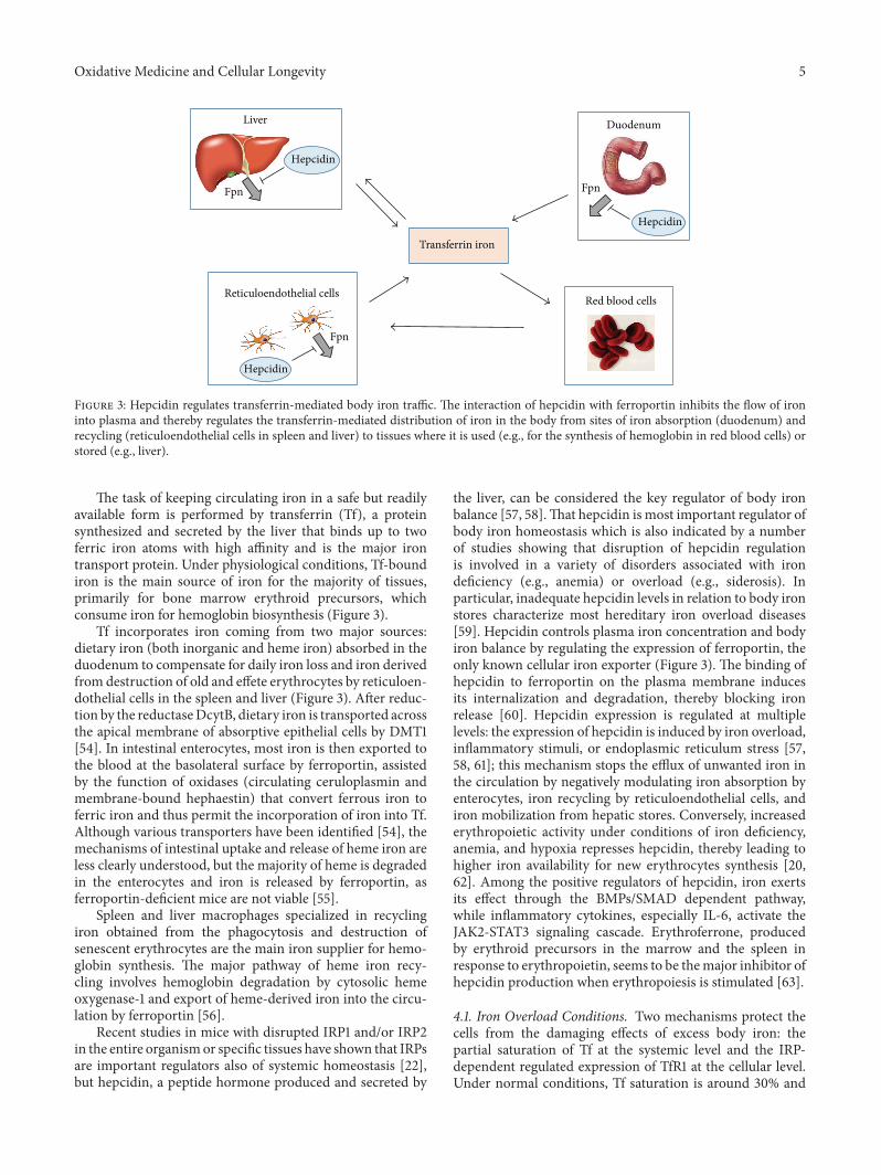
Oxidative Medicine and Cellular Longevity 5
Transferrin iron
Duodenum
Red blood cells
Liver
Hepcidin
Fpn
Reticuloendothelial cells
Hepcidin
Fpn
Fpn
Hepcidin
Figure 3: Hepcidin regulates transferrin-mediated body iron traffic. The interaction of hepcidin with ferroportin inhibits the flow of ironinto plasma and thereby regulates the transferrin-mediated distribution of iron in the body from sites of iron absorption (duodenum) andrecycling (reticuloendothelial cells in spleen and liver) to tissues where it is used (e.g., for the synthesis of hemoglobin in red blood cells) orstored (e.g., liver).
The task of keeping circulating iron in a safe but readilyavailable form is performed by transferrin (Tf), a proteinsynthesized and secreted by the liver that binds up to twoferric iron atoms with high affinity and is the major irontransport protein. Under physiological conditions, Tf-boundiron is the main source of iron for the majority of tissues,primarily for bone marrow erythroid precursors, whichconsume iron for hemoglobin biosynthesis (Figure 3).
Tf incorporates iron coming from two major sources:dietary iron (both inorganic and heme iron) absorbed in theduodenum to compensate for daily iron loss and iron derivedfrom destruction of old and effete erythrocytes by reticuloen-dothelial cells in the spleen and liver (Figure 3). After reduc-tion by the reductaseDcytB, dietary iron is transported acrossthe apical membrane of absorptive epithelial cells by DMT1[54]. In intestinal enterocytes, most iron is then exported tothe blood at the basolateral surface by ferroportin, assistedby the function of oxidases (circulating ceruloplasmin andmembrane-bound hephaestin) that convert ferrous iron toferric iron and thus permit the incorporation of iron into Tf.Although various transporters have been identified [54], themechanisms of intestinal uptake and release of heme iron areless clearly understood, but the majority of heme is degradedin the enterocytes and iron is released by ferroportin, asferroportin-deficient mice are not viable [55].
Spleen and liver macrophages specialized in recyclingiron obtained from the phagocytosis and destruction ofsenescent erythrocytes are the main iron supplier for hemo-globin synthesis. The major pathway of heme iron recy-cling involves hemoglobin degradation by cytosolic hemeoxygenase-1 and export of heme-derived iron into the circu-lation by ferroportin [56].
Recent studies in mice with disrupted IRP1 and/or IRP2in the entire organismor specific tissues have shown that IRPsare important regulators also of systemic homeostasis [22],but hepcidin, a peptide hormone produced and secreted by
the liver, can be considered the key regulator of body ironbalance [57, 58].That hepcidin is most important regulator ofbody iron homeostasis which is also indicated by a numberof studies showing that disruption of hepcidin regulationis involved in a variety of disorders associated with irondeficiency (e.g., anemia) or overload (e.g., siderosis). Inparticular, inadequate hepcidin levels in relation to body ironstores characterize most hereditary iron overload diseases[59]. Hepcidin controls plasma iron concentration and bodyiron balance by regulating the expression of ferroportin, theonly known cellular iron exporter (Figure 3). The binding ofhepcidin to ferroportin on the plasma membrane inducesits internalization and degradation, thereby blocking ironrelease [60]. Hepcidin expression is regulated at multiplelevels: the expression of hepcidin is induced by iron overload,inflammatory stimuli, or endoplasmic reticulum stress [57,58, 61]; this mechanism stops the efflux of unwanted iron inthe circulation by negatively modulating iron absorption byenterocytes, iron recycling by reticuloendothelial cells, andiron mobilization from hepatic stores. Conversely, increasederythropoietic activity under conditions of iron deficiency,anemia, and hypoxia represses hepcidin, thereby leading tohigher iron availability for new erythrocytes synthesis [20,62]. Among the positive regulators of hepcidin, iron exertsits effect through the BMPs/SMAD dependent pathway,while inflammatory cytokines, especially IL-6, activate theJAK2-STAT3 signaling cascade. Erythroferrone, producedby erythroid precursors in the marrow and the spleen inresponse to erythropoietin, seems to be themajor inhibitor ofhepcidin production when erythropoiesis is stimulated [63].
4.1. Iron Overload Conditions. Two mechanisms protect thecells from the damaging effects of excess body iron: thepartial saturation of Tf at the systemic level and the IRP-dependent regulated expression of TfR1 at the cellular level.Under normal conditions, Tf saturation is around 30% and
6 Oxidative Medicine and Cellular Longevity
Tf saturation Tf saturation
HeartJoints
Liver
Tissue damage
NTBI
Normal condition Iron overload
NTBI
NTBI
NTBI
Glands
Figure 4: Mechanisms of iron overload mediated tissue damage. Whenever the iron-binding capacity of transferrin (Tf) is exceeded, non-transferrin-bound iron (NTBI) forms, penetrates into the cells, and undergoes redox cycling, ultimately leading to cell injury and organdamage.
the protein can therefore bind excess iron entering thecirculation, thus functioning as a protective shield againstHO∙ production. However, under conditions of heavy sys-temic iron overload (primarily due to insufficient hepcidinproduction), the iron-binding capacity of Tf is exceeded,andnon-transferrin-bound iron (NTBI) is formed (Figure 4).Despite the downregulation of TfR1, cells become iron-loaded because NTBI, that is, iron associated with variouslow molecular weight plasma components, like citrate, phos-phates, and proteins, lacks a regulated uptake system andhence is able to penetrate into the cells [38]. The consequentdecrease in IRP binding activity leads to efficient ferritinmRNA translation to promote iron storage, but ultimatelythe high capacity of ferritin is exceeded as this unabatedflux of NTBI continues. NTBI is clearly involved in tissuesiderosis, but the incomplete characterization of the ironspecies involved prevents a clear understanding of how itenters the cell under different pathological conditions. NTBIenters into cells mainly through divalent cation transporterssuch asDMT1 andZIP14,which require previous reduction toferrous iron [64, 65] orCa2+ channels, either L-type orT-type,particularly in cardiomyocytes [66]. NTBI toxicity withincells derives from the fact that iron in the expanded LIP isno longer protected from redox cycling; hence, it acceleratesthe catalysis of reactions that produce HO∙, which then causelipid peroxidation and organelle damage and ultimately celldeath (Figure 4). In particular, mitochondria appear to beprimarily affected by iron-mediated HO∙ production. In fact,it has been demonstrated that mitochondria rapidly acquireNTBI, and this, along with their high generation of ROS[67], props up oxidative damage. Apparently, the variety ofenzymatic and nonenzymatic antioxidant defense systems isinadequate to prevent metal-catalyzed oxidative injury onceiron in the LIP is increased [66]. Although production ofhydroxyl radical and lipid peroxidation are important in theinitiation of iron overload pathology, additional mechanismsinvolving apoptosis and fibrosis can account for its complexpathophysiology that leads to organ failure [58].
The most important clinical conditions involving pri-mary and secondary iron overload leading to iron-mediatedtissue damage are genetic hemochromatosis and transfu-sional siderosis, respectively. Hereditary hemochromatosislinked to mutations in HFE, a MHC class I-like proteinthat is a necessary component of the iron-sensing machin-ery controlling hepcidin expression, is the most commongenetic disease in Caucasians and presents a multisysteminvolvement: although iron overload first affects the liver, inhemochromatosis patients endocrine abnormalities, cardiacproblems, and arthropathy are also common [68]. Loss ofHFE function leads to inappropriately low hepcidin produc-tion and unneeded iron release in the bloodstream fromthe duodenum and reticuloendothelial system. ConsequentNTBI formation and iron deposition in parenchymal cellslead to oxidative damage and determine the clinical featuresof hemochromatosis [69].
Secondary iron overload is mainly observed in associa-tion with transfusion-dependent diseases [70]. Since ourbody lacks any regulated mechanism to effectively excreteexcess iron, long-term blood transfusion inescapably resultsin iron overload in patients. Transfusional iron over-load affects particularly patients with inherited hemoglo-binopathies, such as 𝛽 thalassemia, which is the secondaryiron overload condition more closely linked to tissue ironoverload [71, 72]. However, the adverse effects of iron over-load are also found in patients with a variety of conditions(e.g., Blackfan-Diamond anemia, aplastic anemia, sideroblas-tic anemia, myelodysplasia, etc.).
With continued transfusion, reticuloendothelial cells canno longer safely store all the excess iron, which thus entersthe circulation in amounts that exceed the binding capacityof Tf, and NTBI develops [69]. It should be noted that inconditions such as 𝛽 thalassemia hepcidin levels are paradox-ically low because erythropoiesis-dependent downregulationprevails over the upregulation associated with body ironlevels; therefore, high intestinal iron absorption contributesto iron overload [20, 62].While in hemochromatosis patients
Oxidative Medicine and Cellular Longevity 7
excess iron is removed by phlebotomy, the treatment oftransfusional iron overload is mainly based on iron chelationtherapy [61]. Iron chelators can be considered as antioxidants,but not all the possible molecules able to bind iron can beregarded as safe antioxidants. Since all the six iron atoms haveto be bound to the chelator to form a stable complex, incom-plete iron-chelate complexes (e.g., iron-EDTA) can undergoredox cycling and generate harmful free radicals. Therefore,for efficient and safe scavenging of excess iron, chelatingmolecules and/or dosages should be carefully evaluated toraise the thermodynamic stability of the iron-ligand complex.
5. Conclusions
Presently, it is not completely clear as to why in someconditions ROS are associated with cell damage and scav-enging high ROS levels improves metabolic homeostasis,whereas in other settings ROS exert signaling functionsimportant for essential cell activities. Possible explanationsfor these distinct biological specificities of ROS action includedifferences in the amount, sources, duration, and localizationof ROS production; in addition, certainly, the higher ROSreactivity is, the greater toxicity is, while signaling capacityis diminished. Therefore, H
2O2, which is relatively stable
and diffusible, is indeed a mild oxidant but is suitable forsignaling. However, in the presence of ferrous iron, H
2O2
can generate the highly reactive and toxic HO∙. Therefore,we propose that increased availability of iron not bound toproteins specifically evolved to transport or store this essen-tial metal can make the critical difference between the twoopposite functions of ROS. The view that iron plays a mainrole in the scenario leading ROS to become signal or stressagents is supported by the sophisticated systems regulatingintracellular and systemic iron homeostasis that we havesummarized in this review. Additional evidence indicatingthe contribution of iron emerges from the number of studiesshowing the occurrence of ROS-mediated tissue damagewhenever the control of iron metabolism is disrupted, con-ditions of which we selectively highlighted a few illustrativeexamples. Improved understanding of the complex interplaybetween iron metabolism and redox homeostasis will clarifythese pathways and their relevance in pathophysiology. Inconsideration of the disappointing results of antioxidant ther-apy for a variety of diseases [73], targeting iron will possiblyrepresent a more advanced therapeutic approach aimed atpreventing the harmful effects of ROS while permitting theirphysiological role in cell signaling.
Conflict of Interests
The authors declare that there is no conflict of interestsregarding the publication of this paper.
Acknowledgments
This work was supported by grants from Ministero dell’Is-truzione, dell’Università e della Ricerca and Ministero dellaSalute.
References
[1] B. Halliwell and J. M. C. Gutteridge, “Oxygen toxicity, oxygenradicals, transitionmetals anddisease,”Biochemical Journal, vol.219, no. 1, pp. 1–14, 1984.
[2] D. Trachootham, W. Lu, M. A. Ogasawara, N. R.-D. Valle, andP. Huang, “Redox regulation of cell survival,” Antioxidants andRedox Signaling, vol. 10, no. 8, pp. 1343–1374, 2008.
[3] T. Finkel, “Signal transduction by mitochondrial oxidants,”TheJournal of Biological Chemistry, vol. 287, no. 7, pp. 4434–4440,2012.
[4] G. Shadel and T. Horvath, “Mitochondrial ROS signaling inorganismal homeostasis,”Cell, vol. 163, no. 3, pp. 560–569, 2015.
[5] B. Harwell, “Biochemistry of oxidative stress,” BiochemicalSociety Transactions, vol. 35, no. 5, pp. 1147–1150, 2007.
[6] S. J. Dixon, K.M. Lemberg,M. R. Lamprecht et al., “Ferroptosis:an iron-dependent form of nonapoptotic cell death,” Cell, vol.149, no. 5, pp. 1060–1072, 2012.
[7] S. J. Dixon, D. Patel, M. Welsch et al., “Pharmacologicalinhibition of cystine-glutamate exchange induces endoplasmicreticulum stress and ferroptosis,” eLife, vol. 2014, no. 3, 2014.
[8] J. P. Friedmann Angeli, M. Schneider, B. Proneth et al., “Inac-tivation of the ferroptosis regulator Gpx4 triggers acute renalfailure inmice,”Nature Cell Biology, vol. 16, no. 12, pp. 1180–1191,2014.
[9] M. Gao, P. Monian, N. Quadri, R. Ramasamy, and X. Jiang,“Glutaminolysis and transferrin regulate ferroptosis,”MolecularCell, vol. 59, no. 2, pp. 298–308, 2015.
[10] X. Sun, Z. Ou, R. Chen et al., “Activation of the p62-Keap1-NRF2 pathway protects against ferroptosis in hepatocellularcarcinoma cells,” Hepatology, vol. 63, no. 1, pp. 173–184, 2016.
[11] G. Cairo, F. Bernuzzi, and S. Recalcati, “A precious metal: iron,an essential nutrient for all cells,” Genes & Nutrition, vol. 1, no.1, pp. 25–39, 2006.
[12] A. D. Sheftel, A. B. Mason, and P. Ponka, “The long history ofiron in the Universe and in health and disease,” Biochimica etBiophysica Acta (BBA)—General Subjects, vol. 1820, no. 3, pp.161–187, 2012.
[13] N. C. Andrews, “Forging a field: the golden age of iron biology,”Blood, vol. 112, no. 2, pp. 219–230, 2008.
[14] M.W.Hentze,M.U.Muckenthaler, B.Galy, andC.Camaschella,“Two to tango: regulation of Mammalian iron metabolism,”Cell, vol. 142, no. 1, pp. 24–38, 2010.
[15] G. Cairo and S. Recalcati, “Iron-regulatory proteins: molecularbiology and pathophysiological implications,”Expert Reviews inMolecular Medicine, vol. 9, no. 33, pp. 1–13, 2007.
[16] S. Recalcati, G.Minotti, and G. Cairo, “Iron regulatory proteins:from molecular mechanisms to drug development,” Antioxi-dants and Redox Signaling, vol. 13, no. 10, pp. 1593–1616, 2010.
[17] R. C. Hider and X. Kong, “Iron speciation in the cytosol: anoverview,” Dalton Transactions, vol. 42, no. 9, pp. 3220–3229,2013.
[18] T. Moroishi, M. Nishiyama, Y. Takeda, K. Iwai, and K. I.Nakayama, “The FBXL5-IRP2 axis is integral to control of ironmetabolism in vivo,” Cell Metabolism, vol. 14, no. 3, pp. 339–351,2011.
[19] J. C. Ruiz and R. K. Bruick, “F-box and leucine-rich repeatprotein 5 (FBXL5): sensing intracellular iron and oxygen,”Journal of Inorganic Biochemistry, vol. 133, pp. 73–77, 2014.
[20] S. Recalcati, E. Gammella, and G. Cairo, “New perspectiveson the molecular basis of the interaction between oxygen
8 Oxidative Medicine and Cellular Longevity
homeostasis and iron metabolism,” Hypoxia, vol. 3, pp. 93–103,2015.
[21] M. U. Muckenthaler, B. Galy, and M.W. Hentze, “Systemic ironhomeostasis and the iron-responsive element/iron-regulatoryprotein (IRE/IRP) regulatory network,”Annual Review of Nutri-tion, vol. 28, pp. 197–213, 2008.
[22] N.Wilkinson andK. Pantopoulos, “The IRP/IRE system in vivo:insights frommouse models,” Frontiers in Pharmacology, vol. 5,article 176, 2014.
[23] S. Recalcati, A. Alberghini, A. Campanella et al., “Iron regula-tory proteins 1 and 2 in human monocytes, macrophages andduodenum: expression and regulation in hereditary hemochro-matosis and iron deficiency,” Haematologica, vol. 91, no. 3, pp.303–310, 2006.
[24] D.-L. Zhang,M.C.Ghosh, andT.A. Rouault, “Thephysiologicalfunctions of iron regulatory proteins in iron homeostasis—anupdate,” Frontiers in Pharmacology, vol. 5, article 124, 2014.
[25] G. Cairo, S. Recalcati, A. Pietrangelo, and G. Minotti, “Theiron regulatory proteins: targets and modulators of free radicalreactions and oxidative damage,” Free Radical Biology andMedicine, vol. 32, no. 12, pp. 1237–1243, 2002.
[26] P. Arosio, F. Carmona, R. Gozzelino, F. Maccarinelli, and M.Poli, “The importance of eukaryotic ferritins in iron handlingand cytoprotection,” Biochemical Journal, vol. 472, no. 1, pp. 1–15, 2015.
[27] C. P. Anderson, M. Shen, R. S. Eisenstein, and E. A. Leibold,“Mammalian ironmetabolism and its control by iron regulatoryproteins,” Biochimica et Biophysica Acta (BBA)—Molecular CellResearch, vol. 1823, no. 9, pp. 1468–1483, 2012.
[28] K. Pantopoulos, S. K. Porwal, A. Tartakoff, and L. Devireddy,“Mechanisms of mammalian iron homeostasis,” Biochemistry,vol. 51, no. 29, pp. 5705–5724, 2012.
[29] L. C. Kühn, “Iron regulatory proteins and their role in control-ling iron metabolism,” Metallomics, vol. 7, no. 2, pp. 232–243,2015.
[30] T. A. Rouault, “Iron metabolism in the CNS: implications forneurodegenerative diseases,” Nature Reviews Neuroscience, vol.14, no. 8, pp. 551–564, 2013.
[31] G. Isaya, “Mitochondrial iron-sulfur cluster dysfunction inneurodegenerative disease,” Frontiers in Pharmacology, vol. 5,article 29, 2014.
[32] L. Zecca, M. B. H. Youdim, P. Riederer, J. R. Connor, and R. R.Crichton, “Iron, brain ageing and neurodegenerative disorders,”Nature Reviews Neuroscience, vol. 5, no. 11, pp. 863–873, 2004.
[33] A. Martelli and H. Puccio, “Dysregulation of cellular ironmetabolism in Friedreich ataxia: from primary iron-sulfurcluster deficit to mitochondrial iron accumulation,” Frontiers inPharmacology, vol. 5, article 130, 2014.
[34] V. Campuzano, L. Montermini, M. D. Moltò et al., “Friedreich’sataxia: autosomal recessive disease caused by an intronic GAAtriplet repeat expansion,” Science, vol. 271, no. 5254, pp. 1423–1427, 1996.
[35] R. A. Vaubel and G. Isaya, “Iron-sulfur cluster synthesis, ironhomeostasis and oxidative stress in Friedreich ataxia,”Molecularand Cellular Neuroscience, vol. 55, pp. 50–61, 2013.
[36] P. Arosio and S. Levi, “Cytosolic and mitochondrial ferritins inthe regulation of cellular iron homeostasis and oxidative dam-age,” Biochimica et Biophysica Acta (BBA)—General Subjects,vol. 1800, no. 8, pp. 783–792, 2010.
[37] A. Campanella, G. Isaya, H. A. O’Neill et al., “The expression ofhuman mitochondrial ferritin rescues respiratory function in
frataxin-deficient yeast,”HumanMolecular Genetics, vol. 13, no.19, pp. 2279–2288, 2004.
[38] Z. I. Cabantchik, “Labile iron in cells and body fluids: physiol-ogy, pathology, and pharmacology,” Frontiers in Pharmacology,vol. 5, article 45, 2014.
[39] G. Minotti, P. Menna, E. Salvatorelli, G. Cairo, and L. Gianni,“Anthracyclines: molecular advances and pharmacologic devel-opments in antitumor activity and cardiotoxicity,” Pharmaco-logical Reviews, vol. 56, no. 2, pp. 185–229, 2004.
[40] P. Menna, S. Recalcati, G. Cairo, and G. Minotti, “An introduc-tion to the metabolic determinants of anthracycline cardiotox-icity,” Cardiovascular Toxicology, vol. 7, no. 2, pp. 80–85, 2007.
[41] E. Gammella, S. Recalcati, I. Rybinska, P. Buratti, and G.Cairo, “Iron-induced damage in cardiomyopathy: oxidative-dependent and independent mechanisms,” Oxidative Medicineand Cellular Longevity, vol. 2015, Article ID 230182, 10 pages,2015.
[42] G. Minotti, G. Cairo, and E. Monti, “Role of iron in anthra-cycline cardiotoxicity: new tunes for an old song?” The FASEBJournal, vol. 13, no. 2, pp. 199–212, 1999.
[43] M. Štěrba, O. Popelová, A. Vávrová et al., “Oxidative stress,redox signaling, andmetal chelation in anthracycline cardiotox-icity and pharmacological cardioprotection,” Antioxidants andRedox Signaling, vol. 18, no. 8, pp. 899–929, 2013.
[44] F. Bernuzzi, S. Recalcati, A. Alberghini, and G. Cairo, “Reac-tive oxygen species-independent apoptosis in doxorubicin-treatedH9c2 cardiomyocytes: role for heme oxygenase-1 down-modulation,” Chemico-Biological Interactions, vol. 177, no. 1, pp.12–20, 2009.
[45] R. D. Spagnuolo, S. Recalcati, L. Tacchini, and G. Cairo, “Roleof hypoxia-inducible factors in the dexrazoxane-mediated pro-tection of cardiomyocytes from doxorubicin-induced toxicity,”British Journal of Pharmacology, vol. 163, no. 2, pp. 299–312, 2011.
[46] G. Minotti, R. Ronchi, E. Salvatorelli, P. Menna, and G. Cairo,“Doxorubicin irreversibly inactivates iron regulatory proteins1 and 2 in cardiomyocytes: evidence for distinct metabolicpathways and implications for iron-mediated cardiotoxicity ofantitumor therapy,” Cancer Research, vol. 61, no. 23, pp. 8422–8428, 2001.
[47] G. Corna, P. Santambrogio, G. Minotti, and G. Cairo, “Dox-orubicin paradoxically protects cardiomyocytes against iron-mediated toxicity: role of reactive oxygen species and ferritin,”The Journal of Biological Chemistry, vol. 279, no. 14, pp. 13738–13745, 2004.
[48] G. Corna, B. Galy, M. W. Hentze, and G. Cairo, “IRP1-independent alterations of cardiac iron metabolism indoxorubicin-treated mice,” Journal of Molecular Medicine, vol.84, no. 7, pp. 551–560, 2006.
[49] M. C. Asensio-López, J. Sánchez-Más, D. A. Pascual-Figal etal., “Involvement of ferritin heavy chain in the preventive effectof metformin against doxorubicin-induced cardiotoxicity,” FreeRadical Biology and Medicine, vol. 57, pp. 188–200, 2013.
[50] J. C. Kwok and D. R. Richardson, “Examination of the mech-anism(s) involved in doxorubicin-mediated iron accumulationin ferritin: studies using metabolic inhibitors, protein synthesisinhibitors, and lysosomotropic agents,” Molecular Pharmacol-ogy, vol. 65, no. 1, pp. 181–195, 2004.
[51] J. Drysdale, P. Arosio, R. Invernizzi et al., “Mitochondrialferritin: a new player in ironmetabolism,”Blood Cells,Molecules& Diseases, vol. 29, no. 3, pp. 376–383, 2002.
Oxidative Medicine and Cellular Longevity 9
[52] Y. Ichikawa, M. Ghanefar, M. Bayeva et al., “Cardiotoxicity ofdoxorubicin is mediated through mitochondrial iron accumu-lation,” The Journal of Clinical Investigation, vol. 124, no. 2, pp.617–630, 2014.
[53] F. Maccarinelli, E. Gammella, M. Asperti et al., “Mice lack-ing mitochondrial ferritin are more sensitive to doxorubicin-mediated cardiotoxicity,” Journal of Molecular Medicine, vol. 92,no. 8, pp. 859–869, 2014.
[54] S. Gulec, G. J. Anderson, and J. F. Collins, “Mechanistic andregulatory aspects of intestinal iron absorption,”The AmericanJournal of Physiology—Gastrointestinal and Liver Physiology,vol. 307, no. 4, pp. G397–G409, 2014.
[55] J. E. Levy, O. Jin, Y. Fujiwara, F. Kuo, andN. C. Andrews, “Trans-ferrin receptor is necessary for development of erythrocytes andthe nervous system,”Nature Genetics, vol. 21, no. 4, pp. 396–399,1999.
[56] E. Gammella, F. Maccarinelli, P. Buratti, S. Recalcati, andG. Cairo, “The role of iron in anthracycline cardiotoxicity,”Frontiers in Pharmacology, vol. 5, article 25, 2014.
[57] T.Ganz, “Systemic iron homeostasis,”Physiological Reviews, vol.93, no. 4, pp. 1721–1741, 2013.
[58] A. Pietrangelo, “Pathogens, metabolic adaptation, and humandiseases—an iron-thrifty genetic model,” Gastroenterology, vol.149, no. 4, pp. 834–838, 2015.
[59] A. Pietrangelo, “Hepcidin in human iron disorders: therapeuticimplications,” Journal of Hepatology, vol. 54, no. 1, pp. 173–181,2011.
[60] E. Nemeth, M. S. Tuttle, J. Powelson et al., “Hepcidin regulatescellular iron efflux by binding to ferroportin and inducing itsinternalization,” Science, vol. 306, no. 5704, pp. 2090–2093,2004.
[61] C. Camaschella, “Iron-deficiency anemia,” The New EnglandJournal of Medicine, vol. 373, no. 5, pp. 485–486, 2015.
[62] A. Kim and E. Nemeth, “New insights into iron regulation anderythropoiesis,” Current Opinion in Hematology, vol. 22, no. 3,pp. 199–205, 2015.
[63] L. Kautz, G. Jung, E. V. Valore, S. Rivella, E. Nemeth, andT. Ganz, “Identification of erythroferrone as an erythroidregulator of iron metabolism,” Nature Genetics, vol. 46, no. 7,pp. 678–684, 2014.
[64] J. P. Liuzzi, F. Aydemir, H. Nam, M. D. Knutson, and R.J. Cousins, “Zip14 (Slc39a14) mediates non-transferrin-boundiron uptake into cells,” Proceedings of the National Academy ofSciences of the United States of America, vol. 103, no. 37, pp.13612–13617, 2006.
[65] S. Ludwiczek, I. Theurl, M. U. Muckenthaler et al., “Ca2+channel blockers reverse iron overload by a newmechanism viadivalent metal transporter-1,”NatureMedicine, vol. 13, no. 4, pp.448–454, 2007.
[66] G. Y. Oudit, H. Sun, M. G. Trivieri et al., “L-type Ca2+ channelsprovide a major pathway for iron entry into cardiomyocytes iniron-overload cardiomyopathy,” Nature Medicine, vol. 9, no. 9,pp. 1187–1194, 2003.
[67] P. Venditti, L. Di Stefano, and S. Di Meo, “Mitochondrialmetabolism of reactive oxygen species,”Mitochondrion, vol. 13,no. 2, pp. 71–82, 2013.
[68] A. Pietrangelo, “Hereditary hemochromatosis: pathogenesis,diagnosis, and treatment,” Gastroenterology, vol. 139, no. 2, pp.393.e1–408.e2, 2010.
[69] G. Sebastiani and K. Pantopoulos, “Disorders associated withsystemic or local iron overload: from pathophysiology toclinical practice,”Metallomics, vol. 3, no. 10, pp. 971–986, 2011.
[70] R. E. Fleming and P. Ponka, “Iron overload in human disease,”The New England Journal of Medicine, vol. 366, no. 4, pp. 348–359, 2012.
[71] J. B. Porter and M. Garbowski, “The pathophysiology of trans-fusional iron overload,” Hematology/Oncology Clinics of NorthAmerica, vol. 28, no. 4, pp. 683–701, 2014.
[72] V. Berdoukas, T. D. Coates, and Z. I. Cabantchik, “Iron andoxidative stress in cardiomyopathy in thalassemia,” Free RadicalBiology and Medicine, vol. 88, pp. 3–9, 2015.
[73] J. M. C. Gutteridge and B. Halliwell, “Antioxidants: molecules,medicines, and myths,” Biochemical and Biophysical ResearchCommunications, vol. 393, no. 4, pp. 561–564, 2010.
Research ArticleLung Neutrophilia in Myeloperoxidase Deficient Mice duringthe Course of Acute Pulmonary Inflammation
Silvie Kremserova,1,2,3 Tomas Perecko,1,3 Karel Soucek,3,4 Anna Klinke,3,5
Stephan Baldus,5 Jason P. Eiserich,6 and Lukas Kubala1,3
1Department of Free Radical Pathophysiology, Institute of Biophysics, Academy of Sciences of the Czech Republic,61265 Brno, Czech Republic2Department of Experimental Biology, Faculty of Science, Masaryk University, 61137 Brno, Czech Republic3International Clinical Research Center, Center of Biomolecular and Cellular Engineering, St. Anne’s University Hospital Brno,65691 Brno, Czech Republic4Department of Cytokinetics, Institute of Biophysics, Academy of Sciences of the Czech Republic, 61265 Brno, Czech Republic5Heart Center, University of Cologne, 50937 Cologne, Germany6Department of Internal Medicine, School of Medicine, University of California, Davis, CA 95616, USA
Correspondence should be addressed to Lukas Kubala; [email protected]
Received 25 September 2015; Revised 11 January 2016; Accepted 14 January 2016
Academic Editor: Victor M. Victor
Copyright © 2016 Silvie Kremserova et al. This is an open access article distributed under the Creative Commons AttributionLicense, which permits unrestricted use, distribution, and reproduction in any medium, provided the original work is properlycited.
Systemic inflammation accompanying diseases such as sepsis affects primarily lungs and induces their failure. This remains themost common cause of sepsis induced mortality. While neutrophils play a key role in pulmonary failure, the mechanisms remainincompletely characterized. We report that myeloperoxidase (MPO), abundant enzyme in neutrophil granules, modulates thecourse of acute pulmonary inflammatory responses induced by intranasal application of lipopolysaccharide. MPO deficient micehad significantly increased numbers of airway infiltrated neutrophils compared to wild-type mice during the whole course of lunginflammation. This was accompanied by higher levels of RANTES in bronchoalveolar lavage fluid from the MPO deficient mice.Other markers of lung injury and inflammation, which contribute to recruitment of neutrophils into the inflamed lungs, includingtotal protein and other selected proinflammatory cytokines did not significantly differ in bronchoalveolar lavage fluid from thewild-type and the MPO deficient mice. Interestingly, MPO deficient neutrophils revealed a decreased rate of cell death characterized byphosphatidylserine surface expression. Collectively, the importance ofMPO in regulation of pulmonary inflammation, independentof its putative microbicidal functions, can be potentially linked toMPO ability to modulate the life span of neutrophils and to affectaccumulation of chemotactic factors at the inflammatory site.
1. Introduction
Inflammation is protective and vital to health, but when acuteinflammation is unrestrained in amplitude or duration, itcan lead to disease [1]. In most instances, the molecular andcellular events during acute inflammation are successful inlimiting the inciting injury or infection and restoring tissuehomeostasis. However, overwhelming inflammation can leadto fatal consequences. The lungs are generally the first toundergo failure, and this remains the most common cause ofacute life-threatening pathologies such as septic shock and/or
multiple organ failure induced mortality [2, 3]. Althoughacute inflammation is generally self-limited, alternate fatesinclude abscess formation, fibrosis, or conversion to chronicinflammation [1].
While it has become clear that neutrophil granulocytes,the most abundant subpopulation of polymorphonucleargranulocytes, play a key role in pulmonary failure duringsepsis and development of chronic inflammation, the mecha-nisms remain incompletely characterized [2, 4].The progres-sion and control of inflammation are significantly affected bymediators produced by leukocytes accumulated at the site of
Hindawi Publishing CorporationOxidative Medicine and Cellular LongevityVolume 2016, Article ID 5219056, 13 pageshttp://dx.doi.org/10.1155/2016/5219056
2 Oxidative Medicine and Cellular Longevity
inflammation [1]. One of them is myeloperoxidase (MPO),an abundant hemoprotein of neutrophils, which is typicallyperceived to primarily mediate host defense reactions [5–7].However, there is increasing evidence showing a novel regu-latory role of MPO not directly related to host defense. MPOaccumulated in the lungs could significantlymodulate redox-sensitive cellular signaling pathways controlling inflamma-tory processes among others through the catabolism of nitricoxide (NO), induction of wide range of posttranslationalmodification of proteins, and modulation of metabolism ofarachidonic and linoleic acid derived mediators [5–10].
However, the importance of MPO in various inflamma-tory conditions is questioned and is highly dependent on thetype of the inflammatory model. Consistent with microbi-cidal function of MPO, the MPO deficient mice were morelikely than thewild-typemice to be infected or die from infec-tion employing various models, suggesting that the MPO-dependent oxidative system is important for host defenseagainst fungi and bacteria [5, 6, 9]. However, theMPO impor-tance varies by pathogen species [11]. In contrast, in the casesof inflammatory response to noninfectious stimuli or chronicinflammation in the absence of live pathogens, MPO candamage host tissue through the generation of oxidants. Thishas been observed in many chronic inflammatory diseases,including cardiovascular diseases, rheumatoid arthritis, andkidney diseases (see reviews [5–7, 9]). Thus, based on theassumption of detrimental effect of MPO during chronicinflammation, it can be expected that the inflammationshould actually be reduced in MPO deficient mice. On thecontrary, experimental data from various studies employingMPO deficient mice suggest that the MPO deficiency isconnected with dysregulated inflammatory response. Thesestudies show significant adverse effects of MPO deficiencyin various inflammatory models induced by noninfectiousstimuli including nonviable Candida albicans [12], experi-mental autoimmune encephalomyelitis [13], ischemic braininjury [14], lung dysfunction after allogeneic bone marrowtransplantation [15], ultraviolet- (UV-) exposed skin inflam-mation [16], atherosclerotic lesions development [17], skindelayed-type hypersensitivity and antigen-induced arthritis[18], and different types of autoimmune renal diseases [19,20]. These data support the suggestion of a protective, anti-inflammatory role of MPO in pathologies characterized bycomplex inflammatory response in the absence of infectiousagent.
Here, a role of MPO in the regulation of acute lunginflammation and injury was evaluated. Model of acuteairway inflammation induced by intranasal administrationof lipopolysaccharide (LPS) was employed in both the wild-type and the MPO deficient mice. Various markers ofinflammation and injury in lung tissues or lung lavage fluidswere determined. Such LPS instillation is known to cause anacute inflammatory response with transient extravasation ofprimarily neutrophils in the airways [21–23]. The temporalcourse of inflammation was evaluated at different time points(8 h, 24 h, 48 h, and 72 h) following application of LPS. Ourresults indicate that MPO deficiency enhances neutrophiliaduring LPS-induced airway inflammation due to altered
accumulation of proinflammatory cytokine RANTES andreduced cell death of MPO deficient neutrophils.
2. Materials and Methods
2.1. Animal Exposure to LPS. Male C57BL/6J wild-type andMPO deficient mice MPO tm1lus (MPO deficient) (TheJackson Laboratory, USA) [8] both of age 12–16 weeks andof weight 25–30 g were subjected to brief anesthesia withketamine-xylazine, and 50 𝜇L of LPS (from Escherichia coliserotype 055:B5, Sigma-Aldrich,USA) solution in phosphate-buffered saline (PBS) was instilled directly into their nostrilsto reach a dose of 0.3mg/kg LPS as described previously[23]. Previous studies demonstrated that a significant fractionof intranasally administered LPS will reach the lungs andthat such instillation evokes an acute transient inflammatoryresponse [21–23].Mice in the control group received a similarvolume of sterile PBS.The experiments were approved by theAnimal Care Committee and were in accordance with the EUand NIH Guide for Care and Use of Laboratory Animals.
2.2. Collection of Bronchoalveolar Lavage Fluid (BALF). Atvarious time points after LPS instillation, mice were deeplyanesthetized by intraperitoneal administration of ketamine-xylazine and blood was collected from the heart into hep-arinized syringes. The tracheae were cannulated, and thelungs were lavaged with 2 consecutive washes with 1mL ofPBS, which were pooled to a total recovered BALF of 1.6–1.8mL.The BALF was used for the cell analysis after pelleting(5min, 250 g, 4∘C) or the cell-free BALF was obtained aftercentrifugation (4min, 2 000 g, 4∘C) and stored at −80∘C untilfurther analysis.
2.3. Determination of Inflammatory Cells in BALF and TheirDifferentiation Count. Total cell count in the collected lavagesamples and total leukocyte count in blood were determinedwith a Coultre Counter Z1 after lysis of RBCs by Zap-Oglobin II lytic reagent (Coulter, USA). Cell differentialcounts were determined on blood smears or on slides aftercytospin centrifugation stained with Giemsa stain (Diff-Quick, ThermoFisher Scientific, USA).
2.4. Inflammatory Cytokines and Total Proteins in BALF. Toquantitate proinflammatory cytokine production in the lungtumor necrosis factor- (TNF-) 𝛼, monocyte chemoattractantprotein- (MCP-) 1, Regulated on Activation, Normal T CellExpressed and Secreted (RANTES), interleukin- (IL-) 6, IL-10, and IL-12 were determined in BALF by sandwich enzyme-linked immunosorbent assay (ELISA) using murine-specificcommercial kits (Quantikine Mouse cytokine assay kit, R&DSystems, USA).
As a marker of epithelial injury and lung permeability,total protein concentration within BALF was measured witha Protein Assay Kit (Bio-Rad, USA) based on the method ofBradford, with bovine serum albumin as a standard.
2.5. Analysis of NO Production in Lungs. To quantitate theproduction of NO in the lungs, its metabolites nitrates/nitrites were measured in BALF by chemical reduction of
Oxidative Medicine and Cellular Longevity 3
nitrates and nitrites to NO in 120mM vanadium (III) in 2MHCl at 95∘C and analysis by ozone-enhanced chemilumines-cence (ANTEK Instruments Inc., Model 7020, USA).
2.6. Expression of CD11b on Neutrophil Surface. Cells werewashed with PBS (5min, 250 g, 4∘C) and incubated withfluorescein-conjugated anti-mouse CD11b (Invitrogen, USA)antibodies at 4∘C in the dark for 30min, consequentlywashed with PBS (5min, 250 g, 4∘C), and analyzed usinga BD FACSVerse™ flow cytometer (BD Biosciences, USA)[24]. Cells incubated with appropriate isotype control wereused to determine the nonspecific background signal.The data analysis was performed using Flowing Software(http://www.flowingsoftware.com).
2.7. Histological Analysis of Lungs. Following euthanasia, thelungs were instilled with OCT Compound (Sakura, TheNetherlands). The lungs were subsequently removed andplaced into OCT Compound and immediately frozen inliquid nitrogen. 7 𝜇m sections were stained with H&E stainand histological analysis was performed by light microscopy(10x objective). Series of pictures were obtained from thedifferent types of samples that were evaluated and typicalexamples are represented.
2.8. Analysis of MPO Enzymatic Activity. Isolated cells werewashed with PBS (5min, 250 g, 4∘C) and the remainingerythrocytes were lysed by hypotonic lysis using distilledwater for 5 seconds. Cells were washed with PBS (5min,250 g, 4∘C) and subsequently lysed in PBS with 0.1% TritonX-100 (Sigma-Aldrich, USA). Peroxidase activity was mea-sured as the oxidation of tetramethylbenzidine (TMB, 2mM,Sigma-Aldrich, USA) in 300mM sodium acetate buffer(pH 5.4) in the presence of 0.075mM hydrogen peroxide(H2O2) (Sigma-Aldrich, USA) within 20min as described
previously [24]. The formation of the reaction productwas determined spectrophotometrically as the increase inabsorption at 350 nm using an Infinite M200 microplatespectrofluorometer (Tecan, Switzerland).
2.9. Caspase 3 Activity Assay. Caspase 3 activity was analyzedas described previously [25]. Isolated cells were washed twicewith PBS (5min, 250 g, 4∘C) and lysed (50mM HEPES;5mM CHAPS; 5mM DTT) on ice for 20min and cen-trifuged (15min, 15 000 g, 4∘C). Lavage fluid was diluted1 : 1 with 2x concentrated lysing buffer. The proteins presentin supernatants were quantified using Coomassie® ProteinAssay (Bio-Rad, USA) and diluted to an equal concentration.5 𝜇g of protein samples was incubated in an assay bufferin parallels (20mM HEPES; 2.5mM CHAPS, 5mM DTT,2mM EDTA) containing 50𝜇M of caspase 3 (Ac-DEVD-AMC) substrate (Sigma-Aldrich, USA) at 37∘C for 4 h. Thelevel of fluorescence was determined using microplate reader(Infinite 200, Tecan, Switzerland; 360 nm excitation, 460 nmemission).
2.10. Phosphatidylserine (PS) Externalization (AnnexinV/Propidium Iodide Assay). The presence of cells with
permeable membrane (dead cells) and cells with surfaceexpression of PS was evaluated by flow cytometry usingpropidium iodide (PI) and Annexin V staining, respectively[26]. Cells were washed with PBS (5min, 250 g, roomtemperature), resuspended in 100𝜇L Annexin V bindingbuffer (10mMHEPES/NaOH, pH 7.4, 140mMNaCl, 2.5mMCaCl2), and incubated for 15min with 1 𝜇L Annexin V-FITC
(Apronex Ltd., Czech Republic). PI (1 𝜇g/mL) was added1min before analysis. Flow cytometric analysis of the stainedcells was performed using a BD FACSVerse flow cytometer(BD Biosciences, USA). The cell population for the analysiswas gated using forward- versus side-scatter parameters toexclude any debris. Per sample, 10 000 cells were collected.Three different populations can be identified using this assay:intact viable cells are negative for both PI and AnnexinV-FITC, apoptotic cells are positive for Annexin V-FITC butnegative for PI, and dead cells positive for both AnnexinV-FITC and PI. The data analysis was performed usingFlowing Software (http://www.flowingsoftware.com).
2.11. DNA Fragmentation. 3 ∗ 106 cells isolated from BALFwere washed with PBS (5min, 250 g, 4∘C) and DNA wasisolated using the Invisorb Apoptosis Detection Kit II(Invitek; Invitek GmbH, Germany). Gel electrophoresis wasperformed in 1.5% agarose (Sigma-Aldrich, USA), using 1 kbpDNA ladder as a marker (ThermoFisher Scientific, USA).DNA was stained with ethidium bromide (Sigma-Aldrich,USA) and scanned on UV-transilluminator using the ScionImage software (Scion Corporation, USA).
2.12. Statistical Analysis. Statistical comparisons were ana-lyzed with the Student 𝑡-test for pairwise-dependent samples.A 𝑝 value equal to or lower than 0.05 was consideredstatistically significant. Data are presented as the means ±standard error of the mean (SEM). All statistical analyseswere carried out with Statistica 10 (StatSoft, USA).
3. Results
3.1. The LPS-Induced Airway Neutrophilia. Intranasal instil-lation of LPS (0.3mg/kg) in the wild-type and the MPOdeficient mice induced acute airway inflammation, charac-terized by an increase of the lung lavage cell numbers. Thisaccumulation of cells in lungs was maximal 48 h after LPSinstillation and declined thereafter (Figure 1(a)). As expected,while nucleated cells in BALF of untreated mice almostexclusively represented alveolar macrophages (Figure 1(b)),increased total numbers of nucleated cells in lung lavageof both wild-type and MPO deficient mice were almostexclusively due to neutrophils (Figure 1(c)) as assessed byWright-Giemsa stain of cytospin samples. The number oflymphocytes, basophils, eosinophils, or other undeterminednucleated cells in BALF did not exceed 4% at any time point(data not shown). Interestingly, in contrast to expectations,the total numbers of nucleated cells in lavage were signifi-cantly lower in the wild-type mice compared to the MPOdeficient mice at last two evaluated time points 48 h and 72 hafter LPS instillation (Figure 1(a)). This was solely due to
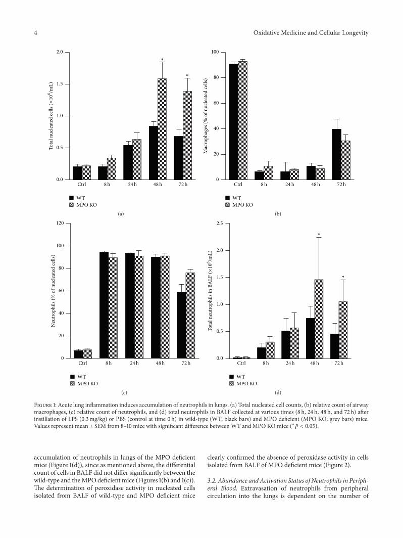
4 Oxidative Medicine and Cellular Longevity
∗
∗
Ctrl 8h 24h 48h 72h
WTMPO KO
0.0
0.5
1.0
1.5
2.0To
tal n
ucle
ated
cells
(×10
6/m
L)
(a)
Ctrl 8h 24h 48h 72h
WTMPO KO
0
20
40
60
80
100
Mac
roph
ages
(% o
f nuc
leat
ed ce
lls)
(b)
Ctrl 8h 24h 48h 72h
WTMPO KO
0
20
40
60
80
100
120
Neu
troph
ils (%
of n
ucle
ated
cells
)
(c)
Tota
l neu
troph
ils in
BA
LF (×
106/m
L)
∗
∗
Ctrl 8h 24h 48h 72h
WTMPO KO
0.0
0.5
1.0
1.5
2.0
2.5
(d)
Figure 1: Acute lung inflammation induces accumulation of neutrophils in lungs. (a) Total nucleated cell counts, (b) relative count of airwaymacrophages, (c) relative count of neutrophils, and (d) total neutrophils in BALF collected at various times (8 h, 24 h, 48 h, and 72 h) afterinstillation of LPS (0.3mg/kg) or PBS (control at time 0 h) in wild-type (WT; black bars) and MPO deficient (MPO KO; grey bars) mice.Values represent mean ± SEM from 8–10 mice with significant difference between WT and MPO KOmice (∗𝑝 < 0.05).
accumulation of neutrophils in lungs of the MPO deficientmice (Figure 1(d)), since as mentioned above, the differentialcount of cells in BALF did not differ significantly between thewild-type and theMPOdeficientmice (Figures 1(b) and 1(c)).The determination of peroxidase activity in nucleated cellsisolated from BALF of wild-type and MPO deficient mice
clearly confirmed the absence of peroxidase activity in cellsisolated from BALF of MPO deficient mice (Figure 2).
3.2. Abundance andActivation Status of Neutrophils in Periph-eral Blood. Extravasation of neutrophils from peripheralcirculation into the lungs is dependent on the number of
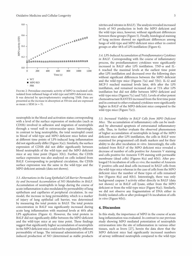
Oxidative Medicine and Cellular Longevity 5
200 400 600 800 1000 1200
Time (s)
WTMPO KOPBS
0
0.0
0.2
0.4
0.6
0.8
MPO
activ
ity (O
D)
Figure 2: Peroxidase enzymatic activity of MPO in nucleated cellsisolated from inflamed lungs of wild-type and MPO deficient mice.It was detected by spectrophotometry employing TMB. Data arepresented as the increase in absorption at 350 nm and are expressedas mean ± SEM (𝑛 = 5).
neutrophils in the blood and activation status correspondingwith a level of the surface expression of molecules (such asCD11b) involved in adhesion and migration of neutrophilsthrough a vessel wall to extravascular space. Interestingly,in contrast to lung neutrophilia, the total neutrophil countin blood of wild-type and MPO deficient mice before andat different time points of LPS-induced lung inflammationdid not significantly differ (Figure 3(a)). Similarly, the surfaceexpression of CD11b did not differ significantly betweenblood neutrophils of the wild-type and the MPO deficientmice at any time point (Figure 3(b)). Further, the CD11bsurface expression was also analyzed on cells isolated fromBALF. Corresponding to peripheral circulation, the CD11bsurface expression was the same in the wild-type and theMPO deficient animals (data not shown).
3.3. Alternations in the Lung Epithelial Cell Barrier Permeabil-ity and Increased Accumulation of NO Metabolites in BALF.Accumulation of neutrophils in lungs during the course ofacute inflammation is alsomodulated by permeability of lungepithelium and capillaries of peripheral circulation. In ourmodel, the increase in lung permeability, which is a markerof injury of lung epithelial cell barrier, was determinedby measuring the total protein in BALF. The total proteinconcentration in BALF was significantly increased duringacute lung inflammation with maximal levels at 48 h afterLPS application (Figure 4). However, the total protein inBALF did not significantly differ between the MPO deficientand the wild-type mice at any time point. These findingssuggest that significantly higher accumulation of neutrophilsin theMPOdeficientmice could not be explained by differentpermeability of lungs. The intranasal administration of LPSinduced production of NO (determined as stable products
nitrites and nitrates in BALF).The analysis revealed increasedlevels of NO production in both the MPO deficient andthe wild-type mice, however, without significant differencesbetween these groups (Figure 5). Finally, histological stainingof lung sections showed no significant differences amonglungs of wild-type and MPO deficient mice either in controlgroups or after 48 h of LPS instillation (Figure 6).
3.4. LPS-Induced Accumulation of Proinflammatory Cytokinesin BALF. Corresponding with the course of inflammatoryprocess, the proinflammatory cytokines were significantlyincreased in BALF after LPS instillation. TNF-𝛼 and IL-6 reached the maximal levels at the earliest time pointafter LPS instillation and decreased over the following dayswithout significant differences between the MPO deficientand the wild-type mice (Figures 7(a) and 7(b)). IL-12 andMCP-5 reached maximal levels later, 48 h after the LPSinstillation, and remained increased also at 72 h after LPSinstillation but did not differ between MPO deficient andwild-type mice (Figures 7(c) and 7(d)). Levels of other potentchemoattractant RANTES increased from the first time pointand in contrast to other evaluated cytokineswere significantlyhigher in BALF of the MPO deficient mice compared to thewild-type mice (Figure 7(e)).
3.5. Increased Viability in BALF Cells from MPO DeficientMice. The accumulation of inflammatory cells can be medi-ated by alternated apoptosis of extravasated inflammatorycells. Thus, to further evaluate the observed phenomenonof higher accumulation of neutrophils in lungs of the MPOdeficient mice after LPS instillation, the cells in BALF wereexamined for programmed cell death markers and for theirability to die after incubation in vitro. Interestingly, the cellsisolated from BALF of the MPO deficient mice revealed adecrease of number of cells positive for Annexin V stainingand cells positive for Annexin V/PI staining with permeablemembrane (dead cells) (Figures 8(a) and 8(b)). After pro-longed 5 h incubation of cells ex vivo, the number of AnnexinV positive cells and dead cells increased in BALF cells fromthe wild-type mice whereas in the case of cells from theMPOdeficient mice the number of these types of cells remainedlow (Figures 8(a) and 8(b)). Interestingly, there was onlybackground caspase 3 activity either directly in BALF (datanot shown) or in BALF cell lysate, either from the MPOdeficient or from the wild-type mice (Figure 9(a)). Similarly,we did not observe any fragmentation of DNA either infreshly isolated cells or after prolonged 5 h incubation of cellsin vitro (Figure 9(b)).
4. Discussion
In this study, the importance of MPO in the course of acutelung inflammation was evaluated. In contrast to our previousstudy showing MPO mediated potentiation of neutrophilsextravasation into the site of inflammation in various othertissues, such as livers [27], herein the data show that theMPO deficient mice had significantly increased numbersof airway infiltrated neutrophils compared to the wild-type
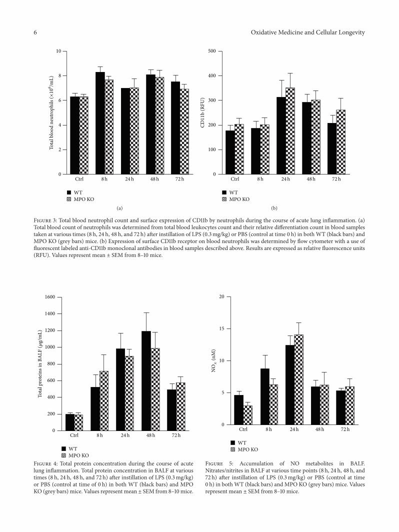
6 Oxidative Medicine and Cellular Longevity
Tota
l blo
od n
eutro
phils
(×10
6 /mL)
Ctrl 8h 24h 48h 72h
WTMPO KO
0
2
4
6
8
10
(a)
Ctrl 8h 24h 48h 72h
WTMPO KO
0
100
200
300
400
500
CD11
b (R
FU)
(b)
Figure 3: Total blood neutrophil count and surface expression of CD11b by neutrophils during the course of acute lung inflammation. (a)Total blood count of neutrophils was determined from total blood leukocytes count and their relative differentiation count in blood samplestaken at various times (8 h, 24 h, 48 h, and 72 h) after instillation of LPS (0.3mg/kg) or PBS (control at time 0 h) in bothWT (black bars) andMPO KO (grey bars) mice. (b) Expression of surface CD11b receptor on blood neutrophils was determined by flow cytometer with a use offluorescent labeled anti-CD11b monoclonal antibodies in blood samples described above. Results are expressed as relative fluorescence units(RFU). Values represent mean ± SEM from 8–10 mice.
Ctrl 8h 24h 48h 72h
WTMPO KO
0
200
400
600
800
1000
1200
1400
1600
Tota
l pro
tein
s in
BALF
(𝜇g/
mL)
Figure 4: Total protein concentration during the course of acutelung inflammation. Total protein concentration in BALF at varioustimes (8 h, 24 h, 48 h, and 72 h) after instillation of LPS (0.3mg/kg)or PBS (control at time of 0 h) in both WT (black bars) and MPOKO (grey bars) mice. Values represent mean ± SEM from 8–10 mice.
Ctrl 8h 24h 48h 72h
WTMPO KO
0
5
10
15
20
NOx
(nM
)
Figure 5: Accumulation of NO metabolites in BALF.Nitrates/nitrites in BALF at various time points (8 h, 24 h, 48 h, and72 h) after instillation of LPS (0.3mg/kg) or PBS (control at time0 h) in bothWT (black bars) and MPO KO (grey bars) mice. Valuesrepresent mean ± SEM from 8–10 mice.
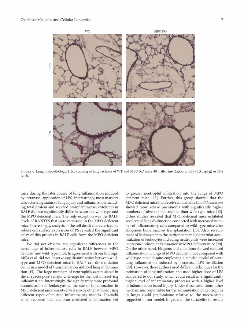
Oxidative Medicine and Cellular Longevity 7
WT MPO KO
Ctrl
LPS
Figure 6: Lung histopathology. H&E staining of lung sections of WT and MPO KO mice 48 h after instillation of LPS (0.3mg/kg) or PBS(ctrl).
mice during the later course of lung inflammation inducedby intranasal application of LPS. Interestingly, most markerscharacterizing status of lung injury and inflammation includ-ing total protein and selected proinflammatory cytokines inBALF did not significantly differ between the wild-type andthe MPO deficient mice. The only exception was the BALFlevels of RANTES that were increased in the MPO deficientmice. Interestingly, analysis of the cell death characterized byrobust cell surface expression of PS revealed the significantdelay of this process in BALF cells from the MPO deficientmice.
We did not observe any significant differences in thepercentage of inflammatory cells in BALF between MPOdeficient and wild-typemice. In agreement with our findings,Milla et al. did not observe any dissimilarities between wild-type and MPO deficient mice in BALF cell differentiationcount in a model of transplantation induced lung inflamma-tion [15]. The large numbers of neutrophils accumulated inthe airspaces pose a major challenge for the host in resolvinginflammation. Interestingly, the significantly more profoundaccumulation of leukocytes at the site of inflammation inMPOdeficientmice was observed also by other authors usingdifferent types of murine inflammatory models. Takeuchiet al. reported that zymosan mediated inflammation led
to greater neutrophil infiltration into the lungs of MPOdeficient mice [28]. Further, this group showed that theMPOdeficientmice that received nonviableCandida albicansshowed more severe pneumonia with significantly highernumbers of alveolar neutrophils than wild-type mice [12].Other studies revealed that MPO deficient mice exhibitedaccelerated lung dysfunction connected with increased num-ber of inflammatory cells compared to wild-type mice afterallogeneic bone marrow transplantation [15]. Also, recruit-ment of leukocyte into the peritoneum and glomerular accu-mulation of leukocytes including neutrophils were increasedin pristine induced inflammation inMPOdeficientmice [20].On the other hand, Haegens and coauthors showed reducedinflammation in lungs of MPO deficient mice compared withwild-type mice despite employing a similar model of acutelung inflammation induced by intranasal LPS instillation[29]. However, these authors used different techniques for theestimation of lung infiltration and used higher dose of LPScompared to our study, which could result in a significantlyhigher level of inflammatory processes with a higher levelof inflammation based injury. Under these conditions, othermechanisms responsible for the accumulation of neutrophilsin lungs could predominate relative to the mechanismssuggested in our model. In general, the variability in results
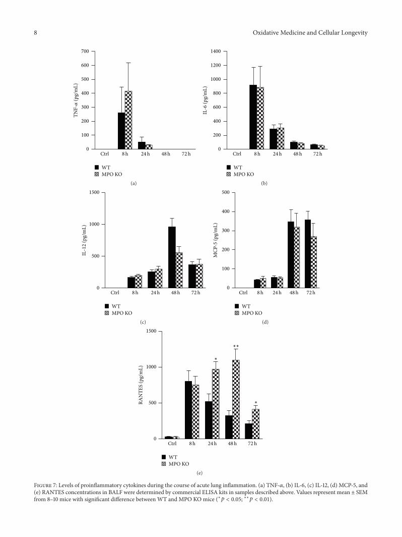
8 Oxidative Medicine and Cellular Longevity
TNF-𝛼
(pg/
mL)
Ctrl 8h 24h 48h 72h
WTMPO KO
0
100
200
300
400
500
600
700
(a)
Ctrl 8h 24h 48h 72h
WTMPO KO
IL-6
(pg/
mL)
0
200
400
600
800
1000
1200
1400
(b)
Ctrl 8h 24h 48h 72h
WTMPO KO
IL-1
2 (p
g/m
L)
0
500
1000
1500
(c)
Ctrl 8h 24h 48h 72h
WTMPO KO
MCP
-5 (p
g/m
L)
0
100
200
300
400
500
(d)
Ctrl 8h 24h 48h 72h
WTMPO KO
RAN
TES
(pg/
mL)
0
500
1000
1500
∗
∗
∗∗
(e)
Figure 7: Levels of proinflammatory cytokines during the course of acute lung inflammation. (a) TNF-𝛼, (b) IL-6, (c) IL-12, (d) MCP-5, and(e) RANTES concentrations in BALF were determined by commercial ELISA kits in samples described above. Values represent mean ± SEMfrom 8–10 mice with significant difference between WT and MPO KOmice (∗𝑝 < 0.05; ∗∗𝑝 < 0.01).
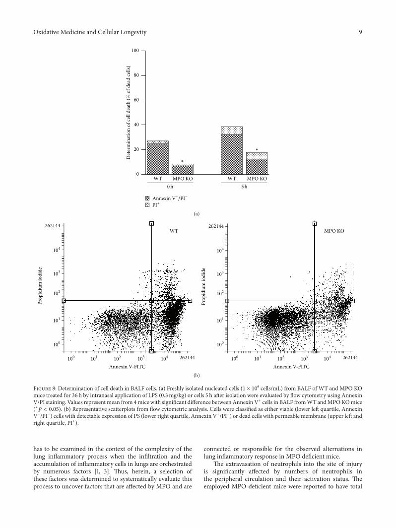
Oxidative Medicine and Cellular Longevity 9
0
20
40
60
80
100
Det
erm
inat
ion
of ce
ll de
ath
(% o
f dea
d ce
lls)
WT MPO KO WT MPO KO
∗
∗
Annexin V+/PI−
PI+
0h 5h
(a)
104
103
102
101
100
104103102101100
Prop
idiu
m io
dide
Annexin V-FITC
MPO KO
262144
262144
104
103
102
101
100
104103102101100
Annexin V-FITC
WT
262144
Prop
idiu
m io
dide
262144
(b)
Figure 8: Determination of cell death in BALF cells. (a) Freshly isolated nucleated cells (1 × 10⁶ cells/mL) from BALF of WT and MPO KOmice treated for 36 h by intranasal application of LPS (0.3mg/kg) or cells 5 h after isolation were evaluated by flow cytometry using AnnexinV/PI staining. Values representmean from 4mice with significant difference between Annexin V+ cells in BALF fromWT andMPOKOmice(∗𝑝 < 0.05). (b) Representative scatterplots from flow cytometric analysis. Cells were classified as either viable (lower left quartile, AnnexinV−/PI−) cells with detectable expression of PS (lower right quartile, Annexin V+/PI−) or dead cells with permeable membrane (upper left andright quartile, PI+).
has to be examined in the context of the complexity of thelung inflammatory process when the infiltration and theaccumulation of inflammatory cells in lungs are orchestratedby numerous factors [1, 3]. Thus, herein, a selection ofthese factors was determined to systematically evaluate thisprocess to uncover factors that are affected by MPO and are
connected or responsible for the observed alternations inlung inflammatory response in MPO deficient mice.
The extravasation of neutrophils into the site of injuryis significantly affected by numbers of neutrophils inthe peripheral circulation and their activation status. Theemployed MPO deficient mice were reported to have total
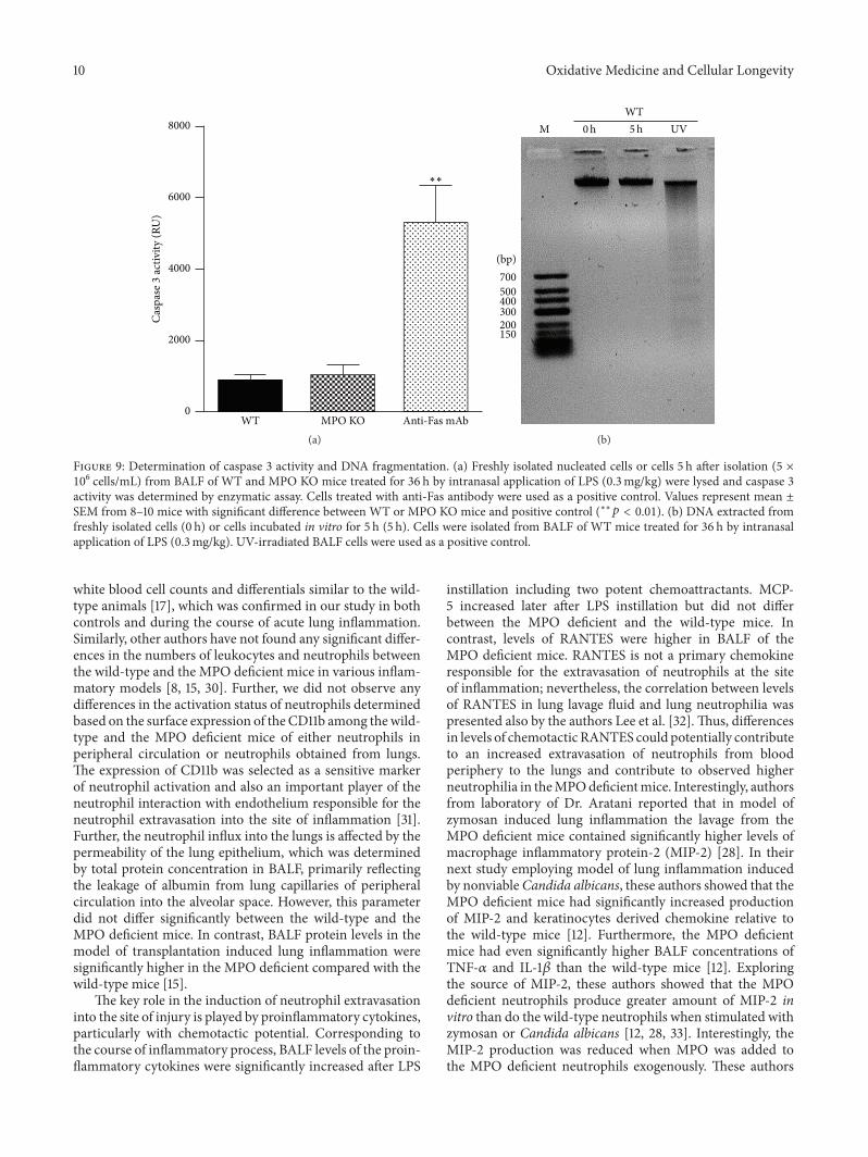
10 Oxidative Medicine and Cellular Longevity
WT MPO KO Anti-Fas mAb0
2000
4000
6000
8000
Casp
ase 3
activ
ity (R
U)
∗∗
(a)
M UVWT
700500400300200150
(bp)
0h 5h
(b)
Figure 9: Determination of caspase 3 activity and DNA fragmentation. (a) Freshly isolated nucleated cells or cells 5 h after isolation (5 ×10⁶ cells/mL) from BALF of WT and MPO KO mice treated for 36 h by intranasal application of LPS (0.3mg/kg) were lysed and caspase 3
activity was determined by enzymatic assay. Cells treated with anti-Fas antibody were used as a positive control. Values represent mean ±SEM from 8–10 mice with significant difference between WT or MPO KO mice and positive control (∗∗𝑝 < 0.01). (b) DNA extracted fromfreshly isolated cells (0 h) or cells incubated in vitro for 5 h (5 h). Cells were isolated from BALF of WT mice treated for 36 h by intranasalapplication of LPS (0.3mg/kg). UV-irradiated BALF cells were used as a positive control.
white blood cell counts and differentials similar to the wild-type animals [17], which was confirmed in our study in bothcontrols and during the course of acute lung inflammation.Similarly, other authors have not found any significant differ-ences in the numbers of leukocytes and neutrophils betweenthe wild-type and the MPO deficient mice in various inflam-matory models [8, 15, 30]. Further, we did not observe anydifferences in the activation status of neutrophils determinedbased on the surface expression of the CD11b among thewild-type and the MPO deficient mice of either neutrophils inperipheral circulation or neutrophils obtained from lungs.The expression of CD11b was selected as a sensitive markerof neutrophil activation and also an important player of theneutrophil interaction with endothelium responsible for theneutrophil extravasation into the site of inflammation [31].Further, the neutrophil influx into the lungs is affected by thepermeability of the lung epithelium, which was determinedby total protein concentration in BALF, primarily reflectingthe leakage of albumin from lung capillaries of peripheralcirculation into the alveolar space. However, this parameterdid not differ significantly between the wild-type and theMPO deficient mice. In contrast, BALF protein levels in themodel of transplantation induced lung inflammation weresignificantly higher in the MPO deficient compared with thewild-type mice [15].
The key role in the induction of neutrophil extravasationinto the site of injury is played by proinflammatory cytokines,particularly with chemotactic potential. Corresponding tothe course of inflammatory process, BALF levels of the proin-flammatory cytokines were significantly increased after LPS
instillation including two potent chemoattractants. MCP-5 increased later after LPS instillation but did not differbetween the MPO deficient and the wild-type mice. Incontrast, levels of RANTES were higher in BALF of theMPO deficient mice. RANTES is not a primary chemokineresponsible for the extravasation of neutrophils at the siteof inflammation; nevertheless, the correlation between levelsof RANTES in lung lavage fluid and lung neutrophilia waspresented also by the authors Lee et al. [32].Thus, differencesin levels of chemotactic RANTES could potentially contributeto an increased extravasation of neutrophils from bloodperiphery to the lungs and contribute to observed higherneutrophilia in theMPOdeficientmice. Interestingly, authorsfrom laboratory of Dr. Aratani reported that in model ofzymosan induced lung inflammation the lavage from theMPO deficient mice contained significantly higher levels ofmacrophage inflammatory protein-2 (MIP-2) [28]. In theirnext study employing model of lung inflammation inducedby nonviableCandida albicans, these authors showed that theMPO deficient mice had significantly increased productionof MIP-2 and keratinocytes derived chemokine relative tothe wild-type mice [12]. Furthermore, the MPO deficientmice had even significantly higher BALF concentrations ofTNF-𝛼 and IL-1𝛽 than the wild-type mice [12]. Exploringthe source of MIP-2, these authors showed that the MPOdeficient neutrophils produce greater amount of MIP-2 invitro than do the wild-type neutrophils when stimulated withzymosan or Candida albicans [12, 28, 33]. Interestingly, theMIP-2 production was reduced when MPO was added tothe MPO deficient neutrophils exogenously. These authors
Oxidative Medicine and Cellular Longevity 11
speculate that both the lack of hypochlorous acid and theaccumulation of H
2O2due to MPO deficiency contribute to
the upregulation of MIP-2 production in the mouse MPOdeficient neutrophils. Similarly, in the model of transplan-tation induced lung inflammation the MPO deficient miceexhibited higher levels of TNF-𝛼 and the chemoattractantMCP-1 compared to the wild-type mice [15]. The importanceof this phenomenon of higher levels of chemotactic factors inlungs of the MPO deficient mice for the observed lung neu-trophilia was shown byHomme et al. when the neutralizationof MIP-2 in vivo significantly reduced neutrophil infiltration[12]. In context of our study, the presence of higher levels ofRANTES could increase extravasation of neutrophils into thelungs and increased survival of neutrophils in lungs.
Another key factor affecting the accumulation of inflam-matory cells in the inflamed lungs is the clearance ofthese cells from alveolar space. Thus, induction of regulatedneutrophil cell death, apoptosis, is a critical event in thedownregulation and resolution of inflammation [34, 35].Therefore, the presence of dying cells in BALF from theMPOdeficient and the wild-type mice was assessed by variousmethods including Annexin V staining, caspase 3 activitydetermination, and the DNA fragmentation. During apop-tosis, PS residues on the inner leaflet of the cell membraneare externalized, providing a marker that can be detectedby binding of Annexin V. Interestingly, significantly fewercells positive for Annexin V and a delay in onset of PSexternalization and cell permeability after ex vivo incubationwere detected in BALF cells isolated from the MPO deficientmice. Substantial delay of the induction of cell death in theMPO deficient neutrophils strongly suggests possible mech-anism for increased neutrophilia in the MPO deficient micebased on the accumulation of the MPO deficient neutrophilsin the inflamed lungs. Interestingly, the defect of apoptoticinduction in neutrophils from the MPO deficient mice wasalready suggested by other authors as well. Tsurubuchi etal. showed that the MPO deficient neutrophils stimulatedwith phorbol 12-myristate 13-acetate (PMA) or H
2O2under-
went apoptosis significantly slower compared to normalneutrophils during interval up to 3 h of incubation [36].However, in their other study employing zymosan inducedlung inflammation they did not observe this phenomenonsince they did not find an obvious difference in cell deathbetween the wild-type and the MPO deficient neutrophilscultured for 6 h in the presence of zymosan [28]. Anothermodel of the transplantation induced lung injury resulted inthe increased number of inflammatory cells in BALF fromthe MPO deficient mice that was associated with suppressedapoptosis of BALF inflammatory cells [15].
Interestingly, in our study the observed neutrophil celldeath is challenging to classify as conventional apoptosissince the dying neutrophils showed only one type of apoptoticmarker, the expression of PS. Other markers of apoptosis, thecaspase 3 activity, and DNA fragmentation were not present.In general, it can be suggested that the type of neutrophil celldeath depends on the way of neutrophil activation. Interest-ingly, similar to our results Aratani and colleagues observeddelayed PS externalization in PMA activated neutrophilsfrom the MPO deficient mice that was not associated with
caspase 3 activation [36, 37]. Moreover, Fadeel et al. showedthe caspases activations in neutrophils treated by Fas ligand;however, it was absent in neutrophils stimulated with PMA[38]. In agreement with our results, DNA fragmentationwas not observed in neutrophils incubated for 6 h alone orstimulated with PMA [39].The PS surface exposure is knownas one of the earliest markers of apoptosis and it precedesthe morphologic appearances of apoptosis and changes inmembrane permeability and the characteristic DNA frag-mentation. Importantly, the PS exposure is a key mechanismby which apoptotic cells are recognized by macrophages,targeted for ingestion, and clearance from lungs [34, 35].These results indicate that MPO probably participates inprocess of regulated cell death and contributes to clearance ofneutrophils from lungs and reduction of airway neutrophilia.Taking into account our previous observations suggestingthat MPO potentiate neutrophils to extravasate into site ofinflammation in various other tissues, such as livers [27], weassume that in this case the higher number of neutrophilsin the MPO deficient mice is not associated with the higherextent of LPS-induced neutrophil influx into the lungs. Incontrast, we suggest that MPO deficiency is responsiblefor the suppressed cell death of lung infiltrating neutro-phils.
Interestingly, the MPO effect on neutrophil presence inthe lungs can be both dependent on and independent ofMPOenzymatic activity. MPO enzymatic activity would be crucialalso in the case of direct contribution of MPO as a cytotoxicagent that contributes to the induction of the death of cellsaccumulated in lungs. We can speculate that MPO enzymaticactivity can decrease the formation of chemotacticmediators,as suggested by Tateno et al. [33]. Further, cytotoxic potentialof MPO derived intermediates is also suggested to be impor-tant for the observed different sensitivity of MPO deficientneutrophils to apoptosis compared to the neutrophils of wild-type animals. MPO deficiency is suggested to be connectedwith suppressed regulated cell death in neutrophils by Millaet al. [15]. Furthermore, delayed PS externalization in PMAstimulated leukocytes from MPO deficient mice comparedto leukocytes from wild-type mice was observed by Tsu-rubuchi et al. [36]. Likewise, pretreatment of neutrophils withthe MPO inhibitor 4-ABAH blocked apoptosis induced bycoactivation of neutrophils by TNF-𝛼 and H
2O2[40]. On
the other hand, current data presented by Metzler et al. alsosuggest the importance of MPO independent of enzymaticactivity in the specific regulated cell death NETosis [41].Based on their observations, MPO is required independentlyof enzymatic activity for the release of proteases acrossintact membranes, which is key for the activation of specificproteases during NETosis. However, the importance of thisnewly suggested function of MPO independent of enzymaticactivity has to be proven also in other types of regulated celldeath that are relevant for the clearance of neutrophils frominflamed lungs.
In conclusion, these results suggest that neutrophil-derived MPO may play an important role in regulatingthe course of pulmonary inflammation, independent of itsputative microbicidal functions. Because the MPO deficientneutrophils undergo delayed apoptosis in vitro, it is possible
12 Oxidative Medicine and Cellular Longevity
that these neutrophils remain alive longer at sites of inflam-mation. As a result, they would continue to release variousROS, inflammatory cytokines, and cytotoxic enzymes for alonger time, eventually resulting in tissue damage. However,further work evaluating the time course of appearance ofapoptotic neutrophils is required to confirm the role ofMPO in regulated cell death and to determine whether suchdefective functions of neutrophils are involved in the pathol-ogy of various inflammatory conditions. Knowledge gainedfrom this research will help to determine more extensivelythe biological functions of MPO in inflammatory humanlung disease and will aid in the development of potentialpharmacological treatments for both acute and chronic lunginjury.
Conflict of Interests
The authors report no conflict of interests. The authors aloneare responsible for the content and writing of the paper.
Acknowledgments
The authors wish to thank Lenka Vystrcilova for herexpert technical assistance. This work was supported bygrants from the Czech Science Foundation no. P305/12/J038;from the DFG no. BA1870/9-1, KL 2516/1-1; and from theNIH no. HL092506/HL/NHLBI. Silvie Kremserova, KarelSoucek, and Lukas Kubala were supported by the EuropeanRegional Development Fund, Project FNUSA-ICRC (no.CZ.1.05/1.1.00/02.0123), andTomas Pereckowas supported byFP7 Regpot (ICRC-ERA-HumanBridge).
References
[1] B. D. Levy and C. N. Serhan, “Resolution of acute inflammationin the lung,” Annual Review of Physiology, vol. 76, pp. 467–492,2014.
[2] R. G. Khadaroo and J. C. Marshall, “ARDS and the multipleorgan dysfunction syndrome: common mechanisms of a com-mon systemic process,” Critical Care Clinics, vol. 18, no. 1, pp.127–141, 2002.
[3] J. Cohen, “The immunopathogenesis of sepsis,”Nature, vol. 420,no. 6917, pp. 885–891, 2002.
[4] J. P. Burns, “Septic shock in the pediatric patient: pathogenesisand novel treatments,” Pediatric Emergency Care, vol. 19, no. 2,pp. 112–115, 2003.
[5] S. J. Klebanoff, A. J. Kettle, H. Rosen, C. C. Winterbourn, andW.M. Nauseef, “Myeloperoxidase: a front-line defender againstphagocytosed microorganisms,” Journal of Leukocyte Biology,vol. 93, no. 2, pp. 185–198, 2013.
[6] W. M. Nauseef, “Myeloperoxidase in human neutrophil hostdefence,” Cellular Microbiology, vol. 16, no. 8, pp. 1146–1155,2014.
[7] C. Nussbaum, A. Klinke, M. Adam, S. Baldus, and M.Sperandio, “Myeloperoxidase: a leukocyte-derived protagonistof inflammation and cardiovascular disease,” Antioxidants andRedox Signaling, vol. 18, no. 6, pp. 692–713, 2013.
[8] L. Kubala, K. R. Schmelzer, A. Klinke et al., “Modulation ofarachidonic and linoleic acid metabolites in myeloperoxidase-deficient mice during acute inflammation,” Free Radical Biologyand Medicine, vol. 48, no. 10, pp. 1311–1320, 2010.
[9] J. Arnhold and J. Flemmig, “Human myeloperoxidase in innateand acquired immunity,” Archives of Biochemistry and Bio-physics, vol. 500, no. 1, pp. 92–106, 2010.
[10] L. Kubala, H. Kolářová, J. Vı́teček et al., “The potentiation ofmyeloperoxidase activity by the glycosaminoglycan- dependentbinding of myeloperoxidase to proteins of the extracellularmatrix,”Biochimica et Biophysica Acta (BBA)—General Subjects,vol. 1830, no. 10, pp. 4524–4536, 2013.
[11] Y. Aratani, F. Kura, H. Watanabe et al., “Differential hostsusceptibility to pulmonary infections with bacteria and fungiin mice deficient in myeloperoxidase,” Journal of InfectiousDiseases, vol. 182, no. 4, pp. 1276–1279, 2000.
[12] M. Homme, N. Tateno, N. Miura, N. Ohno, and Y. Aratani,“Myeloperoxidase deficiency in mice exacerbates lung inflam-mation induced by nonviable Candida albicans,” InflammationResearch, vol. 62, no. 11, pp. 981–990, 2013.
[13] M.-L. Brennan, A. Gaur, A. Pahuja, A. J. Lusis, and W. F.Reynolds, “Mice lacking myeloperoxidase are more susceptibleto experimental autoimmune encephalomyelitis,” Journal ofNeuroimmunology, vol. 112, no. 1-2, pp. 97–105, 2001.
[14] S. Takizawa, Y. Aratani, N. Fukuyama et al., “Deficiency ofmyeloperoxidase increases infarct volume and nitrotyrosineformation in mouse brain,” Journal of Cerebral Blood Flow andMetabolism, vol. 22, no. 1, pp. 50–54, 2002.
[15] C. Milla, S. Yang, D. N. Cornfield et al., “Myeloperoxidasedeficiency enhances inflammation after allogeneic marrowtransplantation,” The American Journal of Physiology—LungCellular and Molecular Physiology, vol. 287, no. 4, pp. L706–L714, 2004.
[16] J. Komatsu, H. Koyama, N. Maeda, and Y. Aratani, “Earlieronset of neutrophil-mediated inflammation in the ultraviolet-exposed skin ofmice deficient inmyeloperoxidase andNADPHoxidase,” Inflammation Research, vol. 55, no. 5, pp. 200–206,2006.
[17] M.-L. Brennan, M. M. Anderson, D. M. Shih et al., “Increasedatherosclerosis in myeloperoxidase-deficient mice,”The Journalof Clinical Investigation, vol. 107, no. 4, pp. 419–430, 2001.
[18] D. Odobasic, A. R. Kitching, Y. Yang et al., “Neutrophil mye-loperoxidase regulates T-cell-driven tissue inflammation inmice by inhibiting dendritic cell function,” Blood, vol. 121, no.20, pp. 4195–4204, 2013.
[19] D. Odobasic, A. R. Kitching, T. J. Semple, and S. R. Holdsworth,“Endogenous myeloperoxidase promotes neutrophil-mediatedrenal injury, but attenuates T cell immunity inducing crescenticglomerulonephritis,” Journal of the American Society of Nephrol-ogy, vol. 18, no. 3, pp. 760–770, 2007.
[20] D. Odobasic, R. C.Muljadi, K.M.O’Sullivan et al., “Suppressionof autoimmunity and renal disease in pristane-induced lupus bymyeloperoxidase ,” Arthritis & Rheumatology, vol. 67, no. 7, pp.1868–1880, 2015.
[21] T. Okamoto, K. Gohil, E. I. Finkelstein, P. Bove, T. Akaike,and A. Van Der Vliet, “Multiple contributing roles for NOS
2
inLPS-induced acute airway inflammation inmice,”TheAmericanJournal of Physiology—Lung Cellular and Molecular Physiology,vol. 286, no. 1, pp. L198–L209, 2004.
[22] M. E. Poynter, C. G. Irvin, and Y. M. W. Janssen-Heininger,“A prominent role for airway epithelial NF-𝜅B activation in
Oxidative Medicine and Cellular Longevity 13
lipopolysaccharide-induced airway inflammation,”The Journalof Immunology, vol. 170, no. 12, pp. 6257–6265, 2003.
[23] D. Viackova, M. Pekarova, T. Crhak et al., “Redox-sensitive reg-ulation of macrophage-inducible nitric oxide synthase expres-sion in vitro does not correlate with the failure of apocynin toprevent lung inflammation induced by endotoxin,” Immunobi-ology, vol. 216, no. 4, pp. 457–465, 2011.
[24] H. Kolarova, A. Klinke, S. Kremserova et al., “Myeloperoxidaseinduces the priming of platelets,” Free Radical Biology andMedicine, vol. 61, pp. 357–369, 2013.
[25] A. Klinke, A. Möller, M. Pekarova et al., “Protective effectsof 10-nitro-oleic acid in a hypoxia-induced murine model ofpulmonary hypertension,” American Journal of Respiratory Celland Molecular Biology, vol. 51, no. 1, pp. 155–162, 2014.
[26] L. Svihálková-Sindlerová, V. Foltinová, A. Vaculová et al., “LA-12 overcomes confluence-dependent resistance of HT-29 coloncancer cells to Pt (II) compounds,” Anticancer Research, vol. 30,no. 4, pp. 1183–1188, 2010.
[27] A. Klinke, C. Nussbaum, L. Kubala et al., “Myeloperoxidaseattracts neutrophils by physical forces,” Blood, vol. 117, no. 4, pp.1350–1358, 2011.
[28] K. Takeuchi, Y. Umeki, N.Matsumoto et al., “Severe neutrophil-mediated lung inflammation inmyeloperoxidase-deficientmiceexposed to zymosan,” Inflammation Research, vol. 61, no. 3, pp.197–205, 2012.
[29] A. Haegens, P. Heeringa, R. J. van Suylen et al., “Myeloper-oxidase deficiency attenuates lipopolysaccharide-induced acutelung inflammation and subsequent cytokine and chemokineproduction,” The Journal of Immunology, vol. 182, no. 12, pp.7990–7996, 2009.
[30] M.-L. Brennan, W.Wu, X. Fu et al., “A tale of two controversies:defining both the role of peroxidases in nitrotyrosine formationin vivo using eosinophil peroxidase and myeloperoxidase-deficient mice, and the nature of peroxidase-generated reactivenitrogen species,” Journal of Biological Chemistry, vol. 277, no.20, pp. 17415–17427, 2002.
[31] E. Kolaczkowska and P. Kubes, “Neutrophil recruitmentand function in health and inflammation,” Nature ReviewsImmunology, vol. 13, no. 3, pp. 159–175, 2013.
[32] C. S. Lee, E. H. Yi, J.-K. Lee et al., “Simvastatin suppressesRANTES-mediated neutrophilia in polyinosinic-polycytidylicacid-induced pneumonia,” European Respiratory Journal, vol.41, no. 5, pp. 1147–1156, 2013.
[33] N. Tateno, N. Matsumoto, T. Motowaki, K. Suzuki, and Y.Aratani, “Myeloperoxidase deficiency induces MIP-2 produc-tion via ERK activation in zymosan-stimulated mouse neu-trophils,” Free Radical Research, vol. 47, no. 5, pp. 376–385, 2013.
[34] C. Haslett, “Granulocyte apoptosis and its role in the resolutionand control of lung inflammation,”American Journal of Respira-tory andCritical CareMedicine, vol. 160, no. 5, part 2, pp. S5–S11,1999.
[35] T. R. Martin, M. Nakamura, and G. Matute-Bello, “The role ofapoptosis in acute lung injury,” Critical Care Medicine, vol. 31,no. 4, pp. S184–S188, 2003.
[36] T. Tsurubuchi, Y. Aratani, N. Maeda, and H. Koyama, “Retar-dation of early-onset PMA-induced apoptosis in mouse neu-trophils deficient in myeloperoxidase,” Journal of LeukocyteBiology, vol. 70, no. 1, pp. 52–58, 2001.
[37] T. Saito, H. Takahashi, H. Doken, H. Koyama, and Y. Aratani,“Phorbol myristate acetate induces neutrophil death throughactivation of p38mitogen-activated protein kinase that requires
endogenous reactive oxygen species other than HOCl,” Bio-science, Biotechnology and Biochemistry, vol. 69, no. 11, pp. 2207–2212, 2005.
[38] B. Fadeel, A. Åhlin, J.-I. Henter, S. Orrenius, and M. B.Hampton, “Involvement of caspases in neutrophil apoptosis:regulation by reactive oxygen species,” Blood, vol. 92, no. 12, pp.4808–4818, 1998.
[39] H. Takei, A. Araki, H. Watanabe, A. Ichinose, and F. Sendo,“Rapid killing of human neutrophils by the potent activa-tor phorbol 12-myristate 13-acetate (PMA) accompanied bychanges different from typical apoptosis or necrosis,” Journal ofLeukocyte Biology, vol. 59, no. 2, pp. 229–240, 1996.
[40] A. Kanayama and Y. Miyamoto, “Apoptosis triggered byphagocytosis-related oxidative stress through FLIPS down-regulation and JNK activation,” Journal of Leukocyte Biology,vol. 82, no. 5, pp. 1344–1352, 2007.
[41] K.D.Metzler, C.Goosmann,A. Lubojemska, A. Zychlinsky, andV. Papayannopoulos, “A myeloperoxidase-containing complexregulates neutrophil elastase release and actin dynamics duringNETosis,” Cell Reports, vol. 8, no. 3, pp. 883–896, 2014.
Research ArticleProtective Effect of Decursin Extracted from Angelica gigas inMale Infertility via Nrf2/HO-1 Signaling Pathway
Woong Jin Bae,1,2 U. Syn Ha,2 Jin Bong Choi,2 Kang Sup Kim,2 Su Jin Kim,2 Hyuk Jin Cho,2
Sung Hoo Hong,2 Ji Youl Lee,2 Zhiping Wang,3 Sung Yeoun Hwang,4 and Sae Woong Kim1,2
1Catholic Integrative Medicine Research Institute, College of Medicine, The Catholic University of Korea,Seoul 137-701, Republic of Korea2Department of Urology, College of Medicine, The Catholic University of Korea, Banpo-daero 222, Seocho-gu,Seoul 137-701, Republic of Korea3Department of Urology, Second Hospital of Lanzhou University, Lanzhou, China4Korea Bio Medical Science Institute, Seoul, Republic of Korea
Correspondence should be addressed to Sae Woong Kim; [email protected]
Received 21 September 2015; Revised 17 November 2015; Accepted 6 December 2015
Academic Editor: Sergio Di Meo
Copyright © 2016 Woong Jin Bae et al.This is an open access article distributed under the Creative Commons Attribution License,which permits unrestricted use, distribution, and reproduction in any medium, provided the original work is properly cited.
Higher testicular temperature results in altered spermatogenesis due to heat-related oxidative stress. We examined the effects ofdecursin extracted from Angelica gigasNakai on antioxidant activity in vitro and in a cryptorchidism-induced infertility rat model.TM3 Leydig cell viability was measured based on oxidative stress according to treatment. Either distilled water or AG 400mg/kgof A. gigas extract was administered orally for 4 weeks after unilateral cryptorchidism was induced. After 1, 2, and 4 weeks,six rats from the control group and six rats from treatment group were sacrificed. Testicular weight, semen quality, antioxidantactivities, nuclear factor erythroid 2-related factor 2 (Nrf2) protein, and mRNA expression of Nrf2-regulated genes were analyzed.Treatment with A. gigas extract (1) protected TM3 cells against oxidative stress in a dose-dependent manner, (2) improved themean weight of the cryptorchid testis, (3) maintained sperm counts, motility, and spermatogenic cell density, (4) decreased levelsof 8-hydroxy-2-deoxyguanosine (8-OHdG) and increased levels of superoxide dismutase (SOD), (5) significantly increased Nrf2and heme oxygenase-1 (HO-1), and (6) significantly decreased apoptosis. This study suggests that decursin extracted from A. gigasis a supplemental agent that can reduce oxidative stress by Nrf2-mediated upregulation of HO-1 in rat experimentally inducedunilateral cryptorchidism and may improve cryptorchidism-induced infertility.
1. Introduction
Infertility, which can be explained as the failure of a couplein trying to conceive after one year of frequent, unprotectedsexual intercourse, is a serious clinical issue that affects 13–15% of couples worldwide [1]. Male infertility is respon-sible for 60% of cases involving conceptive couples withpregnancy-related problems [2]. Sperm is produced by thehighly complicated process of spermatogenesis, and partialor complete interruption of spermatogenesis ultimately leadsto oligospermia or azoospermia.
Observation study on male infertility with oligozoosper-mia or azoospermia, in particular, suggests that some patients
may have testicular heat exposure due to an intrinsic defect inscrotal thermoregulation, varicocele, or work hazard [3]. Sev-eral studies report that testicular hyperthermia above normalranges causes impaired spermatogenesis due to heat-relatedoxidative stress on the seminiferous tubules [4, 5]. Moreover,nuclear factor erythroid 2-related factor 2 (Nrf2) plays asignificant role in preventing the development of oxidativestress in spermatogenesis [6]. Yu et al. [7] also demonstrateda strong correlation between functional discrepancy in Nrf2promoter gene and abnormal spermatogenesis in humans.
Decursin, a major active ingredient from Angelica gigasNakai (Apiaceae), has been reported to inhibit the growth ofvarious cancer cells through cell cycle arrest and apoptosis
Hindawi Publishing CorporationOxidative Medicine and Cellular LongevityVolume 2016, Article ID 5901098, 9 pageshttp://dx.doi.org/10.1155/2016/5901098
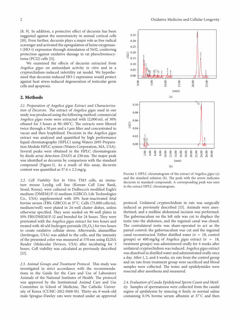
2 Oxidative Medicine and Cellular Longevity
[8, 9]. In addition, a protective effect of decursin has beensuggested against the neurotoxicity in animal cortical cells[10]. Even further, decursin plays a major role as free radicalscavenger and activated the upregulation of heme oxygenase-1 (HO-1) expression through stimulation of Nrf2, conferringprotection against oxidative damage in rat pheochromocy-toma (PC12) cells [11].
We examined the effects of decursin extracted fromAngelica gigas on antioxidant activity in vitro and in acryptorchidism-induced infertility rat model. We hypothe-sized that decursin-induced HO-1 expression would protectagainst heat stress-induced degeneration of testicular germcells and apoptosis.
2. Methods
2.1. Preparation of Angelica gigas Extract and Characteriza-tion of Decursin. The extract of Angelica gigas used in ourstudy was produced using the followingmethod: commercialAngelica gigas roots were extracted with 12,000mL of 30%ethanol for 3 hours at 90–100∘C. The extracts were filteredtwice through a 50𝜇m and a 1 𝜇m filter and concentrated invacuo and then lyophilized. Decursin in the Angelica gigasextract was analyzed and quantified by high performanceliquid chromatography (HPLC) using Waters 2695 Prepara-tion Module HPLC system (Waters Corporation, MA, USA).Several peaks were obtained in the HPLC chromatogramby diode array detection (DAD) at 230 nm. The major peakwas identified as decursin by comparison with the standardcompound (Figure 1). As a result of this assay, decursincontent was quantified as 37.6 ± 2.2mg/g.
2.2. Cell Viability Test In Vitro. TM3 cells, an imma-ture mouse Leydig cell line (Korean Cell Line Bank,Seoul, Korea), were cultured in Dulbecco’s modified Eagle’smedium (DMEM)/F-12 medium (GIBCO, Life TechnologiesCo., USA) supplemented with 10% heat-inactivated fetalbovine serum (FBS; GIBCO) at 37∘C. Cells (75,000 cells/mLmedium/well) were plated in 24-well cluster dishes, unlessotherwise specified. They were seeded on 96-well plates in10% FBS/DMEM/F-12 and breeded for 24 hours. They werepretreated with the Angelica gigas extract for two hours andtreated with 40 uM hydrogen peroxide (H
2O2) for two hours
to create oxidative cellular stress. Afterwards, alamarBlue(Invitrogen, USA) was added to the cells, and the intensityof the presented color was measured at 570 nm using ELISAReader (Molecular Devices, USA) after incubating for 3hours. Cell viability was calculated as previously described[12].
2.3. Animal Groups and Treatment Protocol. This study wasinvestigated in strict accordance with the recommenda-tions in the Guide for the Care and Use of LaboratoryAnimals of the National Institutes of Health. The protocolwas approved by the Institutional Animal Care and UseCommittee in School of Medicine, The Catholic Univer-sity of Korea (CUMC-2012-0168-01). Thirty-six 8-week-oldmale Sprague-Dawley rats were treated under an approved
0.00
0.05
0.10
0.15
0.20
0.25
0.30
0.35
0.00
5.00
10.0
0
15.0
0
20.0
0
25.0
0
2.02
92.
187
2.87
23.
428
3.66
74.
099
4.35
5
5.69
36.
068
7.07
07.
291
8.61
7
10.7
40
11.8
62
21.6
16
22.7
82
24.7
74
(min)
(AU
)
(a)
0.00
0.02
0.04
0.06
0.08
0.10
2.00
4.00
6.00
8.00
10.0
012
.00
14.0
016
.00
18.0
020
.00
22.0
024
.00
26.0
028
.00
1.80
4 2.13
52.
868
23.9
51
25.2
88
(min)
(AU
)
(b)
Figure 1: HPLC chromatogram of the extract of Angelica gigas (a)and the standard solution (b). The peak with the arrow indicatesdecursin in standard compounds. A corresponding peak was seenin the extract HPLC chromatogram.
protocol. Unilateral cryptorchidism in rats was surgicallyinduced as previously described [13]. Animals were anes-thetized, and a midline abdominal incision was performed.The gubernaculum on the left side was cut to displace thetestis into the abdomen, and the inguinal canal was closed.The contralateral testis was sham-operated to act as thepaired control; the gubernaculum was cut and the inguinalcanal reconstructed. Either distilled water (𝑛 = 18, controlgroups) or 400mg/kg of Angelica gigas extract (𝑛 = 18,treatment groups) was administered orally for 4 weeks afterunilateral cryptorchidism was induced.Angelica gigas extractwas dissolved in distilled water and administrated orally oncea day. After 1, 2, and 4 weeks, six rats from the control groupand six rats from treatment group were sacrificed and bloodsamples were collected. The testes and epididymides wereresected after anesthesia and measured.
2.4. Evaluation of Cauda Epididymal Sperm Count and Motil-ity. Samples of spermatozoa were collected from the caudalregion of epididymis by mincing it finely in normal salinecontaining 0.5% bovine serum albumin at 37∘C and then
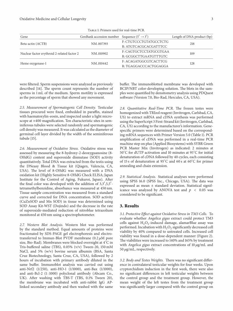
Oxidative Medicine and Cellular Longevity 3
Table 1: Primers used for real-time PCR.
Gene GenBank accession number Sequence (5 → 3) Length of DNA product (bp)
Beta-actin (ACTB) NM 007393 F: CTGTCCCTGTATGCCTCTG 218R: ATGTCACGCACGATTTCC
Nuclear factor erythroid 2-related factor 2 NM 010902 F: CAGTGCTCCTATGCGTGAA 109R: GCGGCTTGAATGTTTGTC
Heme oxygenase-1 NM 010442 F: ACAGATGGCGTCACTTCG 128R: TGAGGACCCACTGGAGGA
were filtered. Sperm suspensions were analyzed as previouslydescribed [14]. The sperm count represents the number ofsperms in 1mL of the medium. Sperm motility is expressedas the percentage of sperm that showed any movement.
2.5. Measurement of Spermatogenic Cell Density. Testiculartissues procured were fixed, embedded in paraffin, stainedwith haematoxylin-eosin, and inspected under a light micro-scope at ×400 magnification. Ten characteristic sites in sem-iniferous tubules were selected randomly and spermatogeniccell density wasmeasured. It was calculated as the diameter ofgerminal cell layer divided by the width of the seminiferoustubule [15].
2.6. Measurement of Oxidative Stress. Oxidative stress wasassessed by measuring the 8-hydroxy-2-deoxyguanosine (8-OHdG) content and superoxide dismutase (SOD) activityquantitatively. Total DNA was extracted from the testis usingthe DNeasy Blood & Tissue kit (Qiagen, Valencia, CA,USA). The level of 8-OHdG was measured with a DNAoxidation kit (Highly Sensitive 8-OHdG Check ELISA; JapanInstitute for the Control of Aging, Fukuroi, Japan). Afterthe final color was developed with the addition of 3,3,5,5-tetramethylbenzidine, absorbance was measured at 450 nm.Tissue sample concentration was measured from a standardcurve and corrected for DNA concentration. SOD activity(CuZnSOD and Mn SOD) in tissue was determined usingSOD Assay Kit-WST (Dojindo) and the decrease in the rateof superoxide-mediated reduction of nitroblue tetrazoliummonitored at 450 nm using a spectrophotometer.
2.7. Western Blot Analysis. Western blot was performedby the standard method. Equal amounts of proteins werefractionated by SDS-PAGE gel electrophoresis and electro-transferred to Immun-Blot PVDF membrane (0.2 𝜇M poresize, Bio-Rad). Membranes were blocked overnight at 4∘C inTris-buffered saline (TBS), 0.05% (v/v) Tween-20, 150mMNaCl, and 5% (w/v) bovine serum albumin (BSA, SantaCruz Biotechnology, Santa Cruz, CA, USA), followed by 2hours of incubation with primary antibody diluted in thesame buffer. Immunoblot analysis was carried out usinganti-Nrf2 (1/250), anti-HO-1 (1/1000), anti-Bax (1/1000),and anti-Bcl-2 (1 : 1000) polyclonal antibody (Abcam Co.,UK). After washing with TBS-T (TBS, 0.1% Tween 20),the membrane was incubated with anti-rabbit IgG AP-linked secondary antibody and then washed with the same
buffer. The immunoblotted membrane was developed withBCIP/NBT color-developing solution. The blots in the sam-ples were quantified by densitometry analysis using PDQuestsoftware (Version 7.0, Bio-Rad, Hercules, CA, USA).
2.8. Quantitative Real-Time PCR. The frozen testes werehomogenized with TRIzol reagent (Invitrogen, Carlsbad, CA,US) to extract mRNA and cDNA synthesis was performedusing the SuperScript 3 First-Strand kit (Invitrogen, Carlsbad,CA, US) according to the manufacturer’s information. Gene-specific primers were determined based on the correspond-ing mRNA sequences with Primer Version 5.0 (Table 1). PCRamplification of cDNA was performed in a real-time PCRmachine step on plus (Applied Biosystems) with SYBRGreenPCR Master Mix (Invitrogen) as indicated: 2 minutes at50∘C for dUTP activation and 10 minutes at 95∘C for initialdenaturation of cDNA followed by 40 cycles, each consistingof 15 s of denaturation at 95∘C and 60 s at 60∘C for primerannealing and chain extension.
2.9. Statistical Analysis. Statistical analyses were performedusing SPSS 16.0 (SPSS Inc., Chicago, USA). The data wasexpressed as mean ± standard deviation. Statistical signif-icance was analyzed by ANOVA test and 𝑝 < 0.05 wasconsidered to be significant.
3. Results
3.1. Protective Effect against Oxidative Stress in TM3 Cells. Toevaluate whether Angelica gigas extract could protect TM3cells against H
2O2-induced damage, alamarBlue assay was
performed. IncubationwithH2O2significantly decreased cell
viability by 40% compared to untreated cells. Increased cellviability was found in a dose-dependent manner (Figure 2).The viabilities were increased to 140% and 165% by treatmentwith Angelica gigas extract concentrations of 10 𝜇g/mL and50 𝜇g/mL, respectively.
3.2. Body and Testes Weights. There was no significant differ-ence in contralateral testicular weights for four weeks. Uponcryptorchidism induction in the first week, there were alsono significant differences in left testicular weights betweenthe control group and the treatment group. However, themean weight of the left testes from the treatment groupwas significantly larger compared with the control group on
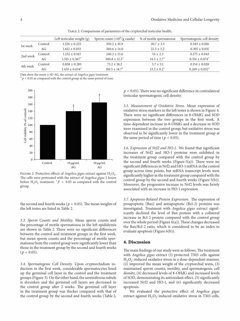
4 Oxidative Medicine and Cellular Longevity
Table 2: Comparisons of parameters of the cryptorchid testicular health.
Left testicular weight (g) Sperm count (×106/g cauda) % of motile spermatozoa Spermatogenic cell density
1st week Control 1.526 ± 0.225 350.2 ± 10.9 20.7 ± 3.3 0.343 ± 0.016AG 1.612 ± 0.033 360.6 ± 14.0 22.5 ± 5.2 0.305 ± 0.031
2nd week Control 1.232 ± 0.165 240.2 ± 13.6 7.6 ± 2.3 0.275 ± 0.043AG 1.515 ± 0.367∗ 300.8 ± 12.5∗ 14.5 ± 2.7∗ 0.314 ± 0.074∗
4th week Control 0.858 ± 0.285 75.2 ± 18.2 5.7 ± 3.1 0.154 ± 0.028AG 1.433 ± 0.634∗ 310.5 ± 14.7∗ 13.2 ± 8.2∗ 0.269 ± 0.052∗
Data show the mean ± SD AG, the extract of Angelica gigas treatment.∗
𝑝 < 0.05 as compared with the control group at the same period of time.
0
20
40
60
80
100
120
140
160
180
200
Control
Cel
l via
bilit
y (%
of c
ontro
l)
AG50𝜇g/mL
AG10𝜇g/mL
∗
∗
Figure 2: Protective effects of Angelica gigas extract against H2
O2
.The cells were pretreated with the extract of Angelica gigas 2 hoursbefore H
2
O2
treatment. ∗𝑝 < 0.05 as compared with the controlgroup.
the second and fourth weeks (𝑝 < 0.05).Themean weights ofthe left testes are listed in Table 2.
3.3. Sperm Counts and Motility. Mean sperm counts andthe percentage of motile spermatozoa in the left epididymisare shown in Table 2. There were no significant differencesbetween the control and treatment groups in the first week,but mean sperm counts and the percentage of motile sper-matozoa from the control groupwere significantly lower thanthose in the treatment group by the second and fourth weeks(𝑝 < 0.05).
3.4. Spermatogenic Cell Density. Upon cryptorchidism in-duction in the first week, considerable spermatocytes linedup the germinal cell layer in the control and the treatmentgroups (Figure 3). On the other hand, the seminiferous tubuleis shrunken and the germinal cell layers are decreased inthe control group after 2 weeks. The germinal cell layerin the treatment group was thicker compared with that ofthe control group by the second and fourth weeks (Table 2,
𝑝 < 0.05).There was no significant difference in contralateraltesticular spermatogenic cell density.
3.5. Measurement of Oxidative Stress. Mean expression ofoxidative stress markers in the left testes is shown in Figure 4.There were no significant differences in 8-OHdG and SODexpression between the two groups in the first week. Atime-dependent increase in 8-OHdG and a decrease in SODwere examined in the control group, but oxidative stress wasobserved to be significantly lower in the treatment group atthe same period of time (𝑝 < 0.05).
3.6. Expression of Nrf2 and HO-1. We found that significantincreases of Nrf2 and HO-1 proteins were exhibited inthe treatment group compared with the control group bythe second and fourth weeks (Figure 5(a)). There were nosignificant differences inNrf2 andHO-1mRNA in the controlgroup across time points, but mRNA transcript levels weresignificantly higher in the treatment group comparedwith thecontrol group by the second and fourth weeks (Figure 5(b)).Moreover, the progressive increase in Nrf2 levels was firmlyassociated with an increase in HO-1 expression.
3.7. Apoptosis-Related Protein Expression. The expression ofproapoptotic (Bax) and antiapoptotic (Bcl-2) proteins wasinvestigated. Treatment with Angelica gigas extract signif-icantly declined the level of Bax protein with a collateralincrease in Bcl-2 protein compared with the control groupover the whole period (Figure 6(a)).These changes decreasedthe Bax/Bcl-2 ratio, which is considered to be an index toevaluate apoptosis (Figure 6(b)).
4. Discussion
Themain findings of our study were as follows.The treatmentwith Angelica gigas extract (1) protected TM3 cells againstH2O2-induced oxidative stress in a dose-dependent manner,
(2) improved the mean weight of the cryptorchid testis, (3)maintained sperm counts, motility, and spermatogenic celldensity, (4) decreased levels of 8-OHdG and increased levelsof SOD, demonstrating its antioxidant effect, (5) significantlyincreased Nrf2 and HO-1, and (6) significantly decreasedapoptosis.
We evaluated the protective effect of Angelica gigasextract against H
2O2-induced oxidative stress in TM3 cells.
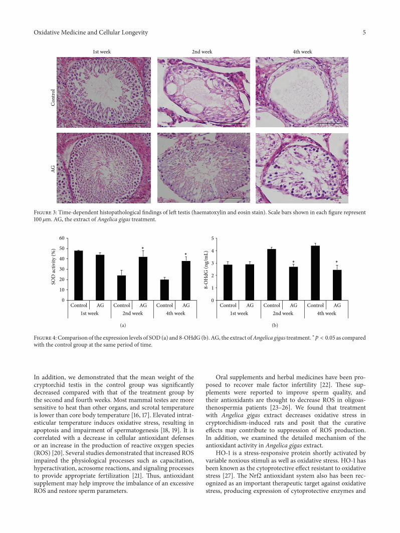
Oxidative Medicine and Cellular Longevity 5
1st weekAG
Con
trol
2nd week 4th week
Figure 3: Time-dependent histopathological findings of left testis (haematoxylin and eosin stain). Scale bars shown in each figure represent100𝜇m. AG, the extract of Angelica gigas treatment.
0
10
20
30
40
50
60
Control AG Control AG Control AG1st week 2nd week 4th week
SOD
activ
ity (%
) ∗∗
(a)
0
1
2
3
4
5
Control AG Control AG Control AG1st week 2nd week 4th week
8-O
HdG
(ng/
mL)
∗∗
(b)
Figure 4: Comparison of the expression levels of SOD (a) and 8-OHdG (b). AG, the extract ofAngelica gigas treatment. ∗𝑝 < 0.05 as comparedwith the control group at the same period of time.
In addition, we demonstrated that the mean weight of thecryptorchid testis in the control group was significantlydecreased compared with that of the treatment group bythe second and fourth weeks. Most mammal testes are moresensitive to heat than other organs, and scrotal temperatureis lower than core body temperature [16, 17]. Elevated intrat-esticular temperature induces oxidative stress, resulting inapoptosis and impairment of spermatogenesis [18, 19]. It iscorrelated with a decrease in cellular antioxidant defensesor an increase in the production of reactive oxygen species(ROS) [20]. Several studies demonstrated that increased ROSimpaired the physiological processes such as capacitation,hyperactivation, acrosome reactions, and signaling processesto provide appropriate fertilization [21]. Thus, antioxidantsupplement may help improve the imbalance of an excessiveROS and restore sperm parameters.
Oral supplements and herbal medicines have been pro-posed to recover male factor infertility [22]. These sup-plements were reported to improve sperm quality, andtheir antioxidants are thought to decrease ROS in oligoas-thenospermia patients [23–26]. We found that treatmentwith Angelica gigas extract decreases oxidative stress incryptorchidism-induced rats and posit that the curativeeffects may contribute to suppression of ROS production.In addition, we examined the detailed mechanism of theantioxidant activity in Angelica gigas extract.
HO-1 is a stress-responsive protein shortly activated byvariable noxious stimuli as well as oxidative stress. HO-1 hasbeen known as the cytoprotective effect resistant to oxidativestress [27]. The Nrf2 antioxidant system also has been rec-ognized as an important therapeutic target against oxidativestress, producing expression of cytoprotective enzymes and
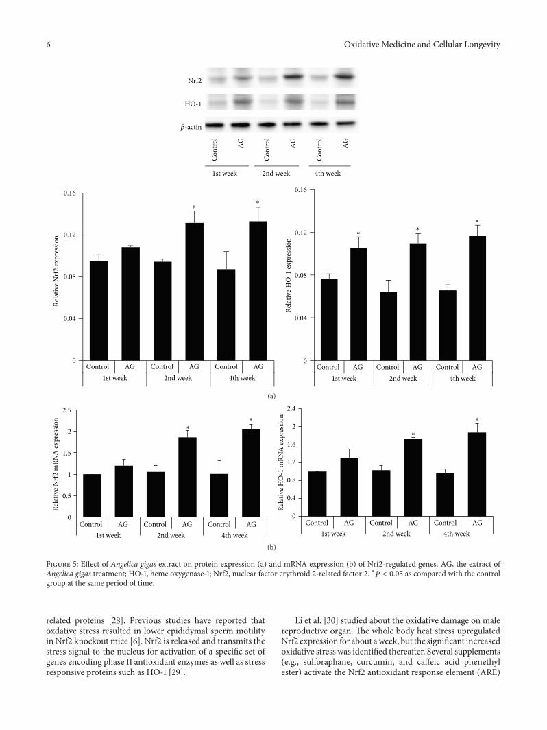
6 Oxidative Medicine and Cellular Longevity
HO-1
Nrf2
𝛽-actin
1st week
AG
Con
trol
AG
Con
trol
AG
Con
trol
2nd week 4th week
0
0.04
0.08
0.12
0.16
Control AG Control AG Control AG1st week 2nd week 4th week
Relat
ive N
rf2 ex
pres
sion
Control AG Control AG Control AG1st week 2nd week 4th week
0
0.04
0.08
0.12
0.16
Relat
ive H
O-1
expr
essio
n
∗∗
∗∗
∗
(a)
0
0.5
1
1.5
2
2.5
Control AG Control AG Control AG1st week 2nd week 4th week
Relat
ive N
rf2 m
RNA
expr
essio
n
0
0.4
0.8
1.2
1.6
2
2.4
Control AG Control AG Control AG1st week 2nd week 4th week
Relat
ive H
O-1
mRN
A ex
pres
sion
∗∗
∗
∗
(b)
Figure 5: Effect of Angelica gigas extract on protein expression (a) and mRNA expression (b) of Nrf2-regulated genes. AG, the extract ofAngelica gigas treatment; HO-1, heme oxygenase-1; Nrf2, nuclear factor erythroid 2-related factor 2. ∗𝑝 < 0.05 as compared with the controlgroup at the same period of time.
related proteins [28]. Previous studies have reported thatoxdative stress resulted in lower epididymal sperm motilityin Nrf2 knockout mice [6]. Nrf2 is released and transmits thestress signal to the nucleus for activation of a specific set ofgenes encoding phase II antioxidant enzymes as well as stressresponsive proteins such as HO-1 [29].
Li et al. [30] studied about the oxidative damage on malereproductive organ. The whole body heat stress upregulatedNrf2 expression for about aweek, but the significant increasedoxidative stress was identified thereafter. Several supplements(e.g., sulforaphane, curcumin, and caffeic acid phenethylester) activate the Nrf2 antioxidant response element (ARE)
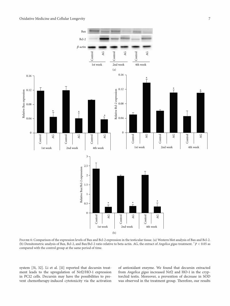
Oxidative Medicine and Cellular Longevity 7
Bax
Bcl-2
𝛽-actin
1st week
AG
Con
trol
AG
Con
trol
AG
Con
trol
2nd week 4th week(a)
0
0.5
1
1.5
2
2.5
3
Con
trol
AG
Con
trol
AG
Con
trol
AG
1st week 2nd week 4th week
Relat
ive B
ax ex
pres
sion
Relat
ive B
cl-2
expr
essio
n
0
0.04
0.08
0.12
0.16
Con
trol
AG
Con
trol
AG
Con
trol
AG
1st week 2nd week 4th week
0
0.04
0.08
0.12
0.16
Con
trol
AG
Con
trol
AG
Con
trol
AG
1st week 2nd week 4th week
Relat
ive B
ax/B
cl-2
expr
essio
n
∗∗
∗
∗
∗∗
∗
∗∗
(b)
Figure 6: Comparison of the expression levels of Bax and Bcl-2 expression in the testicular tissue. (a) Western blot analysis of Bax and Bcl-2.(b) Densitometric analysis of Bax, Bcl-2, and Bax/Bcl-2 ratio relative to beta-actin. AG, the extract of Angelica gigas treatment. ∗𝑝 < 0.05 ascompared with the control group at the same period of time.
system [31, 32]. Li et al. [11] reported that decursin treat-ment leads to the upregulation of Nrf2/HO-1 expressionin PC12 cells. Decursin may have the possibilities to pre-vent chemotherapy-induced cytotoxicity via the activation
of antioxidant enzyme. We found that decursin extractedfrom Angelica gigas increased Nrf2 and HO-1 in the cryp-torchid testis. Moreover, a prevention of decrease in SODwas observed in the treatment group. Therefore, our results
8 Oxidative Medicine and Cellular Longevity
suggest that antioxidant defense is reinforced by decursin viaactivation of Nrf2 and upregulation of antioxidant enzymeactivity.
The Bcl-2 family and related proteins are key regulatorsof apoptosis [33]. They can be classified into two groups:Bcl-2, an antiapoptotic protein, and Bax, a proapoptoticprotein [34]. Knudson et al. [35] demonstrated that the Bcl-2 family mainly plays a role during spermatogenesis in Bax-deficient mice. Several studies have reported the importanceof the Bcl-2 family in heat-induced oxidative stress on malereproductive function [36, 37]. They suggested the possibleinvolvement of Bcl-2 and Bax in the germ cell apoptosis. Wealso investigated the germ cell apoptosis induced by cryp-torchidism and the antiapoptotic effect of decursin extractedfrom Angelica gigas.
We examined the role of decursin extracted from Angel-ica gigas as a supplemental agent to prevent heat-inducedoxidative stress in cryptorchidism. Our limitation is that themore accurate infertility model due to oxidative stress isneeded. More defense mechanisms such as Nrf2 and relatedantioxidative enzyme are required if oxidative stress causesimpaired semen parameters, DNA damage, and apoptosis.Antioxidant supplementation as well as lifestyle change likeno smoking or balanced diet can be helpful for the naturalbalance between ROS and antioxidant. Therefore our studymay support appropriate evidence for the use of a newcomplementary and alternative medicine for treating maleinfertility. In addition, future work should investigate adetailed mechanism of decursin with or without Nrf2 relatedenzyme activity.
The present study suggests that decursin extracted fromAngelica gigas is a supplemental agent that can reduceoxidative stress by Nrf2-mediated upregulation of HO-1 andmay improve cryptorchidism-induced infertility.
Conflict of Interests
The authors declare that there is no conflict of interestsregarding the publication of this paper.
Acknowledgments
This research was supported by a grant of the Korea HealthTechnology R&D Project through the Korea Health IndustryDevelopment Institute (KHIDI), funded by the Ministry ofHealth &Welfare, Republic of Korea (Grant no. HI15C0099).This study was supported by Research Fund of Seoul St.Mary’s Hospital, The Catholic University of Korea.
References
[1] J. P. Jarow, I. D. Sharlip, A.M. Belker et al., “Best practice policiesfor male infertility,” The Journal of Urology, vol. 167, no. 5, pp.2138–2144, 2002.
[2] P.Thonneau, S. Marchand, A. Tallec et al., “Incidence and maincauses of infertility in a resident population (1,850,000) of threeFrench regions (1988-1989),”Human Reproduction, vol. 6, no. 6,pp. 811–816, 1991.
[3] R. Dada, N. P. Gupta, and K. Kucheria, “Spermatogenic arrest inmen with testicular hyperthermia,” Teratog Carcinog Mutagen,supplement 1, pp. 235–243, 2003.
[4] A. Kumagai, H. Kodama, J. Kumagai et al., “Xanthine oxidaseinhibitors suppress testicular germ cell apoptosis induced byexperimental cryptorchidism,”MolecularHumanReproduction,vol. 8, no. 2, pp. 118–123, 2002.
[5] R. M. Vigueras-Villaseñor, I. Ojeda, O. Gutierrez-Pérez et al.,“Protective effect of 𝛼-tocopherol on damage to rat testes byexperimental cryptorchidism,” International Journal of Exper-imental Pathology, vol. 92, no. 2, pp. 131–139, 2011.
[6] B. N. Nakamura, G. Lawson, J. Y. Chan et al., “Knockout of thetranscription factor NRF2 disrupts spermatogenesis in an age-dependent manner,” Free Radical Biology and Medicine, vol. 49,no. 9, pp. 1368–1379, 2010.
[7] B. Yu, H. Lin, L. Yang et al., “Genetic variation in the Nrf2 pro-moter associates with defective spermatogenesis in humans,”Journal of Molecular Medicine, vol. 90, no. 11, pp. 1333–1342,2012.
[8] J. Guo, C. Jiang, Z.Wang et al., “A novel class of pyranocoumarinanti-androgen receptor signaling compounds,”Molecular Can-cer Therapeutics, vol. 6, no. 3, pp. 907–917, 2007.
[9] H. J. Lee, H. J. Lee, E. O. Lee et al., “In vivo anti-cancer activity ofKoreanAngelica gigas and its major pyranocoumarin decursin,”TheAmerican Journal of ChineseMedicine, vol. 37, no. 1, pp. 127–142, 2009.
[10] S. Y. Kang and Y. C. Kim, “Decursinol and decursin protectprimary cultured rat cortical cells from glutamate-inducedneurotoxicity,” Journal of Pharmacy and Pharmacology, vol. 59,no. 6, pp. 863–870, 2007.
[11] L. Li, J.-K. Du, L.-Y. Zou, T. Wu, Y.-W. Lee, and Y.-H. Kim,“Decursin isolated from angelica gigas Nakai rescues PC12 cellsfrom amyloid 𝛽-protein-induced neurotoxicity through Nrf2-mediated upregulation of heme oxygenase-1: potential rolesof MAPK,” Evidence-Based Complementary and AlternativeMedicine, vol. 2013, Article ID 467245, 14 pages, 2013.
[12] H. J. Lee, J.-H. Bach, H.-S. Chae et al., “Mitogen-activatedprotein kinase/extracellular signal-regulated kinase attenuates3-hydroxykynurenine-induced neuronal cell death,” Journal ofNeurochemistry, vol. 88, no. 3, pp. 647–656, 2004.
[13] W. J. Bae, U. S. Ha, K. S. Kim et al., “Effects of KH-204 on theexpression of heat shock protein 70 and germ cell apoptosisin infertility rat models,” BMC Complementary and AlternativeMedicine, vol. 14, article 367, 2014.
[14] K. W. Ko, J. Y. Chae, S. W. Kim et al., “The effect of the partialobstruction site of the renal vein on testis and kidney in rats: isthe traditional animal model suitable for varicocele research?”Korean Journal of Urology, vol. 51, no. 8, pp. 565–571, 2010.
[15] G. Türk, M. Sönmez, M. Aydin et al., “Effects of pomegranatejuice consumption on spermquality, spermatogenic cell density,antioxidant activity and testosterone level in male rats,” ClinicalNutrition, vol. 27, no. 2, pp. 289–296, 2008.
[16] B. P. Setchell, “The Parkes lecture. Heat and the testis,” Journalof Reproduction and Fertility, vol. 114, no. 2, pp. 179–194, 1998.
[17] P. J. Hansen, “Effects of heat stress on mammalian reproduc-tion,”Philosophical Transactions of the Royal Society B: BiologicalSciences, vol. 364, no. 1534, pp. 3341–3350, 2009.
[18] C. Paul, S. Teng, and P. T. K. Saunders, “A single, mild, transientscrotal heat stress causes hypoxia and oxidative stress in mousetestes, which induces germ cell death,” Biology of Reproduction,vol. 80, no. 5, pp. 913–919, 2009.
Oxidative Medicine and Cellular Longevity 9
[19] K. Shiraishi, H. Takihara, and H. Matsuyama, “Elevated scrotaltemperature, but not varicocele grade, reflects testicular oxida-tive stress-mediated apoptosis,” World Journal of Urology, vol.28, no. 3, pp. 359–364, 2010.
[20] R. J. Aitken and S. D. Roman, “Antioxidant systems andoxidative stress in the testes,” Oxidative Medicine and CellularLongevity, vol. 1, no. 1, pp. 15–24, 2008.
[21] A. Agarwal, G. Virk, C. Ong, and S. S. du Plessis, “Effect ofoxidative stress on male reproduction,” The World Journal ofMen’s Health, vol. 32, no. 1, pp. 1–17, 2014.
[22] N. A. Clark, M. Will, M. B. Moravek, and S. Fisseha, “A system-atic review of the evidence for complementary and alternativemedicine in infertility,” International Journal of Gynecology andObstetrics, vol. 122, no. 3, pp. 202–206, 2013.
[23] A. Agarwal and L. H. Sekhon, “The role of antioxidant therapyin the treatment of male infertility,”Human Fertility, vol. 13, no.4, pp. 217–225, 2010.
[24] E. Greco, M. Iacobelli, L. Rienzi, F. Ubaldi, S. Ferrero, and J.Tesarik, “Reduction of the incidence of sperm DNA fragmen-tation by oral antioxidant treatment,” Journal of Andrology, vol.26, no. 3, pp. 349–353, 2005.
[25] A. E. Omu, M. K. Al-Azemi, E. O. Kehinde, J. T. Anim, M.A. Oriowo, and T. C. Mathew, “Indications of the mechanismsinvolved in improved sperm parameters by zinc therapy,”Medical Principles and Practice, vol. 17, no. 2, pp. 108–116, 2008.
[26] M. R. Safarinejad, “Efficacy of coenzyme Q10 on semen param-eters, sperm function and reproductive hormones in infertilemen,” Journal of Urology, vol. 182, no. 1, pp. 237–248, 2009.
[27] L. E. Otterbein and A. M. K. Choi, “Heme oxygenase: colors ofdefense against cellular stress,”American Journal of Physiology—Lung Cellular and Molecular Physiology, vol. 279, no. 6, pp.L1029–L1037, 2000.
[28] T. Ohta, K. Iijima, M. Miyamoto et al., “Loss of Keap1 functionactivates Nrf2 and provides advantages for lung cancer cellgrowth,” Cancer Research, vol. 68, no. 5, pp. 1303–1309, 2008.
[29] K. Itoh, N. Wakabayashi, Y. Katoh, T. Ishii, T. O’Connor,and M. Yamamoto, “Keap1 regulates both cytoplasmic-nuclearshuttling and degradation of Nrf2 in response to electrophiles,”Genes to Cells, vol. 8, no. 4, pp. 379–391, 2003.
[30] Y. Li, Y. Huang, Y. Piao et al., “Protective effects of nuclear factorerythroid 2-related factor 2 on whole body heat stress-inducedoxidative damage in the mouse testis,” Reproductive Biology andEndocrinology, vol. 11, article 23, 2013.
[31] C. Chen and A.-N. T. Kong, “Dietary chemopreventive com-pounds and ARE/EpRE signaling,” Free Radical Biology andMedicine, vol. 36, no. 12, pp. 1505–1516, 2004.
[32] Y. Morimitsu, Y. Nakagawa, K. Hayashi et al., “A sulforaphaneanalogue that potently activates the Nrf2-dependent detoxifica-tion pathway,” The Journal of Biological Chemistry, vol. 277, no.5, pp. 3456–3463, 2002.
[33] J. M. Adams and S. Cory, “The Bcl-2 protein family: arbiters ofcell survival,” Science, vol. 281, no. 5381, pp. 1322–1326, 1998.
[34] Y.Akao, Y.Otsuki, S. Kataoka, Y. Ito, andY. Tsujimoto, “Multiplesubcellular localization of bcl-2: detection in nuclear outermembrane, endoplasmic reticulum membrane, and mitochon-drial membranes,” Cancer Research, vol. 54, no. 9, pp. 2468–2471, 1994.
[35] C. M. Knudson, K. S. K. Tung, W. G. Tourtellotte, G. A. J.Brown, and S. J. Korsmeyer, “Bax-deficient mice with lymphoidhyperplasia and male germ cell death,” Science, vol. 270, no.5233, pp. 96–99, 1995.
[36] C. M. Yamamoto, A. P. Sinha Hikim, P. N. Huynh et al.,“Redistribution of Bax is an early step in an apoptotic pathwayleading to germ cell death in rats, triggered by mild testicularhyperthermia,” Biology of Reproduction, vol. 63, no. 6, pp. 1683–1690, 2000.
[37] Z.-H. Zhang, X. Jin, X.-S. Zhang et al., “Bcl-2 and Bax areinvolved in experimental cryptorchidism-induced testiculargerm cell apoptosis in rhesus monkey,” Contraception, vol. 68,no. 4, pp. 297–301, 2003.
Research ArticlePeriodic Exposure of Keratinocytes to Cold Physical Plasma:An In Vitro Model for Redox-Related Diseases of the Skin
Anke Schmidt, Thomas von Woedtke, and Sander Bekeschus
Leibniz-Institute for Plasma Science and Technology (INP Greifswald), ZIK plasmatis, Felix-Hausdorff-Strasse 2,17489 Greifswald, Germany
Correspondence should be addressed to Sander Bekeschus; [email protected]
Received 25 September 2015; Revised 3 December 2015; Accepted 20 December 2015
Academic Editor: Sergio Di Meo
Copyright © 2016 Anke Schmidt et al. This is an open access article distributed under the Creative Commons Attribution License,which permits unrestricted use, distribution, and reproduction in any medium, provided the original work is properly cited.
Oxidative stress illustrates an imbalance between radical formation and removal. Frequent redox stress is critically involved inmanyhuman pathologies including cancer, psoriasis, and chronic wounds. However, reactive species pursue a dual role being involved insignaling on the one hand and oxidative damage on the other. Using aHaCaT keratinocyte cell culturemodel, we investigated redoxregulation and inflammation to periodic, low-dose oxidative stress after two, six, eight, ten, and twelve weeks. Chronic redox stresswas generated by recurrent incubation with cold physical plasma-treated cell culture medium. Using transcriptome microarraytechnology, we identified both acute ROS-stress responses as well as numerous adaptions after several weeks of redox challenge.We determined a differential expression (2-fold, FDR < 0.01, 𝑝 < 0.05) of 260 genes that function in inflammation and redoxhomeostasis, such as cytokines (e.g., IL-6, IL-8, and IL-10), growth factors (e.g., CSF2, FGF, and IGF-2), and antioxidant enzymes(e.g., HMOX, NQO1, GPX, and PRDX). Apoptotic signaling was affected rather modestly, especially in p53 downstream targets(e.g., BCL2, BBC3, and GADD45). Strikingly, the cell-protective heat shock protein HSP27 was strongly upregulated (𝑝 < 0.001).These results suggested cellular adaptions to frequent redox stress and may help to better understand the inflammatory responsesin redox-related diseases.
1. Introduction
The skin protects against environmental assaults and iscrucial for maintaining the redox homeostasis [1]. The redoxstate is determined by the balance between pro- and antiox-idative processes involving reactive oxygen and nitrogenspecies (ROS/RNS) [2]. Excessive presence of ROS/RNS isassociated with a number of pathologies of the skin. Thisincludes cutaneous malignancies [3], chronic wounds [4],and psoriasis [5]. There were an estimated 3.45 million newcases of cancer and 1.75 million deaths from cancer in Europein 2012 [6]. The incidence of chronic wounds varies [7] butis expected to affect about 6.5 million patients in the UnitedStates [8]. The prevalence of psoriasis ranges from 0% to8.5% according to age and geographic region [9]. Yet, it isimportant to recognize the dual role of reactive species, alsoin the human skin [10]. At low concentrations, they serve assignaling molecules [11] while at higher concentrations theybecome increasingly cytotoxic [12].
Generally, oxidants can be of exogenous or endogenousorigin. The former derive from, for example, ozone, ionizingand nonionizing radiation, cigarette smoke, or invasion ofpathogens and their associated infections [13]. The latterspring from oxygen-metabolizing enzymatic reactions (oxi-dases) and the leakage of superoxide from mitochondria[14]. Upon stimulation, the inducible nitric oxide (NO)synthase contributes to RNS production by generating largeamounts of NO for defense or signaling purposes [15], also inkeratinocytes [16]. Disturbance of the physiological balancebetween formation and removal of ROS/RNS (oxidativestress) disrupts the accurate interplay between the affiliatedcells [17]. However, most toxic effects are counterbalanced bythe complex and finely tuned antioxidative defense system[18].
We here attempted to investigate the redox response ofHaCaT keratinocytes over three months. Frequent exposureto reactive species was generated using cold physical plasma.Cold plasma has been shown to have therapeutic benefit in
Hindawi Publishing CorporationOxidative Medicine and Cellular LongevityVolume 2016, Article ID 9816072, 17 pageshttp://dx.doi.org/10.1155/2016/9816072
2 Oxidative Medicine and Cellular Longevity
diseases of the skin, such as chronic wounds and psoriasis[19–21]. The advantage of cold physical plasma over singleoxidative agents is that it provides a more complex mixtureof reactive molecules. Plasma was used tomimic the frequentredox stress apparent in redox-related diseases on the onehand and to investigate the effect of its long-term use onthe other. We hypothesized that the cellular responses wouldsubstantially differ in cells exposed to rather acute (up to twoweeks) or chronic (up to twelve weeks) oxidative challenge.Understanding these differences may help to better under-stand the skin tissue response in pathologies that involvechronic redox stress. Several studies have investigated acuteoxidative stress conditions in keratinocyte skin cell culturemodels [22–24]. In contrast to primary skin tissue, in vitrocultures obviously allow the investigation of cells with thesame genetic background over long investigation times andunder highly controlled conditions.
Following repeated redox challenge, we used transcrip-tome microarray technology to study the molecular mecha-nisms and to identify markers of cellular redox modulationin keratinocytes. Periodic redox stress was applied over3 months and coincided with an altered gene expressionlinked to cell metabolism, inflammation, and general stressresponse. Ourmodel systemwas designed to emphasize mildredox conditions rather than applying cytotoxic dosages. Wewere able to showdistinct transcript profiles of cytokines, ker-atins, and growth factors as well as an imperative involvementof junctional proteins in redox adaptions. Collectively, thepresented system-wide modifications suggest that the simul-taneous alteration of multiple pathways provides an impor-tant in-depth transcriptome overview linking permanentplasma-evoked oxidative stress effects with cell responses andadaption mechanisms, conceivably having implications inredox-based diseases of the skin.
2. Material and Methods
2.1. Cell Culture and Cold Physical Plasma. Three mil-lion epithelial keratinocyte HaCaT cells [25] were culturedin 75 cm2 dishes using RPMI1640 cell culture mediumsupplemented with 8% bovine calf serum (Sigma, USA),0.1mg/mL penicillin/streptomycin, and 2mM L-glutamine(Lonza, Switzerland). Cold physical plasma-treated cell cul-ture medium was generated by exposure of 5mL of fullysupplemented medium to an atmospheric pressure argonplasma jet (60 s; kINPen 09; neoplas tools, Germany) [26].Three standard liters per minute of argon (99.99%; AirLiquide, France) were used as feed gas. The plasma wasgenerated by applying a voltage of 2–6 kVpp with a fre-quency of 1.0–1.1MHz to the central electrode. Three millionHaCaT keratinocytes were exposed to 15mL of plasma-treated medium twice a week and passaged once a week(Figure 2(a)). After two, six, eight, ten, and twelve weeks, cellswere either subjected to microscopic experiments or RNA orprotein isolation.
2.2. Flow Cytometry. Keratinocytes were loaded with 5 𝜇Mof CM-H
2DCF-DA or mitotracker deep red (MTDR; both
life technologies, USA) according to the manufacturer’sinstructions. After exposure to plasma-treated medium, cellswere trypsinized and green fluorescence was quantifiedusing a Gallios flow cytometer (Beckman-Coulter, USA). Forassessment of apoptosis, keratinocytes were plasma-treatedand incubated for 18 h at 37∘C. Etoposide (50 𝜇M) was usedas positive control (BioVision, USA). Subsequently, cells wereloaded with active caspase 3 detection reagent (Enzo, USA)according to the vendor’s protocol. Keratinocytes were thentrypsinized and subjected to flow cytometric evaluation. Dataanalysis was conducted using Kaluza software (Beckman-Coulter).
2.3. Transcriptomic Analysis. RNA was purified accordingto the manufacturer’s instructions (Bio&Sell, Germany) andRNA quality was maximal (scores of 10 out of 10; data notshown) as evaluated using low-volume gel electrophoresis(Bioanalyzer 2010; Agilent, USA). For cRNA generation,RNA of two independent experiments was pooled for eachsample. cRNA was conjugated to Cy3, and transcriptionalanalysis was carried out using microarray-based exon anal-ysis (Agilent) as described before [27]. Briefly, samples werehybridized onto SurePrint G3 custom GE 8 × 60K chips(OakLabs, Germany) for 17 h at 65∘C in a hybridizationoven (Agilent). After washing, fluorescence intensities wererecorded using a gene chip scanner (Agilent). Background-corrected signal intensities were determined and processedusing feature extraction software (Agilent). Normalization,statistical tests, clustering, and filtering methods were con-ducted using gene expression analysis software (Partek,USA). Microarray data were deposited into gene expressionomnibus database (GSE4876). Data were grouped accordingto their respective treatment regime (2, 6, 8, 10, and 12 weeks)and analyzed statistically usingmultiple testing corrections toidentify differentially expressed genes (at least ±2-fold with𝑝 < 0.05). Also expression changes of less than ±2-fold areshown if target expression at one or more time points wasgreater than ±2-fold. Additionally, sets of coregulated geneswere functionally clustered, and their biological relevancewas analyzed using PANTHER software.
2.4. Protein Analysis. Instead of validating a large numberof microarray results, we rather chose protein targets basedon their significance within main cellular responses. Theseincluded oxidative defense (PRDX2 was among the 333always differentially expressed genes (Figure 2(c)); HSP27and its phosphorylated form were previously found to becentral, studying the redox response in THP-1 monocytes[28]; NQO1 was also found to be redox-controlled using coldplasma [27]), cellular damage and apoptosis (histone H2AXand its phosphorylation are prominent markers of genomicDNA damage; Puma [29] and p21 [30] are involved in redox-related apoptosis induction), and cellular structure (keratin 1showed the strongest regulation among all keratins (Table 2);𝛽-actin served as housekeeping protein). For protein isola-tion, cells were lysed in RIPA buffer containing protease andphosphatase inhibitors (cOmplete Mini, phosSTOP; Roche,Germany) and 2mM phenylmethanesulfonylfluoride (Roth,
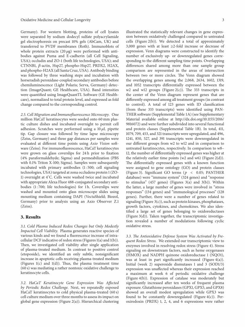
Oxidative Medicine and Cellular Longevity 3
Germany). For western blotting, proteins of cell lysateswere separated by sodium dodecyl sulfate polyacrylamidegel electrophoresis on precast 10% gels (AbCam, UK) andtransferred to PVDF membranes (Roth). Immunoblots ofwhole protein extracts (20𝜇g) were performed with anti-bodies against Puma and E-cadherin (all Cell Signaling,USA); occludin and ZO-1 (both life technologies, USA); andCTNNB1, 𝛽-actin, Hsp27, phospho-Hsp27, PRDX2, H2AX,and phospho-H2AX (all SantaCruz,USA).Antibody bindingwas followed by three washing steps and incubation withhorseradish peroxidase-coupled secondary antibodies beforechemiluminescence (Light Polaris; Serva, Germany) detec-tion (ImageQuant; GE Healthcare, USA). Band intensitieswere quantified using ImageQuantTL Software (GE Health-care), normalized to total protein level, and expressed as foldchange compared to the corresponding control.
2.5. Cell Migration and ImmunofluorescenceMicroscopy. Onemillion HaCaT keratinocytes were seeded onto 60mm plas-tic culture dishes and incubated overnight to permit celladhesion. Scratches were performed using a 10 𝜇L pipettetip. Gap closure was followed by time lapse microscopy(Zeiss, Germany) and three gap distances per samples wereevaluated at different time points using Axio Vision soft-ware (Zeiss). For immunofluorescence, HaCaT keratinocyteswere grown on glass coverslips for 24 h prior to fixation(4% paraformaldehyde; Sigma) and permeabilization (PBSwith 0.1% Triton X-100; Sigma). Samples were subsequentlyincubated with primary antibodies (1 : 500; cell signalingtechnologies, USA) targeted at zona occludens protein 1 (ZO-1) overnight at 4∘C. Cells were washed twice and incubatedwith appropriate Alexa Fluor 488-conjugated secondary anti-bodies (1 : 700; life technologies) for 1 h. Coverslips werewashed and mounted onto glass microscope slides usingmounting medium containing DAPI (VectaShield; Biozol,Germany) prior to analysis using an Axio Observer Z.1(Zeiss).
3. Results
3.1. Cold Plasma Induced Redox Changes but Only ModestlyImpacted Cell Viability. Plasma generates reactive species ofvarious kinds and we found a fluorescence increase of intra-cellular DCF indicative of redox stress (Figures 1(a) and 1(b)).Then, we investigated cell viability after single applicationof plasma-treated medium. In contrast to positive control(etoposide), we identified an only subtle, nonsignificantincrease in apoptotic cells receiving plasma-treated medium(Figures 1(c) and 1(d)). Thus, the plasma treatment time(60 s) was mediating a rather nontoxic oxidative challenge tokeratinocyte cells.
3.2. HaCaT Keratinocyte Gene Expression Was Affectedby Periodic Redox Challenge. Next, we repeatedly exposedHaCaT keratinocytes to cold physical plasma-treated (60 s)cell culturemedium over threemonths to assess its impact onglobal gene expression (Figure 2(a)). Hierarchical clustering
illustrated the statistically relevant changes in gene expres-sion between oxidatively challenged compared to untreatedcells (Figure 2(b)). We detected a total of approximately3,000 genes with at least ≥2-fold increase or decrease ofexpression. Venn diagrams were constructed to identify thenumber of exclusively up- or downregulated genes corre-sponding to the different sampling time points. Overlappingdifferences shared among more than one sample groupcomparison are represented in the areas of intersectionbetween two or more circles. The Venn diagram showedthe overlapping genes among the 2,068, 2634, 1602, 1319,and 1052 transcripts differentially expressed between thew2 and w12 groups (Figure 2(c)). The 333 transcripts inthe center of the Venn diagram represent genes that aredifferently expressed among all treatment groups (in contrastto control). A total of 123 genes with ID classification(from these 333 transcripts) were identified using PAN-THER software (Supplemental Table 1A) (see SupplementaryMaterial available online at http://dx.doi.org/10.1155/2016/9816072) and were further subdivided into several functionaland protein classes (Supplemental Table 1B). In total, 411,1079, 709, 453, and 321 transcripts were upregulated, and 496,1241, 850, 527, and 391 transcripts were downregulated inour different groups from w2 to w12 and in comparison tountreated keratinocytes, respectively. In comparison to w8–12, the number of differentially expressed genes was higher atthe relatively earlier time points (w2 and w6) (Figure 2(d)).The differentially expressed genes with a known functionwere assigned to gene ontology (GO) and protein classes(Figure 3). Significant GO terms (𝑝 < 0.05; PANTHERdatabase) were “immune system” (324 genes) and “responseto stimulus” (457 genes) (Figures 3(a) and 3(b)). Withinthe latter, a large number of genes were involved in “stressresponses” (134 genes) and “immunological processes” (128genes). Further, there were a number of genes related tosignaling (Figure 3(c)), such as protein kinases, phosphatases,growth factors, cytokines, and chemokines. We also iden-tified a large set of genes belonging to oxidoreductases(Figure 3(d)). Taken together, the transcriptomic investiga-tion revealed a number of modulations following cyclicoxidative stress.
3.3. The Antioxidative Defense System Was Activated by Fre-quent Redox Stress. We extended our transcriptomic view toenzymes involved in resolving redox stress (Figure 4). Stresssignaling on downstream factors, such as heme oxygenases(HMOX) and NADPH quinone oxidoreductase 1 (NQO1),was at least in part significantly increased (Figure 4(a)).Initial (week 2) superoxide dismutases 1 and 3 (SOD1/3)expression was unaffected whereas their expression reacheda maximum at week 6 of periodic oxidative challenge(Figure 4(b)). Expression of catalase was moderately butsignificantly increased after ten weeks of frequent plasmaexposure. Glutathione peroxidases (GPX1, GPX5, andGPX8)showed an overall modest upregulation while GPX3 wasfound to be constantly downregulated (Figure 4(c)). Per-oxiredoxin (PRDX) 1, 2, 4, and 6 expressions were rather
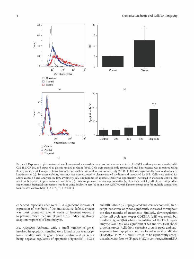
4 Oxidative Medicine and Cellular Longevity
80
60
40
20
0
Cou
nt
100 101 102 103
DCF fluorescence
UnstainedControlPlasma
(a)
0
5
10
15
20
MFI
Control Plasma
∗
(b)
30
20
10
0
Cou
nt
101 102 103
Nuclear fluorescence
Etoposide
ControlPlasma
(c)
0
10
20
30Ap
opto
tic ce
lls (%
) ∗∗∗
Etoposide60 s40 s20 sControl
(d)
Figure 1: Exposure to plasma-treated medium evoked acute oxidative stress but was not cytotoxic. HaCaT keratinocytes were loaded withCM-H
2DCF-DA and exposed to plasma-treated medium (60 s). Cells were subsequently trypsinized and fluorescence was measured using
flow cytometry (a). Compared to control cells, intracellular mean fluorescence intensity (MFI) of DCF was significantly increased in treatedkeratinocytes (b). To assess viability, keratinocytes were exposed to plasma-treated medium and incubated for 18 h. Cells were stained foractive caspase 3 and analyzed by flow cytometry (c). The number of apoptotic cells was significantly increased in etoposide control butnot in cells exposed to plasma-treated medium (d). Data are presented as one representative (a, c) or mean + SD (b, d) of two independentexperiments. Statistical comparisonwas done using Student’s 𝑡-test (b) or one-wayANOVAwithDunnett corrections formultiple comparisonto untreated control (d) (∗𝑝 < 0.05, ∗∗∗𝑝 < 0.001).
enhanced, especially after week 6. A significant increase ofexpression of members of the antioxidative defense systemwas most prominent after 6 weeks of frequent exposureto plasma-treated medium (Figure 4(d)), indicating strongadaption responses of keratinocytes.
3.4. Apoptosis Pathways. Only a small number of genesinvolved in apoptotic signaling were found in our transcrip-tomic studies with 31 genes being positive and 17 genesbeing negative regulators of apoptosis (Figure 5(a)). BCL2
and BBC3 (both p53-upregulated inducers of apoptosis) tran-script levels were only nonsignificantly increased throughoutthe three months of treatments. Similarly, downregulationof the cell cycle gate-keeper CDKN1A (p21) was steady butmodest (Figure 5(b)) while upregulation of the DNA repairenzyme GADD45 was significant at w2 and w6. Heat shockproteins protect cells from excessive protein stress and sub-sequently from apoptosis, and we found several candidates(HSP90A, HSP90AB, andHSP90B) to be significantly upreg-ulated atw2 and/orw6 (Figure 5(c)). In contrast, actinmRNA
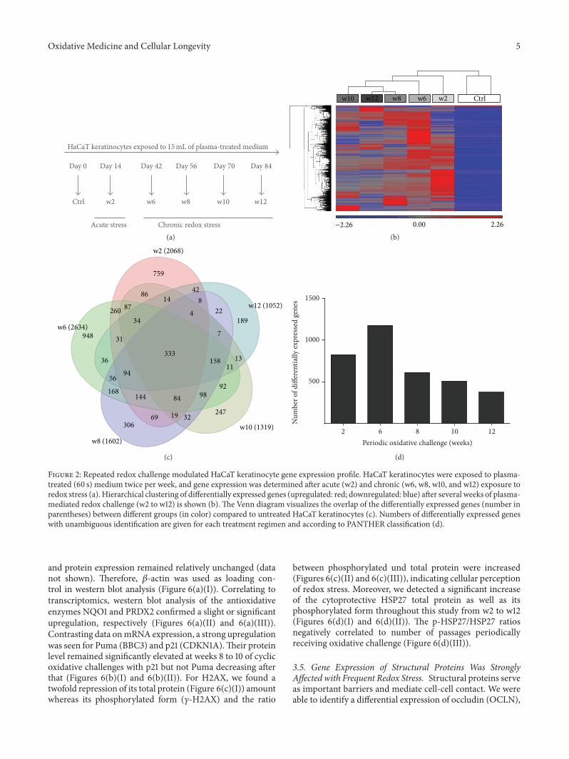
Oxidative Medicine and Cellular Longevity 5
w2 w6 w8 w10 w12Ctrl
Day 14 Day 42 Day 70Day 56 Day 84Day 0
Chronic redox stressAcute stress
HaCaT keratinocytes exposed to 15mL of plasma-treated medium
(a)
2.260.00−2.26
w2w6w8w12w10 Ctrl
(b)
w6 (2634)948
36
56
168144
30669 19 32
84 98
247
92
1113
189
7
158333
94
31
34260 87
86 14
48
22
42
759
w8 (1602)
w10 (1319)
w12 (1052)
w2 (2068)
(c)
102 6 8 12
500
1000
1500
Periodic oxidative challenge (weeks)
Num
ber o
f diff
eren
tially
expr
esse
d ge
nes
(d)
Figure 2: Repeated redox challenge modulated HaCaT keratinocyte gene expression profile. HaCaT keratinocytes were exposed to plasma-treated (60 s) medium twice per week, and gene expression was determined after acute (w2) and chronic (w6, w8, w10, and w12) exposure toredox stress (a). Hierarchical clustering of differentially expressed genes (upregulated: red; downregulated: blue) after several weeks of plasma-mediated redox challenge (w2 to w12) is shown (b). The Venn diagram visualizes the overlap of the differentially expressed genes (number inparentheses) between different groups (in color) compared to untreated HaCaT keratinocytes (c). Numbers of differentially expressed geneswith unambiguous identification are given for each treatment regimen and according to PANTHER classification (d).
and protein expression remained relatively unchanged (datanot shown). Therefore, 𝛽-actin was used as loading con-trol in western blot analysis (Figure 6(a)(I)). Correlating totranscriptomics, western blot analysis of the antioxidativeenzymes NQO1 and PRDX2 confirmed a slight or significantupregulation, respectively (Figures 6(a)(II) and 6(a)(III)).Contrasting data onmRNA expression, a strong upregulationwas seen for Puma (BBC3) and p21 (CDKN1A).Their proteinlevel remained significantly elevated at weeks 8 to 10 of cyclicoxidative challenges with p21 but not Puma decreasing afterthat (Figures 6(b)(I) and 6(b)(II)). For H2AX, we found atwofold repression of its total protein (Figure 6(c)(I)) amountwhereas its phosphorylated form (𝛾-H2AX) and the ratio
between phosphorylated und total protein were increased(Figures 6(c)(II) and 6(c)(III)), indicating cellular perceptionof redox stress. Moreover, we detected a significant increaseof the cytoprotective HSP27 total protein as well as itsphosphorylated form throughout this study from w2 to w12(Figures 6(d)(I) and 6(d)(II)). The p-HSP27/HSP27 ratiosnegatively correlated to number of passages periodicallyreceiving oxidative challenge (Figure 6(d)(III)).
3.5. Gene Expression of Structural Proteins Was StronglyAffected with Frequent Redox Stress. Structural proteins serveas important barriers and mediate cell-cell contact. We wereable to identify a differential expression of occludin (OCLN),
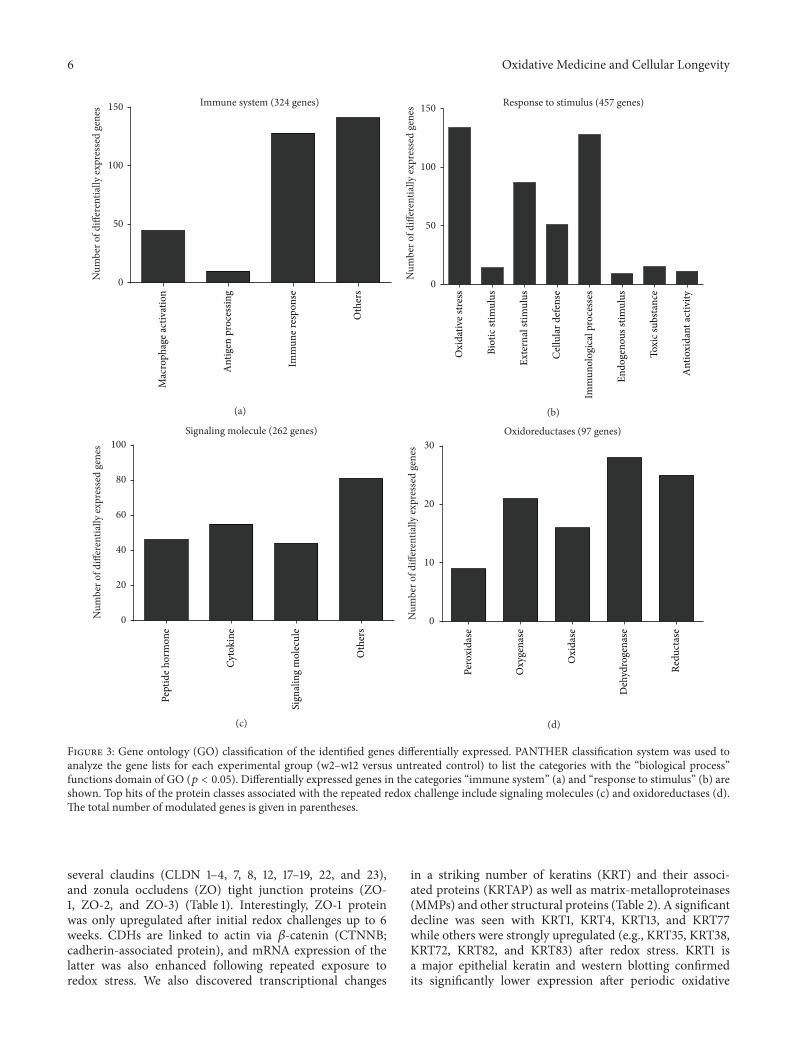
6 Oxidative Medicine and Cellular Longevity
0
50
100
150 Immune system (324 genes)
Mac
roph
age a
ctiv
atio
n
Ant
igen
pro
cess
ing
Imm
une r
espo
nse
Oth
ers
Num
ber o
f diff
eren
tially
expr
esse
d ge
nes
(a)
0
50
100
150 Response to stimulus (457 genes)
Oxi
dativ
e stre
ss
Biot
ic st
imul
us
Exte
rnal
stim
ulus
Cel
lula
r def
ense
Imm
unol
ogic
al p
roce
sses
Endo
geno
us st
imul
us
Toxi
c sub
stan
ce
Ant
ioxi
dant
activ
ity
Num
ber o
f diff
eren
tially
expr
esse
d ge
nes
(b)
0
20
40
60
80
100Signaling molecule (262 genes)
Pept
ide h
orm
one
Sign
alin
g m
olec
ule
Oth
ers
Num
ber o
f diff
eren
tially
expr
esse
d ge
nes
Cyto
kine
(c)
0
10
20
30Oxidoreductases (97 genes)
Pero
xida
se
Oxy
gena
se
Oxi
dase
Deh
ydro
gena
se
Redu
ctas
e
Num
ber o
f diff
eren
tially
expr
esse
d ge
nes
(d)
Figure 3: Gene ontology (GO) classification of the identified genes differentially expressed. PANTHER classification system was used toanalyze the gene lists for each experimental group (w2–w12 versus untreated control) to list the categories with the “biological process”functions domain of GO (𝑝 < 0.05). Differentially expressed genes in the categories “immune system” (a) and “response to stimulus” (b) areshown. Top hits of the protein classes associated with the repeated redox challenge include signaling molecules (c) and oxidoreductases (d).The total number of modulated genes is given in parentheses.
several claudins (CLDN 1–4, 7, 8, 12, 17–19, 22, and 23),and zonula occludens (ZO) tight junction proteins (ZO-1, ZO-2, and ZO-3) (Table 1). Interestingly, ZO-1 proteinwas only upregulated after initial redox challenges up to 6weeks. CDHs are linked to actin via 𝛽-catenin (CTNNB;cadherin-associated protein), and mRNA expression of thelatter was also enhanced following repeated exposure toredox stress. We also discovered transcriptional changes
in a striking number of keratins (KRT) and their associ-ated proteins (KRTAP) as well as matrix-metalloproteinases(MMPs) and other structural proteins (Table 2). A significantdecline was seen with KRT1, KRT4, KRT13, and KRT77while others were strongly upregulated (e.g., KRT35, KRT38,KRT72, KRT82, and KRT83) after redox stress. KRT1 isa major epithelial keratin and western blotting confirmedits significantly lower expression after periodic oxidative
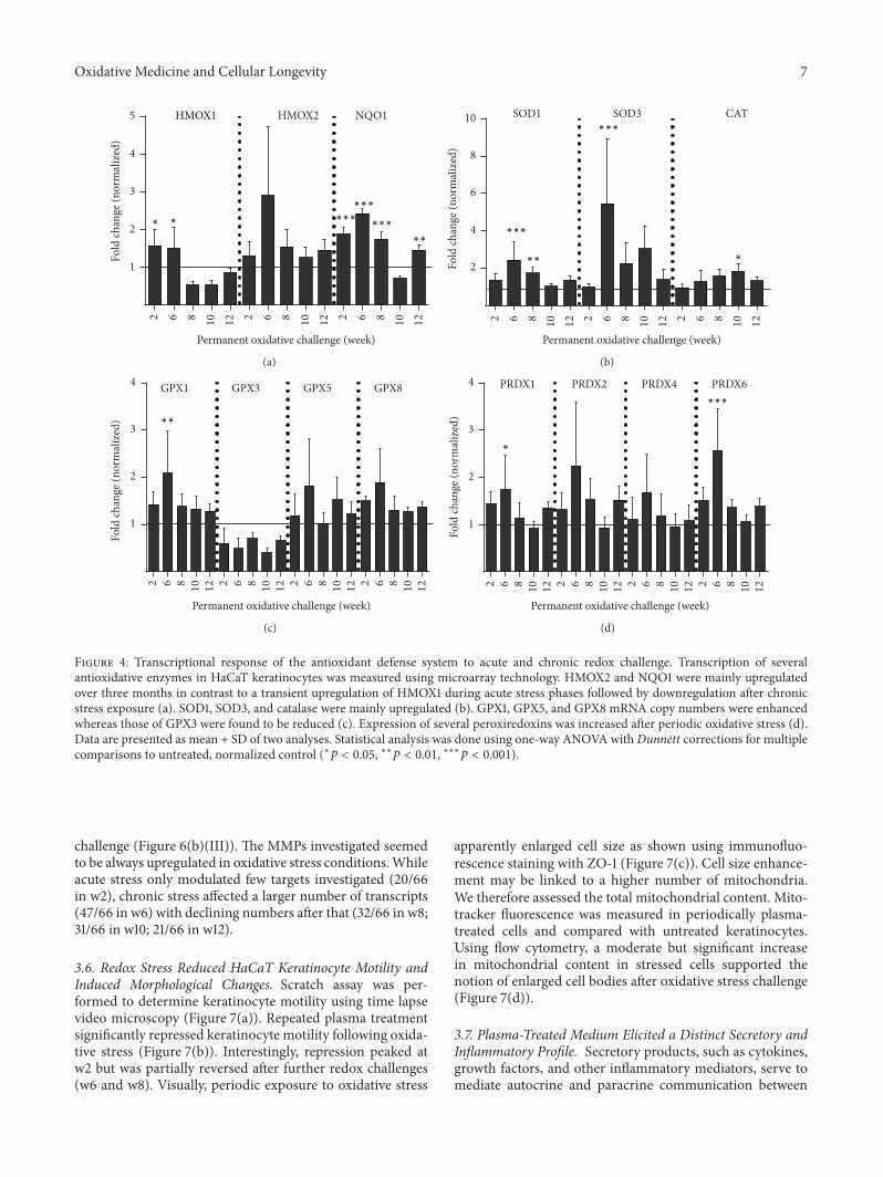
Oxidative Medicine and Cellular Longevity 7
2 6 8 10 12 2 6 8 10 12 2 6 8 10 12
1
2
3
4
5Fo
ld ch
ange
(nor
mal
ized
)NQO1
∗ ∗
HMOX1 HMOX2
Permanent oxidative challenge (week)
∗∗
∗∗∗
∗∗∗∗∗∗
(a)
2 6 8 10 12 2 6 8 10 12 2 6 8 10 12
2
4
6
8
10 SOD3 CAT
Fold
chan
ge (n
orm
aliz
ed)
Permanent oxidative challenge (week)
∗∗∗
∗∗∗
∗∗
SOD1
∗
(b)
2 6 8 10 12 2 6 8 10 12 2 6 8 10 12 2 6 8 10 12
1
2
3
4
Fold
chan
ge (n
orm
aliz
ed)
GPX1 GPX3 GPX5 GPX8
Permanent oxidative challenge (week)
∗∗
(c)
2 6 8 10 12 2 6 8 10 12 2 6 8 10 12 2 6 8 10 12
1
2
3
4
∗Fo
ld ch
ange
(nor
mal
ized
)
Permanent oxidative challenge (week)
∗∗∗
PRDX1 PRDX2 PRDX4 PRDX6
(d)
Figure 4: Transcriptional response of the antioxidant defense system to acute and chronic redox challenge. Transcription of severalantioxidative enzymes in HaCaT keratinocytes was measured using microarray technology. HMOX2 and NQO1 were mainly upregulatedover three months in contrast to a transient upregulation of HMOX1 during acute stress phases followed by downregulation after chronicstress exposure (a). SOD1, SOD3, and catalase were mainly upregulated (b). GPX1, GPX5, and GPX8 mRNA copy numbers were enhancedwhereas those of GPX3 were found to be reduced (c). Expression of several peroxiredoxins was increased after periodic oxidative stress (d).Data are presented as mean + SD of two analyses. Statistical analysis was done using one-way ANOVA withDunnett corrections for multiplecomparisons to untreated, normalized control (∗𝑝 < 0.05, ∗∗𝑝 < 0.01, ∗∗∗𝑝 < 0.001).
challenge (Figure 6(b)(III)). The MMPs investigated seemedto be always upregulated in oxidative stress conditions.Whileacute stress only modulated few targets investigated (20/66in w2), chronic stress affected a larger number of transcripts(47/66 in w6) with declining numbers after that (32/66 in w8;31/66 in w10; 21/66 in w12).
3.6. Redox Stress Reduced HaCaT Keratinocyte Motility andInduced Morphological Changes. Scratch assay was per-formed to determine keratinocyte motility using time lapsevideo microscopy (Figure 7(a)). Repeated plasma treatmentsignificantly repressed keratinocyte motility following oxida-tive stress (Figure 7(b)). Interestingly, repression peaked atw2 but was partially reversed after further redox challenges(w6 and w8). Visually, periodic exposure to oxidative stress
apparently enlarged cell size as shown using immunofluo-rescence staining with ZO-1 (Figure 7(c)). Cell size enhance-ment may be linked to a higher number of mitochondria.We therefore assessed the total mitochondrial content. Mito-tracker fluorescence was measured in periodically plasma-treated cells and compared with untreated keratinocytes.Using flow cytometry, a moderate but significant increasein mitochondrial content in stressed cells supported thenotion of enlarged cell bodies after oxidative stress challenge(Figure 7(d)).
3.7. Plasma-Treated Medium Elicited a Distinct Secretory andInflammatory Profile. Secretory products, such as cytokines,growth factors, and other inflammatory mediators, serve tomediate autocrine and paracrine communication between
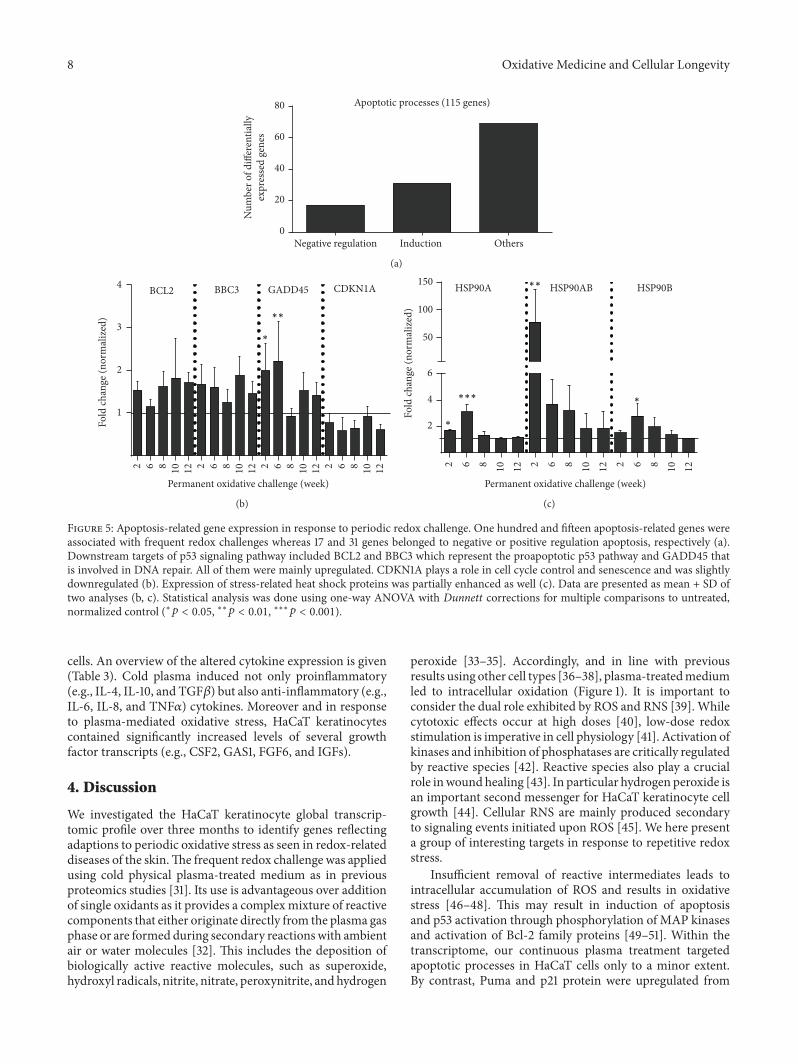
8 Oxidative Medicine and Cellular Longevity
Induction Others0
20
40
60
80 Apoptotic processes (115 genes)
Num
ber o
f diff
eren
tially
expr
esse
d ge
nes
Negative regulation
(a)
2 6 8 10 12 2 6 8 10 12 2 6 8 10 12 2 6 8 10 12
1
2
3
4
∗
∗∗
Fold
chan
ge (n
orm
aliz
ed)
BCL2 BBC3 GADD45 CDKN1A
Permanent oxidative challenge (week)
(b)
2 6 8 10 12 2 6 8 10 12 2 6 8 10 12
2
4
6
50
100
150 HSP90A HSP90B
∗
∗∗∗∗Fo
ld ch
ange
(nor
mal
ized
)
∗∗
Permanent oxidative challenge (week)
HSP90AB
(c)
Figure 5: Apoptosis-related gene expression in response to periodic redox challenge. One hundred and fifteen apoptosis-related genes wereassociated with frequent redox challenges whereas 17 and 31 genes belonged to negative or positive regulation apoptosis, respectively (a).Downstream targets of p53 signaling pathway included BCL2 and BBC3 which represent the proapoptotic p53 pathway and GADD45 thatis involved in DNA repair. All of them were mainly upregulated. CDKN1A plays a role in cell cycle control and senescence and was slightlydownregulated (b). Expression of stress-related heat shock proteins was partially enhanced as well (c). Data are presented as mean + SD oftwo analyses (b, c). Statistical analysis was done using one-way ANOVA with Dunnett corrections for multiple comparisons to untreated,normalized control (∗𝑝 < 0.05, ∗∗𝑝 < 0.01, ∗∗∗𝑝 < 0.001).
cells. An overview of the altered cytokine expression is given(Table 3). Cold plasma induced not only proinflammatory(e.g., IL-4, IL-10, and TGF𝛽) but also anti-inflammatory (e.g.,IL-6, IL-8, and TNF𝛼) cytokines. Moreover and in responseto plasma-mediated oxidative stress, HaCaT keratinocytescontained significantly increased levels of several growthfactor transcripts (e.g., CSF2, GAS1, FGF6, and IGFs).
4. Discussion
We investigated the HaCaT keratinocyte global transcrip-tomic profile over three months to identify genes reflectingadaptions to periodic oxidative stress as seen in redox-relateddiseases of the skin.The frequent redox challenge was appliedusing cold physical plasma-treated medium as in previousproteomics studies [31]. Its use is advantageous over additionof single oxidants as it provides a complex mixture of reactivecomponents that either originate directly from the plasma gasphase or are formed during secondary reactionswith ambientair or water molecules [32]. This includes the deposition ofbiologically active reactive molecules, such as superoxide,hydroxyl radicals, nitrite, nitrate, peroxynitrite, and hydrogen
peroxide [33–35]. Accordingly, and in line with previousresults using other cell types [36–38], plasma-treatedmediumled to intracellular oxidation (Figure 1). It is important toconsider the dual role exhibited by ROS and RNS [39]. Whilecytotoxic effects occur at high doses [40], low-dose redoxstimulation is imperative in cell physiology [41]. Activation ofkinases and inhibition of phosphatases are critically regulatedby reactive species [42]. Reactive species also play a crucialrole inwoundhealing [43]. In particular hydrogen peroxide isan important second messenger for HaCaT keratinocyte cellgrowth [44]. Cellular RNS are mainly produced secondaryto signaling events initiated upon ROS [45]. We here presenta group of interesting targets in response to repetitive redoxstress.
Insufficient removal of reactive intermediates leads tointracellular accumulation of ROS and results in oxidativestress [46–48]. This may result in induction of apoptosisand p53 activation through phosphorylation of MAP kinasesand activation of Bcl-2 family proteins [49–51]. Within thetranscriptome, our continuous plasma treatment targetedapoptotic processes in HaCaT cells only to a minor extent.By contrast, Puma and p21 protein were upregulated from
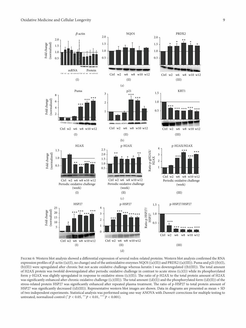
Oxidative Medicine and Cellular Longevity 9
(I) (II) (III)Ctrl w2 w6 w8 w10 w12
0.5
1.0
1.5
2.0PRDX2
Ctrl w2 w6 w8 w10 w12 Ctrl w2 w6 w8 w10 w12
0.5
1.0
1.5
2.0
mRNA ProteinCtrl w2 w6 w8 w10 w12
0.5
1.0
1.5
2.0NQO1𝛽-actin
∗∗∗
∗ ∗∗
Fold
chan
ge(n
orm
aliz
ed)
(a)
(I) (II) (III)
Ctrl w2 w6 w8 w10 w12
1
2
3p21
Ctrl w2 w6 w8 w10 w12
0.5
1.0
1.5 KRT1
Ctrl w2 w6 w8 w10 w12
2
4
6
8Puma
∗∗∗
∗∗∗∗∗∗
∗∗∗∗∗∗
∗∗∗∗∗∗ ∗∗∗ ∗∗∗ ∗∗∗
Fold
chan
ge(n
orm
aliz
ed)
(b)
(I) (II) (III)
Ctrl w2 w6 w8 w10 w12
0.5
1.0
1.5
Periodic oxidative challenge
H2AX
Ctrl w2 w6 w8 w10 w12
0.51.01.52.02.5
Periodic oxidative challenge
p-H2AX
Ctrl w2 w6 w8 w10 w12
1
2
3
4
Periodic oxidative challenge
Ratio
p-g
H2A
X/H
2AX
p-H2AX/H2AX
∗∗∗
∗∗∗
∗∗∗
∗∗∗
∗∗∗
∗∗∗∗∗∗
∗∗
∗∗∗∗∗ ∗ ∗
(week)(week)(week)
Fold
chan
ge(n
orm
aliz
ed)
(c)
Ctrl w2 w6 w8 w10 w12
510152025
HSP27
Fold
chan
ge
(I) (II) (III)Ctrl w2 w6 w8 w10 w12
0
1
2
3
4p-HSP27
∗∗∗∗∗∗∗∗∗∗∗∗
∗∗∗
Ctrl w2 w6 w8 w10 w12
0.5
1.0
1.5
Ratio
p-H
SP27
/H
SP27
p-HSP27/HSP27
∗∗∗ ∗∗∗∗∗∗ ∗∗∗
∗∗∗
∗∗∗∗∗∗
∗∗∗∗∗∗
∗
(nor
mal
ized
)
(d)
Figure 6: Western blot analysis showed a differential expression of several redox-related proteins. Western blot analysis confirmed the RNAexpression profiles of𝛽-actin ((a)(I), no change) and of the antioxidative enzymesNQO1 ((a)(II)) and PRDX2 ((a)(III)). Puma and p21 ((b)(I),(b)(II)) were upregulated after chronic but not acute oxidative challenge whereas keratin 1 was downregulated ((b)(III)). The total amountof H2AX protein was twofold downregulated after periodic oxidative challenge in contrast to acute stress ((c)(I)) while its phosphorylatedform 𝛾-H2AX was slightly upregulated in response to oxidative stress ((c)(II)). The ratio of p-H2AX to the total protein amount of H2AXwas significantly enhanced after chronic oxidative challenge ((c)(III)).The total amount ((d)(I)) and the phosphorylated form ((d)(II)) of thestress-related protein HSP27 was significantly enhanced after repeated plasma treatment. The ratio of p-HSP27 to total protein amount ofHSP27 was significantly decreased ((d)(III)). Representative western blot images are shown. Data in diagrams are presented as mean + SDof two independent experiments. Statistical analysis was performed using one-way ANOVA with Dunnett corrections for multiple testing tountreated, normalized control (∗𝑝 < 0.05, ∗∗𝑝 < 0.01, ∗∗∗𝑝 < 0.001).
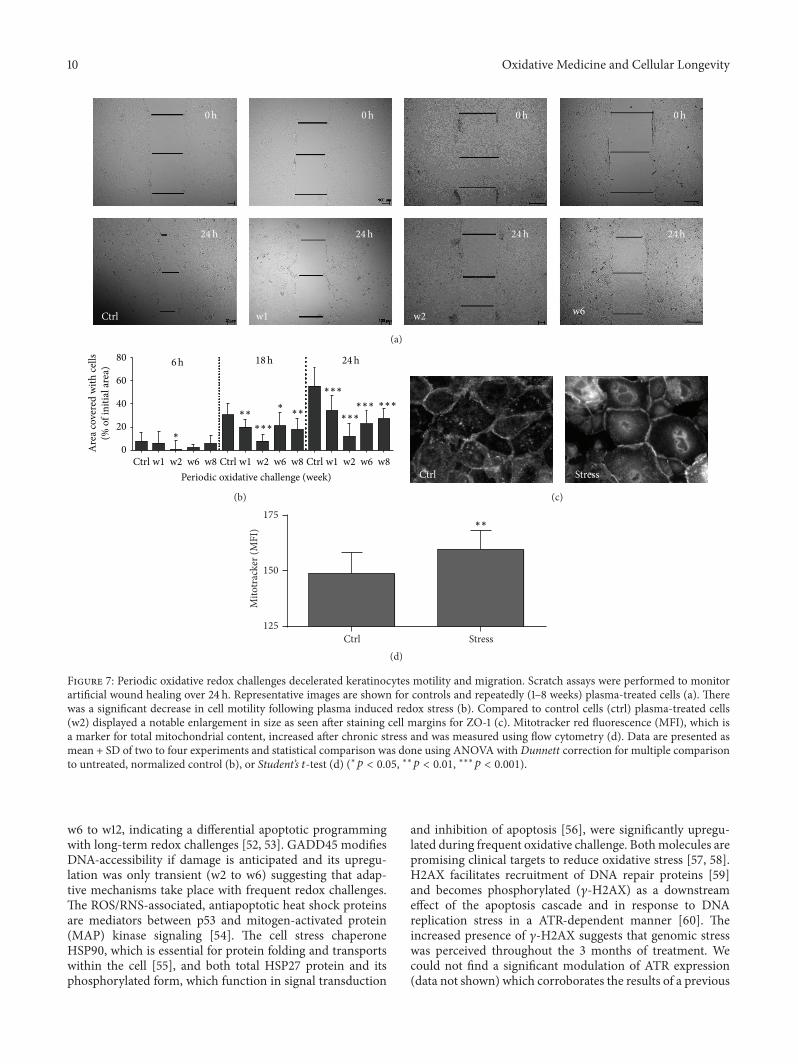
10 Oxidative Medicine and Cellular Longevity
Ctrl w1 w2 w6
0h 0h 0h 0h
24h 24h 24h 24h
(a)
Ctrl w1 w2 w6 w8 Ctrl w1 w2 w6 w8 Ctrl w1 w2 w6 w80
20
40
60
80
Periodic oxidative challenge (week)
∗
6h 18h 24h
∗∗∗ ∗∗
∗∗∗
∗∗∗
∗∗∗∗∗∗ ∗∗∗
Are
a cov
ered
with
cells
(% o
f ini
tial a
rea)
(b)
Ctrl Stress
(c)
Ctrl Stress125
150
175
Mito
trac
ker (
MFI
) ∗∗
(d)
Figure 7: Periodic oxidative redox challenges decelerated keratinocytes motility and migration. Scratch assays were performed to monitorartificial wound healing over 24 h. Representative images are shown for controls and repeatedly (1–8 weeks) plasma-treated cells (a). Therewas a significant decrease in cell motility following plasma induced redox stress (b). Compared to control cells (ctrl) plasma-treated cells(w2) displayed a notable enlargement in size as seen after staining cell margins for ZO-1 (c). Mitotracker red fluorescence (MFI), which isa marker for total mitochondrial content, increased after chronic stress and was measured using flow cytometry (d). Data are presented asmean + SD of two to four experiments and statistical comparison was done using ANOVA with Dunnett correction for multiple comparisonto untreated, normalized control (b), or Student’s 𝑡-test (d) (∗𝑝 < 0.05, ∗∗𝑝 < 0.01, ∗∗∗𝑝 < 0.001).
w6 to w12, indicating a differential apoptotic programmingwith long-term redox challenges [52, 53]. GADD45 modifiesDNA-accessibility if damage is anticipated and its upregu-lation was only transient (w2 to w6) suggesting that adap-tive mechanisms take place with frequent redox challenges.The ROS/RNS-associated, antiapoptotic heat shock proteinsare mediators between p53 and mitogen-activated protein(MAP) kinase signaling [54]. The cell stress chaperoneHSP90, which is essential for protein folding and transportswithin the cell [55], and both total HSP27 protein and itsphosphorylated form, which function in signal transduction
and inhibition of apoptosis [56], were significantly upregu-lated during frequent oxidative challenge. Bothmolecules arepromising clinical targets to reduce oxidative stress [57, 58].H2AX facilitates recruitment of DNA repair proteins [59]and becomes phosphorylated (𝛾-H2AX) as a downstreameffect of the apoptosis cascade and in response to DNAreplication stress in a ATR-dependent manner [60]. Theincreased presence of 𝛾-H2AX suggests that genomic stresswas perceived throughout the 3 months of treatment. Wecould not find a significant modulation of ATR expression(data not shown) which corroborates the results of a previous
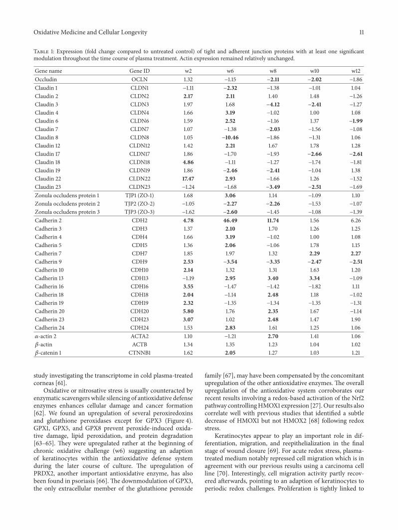
Oxidative Medicine and Cellular Longevity 11
Table 1: Expression (fold change compared to untreated control) of tight and adherent junction proteins with at least one significantmodulation throughout the time course of plasma treatment. Actin expression remained relatively unchanged.
Gene name Gene ID w2 w6 w8 w10 w12Occludin OCLN 1.32 −1.15 −2.11 −2.02 −1.86Claudin 1 CLDN1 −1.11 −2.32 −1.38 −1.01 1.04Claudin 2 CLDN2 2.17 2.11 1.40 1.48 −1.26Claudin 3 CLDN3 1.97 1.68 −4.12 −2.41 −1.27Claudin 4 CLDN4 1.66 3.19 −1.02 1.00 1.08Claudin 6 CLDN6 1.59 2.52 −1.16 1.37 −1.99Claudin 7 CLDN7 1.07 −1.38 −2.03 −1.56 −1.08Claudin 8 CLDN8 1.05 −10.46 −1.86 −1.31 1.06Claudin 12 CLDN12 1.42 2.21 1.67 1.78 1.28Claudin 17 CLDN17 1.86 −1.70 −1.93 −2.66 −2.61Claudin 18 CLDN18 4.86 −1.11 −1.27 −1.74 −1.81Claudin 19 CLDN19 1.86 −2.46 −2.41 −1.04 1.38Claudin 22 CLDN22 17.47 2.93 −1.66 1.26 −1.52Claudin 23 CLDN23 −1.24 −1.68 −3.49 −2.51 −1.69Zonula occludens protein 1 TJP1 (ZO-1) 1.68 3.06 1.14 −1.09 1.10Zonula occludens protein 2 TJP2 (ZO-2) −1.05 −2.27 −2.26 −1.53 −1.07Zonula occludens protein 3 TJP3 (ZO-3) −1.62 −2.60 −1.45 −1.08 −1.39Cadherin 2 CDH2 4.78 46.49 11.74 1.56 6.26Cadherin 3 CDH3 1.37 2.10 1.70 1.26 1.25Cadherin 4 CDH4 1.66 3.19 −1.02 1.00 1.08Cadherin 5 CDH5 1.36 2.06 −1.06 1.78 1.15Cadherin 7 CDH7 1.85 1.97 1.32 2.29 2.27Cadherin 9 CDH9 2.53 −3.54 −3.35 −2.47 −2.51Cadherin 10 CDH10 2.14 1.32 1.31 1.63 1.20Cadherin 13 CDH13 −1.19 2.95 3.40 3.34 −1.09Cadherin 16 CDH16 3.55 −1.47 −1.42 −1.82 1.11Cadherin 18 CDH18 2.04 −1.14 2.48 1.18 −1.02Cadherin 19 CDH19 2.32 −1.35 −1.34 −1.35 −1.31Cadherin 20 CDH20 5.80 1.76 2.35 1.67 −1.14Cadherin 23 CDH23 3.07 1.02 2.48 1.47 1.90Cadherin 24 CDH24 1.53 2.83 1.61 1.25 1.06𝛼-actin 2 ACTA2 1.10 −1.21 2.70 1.41 1.06𝛽-actin ACTB 1.34 1.35 1.23 1.04 1.02𝛽-catenin 1 CTNNB1 1.62 2.05 1.27 1.03 1.21
study investigating the transcriptome in cold plasma-treatedcorneas [61].
Oxidative or nitrosative stress is usually counteracted byenzymatic scavengers while silencing of antioxidative defenseenzymes enhances cellular damage and cancer formation[62]. We found an upregulation of several peroxiredoxinsand glutathione peroxidases except for GPX3 (Figure 4).GPX1, GPX5, and GPX8 prevent peroxide-induced oxida-tive damage, lipid peroxidation, and protein degradation[63–65]. They were upregulated rather at the beginning ofchronic oxidative challenge (w6) suggesting an adaptionof keratinocytes within the antioxidative defense systemduring the later course of culture. The upregulation ofPRDX2, another important antioxidative enzyme, has alsobeen found in psoriasis [66]. The downmodulation of GPX3,the only extracellular member of the glutathione peroxide
family [67], may have been compensated by the concomitantupregulation of the other antioxidative enzymes. The overallupregulation of the antioxidative system corroborates ourrecent results involving a redox-based activation of the Nrf2pathway controllingHMOX1 expression [27].Our results alsocorrelate well with previous studies that identified a subtledecrease of HMOX1 but not HMOX2 [68] following redoxstress.
Keratinocytes appear to play an important role in dif-ferentiation, migration, and reepithelialization in the finalstage of wound closure [69]. For acute redox stress, plasma-treated medium notably repressed cell migration which is inagreement with our previous results using a carcinoma cellline [70]. Interestingly, cell migration activity partly recov-ered afterwards, pointing to an adaption of keratinocytes toperiodic redox challenges. Proliferation is tightly linked to
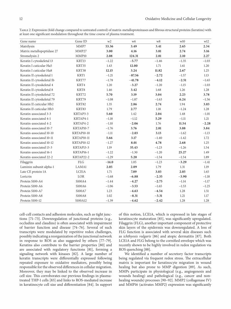
12 Oxidative Medicine and Cellular Longevity
Table 2: Expression (fold change compared to untreated control) of matrix-metalloproteinases and fibrous structural proteins (keratins) withat least one significant modulation throughout the time course of plasma treatment.
Gene name Gene ID w2 w6 w8 w10 w12Matrilysin MMP7 53.36 3.49 3.41 2.65 2.34Matrix metallopeptidase 27 MMP27 3.00 4.16 3.81 2.74 3.16Stromelysin 2 MMP10 2.08 124.31 2.81 2.10 2.27Keratin I cytoskeletal 13 KRT13 −1.22 −5.77 −1.46 −1.35 −1.03Keratin I cuticular Ha5 KRT35 1.43 12.00 1.71 1.61 1.20Keratin I cuticular Ha8 KRT38 12.15 3.24 11.82 2.67 1.25Keratin II cytoskeletal 1 KRT1 −1.21 −87.56 −2.72 −1.57 1.13Keratin II cytoskeletal 1b KRT77 −1.78 −41.78 −4.12 −2.51 −1.65Keratin II cytoskeletal 4 KRT4 1.20 −3.27 −1.20 −1.15 −1.03Keratin II cytoskeletal 8 KRT8 1.46 3.42 1.68 1.26 1.28Keratin II cytoskeletal 72 KRT72 5.70 3.19 3.04 2.23 3.78Keratin II cytoskeletal 79 KRT79 −1.08 −1.87 −1.63 6.24 −1.56Keratin II cuticular Hb2 KRT82 1.35 2.86 2.74 1.94 3.03Keratin II cuticular Hb3 KRT83 1.79 2.77 1.18 −1.24 1.28Keratin-associated 3-3 KRTAP3-3 5.60 1.42 2.04 1.48 −1.01Keratin-associated 4-1 KRTAP4-1 −1.18 −1.12 5.29 −1.11 1.21Keratin-associated 4-2 KRTAP4-2 −1.10 −2.06 1.76 11.54 −2.28Keratin-associated 10-7 KRTAP10-7 −1.76 3.76 2.81 3.08 3.84Keratin-associated 10-10 KRTAP10-10 −1.11 −2.03 3.13 −1.62 −1.13Keratin-associated 10-11 KRTAP10-11 5.66 3.17 −1.40 −1.42 1.72Keratin-associated 10-12 KRTAP10-12 −1.27 8.01 4.78 2.68 1.25Keratin-associated 13-3 KRTAP13-3 1.19 35.43 −1.23 −1.26 1.54Keratin-associated 16-1 KRTAP16-1 −1.22 −1.30 −1.20 25.27 1.49Keratin-associated 22-2 KRTAP22-2 −1.29 5.20 −1.54 −1.54 1.89Filaggrin FLG −14.0 1.05 −1.23 −3.29 −1.41Laminin subunit alpha-1 LAMA1 2.05 2.09 1.79 1.31 1.19Late CE protein 1A LCE1A 1.71 7.89 3.03 2.05 1.65Loricrin LOR −1.68 −6.88 −2.35 −3.90 −1.18Protein S100-A4 S100A4 −1.44 −4.27 −2.75 −1.14 −1.17Protein S100-A6 S100A6 −1.06 −3.53 −1.65 −1.53 −1.25Protein S100-A7 S100A7 1.23 −4.63 −4.54 1.28 1.51Protein S100-A8 S100A8 1.02 −8.31 −3.31 1.21 1.17Protein S100-12 S100A12 −1.39 −4.62 −2.42 1.28 1.28
cell-cell contacts and adhesion molecules, such as tight junc-tions [71–73]. Downregulation of junctional proteins (e.g.,occludins and claudins) is often associated with impairmentof barrier function and disease [74–76]. Several of suchtranscripts were modulated by repetitive redox challenges,possibly indicating a reorganization of the junctional networkin response to ROS as also suggested by others [77–79].Keratins also contribute to the barrier properties [80] andare associated with regulatory functions [81], forming asignaling network with kinases [82]. A large number ofkeratin transcripts were differentially expressed followingrepeated exposure to oxidative mediators, possibly beingresponsible for the observed differences in cellularmigration.Moreover, they may be linked to the observed increase incell size. This corroborates our previous findings in plasma-treated THP-1 cells [83] and links to ROS-mediated increasein keratinocyte cell size and differentiation [84]. In support
of this notion, LCE1A, which is expressed in late stages ofkeratinocyte maturation [85], was significantly upregulated.Filaggrin (FLG), another important component of protectiveskin layers of the epidermis was downregulated. A loss ofFLG function is associated with several skin diseases suchas ichthyosis vulgaris [86] and atopic dermatitis [87]. BothLCE1A and FLG belong to the cornified envelope which wasrecently shown to be highly involved in redox regulation viaROS quenching [88].
We identified a number of secretory factor transcriptsbeing regulated via frequent redox stress. The extracellularmatrix is important for keratinocyte migration in woundhealing but also prone to MMP digestion [89]. As such,MMPs participate in physiological (e.g., angiogenesis andwounds healing) and pathological (e.g., cancer and non-healing wounds) processes [90–92]. MMP2 (collagenase IV)and MMP16 (activates MMP2) expression was significantly
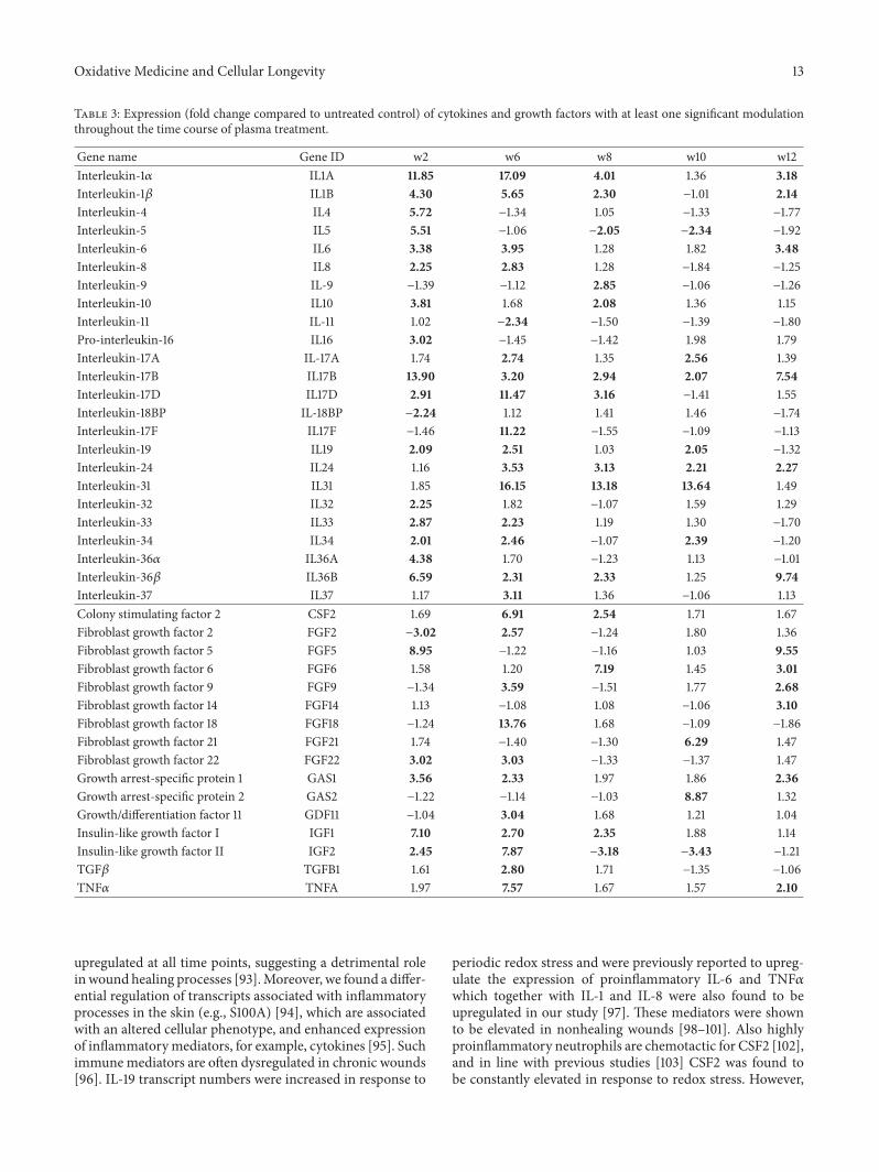
Oxidative Medicine and Cellular Longevity 13
Table 3: Expression (fold change compared to untreated control) of cytokines and growth factors with at least one significant modulationthroughout the time course of plasma treatment.
Gene name Gene ID w2 w6 w8 w10 w12Interleukin-1𝛼 IL1A 11.85 17.09 4.01 1.36 3.18Interleukin-1𝛽 IL1B 4.30 5.65 2.30 −1.01 2.14Interleukin-4 IL4 5.72 −1.34 1.05 −1.33 −1.77Interleukin-5 IL5 5.51 −1.06 −2.05 −2.34 −1.92Interleukin-6 IL6 3.38 3.95 1.28 1.82 3.48Interleukin-8 IL8 2.25 2.83 1.28 −1.84 −1.25Interleukin-9 IL-9 −1.39 −1.12 2.85 −1.06 −1.26Interleukin-10 IL10 3.81 1.68 2.08 1.36 1.15Interleukin-11 IL-11 1.02 −2.34 −1.50 −1.39 −1.80Pro-interleukin-16 IL16 3.02 −1.45 −1.42 1.98 1.79Interleukin-17A IL-17A 1.74 2.74 1.35 2.56 1.39Interleukin-17B IL17B 13.90 3.20 2.94 2.07 7.54Interleukin-17D IL17D 2.91 11.47 3.16 −1.41 1.55Interleukin-18BP IL-18BP −2.24 1.12 1.41 1.46 −1.74Interleukin-17F IL17F −1.46 11.22 −1.55 −1.09 −1.13Interleukin-19 IL19 2.09 2.51 1.03 2.05 −1.32Interleukin-24 IL24 1.16 3.53 3.13 2.21 2.27Interleukin-31 IL31 1.85 16.15 13.18 13.64 1.49Interleukin-32 IL32 2.25 1.82 −1.07 1.59 1.29Interleukin-33 IL33 2.87 2.23 1.19 1.30 −1.70Interleukin-34 IL34 2.01 2.46 −1.07 2.39 −1.20Interleukin-36𝛼 IL36A 4.38 1.70 −1.23 1.13 −1.01Interleukin-36𝛽 IL36B 6.59 2.31 2.33 1.25 9.74Interleukin-37 IL37 1.17 3.11 1.36 −1.06 1.13Colony stimulating factor 2 CSF2 1.69 6.91 2.54 1.71 1.67Fibroblast growth factor 2 FGF2 −3.02 2.57 −1.24 1.80 1.36Fibroblast growth factor 5 FGF5 8.95 −1.22 −1.16 1.03 9.55Fibroblast growth factor 6 FGF6 1.58 1.20 7.19 1.45 3.01Fibroblast growth factor 9 FGF9 −1.34 3.59 −1.51 1.77 2.68Fibroblast growth factor 14 FGF14 1.13 −1.08 1.08 −1.06 3.10Fibroblast growth factor 18 FGF18 −1.24 13.76 1.68 −1.09 −1.86Fibroblast growth factor 21 FGF21 1.74 −1.40 −1.30 6.29 1.47Fibroblast growth factor 22 FGF22 3.02 3.03 −1.33 −1.37 1.47Growth arrest-specific protein 1 GAS1 3.56 2.33 1.97 1.86 2.36Growth arrest-specific protein 2 GAS2 −1.22 −1.14 −1.03 8.87 1.32Growth/differentiation factor 11 GDF11 −1.04 3.04 1.68 1.21 1.04Insulin-like growth factor I IGF1 7.10 2.70 2.35 1.88 1.14Insulin-like growth factor II IGF2 2.45 7.87 −3.18 −3.43 −1.21TGF𝛽 TGFB1 1.61 2.80 1.71 −1.35 −1.06TNF𝛼 TNFA 1.97 7.57 1.67 1.57 2.10
upregulated at all time points, suggesting a detrimental roleinwound healing processes [93].Moreover, we found a differ-ential regulation of transcripts associated with inflammatoryprocesses in the skin (e.g., S100A) [94], which are associatedwith an altered cellular phenotype, and enhanced expressionof inflammatorymediators, for example, cytokines [95]. Suchimmunemediators are often dysregulated in chronic wounds[96]. IL-19 transcript numbers were increased in response to
periodic redox stress and were previously reported to upreg-ulate the expression of proinflammatory IL-6 and TNF𝛼which together with IL-1 and IL-8 were also found to beupregulated in our study [97]. These mediators were shownto be elevated in nonhealing wounds [98–101]. Also highlyproinflammatory neutrophils are chemotactic for CSF2 [102],and in line with previous studies [103] CSF2 was found tobe constantly elevated in response to redox stress. However,
14 Oxidative Medicine and Cellular Longevity
periodic redox stress also increased the transcription of anti-inflammatory mediators. IL-37 is a fundamental inhibitor ofinnate immunity [104] and keratinocyte-derived IL-4 and IL-10 mediate immune suppression [105]. TGF𝛽 has been usedin clinical trials combating deficient wound healing [106] andwas partially increased following plasma as well. Frequentredox challenges thus generated not only a pro- but alsoanti-inflammatory cytokine signature and thus may have acomplex impact on the quality of inflammation in redox-related diseases.
We can only speculate why there was no consistentup- and downregulation patterns over time in the targetsinvestigated in this study. First, gene expression may beheterogeneous within cultured cells. Frequent cold plasmatreatment may have promoted growth of cells that are betterequipped against oxidative stress, consequently overgrowingthe cells that are not. For example, single cold plasmatreatment of keratinocytes led to G2/M-phase cell cycle arrestbut this was dependent on the total treatment time and wasnot present in all cells [107]. Alternatively, all keratinocytecells may have adapted to redox stress over time, significantlyaltering their basal gene expression profile.
5. Conclusion
Using cold physical plasma-derived reactive species, ourkeratinocyte-based in vitro model aimed to mimic thecharacteristics of chronic oxidative stress in redox-relateddisorders of the skin to pinpoint biomarkers for moleculartherapies. Redox processes are important decision makersin skin disease and tumorigenesis and here affected cell-cell communication, cellular proliferation, and inflammatoryprocesses. The identification of genes and proteins whoseexpression is altered following permanent oxidative challengeis an important step to better understand redox regulations inclinical settings.
Conflict of Interests
The authors declare that there is no conflict of interestsregarding the publication of this paper.
Acknowledgments
This work was funded by the Ministry of Education, Scienceand Culture of the State of Mecklenburg-Western Pomeraniaand European Union, European Social Fund under Grantnumber AU 11 038; ESF/IV-BM-B35-0010/13.
References
[1] D. R. Bickers andM.Athar, “Oxidative stress in the pathogenesisof skin disease,” Journal of Investigative Dermatology, vol. 126,no. 12, pp. 2565–2575, 2006.
[2] F. Q. Schafer and G. R. Buettner, “Redox environment of thecell as viewed through the redox state of the glutathione disul-fide/glutathione couple,” Free Radical Biology andMedicine, vol.30, no. 11, pp. 1191–1212, 2001.
[3] C. S. Sander, F. Hamm, P. Elsner, and J. J. Thiele, “Oxidativestress in malignant melanoma and non-melanoma skin cancer,”British Journal of Dermatology, vol. 148, no. 5, pp. 913–922, 2003.
[4] T. J. James, M. A. Hughes, G. W. Cherry, and R. P. Taylor,“Evidence of oxidative stress in chronic venous ulcers,” WoundRepair and Regeneration, vol. 11, no. 3, pp. 172–176, 2003.
[5] Q. Zhou, U. Mrowietz, andM. Rostami-Yazdi, “Oxidative stressin the pathogenesis of psoriasis,” Free Radical Biology andMedicine, vol. 47, no. 7, pp. 891–905, 2009.
[6] J. Ferlay, E. Steliarova-Foucher, J. Lortet-Tieulent et al., “Cancerincidence and mortality patterns in Europe: estimates for 40countries in 2012,” European Journal of Cancer, vol. 49, no. 6,pp. 1374–1403, 2013.
[7] N. Graves and H. Zheng, “The prevalence and incidenceof chronic wounds: a literature review,” Wound Practice andResearch, vol. 22, no. 1, pp. 14–19, 2014.
[8] C. K. Sen, “Wound healing essentials: let there be oxygen,”Wound Repair and Regeneration, vol. 17, no. 1, pp. 1–18, 2009.
[9] R. Parisi, D. P. M. Symmons, C. E. M. Griffiths, and D.M. Ashcroft, “Global epidemiology of psoriasis: a systematicreview of incidence and prevalence,” Journal of InvestigativeDermatology, vol. 133, no. 2, pp. 377–385, 2013.
[10] D. Darr and I. Fridovich, “Free radicals in cutaneous biology,”Journal of Investigative Dermatology, vol. 102, no. 5, pp. 671–675,1994.
[11] C. Lehner, R. Gehwolf, H. Tempfer et al., “Oxidative stress andblood-brain barrier dysfunction under particular considerationof matrix metalloproteinases,” Antioxidants and Redox Signal-ing, vol. 15, no. 5, pp. 1305–1323, 2011.
[12] K. R. Martin and J. C. Barrett, “Reactive oxygen speciesas double-edged swords in cellular processes: low-dose cellsignaling versus high-dose toxicity,” Human and ExperimentalToxicology, vol. 21, no. 2, pp. 71–75, 2002.
[13] R. Kohen and A. Nyska, “Oxidation of biological systems:oxidative stress phenomena, antioxidants, redox reactions, andmethods for their quantification,” Toxicologic Pathology, vol. 30,no. 6, pp. 620–650, 2002.
[14] E. H. Sarsour,M. G. Kumar, L. Chaudhuri, A. L. Kalen, and P. C.Goswami, “Redox control of the cell cycle in health and disease,”Antioxidants and Redox Signaling, vol. 11, no. 12, pp. 2985–3011,2009.
[15] S. J. Green, L. F. Scheller, M. A. Marletta et al., “Nitricoxide: cytokine-regulation of nitric oxide in host resistance tointracellular pathogens,” Immunology Letters, vol. 43, no. 1-2, pp.87–94, 1994.
[16] M.-M. Cals-Grierson and A. D. Ormerod, “Nitric oxide func-tion in the skin,” Nitric Oxide, vol. 10, no. 4, pp. 179–193, 2004.
[17] J. J. P.Gille, P. Pasman,C.G.M. vanBerkel, andH. Joenje, “Effectof antioxidants on hyperoxia-induced chromosomal breakagein Chinese hamster ovary cells: protection by carnosine,”Mutagenesis, vol. 6, no. 4, pp. 313–318, 1991.
[18] A. T. Black, J. P. Gray, M. P. Shakarjian, D. L. Laskin, D. E.Heck, and J. D. Laskin, “Increased oxidative stress and antiox-idant expression in mouse keratinocytes following exposure toparaquat,” Toxicology and Applied Pharmacology, vol. 231, no. 3,pp. 384–392, 2008.
[19] F. Brehmer, H. A. Haenssle, G. Daeschlein et al., “Alleviation ofchronic venous leg ulcers with a hand-held dielectric barrierdischarge plasma generator (PlasmaDermⓇ VU-2010): resultsof a monocentric, two-armed, open, prospective, randomizedand controlled trial (NCT01415622),” Journal of the European
Oxidative Medicine and Cellular Longevity 15
Academy of Dermatology andVenereology, vol. 29, no. 1, pp. 148–155, 2015.
[20] M. Klebes, J. Lademann, S. Philipp et al., “Effects of tissue-tolerable plasma on psoriasis vulgaris treatment compared toconventional local treatment: a pilot study,” Clinical PlasmaMedicine, vol. 2, no. 1, pp. 22–27, 2014.
[21] H. R. Metelmann, T. von Woedtke, R. Bussiahn et al., “Exper-imental recovery of CO
2-laser skin lesions by plasma stimula-
tion,” American Journal of Cosmetic Surgery, vol. 29, pp. 52–56,2012.
[22] S. K.Mantena and S. K. Katiyar, “Grape seed proanthocyanidinsinhibit UV-radiation-induced oxidative stress and activationof MAPK and NF-𝜅B signaling in human epidermal ker-atinocytes,” Free Radical Biology andMedicine, vol. 40, no. 9, pp.1603–1614, 2006.
[23] N. Kanda and S. Watanabe, “17𝛽-estradiol inhibits oxidativestress-induced apoptosis in keratinocytes by promoting Bcl-2expression,” Journal of Investigative Dermatology, vol. 121, no. 6,pp. 1500–1509, 2003.
[24] A. A. Shvedova, C. Kommineni, B. A. Jeffries et al., “Redoxcycling of phenol induces oxidative stress in human epidermalkeratinocytes,” Journal of Investigative Dermatology, vol. 114, no.2, pp. 354–364, 2000.
[25] P. Boukamp, R. T. Petrussevska, D. Breitkreutz, J. Hornung,A. Markham, and N. E. Fusenig, “Normal keratinization ina spontaneously immortalized aneuploid human keratinocytecell line,”The Journal of Cell Biology, vol. 106, no. 3, pp. 761–771,1988.
[26] K.-D.Weltmann, E. Kindel, R. Brandenburg et al., “Atmosphericpressure plasma jet formedical therapy: plasma parameters andrisk estimation,” Contributions to Plasma Physics, vol. 49, no. 9,pp. 631–640, 2009.
[27] A. Schmidt, S. Dietrich, A. Steuer et al., “Non-thermal plasmaactivates human keratinocytes by stimulation of antioxidantand phase II pathways,”The Journal of Biological Chemistry, vol.290, no. 11, pp. 6731–6750, 2015.
[28] L. Bundscherer, K. Wende, K. Ottmüller et al., “Impact of non-thermal plasma treatment on MAPK signaling pathways ofhuman immune cell lines,” Immunobiology, vol. 218, no. 10, pp.1248–1255, 2013.
[29] A. Maillet and S. Pervaiz, “Redox regulation of p53, redoxeffectors regulated by p53: a subtle balance,” Antioxidants &Redox Signaling, vol. 16, no. 11, pp. 1285–1294, 2012.
[30] A. Matsuzawa and H. Ichijo, “Stress-responsive protein kinasesin redox-regulated apoptosis signaling,”Antioxidants andRedoxSignaling, vol. 7, no. 3-4, pp. 472–481, 2005.
[31] K. Wende, A. Barton, S. Bekeschus et al., “Proteomic tools tocharacterize non-thermal plasma effects in eukaryotic cells,”Plasma Medicine, vol. 3, no. 1-2, pp. 81–95, 2013.
[32] A. Schmidt-Bleker, J. Winter, S. Iseni, M. Dünnbier, K.-D.Welt-mann, and S. Reuter, “Reactive species output of a plasma jetwith a shielding gas device—combination of FTIR absorptionspectroscopy and gas phase modelling,” Journal of Physics D:Applied Physics, vol. 47, no. 14, Article ID 145201, 2014.
[33] H. Tresp, M. U. Hammer, K.-D. Weltmann, and S. Reuter,“Effects of atmosphere composition and liquid type on plasma-generated reactive species in biologically relevant solutions,”Plasma Medicine, vol. 3, no. 1-2, pp. 45–55, 2013.
[34] H. Tresp, M. U. Hammer, J. Winter, K.-D. Weltmann, and S.Reuter, “Quantitative detection of plasma-generated radicalsin liquids by electron paramagnetic resonance spectroscopy,”
Journal of Physics D: Applied Physics, vol. 46, no. 43, Article ID435401, 2013.
[35] S. Bekeschus, S. Iseni, S. Reuter, K.Masur, and K.-D.Weltmann,“Nitrogen shielding of an argon plasma jet and its effects onhuman immune cells,” IEEE Transactions on Plasma Science,vol. 43, pp. 776–781, 2015.
[36] S. Bekeschus, J. Kolata, A. Müller et al., “Differential viabilityof eight human blood mononuclear cell subpopulations afterplasma treatment,” Plasma Medicine, vol. 3, no. 1-2, pp. 1–13,2013.
[37] X. Yan, Z. Xiong, F. Zou et al., “Plasma-induced death of HepG2cancer cells: intracellular effects of reactive species,” PlasmaProcesses and Polymers, vol. 9, no. 1, pp. 59–66, 2012.
[38] M. Yokoyama, K. Johkura, and T. Sato, “Gene expressionresponses of HeLa cells to chemical species generated by anatmospheric plasma flow,” Biochemical and Biophysical ResearchCommunications, vol. 450, no. 4, pp. 1266–1271, 2014.
[39] L. A. Pham-Huy, H. He, and C. Pham-Huy, “Free radicals,antioxidants in disease and health,” International Journal ofBiomedical Science, vol. 4, no. 2, pp. 89–96, 2008.
[40] L. Bundscherer, S. Bekeschus, H. Tresp et al., “Viability ofhuman blood leukocytes compared with their respective celllines after plasma treatment,” Plasma Medicine, vol. 3, no. 1-2,pp. 71–80, 2013.
[41] B. D’Autréaux and M. B. Toledano, “ROS as signallingmolecules: mechanisms that generate specificity in ROS home-ostasis,”Nature ReviewsMolecular Cell Biology, vol. 8, no. 10, pp.813–824, 2007.
[42] T.-C. Meng, T. Fukada, and N. K. Tonks, “Reversible oxidationand inactivation of protein tyrosine phosphatases in vivo,”Molecular Cell, vol. 9, no. 2, pp. 387–399, 2002.
[43] C. K. Sen and S. Roy, “Redox signals in wound healing,”Biochimica et Biophysica Acta—General Subjects, vol. 1780, no.11, pp. 1348–1361, 2008.
[44] A. E. K. Loo and B. Halliwell, “Effects of hydrogen peroxide ina keratinocyte-fibroblast co-culture model of wound healing,”Biochemical andBiophysical ResearchCommunications, vol. 423,no. 2, pp. 253–258, 2012.
[45] R. B. Mikkelsen and P. Wardman, “Biological chemistry ofreactive oxygen and nitrogen and radiation-induced signaltransduction mechanisms,” Oncogene, vol. 22, no. 37, pp. 5734–5754, 2003.
[46] H. J. Ahn, K. I. Kim, G. Kim, E. Moon, S. S. Yang, and J.-S. Lee,“Atmospheric-pressure plasma jet induces apoptosis involvingmitochondria via generation of free radicals,” PLoS ONE, vol. 6,no. 11, Article ID e28154, 2011.
[47] S. Bekeschus, T. vonWoedtke, A. Kramer, K.-D.Weltmann, andK. Masur, “Cold physical plasma treatment alters redox balancein human immune cells,” PlasmaMedicine, vol. 3, no. 4, pp. 267–278, 2013.
[48] K. Priya Arjunan and A. Morss Clyne, “Hydroxyl radical andhydrogen peroxide are primarily responsible for dielectric bar-rier discharge plasma-induced angiogenesis,” Plasma Processesand Polymers, vol. 8, no. 12, pp. 1154–1164, 2011.
[49] J. A. Daniel, M. Pellegrini, B.-S. Lee et al., “Loss of ATM kinaseactivity leads to embryonic lethality in mice,” Journal of CellBiology, vol. 198, no. 3, pp. 295–304, 2012.
[50] S. Ditch and T. T. Paull, “The ATM protein kinase and cellularredox signaling: beyond the DNA damage response,” Trends inBiochemical Sciences, vol. 37, no. 1, pp. 15–22, 2012.
16 Oxidative Medicine and Cellular Longevity
[51] J. R. Jeffers, E. Parganas, Y. Lee et al., “Puma is an essentialmedi-ator of p53-dependent and -independent apoptotic pathways,”Cancer Cell, vol. 4, no. 4, pp. 321–328, 2003.
[52] M. Ott, V. Gogvadze, S. Orrenius, and B. Zhivotovsky, “Mito-chondria, oxidative stress and cell death,” Apoptosis, vol. 12, no.5, pp. 913–922, 2007.
[53] T. Finkel, “Oxidant signals and oxidative stress,” Current Opin-ion in Cell Biology, vol. 15, no. 2, pp. 247–254, 2003.
[54] L. Xu, S. Chen, and R. C. Bergan, “MAPKAPK2 and HSP27are downstream effectors of p38 MAP kinase-mediated matrixmetalloproteinase type 2 activation and cell invasion in humanprostate cancer,”Oncogene, vol. 25, no. 21, pp. 2987–2998, 2006.
[55] L.Whitesell and S. L. Lindquist, “HSP90 and the chaperoning ofcancer,”Nature Reviews Cancer, vol. 5, no. 10, pp. 761–772, 2005.
[56] J. Huot, F. Houle, D. R. Spitz, and J. Landry, “HSP27phosphorylation-mediated resistance against actin fragmenta-tion and cell death induced by oxidative stress,”Cancer Research,vol. 56, no. 2, pp. 273–279, 1996.
[57] S. Tukaj, D. Grüner, D. Zillikens, andM. Kasperkiewicz, “Hsp90blockade modulates bullous pemphigoid IgG-induced IL-8production by keratinocytes,” Cell Stress and Chaperones, vol.19, no. 6, pp. 887–894, 2014.
[58] E. A. Voll, I. M. Ogden, J. M. Pavese et al., “Heat shock protein27 regulates human prostate cancer cell motility and metastaticprogression,” Oncotarget, vol. 5, no. 9, pp. 2648–2663, 2014.
[59] E. P. Rogakou, D. R. Pilch, A. H. Orr, V. S. Ivanova, andW. M. Bonner, “DNA double-stranded breaks induce histoneH2AX phosphorylation on serine 139,”The Journal of BiologicalChemistry, vol. 273, no. 10, pp. 5858–5868, 1998.
[60] I. M. Ward and J. J. Chen, “Histone H2AX is phosphorylated inan ATR-dependent manner in response to replicational stress,”The Journal of Biological Chemistry, vol. 276, no. 51, pp. 47759–47762, 2001.
[61] U. Rosani, E. Tarricone, P. Venier et al., “Atmospheric-pressurecold plasma induces transcriptional changes in ex vivo humancorneas,” PLoS ONE, vol. 10, no. 7, Article ID e0133173, 2015.
[62] B. Halliwell, “Oxidative stress and cancer: have we movedforward?” Biochemical Journal, vol. 401, no. 1, pp. 1–11, 2007.
[63] J. B.DeHaan, P. J. Crack,N. Flentjar, R. C. Iannello, P. J. Hertzog,and I. Kola, “An imbalance in antioxidant defense aftfectscellular function: the pathophysiological consequences of areduction in antioxidant defense in the glutathione peroxidase-1 (Gpx1) knockoutmouse,”Redox Report, vol. 8, no. 2, pp. 69–79,2003.
[64] R. J. Aitken, “Gpx5 protects the family jewels,” Journal of ClinicalInvestigation, vol. 119, no. 7, pp. 1849–1851, 2009.
[65] T. Ramming, H. G. Hansen, K. Nagata, L. Ellgaard, and C.Appenzeller-Herzog, “GPx8 peroxidase prevents leakage ofH2O2from the endoplasmic reticulum,” Free Radical Biology
and Medicine, vol. 70, pp. 106–116, 2014.[66] J. Ryu, S. G. Park, B. C. Park, M. Choe, K.-S. Lee, and J.-W. Cho,
“Proteomic analysis of psoriatic skin tissue for identification ofdifferentially expressed proteins: up-regulation of GSTP1, SFNand PRDX2 in psoriatic skin,” International Journal ofMolecularMedicine, vol. 28, no. 5, pp. 785–792, 2011.
[67] Y. Dincer, Y. Erzin, S. Himmetoglu, K. N. Gunes, K. Bal, andT. Akcay, “Oxidative DNA damage and antioxidant activity inpatients with inflammatory bowel disease,” Digestive Diseasesand Sciences, vol. 52, no. 7, pp. 1636–1641, 2007.
[68] A. Pellacani, P. Wiesel, A. Sharma et al., “Induction of hemeoxygenase-1 during endotoxemia is downregulated by trans-forming growth factor-𝛽1,” Circulation Research, vol. 83, no. 4,pp. 396–403, 1998.
[69] T. Jacks and R. A. Weinberg, “Cell-cycle control and itswatchman,” Nature, vol. 381, no. 6584, pp. 643–644, 1996.
[70] A. Schmidt, S. Bekeschus, T. von Woedtke, and S. Hasse, “Cellmigration and adhesion of a human melanoma cell line isdecreased by cold plasma treatment,” Clinical Plasma Medicine,vol. 3, no. 1, pp. 24–31, 2015.
[71] J. M. Brandner, S. Kief, C. Grund et al., “Organization andformation of the tight junction system in human epidermis andcultured keratinocytes,” European Journal of Cell Biology, vol. 81,no. 5, pp. 253–263, 2002.
[72] L. Langbein, C. Grund, C. Kuhn et al., “Tight junctions andcompositionally related junctional structures in mammalianstratified epithelia and cell cultures derived therefrom,” Euro-pean Journal of Cell Biology, vol. 81, no. 8, pp. 419–435, 2002.
[73] C. M. Nelson and C. S. Chen, “Cell-cell signaling by direct con-tact increases cell proliferation via a PI3K-dependent signal,”FEBS Letters, vol. 514, no. 2-3, pp. 238–242, 2002.
[74] N. S. Harhaj and D. A. Antonetti, “Regulation of tight junctionsand loss of barrier function in pathophysiology,” InternationalJournal of Biochemistry and Cell Biology, vol. 36, no. 7, pp. 1206–1237, 2004.
[75] S. Zeissig, N. Bürgel, D. Günzel et al., “Changes in expressionand distribution of claudin 2, 5 and 8 lead to discontinuous tightjunctions and barrier dysfunction in active Crohn’s disease,”Gut, vol. 56, no. 1, pp. 61–72, 2007.
[76] M. P. Fink and R. L. Delude, “Epithelial barrier dysfunction: aunifying theme to explain the pathogenesis of multiple organdysfunction at the cellular level,” Critical Care Clinics, vol. 21,no. 2, pp. 177–196, 2005.
[77] C. Coisne and B. Engelhardt, “Tight junctions in brain barriersduring central nervous system inflammation,”Antioxidants andRedox Signaling, vol. 15, no. 5, pp. 1285–1303, 2011.
[78] K. D. Rochfort, L. E. Collins, R. P. Murphy, and P. M. Cummins,“Downregulation of blood-brain barrier phenotype by proin-flammatory cytokines involves NADPH oxidase-dependentROS generation: consequences for interendothelial adherensand tight junctions,” PLoS ONE, vol. 9, no. 7, Article ID e101815,2014.
[79] R. Rao, “Oxidative stress-induced disruption of epithelial andendothelial tight junctions,” Frontiers in Bioscience, vol. 13, no.18, pp. 7210–7226, 2008.
[80] L. A. Torre, F. Bray, R. L. Siegel, J. Ferlay, J. Lortet-Tieulent, andA. Jemal, “Global cancer statistics, 2012,” CA: A Cancer Journalfor Clinicians, vol. 65, no. 2, pp. 87–108, 2015.
[81] P. A. Coulombe and C.-H. Lee, “Defining keratin proteinfunction in skin epithelia: epidermolysis bullosa simplex and itsaftermath,” Journal of Investigative Dermatology, vol. 132, no. 3,pp. 763–775, 2012.
[82] M. Moravcová, A. Libra, J. Dvořáková et al., “Modulationof keratin 1, 10 and involucrin expression as part of thecomplex response of the human keratinocyte cell line HaCaTto ultraviolet radiation,” Interdisciplinary Toxicology, vol. 6, no.4, pp. 203–208, 2013.
[83] S. Bekeschus, A. Schmidt, L. Bethge et al., “Redox stimulation ofhuman THP-1 monocytes in response to cold physical plasma,”Oxidative Medicine and Cellular Longevity, vol. 2016, Article ID5910695, 11 pages, 2016.
Oxidative Medicine and Cellular Longevity 17
[84] S. Tamiji, J.-C. Beauvillain, L. Mortier et al., “Induction ofapoptosis-like mitochondrial impairment triggers antioxidantand Bcl-2-dependent keratinocyte differentiation,” Journal ofInvestigative Dermatology, vol. 125, no. 4, pp. 647–658, 2005.
[85] J. S. Breitenbach, M. Rinnerthaler, A. Trost et al., “Transcrip-tome and ultrastructural changes in dystrophic epidermolysisbullosa resemble skin aging,” Aging, vol. 7, no. 6, pp. 389–411,2015.
[86] F. J. D. Smith, A. D. Irvine, A. Terron-Kwiatkowski et al., “Loss-of-function mutations in the gene encoding filaggrin causeichthyosis vulgaris,” Nature Genetics, vol. 38, no. 3, pp. 337–342,2006.
[87] J. P.Thyssen and S. Kezic, “Causes of epidermal filaggrin reduc-tion and their role in the pathogenesis of atopic dermatitis,”Journal of Allergy and Clinical Immunology, vol. 134, no. 4, pp.792–799, 2014.
[88] W. P. Vermeij, A. Alia, and C. Backendorf, “ROS quenchingpotential of the epidermal cornified cell envelope,” Journal ofInvestigative Dermatology, vol. 131, no. 7, pp. 1435–1441, 2011.
[89] E. A. O’Toole, “Extracellular matrix and keratinocyte migra-tion,” Clinical and Experimental Dermatology, vol. 26, no. 6, pp.525–530, 2001.
[90] W. C. Parks, “Matrix metalloproteinases in repair,” WoundRepair and Regeneration, vol. 7, no. 6, pp. 423–432, 1999.
[91] W. C. Parks, Y. S. López-Boado, and C. L.Wilson, “Matrilysin inepithelial repair and defense,” Chest, vol. 120, no. 1, supplement,pp. 36S–41S, 2001.
[92] A. B. Wysocki, L. Staiano-Coico, and F. Grinnell, “Woundfluid from chronic leg ulcers contains elevated levels of met-alloproteinases MMP-2 and MMP-9,” Journal of InvestigativeDermatology, vol. 101, no. 1, pp. 64–68, 1993.
[93] J. Rohl and R. Murray, “Matrix metalloproteinases duringwound healing-a double edged sword,” Wound Practice andResearch, vol. 21, no. 4, pp. 174–182, 2013.
[94] C. Gebhardt, J. Németh, P. Angel, and J. Hess, “S100A8 andS100A9 in inflammation and cancer,” Biochemical Pharmacol-ogy, vol. 72, no. 11, pp. 1622–1631, 2006.
[95] D. N. Granger, T. Vowinkel, and T. Petnehazy, “Modulation ofthe inflammatory response in cardiovascular disease,” Hyper-tension, vol. 43, no. 5, pp. 924–931, 2004.
[96] B. Behm, P. Babilas, M. Landthaler, and S. Schreml, “Cytokines,chemokines and growth factors in wound healing,” Journal ofthe European Academy of Dermatology and Venereology, vol. 26,no. 7, pp. 812–820, 2012.
[97] Y.-C. Liao, W.-G. Liang, F.-W. Chen, J.-H. Hsu, J.-J. Yang, andM.-S. Chang, “IL-19 induces production of IL-6 and TNF-𝛼 and results in cell apoptosis through TNF-𝛼,” Journal ofImmunology, vol. 169, no. 8, pp. 4288–4297, 2002.
[98] R. W. Tarnuzzer and G. S. Schultz, “Biochemical analysis ofacute and chronic wound environments,” Wound Repair andRegeneration, vol. 4, no. 3, pp. 321–325, 1996.
[99] C. C. Finnerty, D. N. Herndon, R. Przkora et al., “Cytokineexpression profile over time in severely burned pediatricpatients,” Shock, vol. 26, no. 1, pp. 13–19, 2006.
[100] B. A. Mast and G. S. Schultz, “Interactions of cytokines, growthfactors, and proteases in acute and chronic wounds,” WoundRepair and Regeneration, vol. 4, no. 4, pp. 411–420, 1996.
[101] M. C. Peral, M. M. Rachid, N. M. Gobbato, M. A. HuamanMartinez, and J. C. Valdez, “Interleukin-8 production by poly-morphonuclear leukocytes from patients with chronic infectedleg ulcers treated with Lactobacillus plantarum,” Clinical Micro-biology and Infection, vol. 16, no. 3, pp. 281–286, 2010.
[102] R. H. Weisbart, L. Kwan, D. W. Golde, and J. C. Gasson,“Human GM-CSF primes neutrophils for enhanced oxidativemetabolism in response to the major physiological chemoat-tractants,” Blood, vol. 69, no. 1, pp. 18–21, 1987.
[103] A. Barton, K. Wende, L. Bundscherer et al., “Non-thermalplasma increases expression of wound healing related genes in akeratinocyte cell line,” Plasma Medicine, vol. 3, no. 1-2, pp. 125–136, 2013.
[104] M. F. Nold, C. A. Nold-Petry, J. A. Zepp, B. E. Palmer, P. Bufler,and C. A. Dinarello, “IL-37 is a fundamental inhibitor of innateimmunity,” Nature Immunology, vol. 11, no. 11, pp. 1014–1022,2010.
[105] V. Shreedhar, T. Giese, V. W. Sung, and S. E. Ullrich, “Acytokine cascade including prostaglandin E2, IL-4, and IL-10is responsible for UV-induced systemic immune suppression,”The Journal of Immunology, vol. 160, no. 8, pp. 3783–3789, 1998.
[106] S. Barrientos, O. Stojadinovic, M. S. Golinko, H. Brem, andM. Tomic-Canic, “Growth factors and cytokines in woundhealing,”Wound Repair and Regeneration, vol. 16, no. 5, pp. 585–601, 2008.
[107] K. Wende, S. Straßenburg, B. Haertel et al., “Atmosphericpressure plasma jet treatment evokes transient oxidative stressin HaCaT keratinocytes and influences cell physiology,” CellBiology International, vol. 38, no. 4, pp. 412–425, 2014.
Review ArticleUse of Carnosine for Oxidative Stress Reduction inDifferent Pathologies
V. D. Prokopieva,1 E. G. Yarygina,1 N. A. Bokhan,2 and S. A. Ivanova1
1Department of Biological Psychiatry and Narcology, Mental Health Research Institute, Tomsk 634014, Russia2Addictive States Department, Mental Health Research Institute, Tomsk 634014, Russia
Correspondence should be addressed to V. D. Prokopieva; [email protected]
Received 25 September 2015; Revised 21 December 2015; Accepted 21 December 2015
Academic Editor: Sergio Di Meo
Copyright © 2016 V. D. Prokopieva et al. This is an open access article distributed under the Creative Commons AttributionLicense, which permits unrestricted use, distribution, and reproduction in any medium, provided the original work is properlycited.
The main properties and biological effects of the antioxidant carnosine, the natural dipeptide 𝛽-alanyl-L-histidine, are considered.Data on the effective use of carnosine in different pathologies are presented. Special attention is paid to issues of use of carnosine inneurologic and mental diseases, in alcoholism as well as in physiological states accompanied by activation of free-radical processesand formation of oxidative stress.
1. Oxidative Stress and Its Correctionby Antioxidants
The pathogenesis of most diseases involves excess activationof free-radical processes and disturbance of functioning ofthe organism’s antioxidant protection systems. This leadsto increase in the level of reactive oxygen species (ROS)and forming of oxidative stress (OS). Mechanisms of OSformation in different pathologies are quite universal andare especially linked with disturbance of homeostasis andredox processes. The characterization of ROS, their types,main sources of formation in the organism, properties, andtransformations are well described in various publications [1–3]. The main targets of damage under conditions of OS areproteins, lipids, carbohydrates, and nucleic acids.
It is well known that under normal physiological condi-tions ROS carry out important regulatory functions in theorganism [1, 4]. However, under uncontrolled increase inROS they interact with biomolecules, leading to their oxida-tivemodifications. Products of suchmodification usually loseability to carry out their functions. These products serveas “markers of oxidative stress,” and they include carbony-lated, nitrosylated, and glycated proteins; aggregates due tocrosslinking of protein molecules; products of lipid peroxi-dation (malondialdehyde, diene conjugates, hydroxynonenal,etc.); various types of hybrid adducts; advanced glycation end
products (AGE products); dihydroguanosine, homocysteine,and so forth [1, 5]. All these products of oxidative damage ofbiomolecules are resistant to destructions and accumulate incells, complicating their vital functions. Their neutralizationcan play an important role in correction of oxidative stress.
The search for and development of ways to correctoxidative stress is a relevant problem of modern medicine.One way that can be effective under clinical conditionsinvolves the use of the so-called antioxidants, substances thatneutralize ROS, reducing their reactivity in the organism.Despite the very large number of known antioxidants, tochoose one for effective use in a specific clinical situation isquite difficult. This is caused by the abundance of factors ofmodification of macromolecules under OS. In addition, themechanism of action of an antioxidant changes dependingon its chemical structure, bioavailability, and damage rateof redox processes and severity of oxidative stress in theorganism.
It was found that under conditions of oxidative stress, theendogenous systems of antioxidant response of the organismare activated through transcription factor Nrf2 [6]. Conse-quently, expression of endogenous antioxidant enzyme genesincreased, increasing cellular defenses against detrimentalredox modulations [7, 8].
The human endogenous antioxidant response systemcan regulate the amount of reactive species tightly and
Hindawi Publishing CorporationOxidative Medicine and Cellular LongevityVolume 2016, Article ID 2939087, 8 pageshttp://dx.doi.org/10.1155/2016/2939087
2 Oxidative Medicine and Cellular Longevity
minimize related cellular damage. But the role of exogenousantioxidants is also important. It was found that exoge-nous antioxidants have a priming effect on the antioxidantresponse system [9]. Working together with the endogenousantioxidant response system, exogenous antioxidants allowfor amore enhanced and efficient defense against detrimentalredox modulations.
Extensive experimental and clinical material on the use ofantioxidants has accumulated. In medicine, they are mainlyused as additional agents to basic therapy.Manymedications,in addition to their main therapeutic effect, also manifestantioxidant properties. However, depending on conditionsand concentration, antioxidants can also show the oppositeto antioxidant effect, that is, prooxidant action. Carotenes arepolyunsaturated compounds; therefore, they can be oxidizedvia a radical mechanism and act as prooxidants [10]. Undercertain conditions, for example, in the presence of mixed-valence metal ions, prooxidative effect is shown by ascorbate.Vitamin E as an antioxidant is most effective in a complexwith other fat- and water-soluble reductants (ascorbic acid,ubiquinol, and flavonoids) in whose absence it is quicklyinactivated or transforms into tocopheryl radical capable ofinitiating new chains of oxidation of unsaturated lipids; thatis, it becomes a prooxidant as well [5, 11].
Adhering to the correct dosage of an antioxidant, as withevery pharmacologically active compound, is very important.There are examples of ineffective use of antioxidants in thetreatment of some pathologies accompanied by decrease inthe level of antioxidants in blood plasma. So, clinical trials ofthe treatment of Alzheimer’s disease with the addition of thewell-known antioxidants lycopene and vitamins A, C, and Edid not show positive results and even showed progressivedecrease in cognitive function in the study participants insome cases [12].Though these results do not favor antioxidanttherapy, it might be due to prooxidative effects of theseantioxidants under these conditions as well as terms and thescheme of their administration.
The choice of a specific antioxidant and exact indicationsand contraindications are still insufficiently developed forevery specific disease. There is no information on the inter-action of pharmaceuticals of natural origin with syntheticmedications. In addition, antioxidants can cause allergicreactions, be toxic, and show low efficiency, and standard-ization is not always possible; the possibility of overdose alsoremains.Therefore, the search for substances with maximumantioxidant action and minimum side effects under condi-tions of OS continues and remains an important problem.Ideally, the antioxidant should show considerable antioxidantaction within a broad range of concentration, be naturaland hydrophilic, have good bioavailability, be nontoxic, notform toxic products during interaction with reactive oxygenspecies, not have negative effects in case of overdose, and havegood compatibility with other medications.
In spite of the fact that the use of antioxidants inclinical practice does not always show positive results, theconcept of use of antioxidant therapy is still relevant andhas the potential for effective treatment of a number ofdisorders accounting for pathophysiological mechanisms oftheir forming and development.
2. Main Properties and Biological Effectsof Carnosine
Numerous references as well as our own work experienceindicate that the antioxidant carnosine, the natural dipep-tide 𝛽-alanyl-L-histidine, meets almost all requirements foran ideal antioxidant. It is synthesized and contained inhuman muscle and nervous tissues, is easily absorbed in thedigestive tract, penetrates through blood-brain barrier, andhas high bioavailability and membrane-stabilizing action.Carnosine is a low molecular weight hydrophilic antioxidantof direct action, though it can also have an impact on theantiradical protection system of the organism [13]. Resultsof experiments on rats showed that carnosine acceleratesthe metabolizing of cortisol and noradrenaline released intoblood of animals under stress, showing the mediation effectof carnosine [14]. Decrease in level of stress hormones inblood leads to a decrease in the severity of OS. In addition,carnosine is not addictive; there is no danger of overdose,and it does not accumulate in the organism during long-term administration because its surplus is cleaved by theenzyme carnosinase into amino acids that are easily elim-inated from the organism [13]. However, it is noteworthythat there are cases of development of carnosinemia, a rareautosomal recessive [15] metabolic disorder [16] caused by adeficiency of carnosinase. This disorder results in an excessof carnosine in the urine, blood, and nervous tissue [17],and variety of neurological symptoms have been associatedwith carnosinemia [16, 18]; that is, under certain conditions,carnosine can exert negative effects.
There are publications in which positive biological effectsof carnosine are explained by its pH-buffering properties [19].However, carnosine is a buffer not only for protons, but alsoa buffer for mixed-valence metal ions and reactive oxygenspecies [20]. The ability of carnosine to form complexeswith bivalent metals is known: with ions of copper, cobalt,manganese, and cadmium [21]. In anotherwork, it was shownthat carnosine binds iron ions [22]. Because ions of metalstake an active part in many metabolic processes and canactivate free-radical processes, the ability of carnosine toregulate the level ofmixed-valencemetal ions in the organismis onemore important property of carnosine that confirms itsantioxidant status.
Further, the antiglycating [23, 24] and the anticrosslink-ing [25] properties of carnosine have been shown, which are,in essence, reflections of its antioxidant effects, the ability toblock oxidation of biomolecules.
Great contributions to the study of molecular mech-anisms of protection of biomolecules by carnosine weremade by Aldini et al. [26, 27]. Using liquid chromatogra-phy/electrospray ionization tandem mass spectrometry, theyshowed that carnosine and related peptides act as quenchersof reactive and cytotoxic carbonyl species through its abilityto form adducts with them. This suggested that carnosine isa protector of biomolecules from oxidative/carbonyl stress.The ability of carnosine to react with carbonyls of proteins(termed “carnosinylation” of proteins) was reported by otherauthors [28], who considered this property of carnosineimportant for inactivation/removal of damaged proteins.
Oxidative Medicine and Cellular Longevity 3
In culture of human cells, it has been shown that additionof carnosine into the medium at concentrations close tophysiological (20–50mM) increases longevity of the cells[29]. This was attributed to either reduction of the length oftelomere fragments of chromosomes, lost by the cell duringevery doubling, or decrease in methylation of DNA. It couldnot be ruled out that carnosine decreases the accumulationof some other changes in DNA, whose accumulation above acritical point leads to termination of divisions.
It has also been reported that carnosine prevents toxiceffects of hyperhomocysteinemia in rats [30]. It is knownthat homocysteine is a potent initiator of oxidative stress inmany tissues. However, the molecular mechanism of suchprotection is not clear. Perhaps carnosine modulates affinityof glutamate receptors to homocysteine, prevents accumula-tion of ROS, or has other protective mechanisms. But it hasbeen shown that these effects of carnosine are not connectedwith the improvement of homocysteine metabolism or thedecrease in its concentration.
Data on the study of biological effects of carnosine showsthat molecular mechanisms of its effects cannot always beexplained only by antioxidant action. The exact molecularmechanisms of some effects of carnosine observed in exper-iment should be found. At the same time, obvious positiveeffect of this dipeptide allows using carnosine widely alreadyin routine clinical practice.
Prospects for the use of carnosine in the treatment ofsome pathologies are reported in a report by Quinn et al. [31].Data on the possible physiological role of carnosine based onits biochemical properties and study of the therapeutic poten-tial of carnosine in a number of pathologies accompaniedby oxidative or carbonyl stress are presented in a review byBoldyrev et al. [32].
3. Clinical Use of Carnosine
Researchers of the Kharkov Physiotherapeutic Institute werethe creators of the first injection dosage form of carnosine.During subcutaneous injection of 0.5–1.0mg, high therapeu-tic effectiveness in treatment of infectious and rheumaticpolyarthritis and ulcer of the gastrointestinal tract wasobtained [33]. Later, positive effect of carnosine in healingwounds of lung tissue was shown [34]. Japanese researchersplayed a great role in the study of a wound healing effectof carnosine. They created the agent Z-103 based on acomplex of carnosine and zinc ions (L-carnosine-Zn2+) thathas considerable antiulcer effect and reduces damage to thestomach lining induced by different forms of stress andchemical agents [35]. Japanese scientists also have priorityfor the use of carnosine in cancer diseases [36]. Carnosinecombined with radiotherapy in treatment of patients withbreast cancer considerably reduced side effects of radiation,injury of skin and intoxication of the organism, and increasesimmunity and increases likelihood of healing of treatmentseveralfold. Carnosine was effective also for the preventionof the cachexia caused by chemotherapy in cancer therapy[36]. In experimental studies on cultures of tumor cells,it has been shown that carnosine suppresses proliferation
of human glioblastoma completely, and it decreases thelevel of reactive oxygen species and increases the activityof mitochondrial superoxide dismutase in tumor cells [37].Possible mechanisms of inhibition of tumor cells growth bycarnosine were considered recently [38].
The ability of carnosine to prevent age-related phacosco-tasmus of the eye has been shown. Free-radical reactionsleading to oxidative modification of lipids and proteinsof crystallins of tissues of the eye are a basic reason forphacoscotasmus in senile cataract. In the development ofcataract in the crystalline lens, a considerable decrease in theendogenous antioxidants glutathione and carnosine occurs.In clinical trials, efficiency of the agent in the form ofeye drops for treatment of cataract containing 5% solu-tion of carnosine has been shown. Later, when developingeye drops, a natural dipeptide, the relative of carnosineN-acetylcarnosine [13, 39], was successfully applied. Also,Chinese authors report the ability of carnosine to preventdevelopment of cataract [40].
Carnosine in the formof 5% solutionwas also successfullyused for the treatment of seasonal allergic rhinoconjunc-tivitis; thus, the need for additional administration of anti-histaminic medications disappeared [13]. Carnosine foundapplication also for the treatment of inflammatory diseasesof parodentium for patients with fixed orthodontic designs:5% solution of the dipeptide had a substantial immunocor-recting effect and increased activity of enzymes of antioxidantprotection in saliva [41].
Carnosine was effective in treatment of diabetic compli-cations in experimental studies on rats with streptozotocin-induced diabetes. It was found that treatment with carnosine(1 g/kg body weight per day) restored carnosine kidney levels,prevented podocyte loss, restrained glomerular apoptosis,and reduced expression of Bax and cytochromeC [42]. In ratswith experimental diabetic retinopathy, carnosine exertedconsiderable protective effect on cells of capillaries of theretina [43]. Administration of carnosine (100mg/kg injecteddaily) to mice with type 2 diabetes, to which experimentwounds were made (6 millimeters), enhanced significantlyhealing of wounds, which was accompanied by increasedexpression of growth factors and cytokine genes involved inwound healing [44].
Carnosine is applied successfully in cardiological prac-tice. Addition of L-carnosine in cardioplegic solution duringstopped heart operations allows increasing the operationduration severalfold without signs of necrotic damage oftissues of the heart in the operative field [45].
In experiments on rats with isoproterenol-inducedmyocardial infarction, it was shown that preliminary admin-istration of carnosine (250mg/kg/day i.p.) reduces cardiactoxicity of isoproterenol due to reduction of oxidative stress[46]. Use of carnosine in metabolic syndrome is promising,a state accompanied by oxidative stress and inflammationleading to development of diabetes and cardiovascular dis-eases [47]. There are also data indicating that carnosine hasnephroprotective properties [48].That report provides resultsof studies concerning the role of carnosine in kidney diseases,particularly in ischemia/reperfusion induced acute renal
4 Oxidative Medicine and Cellular Longevity
failure, diabetic nephropathy, gentamicin-induced nephro-toxicity, and blood pressure regulation.
Currently in Russia, a tableted dietary supplement underthe name Sevitin is applied as a source of carnosine. It hasbeen shown that this agent promotes recovery of cerebralcirculation in chronic discirculatory encephalopathy and hasa regulating effect on the activity of the immune system [49].Studies are carried out focusing on obtaining new carnosine-containing agents for use under clinical conditions. There isa report on the creation and testing of nanocomplexes con-taining carnosine included in the structure of phospholipidnanostructures [50]. Use of such nanocomplexes providesresistance of carnosine to the action of carnosinase during itssupply to the destination, which can significantly increase theinfluence of the dipeptide.
Recently, the question of reaching the effective concen-trations of carnosine in tissues during its injection into theorganism was specially studied on mice of C57 Black/6 line.It was shown that, after intraperitoneal administration of theagent at dose 1 g/kg, its maximum concentration in bloodplasma is reached in 15 minutes. It was found that admin-istration of exogenous carnosine could considerably increaseits concentration in the brain: themaximum concentration ofcarnosine in the brain is reached 6 hours after injection, whenthe concentration of the agent in blood is the minimum [51].
4. Use of Carnosine in Neurologicand Mental Disorders
It is known that OS develops in Parkinson’s and Alzheimer’sdiseases [52], acute ischemic stroke [53], schizophrenia [54],depression [55], addictive disorders, alcoholism [56–58], andso forth. Cells of the nervous system are very sensitive tofree-radical oxidation due to many factors: high intensity ofmetabolic processes and high level of oxygen consumption;large amounts of lipids with polyunsaturated fatty acids;increased content of bound iron ions (oxidation inducers);low content of its transporters; formation of ROS duringcellular metabolism of secondary messengers in neuronalcells; participation of free radicals in neuroregulation; lowlevel of antioxidant protection in comparison with cells ofother organs. This initiates an excitotoxic “chain reaction” inwhich neurons continually experience excessive extracellularglutamate levels and so forth [3, 5, 59].
This determines the special need for the protectionof cells of nervous tissue against free-radical oxidation bynatural antioxidants able to penetrate through the blood-brain barrier, such as carnosine.
Positive results were obtained during carnosine addition(2.0 g/day) to basic therapy of patients with chronic discircu-latory encephalopathy. Such treatment led to increase in resis-tance of lipoproteins of blood plasma against Fe2+-inducedoxidation, stabilization of erythrocytes against acid-inducedhemolysis, intensification of respiratory burst of leukocytes,strengthening of endogenous antioxidant protection of theorganism, and improvement of cognitive functions of thebrain of patients [49], that is, carnosine exerted antioxidant,
membrane-stabilizing, and immunomodulatory effects inthis pathology.
Considerable improvement of clinical state of patientswas observed during administration of carnosine at doseof 1.5 g/days for 30 days in addition to traditional therapyin treatment of Parkinson’s disease [60]. Use of carno-sine reduced toxic effects of basic therapy (side effects ofantiparkinsonian agents). In patients, a statistically signif-icant reduction of neurologic symptoms (improvement ofcoordination of movements) was observed. Positive corre-lation between activation of antioxidant enzyme of super-oxide dismutase in erythrocytes and decrease in neurologicsymptoms was revealed. Addition of carnosine in the schemeof treatment led to reliable decrease in hydroperoxides inlipoproteins of blood plasma and considerably increasedthe resistance low-density and very-low-density lipoproteinsagainst Fe2+-induced oxidation and also reduction in amountof oxidized proteins in blood plasma. Thus, addition ofcarnosine to basic therapy not only improved considerablyclinical indices, but also elevated the antioxidant status of theorganism in patients with Parkinson’s disease.
Carnosine was reported to have application also in schiz-ophrenia. A randomized double-blind placebo-controlledstudy revealed that carnosine inclusion (2.0 g/days) as anaddition to basic therapy in treatment of patients withschizophrenia improved their cognitive functions [61].
The protective activity of carnosine against zinc-inducedneurotoxicity and its molecular mechanisms such as cellularZn influx and Zn-induced gene expression were investigatedusing hypothalamic neurons (GT1-7 cells) [62]. The findingsshowed that carnosine could be effective in the treatmentof vascular dementia, as Zn-induced neurotoxicity plays acrucial role in the pathogenesis of this disorder, and carnosineinhibits Zn-induced neuronal death.
Dietary supplementation with carnosine has been shownto suppress stress in animals and improve behaviour, cogni-tion, and well-being in human subjects [63]. These resultsallow with great confidence assuming efficiency of treatmentusing carnosine for stress-related and depressive disorders.
5. Correction of Oxidative Stress withCarnosine in Alcoholic Patients
It has been reported that in alcoholic patients oxidativestress contributes strongly to forming somatic complications[64], disturbance of immune status [65], and inductionof apoptosis [66]. In alcoholism, formation of OS can beincreased by ethanol, the concentration of which significantlyexceeds the norm in patients, as well as the toxic metaboliteof ethanol-acetaldehyde, whose level also increases in theorganism during alcoholic intoxication. Acetaldehyde canbind with many biological molecules (proteins of plasma,hemoglobin, factors of coagulant systemof blood, lipids, etc.),forming with them aldehydic adducts that are deposited andaccumulated in different tissues (liver, brain, heart, muscles,and intestines) [67, 68].
Oxidative Medicine and Cellular Longevity 5
High indices of oxidative modification of biomolecules ofmembranes of erythrocytes and blood serum were found inalcoholic patients who were in the state of abstinence [69]. Inother works, elevated content of carbonylated proteins andactivity of aminotransferases of blood serum were revealedin patients with alcoholic delirium who were infected withhepatitis C or HIV virus [70]. The relationship between levelof oxidation (carbonylation) of proteins of blood plasma andseverity of manifestations of abstinence syndrome in patientswas reported [71]. There is an opinion that a metabolic basisof developing alcoholic psychosis is the accumulation ofacetaldehyde which, interacting with serotonin, forms toxicproducts having hallucinogenic properties [72]. It is knownthat, in patients with alcohol addiction, hyperhomocysteine-mia is observed [73, 74]. The elevated concentrations ofhomocysteine stimulate entrance ofCa2+ and increase inROSin the cytoplasm of neurons, which aggravates the state of OS.It has been reported that, in homocysteinemia, the functionalactivity of both nervous and immune systems of the organismdecreases [30].
Thus, the activation of free-radical processes leadingto accumulation of products of oxidative modification ofbiomolecules contributes considerably to the clinical courseof alcoholism and can determine its features, whichmakes thestudy of effects of antioxidants in this pathology extremelyimportant.
We have carried out several investigations on the effects ofcarnosine in alcoholism. In experiments in vitro, it is shownthat addition of carnosine in tests with blood of alcoholicsleads to increase in resistance of erythrocytes to acid hemol-ysis, promoting preservation of normal morphology of thesecells [75].
A placebo-controlled study of the efficiency of carnosinein correction of OS in patients with alcohol addiction atthe stage of forming of remission has been published [76].Patients after basic treatment received carnosine at a doseof 1.2 g/day for one month before being released from thehospital. It was found that, after treatment in hospital, OSremained at a high level in patients. One month afterwards,during investigation in comparison group (patients who didnot receive any agents at the stage of remission formation),severity of OS remained at the same level, as at baseline. Inthe patient groupwho received carnosine, reliable decrease incarbonylated proteins and products of lipid peroxidation (LP)in blood plasma to values corresponding to healthy personswas found. Intake of carnosine by patients for one monthalso led to an increase in the activity of superoxide dismutaseof plasma and decrease in the activity of aminotransferasesof blood serum. These results show that intake of carnosineeffectively reduces severity of OS in the organism of alcoholicpatients. Undesirable side effects were not observed. Themechanism of positive effect of carnosine on severity of OS inalcoholic patients remains unclear. However, our data on theability of carnosine to prevent oxidative damage of proteinsand lipids of blood induced by ethanol or acetaldehydein vitro [77] show the ability of this dipeptide to protectbiomolecules against direct toxic effects of ethanol and itsmetabolites.
6. Use of Carnosine in PhysiologicalStates Accompanied by Activation ofFree-Radical Processes
Oxidative stress can develop not only in pathological pro-cesses, but also during considerable physical loads and duringphysiological aging of the organism. Therefore, carnosinenow finds broad application as a general health-improvingagent for healthy people under conditions of physical andpsychological stress, during the impact of various adversefactors, and under extreme conditions. Carnosine is appliedfor the acceleration of recovery of tired muscles and increasein their working capacity in athletes [78] and in healthyelderly persons with active lifestyle [79]. Under experimentalconditions, the geroprotective effect of carnosine has beenshown. In experiments with the use of a specially bred line ofrapidly agingmice, it was found that inclusion of carnosine intheir diet leads to delay of aging of the animals due to increasein their antioxidant status [80].
The geroprotective effect of carnosine is mentioned inmany publicationswhere antioxidant, antiglycating, and anti-crosslinking properties of carnosine are considered, becauseit was proven in the course of aging of the organism productsof carbonylation, glycation, and cross-linking accumulate,which are well neutralized by carnosine.
Developments on the use of carnosine in the cosmeticindustry are promising, which is confirmed by the availabledata on the ability of carnosine to prevent structural changesof collagen in skin and to prevent loss of its elasticity [81].
The cited data on successful use of carnosine in vari-ous pathologies and in physiological states accompanied byactivation of free-radical oxidation shows prospects for itsuse as an effective antioxidant, a protector of tissues againstvarious adverse factors inducing development of oxidativestress. Carnosine reduces action of factors whose excess in acell has toxic effects.
Abbreviations
ROS: Reactive oxygen speciesOS: Oxidative stressAGE products: Advanced glycation end productsNrf2: Nuclear factor (erythroid-derived 2) like 2.
Conflict of Interests
The authors declare that there is no conflict of interestsregarding the publication of this paper.
Acknowledgments
The authors appreciate the help of Referent of Director ofMental Health Research Institute (Tomsk, Russia), SvetlanaVladimirova, in designing the paper according to the styleand format of Oxidative Medicine and Cellular Longevity.They thank Dr. Richard H. Lozier for the scientific andlanguage editing.
6 Oxidative Medicine and Cellular Longevity
References
[1] B. Halliwell and J.M. C. Gutteridge, Free Radicals in Biology andMedicine, Clarendon Press, Oxford, UK, 4th edition, 2007.
[2] M. T. Lin and M. F. Beal, “Mitochondrial dysfunction andoxidative stress in neurodegenerative diseases,”Nature, vol. 443,no. 7113, pp. 787–795, 2006.
[3] A. A. Boldyrev, “Oxidative stress and brain,” Sorosovskij Obra-zovatel’nyj Zhurnal, vol. 7, no. 4, pp. 21–28, 2001 (Russian).
[4] M. Valko, D. Leibfritz, J. Moncol, M. T. D. Cronin, M. Mazur,and J. Telser, “Free radicals and antioxidants in normal physi-ological functions and human disease,” International Journal ofBiochemistry and Cell Biology, vol. 39, no. 1, pp. 44–84, 2007.
[5] E. B. Menshchikova, V. Z. Lankin, and N. K. Zenkov, OxidativeStress. Pro-Oxidants and Antioxidants, Slovo, Moscow, Russia,2006 (Russian).
[6] T. W. Kensler, N. Wakabayashi, and S. Biswal, “Cell survivalresponses to environmental stresses via the Keap1-Nrf2-AREpathway,” Annual Review of Pharmacology and Toxicology, vol.47, pp. 89–116, 2007.
[7] J.W. Kaspar, S. K. Niture, and A. K. Jaiswal, “Nrf2:INrf2 (Keap1)signaling in oxidative stress,” Free Radical Biology andMedicine,vol. 47, no. 9, pp. 1304–1309, 2009.
[8] C. Gorrini, I. S. Harris, and T.W.Mak, “Modulation of oxidativestress as an anticancer strategy,”Nature Reviews DrugDiscovery,vol. 12, no. 12, pp. 931–947, 2013.
[9] S. K. Niture, R. Khatri, and A. K. Jaiswal, “Regulation of Nrf2—an update,” Free Radical Biology and Medicine, vol. 66, pp. 36–44, 2014.
[10] G. M. Lowe, K. Vlismas, and A. J. Young, “Carotenoids asprooxidants?”Molecular Aspects of Medicine, vol. 24, no. 6, pp.363–369, 2003.
[11] V. W. Bowry, K. U. Ingold, and R. Stocker, “Vitamin E inhuman low-density lipoprotein.When andhow this antioxidantbecomes a pro-oxidant,” Biochemical Journal, vol. 288, no. 2, pp.341–344, 1992.
[12] T. Persson, B. O. Popescu, and A. Cedazo-Minguez, “Oxidativestress in Alzheimer’s disease: why did antioxidant therapy fail?”Oxidative Medicine and Cellular Longevity, vol. 2014, Article ID427318, 11 pages, 2014.
[13] A. A. Boldyrev,Carnosine—UnraveledMystery of Nature, IKAR,Moscow, Russia, 2009 (Russian).
[14] K. Nagai, T. Suda, K. Kawasaki, and Y. Yamaguchi, “Accelerationof metabolism of stress-related substances by L-carnosine,”Journal of the Physiological Society of Japan, vol. 52, no. 7, pp.221–228, 1990.
[15] S. M. Willi, Y. Zhang, J. B. Hill, M. C. Phelan, R. C. Michaelis,and K. R. Holden, “A deletion in the long arm of chromosome18 in a child with serum carnosinase deficiency,” PediatricResearch, vol. 41, no. 2, pp. 210–213, 1997.
[16] T. L. Perry, S. Hansen, B. Tischler, R. Bunting, and K. Berry,“Carnosinemia—a new metabolic disorder associated withneurologic disease andmental defect,”TheNewEngland Journalof Medicine, vol. 277, no. 23, pp. 1219–1227, 1967.
[17] L. R. Gjessing, H. A. Lunde, L. Mørkrid, J. F. Lenney, andO. Sjaastad, “Inborn errors of carnosine and homocarnosinemetabolism,” Journal of Neural Transmission, vol. 29, pp. 91–106,1990.
[18] K. Wisniewski, L. Fleisher, D. Rassin, and H. Lassmann,“Neurological disease in a child with carnosinase deficiency,”Neuropediatrics, vol. 12, no. 2, pp. 143–151, 1981.
[19] V. P. Skulachev, “Carnosine and anserine as specialised pH-buffers—hydrogen ions transporters,” Biohimija, vol. 57, pp.1311–1316, 1992 (Russian).
[20] S. E. Severin, A. A. Boldyrev, and A. M. Dupin, “Biological roleof histidine dipeptides in excitable tissues,”Voprosy MedicinskojChimii, vol. 3, no. 3, pp. 32–36, 1984 (Russian).
[21] C. E. Brown and W. E. Antholine, “Chelation chemistry ofcarnosine. Evidence that mixed complexes may occur in vivo,”Journal of Physical Chemistry, vol. 83, no. 26, pp. 3314–3319, 1979.
[22] Y. A. Vladimirov, “Studies of the antioxidant activity bymeasuring chemiluminescence kinetics,” in Proceedings of theInternational Symposium on Natural Antioxidants: MolecularMechanisms and Health Effects, L. Parcker, M. G. Traber, andW.Xin, Eds., pp. 125–144, CRC Press, Champaign, Ill, USA, 1996.
[23] A. R. Hipkiss, J. Michaelis, and P. Syrris, “Non-enzymaticglycosylation of the dipeptide l-carnosine, a potential anti-protein-cross-linking agent,” FEBS Letters, vol. 371, no. 1, pp. 81–85, 1995.
[24] V. P. Reddy, M. R. Garrett, G. Perry, and M. A. Smith, “Carno-sine: a versatile antioxidant and antiglycating agent,” Science ofAging Knowledge Environment, vol. 2005, no. 18, article 12, 2005.
[25] L. J. Hobart, I. Seibel, G. S. Yeargans, and N. W. Seidler, “Anti-crosslinking properties of carnosine: significance of histidine,”Life Sciences, vol. 75, no. 11, pp. 1379–1389, 2004.
[26] G. Aldini, R. M. Facino, G. Beretta, and M. Carini, “Carnosineand related dipeptides as quenchers of reactive carbonyl species:from structural studies to therapeutic perspectives,” BioFactors,vol. 24, no. 1–4, pp. 77–87, 2005.
[27] G. Vistoli, M. Orioli, A. Pedretti et al., “Design, synthesis, andevaluation of carnosine derivatives as selective and efficientsequestering agents of cytotoxic reactive carbonyl species,”ChemMedChem, vol. 4, no. 6, pp. 967–975, 2009.
[28] C. Brownson and A. R. Hipkiss, “Carnosine reacts with aglycated protein,” Free Radical Biology andMedicine, vol. 28, no.10, pp. 1564–1570, 2000.
[29] R. Holliday and G. A. McFarland, “A role for carnosine incellular maintenance,” Biochemistry, vol. 65, no. 7, pp. 843–848,2000.
[30] A. A. Boldyrev, “Why is homocysteine toxic?” Priroda, vol. 10,pp. 18–23, 2009 (Russian).
[31] P. J. Quinn, A. A. Boldyrev, and V. E. Formazuyk, “Carnosine:its properties, functions and potential therapeutic applications,”Molecular Aspects of Medicine, vol. 13, no. 5, pp. 379–444, 1992.
[32] A. A. Boldyrev, G. Aldini, and W. Derave, “Physiology andpathophysiology of carnosine,” Physiological Reviews, vol. 93,no. 4, pp. 1803–1845, 2013.
[33] P. R. Normark, Z. S. Edel, and I. S. Leonov, “Experience ofuse of carnosine per se and in combination with diathermia ininfectious and rheumatoid polyarthritis,”Vracebnoedelo, vol. 11-12, pp. 741–744, 1940 (Russian).
[34] M. I. Perelman, Z. Kh. Kornilova, V. S. Paukov, A. K. Boı̆kov,and A. A. Prĭımak, “Influence of carnosine on healing of woundof lung,” Bjulleten’ Jeksperimental’noj Biologii Mediciny, vol. 108,no. 9, pp. 352–355, 1989 (Russian).
[35] M. Seiki, S. Ueki, Y. Tanaka et al., “Studies on anti-ulcereffects of a new compound, zinc L-carnosine (Z-103),” FoliaPharmacologica Japonica, vol. 95, no. 5, pp. 257–269, 1990.
[36] S. Shimanaka, “Treatment of cancer with carnosine,” inProceed-ings of the 31st International Congress of Physiological Sciences, p.3306, Helsinki, Finland, 1989.
Oxidative Medicine and Cellular Longevity 7
[37] Y. S. Rybakova and A. A. Boldyrev, “Effect of carnosine andrelated compounds on proliferation of cultured rat pheochro-mocytoma PC-12 cells,” Bulletin of Experimental Biology andMedicine, vol. 154, no. 1, pp. 136–140, 2012 (Russian).
[38] A. R. Hipkiss and F. Gaunitz, “Inhibition of tumour cell growthby carnosine: some possible mechanisms,”Amino Acids, vol. 46,no. 2, pp. 327–337, 2014.
[39] M. A. Babizhayev, “Biomarkers and special features of oxidativestress in the anterior segment of the eye linked to lens cataractand the trabecular meshwork injury in primary open-angleglaucoma: challenges of dual combination therapy with N-acetylcarnosine lubricant eye drops and oral formulation ofnonhydrolyzed carnosine,” Fundamental and Clinical Pharma-cology, vol. 26, no. 1, pp. 86–117, 2012.
[40] Y. Guo and H. Yan, “Preventive effect of carnosine on cataractdevelopment,” Yan Ke Xue Bao, vol. 22, no. 2, pp. 85–88, 2006(Chinese).
[41] Yu.A.Gioeva, E. A. Bazikyan, andD.A. Seleznev, “Use of carno-sine for treatment of gingivitis in patients under orthodontictherapy,” Ortodontija, vol. 3, p. 60, 2005 (Russian).
[42] E. Riedl, F. Pfister, M. Braunagel et al., “Carnosine preventsapoptosis of glomerular cells and podocyte loss in stz diabeticrats,” Cellular Physiology and Biochemistry, vol. 28, no. 2, pp.279–288, 2011.
[43] F. Pfister, E. Riedl, Q. Wang et al., “Oral carnosine supple-mentation prevents vascular damage in experimental diabeticretinopathy,” Cellular Physiology and Biochemistry, vol. 28, no.1, pp. 125–136, 2011.
[44] I. Ansurudeen, V. G. Sunkari, J. Grünler et al., “Carnosineenhances diabetic wound healing in the db/db mouse model oftype 2 diabetes,” Amino Acids, vol. 43, no. 1, pp. 127–134, 2012.
[45] L. A. Bokeriya, A. A. Boldyrev, P. P. Movsesyan et al., “Car-dioprotective effect of histidine-containing dipeptides in phar-macological cold cardioplegia,” Bulletin of Experimental Biologyand Medicine, vol. 145, no. 3, pp. 291–295, 2008.
[46] B. Evran, H. Karpuzoğlu, S. Develi et al., “Effects of carnosineon prooxidant-antioxidant status in heart tissue, plasma anderythrocytes of rats with isoproterenol-induced myocardialinfarction,” Pharmacological Reports, vol. 66, no. 1, pp. 81–86,2014.
[47] B. C. Song, N.-S. Joo, G. Aldini, and K.-J. Yeum, “Biologicalfunctions of histidine-dipeptides and metabolic syndrome,”Nutrition Research and Practice, vol. 8, no. 1, pp. 3–10, 2014.
[48] K. Kiliś-Pstrusińska, “Carnosine, carnosinase and kidney dis-eases,” Postepy Higieny i Medycyny Doswiadczalnej, vol. 66, pp.215–221, 2012 (Polish).
[49] T. N. Fedorova, M. S. Belyaev, O. A. Trunova, V. V. Gnezditsky,M. Y. Maximova, and A. A. Boldyrev, “Neuropeptide carnosineincreases stability of lipoproteins and red blood cells as wellas efficiency of immune competent system in patients withchronic discirculatory encephalopathy,” Biochemistry (Moscow)Supplement Series A, vol. 3, no. 1, pp. 62–65, 2009.
[50] E. V. Konovalova, L. V. Karpova, S. L. Stvolinsky, and A. A.Boldyrev, “Carnosine containing nanoliposomes protect PC-12cells and neurons from oxidative stress in vitro,” in Proceedingsof the International Congress on Carnosine in Exercise andDisease, Abstract Book, p. 47, Ghent, Belgium, July 2011.
[51] A. K. Sariev, D. A. Abaimov, M. V. Tankevich et al., “Exper-imental study of the basic pharmacokinetic characteristics ofdipeptide carnosine and its efficiency of penetration into braintissues,” Experimental and Clinical Pharmacology, vol. 78, no. 3,pp. 30–35, 2015.
[52] S. Cahill-Smith and J.-M. Li, “Oxidative stress, redox signallingand endothelial dysfunction in ageing-related neurodegenera-tive diseases: a role of NADPH oxidase 2,” British Journal ofClinical Pharmacology, vol. 78, no. 3, pp. 441–453, 2014.
[53] I. M. Cojocaru, M. Cojocaru, V. Sapira, and A. Ionescu,“Evaluation of oxidative stress in patients with acute ischemicstroke,” Romanian Journal of Internal Medicine, vol. 51, no. 2,pp. 97–106, 2013.
[54] C. Fendri, A. Mechri, G. Khiari, A. Othman, A. Kerkeni, andL. Gaha, “Oxidative stress involvement in schizophrenia patho-physiology: a review,” Encephale, vol. 32, no. 2, part 1, pp. 244–252, 2006.
[55] T. M. Michel, D. Pülschen, and J. Thome, “The role of oxidativestress in depressive disorders,” Current Pharmaceutical Design,vol. 18, no. 32, pp. 5890–5899, 2012.
[56] V. D. Prokopieva, N. A. Bokhan, E. V. Patysheva et al., “Assess-ment of severity of oxidative stress in alcoholic patients and itscorrection with Sevitin,” Sibirskij Vestnik Psihiatrii Narkologii,vol. 2, pp. 37–40, 2007 (Russian).
[57] M.-C. Huang, C.-H. Chen, F.-C. Peng, S.-H. Tang, and C.-C.Chen, “Alterations in oxidative stress status during early alcoholwithdrawal in alcoholic patients,” Journal of the FormosanMedical Association, vol. 108, no. 7, pp. 560–569, 2009.
[58] T. Zima, L. Fialová, O. Mestek et al., “Oxidative stress,metabolism of ethanol and alcohol-related diseases,” Journal ofBiomedical Science, vol. 8, no. 1, pp. 59–70, 2001.
[59] B. Halliwell, “Oxidative stress and neurodegeneration: whereare we now?” Journal of Neurochemistry, vol. 97, no. 6, pp. 1634–1658, 2006.
[60] A. Boldyrev, T. Fedorova, M. Stepanova et al., “Carnisoneincreases efficiency of DOPA therapy of Parkinson’s disease: apilot study,” Rejuvenation Research, vol. 11, no. 4, pp. 821–827,2008.
[61] K. N. R. Chengappa, S. R. Turkin, S. Desanti et al., “A prelimi-nary, randomized, double-blind, placebo-controlled trial of L-carnosine to improve cognition in schizophrenia,” Schizophre-nia Research, vol. 142, no. 1–3, pp. 145–152, 2012.
[62] D. Mizuno, K. Konoha-Mizuno, M. Mori et al., “Protectiveactivity of carnosine and anserine against zinc-induced neu-rotoxicity: a possible treatment for vascular dementia,” Metal-lomics, vol. 7, no. 8, pp. 1233–1239, 2015.
[63] A. R. Hipkiss, “Possible benefit of dietary carnosine towardsdepressive disorders,” Aging and Disease, vol. 6, no. 5, pp. 300–303, 2015.
[64] L. F. Panchenko, B. V. Davydov, N. N. Terebilina, V. Yu.Baronets, and T. A. Naumova, “Oxidative stress in the patho-genesis of alcoholic liver disease,” Voprosy Narkologii, vol. 2, pp.82–91, 2013 (Russian).
[65] S. A. Ivanova, T. P. Vetlugina, N. A. Bokhan, and O. I.Epstein, “Immunobiology of addictive disorders: mechanismsof psychoneuroimmunomodulation,” Sibirskij Vestnik PsihiatriiNarkologii, vol. 1, pp. 50–57, 2002 (Russian).
[66] S. A. Ivanova, N.M. Vyalova, E. V. Zhernova, andN. A. Bokhan,“Spontaneous and in vitro induced apoptosis of lymphocytesand neutrophils in patients with alcohol dependence,” Bulletinof Experimental Biology and Medicine, vol. 149, no. 2, pp. 246–249, 2010.
[67] O. Niemelä, “Acetaldehyde adducts in circulation,” NovartisFoundation Symposium, vol. 285, pp. 183–192, 2007.
[68] M. Setshedi, J. R. Wands, and S. M. de la Monte, “Acetaldehydeadducts in alcoholic liver disease,” Oxidative Medicine andCellular Longevity, vol. 3, no. 3, pp. 178–185, 2010.
8 Oxidative Medicine and Cellular Longevity
[69] N. A. Bokhan and V. D. Prokopieva, Molecular Mechanisms ofInfluence of Ethanol and its Metabolites on Erythrocytes in Vitroand in Vivo, House of Tomsk University, Tomsk, Russia, 2004.
[70] V. E. Tseilikman, K. A. Babin, D. B. Vinogradov et al., “Fea-tures of oxidative stress in patients with delirium tremensinfected with hepatitis C and human immunodeficiency virus,”Kazanskij Medicinskij Zhurnal, vol. 94, no. 5, pp. 778–781, 2013(Russian).
[71] A. Kh. Mingazov, E. N. Krivulin, K. A. Babin, Yu. M. Shatrova,and D. B. Vinogradov, “Gender characteristics of blood plasmaoxidative protein modification among older drinkers,” SibirskijVestnik Psihiatrii Narkologii, vol. 3, pp. 9–13, 2013 (Russian).
[72] I. E. Kovalev, N. E. Kovalev, and N. G. Seleznev, “Reactionof acetylation in chronic alcoholism,” Zhurnal Nevropatologii iPsikhiatrii Imeni S.S. Korsakova, vol. 84, no. 2, pp. 232–234, 1984(Russian).
[73] S. Bleich, D. Degner, K. Javaheripour, C. Kurth, and J. Kornhu-ber, “Homocysteine and alcoholism,” Journal of Neural Trans-mission, Supplement, vol. 60, pp. 187–196, 2000.
[74] S. Bleich, D. Degner, W. Sperling, D. Bönsch, N. Thürauf, andJ. Kornhuber, “Homocysteine as a neurotoxin in chronic alco-holism,” Progress in Neuro-Psychopharmacology and BiologicalPsychiatry, vol. 28, no. 3, pp. 453–464, 2004.
[75] V. D. Prokopieva, N. A. Bohan, P. Johnson, H. Abe, and A.A. Boldyrev, “Effects of carnosine and related compounds onthe stability and morphology of erythrocytes from alcoholics,”Alcohol and Alcoholism, vol. 35, no. 1, pp. 44–48, 2000.
[76] E. G. Yarygina, V. D. Prokopieva,M. B. Arzhanik, L. G.Molkina,and N. A. Bokhan, “Randomized placebo-controlled investiga-tion of efficacy of application of Sevitin in alcoholic patientsat the stage of formation of remission,” Sibirski JmedicinskijZhurnal, vol. 25, no. 1–4, pp. 84–88, 2010 (Russian).
[77] E. G. Yarygina and V. D. Prokopieva, “Protection of bloodplasma proteins and lipids against damage induced by ethanoland acetaldehyde,” Sibirskij Vestnik Psihiatrii Narkologii, vol. 3,pp. 5–8, 2015 (Russian).
[78] A. Baguet, J. Bourgois, L. Vanhee, E. Achten, and W. Derave,“Important role of muscle carnosine in rowing performance,”Journal of Applied Physiology, vol. 109, no. 4, pp. 1096–1101, 2010.
[79] S. Del Favero, H. Roschel, M. Y. Solis et al., “Beta-alanine(CarnosynŮ) supplementation in elderly subjects (60–80 years):effects on muscle carnosine content and physical capacity,”Amino Acids, vol. 43, no. 1, pp. 49–56, 2012.
[80] A. A. Boldyrev, S. L. Stvolinsky, T. N. Fedorova, and Z. A.Suslina, “Carnosine as a natural antioxidant and geroprotector:from molecular mechanisms to clinical trials,” RejuvenationResearch, vol. 13, no. 2-3, pp. 156–158, 2010.
[81] C. Rona, F. Vailati, and E. Berardesca, “The cosmetic treatmentof wrinkles,” Journal of Cosmetic Dermatology, vol. 3, no. 1, pp.26–34, 2004.
Research ArticleBeneficial Effects of Physical Exercise on FunctionalCapacity and Skeletal Muscle Oxidative Stress in Rats withAortic Stenosis-Induced Heart Failure
Mariana Janini Gomes,1 Paula Felippe Martinez,1
Dijon Henrique Salomé Campos,1 Luana Urbano Pagan,1 Camila Bonomo,1
Aline Regina Ruiz Lima,1 Ricardo Luiz Damatto,1 Marcelo D. M. Cezar,1
Felipe Cezar Damatto,1 Camila Moreno Rosa,1 Camila Marchiolli Garcia,1
David Rafael Abreu Reyes,1 Ana Angélica Henrique Fernandes,2
Denise Castro Fernandes,3 Francisco Rafael Laurindo,3 Katashi Okoshi,1
and Marina Politi Okoshi1
1Botucatu Medical School, Internal Medicine Department, Sao Paulo State University (UNESP), Botucatu, SP, Brazil2Institute of Biosciences of Botucatu, Sao Paulo State University (UNESP), Botucatu, SP, Brazil3Heart Institute, School of Medicine, University of Sao Paulo (USP), Sao Paulo, SP, Brazil
Correspondence should be addressed to Marina Politi Okoshi; [email protected]
Received 10 November 2015; Revised 21 December 2015; Accepted 24 December 2015
Academic Editor: Paola Venditti
Copyright © 2016 Mariana Janini Gomes et al. This is an open access article distributed under the Creative Commons AttributionLicense, which permits unrestricted use, distribution, and reproduction in any medium, provided the original work is properlycited.
Objective. We evaluated the influence of exercise on functional capacity, cardiac remodeling, and skeletal muscle oxidative stress,MAPK, andNF-𝜅B pathway in rats with aortic stenosis- (AS-) induced heart failure (HF).Methods and Results. Eighteenweeks afterAS induction, rats were assigned into sedentary control (C-Sed), exercised control (C-Ex), sedentary AS (AS-Sed), and exercisedAS (AS-Ex) groups. Exercise was performed on treadmill for eight weeks. Statistical analyses were performed with Goodman andANOVA or Mann-Whitney. HF features frequency and mortality did not differ between AS groups. Exercise improved functionalcapacity, assessed by maximal exercise test on treadmill, without changing echocardiographic parameters. Soleus cross-sectionalareas did not differ between groups. Lipid hydroperoxide concentration was higher in AS-Sed than C-Sed and AS-Ex. Activityof antioxidant enzymes superoxide dismutase and glutathione peroxidase was changed in AS-Sed and restored in AS-Ex. NADPHoxidase activity and gene expression of its subunits did not differ betweenAS groups. Total ROS generationwas lower in AS-Ex thanC-Ex. Exercise modulatedMAPK in AS-Ex and did not change NF-𝜅B pathway proteins. Conclusion. Exercise improves functionalcapacity in rats withAS-inducedHF regardless of echocardiographic parameter changes. In soleus, exercise reduces oxidative stress,preserves antioxidant enzyme activity, and modulates MAPK expression.
1. Introduction
Heart failure is an important public health issue due toits high prevalence and poor prognosis [1]. Heart failureis clinically characterized by a reduced exercise capacitywith the early occurrence of fatigue and dyspnea [2, 3].Skeletal muscle abnormalities can contribute to the impairedability to perform physical activities. Muscle changes include
wasting and fibrosis, changes in fiber type and myosin heavychain composition, decrease in oxidative capacity, increase inoxidative stress, and contractile dysfunction [4–8].
Current guidelines strongly recommend regular physicalexercise for patientswith stable heart failure to prevent and/orattenuate cardiac remodeling and skeletal muscle alterations[9, 10]. Clinical and experimental studies have shown thatphysical exercise attenuates abnormal cardiac remodeling,
Hindawi Publishing CorporationOxidative Medicine and Cellular LongevityVolume 2016, Article ID 8695716, 12 pageshttp://dx.doi.org/10.1155/2016/8695716
2 Oxidative Medicine and Cellular Longevity
reduces muscle wasting and local inflammation, improvesmuscle capillarization, blood flow, and oxygen utilization,and increases functional capacity, exercise duration, andquality of life [11–16].
However, most studies on the effects of exercise haveevaluated postmyocardial infarction-induced heart failure.The influence of exercise during conditions characterized bypersistent left ventricular pressure-overload such as aorticstenosis remains unsettled. As life expectancy is increasingand aortic stenosis prevalence augmentswith age, the numberof patients with pressure-overload-induced heart failure willgrow [17]. Experimental studies on the effects of exercise oncardiac and skeletalmuscle during aortic stenosis have showncontroversial results. Mice with mild or severe aortic stenosissubjected to short-term voluntary rotating wheel exercisepresented unchanged left ventricular function with a trendtowards aggravated ventricular dysfunction in severe cases[18]. On the other hand, we observed that aerobic treadmilltraining attenuated systolic dysfunction during transitionfrom compensated cardiac dysfunction to heart failure inaortic stenosis rats [19]. Furthermore, exercise preventedskeletal muscle atrophy through anticatabolic effects [19].
Ascending aortic stenosis in rats has been used to inducepersistent and chronic pressure overload. In this model, 3-4 week-old rats are subjected to a clip placement aroundthe ascending aorta. After clip placement, aorta diameter ispreserved; as rats grow, stenosis progressively develops. Themodel has the advantage that, despite rapid left ventricularhypertrophy onset, ventricular dysfunction and heart failureoccur slowly [19], similar to what is observed in humanchronic pressure overload. In this study we evaluated theinfluence of physical exercise on functional capacity, cardiacremodeling, and skeletal muscle phenotype and oxidativestress in rats with aortic stenosis-induced heart failure. Sincethe NADPH oxidase (NOX) family is an important sourceof reactive oxygen species in various tissues [20] and skeletalmuscle expresses two NADPH oxidase isoforms, we analyzedNADPH oxidase activity and gene expression of its NOX2and NOX4 subunits. As mitogen-activated protein kinases(MAPK) and the nuclear factor-kappa B (NF-𝜅B) pathwaymay be involved in skeletal muscle response to oxidativestress [21], we also evaluated protein expression of thesepathways.
2. Materials and Methods
2.1. Experimental Animals and Study Protocol. Male Wistarrats weighing 90–100 g were purchased from the CentralAnimal House, Botucatu Medical School, UNESP. All exper-iments and procedures were approved by the Animal Exper-imentation Ethics Committee of Botucatu Medical School,UNESP, SP, Brazil, which follows the guidelines established bythe Brazilian College for Animal Experimentation (protocolnumber 999/2013).
Rats were anaesthetized with a mixture of ketaminehydrochloride (50mg/kg, i.m.) and xylazine hydrochloride(10mg/kg, i.m.) and aortic stenosis (AS) was induced byplacing a 0.6mm stainless-steel clip on the ascending aortavia a thoracic incision according to a previously described
method [19]. Sham operated rats were used as controls. Allanimals were housed in a temperature controlled room at23∘C and kept on a 12-hour light/dark cycle. Food and waterwere supplied ad libitum. Eighteen weeks after surgery, ratswere subjected to transthoracic echocardiogram to evalu-ate degree of cardiac injury and assigned to four groups:sedentary control (C-Sed, 𝑛 = 22), exercised control (C-Ex,𝑛 = 23), sedentary aortic stenosis (AS-Sed, 𝑛 = 25), andexercised aortic stenosis (AS-Ex, 𝑛 = 27) for eight weeks.At the end of the experimental period, rats were subjected totransthoracic echocardiogram and euthanized the next day.During euthanasia, we determined the presence or absenceof clinical and pathologic heart failure features. The clinicalfinding suggestive of heart failure was tachypnea/laboredrespiration. Pathologic assessment of heart failure includedpleuropericardial effusion, left atrial thrombi, ascites, hepaticcongestion, pulmonary congestion (lung weight/body weightratio higher than 2 standard deviations above the C-Sedgroupmean), and right ventricular hypertrophy (right ventri-cle weight/body weight ratio higher than 0.8mg/g) [22, 23].
2.2. Exercise Testing. Functional capacity was assessed beforeand after the exercise period. Rats underwent 10min/day test-ing environment adaption for one week before evaluations.Each animal was tested individually. The test consisted ofan initial 5min warm-up at 5m/min on treadmill. The ratswere then subjected to interval exercise at a speed of 8m/minfollowed by 3m/min increases in speed every 3min untilexhaustion. Exhaustion was determined when the animalrefused to run even after electric stimulation or was unable tocoordinate steps [24].Maximum running speedwas recordedand total distance calculated.
2.3. Exercise Training Protocol. Exercise was performed ona treadmill five times a week for eight weeks [14, 25]. Therewas an initial adaptation period, with a gradual increase inspeed and exercise time. Speed from the 1st to the 3rd weekwas 5, 7.5, and 10m/min and then remained constant untilthe end of the protocol. Exercise duration from the 1st tothe 6th week was 10, 14, 18, 22, 26, and 30min and thenremained constant until the end of the experiment. In the firsttwo weeks of training, animals were subjected to low-voltageelectrical stimulation to start exercise. No animals were lostduring exercise training.
2.4. Echocardiography. Cardiac structures and left ventricularfunction were evaluated by transthoracic echocardiogramand tissue Doppler imaging using a commercially availableechocardiograph (General Electric Medical Systems, VividS6 model, Tirat Carmel, Israel) equipped with a 5–11.5MHzmultifrequency transducer as previously described [26–29].After anesthesia with ketamine hydrochloride (50mg/kg)and xylazine hydrochloride (1mg/kg) intramuscularly, therats were placed in the left lateral decubitus. A two-dimensional parasternal short-axis view of the left ventricle(LV) was obtained at the level of the papillary muscles. M-mode tracings were obtained from short-axis views of theLV at or just below the tip of the mitral-valve leaflets and
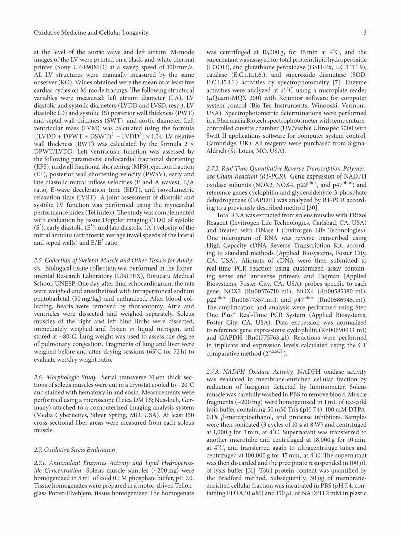
Oxidative Medicine and Cellular Longevity 3
at the level of the aortic valve and left atrium. M-modeimages of the LV were printed on a black-and-white thermalprinter (Sony UP-890MD) at a sweep speed of 100mm/s.All LV structures were manually measured by the sameobserver (KO). Values obtained were the mean of at least fivecardiac cycles on M-mode tracings. The following structuralvariables were measured: left atrium diameter (LA), LVdiastolic and systolic diameters (LVDD and LVSD, resp.), LVdiastolic (D) and systolic (S) posterior wall thickness (PWT)and septal wall thickness (SWT), and aortic diameter. Leftventricular mass (LVM) was calculated using the formula[(LVDD + DPWT + DSWT)3 − LVDD3] × 1.04. LV relativewall thickness (RWT) was calculated by the formula 2 ×DPWT/LVDD. Left ventricular function was assessed bythe following parameters: endocardial fractional shortening(EFS),midwall fractional shortening (MFS), ejection fraction(EF), posterior wall shortening velocity (PWSV), early andlate diastolic mitral inflow velocities (E and A waves), E/Aratio, E-wave deceleration time (EDT), and isovolumetricrelaxation time (IVRT). A joint assessment of diastolic andsystolic LV function was performed using the myocardialperformance index (Tei index).The study was complementedwith evaluation by tissue Doppler imaging (TDI) of systolic(S), early diastolic (E), and late diastolic (A) velocity of themitral annulus (arithmetic average travel speeds of the lateraland septal walls) and E/E ratio.
2.5. Collection of Skeletal Muscle and Other Tissues for Analy-sis. Biological tissue collection was performed in the Exper-imental Research Laboratory (UNIPEX), Botucatu MedicalSchool, UNESP. One day after final echocardiogram, the ratswere weighed and anesthetized with intraperitoneal sodiumpentobarbital (50mg/kg) and euthanized. After blood col-lecting, hearts were removed by thoracotomy. Atria andventricles were dissected and weighed separately. Soleusmuscles of the right and left hind limbs were dissected,immediately weighed and frozen in liquid nitrogen, andstored at −80∘C. Lung weight was used to assess the degreeof pulmonary congestion. Fragments of lung and liver wereweighed before and after drying sessions (65∘C for 72 h) toevaluate wet/dry weight ratio.
2.6. Morphologic Study. Serial transverse 10 𝜇m thick sec-tions of soleus muscles were cut in a cryostat cooled to −20∘Cand stained with hematoxylin and eosin. Measurements wereperformed using a microscope (Leica DMLS; Nussloch, Ger-many) attached to a computerized imaging analysis system(Media Cybernetics, Silver Spring, MD, USA). At least 150cross-sectional fiber areas were measured from each soleusmuscle.
2.7. Oxidative Stress Evaluation
2.7.1. Antioxidant Enzymes Activity and Lipid Hydroperox-ide Concentration. Soleus muscle samples (∼200mg) werehomogenized in 5mL of cold 0.1M phosphate buffer, pH 7.0.Tissue homogenates were prepared in amotor-driven Teflon-glass Potter-Elvehjem, tissue homogenizer. The homogenate
was centrifuged at 10,000 g, for 15min at 4∘C, and thesupernatantwas assayed for total protein, lipid hydroperoxide(LOOH), and glutathione peroxidase (GSH-Px, E.C.1.11.1.9),catalase (E.C.1.11.1.6.), and superoxide dismutase (SOD,E.C.1.15.1.1.) activities by spectrophotometry [7]. Enzymeactivities were analyzed at 25∘C using a microplate reader(𝜇Quant-MQX 200) with Kcjunior software for computersystem control (Bio-Tec Instruments, Winooski, Vermont,USA). Spectrophotometric determinations were performedin aPharmacia Biotech spectrophotometerwith temperature-controlled cuvette chamber (UV/visible Ultrospec 5000 withSwift II applications software for computer system control,Cambridge, UK). All reagents were purchased from Sigma-Aldrich (St. Louis, MO, USA).
2.7.2. Real-Time Quantitative Reverse Transcription-Polymer-ase Chain Reaction (RT-PCR). Gene expression of NADPHoxidase subunits (NOX2, NOX4, p22phox, and p47phox) andreference genes cyclophilin and glyceraldehyde-3-phosphatedehydrogenase (GAPDH) was analyzed by RT-PCR accord-ing to a previously described method [30].
Total RNAwas extracted from soleusmuscleswithTRIzolReagent (Invitrogen Life Technologies, Carlsbad, CA, USA)and treated with DNase I (Invitrogen Life Technologies).One microgram of RNA was reverse transcribed usingHigh Capacity cDNA Reverse Transcription Kit, accord-ing to standard methods (Applied Biosystems, Foster City,CA, USA). Aliquots of cDNA were then submitted toreal-time PCR reaction using customized assay contain-ing sense and antisense primers and Taqman (AppliedBiosystems, Foster City, CA, USA) probes specific to eachgene: NOX2 (Rn00576710 m1), NOX4 (Rn00585380 m1),p22phox (Rn00577357 m1), and p47phox (Rn00586945 m1).The amplification and analysis were performed using StepOne Plus™ Real-Time PCR System (Applied Biosystems,Foster City, CA, USA). Data expression was normalizedto reference gene expressions: cyclophilin (Rn00690933 m1)and GAPDH (Rn01775763 g1). Reactions were performedin triplicate and expression levels calculated using the CTcomparative method (2−ΔΔCT).
2.7.3. NADPH Oxidase Activity. NADPH oxidase activitywas evaluated in membrane-enriched cellular fraction byreduction of lucigenin detected by luminometer. Soleusmuscle was carefully washed in PBS to remove blood. Musclefragments (∼200mg) were homogenized in 1mL of ice-coldlysis buffer containing 50mM Tris (pH 7.4), 100mM DTPA,0.1% 𝛽-mercaptoethanol, and protease inhibitors. Sampleswere then sonicated (3 cycles of 10 s at 8W) and centrifugedat 1,000 g for 3min, at 4∘C. Supernatant was transferred toanother microtube and centrifuged at 18,000 g for 10min,at 4∘C, and transferred again to ultracentrifuge tubes andcentrifuged at 100,000 g for 45min, at 4∘C. The supernatantwas then discarded and the precipitate resuspended in 100 𝜇Lof lysis buffer [31]. Total protein content was quantified bythe Bradford method. Subsequently, 30 𝜇g of membrane-enriched cellular fraction was incubated in PBS (pH 7.4, con-taining EDTA 10 𝜇M) and 150 𝜇L of NADPH 2mM in plastic
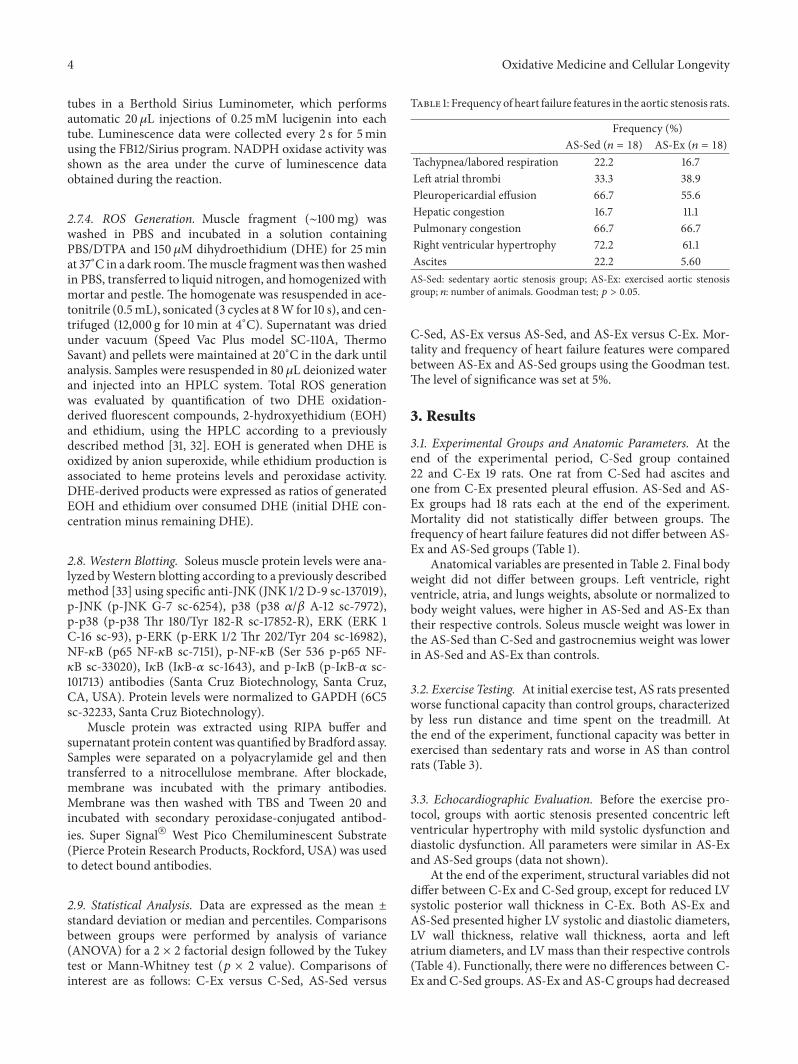
4 Oxidative Medicine and Cellular Longevity
tubes in a Berthold Sirius Luminometer, which performsautomatic 20𝜇L injections of 0.25mM lucigenin into eachtube. Luminescence data were collected every 2 s for 5minusing the FB12/Sirius program. NADPH oxidase activity wasshown as the area under the curve of luminescence dataobtained during the reaction.
2.7.4. ROS Generation. Muscle fragment (∼100mg) waswashed in PBS and incubated in a solution containingPBS/DTPA and 150 𝜇M dihydroethidium (DHE) for 25minat 37∘C in a dark room.Themuscle fragmentwas thenwashedin PBS, transferred to liquid nitrogen, and homogenized withmortar and pestle. The homogenate was resuspended in ace-tonitrile (0.5mL), sonicated (3 cycles at 8W for 10 s), and cen-trifuged (12,000 g for 10min at 4∘C). Supernatant was driedunder vacuum (Speed Vac Plus model SC-110A, ThermoSavant) and pellets were maintained at 20∘C in the dark untilanalysis. Samples were resuspended in 80 𝜇L deionized waterand injected into an HPLC system. Total ROS generationwas evaluated by quantification of two DHE oxidation-derived fluorescent compounds, 2-hydroxyethidium (EOH)and ethidium, using the HPLC according to a previouslydescribed method [31, 32]. EOH is generated when DHE isoxidized by anion superoxide, while ethidium production isassociated to heme proteins levels and peroxidase activity.DHE-derived products were expressed as ratios of generatedEOH and ethidium over consumed DHE (initial DHE con-centration minus remaining DHE).
2.8. Western Blotting. Soleus muscle protein levels were ana-lyzed byWestern blotting according to a previously describedmethod [33] using specific anti-JNK (JNK 1/2 D-9 sc-137019),p-JNK (p-JNK G-7 sc-6254), p38 (p38 𝛼/𝛽 A-12 sc-7972),p-p38 (p-p38 Thr 180/Tyr 182-R sc-17852-R), ERK (ERK 1C-16 sc-93), p-ERK (p-ERK 1/2 Thr 202/Tyr 204 sc-16982),NF-𝜅B (p65 NF-𝜅B sc-7151), p-NF-𝜅B (Ser 536 p-p65 NF-𝜅B sc-33020), I𝜅B (I𝜅B-𝛼 sc-1643), and p-I𝜅B (p-I𝜅B-𝛼 sc-101713) antibodies (Santa Cruz Biotechnology, Santa Cruz,CA, USA). Protein levels were normalized to GAPDH (6C5sc-32233, Santa Cruz Biotechnology).
Muscle protein was extracted using RIPA buffer andsupernatant protein content was quantified by Bradford assay.Samples were separated on a polyacrylamide gel and thentransferred to a nitrocellulose membrane. After blockade,membrane was incubated with the primary antibodies.Membrane was then washed with TBS and Tween 20 andincubated with secondary peroxidase-conjugated antibod-ies. Super SignalⓇ West Pico Chemiluminescent Substrate(Pierce Protein Research Products, Rockford, USA) was usedto detect bound antibodies.
2.9. Statistical Analysis. Data are expressed as the mean ±standard deviation or median and percentiles. Comparisonsbetween groups were performed by analysis of variance(ANOVA) for a 2 × 2 factorial design followed by the Tukeytest or Mann-Whitney test (𝑝 × 2 value). Comparisons ofinterest are as follows: C-Ex versus C-Sed, AS-Sed versus
Table 1: Frequency of heart failure features in the aortic stenosis rats.
Frequency (%)AS-Sed (𝑛 = 18) AS-Ex (𝑛 = 18)
Tachypnea/labored respiration 22.2 16.7Left atrial thrombi 33.3 38.9Pleuropericardial effusion 66.7 55.6Hepatic congestion 16.7 11.1Pulmonary congestion 66.7 66.7Right ventricular hypertrophy 72.2 61.1Ascites 22.2 5.60AS-Sed: sedentary aortic stenosis group; AS-Ex: exercised aortic stenosisgroup; 𝑛: number of animals. Goodman test; 𝑝 > 0.05.
C-Sed, AS-Ex versus AS-Sed, and AS-Ex versus C-Ex. Mor-tality and frequency of heart failure features were comparedbetween AS-Ex and AS-Sed groups using the Goodman test.The level of significance was set at 5%.
3. Results
3.1. Experimental Groups and Anatomic Parameters. At theend of the experimental period, C-Sed group contained22 and C-Ex 19 rats. One rat from C-Sed had ascites andone from C-Ex presented pleural effusion. AS-Sed and AS-Ex groups had 18 rats each at the end of the experiment.Mortality did not statistically differ between groups. Thefrequency of heart failure features did not differ between AS-Ex and AS-Sed groups (Table 1).
Anatomical variables are presented in Table 2. Final bodyweight did not differ between groups. Left ventricle, rightventricle, atria, and lungs weights, absolute or normalized tobody weight values, were higher in AS-Sed and AS-Ex thantheir respective controls. Soleus muscle weight was lower inthe AS-Sed than C-Sed and gastrocnemius weight was lowerin AS-Sed and AS-Ex than controls.
3.2. Exercise Testing. At initial exercise test, AS rats presentedworse functional capacity than control groups, characterizedby less run distance and time spent on the treadmill. Atthe end of the experiment, functional capacity was better inexercised than sedentary rats and worse in AS than controlrats (Table 3).
3.3. Echocardiographic Evaluation. Before the exercise pro-tocol, groups with aortic stenosis presented concentric leftventricular hypertrophy with mild systolic dysfunction anddiastolic dysfunction. All parameters were similar in AS-Exand AS-Sed groups (data not shown).
At the end of the experiment, structural variables did notdiffer between C-Ex and C-Sed group, except for reduced LVsystolic posterior wall thickness in C-Ex. Both AS-Ex andAS-Sed presented higher LV systolic and diastolic diameters,LV wall thickness, relative wall thickness, aorta and leftatrium diameters, and LVmass than their respective controls(Table 4). Functionally, there were no differences between C-Ex and C-Sed groups. AS-Ex and AS-C groups had decreased
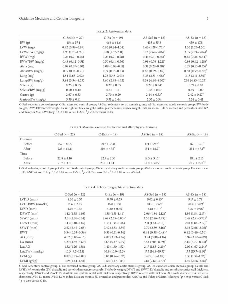
Oxidative Medicine and Cellular Longevity 5
Table 2: Anatomical data.
C-Sed (𝑛 = 22) C-Ex (𝑛 = 19) AS-Sed (𝑛 = 18) AS-Ex (𝑛 = 18)BW (g) 454 ± 57.4 466 ± 64.6 435 ± 35.8 439 ± 47.8LVW (mg) 0.92 (0.86–0.99) 0.96 (0.84–1.04) 1.40 (1.28–1.73)∗ 1.36 (1.23–1.50)†
LVW/BW (mg/g) 1.95 (1.78–1.99) 1.88 (1.67–2.11) 3.17 (2.67–3.86)∗ 3.35 (2.74–3.84)†
RVW (mg) 0.24 (0.21–0.25) 0.23 (0.21–0.28) 0.45 (0.31–0.55)∗ 0.43 (0.26–0.54)†
RVW/BW (mg/g) 0.48 (0.42–0.51) 0.50 (0.41–0.56) 0.99 (0.76–1.22)∗ 0.98 (0.62–1.28)†
Atria (mg) 0.09 (0.07–0.10) 0.09 (0.08–0.11) 0.31 (0.27–0.38)∗ 0.27 (0.15–0.35)†
Atria/BW (mg/g) 0.19 (0.14–0.21) 0.19 (0.16–0.23) 0.68 (0.59–0.87)∗ 0.68 (0.59–0.87)†
Lung (mg) 1.84 (1.65–2.02) 1.78 (1.48–2.03) 3.35 (2.31–4.08)∗ 3.15 (2.11–3.50)†
Lung/BW (mg/g) 3.84 (3.54–4.25) 3.60 (2.98–4.12) 6.58 (4.40–8.10)∗ 7.36 (4.83–10.25)†
Soleus (g) 0.25 ± 0.05 0.22 ± 0.05 0.22 ± 0.04∗ 0.21 ± 0.03Soleus/BW (mg/g) 0.50 ± 0.10 0.45 ± 0.11 0.48 ± 0.07 0.49 ± 0.09Gastro (g) 2.67 ± 0.33 2.70 ± 0.29 2.44 ± 0.33∗ 2.42 ± 0.27†
Gastro/BW (mg/g) 5.39 ± 0.41 5.51 ± 0.44 5.33 ± 0.54 5.54 ± 0.41C-Sed: sedentary control group; C-Ex: exercised control group; AS-Sed: sedentary aortic stenosis group; AS-Ex: exercised aortic stenosis group; BW: bodyweight; LVW: left ventricle weight; RVW: right ventricle weight; Gastro: gastrocnemius muscle weight. Data are mean± SD ormedian and percentiles; ANOVAand Tukey or Mann-Whitney; ∗𝑝 < 0.05 versus C-Sed; †𝑝 < 0.05 versus C-Ex.
Table 3: Maximal exercise test before and after physical training.
C-Sed (𝑛 = 22) C-Ex (𝑛 = 19) AS-Sed (𝑛 = 18) AS-Ex (𝑛 = 18)Distance
Before 257 ± 86.5 247 ± 55.8 171 ± 59.7∗ 163 ± 35.5†
After 225 ± 64.8 304 ± 47.5∗ 154 ± 48.9∗ 254 ± 47.2†‡
TimeBefore 22.8 ± 4.10 22.7 ± 2.53 18.5 ± 3.16∗ 18.1 ± 2.16†
After 21.7 ± 3.51 25.1 ± 1.94∗ 18.0 ± 3.05∗ 22.7 ± 2.61†‡
C-Sed: sedentary control group; C-Ex: exercised control group; AS-Sed: sedentary aortic stenosis group; AS-Ex: exercised aortic stenosis group. Data are mean± SD; ANOVA and Tukey; ∗𝑝 < 0.05 versus C-Sed; †𝑝 < 0.05 versus C-Ex; ‡𝑝 < 0.05 versus AS-Sed.
Table 4: Echocardiographic structural data.
C-Sed (𝑛 = 22) C-Ex (𝑛 = 19) AS-Sed (𝑛 = 18) AS-Ex (𝑛 = 18)LVDD (mm) 8.30 ± 0.55 8.50 ± 0.55 9.02 ± 0.85∗ 9.17 ± 0.74†
LVDD/BW (mm/kg) 16.6 ± 2.05 16.8 ± 1.91 18.9 ± 2.69∗ 20.4 ± 3.09†
LVSD (mm) 4.05 ± 0.55 4.30 ± 0.60 4.81 ± 1.17∗ 5.27 ± 0.98†
DPWT (mm) 1.42 (1.38–1.46) 1.38 (1.31–1.44) 2.06 (1.84–2.12)∗ 1.99 (1.84–2.17)†
SPWT (mm) 3.01 (2.76–3.14) 2.69 (2.65–3.00)∗ 3.60 (2.86–3.79)∗ 3.49 (2.91–3.72)†
DSWT (mm) 1.43 (1.40–1.46) 1.38 (1.34–1.46) 2.11 (1.84–2.16)∗ 2.01 (1.84–2.17)†
SSWT (mm) 2.52 (2.42–2.65) 2.42 (2.33–2.59) 2.79 (2.59–3.16)∗ 2.93 (2.68–3.25)†
RWT 0.34 (0.33–0.36) 0.33 (0.31–0.34) 0.44 (0.38–0.48)∗ 0.42 (0.40–0.50)†
AO (mm) 4.02 (3.83–4.16) 4.02 (3.83–4.16) 3.94 (3.80–4.16) 3.94 (3.80–4.09)LA (mm) 5.29 (4.93–5.69) 5.66 (5.47–5.99) 8.54 (7.88–8.69)∗ 8.14 (6.79–8.54)†
LA/AO 1.32 (1.26–1.38) 1.43 (1.30–1.52) 2.17 (1.83–2.29)∗ 2.09 (1.67–2.24)†
LA/BW (mm/kg) 10.3 (9.5–12.1) 10.9 (10.0–12.3) 17.3 (14.8–19.5)∗ 17.3 (15.7–18.9)†
LVM (g) 0.82 (0.77–0.89) 0.83 (0.74–0.93) 1.62 (1.18–1.87)∗ 1.38 (1.32–1.93)†
LVMI (g/kg) 1.69 (1.44–1.88) 1.64 (1.47–1.85) 2.81 (2.69–3.67)∗ 3.40 (2.66–4.16)†
C-Sed: sedentary control group; C-Ex: exercised control group; AS-Sed: sedentary aortic stenosis group; AS-Ex: exercised aortic stenosis group; LVDD andLVSD: left ventricular (LV) diastolic and systolic diameter, respectively; BW: body weight; DPWT and SPWT: LV diastolic and systolic posterior wall thickness,respectively; DSWT and SSWT: LV diastolic and systolic septal wall thickness, respectively; RWT: relative wall thickness; AO: aorta diameter; LA: left atrialdiameter; LVM: LV mass; LVMI: LVM index. Data are mean ± SD or median and percentiles; ANOVA and Tukey or Mann-Whitney; ∗𝑝 < 0.05 versus C-Sed;†
𝑝 < 0.05 versus C-Ex.
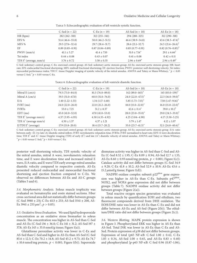
6 Oxidative Medicine and Cellular Longevity
Table 5: Echocardiographic evaluation of left ventricle systolic function.
C-Sed (𝑛 = 22) C-Ex (𝑛 = 19) AS-Sed (𝑛 = 18) AS-Ex (𝑛 = 18)HR (bpm) 282 (262–318) 312 (255–341) 294 (280–325) 312 (290–318)EFS % 51.6 (46.6–55.8) 50.0 (46.2–51.5) 46.6 (38.9–54.8) 43.6 (38.3–47.4)†
MFS % 29.5 (27.6–32.4) 29.7 (28.4–31.7) 28.6 (21.1–32.7) 24.5 (21.6–26.6)†
EF 0.89 (0.85–0.91) 0.87 (0.84–0.89) 0.85 (0.77–0.91) 0.82 (0.76–0.85)†
PWSV (mm/s) 41.1 ± 5.27 41.4 ± 7.10 31.0 ± 7.14∗ 29.1 ± 6.66†
Tei index 0.44 ± 0.08 0.43 ± 0.07 0.41 ± 0.08 0.42 ± 0.11TDI S (average, mm/s) 3.76 ± 0.72 3.58 ± 0.35 2.96 ± 0.69∗ 2.94 ± 0.49†
C-Sed: sedentary control group; C-Ex: exercised control group; AS-Sed: sedentary aortic stenosis group; AS-Ex: exercised aortic stenosis group; HR: heartrate; EFS: endocardial fractional shortening; MFS: midwall fractional shortening; EF: ejection fraction; PWSV: posterior wall shortening velocity; Tei index:myocardial performance index; TDI S: tissue Doppler imaging of systolic velocity of the mitral annulus. ANOVA and Tukey or Mann-Whitney; ∗𝑝 < 0.05versus C-Sed; †𝑝 < 0.05 versus C-Ex.
Table 6: Echocardiographic evaluation of left ventricle diastolic function.
C-Sed (𝑛 = 22) C-Ex (𝑛 = 19) AS-Sed (𝑛 = 18) AS-Ex (𝑛 = 18)Mitral E (cm/s) 79.5 (73.0–84.0) 81.5 (76.0–89.0) 142 (89.0–165)∗ 145 (85.0–159)†
Mitral A (cm/s) 59.5 (51.0–67.0) 60.0 (51.0–76.0) 24.0 (22.0–47.5)∗ 22.5 (16.0–59.0)†
E/A 1.40 (1.22–1.53) 1.34 (1.17–1.60) 5.85 (1.73–7.15)∗ 7.50 (1.47–9.16)†
IVRT (ms) 24.0 (22.0–26.0) 22.0 (21.2–26.0) 18.0 (15.0–22.0)∗ 16.0 (15.0–22.0)†
IVRTn 53.0 ± 7.25 51.2 ± 8.37 42.6 ± 11.4∗ 39.0 ± 10.2†
EDT (ms) 45.0 (41.0–52.0) 47.0 (41.0–53.0) 28.0 (23.0–35.0)∗ 29.0 (24.5–33.0)†
TDI E (average mm/s) 4.37 (3.95–4.95) 4.50 (4.35–4.92) 4.25 (3.04–4.90) 4.17 (3.50–5.25)TDI A (average mm/s) 4.50 ± 1.37 4.37 ± 1.21 3.79 ± 1.47 4.12 ± 1.07E/TDI E (average) 17.9 (15.8–20.0) 18.4 (15.7–20.2) 33.0 (25.7–41.3)∗ 32.1 (24.8–40.6)†
C-Sed: sedentary control group; C-Ex: exercised control group; AS-Sed: sedentary aortic stenosis group; AS-Ex: exercised aortic stenosis group. E/A: ratiobetween early- (E-) to-late (A) diastolic mitral inflow; IVRT: isovolumetric relaxation time; IVRTn: IVRT normalized to heart rate; EDT: E wave decelerationtime; TDI E and A: tissue Doppler imaging (TDI) of early (E) and late (A) diastolic velocity of mitral annulus. ANOVA and Tukey or Mann-Whitney;∗
𝑝 < 0.05 versus C-Sed; †𝑝 < 0.05 versus C-Ex.
posterior wall shortening velocity, TDI systolic velocity ofthe mitral annulus, mitral A wave, isovolumetric relaxationtime, and E wave deceleration time and increased mitral Ewave, E/A ratio, and Ewave/TDI early averagemitral annulusdiastolic velocity compared to respective controls. AS-Expresented reduced endocardial and mesocardial fractionalshortening and ejection fraction compared to C-Ex. Weobserved no differences between AS-Ex and AS-C groups(Tables 5 and 6).
3.4. Morphometric Analysis. Soleus muscle trophicity wasevaluated on hematoxylin and eosin stained sections. Fibercross-sectional area did not statistically differ between groups(C-Sed 3980 ± 231; C-Ex 4113 ± 253; AS-Sed 3541 ± 200, AS-Ex 3941 ± 253𝜇m2; 𝑝 > 0.05).
3.5. Oxidative Stress Evaluation. Weused lipid hydroperoxideconcentration as an oxidative stress biomarker in soleusmuscle. The concentration was higher in AS-Sed than C-Sedand AS-Ex (C-Sed 136 ± 36.0; C-Ex 141 ± 31.2; AS-Sed 197 ±37.8; AS-Ex 143 ± 35.0 nmol/g tissue; Figure 1(a)).
Glutathione peroxidase activity was lower in C-Ex andAS-Sed than C-Sed and higher in AS-Ex than AS-Sed (C-Sed85.6 ± 12.3; C-Ex 70.2 ± 14.8; AS-Sed 45.2 ± 9.73; AS-Ex 74.7± 10.6 nmol/mg protein, 𝑝 < 0.001; Figure 1(b)). Superoxide
dismutase activity was higher in AS-Sed than C-Sed and AS-Ex (C-Sed 8.52 ± 1.95; C-Ex 8.09 ± 0.94; AS-Sed 12.7 ± 1.55,AS-Ex 8.60 ± 0.93 nmol/mg protein, 𝑝 < 0.001; Figure 1(c)).Catalase activity did not differ between groups (C-Sed 34.9± 9.20; C-Ex 41.8 ± 10.2; AS-Sed 52.9 ± 10.9; AS-Ex 43.6 ±15.2 𝜇mol/g tissue; Figure 1(d)).
NADPH oxidase complex subunit p22phox gene expres-sion was higher in AS-Ex than C-Ex. Subunits p47phox,NOX2, and NOX4 gene expression did not differ betweengroups (Table 7). NADPH oxidase activity did not differbetween groups (Figure 2(a)).
Total reactive oxygen species generation was evaluatedin soleus muscle by quantification EOH and ethidium, twofluorescent compounds derived from DHE oxidation. TheEOH/DHE ratio was lower in AS-Ex than C-Ex and did notdiffer between AS-Ex and AS-Sed (Figure 2(b)). The ethid-ium/DHE ratio did not differ between groups (Figure 2(c)).
3.6. Western Blotting. MAPK protein expression is shownin Figure 3. Phosphorylated ERK was higher in AS-Ex thanAS-Sed. Total JNK was lower in AS-Ex than C-Ex and AS-Sed. Protein expression of p38 did not differ between groups.Expression of total (p65 NF-𝜅B: C-Sed 1.00 ± 0.42, C-Ex1.05 ± 0.34, AS-Sed 1.00 ± 0.65, and AS-Ex 0.83 ± 0.40)and phosphorylated [p-p65 NF-𝜅B: C-Sed 0.96 (0.87–1.06),
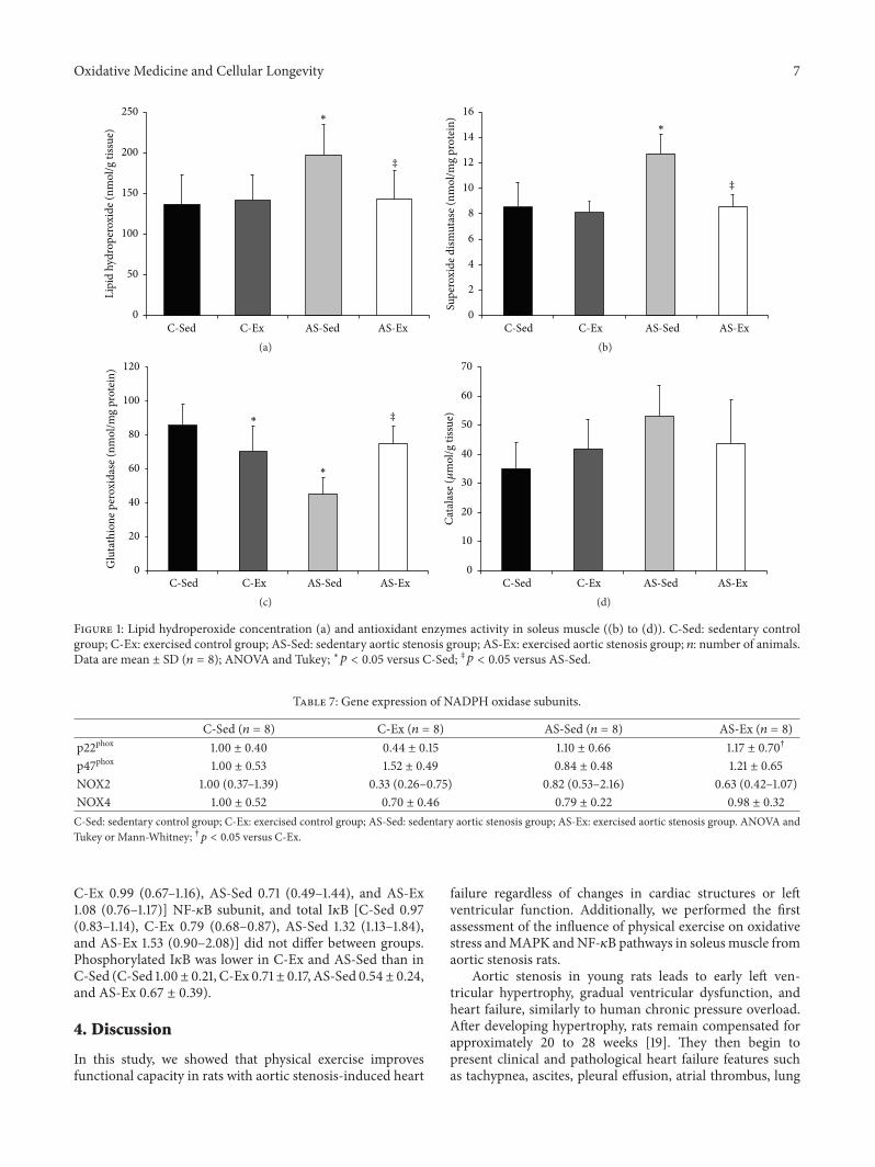
Oxidative Medicine and Cellular Longevity 7
C-Sed C-Ex AS-Sed AS-Ex
‡
∗
0
50
100
150
200
250Li
pid
hydr
oper
oxid
e (nm
ol/g
tiss
ue)
(a)C-Sed C-Ex AS-Sed AS-Ex
∗
‡
0
2
4
6
8
10
12
14
16
Supe
roxi
de d
ismut
ase (
nmol
/mg
prot
ein)
(b)
C-Sed C-Ex AS-Sed AS-Ex
‡∗
∗
0
20
40
60
80
100
120
Glu
tath
ione
per
oxid
ase (
nmol
/mg
prot
ein)
(c)C-Sed C-Ex AS-Sed AS-Ex
0
10
20
30
40
50
60
70
Cata
lase
(𝜇m
ol/g
tiss
ue)
(d)
Figure 1: Lipid hydroperoxide concentration (a) and antioxidant enzymes activity in soleus muscle ((b) to (d)). C-Sed: sedentary controlgroup; C-Ex: exercised control group; AS-Sed: sedentary aortic stenosis group; AS-Ex: exercised aortic stenosis group; 𝑛: number of animals.Data are mean ± SD (𝑛 = 8); ANOVA and Tukey; ∗𝑝 < 0.05 versus C-Sed; ‡𝑝 < 0.05 versus AS-Sed.
Table 7: Gene expression of NADPH oxidase subunits.
C-Sed (𝑛 = 8) C-Ex (𝑛 = 8) AS-Sed (𝑛 = 8) AS-Ex (𝑛 = 8)p22phox 1.00 ± 0.40 0.44 ± 0.15 1.10 ± 0.66 1.17 ± 0.70†
p47phox 1.00 ± 0.53 1.52 ± 0.49 0.84 ± 0.48 1.21 ± 0.65NOX2 1.00 (0.37–1.39) 0.33 (0.26–0.75) 0.82 (0.53–2.16) 0.63 (0.42–1.07)NOX4 1.00 ± 0.52 0.70 ± 0.46 0.79 ± 0.22 0.98 ± 0.32C-Sed: sedentary control group; C-Ex: exercised control group; AS-Sed: sedentary aortic stenosis group; AS-Ex: exercised aortic stenosis group. ANOVA andTukey or Mann-Whitney; †𝑝 < 0.05 versus C-Ex.
C-Ex 0.99 (0.67–1.16), AS-Sed 0.71 (0.49–1.44), and AS-Ex1.08 (0.76–1.17)] NF-𝜅B subunit, and total I𝜅B [C-Sed 0.97(0.83–1.14), C-Ex 0.79 (0.68–0.87), AS-Sed 1.32 (1.13–1.84),and AS-Ex 1.53 (0.90–2.08)] did not differ between groups.Phosphorylated I𝜅B was lower in C-Ex and AS-Sed than inC-Sed (C-Sed 1.00± 0.21, C-Ex 0.71± 0.17, AS-Sed 0.54± 0.24,and AS-Ex 0.67 ± 0.39).
4. Discussion
In this study, we showed that physical exercise improvesfunctional capacity in rats with aortic stenosis-induced heart
failure regardless of changes in cardiac structures or leftventricular function. Additionally, we performed the firstassessment of the influence of physical exercise on oxidativestress andMAPK andNF-𝜅B pathways in soleusmuscle fromaortic stenosis rats.
Aortic stenosis in young rats leads to early left ven-tricular hypertrophy, gradual ventricular dysfunction, andheart failure, similarly to human chronic pressure overload.After developing hypertrophy, rats remain compensated forapproximately 20 to 28 weeks [19]. They then begin topresent clinical and pathological heart failure features suchas tachypnea, ascites, pleural effusion, atrial thrombus, lung
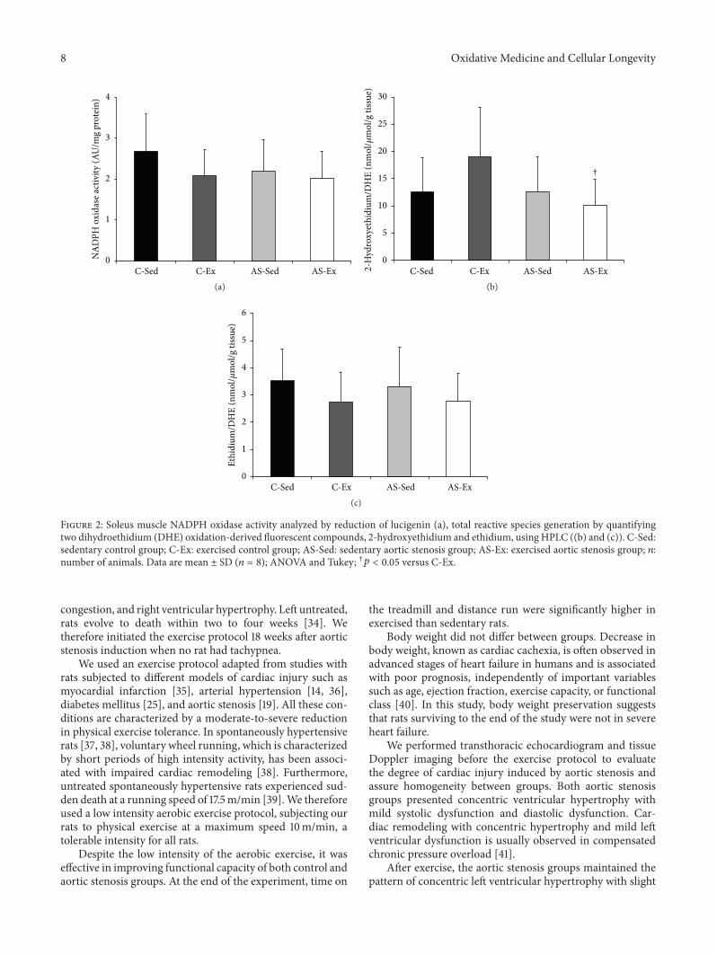
8 Oxidative Medicine and Cellular Longevity
C-Sed C-Ex AS-Sed AS-Ex
NA
DPH
oxi
dase
activ
ity (A
U/m
g pr
otei
n)
0
1
2
3
4
(a)C-Sed C-Ex AS-Sed AS-Ex
†
0
5
10
15
20
25
30
2-H
ydro
xyet
hidi
um/D
HE
(nm
ol/𝜇
mol
/g ti
ssue
)
(b)
C-Sed C-Ex AS-Sed AS-Ex0
1
2
3
4
5
6
Ethi
dium
/DH
E (n
mol
/𝜇m
ol/g
tiss
ue)
(c)
Figure 2: Soleus muscle NADPH oxidase activity analyzed by reduction of lucigenin (a), total reactive species generation by quantifyingtwo dihydroethidium (DHE) oxidation-derived fluorescent compounds, 2-hydroxyethidium and ethidium, using HPLC ((b) and (c)). C-Sed:sedentary control group; C-Ex: exercised control group; AS-Sed: sedentary aortic stenosis group; AS-Ex: exercised aortic stenosis group; 𝑛:number of animals. Data are mean ± SD (𝑛 = 8); ANOVA and Tukey; †𝑝 < 0.05 versus C-Ex.
congestion, and right ventricular hypertrophy. Left untreated,rats evolve to death within two to four weeks [34]. Wetherefore initiated the exercise protocol 18 weeks after aorticstenosis induction when no rat had tachypnea.
We used an exercise protocol adapted from studies withrats subjected to different models of cardiac injury such asmyocardial infarction [35], arterial hypertension [14, 36],diabetes mellitus [25], and aortic stenosis [19]. All these con-ditions are characterized by a moderate-to-severe reductionin physical exercise tolerance. In spontaneously hypertensiverats [37, 38], voluntary wheel running, which is characterizedby short periods of high intensity activity, has been associ-ated with impaired cardiac remodeling [38]. Furthermore,untreated spontaneously hypertensive rats experienced sud-den death at a running speed of 17.5m/min [39].We thereforeused a low intensity aerobic exercise protocol, subjecting ourrats to physical exercise at a maximum speed 10m/min, atolerable intensity for all rats.
Despite the low intensity of the aerobic exercise, it waseffective in improving functional capacity of both control andaortic stenosis groups. At the end of the experiment, time on
the treadmill and distance run were significantly higher inexercised than sedentary rats.
Body weight did not differ between groups. Decrease inbody weight, known as cardiac cachexia, is often observed inadvanced stages of heart failure in humans and is associatedwith poor prognosis, independently of important variablessuch as age, ejection fraction, exercise capacity, or functionalclass [40]. In this study, body weight preservation suggeststhat rats surviving to the end of the study were not in severeheart failure.
We performed transthoracic echocardiogram and tissueDoppler imaging before the exercise protocol to evaluatethe degree of cardiac injury induced by aortic stenosis andassure homogeneity between groups. Both aortic stenosisgroups presented concentric ventricular hypertrophy withmild systolic dysfunction and diastolic dysfunction. Car-diac remodeling with concentric hypertrophy and mild leftventricular dysfunction is usually observed in compensatedchronic pressure overload [41].
After exercise, the aortic stenosis groups maintained thepattern of concentric left ventricular hypertrophy with slight
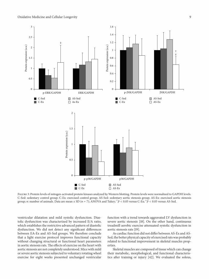
Oxidative Medicine and Cellular Longevity 9
p-p38/GAPDH p38/GAPDH
Prot
ein
expr
essio
n (a
.u.)
0
0.5
1
1.5
2
C-SedC-Ex
AS-SedAS-Ex
p-JNK/GAPDH JNK/GAPDH
†‡
Prot
ein
expr
essio
n (a
.u.)
0
0.2
0.4
0.6
0.8
1
1.2
1.4
1.6
C-SedC-Ex
AS-SedAS-Ex
p-ERK/GAPDH ERK/GAPDH
‡
Prot
ein
expr
essio
n (a
.u.)
0
0.5
1
1.5
2
2.5
3
C-SedC-Ex
AS-SedAs-Ex
Figure 3: Protein levels of mitogen-activated protein kinases analyzed byWestern blotting. Protein levels were normalized to GAPDH levels.C-Sed: sedentary control group; C-Ex: exercised control group; AS-Sed: sedentary aortic stenosis group; AS-Ex: exercised aortic stenosisgroup; 𝑛: number of animals. Data are mean ± SD (𝑛 = 7); ANOVA and Tukey; †𝑝 < 0.05 versus C-Ex; ‡𝑝 < 0.05 versus AS-Sed.
ventricular dilatation and mild systolic dysfunction. Dias-tolic dysfunction was characterized by increased E/A ratio,which establishes the restrictive advanced pattern of diastolicdysfunction. We did not detect any significant differencesbetween EA-Ex and AS-Sed groups. We therefore concludethat a light exercise protocol improves functional capacitywithout changing structural or functional heart parametersin aortic stenosis rats.The effects of exercise on the heart withaortic stenosis are not completely understood.Micewithmildor severe aortic stenosis subjected to voluntary rotatingwheelexercise for eight weeks presented unchanged ventricular
function with a trend towards aggravated LV dysfunction insevere aortic stenosis [18]. On the other hand, continuoustreadmill aerobic exercise attenuated systolic dysfunction inaortic stenosis rats [19].
As cardiac function did not differ betweenAS-Ex andAS-Sed, the better physical capacity of exercised ratswas probablyrelated to functional improvement in skeletal muscles prop-erties.
Skeletalmuscles are composed of tissue which can changetheir metabolic, morphological, and functional characteris-tics after training or injury [42]. We evaluated the soleus,
10 Oxidative Medicine and Cellular Longevity
a muscle with predominantly slow twitch fibers and oxidativemetabolism. Previous studies have shown that this muscleis sensitive to alterations induced by acute or chronic heartfailure [6–8]. Furthermore, skeletal muscles preferentiallycomposed of oxidative fibers are more sensitive to the effectsof aerobic exercise than muscles preferably composed ofglycolytic fibers [43].
Wefirst evaluated redox status. Although themechanismsinvolved inmuscle alterations are not completely understood,it is well established that increased oxidative stress plays a rolein muscle abnormalities [7, 44].
Lipid hydroperoxide concentration was greater in AS-Sed than C-Sed and AS-Ex, showing that muscle oxidativestress is increased during heart failure and normalized byexercise. Cellular response to oxidative stress depends onthe intensity of the reactive oxygen species generation.At low concentrations, reactive oxygen species stimulatesantioxidant enzymes and receptors; at high concentrations,however, they inhibit enzymatic activity leading to cellulardamage [45]. In the AS-Sed group, increased oxidativestress was combined with increased superoxide dismutaseactivity and reduced glutathione peroxidase activity. Exerciseprevented changes in antioxidant enzymes in the AS-Exgroup. Reduction of antioxidant enzyme activity has beendescribed in skeletal muscle during heart failure [7, 35, 44].In addition, clinical and experimental studies have shownthat exercise induces antioxidant properties in skeletalmuscleduring heart failure [35, 46]. Our results therefore show thataerobic exercise prevented increases in oxidative stress andchanges in antioxidant enzymes.
The NADPH oxidase complex is an important source ofreactive oxygen species. To determine whether this complexis involved in increased oxidative stress and its modulationby exercise, we evaluated NADPH oxidase activity andgene subunit expression. We analyzed gene expression oftransmembrane subunits NOX2, NOX4, and p22phox and thecytosolic subunit p47phox. NADPH oxidase activity and itssubunits expression did not differ between groups, exceptfor higher p22phox subunit expression in AS-Ex than C-Ex. Despite this increased p22phox gene expression, AS-ExgroupNADPHoxidase activity was unchanged.We evaluatedNADPH oxidase activity by measuring the light emitted bya reaction between reduced lucigenin and superoxide anion,which is a product of NADPH oxidase [31, 32]. The roleof NADPH oxidase in muscle oxidative stress during heartfailure is still unclear. In infarcted rats [21] and mice [47],increased NADPH oxidase activity was observed in the plan-taris and hindlimbmuscles. On the other hand, by evaluatingNADPH oxidase activity using dihydroethidium, a moleculeoxidized by superoxide anion, we found unchanged activityin soleus from rats with myocardial infarction-induced heartfailure [7]. Different animal species, experimental model, ormuscle evaluated may be responsible for divergent results.
Total reactive oxygen species generation was assessedby quantifying two fluorescent compounds derived fromdihydroethidium oxidation: 2-hydroxyethidium and ethid-ium. Hydroxyethidium detection by HPLC fluorescence wasproposed to more precisely measure superoxide production
in blood vessels and was later adapted for use in striatedmuscle [31, 32]. Total reactive oxygen species generationdid not change between groups, except for a lower hydrox-yethidium/dihydroethidium ratio in AS-Ex than C-Ex. Thisresult is in accordance with the lower lipid hydroperoxideconcentration in the AS-Ex group.
Intracellular reactive oxygen species signaling pathwaysare not completely understood. Studies suggest that MAPKand the NF-𝜅B pathway are involved in muscle response tooxidative stress [21, 48]. MAPKs consist of four members:extracellular signal-regulated kinase (ERK) 1/2, p38, c-JunNH2-terminal kinase (JNK), and ERK 5 [49]. Expression oftheMAPKproteins did not differ betweenAS-Sed andC-Sed.Despite ERK expression being approximately 46% lower inAS-Sed than C-Sed, this difference did not reach statisticalsignificance. Exercise reduced total JNK expression in AS-Excompared to C-Ex andAS-Sed and increased phosphorylatedERK in AS-Ex compared to AS-Sed. While activation of JNKand p38 is associated with cell response to stress and muscleloss, activation of ERK is related to anabolic processes suchas cell division, growth, and differentiation [49].Therefore, inthis study exercise modulated total JNK and phosphorylatedERK towards preserving protein synthesis and muscle mass.We have previously observed MAPK changes in diaphragmand soleus muscle in infarcted rats with heart failure [7,50]. Except for reduced I𝜅B expression in C-Ex and AS-Sed compared to C-Sed, NF-𝜅B pathway protein expressionremained unchanged in this experimental model.
5. Conclusion
Exercise improves the functional capacity of rats with aorticstenosis-induced heart failure regardless of changes in cardiacstructures or left ventricular function. In soleus muscle, exer-cise reduces oxidative stress, preserves antioxidant enzymeactivity, and modulates JNK and p-ERK expression with nochanges in NADPH oxidase activity, NADPH oxidase sub-units gene expression, or NF-𝜅B pathway protein expression.
Conflict of Interests
The authors report no conflict of interests.
Acknowledgments
The authors are grateful to Jose Carlos Georgette andMario Batista Bruno for their technical assistance andColin Edward Knaggs for English editing. Financial sup-port was provided by CNPq (306857/2012-0, 306845/2012-1, and 479085/2013-7), FAPESP (2012/22485-3, 2012/21687-1),CAPES, and PROPe, UNESP.
References
[1] D. Mozaffarian, E. J. Benjamin, A. S. Go et al., “Heart diseaseand stroke statistics—2015 update: a report from the AmericanHeart Association,” Circulation, vol. 131, no. 4, pp. e29–e322,2015.
Oxidative Medicine and Cellular Longevity 11
[2] S. Mandic, J. Myers, S. E. Selig, and I. Levinger, “Resistance ver-sus aerobic exercise training in chronic heart failure,” CurrentHeart Failure Reports, vol. 9, no. 1, pp. 57–64, 2012.
[3] S. A. Phillips, K. Vuckovic, L. P. Cahalin, and T. Baynard,“Defining the system: contributors to exercise limitations inheart failure,” Heart Failure Clinics, vol. 11, no. 1, pp. 1–16, 2015.
[4] R. L. Damatto, P. F.Martinez, A. R. R. Lima et al., “Heart failure-induced skeletal myopathy in spontaneously hypertensive rats,”International Journal of Cardiology, vol. 167, no. 3, pp. 698–703,2013.
[5] J. Downing and G. J. Balady, “The role of exercise training inheart failure,” Journal of the American College of Cardiology, vol.58, no. 6, pp. 561–569, 2011.
[6] A. R. R. Lima, P. F. Martinez, K. Okoshi et al., “Myostatin andfollistatin expression in skeletal muscles of rats with chronicheart failure,” International Journal of Experimental Pathology,vol. 91, no. 1, pp. 54–62, 2010.
[7] P. F.Martinez, C. Bonomo, D.M. Guizoni et al., “Influence of N-acetylcysteine on oxidative stress in slow-twitch soleus muscleof heart failure rats,” Cellular Physiology and Biochemistry, vol.35, no. 1, pp. 148–159, 2015.
[8] P. F. Martinez, K. Okoshi, L. A. Zornoff et al., “Chronic heartfailure-induced skeletal muscle atrophy, necrosis, and changesin myogenic regulatory factors,” Medical Science Monitor, vol.16, no. 12, pp. BR374–BR383, 2010.
[9] J. J. V. McMurray, S. Adamopoulos, S. D. Anker et al., “ESCGuidelines for the diagnosis and treatment of acute and chronicheart failure 2012: the Task Force for the Diagnosis andTreatment of Acute and Chronic Heart Failure 2012 of theEuropean Society of Cardiology. Developed in collaborationwith theHeart Failure Association (HFA) of the ESC,” EuropeanHeart Journal, vol. 33, no. 14, pp. 1787–1847, 2012.
[10] C. W. Yancy, M. Jessup, B. Bozkurt et al., “2013 ACCF/AHAguideline for the management of heart failure: executivesummary: a report of the American College of CardiologyFoundation/American Heart Association task force on practiceguidelines,” Circulation, vol. 128, no. 16, pp. 1810–1852, 2013.
[11] K. E. Flynn, I. L. Piña, D. J. Whellan et al., “Effects of exercisetraining on health status in patients with chronic heart failure,”The Journal of the AmericanMedical Association, vol. 301, no. 14,pp. 1451–1459, 2009.
[12] P. Giannuzzi, P. L. Temporelli, U. Corrà, and L. Tavazzi,“Antiremodeling effect of long-termexercise training in patientswith stable chronic heart failure: results of the exercise in leftventricular dysfunction and chronic heart failure (ELVD-CHF)trial,” Circulation, vol. 108, no. 5, pp. 554–559, 2003.
[13] C. E. Negrao, H. R. Middlekauff, I. L. Gomes-Santos, and L. M.Antunes-Correa, “Effects of exercise training on neurovascularcontrol and skeletal myopathy in systolic heart failure,” TheAmerican Journal of Physiology—Heart and Circulatory Physi-ology, vol. 308, no. 8, pp. H792–H802, 2015.
[14] L. U. Pagan, R. L. Damatto, M. D. Cezar et al., “Long-term lowintensity physical exercise attenuates heart failure developmentin aging spontaneously hypertensive rats,” Cellular Physiologyand Biochemistry, vol. 36, no. 1, pp. 61–74, 2015.
[15] U. Wisløff, A. Støylen, J. P. Loennechen et al., “Superior car-diovascular effect of aerobic interval training versus moderatecontinuous training in heart failure patients. A randomizedstudy,” Circulation, vol. 115, no. 24, pp. 3086–3094, 2007.
[16] J. C. Campos, T. Fernandes, L. R. Bechara et al., “Increasedclearance of reactive aldehydes and damaged proteins in hyper-tension-induced compensated cardiac hypertrophy: impact of
exercise training,” Oxidative Medicine and Cellular Longevity,vol. 2015, Article ID 464195, 11 pages, 2015.
[17] D. J. Duncker, E. D. van Deel, M. C. de Waard, M. de Boer, D.Merkus, and J. van der Velden, “Exercise training in adversecardiac remodeling,” Pflügers Archiv, vol. 466, no. 6, pp. 1079–1091, 2014.
[18] E. D. van Deel, M. de Boer, D. W. Kuster et al., “Exercisetraining does not improve cardiac function in compensated ordecompensated left ventricular hypertrophy induced by aorticstenosis,” Journal of Molecular and Cellular Cardiology, vol. 50,no. 6, pp. 1017–1025, 2011.
[19] R. W. A. Souza, W. P. Piedade, L. C. Soares et al., “Aerobicexercise training prevents heart failure-induced skeletal muscleatrophy by anti-catabolic, but not anabolic actions,” PLoS ONE,vol. 9, no. 10, Article ID e110020, 2014.
[20] K. Bedard and K.-H. Krause, “The NOX family of ROS-generatingNADPHoxidases: physiology and pathophysiology,”Physiological Reviews, vol. 87, no. 1, pp. 245–313, 2007.
[21] L. R. G. Bechara, J. B. N. Moreira, P. R. Jannig et al., “NADPHoxidase hyperactivity induces plantaris atrophy in heart failurerats,” International Journal of Cardiology, vol. 175, no. 3, pp. 499–507, 2014.
[22] M.D.M. Cezar, R. L. Damatto, P. F.Martinez et al., “Aldosteroneblockade reduces mortality without changing cardiac remodel-ing in spontaneously hypertensive rats,” Cellular Physiology andBiochemistry, vol. 32, no. 5, pp. 1275–1287, 2013.
[23] P. F. Martinez, K. Okoshi, L. A. M. Zornoff et al., “Echocardio-graphic detection of congestive heart failure in postinfarctionrats,” Journal of Applied Physiology, vol. 111, no. 2, pp. 543–551,2011.
[24] O. C. Mendes, M. M. Sugizaki, D. S. Campos et al., “Exercisetolerance in rats with aortic stenosis and ventricular diastolicand/or systolic dysfunction,” Arquivos Brasileiros de Cardiolo-gia, vol. 100, no. 1, pp. 44–51, 2013.
[25] C. Gimenes, R. Gimenes, C. M. Rosa et al., “Low intensityphysical exercise attenuates cardiac remodeling andmyocardialoxidative stress and dysfunction in diabetic rats,” Journal ofDiabetes Research, vol. 2015, Article ID 457848, 10 pages, 2015.
[26] K. Okoshi, J. R. Fioretto, M. P. Okoshi et al., “Food restrictioninduces in vivo ventricular dysfunction in spontaneously hyper-tensive rats without impairment of in vitro myocardial contrac-tility,” Brazilian Journal of Medical and Biological Research, vol.37, no. 4, pp. 607–613, 2004.
[27] K. Okoshi, L. S. Matsubara, M. P. Okoshi et al., “Foodrestriction-induced myocardial dysfunction demonstrated bythe combination of in vivo and in vitro studies,” NutritionResearch, vol. 22, no. 11, pp. 1353–1364, 2002.
[28] K. Okoshi, H. B. Ribeiro, M. P. Okoshi et al., “Improved systolicventricular function with normal myocardial mechanics incompensated cardiac hypertrophy,” Japanese Heart Journal, vol.45, no. 4, pp. 647–656, 2004.
[29] C. Di Filippo, M. C. Trotta, R. Maisto et al., “Daily oxygen/O3
treatment reduces muscular fatigue and improves cardiac per-formance in rats subjected to prolonged high intensity physicalexercise,” Oxidative Medicine and Cellular Longevity, vol. 2015,Article ID 190640, 11 pages, 2015.
[30] C.M. Rosa, N. P. Xavier, D. H. Campos et al., “Diabetes mellitusactivates fetal gene program and intensifies cardiac remodelingand oxidative stress in aged spontaneously hypertensive rats,”Cardiovascular Diabetology, vol. 12, article 152, 2013.
[31] F. R. M. Laurindo, D. C. Fernandes, and C. X. C. Santos,“Assessment of superoxide production and NADPH oxidase
12 Oxidative Medicine and Cellular Longevity
activity by HPLC analysis of dihydroethidium oxidation prod-ucts,”Methods in Enzymology, vol. 441, pp. 237–260, 2008.
[32] D. C. Fernandes, J.Wosniak Jr., L. A. Pescatore et al., “Analysis ofDHE-derived oxidation products by HPLC in the assessment ofsuperoxide production andNADPHoxidase activity in vascularsystems,” The American Journal of Physiology—Cell Physiology,vol. 292, no. 1, pp. C413–C422, 2007.
[33] X. Yan, A. J. T. Schuldt, R. L. Price et al., “Pressure overload-induced hypertrophy in transgenic mice selectively overex-pressing AT
2
receptors in ventricular myocytes,” AmericanJournal of Physiology—Heart and Circulatory Physiology, vol.294, no. 3, pp. H1274–H1281, 2008.
[34] M. O. Boluyt, K. G. Robinson, A. L. Meredith et al., “Heartfailure after long-term supravalvular aortic constriction in rats,”TheAmerican Journal ofHypertension, vol. 18, no. 2, pp. 202–212,2005.
[35] J. B. N. Moreira, L. R. G. Bechara, L. H. M. Bozi et al., “High-versus moderate-intensity aerobic exercise training effects onskeletal muscle of infarcted rats,” Journal of Applied Physiology,vol. 114, no. 8, pp. 1029–1041, 2013.
[36] A. J. Chicco, S. A. McCune, C. A. Emter et al., “Low-intensityexercise training delays heart failure and improves survival infemale hypertensive heart failure rats,”Hypertension, vol. 51, no.4, pp. 1096–1102, 2008.
[37] R. L. Schultz, E. L. Kullman, R. P.Waters et al., “Metabolic adap-tations of skeletalmuscle to voluntarywheel running exercise inhypertensive heart failure rats,” Physiological Research, vol. 62,no. 4, pp. 361–369, 2013.
[38] R. L. Schultz, J. G. Swallow, R. P. Waters et al., “Effects ofexcessive long-term exercise on cardiac function and myocyteremodeling in hypertensive heart failure rats,” Hypertension,vol. 50, no. 2, pp. 410–416, 2007.
[39] C. A. Emter, S. A. McCune, G. C. Sparagna, M. J. Radin,and R. L. Moore, “Low-intensity exercise training delays onsetof decompensated heart failure in spontaneously hypertensiveheart failure rats,” American Journal of Physiology—Heart andCirculatory Physiology, vol. 289, no. 5, pp. H2030–H2038, 2005.
[40] S. D. Anker, A. Negassa, A. J. S. Coats et al., “Prognosticimportance of weight loss in chronic heart failure and the effectof treatment with angiotensin-converting-enzyme inhibitors:an observational study,”The Lancet, vol. 361, no. 9363, pp. 1077–1083, 2003.
[41] M. D. Cezar, R. L. Damatto, L. U. Pagan et al., “Early spironolac-tone treatment attenuates heart failure development by improv-ing myocardial function and reducing fibrosis in spontaneouslyhypertensive rats,”Cellular Physiology and Biochemistry, vol. 36,no. 4, pp. 1453–1466, 2015.
[42] P. Bonaldo andM. Sandri, “Cellular andmolecular mechanismsof muscle atrophy,” Disease Models and Mechanisms, vol. 6, no.1, pp. 25–39, 2013.
[43] A. P. Russell, V. C. Foletta, R. J. Snow, andG.D.Wadley, “Skeletalmuscle mitochondria: a major player in exercise, health anddisease,” Biochimica et Biophysica Acta—General Subjects, vol.1840, no. 4, pp. 1276–1284, 2014.
[44] A. Linke, V. Adams, P. C. Schulze et al., “Antioxidative effectsof exercise training in patients with chronic heart failure:increase in radical scavenger enzyme activity in skeletalmuscle,”Circulation, vol. 111, no. 14, pp. 1763–1770, 2005.
[45] Z. Radak, H. Y. Chung, and S. Goto, “Exercise and horme-sis: oxidative stress-related adaptation for successful aging,”Biogerontology, vol. 6, no. 1, pp. 71–75, 2005.
[46] A. V. N. Bacurau, M. A. Jardim, J. C. B. Ferreira et al.,“Sympathetic hyperactivity differentially affects skeletal musclemass in developing heart failure: role of exercise training,”Journal of Applied Physiology, vol. 106, no. 5, pp. 1631–1640, 2009.
[47] Y. Ohta, S. Kinugawa, S. Matsushima et al., “Oxidative stressimpairs insulin signal in skeletal muscle and causes insulinresistance in postinfarct heart failure,”The American Journal ofPhysiology—Heart and Circulatory Physiology, vol. 300, no. 5,pp. H1637–H1644, 2011.
[48] M.-S. Byun, K.-I. Jeon, J.-W. Choi, J.-Y. Shim, and D.-M. Jue,“Dual effect of oxidative stress on NF-𝜅B activation in HeLacells,” Experimental and Molecular Medicine, vol. 34, no. 5, pp.332–339, 2002.
[49] H. Shi, J. M. Scheffler, C. Zeng et al., “Mitogen-activated proteinkinase signaling is necessary for the maintenance of skeletalmuscle mass,” American Journal of Physiology—Cell Physiology,vol. 296, no. 5, pp. C1040–C1048, 2009.
[50] A. R. R. Lima, P. F. Martinez, R. L. Damatto et al., “Heartfailure-induced diaphragm myopathy,” Cellular Physiology andBiochemistry, vol. 34, no. 2, pp. 333–345, 2014.
Research ArticleEffect of Carnosine in Experimental Arthritis and on PrimaryCulture Chondrocytes
S. Ponist,1 F. Drafi,1 V. Kuncirova,1 D. Mihalova,1 L. Rackova,1 L. Danisovic,2
O. Ondrejickova,1 I. Tumova,3 O. Trunova,4 T. Fedorova,4 and K. Bauerova1,3
1 Institute of Experimental Pharmacology and Toxicology, Slovak Academy of Sciences, Dubravska cesta 9, 841 04 Bratislava, Slovakia2Faculty of Medicine, Comenius University in Bratislava, Spitalska 24, 813 72 Bratislava, Slovakia3Faculty of Pharmacy, Comenius University in Bratislava, Odbojarov 10, 832 32 Bratislava, Slovakia4Research Center of Neurology, RAMS, Volokolamskoyeshosse 80, Moscow 125367, Russia
Correspondence should be addressed to K. Bauerova; [email protected]
Received 25 September 2015; Revised 27 November 2015; Accepted 8 December 2015
Academic Editor: Victor M. Victor
Copyright © 2016 S. Ponist et al.This is an open access article distributed under the Creative Commons Attribution License, whichpermits unrestricted use, distribution, and reproduction in any medium, provided the original work is properly cited.
Carnosine’s (CARN) anti-inflammatory potential in autoimmune diseases has been but scarcely investigated as yet. The aim of thisstudy was to evaluate the therapeutic potential of CARN in rat adjuvant arthritis, in the model of carrageenan induced hind pawedema (CARA), and also in primary culture of chondrocytes under H
2O2injury. The experiments were done on healthy animals,
arthritic animals, and arthritic animals with oral administration of CARN in a daily dose of 150mg/kg b.w. during 28 days aswell as animals with CARA treated by a single administration of CARN in the same dose. CARN beneficially affected hind pawvolume and changes in body weight on day 14 and reduced hind paw swelling in CARA. Markers of oxidative stress in plasmaand brain (malondialdehyde, 4-hydroxynonenal, protein carbonyls, and lag time of lipid peroxidation) and also activity of gamma-glutamyltransferase were significantly corrected by CARN. CARN also reduced IL-1alpha in plasma. Suppression of intracellularoxidant levels was also observed in chondrocytes pretreated with CARN. Our results obtained on two animal models showed thatCARNhas systemic anti-inflammatory activity and protected rat brain and chondrocytes fromoxidative stress.This finding suggeststhat CARN might be beneficial for treatment of arthritic diseases.
We dedicate this paper to the memory of Professor Alexander A. Boldyrev with respect and gratitude.
1. Introduction
Carnosine (CARN) is dipeptide consisting of 𝛽-alanine andL-histidine. It was shown to be a specific constituent ofexcitable tissues of all vertebrates accumulating in amountsexceeding that of ATP [1]. The antioxidant capacity of thiscompound is well documented, as well as its pH buffering,osmoregulating, and metal-chelating abilities [2]. However,a more recent study showed unexpectedly lower bindingconstant values of carnosine and therefore a relatively minorrole of the transition metal ion chelation in its antioxidantabilities [3]. We previously showed inhibitory propertiesof CARN against degradation of hyaluronan solutions at
experimental conditions in vitro. In the reaction systemwith the prevalence of hydroxyl and/or peroxy-type radicals,CARN in 200𝜇mol/L concentration tested exerted a pro-tective action on hyaluronan degradation [4]. CARN, whencompared with ascorbate, was more effective in inhibitionof lipid peroxidation in meat [5]. At physiological concen-trations, CARN was found to directly react with superoxideanion comparably to superoxide dismutase and the constantfor interaction of carnosine with O
2
∙− was calculated tobe 105M−1⋅s−1 and not significantly different in respect tothat of ascorbic acid and 𝛼-tocopherol [6]. Also, a sparingor regenerating effect of carnosine towards endogenousantioxidants was demonstrated in the liver of rats treatedwith
Hindawi Publishing CorporationOxidative Medicine and Cellular LongevityVolume 2016, Article ID 8470589, 11 pageshttp://dx.doi.org/10.1155/2016/8470589
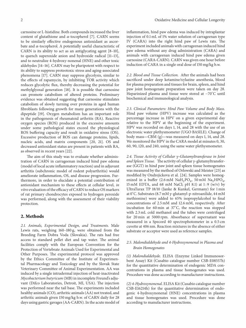
2 Oxidative Medicine and Cellular Longevity
carnosine or L-histidine. Both compounds increased the livercontent of glutathione and 𝛼-tocopherol [7]. CARN seemsto be similarly effective endogenous antioxidant as ascor-bate and 𝛼-tocopherol. A potentially useful characteristic ofCARN is its ability to act as an antiglycating agent [8–10],to quench superoxide anion and hydroxide radical [11–13],and to neutralize 4-hydroxy-nonenal (HNE) and other toxicaldehydes [14–16]. CARNmay be pluripotent with respect toits ability to suppress proteotoxic stress and aging-associatedphenomena [17]. CARN may suppress glycolysis, similar tothe effects of rapamycin, by inhibiting TOR activity whichreduces glycolytic flux, thereby decreasing the potential formethylglyoxal generation [18]. It is possible that carnosinecan promote catabolism of altered proteins. Preliminaryevidence was obtained suggesting that carnosine stimulatescatabolism of slowly turning over proteins in aged humanfibroblasts following growth for many generations with thedipeptide [19]. Oxygen metabolism has an important rolein the pathogenesis of rheumatoid arthritis (RA). Reactiveoxygen species (ROS) produced in the excessive amountsunder some pathological states exceed the physiologicalROS buffering capacity and result in oxidative stress (OS).Excessive production of ROS can damage proteins, lipids,nucleic acids, and matrix components [20, 21]. OS anddecreased antioxidant status are present in patients with RA,as observed in recent years [22].
The aim of this study was to evaluate whether adminis-tration of CARN in carrageenan induced hind paw edema(model of local acute inflammatory reaction) and in adjuvantarthritis (subchronic model of rodent polyarthritis) wouldameliorate inflammation, OS, and disease progression. Fur-thermore, in order to elucidate a potential contribution ofantioxidant mechanism to these effects at cellular level, invitro evaluation of the efficacy ofCARN to reduceOSmarkersof rat primary chondrocytes exposed to hydrogen peroxidewas performed, along with the assessment of their viabilityprotection.
2. Methods
2.1. Animals, Experimental Design, and Treatments. MaleLewis rats, weighing 160–180 g, were obtained from theBreeding Farm Dobra Voda (Slovakia). The rats had freeaccess to standard pellet diet and tap water. The animalfacilities comply with the European Convention for theProtection of Vertebrate Animals Used for Experimental andOther Purposes. The experimental protocol was approvedby the Ethics Committee of the Institute of Experimen-tal Pharmacology and Toxicology and by the Slovak StateVeterinary Committee of Animal Experimentation. AA wasinduced by a single intradermal injection of heat-inactivatedMycobacterium butyricum (MB) in incomplete Freund’s adju-vant (Difco Laboratories, Detroit, MI, USA). The injectionwas performed near the tail base. The experiments includedhealthy animals (CO), arthritic animals (AA) not treated, andarthritic animals given 150mg/kg b.w. of CARN daily for 28days using gastric gavages (AA-CARN). In the acutemodel of
inflammation, hind paw edema was induced by intraplantarinjection of 0.1mL of 1% water solution of carrageenan typeIV (CARA) into the right hind paw of Lewis rats. Theexperiment included animals with carrageenan induced hindpaw edema without any drug administration (CARA) andanimals with carrageenan induced hind paw edema givencarnosine (CARA-CARN). CARNwas given one hour beforeinduction of CARA in a single oral dose of 150mg/kg b.w.
2.2. Blood and Tissue Collection. After the animals had beensacrificed under deep ketamine/xylazine anesthesia, bloodfor plasma preparation and tissues for brain, spleen, and hindpaw joint homogenate preparation were taken on day 28.Heparinized plasma and tissue were stored at −70∘C untilbiochemical and immunological analysis.
2.3. Clinical Parameters: Hind Paw Volume and Body Mass.Hind paw volume (HPV) increase was calculated as thepercentage increase in HPV on a given experimental dayrelative to the HPV at the beginning of the experiment.HPV was recorded on days 1, 14, and 28 with the use of anelectronic water plethysmometer (UGO BASILE). Change ofbody mass—CBM (g)—was measured on days 1, 14, and 28.Wemonitored the HPV in the CARAmodel at minutes 0, 30,60, 90, 120, and 240, using the same water plethysmometer.
2.4. Tissue Activity of Cellular 𝛾-Glutamyltransferase in Jointand Spleen Tissue. Theactivity of cellular 𝛾-glutamyltransfer-ase (GGT) in hind paw joint and spleen tissue homogenateswas measured by the method of Orlowski andMeister [23] asmodified by Ondrejickova et al. [24]. Samples were homog-enized in a buffer (2.6mM NaH
2PO4, 50mM Na
2HPO4,
15mM EDTA, and 68 mM NaCl; pH 8.1) at 1 : 9 (w/v) byUltraTurax TP 18/10 (Janke & Kunkel, Germany) for 1minat 0∘C. Substrates (8.7mM 𝛾-glutamyl-p-nitroanilide, 44mMmethionine) were added to 65% isopropylalcohol to finalconcentrations of 2.5mM and 12.6mM, respectively. Afterincubation for 60min at 37∘C, the reaction was stoppedwith 2.3mL cold methanol and the tubes were centrifugedfor 20min at 5000 rpm. Absorbance of supernatant wasmeasured in a Specord 40 spectrophotometer in a 0.5 cmcuvette at 406 nm. Reaction mixtures in the absence of eithersubstrate or acceptor were used as reference samples.
2.5. Malondialdehyde and 4-Hydroxynonenal in Plasma andBrain Homogenates
(1) Malondialdehyde. ELISA (Enzyme Linked Immunosor-bent Assay) Kit (Cusabio catalogue number CSB-E08557h)for the quantitative determination of endogenic MDA con-centrations in plasma and tissue homogenates was used.Procedure was done according to manufacturer instructions.
(2) 4-Hydroxynonenal. ELISAKit (Cusabio catalogue numberCSB-E16214h) for the quantitative determination of endo-genic 4-hydroxynonenal (HNE) concentrations in plasmaand tissue homogenates was used. Procedure was doneaccording to manufacturer instructions.
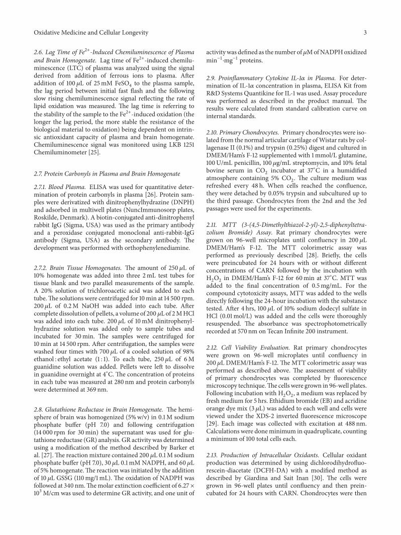
Oxidative Medicine and Cellular Longevity 3
2.6. Lag Time of Fe2+-Induced Chemiluminescence of Plasmaand Brain Homogenate. Lag time of Fe2+-induced chemilu-minescence (LTC) of plasma was analyzed using the signalderived from addition of ferrous ions to plasma. Afteraddition of 100𝜇L of 25mM FeSO
4to the plasma sample,
the lag period between initial fast flash and the followingslow rising chemiluminescence signal reflecting the rate oflipid oxidation was measured. The lag time is referring tothe stability of the sample to the Fe2+-induced oxidation (thelonger the lag period, the more stable the resistance of thebiological material to oxidation) being dependent on intrin-sic antioxidant capacity of plasma and brain homogenate.Chemiluminescence signal was monitored using LKB 1251Chemiluminometer [25].
2.7. Protein Carbonyls in Plasma and Brain Homogenate
2.7.1. Blood Plasma. ELISA was used for quantitative deter-mination of protein carbonyls in plasma [26]. Protein sam-ples were derivatized with dinitrophenylhydrazine (DNPH)and adsorbed in multiwell plates (NuncImmunosorp plates,Roskilde, Denmark). A biotin-conjugated anti-dinitrophenylrabbit IgG (Sigma, USA) was used as the primary antibodyand a peroxidase conjugated monoclonal anti-rabbit-IgGantibody (Sigma, USA) as the secondary antibody. Thedevelopment was performed with orthophenylenediamine.
2.7.2. Brain Tissue Homogenates. The amount of 250𝜇L of10% homogenate was added into three 2mL test tubes fortissue blank and two parallel measurements of the sample.A 20% solution of trichloroacetic acid was added to eachtube.The solutions were centrifuged for 10min at 14 500 rpm.200𝜇L of 0.2M NaOH was added into each tube. Aftercomplete dissolution of pellets, a volumeof 200𝜇Lof 2MHClwas added into each tube. 200𝜇L of 10mM dinitrophenyl-hydrazine solution was added only to sample tubes andincubated for 30min. The samples were centrifuged for10min at 14 500 rpm. After centrifugation, the samples werewashed four times with 700𝜇L of a cooled solution of 98%ethanol : ethyl acetate (1 : 1). To each tube, 250 𝜇L of 6Mguanidine solution was added. Pellets were left to dissolvein guanidine overnight at 4∘C. The concentration of proteinsin each tube was measured at 280 nm and protein carbonylswere determined at 369 nm.
2.8. Glutathione Reductase in Brain Homogenate. The hemi-sphere of brain was homogenized (5%w/v) in 0.1M sodiumphosphate buffer (pH 7.0) and following centrifugation(14 000 rpm for 30min) the supernatant was used for glu-tathione reductase (GR) analysis. GR activity was determinedusing a modification of the method described by Barker etal. [27].The reactionmixture contained 200𝜇L 0.1M sodiumphosphate buffer (pH 7.0), 30 𝜇L 0.1mMNADPH, and 60 𝜇Lof 5% homogenate.The reaction was initiated by the additionof 10 𝜇L GSSG (110mg/1mL). The oxidation of NADPH wasfollowed at 340 nm.Themolar extinction coefficient of 6.27 ×10³M/cm was used to determine GR activity, and one unit of
activitywas defined as the number of𝜇MofNADPHoxidizedmin−1⋅mg−1 proteins.
2.9. Proinflammatory Cytokine IL-1𝛼 in Plasma. For deter-mination of IL-1𝛼 concentration in plasma, ELISA Kit fromR&D Systems Quantikine for IL-1 was used. Assay procedurewas performed as described in the product manual. Theresults were calculated from standard calibration curve oninternal standards.
2.10. Primary Chondrocytes. Primary chondrocytes were iso-lated from the normal articular cartilage ofWistar rats by col-lagenase II (0.1%) and trypsin (0.25%) digest and cultured inDMEM/Ham’s F-12 supplemented with 1mmol/L glutamine,100U/mL penicillin, 100 𝜇g/mL streptomycin, and 10% fetalbovine serum in CO
2incubator at 37∘C in a humidified
atmosphere containing 5% CO2. The culture medium was
refreshed every 48 h. When cells reached the confluence,they were detached by 0.05% trypsin and subcultured up tothe third passage. Chondrocytes from the 2nd and the 3rdpassages were used for the experiments.
2.11. MTT (3-(4,5-Dimethylthiazol-2-yl)-2,5-diphenyltetra-zolium Bromide) Assay. Rat primary chondrocytes weregrown on 96-well microplates until confluency in 200𝜇LDMEM/Ham’s F-12. The MTT colorimetric assay wasperformed as previously described [28]. Briefly, the cellswere preincubated for 24 hours with or without differentconcentrations of CARN followed by the incubation withH2O2in DMEM/Ham’s F-12 for 60min at 37∘C. MTT was
added to the final concentration of 0.5mg/mL. For thecompound cytotoxicity assays, MTT was added to the wellsdirectly following the 24-hour incubation with the substancetested. After 4 hrs, 100 𝜇L of 10% sodium dodecyl sulfate inHCl (0.01mol/L) was added and the cells were thoroughlyresuspended. The absorbance was spectrophotometricallyrecorded at 570 nm on Tecan Infinite 200 instrument.
2.12. Cell Viability Evaluation. Rat primary chondrocyteswere grown on 96-well microplates until confluency in200𝜇L DMEM/Ham’s F-12. The MTT colorimetric assay wasperformed as described above. The assessment of viabilityof primary chondrocytes was completed by fluorescencemicroscopy technique.The cells were grown in 96-well plates.Following incubation with H
2O2, a medium was replaced by
fresh medium for 5 hrs. Ethidium bromide (EB) and acridineorange dye mix (3 𝜇L) was added to each well and cells wereviewed under the XDS-2 inverted fluorescence microscope[29]. Each image was collected with excitation at 488 nm.Calculations were doneminimum in quadruplicate, countinga minimum of 100 total cells each.
2.13. Production of Intracellular Oxidants. Cellular oxidantproduction was determined by using dichlorodihydrofluo-rescein-diacetate (DCFH-DA) with a modified method asdescribed by Giardina and Sait Inan [30]. The cells weregrown in 96-well plates until confluency and then prein-cubated for 24 hours with CARN. Chondrocytes were then
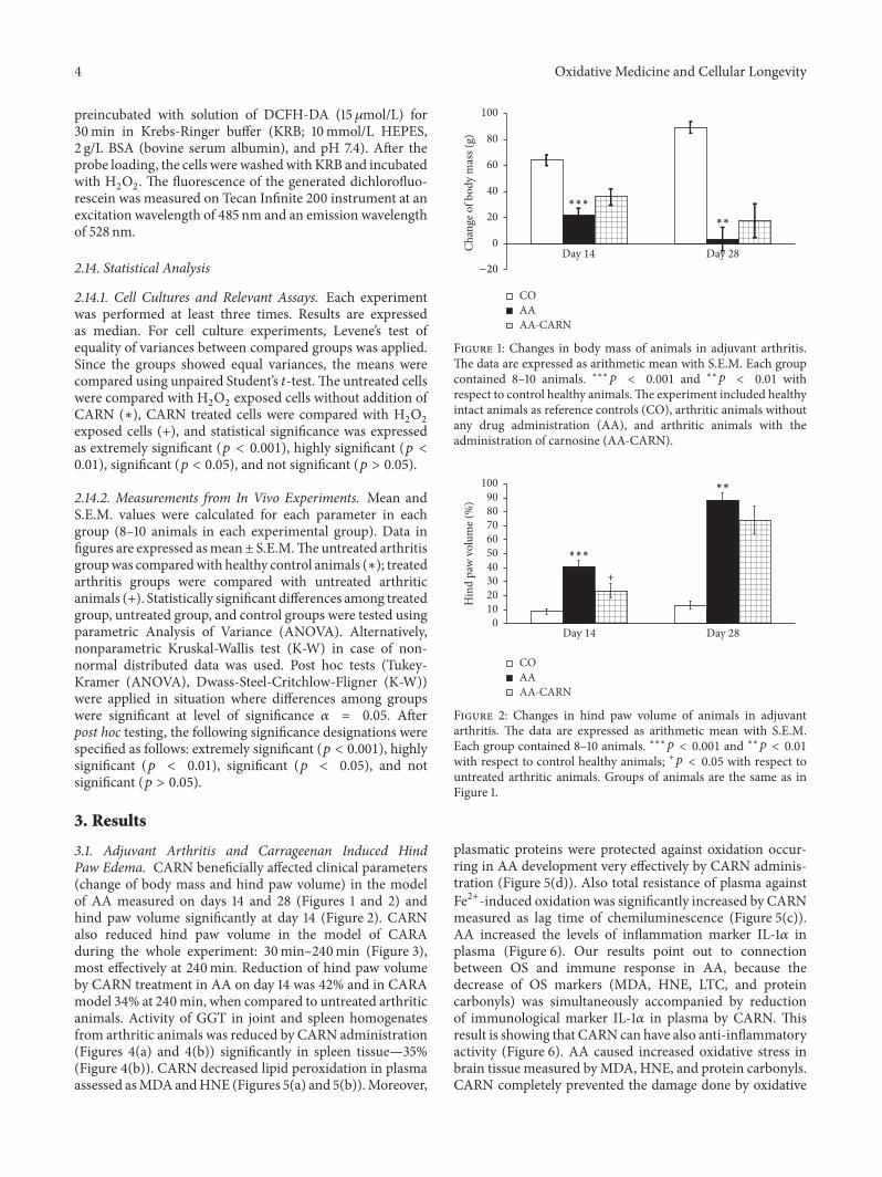
4 Oxidative Medicine and Cellular Longevity
preincubated with solution of DCFH-DA (15 𝜇mol/L) for30min in Krebs-Ringer buffer (KRB; 10mmol/L HEPES,2 g/L BSA (bovine serum albumin), and pH 7.4). After theprobe loading, the cells were washedwith KRB and incubatedwith H
2O2. The fluorescence of the generated dichlorofluo-
rescein was measured on Tecan Infinite 200 instrument at anexcitation wavelength of 485 nm and an emission wavelengthof 528 nm.
2.14. Statistical Analysis
2.14.1. Cell Cultures and Relevant Assays. Each experimentwas performed at least three times. Results are expressedas median. For cell culture experiments, Levene’s test ofequality of variances between compared groups was applied.Since the groups showed equal variances, the means werecompared using unpaired Student’s 𝑡-test. The untreated cellswere compared with H
2O2exposed cells without addition of
CARN (∗), CARN treated cells were compared with H2O2
exposed cells (+), and statistical significance was expressedas extremely significant (𝑝 < 0.001), highly significant (𝑝 <0.01), significant (𝑝 < 0.05), and not significant (𝑝 > 0.05).
2.14.2. Measurements from In Vivo Experiments. Mean andS.E.M. values were calculated for each parameter in eachgroup (8–10 animals in each experimental group). Data infigures are expressed asmean± S.E.M.The untreated arthritisgroupwas comparedwith healthy control animals (∗); treatedarthritis groups were compared with untreated arthriticanimals (+). Statistically significant differences among treatedgroup, untreated group, and control groups were tested usingparametric Analysis of Variance (ANOVA). Alternatively,nonparametric Kruskal-Wallis test (K-W) in case of non-normal distributed data was used. Post hoc tests (Tukey-Kramer (ANOVA), Dwass-Steel-Critchlow-Fligner (K-W))were applied in situation where differences among groupswere significant at level of significance 𝛼 = 0.05. Afterpost hoc testing, the following significance designations werespecified as follows: extremely significant (𝑝 < 0.001), highlysignificant (𝑝 < 0.01), significant (𝑝 < 0.05), and notsignificant (𝑝 > 0.05).
3. Results
3.1. Adjuvant Arthritis and Carrageenan Induced HindPaw Edema. CARN beneficially affected clinical parameters(change of body mass and hind paw volume) in the modelof AA measured on days 14 and 28 (Figures 1 and 2) andhind paw volume significantly at day 14 (Figure 2). CARNalso reduced hind paw volume in the model of CARAduring the whole experiment: 30min–240min (Figure 3),most effectively at 240min. Reduction of hind paw volumeby CARN treatment in AA on day 14 was 42% and in CARAmodel 34% at 240min, when compared to untreated arthriticanimals. Activity of GGT in joint and spleen homogenatesfrom arthritic animals was reduced by CARN administration(Figures 4(a) and 4(b)) significantly in spleen tissue—35%(Figure 4(b)). CARN decreased lipid peroxidation in plasmaassessed asMDAandHNE (Figures 5(a) and 5(b)).Moreover,
Day 14 Day 28
COAAAA-CARN
∗∗∗
∗∗
−20
0
20
40
60
80
100
Chan
ge o
f bod
y m
ass (
g)
Figure 1: Changes in body mass of animals in adjuvant arthritis.The data are expressed as arithmetic mean with S.E.M. Each groupcontained 8–10 animals. ∗∗∗𝑝 < 0.001 and ∗∗𝑝 < 0.01 withrespect to control healthy animals.The experiment included healthyintact animals as reference controls (CO), arthritic animals withoutany drug administration (AA), and arthritic animals with theadministration of carnosine (AA-CARN).
Day 14 Day 28
+
0102030405060708090
100H
ind
paw
vol
ume (
%)
COAAAA-CARN
∗∗∗
∗∗
Figure 2: Changes in hind paw volume of animals in adjuvantarthritis. The data are expressed as arithmetic mean with S.E.M.Each group contained 8–10 animals. ∗∗∗𝑝 < 0.001 and ∗∗𝑝 < 0.01with respect to control healthy animals; +𝑝 < 0.05 with respect tountreated arthritic animals. Groups of animals are the same as inFigure 1.
plasmatic proteins were protected against oxidation occur-ring in AA development very effectively by CARN adminis-tration (Figure 5(d)). Also total resistance of plasma againstFe2+-induced oxidation was significantly increased by CARNmeasured as lag time of chemiluminescence (Figure 5(c)).AA increased the levels of inflammation marker IL-1𝛼 inplasma (Figure 6). Our results point out to connectionbetween OS and immune response in AA, because thedecrease of OS markers (MDA, HNE, LTC, and proteincarbonyls) was simultaneously accompanied by reductionof immunological marker IL-1𝛼 in plasma by CARN. Thisresult is showing that CARN can have also anti-inflammatoryactivity (Figure 6). AA caused increased oxidative stress inbrain tissue measured byMDA, HNE, and protein carbonyls.CARN completely prevented the damage done by oxidative
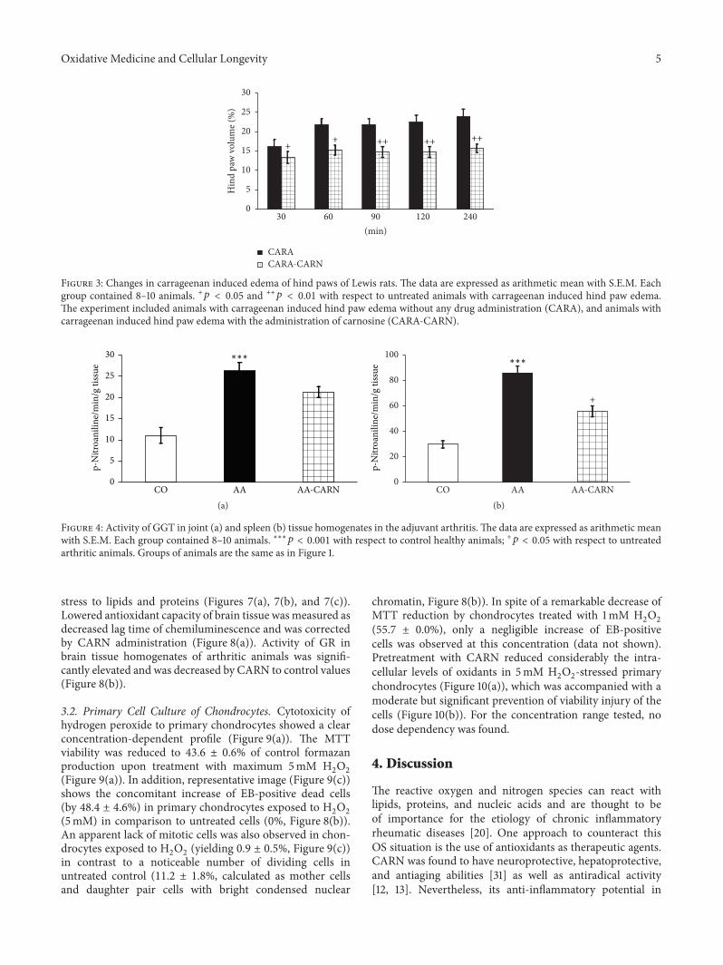
Oxidative Medicine and Cellular Longevity 5
+ ++ ++ +++
0
5
10
15
20
25
30
Hin
d pa
w v
olum
e (%
)60 90 120 24030
(min)
CARACARA-CARN
Figure 3: Changes in carrageenan induced edema of hind paws of Lewis rats. The data are expressed as arithmetic mean with S.E.M. Eachgroup contained 8–10 animals. +𝑝 < 0.05 and ++𝑝 < 0.01 with respect to untreated animals with carrageenan induced hind paw edema.The experiment included animals with carrageenan induced hind paw edema without any drug administration (CARA), and animals withcarrageenan induced hind paw edema with the administration of carnosine (CARA-CARN).
∗∗∗
0
5
10
15
20
25
30
p-N
itroa
nilin
e/m
in/g
tiss
ue
AA AA-CARNCO(a)
+
∗∗∗
0
20
40
60
80
100
p-N
itroa
nilin
e/m
in/g
tiss
ue
AA AA-CARNCO(b)
Figure 4: Activity of GGT in joint (a) and spleen (b) tissue homogenates in the adjuvant arthritis. The data are expressed as arithmetic meanwith S.E.M. Each group contained 8–10 animals. ∗∗∗𝑝 < 0.001 with respect to control healthy animals; +𝑝 < 0.05 with respect to untreatedarthritic animals. Groups of animals are the same as in Figure 1.
stress to lipids and proteins (Figures 7(a), 7(b), and 7(c)).Lowered antioxidant capacity of brain tissue wasmeasured asdecreased lag time of chemiluminescence and was correctedby CARN administration (Figure 8(a)). Activity of GR inbrain tissue homogenates of arthritic animals was signifi-cantly elevated and was decreased by CARN to control values(Figure 8(b)).
3.2. Primary Cell Culture of Chondrocytes. Cytotoxicity ofhydrogen peroxide to primary chondrocytes showed a clearconcentration-dependent profile (Figure 9(a)). The MTTviability was reduced to 43.6 ± 0.6% of control formazanproduction upon treatment with maximum 5mM H
2O2
(Figure 9(a)). In addition, representative image (Figure 9(c))shows the concomitant increase of EB-positive dead cells(by 48.4 ± 4.6%) in primary chondrocytes exposed to H
2O2
(5mM) in comparison to untreated cells (0%, Figure 8(b)).An apparent lack of mitotic cells was also observed in chon-drocytes exposed to H
2O2(yielding 0.9 ± 0.5%, Figure 9(c))
in contrast to a noticeable number of dividing cells inuntreated control (11.2 ± 1.8%, calculated as mother cellsand daughter pair cells with bright condensed nuclear
chromatin, Figure 8(b)). In spite of a remarkable decrease ofMTT reduction by chondrocytes treated with 1mM H
2O2
(55.7 ± 0.0%), only a negligible increase of EB-positivecells was observed at this concentration (data not shown).Pretreatment with CARN reduced considerably the intra-cellular levels of oxidants in 5mM H
2O2-stressed primary
chondrocytes (Figure 10(a)), which was accompanied with amoderate but significant prevention of viability injury of thecells (Figure 10(b)). For the concentration range tested, nodose dependency was found.
4. Discussion
The reactive oxygen and nitrogen species can react withlipids, proteins, and nucleic acids and are thought to beof importance for the etiology of chronic inflammatoryrheumatic diseases [20]. One approach to counteract thisOS situation is the use of antioxidants as therapeutic agents.CARN was found to have neuroprotective, hepatoprotective,and antiaging abilities [31] as well as antiradical activity[12, 13]. Nevertheless, its anti-inflammatory potential in
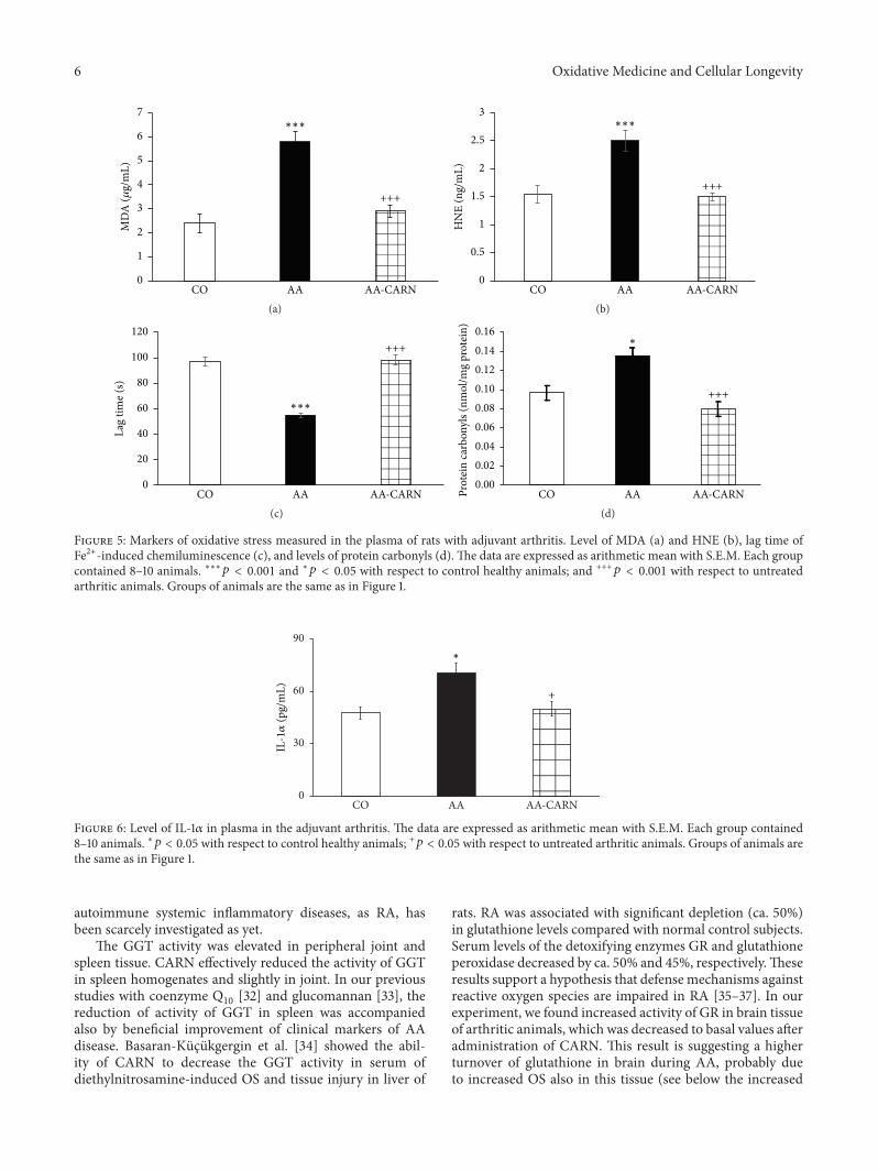
6 Oxidative Medicine and Cellular Longevity
+++
MD
A (𝜇
g/m
L)∗∗∗
AA AA-CARNCO0
1
2
3
4
5
6
7
(a)
+++
∗∗∗
AA AA-CARNCO0
0.5
1
1.5
2
2.5
3
HN
E (n
g/m
L)
(b)
+++
∗∗∗
0
20
40
60
80
100
120
Lag
time (
s)
AA AA-CARNCO(c)
+++
∗
0.000.020.040.060.080.100.120.140.16
Prot
ein
carb
onyl
s (nm
ol/m
g pr
otei
n)
AA AA-CARNCO(d)
Figure 5: Markers of oxidative stress measured in the plasma of rats with adjuvant arthritis. Level of MDA (a) and HNE (b), lag time ofFe2+-induced chemiluminescence (c), and levels of protein carbonyls (d). The data are expressed as arithmetic mean with S.E.M. Each groupcontained 8–10 animals. ∗∗∗𝑝 < 0.001 and ∗𝑝 < 0.05 with respect to control healthy animals; and +++𝑝 < 0.001 with respect to untreatedarthritic animals. Groups of animals are the same as in Figure 1.
+
∗
0
30
60
90
IL-1𝛼
(pg/
mL)
AA AA-CARNCO
Figure 6: Level of IL-1𝛼 in plasma in the adjuvant arthritis. The data are expressed as arithmetic mean with S.E.M. Each group contained8–10 animals. ∗𝑝 < 0.05 with respect to control healthy animals; +𝑝 < 0.05 with respect to untreated arthritic animals. Groups of animals arethe same as in Figure 1.
autoimmune systemic inflammatory diseases, as RA, hasbeen scarcely investigated as yet.
The GGT activity was elevated in peripheral joint andspleen tissue. CARN effectively reduced the activity of GGTin spleen homogenates and slightly in joint. In our previousstudies with coenzyme Q
10[32] and glucomannan [33], the
reduction of activity of GGT in spleen was accompaniedalso by beneficial improvement of clinical markers of AAdisease. Basaran-Küçükgergin et al. [34] showed the abil-ity of CARN to decrease the GGT activity in serum ofdiethylnitrosamine-induced OS and tissue injury in liver of
rats. RA was associated with significant depletion (ca. 50%)in glutathione levels compared with normal control subjects.Serum levels of the detoxifying enzymes GR and glutathioneperoxidase decreased by ca. 50% and 45%, respectively.Theseresults support a hypothesis that defense mechanisms againstreactive oxygen species are impaired in RA [35–37]. In ourexperiment, we found increased activity of GR in brain tissueof arthritic animals, which was decreased to basal values afteradministration of CARN. This result is suggesting a higherturnover of glutathione in brain during AA, probably dueto increased OS also in this tissue (see below the increased
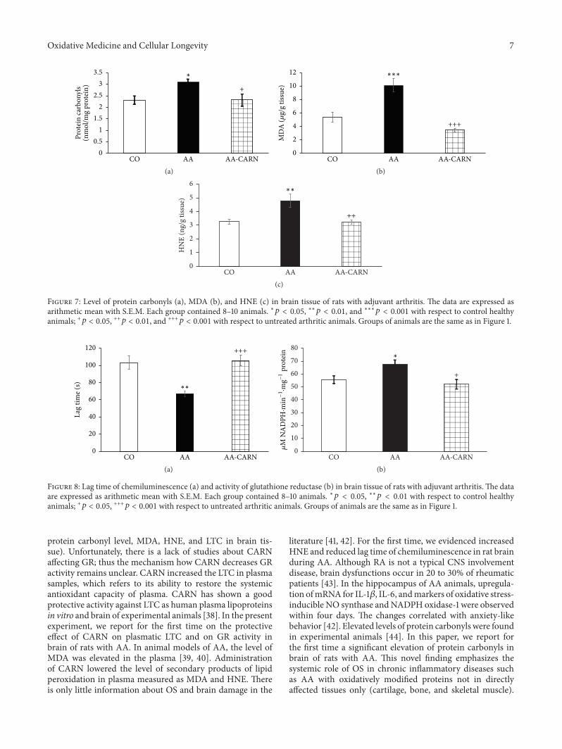
Oxidative Medicine and Cellular Longevity 7
Prot
ein
carb
onyl
s +
00.5
11.5
22.5
33.5
(nm
ol/m
g pr
otei
n)
AA AA-CARNCO
∗
(a)
+++
AA AA-CARNCO
MD
A (𝜇
g/g
tissu
e)
∗∗∗
0
2
4
6
8
10
12
(b)
++
0
1
2
3
4
5
6H
NE
(ng/
g tis
sue)
AA AA-CARNCO
∗∗
(c)
Figure 7: Level of protein carbonyls (a), MDA (b), and HNE (c) in brain tissue of rats with adjuvant arthritis. The data are expressed asarithmetic mean with S.E.M. Each group contained 8–10 animals. ∗𝑝 < 0.05, ∗∗𝑝 < 0.01, and ∗∗∗𝑝 < 0.001 with respect to control healthyanimals; +𝑝 < 0.05, ++𝑝 < 0.01, and +++𝑝 < 0.001 with respect to untreated arthritic animals. Groups of animals are the same as in Figure 1.
+++
∗∗
0
20
40
60
80
100
120
Lag
time (
s)
AA AA-CARNCO(a)
+
∗
0
10
20
30
40
50
60
70
80
AA AA-CARNCO
𝜇M
NA
DPH
·min−1·m
g−1
prot
ein
(b)
Figure 8: Lag time of chemiluminescence (a) and activity of glutathione reductase (b) in brain tissue of rats with adjuvant arthritis. The dataare expressed as arithmetic mean with S.E.M. Each group contained 8–10 animals. ∗𝑝 < 0.05, ∗∗𝑝 < 0.01 with respect to control healthyanimals; +𝑝 < 0.05, +++𝑝 < 0.001 with respect to untreated arthritic animals. Groups of animals are the same as in Figure 1.
protein carbonyl level, MDA, HNE, and LTC in brain tis-sue). Unfortunately, there is a lack of studies about CARNaffecting GR; thus the mechanism how CARN decreases GRactivity remains unclear. CARN increased the LTC in plasmasamples, which refers to its ability to restore the systemicantioxidant capacity of plasma. CARN has shown a goodprotective activity against LTC as human plasma lipoproteinsin vitro and brain of experimental animals [38]. In the presentexperiment, we report for the first time on the protectiveeffect of CARN on plasmatic LTC and on GR activity inbrain of rats with AA. In animal models of AA, the level ofMDA was elevated in the plasma [39, 40]. Administrationof CARN lowered the level of secondary products of lipidperoxidation in plasma measured as MDA and HNE. Thereis only little information about OS and brain damage in the
literature [41, 42]. For the first time, we evidenced increasedHNE and reduced lag time of chemiluminescence in rat brainduring AA. Although RA is not a typical CNS involvementdisease, brain dysfunctions occur in 20 to 30% of rheumaticpatients [43]. In the hippocampus of AA animals, upregula-tion ofmRNA for IL-1𝛽, IL-6, andmarkers of oxidative stress-inducible NO synthase andNADPH oxidase-1 were observedwithin four days. The changes correlated with anxiety-likebehavior [42]. Elevated levels of protein carbonyls were foundin experimental animals [44]. In this paper, we report forthe first time a significant elevation of protein carbonyls inbrain of rats with AA. This novel finding emphasizes thesystemic role of OS in chronic inflammatory diseases suchas AA with oxidatively modified proteins not in directlyaffected tissues only (cartilage, bone, and skeletal muscle).
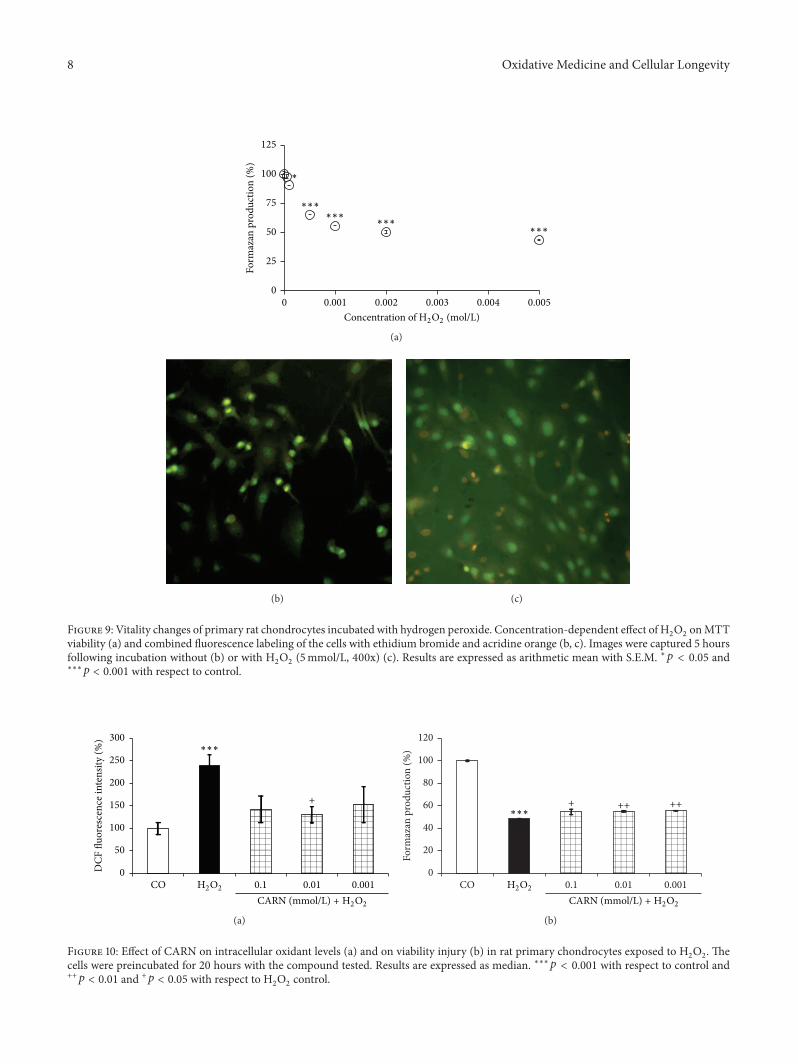
8 Oxidative Medicine and Cellular Longevity
0
25
50
75
100
125
Form
azan
pro
duct
ion
(%)
∗
∗∗∗∗∗∗
∗∗∗∗∗∗
0.001 0.002 0.003 0.004 0.0050Concentration of H2O2 (mol/L)
(a)
(b) (c)
Figure 9: Vitality changes of primary rat chondrocytes incubated with hydrogen peroxide. Concentration-dependent effect of H2O2onMTT
viability (a) and combined fluorescence labeling of the cells with ethidium bromide and acridine orange (b, c). Images were captured 5 hoursfollowing incubation without (b) or with H
2O2(5mmol/L, 400x) (c). Results are expressed as arithmetic mean with S.E.M. ∗𝑝 < 0.05 and
∗∗∗𝑝 < 0.001 with respect to control.
+
0
50
100
150
200
250
300
DCF
fluo
resc
ence
inte
nsity
(%)
∗∗∗
H2O2 0.1 0.01 0.001COCARN (mmol/L) + H2O2
(a)
+ ++ ++
0
20
40
60
80
100
120
Form
azan
pro
duct
ion
(%)
∗∗∗
H2O2 0.1 0.01 0.001COCARN (mmol/L) + H2O2
(b)
Figure 10: Effect of CARN on intracellular oxidant levels (a) and on viability injury (b) in rat primary chondrocytes exposed to H2O2. The
cells were preincubated for 20 hours with the compound tested. Results are expressed as median. ∗∗∗𝑝 < 0.001 with respect to control and++𝑝 < 0.01 and +𝑝 < 0.05 with respect to H
2O2control.
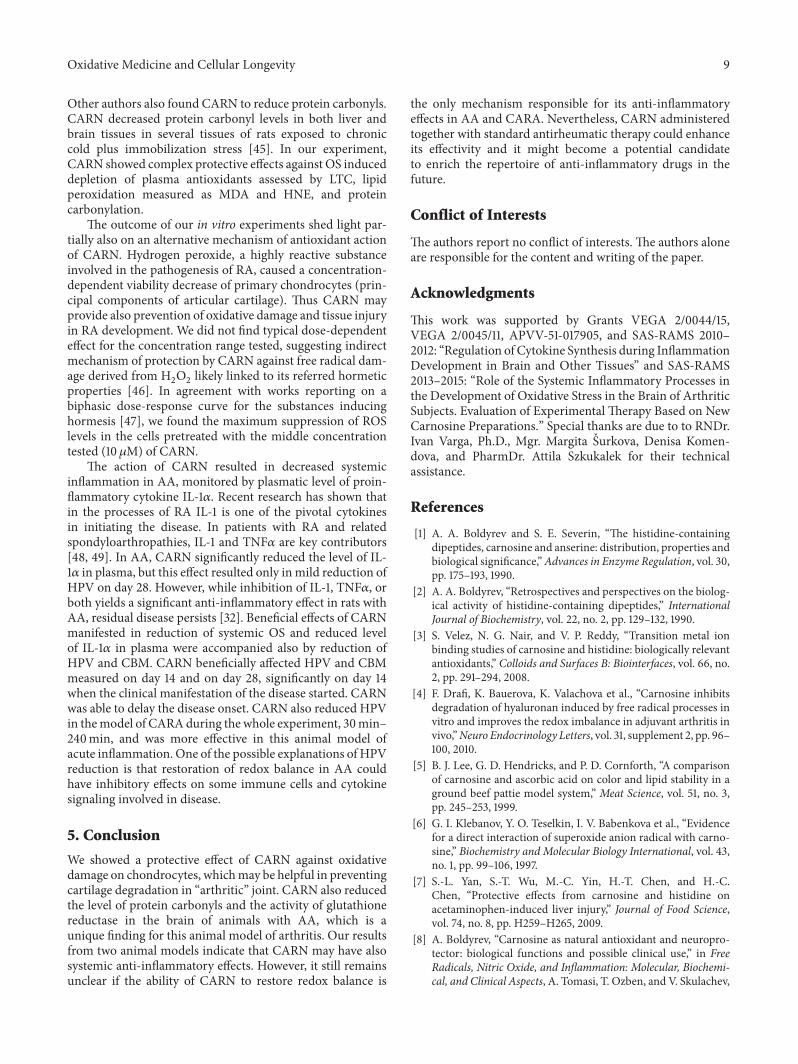
Oxidative Medicine and Cellular Longevity 9
Other authors also found CARN to reduce protein carbonyls.CARN decreased protein carbonyl levels in both liver andbrain tissues in several tissues of rats exposed to chroniccold plus immobilization stress [45]. In our experiment,CARN showed complex protective effects against OS induceddepletion of plasma antioxidants assessed by LTC, lipidperoxidation measured as MDA and HNE, and proteincarbonylation.
The outcome of our in vitro experiments shed light par-tially also on an alternative mechanism of antioxidant actionof CARN. Hydrogen peroxide, a highly reactive substanceinvolved in the pathogenesis of RA, caused a concentration-dependent viability decrease of primary chondrocytes (prin-cipal components of articular cartilage). Thus CARN mayprovide also prevention of oxidative damage and tissue injuryin RA development. We did not find typical dose-dependenteffect for the concentration range tested, suggesting indirectmechanism of protection by CARN against free radical dam-age derived from H
2O2likely linked to its referred hormetic
properties [46]. In agreement with works reporting on abiphasic dose-response curve for the substances inducinghormesis [47], we found the maximum suppression of ROSlevels in the cells pretreated with the middle concentrationtested (10 𝜇M) of CARN.
The action of CARN resulted in decreased systemicinflammation in AA, monitored by plasmatic level of proin-flammatory cytokine IL-1𝛼. Recent research has shown thatin the processes of RA IL-1 is one of the pivotal cytokinesin initiating the disease. In patients with RA and relatedspondyloarthropathies, IL-1 and TNF𝛼 are key contributors[48, 49]. In AA, CARN significantly reduced the level of IL-1𝛼 in plasma, but this effect resulted only inmild reduction ofHPV on day 28. However, while inhibition of IL-1, TNF𝛼, orboth yields a significant anti-inflammatory effect in rats withAA, residual disease persists [32]. Beneficial effects of CARNmanifested in reduction of systemic OS and reduced levelof IL-1𝛼 in plasma were accompanied also by reduction ofHPV and CBM. CARN beneficially affected HPV and CBMmeasured on day 14 and on day 28, significantly on day 14when the clinical manifestation of the disease started. CARNwas able to delay the disease onset. CARN also reduced HPVin themodel of CARAduring the whole experiment, 30min–240min, and was more effective in this animal model ofacute inflammation. One of the possible explanations of HPVreduction is that restoration of redox balance in AA couldhave inhibitory effects on some immune cells and cytokinesignaling involved in disease.
5. Conclusion
We showed a protective effect of CARN against oxidativedamage on chondrocytes, whichmay be helpful in preventingcartilage degradation in “arthritic” joint. CARN also reducedthe level of protein carbonyls and the activity of glutathionereductase in the brain of animals with AA, which is aunique finding for this animal model of arthritis. Our resultsfrom two animal models indicate that CARN may have alsosystemic anti-inflammatory effects. However, it still remainsunclear if the ability of CARN to restore redox balance is
the only mechanism responsible for its anti-inflammatoryeffects in AA and CARA. Nevertheless, CARN administeredtogether with standard antirheumatic therapy could enhanceits effectivity and it might become a potential candidateto enrich the repertoire of anti-inflammatory drugs in thefuture.
Conflict of Interests
The authors report no conflict of interests. The authors aloneare responsible for the content and writing of the paper.
Acknowledgments
This work was supported by Grants VEGA 2/0044/15,VEGA 2/0045/11, APVV-51-017905, and SAS-RAMS 2010–2012: “Regulation of Cytokine Synthesis during InflammationDevelopment in Brain and Other Tissues” and SAS-RAMS2013–2015: “Role of the Systemic Inflammatory Processes inthe Development of Oxidative Stress in the Brain of ArthriticSubjects. Evaluation of Experimental Therapy Based on NewCarnosine Preparations.” Special thanks are due to to RNDr.Ivan Varga, Ph.D., Mgr. Margita Šurkova, Denisa Komen-dova, and PharmDr. Attila Szkukalek for their technicalassistance.
References
[1] A. A. Boldyrev and S. E. Severin, “The histidine-containingdipeptides, carnosine and anserine: distribution, properties andbiological significance,”Advances in Enzyme Regulation, vol. 30,pp. 175–193, 1990.
[2] A. A. Boldyrev, “Retrospectives and perspectives on the biolog-ical activity of histidine-containing dipeptides,” InternationalJournal of Biochemistry, vol. 22, no. 2, pp. 129–132, 1990.
[3] S. Velez, N. G. Nair, and V. P. Reddy, “Transition metal ionbinding studies of carnosine and histidine: biologically relevantantioxidants,” Colloids and Surfaces B: Biointerfaces, vol. 66, no.2, pp. 291–294, 2008.
[4] F. Drafi, K. Bauerova, K. Valachova et al., “Carnosine inhibitsdegradation of hyaluronan induced by free radical processes invitro and improves the redox imbalance in adjuvant arthritis invivo,”Neuro Endocrinology Letters, vol. 31, supplement 2, pp. 96–100, 2010.
[5] B. J. Lee, G. D. Hendricks, and P. D. Cornforth, “A comparisonof carnosine and ascorbic acid on color and lipid stability in aground beef pattie model system,” Meat Science, vol. 51, no. 3,pp. 245–253, 1999.
[6] G. I. Klebanov, Y. O. Teselkin, I. V. Babenkova et al., “Evidencefor a direct interaction of superoxide anion radical with carno-sine,” Biochemistry and Molecular Biology International, vol. 43,no. 1, pp. 99–106, 1997.
[7] S.-L. Yan, S.-T. Wu, M.-C. Yin, H.-T. Chen, and H.-C.Chen, “Protective effects from carnosine and histidine onacetaminophen-induced liver injury,” Journal of Food Science,vol. 74, no. 8, pp. H259–H265, 2009.
[8] A. Boldyrev, “Carnosine as natural antioxidant and neuropro-tector: biological functions and possible clinical use,” in FreeRadicals, Nitric Oxide, and Inflammation: Molecular, Biochemi-cal, and Clinical Aspects, A. Tomasi, T. Ozben, and V. Skulachev,
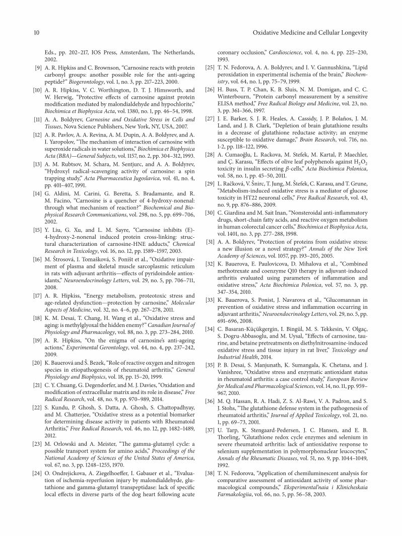
10 Oxidative Medicine and Cellular Longevity
Eds., pp. 202–217, IOS Press, Amsterdam, The Netherlands,2002.
[9] A. R. Hipkiss and C. Brownson, “Carnosine reacts with proteincarbonyl groups: another possible role for the anti-ageingpeptide?” Biogerontology, vol. 1, no. 3, pp. 217–223, 2000.
[10] A. R. Hipkiss, V. C. Worthington, D. T. J. Himsworth, andW. Herwig, “Protective effects of carnosine against proteinmodification mediated by malondialdehyde and hypochlorite,”Biochimica et Biophysica Acta, vol. 1380, no. 1, pp. 46–54, 1998.
[11] A. A. Boldyrev, Carnosine and Oxidative Stress in Cells andTissues, Nova Science Publishers, New York, NY, USA, 2007.
[12] A. R. Pavlov, A. A. Revina, A. M. Dupin, A. A. Boldyrev, and A.I. Yaropolov, “The mechanism of interaction of carnosine withsuperoxide radicals in water solutions,” Biochimica et BiophysicaActa (BBA)—General Subjects, vol. 1157, no. 2, pp. 304–312, 1993.
[13] A. M. Rubtsov, M. Schara, M. Sentjurc, and A. A. Boldyrev,“Hydroxyl radical-scavenging activity of carnosine: a spintrapping study,” Acta Pharmaceutica Jugoslavica, vol. 41, no. 4,pp. 401–407, 1991.
[14] G. Aldini, M. Carini, G. Beretta, S. Bradamante, and R.M. Facino, “Carnosine is a quencher of 4-hydroxy-nonenal:through what mechanism of reaction?” Biochemical and Bio-physical Research Communications, vol. 298, no. 5, pp. 699–706,2002.
[15] Y. Liu, G. Xu, and L. M. Sayre, “Carnosine inhibits (E)-4-hydroxy-2-nonenal induced protein cross-linking: struc-tural characterization of carnosine-HNE adducts,” ChemicalResearch in Toxicology, vol. 16, no. 12, pp. 1589–1597, 2003.
[16] M. Štrosová, I. Tomašková, S. Poništ et al., “Oxidative impair-ment of plasma and skeletal muscle sarcoplasmic reticulumin rats with adjuvant arthritis—effects of pyridoindole antiox-idants,” Neuroendocrinology Letters, vol. 29, no. 5, pp. 706–711,2008.
[17] A. R. Hipkiss, “Energy metabolism, proteotoxic stress andage-related dysfunction—protection by carnosine,” MolecularAspects of Medicine, vol. 32, no. 4–6, pp. 267–278, 2011.
[18] K. M. Desai, T. Chang, H. Wang et al., “Oxidative stress andaging: ismethylglyoxal the hidden enemy?”Canadian Journal ofPhysiology and Pharmacology, vol. 88, no. 3, pp. 273–284, 2010.
[19] A. R. Hipkiss, “On the enigma of carnosine’s anti-ageingactions,” Experimental Gerontology, vol. 44, no. 4, pp. 237–242,2009.
[20] K. Bauerová and Š. Bezek, “Role of reactive oxygen and nitrogenspecies in etiopathogenesis of rheumatoid arthritis,” GeneralPhysiology and Biophysics, vol. 18, pp. 15–20, 1999.
[21] C. Y. Chuang, G. Degendorfer, andM. J. Davies, “Oxidation andmodification of extracellular matrix and its role in disease,” FreeRadical Research, vol. 48, no. 9, pp. 970–989, 2014.
[22] S. Kundu, P. Ghosh, S. Datta, A. Ghosh, S. Chattopadhyay,and M. Chatterjee, “Oxidative stress as a potential biomarkerfor determining disease activity in patients with RheumatoidArthritis,” Free Radical Research, vol. 46, no. 12, pp. 1482–1489,2012.
[23] M. Orlowski and A. Meister, “The gamma-glutamyl cycle: apossible transport system for amino acids,” Proceedings of theNational Academy of Sciences of the United States of America,vol. 67, no. 3, pp. 1248–1255, 1970.
[24] O. Ondrejickova, A. Ziegelhoeffer, I. Gabauer et al., “Evalua-tion of ischemia-reperfusion injury by malondialdehyde, glu-tathione and gamma-glutamyl transpeptidase: lack of specificlocal effects in diverse parts of the dog heart following acute
coronary occlusion,” Cardioscience, vol. 4, no. 4, pp. 225–230,1993.
[25] T. N. Fedorova, A. A. Boldyrev, and I. V. Gannushkina, “Lipidperoxidation in experimental ischemia of the brain,” Biochem-istry, vol. 64, no. 1, pp. 75–79, 1999.
[26] H. Buss, T. P. Chan, K. B. Sluis, N. M. Domigan, and C. C.Winterbourn, “Protein carbonyl measurement by a sensitiveELISA method,” Free Radical Biology and Medicine, vol. 23, no.3, pp. 361–366, 1997.
[27] J. E. Barker, S. J. R. Heales, A. Cassidy, J. P. Bolaños, J. M.Land, and J. B. Clark, “Depletion of brain glutathione resultsin a decrease of glutathione reductase activity; an enzymesusceptible to oxidative damage,” Brain Research, vol. 716, no.1-2, pp. 118–122, 1996.
[28] A. Cumaoğlu, L. Rackova, M. Stefek, M. Kartal, P. Maechler,and Ç. Karasu, “Effects of olive leaf polyphenols against H
2O2
toxicity in insulin secreting 𝛽-cells,” Acta Biochimica Polonica,vol. 58, no. 1, pp. 45–50, 2011.
[29] L. Račková, V. Šnirc, T. Jung,M. Štefek, C. Karasu, andT.Grune,“Metabolism-induced oxidative stress is a mediator of glucosetoxicity in HT22 neuronal cells,” Free Radical Research, vol. 43,no. 9, pp. 876–886, 2009.
[30] C. Giardina andM. Sait Inan, “Nonsteroidal anti-inflammatorydrugs, short-chain fatty acids, and reactive oxygen metabolismin human colorectal cancer cells,”Biochimica et Biophysica Acta,vol. 1401, no. 3, pp. 277–288, 1998.
[31] A. A. Boldyrev, “Protection of proteins from oxidative stress:a new illusion or a novel strategy?” Annals of the New YorkAcademy of Sciences, vol. 1057, pp. 193–205, 2005.
[32] K. Bauerova, E. Paulovicova, D. Mihalova et al., “Combinedmethotrexate and coenzyme Q10 therapy in adjuvant-inducedarthritis evaluated using parameters of inflammation andoxidative stress,” Acta Biochimica Polonica, vol. 57, no. 3, pp.347–354, 2010.
[33] K. Bauerova, S. Ponist, J. Navarova et al., “Glucomannan inprevention of oxidative stress and inflammation occurring inadjuvant arthritis,”Neuroendocrinology Letters, vol. 29, no. 5, pp.691–696, 2008.
[34] C. Basaran-Küçükgergin, I. Bingül, M. S. Tekkesin, V. Olgaç,S. Dogru-Abbasoglu, and M. Uysal, “Effects of carnosine, tau-rine, and betaine pretreatments on diethylnitrosamine-inducedoxidative stress and tissue injury in rat liver,” Toxicology andIndustrial Health, 2014.
[35] P. B. Desai, S. Manjunath, K. Sumangala, K. Chetana, and J.Vanishree, “Oxidative stress and enzymatic antioxidant statusin rheumatoid arthritis: a case control study,” European ReviewforMedical and Pharmacological Sciences, vol. 14, no. 11, pp. 959–967, 2010.
[36] M. Q. Hassan, R. A. Hadi, Z. S. Al-Rawi, V. A. Padron, and S.J. Stohs, “The glutathione defense system in the pathogenesis ofrheumatoid arthritis,” Journal of Applied Toxicology, vol. 21, no.1, pp. 69–73, 2001.
[37] U. Tarp, K. Stengaard-Pedersen, J. C. Hansen, and E. B.Thorling, “Glutathione redox cycle enzymes and selenium insevere rheumatoid arthritis: lack of antioxidative response toselenium supplementation in polymorphonuclear leucocytes,”Annals of the Rheumatic Diseases, vol. 51, no. 9, pp. 1044–1049,1992.
[38] T. N. Fedorova, “Application of chemiluminescent analysis forcomparative assessment of antioxidant activity of some phar-macological compounds,” Eksperimental’naia i KlinicheskaiaFarmakologiia, vol. 66, no. 5, pp. 56–58, 2003.
Oxidative Medicine and Cellular Longevity 11
[39] K. Bauerova, J. Kucharska, D. Mihalova, J. Navarova, A. Gvozd-jakova, and Z. Sumbalova, “Effect of coenzyme Q
10supplemen-
tation in the rat model of adjuvant arthritis,” Biomedical Papers,vol. 149, no. 2, pp. 501–503, 2005.
[40] Y.-H. He, J. Zhou, Y.-S. Wang et al., “Anti-inflammatory andanti-oxidative effects of cherries on Freund’s adjuvant-inducedarthritis in rats,” Scandinavian Journal of Rheumatology, vol. 35,no. 5, pp. 356–358, 2006.
[41] M. Skurlova, A. Stofkova, A. Kiss et al., “Transient anorexia,hyper-nociception and cognitive impairment in early adjuvantarthritis in rats,” Endocrine Regulations, vol. 44, no. 4, pp. 165–173, 2010.
[42] M. Skurlova, A. Stofkova, and J. Jurcovicova, “Anxiety-likebehavior in the elevated-plus maze tests and enhanced IL-1𝛽,IL-6,NADPHoxidase-1, and iNOSmRNAs in the hippocampusduring early stage of adjuvant arthritis in rats,” NeuroscienceLetters, vol. 487, no. 2, pp. 250–254, 2011.
[43] S. Appenzeller, M. B. Bértolo, and L. T. L. Costallat, “Cognitiveimpairment in rheumatoid arthritis,” Methods and Findings inExperimental and Clinical Pharmacology, vol. 26, no. 5, pp. 339–343, 2004.
[44] G. Kogan, A. Stasko, K. Bauerova, M. Polovka, L. Soltes, and V.Brezova, “Antioxidant properties of yeast (1 → 3)-𝛽-D-glucanstudied by electron paramagnetic resonance spectroscopy andits activity in the adjuvant arthritis,”Carbohydrate Polymers, vol.61, pp. 18–28, 2005.
[45] E. B. Kalaz, B. Evran, S. Develi-İş, P. Vural, S. Dogru-Abbasoglu,and M. Uysal, “Effect of carnosine on prooxidant-antioxidantbalance in several tissues of rats exposed to chronic cold plusimmobilization stress,” Journal of Pharmacological Sciences, vol.120, no. 2, pp. 98–104, 2012.
[46] V. Calabrese, C. Cornelius, S. Cuzzocrea, I. Iavicoli, E. Rizzarelli,and E. J. Calabrese, “Hormesis, cellular stress response andvitagenes as critical determinants in aging and longevity,”Molecular Aspects of Medicine, vol. 32, no. 4–6, pp. 279–304,2011.
[47] E. J. Calabrese, “Hormesis: once marginalized, evidence nowsupports hormesis as the most fundamental dose response,” inHormesis: A Revolution in Biology, Toxicology and Medicine, M.P. Mattson and E. J. Calabrese, Eds., pp. 15–56, Humana Press,2010.
[48] F. M. Brennan, M. Field, C. Q. Chu, M. Feldmann, and R. N.Maini, “Cytokine expression in rheumatoid arthritis,” BritishJournal of Rheumatology, vol. 30, pp. 76–80, 1991.
[49] C. Keller, A. Webb, and J. Davis, “Cytokines in the seroneg-ative spondyloarthropathies and their modification by TNFblockade: a brief report and literature review,” Annals of theRheumatic Diseases, vol. 62, no. 12, pp. 1128–1132, 2003.
Review ArticleThe Role of the Reactive Oxygen Species andOxidative Stress in the Pathomechanism of the Age-RelatedOcular Diseases and Other Pathologies of the Anterior andPosterior Eye Segments in Adults
MaBgorzata Nita1 and Andrzej Grzybowski2,3
1Domestic and Specialized Medicine Centre “Dilmed”, Ulica Bohaterów Monte Cassino 3, 40-231 Katowice, Poland2Department of Ophthalmology, Poznan City Hospital, Ulica Szwajcarska 3, 61-285 Poznań, Poland3Chair of Ophthalmology, Medical Faculty, University of Warmia and Mazury, Ulica Żołnierska 14 C, 10-719 Olsztyn, Poland
Correspondence should be addressed to Andrzej Grzybowski; [email protected]
Received 29 September 2015; Revised 16 November 2015; Accepted 17 November 2015
Academic Editor: Paola Venditti
Copyright © 2016 M. Nita and A. Grzybowski. This is an open access article distributed under the Creative Commons AttributionLicense, which permits unrestricted use, distribution, and reproduction in any medium, provided the original work is properlycited.
The reactive oxygen species (ROS) form under normal physiological conditions and may have both beneficial and harmful role.We search the literature and current knowledge in the aspect of ROS participation in the pathogenesis of anterior and posterior eyesegment diseases in adults. ROS take part in the pathogenesis of keratoconus, Fuchs endothelial corneal dystrophy, and granularcorneal dystrophy type 2, stimulating apoptosis of corneal cells. ROS play a role in the pathogenesis of glaucoma stimulatingapoptotic and inflammatory pathways on the level of the trabecular meshwork and promoting retinal ganglion cells apoptosisand glial dysfunction in the posterior eye segment. ROS play a role in the pathogenesis of Leber’s hereditary optic neuropathy andtraumatic optic neuropathy. ROS induce apoptosis of human lens epithelial cells. ROS promote apoptosis of vascular and neuronalcells and stimulate inflammation and pathological angiogenesis in the course of diabetic retinopathy. ROS are associated with thepathophysiological parainflammation and autophagy process in the course of the age-related macular degeneration.
1. Introduction
The reactive oxygen species (ROS) form as products undernormal physiological conditions due to the partial reductionof molecular oxygen. ROS, that is, superoxide anion (O
2
−),hydroxyl radical (OH∙), hydrogen peroxide (H
2O2), and
singlet oxygen (1O2), arise in many ways, as a product of
the respiratory chain in mitochondria, in photochemicaland enzymatic reactions, as a result of the exposure to UVlight, ionizing radiation, or heavy metal ions. Superoxide isgenerated directly from the reduction of oxygen and thendismutated to hydrogen peroxide. Hydrogen peroxide is amolecule with low reactivity, but it can readily penetrate cell’smembranes and generate the most reactive form of oxygen,the hydroxyl radical, via Fenton’s reaction (H
2O2+ Fe2+ →
Fe3+ + OH− + OH∙).
Low levels of ROS production are required to maintainphysiological functions, including proliferation, host defense,signal transduction, and gene expression [1]. ROS are pro-ducedmainly bymitochondrion.Themitochondrial electrontransport chain generates superoxide radicals through thesingle-electron leak at respiratory complexes I and III of theoxidative phosphorylation (OXPHOS) pathway [1, 2]; how-ever, flavin-dependent enzymes in the mitochondrial matrixmay produce the reactive oxygen species atmuch higher ratesthan complex I [3]. Under physiological conditions there isa cellular balance between ROS generation and clearance,since eukaryotic cells have several antioxidative defensemechanisms, including enzymes and antioxidants. There arefivemajor types of primary intracellular antioxidant enzymes,that is, Cu/Zn-superoxide dismutase (Cu/Zn-SOD, SOD1)in the cytosol, manganese superoxide dismutase (Mn-SOD,
Hindawi Publishing CorporationOxidative Medicine and Cellular LongevityVolume 2016, Article ID 3164734, 23 pageshttp://dx.doi.org/10.1155/2016/3164734
2 Oxidative Medicine and Cellular Longevity
SOD2) in the mitochondrial matrix, catalase, glutathioneperoxidase (GPx), and glutathione reductase (GR).The SODsdismute superoxide to oxygen and hydrogen peroxide, whilecatalase and GPx convert hydrogen peroxide into H
2O and
O2. Apart from the antioxidant enzymes, small molecular
weight and nonenzymatic antioxidants are also involvedin the protection of the intracellular components againstthe reactive oxygen species. However, when ROS cellularoverproduction overwhelms intrinsic antioxidant capacity,then the oxidative stress occurs and next the damage to thebiomolecules of normal cells and tissues may occur [1].
The oxidative stress usually results from either exces-sive ROS production, mitochondrial dysfunction, impairedantioxidant system, or a combination of these factors. Theprooxidative/antioxidative cellular imbalance between theROS production and ability of the biological systems’ defensemechanisms to eliminate the cellular stress disturbances leadsto the vicious circle, since the oxidative stress reciprocallyaggravates ROS production. ROS can be generated at elevatedrates under normal aging, as well as in acute or chronicpathophysiological conditions [4–6]. The excessiveness ofROS causes oxidative damage to the deoxyribonucleic acid(DNA), proteins, and lipids. ROS can react with the nucleicacids attacking the nitrogenous bases and the sugar phos-phate backbone and can evoke single- and double-strandedDNA breaks. Human mitochondrial DNA (mtDNA) is acovalently closed, double-stranded molecule, encoding 13proteins of the oxidative phosphorylation chain, 22 tRNAs,and 2 rRNAs. mtDNA is more susceptible to the oxidativedamage than its nuclear counterpart, since it is locatedin close vicinity to the inner mitochondrial membrane; amajor site of ROS production is not protected by histonesor other associated proteins, has intronless regions and ahigh transcription rate, and has a higher susceptibility to theoxidative modifications in its coding region. DNA damageinduced by the oxidative stress may affect the protein-codingregion of mtDNA and influence oxidative phosphorylation.mtDNA mutations can cause disturbances in the respiratorychain as well as the loss of control of ROS production. Themuch less effective repair system for mtDNA damage may bethe cause for accumulating the oxidative stress together withits consequences. ROS also attack structural and enzymaticproteins by the oxidation of residual amino acids, prostheticgroups, formation of cross links, protein aggregates, andproteolysis. The inactivation of the key proteins can lead tothe serious consequences in the vital metabolic pathways.Lipid peroxidation (autooxidation) is a process of oxidationof polyunsaturated fatty acids due to the presence of severaldouble bonds in their structure and it involves production ofperoxides (chemical compounds in which two oxygen atomsare linked together by a single covalent bond), ROS, andother reactive organic free radicals.There are several markersof oxidative damage, including the following: 8-hydroxy-2-deoxyguanosine (8-OHdG), a marker of oxidative damage toDNA; protein carbonyl groups, amarker of protein oxidation;malondialdehyde (MDA), amarker of lipid peroxidation; and4-hydroxynonenal (4-HNE), a marker of lipid peroxidation[4–6].
The cell’s inability to repair the incurred damage maycause genetically programmed cell’s death (apoptosis) ormutations in the DNA, which leads to carcinogenesis ordevelopment of many neurodegenerative diseases. IncreasedROS levels and oxidative damage of the cellular componentsplay an important role in the senescence process.The amountof accumulated damage increases with the age due to impairsin the DNA repair system and the intensified oxidative stressas well as with the decreased antioxidant defense. Mutationsin the keyDNA repair genes result in an impaired recognitionsystem and an inefficient repair of DNA damage, whichaccelerates the aging of the organism, leading to the age-related disruptions in cellular and tissue functions.The agingof the organism is an inevitable process since the formation ofROS is a result of normal daily cellularmetabolism.Therefore,cells have complex defense mechanisms to combat both theformation of ROS and the impacts of their actions. Theoxidative stress leads to apoptosis when antioxidant capacityis insufficient. The oxidative stress can induce apoptosis bydamaging mtDNA, inhibiting the mitochondrial respiratorychain transition, and increasing mitochondrial membranepermeability [4, 7]. Cell’s death induced by the excessive ROSproduction and the oxidative stress is involved in pathomech-anismofmany general neurodegenerative pathologies such asAlzheimer’s disease [8], Parkinson’s disease [9], prion disease[10], protein misfolding diseases [11], and ophthalmologicaldiseases [12].
2. The Role of ROS and the Oxidative Stress inthe Cornea Diseases
Thecornea, an avascular tissuewhichmaintains transparencyat the frontal surface of the eye, contains three major layers,that is, the outer epithelium, a thick stroma with cornealfibroblasts, and the inner endothelium.
In the cornea, the source of oxidative stimuli is solarultra violet (UV) radiation [13]; the human cornea absorbs92% of UV-B, that is, 280–315 nm sunlight radiation reachingthe eye [14] and atmospheric oxygen, mainly dioxygen[13]. Due to its localization and function, the cornea ischronically exposed to ROS accumulation as well as to theoxidative stress [13]; however, normal corneas have well-developed antioxidant defense systems which contain directfree radical scavengers, including nonenzymatic, low molec-ular weight antioxidants covering vitamin C, vitamin E, b-carotene [15, 16], reduced glutathione (GSH) [17], ferritin[18], 𝛼-tocopherol [19], and several indirect, enzymatic highmolecular weight antioxidants, that is, catalase, superoxidedismutase, glutathione peroxidase, and glutathione reductase[20].With the age, themalfunction of the corneal antioxidantdefense mechanisms leads to ROS accumulation and theoxidative stress. The prooxidant/antioxidant imbalance isdue to the inactivation of the antioxidant enzymes, mainlycatalase, glutathione peroxidase, and lactate dehydrogenase,and leads to the functional and structural changes in thecorneal tissue [21]. In response to the oxidative stress andwiththe age, through the apoptotic process the number of cornealfibroblasts and corneal endothelial cells declines in normal
Oxidative Medicine and Cellular Longevity 3
human corneas [22]. However, in specific conditions theincreased ROS production and accumulation, the oxidativestress, and the prooxidant/antioxidant imbalance lead to thecorneal pathologies.
The increased ROS levels and the oxidative stress bothplay a crucial role in the development of Fuchs endothelialcorneal dystrophy, keratoconus, and granular corneal dystro-phy type 2.
2.1. Fuchs Endothelial Corneal Dystrophy (FECD). FECD isan oxidative stress disorder which leads to the age-relatedgradual loss of corneal endothelial cells (CECs), resulting incorneal edema and loss of vision. FECD affects approximately4% of the population in the fourth or fifth decade of lifeand is characterized by an accelerated decrease of postmitoticendothelial cells density caused by apoptosis, as well as for-mation of posterior excrescences of Descemet’s membrane,termed guttae, which arise as abnormal accumulation ofsubendothelial deposition of profibrotic extracellular matrix[23, 24]. Nondividing nature, positioned within the lightpath and high metabolic activity, predisposes CECs, mainlyfrom the corneal center to the oxidative stress. Oxidativestress is the major contributor to the slow developing ofCECs degeneration, loss of their hexagonal shape, and densityin FECD patients [25]. FECD corneas exhibit increasedaccumulation of ROS (and RNS) in comparison with thenormal tissues [21]. Central areas of FECD corneal buttonsshowed increased level of 8-hydroxy-2-deoxyguanosine (8-OHdG), decreased numbers of mitochondria, and reducedactivity of cytochrome oxidase (the major respiratory chainenzyme) in Jurkunas et al.’s study. According to the authors,increased level of oxidative mtDNA damage exhibited next tocorneal guttae confirms association betweenmacromoleculardamage triggered by the oxidative stress and the endothelialcells apoptosis. Endothelial oxidative DNA damage is causedby imbalance of oxidant/antioxidant factors in CECs, basedon an abnormal response of the transcription factor Nrf2and its antioxidant targets, including superoxide dismutases[26]. Nrf2, nuclear factor-erythroid 2-related factor-2, is akey nuclear transcription factor coordinating upregulationof antioxidant defense in response to cellular stress. Nrf2shows a high affinity to the antioxidant response element(ARE), which is involved in transcriptional activation ofgenes encoding proteins important for the protection againstthe oxidative stress, including peroxiredoxin, glutathione S-transferases, heme oxygenase-1 (HO-1), thioredoxin reduc-tase 1 (TXNRD1), and ferritin [27]. Aberrant Nrf2 responseinfluences the expression of multiple antioxidants in FECDcorneal endothelium, causing accumulation of free radicalsand other reactive species [23, 28]. Peroxiredoxins, whichare involved in the removal of hydrogen peroxide fromthe cells and in the inhibition of ROS-induced apopto-sis, show decreased expression in corneal endothelial cellsand Descemet’s membrane of FECD corneas [28]. Matthaeiet al. showed decreased transcriptional levels of super-oxide dismutase 1 and superoxide dismutase 2 in FECDendothelial samples and overexpression of NOX4 enzyme(NADPH oxidase 4) [29]. NAPDH oxidase, (NOX) family,is a reduced form of nicotinamide adenine dinucleotide
phosphate and one of the most important sources for ROSgeneration [30]. Decreased levels of SOD1 and SOD2,decreased Nrf2-expression, and augmented NOX4 activitysignificantly exacerbate antioxidant/oxidant imbalance andcontribute to consecutive induction senescence of CECs inFECD patients [29]. Lack of Nrf2 leads to perturbation oftissue homeostasis and activates p53-dependent apoptoticpathway in FECD endothelial cells [31]. On the other hand,experimental applying of sulfolane, which is Nrf2 agonist,caused cytoprotective effect by significant upregulation ofmajorARE-dependent antioxidants, decreased intracellularlyROS production, and ameliorated oxidative stress-inducedCECs apoptosis in FECD corneas [32].
2.2. Keratoconus (KC). KC is a degenerative disorder charac-terized by the corneal ectasia (stromal thinning leads to cone-shaped protrusion, often in the inferotemporal quadrant),associatedwith breaks in Bowman’s layer and Fleischer’s ring-iron deposits in the basal layer of the epithelium. Irregularastigmatism, myopia, and cornea scarring reduce the visualacuity. KC is mostly bilateral, with the onset usually inpuberty and arresting around the fourth decade of life, affect-ing both genders and all ethnicities [33]. Visual impairmentmay be alleviated by spectacles or specialized contact lensesinmost of the patients and in a part of themmay be employedriboflavin-ultraviolet-A-induced collagen cross-linking ther-apy, which causes covalent bonding between the collagenfibrils and biomechanically strengthens the diseased cornea[34]; however, 10–20% of affected patients may necessitatethe corneal transplantation [35]. Oxidizing UV radiation andblue light, genetic predispositions, and the environmentalmechanical influences such as contact lens wear, atopy, andeye rubbing play a role in KC pathogenesis [36–38].
In comparison with the normal cornea samples, humanKC corneas exhibit an increased stress-induced ROS genera-tion, including superoxide [39], as well as the accumulationof nitrotyrosine, a marker for the formation of peroxynitrite,and increased production of nitric oxide (NO) radicals; ele-vated amounts of endothelial nitric oxide synthesis (eNOS)were detected at the site of Bowman’s layer breaks [21].Increased ROS and RNS formation lead to oxidative stress[37] and cause mtDNA impairment in keratoconus corneas.
Apparent and important role of mtDNA damage in thedevelopment of KC pathogenesis confirm many studies. Incomparison with the age-matched normal corneas, kerato-conus corneas present an increased level of mtDNA damage,which affects the protein-codingmtDNA region and disturbsthe mitochondrial process of oxidative phosphorylation.Aberrations in the expression of oxidative phosphorylationproteins lead to improperATP synthesis, increased ROS/RNSformation, and then in turn the further oxidative damage andincreased ROS/RNS formation. KC corneas exhibit decreasedactivity of complex IV subunit 1 (CO I) in areas of cornealthinning [40]. KC stromal fibroblasts also show in vitroincreased mitochondrial cytochrome oxidase subunit 2 (COII) RNA levels in comparison with the normal cultures[41]. Han Chinese population exhibited a significant (𝑝 =0.0002) decrease of leukocyte mtDNA copy number in KCpatients compared to control subjects, which remained even
4 Oxidative Medicine and Cellular Longevity
when age, gender, and mtDNA genetic background wereconsidered. Leukocytes are normal tissues in keratoconiccorneas and leukocyte mtDNA copy number representsthe general mtDNA copy numbers of the individual. Therewas no correlation between mtDNA haplogroup and therisk of keratoconus in this group [42], contrary to SaudiArabian population, in which increased risk to develop KCin individuals correlated with themitochondrial haplogroupsH and R [43]. According to the authors, decreased leukocytemtDNA copy number in keratoconus patients represents agenetic susceptibility to KC. Moreover, it is a predisposingfactor for disease development by influencing oxidative stress,since decreased mtDNA copy number sustains imbalancebetween damaged and normal mtDNA, which favours fur-ther increasingROS formation and additional oxidative stressin KC cornea [42].
KC corneas exhibit disturbance in the level of transcriptsand/or activities of different antioxidant enzymes [44]. Theactivity of extracellular superoxide dismutase [45] and con-tent of glutathione [37] are decreased in KC corneas incomparison to the normal samples.
Increased ROS formation, oxidative stress, and mtDNAdamage and decreased antioxidant defenses in KC corneascause keratocyte apoptosis and unfavourable changes inextracellular matrix (ECM), which finally lead to thinningand deformation of keratoconus corneas. Oxidative stressaccelerates keratocyte apoptosis [41]. Integrity of mtDNAplays an important role in viability of cells and the mitochon-drial dysfunction contributes to KC deformation [40].
Oxidative stress changes expression of two structuralcomponents of ECM, that is, collagen type XVIII/endostatinand collagen type XV. Remodeling of the extracellular matrixmakes stroma more susceptible to degradation and results inits thinning [46]. Degradation of collagen components of theextracellular matrix in keratoconus corneas favours changesin the expression of ECM enzymatic regulators, that is,gelatinase A andmatrixmetalloproteinase 2 (MMP-2), whichdigest the main structural elements of the ECM, namely,collagen IV, collagen V, fibronectin, and laminin [47, 48], andMMP-2 inhibitor, that is, tissue inhibitor metalloproteinase 1(TIMP-1). KC corneas exhibit increased activity of gelatinaseA and decreased mRNA expression and protein levels ofTIMP-1 [49]. ROS/nitric oxide pathway degrade TIMP-1 andincrease MMP-2 activity [50]. TIMP-1 plays a role in theinhibition of apoptosis in a variety of cell types; thereforelower amount of this protein is associated with fragmentationof the epithelium and stroma thinning of KC corneas [51].
In some patients, their genetic predisposition to kera-toconus, that is, polymorphisms in COL4A3 and COL4A4genes, encoding components of type IV collagen, a majorcorneal structural protein [52], and/or mutation in thesuperoxide dismutase 1 gene [53] may accelerate the cornealchanges. However, Stabuc-Silih et al. did not confirm correla-tions between mutation of SOD1 gene and keratoconus [54].
2.3. Granular Corneal Dystrophy Type 2 (GCD2). GCD2 isan autosomal dominant disorder caused by point mutations(R124H) in transforming growth factor-𝛽-induced gene-h3(BIGH3) and is characterized by age-dependent progressive
accumulation of transforming growth factor-𝛽-induced pro-tein (TGFBIp) deposits in the corneal epithelia and stroma,which interferes with corneal transparency [55].
Choi et al. confirmed that the oxidative stress is alsoinvolved in the pathogenesis of GCD2, since GCD2 primarycultured corneal fibroblasts demonstrate increased intracel-lular ROS and H
2O2generation and they are highly suscepti-
ble to the oxidative stress-induced cell’s death in comparisonwith the normal primary cultured corneal fibroblasts [56].Choi et al. showed in the other study that melatonin, whichis involved in the control of various physiological func-tions and also has antioxidant and antiapoptotic properties,protected GCD2 corneal fibroblasts against the paraquat-(PQ-) induced oxidative stress, since it reduced intracellularlevels ofH
2O2and increased expression ofCu/Zn-superoxide
dismutase and glutathione reductase in fibroblasts GCD2corneas [57].
Increased ROS production and increased level of theoxidative stress play role in the etiology of the superficialpunctate keratopathy [58] and impair the corneal woundhealing [59, 60].
3. The Role of ROS and the Oxidative Stress inGlaucoma Pathogenesis
Glaucoma, an age-dependent disease being more commonin the elderly population, is one of the leading causes ofirreversible blindness [61]. Glaucoma is an optic neuropathycharacterized by the progressive degeneration of retinal gan-glion cells (RGCs), which die through an apoptotic process[62]. Increased intraocular pressure (IOP) is a consequenceof abnormal high resistance to aqueous humor drainagevia the trabecularmeshwork, causing anterograde/retrogradeaxoplasmic flow impairment (the mechanical theory of glau-coma), and it is the leading risk factor for RGCs apoptosis inglaucoma [63]. However, several concomitant factors such asincreased ROS production and oxidative retina damage andimbalance between prooxidative and antioxidant capacityhave been postulated as the crucial factors in early retinalinjury [64], together with the reduced perfusion pressure inthe blood vessels (the vascular theory of glaucoma), whichalso significantly contribute to the glaucomatous neurode-generation [65]. In the glaucoma pathogenesis are involvedthe trabecular meshwork in the anterior chamber of the eye;RGCs and their axons in the posterior eye segment; and thelateral geniculate nuclei and the visual cortex in the centralnervous system [66–68].
3.1. The Influence of ROS and the Oxidative Stress on theHuman Trabecular Meshwork. The human trabecular mesh-work (TM) is themost sensitive tissue of the anterior chamberto the oxidative damage, since it is hidden in the sclerocornealangle and not directly exposed to light and in consequencehas fewer, than cornea and iris, antioxidant defence [69]. ROSinduced by light change the oxidant/antioxidant balance inthe aqueous humor.The oxidative stress stimulates enzymaticantioxidant defence systems and decreases the total antiox-idant potential in aqueous humor; therefore, the level andactivity of protecting superoxide dismutase and glutathione
Oxidative Medicine and Cellular Longevity 5
peroxidase decrease and such oxidant/antioxidant imbalancecauses TMcells impairment [70].HumanTMcells are in con-tact with the relatively high concentrations of hydrogen per-oxide and such exposure to H
2O2has no effect on outflow in
normal eyes; however, it causes a 33% decrease in outflow inreduced glutathione-depleted eyes [71]. Patientswith primaryopen-angle glaucoma (POAG) expose higher susceptibilityto oxidative damage, since their total reactive antioxidantpotential is reduced by 60–70%, although the activity ofantioxidative enzymes is increased by the same amount [70],and they show the reduced levels of glutathione in plasma[72]. POAG patients display a genetic background renderingthem susceptible to ROS-induced damage, since there is amore frequent deletion of the gene encoding for GSH S-transferase compared with the control individuals [66].
Increased hydrogen peroxide levels and the oxidativestress damage mainly structural and functional componentsof mtDNA in TM endothelial cells; however, damage ofproteins and membrane lipids also occurs [73].
Increased level of 8-OH-dG derived from guanosine oxi-dation is an established biomarker of oxidative DNA damage.In Sorkhabi et al.’s study, both aqueous and serum 8-OH-dGlevels were significantly higher in glaucoma patients than inthe control group (4.61±2.97ng/mL versus 1.98±0.70 ng/mL,𝑝 = 0.002, and 17.80±8.06 ng/mL versus 13.63±3.54 ng/mL,𝑝 = 0.046, resp.), and total antioxidant status determinedin serum and in aqueous humor was significantly lower inglaucomapatients than in control group (0.55±0.13mmol/lit.versus 0.70±0.14, 𝑝 = 0.001, and 0.23±0.13mmol/lit. versus0.34 ± 0.15, 𝑝 = 0.001, resp.) [74]. Trabecular meshworksamples of patients with POAG, obtained during the filtrationsurgery, also exhibited increased level of 8-OH-dG [75].More recently, the altered stability of mRNAs in humanTM cells exposed to oxidative stress has been reported aswell [76]. Mitochondrial dysfunction and oxidative mtDNAimpairment of the human TM endothelial cells occur inPOAG and pseudoexfoliative glaucoma, however not in othertypes of glaucoma [77], and are proportional to the clinicalsymptomsof the POAG, that is, intraocular pressure elevationand visual fields damage [75], and contribute to POAGprogression [73].
Elevated ROS concentration influences TM activat-ing nuclear factor-𝜅B (NF-𝜅B) pathway and causes oxida-tive/peroxynitrite stress [78].
NF-𝜅B is a family of transcription factors which play crit-ical roles in inflammation, immunity, cell proliferation, dif-ferentiation, and survival. Increased ROS generation resultsin sustained NF-𝜅B activation, which in turn induces theexpression of proinflammatory markers, including endothe-lial leukocyte adhesion molecule-1 (ELAM-1), interleukin-(IL-) 1𝛼, IL-6, and IL-8 [79]. Short-termROS/NF-𝜅B pathwayactivation contributes to the decrease of IOP [80]; however,its chronic stimulation exerts pathological effects on the TMand leads to glaucoma progression [81]. Enhanced level ofsuperoxide anions induces overproduction of nitric oxide(NO), which in turn reacts with H
2O2and produces toxic
metabolites, reactive peroxynitrite (ONOO−). Physiologicallevels of NO play an important role in controlling ocularvascular tone and the blood flow. NO is synthesized from
l-arginine by a family of nitric oxide synthase (NOS) isozymeswhich includes neuronal (n)NOS, endothelial (e)NOS, andinducible (i)NOS. nNOS and eNOS are constitutive Ca2+(calcium)/calmodulin-dependent enzymes and are tightlycontrolled by mechanisms regulating physiological intracel-lular Ca2+ levels, whereas iNOS is Ca2+-independent [82].The human TM endothelium cells, involved in modulatingthe permeability of the endothelial barrier, express mainlyeNOS isoform (and significantly lower amount of nNOS),which physiologically regulates aqueous outflow by main-taining endothelial cell function.The oxidative/peroxynitratestress leads to eNOS deficit, which alters TM mobil-ity and causes its contractile dysfunction [83]. Moreover,ROS/ONOO− stress induces and sustains inflammation andproliferation of TM endothelial cells [64], as well as breakageofmtDNA and impairment ofmitochondrial respiration, andthe energy failure leads finally to the endothelial cells damage[64, 84].
Increased hydrogen peroxide levels and the oxidativestress cause remodeling of TM cytoarchitecture and this leadsto TM enlargement or collapse. The oxidative stress stimu-lates mobility (migration) of human TM cells in vitro, whichcauses trabecular thickening and fusion and contributes totrabecular enlargement [85]. Human TM cellularity declineslinearly in relation to age; however, glaucomatous subjectshave lower TM cellularity than nonglaucomatous subjectsat the same age [86]. Human TM cells exposed to 1mmolof H2O2stress show reduced adhesiveness to the extra-
cellular matrix structural components (to collagen types Iand IV, laminin, and fibronectin) and such rearrangementof cytoskeletal structures may also lead to endothelial cellsloss and cause TM disruption and collapse [87]. The exactmechanism of TM cells loss and the environmental factorscontributing to it are not known yet; however, exogenousinfluence of higher H
2O2levels, combined with the insuffi-
cient glutathione level, may induce collagen matrix remod-eling and cause trabecular cell apoptosis, independently ofmitochondria [84]. TM plays a major role in the regulation ofaqueous outflow [88]; therefore, its oxidative-related enlarge-ment or collapse leads to the increased outflow resistance andelevation of IOP (Figure 1) [84].
3.2. The Influence of ROS and the Oxidative Stress on theDevelopment of Glaucomatous Neurodegeneration. IncreasedIOP and/or hypoxia stimulate ROS production in glaucomapatients. Amplified ROS generation causes chronic stressstate of the retina and of the optic nerve head tissue. ROS andthe oxidative stress constitute an important noxious stimulus,which leads to progressive retinal ganglion cells (RGCs) lossin apoptotic and autophagic process, causes retinal and opticnerve glial dysfunction, and dysregulates ocular hemody-namics. In patients with glaucomatous neurodegeneration,progressive loss of RGCs and optic nerve axons leads finallyto characteristic optic nerve atrophy and visual field defects.
3.2.1. The Role of ROS and the Oxidative Stress in thePathomechanism of RGCs Death. Oxidative mitochondrialdysfunction caused by amplified ROS generation (as well asnitric oxide-induced damage) is a part of the pathway for
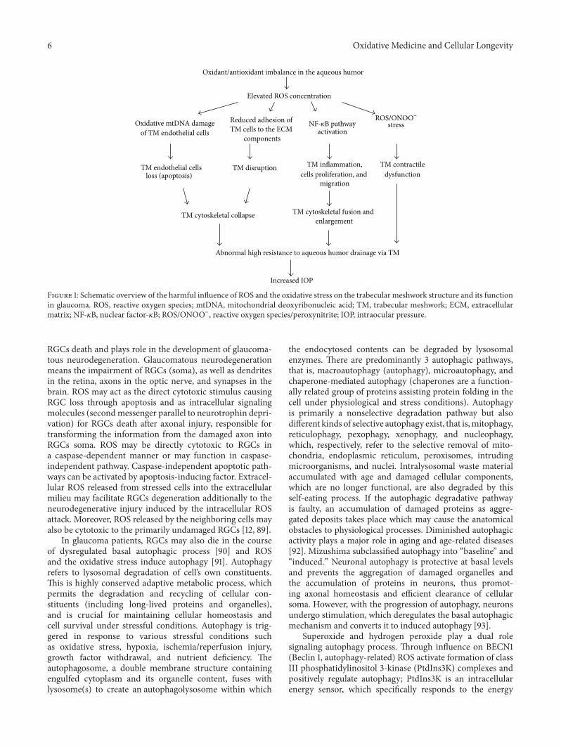
6 Oxidative Medicine and Cellular Longevity
TM cytoskeletal collapse
Abnormal high resistance to aqueous humor drainage via TM
Increased IOP
Oxidative mtDNA damageof TM endothelial cells
Reduced adhesion of TM cells to the ECM
componentsactivation
stress
TM endothelial cellsloss (apoptosis)
TM disruption TM inflammation,cells proliferation, and
migration
TM contractiledysfunction
TM cytoskeletal fusion andenlargement
Oxidant/antioxidant imbalance in the aqueous humor
Elevated ROS concentration
NF-𝜅B pathwayROS/ONOO−
Figure 1: Schematic overview of the harmful influence of ROS and the oxidative stress on the trabecular meshwork structure and its functionin glaucoma. ROS, reactive oxygen species; mtDNA, mitochondrial deoxyribonucleic acid; TM, trabecular meshwork; ECM, extracellularmatrix; NF-𝜅B, nuclear factor-𝜅B; ROS/ONOO−, reactive oxygen species/peroxynitrite; IOP, intraocular pressure.
RGCs death and plays role in the development of glaucoma-tous neurodegeneration. Glaucomatous neurodegenerationmeans the impairment of RGCs (soma), as well as dendritesin the retina, axons in the optic nerve, and synapses in thebrain. ROS may act as the direct cytotoxic stimulus causingRGC loss through apoptosis and as intracellular signalingmolecules (secondmessenger parallel to neurotrophin depri-vation) for RGCs death after axonal injury, responsible fortransforming the information from the damaged axon intoRGCs soma. ROS may be directly cytotoxic to RGCs ina caspase-dependent manner or may function in caspase-independent pathway. Caspase-independent apoptotic path-ways can be activated by apoptosis-inducing factor. Extracel-lular ROS released from stressed cells into the extracellularmilieu may facilitate RGCs degeneration additionally to theneurodegenerative injury induced by the intracellular ROSattack. Moreover, ROS released by the neighboring cells mayalso be cytotoxic to the primarily undamaged RGCs [12, 89].
In glaucoma patients, RGCs may also die in the courseof dysregulated basal autophagic process [90] and ROSand the oxidative stress induce autophagy [91]. Autophagyrefers to lysosomal degradation of cell’s own constituents.This is highly conserved adaptive metabolic process, whichpermits the degradation and recycling of cellular con-stituents (including long-lived proteins and organelles),and is crucial for maintaining cellular homeostasis andcell survival under stressful conditions. Autophagy is trig-gered in response to various stressful conditions suchas oxidative stress, hypoxia, ischemia/reperfusion injury,growth factor withdrawal, and nutrient deficiency. Theautophagosome, a double membrane structure containingengulfed cytoplasm and its organelle content, fuses withlysosome(s) to create an autophagolysosome within which
the endocytosed contents can be degraded by lysosomalenzymes. There are predominantly 3 autophagic pathways,that is, macroautophagy (autophagy), microautophagy, andchaperone-mediated autophagy (chaperones are a function-ally related group of proteins assisting protein folding in thecell under physiological and stress conditions). Autophagyis primarily a nonselective degradation pathway but alsodifferent kinds of selective autophagy exist, that is,mitophagy,reticulophagy, pexophagy, xenophagy, and nucleophagy,which, respectively, refer to the selective removal of mito-chondria, endoplasmic reticulum, peroxisomes, intrudingmicroorganisms, and nuclei. Intralysosomal waste materialaccumulated with age and damaged cellular components,which are no longer functional, are also degraded by thisself-eating process. If the autophagic degradative pathwayis faulty, an accumulation of damaged proteins as aggre-gated deposits takes place which may cause the anatomicalobstacles to physiological processes. Diminished autophagicactivity plays a major role in aging and age-related diseases[92]. Mizushima subclassified autophagy into “baseline” and“induced.” Neuronal autophagy is protective at basal levelsand prevents the aggregation of damaged organelles andthe accumulation of proteins in neurons, thus promot-ing axonal homeostasis and efficient clearance of cellularsoma. However, with the progression of autophagy, neuronsundergo stimulation, which deregulates the basal autophagicmechanism and converts it to induced autophagy [93].
Superoxide and hydrogen peroxide play a dual rolesignaling autophagy process. Through influence on BECN1(Beclin 1, autophagy-related) ROS activate formation of classIII phosphatidylinositol 3-kinase (PtdIns3K) complexes andpositively regulate autophagy; PtdIns3K is an intracellularenergy sensor, which specifically responds to the energy
Oxidative Medicine and Cellular Longevity 7
depletion [94]. BECN1 contributes to the early formationof autophagic vesicles [95] and is predominantly located inRGCs layer of the retina [96]. However, ROS also can activateBCL2 (B-cell CLL/lymphoma 2) family proteins, which afterbinding to BECN1 disorganize the formation of PtdIns3K anddisrupt the induction of autophagy [94].
Both chronic hypertensive glaucoma and/or conditions ofretinal transient ischemia, induced by acutely increasing IOP,stimulate ROS production and dysregulate basal autophagy.Autophagy is originally activated in the dendrites of RGCsto promote cellular protection and maintain intracellularhomeostasis. Axons contain a large number of mitochon-dria, which make them more sensitive to chronic ischemia.Neuroprotective effect of autophagy in axons lessens after 4weeks of progressive increase of IOP. Thereafter, autophagyis predominantly activated in the neuronal soma, whichdisrupts homeostasis, decreases cell viability, and triggersautophagic RGC death [90, 96]. If the IOP increases acutely,autophagy is enhanced immediately and occursmostly in cellbodies, which induces neuronal cell death in a relatively shorttime [90].
3.2.2. The Influence of ROS and the Oxidative Stress on GlialCells of the Retina and Optic Nerve Head. Under oxidativeconditions glial cell lessens its neuroprotective ability andmay even become neurodestructive. ROS and the oxidativestress cause supportive glial dysfunction, which lead tothe secondary RGCs apoptosis. ROS stimulate pathologicalglial activity (increase secretion of TNF-𝛼 and nitric oxide),cause oxidation of its protein (glutamine synthetase), anddamage glial cells through activation ofAGE/RAGE signalingpathway. ROS induce glial immune system as well, whichadditionally facilitate the progression of glaucomatous neu-rodegeneration [12].
ROS and aging promote accumulation of advanced gly-cation end products (AGEs), a hallmark of many chronicneurodegenerative diseases. AGEs stimulate ROS productionand assist development of glaucomatous neurodegeneration[12]. AGEs are proteins, lipids, or nucleic acids formed bynonenzymatic glycation or glycoxidated, after the exposureto the aldose sugars. The oxidative stress increases with ageand, furthermore, the ability to respond to the oxidativestress declines with age, mostly due to the imbalance betweenincreasing oxidant production and decreasing antioxidantcapacity andAGEs accumulate in various tissues in the courseof physiological aging. Due to their synergismwith the oxida-tive stress, AGEs production is promoted by the oxidativestress; while AGEs lead to ROS generation, AGEs are com-monly implicated as factor which exacerbates progression ofmany neurodegenerative diseases. These detergent insolubleand protease-resistant, nondegradable AGEs aggregates mayimpair normal cellular/tissue functions directly, or indirectly,byAGE/RAGEpathway after binding to the specific receptorsfor advanced glycation end products (RAGEs). AGE/RAGEpathway results in the cell activation, proliferation, apoptoticcell death, chemotaxis, angiogenesis, and ROS generation. Asa consequence of AGEs accumulation, many proteins losetheir function, including proteins involved in the regulationof gene transcription [97].
In glaucoma, AGEs are accumulated predominantlyextracellularly in laminar cribriform plates and blood ves-sels of the optic nerve head, mainly on many long-livedmacromolecules like collagen [98]. Extracellular aggregatesincrease tissue rigidity and mechanical strength and increaseimpairment of microcirculation, which additionally facilitateinjury of axons damaged by increased IOP [12]. AGEs arealso accumulated intracellularly in RGCs, glial cells, andaxons as well [98], which cause intracellular protein trafficand impair axonal transport [12]. Receptors for AGEs areupregulated predominantly on Müller cells of retinal glialcells and on RGCs [98]. It makes glial cells and RGCsparticularly susceptible to AGE/RAGE signaling pathwayand such receptor-mediated signaling may amplify directcytotoxic effect caused by extracellular/intracellular AGEsaccumulation [12].
The retina and optic nerve glial cells vigorously respondto ROS stimulation. Activated autoimmune response mayfacilitate primary and/or secondary RGCs degenerationthrough stimulation of an aberrant immune response [12].In Tezel et al.’s study, glial cells exposed to ROS upregulatedmajor histocompatibility complex (MHC) class II molecules,important in autoimmune response. T cells recognize anti-gens in the form of small peptides tightly bound to MHCclass II molecules displayed on the surfaces of antigen-presenting cells and MHC complex interacts with antigen-specific receptors on T cells to induce an antigen-specificreaction. Compared with the control, glial cells in ROS-generating systems were more potent inducers of T cell acti-vation in a cell density- and time-dependentmanner, assessedby increased T cell proliferation (approximately threefold)and TNF-𝛼 secretion (approximately sixfold), 𝑝 < 0.01 [99].
McElnea et al. confirmed the influence of ROS overpro-duction, oxidative stress, and mitochondrial dysfunction (aswell as impaired calcium extrusions) on glial cells of the opticnerve head in glaucomatous patients. The compare levelsof the oxidative stress, mitochondrial function (as well ascalcium homeostasis) in glial fibrillary acid-negative proteinlamina cribrosa cells obtained from the optic nerve headregion of glaucomatous lamina cribrosa (GLC), and normallamina cribrosa (NLC) human donor eyes showed that intra-cellular ROS production was increased in GLC comparedto NLC (27.19 ± 7.05 𝜇M MDA versus 14.59 ± 0.82 𝜇MMDA, 𝑝 < 0.05); malondialdehyde (MDA) is a naturallyoccurring product of lipid peroxidation used as an indicatorof the oxidative stress. Moreover, mitochondrial membranepotential was lower in GLC (57.5 ± 6.8%) compared toNLC (41.8 ± 5.3%), expression of the antioxidants (aldo-ketoreductase family 1 member C1 and glutamate cysteine ligasecatalytic subunit) was significantly (𝑝 = 0.02) lower in GLCcompared to NLC control, and intracellular calcium (Ca2+)levels were significantly higher (𝑝 < 0.001) in GLC cellscompared to NLC [100].
ROS and the oxidative stress have influence on the opticnerve tissue remodeling, since they trigger neuronal loss,disorganize laminar cribriform plates secondary to extracel-lular AGEs accumulation, and stimulate matrix metallopro-teinases (MMPs) for digestion of extracellular matrix (ECM)in glaucomatous eyes.Degradation of the optic nerve neurons
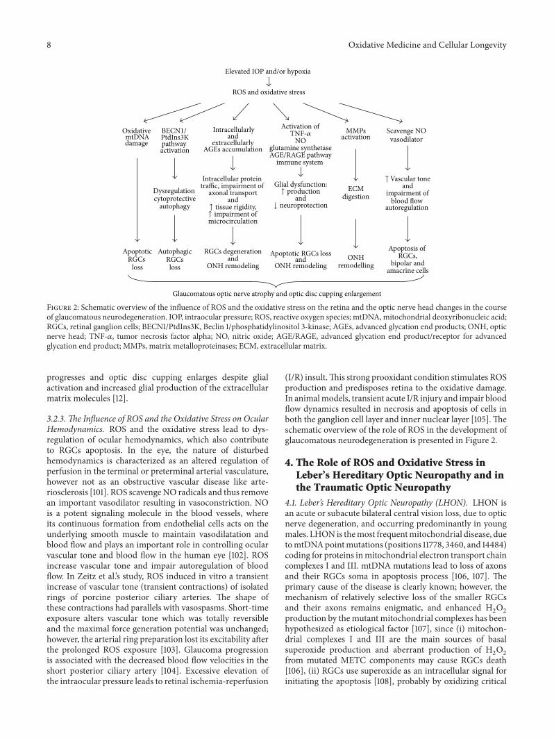
8 Oxidative Medicine and Cellular Longevity
andimpairment of
blood flow autoregulation
Elevated IOP and/or hypoxia
ROS and oxidative stress
Oxidative mtDNAdamage
BECN1/ PtdIns3Kpathway activation
Activation of
glutamine synthetaseAGE/RAGE pathway
immune system
Intracellularlyand
extracellularly AGEs accumulation
Scavenge NOvasodilator
MMPsactivation
ECMdigestion
Dysregulation cytoprotective
autophagy
Glial dysfunction:
and
Intracellular protein traffic, impairment of
axonal transportand
microcirculation
ApoptoticRGCs
loss
AutophagicRGCsloss
RGCs degenerationand
ONH remodelingApoptotic RGCs loss
andONH remodeling
ONHremodelling
Apoptosis ofRGCs,
bipolar andamacrine cells
Glaucomatous optic nerve atrophy and optic disc cupping enlargement
↑ impairment of↑ tissue rigidity, ↓ neuroprotection
↑ production
TNF-𝛼NO
↑Vascular tone
Figure 2: Schematic overview of the influence of ROS and the oxidative stress on the retina and the optic nerve head changes in the courseof glaucomatous neurodegeneration. IOP, intraocular pressure; ROS, reactive oxygen species; mtDNA, mitochondrial deoxyribonucleic acid;RGCs, retinal ganglion cells; BECN1/PtdIns3K, Beclin 1/phosphatidylinositol 3-kinase; AGEs, advanced glycation end products; ONH, opticnerve head; TNF-𝛼, tumor necrosis factor alpha; NO, nitric oxide; AGE/RAGE, advanced glycation end product/receptor for advancedglycation end product; MMPs, matrix metalloproteinases; ECM, extracellular matrix.
progresses and optic disc cupping enlarges despite glialactivation and increased glial production of the extracellularmatrix molecules [12].
3.2.3. The Influence of ROS and the Oxidative Stress on OcularHemodynamics. ROS and the oxidative stress lead to dys-regulation of ocular hemodynamics, which also contributeto RGCs apoptosis. In the eye, the nature of disturbedhemodynamics is characterized as an altered regulation ofperfusion in the terminal or preterminal arterial vasculature,however not as an obstructive vascular disease like arte-riosclerosis [101]. ROS scavengeNO radicals and thus removean important vasodilator resulting in vasoconstriction. NOis a potent signaling molecule in the blood vessels, whereits continuous formation from endothelial cells acts on theunderlying smooth muscle to maintain vasodilatation andblood flow and plays an important role in controlling ocularvascular tone and blood flow in the human eye [102]. ROSincrease vascular tone and impair autoregulation of bloodflow. In Zeitz et al.’s study, ROS induced in vitro a transientincrease of vascular tone (transient contractions) of isolatedrings of porcine posterior ciliary arteries. The shape ofthese contractions had parallels with vasospasms. Short-timeexposure alters vascular tone which was totally reversibleand the maximal force generation potential was unchanged;however, the arterial ring preparation lost its excitability afterthe prolonged ROS exposure [103]. Glaucoma progressionis associated with the decreased blood flow velocities in theshort posterior ciliary artery [104]. Excessive elevation ofthe intraocular pressure leads to retinal ischemia-reperfusion
(I/R) insult.This strong prooxidant condition stimulates ROSproduction and predisposes retina to the oxidative damage.In animalmodels, transient acute I/R injury and impair bloodflow dynamics resulted in necrosis and apoptosis of cells inboth the ganglion cell layer and inner nuclear layer [105].Theschematic overview of the role of ROS in the development ofglaucomatous neurodegeneration is presented in Figure 2.
4. The Role of ROS and Oxidative Stress inLeber’s Hereditary Optic Neuropathy and inthe Traumatic Optic Neuropathy
4.1. Leber’s Hereditary Optic Neuropathy (LHON). LHON isan acute or subacute bilateral central vision loss, due to opticnerve degeneration, and occurring predominantly in youngmales. LHON is themost frequentmitochondrial disease, duetomtDNApointmutations (positions 11778, 3460, and 14484)coding for proteins inmitochondrial electron transport chaincomplexes I and III. mtDNA mutations lead to loss of axonsand their RGCs soma in apoptosis process [106, 107]. Theprimary cause of the disease is clearly known; however, themechanism of relatively selective loss of the smaller RGCsand their axons remains enigmatic, and enhanced H
2O2
production by themutant mitochondrial complexes has beenhypothesized as etiological factor [107], since (i) mitochon-drial complexes I and III are the main sources of basalsuperoxide production and aberrant production of H
2O2
from mutated METC components may cause RGCs death[106], (ii) RGCs use superoxide as an intracellular signal forinitiating the apoptosis [108], probably by oxidizing critical
Oxidative Medicine and Cellular Longevity 9
sulfhydryls in signalingmacromolecules [109], and (iii) RGCsdeath is triggered when the mitochondrial superoxide levelsare increased by knocking down mitochondrial superoxidedismutase 2 [110].
The effect of LHON mutations has been studied byproducing transmitochondrial cybrids in human cell lines,which demonstrate upregulation of somemtDNA transcriptsand exhibit increased superoxide production compared towild-type cells. In Giordano et al.’s study, LHON cybridspresented overproduction of ROS, as well as decrease inmitochondrial membrane potential, increased apoptotic rate,loss of cell viability, and hyperfragmented mitochondrialmorphology compared with control cybrids [111]. In anotherstudy, LHON cybrids carrying the np11778 mutation becameselectively more H
2O2sensitive compared with the parental
cell line.They contained a decreased cellular glutathione pool(49%, 𝑝 ≤ 0.05), despite 1.5-fold enhanced expression ofthe regulatory subunit of 𝛾-glutamylcysteine synthetase (𝑝 ≤0.05). The capacity to detoxify H
2O2was reduced although
the activity of superoxide dismutase, glutathione peroxidase,and glutathione reductase was unchanged [107].
4.2. ROS Overproduction Contributes to the Traumatic OpticNeuropathy (TON) Pathogenesis. TON means partial orcomplete loss of optic nerve function due to the direct orindirect optic nerve injury, after head trauma sequelae suchas edema, hemorrhage, and concussion [112]. RGCs apoptosisafter optic nerve injury is caused by lack of neurotrophinsupport, increased extracellular glutamate levels, disruptionof cellular homeostasis, and damage from free radicals. Apop-totic processes are also activated by microglial cells, whichrelease inflammatory mediators (cytokines, prostaglandins,and complement molecules) and reactive oxygen species[113]. Ahmad et al. confirmed the increased oxidativestress in mice retina with TON [114]; however, treatmentwith ABT702 (pharmacological adenosine kinase inhibitor)attenuated neurotoxicity and significantly decreased levelsof the oxidative stress markers, that is, superoxide anion,iNOS/nNOS, and nitrotyrosine, and attenuated inflamma-tion (decreased expression of many inflammatory moleculesmediated by adenosine) in retinal sections of mouse withTON [115]. ROS are also overproduced during secondarydegeneration following neurotrauma, which means that theprecise impairment of only dorsal axons of optic nerve causessecondary degeneration of intact ventral axons [116]. Inexperimental study, the level of ROS (and nitrogen species)increased at 1, 3, and 7 days in ventral optic nerve afterdorsal injury. Immunoreactivity for glutathione peroxidaseand heme oxygenase-1 increased in ventral optic nerve at 3and 7 days after injury, respectively. Despite the increasedantioxidant immunoreactivity, DNA oxidation was evidentjust since the 1st day, lipid oxidation after 3 days, andprotein nitration after 7 days since the injury. Oxidative (andnitrosative) damage was particularly evident in CC1-positiveoligodendrocytes [117].
5. The Role of ROS and the OxidativeStress in Cataractogenesis
Cataract, the opacification of the crystalline lens, is one ofthe leading causes of blindness in the world and aging is
the greatest risk factor for noncongenital cataract formation.However, it is amultifactorial optic disorder and other factorslike exposure to sunlight UV radiation, smoking, diabetes,malnutrition, myopia, and drug (steroid) use also contributesignificantly to cataractogenesis [118].
The progressive loss of lens transparency associated withthe increasing age is a cumulative physiological responseto toxic environmental factors leading to an excessive gen-eration of ROS in the lens epithelium cells (LECs) and inthe superficial lens fiber cells, as well as in the aqueoushumor [119]. UV-induced oxidative damage is a significantcontributory factor to cataractogenesis. The eyes are con-tinuously exposed to solar radiation, which can be dividedinto five regions in increasing order of wavelengths, thatis, ultraviolet UVC, UVB, UVA, visible range, and infraredrange. Among these radiations, UVC and UVB are respon-sible for photochemical reactions. UVA, visible range, andinfrared range radiation are traditionally thought to be lessdamaging [120]. The major effect of UV radiation is throughphotochemical generation of ROS, including superoxide andits derivatization to other potent entities such as hydrogenperoxide, hydroxyl radicals, and singlet oxygen in the lensand in aqueous humor, which lead to oxidative damage of thelens tissue [120, 121].The incidence of cataract is higher in thepopulation which is more exposed to the sunlight [122].
Under normal conditions the lens is well equipped anduses multiple physiologic defense strategies to scavenge ROSand to maintain them at low levels to protect the lensfrom the toxic effects of oxidative damage. The lens defensesystem constitutes enzymatic antioxidants, that is, superoxidedismutase (SOD), catalase (CAT), and glutathione peroxidase(GPX) utilizing H
2O2, chemical antioxidants, that is, alfa-
tocopherol, beta-carotene, ascorbate, and GSH, structuralantioxidants, that is, cholesterol and membrane protein, andtransition metal-sequestering protein including aqueous andplasma ceruloplasmin [123, 124]. Enzymatic defense systemalso protects the lens from lipid-derived hydrogen peroxide[123]. However, protective systems decrease with the age andlong-term exposure to oxidative stress predispose lens cellsat risk for cumulative oxidative damage and cataract forma-tion [118]. The effectiveness of SOD and other antioxidantenzymes is limited to several reasons such as the following: (i)their deactivation with aging and their selective distributionand availability in various cellular compartments, (ii) beingmacromolecules, where they cannot penetrate the certainsensitive sites of oxidation in nucleic acids and in proteins,(iii) the fact that the lens is surrounded and bathed byaqueous and the vitreous humors, fluids which lack theenzymatic defenses. Therefore the lens cell membranes,which are continuously exposed to a photochemical oxidativeenvironment due to the continued light penetration duringthe long periods of photopic vision, remain susceptible tophoto damage [121]. Human mature cataractous lenses showdecreased activity of SOD, glutathione peroxidase (reducingorganic hydrogen peroxide, including hydrogen peroxide oflipids) and glutathione reductase, and however no signs ofdeficiency in activities of catalase [123]. Genetic variationsin the antioxidant genes coding for the SOD, CAT, and GPXenzymes may also lead to decreased or impaired regulation
10 Oxidative Medicine and Cellular Longevity
of their enzymatic activity and alter ROS detoxification.Zhang et al.’s study showed that the G/G genotype of theSOD1-251A/G polymorphism may be associated with anincreased risk of cataract. However, in CAT-21A/T andGPX1-198C/T polymorphisms, there were no significant differencesin the variant homozygous frequencies in age-related cataractpatients in comparison to controls [125].
5.1.TheRole of ROS inOxidative Stress-InducedMitochondria-Dependent Apoptosis in Human Lens Epithelial Cells. Oxida-tive stress of the lens is not only the result of an imbalancebetween lens oxidants and antioxidants but also the conse-quence of cellular redox status imbalance in the lens [123].The short range packing of the crystallins, which make upover 90% of the soluble lens, must exist in a homogenousstate, that is, in the redox balance, to maintain lens trans-parency, whichmeans the necessity tomaintain continuouslythe thiol (sulfhydryl) groups of proteins of the lens centerin a nearly 100% reduced state to prevent formation ofhigh molecular weight protein aggregates, which contributesto the cataract formation [118]. LECs are the center oflens metabolic activity and their oxidative damage plays asignificant role in cataractogenesis [126]. Mitochondria areabundant in the lens but only within the epithelium anddifferentiating fibers; mature fibers in the core of the lens lackmitochondria [127]. Mitochondria of LECs and superficialfiber cells consume 90% of the oxygen entering the lensand are major endogenous sources of ROS [118, 128]. It isestimated that up to 1% to 5% of oxygen consumed by thelens mitochondria is converted to ROS [129].
Age-related mitochondrial dysfunction and ROS imbal-ance induce oxidative damage of cellular components andplay crucial role in the pathogenesis of senile cataract devel-opment [118]. ROS are formed in LECs mitochondria as abyproduct of normal metabolism and as a consequence ofexposure to environmental compounds and if not eliminatedcause oxidative damage of DNA, proteins, and lipids [130].
Nucleic acids are prone to oxidative damage by ROS.Continuous attack of ROS leads to DNA oxidation. OH∙modify guanine of DNA and form 8-hydroxyguanine. 8-OHdG is highly mutagenic, causes GC to TA transversions,and has been commonly quantified as a steady-state estimateof oxidative stress in tissues [131]. The extent of lens DNAdamage caused by direct ROS attack can be assessed by8-hydroxyguanine assay and comet assay. Cultured normalhuman LECs show increase of 8-hydroxyguanine marker inresponse to the oxidative stress [132]. Quantitative assessmentof DNA damage in LECs achieved from senile cataractpatients and performed in comet assay showed smearing ofDNA fragments instead of bands in the tail, which indicaterandom (nonenzymatic) damage with ROS, which act bychemical reaction [133]. Oxidative mtDNA damage is acausative factor in aging and a wide variety of degenerativediseases. mtDNA damage is more extensive and persistslonger than nDNA damage because of its close proximityto ROS generation through the respiratory chain and itspaucity of protective histones. A vicious cycle of mtDNAdamage and ROS production established within cells leads toloss of the mitochondrial membrane potential and release of
cytochrome c, resulting in the cell apoptosis [130]. Oxidativestress can disrupt the balance between ROS production andthe radical scavenging effect and lead to apoptotic cell deaththrough the mitochondrial apoptosis pathway. Numerousstudies of human cataractous lenses confirmed extensiveoxidative damage of mtDNA and membrane pumps of lenscells, as well as increased unscheduled expression of genesstimulated by excessive ROS production. mtDNA damageand pathological gene expression are both responsible for lossof LECs viability and their death by apoptotic and necroticmechanisms. LECs death by apoptosis plays key role inthe pathogenesis of noncongenital cataract development inhuman [118, 134–136].
Increased accumulation of oxidized proteins, mainlymethionines and cysteine residues, also is linked to catarac-togenesis and confirms age-related increase in rates of ROSgeneration, decrease in antioxidant activity, and loss in thecapacity to degrade oxidized proteins [137]. Moreover, exces-sive ROS production and oxidative stress lead to formationof lipid peroxides, which contribute to pathological processesof aging and play role in systemic (diabetes, atherosclerosis,chronic renal failure, and inflammation) and retinal degener-ative diseases, and they are statistically significant risk factorsfor cataract development [123]. Lipid peroxides impair bothcell membrane and cytosol regions [138], damage DNA [139],induce a drop in total glutathione and dramatic change in theredox ratio of glutathione, and lead to the appearance of newfluorophores and large protein aggregates with low solubility(clouding matrix) in the lens matter [140].
6. The Role of ROS and the Oxidative Stress inDiabetic Retinopathy
Diabetesmellitus (DM) is a chronic and progressive neurode-generative disease, characterized by chronic hyperglycemiaand altered cellular homeostasis, which lead to the diffusemicrovascular and macrovascular damage, numerous com-plications, and multiorgan dysfunction. During the courseof DM, every cell is exposed to the abnormally high glucoseconcentrations; however, high glucose-related damage onlytargets specific tissues, that is, retina, nerve tissues, andkidney, since these tissues are deficient in the ability tochange glucose transport rates when faced with the elevatedextracellular glucose concentrations [141].
Diabetic retinopathy (DR) is a chronic and progressivecomplication in the course of diabetes mellitus type 1 or type2 and the major cause of blindness in people of working age.It develops over approximately 10 to 25 years, and duringthe first two decades of the disease, nearly all individualswith type 1 and approximately 60% of individuals with type 2diabetes will have some degree of retinopathy [142]. Diabeticretinopathy is one of the microvascular diabetes complica-tions, characterized by gradual and progressive alterations inthe retinal microvasculature with accompanying damage ofglia and neurons [143]. DR results from capillaries damage.Capillaries are lined with endothelial cells, surrounded bysmooth muscle cells and sealed by pericytes, which providetone to the vessels and create a blood barrier for closedcapillaries in the retina and in the choroid. In early DR
Oxidative Medicine and Cellular Longevity 11
stage, pericyte and endothelial cells undergo accelerateddeath by apoptosis, which leads to the reduction of pericytenumbers manifested by their degeneration (ghost cells) orloss, followed by the increased numbers of acellular-occludedcapillaries, microaneurysms, and capillary basement mem-brane thickening. Noncapillary cells (Müller cells and otherglial cells) are also lost selectively via apoptosis. Unsealedcapillaries begin to leak plasma and erythrocytes into the sur-rounding retinal tissue, resulting in edema and intraretinalhemorrhages. Endothelial cells try to repair the damage bymultiplying on the inner membrane; however, it leads to thecapillaries occlusion and ischemic retina releases the growthfactors leading to the pathological angiogenesis [144]. Basedon the extent of microvascular damage, DR is classified intoeither nonproliferative (mild, moderate, or preproliferative,characterized by cotton wool spots, venous beading andloops, blood vessel closure, tissue ischaemia, and the forma-tion of intraretinal microvascular abnormalities) or prolifer-ative. In proliferative DR, pathological angiogenesis is drivenby the vascular endothelial growth factor (VEGF) sourcedfrom retinal vascular pericytes, retinal ganglion cells, andglia. Vision loss occurs from breakdown of the blood-retinalbarrier, resulting inmacular edema, inner retinal and vitreoushemorrhages, and tractional retinal detachment [144].
6.1. The Interaction between Hyperglycemia, ROS Stress, andHyperglycemia-InducedMetabolite Pathways. Hyperglycemiastimulates overproduction of mitochondrial ROS and gener-ates the oxidative stress. Increased mitochondrial ROS levelsactivate the poly-ADP-ribose polymerase (PARP) pathway,which reduces glyceraldehydes 3-phosphate dehydrogenase(GAPDH) activity. Decreased GAPDH level in turn con-tributes to overactivation of four classic hyperglycemia-induced metabolite mechanisms, that is, the polyol pathway,the protein kinase C (PKC) pathway, AGEs pathway, and thehexosamine pathway. The reduction of GADPH activity canbe prevented by MnSOD [145–147].
According to Brownlee, all four classic hyperglycemia-induced pathways are activated by a single upstream event;that is, the mitochondrial overproduction of ROS and allfour pathways become the source of increased ROS produc-tion and stimulation of the oxidative stress [145, 146]. Allclassic hyperglycemia-inducedmetabolitemechanisms resultin decreased NADPH levels and increased NADPH oxidase(Nox) levels (see below). NADPH regenerate glutathione,an important scavenger of ROS. Therefore, decreased lev-els of NADPH are responsible for the increased ROSaccumulation and the oxidative stress damage. Moreover,decreased NADPH levels lead to the inhibition of GAPDH,which in turn activate four hyperglycemia-induced pathways[145, 146]. The unifying mechanism proposed by Brownleeinterconnects increased ROS production and the oxidativestress with four main hyperglycemia-induced processes andexplains the hyperglycemia-induced endothelial cells damage(apoptosis) and the progression of diabetic retinopathy [145].
ROS and the oxidative stress contribute to “metabolicmemory” or “legacy effect.” It means that the diabeticretinopathy progress, even after glycemia, has been normal-ized and well controlled. Mitochondrial abnormalities are
irreversible, even after hyperglycemia stress is terminatedand these impaired mitochondria are source of perma-nent ROS overproduction [145]. In laboratory conditions,human ARPE-19 retinal cells [148] and retinal pericytes[145] continue ROS overproduction even after the glucosenormalization. Hyperglycemia, increased ROS production,the oxidative stress, and excessive AGE formation are causallyassociated with and show linear relationship in the earlyyears of diabetes. ROS, which act at the mitochondrial level,are associated with “micro” metabolic memory and formthe lowest denominator of diabetic complications. However,persistent mtDNA damage and respiratory chain proteinglycation generate AGEs, which stimulate ROS production,and more ROS amplified AGEs formation and such viciouscycle acts independently of hyperglycemia level in advanceddiabetes stage. AGEs, which are result of chronic interactionwith the oxidative stress at tissue/vessel level, are associ-ated with “macro” metabolic memory and form the bridgebetween micro- and macrovascular diabetic damage [149].
Intracellular AGEs formed via nonenzymatic glycationand glycoxidation processes [146] on short half-life proteinslead to endothelial cells dysfunction, loss of pericytes, andneuronal cells damage. They also reduce platelet survival,increase platelet aggregation, promote a procoagulant state,lead to ischemia, and induce growth factors, which stimulatepathological angiogenesis [150]. AGEs formed on long half-life proteins like collagen modify extracellular matrix andcause loss of charge and structural distortions associatedwith a lower integrin binding affinity, which leads to celldetachment, basementmembrane thickening, and their resis-tance to proteolytic digestion [151]. AGEs/RAGE pathwayincreases cytosolic ROS level, activates NF-𝜅B pathway, andincreases expression of cytokine and adhesion molecule.RAGEs activate indirectly the toll-like receptor 4 (TLR-4),which can trigger interaction with an innate immune systemas well in type 2 diabetic patients [152].
6.2. The Influence of ROS and the Oxidative Stress onEndothelial Cells Apoptosis in Diabetic Retinopathy. Sus-tained hyperglycemia and increased chronic local oxidativestress disrupt retinal metabolism and accelerate prematureendothelial cells apoptosis via mitochondrial dysfunction inboth type 1 and type 2 diabetes retinopathy. In the earlystages of diabetes, increased mtDNA biogenesis and repaircompensate the ROS-induced damage. However, while itsustained insulting, this mechanism is overwhelmed andboth the function and structure of mtDNA are damaged(mitochondrial electron transport chain is highly sensitiveto the oxidative stress) [153–155]. The compromised electrontransport chain propagates a vicious cycle of ROS and thedysfunctional mitochondria fuel loss of capillary endothelialcells by initiating their apoptosis [153–155]. Poor glycemiacontrol and chronically increased intracellular glucose fluxdecrease retinal mtDNA copy number [156]. In diabeticretinopathy, the mtDNA damage at the regulatory region(the displacement loop) is considerably higher in comparisonto other mtDNA portions [156]. The enzymes importantfor mtDNA repair, that is, 8-oxoguanine DNA glycosylase(OGG1), MutY homolog, and thymine DNA glycosylase,
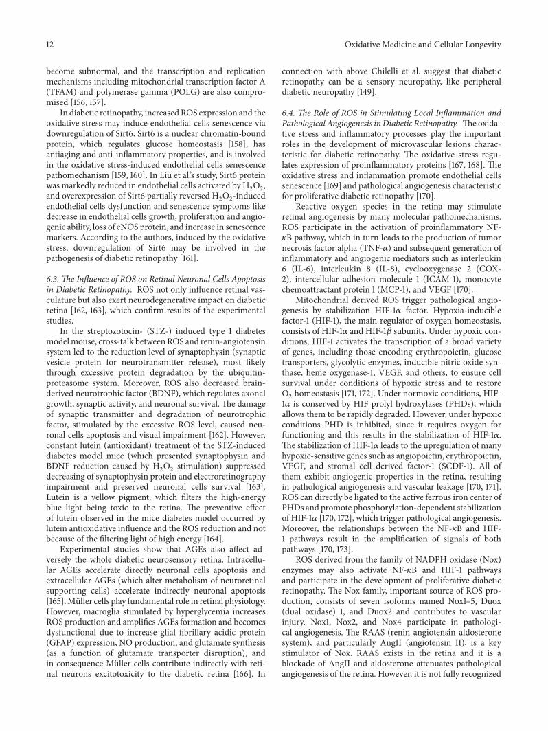
12 Oxidative Medicine and Cellular Longevity
become subnormal, and the transcription and replicationmechanisms including mitochondrial transcription factor A(TFAM) and polymerase gamma (POLG) are also compro-mised [156, 157].
In diabetic retinopathy, increasedROS expression and theoxidative stress may induce endothelial cells senescence viadownregulation of Sirt6. Sirt6 is a nuclear chromatin-boundprotein, which regulates glucose homeostasis [158], hasantiaging and anti-inflammatory properties, and is involvedin the oxidative stress-induced endothelial cells senescencepathomechanism [159, 160]. In Liu et al.’s study, Sirt6 proteinwas markedly reduced in endothelial cells activated by H
2O2,
and overexpression of Sirt6 partially reversed H2O2-induced
endothelial cells dysfunction and senescence symptoms likedecrease in endothelial cells growth, proliferation and angio-genic ability, loss of eNOS protein, and increase in senescencemarkers. According to the authors, induced by the oxidativestress, downregulation of Sirt6 may be involved in thepathogenesis of diabetic retinopathy [161].
6.3. The Influence of ROS on Retinal Neuronal Cells Apoptosisin Diabetic Retinopathy. ROS not only influence retinal vas-culature but also exert neurodegenerative impact on diabeticretina [162, 163], which confirm results of the experimentalstudies.
In the streptozotocin- (STZ-) induced type 1 diabetesmodelmouse, cross-talk betweenROS and renin-angiotensinsystem led to the reduction level of synaptophysin (synapticvesicle protein for neurotransmitter release), most likelythrough excessive protein degradation by the ubiquitin-proteasome system. Moreover, ROS also decreased brain-derived neurotrophic factor (BDNF), which regulates axonalgrowth, synaptic activity, and neuronal survival. The damageof synaptic transmitter and degradation of neurotrophicfactor, stimulated by the excessive ROS level, caused neu-ronal cells apoptosis and visual impairment [162]. However,constant lutein (antioxidant) treatment of the STZ-induceddiabetes model mice (which presented synaptophysin andBDNF reduction caused by H
2O2stimulation) suppressed
decreasing of synaptophysin protein and electroretinographyimpairment and preserved neuronal cells survival [163].Lutein is a yellow pigment, which filters the high-energyblue light being toxic to the retina. The preventive effectof lutein observed in the mice diabetes model occurred bylutein antioxidative influence and the ROS reduction and notbecause of the filtering light of high energy [164].
Experimental studies show that AGEs also affect ad-versely the whole diabetic neurosensory retina. Intracellu-lar AGEs accelerate directly neuronal cells apoptosis andextracellular AGEs (which alter metabolism of neuroretinalsupporting cells) accelerate indirectly neuronal apoptosis[165].Müller cells play fundamental role in retinal physiology.However, macroglia stimulated by hyperglycemia increasesROS production and amplifies AGEs formation and becomesdysfunctional due to increase glial fibrillary acidic protein(GFAP) expression, NO production, and glutamate synthesis(as a function of glutamate transporter disruption), andin consequence Müller cells contribute indirectly with reti-nal neurons excitotoxicity to the diabetic retina [166]. In
connection with above Chilelli et al. suggest that diabeticretinopathy can be a sensory neuropathy, like peripheraldiabetic neuropathy [149].
6.4. The Role of ROS in Stimulating Local Inflammation andPathological Angiogenesis inDiabetic Retinopathy. Theoxida-tive stress and inflammatory processes play the importantroles in the development of microvascular lesions charac-teristic for diabetic retinopathy. The oxidative stress regu-lates expression of proinflammatory proteins [167, 168]. Theoxidative stress and inflammation promote endothelial cellssenescence [169] and pathological angiogenesis characteristicfor proliferative diabetic retinopathy [170].
Reactive oxygen species in the retina may stimulateretinal angiogenesis by many molecular pathomechanisms.ROS participate in the activation of proinflammatory NF-𝜅B pathway, which in turn leads to the production of tumornecrosis factor alpha (TNF-𝛼) and subsequent generation ofinflammatory and angiogenic mediators such as interleukin6 (IL-6), interleukin 8 (IL-8), cyclooxygenase 2 (COX-2), intercellular adhesion molecule 1 (ICAM-1), monocytechemoattractant protein 1 (MCP-1), and VEGF [170].
Mitochondrial derived ROS trigger pathological angio-genesis by stabilization HIF-1𝛼 factor. Hypoxia-induciblefactor-1 (HIF-1), the main regulator of oxygen homeostasis,consists of HIF-1𝛼 and HIF-1𝛽 subunits. Under hypoxic con-ditions, HIF-1 activates the transcription of a broad varietyof genes, including those encoding erythropoietin, glucosetransporters, glycolytic enzymes, inducible nitric oxide syn-thase, heme oxygenase-1, VEGF, and others, to ensure cellsurvival under conditions of hypoxic stress and to restoreO2homeostasis [171, 172]. Under normoxic conditions, HIF-
1𝛼 is conserved by HIF prolyl hydroxylases (PHDs), whichallows them to be rapidly degraded. However, under hypoxicconditions PHD is inhibited, since it requires oxygen forfunctioning and this results in the stabilization of HIF-1𝛼.The stabilization of HIF-1𝛼 leads to the upregulation of manyhypoxic-sensitive genes such as angiopoietin, erythropoietin,VEGF, and stromal cell derived factor-1 (SCDF-1). All ofthem exhibit angiogenic properties in the retina, resultingin pathological angiogenesis and vascular leakage [170, 171].ROS can directly be ligated to the active ferrous iron center ofPHDs and promote phosphorylation-dependent stabilizationof HIF-1𝛼 [170, 172], which trigger pathological angiogenesis.Moreover, the relationships between the NF-𝜅B and HIF-1 pathways result in the amplification of signals of bothpathways [170, 173].
ROS derived from the family of NADPH oxidase (Nox)enzymes may also activate NF-𝜅B and HIF-1 pathwaysand participate in the development of proliferative diabeticretinopathy. The Nox family, important source of ROS pro-duction, consists of seven isoforms named Nox1–5, Duox(dual oxidase) 1, and Duox2 and contributes to vascularinjury. Nox1, Nox2, and Nox4 participate in pathologi-cal angiogenesis. The RAAS (renin-angiotensin-aldosteronesystem), and particularly AngII (angiotensin II), is a keystimulator of Nox. RAAS exists in the retina and it is ablockade of AngII and aldosterone attenuates pathologicalangiogenesis of the retina. However, it is not fully recognized
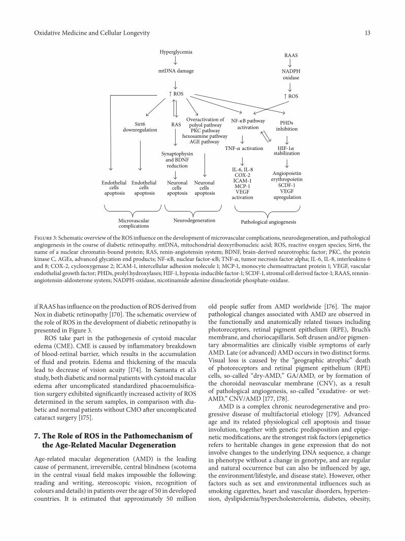
Oxidative Medicine and Cellular Longevity 13
Microvascularcomplications
Pathological angiogenesis
Hyperglycemia
mtDNA damage
RAAS
NADPHoxidase
Sirt6downregulation
Synaptophysinand BDNFreduction
Overactivation ofpolyol pathwayPKC pathway
hexosamine pathway AGE pathway
activationPHDs
inhibitionRAS
Endothelial cells
apoptosis
Neuronalcells
apoptosis
Neuronalcells
apoptosis
Endothelial cells
apoptosis
IL-6, IL-8COX-2
ICAM-1MCP-1VEGF
activation
Angiopoietinerythropoietin
SCDF-1VEGF
upregulation
Neurodegeneration
↑ ROS ↑ ROS
stabilizationTNF-𝛼 activation
NF-𝜅B pathway
HIF-1𝛼
Figure 3: Schematic overview of the ROS influence on the development ofmicrovascular complications, neurodegeneration, and pathologicalangiogenesis in the course of diabetic retinopathy. mtDNA, mitochondrial deoxyribonucleic acid; ROS, reactive oxygen species; Sirt6, thename of a nuclear chromatin-bound protein; RAS, renin-angiotensin system; BDNF, brain-derived neurotrophic factor; PKC, the proteinkinase C, AGEs, advanced glycation end products; NF-𝜅B, nuclear factor-𝜅B; TNF-𝛼, tumor necrosis factor alpha; IL-6, IL-8, interleukins 6and 8; COX-2, cyclooxygenase 2; ICAM-1, intercellular adhesion molecule 1; MCP-1, monocyte chemoattractant protein 1; VEGF, vascularendothelial growth factor; PHDs, prolyl hydroxylases; HIF-1, hypoxia-inducible factor-1; SCDF-1, stromal cell derived factor-1; RAAS, rennin-angiotensin-aldosterone system; NADPH-oxidase, nicotinamide adenine dinucleotide phosphate-oxidase.
if RAAShas influence on the production of ROS derived fromNox in diabetic retinopathy [170]. The schematic overview ofthe role of ROS in the development of diabetic retinopathy ispresented in Figure 3.
ROS take part in the pathogenesis of cystoid macularedema (CME). CME is caused by inflammatory breakdownof blood-retinal barrier, which results in the accumulationof fluid and protein. Edema and thickening of the maculalead to decrease of vision acuity [174]. In Samanta et al.’sstudy, both diabetic andnormal patientswith cystoidmacularedema after uncomplicated standardized phacoemulsifica-tion surgery exhibited significantly increased activity of ROSdetermined in the serum samples, in comparison with dia-betic and normal patients without CMO after uncomplicatedcataract surgery [175].
7. The Role of ROS in the Pathomechanism ofthe Age-Related Macular Degeneration
Age-related macular degeneration (AMD) is the leadingcause of permanent, irreversible, central blindness (scotomain the central visual field makes impossible the following:reading and writing, stereoscopic vision, recognition ofcolours and details) in patients over the age of 50 in developedcountries. It is estimated that approximately 50 million
old people suffer from AMD worldwide [176]. The majorpathological changes associated with AMD are observed inthe functionally and anatomically related tissues includingphotoreceptors, retinal pigment epithelium (RPE), Bruch’smembrane, and choriocapillaris. Soft drusen and/or pigmen-tary abnormalities are clinically visible symptoms of earlyAMD. Late (or advanced) AMD occurs in two distinct forms.Visual loss is caused by the “geographic atrophic” deathof photoreceptors and retinal pigment epithelium (RPE)cells, so-called “dry-AMD,” GA/AMD, or by formation ofthe choroidal neovascular membrane (CNV), as a resultof pathological angiogenesis, so-called “exudative- or wet-AMD,” CNV/AMD [177, 178].
AMD is a complex chronic neurodegenerative and pro-gressive disease of multifactorial etiology [179]. Advancedage and its related physiological cell apoptosis and tissueinvolution, together with genetic predisposition and epige-netic modifications, are the strongest risk factors (epigeneticsrefers to heritable changes in gene expression that do notinvolve changes to the underlying DNA sequence, a changein phenotype without a change in genotype, and are regularand natural occurrence but can also be influenced by age,the environment/lifestyle, and disease state). However, otherfactors such as sex and environmental influences such assmoking cigarettes, heart and vascular disorders, hyperten-sion, dyslipidemia/hypercholesterolemia, diabetes, obesity,
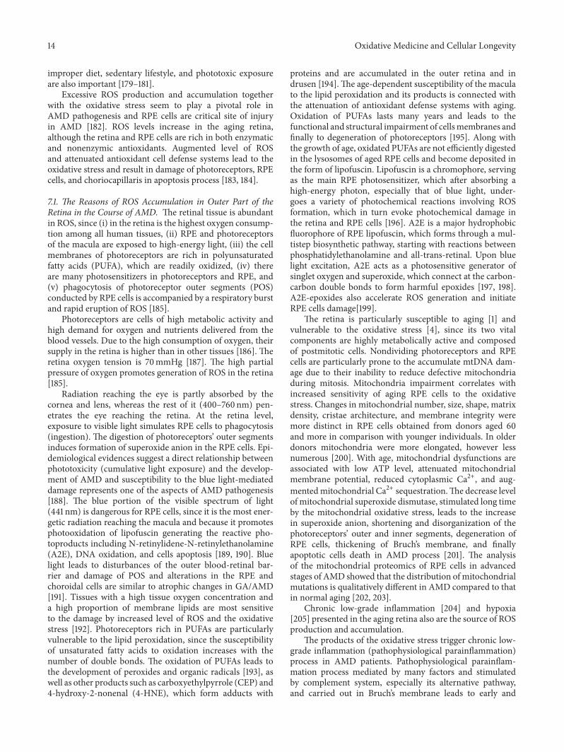
14 Oxidative Medicine and Cellular Longevity
improper diet, sedentary lifestyle, and phototoxic exposureare also important [179–181].
Excessive ROS production and accumulation togetherwith the oxidative stress seem to play a pivotal role inAMD pathogenesis and RPE cells are critical site of injuryin AMD [182]. ROS levels increase in the aging retina,although the retina and RPE cells are rich in both enzymaticand nonenzymic antioxidants. Augmented level of ROSand attenuated antioxidant cell defense systems lead to theoxidative stress and result in damage of photoreceptors, RPEcells, and choriocapillaris in apoptosis process [183, 184].
7.1. The Reasons of ROS Accumulation in Outer Part of theRetina in the Course of AMD. The retinal tissue is abundantin ROS, since (i) in the retina is the highest oxygen consump-tion among all human tissues, (ii) RPE and photoreceptorsof the macula are exposed to high-energy light, (iii) the cellmembranes of photoreceptors are rich in polyunsaturatedfatty acids (PUFA), which are readily oxidized, (iv) thereare many photosensitizers in photoreceptors and RPE, and(v) phagocytosis of photoreceptor outer segments (POS)conducted by RPE cells is accompanied by a respiratory burstand rapid eruption of ROS [185].
Photoreceptors are cells of high metabolic activity andhigh demand for oxygen and nutrients delivered from theblood vessels. Due to the high consumption of oxygen, theirsupply in the retina is higher than in other tissues [186]. Theretina oxygen tension is 70mmHg [187]. The high partialpressure of oxygen promotes generation of ROS in the retina[185].
Radiation reaching the eye is partly absorbed by thecornea and lens, whereas the rest of it (400–760 nm) pen-etrates the eye reaching the retina. At the retina level,exposure to visible light simulates RPE cells to phagocytosis(ingestion). The digestion of photoreceptors’ outer segmentsinduces formation of superoxide anion in the RPE cells. Epi-demiological evidences suggest a direct relationship betweenphototoxicity (cumulative light exposure) and the develop-ment of AMD and susceptibility to the blue light-mediateddamage represents one of the aspects of AMD pathogenesis[188]. The blue portion of the visible spectrum of light(441 nm) is dangerous for RPE cells, since it is the most ener-getic radiation reaching the macula and because it promotesphotooxidation of lipofuscin generating the reactive pho-toproducts including N-retinylidene-N-retinylethanolamine(A2E), DNA oxidation, and cells apoptosis [189, 190]. Bluelight leads to disturbances of the outer blood-retinal bar-rier and damage of POS and alterations in the RPE andchoroidal cells are similar to atrophic changes in GA/AMD[191]. Tissues with a high tissue oxygen concentration anda high proportion of membrane lipids are most sensitiveto the damage by increased level of ROS and the oxidativestress [192]. Photoreceptors rich in PUFAs are particularlyvulnerable to the lipid peroxidation, since the susceptibilityof unsaturated fatty acids to oxidation increases with thenumber of double bonds. The oxidation of PUFAs leads tothe development of peroxides and organic radicals [193], aswell as other products such as carboxyethylpyrrole (CEP) and4-hydroxy-2-nonenal (4-HNE), which form adducts with
proteins and are accumulated in the outer retina and indrusen [194]. The age-dependent susceptibility of the maculato the lipid peroxidation and its products is connected withthe attenuation of antioxidant defense systems with aging.Oxidation of PUFAs lasts many years and leads to thefunctional and structural impairment of cellsmembranes andfinally to degeneration of photoreceptors [195]. Along withthe growth of age, oxidated PUFAs are not efficiently digestedin the lysosomes of aged RPE cells and become deposited inthe form of lipofuscin. Lipofuscin is a chromophore, servingas the main RPE photosensitizer, which after absorbing ahigh-energy photon, especially that of blue light, under-goes a variety of photochemical reactions involving ROSformation, which in turn evoke photochemical damage inthe retina and RPE cells [196]. A2E is a major hydrophobicfluorophore of RPE lipofuscin, which forms through a mul-tistep biosynthetic pathway, starting with reactions betweenphosphatidylethanolamine and all-trans-retinal. Upon bluelight excitation, A2E acts as a photosensitive generator ofsinglet oxygen and superoxide, which connect at the carbon-carbon double bonds to form harmful epoxides [197, 198].A2E-epoxides also accelerate ROS generation and initiateRPE cells damage[199].
The retina is particularly susceptible to aging [1] andvulnerable to the oxidative stress [4], since its two vitalcomponents are highly metabolically active and composedof postmitotic cells. Nondividing photoreceptors and RPEcells are particularly prone to the accumulate mtDNA dam-age due to their inability to reduce defective mitochondriaduring mitosis. Mitochondria impairment correlates withincreased sensitivity of aging RPE cells to the oxidativestress. Changes in mitochondrial number, size, shape, matrixdensity, cristae architecture, and membrane integrity weremore distinct in RPE cells obtained from donors aged 60and more in comparison with younger individuals. In olderdonors mitochondria were more elongated, however lessnumerous [200]. With age, mitochondrial dysfunctions areassociated with low ATP level, attenuated mitochondrialmembrane potential, reduced cytoplasmic Ca2+, and aug-mentedmitochondrial Ca2+ sequestration.The decrease levelof mitochondrial superoxide dismutase, stimulated long timeby the mitochondrial oxidative stress, leads to the increasein superoxide anion, shortening and disorganization of thephotoreceptors’ outer and inner segments, degeneration ofRPE cells, thickening of Bruch’s membrane, and finallyapoptotic cells death in AMD process [201]. The analysisof the mitochondrial proteomics of RPE cells in advancedstages of AMD showed that the distribution of mitochondrialmutations is qualitatively different in AMD compared to thatin normal aging [202, 203].
Chronic low-grade inflammation [204] and hypoxia[205] presented in the aging retina also are the source of ROSproduction and accumulation.
The products of the oxidative stress trigger chronic low-grade inflammation (pathophysiological parainflammation)process in AMD patients. Pathophysiological parainflam-mation process mediated by many factors and stimulatedby complement system, especially its alternative pathway,and carried out in Bruch’s membrane leads to early and
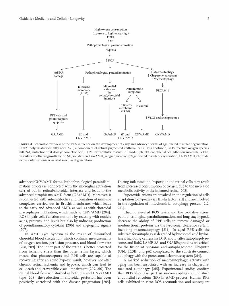
Oxidative Medicine and Cellular Longevity 15
RPE cells andphotoreceptors
apoptosis
GA/AMD SD andCNV/AMD
In Bruch’smembrane
(ECM)
In choroid
SD and CNV/AMD
CNV/AMD
High oxygen consumptionExposure to high-energy light
PUFAA2E
Pathophysiological parainflammationHypoxia
mtDNAdamage
Pathophysiological parainflammation
In Bruch’smembrane
(ECM)
Microglialactivation
inretinal/choroidal
Autoimmunecomplexes PECAM-1
GA/AMD CNV/AMD
↑ ROS
↑ Microautophagy
↑VEGF and angiopoietin-1
↑ Chaperone-autophagy↓ Macroautophagy
interface
Figure 4: Schematic overview of the ROS influence on the development of early and advanced forms of age-related macular degeneration.PUFA, polyunsaturated fatty acid; A2E, a component of retinal pigmented epithelial cell (RPE) lipofuscin; ROS, reactive oxygen species;mtDNA, mitochondrial deoxyribonucleic acid; ECM, extracellular matrix; PECAM-1, platelet endothelial cell adhesion molecule; VEGF,vascular endothelial growth factor; SD, soft drusen; GA/AMD, geographic atrophy/age-related macular degeneration; CNV/AMD, choroidalneovascularization/age-related macular degeneration.
advanced CNV/AMD forms. Pathophysiological parainflam-mation process is connected with the microglial activationcarried out in retinal/choroidal interface and leads to theadvanced atrophicans AMD form (GA/AMD). Moreover, itis connected with autoantibodies and formation of immunecomplexes carried out in Bruch’s membrane, which leadsto the early and advanced AMD, as well as with choroidalmacrophages infiltration, which leads to CNV/AMD [204].ROS impair cells function not only by reacting with nucleicacids, proteins, and lipids but also by inducing productionof proinflammatory cytokine [206] and angiogenic signals[207].
In AMD eyes hypoxia is the result of diminishedchoroidal blood circulation, which confirms measurementsof oxygen tension, perfusion pressure, and blood flow rate[208, 209]. The inner part of the retina is better protectedfrom ischemic stress than the outer retina layers, whichmeans that photoreceptors and RPE cells are capable ofrecovering after an acute hypoxic insult, however not afterchronic retinal ischemia and hypoxia, which can lead tocell death and irreversible visual impairment [209, 210]. Theretinal blood flow is disturbed in both dry and CNV/AMDtype [208]; the reduction in choroidal perfusion has beenpositively correlated with the disease progression [205].
During inflammation, hypoxia in the retinal cells may resultfrom increased consumption of oxygen due to the increasedmetabolic activity of the inflamed retina [205].
Superoxide anions are involved in the regulation of cellsadaptation to hypoxia viaHIF-1𝛼 factor [211] and are involvedin the regulation of mitochondrial autophagy process [212,213].
Chronic elevated ROS levels and the oxidative stress,pathophysiological parainflammation, and long stay hypoxiadecrease the ability of RPE cells to remove damaged ornonfunctional proteins via the lysosomal clearance system,including macroautophagy [214]. In aged RPE cells thesubstrate for autophagy is degraded by lysosomal acid hydro-lases, including cathepsins D, B, and L, after autophagolyso-some, and Rab7, LAMP-2A, and SNAREs proteins are criticalfor the fusion of lysosome and autophagosome. Ubiquitin(Ub), LC3II, and p62 complexed to the substrate connectautophagy with the proteasomal clearance system [214].
A marked reduction of macroautophagic activity withaging has been associated with an increase in chaperone-mediated autophagy [215]. Experimental studies confirmthat ROS also take part in microautophagy and disturbendothelial reticulum (ER) in AMD process. Human RPEcells exhibited in vitro ROS accumulation and subsequent
16 Oxidative Medicine and Cellular Longevity
elevation of GRP78 and CHOP expression (indicators ofER stress) after A2E and blue light-induced damage. More-over, N-acetylcysteine (NAC), ROS scavenger, diminishedexpression protein of ER stress [216]. In another study, t-butylhydroperoxide induced the oxidative stress which ledalso to the accumulation of ROS in the internal space (lumen)of the endoplasmic reticulum and disturbed ER homeostasisin RPE cells [217].
According to Blasiak et al., triplet consists of the oxidativestress, hypoxia, and autophagy which play an important rolein CNV/AMD pathogenesis [214]. Pathological angiogene-sis in AMD is connected with the activity of VEGF andangiopoietin-1 (and its receptor) [218]. Two proteins, that is,platelet endothelial cell adhesion molecule (PECAM-1) andthrombospondin-1 (TSP-1), act as linkage molecules, whichhave a reciprocal relationship with autophagy and angio-genesis mediated by angiopoietin 1 [214]. TSP-1, a commondenominator between autophagy, angiogenesis, and AMD,acts as antiangiogenic molecule (and a target for antineovas-cular therapy). Impaired expression of thrombospondin-1 inBruch’s membrane and choroidal vessels was shown in therodent eyes with age-related macular degeneration [219].Theschematic overview of the role of ROS in the development ofearly and advanced AMD is presented in Figure 4.
8. Conclusions
Excessive production of the reactive oxygen species and theoxidative stress play important role in the pathogenesis ofmany age-related ocular diseases and other pathologies of theanterior and posterior eye segment in adults.
ROS stimulate cells’ death via apoptosis process, partici-pate in the activation of proinflammatory and proangiogenicpathways, and are associated with the autophagy process.
Disclosure
This paper is supported by self-financing unit.
Conflict of Interests
The authors declare that there is no conflict of interestsregarding the publication of this paper.
References
[1] W. Dröge, “Free radicals in the physiological control of cellfunction,” Physiological Reviews, vol. 82, no. 1, pp. 47–95, 2002.
[2] M.D. Brand, “The sites and topology ofmitochondrial superox-ide production,” Experimental Gerontology, vol. 45, no. 7-8, pp.466–472, 2010.
[3] C. L.Quinlan, R. L. S. Goncalves,M.Hey-Mogensen,N. Yadava,V. I. Bunik, and M. D. Brand, “The 2-oxoacid dehydrogenasecomplexes in mitochondria can produce superoxide/hydrogenperoxide at much higher rates than complex I,” The Journal ofBiological Chemistry, vol. 289, no. 12, pp. 8312–8325, 2014.
[4] H.Cui, Y. Kong, andH. Zhang, “Oxidative stress,mitochondrialdysfunction, and aging,” Journal of Signal Transduction, vol.2012, Article ID 646354, 13 pages, 2012.
[5] T. Finkel, “Signal transduction by reactive oxygen species,”TheJournal of Cell Biology, vol. 194, no. 1, pp. 7–15, 2011.
[6] M. D. Evans, M. Dizdaroglu, and M. S. Cooke, “OxidativeDNA damage and disease: Induction, repair and significance,”Mutation Research, vol. 567, no. 1, pp. 1–61, 2004.
[7] R. Gredilla, “DNA damage and base excision repair in mito-chondria and their role in aging,” Journal of Aging Research, vol.2011, Article ID 257093, 9 pages, 2011.
[8] D. A. Butterfield, J. Drake, C. Pocernich, and A. Castegna, “Evi-dence of oxidative damage in Alzheimer’s disease brain: centralrole for amyloid 𝛽-peptide,” Trends in Molecular Medicine, vol.7, no. 12, pp. 548–554, 2001.
[9] I. G. Onyango, “Mitochondrial dysfunction and oxidative stressin Parkinson’s disease,” Neurochemical Research, vol. 33, no. 3,pp. 589–597, 2008.
[10] J. I. Kim, S. I. Cho, N. H. Kim et al., “Oxidative stress andneurodegeneration in prion diseases,” Annals of the New YorkAcademy of Sciences, vol. 928, pp. 182–186, 2001.
[11] B. J. Tabner, O. M. A. El-Agnaf, M. J. German, N. J. Fullwood,and D. Allsop, “Protein aggregation, metals and oxidative stressin neurodegenerative diseases,” Biochemical Society Transac-tions, vol. 33, no. 5, pp. 1082–1086, 2005.
[12] G. Tezel, “Oxidative stress in glaucomatous neurodegeneration:mechanisms and consequences,” Progress in Retinal and EyeResearch, vol. 25, no. 5, pp. 490–513, 2006.
[13] A. Shoham,M. Hadziahmetovic, J. L. Dunaief, M. B. Mydlarski,and H. M. Schipper, “Oxidative stress in diseases of the humancornea,” Free Radical Biology and Medicine, vol. 45, no. 8, pp.1047–1055, 2008.
[14] H. L. Chandler, K. S. Reuter, L. T. Sinnott, and J. J. Nichols,“Prevention of UV-induced damage to the anterior segmentusing class I UV-absorbing hydrogel contact lenses,” Investiga-tive Ophthalmology and Visual Science, vol. 51, no. 1, pp. 172–178,2010.
[15] M.H. Suh, J.-W. Kwon,W. R.Wee, Y. K. Han, J. H. Kim, and J. H.Lee, “Protective effect of ascorbic acid against corneal damageby ultraviolet B irradiation: a pilot study,” Cornea, vol. 27, no. 8,pp. 916–922, 2008.
[16] H. Sies and W. Stahl, “Vitamins E and C, beta-carotene, andother carotenoids as antioxidants,” The American Journal ofClinical Nutrition, vol. 62, pp. 1315S–1321S, 1995.
[17] M. Araie, E. Shirasawa, and M. Hikita, “Effect of oxidizedglutathione on the barrier function of the corneal endothelium,”Investigative Ophthalmology and Visual Science, vol. 29, no. 12,pp. 1884–1887, 1988.
[18] T. F. Linsenmayer, C. X. Cai, J.M.Millholland, K. E. Beazley, andJ. M. Fitch, “Nuclear ferritin in corneal epithelial cells: tissuespecific nuclear transport and protection from UV-damage,”Progress in Retinal and Eye Research, vol. 24, no. 2, pp. 139–159,2005.
[19] R. Brancato, T. Fiore, L. Papucci et al., “Concomitant effect oftopical ubiquinone Q10 and vitamin E to prevent keratocyteapoptosis after excimer laser photoablation in rabbits,” Journalof Refractive Surgery, vol. 18, no. 2, pp. 135–139, 2002.
[20] S. A. Marchitti, Y. Chen, D. C. Thompson, and V. Vasiliou,“Ultraviolet radiation: cellular antioxidant response and the roleof ocular aldehyde dehydrogenase enzymes,” Eye and ContactLens, vol. 37, no. 4, pp. 206–213, 2011.
[21] R. Buddi, B. Lin, S. R. Atilano, N. C. Zorapapel, M. C. Kenney,and D. J. Brown, “Evidence of oxidative stress in human cornealdiseases,” Journal of Histochemistry and Cytochemistry, vol. 50,no. 3, pp. 341–351, 2002.
Oxidative Medicine and Cellular Longevity 17
[22] J. Berlau, H.-H. Becker, J. Stave, C. Oriwol, and R. F. Guthoff,“Depth and age-dependent distribution of keratocytes inhealthy human corneas: a study using scanning-slit confocalmicroscopy in vivo,” Journal of Cataract and Refractive Surgery,vol. 28, no. 4, pp. 611–616, 2002.
[23] T. Schmedt, M. M. Silva, A. Ziaei, and U. Jurkunas, “Molecularbases of corneal endothelial dystrophies,” Experimental EyeResearch, vol. 95, no. 1, pp. 24–34, 2012.
[24] H. Elhalis, B. Azizi, and U. V. Jurkunas, “Fuchs endothelialcorneal dystrophy,” Ocular Surface, vol. 8, no. 4, pp. 173–184,2010.
[25] N. C. Joyce, “Proliferative capacity of corneal endothelial cells,”Experimental Eye Research, vol. 95, no. 1, pp. 16–23, 2012.
[26] U. V. Jurkunas, M. S. Bitar, T. Funaki, and B. Azizi, “Evidence ofoxidative stress in the pathogenesis of Fuchs endothelial cornealdystrophy,” American Journal of Pathology, vol. 177, no. 5, pp.2278–2289, 2010.
[27] J. M. Lee, M. J. Calkins, K. Chan, Y. W. Kan, and J. A. Johnson,“Identification of the NF-E2-related factor-2-dependent genesconferring protection against oxidative stress in primary cor-tical astrocytes using oligonucleotide microarray analysis,” TheJournal of Biological Chemistry, vol. 278, no. 14, pp. 12029–12038,2003.
[28] U. V. Jurkunas, I. Rawe, M. S. Bitar et al., “Decreased expressionof peroxiredoxins in Fuchs’ endothelial dystrophy,” InvestigativeOphthalmology and Visual Science, vol. 49, no. 7, pp. 2956–2963,2008.
[29] M. Matthaei, A. Y. Zhu, L. Kallay, C. G. Eberhart, C. Cursiefen,and A. S. Jun, “Transcript profile of cellular senescence-relatedgenes in Fuchs endothelial corneal dystrophy,”Experimental EyeResearch, vol. 129, pp. 13–17, 2014.
[30] Y. Gorin and K. Block, “Nox4 and diabetic nephropathy: with afriend like this, who needs enemies?” Free Radical Biology andMedicine, vol. 61, pp. 130–142, 2013.
[31] B. Azizi, A. Ziaei, T. Fuchsluger, T. Schmedt, Y. Chen, and U. V.Jurkunas, “p53-regulated increase in oxidative-stress-inducedapoptosis in Fuchs endothelial corneal dystrophy: a native tissuemodel,” Investigative Ophthalmology and Visual Science, vol. 52,no. 13, pp. 9291–9297, 2011.
[32] A. Ziaei, T. Schmedt, Y. Chen, andU.V. Jurkunas, “Sulforaphanedecreases endothelial cell apoptosis in fuchs endothelial cornealdystrophy: a novel treatment,” Investigative Ophthalmology andVisual Science, vol. 54, no. 10, pp. 6724–6734, 2013.
[33] T. Sherwin andN. H. Brookes, “Morphological changes in kera-toconus: pathology or pathogenesis,” Clinical and ExperimentalOphthalmology, vol. 32, no. 2, pp. 211–217, 2004.
[34] B. J. Dahl, E. Spotts, and J. Q. Truong, “Corneal collagen cross-linking: an introduction and literature review,” Optometry, vol.83, no. 1, pp. 33–42, 2012.
[35] J. Colin and S. Velou, “Current surgical options for kerato-conus,” Journal of Cataract and Refractive Surgery, vol. 29, no.2, pp. 379–386, 2003.
[36] K. P. Burdon and A. L. Vincent, “Insights into keratoconus froma genetic perspective,” Clinical and Experimental Optometry,vol. 96, no. 2, pp. 146–154, 2013.
[37] E. Arnal, C. Peris-Mart́ınez, J. L. Menezo, S. Johnsen-Soriano,and F. J. Romero, “Oxidative stress in keratoconus?” Investiga-tive Ophthalmology and Visual Science, vol. 52, no. 12, pp. 8592–8597, 2011.
[38] M. Edwards, C. N. J. McGhee, and S. Dean, “The genetics ofkeratoconus,”Clinical and Experimental Ophthalmology, vol. 29,no. 6, pp. 345–351, 2001.
[39] M. Chwa, S. R. Atilano, V. Reddy, N. Jordan, D. W. Kim,and M. C. Kenney, “Increased stress-induced generation ofreactive oxygen species and apoptosis in human keratoconusfibroblasts,” Investigative Ophthalmology & Visual Science, vol.47, no. 5, pp. 1902–1910, 2006.
[40] S. R. Atilano, P. Coskun,M. Chwa et al., “Accumulation ofmito-chondrial DNA damage in keratoconus corneas,” InvestigativeOphthalmology and Visual Science, vol. 46, no. 4, pp. 1256–1263,2005.
[41] M. Chwa, S. R. Atilano, D. Hertzog et al., “Hypersensitiveresponse to oxidative stress in keratoconus corneal fibroblasts,”Investigative Ophthalmology and Visual Science, vol. 49, no. 10,pp. 4361–4369, 2008.
[42] X. D. Hao, A. P. Chen, Y. Wang, S.-X. Li, and L.-X. Xie, “Mito-chondrial DNA copy number, but not haplogroup is associatedwith keratoconus in Han Chinese population,” ExperimentalEye Research, vol. 132, pp. 59–63, 2015.
[43] K. K. Abu-Amero, T. A. Azad, T. Sultan, H. Kalantan, A. A.Kondkar, and A. M. Al-Muammar, “Association of mitochon-drial haplogroups H and R with keratoconus in Saudi Arabianpatients,” Investigative Ophthalmology and Visual Science, vol.55, no. 5, pp. 2827–2831, 2014.
[44] M. C. Kenney and D. J. Brown, “The cascade hypothesis ofkeratoconus,” Contact Lens and Anterior Eye, vol. 26, no. 3, pp.139–146, 2003.
[45] E. M. Olofsson, S. L. Marklund, F. Pedrosa-Domellöf, andA. Behndig, “Interleukin-1𝛼 downregulates extracellular-superoxide dismutase in human corneal keratoconus stromalcells,”Molecular Vision, vol. 13, pp. 1285–1290, 2007.
[46] M. Määttä, R. Heljasvaara, R. Sormunen, T. Pihlajaniemi, H.Autio-Harmainen, and T. Tervo, “Differential expression of col-lagen Types XVIII/endostatin and XV in normal, keratoconus,and scarred human corneas,”Cornea, vol. 25, no. 3, pp. 341–349,2006.
[47] M. Nita, B. Strzałka-Mrozik, A. Grzybowski, U. Mazurek, andW. Romaniuk, “Age-related macular degeneration and changesin the extracellularmatrix,”Medical ScienceMonitor, vol. 20, pp.1003–1016, 2014.
[48] H. Nagase, R. Visse, and G. Murphy, “Structure and func-tion of matrix metalloproteinases and TIMPs,” CardiovascularResearch, vol. 69, no. 3, pp. 562–573, 2006.
[49] M. C. Kenney, M. Chwa, S. R. Atilano et al., “Increasedlevels of catalase and cathepsin V/l2 but decreased TIMP-1in keratoconus corneas: evidence that oxidative stress plays arole in this disorder,” Investigative Ophthalmology and VisualScience, vol. 46, no. 3, pp. 823–832, 2005.
[50] D. J. Brown, B. Lin, M. Chwa, S. R. Atilano, D. W. Kim, and M.C. Kenney, “Elements of the nitric oxide pathway can degradeTIMP-1 and increase gelatinase activity,” Molecular Vision, vol.10, pp. 281–288, 2004.
[51] V. A. Smith, H. B. Hoh, M. Littleton, and D. L. Easty, “Over-expression of a gelatinase A activity in keratoconus,” Eye, vol. 9,no. 4, pp. 429–433, 1995.
[52] M. Stabuc-Silih, M. Ravnik-Glavac, D. Glavac, M. Hawlina,and M. Strazisar, “Polymorphisms in COL4A3 and COL4A4genes associatedwith keratoconus,”MolecularVision, vol. 15, pp.2848–2860, 2009.
[53] S. Saee-Rad, H. Hashemi, M. Miraftab et al., “Mutation analysisof VSX1 and SOD1 in Iranian patients with keratoconus,”Molecular Vision, vol. 17, pp. 3128–3136, 2011.
18 Oxidative Medicine and Cellular Longevity
[54] M. Stabuc-Silih, M. Strazisar, M. Ravnik-Glavac, M. Hawlina,and D. Glavac, “Genetics and clinical characteristics of kerato-conus,” Acta Dermatovenerologica Alpina, Panonica, et Adriat-ica, vol. 19, pp. 3–10, 2010.
[55] E. Korvatska, H. Henry, Y. Mashima et al., “Amyloid andnon-amyloid forms of 5q31-linked corneal dystrophy resultingfrom kerato-epithelin mutations at Arg-124 are associatedwith abnormal turnover of the protein,” Journal of BiologicalChemistry, vol. 275, no. 15, pp. 11465–11469, 2000.
[56] S. I. Choi, T. I. Kim, K. S. Kim et al., “Decreased catalase expres-sion and increased susceptibility to oxidative stress in primarycultured corneal fibroblasts from patients with granular cornealdystrophy type II,”The American Journal of Pathology, vol. 175,no. 1, pp. 248–261, 2009.
[57] S. I. Choi, S. Dadakhujaev, H. Ryu, T. I. Kim, and E. K. Kim,“Melatonin protects against oxidative stress in granular cornealdystrophy type 2 corneal fibroblasts bymechanisms that involvemembranemelatonin receptors,” Journal of Pineal Research, vol.51, no. 1, pp. 94–103, 2011.
[58] S. Nakamura, M. Shibuya, H. Nakashima et al., “Involvementof oxidative stress on corneal epithelial alterations in a blink-suppressed dry eye,” Investigative Ophthalmology and VisualScience, vol. 48, no. 4, pp. 1552–1558, 2007.
[59] N. Bryan, H. Ahswin, N. Smart, Y. Bayon, S. Wohlert, andJ. A. Hunt, “Reactive oxygen species (ROS)—a family of fatedeciding molecules pivotal in constructive inflammation andwound healing,” European Cells and Materials, vol. 24, pp. 249–265, 2012.
[60] M. Schäfer and S. Werner, “Oxidative stress in normal andimpaired wound repair,” Pharmacological Research, vol. 58, no.2, pp. 165–171, 2008.
[61] H. A. Quigley and A. T. Broman, “The number of peoplewith glaucoma worldwide in 2010 and 2020,” British Journal ofOphthalmology, vol. 90, no. 3, pp. 262–267, 2006.
[62] R. N. Weinreb and P. T. Khaw, “Primary open-angle glaucoma,”The Lancet, vol. 363, no. 9422, pp. 1711–1720, 2004.
[63] M. C. Moreno, J. Campanelli, P. Sande, D. A. Sáenz, M. I.Keller Sarmiento, and R. E. Rosenstein, “Retinal oxidative stressinduced by high intraocular pressure,” Free Radical Biology andMedicine, vol. 37, no. 6, pp. 803–812, 2004.
[64] M. Aslan, A. Cort, and I. Yucel, “Oxidative and nitrative stressmarkers in glaucoma,” Free Radical Biology and Medicine, vol.45, no. 4, pp. 367–376, 2008.
[65] M.Mozaffarieh,M.C.Grieshaber, and J. Flammer, “Oxygen andblood flow: players in the pathogenesis of glaucoma,”MolecularVision, vol. 14, pp. 224–233, 2008.
[66] S. C. Saccà andA. Izzotti, “Oxidative stress and glaucoma: injuryin the anterior segment of the eye,” Progress in Brain Research,vol. 173, pp. 385–407, 2008.
[67] G. R. Howell, R. T. Libby, T. C. Jakobs et al., “Axons of retinalganglion cells are insulted in the optic nerve early in DBA/2Jglaucoma,” Journal of Cell Biology, vol. 179, no. 7, pp. 1523–1537,2007.
[68] N. Gupta, L. C. Ang, L. Noël de Tilly, L. Bidaisee, andY. H. Yücel, “Human glaucoma and neural degeneration inintracranial optic nerve, lateral geniculate nucleus, and visualcortex,”British Journal of Ophthalmology, vol. 90, no. 6, pp. 674–678, 2006.
[69] A. Izzotti, S. C. Saccà, M. Longobardi, and C. Cartigl, “Sensi-tivity of ocular anterior chamber tissues to oxidative damageand its relevance to the pathogenesis of glaucoma,” Investigative
Ophthalmology and Visual Science, vol. 50, no. 11, pp. 5251–5258,2009.
[70] S. M. Ferreira, S. F. Lerner, R. Brunzini, P. A. Evelson, andS. F. Llesuy, “Oxidative stress markers in aqueous humor ofglaucoma patients,” American Journal of Ophthalmology, vol.137, no. 1, pp. 62–69, 2004.
[71] M. G. Kahn, F. J. Giblin, and D. L. Epstein, “Glutathione in calftrabecularmeshwork and its relation to aqueous humor outflowfacility,” Investigative Ophthalmology and Visual Science, vol. 24,no. 9, pp. 1283–1287, 1983.
[72] D. Gherghel, H. R. Griffiths, E. J. Hilton, I. A. Cunliffe, and S.L. Hosking, “Systemic reduction in glutathione levels occursin patients with primary open-angle glaucoma,” InvestigativeOphthalmology and Visual Science, vol. 46, no. 3, pp. 877–883,2005.
[73] A. Izzotti, S. C. Saccà, M. Longobardi, and C. Cartiglia,“Mitochondrial damage in the trabecular meshwork of patientswith glaucoma,” Archives of Ophthalmology, vol. 128, no. 6, pp.724–730, 2010.
[74] R. Sorkhabi, A. Ghorbanihaghjo, A. Javadzadeh, N. Rashtchiz-adeh, and M. Moharrery, “Oxidative DNA damage and totalantioxidant status in glaucoma patients,”Molecular Vision, vol.17, pp. 41–46, 2011.
[75] S. C. Saccà, A. Pascotto, P. Camicione, P. Capris, and A.Izzotti, “Oxidative DNA damage in the human trabecularmeshwork: clinical correlation in patients with primary open-angle glaucoma,” Archives of Ophthalmology, vol. 123, no. 4, pp.458–463, 2005.
[76] H. Mochizuki, C. J. Murphy, J. D. Brandt, Y. Kiuchi, and P.Russell, “Altered stability of mRNAs associated with glaucomaprogression in human trabecular meshwork cells followingoxidative stress,” Investigative Ophthalmology and Visual Sci-ence, vol. 53, no. 4, pp. 1734–1741, 2012.
[77] A. Izzotti, M. Longobardi, C. Cartiglia, and S. C. Saccà,“Mitochondrial damage in the trabecular meshwork occursonly in primary open-angle glaucoma and in pseudoexfoliativeglaucoma,” PLoS ONE, vol. 6, no. 1, Article ID e14567, 2011.
[78] S. C. Saccà and A. Izzotti, “Focus on molecular events in theanterior chamber leading to glaucoma,” Cellular and MolecularLife Sciences, vol. 71, no. 12, pp. 2197–2218, 2014.
[79] G. Li, C. Luna, P. B. Liton, I. Navarro, D. L. Epstein, and P.Gonzalez, “Sustained stress response after oxidative stress intrabecular meshwork cells,”Molecular Vision, vol. 13, pp. 2282–2288, 2007.
[80] P. B. Liton, C. Luna, M. Bodman, A. Hong, D. L. Epstein, and P.Gonzalez, “Induction of IL-6 expression bymechanical stress inthe trabecularmeshwork,”Biochemical and Biophysical ResearchCommunications, vol. 337, no. 4, pp. 1229–1236, 2005.
[81] P. B. Liton, C. Luna, P. Challa, D. L. Epstein, and P. Gonzalez,“Genome-wide expression profile of human trabecular mesh-work cultured cells, nonglaucomatous and primary open angleglaucoma tissue,”Molecular Vision, vol. 12, pp. 774–790, 2006.
[82] D. S. Bredt and S. H. Snyder, “Nitric oxide: a physiologicmessenger molecule,” Annual Review of Biochemistry, vol. 63,pp. 175–195, 1994.
[83] J. A. Nathanson and M. McKee, “Alterations of ocular nitricoxide synthase in human glaucoma,” Investigative Ophthalmol-ogy and Visual Science, vol. 36, no. 9, pp. 1774–1784, 1995.
[84] S. C. Saccà, A. Izzotti, P. Rossi, and C. Traverso, “Glaucoma-tous outflow pathway and oxidative stress,” Experimental EyeResearch, vol. 84, no. 3, pp. 389–399, 2007.
Oxidative Medicine and Cellular Longevity 19
[85] P. Hogg, M. Calthorpe, M. Batterbury, and I. Grierson, “Aque-ous humor stimulates the migration of human trabecularmeshwork cells in vitro,” InvestigativeOphthalmology andVisualScience, vol. 41, no. 5, pp. 1091–1098, 2000.
[86] J. Alvarado, C. Murphy, and R. Juster, “Trabecular meshworkcellularity in primary open-angle glaucoma and nonglaucoma-tous normals,” Ophthalmology, vol. 91, no. 6, pp. 564–579, 1984.
[87] L. Zhou, Y. Li, and B. Y. J. T. Yue, “Oxidative stress affectscytoskeletal structure and cell-matrix interactions in cells froman ocular tissue: the trabecular meshwork,” Journal of CellularPhysiology, vol. 180, no. 2, pp. 182–189, 1999.
[88] J. A. Alvarado, R. G. Alvarado, R. F. Yeh, L. Franse-Carman,G. R. Marcellino, and M. J. Brownstein, “A new insight intothe cellular regulation of aqueous outflow: how trabecularmeshwork endothelial cells drive a mechanism that regulatesthe permeability of Schlemm’s canal endothelial cells,” BritishJournal of Ophthalmology, vol. 89, no. 11, pp. 1500–1505, 2005.
[89] G. Tezel and X. Yang, “Caspase-independent component ofretinal ganglion cell death, in vitro,” InvestigativeOphthalmology& Visual Science, vol. 45, no. 11, pp. 4049–4059, 2004.
[90] W. J. Lin and H. Y. Kuang, “Oxidative stress induces autophagyin response tomultiple noxious stimuli in retinal ganglion cells,”Autophagy, vol. 10, no. 10, pp. 1692–1701, 2014.
[91] J. Lee, S. Giordano, and J. Zhang, “Autophagy,mitochondria andoxidative stress: cross-talk and redox signaling,” BiochemicalJournal, vol. 441, no. 2, pp. 523–540, 2012.
[92] P. Jiang and N. Mizushima, “Autophagy and human diseases,”Cell Research, vol. 24, no. 1, pp. 69–79, 2014.
[93] N.Mizushima, “The pleiotropic role of autophagy: from proteinmetabolism to bactericide,” Cell Death and Differentiation, vol.12, no. 2, pp. 1535–1541, 2005.
[94] M. Høyer-Hansen, L. Bastholm, P. Szyniarowski et al., “Controlof macroautophagy by calcium, calmodulin-dependent kinasekinase-beta, and Bcl-2,” Molecular Cell, vol. 25, no. 2, pp. 193–205, 2007.
[95] M. V. Guillot-Sestier, C. Sunyach, C. Druon, S. Scarzello, andF. Checler, “The 𝛼-secretase-derived N-terminal product ofcellular prion, N1, displays neuroprotective function in vitroand in vivo,” The Journal of Biological Chemistry, vol. 284, no.51, pp. 35973–35986, 2009.
[96] R. Russo, L. Berliocchi, A. Adornetto et al., “Calpain-mediatedcleavage of Beclin-1 and autophagy deregulation followingretinal ischemic injury in vivo,” Cell Death and Disease, vol. 2,article e144, 2011.
[97] A. M. Schmidt, S. D. Yan, S. F. Yan, and D. M. Stern, “Thebiology of the receptor for advanced glycation end products andits ligands,” Biochimica et Biophysica Acta, vol. 1498, no. 2-3, pp.99–111, 2000.
[98] C. Luo, X. Yang, and G. Tezel, “Accelerated aging in glaucoma:immunohistochemical assessment of advanced glycation endproducts in the human retina and optic nerve head,” Investiga-tive Opthalmology & Visual Science, vol. 48, no. 3, pp. 1201–1211,2007.
[99] G. Tezel, X. Yang, C. Luo, Y. Peng, S. L. Sun, and D. Sun, “Mech-anisms of immune system activation in glaucoma: oxidativestress-stimulated antigen presentation by the retina and opticnerve head glia,” Investigative Ophthalmology & Visual Science,vol. 48, no. 2, pp. 705–714, 2007.
[100] E.M.McElnea, B. Quill, N. G. Docherty et al., “Oxidative stress,mitochondrial dysfunction and calcium overload in humanlamina cribrosa cells from glaucoma donors,”Molecular Vision,vol. 17, pp. 1182–1191, 2011.
[101] J. Flammer, M. Pache, and T. Resink, “Vasospasm, its role in thepathogenesis of diseases with particular reference to the eye,”Progress in Retinal and Eye Research, vol. 20, no. 3, pp. 319–349,2001.
[102] R. M. Mann, C. E. Riva, R. A. Stone, G. E. Barnes, and S. D.Cranstoun, “Nitric oxide and choroidal blood flow regulation,”Investigative Ophthalmology and Visual Science, vol. 36, no. 5,pp. 925–930, 1995.
[103] O. Zeitz, L. Wagenfeld, N.Wirtz et al., “Influence of oxygen freeradicals on the tone of ciliary arteries: a model of vasospasms ofocular vasculature,”Graefe’s Archive for Clinical and Experimen-tal Ophthalmology, vol. 245, no. 9, pp. 1327–1333, 2007.
[104] O. Zeitz, P. Galambos, L. Wagenfeld et al., “Glaucoma progres-sion is associated with decreased blood flow velocities in theshort posterior ciliary artery,” British Journal of Ophthalmology,vol. 90, no. 10, pp. 1245–1248, 2006.
[105] S. Y. Li, Z. J. Fu, H. Ma et al., “Effect of lutein on retinalneurons and oxidative stress in a model of acute retinalischemia/reperfusion,” Investigative Ophthalmology and VisualScience, vol. 50, no. 2, pp. 836–843, 2009.
[106] M. J. Hoegger, C. J. Lieven, and L. A. Levin, “Differentialproduction of superoxide by neuronal mitochondria,” BMCNeuroscience, vol. 9, article 4, 14 pages, 2008.
[107] S. Schoeler, K. Winkler-Stuck, R. Szibor et al., “Glutathionedepletion in antioxidant defense of differentiated NT2-LHONcybrids,” Neurobiology of Disease, vol. 25, no. 3, pp. 536–544,2007.
[108] C. J. Lieven, M. J. Hoegger, C. R. Schlieve, and L. A. Levin,“Retinal ganglion cell axotomy induces an increase in intracel-lular superoxide anion,” Investigative Ophthalmology & VisualScience, vol. 47, no. 4, pp. 1477–1485, 2006.
[109] K. I. Swanson, C. R. Schlieve, C. J. Lieven, and L. A. Levin,“Neuroprotective effect of sulfhydryl reduction in a rat opticnerve crush model,” Investigative Ophthalmology and VisualScience, vol. 46, no. 10, pp. 3737–3741, 2005.
[110] X. Qi, A. S. Lewin, W. W. Hauswirth, and J. Guy, “Opticneuropathy induced by reductions inmitochondrial superoxidedismutase,” Investigative Ophthalmology and Visual Science, vol.44, no. 3, pp. 1088–1096, 2003.
[111] C. Giordano, M. Montopoli, E. Perli et al., “Oestrogens ame-liorate mitochondrial dysfunction in Leber’s hereditary opticneuropathy,” Brain, vol. 134, no. 1, pp. 220–234, 2011.
[112] K. D. Steinsapir and R. A. Goldberg, “Traumatic optic neu-ropathy: an evolving understanding,” American Journal ofOphthalmology, vol. 151, no. 6, pp. 928–933, 2011.
[113] S. M. Lucas, N. J. Rothwell, and R. M. Gibson, “The role ofinflammation in CNS injury and disease,” British Journal ofPharmacology, vol. 147, supplement 1, pp. S232–S240, 2006.
[114] S. Ahmad, N. Fatteh, N. M. El-Sherbiny et al., “Potential role ofA2A adenosine receptor in traumatic optic neuropathy,” Journalof Neuroimmunology, vol. 264, no. 1-2, pp. 54–64, 2013.
[115] S. Ahmad, N. M. Elsherbiny, K. Bhatia, A. M. Elsherbini,S. Fulzele, and G. I. Liou, “Inhibition of adenosine kinaseattenuates inflammation and neurotoxicity in traumatic opticneuropathy,” Journal of Neuroimmunology, vol. 277, no. 1-2, pp.96–104, 2014.
[116] M. Fitzgerald, C. A. Bartlett, L. Evill, J. Rodger, A. R. Harvey,and S. A. Dunlop, “Secondary degeneration of the optic nervefollowing partial transection: the benefits of lomerizine,” Exper-imental Neurology, vol. 216, no. 1, pp. 219–230, 2009.
20 Oxidative Medicine and Cellular Longevity
[117] R. L. O’Hare Doig, C. A. Bartlett, G. J. Maghzal et al., “Reac-tive species and oxidative stress in optic nerve vulnerable tosecondary degeneration,” Experimental Neurology, vol. 261, pp.136–146, 2014.
[118] R. J. W. Truscott, “Age-related nuclear cataract—oxidation isthe key,” Experimental Eye Research, vol. 80, no. 5, pp. 709–725,2005.
[119] J. Pande, E. B. Hanlon, and A. Pande, “A comparison of theenvironment of thiol groups in bovine and human 𝛾 crystallinsusing Raman spectroscopy,” Experimental Eye Research, vol. 75,no. 3, pp. 359–363, 2002.
[120] M. J. Davies and R. J. W. Truscott, “Photo-oxidation of proteinsand its role in cataractogenesis,” Journal of Photochemistry andPhotobiology B: Biology, vol. 63, no. 1–3, pp. 114–125, 2001.
[121] S. D. Varma, S. Kovtun, and K. R. Hegde, “Role of ultravioletirradiation and oxidative stress in cataract formation-medicalprevention by nutritional antioxidants and metabolic agonists,”Eye and Contact Lens, vol. 37, no. 4, pp. 233–245, 2011.
[122] C. Delcourt, I. Carrière, A. Ponton-Sanchez, A. Lacroux, M.-J.Covacho, and L. Papoz, “Light exposure and the risk of cortical,nuclear, and posterior subcapsular cataracts: the PathologiesOculaires Liees a l’age (POLA) Study,” Archives of Ophthalmol-ogy, vol. 118, no. 3, pp. 385–392, 2000.
[123] M. A. Babizhayev, “Mitochondria induce oxidative stress, gen-eration of reactive oxygen species and redox state unbalance ofthe eye lens leading to human cataract formation: disruptionof redox lens organization by phospholipid hydroperoxides asa common basis for cataract disease,” Cell Biochemistry andFunction, vol. 29, no. 3, pp. 183–206, 2011.
[124] Z. Yildirim, F. Yildirim, N. I. Ucgun, and N. Kilic, “Theevaluation of the oxidative stress parameters in nondiabeticand diabetic senile cataract patients,” Biological Trace ElementResearch, vol. 128, no. 2, pp. 135–143, 2009.
[125] Y. Zhang, L. Zhang, D. L. Sun, Z. S. Li, L. Wang, and P. Liu,“Genetic polymorphisms of superoxide dismutases, catalase,and glutathione peroxidase in age-related cataract,” MolecularVision, vol. 17, pp. 2325–2332, 2011.
[126] M. Kernt, C. Hirneiss, A. S. Neubauer, M. W. Ulbig, and A.Kampik, “Coenzyme Q10 prevents human lens epithelial cellsfrom light-induced apoptotic cell death by reducing oxidativestress and stabilizing BAX/Bcl-2 ratio,” Acta Ophthalmologica,vol. 88, no. 3, pp. e78–e86, 2010.
[127] Z. Huang, J. Jiang, V. A. Tyurin et al., “Cardiolipin deficiencyleads to decreased cardiolipin peroxidation and increased resis-tance of cells to apoptosis,” Free Radical Biology and Medicine,vol. 44, no. 11, pp. 1935–1944, 2008.
[128] L. Huang, M. C. Yappert, M. M. Jumblatt, and D. Borchman,“Hyperoxia and thyroxine treatment and the relationshipsbetween reactive oxygen species generation, mitochondrialmembrane potential, and cardiolipin in human lens epithelialcell cultures,” Current Eye Research, vol. 33, no. 7, pp. 575–586,2008.
[129] V. Bantseev, D.McCanna, A. Banh et al., “Mechanisms of oculartoxicity using the in vitro bovine lens and sodium dodecylsulfate as a chemical model,” Toxicological Sciences, vol. 73, no.1, pp. 98–107, 2003.
[130] J. H. Santos, L. Hunakova, Y. Chen, C. Bortner, and B. VanHouten, “Cell sorting experiments link persistent mitochon-drial DNA damage with loss of mitochondrial membranepotential and apoptotic cell death,” The Journal of BiologicalChemistry, vol. 278, no. 3, pp. 1728–1734, 2003.
[131] B. S. Mandavilli, J. H. Santos, and B. Van Houten, “Mitochon-drial DNA repair and aging,” Mutation Research, vol. 509, no.1-2, pp. 127–151, 2002.
[132] N. C. Taarnhoj, A. C. Shenl, D. F. Gebhard, B. D. Brees, andC. J. Somps, “Investigating markers of DNA oxidation, lipidperoxidation and stress response in human lens epithelial celllines,” Investigative Ophthalmology & Visual Science, vol. 46, pp.3850–3854, 2005.
[133] K. Sorte, P. Sune, A. Bhake, V. B. Shivkumar, N. Gangane, andA.Basak, “Quantitative assessment ofDNAdamage directly in lensepithelial cells from senile cataract patients,” Molecular Vision,vol. 17, pp. 1–6, 2011.
[134] K. Yao, P. P. Ye, L. Zhang, J. Tan, X. J. Tang, and Y. D.T. Zhang, “Epigallocatechin gallate protects against oxidativestress-induced mitochondria-dependent apoptosis in humanlens epithelial cells,”Molecular Vision, vol. 14, pp. 217–223, 2008.
[135] W. C. Li, J. R. Kuszak, K. Dunn et al., “Lens epithelial cellapoptosis appears to be a common cellular basis for non-congenital cataract development in humans and animals,” TheJournal of Cell Biology, vol. 130, no. 1, pp. 169–181, 1995.
[136] J. G. Jose and K. L. Yielding, “‘Unscheduled’ DNA synthesis inlens epithelium following ultraviolet irradiation,” ExperimentalEye Research, vol. 24, no. 2, pp. 113–119, 1977.
[137] E. R. Stadtman, “Protein oxidation and aging,” Free RadicalResearch, vol. 40, no. 12, pp. 1250–1258, 2006.
[138] M. A. Babizhayev and A. I. Deyew, “Lens opacity induced bylipid peroxidation products as a model of cataract associatedwith retinal disease,” Biochimica et Biophysica Acta, vol. 1004,no. 1, pp. 124–133, 1989.
[139] J. W. Park and R. A. Floyd, “Lipid peroxidation productsmediate the formation of 8-hydroxydeoxyguanosine in DNA,”Free Radical Biology and Medicine, vol. 12, no. 4, pp. 245–250,1992.
[140] A. H. Shin, C. J. Oh, and J.-W. Park, “Glycation-inducedinactivation of antioxidant enzymes and modulation of cellularredox status in lens cells,” Archives of Pharmacal Research, vol.29, no. 7, pp. 577–581, 2006.
[141] C. J. Nolan, P. Damm, and M. Prentki, “Type 2 diabetesacross generations: from pathophysiology to prevention andmanagement,”The Lancet, vol. 378, no. 9786, pp. 169–181, 2011.
[142] C. P. Wilkinson, F. L. Ferris III, R. E. Klein et al., “Proposedinternational clinical diabetic retinopathy and diabetic macularedema disease severity scales,” Ophthalmology, vol. 110, no. 9,pp. 1677–1682, 2003.
[143] E. L. Fletcher, J. A. Phipps, M. M. Ward, T. Puthussery, andJ. L. Wilkinson-Berka, “Neuronal and glial cell abnormalityas predictors of progression of diabetic retinopathy,” CurrentPharmaceutical Design, vol. 13, no. 26, pp. 2699–2712, 2007.
[144] R. N. Frank, “Diabetic retinopathy,”TheNew England Journal ofMedicine, vol. 350, no. 1, pp. 48–58, 2004.
[145] Y. Wu, L. Tang, and B. Chen, “Oxidative stress: implications forthe development of diabetic retinopathy and antioxidant thera-peutic perspectives,”OxidativeMedicine and Cellular Longevity,vol. 2014, Article ID 752387, 12 pages, 2014.
[146] M. Brownlee, “The pathobiology of diabetic complications: aunifying mechanism,” Diabetes, vol. 54, no. 6, pp. 1615–1625,2005.
[147] X. Du, T.Matsumura, D. Edelstein et al., “Inhibition of GAPDHactivity by poly(ADP-ribose) polymerase activates three majorpathways of hyperglycemic damage in endothelial cells,” TheJournal of Clinical Investigation, vol. 112, no. 7, pp. 1049–1057,2003.
Oxidative Medicine and Cellular Longevity 21
[148] M. A. Ihnat, J. E. Thorpe, C. D. Kamat et al., “Reactive oxygenspecies mediate a cellular “memory” of high glucose stresssignalling,” Diabetologia, vol. 50, no. 7, pp. 1523–1531, 2007.
[149] N. C. Chilelli, S. Burlina, and A. Lapolla, “AGEs, rather thanhyperglycemia, are responsible formicrovascular complicationsin diabetes: a ‘glycoxidation-centric’ point of view,” Nutrition,Metabolism & Cardiovascular Diseases, vol. 23, no. 10, pp. 913–919, 2013.
[150] D. A. Antonetti, A. J. Barber, S. K. Bronson et al., “Diabeticretinopathy: seeing beyond glucose-inducedmicrovascular dis-ease,” Diabetes, vol. 55, no. 9, pp. 2401–2411, 2006.
[151] B. Duran-Jimenez, D. Dobler, S. Moffatt et al., “Advanced glyca-tion end products in extracellular matrix proteins contribute tothe failure of sensory nerve regeneration in diabetes,” Diabetes,vol. 58, no. 12, pp. 2893–2903, 2009.
[152] F. Piarulli, G. Sartore, A. Ceriello et al., “Relationship betweenglyco-oxidation, antioxidant status and microalbuminuria intype 2 diabetic patients,” Diabetologia, vol. 52, no. 7, pp. 1419–1425, 2009.
[153] J. M. Santos, S. Tewari, and R. A. Kowluru, “A compensatorymechanism protects retinal mitochondria from initial insult indiabetic retinopathy,” Free Radical Biology andMedicine, vol. 53,no. 9, pp. 1729–1737, 2012.
[154] Q. Zhong and R. A. Kowluru, “Diabetic retinopathy anddamage to mitochondrial structure and transport machinery,”Investigative Ophthalmology and Visual Science, vol. 52, no. 12,pp. 8739–8746, 2011.
[155] S. G. Jarrett, H. Lin, B. F. Godley, and M. E. Boulton,“Mitochondrial DNA damage and its potential role in retinaldegeneration,” Progress in Retinal and Eye Research, vol. 27, no.6, pp. 596–607, 2008.
[156] S. Tewari, J. M. Santos, and R. A. Kowluru, “Damaged mito-chondrial DNA replication system and the development ofdiabetic retinopathy,” Antioxidants and Redox Signaling, vol. 17,no. 3, pp. 492–504, 2012.
[157] J. M. Santos, S. Tewari, A. F. X. Goldberg, and R. A. Kowluru,“Mitochondrial biogenesis and the development of diabeticretinopathy,” Free Radical Biology and Medicine, vol. 51, no. 10,pp. 1849–1860, 2011.
[158] L. Zhong, A. D’Urso, D. Toiber et al., “The histone deacetylaseSirt6 regulates glucose homeostasis via Hif1𝛼,” Cell, vol. 140, no.2, pp. 280–293, 2010.
[159] M. Lappas, “Anti-inflammatory properties of sirtuin 6 in humanumbilical vein endothelial cells,”Mediators of Inflammation, vol.2012, Article ID 597514, 11 pages, 2012.
[160] D. B. Lombard, B. Schwer, F. W. Alt, and R. Mostoslavsky,“SIRT6 in DNA repair, metabolism and ageing,” Journal ofInternal Medicine, vol. 263, no. 2, pp. 128–141, 2008.
[161] R. Liu, H. Liu, Y. Ha, R. G. Tilton, and W. Zhang, “Oxidativestress induces endothelial cell senescence via downregulationof Sirt6,” BioMed Research International, vol. 2014, Article ID902842, 13 pages, 2014.
[162] Y. Ozawa, T. Kurihara, M. Sasaki et al., “Neural degeneration inthe retina of the streptozotocin-induced type 1 diabetes model,”Experimental Diabetes Research, vol. 2011, Article ID 108328, 7pages, 2011.
[163] M. Y. Sasaki, Y. Ozawa, T. Kurihara et al., “Neurodegenerativeinfluence of oxidative stress in the retina of a murine model ofdiabetes,” Diabetologia, vol. 53, no. 5, pp. 971–979, 2010.
[164] P. Bhosale, B. Li, M. Sharifzadeh et al., “Purification andpartial characterization of a lutein-binding protein fromhumanretina,” Biochemistry, vol. 48, no. 22, pp. 4798–4807, 2009.
[165] A. W. Stitt, “AGEs and diabetic retinopathy,” Investigative Oph-thalmology and Visual Science, vol. 51, no. 10, pp. 4867–4874,2010.
[166] S. Genuth, W. Sun, P. Cleary et al., “Glycation and car-boxymethyllysine levels in skin collagen predict the riskof future 10-year progression of diabetic retinopathy andnephropathy in the Diabetes Control and Complications TrialandEpidemiology ofDiabetes Interventions andComplicationsparticipants with type 1 diabetes,” Diabetes, vol. 54, no. 11, pp.3103–3111, 2005.
[167] J. Tang and T. S. Kern, “Inflammation in diabetic retinopathy,”Progress in Retinal and Eye Research, vol. 30, no. 5, pp. 343–358,2011.
[168] L. Zheng and T. Kern, “Role of nitric oxide, superoxide,peroxynitrite and poly (ADPribose) polymerase in diabeticretinopathy,” Frontiers in Bioscience, vol. 14, pp. 3974–3987,2006.
[169] R. A. Kowluru and P.-S. Chan, “Oxidative stress and diabeticretinopathy,” Experimental Diabetes Research, vol. 2007, ArticleID 43603, 12 pages, 2007.
[170] J. L. Wilkinson-Berka, I. Rana, R. Armani, and A. Agrotis,“Reactive oxygen species, Nox and angiotensin II in angiogen-esis: implications for retinopathy,” Clinical Science, vol. 124, no.10, pp. 597–615, 2013.
[171] O. Arjamaa and M. Nikinmaa, “Oxygen-dependent diseases inthe retina: role of hypoxia-inducible factors,” Experimental EyeResearch, vol. 83, no. 3, pp. 473–483, 2006.
[172] T. Kietzmann and A. Görlach, “Reactive oxygen species in thecontrol of hypoxia-inducible factor-mediated gene expression,”Seminars in Cell and Developmental Biology, vol. 16, no. 4-5, pp.474–486, 2005.
[173] A. Görlach and S. Bonello, “The cross-talk between NF-𝜅Band HIF-1: further evidence for a significant liaison,” TheBiochemical Journal, vol. 412, no. 3, pp. e17–e19, 2008.
[174] A. Augustin, A. Loewenstein, and B. D. Kuppermann, “Generalpathophysiology,” Developments in Ophthalmology, vol. 47, pp.10–26, 2010.
[175] A. Samanta, P. Kumar, S. Machhua, G. N. Rao, and A. Pal,“Incidence of cystoid macular oedema in diabetic patients afterphacoemulsification and free radical link to its pathogenesis,”British Journal of Ophthalmology, vol. 98, no. 9, pp. 1266–1272,2014.
[176] A. Gordois, H. Cutler, L. Pezzullo et al., “An estimation of theworldwide economic and health burden of visual impairment,”Global Public Health, vol. 7, no. 5, pp. 465–481, 2012.
[177] I. Bhutto and G. Lutty, “Understanding age-related maculardegeneration (AMD): relationships between the photorecep-tor/retinal pigment epithelium/Bruch’s membrane/choriocap-illaris complex,” Molecular Aspects of Medicine, vol. 33, no. 4,pp. 295–317, 2012.
[178] P. T. V. M. De Jong, “Age-related macular degeneration,” TheNew England Journal ofMedicine, vol. 355, no. 14, pp. 1474–1485,2006.
[179] K. A. Kaarniranta, A. Salminen, A. Haapasalo, H. Soininen,and M. Hiltunen, “Age-related macular degeneration (AMD):Alzheimer’s disease in the eye?” Journal of Alzheimer’s Disease,vol. 24, no. 4, pp. 615–631, 2011.
[180] J. Blasiak, A. Salminen, and K. Kaarniranta, “Potential of epige-netic mechanisms in AMD pathology,” Frontiers in Bioscience,vol. 5, no. 2, pp. 412–425, 2013.
22 Oxidative Medicine and Cellular Longevity
[181] R. D. Jager, W. F. Mieler, and J. W. Miller, “Age-related maculardegeneration,” The New England Journal of Medicine, vol. 358,no. 24, pp. 2606–2617, 2008.
[182] K. Kaarniranta, A. Salminen, E.-L. Eskelinen, and J. Kopitz,“Heat shock proteins as gatekeepers of proteolytic pathways—implications for age-related macular degeneration (AMD),”Ageing Research Reviews, vol. 8, no. 2, pp. 128–139, 2009.
[183] L. Lu, S. F. Hackett, A. Mincey, H. Lai, and P. A. Campochiaro,“Effects of different types of oxidative stress in RPE cells,”Journal of Cellular Physiology, vol. 206, no. 1, pp. 119–125, 2006.
[184] M. A. Zarbin, “Current concepts in the pathogenesis of age-related macular degeneration,” Archives of Ophthalmology, vol.122, no. 4, pp. 598–614, 2004.
[185] P. Tokarz, K. Kaarniranta, and J. Blasiak, “Role of antiox-idant enzymes and small molecular weight antioxidants inthe pathogenesis of age-related macular degeneration (AMD),”Biogerontology, vol. 14, no. 5, pp. 461–482, 2013.
[186] D. Y. Yu and S. J. Cringle, “Oxygen distribution and consump-tion within the retina in vascularised and avascular retinas andin animal models of retinal disease,” Progress in Retinal and EyeResearch, vol. 20, no. 2, pp. 175–208, 2001.
[187] A. J.Whitehead, J. A.Mares, and R. P. Danis, “Macular pigment:a review of current knowledge,” Archives of Ophthalmology, vol.124, no. 7, pp. 1038–1045, 2006.
[188] J. J. Hunter, J. I. W. Morgan, W. H. Merigan, D. H. Sliney, J. R.Sparrow, and D. R. Williams, “The susceptibility of the retinato photochemical damage from visible light,” Progress in Retinaland Eye Research, vol. 31, no. 1, pp. 28–42, 2012.
[189] J. R. Sparrow, J. Zhou, S. Ben-Shabat, H. Vollmer, Y. Itagaki,and K. Nakanishi, “Involvement of oxidative mechanisms inblue-light-induced damage to A2E-laden RPE,” InvestigativeOphthalmology & Visual Science, vol. 43, no. 4, pp. 1222–1227,2002.
[190] J. R. Sparrow, J. Zhou, and B. Cai, “DNA is a target of thephotodynamic effects elicited in A2E-laden RPE by blue-lightillumination,” Investigative Ophthalmology and Visual Science,vol. 44, no. 5, pp. 2245–2251, 2003.
[191] W. T. Ham Jr., J. J. Ruffolo Jr., H. A. Mueller, A. M. Clarke, andM. E. Moon, “Histologic analysis of photochemical lesions pro-duced in rhesus retina by short-wave-length light,” InvestigativeOphthalmology and Visual Science, vol. 17, no. 10, pp. 1029–1035,1978.
[192] M. A. De la Paz and R. E. Anderson, “Lipid peroxidation in rodouter segments: role of hydroxyl radical and lipid hydroperox-ides,” InvestigativeOphthalmology andVisual Science, vol. 33, no.7, pp. 2091–2096, 1992.
[193] A. U. Arstila, M. A. Smith, and B. F. Trump, “Microsomal lipidperoxidation: morphological characterization,” Science, vol. 175,no. 4021, pp. 530–533, 1972.
[194] J. G. Hollyfield, V. L. Bonilha, M. E. Rayborn et al., “Oxidativedamage-induced inflammation initiates age-related maculardegeneration,”NatureMedicine, vol. 14, no. 2, pp. 194–198, 2008.
[195] M.A.De la Paz andR. E.Anderson, “Region and age-dependentvariation in susceptibility of the human retina to lipid peroxi-dation,” Investigative Ophthalmology and Visual Science, vol. 33,no. 13, pp. 3497–3499, 1992.
[196] J. R. Sparrow and M. Boulton, “RPE lipofuscin and its role inretinal pathobiology,” Experimental Eye Research, vol. 80, no. 5,pp. 595–606, 2005.
[197] Y. Wu, E. Yanase, X. Feng, M. M. Siegel, and J. R. Sparrow,“Structural characterization of bisretinoid A2E photocleavage
products and implications for age-related macular degenera-tion,” Proceedings of the National Academy of Sciences of theUnited States of America, vol. 107, no. 16, pp. 7275–7280, 2010.
[198] J. R. Sparrow, K. Nakanishi, and C. A. Parish, “The lipofuscinfluorophore A2Emediates blue light-induced damage to retinalpigmented epithelial cells,” Investigative Ophthalmology andVisual Science, vol. 41, no. 7, pp. 1981–1989, 2000.
[199] J. R. Sparrow, H. R. Vollmer-Snarr, J. Zhou et al., “A2E-epoxidesdamage DNA in retinal pigment epithelial cells. Vitamin E andother antioxidants inhibit A2E-epoxide formation,”The Journalof Biological Chemistry, vol. 278, no. 20, pp. 18207–18213, 2003.
[200] Y. He, J. Ge, J. M. Burke, R. L. Myers, Z. Z. Dong, andJ. Tombran-Tink, “Mitochondria impairment correlates withincreased sensitivity of aging RPE cells to oxidative stress,”Journal of Ocular Biology, Diseases, and Informatics, vol. 3, no.3, pp. 92–108, 2011.
[201] V. Justilien, J.-J. Pang, K. Renganathan et al., “SOD2 knockdownmouse model of early AMD,” Investigative Ophthalmology andVisual Science, vol. 48, no. 10, pp. 4407–4420, 2007.
[202] P. P. Karunadharma, C. L. Nordgaard, T. W. Olsen, and D.A. Ferrington, “Mitochondrial DNA damage as a potentialmechanism for age-related macular degeneration,” InvestigativeOphthalmology andVisual Science, vol. 51, no. 11, pp. 5470–5479,2010.
[203] C. L. Nordgaard, P. P. Karunadharma, X. Feng, T. W. Olsen,and D. A. Ferrington, “Mitochondrial proteomics of the retinalpigment epithelium at progressive stages of age-relatedmaculardegeneration,” Investigative Ophthalmology and Visual Science,vol. 49, no. 7, pp. 2848–2855, 2008.
[204] M. Nita, A. Grzybowski, F. J. Ascaso, and V. Huerva, “Age-related macular degeneration in the aspect of chronic low-grade inflammation (pathophysiological parainflammation),”Mediators of Inflammation, vol. 2014, Article ID 930671, 10pages, 2014.
[205] O. Arjamaa, M. Nikinmaa, A. Salminen, and K. Kaarniranta,“Regulatory role of HIF-1𝛼 in the pathogenesis of age-relatedmacular degeneration (AMD),” Ageing Research Reviews, vol. 8,no. 4, pp. 349–358, 2009.
[206] E. Naik and V. M. Dixit, “Mitochondrial reactive oxygenspecies drive proinflammatory cytokine production,” Journal ofExperimental Medicine, vol. 208, no. 3, pp. 417–420, 2011.
[207] H.Wang, E. S.Wittchen, andM. E. Hartnett, “Breakingbarriers:insight into the pathogenesis of neovascular agerelatedmaculardegeneration,” Eye and Brain, vol. 2011, no. 3, pp. 19–28, 2011.
[208] T. I. Metelitsina, J. E. Grunwald, J. C. DuPont, G.-S. Ying, A. J.Brucker, and J. L. Dunaief, “Foveolar choroidal circulation andchoroidal neovascularization in age-related macular degenera-tion,” Investigative Ophthalmology and Visual Science, vol. 49,no. 1, pp. 358–363, 2008.
[209] J. E. Grunwald, T. I. Metelitsina, J. C. DuPont, G.-S. Ying, andM. G. Maguire, “Reduced foveolar choroidal blood flow in eyeswith increasingAMDseverity,” InvestigativeOphthalmology andVisual Science, vol. 46, no. 3, pp. 1033–1038, 2005.
[210] T. Bek, “Inner retinal ischaemia: current understanding andneeds for further investigations,” Acta Ophthalmologica, vol. 87,no. 4, pp. 362–367, 2009.
[211] J. W. Kim, I. Tchernyshyov, G. L. Semenza, and C. V. Dang,“HIF-1-mediated expression of pyruvate dehydrogenase kinase:a metabolic switch required for cellular adaptation to hypoxia,”Cell Metabolism, vol. 3, no. 3, pp. 177–185, 2006.
[212] G. Bellot, R. Garcia-Medina, P. Gounon et al., “Hypoxia-induced autophagy is mediated through hypoxia-inducible
Oxidative Medicine and Cellular Longevity 23
factor induction of BNIP3 and BNIP3L via their BH3 domains,”Molecular and Cellular Biology, vol. 29, no. 10, pp. 2570–2581,2009.
[213] H. Zhang, M. Bosch-Marce, L. A. Shimoda et al., “Mito-chondrial autophagy is an HIF-1-dependent adaptive metabolicresponse to hypoxia,” Journal of Biological Chemistry, vol. 283,no. 16, pp. 10892–10903, 2008.
[214] J. Blasiak, G. Petrovski, Z. Veréb, A. Facskó, and K. Kaarniranta,“Oxidative stress, hypoxia, and autophagy in the neovascu-lar processes of age-related macular degeneration,” BioMedResearch International, vol. 2014, Article ID 768026, 7 pages,2014.
[215] N. Rodŕıguez-Muela, H. Koga, L. Garćıa-Ledo et al., “Balancebetween autophagic pathways preserves retinal homeostasis,”Aging Cell, vol. 12, no. 3, pp. 478–488, 2013.
[216] J. Feng, X. Chen, X. Sun, F. Wang, and X. Sun, “Expressionof endoplasmic reticulum stress markers GRP78 and CHOPinduced by oxidative stress in blue light-mediated damage ofA2E-containing retinal pigment epithelium cells,” OphthalmicResearch, vol. 52, pp. 224–233, 2014.
[217] S. He, J. Yaung, Y. H. Kim, E. Barron, S. J. Ryan, and D. R.Hinton, “Endoplasmic reticulum stress induced by oxidativestress in retinal pigment epithelial cells,” Graefe’s Archive forClinical and Experimental Ophthalmology, vol. 246, no. 5, pp.677–683, 2008.
[218] A. Otani, H. Takagi, H. Oh, S. Koyama, M. Matsumura, andY. Honda, “Expressions of angiopoietins and Tie2 in humanchoroidal neovascular membranes,” Investigative Ophthalmol-ogy and Visual Science, vol. 40, no. 9, pp. 1912–1920, 1999.
[219] K. Uno, I. A. Bhutto, D. S. McLeod, C. Merges, and G. A. Lutty,“Impaired expression of thrombospondin-1 in eyes with agerelated macular degeneration,” British Journal of Ophthalmol-ogy, vol. 90, no. 1, pp. 48–54, 2006.
Research ArticleEvaluation of Lasting Effects of Heat Stress on Sperm Profile andOxidative Status of Ram Semen and Epididymal Sperm
Thais Rose dos Santos Hamilton,1 Camilla Mota Mendes,1 Letícia Signori de Castro,1
Patrícia Monken de Assis,1 Adriano Felipe Perez Siqueira,1 Juliana de Carvalho Delgado,1
Marcelo Demarchi Goissis,1 Teresa Muiño-Blanco,2 José Álvaro Cebrián-Pérez,2
Marcílio Nichi,1 José Antonio Visintin,1 and Mayra Elena Ortiz D’Ávila Assumpção1
1Department of Animal Reproduction, School of Veterinary Medicine and Animal Science, University of Sao Paulo,Avenue Prof. Dr. Orlando Marques de Paiva 87, Cidade Universitária, 05508 270 Sao Paulo, SP, Brazil2Department of Biochemistry and Molecular and Cell Biology, University of Zaragoza, Miguel Servet 177, 50013 Zaragoza, Spain
Correspondence should be addressed to Mayra Elena Ortiz D’Ávila Assumpção; [email protected]
Received 25 September 2015; Revised 30 November 2015; Accepted 2 December 2015
Academic Editor: Paola Venditti
Copyright © 2016 Thais Rose dos Santos Hamilton et al. This is an open access article distributed under the Creative CommonsAttribution License, which permits unrestricted use, distribution, and reproduction in any medium, provided the original work isproperly cited.
Higher temperatures lead to an increase of testicularmetabolism that results in spermatic damage.Oxidative stress is themain factorresponsible for testicular damage caused by heat stress.The aimof this studywas to evaluate lasting effects of heat stress on ejaculatedsperm and immediate or long-term effects of heat stress on epididymal sperm.We observed decrease in motility and mass motilityof ejaculated sperm, as well as an increase in the percentages of sperm showing major and minor defects, damaged plasma andacrosomemembranes, and a decrease in the percentage of spermwith highmitochondrial membrane potential in the treated groupuntil one spermatic cycle. An increased enzymatic activity of glutathione peroxidase and an increase of stressed cells were observedin ejaculated sperm of the treated group. A decrease in the percentage of epididymal sperm with high mitochondrial membranepotential was observed in the treated group. However, when comparing immediate and long-term effects, we observed an increasein the percentage of sperm with low mitochondrial membrane potential. In conclusion, testicular heat stress induced oxidativestress that led to rescuable alterations after one spermatic cycle in ejaculated sperm and also after 30 days in epididymal sperm.
1. Introduction
The testicle is the organ of themale reproductive tract respon-sible for spermatogenesis. Inmammals, the testis temperaturemust range from 2 to 8∘C below body temperature to ensuresuccessful spermatogenesis [1]. The lower temperature ismaintained by a cooling system comprising the scrotum,pampiniform plexus, and muscles [1]. Higher temperatureswould lead to an increase of testicular metabolism withouta corresponding increase in blood supply, resulting in localhypoxia and deleterious effects for the tissue [2, 3]. Moreover,similar to organ transplantation procedures, a phenomenonknown as hypoxia-reperfusion injury may occur [3, 4]. Inthis condition, the oxidative imbalance may occur afterthe reestablishment of the normal temperature and tissuereperfusion. This situation has been described in studies
where suppression of testicular function under heat stress ledto a decrease in fertility in ruminants [5, 6], murine [7], andhuman affected by varicocele [8]. These studies concludedthat oxidative stress is themain factor responsible for damagecaused by heat stress.
Oxidative stress is defined as the damage causedto biomolecules by the imbalance between prooxidativemolecules overlapping antioxidative molecules [9]. Theincrease in reactive oxygen species (ROS) or decrease inantioxidant levels could happen after heat stress; however, theexact mechanism is still unknown. The use of experimentalmodels is important due to obvious impossibility of humanexperimentation. In this context, the ram would be aninteresting model based on the ease of maintenance andgenetic proximity with human [10].
Hindawi Publishing CorporationOxidative Medicine and Cellular LongevityVolume 2016, Article ID 1687657, 12 pageshttp://dx.doi.org/10.1155/2016/1687657
2 Oxidative Medicine and Cellular Longevity
Spermatozoa are very sensitive to oxidative damage dueto the high levels of polyunsaturated fatty acids (PUFAs) inthe plasma membrane [11]. In addition, the reduced cyto-plasm limits the intracellular antioxidant levels [11]. Structureand function of the spermmembrane are affected by oxidativestress and this compromises fertilization [12]. For instance,loss of membrane fluidity causes a decrease in spermmotilityand impairs sperm-oocyte fusion [13, 14]. ROS not onlyaffect the sperm membrane, as high levels in seminal plasmawere negatively correlated with spermmotility and positivelycorrelated with the incidence of sperm DNA fragmentationin infertile men [15]. Ram spermatozoa produce high levelsof hydrogen peroxide, especially due to high amounts ofpolyunsaturated/saturated fatty acids and low proportionsof cholesterol/phospholipids in the plasma membrane whencomparedwith other species [16].These ratios are responsiblefor an increased susceptibility to oxidative damage in thepresence of ROS, and subsequent loss of membrane andacrosome integrity [16].
A variety of enzymatic and nonenzymatic antioxidantspresent in the plasma allows neutralization of ROS. Indeed,studies on lipid peroxidation and antioxidant enzymes infertile and infertilemenhave shown an increase in superoxidedismutase (SOD) activity [17]. The epididymis is an impor-tant source for the antioxidant content in the seminal plasma,protecting sperm cell from oxidative damage during storage[18]. In this context, the study of the antioxidant activity inthe epididymal environment could bring information aboutprotection mechanisms after oxidative stress induced by heatstress. The relationship between spermatic attributes andthe antioxidant activity present in the seminal plasma or inthe sperm cell under oxidative stress conditions is poorlydescribed in ovine.
The antioxidant response to a stressful event may involvean immediate response in cases of acute situations, accom-plished mainly by protein activation. On the other hand, along-term response is also important, which would requiregene activation and translation of new proteins [19, 20].Little is known about the contribution of the epididymisto the maintenance of oxidative balance in immediate orlong-term response to stressful conditions. The dynamicsof antioxidant responses are critical during spermatogenesisonce it is a cyclical and continuous event. Therefore, whenconsidering the testicular and epididymal environments,the longitudinal effect of this oxidative balance must beperformed considering the sperm cycle.
In this context, the aim of this study was to evaluate howheat stress, induced by ram testicular insulation, affects spermprofile and the enzymatic antioxidant activity in ejaculatedsperm during consecutive weeks or in epididymal spermimmediately after insulation or in a long-term response.
2. Material and Methods
Unless otherwise indicated, all chemicals were obtainedfrom Sigma Chemicals (St. Louis, MO). All experimentswere performed using fresh ram semen collected using anartificial vagina. Semen collections were performed weekly,during nine weeks, from twelve mature (8 months old) Santa
Ines rams. Animals belonged to the Department of AnimalReproduction of the School of Veterinary Medicine andAnimal Science from theUniversity of Sao Paulo.The animalswere submitted to uniform nutritional conditions, and theexperiments were approved by the Bioethics Committee ofthe School of Veterinary Medicine and Animal Sciences,University of Sao Paulo (protocol number 2445-2011).
2.1. Reagents and Solutions. All chemical reagents and solu-tions used in this study were purchased from Sigma-Aldrich(St. Louis, MO, EUA) unless otherwise stated.
2.2. Experiment 1: Lasting Effects of Heat Stress on Sperm Pro-file and Oxidative Status on Ejaculated Sperm. The animalswere randomly divided into two groups: animals undergoingtesticular insulation (treated, 𝑛 = 6) and control group(𝑛 = 6). An insulating bag was placed in the testicles ofanimals from the treated group to induce heat stress effectson spermatogenesis. The bags were kept for 288 consecutivehours, and during this period the internal temperature ofeach bag and environmental temperature were monitoredusing a digital thermometer. After the removal of the bags,semen was collected weekly, during 9 weeks.
2.2.1. Immediate and Morphological Sperm Evaluations. Thefollowing evaluations were immediately performed: seminalvolume (mL), motility (%), and mass motility (0–5). Spermconcentration count was performed using a hemocytometer.Spermmorphological abnormalities were assessed in a phasecontrast microscope at 1000x magnification under oil using10 𝜇L of fresh semen fixed in 1mL of buffered formalin(Phosphate-Buffered Saline, PBS, Gibco, Life Technologies,Carlsbad, USA, with 2% of formalin 37%). Sperm abnor-malities were quantified and classified into major and minordefects, and the sum of defects was considered as total defects[21].
A total of 200 cells per sample were evaluated.
2.2.2. Flow Cytometry (Plasma and Acrosome MembranesIntegrity, Mitochondrial Membrane Potential, and Oxida-tive Status). Plasma membrane and acrosome integritieswere evaluated by propidium iodide (PI) and fluoresceinisothiocyanate-conjugated Pisum sativum agglutinin (FITC-PSA), respectively.The association of these fluorescent probesdivides spermpopulations into four groups: intactmembraneand intact acrosome (IMIA), intact membrane and damagedacrosome (IMDA), damaged membrane and intact acro-some (DMIA), and damaged membrane and damaged acro-some (DMDA). The procedure was performed with 200,000cells diluted in SP-Talp [Modified Tyrode’s Albumin Lac-tate Pyruvate (NaCL 0.1M, KCl 0.003M, MgCl
20.0004M,
NaH2PO40.0003M, NaHCO
30.025M, CaCl
2H2O 0.003M,
Ácido Lático Syr 0.3% v/v, Hepes 0.01M, pH 7.4, andOsm 295–300)] and stained for 5 minutes with 0.5mg/mLPI and 100 𝜇g/mL FITC-PSA. Samples were analyzed byflow cytometry, using a 488 nm excitation laser and emis-sion was detected at 630–650 nm (PI) and 515–530 nm(FITC). Mitochondrial membrane potential was evaluated
Oxidative Medicine and Cellular Longevity 3
using JC-1 probe (5,5,6,6-tetrachloro-1,1,3,3-tetraethyl-benzimidazolylcarbocyanine chloride, Invitrogen, Eugene,OR, USA). This probe emits green fluorescent light fromcells with low (LMM) and medium (MMM) mitochondrialpotential or red-orange fluorescent from cells with highmito-chondrial potential (HMP). The procedure was performedwith 200,000 cells diluted in SP-Talp and stainedwith 76.5𝜇MJC-1 for 5 minutes. Samples were analyzed by flow cytometry,excited at 488 nm, and detected at 590 nm. Sperm oxidativestress was assessed using fluorescent probe dichlorofluores-cein (2,7-dichlorofluorescein diacetate, DCF). DCF emitsfluorescence when in contact with free radicals [14, 22, 23].Evaluations were performed by flow cytometry. In brief,approximately 4,000 spermatozoa were resuspended in 1 𝜇Lof TALP medium and incubated with 3.5 𝜇L of 1mM flu-orescent probe DCF for 5 minutes. Propidium iodide (PI;0.5mg/mL, 0.5 𝜇L) was added simultaneously to identify andexclude cells with damagedmembrane, as this condition doesnot allow proper function of DCF stain. Flow cytometeranalysis was performed as described above using the 525 nmdetector (green fluorescence). The same protocol was per-formed in Experiment 2.
Flow cytometry analysis of sperm samples was per-formed using Guava EasyCyte Mini System (Guava Tech-nologies, Hayward, CA, USA). A total of 10,000 events persample were analyzed and data corresponding to yellow(PM1 photodetector—583 nm), red (PM2 photodetector—680 nm), and green (PM3 photodetector—525 nm) fluores-cent signals were recorded after a logarithmic amplification.For analysis, cell doublets and debris were excluded usingPM3/FSC (forward scatter). All data was analyzed by FlowJoversion 8.7 software.
2.2.3. Seminal Plasma Enzymatic Activity. Seminal plasmawas obtained by centrifuging 500𝜇L of fresh semen at 5∘Cfor 10 minutes at 660 g. The supernatant was recovered andstored at −20∘C for further analysis. Measurements wereperformed based on the rate of substrate consumption inreactions catalyzed by each antioxidant enzyme in a giventime interval using a spectrophotometer (Evolution 300UV-Vis, Thermo Scientific, Waltham, MA, USA). Activi-ties of superoxide dismutase (SOD), glutathione peroxidase(GPx), and glutathione reductase (GRD) were determinedas described previously [24]. In addition, catalase activitywas determined by evaluating the consumption of hydrogenperoxide for 3minutes at 242 nm, and the 18.6× 103 cm−1M−1molar extinction coefficient was used.
2.2.4. Quantification of Antioxidant Enzymes. We used SDS-polyacrylamide gel electrophoresis and western blotting toquantify the levels of antioxidant enzymes in the seminalplasma. Total protein concentration (Protein Assay, Bio-Rad,Hercules, CA, USA) in seminal plasma was determined bythe Bradford method [25]. Then, 20mg of protein was mixedwith 5𝜇Lof loading buffer (0.045MTris/HCl, 0.8mMEDTA,3% SDS 10% glycerol, 5%𝛽-mercaptoethanol, and 0.004%bromophenol blue) and loaded into wells. Proteins wereseparated by dimension on 12% polyacrylamide gel (v/v)by standard SDS-PAGE using a Mini Protean III System
(Bio-Rad, Hercules, CA, USA). A mixture of prestainedprotein standardswas used asmarker, withmolecularweightsranging from 10 to 250 kDa (Bio-Rad, Hercules, CA, USA).Electrophoresis was performed for 90 minutes at 130V at4∘C. Subsequently, proteins were blotted onto polyvinylidenedifluoride (PVDF) membranes using the Trans-Blot Turbofor 10 minutes at 2.5 A, 25V (Bio-Rad, Hercules, CA, USA).After air-drying the membrane, blocking of nonspecific siteswas performed with 5% BSA in PBS for 2 hours. Membraneswere incubated overnight at 4∘C with primary antibodies;anticatalase (SC 50508, H-300, Santa Cruz Biotechnology,Dallas, Texas, USA), anti-SOD (SOD-3, SC 67088, H-90,Santa Cruz Biotechnology), anti-GPx (GPx-5, SC 50498,H-45, Santa Cruz Biotechnology), and anti-GRD (antiglu-tathione reductase antibody ab84963, Abcam, Cambridge,Fl, UK) diluted in 1/1000 PBS-Tween with 1% BSA. After3 washes every 5 minutes, membranes were incubated withFITC conjugated secondary donkey anti-rabbit antibody (Li-COR Biotechnology, Bad Homburg, Germany, 1/15000 inPBS-Tweenwith 1%BSA) for 75minutes at room temperatureand protected from light. Quantifications of signal inten-sities and areas of bands were performed by scanning themembranes using Odyssey CLX (Li-Cor Biotechnology, BadHomburg, Germany). Results were expressed considering therelationship between signal (pixel) and band area.
2.2.5. Measurement of Seminal Plasma (Spontaneous) andSperm (Induced) Thiobarbituric Acid Reactive Substances(TBARS). This technique is based on methodology pre-viously described by Ohkawa et al. [26], in which twothiobarbituric acid molecules reacted with one molecule ofmalondialdehyde, producing a pink color complex, whichwas measured spectrophotometrically at 532 nm. This reac-tion occurs between 90 and 100∘C at an acidic pH. For thedetermination of spontaneous TBARS in seminal plasma,aliquots of 300 𝜇L of fresh semen associated with 600𝜇Lof 10% trichloroacetic acid solution were centrifuged at5∘C, 16,000 g, for 10 minutes to precipitate the proteins.After precipitation of proteins, approximately 700 𝜇L of thesupernatant was frozen at −20∘C for further analysis. Forquantification of induced TBARS, about 1 million spermdiluted in 200 𝜇L of PBSwere incubatedwith 4mMof ferroussulfate (50 𝜇L) and 20mM ascorbate (50 𝜇L) at 37∘C for 1.5hours, as described by Simões et al. [27]. Immediately afterROS induction, 600𝜇L of 10% (v/v) trichloroacetic was addedto the mixture (2 : 1) in order to precipitate proteins andcellular debris. Samples were centrifuged (16,000 g, for 10minutes) and the supernatant was recovered (500 𝜇L) andstored (−20∘C). TBARS seminal and sperm samples werethawed and incubated with 500 𝜇L of a 1% thiobarbituricacid solution (in NaOH 0.05M) for 10 minutes at 90–100∘C.Reaction was stopped by placing samples on ice. Levels ofTBARS were assessed using a spectrophotometer at 532 nm.Results were compared to a standard curve previously pre-pared with malondialdehyde. Malondialdehyde is the majorsubstance that reacts with thiobarbituric acid, and the TBARSconcentration was determined using the value of 1.56 ×10⁵M−1× mL−1 as the malondialdehyde molar extinction
4 Oxidative Medicine and Cellular Longevity
coefficient. Lipid peroxidation in semen is expressed inTBARS/mL nanograms of seminal plasma (spontaneous) orfor each 106 sperm (induced). The same protocol for inducedTBARS was performed in Experiment 2.
2.3. Experiment 2: Study of Immediate and Long-Term Effectsof Heat Stress on Epididymal Sperm. The same animalsdistributed in the same experimental groups were subjectedto a second period of testicular insulation, which lasted 10days (240 hours). This was performed 60 days after the endof the first experiment when the sperm profile returned toresults similar to those observed in the beginning of the firstexperiment. The early and the late effect of heat stress onepididymal sperm were evaluated in this study. Thus, eachanimal was subjected to two unilateral orchiectomies: the firstone 24 hours after the removal of insulation bag (D0) and thesecond one in 30 days after the first orchiectomy (D30).
2.3.1. Epididymis Sperm Collection. After surgery, epididym-ides were immediately taken to the laboratory and washed insaline solution at 37∘C. To collect epididymis semen samples,small incisions were performed with a scalpel blade in theepididymis tail, and pressure was applied on its base usinghemostats and sperm collected with the aid of automaticpipette [28]. Sperm were resuspended in PBS for spermconcentration assessment.
2.3.2. Computer Assisted Sperm Analysis (CASA) System.Motility parameters of epididymis semen were performed byComputerAssisted SpermAnalysis System (CASA,HamiltonThorne IVOS). The chambers (Standard Count 4-chamberslide, 20 microns, Leja), heated at 37∘C, were filled with 6 𝜇Lof sample (approximately 5 × 10⁷ cells/mL), and 5 fields wereselected for the analysis, inwhich approximately 1× 10⁶ spermcells were analyzed. The setup used was as follows: imagecapture: frames per sec = 60Hz, and number of frames = 45;cell detection: minimum contrast = 70, minimum cell size =5 pix; defaults: cell size = 10 pix, cell intensity = 80; progressivecells: path velocity (VAP) = 50𝜇/s, straightness (STR) = 80%;slow cells: VAP cutoff = 20𝜇/s, VSL cutoff = 5 𝜇/s; staticintensity gates = minimum 0.20 and maximum 1.92; staticintensity gates = minimum 0.60 and maximum 4.32; andstatic elongation gates = minimum 7 and maximum 91. Thefollowing parameters were evaluated: mean average velocity(VAP, 𝜇m/s), curvilinear velocity (VCL, 𝜇m/s), straight-line velocity (VSL, 𝜇m/s), linearity coefficient (LIN, %),straightness coefficient (STR, %), amplitude of lateral headdisplacement (ALH, 𝜇m), beating cross frequency (BCF, Hz),total (%) and progressive (%) motility, and percentage of cellswith fast, medium, slow, and static movement.
2.3.3. Epididymal SpermEnzymatic Activity. After extraction,epididymal sperm were diluted in BotuBov (BotupharmaAnimal Biotechnology, Botucatu-SP, Brazil) and frozen at−196∘C. Standard freezing curve was performed (37∘C to5∘C in 2 hours with a decrease of −0.25∘C/min; 3 hoursand 45 minutes in balance time, −20∘C with a decrease of
−5∘C/min, −196∘C with a decrease of 125∘C/min). Strawswere thawed at 37∘C (90 seconds) and removal of the diluentwas performed by addition of 1mL of semen diluted inSP-Talp (750 𝜇L of semen in 3mL of SP-Talp) carefully on7.5mL of sucrose solution (0.9% NaCl, 7.5% sucrose, and0.18% glucose). After two centrifugations (200 g/5 minutesand 900 g/10min), the supernatant was discarded and 1mLof the sediment was incubated with 200𝜇L 4% Tritonduring 30 minutes in water bath with agitation. Sampleswere centrifuged (600 g/8 minutes), and the supernatantwas removed and stored at −20∘C for further analysis.Quantification of intracellular antioxidant activity of SODand GPx enzymes was performed according to Nichi et al.[5]. SOD activity was measured indirectly by reduction ofcytochrome c by superoxide (O
2
−) generated by xanthineoxidase/xanthine system. The SOD present in the samplecompetes with cytochrome c by converting superoxide intohydrogen peroxide. We observed absorbance for 5 min-utes in spectrophotometer at 470 nm at 25∘C. Enzymaticactivity of GPx was based on the consumption of NADPHby GSSH conversion into GSH. A reaction was inducedbetween hydrogen peroxide and reduced glutathione (GSH),catalyzed by GPx and the enzyme glutathione reductase(GSR). NADPH consumption was detected at a wave-length of 340 nm, for 10 minutes at 37∘C (measurementsat every 5 seconds). SOD and GPx activity results wereexpressed in IU/106 sperm. Molar attenuation coefficient ofNADPH (6.22mM−1 cm−1) was used for determination ofvalues.
3. Statistical Analysis
Thedependent variables were analyzed by Statistical AnalysisSystem 9.3 (SAS Institute, Cary, NC). All data were tested fornormality of residues and homogeneity of variance. Variablesthat did not comply with these statistical premises weresubjected to transformations. In Experiment 1, the MIXEDprocedure was used for analysis of variance with repeatedmeasures over time. Comparisons of means were performedusing least square means (LS means) for different dependentvariables and for each condition of the statistical model(treatment, week, and treatment × week). For nonparametricdata, we used nonparametric analysis of variance (KruskalWallis, through NPAR1WAY procedure), and for comparisonof means we used comparison between two groups at a time(Wilcoxon). The parametric results are presented as mean± standard error. The nonparametric results are presentedas median (low quartile, high quartile). In Experiment 2,the analysis of variance was carried using GLM proce-dure considering the 2 × 2 factorial. Factors consideredtreatment effect (treated group versus control group), andimmediate and long-term effect of the damage induced byheat stress (first and second unilateral orchiectomies), withsubsequent comparison of means by the LSD method. A5% significance level was used to reject the hypothesis ofnullity.
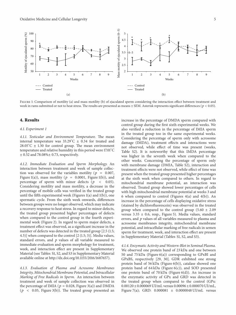
Oxidative Medicine and Cellular Longevity 5
1 2 3 4 5 6 7 8 9Weeks
ControlTreated
50
60
70
80
90
100
Mot
ility
of e
jacu
lated
sper
m (%
)
∗
∗∗ ∗ ∗
(a)
1 2 3 4 5 6 7 8 9Weeks
ControlTreated
2
2.5
3
3.5
4
4.5
5∗
∗∗
∗
∗
0–5
Mas
s mot
iity
of ej
acul
ated
sper
m (0
–5)
(b)
Figure 1: Comparison of motility (a) and mass motility (b) of ejaculated sperm considering the interaction effect between treatment andweek in rams submitted or not to heat stress.The results are presented as means ± SEM. Asterisk represents significant differences (𝑝 < 0.05).
4. Results
4.1. Experiment 1
4.1.1. Testicular and Environment Temperature. The meaninternal temperature was 33.29∘C ± 0.34 for treated and28.05∘C ± 1.30 for control group. The mean environmenttemperature and relative humidity in this period were 17.01∘C± 0.52 and 78.08%± 0.73, respectively.
4.1.2. Immediate Evaluation and Sperm Morphology. Aninteraction between treatment and week of sample collec-tion was observed for the variables motility (𝑝 = 0.007,Figure 1(a)), mass motility (𝑝 = 0.0001, Figure 1(b)), andpercentage of sperm with minor defects (𝑝 < 0.05).Considering motility and mass motility, a decrease in thepercentage of mobile cells was verified in the treated groupuntil the fifth experimental week (Figures 1(a) and 1(b)), onespermatic cycle. From the sixth week onwards, differencesbetween groups were no longer observed, whichmay indicatea recovery response to heat stress. In regard to minor defects,the treated group presented higher percentages of defectswhen compared to the control group in the fourth experi-mental week (Figure 2). In regard to sperm major defects, atreatment effect was observed, as a significant increase in thenumber of defects was detected in the treated group [2.5 (1.5;4.5)] when compared to the control [2 (1.5; 3)]. Media values,standard errors, and 𝑝 values of all variable measured toimmediate evaluation and sperm morphology for treatment,week, and interaction effect are present in SupplementaryMaterial (see Tables S1, S2, and S3 in SupplementaryMaterialavailable online at http://dx.doi.org/10.1155/2016/1687657).
4.1.3. Evaluation of Plasma and Acrosome MembranesIntegrity,MitochondrialMembrane Potential, and IntracellularMarking of Free Radicals in Sperm. An interaction betweentreatment and week of sample collection was observed inthe percentage of IMIA (𝑝 = 0.028, Figure 3(a)) and DMDA(𝑝 < 0.05, Figure 3(b)). The treated group presented an
increase in the percentage of DMDA sperm compared withcontrol group during the first sixth experimental weeks. Wealso verified a reduction in the percentage of IMIA spermin the treated group too in the same experimental weeks.Considering the percentage of sperm only with acrosomedamage (IMDA), treatment effects and interactions werenot observed, while effect of time was present (weeks,Table S2). It is noteworthy that this IMDA percentagewas higher in the seventh week when compared to theother weeks. Concerning the percentage of sperm onlywith membrane damage (DMIA, Table S2), interaction andtreatment effects were not observed, while effect of time waspresent when the treated group presented higher percentagesat the sixth week when compared to others. In regard tomitochondrial membrane potential, an interaction wasobserved. Treated group showed lower percentages of cellswith high mitochondrial membrane potential at weeks 3 and6 when compared to control (Figures 4(a) and 4(b)). Anincrease in the percentage of cells displaying oxidative stress(stained by dichlorofluorescein) was observed in the treatedgroup when compared to the control group (5.60 ± 2.09versus 3.35 ± 0.6, resp., Figure 5). Media values, standarderrors, and 𝑝 values of all variables measured to plasma andacrosome membranes integrity, mitochondrial membranepotential, and intracellular marking of free radicals in semensperm for treatment, week, and interaction effect are presentin Supplementary Material (Tables S1, S2, and S3).
4.1.4. Enzymatic Activity andWestern-Blot in Seminal Plasma.We observed one protein band of 23 kDa and one between50 and 75 kDa (Figure 6(a)) corresponding to GPxBI andGPxBS, respectively [29, 30]. GDR exhibited one strongprotein band of 56 kDa (Figure 6(b)), catalase showed oneprotein band of 64 kDa (Figure 6(c)), and SOD presentedone protein band of 70 kDa (Figure 6(d)). An increase inthe enzymatic activity of GPx and GRD was detected inthe treated group when compared to the control (GPx:0.00120±0.000069UI/mL versus 0.00096±0.000070UI/mL,Figure 7(a); GRD: 0.000081 ± 0.0000048UI/mL versus
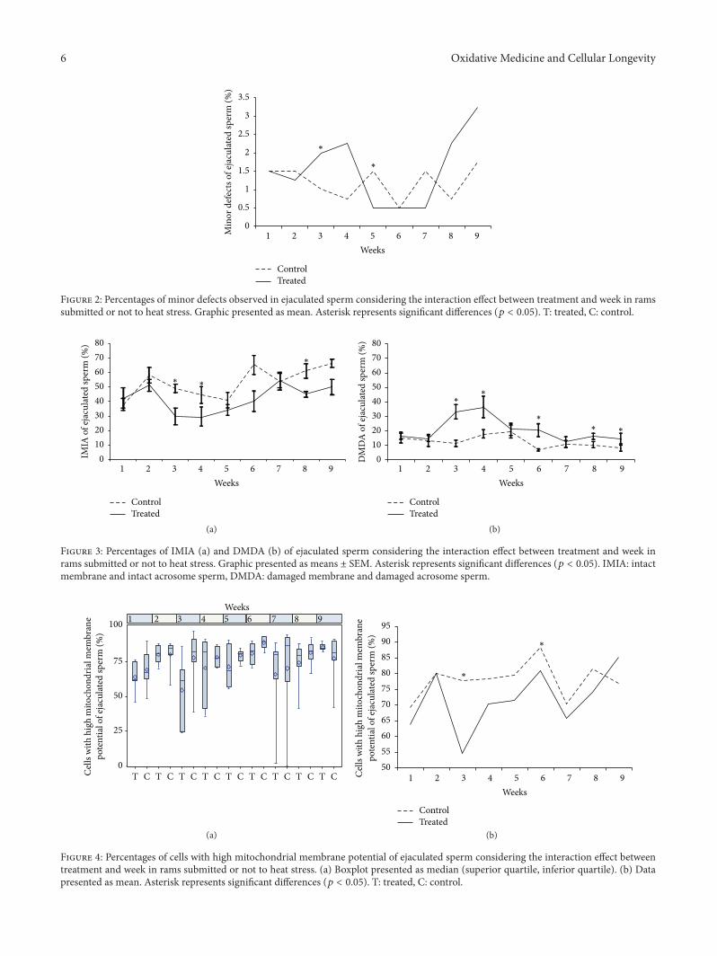
6 Oxidative Medicine and Cellular Longevity
∗
∗
0
0.5
1
1.5
2
2.5
3
3.5
Min
or d
efec
ts of
ejac
ulat
ed sp
erm
(%)
1 2 3 4 5 6 7 8 9Weeks
ControlTreated
Figure 2: Percentages of minor defects observed in ejaculated sperm considering the interaction effect between treatment and week in ramssubmitted or not to heat stress. Graphic presented as mean. Asterisk represents significant differences (𝑝 < 0.05). T: treated, C: control.
01020304050607080
IMIA
of e
jacu
lated
sper
m (%
)
1 2 3 4 5 6 7 8 9Weeks
ControlTreated
∗ ∗
∗
(a)
01020304050607080
DM
DA
of e
jacu
lated
sper
m (%
)
1 2 3 4 5 6 7 8 9Weeks
ControlTreated
∗∗
∗
∗∗
(b)
Figure 3: Percentages of IMIA (a) and DMDA (b) of ejaculated sperm considering the interaction effect between treatment and week inrams submitted or not to heat stress. Graphic presented as means ± SEM. Asterisk represents significant differences (𝑝 < 0.05). IMIA: intactmembrane and intact acrosome sperm, DMDA: damaged membrane and damaged acrosome sperm.
Weeks
100
75
50
25
0
Cell
s with
hig
h m
itoch
ondr
ial m
embr
ane
pote
ntia
l of e
jacu
lated
sper
m (%
)
T C T C T C T C T C T C T C T C T C
2 3 4 5 6 7 8 91
(a)
Cell
s with
hig
h m
itoch
ondr
ial m
embr
ane
50556065707580859095
pote
ntia
l of e
jacu
lated
sper
m (%
)
2 3 4 5 6 7 8 91Weeks
∗
∗
ControlTreated
(b)
Figure 4: Percentages of cells with high mitochondrial membrane potential of ejaculated sperm considering the interaction effect betweentreatment and week in rams submitted or not to heat stress. (a) Boxplot presented as median (superior quartile, inferior quartile). (b) Datapresented as mean. Asterisk represents significant differences (𝑝 < 0.05). T: treated, C: control.
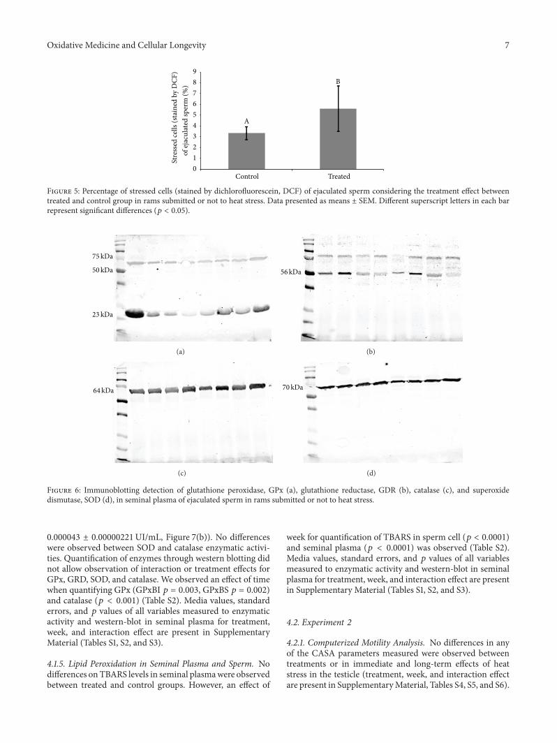
Oxidative Medicine and Cellular Longevity 7
Control Treated
A
B
Stre
ssed
cells
(sta
ined
by
DCF
)0123456789
of ej
acul
ated
sper
m (%
)Figure 5: Percentage of stressed cells (stained by dichlorofluorescein, DCF) of ejaculated sperm considering the treatment effect betweentreated and control group in rams submitted or not to heat stress. Data presented as means ± SEM. Different superscript letters in each barrepresent significant differences (𝑝 < 0.05).
75kDa
23kDa
50kDa
(a)
56kDa
(b)
64kDa
(c)
70kDa
(d)
Figure 6: Immunoblotting detection of glutathione peroxidase, GPx (a), glutathione reductase, GDR (b), catalase (c), and superoxidedismutase, SOD (d), in seminal plasma of ejaculated sperm in rams submitted or not to heat stress.
0.000043 ± 0.00000221UI/mL, Figure 7(b)). No differenceswere observed between SOD and catalase enzymatic activi-ties. Quantification of enzymes through western blotting didnot allow observation of interaction or treatment effects forGPx, GRD, SOD, and catalase. We observed an effect of timewhen quantifying GPx (GPxBI 𝑝 = 0.003, GPxBS 𝑝 = 0.002)and catalase (𝑝 < 0.001) (Table S2). Media values, standarderrors, and 𝑝 values of all variables measured to enzymaticactivity and western-blot in seminal plasma for treatment,week, and interaction effect are present in SupplementaryMaterial (Tables S1, S2, and S3).
4.1.5. Lipid Peroxidation in Seminal Plasma and Sperm. Nodifferences onTBARS levels in seminal plasmawere observedbetween treated and control groups. However, an effect of
week for quantification of TBARS in sperm cell (𝑝 < 0.0001)and seminal plasma (𝑝 < 0.0001) was observed (Table S2).Media values, standard errors, and 𝑝 values of all variablesmeasured to enzymatic activity and western-blot in seminalplasma for treatment, week, and interaction effect are presentin Supplementary Material (Tables S1, S2, and S3).
4.2. Experiment 2
4.2.1. Computerized Motility Analysis. No differences in anyof the CASA parameters measured were observed betweentreatments or in immediate and long-term effects of heatstress in the testicle (treatment, week, and interaction effectare present in SupplementaryMaterial, Tables S4, S5, and S6).
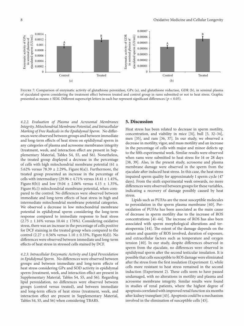
8 Oxidative Medicine and Cellular Longevity
Control Treated
A
B
Enzy
mat
ic ac
tivity
of G
Px
in se
min
al p
lasm
a of
00.00020.00040.00060.0008
0.0010.00120.0014
ejac
ulat
ed sp
erm
(UI/m
L)
(a)Control Treated
A
B
Enzy
mat
ic ac
tivity
of G
DR
in se
min
al p
lasm
a of
0
0.00001
0.00002
0.00003
0.00004
0.00005
0.00006
0.00007
ejac
ulat
ed sp
erm
(UI/m
L)
(b)
Figure 7: Comparison of enzymatic activity of glutathione peroxidase, GPx (a), and glutathione reductase, GDR (b), in seminal plasmaof ejaculated sperm considering the treatment effect between treated and control group in rams submitted or not to heat stress. Graphicpresented as means ± SEM. Different superscript letters in each bar represent significant differences (𝑝 < 0.05).
4.2.2. Evaluation of Plasma and Acrosomal MembranesIntegrity,MitochondrialMembrane Potential, and IntracellularMarking of Free Radicals in the Epididymal Sperm. No differ-ences were observed between groups and between immediateand long-term effects of heat stress on epididymal sperm inany categories of plasma and acrosome membranes integrity(treatment, week, and interaction effect are present in Sup-plementary Material, Tables S4, S5, and S6). Nonetheless,the treated group displayed a decrease in the percentageof cells with high mitochondrial membrane potential (61 ±5.62% versus 78.39 ± 2.29%, Figure 8(a)). Furthermore, thetreated group presented an increase in the percentage ofcells with intermediary (29.96 ± 4.71% versus 18.41 ± 1.45%,Figure 8(b)) and low (9.04 ± 2.06% versus 4.15 ± 1.35%,Figure 8(c)) mitochondrial membrane potential, when com-pared to the control. No differences were observed betweenimmediate and long-term effects of heat stress in high andintermediate mitochondrial membrane potential categories.We observed a decrease in low mitochondrial membranepotential in epididymal sperm considering the long-termresponse compared to immediate response to heat stress(2.75 ± 1.16% versus 10.44 ± 176%). Considering oxidativestress, there was an increase in the percentage of cells positivefor DCF staining in the treated group when compared to thecontrol (2.27 ± 0.56% versus 1.10 ± 0.33%, Figure 8(d)). Nodifferences were observed between immediate and long-termeffects of heat stress in stressed cells stained by DCF.
4.2.3. Intracellular Enzymatic Activity and Lipid Peroxidationin Epididymal Sperm. No differences were observed betweengroups and between immediate and long-term effects ofheat stress considering GPx and SOD activity in epididymalsperm (treatment, week, and interaction effect are present inSupplementary Material, Tables S4, S5, and S6). Regardinglipid peroxidation, no differences were observed betweengroups (control versus treated), and between immediateand long-term effects of heat stress (treatment, week, andinteraction effect are present in Supplementary Material,Tables S4, S5, and S6) when considering TBARS.
5. Discussion
Heat stress has been related to decrease in sperm motility,concentration, and viability in mice [31], bull [5, 32–34],men [35], and ram [36, 37]. In our study, we observed adecrease in motility, vigor, and mass motility and an increasein the percentage of cells with major and minor defects upto the fifth experimental week. Similar results were observedwhen rams were submitted to heat stress for 14 or 28 days[38, 39]. Also, in the present study, acrosome and plasmamembrane damage were observed in the sperm from theejaculate after induced heat stress. In this case, the heat stressimpaired sperm quality for approximately 1 sperm cycle (47days). From the sixth experimental week onwards, no moredifferences were observed between groups for these variables,indicating a recovery of damage possibly caused by heatstress.
Lipids such as PUFAs are the most susceptible moleculesto peroxidation in the sperm plasma membrane [40]. Per-oxidation of PUFAs has been associated as the main causeof decrease in sperm motility due to the increase of ROSconcentrations [41–44]. The increase of ROS has also beenassociated with sperm morphological alterations and ter-atospermia [44]. The extent of the damage depends on thenature and quantity of ROS involved, duration of exposure,and extracellular factors such as temperature and oxygentension [40]. In our study, despite differences observed insperm from the ejaculate, no differences were observed inepididymal sperm after the second testicular insulation. It ispossible that cells susceptible to ROS damagewere eliminatedafter the stress from the first insulation (Experiment 1), whilecells more resistant to heat stress remained in the secondinduction (Experiment 2). These cells seem to have passedundamaged, with no alterations in motility and plasma andacrosome membrane integrity. Similar results were foundin studies of renal patients, where the highest degree ofapoptosis correlatedwith improved renal function sixmonthsafter kidney transplant [45]. Apoptosis could be amechanisminvolved in the elimination of susceptible cells [45].
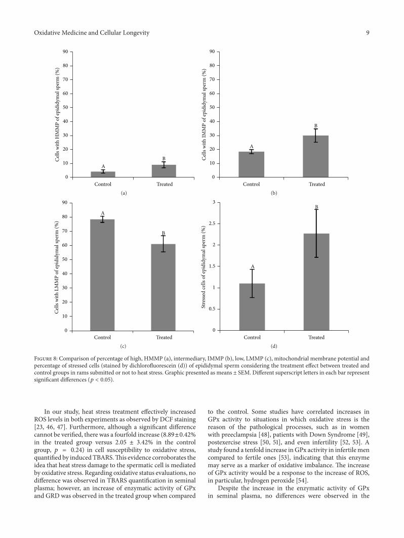
Oxidative Medicine and Cellular Longevity 9
Control Treated
AB
0
10
20
30
40
50
60
70
80
90C
ells w
ith H
MM
P of
epid
idym
al sp
erm
(%)
(a)Control Treated
A
B
0
10
20
30
40
50
60
70
80
90
Cell
s with
IMM
P of
epid
idym
al sp
erm
(%)
(b)
Control Treated
A
B
0
10
20
30
40
50
60
70
80
90
Cell
s with
LM
MP
of ep
idid
ymal
sper
m (%
)
(c)Control Treated
A
B
0
0.5
1
1.5
2
2.5
3St
ress
ed ce
lls o
f epi
didy
mal
sper
m (%
)
(d)
Figure 8: Comparison of percentage of high, HMMP (a), intermediary, IMMP (b), low, LMMP (c), mitochondrial membrane potential andpercentage of stressed cells (stained by dichlorofluorescein (d)) of epididymal sperm considering the treatment effect between treated andcontrol groups in rams submitted or not to heat stress. Graphic presented as means ± SEM. Different superscript letters in each bar representsignificant differences (𝑝 < 0.05).
In our study, heat stress treatment effectively increasedROS levels in both experiments as observed by DCF staining[23, 46, 47]. Furthermore, although a significant differencecannot be verified, there was a fourfold increase (8.89±0.42%in the treated group versus 2.05 ± 3.42% in the controlgroup, 𝑝 = 0.24) in cell susceptibility to oxidative stress,quantified by inducedTBARS.This evidence corroborates theidea that heat stress damage to the spermatic cell is mediatedby oxidative stress. Regarding oxidative status evaluations, nodifference was observed in TBARS quantification in seminalplasma; however, an increase of enzymatic activity of GPxand GRD was observed in the treated group when compared
to the control. Some studies have correlated increases inGPx activity to situations in which oxidative stress is thereason of the pathological processes, such as in womenwith preeclampsia [48], patients with Down Syndrome [49],postexercise stress [50, 51], and even infertility [52, 53]. Astudy found a tenfold increase inGPx activity in infertilemencompared to fertile ones [53], indicating that this enzymemay serve as a marker of oxidative imbalance. The increaseof GPx activity would be a response to the increase of ROS,in particular, hydrogen peroxide [54].
Despite the increase in the enzymatic activity of GPxin seminal plasma, no differences were observed in the
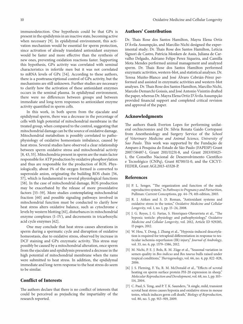
10 Oxidative Medicine and Cellular Longevity
immunodetection. One hypothesis could be that GPx ispresent in the epididymis in an inactive state, becoming activewhen necessary [9]. In epididymal environment, this acti-vation mechanism would be essential for sperm protection,since activation of already translated antioxidant enzymeswould be faster and more effective than the synthesis ofnew ones, preventing oxidation reactions faster. Supportingthis hypothesis, GPx activity was correlated with seminalcharacteristics in infertile men but it was not correlatedto mRNA levels of GPx [54]. According to these authors,there is a posttranscriptional control of GPx activity, but themechanisms are still unknown. Further studies are necessaryto clarify how the activation of these antioxidant enzymesoccurs in the seminal plasma. In epididymal environment,there were no differences between groups and betweenimmediate and long-term responses to antioxidant enzymeactivity quantified in sperm cells.
In this work, in both sperm from the ejaculate andepididymal sperm, there was a decrease in the percentage ofcells with high potential of mitochondrial membrane in thetreated group, when compared to the control, suggesting thatmitochondrial damage can be the source of oxidative damage.Mitochondrial metabolism is possibly correlated to patho-physiology of oxidative homeostasis imbalance, caused byheat stress. Several studies have observed a clear relationshipbetween sperm oxidative stress and mitochondrial activity[8, 43, 55].Mitochondria present in sperm are themain factorresponsible forATPproduction by oxidative phosphorylationand thus are responsible for the production of ROS. Phys-iologically, about 1% of the oxygen formed is converted tosuperoxide anion, originating the building ROS chain [56,57], which is fundamental to several physiological functions[58]. In the case of mitochondrial damage, ROS productionmay be exacerbated by the release of more prooxidativefactors [55–59]. More studies contemplating mitochondrialfraction [60] and possible signaling pathways involved inmitochondrial function must be conducted to clarify howheat stress alters oxidative balance, such as cytochrome clevels by western blotting [61], disturbances in mitochondrialenzyme complexes (I–IV), and decrements in tricarboxylicacid cycle enzymes [62].
One may conclude that heat stress causes alterations insperm during a spermatic cycle and disruption of oxidativehomeostasis, due to oxidative stress, observed by increase inDCF staining and GPx enzymatic activity. This stress maypossibly be caused by a mitochondrial alteration, once spermfrom the ejaculate and epididymis presented a decrease in thehigh potential of mitochondrial membrane when the ramswere submitted to heat stress. In addition, the epididymalimmediate and long-term response to the heat stress do seemto be similar.
Conflict of Interests
The authors declare that there is no conflict of interests thatcould be perceived as prejudicing the impartiality of theresearch reported.
Authors’ Contribution
Dr. Thais Rose dos Santos Hamilton, Mayra Elena OrtizD’Ávila Assumpção, and Marćılio Nichi designed the exper-imental study; Dr. Thais Rose dos Santos Hamilton, Let́ıciaSignori de Castro, Patŕıcia Monken de Assis, Juliana de Car-valho Delgado, Adriano Felipe Perez Siqueira, and CamillaMota Mendes performed animal management and analyzedsperm. Dr. Thais Rose dos Santos Hamilton performedenzymatic activities, western-blot, and statistical analyses. Dr.Teresa Muiño-Blanco and José Álvaro Cebrián-Pérez per-formed and assisted in enzymatic activities and western-blotanalyses. Dr.Thais Rose dos SantosHamilton,MarćılioNichi,Marcelo Demarchi Goissis, and José Antonio Visintin draftedthe paper, whereasDr.Mayra ElenaOrtizD’Ávila Assumpçãoprovided financial support and completed critical revisionand approval of the paper.
Acknowledgments
The authors thank Everton Lopes for performing unilat-eral orchiectomies and Dr. Silvia Renata Gaido Cortopassifrom Anesthesiology and Surgery Service of the Schoolof Veterinary Medicine and Animal Science, University ofSao Paulo. This work was supported by the Fundação deAmparo à Pesquisa do Estado de São Paulo (FAPESP) Grant2007/58487-1, Grant 2011/11231-8, and Grant 2013/05025-1, the Conselho Nacional de Desenvolvimento Cient́ıficoe Tecnológico (CNPq), Grant 81700/11-0, and the CICYT-FEDER, Grant AGL2013-43328-P.
References
[1] P. L. Senger, “The organization and function of the malereproductive system,” in Pathways to Pregnancy and Parturition,Pullman: Current Conceptions, pp. 44–79, 4th edition, 2003.
[2] R. J. Aitken and S. D. Roman, “Antioxidant systems andoxidative stress in the testes,” Oxidative Medicine and CellularLongevity, vol. 1, no. 1, pp. 15–24, 2008.
[3] J. G. Reyes, J. G. Farias, S. Henŕıquez-Olavarrieta et al., “Thehypoxic testicle: physiology and pathophysiology,” OxidativeMedicine and Cellular Longevity, vol. 2012, Article ID 929285,15 pages, 2012.
[4] W. Hou, Y. Dong, J. Zhang et al., “Hypoxia-induced deacetyla-tion is required for tetraploid differentiation in response to tes-ticular ischemia-reperfusion (IR) injury,” Journal of Andrology,vol. 33, no. 6, pp. 1379–1386, 2012.
[5] M. Nichi, P. E. J. Bols, R. M. Züge et al., “Seasonal variation insemen quality in Bos indicus and Bos taurus bulls raised undertropical conditions,”Theriogenology, vol. 66, no. 4, pp. 822–828,2006.
[6] J. S. Fleming, F. Yu, R. M. McDonald et al., “Effects of scrotalheating on sperm surface protein PH-20 expression in sheep,”Molecular Reproduction andDevelopment, vol. 68, no. 1, pp. 103–114, 2004.
[7] C. Paul, S. Teng, and P. T. K. Saunders, “A single, mild, transientscrotal heat stress causes hypoxia and oxidative stress in mousetestes, which induces germ cell death,” Biology of Reproduction,vol. 80, no. 5, pp. 913–919, 2009.
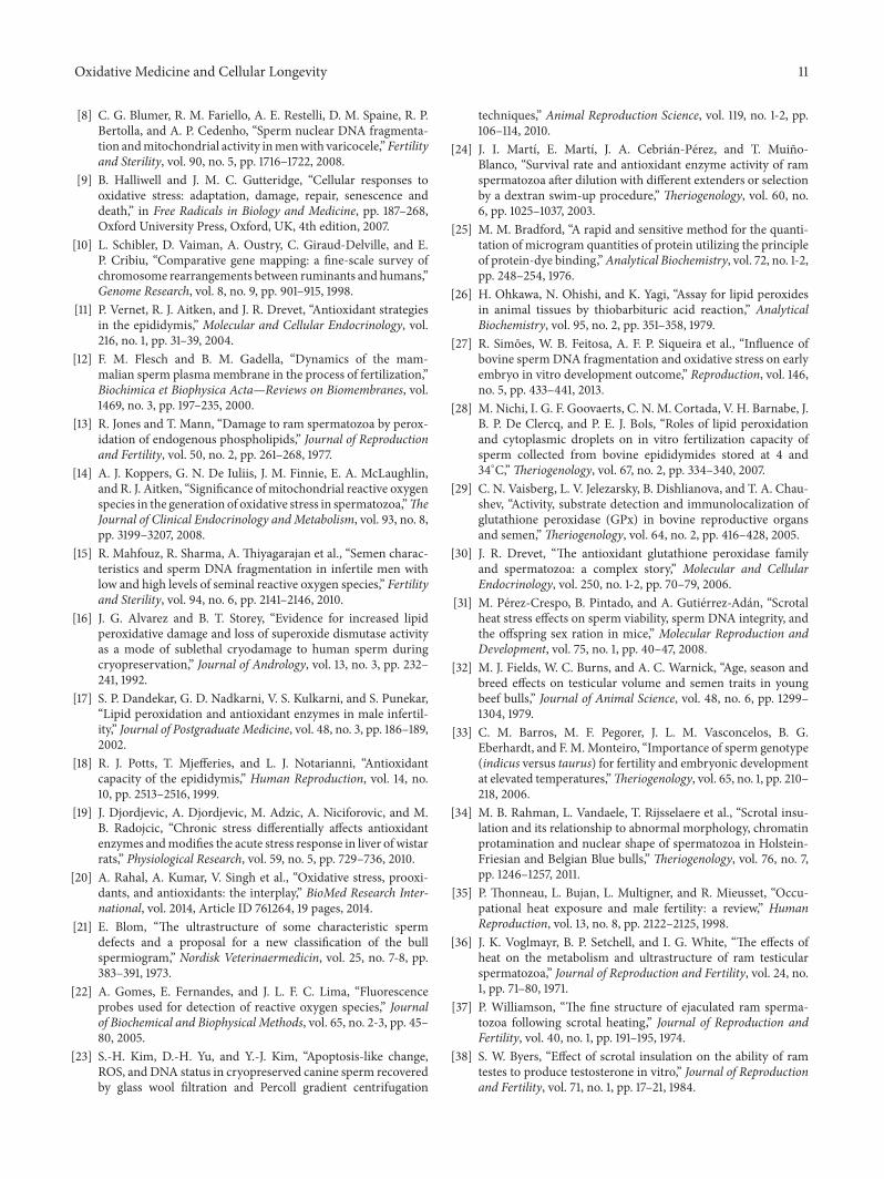
Oxidative Medicine and Cellular Longevity 11
[8] C. G. Blumer, R. M. Fariello, A. E. Restelli, D. M. Spaine, R. P.Bertolla, and A. P. Cedenho, “Sperm nuclear DNA fragmenta-tion andmitochondrial activity inmenwith varicocele,”Fertilityand Sterility, vol. 90, no. 5, pp. 1716–1722, 2008.
[9] B. Halliwell and J. M. C. Gutteridge, “Cellular responses tooxidative stress: adaptation, damage, repair, senescence anddeath,” in Free Radicals in Biology and Medicine, pp. 187–268,Oxford University Press, Oxford, UK, 4th edition, 2007.
[10] L. Schibler, D. Vaiman, A. Oustry, C. Giraud-Delville, and E.P. Cribiu, “Comparative gene mapping: a fine-scale survey ofchromosome rearrangements between ruminants and humans,”Genome Research, vol. 8, no. 9, pp. 901–915, 1998.
[11] P. Vernet, R. J. Aitken, and J. R. Drevet, “Antioxidant strategiesin the epididymis,” Molecular and Cellular Endocrinology, vol.216, no. 1, pp. 31–39, 2004.
[12] F. M. Flesch and B. M. Gadella, “Dynamics of the mam-malian sperm plasmamembrane in the process of fertilization,”Biochimica et Biophysica Acta—Reviews on Biomembranes, vol.1469, no. 3, pp. 197–235, 2000.
[13] R. Jones and T. Mann, “Damage to ram spermatozoa by perox-idation of endogenous phospholipids,” Journal of Reproductionand Fertility, vol. 50, no. 2, pp. 261–268, 1977.
[14] A. J. Koppers, G. N. De Iuliis, J. M. Finnie, E. A. McLaughlin,and R. J. Aitken, “Significance of mitochondrial reactive oxygenspecies in the generation of oxidative stress in spermatozoa,”TheJournal of Clinical Endocrinology and Metabolism, vol. 93, no. 8,pp. 3199–3207, 2008.
[15] R. Mahfouz, R. Sharma, A. Thiyagarajan et al., “Semen charac-teristics and sperm DNA fragmentation in infertile men withlow and high levels of seminal reactive oxygen species,” Fertilityand Sterility, vol. 94, no. 6, pp. 2141–2146, 2010.
[16] J. G. Alvarez and B. T. Storey, “Evidence for increased lipidperoxidative damage and loss of superoxide dismutase activityas a mode of sublethal cryodamage to human sperm duringcryopreservation,” Journal of Andrology, vol. 13, no. 3, pp. 232–241, 1992.
[17] S. P. Dandekar, G. D. Nadkarni, V. S. Kulkarni, and S. Punekar,“Lipid peroxidation and antioxidant enzymes in male infertil-ity,” Journal of Postgraduate Medicine, vol. 48, no. 3, pp. 186–189,2002.
[18] R. J. Potts, T. Mjefferies, and L. J. Notarianni, “Antioxidantcapacity of the epididymis,” Human Reproduction, vol. 14, no.10, pp. 2513–2516, 1999.
[19] J. Djordjevic, A. Djordjevic, M. Adzic, A. Niciforovic, and M.B. Radojcic, “Chronic stress differentially affects antioxidantenzymes andmodifies the acute stress response in liver of wistarrats,” Physiological Research, vol. 59, no. 5, pp. 729–736, 2010.
[20] A. Rahal, A. Kumar, V. Singh et al., “Oxidative stress, prooxi-dants, and antioxidants: the interplay,” BioMed Research Inter-national, vol. 2014, Article ID 761264, 19 pages, 2014.
[21] E. Blom, “The ultrastructure of some characteristic spermdefects and a proposal for a new classification of the bullspermiogram,” Nordisk Veterinaermedicin, vol. 25, no. 7-8, pp.383–391, 1973.
[22] A. Gomes, E. Fernandes, and J. L. F. C. Lima, “Fluorescenceprobes used for detection of reactive oxygen species,” Journalof Biochemical and Biophysical Methods, vol. 65, no. 2-3, pp. 45–80, 2005.
[23] S.-H. Kim, D.-H. Yu, and Y.-J. Kim, “Apoptosis-like change,ROS, andDNA status in cryopreserved canine sperm recoveredby glass wool filtration and Percoll gradient centrifugation
techniques,” Animal Reproduction Science, vol. 119, no. 1-2, pp.106–114, 2010.
[24] J. I. Mart́ı, E. Mart́ı, J. A. Cebrián-Pérez, and T. Muiño-Blanco, “Survival rate and antioxidant enzyme activity of ramspermatozoa after dilution with different extenders or selectionby a dextran swim-up procedure,” Theriogenology, vol. 60, no.6, pp. 1025–1037, 2003.
[25] M. M. Bradford, “A rapid and sensitive method for the quanti-tation of microgram quantities of protein utilizing the principleof protein-dye binding,”Analytical Biochemistry, vol. 72, no. 1-2,pp. 248–254, 1976.
[26] H. Ohkawa, N. Ohishi, and K. Yagi, “Assay for lipid peroxidesin animal tissues by thiobarbituric acid reaction,” AnalyticalBiochemistry, vol. 95, no. 2, pp. 351–358, 1979.
[27] R. Simões, W. B. Feitosa, A. F. P. Siqueira et al., “Influence ofbovine sperm DNA fragmentation and oxidative stress on earlyembryo in vitro development outcome,” Reproduction, vol. 146,no. 5, pp. 433–441, 2013.
[28] M. Nichi, I. G. F. Goovaerts, C. N. M. Cortada, V. H. Barnabe, J.B. P. De Clercq, and P. E. J. Bols, “Roles of lipid peroxidationand cytoplasmic droplets on in vitro fertilization capacity ofsperm collected from bovine epididymides stored at 4 and34∘C,”Theriogenology, vol. 67, no. 2, pp. 334–340, 2007.
[29] C. N. Vaisberg, L. V. Jelezarsky, B. Dishlianova, and T. A. Chau-shev, “Activity, substrate detection and immunolocalization ofglutathione peroxidase (GPx) in bovine reproductive organsand semen,”Theriogenology, vol. 64, no. 2, pp. 416–428, 2005.
[30] J. R. Drevet, “The antioxidant glutathione peroxidase familyand spermatozoa: a complex story,” Molecular and CellularEndocrinology, vol. 250, no. 1-2, pp. 70–79, 2006.
[31] M. Pérez-Crespo, B. Pintado, and A. Gutiérrez-Adán, “Scrotalheat stress effects on sperm viability, sperm DNA integrity, andthe offspring sex ration in mice,” Molecular Reproduction andDevelopment, vol. 75, no. 1, pp. 40–47, 2008.
[32] M. J. Fields, W. C. Burns, and A. C. Warnick, “Age, season andbreed effects on testicular volume and semen traits in youngbeef bulls,” Journal of Animal Science, vol. 48, no. 6, pp. 1299–1304, 1979.
[33] C. M. Barros, M. F. Pegorer, J. L. M. Vasconcelos, B. G.Eberhardt, and F. M.Monteiro, “Importance of sperm genotype(indicus versus taurus) for fertility and embryonic developmentat elevated temperatures,”Theriogenology, vol. 65, no. 1, pp. 210–218, 2006.
[34] M. B. Rahman, L. Vandaele, T. Rijsselaere et al., “Scrotal insu-lation and its relationship to abnormal morphology, chromatinprotamination and nuclear shape of spermatozoa in Holstein-Friesian and Belgian Blue bulls,” Theriogenology, vol. 76, no. 7,pp. 1246–1257, 2011.
[35] P. Thonneau, L. Bujan, L. Multigner, and R. Mieusset, “Occu-pational heat exposure and male fertility: a review,” HumanReproduction, vol. 13, no. 8, pp. 2122–2125, 1998.
[36] J. K. Voglmayr, B. P. Setchell, and I. G. White, “The effects ofheat on the metabolism and ultrastructure of ram testicularspermatozoa,” Journal of Reproduction and Fertility, vol. 24, no.1, pp. 71–80, 1971.
[37] P. Williamson, “The fine structure of ejaculated ram sperma-tozoa following scrotal heating,” Journal of Reproduction andFertility, vol. 40, no. 1, pp. 191–195, 1974.
[38] S. W. Byers, “Effect of scrotal insulation on the ability of ramtestes to produce testosterone in vitro,” Journal of Reproductionand Fertility, vol. 71, no. 1, pp. 17–21, 1984.
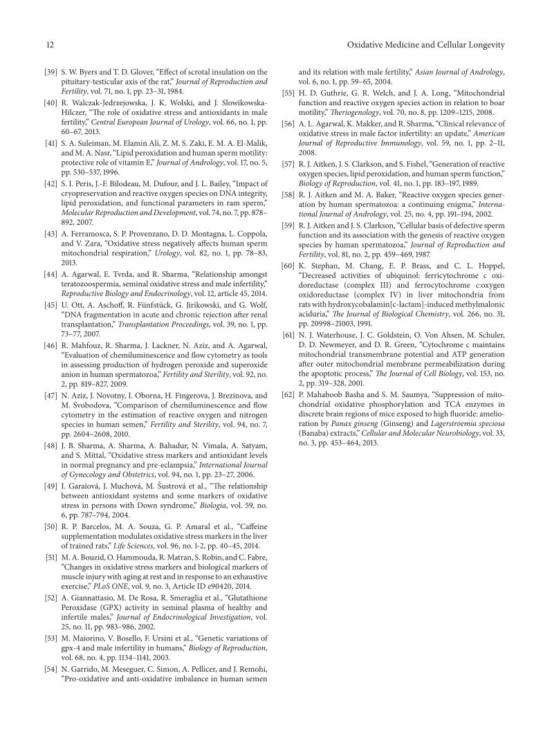
12 Oxidative Medicine and Cellular Longevity
[39] S. W. Byers and T. D. Glover, “Effect of scrotal insulation on thepituitary-testicular axis of the rat,” Journal of Reproduction andFertility, vol. 71, no. 1, pp. 23–31, 1984.
[40] R. Walczak-Jedrzejowska, J. K. Wolski, and J. Slowikowska-Hilczer, “The role of oxidative stress and antioxidants in malefertility,” Central European Journal of Urology, vol. 66, no. 1, pp.60–67, 2013.
[41] S. A. Suleiman, M. Elamin Ali, Z. M. S. Zaki, E. M. A. El-Malik,andM.A.Nasr, “Lipid peroxidation and human spermmotility:protective role of vitamin E,” Journal of Andrology, vol. 17, no. 5,pp. 530–537, 1996.
[42] S. I. Peris, J.-F. Bilodeau, M. Dufour, and J. L. Bailey, “Impact ofcryopreservation and reactive oxygen species onDNA integrity,lipid peroxidation, and functional parameters in ram sperm,”Molecular Reproduction andDevelopment, vol. 74, no. 7, pp. 878–892, 2007.
[43] A. Ferramosca, S. P. Provenzano, D. D. Montagna, L. Coppola,and V. Zara, “Oxidative stress negatively affects human spermmitochondrial respiration,” Urology, vol. 82, no. 1, pp. 78–83,2013.
[44] A. Agarwal, E. Tvrda, and R. Sharma, “Relationship amongstteratozoospermia, seminal oxidative stress and male infertility,”Reproductive Biology and Endocrinology, vol. 12, article 45, 2014.
[45] U. Ott, A. Aschoff, R. Fünfstück, G. Jirikowski, and G. Wolf,“DNA fragmentation in acute and chronic rejection after renaltransplantation,” Transplantation Proceedings, vol. 39, no. 1, pp.73–77, 2007.
[46] R. Mahfouz, R. Sharma, J. Lackner, N. Aziz, and A. Agarwal,“Evaluation of chemiluminescence and flow cytometry as toolsin assessing production of hydrogen peroxide and superoxideanion in human spermatozoa,” Fertility and Sterility, vol. 92, no.2, pp. 819–827, 2009.
[47] N. Aziz, J. Novotny, I. Oborna, H. Fingerova, J. Brezinova, andM. Svobodova, “Comparison of chemiluminescence and flowcytometry in the estimation of reactive oxygen and nitrogenspecies in human semen,” Fertility and Sterility, vol. 94, no. 7,pp. 2604–2608, 2010.
[48] J. B. Sharma, A. Sharma, A. Bahadur, N. Vimala, A. Satyam,and S. Mittal, “Oxidative stress markers and antioxidant levelsin normal pregnancy and pre-eclampsia,” International Journalof Gynecology and Obstetrics, vol. 94, no. 1, pp. 23–27, 2006.
[49] I. Garaiová, J. Muchová, M. Šustrová et al., “The relationshipbetween antioxidant systems and some markers of oxidativestress in persons with Down syndrome,” Biologia, vol. 59, no.6, pp. 787–794, 2004.
[50] R. P. Barcelos, M. A. Souza, G. P. Amaral et al., “Caffeinesupplementationmodulates oxidative stressmarkers in the liverof trained rats,” Life Sciences, vol. 96, no. 1-2, pp. 40–45, 2014.
[51] M.A. Bouzid,O.Hammouda, R.Matran, S. Robin, andC. Fabre,“Changes in oxidative stress markers and biological markers ofmuscle injurywith aging at rest and in response to an exhaustiveexercise,” PLoS ONE, vol. 9, no. 3, Article ID e90420, 2014.
[52] A. Giannattasio, M. De Rosa, R. Smeraglia et al., “GlutathionePeroxidase (GPX) activity in seminal plasma of healthy andinfertile males,” Journal of Endocrinological Investigation, vol.25, no. 11, pp. 983–986, 2002.
[53] M. Maiorino, V. Bosello, F. Ursini et al., “Genetic variations ofgpx-4 and male infertility in humans,” Biology of Reproduction,vol. 68, no. 4, pp. 1134–1141, 2003.
[54] N. Garrido, M. Meseguer, C. Simon, A. Pellicer, and J. Remohi,“Pro-oxidative and anti-oxidative imbalance in human semen
and its relation with male fertility,” Asian Journal of Andrology,vol. 6, no. 1, pp. 59–65, 2004.
[55] H. D. Guthrie, G. R. Welch, and J. A. Long, “Mitochondrialfunction and reactive oxygen species action in relation to boarmotility,”Theriogenology, vol. 70, no. 8, pp. 1209–1215, 2008.
[56] A. L. Agarwal, K. Makker, and R. Sharma, “Clinical relevance ofoxidative stress in male factor infertility: an update,” AmericanJournal of Reproductive Immunology, vol. 59, no. 1, pp. 2–11,2008.
[57] R. J. Aitken, J. S. Clarkson, and S. Fishel, “Generation of reactiveoxygen species, lipid peroxidation, and human sperm function,”Biology of Reproduction, vol. 41, no. 1, pp. 183–197, 1989.
[58] R. J. Aitken and M. A. Baker, “Reactive oxygen species gener-ation by human spermatozoa: a continuing enigma,” Interna-tional Journal of Andrology, vol. 25, no. 4, pp. 191–194, 2002.
[59] R. J. Aitken and J. S. Clarkson, “Cellular basis of defective spermfunction and its association with the genesis of reactive oxygenspecies by human spermatozoa,” Journal of Reproduction andFertility, vol. 81, no. 2, pp. 459–469, 1987.
[60] K. Stephan, M. Chang, E. P. Brass, and C. L. Hoppel,“Decreased activities of ubiquinol: ferricytochrome c oxi-doreductase (complex III) and ferrocytochrome c:oxygenoxidoreductase (complex IV) in liver mitochondria fromratswith hydroxycobalamin[c-lactam]-inducedmethylmalonicaciduria,” The Journal of Biological Chemistry, vol. 266, no. 31,pp. 20998–21003, 1991.
[61] N. J. Waterhouse, J. C. Goldstein, O. Von Ahsen, M. Schuler,D. D. Newmeyer, and D. R. Green, “Cytochrome c maintainsmitochondrial transmembrane potential and ATP generationafter outer mitochondrial membrane permeabilization duringthe apoptotic process,” The Journal of Cell Biology, vol. 153, no.2, pp. 319–328, 2001.
[62] P. Mahaboob Basha and S. M. Saumya, “Suppression of mito-chondrial oxidative phosphorylation and TCA enzymes indiscrete brain regions of mice exposed to high fluoride: amelio-ration by Panax ginseng (Ginseng) and Lagerstroemia speciosa(Banaba) extracts,”Cellular andMolecular Neurobiology, vol. 33,no. 3, pp. 453–464, 2013.
Review ArticleLipids and Oxidative Stress Associated with Ethanol-InducedNeurological Damage
José A. Hernández,1 Rosa C. López-Sánchez,1 and Adela Rendón-Ramírez2
1Tecnologico de Monterrey, Escuela Nacional de Medicina, 64710 Monterrey, NL, Mexico2Unidad de Biof́ısica (CSIC, UPV/EHU) and Departamento de Bioquı́mica, Universidad del Paı́s Vasco, Apartado 644,48080 Bilbao, Spain
Correspondence should be addressed to Adela Rendón-Ramı́rez; [email protected]
Received 26 September 2015; Revised 10 December 2015; Accepted 13 December 2015
Academic Editor: Tanea T. Reed
Copyright © 2016 José A. Hernández et al. This is an open access article distributed under the Creative Commons AttributionLicense, which permits unrestricted use, distribution, and reproduction in any medium, provided the original work is properlycited.
The excessive intake of alcohol is a serious public health problem, especially given the severe damage provoked by chronic or prenatalexposure to alcohol that affects many physiological processes, such asmemory, motor function, and cognitive abilities.This damageis related to the ethanol oxidation in the brain. The metabolism of ethanol to acetaldehyde and then to acetate is associated withthe production of reactive oxygen species that accentuate the oxidative state of cells. This metabolism of ethanol can induce theoxidation of the fatty acids in phospholipids, and the bioactive aldehydes produced are known to be associated with neurotoxicityand neurodegeneration. As such, here we will review the role of lipids in the neuronal damage induced by ethanol-related oxidativestress and the role that lipids play in the related compensatory or defense mechanisms.
1. Introduction
A clear relationship has been established between ethanolintake, addiction and dependency [1–3], and several riskfactors for chronic disease and injury [4]. Indeed, the publichealth problem associated with increased alcohol consump-tion and alcoholism [5, 6] is becoming ever more severedue to the increased economic burden of the complicationson the health national systems and the cost of the relevanttreatments [4, 7–10]. Alcoholism provokes high rates ofmortality and it increases in risk of several disabling disorders[4, 11]. Such damage can be classified in function of the organsinvolved (liver, kidney, heart, brain, etc.), the type of intake(acute or chronic), or the subject’s age at the time of exposureto ethanol (prenatal, neonatal, or adult). In summary, ethanolhas several negative health effects, especially if we considerprenatal exposure where the brain is a major target for thedamage provoked.
2. Effects of Ethanol in the Brain
Ethanol has many effects in the brain depending on theage of exposure (prenatal, postnatal, or adult). For example,
aggressive behavior and depression are observed after acutepostnatal exposure to ethanol, possibly due to a decrease incirculating tryptophan, followedby the depletion of serotoninin the brain [12, 13]. Another effect of acute postnatalalcohol exposure is related to impaired impulsive and controlbehavior [14, 15], although few in vivo studies have focusedon this issue. Cognitive performance has been associatedwith specific prefrontal cortical regions in Rhesus Macaquemonkeys [11] and GABA receptors in this structure havebeen implicated in the effects of acute postnatal ethanolexposure [16–18]. Indeed, GABA was found to be a mediatorin ethanol-induced ataxia [18, 19].
The most severe alcohol-related damage is found follow-ing acute prenatal or chronic pre- and postnatal ethanol expo-sure, effects that have been associated with a loss of neurons(Table 1). In terms of prenatal exposure, the babies bornto women that drink alcohol excessively during pregnancymay suffer from fetal alcohol syndrome [20], a conditioncharacterized by specific craniofacial abnormalities, pre- andpostnatal growth deficiencies, and nervous system dysfunc-tion that is manifested as persistent intellectual, behavioral,and neurological defects [5, 21]. These latter symptoms have
Hindawi Publishing CorporationOxidative Medicine and Cellular LongevityVolume 2016, Article ID 1543809, 15 pageshttp://dx.doi.org/10.1155/2016/1543809
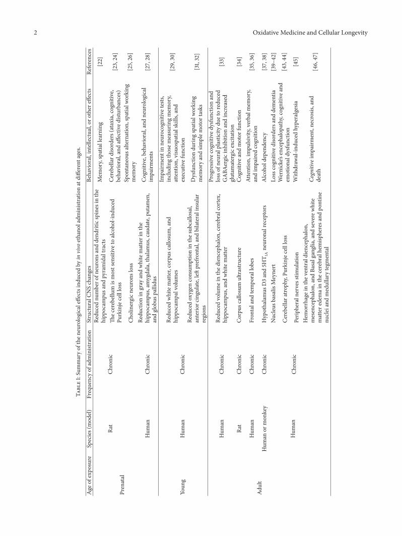
2 Oxidative Medicine and Cellular Longevity
Table1:Summaryof
then
eurologicaleffectsind
uced
byin
vivo
ethano
ladm
inistratio
natdifferent
ages.
Age
ofexpo
sure
Species(mod
el)
Frequencyof
administratio
nStructuralCN
Schanges
Behavioral,intellectual,or
othere
ffects
References
Prenatal
Rat
Chronic
Redu
cednu
mbero
fneurons
anddend
riticspines
inthe
hipp
ocam
pusa
ndpyramidaltracts
Mem
ory,spatiallearning
[22]
Thec
erebellum
ismostsensitivetoalcoho
l-ind
uced
Purkinjecellloss
Cerebellar
disorders(ataxia,cognitiv
e,behavioral,and
affectiv
edisturbances)
[23,24]
Cholinergicn
eurons
loss
Spon
taneou
salternation,
spatialw
orking
mem
ory
[25,26]
Hum
anCh
ronic
Redu
ctionin
gray
andwhitematterinthe
hipp
ocam
pus,am
ygdala,thalamus,caudate,putam
en,
andglob
uspallidu
s
Cognitiv
e,behavioral,and
neurological
impairm
ents
[27,28]
Youn
gHum
anCh
ronic
Redu
cedwhitematter,corpus
callo
sum,and
hipp
ocam
palvolum
es
Impairm
entinneurocognitiv
etests,
inclu
ding
thosem
easurin
gmem
ory,
attention,
visuospatia
lskills,and
executivefun
ction
[29,30]
Redu
cedoxygen
consum
ptionin
thes
ubcallo
sal,
anterio
rcingu
late,left
prefrontal,and
bilateralinsular
region
s
Dysfunctio
ndu
ringspatialw
orking
mem
oryandsim
plem
otor
tasks
[31,32]
Adult
Hum
anCh
ronic
Redu
cedvolumeinthed
iencephalon,
cerebralcortex,
hipp
ocam
pus,andwhitematter
Progressivec
ognitiv
edysfunctio
nand
lossof
neuralplastic
itydu
etoredu
ced
GABA
ergicinh
ibition
andincreased
glutam
atergice
xcitatio
n
[33]
Rat
Chronic
Corpu
scallosum
ultrastructure
Cognitiv
eand
motor
functio
n[34]
Hum
anCh
ronic
Fron
taland
tempo
rallob
esAttention,
impu
lsivity,verbalm
emory,
andim
paire
dcogn
ition
[35,36]
Hum
anor
mon
key
Chronic
Hypothalamus
D3and5H
T 1Aneuron
alreceptors
Alcoh
oldepend
ency
[37,38]
Hum
anCh
ronic
Nucleus
basalis
Meynert
Losscogn
itive
disordersa
nddementia
[39–
42]
Cerebellara
troph
y,Pu
rkinjecellloss
Wernicke’s
enceph
alop
athy,cognitiv
eand
emotionald
ysfunctio
n[43,44
]
Perip
heraln
ervesstim
ulation
With
draw
al-in
ducedhyperalgesia
[45]
Hem
orrhageinthev
entraldienceph
alon
,mesenceph
alon
,and
Basalganglia,and
severe
white
mattere
demainthec
erebralh
emisp
heresa
ndpo
ntine
nucle
iand
medullary
tegm
ental
Cognitive
impairm
ent,necrosis,
and
death
[46,47]
Oxidative Medicine and Cellular Longevity 3
been related to neurodegeneration in experimental animalmodels (see Table 1 for a summary of some of the availableliterature).
Chronic alcohol exposure has been associated with per-manent neuronal loss in brain regions like the hippocampusand cerebellum.Moreover, in vivo studies have demonstratedneurological effects following chronic ethanol exposure inyoung and adult populations, with deterioration in memory,motor function, cognition, and so forth. All these effectscould be due to neurotoxicity or neurodegeneration, andthere is evidence that oxidative stress associated with ethanolmetabolism is involved.
3. The Pharmacokinetics of Ethanol
The ethanol concentration that can be found in bloodfollowing its ingestion depends on its pharmacokinetics (PK).PK determines not only the time-course and persistenceof ethanol in blood but also the amount of alcohol andits metabolic products that accumulate in different tissues,and hence their pharmacological and toxicological responses[48].
3.1. Absorption. In adults, the ethanol ingested is almost com-pletely and instantly absorbed by passive diffusion, reachinga peak concentration in humans between 30 and 90min.Absorption is more efficient in the small intestine thanin stomach [49], a difference in absorption that is due totwo factors. First, the thickness of mucus that protects thestomach appears to have a resistance ∼16 times greater thanthat which protects the small intestine [49, 50], which also hasa greater intestinal absorption surface due to the presence villiandmicrovilli [51].The second difference reflects the speed ofstirring caused by peristalsis, which is more important in thesmall intestine than in the stomach, playing a role in gastricemptying and in the intestinal transit time [49].
In addition, the presence of food is another factor thatmodifies the absorption rate [52], mainly as food reducesgastric emptying and ethanol is absorbed more slowly [53].Solid food intake can reduce the ethanol absorption rate by30% and it has been suggested that this effect is due to theneed for food digestion prior to absorption process. As such,if food is taken in as a liquid then it would not producethis effect [49, 54]. Moreover, a small amount of ethanolcan be oxidized to acetaldehyde by alcohol dehydrogenase(ADH) classes I and IV [52, 55] in the stomach and intestine.This acetaldehyde can be absorbed along with ethanol andmetabolized by the liver or other tissues.
3.2. First Pass Metabolism and the Distribution of Ethanol.The amount of alcohol in any given tissue depends on itsrelative concentration in the blood, which is a function of firstpass metabolism [49], that is, the oxidation of ethanol in thestomach, intestine, and liver.
Most first pass metabolism occurs in the liver [49, 55]and the rate-limiting step is the oxidation of ethanol toacetaldehyde. This reaction is catalyzed by proteins of theADH family [56], of which class I (ADH1) and III (ADH3)enzymes metabolize ethanol in the liver [57, 58]. These two
types of enzymes differ in their Km, with ADH1 having a lowKm while ADH3 has a high Km value [57, 59]. ConsequentlyADH3 plays a more important role in the metabolism ofalcohol at high concentrations. In addition, microsomalethanol oxidizing system (MEOS) and catalase contribute tothe metabolism of alcohol in specific circumstances, such ashigh ethanol concentrations [48, 60].
The acetaldehyde produced by the oxidation of ethanol isthereafter transformed to acetate by aldehyde dehydrogenase(ALDH) [61], which can be further metabolized throughthe tricarboxylic acid cycle to generate energy, or thesemetabolites can be deposited in the plasma [62, 63]. Indeed,increases in acetate but not acetaldehyde can be detected inhuman plasma after ethanol intake [64, 65] (Figure 1).
The efficiency of ethanol metabolism is dependent onthe enzymatic activity and pathways involved. It has beenreported that ADH, cytochrome P450 (CYP), and ALDHshow genetic variations (ADH1B, ALDH2, CYP2E1∗6, andCYP2E1∗7B besides others) that affect enzymatic activityin the liver and alcohol metabolism [66–68]. As a result,ethanol’s pharmacokinetic and pharmacodynamic propertiesare affected by this genetic variation, as reflected in interracialand ethnic pharmacological differences [56, 66–71]. Conse-quently the risk of developing diseasesmay increase in certainpopulations, including that of hypertension [70], alcoholdependence, and several types of alcohol-related cancer [60,72–75].
After first pass metabolism, the remaining ethanol and itsmetabolites are distributed in different tissue, and the excessalcohol is excreted in the breath, urine, and sweat [56]. Thedistribution of ethanol throughout the body is driven in directproportion to water content of each tissue, especially at theethanol steady-state. Since ethanol is a small, polar molecule,the distribution volume of ethanol is dependent on the totalbody water of an individual (50 to 60% lean body weight)[76–78]. The variation in the distribution volume of ethanolhas been evaluated for women and men, and in both sexes,the distribution volume decreases as the body mass indexincreases [79].
Alcohol-driven physiological changes, such as vasculareffects (vasodilation) or changes in cardiac output, can alsomodify tissue blood flow and ethanol distribution [78]. Sincethe blood flow to the brain remains relatively constant,changes in the blood concentration of ethanol are the mostrelevant factor influencing the amount of ethanol deliveredto the brain and therefore for the different levels of brainintoxication [78–80].
The distribution of ethanol is also particularly rele-vant during pregnancy, as 1-2 hours after maternal alco-hol ingestion the fetal alcohol concentrations reach levelsthat are nearly equivalent to the maternal levels [81]. Theelimination of ethanol by the fetus is impaired due to itsreducedmetabolic capacity.Thus, fetal exposure is prolongedthrough the reuptake of amniotic-fluid containing ethanol[81]. Ultimately, the elimination of alcohol from the fetusrelies on the mother’s metabolic capacity, which inevitably isa process that occurs late, meaning that the fetus is exposedto the toxicological effects of alcohol [82]. Therefore, manyof the physical effects of ethanol on brain structure not only
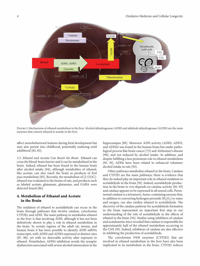
4 Oxidative Medicine and Cellular Longevity
EthanolAcetaldehydeADH1, ADH3
Acetate
Tricarboxylic acid cycle
NADHFADH2
ATP
Catalase
Peroxisomes
NADHNADH
Liver
H2OH2O2
Cytosolic
NAD+ NADH + H+
∗CYP2E1
NADP+ + H2ONADPH + H+ + 2O2
∗Mitochondrial
∗A
LDH
NAD+
NADH + H+
H2O
CO2
CO2
Figure 1: Mechanisms of ethanol metabolism in the liver. Alcohol dehydrogenase (ADH) and aldehyde dehydrogenase (ALDH) are the mainenzymes that convert ethanol to acetate in the liver.
affect neurobehavioral features during fetal development butmay also persist into childhood, potentially enduring untiladulthood [82, 83].
3.3. Ethanol and Acetate Can Reach the Brain. Ethanol cancross the blood-brain barrier and it can bemetabolized in thebrain. Indeed, ethanol has been found in the human brainafter alcohol intake [84], although metabolites of ethanol,like acetate, can also reach the brain as products of firstpass metabolism [85]. Recently, the metabolism of [2-(13)C]-ethanol was evaluated in the brains of rats, and products suchas labeled acetate, glutamate, glutamine, and GABA weredetected found [86].
4. Metabolism of Ethanol and Acetatein the Brain
The oxidation of ethanol to acetaldehyde can occur in thebrain through pathways that involve catalase, cytochromeCYP2E1, and ADH.Themain pathway to metabolize ethanolin the liver is that involving ADH, although it has not beendefinitively shown to play a role in ethanol metabolism inthe brain. In certain regions of the adult rat, mouse, andhuman brain it has been possible to identify ADH mRNAtranscripts, with ADH1 and ADH4 expressed at distinct sites[87, 88], yet with no detectable activity after exposure toethanol. Nonetheless, ADH4 inhibition avoids the synapticdysfunction associated with severe alcohol intoxication in the
hippocampus [89]. Moreover ADH activity (ADH1, ADH3,and ADH4) was found in the human brain but under patho-logical process like brain cancer [73] and Alzheimer’s disease[90], and not induced by alcohol intake. In addition, anddespite fulfilling a less prominent role in ethanol metabolism[85, 91], ADHs have been related to enhanced voluntaryalcohol intake in rats [92].
Other pathways metabolize ethanol in the brain. Catalaseand CYP2E1 are the main pathways; there is evidence thatthey do indeed play an important role in ethanol oxidation toacetaldehyde in the brain [91]. Indeed, acetaldehyde produc-tion in the brain in vivo depends on catalase activity [85, 93]and catalase appears to be expressed in all neural cells. Perox-isomal catalase is a tetrameric, heme-containing enzyme that,in addition to converting hydrogen peroxide (H
2O2) to water
and oxygen, can also oxidize ethanol to acetaldehyde. Thediscovery of the catalase pathway for acetaldehyde formationin the brain represented an important first step in ourunderstanding of the role of acetaldehyde in the effects ofethanol in the brain [94]. Studies using inhibitors of catalaseand acatalasemicmice revealed that catalase is responsible forapproximately half of the ethanol metabolism occurring inthe CNS [91]. Indeed, inhibitors of catalase are also effectivein inhibiting the production of acetaldehyde.
The cytochrome P450 enzymes (CYP2E1) that areinvolved in ethanol metabolism in the liver have also beenimplicated in its metabolism in the brain. CYP2E1 reduces
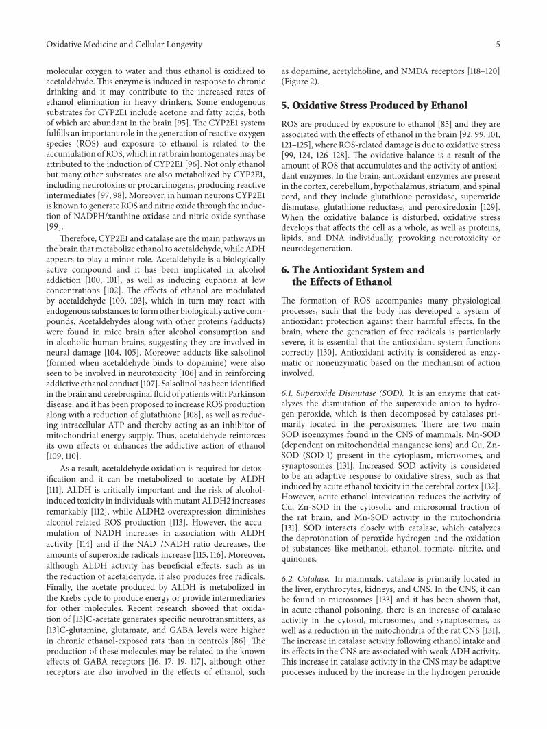
Oxidative Medicine and Cellular Longevity 5
molecular oxygen to water and thus ethanol is oxidized toacetaldehyde. This enzyme is induced in response to chronicdrinking and it may contribute to the increased rates ofethanol elimination in heavy drinkers. Some endogenoussubstrates for CYP2E1 include acetone and fatty acids, bothof which are abundant in the brain [95]. The CYP2E1 systemfulfills an important role in the generation of reactive oxygenspecies (ROS) and exposure to ethanol is related to theaccumulation of ROS,which in rat brain homogenatesmay beattributed to the induction of CYP2E1 [96]. Not only ethanolbut many other substrates are also metabolized by CYP2E1,including neurotoxins or procarcinogens, producing reactiveintermediates [97, 98]. Moreover, in human neurons CYP2E1is known to generate ROS and nitric oxide through the induc-tion of NADPH/xanthine oxidase and nitric oxide synthase[99].
Therefore, CYP2E1 and catalase are the main pathways inthe brain thatmetabolize ethanol to acetaldehyde, whileADHappears to play a minor role. Acetaldehyde is a biologicallyactive compound and it has been implicated in alcoholaddiction [100, 101], as well as inducing euphoria at lowconcentrations [102]. The effects of ethanol are modulatedby acetaldehyde [100, 103], which in turn may react withendogenous substances to formother biologically active com-pounds. Acetaldehydes along with other proteins (adducts)were found in mice brain after alcohol consumption andin alcoholic human brains, suggesting they are involved inneural damage [104, 105]. Moreover adducts like salsolinol(formed when acetaldehyde binds to dopamine) were alsoseen to be involved in neurotoxicity [106] and in reinforcingaddictive ethanol conduct [107]. Salsolinol has been identifiedin the brain and cerebrospinal fluid of patientswith Parkinsondisease, and it has been proposed to increase ROS productionalong with a reduction of glutathione [108], as well as reduc-ing intracellular ATP and thereby acting as an inhibitor ofmitochondrial energy supply. Thus, acetaldehyde reinforcesits own effects or enhances the addictive action of ethanol[109, 110].
As a result, acetaldehyde oxidation is required for detox-ification and it can be metabolized to acetate by ALDH[111]. ALDH is critically important and the risk of alcohol-induced toxicity in individuals withmutantALDH2 increasesremarkably [112], while ALDH2 overexpression diminishesalcohol-related ROS production [113]. However, the accu-mulation of NADH increases in association with ALDHactivity [114] and if the NAD+/NADH ratio decreases, theamounts of superoxide radicals increase [115, 116]. Moreover,although ALDH activity has beneficial effects, such as inthe reduction of acetaldehyde, it also produces free radicals.Finally, the acetate produced by ALDH is metabolized inthe Krebs cycle to produce energy or provide intermediariesfor other molecules. Recent research showed that oxida-tion of [13]C-acetate generates specific neurotransmitters, as[13]C-glutamine, glutamate, and GABA levels were higherin chronic ethanol-exposed rats than in controls [86]. Theproduction of these molecules may be related to the knowneffects of GABA receptors [16, 17, 19, 117], although otherreceptors are also involved in the effects of ethanol, such
as dopamine, acetylcholine, and NMDA receptors [118–120](Figure 2).
5. Oxidative Stress Produced by Ethanol
ROS are produced by exposure to ethanol [85] and they areassociated with the effects of ethanol in the brain [92, 99, 101,121–125], where ROS-related damage is due to oxidative stress[99, 124, 126–128]. The oxidative balance is a result of theamount of ROS that accumulates and the activity of antioxi-dant enzymes. In the brain, antioxidant enzymes are presentin the cortex, cerebellum, hypothalamus, striatum, and spinalcord, and they include glutathione peroxidase, superoxidedismutase, glutathione reductase, and peroxiredoxin [129].When the oxidative balance is disturbed, oxidative stressdevelops that affects the cell as a whole, as well as proteins,lipids, and DNA individually, provoking neurotoxicity orneurodegeneration.
6. The Antioxidant System andthe Effects of Ethanol
The formation of ROS accompanies many physiologicalprocesses, such that the body has developed a system ofantioxidant protection against their harmful effects. In thebrain, where the generation of free radicals is particularlysevere, it is essential that the antioxidant system functionscorrectly [130]. Antioxidant activity is considered as enzy-matic or nonenzymatic based on the mechanism of actioninvolved.
6.1. Superoxide Dismutase (SOD). It is an enzyme that cat-alyzes the dismutation of the superoxide anion to hydro-gen peroxide, which is then decomposed by catalases pri-marily located in the peroxisomes. There are two mainSOD isoenzymes found in the CNS of mammals: Mn-SOD(dependent on mitochondrial manganese ions) and Cu, Zn-SOD (SOD-1) present in the cytoplasm, microsomes, andsynaptosomes [131]. Increased SOD activity is consideredto be an adaptive response to oxidative stress, such as thatinduced by acute ethanol toxicity in the cerebral cortex [132].However, acute ethanol intoxication reduces the activity ofCu, Zn-SOD in the cytosolic and microsomal fraction ofthe rat brain, and Mn-SOD activity in the mitochondria[131]. SOD interacts closely with catalase, which catalyzesthe deprotonation of peroxide hydrogen and the oxidationof substances like methanol, ethanol, formate, nitrite, andquinones.
6.2. Catalase. In mammals, catalase is primarily located inthe liver, erythrocytes, kidneys, and CNS. In the CNS, it canbe found in microsomes [133] and it has been shown that,in acute ethanol poisoning, there is an increase of catalaseactivity in the cytosol, microsomes, and synaptosomes, aswell as a reduction in the mitochondria of the rat CNS [131].The increase in catalase activity following ethanol intake andits effects in the CNS are associated with weak ADH activity.This increase in catalase activity in the CNS may be adaptiveprocesses induced by the increase in the hydrogen peroxide
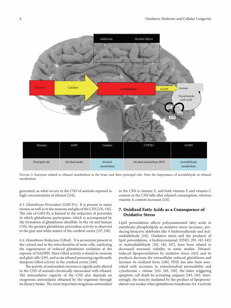
6 Oxidative Medicine and Cellular Longevity
AcetaldehydeCatalaseEthanol
ADH
CYP2E1
ALDH
Enzyme ADH Catalase CYP2E1 ALDH
Principal role Alcohol intake Alcohol metabolism
Alcohol metabolism-ROS Acetaldehydemetabolism
Addiction Alcohol effects
Alcohol intake Adducts
NADH
NADH
Tricarboxylic acid cycle
Acetate
NADHFADH2
ATP
Figure 2: Enzymes related to ethanol metabolism in the brain and their principal role. Note the importance of acetaldehyde in ethanolmetabolism.
generated, as what occurs in the CNS of animals exposed tohigh concentrations of ethanol [134].
6.3. Glutathione Peroxidase (GSH-Px). It is present in manytissues, as well as in the neurons and glia of theCNS [135, 136].The role of GSH-Px is limited to the reduction of peroxidesin which glutathione participates, which is accompanied bythe formation of glutathione disulfide. In the rat and humanCNS, the greatest glutathione peroxidase activity is observedin the gray and white matter of the cerebral cortex [137, 138].
6.4. Glutathione Reductase (GRed). It is an enzyme present inthe cytosol and in the mitochondria of most cells, catalyzingthe regeneration of reduced glutathione oxidation at theexpense of NADPH. Most GRed activity is found in neuronsand glial cells [139], and acute ethanol poisoning significantlydampens GRed activity in the cerebral cortex [140].
The activity of antioxidant enzymes is significantly alteredin the CNS of animals chronically intoxicated with ethanol.The antioxidative capacity of the CNS also depends onexogenous antioxidants obtained by the organism throughits dietary intake.Themost important exogenous antioxidant
in the CNS is vitamin E, and both vitamin E and vitamin Ccontent in the CNS falls after ethanol consumption, whereasvitamin A content increases [131].
7. Oxidized Fatty Acids as a Consequence ofOxidative Stress
Lipid peroxidation affects polyunsaturated fatty acids inmembrane phospholipids as oxidative stress increases, pro-ducing bioactive aldehydes like 4-hydroxyalkenals and mal-ondialdehyde [141]. Oxidative stress and the products oflipid peroxidation, 4-hydroxynonenal (HNE) [99, 142–145]or malondialdehyde [141, 146, 147], have been related todecreased neuronal viability in some studies. Ethanol-induced lipoperoxidation by oxidative stress [142] and itsproducts decrease the intracellular reduced glutathione andincrease its oxidized form [148]. HNE has also been asso-ciated with increases in mitochondrial permeability andcytochrome c release [143, 149, 150], the latter triggeringapoptotic cell death by activating caspases [145, 150]. Inter-estingly, the toxicity mediated by the product of lipoperoxi-dationwasweaker when glutathione transferaseA4-4 activity
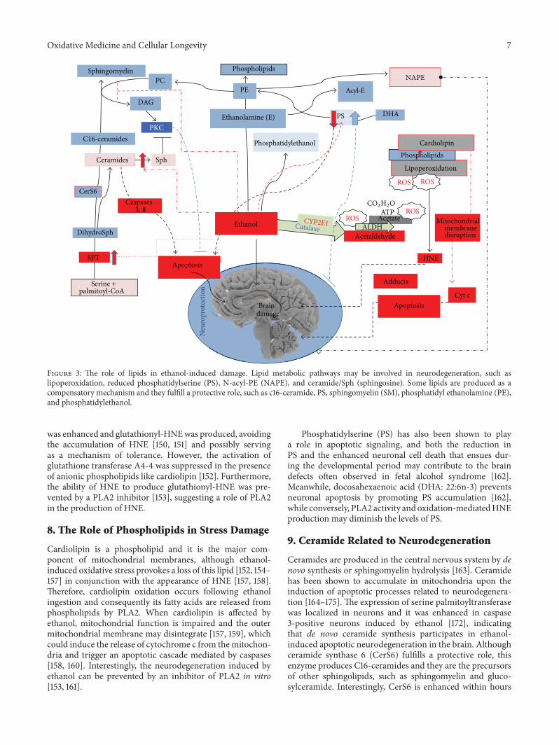
Oxidative Medicine and Cellular Longevity 7
Apoptosis
EthanolAcetaldehyde
ROS
PE
ROS
Lipoperoxidation
PKC
Adducts
Phosphatidylethanol
Ceramides Phospholipids
Phospholipids
CerS6
Sph
PS
Caspases 3, 8
Serine + palmitoyl-CoA
DihydroSph
SPT
SphingomyelinPC
DAG
C16-ceramides
DHAEthanolamine (E)
NAPE
Acyl-E
Cardiolipin
AcetateATP
ALDH
ROS
HNE
Mitochondrialmembranedisruption
Cyt cApoptosisBrain
damage
ROS
CYP2E1
CO2H2O
Catalase
Neu
ropr
otec
tion
Figure 3: The role of lipids in ethanol-induced damage. Lipid metabolic pathways may be involved in neurodegeneration, such aslipoperoxidation, reduced phosphatidylserine (PS), N-acyl-PE (NAPE), and ceramide/Sph (sphingosine). Some lipids are produced as acompensatory mechanism and they fulfill a protective role, such as c16-ceramide, PS, sphingomyelin (SM), phosphatidyl ethanolamine (PE),and phosphatidylethanol.
was enhanced and glutathionyl-HNEwas produced, avoidingthe accumulation of HNE [150, 151] and possibly servingas a mechanism of tolerance. However, the activation ofglutathione transferase A4-4 was suppressed in the presenceof anionic phospholipids like cardiolipin [152]. Furthermore,the ability of HNE to produce glutathionyl-HNE was pre-vented by a PLA2 inhibitor [153], suggesting a role of PLA2in the production of HNE.
8. The Role of Phospholipids in Stress Damage
Cardiolipin is a phospholipid and it is the major com-ponent of mitochondrial membranes, although ethanol-induced oxidative stress provokes a loss of this lipid [152, 154–157] in conjunction with the appearance of HNE [157, 158].Therefore, cardiolipin oxidation occurs following ethanolingestion and consequently its fatty acids are released fromphospholipids by PLA2. When cardiolipin is affected byethanol, mitochondrial function is impaired and the outermitochondrial membrane may disintegrate [157, 159], whichcould induce the release of cytochrome c from themitochon-dria and trigger an apoptotic cascade mediated by caspases[158, 160]. Interestingly, the neurodegeneration induced byethanol can be prevented by an inhibitor of PLA2 in vitro[153, 161].
Phosphatidylserine (PS) has also been shown to playa role in apoptotic signaling, and both the reduction inPS and the enhanced neuronal cell death that ensues dur-ing the developmental period may contribute to the braindefects often observed in fetal alcohol syndrome [162].Meanwhile, docosahexaenoic acid (DHA: 22:6n-3) preventsneuronal apoptosis by promoting PS accumulation [162],while conversely, PLA2 activity and oxidation-mediatedHNEproduction may diminish the levels of PS.
9. Ceramide Related to Neurodegeneration
Ceramides are produced in the central nervous system by denovo synthesis or sphingomyelin hydrolysis [163]. Ceramidehas been shown to accumulate in mitochondria upon theinduction of apoptotic processes related to neurodegenera-tion [164–175]. The expression of serine palmitoyltransferasewas localized in neurons and it was enhanced in caspase3-positive neurons induced by ethanol [172], indicatingthat de novo ceramide synthesis participates in ethanol-induced apoptotic neurodegeneration in the brain. Althoughceramide synthase 6 (CerS6) fulfills a protective role, thisenzyme produces C16-ceramides and they are the precursorsof other sphingolipids, such as sphingomyelin and gluco-sylceramide. Interestingly, CerS6 is enhanced within hours
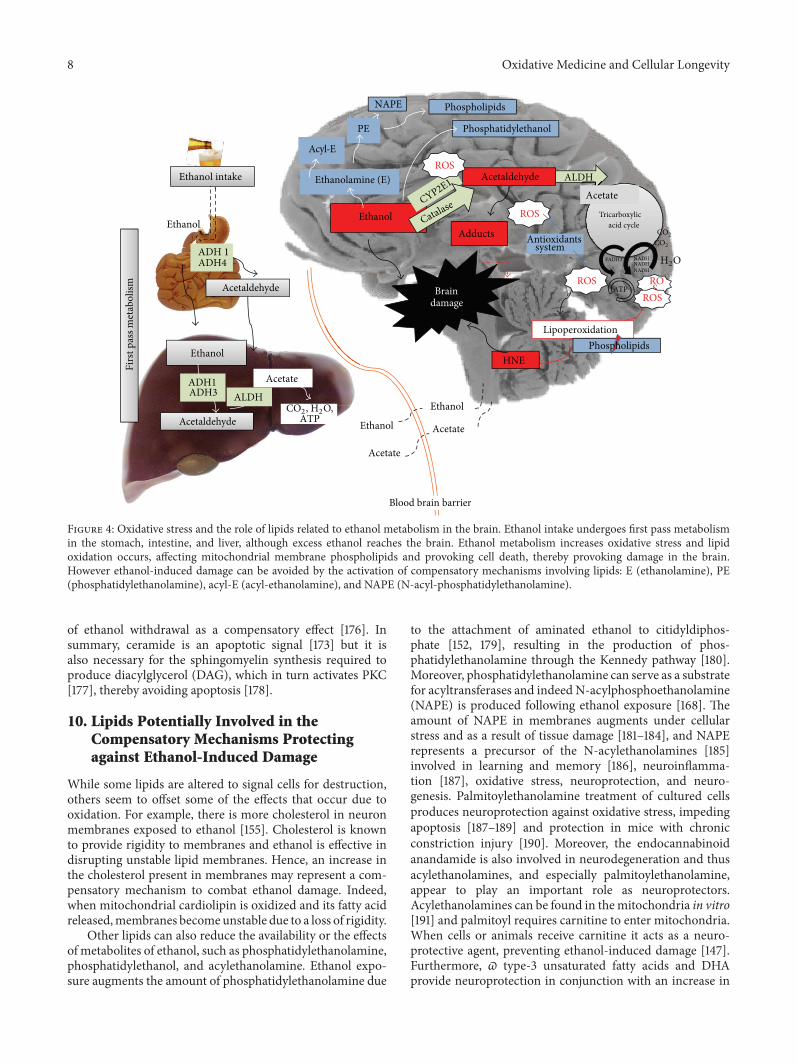
8 Oxidative Medicine and Cellular Longevity
Firs
t pas
s met
abol
ism Acetaldehyde
Ethanol
Ethanol
Acetaldehyde
Acetate
ATP Ethanol
Acetate
ADH 1ADH4
ADH1ADH3 ALDH
ALDH
Tricarboxylic acid cycle
NADHFADH2
ATP
Ethanol
Acetate
Acetaldehyde
CYP2E1
Catalase ROS
Ethanolamine (E)
PE
ROS ROS
Lipoperoxidation
HNE
Brain damage
Adducts
Phosphatidylethanol
Antioxidants system
Phospholipids
Phospholipids
Ethanol
Acetate
NADHNADH
ROSEthanol intake
Blood brain barrier
NAPE
Acyl-E
CO2, H2O,
CO2
CO2
H2O
ROS
Figure 4: Oxidative stress and the role of lipids related to ethanol metabolism in the brain. Ethanol intake undergoes first pass metabolismin the stomach, intestine, and liver, although excess ethanol reaches the brain. Ethanol metabolism increases oxidative stress and lipidoxidation occurs, affecting mitochondrial membrane phospholipids and provoking cell death, thereby provoking damage in the brain.However ethanol-induced damage can be avoided by the activation of compensatory mechanisms involving lipids: E (ethanolamine), PE(phosphatidylethanolamine), acyl-E (acyl-ethanolamine), and NAPE (N-acyl-phosphatidylethanolamine).
of ethanol withdrawal as a compensatory effect [176]. Insummary, ceramide is an apoptotic signal [173] but it isalso necessary for the sphingomyelin synthesis required toproduce diacylglycerol (DAG), which in turn activates PKC[177], thereby avoiding apoptosis [178].
10. Lipids Potentially Involved in theCompensatory Mechanisms Protectingagainst Ethanol-Induced Damage
While some lipids are altered to signal cells for destruction,others seem to offset some of the effects that occur due tooxidation. For example, there is more cholesterol in neuronmembranes exposed to ethanol [155]. Cholesterol is knownto provide rigidity to membranes and ethanol is effective indisrupting unstable lipid membranes. Hence, an increase inthe cholesterol present in membranes may represent a com-pensatory mechanism to combat ethanol damage. Indeed,when mitochondrial cardiolipin is oxidized and its fatty acidreleased,membranes becomeunstable due to a loss of rigidity.
Other lipids can also reduce the availability or the effectsof metabolites of ethanol, such as phosphatidylethanolamine,phosphatidylethanol, and acylethanolamine. Ethanol expo-sure augments the amount of phosphatidylethanolamine due
to the attachment of aminated ethanol to citidyldiphos-phate [152, 179], resulting in the production of phos-phatidylethanolamine through the Kennedy pathway [180].Moreover, phosphatidylethanolamine can serve as a substratefor acyltransferases and indeed N-acylphosphoethanolamine(NAPE) is produced following ethanol exposure [168]. Theamount of NAPE in membranes augments under cellularstress and as a result of tissue damage [181–184], and NAPErepresents a precursor of the N-acylethanolamines [185]involved in learning and memory [186], neuroinflamma-tion [187], oxidative stress, neuroprotection, and neuro-genesis. Palmitoylethanolamine treatment of cultured cellsproduces neuroprotection against oxidative stress, impedingapoptosis [187–189] and protection in mice with chronicconstriction injury [190]. Moreover, the endocannabinoidanandamide is also involved in neurodegeneration and thusacylethanolamines, and especially palmitoylethanolamine,appear to play an important role as neuroprotectors.Acylethanolamines can be found in themitochondria in vitro[191] and palmitoyl requires carnitine to enter mitochondria.When cells or animals receive carnitine it acts as a neuro-protective agent, preventing ethanol-induced damage [147].Furthermore, 𝜛 type-3 unsaturated fatty acids and DHAprovide neuroprotection in conjunction with an increase in
Oxidative Medicine and Cellular Longevity 9
the formation of acylethanolamine [161, 162], suggesting thatthe formation of the latter prevents the damage caused by theoxidative metabolism of ethanol. Finally, ethanol can also bemetabolized as phosphatidylethanol, a molecule found in thebrain of rats [192] that is possibly formed to avoid ethanoloxidation.
11. Conclusions
Lipid metabolism is clearly affected by exposure to ethanol(Figure 3), and the alterations to lipid components likecardiolipin and some phospholipids in response to ethanolprovide evidence of cell damage. The formation of oxidizedspecies, abnormal lipids, and dysfunctional membranes dueto ethanol uptake also provokes cell degeneration. However,compensatory mechanisms exist to dampen the effects ofthese metabolic events and to minimize cell damage, asreflected by the neuroprotective activities of natural lipidslike DHA, esters, vitamin E, and so forth. Thus, ethanol-induced neurodegeneration is at least partly the result ofthe equilibrium maintained between the toxicity of signalinglipids and the protection they confer on the cell (Figure 4).
Conflict of Interests
The authors have no conflict of interests to declare.
Acknowledgment
The authors are grateful to Dr. Mark Sefton for his revisionsand comments on the final version of the paper.
References
[1] K. G. Chartier, M. N. Hesselbrock, and V. M. Hesselbrock,“Ethnicity and gender comparisons of health consequences inadults with alcohol dependence,” Substance Use andMisuse, vol.48, no. 3, pp. 200–210, 2013.
[2] K. J. Kiecolt, S. H. Aggen, and K. S. Kendler, “Genetic andenvironmental influences on the relationship between masteryand alcohol dependence,”Alcoholism: Clinical and ExperimentalResearch, vol. 37, no. 6, pp. 905–913, 2013.
[3] S. Alaux-Cantin, V. Warnault, R. Legastelois et al., “Alcoholintoxications during adolescence increase motivation for alco-hol in adult rats and induce neuroadaptations in the nucleusaccumbens,” Neuropharmacology, vol. 67, pp. 521–531, 2013.
[4] M. Roerecke and J. Rehm, “Cause-specific mortality risk inalcohol use disorder treatment patients: a systematic review andmeta-analysis,” International Journal of Epidemiology, vol. 43,no. 3, Article ID dyu018, pp. 906–919, 2014.
[5] F. Calhoun, M. L. Attilia, P. A. Spagnolo, C. Rotondo, R.Mancinelli, and M. Ceccanti, “National Institute on AlcoholAbuse and Alcoholism and the study of fetal alcohol spectrumdisorders. The International Consortium,” Annali dell’IstitutoSuperiore di Sanita, vol. 42, no. 1, pp. 4–7, 2006.
[6] D. Dias, M. C. Mendonça, F. C. Real, D. N. Vieira, and H.M. Teixeira, “Suicides in the Centre of Portugal: seven yearsanalysis,” Forensic Science International, vol. 234, no. 1, pp. 22–28, 2014.
[7] T. M. Wickizer, “State-level estimates of the economic costs ofalcohol and drug abuse,” Journal of Health Care Finance, vol. 39,no. 3, pp. 71–84, 2013.
[8] G. Scally, “Crunch time for the government on alcohol pricingin England: backtracking on the minimum unit price pledgewould be a public health disaster,” British Medical Journal, vol.346, no. 7900, Article ID f1784, 2013.
[9] M. P. Mundt, S. Parthasarathy, F. W. Chi, S. Sterling, and C.I. Campbell, “12-Step participation reduces medical use costsamong adolescents with a history of alcohol and other drugtreatment,” Drug and Alcohol Dependence, vol. 126, no. 1-2, pp.124–130, 2012.
[10] J. Rehm, C. Mathers, S. Popova, M. Thavorncharoensap, Y.Teerawattananon, and J. Patra, “Global burden of disease andinjury and economic cost attributable to alcohol use andalcohol-use disorders,”The Lancet, vol. 373, no. 9682, pp. 2223–2233, 2009.
[11] H. P. Jedema,M. D. Carter, B. P. Dugan, K. Gurnsey, A. S. Olsen,and C.W. Bradberry, “The acute impact of ethanol on cognitiveperformance in rhesus macaques,” Cerebral Cortex, vol. 21, no.8, pp. 1783–1791, 2011.
[12] A. A.-B. Badawy, C. J. Morgan, J. W. T. Lowett, D. M. Bradley,and R.Thomas, “Decrease in circulating tryptophan availabilityto the brain after acute ethanol consumption by normal vol-unteers: implications for alcohol-induced aggressive behaviourand depression,” Pharmacopsychiatry, vol. 28, supplement 2, pp.93–97, 1995.
[13] A. A.-B. Badawy, D.M. Doughrty, D.M.Marsh-Richard, andA.Steptoe, “Activation of liver tryptophan pyrrolase mediates thedecrease in tryptophan availability to the brain after acute alco-hol consumption by normal subjects,” Alcohol and Alcoholism,vol. 44, no. 3, pp. 267–271, 2009.
[14] S. Loeber and T. Duka, “Extinction learning of stimulus rewardcontingencies: the acute effects of alcohol,” Drug and AlcoholDependence, vol. 102, no. 1–3, pp. 56–62, 2009.
[15] D. M. Dougherty, D. M. Marsh-Richard, E. S. Hatzis, S. O.Nouvion, and C. W. Mathias, “A test of alcohol dose effects onmultiple behavioral measures of impulsivity,” Drug and AlcoholDependence, vol. 96, no. 1-2, pp. 111–120, 2008.
[16] H.M.Haughey, L. A. Ray, P. Finan, R. Villanueva,M.Niculescu,and K. E. Hutchison, “Human 𝛾-aminobutyric acid A receptor𝛼2 genemoderates the acute effects of alcohol and brain mRNAexpression,” Genes, Brain and Behavior, vol. 7, no. 4, pp. 447–454, 2008.
[17] A. Wadleigh and C. F. Valenzuela, “Ethanol increases GABAer-gic transmission and excitability in cerebellar molecular layerinterneurons from GAD67-GFP knock-in mice,” Alcohol andAlcoholism, vol. 47, no. 1, pp. 1–8, 2012.
[18] V. Tiwari, P. Veeraiah, V. Subramaniam, and A. B. Patel,“Differential effects of ethanol on regional glutamatergic andGABAergic neurotransmitter pathways inmouse brain,” Journalof Neurochemistry, vol. 128, no. 5, pp. 628–640, 2014.
[19] G.Wu, H. Liu, J. Jin et al., “Ethanol attenuates sensory stimulus-evoked responses in cerebellar granule cells via activation ofGABAA receptors in vivo inmice,”Neuroscience Letters, vol. 561,pp. 107–111, 2014.
[20] H. L. Halliday, M. M. Reid, and G. McClure, “Results ofheavy drinking in pregnancy,” British Journal of Obstetrics andGynaecology, vol. 89, no. 11, pp. 892–895, 1982.
[21] J. R. West and C. A. Blake, “Fetal alcohol syndrome: anassessment of the field,” Experimental Biology andMedicine, vol.230, no. 6, pp. 354–356, 2005.
10 Oxidative Medicine and Cellular Longevity
[22] R. F. Berman and J. H. Hannigan, “Effects of prenatal alcoholexposure on the hippocampus: spatial behavior, electrophysiol-ogy, and neuroanatomy,”Hippocampus, vol. 10, no. 1, pp. 94–110,2000.
[23] D. H. Lindquist, G. Sokoloff, E. Milner, and J. E. Stein-metz, “Neonatal ethanol exposure results in dose-dependentimpairments in the acquisition and timing of the conditionedeyeblink response and altered cerebellar interpositus nucleusand hippocampal CA1 unit activity in adult rats,” Alcohol, vol.47, no. 6, pp. 447–457, 2013.
[24] A. Ramezani, I. Goudarzi, T. Lashkarboluki, M. T. Ghorbanian,K. Abrari, and M. E. Salmani, “Role of oxidative stress inethanol-induced neurotoxicity in the developing cerebellum,”Iranian Journal of Basic Medical Sciences, vol. 15, no. 4, pp. 965–974, 2012.
[25] J. D. Thomas, E. J. Abou, and H. D. Dominguez, “Prenatalcholine supplementation mitigates the adverse effects of prena-tal alcohol exposure on development in rats,” Neurotoxicologyand Teratology, vol. 31, no. 5, pp. 303–311, 2009.
[26] M. Krishnamoorthy, B. A. Gerwe, C. D. Scharer et al., “Lowethanol concentration alters CHRNA5 RNA levels during earlyhumandevelopment,”Reproductive Toxicology, vol. 30, no. 3, pp.489–492, 2010.
[27] A. Nardelli, C. Lebel, C. Rasmussen, G. Andrew, and C.Beaulieu, “Extensive deep gray matter volume reductions inchildren and adolescents with fetal alcohol spectrumdisorders,”Alcoholism: Clinical and Experimental Research, vol. 35, no. 8,pp. 1404–1417, 2011.
[28] D. Kuehn, S. Aros, F. Cassorla et al., “A prospective cohortstudy of the prevalence of growth, facial, and central nervoussystem abnormalities in children with heavy prenatal alcoholexposure,” Alcoholism: Clinical and Experimental Research, vol.36, no. 10, pp. 1811–1819, 2012.
[29] S. F. Tapert, C. Pulido, M. P. Paulus, M. A. Schuckit, and C.Burke, “Level of response to alcohol and brain response duringvisual working memory,” Journal of Studies on Alcohol, vol. 65,no. 6, pp. 692–700, 2004.
[30] D. B. Clark, D. L. Thatcher, and S. F. Tapert, “Alcohol, psy-chological dysregulation, and adolescent brain development,”Alcoholism: Clinical and Experimental Research, vol. 32, no. 3,pp. 375–385, 2008.
[31] S. F. Tapert, G. G. Brown, M. V. Baratta, and S. A. Brown, “fMRIBOLD response to alcohol stimuli in alcohol dependent youngwomen,” Addictive Behaviors, vol. 29, no. 1, pp. 33–50, 2004.
[32] S. F. Tapert, A. D. Schweinsburg, V. C. Barlett et al., “Bloodoxygen level dependent response and spatial working memoryin adolescents with alcohol use disorders,” Alcoholism: Clinicaland Experimental Research, vol. 28, no. 10, pp. 1577–1586, 2004.
[33] A. M. White, “What happened? Alcohol, memory blackouts,and the brain,” Alcohol Research and Health, vol. 27, no. 2, pp.186–196, 2003.
[34] X. He, E. V. Sullivan, R. K. Stankovic, C. G. Harper, and A.Pfefferbaum, “Interaction of thiamine deficiency and voluntaryalcohol consumption disrupts rat corpus callosum ultrastruc-ture,” Neuropsychopharmacology, vol. 32, no. 10, pp. 2207–2216,2007.
[35] S. A. Magrys and M. C. Olmstead, “Alcohol intoxication alterscognitive skillsmediated by frontal and temporal brain regions,”Brain and Cognition, vol. 85, pp. 271–276, 2014.
[36] C. F. Zorumski, S. Mennerick, and Y. Izumi, “Acute and chroniceffects of ethanol on learning-related synaptic plasticity,” Alco-hol, vol. 48, no. 1, pp. 1–17, 2014.
[37] D. Erritzoe, A. Tziortzi, D. Bargiela et al., “In vivo imagingof cerebral dopamine D3 receptors in alcoholism,” Neuropsy-chopharmacology, vol. 39, no. 7, pp. 1703–1712, 2014.
[38] E. J. Burnett, K. A. Grant, A. T. Davenport, S. E. Hemby, and D.P. Friedman, “The effects of chronic ethanol self-administrationon hippocampal 5-HT
1A receptors in monkeys,” Drug andAlcohol Dependence, vol. 136, pp. 135–142, 2014.
[39] R. P. Vetreno, J. M. Hall, and L. M. Savage, “Alcohol-relatedamnesia and dementia: animal models have revealed the con-tributions of different etiological factors on neuropathology,neurochemical dysfunction and cognitive impairment,” Neuro-biology of Learning and Memory, vol. 96, no. 4, pp. 596–608,2011.
[40] R. P. Vetreno, R. L. Ramos, S. Anzalone, and L. M. Savage,“Brain and behavioral pathology in an animal model of Wer-nicke’s encephalopathy and Wernicke-Korsakoff syndrome,”Brain Research, vol. 1436, pp. 178–192, 2012.
[41] J. Akai and K. Akai, “Neuropathological study of the nucleusbasalis of meynert in alcoholic dementia,” Arukoru KenkyutoYakubutsu Ison, vol. 24, no. 2, pp. 80–88, 1989.
[42] Z. Zhang, Y. Liu, B. Zhou et al., “Altered functional connec-tivity of the marginal division in Alzheimer’s disease,” CurrentAlzheimer Research, vol. 11, no. 2, pp. 145–155, 2014.
[43] B. B. Andersen, “Reduction of Purkinje cell volume in cerebel-lum of alcoholics,” Brain Research, vol. 1007, no. 1-2, pp. 10–18,2004.
[44] G. Halliday, K. Cullen, and A. Harding, “Neuropathologicalcorrelates of memory dysfunction in the Wernicke-Korsakoffsyndrome,” Alcohol and Alcoholism Supplement, vol. 2, pp. 245–251, 1994.
[45] T. Jochum,M.K. Boettger, C. Burkhardt,G. Juckel, andK.-J. Bär,“Increased pain sensitivity in alcohol withdrawal syndrome,”European Journal of Pain, vol. 14, no. 7, pp. 713–718, 2010.
[46] M. Mizuguchi, M. Tomonaga, T. Fukusato, and M. Asano,“Acute necrotizing encephalopathy with widespread edematouslesions of symmetrical distribution,”ActaNeuropathologica, vol.78, no. 1, pp. 108–111, 1989.
[47] L. Riethdorf, R. Warzok, and G. Schwesinger, “Alcoholicencephalopathies in autopsy material,” Zentralblatt für Patholo-gie, vol. 137, no. 1, pp. 48–56, 1991.
[48] V. A. Ramchandani, W. F. Bosron, and T. K. Li, “Researchadvances in ethanol metabolism,” Pathologie Biologie, vol. 49,no. 9, pp. 676–682, 2001.
[49] M. D. Levitt, R. Li, E. G. Demaster, M. Elson, J. Furne, and D.G. Levitt, “Use of measurements of ethanol absorption fromstomach and intestine to assess human ethanol metabolism,”American Journal of Physiology—Gastrointestinal and LiverPhysiology, vol. 273, no. 4, pp. G951–G957, 1997.
[50] M. D. Levitt, C. Fine, J. K. Furne, and D. G. Levitt, “Use of mal-tose hydrolysis measurements to characterize the interactionbetween the aqueous diffusion barrier and the epithelium in therat jejunum,”The Journal of Clinical Investigation, vol. 97, no. 10,pp. 2308–2315, 1996.
[51] K. S. Pang, “Modeling of intestinal drug absorption: roles oftransporters and metabolic enzymes (for the Gillette reviewseries),” Drug Metabolism and Disposition, vol. 31, no. 12, pp.1507–1519, 2003.
[52] C. M. Oneta, U. A. Simanowski, M. Martinez et al., “First passmetabolism of ethanol is strikingly influenced by the speed ofgastric emptying,” Gut, vol. 43, no. 5, pp. 612–619, 1998.
Oxidative Medicine and Cellular Longevity 11
[53] J. Shultz, H. Weiner, and J. Westcott, “Retardation of ethanolabsorption by food in the stomach,” Journal of Studies onAlcohol, vol. 41, no. 9, pp. 861–870, 1980.
[54] A. J. Sedman, P. K. Wilkinson, E. Sakmar, D. J. Weidler, andJ. G. Wagner, “Food effects on absorption and metabolism ofalcohol,” Journal of Studies on Alcohol, vol. 37, no. 9, pp. 1197–1214, 1976.
[55] S.-L. Lee, G.-Y. Chau, C.-T. Yao, C.-W.Wu, and S.-J. Yin, “Func-tional assessment of human alcohol dehydrogenase familyin ethanol metabolism: significance of first-pass metabolism,”Alcoholism: Clinical and Experimental Research, vol. 30, no. 7,pp. 1132–1142, 2006.
[56] Å. Norberg,W.A. Jones, R.G.Hahn, and J. L.Gabrielsson, “Roleof variability in explaining ethanol pharmacokinetics: researchand forensic applications,” Clinical Pharmacokinetics, vol. 42,no. 1, pp. 1–31, 2003.
[57] J. S. Deetz, C. A. Luehr, and B. L. Vallee, “Human liver alcoholdehydrogenase isozymes: reduction of aldehydes and ketones,”Biochemistry, vol. 23, no. 26, pp. 6822–6828, 1984.
[58] T. Haseba, Y. Tomita, M. Kurosu, and Y. Ohno, “Dose and timechanges in liver alcohol dehydrogenase (ADH) activity duringacute alcohol intoxication involve not only class I but also classIII ADH and govern elimination rate of blood ethanol,” LegalMedicine, vol. 5, no. 4, pp. 202–211, 2003.
[59] T. Haseba, K. Kameyama, K. Mashimo, and Y. Ohno, “Dose-dependent change in elimination kinetics of ethanol due toshift of dominant metabolizing enzyme from ADH 1 (ClassI) to ADH 3 (Class III) in mouse,” International Journal ofHepatology, vol. 2012, Article ID 408190, 8 pages, 2012.
[60] T.-K. Li, S.-J. Yin, D. W. Crabb, S. O’Connor, and V. A. Ram-chandani, “Genetic and environmental influences on alcoholmetabolism in humans,” Alcoholism: Clinical and ExperimentalResearch, vol. 25, no. 1, pp. 136–144, 2001.
[61] T. N. Smolen, A. Smolen, and J. L. van de Kamp, “Develop-mental profile of hepatic alcohol and aldehyde dehydrogenaseactivities in long-sleep and short-sleep mice,” Alcohol, vol. 7, no.1, pp. 69–74, 1990.
[62] F. Lundquist, N. Tygstrup, K. Winkler, K. Mellemgaard, and S.Munck-Petersen, “Ethanol metabolism and production of freeacetate in the human liver,”The Journal of Clinical Investigation,vol. 41, pp. 955–961, 1962.
[63] K. O. Lindros, A. Stowell, P. Pikkarainen, and M. Salaspuro,“Elevated blood acetaldehyde in alcoholics with acceleratedethanol elimination,” Pharmacology Biochemistry and Behavior,vol. 13, supplement 1, pp. 119–124, 1980.
[64] J. G. Puig and I. H. Fox, “Ethanol-induced activation of adeninenucleotide turnover. Evidence for a role of acetate,”The Journalof Clinical Investigation, vol. 74, no. 3, pp. 936–941, 1984.
[65] T. Sarkola, M. R. Iles, K. Kohlenberg-Mueller, and C. J.P. Eriksson, “Ethanol, acetaldehyde, acetate, and lactate lev-els after alcohol intake in white men and women: effectof 4-methylpyrazole,” Alcoholism: Clinical and ExperimentalResearch, vol. 26, no. 2, pp. 239–245, 2002.
[66] Z. Kayaalti and T. Soylemezoglu, “Distribution of ADH1B,ALDH2, CYP2E1 ∗6, and CYP2E1 ∗7B genotypes in Turkishpopulation,” Alcohol , vol. 44, no. 5, pp. 415–423, 2010.
[67] A. J. Montano Loza, M. T. Ramirez Iglesias, I. Perez Diaz et al.,“Association of alcohol-metabolizing genes with alcoholism ina Mexican Indian (Otomi) population,” Alcohol, vol. 39, no. 2,pp. 73–79, 2006.
[68] J. Garćıa-Bañuelos, A. Panduro, D. Gordillo-Bastidas etal., “Genetic polymorphisms of genes coding to alcohol-metabolizing enzymes in Western Mexicans: association ofCYP2E1∗c2/CYP2E1∗5B allele with cirrhosis and liver function,”Alcoholism: Clinical and Experimental Research, vol. 36, no. 3,pp. 425–431, 2012.
[69] K. Y. Seng, L. M. G. Limenta, D. Heng, and E. J. D. Lee, “Pop-ulation pharmacokinetics and pharmacogenetics of alcohol inChinese and Indians in Singapore,” Journal of Clinical PharmacyandTherapeutics, vol. 38, no. 2, pp. 141–149, 2013.
[70] Y. Yamada, F. Sun, I. Tsuritani, and R. Honda, “Genetic differ-ences in ethanol metabolizing enzymes and blood pressure inJapanese alcohol consumers,” Journal of Human Hypertension,vol. 16, no. 7, pp. 479–486, 2002.
[71] A. Lorenzo, T. Auguet, F. Vidal et al., “Polymorphisms ofalcohol-metabolizing enzymes and the risk for alcoholism andalcoholic liver disease in Caucasian Spanish women,” Drug andAlcohol Dependence, vol. 84, no. 2, pp. 195–200, 2006.
[72] Y.-C. Chen, G.-S. Peng, M.-F. Wang, T.-P. Tsao, and S.-J. Yin,“Polymorphism of ethanol-metabolism genes and alcoholism:correlation of allelic variations with the pharmacokinetic andpharmacodynamic consequences,” Chemico-Biological Interac-tions, vol. 178, no. 1–3, pp. 2–7, 2009.
[73] M. Laniewska-Dunaj, W. Jelski, K. Orywal, J. Kochanowicz, R.Rutkowski, and M. Szmitkowski, “The activity of class I, II, IIIand IV of alcohol dehydrogenase (ADH) isoenzymes and alde-hyde dehydrogenase (ALDH) in brain cancer,” NeurochemicalResearch, vol. 38, no. 7, pp. 1517–1521, 2013.
[74] R. Sanchez-Alvarez, U. E. Martinez-Outschoorn, Z. Lin etal., “Ethanol exposure induces the cancer-associated fibroblastphenotype and lethal tumormetabolism: implications for breastcancer prevention,” Cell Cycle, vol. 12, no. 2, pp. 289–301, 2013.
[75] I. S. Chan, C. D. Guy, M. V. Machado et al., “Alcohol activatesthe hedgehog pathway and induces related procarcinogenicprocesses in the alcohol-preferring rat model of hepatocarcino-genesis,” Alcoholism: Clinical and Experimental Research, vol.38, no. 3, pp. 787–800, 2014.
[76] J. M. Cowan Jr., A. Weathermon, J. R. McCutcheon, and R. D.Oliver, “Determination of volume of distribution for ethanol inmale and female subjects,” Journal of Analytical Toxicology, vol.20, no. 5, pp. 287–290, 1996.
[77] H. G. E. Endres and O. Grüner, “Comparison of D2O and
ethanol dilutions in total bodywatermeasurements in humans,”The Clinical Investigator, vol. 72, no. 11, pp. 830–837, 1994.
[78] A. F. Pizon, C. E. Becker, andD. Bikin, “The clinical significanceof variations in ethanol toxicokinetics,” Journal of MedicalToxicology, vol. 3, no. 2, pp. 63–72, 2007.
[79] K. E. Maudens, L. Patteet, A. L. N. van Nuijs, C. VanBroekhoven, A. Covaci, and H. Neels, “The influence of theBody Mass Index (BMI) on the volume of distribution ofethanol,”Forensic Science International, vol. 243, pp. 74–78, 2014.
[80] M. Marxen, G. Gan, D. Schwarz et al., “Acute effects of alcoholon brain perfusion monitored with arterial spin labeling mag-netic resonance imaging in young adults,” Journal of CerebralBlood Flow and Metabolism, vol. 34, no. 3, pp. 472–479, 2014.
[81] L. Burd, J. Blair, and K. Dropps, “Prenatal alcohol exposure,blood alcohol concentrations and alcohol elimination rates forthe mother, fetus and newborn,” Journal of Perinatology, vol. 32,no. 9, pp. 652–659, 2012.
[82] E. M. Moore and E. P. Riley, “What happens when childrenwith fetal alcohol spectrum disorders become adults?” CurrentDevelopmental Disorders Reports, vol. 2, no. 3, pp. 219–227, 2015.
12 Oxidative Medicine and Cellular Longevity
[83] A. G. Skorput, V. P. Gupta, P. W. Yeh, and H. H. Yeh, “Persistentinterneuronopathy in the prefrontal cortex of young adult off-spring exposed to ethanol in utero,”The Journal of Neuroscience,vol. 35, no. 31, pp. 10977–10988, 2015.
[84] G. Fein and D. J. Meyerhoff, “Ethanol in human brain bymagnetic resonance spectroscopy: correlation with blood andbreath levels, relaxation, and magnetization transfer,” Alco-holism: Clinical and Experimental Research, vol. 24, no. 8, pp.1227–1235, 2000.
[85] S.M. Zimatkin andA. I. Buben, “Ethanol oxidation in the livingbrain,” Alcohol and Alcoholism, vol. 42, no. 6, pp. 529–532, 2007.
[86] J. Wang, H. Du, X. Ma et al., “Metabolic products of [2–13C]ethanol in the rat brain after chronic ethanol exposure,”Journal of Neurochemistry, vol. 127, no. 3, pp. 353–364, 2013.
[87] S. E. Mart́ınez, J. Vaglenova, J. Sabrià, M. Carmen Mart́ınez, J.Farrés, and X. Parés, “Distribution of alcohol dehydrogenasemRNA in the rat central nervous system. Consequences forbrain ethanol and retinoid metabolism,” European Journal ofBiochemistry, vol. 268, no. 19, pp. 5045–5056, 2001.
[88] D. Galter, A. Carmine, S. Buervenich, G. Duester, and L. Olson,“Distribution of class I, III and IV alcohol dehydrogenasemRNAS in the adult rat, mouse and human brain,” EuropeanJournal of Biochemistry, vol. 270, no. 6, pp. 1316–1326, 2003.
[89] K. Tokuda, Y. Izumi, and C. F. Zorumski, “Locally-generatedacetaldehyde contributes to the effects of ethanol on neuros-teroids and LTP in the hippocampus,” Neurology and ClinicalNeuroscience, vol. 1, no. 4, pp. 138–147, 2013.
[90] E. Borger, L. Aitken, H. Du, W. Zhang, F. J. Gunn-Moore, andS. S. Du Yan, “Is amyloid binding alcohol dehydrogenase adrug target for treatingAlzheimer’s disease?”Current AlzheimerResearch, vol. 10, no. 1, pp. 21–29, 2013.
[91] S. M. Zimatkin, S. P. Pronko, V. Vasiliou, F. J. Gonzalez, and R.A.Deitrich, “Enzymaticmechanisms of ethanol oxidation in thebrain,” Alcoholism: Clinical and Experimental Research, vol. 30,no. 9, pp. 1500–1505, 2006.
[92] E. Karahanian, M. E. Quintanilla, L. Tampier et al., “Ethanol asa prodrug: brainmetabolism of ethanol mediates its reinforcingeffects,” Alcoholism: Clinical and Experimental Research, vol. 35,no. 4, pp. 606–612, 2011.
[93] D. E. Rhoads, C. Contreras, and S. Fathalla, “Brain levels ofcatalase remain constant through strain, developmental, andchronic alcohol challenges,” Enzyme Research, vol. 2012, ArticleID 572939, 6 pages, 2012.
[94] C.M.G.Aragon, F. Rogan, andZ.Amit, “Ethanolmetabolism inrat brain homogenates by a catalase-H
2O2system,” Biochemical
Pharmacology, vol. 44, no. 1, pp. 93–98, 1992.[95] C. S. Lieber, “Microsomal ethanol-oxidizing system (MEOS):
the first 30 years (1968–1998)—a review,” Alcoholism: Clinicaland Experimental Research, vol. 23, no. 6, pp. 991–1007, 1999.
[96] Y. Zhong, G. Dong, H. Luo et al., “Induction of brain CYP2E1by chronic ethanol treatment and related oxidative stress inhippocampus, cerebellum, and brainstem,” Toxicology, vol. 302,no. 2-3, pp. 275–284, 2012.
[97] D. R. Koop, “Oxidative and reductive metabolism bycytochrome P450 2E1,” The FASEB Journal, vol. 6, no. 2,pp. 724–730, 1992.
[98] T. Leung, R. Rajendran, S. Singh, R. Garva, M. Krstic-Dem-onacos, and C. Demonacos, “Cytochrome P450 2E1 (CYP2E1)regulates the response to oxidative stress andmigration of breastcancer cells,” Breast Cancer Research, vol. 15, no. 6, article R107,2013.
[99] J. Haorah, S. H. Ramirez, N. Floreani, S. Gorantla, B. Morsey,and Y. Persidsky, “Mechanism of alcohol-induced oxidativestress and neuronal injury,” Free Radical Biology and Medicine,vol. 45, no. 11, pp. 1542–1550, 2008.
[100] G. A. Deehan Jr., S. R. Hauser, J. A. Wilden, W. A. Truitt, andZ. A. Rodd, “Elucidating the biological basis for the reinforcingactions of alcohol in the mesolimbic dopamine system: therole of active metabolites of alcohol,” Frontiers in BehavioralNeuroscience, vol. 7, article 104, 2013.
[101] X.-S. Deng and R. A. Deitrich, “Putative role of brain acetalde-hyde in ethanol addiction,” Current Drug Abuse Reviews, vol. 1,no. 1, pp. 3–8, 2008.
[102] C. J. Eriksson, “The role of acetaldehyde in the actions of alcohol(update 2000),” Alcoholism Clinical and Experimental Research,vol. 25, no. 5, supplement, pp. 15s–32s, 2001.
[103] A. I. Cederbaum, “Alcoholmetabolism,”Clinics in Liver Disease,vol. 16, no. 4, pp. 667–685, 2012.
[104] K. Nakamura, K. Iwahashi, A. Furukawa et al., “Acetaldehydeadducts in the brain of alcoholics,” Archives of Toxicology, vol.77, no. 10, pp. 591–593, 2003.
[105] K. Nakamura, K. Iwahashi, M. Itoh et al., “Immunohisto-chemical study on acetaldehyde adducts in alcohol-fed mice,”Alcoholism, Clinical and Experimental Research, vol. 24, no. 4,supplement, pp. 93S–96S, 2000.
[106] G. Xie, K. Krnjević, and J.-H. Ye, “Salsolinol modulation ofdopamine neurons,” Frontiers in Behavioral Neuroscience, vol.7, article 52, 2013.
[107] M. E. Quintanilla, M. Rivera-Meza, P. A. Berrios-Cárcamo etal., “Salsolinol, free of isosalsolinol, exerts ethanol-like moti-vational/sensitization effects leading to increases in ethanolintake,” Alcohol, vol. 48, no. 6, pp. 551–559, 2014.
[108] S. Wanpen, P. Govitrapong, S. Shavali, P. Sangchot, and M.Ebadi, “Salsolinol, a dopamine-derived tetrahydroisoquinoline,induces cell death by causing oxidative stress in dopaminergicSH-SY5Y cells, and the said effect is attenuated by metalloth-ionein,” Brain Research, vol. 1005, no. 1-2, pp. 67–76, 2004.
[109] A. T. Peana and E. Acquasb, “Behavioral and biochemicalevidence of the role of acetaldehyde in the motivational effectsof ethanol,” Frontiers in Behavioral Neuroscience, vol. 7, article86, 2013.
[110] M. E. Quintanilla, M. Rivera-Meza, P. Berŕıos-Cárcamo, B. K.Cassels, M. Herrera-Marschitz, and Y. Israel, “(R)-Salsolinol,a product of ethanol metabolism, stereospecifically inducesbehavioral sensitization and leads to excessive alcohol intake,”Addiction Biology, 2015.
[111] A. Kikonyogo and R. Pietruszko, “Aldehyde dehydrogenasefrom adult human brain that dehydrogenates 𝛾-aminobutyr-aldehyde: purification, characterization, cloning and distribu-tion,” Biochemical Journal, vol. 316, part 1, pp. 317–324, 1996.
[112] Y. Zhang and J. Ren, “ALDH2 in alcoholic heart diseases:molecular mechanism and clinical implications,” PharmacologyandTherapeutics, vol. 132, no. 1, pp. 86–95, 2011.
[113] J. Bai and Y.Mei, “Overexpression of aldehyde dehydrogenase-2attenuates neurotoxicity induced by 4-hydroxynonenal in cul-tured primary hippocampal neurons,” Neurotoxicity Research,vol. 19, no. 3, pp. 412–422, 2011.
[114] S. Manzo-Avalos and A. Saavedra-Molina, “Cellular and mito-chondrial effects of alcohol consumption,” International Journalof Environmental Research and Public Health, vol. 7, no. 12, pp.4281–4304, 2010.
Oxidative Medicine and Cellular Longevity 13
[115] J. Antosiewicz, J. H. Spodnik, M. Teranishi et al., “NADH-generating substrates reduce peroxyl radical toxicity in RL-34cells,” Folia Morphologica, vol. 68, no. 4, pp. 247–255, 2009.
[116] M.Velayutham,C.Hemann, and J. L. Zweier, “Removal ofH2O2
and generation of superoxide radical: role of cytochrome c andNADH,” Free Radical Biology and Medicine, vol. 51, no. 1, pp.160–170, 2011.
[117] B. M. Walker and G. F. Koob, “The 𝛾-aminobutyric acid-Breceptor agonist baclofen attenuates responding for ethanol inethanol-dependent rats,” Alcoholism: Clinical and ExperimentalResearch, vol. 31, no. 1, pp. 11–18, 2007.
[118] E. B. Rex, M. L. Rankin, M. A. Ariano, and D. R. Sibley,“Ethanol regulation of D
1dopamine receptor signaling is
mediated by protein kinase C in an isozyme-specific manner,”Neuropsychopharmacology, vol. 33, no. 12, pp. 2900–2911, 2008.
[119] L. M. Hendrickson, P. Gardner, and A. R. Tapper, “Nicotinicacetylcholine receptors containing the 𝛼4 subunit are criticalfor the nicotine-induced reduction of acute voluntary ethanolconsumption,” Channels, vol. 5, no. 2, pp. 124–127, 2011.
[120] E. Villegas, R. Estruch, G. Mengod, and R. Cortés, “NMDAreceptors in frontal cortex and hippocampus of alcohol con-sumers,” Addiction Biology, vol. 16, no. 1, pp. 163–165, 2011.
[121] M.Correa,M.N.Arizzi, A. Betz, S.Mingote, and J.D. Salamone,“Open field locomotor effects in rats after intraventricularinjections of ethanol and the ethanol metabolites acetaldehydeand acetate,” Brain Research Bulletin, vol. 62, no. 3, pp. 197–202,2003.
[122] N. D. Volkow, S. W. Kim, G.-J. Wang et al., “Acute alcoholintoxication decreases glucosemetabolism but increases acetateuptake in the human brain,”NeuroImage, vol. 64, no. 1, pp. 277–283, 2013.
[123] G. U. Corsini, A. Zuddas, U. Bonuccelli, S. Schinelli, and I. J.Kopin, “1-Methyl-4-phenyl-1,2,3,6-tetrahydropyridine (MPTP)neurotoxicity in mice is enhanced by ethanol or acetaldehyde,”Life Sciences, vol. 40, no. 9, pp. 827–832, 1987.
[124] L. Heap, R. J. Ward, C. Abiaka et al., “The influence of brainacetaldehyde on oxidative status, dopamine metabolism andvisual discrimination task,” Biochemical Pharmacology, vol. 50,no. 2, pp. 263–270, 1995.
[125] J. Y. Wan, J. Y. Wang, Y. Wang, and J. Y. Wang, “A com-parison between acute exposures to ethanol and acetaldehydeon neurotoxicity, nitric oxide production and NMDA-inducedexcitotoxicity in primary cultures of cortical neurons,” TheChinese Journal of Physiology, vol. 43, no. 3, pp. 131–138, 2000.
[126] F. T. Crews and K. Nixon, “Mechanisms of neurodegenerationand regeneration in alcoholism,” Alcohol and Alcoholism, vol.44, no. 2, pp. 115–127, 2009.
[127] C. Ikonomidou and A. M. Kaindl, “Neuronal death and oxida-tive stress in the developing brain,” Antioxidants and RedoxSignaling, vol. 14, no. 8, pp. 1535–1550, 2011.
[128] J. Luo, “Mechanisms of ethanol-induced death of cerebellargranule cells,” Cerebellum, vol. 11, no. 1, pp. 145–154, 2012.
[129] S.-Y. Shim and H.-S. Kim, “Oxidative stress and the antioxidantenzyme system in the developing brain,” Korean Journal ofPediatrics, vol. 56, no. 3, pp. 107–111, 2013.
[130] A. Augustyniak, K. Michalak, and E. Skrzydlewska, “The actionof oxidative stress induced by ethanol on the central nervoussystem (CNS),” PostępyHigieny iMedycynyDoświadczalnej, vol.59, pp. 464–471, 2005.
[131] S. K. Reddy, K. Husain, E. C. Schlorff, R. B. Scott, and S. M.Somani, “Dose response of ethanol ingestion on antioxidant
defense system in rat brain subcellular fractions,”NeuroToxicol-ogy, vol. 20, no. 6, pp. 977–987, 1999.
[132] M. Enache, V. Van Waes, E. Vinner, M. Lhermitte, S. Maccari,and M. Darnaudéry, “Impact of an acute exposure to ethanolon the oxidative stress status in the hippocampus of prenatalrestraint stress adolescent male rats,” Brain Research, vol. 1191,pp. 55–62, 2008.
[133] L. Hipolito, M. J. Sanchez, A. Polache, and L. Granero, “Brainmetabolism of ethanol and alcoholism: an update,” CurrentDrug Metabolism, vol. 8, no. 7, pp. 716–727, 2007.
[134] Y. Fukui and H. Sakata-Haga, “Intrauterine environment-genome interaction and children’s development (1): ethanol: ateratogen in developing brain,” Journal of Toxicological Sciences,vol. 34, supplement 2, pp. SP273–SP278, 2009.
[135] F. Bosch-Morell, F.Mart́ınez-Soriano,A.Colell, J. C. Fernández-Checa, and F. J. Romero, “Chronic ethanol feeding inducescellular antioxidants decrease and oxidative stress in rat periph-eral nerves. Effect of S-adenosyl-l-methionine and N-acetyl-l-cysteine,” Free Radical Biology and Medicine, vol. 25, no. 3, pp.365–368, 1998.
[136] M. Chalimoniuk, S. Jagsz, E. Sadowska-Krepa, S. J. Chrapusta,B. Klapcinska, and J. Langfort, “Diversity of endurance trainingeffects on antioxidant defenses and oxidative damage in differ-ent brain regions of adolescent male rats,” Journal of Physiologyand Pharmacology, vol. 66, no. 4, pp. 539–547, 2015.
[137] K. A. Ansari, D. Bigelow, and E. Kaplan, “Glutathione per-oxidase activity in surgical and autopsied human brains,”Neurochemical Research, vol. 10, no. 5, pp. 703–711, 1985.
[138] J. Huang and M. A. Philbert, “Distribution of glutathioneand glutathione-related enzyme systems in mitochondria andcytosol of cultured cerebellar astrocytes and granule cells,”BrainResearch, vol. 680, no. 1-2, pp. 16–22, 1995.
[139] R. Dringen and J. Hirrlinger, “Glutathione pathways in thebrain,” Biological Chemistry, vol. 384, no. 4, pp. 505–516, 2003.
[140] S. M. Somani and K. Husain, “Interaction of exercise trainingand chronic ethanol ingestion on antioxidant systemof rat brainregions,” Journal of Applied Toxicology, vol. 17, no. 5, pp. 329–336,1997.
[141] S. Pizzimenti, E. Ciamporcero, M. Daga et al., “Interactionof aldehydes derived from lipid peroxidation and membraneproteins,” Frontiers in Physiology, vol. 4, article 242, 2013.
[142] J. J. Chen, S. Schenker, and G. I. Henderson, “4-Hydroxynone-nal levels are enhanced in fetal liver mitochondria by in uteroethanol exposure,” Hepatology, vol. 25, no. 1, pp. 142–147, 1997.
[143] V. Ramachandran, A. Perez, J. Chen,D. Senthil, S. Schenker, andG. I. Henderson, “In utero ethanol exposure causes mitochon-drial dysfunction, which can result in apoptotic cell death infetal brain: a potential role for 4-hydroxynonenal,” Alcoholism:Clinical and Experimental Research, vol. 25, no. 6, pp. 862–871,2001.
[144] V. Ramachandran, L. T. Watts, S. K. Maffi, J. Chen, S. Schenker,and G. I. Henderson, “Ethanol-induced oxidative stress pre-cedes mitochondrially mediated apoptotic death of culturedfetal cortical neurons,” Journal of Neuroscience Research, vol. 74,no. 4, pp. 577–588, 2003.
[145] S. K. Maffi, M. L. Rathinam, P. P. Cherian et al., “Glutathionecontent as a potential mediator of the vulnerability of culturedfetal cortical neurons to ethanol-induced apoptosis,” Journal ofNeuroscience Research, vol. 86, no. 5, pp. 1064–1076, 2008.
[146] A. Rendón-Ramı́rez, M. Cortés-Couto, A. B. Mart́ınez-Rizo, S.Muñiz-Hernández, and J. B. Velázquez-Fernández, “Oxidative
14 Oxidative Medicine and Cellular Longevity
damage in young alcohol drinkers: a preliminary study,” Alco-hol, vol. 47, no. 7, pp. 501–504, 2013.
[147] A. Augustyniak and E. Skrzydlewska, “The influence of L-carnitine suplementation on the antioxidative abilities of serumand the central nervous system of ethanol-induced rats,”Metabolic Brain Disease, vol. 25, no. 4, pp. 381–389, 2010.
[148] W. Liu, M. Kato, A. A. Akhand et al., “4-hydroxynonenalinduces a cellular redox status-related activation of the caspasecascade for apoptotic cell death,” Journal of Cell Science, vol. 113,no. part 4, pp. 635–641, 2000.
[149] A.Mahr, F. Batteux, S. Tubiana et al., “Brief report: prevalence ofantineutrophil cytoplasmic antibodies in infective endocardi-tis,”Arthritis&Rheumatology, vol. 66, no. 6, pp. 1672–1677, 2014.
[150] H. Raza and A. John, “4-Hydroxynonenal induces mitochon-drial oxidative stress, apoptosis and expression of glutathioneS-transferase A4-4 and cytochrome P450 2E1 in PC12 cells,”Toxicology and Applied Pharmacology, vol. 216, no. 2, pp. 309–318, 2006.
[151] K. E. McElhanon, C. Bose, R. Sharma, L. Wu, Y. C. Awasthi,and S. P. Singh, “4 null mouse embryonic fibroblasts exhibitenhanced sensitivity to oxidants: role of 4-hydroxynonenal inoxidant toxicity,” Open Journal of Apoptosis, vol. 2, no. 1, pp. 1–11, 2013.
[152] M. Shimoji, N. Imaizumi, and Y. Aniya, “Modulation of mem-brane-bound glutathione transferase activity by phospholipidsincluding cardiolipin,” Biological and Pharmaceutical Bulletin,vol. 34, no. 2, pp. 209–213, 2011.
[153] K.-H. Moon, N. Tajuddin, J. Brown, E. J. Neafsey, H.-Y. Kim,and M. A. Collins, “Phospholipase A2, oxidative stress, andneurodegeneration in binge ethanol-treated organotypic slicecultures of developing rat brain,” Alcoholism: Clinical andExperimental Research, vol. 38, no. 1, pp. 161–169, 2014.
[154] M. A. C. Rodrigues, J. L. Rodrigues, N. M. Martins et al.,“Carvedilol protects against cisplatin-induced oxidative stress,redox state unbalance and apoptosis in rat kidney mitochon-dria,”Chemico-Biological Interactions, vol. 189, no. 1-2, pp. 45–51,2011.
[155] G. Barceló-Coblijn, L. E. Wold, J. Ren, and E. J. Murphy,“Prenatal ethanol exposure increases brain cholesterol contentin adult rats,” Lipids, vol. 48, no. 11, pp. 1059–1068, 2013.
[156] V. D. Reddy, P. Padmavathi, G. Kavitha, B. Saradamma, and N.Varadacharyulu, “Alcohol-induced oxidative/nitrosative stressalters brain mitochondrial membrane properties,” Molecularand Cellular Biochemistry, vol. 375, no. 1-2, pp. 39–47, 2013.
[157] G. Petrosillo, P. Portincasa, I. Grattagliano et al., “Mitochondrialdysfunction in rat with nonalcoholic fatty liver. Involvement ofcomplex I, reactive oxygen species and cardiolipin,” Biochimicaet Biophysica Acta (BBA)—Bioenergetics, vol. 1767, no. 10, pp.1260–1267, 2007.
[158] W. Liu, N. A. Porter, C. Schneider, A. R. Brash, and H. Yin,“Formation of 4-hydroxynonenal from cardiolipin oxidation:intramolecular peroxyl radical addition and decomposition,”Free Radical Biology and Medicine, vol. 50, no. 1, pp. 166–178,2011.
[159] E. Rubin and H. Rottenberg, “Ethanol-induced injury andadaptation in biological membranes,” Federation Proceedings,vol. 41, no. 8, pp. 2465–2471, 1982.
[160] Y. A. Vladimirov, E. V. Proskurnina, and A. V. Alekseev,“Molecular mechanisms of apoptosis. Structure of cytochromec-cardiolipin complex,” Biochemistry, vol. 78, no. 10, pp. 1086–1097, 2013.
[161] J. Brown III, N. Achille, E. J. Neafsey, and M. A. Collins,“Binge ethanol-induced neurodegeneration in rat organotypicbrain slice cultures: effects of PLA2 inhibitor mepacrine andDocosahexaenoic Acid (DHA),” Neurochemical Research, vol.34, no. 2, pp. 260–267, 2009.
[162] M.Akbar, J. Baick, F. Calderon, Z.Wen, andH.-Y.Kim, “Ethanolpromotes neuronal apoptosis by inhibiting phosphatidylserineaccumulation,” Journal of Neuroscience Research, vol. 83, no. 3,pp. 432–440, 2006.
[163] C. R. Horres and Y. A. Hannun, “The roles of neutral sph-ingomyelinases in neurological pathologies,” NeurochemicalResearch, vol. 37, no. 6, pp. 1137–1149, 2012.
[164] M. Pascual, S. L. Valles, J. Renau-Piqueras, and C. Guerri,“Ceramide pathways modulate ethanol-induced cell death inastrocytes,” Journal of Neurochemistry, vol. 87, no. 6, pp. 1535–1545, 2003.
[165] M. Saito, M. Saito, T. B. Cooper, and C. Vadasz, “Ethanol-induced changes in the content of triglycerides, ceramides, andglucosylceramides in cultured neurons,” Alcoholism: Clinicaland Experimental Research, vol. 29, no. 8, pp. 1374–1383, 2005.
[166] B. Schatter, S. Jin, K. Löffelholz, and J. Klein, “Cross-talkbetween phosphatidic acid and ceramide during ethanol-induced apoptosis in astrocytes,” BMC Pharmacology, vol. 5,article 3, 2005.
[167] S. Dasgupta, J. A. Adams, and E. L. Hogan, “Maternal alcoholconsumption increases sphingosine levels in the brains ofprogenymice,”Neurochemical Research, vol. 32, no. 12, pp. 2217–2224, 2007.
[168] M. Saito, G. Chakraborty, R.-F. Mao et al., “Ethanol alters lipidprofiles and phosphorylation status of AMP-activated proteinkinase in the neonatal mouse brain,” Journal of Neurochemistry,vol. 103, no. 3, pp. 1208–1218, 2007.
[169] N. A. Babenko and E. G. Shakhova, “Effects of flavonoids onsphingolipid turnover in the toxin-damaged liver and livercells,” Lipids in Health and Disease, vol. 7, article 1, 2008.
[170] S. A. Novgorodov, T. I. Gudz, and L. M. Obeid, “Long-chainceramide is a potent inhibitor of themitochondrial permeabilitytransition pore,”TheJournal of Biological Chemistry, vol. 283, no.36, pp. 24707–24717, 2008.
[171] S. M. de La Monte, L. Longato, M. Tong, S. Denucci, andJ. R. Wands, “The liver-brain axis of alcohol-mediated neu-rodegeneration: role of toxic lipids,” International Journal ofEnvironmental Research and Public Health, vol. 6, no. 7, pp.2055–2075, 2009.
[172] M. Saito, G. Chakraborty, M. Hegde et al., “Involvement ofceramide in ethanol-induced apoptotic neurodegeneration inthe neonatal mouse brain,” Journal of Neurochemistry, vol. 115,no. 1, pp. 168–177, 2010.
[173] G. Wang and E. Bieberich, “Prenatal alcohol exposure triggersceramide-induced apoptosis in neural crest-derived tissuesconcurrent with defective cranial development,” Cell Death andDisease, vol. 1, no. 5, article e46, 2010.
[174] T.-X. Deng, Z.-X. Wang, X.-Q. Gao et al., “Alcohol-inducedproliferation of neurons in mouse hippocampal dentate gyrus:a possible role of ceramide,” Sheng Li Xue Bao, vol. 63, no. 6, pp.479–490, 2011.
[175] T. Ramirez, L. Longato,M. Tong, J. R.Wands, S. M. de laMonte,and M. Dostalek, “Insulin resistance, ceramide accumulationand endoplasmic reticulum stress in experimental chronicalcohol-induced steatohepatitis,” Alcohol and Alcoholism, vol.48, no. 1, pp. 39–52, 2013.

Oxidative Medicine and Cellular Longevity 15
[176] M. Bae, V. V. R. Bandaru,N. Patel, andN. J. Haughey, “Ceramidemetabolism analysis in a model of binge drinking revealsboth neuroprotective and toxic effects of ethanol,” Journal ofNeurochemistry, vol. 131, no. 5, pp. 645–654, 2014.
[177] J. Cerbón and R. D. C. López-Sánchez, “Diacylglycerol gen-erated during sphingomyelin synthesis is involved in proteinkinase C activation and cell proliferation in Madin-Darbycanine kidney cells,”Biochemical Journal, vol. 373, no. 3, pp. 917–924, 2003.
[178] S. A. Novgorodov, D. A. Chudakova, B. W. Wheeler et al.,“Developmentally regulated ceramide synthase 6 increasesmitochondrial Ca2+ loading capacity and promotes apoptosis,”The Journal of Biological Chemistry, vol. 286, no. 6, pp. 4644–4658, 2011.
[179] Z. Wen and H.-Y. Kim, “Alterations in hippocampal phos-pholipid profile by prenatal exposure to ethanol,” Journal ofNeurochemistry, vol. 89, no. 6, pp. 1368–1377, 2004.
[180] F. Gibellini and T. K. Smith, “The Kennedy pathway—denovo synthesis of phosphatidylethanolamine and phosphatidyl-choline,” IUBMB Life, vol. 62, no. 6, pp. 414–428, 2010.
[181] S. Subbanna, M. Shivakumar, D. Psychoyos, S. Xie, and B.S. Basavarajappa, “Anandamide-CB1 receptor signaling con-tributes to postnatal ethanol-induced neonatal neurodegen-eration, adult synaptic, and memory deficits,” The Journal ofNeuroscience, vol. 33, no. 15, pp. 6350–6366, 2013.
[182] R. Taléns-Visconti, I. Sanchez-Vera, J. Kostic et al., “Neuraldifferentiation from human embryonic stem cells as a tool tostudy early brain development and the neuroteratogenic effectsof ethanol,” Stem Cells and Development, vol. 20, no. 2, pp. 327–339, 2011.
[183] J. Suárez, Y. Romero-Zerbo, L.Márquez et al., “Ulcerative colitisimpairs the acylethanolamide-based anti-inflammatory systemreversal by 5-aminosalicylic acid and glucocorticoids,” PLoSONE, vol. 7, no. 5, Article ID e37729, 2012.
[184] A. K. Gebeh, J. M.Willets, M. Bari et al., “Elevated anandamideand related N-acylethanolamine levels occur in the peripheralblood of women with ectopic pregnancy and are mirroredby changes in peripheral fatty acid amide hydrolase activity,”Journal of Clinical Endocrinology and Metabolism, vol. 98, no.3, pp. 1226–1234, 2013.
[185] K. Tsuboi, Y. Okamoto, N. Ikematsu et al., “Enzymaticformation of N-acylethanolamines from N-acylethanolamineplasmalogen through N-acylphosphatidylethanolamine-hydrolyzing phospholipase D-dependent and -independentpathways,” Biochimica et Biophysica Acta—Molecular and CellBiology of Lipids, vol. 1811, no. 10, pp. 565–577, 2011.
[186] N.Maroof,M. C. Pardon, andD. A. Kendall, “Endocannabinoidsignalling in Alzheimer’s disease,” Biochemical Society Transac-tions, vol. 41, no. 6, pp. 1583–1587, 2013.
[187] P. Garg, R. S. Duncan, S. Kaja, and P. Koulen, “Intracellularmechanisms ofN-acylethanolamine-mediated neuroprotectionin a rat model of stroke,” Neuroscience, vol. 166, no. 1, pp. 252–262, 2010.
[188] G. Lombardi, G. Miglio, F. Varsaldi, A. Minassi, and G.Appendino, “Oxyhomologation of the amide bond potentiatesneuroprotective effects of the endolipid N-palmitoylethanola-mine,” Journal of Pharmacology and Experimental Therapeutics,vol. 320, no. 2, pp. 599–606, 2007.
[189] R. S. Duncan, K. D. Chapman, and P. Koulen, “The neuropro-tective properties of palmitoylethanolamine against oxidativestress in a neuronal cell line,”Molecular Neurodegeneration, vol.4, no. 1, article 50, 2009.
[190] L. Di Cesare Mannelli, G. D’Agostino, A. Pacini et al., “Palmi-toylethanolamide is a disease-modifying agent in peripheralneuropathy: pain relief and neuroprotection share a PPAR-alpha-mediated mechanism,” Mediators of Inflammation, vol.2013, Article ID 328797, 12 pages, 2013.
[191] R. S. Duncan, H. Xin, D. L. Goad, K. D. Chapman, and P.Koulen, “Protection of neurons in the retinal ganglion celllayer against excitotoxicity by the N-acylethanolamine, N-linoleoylethanolamine,”Clinical Ophthalmology, vol. 5, no. 1, pp.543–548, 2011.
[192] C. Alling, L. Gustavsson, J.-E. Månsson, G. Benthin, and E.Änggård, “Phosphatidylethanol formation in rat organs afterethanol treatment,” Biochimica et Biophysica Acta (BBA)—Lipids and Lipid Metabolism, vol. 793, no. 1, pp. 119–122, 1984.
Review ArticleXanthine Oxidoreductase-Derived Reactive Species:Physiological and Pathological Effects
Maria Giulia Battelli, Letizia Polito, Massimo Bortolotti, and Andrea Bolognesi
Alma Mater Studiorum-University of Bologna, Department of Experimental, Diagnostic and Specialty Medicine (DIMES),General Pathology Unit, Via S. Giacomo 14, 40126 Bologna, Italy
Correspondence should be addressed to Letizia Polito; [email protected]
Received 25 September 2015; Accepted 1 November 2015
Academic Editor: Tanea T. Reed
Copyright © 2016 Maria Giulia Battelli et al. This is an open access article distributed under the Creative Commons AttributionLicense, which permits unrestricted use, distribution, and reproduction in any medium, provided the original work is properlycited.
Xanthine oxidoreductase (XOR) is the enzyme that catalyzes the oxidation of hypoxanthine to xanthine and xanthine to uric acidand is widely distributed among species. In addition to this housekeeping function, mammalian XOR is a physiological source ofsuperoxide ion, hydrogen peroxide, and nitric oxide, which can function as secondmessengers in the activation of various pathways.This review intends to address the physiological and pathological roles of XOR-derived oxidant molecules. The cytocidal action ofXOR products has been claimed in relation to tissue damage, in particular damage induced by hypoxia and ischemia. Attemptsto exploit this activity to eliminate unwanted cells via the construction of conjugates have also been reported. Moreover, differentaspects of XOR activity related to phlogosis, endothelial activation, leukocyte activation, and vascular tone regulation, have beentaken into consideration. Finally, the positive and negative outcomes concerning cancer pathology have been analyzed becauseXOR products may induce mutagenesis, cell proliferation, and tumor progression, but they are also associated with apoptosis andcell differentiation. In conclusion, XOR activity generates free radicals and other oxidant reactive species that may result in eitherharmful or beneficial outcomes.
1. Introduction
The enzyme xanthine oxidoreductase (XOR) has a widedistribution throughout living organisms and is highly con-served in prokaryotic, plant, and animal species (reviewed in[1]). XOR is a dimeric metalloflavoprotein comprising twoidentical subunits of approximately 145 kDa each, includingone molybdenum-containing molybdopterin cofactor (Mo-co) and one flavin adenine dinucleotide (FAD) cofactor, aswell as two nonidentical iron-sulfur redox centers.Thepurineoxidation occurs at the Mo-co site, while the FAD site is theoxidized nicotinamide adenine dinucleotide (NAD+) and O
2
reduction sites. The electron flux moves between the Mo-co and FAD cofactors through the two iron-sulfur clusters(reviewed in [2]).
XOR catalyzes the oxidation of hypoxanthine to xanthineand xanthine to uric acid, which are the last two steps ofpurine catabolism in the highest primates. XOR has therate-limiting function of generating irreversible products,thus precluding the salvage pathway of purine nucleotides.
Additionally, different endogenous metabolites and variousxenobiotics can be oxidized by XOR. Uric acid and itsoxidized derivatives may exert prooxidant activity, mainlywithin the cell; however, it has in vivo antioxidant activity,mainly in body fluids. This scavenger action is supposed toprovide an evolutionary advantage to primates that lost theiruricase activity via mutation and acquired a crucial defenseagainst oncogenesis by free radicals [3].
XOR is highly regulated at both the transcriptional andposttranslational levels. XOR activity is present in all mam-malian tissue and fluids, although, in most of them, it isexpressed at very low levels because the human XOR geneis usually subjected to a repressing regulation at the tran-scriptional level [4]. The highest XOR levels are expressedin liver, intestine, kidney, and lactating mammary glandepithelial cells and in vascular endothelial cells (reviewed in[5]). XOR expression may be increased by various stimuli,such as hormones, growth factors, inflammatory cytokines,and low oxygen tension. At the posttranslational level, XOR
Hindawi Publishing CorporationOxidative Medicine and Cellular LongevityVolume 2016, Article ID 3527579, 8 pageshttp://dx.doi.org/10.1155/2016/3527579
2 Oxidative Medicine and Cellular Longevity
is modulated with both quantitative and qualitative changesin its activity. XOR protein may be produced in demolybdo-and/or desulfo-forms, which are inactive in xanthine catalysisat the Mo-co site, although they can oxidize the reducednicotinamide adenine dinucleotide (NADH) at FAD site.These defectiveXOR forms are present in varying percentagesin milk and could be reactivated with the reinsertion of thelacking atoms at the active site. XOR activity was observed toincrease in response to hypoxia without changes in the levelsof mRNA or enzyme protein, indicating a posttranslationalregulation of XOR (reviewed in [6]). However, the mostpeculiar modulation of XOR activity in mammals consists ofthe conversion from the dehydrogenase to the oxidase form.This transition occurs in various pathological conditions(reviewed in [7]).
In all organisms, XOR is present in its constitutively activedehydrogenase form, whereas, only in mammals, the NAD+-dependent xanthine dehydrogenase (XDH, EC 1.1.1.204) canbe converted to the oxidase form (XO, EC 1.1.3.22) throughsulfhydryl group oxidation or limited proteolysis [8]. XOdelivers electrons directly to molecular oxygen (O
2), thus
generating the reactive oxygen species (ROS), superoxideanion (O
2
∙−), and hydrogen peroxide (H2O2), via a one-
electron and a two-electron reduction, respectively.This givesrise to the hydroxyl radical (HO∙) in the presence of ironvia the Haber-Weiss and Fenton reactions. The percentageof divalent versus univalent electron transfer to O
2and the
relative quantities of O2
∙− and H2O2generated by XO are
dependent upon O2tension, pH, and purine concentration.
Thus, under normal physiological conditions, H2O2is the
major reactive product derived from the XO-catalyzed O2
reduction. H2O2formation is further favored when both
the O2levels and pH are reduced, such as under ischemic
and/or hypoxic conditions (reviewed in [9]). Under hypoxicconditions, these ROS can also be produced by XDH, which,at the FAD site, can oxidize NADH.Hypoxia-mediated acidicpH and lowO
2tension lessen the nitric oxide (NO) formation
by NO synthase and increase its potential to uncouple andproduce O
2
∙−. These conditions reduce XOR affinity forxanthine while increasing affinity for nitrites, which competewith xanthine at the Mo-co site and can be reduced to NO.Under the same conditions the amount of O
2
∙− formationby XOR is sufficient to react with NO and generate reactivenitrogen species (RNS), particularly peroxynitrite (ONOO−).Both free radicals, such as O
2
∙−, HO∙, and NO, and nonrad-ical forms, such as H
2O2and ONOO−, have an oxidizing
effect, thereby contributing to oxidative stress (reviewed in[10]).
The generation of these oxidants may be only partiallyblocked by allopurinol, which inhibits the Mo-co site in acompetitive manner but does not inhibit the catalytic activityat the FAD site. All together, these products are responsiblefor XOR cytotoxic and proinflammatory activities and forpro- and antitumorigenic effects, in both physiological andpathological conditions. The various XOR functions aredependent on (i) the level of ROS production, as in the caseof cytotoxic effects; (ii) the type of the prevalent product,for instance, NO in the presence of high nitrate level; (iii)the specificity of different cell types, such as phagocytes
in inflammation; (iv) the level of XOR gene expression, inparticular in cancer.
2. Cytotoxicity of XanthineOxidoreductase Products
XOR cytotoxicity received much attention during the secondhalf of last century, together with the circumstances of theconversion from XDH to XO. An elevated XO/XDH activityratio has been reported in different pathological conditions,which were characterized by tissue damage and cell necrosis.In particular, the XDH to XO shift was observed in a varietyof hypoxic/ischemic conditions (reviewed in [6]), includingorgan transplantation (reviewed in [11]). In such circum-stances, any reoxygenation/reperfusion could increase thesupply of oxygen for the formation of oxidants, but it was notstrictly required. Additionally, the conversion from XDH toXOwas not necessary for ROS generation, as discussed above,especially in the presence of low oxygen tension that favorsthe NADH oxidase activity of XOR. However, the formationof XOR-derived ROS was indicated as the causal agent of theinjury or, at least, of the damage amplification, althoughmorethan one source of ROS could be implicated (reviewed in[12]).
The mechanism of ROS cytotoxicity is attributed toperoxidation of membrane lipids, DNA damage, and proteinoxidation, which impair mitochondrial function and lead toapoptosis (reviewed in [13]) (Figure 1(a)). Indeed, DNA dam-age and the consequent loss of cloning efficiency occurredin a Burkitt lymphoma-derived cell line via XOR activitythrough the production of ROS [14]. Apoptosis and necrosiswere induced to proliferating human lymphocytes by XOR-derived oxidative stress, which was prevented by catalase [15].Additionally, oxidative DNA damage, consequent to the ROSgenerated by XOR activity, provoked cell death in a nasopha-ryngeal carcinoma cell line [16]. Accordingly, XOR-derivedROS caused DNA double-strand breaks that were associatedwith p53 function/expression and caspase-dependent apop-tosis in primary human lung microvascular endothelial cellsthat were exposed to cigarette smoke extract [17].
The oxidative stress could be utilized to eliminate un-wanted cells, particularly cancer cells. An attempt to takeadvantage of the cytotoxicity of XORproducts was performedby conjugating the XOR protein to monoclonal antibodies,with the intent of delivering XOR activity to the antigen-bearing cell. XOR-containing conjugates recognizing B lym-phocyte antigens were prepared with the purpose of autol-ogous bone marrow grafting. These conjugates selectivelykilled B lymphoma cell lines [18] without reducing normalmyeloid clonogenic efficiency [19] and were effective inbone marrow purging from malignant B lymphocytes [20].XOR immunotargeting was also studied in an experimen-tal model to eliminate T lymphocytes from bone marrowfor heterologous transplantation [21]. The cytotoxicity andselectivity of conjugated XOR were enhanced by the additionof chelated iron that potentiates the free radical formation(reviewed in [22]). The efficacy of XOR activity was provenin conditions that were very similar to the ex vivo treatmentfor bone marrow purging from multiple myeloma cells, with
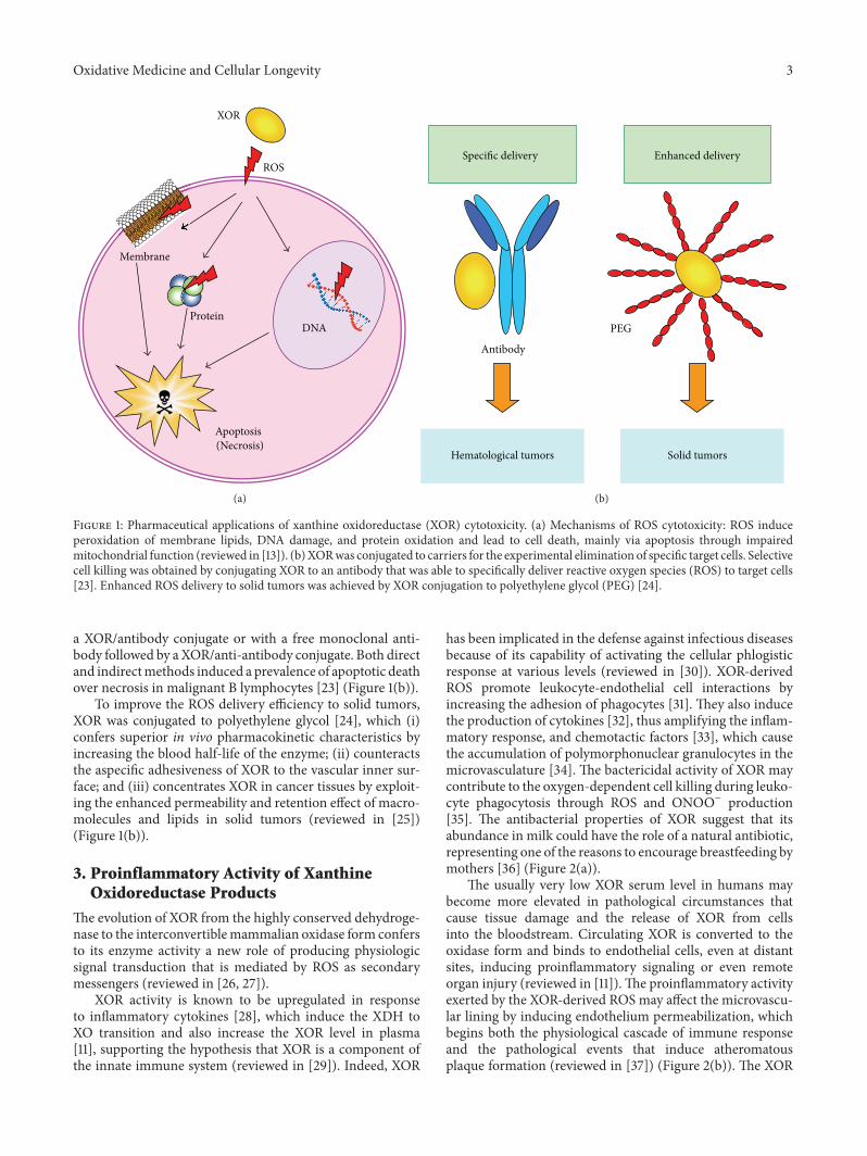
Oxidative Medicine and Cellular Longevity 3
Apoptosis
ROS
XOR
Membrane
ProteinDNA
(Necrosis)
(a)
Antibody
PEG
Hematological tumors Solid tumors
Specific delivery Enhanced delivery
(b)
Figure 1: Pharmaceutical applications of xanthine oxidoreductase (XOR) cytotoxicity. (a) Mechanisms of ROS cytotoxicity: ROS induceperoxidation of membrane lipids, DNA damage, and protein oxidation and lead to cell death, mainly via apoptosis through impairedmitochondrial function (reviewed in [13]). (b)XORwas conjugated to carriers for the experimental elimination of specific target cells. Selectivecell killing was obtained by conjugating XOR to an antibody that was able to specifically deliver reactive oxygen species (ROS) to target cells[23]. Enhanced ROS delivery to solid tumors was achieved by XOR conjugation to polyethylene glycol (PEG) [24].
a XOR/antibody conjugate or with a free monoclonal anti-body followed by a XOR/anti-antibody conjugate. Both directand indirectmethods induced a prevalence of apoptotic deathover necrosis in malignant B lymphocytes [23] (Figure 1(b)).
To improve the ROS delivery efficiency to solid tumors,XOR was conjugated to polyethylene glycol [24], which (i)confers superior in vivo pharmacokinetic characteristics byincreasing the blood half-life of the enzyme; (ii) counteractsthe aspecific adhesiveness of XOR to the vascular inner sur-face; and (iii) concentrates XOR in cancer tissues by exploit-ing the enhanced permeability and retention effect of macro-molecules and lipids in solid tumors (reviewed in [25])(Figure 1(b)).
3. Proinflammatory Activity of XanthineOxidoreductase Products
The evolution of XOR from the highly conserved dehydroge-nase to the interconvertiblemammalian oxidase form confersto its enzyme activity a new role of producing physiologicsignal transduction that is mediated by ROS as secondarymessengers (reviewed in [26, 27]).
XOR activity is known to be upregulated in responseto inflammatory cytokines [28], which induce the XDH toXO transition and also increase the XOR level in plasma[11], supporting the hypothesis that XOR is a component ofthe innate immune system (reviewed in [29]). Indeed, XOR
has been implicated in the defense against infectious diseasesbecause of its capability of activating the cellular phlogisticresponse at various levels (reviewed in [30]). XOR-derivedROS promote leukocyte-endothelial cell interactions byincreasing the adhesion of phagocytes [31]. They also inducethe production of cytokines [32], thus amplifying the inflam-matory response, and chemotactic factors [33], which causethe accumulation of polymorphonuclear granulocytes in themicrovasculature [34]. The bactericidal activity of XOR maycontribute to the oxygen-dependent cell killing during leuko-cyte phagocytosis through ROS and ONOO− production[35]. The antibacterial properties of XOR suggest that itsabundance in milk could have the role of a natural antibiotic,representing one of the reasons to encourage breastfeeding bymothers [36] (Figure 2(a)).
The usually very low XOR serum level in humans maybecome more elevated in pathological circumstances thatcause tissue damage and the release of XOR from cellsinto the bloodstream. Circulating XOR is converted to theoxidase form and binds to endothelial cells, even at distantsites, inducing proinflammatory signaling or even remoteorgan injury (reviewed in [11]).The proinflammatory activityexerted by the XOR-derived ROS may affect the microvascu-lar lining by inducing endothelium permeabilization, whichbegins both the physiological cascade of immune responseand the pathological events that induce atheromatousplaque formation (reviewed in [37]) (Figure 2(b)). The XOR
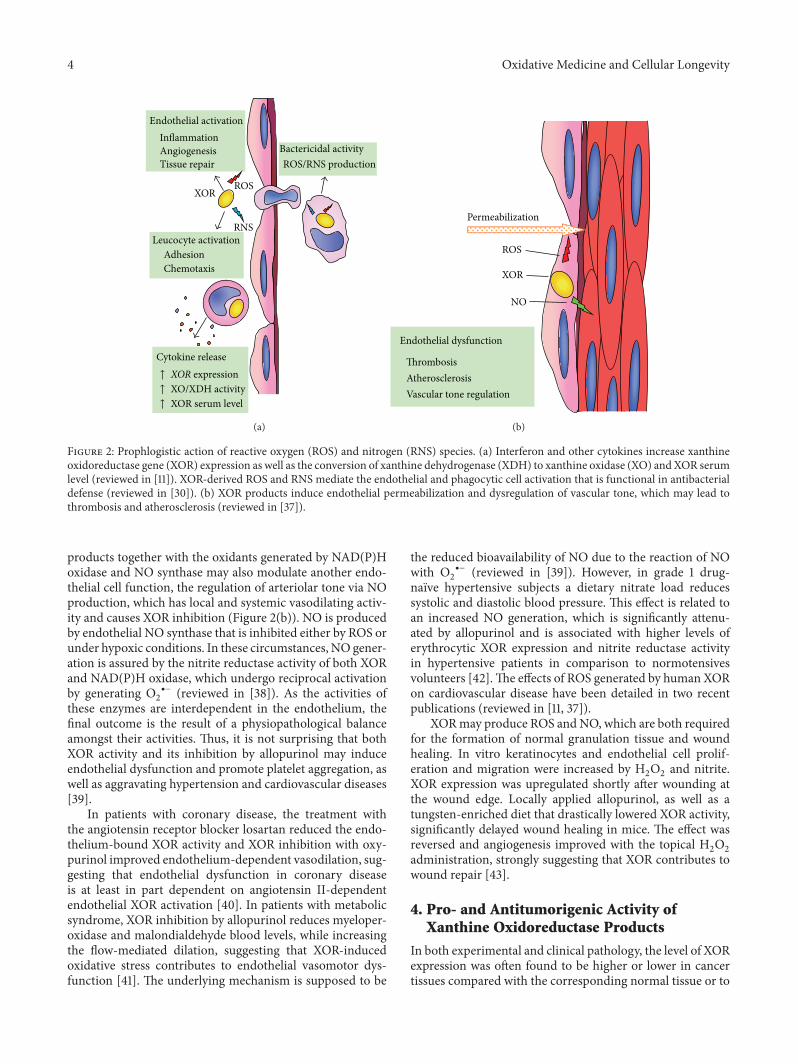
4 Oxidative Medicine and Cellular Longevity
InflammationAngiogenesis Tissue repair
ROS
AdhesionChemotaxis
XOR
Cytokine release
ROS/RNS production
RNS
Endothelial activation
Leucocyte activation
Bactericidal activity
↑ XOR serum level↑ XO/XDH activity↑ XOR expression
(a)
ThrombosisAtherosclerosisVascular tone regulation
ROS
Permeabilization
NO
XOR
Endothelial dysfunction
(b)
Figure 2: Prophlogistic action of reactive oxygen (ROS) and nitrogen (RNS) species. (a) Interferon and other cytokines increase xanthineoxidoreductase gene (XOR) expression as well as the conversion of xanthine dehydrogenase (XDH) to xanthine oxidase (XO) and XOR serumlevel (reviewed in [11]). XOR-derived ROS and RNS mediate the endothelial and phagocytic cell activation that is functional in antibacterialdefense (reviewed in [30]). (b) XOR products induce endothelial permeabilization and dysregulation of vascular tone, which may lead tothrombosis and atherosclerosis (reviewed in [37]).
products together with the oxidants generated by NAD(P)Hoxidase and NO synthase may also modulate another endo-thelial cell function, the regulation of arteriolar tone via NOproduction, which has local and systemic vasodilating activ-ity and causes XOR inhibition (Figure 2(b)). NO is producedby endothelial NO synthase that is inhibited either by ROS orunder hypoxic conditions. In these circumstances, NO gener-ation is assured by the nitrite reductase activity of both XORand NAD(P)H oxidase, which undergo reciprocal activationby generating O
2
∙− (reviewed in [38]). As the activities ofthese enzymes are interdependent in the endothelium, thefinal outcome is the result of a physiopathological balanceamongst their activities. Thus, it is not surprising that bothXOR activity and its inhibition by allopurinol may induceendothelial dysfunction and promote platelet aggregation, aswell as aggravating hypertension and cardiovascular diseases[39].
In patients with coronary disease, the treatment withthe angiotensin receptor blocker losartan reduced the endo-thelium-bound XOR activity and XOR inhibition with oxy-purinol improved endothelium-dependent vasodilation, sug-gesting that endothelial dysfunction in coronary diseaseis at least in part dependent on angiotensin II-dependentendothelial XOR activation [40]. In patients with metabolicsyndrome, XOR inhibition by allopurinol reduces myeloper-oxidase and malondialdehyde blood levels, while increasingthe flow-mediated dilation, suggesting that XOR-inducedoxidative stress contributes to endothelial vasomotor dys-function [41]. The underlying mechanism is supposed to be
the reduced bioavailability of NO due to the reaction of NOwith O
2
∙− (reviewed in [39]). However, in grade 1 drug-näıve hypertensive subjects a dietary nitrate load reducessystolic and diastolic blood pressure. This effect is related toan increased NO generation, which is significantly attenu-ated by allopurinol and is associated with higher levels oferythrocytic XOR expression and nitrite reductase activityin hypertensive patients in comparison to normotensivesvolunteers [42].The effects of ROS generated by human XORon cardiovascular disease have been detailed in two recentpublications (reviewed in [11, 37]).
XORmay produce ROS andNO, which are both requiredfor the formation of normal granulation tissue and woundhealing. In vitro keratinocytes and endothelial cell prolif-eration and migration were increased by H
2O2and nitrite.
XOR expression was upregulated shortly after wounding atthe wound edge. Locally applied allopurinol, as well as atungsten-enriched diet that drastically lowered XOR activity,significantly delayed wound healing in mice. The effect wasreversed and angiogenesis improved with the topical H
2O2
administration, strongly suggesting that XOR contributes towound repair [43].
4. Pro- and Antitumorigenic Activity ofXanthine Oxidoreductase Products
In both experimental and clinical pathology, the level of XORexpression was often found to be higher or lower in cancertissues compared with the corresponding normal tissue or to
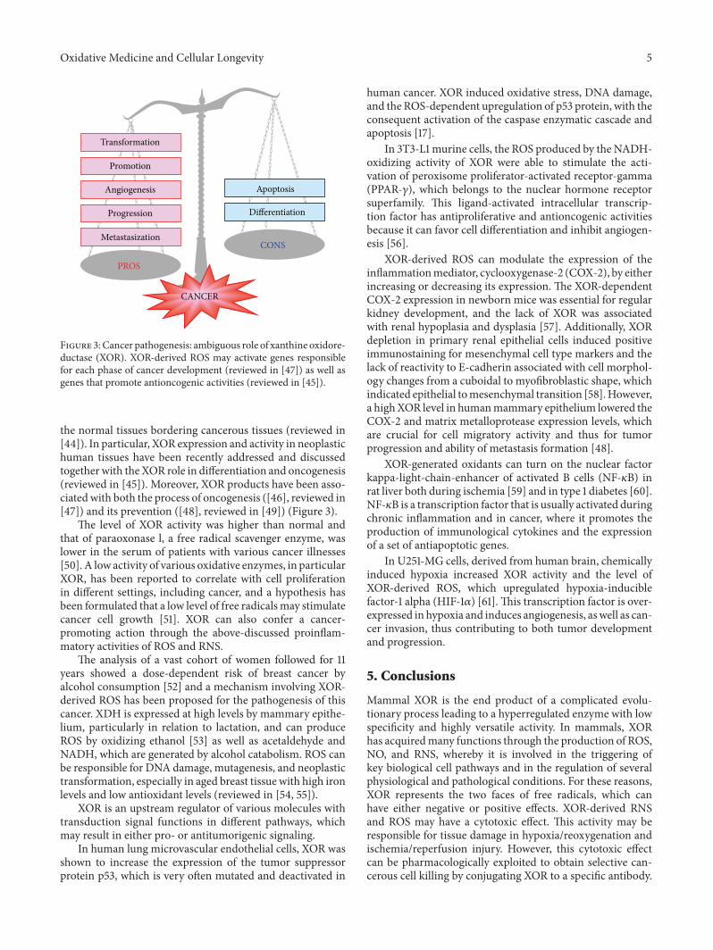
Oxidative Medicine and Cellular Longevity 5
Promotion
Differentiation
Transformation
Metastasization
Progression
Angiogenesis Apoptosis
PROS
CONS
CANCER
Figure 3: Cancer pathogenesis: ambiguous role of xanthine oxidore-ductase (XOR). XOR-derived ROS may activate genes responsiblefor each phase of cancer development (reviewed in [47]) as well asgenes that promote antioncogenic activities (reviewed in [45]).
the normal tissues bordering cancerous tissues (reviewed in[44]). In particular, XOR expression and activity in neoplastichuman tissues have been recently addressed and discussedtogether with the XOR role in differentiation and oncogenesis(reviewed in [45]). Moreover, XOR products have been asso-ciated with both the process of oncogenesis ([46], reviewed in[47]) and its prevention ([48], reviewed in [49]) (Figure 3).
The level of XOR activity was higher than normal andthat of paraoxonase l, a free radical scavenger enzyme, waslower in the serum of patients with various cancer illnesses[50]. A low activity of various oxidative enzymes, in particularXOR, has been reported to correlate with cell proliferationin different settings, including cancer, and a hypothesis hasbeen formulated that a low level of free radicalsmay stimulatecancer cell growth [51]. XOR can also confer a cancer-promoting action through the above-discussed proinflam-matory activities of ROS and RNS.
The analysis of a vast cohort of women followed for 11years showed a dose-dependent risk of breast cancer byalcohol consumption [52] and a mechanism involving XOR-derived ROS has been proposed for the pathogenesis of thiscancer. XDH is expressed at high levels by mammary epithe-lium, particularly in relation to lactation, and can produceROS by oxidizing ethanol [53] as well as acetaldehyde andNADH, which are generated by alcohol catabolism. ROS canbe responsible for DNA damage, mutagenesis, and neoplastictransformation, especially in aged breast tissue with high ironlevels and low antioxidant levels (reviewed in [54, 55]).
XOR is an upstream regulator of various molecules withtransduction signal functions in different pathways, whichmay result in either pro- or antitumorigenic signaling.
In human lung microvascular endothelial cells, XOR wasshown to increase the expression of the tumor suppressorprotein p53, which is very often mutated and deactivated in
human cancer. XOR induced oxidative stress, DNA damage,and the ROS-dependent upregulation of p53 protein, with theconsequent activation of the caspase enzymatic cascade andapoptosis [17].
In 3T3-L1 murine cells, the ROS produced by the NADH-oxidizing activity of XOR were able to stimulate the acti-vation of peroxisome proliferator-activated receptor-gamma(PPAR-𝛾), which belongs to the nuclear hormone receptorsuperfamily. This ligand-activated intracellular transcrip-tion factor has antiproliferative and antioncogenic activitiesbecause it can favor cell differentiation and inhibit angiogen-esis [56].
XOR-derived ROS can modulate the expression of theinflammationmediator, cyclooxygenase-2 (COX-2), by eitherincreasing or decreasing its expression. The XOR-dependentCOX-2 expression in newborn mice was essential for regularkidney development, and the lack of XOR was associatedwith renal hypoplasia and dysplasia [57]. Additionally, XORdepletion in primary renal epithelial cells induced positiveimmunostaining for mesenchymal cell type markers and thelack of reactivity to E-cadherin associated with cell morphol-ogy changes from a cuboidal to myofibroblastic shape, whichindicated epithelial tomesenchymal transition [58].However,a highXOR level in humanmammary epithelium lowered theCOX-2 and matrix metalloprotease expression levels, whichare crucial for cell migratory activity and thus for tumorprogression and ability of metastasis formation [48].
XOR-generated oxidants can turn on the nuclear factorkappa-light-chain-enhancer of activated B cells (NF-𝜅B) inrat liver both during ischemia [59] and in type 1 diabetes [60].NF-𝜅B is a transcription factor that is usually activated duringchronic inflammation and in cancer, where it promotes theproduction of immunological cytokines and the expressionof a set of antiapoptotic genes.
In U251-MG cells, derived from human brain, chemicallyinduced hypoxia increased XOR activity and the level ofXOR-derived ROS, which upregulated hypoxia-induciblefactor-1 alpha (HIF-1𝛼) [61]. This transcription factor is over-expressed in hypoxia and induces angiogenesis, aswell as can-cer invasion, thus contributing to both tumor developmentand progression.
5. Conclusions
Mammal XOR is the end product of a complicated evolu-tionary process leading to a hyperregulated enzyme with lowspecificity and highly versatile activity. In mammals, XORhas acquiredmany functions through the production of ROS,NO, and RNS, whereby it is involved in the triggering ofkey biological cell pathways and in the regulation of severalphysiological and pathological conditions. For these reasons,XOR represents the two faces of free radicals, which canhave either negative or positive effects. XOR-derived RNSand ROS may have a cytotoxic effect. This activity may beresponsible for tissue damage in hypoxia/reoxygenation andischemia/reperfusion injury. However, this cytotoxic effectcan be pharmacologically exploited to obtain selective can-cerous cell killing by conjugating XOR to a specific antibody.
6 Oxidative Medicine and Cellular Longevity
XORactivity increases during infectious diseases and its cyto-toxic action is useful for the defenses against bacteria. Addi-tionally, XOR-derived NO and ROS have proinflammatoryactivity because they regulate endothelial functions, by bothincreasing the permeability of vascular lining andmodulatingthe arteriolar tone. For this reason, XOR has been implicatedin hypertension, cardiovascular diseases, and atherosclerosis.XOR-derived ROS are also involved in cancer pathogenesisbecause they may promote neoplastic transformation byactivating target genes with prophlogistic, antiapoptotic, andproliferative actions. Moreover, they favor the progression tomalignancy by inducing angiogenesis and cell migration. Onthe other hand, XOR products may activate the expressionof the proapoptotic protein p53 and of transcription factorsbelonging to the nuclear hormone receptor superfamily withantitumorigenic and antiproliferative activity, promoting celldifferentiation and inhibiting angiogenesis.
Highlights
(i) XOR-derived ROS, NO, and RNS have proinflamma-tory and bactericidal activities.
(ii) XOR products may be cytotoxic in many circum-stances.
(iii) XOR products modulate endothelial function andarteriolar tone.
(iv) XOR products may induce mutagenesis, cell prolifer-ation, and tumor progression.
(v) XOR products are associated with apoptosis and celldifferentiation.
Abbreviations
COX-2: Cyclooxygenase-2FAD: Flavin adenine dinucleotideHO∙: Hydroxyl radicalH2O2: Hydrogen peroxide
Mo-co: Molybdenum-containing molybdopterincofactor
NAD+: Oxidized nicotinamide adenine dinucleotideNADH: Reduced nicotinamide adenine dinucleotideNF-𝜅B: Nuclear factor kappa-light-chain-enhancer
of activated B cellsNO: Nitric oxideONOO−: PeroxynitriteRNS: Reactive nitrogen speciesROS: Reactive oxygen speciesO2: Molecular oxygen
O2
∙−: Superoxide anionXDH: Xanthine dehydrogenaseXO: Xanthine oxidaseXOR: Xanthine oxidoreductase.
Conflict of Interests
The authors declare that there is no conflict of interestsregarding the publication of this paper.
Acknowledgment
This work was supported by the Pallotti Legacies for CancerResearch.
References
[1] D.A. Parks andD.N.Granger, “Xanthine oxidase: biochemistry,distribution and physiology,” Acta Physiologica Scandinavica,vol. 126, no. 548, pp. 87–99, 1986.
[2] R. Hille and T. Nishino, “Xanthine oxidase and xanthine dehy-drogenase,”TheFASEB Journal, vol. 9, no. 11, pp. 995–1003, 1995.
[3] B. N. Ames, R. Cathcart, E. Schwiers, and P. Hochstein, “Uricacid provides an antioxidant defense in humans against oxi-dant- and radical-caused aging and cancer: a hypothesis,” Pro-ceedings of the National Academy of Sciences of the United Statesof America, vol. 78, no. 11, pp. 6858–6862, 1981.
[4] P. Xu, P. LaVallee, and J. R. Hoidal, “Repressed expression ofthe human xanthine oxidoreductase gene. E-box and TATA-like elements restrict ground state transcriptional activity,”TheJournal of Biological Chemistry, vol. 275, no. 8, pp. 5918–5926,2000.
[5] A. Kooij, “A re-evaluation of the tissue distribution and physiol-ogy of xanthine oxidoreductase,”Histochemical Journal, vol. 26,no. 12, pp. 889–915, 1994.
[6] C. E. Berry and J. M. Hare, “Xanthine oxidoreductase andcardiovascular disease: molecular mechanisms and pathophys-iological implications,”The Journal of Physiology, vol. 555, no. 3,pp. 589–606, 2004.
[7] A. Boueiz, M. Damarla, and P. M. Hassoun, “Xanthine oxi-doreductase in respiratory and cardiovascular disorders,” TheAmerican Journal of Physiology—Lung Cellular and MolecularPhysiology, vol. 294, no. 5, pp. L830–L840, 2008.
[8] E. Della Corte and F. Stirpe, “The regulation of rat liver xanthineoxidase. Involvement of thiol groups in the conversion of theenzyme activity from dehydrogenase (type D) into oxidase(type O) and purification of the enzyme,” Biochemical Journal,vol. 126, no. 3, pp. 739–745, 1972.
[9] N. Cantu-Medellin and E. E. Kelley, “Xanthine oxidoreductase-catalyzed reactive species generation: a process in critical needof reevaluation,” Redox Biology, vol. 1, no. 1, pp. 353–358, 2013.
[10] R. Harrison, “Structure and function of xanthine oxidoreduc-tase: where are we now?” Free Radical Biology andMedicine, vol.33, no. 6, pp. 774–797, 2002.
[11] M. G. Battelli, A. Bolognesi, and L. Polito, “Pathophysiology ofcirculating xanthine oxidoreductase: new emerging roles for amulti-tasking enzyme,” Biochimica et Biophysica Acta, vol. 1842,no. 9, pp. 1502–1517, 2014.
[12] C. Li and R. M. Jackson, “Reactive species mechanisms ofcellular hypoxia-reoxygenation injury,” American Journal ofPhysiology: Cell Physiology, vol. 282, no. 2, pp. C227–C241, 2002.
[13] A. H. Bhat, K. B. Dar, S. Anees et al., “Oxidative stress, mito-chondrial dysfunction and neurodegenerative diseases; amech-anistic insight,” Biomedicine & Pharmacotherapy, vol. 74, pp.101–110, 2015.
[14] M. Chiricolo, P. L. Tazzari, A. Abbondanza, A. Dinota, and M.G. Battelli, “Cytotoxicity of, and DNA damage by, active oxygenspecies produced by xanthine oxidase,” FEBS Letters, vol. 291,no. 2, pp. 173–176, 1991.
[15] M. G. Battelli, S. Musiani, P. L. Tazzari, and F. Stirpe, “Oxida-tive stress to human lymphocytes by xanthine oxidoreductaseactivity,” Free Radical Research, vol. 35, no. 6, pp. 665–679, 2001.
Oxidative Medicine and Cellular Longevity 7
[16] C.-C. Huang, K.-L. Chen, C. H. A. Cheung, and J.-Y. Chang,“Autophagy induced by cathepsin S inhibition induces earlyROS production, oxidative DNA damage, and cell death viaxanthine oxidase,” Free Radical Biology and Medicine, vol. 65,pp. 1473–1486, 2013.
[17] B. S. Kim, L. Serebreni, O. Hamdan et al., “Xanthine oxidore-ductase is a critical mediator of cigarette smoke-induced endo-thelial cell DNA damage and apoptosis,” Free Radical Biologyand Medicine, vol. 60, pp. 336–343, 2013.
[18] M. G. Battelli, A. Abbondanza, P. L. Tazzari et al., “Selectivecytotoxicity of an oxygen-radical-generating enzyme conju-gated to a monoclonal antibody,” Clinical and ExperimentalImmunology, vol. 73, no. 1, pp. 128–133, 1988.
[19] P. L. Tazzari, M. G. Battelli, A. Abbondanza et al., “Targeting ofa plasma cell line with a conjugate containing xanthine oxidaseand the monoclonal antibody 62B1,” Transplantation, vol. 48,no. 1, pp. 119–122, 1989.
[20] A. Dinota, P. L. Tazzari, A. Abbondanza, M. G. Battelli, M.Gobbi, and F. Stirpe, “Bone marrow purging by a xanthine oxi-dase-antibody conjugate,” Bone Marrow Transplantation, vol. 6,no. 1, pp. 31–36, 1990.
[21] M. G. Battelli, A. Abbondanza, P. L. Tazzari, A. Bolognesi, R. M.Lemoli, and F. Stirpe, “T lymphocyte killing by a xanthine-oxi-dase-containing immunotoxin,” Cancer Immunology, Immun-otherapy, vol. 35, no. 6, pp. 421–425, 1992.
[22] S. J. Dixon and B. R. Stockwell, “The role of iron and reactiveoxygen species in cell death,” Nature Chemical Biology, vol. 10,no. 1, pp. 9–17, 2014.
[23] M. G. Battelli, L. Polito, F. Falà et al., “Toxicity of xanthine oxi-doreductase to malignant B lymphocytes,” Journal of BiologicalRegulators & Homeostatic Agents, vol. 19, no. 3-4, pp. 120–129,2005.
[24] T. Sawa, J. Wu, T. Akaike, and H. Maeda, “Tumor-targetingchemotherapy by a xanthine oxidase-polymer conjugate thatgenerates oxygen-free radicals in tumor tissue,” Cancer Re-search, vol. 60, no. 3, pp. 666–671, 2000.
[25] J. Fang, T. Seki, and H. Maeda, “Therapeutic strategies by mod-ulating oxygen stress in cancer and inflammation,” AdvancedDrug Delivery Reviews, vol. 61, no. 4, pp. 290–302, 2009.
[26] H. Sauer, M. Wartenberg, and J. Hescheler, “Reactive oxygenspecies as intracellular messengers during cell growth anddifferentiation,”Cellular Physiology and Biochemistry, vol. 11, no.4, pp. 173–186, 2001.
[27] A. Meneshian and G. B. Bulkley, “The physiology of endothelialxanthine oxidase: from urate catabolism to reperfusion injuryto inflammatory signal transduction,” Microcirculation, vol. 9,no. 3, pp. 161–175, 2002.
[28] S. Page, D. Powell, M. Benboubetra et al., “Xanthine oxi-doreductase in human mammary epithelial cells: activation inresponse to inflammatory cytokines,” Biochimica et BiophysicaActa—General Subjects, vol. 1381, no. 2, pp. 191–202, 1998.
[29] A.Agarwal, A. Banerjee, andU.C. Banerjee, “Xanthine oxidore-ductase: a journey from purine metabolism to cardiovascularexcitation-contraction coupling,”Critical Reviews in Biotechnol-ogy, vol. 31, no. 3, pp. 264–280, 2011.
[30] H.M.Martin, J. T.Hancock, V. Salisbury, andR.Harrison, “Roleof xanthine oxidoreductase as an antimicrobial agent,” Infectionand Immunity, vol. 72, no. 9, pp. 4933–4939, 2004.
[31] M. Suzuki, M. B. Grisham, and D. N. Granger, “Leukocyte-endothelial cell adhesive interactions: role of xanthine oxidase-derived oxidants,” Journal of Leukocyte Biology, vol. 50, no. 5,pp. 488–494, 1991.
[32] R. Shenkar and E. Abraham, “Plasma from hemorrhagedmice activates CREB and increases cytokine expression inlung mononuclear cells through a xanthine oxidase-dependentmechanism,” American Journal of Respiratory Cell and Molecu-lar Biology, vol. 14, no. 2, pp. 198–206, 1996.
[33] W. F. Petrone, D. K. English, K. Wong, and J. M. McCord, “Freeradicals and inflammation: superoxide-dependent activation ofa neutrophil chemotactic factor in plasma,” Proceedings of theNational Academy of Sciences of the United States of America,vol. 77, no. 2, pp. 1159–1163, 1980.
[34] M. J. Müller, B. Vollmar, H.-P. Friedl, and M. D. Menger, “Xan-thine oxidase and superoxide radicals in portal triad cross-clamping-induced microvascular reperfusion injury of theliver,” Free Radical Biology and Medicine, vol. 21, no. 2, pp. 189–197, 1996.
[35] E. Tubaro, B. Lotti, C. Santiangelli, and G. Cavallo, “Xanthineoxidase: an enzyme playing a role in the killing mechanismof polymorphonuclear leucocytes,” Biochemical Pharmacology,vol. 29, no. 21, pp. 3018–3020, 1980.
[36] C. R. Stevens, T. M. Millar, J. G. Clinch, J. M. Kanczler, T. Bod-amyali, and D. R. Blake, “Antibacterial properties of xanthineoxidase in humanmilk,”The Lancet, vol. 356, no. 9232, pp. 829–830, 2000.
[37] M. G. Battelli, L. Polito, and A. Bolognesi, “Xanthine oxi-doreductase in atherosclerosis pathogenesis: not only oxidativestress,” Atherosclerosis, vol. 237, no. 2, pp. 562–567, 2014.
[38] H. Cai and D. G. Harrison, “Endothelial dysfunction in car-diovascular diseases: the role of oxidant stress,” CirculationResearch, vol. 87, no. 10, pp. 840–844, 2000.
[39] N. Cantu-Medellin and E. E. Kelley, “Xanthine oxidoreductase-catalyzed reduction of nitrite to nitric oxide: insights regardingwhere, when and how,” Nitric Oxide, vol. 34, pp. 19–26, 2013.
[40] U. Landmesser, S. Spiekermann, C. Preuss et al., “AngiotensinII induces endothelial xanthine oxidase activation: role forendothelial dysfunction in patients with coronary disease,”Arteriosclerosis, Thrombosis, and Vascular Biology, vol. 27, no.4, pp. 943–948, 2007.
[41] O. Yiginer, F. Ozcelik, T. Inanc et al., “Allopurinol improvesendothelial function and reduces oxidant-inflammatory en-zyme of myeloperoxidase in metabolic syndrome,” ClinicalResearch in Cardiology, vol. 97, no. 5, pp. 334–340, 2008.
[42] S. M. Ghosh, V. Kapil, I. Fuentes-Calvo et al., “Enhancedvasodilator activity of nitrite in hypertension: critical role forerythrocytic xanthine oxidoreductase and translational poten-tial,” Hypertension, vol. 61, no. 5, pp. 1091–1102, 2013.
[43] M. C. Madigan, R. M. McEnaney, A. J. Shukla et al., “Xanthineoxidoreductase function contributes to normal wound healing,”Molecular Medicine, vol. 21, no. 1, pp. 313–322, 2015.
[44] G. Weber, “Biochemical strategy of cancer cells and the designof chemotherapy: G.H.A. Clowes memorial lecture,” CancerResearch, vol. 43, no. 8, pp. 3466–3493, 1983.
[45] M. G. Battelli, L. Polito, M. Bortolotti, and A. Bolognesi, “Xan-thine oxidoreductase in cancer: more than a differentiationmarker,” Cancer Medicine, In press.
[46] I. A. Shmarakov and M. M. Marchenko, “Xanthine oxidaseactivity in transplantable Guerin’s carcinoma in rats,” VoprosyOnkologii, vol. 55, no. 3, pp. 345–350, 2009.
[47] K. Balamurugan, “HIF-1 at the crossroads of hypoxia, inflam-mation, and cancer,” International Journal of Cancer, 2015.
[48] M. A. Fini, D. Orchard-Webb, B. Kosmider et al., “Migratoryactivity of human breast cancer cells ismodulated by differential
8 Oxidative Medicine and Cellular Longevity
expression of xanthine oxidoreductase,” Journal of CellularBiochemistry, vol. 105, no. 4, pp. 1008–1026, 2008.
[49] M. Valko, C. J. Rhodes, J. Moncol, M. Izakovic, and M. Mazur,“Free radicals, metals and antioxidants in oxidative stress-induced cancer,” Chemico-Biological Interactions, vol. 160, no. 1,pp. 1–40, 2006.
[50] Z. Q. Samra, S. Pervaiz, S. Shaheen, N. Dar, and M. A. Athar,“Determination of oxygen derived free radicals producer (xan-thine oxidase) and scavenger (paraoxonasel) enzymes and lipidparameters in different cancer patients,” Clinical Laboratory,vol. 57, no. 9-10, pp. 741–747, 2011.
[51] A. S. Sun and A. I. Cederbaum, “Oxidoreductase activities innormal rat liver, tumor-bearing rat liver, and hepatoma HC-252,” Cancer Research, vol. 40, no. 12, pp. 4677–4681, 1980.
[52] S. A. Smith-Warner, D. Spiegelman, S.-S. Yaun et al., “Alcoholand breast cancer in women: a pooled analysis of cohortstudies,” The Journal of the American Medical Association, vol.279, no. 7, pp. 535–540, 1998.
[53] G. D. Castro, A. M. A. Delgado de Layo, M. H. Costantini,and J. A. Castro, “Cytosolic xanthine oxidoreductase mediatedbioactivation of ethanol to acetaldehyde and free radicals in ratbreast tissue. Its potential role in alcohol-promoted mammarycancer,” Toxicology, vol. 160, no. 1–3, pp. 11–18, 2001.
[54] R. M. Wright, J. L. McManaman, and J. E. Repine, “Alcohol-induced breast cancer: a proposed mechanism,” Free RadicalBiology and Medicine, vol. 26, no. 3-4, pp. 348–354, 1999.
[55] R. G. Dumitrescu and P. G. Shields, “The etiology of alcohol-induced breast cancer,”Alcohol, vol. 35, no. 3, pp. 213–225, 2005.
[56] K. J. Cheung, I. Tzameli, P. Pissios et al., “Xanthine oxidore-ductase is a regulator of adipogenesis and PPAR𝛾 activity,” CellMetabolism, vol. 5, no. 2, pp. 115–128, 2007.
[57] T. Ohtsubo, I. I. Rovira, M. F. Starost, C. Liu, and T. Finkel,“Xanthine oxidoreductase is an endogenous regulator of cy-clooxygenase-2,” Circulation Research, vol. 95, no. 11, pp. 1118–1124, 2004.
[58] T. Ohtsubo, K. Matsumura, K. Sakagami et al., “Xanthine oxi-doreductase depletion induces renal interstitial fibrosis throughaberrant lipid and purine accumulation in renal tubules,”Hypertension, vol. 54, no. 4, pp. 868–876, 2009.
[59] N.Matsui, I. Satsuki, Y.Morita et al., “Xanthine oxidase-derivedreactive oxygen species activate nuclear factor kappa B duringhepatic ischemia in rats,”The Japanese Journal of Pharmacology,vol. 84, no. 3, pp. 363–366, 2000.
[60] M. Romagnoli, M.-C. Gomez-Cabrera, M.-G. Perrelli et al.,“Xanthine oxidase-induced oxidative stress causes activation ofNF-𝜅B and inflammation in the liver of type I diabetic rats,” FreeRadical Biology and Medicine, vol. 49, no. 2, pp. 171–177, 2010.
[61] C. E. Griguer, C. R. Oliva, E. E. Kelley, G. I. Giles, J. R. LancasterJr., and G. Y. Gillespie, “Xanthine oxidase-dependent regulationof hypoxia-inducible factor in cancer cells,” Cancer Research,vol. 66, no. 4, pp. 2257–2263, 2006.
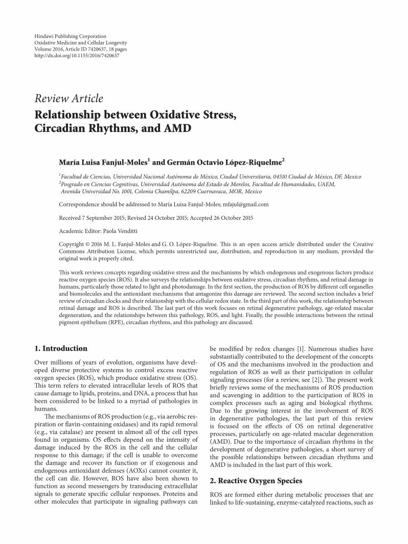
Review ArticleRelationship between Oxidative Stress,Circadian Rhythms, and AMD
María Luisa Fanjul-Moles1 and Germán Octavio López-Riquelme2
1Facultad de Ciencias, Universidad Nacional Autónoma de México, Ciudad Universitaria, 04510 Ciudad de México, DF, Mexico2Posgrado en Ciencias Cognitivas, Universidad Autónoma del Estado de Morelos, Facultad de Humanidades, UAEM,Avenida Universidad No. 1001, Colonia Chamilpa, 62209 Cuernavaca, MOR, Mexico
Correspondence should be addressed to Maŕıa Luisa Fanjul-Moles; [email protected]
Received 7 September 2015; Revised 24 October 2015; Accepted 26 October 2015
Academic Editor: Paola Venditti
Copyright © 2016 M. L. Fanjul-Moles and G. O. López-Riquelme. This is an open access article distributed under the CreativeCommons Attribution License, which permits unrestricted use, distribution, and reproduction in any medium, provided theoriginal work is properly cited.
This work reviews concepts regarding oxidative stress and the mechanisms by which endogenous and exogenous factors producereactive oxygen species (ROS). It also surveys the relationships between oxidative stress, circadian rhythms, and retinal damage inhumans, particularly those related to light and photodamage. In the first section, the production of ROS by different cell organellesand biomolecules and the antioxidant mechanisms that antagonize this damage are reviewed. The second section includes a briefreview of circadian clocks and their relationship with the cellular redox state. In the third part of this work, the relationship betweenretinal damage and ROS is described. The last part of this work focuses on retinal degenerative pathology, age-related maculardegeneration, and the relationships between this pathology, ROS, and light. Finally, the possible interactions between the retinalpigment epithelium (RPE), circadian rhythms, and this pathology are discussed.
1. Introduction
Over millions of years of evolution, organisms have devel-oped diverse protective systems to control excess reactiveoxygen species (ROS), which produce oxidative stress (OS).This term refers to elevated intracellular levels of ROS thatcause damage to lipids, proteins, and DNA, a process that hasbeen considered to be linked to a myriad of pathologies inhumans.
Themechanisms of ROS production (e.g., via aerobic res-piration or flavin-containing oxidases) and its rapid removal(e.g., via catalase) are present in almost all of the cell typesfound in organisms. OS effects depend on the intensity ofdamage induced by the ROS in the cell and the cellularresponse to this damage; if the cell is unable to overcomethe damage and recover its function or if exogenous andendogenous antioxidant defenses (AOXs) cannot counter it,the cell can die. However, ROS have also been shown tofunction as second messengers by transducing extracellularsignals to generate specific cellular responses. Proteins andother molecules that participate in signaling pathways can
be modified by redox changes [1]. Numerous studies havesubstantially contributed to the development of the conceptsof OS and the mechanisms involved in the production andregulation of ROS as well as their participation in cellularsignaling processes (for a review, see [2]). The present workbriefly reviews some of the mechanisms of ROS productionand scavenging in addition to the participation of ROS incomplex processes such as aging and biological rhythms.Due to the growing interest in the involvement of ROSin degenerative pathologies, the last part of this reviewis focused on the effects of OS on retinal degenerativeprocesses, particularly on age-related macular degeneration(AMD). Due to the importance of circadian rhythms in thedevelopment of degenerative pathologies, a short survey ofthe possible relationships between circadian rhythms andAMD is included in the last part of this work.
2. Reactive Oxygen Species
ROS are formed either during metabolic processes that arelinked to life-sustaining, enzyme-catalyzed reactions, such as
Hindawi Publishing CorporationOxidative Medicine and Cellular LongevityVolume 2016, Article ID 7420637, 18 pageshttp://dx.doi.org/10.1155/2016/7420637
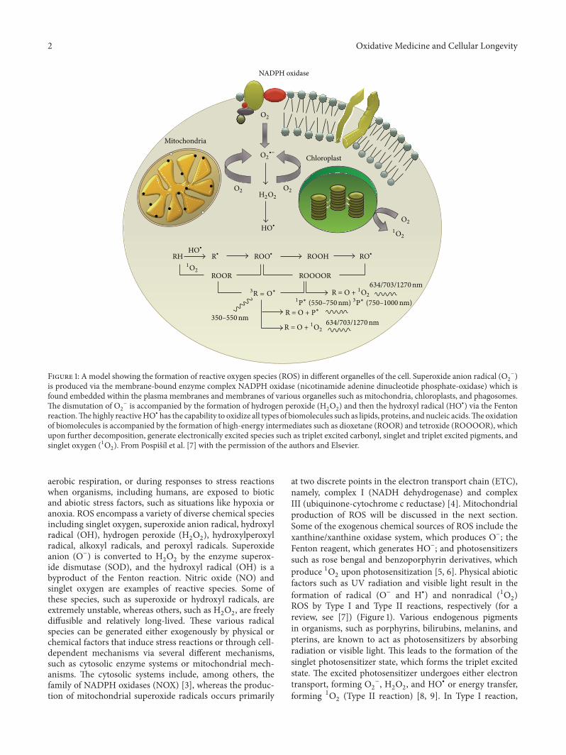
2 Oxidative Medicine and Cellular Longevity
NADPH oxidase
Chloroplast
Mitochondria
O2
O2
O2
O2
O2
∙−
H2O2
HO∙
HO∙
1O2
1O2
RH R∙ ROO∙ ROOH
ROOR ROOOOR
RO∙
350–550nm
1P∗ (550–750nm) 3P∗ (750–1000 nm)
634/703/1270nm
634/703/1270nm3R = O∗ R = O + 1O2
R = O + 1O2
R = O + P∗
Figure 1: A model showing the formation of reactive oxygen species (ROS) in different organelles of the cell. Superoxide anion radical (O2
−)is produced via the membrane-bound enzyme complex NADPH oxidase (nicotinamide adenine dinucleotide phosphate-oxidase) which isfound embedded within the plasma membranes and membranes of various organelles such as mitochondria, chloroplasts, and phagosomes.The dismutation of O
2
− is accompanied by the formation of hydrogen peroxide (H2O2) and then the hydroxyl radical (HO∙) via the Fenton
reaction.Thehighly reactiveHO∙ has the capability to oxidize all types of biomolecules such as lipids, proteins, andnucleic acids.Theoxidationof biomolecules is accompanied by the formation of high-energy intermediates such as dioxetane (ROOR) and tetroxide (ROOOOR), whichupon further decomposition, generate electronically excited species such as triplet excited carbonyl, singlet and triplet excited pigments, andsinglet oxygen (1O
2). From Pospiš́ıl et al. [7] with the permission of the authors and Elsevier.
aerobic respiration, or during responses to stress reactionswhen organisms, including humans, are exposed to bioticand abiotic stress factors, such as situations like hypoxia oranoxia. ROS encompass a variety of diverse chemical speciesincluding singlet oxygen, superoxide anion radical, hydroxylradical (OH), hydrogen peroxide (H
2O2), hydroxylperoxyl
radical, alkoxyl radicals, and peroxyl radicals. Superoxideanion (O−) is converted to H
2O2by the enzyme superox-
ide dismutase (SOD), and the hydroxyl radical (OH) is abyproduct of the Fenton reaction. Nitric oxide (NO) andsinglet oxygen are examples of reactive species. Some ofthese species, such as superoxide or hydroxyl radicals, areextremely unstable, whereas others, such as H
2O2, are freely
diffusible and relatively long-lived. These various radicalspecies can be generated either exogenously by physical orchemical factors that induce stress reactions or through cell-dependent mechanisms via several different mechanisms,such as cytosolic enzyme systems or mitochondrial mech-anisms. The cytosolic systems include, among others, thefamily of NADPH oxidases (NOX) [3], whereas the produc-tion of mitochondrial superoxide radicals occurs primarily
at two discrete points in the electron transport chain (ETC),namely, complex I (NADH dehydrogenase) and complexIII (ubiquinone-cytochrome c reductase) [4]. Mitochondrialproduction of ROS will be discussed in the next section.Some of the exogenous chemical sources of ROS include thexanthine/xanthine oxidase system, which produces O−; theFenton reagent, which generates HO−; and photosensitizerssuch as rose bengal and benzoporphyrin derivatives, whichproduce 1O
2upon photosensitization [5, 6]. Physical abiotic
factors such as UV radiation and visible light result in theformation of radical (O− and H∙) and nonradical (1O
2)
ROS by Type I and Type II reactions, respectively (for areview, see [7]) (Figure 1). Various endogenous pigmentsin organisms, such as porphyrins, bilirubins, melanins, andpterins, are known to act as photosensitizers by absorbingradiation or visible light. This leads to the formation of thesinglet photosensitizer state, which forms the triplet excitedstate. The excited photosensitizer undergoes either electrontransport, forming O
2
−, H2O2, and HO∙ or energy transfer,
forming 1O2(Type II reaction) [8, 9]. In Type I reaction,
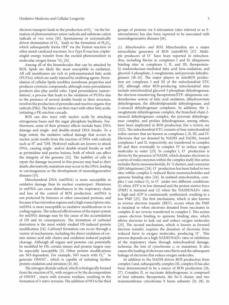
Oxidative Medicine and Cellular Longevity 3
electron transport leads to the production of O2
− via the for-mation of photosensitizer anion radicals and substrate cationradicals or vice versa [10]. Spontaneous or enzymaticallydriven dismutation of O
2
− leads to the formation of H2O2,
which subsequently forms OH∙ via the Fenton reaction orother metal-catalyzed reactions. In a Type II reaction, triplet-singlet energy transfer from the excited photosensitizer tomolecular oxygen forms 1O
2[11].
Among all of the biomolecules that can be attacked byROS, lipids are likely the most susceptible to oxidation.All cell membranes are rich in polyunsaturated fatty acids(PUFAs), which are easily injured by oxidizing agents. Perox-idation of cellular lipids modifies membrane properties andproduces cytotoxic compounds, although some peroxidationproducts also play useful roles. Lipid peroxidation (autoxi-dation), a process that leads to the oxidation of PUFAs dueto the presence of several double bonds in their structure,involves the production of peroxides and reactive organic freeradicals (FRs).The latter can then react with other fatty acids,initiating a FR reaction cascade.
ROS can also react with nucleic acids by attackingnitrogenous bases and the sugar phosphate backbone. Fur-thermore, some of their primary effects include basic DNAdamage and single- and double-strand DNA breaks. To alarge extent, the oxidative radical damage that occurs innucleic acids results from the reaction of DNA with radicalssuch as H∙ and ∙OH. Hydroxyl radicals are known to attackDNA, causing single- and/or double-strand breaks as wellas pyrimidine and purine lesions, both of which can affectthe integrity of the genome [12]. The inability of cells torepair the damage incurred in this process may lead to theirdeath; alternatively,mutationsmay occur in theDNA, leadingto carcinogenesis or the development of neurodegenerativediseases [13].
Mitochondrial DNA (mtDNA) is more susceptible tooxidative damage than its nuclear counterpart. Mutationsin mtDNA can cause disturbances in the respiratory chainand loss of the control of ROS production. mtDNA isnot protected by histones or other associated proteins, andbecause it has intronless regions and a high transcription rate,mtDNA is more susceptible to oxidative modifications in itscoding regions.The reduced effectiveness of the repair systemfor mtDNA damage may be the cause of the accumulationof OS and its consequences. The formation of carbonylderivatives is the most widely studied OS-induced proteinmodification [14]. Carbonyl formation can occur through avariety of mechanisms, including the direct oxidation of cer-tain amino acid side chains and oxidation-induced peptidecleavage. Although all organs and proteins can potentiallybe modified by OS, certain tissues and protein targets maybe especially susceptible [3]. Other protein modificationsare NO-dependent. For example, NO reacts with O
2
∙− togenerate ONOO−, which is capable of initiating furtherprotein oxidation and nitration [15, 16].
Thenitrogen dioxide radical, which is biologically formedfrom the reaction of N
2with oxygen or by the decomposition
of ONOO−, reacts with tyrosine residues, resulting in theformation of 3-nitro-tyrosine.The addition of NO to the thiol
groups of proteins via S-nitrosation (also referred to as S-nitrosylation) has also been reported to be associated withneurodegenerative diseases [16].
2.1. Mitochondria and ROS. Mitochondria are a majorintracellular generator of ROS (mitoROS) [17]. Multi-ple producers of O− have been reported in mitochon-dria, including flavins in complexes I and II; ubiquinonebinding sites in complexes I, II, and III; flavoprotein-Q oxidoreductase-mediated fatty acid beta-oxidation; andglycerol-3-phosphate, 2-oxoglutarate, and pyruvate dehydro-genases [18–21]. The major players in mitoROS produc-tion are complexes I and III of the mitochondrial ETC[18], although other ROS-producing mitochondrial sitesinclude mitochondrial glycerol-3-phosphate dehydrogenase,the electron-transferring flavoprotein/ETF: ubiquinone oxi-doreductase system of fatty acid oxidation, dihydroorotatedehydrogenase, the dihydrolipoamide dehydrogenase, and2-oxoacid dehydrogenase complexes. In addition, the 2-oxoglutarate dehydrogenase complex, the branched-chain 2-oxoacid dehydrogenase complex, the pyruvate dehydroge-nase complex, and proline dehydrogenase, among others,have been implicated in ROS production (for a review, see[22]). The mitochondrial ETC consists of four mitochondrialredox carriers that are known as complexes I, II, III, and IV.Electrons that are donated by NADH and FADH
2toward
complexes I and II, respectively, are transferred to complexIII and then eventually to complex IV to reduce oxygenmolecules to water [23]. In complex I, O− production isdriven by the presence of NADH, which donates electrons toa series of redox enzymes within the complex itself; this seriesincludes flavinmononucleotide, Fe−S clusters, and coenzymeQ10 (ubiquinone) [24]. O− production has been linked to twosites within complex I: reduced flavin mononucleotides andquinone binding sites [24]. In isolated mitochondria, com-plex I can reduce O
2to O− under two different conditions:
(1) when ATP is in low demand and the proton motive force(PMF) is maximal and (2) when the NADH/NAD+ ratiois high and ATP is continuously synthesized, resulting in alow PMF [25]. The first mechanism, which is also knownas reverse electron transfer (RET), occurs when the PMFis maximal or when electrons donated from succinates incomplex II are reverse transferred to complex I. This actioncauses electron binding to quinone binding sites, whichallows electrons to leak out and reduce oxygen molecules[25]. The second mechanism, which is known as forwardelectron transfer, requires the donation of electrons fromreduced form to oxygen molecules, producing O−. Thisprocess depends on a high NADH/NAD+ ratio or inhibitionof the respiratory chain through mitochondrial damage,ischemia, the loss of cytochrome c, or mutations. It alsocauses the loading of electrons onto flavin and the subsequentleakage of electrons that reduce oxygen molecules.
In addition to the NADH-driven ROS production fromcomplex I and, subsequently, complex III, complex II has alsobeen demonstrated to be a source of ROS production [26,27]. Complex II, or succinate dehydrogenase, is composedof four subunits: flavoprotein, the Fe–S cluster, and twotransmembrane cytochrome b heme subunits [21, 28]. In
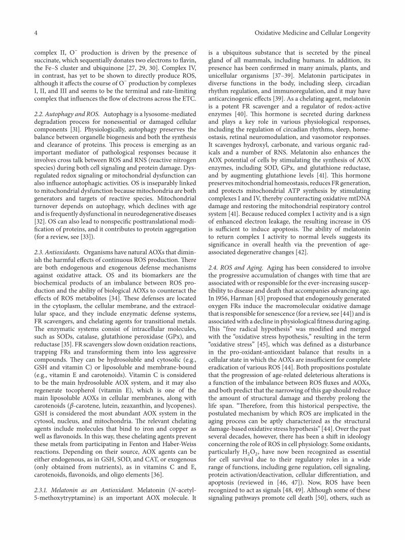
4 Oxidative Medicine and Cellular Longevity
complex II, O− production is driven by the presence ofsuccinate, which sequentially donates two electrons to flavin,the Fe–S cluster and ubiquinone [27, 29, 30]. Complex IV,in contrast, has yet to be shown to directly produce ROS,although it affects the course of O− production by complexesI, II, and III and seems to be the terminal and rate-limitingcomplex that influences the flow of electrons across the ETC.
2.2. Autophagy and ROS. Autophagy is a lysosome-mediateddegradation process for nonessential or damaged cellularcomponents [31]. Physiologically, autophagy preserves thebalance between organelle biogenesis and both the synthesisand clearance of proteins. This process is emerging as animportant mediator of pathological responses because itinvolves cross talk between ROS and RNS (reactive nitrogenspecies) during both cell signaling and protein damage. Dys-regulated redox signaling or mitochondrial dysfunction canalso influence autophagic activities. OS is inseparably linkedtomitochondrial dysfunction becausemitochondria are bothgenerators and targets of reactive species. Mitochondrialturnover depends on autophagy, which declines with ageand is frequently dysfunctional in neurodegenerative diseases[32]. OS can also lead to nonspecific posttranslational modi-fication of proteins, and it contributes to protein aggregation(for a review, see [33]).
2.3. Antioxidants. Organisms have natural AOXs that dimin-ish the harmful effects of continuous ROS production. Thereare both endogenous and exogenous defense mechanismsagainst oxidative attack. OS and its biomarkers are thebiochemical products of an imbalance between ROS pro-duction and the ability of biological AOXs to counteract theeffects of ROS metabolites [34]. These defenses are locatedin the cytoplasm, the cellular membrane, and the extracel-lular space, and they include enzymatic defense systems,FR scavengers, and chelating agents for transitional metals.The enzymatic systems consist of intracellular molecules,such as SODs, catalase, glutathione peroxidase (GPx), andreductase [35]. FR scavengers slow down oxidation reactions,trapping FRs and transforming them into less aggressivecompounds. They can be hydrosoluble and cytosolic (e.g.,GSH and vitamin C) or liposoluble and membrane-bound(e.g., vitamin E and carotenoids). Vitamin C is consideredto be the main hydrosoluble AOX system, and it may alsoregenerate tocopherol (vitamin E), which is one of themain liposoluble AOXs in cellular membranes, along withcarotenoids (𝛽-carotene, lutein, zeaxanthin, and lycopenes).GSH is considered the most abundant AOX system in thecytosol, nucleus, and mitochondria. The relevant chelatingagents include molecules that bind to iron and copper aswell as flavonoids. In this way, these chelating agents preventthese metals from participating in Fenton and Haber-Weissreactions. Depending on their source, AOX agents can beeither endogenous, as in GSH, SOD, and CAT, or exogenous(only obtained from nutrients), as in vitamins C and E,carotenoids, flavonoids, and oligo elements [36].
2.3.1. Melatonin as an Antioxidant. Melatonin (N-acetyl-5-methoxytryptamine) is an important AOX molecule. It
is a ubiquitous substance that is secreted by the pinealgland of all mammals, including humans. In addition, itspresence has been confirmed in many animals, plants, andunicellular organisms [37–39]. Melatonin participates indiverse functions in the body, including sleep, circadianrhythm regulation, and immunoregulation, and it may haveanticarcinogenic effects [39]. As a chelating agent, melatoninis a potent FR scavenger and a regulator of redox-activeenzymes [40]. This hormone is secreted during darknessand plays a key role in various physiological responses,including the regulation of circadian rhythms, sleep, home-ostasis, retinal neuromodulation, and vasomotor responses.It scavenges hydroxyl, carbonate, and various organic rad-icals and a number of RNS. Melatonin also enhances theAOX potential of cells by stimulating the synthesis of AOXenzymes, including SOD, GPx, and glutathione reductase,and by augmenting glutathione levels [41]. This hormonepreservesmitochondrial homeostasis, reduces FR generation,and protects mitochondrial ATP synthesis by stimulatingcomplexes I and IV, thereby counteracting oxidative mtDNAdamage and restoring the mitochondrial respiratory controlsystem [41]. Because reduced complex I activity and is a signof enhanced electron leakage, the resulting increase in OSis sufficient to induce apoptosis. The ability of melatoninto return complex I activity to normal levels suggests itssignificance in overall health via the prevention of age-associated degenerative changes [42].
2.4. ROS and Aging. Aging has been considered to involvethe progressive accumulation of changes with time that areassociated with or responsible for the ever-increasing suscep-tibility to disease and death that accompanies advancing age.In 1956, Harman [43] proposed that endogenously generatedoxygen FRs induce the macromolecular oxidative damagethat is responsible for senescence (for a review, see [44]) and isassociatedwith a decline in physiological fitness during aging.This “free radical hypothesis” was modified and mergedwith the “oxidative stress hypothesis,” resulting in the term“oxidative stress” [45], which was defined as a disturbancein the pro-oxidant-antioxidant balance that results in acellular state in which the AOXs are insufficient for completeeradication of various ROS [44]. Both propositions postulatethat the progression of age-related deleterious alterations isa function of the imbalance between ROS fluxes and AOXs,and both predict that the narrowing of this gap should reducethe amount of structural damage and thereby prolong thelife span. “Therefore, from this historical perspective, thepostulated mechanism by which ROS are implicated in theaging process can be aptly characterized as the structuraldamage-based oxidative stress hypothesis” [44].Over the pastseveral decades, however, there has been a shift in ideologyconcerning the role of ROS in cell physiology. Some oxidants,particularly H
2O2, have now been recognized as essential
for cell survival due to their regulatory roles in a widerange of functions, including gene regulation, cell signaling,protein activation/deactivation, cellular differentiation, andapoptosis (reviewed in [46, 47]). Now, ROS have beenrecognized to act as signals [48, 49]. Although some of thesesignaling pathways promote cell death [50], others, such as
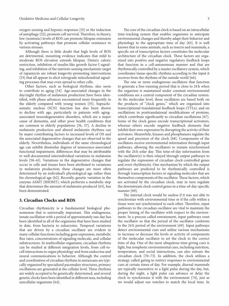
Oxidative Medicine and Cellular Longevity 5
oxygen-sensing and hypoxic responses [51] or the inductionof autophagy [52], promote cell survival.Therefore, in theory,low (nontoxic) levels of ROS can promote lifespan extensionby activating pathways that promote cellular resistance tovarious stresses.
Although there is little doubt that high levels of ROSare detrimental, mounting evidence indicates that mild tomoderate ROS elevation extends lifespan. Dietary caloricrestriction, inhibition of insulin-like growth factor-I signal-ing, and inhibition of the nutrient-sensingmechanistic targetof rapamycin are robust longevity-promoting interventions[53] that all appear to elicit retrograde mitochondrial signal-ing processes that may even spread to other cells.
Other factors, such as biological rhythms, also seemto contribute to aging [54]. Age-associated changes in theday/night rhythm of melatonin production have been iden-tified, with phase advances encountered more frequently inthe elderly compared with young women [55]. Suprachi-asmatic nucleus (SCN) function has also been shownto decline with age, particularly in patients with aging-associated neurodegenerative disorders, which are a majorcause of dementia, and other poor health conditions thatare common in elderly populations [56, 57]. A decline inmelatonin production and altered melatonin rhythms canbe major contributing factors to increased levels of OS andthe associated degenerative changes that are observed in theelderly. Nevertheless, individuals of the same chronologicalage can exhibit dissimilar degrees of senescence-associatedfunctional impairment, differences that may be attributableto well-documented interindividual variations in melatoninlevels [58–61]. Variations in the degenerative changes thatoccur in cells and tissues have been attributed to variationsin melatonin production; these changes are more oftendetermined by an individual’s physiological age rather thanhis chronological age [62]. Recently, genetic variation in theenzyme ASMT (HIOMT), which performs a metabolic stepthat determines the amount of melatonin produced [63], hasbeen demonstrated.
3. Circadian Clocks and ROS
Circadian rhythmicity is a fundamental biological phe-nomenon that is universally important. This endogenous,innate oscillation with a period of approximately one day hasbeen identified in all of the organisms that have been studiedto date, from bacteria to eukaryotes. Temporal variationsthat are driven by a circadian oscillator are evident inmany cellular functions including gene expression, metabolicflux rates, concentrations of signaling molecule, and cellularsubstructures. In multicellular organisms, circadian rhythmscan be studied at different integration levels, from cell-to-cell interactions to organ physiology and from endocrine andneural communications to behavior. Although the controland coordination of circadian rhythms inmetazoans are typi-cally organized by specialized pacemaker structures, primaryoscillations are generated at the cellular level. These rhythmsare widely accepted to be genetically determined, and severalgenetic clocks have been identified in different taxa, includingunicellular organisms [64].
The core of the circadian clock is based on an intracellulartime-tracking system that enables organisms to anticipateenvironmental changes and thereby adapt their behavior andphysiology to the appropriate time of day [65]. It is wellknown that in some animals, such as insects and mammals, aspecific set of transcription factors constitutes the moleculararchitecture of the circadian clock. These factors are orga-nized into positive and negative regulatory feedback loopsthat function in a cell-autonomous manner and that arerhythmically controlled by amaster oscillatory system, whichcoordinates tissue-specific rhythms according to the input itreceives from the rhythms of the outside world [66].
The one or more endogenous oscillators that functionto generate a free-running period that is close to 24 h whenthe organism is maintained under constant environmentalconditions are a central component of the circadian system.At the molecular level, these oscillators are based both onthe products of “clock genes,” which are organized intotranscriptional-translational feedback loops (TTLs), and onoscillations in posttranslational modifications of proteins,which contribute significantly to circadian oscillations [67].Some of the clock genes encode transcriptional activators,whereas others encode negative feedback elements thatinhibit their own expression by disrupting the activity of theiractivators. Meanwhile, kinases and phosphatases regulate thespeed and precision of the clock [68]. Components of theoscillators receive environmental information through inputpathways, allowing the oscillators to remain synchronizedwith the 24 h solar day. This time-of-day information fromthe oscillator(s) is then relayed through output pathways toregulate the expression of circadian clock-controlled genesand overt rhythmicity. One mechanism by which the outputpathways are predicted to be rhythmically controlled isthrough transcription factors or signaling molecules that arethemselves components of the oscillator.These factors, whichare activated by the circadian clock, may in turn regulatethe downstream clock control genes in a time-of-day-specificmanner [69].
The internal clock would be useless if it was not able tosynchronize with environmental time or if the cells within atissue were not synchronized to each other. Therefore, inputpathways to the circadian oscillator are vital to maintain theproper timing of the oscillator with respect to the environ-ment. In a process called entrainment, input pathways resetthe oscillator so that the period of the oscillator conformsto the 24 h period of the environment [69]. Input pathwaysdetect environmental cues and utilize various mechanismsto increase or decrease the levels or activity of componentsof the molecular oscillator to set the clock to the correcttime of day. One of the most ubiquitous time-giving cues islight, but nonphotic environmental cues, including nutrition,temperature, and social interactions, can also entrain thecircadian clock [70–73]. In addition, the clock utilizes astrategy called gating to restrict responses to environmentalcues at certain times of day. For example, diurnal mammalsare typically insensitive to a light pulse during the day, but,during the night, a light pulse can advance or delay theclock to synchronize it with the environment [74], just aswe would adjust our watches to match the local time. In
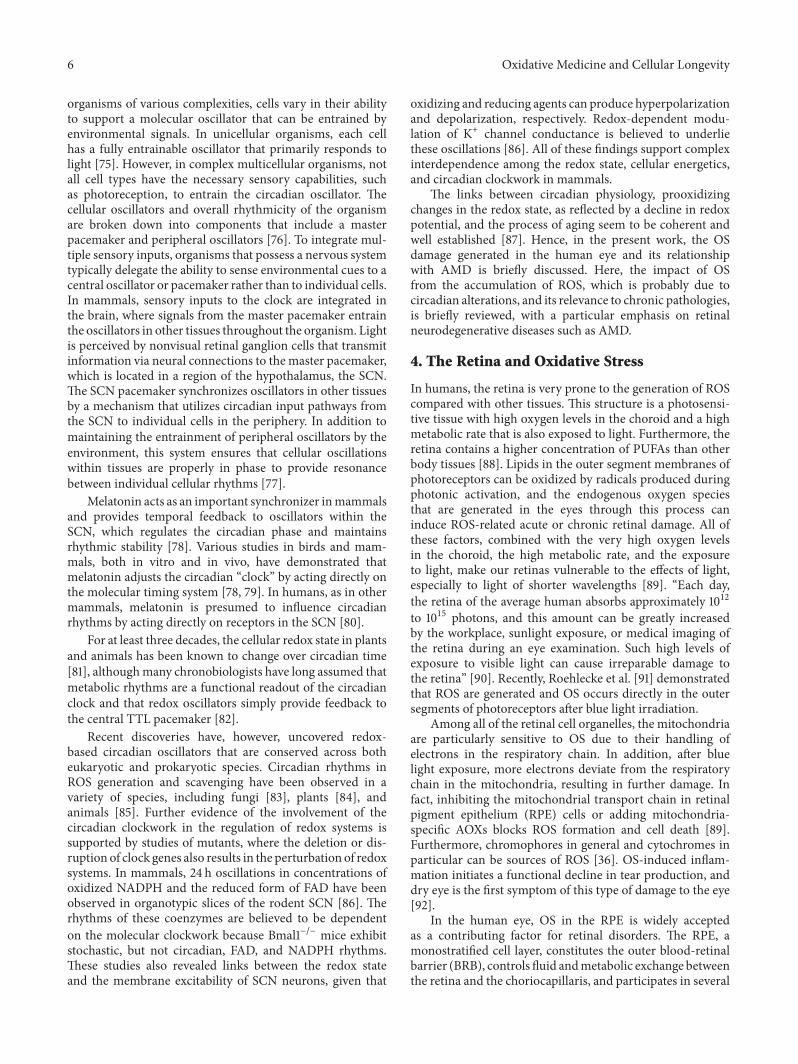
6 Oxidative Medicine and Cellular Longevity
organisms of various complexities, cells vary in their abilityto support a molecular oscillator that can be entrained byenvironmental signals. In unicellular organisms, each cellhas a fully entrainable oscillator that primarily responds tolight [75]. However, in complex multicellular organisms, notall cell types have the necessary sensory capabilities, suchas photoreception, to entrain the circadian oscillator. Thecellular oscillators and overall rhythmicity of the organismare broken down into components that include a masterpacemaker and peripheral oscillators [76]. To integrate mul-tiple sensory inputs, organisms that possess a nervous systemtypically delegate the ability to sense environmental cues to acentral oscillator or pacemaker rather than to individual cells.In mammals, sensory inputs to the clock are integrated inthe brain, where signals from the master pacemaker entrainthe oscillators in other tissues throughout the organism. Lightis perceived by nonvisual retinal ganglion cells that transmitinformation via neural connections to the master pacemaker,which is located in a region of the hypothalamus, the SCN.The SCN pacemaker synchronizes oscillators in other tissuesby a mechanism that utilizes circadian input pathways fromthe SCN to individual cells in the periphery. In addition tomaintaining the entrainment of peripheral oscillators by theenvironment, this system ensures that cellular oscillationswithin tissues are properly in phase to provide resonancebetween individual cellular rhythms [77].
Melatonin acts as an important synchronizer inmammalsand provides temporal feedback to oscillators within theSCN, which regulates the circadian phase and maintainsrhythmic stability [78]. Various studies in birds and mam-mals, both in vitro and in vivo, have demonstrated thatmelatonin adjusts the circadian “clock” by acting directly onthe molecular timing system [78, 79]. In humans, as in othermammals, melatonin is presumed to influence circadianrhythms by acting directly on receptors in the SCN [80].
For at least three decades, the cellular redox state in plantsand animals has been known to change over circadian time[81], althoughmany chronobiologists have long assumed thatmetabolic rhythms are a functional readout of the circadianclock and that redox oscillators simply provide feedback tothe central TTL pacemaker [82].
Recent discoveries have, however, uncovered redox-based circadian oscillators that are conserved across botheukaryotic and prokaryotic species. Circadian rhythms inROS generation and scavenging have been observed in avariety of species, including fungi [83], plants [84], andanimals [85]. Further evidence of the involvement of thecircadian clockwork in the regulation of redox systems issupported by studies of mutants, where the deletion or dis-ruption of clock genes also results in the perturbation of redoxsystems. In mammals, 24 h oscillations in concentrations ofoxidized NADPH and the reduced form of FAD have beenobserved in organotypic slices of the rodent SCN [86]. Therhythms of these coenzymes are believed to be dependenton the molecular clockwork because Bmal1−/− mice exhibitstochastic, but not circadian, FAD, and NADPH rhythms.These studies also revealed links between the redox stateand the membrane excitability of SCN neurons, given that
oxidizing and reducing agents can produce hyperpolarizationand depolarization, respectively. Redox-dependent modu-lation of K+ channel conductance is believed to underliethese oscillations [86]. All of these findings support complexinterdependence among the redox state, cellular energetics,and circadian clockwork in mammals.
The links between circadian physiology, prooxidizingchanges in the redox state, as reflected by a decline in redoxpotential, and the process of aging seem to be coherent andwell established [87]. Hence, in the present work, the OSdamage generated in the human eye and its relationshipwith AMD is briefly discussed. Here, the impact of OSfrom the accumulation of ROS, which is probably due tocircadian alterations, and its relevance to chronic pathologies,is briefly reviewed, with a particular emphasis on retinalneurodegenerative diseases such as AMD.
4. The Retina and Oxidative Stress
In humans, the retina is very prone to the generation of ROScompared with other tissues. This structure is a photosensi-tive tissue with high oxygen levels in the choroid and a highmetabolic rate that is also exposed to light. Furthermore, theretina contains a higher concentration of PUFAs than otherbody tissues [88]. Lipids in the outer segment membranes ofphotoreceptors can be oxidized by radicals produced duringphotonic activation, and the endogenous oxygen speciesthat are generated in the eyes through this process caninduce ROS-related acute or chronic retinal damage. All ofthese factors, combined with the very high oxygen levelsin the choroid, the high metabolic rate, and the exposureto light, make our retinas vulnerable to the effects of light,especially to light of shorter wavelengths [89]. “Each day,the retina of the average human absorbs approximately 1012to 1015 photons, and this amount can be greatly increasedby the workplace, sunlight exposure, or medical imaging ofthe retina during an eye examination. Such high levels ofexposure to visible light can cause irreparable damage tothe retina” [90]. Recently, Roehlecke et al. [91] demonstratedthat ROS are generated and OS occurs directly in the outersegments of photoreceptors after blue light irradiation.
Among all of the retinal cell organelles, the mitochondriaare particularly sensitive to OS due to their handling ofelectrons in the respiratory chain. In addition, after bluelight exposure, more electrons deviate from the respiratorychain in the mitochondria, resulting in further damage. Infact, inhibiting the mitochondrial transport chain in retinalpigment epithelium (RPE) cells or adding mitochondria-specific AOXs blocks ROS formation and cell death [89].Furthermore, chromophores in general and cytochromes inparticular can be sources of ROS [36]. OS-induced inflam-mation initiates a functional decline in tear production, anddry eye is the first symptom of this type of damage to the eye[92].
In the human eye, OS in the RPE is widely acceptedas a contributing factor for retinal disorders. The RPE, amonostratified cell layer, constitutes the outer blood-retinalbarrier (BRB), controls fluid andmetabolic exchange betweenthe retina and the choriocapillaris, and participates in several
Oxidative Medicine and Cellular Longevity 7
functions linked to photoreceptor physiology, such as thephagocytosis of shed photoreceptor outer segments andtransportation of molecules to and from the retina [93].
In addition, RPE cells release factors that control neuronalsurvival and angiogenesis (e.g., pigment epithelium-derivedfactor, PEDF) and that promote photoreceptor survival andhave an antiangiogenic effect on the choriocapillaris [94, 95].The RPE secretes PEDF on the apical side; in contrast, theangiogenic factor vascular endothelial growth factor (VEGF)is secreted on the basolateral side to maintain fenestrationof the choriocapillaris. An imbalance in the expression ofPEDF and VEGF appears to be involved in ocular patholo-gies. Because PEDF has antioxidative and antiangiogenicproperties, this factor may protect against vision-threateningangiogenic mechanisms. In contrast, VEGF is generallyaccepted as the most potent inducer of endothelial activationand angiogenesis, a process in which new vessels developfrom the preexisting vasculature (for a review, see [96]).Under ischemic or hypoxic conditions, which occur in manyneovascular diseases, retinal expression and production ofVEGF are dramatically increased [97, 98].
Theoxidation of polyunsaturated fats in the retina leads tolipid peroxidation products such as carboxyethylpyrrole and4-hydroxy-2-nonenal, which can form adducts with proteinsand accumulate in the outer retina and in drusen [99]. Thelevels of AOXproteins such as catalase and SODare increasedin RPEhomogenates derived from the eyes of late-stageAMDpatients, indicating a response to increased OS in the eyes ofthese patients [100].
As indicated in previous sections, the NOX [101] family ofenzymes has recently been recognized as a generator of ROSin rod or cone photoreceptors after they are damaged due toserum deprivation in a model of retinitis pigmentosa [102].Roehlecke et al. [103] further demonstrated that blue lightirradiation results in increased superoxide anion production.
Lipofuscin is the generic name given to a heterogeneousgroup of complex and autofluorescent bisretinoids, lipid per-oxides, and proteins and to various fluorescent compoundsthat are formed frommodified lipids or that are derived fromvitamin A. The major substrate for lipofuscin in the RPE is anondegradable end product that results from phagocytosis ofthe photoreceptor outer segments, which are rich in PUFAsand vitamin A. This substrate is located within the RPE,where it accumulates in lysosomes with age. Lipofuscin isa byproduct of the visual cycle [104, 105] that is producedwhen phagocytosed material is not entirely degraded withinthe RPE lysosomes, resulting in the accumulation of thiscomplex over time. Lipofuscin is continually exposed tohigh oxygen tensions (70mmHg) and to visible light (400–700 nm) during daylight hours, creating a prime environmentfor the generation of ROS that have the potential to damagecellular proteins and lipid membranes. Thus, lipofuscin isa photoinducible generator of ROS that increases the riskof oxidative injury [106]. A lipofuscin fluorophore, A2E,is now known to mediate the blue light-induced apoptosisof RPE cells, which inhibits the degradative capacity oflysosomes and disrupts membrane integrity [107, 108]. Thiscomplex phototoxic effect is wavelength-dependent; moresuperoxide anions are generated in granules exposed to blue
light (400–520 nm) than in granules exposed to red light(660–730 nm) or full white light [109]. Lipofuscin inducesthe apoptosis of cultured RPE cells, leading to a decline inmitochondrial activity that is associated with the transloca-tion of cytochrome c, an apoptosis-inducing factor, and twoapoptosis-inducing proteins into the cytoplasm and nucleus[110].
However, whether A2E or lipofuscin may impair lyso-somal function is still controversial. Recently, Saadat et al.[111] demonstrated thatA2E and impaired autophagymediateRPE cell damage. These authors proposed that decreasedlysosomal capacity might also result in decreased autophagyin RPE cells, which would not affect autophagosome bio-genesis or fusion with lysosomes but would instead impairthe terminal stage of the degradation process. Thus, theaccumulation of A2E in aged RPE cells, in which autophagyis already impaired, may result in RPE cell damage. Theauthors also considered the possibility that A2E inhibitedlysosomal function, which then resulted in the accumulationof autophagosomes rather than an increase in autophagic fluxthrough this pathway.
Some authors havemaintained [112] that the RPE secretesapolipoprotein B particles into Bruch’s membrane and thatthese particles accumulate with age and may in turn form alipidwall, a precursor of the basal linear deposit.Then, certainconstituents of the described aggregates may interact withROS, resulting in proinflammatory, peroxidized lipids andleading to the upregulation of cytokines/chemokines, whichthen promotes neovascularization. However, recent publica-tions indicate that, in the RPE, OS is also capable of inducingprotective pathways, such as the phosphatidylinositide 3-kinase (PI3K)/Akt and nuclear factor erythroid-2-relatedfactor 2 pathways. In addition, VEGF and neuroprotectin D1signaling act to protect the retina [113, 114].
The phagocytosis of oxidized fatty acids from photore-ceptor outer segments contributes to metabolic failure in theRPE [115]. Importantly, the FR byproducts of mitochondrialenergy metabolism damage the RPE [116]. In addition, themitochondrial ETC generates superoxide radicals throughsingle-electron leaks at respiratory complexes I and III [18].
Flavin-dependent enzymes in the mitochondrial matrixmay also be large contributors of ROS [22]. Superoxide candirectly damage mitochondrial DNA, proteins, and lipids,and it can also be converted to H
2O2by manganese SOD in
the mitochondria [22]. The potential for RPE light damageis influenced by a number of factors, including age, diurnalfluctuations, and pathology [117].
Otherwise, circadian photoreception decreases with agedue to disruptions to circadian rhythms, age-related pupillarymiosis and reduced crystalline lens transmission, particularlyof blue light. Circadian studies have revealed loss of thecontrol of pupil size and the crystalline lens in aging subjects[118].
5. ROS and Age-Related MacularDegeneration (AMD)
AMD, a major cause of visual impairment in the elderly,is linked to pathological changes involving the RPE. AMD
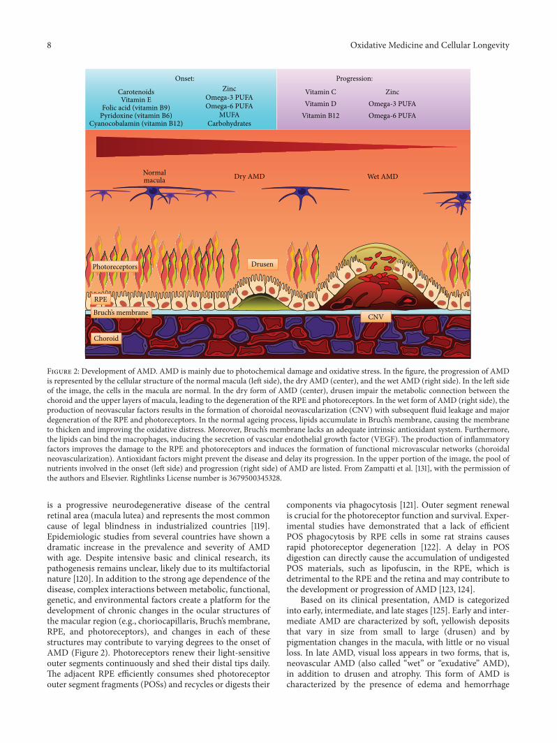
8 Oxidative Medicine and Cellular Longevity
Onset:
CarotenoidsVitamin E
Folic acid (vitamin B9)Pyridoxine (vitamin B6)
Cyanocobalamin (vitamin B12)
ZincOmega-3 PUFAOmega-6 PUFA
MUFACarbohydrates
Progression:
Vitamin CVitamin D
Vitamin B12
ZincOmega-3 PUFAOmega-6 PUFA
Normalmacula Dry AMD Wet AMD
Photoreceptors Drusen
CNV
RPE
Bruch’s membrane
Choroid
Figure 2: Development of AMD. AMD is mainly due to photochemical damage and oxidative stress. In the figure, the progression of AMDis represented by the cellular structure of the normal macula (left side), the dry AMD (center), and the wet AMD (right side). In the left sideof the image, the cells in the macula are normal. In the dry form of AMD (center), drusen impair the metabolic connection between thechoroid and the upper layers of macula, leading to the degeneration of the RPE and photoreceptors. In the wet form of AMD (right side), theproduction of neovascular factors results in the formation of choroidal neovascularization (CNV) with subsequent fluid leakage and majordegeneration of the RPE and photoreceptors. In the normal ageing process, lipids accumulate in Bruch’s membrane, causing the membraneto thicken and improving the oxidative distress. Moreover, Bruch’s membrane lacks an adequate intrinsic antioxidant system. Furthermore,the lipids can bind the macrophages, inducing the secretion of vascular endothelial growth factor (VEGF). The production of inflammatoryfactors improves the damage to the RPE and photoreceptors and induces the formation of functional microvascular networks (choroidalneovascularization). Antioxidant factors might prevent the disease and delay its progression. In the upper portion of the image, the pool ofnutrients involved in the onset (left side) and progression (right side) of AMD are listed. From Zampatti et al. [131], with the permission ofthe authors and Elsevier. Rightlinks License number is 3679500345328.
is a progressive neurodegenerative disease of the centralretinal area (macula lutea) and represents the most commoncause of legal blindness in industrialized countries [119].Epidemiologic studies from several countries have shown adramatic increase in the prevalence and severity of AMDwith age. Despite intensive basic and clinical research, itspathogenesis remains unclear, likely due to its multifactorialnature [120]. In addition to the strong age dependence of thedisease, complex interactions between metabolic, functional,genetic, and environmental factors create a platform for thedevelopment of chronic changes in the ocular structures ofthe macular region (e.g., choriocapillaris, Bruch’s membrane,RPE, and photoreceptors), and changes in each of thesestructures may contribute to varying degrees to the onset ofAMD (Figure 2). Photoreceptors renew their light-sensitiveouter segments continuously and shed their distal tips daily.The adjacent RPE efficiently consumes shed photoreceptorouter segment fragments (POSs) and recycles or digests their
components via phagocytosis [121]. Outer segment renewalis crucial for the photoreceptor function and survival. Exper-imental studies have demonstrated that a lack of efficientPOS phagocytosis by RPE cells in some rat strains causesrapid photoreceptor degeneration [122]. A delay in POSdigestion can directly cause the accumulation of undigestedPOS materials, such as lipofuscin, in the RPE, which isdetrimental to the RPE and the retina and may contribute tothe development or progression of AMD [123, 124].
Based on its clinical presentation, AMD is categorizedinto early, intermediate, and late stages [125]. Early and inter-mediate AMD are characterized by soft, yellowish depositsthat vary in size from small to large (drusen) and bypigmentation changes in the macula, with little or no visualloss. In late AMD, visual loss appears in two forms, that is,neovascular AMD (also called “wet” or “exudative” AMD),in addition to drusen and atrophy. This form of AMD ischaracterized by the presence of edema and hemorrhage
Oxidative Medicine and Cellular Longevity 9
within or below the retina or RPE and geographic atrophy(also called “dry” AMD). Currently, there are no treatmentsfor geographic atrophy, but neovascular AMD is treated withVEGF inhibitors, which, although not curative, are ofteneffective in preventing severe visual loss [126]. The dry form,also known as age-related maculopathy, is characterized bythe presence of drusen under the RPE that is accompaniedby either the loss or focal accumulation of melanin pigment.This form of AMD is typically characterized by a progressivecourse that leads to degeneration of the RPE and photorecep-tors. The exudative form is linked to choroidal neovascular-ization that is directed toward the subretinal macular region,with subsequent bleeding and/or fluid leakage that can resultin a sudden loss of central vision. It is the most rapidlyprogressing form of AMD. Both the atrophic and exudativeforms are associated with severe visual impairment [119].Thepathophysiology of AMD is complex, and, in addition togenetic predispositions, at least 4 processes contribute to thedisease: lipofuscinogenesis, drusogenesis, local inflammationand neovascularization (in the case of the wet form), andimmunological mechanisms [127].
The current pathophysiological conception of AMDassigns a primary role to age-related, cumulative oxidativedamage to the RPE due to an imbalance between the gener-ation and elimination of ROS [128]. In particular, lipofuscinhas been hypothesized to be the primary source of the ROSresponsible for both the cellular and extracellular matrixalterations found in AMD [129, 130].
The accumulation of lipofuscin and other lipid peroxidesand potentially toxic substances may dramatically influenceRPE physiology, as described above. This accumulationgreatly reduces the phagocytic capacity, lysosomal enzymeactivities, and AOX potential of the human RPE in vitro[133, 134]. The first clinical sign of early AMD is drusen, asmentioned above. Drusen are lipid-rich, sub-RPE depositsthat contain a variety of proteins, including vitronectin,components of the terminal complement cascade, and b-amyloid [135, 136]. Microscopic analysis of eyes donated bypatients with AMD revealed lipid deposits within Bruch’smembrane and apoptosis of RPE cells as features of thedisease that are distinct from normal aging. The advancedform of dry AMD, which leads to geographic atrophy, wascharacterized by breakdown of the RPE, the choriocapillaris,and photoreceptors in regions of the retina, often where largedrusen were present [137]. Dysregulation of the phagocytosisof oxidized fatty acids from photoreceptor outer segmentsis a contributing factor to the metabolic failure of RPE cells[115, 123]. In addition to all of these factors, the damage causedby the FR byproducts of mitochondrial energy metabolismhas been implicated in age-related damage to the RPE [116].
As reviewed above, the mitochondrial ETC generatessuperoxide radicals through single-electron leaks at respira-tory complexes I and III [18], and flavin-dependent enzymesin themitochondrialmatrixmay be large contributors of ROS[29].
Otherwise, starvation and hypoxia, which can resultfrom poor perfusion, are generally associated with increasedamounts of ROS, which promote autophagy via severalcomplex signaling mechanisms [138] (Figure 3). In response
to OS, autophagy is significantly increased in an attemptto remove oxidatively damaged organelles such as mito-chondria. At this time, accumulating evidence linking theimpairment of autophagy with a range of age-related neu-rodegenerative diseases, including AMD, has suggested thatautophagy occurs in the RPE to maintain homeostasisbecause these cells are exposed to sustained OS. However,insufficient digestion due to impaired autophagy or lysosomaldegradation in the RPE can lead to an accumulation ofdamaged organelles, toxic proteins (including lipofuscin),and extracellular drusen deposits, all of which can contributeto RPE dysfunction or RPE cell death, which have beenassociated with the pathogenesis of AMD [139]. In addition,the AOXs of the retina (e.g., via macular molecules such aslutein and zeaxanthin) are reduced in AMD [140].
5.1. VEGF and AMD. VEGF is the most potent inducerof endothelial activation and angiogenesis. It is mainlyexpressed in retinal neurons and glial cells and is present inonly scant amounts in blood vessels [141]. Under ischemicconditions, retinal expression and production of VEGF isincreased [142]. This factor has also been implicated inthe development of retinal neovascularization in ischemicretinopathies such as AMD [98]. Through a paracrine mech-anism, VEGF binds to its cell-surface receptors, includingVEGFR1/Flt-1, VEGFR2/Flk-1/KDR, and VEGFR3, and pro-motes endothelial cell survival, proliferation, migration, andtubular structure formation [142]. Among these receptors,VEGFR2 is the crucial receptor that mediates angiogenicand vascular permeability, whereasVEGFR3mainlymediateslymphangiogenic functions. The activation of VEGFR1 playsa dual role and can either stimulate or inhibit angiogenesis,whereas the activation of VEGFR2 seems to only stimulateangiogenesis [143, 144].
Upon binding VEGF, VEGFR2 undergoes dimerizationand autophosphorylation, resulting in the activation of itsdownstream kinases, including mitogen-activated proteinkinase (MAPK), ERK1/2, p38, JNK, and PI3K/Akt, and ofendothelial NO synthase (e-NOS), which may lead to furtheralterations in endothelial cell survival, proliferation, andmigration. Recent reviews provide further information onthis important factor [145, 146].
A constant oxygen supply is clearly essential for properhomeostasis and normal functioning in all retinal tissues.Cellular responses to reduced oxygen levels are mediatedby the transcriptional regulator hypoxia-inducible factor-1(HIF-1), a heterodimeric protein complex that consists ofan oxygen-dependent subunit (HIF-1𝛼) and a constitutivelyexpressed nuclear subunit (HIF-1𝛽). Under normoxic condi-tions, de novo synthesized cytoplasmic HIF-1𝛼 is degradedby the 26S proteasome. Under hypoxic conditions, HIF-1𝛼is stabilized, binds to HIF-1𝛽, and activates the transcriptionof various target genes. These genes play a key role in theregulation of angiogenesis in various visual pathologies, suchas AMD [147].
5.2. AMD and the Complement Pathway. Growing evidenceindicates that AMD is downstreamof a chronic inflammatorycondition in which activation of the immune system plays
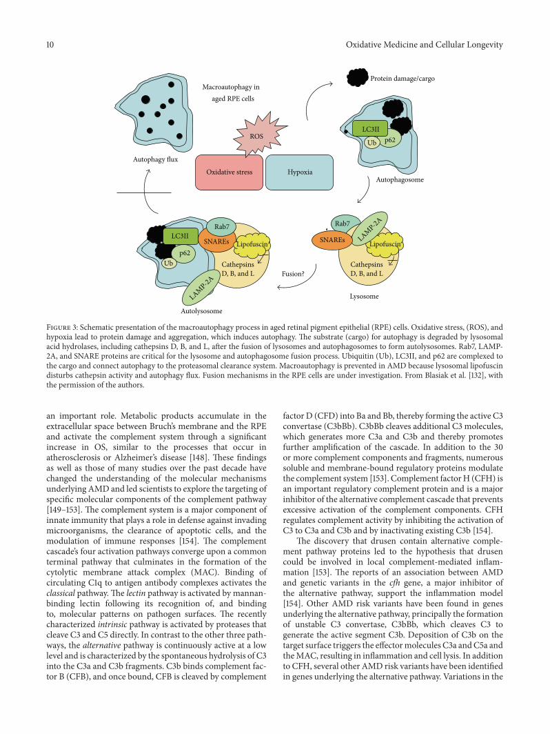
10 Oxidative Medicine and Cellular Longevity
Rab7
p62LC3II
Ub
Lysosome
Autophagosome
Autolysosome
Protein damage/cargo
p62
LC3II
Ub CathepsinsD, B, and L D, B, and L
SNAREs
Rab7
Oxidative stress Hypoxia
ROS
Fusion?
Macroautophagy inaged RPE cells
SNAREs
Cathepsins
LAMP-2A
Lipofuscin Lipofuscin
LAMP-2A
Autophagy flux
Figure 3: Schematic presentation of the macroautophagy process in aged retinal pigment epithelial (RPE) cells. Oxidative stress, (ROS), andhypoxia lead to protein damage and aggregation, which induces autophagy. The substrate (cargo) for autophagy is degraded by lysosomalacid hydrolases, including cathepsins D, B, and L, after the fusion of lysosomes and autophagosomes to form autolysosomes. Rab7, LAMP-2A, and SNARE proteins are critical for the lysosome and autophagosome fusion process. Ubiquitin (Ub), LC3II, and p62 are complexed tothe cargo and connect autophagy to the proteasomal clearance system. Macroautophagy is prevented in AMD because lysosomal lipofuscindisturbs cathepsin activity and autophagy flux. Fusion mechanisms in the RPE cells are under investigation. From Blasiak et al. [132], withthe permission of the authors.
an important role. Metabolic products accumulate in theextracellular space between Bruch’s membrane and the RPEand activate the complement system through a significantincrease in OS, similar to the processes that occur inatherosclerosis or Alzheimer’s disease [148]. These findingsas well as those of many studies over the past decade havechanged the understanding of the molecular mechanismsunderlying AMD and led scientists to explore the targeting ofspecific molecular components of the complement pathway[149–153]. The complement system is a major component ofinnate immunity that plays a role in defense against invadingmicroorganisms, the clearance of apoptotic cells, and themodulation of immune responses [154]. The complementcascade’s four activation pathways converge upon a commonterminal pathway that culminates in the formation of thecytolytic membrane attack complex (MAC). Binding ofcirculating C1q to antigen antibody complexes activates theclassical pathway.The lectin pathway is activated by mannan-binding lectin following its recognition of, and bindingto, molecular patterns on pathogen surfaces. The recentlycharacterized intrinsic pathway is activated by proteases thatcleave C3 and C5 directly. In contrast to the other three path-ways, the alternative pathway is continuously active at a lowlevel and is characterized by the spontaneous hydrolysis of C3into the C3a and C3b fragments. C3b binds complement fac-tor B (CFB), and once bound, CFB is cleaved by complement
factor D (CFD) into Ba and Bb, thereby forming the active C3convertase (C3bBb). C3bBb cleaves additional C3 molecules,which generates more C3a and C3b and thereby promotesfurther amplification of the cascade. In addition to the 30or more complement components and fragments, numeroussoluble and membrane-bound regulatory proteins modulatethe complement system [153]. Complement factorH (CFH) isan important regulatory complement protein and is a majorinhibitor of the alternative complement cascade that preventsexcessive activation of the complement components. CFHregulates complement activity by inhibiting the activation ofC3 to C3a and C3b and by inactivating existing C3b [154].
The discovery that drusen contain alternative comple-ment pathway proteins led to the hypothesis that drusencould be involved in local complement-mediated inflam-mation [153]. The reports of an association between AMDand genetic variants in the cfh gene, a major inhibitor ofthe alternative pathway, support the inflammation model[154]. Other AMD risk variants have been found in genesunderlying the alternative pathway, principally the formationof unstable C3 convertase, C3bBb, which cleaves C3 togenerate the active segment C3b. Deposition of C3b on thetarget surface triggers the effectormolecules C3a andC5a andtheMAC, resulting in inflammation and cell lysis. In additionto CFH, several other AMD risk variants have been identifiedin genes underlying the alternative pathway. Variations in the
Oxidative Medicine and Cellular Longevity 11
factor B (BF) and complement component 2 (C2) genes arealso associated with AMD [155].
A third line of evidence in support of complementinvolvement in AMD was provided by studies that showedthat AMD patients have higher levels of complement activa-tion products in their blood [156].
In 2001, data collected from the Age-Related Eye DiseaseStudy (AREDS) revealed that patients who were treated withzinc, either alone or in combination with vitamins, displayedreduced progression to advanced AMD. AREDS results ledto the recommendation that persons who are older than 55years of age and who are at risk of developing advancedAMD should consider taking vitamin supplements plus zinc.Aprevious report published by theBlueMountains Eye Study,a population-based study, confirmed the beneficial effect ofzinc in AMD patients [157]. A recent study also providedevidence that daily administration of 50mg of zinc sulfate caninhibit complement involvement in AMD patients who haveincreased complement activation [158].
5.3. AMD, RPE, and Daily and Circadian Rhythms. The reti-nal circadian system involves a unique structure. It containsa complete circadian system with multiple generation sitesof numerous circadian rhythms, each of which deserves itsown review. However, in the vertebrate retina, the intimatereciprocal relationship that exists between the neural retinaand the underlying RPE is crucial for vision, while thediurnal and circadian rhythmicity of the RPE is critical forphotoreceptor support and retinal function. Thus, in thissection, we briefly review some of the rhythmic functions ofthe RPE that contribute to normal and pathological vision.
The association between AMD and biological rhythmshas been poorly studied; however, there is a strong linkbetween ocular physiology and circadian rhythms in bothhumans and animals. The renewal and elimination of agedphotoreceptor outer segment tips by cells from the RPE is adaily rhythmic process that is crucial for long-term vision.Photoreceptors indefinitely renew their light-sensitive outersegments by disk shedding and the subsequent formationof new disks from the cilium of the inner segment. Inhigher vertebrates, outer segment renewal is synchronized bycircadian rhythms [121, 159].
This shedding occurs once per day. In mice and rats,rod shedding is synchronized with light onset [160, 161]. Tomaintain the constant length of photoreceptors, the outersegment needs to be shed, and the formation of new outersegments must be coordinated.
The task of the adjacent RPE is to absorb shed POSsby phagocytosis and to recycle or digest their components.Outer segment renewal is crucial for photoreceptor functionand survival, and a lack of efficient phagocytosis is sufficientto cause rapid photoreceptor degeneration by disk shed-ding [121, 162]. Photoreceptor disk shedding and subsequentphagocytosis by the RPE must be precisely regulated [163].Alterations, such as delayed termination of shedding ordefective digestion in the RPE, can cause the accumulation oflipofuscin [123]. Outer segment renewal and RPE phagocyto-sis are synchronized under circadian control and are triggeredby the dark/light periods of the daily rhythm [163, 164].
Studies in higher vertebrates have revealed differencesbetween cone- and rod-dominant species. Rod sheddingmainly occurs in the morning, resulting in complementaryRPE phagocytic activity by an increased number of phago-somes within the first 2 h after light onset, whereas coneshedding is more variable and mainly occurs either duringthe night or during the first 2 h after light onset [163, 164]. Anydisruption in this process causes photoreceptor dysfunctionandblindness in animalmodels and retinal disease in humans[93]. The synchronization of shedding with light seems tobe crucial to photoreceptor physiology and survival becausethe accumulation of undigested material is detrimental tothe RPE and retina and may contribute to the developmentor progression of AMD [123, 124]. These rhythms thatsynchronize with light are perceived by photosensitive retinalganglion cells that contain the pigment melanopsin. Dailyinformation is transmitted to the master circadian oscillatorlocated in the SCN via the retinohypothalamic tract [66].The action spectrum of light information for the circadianbiological rhythm shows a peak at a shorter wavelength(464 nm) than that for visual information (approximately555 nm).
Pivotal studies [161–164] have conclusively demonstratedthat, in the rat retina, the diurnal rhythmof rodPOS sheddingand RPE phagocytosis is under circadian regulation. Themammalian retina displays persistent rhythmic activity evenwhen it is isolated from the brain. In rats with transectednerves, POS shedding and RPE phagocytosis continue diur-nally despite the loss of synaptic connections between theeye and the brain, suggesting that this rhythm is generatedand controlled locally in the eye [165]. However, this rhythmcannot be reset by light unless the optic nerve remains intact[166]. Thus, the circadian renewal of POS involves both localcontrol within the retina and central regulation by the brain.
The cellular and molecular mechanisms that underliecircadian regulation of shedding/phagocytosis are complexand are not the subject of this review. Numerous genes,proteins and signaling pathways play important roles in theengulfment of POS and the subsequent lysosomal degrada-tion of spent photoreceptor disks within the RPE.
The lack of POS phagocytosis or digestion leads to pho-toreceptor dystrophy and blindness [122] due to the absenceof the engulfment activity of the RPE. In rats that are deficientin MerTK, this absence causes dramatic and early onsetretinal degeneration [122, 167]. Because the ingestion rate ofPOS by RPE cells exhibits a pronounced circadian rhythmthat peaks around subjective dawn in both rat and mousestrains [159, 168], Prasad and colleagues [169] suggested thata feature of the TAM receptor system, such as ligand and/orreceptor expression levels, might be regulated as a function ofposition in the circadian cycle.
Despite our vast knowledge of circadian biology andangiogenesis, the role of the circadian clock in the regulationof angiogenesis and vascular patterning remains poorlyunderstood. Experimental animal models may help definethe relationships between circadian rhythms and some retinalpathologies, such as AMD. Jensen et al. [170] showed thatdisruption of the circadian clock by both constant exposureto light and genetic manipulation of key genes in zebrafish
12 Oxidative Medicine and Cellular Longevity
led to impaired developmental angiogenesis. The disruptionof crucial circadian regulatory genes, including Bmal1 andPeriod2, resulted in either marked impairment or enhance-ment of vascular development. At the molecular level, theseauthors showed that the circadian regulator Bmal1 directlytargets the promoter region of the vegf gene in zebrafish,leading to elevated VEGF expression. Interestingly, deletionof these E-boxes in the promoter region of the zebrafish vegfgene resulted in inactivation of the promoter. These findingscan be reasonably extended to developmental angiogenesis inmammals and even to pathological angiogenesis in humans[171]. Disruption of the circadian clock systemnot only affectsthe physiological activity of an organism but also often leadsto the onset, development and progression of various diseases[172].
An important circadian hormone that is involved invertebrate retinal circadian rhythms is melatonin, which issynthesized and produced by photoreceptors and shows aclear daily rhythm, with an acrophase at night [173]. Thisindolamine seems to have protective effects on other retinalcell types, including RPE cells and photoreceptors. Melatoninprotects cultured RPE cells from OS and ischemia-inducedcell death [174, 175]. Several studies have reported that mela-tonin is involved in the pathogenesis ofAMD. In 2005, Yi et al.[176] reported that daily administration of melatonin (3mg)may protect the retina and delay the progression of AMD.Rosen et al. [177] reported that the production of melatoninis decreased in AMD patients compared with age-matchedcontrols, suggesting that a deficiency inmelatoninmay play arole in the occurrence of AMD. A further indication of thepossible role of melatonin in age-related pathologies is theobservation that retinalmelatonin synthesis decreases duringaging. In 2012, Tosini et al. [178] proposed that melatonincould affect the circadian clocks in photoreceptors and RPEcells and could thereby affect metabolism in these cells.
Melatonin and dopamine, two regulatory signals that playimportant roles in retinal physiology, have been proposedto be involved in the control of circadian POS sheddingand RPE phagocytosis. In the retina, the production andrelease of melatonin and dopamine are under circadiancontrol [179, 180]. Although there is insufficient clinical andexperimental evidence to demonstrate a direct relationshipbetween melatonin, circadian rhythms, and AMD, somereports have suggested that the melatonin rhythm is reversedin AMD patients [181].
6. Conclusion
This review highlights the role of OS as one of the maincauses of AMD etiologies. In this retinal disease, as in othermetabolic and degenerative pathologies, complex interac-tions amongmetabolic, functional, genetic, and environmen-tal factors create a platform for the development of chronicchanges in the ocular structures of the macular region inaddition to a strong age dependence. In addition to geneticpredispositions, at least four processes contribute to thedisease: lipofuscinogenesis, drusogenesis, local inflammationand neovascularization (in the case of the wet form), andimmunological mechanisms.
The current pathophysiological conception of AMDassigns a primary role to the age-related, cumulative oxida-tive damage to the RPE that occurs due to an imbalancebetween the generation and elimination of ROS. In particular,lipofuscin has been hypothesized to be the primary sourceof ROS and to be responsible for both the cellular andextracellular matrixes alterations in AMD. However, theremay also be an association between the increasing levels ofenvironmental sun radiation, especially short wavelengths inthe violet and blue spectrum, due to both the ozone holeand climate change over the last decades, particularly giventhat AMD remains the leading cause of irreversible visionloss among the elderly in developed nations. In 2004, theoverall global prevalence of AMD was approximately 8.7percent, and the number of AMD patients was projectedto rise to 196 million people worldwide by 2020 and to288 million by 2040 [182]. Interestingly, the associationbetween AMD and circadian rhythms, particularly differentRPE rhythms, suggests a role for the circadian clock inAMD-related circadian abnormalities, which have generallybeen considered to be a consequence of neurodegeneration.However, recent evidence suggests that circadian disruptionmight actually contribute to the degenerative process andthus might be a modifiable cause of cell or neural injury.
Circadian clock genes have been shown to regulate VEGFsignaling in tumorigenesis [183], and dopamine has beenshown to modulate the effects of VEGF receptor activationon vascular endothelial cells [184, 185]. Recent results alsoindicate thatPeriod genesmay play a similar role in regulatingvascularization signals in the retina in retinopathy diseasemodels [186]. Regardlessly, retinal clock gene expression isdisrupted in proliferative neovascularizing diseases [187].
Although circadian disturbances due to aging and neu-rodegenerative diseases have been duly noted, a key questionis whether these disturbances influence the pathology ofAMD.This question deserves further investigation.
List of Abbreviations
AMD: Age-related macular degenerationAOX: Antioxidant defenseBRB: Blood-retinal barrierCFH: Complement factor HETC: Electron transport chainFR: Free radicalGPx: Glutathione peroxidaseHIF-1: Hypoxia-inducible factor-1Melatonin: 𝑁-acetyl-5-methoxytryptamineMAC: Membrane attack complexmitoROS: Mitochondrially generated ROSmtDNA: Mitochondrial DNANOX: NADPH oxidasesOS: Oxidative stressPEDF: Pigment epithelium-derived factorPMF: Proton motive forcePOSs: Photoreceptor outer segment fragmentsPUFAs: Polyunsaturated fatty acidsRET: Reverse electron transferRNS: Reactive nitrogen species
Oxidative Medicine and Cellular Longevity 13
ROS: Reactive oxygen speciesRPE: Retinal pigment epitheliumSCN: Suprachiasmatic nucleusSODs: Superoxide dismutasesTTL: Transcriptional-translational feedback loopVEGF: Vascular endothelial growth factor.
Disclosure
Maŕıa Luisa Fanjul-Moles is a retired Professor.
Conflict of Interests
The authors declare that there is no conflict of interestsregarding the publication of this paper.
References
[1] B. Halliwell, “The antioxidant paradox: less paradoxical now?”British Journal of Clinical Pharmacology, vol. 75, no. 3, pp. 637–644, 2013.
[2] M. Schieber and N. S. Chandel, “ROS function in redoxsignaling and oxidative stress,” Current Biology, vol. 24, no. 10,pp. R453–R462, 2014.
[3] T. Finkel and N. J. Holbrook, “Oxidants, oxidative stress and thebiology of ageing,”Nature, vol. 408, no. 6809, pp. 239–247, 2000.
[4] J. F. Turrens, “Superoxide production by the mitochondrialrespiratory chain,” Bioscience Reports, vol. 17, no. 1, pp. 3–8, 1997.
[5] P. Kuppusamy and J. L. Zweier, “Characterization of free radicalgeneration by xanthine oxidase. Evidence for hydroxyl radicalgeneration,” Journal of Biological Chemistry, vol. 264, no. 17, pp.9880–9884, 1989.
[6] M. C. DeRosa and R. J. Crutchley, “Photosensitized singletoxygen and its applications,” Coordination Chemistry Reviews,vol. 233-234, pp. 351–371, 2002.
[7] P. Pospı́šil, A. Prasad, and M. Rác, “Role of reactive oxygenspecies in ultra-weak photon emission in biological systems,”Journal of Photochemistry and Photobiology B: Biology, vol. 139,pp. 11–23, 2014.
[8] B. I. Kruft and A. Greer, “Photosensitization reactions in vitroand in vivo,” Photochemistry and Photobiology, vol. 87, no. 6, pp.1204–1213, 2011.
[9] F. Liebel, S. Kaur, E. Ruvolo, N. Kollias, and M. D. Southall,“Irradiation of skin with visible light induces reactive oxygenspecies and matrix-degrading enzymes,” Journal of InvestigativeDermatology, vol. 132, no. 7, pp. 1901–1907, 2012.
[10] G. R. Buettner, “Molecular targets of photosensitization—somebiological chemistry of singlet oxygen,” in Photobiology Scienceson Line, K. C. Smith, Ed., American Society for Photobiology,2011, http://www.photobiology.info/Buettner.html.
[11] J. R. Kanofsky, “Measurement of singlet-oxygen in vivo:progress and pitfalls,” Photochemistry and Photobiology, vol. 87,no. 1, pp. 14–17, 2011.
[12] D. Ziech, R. Franco, A. Pappa, and M. I. Panayiotidis,“Reactive oxygen species (ROS)—induced genetic and epige-netic alterations in human carcinogenesis,”Mutation Research/Fundamental and Molecular Mechanisms of Mutagenesis, vol.711, no. 1-2, pp. 167–173, 2011.
[13] M. D. Evans, M. Dizdaroglu, and M. S. Cooke, “OxidativeDNA damage and disease: induction, repair and significance,”
Mutation Research/Reviews in Mutation Research, vol. 567, no.1, pp. 1–61, 2004.
[14] I. Dalle-Donne, R. Rossi, D. Giustarini, A. Milzani, and R.Colombo, “Protein carbonyl groups as biomarkers of oxidativestress,” Clinica Chimica Acta, vol. 329, no. 1-2, pp. 23–38, 2003.
[15] P. Pacher, J. S. Beckman, and L. Liaudet, “Nitric oxide andperoxynitrite in health and disease,” Physiological Reviews, vol.87, no. 1, pp. 315–424, 2007.
[16] B. G. Hill, B. P. Dranka, S. M. Bailey, J. R. Lancaster Jr., andV. M. Darley-Usmar, “What part of NO don’t you understand?Some answers to the cardinal questions in nitric oxide biology,”The Journal of Biological Chemistry, vol. 285, no. 26, pp. 19699–19704, 2010.
[17] J. F. Turrens, “Mitochondrial formation of reactive oxygenspecies,” The Journal of Physiology, vol. 552, no. 2, pp. 335–344,2003.
[18] M.D. Brand, “The sites and topology ofmitochondrial superox-ide production,” Experimental Gerontology, vol. 45, no. 7-8, pp.466–472, 2010.
[19] J. Guo and B. D. Lemire, “The ubiquinone-binding site of theSaccharomyces cerevisiae succinate-ubiquinone oxidoreductaseis a source of superoxide2,” Journal of Biological Chemistry, vol.278, no. 48, pp. 47629–47635, 2003.
[20] M. P. Paranagama, K. Sakamoto, H. Amino, M. Awano, H.Miyoshi, and K. Kita, “Contribution of the FAD and quinonebinding sites to the production of reactive oxygen species fromAscaris suummitochondrial complex II,”Mitochondrion, vol. 10,no. 2, pp. 158–165, 2010.
[21] V. Yankovskaya, R. Horsefield, S. Törnroth et al., “Architectureof succinate dehydrogenase and reactive oxygen species gener-ation,” Science, vol. 299, no. 5607, pp. 700–704, 2003.
[22] C. L.Quinlan, R. L. S. Goncalves,M.Hey-Mogensen,N. Yadava,V. I. Bunik, and M. D. Brand, “The 2-oxoacid dehydrogenasecomplexes in mitochondria can produce superoxide/hydrogenperoxide at much higher rates than complex I,” Journal ofBiological Chemistry, vol. 289, no. 12, pp. 8312–8325, 2014.
[23] Y. Liu, G. Fiskum, and D. Schubert, “Generation of reactiveoxygen species by the mitochondrial electron transport chain,”Journal of Neurochemistry, vol. 80, no. 5, pp. 780–787, 2002.
[24] J. Hirst, J. Carroll, I. M. Fearnley, R. J. Shannon, and J. E.Walker,“The nuclear encoded subunits of complex I from bovine heartmitochondria,” Biochimica et Biophysica Acta, vol. 1604, no. 3,pp. 135–150, 2003.
[25] M. P. Murphy, “How mitochondria produce reactive oxygenspecies,” Biochemical Journal, vol. 417, no. 1, pp. 1–13, 2009.
[26] C. Gleason, S. Huang, L. F. Thatcher et al., “Mitochondrialcomplex II has a key role in mitochondrial-derived reactiveoxygen species influence on plant stress gene regulation anddefense,” Proceedings of the National Academy of Sciences of theUnited States of America, vol. 108, no. 26, pp. 10768–10773, 2011.
[27] C. L. Quinlan, A. L. Orr, I. V. Perevoshchikova, J. R. Treberg,B. A. Ackrell, and M. D. Brand, “Mitochondrial complex IIcan generate reactive oxygen species at high rates in both theforward and reverse reactions,” Journal of Biological Chemistry,vol. 287, no. 32, pp. 27255–27264, 2012.
[28] C. Hägerhäll, “Succinate: quinone oxidoreductases: variationson a conserved theme,” Biochimica et Biophysica Acta—Bioenergetics, vol. 1320, no. 2, pp. 107–141, 1997.
[29] R. F. Anderson, R.Hille, S. S. Shinde, andG. Cecchini, “Electrontransfer within complex II. Succinate:ubiquinone oxidoreduc-tase of Escherichia coli,” Journal of Biological Chemistry, vol. 280,no. 39, pp. 33331–33337, 2005.
14 Oxidative Medicine and Cellular Longevity
[30] J. Zhang, F. E. Frerman, and J.-J. P. Kim, “Structure of electrontransfer flavoprotein-ubiquinone oxidoreductase and electrontransfer to the mitochondrial ubiquinone pool,” Proceedings ofthe National Academy of Sciences of the United States of America,vol. 103, no. 44, pp. 16212–16217, 2006.
[31] J. Lee, S. Giordano, and J. Zhang, “Autophagy,mitochondria andoxidative stress: cross-talk and redox signalling,” BiochemicalJournal, vol. 441, no. 2, pp. 523–540, 2012.
[32] K. A. Malkus, E. Tsika, and H. Ischiropoulos, “Oxidative mod-ifications, mitochondrial dysfunction, and impaired proteindegradation in Parkinson’s disease: how neurons are lost in theBermuda triangle,”Molecular Neurodegeneration, vol. 4, article24, 24 pages, 2009.
[33] S. J. Chong, I. C. C. Low, and S. Pervaiz, “Mitochondrial ROSand involvement of Bcl-2 as a mitochondrial ROS regulator,”Mitochondrion, vol. 19, pp. 39–48, 2014.
[34] W. Dröge, “Aging-related changes in the thiol/disulfide redoxstate: implications for the use of thiol antioxidants,” Experimen-tal Gerontology, vol. 37, no. 12, pp. 1333–1345, 2002.
[35] E. Birben, U. M. Sahiner, C. Sackesen, S. Erzurum, and O.Kalayci, “Oxidative stress and antioxidant defense,” WorldAllergy Organization Journal, vol. 5, no. 1, pp. 9–19, 2012.
[36] K. Rahman, “Studies on free radicals, antioxidants and co-factors,”Clinical Interventions inAging, vol. 2, no. 2, pp. 219–236,2007.
[37] I. Balzer and R. Hardeland, “Photoperiodism and effects ofindoleamines in a unicellular alga,Gonyaulax polyedra,” Science,vol. 253, no. 5021, pp. 795–797, 1991.
[38] R. Hardeland, “Antioxidative protection by melatonin: multi-plicity of mechanisms from radical detoxification to radicalavoidance,” Endocrine, vol. 27, no. 2, pp. 119–130, 2005.
[39] S. R. Pandi-Perumal, V. Srinivasan, G. J. M. Maestroni, D. P.Cardinali, B. Poeggeler, and R. Hardeland, “Melatonin: nature’smost versatile biological signal?” FEBS Journal, vol. 273, no. 13,pp. 2813–2838, 2006.
[40] A. Galano, D. X. Tan, and R. J. Reiter, “Melatonin as a naturalally against oxidative stress: a physicochemical examination,”Journal of Pineal Research, vol. 51, no. 1, pp. 1–16, 2011.
[41] H.-M. Zhang and Y. Zhang, “Melatonin: a well-documentedantioxidant with conditional pro-oxidant actions,” Journal ofPineal Research, vol. 57, no. 2, pp. 131–146, 2014.
[42] D. Acuña-Castroviejo, A. Coto-Montes, M. G. Monti, G. G.Ortiz, and R. J. Reiter, “Melatonin is protective against MPTP-induced striatal and hippocampal lesions,” Life Sciences, vol. 60,no. 2, pp. PL23–PL29, 1996.
[43] D. Harman, “Aging: a theory based on free radical and radiationchemistry,” Journal of Gerontology, vol. 11, no. 3, pp. 298–300,1956.
[44] R. S. Sohal andW.C.Orr, “The redox stress hypothesis of aging,”Free Radical Biology&Medicine, vol. 52, no. 3, pp. 539–555, 2012.
[45] H. Sies, “Biochemistry of oxidative stress,” Angewandte ChemieInternational Edition in English, vol. 25, no. 12, pp. 1058–1071,1986.
[46] N. Brandes, S. Schmitt, and U. Jakob, “Thiol-based redoxswitches in eukaryotic proteins,” Antioxidants & Redox Signal-ing, vol. 11, no. 5, pp. 997–1014, 2009.
[47] C. Klomsiri, P. A. Karplus, and L. B. Poole, “Cysteine-basedredox switches in enzymes,” Antioxidants & Redox Signaling,vol. 14, no. 6, pp. 1065–1077, 2011.
[48] Y. S. Bae, H. Oh, S. G. Rhee, and Y. D. Yoo, “Regulation ofreactive oxygen species generation in cell signaling,” Moleculesand Cells, vol. 32, no. 6, pp. 491–509, 2011.
[49] T. Finkel, “Signal transduction by mitochondrial oxidants,”Journal of Biological Chemistry, vol. 287, no. 7, pp. 4434–4440,2012.
[50] H. Kamata, S.-I. Honda, S. Maeda, L. Chang, H. Hirata, andM. Karin, “Reactive oxygen species promote TNF𝛼-induceddeath and sustained JNK activation by inhibiting MAP kinasephosphatases,” Cell, vol. 120, no. 5, pp. 649–661, 2005.
[51] N. S. Chandel, E. Maltepe, E. Goldwasser, C. E. Mathieu, M. C.Simon, and P. T. Schumacker, “Mitochondrial reactive oxygenspecies trigger hypoxia-induced transcription,” Proceedings ofthe National Academy of Sciences of the United States of America,vol. 95, no. 20, pp. 11715–11720, 1998.
[52] R. Scherz-Shouval, E. Shvets, E. Fass, H. Shorer, L. Gil, and Z.Elazar, “Reactive oxygen species are essential for autophagy andspecifically regulate the activity of Atg4,” The EMBO Journal,vol. 26, no. 7, pp. 1749–1760, 2007.
[53] Y. C. Long, T.M. C. Tan, I. Takao, and B. L. Tang, “The biochem-istry and cell biology of aging: metabolic regulation throughmitochondrial signaling,” American Journal of Physiology—Endocrinology and Metabolism, vol. 306, no. 6, pp. E581–E591,2014.
[54] G. Banks, I. Heise, B. Starbuck et al., “Genetic backgroundinfluences age-related decline in visual and nonvisual retinalresponses, circadian rhythms, and sleep,”Neurobiology of Aging,vol. 36, no. 1, pp. 380–393, 2015.
[55] D. J. Skene andD. F. Swaab, “Melatonin rhythmicity; effect of ageand Alzheimer’s disease,” Experimental Gerontology, vol. 38, no.1-2, pp. 199–206, 2003.
[56] S. R. Pandi-Perumal, L. K. Seils, L. Kayumov et al., “Senescence,sleep, and circadian rhythms,” Ageing Research Reviews, vol. 1,no. 3, pp. 559–604, 2002.
[57] Y.-H. Wu and D. F. Swaab, “Disturbance and strategies forreactivation of the circadian rhythm system in aging andAlzheimer’s disease,” Sleep Medicine, vol. 8, no. 6, pp. 623–636,2007.
[58] E. Ferrari, L. Cravello, F. Falvo et al., “Neuroendocrine featuresin extreme longevity,” Experimental Gerontology, vol. 43, no. 2,pp. 88–94, 2008.
[59] R. C. Travis, N. E. Allen, P. H. M. Peeters, P. A. H. Van Noord,and T. J. Key, “Reproducibility over 5 years of measurements of6-sulphatoxymelatonin in urine samples from postmenopausalwomen,”Cancer Epidemiology Biomarkers & Prevention, vol. 12,no. 8, pp. 806–808, 2003.
[60] E. Grof, P. Grof, G. M. Brown, M. Arato, and J. Lane, “Inves-tigations of melatonin secretion in man,” Progress in Neuro-Psychopharmacology and Biological Psychiatry, vol. 9, no. 5-6,pp. 609–612, 1985.
[61] J. D. Bergiannaki, C. R. Soldatos, T. J. Paparrigopoulos, M.Syrengelas, and C. N. Stefanis, “Low and high melatonin excre-tors among healthy individuals,” Journal of Pineal Research, vol.18, no. 3, pp. 159–164, 1995.
[62] N. Barzilai and I. Gabriely, “Genetic studies reveal the role ofthe endocrine and metabolic systems in aging,” The Journal ofClinical Endocrinology & Metabolism, vol. 95, no. 10, pp. 4493–4500, 2010.
[63] C. Pagan, H. Goubran-Botros, K. Poirier et al., “Mutationscreening of ASMT, the last enzyme of the melatonin pathway,in a large sample of patients with intellectual disability,” BMCMedical Genetics, vol. 12, no. 1, article 17, 2011.
[64] S. R. Mackey, S. S. Golden, and J. L. Ditty, “The itty-bittytime machine: genetics of the cyanobacterial circadian clock,”Advances in Genetics, vol. 74, pp. 13–53, 2011.

Oxidative Medicine and Cellular Longevity 15
[65] J. C. Dunlap, “Molecular bases for circadian clocks,” Cell, vol.96, no. 2, pp. 271–290, 1999.
[66] S. M. Reppert and D. R. Weaver, “Coordination of circadiantiming in mammals,” Nature, vol. 418, no. 6901, pp. 935–941,2002.
[67] P. E. Hardin, “The circadian timekeeping system ofDrosophila,”Current Biology, vol. 15, no. 17, pp. R714–R722, 2005.
[68] J. T. Vanselow and A. Kramer, “Posttranslational regulation ofcircadian clocks,” Protein Reviews, vol. 12, pp. 79–104, 2010.
[69] C. H. Johnson, J. A. Elliott, and R. Foster, “Entrainment ofcircadian programs,” Chronobiology International, vol. 20, no.5, pp. 741–774, 2003.
[70] B. T. S. Carneiro and J. F. Araujo, “Food entrainment: majorand recent findings,” Frontiers in Behavioral Neuroscience, vol.6, article 83, 2012.
[71] I. Palma-Anzures, J. Prieto-Sagredo, and M. L. Fanjul-Moles,“Temperature pulses synchronise the crayfish locomotor activ-ity rhythm,” Biological RhythmResearch, vol. 43, no. 1, pp. 15–24,2012.
[72] G. Bloch, E. D. Herzog, J. D. Levine, and W. J. Schwartz,“Socially synchronized circadian oscillators,” Proceedings of theRoyal Society of London B: Biological Sciences, vol. 280, no. 1765,Article ID 20130035, 2013.
[73] N. Mrosovsky, “Locomotor activity and non-photic influenceson circadian clocks,” Biological Reviews, vol. 71, no. 3, pp. 343–372, 1996.
[74] C. Pittendrigh, “Circadian systems: entrainment,” in Handbookof Behavioral Neurobiology, Vol 4 Biological Rhythms, J. Aschoff,Ed., pp. 95–124, Plenum Press, New York, NY, USA, 1981.
[75] D. Bell-Pedersen, V. M. Cassone, D. J. Earnest et al., “Circadianrhythms from multiple oscillators: lessons from diverse organ-isms,” Nature Reviews Genetics, vol. 6, no. 7, pp. 544–556, 2005.
[76] J. A. Mohawk, C. B. Green, and J. S. Takahashi, “Central andperipheral circadian clocks in mammals,” Annual Review ofNeuroscience, vol. 35, pp. 445–462, 2012.
[77] C. Dibner, U. Schibler, and U. Albrecht, “The mammaliancircadian timing system: organization and coordination ofcentral and peripheral clocks,”Annual Review of Physiology, vol.72, pp. 517–549, 2009.
[78] V. M. Cassone, “Effects of melatonin on vertebrate circadiansystems,” Trends in Neurosciences, vol. 13, no. 11, pp. 457–464,1990.
[79] G. C. Wagner, J. D. Johnston, B. B. Tournier, F. J. P. Ebling,and D. G. Hazlerigg, “Melatonin induces gene-specific effectson rhythmic mRNA expression in the pars tuberalis of theSiberian hamster (Phodopus sungorus),” European Journal ofNeuroscience, vol. 25, no. 2, pp. 485–490, 2007.
[80] D. R. Weaver, J. H. Stehle, E. G. Stopa, and S. M. Reppert,“Melatonin receptors in human hypothalamus and pituitary:implications for circadian and reproductive responses to mela-tonin,” Journal of Clinical Endocrinology & Metabolism, vol. 76,no. 2, pp. 295–301, 1993.
[81] R. Hardeland, A. Coto-Montes, and B. Poeggeler, “Circadianrhythms, oxidative stress, and antioxidative defense mecha-nisms,” Chronobiology International, vol. 20, no. 6, pp. 921–962,2003.
[82] M. Merrow and T. Roenneberg, “Circadian clocks: running onredox,” Cell, vol. 106, no. 2, pp. 141–143, 2001.
[83] Y. Yoshida, H. Iigusa, N. Wang, and K. Hasunuma, “Cross-talkbetween the cellular redox state and the circadian system inNeurospora,” PLoS ONE, vol. 6, no. 12, Article ID e28227, 2011.
[84] A. G. Lai, C. J. Doherty, B. Mueller-Roeber, S. A. Kay, J. H.M. Schippers, and P. P. Dijkwel, “Circadian clock-associated1 regulates ROS homeostasis and oxidative stress responses,”Proceedings of the National Academy of Sciences of the UnitedStates of America, vol. 109, no. 42, pp. 17129–17134, 2012.
[85] M. L. Fanjul-Moles, “ROS signaling pathways and biologicalrhythms: perspectives in crustaceans,” Frontiers in Bioscience,vol. 18, no. 2, pp. 665–675, 2013.
[86] T. A. Wang, Y. V. Yu, G. Govindaiah et al., “Circadian rhythmof redox state regulates excitability in suprachiasmatic nucleusneurons,” Science, vol. 337, no. 6096, pp. 839–842, 2012.
[87] A. Bednářová, D. Kodŕık, andN. Krishnan, “Nature’s timepiece-molecular coordination ofmetabolism and its impact on aging,”International Journal of Molecular Sciences, vol. 14, no. 2, pp.3026–3049, 2013.
[88] N. G. Bazan, “Survival signaling in retinal pigment epithelialcells in response to oxidative stress: significance in retinaldegenerations,”Advances in ExperimentalMedicine and Biology,vol. 572, pp. 531–540, 2005.
[89] A. King, E. Gottlieb, D. G. Brooks, M. P. Murphy, and J.L. Dunaief, “Mitochondria-derived reactive oxygen speciesmediate blue light-induced death of retinal pigment epithelialcells,” Photochemistry and Photobiology, vol. 79, no. 5, pp. 470–475, 2004.
[90] J. J. Hunter, J. I. W. Morgan, W. H. Merigan, D. H. Sliney, J. R.Sparrow, and D. R. Williams, “The susceptibility of the retinato photochemical damage from visible light,” Progress in Retinaland Eye Research, vol. 31, no. 1, pp. 28–42, 2012.
[91] C. Roehlecke, U. Schumann, M. Ader et al., “Stress reaction inouter segments of photoreceptors after blue light irradiation,”PLoS ONE, vol. 8, no. 9, Article ID e71570, 2013.
[92] Y. Uchino, T. Kawakita, M. Miyazawa et al., “Oxidative stressinduced inflammation initiates functional decline of tear pro-duction,” PLoS ONE, vol. 7, no. 10, Article ID e45805, 2012.
[93] O. Strauss, “The retinal pigment epithelium in visual function,”Physiological Reviews, vol. 85, no. 3, pp. 845–881, 2005.
[94] D. W. Dawson, O. V. Volpert, P. Gillis et al., “Pigmentepithelium-derived factor: a potent inhibitor of angiogenesis,”Science, vol. 285, no. 5425, pp. 245–248, 1999.
[95] G. L. King and K. Suzuma, “Pigment-epithelium-derivedfactor—a key coordinator of retinal neuronal and vascularfunctions,” The New England Journal of Medicine, vol. 342, no.5, pp. 349–351, 2000.
[96] A. N. Witmer, G. F. J. M. Vrensen, C. J. F. Van Noorden,and R. O. Schlingemann, “Vascular endothelial growth factorsand angiogenesis in eye disease,” Progress in Retinal and EyeResearch, vol. 22, no. 1, pp. 1–29, 2003.
[97] Y. Yoshida, S.-I. Yamagishi, T. Matsui et al., “Protective roleof pigment epithelium-derived factor (PEDF) in early phaseof experimental diabetic retinopathy,” Diabetes/MetabolismResearch and Reviews, vol. 25, no. 7, pp. 678–686, 2009.
[98] J. Y. Do, Y. K. Choi, H. Kook, K. Suk, I. K. Lee, and D. H. Park,“Retinal hypoxia induces vascular endothelial growth factorthrough induction of estrogen-related receptor 𝛾,” Biochemicaland Biophysical Research Communications, vol. 460, no. 2, pp.457–463, 2015.
[99] J. G. Hollyfield, V. L. Bonilha, M. E. Rayborn et al., “Oxidativedamage-induced inflammation initiates age-related maculardegeneration,”NatureMedicine, vol. 14, no. 2, pp. 194–198, 2008.
[100] A. Decanini, C. L. Nordgaard, X. Feng, D. A. Ferrington, andT. W. Olsen, “Changes in select redox proteins of the reti-nal pigment epithelium in age-related macular degeneration,”
16 Oxidative Medicine and Cellular Longevity
American Journal of Ophthalmology, vol. 143, no. 4, pp. 607–615,2007.
[101] K. Bedard and K.-H. Krause, “The NOX family of ROS-generatingNADPHoxidases: physiology and pathophysiology,”Physiological Reviews, vol. 87, no. 1, pp. 245–313, 2007.
[102] L. Bhatt, G. Groeger, K. McDermott, and T. G. Cotter, “Rod andcone photoreceptor cells produce ROS in response to stress ina live retinal explant system,”Molecular Vision, vol. 16, pp. 283–293, 2010.
[103] C. Roehlecke, U. Schumann, M. Ader, L. Knels, and R. H. W.Funk, “Influence of blue light on photoreceptors in a live retinalexplant system,”Molecular Vision, vol. 17, pp. 876–884, 2011.
[104] H. E. P. Bazan, N. G. Bazan, L. Feeney-Burns, and E. R. Berman,“Lipids in human lipofuscin-enriched subcellular fractions oftwo age populations. Comparison with rod outer segments andneural retina,” Investigative Ophthalmology & Visual Science,vol. 31, no. 8, pp. 1433–1443, 1990.
[105] K.-P. Ng, B. Gugiu, K. Renganathan et al., “Retinal pigmentepithelium lipofuscin proteomics,” Molecular & Cellular Pro-teomics, vol. 7, no. 7, pp. 1397–1405, 2008.
[106] J. Wassell, S. Davies, W. Bardsley, and M. Boulton, “The pho-toreactivity of the retinal age pigment lipofuscin,” The Journalof Biological Chemistry, vol. 274, no. 34, pp. 23828–23832, 1999.
[107] F. Schütt, S. Davies, J. Kopitz, M. Boulton, and F. G. Holz, “Aretinoid constituent of lipofuscin, A2-E, is a photosensitizer inhuman retinal pigment epithelial cells,”Der Ophthalmologe, vol.97, no. 10, pp. 682–687, 2000.
[108] J. R. Sparrow, K. Nakanishi, and C. A. Parish, “The lipofuscinfluorophore A2E mediates blue light-induced damage to reti-nal pigmented epithelial cells,” Investigative Ophthalmology &Visual Science, vol. 41, no. 7, pp. 1981–1989, 2000.
[109] M. Boulton, A. Dontsov, J. Jarvis-Evans, M. Ostrovsky, and D.Svistunenko, “Lipofuscin is a photoinducible free radical gen-erator,” Journal of Photochemistry and Photobiology B: Biology,vol. 19, no. 3, pp. 201–204, 1993.
[110] M. Suter, C. Remé, C. Grimm, M. Suter et al., “Age-relatedmacular degeneration.The lipofusion componentN-retinyl-N-retinylidene ethanolamine detaches proapoptotic proteins frommitochondria and induces apoptosis in mammalian retinalpigment epithelial cell,”The Journal of Biological Chemistry, vol.275, no. 50, pp. 39625–39630, 2000.
[111] K. A. S. M. Saadat, Y. Murakami, X. Tan et al., “Inhibition ofautophagy induces retinal pigment epithelial cell damage by thelipofuscin fluorophore A2E,” FEBS Open Bio, vol. 4, pp. 1007–1014, 2014.
[112] C. A. Curcio, M. Johnson, M. Rudolf, and J.-D. Huang, “The oilspill in ageing Bruchmembrane,” British Journal of Ophthalmol-ogy, vol. 95, no. 12, pp. 1638–1645, 2011.
[113] Z. Faghiri and N. G. Bazan, “PI3K/Akt and mTOR/p70S6Kpathways mediate neuroprotectin D1-induced retinal pigmentepithelial cell survival during oxidative stress-induced apopto-sis,” Experimental Eye Research, vol. 90, no. 6, pp. 718–725, 2010.
[114] A. Klettner, “Oxidative stress induced cellular signaling in RPEcells,” Frontiers in Bioscience, vol. 4, no. 2, pp. 392–411, 2012.
[115] K. Kaarniranta, D. Sinha, J. Blasiak et al., “Autophagy andheterophagy dysregulation leads to retinal pigment epitheliumdysfunction and development of age-related macular degener-ation,” Autophagy, vol. 9, no. 7, pp. 973–984, 2013.
[116] S. G. Jarrett and M. E. Boulton, “Consequences of oxidativestress in age-related macular degeneration,” Molecular Aspectsof Medicine, vol. 33, no. 4, pp. 399–417, 2012.
[117] V. McConnell and G. Silvestri, “Age-related macular degenera-tion,” Ulster Medical Journal, vol. 74, no. 2, pp. 82–92, 2005.
[118] P. L. Turner and M. A. Mainster, “Circadian photoreception:ageing and the eye’s important role in systemic health,” BritishJournal of Ophthalmology, vol. 92, no. 11, pp. 1439–1444, 2008.
[119] M. van Lookeren Campagne, J. LeCouter, B. L. Yaspan, andW. Ye, “Mechanisms of age-related macular degeneration andtherapeutic opportunities,” Journal of Pathology, vol. 232, no. 2,pp. 151–164, 2014.
[120] J. Z. Nowak, “Age-relatedmacular degeneration (AMD): patho-genesis and therapy,” Pharmacological Reports, vol. 58, no. 3, pp.353–363, 2006.
[121] R. W. Young and D. Bok, “Participation of the retinal pigmentepithelium in the rod outer segment renewal process,” TheJournal of Cell Biology, vol. 42, no. 2, pp. 392–403, 1969.
[122] R. J. Mullen and M. M. LaVail, “Inherited retinal dystrophy:primary defect in pigment epithelium determined with exper-imental rat chimeras,” Science, vol. 192, no. 4241, pp. 799–801,1976.
[123] S. C. Finnemann, L. W. Leung, and E. Rodriguez-Boulan, “Thelipofuscin component A2E selectively inhibits phagolysoso-mal degradation of photoreceptor phospholipid by the retinalpigment epithelium,” Proceedings of the National Academy ofSciences of the United States of America, vol. 99, no. 6, pp. 3842–3847, 2002.
[124] J. R. Sparrow and M. Boulton, “RPE lipofuscin and its role inretinal pathobiology,” Experimental Eye Research, vol. 80, no. 5,pp. 595–606, 2005.
[125] F. L. Ferris III, C. P.Wilkinson, A. Bird et al., “Clinical classifica-tion of age-related macular degeneration,” Ophthalmology, vol.120, no. 4, pp. 844–851, 2013.
[126] A.W. Scott and S. B. Bressler, “Long-term follow-up of vascularendothelial growth factor inhibitor therapy for neovascular age-related macular degeneration,” Current Opinion in Ophthalmol-ogy, vol. 24, no. 3, pp. 190–196, 2013.
[127] J. Zhou, Y. P. Jang, S. R. Kim, and J. R. Sparrow, “Complementactivation by photooxidation products of A2E, a lipofuscinconstituent of the retinal pigment epithelium,”Proceedings of theNational Academy of Sciences of theUnited States of America, vol.103, no. 44, pp. 16182–16187, 2006.
[128] A.Wiktorowska-Owczarek and J. Z. Nowak, “Oxidative damagein age-related macular degeneration (AMD) and antioxidantprotection as a therapeutic strategy,” Polish Journal of Environ-mental Studies, vol. 15, pp. 69–72, 2006.
[129] J. G. Hollyfield, R. G. Salomon, and J. W. Crabb, “Proteomicapproaches to understanding age-related macular degenera-tion,” Advances in Experimental Medicine and Biology, vol. 533,pp. 83–89, 2003.
[130] G. Wolf, “Lipofuscin and macular degeneration,” NutritionReviews, vol. 61, no. 10, pp. 342–346, 2003.
[131] S. Zampatti, F. Ricci, A. Cusumano, L. T. Marsella, G. Novelli,and E. Giardina, “Review of nutrient actions on age-relatedmacular degeneration,”Nutrition Research, vol. 34, no. 2, pp. 95–105, 2014.
[132] J. Blasiak, G. Petrovski, Z. Veréb, A. Facskó, and K. Kaarniranta,“Oxidative stress, hypoxia, and autophagy in the neovascu-lar processes of age-related macular degeneration,” BioMedResearch International, vol. 2014, Article ID 768026, 7 pages,2014.
[133] F. A. Shamsi andM. Boulton, “Inhibition of RPE lysosomal andantioxidant activity by the age pigment lipofuscin,” Investigative
Oxidative Medicine and Cellular Longevity 17
Ophthalmology and Visual Science, vol. 42, no. 12, pp. 3041–3046, 2001.
[134] S. Sundelin, U. Wihlmark, S. E. G. Nilsson, and U. T. Brunk,“Lipofuscin accumulation in cultured retinal pigment epithelialcells reduces their phagocytic capacity,” Current Eye Research,vol. 17, no. 8, pp. 851–857, 1998.
[135] J. W. Crabb, M. Miyagi, X. Gu et al., “Drusen proteomeanalysis: an approach to the etiology of age-related maculardegeneration,” Proceedings of the National Academy of Sciencesof the United States of America, vol. 99, no. 23, pp. 14682–14687,2002.
[136] L.Wang,M. E. Clark, D. K. Crossman et al., “Abundant lipid andprotein components of drusen,” PLoS ONE, vol. 5, no. 4, ArticleID e10329, 2010.
[137] F. G. Holz, E. C. Strauss, S. Schmitz-Valckenberg, and M. VanLookeren Campagne, “Geographic atrophy: clinical featuresand potential therapeutic approaches,” Ophthalmology, vol. 121,no. 5, pp. 1079–1091, 2014.
[138] L. Galluzzi, F. Pietrocola, B. Levine, andG. Kroemer, “Metaboliccontrol of autophagy,” Cell, vol. 159, no. 6, pp. 1263–1276, 2014.
[139] S. K. Mitter, H. V. Rao, X. Qi et al., “Autophagy in the retina: apotential role in age-related macular degeneration,” in RetinalDegenerative Diseases, vol. 723 of Advances in ExperimentalMedicine and Biology, pp. 83–90, Springer, Berlin, Germany,2012.
[140] J. Wu, S. Seregard, and P. V. Algvere, “Photochemical damage ofthe retina,” Survey of Ophthalmology, vol. 51, no. 5, pp. 461–481,2006.
[141] E. Famiglietti, E. G. Stopa, E. D.McGookin, P. Song, V. LeBlanc,and B. W. Streeten, “Immunocytochemical localization of vas-cular endothelial growth factor in neurons and glial cells ofhuman retina,” Brain Research, vol. 969, no. 1-2, pp. 195–204,2003.
[142] S. A. Vinores, A. I. Youssri, J. D. Luna et al., “Upregulationof vascular endothelial growth factor in ischemic and non-ischemic human and experimental retinal disease,” Histologyand Histopathology, vol. 12, no. 1, pp. 99–109, 1997.
[143] Y. Cao, “Positive and negative modulation of angiogenesis byVEGFR1 ligands,” Science Signaling, vol. 2, no. 59, article re1,2009.
[144] P. R. Somanath, N. L. Malinin, and T. V. Byzova, “Cooperationbetween integrin alphavbeta3 and VEGFR2 in angiogenesis,”Angiogenesis, vol. 12, no. 2, pp. 177–185, 2009.
[145] J. Li, J. J. Wang, and S. X. Zhang, “NADPH oxidase 4-derivedH2O2promotes aberrant retinal neovascularization via activa-
tion of VEGF receptor 2 pathway in oxygen-induced retinopa-thy,” Journal of Diabetes Research, vol. 2015, Article ID 963289,13 pages, 2015.
[146] C. S. Metzger, D. Koutsimpelas, and J. Brieger, “Transcriptionalregulation of the VEGF gene in dependence of individualgenomic variations,” Cytokine, vol. 76, no. 2, pp. 519–526, 2015.
[147] R. K. Vadlapatla, A. D. Vadlapudi, and A. K. Mitra, “Hypoxia-inducible factor-1 (HIF-1): a potential target for intervention inocular neovascular diseases,” Current Drug Targets, vol. 14, no.8, pp. 919–935, 2013.
[148] A. W. A. Weinberger, C. Eddahabi, D. Carstesen, P. F. Zipfel,P. Walter, and C. Skerka, “Human complement factor H andfactor H-like protein 1 are expressed in human retinal pigmentepithelial cells,” Ophthalmic Research, vol. 51, no. 2, pp. 59–66,2014.
[149] M. Ansari, P. M. Mckeigue, C. Skerka et al., “Genetic influenceson plasma CFH and CFHR1 concentrations and their rolein susceptibility to age-related macular degeneration,” HumanMolecular Genetics, vol. 22, no. 23, pp. 4857–4869, 2013.
[150] P. F. Zipfel and C. Skerka, “Complement regulators andinhibitory proteins,” Nature Reviews Immunology, vol. 9, no. 10,pp. 729–740, 2009.
[151] P. F. Zipfel and N. Lauer, “Defective complement action andcontrol defines disease pathology for retinal and renal disordersand provides a basis for new therapeutic approaches,” Advancesin Experimental Medicine and Biology, vol. 734, pp. 173–187,2013.
[152] M.A.Khan,A.M.Assiri, andD.C. Broering, “Complement andmacrophage crosstalk during process of angiogenesis in tumorprogression,” Journal of Biomedical Science, vol. 22, no. 1, p. 58,2015.
[153] D. H. Anderson, M. J. Radeke, N. B. Gallo et al., “The pivotalrole of the complement system in aging and age-relatedmaculardegeneration: hypothesis re-visited,” Progress in Retinal and EyeResearch, vol. 29, no. 2, pp. 95–112, 2010.
[154] R. J. Klein, C. Zeiss, E. Y. Chew et al., “Complement factor Hpolymorphism in age-related macular degeneration,” Science,vol. 308, no. 5720, pp. 385–389, 2005.
[155] B. Gold, J. E. Merriam, J. Zernant et al., “Variation in factor B(BF) and complement component 2 (C2) genes is associatedwith age-related macular degeneration,” Nature Genetics, vol.38, no. 4, pp. 458–462, 2006.
[156] R. Reynolds, M. E. Hartnett, J. P. Atkinson, P. C. Giclas, B.Rosner, and J. M. Seddon, “Plasma complement componentsand activation fragments: associations with age-relatedmaculardegeneration genotypes and phenotypes,” InvestigativeOphthal-mology & Visual Science, vol. 50, no. 12, pp. 5818–5827, 2009.
[157] W. Smith, P. Mitchell, K. Webb, and S. R. Leeder, “Dietaryantioxidants and age-related maculopathy: the Blue MountainsEye Study,” Ophthalmology, vol. 106, no. 4, pp. 761–767, 1999.
[158] D. Smailhodzic, F. Van Asten, A. M. Blom et al., “Zinc supple-mentation inhibits complement activation in age-related mac-ular degeneration,” PLoS ONE, vol. 9, no. 11, Article ID e112682,2014.
[159] M. M. LaVail, “Rod outer segment disk shedding in rat retina:relationship to cyclic lighting,” Science, vol. 194, no. 4269, pp.1071–1074, 1976.
[160] R. W. Young, “The renewal of photoreceptor cell outer seg-ments,” Journal of Cell Biology, vol. 33, no. 1, pp. 61–72, 1967.
[161] A. I. Goldman, P. S. Teirstein, and P. J. O’Brien, “The role ofambient lighting in circadian disc shedding in the rod outersegment of the rat retina,” Investigative Ophthalmology & VisualScience, vol. 19, no. 11, pp. 1257–1267, 1980.
[162] R. W. Young, “The daily rhythm of shedding and degradationof rod and cone outer segment membranes in the chick retina,”Investigative Ophthalmology & Visual Science, vol. 17, no. 2, pp.105–116, 1978.
[163] M. M. LaVail, “Circadian nature of rod outer segment discshedding in the rat,” Investigative Ophthalmology & VisualScience, vol. 19, no. 4, pp. 407–411, 1980.
[164] C. Bobu, C. M. Craft, M. Masson-Pevet, and D. Hicks, “Pho-toreceptor organization and rhythmic phagocytosis in the nilerat Arvicanthis ansorgei: a novel diurnal rodent model for thestudy of cone pathophysiology,” Investigative Ophthalmology &Visual Science, vol. 47, no. 7, pp. 3109–3118, 2006.
18 Oxidative Medicine and Cellular Longevity
[165] J. S. Terman, C. E. Remé, and M. Terman, “Rod outer segmentdisk shedding in rats with lesions of the suprachiasmaticnucleus,” Brain Research, vol. 605, no. 2, pp. 256–264, 1993.
[166] P. S. Teirstein, A. I. Goldman, and P. J. O’Brien, “Evidence forboth local and central regulation of rat rod outer segment discshedding,” Investigative Ophthalmology and Visual Science, vol.19, no. 11, pp. 1268–1273, 1980.
[167] P. M. D’Cruz, D. Yasumura, J. Weir et al., “Mutation of thereceptor tyrosine kinase gene Mertk in the retinal dystrophicRCS rat,” Human Molecular Genetics, vol. 9, no. 4, pp. 645–651,2000.
[168] D. Gibbs, J. Kitamoto, and D. S. Williams, “Abnormal phagocy-tosis by retinal pigmented epithelium that lacks myosin VIIa,the Usher syndrome 1B protein,” Proceedings of the NationalAcademy of Sciences of the United States of America, vol. 100, no.11, pp. 6481–6486, 2003.
[169] D. Prasad, C. V. Rothlin, P. Burrola et al., “TAM receptorfunction in the retinal pigment epithelium,” Molecular andCellular Neuroscience, vol. 33, no. 1, pp. 96–108, 2006.
[170] L. D. Jensen, Z. Cao, M. Nakamura et al., “Opposing effects ofcircadian clock genes bmal1 and period2 in regulation ofVEGF-dependent angiogenesis in developing zebrafish,” Cell Reports,vol. 2, no. 2, pp. 231–241, 2012.
[171] L. D. Jensen and Y. Cao, “Clock controls angiogenesis,” CellCycle, vol. 12, no. 3, pp. 405–408, 2013.
[172] S. Sahar and P. Sassone-Corsi, “Circadian clock and breastcancer: a molecular link,” Cell Cycle, vol. 6, no. 11, pp. 1329–1331,2007.
[173] G. Tosini and M. Menaker, “Circadian rhythms in culturedmammalian retina,” Science, vol. 272, no. 5260, pp. 419–421,1996.
[174] F.-Q. Liang, L. Green, C. Wang, R. Alssadi, and B. F. Godley,“Melatonin protects human retinal pigment epithelial (RPE)cells against oxidative stress,”Experimental Eye Research, vol. 78,no. 6, pp. 1069–1075, 2004.
[175] Y. Fu, M. Tang, Y. Fan, H. Zou, X. Sun, and X. Xu, “Anti-apoptotic effects ofmelatonin in retinal pigment epithelial cells,”Frontiers in Bioscience, vol. 17, no. 4, pp. 1461–1468, 2012.
[176] C. Yi, X. Pan, H. Yan, M. Guo, and W. Pierpaoli, “Effects ofmelatonin in age-related macular degeneration,” Annals of theNewYorkAcademy of Sciences, vol. 1057, no. 1, pp. 384–392, 2005.
[177] R. Rosen, D.-N. Hu, V. Perez et al., “Urinary 6-sulfatoxymela-tonin level in age-related macular degeneration patients,”Molecular Vision, vol. 15, pp. 1673–1679, 2009.
[178] G. Tosini, K. Baba, C. K. Hwang, and P. M. Iuvone, “Mela-tonin: an underappreciated player in retinal physiology andpathophysiology,” Experimental Eye Research, vol. 103, pp. 82–89, 2012.
[179] G. Tosini and J. C. Dirden, “Dopamine inhibits melatoninrelease in the mammalian retina: in vitro evidence,” Neuro-science Letters, vol. 286, no. 2, pp. 119–122, 2000.
[180] S. E.Doyle,M. S.Grace,W.McIvor, andM.Menaker, “Circadianrhythms of dopamine in mouse retina: the role of melatonin,”Visual Neuroscience, vol. 19, no. 5, pp. 593–601, 2002.
[181] K. E. Schmid-Kubista, C. G. Glittenberg, M. Cezanne, K. Holz-mann, B. Neumaier-Ammerer, and S. Binder, “Daytime levels ofmelatonin in patients with age-related macular degeneration,”Acta Ophthalmologica, vol. 87, no. 1, pp. 89–93, 2009.
[182] W. L.Wong, X. Su, X. Li et al., “Global prevalence of age-relatedmacular degeneration and disease burden projection for 2020and 2040: a systematic review and meta-analysis,” The LancetGlobal Health, vol. 2, no. 2, pp. e106–e116, 2014.
[183] S. Koyanagi, Y. Kuramoto, H. Nakagawa et al., “A molec-ular mechanism regulating circadian expression of vascularendothelial growth factor in tumor cells,” Cancer Research, vol.63, no. 21, pp. 7277–7283, 2003.
[184] C. Sarkar, D. Chakroborty, R. B. Mitra, S. Banerjee, P. S. Das-gupta, and S. Basu, “Dopamine in vivo inhibits VEGF-inducedphosphorylation of VEGFR-2, MAPK, and focal adhesionkinase in endothelial cells,” American Journal of Physiology—Heart and Circulatory Physiology, vol. 287, no. 4, pp. H1554–H1560, 2004.
[185] S. Sinha, P. K. Vohra, R. Bhattacharya, S. Dutta, S. Sinha,and D. Mukhopadhyay, “Dopamine regulates phosphorylationof VEGF receptor 2 by engaging Src-homology-2-domain-containing protein tyrosine phosphatase 2,” Journal of CellScience, vol. 122, no. 18, pp. 3385–3392, 2009.
[186] A. D. Bhatwadekar, Y. Yan, X. Qi et al., “Per2 mutationrecapitulates the vascular phenotype of diabetes in the retinaand bone marrow,” Diabetes, vol. 62, no. 1, pp. 273–282, 2013.
[187] J. V. Busik, M. Tikhonenko, A. Bhatwadekar et al., “Diabeticretinopathy is associated with bone marrow neuropathy anda depressed peripheral clock,” The Journal of ExperimentalMedicine, vol. 206, no. 13, pp. 2897–2906, 2009.
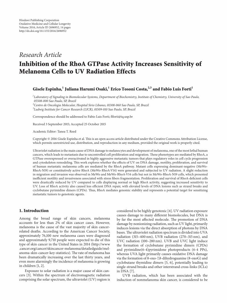
Research ArticleInhibition of the RhoA GTPase Activity Increases Sensitivity ofMelanoma Cells to UV Radiation Effects
Gisele Espinha,1 Juliana Harumi Osaki,1 Erico Tosoni Costa,2,3 and Fabio Luis Forti1
1Laboratory of Signaling in Biomolecular Systems, Department of Biochemistry, Institute of Chemistry, University of Sao Paulo,05508-000 Sao Paulo, SP, Brazil2Centro de Oncologia Molecular, Hospital Sirio Libanes, 01308-060 Sao Paulo, SP, Brazil3Ludwig Institute for Cancer Research (LICR), 01509-010 Sao Paulo, SP, Brazil
Correspondence should be addressed to Fabio Luis Forti; [email protected]
Received 3 September 2015; Accepted 25 October 2015
Academic Editor: Tanea T. Reed
Copyright © 2016 Gisele Espinha et al. This is an open access article distributed under the Creative Commons Attribution License,which permits unrestricted use, distribution, and reproduction in any medium, provided the original work is properly cited.
Ultraviolet radiation is themain cause ofDNAdamage tomelanocytes and development ofmelanoma, one of themost lethal humancancers, which leads to metastasis due to uncontrolled cell proliferation and migration.These phenotypes are mediated by RhoA, aGTPase overexpressed or overactivated in highly aggressive metastatic tumors that plays regulatory roles in cell cycle progressionand cytoskeleton remodeling. This work explores whether the effects of UV on DNA damage, motility, proliferation, and survivalof human metastatic melanoma cells are mediated by the RhoA pathway. Mutant cells expressing dominant-negative (MeWo-RhoA-N19) or constitutively active RhoA (MeWo-RhoA-V14) were generated and subjected to UV radiation. A slight reductionin migration and invasion was observed in MeWo and MeWo-RhoA-V14 cells but not in MeWo-RhoA-N19 cells, which presentedinefficient motility and invasiveness associated with stress fibers fragmentation. Proliferation and survival of RhoA-deficient cellswere drastically reduced by UV compared to cells displaying normal or high RhoA activity, suggesting increased sensitivity toUV. Loss of RhoA activity also caused less efficient DNA repair, with elevated levels of DNA lesions such as strand breaks andcyclobutane pyrimidine dimers (CPDs). Thus, RhoA mediates genomic stability and represents a potential target for sensitizingmetastatic tumors to genotoxic agents.
1. Introduction
Among the broad range of skin cancers, melanomaaccounts for less than 2% of skin cancer cases. However,melanoma is the cause of the vast majority of skin cancer-related deaths. According to the American Cancer Society,approximately 76,100 new melanoma cases were diagnosedand approximately 9,710 people were expected to die of thistype of skin cancer in the United States in 2014 (http://www.cancer.org/cancer/skincancer-melanoma/detailedguide/mel-anoma-skin-cancer-key-statistics).The rate of melanoma hasbeen dramatically increasing over the last thirty years, andeven more alarmingly the incidence of melanoma is growingin children [1, 2].
Exposure to solar radiation is a major cause of skin can-cers [3]. Within the spectrum of electromagnetic radiationcomprising the solar spectrum, the ultraviolet (UV) region is
considered to be highly genotoxic [4]. UV radiation exposurecauses damage to many different biomolecules, but DNA isby far the most affected molecule. The promotion of DNAdamage by nonionizing radiation, such as UV light, primarilyinduces lesions via the direct absorption of photons by DNAbases.The ultraviolet radiation spectrum is divided into UVAradiation (315–400 nm), UVB radiation (270–315 nm), andUVC radiation (100–280 nm). UVB and UVC light inducethe formation of cyclobutane pyrimidine dimers (CPDs)and pyrimidine(6-4)pyrimidone photoproducts (6-4 PPs),whereas UVA light primarily causes oxidative DNA damagevia the formation of 8-oxo-7,8-dihydroguanine (8-oxoG) andcyclobutane thymidine dimers [5, 6], potentially leading tosingle-strand breaks and other interstrand cross-links (ICLs)in DNA [7].
UVB radiation, which has been associated with theinduction of nonmelanoma skin cancer, is considered to be
Hindawi Publishing CorporationOxidative Medicine and Cellular LongevityVolume 2016, Article ID 2696952, 14 pageshttp://dx.doi.org/10.1155/2016/2696952
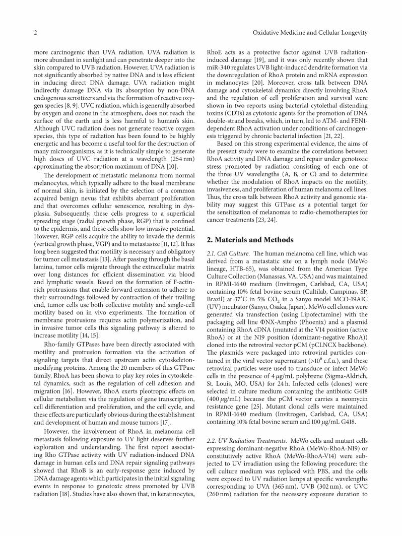
2 Oxidative Medicine and Cellular Longevity
more carcinogenic than UVA radiation. UVA radiation ismore abundant in sunlight and can penetrate deeper into theskin compared to UVB radiation. However, UVA radiation isnot significantly absorbed by native DNA and is less efficientin inducing direct DNA damage. UVA radiation mightindirectly damage DNA via its absorption by non-DNAendogenous sensitizers and via the formation of reactive oxy-gen species [8, 9]. UVC radiation, which is generally absorbedby oxygen and ozone in the atmosphere, does not reach thesurface of the earth and is less harmful to human’s skin.Although UVC radiation does not generate reactive oxygenspecies, this type of radiation has been found to be highlyenergetic and has become a useful tool for the destruction ofmany microorganisms, as it is technically simple to generatehigh doses of UVC radiation at a wavelength (254 nm)approximating the absorption maximum of DNA [10].
The development of metastatic melanoma from normalmelanocytes, which typically adhere to the basal membraneof normal skin, is initiated by the selection of a commonacquired benign nevus that exhibits aberrant proliferationand that overcomes cellular senescence, resulting in dys-plasia. Subsequently, these cells progress to a superficialspreading stage (radial growth phase, RGP) that is confinedto the epidermis, and these cells show low invasive potential.However, RGP cells acquire the ability to invade the dermis(vertical growth phase, VGP) and tometastasize [11, 12]. It haslong been suggested that motility is necessary and obligatoryfor tumor cell metastasis [13]. After passing through the basallamina, tumor cells migrate through the extracellular matrixover long distances for efficient dissemination via bloodand lymphatic vessels. Based on the formation of F-actin-rich protrusions that enable forward extension to adhere totheir surroundings followed by contraction of their trailingend, tumor cells use both collective motility and single-cellmotility based on in vivo experiments. The formation ofmembrane protrusions requires actin polymerization, andin invasive tumor cells this signaling pathway is altered toincrease motility [14, 15].
Rho-family GTPases have been directly associated withmotility and protrusion formation via the activation ofsignaling targets that direct upstream actin cytoskeleton-modifying proteins. Among the 20 members of this GTPasefamily, RhoA has been shown to play key roles in cytoskele-tal dynamics, such as the regulation of cell adhesion andmigration [16]. However, RhoA exerts pleotropic effects oncellular metabolism via the regulation of gene transcription,cell differentiation and proliferation, and the cell cycle, andthese effects are particularly obvious during the establishmentand development of human and mouse tumors [17].
However, the involvement of RhoA in melanoma cellmetastasis following exposure to UV light deserves furtherexploration and understanding. The first report associat-ing Rho GTPase activity with UV radiation-induced DNAdamage in human cells and DNA repair signaling pathwaysshowed that RhoB is an early-response gene induced byDNAdamage agentswhich participates in the initial signalingevents in response to genotoxic stress promoted by UVBradiation [18]. Studies have also shown that, in keratinocytes,
RhoE acts as a protective factor against UVB radiation-induced damage [19], and it was only recently shown thatmiR-340 regulates UVB light-induced dendrite formation viathe downregulation of RhoA protein and mRNA expressionin melanocytes [20]. Moreover, cross talk between DNAdamage and cytoskeletal dynamics directly involving RhoAand the regulation of cell proliferation and survival wereshown in two reports using bacterial cytolethal distendingtoxins (CDTs) as cytotoxic agents for the promotion of DNAdouble-strand breaks, which, in turn, led to ATM- and FEN1-dependent RhoA activation under conditions of carcinogen-esis triggered by chronic bacterial infection [21, 22].
Based on this strong experimental evidence, the aims ofthe present study were to examine the correlations betweenRhoA activity and DNA damage and repair under genotoxicstress promoted by radiation consisting of each one ofthe three UV wavelengths (A, B, or C) and to determinewhether the modulation of RhoA impacts on the motility,invasiveness, and proliferation of humanmelanoma cell lines.Thus, the cross talk between RhoA activity and genomic sta-bility may suggest this GTPase as a potential target forthe sensitization of melanomas to radio-chemotherapies forcancer treatments [23, 24].
2. Materials and Methods
2.1. Cell Culture. The human melanoma cell line, which wasderived from a metastatic site on a lymph node (MeWolineage, HTB-65), was obtained from the American TypeCulture Collection (Manassas, VA,USA) andwasmaintainedin RPMI-1640 medium (Invitrogen, Carlsbad, CA, USA)containing 10% fetal bovine serum (Cultilab, Campinas, SP,Brazil) at 37∘C in 5% CO
2in a Sanyo model MCO-19AIC
(UV) incubator (Sanyo,Osaka, Japan).MeWocell clonesweregenerated via transfection (using Lipofectamine) with thepackaging cell line ΦNX-Ampho (Phoenix) and a plasmidcontaining RhoA cDNA (mutated at the V14 position (activeRhoA) or at the N19 position (dominant-negative RhoA))cloned into the retroviral vector pCM (pCLNCX backbone).The plasmids were packaged into retroviral particles con-tained in the viral vector supernatant (>106 c.f.u.), and theseretroviral particles were used to transduce or infect MeWocells in the presence of 4𝜇g/mL polybrene (Sigma-Aldrich,St. Louis, MO, USA) for 24 h. Infected cells (clones) wereselected in culture medium containing the antibiotic G418(400 𝜇g/mL) because the pCM vector carries a neomycinresistance gene [25]. Mutant clonal cells were maintainedin RPMI-1640 medium (Invitrogen, Carlsbad, CA, USA)containing 10% fetal bovine serum and 100 𝜇g/mL G418.
2.2. UV Radiation Treatments. MeWo cells and mutant cellsexpressing dominant-negative RhoA (MeWo-RhoA-N19) orconstitutively active RhoA (MeWo-RhoA-V14) were sub-jected to UV irradiation using the following procedure: thecell culture medium was replaced with PBS, and the cellswere exposed to UV radiation lamps at specific wavelengthscorresponding to UVA (365 nm), UVB (302 nm), or UVC(260 nm) radiation for the necessary exposure duration to
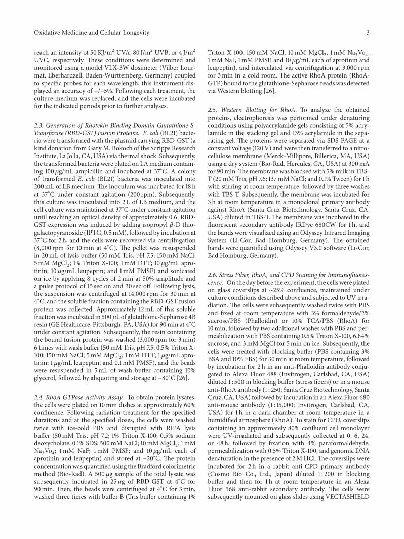
Oxidative Medicine and Cellular Longevity 3
reach an intensity of 50KJ/m2 UVA, 80 J/m2 UVB, or 4 J/m2UVC, respectively. These conditions were determined andmonitored using a model VLX-3W dosimeter (Vilber Lour-mat, Eberhardzell, Baden-Württemberg, Germany) coupledto specific probes for each wavelength; this instrument dis-played an accuracy of +/−5%. Following each treatment, theculture medium was replaced, and the cells were incubatedfor the indicated periods prior to further analyses.
2.3. Generation of Rhotekin-Binding Domain-Glutathione S-Transferase (RBD-GST) Fusion Proteins. E. coli (BL21) bacte-ria were transformed with the plasmid carrying RBD-GST (akind donation from Gary M. Bokoch of the Scripps ResearchInstitute, La Jolla, CA, USA) via thermal shock. Subsequently,the transformed bacteria were plated on LAmedium contain-ing 100 𝜇g/mL ampicillin and incubated at 37∘C. A colonyof transformed E. coli (BL21) bacteria was inoculated into200mL of LB medium. The inoculum was incubated for 18 hat 37∘C under constant agitation (200 rpm). Subsequently,this culture was inoculated into 2 L of LB medium, and thecell culture was maintained at 37∘C under constant agitationuntil reaching an optical density of approximately 0.6. RBD-GST expression was induced by adding isopropyl 𝛽-D thio-galactopyranoside (IPTG, 0.5mM), followed by incubation at37∘C for 2 h, and the cells were recovered via centrifugation(8,000 rpm for 10min at 4∘C). The pellet was resuspendedin 20mL of lysis buffer (50mM Tris, pH 7.5; 150mM NaCl;5mM MgCl
2; 1% Triton X-100; 1mM DTT; 10 𝜇g/mL apro-
tinin; 10 𝜇g/mL leupeptin; and 1mM PMSF) and sonicatedon ice by applying 8 cycles of 2min at 50% amplitude anda pulse protocol of 15 sec on and 30 sec off. Following lysis,the suspension was centrifuged at 14,000 rpm for 30min at4∘C, and the soluble fraction containing the RBD-GST fusionprotein was collected. Approximately 12mL of this solublefractionwas incubated in 500𝜇L of glutathione-Sepharose 4Bresin (GEHealthcare, Pittsburgh, PA, USA) for 90min at 4∘Cunder constant agitation. Subsequently, the resin containingthe bound fusion protein was washed (3,000 rpm for 3min)6 times with wash buffer (50mM Tris, pH 7.5; 0.5% Triton X-100; 150mM NaCl; 5mM MgCl
2; 1 mM DTT; 1 𝜇g/mL apro-
tinin; 1 𝜇g/mL leupeptin; and 0.1mM PMSF), and the beadswere resuspended in 5mL of wash buffer containing 10%glycerol, followed by aliquoting and storage at −80∘C [26].
2.4. RhoA GTPase Activity Assay. To obtain protein lysates,the cells were plated on 10mm dishes at approximately 60%confluence. Following radiation treatment for the specifieddurations and at the specified doses, the cells were washedtwice with ice-cold PBS and disrupted with RIPA lysisbuffer (50mM Tris, pH 7.2; 1% Triton X-100; 0.5% sodiumdeoxycholate; 0.1% SDS; 500mMNaCl; 10mMMgCl
2; 1 mM
Na3Vo4; 1 mM NaF; 1mM PMSF; and 10 𝜇g/mL each of
aprotinin and leupeptin) and stored at −20∘C. The proteinconcentrationwas quantified using the Bradford colorimetricmethod (Bio-Rad). A 500𝜇g sample of the total lysate wassubsequently incubated in 25 𝜇g of RBD-GST at 4∘C for90min. Then, the beads were centrifuged at 4∘C for 3min,washed three times with buffer B (Tris buffer containing 1%
Triton X-100, 150mM NaCl, 10mM MgCl2, 1 mM Na
3Vo4,
1 mM NaF, 1mM PMSF, and 10 𝜇g/mL each of aprotinin andleupeptin), and intercalated via centrifugation at 3,000 rpmfor 3min in a cold room. The active RhoA protein (RhoA-GTP) bound to the glutathione-Sepharose beadswas detectedvia Western blotting [26].
2.5. Western Blotting for RhoA. To analyze the obtainedproteins, electrophoresis was performed under denaturingconditions using polyacrylamide gels consisting of 5% acry-lamide in the stacking gel and 13% acrylamide in the sepa-rating gel. The proteins were separated via SDS-PAGE at aconstant voltage (120V) and were then transferred to a nitro-cellulose membrane (Merck-Millipore, Billerica, MA, USA)using a dry system (Bio-Rad, Hercules, CA, USA) at 300mAfor 90min.Themembrane was blocked with 5%milk in TBS-T (20mMTris, pH 7.6; 137mMNaCl; and 0.1% Tween) for 1 hwith stirring at room temperature, followed by three washeswith TBS-T. Subsequently, the membrane was incubated for3 h at room temperature in a monoclonal primary antibodyagainst RhoA (Santa Cruz Biotechnology, Santa Cruz, CA,USA) diluted in TBS-T. The membrane was incubated in thefluorescent secondary antibody IRDye 680CW for 1 h, andthe bands were visualized using anOdyssey Infrared ImagingSystem (Li-Cor, Bad Homburg, Germany). The obtainedbands were quantified using Odyssey V3.0 software (Li-Cor,Bad Homburg, Germany).
2.6. Stress Fiber, RhoA, and CPD Staining for Immunofluores-cence. On the day before the experiment, the cells were platedon glass coverslips at ∼25% confluence, maintained underculture conditions described above and subjected to UV irra-diation. The cells were subsequently washed twice with PBSand fixed at room temperature with 3% formaldehyde/2%sucrose/PBS (Phalloidin) or 10% TCA/PBS (RhoA) for10min, followed by two additional washes with PBS and per-meabilization with PBS containing 0.5% Triton X-100, 6.84%sucrose, and 3mM MgCl for 5min on ice. Subsequently, thecells were treated with blocking buffer (PBS containing 3%BSA and 10% FBS) for 30min at room temperature, followedby incubation for 2 h in an anti-Phalloidin antibody conju-gated to Alexa Fluor 488 (Invitrogen, Carlsbad, CA, USA)diluted 1 : 500 in blocking buffer (stress fibers) or in a mouseanti-RhoA antibody (1 : 250; Santa Cruz Biotechnology, SantaCruz, CA,USA) followed by incubation in anAlexa Fluor 680anti-mouse antibody (1 : 15,000; Invitrogen, Carlsbad, CA,USA) for 1 h in a dark chamber at room temperature in ahumidified atmosphere (RhoA). To stain for CPD, coverslipscontaining an approximately 80% confluent cell monolayerwere UV-irradiated and subsequently collected at 0, 6, 24,or 48 h, followed by fixation with 4% paraformaldehyde,permeabilization with 0.5% Triton X-100, and genomic DNAdenaturation in the presence of 2MHCl.The coverslips wereincubated for 2 h in a rabbit anti-CPD primary antibody(Cosmo Bio Co., Ltd., Japan) diluted 1 : 200 in blockingbuffer and then for 1 h at room temperature in an AlexaFluor 568 anti-rabbit secondary antibody. The cells weresubsequently mounted on glass slides using VECTASHIELD
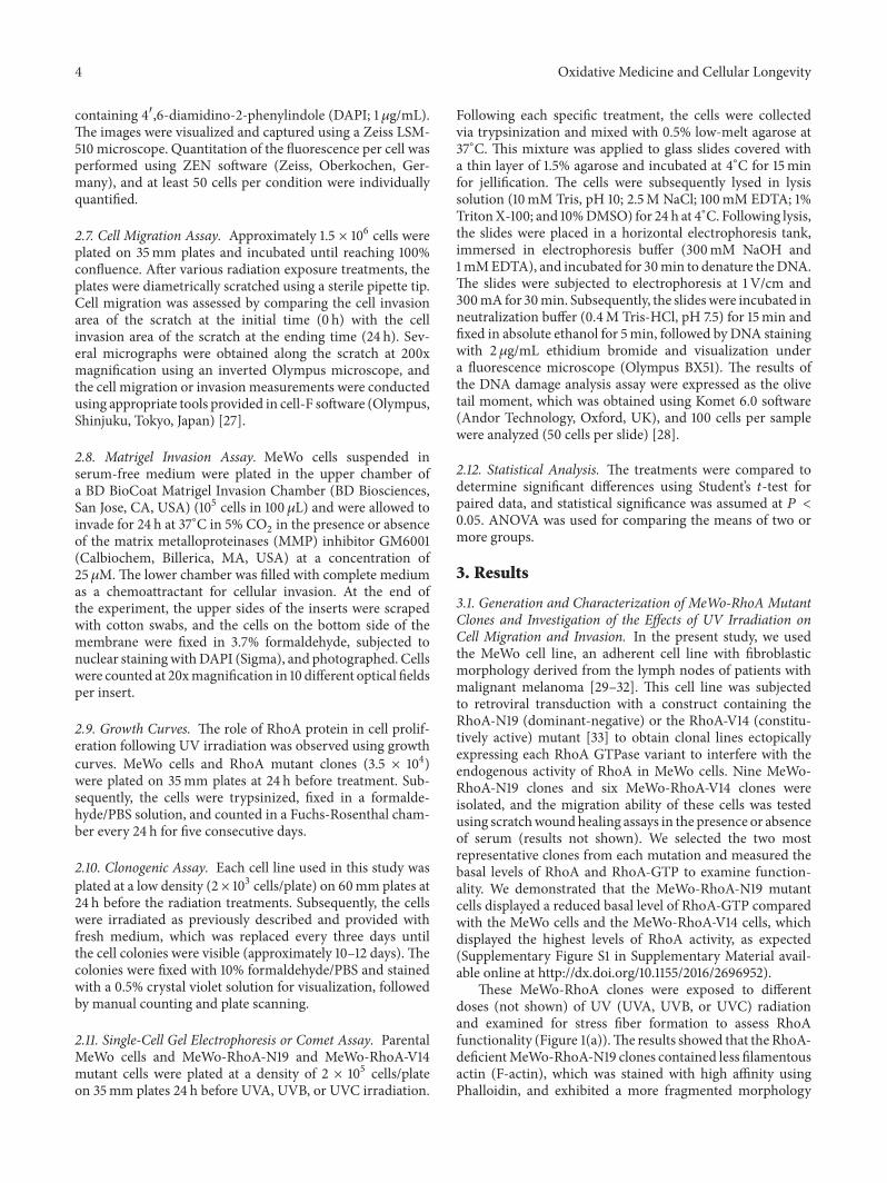
4 Oxidative Medicine and Cellular Longevity
containing 4,6-diamidino-2-phenylindole (DAPI; 1𝜇g/mL).The images were visualized and captured using a Zeiss LSM-510 microscope. Quantitation of the fluorescence per cell wasperformed using ZEN software (Zeiss, Oberkochen, Ger-many), and at least 50 cells per condition were individuallyquantified.
2.7. Cell Migration Assay. Approximately 1.5 × 10⁶ cells wereplated on 35mm plates and incubated until reaching 100%confluence. After various radiation exposure treatments, theplates were diametrically scratched using a sterile pipette tip.Cell migration was assessed by comparing the cell invasionarea of the scratch at the initial time (0 h) with the cellinvasion area of the scratch at the ending time (24 h). Sev-eral micrographs were obtained along the scratch at 200xmagnification using an inverted Olympus microscope, andthe cell migration or invasionmeasurements were conductedusing appropriate tools provided in cell-F software (Olympus,Shinjuku, Tokyo, Japan) [27].
2.8. Matrigel Invasion Assay. MeWo cells suspended inserum-free medium were plated in the upper chamber ofa BD BioCoat Matrigel Invasion Chamber (BD Biosciences,San Jose, CA, USA) (105 cells in 100 𝜇L) and were allowed toinvade for 24 h at 37∘C in 5% CO
2in the presence or absence
of the matrix metalloproteinases (MMP) inhibitor GM6001(Calbiochem, Billerica, MA, USA) at a concentration of25 𝜇M.The lower chamber was filled with complete mediumas a chemoattractant for cellular invasion. At the end ofthe experiment, the upper sides of the inserts were scrapedwith cotton swabs, and the cells on the bottom side of themembrane were fixed in 3.7% formaldehyde, subjected tonuclear stainingwithDAPI (Sigma), and photographed. Cellswere counted at 20xmagnification in 10 different optical fieldsper insert.
2.9. Growth Curves. The role of RhoA protein in cell prolif-eration following UV irradiation was observed using growthcurves. MeWo cells and RhoA mutant clones (3.5 × 10⁴)were plated on 35mm plates at 24 h before treatment. Sub-sequently, the cells were trypsinized, fixed in a formalde-hyde/PBS solution, and counted in a Fuchs-Rosenthal cham-ber every 24 h for five consecutive days.
2.10. Clonogenic Assay. Each cell line used in this study wasplated at a low density (2 × 10³ cells/plate) on 60mm plates at24 h before the radiation treatments. Subsequently, the cellswere irradiated as previously described and provided withfresh medium, which was replaced every three days untilthe cell colonies were visible (approximately 10–12 days). Thecolonies were fixed with 10% formaldehyde/PBS and stainedwith a 0.5% crystal violet solution for visualization, followedby manual counting and plate scanning.
2.11. Single-Cell Gel Electrophoresis or Comet Assay. ParentalMeWo cells and MeWo-RhoA-N19 and MeWo-RhoA-V14mutant cells were plated at a density of 2 × 10⁵ cells/plateon 35mm plates 24 h before UVA, UVB, or UVC irradiation.
Following each specific treatment, the cells were collectedvia trypsinization and mixed with 0.5% low-melt agarose at37∘C. This mixture was applied to glass slides covered witha thin layer of 1.5% agarose and incubated at 4∘C for 15minfor jellification. The cells were subsequently lysed in lysissolution (10mM Tris, pH 10; 2.5M NaCl; 100mM EDTA; 1%TritonX-100; and 10%DMSO) for 24 h at 4∘C. Following lysis,the slides were placed in a horizontal electrophoresis tank,immersed in electrophoresis buffer (300mM NaOH and1mMEDTA), and incubated for 30min to denature theDNA.The slides were subjected to electrophoresis at 1 V/cm and300mA for 30min. Subsequently, the slideswere incubated inneutralization buffer (0.4M Tris-HCl, pH 7.5) for 15min andfixed in absolute ethanol for 5min, followed by DNA stainingwith 2 𝜇g/mL ethidium bromide and visualization undera fluorescence microscope (Olympus BX51). The results ofthe DNA damage analysis assay were expressed as the olivetail moment, which was obtained using Komet 6.0 software(Andor Technology, Oxford, UK), and 100 cells per samplewere analyzed (50 cells per slide) [28].
2.12. Statistical Analysis. The treatments were compared todetermine significant differences using Student’s 𝑡-test forpaired data, and statistical significance was assumed at 𝑃 <0.05. ANOVA was used for comparing the means of two ormore groups.
3. Results
3.1. Generation and Characterization of MeWo-RhoA MutantClones and Investigation of the Effects of UV Irradiation onCell Migration and Invasion. In the present study, we usedthe MeWo cell line, an adherent cell line with fibroblasticmorphology derived from the lymph nodes of patients withmalignant melanoma [29–32]. This cell line was subjectedto retroviral transduction with a construct containing theRhoA-N19 (dominant-negative) or the RhoA-V14 (constitu-tively active) mutant [33] to obtain clonal lines ectopicallyexpressing each RhoA GTPase variant to interfere with theendogenous activity of RhoA in MeWo cells. Nine MeWo-RhoA-N19 clones and six MeWo-RhoA-V14 clones wereisolated, and the migration ability of these cells was testedusing scratchwoundhealing assays in the presence or absenceof serum (results not shown). We selected the two mostrepresentative clones from each mutation and measured thebasal levels of RhoA and RhoA-GTP to examine function-ality. We demonstrated that the MeWo-RhoA-N19 mutantcells displayed a reduced basal level of RhoA-GTP comparedwith the MeWo cells and the MeWo-RhoA-V14 cells, whichdisplayed the highest levels of RhoA activity, as expected(Supplementary Figure S1 in Supplementary Material avail-able online at http://dx.doi.org/10.1155/2016/2696952).
These MeWo-RhoA clones were exposed to differentdoses (not shown) of UV (UVA, UVB, or UVC) radiationand examined for stress fiber formation to assess RhoAfunctionality (Figure 1(a)).The results showed that the RhoA-deficientMeWo-RhoA-N19 clones contained less filamentousactin (F-actin), which was stained with high affinity usingPhalloidin, and exhibited a more fragmented morphology
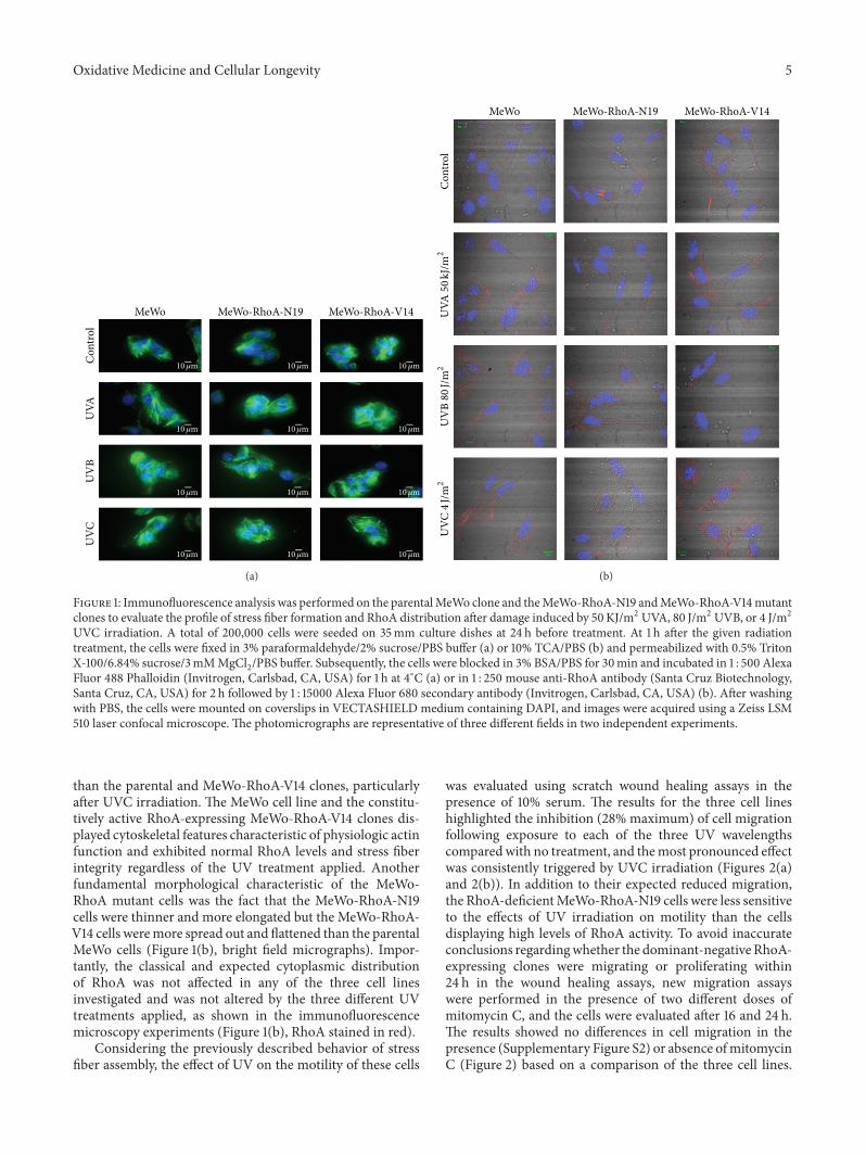
Oxidative Medicine and Cellular Longevity 5
MeWo MeWo-RhoA-N19 MeWo-RhoA-V14
Con
trol
UVA
UV
BU
VC
10𝜇m 10𝜇m 10𝜇m
10𝜇m 10𝜇m 10𝜇m
10𝜇m 10𝜇m 10𝜇m
10𝜇m 10𝜇m 10𝜇m
(a)
MeWo MeWo-RhoA-N19 MeWo-RhoA-V14
Con
trol
UVA
50
kJ/m
2U
VB80
J/m2
UVC
4J/m
2
(b)
Figure 1: Immunofluorescence analysis was performed on the parentalMeWo clone and theMeWo-RhoA-N19 andMeWo-RhoA-V14mutantclones to evaluate the profile of stress fiber formation and RhoA distribution after damage induced by 50KJ/m2 UVA, 80 J/m2 UVB, or 4 J/m2UVC irradiation. A total of 200,000 cells were seeded on 35mm culture dishes at 24 h before treatment. At 1 h after the given radiationtreatment, the cells were fixed in 3% paraformaldehyde/2% sucrose/PBS buffer (a) or 10% TCA/PBS (b) and permeabilized with 0.5% TritonX-100/6.84% sucrose/3mMMgCl
2
/PBS buffer. Subsequently, the cells were blocked in 3% BSA/PBS for 30min and incubated in 1 : 500 AlexaFluor 488 Phalloidin (Invitrogen, Carlsbad, CA, USA) for 1 h at 4∘C (a) or in 1 : 250 mouse anti-RhoA antibody (Santa Cruz Biotechnology,Santa Cruz, CA, USA) for 2 h followed by 1 : 15000 Alexa Fluor 680 secondary antibody (Invitrogen, Carlsbad, CA, USA) (b). After washingwith PBS, the cells were mounted on coverslips in VECTASHIELD medium containing DAPI, and images were acquired using a Zeiss LSM510 laser confocal microscope. The photomicrographs are representative of three different fields in two independent experiments.
than the parental and MeWo-RhoA-V14 clones, particularlyafter UVC irradiation. The MeWo cell line and the constitu-tively active RhoA-expressing MeWo-RhoA-V14 clones dis-played cytoskeletal features characteristic of physiologic actinfunction and exhibited normal RhoA levels and stress fiberintegrity regardless of the UV treatment applied. Anotherfundamental morphological characteristic of the MeWo-RhoA mutant cells was the fact that the MeWo-RhoA-N19cells were thinner and more elongated but the MeWo-RhoA-V14 cells weremore spread out and flattened than the parentalMeWo cells (Figure 1(b), bright field micrographs). Impor-tantly, the classical and expected cytoplasmic distributionof RhoA was not affected in any of the three cell linesinvestigated and was not altered by the three different UVtreatments applied, as shown in the immunofluorescencemicroscopy experiments (Figure 1(b), RhoA stained in red).
Considering the previously described behavior of stressfiber assembly, the effect of UV on the motility of these cells
was evaluated using scratch wound healing assays in thepresence of 10% serum. The results for the three cell lineshighlighted the inhibition (28% maximum) of cell migrationfollowing exposure to each of the three UV wavelengthscomparedwith no treatment, and themost pronounced effectwas consistently triggered by UVC irradiation (Figures 2(a)and 2(b)). In addition to their expected reduced migration,the RhoA-deficientMeWo-RhoA-N19 cells were less sensitiveto the effects of UV irradiation on motility than the cellsdisplaying high levels of RhoA activity. To avoid inaccurateconclusions regardingwhether the dominant-negativeRhoA-expressing clones were migrating or proliferating within24 h in the wound healing assays, new migration assayswere performed in the presence of two different doses ofmitomycin C, and the cells were evaluated after 16 and 24 h.The results showed no differences in cell migration in thepresence (Supplementary Figure S2) or absence ofmitomycinC (Figure 2) based on a comparison of the three cell lines.
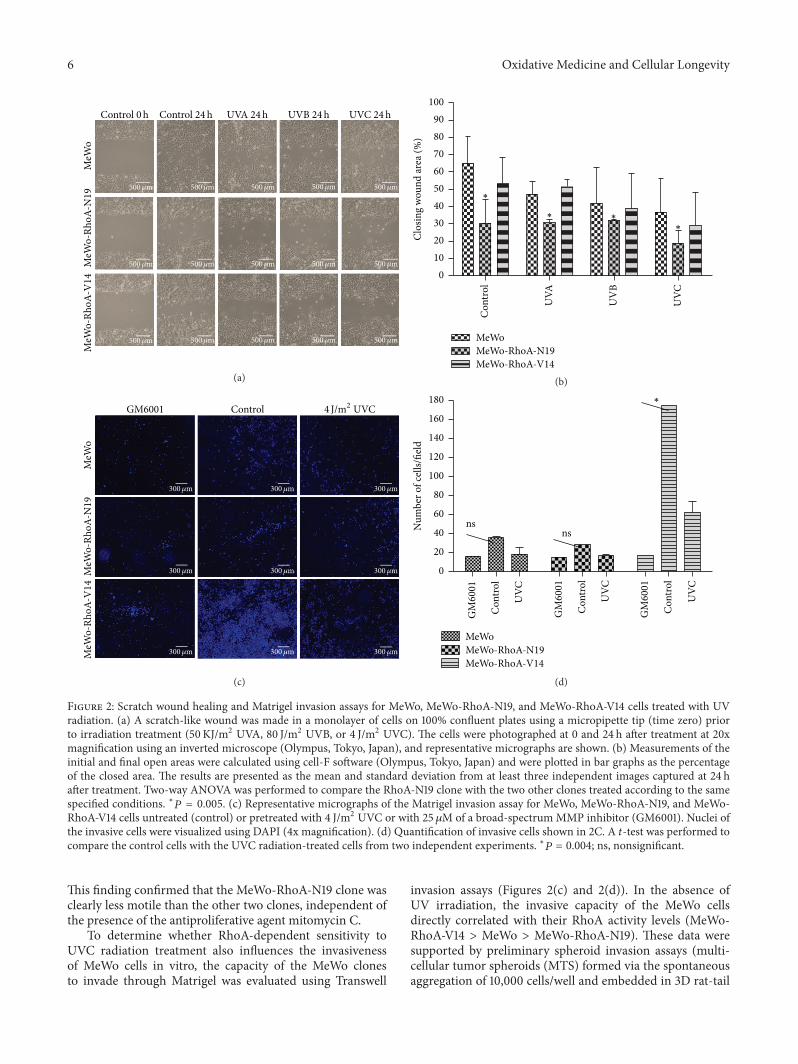
6 Oxidative Medicine and Cellular LongevityM
eWo
MeW
o-Rh
oA-N
19M
eWo-
RhoA
-V14
UVA 24h UVB 24h UVC 24hControl 0h Control 24h
500𝜇m 500𝜇m 500𝜇m 500𝜇m 500𝜇m
500𝜇m 500𝜇m 500𝜇m 500𝜇m 500𝜇m
500𝜇m 500𝜇m 500𝜇m 500𝜇m 500𝜇m
(a)
MeWoMeWo-RhoA-N19MeWo-RhoA-V14
Con
trol
UVC
UV
B
UVA
0102030405060708090
100
Clos
ing
wou
nd ar
ea (%
)
∗
∗∗
∗
(b)
GM6001 Control
MeW
oM
eWo-
RhoA
-N19
MeW
o-Rh
oA-V
14
4 J/m2 UVC
300𝜇m 300𝜇m 300𝜇m
300𝜇m 300𝜇m 300𝜇m
300𝜇m 300𝜇m 300𝜇m
(c)
MeWo
GM
6001
Con
trol
UVC
GM
6001
Con
trol
UVC
GM
6001
Con
trol
UVC
MeWo-RhoA-N19MeWo-RhoA-V14
nsns
0
20
40
60
80
100
120
140
160
180
Num
ber o
f cel
ls/fie
ld
∗
(d)
Figure 2: Scratch wound healing and Matrigel invasion assays for MeWo, MeWo-RhoA-N19, and MeWo-RhoA-V14 cells treated with UVradiation. (a) A scratch-like wound was made in a monolayer of cells on 100% confluent plates using a micropipette tip (time zero) priorto irradiation treatment (50KJ/m2 UVA, 80 J/m2 UVB, or 4 J/m2 UVC). The cells were photographed at 0 and 24 h after treatment at 20xmagnification using an inverted microscope (Olympus, Tokyo, Japan), and representative micrographs are shown. (b) Measurements of theinitial and final open areas were calculated using cell-F software (Olympus, Tokyo, Japan) and were plotted in bar graphs as the percentageof the closed area. The results are presented as the mean and standard deviation from at least three independent images captured at 24 hafter treatment. Two-way ANOVA was performed to compare the RhoA-N19 clone with the two other clones treated according to the samespecified conditions. ∗𝑃 = 0.005. (c) Representative micrographs of the Matrigel invasion assay for MeWo, MeWo-RhoA-N19, and MeWo-RhoA-V14 cells untreated (control) or pretreated with 4 J/m2 UVC or with 25 𝜇M of a broad-spectrum MMP inhibitor (GM6001). Nuclei ofthe invasive cells were visualized using DAPI (4x magnification). (d) Quantification of invasive cells shown in 2C. A 𝑡-test was performed tocompare the control cells with the UVC radiation-treated cells from two independent experiments. ∗𝑃 = 0.004; ns, nonsignificant.
This finding confirmed that the MeWo-RhoA-N19 clone wasclearly less motile than the other two clones, independent ofthe presence of the antiproliferative agent mitomycin C.
To determine whether RhoA-dependent sensitivity toUVC radiation treatment also influences the invasivenessof MeWo cells in vitro, the capacity of the MeWo clonesto invade through Matrigel was evaluated using Transwell
invasion assays (Figures 2(c) and 2(d)). In the absence ofUV irradiation, the invasive capacity of the MeWo cellsdirectly correlated with their RhoA activity levels (MeWo-RhoA-V14 > MeWo > MeWo-RhoA-N19). These data weresupported by preliminary spheroid invasion assays (multi-cellular tumor spheroids (MTS) formed via the spontaneousaggregation of 10,000 cells/well and embedded in 3D rat-tail
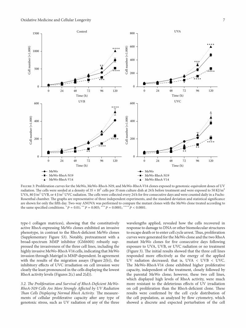
Oxidative Medicine and Cellular Longevity 7
Control
0 9624 7248 120Time (h)
0
500
1000
1500C
ell n
umbe
r (×1.000
)
∗
∗∗
∗∗∗∗
∗∗∗∗
∗∗∗
UVA
0 9624 7248 120Time (h)
0
200
400
600
800
Cel
l num
ber (×1.000
)
∗∗∗
∗∗∗∗
∗∗∗∗
∗∗∗∗
∗
∗
UVB
MeWoMeWo-RhoA-N19MeWo-RhoA-V14
0 9624 7248 120Time (h)
0
200
400
600
Cel
l num
ber (×1.000
)
UVC
MeWoMeWo-RhoA-N19MeWo-RhoA-V14
0 9624 7248 120Time (h)
0
200
400
600
Cel
l num
ber (×1.000
)
∗∗
∗∗
∗∗
∗∗∗∗
∗∗∗∗
∗∗∗∗
∗∗∗∗
Figure 3: Proliferation curves for the MeWo, MeWo-RhoA-N19, and MeWo-RhoA-V14 clones exposed to genotoxic-equivalent doses of UVradiation. The cells were seeded at a density of 35 × 103 cells per 35mm culture dish at 24 h before treatment and were exposed to 50KJ/m2UVA, 80 J/m2 UVB, or 4 J/m2 UVC radiation.The cells were collected every 24 h for five consecutive days and were counted daily in a Fuchs-Rosenthal chamber. The graphs are representative of three independent experiments, and the standard deviation and statistical significanceare shown for only the fifth day. Two-way ANOVA was performed to compare the mutant clones with the MeWo clone treated according tothe same specified conditions. ∗𝑃 = 0.01; ∗∗𝑃 = 0.005; ∗∗∗𝑃 = 0.0001; ∗∗∗∗𝑃 < 0.0001.
type-1 collagen matrices), showing that the constitutivelyactive RhoA-expressing MeWo clones exhibited an invasivephenotype, in contrast to the RhoA-deficient MeWo clones(Supplementary Figure S3). Notably, pretreatment with abroad-spectrum MMP inhibitor (GM6001) robustly sup-pressed the invasiveness of the three cell lines, including thehighly invasiveMeWo-RhoA-V14 cells, indicating thatMeWoinvasion through Matrigel is MMP-dependent. In agreementwith the results of the migration assays (Figure 2(b)), theinhibitory effects of UVC irradiation on cell invasion wereclearly the least pronounced in the cells displaying the lowestRhoA activity levels (Figures 2(c) and 2(d)).
3.2. The Proliferation and Survival of RhoA-Deficient MeWo-RhoA-N19 Cells Are More Strongly Affected by UV RadiationThan Cells Displaying Normal RhoA Activity. The measure-ments of cellular proliferative capacity after any type ofgenotoxic stress, such as UV radiation of any of the three
wavelengths applied, revealed how the cells recovered inresponse to damage to DNA or other biomolecular structuresto escape death or to enter cell cycle arrest.Thus, proliferationcurves were generated for theMeWo clone and the two RhoAmutant MeWo clones for five consecutive days followingexposure to UVA, UVB, or UVC radiation or no treatment(Figure 3). The initial results showed that the three cell linesresponded more effectively as the energy of the appliedUV radiation decreased; that is, UVA < UVB < UVC.The MeWo-RhoA-V14 clone exhibited higher proliferativecapacity, independent of the treatment, closely followed bythe parental MeWo clone; however, these two cell lines,which displayed high levels of RhoA activity, were muchmore resistant to the deleterious effects of UV irradiationon cell proliferation than the RhoA-deficient clone. Theseresults were confirmed by the cell cycle distribution ofthe cell population, as analyzed by flow cytometry, whichshowed a discrete and expected perturbation of the cell
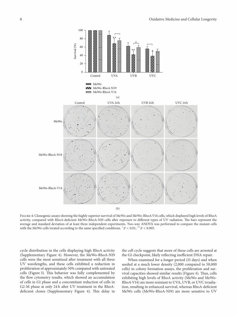
8 Oxidative Medicine and Cellular Longevity
MeWo
Control UVA UVB UVC
MeWo-RhoA-N19MeWo-RhoA-V14
0
20
40
60
80
100
Surv
ival
(%) ∗
∗
∗∗
∗∗
(a)
MeWo
MeWo-RhoA-N19
MeWo-RhoA-V14
Control UVC 24hUVB 24hUVA 24h
(b)
Figure 4: Clonogenic assays showing the highly superior survival of MeWo andMeWo-RhoA-V14 cells, which displayed high levels of RhoAactivity, compared with RhoA-deficient MeWo-RhoA-N19 cells after exposure to different types of UV radiation. The bars represent theaverage and standard deviation of at least three independent experiments. Two-way ANOVA was performed to compare the mutant cellswith the MeWo cells treated according to the same specified conditions. ∗𝑃 < 0.01; ∗∗𝑃 < 0.005.
cycle distribution in the cells displaying high RhoA activity(Supplementary Figure 4). However, the MeWo-RhoA-N19cells were the most sensitized after treatment with all threeUV wavelengths, and these cells exhibited a reduction inproliferation of approximately 50% compared with untreatedcells (Figure 3). This behavior was fully complemented bythe flow cytometry results, which showed an accumulationof cells in G1 phase and a concomitant reduction of cells inG2-M phase at only 24 h after UV treatment in the RhoA-deficient clones (Supplementary Figure 4). This delay in
the cell cycle suggests that more of these cells are arrested atthe G1 checkpoint, likely reflecting inefficient DNA repair.
When examined for a longer period (15 days) and whenseeded at a much lower density (2,000 compared to 50,000cells) in colony formation assays, the proliferation and sur-vival capacities showed similar results (Figure 4). Thus, cellsexhibiting high levels of RhoA activity (MeWo and MeWo-RhoA-V14) are more resistant to UVA, UVB, or UVC irradia-tion, resulting in enhanced survival, whereas RhoA-deficientMeWo cells (MeWo-RhoA-N19) are more sensitive to UV
Oxidative Medicine and Cellular Longevity 9
irradiation, resulting in reduced survival.The different chem-ical and physical effects of the three UV wavelengths appliedapparently equivalently affected the three cell lines, as eachrespective cell line responded similarly to all UV radiationtreatments.
Consistent with cell proliferation, acute cell death, par-ticularly apoptosis, was evaluated and Annexin V labelingexperiments were performed on the same cell lines under thesame treatment conditions [34]. As a positive control, H
2O2
was used to produce approximately 50% apoptotic cells.UVA, UVB, or UVC radiation treatment resulted in higherapoptosis in MeWo-RhoA-N19 cells than in MeWo andMeWo-RhoA-V14 cells, which exhibited even lower apoptosisthan the parental MeWo cells (Supplementary Figure 5). Asthe loss of plasmamembrane asymmetry is an early reversibleevent in apoptosis that results in the exposure of phos-phatidylserine (PS) residues on the outer plasma membrane[35], these preliminary results show that RhoA deficiencyincreases the sensitivity of MeWo cells to UV irradiation andrenders these cells more susceptible to apoptotic cell death.
3.3. MeWo Cells Displaying High RhoA Activity AreMuch LessAffected by UV Radiation-Induced Damage, Such DNA StrandBreaks and CPDs, and Exhibit More Efficient DNA RepairThan RhoA-Deficient MeWo Cells. Previous results showedan evident association between reduced proliferative abilityand cellular recovery from UV radiation-induced damage,as reflected by the reduced levels of RhoA activity in theMeWo-RhoA-N19 mutant clones. The multiple DNA lesionspromoted byUV radiation are well known; therefore, we nextexplored another potential correlation betweenDNAdamageand RhoA activity in MeWo melanoma cells. For this inves-tigation, we used the alkaline comet assay, which is a generalassay forDNAdamage, to detect both single and doubleDNAstrand breaks after exposure to the three UV wavelengths(Figure 5). Moreover, using kinetics experiments to detectdamage over time, it is possible to infer repair ability over aperiod of up to 6 h after the irradiation of the cells. For exam-ple, the improved ability ofMeWo-RhoA-V14 clones to repairUV radiation-promoted DNA damage has been observed, asthese cells completely recover to the initial conditions by 6 hafter treatment. Additionally, in MeWo cells displaying highlevels of RhoA activity, this recovery capacity is highly sim-ilar. However, the RhoA-deficient MeWo-RhoA-N19 cloneexhibited higher basal damage under the control conditions(minimum of 30% more damage), peaking at 30min afterUV radiation and increasing to approximately 40–50%of thatin the other two clones. Intriguingly, the RhoA deficiencyof these cells likely reflects their inability to recover fromDNA damage up to 6 h after treatment or even to prevent theaccumulation of these lesions over time (Figure 5).
The DNA lesions promoted by different UV wavelengthsand detected using alkaline comet assays indicate a strongcorrelation between RhoA activity and DNA damage andrepair. Thus, we further measured the formation of CPDs,which are specific, highly toxic, and mutagenic DNA lesionspromoted by UV radiation. Using a specific antibody todetect CPDs in the nucleus of the three cell lines, which was
delimited based on nuclear staining using DAPI via confocalmicroscopy, we quantified the DNA lesions after UV irra-diation for different periods (Figure 6(a)). Consistent withthe results of the comet assay, we observed that the RhoA-deficient MeWo-RhoA-N19 cells exhibited higher levels ofCPDs, particularly after UVB or UVC irradiation, and themost striking results were the accumulation of CPDs up to48 h after either UVA, UVB, or UVC irradiation. Alterna-tively, the MeWo and MeWo-RhoA-V14 cells exhibited lowerCPD staining and greater recovery at 24 h after all threetreatments. All of these results were clearly observed directlyon the micrographs shown in Figure 6(b), showing a directcorrelation between RhoA activity and the function of thenucleotide excision repair (NER) pathway, which is the mainpathway responsible for the recovery from CPD lesions.
4. Discussion
Approximately ten years ago, the biochemical functions ofRhoA (and the typical GTPases) were associatedwith the reg-ulation of the actin cytoskeleton, the microtubule cytoskele-ton, gene expression, and certain uncommon enzymaticactivities (involving lipid metabolism and ROS generation).These GTPases were responsible for biological functions suchas cell cycle control (G1 progression, mitosis, and cytoki-nesis), cell morphogenesis (cell-cell interactions and cellpolarity), and cellmigration (movement anddirectional sens-ing) [36]. However, recently, novel RhoA functions similarto those of the Ras homolog were found to be regulated byreactive oxygen species [37, 38], and this regulation may beparticularly relevant to some pathological conditions, suchas genotoxic stress-induced DNA damage [39]. Thus, RhoAand certain subfamily members were reported to mediategenomic stability or integrity via their indirect involvementin DNA repair mechanisms [18, 19, 21, 22]. Additionally, itwas recently shown that RhoA activation is mediated by itsphysical interaction with the OGG1 protein, a key enzyme inthe DNA repair of 8-oxoG modifications [40].
Thus, taking advantage of the well-known roles of RhoA(and otherGTPases) in the regulation of actin polymerizationand in the metastasis of many types of aggressive tumors[13, 15, 16], including melanomas [11], and considering thatthe mutagenic effects of UV radiation on melanocytes andkeratinocytes trigger metastasis [12, 41], we explored thepotential cross talk between the small GTPase RhoA, UVdamage and melanoma cell migration, invasion, prolifera-tion, and DNA repair.
These studies were performed in the human metastaticmelanoma cell line MeWo upon exposure to three differentwavelengths of UV light, as the oxidative stress generatedvia UVA and UVB radiation and the high energy of UVCradiation induce direct electron transfer/rearrangements inDNA, resulting in serious consequences for the cell cycle[42, 43]. First, we generated cellular models of MeWo cellsexpressing either constitutively active RhoA (MeWo-RhoA-V14) or deficient RhoA (MeWo-RhoA-N19) for comparisonswith parental MeWo cells. These clones were characterizedusing three different methodologies: (i) pull-down assays,which showed higher levels of RhoA-GTP and stronger
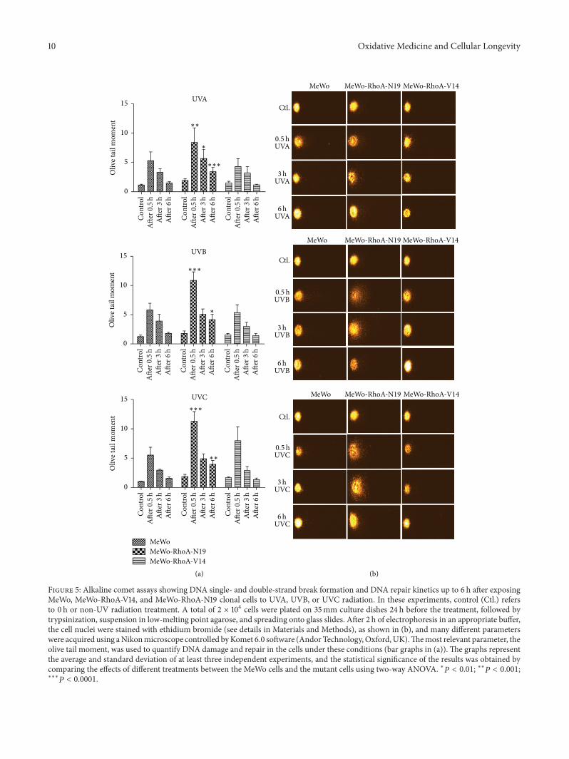
10 Oxidative Medicine and Cellular Longevity
UVA
MeWo MeWo-RhoA-N19 MeWo-RhoA-V14
UVB
UVC
∗
∗∗
∗∗∗
Con
trol
Ctl.
Afte
r0.5
hA
fter3
hA
fter6
h
Con
trol
Afte
r0.5
hA
fter3
hA
fter6
h
Con
trol
Afte
r0.5
hA
fter3
hA
fter6
h
UVA0.5h
UVA3h
UVA6h
∗
∗∗∗
MeWoMeWo-RhoA-N19MeWo-RhoA-V14
(a) (b)
∗∗
∗∗∗
MeWo MeWo-RhoA-N19 MeWo-RhoA-V14
Ctl.
UVB0.5h
UVB3h
UVB6h
MeWo MeWo-RhoA-N19 MeWo-RhoA-V14
Ctl.
UVC0.5h
UVC3h
UVC6h
0
5
10
15O
live t
ail m
omen
t
0
5
10
15
Oliv
e tai
l mom
ent
Con
trol
Afte
r0.5
hA
fter3
hA
fter6
h
Con
trol
Afte
r0.5
hA
fter3
hA
fter6
h
Con
trol
Afte
r0.5
hA
fter3
hA
fter6
h
0
5
10
15
Oliv
e tai
l mom
ent
Con
trol
Afte
r0.5
hA
fter3
hA
fter6
h
Con
trol
Afte
r0.5
hA
fter3
hA
fter6
h
Con
trol
Afte
r0.5
hA
fter3
hA
fter6
h
Figure 5: Alkaline comet assays showing DNA single- and double-strand break formation and DNA repair kinetics up to 6 h after exposingMeWo, MeWo-RhoA-V14, and MeWo-RhoA-N19 clonal cells to UVA, UVB, or UVC radiation. In these experiments, control (Ctl.) refersto 0 h or non-UV radiation treatment. A total of 2 × 10⁴ cells were plated on 35mm culture dishes 24 h before the treatment, followed bytrypsinization, suspension in low-melting point agarose, and spreading onto glass slides. After 2 h of electrophoresis in an appropriate buffer,the cell nuclei were stained with ethidium bromide (see details in Materials and Methods), as shown in (b), and many different parameterswere acquired using aNikonmicroscope controlled byKomet 6.0 software (Andor Technology,Oxford,UK).Themost relevant parameter, theolive tail moment, was used to quantify DNA damage and repair in the cells under these conditions (bar graphs in (a)). The graphs representthe average and standard deviation of at least three independent experiments, and the statistical significance of the results was obtained bycomparing the effects of different treatments between the MeWo cells and the mutant cells using two-way ANOVA. ∗𝑃 < 0.01; ∗∗𝑃 < 0.001;∗∗∗
𝑃 < 0.0001.
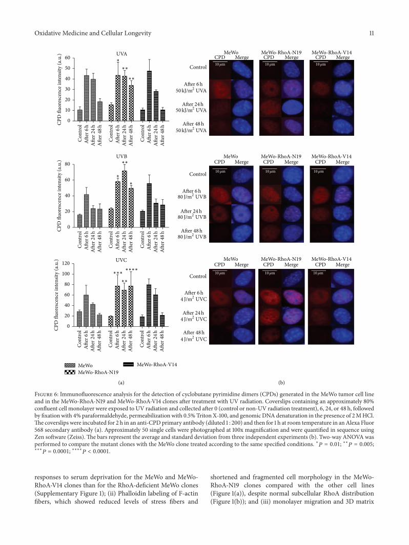
Oxidative Medicine and Cellular Longevity 11
UVA
UVB
UVC
Con
trol
∗
∗∗
∗∗
∗∗∗∗∗∗∗
Afte
r24
hA
fter4
8h
Afte
r6h
Con
trol
Afte
r24
hA
fter4
8h
Afte
r6h
Con
trol
Afte
r24
hA
fter4
8h
Afte
r6h
MeWoMeWo-RhoA-N19
MeWo-RhoA-V14
(a) (b)
MeWoCPD Merge CPD Merge CPD Merge
MeWo-RhoA-N19 MeWo-RhoA-V14
Control
∗∗
∗
∗
∗∗
50kJ/m2 UVA
50kJ/m2 UVA
50kJ/m2 UVA
80 J/m2 UVB
MeWoCPD Merge CPD Merge CPD Merge
MeWo-RhoA-N19 MeWo-RhoA-V14
Control
80 J/m2 UVB
80 J/m2 UVB
MeWoCPD Merge CPD Merge CPD Merge
MeWo-RhoA-N19 MeWo-RhoA-V14
Control
4 J/m2 UVC
4 J/m2 UVC
4 J/m2 UVC
0
10
20
30
40
50
60CP
D fl
uore
scen
ce in
tens
ity (a
.u.)
0
20
40
60
80
CPD
fluo
resc
ence
inte
nsity
(a.u
.)
Con
trol
Afte
r24
hA
fter4
8h
Afte
r6h
Con
trol
Afte
r24
hA
fter4
8h
Afte
r6h
Con
trol
Afte
r24
hA
fter4
8h
Afte
r6h
After 24h
After 48h
After 6h
0
20
40
60
80
100
120
CPD
fluo
resc
ence
inte
nsity
(a.u
.)
Con
trol
Afte
r24
hA
fter4
8h
Afte
r6h
Con
trol
Afte
r24
hA
fter4
8h
Afte
r6h
Con
trol
Afte
r24
hA
fter4
8h
Afte
r6h
10𝜇m 10𝜇m 10𝜇m
10𝜇m 10𝜇m 10𝜇m
10𝜇m 10𝜇m 10𝜇m
After 24h
After 48h
After 6h
After 24h
After 48h
After 6h
Figure 6: Immunofluorescence analysis for the detection of cyclobutane pyrimidine dimers (CPDs) generated in the MeWo tumor cell lineand in the MeWo-RhoA-N19 and MeWo-RhoA-V14 clones after treatment with UV radiation. Coverslips containing an approximately 80%confluent cell monolayer were exposed to UV radiation and collected after 0 (control or non-UV radiation treatment), 6, 24, or 48 h, followedby fixation with 4% paraformaldehyde, permeabilization with 0.5% Triton X-100, and genomic DNA denaturation in the presence of 2MHCl.The coverslips were incubated for 2 h in an anti-CPD primary antibody (diluted 1 : 200) and then for 1 h at room temperature in anAlexa Fluor568 secondary antibody (a). Approximately 50 single cells were photographed at 100x magnification and were quantified in sequence usingZen software (Zeiss). The bars represent the average and standard deviation from three independent experiments (b). Two-way ANOVA wasperformed to compare the mutant clones with the MeWo clone treated according to the same specified conditions. ∗𝑃 = 0.01; ∗∗𝑃 = 0.005;∗∗∗
𝑃 = 0.0001; ∗∗∗∗𝑃 < 0.0001.
responses to serum deprivation for the MeWo and MeWo-RhoA-V14 clones than for the RhoA-deficient MeWo clones(Supplementary Figure 1); (ii) Phalloidin labeling of F-actinfibers, which showed reduced levels of stress fibers and
shortened and fragmented cell morphology in the MeWo-RhoA-N19 clones compared with the other cell lines(Figure 1(a)), despite normal subcellular RhoA distribution(Figure 1(b)); and (iii) monolayer migration and 3D matrix
12 Oxidative Medicine and Cellular Longevity
penetration assays, which showed reduced motility andinvasion capacity ofMeWo-RhoA-N19 clones compared withthe parental andMeWo-RhoA-V14 clones under control con-ditions in the presence of serum (Figure 2 and SupplementaryFigure S3).
Second, the effects of UV radiation treatment on the threecell lines with respect to stress fiber formation, cell shape,RhoA distribution, migration, and invasion were examinedto confirm the deleterious effects of these genotoxic stressors.We observed that UVA, UVB, or even UVC irradiation didnot lead to any detectable change in the cell edge shape orthe stress fiber morphology in the MeWo or MeWo-RhoA-V14 cells (Figure 1). The migration of these cells was slightlyreduced after 24 h, particularly under UVB or UVC radiationtreatment (Figure 2(a)), and their invasion capacity wasslightly reduced by UVC radiation treatment (Figure 2(b));these effects were not strictly dependent on the actin-myosincytoskeleton and were potentially caused by many other fac-tors [44]. Conversely, negative effects on stress fibermorphol-ogy and content were observed in RhoA-deficient MeWo-RhoA-N19 clones (Figure 1(a)), very likely reflecting theirreduced motility (Figures 2(a) and 2(b)) and invasion ability(Figures 2(c) and 2(d)). The three evaluated cytoskeletalfeatures (Phalloidin staining of actin-myosin fibers, cell edgeshape, and RhoA subcellular distribution) are in agreementwith each other and with the biological responses of motilityand invasiveness characteristic of the aggressive phenotypeof melanoma cells and, moreover, with the modulation ofRhoA activity. Corroborating the effects of UV radiationtreatment on cell migration, a significant reduction in theMMP-dependent invasive capacity of the MeWo cells wasobserved in all experimental groups followingUVC radiationtreatment. However, this suppressive effect appeared to beless pronounced in the RhoA-deficient MeWo-RhoA-N19clones, and this result supports the hypothesis that the inhib-itory effects of UV radiation on melanoma cell invasion arepartially dependent on RhoA activity.
We next confirmed the well-known antiproliferativeeffects of UV radiation on melanomas and the potential roleof RhoA modulation in this process [17, 24]. Growth curvesand colony formation assays confirmed the higher resistanceof MeWo-RhoA-V14 and MeWo cells (to a lesser extent)to UV radiation treatment compared with RhoA-deficientcells (Figures 3 and 4), as the MeWo-RhoA-V14 and MeWocells recovered even after UVB and UVC radiation-induceddamage. The opposite effects were observed for the RhoA-deficient clones; that is, these cells exhibited approximately50% higher sensitivity to UV radiation-induced damage.
As previously reported, these results likely reflect thatRhoA affects the efficiency of DNA repair mechanisms [18,40].Thus, to assess DNA integrity and specific UV radiation-promoted damage, we performed alkaline comet assays andCPD formation experiments. We showed that the inabilityto remove damage over time clearly reflects cell proliferationvia the modulation of RhoA activity-proficient and activity-deficient MeWo clones. Measurements of single- and double-strand breaks showed that cells displaying high RhoA activityexhibit less damage at 0.5 h (peak) after treatment with thethree types of UV radiation and exhibit more efficient repair,
completely recovering to the basal levels after 6 h. In con-trast, dominant-negative RhoA-expressing cells showed anaccumulation of damage from 0.5 to 6 h after injury inducedby UVA, UVB, or UVC radiation (Figure 5). Intriguingly,the levels of CPDs peaked at approximately 6 h after UVradiation in all three cell lines, irrespective of RhoA activity,but these lesions were almost completely removed after 48 hin cells exhibiting high levels of RhoA activity. In addition, inthe RhoA-deficient MeWo cells (MeWo-RhoA-N19 clones),the accumulation of CPD lesions remained high for up to48 h, independently of treatment with UVA, UVB, or UVCradiation. Thus, this specific UV radiation-induced damageaccumulates in the cellular background of low RhoA activity(Figure 6).
As predicted from the results of other previous studiesand confirmed in the present study, the higher the RhoAactivity the more efficient the DNA repair; this phenomenonis common to many human tumor cells [45]. UV radia-tion induces DNA damage, such as single-strand breaks,pyrimidine dimers, and 6-4 PPs, which induce mutationsthat are characteristic of the promotion, establishment, anddevelopment of tumors [46]. These forms of damage aretypically repaired by theNER cascade [47], and failures in thisrepair machinery result in many diseases, such as xerodermapigmentosum [3]. Thus, in the present study, we establisheda strong correlation between RhoA activity and the efficiencyof the repair of UV radiation-induced damage to melanomacells, suggesting thatNERpathway functionmight be affectedby RhoA-transduced signals that activate cellular responses,such as gene transcription, cell proliferation, and cell death[17]. Although additional molecular studies are needed,RhoA represents a potential target for UV radiation-inducedcarcinogenesis in skin [48], as well as for gamma radiation-induced damage in cervix carcinomas, where RhoA was alsoshown to mediate double-strand breaks repair [49]. Similarresults, in the same cellular models, were also found forthe Rac1 GTPase [50] again suggesting actin cytoskeletonremodeling signals towards the nuclearmachineries in chargeof the genomic stability.
Conflict of InterestsThe authors declare that there is no conflict of interestsregarding the publication of this paper.
Authors’ ContributionConception, design, and supervision were done by FabioLuis Forti. Development of methodology was done by GiseleEspinha, Juliana Harumi Osaki, and Erico Tosoni Costa.Acquisition of data was done by Gisele Espinha and JulianaHarumi Osaki. Analysis and interpretation of data wereperformed by Gisele Espinha, Erico Tosoni Costa, and FabioLuis Forti. Writing and revision of the paper were made byGisele Espinha and Fabio Luis Forti.
AcknowledgmentsThe authors would like to thank Andressa P. Costa, Juliana R.Domingos, Benedita Oliveira, and Viviane Q. Machtura for
Oxidative Medicine and Cellular Longevity 13
technical assistance.The authorswould also like to thankPro-fessor Dr. Marisa G. Medeiros for allowing them to conductthe comet assays in her laboratory, Professor Dr. Carlos F. M.Menck for kindly providing reagents for CPD analysis, andProfessor Dr. Leticia Labriola for kindly providing reagentsfor Matrigel invasion assay. Additionally, the authors wouldlike to thank theAJE staff and editors for editing services.Thisresearch was supported by the Brazilian agency Fundação deAmparo à Pesquisa do Estado de São Paulo (FAPESP) viaa Young Investigator Fellowship to Fabio Luis Forti (Grantsnos. 2008/58264-5 and 2015/03983-0). Juliana Harumi Osakireceived a FAPESP Ph.D. Fellowship (2010/09453-0) andGisele Espinha received an M.S. Fellowship (2011/05822-3) from Coordenação de Aperfeiçoamento de Pessoal deNı́vel Superior (CAPES). All research projects in the authors’laboratory have been also supported by the Brazilian agencyCAPES through the Graduate Program in Biochemistry ofthe Chemistry Institute. The final publication of this paperwas supported by the CAPES/PROEX funds and by the SaoPaulo State Research Foundation (FAPESP) through Grantno. 2015/18341-1 to Fabio Luis Forti.
References
[1] K. Ferguson, “Melanoma,”The Journal of Continuing Educationin Nursing, vol. 36, no. 6, pp. 242–243, 2005.
[2] J. J. Strouse, T. R. Fears, M. A. Tucker, and A. S. Wayne, “Pedi-atric melanoma: risk factor and survival analysis of the surveil-lance, epidemiology and end results database,” Journal of Clini-cal Oncology, vol. 23, no. 21, pp. 4735–4741, 2005.
[3] S. Hu, F. Ma, F. Collado-Mesa, and R. S. Kirsner, “UV radiation,latitude, and melanoma in US Hispanics and blacks,” Archivesof Dermatology, vol. 140, no. 7, pp. 819–824, 2004.
[4] R. G. Zepp, D. J. Erickson III, N. D. Paul, and B. Sulzberger,“Interactive effects of solar UV radiation and climate changeon biogeochemical cycling,” Photochemical and PhotobiologicalSciences, vol. 6, no. 3, pp. 286–300, 2007.
[5] T. Douki, A. Reynaud-Angelin, J. Cadet, and E. Sage, “Bipyrim-idine photoproducts rather than oxidative lesions are the maintype of DNA damage involved in the genotoxic effect of solarUVA radiation,” Biochemistry, vol. 42, no. 30, pp. 9221–9226,2003.
[6] S. Mouret, C. Baudouin, M. Charveron, A. Favier, J. Cadet, andT. Douki, “Cyclobutane pyrimidine dimers are predominantDNA lesions in whole human skin exposed to UVA radiation,”Proceedings of the National Academy of Sciences of the UnitedStates of America, vol. 103, no. 37, pp. 13765–13770, 2006.
[7] J. Cadet, T. Douki, J.-L. Ravanat, and P. Di Mascio, “Sensitizedformation of oxidatively generated damage to cellular DNAby UVA radiation,” Photochemical and Photobiological Sciences,vol. 8, no. 7, pp. 903–911, 2009.
[8] A. Besaratinia, S. E. Bates, T.W. Synold, and G. P. Pfeifer, “Simi-lar mutagenicity of photoactivated porphyrins and ultravioletA radiation in mouse embryonic fibroblasts: involvement ofoxidative DNA lesions in mutagenesis,” Biochemistry, vol. 43,no. 49, pp. 15557–15566, 2004.
[9] H.-C. DeFedericis, H. B. Patrzyc, M. J. Rajecki et al., “Singletoxygen-inducedDNAdamage,”Radiation Research, vol. 165, no.4, pp. 445–451, 2006.
[10] G. P. Pfeifer, Y.-H. You, and A. Besaratinia, “Mutations inducedby ultraviolet light,”Mutation Research, vol. 571, no. 1-2, pp. 19–31, 2005.
[11] A. J. Miller and M. C. Mihm Jr., “Melanoma,”The New EnglandJournal of Medicine, vol. 355, no. 1, pp. 51–65, 2006.
[12] M. R. Zaidi, C.-P. Day, and G. Merlino, “From UVs to metas-tases: modeling melanoma initiation and progression in themouse,” The Journal of Investigative Dermatology, vol. 128, no.10, pp. 2381–2391, 2008.
[13] L. A. Liotta, “Tumor invasion andmetastases—role of the extra-cellular matrix: Rhoads Memorial Award lecture,” Cancer Re-search, vol. 46, no. 1, pp. 1–7, 1986.
[14] W. Wang, S. Goswami, K. Lapidus et al., “Identification andtesting of a gene expression signature of invasive carcinoma cellswithin primarymammary tumors,”Cancer Research, vol. 64, no.23, pp. 8585–8594, 2004.
[15] W. Wang, J. B. Wyckoff, S. Goswami et al., “Coordinated regu-lation of pathways for enhanced cell motility and chemotaxisis conserved in rat and mouse mammary tumors,” CancerResearch, vol. 67, no. 8, pp. 3505–3511, 2007.
[16] J. J. Bravo-Cordero, L. Hodgson, and J. Condeelis, “Directed cellinvasion and migration during metastasis,” Current Opinion inCell Biology, vol. 24, no. 2, pp. 277–283, 2012.
[17] R. Karlsson, E. D. Pedersen, Z. Wang, and C. Brakebusch, “RhoGTPase function in tumorigenesis,” Biochimica et BiophysicaActa, vol. 1796, no. 2, pp. 91–98, 2009.
[18] G. Fritz, B. Kaina, and K. Aktories, “TheRas-related small GTP-binding protein RhoB is immediate-early inducible by DNAdamaging treatments,”The Journal of Biological Chemistry, vol.270, no. 42, pp. 25172–25177, 1995.
[19] S. A. Boswell, P. P. Ongusaha, P. Nghiem, and S. W. Lee, “Theprotective role of a small GTPase RhoE against UVB-inducedDNA damage in keratinocytes,”The Journal of Biological Chem-istry, vol. 282, no. 7, pp. 4850–4858, 2007.
[20] Q. Jian, Q. An, D. Zhu et al., “MicroRNA 340 is involvedin UVB-induced dendrite formation through the regulationof RhoA expression in melanocytes,” Molecular and CellularBiology, vol. 34, no. 18, pp. 3407–3420, 2014.
[21] T. Frisan, X. Cortes-Bratti, E. Chaves-Olarte, B. Stenerlöw, andM. Thelestam, “The Haemophilus ducreyi cytolethal distendingtoxin induces DNA double-strand breaks and promotes ATM-dependent activation of RhoA,”CellularMicrobiology, vol. 5, no.10, pp. 695–707, 2003.
[22] L. Guerra, R. Guidi, I. Slot et al., “Bacterial genotoxin triggersFEN1-dependent RhoA activation, cytoskeleton remodelingand cell survival,” Journal of Cell Science, vol. 124, no. 16, pp.2735–2742, 2011.
[23] S. Pavey, L. Spoerri, N. K. Haass, and B. Gabrielli, “DNA repairand cell cycle checkpoint defects as drivers and therapeutictargets inmelanoma,”Pigment Cell andMelanomaResearch, vol.26, no. 6, pp. 805–816, 2013.
[24] V. Sanz-Moreno and C. J. Marshall, “Rho-GTPase signalingdrives melanoma cell plasticity,” Cell Cycle, vol. 8, no. 10, pp.1484–1487, 2009.
[25] F. L. Forti and H. A. Armelin, “Vasopressin triggers senescencein K-ras transformed cells via RhoA-dependent downregula-tion of cyclin D1,” Endocrine-Related Cancer, vol. 14, no. 4, pp.1117–1125, 2007.
[26] X.-D. Ren, W. B. Kiosses, and M. A. Schwartz, “Regulation ofthe small GTP-binding protein Rho by cell adhesion and thecytoskeleton,” The EMBO Journal, vol. 18, no. 3, pp. 578–585,1999.
14 Oxidative Medicine and Cellular Longevity
[27] C.-C. Liang, A. Y. Park, and J.-L. Guan, “In vitro scratch assay:a convenient and inexpensive method for analysis of cell migra-tion in vitro,” Nature Protocols, vol. 2, no. 2, pp. 329–333, 2007.
[28] N. P. Singh, M. T. McCoy, R. R. Tice, and E. L. Schneider, “Asimple technique for quantitation of low levels of DNA damagein individual cells,” Experimental Cell Research, vol. 175, no. 1,pp. 184–191, 1988.
[29] T. E. Carey, T. Takahashi, L. A. Resnick, H. F. Oettgen, and L.J. Old, “Cell surface antigens of human malignant melanoma:mixed hemadsorption assays for humoral immunity to cul-tured autologous melanoma cells,” Proceedings of the NationalAcademy of Sciences of the United States of America, vol. 73, no.9, pp. 3278–3282, 1976.
[30] J. Fogh, J. M. Fogh, and T. Orfeo, “One hundred and twenty-seven cultured human tumor cell lines producing tumors innude mice,” Journal of the National Cancer Institute, vol. 59, no.1, pp. 221–226, 1977.
[31] J. Fogh, W. C. Wright, and J. D. Loveless, “Absence of HeLa cellcontamination in 169 cell lines derived from human tumors,”Journal of the National Cancer Institute, vol. 58, no. 2, pp. 209–214, 1977.
[32] W.C.Wright,W. P.Daniels, and J. Fogh, “Distinction of seventy-one cultured human tumor cell lines by polymorphic enzymeanalysis,” Journal of the National Cancer Institute, vol. 66, no. 2,pp. 239–247, 1981.
[33] É. T. Costa, F. L. Forti, T. G. F. Matos et al., “Fibroblast growthfactor 2 restrains Ras-driven proliferation of malignant cells bytriggering RhoA-mediated senescence,” Cancer Research, vol.68, no. 15, pp. 6215–6223, 2008.
[34] M. van Engeland, L. J. W. Nieland, F. C. S. Ramaekers, B.Schutte, and C. P. M. Reutelingsperger, “Annexin V-affinityassay: a review on an apoptosis detection system based on phos-phatidylserine exposure,” Cytometry, vol. 31, no. 1, pp. 1–9, 1998.
[35] G.Kroemer, L. Galluzzi, P. Vandenabeele et al., “Classification ofcell death: recommendations of the Nomenclature Committeeon Cell Death 2009,” Cell Death and Differentiation, vol. 16, no.1, pp. 3–11, 2009.
[36] A. B. Jaffe and A. Hall, “Rho GTPases: biochemistry and biol-ogy,” Annual Review of Cell and Developmental Biology, vol. 21,pp. 247–269, 2005.
[37] J. Heo, K. W. Raines, V. Mocanu, and S. L. Campbell, “Redoxregulation of RhoA,” Biochemistry, vol. 45, no. 48, pp. 14481–14489, 2006.
[38] K. W. Raines, M. G. Bonini, and S. L. Campbell, “Nitricoxide cell signaling: S-nitrosation of Ras superfamily GTPases,”Cardiovascular Research, vol. 75, no. 2, pp. 229–239, 2007.
[39] A. Aghajanian, E. S. Wittchen, S. L. Campbell, and K. Burridge,“Direct activation of RhoA by reactive oxygen species requiresa redox-sensitive motif,” PLoS ONE, vol. 4, no. 11, Article IDe8045, 2009.
[40] J. Luo, K. Hosoki, A. Bacsi et al., “8-Oxoguanine DNA glyco-sylase-1-mediated DNA repair is associated with Rho GTPaseactivation and alpha-smoothmuscle actin polymerization,” FreeRadical Biology and Medicine, vol. 73, pp. 430–438, 2014.
[41] C. Jhappan, F. P. Noonan, andG.Merlino, “Ultraviolet radiationand cutaneous malignant melanoma,”Oncogene, vol. 22, no. 20,pp. 3099–3112, 2003.
[42] W. K. Kaufmann, “The human intra-S checkpoint response toUVC-induced DNA damage,” Carcinogenesis, vol. 31, no. 5, pp.751–765, 2010.
[43] S. Miwa, S. Yano, Y. Hiroshima et al., “Imaging UVC-inducedDNA damage response in models of minimal cancer,” Journalof Cellular Biochemistry, vol. 114, no. 11, pp. 2493–2499, 2013.
[44] K. Kaibuchi, S. Kuroda, and M. Amano, “Regulation of thecytoskeleton and cell adhesion by the Rho family GTPases inmammalian cells,” Annual Review of Biochemistry, vol. 68, pp.459–486, 1999.
[45] E. Sahai, M. F. Olson, and C. J. Marshall, “Cross-talk betweenRas and Rho signalling pathways in transformation favoursproliferation and increased motility,” The EMBO Journal, vol.20, no. 4, pp. 755–766, 2001.
[46] A. Besaratinia, J.-I. Yoon, C. Schroeder, S. E. Bradforth, M.Cockburn, and G. P. Pfeifer, “Wavelength dependence of ultra-violet radiation-induced DNA damage as determined by laserirradiation suggests that cyclobutane pyrimidine dimers are theprincipal DNA lesions produced by terrestrial sunlight,” TheFASEB Journal, vol. 25, no. 9, pp. 3079–3091, 2011.
[47] R. P. Rastogi, Richa, A. Kumar, M. B. Tyagi, and R. P. Sinha,“Molecular mechanisms of ultraviolet radiation-induced DNAdamage and repair,” Journal of Nucleic Acids, vol. 2010, ArticleID 592980, 32 pages, 2010.
[48] A. Brozyna, B. Zbytek, J. Granese, J. A. Carlson, J. Ross, andA. Slominski, “Mechanism of UV-related carcinogenesis and itscontribution to nevi/melanoma,”Expert Review ofDermatology,vol. 2, no. 4, pp. 451–469, 2007.
[49] J. H. Osaki, G. Espinha, Y. T. Magalhaes, and F. L. Forti,“Modulation of RhoA GTPase activity sensitizes human cervixcarcinoma cells to 𝛾-radiation by attenuating DNA repair path-ways,” Oxidative Medicine and Cellular Longevity, vol. 2016,Article ID 6012642, 11 pages, 2016.
[50] G. Espinha, J. H. Osaki, Y. T. Magalhaes, and F. L. Forti,“Rac1 GTPase-deficientHeLa cells present reducedDNA repair,proliferation, and survival under UV or gamma irradiation,”Molecular and Cellular Biochemistry, vol. 404, no. 1, pp. 281–297,2015.
Research ArticleActivation of Antioxidative Functions by Radon InhalationEnhances the Mitigation Effects of Pregabalin onChronic Constriction Injury-Induced Neuropathic Pain in Mice
Takahiro Kataoka, Shunsuke Horie, Reo Etani, Norie Kanzaki, Kaori Sasaoka,Yusuke Kobashi, Katsumi Hanamoto, and Kiyonori Yamaoka
Graduate School of Health Sciences, OkayamaUniversity, 5-1 Shikata-cho, 2-chome, Kita-ku, Okayama-shi, Okayama 700-8558, Japan
Correspondence should be addressed to Kiyonori Yamaoka; [email protected]
Received 4 September 2015; Revised 5 October 2015; Accepted 5 October 2015
Academic Editor: Paola Venditti
Copyright © 2016 Takahiro Kataoka et al. This is an open access article distributed under the Creative Commons AttributionLicense, which permits unrestricted use, distribution, and reproduction in any medium, provided the original work is properlycited.
Radon inhalation brings pain relief for chronic constriction injury- (CCI-) induced neuropathic pain in mice due to the activationof antioxidative functions, which is different from the mechanism of the pregabalin effect. In this study, we assessed whether acombination of radon inhalation and pregabalin administration ismore effective against neuropathic pain than radon or pregabalinonly. Mice were treated with inhaled radon at a concentration of 1,000 Bq/m3 for 24 hours and pregabalin administration afterCCI surgery. In mice treated with pregabalin at a dose of 3mg/kg weight, the 50% paw withdrawal threshold of mice treatedwith pregabalin or radon and pregabalin was significantly increased, suggesting pain relief. The therapeutic effects of radoninhalation or the combined effects of radon and pregabalin (3mg/kg weight) were almost equivalent to treatment with pregabalinat a dose of 1.4mg/kg weight or 4.1mg/kg weight, respectively. Radon inhalation and the combination of radon and pregabalinincreased antioxidant associated substances in the paw.The antioxidant substances increased much more in radon inhalation thanin pregabalin administration. These findings suggested that the activation of antioxidative functions by radon inhalation enhancesthe pain relief of pregabalin and that this combined effect is probably an additive effect.
1. Introduction
Radon therapy is performed for mainly pain-related diseasesusing radon hot springs in Japan [1] and Europe [2] and usingmines in Europe [2]. In both cases, patients inhale radonthrough their nose because radon is a radioactive gas. Theconditions of radon therapy in the Misasa Medical Centerin Japan and the hospitals in Europe are slightly different[3]. The radon concentration in the treatment room of theMisasa Medical Center is approximately 2,000 Bq/m3 [1],while the concentration in Europe is twenty-five times higher[2]. However, the beneficial effects of radon therapy showthat there is not much difference betweenMisasa and Europein terms of alleviation of pain-related diseases. A reportsuggested that radon and thermal therapy using hot springsalleviated osteoarthritis [1]. One of the possible reasons forthe effects is the prevention of peroxidation reactions and
immune depression. Another possible reason is an increasein tissue perfusion because vasoactive-associated substancedecreased and pain-associated substance increased by radontherapy. Another report suggested that not only radon andthermal therapy, but also thermal therapy alleviated anky-losing spondylitis [4]. However, radon and thermal therapycontinue the alleviation effect longer than thermal therapy.Radon therapy is also effective against rheumatic diseases [5].In addition, radon therapy reduced the dosage of medicine[6].These findings indicated that radon therapy has a positiveeffect on pain-related diseases such as osteoarthritis, ankylos-ing spondylitis, and rheumatic diseases. Although it is likelythat radon therapy has a positive effect against pain-relateddiseases, it is insufficient to understand the mechanisms ofradon therapy. To investigate the mechanisms, we previouslyreported that radon inhalation has preventive and curativeeffects against chronic constriction injury (CCI) in mice
Hindawi Publishing CorporationOxidative Medicine and Cellular LongevityVolume 2016, Article ID 9853692, 8 pageshttp://dx.doi.org/10.1155/2016/9853692
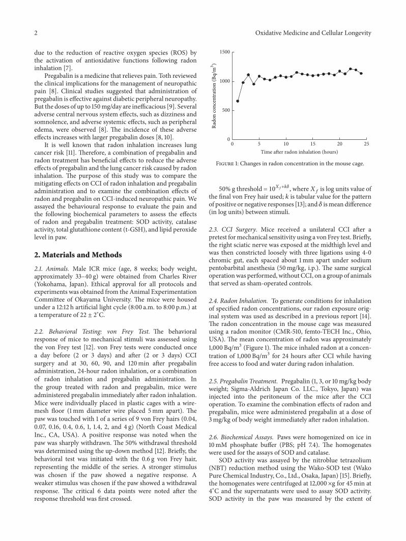
2 Oxidative Medicine and Cellular Longevity
due to the reduction of reactive oxygen species (ROS) bythe activation of antioxidative functions following radoninhalation [7].
Pregabalin is a medicine that relieves pain. Toth reviewedthe clinical implications for the management of neuropathicpain [8]. Clinical studies suggested that administration ofpregabalin is effective against diabetic peripheral neuropathy.But the doses of up to 150mg/day are inefficacious [9]. Severaladverse central nervous system effects, such as dizziness andsomnolence, and adverse systemic effects, such as peripheraledema, were observed [8]. The incidence of these adverseeffects increases with larger pregabalin doses [8, 10].
It is well known that radon inhalation increases lungcancer risk [11]. Therefore, a combination of pregabalin andradon treatment has beneficial effects to reduce the adverseeffects of pregabalin and the lung cancer risk caused by radoninhalation. The purpose of this study was to compare themitigating effects on CCI of radon inhalation and pregabalinadministration and to examine the combination effects ofradon and pregabalin on CCI-induced neuropathic pain. Weassayed the behavioural response to evaluate the pain andthe following biochemical parameters to assess the effectsof radon and pregabalin treatment: SOD activity, catalaseactivity, total glutathione content (t-GSH), and lipid peroxidelevel in paw.
2. Materials and Methods
2.1. Animals. Male ICR mice (age, 8 weeks; body weight,approximately 33–40 g) were obtained from Charles River(Yokohama, Japan). Ethical approval for all protocols andexperiments was obtained from the Animal ExperimentationCommittee of Okayama University. The mice were housedunder a 12:12 h artificial light cycle (8:00 a.m. to 8:00 p.m.) ata temperature of 22 ± 2∘C.
2.2. Behavioral Testing: von Frey Test. The behavioralresponse of mice to mechanical stimuli was assessed usingthe von Frey test [12]. von Frey tests were conducted oncea day before (2 or 3 days) and after (2 or 3 days) CCIsurgery and at 30, 60, 90, and 120min after pregabalinadministration, 24-hour radon inhalation, or a combinationof radon inhalation and pregabalin administration. Inthe group treated with radon and pregabalin, mice wereadministered pregabalin immediately after radon inhalation.Mice were individually placed in plastic cages with a wire-mesh floor (1mm diameter wire placed 5mm apart). Thepaw was touched with 1 of a series of 9 von Frey hairs (0.04,0.07, 0.16, 0.4, 0.6, 1, 1.4, 2, and 4 g) (North Coast MedicalInc., CA, USA). A positive response was noted when thepaw was sharply withdrawn. The 50% withdrawal thresholdwas determined using the up-down method [12]. Briefly, thebehavioral test was initiated with the 0.6 g von Frey hair,representing the middle of the series. A stronger stimuluswas chosen if the paw showed a negative response. Aweaker stimulus was chosen if the paw showed a withdrawalresponse. The critical 6 data points were noted after theresponse threshold was first crossed.
5 10 15 20 250Time after radon inhalation (hours)
0
500
1000
1500
Rado
n co
ncen
trat
ion
(Bq/
m3 )
Figure 1: Changes in radon concentration in the mouse cage.
50% g threshold = 10𝑋𝑓+𝑘𝛿, where𝑋𝑓is log units value of
the final von Frey hair used; 𝑘 is tabular value for the patternof positive or negative responses [13]; and 𝛿 ismean difference(in log units) between stimuli.
2.3. CCI Surgery. Mice received a unilateral CCI after apretest formechanical sensitivity using a vonFrey test. Briefly,the right sciatic nerve was exposed at the midthigh level andwas then constricted loosely with three ligations using 4-0chromic gut, each spaced about 1mm apart under sodiumpentobarbital anesthesia (50mg/kg, i.p.). The same surgicaloperationwas performed, without CCI, on a group of animalsthat served as sham-operated controls.
2.4. Radon Inhalation. To generate conditions for inhalationof specified radon concentrations, our radon exposure orig-inal system was used as described in a previous report [14].The radon concentration in the mouse cage was measuredusing a radon monitor (CMR-510, femto-TECH Inc., Ohio,USA). The mean concentration of radon was approximately1,000 Bq/m3 (Figure 1). The mice inhaled radon at a concen-tration of 1,000 Bq/m3 for 24 hours after CCI while havingfree access to food and water during radon inhalation.
2.5. Pregabalin Treatment. Pregabalin (1, 3, or 10mg/kg bodyweight; Sigma-Aldrich Japan Co. LLC., Tokyo, Japan) wasinjected into the peritoneum of the mice after the CCIoperation. To examine the combination effects of radon andpregabalin, mice were administered pregabalin at a dose of3mg/kg of body weight immediately after radon inhalation.
2.6. Biochemical Assays. Paws were homogenized on ice in10mM phosphate buffer (PBS; pH 7.4). The homogenateswere used for the assays of SOD and catalase.
SOD activity was assayed by the nitroblue tetrazolium(NBT) reduction method using the Wako-SOD test (WakoPure Chemical Industry, Co., Ltd., Osaka, Japan) [15]. Briefly,the homogenates were centrifuged at 12,000×g for 45min at4∘C and the supernatants were used to assay SOD activity.SOD activity in the paw was measured by the extent of
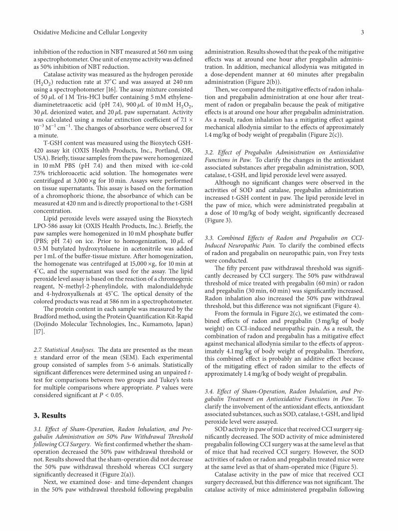
Oxidative Medicine and Cellular Longevity 3
inhibition of the reduction in NBTmeasured at 560 nm usinga spectrophotometer.One unit of enzyme activitywas definedas 50% inhibition of NBT reduction.
Catalase activity was measured as the hydrogen peroxide(H2O2) reduction rate at 37∘C and was assayed at 240 nm
using a spectrophotometer [16]. The assay mixture consistedof 50 𝜇L of 1M Tris-HCl buffer containing 5mM ethylene-diaminetetraacetic acid (pH 7.4), 900 𝜇L of 10mM H
2O2,
30 𝜇L deionized water, and 20𝜇L paw supernatant. Activitywas calculated using a molar extinction coefficient of 7.1 ×10⁻³M−1 cm−1. The changes of absorbance were observed fora minute.
T-GSH content was measured using the Bioxytech GSH-420 assay kit (OXIS Health Products, Inc., Portland, OR,USA). Briefly, tissue samples from the pawwere homogenizedin 10mM PBS (pH 7.4) and then mixed with ice-cold7.5% trichloroacetic acid solution. The homogenates werecentrifuged at 3,000×g for 10min. Assays were performedon tissue supernatants. This assay is based on the formationof a chromophoric thione, the absorbance of which can bemeasured at 420 nm and is directly proportional to the t-GSHconcentration.
Lipid peroxide levels were assayed using the BioxytechLPO-586 assay kit (OXIS Health Products, Inc.). Briefly, thepaw samples were homogenized in 10mM phosphate buffer(PBS; pH 7.4) on ice. Prior to homogenization, 10 𝜇L of0.5M butylated hydroxytoluene in acetonitrile was addedper 1mL of the buffer-tissue mixture. After homogenization,the homogenate was centrifuged at 15,000×g, for 10min at4∘C, and the supernatant was used for the assay. The lipidperoxide level assay is based on the reaction of a chromogenicreagent, N-methyl-2-phenylindole, with malondialdehydeand 4-hydroxyalkenals at 45∘C. The optical density of thecolored products was read at 586 nm in a spectrophotometer.
The protein content in each sample was measured by theBradfordmethod, using the ProteinQuantificationKit-Rapid(Dojindo Molecular Technologies, Inc., Kumamoto, Japan)[17].
2.7. Statistical Analyses. The data are presented as the mean± standard error of the mean (SEM). Each experimentalgroup consisted of samples from 5-6 animals. Statisticallysignificant differences were determined using an unpaired 𝑡-test for comparisons between two groups and Tukey’s testsfor multiple comparisons where appropriate. 𝑃 values wereconsidered significant at 𝑃 < 0.05.
3. Results
3.1. Effect of Sham-Operation, Radon Inhalation, and Pre-gabalin Administration on 50% Paw Withdrawal Thresholdfollowing CCI Surgery. Wefirst confirmedwhether the sham-operation decreased the 50% paw withdrawal threshold ornot. Results showed that the sham-operation did not decreasethe 50% paw withdrawal threshold whereas CCI surgerysignificantly decreased it (Figure 2(a)).
Next, we examined dose- and time-dependent changesin the 50% paw withdrawal threshold following pregabalin
administration. Results showed that the peak of themitigativeeffects was at around one hour after pregabalin adminis-tration. In addition, mechanical allodynia was mitigated ina dose-dependent manner at 60 minutes after pregabalinadministration (Figure 2(b)).
Then, we compared themitigative effects of radon inhala-tion and pregabalin administration at one hour after treat-ment of radon or pregabalin because the peak of mitigativeeffects is at around one hour after pregabalin administration.As a result, radon inhalation has a mitigating effect againstmechanical allodynia similar to the effects of approximately1.4mg/kg of body weight of pregabalin (Figure 2(c)).
3.2. Effect of Pregabalin Administration on AntioxidativeFunctions in Paw. To clarify the changes in the antioxidantassociated substances after pregabalin administration, SOD,catalase, t-GSH, and lipid peroxide level were assayed.
Although no significant changes were observed in theactivities of SOD and catalase, pregabalin administrationincreased t-GSH content in paw. The lipid peroxide level inthe paw of mice, which were administrated pregabalin ata dose of 10mg/kg of body weight, significantly decreased(Figure 3).
3.3. Combined Effects of Radon and Pregabalin on CCI-Induced Neuropathic Pain. To clarify the combined effectsof radon and pregabalin on neuropathic pain, von Frey testswere conducted.
The fifty percent paw withdrawal threshold was signifi-cantly decreased by CCI surgery. The 50% paw withdrawalthreshold of mice treated with pregabalin (60min) or radonand pregabalin (30min, 60min) was significantly increased.Radon inhalation also increased the 50% paw withdrawalthreshold, but this difference was not significant (Figure 4).
From the formula in Figure 2(c), we estimated the com-bined effects of radon and pregabalin (3mg/kg of bodyweight) on CCI-induced neuropathic pain. As a result, thecombination of radon and pregabalin has a mitigative effectagainst mechanical allodynia similar to the effects of approx-imately 4.1mg/kg of body weight of pregabalin. Therefore,this combined effect is probably an additive effect becauseof the mitigating effect of radon similar to the effects ofapproximately 1.4mg/kg of body weight of pregabalin.
3.4. Effect of Sham-Operation, Radon Inhalation, and Pre-gabalin Treatment on Antioxidative Functions in Paw. Toclarify the involvement of the antioxidant effects, antioxidantassociated substances, such as SOD, catalase, t-GSH, and lipidperoxide level were assayed.
SODactivity in pawofmice that receivedCCI surgery sig-nificantly decreased. The SOD activity of mice administeredpregabalin following CCI surgery was at the same level as thatof mice that had received CCI surgery. However, the SODactivities of radon or radon and pregabalin treated mice wereat the same level as that of sham-operated mice (Figure 5).
Catalase activity in the paw of mice that received CCIsurgery decreased, but this difference was not significant.Thecatalase activity of mice administered pregabalin following
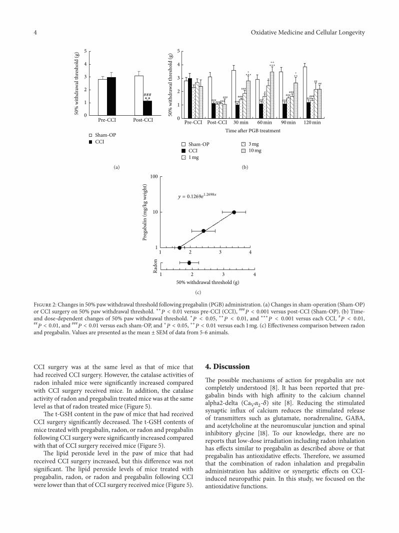
4 Oxidative Medicine and Cellular Longevity
###
Pre-CCI Post-CCI
50%
with
draw
al th
resh
old
(g)
Sham-OPCCI
∗∗
0
1
2
3
4
5
(a)
######
##
###
#########
#### ###
###
###
###
##
###
+
++
+
##
0
1
2
3
4
5
Pre-CCI Post-CCI 30 min
50%
with
draw
al th
resh
old
(g)
Time after PGB treatment
Sham-OPCCI1mg
120min90min60min
3mg10mg
∗∗∗
∗∗∗
∗∗∗
(b)
1 2 3 4
50% withdrawal threshold (g)1 2 3 4Ra
don
1
10
100
Preg
abal
in (m
g/kg
wei
ght)
y = 0.1269e1.2698x
(c)
Figure 2: Changes in 50% pawwithdrawal threshold following pregabalin (PGB) administration. (a) Changes in sham-operation (Sham-OP)or CCI surgery on 50% paw withdrawal threshold. ∗∗𝑃 < 0.01 versus pre-CCI (CCI), ###𝑃 < 0.001 versus post-CCI (Sham-OP). (b) Time-and dose-dependent changes of 50% paw withdrawal threshold. ∗𝑃 < 0.05, ∗∗𝑃 < 0.01, and ∗∗∗𝑃 < 0.001 versus each CCI, #𝑃 < 0.01,##𝑃 < 0.01, and ###
𝑃 < 0.01 versus each sham-OP, and +𝑃 < 0.05, ++𝑃 < 0.01 versus each 1mg. (c) Effectiveness comparison between radonand pregabalin. Values are presented as the mean ± SEM of data from 5-6 animals.
CCI surgery was at the same level as that of mice thathad received CCI surgery. However, the catalase activities ofradon inhaled mice were significantly increased comparedwith CCI surgery received mice. In addition, the catalaseactivity of radon and pregabalin treated mice was at the samelevel as that of radon treated mice (Figure 5).
The t-GSH content in the paw of mice that had receivedCCI surgery significantly decreased. The t-GSH contents ofmice treated with pregabalin, radon, or radon and pregabalinfollowing CCI surgery were significantly increased comparedwith that of CCI surgery received mice (Figure 5).
The lipid peroxide level in the paw of mice that hadreceived CCI surgery increased, but this difference was notsignificant. The lipid peroxide levels of mice treated withpregabalin, radon, or radon and pregabalin following CCIwere lower than that of CCI surgery receivedmice (Figure 5).
4. Discussion
The possible mechanisms of action for pregabalin are notcompletely understood [8]. It has been reported that pre-gabalin binds with high affinity to the calcium channelalpha2-delta (CaV𝛼2-𝛿) site [8]. Reducing the stimulatedsynaptic influx of calcium reduces the stimulated releaseof transmitters such as glutamate, noradrenaline, GABA,and acetylcholine at the neuromuscular junction and spinalinhibitory glycine [18]. To our knowledge, there are noreports that low-dose irradiation including radon inhalationhas effects similar to pregabalin as described above or thatpregabalin has antioxidative effects. Therefore, we assumedthat the combination of radon inhalation and pregabalinadministration has additive or synergetic effects on CCI-induced neuropathic pain. In this study, we focused on theantioxidative functions.
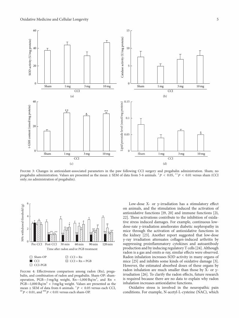
Oxidative Medicine and Cellular Longevity 5
ShamCCI
1mg 3mg 10mg0
20
40
60SO
D ac
tivity
(U/m
g pr
otei
n)
(a)
0
5
10
15
Cata
lase
activ
ity (U
/mg
prot
ein)
ShamCCI
1mg 3mg 10mg
(b)
0
20
40
t-GSH
cont
ent (
nmol
/mg
prot
ein)
∗∗∗∗∗
ShamCCI
1mg 3mg 10mg
(c)
0
0.05
0.1
0.15
Lipi
d pe
roxi
de le
vel (
nmol
/mg
prot
ein)
∗
ShamCCI
1mg 3mg 10mg
(d)
Figure 3: Changes in antioxidant-associated parameters in the paw following CCI surgery and pregabalin administration. Sham; nopregabalin administration. Values are presented as the mean ± SEM of data from 5-6 animals. ∗𝑃 < 0.05, ∗∗𝑃 < 0.01 versus sham (CCIonly; no administration of pregabalin).
###
###
###
######
##
###
##
######
##
######
### ######
0
2
4
6
Time after radon and/or PGB treatment
Sham-OPCCICCI-PGB
CCI + RnCCI + Rn + PGB
Pre-CCI Post-CCI 30 min 120min90min60min
50%
with
draw
al th
resh
old
(g)
∗∗
∗
Figure 4: Effectiveness comparison among radon (Rn), prega-balin, and combination of radon and pregabalin. Sham-OP: sham-operation, PGB—3mg/kg weight, Rn—1,000 Bq/m3, and Rn +PGB—1,000 Bq/m3 + 3mg/kg weight. Values are presented as themean ± SEM of data from 6 animals. ∗𝑃 < 0.05 versus each CCI,##𝑃 < 0.01, and ###
𝑃 < 0.01 versus each sham-OP.
Low-dose X- or 𝛾-irradiation has a stimulatory effecton animals, and the stimulation induced the activation ofantioxidative functions [19, 20] and immune functions [21,22]. These activations contribute to the inhibition of oxida-tive stress induced damages. For example, continuous low-dose-rate 𝛾-irradiation ameliorates diabetic nephropathy inmice through the activation of antioxidative functions inthe kidney [23]. Another report suggested that low-dose𝛾-ray irradiation attenuates collagen-induced arthritis bysuppressing proinflammatory cytokines and autoantibodyproduction and by inducing regulatory T cells [24]. Althoughradon is a gas and emits 𝛼-ray, similar effects were observed.Radon inhalation increases SOD activity in many organs ofmice [25] and inhibits some kinds of oxidative damage [3].However, the estimated absorbed doses of these organs byradon inhalation are much smaller than those by X- or 𝛾-irradiation [26]. To clarify the radon effects, future researchis required because there are no data to explain why radoninhalation increases antioxidative functions.
Oxidative stress is involved in the neuropathic painconditions. For example, N-acetyl-L-cysteine (NAC), which
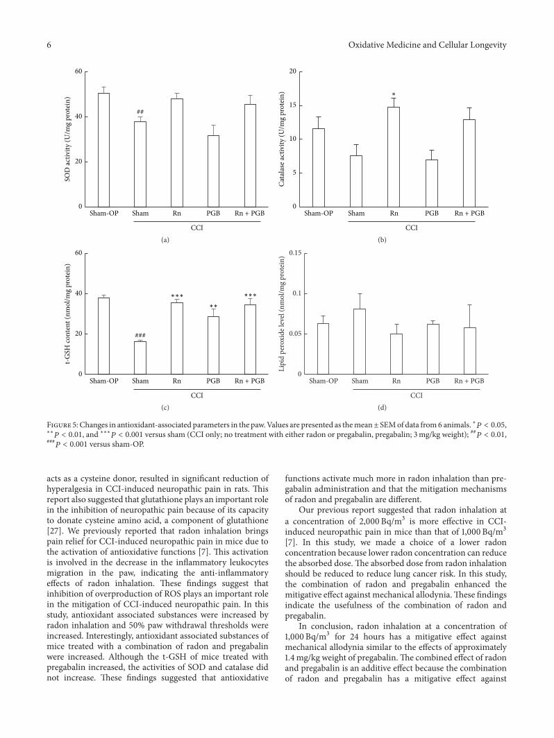
6 Oxidative Medicine and Cellular Longevity
##
Sham-OP Sham Rn PGB Rn + PGB
CCI
0
20
40
60SO
D ac
tivity
(U/m
g pr
otei
n)
(a)
Sham-OP Sham Rn PGB Rn + PGB
CCI
0
5
10
15
20
Cata
lase
activ
ity (U
/mg
prot
ein) ∗
(b)
###
0
20
40
60
t-GSH
cont
ent (
nmol
/mg
prot
ein)
Sham-OP Sham Rn PGB Rn + PGB
CCI
∗∗
∗∗∗∗∗∗
(c)
Sham-OP Sham Rn PGB Rn + PGB
CCI
0
0.05
0.1
0.15
Lipi
d pe
roxi
de le
vel (
nmol
/mg
prot
ein)
(d)
Figure 5: Changes in antioxidant-associated parameters in the paw.Values are presented as themean± SEMof data from6 animals. ∗𝑃 < 0.05,∗∗𝑃 < 0.01, and ∗∗∗𝑃 < 0.001 versus sham (CCI only; no treatment with either radon or pregabalin, pregabalin; 3mg/kg weight); ##𝑃 < 0.01,
###𝑃 < 0.001 versus sham-OP.
acts as a cysteine donor, resulted in significant reduction ofhyperalgesia in CCI-induced neuropathic pain in rats. Thisreport also suggested that glutathione plays an important rolein the inhibition of neuropathic pain because of its capacityto donate cysteine amino acid, a component of glutathione[27]. We previously reported that radon inhalation bringspain relief for CCI-induced neuropathic pain in mice due tothe activation of antioxidative functions [7]. This activationis involved in the decrease in the inflammatory leukocytesmigration in the paw, indicating the anti-inflammatoryeffects of radon inhalation. These findings suggest thatinhibition of overproduction of ROS plays an important rolein the mitigation of CCI-induced neuropathic pain. In thisstudy, antioxidant associated substances were increased byradon inhalation and 50% paw withdrawal thresholds wereincreased. Interestingly, antioxidant associated substances ofmice treated with a combination of radon and pregabalinwere increased. Although the t-GSH of mice treated withpregabalin increased, the activities of SOD and catalase didnot increase. These findings suggested that antioxidative
functions activate much more in radon inhalation than pre-gabalin administration and that the mitigation mechanismsof radon and pregabalin are different.
Our previous report suggested that radon inhalation ata concentration of 2,000 Bq/m3 is more effective in CCI-induced neuropathic pain in mice than that of 1,000 Bq/m3[7]. In this study, we made a choice of a lower radonconcentration because lower radon concentration can reducethe absorbed dose.The absorbed dose from radon inhalationshould be reduced to reduce lung cancer risk. In this study,the combination of radon and pregabalin enhanced themitigative effect against mechanical allodynia.These findingsindicate the usefulness of the combination of radon andpregabalin.
In conclusion, radon inhalation at a concentration of1,000 Bq/m3 for 24 hours has a mitigative effect againstmechanical allodynia similar to the effects of approximately1.4mg/kg weight of pregabalin.The combined effect of radonand pregabalin is an additive effect because the combinationof radon and pregabalin has a mitigative effect against
Oxidative Medicine and Cellular Longevity 7
mechanical allodynia similar to the effects of approximately4.1mg/kg weight of pregabalin. The possible mechanism ofthe additive effect is the activation of antioxidative functionsinduced by radon inhalation. However, the effects of radoninhalation on the nerve system have yet to be confirmed.They provide a substantial basis for future studies aimed atassessing the detailed mechanisms of the additive effects ofneuropathic pain.
Conflict of Interests
The authors report no conflict of interests.
Acknowledgments
The authors are grateful to the staff of both the Depart-ments of Radiation Research and Animal Resources, ShikataLaboratory, Advanced Science Research Center, OkayamaUniversity, for their technical support. They are also gratefulto Mr. Tetsuya Nakada and Mr. Yuki Hino for their technicalassistance. This research was supported by the Ryobi TeienMemory Foundation.
References
[1] K. Yamaoka, F. Mitsunobu, K. Hanamoto, S. Mori, Y. Tanizaki,and K. Sugita, “Study on biologic effects of radon and thermaltherapy on osteoarthritis,” The Journal of Pain, vol. 5, no. 1, pp.20–25, 2004.
[2] A. Falkenbach, J. Kovacs, A. Franke, K. Jörgens, and K. Ammer,“Radon therapy for the treatment of rheumatic diseases-reviewand meta-analysis of controlled clinical trials,” RheumatologyInternational, vol. 25, no. 3, pp. 205–210, 2005.
[3] T. Kataoka, “Study of antioxidative effects and anti-inflammatory effects in mice due to low-dose X-irradiation orradon inhalation,” Journal of Radiation Research, vol. 54, no. 4,pp. 587–596, 2013.
[4] A. van Tubergen, R. Landewé, D. van der Heijde et al., “Com-bined spa-exercise therapy is effective in patients with anky-losing spondylitis: a randomized controlled trial,” Arthritis &Rheumatology, vol. 45, no. 5, pp. 430–438, 2001.
[5] A. Franke, L. Reiner, H. G. Pratzel, T. Franke, and K. L.Resch, “Long-term efficacy of radon spa therapy in rheumatoidarthritis-a randomised, sham-controlled study and follow-up,”Rheumatology, vol. 39, no. 8, pp. 894–902, 2000.
[6] A. Franke, L. Reiner, and K.-L. Resch, “Long-term benefit ofradon spa therapy in the rehabilitation of rheumatoid arthritis: arandomised, double-blinded trial,”Rheumatology International,vol. 27, no. 8, pp. 703–713, 2007.
[7] K. Yamato, T. Kataoka, Y. Nishiyama, T. Taguchi, and K.Yamaoka, “Preventive and curative effects of radon inhalationon chronic constriction injury-induced neuropathic pain inmice,” European Journal of Pain, vol. 17, no. 4, pp. 480–492, 2013.
[8] C. Toth, “Pregabalin: latest safety evidence and clinical impli-cations for the management of neuropathic pain,” TherapeuticAdvances in Drug Safety, vol. 5, no. 1, pp. 38–56, 2014.
[9] J. Satoh, S. Yagihashi, M. Baba et al., “Efficacy and safetyof pregabalin for treating neuropathic pain associated with
diabetic peripheral neuropathy: a 14 week, randomized, double-blind, placebo-controlled trial,”Diabetic Medicine, vol. 28, no. 1,pp. 109–116, 2011.
[10] D. Semel, T. K. Murphy, G. Zlateva, R. Cheung, and B. Emir,“Evaluation of the safety and efficacy of pregabalin in olderpatients with neuropathic pain: results from a pooled analysisof 11 clinical studies,” BMC Family Practice, vol. 11, article 85,2010.
[11] World Health Organization (WHO), WHO Handbook onIndoor Radon, World Health Organization, Geneva, Switzer-land, 2009.
[12] W. J. Dixon, “Efficient analysis of experimental observations,”Annual Review of Pharmacology andToxicology, vol. 20, pp. 441–462, 1980.
[13] S. R. Chaplan, F. W. Bach, J. W. Pogrel, J. M. Chung, and T. L.Yaksh, “Quantitative assessment of tactile allodynia in the ratpaw,” Journal of Neuroscience Methods, vol. 53, no. 1, pp. 55–63,1994.
[14] T. Kataoka, J. Teraoka, A. Sakoda et al., “Protective effects ofradon inhalation on carrageenan-induced inflammatory pawedema in mice,” Inflammation, vol. 35, no. 2, pp. 713–722, 2012.
[15] R. L. Baehner, S. K. Murrmann, J. Davis, and R. B. JohnstonJr., “The role of superoxide anion and hydrogen peroxidein phagocytosis-associated oxidative metabolic reactions,” TheJournal of Clinical Investigation, vol. 56, no. 3, pp. 571–576, 1975.
[16] H. Aebi, S. R. Wyss, B. Scherz, and J. Gross, “Properties oferythrocyte catalase from homozygotes and heterozygotes forSwiss-type acatalasemia,” Biochemical Genetics, vol. 14, no. 9-10,pp. 791–807, 1976.
[17] M. M. Bradford, “A rapid and sensitive method for the quanti-tation of microgram quantities of protein utilizing the principleof protein-dye binding,”Analytical Biochemistry, vol. 72, no. 1-2,pp. 248–254, 1976.
[18] C. P. Taylor, “Mechanisms of analgesia by gabapentin andpregabalin—calcium channel 𝛼
2-𝛿 [Ca
𝑣𝛼2-𝛿] ligands,”Pain, vol.
142, no. 1-2, pp. 13–16, 2009.[19] S. Kojima, O. Matsuki, T. Nomura et al., “Induction of mRNAs
for glutathione synthesis-related proteins in mouse liver bylow doses of 𝛾-rays,” Biochimica et Biophysica Acta—GeneralSubjects, vol. 1381, no. 3, pp. 312–318, 1998.
[20] S. Kojima, O. Matsuki, T. Nomura et al., “Localization of glu-tathione and induction of glutathione synthesis-related proteinsin mouse brain by low doses of 𝛾-rays,” Brain Research, vol. 808,no. 2, pp. 262–269, 1998.
[21] S. Kojima, K. Nakayama, and H. Ishida, “Low dose gamma-rays activate immune functions via induction of glutathione anddelay tumor growth,” Journal of Radiation Research, vol. 45, no.1, pp. 33–39, 2004.
[22] Y. Kawakita, M. Ikekita, R. Kurozumi, and S. Kojima, “Increaseof intracellular glutathione by low-dose gamma-ray irradiationis mediated by transcription factor AP-1 in RAW 264.7 cells,”Biological and Pharmaceutical Bulletin, vol. 26, no. 1, pp. 19–23,2003.
[23] T. Nomura, X.-H. Li, H. Ogata et al., “Suppressive effects of con-tinuous low-dose-rate 𝛾 irradiation on diabetic nephropathy intype II diabetes mellitus model mice,” Radiation Research, vol.176, no. 3, pp. 356–365, 2011.
[24] H.Nakatsukasa,M. Tsukimoto, Y.Ohshima, F. Tago, A.Masada,and S. Kojima, “Suppressing effect of low-dose gamma-rayirradiation on collagen-induced arthritis,” Journal of RadiationResearch, vol. 49, no. 4, pp. 381–389, 2008.
8 Oxidative Medicine and Cellular Longevity
[25] T. Kataoka, A. Sakoda, Y. Ishimori et al., “Study of the responseof superoxide dismutase in mouse organs to radon using a newlarge-scale facility for exposing small animals to radon,” Journalof Radiation Research, vol. 52, no. 6, pp. 775–781, 2011.
[26] A. Sakoda, Y. Ishimori, A. Kawabe, T. Kataoka, K. Hanamoto,and K. Yamaoka, “Physiologically based pharmacokinetic mod-eling of inhaled radon to calculate absorbed doses in mice, rats,and humans,” Journal of Nuclear Science and Technology, vol. 47,no. 8, pp. 731–738, 2010.
[27] A. K. Naik, S. K. Tandan, S. P. Dudhgaonkar et al., “Role ofoxidative stress in pathophysiology of peripheral neuropathyand modulation by N-acetyl-L-cysteine in rats,” European Jour-nal of Pain, vol. 10, no. 7, pp. 573–579, 2006.
Research ArticleCharacteristics of Skeletal Muscle Fibers ofSOD1 Knockout Mice
Hiroshi Nagahisa,1 Kazuma Okabe,1 Yoshihito Iuchi,2 Junichi Fujii,3 and Hirofumi Miyata1
1Biological Sciences, Graduate School of Medicine, Yamaguchi University, Yamaguchi, Yamaguchi Perfecture 753-8515, Japan2Biological Chemistry, Faculty of Agriculture, Yamaguchi University, Yamaguchi, Yamaguchi Perfecture 753-8515, Japan3Department of Biochemistry and Molecular Biology, Graduate School of Medical Science, Yamagata University,Yamagata, Yamagata Perfecture 990-9585, Japan
Correspondence should be addressed to Hirofumi Miyata; [email protected]
Received 7 August 2015; Accepted 21 September 2015
Academic Editor: Paola Venditti
Copyright © 2016 Hiroshi Nagahisa et al. This is an open access article distributed under the Creative Commons AttributionLicense, which permits unrestricted use, distribution, and reproduction in any medium, provided the original work is properlycited.
Cu/Zn superoxide dismutase (SOD1) knockout (KO) mice are known as an aging model in some aspects, but the damage andregeneration process of each fiber type have not been sufficiently studied. In this study, we investigated the damage and satellite cellstate of the gastrocnemius muscle in SOD1 KO mice (6 months old) using immunohistochemical staining and real-time RT-PCR.The proportion of central nuclei-containing Type IIx/b fibers in the deep and superficial portions of the gastrocnemius musclewas significantly higher in SOD1 KO than control mice. The number of satellite cells per muscle fiber decreased in all muscle fibertypes in the deep portion of the gastrocnemius muscle in SOD1 KO mice. In addition, the mRNA expression levels of Pax7 andmyogenin, which are expressed in satellite cells in the activation, proliferation, and differentiation states, significantly increasedin the gastrocnemius muscle of SOD1 KO mice. Furthermore, mRNA of myosin heavy chain-embryonic, which is expressed inthe early phase of muscle regeneration, significantly increased in SOD1 KO mice. It was suggested that muscle is damaged byreactive oxygen species produced in the mitochondrial intermembrane space in Type IIxb fibers, accelerating the proliferation anddifferentiation of satellite cells through growth factors in SOD1 KO mice.
1. Introduction
Reactive oxygen species (ROS) are associated with cardiovas-cular and neurodegenerative diseases developing with aging[1]. ROS also inhibit the normal skeletal muscle regenerationprocess, being involved in age-related skeletal muscle weak-ness, that is, sarcopenia [2]. Generally, when skeletal muscleis damaged by stimulation, such as an overload, satellitecells, which are dormant myogenic stem cells, are activated.Activated satellite cells supply myonuclei to damaged musclefibers through proliferation and differentiation, contributingto skeletal muscle repair. Some proliferated satellite cells donot differentiate into myonuclei, and they return to the dor-mant state and maintain a satellite cell pool for regenerationas a self-renewal system. Since ROS production markedlyincreases with aging [3], interference with the regenerationprocess described above by ROS may be a cause of skeletalmuscle weakness.
Many tissues including skeletal muscle possess a superiorsystem to control ROS, and superoxide dismutase (SOD) isone of the enzymes playing a control role. SOD mediates thedisproportional reaction of converting oxygen, a species ofROS, to hydrogen peroxide in the body. There are isoformsof SOD: Cu-Zn SOD (SOD1) localized in the cytoplasm andmitochondrial intermembrane space and Mn SOD (SOD2)localized in mitochondria. Previous studies reported thatSOD1 deficiency accelerated aging-related muscle weightreduction and the accumulation of oxidative damage inmice [4], suggesting that these were caused by superoxideincreased by SOD1 deficiency and its secondary product.Thus, SOD1 KO mice are a useful model to investigate theinfluence of ROS and aging on the regeneration processof skeletal muscle. However, the muscle fiber regenerationprocess with satellite cell dynamics in SOD1 KO mice hasnot been sufficiently studied. In this study, SOD1 deficiency-induced structural and functional changes in muscle cells
Hindawi Publishing CorporationOxidative Medicine and Cellular LongevityVolume 2016, Article ID 9345970, 8 pageshttp://dx.doi.org/10.1155/2016/9345970
2 Oxidative Medicine and Cellular Longevity
were investigated using immunohistochemical techniquesand real-time RT-PCR.
2. Materials and Methods
All procedures were approved by the Animal Welfare andEthics Committee of the Yamaguchi University and followedthe American Physiological Society’s Animal Care Guide-lines.
2.1. Animals and Muscle Sampling. Seven SOD1−/− (SOD1KO) mice [5] and seven control (CTL; C57BL/6 strain)mice were used in this study (males, 6.2 ± 0.2 months old).SOD1+/− b129Sv mice purchased through Jackson Labora-tories (Bar Harbor, ME, USA) were backcrossed more than8 times with C57BL/6 males and bred at our institute. Allmice were maintained in a room controlled temperature and12 h : 12 h light/dark cycle and unrestricted access to foodand water. All animals were anaesthetized with pentobarbitalsodium (60mg/kg, intraperitoneally), and then left and rightgastrocnemius muscles were removed. All muscle sampleswere frozen by liquid nitrogen and stored at −80∘C untilanalyzed.
2.2. Immunohistochemical Analysis. Serial 10 𝜇m cross sec-tions of the right muscle were obtained on a cryostat (CM510;Leica, Wetzlar, Germany) at −20∘C. The sections werewarmed to room temperature (RT) and then preincubatedin 1% normal goat serum (EMD Millipore, Billerica, MA)in 0.1M phosphate buffered saline (PBS; pH 7.6) at RT for10min. The primary monoclonal antibody was then applied:either (1) fast myosin (1 : 2000; Sigma, St. Louis, MO), whichspecifically reacts with the myosin heavy chain- (MHC-)IIa and IIx, or (2) SC-71 (1 : 1000; Developmental StudiesHybridoma Bank, Iowa City, IA), which specifically reactswith MHC-IIa.The sections were incubated in these primaryantibodies overnight at RT and incubated with a secondaryantibody (goat anti-mouse IgG) conjugated with horseradishperoxidase (HRP, Bio-Rad, Hercules, CA, 1 : 1,000) at RT for3 hours. Diaminobenzidine tetrahydrochloride was used asa chromogen to localize HRP. Images of the stained musclefibers were recorded with a photomicroscopic (E600; Nikon,Tokyo, Japan) image processing system (DS-U1; Nikon).The fibers were classified as Type I, IIa, or IIx/b fibersbased on their immunohistochemical staining properties,and population and cross-sectional areas (CSAs) of eachmuscle fiber type were calculated in deep and superficialportions (Figure 1(a)).
2.3. Muscle Nuclei and Satellite Cell Identification. In anotherserial section, hematoxylin and eosin (HE) staining wasconducted based on standard procedures. Number of nucleiand percentage of fibers with central nuclei were calculatedfor the 3 fiber types separately (Figures 1(b) and 1(c)).
Another serial section was fixed in 4% paraformaldehydein 0.1M PBS at RT for 10min. These sections were prein-cubated in blocking solution containing 10% normal goatserum (EMDMillipore) and 2%bovine serum albumin (BSA;
Sigma) in PBS at RT for 30min. Each section was incubatedfor 1 hour at RT in the primary antibodies, a mouse anti-Pax7 (1 : 1000; Developmental Studies Hybridoma Bank) anda rabbit anti-laminin (1 : 1000; Sigma) diluted in 2% bovineserum albumin/PBS. The sections were incubated in appro-priate secondary antibodies: Cy3-conjugated AffiniPure goatanti-mouse IgG (1 : 1000; Jackson ImmunoResearch, WestGrove, PA) for Pax7 and AlexaFluor488 goat anti-rabbit IgG(1 : 1000; Molecular Probes, Eugene, OR) for laminin, respec-tively. After incubation, the sections were stained with 4,6-diamidino-2-phenylindole (DAPI,Molecular Probes) dilutedin PBS at RT for 5min. Images for anti-Pax7, antilaminin,and DAPI were merged with an image processing software(Adobe Photoshop software CS2) and used for quantificationof satellite cells. Satellite cells were identified as stainedpositive for bothDAPI and Pax7 at the periphery of each fiberbeneath the basal lamina. The numbers of satellite cells/fiberwere calculated for the 3 fiber types separately (Figure 1(d)).
2.4. RNA Isolation and Real-Time RT-PCR. Total RNA wasextracted from the left muscles with TRIZOL reagent (Invit-rogen, Carlsbad, CA). The purity and quantity of total RNAwere determined by measuring the absorbance of aliquotsat 260 and 280 nm. Total RNA was then treated for 30minat 37∘C with TURBO DNase (Ambion-Life Technologies,Austin, TX) to remove genomic DNA from samples. DNase-treated RNA (0.5𝜇g) was used to synthesize first-strandcDNA with an Exscript RT reagent Kit (TaKaRa Bio, Otsu,Japan).Thereafter, the cDNA products were analyzed by real-time PCR using the SYBR Green PCR Master Mix protocolin a StepOne Real-Time PCR System (Applied BiosystemsJapan, Japan).
The amplification program included an initial denatu-ration step at 95∘C for 10min, 40 cycles of denaturation at95∘C for 30 sec, and annealing/extension at 58∘C for 1min.The amount of glyceraldehyde-3-phosphate dehydrogenase(GAPDH) mRNA was estimated as an internal control. EachmRNA was normalized to GAPDH by subtracting the cyclethreshold (Ct) value of GAPDH from the Ct value of the genetarget [ΔCt (target)].The relative expression of the target genewas calculated as the relative quantification (RQ) value forCTL value. Following the relative expression, dissociation-curve analysis detected no nonspecific amplification in cDNAsamples.
The sequences of the specific primers used in this studywere presented in Table 1. Each PCR primer was designed byPrimer Express software (v3.0; Applied Biosystems), and theoligonucleotides were purchased from FASMAC (Kanagawa,Japan).
2.5. Statistics. All data are presented as the mean ± SE. Dataobtained from the histochemical analysis were analyzed withone-way ANOVA followed by 𝑡-test with Bonferroni adjust-ment. A Wilcoxon’s signed-rank test was used to comparedifferences inmRNAexpressions between SOD1KOandCTLmouse. Statistical significance was set at 𝑃 < 0.05.
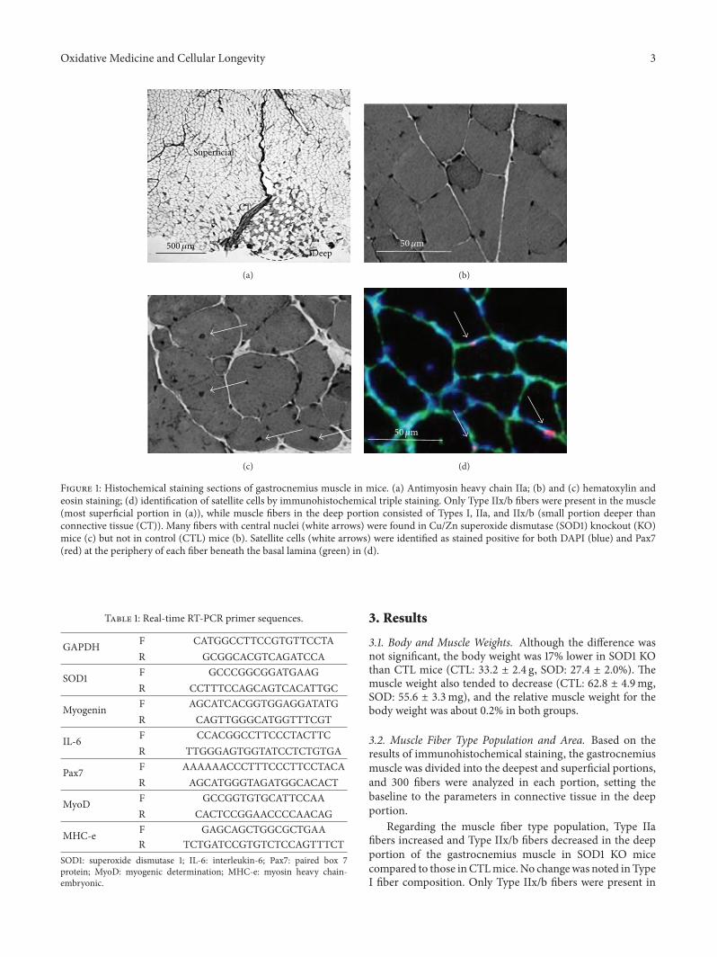
Oxidative Medicine and Cellular Longevity 3
Deep
Superficial
CT
500𝜇m
(a)
50𝜇m
(b)
(c)
50𝜇m
(d)
Figure 1: Histochemical staining sections of gastrocnemius muscle in mice. (a) Antimyosin heavy chain IIa; (b) and (c) hematoxylin andeosin staining; (d) identification of satellite cells by immunohistochemical triple staining. Only Type IIx/b fibers were present in the muscle(most superficial portion in (a)), while muscle fibers in the deep portion consisted of Types I, IIa, and IIx/b (small portion deeper thanconnective tissue (CT)). Many fibers with central nuclei (white arrows) were found in Cu/Zn superoxide dismutase (SOD1) knockout (KO)mice (c) but not in control (CTL) mice (b). Satellite cells (white arrows) were identified as stained positive for both DAPI (blue) and Pax7(red) at the periphery of each fiber beneath the basal lamina (green) in (d).
Table 1: Real-time RT-PCR primer sequences.
GAPDH F CATGGCCTTCCGTGTTCCTAR GCGGCACGTCAGATCCA
SOD1 F GCCCGGCGGATGAAGR CCTTTCCAGCAGTCACATTGC
Myogenin F AGCATCACGGTGGAGGATATGR CAGTTGGGCATGGTTTCGT
IL-6 F CCACGGCCTTCCCTACTTCR TTGGGAGTGGTATCCTCTGTGA
Pax7 F AAAAAACCCTTTCCCTTCCTACAR AGCATGGGTAGATGGCACACT
MyoD F GCCGGTGTGCATTCCAAR CACTCCGGAACCCCAACAG
MHC-e F GAGCAGCTGGCGCTGAAR TCTGATCCGTGTCTCCAGTTTCT
SOD1: superoxide dismutase 1; IL-6: interleukin-6; Pax7: paired box 7protein; MyoD: myogenic determination; MHC-e: myosin heavy chain-embryonic.
3. Results
3.1. Body and Muscle Weights. Although the difference wasnot significant, the body weight was 17% lower in SOD1 KOthan CTL mice (CTL: 33.2 ± 2.4 g, SOD: 27.4 ± 2.0%). Themuscle weight also tended to decrease (CTL: 62.8 ± 4.9mg,SOD: 55.6 ± 3.3mg), and the relative muscle weight for thebody weight was about 0.2% in both groups.
3.2. Muscle Fiber Type Population and Area. Based on theresults of immunohistochemical staining, the gastrocnemiusmuscle was divided into the deepest and superficial portions,and 300 fibers were analyzed in each portion, setting thebaseline to the parameters in connective tissue in the deepportion.
Regarding the muscle fiber type population, Type IIafibers increased and Type IIx/b fibers decreased in the deepportion of the gastrocnemius muscle in SOD1 KO micecompared to those inCTLmice.No changewas noted inTypeI fiber composition. Only Type IIx/b fibers were present in
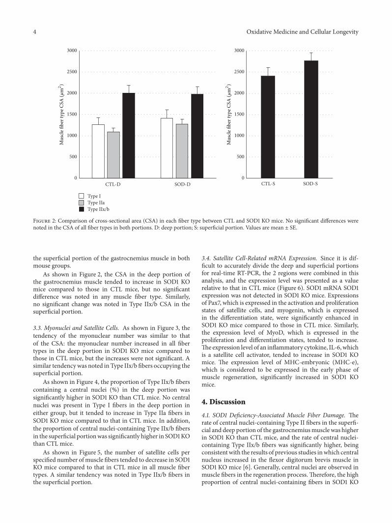
4 Oxidative Medicine and Cellular Longevity
CTL-S SOD-SCTL-D
Type IType IIa
SOD-D
Mus
cle fi
ber t
ype C
SA (𝜇
m2)
0
500
1000
1500
2000
2500
3000
Mus
cle fi
ber t
ype C
SA (𝜇
m2)
0
500
1000
1500
2000
2500
3000
Type IIx/b
Figure 2: Comparison of cross-sectional area (CSA) in each fiber type between CTL and SOD1 KO mice. No significant differences werenoted in the CSA of all fiber types in both portions. D: deep portion; S: superficial portion. Values are mean ± SE.
the superficial portion of the gastrocnemius muscle in bothmouse groups.
As shown in Figure 2, the CSA in the deep portion ofthe gastrocnemius muscle tended to increase in SOD1 KOmice compared to those in CTL mice, but no significantdifference was noted in any muscle fiber type. Similarly,no significant change was noted in Type IIx/b CSA in thesuperficial portion.
3.3. Myonuclei and Satellite Cells. As shown in Figure 3, thetendency of the myonuclear number was similar to thatof the CSA: the myonuclear number increased in all fibertypes in the deep portion in SOD1 KO mice compared tothose in CTL mice, but the increases were not significant. Asimilar tendencywas noted in Type IIx/b fibers occupying thesuperficial portion.
As shown in Figure 4, the proportion of Type IIx/b fiberscontaining a central nuclei (%) in the deep portion wassignificantly higher in SOD1 KO than CTL mice. No centralnuclei was present in Type I fibers in the deep portion ineither group, but it tended to increase in Type IIa fibers inSOD1 KO mice compared to that in CTL mice. In addition,the proportion of central nuclei-containing Type IIx/b fibersin the superficial portionwas significantly higher in SOD1KOthan CTL mice.
As shown in Figure 5, the number of satellite cells perspecified number ofmuscle fibers tended to decrease in SOD1KO mice compared to that in CTL mice in all muscle fibertypes. A similar tendency was noted in Type IIx/b fibers inthe superficial portion.
3.4. Satellite Cell-Related mRNA Expression. Since it is dif-ficult to accurately divide the deep and superficial portionsfor real-time RT-PCR, the 2 regions were combined in thisanalysis, and the expression level was presented as a valuerelative to that in CTL mice (Figure 6). SOD1 mRNA SOD1expression was not detected in SOD1 KO mice. Expressionsof Pax7, which is expressed in the activation and proliferationstates of satellite cells, and myogenin, which is expressedin the differentiation state, were significantly enhanced inSOD1 KO mice compared to those in CTL mice. Similarly,the expression level of MyoD, which is expressed in theproliferation and differentiation states, tended to increase.The expression level of an inflammatory cytokine, IL-6, whichis a satellite cell activator, tended to increase in SOD1 KOmice. The expression level of MHC-embryonic (MHC-e),which is considered to be expressed in the early phase ofmuscle regeneration, significantly increased in SOD1 KOmice.
4. Discussion
4.1. SOD1 Deficiency-Associated Muscle Fiber Damage. Therate of central nuclei-containing Type II fibers in the superfi-cial and deep portion of the gastrocnemiusmuscle was higherin SOD1 KO than CTL mice, and the rate of central nuclei-containing Type IIx/b fibers was significantly higher, beingconsistentwith the results of previous studies inwhich centralnucleus increased in the flexor digitorum brevis muscle inSOD1 KO mice [6]. Generally, central nuclei are observed inmuscle fibers in the regeneration process.Therefore, the highproportion of central nuclei-containing fibers in SOD1 KO
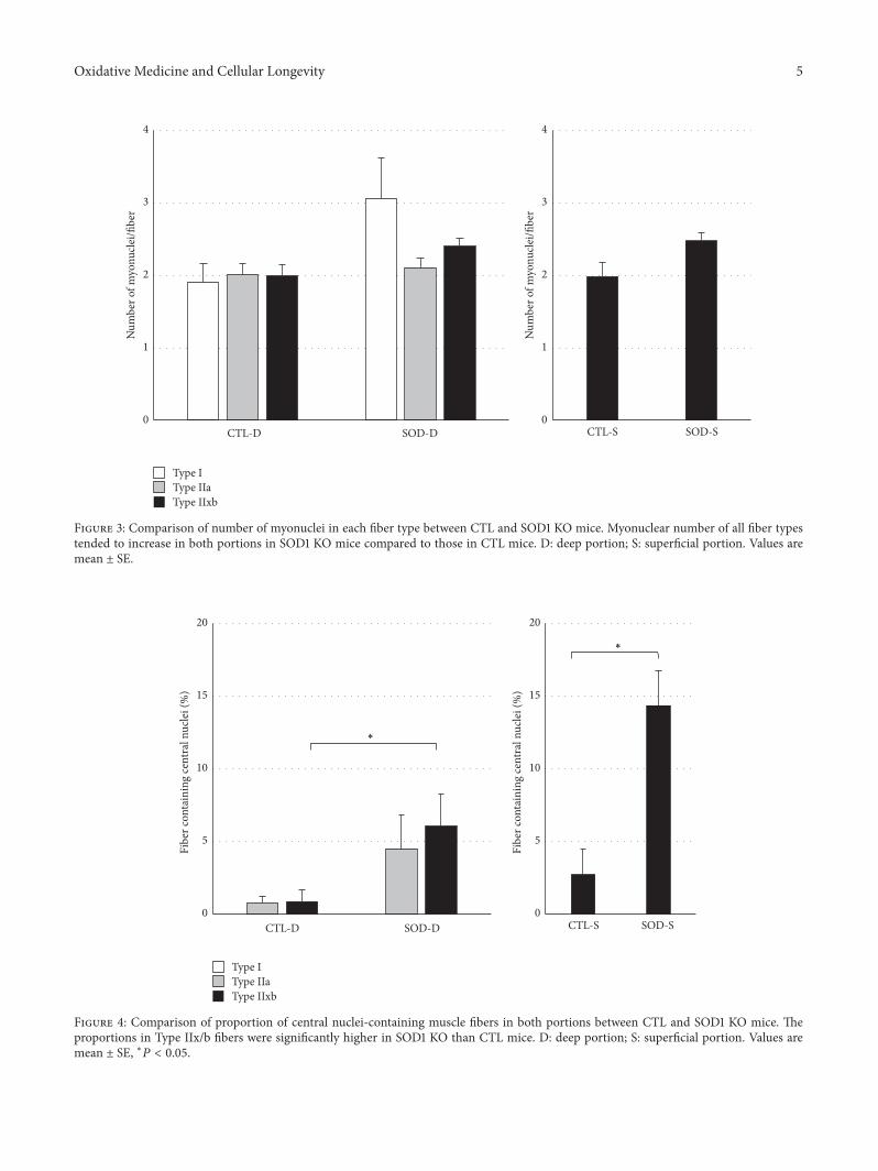
Oxidative Medicine and Cellular Longevity 5
CTL-S SOD-SCTL-D SOD-D
Num
ber o
f myo
nucle
i/fibe
r
0
1
2
3
4
Num
ber o
f myo
nucle
i/fibe
r
0
1
2
3
4
Type IType IIaType IIxb
Figure 3: Comparison of number of myonuclei in each fiber type between CTL and SOD1 KO mice. Myonuclear number of all fiber typestended to increase in both portions in SOD1 KO mice compared to those in CTL mice. D: deep portion; S: superficial portion. Values aremean ± SE.
CTL-S SOD-SCTL-D SOD-D
Fibe
r con
tain
ing
cent
ral n
ucle
i (%
)
Fibe
r con
tain
ing
cent
ral n
ucle
i (%
)
0
5
10
15
20
0
5
10
15
20
∗
∗
Type IType IIaType IIxb
Figure 4: Comparison of proportion of central nuclei-containing muscle fibers in both portions between CTL and SOD1 KO mice. Theproportions in Type IIx/b fibers were significantly higher in SOD1 KO than CTL mice. D: deep portion; S: superficial portion. Values aremean ± SE, ∗𝑃 < 0.05.
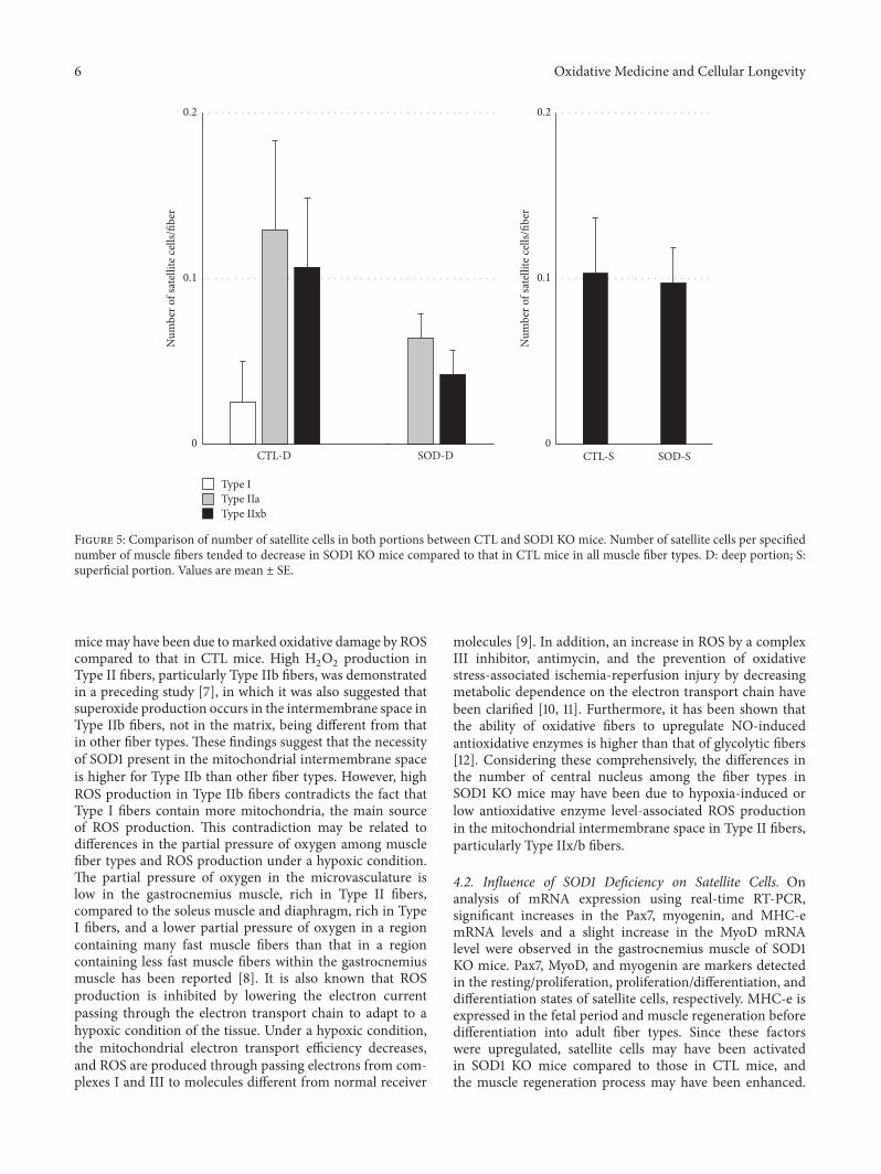
6 Oxidative Medicine and Cellular Longevity
CTL-S SOD-SCTL-D SOD-D0
0.1
0.2
Num
ber o
f sat
ellite
cells
/fibe
r
0
0.1
0.2
Num
ber o
f sat
ellite
cells
/fibe
r
Type IType IIaType IIxb
Figure 5: Comparison of number of satellite cells in both portions between CTL and SOD1 KO mice. Number of satellite cells per specifiednumber of muscle fibers tended to decrease in SOD1 KO mice compared to that in CTL mice in all muscle fiber types. D: deep portion; S:superficial portion. Values are mean ± SE.
micemay have been due tomarked oxidative damage by ROScompared to that in CTL mice. High H
2O2production in
Type II fibers, particularly Type IIb fibers, was demonstratedin a preceding study [7], in which it was also suggested thatsuperoxide production occurs in the intermembrane space inType IIb fibers, not in the matrix, being different from thatin other fiber types. These findings suggest that the necessityof SOD1 present in the mitochondrial intermembrane spaceis higher for Type IIb than other fiber types. However, highROS production in Type IIb fibers contradicts the fact thatType I fibers contain more mitochondria, the main sourceof ROS production. This contradiction may be related todifferences in the partial pressure of oxygen among musclefiber types and ROS production under a hypoxic condition.The partial pressure of oxygen in the microvasculature islow in the gastrocnemius muscle, rich in Type II fibers,compared to the soleus muscle and diaphragm, rich in TypeI fibers, and a lower partial pressure of oxygen in a regioncontaining many fast muscle fibers than that in a regioncontaining less fast muscle fibers within the gastrocnemiusmuscle has been reported [8]. It is also known that ROSproduction is inhibited by lowering the electron currentpassing through the electron transport chain to adapt to ahypoxic condition of the tissue. Under a hypoxic condition,the mitochondrial electron transport efficiency decreases,and ROS are produced through passing electrons from com-plexes I and III to molecules different from normal receiver
molecules [9]. In addition, an increase in ROS by a complexIII inhibitor, antimycin, and the prevention of oxidativestress-associated ischemia-reperfusion injury by decreasingmetabolic dependence on the electron transport chain havebeen clarified [10, 11]. Furthermore, it has been shown thatthe ability of oxidative fibers to upregulate NO-inducedantioxidative enzymes is higher than that of glycolytic fibers[12]. Considering these comprehensively, the differences inthe number of central nucleus among the fiber types inSOD1 KO mice may have been due to hypoxia-induced orlow antioxidative enzyme level-associated ROS productionin the mitochondrial intermembrane space in Type II fibers,particularly Type IIx/b fibers.
4.2. Influence of SOD1 Deficiency on Satellite Cells. Onanalysis of mRNA expression using real-time RT-PCR,significant increases in the Pax7, myogenin, and MHC-emRNA levels and a slight increase in the MyoD mRNAlevel were observed in the gastrocnemius muscle of SOD1KO mice. Pax7, MyoD, and myogenin are markers detectedin the resting/proliferation, proliferation/differentiation, anddifferentiation states of satellite cells, respectively. MHC-e isexpressed in the fetal period and muscle regeneration beforedifferentiation into adult fiber types. Since these factorswere upregulated, satellite cells may have been activatedin SOD1 KO mice compared to those in CTL mice, andthe muscle regeneration process may have been enhanced.
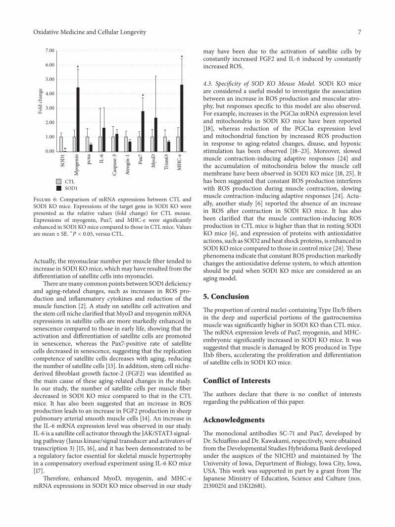
Oxidative Medicine and Cellular Longevity 7
SOD
1
Myo
geni
n
pcna
IL-6
Casp
ase-
3
Atro
gin-
1
Pax7
Myo
D
Trim
63
MH
C-e
0.00
1.00
2.00
3.00
4.00
5.00
6.00
7.00
Fold
chan
ge
∗
∗
∗
∗
CTLSOD1
Figure 6: Comparison of mRNA expressions between CTL andSOD1 KO mice. Expressions of the target gene in SOD1 KO werepresented as the relative values (fold change) for CTL mouse.Expressions of myogenin, Pax7, and MHC-e were significantlyenhanced in SOD1 KOmice compared to those in CTLmice. Valuesare mean ± SE. ∗𝑃 < 0.05, versus CTL.
Actually, the myonuclear number per muscle fiber tended toincrease in SOD1 KOmice, whichmay have resulted from thedifferentiation of satellite cells into myonuclei.
There aremany commonpoints between SOD1 deficiencyand aging-related changes, such as increases in ROS pro-duction and inflammatory cytokines and reduction of themuscle function [2]. A study on satellite cell activation andthe stem cell niche clarified thatMyoD andmyogeninmRNAexpressions in satellite cells are more markedly enhanced insenescence compared to those in early life, showing that theactivation and differentiation of satellite cells are promotedin senescence, whereas the Pax7-positive rate of satellitecells decreased in senescence, suggesting that the replicationcompetence of satellite cells decreases with aging, reducingthe number of satellite cells [13]. In addition, stem cell niche-derived fibroblast growth factor-2 (FGF2) was identified asthe main cause of these aging-related changes in the study.In our study, the number of satellite cells per muscle fiberdecreased in SOD1 KO mice compared to that in the CTLmice. It has also been suggested that an increase in ROSproduction leads to an increase in FGF2 production in sheeppulmonary arterial smooth muscle cells [14]. An increase inthe IL-6 mRNA expression level was observed in our study.IL-6 is a satellite cell activator through the JAK/STAT3 signal-ing pathway (Janus kinase/signal transducer and activators oftranscription 3) [15, 16], and it has been demonstrated to bea regulatory factor essential for skeletal muscle hypertrophyin a compensatory overload experiment using IL-6 KO mice[17].
Therefore, enhanced MyoD, myogenin, and MHC-emRNA expressions in SOD1 KO mice observed in our study
may have been due to the activation of satellite cells byconstantly increased FGF2 and IL-6 induced by constantlyincreased ROS.
4.3. Specificity of SOD KO Mouse Model. SOD1 KO miceare considered a useful model to investigate the associationbetween an increase in ROS production and muscular atro-phy, but responses specific to this model are also observed.For example, increases in the PGC1𝛼mRNA expression leveland mitochondria in SOD1 KO mice have been reported[18], whereas reduction of the PGC1𝛼 expression leveland mitochondrial function by increased ROS productionin response to aging-related changes, disuse, and hypoxicstimulation has been observed [18–23]. Moreover, slowedmuscle contraction-inducing adaptive responses [24] andthe accumulation of mitochondria below the muscle cellmembrane have been observed in SOD1 KO mice [18, 25]. Ithas been suggested that constant ROS production interfereswith ROS production during muscle contraction, slowingmuscle contraction-inducing adaptive responses [24]. Actu-ally, another study [6] reported the absence of an increasein ROS after contraction in SOD1 KO mice. It has alsobeen clarified that the muscle contraction-inducing ROSproduction in CTL mice is higher than that in resting SOD1KO mice [6], and expression of proteins with antioxidativeactions, such as SOD2 and heat shock proteins, is enhanced inSOD1KOmice compared to those in controlmice [24].Thesephenomena indicate that constant ROS productionmarkedlychanges the antioxidative defense system, to which attentionshould be paid when SOD1 KO mice are considered as anaging model.
5. Conclusion
The proportion of central nuclei-containing Type IIx/b fibersin the deep and superficial portions of the gastrocnemiusmuscle was significantly higher in SOD1 KO than CTL mice.The mRNA expression levels of Pax7, myogenin, and MHC-embryonic significantly increased in SOD1 KO mice. It wassuggested that muscle is damaged by ROS produced in TypeIIxb fibers, accelerating the proliferation and differentiationof satellite cells in SOD1 KO mice.
Conflict of Interests
The authors declare that there is no conflict of interestsregarding the publication of this paper.
Acknowledgments
The monoclonal antibodies SC-71 and Pax7, developed byDr. Schiaffino and Dr. Kawakami, respectively, were obtainedfrom the Developmental Studies Hybridoma Bank developedunder the auspices of the NICHD and maintained by TheUniversity of Iowa, Department of Biology, Iowa City, Iowa,USA. This work was supported in part by a grant from TheJapanese Ministry of Education, Science and Culture (nos.21300251 and 15K12681).
8 Oxidative Medicine and Cellular Longevity
References
[1] D. H. Kang and S. W. Kang, “Targeting cellular antioxi-dant enzymes for treating atherosclerotic vascular disease,”Biomolecules and Therapeutics, vol. 21, no. 2, pp. 89–96, 2013.
[2] A. Vasilaki and M. J. Jackson, “Role of reactive oxygen speciesin the defective regeneration seen in agingmuscle,” Free RadicalBiology and Medicine, vol. 65, pp. 317–323, 2013.
[3] S. Fulle, F. Protasi, G. Di Tano et al., “The contribution ofreactive oxygen species to sarcopenia and muscle ageing,”Experimental Gerontology, vol. 39, no. 1, pp. 17–24, 2004.
[4] F. L. Muller, W. Song, Y. Liu et al., “Absence of CuZn superoxidedismutase leads to elevated oxidative stress and acceleration ofage-dependent skeletal muscle atrophy,” Free Radical Biologyand Medicine, vol. 40, no. 11, pp. 1993–2004, 2006.
[5] M. M. Matzuk, L. Dionne, Q. Guo, T. R. Kumar, and R. M.Lebovitz, “Ovarian function in superoxide dismutase 1 and 2knockout mice,” Endocrinology, vol. 139, no. 9, pp. 4008–4011,1998.
[6] G. K. Sakellariou, D. Pye, A. Vasilaki et al., “Role of superoxide-nitric oxide interactions in the accelerated age-related loss ofmuscle mass in mice lacking Cu,Zn superoxide dismutase,”Aging Cell, vol. 10, no. 5, pp. 749–760, 2011.
[7] E. J. Anderson and P. D. Neufer, “Type II skeletal myofiberspossess unique properties that potentiate mitochondrialH2
O2
generation,” The American Journal of Physiology—CellPhysiology, vol. 290, no. 3, pp. C844–C851, 2006.
[8] L. F. Ferreira, P. McDonough, B. J. Behnke, T. I. Musch, andD. C. Poole, “Blood flow and O
2
extraction as a functionof O2
uptake in muscles composed of different fiber types,”Respiratory Physiology andNeurobiology, vol. 153, no. 3, pp. 237–249, 2006.
[9] G. L. Semenza, “Hypoxia-inducible factors in physiology andmedicine,” Cell, vol. 148, no. 3, pp. 399–408, 2012.
[10] W. W. Wheaton and N. S. Chandel, “Hypoxia. 2. Hypoxia regu-lates cellular metabolism,”American Journal of Physiology—CellPhysiology, vol. 300, no. 3, pp. C385–C393, 2011.
[11] N. S. Chandel, E. Maltepe, E. Goldwasser, C. E. Mathieu, M. C.Simon, and P. T. Schumacker, “Mitochondrial reactive oxygenspecies trigger hypoxia-induced transcription,” Proceedings ofthe National Academy of Sciences of the United States of America,vol. 95, no. 20, pp. 11715–11720, 1998.
[12] Z. Yu, P. Li, M. Zhang, M. Hannink, J. S. Stamler, and Z. Yan,“Fiber type-specific nitric oxide protects oxidative myofibersagainst cachectic stimuli,” PLoS ONE, vol. 3, no. 5, Article IDe2086, 2008.
[13] J. V. Chakkalakal, K. M. Jones, M. A. Basson, and A. S. Brack,“The aged niche disrupts muscle stem cell quiescence,” Nature,vol. 490, no. 7420, pp. 355–360, 2012.
[14] S. M. Black, J. M. DeVol, and S. Wedgwood, “Regulation offibroblast growth factor-2 expression in pulmonary arterialsmooth muscle cells involves increased reactive oxygen speciesgeneration,” American Journal of Physiology—Cell Physiology,vol. 294, no. 1, pp. C345–C354, 2008.
[15] M. Masuda, M. Suzui, R. Yasumatu et al., “Constitutive acti-vation of signal transducers and activators of transcription3 correlates with cyclin D1 overexpression and may providea novel prognostic marker in head and neck squamous cellcarcinoma,”Cancer Research, vol. 62, no. 12, pp. 3351–3355, 2002.
[16] M. Ernst and B. J. Jenkins, “Acquiring signalling specificity fromthe cytokine receptor gp130,” Trends in Genetics, vol. 20, no. 1,pp. 23–32, 2004.
[17] A. L. Serrano, B. Baeza-Raja, E. Perdiguero, M. Jardı́, andP. Muñoz-Cánoves, “Interleukin-6 is an essential regulatorof satellite cell-mediated skeletal muscle hypertrophy,” CellMetabolism, vol. 7, no. 1, pp. 33–44, 2008.
[18] Y. C. Jang, M. S. Lustgarten, Y. Liu et al., “Increased superoxidein vivo accelerates age-associated muscle atrophy throughmitochondrial dysfunction andneuromuscular junction degen-eration,”The FASEB Journal, vol. 24, no. 5, pp. 1376–1390, 2010.
[19] T. Wenz, “Regulation of mitochondrial biogenesis and PGC-1𝛼under cellular stress,”Mitochondrion, vol. 13, no. 2, pp. 134–142,2013.
[20] M. Picard, D. Ritchie, M. M. Thomas, K. J. Wright, and R. T.Hepple, “Alterations in intrinsic mitochondrial function withaging are fiber type-specific and do not explain differentialatrophy between muscles,” Aging Cell, vol. 10, no. 6, pp. 1047–1055, 2011.
[21] E. E. Talbert, A. J. Smuder, K. Min, O. S. Kwon, H. H.Szeto, and S. K. Powers, “Immobilization-induced activationof key proteolytic systems in skeletal muscles is preventedby a mitochondria-targeted antioxidant,” Journal of AppliedPhysiology, vol. 115, no. 4, pp. 529–538, 2013.
[22] J. L. Gamboa and F. H. Andrade, “Mitochondrial contentand distribution changes specific to mouse diaphragm afterchronic normobaric hypoxia,” American Journal of Physiology:Regulatory Integrative and Comparative Physiology, vol. 298, no.3, pp. R575–R583, 2010.
[23] J. Magalhães, A. Ascensão, J. M. C. Soares et al., “Acute andsevere hypobaric hypoxia increases oxidative stress and impairsmitochondrial function in mouse skeletal muscle,” Journal ofApplied Physiology, vol. 99, no. 4, pp. 1247–1253, 2005.
[24] A. Vasilaki, J. H. van der Meulen, L. Larkin, and et al, “The age-related failure of adaptive responses to contractile activity inskeletal muscle is mimicked in youngmice by deletion of Cu,Znsuperoxide dismutase,” Aging Cell, vol. 9, no. 6, pp. 979–990,2010.
[25] T. Y. Kostrominova, K. A. Pasyk, H. Van Remmen, A. G.Richardson, and J. A. Faulkner, “Adaptive changes in structureof skeletal muscles from adult Sod1 homozygous knockoutmice,”Cell and Tissue Research, vol. 327, no. 3, pp. 595–605, 2007.
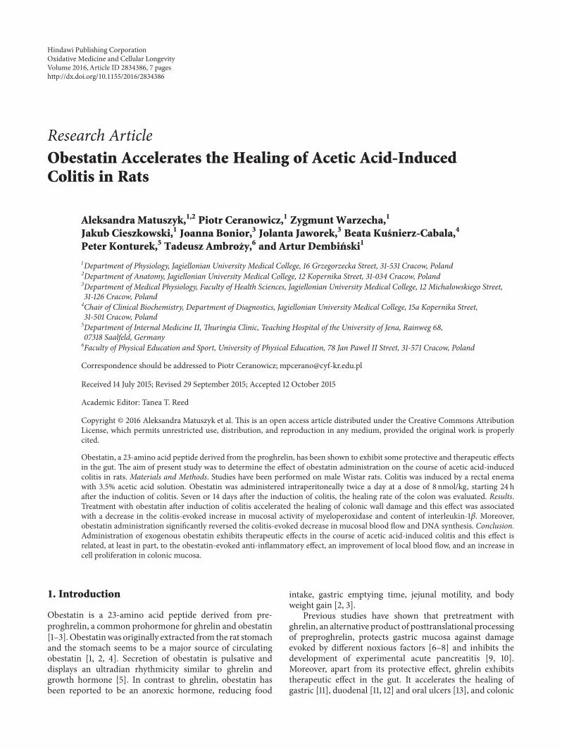
Research ArticleObestatin Accelerates the Healing of Acetic Acid-InducedColitis in Rats
Aleksandra Matuszyk,1,2 Piotr Ceranowicz,1 Zygmunt Warzecha,1
Jakub Cieszkowski,1 Joanna Bonior,3 Jolanta Jaworek,3 Beata KuVnierz-Cabala,4
Peter Konturek,5 Tadeusz Ambrohy,6 and Artur DembiNski1
1Department of Physiology, Jagiellonian University Medical College, 16 Grzegorzecka Street, 31-531 Cracow, Poland2Department of Anatomy, Jagiellonian University Medical College, 12 Kopernika Street, 31-034 Cracow, Poland3Department of Medical Physiology, Faculty of Health Sciences, Jagiellonian University Medical College, 12 Michałowskiego Street,31-126 Cracow, Poland4Chair of Clinical Biochemistry, Department of Diagnostics, Jagiellonian University Medical College, 15a Kopernika Street,31-501 Cracow, Poland5Department of Internal Medicine II, Thuringia Clinic, Teaching Hospital of the University of Jena, Rainweg 68,07318 Saalfeld, Germany6Faculty of Physical Education and Sport, University of Physical Education, 78 Jan Paweł II Street, 31-571 Cracow, Poland
Correspondence should be addressed to Piotr Ceranowicz; [email protected]
Received 14 July 2015; Revised 29 September 2015; Accepted 12 October 2015
Academic Editor: Tanea T. Reed
Copyright © 2016 Aleksandra Matuszyk et al. This is an open access article distributed under the Creative Commons AttributionLicense, which permits unrestricted use, distribution, and reproduction in any medium, provided the original work is properlycited.
Obestatin, a 23-amino acid peptide derived from the proghrelin, has been shown to exhibit some protective and therapeutic effectsin the gut. The aim of present study was to determine the effect of obestatin administration on the course of acetic acid-inducedcolitis in rats. Materials and Methods. Studies have been performed on male Wistar rats. Colitis was induced by a rectal enemawith 3.5% acetic acid solution. Obestatin was administered intraperitoneally twice a day at a dose of 8 nmol/kg, starting 24 hafter the induction of colitis. Seven or 14 days after the induction of colitis, the healing rate of the colon was evaluated. Results.Treatment with obestatin after induction of colitis accelerated the healing of colonic wall damage and this effect was associatedwith a decrease in the colitis-evoked increase in mucosal activity of myeloperoxidase and content of interleukin-1𝛽. Moreover,obestatin administration significantly reversed the colitis-evoked decrease in mucosal blood flow and DNA synthesis. Conclusion.Administration of exogenous obestatin exhibits therapeutic effects in the course of acetic acid-induced colitis and this effect isrelated, at least in part, to the obestatin-evoked anti-inflammatory effect, an improvement of local blood flow, and an increase incell proliferation in colonic mucosa.
1. Introduction
Obestatin is a 23-amino acid peptide derived from pre-proghrelin, a common prohormone for ghrelin and obestatin[1–3].Obestatinwas originally extracted from the rat stomachand the stomach seems to be a major source of circulatingobestatin [1, 2, 4]. Secretion of obestatin is pulsative anddisplays an ultradian rhythmicity similar to ghrelin andgrowth hormone [5]. In contrast to ghrelin, obestatin hasbeen reported to be an anorexic hormone, reducing food
intake, gastric emptying time, jejunal motility, and bodyweight gain [2, 3].
Previous studies have shown that pretreatment withghrelin, an alternative product of posttranslational processingof preproghrelin, protects gastric mucosa against damageevoked by different noxious factors [6–8] and inhibits thedevelopment of experimental acute pancreatitis [9, 10].Moreover, apart from its protective effect, ghrelin exhibitstherapeutic effect in the gut. It accelerates the healing ofgastric [11], duodenal [11, 12] and oral ulcers [13], and colonic
Hindawi Publishing CorporationOxidative Medicine and Cellular LongevityVolume 2016, Article ID 2834386, 7 pageshttp://dx.doi.org/10.1155/2016/2834386
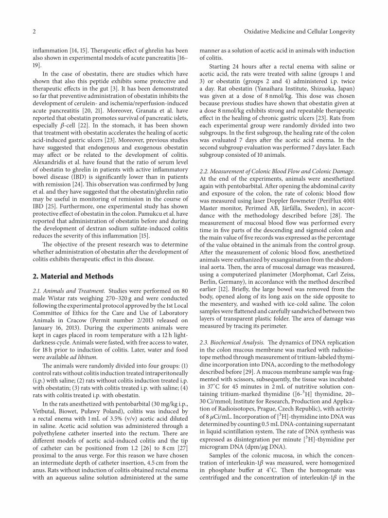
2 Oxidative Medicine and Cellular Longevity
inflammation [14, 15]. Therapeutic effect of ghrelin has beenalso shown in experimental models of acute pancreatitis [16–19].
In the case of obestatin, there are studies which haveshown that also this peptide exhibits some protective andtherapeutic effects in the gut [3]. It has been demonstratedso far that preventive administration of obestatin inhibits thedevelopment of cerulein- and ischemia/reperfusion-inducedacute pancreatitis [20, 21]. Moreover, Granata et al. havereported that obestatin promotes survival of pancreatic islets,especially 𝛽-cell [22]. In the stomach, it has been shownthat treatment with obestatin accelerates the healing of aceticacid-induced gastric ulcers [23]. Moreover, previous studieshave suggested that endogenous and exogenous obestatinmay affect or be related to the development of colitis.Alexandridis et al. have found that the ratio of serum levelof obestatin to ghrelin in patients with active inflammatorybowel disease (IBD) is significantly lower than in patientswith remission [24]. This observation was confirmed by Junget al. and they have suggested that the obestatin/ghrelin ratiomay be useful in monitoring of remission in the course ofIBD [25]. Furthermore, one experimental study has shownprotective effect of obestatin in the colon. Pamukcu et al. havereported that administration of obestatin before and duringthe development of dextran sodium sulfate-induced colitisreduces the severity of this inflammation [15].
The objective of the present research was to determinewhether administration of obestatin after the development ofcolitis exhibits therapeutic effect in this disease.
2. Material and Methods
2.1. Animals and Treatment. Studies were performed on 80male Wistar rats weighing 270–320 g and were conductedfollowing the experimental protocol approved by the 1st LocalCommittee of Ethics for the Care and Use of LaboratoryAnimals in Cracow (Permit number 2/2013 released onJanuary 16, 2013). During the experiments animals werekept in cages placed in room temperature with a 12 h light-darkness cycle. Animals were fasted, with free access to water,for 18 h prior to induction of colitis. Later, water and foodwere available ad libitum.
The animals were randomly divided into four groups: (1)control ratswithout colitis induction treated intraperitoneally(i.p.) with saline; (2) rats without colitis induction treated i.p.with obestatin; (3) rats with colitis treated i.p. with saline; (4)rats with colitis treated i.p. with obestatin.
In the rats anesthetized with pentobarbital (30mg/kg i.p.,Vetbutal, Biowet, Puławy Poland), colitis was induced bya rectal enema with 1mL of 3.5% (v/v) acetic acid dilutedin saline. Acetic acid solution was administered through apolyethylene catheter inserted into the rectum. There aredifferent models of acetic acid-induced colitis and the tipof catheter can be positioned from 1.2 [26] to 8 cm [27]proximal to the anus verge. For this reason we have chosenan intermediate depth of catheter insertion, 4.5 cm from theanus. Rats without induction of colitis obtained rectal enemawith an aqueous saline solution administered at the same
manner as a solution of acetic acid in animals with inductionof colitis.
Starting 24 hours after a rectal enema with saline oracetic acid, the rats were treated with saline (groups 1 and3) or obestatin (groups 2 and 4) administered i.p. twicea day. Rat obestatin (Yanaihara Institute, Shizuoka, Japan)was given at a dose of 8 nmol/kg. This dose was chosenbecause previous studies have shown that obestatin given ata dose 8 nmol/kg exhibits strong and repeatable therapeuticeffect in the healing of chronic gastric ulcers [23]. Rats fromeach experimental group were randomly divided into twosubgroups. In the first subgroup, the healing rate of the colonwas evaluated 7 days after the acetic acid enema. In thesecond subgroup evaluation was performed 7 days later. Eachsubgroup consisted of 10 animals.
2.2. Measurement of Colonic Blood Flow and Colonic Damage.At the end of the experiments, animals were anesthetizedagain with pentobarbital. After opening the abdominal cavityand exposure of the colon, the rate of colonic blood flowwas measured using laser Doppler flowmeter (PeriFlux 4001Master monitor, Perimed AB, Järfälla, Sweden), in accor-dance with the methodology described before [28]. Themeasurement of mucosal blood flow was performed everytime in five parts of the descending and sigmoid colon andthemain value of five records was expressed as the percentageof the value obtained in the animals from the control group.After the measurement of colonic blood flow, anesthetizedanimals were euthanized by exsanguination from the abdom-inal aorta. Then, the area of mucosal damage was measured,using a computerized planimeter (Morphomat, Carl Zeiss,Berlin, Germany), in accordance with the method describedearlier [12]. Briefly, the large bowel was removed from thebody, opened along of its long axis on the side opposite tothe mesentery, and washed with ice-cold saline. The colonsampleswere flattened and carefully sandwiched between twolayers of transparent plastic folder. The area of damage wasmeasured by tracing its perimeter.
2.3. Biochemical Analysis. The dynamics of DNA replicationin the colon mucous membrane was marked with radioiso-topemethod throughmeasurement of tritium-labeled thymi-dine incorporation into DNA, according to the methodologydescribed before [29]. Amucousmembrane sample was frag-mented with scissors, subsequently, the tissue was incubatedin 37∘C for 45 minutes in 2mL of nutritive solution con-taining tritium-marked thymidine ([6-3H] thymidine, 20–30Ci/mmol; Institute for Research, Production and Applica-tion of Radioisotopes, Prague, Czech Republic), with activityof 8𝜇Ci/mL. Incorporation of [3H]-thymidine into DNAwasdetermined by counting 0.5mLDNA-containing supernatantin liquid scintillation system. The rate of DNA synthesis wasexpressed as disintegration per minute [3H]-thymidine permicrogram DNA (dpm/𝜇g DNA).
Samples of the colonic mucosa, in which the concen-tration of interleukin-1𝛽 was measured, were homogenizedin phosphate buffer at 4∘C. Then the homogenate wascentrifuged and the concentration of interleukin-1𝛽 in the
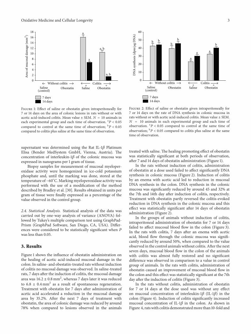
Oxidative Medicine and Cellular Longevity 3
02468
1012141618
a, b
7 days14 days
a, b
a
a
ColitisObestatinSalineObestatinSaline
Without colitis
(control)++
Are
a of m
ucos
al d
amag
e (m
m2)
Figure 1: Effect of saline or obestatin given intraperitoneally for7 or 14 days on the area of colonic lesions in rats without or withacetic acid-induced colitis. Mean value ± SEM. 𝑁 = 10 animals ineach experimental group and each time of observation. a𝑃 < 0.05
compared to control at the same time of observation; b𝑃 < 0.05compared to colitis plus saline at the same time of observation.
supernatant was determined using the Rat IL-1𝛽 PlatinumElisa (Bender MedSystem GmbH, Vienna, Austria). Theconcentration of interleukin-1𝛽 of the colonic mucosa wasexpressed in nanograms per 1 gram of tissue.
Biopsy samples for measurement of mucosal myeloper-oxidase activity were homogenized in ice-cold potassiumphosphate and, until the marking was done, stored at thetemperature of−60∘C.Markingmyeloperoxidase activity wasperformed with the use of a modification of the methoddescribed by Bradley et al. [30]. Results obtained in units pergram of tissue were finally expressed as a percentage of thevalue observed in the control group.
2.4. Statistical Analysis. Statistical analysis of the data wascarried out by one-way analysis of variance (ANOVA) fol-lowed by Tukey’s multiple comparison test using GraphPad-Prism (GraphPad Software, San Diego, CA, USA). Differ-ences were considered to be statistically significant when 𝑃was less than 0.05.
3. Results
Figure 1 shows the influence of obestatin administration onthe healing of acetic acid-induced mucosal damage in thecolon. In saline- and obestatin-treated rats without inductionof colitis no mucosal damage was observed. In saline-treatedrats, 7 days after the induction of colitis, the mucosal damagearea was 16.2 ± 0.8mm2, whereas 7 days later it was reducedto 6.8 ± 0.4mm2 as a result of spontaneous regeneration.Treatment with obestatin for 7 days after administration ofacetic acid accelerated a reduction in the mucosal damagearea by 35.2%. After the next 7 days of treatment withobestatin, the area of colonic damage was reduced by around78% when compared to lesions observed in the animals
0
10
20
30
40
50
60
a
a
a
b b bb b
7 days14 days
ColitisObestatinSalineObestatinSaline
Without colitis
(control)++
DN
A sy
nthe
sis (d
pm/𝜇
g D
NA
)
Figure 2: Effect of saline or obestatin given intraperitoneally for7 or 14 days on the rate of DNA synthesis in colonic mucosa inrats without or with acetic acid-induced colitis. Mean value ± SEM.𝑁 = 10 animals in each experimental group and each time ofobservation. a𝑃 < 0.05 compared to control at the same time ofobservation; b𝑃 < 0.05 compared to colitis plus saline at the sametime of observation.
treated with saline. The healing promoting effect of obestatinwas statistically significant at both periods of observation,after 7 and 14 days of obestatin administration (Figure 1).
In the rats without induction of colitis, administrationof obestatin at a dose used failed to affect significantly DNAsynthesis in colonic mucosa (Figure 2). Induction of colitisby an enema with acetic acid led to reduction in mucosalDNA synthesis in the colon. DNA synthesis in the colonicmucosa was significantly reduced by around 45 and 32% atthe 7th and 14th day after induction of colitis, respectively.Treatment with obestatin partly reversed the colitis-evokedreduction in DNA synthesis in the colonic mucosa and thiseffect was statistically significant after 14 days of obestatinadministration (Figure 2).
In the groups of animals without induction of colitis,intraperitoneal administration of obestatin for 7 or 14 daysfailed to affect mucosal blood flow in the colon (Figure 3).In the rats with colitis, 7 days after an enema with aceticacid, blood flow through the colonic mucosa was signifi-cantly reduced by around 50%, when compared to the valueobserved in the control animals without colitis. After the nextseven days, mucosal blood flow in the colon of the animalswith colitis was almost fully restored and no significantdifference was observed in comparison to a value in controlgroup of animals. In the rats with colitis, administration ofobestatin caused an improvement of mucosal blood flow inthe colon and this effect was statistically significant at the 7thday after the induction of colitis (Figure 3).
In the rats without colitis, administration of obestatinfor 7 or 14 days at the dose used was without any effecton mucosal concentration of interleukin-1𝛽 (IL-1𝛽) in thecolon (Figure 4). Induction of colitis significantly increasedmucosal concentration of IL-1𝛽 in the colon. As shown inFigure 4, ratswith colitis demonstratedmore than 10-fold and
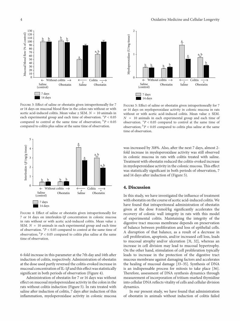
4 Oxidative Medicine and Cellular Longevity
0102030405060708090
100110120130
a, b
a
bb bb
Muc
osal
blo
od fl
ow (%
of c
ontro
l)
7 days14 days
ColitisObestatinSalineObestatinSaline
Without colitis
(control)++
Figure 3: Effect of saline or obestatin given intraperitoneally for 7or 14 days on mucosal blood flow in the colon rats without or withacetic acid-induced colitis. Mean value ± SEM. 𝑁 = 10 animals ineach experimental group and each time of observation. a𝑃 < 0.05
compared to control at the same time of observation; b𝑃 < 0.05compared to colitis plus saline at the same time of observation.
0
1
2
3
4
5
a, b
a
a
a, b
bbbb
7 days14 days
ColitisObestatinSalineObestatinSaline
Without colitis
(control)++
Inte
rleuk
in-1𝛽
(ng/
g tis
sue)
Figure 4: Effect of saline or obestatin given intraperitoneally for7 or 14 days on interleukin-1𝛽 concentration in colonic mucosain rats without or with acetic acid-induced colitis. Mean value ±SEM. 𝑁 = 10 animals in each experimental group and each timeof observation. a𝑃 < 0.05 compared to control at the same time ofobservation; b𝑃 < 0.05 compared to colitis plus saline at the sametime of observation.
6-fold increase in this parameter at the 7th day and 14th afterinduction of colitis, respectively. Administration of obestatinat the dose used partly reversed the colitis-evoked increase inmucosal concentration of IL-1𝛽 and this effectwas statisticallysignificant in both periods of observation (Figure 4).
Administration of obestatin for 7 or 14 days was withouteffect onmucosalmyeloperoxidase activity in the colon in therats without colitis induction (Figure 5). In rats treated withsaline after induction of colitis, 7 days after induction of thisinflammation, myeloperoxidase activity in colonic mucosa
0
1
2
a, b
a
a
bbbbb
Mye
lope
roxi
dase
(U/m
g tis
sue)
7 days14 days
ColitisObestatinSalineObestatinSaline
Without colitis
(control)++
Figure 5: Effect of saline or obestatin given intraperitoneally for 7or 14 days on myeloperoxidase activity in colonic mucosa in ratswithout or with acetic acid-induced colitis. Mean value ± SEM.𝑁 = 10 animals in each experimental group and each time ofobservation. a𝑃 < 0.05 compared to control at the same time ofobservation; b𝑃 < 0.05 compared to colitis plus saline at the sametime of observation.
was increased by 318%. Also, after the next 7 days, almost 2-fold increase in myeloperoxidase activity was still observedin colonic mucosa in rats with colitis treated with saline.Treatment with obestatin reduced the colitis-evoked increaseinmyeloperoxidase activity in the colonicmucosa.This effectwas statistically significant in both periods of observation, 7and 14 days after induction of (Figure 5).
4. Discussion
In this study, we have investigated the influence of treatmentwith obestatin on the course of acetic acid-induced colitis.Wehave found that intraperitoneal administration of obestatingiven at the dose 8 nmol/kg significantly accelerates therecovery of colonic wall integrity in rats with this modelof experimental colitis. Maintaining the integrity of thedigestive tract mucous membrane depends on preservationof balance between proliferation and loss of epithelial cells.A disruption of that balance, as a result of a decrease incell proliferation, apoptosis, and/or increased cell loss, leadsto mucosal atrophy and/or ulceration [31, 32], whereas anincrease in cell division may lead to mucosal hypertrophy.On the other hand, stimulation of cell proliferation typicallyleads to increase in the protection of the digestive tractmucous membrane against damaging factors and acceleratesthe healing of mucosal damage [33–35]. Synthesis of DNAis an indispensable process for mitosis to take place [36].Therefore, assessment of DNA synthesis dynamics throughmeasurement of incorporation of tritium-marked thymidineinto cellular DNA reflects vitality of cells and cellular divisiondynamics.
In our present study, we have found that administrationof obestatin in animals without induction of colitis failed
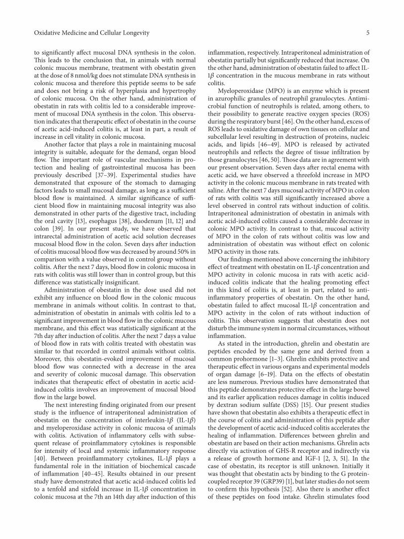
Oxidative Medicine and Cellular Longevity 5
to significantly affect mucosal DNA synthesis in the colon.This leads to the conclusion that, in animals with normalcolonic mucous membrane, treatment with obestatin givenat the dose of 8 nmol/kg does not stimulate DNA synthesis incolonic mucosa and therefore this peptide seems to be safeand does not bring a risk of hyperplasia and hypertrophyof colonic mucosa. On the other hand, administration ofobestatin in rats with colitis led to a considerable improve-ment of mucosal DNA synthesis in the colon. This observa-tion indicates that therapeutic effect of obestatin in the courseof acetic acid-induced colitis is, at least in part, a result ofincrease in cell vitality in colonic mucosa.
Another factor that plays a role in maintaining mucosalintegrity is suitable, adequate for the demand, organ bloodflow. The important role of vascular mechanisms in pro-tection and healing of gastrointestinal mucosa has beenpreviously described [37–39]. Experimental studies havedemonstrated that exposure of the stomach to damagingfactors leads to small mucosal damage, as long as a sufficientblood flow is maintained. A similar significance of suffi-cient blood flow in maintaining mucosal integrity was alsodemonstrated in other parts of the digestive tract, includingthe oral cavity [13], esophagus [38], duodenum [11, 12] andcolon [39]. In our present study, we have observed thatintrarectal administration of acetic acid solution decreasesmucosal blood flow in the colon. Seven days after inductionof colitis mucosal blood flowwas decreased by around 50% incomparison with a value observed in control group withoutcolitis. After the next 7 days, blood flow in colonic mucosa inrats with colitis was still lower than in control group, but thisdifference was statistically insignificant.
Administration of obestatin in the dose used did notexhibit any influence on blood flow in the colonic mucousmembrane in animals without colitis. In contrast to that,administration of obestatin in animals with colitis led to asignificant improvement in blood flow in the colonic mucousmembrane, and this effect was statistically significant at the7th day after induction of colitis. After the next 7 days a valueof blood flow in rats with colitis treated with obestatin wassimilar to that recorded in control animals without colitis.Moreover, this obestatin-evoked improvement of mucosalblood flow was connected with a decrease in the areaand severity of colonic mucosal damage. This observationindicates that therapeutic effect of obestatin in acetic acid-induced colitis involves an improvement of mucosal bloodflow in the large bowel.
The next interesting finding originated from our presentstudy is the influence of intraperitoneal administration ofobestatin on the concentration of interleukin-1𝛽 (IL-1𝛽)and myeloperoxidase activity in colonic mucosa of animalswith colitis. Activation of inflammatory cells with subse-quent release of proinflammatory cytokines is responsiblefor intensity of local and systemic inflammatory response[40]. Between proinflammatory cytokines, IL-1𝛽 plays afundamental role in the initiation of biochemical cascadeof inflammation [40–45]. Results obtained in our presentstudy have demonstrated that acetic acid-induced colitis ledto a tenfold and sixfold increase in IL-1𝛽 concentration incolonic mucosa at the 7th an 14th day after induction of this
inflammation, respectively. Intraperitoneal administration ofobestatin partially but significantly reduced that increase. Onthe other hand, administration of obestatin failed to affect IL-1𝛽 concentration in the mucous membrane in rats withoutcolitis.
Myeloperoxidase (MPO) is an enzyme which is presentin azurophilic granules of neutrophil granulocytes. Antimi-crobial function of neutrophils is related, among others, totheir possibility to generate reactive oxygen species (ROS)during the respiratory burst [46].On the other hand, excess ofROS leads to oxidative damage of own tissues on cellular andsubcellular level resulting in destruction of proteins, nucleicacids, and lipids [46–49]. MPO is released by activatedneutrophils and reflects the degree of tissue infiltration bythose granulocytes [46, 50].Those data are in agreement withour present observation. Seven days after rectal enema withacetic acid, we have observed a threefold increase in MPOactivity in the colonic mucous membrane in rats treated withsaline. After the next 7 daysmucosal activity ofMPO in colonof rats with colitis was still significantly increased above alevel observed in control rats without induction of colitis.Intraperitoneal administration of obestatin in animals withacetic acid-induced colitis caused a considerable decrease incolonic MPO activity. In contrast to that, mucosal activityof MPO in the colon of rats without colitis was low andadministration of obestatin was without effect on colonicMPO activity in those rats.
Our findings mentioned above concerning the inhibitoryeffect of treatment with obestatin on IL-1𝛽 concentration andMPO activity in colonic mucosa in rats with acetic acid-induced colitis indicate that the healing promoting effectin this kind of colitis is, at least in part, related to anti-inflammatory properties of obestatin. On the other hand,obestatin failed to affect mucosal IL-1𝛽 concentration andMPO activity in the colon of rats without induction ofcolitis. This observation suggests that obestatin does notdisturb the immune system in normal circumstances, withoutinflammation.
As stated in the introduction, ghrelin and obestatin arepeptides encoded by the same gene and derived from acommon prohormone [1–3]. Ghrelin exhibits protective andtherapeutic effect in various organs and experimental modelsof organ damage [6–19]. Data on the effects of obestatinare less numerous. Previous studies have demonstrated thatthis peptide demonstrates protective effect in the large boweland its earlier application reduces damage in colitis inducedby dextran sodium sulfate (DSS) [15]. Our present studieshave shown that obestatin also exhibits a therapeutic effect inthe course of colitis and administration of this peptide afterthe development of acetic acid-induced colitis accelerates thehealing of inflammation. Differences between ghrelin andobestatin are based on their action mechanisms. Ghrelin actsdirectly via activation of GHS-R receptor and indirectly viaa release of growth hormone and IGF-1 [2, 3, 51]. In thecase of obestatin, its receptor is still unknown. Initially itwas thought that obestatin acts by binding to the G protein-coupled receptor 39 (GRP39) [1], but later studies do not seemto confirm this hypothesis [52]. Also there is another effectof these peptides on food intake. Ghrelin stimulates food
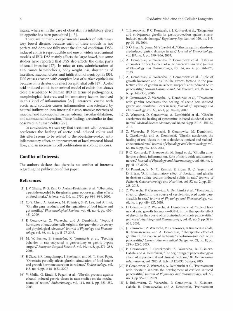
6 Oxidative Medicine and Cellular Longevity
intake, whereas, in the case of obestatin, its inhibitory effecton appetite has been postulated [1–3].
There are numerous experimental models of inflamma-tory bowel disease, because each of these models is notperfect and does not fully meet the clinical condition. DSS-induced colitis is reproducible and one of widely used animalmodels of IBD. DSS mainly affects the large bowel, but somestudies have reported that DSS also affects the distal partsof small intestine [27]. In mice or rats, administration ofDSS causes hematochezia, body weight loss, shortening ofintestine, mucosal ulcers, and infiltration of neutrophils [53].DSS causes erosion with complete loss of surface epitheliumbecause of its deleterious effect on epithelial cells [27]. Aceticacid-induced colitis is an animal model of colitis that showsclose resemblance to human IBD in terms of pathogenesis,morphological features, and inflammatory factors involvedin this kind of inflammation [27]. Intrarectal enema withacetic acid solution causes inflammation characterized byneutral infiltration into intestinal tissue, massive necrosis ofmucosal and submucosal tissues, edema, vascular dilatation,and submucosal ulceration.Those findings are similar to thatobserved in human colitis [27].
In conclusion, we can say that treatment with obestatinaccelerates the healing of acetic acid-induced colitis andthis effect seems to be related to the obestatin-evoked anti-inflammatory effect, an improvement of local mucosal bloodflow, and an increase in cell proliferation in colonic mucosa.
Conflict of Interests
The authors declare that there is no conflict of interestsregarding the publication of this paper.
References
[1] J. V. Zhang, P.-G. Ren, O. Avsian-Kretchmer et al., “Obestatin,a peptide encoded by the ghrelin gene, opposes ghrelin’s effectson food intake,” Science, vol. 310, no. 5750, pp. 996–999, 2005.
[2] C.-Y. Chen, A. Asakawa, M. Fujimiya, S.-D. Lee, and A. Inui,“Ghrelin gene products and the regulation of food intake andgut motility,” Pharmacological Reviews, vol. 61, no. 4, pp. 430–481, 2009.
[3] P. Ceranowicz, Z. Warzecha, and A. Dembinski, “Peptidylhormones of endocrine cells origin in the gut—their discoveryand physiological relevance,” Journal of Physiology and Pharma-cology, vol. 66, no. 1, pp. 11–27, 2015.
[4] M. W. Furnes, B. Stenström, K. Tømmerås et al., “Feedingbehavior in rats subjected to gastrectomy or gastric bypasssurgery,” European Surgical Research, vol. 40, no. 3, pp. 279–288,2008.
[5] P. Zizzari, R. Longchamps, J. Epelbaum, and M. T. Bluet-Pajot,“Obestatin partially affects ghrelin stimulation of food intakeand growth hormone secretion in rodents,” Endocrinology, vol.148, no. 4, pp. 1648–1653, 2007.
[6] V. Sibilia, G. Rindi, F. Pagani et al., “Ghrelin protects againstethanol-induced gastric ulcers in rats: studies on the mecha-nisms of action,” Endocrinology, vol. 144, no. 1, pp. 353–359,2003.
[7] T. Brzozowski, P. C. Konturek, S. J. Konturek et al., “Exogenousand endogenous ghrelin in gastroprotection against stress-induced gastric damage,” Regulatory Peptides, vol. 120, no. 1–3,pp. 39–51, 2004.
[8] S. Ö. Íşeri, G. Şener, M. Yüksel et al., “Ghrelin against alendron-ate-induced gastric damage in rats,” Journal of Endocrinology,vol. 187, no. 3, pp. 399–406, 2005.
[9] A. Dembinski, Z. Warzecha, P. Ceranowicz et al., “Ghrelinattenuates the development of acute pancreatitis in rats,” Journalof Physiology and Pharmacology, vol. 54, no. 4, pp. 561–573,2003.
[10] A. Dembiński, Z. Warzecha, P. Ceranowicz et al., “Role ofgrowth hormone and insulin-like growth factor-1 in the pro-tective effect of ghrelin in ischemia/reperfusion-induced acutepancreatitis,” Growth Hormone and IGF Research, vol. 16, no. 5-6, pp. 348–356, 2006.
[11] P. Ceranowicz, Z. Warzecha, A. Dembinski et al., “Treatmentwith ghrelin accelerates the healing of acetic acid-inducedgastric and duodenal ulcers in rats,” Journal of Physiology andPharmacology, vol. 60, no. 1, pp. 87–98, 2009.
[12] Z. Warzecha, D. Ceranowicz, A. Dembiński et al., “Ghrelinaccelerates the healing of cysteamine-induced duodenal ulcersin rats,”Medical ScienceMonitor, vol. 18, no. 5, pp. BR181–BR187,2012.
[13] Z. Warzecha, P. Kownacki, P. Ceranowicz, M. Dembinski,J. Cieszkowski, and A. Dembinski, “Ghrelin accelerates thehealing of oral ulcers in non-sialoadenectomized and siaload-enectomized rats,” Journal of Physiology and Pharmacology, vol.64, no. 5, pp. 657–668, 2013.
[14] P. C. Konturek, T. Brzozowski, M. Engel et al., “Ghrelin ame-liorates colonic inflammation. Role of nitric oxide and sensorynerves,” Journal of Physiology and Pharmacology, vol. 60, no. 2,pp. 41–47, 2009.
[15] O. Pamukcu, Z. N. O. Kumral, F. Ercan, B. Ç. Yegen, andD. Ertem, “Anti-inflammatory effect of obestatin and ghrelinin dextran sulfate sodium-induced colitis in rats,” Journal ofPediatric Gastroenterology and Nutrition, vol. 57, no. 2, pp. 211–218, 2013.
[16] Z. Warzecha, P. Ceranowicz, A. Dembinski et al., “Therapeuticeffect of ghrelin in the course of cerulein-induced acute pan-creatitis in rats,” Journal of Physiology and Pharmacology, vol.61, no. 4, pp. 419–427, 2010.
[17] D. Ceranowicz, Z. Warzecha, A. Dembinski et al., “Role of hor-monal axis, growth hormone—IGF-1, in the therapeutic effectof ghrelin in the course of cerulein-induced acute pancreatitis,”Journal of Physiology and Pharmacology, vol. 61, no. 5, pp. 599–606, 2010.
[18] J. Bukowczan, Z.Warzecha, P. Ceranowicz, B. Kusnierz-Cabala,R. Tomaszewska, and A. Dembinski, “Therapeutic effect ofghrelin in the course of ischemia/reperfusion-induced acutepancreatitis,” Current Pharmaceutical Design, vol. 21, no. 17, pp.2284–2290, 2015.
[19] P. Ceranowicz, J. Cieszkowski, Z. Warzecha, B. Kuśnierz-Cabala, andA. Dembiński, “The beginnings of pancreatology asa field of experimental and clinical medicine,” BioMed ResearchInternational, vol. 2015, Article ID 128095, 5 pages, 2015.
[20] P. Ceranowicz, Z. Warzecha, A. Dembinski et al., “Pretreatmentwith obestatin inhibits the development of cerulein-inducedpancreatitis,” Journal of Physiology and Pharmacology, vol. 60,no. 3, pp. 95–101, 2009.
[21] J. Bukowczan, Z. Warzecha, P. Ceranowicz, B. Kuśnierz-Cabala, R. Tomaszewska, and A. Dembinski, “Pretreatment
Oxidative Medicine and Cellular Longevity 7
with obestatin reduces the severity of ischemia/reperfusion-induced acute pancreatitis in rats,” European Journal of Phar-macology, vol. 760, pp. 113–121, 2015.
[22] R. Granata, F. Settanni, D. Gallo et al., “Obestatin promotessurvival of pancreatic 𝛽-cells and human islets and inducesexpression of genes involved in the regulation of𝛽-cellmass andfunction,” Diabetes, vol. 57, no. 4, pp. 967–979, 2008.
[23] A. Dembiński, Z. Warzecha, P. Ceranowicz et al., “Adminis-tration of obestatin accelerates the healing of chronic gastriculcers in rats,”Medical ScienceMonitor, vol. 17, no. 8, pp. BR196–BR200, 2011.
[24] E. Alexandridis, A. Zisimopoulos, N. Liratzopoulos, I. Katsos,K. Manolas, and G. Kouklakis, “Obestatin/ghrelin ratio: a newactivity index in inflammatory bowel diseases,” InflammatoryBowel Diseases, vol. 15, no. 10, pp. 1557–1561, 2009.
[25] J. Y. Jung, J. B. Jeong, J. W. Kim et al., “Circulating ghrelin levelsand obestatin/ghrelin ratio as a marker of activity in ulcerativecolitis,” Intestinal Research, vol. 13, no. 1, pp. 68–73, 2015.
[26] B. R. MacPherson and C. J. Pfeiffer, “Experimental productionof diffuse colitis in rats,” Digestion, vol. 17, no. 2, pp. 135–150,1978.
[27] P. K. Randhawa, K. Singh, N. Singh, and A. S. Jaggi, “A reviewon chemical-induced inflammatory bowel disease models inrodents,” Korean Journal of Physiology and Pharmacology, vol.18, no. 4, pp. 279–288, 2014.
[28] K. Johansson, H. Ahn, J. Lindhagen, and O. Lundgren, “Tissuepenetration and measuring depth of laser doppler flowmetryin the gastrointestinal application,” Scandinavian Journal ofGastroenterology, vol. 22, no. 9, pp. 1081–1088, 1987.
[29] A. Dembiński, Z. Warzecha, S. J. Konturek, R. Z. Cai, and A.V. Schally, “The effects of antagonists of receptors for gastrin,cholecystokinin and bombesin on growth of gastroduodenalmucosa and pancreas,” Journal of Physiology and Pharmacology,vol. 42, no. 2, pp. 195–209, 1991.
[30] P. P. Bradley, D. A. Priebat, R. D. Christensen, and G. Rothstein,“Measurement of cutaneous inflammation: estimation of neu-trophil content with an enzyme marker,” Journal of InvestigativeDermatology, vol. 78, no. 3, pp. 206–209, 1982.
[31] P. Greant, G. Delvaux, and G. Willems, “Influence of stress onepithelial cell proliferation in the gut mucosa of rats,”Digestion,vol. 40, no. 4, pp. 212–218, 1988.
[32] P. A. Hall, P. J. Coates, B. Ansari, and D. Hopwood, “Regulationof cell number in the mammalian gastrointestinal tract: theimportance of apoptosis,” Journal of Cell Science, vol. 107, no. 12,pp. 3569–3577, 1994.
[33] S. J. Konturek, A. Dembinski, Z. Warzecha, T. Brzozowski, andH. Gregory, “Role of epidermal growth factor in healing ofchronic gastroduodenal ulcers in rats,”Gastroenterology, vol. 94,no. 6, pp. 1300–1307, 1988.
[34] T. Brzozowski, P. C. Konturek, S. J. Konturek et al., “Effect oflocal application of growth factors on gastric ulcer healing andmucosal expression of cyclooxygenase-1 and -2,” Digestion, vol.64, no. 1, pp. 15–29, 2001.
[35] S. Beckert, N. Class, F. Farrahi, and S. Coerper, “Growthhormone enhances gastric ulcer healing in rats,”Medical ScienceMonitor, vol. 10, no. 8, pp. BR255–BR258, 2004.
[36] D. K. Podolsky, “Regulation of intestinal epithelial prolifer-ation: a few answers, many questions,” American Journal ofPhysiology—Gastrointestinal and Liver Physiology, vol. 264, no.2, pp. G179–G186, 1993.
[37] H. Sørbye and K. Svanes, “The role of blood flow in gastricmucosal defence, damage and healing.,” Digestive Diseases, vol.12, no. 5, pp. 305–317, 1994.
[38] R. C.Orlando, “The integrity of the esophagealmucosa. Balancebetween offensive and defensivemechanisms,” Best Practice andResearch: Clinical Gastroenterology, vol. 24, no. 6, pp. 873–882,2010.
[39] F. W. Leung, K. C. Su, J. M. Pique, G. Thiefin, E. Passaro Jr.,and P. H. Guth, “Superior mesenteric artery is more importantthan inferior mesenteric artery in maintaining colonic mucosalperfusion and integrity in rats,” Digestive Diseases and Sciences,vol. 37, no. 9, pp. 1329–1335, 1992.
[40] C. A. Dinarello, “Immunological and inflammatory functionsof the interleukin-1 family,” Annual Review of Immunology, vol.27, pp. 519–550, 2009.
[41] C. A. Dinarello, A. Simon, and J. W. M. van der Meer, “Treatinginflammation by blocking interleukin-1 in a broad spectrum ofdiseases,”Nature Reviews Drug Discovery, vol. 11, no. 8, pp. 633–652, 2012.
[42] J. Norman, M. Franz, J. Messina et al., “Interleukin-1 receptorantagonist decreases severity of experimental acute pancreati-tis,” Surgery, vol. 117, no. 6, pp. 648–655, 1995.
[43] C.A.Dinarello, “A clinical perspective of IL-1𝛽 as the gatekeeperof inflammation,” European Journal of Immunology, vol. 41, no.5, pp. 1203–1217, 2011.
[44] H. D. De Koning, J. Schalkwijk, J. van der Ven-Jongekrijg,M. Stoffels, J. W. M. van der Meer, and A. Simon, “Sustainedefficacy of the monoclonal anti-interleukin-1 beta antibodycanakinumab in a 9-month trial in Schnitzler’s syndrome,”Annals of the Rheumatic Diseases, vol. 72, no. 10, pp. 1634–1638,2013.
[45] T. Herlin, B. Fiirgaard, M. Bjerre et al., “Efficacy of anti-IL-1 treatment in Majeed syndrome,” Annals of the RheumaticDiseases, vol. 72, no. 3, pp. 410–413, 2013.
[46] S. J. Klebanoff, “Myeloperoxidase: friend and foe,” Journal ofLeukocyte Biology, vol. 77, no. 5, pp. 598–625, 2005.
[47] F. J. Romero, F. Bosch-Morell, M. J. Romero et al., “Lipidperoxidation products and antioxidants in human disease,”EnvironmentalHealth Perspectives, vol. 106, no. 5, pp. 1229–1234,1998.
[48] M. D. Evans and M. S. Cooke, “Factors contributing to theoutcome of oxidative damage to nucleic acids,” BioEssays, vol.26, no. 5, pp. 533–542, 2004.
[49] S. Kwiecien, K. Jasnos, M. Magierowski et al., “Lipid perox-idation, reactive oxygen species and antioxidative factors inthe pathogenesis of gastric mucosal lesions and mechanismof protection against oxidative stress—induced gastric injury,”Journal of Physiology and Pharmacology, vol. 65, no. 5, pp. 613–622, 2014.
[50] K. M. Mullane, R. Kraemer, and B. Smith, “Myeloperoxidaseactivity as a quantitative assessment of neutrophil infiltrationinto ischemie myocardium,” Journal of Pharmacological Meth-ods, vol. 14, no. 3, pp. 157–167, 1985.
[51] Z. Warzecha and A. Dembiński, “Protective and therapeuticeffects of ghrelin in the gut,” Current Medicinal Chemistry, vol.19, no. 1, pp. 118–125, 2012.
[52] B. Holst, K. L. Egerod, E. Schild et al., “GPR39 signaling isstimulated by zinc ions but not by obestatin,”Endocrinology, vol.148, no. 1, pp. 13–20, 2007.
[53] A. R. Jurjus, N. N. Khoury, and J.-M. Reimund, “Animal modelsof inflammatory bowel disease,” Journal of Pharmacological andToxicological Methods, vol. 50, no. 2, pp. 81–92, 2004.