invasive pneumococcal disease in children can reveal a primary immunodeficiency
TRANSCRIPT

Acce
pted M
anus
cript
1
© The Author 2014. Published by Oxford University Press on behalf of the Infectious Diseases Society of America. All rights reserved. For Permissions, please e-mail: [email protected].
Invasive pneumococcal disease in children can reveal a primary immunodeficiency Jean Gaschignard1,2,3, Corinne Levy3,4,5, Maya Chrabieh1,2, Bertrand Boisson6, Cécile Bost-Bru7, Stéphane Dauger8, François Dubos3,9, Philippe Durand10, Joël Gaudelus3,11, Dominique Gendrel3,12, Christèle Gras Le Guen3,13, Emmanuel Grimprel3,14, Gaël Guyon15, Catherine Jeudy16, Eric Jeziorski15, Francis Leclerc17, Pierre-Louis Léger14, Fabrice Lesage18, Mathie Lorrot19, Isabelle Pellier16, Didier Pinquier3,20, Loïc de Pontual3,11, Philippe Sachs8, Caroline Thomas21, Pierre Tissières10, Frédéric V. Valla22, Philippe Desprez23, Véronique Frémeaux-Bacchi24, Emmanuelle Varon3,25, Xavier Bossyut26, Robert Cohen3,4,5, Laurent Abel1,2,6, Jean-Laurent Casanova1,2,6,27,28, Anne Puel1,2, Capucine Picard1,2,27,29
1Laboratory of Human Genetics of Infectious Diseases, Necker Branch, Institut National de la Santé et de la Recherche Médicale, UMR1163, Paris, France, EU 2University Paris Descartes, Sorbonne Paris Cité, Imagine Institute, Paris, France, EU 3GPIP, Pediatric Infectious Disease Group, France, EU 4ACTIV, Pediatric Clinical and Therapeutic Association of the Val de Marne, Saint-Maur, France, EU 5Clinical Research Center (CRC), Centre Hospitalier Intercommunal de Créteil, Créteil, France, EU 6St. Giles Laboratory of Human Genetics of Infectious Diseases, Rockefeller Branch, the Rockefeller University, New York, NY, USA 7Pediatric Unit, Grenoble Hospital, Grenoble, France, EU 8Pediatric Intensive Care Unit, Robert Debré Hospital, Assistance Publique-Hôpitaux de Paris Paris (APHP), France, EU 9Pediatric Emergency and Infectious Diseases Unit, CHRU, Lille University Hospital, UDSL, Lille, France, EU 10Pediatric Intensive Care Unit, Kremlin Bicêtre Hospital, APHP, Kremlin Bicêtre, France, EU 11Pediatric Unit, Jean Verdier Hospital, APHP, Bondy, France, EU 12Pediatric Unit, Necker Hospital, APHP, Paris, France, EU
13Pediatric Unit, Nantes Hospital, Nantes, France, EU 14Pediatric and Neonatal Intensive Care Units, Armand Trousseau Hospital, Paris, France, EU 15Pediatric Unit, Montpellier Hospital, Montpellier, France, EU 16Pediatric Onco-Hematology Unit, Angers Hospital, Angers, France, EU 17Pediatric Intensive Care Unit, CHRU, Lille University Hospital, UDSL, Lille, France, EU
18Pediatric Intensive Care Unit, Necker Hospital, Paris, France, EU
19Pediatric Unit, Robert Debré Hospital, APHP, Paris, France, EU 20Pediatric Intensive Care Unit, Rouen Hospital, Rouen, France, EU 21Pediatric Intensive Care and Onco-Hematology Units, Nantes Hospital, Nantes, France, EU 22Pediatric Intensive Care Unit - Hôpital Femme Mère Enfant, Lyon, France, EU 23Pediatric Intensive Care Unit – Hôpital Hautepierre, Strasbourg, France, EU 24Immunology Laboratory, Georges Pompidou European Hospital, APHP, Paris, France, EU 25National Reference Center for Pneumococci, Microbiology Laboratory, Georges Pompidou European Hospital, APHP, Paris, France, EU 26Laboratory Medicine, University Hospitals Leven and Experimental Laboratory Immunology, Department Microbiology and Immunology, Catholic University Leuven, Herestraat 49, 3000 Leuven, Belgium, EU 27Pediatric Hematology-Immunology Unit, Necker-Enfants Malades Hospital, Paris, France, EU 28Howard Hughes Medical Institute, New York, NY USA
Clinical Infectious Diseases Advance Access published April 23, 2014 at R
ockefeller University on M
ay 13, 2014http://cid.oxfordjournals.org/
Dow
nloaded from

Acce
pted M
anus
cript
2
29Center for the Study of Primary Immunodeficiencies, APHP, Necker Hospital, Paris, France, EU Corresponding author: Capucine Picard, MD, PhD, Laboratory of Human Genetics of
Infectious Diseases, INSERM U.980, Necker Medical School, IMAGINE Foundation, 75015
Paris, France, UE Phone: 33 1 44 49 50 88, Fax: 33 1 42 73 06 40, E-mail:
Short summary:
The prospective inclusion of 163 children hospitalized in France for an invasive
pneumococcal disease revealed that at least 10% of these children had a primary
immunodeficiency. We advocate systematic immunological exploration for all children
hospitalized for an invasive pneumococcal disease.
at Rockefeller U
niversity on May 13, 2014
http://cid.oxfordjournals.org/D
ownloaded from

Acce
pted M
anus
cript
3
Abstract Background: About 10% of pediatric patients with invasive pneumococcal disease (IPD) die
from the disease. Some primary immunodeficiencies (PIDs) are known to confer
predisposition to IPD. However, a systematic search for these PIDs has never been carried out
in children presenting with IPD.
Methods: We prospectively collected pediatric cases of IPD requiring hospitalization,
between 2005 and 2011, in 28 pediatric wards throughout France. IPD was defined as a
positive pneumococcal culture, PCR result and/or soluble antigen detection at a normally
sterile site. The immunological assessment included abdominal ultrasound, whole-blood
counts and smears, determinations of plasma Ig and complement levels, and the evaluation of
pro-inflammatory cytokines.
Results: We included a total of 163 children with IPD (M/F sex ratio: 1.3, median age: 13
months). Seventeen children had recurrent IPD. Meningitis was the most frequent type of
infection (87%), others were pleuropneumonitis, isolated bloodstream infection,
osteomyelitis, endocarditis and mastoiditis. One patient with recurrent meningitis had a
congenital cerebrospinal fluid fistula. The results of immunological explorations were
abnormal in 26 children (16%) and a PID was identified in 17 patients (10%), including one
case of MyD88 deficiency, three of complement fraction C2 or C3 deficiencies, one of
isolated congenital asplenia and two of Bruton’s agammaglobulinemia. The proportion of
PIDs was much higher in children older than two years than in younger children (26% vs 2%,
p < 0.001).
Conclusions: Children with IPD should undergo immunological investigations, particularly
those older than two years, as PIDs may be discovered in up to 26% of cases.
at Rockefeller U
niversity on May 13, 2014
http://cid.oxfordjournals.org/D
ownloaded from

Acce
pted M
anus
cript
4
Introduction
Streptococcus pneumoniae is an almost ubiquitous Gram-positive encapsulated
bacterium. Cross-sectional studies have shown that, at any given time, 20 to 50 % of healthy
children harbor at least one serotype of this bacterium in the rhinopharynx, whereas
longitudinal have reported that almost all children can be shown to carry this bacterial species
1,2. S. pneumoniae is a leading cause of benign conditions, such as otitis media 3, but may also
cause more serious illnesses, such as pneumonitis 4, and it therefore constitutes a public health
burden worldwide for young children 5. In rare cases, S. pneumoniae may even cause severe,
life-threatening diseases, such as arthritis 6, peritonitis 7, septicemia and meningitis 8,
collectively known as invasive pneumococcal disease (IPD) 9. The greatest risk factor for IPD
in children, apart from young age, is IPD itself, as this condition has a recurrence rate of 2 to
4% 10. The rarity of IPD, despite the asymptomatic carriage of S. pneumoniae, strongly
suggests that host factors play a key role in the pathogenesis of this disease 11–14. Indeed, a
number of acquired and inherited host factors conferring predisposition to IPD have already
been identified. The best known acquired factors determining susceptibility to recurrent IPD
are co-infection with human immunodeficiency virus 9,15 and splenectomy 16. Other acquired
factors include certain cancers 17 and traumatic cerebrospinal fluid fistulas 18–20. Host genetic
factors have also long been known to confer predisposition to IPD, as in sickle cell disease 21
or certain primary immunodeficiencies (PIDs) 22.
PIDs known to underlie clinical disease caused by encapsulated bacteria, such as S.
pneumoniae, include congenital asplenia 23,24, complement deficiency 22,25, and antibody
deficiency 22. Fourteen years ago, hypomorphic mutations of NEMO were identified in
patients with anhidrotic ectodermal dysplasia with immunodeficiency (EDA-ID) 26,27. In these
patients, impaired NF-κB signaling results in susceptibility to multiple pathogens, including S.
pneumoniae in particular 28,29. The molecular genetic basis of a more selective predisposition
to IPD remained elusive until the identification of children with mutations of IRAK4 or
at Rockefeller U
niversity on May 13, 2014
http://cid.oxfordjournals.org/D
ownloaded from

Acce
pted M
anus
cript
5
MYD88 genes 30,31. These two genes encode proteins specifically involved in the Toll-like and
interleukin receptors (TIRs) superfamily signaling pathway. However, the pathogenesis of
IPD in most children remains unknown. Sporadic, isolated childhood IPD, which is generally
described as “idiopathic”, may be favored by undetected, underlying PIDs conferring a
selective predisposition to IPD. The proportion of children presenting IPD with known
inherited susceptibility factors, including conventional PIDs in particular, has never been
precisely evaluated. In 2005, we set up a collaboration with GPIP/ACTIV to address this
question, by creating an observatory comprising French pediatric hospitals. These hospitals
were asked to collect blood samples prospectively from children who had recovered from
IPD. In this study, we aimed to carry out systematic explorations of the immunity of
hospitalized children with IPD, by looking for both known and unknown PIDs.
Methods
Definitions and data collection
The National Observatory for IPD was created by the GPIP/ACTIV network and
brings together 28 pediatric units across France, which included all children hospitalized for
IPD. Children with sickle cell disease and HIV-positive children were excluded. Children
who died were included whenever immunological explorations had been performed before
death. IPD was defined as an infection requiring hospitalization confirmed by the isolation of
S. pneumoniae from a normally sterile site (blood, cerebrospinal fluid, synovial fluid, pleural
fluid, but not sputum). If pneumococcus was isolated from blood, signs of hemodynamic
shock were required for inclusion.
The following data were collected: family and patient history, pneumococcal
vaccination status, clinical signs, microbiological data, abdominal ultrasound, whole-blood
cell (WBC) counts with smears, and determinations of immunoglobulins (Ig) and
complement. Most children also underwent IL-6 determinations after stimulation. All children
at Rockefeller U
niversity on May 13, 2014
http://cid.oxfordjournals.org/D
ownloaded from

Acce
pted M
anus
cript
6
with recurrent meningitis underwent brain MRI to rule out the possibility of cerebrospinal
fistula. Children with normal results for abdominal ultrasound, WBC counts, smears, plasma
Ig levels, classical and alternative complement studies and evaluations of IL-6 production by
WBC were considered not to have a PID. If any of these examinations had not been carried
out or the results were missing, the patient was classified in the “incomplete exploration”
category. We considered two time periods: 2005-2009 (before the introduction of the 13-
valent pneumococcal vaccine - PCV13) and 2010-2011 (after PCV13 implementation), 2010
being the year in which French national authorities recommended switching from PCV7 to
PCV13 for antipneumococcal immunization.
Microbiology
S. pneumoniae was identified, by standard methods, in the microbiology laboratories
of the various participating hospitals. Serotyping was performed with latex particles sensitized
with antisera purchased from the Statens Serum Institut (Copenhagen, Denmark), mostly at
the national reference center.
Immunological explorations and whole-blood activation
Classical and alternative complement tests were carried out with standard techniques,
by ELISA (Wielisa®-kit, Lund Swenden) or nephelometry (Dade Behring, Paris La Defense,
France). Whenever complete lack of CH50 or AP50 was identified, genetic tests to determine
which component was missing were carried out. The IgG subclasses were assessed by ELISA
methods 32. Total IgG antibody levels against multiple pneumococcal serotypes (23 serotypes)
were assessed by multiplexed bead assay or standard ELISA techniques (binding site assay,
Birmingham, UK) 33. Children with a persisting defect in the production of antibodies specific
for pneumococcal capsular antigens with normal Ig after the age of two years were considered
to have “specific polysaccharides antibody deficiency” (SPAD). Patient’s blood sample was
at Rockefeller U
niversity on May 13, 2014
http://cid.oxfordjournals.org/D
ownloaded from

Acce
pted M
anus
cript
7
diluted 1:2 in RPMI 1640 medium (Gibco-BRL, Invitrogen, USA) and incubated at 37°C in
several different sets of conditions for 48h: with medium alone, with 20 ng/ml IL-1β (R&D
Systems), 1 ng/ml LPS (Sigma, USA), 100 µg/m PAM2CSK4 (Invivogen, USA), 107
particles/ml SAC (Invivogen, USA), 5 x 10⁶ particles/ml heat-killed pneumococcal strains or
10-7M PMA (Sigma, USA) and 10-5M ionomycin (Sigma, USA). Supernatants were collected
and IL-6 levels were assessed by ELISA (Sanquin kit), according to the kit manufacturer’s
instructions and read with an MRX microplate reader (Thermo Lab Systems, USA).
Statistical analysis
We used standard statistical tests implemented in R v2.15.1 software to compare
categorical variables and means. Values of P<0.05 were considered statistically significant.
Ethics
This study was conducted in accordance with the Helsinki Declaration, with informed
consent obtained from each patient or the patient’s family. The study was approved by the
local ethics committee of Necker-Enfants Malades Hospital, Paris, France.
Results
Epidemiology
Between January 2005 and December 2011, 163 pediatric cases of IPD were collected.
Seven hospitals reported 10 or more cases and four hospitals included between five and nine
children. The M/F sex ratio was 1.3 (91 male and 72 female patients, p=0.14). Median age
was 13 months (range 1 month – 15 years), with a peak in the number of cases between the
ages or three and six months. There was seasonal variation in the number of cases, with a
peak in February. Meningitis was the most frequent type of infection in this cohort (n=142;
87%), followed by pleuropneumonitis (n=11; 7%), bloodstream infection (n=6; 4%),
at Rockefeller U
niversity on May 13, 2014
http://cid.oxfordjournals.org/D
ownloaded from

Acce
pted M
anus
cript
8
osteomyelitis (n=1), arthritis (n=1), endocarditis (n=1) and mastoiditis (n=1). Seventeen
patients (10%) had recurrent IPD. The proportion of patients with meningitis seemed to be
higher in children under the age of two years (n=98/109, 90%) than in older children
(n=44/54, 81%) (p = 0.21, ns). Overall vaccination coverage with the conjugate vaccine
against S. pneumoniae was 71% for the children included in the study (n=114/160, data
missing in three cases). Vaccination coverage increased from 69% (n=81/118) during the
2005-2009 period to 79% (n=33/42) during the 2010-2011 period (p=0.31, ns). This trend was
observed both in children under the age of two years and in children over the age of two
years: 81% and 41%, respectively, in 2005-2009, increasing to 88% and 65%, respectively, in
2010-2011 (ns) (Table 1).
Ten patients were from families known to be consanguineous. No underlying
conditions (including PIDs) had previously been identified in these children. However, 17
patients had already been hospitalized for IPD: 11 for meningitis, four for pleuro/pneumonitis,
one for osteoarthritis and one for septicemia. In addition, four children had previously
presented with another severe infectious disease: one with regional BCG-itis, one with
encephalopathy due to Mycoplasma pneumoniae and two with ethmoiditis for which the
causal microbe was not identified but was probably bacterial, given the response to antibiotic
treatment observed. Nine children (6%) between the ages of three months and nine years died,
all secondary to pneumococcal meningitis (recurrent in one case). We identified a
cerebrospinal fluid fistula in an eight-year-old girl hospitalized for her third episode of
meningitis due to S. pneumoniae, on brain MRI, despite the normal results obtained for a
brain scan after the second meningitis.
Bacteriology
Infection was documented by culture (154 cases), PCR (two cases) or the presence of
soluble antigens (seven cases), and S. pneumoniae serotype was available for 130 isolates
at Rockefeller U
niversity on May 13, 2014
http://cid.oxfordjournals.org/D
ownloaded from

Acce
pted M
anus
cript
9
(Table 1). Nine children correctly vaccinated for age developed infection due to a serotype
included in the vaccine (five for PCV7 and four for PCV13). The proportion of infections due
to a serotype included in the PCV7 vaccine decreased between the 2005-2009 and 2010-2011
periods (n=12/100 to 1/30, ns), as did the proportion of infections due to serotype 19A after
the introduction of PCV13 (n=30/100 vs. 5/30, ns). Overall, in this study, the serotypes
included in the PCV7 vaccine accounted for 10% (n=13/130) of infections, and those included
in the PCV13 vaccine accounted for 63% of infections (n=82/130). The distribution of
serotypes differed between children under and over the age of two years, with a higher
proportion of children over the age of two years presenting infections with serotypes included
in the PCV7 vaccine (n=4/91 vs. 9/39, p<0.01). Patients with and without PID were infected
equally frequently with non-vaccine serotypes (8/20 vs. 39/110, p=0.89 – serotype data
missing for five children in the PID group and 28 children in the non-PID group).
Immunology
All immunological explorations were carried out for 127 patients, whereas these
explorations were incomplete for 35 patients (Table 1). The patient with the cerebrospinal
fluid fistula was excluded from the immunological analysis. Abnormal immunological
responses were discovered in 26 children (16%) (Table 2). These patients included 17 (11%)
displaying a persistent immunodeficiency after the age of two years. One patient displayed
impaired IL-6 production in response to TIR agonists. This led to the identification of the first
case of MyD88 deficiency in this 11-month-old boy 31. This patient had previously had
ethmoiditis, at the age of eight months. Low complement levels led to the identification of
three genetically confirmed complement (two C2 and one C3) deficiencies in children aged
one, three and seven years. Blood smears revealed the presence of Howell-Jolly bodies in a
three-year-old boy with meningitis, and isolated congenital asplenia in this patient was
confirmed by an abdominal ultrasound scan and splenic scintigraphy.
at Rockefeller U
niversity on May 13, 2014
http://cid.oxfordjournals.org/D
ownloaded from
Acce
pted M
anus
cript
10
Twelve children had a primary antibody deficiency: two boys aged seven and four had
Bruton’s disease (XLA) with a hemizygous BTK mutation, four children had
hypogammaglobulinemia, two had an IgG2 production deficiency and four had a SPAD,
established after confirmation of the deficiency following immunization with pneumococcal
polysaccharide vaccine. Isolated IgG deficiency was identified in nine children under the age
of two years considered to have “transient IgG deficiency”, either because spontaneous
correction of the deficiency was observed after the age of two years, during follow-up, or
because these children have not yet been checked. The cases of transient deficiency included
four children with impaired total Ig production, one with impaired IgG2 production, one with
IgG3 deficiency, two children aged 12 and 18 months with combined IgG2 and IgG4 subclass
deficiencies and a 14-month-old child had both IgG and IgA deficiencies. Finally, eight
children had isolated low levels of IgA production (below the 3rd percentile for age); six of
them were younger than one year, and the two remaining were two and five years old. These
patients were not included in the “abnormal immunological responses” group.
PIDs were more frequently detected in children over the age of two years (1/53, 26%)
than in younger ones (3/109, 3%) (p < 0.001). Immunological deficiencies other than those
affecting antibodies were rare among children over the age of two years. PIDs were detected
more frequently in boys than in girls (15/91 vs. 2/71, p < 0.01), even when X-linked defects
were retrieved, and this trend persisted when both transient and permanent deficiencies were
considered (20/91 for boys vs. 6/71 for girls, p=0.03). Patients from consanguineous families
were more likely to have a PID than patients from non-consanguineous families (4/10 vs.
13/152, p=0.01). The proportion of patients with detectable PIDs did not differ significantly
between the pleuropneumonitis group (3/11) and the meningitis group (23/141) (p=0.40).
Finally, the proportion of patients found to have a PID was similar in children hospitalized for
at Rockefeller U
niversity on May 13, 2014
http://cid.oxfordjournals.org/D
ownloaded from

Acce
pted M
anus
cript
11
their first episode of IPD (15/146) and in children hospitalized for recurrent IPD (2/16)
(p=0.68) (Table 3). Follow-up of patients is available in the Supplementary data.
Discussion
We provide the first detailed description of the immunological features of children
presenting with IPD. All but 16 of the patients were included after a first episode of
pneumococcal infection, meningitis in most cases. Between 2005 and 2008, the incidence of
IPD in France was 23 and 5/100,000 in children under and over the age of two years,
respectively 35. Over this period, we included 94 children (59 under and 35 over the age of
two years). These figures are similar to those reported in other developed countries, such as
the USA 36, Scotland 37 and Australia 38. The introduction of conjugated pneumococcal
vaccines has reduced the burden of IPD in developed countries, in both vaccinated and
unvaccinated populations (due to herd immunity) 39–41. Routine infant immunization with
PCV7 was introduced in France in 2006, 42 and there was a switch to the use of PCV13 in
2010. Vaccination coverage in children of two to three years of age had reached almost 93%
by 2011 43. Although not designed for this purpose, our study highlighted a clear impact of
pneumococcal vaccination on serotype distribution, with the serotypes included in the PCV7
vaccine accounting for only 10% of the cases of IPD included in this report. This study was,
nevertheless, subject to several limitations. First, the non-inclusion of some of the children
who died made it impossible to determine the true proportion of children suffering from IPD
with a PID. This proportion may exceed 10%, as the prognosis of IPD might be more severe
in children with PIDs. Second, mortality in our cohort (n=9/162, 6%) was below the 12%
overall death rate following IPD in the most active hospitals (those including more than five
patients) (n=23/194, p=0.04, data not shown) and below the 11% fatality rate after
pneumococcal meningitis observed in France between 2001 and 2008 44. Third, there was
probably an overrepresentation of meningitis and an underrepresentation of sepsis in our
at Rockefeller U
niversity on May 13, 2014
http://cid.oxfordjournals.org/D
ownloaded from

Acce
pted M
anus
cript
12
cohort, because most of the patients were included during/after a stay in a pediatric intensive
care unit.
In our study, 10% of children with IPD were found to have a PID, and this proportion
rose to 26% in children over the age of two years. The main deficiency identified was primary
antibody deficiency (12 patients, including two XLA patients), followed by innate immunity
deficiencies (five cases, including two patients with C2 deficiency, one with C3 deficiency,
one with MyD88 deficiency, and one asplenic patient). The C3 and MyD88 deficiencies were
identified in patients from consanguineous kindreds. Two other children with primary
antibody deficiency and two with normal immunological findings were from consanguineous
kindreds and are currently the subject of genetic studies based on whole-exome sequencing
(WES). PID could have been suspected in four patients before the episode of IPD: the two
patients with XLA had suffered previous episodes of IPD, and the patient with MyD88
deficiency had previously had ethmoiditis. For the other 16 children with a history of severe
bacterial infection, complete immunological explorations were carried out and the results
were normal. WES will be performed for these patients, to identify new genetic disorders, as
in the discovery of many other novel PIDs 45–47,34. Eight children from our cohort were found
to have low levels of IgA: this proportion (5%, 8/162) is greater than would be expected from
findings for the general population (1/600) 48 but the clinical impact of these low levels of IgA
on infection status has yet to be determined.
The identification of new genetic etiologies of IPD by WES would provide further
evidence that IPD results from a group of single-gene inborn errors of immunity, at least in
some children 49,50. It would also make it possible to determine the proportion of pediatric IPD
cases attributable to human monogenic variants in this cohort of patients. It would, thus,
provide fundamental new insight into the genetic basis of childhood IPD at the patient and
at Rockefeller U
niversity on May 13, 2014
http://cid.oxfordjournals.org/D
ownloaded from

Acce
pted M
anus
cript
13
population levels. This information would make it possible for physicians to provide patients
with a molecular diagnosis and their families with genetic counseling. It would also provide a
basis for new avenues of treatment. Going beyond IPD, our findings should lead to a
paradigm shift in the field of PIDs, by providing proof-of-principle that a life-threatening
infectious disease striking otherwise healthy patients may actually result from a new group of
PIDs, specifically impairing immunity to S. pneumoniae. Finally, this new information should
pave the way for investigations of the molecular genetic basis of other pediatric infections. In
conclusion, we advocate systematic immunological exploration for all children hospitalized
for IPD. These explorations should include abdominal ultrasound scans, whole-blood cell
counts with blood smears, the determination of plasma Ig levels (and IgG subclasses for
patients older than two years), classical (and alternative) complement pathway analysis and
checking for a traumatic cerebrospinal fistula if the patient has been diagnosed with
meningitis, particularly in cases of recurrent meningitis. This would make it possible to detect
a non-negligible number of PIDs and to provide the affected patients with the best preventive
treatment available. However, we are fully aware that WES and, in the near future, whole-
genome sequencing, will prove to be more powerful for both the diagnosis of known genetic
etiologies and the discovery of new ones.
Acknowledgments
We thank the children and their families for participating. We thank, in particular, Pegah
Ghandil and Mélanie Migaud for technical assistance Alexandre Bolze and Josette Raymond
for fruitful advice. Reinhart Wurzner kindly provided his expertise for complement
explorations. This work was funded by GIS-maladies rares 2003, Le programme hospitalier
de recherche clinique (PHRC) 2006, Agence Nationale de la Recherche (ANR), Programme
Pluriannuel de Recherche sur les Maladies Rares (MRAR) 2007,The French National
Research Agency (ANR) under the “Investissement d’avenir” program (grant number ANR-
at Rockefeller U
niversity on May 13, 2014
http://cid.oxfordjournals.org/D
ownloaded from
Acce
pted M
anus
cript
14
10-IAHU-01),and Fondation pour la Recherche Médicale (FRM) grant number
DMI20091117320, as well as the National Institute of Allergy and Infectious Diseases grant
number P01AI061093, the March of Dimes grant number 1-FY12-440, the National Center
for Research Resources and the National Center for Advancing Sciences of the National
Institutes of Health grant number 8UL1TR000043, the St. Giles Foundation, the Rockefeller
University, INSERM, and Paris Descartes University.
The authors have no conflict of interest to declare.
at Rockefeller U
niversity on May 13, 2014
http://cid.oxfordjournals.org/D
ownloaded from

Acce
pted M
anus
cript
15
References
1. Cohen, R. et al. [Nasopharyngeal carriage of children 6 to 60 months during the implementation of the 13-valent pneumococcal conjugate vaccine]. Arch Pediatr 19, 1132–1139 (2012). 2. Sharma, D. et al. Pneumococcal carriage and invasive disease in children before introduction of the 13-valent conjugate vaccine: comparison with the era before 7-valent conjugate vaccine. Pediatr. Infect. Dis. J. 32, e45–53 (2013). 3. Coker, T. R. et al. Diagnosis, microbial epidemiology, and antibiotic treatment of acute otitis media in children: a systematic review. JAMA 304, 2161–2169 (2010). 4. Griffin, M. R., Zhu, Y., Moore, M. R., Whitney, C. G. & Grijalva, C. G. U.S. hospitalizations for pneumonia after a decade of pneumococcal vaccination. N. Engl. J. Med. 369, 155–163 (2013). 5. O’Brien, K. L. et al. Burden of disease caused by Streptococcus pneumoniae in children younger than 5 years: global estimates. Lancet 374, 893–902 (2009). 6. Ross, J. J., Saltzman, C. L., Carling, P. & Shapiro, D. S. Pneumococcal septic arthritis: review of 190 cases. Clin Infect Dis 36, 319–27. (2003). 7. Nielsen, K. R., Ejlertsen, T., El-Batran, S. & Prag, J. A five-year survey of pneumococcal peritonitis in two Danish counties--incidence, diagnosis and clinical entities. Clin. Microbiol. Infect. 9, 738–740 (2003). 8. Mook-Kanamori, B. B., Geldhoff, M., van der Poll, T. & van de Beek, D. Pathogenesis and pathophysiology of pneumococcal meningitis. Clin. Microbiol. Rev. 24, 557–591 (2011). 9. Black, S. Changing epidemiology of invasive pneumococcal disease: a complicated story. Clin. Infect. Dis. 47, 485–486 (2008). 10. Ku, C. L. et al. IRAK4 and NEMO mutations in otherwise healthy children with recurrent invasive pneumococcal disease. J Med Genet 44, 16–23 (2007). 11. Brouwer, M. C. et al. Host genetic susceptibility to pneumococcal and meningococcal disease: a systematic review and meta-analysis. Lancet Infect Dis 9, 31–44 (2009). 12. Meli, D. N., Christen, S., Leib, S. L. & Tauber, M. G. Current concepts in the pathogenesis of meningitis caused by Streptococcus pneumoniae. Curr Opin Infect Dis 15, 253–7 (2002). 13. Koedel, U., Scheld, W. M. & Pfister, H. W. Pathogenesis and pathophysiology of pneumococcal meningitis. Lancet Infect Dis 2, 721–36 (2002). 14. Mason, E. O., Jr et al. Recurrent systemic pneumococcal disease in children. Pediatr. Infect. Dis. J. 26, 480–484 (2007). 15. McEllistrem, M. C. et al. Recurrent invasive pneumococcal disease in individuals with human immunodeficiency virus infection. J Infect Dis 185, 1364–8. (2002). 16. Altamura, M. et al. Splenectomy and sepsis: the role of the spleen in the immune-mediated bacterial clearance. Immunopharmacol Immunotoxicol 23, 153–61. (2001). 17. Meisel, R. et al. Increased risk for invasive pneumococcal diseases in children with acute lymphoblastic leukaemia. Br. J. Haematol. 137, 457–460 (2007). 18. Davies, M. A. & Teo, C. Management of traumatic cerebrospinal fluid fistula. J Craniomaxillofac Trauma 1, 9–17. (1995). 19. González Doce, V. & Girón Úbeda, J. M. Pneumocephalus and pneumococcal meningitis secondary to a cerebrospinal fluid fistula. Neurologia 26, e7–8 (2011). 20. Poletti-Muringaseril, S. C., Rufibach, K., Ruef, C., Holzmann, D. & Soyka, M. B. Low meningitis-incidence in primary spontaneous compared to secondary cerebrospinal fluid rhinorrhoea. Rhinology 50, 73–79 (2012).
at Rockefeller U
niversity on May 13, 2014
http://cid.oxfordjournals.org/D
ownloaded from
Acce
pted M
anus
cript
16
21. Payne, A. B. et al. Invasive Pneumococcal Disease among Children with and without Sickle Cell Disease in the United States, 1998-2009. Pediatr. Infect. Dis. J. (2013). doi:10.1097/INF.0b013e3182a11808 22. Picard, C., Puel, A., Bustamante, J., Ku, C. L. & Casanova, J. L. Primary immunodeficiencies associated with pneumococcal disease. Curr Opin Allergy Clin Immunol 3, 451–9 (2003). 23. Gilbert, B. et al. Familial isolated congenital asplenia: a rare, frequently hereditary dominant condition, often detected too late as a cause of overwhelming pneumococcal sepsis. Report of a new case and review of 31 others. Eur J Pediatr 161, 368–72. (2002). 24. Mahlaoui, N. et al. Isolated congenital asplenia: a French nationwide retrospective survey of 20 cases. J Pediatr 158, 142–8, 148 e1 (2011). 25. Sullivan, K. E. & Winkelstein, J. A. in Primary immunodefiency diseases�: a molecular and genetic approach (Ochs, H., Edvard Smith, C. I. & Puck, J. M.) (Oxford University Press, 1999). 26. Zonana, J. et al. A novel X-linked disorder of immune deficiency and hypohidrotic ectodermal dysplasia is allelic to incontinentia pigmenti and due to mutations in IKK-gamma (NEMO). Am J Hum Genet 67, 1555–62. (2000). 27. Doffinger, R. et al. X-linked anhidrotic ectodermal dysplasia with immunodeficiency is caused by impaired NF-kappaB signaling. Nat Genet 27, 277–85. (2001). 28. Hanson, E. P. et al. Hypomorphic nuclear factor-kappaB essential modulator mutation database and reconstitution system identifies phenotypic and immunologic diversity. J Allergy Clin Immunol 122, 1169–1177 e16 (2008). 29. Picard, C., Casanova, J. L. & Puel, A. Infectious diseases in patients with IRAK-4, MyD88, NEMO, or IkappaBalpha deficiency. Clin Microbiol Rev 24, 490–7 (2011). 30. Picard, C. et al. Pyogenic bacterial infections in humans with IRAK-4 deficiency. Science 299, 2076–9. (2003). 31. Von Bernuth, H. et al. Pyogenic bacterial infections in humans with MyD88 deficiency. Science 321, 691–6 (2008). 32. Aucouturier, P. et al. Serum IgG subclass levels in patients with primary immunodeficiency syndromes or abnormal susceptibility to infections. Clin. Immunol. Immunopathol. 51, 22–37 (1989). 33. Borgers, H. et al. Laboratory diagnosis of specific antibody deficiency to pneumococcal capsular polysaccharide antigens by multiplexed bead assay. Clin. Immunol. 134, 198–205 (2010). 34. Bolze, A. et al. Ribosomal protein SA haploinsufficiency in humans with isolated congenital asplenia. Science 340, 976–978 (2013). 35. Dorleans, F. et al. Impact de la vaccination par le vaccin pneumococcique conjugue heptavalent sur l’incidence des infections invasives a pneumocoques en France. at <http://www.invs.sante.fr/presse/2010/le_point_sur/vaccination_pneumo_050710/index.html> 36. Tan, T. Q. Pediatric invasive pneumococcal disease in the United States in the era of pneumococcal conjugate vaccines. Clin. Microbiol. Rev. 25, 409–419 (2012). 37. Lamb, K. E. et al. Trends in serotypes and sequence types among cases of invasive pneumococcal disease in Scotland, 1999-2010. Vaccine (2013). doi:10.1016/j.vaccine.2013.05.079 38. Flego, K. L., Truman, G., Sheppeard, V. & Gilmour, R. E. Invasive pneumococcal disease in western Sydney, 2002-2010. N S W Public Health Bull 22, 219–221 (2011). 39. Haber, M. et al. Herd immunity and pneumococcal conjugate vaccine: a quantitative model. Vaccine 25, 5390–5398 (2007).
at Rockefeller U
niversity on May 13, 2014
http://cid.oxfordjournals.org/D
ownloaded from

Acce
pted M
anus
cript
17
40. Kaplan, S. L. et al. Early trends for invasive pneumococcal infections in children after the introduction of the 13-valent pneumococcal conjugate vaccine. Pediatr. Infect. Dis. J. 32, 203–207 (2013). 41. Davis, S., Deloria-Knoll, M., Kassa, H. T. & O’Brien, K. L. Impact of pneumococcal conjugate vaccines on nasopharyngeal carriage and invasive disease among unvaccinated people: Review of evidence on indirect effects. Vaccine (2013). doi:10.1016/j.vaccine.2013.05.005 42. Anonymous. French vaccinal calendar. Bull Epidemiol Hebd 29-30, 212–224 (2006). 43. Martinot, A. et al. [Annual trends (2008-2011) in early childhood vaccination coverage for the French population: The Vaccinoscopie(®) study.]. Arch Pediatr (2013). doi:10.1016/j.arcped.2013.05.016 44. Levy, C. et al. PneumococcaL meningitis in french children before and after the introduction of pneumococcal conjugate vaccine. Pediatr. Infect. Dis. J. 30, 168–170 (2011). 45. Bolze, A. et al. Whole-exome-sequencing-based discovery of human FADD deficiency. Am. J. Hum. Genet. 87, 873–881 (2010). 46. Byun, M. et al. Whole-exome sequencing-based discovery of STIM1 deficiency in a child with fatal classic Kaposi sarcoma. J Exp Med 207, 2307–12 (2010). 47. Bogunovic, D. et al. Mycobacterial Disease and Impaired IFN-γ Immunity in Humans with Inherited ISG15 Deficiency. Science (2012). doi:10.1126/science.1224026 48. Pan-Hammarström, Q. & Hammarström, L. Antibody deficiency diseases. Eur. J. Immunol. 38, 327–333 (2008). 49. Alcaïs, A. et al. Life-threatening infectious diseases of childhood: single-gene inborn errors of immunity? Ann. N. Y. Acad. Sci. 1214, 18–33 (2010). 50. Casanova, J.-L. & Abel, L. The genetic theory of infectious diseases: a brief history and selected illustrations. Annu Rev Genomics Hum Genet 14, 215–243 (2013).
at Rockefeller U
niversity on May 13, 2014
http://cid.oxfordjournals.org/D
ownloaded from

Acce
pted M
anus
cript
18
Table 1: Epidemiology of the cases pre PCV13 post PCV13 Total Total Time period 2005-2009 2010-2011 2005-2011 Age <2 >2 <2 >2 <2 >2
Demographics Total number 82 37 27 17 109 54 163 Sex = F 38 17 13 4 51 20 72 Sex = M 44 20 14 13 58 33 91 Median age (months) 7 48 6 72 7 55 13 Range (months) [1-22] [24-162] [2-23] [37-185] [1-23] [24-185] [1-185] Type of infection Meningitis 73 29 25 15 98 43 142 Pneumonia 3 7 0 1 3 8 11 Bloodstream infection 4 1 0 1 4 2 6 Osteomyelitis/arthritis 1 0 1 0 2 0 2 Mastoiditis 0 0 1 0 1 0 1 Endocarditis 1 0 0 0 1 0 1 Vaccination YES 66 15 22 11 88 26 114 NO 15 22 3 6 18 28 46 NA 1 0 2 0 3 0 3 Pneumococcal serotypes PCV7 (4, 6B, 9V, 14, 18C, 19F and 23F) 4 8 0 1 4 9 13 1 3 3 0 0 3 3 6 3 3 2 0 0 3 2 5 5 1 0 0 0 1 0 1 6A 2 0 1 0 3 0 3 7F 12 2 4 1 16 3 19 19A 25 5 4 1 29 6 35 Other* 18 11 13 5 32 16 48 NA 13 6 5 9 18 15 33 Abnormal results for immunological explorations YES 10 10 1 5 11 15 26 NO 55 20 19 7 74 27 101 Incomplete explorations** 17 6 7 5 24 11 35
at Rockefeller U
niversity on May 13, 2014
http://cid.oxfordjournals.org/D
ownloaded from
Acce
pted M
anus
cript
19
*Other: three 8, two 10A, one 11A, five 12F, three 15B, one 22F, four 33F (total of 19
included in the 23-valent non-conjugated pneumococcal vaccine); one 6C, one 9N, three 15A,
one 15C, four 16F, two 17F, two 21, one 23A, one 23B, five 24F, two 25A, one 31 and four
35B (total of 29 not included in the 23-valent vaccine).
**Determinations of IgG, A and M were missing for 14 patients and IgG subclass data for 8
children over the age of two years; CH50 and AP50 data were missing for 8 patients, AP50
data alone for two patients and IL-6 production data for 5 patients
NA: not analyzed
at Rockefeller U
niversity on May 13, 2014
http://cid.oxfordjournals.org/D
ownloaded from

Acce
pted M
anus
cript
20
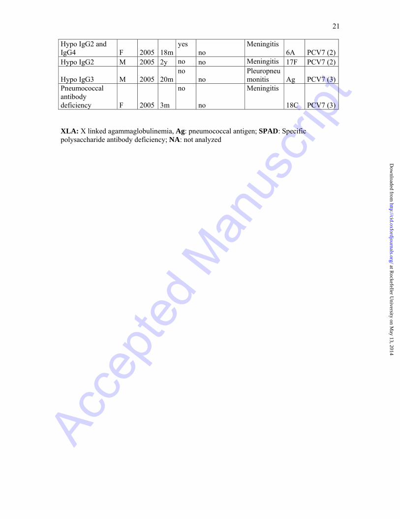
Table 2: Primary immunodeficiencies identified
Sex
Year
Age
Consan- guinity
History
Infection
Serotype
Vaccinated (number of injections)
Innate immunity deficiency
Asplenia M 2011 3y no no Meningitis 12F no C2 deficiency M 2011 11m no no Meningitis 24F no C2 deficiency F 2008 3y no no Meningitis 24F PCV7 (4)C3 deficiency M 2008 7y yes no Meningitis 15B no MyD88 deficiency M 2005 11m
yes Ethmoiditis
Meningitis 19A no
Antibody deficiency
XLA M 2006 4y no Osteoarthritis
(Sp) Meningitis
6B no
XLA M 2011 7y no Pleuropneumo
nitis (Sp) Meningitis
NA no Hypogammaglobulinemia M 2008 5y
yes Ethmoiditis
Meningitis Ag no
Hypogammaglobulinemia M 2010 4y
no no
Meningitis NA
PCV13 (1)
Hypogammaglobulinemia M 2006 9y
no no
Meningitis 18C no
Hypogammaglobulinemia M 2011 3y
no no
Meningitis NA
PCV13 (NA)
Subclass IgG2 deficiency F 2005 2y
yes no
Meningitis 19F PCV7 (3)
Subclass IgG2 deficiency M 2005 2y
no no
Meningitis 17F no
SPAD M 2009 22m no no Meningitis 7F PCV7 (4)SPAD M 2005 3y no no Meningitis 14 no
SPAD M 2005 3y no
no Pleuropneumonitis 1 no
SPAD M 2008 6y no
no Pleuropneumonitis 1 no
Transient immunoglobulin deficiency Hypogammaglobulinemia F 2007 2m
no no
Meningitis 19A no
Hypogammaglobulinemia M 2011 8m
no no
Meningitis 33F no
Hypogammaglobulinemia F 2009 14m
no no
Meningitis 12F PCV7 (4)
Hypogammaglobulinemia M 2008 14m
no no
Meningitis 23F no
Hypo IgG2 and IgG4 F 2005 12m
yes no
Meningitis 19A no
at Rockefeller U
niversity on May 13, 2014
http://cid.oxfordjournals.org/D
ownloaded from
Acce
pted M
anus
cript
21
Hypo IgG2 and IgG4 F 2005 18m
yes no
Meningitis 6A PCV7 (2)
Hypo IgG2 M 2005 2y no no Meningitis 17F PCV7 (2)
Hypo IgG3 M 2005 20mno
no Pleuropneumonitis Ag PCV7 (3)
Pneumococcal antibody deficiency F 2005 3m
no
no
Meningitis
18C PCV7 (3)
XLA: X linked agammaglobulinemia, Ag: pneumococcal antigen; SPAD: Specific polysaccharide antibody deficiency; NA: not analyzed
at Rockefeller U
niversity on May 13, 2014
http://cid.oxfordjournals.org/D
ownloaded from

Acce
pted M
anus
cript
23
Table 3: Description of patients with recurrent IPD (N = 17)
Age Sex Prior pneumococcal infections (serotype)
Infection upon inclusion (serotype)
Vaccinated (nb of injections)
Identified PID Outcome
4m F Meningitis and cerebral abscess Meningitis (AG) yes (3) no alive 4m M Meningitis Meningitis (6A) yes (2) no alive 8m F Meningitis x 2 Meningitis (14) yes (3) no dead 9m M Meningitis Meningitis (NA) yes no alive 14m F Maternofoetal infection Sepsis (NA) yes (3) no alive 19m M Meningitis Meningitis (19A) yes no alive 3y M Meningitis (19A) Meningitis (6B) yes (3) no alive 3y F Pneumonitis Sepsis (7F) yes (4) no alive 4y F Pleuropneumonitis Pleuropneumonitis (AG) no no alive 4y F Meningitis Meningitis (NA) yes no alive 4y M Osteoarthritis and maxillary abscess Meningitis (6B) no XLA alive 6y F Pleuropneumonitis Pleuropneumonitis (NA) yes no alive 7y M Meningitis (15B) Meningitis (22F) yes (3) no alive 7y M Pleuropneumonitis Meningitis (NA) no XLA alive 8y F Meningitis x 2(22F and 9) Meningitis (11A) no Cerebrospinal fistula alive 10y M Meningitis and arthritis Meningitis (23A) no no dead 15y M Meningitis Meningitis (17F) no no alive
XLA: X linked agammaglobulinemia, Ag: pneumococcal antigen; NA: not analyzed
at Rockefeller University on May 13, 2014 http://cid.oxfordjournals.org/ Downloaded from