immunodeficiency-associated lymphomas
TRANSCRIPT

Blood Reviews (2008) 22, 261–281
www.elsevier.com
REVIEW
Immunodeficiency-associated lymphomas
Huy Tran a, Jamie Nourse b, Sara Hall a, Michael Green c, Lyn Griffiths c,Maher K. Gandhi a,b,*
a Department of Haematology, Princess Alexandra Hospital, Ipswich Road, Brisbane, Queensland, 4102,Australiab Clinical Immunohaematology Lab, Queensland Institute of Medical Research, Floor I, CBCRC Building,300 Herston Rd, Brisbane, 4006, Queensland, Australiac Genomics Research Centre, Gold Coast Campus, Griffith University, PMB 50 Gold Coast Mail Centre,Queensland, 9726, Australia
Summary This article covers lymphoproliferative disorders in patients with primaryor acquired immunodeficiencies. Primary immunodeficiences include Ataxia Telangi-ectasia and X-linked disorders such as Wiskott-Aldrich syndrome. Acquired immunode-ficiencies predominantly occur in the setting of infection with the HumanImmunodeficiency Virus or arise following immunosuppressive therapy administeredafter organ transplantation. The rising incidence of HIV throughout the world andthe dramatic increase in transplant surgery since the 1990’s suggest that these lym-phomas will remain an important health problem. Evidence for lymphoma developingas a result of treatment with methotrexate or Tumour Necrosis Factor Antagonists forautoimmune entities will also be reviewed. The lymphoproliferations that occur withimmunodeficiency are extremely heterogenous. In part this reflects the diversity ofthe causal immune defect. The most striking clinical characteristic is the high fre-quency of extranodal disease. Frequently, these lymphomas are driven by viruses suchas Epstein-Barr virus (EBV), although the lack of EBV in a proportion indicates thatalternate pathways must also be involved in the pathogenesis. Lastly, discussion willcentre on mechanisms utilized by lymphomas in the immunodeficient as these mayhave applications to lymphomas in the ‘‘immunocompetent’’, by serving as a para-digm for the altered immunoregulatory environment present in many lymphomasub-types.
�c 2008 Elsevier Ltd. All rights reserved.
KEYWORDSImmunodeficiency;Lymphoma;Ataxia Telangiectasia;Combined variableimmunodeficiencydisorder;Post-transplantationlymphoproliferativedisorder;Epstein-barr virus;Human herpes virus 8;Methotrexate;Infliximab;Human immuno-deficiency virus;Primary central nervoussystem lymphoma;Primary effusionlymphoma;Hodgkin’s lymphoma;Diffuse large B celllymphoma; Rituximab
0d
B
268-960X/$ - see front matter �c 2008 Elsevier Ltd. All rights reserved.oi:10.1016/j.blre.2008.03.009
* Corresponding author. Address: Clinical Immunohaematology Lab, Queensland Institute of Medical Research, Floor I, CBCRCuilding, 300 Herston Rd, Brisbane, 4006, Queensland, Australia. Tel.: +61 7 3845 3792; fax: +61 7 3845 3510.E-mail address: [email protected] (M.K. Gandhi).

262 H. Tran et al.
Introduction
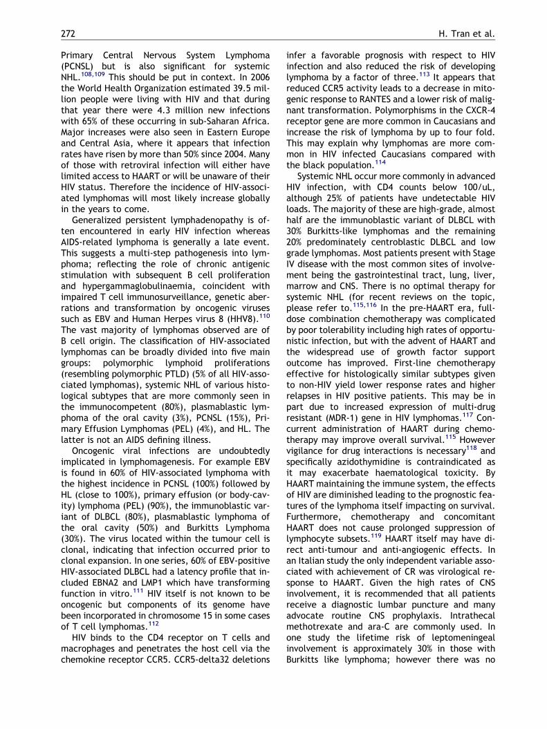
There are a multitude of factors known to be asso-ciated with cancer aetiology, with escape fromhost immunity only one facet. Transformation ofa cell to one with malignant potential in an immu-nodeficient host may allow clonogenic expansionand eventually clinical cancer.1 The importanceof defective immunosurveillance in malignancy ismost marked in cells with strong antigenic poten-tial including those that have undergone viralinduction. Consistent with this is the observationthat the predominant cancer sub-type in immuno-deficient subjects is lymphoma (a neoplasm ofthe immune system), that frequently these lym-phomas are driven by viruses such as Epstein-Barrvirus (EBV) and that restoration of immunity can re-sult in tumour regression.2 Yet the lack of EBV in aproportion indicates that alternate pathways mustalso be involved in lymphomagenesis. Indeed thewide clinical spectrum of lymphoproliferative dis-orders (LPD) encountered in immunodeficiencyalong with its associations with germ-line and ac-quired genetic defects, with oncogenic viruses,and with autoimmunity suggests an interplay inwhich genetic aberrations interact with viral onco-genes, impaired immunosurveillance and chronicantigen stimulation (Fig. 1). Increasing evidencesuggests that the immune escape mechanisms uti-lized by lymphomas in the immunodeficient mayhave relevance to lymphomas in the overtly‘‘immunocompetent’’.3–5 Thus the study of immu-
Figure 1 Interaction of factors contributing to lymphomaindicates an increased risk of developing lymphoma resultinataxia-telangiectasia; BCL6, B cell lymphoma 6 gene; WAimmunodeficiency; SCID, severe combined immunodeficiency
nodeficiency-associated lymphoproliferations mayserve as a paradigm for the altered immunoregula-tory environment present in many lymphoma sub-types.
Primary immune deficiency andlymphoproliferative disorders
The occurrence of lymphoproliferative disorders inpatients with a primary immunodeficiency (PID) hasbeen documented in the literature for nearly 40years.6 These PIDs are a heterogeneous group ofgenetically determined disorders; hence the lym-phoproliferative diseases (LPD) that arise are di-verse and variable.
The risk of developing LPD is related to thetype of PID. Accurate quantification of this riskis difficult because PID is rare; hence the inci-dence of lymphoma is low, leading to a relianceon small case series for estimates. These rangefrom 0.7% to 15%.7 However the PIDs more com-monly associated with LPD can be classified asfollows:8 T and B cell immunodeficiencies e.g.Severe combined immunodeficiency (SCID), andX-linked hyper-IgM syndrome (XHIGM); antibodydeficiencies e.g. Common variable immunodefi-ciency (CVID); DNA repair defects e.g. Ataxia-telangectasia (A-T); Immune dysregulation e.g.X-linked lymphoproliferative syndrome (XLP);Autoimmunity e.g. Autoimmune lymphoprolifera-tive disorder (ALPS); and other syndromes such
genesis in immunosuppressed patients. Darker shadingg from the combination of factors. Abbreviations: A-T,S, Wiskott-Aldrich syndrome; CVID, common variable; HHV8, human herpes virus 8.

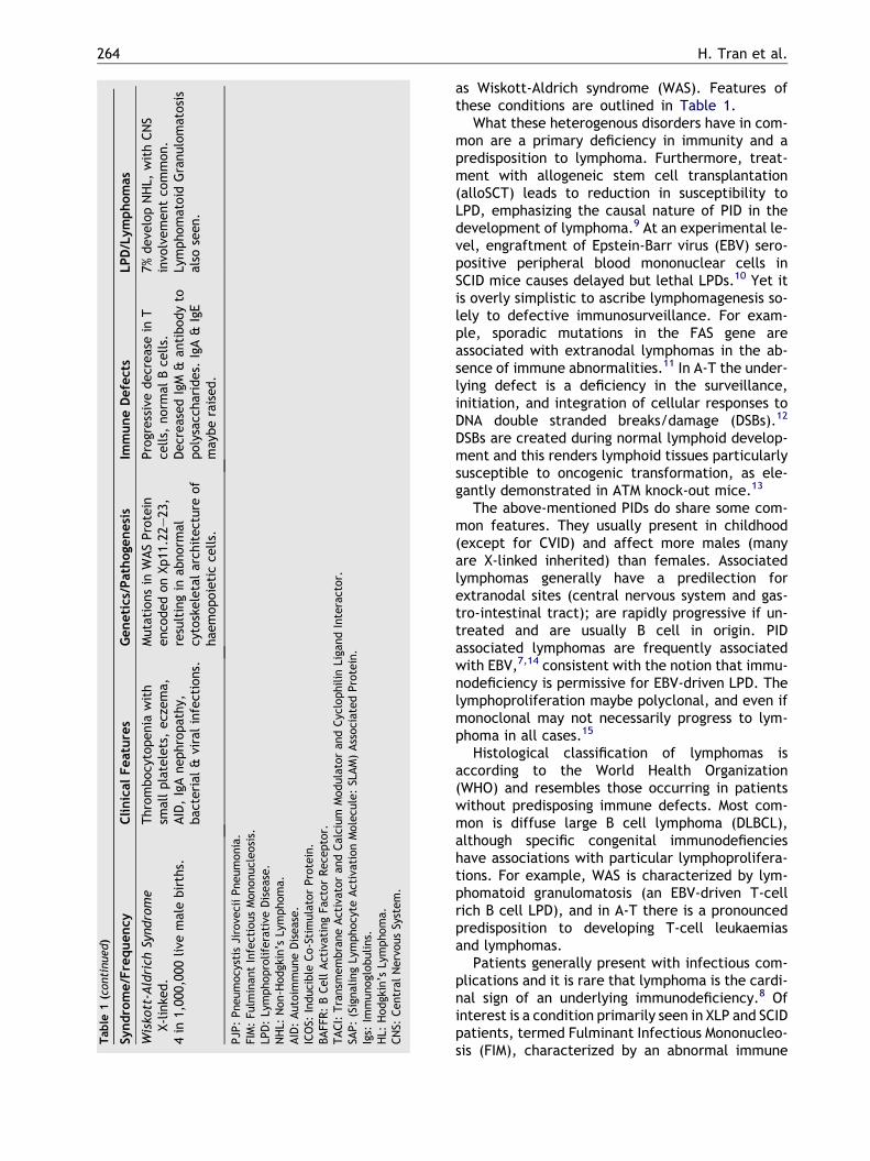
Table 1 Summary of Primary Immune Disorders with an increased risk of Lymphoproliferative Disorders and Lymphoma.
Syndrome/Frequency Clinical Features Genetics/Pathogenesis Immune Defects LPD/Lymphomas
SCID1 in 100,000 live births. High
prevalence in Navajo &Apache Indians.
If untreated, death within 1 yeardue to severe, recurrentinfections. Chronic diarrhea, earinfections, recurrent PJP & oralcandidiasis.
X-linked SCID: c chainmutations cause defectiveinterleukin signaling; ADA-SCID: defective adenosinedeaminase (ADA), resulting inbuild-up of lymphotoxins.
Dependent on sub-type, butin general low or absent Tcells & NK cells & non-functional B cells observed.
FIM following primary EBVinfection. Lymphoma not welldocumented. T cell LPD hasdeveloped in a proportion ofpatients as a result ofretroviral gene therapy forX-linked SCID.
X-linked (XLSCID) & AR(ADA-SCID) inheritance.
Common VariableImmunodeficiency
Several different clinicalphenotypes. Prone to recurrentbacterial infections, AID, LPD &granulomatous disease. Elevatedrisk of malignancy. Typicallypresent aged 20–40 years.
In a proportion, homozygousdefects in genes encodingICOS, CD19, & BAFFR. Defectsin TACI can be dominant orrecessive. Loci at 4q, 5p, 16qimplicated.
Low IgG & IgA; variable IgM.Reduced B cells.
LPD in the GI tract & lungs.HHV8 implicated inpathogenesis ofgranulomatous/lymphocyticinterstitial lung disease.Increased risk of (typically) Bcell, extranodal, Ig secretingNHL.
1 in 10–50,000 live births.Cases are generally sporadic,
but �10% are familial.
X-linked HyperimmunoglobulinM Syndrome
Neutropenia,thrombocytopenia, anaemia,biliary tract/liver disease,recurrent PJP infection anddiahorrea. Increased risk of AID.
CD40 ligand mutation onXq26–27.2, leading todefective B cell & dendriticcell signaling. CD40L requiredfor isotype class-switchingfrom IgM to IgG or IgA.
Low/absent IgG & IgA,normal/increased IgM.Variable defect in effector Tcell and macrophagefunction.
LPD in 65%, predominantlyperipheral & abdominaladenpathy. Nodes lackgerminal centres. Lymphomanot well documented but LPDcan be fatal.
I in 20,000,000 live male births.
Ataxia Telangiectasia Cerebellar ataxia, chromosomalinstability, oculocutaneoustelangiectasia, thymic aplasia,radiosensitivity, recurrentsinopulmonary infections,lymphoid (80%) & other tumours.
AT Mutated gene on 11q22–23, encodes for proteinkinase with >40 knownsubstrates. Disorder in cell-cycle check-point & impairedDNA double-stranded breakrepair.
IgA, IgE, IgG2 & IgG4decreased. Increased IgMmonomers. Progressivedecrease in T cells. Normal Bcell numbers.
Bimodal distribution: age1–5: T cell acutelymphoblastic leukaemia/lymphoma; young adults: Tprolymphocytic leukaemia.
1 in 40–100,000 live births.AR. Carrier rate 1%.
X-linked LymphoproliferativeSyndrome-1. (XLPS-2 caused by
mutation in XIAP shares manyfeatures).
Clinical & immunologicalabnormalities triggered by EBVinfection.
Mutations in SH2D1A on Xq25,encodes for SAP which isinvolved in T–B cellinteractions.
Variably decreased Igs. B cellsnormal or reduced. Defectivelysis & polarization of EBV-specific T and NKT cells.
FIM, aplastic anaemia &hepatitis. Survivors acquirehypogammaglobulinaemia &extranodal B cell NHL,particularly of the terminalileum.
400 documented cases from 100US families.
AutoimmuneLymphoproliferativeSyndrome
Present in infancy withautoimmune cytopenias, AID,adenopathy & splenomegaly.
3 groupings, characterizedby the defect in the FAS-mediated apoptosis pathway.
Increased circulating CD4-/CD8-T cells due to defectivelymphocyte apoptosis.Normal B cells & Igs.
50· increased risk of HL; 10–15· increased risk of NHL.
Rare. Frequency unknown. AD &AR forms of inheritance.
(continued on next page)
Immunodeficie
ncy-asso
ciatedlym
phomas
263
Table
1(continued)
Syndrome/Frequency
ClinicalFeatures
Genetics/Pathoge
nesis
ImmuneDefects
LPD/Lymphomas
Wisko
tt-Aldrich
Syndrome
X-linke
d.
Thrombocytopenia
with
smallplatelets,eczema,
AID,IgAnephropathy,
bac
terial
&viralinfections.
Mutationsin
WASProtein
enco
dedonXp11
.22–
23,
resultingin
abnorm
alcytoskeletalarch
itecture
of
hae
mopoieticce
lls.
Progressivedecreasein
Tce
lls,
norm
alBce
lls.
DecreasedIgM
&an
tibodyto
polysacc
harides.
IgA&
IgE
may
beraised.
7%deve
lopNHL,
withCNS
invo
lvementco
mmon.
Lymphomatoid
Granulomatosis
also
seen.
4in
1,00
0,00
0live
malebirths.
PJP
:Pneumocystis
Jirove
ciiPneumonia.
FIM:Fu
lminan
tInfectiousMononucleosis.
LPD:Lymphoproliferative
Disease.
NHL:
Non-H
odgk
in’s
Lymphoma.
AID:Autoim
muneDisease.
ICOS:
Inducible
Co-StimulatorProtein.
BAFF
R:BCellActivatingFa
ctorRece
ptor.
TACI:Transm
embraneActivatoran
dCalcium
Modulatoran
dCyclophilin
Liga
ndInteractor.
SAP:(SignalingLymphocyte
ActivationMolecu
le:SLAM)AssociatedProtein.
Igs:
Immunoglobulins.
HL:
Hodgk
in’s
Lymphoma.
CNS:
Central
NervousSystem.
264 H. Tran et al.
as Wiskott-Aldrich syndrome (WAS). Features ofthese conditions are outlined in Table 1.
What these heterogenous disorders have in com-mon are a primary deficiency in immunity and apredisposition to lymphoma. Furthermore, treat-ment with allogeneic stem cell transplantation(alloSCT) leads to reduction in susceptibility toLPD, emphasizing the causal nature of PID in thedevelopment of lymphoma.9 At an experimental le-vel, engraftment of Epstein-Barr virus (EBV) sero-positive peripheral blood mononuclear cells inSCID mice causes delayed but lethal LPDs.10 Yet itis overly simplistic to ascribe lymphomagenesis so-lely to defective immunosurveillance. For exam-ple, sporadic mutations in the FAS gene areassociated with extranodal lymphomas in the ab-sence of immune abnormalities.11 In A-T the under-lying defect is a deficiency in the surveillance,initiation, and integration of cellular responses toDNA double stranded breaks/damage (DSBs).12
DSBs are created during normal lymphoid develop-ment and this renders lymphoid tissues particularlysusceptible to oncogenic transformation, as ele-gantly demonstrated in ATM knock-out mice.13
The above-mentioned PIDs do share some com-mon features. They usually present in childhood(except for CVID) and affect more males (manyare X-linked inherited) than females. Associatedlymphomas generally have a predilection forextranodal sites (central nervous system and gas-tro-intestinal tract); are rapidly progressive if un-treated and are usually B cell in origin. PIDassociated lymphomas are frequently associatedwith EBV,7,14 consistent with the notion that immu-nodeficiency is permissive for EBV-driven LPD. Thelymphoproliferation maybe polyclonal, and even ifmonoclonal may not necessarily progress to lym-phoma in all cases.15
Histological classification of lymphomas isaccording to the World Health Organization(WHO) and resembles those occurring in patientswithout predisposing immune defects. Most com-mon is diffuse large B cell lymphoma (DLBCL),although specific congenital immunodefiencieshave associations with particular lymphoprolifera-tions. For example, WAS is characterized by lym-phomatoid granulomatosis (an EBV-driven T-cellrich B cell LPD), and in A-T there is a pronouncedpredisposition to developing T-cell leukaemiasand lymphomas.
Patients generally present with infectious com-plications and it is rare that lymphoma is the cardi-nal sign of an underlying immunodeficiency.8 Ofinterest is a condition primarily seen in XLP and SCIDpatients, termed Fulminant Infectious Mononucleo-sis (FIM), characterized by an abnormal immune

Immunodeficiency-associated lymphomas 265
response to primary EBV infection. FIM is a life-threatening condition marked by fever, rash, gener-alized lymphadenopathy, hepatosplenomegaly, andcytopenias. Characteristic findings include theuncontrolled systemic expansion of polymorphous Blymphocytes, involving lymphoid and non-lymphoidorgans (such as the terminal ileum). EBV-specificcellular immunity is defective.16,17 Haemophagocy-tois is frequent, with extensive marrow infiltrationby lymphoid cells and cellular necrosis resulting insevere pancytopenia. Opportunistic infections andliver failure, often associated with acute hemor-rhage are the major causes of death.18 In XLP, thosesurviving frequently acquire hypogammoglobulina-emia (involving lymph nodes necrosis althoughperipheral B cell numbers maybe maintained) ormalignant B cell lymphoma. EBV may also triggeraplastic anaemia and/or hepatitis. Pre-emptiveadministration of the anti-CD20 antibody rituximabmay reduce the devastating sequelae of primaryEBV infection in these patients.19
There is limited data on treatment and prognosisin PID associated lymphomas given the rarity of thisdisease and the wide variety of predisposing immu-nodeficencies. In the absence of randomized clini-cal trials, it is recommended that treatment betailored according to histological subtype and prog-nosis. Replacement immunoglobulin is indicatedfor hypogammaglobulinaemia, as is early andaggressive treatment for infections with specialconsideration for Pneumocystis Jirovecii Pneumo-nia antibiotic prophylaxis. There are currentlytwo clinical trials underway examining the efficacyof anti-oxidants in the treatment of A-T. AlloSCThas been shown to restore immune function andprevent lymphoma in ATM-deficient mice.20 Clini-cally, alloSCT has been used successfully for lym-phomas arising in XLP, ALPS, SCID, WAS andXHIGM.21,22 Gene therapy has been successfullyused for SCID. Although cases of T-cell LPD drivenby integration of the retroviral vector have beenreported,23 these results should be seen in context,and it is still gene therapy that has the greatest po-tential for long-term cure of PID.
Post-transplant lymphoproliferativedisorders
Post-transplant lymphoproliferative disorder(PTLD) represent the group of lymphoid disordersthat arise following solid-organ transplantation(SOT) and stem cell transplantation.24–27 Relativeto the immunocompetent, the risk of lymphomaafter transplantation is increased by the order of
30 times.28 The incidence of PTLD varies, depend-ing on the intensity of immunosuppression, recipi-ent age, the organ transplanted, the number ofprevious allografts, and in liver transplant recipi-ents by the presence of Hepatitis C cirrhosis or pri-mary biliary cirrhosis.29 These risk factors arereflected in the highest incidence of PTLD occur-ring after lung and small bowel transplants (up to30%,30 in contrast to 1–5% for renal, cardiac andliver transplants. After SCT, the cumulativeincidence of PTLD at 10 years is 1%.27 As immuno-suppression protocols become less intensive, sothe relative frequency of PTLD after SOT maydiminish. However since the 1990’s there has beena dramatic rise in transplant surgery and this cou-pled with improvements in long-term SOT patientsurvival, will likely result in an absolute rise inthe incidence of PTLD.
Approximately 80% of PTLD biopsies are positivefor EBV within the tumour cells. Reflecting the crit-ical role of reduced cellular immuno-surveillanceagainst Epstein-Barr virus (EBV) in the pathogene-sis, other risk factors are the EBV-serostatus ofthe donor (D) and recipient (R) (for SOT: D+/R�;and for SCT D�/R+ are at greatest risk respec-tively); cytomegaloviral disease31 and the use ofT cell depleting agents. Furthermore, use of theanti-CD52 antibody alemtuzumab which depletesboth B and T cells is associated with a lower inci-dence of PTLD than when T cells are depleted inisolation.32 In SOT recipients, the incidence is high-est in the first year post-transplant (the period ofmost intense immunosuppression), and a reducedrisk remains thereafter. Early post-transplantationEBV-seroconversion due to primary EBV infectionplaces the recipient at a major risk of EBV-positivePTLD. Although at least 90% of the population areEBV-seropositive by the age of 40, children are fre-quently seronegative, and in the majority of PTLDseen in the paediatric setting primary EBV infectionhas occurred in the 6 months prior to presenta-tion.33,34 In vitro, TOR inhibitors such as sirolimusreduce the proliferation of EBV transformed lym-phoblastoid cell lines (LCL),35 and unlike calcineu-rin inhibitors such as cyclosporine and tacrolimusdue not protect LCL from apoptosis.36 The rolefor sirolimus as a immunosuppressive agent follow-ing SOT is still evolving, and currently there isinsufficient data for a definitive statement as towhether patients are less likely to develop PTLD.
Although exceptions have been reported, in gen-eral following SOT, EBV-positive PTLD arises fromhost lymphocytes. In EBV-seronegative recipients,recipient cells are infected by the virus present inthe donor organ; whereas in seropositive recipientsthe PTLD tissue frequently but not invariably
266 H. Tran et al.
contains the recipient EBV isolate.37 In contrast, inallogeneic stem cell transplants (SCT) it is gener-ally donor-derived lymphocytes that undergo trans-formation. Frequently, this is as a result of loss ofendogenous EBV and acquisition of the donor virusisolate.38
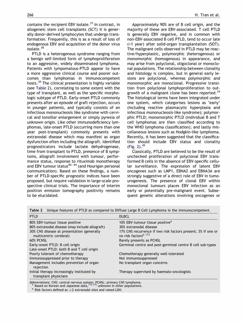
PTLD is a heterogenous syndrome ranging froma benign self-limited form of lymphoproliferationto an aggressive, widely disseminated lymphoma.Patients with lympomatous-PTLD appear to havea more aggressive clinical course and poorer out-comes than lymphomas in immunocompetenthosts.39 The clinical presentation is highly variable(see Table 2), correlating to some extent with thetype of transplant, as well as the specific morpho-logic subtype of PTLD. Early-onset PTLD commonlypresents after an episode of graft rejection, occursin younger patients, and typically consists of aninfectious mononucleosis like syndrome with cervi-cal and tonsillar enlargement or simply pyrexia ofunknown origin. Like other immunodeficiency lym-phomas, late-onset PTLD (occurring more than oneyear post-transplant) commonly presents withextranodal disease which may manifest as organdysfunction often including the allograft. Identifiedprognosticators include lactate dehydrogenase,time from transplant to PTLD, presence of B symp-toms, allograft involvement with tumour, perfor-mance status, response to rituximab monotherapyand EBV tumour status40–44 (and Hourigan personalcommunication). Based on these findings, a num-ber of PTLD-specific prognostic indices have beenproposed, but require validation in large-scale pro-spective clinical trials. The importance of interimpositron emission tomography positivity remainsto be elucidated.
Table 2 Unique features of PTLD as compared to Diffuse
PTLD DLBC
80% EBV-tumour tissue positive 10%80% extranodal disease (may include allograft) 30%30% CNS disease at presentation (generallymulticentric cerebral)
17%no ri
60% PCNSL RareEarly-onset PTLD: B cell origin GermLate-onset PTLD: both B and T cell originPoorly tolerant of chemotherapy ChemImmunosuppressed prior to therapy Not iManagement includes prevention of organrejection
No tr
Initial therapy increasingly instituted bytransplant physicians
Ther
Abbreviations: CNS: central nervous system; PCNSL: primary CNS la Based on Korean and Japanese data,174,175 unknown in other pb Risk factors defined as P2 extranodal sites and raised LDH.
Approximately 90% are of B cell origin, and themajority of these are EBV-associated. T cell PTLDis generally EBV negative, and in common withnon-EBV-associated B cell PTLD, tend to occur late(>1 year) after solid-organ transplantation (SOT).The malignant cells observed in PTLD may be reac-tive/hyperplastic, polymorphic (heterogenous) ormonomorphic (homogenous) in appearance, andmay arise from polyclonal, oligoclonal or monoclo-nal populations. The relationship between clonalityand histology is complex, but in general early le-sions are polyclonal, whereas polymorphic andmonomorphic are monoclonal. Progressive transi-tion from polyclonal lymphoproliferation to out-growth of a malignant clone has been reported.34
The histological terms have been integrated underone system, which categorises lesions as ‘early’(including reactive plasmacytic hyperplasia andinfectious mononucleosis-like syndrome); polymor-phic PTLD; monomorphic PTLD (individual B and Tcell lymphomas are then classified according tothe WHO lymphoma classification); and lastly mis-cellaneous lesions such as Hodgkin-like lymphoma.Recently, it has been suggested that the classifica-tion should include EBV status and clonality(Fig. 2).45
Classically, PTLD are believed to be the result ofunchecked proliferation of polyclonal EBV trans-formed B cells in the absence of EBV-specific cellu-lar surveillance. The expression of latent EBVoncogenes such as LMP1, EBNA2 and EBNA3A arestrongly suggestive of a direct role of EBV in tumo-urogenesis. The presence of clonal EBV withinmonoclonal tumours places EBV infection as anearly or potentially pre-malignant event. Subse-quent genetic alterations involving oncogenes or
Large B Cell Lymphoma in the immunocompetent.
L
EBV-tumour tissue positivea
extranodal diseaseCNS recurrence if two risk factors present; 3% if one orsk factorsb 173
ly presents as PCNSLinal centre and post-germinal centre B cell sub-types
otherapy generally well-toleratedmmunosuppressedansplant organ concerns
apy supervised by haemato-oncologists
ymphoma.opulations.

Figure 2 Relationship between EBV latency gene expression, histological subtype of PTLD and recovery of EBV-specific immunity. After transplant EBV-specific immunity is ablated but gradually returns. In PTLD a spectrum of EBVlatent gene expression is observed, with a broad range of genes tending to be expressed in early onset PTLD, whereas inlate onset cases expression is restricted.
Immunodeficiency-associated lymphomas 267
tumour suppressor genes are thought to accumu-late resulting in a survival advantage of an aggres-sive malignant clone.46 5’ non coding regionmutations of BCL6 gene are frequent and are asso-ciated with an aggressive clinical outcome.25 Otherfactors are likely implicated. For example the pre-dilection of PTLD to extranodal sites suggests thatthe tumour microenvironment is important. Theheavy infiltration with CD4 T cells47 may be an indi-cator for the importance of immuno-regulatoryfactors, as seen in EBV-positive Hodgkins Lym-phoma.4,5 Involvement of the transplanted organwhich is itself subject to frequent sub-clinicalrejection might induce a state of chronic antigenstimulation, as might co-infection with non-EBVpathogens. The incidence of non-EBV-positive PTLDremains higher than in non-immunocompromisedsubjects, and the possible role of other viruses inlymphomagenesis (such as Human Herpes Virus 6)remains unresolved.48
Although reduction of immunosuppression is rec-ommended as initial therapy, as monotherapy this
generally leads to response in only a minority of pa-tients, particularly those with early lesions or wild-type BCL-6.25,43 Nevertheless in all patients withPTLD the immunosuppression should be cautiouslyreduced as per current European and Americanguidelines (see Table 3).45,49 Indeed some centresreport good results with complete cessation ofimmunosuppression during induction chemotherapyfor renal-PTLD (D. Gill, personal communication).Chemotherapy remains the gold-standard therapybut is frequently poorly-tolerated and results re-main worse than for equivalent lymphomas in non-immunosuppressed patients.50 The anti-CD20 anti-body rituximab is generally non-toxic and has activ-ity as a single-agent but relapse is frequent.51,52
Sequential combination therapy with x4 cycles ofweekly rituximab followed by x4 G-CSF supportedCHOP-21 reports encouraging results, with responseto rituximab predictive of survival .53 Importantly,this combination does not appear to impairEBV-specific cellular immunity,54 except in thosepatients receiving ongoing immunosuppression.55
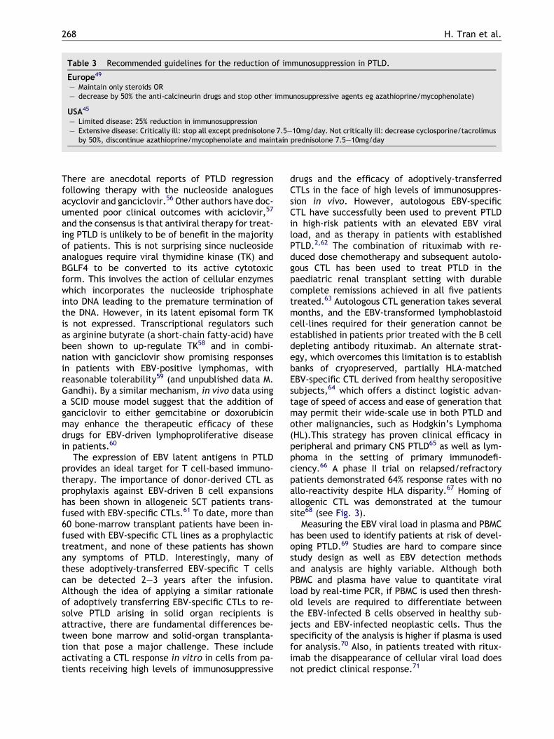
Table 3 Recommended guidelines for the reduction of immunosuppression in PTLD.
Europe49
– Maintain only steroids OR– decrease by 50% the anti-calcineurin drugs and stop other immunosuppressive agents eg azathioprine/mycophenolate)
USA45
– Limited disease: 25% reduction in immunosuppression– Extensive disease: Critically ill: stop all except prednisolone 7.5–10mg/day. Not critically ill: decrease cyclosporine/tacrolimus
by 50%, discontinue azathioprine/mycophenolate and maintain prednisolone 7.5–10mg/day
268 H. Tran et al.
There are anecdotal reports of PTLD regressionfollowing therapy with the nucleoside analoguesacyclovir and ganciclovir.56 Other authors have doc-umented poor clinical outcomes with aciclovir,57
and the consensus is that antiviral therapy for treat-ing PTLD is unlikely to be of benefit in the majorityof patients. This is not surprising since nucleosideanalogues require viral thymidine kinase (TK) andBGLF4 to be converted to its active cytotoxicform. This involves the action of cellular enzymeswhich incorporates the nucleoside triphosphateinto DNA leading to the premature termination ofthe DNA. However, in its latent episomal form TKis not expressed. Transcriptional regulators suchas arginine butyrate (a short-chain fatty-acid) havebeen shown to up-regulate TK58 and in combi-nation with ganciclovir show promising responsesin patients with EBV-positive lymphomas, withreasonable tolerability59 (and unpublished data M.Gandhi). By a similar mechanism, in vivo data usinga SCID mouse model suggest that the addition ofganciclovir to either gemcitabine or doxorubicinmay enhance the therapeutic efficacy of thesedrugs for EBV-driven lymphoproliferative diseasein patients.60
The expression of EBV latent antigens in PTLDprovides an ideal target for T cell-based immuno-therapy. The importance of donor-derived CTL asprophylaxis against EBV-driven B cell expansionshas been shown in allogeneic SCT patients trans-fused with EBV-specific CTLs.61 To date, more than60 bone-marrow transplant patients have been in-fused with EBV-specific CTL lines as a prophylactictreatment, and none of these patients has shownany symptoms of PTLD. Interestingly, many ofthese adoptively-transferred EBV-specific T cellscan be detected 2–3 years after the infusion.Although the idea of applying a similar rationaleof adoptively transferring EBV-specific CTLs to re-solve PTLD arising in solid organ recipients isattractive, there are fundamental differences be-tween bone marrow and solid-organ transplanta-tion that pose a major challenge. These includeactivating a CTL response in vitro in cells from pa-tients receiving high levels of immunosuppressive
drugs and the efficacy of adoptively-transferredCTLs in the face of high levels of immunosuppres-sion in vivo. However, autologous EBV-specificCTL have successfully been used to prevent PTLDin high-risk patients with an elevated EBV viralload, and as therapy in patients with establishedPTLD.2,62 The combination of rituximab with re-duced dose chemotherapy and subsequent autolo-gous CTL has been used to treat PTLD in thepaediatric renal transplant setting with durablecomplete remissions achieved in all five patientstreated.63 Autologous CTL generation takes severalmonths, and the EBV-transformed lymphoblastoidcell-lines required for their generation cannot beestablished in patients prior treated with the B celldepleting antibody rituximab. An alternate strat-egy, which overcomes this limitation is to establishbanks of cryopreserved, partially HLA-matchedEBV-specific CTL derived from healthy seropositivesubjects,64 which offers a distinct logistic advan-tage of speed of access and ease of generation thatmay permit their wide-scale use in both PTLD andother malignancies, such as Hodgkin’s Lymphoma(HL).This strategy has proven clinical efficacy inperipheral and primary CNS PTLD65 as well as lym-phoma in the setting of primary immunodefi-ciency.66 A phase II trial on relapsed/refractorypatients demonstrated 64% response rates with noallo-reactivity despite HLA disparity.67 Homing ofallogenic CTL was demonstrated at the tumoursite68 (see Fig. 3).
Measuring the EBV viral load in plasma and PBMChas been used to identify patients at risk of devel-oping PTLD.69 Studies are hard to compare sincestudy design as well as EBV detection methodsand analysis are highly variable. Although bothPBMC and plasma have value to quantitate viralload by real-time PCR, if PBMC is used then thresh-old levels are required to differentiate betweenthe EBV-infected B cells observed in healthy sub-jects and EBV-infected neoplastic cells. Thus thespecificity of the analysis is higher if plasma is usedfor analysis.70 Also, in patients treated with ritux-imab the disappearance of cellular viral load doesnot predict clinical response.71

Figure 3 Allogeneic EBV-specific T cells home to sites of disease. Immunohistochemistry (IHC) and fluorescent in-situhybridization (FISH) in left and right panels respectively demonstrating scattered CD8+ T cells and XY cells admixedwithin the B cell PTLD, taken at autopsy. The donor-CTL donor was male, and the recipient was female. The PTLD wasof recipient origin. Reproduced with kind permission from an article originally published in the American Journal ofTransplantation.68
Immunodeficiency-associated lymphomas 269
Methotrexate-associatedlymphoproliferative disorders
The development of methotrexate (MTX) by Far-ber’s team in Boston was a pivotal moment in themodern era of chemotherapy. It remains a criticalcomponent of acute lymphoblastic leukaemia ther-apy, and high-dose MTX is the mainstay for thetreatment of lymphomas within the nervous sys-tem. Ironically, low-dose MTX, used most com-monly in the setting of treatment for RA,psoriasis, dermatomyositis and myasthenia, isimplicated in lymphomagenesis. In their most re-cent classification, the World Health Organizationincluded MTX associated lymphoproliferative disor-ders as a subcategory of lymphoma.72
The histology’s observed are variable and in-clude DLBCL, HL, follicular lymphoma, lymphoplas-macytic lymphoma, mantle cell lymphoma and apicture analogous to polymorphous PTLD.73 Extran-odal presentation appears common, but in contrastto AIDS patients high frequencies of neurologicalinvolvement are not seen.74 However, studies areconflicting and there is no definitive epidemiologi-cal evidence as to the extent to which MTX in-creases the risk of lymphoma in such patients, ifat all. No excessive risk of lymphoma was observedin one retrospective study75 and in several longitu-dinal studies of patients receiving MTX, even afterlong-term follow-up.76,77 These studies includedonly several hundred patients, and may thereforehave been insufficiently powered to detect anexcessive risk of a rare event (lymphoma), andmay be further confounded by reported increasein prevalence of lymphoma in patients takingcyclosporine A.78 A national French prospectivestudy conducted over 3 years found a significant
increase in HL but not NHL in RA patients treatedwith MTX.79 The issue of causality is confoundedby the estimated 2-fold increased risk of lymphoma(particularly NHL) in patients with rheumatoidarthritis (RA) (see Fig. 4).80,81
MTX has two mechanisms of action. It is an anti-metabolite that competitively inhibits dihydrofo-late reductase (DHFR), thus preventing DNA, RNAand proteins synthesis, resulting in cytotoxicityduring the S-phase of the cell-cycle. At the lowerdoses used in the management of RA, inhibitionof DHFR is not thought to be the principle mecha-nism. Rather, many of the anti-inflammatory ef-fects of MTX are mediated by adenosine, or theinhibition of T cell activation and suppression ofintercellular adhesion molecule expression by Tcells.82
Several genes within the folate pathway havebeen associated with the risk of lymphoma in gen-eral, including 5,10-methylenetetrahydrofolatereductase (MTHFR), methionine synthase, serinehydroxymethyltransferase, thymidylate synthase,and folylpolyglutamate synthase.83–88 However,these associations are heavily confounded by vari-ances in the dietary intake of folate, methionineand multiple co-factors for their metabolism.88
To our knowledge, no data exists as to whetherthe variations in these genes are linked to MTX-associated lymphoma susceptibility.
Intriguingly, approximately 50% of the lympho-proliferative disorders are EBV-associated.89 Thefrequency of EBV infection within the malignantcell varies between histology’s, with rates highestin HL (approximately 75% of cases). Since diagnos-tic laboratories tend to use only EBER staining, thepattern of EBV viral latency seen in these lympho-mas remains to be ascertained.
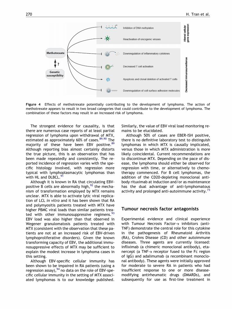
Figure 4 Effects of methotrexate potentially contributing to the development of lymphoma. The action ofmethotrexate appears to result in two broad categories that could contribute to the development of lymphoma. Thecombination of these factors may result in an increased risk of lymphoma.
270 H. Tran et al.
The strongest evidence for causality, is thatthere are numerous case reports of at least partialregression of lymphoma upon withdrawal of MTX,estimated as approximately 60% of cases.89,90 Themajority of these have been EBV positive.89
Although reporting bias almost certainly distortsthe true picture, this is an observation that hasbeen made repeatedly and consistently. The re-ported incidence of regression varies with the spe-cific histology involved, with regression moretypical with lymphoplasmacytic lymphomas thanwith HL and DLBCL.91
Although it is known in RA that circulating EBV-positive B cells are abnormally high,92 the mecha-nism of transformation employed by MTX remainsunclear. MTX is able to activate lytic viral replica-tion of LCL in vitro and it has been shown that RAand polymyositis patients treated with MTX havehigher PBMC viral loads than similar patients trea-ted with other immunosuppressive regimens.93
EBV load was also higher than that observed inWegener granulomatosis patients treated withMTX (consistent with the observation that these pa-tients are not at an increased risk of EBV-drivenlymphoproliferative disorders). Given the knowntransforming capacity of EBV, the additional immu-nosuppressive effects of MTX may be sufficient toexplain the modest increase in lymphoma cases inthis setting.
Although EBV-specific cellular immunity hasbeen shown to be impaired in RA patients (using aregression assay),94 no data on the role of EBV-spe-cific cellular immunity in the setting of MTX associ-ated lymphomas is to our knowledge published.
Similarly, the value of EBV viral load monitoring re-mains to be elucidated.
Although 50% of cases are EBER-ISH positive,there is no definitive laboratory test to distinguishlymphomas in which MTX is causally implicated,versus those in which MTX administration is morelikely coincidental. Current recommendations areto discontinue MTX. Depending on the pace of dis-ease, the lymphoma should either be observed forregression with time, or alternatively to chemo-therapy commenced. For B cell lymphomas, theaddition of the CD20-depleting monoclonal anti-body rituximab at induction and/or as maintenancehas the dual advantage of anti-lymphomatousactivity and prolonged anti-autoimmune activity.73
Tumour necrosis factor antagonists
Experimental evidence and clinical experiencewith Tumour Necrosis Factor-a inhibitors (anti-TNF) demonstrate the central role for this cytokinein the pathogenesis of Rheumatoid Arthritis(RA), Crohns Disease (CD) and other autoimmunediseases. Three agents are currently licensed:infliximab (a chimeric monoclonal antibody), eta-nercept (a TNF-a receptor fused to the Fc regionof IgG) and adalimumab (a recombinant monoclo-nal antibody). These agents were initially approvedfor moderate to severe RA in patients who hadinsufficient response to one or more disease-modifying antirheumatic drugs (DMARDs), andsubsequently for use as first-line treatment in

Table 4 Key Features of Hepatosplenic T cell lymphoma.
EpidemiologyRare (approximately 200 cases in world-wide literature). Estimated at <5% of all peripheral T cell lymphomas. Far
more frequent in males. Peak incidence in adolescents/young adults, but paediatric and elderly cases reported. Ina third of cases, HSTCL occurs in setting of concomitant immunomodulatory agents and post organtransplantation. At least 11 reported cases associated with infliximab.
Clinical FeaturesSplenic & bone marrow infiltration in virtually all cases. Hepatomegaly in 75%. Peripheral adenopathy rare.
B symptoms common. Elevated liver enzymes in 40%. Peripheral blood involvement occurs late.
Cellular PhenotypeImmature cytotoxic T cells. Usually cd CD3+, but abCD3+ reported (usually in females).
SurvivalAggressive and frequently chemo-refractory. Median survival <2 years.
Immunodeficiency-associated lymphomas 271
recent-onset moderate to severe RA. In RA it is fre-quently co-administered with MTX or other immu-nosuppressive therapy, and maintenance anti-TNFcan also be given. Anti-TNF’s also have activity inplaque psoriasis, psoriatic arthritis and ankylosingspondylitis. Infliximab is also indicated for refrac-tory or fistulizing CD.
Clinical trials95 and post-marketing surveil-lance96,97 indicates that these three agents maybe associated with a modest increased risk of lym-phoma. However, these findings are not univer-sal,98,99 and given that irrespective of treatmentRA patients have an approximately 2-fold higherrisk of lymphoma than the general population(even more so in those with highly active RA),80,81
this data must be interpreted with caution. It is un-clear whether patients with CD are truly at a higherrisk for developing lymphoma, with studies givingconflicting or inconclusive results.100,101 The truepicture has been hampered by incomplete report-ing of clinico-histopathological correlates (specifi-cally concerning histological subtypes and thefrequency of extranodal sites), although it is clearthat a wide variety of lymphomas (including HL,DLBCL and follicular lymphoma) may occur. In mostcases, EBER-ISH has not been performed, but acase of EBER-ISH+/EBNA2+/LMP1+ metachronouslymphoma in a patient with CD has been re-ported.102 In general lymphomas are B cell, butcases of mycosis fungoides and especially hepa-tosplenic T cell lymphoma (HSTCL) have been de-scribed103–105 (see Table 4). Given the relativescarcity of HSTCL in any setting and its known asso-ciation with immunomodulatory drugs, the reportsof HSTCL with infliximab for Crohn’s Disease are ofconcern.
The mechanism for a contributory role of anti-TNF (in patients with RA or CD) towards lymphoma-
genesis remains entirely speculative. Of note,transgenic mouse models of TNF-a do not have anincreased susceptibility to lymphoma.106 Althoughthere may be an association with granulomatousinfections, anti-TNF’s appear to cause a selectiverather than pleiotropic immunosuppression,107 sug-gesting that it is an imbalance of immunity (per-haps in the setting of chronic antigen stimulation)that might be implicated.
The observed ratio of observed rate to age-ad-justed expected frequency for all lymphomas in pa-tients treated with anti-TNF’s is raised comparedto the general population, but not to that seen inRA patients. Furthermore, to date analysis of thetime-to-onset of the cases of lymphoma seen withanti-TNF’s is not suggestive of a causal relation-ship. Currently, available data is insufficient todetermine whether anti-TNF’s increases the inci-dence of lymphomas, let alone whether theseagents are truly lymphogenic. Until data confirmsor refutes the association, it is prudent to adopta policy of vigilance and careful monitoring of pa-tients in receipt of anti-TNF’s. Also, althoughsplenomegaly and occasionally lymphadenopathycan occur in patients with RA and CD, in such pa-tients every effort must be made to exclude a lym-phomatous aetiology.
Human immunodeficiency virus (HIV)associated lymphomas
Approximately 1–6% of HIV positive patients devel-op lymphoma each year. However with the wide-spread use of highly active antiretroviral therapy(HAART) from 1996, the incidence of NHL appearsto be declining. This reduction is most marked for
272 H. Tran et al.
Primary Central Nervous System Lymphoma(PCNSL) but is also significant for systemicNHL.108,109 This should be put in context. In 2006the World Health Organization estimated 39.5 mil-lion people were living with HIV and that duringthat year there were 4.3 million new infectionswith 65% of these occurring in sub-Saharan Africa.Major increases were also seen in Eastern Europeand Central Asia, where it appears that infectionrates have risen by more than 50% since 2004. Manyof those with retroviral infection will either havelimited access to HAART or will be unaware of theirHIV status. Therefore the incidence of HIV-associ-ated lymphomas will most likely increase globallyin the years to come.
Generalized persistent lymphadenopathy is of-ten encountered in early HIV infection whereasAIDS-related lymphoma is generally a late event.This suggests a multi-step pathogenesis into lym-phoma; reflecting the role of chronic antigenicstimulation with subsequent B cell proliferationand hypergammaglobulinaemia, coincident withimpaired T cell immunosurveillance, genetic aber-rations and transformation by oncogenic virusessuch as EBV and Human Herpes virus 8 (HHV8).110
The vast majority of lymphomas observed are ofB cell origin. The classification of HIV-associatedlymphomas can be broadly divided into five maingroups: polymorphic lymphoid proliferations(resembling polymorphic PTLD) (5% of all HIV-asso-ciated lymphomas), systemic NHL of various histo-logical subtypes that are more commonly seen inthe immunocompetent (80%), plasmablastic lym-phoma of the oral cavity (3%), PCNSL (15%), Pri-mary Effusion Lymphomas (PEL) (4%), and HL. Thelatter is not an AIDS defining illness.
Oncogenic viral infections are undoubtedlyimplicated in lymphomagenesis. For example EBVis found in 60% of HIV-associated lymphoma withthe highest incidence in PCNSL (100%) followed byHL (close to 100%), primary effusion (or body-cav-ity) lymphoma (PEL) (90%), the immunoblastic var-iant of DLBCL (80%), plasmablastic lymphoma ofthe oral cavity (50%) and Burkitts Lymphoma(30%). The virus located within the tumour cell isclonal, indicating that infection occurred prior toclonal expansion. In one series, 60% of EBV-positiveHIV-associated DLBCL had a latency profile that in-cluded EBNA2 and LMP1 which have transformingfunction in vitro.111 HIV itself is not known to beoncogenic but components of its genome havebeen incorporated in chromosome 15 in some casesof T cell lymphomas.112
HIV binds to the CD4 receptor on T cells andmacrophages and penetrates the host cell via thechemokine receptor CCR5. CCR5-delta32 deletions
infer a favorable prognosis with respect to HIVinfection and also reduced the risk of developinglymphoma by a factor of three.113 It appears thatreduced CCR5 activity leads to a decrease in mito-genic response to RANTES and a lower risk of malig-nant transformation. Polymorphisms in the CXCR-4receptor gene are more common in Caucasians andincrease the risk of lymphoma by up to four fold.This may explain why lymphomas are more com-mon in HIV infected Caucasians compared withthe black population.114
Systemic NHL occur more commonly in advancedHIV infection, with CD4 counts below 100/uL,although 25% of patients have undetectable HIVloads. The majority of these are high-grade, almosthalf are the immunoblastic variant of DLBCL with30% Burkitts-like lymphomas and the remaining20% predominately centroblastic DLBCL and lowgrade lymphomas. Most patients present with StageIV disease with the most common sites of involve-ment being the gastrointestinal tract, lung, liver,marrow and CNS. There is no optimal therapy forsystemic NHL (for recent reviews on the topic,please refer to.115,116 In the pre-HAART era, full-dose combination chemotherapy was complicatedby poor tolerability including high rates of opportu-nistic infection, but with the advent of HAART andthe widespread use of growth factor supportoutcome has improved. First-line chemotherapyeffective for histologically similar subtypes givento non-HIV yield lower response rates and higherrelapses in HIV positive patients. This may be inpart due to increased expression of multi-drugresistant (MDR-1) gene in HIV lymphomas.117 Con-current administration of HAART during chemo-therapy may improve overall survival.115 Howevervigilance for drug interactions is necessary118 andspecifically azidothymidine is contraindicated asit may exacerbate haematological toxicity. ByHAART maintaining the immune system, the effectsof HIV are diminished leading to the prognostic fea-tures of the lymphoma itself impacting on survival.Furthermore, chemotherapy and concomitantHAART does not cause prolonged suppression oflymphocyte subsets.119 HAART itself may have di-rect anti-tumour and anti-angiogenic effects. Inan Italian study the only independent variable asso-ciated with achievement of CR was virological re-sponse to HAART. Given the high rates of CNSinvolvement, it is recommended that all patientsreceive a diagnostic lumbar puncture and manyadvocate routine CNS prophylaxis. Intrathecalmethotrexate and ara-C are commonly used. Inone study the lifetime risk of leptomeningealinvolvement is approximately 30% in those withBurkitts like lymphoma; however there was no

Immunodeficiency-associated lymphomas 273
difference in prognosis if appropriate intrathecaltherapy was administered.120
The frequent expression of CD20 on HIV-associ-ated systemic NHL has led to extensive and ongoinginvestigations of the role of rituximab in combina-tion with standard and dose-intense.121,122
Although cellular immunity appears to be main-tained following rituximab54 concerns exist over ahigher rate of infectious mortality in HIV patientswith low CD4 counts and IPI > 1, which may offsetgains in lymphomatous activity. Without furtherdata definitive recommendations cannot be given,but prophylactic antibiotics, monitoring for CMVreactivation and increased medical surveillanceshould be routine in HIV patients given rituximab.Relapsed/refractory HIV-lymphoma patients havea very poor prognosis. Autologous stem cell trans-plantation should be considered for patients withchemosensitive relapse.123,124
Plasmablastic lymphomas are localized to theoral cavity and jaw with frequent extension distallywith time. These other sites include anorectum,skin, testes, lymph nodes and nasal passages.Malignant cells express plasma cell markers suchas CD138 and often lack CD20. In contrast to EBV,HHV8 does not appear to play a role in the patho-genesis.125 Despite the use of aggressive chemo-therapy and HAART, the prognosis remains poor.115
Accounting for 15% of HIV-associated lympho-mas, PCNSL has a reported incidence of over 1000times greater than in the non-HIV population.126
This is most likely a reflection of the brain as a rel-atively immuno-privileged site. There has been adecline in its incidence since HAART introduc-tion.127 The most common histology is immunoblas-tic variant DLBCL. Clinical presentation resultsfrom neurological deficits related to the site ofthe tumor, with mental state disturbance and sei-zures more common than in non-HIV PCNSL.128 Sys-temic B symptoms are also common. Most patientshave CD4 counts <50/uL and have multifocal le-sions at time of diagnosis. Ocular involvement oc-curs in up to 20% of cases.129 Full staging at timeof diagnosis is essential to exclude system NHLinvolving the brain. MRI brain scan has a higherdiagnostic yield than CT and is recommended forsuspected intracranial masses.130 Up to 30% ofCNS lesions in HIV patients are found to be PCNSLwith toxoplasmosis and progressive multifocal leu-koencephalopathy making up the remainingcases.131 Differentiation between PCNSL and toxo-plasmosis can be difficult, as both cause ringenhancing lesions with mass effect and oedema(although PCNSL lesions are more likely to be peri-ventricular) and up to 15% false negative rates fortoxoplasmosis serology. Radionuclide scanning has
also been investigated. PCNSL lesions are avidby 201Thallium single photon emission CT and 18flu-orodeoxyglucose-positron emission tomography(FDG-PET), however improve specificity should becombined with PCR and is emerging as a alternativeto brain biopsy.132,133 This needs to be further val-idated and brain biopsy is still the definitive diag-nostic procedure, but must be weighed against amortality rate of 2–3%.131 In a new ECOG trialthe use of EBV-DNA measurement is used as a sur-rogate to brain biopsy. Response to therapy mayalso be monitored with EBV-DNA.
There is no standard therapy for PCNSL. Whole-brain radiation (WBRT) achieves CR in up to 50%but this is not translated to increased survival,with median survival no more than 3 months.Deaths are related to opportunistic infectionsdue to overwhelming immunosuppression at timediagnosis. Many patients are unable to toleratethe full dose of radiation. The strongest predic-tors of outcome are performance status and theability to deliver higher effective radiation doses.A promising alternative to WBRT was studied in15 patients using single-agent MTX intravenouslyat 3g/m2. The mean CD4 count in these patientswas 30/uL. Almost 50% had achieved CR with amedian survival of 19 months and a relapse rateof only 14%.134 There is a survival benefit associ-ated with the use of HAART after diagnosis.135
There is evidence that HAART may increase theradio-sensitivity of B cells within the lym-phoma.136,137 Given the very limited benefit ofcurrent modalities, patients should be referredto clinical trials. Since there is universal associa-tion of EBV in HIV-associated PCNSL, therapeuticoptions which target the virus should be explored,and in this regard it should be noted that EBV-specific allogeneic CTL have been shown to crossthe blood brain barrier and induce tumour lysis.68
In the absence of an available study, either first-line WBRT or alternatively high-dose MTX with theoption of WBRT consolidation should be consid-ered. Concomitant HAART therapy to enhancethe immune system is critical to successfuloutcomes.
HL is the most common type of non-AIDS defin-ing tumor, first described in HIV infection in 1984.The risk of developing HL in HIV patients is up to18 times above the general population.138 It is asso-ciated with advanced disease and is more commonin the intravenous drug group than in homosexualmen. Its hallmark includes aggressive clinical pre-sentation with systemic B symptoms, widespreadnon-contiguous extranodal lesions and frequentbone marrow involvement (in up to 50% of cases).There is a predominance of the poorer prognostic

274 H. Tran et al.
histological subtypes (lymphocyte depleted andmixed cellularity) and it is almost always EBV posi-tive. HIV-HL patients have reduced CR rates andsurvival compared with the HIV negative popula-tion. In the early years post-HAART therapy theincidence of HIV-HL appeared to be in decline how-ever two studies showed that the incidence mayactually have increased.139,140 The post-HAARTera was also associated with an improvement insurvival which was attributed to virological re-sponse to anti-retroviral therapy and a reductionin HIV associated mortality.141 In another study of47 patients in the post-HAART era, the median sur-vival was not reached compared with 19 months inthe pre-HAART era (61 patients).142 However in theSan Francisco Bay Area study the improvement insurvival was marginal and was related to the lowerincidence of Stage III–IV disease at presenta-tion.143 Optimal therapy for HIV-HL has not beendefined. Treatment regimes used are similarto those used in HL in the seronegativepopulation.144,145
The majority of PEL occur in the HIV settingand it is more prevalent in those with a historyof Kaposi’s sarcoma. There is a male predomi-nance. It is characterised by pleural, pericardialor peritoneal lymphomatous effusions. Althoughit usually affects only one body cavity and thereis no lymphadenopathy, it may present as a solidtumour mass, most commonly involving the gas-trointestinal tract or soft tissue and all patientsshould undergo full staging. Cells lack B or T cellantigens but are usually positive for CD30, CD38and CD138, and typically have an immunoblasticor plasmablastic morphology. Data from geneexpression profiling reveals that PEL cells closelyresemble plasma cells.146 No characteristic chro-mosomal abnormality has been identified. Malig-nant cells contain clonal immunoglobulin generearrangements and have undergone somatichypermutation consistent with a post-germinalcentre B cell origin.147 However, PEL cells lacksignificant surface immunoglobulin expression.Comparable with observations in HL lines, thisobservation may be explained by a disruptionof the B cell-specific transcription program.148
HHV8 is present in all cases of PEL. Althoughthe majority of cases are co-infected with EBV,evidence suggests that HHV8 is primarily respon-sible for transformation.149 It is possible thatHHV8 needs to act in concert with EBV to inducemalignant change. The HHV8 latency geneexpression in PEL parallels that of KS,150 andinterestingly up to 25% of patients with PEL haveco-existent KS.151 LANA1 is implicated in dysregu-lation of beta-catenin, a nuclear effector of the
highlyconserved Wnt pathway, which is upregu-lated in both PEL and KS.152 Although both PELand plasmablastic multicentric CD are distinct inmany ways, both are associated with HHV8 andhave elevated levels of viral IL-6 which may serveas an autocrine growth factor to and also to en-hance angiogenesis.153 IL-10 is also expressed byPEL cells and plays a similar role.154 Other stud-ies have showed that these two interleukins con-stitutively phosphorylate Signal Transducer andActivator of Transcription-3 (STAT3) which in turnactivates survivin, an anti-apoptotic protein con-tributing to malignant progression of the PELcells.155 In a transgenic murine model, theHHV8 encoded K1 protein increased Lyn kinaseactivity, enhanced VEGF and NFkB activationand induced the development of plasmablasticlymphoma, factors implicated in both PEL andCD pathogenesis.156
With or without therapy, PEL is invariably asso-ciated with an adverse prognosis. There is limiteddata on the treatment of PEL. The destruction oflocal tissue despite aggressive therapy leads toshortened survival.157 Chemotherapy and radio-therapy may result in responses but these are sel-dom durable and survival is generally less than 12months although a small series suggests the addi-tion of high-dose MTX may improve outcome.158
Interestingly, a patient treated with a combina-tion of zidovudine and a-interferon (a-IFN) en-tered into durable remission after only 5 days.159
Study of the primary tumour cells derived fromthis patient demonstrated that azidothymidine(AZT) blocked nuclear translocation of NFkB andpotentiated the pro-apoptotic effect of a-IFN(which induces another death receptor ligand,TRAIL). Further clinical studies of this combina-tion are under way. In a murine system sirolimusshowed promising activity which was in part med-iated by inhibition of IL-10 signaling.154 Given therelative rarity of this lymphoma, patients shouldbe enrolled in clinical trials where possible. Con-comitant administration of HAART is advised andthere are several reports of remission of PEL withuse of HAART alone.
Conclusion
Although primary or acquired immunodeficiency isthe most well established risk factor for lym-phoma, genetic predisposition to these malignan-cies has been shown by the strong aggregation oflymphoma in otherwise healthy families.160 Insome cases, evidence suggests that genetic sus-ceptibility to lymphoma is mediated by ‘‘selective
Immunodeficiency-associated lymphomas 275
immunodeficiency’’ in overtly immunocompetentsubjects. For example, there have been severalfindings linking genetic polymorphisms with lym-phoma susceptibility. Previous work by Diepstraet al. has demonstrated that the HLA class I regionis associated with EBV-positive HL.161 Recently thegroup expanded their findings with the observationthat there were reduced rates of HLA-A*02 ascompared to healthy controls and EBV-negativeHL patients. Interestingly, a large-scale epidemio-logical study has indicated a causal associationbetween infectious mononucleosis-related EBV-seroconversion and EBV-positive HL.162 Infectiousmononucleosis is an immunopathology mediatedby an overly robust lytic and EBNA3/4/6 T cell re-sponse. The observed HLA class I associations sug-gest that future studies are required to determinewhether following an episode of infectious mono-nucleosis, the HLA-A*02 LMP1/2 T cell responseconfers a protective role in the predisposition tothe development of EBV-positive HL. Polymor-phisms within the TNFa gene, localized to theHLA class III gene cluster, has been associatedwith susceptibility for DLBCL, as have the genesfor interleukin-10 (IL-10)163,164 and the alpha sub-unit of the IL-10 receptor (IL-10RA).165 Interest-ingly, a recent study by Wang et al.166 has pro-vided evidence that lymphoma predispositionassociated with autoimmune disease is restrictedto those individuals carrying a variant allele ofeither of the implicated TNF-a or IL-10 polymor-phisms. Furthermore, TNF-a and IL-10 gene poly-morphisms have also been linked with diseaseoutcome, demonstrating that their role is not re-stricted to disease genesis.167,168 It should benoted that genetic susceptibility to lymphoma inhealthy individuals is a continually evolving fieldof research that is influenced by both gene-gene84,164,169 and gene-environment interac-tions.88,166 This may explain the contradictory re-ports for many of the candidate genes.170–172
Further elucidation of the molecular pathogenesisof lymphoma within the context of primary or ac-quired immunodeficiencies is an attractive pros-pect for future research. This can best beachieved by disease consortia, which emphasizesthe critical importance of fostering close collabo-ration between tissue banks, disease registries,scientists, industry and clinicians.
Lymphomas are perhaps the most importantgroup of malignancies that occur in primary andacquired immunodeficiency. To understand theirpathogenesisis we need a clearer definition ofthe interplay between chronic antigen stimula-
tion, oncogenic viruses, genetic factors and defi-cient immunity. This basic laboratory data willinfluence the new treatment approaches thatare urgently needed. Ultimately, the goal is pre-vention: prophylactic measures will include genetherapy (e.g. PID) and strategies to preserveand enhance immunity involving vaccines andanti-virals (e.g. HIV, PTLD), and to induce im-mune tolerance (PTLD). Lastly, further data isneeded to determine the true impact of autoim-mune agents on the incidence of lymphoma andthe mechanisms by which these agents may in-duce lymphomagenesis.
Practice Points
� Patients with Primary Immune Disordersshould be referred early for consideration ofallogeneic stem cell transplantation.� Immunosuppression should be reduced in linewith current guidelines in patients with a diag-nosis of PTLD. In those who receive chemother-apy and enter remission, immunosuppressionshould be reinstituted at levels as low as practi-cally possible.� Whenever practical, immunodeficientpatients with lymphoma should be treated inthe context of a clinical trial or a laboratorystudy.� (Cell-free) EBV-DNA viral load by PCR shouldbe used as a biomarker for systemic and PCNSLEBV-associated lymphomas.� Since T-cells localize to tumour sites and cantraverse the blood brain barrier, autologousand allogeneic EBV-specific T cell therapiesprovide an effective targeted strategy for sys-temic and PCNSL EBV-associated lymphomas.Results to date indicate an excellent safetyprofile.� Rituximab has anti-lymphoma and anti-autoimmune activity and is advocated forinduction and maintenance therapy forautoimmune and MTX-associated lymphomas.� Concurrent administration of HAART duringchemotherapy for HIV-associated lymphomasis recommended but vigilance for drug inter-actions is necessary and azidothymidine iscontraindicated.� The sequential combining of chemotherapeu-tic, antibody and T cell therapies should beactively explored.� Regulatory understanding is necessary if inves-tigator-led studies are to be successful.

276 H. Tran et al.
Research agenda
� Gene therapy for Primary ImmunodeficiencyDisorders will most likely provide the bestlong-term strategy.� New approaches to minimize immunosuppres-sion in transplant patients need to be activelyexplored. Rituximab may have dual potentialas a prophylactic for transplant rejection andPTLD.� Further research is needed to determine theoptimal use of cell-free EBV viral load moni-toring as a biomarker of disease evolutionand response.� The aetiology of non-EBV associated lympho-mas arising in immunodeficiency needselucidation.� Population based data and further post-mar-keting surveillance is needed to accuratelyascertain the true incidence of Tumour Necro-sis Factor Antagonist associated lymphomas.� Improved cellular therapies will result fromfurther understanding of viral immune evasionmechanisms, and these in turn will provide thenecessary information to design an effectivevaccine.� Data regarding immune and genetic suscepti-bility in immunodeficiency associated lympho-mas is likely to have applicability tolymphomas arising in the overtly ‘‘immu-nocompetent’’.
Conflict of interest statement
None declared.
Acknowledgements
M.K.G. gratefully acknowledges the support of theNational Health & Medical Research Council, theRoyal Australasian College of Physicians, theQueens-land State Department of Trade and Innovation, theCancer Council of Queensland, the Cancer Collabora-tive Group and the Leukaemia Foundation.
References
1. Burnet FM. Immunological surveillance in neoplasia. Trans-plant Rev 1971;7:3–25.
2. Khanna R, Bell S, Sherritt M et al. Activation and adoptivetransfer of Epstein-Barr virus-specific cytotoxic T cells insolid organ transplant patients with posttransplant lym-phoproliferative disease. Proc Natl Acad Sci USA1999;96:10391–6.
3. Dave SS, Wright G, Tan B et al. Prediction of survival infollicular lymphoma based on molecular features of tumor-infiltrating immune cells. N Engl J Med 2004;351:2159–69.
4. Gandhi MK, Lambley E, Duraiswamy J et al. Expression ofLAG-3 by tumor-infiltrating lymphocytes is coincident withthe suppression of latent membrane antigen-specific CD8+T-cell function in Hodgkin lymphoma patients. Blood2006;108:2280–9.
5. Gandhi MK, Moll G, Smith C et al. Galectin-1 mediatedsuppression of Epstein-Barr virus specific T-cell immunityin classic Hodgkin lymphoma. Blood 2007;110:1326–9.
6. Penn I, Hammond W, Brettschneider L, Starzl TE. Malig-nant lymphomas in transplantation patients. TransplantProc 1969;1:106–12.
7. Oertel S, Reiss H. Immunosurveillance, immunodeficiencyand lymphoproliferations. Heidelberg: Springer; 2002.
8. Geha RS, Notarangelo LD, Casanova JL et al. Primaryimmunodeficiency diseases: an update from the Interna-tional Union of Immunological Societies Primary Immuno-deficiency Diseases Classification Committee. J AllergyClin Immunol 2007;120:776–94.
9. Filipovich AH, Stone JV, Tomany SC et al. Impact of donortype on outcome of bone marrow transplantation forWiskott-Aldrich syndrome: collaborative study of theInternational Bone Marrow Transplant Registry and theNational Marrow Donor Program. Blood 2001;97:1598–603.
10. Mosier DE, Gulizia RJ, Baird SM, Wilson DB. Transfer of afunctional human immune system to mice with severecombined immunodeficiency. Nature 1988;335:256–9.
11. Gronbaek K, Straten PT, Ralfkiaer E et al. Somatic Fasmutations in non-Hodgkin’s lymphoma: associationwith extranodal disease and autoimmunity. Blood1998;92:3018–24.
12. Kastan MB, Lim DS. The many substrates and functions ofATM. Nat Rev Mol Cell Biol 2000;1:179–86.
13. Barlow C, Hirotsune S, Paylor R et al. Atm-deficient mice:a paradigm of ataxia telangiectasia. Cell 1996;86:159–71.
14. Gulley ML, Chen CL, Raab-Traub N. Epstein-Barr virus-related lymphomagenesis in a child with Wiskott-Aldrichsyndrome. Hematol Oncol 1993;11:139–45.
15. Laszewski MJ, Kemp JD, Goeken JA, Mitros FA, Platz CE,Dick FR. Clonal immunoglobulin gene rearrangement innodular lymphoid hyperplasia of the gastrointestinal tractassociated with common variable immunodeficiency. Am JClin Pathol 1990;94:338–43.
16. Dupre L, Andolfi G, Tangye SG et al. SAP controls thecytolytic activity of CD8+ T cells against EBV-infectedcells. Blood 2005;105:4383–9.
17. Morra M, Howie D, Grande MS et al. X-linked lymphopro-liferative disease: a progressive immunodeficiency. AnnuRev Immunol 2001;19:657–82.
18. Harrington DS, Weisenburger DD, Purtilo DT. Malignantlymphoma in the X-linked lymphoproliferative syndrome.Cancer 1987;59:1419–29.
19. Milone MC, Tsai DE, Hodinka RL et al. Treatment ofprimary Epstein-Barr virus infection in patients with X-linked lymphoproliferative disease using B-cell-directedtherapy. Blood 2005;105:994–6.
20. Bagley J, Cortes ML, Breakefield XO, Iacomini J. Bonemarrow transplantation restores immune system functionand prevents lymphoma in Atm-deficient mice. Blood2004;104:572–8.

Immunodeficiency-associated lymphomas 277
21. Kawa K, Okamura T, Yasui M, Sato E, Inoue M. Allogeneichematopoietic stem cell transplantation for Epstein-Barrvirus-associated T/NK-cell lymphoproliferative disease.Crit Rev Oncol Hematol 2002;44:251–7.
22. Williams LL, Rooney CM, Conley ME, Brenner MK, KranceHE, Heslop HE. Correction of Duncan’s syndrome byallogeneic bone marrow transplantation. Lancet1993;342:587–8.
23. Hacein-Bey-Abina S, Von Kalle C, Schmidt M et al. LMO2-associated clonal T cell proliferation in two patients aftergene therapy for SCID-X1. Science 2003;302:415–9.
24. Penn I. Cancers complicating organ transplantation. N EnglJ Med 1990;323:1767–9.
25. Cesarman E, Chadburn A, Liu YF, Migliazza A, Dalla-FaveraDM, Knowles DM. BCL-6 gene mutations in posttransplan-tation lymphoproliferative disorders predict response totherapy and clinical outcome. Blood 1998;92:2294–302.
26. Young LS, Rickinson AB. Epstein-Barr virus: 40 years on.Nat Rev Cancer 2004;4:757–68.
27. Curtis RE, Travis LB, Rowlings PA et al. Risk of lympho-proliferative disorders after bone marrow transplantation:a multi-institutional study. Blood 1999;94:2208–16.
28. Boubenider S, Hiesse C, Goupy C, Kriaa F, Marchand S,Charpentier B. Incidence and consequences of post-trans-plantation lymphoproliferative disorders. J Nephrol1997;10:136–45.
29. Hezode C, Duvoux C, Germanidis G et al. Role of hepatitisC virus in lymphoproliferative disorders after liver trans-plantation. Hepatology 1999;30:775–8.
30. Finn L, Reyes J, Bueno J, Yunis E. Epstein-Barr virusinfections in children after transplantation of the smallintestine. Am J Surg Pathol 1998;22:299–309.
31. Gandhi MK, Khanna R. Human cytomegalovirus: clinicalaspects, immune regulation, and emerging treatments.Lancet Infect Dis 2004;4:725–38.
32. Hale G, Waldmann H. Risks of developing Epstein-Barrvirus-related lymphoproliferative disorders after T-cell-depleted marrow transplants. CAMPATH Users. Blood1998;91:3079–83.
33. Ho M, Jaffe R, Miller G et al. The frequency of Epstein-Barr virus infection and associated lymphoproliferativesyndrome after transplantation and its manifestations inchildren. Transplantation 1988;45:719–27.
34. Thomas JA, Crawford DH, Burke M. Clinicopathologicimplications of Epstein-Barr virus related B cell lymphomain immunocompromised patients. J Clin Pathol 1995;48:287–90.
35. Beatty PR, Krams SM, Esquivel CO, Martinez OM. Effect ofcyclosporine and tacrolimus on the growth of Epstein-Barrvirus-transformed B-cell lines. Transplantation 1998;65:1248–55.
36. Majewski M, Korecka M, Kossev P et al. The immunosup-pressive macrolide RAD inhibits growth of human Epstein-Barr virus-transformed B lymphocytes in vitro and in vivo:A potential approach to prevention and treatment ofposttransplant lymphoproliferative disorders. Proc NatlAcad Sci U S A 2000;97:4285–90.
37. Haque T, Crawford DH. Role of donor versus recipienttype Epstein-Barr virus in post-transplant lymphoprolifera-tive disorders. Springer Semin Immunopathol 1998;20:375–87.
38. Gratama JW, Oosterveer MA, Zwaan FE, Lepoutre J, KleinI, Ernberg I. Eradication of Epstein-Barr virus by allogeneicbone marrow transplantation: implications for sites of virallatency. Proc Natl Acad Sci USA 1988;85:8693–6.
39. Meijer E, Dekker AW, Weersink AJ, Rozenberg-Arska M,Verdonck LF. Prevention and treatment of Epstein-Barr
virus-associated lymphoproliferative disorders in recipi-ents of bone marrow and solid organ transplants. Br JHaematol 2002;119:596–607.
40. Choquet S, Oertel S, LeBlond V et al. Rituximab in themanagement of post-transplantation lymphoproliferativedisorder after solid organ transplantation: proceed withcaution. Ann Hematol 2007;86:599–607.
41. Ghobrial IM, Habermann TM, Maurer MJ et al. Prognosticanalysis for survival in adult solid organ transplant recip-ients with post-transplantation lymphoproliferative disor-ders. J Clin Oncol 2005;23:7574–82.
42. Leblond V, Dhedin N, Mamzer Bruneel MF et al. Identifi-cation of prognostic factors in 61 patients with posttrans-plantation lymphoproliferative disorders. J Clin Oncol2001;19:772–8.
43. Tsai DE, Hardy CL, Tomaszewski JE et al. Reduction inimmunosuppression as initial therapy for posttransplantlymphoproliferative disorder: analysis of prognostic vari-ables and long-term follow-up of 42 adult patients.Transplantation 2001;71:1076–88.
44. Elstrom RL, Andreadis C, Aqui NA et al. Treatment of PTLDwith Rituximab or Chemotherapy. Am J Transplant 2006;6:569–76.
45. Paya CV, Fung JJ, Nalesnik MA et al. Epstein-Barr virus-induced posttransplant lymphoproliferative disorders.ASTS/ASTP EBV-PTLD Task Force and The Mayo ClinicOrganized International Consensus Development Meeting.Transplantation 1999;68:1517–25.
46. Knowles DM, Cesarman E, Chadburn A et al. Correl-ative morphologic and molecular genetic analysisdemonstrates three distinct categories of posttrans-plantation lymphoproliferative disorders. Blood 1995;85:552–65.
47. Perera SM, Thomas JA, Burke M, Crawford DH. Analysis ofthe T-cell micro-environment in Epstein-Barr virus-relatedpost-transplantation B lymphoproliferative disease. JPathol 1998;184:177–84.
48. Kashanchi F, Araujo J, Doniger J et al. Human herpesvirus6 (HHV-6) ORF-1 transactivating gene exhibits malignanttransforming activity and its protein binds to p53. Onco-gene 1997;14:359–67.
49. European best practice guidelines for renal transplanta-tion. Section IV: Long-term management of the transplantrecipient. IV.6.1. Cancer risk after renal transplantation.Post-transplant lymphoproliferative disease (PTLD): pre-vention and treatment. Nephrol Dial Transplant.2002;17(Suppl 4):31–33, 35–36.
50. Herzig KA, Juffs HG, Norris D et al. A single-centreexperience of post-renal transplant lymphoproliferativedisorder. Transpl Int 2003;16:529–36.
51. Milpied N, Vasseur B, Parquet N et al. Humanized anti-CD20 monoclonal antibody (Rituximab) in post transplantB-lymphoproliferative disorder: a retrospective analysis on32 patients. Ann Oncol 2000;11(Suppl 1):113–6.
52. Gonzalez-Barca E, Domingo-Domenech E, Javier Capote Fet al. Prospective phase II trial of extended treatmentwith rituximab in patients with B-cell post-transplantlymphoproliferative disease. Haematologica 2007;92:1489–94.
53. Trappe R, Choquet S, Oertel S et al. Sequential Treat-ment with Rituximab and CHOP chemotherapy in B-cellPTLD - A new standard in Therapy? Blood 2007;110:390.Abstract.
54. Nehring AK, Dua U, Mollee P et al. Epstein-Barr virus T-cellimmunity despite rituximab. Br J Haematol 2007.
55. Savoldo B, Rooney CM, Quiros-Tejeira RE et al. Cellularimmunity to epstein-barr virus in liver transplant recipients

278 H. Tran et al.
treated with rituximab for post-transplant lymphoprolifer-ative disease. Am J Transplant 2005;5:566–72.
56. Starzl TE, Nalesnik MA, Porter KA et al. Reversibility oflymphomas and lymphoproliferative lesions developingunder cyclosporin-steroid therapy. Lancet 1984;1:583–7.
57. Hanto DW, Frizzera G, Gajl-Peczalska KJ, Simmons RL.Epstein-Barr virus, immunodeficiency, and B cell lympho-proliferation. Transplantation 1985;39:461–72.
58. Mentzer SJ, Fingeroth J, Reilly JJ, Perrine SP, Faller DV.Arginine butyrate-induced susceptibility to ganciclovir inan Epstein-Barr-virus-associated lymphoma. Blood CellsMol Dis 1998;24:114–23.
59. Perrine SP, Hermine O, Small T et al. A phase 1/2 trial ofarginine butyrate and ganciclovir in patients with Epstein-Barr virus-associated lymphoid malignancies. Blood2007;109:2571–8.
60. Feng WH, Hong G, Delecluse HJ, Kenney SC. Lytic inductiontherapy for Epstein-Barr virus-positive B-cell lymphomas. JVirol 2004;78:1893–902.
61. Rooney CM, Smith CA, Ng CY et al. Infusion of cytotoxic Tcells for the prevention and treatment of Epstein-Barrvirus-induced lymphoma in allogeneic transplant recipi-ents. Blood 1998;92:1549–55.
62. Comoli P, Labirio M, Basso S et al. Infusion of autologousEpstein-Barr virus (EBV)-specific cytotoxic T cells forprevention of EBV-related lymphoproliferative disorder insolid organ transplant recipients with evidence of activevirus replication. Blood 2002;99:2592–8.
63. Comoli P, Maccario R, Locatelli F et al. Treatment of EBV-Related Post-Renal Transplant Lymphoproliferative Dis-ease with a Tailored Regimen Including EBV-Specific TCells. Am J Transplant 2005;5:1415–22.
64. Wilkie GM, Taylor C, Jones MM et al. Establishment andcharacterization of a bank of cytotoxic T lymphocytes forimmunotherapy of epstein-barr virus-associated diseases.J Immunother 2004;27:309–16.
65. Haque T, Wilkie GM, Taylor C et al. Treatment of Epstein-Barr-virus-positive post-transplantation lymphoprolifera-tive disease with partly HLA-matched allogeneic cytotoxicT cells. Lancet 2002;360:436–42.
66. Wynn RF, Arkwright PD, Haque T et al. Treatment ofEpstein-Barr-virus-associated primary CNS B cell lymphomawith allogeneic T-cell immunotherapy and stem-cell trans-plantation. Lancet Oncol 2005;6:344–6.
67. Haque T, Wilkie GM, Jones MM et al. Allogeneic cytotoxicT-cell therapy for EBV-positive posttransplantation lym-phoproliferative disease: results of a phase 2 multicenterclinical trial. Blood 2007;110:1123–31.
68. Gandhi MK, Wilkie GM, Dua U et al. Immunity, homing andefficacy of allogeneic adoptive immunotherapy for post-transplant lymphoproliferative disorders. Am J Transplant2007;7:1293–9.
69. Savoie A, Perpete C, Carpentier L, Joncas J, Alfieri C.Direct correlation between the load of Epstein-Barr virus-infected lymphocytes in the peripheral blood of pediatrictransplant patients and risk of lymphoproliferative disease.Blood 1994;83:2715–22.
70. Wagner HJ, Wessel M, Jabs W et al. Patients at risk fordevelopment of posttransplant lymphoproliferative disor-der: plasma versus peripheral blood mononuclear cells asmaterial for quantification of Epstein-Barr viral load byusing real-time quantitative polymerase chain reaction.Transplantation 2001;72:1012–9.
71. Yang J, Tao Q, Flinn IW et al. Characterization of Epstein-Barr virus-infected B cells in patients with posttransplan-tation lymphoproliferative disease: disappearance after
rituximab therapy does not predict clinical response. Blood2000;96:4055–63.
72. Harris NL, Swerdlow SH. Methotrexate-associatedlymphoproliferative disorders. In: Jaffe ES, Harris NL,Stein H, Vardiman JW, editors. Pathology and Genetics ofTumours of Haematopoietic and Lymphoid Tis-sues. Lyon: International Agency for Research on CancerPress; 2001. p. 270–1.
73. Tran H, Cheung C, Gill D, et al. Methotrexate-associatedmantle-cell lymphoma in an elderly man with myastheniagravis. Nat Clin Pract Oncol (in press).
74. Georgescu L, Paget SA. Lymphoma in patients withrheumatoid arthritis: what is the evidence of a link withmethotrexate? Drug Saf 1999;20:475–87.
75. Moder KG, Tefferi A, Cohen MD, Menke DM, Luthra HS.Hematologic malignancies and the use of methotrexate inrheumatoid arthritis: a retrospective study. Am J Med1995;99:276–81.
76. Kremer JM. Safety, efficacy, and mortality in a long-termcohort of patients with rheumatoid arthritis taking meth-otrexate: followup after a mean of 13.3 years. ArthritisRheum 1997;40:984–5.
77. Weinblatt ME, Maier AL, Fraser PA, Coblyn JS. Longtermprospective study of methotrexate in rheumatoid arthritis:conclusion after 132 months of therapy. J Rheumatol1998;25:238–42.
78. Cockburn IT, Krupp P. The risk of neoplasms in patientstreated with cyclosporine A. J Autoimmun 1989;2:723–31.
79. Mariette X, Cazals-Hatem D, Warszawki J, Liote F,Balandraud N, Sibilia J. Lymphomas in rheumatoid arthritispatients treated with methotrexate: a 3-year prospectivestudy in France. Blood 2002;99:3909–15.
80. Hakulinen T, Isomaki H, Knekt P. Rheumatoid arthritis andcancer studies based on linking nationwide registries inFinland. Am J Med 1985;78:29–32.
81. Franklin J, Lunt M, Bunn D, Symmons D, Silman A.Incidence of lymphoma in a large primary care derivedcohort of cases of inflammatory polyarthritis. Ann RheumDis 2006;65:617–22.
82. Johnston A, Gudjonsson JE, Sigmundsdottir H, LudvikssonH, Valdimarsson H. The anti-inflammatory action of meth-otrexate is not mediated by lymphocyte apoptosis, but bythe suppression of activation and adhesion molecules. ClinImmunol 2005;114:154–63.
83. Matsuo K, Suzuki R, Hamajima N et al. Associationbetween polymorphisms of folate- and methionine-metab-olizing enzymes and susceptibility to malignant lymphoma.Blood 2001;97:3205–9.
84. Niclot S, Pruvot Q, Besson C et al. Implication of thefolate-methionine metabolism pathways in susceptibilityto follicular lymphomas. Blood 2006;108:278–85.
85. Lincz LF, Scorgie FE, Kerridge I, Potts R, Spencer A, EnnoA. Methionine synthase genetic polymorphism MS A2756Galters susceptibility to follicular but not diffuse large B-cellnon-Hodgkin’s lymphoma or multiple myeloma. Br JHaematol 2003;120:1051–4.
86. Hishida A, Matsuo K, Hamajima N et al. Associationsbetween polymorphisms in the thymidylate synthase andserine hydroxymethyltransferase genes and susceptibilityto malignant lymphoma. Haematologica 2003;88:159–66.
87. Skibola CF, Forrest MS, Coppede F et al. Polymorphismsand haplotypes in folate-metabolizing genes and risk ofnon-Hodgkin lymphoma. Blood 2004;104:2155–62.
88. Lim U, Wang SS, Hartge P et al. Gene-nutrient interactionsamong determinants of folate and one-carbon metabolismon the risk of non-Hodgkin lymphoma: NCI-SEER case-control study. Blood 2007;109:3050–9.
Immunodeficiency-associated lymphomas 279
89. Salloum E, Cooper DL, Howe G et al. Spontaneous regres-sion of lymphoproliferative disorders in patients treatedwith methotrexate for rheumatoid arthritis and otherrheumatic diseases. J Clin Oncol 1996;14:1943–9.
90. Kamel OW, van de Rijn M, Weiss LM et al. Brief report:reversible lymphomas associated with Epstein-Barr virusoccurring during methotrexate therapy for rheumatoidarthritis and dermatomyositis. N Engl J Med 1993;328:1317–21.
91. Kamel OW, Weiss LM, van de Rijn M, Colby TV, Kingma DW,Jaffe ES. Hodgkin’s disease and lymphoproliferationsresembling Hodgkin’s disease in patients receiving long-term low-dose methotrexate therapy. Am J Surg Pathol1996;20:1279–87.
92. Tosato G, Steinberg AD, Yarchoan R et al. Abnormallyelevated frequency of Epstein-Barr virus-infected B cells inthe blood of patients with rheumatoid arthritis. J ClinInvest 1984;73:1789–95.
93. Feng WH, Cohen JI, Fischer S et al. Reactivation of latentEpstein-Barr virus by methotrexate: a potential contribu-tor to methotrexate-associated lymphomas. J Natl CancerInst 2004;96:1691–702.
94. Tosato G, Steinberg AD, Blaese RM. Defective EBV-specificsuppressor T-cell function in rheumatoid arthritis. N Engl JMed 1981;305:1238–43.
95. Bongartz T, Sutton AJ, Sweeting MJ, Buchan I, MattesonV, Montori V. Anti-TNF antibody therapy in rheumatoidarthritis and the risk of serious infections and malignan-cies: systematic review and meta-analysis of rare harmfuleffects in randomized controlled trials. Jama 2006;295:2275–85.
96. Geborek P, Bladstrom A, Turesson C et al. Tumournecrosis factor blockers do not increase overall tumourrisk in patients with rheumatoid arthritis, but may beassociated with an increased risk of lymphomas. AnnRheum Dis 2005;64:699–703.
97. Hansen RA, Gartlehner G, Powell GE, Sandler RS. Seriousadverse events with infliximab: analysis of spontaneouslyreported adverse events. Clin Gastroenterol Hepatol2007;5:729–35.
98. Askling J, Fored CM, Baecklund E et al. Haematopoieticmalignancies in rheumatoid arthritis: lymphoma risk andcharacteristics after exposure to tumour necrosis factorantagonists. Ann Rheum Dis 2005;64:1414–20.
99. Wolfe F, Michaud K. The effect of methotrexate and anti-tumor necrosis factor therapy on the risk of lymphoma inrheumatoid arthritis in 19,562 patients during 89,710person-years of observation. Arthritis Rheum 2007;56:1433–9.
100. Connell WR, Kamm MA, Dickson M, Balkwill AM, Ritchie JK,Lennard-Jones JE. Long-term neoplasia risk after azathi-oprine treatment in inflammatory bowel disease. Lancet1994;343:1249–52.
101. Farrell RJ, Ang Y, Kileen P et al. Increased incidence ofnon-Hodgkin’s lymphoma in inflammatory bowel diseasepatients on immunosuppressive therapy but overall risk islow. Gut 2000;47:514–9.
102. Bucher C, Degen L, Dirnhofer S et al. Biologics in inflam-matory disease: infliximab associated risk of lymphomadevelopment. Gut 2007;54:732–3.
103. Rosh JR, Gross T, Mamula P, Griffiths A, Hyams J.Hepatosplenic T-cell lymphoma in adolescents and youngadults with Crohn’s disease: a cautionary tale? InflammBowel Dis 2007;13:1024–30.
104. Sanli H, Ataman S, Akay BN, Yilmaz A, Yildizlar D, GurgeyE. Mycosis fungoides in a patient with ankylosing spondy-
litis during infliximab therapy. J Drugs Dermatol 2007;6:834–6.
105. Zeidan A, Sham R, Shapiro J, Baratta A, Kouides P.Hepatosplenic T-cell lymphoma in a patient with Crohn’sdisease who received infliximab therapy. Leuk Lymphoma2007;48:1410–3.
106. Erickson SL, de Sauvage FJ, Kikly K et al. Decreasedsensitivity to tumour-necrosis factor but normal T-celldevelopment in TNF receptor-2-deficient mice. Nature1994;372:560–3.
107. Feldmann M, Elliott MJ, Woody JN, Maini RN. Anti-tumornecrosis factor-alpha therapy of rheumatoid arthritis. AdvImmunol 1997;64:283–350.
108. Bonnet F, Balestre E, Thiebaut R et al. Factors associatedwith the occurrence of AIDS-related non-Hodgkin lym-phoma in the era of highly active antiretroviral therapy:Aquitaine Cohort, France. Clin Infect Dis 2006;42:411–7.
109. Highly active antiretroviral therapy and incidence ofcancer in human immunodeficiency virus-infected adults.J Natl Cancer Inst. 2000;92:1823–30.
110. Grulich AE, Wan X, Law MG et al. B-cell stimulation andprolonged immune deficiency are risk factors for non-Hodgkin’s lymphoma in people with AIDS. Aids2000;14:133–40.
111. Delecluse HJ, Hummel M, Marafioti T, Anagnostopoulos I,Stein H. Common and HIV-related diffuse large B-celllymphomas differ in their immunoglobulin gene mutationpattern. J Pathol 1999;188:133–8.
112. Shiramizu B, Herndier BG, McGrath MS. Identification of acommon clonal human immunodeficiency virus integrationsite in human immunodeficiency virus-associated lympho-mas. Cancer Res 1994;54:2069–72.
113. Dean M, Jacobson LP, McFarlane G et al. Reduced risk ofAIDS lymphoma in individuals heterozygous for the CCR5-delta32 mutation. Cancer Res 1999;59:3561–4.
114. D’Apuzzo M, Rolink A, Loetscher M et al. The chemokineSDF-1, stromal cell-derived factor 1, attracts early stage Bcell precursors via the chemokine receptor CXCR4. Eur JImmunol 1997;27:1788–93.
115. Mounier N, Spina M, Gisselbrecht C. Modern managementof non-Hodgkin lymphoma in HIV-infected patients. Br JHaematol 2007;136:685–98.
116. Sparano JA. HIV-associated lymphoma: the evidence fortreating aggressively but with caution. Curr Opin Oncol2007;19:458–63.
117. Tulpule A, Sherrod A, Dharmapala D et al. Multidrugresistance (MDR-1) expression in AIDS-related lymphomas.Leuk Res 2002;26:121–7.
118. Sparano JA, Wiernik PH, Hu X, Sarta C, Henry DH, RatechH. Saquinavir enhances the mucosal toxicity of infusionalcyclophosphamide, doxorubicin, and etoposide in patientswith HIV-associated non-Hodgkin’s lymphoma. Med Oncol1998;15:50–7.
119. Bower M, Stebbing J, Tuthill M et al. Immunologicalrecovery in survivors following chemotherapy for AIDSrelated non Hodgkin’s lymphoma. Blood 2008.
120. Sarker D, Thirlwell C, Nelson M, Gazzard B, Bower M.Leptomeningeal disease in AIDS-related non-Hodgkin’slymphoma. Aids 2003;17:861–5.
121. Kaplan LD, Lee JY, Ambinder RF et al. Rituximab does notimprove clinical outcome in a randomized phase 3 trial ofCHOP with or without rituximab in patients with HIV-associated non-Hodgkin lymphoma: AIDS-MalignanciesConsortium Trial 010. Blood 2005;106:1538–43.
122. Spina M, Jaeger U, Sparano JA et al. Rituximab plusinfusional cyclophosphamide, doxorubicin, and etoposide
280 H. Tran et al.
in HIV-associated non-Hodgkin lymphoma: pooled resultsfrom 3 phase 2 trials. Blood 2005;105:1891–7.
123. Gabarre J, Azar N, Autran B, Katlama C, Leblond V. High-dose therapy and autologous haematopoietic stem-celltransplantation for HIV-1-associated lymphoma. Lancet2000;355:1071–2.
124. Gabarre J, Marcelin AG, Azar N et al. High-dose therapyplus autologous hematopoietic stem cell transplantationfor human immunodeficiency virus (HIV)-related lym-phoma: results and impact on HIV disease. Haematologica2004;89:1100–8.
125. Carbone A, Gloghini A, Gaidano G. KSHV/HHV8 Doesn’tPlay a Significant Role in the Development of PlasmablasticLymphoma of the Oral Cavity. Am J Surg Pathol2008;32:172–4.
126. Flinn IW, Ambinder RF. AIDS primary central nervoussystem lymphoma. Curr Opin Oncol 1996;8:373–6.
127. Besson C, Goubar A, Gabarre J et al. Changes in AIDS-related lymphoma since the era of highly active antiret-roviral therapy. Blood 2001;98:2339–44.
128. Raez LE, Patel P, Feun L, Restrepo A, Raub Jr WA, CassilethPA. Natural history and prognostic factors for survival inpatients with acquired immune deficiency syndrome(AIDS)-related primary central nervous system lymphoma(PCNSL). Crit Rev Oncog 1998;9:199–208.
129. Maher EA, Fine HA. Primary CNS lymphoma. Semin Oncol1999;26:346–56.
130. Johnson BA, Fram EK, Johnson PC, Jacobowitz R. Thevariable MR appearance of primary lymphoma of thecentral nervous system: comparison with histopathologicfeatures. AJNR Am J Neuroradiol 1997;18:563–72.
131. Skolasky RL, Dal Pan GJ, Olivi A, Lenz FA, Abrams RA,McArthur JC. HIV-associated primary CNS lymorbidity andutility of brain biopsy. J Neurol Sci 1999;163:32–8.
132. Antinori A, De Rossi G, Ammassari A et al. Value ofcombined approach with thallium-201 single-photon emis-sion computed tomography and Epstein-Barr virus DNApolymerase chain reaction in CSF for the diagnosis of AIDS-related primary CNS lymphoma. J Clin Oncol 1999;17:554–60.
133. Castagna A, Cinque P, d’Amico A, Messa C, Fazio F,Lazzarin A. Evaluation of contrast-enhancing brain lesionsin AIDS patients by means of Epstein-Barr virus detection incerebrospinal fluid and 201thallium single photon emissiontomography. Aids 1997;11:1522–3.
134. Jacomet C, Girard PM, Lebrette MG, Farese VL, Monfort L,Rozenbaum W. Intravenous methotrexate for primarycentral nervous system non-Hodgkin’s lymphoma in AIDS.Aids 1997;11:1725–30.
135. Newell ME, Hoy JF, Cooper SG et al. Human immunode-ficiency virus-related primary central nervous systemlymphoma: factors influencing survival in 111 patients.Cancer 2004;100:2627–36.
136. Hoffmann C, Tabrizian S, Wolf E et al. Survival of AIDSpatients with primary central nervous system lymphoma isdramatically improved by HAART-induced immune recov-ery. Aids 2001;15:2119–27.
137. Pajonk F, McBride WH. Survival of AIDS patients withprimary central nervous system lymphoma may beimproved by the radiosensitizing effects of highly activeantiretroviral therapy. Aids 2002;16:1195–6.
138. Goedert JJ, Cote TR, Virgo P et al. Spectrum of AIDS-associated malignant disorders. Lancet. 1998;351:1833–9.
139. Biggar RJ, Jaffe ES, Goedert JJ, Chaturvedi A, Pfeiffer R,Engels EA. Hodgkin lymphoma and immunodeficiency inpersons with HIV/AIDS. Blood 2006;108:3786–91.
140. Herida M, Mary-Krause M, Kaphan R et al. Incidence ofnon-AIDS-defining cancers before and during the highlyactive antiretroviral therapy era in a cohort of humanimmunodeficiency virus-infected patients. J Clin Oncol2003;21:3447–53.
141. Palella Jr FJ, Delaney KM, Moorman AC et al. Decliningmorbidity and mortality among patients with advancedhuman immunodeficiency virus infection. HIV OutpatientStudy Investigators. N Engl J Med. 1998;338:853–60.
142. Gerard L, Galicier L, Boulanger E et al. Improved survivalin HIV-related Hodgkin’s lymphoma since the introductionof highly active antiretroviral therapy. Aids 2003;17:81–7.
143. Glaser SL, Clarke CA, Gulley ML et al. Population-basedpatterns of human immunodeficiency virus-related Hodg-kin lymphoma in the Greater San Francisco Bay Area, 1988-1998. Cancer 2003;98:300–9.
144. Hartmann P, Rehwald U, Salzberger B et al. BEACOPPtherapeutic regimen for patients with Hodgkin’s diseaseand HIV infection. Ann Oncol 2003;14:1562–9.
145. Spina M, Gabarre J, Rossi G et al. Stanford V regimen andconcomitant HAART in 59 patients with Hodgkin diseaseand HIV infection. Blood 2002;100:1984–8.
146. Jenner RG, Maillard K, Cattini N et al. Kaposi’s sarcoma-associated herpesvirus-infected primary effusion lym-phoma has a plasma cell gene expression profile. ProcNatl Acad Sci U S A 2003;100:10399–404.
147. Fais F, Gaidano G, Capello D et al. Immunoglobulin Vregion gene use and structure suggest antigen selection inAIDS-related primary effusion lymphomas. Leukemia1999;13:1093–9.
148. Arguello M, Sgarbanti M, Hernandez E et al. Disruption ofthe B-cell specific transcriptional program in HHV-8 asso-ciated primary effusion lymphoma cell lines. Oncogene2003;22:964–73.
149. Carbone A, Gloghini A. KSHV/HHV8-associated lymphomas.Br J Haematol 2008;140:13–24.
150. Parravicini C, Chandran B, Corbellino M et al. Differentialviral protein expression in Kaposi’s sarcoma-associatedherpesvirus-infected diseases: Kaposi’s sarcoma, primaryeffusion lymphoma, and multicentric Castleman’s disease.Am J Pathol 2000;156:743–9.
151. Cesarman E, Chang Y, Moore PS, Said JW, Knowles DM.Kaposi’s sarcoma-associated herpesvirus-like DNAsequences in AIDS-related body-cavity-based lymphomas.N Engl J Med 1995;332:1186–91.
152. Fujimuro M, Wu FY, ApRhys C et al. A novel viralmechanism for dysregulation of beta-catenin in Kaposi’ssarcoma-associated herpesvirus latency. Nat Med 2003;9:300–6.
153. Aoki Y, Tosato G. Targeted inhibition of angiogenic factorsin AIDS-related disorders. Curr Drug Targets Infect Disord2003;3:115–28.
154. Sin SH, Roy D, Wang L et al. Rapamycin is efficaciousagainst primary effusion lymphoma (PEL) cell lines in vivo byinhibiting autocrine signaling. Blood 2007;109: 2165–73.
155. Aoki Y, Feldman GM, Tosato G. Inhibition of STAT3signaling induces apoptosis and decreases survivin expres-sion in primary effusion lymphoma. Blood 2003;101:1535–42.
156. Prakash O, Swamy OR, Peng X et al. Activation of Srckinase Lyn by the Kaposi sarcoma-associated herpesvirusK1 protein: implications for lymphomagenesis. Blood2005;105:3987–94.
157. Simonelli C, Spina M, Cinelli R et al. Clinical features andoutcome of primary effusion lymphoma in HIV-infectedpatients: a single-institution study. J Clin Oncol 2003;21:3948–54.
Immunodeficiency-associated lymphomas 281
158. Boulanger E, Daniel MT, Agbalika F, Oksenhendler E.Combined chemotherapy including high-dose methotrex-ate in KSHV/HHV8-associated primary effusion lymphoma.Am J Hematol 2003;73:143–8.
159. Ghosh SK, Wood C, Boise LH et al. Potentiation of TRAIL-induced apoptosis in primary effusion lymphoma throughazidothymidine-mediated inhibition of NF-kappa B. Blood2003;101:2321–7.
160. Wang SS, Slager SL, Brennan P et al. Family history ofhematopoietic malignancies and risk of non-Hodgkin lym-phoma (NHL): a pooled analysis of 10 211 cases and 11 905controls from the International Lymphoma EpidemiologyConsortium (InterLymph). Blood 2007;109:3479–88.
161. Diepstra A, Niens M, Vellenga E et al. Association with HLAclass I in Epstein-Barr-virus-positive and with HLA class IIIin Epstein-Barr-virus-negative Hodgkin’s lymphoma. Lan-cet 2005;365:2216–24.
162. Hjalgrim H, Askling J, Rostgaard K et al. Characteristics ofHodgkin’s lymphoma after infectious mononucleosis. NEngl J Med 2003;349:1324–32.
163. Guzowski D, Chandrasekaran A, Gawel C et al. Analysis ofsingle nucleotide polymorphisms in the promoter region ofinterleukin-10 by denaturing high-performance liquid chro-matography. J Biomol Tech 2005;16:154–66.
164. Rothman N, Skibola CF, Wang SS et al. Genetic variation inTNF and IL10 and risk of non-Hodgkin lymphoma: a reportfrom the InterLymph Consortium. Lancet Oncol 2006;7:27–38.
165. Nieters A, Beckmann L, Deeg E, Becker N. Gene polymor-phisms in Toll-like receptors, interleukin-10, and interleu-kin-10 receptor alpha and lymphoma risk. Genes Immun2006;7:615–24.
166. Wang SS, Cozen W, Cerhan JR et al. Immune mechanismsin non-Hodgkin lymphoma: joint effects of the TNF G308Aand IL10 T3575A polymorphisms with non-Hodgkin lym-phoma risk factors. Cancer Res 2007;67:5042–54.
167. Warzocha K, Ribeiro P, Bienvenu J et al. Geneticpolymorphisms in the tumor necrosis factor locus influ-ence non-Hodgkin’s lymphoma outcome. Blood 1998;91:3574–81.
168. Lech-Maranda E, Baseggio L, Bienvenu J et al. Interleukin-10 gene promoter polymorphisms influence the clinicaloutcome of diffuse large B-cell lymphoma. Blood2004;103:3529–34.
169. Hill DA, Wang SS, Cerhan JR et al. Risk of non-Hodgkinlymphoma (NHL) in relation to germline variation in DNArepair and related genes. Blood 2006;108:3161–7.
170. Gemmati D, Ongaro A, Scapoli GL et al. Common genepolymorphisms in the metabolic folate and methylationpathway and the risk of acute lymphoblastic leukemia andnon-Hodgkin’s lymphoma in adults. Cancer EpidemiolBiomarkers Prev 2004;13:787–94.
171. Hishida A, Matsuo K, Hamajima N et al. Polymorphism inthe hMSH2 gene (gIVS 12-6T–>C) and risk of non-Hodgkinlymphoma in a Japanese population. Cancer Genet Cyto-genet 2003;147:71–4.
172. Shen M, Zheng T, Lan Q et al. Polymorphisms in DNA repairgenes and risk of non-Hodgkin lymphoma among women inConnecticut. Hum Genet 2006;119:659–68.
173. van Besien K, Ha CS, Murphy S et al. Risk factors,treatment, and outcome of central nervous system recur-rence in adults with intermediate-grade and immunoblas-tic lymphoma. Blood 1998;91:1178–84.
174. Kuze T, Nakamura N, Hashimoto Y, Sasaki Y, Abe M. Thecharacteristics of Epstein-Barr virus (EBV)-positive diffuselarge B-cell lymphoma: comparison between EBV(+) andEBV(�) cases in Japanese population. Jpn J Cancer Res2000;91:1233–40.
175. Park S, Lee J, Ko YH et al. The impact of Epstein-Barr virusstatus on clinical outcome in diffuse large B-cell lym-phoma. Blood 2007;110:972–8.
Available online at www.sciencedirect.com