estimating the protective concentration of anti-pneumococcal capsular polysaccharide antibodies
TRANSCRIPT

Vaccine 25 (2007) 3816–3826
Estimating the protective concentration of anti-pneumococcalcapsular polysaccharide antibodies
George R. Siber a,∗, Ih Chang b, Sherryl Baker a, Philip Fernsten a, Katherine L. O’Brien c,Mathuram Santosham c, Keith P. Klugman d,e, Shabir A. Madhi e,
Peter Paradiso a, Robert Kohberger f
a Wyeth Vaccines Research, Pearl River, New York, USAb Novartis Pharmaceutical Corp, East Hanover, New Jersey, USA
c Center for American Indian Health, Department of International Health, Johns Hopkins Bloomberg School of Public Health,Baltimore, MD 21205, USA
d Hubert Department of Global Health, Rollins School of Public Health, Emory University, Atlanta, Georgia 30322, USAe University of the Witwatersrand/Medical Research Council, Respiratory and Meningeal Pathogens Research Unit, Johannesburg, South Africa
f Statistical Consultant, Greenwich, CT, USA
Received 25 October 2006; received in revised form 24 January 2007; accepted 30 January 2007Available online 21 February 2007
Abstract
Estimates of minimum protective antibody concentrations for vaccine preventable diseases are of critical importance in assessing whethernew vaccines will be as effective as those for which clinical efficacy was shown directly.
We describe a method for correlating pneumococcal anticapsular antibody responses of infants immunized with pneumococcal conjugate(PnC) vaccine (Prevenar) with clinical protection from invasive pneumococcal disease (IPD). Data from three double blind controlled trialsin Northern Californian, American Indian and South African infants were pooled in a meta-analysis to derive a protective concentration of0.35 �g/ml for anticapsular antibodies to the 7 serotypes in Prevenar. This concentration has been recommended by a WHO Working Groupas applicable on a global basis for assessing the efficacy of future pneumococcal conjugate vaccines.
The WHO Working Groups anticipated that modifications in antibody assays for pneumococcal anticapsular antibodies would occur. Theprinciples for determining whether such assay modifications should change the protective concentration are outlined. These principles wereapplied to an improvement in the ELISA for anticapsular antibodies, i.e. absorption with 22F pneumococcal polysaccharide, which increasesthe specificity of the assay for vaccine serotype anticapsular antibodies by removing non-specific antibodies. Using sera from infants inthe pivotal efficacy trial in Northern California Kaiser Permanente (NCKP), 22F absorption resulted in minimal declines in pneumococcalantibody in Prevenar immunized infants but significant declines in unimmunized controls. Recalculation of the protective concentrationafter 22F absorption resulted in only a small decline from 0.35 �g/ml to 0.32 �g/ml. These data support retaining the 0.35 �g/ml minimumprotective concentration recommended by WHO for assessing the efficacy of pneumococcal conjugate vaccines in infants.
© 2007 Elsevier Ltd. All rights reserved.Keywords: Streptococcus pneumoniae; Pneumococcus; Vaccine; Conjugate vaccine; Antibody response; Protective correlate; Protective antibody level; Vaccinelicensing; Vaccine regulation
∗ Corresponding author. Tel.: +1 845 602 5766; fax: +1 845 602 4078.E-mail address: [email protected] (G.R. Siber).
1
vet
0264-410X/$ – see front matter © 2007 Elsevier Ltd. All rights reserved.doi:10.1016/j.vaccine.2007.01.119
. Introduction
When antibodies are the major mechanism by whichaccines provide protection, it is theoretically possible tostablish an antibody concentration which predicts protec-ion in an immunized population [1]. In practice, protective

ccine 25
aaimmRmr
cntciatatrihApnde
tFtcehpbivia
iavipah
mpfmcoit
hagaca
tvpmodcPA
mica2tavt
2
2
ctam7orKATgiAsinc
g
G.R. Siber et al. / Va
ntibody concentrations have been proposed and generallyccepted for a number of vaccine preventable diseases,ncluding tetanus, diphtheria, polio, Japanese encephalitis,
easles, mumps, rubella, hepatitis B, varicella, influenza,eningococci and Haemophilus influenzae type b (Hib). [2]ecently a protective concentration has also been recom-ended for pneumococci [3,4], which is the subject of this
eport.The most important application of protective antibody
oncentrations is for establishing the protective efficacy ofew or improved vaccines when placebo controlled efficacyrials are no longer feasible or ethical. The protectiveoncentration is also used as the benchmark for assessingnterference between vaccines given concomitantly. In thesessessments, the proportion of the test population receivinghe new vaccine or new vaccine combination which achievesn antibody concentration equal or greater than the pro-ective concentration is compared to the control populationeceiving the control vaccine or vaccine combination whichs the current standard of practice and for which efficacyas already been established in controlled clinical trials.chieving the protective concentration thus serves as therimary outcome in determining whether a new vaccine isot inferior to an already licensed vaccine with clinicallyocumented efficacy and thus can be inferred also to beffective.
Certain fundamental principles must be followed in ordero develop a valid estimate of the protective concentration.irst, it must be established that the immune mechanism
hat is measured correlates with protective activity. In allases for which protective antibody correlates have beenstablished to date, protection is mediated by antibody [2]. Itas however not been possible to establish an antibody basedrotective correlate for certain vaccines which are giveny mucosal routes, including the intranasal cold adaptednfluenza vaccine [5], oral rotavirus vaccine and oral typhoidaccine [2], perhaps because local cellular and humoralmmunity may protect in the absence of measurable systemicntibody.
Second, it is important to verify that the target antigens indeed a virulence factor of the organism or serves as
protective antigen when antibody binds to it. For manyaccines, the target antigen is the major virulence factor,ncluding tetanus, diphtheria and pertussis toxins, capsularolysaccharides of pneumococcus, meningococcus and Hib,nd critical surface exposed viral antigens such as influenzaemagglutinin.
Third, the antibody assay chosen ideally should directlyeasure the functional activity mediating protection. Exam-
les of such functional assays include antitoxin assaysor tetanus and diphtheria, bactericidal assays for Hib andeningococcus, opsonophagocytic assays for pneumococ-
us and direct virus neutralization assays for measles andther viruses. Because of technical difficulties or variabil-ty associated with complex bioassays, it is often desirableo use a binding or binding inhibition assay which correlates
absp
(2007) 3816–3826 3817
ighly with functional activity as surrogate assays. Examplesre influenza hemagglutination inhibition (HAI) as a surro-ate for viral neutralization and ELISA assays for IgG classnticapsular polysaccharide antibodies to Hib and pneumo-occal capsular polysaccharides as surrogates for bactericidalnd opsonic activity respectively.
Recently a WHO working group has proposed a pro-ective concentration for pneumococcal conjugate (PnC)accine in infants [3,4]. A concentration of IgG anticapsularolysaccharide antibodies measured by ELISA ≥ 0.35 �g/mleasured one month after primary immunization was rec-
mmended as the protective threshold based on threeouble-blind controlled efficacy trials for invasive pneumo-occal disease (IPD) performed in Northern California Kaiserermanente (NCKP) [6], American Indians [7] and Southfrica [8].This report describes the serologic data and statistical
ethods used to derive this estimate. In addition, we presentnformation on the effect on pneumococcal antibody con-entrations and on the protective correlate of performing andditional absorption of the test sera with pneumococcal type2F polysaccharide. This modification has been introducedo remove antibodies to non-capsular pneumococcal antigensnd thereby make the ELISA more specific for antibodies toaccine-type capsular polysaccharides which confer protec-ion [9].
. Materials and methods
.1. Patient populations
Three double blind controlled efficacy trials of pneumo-occal conjugate vaccine were utilized in a meta-analysiso estimate the concentration of anticapsular polysaccharidentibodies associated with protection against invasive pneu-ococal disease (IPD). Two trials were conducted usingvalent PnC vaccine, (Prevenar®, Wyeth Vaccines) given
n a US schedule (2, 4, 6 and 12 months) using individualandomization in 37,868 infants at Northern Californiaaiser Permanente [6] and group randomization in 8292merican Indian infants in the South Western US [7].he third study was conducted with 9 valent PnC vaccineiven on the EPI schedule (6, 10 and 14 weeks) usingndividual randomization in 19,922 infants in Soweto, Southfrica [8]. The 9 valent PnC vaccine contained the seven
erotypes in Prevenar plus types 1 and 5 which are commonn developing countries [10]. Only efficacy estimates in theon-HIV infected infants were used to calculate protectiveoncentrations.
Table 1 summarizes the number of patients by treatmentroup which were immunized per protocol in the three tri-
ls, the numbers of IPD cases observed per protocol causedy the 7 vaccine serotypes in Prevnar and, the numbers ofera assayed for pneumococcal antibodies to generate therotective correlate.
3818 G.R. Siber et al. / Vaccine 25 (2007) 3816–3826
Table 1Three controlled double blind efficacy trials of pneumococcal conjugate vaccine used in meta-analysis of protective pneumococcal antibody concentration
Evaluable patients(per protocol)
IPD Cases (7vPnCtypes, per protocol)
Efficacy (95% CI) No. of sera assayed by singleabsorption ELISA with C-Ps
Study/Author Control PnC Control PnC Control PnC
NCKP (2000)Black et al. [6]
10,995 (MnCC) 10,940 (7vPnC) 39 1 97.4% (82.7, 99.9) 189 (180)a 190 (188)a
American Indian (2003)O’Brien et al. [7]
2818 (MnCC) 2974 (7vPnC) 8 2 76.8% (−9.4, 95.1) 481 445
South Africa (2003)Klugman et al. [8]
18,550 (Placebo) 18,557 (9vPnC) 10 1 90% (29.7, 99.8)b 302 256
Pooled studies 33,363 32,471 57 4 93.0% (81, 98.2) 972 891
T CI).type 22.
2
posa:saSCtnscsWtc2
2p
atohbippliop
ab
itfusf
p
w
p
a
b = probability of IPD when serum antibody is ≥ [C]prot
Then,
Prob (IPD event in vaccines) = apv + b × (1 − pv)
he bold number is Geometric Mean followed by 95% CI (GM is bold, 95%a No. of sera re-assayed by double absorption ELISA with both C-Ps andb Efficacy for the 7 serotypes in Prevnar in non-HIV infected children [8]
.2. Pneumococcal antibody assays
IgG antibodies for type-specific pneumococcal capsularolysaccharides were measured by validated ELISA usingnly C-Ps absorption of both unknown sera and the 89SFtandard serum as described [11]. This assay was performeds described in the WHO ELISA protocol [12] except that(1) the WHO protocol uses double absorption of unknownera with both C-Ps and 22F Ps; and (2) the Wyeth assay usedC-Ps preparation made by Wyeth rather than C-Ps from thetaten Serum Institute, Denmark. The performance of the two-Ps absorbents was shown to be equivalent in assay valida-
ion studies (data not shown). To further reduce binding byon-specific pneumococcal antibodies, double absorption ofera with both C-Ps and type 22F pneumococcal polysac-haride was introduced [9] but was applied only to unknownera and not to the standard 89SF serum according to the
HO protocol. The 89SF standard is not absorbed in ordero retain the original assignments of anti-capsular polysac-haride antibody concentrations which were done without2F absorption [11].
.3. Derivation of protective concentration ofneumococcal antibodies
The theoretical relationship between risk of IPD andnticapsular antibody concentration can be modeled as a con-inuous logistic function shown in Fig. 1. In the absencef anticapsular antibody, IPD rates are high, shown by theigher plateau. This disease rate is presumably determinedy host factors such as innate immune defenses and specificmmunity to non-capsular antigens, by the virulence of theathogen and by the inoculum size of the exposure. In theresence of high anticapsular antibody, IPD rates would beow, shown by the lower plateau. If antibody provides steriliz-ng immunity this rate would be zero. Given the low incidencef IPD, it is not feasible to define this curve precisely for
neumococcus or for most human pathogens.In order to estimate a specific level of antibody associ-ted with protection, several simplifying assumptions muste made. The first is to assume that the relationship of the
Fpt
F Ps.
mmune response and the probability of IPD is a step func-ion as shown by the dotted line in Fig. 1, rather than a logisticunction. It is not necessary to define the rates of IPD at thepper and lower plateau in order to utilize the method. Thistep function can then be linked to vaccine efficacy (VE) asollows:
Let,
v = % subjects with antibody levels less than
× [C]prot in the vaccinated group,
here [C]prot is the protective concentration
c = % subjects with antibody levels less than
× [C]prot in the control group
= probability of IPD when serum antibody is < [C]prot
ig. 1. Theoretical relationship between risk of disease and concentration ofrotective antibodies. The step function represents the simplifying assump-ion required to calculate a protective concentration, [C]prot.

ccine 25 (2007) 3816–3826 3819
P
V
V
Vtl
fttttoev[te
2c
fac
atapwptcsitwtpsfs
gias
cyE
stim
ates
for
Indi
vidu
alSe
roty
pes
in3
cont
rolle
dtr
ials
ofPn
CV
acci
nean
da
post
-mar
ketin
gca
se-c
ontr
olst
udy
Pool
edpe
rpr
otoc
olPo
oled
inte
nt-t
o-tr
eat
Post
-mar
ketin
gC
ase-
Con
trol
Stud
y(1
7)
Num
ber
ofca
ses
VE
(95%
CI)
p-va
lue
Num
ber
ofca
ses
VE
(95%
CI)
p-va
lue
Dis
cord
antp
airs
VE
(95%
CI)
Con
trol
PnC
CR
MC
ontr
olPn
CC
RM
21
50%
(−10
0,99
.2)
1.00
02
150
%(−
100,
99.2
)1.
000
1993
%(6
5,99
)9
010
0%(4
9.3,
100)
0.00
414
285
.7%
(37.
8,98
.4)
0.00
432
94%
(77,
98)
61
83.3
%(−
37.4
,99.
6)0.
125
71
85.7
%(−
11.2
,99.
7)0.
070
2010
0%(8
8,10
0)12
191
.7%
(43.
7,99
.8)
0.00
316
193
.8%
(59.
8,99
.9)
<0.
001
4794
%(8
1,98
)9
010
0%(4
9.3,
100)
0.00
411
010
0%(6
0.2,
100)
0.00
130
97%
(85,
99)
131
92.3
%(4
8.8,
99.8
)0.
002
152
86.7
%(4
2.7,
98.5
)0.
002
3487
%(6
5,95
)6
010
0%(1
5.1,
100)
0.03
18
010
0%(4
1.4,
100)
0.00
818
98%
(80,
100)
574
93.0
%(8
1,98
.2)
737
90.4
%(7
9.2,
96.3
)20
094
%(9
0,96
)
ber
isG
eom
etri
cM
ean
follo
wed
by95
%C
I(G
Mis
bold
,95%
CI)
.B
lack
etal
.[6]
,O’B
rien
etal
.[7]
and
Klu
gman
etal
.[8]
.
G.R. Siber et al. / Va
rob (IPD event in controls) = apc + b × (1 − pc)
Since
E = 1 −[
prob(IPD event in vaccines)
prob(IPD event in controls)
]
E = [(a − b) × (pc − pv)]
[b + pc(a − b)]
If b, is close to zero, then this relationship is simplified to:E ≈ 1 − [pv/pc]. In other words, the relative risk of IPD is
he same as the relative risk of having antibody concentrationess than [C]prot.
When VE is known, [C]prot may be determined directlyrom the reverse cumulative distribution curves (RCDC) ofhe antibody concentrations of the vaccinated group andhe control group. The variability in [C]prot is a func-ion of the variability of the vaccine efficacy estimate andhe serology data. Because the sample size of the serol-gy data is so large compared to the cases in the efficacystimate, the variability in [C]prot is dominated by theariability in the efficacy estimate. Confidence limits onC]prot were therefore estimated by calculating the protec-ive level at the lower and upper confidence limits of vaccinefficacy.
.4. Simplifying assumptions to estimate protectiveoncentration of pneumococcal capsular antibodies
As described by Jodar et al. [3], in addition to the “stepunction” model described above, additional simplifyingssumptions were made to estimate the protective antibodyoncentration.
First, the antibody concentration measured ∼ 4 weeksfter the primary immunization of infants was assumedo predict long-term protection. Second, the protectiventibody concentrations were assumed to be similar for allneumococcal serotypes and therefore a single estimateas used for all types. In order to estimate type specificrotective antibody concentrations, we would require preciseype-specific efficacy estimates. However, even the largestontrolled trial at NCKP showed statistically significant typepecific efficacy for only 3 of 7 types with wide confidencentervals due to the small numbers of IPD cases. When allhree controlled trials are pooled together, significant efficacyas found for 6 of 7 types but the confidence intervals around
he point estimates remain too wide to be useful in estimatingrotective concentrations for each serotype (Table 2). Licen-ure of Prevenar was, in fact, based on aggregate efficacyor all 7 serotypes, not type-specific efficacy for individualerotypes.
Therefore the WHO committee elected to accept a sin-
le protective concentration applicable to all seven serotypesn the seven valent PnC vaccine using pooled efficacynd pooled serology results from the three controlledtudies. Table
2Po
oled
aE
ffica
Sero
type
4 6B 9V 14 18C
19F
23F
All
type
s
The
bold
num
aSt
udie
sby

3 ccine 25 (2007) 3816–3826
2a
oSem
(
(
(
3
3i
ti
hiap
abfihiigAnce(rft
anIg
Gan
ticap
sula
ran
tibod
yco
ncen
trat
ions
toin
divi
dual
sero
type
san
dag
greg
ated
sero
type
sin
infa
nts
imm
uniz
edw
ithPr
even
ara
Geo
met
ric
mea
nco
ncen
trat
ion
(�g/
mL
)w
ith95
%co
nfide
nce
inte
rval
s
Imm
uniz
edpo
pula
tion
p-va
lueb
Un-
imm
uniz
edpo
pula
tion
p-va
lueb
NC
KP
Am
Indi
anSA
NC
KP
Am
Indi
anSA
190
445
256
189
481
302
1.36
(1.2
0,1.
56)
2.96
(2.6
8,3.
27)
4.37
(3.8
3,4.
99)
<0.
001
0.03
(0.0
2,0.
03)
0.03
(0.0
3,0.
03)
0.04
(0.0
4,0.
05)
<0.
001
3.34
(2.7
5,4.
05)
3.19
(2.7
4,3.
72)
4.54
(3.8
4,5.
36)
0.01
10.
08(0
.07,
0.09
)0.
13(0
.12,
0.15
)0.
19(0
.16,
0.22
)<
0.00
11.
60(1
.41,
1.83
)1.
67(1
.53,
1.84
)2.
37(2
.12,
2.65
)<
0.00
10.
05(0
.05,
0.06
)0.
11(0
.10,
0.12
)0.
12(0
.10,
0.13
)<
0.00
14.
68(4
.07,
5.40
)3.
76(3
.27,
4.32
)3.
99(3
.37,
4.71
)0.
174
0.05
(0.0
4,0.
06)
0.03
(0.0
3,0.
04)
0.19
(0.1
6,0.
22)
<0.
001
1.96
(1.7
1,2.
25)
1.99
(1.8
1,2.
20)
2.50
(2.2
1,2.
82)
0.01
00.
04(0
.04,
0.05
)0.
06(0
.05,
0.07
)0.
07(0
.06,
0.08
)<
0.00
11.
44(1
.26,
1.65
)1.
88(1
.70,
2.09
)3.
91(3
.48,
4.39
)<
0.00
10.
11(0
.09,
0.13
)0.
24(0
.21,
0.27
)0.
29(0
.25,
0.33
)<
0.00
11.
44(1
.22,
1.70
)1.
80(1
.59,
2.04
)2.
23(1
.91,
2.59
)0.
002
0.05
(0.0
4,0.
06)
0.08
(0.0
7,0.
09)
0.10
(0.0
8,0.
11)
<0.
001
roty
pes
2.02
(1.9
0,2.
15)
2.35
(2.2
4,2.
46)
3.28
(3.1
0,3.
46)
<0.
001
0.05
(0.0
5,0.
06)
0.08
(0.0
7,0.
08)
0.12
(0.1
1,0.
13)
<0.
001
ber
isG
eom
etri
cM
ean
follo
wed
by95
%C
I(G
Mis
bold
,95%
CI)
.se
dfr
omea
chpo
pula
tion
diff
erfr
omth
ose
repo
rted
for
NC
KP
[13]
,Sou
thA
fric
an[1
4]an
dA
mer
ican
Indi
anst
udie
s[1
5].
ne-w
ayA
NO
VA
usin
glo
g-tr
ansf
orm
edan
tibod
yco
ncen
trat
ions
with
stud
ypo
pula
tion
asth
ecl
assi
ficat
ion
vari
able
.
820 G.R. Siber et al. / Va
.5. Statistical methods for estimating protectiventibody concentrations from the pooled efficacy trials
Because of the differences in the efficacy trials, poolingf the correlates will obtain a more widely applicable level.uch pooling is valid since there was not a statistically differ-nt protective concentration across the trials. Three poolingethods were used:
1) Simple unweighted pooling. This method merely com-bines all observations: pneumococcal invasive diseasecases and antibody concentrations. It will give greaterweight to trials with more observations for both IPD casesand immunogenicity results.
2) Weighted pooling. This method weights the immuno-genicity data for each trial according to the number ofsubjects in the trial.
3) Weighted average of [C]prot. This method weights eachtrial’s [C]prot by the variability of the estimate. Theactual variability is not known but a good approxi-mation is the number of disease cases in the trial.Because the actual variability is not known, it shouldbe viewed as a confirmation of the other poolingmethods.
. Results
.1. Antibody concentrations in immunized and controlnfants by single absorption ELISA
Table 3 summarizes the geometric mean antibody concen-rations (GMC) for 7 vaccine types and all types combinedn the three controlled trials.
The antibody responses in each of the trial propulationsave been reported separately [13–15] but samples utilizedn this study and the published studies differed, based onvailability of specimens [13–15] and differing timing ofost-immunization samples [15].
The GMCs after immunization differed significantlymong the three populations for 6 of 7 serotypes. Higher anti-ody responses in the South African infants were responsibleor most of the differences. The American Indian and NCKPnfants had overlapping confidence intervals for 4 types butigher responses were observed to types 4 and 19F in Amer-can Indians and to type 14 in NCKP infants. The NCKPnfants had the lowest antibody concentrations in the aggre-ated serotypes and the South African infants, the highest.lthough antibody levels were low in the control groups, sig-ificant differences were observed for all serotypes among theontrol study populations as well. It should be noted, how-ver, that the younger age of the South African control group
18 weeks versus 7 mos) may have resulted in a higher level ofesidual maternal antibody in that group. In addition varyingrequencies of exposure to pneumococci may also contributeo the differences. Table
3G
eom
etri
cm
e
Sero
type
Na
4 6B 9V 14 18C
19F
23F
Agg
rega
ted
se
The
bold
num
aSa
mpl
esu
bB
ased
ono
G.R. Siber et al. / Vaccine 25 (2007) 3816–3826 3821
F apsulari numbe
tsiP
3C
atc(
TEc
S
NASPP
TC
0f
tawaetp
ig. 2. Reverse cumulative distribution curves of IgG anti-pneumococcal cn three controlled PnC efficacy studies and the pooled studies weighted for
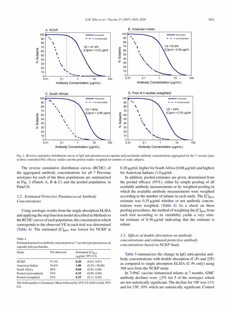
The reverse cumulative distribution curves (RCDC) ofhe aggregated antibody concentrations for all 7 Prevenarerotypes for each of the three populations are summarizedn Fig. 2 (Panels A, B & C) and the pooled population, inanel D.
.2. Estimated Protective Pneumococcal Antibodyoncentrations
Using serologic results from the single absorption ELISA
nd applying the step function model described in Methods tohe RCDC curves of each population, the concentration whichorresponds to the observed VE in each trial was determinedTable 4). The estimated [C]prot was lowest for NCKP atable 4stimated protective antibody concentration to 7 vaccine type pneumococcalapsular polysaccharides
tudy VE observed Estimated [C]prot
(�g/ml, 95% CI)
CKP 97.4% 0.20 (0.03, 0.67)merican Indian 76.8% 1.00 (0.25 > 50.00)outh Africa 90% 0.68 (0.03, 6.00)ooled (unweighted) 93% 0.35 (0.09. 0.89)ooled (weighted) 93% 0.35 (0.11, 0.85)
he bold number is Geometric Mean followed by 95% CI (GM is bold, 95%I).
elr
3cc
ba3
aaa
polysaccharide antibody concentrations aggregated for the 7 vaccine typesr of study subjects.
.20 �g/ml, higher for South Africa (0.68 �g/ml) and highestor American Indians (1.0 �g/ml.
In addition, pooled estimates are given, determined fromhe pooled efficacy (93%), either by simple pooling of allvailable antibody measurements or by weighted pooling inhich the available antibody measurements were weighted
ccording to the number of infants in each study. The [C]protstimate was 0.35 �g/ml whether or not antibody concen-rations were weighted. (Table 4) As a check on theseooling procedures, the method of weighting the [C]prot fromach trial according to its variability yields a very simi-ar estimate of 0.38 �g/ml indicating that the estimate isobust.
.3. Effects of double absorption on antibodyoncentrations and estimated protective antibodyoncentrations based on NCKP Study
Table 5 summarizes the change in IgG anticapsular anti-ody concentrations with double absorption (C-Ps and 22F)s compared to single absorption ELISA (C-Ps only) using68 sera from the NCKP study.
In 7vPnC vaccine immunized infants at 7 months, GMCntibody declines were ≤5% for 5 of the serotypes whichre not statistically significant. The decline for 19F was 11%nd for 23F, 10% which are statistically significant. Control

3822 G.R. Siber et al. / Vaccine 25 (2007) 3816–3826
Table 5Comparison of GMC IgG antibodies to 7 vaccine type capsular polysaccharides with and without 22F Absorption in infants immunized with Prevenar
Serotype Randomized treatmentgroupsa
GMC Antibody Concentration (�g/ml) Ratio (95%CI) of Antibodyconcentrations with/without22F absorptionWith 22F absorption Without 22F absorption
4 7vPnC 0.92 (0.81, 1.04) 0.94 (0.83, 1.05) 0.98 (0.96, 1.01)MnCC 0.01 (0.01, 0.01) 0.03 (0.02, 0.03) 0.43 (0.38, 0.48)
6B 7vPnC 2.75 (2.21, 3.41) 2.90 (2.40, 3.50) 0.95 (0.90, 1.00)MnCC 0.06 (0.06, 0.07) 0.15 (0.13, 0.17) 0.44 (0.40, 0.49)
9V 7vPnC 1.08 (0.97, 1.20) 1.11 (1.01, 1.23) 0.97 (0.95, 0.99)MnCC 0.05 (0.05, 0.06) 0.09 (0.08, 0.11) 0.59 (0.54, 0.63)
14 7vPnC 4.54 (3.98, 5.18) 4.48 (3.94, 5.10) 1.01 (0.98, 1.05)MnCC 0.03 (0.02, 0.04) 0.03 (0.03, 0.04) 0.85 (0.77, 0.95)
18C 7vPnC 1.23 (1.10, 1.39) 1.27 (1.13, 1.43) 0.97 (0.94, 1.01)MnCC 0.02 (0.02, 0.03) 0.05 (0.05, 0.06) 0.44 (0.39, 0.50)
19F 7vPnC 1.18 (1.04, 1.34) 1.33 (1.18, 1.49) 0.89 (0.85, 0.93)MnCC 0.05 (0.05, 0.06) 0.13 (0.11, 0.15) 0.43 (0.39, 0.48)
23F 7vPnC 1.12 (0.906, 1.31) 1.25 (1.08, 1.44) 0.90 (0.86, 0.94)MnCC 0.03 (0.03, 0.04) 0.07 (0.06, 0.08) 0.50 (0.45, 0.55)
Aggregated serotypes 7vPnC 1.54 (1.44, 1.63) 1.61 (1.52, 1.71) 0.95 (0.94, 0.97)03, 0.04
T CI).group w
swi
aw(l
cFm9ibtw
(iU[itc
3S
w
TCa
S
4691112
T
MnCC 0.03 (0.
he bold number is Geometric Mean followed by 95% CI (GM is bold, 95%a The number of subjects in the 7vPnC group was 188, and in the control
ubjects had very low anticapsular antibody concentrationshich showed significant declines after 22F absorption rang-
ng from 15 to 57% (Table 5).The proportion of infants responding to ≥0.35 �g/ml was
lso not changed significantly for 5 of the 7 types andas reduced by 2.7% for 19F (p < 0.05) and 5.3% for 23F
p < 0.01) (Table 6). Very few control infants had antibodyevels ≥0.35 �g/ml.
The RCDC of the antibody concentrations to all 7 typesombined, with and without 22F absorption, are shown inig. 3. Using the single absorbed RCDC (−22F), the esti-ated [C]prot of the NCKP population corresponding to the
3% pooled efficacy observed in the three controlled trials
s 0.35 �g/ml. This value is identical to the value obtainedy the WHO committee using the pooled RCDCs from all 3rial populations (Fig. 2, Panel D). This observation togetherith the similar effect of 22F absorption in the 3 populationsaiTI
able 6omparison of percent of subjects achieving IgG antibody concentration ≥0.35 �
bsorption in infants immunized with Prevenar
erotype % responding at ≥0.35 �g/ml (95% CI)
With 22 F absorptiona Without 22F
88.3 (82.8, 92.5) 87.8B 89.4 (84.0, 93.4) 91.5V 95.2 (91.1, 97.8) 95.74 97.9 (94.6, 99.4) 98.48C 93.6 (89.1, 96.7) 94.79F 92.0 (87.2, 95.5) 94.73F 85.1 (79.2, 89.9) 90.4
he bold number is Geometric Mean followed by 95% CI (GM is bold, 95% CI).a Exact 2-sided confidence interval based upon the observed proportion of subjecb p-value computed using the Exact McNemar Test for paired samples.
) 0.07 (0.06, 0.07) 0.51 (0.49–0.53)
as 180.
Section 3.4) supports the use of the RCDC from the NCKPnfants to assess the impact of 22F absorption on the [C]prot.sing the double absorbed RCDC curves (+22F, Fig. 3), the
C]prot corresponding to the 93% pooled efficacy observedn the three trials was determined to be 0.32 �g/ml. Thushe impact of 22F absorption on the estimate of protectiveoncentration is small.
.4. Effect of 22F absorption in American Indian andouth African infants
The effect of 22F absorption in American Indian infantsas assessed in parallel assays in 76 Prevenar recipients
nd 86 controls but the new sample differed from the orig-nal sera assayed because of insufficient available volumes.he new sample of Prevenar immunized infants had higher
gG ELISA antibody concentrations without 22F absorption
g/mL to 7 vaccine type capsular polysaccharides with and without 22F
Difference (%) p-valueb
absorptiona
(82.2, 92.1) 0.5 0.564(86.5, 95.1) −2.1 0.102(91.8, 98.1) −0.5 0.564(95.4, 99.7) −0.5 0.317(90.4, 97.4) −1.1 0.317(90.4, 97.4) −2.7 0.025(85.3, 94.2) −5.3 0.002
ts.

G.R. Siber et al. / Vaccine 25
FaN
(2t0nfcTwn
nwtowtfp
cap3aciA
4
ifegct
epmii(aw
dcms
detoctibei
smcao2eratp
sdwtbecftu7ilt
ig. 3. RCDC of IgG pneumococcal capsular polysaccharide antibodiesggregated for 7 vaccine types with and without type 22F absorption inCKP infants.
GMC: 2.97 �g/ml) for the pooled serotypes compared to.35 �g/ml in the original WHO sample (Table 3). The con-rols were similar for the new sample: 0.09 �g/ml versus.08 �g/ml in the WHO sample. After 22F absorption of theew post-immunization samples the pooled GMC declinedrom 2.97 �g/ml to 2.84 �g/ml (ratio 0.96, p < .01) and ofontrols from 0.09 �g/ml to 0.06 �g/ml (ratio 0.63 p < .001).hese declines are of similar magnitude as in NCKP infantsho had ratios of 0.95 and 0.51 in immunized and unimmu-ized infants, respectively. (Table 5)
When the protective concentration was calculated from theew American Indian sample, it increased from 1.00 �g/mlith the original sample utilized by WHO to 1.41 �g/ml with
he new sample when the new samples were assayed with-ut 22F absorption. When the new samples were absorbedith 22F, the protective estimate declined from 1.41 �g/ml
o 1.37 �g/ml (2.8%) a proportion even less than the declinerom 0.35 �g/ml to 0.32 �g/ml (8.6%) observed in the NCKPopulation. (Fig. 3)
The effect of 22F absorption in South African infantsould be evaluated in only 19 infants (7 Prevenar recipientsnd 12 controls) from whom sera were still available. Theooled GMC for all 7 serotypes declined from 3.97 �g/ml to.71 �g/ml (ratio 0.93, NS) in Prevenar immunized infantsnd from 0.13 �g/ml to 0.07 �g/ml (ratio: 0.52, p < 0.1) inontrols. Again, the effect of 22F absorption in South Africannfants was similar to the effects observed in NCKP andmerican Indian infants.
. Discussion
Once an effective vaccine has been licensed and placednto widespread use, it is no longer feasible or ethical to per-orm additional controlled clinical studies to demonstrate the
fficacy of a new vaccine against the same pathogen in the ageroup for which the vaccine is recommended. Immunologicorrelates of protection then become critical for predictinghe efficacy of new vaccines.osap
(2007) 3816–3826 3823
For most vaccines, protective activity is mediated eitherxclusively or primarily by antibodies, and the correlate ofrotection is thus a specified concentration of antibody esti-ated to confer protection in an immunized population. An
deal protective correlate should have a number of character-stics. First it should measure the protective activity directlye.g. antitoxin, bacterial killing or virus neutralization) orlternatively measure binding antibodies which are correlatedith functional protective activity.Second, it should be a well standardized, validated, repro-
ucible and preferably inexpensive assay which can beonveniently applied to the large numbers of patients thatust be studied during clinical development of vaccines and
ubsequently during post-marketing studies.Third, the chosen protective concentration should be
irectly linked to clinical protection observed in controlledfficacy trials. In determining the protective level, it is impor-ant to avoid choosing concentrations that are either too highr too low. If the chosen correlate is too high, then new vac-ines may be rejected for inadequate efficacy even thoughhey would be highly protective. If the protective correlates set inappropriately low, then new vaccines may meet thisenchmark and yet have lower antibody responses and lowerfficacy than the vaccine which was shown to be efficaciousn clinical trials.
In this paper we present the antibody concentrations andtatistical methods used by a WHO Working Group to recom-end the protective concentration of IgG class pneumococcal
apsular polysaccharide antibodies measured by ELISA. Inddition we present the impact of a significant modificationf the IgG ELISA assay, absorption of test sera with type2F polysaccharide, on antibody concentrations and on thestimated protective concentration.Absorption with type 22Fenders the assay more specific for capsular polysaccharidentibodies which are the only known functional and protec-ive antibodies induced by pneumococcal polysaccharide andolysaccharide conjugate vaccines.
WHO conducted an initial consultation in Alaska in 2002,ummarized by Jodar et al. [3] and developed recommen-ations at a second consultation in Geneva in 2003 whichere issued in 2005 [4]. The theoretical basis for estimating
he protective concentration was to relate the observed anti-ody concentrations in the populations studied in controlledfficacy trials to the observed point estimates of vaccine effi-acy against invasive pneumococcal disease (see Section 2or details). At the time of the WHO consultations, three con-rolled efficacy trials had been conducted with PnC vaccinessing IPD as a primary outcome. The first and largest trial ofvPnC vaccine at NCKP, on which licensure of this vaccinen the US was based, had the highest VE (97.3%) and theowest estimated [C]prot (0.20 �g/ml). The committee madehe decision not to base the protective concentration solely
n the NCKP trial. Rather, the committee chose a more con-ervative approach, pooling the NCKP study with the twodditional controlled studies then available which had beenerformed in American Indian and South African infants.
3 ccine 25
Tap(
otcot0rta
feoibbtmpwcwsemmIpoiatIpfc0l
eiibiocpTcop
7tspssmtoiaet(io(
ptintwtpsw
dvaPibtbaltatTup
fto9cu
824 G.R. Siber et al. / Va
hese additional trials showed vaccine efficacies of 76.8%nd 90% respectively for the 7 vaccine types in Prevenar androtective concentrations of 1.0 and 0.68 �g/ml respectively.See Tables 1 and 3)
The committee deemed the pooled efficacy of 93%btained by combining these trials more representative ofhe likely efficacy of the vaccine globally. The pooled effi-acy estimate is also very close to the intent-to-treat efficacyf 93.9% observed in NCKP Study [6]. The estimated pro-ective antibody concentration from the pooled studies was.35 �g/ml. As described in Section 3, this estimate wasobust, regardless of whether the measured antibody concen-rations were simply pooled or whether they were weightedccording to the numbers of patients in each study.
A subsequent controlled study of 9 valent PnC vaccine per-ormed in The Gambia [16], showed a vaccine efficacy pointstimate of 86% for IPD. In the per protocol analysis, 28 casesf IPD caused by the 7 serotypes in Prevenar were observedn controls and 4, in the 9vPnC group. The protective anti-ody estimate in Gambian infants was 1.20 �g/ml measuredy ELISA with 22F absorption. These results suggest that thehree groups who are at higher risk for pneumococcal diseases
ay require higher antibody levels to achieve an equivalentrotective efficacy than Californian infants. However, evenhen the three studies in high-risk groups are pooled, the 95%
onfidence intervals of the IPD VE (85%, Cl: 66, 94) overlapith those of the NCKP (97.4%, Cl: 85, 100). Possible rea-
ons for lower vaccine efficacy in these populations includearlier exposures to pneumococci, more frequent exposure toultiple serotypes shown by more rapid acquisition of pneu-ococcal carriage in South African, Gambian and American
ndian infants, exposure to larger inoculum sizes and theresence of concurrent infections such as endemic malariar respiratory infections. It is also possible that differencesn host factors such as innate immune defenses, functionalctivity of their antibodies or nutrition play a role. Finally,he severity of the IPD outcomes was greater in the Americanndian and South African studies than at NCKP where a largeroportion had occult pneumococcal bacteremia without aocus. In any case, lowering the estimate of protective con-entration for purposes of assessing vaccine efficacy below.35 �g/ml would risk the licensure of vaccines which haveower efficacy in populations with diverse risk factors.
An important simplifying assumption that is required tostimate a single protective level applicable to all serotypess that the concentration of antibody required for protections in fact similar for all types. This assumption is necessaryecause the rates of IPD are low, resulting in estimates ofndividual serotype efficacy with wide confidence intervalsr with point estimates of 100%, which preclude the cal-ulation of protective concentrations. Several observationsrovide some assurance that this assumption is reasonable.
he variation of GMC antibody concentrations to the 7 vac-ine types after immunization is relatively narrow, rangingver a 2 to 3 fold range in the three studies (Table 3). Theooled serotype specific efficacies in the three studies for theaidc
(2007) 3816–3826
vaccine types are also quite similar (Table 2) suggestinghat the observed differences in immunogenicity among theeven vaccine serotypes do not lead to major differences inrotective activity. Post-marketing studies in the US ABCurveillance system have yielded the most precise serotypepecific effectiveness data available to date [17]. The post-arketing effectiveness differ from per protocol efficacy in
he controlled studies in that children who received onlyne or more doses of PnC on a variety of schedules werencluded, which would tend to reduce vaccine effectiveness,nd indirect herd immune effects occur, tending to increaseffectiveness. Nevertheless, these data confirm that protec-ive activity is similar across all 7 serotypes in the vaccineTable 2, last column). For each of the 7 serotypes, 90% ofndividuals with >0.35 �g/ml of IgG antibody have evidencef opsonophagocytic activity in a validated functional assay3 and unpublished data, Wyeth).
After the WHO Working Group defined the protectiveneumococcal antibody concentration of 0.35 �g/ml, a fur-her improvement in the specificity of the IgG ELISA wasntroduced and has now been widely accepted [9]. The origi-al protocol absorbed non-specific antibodies binding to theype specific polysaccharides used for coating ELISA platesith C-Ps (single absorption). Conception and Frasch found
hat the use of a second absorbent, type 22F pneumococcalolysaccharide (double absorption), further reduced non-pecific antibody binding which still occurred, particularlyith sera from adults. [9]We utilized sera from the pivotal NCKP efficacy trial to
irectly compare anti-pneumococcal antibodies in the Pre-nar immunized and control infants at 7 months. Doublebsorption induced negligible additional declines in sera fromrevenar immunized infants and somewhat greater reductions
n the unimmunized controls (Table 6, Fig. 3). The differenceetween immunized and control groups may be explained byhe fact that the high concentrations of pneumococcal anti-ody measured in the immunized group is mainly specificnti-capsular antibody induced by PnC vaccine, whereas theow concentrations of pneumococcal antibody in the con-rol group represents either residual pneumococcal antibodycquired transplacentally from mothers or the responses ofhe infants themselves to natural pneumococcal exposures.hese naturally induced antibodies in unimmunized individ-als are more likely to contain antibodies to non-capsularneumococcal antigens.
Using the single absorbed ELISA values and the RCDCrom the NCKP study, the 0.35 �g/ml protective concentra-ion established by the WHO working group predicts a VEf 92.7%, which is very close to the estimated pooled VE of3% utilized by the WHO committee to determine this con-entration originally. If double absorbed antibody values aresed, the calculated protective concentration is slightly lower
t 0.32 �g/ml. This decline is driven not by the slight changen antibody levels in immunized infants but by the largereclines in control infants. Both are used in the formula foralculating [C]prot.
ccine 25
ceppiSliras
ltp
iwsa1vPdbd
capcpfo[eC6a
icpwasa
ttpospa
iac
ttcWcmwssttc
mTccmwcIboa
ricovmi
A
SatSoaS
R
G.R. Siber et al. / Va
Recently Henckaerts et al. proposed that the protectiveoncentration for assessing PnC vaccine efficacy be low-red from 0.35 �g/ml to 0.20 �g/ml when using their ELISArotocol [18]. They based this proposal on reductions inost- immunization antibodies measured in sera of infantsmmunized with a different PnC manufactured by GlaxomithKline utilizing a different protein carrier and different
inkage chemistry. The ELISA assay showing the reductionn antibody after 22F absorption also differed in importantespects from the WHO ELISA protocol including coatingntigens, coating conditions, and absorption of the 89SFtandard with 22F Ps. [12]
The data presented by Henckaerts et al. do not supportowering the protective pneumococcal antibody concentra-ion because they did not follow a number of fundamentalrinciples required for valid bridging of this estimate.
The most important principle is that the sera used for bridg-ng must be from infants immunized with the vaccine forhich efficacy was clinically demonstrated i.e, Prevenar. The
era evaluated by Henckaerts et al. which showed a declinefter 22F absorption were from infants immunized with an1-valent PnC manufactured by Glaxo SmithKline. The 11-alent vaccine is manufactured by different processes thanrevenar. In particular, a different linkage chemistry and aifferent protein carrier are used. It is well known that anti-ody responses induced by different conjugate vaccines mayiffer in concentrations and quality of antibody [19,20].
It has also been demonstrated that pneumococcal polysac-haride and conjugate vaccines can induce antibodies to C-Psnd protein contaminants [21–24]. Different manufacturingrocesses may result in different concentrations of theseontaminating antigens in the final product. For example,eriodate oxidation, a step used in the conjugation processor Prevenar, has been shown to reduce the immunogenicityf the immunodominant phosphorylcholine epitope on C-Ps24–26]. Thus, pneumococcal antibodies induced by differ-nt conjugates may differ in the effects of absorption with-Ps and 22F. Indeed the Henckaerts report showed that typeB pneumococcal antibodies after Prevenar did not declinefter type 22F absorption [18].
The second principle is that the study population for bridg-ng should be the same or equivalent to the one in whichlinical efficacy was demonstrated. Theoretically, differentopulations may vary in their exposure to pneumococcihich may result in differences in their concentrations of
ntibodies to nonspecific pneumococcal antigens and thushow a greater reduction in anticapsular antibodies with 22Fbsorption than pertained in the NCKP study.
The third principle is that the ELISA assay used to assesshe effect of 22F absorption must be the same as the assay usedo establish the protective concentration (ie, the WHO ELISArotocol) [12]. Therefore the effect of an assay modification
n protective anticapsular antibody concentrations should betudied by assessing the modification in the WHO method inarallel assays in a single laboratory where the presence orbsence of 22F is the only variable. The comparison should(2007) 3816–3826 3825
nclude a sufficient number of sera from Prevenar immunizednd control infants to give confidence in the result. Theseonditions were not met by Henckaerts et al.
The data on 22F absorption presented in this report followhe above principles for bridging the pneumococcal protec-ive antibody concentration. Using the NCKP sera, the con-entration declined from 0.35 �g/ml estimated by the WHOorking Group to 0.32 �g/ml after 22F absorption, providing
onfidence that this concentration is based on the measure-ent of antibodies specific for the capsular polysaccharidehich confer protection. Although many of the original
erum samples from the American Indian and South Africantudies were not available in sufficient volumes, we did assesshe effect of 22F absorption on a modified serum set fromhese studies and showed only small declines in antibody con-entration, similar to the declines observed in NCKP infants.
Several caveats should be kept in mind about the pneu-ococcal protective concentration described in this report.his concentration applies only to the prevention of IPD inhildren who resemble the trial populations. For examplehildren with immuno-compromising conditions such a HIVay require higher antibody levels for protection. Infantsith HIV have been shown to produce antibodies to pneumo-
occi [27] and Hib [28] with lower functional activity per �ggG than healthy infants.Also, circulating anticapsular anti-ody concentrations required for prevention of pneumonia,titis media or colonization are likely to be higher [29,30]nd have not been established.
Also the protective level is best applied to populationsather than to individuals [1]. Thus, if a high proportion ofndividuals in a population achieves anticapsular antibodyoncentration ≥ 0.35 �g/ml then we can predict a high levelf protection for IPD in that population. In a particular indi-idual the outcome of an encounter with the pneumococcusay vary depending on multiple host and pathogen factors,
n addition to the circulating antibody level.
cknowledgements
We thank Bernard Fritzell, Jill Hackell, Mark Fletcher,teve Lockhart, Dace Madore, and Sally Quataert for valu-ble discussions and review of the manuscript. We thankhe investigators of Northern California Kaiser Permanentetudy for the use of the antibody data from their study. Thepinions expressed in this paper are those of the authorsnd do not necessarily reflect the views of the Indian Healthervice.
eferences
[1] Siber GR. Methods for estimating serological correlates of protection.Dev Biol Stand 1997;89:283–96.
[2] Robbins JB, Schneerson R, Szu SC. Perspective: hypothesis: serum IgGantibody is sufficient to confer protection against infectious diseases byinactivating the inoculum. J Infect Dis 1995;171:1387–98.

3 ccine 25
[
[
[
[
[
[
[
[
[
[
[
[
[
[
[
[
[
[
[
[
826 G.R. Siber et al. / Va
[3] Jodar L, Butler J, Carlone G, Dagan R, Goldblatt D, et al. Serologicalcriteria for evaluation and licensure of new pneumococcal conjugatevaccine formulations for use in infants. Vaccine 2003;21:3265–72.
[4] WHO, (2005) Recommendations for the production and control ofpneumococcal conjugate vaccines. WHO Technical Report Series. No.927.
[5] Belshe RB, Gruber WC, Mendelman PM, Mehta HB, Mahmood K,et al. Correlates of immune protection induced by live, attenuated,cold adapted, trivalent, intranasal influenza virus vaccine. J Infect Dis2000;181:1133–7.
[6] Black S, Shinefield H, Fireman B, Lewis E, Ray P, et al. Efficacy, safetyand immunogenicity of heptavalent pneumococcal conjugate vaccinein children. Pediatr Infect Dis J 2000;19:187–95.
[7] O’Brien KL, Moulton LH, Reid R, Weatherholtz R, Oski J, et al.Efficacy and safety of seven-valent conjugate pneumococcal vac-cine in American Indian children: group randomized trial. Lancet2003;362:355–431.
[8] Klugman KP, Madhi SA, Huebner RE, Kohberger R, Mbelle N, et al.A trial of a 9 valent pneumococcal conjugate vaccine in children withand those without HIV infection. N Engl J Med 2003;349:1341.
[9] Concepcion NF, Frasch CE. Pneumococcal type 22F polysaccharideabsorption improves the specificity of a pneumococcal polysaccha-ride enzyme linked immunosorbent assay. Clin Diagn Lab Immunol2001;8:266–72.
10] Hausdorff WP, Bryant J, Paradiso PR, Siber GR. Which pneumococ-cal serogroups cause the most invasive disease. Part I. Implicationsfor conjugate vaccine formulation and use. Clin Infect Dis 2000;30:100–21.
11] Quataert SA, Kirch CS, Wiedl LJ, Phipps DC, Strohmeyer S, et al.Assignment of weight-based antibody units to a human antipneumo-coccal standard reference serum, lot 89-S. Clin Diagn Lab Immunol1995;2:590–7.
12] Wernette CM, Frasch CE, Madore D, Carlone G, Goldblatt D, et al.Enzyme linked immunosorbent assay for quantitation of human anti-bodies to pneumococcal polysaccharides. Clin Diagn Lab Immunol2003;10:514–9.
13] Shinefield HR, Black S, Ray P, Chang I, Lewis N, Fireman B, etal. Safety and immunogenicity of heptavalent pneumococcal CRM197 conjugate vaccine in infants and toddlers. Pediatr Infect Dis J1999;18:757–63.
14] Huebner R, Mbelle N, Forrest B, Madore DV, Klugman KP. Immuno-genicity after one, two or three doses and impact on the antibodyresponse to coadministered antigens of a nonavalent pneumococcalconjugate vaccine in infants of Soweto, South Africa. Pediatr InfectDis J 2002;21:1004–7.
15] O’Brien KL, Moisi J, Moulton LH, Madore DV, Eick A, Reid R, et al.Predictors of conjugate pneumococcal vaccine immunogenicity amonginfants and toddlers in the American Indian PnCRM 7 efficacy trial. J
Infect Dis 2007; in press.16] Cutts FT, Saman SMA, Enwere G, Jaffar S, Levine OS, et al. Efficacyof nine-valent pneumococcal conjugate vaccine against pneumonia andinvasive pneumococcal disease in The Gambia: randomized, double-blind, placebo-controlled trial. Lancet 2005;356:1139.
[
(2007) 3816–3826
17] Whitney CG, Pilishvili T, Farley MM, Schaffner W, Craig AS, et al.Effectiveness of seven-valent pneumococcal conjugate vaccine againstinvasive pneumococcal disease: a matched case control study. TheLancet 2006;368:1495.
18] Henckaerts I, Goldblatt D, Ashton L, Poolman J. Critical differencesbetween pneumococcal polysaccharide enzyme-linked immunosorbentassays with and without 22F inhibition at low antibody concen-trations in pediatric sera. Clin Vaccine Immunol Mar 2006:356–60.
19] Soininen A, Seppala I, Nieminen T, Eskola J, Kayhty H. IgG subclassdistribution of antibodies after vaccination of adults with pneumococcalconjugate vaccines. Vaccine 1999;17:1889–97.
20] Anttila M, Eskola J, Ahman H, Kayhty. Differences in the avidity ofantibodies evoked by four different pneumococcal conjugate vaccinesin early childhood. Vaccine 1999;17:1970–7.
21] Pedersen FK, Henrichsen J, Sorensen US, Nielsen JL. Anti-C- carbohy-drate antibodies after pneumococcal vaccination. Acta Path MicrobiolImmunol Scand Sect C 1982 1982;90:353–5.
22] Soininen A, van den Dobbelsteen G, Oomen L, Kayhty H. Arethe enzyme immunoassays for antibodies to pneumococcal cap-sular polysaccharides serotype specific? Clin Diagn Lab Immunol2000;7:468–76.
23] Yu J, Briles DE, Englund JA, Hollingshead SK, Glezen WP, NahmMH. Immunogenic Protein Contaminants in Pneumococcal Vaccines.J Infect Dis 2003;187:1019.
24] Peeters C, Teubergen-Meekes A-M, Poolmann J, Zegers B, RijkersG. Induction of antipneumococcal cell wall polysaccharide antibod-ies by type 4 pneumococcal polysaccharide-protein conjugates. MedMicrobiol Immunol 1992;181:35–42.
25] Casal J, Jado I, Fenoll A, Perez A, Torano A. Periodate oxidation ofR36A pneumococci greatly enhances production of hybridomas secret-ing anti protein antibodies. Microb Pathog 1998;24:111–6.
26] Laferriere CA, Sood RK, deMuys J-M, Michon F, Jennings HJ. Thesynthesis of Streptococcus pneumoniae polysaccharide tetanus toxoidconjugates and the effect of chain length on immunogenicity. Vaccine1997;15(2):179–86.
27] Madhi SA, Kuwanda L, Cutland C, Holm A, Kayhty H, et al. Quan-titative and qualitative antibody response to pneumococcal conjugatevaccine among African human immunodeficiency virus-infected anduninfected children. Ped lnf Dis J 2004;24(5):410–6.
28] Madhi SA, Kuwanda L, Saarinen L, Cutland C, Mothupi R, KayhtyH, Klugman K. Immunogenicity and effectiveness of Haemophilusinfluenzae type b conjugate vaccine in HIV infected and uninfectedAfrican children. Vaccine 2005;23:5517–25.
29] Jokinen JT, Ahman H, Kilpi TM, Makela PH, Kayhty MH. Con-centration of antipneumococcal antibodies as a serological correlateof protection: an application to acute otitis media. J Infect Dis2004;190:545–50.
30] Dagan R, Givon-Lavi N, Fraser D, Lipsitch M, Siber GR, KohbergerR. Serum serotype-specific pneumococcal anticapsular immunoglob-ulin G concentrations after immunization with a 9-valent conjugatepneumococcal vaccine correlate with nasopharyngeal acquisition ofpneumococcus. J Infect Dis 2005;192:3670–6.