diverse evolutionary patterns of pneumococcal antigens
TRANSCRIPT
Diverse evolutionary patterns of pneumococcalantigens identified by pangenome-wideimmunological screeningNicholas J. Crouchera,1, Joseph J. Campob, Timothy Q. Leb, Xiaowu Liangb, Stephen D. Bentleyc, William P. Hanaged,and Marc Lipsitchd
aDepartment of Infectious Disease Epidemiology, Imperial College London, London W2 1PG, United Kingdom; bAntigen Discovery Inc., Irvine, CA 92618;cInfection Genomics, The Wellcome Trust Sanger Institute, Wellcome Trust Genome Campus, Hinxton, Cambridge CB10 1SA, United Kingdom; and dCenterfor Communicable Disease Dynamics, Harvard T. H. Chan School of Public Health, Boston, MA 02115
Edited by Roy Curtiss III, University of Florida, Gainesville, FL, and approved December 5, 2016 (received for review August 25, 2016)
Characterizing the immune response to pneumococcal proteins iscritical in understanding this bacterium’s epidemiology and vaccinol-ogy. Probing a custom-designed proteome microarray with serafrom 35 healthy US adults revealed a continuous distribution ofIgG affinities for 2,190 potential antigens from the species-widepangenome. Reproducibly elevated IgG binding was elicited by208 “antibody binding targets” (ABTs), which included 109 variantsof the diverse pneumococcal surface proteins A and C (PspA andPspC) and zinc metalloprotease A and B (ZmpA and ZmpB) proteins.Functional analysis found ABTs were enriched in motifs for secretionand cell surface association, with extensive representation of cellwall synthesis machinery, adhesins, transporter solute-binding pro-teins, and degradative enzymes. ABTs were associated with strongerevidence for evolving under positive selection, although this variedbetween functional categories, as did rates of diversification throughrecombination. Particularly rapid variation was observed at some im-munogenic accessory loci, including a phage protein and a phase-vari-able glycosyltransferase ubiquitous among the diverse set of genomicislands encoding the serine-rich PsrP glycoprotein. Nevertheless, manyantigens were conserved in the core genome, and strains’ antigenicprofiles were generally stable. No strong evidence was found for anyepistasis between antigens driving population dynamics, or redun-dancy between functionally similar accessory ABTs, or age stratifica-tion of antigen profiles. These results highlight the paradox of whysubstantial variation is observed in only a subset of epitopes. Thisresult may indicate only some interactions between immunoglobulinsand ABTs clear pneumococcal colonization or that acquired immunityto pneumococci is an accumulation of individually weak responses toABTs evolving under different levels of functional constraint.
genomics | pathogens | evolution | immunology | epidemiology
The pneumococcus (Streptococcus pneumoniae) is a Gram-positive human commensal and respiratory pathogen com-
monly carried in the nasopharynx of young infants (1). Theprevalence of carriage peaks within the first 3 y of life (2). How-ever, the bacteria are cleared increasingly quickly with age fol-lowing successive episodes of carriage, a change associated withthe development of mucosal immune responses (3). These involveinteractions with antibody binding targets (ABTs), recognized byIg A (IgA) and G (IgG) antibodies, or T-cell receptor targets(TCRTs), recognized by CD4+ TH17 cells (4). The former re-sponse leads to targeted opsonophagocytosis of cells bearingABTs, neutralization of toxins, and inhibition of adhesion to hosttissues (5, 6). TCRT-triggered secretion of interleukin-17A byCD4+ TH17 cells results in nonspecific clearance of pneumococcifrom the nasopharynx through the recruitment of neutrophils andmacrophages (7, 8). These interactions with the host immunesystem are likely to be important in this bacterium’s evolution (9, 10).The historical focus of pneumococcal immunology has been
the polysaccharide capsule, which has over 90 antigenicallydistinguishable variants (serotypes). Systemic immunization with
capsular polysaccharides can stimulate protective levels of se-rotype-specific antibodies (11, 12). Conjugate vaccines including7–13 different capsular polysaccharides (13) provide protectionagainst the serotypes included in the vaccine, but a desire forvaccines with broader coverage has caused growing interest inalternative protein- or whole cell-based formulations (14). Pre-vious studies have used ELISA (15), phage display libraries (16),or “antibody fingerprinting” (17) to measure the antibody re-sponse to the noncapsular pneumococcal antigens of individualstrains. Now, population genomic datasets can be combined withproteome microarrays, an approach with several advantages forstudying the host–bacterium interaction. First, this pairing canidentify a more complete set of antigens, including proteins thatmay be absent from any one individual strain, such as the type 1pilus (18, 19); the prevalence of these proteins can also beascertained from the genomic data. Second, for highly poly-morphic proteins such as pneumococcal surface proteins A (PspA)and C (PspC) (20, 21), this combination allows the immuneresponse to a diverse panel of variants to be assayed, and their
Significance
The wealth of genomic data available for the respiratory patho-gen Streptococcus pneumoniae enabled the design of a pan-genome-wide proteome microarray. Of over 2,000 pneumococcalproteins, 208 strongly bound antibodies in adult human sera. Thevast majority could be classified as either variants of four diverseloci or more conserved proteins involved in adhesion, enzymaticdegradation, solute binding, or cell wall synthesis. Detailed anal-yses of the genomic data revealed some variable antigens rapidlydiversified through mechanisms including homologous recom-bination, mobile genetic element transmission, and phase varia-tion. Other antigens were conserved across the population andmay be better candidates for simple vaccine formulations. Thisraises the question of what evolutionary advantage bacteria de-rive from altering only a subset of their antigenic loci.
Author contributions: N.J.C., W.P.H., and M.L. designed research; N.J.C., J.J.C., T.Q.L., andX.L. performed research; N.J.C. contributed new reagents/analytic tools; N.J.C., J.J.C., T.Q.L.,X.L., and S.D.B. analyzed data; and N.J.C., W.P.H., and M.L. wrote the paper.
Conflict of interest statement: J.J.C., T.Q.L., and X.L. are employees of Antigen Discovery,Inc. In addition, X.L. has an equity interest in Antigen Discovery, Inc. N.J.C., S.D.B., W.P.H.,and M.L. were consultants asked to design the proteome array by Antigen Discovery, Inc.In addition, M.L. has received consulting fees from Pfizer and Affinivax, travel reimburse-ment from GlaxoSmithKline, and grant funding through his institution from Pfizer andPATH Vaccine Solutions.
This article is a PNAS Direct Submission.
Data deposition: The sequences reported in this paper have been deposited in the Euro-pean Nucleotide Archive under project code PRJEB2632 and accession codes LT669625–LT669755, and in the Dryad repository under doi.org/10.5061/dryad.t55gq.1To whom correspondence should be addressed. Email: [email protected].
This article contains supporting information online at www.pnas.org/lookup/suppl/doi:10.1073/pnas.1613937114/-/DCSupplemental.
www.pnas.org/cgi/doi/10.1073/pnas.1613937114 PNAS | Published online January 4, 2017 | E357–E366
MICRO
BIOLO
GY
PNASPL
US
Dow
nloa
ded
by g
uest
on
Feb
ruar
y 10
, 202
2
distribution across the population to be established. Third, therate at which different loci diversify through mutation and re-combination can be inferred from evolutionary analyses of ge-nomes. This is particularly important in a naturally transformablespecies such as S. pneumoniae, in which vaccine-escape variantscan emerge through recombination, as previously observed for thecapsule polysaccharide synthesis (cps) locus (22).This study used genomic data from 616 nasopharyngeal carriage
isolates from young children in Massachusetts (23, 24). The majorityof these isolates were classified into 15 monophyletic sequenceclusters (SCs), using a core genome alignment; each of these cor-responded to a common genotype, the recent diversification ofwhich could be reconstructed based on whole-genome alignments(25). The 1.2 million protein-coding sequences (CDSs) identified inthese de novo assemblies were grouped into 5,442 clusters oforthologous genes (COGs) (26); these groupings of similar proteinswere used to inform the design of a proteome microarray formeasuring IgG binding levels. Here, we characterize the antibodyresponse to thousands of pneumococcal proteins, using healthyadult sera, and describe the distinct evolutionary patterns associatedwith the most immunogenic sequences.
ResultsExtensive Interactions Between the Host and Pneumococcus. To capturethe most common interactions between hosts and pneumococci,representative sequences were included for each COG present in atleast 20% of the 616 isolates. Other proteins that proved difficult toassemble from short read data were also included: the pneumo-coccal serine-rich repeat protein PsrP; the cellular autolytic amidaseLytA; a phage amidase; the phage antireceptor PblB; and an olig-omer of choline binding domains (CBDs), a common cell-surfaceattachment motif found in many pneumococcal protein antigens.Rare COGs were included if they were likely to be antigenic: Thesewere type 2 pilus components, zinc metalloprotease ZmpC, andZmpE, a zinc metalloprotease unique to the atypical unencapsu-lated pneumococci of SC12 (24, 27). Additionally, multiple variantsof some diverse loci were included. Each of the three “clades” ofthe type I pilus RrgB protein (28) corresponded to a separate COG,as did five variants (one truncated) of the PclA protein. Repre-sentatives were added for each of the previously defined variants ofthe three diverse penicillin-binding proteins (three for Pbp2X andPbp1A and four for Pbp2B), which were associated with differinglevels of β-lactam sensitivity (24). Finally, the manually curated setsof all complete representatives of the “diverse core loci” (encodingPspA, PspC, and the zinc metalloproteases ZmpA and ZmpB) wereidentified (SI Appendix). Owing to the low similarity between manyrepresentatives, these sets were divided into variants, using analignment-free, kmer-based approach (SI Appendix, Fig. S1). Rep-resentatives could be included on the microarray for 36 of the 39PspA variants, 57 of the 59 PspC variants, and all of the 18 ZmpAvariants and 16 ZmpB variants. An independent phylogenetic anal-ysis indicated these sets encompassed the full previously observeddiversity of these proteins (20, 21) (SI Appendix, Fig. S2). Overall, atotal of 2,190 proteins derived from the Massachusetts pneumococ-cal population were included on the microarray; also included wasthe full proteome of S. pneumoniae TIGR4 (29), but these proteinswere excluded from the described analyses to maintain unbiasedcoverage across the systematically sampled bacterial isolates.Using the sera of 35 healthy adults, IgG binding responses to all
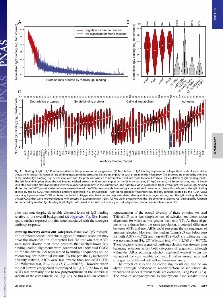
of the Massachusetts-derived proteins on the microarray werenormalized and plotted on a logarithmic scale (Fig. 1A). Althoughthe distribution of binding responses was continuous, a subset of theproteins did elicit a consistently elevated antibody response acrossthe serum samples. Dividing these data into two groups classed 208of the 2,190 proteins as ABTs for subsequent analyses. Approxi-mately half of these ABTs were variants of the four diverse core loci(Fig. 1B and SI Appendix, Figs. S3–S5): These included 29 of the 36PspA probes [Fisher’s exact test, odds ratio (OR) relative to
COGs = 84.9, P < 2.2 × 10⁻¹⁶], 48 of the 57 PspC probes (OR =109.5, P < 2.2 × 10⁻¹⁶); 17 of the 18 ZmpA probes (OR = 346.0, P <2.2 × 10⁻¹⁶), and 15 of the 16 ZmpB probes (OR = 304.8, P < 2.2 ×10⁻¹⁶). The IgG binding across the diverse variants of PspA andPspC (SI Appendix, Figs. S3 and S4) is generally substantiallyhigher than that of the CBD (SI Appendix, Fig. S6), the one se-quence motif commonly shared between these proteins, indicatingthat these results reflect a broad immune repertoire spanning theallelic diversity of these proteins.These results were compared with a previous “antigenic finger-
printing” study of S. pneumoniae TIGR4 that identified epitopes in97 annotated CDSs (17). The proteins from the Massachusettsisolates on the array contained 2,039 orthologs of sequences inS. pneumoniae TIGR4 (considering the sets of PspA, PspC, ZmpA,and ZmpB variants each as single orthologs). The 88 COGs fromthe Massachusetts isolates’ proteome that were orthologous to theantigens identified by fingerprinting had a significantly increasedprobability of being identified as ABTs in this study relative to the265 COGs on the microarray not linked to CDSs in TIGR4(Fisher’s exact test, OR = 8.93, P = 1.81 × 10⁻¹¹; Fig. 1B). In turn,those TIGR4 proteins not identified through fingerprinting weresignificantly less likely to be identified as ABTs than those proteinsnot linked to CDSs in TIGR4 (Fisher’s exact test, OR = 0.45, P =0.014). Hence, the proteins eliciting the highest antibody bindingresponse comprised a combination of diverse PspA, PspC, ZmpA,and ZmpB variants, and representatives of other proteins that sig-nificantly overlap with the antigens identified by a different meth-odology applied to an individual strain.
Functional Categories Associated with ABTs. A multivariable logisticregression was used to identify the protein characteristics enrichedin ABTs (SI Appendix, Table S1). Both the conventional signalpeptide and the secretion-associated YSIRKmotif were significantlyassociated with ABTs, as were the lipid attachment site, which di-rects lipoproteins to be connected to the cell membrane, and theCBD. Notably, whereas the size of a protein was significantly as-sociated with elevated IgG binding, the number of transmembranehelices was not. Therefore, the ABTs are enriched for large proteinsthat are secreted or peripherally associated with the cell surface.Manual annotation of the ABTs revealed they performed di-
verse functional roles, but most could be grouped into four cate-gories (Fig. 1C and SI Appendix). The first group was degradativeenzymes, acting on a wide range of extracellular substrates in-cluding proteins, polysaccharides, and their derivatives. The sec-ond group was ABC transporter solute binding proteins (SBPs),with representatives specific for amino acids, sugars, nucleotides,and siderophores. The third group was enzymes involved in cellwall metabolism, including proteins required for peptidoglycansynthesis and cell division. The fourth group consisted of adhesins,of which the most immunogenic were the histidine triad proteins(PhtA, PhtB/D, and PhtE), each containing the histidine triadmotif identified by the multivariable regression as enriched inABTs. These divisions are inevitably simplified: There is evidencethe histidine triad proteins are important in acquiring divalentcations (30), whereas PsaA is an SBP with a possible role in ad-hesion (31). Similarly, the transpeptidase domain was enriched inABTs; this is found in penicillin-binding proteins (PBPs), some ofwhich have been evolving under selection from antibiotic use (24).A second multivariable regression was conducted within ABTs
to identify the characteristics associated with the highest levels ofIgG binding (SI Appendix, Table S2). This also found signal pep-tides, histidine triad motifs, and CBDs to be associated with ele-vated antibody responses. Additionally, this regression identifiedthe LysM domain, for peptidoglycan binding, and the sortase at-tachment motif, for covalent attachment to the cell wall. Thislatter mechanism of surface attachment anchors the two types ofpneumococcal pili to the cell surface; whereas the type 2 pilus PitBprotein was classed as an ABT, the RrgB backbone of the type 1
E358 | www.pnas.org/cgi/doi/10.1073/pnas.1613937114 Croucher et al.
Dow
nloa
ded
by g
uest
on
Feb
ruar
y 10
, 202
2
pilus was not, despite detectably elevated levels of IgG bindingrelative to the overall background (SI Appendix, Fig. S6). Henceagain, surface-exposed proteins were associated with the strongestantibody response.
Differing Diversity Across ABT Categories. Extensive IgG recogni-tion of pneumococcal proteins suggested immune selection maydrive the diversification of targeted loci. To test whether ABTswere more diverse than those proteins that elicited lower IgGbinding, codon alignments were generated for individual COGsor, for the diverse loci represented by multiple sequences on themicroarray, for individual variants. By the per site πn nucleotidediversity statistic, ABTs were less diverse than non-ABTs (Fig.2A; Wilcoxon test, W = 191,172, P = 0.00182). However, whenthe ABTs were categorized as displayed in Fig. 1C, the low πn forABTs was primarily due to few polymorphisms in the individualvariants of the core variable loci (Fig. 2A). As this is not an accurate
representation of the overall diversity of these proteins, we usedTajima’s D as a less simplistic test of selection on those codonalignments for which πn was greater than zero (32). As these align-ments were drawn from the same population, a detected differencebetween ABTs and non-ABTs could represent the consequences ofimmune selection. However, the median Tajima’s D was below zerofor both ABTs (−0.762) and non-ABTs (−0.934), a difference thatwas nonsignificant (Fig. 2B; Wilcoxon test,W = 142,788, P = 0.0762).These negative values suggested purifying selection was stronger thanbalancing selection across both categories of alignment, althoughwithin the ABTs purifying selection appeared weakest within thevariants of the core variable loci, with D values around zero, andstrongest for SBPs and cell wall synthesis machinery.The effects of selection on codon alignments can also be an-
alyzed through phylogenetic reconstruction of sequences’ di-versification under different models of evolution, using PAML (33).The ratio of nonsynonymous to synonymous base substitutions
01
23
45
6
Proteins rank ordered by median IgG binding
Significant immune reactionNo significant immune reaction
01
23
45
67
1260
1995
630
560
7199
5
3080
5964
0
9275
PspA
PspC
ZmpA
ZmpB
All COGs
ABT COGs
in TIG
R4
Non−A
BT COGs
in TIG
R4
COGs not
pre
sent
in TIG
R4
n =
01
23
45
67
Nan
B
Pep
tidas
e
Htr
A
Nan
A
Nan
C
Pul
A
Eng
Zm
pC
Bga
A
Str
H
Hyl
A
Zm
pD
Prt
A
TrpX AliA
Abp
B
Mal
X
Pst
S2
Aat
B
Piu
A
Pst
S1
Sat
A
Am
iA
Gln
PH
4
Psa
A
LivJ
AliB
Pia
A
Pnr
A
Fts
K
Pbp
3
Msr
R
Ezr
A
Pgd
A
Cbp
D
Pbp
2X
Mre
C
MltG
Srt
A
Sep
LytA
Stk
P
Dac
B
LytR
LytD
Pbp
2B
Pcs
B
LytC
LysM
Rrg
B
Pfb
A
Psr
P
Pcl
A
Cbp
L
Pht
E (
frag
men
t)
PitB
Pcp
A
Pht
A
Pht
B/D
Pht
E
35 35 35 35 35 35 35 35 35 35 35 35 35 35 35 35 35 35 35 35 35 35 35 35 35 35 35 35 35 35 35 35 35 35 35 35 105
35 35 35 35 35 35 35 35 35 140
35 35 35 105
35 35 175
35 35 35 35 35 35 35
Degradative enzymes Solute binding proteins Cell wall metabolism Adhesins
n =
Pav
B
35
Nor
mal
ised
IgG
bin
ding
(lo
g 2 tr
ansf
orm
ed)
Nor
mal
ised
IgG
bin
ding
(lo
g 2 tr
ansf
orm
ed)
Nor
mal
ised
IgG
bin
ding
(lo
g 2 tr
ansf
orm
ed)
Antibody Binding Target
BA
C
Fig. 1. Binding of IgG to 2,190 representatives of the pneumococcal pangenome. (A) Distribution of IgG binding responses on a logarithmic scale. A vertical lineshows the interquartile range of IgG binding measurements across the 35 serum samples for each protein on the microarray. The proteins are ordered by the rankof the median IgG binding level across sera, with lines for proteins classified as ABTs colored red and those for non-ABTs blue. (B) Validation of IgG binding results.The left four violin plots show the IgG binding elicited across the 35 serum samples by the 36 PspA variants, 57 PspC variants, 18 ZmpA variants, and 16 ZmpBvariants. Each violin plot is annotated with the number of datapoints in the distribution. The right four violin plots show, from left to right, the overall IgG bindingelicited by the 2,057 proteins selected as representatives of the COGs previously defined using a population of pneumococci from Massachusetts, the IgG bindingelicited by the 88 COGs that matched antigens identified in S. pneumoniae TIGR4 using antibody fingerprinting, the IgG binding elicited by the 1,704 COGsmatching S. pneumoniae TIGR4 proteins that failed to trigger adaptive immune responses detectable by antibody fingerprinting, and the IgG binding elicited bythe 265 COGs that were not orthologous with proteins in S. pneumoniae TIGR4. (C) Red violin plots showing the IgG binding to selected ABTs grouped by functionand ordered by median IgG binding level. RrgB, not classed as an ABT in this analysis, is displayed for comparison as a blue violin plot.
Croucher et al. PNAS | Published online January 4, 2017 | E359
MICRO
BIOLO
GY
PNASPL
US
Dow
nloa
ded
by g
uest
on
Feb
ruar
y 10
, 202
2
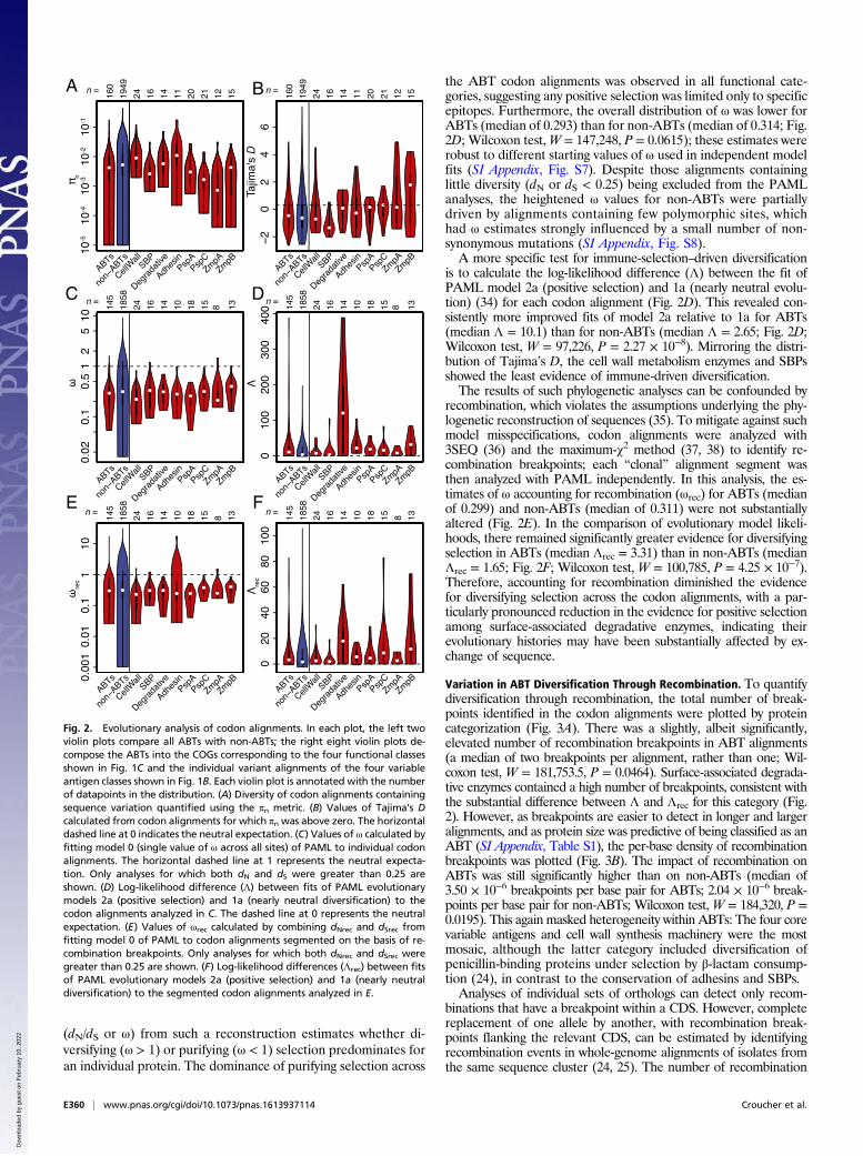
(dN/dS or ω) from such a reconstruction estimates whether di-versifying (ω > 1) or purifying (ω < 1) selection predominates foran individual protein. The dominance of purifying selection across
the ABT codon alignments was observed in all functional cate-gories, suggesting any positive selection was limited only to specificepitopes. Furthermore, the overall distribution of ω was lower forABTs (median of 0.293) than for non-ABTs (median of 0.314; Fig.2D; Wilcoxon test,W = 147,248, P = 0.0615); these estimates wererobust to different starting values of ω used in independent modelfits (SI Appendix, Fig. S7). Despite those alignments containinglittle diversity (dN or dS < 0.25) being excluded from the PAMLanalyses, the heightened ω values for non-ABTs were partiallydriven by alignments containing few polymorphic sites, whichhad ω estimates strongly influenced by a small number of non-synonymous mutations (SI Appendix, Fig. S8).A more specific test for immune-selection–driven diversification
is to calculate the log-likelihood difference (Λ) between the fit ofPAML model 2a (positive selection) and 1a (nearly neutral evolu-tion) (34) for each codon alignment (Fig. 2D). This revealed con-sistently more improved fits of model 2a relative to 1a for ABTs(median Λ = 10.1) than for non-ABTs (median Λ = 2.65; Fig. 2D;Wilcoxon test, W = 97,226, P = 2.27 × 10⁻⁸). Mirroring the distri-bution of Tajima’s D, the cell wall metabolism enzymes and SBPsshowed the least evidence of immune-driven diversification.The results of such phylogenetic analyses can be confounded by
recombination, which violates the assumptions underlying the phy-logenetic reconstruction of sequences (35). To mitigate against suchmodel misspecifications, codon alignments were analyzed with3SEQ (36) and the maximum-χ2 method (37, 38) to identify re-combination breakpoints; each “clonal” alignment segment wasthen analyzed with PAML independently. In this analysis, the es-timates of ω accounting for recombination (ωrec) for ABTs (medianof 0.299) and non-ABTs (median of 0.311) were not substantiallyaltered (Fig. 2E). In the comparison of evolutionary model likeli-hoods, there remained significantly greater evidence for diversifyingselection in ABTs (median Λrec = 3.31) than in non-ABTs (medianΛrec = 1.65; Fig. 2F; Wilcoxon test, W = 100,785, P = 4.25 × 10⁻⁷).Therefore, accounting for recombination diminished the evidencefor diversifying selection across the codon alignments, with a par-ticularly pronounced reduction in the evidence for positive selectionamong surface-associated degradative enzymes, indicating theirevolutionary histories may have been substantially affected by ex-change of sequence.
Variation in ABT Diversification Through Recombination. To quantifydiversification through recombination, the total number of break-points identified in the codon alignments were plotted by proteincategorization (Fig. 3A). There was a slightly, albeit significantly,elevated number of recombination breakpoints in ABT alignments(a median of two breakpoints per alignment, rather than one; Wil-coxon test, W = 181,753.5, P = 0.0464). Surface-associated degrada-tive enzymes contained a high number of breakpoints, consistent withthe substantial difference between Λ and Λrec for this category (Fig.2). However, as breakpoints are easier to detect in longer and largeralignments, and as protein size was predictive of being classified as anABT (SI Appendix, Table S1), the per-base density of recombinationbreakpoints was plotted (Fig. 3B). The impact of recombination onABTs was still significantly higher than on non-ABTs (median of3.50 × 10−6 breakpoints per base pair for ABTs; 2.04 × 10⁻⁶ break-points per base pair for non-ABTs; Wilcoxon test, W = 184,320, P =0.0195). This again masked heterogeneity within ABTs: The four corevariable antigens and cell wall synthesis machinery were the mostmosaic, although the latter category included diversification ofpenicillin-binding proteins under selection by β-lactam consump-tion (24), in contrast to the conservation of adhesins and SBPs.Analyses of individual sets of orthologs can detect only recom-
binations that have a breakpoint within a CDS. However, completereplacement of one allele by another, with recombination break-points flanking the relevant CDS, can be estimated by identifyingrecombination events in whole-genome alignments of isolates fromthe same sequence cluster (24, 25). The number of recombination
A B
C D
E F
D
Fig. 2. Evolutionary analysis of codon alignments. In each plot, the left twoviolin plots compare all ABTs with non-ABTs; the right eight violin plots de-compose the ABTs into the COGs corresponding to the four functional classesshown in Fig. 1C and the individual variant alignments of the four variableantigen classes shown in Fig. 1B. Each violin plot is annotated with the numberof datapoints in the distribution. (A) Diversity of codon alignments containingsequence variation quantified using the πn metric. (B) Values of Tajima’s Dcalculated from codon alignments for which πn was above zero. The horizontaldashed line at 0 indicates the neutral expectation. (C) Values of ω calculated byfitting model 0 (single value of ω across all sites) of PAML to individual codonalignments. The horizontal dashed line at 1 represents the neutral expecta-tion. Only analyses for which both dN and dS were greater than 0.25 areshown. (D) Log-likelihood difference (Λ) between fits of PAML evolutionarymodels 2a (positive selection) and 1a (nearly neutral diversification) to thecodon alignments analyzed in C. The dashed line at 0 represents the neutralexpectation. (E) Values of ωrec calculated by combining dNrec and dSrec fromfitting model 0 of PAML to codon alignments segmented on the basis of re-combination breakpoints. Only analyses for which both dNrec and dSrec weregreater than 0.25 are shown. (F) Log-likelihood differences (Λrec) between fitsof PAML evolutionary models 2a (positive selection) and 1a (nearly neutraldiversification) to the segmented codon alignments analyzed in E.
E360 | www.pnas.org/cgi/doi/10.1073/pnas.1613937114 Croucher et al.
Dow
nloa
ded
by g
uest
on
Feb
ruar
y 10
, 202
2
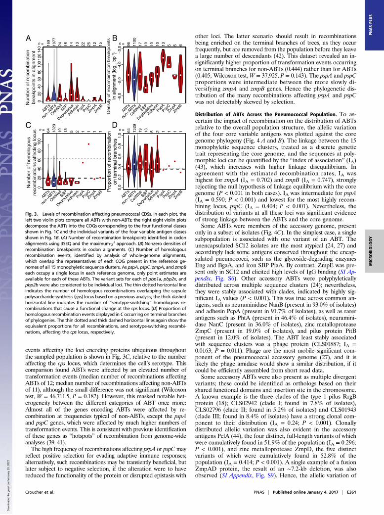
events affecting the loci encoding proteins ubiquitous throughoutthe sampled population is shown in Fig. 3C, relative to the numberaffecting the cps locus, which determines the cell’s serotype. Thiscomparison found ABTs were affected by an elevated number oftransformation events (median number of recombinations affectingABTs of 12; median number of recombinations affecting non-ABTsof 11), although the small difference was not significant (Wilcoxontest, W = 46,711.5, P = 0.182). However, this masked notable het-erogeneity between the different categories of ABT once more:Almost all of the genes encoding ABTs were affected by re-combination at frequencies typical of non-ABTs, except the pspAand pspC genes, which were affected by much higher numbers oftransformation events. This is consistent with previous identificationof these genes as “hotspots” of recombination from genome-wideanalyses (39–41).The high frequency of recombinations affecting pspA or pspCmay
reflect positive selection for evading adaptive immune responses;alternatively, such recombinations may be transiently beneficial, butlater subject to negative selection, if the alteration were to havereduced the functionality of the protein or disrupted epistasis with
other loci. The latter scenario should result in recombinationsbeing enriched on the terminal branches of trees, as they occurfrequently, but are removed from the population before they leavea large number of descendants (42). This dataset revealed an in-significantly higher proportion of transformation events occurringon terminal branches for non-ABTs (0.444) rather than for ABTs(0.405; Wilcoxon test,W = 37,925, P = 0.143). The pspA and pspCproportions were intermediate between the more slowly di-versifying zmpA and zmpB genes. Hence the phylogenetic dis-tribution of the many recombinations affecting pspA and pspCwas not detectably skewed by selection.
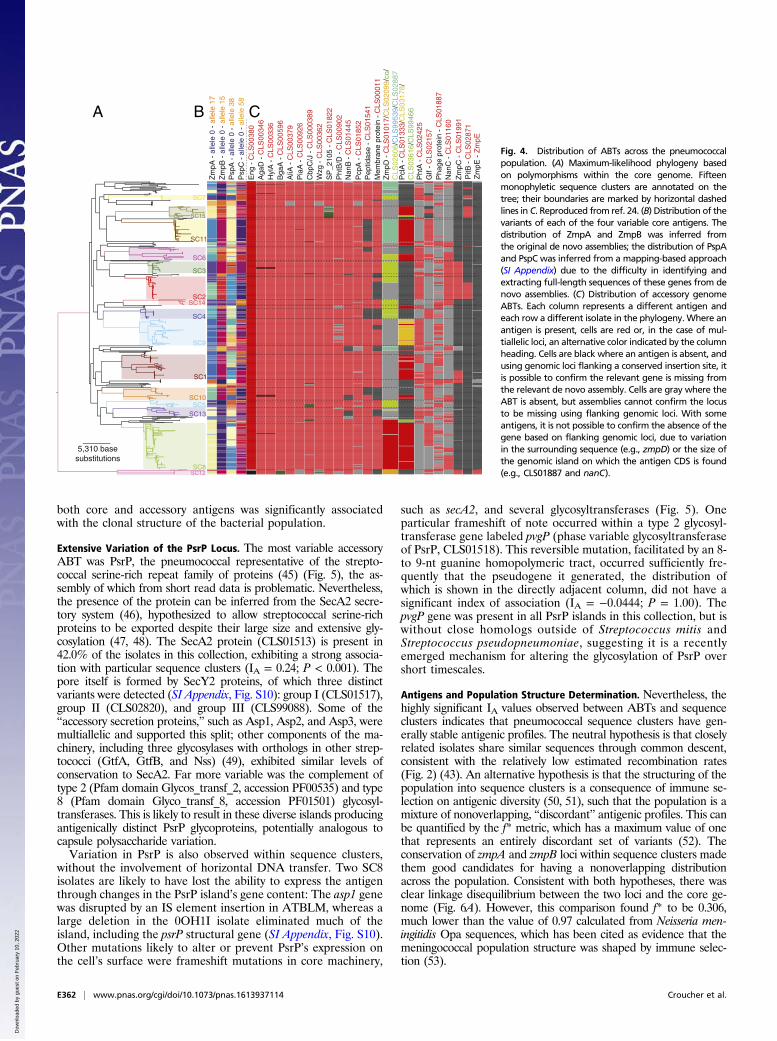
Distribution of ABTs Across the Pneumococcal Population. To as-certain the impact of recombination on the distribution of ABTsrelative to the overall population structure, the allelic variationof the four core variable antigens was plotted against the coregenome phylogeny (Fig. 4 A and B). The linkage between the 15monophyletic sequence clusters, treated as a discrete genetictrait representing the core genome, and the sequences at poly-morphic loci can be quantified by the “index of association” (IA)(43), which increases with higher linkage disequilibrium. Inagreement with the estimated recombination rates, IA washighest for zmpA (IA = 0.702) and zmpB (IA = 0.747), stronglyrejecting the null hypothesis of linkage equilibrium with the coregenome (P < 0.001 in both cases). IA was intermediate for pspA(IA = 0.590; P < 0.001) and lowest for the most highly recom-bining locus, pspC (IA = 0.404; P < 0.001). Nevertheless, thedistribution of variants at all these loci was significant evidenceof strong linkage between the ABTs and the core genome.Some ABTs were members of the accessory genome, present
only in a subset of isolates (Fig. 4C). In the simplest case, a singlesubpopulation is associated with one variant of an ABT. Theunencapsulated SC12 isolates are the most atypical (24, 27) andaccordingly lack some antigens conserved throughout the encap-sulated pneumococci, such as the glycoside-degrading enzymesEng and BgaA, and iron SBP PiaA. By contrast, ZmpE was pre-sent only in SC12 and elicited high levels of IgG binding (SI Ap-pendix, Fig. S6). Other accessory ABTs were polyphyleticallydistributed across multiple sequence clusters (24); nevertheless,they were stably associated with clades, indicated by highly sig-nificant IA values (P < 0.001). This was true across common an-tigens, such as neuraminidase NanB (present in 93.0% of isolates)and adhesin PcpA (present in 91.7% of isolates), as well as rarerantigens such as PhtA (present in 46.4% of isolates), neuramini-dase NanC (present in 36.0% of isolates), zinc metalloproteaseZmpC (present in 19.0% of isolates), and pilus protein PitB(present in 12.0% of isolates). The ABT least stably associatedwith sequence clusters was a phage protein (CLS01887; IA =0.0163; P = 0.011). Phage are the most mobile significant com-ponent of the pneumococcal accessory genome (27), and it islikely the phage amidase would show a similar distribution, if itcould be efficiently assembled from short read data.Some accessory ABTs were also present as multiple divergent
variants; these could be identified as orthologs based on theirshared functional domains and insertion site in the chromosome.A known example is the three clades of the type 1 pilus RrgBprotein (18); CLS02942 (clade I; found in 7.8% of isolates),CLS02796 (clade II; found in 5.2% of isolates) and CLS01943(clade III; found in 8.4% of isolates) have a strong clonal com-ponent to their distribution (IA = 0.24; P < 0.001). Clonallydistributed allelic variation was also evident in the accessoryantigens PclA (44), the four distinct, full-length variants of whichwere cumulatively found in 51.9% of the population (IA = 0.296;P < 0.001), and zinc metalloprotease ZmpD, the five distinctvariants of which were cumulatively found in 52.8% of thepopulation (IA = 0.414; P < 0.001). A single example of a fusionZmpAD protein, the result of an ∼7.2-kb deletion, was alsoobserved (SI Appendix, Fig. S9). Hence, the allelic variation of
A B
C D
Den
sity
of r
ecom
bina
tion
brea
kpoi
nts
in a
lignm
ent (
log 10
bp-1
)
Fig. 3. Levels of recombination affecting pneumococcal CDSs. In each plot, theleft two violin plots compare all ABTs with non-ABTs; the right eight violin plotsdecompose the ABTs into the COGs corresponding to the four functional classesshown in Fig. 1C and the individual variants of the four variable antigen classesshown in Fig. 1B. (A) Number of recombination breakpoints identified in codonalignments using 3SEQ and the maximum-χ2 approach. (B) Nonzero densities ofrecombination breakpoints in codon alignments. (C) Number of homologousrecombination events, identified by analysis of whole-genome alignments,which overlap the representatives of each COG present in the reference ge-nomes of all 15 monophyletic sequence clusters. As pspA, pspC, zmpA, and zmpBeach occupy a single locus in each reference genome, only point estimates areavailable for each of these ABTs. The variant sets for each of pbp1a, pbp2x, andpbp2bwere also considered to be individual loci. The thin dotted horizontal lineindicates the number of homologous recombinations overlapping the capsulepolysaccharide synthesis (cps) locus based on a previous analysis; the thick dashedhorizontal line indicates the number of “serotype-switching” homologous re-combinations that cause a functional change at the cps locus. (D) Proportion ofhomologous recombination events displayed in C occurring on terminal branchesof phylogenies. The thin dotted and thick dashed horizontal lines again show theequivalent proportions for all recombinations, and serotype-switching recombi-nations, affecting the cps locus, respectively.
Croucher et al. PNAS | Published online January 4, 2017 | E361
MICRO
BIOLO
GY
PNASPL
US
Dow
nloa
ded
by g
uest
on
Feb
ruar
y 10
, 202
2
both core and accessory antigens was significantly associatedwith the clonal structure of the bacterial population.
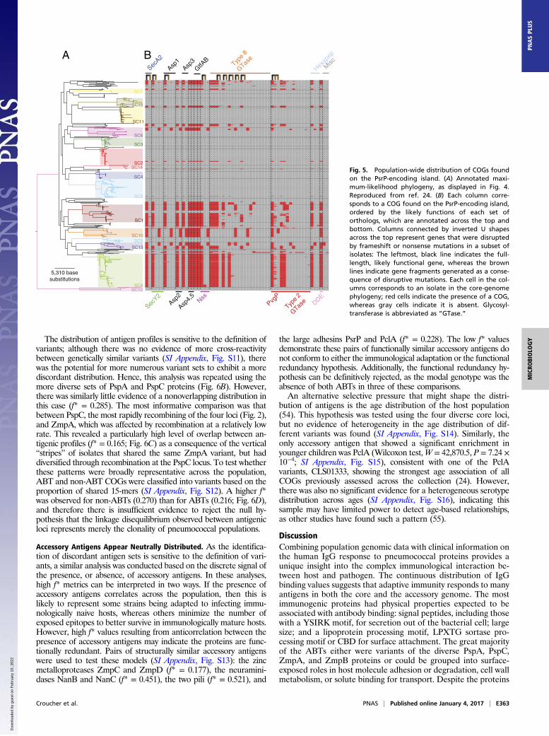
Extensive Variation of the PsrP Locus. The most variable accessoryABT was PsrP, the pneumococcal representative of the strepto-coccal serine-rich repeat family of proteins (45) (Fig. 5), the as-sembly of which from short read data is problematic. Nevertheless,the presence of the protein can be inferred from the SecA2 secre-tory system (46), hypothesized to allow streptococcal serine-richproteins to be exported despite their large size and extensive gly-cosylation (47, 48). The SecA2 protein (CLS01513) is present in42.0% of the isolates in this collection, exhibiting a strong associa-tion with particular sequence clusters (IA = 0.24; P < 0.001). Thepore itself is formed by SecY2 proteins, of which three distinctvariants were detected (SI Appendix, Fig. S10): group I (CLS01517),group II (CLS02820), and group III (CLS99088). Some of the“accessory secretion proteins,” such as Asp1, Asp2, and Asp3, weremultiallelic and supported this split; other components of the ma-chinery, including three glycosylases with orthologs in other strep-tococci (GtfA, GtfB, and Nss) (49), exhibited similar levels ofconservation to SecA2. Far more variable was the complement oftype 2 (Pfam domain Glycos_transf_2, accession PF00535) and type8 (Pfam domain Glyco_transf_8, accession PF01501) glycosyl-transferases. This is likely to result in these diverse islands producingantigenically distinct PsrP glycoproteins, potentially analogous tocapsule polysaccharide variation.Variation in PsrP is also observed within sequence clusters,
without the involvement of horizontal DNA transfer. Two SC8isolates are likely to have lost the ability to express the antigenthrough changes in the PsrP island’s gene content: The asp1 genewas disrupted by an IS element insertion in ATBLM, whereas alarge deletion in the 0OH1I isolate eliminated much of theisland, including the psrP structural gene (SI Appendix, Fig. S10).Other mutations likely to alter or prevent PsrP’s expression onthe cell’s surface were frameshift mutations in core machinery,
such as secA2, and several glycosyltransferases (Fig. 5). Oneparticular frameshift of note occurred within a type 2 glycosyl-transferase gene labeled pvgP (phase variable glycosyltransferaseof PsrP, CLS01518). This reversible mutation, facilitated by an 8-to 9-nt guanine homopolymeric tract, occurred sufficiently fre-quently that the pseudogene it generated, the distribution ofwhich is shown in the directly adjacent column, did not have asignificant index of association (IA = −0.0444; P = 1.00). ThepvgP gene was present in all PsrP islands in this collection, but iswithout close homologs outside of Streptococcus mitis andStreptococcus pseudopneumoniae, suggesting it is a recentlyemerged mechanism for altering the glycosylation of PsrP overshort timescales.
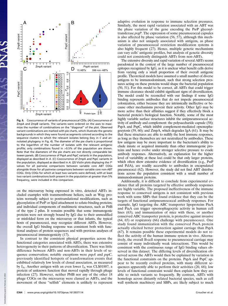
Antigens and Population Structure Determination. Nevertheless, thehighly significant IA values observed between ABTs and sequenceclusters indicates that pneumococcal sequence clusters have gen-erally stable antigenic profiles. The neutral hypothesis is that closelyrelated isolates share similar sequences through common descent,consistent with the relatively low estimated recombination rates(Fig. 2) (43). An alternative hypothesis is that the structuring of thepopulation into sequence clusters is a consequence of immune se-lection on antigenic diversity (50, 51), such that the population is amixture of nonoverlapping, “discordant” antigenic profiles. This canbe quantified by the f* metric, which has a maximum value of onethat represents an entirely discordant set of variants (52). Theconservation of zmpA and zmpB loci within sequence clusters madethem good candidates for having a nonoverlapping distributionacross the population. Consistent with both hypotheses, there wasclear linkage disequilibrium between the two loci and the core ge-nome (Fig. 6A). However, this comparison found f* to be 0.306,much lower than the value of 0.97 calculated from Neisseria men-ingitidis Opa sequences, which has been cited as evidence that themeningococcal population structure was shaped by immune selec-tion (53).
SC7
SC15
SC11
SC6
SC3
SC2SC14
SC4
SC9
SC1
SC10SC5
SC13
SC8SC12
5,310 basesubstitutions
Zm
pE -
Zm
pEP
itB -
CLS
0287
1Z
mpC
- C
LS01
991
Nan
C -
CLS
0116
0P
hage
pro
tein
- C
LS01
887
Glf
- C
LS02
157
Pht
A -
CLS
0242
5
Pcl
A -
CLS
0133
3/C
LS03
178/
CLS
0361
6/C
LS99
466
Zm
pD -
CLS
0101
7/C
LS02
099/
co/
CLS
0260
8/C
LS99
539/
CLS
0288
7
Mem
bran
e pr
otei
n -
CLS
0001
1P
eptid
ase
- C
LS01
541
Pcp
A -
CLS
0185
2N
anB
- C
LS01
445
Pht
B/D
- C
LS00
902
SP
_210
5 -
CLS
0182
2W
zg -
CLS
0036
2C
bpC
/J -
CLS
0003
89P
iaA
- C
LS00
926
AliA
- C
LS00
379
Bga
A -
CLS
0059
6H
ylA
- C
LS00
336
Aga
D -
CLS
0034
6E
ng -
CLS
0038
0
Zm
pA -
alle
le 0
- a
llele
17
Zm
pB -
alle
le 0
- a
llele
15
Psp
A -
alle
le 0
- a
llele
38
Psp
C -
alle
le 0
- a
llele
58
A B C
Fig. 4. Distribution of ABTs across the pneumococcalpopulation. (A) Maximum-likelihood phylogeny basedon polymorphisms within the core genome. Fifteenmonophyletic sequence clusters are annotated on thetree; their boundaries are marked by horizontal dashedlines in C. Reproduced from ref. 24. (B) Distribution of thevariants of each of the four variable core antigens. Thedistribution of ZmpA and ZmpB was inferred fromthe original de novo assemblies; the distribution of PspAand PspC was inferred from a mapping-based approach(SI Appendix) due to the difficulty in identifying andextracting full-length sequences of these genes from denovo assemblies. (C) Distribution of accessory genomeABTs. Each column represents a different antigen andeach row a different isolate in the phylogeny. Where anantigen is present, cells are red or, in the case of mul-tiallelic loci, an alternative color indicated by the columnheading. Cells are black where an antigen is absent, andusing genomic loci flanking a conserved insertion site, itis possible to confirm the relevant gene is missing fromthe relevant de novo assembly. Cells are gray where theABT is absent, but assemblies cannot confirm the locusto be missing using flanking genomic loci. With someantigens, it is not possible to confirm the absence of thegene based on flanking genomic loci, due to variationin the surrounding sequence (e.g., zmpD) or the size ofthe genomic island on which the antigen CDS is found(e.g., CLS01887 and nanC).
E362 | www.pnas.org/cgi/doi/10.1073/pnas.1613937114 Croucher et al.
Dow
nloa
ded
by g
uest
on
Feb
ruar
y 10
, 202
2
The distribution of antigen profiles is sensitive to the definition ofvariants; although there was no evidence of more cross-reactivitybetween genetically similar variants (SI Appendix, Fig. S11), therewas the potential for more numerous variant sets to exhibit a morediscordant distribution. Hence, this analysis was repeated using themore diverse sets of PspA and PspC proteins (Fig. 6B). However,there was similarly little evidence of a nonoverlapping distribution inthis case (f* = 0.285). The most informative comparison was thatbetween PspC, the most rapidly recombining of the four loci (Fig. 2),and ZmpA, which was affected by recombination at a relatively lowrate. This revealed a particularly high level of overlap between an-tigenic profiles (f* = 0.165; Fig. 6C) as a consequence of the vertical“stripes” of isolates that shared the same ZmpA variant, but haddiversified through recombination at the PspC locus. To test whetherthese patterns were broadly representative across the population,ABT and non-ABT COGs were classified into variants based on theproportion of shared 15-mers (SI Appendix, Fig. S12). A higher f*was observed for non-ABTs (0.270) than for ABTs (0.216; Fig. 6D),and therefore there is insufficient evidence to reject the null hy-pothesis that the linkage disequilibrium observed between antigenicloci represents merely the clonality of pneumococcal populations.
Accessory Antigens Appear Neutrally Distributed. As the identifica-tion of discordant antigen sets is sensitive to the definition of vari-ants, a similar analysis was conducted based on the discrete signal ofthe presence, or absence, of accessory antigens. In these analyses,high f* metrics can be interpreted in two ways. If the presence ofaccessory antigens correlates across the population, then this islikely to represent some strains being adapted to infecting immu-nologically naive hosts, whereas others minimize the number ofexposed epitopes to better survive in immunologically mature hosts.However, high f* values resulting from anticorrelation between thepresence of accessory antigens may indicate the proteins are func-tionally redundant. Pairs of structurally similar accessory antigenswere used to test these models (SI Appendix, Fig. S13): the zincmetalloproteases ZmpC and ZmpD (f* = 0.177), the neuramini-dases NanB and NanC (f* = 0.451), the two pili (f* = 0.521), and
the large adhesins PsrP and PclA (f* = 0.228). The low f* valuesdemonstrate these pairs of functionally similar accessory antigens donot conform to either the immunological adaptation or the functionalredundancy hypothesis. Additionally, the functional redundancy hy-pothesis can be definitively rejected, as the modal genotype was theabsence of both ABTs in three of these comparisons.An alternative selective pressure that might shape the distri-
bution of antigens is the age distribution of the host population(54). This hypothesis was tested using the four diverse core loci,but no evidence of heterogeneity in the age distribution of dif-ferent variants was found (SI Appendix, Fig. S14). Similarly, theonly accessory antigen that showed a significant enrichment inyounger children was PclA (Wilcoxon test,W = 42,870.5, P = 7.24 ×10⁻⁴; SI Appendix, Fig. S15), consistent with one of the PclAvariants, CLS01333, showing the strongest age association of allCOGs previously assessed across the collection (24). However,there was also no significant evidence for a heterogeneous serotypedistribution across ages (SI Appendix, Fig. S16), indicating thissample may have limited power to detect age-based relationships,as other studies have found such a pattern (55).
DiscussionCombining population genomic data with clinical information onthe human IgG response to pneumococcal proteins provides aunique insight into the complex immunological interaction be-tween host and pathogen. The continuous distribution of IgGbinding values suggests that adaptive immunity responds to manyantigens in both the core and the accessory genome. The mostimmunogenic proteins had physical properties expected to beassociated with antibody binding: signal peptides, including thosewith a YSIRK motif, for secretion out of the bacterial cell; largesize; and a lipoprotein processing motif, LPXTG sortase pro-cessing motif or CBD for surface attachment. The great majorityof the ABTs either were variants of the diverse PspA, PspC,ZmpA, and ZmpB proteins or could be grouped into surface-exposed roles in host molecule adhesion or degradation, cell wallmetabolism, or solute binding for transport. Despite the proteins
SecA2
Asp1
Asp3
GtfAB
Type
8
GTase
Hexap
ep
Asp4,
5
Type
2
GTase DDE
PvgP
SecY2
Asp2
Nss
SC7
SC15
SC11
SC6
SC3
SC2SC14
SC4
SC9
SC1
SC10SC5
SC13
SC8SC12
5,310 basesubstitutions
Misc
A B
Fig. 5. Population-wide distribution of COGs foundon the PsrP-encoding island. (A) Annotated maxi-mum-likelihood phylogeny, as displayed in Fig. 4.Reproduced from ref. 24. (B) Each column corre-sponds to a COG found on the PsrP-encoding island,ordered by the likely functions of each set oforthologs, which are annotated across the top andbottom. Columns connected by inverted U shapesacross the top represent genes that were disruptedby frameshift or nonsense mutations in a subset ofisolates: The leftmost, black line indicates the full-length, likely functional gene, whereas the brownlines indicate gene fragments generated as a conse-quence of disruptive mutations. Each cell in the col-umns corresponds to an isolate in the core-genomephylogeny; red cells indicate the presence of a COG,whereas gray cells indicate it is absent. Glycosyl-transferase is abbreviated as “GTase.”
Croucher et al. PNAS | Published online January 4, 2017 | E363
MICRO
BIOLO
GY
PNASPL
US
Dow
nloa
ded
by g
uest
on
Feb
ruar
y 10
, 202
2
on the microarray being expressed in vitro, detected ABTs in-cluded examples with transmembrane helices, such as Wzg; pro-teins normally subject to posttranslational modifications, such asglycosylation of PsrP or lipid attachment to solute-binding proteins;and individual components of multimeric structures, such as PitBof the type 2 pilus. It remains possible that some immunogenicproteins were not strongly bound by IgG due to their unmodifiedor misfolded form on the microarray or that infants, the typicalhosts of pneumococci, may recognize different ABTs. However,the overall IgG binding response was consistent both with func-tional analyses of protein sequences and with previous analyses ofpneumococcal immunogenicity (17).In contrast to the consistent physical properties across the
functional categories associated with ABTs, there was extensiveheterogeneity in their patterns of diversification. There was littledifference between ABTs and non-ABTs in their levels of se-quence conservation; notable exceptions were pspA and pspC,previously identified hotspots of transformation events thatexhibited relatively low levels of clonal association, as measuredby IA. Another antigen with an even lower IA was CLS01887, aprotein of unknown function that moved rapidly through phageinfection (27). However, neither PblB nor any of the other 18phage COGs on the microarray were classed as ABTs, and themovement of these “selfish” elements is unlikely to represent
adaptive evolution in response to immune selection pressures.Similarly, the most rapid variation associated with an ABT wasthe phase variation of the gene encoding the PsrP glycosyl-transferase pvgP. The expression of some pneumococcal capsulesis also affected by phase variation (56, 57), although this mech-anism is also not uniquely associated with antigens, as phasevariation of pneumococcal restriction modification systems isalso highly frequent (27). Hence, multiple genetic mechanismscan vary cells’ antigenic profiles, but analysis of genetic diversitycould not consistently distinguish ABTs from non-ABTs.The extensive diversity and rapid variation of several ABTs seems
paradoxical in the context of the large number of pneumococcalepitopes recognized by IgG, as it is unclear what benefit cells derivefrom varying only a small proportion of their overall antigenicprofile. Theoretical models have assumed a small number of diverseantigens to be immunodominant, such that strong selection pres-sures acting on these proteins would shape the bacterial population(50, 51). For this model to be correct, all ABTs that could triggerimmune clearance should exhibit significant signs of diversification.The model could be reconciled with our findings if some IgGbinding represents antibodies that do not impede pneumococcalcolonization, either because they are intrinsically ineffective or be-cause other mechanisms prevent their activity. Other IgG may bemore active than their affinities suggest if they effectively block abacterial protein’s biological function. Notably, some of the mosthighly variable surface structures inhibit the antipneumococcal ac-tivity of antibody and complement: the polysaccharide capsule (58);PspA and PspC, which inhibit complement-mediated opsonopha-gocytosis (59, 60); and ZmpA, which degrades IgA (61). It may bethat these structures are able to nullify the host immune responses,as long as they themselves are not neutralized by antibodies. Thesefew antigens may be more important to the bacterium’s ability toelude innate or acquired immunity than other immunogenic pro-teins and hence evolve under stronger selective pressure to evadeantibody responses. Alternatively, an explanation for the higherlevel of variability at these loci could be that only larger proteins,which often show extensive evidence of diversification (e.g., PsrPand PclA), are readily accessible to antibodies in encapsulatedpneumococci (62). However, the study did not find ABT distribu-tions across the population consistent with a small number ofimmunodominant proteins.Additionally, it is difficult to conclude from experimental ev-
idence that all proteins targeted by effective antibody responsesare highly variable. The proposed ineffectiveness of the immuneresponse to conserved antigens is not consistent with previouswork with some SBPs that found evidence that they can be thetargets of functional antipneumococcal antibody responses. Forexample, IgG targeting the ABC transporter lipoproteins PiuAand PiaA can trigger opsonophagocytic activity in human celllines (63), and immunization of mice with these, or anotherconserved ABC transporter protein, is protective against invasive(64, 65) or respiratory (66) challenge with pneumococci. Onestudy found immunization with the conserved PsaA proteinactually elicited better protection against carriage than PspA(67). It remains possible these experimental models do not re-flect the activity of the human immune system in vivo. Alterna-tively, the overall B-cell response to pneumococcal carriage mayconsist of many individually weak interactions. This would beconsistent with the continuous range of IgG binding values ob-served in this dataset. The differing levels of diversification ob-served across the ABTs would then be explained by variation inthe functional constraints on the proteins. PspA and PspC ap-pear to be recently evolved proteins, with many different se-quences apparently able to perform their activities (20, 68); lowlevels of functional constraint would then explain how they areable to switch variants so frequently. By contrast, ABTs withhomologs across distantly related bacterial species, such as cellwall synthesis machinery and SBPs, are likely subject to much
ZmpA variant
Zm
pB v
aria
nt
ZmpA variant
Psp
C v
aria
nt
PspA variantP
spC
var
iant
SC1SC2SC3SC4SC5SC6SC7SC8SC9SC10SC11SC12SC13SC14SC15SC16
Protein type
f*0.
00.
20.
40.
60.
81.
0
AB
T
non−
AB
T
15 ABTs 598 non−ABTs
A B
C D
Fig. 6. Cooccurrence of variants of pneumococcal CDSs. (A) Cooccurrence ofZmpA and ZmpB variants. The variants were ordered on the axes to maxi-mize the number of combinations on the “diagonal” of the plot. Observedvariant combinations are marked with pie charts, which illustrate the geneticbackgrounds in which they were found as segments colored according to thesequence clusters to which the relevant isolates belong (key in C and an-notated phylogeny in Fig. 4). The diameter of the pie charts is proportionalto the logarithm of the number of isolates with the relevant antigenicprofile; only combinations found in >0.5% of the population are shown.Note that the diameters of the pie charts are not directly comparable be-tween panels. (B) Cooccurrence of PspA and PspC variants in the population,displayed as described in A. (C) Cooccurrence of ZmpA and PspC variants inthe population, displayed as described in A. (D) Violin plots displaying the f*values for all pairwise comparisons between variable core ABT COGsalongside those for all pairwise comparisons between variable core non-ABTCOGs. Only COGs for which at least two variants were defined, with at leasttwo variant combinations both present in the population at greater than 5%frequency, were included in this comparison.
E364 | www.pnas.org/cgi/doi/10.1073/pnas.1613937114 Croucher et al.
Dow
nloa
ded
by g
uest
on
Feb
ruar
y 10
, 202
2
stronger functional constraint, and hence their greater conser-vation between pneumococci.One argument against this latter model is that the poly-
saccharide capsule exhibits similar levels of diversity to PspA andPspC, but experimental and theoretical analyses show that thisvariation strongly affects strain fitness (10, 69). However, theimmune responses to proteins and polysaccharides are distinct,and it may be that there is stronger selection for switching ofserotype, rather than some protein antigens; additionally, sero-type switching is not free from selective constraints (22). Re-solving the paradox of why extensive variation is observed in onlya subset of ABTs will be usefully informed by both experimentaland clinical data. The combination of population genomicdatasets with pangenome-wide information on antibody bindingpromises to be a valuable tool for addressing such unresolvedquestions about the immunological response to infections.
MethodsAnalysis of Proteome Microarray Data. The design and construction of theproteome microarray, and the processing and analysis of the raw data, aredescribed in SI Appendix. Serum samples came from the VAC-002 studyapproved by the Western Institutional Review Board. For each person, eachprotein was represented by the probe recording the highest normalized,log2-transformed antibody response score. These IgG binding values acrossall individuals were used to generate an all-vs.-all distance matrix betweenproteins, which allowed the proteins to be classified into 208 ABTs and theremaining non-ABT, using the R function “hclust” (70). The annotation ofthe proteins and the associated functional domain information used in re-gression analyses are detailed in Dataset S1.
Determining Patterns of Presence and Absence. The pattern of presence andabsence of each COG across isolates was determined by the clustering ofsequences defined and made freely available previously (24, 26), combinedwith the information from scaffolded assemblies, as described elsewhere(27). For each ABT, “flanking” COGs were defined as the nearest COGs, bothupstream and downstream, that were found only in single copy in eachgenome in which they were present and therefore could be used to define aunique locus in the chromosome. The presence of both an ABT’s flankingCOGs on the same scaffold, in the absence of the ABT itself, was taken torepresent stronger evidence that the ABT was genuinely missing from anisolate (Fig. 4). Multiple COGs were considered to be orthologous representa-tives of the same antigen if they were present at equivalent positions in differentgenomes, as judged by sharing the same flanking COGs; if they had similarfunctional domain structures, as judged by sharing the majority of motifs iden-tified by Pfam in both sequences; and if they never cooccurred in the same strain,to exclude paralogs. These criteria identified the four functional PclA variants(CLS01333, CLS03616, CLS99466, and CLS03178), the three clades of RrgB(CLS01943, CLS02796, and CLS02942), ZmpD (CLS02608, CLS02099, CLS02887,CLS01017, and CLS99539), and PhtD/E (CLS00904 and CLS02083). However, poorassembly precluded further analysis of the repetitive PhtD/E CDSs.
A similar approach was also used to identify the COGs present on PsrPislands. The boundary of these islands was defined as the nearest core COG,CLS01506 (present in a single copy in all isolates), upstream of the SecA2protein. Therefore, in all cases where CLS01513 (SecA2) was present, the CDSson the same contig as, and downstream of, the core COG CLS01506 weretreated asmembers of the PsrP island; the other boundarywas not defined, asthe psrP gene never assembled completely. This set was then manually cu-rated to remove fragments of the psrP CDS and noncore CDSs that were notpart of the island.
Detection of Recombination and Selection. All COGs included on the micro-array, and alignments of the individual variants of the core diverse loci, werefiltered to remove sequences less than 75%, or more than 125%, of themedian sequence length. Protein sequences were aligned with MUSCLE (71),and the corresponding DNA sequences were back translated to give a codon
alignment. Population genetic statistics, including πn and Tajima’s D, werecalculated using Bioperl (72) and FAST (73). Duplicate sequences were re-moved, and then a maximum-likelihood phylogeny was generated usingRAxML v7.0.3 with a general time-reversible substitution model and agamma model of rate heterogeneity (74). Evolutionary models were thenfitted to the alignments and phylogenies (with fixed branch lengths), usingthe codeml software of PAML v4.8 (33), applying site models 0 (single ω ratioacross the alignment), 1a (nearly neutral diversification), and 2a (evolutionunder positive selection). Independent runs were conducted with starting ωvalues of 0.1, 1, and 10 to test for convergence. The ω and Λ results werereported only if dN and dS estimated from an alignment were both greaterthan 0.25. All amino acids were considered equally dissimilar from oneanother.
To reduce any biases introduced by recombination, the codon alignmentswere scanned for breakpoints. The initial scan was performed with 3SEQ (36),which is able to detect exchanges between sequences in an alignment, usingthe “fullrun” mode. Each segment defined by the first set of breakpointswas then iteratively scanned with 3SEQ for further evidence of re-combination. This set of segments was then iteratively scanned for importsof divergent tracts of sequence, using the maximum-χ2 approach (37, 38).Following the convergence of these methods, each segment of the align-ment was analyzed with RAxML and PAML as described for the wholealignment analysis above.
To calculate the level of recombination affecting ABTs based on theanalyses of whole-genome alignments (24–26), the loci within each sequencecluster’s representative reference genome (26) corresponding to each COGin the complete set of isolates had to be identified. As there should be nogenuine genetic variation between different assemblies of the same sets ofsequence reads, except for potential variance in the start codon selection,the middle segment of each COG was used to select identical matches in thereference sequences of the monophyletic sequence clusters. The corre-sponding CDSs were then used to calculate the number of recombinationevents that affected the evolution of these proteins. Only COGs found in all15 reference sequences were analyzed, to avoid unequal sample sizes. Theloci encoding zmpA, zmpB, pspA, and pspC were manually identified (24),and recombinations affecting cps loci were described previously (22).
Analyses of Variant Sequence Distributions. Indexes of association were cal-culated using the R package PoppR (75). The f* metric was calculated usingthe approach described by Buckee et al. (52),
f* =12
Xi
x3ikpiqk
+Xj
x3hjphqj
!,
where xij is the frequency of variant combination i (at the first locus) and j (atthe second locus) across the population; pi is the frequency of variant i at thefirst locus; qj is the frequency of variant j at the second locus; and k is thevalue of j that maximizes xij, whereas h is the value of i that maximizes xij.The comparison of f* between ABTs and non-ABTs used the COG sequencesanalyzed in Fig. 2. Variants were defined by generating pairwise similaritiesbetween sequences within the same alignment based on the number ofshared identical 15-mers from the full set of 15 amino acid patterns presentin either sequence. Based on the overall distribution of pairwise similarities(SI Appendix, Fig. S12), a threshold similarity of 0.75 was used to definediscrete variants within each COG alignment. The f* value was calculated forpairs of ABT or non-ABT COGs if both were divided into two or more vari-ants and more than one combination was present at greater than 5% fre-quency in the population; only these common combinations were used inthe calculation of f*.
ACKNOWLEDGMENTS. We thank the participants in the phase I trial of thewhole-cell vaccine. Research reported in this publication was supported byThe Bill & Melinda Gates Foundation, PATH, the National Institute of Allergyand Infectious Diseases of the US National Institutes of Health GrantR01AI066304, and the Wellcome Trust Grant 098051. N.J.C. is funded by aSir Henry Dale Fellowship, jointly funded by the Wellcome Trust and RoyalSociety (Grant 104169/Z/14/Z).
1. Gratten M, et al. (1986) Colonisation of Haemophilus influenzae and Streptococcus
pneumoniae in the upper respiratory tract of neonates in Papua New Guinea: Primary
acquisition, duration of carriage, and relationship to carriage in mothers. Biol
Neonate 50(2):114–120.2. Gray BM, Turner ME, Dillon HC, Jr (1982) Epidemiologic studies of Streptococcus
pneumoniae in infants. The effects of season and age on pneumococcal acquisition
and carriage in the first 24 months of life. Am J Epidemiol 116(4):692–703.
3. Zhang Q, et al. (2006) Serum and mucosal antibody responses to pneumococcal protein
antigens in children: Relationships with carriage status. Eur J Immunol 36(1):46–57.4. Holmgren J, Czerkinsky C (2005) Mucosal immunity and vaccines. Nat Med 11(4,
Suppl):S45–S53.5. Anttila M, Voutilainen M, Jäntti V, Eskola J, Käyhty H (1999) Contribution of serotype-
specific IgG concentration, IgG subclasses and relative antibody avidity to opsonophagocytic
activity against Streptococcus pneumoniae. Clin Exp Immunol 118(3):402–407.
Croucher et al. PNAS | Published online January 4, 2017 | E365
MICRO
BIOLO
GY
PNASPL
US
Dow
nloa
ded
by g
uest
on
Feb
ruar
y 10
, 202
2
6. Janoff EN, et al. (1999) Killing of Streptococcus pneumoniae by capsular polysaccharide-specific polymeric IgA, complement, and phagocytes. J Clin Invest 104(8):1139–1147.
7. Zhang Z, Clarke TB, Weiser JN (2009) Cellular effectors mediating Th17-dependentclearance of pneumococcal colonization in mice. J Clin Invest 119(7):1899–1909.
8. Malley R, et al. (2005) CD4+ T cells mediate antibody-independent acquired immunityto pneumococcal colonization. Proc Natl Acad Sci USA 102(13):4848–4853.
9. Li Y, et al. (2012) Distinct effects on diversifying selection by two mechanisms ofimmunity against Streptococcus pneumoniae. PLoS Pathog 8(11):e1002989.
10. Cobey S, Lipsitch M (2012) Niche and neutral effects of acquired immunity permitcoexistence of pneumococcal serotypes. Science 335(6074):1376–1380.
11. Rennels MB, et al. (1998) Safety and immunogenicity of heptavalent pneumococcalvaccine conjugated to CRM197 in United States infants. Pediatrics 101(4 Pt 1):604–611.
12. Ghaffar F, et al. (2004) Effect of the 7-valent pneumococcal conjugate vaccine onnasopharyngeal colonization by Streptococcus pneumoniae in the first 2 years of life.Clin Infect Dis 39(7):930–938.
13. Shinefield HR, et al. (1999) Safety and immunogenicity of heptavalent pneumococcalCRM197 conjugate vaccine in infants and toddlers. Pediatr Infect Dis J 18(9):757–763.
14. Moffitt KL, Malley R (2011) Next generation pneumococcal vaccines. Curr OpinImmunol 23(3):407–413.
15. Goldblatt D, et al. (2005) Antibody responses to nasopharyngeal carriage of Streptococcuspneumoniae in adults: A longitudinal household study. J Infect Dis 192(3):387–393.
16. Beghetto E, et al. (2006) Discovery of novel Streptococcus pneumoniae antigens byscreening a whole-genome λ-display library. FEMS Microbiol Lett 262(1):14–21.
17. Giefing C, et al. (2008) Discovery of a novel class of highly conserved vaccine antigensusing genomic scale antigenic fingerprinting of pneumococcus with human anti-bodies. J Exp Med 205(1):117–131.
18. Barocchi MA, et al. (2006) A pneumococcal pilus influences virulence and host in-flammatory responses. Proc Natl Acad Sci USA 103(8):2857–2862.
19. Bagnoli F, et al. (2008) A second pilus type in Streptococcus pneumoniae is prevalent inemerging serotypes and mediates adhesion to host cells. J Bacteriol 190(15):5480–5492.
20. Hollingshead SK, Becker R, Briles DE (2000) Diversity of PspA: Mosaic genes and evidencefor past recombination in Streptococcus pneumoniae. Infect Immun 68(10):5889–5900.
21. Brooks-Walter A, Briles DE, Hollingshead SK (1999) The pspC gene of Streptococcuspneumoniae encodes a polymorphic protein, PspC, which elicits cross-reactive anti-bodies to PspA and provides immunity to pneumococcal bacteremia. Infect Immun67(12):6533–6542.
22. Croucher NJ, et al. (2015) Selective and genetic constraints on pneumococcal serotypeswitching. PLoS Genet 11(3):e1005095.
23. Huang SS, et al. (2009) Continued impact of pneumococcal conjugate vaccine oncarriage in young children. Pediatrics 124(1):e1–e11.
24. Croucher NJ, et al. (2013) Population genomics of post-vaccine changes in pneumo-coccal epidemiology. Nat Genet 45(6):656–663.
25. Croucher NJ, et al. (2015) Rapid phylogenetic analysis of large samples of recombi-nant bacterial whole genome sequences using Gubbins. Nucleic Acids Res 43(3):e15.
26. Croucher NJ, et al. (2015) Population genomic datasets describing the post-vaccineevolutionary epidemiology of Streptococcus pneumoniae. Sci Data 2:150058.
27. Croucher NJ, et al. (2014) Diversification of bacterial genome content through distinctmechanisms over different timescales. Nat Commun 5:5471.
28. Moschioni M, et al. (2008) Streptococcus pneumoniae contains 3 rlrA pilus variantsthat are clonally related. J Infect Dis 197(6):888–896.
29. Tettelin H, et al. (2001) Complete genome sequence of a virulent isolate of Strepto-coccus pneumoniae. Science 293(5529):498–506.
30. Loisel E, et al. (2011) Biochemical characterization of the histidine triad protein PhtD as acell surface zinc-binding protein of pneumococcus. Biochemistry 50(17):3551–3558.
31. Anderton JM, et al. (2007) E-cadherin is a receptor for the common protein pneu-mococcal surface adhesin A (PsaA) of Streptococcus pneumoniae. Microb Pathog42(5-6):225–236.
32. Tajima F (1989) Statistical method for testing the neutral mutation hypothesis by DNApolymorphism. Genetics 123(3):585–595.
33. Yang Z (2007) PAML 4: Phylogenetic analysis by maximum likelihood. Mol Biol Evol24(8):1586–1591.
34. Anisimova M, Bielawski JP, Yang Z (2001) Accuracy and power of the likelihood ratiotest in detecting adaptive molecular evolution. Mol Biol Evol 18(8):1585–1592.
35. Anisimova M, Nielsen R, Yang Z (2003) Effect of recombination on the accuracy of the like-lihood method for detecting positive selection at amino acid sites. Genetics 164(3):1229–1236.
36. Boni MF, Posada D, Feldman MW (2007) An exact nonparametric method for in-ferring mosaic structure in sequence triplets. Genetics 176(2):1035–1047.
37. Smith JM (1992) Analyzing the mosaic structure of genes. J Mol Evol 34(2):126–129.38. Piganeau G, Gardner M, Eyre-Walker A (2004) A broad survey of recombination in
animal mitochondria. Mol Biol Evol 21(12):2319–2325.39. Croucher NJ, et al. (2011) Rapid pneumococcal evolution in response to clinical in-
terventions. Science 331(6016):430–434.40. Croucher NJ, et al. (2014) Evidence for soft selective sweeps in the evolution of
pneumococcal multidrug resistance and vaccine escape. Genome Biol Evol 6(7):1589–1602.
41. Croucher NJ, et al. (2014) Variable recombination dynamics during the emergence, trans-mission and ‘disarming’ of a multidrug-resistant pneumococcal clone. BMC Biol 12(1):49.
42. Ruiz-Pesini E, Mishmar D, Brandon M, Procaccio V, Wallace DC (2004) Effects of pu-rifying and adaptive selection on regional variation in human mtDNA. Science303(5655):223–226.
43. Smith JM, Smith NH, O’Rourke M, Spratt BG (1993) How clonal are bacteria? Proc NatlAcad Sci USA 90(10):4384–4388.
44. Paterson GK, Nieminen L, Jefferies JM, Mitchell TJ (2008) PclA, a pneumococcal col-lagen-like protein with selected strain distribution, contributes to adherence andinvasion of host cells. FEMS Microbiol Lett 285(2):170–176.
45. Rose L, et al. (2008) Antibodies against PsrP, a novel Streptococcus pneumoniae ad-hesin, block adhesion and protect mice against pneumococcal challenge. J Infect Dis198(3):375–383.
46. Bensing BA, Sullam PM (2002) An accessory sec locus of Streptococcus gordonii isrequired for export of the surface protein GspB and for normal levels of binding tohuman platelets. Mol Microbiol 44(4):1081–1094.
47. Bensing BA, Gibson BW, Sullam PM (2004) The Streptococcus gordonii plateletbinding protein GspB undergoes glycosylation independently of export. J Bacteriol186(3):638–645.
48. Feltcher ME, Braunstein M (2012) Emerging themes in SecA2-mediated protein ex-port. Nat Rev Microbiol 10(11):779–789.
49. Yen YT, et al. (2013) Differential localization of the streptococcal accessory seccomponents and implications for substrate export. J Bacteriol 195(4):682–695.
50. Gupta S, et al. (1996) The maintenance of strain structure in populations of re-combining infectious agents. Nat Med 2(4):437–442.
51. Gupta S, Ferguson N, Anderson R (1998) Chaos, persistence, and evolution of strainstructure in antigenically diverse infectious agents. Science 280(5365):912–915.
52. Buckee CO, Gupta S, Kriz P, Maiden MCJ, Jolley KA (2010) Long-term evolution ofantigen repertoires among carried meningococci. Proc Biol Sci 277(1688):1635–1641.
53. Callaghan MJ, et al. (2008) The effect of immune selection on the structure of themeningococcal Opa protein repertoire. PLoS Pathog 4(3):e1000020.
54. Regev-Yochay G, et al. (2010) Re-emergence of the type 1 pilus among Streptococcuspneumoniae isolates in Massachusetts, USA. Vaccine 28(30):4842–4846.
55. Hausdorff WP, Feikin DR, Klugman KP (2005) Epidemiological differences amongpneumococcal serotypes. Lancet Infect Dis 5(2):83–93.
56. Waite RD, Struthers JK, Dowson CG (2001) Spontaneous sequence duplication withinan open reading frame of the pneumococcal type 3 capsule locus causes high-fre-quency phase variation. Mol Microbiol 42(5):1223–1232.
57. Waite RD, Penfold DW, Struthers JK, Dowson CG (2003) Spontaneous sequence du-plications within capsule genes cap8E and tts control phase variation in Streptococcuspneumoniae serotypes 8 and 37. Microbiology 149(Pt 2):497–504.
58. Hyams C, Camberlein E, Cohen JM, Bax K, Brown JS (2010) The Streptococcus pneu-moniae capsule inhibits complement activity and neutrophil phagocytosis by multiplemechanisms. Infect Immun 78(2):704–715.
59. Tu AH, Fulgham RL, McCrory MA, Briles DE, Szalai AJ (1999) Pneumococcal surfaceprotein A inhibits complement activation by Streptococcus pneumoniae. InfectImmun 67(9):4720–4724.
60. Janulczyk R, Iannelli F, Sjoholm AG, Pozzi G, Bjorck L (2000) Hic, a novel surfaceprotein of Streptococcus pneumoniae that interferes with complement function.J Biol Chem 275(47):37257–37263.
61. Wani JH, Gilbert JV, Plaut AG, Weiser JN (1996) Identification, cloning, and se-quencing of the immunoglobulin A1 protease gene of Streptococcus pneumoniae.Infect Immun 64(10):3967–3974.
62. Gor DO, Ding X, Briles DE, Jacobs MR, Greenspan NS (2005) Relationship betweensurface accessibility for PpmA, PsaA, and PspA and antibody-mediated immunity tosystemic infection by Streptococcus pneumoniae. Infect Immun 73(3):1304–1312.
63. Jomaa M, et al. (2005) Antibodies to the iron uptake ABC transporter lipoproteinsPiaA and PiuA promote opsonophagocytosis of Streptococcus pneumoniae. InfectImmun 73(10):6852–6859.
64. Brown JS, Ogunniyi AD, Woodrow MC, Holden DW, Paton JC (2001) Immunizationwith components of two iron uptake ABC transporters protects mice against systemicStreptococcus pneumoniae infection. Infect Immun 69(11):6702–6706.
65. Saxena S, Khan N, Dehinwal R, Kumar A, Sehgal D (2015) Conserved surface accessiblenucleoside ABC transporter component SP0845 is essential for pneumococcal viru-lence and confers protection in vivo. PLoS One 10(2):e0118154.
66. Jomaa M, et al. (2006) Immunization with the iron uptake ABC transporter proteinsPiaA and PiuA prevents respiratory infection with Streptococcus pneumoniae.Vaccine 24(24):5133–5139.
67. Briles DE, et al. (2000) Intranasal immunization of mice with a mixture of the pneu-mococcal proteins PsaA and PspA is highly protective against nasopharyngeal car-riage of Streptococcus pneumoniae. Infect Immun 68(2):796–800.
68. Iannelli F, Oggioni MR, Pozzi G (2002) Allelic variation in the highly polymorphic locuspspC of Streptococcus pneumoniae. Gene 284(1-2):63–71.
69. Trzci�nski K, et al. (2015) Effect of serotype on pneumococcal competition in a mousecolonization model. MBio 6(5):e00902–e00915.
70. R Core Development Team (2011) R: A Language and Environment for StatisticalComputing (R Foundation for Statistical Computing, Vienna).
71. Edgar RC (2004) MUSCLE: Multiple sequence alignment with high accuracy and highthroughput. Nucleic Acids Res 32(5):1792–1797.
72. Stajich JE, et al. (2002) The Bioperl toolkit: Perl modules for the life sciences. GenomeRes 12(10):1611–1618.
73. Lawrence TJ, et al. (2015) FAST: FAST Analysis of Sequences Toolbox. Front Genet 6:172.74. Stamatakis A, Ludwig T, Meier H (2005) RAxML-III: A fast program for maximum
likelihood-based inference of large phylogenetic trees. Bioinformatics 21(4):456–463.75. Kamvar ZN, Brooks JC, Grünwald NJ (2015) Novel R tools for analysis of genome-wide
population genetic data with emphasis on clonality. Front Genet 6(JUN):208.
E366 | www.pnas.org/cgi/doi/10.1073/pnas.1613937114 Croucher et al.
Dow
nloa
ded
by g
uest
on
Feb
ruar
y 10
, 202
2