microarray analysis of pneumococcal gene expression during invasive disease
TRANSCRIPT

INFECTION AND IMMUNITY, Oct. 2004, p. 5582–5596 Vol. 72, No. 100019-9567/04/$08.00�0 DOI: 10.1128/IAI.72.10.5582–5596.2004Copyright © 2004, American Society for Microbiology. All Rights Reserved.
Microarray Analysis of Pneumococcal Gene Expression duringInvasive Disease
Carlos J. Orihuela,1 Jana N. Radin,1,2 Jack E. Sublett,1 Geli Gao,1Deepak Kaushal,3 and Elaine I. Tuomanen1,2*
Department of Infectious Diseases1 and Hartwell Center for Bioinformatics and Biotechnology,3 St. JudeChildren’s Research Hospital, and Department of Molecular Sciences, University of
Tennessee Health Science Center,2 Memphis, Tennessee
Received 23 March 2004/Returned for modification 19 May 2004/Accepted 29 June 2004
Streptococcus pneumoniae is a leading cause of invasive bacterial disease. This is the first study to examinethe expression of S. pneumoniae genes in vivo by using whole-genome microarrays available from The Institutefor Genomic Research. Total RNA was collected from pneumococci isolated from infected blood, infectedcerebrospinal fluid, and bacteria attached to a pharyngeal epithelial cell line in vitro. Microarray analysis ofpneumococcal genes expressed in these models identified body site-specific patterns of expression for virulencefactors, transporters, transcription factors, translation-associated proteins, metabolism, and genes with un-known function. Contributions to virulence predicted for several unknown genes with enhanced expression invivo were confirmed by insertion duplication mutagenesis and challenge of mice with the mutants. Finally, wecross-referenced our results with previous studies that used signature-tagged mutagenesis and differentialfluorescence induction to identify genes that are potentially required by a broad range of pneumococcal strainsfor invasive disease.
Streptococcus pneumoniae is the primary cause of communi-ty-acquired pneumonia and a major cause of invasive bacterialdisease (28). Each year in the United States, pneumococci areresponsible for 100,000 to 135,000 hospitalizations for pneu-monia, 50,000 cases of bacteremia, and 3,000 cases of menin-gitis (18). Worldwide these diseases account for more than amillion deaths a year. Currently, a 7-valent polysaccharide-protein conjugate vaccine is effective; however, it protectsagainst only a small subset of the serotypes known to causeinvasive disease (9). Invasive disease follows colonization ofthe nasopharynx and is the result of spread of the bacteria tothe lungs and blood. Once in the bloodstream, infection mayresult in septicemia or meningitis, both of which have highmortality despite accepted antibiotic therapy (28).
Since Pasteur and Sternberg first described the pneumococ-cus, a number of virulence factors required for invasive diseasehave been identified. Major virulence determinants such ascapsular polysaccharide, pneumolysin, and choline-bindingproteins have clearly established roles in pathogenesis (18).More recently, large-scale identification of S. pneumoniae vir-ulence determinants has been attempted. Studies, such as sig-nature-tagged mutagenesis (STM), have used transposons andsuicide vectors to pepper the chromosome of the bacteria withmutations and identify genes required for virulence (13, 25,39). STM screens rely on negative selection of mutants throughpassage in animal models and, to date, STM has been usedthree times for the pneumococcus, each time in a differentstrain.
Ideally, characterization of pneumococcal virulence deter-
minants should include analysis of gene expression during in-vasive disease. Confirmation of their expression in vivo wouldnot only verify the contribution of these genes to pathogenesisbut would also elucidate their contribution to discrete forms ofdisease (e.g., genes expressed during pneumonia versus thosein the blood during bacteremia). Unfortunately, analysis of invivo bacterial gene expression, much less at discrete sites, hasbeen limited by the difficulties of isolating sufficient quantitiesof pure and intact bacterial RNA from infected host tissues. Tocircumvent the requirement for RNA, investigators have useddifferential fluorescence induction (DFI) to identify S. pneu-moniae promoters that are induced during disease (29). Incontrast to STM, DFI relies on the promoter activity of ran-dom fragments of DNA cloned upstream of an episomal, pro-moterless green fluorescent protein. Fluorescence is impartedby the promoter activity of the randomly integrated DNAfragment under the environmental conditions tested (in vivo).Fluorescent bacteria can then be sorted by fluorescence-acti-vated cell sorting analysis, leaving only bacteria containingplasmids with active promoters. Using DFI, Marra et al. iden-tified operons that are enhanced during bacterial growth in achinchilla model of otitis media, lower respiratory tract infec-tion in a mouse, and growth in an intraperitoneal chamberimplant model (29).
Large-scale analysis of gene expression during invasive dis-ease provides not only transcriptional data with regard to cer-tain genes, such as virulence determinants but, after interpre-tation, also provides information as to the status of the bacteriaduring infection and the response of the bacteria to the hostenvironment. In vivo gene expression therefore potentiallyidentifies unknown or unappreciated targets for pharmaceuti-cal or vaccine intervention since, presumably, the genes whosetranscription is altered during invasive disease are those thatare required by the bacteria for survival in the host (14).
* Corresponding author. Mailing address: St. Jude Children’s Re-search Hospital, Mailstop 320, IRC 8057, 332 North Lauderdale Rd.,Memphis, TN 38105. Phone: (901) 495-3486. Fax: (901) 495-3099.E-mail: [email protected].
5582

We describe here a protocol for the collection of RNA frombacteria within infected blood and cerebrospinal fluid (CSF) invivo and bacteria adherent to epithelial cells in vitro. Usinggenome-wide cDNA microarrays available from the PathogenFunctional Genomic Research Center (PFGRC) at The Insti-tute for Genomic Research (TIGR; Rockville, Md.), we haveexamined pneumococcal gene expression during bacteremia,meningitis, and epithelial cell contact (ECC). Analysis of pneu-mococcal genes expressed in these models identified globalpatterns of expression unique to each condition tested. Pat-terns of expression were identified with regard to virulencefactors, transporters, transcription factors, translation-associ-ated proteins, metabolism, and genes with unknown function.These findings were then cross referenced to previous STMand DFI studies. Finally, we examined several candidate viru-lence genes with unknown function by insertion duplicationmutagenesis and challenge of mice.
MATERIALS AND METHODS
Media and growth conditions. S. pneumoniae was grown on tryptic soy agar(Difco, Detroit, Mich.) supplemented with 3% defibrinated sheep blood or indefined semisynthetic casein liquid medium (24) supplemented with 0.5% yeastextract (C�Y). Erythromycin (1 �g/ml) and kanamycin (400 �g/ml) (Sigma, St.Louis, Mo.) were added to the growth medium as appropriate. S. pneumoniaecultures were inoculated from frozen stock and incubated at 37°C in 5% CO2.Escherichia coli strains were grown in Luria-Bertani medium (Difco) at 37°C inan orbital shaker; erythromycin (1 mg/ml) was added to the E. coli cultures tomaintain any plasmids.
Bacterial strains and construction of mutants. D39 Xen7 (D39X), a stablebioluminescent isolate of S. pneumoniae, serotype 2, strain D39 (2), was createdas previously described (11). TIGR4 Xen 35 (T4X) was created by transforma-tion of TIGR4 with genomic DNA from D39X by using CSP-2 (35). Biolumi-nescent mutants deficient in genes identified as enhanced during invasive diseasewere created by insertion duplication mutagenesis (37). PCR primers with nestedEcoRI and BamHI restriction sites were used to amplify 200- to 500-bp frag-ments from the N termini of the genes. Amplified PCR fragments were purifiedand digested with EcoRI and BamHI (New England BioLabs, Beverly, Mass.).Digested fragments were ligated into the vector pJDC9 and transformed intochemically competent XL1-Blue E. coli (Stratagene, La Jolla, Calif.). Singletransformants containing the insert were identified and plasmid DNA from theseclones was used to transform D39X by using CSP-1 or, alternatively, T4X withCSP-2 (7). Chromosomal integration of the vector at the correct locus wasverified by PCR analysis and sequencing by using a primer contained within theintegrated vector and a primer upstream of the gene of interest.
Isolation of bacterial RNA from infected mouse blood. Female BALB/cJ mice(4 to 5 weeks old; The Jackson Laboratory, Bar Harbor, Maine) were maintainedin a biosafety level 2 facility at St. Jude Children’s Research Hospital. Allexperimental procedures were done with mice anesthetized with either inhaledisoflurane (Baxter Healthcare Corp., Deerfield, Ill.) at 2.5% or intraperitonealMKX (1 ml of ketamine [Fort Dodge Laboratories, Fort Dodge, Iowa] at 100mg/ml; 5 ml of xylazine [Miles Laboratories, Shawnee Mission, KA] at 100mg/ml; 21 ml of phosphate-buffered saline [PBS]) at 5 �l/g of body weight.Bacteria in the blood were collected by using a modified version of the protocoldescribed by Ogunniyi et al. (33). Mice were infected intratracheally with 105
CFU of D39X and imaged by using a charge-coupled device camera at 48 hpostchallenge. Mice with severe sepsis, as determined by bioluminescent imaging(�50,000 relative light units/mouse) (11), were exsanguinated, and the blood wasimmediately transferred to a tube containing RNAprotect (Qiagen, Valencia,Calif.) at a ratio of 35:1 (vol/vol; RNAprotect/expected total mouse blood col-lected) and vortexed for 5 s. Bacteria were harvested by centrifugation of thesuspension at 825 � g for 10 min to remove debris, followed by centrifugation at15,000 � g for 15 min to pellet the bacteria. RNA isolation was performed byusing a Qiagen RNeasy minikit (Qiagen) with the following modifications. Bac-teria were lysed in the presence of 400 mg of 0.1-mm zirconia-silica beads(BioSpec Products, Inc., Bartlesville, Okla.) by using the Mini-Beadbeater3110BX (BioSpec Products, Inc.) and then incubated at 70° C for 10 min.Bacterial lysate was spun through a Qiashredder column (Qiagen) to remove thebeads, and the remaining solution was subsequently processed according to the
manufacturer’s protocol with an on-column DNase digestion step. Quantitationof RNA was performed by using a UV spectrophotometer (UV-1601; ShimadzuCorp., Kyoto, Japan) at an optical density at 260 nm (OD260), whereas degra-dation was assessed by visualization of the RNA on a 1% agarose Tris-borate-EDTA (TBE) gel.
Isolation of bacterial RNA from infected rabbit CSF. Male New ZealandWhite rabbits (5 kg; Myrtles, Thompson Station, Tenn.) were anesthetized with35 ml of 25% urethane administered subcutaneously and pentobarbital sodium(15 mg/kg) given intravenously. Anesthetized rabbits were immobilized on astereotaxic frame and challenged by direct intracisternal injection with 108 CFUof T4X by using a 25-gauge spinal needle (27). At 4 h after challenge, CSF wascollected and transferred immediately to a sterile tube containing RNAprotect ata ratio of 3:1 (RNAprotect/rabbit CSF). RNA was then collected as describedabove.
Isolation of bacterial RNA from pneumococci attached to Detroit cells. AT-175 flask of confluent Detroit pharyngeal epithelial cells (38) was activatedwith tumor necrosis factor alpha (Sigma) at 10 ng/ml for 2 h. Cells were chal-lenged with 25 ml of a 4 � 106-CFU/ml suspension of T4R (12), an unencapsu-lated derivative of TIGR4, and incubated at 37°C for 3 h to allow for adherence.After the incubation, cells were washed three times with PBS (BioWhittaker,Walkersville, Md.) to remove nonadherent bacteria and covered with 10 ml ofRNAprotect. Cells were scraped off the surface of the flask, and the suspensionwas transferred to a new T-175 flask containing 3-mm glass beads. Cells weresonicated for 5 min by using a tabletop ultrasonic cleaner (FS20; Fisher Scien-tific, Pittsburgh, Pa.) to lyse the epithelial cells but leave the bacteria intact. Thesuspension was then centrifuged at 800 � g for 5 min to remove cellular debrisand then centrifuged at 4,600 � g for 10 min to pellet the bacteria. RNA wascollected from the bacterial/eukaryotic pellet by using a Qiagen RNeasy minikit.To remove the remaining eukaryotic RNA, bacterial RNA isolated from thebacterial/eukaryotic pellet was then enriched by using MICROBEnrich (Am-bion, Austin, Tex.). Quantitation of RNA was performed by using a UV spec-trophotometer at OD260, whereas degradation was assessed by visualization ofthe RNA on a 1% agarose TBE gel. As a control, RNA was collected frombacteria grown in tissue culture medium in parallel and was processed by usingMICROBEnrich.
Microarray analysis of bacterial RNA. Microarray experiments were per-formed by using whole-genome S. pneumoniae cDNA microarrays obtained fromthe PFGRC at TIGR (http://pfgrc.tigr.org.). The S. pneumoniae genome mi-croarray consisted of PCR products representing segments of 2,131 open readingframes from S. pneumoniae strain TIGR4 (44) and 118 unique open readingframes from strains R6 (17) and G54 (39). Microarray experiments, includingRNA quality control, Cy3 and Cy5 dye labeling, hybridization, washing, andscanning, were performed at the Functional Genomics lab, Hartwell Center forBioinformatics and Biotechnology, St. Jude Children’s Research Hospital, byusing protocols from the PFGRC (http://pfgrc.tigr.org/protocols.shtml). The hy-bridization probe was constituted by mixture of differentially labeled cDNAderived from (i) total RNA isolated from S. pneumoniae obtained from eitherinfected mouse blood or rabbit CSF or (ii) RNA from bacteria grown in C�Y.Alternatively, probe for ECC analysis was constituted with (i) RNA isolated frombacteria adhered to Detroit cells or (ii) RNA from parallel cultures grown intissue culture media. RNA samples from both conditions were labeled withmonofunctional Cy3 and Cy5 dyes by using an indirect amino-allyl labelingmethod, combined, and hybridized overnight to the printed slides. Slides werewashed and scanned by using an Axon 4000B dual channel scanner (Axon Corp.,Union City, Calif.) to generate a multi-TIFF image of each slide. Images wereanalyzed by using Axon GenePix 4.1 image analysis software, and the resultingtext-data files were imported into Spotfire DecisionSite for Functional Genomics(version 7.2; Spotfire, Sommerville, Mass.). A series of filtration algorithms wereapplied to remove spots that consistently generated bad data (based on thefrequency with which a particular spot failed to reach a minimum requiredsignal-to-noise ratio [SNR] and the frequency with which a particular spot wasflagged bad by the image analysis software GenePix Pro 4.1). Genes that wereflagged �66% of the time were removed from analysis. Similarly, spots thatfailed to meet the SNR criteria 75% of the time (9 of 12 times for blood and ECCmicroarrays and 6 of 8 for CSF microarrays) were removed from consideration.Intensity-based global normalization was then performed to remove dye-specificbias, and background correction was performed by subtracting the normalizedmedian pixel intensity of the background value from the normalized median pixelintensity of the spot itself. Cy5/Cy3 ratios (fold changes) were then calculated forevery spot. Since each gene was spotted four times per glass microarray, onlygenes whose corresponding spots were not flagged at least 75% of the times wereconsidered.
VOL. 72, 2004 PNEUMOCOCCAL GENE EXPRESSION IN VIVO 5583
Statistical analysis of microarray data. Microarray analysis examining RNAfrom bacteria adhered to Detroit cells and blood experiments was performed ontotal RNA isolated from three independent biological replicate experiments.Microarray analysis examining RNA from infected rabbit CSF was performed ontotal RNA isolated from two independent biological replicate experiments. Ac-curacy and statistical significance of the gene expression differential over thecourse of the replicate experiments was calculated by using a Student t test-analysis of variance algorithm available in Spotfire DecisionSite (19). Genes withhigh levels of significance (P � 0.001) and a minimum fold change of 2.0 wereconsidered up- or downregulated.
Virulence assessment of mutants. To assess the virulence potential of mutantsdeficient in genes with altered expression in vivo, mice were challenged intra-tracheally and monitored for 4 days or challenged intranasally and monitored for7 days. Exponential cultures (OD620 � 0.5) of D39X, T4X, or their derivativeswere centrifuged, and the bacteria were washed with and suspended in PBS.Mice were anesthetized with isoflurane and challenged intranasally with 107 CFUin 25 �l or intratracheally with 105 CFU in 100 �l of PBS. At 2 days postchal-lenge, all mice were sampled by intranasal lavage, blood collection from the tailvein, and bioluminescent imaging with a Xenogen IVIS camera. Bioluminescentimaging provided a noninvasive method for assessing the bacterial burden in thelungs of all of the mice (data not shown). After imaging, 6 to 10 mice wererandomly selected from each cohort and sacrificed, and bacterial titers in lungswere determined. The number of CFU present per gram of homogenized lungswas used to assess bacterial burden in the lungs. Remaining mice (6 to 10 pergroup) were then observed to determine the percent survival over time. In allinstances after challenge, the infectious dose was confirmed by serial dilution andplating of the bacterial suspension on blood agar.
RESULTS AND DISCUSSION
Microarray analysis is a powerful tool for transcriptionalanalysis of pneumococcal gene expression during invasive dis-ease. Provided RNA is obtainable, microarrays directly mea-sure transcription for each gene on the chromosome at once.To date, however, pneumococcal microarrays have only beenused to examine bacteria in vitro, more specifically, to examinethe pneumococcal response to a particular environment or toexamine isogenic mutants. In the present study, we describe forthe first time microarray analysis of in vivo gene expressionduring bacteremia and meningitis. We also examine gene ex-pression in response to intimate contact of the pneumococcuswith epithelial cells in vitro.
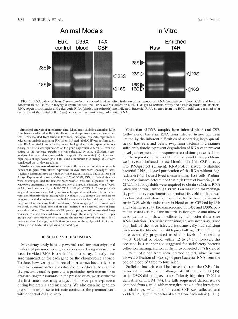
Collection of RNA samples from infected blood and CSF.Collection of bacterial RNA from infected tissues has beenlimited by the inherent difficulties of separating large quanti-ties of host cells and debris away from bacteria in a mannersufficiently timely to prevent degradation of RNA or to preventnovel gene expression in response to conditions presented dur-ing the separation process (14, 36). To avoid these problems,we harvested infected mouse blood and rabbit CSF directlyinto RNAprotect (Qiagen). RNAprotect served to stabilizebacterial RNA, allowed purification of the RNA without deg-radation (Fig. 1), and lysed contaminating host cells. Prelimi-nary experiments determined that high titers of bacteria (�108
CFU/ml) in body fluids were required to obtain sufficient RNA(data not shown). Although strain T4X was used for meningi-tis, preliminary experiments determined its yield in blood wastoo low (data not shown). Therefore, for bacteremia we usedstrain D39, which attains titers in blood of 109 CFU/ml by 48 hafter challenge (35). Bioluminescence of T4X and D39X per-mitted visualization of the bacteria in living mice and allowedus to identify animals with sufficiently high bacterial titers forRNA isolation. Bioluminescent imaging was necessary, sinceonly half of the mice infected intratracheally had sufficientbacteria in the bloodstream 48 h postchallenge. The remainingmice eventually progressed to similar levels of bacteremia(�109 CFU/ml of blood within 12 to 24 h); however, thisoccurred in a manner too staggered for satisfactory bacteriacollection. Exsanguination of the mice collected at 48 h yielded�0.75 ml of blood from each infected animal, which in turnallowed collection of �25 �g of pure bacterial RNA from thepooled blood of three to four mice.
Sufficient bacteria could be harvested from the CSF of in-fected rabbits only upon challenge with 108 CFU of T4X (35);strain D39X did not grow to a sufficiently high titer. T4X is aderivative of TIGR4 (44), the fully sequenced clinical isolateobtained from a child with meningitis. At 4 h after intracister-nal challenge, �1.0 ml of infected CSF was collected andyielded �5 �g of pure bacterial RNA from each rabbit (Fig. 1).
FIG. 1. RNA collected from S. pneumoniae in vivo and in vitro. After isolation of pneumococcal RNA from infected blood, CSF, and bacteriaadherent to the Detroit pharyngeal epithelial cell line, RNA was visualized on a 1% TBE gel to confirm purity and assess degradation. BacterialRNA (open arrowheads) and eukaryotic RNA (shaded arrowheads) are indicated. Bacterial RNA isolated from the ECC model was enriched aftercollection of the initial pellet (raw) to remove contaminating eukaryotic RNA.
5584 ORIHUELA ET AL. INFECT. IMMUN.

Collection of RNA samples from pneumococci attached toepithelial cells. To examine bacterial gene expression in re-sponse to ECC, we infected confluent monolayers of Detroitcells (38), a pharyngeal epithelial cell line, with pneumococcusT4R. T4R is an unencapsulated derivative of T4 (12). As acontrol, RNA was collected from bacteria grown in the sametissue culture media without cells. These analyses thereforereflect only changes in gene expression that are a result of hostcell exposure. Stimuli resulting in alterations in gene expres-sion should therefore be limited to (i) intimate contact with thehost cells, (ii) soluble factors released by the host cell duringnormal processes, and (iii) soluble factors released by the hostcell in response to the bacterial infection. Collection of RNAfrom bacteria attached to Detroit cells resulted in the collec-tion of �25 �g of mixed eukaryotic and prokaryotic RNA fromthe bacteria and host cell pellet. Using MICROBencrich, wewere able to purify the prokaryotic RNA (Fig. 1), with a finalyield of �10 �g per T-175 flask.
Microarray analysis of gene expression relies on the com-parison of two RNA species. Levels of expression reported inthe test condition are reported in relation to levels of tran-scription observed in the control condition. A direct compar-ison of test versus control can be set up in vitro with tissueculture (medium to medium plus Detroit cells). With regard toin vivo gene expression, an appropriate in vitro control condi-tion does not exist. However, reference to the same arbitrarycontrol media (blood versus C�Y or CSF versus C�Y) allowsfor indirect comparisons between the different test conditions(blood versus CSF). As a baseline, �99% of genes showedsimilar expression levels in D39 and T4 grown in C�Y (datanot shown).
Microarray analysis of bacterial physiology in different bodysites. Filtration algorithms and SNR analysis of the microar-rays by GenePix 4.1 removed genes whose corresponding spotson the microarray were not valid �75% of the time. As such,the results reported in the present study correspond to 69% ofthe genome during growth in the bloodstream, 53% of thegenome during meningitis, and 68% of the genome after ECC.A list of the genes that did not meet the above criteria andtherefore were not analyzed for alternate gene expression invivo can be found at http://www.stjuderesearch.org/vivogene.Stringent requirements imposed by the filtration algorithmsensured that any alterations in gene expression that are re-ported here are rigorously supported by the data collectedfrom the microarrays.
For genes that met the established criteria for transcriptionanalysis, it was determined that the majority (92% in the blood,85% in CSF, and 90% after ECC) were expressed in a fashionsimilar to growth in C�Y. Table 1 lists the genes with alteredtranscription sorted based on their putative cellular role andtheir location on the bacterial chromosome. Inclusion criteriawere a �2-fold change in levels of expression and a P value of�0.01. Of the genes with altered expression in vivo, 55% hadenhanced expression in blood, while 81% were enhanced dur-ing ECC. In contrast, only 35% were enhanced during growthin CSF. Overall, the patterns of gene expression were deter-mined to be distinct for bacteria in each anatomic site. Figure2 is a Venn diagram illustrating the differences and similaritiesbetween the expression profiles of the pneumococci in blood,in CSF, and during ECC. Highlighting the disparity between
these conditions, only eight genes had similar alterations ingene expression during bacterial growth in blood, in CSF, orduring ECC: two that encode the virulence determinants PspA(46) and PrtA (4), three that encode genes in the psa operon(manganese acquisition and transport) (30), two that are in-volved in energy metabolism (an enolase [eno] and a glycosylhydrolase [SP0265]), and a transporter (SP1587). Their com-mon expression warrants further investigation as potential tar-gets for intervention, since they may reflect a core set of genesrequired for virulence.
Analysis of gene transcription indicated several features ofbacterial physiology unique to each body compartment. Anal-ysis of blood-dependent transcription revealed that cell walland membrane synthesis, cell division, and competence wereunchanged from in vitro. Nonetheless, the pneumococcus ap-pears to be under stress. Supporting this view, 24 genes encod-ing ribosomal proteins had reduced transcription in the blood.We interpret this as indicating that the pneumococcus is re-ducing its translational capacity. Likewise, relA, the gene en-coding the GTP pyrophosphokinase was enhanced duringgrowth in blood (26). RelA is expressed in response to lack ofnutrients and is responsible for ppGpp synthesis and entry ofthe bacteria into the stringent response. Finally, the genesencoding manganese and zinc transporters and the genes re-sponsible for purine and folic acid biosynthesis were also in-creased.
In the CSF, the pneumococcus decreased transcription of atleast 20 genes involved with competence (15), includingcomX1, comA, comB, comE, and comD. Likewise, 11 genesinvolved in the biosynthesis of fatty acids were also markedlyreduced. Decreased expression of the fab operon (31) mayindicate that sufficient fatty acids are available in the CSF.Other differences observed in CSF cultures included enhancedexpression of the lic operon (50), which indicates that thebacteria are decorating their cell wall with phosphorylcholine.SP1804, a gene encoding a general stress protein, is enhancedin the CSF, perhaps indicating that the bacteria are understress in adapting from the rich medium of blood to the poormedium of CSF.
Pneumococci adherent to epithelial cells also had uniquealterations in their expression profiles. We observed enhancedexpression of the lic operon (50) and the dlt operon (49) duringECC. These changes indicate enhanced addition of phospho-rylcholine and D-alanine to teichoic acids. Enhanced expres-sion of VncS and CiaR/H, genes encoding two-componentsystems (42), indicate global changes in the synthesis of cellwall polymers, peptide pheromones, bacteriocins, and htrA ex-pression. Manganese acquisition also appears to be particu-larly important since the psa operon showed enhanced expres-sion in all three of the conditions tested and has recently beenshown to undergo enhanced transcription in vivo (30).
With regard to energy metabolism, during growth in bloodthe pneumococcus reduces the expression of the genes encod-ing 1-phosphofructokinase and fructose-bisphosphate aldo-lase. Phosphorylation of 1-phosphofructose by 1-phophofruc-tokinase is the rate-limiting step of glycolysis and targets themolecule for glycolysis (22). Reducing expression levels of1-phosphofructokinase suggests that the pneumococcus has areadily available carbon source in the blood. At the same time,concurrent enhanced expression of SP0265 and SP2021, glyco-
VOL. 72, 2004 PNEUMOCOCCAL GENE EXPRESSION IN VIVO 5585

TABLE 1. Differential expression of S. pneumoniae genes as determined by microarray analysis
Gene name and/or description TIGRannotationb
Fold increase or decrease and P valuea in:
Blood CSF ECC
Fold change P Fold change P Fold change P
Virulence determinantsblpU; bacteriocin SP0041 �3.1 3E-07cps4A; capsular polysaccharide biosynthesis SP0346 2.2 2E-03cps4C; capsular polysaccharide biosynthesis SP0348 3.9 2E-12pspA; pneumococcal surface protein A SP0117A 3.4 2E-04 50.3 1E-08 4.0 1E-04cbpJ; choline-binding protein J SP0378 3.6 9E-07cbpG; choline-binding protein G SP0390 1.7 5E-03cbpF; choline-binding protein F SP0391 2.3 2E-03blpK; bacteriocin associated protein SP0533 �2.5 3E-06blpY; Immunity protein SP0545 NA NA �16.8 8E-13 NA NAprtA; protective antigen A SP0641A,D 15.5 7E-06 9.8 8E-13 3.3 1E-02spxB; pyruvate oxidase SP0730 �2.6 6E-06lmb; adhesion lipoprotein SP1002 3.4 2E-05 �5.2 1E-09Conserved hypothetical protein SP1003A 8.8 3E-05 �4.6 7E-09xseA; exodeoxyribonuclease VII, large subunit SP1207 �2.2 3E-07pln; pneumolysin SP1923A �5.6 5E-06 �9.8 3E-03lytA; autolysin SP1937 �7.6 9E-13chpA; choline-binding protein A SP2190A 2.3 2E-02chpD; choline-binding protein D SP2201A NA NA 12.0 2E-12htrA; serine protease SP2239A NA NA 8.2 4E-05spoJ; homologous to sporulation protein SP2240D 3.9 9E-05
Cell wall synthesisbacA; bacitracin resistance protein SP0457B 2.5 4E-04fibA; beta-lactam resistance factor SP0615 4.0 1E-03lytB; endo--N-acetylglucosaminldase SP0965 3.5 4E-03licC; phosphocholine cytidytyltransferase SP1267 4.7 1E-09 14.4 7E-08licB; choline transport SP1268 5.1 2E-09 15.4 1E-09pck; choline kinase SP1269 4.4 6E-09 18.2 2E-07Alcohol dehydrogenase, zinc containing SP1270 5.5 4E-09 17.2 9E-06Cytidine diphosphocholine pyrophosphorylase,
putativeSP1271 5.7 5E-10 14.0 3E-08
pgdA; N-acetylglucosamine deacetylase A SP1479D �3.2 2E-05penA; penicillin-binding protein 2B SP1673 2.4 2E-02dltD; D-alanyl-lipotechoic acid biosynthesis SP2173 2.8 7E-04 6.6 1E-03dltA; D-alanine activating protein SP2176A 3.6 1E-03
Cell membraneLipoprotein SP0149 3.7 2E-09Glycosyl transferase, group 1 SP1366 2.8 6E-04pgm; phosphoglucomutase SP1498 �2.1 8E-03
Competence/DNA transformationcomX1; transcriptional regulator SP0014 �12.1 6E-11Hypothetical protein SP0029 NA NA �8.1 5E-09 NA NAccs16; competence-induced protein Ccs16 SP0030 NA NA �16.8 4E-08 NA NAcomA; competence factor transport protein SP0042D �32.9 8E-12comB; competence factor transport protein SP0043D,B �48.8 2E-12ccs 4; competence-induced protein SP0200 �5.7 2E-07 3.0 2E-02Hypothetical protein SP0201 �2.9 2E-05dprA; DNA processing protein SP1266 �49.7 2E-11 6.2 9E-04cspC-related protein SP1913 5.2 8E-04cbf1; cmp-binding-factor 1 SP1980 �4.7 2E-09ccs50; competence-induced protein SP1981 �4.5 2E-06recA; DNA replication, recombination, and
repairSP1940 �12.9 1E-12
cinA; competence/damage-inducible protein SP1941A �39.0 1E-11 2.1 8E-03Conserved hypothetical protein SP1944 �2.4 2E-05Hypothetical protein SP1945 �12.1 5E-06comE; response regulator ComE SP2235 �15.0 2E-14 NA NAcomD; sensor histidine kinase SP2236A �6.7 2E-08
Stress relatedhcrA; heat-inducible transcription repressor SP0515 �4.5 1E-07 �4.4 8E-04
Continued on following page
5586 ORIHUELA ET AL. INFECT. IMMUN.

TABLE 1—Continued
Gene name and/or description TIGRannotationb
Fold increase or decrease and P valuea in;
Blood CSF ECC
Fold change P Fold change P Fold change P
grpE; heat shock protein SP0516 �3.7 2E-08 �3.7 2E-03dnaK; protein folding and stabilization SP0517 �3.1 6E-06 �3.9 2E-03sodA; superoxide dismutase SP0766A �3.5 3E-09Cadmium resistance transporter, putative SP1625 �3.9 1E-10Conserved hypothetical protein SP1644 2.3 3E-04relA; GTP pyrophosphokinase SP1645A 2.2 1E-03 2.8 1E-03Conserved hypothetical protein SP1801 NA NA 3.2 4E-03Hypothetical protein SP1802 5.5 1E-03Conserved hypothetical protein SP1803 NA NA 4.1 6E-05General stress protein 24, putative SP1804 2.7 1E-07 6.7 2E-03Hypothetical protein SP1805 3.5 8E-10 3.4 2E-03groEL; chaperonin, 60 kDa SP1906 �6.1 4E-11groES; chaperonin, 10 kDa SP1907 �6.9 2E-08
DNA repair, recombination, and modificationConserved hypothetical protein SP0022 NA NA �3.5 4E-06 NA NAradA; DNA repair protein SP0023AA,C NA NA �6.3 2E-07 NA NAHypothetical protein SP0025D �2.8 8E-07hexB; DNA mismatch repair protein SP0173 14.0 5E-14Hypothetical protein SP0792 2.3 4E-08MutT/nudix family protein SP0794 2.0 1E-09radC; DNA repair protein SP1088 �63.2 9E-11Type II restriction endonuclease, putative SP1221 3.0 6E-03recR; recombination protein SP1672 2.1 3E-03recG; ATP-dependent DNA helicase SP1697 2.6 6E-04
Cell divisionftsW; cell division protein SP1067 3.8 5E-03ppc; phosphoenolpyruvate carboxylase SP1068B 5.0 6E-04DivlVA; cell division protein SP1661 1.9 3E-03y/mH SP1662 2.2 2E-02y/mF SP1664 1.8 4E-02y/mE SP1665 2.0 3E-02spollLJ family protein SP1975 �2.2 1E-05
Energy metabolismGlycosyl hydrolase, family 1 SP0265A 3.5 1E-03 2.2 1E-06 5.5 5E-03Glycosamine–fructose-6-phosphate SP0266 2.9 1E-11manN; PTS system, IIC component SP0283 �2.2 7E-03Alcohol dehydrogenase, zinc-containing SP0285 �1.7 6E-03pfl; formate acetyltransferase SP0459 2.6 7E-04fba; fructose-bisphosphate aldolase SP0605 �2.1 1E-07 �2.2 2E-04Thioredoxin family protein SP0659B NA NA NA NA �3.7 2E-04manA; mannose-6-phosphate isomerase SP0736 2.7 1E-11 4.2 8E-04lacR; lactose system repressor SP0875 �4.8 9E-041-phosphofructokinase, putative SP0876 �4.0 3E-04aid; alanine dehydrogenase, authentic
frameshiftSP0952 NA NA 3.7 2E-03
Phosphoglycerate mutase family protein SP0984 NA NA 2.1 4E-03glgC; glucose-1-phosphate edenylyltransferase SP1122 3.5 3E-08eno; enolase SP1128 �4.2 3E-03 �2.1 1E-05 �3.3 4E-05Acetoin dehydrogenase, E1 component SP1164 2.3 8E-03lacG; 6-phospho--galaciosidase SP1184 NA NA 6.5 4E-03lacB; galactose-6-phosphate isomerase SP1192 NA NA 5.9 2E-06lacA; galactose-6-phosphate isomerase SP1193A NA NA 4.2 6E-03ldh; L-lactate dehydrogenase SP1220 �2.3 3E-04fhs; formate-tetrahydrofolate ligase SP1229 3.9 3E-04nagB; glucosamine-6-phosphate isomerase SP1415 �7.5 3E-12alpC; ATP synthase F1, epsilon subunit SP1507 4.8 3E-06atpD; ATP synthase F1, beta subunit SP1508 4.0 6E-06alpG; ATP synthase F1, gamma subunit SP1509 3.4 5E-04atpA; ATP synthase F1, alpha subunit SP1510 4.1 1E-08atpH; ATP synthase F1, delta subunit SP1511 3.8 7E-03atpF; ATP synthase F0, B subunit SP1512 NA NA NA NA 2.7 4E-04atpB; ATP synthase F0, A subunit SP1513 5.3 4E-06
Continued on following page
VOL. 72, 2004 PNEUMOCOCCAL GENE EXPRESSION IN VIVO 5587

TABLE 1—Continued
Gene name and/or description TIGRannotationb
Fold increase or decrease and P valuea in:
Blood CSF ECC
Fold change P Fold change P Fold change P
Glutathione S-transferase family protein SP1550 5.1 2E-05Cation-transporting ATPase, E1–E2 family SP1551 3.7 9E-09tpi; triosephosphate isomerase SP1574 2.2 7E-03Oxalate formate antiporter SP1587 3.5 5E-05 4.3 2E-11 3.9 7E-04gpmA; phosphoglycerate mutase SP1655 �5.6 3E-03ROK family protein SP1675 6.0 2E-09scrR; sucrose operon repressor SP1725 3.8 4E-03 5.3 9E-10galK; galactokinase SP1853 2.7 3E-03gap; glyceraldehyde-3-phosphate
dehydrogenaseSP2012 �6.5 5E-04
Glycosyl hydrolase, family 1 SP2021 5.8 6E-06 14.5 3E-03malA; maltose metabolism SP2111 2.0 8E-04 NA NA NA NAmalR; maltose operon transcriptional
repressorSP2112 3.1 2E-04
Transketolase, N-terminal subunit SP2128A NA NA 5.2 3E-05PTS system, IIC component, putative SP2129 NA NA 6.8 4E-06argF; ornithine carbamoyltransferase SP2150 7.6 8E-04arcC; carbamate kinase SP2151 9.2 2E-03
Fatty acid metabolismcls; cardiolipin synthetase SP0199A 3.6 3E-02enoyl-CoAc hydratase/isomerase family
proteinSP0415 �17.3 9E-09 �3.0 6E-04
acp; acyl carrier protein SP0417 �7.1 7E-04fabH; 3-oxoacyl-(acyl carrier protein) synthase
IIISP0418 �4.9 3E-07
fabK; enoyl-(acyl carrier protein) reductase SP0419 �12.8 2E-09fabD SP0420 �9.8 6E-07fabG; 3-oxoacyl- (acyl carrier protein)
reductaseSP0421 �13.0 8E-10 �2.1 4E-03
fabF; 3-oxoacyl-(acyl carrier protein) synthaseII
SP0422 �10.6 4E-09
accB; acetyl-CoA carboxylase SP0423 �10.3 9E-11 �2.1 1E-03fabZ SP0424 �6.3 9E-09 �2.1 2E-03accC; acetyl-CoA carboxylase SP0425 �10.7 4E-11 �2.3 6E-03accD; acetyl-CoA carboxylase, subunit beta SP0426 �8.8 5E-11accA; acetyl-CoA carboxylase, subunit alpha SP0427 �7.7 1E-10
Amino acid biosynthesis and acquisitionAmino acid ABC transporter, ATP-binding
proteinSP0111 2.3 1E-09
aliA; oligopeptide ABC transporter SP0366 3.9 2E-03ilvB; acetolactate synthase, large subunit SP0445A 7.4 4E-03ilvN; acetolactate synthase, small subunit SP0446 2.0 1E-10ilvC; ketol-acid reductoisomerase SP0447 5.5 4E-04ilvA; threonine dehydratase SP0450 4.5 5E-04Transcriptional regulator, MerR family SP0501 �5.7 1E-10Glutamine synthetase, type I SP0502 �4.4 1E-09livH; branched-chain amino acid ABC
transporterSP0750 2.5 2E-06
livM; branched-chain amino acid ABCtransporter
SP0751 2.3 3E-07
livG; branched-chain amino acid ABCtransporter
SP0752 2.3 5E-09
livF; branched-chain amino acid ABCtransporter
SP0753 2.3 3E-07
Conserved hypothetical protein SP0783 �4.7 1E-11Glutathione reductase SP0784 �2.2 2E-06lcEL; branched-chain amino acid
aminotransferaseSP0856A 3.5 3E-03
Oligopeptide-binding protein, internaldeletion
SP0857 3.8 2E-04
proA; gamma-glutamyl phosphate reductase SP0932B 4.0 3E-03dapA; dihydrodipicolinate synthase SP1014 2.6 2E-10 3.6 4E-04
Continued on following page
5588 ORIHUELA ET AL. INFECT. IMMUN.

TABLE 1—Continued
Gene name and/or description TIGRannotationb
Fold increase or decrease and P valuea in:
Blood CSF ECC
Fold change P Fold change P Fold change P
hemK; conserved protein SP1021D 14.3 1E-04amiE; ATP-binding protein SP1888 2.4 6E-05 2.9 1E-08amiD; permease protein SP1889A 2.1 1E-04 2.8 9E-09AmiC; permease protein SP1890A 2.9 2E-11
Nucleoside/nucleotride metabolismpurA; adenylosuccinate synthetase SP0019 �3.9 5E-09Phosphoribosytformylglycinamidine synthase SP0045A,C,B 6.1 5E-05 NA NApurF; amidophosphoribosyltransferase SP0046 11.6 1E-06 NA NApurM; purine ribonucleotide biosynthesis SP0047B 8.4 1E-05 NA NApurN; purine ribonucleotide biosynthesis SP0048B 9.7 1E-05 NA NApurH; purine ribonucleotide biosynthesis SP0050AA 10.3 4E-06 NA NApurE; purine ribonucleotide biosynthesis SP0053C 3.0 1E-04 NA NApurK; purine ribonucleotide biosynthesis SP0054C 6.9 4E-05Hypothetical protein SP0055 2.4 4E-04 NA NApurB; adenylosuccinate lyase SP0056B 2.7 5E-05nrdD; anaerobic ribonucleoside triphosphate SP0202 2.7 5E-05Acetyltransferase. GNAT family SP0204 3.2 2E-04nrdG; anaerobic ribonucleoside-triphosphate SP0205 3.5 3E-05 5.6 4E-07Hypothetical protein SP0206 2.3 2E-04Conserved domain protein SP0207 2.9 5E-05adk; adenylate kinase SP0231 �3.8 2E-03
Amino acid biosynthesis and acquisitionpyrF; orotidine 5-phosphate decarboxylase SP0701D �6.7 8E-04pyrE; orotate phosphoribosyltransferase SP0702D �11.0 9E-05 NA NAupp; uracil phosphoribosyltransferase SP0745 �3.9 4E-03Hypothetical protein SP0830 �2.5 3E-03deoD; purine nucleoside phosphorylase,
family 2SP0831 �3.3 2E-03
pyrK; dihydroorotate dehydrogenase, electron SP0963 �8.9 7E-05 3.4 IE-03pyrDb; dihydroorotate dehydrogenase B SP0964D �5.7 2E-03 4.7 3E-04carA; carbamoyl-phosphate synthase, small
subunitSP1276 �4.9 9E-04 2.2 2E-06
pyrB; aspartate carbamoyltransferase SP1277 �6.9 1E-03 2.0 3E-06pyrR; pyrimidine operon regulatory protein SP1278AA �8.0 8E-05uraA; uracil permease SP1286A �7.7 1E-04Oxidoreductase, pyridine nucleotide-disulfide SP1588 �4.5 8E-04
Co factor metabolismribAB; riboflavin, FMN, and FAD biosynthesis SP0176D,B �4.2 8E-04ribD; riboflavin, FMN, and FAD biosynthesis SP0178D �5.4 4E-04folC; dihydrofolate synthetase SP0290 2.1 8E-05 3.1 4E-03folE; GTP cyclohydrolase I SP0291 2.3 8E-05folE; GTP cyclohydrolase I SP0291 2.3 8E-05Bifunctional lolate synthesis protein SP0292 2.6 1E-04 2.9 2E-03coaA; pantothenale kinase SP0839 5.1 4E-03Macrolide-efflux protein SP1110 �4.9 3E-07Pyridoxine biosynthesis protein SP1468 2.8 2E-10coaD; phosphopantetheine adenylyltransferase SP1968 2.3 2E-06nadC; pyridine biosynthesis SP2016 �7.2 1E-07Membrane protein SP2017A,D NA NA �15.7 5E-125-Formyltetrahydrofolate cyclo-ligase SP2095A 2.5 4E-10
Anion/cation aquisitionIron
Non-heme Iron-containing ferritin SP1572 �8.4 8E-05 �2.4 4E-06Alcohol dehydrogenase, iron-containing SP2026 �3.6 2E-03 NA NA
PotassiumPotassium uptake protein, Trk family SP0480 2.6 4E-04
PhosphatepstA; putative membrane protein SP1393 2.8 2E-03
MagnesiumMagnesium transporter, CorA family SP0185 �2.1 5E-03
ManganesepsaB; ATP-binding protein SP1648 7.3 1E-05 5.0 2E-09 3.7 3E-05
Continued on following page
VOL. 72, 2004 PNEUMOCOCCAL GENE EXPRESSION IN VIVO 5589

TABLE 1—Continued
Gene name and/or description TIGRannotationb
Fold increase or decrease and P valuea in:
Blood CSF ECC
Fold change P Fold change P Fold change P
psaC; permease, authentic frameshift SP1649 9.3 2E-06 4.8 2E-11 7.6 3E-04psaA; manganese-binding adhesion liprotein SP1650 9.6 3E-07 5.6 1E-08 8.0 3E-04
ZincadcA; zinc-binding adhesion liprotein SP2169 9.1 3E-06 NA NA NA NAadcB; permease protein SP2170A 7.7 1E-05adcC; ATP-binding protein SP2171 6.8 3E-05adcR; repressor SP2172 4.2 8E-06
TranscriptionmutR; transcriptional regulator SP0141AA 3.0 2E-04Conserved hypothetical protein SP0385A,D �5.0 6E-10Sensor histidine kinase, putative SP0386D �3.9 2E-07DNA-binding response regulator SP0387D �4.3 1E-09Hypothetical protein SP0389 �3.3 2E-06Transcriptional regulator, MarR family SP0416 �5.3 1E-10vncS; sensor histidine kinase SP0604 2.2 5E-04 2.5 1E-02ciaR; DNA-binding response regulator SP0798D 3.1 4E-03ciaH; sensor histidine kinase ClaH SP0799D 3.1 1E-03Transcriptional regulator, putative SP0908 2.1 7E-04Conserved hypothetical protein SP0909 2.2 2E-03 NA NAprsA; ribose-phosphate pyrophosphokinase SP1095D �2.5 1E-06dnaG; DNA primase SP1072 �5.2 3E-09rpoD; RNA polymerase sigma-70 factor SP1073 �5.0 4E-06Conserved hypothetical protein SP1074 �4.4 3E-10vicX protein SP1225 2.5 3E-05Sensory box sensor histidine kinase SP1226 2.4 3E-04ATP-dependent RNA helicase, putative SP1586 3.1 1E-05Transcriptional regulator, Gn1R family SP1714 �6.6 2E-11ABC transporter, ATP-binding protein SP1715A,B �6.7 1E-08Conserved hypothetical protein SP1716 �12.0 3E-09ABC transporter, ATP-binding protein SP1717A �13.3 1E-10marR; transcriptional regulator SP1863 �2.3 7E-06ccpA; catabolite control protein A SP1999 4.2 3E-03
TranslationRibosomal protein S10 SP0208 �4.8 3E-04Ribosomal protein L3 SP0209 �4.2 1E-03 2.9 8E-04Ribosomal protein L23 SP0211 �4.0 4E-03 2.8 2E-05Ribosomal protein L2 SP0212 �4.1 1E-03Ribosomal protein S19 SP0213 �3.9 3E-03Ribosomal protein L14 SP0219 �3.6 4E-03Translation initiation factor IF SP0232 �5.0 9E-04Ribosomal protein L36 SP0233 �4.5 4E-03Ribosomal protein S13 SP0234 �4.7 5E-04Ribosomal protein S11 SP0235 �4.4 2E-03Ribosomal protein L17 SP0237 �6.1 5E-04serS; seryl-tRNA synthetase SP0411 4.5 6E-03Ribosomal protein L11 SP0630 �4.7 1E-03Ribosomal protein L1 SP0631 �5.0 5E-04Ribosomal protein S16 SP0775 �5.2 2E-04rpsA; ribosomal protein S1 SP0862 3.7 2E-04Translation initiation factor IF-3 SP0959 �6.4 6E-05Ribosomal protein L35 SP0960 �5.3 3E-03Ribosomal protein L20 SP0961 �6.0 9E-04Conserved hypothetical protein SP1097 �3.5 4E-05Conserved hypothetical protein SP1098 2.5 3E-02Ribosomal large subunit pseudouridine
synthaseSP1099 �3.4 5E-05 2.1 2E-02
Ribosomal protein L7/L12 SP1354 �7.8 8E-06Ribosomal protein L10 SP1355 �7.3 3E-05Ribosomal protein S21 SP1414 �3.8 4E-03glyQ; glyl-tRNA synthetase, alpha subunit SP1475 �3.0 4E-06Ribosomal protein S18 SP1539 �4.2 9E-04Single-strand binding protein SP1540 �4.6 9E-04Ribosomal protein S6 SP1541 �4.2 5E-03
Continued on following page
5590 ORIHUELA ET AL. INFECT. IMMUN.

TABLE 1—Continued
Gene name and/or description TIGRannotationb
Fold increase or decrease and P valuea in:
Blood CSF ECC
Fold change P Fold change P Fold change P
Ribosomal protein S15 SP1626 �3.5 5E-03fmt; methionyl-tRNA formyltransferase SP1735 2.4 1E-07oligoendopeptidase F. putative SP1780A 2.9 5E-05Conserved hypothetical protein SP1781 3.2 1E-04prmA: ribosomal protein L11
methyltransferaseSP1782C 2.4 1E-04
tgt: queuine tRNA-ribosyltransferase SP2058 �3.1 1E-05Ribosomal subunit interface protein SP2206 �3.9 2E-07
Post translational protein alternationsABC transporter, ATP-binding protein SP0151 2.1 1E-06ATP-dependent Clp protease SP0338A NA NA �5.8 2E-08 NA NALipoate-protein ligase, putative SP1160 3.1 9E-05 2.6 4E-07Peptidase, U32 family SP1429 �5.6 2E-03secA: preprotein translocase SP1702 2.2 4E-03Serine/threonine protein kinase SP1732 2.0 2E-03Peptidase, M20/M25/M40 family SP2096 2.9 3E-03Conserved hypothetical protein SP2143A NA NA NA NA 11.9 8E-07
Unknown functionTransporters
ABC transporter, ATP-binding protein SP0636 �4.9 1E-07Conserved hypothetical protein SP0638 �4.8 1E-08Hypothetical protein SP0639 �4.0 2E-06ABC transporter, ATP-binding protein SP0786 2.1 1E-04Conserved hypothetical protein SP0787 2.7 2E-04ABC transporter, ATP-binding protein SP1114 NA NA �2.9 4E-04Atz/Trz family protein SP1356D 8.1 9E-06ABC transporter, ATP-binding/permease
proteinSP1357D 3.9 4E-03
ABC transporter, ATP-binding/permeaseprotein
SP1358D 3.9 2E-04
Glycerol uptake facilitator protein, putative SP1491 3.4 4E-04Sodium/dicarboxylate symporter family
proteinSP1753 10.2 2E-04
MATE efflux family protein SP2065 NA NA 2.4 5E-03ABC transporter, ATP-binding/permease
proteinSP2073 2.2 2E-03 NA NA NA NA
ABC transporter, ATP-binding/permeaseprotein
SP2075D 3.0 5E-03
Cation-Transporting ATPase, E1–E2 family SP2101A 2.2 1E-07ABC transporter, ATP-binding protein SP2196 2.3 1E-08ABC transporter, substrate-binding protein SP2197 �4.0 1E-03 �2.8 4E-05
UnknownHypothetical protein SP0088 2.9 1E-07LysM domain protein SP0107 2.7 1E-04Hypothetical protein SP0115 18.0 9E-06 NA NAConserved hypothetical protein SP0122 �7.8 1E-06Hypothetical protein SP0124 �7.9 1E-06Hypothetical protein SP0125 �3.1 1E-05Hypothetical protein SP0142D 13.2 6E-06Conserved domain protein SP0143D,C 18.8 2E-05Hypothetical protein SP0144D 13.6 1E-05Conserved hypothetical protein SP0145A,D 13.0 7E-05Conserved hypothetical protein SP0159 4.4 2E-04Hypothetical protein SP0198A 2.1 1E-03Conserved hypothetical protein SP0239 3.1 3E-03Conserved hypothetical protein SP0288 2.1 8E-04 6.7 3E-04IS66 family element: Orf3, degenerate SP0362 �2.6 4E-03IS3-Spn 1: transposase, degenerate SP0392 �4.8 8E-04Conserved hypothetical protein SP0481 2.3 7E-07ABC transporter, ATP-binding protein SP0483B 2.2 5E-12Conserved hypothetical protein SP0488 �4.7 1E-03 �2.9 3E-07Hypothetical protein SP0582 �161.9 3E-15Conserved domain protein SP0617 3.5 1E-03
Continued on following page
VOL. 72, 2004 PNEUMOCOCCAL GENE EXPRESSION IN VIVO 5591

TABLE 1—Continued
Gene name and/or description TIGRannotationb
Fold increase or decrease and P valuea in:
Blood CSF ECC
Fold change P Fold change P Fold change P
Conserved hypothetical protein SP0629B 2.4 6E-07Hypothetical protein SP0703 �4.0 2E-03 NA NAConserved hypothetical protein SP0742C �4.3 1E-09KH domain protein SP0776 �5.0 7E-04Conserved hypothetical protein SP0787 3.7 6E-03Hypothetical protein SP0800 4.5 2E-05 2.1 4E-06Hypothetical protein SP0816 �3.5 2E-03Hypothetical protein SP0833 �2.6 6E-04Hemolysin-related protein SP0834 �3.7 3E-03Conserved hypothetical protein SP0841 3.3 1E-08Conserved hypothetical protein SP0868 �3.9 5E-07NifU family protein SP0870 �3.5 7E-09Conserved hypothetical protein SP0871 3.2 6E-03Conserved hypothetical protein SP0921 �2.2 1E-03Hypothetical protein SP0958 �6.7 1E-11O-methyltransferase SP0980D �3.3 6E-06Conserved hypothetical protein SP1012 �7.9 6E-04 NAConserved hypothetical protein SP1090 �3.9 7E-09Conserved domain protein SP1174A 6.6 1E-08 �7.8 2E-14Conserved domain protein SP1175A 16.3 1E-07 3.4 1E-02Conserved hypothetical protein SP1240 2.7 6E-04Conserved hypothetical protein, strain
TIGR4SP1249 3.6 1E-07 6.9 4E-03
lemA SP1284 �2.1 4E-07Transposase family protein, authentic
frameshiftSP1484 NA NA 3.6 6E-04
Methyltransferase, putative SP1578 3.4 2E-05Conserved domain protein SP1641 �15.5 3E-11Hypothetical protein SP1852A 2.1 3E-04 NA NANitroreductase family protein SP1710 NA NA 2.2 2E-08 NA NAConserved hypothetical protein SP1716 3.2 8E-03Hypothetical protein SP1779A 2.8 8E-05Hypothetical protein SP1787 �5.1 8E-09Conserved hypothetical protein SP1875 2.2 9E-05Conserved hypothetical protein SP1876 2.2 1E-04Integrase/recombinase, phage integrase
familySP1877 2.1 7E-04
CBS domain protein SP1878 2.1 7E-04Oxidoreductase, short chain dehydrogenase/
reductaseSP1909 5.9 1E-10
Conserved hypothetical protein SP1922 �5.8 2E-13Hypothetical protein SP1924 �9.2 2E-09Hypothetical protein SP1925 �4.1 3E-07Hypothetical protein SP1926 �3.7 3E-06Conserved hypothetical protein SP2027D NA NA 2.6 3E-03Conserved hypothetical protein SP2045 �3.0 9E-09LysM domain protein, authentic frameshift SP2063 4.9 5E-06 NA NAConserved hypothetical protein SP2132 2.8 1E-03 NA NA NA NAConserved hypothetical protein SP2152 18.2 4E-03Transporter, truncation NTL02SP0105 4.2 4E-05Hypothetical protein NTL02SP0107 5.0 6E-05Conserved hypothetical protein NTL02SP0108 13.7 2E-10Hypothetical protein NTL02SP1212 �5.9 2E-04
TransposonIS66 family element; Orf3, degenerate SP0362 �11.6 4E-15IS66 family element; Orf3, degenerate SP0644 �12.9 2E-13IS66 family element; Orf2, Interruption SP0812 2.5 1E-07IS1381; transposase OrfA SP1310 5.3 2E-11IS66 family element; Orf3, degenerate SP1311 �27.4 3E-07IS1167; transposase SP1692 �2.4 3E-06IS1381; transposase OrfA, internal deletion SP2137 4.9 4E-08
a Minus (�) fold change values indicate genes with decreased transcription. NA, insufficient data was obtained from the microarray analysis to determine geneexpression levels.
b Superscript letters: A, identified by STM (13); AA, confirmed by animal infection (13); B, identified by STM (25); C, identified by STM (39); D, identified by DFI (29).c CoA, coenzyme A.
5592 ORIHUELA ET AL. INFECT. IMMUN.

syl hydrolases, and enhanced expression of scrR and malR, therepressors for sucrose and maltose operons, suggest that thepneumococcus is also utilizing alternative sources of energy.
Phase variation. S. pneumoniae undergoes spontaneousphase variation between a transparent phenotype and anopaque phenotype (21); the transparent phenotype has anenhanced capacity to adhere and colonize the nasopharynx,whereas the more phagocytosis-resistant opaque phenotypepredominates in blood. An increased capacity to adhere by thetransparent phenotype corresponds to higher levels of CbpA,phosphorylcholine, teichoic acid, and autolysin than do opaquevariants (21, 41, 48). In contrast, the opaque phenotype pro-duces more capsular polysaccharide and hydrogen peroxideand requires up to 30-fold more human immune serum toachieve 50% opsonophagocytic killing than related transparentstrains (20, 43).
Analysis of the genes enhanced during ECC confirmed aselection for the transparent phenotype. We observed en-hanced expression of cbpA and the lic and dlt operons. Similarresults for bacteria in CSF taken together with downregulationof spxB in CSF indicate that the transparent phenotype mayreappear after bacteria leave the bloodstream. Interestingly,we did not observe phenotype-enhanced expression of spxB orof the genes involved in capsule production in the blood.
Microarray analysis of known virulence determinants. Mi-croarray analysis of 20 known pneumococcal virulence deter-minants indicated site-specific expression for 17 of them (Table1). Only two virulence determinants with altered gene expres-sion, pspA and prtA, were expressed in a similar fashion inblood, CSF, or ECC (see above). pspA, encodes a choline-binding protein that inhibits complement deposition on thesurface of the bacteria and is upregulated in blood by Northernanalysis (46). prtA encodes a conserved serine protease (4).
Expression profiles were consistent with much of the currentunderstanding of pneumococcal pathogenesis. During attach-ment to pharyngeal epithelial cells, pneumococci enhancedexpression of the genes encoding choline-binding protein A(CbpA) and HtrA, two proteins shown to contribute to naso-pharyngeal colonization (41, 42). Expression of ply was de-creased, suggesting attenuation of the ability to injure hostcells with the toxin pneumolysin. We observed enhanced ex-pression in the blood of the operon encoding choline-bindingprotein G (CbpG) and choline-binding protein F (CbpF) (12).CbpG has been demonstrated in our laboratory to be required
for the development of high-grade bacteremia (preliminarydata). Lastly, we observed decreased expression of the genesencoding pneumolysin (ply) (8), autolysin (lytA) (45), and pyru-vate oxidase (spxB) (43) in the CSF. Pyruvate oxidase, SpxB, isresponsible for the production of hydrogen peroxide by thepneumococcus. This trio was striking since pneumolysin, hy-drogen peroxide, and inflammatory cell wall components re-leased by autolysin are the principle agents by which the pneu-mococcus induces neuronal damage (6, 47). Decreasedexpression of these genes in the CSF suggests that the pneu-mococcus adapts so as to decrease damage to neurons.
We also observed alterations in the gene expression of less-well-characterized virulence determinants. In the blood, weobserved enhanced expression of lmb, a gene homologous to alaminin-binding protein in Streptococcus anginosus (1). lmbtranscription was subsequently reduced in the CSF. Likewise,we observed a decrease in the expression of genes involved inbacteriocin synthesis and immunity (10). These expression pro-files suggest a potentially underappreciated role for thesegenes during pathogenesis.
Cross-reference to STM studies and DFI analyses. Investi-gators have used STM and DFI to identify pneumococcalgenes required for invasive disease (13, 25, 29, 39). In Table 1,we highlight genes with altered expression that have been iden-tified by STM or DFI. Comparison of the three STM studiesdetermined that the majority of loci identified were hit by onlyone study. Although this may reflect differences in methodol-ogy, it is likely that the use of three different strains of S.pneumoniae—G54, a serotype 19F (39); strain 0100993, a se-rotype 3 (25); and T4, a serotype 4 (13)—contributed to thediscrepancy observed between the three STM studies. Thisdisparity suggests that strain-dependent variations may occurwith regard to virulence. As such, genes identified by morethan one method are therefore particularly interesting for thecurrent analysis since they may represent a set of core viru-lence elements required by all pneumococci for the diseaseprocess. Moreover, core genes need not have altered transcrip-tion in vivo. The genes encoding immunoglobulin A1 protease(40), ZmpB (a metalloprotease that elicits inflammation in thelower respiratory tract) (5), and PavA (a fibronectin-bindingprotein) (16) all maintained unchanged levels of expression invivo and yet were all identified by more than one STM study(Table 2). Table 2 is a comprehensive list of genes with unal-tered expression that have been identified by more than oneSTM study. This approach is not comprehensive, however,since none of the major virulence determinants, such as pneu-molysin, capsular polysaccharide, or autolysin, was identifiedby more than one STM study. This may indicate a more site-specific contribution to pathogenesis (34). Finally, the majorityof genes hit by more than one STM analysis encoded proteinswhose function is unknown (Table 1). Those whose expressionwas altered in vivo and that are characterized include the puroperon (32) and rib operon (23) for purine and riboflavinbiosynthesis, respectively, as well as radA, a DNA repair en-zyme (3).
Virulence assessment of mutants. It is likely that bacterialgenes with enhanced expression during invasive disease con-tribute to the survival of the bacteria in vivo and to the pro-gression of disease. To test this hypothesis, we created mutantsin a number of genes of unknown function that were enhanced
FIG. 2. Venn diagrams highlighting disparity between infectiousmodels for genes with altered expression. Numbers indicate theamount of genes determined to have altered expression that are eithershared or exclusive to growth of the pneumococci in blood, CSF, orECC.
VOL. 72, 2004 PNEUMOCOCCAL GENE EXPRESSION IN VIVO 5593

during ECC or in the bloodstream. Some of these mutantscorresponded to genes (SP0090, SP0092, SP1434, and SP2163)that did not meet the criteria listed here. These genes wereexamined nonetheless due to large changes in gene expressiondetermined in vivo after less-stringent screening (data notshown). Mutants in genes enhanced during ECC were createdin T4X and assessed in an intranasal model of infection. Mu-tants lacking genes enhanced during bacteremia were createdin a D39X background and assessed in an intratracheal chal-lenge model of bacteremia. T4X mutants that were determinedto be attenuated were then subsequently transferred into aD39X background to identify strain differences (Table 3).
Analysis of mutants made in the T4X background deter-mined that five of seven mutants had a significantly reducedcapacity to colonize the nasopharynx. Three of these werecompletely attenuated and did not cross into the bloodstream,indicating that the genes deleted contributed to the pathogenicpotential of the bacteria. Surprisingly, when these three geneswere mutagenized in a D39X background, no differences invirulence were observed. This occurred despite the observationthat several of the genes mutated in D39X had previously beenidentified by STM as required for in vivo passage. Biolumines-cent imaging of the mice with the Xenogen IVIS cameraagreed with bacterial titers collected from lungs of infected
TABLE 2. Loci or genes within the same operon identified by multiple STM analyses with constitutive gene expression as determined by invivo microarray analysis
Gene name TIGR annotationa Cellular role
iga; immunoglobulin A1 protease SP0071A,B Pathogenesis: degradation of slgAFormate acetyltransferase, putative SP0251A,C Energy metabolism: fermentationTranscriptional regulator, putative SP0306A,C Regulatory functions: otherzmpB; zinc metalloprotease ZmpB SP664A,B Pathogenesis: protein and peptide secretion
degradation of proteinsTranscriptional regulator, LysR family SP927A,C Regulatory functions: DNA interactionspavA; adherence and virulence protein A SP966A,C Pathogenesis: cell adhesionCof family protein SP1291A UnknownSAP domain protein SP1292B UnknownOxidoreductase, Gfo/Idh/MocA family SP1482C Unknown substrateATP-dependent RNA helicase SP1483A TranscriptionmsmK; sugar ABC transporter, ATP-binding protein SP1580A,B Carbohydrate transportROK family protein SP2142A,C Small molecule interactionsAntigen, cell wall surface anchor protein SP2145A,B Unknown
a Superscript letters: A, identified by STM (13); B, identified by STM (39); C, identified by STM (25).
TABLE 3. Virulence assessment of mutants deficient in genes with enhanced expression during ECC or growth in blood
Challenge type and strain Function
Log10 median titer 2 days postchallengera in:
%Survivalc
Nasal lavage orlungsb Blood
Titer P Titer P
Intranasal challengeT4X 6.1 NA 5.6 NA 10T4X SP0090- ABC transporter, permease 4.3 0.003 �3.0 <0.001 100T4X SP0092- ABC transporter, substrate binding protein 5.7 0.278 4.6 0.107 10T4X SP0498- Endo--N-acetylglucosaminidase, putative 5.5 0.026 5.7 0.377 10T4X SP1021- hemK; conserved protein 5.7 0.088 4.7 0.163 20T4X SP1753- Sodium/dicarboxylate symporter family protein 4.5 0.011 4.3 0.077 10T4X SP2143- Conserved hypothetical protein 5.0 0.029 �3.0 <0.001 100T4X SP2163- PTS system, IIB component 5.1 0.011 �3.0 <0.001 70D39X 4.9 NA 7.1 NA 27D39X SP0090- ABC transporter, permease protein 4.3 0.119 6.6 0.527 40D39X SP2143- Conserved hypothetical protein 4.2 0.111 6.9 0.748 27D39X SP2163- PTS system, IIB component 6.2 0.235 7.0 0.852 13
Intratracheal challengeD39X 8.4 NA 9.0 NA 0D39X SP0115- Hypothetical protein 8.4 0.902 9.1 0.535 0D39X SP0142- Hypothetical protein 8.6 0.805 9.1 0.710 0D39X SP0641- Serine protease 8.3 1.000 9.0 0.620 0D39X SP1002- lmb; adhesion lipoprotein 7.2 0.128 9.0 0.620 0D39X SP1175- Conserved domain protein 7.5 0.234 8.6 0.181 0D39X SP1434- ABC transporter 8.5 0.383 9.2 0.165 0
a A total of 12 to 20 mice were used to examine each mutant in both challenge models. Statistical analysis was performed by using a Mann-Whitney rank sum test.Mutant titers were compared to the corresponding wild-type titers. Values in bold face indicate a statistically significant difference. NA, not applicable.
b Nasal lavage for intranasal challenge and lungs for intratracheal challenge.c That is, the percent survival 1 week after intranasal challenge or the percent survival 4 days after intratracheal challenge.
5594 ORIHUELA ET AL. INFECT. IMMUN.
mice. These images are thus not shown since they are redun-dant with the values listed in Table 3. Overall, these resultssuggest that D39X is can overcome deficiencies in virulencetraits that T4X cannot.
Considerations. Although the present study is the first todirectly measure pneumococcal gene expression during inva-sive disease, it must be acknowledged that to obtain sufficientquantities of bacterial RNA from the disease models, certainaspects of the experimental design were not optimal. Fore-most, it was necessary to use a different strain for each diseasemodel (D39X for blood, T4X for CSF, and T4R for ECC).Although preliminary data from in vitro experiments (data notshown) demonstrates that D39 and T4 respond in a similarmanner to growth in vitro (�99% of the genes examined whengrown in C�Y), how each strain responds to different anatom-ical sites remains unknown. It was also necessary to use adifferent host species for each disease model (mice for blood-stream infection, rabbits for CSF infection, and humans forECC), as well as a different RNA extraction protocol for ECC.The relationship between different host species and the pneu-mococcus may also result in alterations of the pneumococcalresponse. As a result of these caveats, care must be taken whengene expression levels for different anatomical sites are com-pared.
A second consideration is the requirement for very hightiters of pneumococci in the biological samples and the variousdurations of infection. For blood, bacterial titers were ca. 109
CFU/ml and were collected 2 days after intratracheal chal-lenge. As such, the expression profile observed does not rep-resent the initial phases of bloodstream infection. Likewise,rabbits were infected intracisternally with 108 CFU, and thebacteria were collected 4 h later. Since the bacteria were col-lected after only 4 h, our model represents bacterial geneexpression during early meningitis. During a natural infection,only a few animals would have a bacterial titer as high as 108
CFU/ml in the CSF, and leukocytes and other host factorswould be present in the CSF.
A final consideration is that not all of the genes in thegenome were examined. Although our analysis of in vivo geneexpression is the most comprehensive to date, we were unableto report on the expression of 31% of the genome duringgrowth in blood, 47% in the CSF, and 32% after ECC. As such,the present study is not comprehensive and further studies,including confirmation by Northern analysis of specific genes,are warranted.
Conclusions. Microarray analysis of pneumococcal gene ex-pression during invasive disease sheds light on the complexmechanisms of pneumococcal pathogenesis. Despite limita-tions imposed by technical challenges associated with in vivoRNA collection, we observed dramatic changes in a variety ofgenes, thus providing important information as to the physiol-ogy of the pneumococcus during invasive disease. Most genes(�90%) do not change expression from in vitro growth me-dium to in vivo. However, strikingly, the pneumococcus seemsto adapt to the blood, CSF, and ECC in a site-specific manner.These changes may be a result of the pneumococcus adaptingto nutrient availability and the stresses placed on the bacteria.In blood, cell wall and membrane synthesis and competencewere unchanged, whereas the bacteria strongly decreased ri-bosomal transcription, reduced glycolysis, and increased CbpG
and CbpF. In CSF, pneumococci drastically curtailed geneexpression for competence, autolysis, and production of toxins,while increasing amino acid biosynthesis and choline on thecell surface.
Cross-referencing of microarray analyses with that of STMand DFI and the virulence assessment of our own mutantsstrongly implies that pneumococcal virulence is dependent ona network of genes, the majority of which are not fully under-stood. Although an understanding of this network may seemdaunting, it seems that the continued use of global analyticaltechniques will shed light on the core genes that are requiredfor virulence. Such an attempt will require synthesis of datafrom multiple strains to derive a general picture of virulenceabove the noise of strain variation.
ACKNOWLEDGMENTS
We thank Robert Fleischmann and Scott Peterson at the PFGRC atTIGR for providing the pneumococcal microarrays necessary for thisproject. We also thank Alessandra Polissi, Sauli Haataja, and JeremyBrown for providing data from their STM studies.
This study was supported by NIH grant R01 AI27913 and TheAmerican Lebanese Syrian Associated Charities.
REFERENCES
1. Allen, B. L., and M. Hook. 2002. Isolation of a putative laminin bindingprotein from Streptococcus anginosus. Microb. Pathog. 33:23–31.
2. Avery, O. T., C. M. MacLeod, and M. McCarty. 1944. Studies on the chem-ical nature of the substance inducing transformation of the pneumococcaltypes: induction of transformation by the deoxyribonucleic acid fractionisolated from pneumococcus type III. J. Exp. Med. 79:137–157.
3. Beam, C. E., C. J. Saveson, and S. T. Lovett. 2002. Role for radA/sms inrecombination intermediate processing in Escherichia coli. J. Bacteriol. 184:6836–6844.
4. Bethe, G., R. Nau, A. Wellmer, R. Hakenbeck, R. R. Reinert, H. P. Heinz, andG. Zysk. 2001. The cell wall-associated serine protease PrtA: a highly con-served virulence factor of Streptococcus pneumoniae. FEMS Microbiol. Lett.205:99–104.
5. Blue, C. E., G. K. Paterson, A. R. Kerr, M. Berge, J. P. Claverys, and T. J.Mitchell. 2003. ZmpB, a novel virulence factor of Streptococcus pneumoniaethat induces tumor necrosis factor alpha production in the respiratory tract.Infect. Immun. 71:4925–4935.
6. Braun, J. S., J. E. Sublett, D. Freyer, T. J. Mitchell, J. L. Cleveland, E. I.Tuomanen, and J. R. Weber. 2002. Pneumococcal pneumolysin and H2O2mediate brain cell apoptosis during meningitis. J. Clin. Investig. 109:19–27.
7. Bricker, A. L., and A. Camilli. 1999. Transformation of a type 4 encapsulatedstrain of Streptococcus pneumoniae. FEMS Microbiol. Lett. 172:131–135.
8. Cockeran, R., R. Anderson, and C. Feldman. 2002. The role of pneumolysinin the pathogenesis of Streptococcus pneumoniae infection. Curr. Opin. In-fect. Dis. 15:235–239.
9. Darkes, M. J., and G. L. Plosker. 2002. Pneumococcal conjugate vaccine(Prevnar; PNCRM7): a review of its use in the prevention of Streptococcuspneumoniae infection. Paediatr. Drugs 4:609–630.
10. de Saizieu, A., C. Gardes, N. Flint, C. Wagner, M. Kamber, T. J. Mitchell, W.Keck, K. E. Amrein, and R. Lange. 2000. Microarray-based identification ofa novel Streptococcus pneumoniae regulon controlled by an autoinducedpeptide. J. Bacteriol. 182:4696–4703.
11. Francis, K. P., J. Yu, C. Bellinger-Kawahara, D. Joh, M. J. Hawkinson, G.Xiao, T. F. Purchio, M. G. Caparon, M. Lipsitch, and P. R. Contag. 2001.Visualizing pneumococcal infections in the lungs of live mice using biolumi-nescent Streptococcus pneumoniae transformed with a novel gram-positivelux transposon. Infect. Immun. 69:3350–3358.
12. Gosink, K. K., E. R. Mann, C. Guglielmo, E. I. Tuomanen, and H. R.Masure. 2000. Role of novel choline binding proteins in virulence of Strep-tococcus pneumoniae. Infect. Immun. 68:5690–5695.
13. Hava, D. L., and A. Camilli. 2002. Large-scale identification of serotype 4Streptococcus pneumoniae virulence factors. Mol. Microbiol. 45:1389–1406.
14. Hava, D. L., J. LeMieux, and A. Camilli. 2003. From nose to lung: theregulation behind Streptococcus pneumoniae virulence factors. Mol. Micro-biol. 50:1103–1110.
15. Havarstein, L. S. 1998. Identification of a competence regulon in Strepto-coccus pneumoniae by genomic analysis. Trends Microbiol. 6:297–300.
16. Holmes, A. R., R. McNab, K. W. Millsap, M. Rohde, S. Hammerschmidt,J. L. Mawdsley, and H. F. Jenkinson. 2001. The pavA gene of Streptococcuspneumoniae encodes a fibronectin-binding protein that is essential for viru-lence. Mol. Microbiol. 41:1395–1408.
VOL. 72, 2004 PNEUMOCOCCAL GENE EXPRESSION IN VIVO 5595

17. Hoskins, J., W. E. Alborn, Jr., J. Arnold, L. C. Blaszczak, S. Burgett, B. S.DeHoff, S. T. Estrem, L. Fritz, D. J. Fu, W. Fuller, C. Geringer, R. Gilmour,J. S. Glass, H. Khoja, A. R. Kraft, R. E. Lagace, D. J. LeBlanc, L. N. Lee,E. J. Lefkowitz, J. Lu, P. Matsushima, S. M. McAhren, M. McHenney, K.McLeaster, C. W. Mundy, T. I. Nicas, F. H. Norris, M. O’Gara, R. B. Peery,G. T. Robertson, P. Rockey, P. M. Sun, M. E. Winkler, Y. Yang, M. Young-Bellido, G. Zhao, C. A. Zook, R. H. Baltz, S. R. Jaskunas, P. R. Rosteck, Jr.,P. L. Skatrud, and J. I. Glass. 2001. Genome of the bacterium Streptococcuspneumoniae strain R6. J. Bacteriol. 183:5709–5717.
18. Jedrzejas, M. J. 2001. Pneumococcal virulence factors: structure and func-tion. Microbiol. Mol. Biol. Rev. 65:187–207.
19. Kerr, M. K., and G. A. Churchill. 2001. Bootstrapping cluster analysis:assessing the reliability of conclusions from microarray experiments. Proc.Natl. Acad. Sci. USA 98:8961–8965.
20. Kim, J. O., S. Romero-Steiner, U. B. Sorensen, J. Blom, M. Carvalho, S.Barnard, G. Carlone, and J. N. Weiser. 1999. Relationship between cellsurface carbohydrates and intrastrain variation on opsonophagocytosis ofStreptococcus pneumoniae. Infect. Immun. 67:2327–2333.
21. Kim, J. O., and J. N. Weiser. 1998. Association of intrastrain phase variationin quantity of capsular polysaccharide and teichoic acid with the virulence ofStreptococcus pneumoniae. J. Infect. Dis. 177:368–377.
22. Kornberg, H. L. 2001. Routes for fructose utilization by Escherichia coli. J.Mol. Microbiol. Biotechnol. 3:355–359.
23. Kreger, A. S., and R. H. Olsen. 1968. Purification and properties of mutantand wild-type diaphorases from Diplococcus pneumoniae. J. Bacteriol. 96:1029–1036.
24. Lacks, S., and R. D. Hotchkiss. 1960. A study of the genetic material deter-mining an enzyme activity in the pneumococcus. Biochim. Biophys. Acta39:508–517.
25. Lau, G. W., S. Haataja, M. Lonetto, S. E. Kensit, A. Marra, A. P. Bryant, D.McDevitt, D. A. Morrison, and D. W. Holden. 2001. A functional genomicanalysis of type 3 Streptococcus pneumoniae virulence. Mol. Microbiol. 40:555–571.
26. Lemos, J. A., T. A. Brown, Jr., and R. A. Burne. 2004. Effects of RelA on keyvirulence properties of planktonic and biofilm populations of Streptococcusmutans. Infect. Immun. 72:1431–1440.
27. Madu, A., C. Cioffe, U. Mian, M. Burroughs, E. Tuomanen, M. Mayers, E.Schwartz, and M. Miller. 1994. Pharmacokinetics of fluconazole in cerebro-spinal fluid and serum of rabbits: validation of an animal model used tomeasure drug concentrations in cerebrospinal fluid. Antimicrob. AgentsChemother. 38:2111–2115.
28. Mandell, L. A. 1995. Community-acquired pneumonia: etiology, epidemiol-ogy, and treatment. Chest 108:35S–42S.
29. Marra, A., J. Asundi, M. Bartilson, S. Lawson, F. Fang, J. Christine, C.Wiesner, D. Brigham, W. P. Schneider, and A. E. Hromockyj. 2002. Differ-ential fluorescence induction analysis of Streptococcus pneumoniae identifiesgenes involved in pathogenesis. Infect. Immun. 70:1422–1433.
30. Marra, A., S. Lawson, J. S. Asundi, D. Brigham, and A. E. Hromockyj. 2002.In vivo characterization of the psa genes from Streptococcus pneumoniae inmultiple models of infection. Microbiology 148:1483–1491.
31. Marrakchi, H., K. H. Choi, and C. O. Rock. 2002. A new mechanism foranaerobic unsaturated fatty acid formation in Streptococcus pneumoniae.J. Biol. Chem. 277:44809–44816.
32. Ng, W. L., K. M. Kazmierczak, G. T. Robertson, R. Gilmour, and M. E.Winkler. 2003. Transcriptional regulation and signature patterns revealed bymicroarray analyses of Streptococcus pneumoniae R6 challenged with suble-thal concentrations of translation inhibitors. J. Bacteriol. 185:359–370.
33. Ogunniyi, A. D., P. Giammarinaro, and J. C. Paton. 2002. The genes en-coding virulence-associated proteins and the capsule of Streptococcus pneu-moniae are upregulated and differentially expressed in vivo. Microbiology148:2045–2053.
34. Orihuela, C. J., G. Gao, K. P. Francis, J. Yu, and E. Tuomanen. Tissue-specific contributions of pneumococcal virulence factors to pathogenesis.J. Infect. Dis., in press.
35. Orihuela, C. J., G. Gao, M. McGee, J. Yu, K. P. Francis, and E. I. Tuomanen.2003. Organ-specific models of Streptococcus pneumoniae disease. Scand.J. Infect. Dis. 35:647–652.
36. Orihuela, C. J., R. Janssen, C. W. Robb, D. A. Watson, and D. W. Niesel.2000. Peritoneal culture alters Streptococcus pneumoniae protein profiles andvirulence properties. Infect. Immun. 68:6082–6086.
37. Pearce, B. J., Y. B. Yin, and H. R. Masure. 1993. Genetic identification ofexported proteins in Streptococcus pneumoniae. Mol. Microbiol. 9:1037–1050.
38. Peterson, W. D., Jr., C. S. Stulberg, and W. F. Simpson. 1971. A permanentheteroploid human cell line with type B glucose-6-phosphate dehydrogenase.Proc. Soc. Exp. Biol. Med. 136:1187–1191.
39. Polissi, A., A. Pontiggia, G. Feger, M. Altieri, H. Mottl, L. Ferrari, and D.Simon. 1998. Large-scale identification of virulence genes from Streptococcuspneumoniae. Infect. Immun. 66:5620–5629.
40. Poulsen, K., J. Reinholdt, and M. Kilian. 1996. Characterization of theStreptococcus pneumoniae immunoglobulin A1 protease gene (iga) and itstranslation product. Infect. Immun. 64:3957–3966.
41. Rosenow, C., P. Ryan, J. N. Weiser, S. Johnson, P. Fontan, A. Ortqvist, andH. R. Masure. 1997. Contribution of novel choline-binding proteins to ad-herence, colonization, and immunogenicity of Streptococcus pneumoniae.Mol. Microbiol. 25:819–829.
42. Sebert, M. E., L. M. Palmer, M. Rosenberg, and J. N. Weiser. 2002. Mi-croarray-based identification of htrA, a Streptococcus pneumoniae gene thatis regulated by the CiaRH two-component system and contributes to naso-pharyngeal colonization. Infect. Immun. 70:4059–4067.
43. Spellerberg, B., D. R. Cundell, J. Sandros, B. J. Pearce, I. Idanpaan-Heik-kila, C. Rosenow, and H. R. Masure. 1996. Pyruvate oxidase, as a determi-nant of virulence in Streptococcus pneumoniae. Mol. Microbiol. 19:803–813.
44. Tettelin, H., K. E. Nelson, I. T. Paulsen, J. A. Eisen, T. D. Read, S. Peterson,J. Heidelberg, R. T. DeBoy, D. H. Haft, R. J. Dodson, A. S. Durkin, M.Gwinn, J. F. Kolonay, W. C. Nelson, J. D. Peterson, L. A. Umayam, O. White,S. L. Salzberg, M. R. Lewis, D. Radune, E. Holtzapple, H. Khouri, A. M.Wolf, T. R. Utterback, C. L. Hansen, L. A. McDonald, T. V. Feldblyum, S.Angiuoli, T. Dickinson, E. K. Hickey, I. E. Holt, B. J. Loftus, F. Yang, H. O.Smith, J. C. Venter, B. A. Dougherty, D. A. Morrison, S. K. Hollingshead,and C. M. Fraser. 2001. Complete genome sequence of a virulent isolate ofStreptococcus pneumoniae. Science 293:498–506.
45. Tomasz, A., and S. Waks. 1975. Mechanism of action of penicillin: triggeringof the pneumococcal autolytic enzyme by inhibitors of cell wall synthesis.Proc. Natl. Acad. Sci. USA 72:4162–4166.
46. Tu, A. H., R. L. Fulgham, M. A. McCrory, D. E. Briles, and A. J. Szalai. 1999.Pneumococcal surface protein A inhibits complement activation by Strepto-coccus pneumoniae. Infect. Immun. 67:4720–4724.
47. Tuomanen, E., B. Hengstler, O. Zak, and A. Tomasz. 1986. Induction ofmeningeal inflammation by diverse bacterial cell walls. Eur. J. Clin. Micro-biol. 5:682–684.
48. Weiser, J. N., R. Austrian, P. K. Sreenivasan, and H. R. Masure. 1994. Phasevariation in pneumococcal opacity: relationship between colonial morphol-ogy and nasopharyngeal colonization. Infect. Immun. 62:2582–2589.
49. Whiting, G. C., and S. H. Gillespie. 1996. Investigation of a choline phos-phate synthesis pathway in Streptococcus pneumoniae: evidence for cholinephosphate cytidylyltransferase activity. FEMS Microbiol. Lett. 143:279–284.
50. Zhang, J. R., I. Idanpaan-Heikkila, W. Fischer, and E. I. Tuomanen. 1999.Pneumococcal licD2 gene is involved in phosphorylcholine metabolism. Mol.Microbiol. 31:1477–1488.
Editor: J. N. Weiser
5596 ORIHUELA ET AL. INFECT. IMMUN.