pneumococcal vaccination in children with cancer te-yu hung
TRANSCRIPT
Pneumococcal Vaccination in Children
with Cancer
Te-Yu Hung
MBBS DCH BMedSci(Hon) FRACP
This thesis is presented for the degree of Master of Medical Science of
The University of Western Australia
School of Paediatrics and Child Health
August 2018

i | P a g e
i | P a g e
THESIS DECLARATION
I, Te-Yu Hung certify that:
This thesis has been substantially accomplished during enrolment in the degree.
This thesis does not contain material which has been accepted for the award of any
other degree or diploma in my name, in any university or other tertiary institution.
No part of this work will, in the future, be used in a submission in my name, for any
other degree or diploma in any university or other tertiary institution without the prior
approval of The University of Western Australia and where applicable, any partner
institution responsible for the joint-award of this degree.
This thesis does not contain any material previously published or written by another
person, except where due reference has been made in the text.
The work(s) are not in any way a violation or infringement of any copyright,
trademark, patent, or other rights whatsoever of any person.
The research involving human data reported in this thesis was assessed and
approved by The University of Western Australia Human Research Ethics
Committee with recognition of prior Ethics approval from Child and Adolescent
Health Service Ethics Committee (HREC 2072EP) with delegated authority from
the Princess Margaret Hospital for Children Institutional Review Board.
Written patient consent has been received and archived for the research involving
patient data reported in this thesis.
The work described in this thesis was funded by Princess Margaret Hospital
Foundation Seeding Grant and Telethon Research Fellowship.
ii | P a g e
Technical assistance was kindly provided by Karli Corscadden, Jan Jones, Lisa
Montgomery, Janessa Pickering, Jacinta Bowman, Natalie Stemberger and the
staff at the Vaccine Trials Group, Telethon Kids Institute; and the nursing staff on
Ward 3B, Princess Margaret Hospital for Children for laboratory support, sample
transport and collection that is described in Chapters 4 and 5 of this thesis.
This thesis contains published work and/or work prepared for publication, some of
which has been co-authored.
Signature:
Date: 28/08/2018
iii | P a g e
AUTHORSHIP DECLARATION: CO-AUTHORED PUBLICATIONS
This thesis contains work that has been accepted for publication.
Title: Immunogenicity And Safety Of Single-Dose 13-Valent Pneumococcal
Conjugate Vaccine In Pediatric And Adolescent Oncology Patients
Location in thesis:
[Chapter 4
Student contribution to work:
Te-Yu Hung: Designed the study, recruited for the clinical aspect of the study,
acted as chief investigator, entered and managed data, contributed to
interpretation and discussion of the results, writing first draft, and writing- review
and editing.
I, Peter Richmond certify that the student statements regarding their contribution
to each of the works listed above are correct
Coordinating supervisor signature:
Date: 22/08/2018
iv | P a g e
ACKNOWLEDGEMENTS
Thank you to all the children and families that I’ve had the privilege of caring for at
Princess Margaret Hospital for Children that inspired me to start on this journey in
paediatrics. You taught me about immeasurable love, resilience and the beauty of
life. I’d like to thank my supervisors Peter Richmond, Catherine Cole, Chris Blyth for
their support and supervision of my journey with this project. I will take with me the
highest integrity in research standards to strive for through research in our
endeavors to find better and improved care of children. This project would be
impossible without the support, mentorship and encouragement of Rishi Kotecha,
Sarah Steed and Angela Alessandri. To the dedicated staff on ward 3B, Nick
Gottardo, Tom Walwyn, Felicity Hodder, Shanti Ramachandran, Hazel Gough,
Natalie Gamble-Williams, Caroline Stergiou, Carol Imms, Lisa, Karen, Fiona Kerr,
Shirley Wong, Siew Lee, Fleur Gowland for assistance on this project, I am one of
the many people that are grateful for your amazing presence.
Many thanks to Karli Corscadden, Ruth Thornton, Lisa Montgomery, Janessa
Pickering, Jan Jones and Jennifer Kent for laboratory support at Vaccine Trials
Group, Telethon Kids, Perth, Western Australia. Many thanks to Jacinta Bowman
and Natalie Stemberger for laboratory support at Pathwest Microbiology at Queen
Elizabeth II Medical Centre.
This study would not be possible without the funding support by Perth Children’s
Hospital Foundation formerly known as the PMH Foundation. Dr Hung was
generously supported by a Telethon Research Fellowship (2013). Dr Blyth is
supported by a WA Department of Health / Raine Early Career Research Fellowship
v | P a g e
(2014-2016) and a NHMRC Career Development Fellowship (APP1111596; 2016-
2019). Dr Thornton is supported by a BrightSpark Foundation Fellowship.
Most importantly, thank you to my best friend who is my husband and our two
children. It’s been a long journey but you’ve taught me to laugh and love and thank
you for travelling with me.
vi | P a g e
SUMMARY
Children with cancer receiving immunosuppressive therapy are vulnerable to severe
pneumococcal disease (IPD). This thesis examines the role of the 13-valent
pneumococcal conjugate vaccine (PCV13) in children on active (AIT) or recently
completed immunosuppressive cancer therapy (CIT) as a preventative strategy to
reduce IPD. A prospective, open-label cohort study included children in AIT and CIT
groups given a dose PCV13 demonstrated satisfactory immunogenic responses.
Safety data showed low rate of serious adverse events (3.5%). Our study design
was suboptimal to evaluate vaccination and its role for preventing nasopharyngeal
colonization. This data supports the administration of PCV13 during or immediately
prior to commencing therapy.
vii | P a g e
TABLE OF CONTENTS
Declaration i
Acknowledgement iv
Summary vi
Contents viii
Abbreviations xi
Chapter 1- Introduction 1
Chapter 2- Literature review 3
2.1 Introduction 3
2.2 Invasive Pneumococcal Disease and Immunocompromised Hosts 4
2.3 Vaccines targeting Streptococcus pneumoniae 6
2.4 Pneumococcal conjugate vaccines (PCVs) 6
2.5 Current Australian Recommendations for Pneumococcal Vaccinations 8
2.6 Pneumococcal Vaccinations in Immunocompromised Hosts 9
2.7 Protecting Cancer Survivors from Pneumococcal Disease 11
2.8 Assessing Immunogenicity of Pneumococcal Vaccines 12
2.9 Current Gaps in Knowledge 14
Chapter 3- Methodology
3.1 Patient Selection 15
3.2 Enrolment procedures 16
3.3 Vaccine administration 17
3.4 Adverse reaction profile 17
viii | P a g e
3.5 Antibody assay 18
3.6 Nasopharyngeal swab and culture 19
3.7 Statistical methods 20
3.8 Ethics 20
Chapter 4- Immunogenicity and safety of 13-valent pneumococcal conjugate
vaccine in paediatric oncology patients
4.1 Chapter Outline 21
4.2 Published manuscript 21
4.3 Statement of contribution 22
4.4 Funding 22
4.5 Acknowledgement 23
4.6 Disclosure Statement 23
4.7 Abstract 24
4.8 Introduction 25
4.9 Methods 27
4.10 Results 31
4.11 Discussion 42
4.12 Conclusion 45
Chapter 5- Nasopharyngeal Colonisation of Streptococcus pneumoniae in
Children with Cancer
5.1 Chapter Outline 46
5.2 Statement of contribution 46
5.3 Funding 47
5.4 Acknowledgement 47
ix | P a g e
5.5 Disclosure Statement 48
5.6 Abstract 48
5.7 Introduction 49
5.8 Methods 50
5.9 Results 52
5.10 Discussion 56
5.11 Conclusions 58
Chapter 6- Discussion 60
6.1 Chapter Outline 60
6.2 Summary of Findings 60
6.3 Recommendations for Clinical Practice 62
6.4 Future Research Directions 63
6.5 Conclusion 66
References 67
x | P a g e
ABBREVIATIONS
AIT active immunosuppressive therapy
ALL acute lymphoblastic leukaemia
CIT completed immunosuppressive therapy
CLL chronic lymphocytic leukaemia
ELISA enzyme-linked immunosorbent assay
GMC Geometric mean concentration
HIV Human Immunodeficiency Virus
HSCT haematopoietic stem cell transplant
IPD invasive pneumococcal disease
IRR incidence rate ratio
MIC Minimum inhibitory concentration
NIP National Immunisation Program
NP nasopharyngeal
NPS nasopharyngeal swab
PCV pneumococcal conjugate vaccine
PCV7 7-valent pneumococcal conjugate vaccine
PCV13 13-valent pneumococcal conjugate vaccine
PPV pneumococcal polysaccharide vaccine
PPV23 23-valent pneumococcal polysaccharide vaccine
PCR polymerase chain reaction
SMTGGB skim milk-tryptone-glucose-glycerin broth
S. pn Streptococcus pneumoniae
TMP/SMX trimethoprim/sulfamethoxazole; cotrimoxazole
WHO World Health Organisation
1 | P a g e
************************************************************************
CHAPTER 1- INTRODUCTION
************************************************************************
In children and adolescents, research advances have been made in the
development of potent therapies for several childhood cancers, so much so, that
many previously fatal conditions are now considered curative. Despite
improvements, morbidity and mortality continue to cause significant burden during
and after cancer therapy.
The vulnerability of immunocompromised patients to infections have been well
recognized. Over the last few decades, the population of children and adults with
acquired immunosuppression related to cancer therapy has increased rapidly.
Advances in strategies to prevent infectious complications during and after non-
transplant and haematopoietic stem cell transplant (HSCT) is an area of ongoing
research and potential development (1, 2).
Current strategies to prevent infection in immunocompromised populations are
frequently multi-pronged and include central catheter-care bundles, antimicrobial-
lock, and systemic antimicrobial prophylaxis(1). Although incidence of all-cause
mortality is lower in patients treated with prophylaxis (e.g. ciprofloxacin), however,
this comes with the cost of developing multi-resistant bacteria, complicating future
treatment. Vaccination has the potential of preventing infection and limiting
antimicrobial exposure.
Influenza and pneumococcus vaccination have been examined as potentially
vaccine-preventable disease in the immunocompromised population (2, 3). With the
2 | P a g e
advent and availability of highly immunogenic pneumococcal conjugate vaccines
(PCVs), there is renewed interest in the evaluation of this in a wider cohort of
immunocompromised population.
The primary aim of this thesis was to evaluate the immunogenicity and safety of a
single dose of 13-valent pneumococcal conjugate vaccine (PCV13) in children on
active or recently completed immunosuppressive cancer therapy. The effect of timing
and impact of varying intensity of chemotherapy was also explored. The secondary
aim was to explore the pneumococcal nasopharyngeal (NP) colonization, a key
antecedent to invasive pneumococcal disease (IPD) in this unique cohort.
The thesis is structured into 6 chapters, beginning with a review of the literature
which describes the burden of IPD and prevention strategies. This is followed by
Chapter 3 which describes the prospective interventional cohort study carried out at
Princess Margaret Hospital for Children in Western Australia between 2013 and
2014. The published manuscript of the immunogenicity and safety data from the
study is presented in Chapter 4. Chapter 5 describes the methods and results of a
small cross-sectional pneumococcal colonization study of the same cohort in
Chapter 5. The thesis is concluded by Chapter 6 which summarizes the findings of
the presented studies and outlines the implications for clinical practice and directions
for further research.
3 | P a g e
************************************************************************************************
CHAPTER 2 LITERATURE REVIEW
************************************************************************************************
2.1 Introduction
Streptococcus pneumoniae (S. pneumoniae; pneumococcus) is a Gram positive,
facultative aerobic bacteria known to cause invasive pneumococcal disease (IPD) or
mucosal disease. There are over 90 different pneumococcal serotypes which are
characterized by the capsular polysaccharide, which is its most important virulence
factor. Children under 5 years, immunocompromised patients and adults >65 years
are the most vulnerable populations to IPD (including sepsis and meningitis) that can
result in significant mortality and morbidity. The less severe clinical manifestations
caused by S. pneumoniae include pneumonia, otitis media and sinusitis(4).
Humans are the only reservoir of carriers of S. pneumoniae and most are
asymptomatic nasopharyngeal (NP) carriers via respiratory droplets. Colonisation
rates vary throughout life beginning in infancy and are influenced by geography,
overcrowding, number of siblings, childcare attendance, cigarette and cooking
smoke exposure and vaccination.
The relationship between NP carriage and virulence in causing IPD is complex(5).
Although NP carriage is a mandatory precursor to invasive disease, colonisation
studies may not detect all the strains which are causing invasive disease. For
example, strains with low virulence but less immunogenic serotypes (e.g. 6B) elicit
less humoral immune responses, are more prevalent and have higher detection
rates in colonization studies and have higher transmission rates.
4 | P a g e
2.2 Invasive Pneumococcal Disease and Immunocompromised Hosts
The survival rates for childhood cancer have improved significantly over the last 50
years some cancer types are now considered curable. For several childhood
malignancies, this has been achieved by increasing the duration and intensity of
chemotherapy. However, the consequent prolonged immunosuppression increases
vulnerability to infectious complications, which remain a common cause for mortality
and morbidity during treatment. The leading bacterial causes of sepsis can be
controlled by the use of preventative strategies including antibiotic prophylaxis or
central catheter care bundles. However, community-related infections such as
pneumococcal disease and influenza remain a significant risk to
immunocompromised children with cancer.
In the post-PCV era, the incidence ratio of IPD in children with haematological
malignancies is 188 times higher than in healthy children, and higher than children
with sickle-cell disease or other immunocompromised conditions such as Human
Immunodeficiency Virus (HIV) or solid and HSCT(6). There is no data specifically
examining the rates of IPD in children with solid organ malignancies. In adults with
current haematological and solid organ malignancy (e.g. lung cancer), the risk of IPD
is estimated to be between 13.9 to 22.9 times greater than healthy adult population(7,
8). It is unclear whether this risks remains elevated after immunosuppressive therapy
is ceased.
In healthy children who are unvaccinated against S pneumoniae, serotype-specific
humoral immunity increases gradually with age. In children receiving treatment for
acute lymphoblastic leukaemia (ALL), there is specific impairment of pneumococcal
humoral immunity which persists for up to nine months after cessation of
5 | P a g e
chemotherapy, despite recovery of lymphocyte counts and IgG levels(9, 10). As such,
disease burden of IPD is well-described in children with ALL and other non-
hematological malignancies in the pre- PCV era(11, 12). This is largely due to the
disease-specific effect of hematological malignancies on humoral immunity
combined with immunosuppression from chemotherapy and radiotherapy(13).
The literature in describing the NP colonisation of S pneumoniae in children
diagnosed with cancer during and after completion of therapy is limited(14, 15).This
is important to evaluate the risk-benefit ratio of PCV13 administration during cancer
therapy. Patients undergoing cancer therapy have different risk factors for
pneumococcal carriage. The reduced or defective humoral immunity is the most
likely cause for increased risk of pneumococcal colonisation in the nasopharynx. In
contrast, increased exposure to antibiotics (empirical use or prophylactic use of
trimethoprim/sulfamethoxazole (TMP/SMX) for opportunistic Pneumocystis jirovecii
pneumonitis; PJP) and reduced contact with peers through childcare or school may
decrease the overall rate of nasopharyngeal colonisation for pneumococcus. In
combination, the patient may also be at increased risk of colonisation by multi-drug
resistant pneumococcus.
In 2015, Princi et al compared the rates of pneumococcal NP colonisation in school
aged children receiving maintenance chemotherapy or had completed cancer
therapy within 6 months of the study(14). Using nucleic-acid amplification method to
detect S. pneumoniae, rate of carriage decreased significantly with age (range 6-17
years), prior PCV7 immunisation and TMP/SMX prophylaxis were negative
predictors of pneumococcal carriage [OR: 0.30 (95% CI: 0.16-0.73); OR 1.70 (95%
CI: 0.79-3.65); OR 0.41 (95% CI: 0.19-0.89) respectively].
6 | P a g e
More recently, serotype distribution of 50 invasive pneumococcal isolates from adult
and children with cancer in Brazil showed that the commonest serotypes were 23F
(12%), 6A (8%), 3, 4, 20, and 23A (6% each). Of these isolates, 14% demonstrated
reduced penicillin susceptibility and one isolate also had concomitant intermediate
ceftriaxone susceptibility(15).
2.3 Vaccines targeting Streptococcus pneumoniae
The 23-valent pneumococcal polysaccharide vaccine (PPV23) was first licensed in
United States in 1983 having demonstrated efficacy in adults. It is efficacious in
producing IgG antibodies response against 23 serotypes through a T-independent
immune response. The serotypes include 75% of IPD in Indigenous adults (62% for
non-Indigenous Australians) (16). However, this immune process lacks formation of
immune memory and booster vaccines results in immune hyporesponsiveness(17).
Importantly, the PPV23 is poorly immunogenic in high-risk populations, particularly in
children under 2 years and immunocompromised adults >65 years. In contrast, the
PCVs elicit greater immunogenic response in children who are most at risk of IPD.
By conjugating the capsular polysaccharide to the protein, the T-cell dependent
response is elicited, which includes memory B cells and generates a robust humoral-
response.
2.4 Pneumococcal Conjugate Vaccines (PCVs)
The 7-valent pneumococcal conjugate vaccine (PCV7) was first licensed in United
States in 2000 and was introduced to the Australian National Immunisation Program
(NIP) for all children in 2005. This vaccine contains antigens for serotypes 4, 6B, 9V,
14, 18C, 19F, 23F. Before the availability of PCV7, these serotypes accounted for
7 | P a g e
more than 80% of the IPD in US children(17). In Australia prior to vaccine
introduction, 84% of IPD isolates were due to serotypes contained in PCV7 (18).
PCV7 has been highly efficacious in reducing IPD in children in Australia and
overseas as demonstrated in several studies(16, 19-21). Other effects of PCV7 also
include reduced NP colonisation rates of pneumococcal vaccine-serotypes (22, 23),
moderate reduction in acute otitis media in high incidence populations and herd
immunity with reduced pneumococcal disease in older populations.
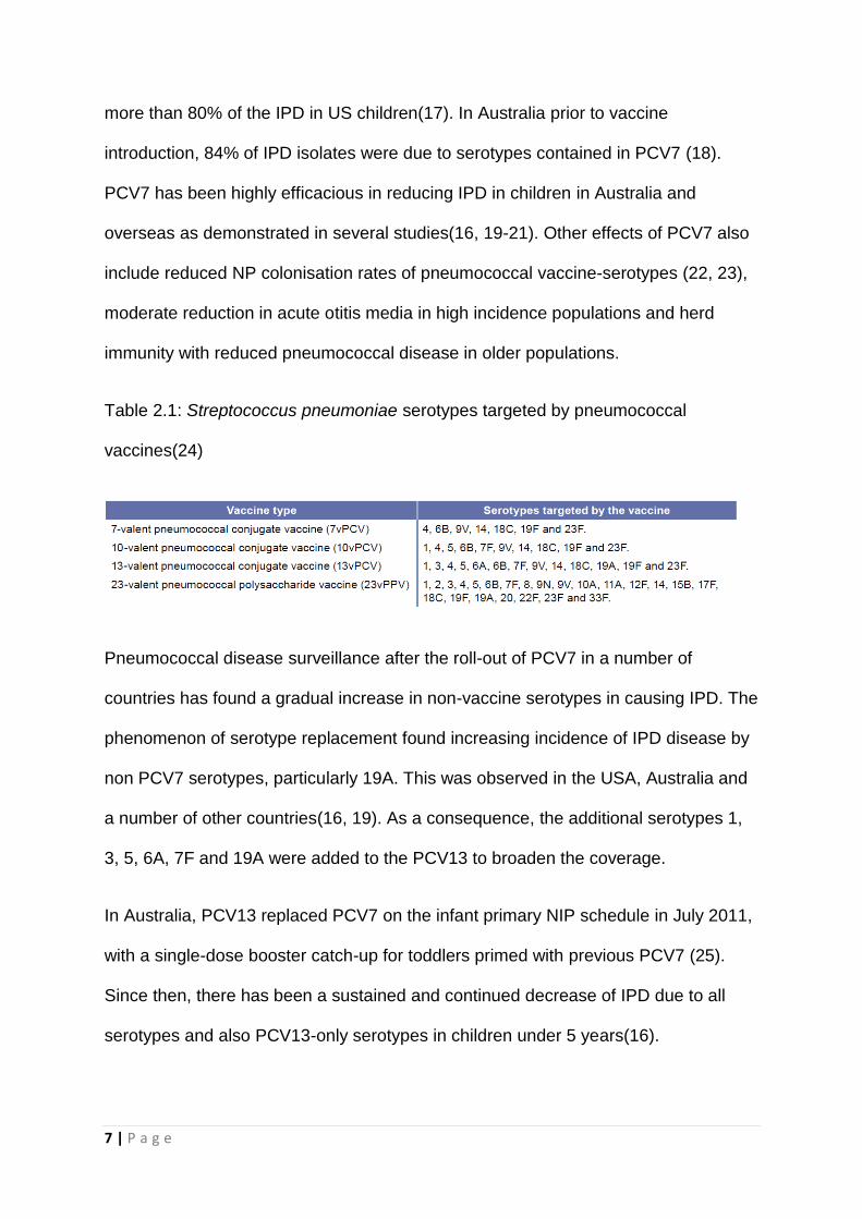
Table 2.1: Streptococcus pneumoniae serotypes targeted by pneumococcal
vaccines(24)
Pneumococcal disease surveillance after the roll-out of PCV7 in a number of
countries has found a gradual increase in non-vaccine serotypes in causing IPD. The
phenomenon of serotype replacement found increasing incidence of IPD disease by
non PCV7 serotypes, particularly 19A. This was observed in the USA, Australia and
a number of other countries(16, 19). As a consequence, the additional serotypes 1,
3, 5, 6A, 7F and 19A were added to the PCV13 to broaden the coverage.
In Australia, PCV13 replaced PCV7 on the infant primary NIP schedule in July 2011,
with a single-dose booster catch-up for toddlers primed with previous PCV7 (25).
Since then, there has been a sustained and continued decrease of IPD due to all
serotypes and also PCV13-only serotypes in children under 5 years(16).
8 | P a g e
Serotype replacement was also observed in Canada, with approximately 30% IPD
are caused by non-PCV13 serotypes(6). Similarly, proportion of IPD due to PCV13
serotypes have diminished in Native Alaskan children under 5 years; from 65% (pre-
PCV era) to 26%, 45 months after the introduction of PCV13(26). In contrast, for
antibiotic resistant pneumococcal isolates, PCV13 is estimated to cover over 90% of
antibiotic resistant serotypes causing IPD(27).
2.5 Current Australian Recommendations for Pneumococcal vaccinations
At the time of the study, the Australia National Immunisation Program (NIP) funded a
3-dose series of PCV13 for all infants at 2, 4, and 6 months of age (Table 2).
Australia was unique in industrialised countries in not having a PCV booster dose in
the second year of life for most children which may impact on long-term protection
(28). The 4th PCV dose was recommended at 12 months for children who are at high
medical-risk, including Indigenous and Torres Strait Island children and those with
immunodeficiency(25).
9 | P a g e
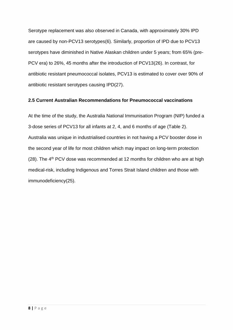
Table 2.2: Australian National Immunisation Program Recommendations for
pneumococcal vaccination for children aged <5 years(25)
2.6 Pneumococcal Vaccinations in Immunocompromised Hosts
Vaccine-responses in immunocompromised hosts vary according to many factors,
including cause and severity of immunosuppression and the vaccine itself. The
phenomenon of the “cancer paradox” refers to the dampened immune response in
an immunosuppressed person to a vaccine, whilst being more vulnerable to
infectious complications. Consequences of this stems not just from the morbidity and
10 | P a g e
mortality from the infection itself, but also the indirect effect on the timing and
possible delays of the planned chemotherapy.
In children who received non-transplant chemotherapy, the biological observations of
immune recovery largely form the basis of 6 months as the time-point for re-
immunisation to start in most guidelines. In children under 18 years, the data that the
recovery of B-lymphocytes correlates closely with recovery of total blood
lymphocytes came from a small study cohort of 14 patients(29). A systematic review
of children with ALL with transient decrease of humoral responses to previous
vaccines takes 6-9 months post cessation of chemotherapy with few showing
continuing recovery beyond 1 year(30).
Early immunogenicity studies with PPV23 in adult patients with multiple myeloma,
Hodgkin’s lymphoma and chronic lymphocytic leukaemia (CLL) found no response to
vaccination once the patient has started on chemotherapy (25, 31-34). Reported
practice among Australian oncologists reflected these early studies such that
inactivated vaccinations except for annual trivalent influenza vaccine are infrequently
recommended during chemotherapy(35). The administration of other vaccines are
generally withheld during chemotherapy or radiotherapy until 6 months after
completion of therapy.
Since the advent of PCVs, there has been renewed interest in evaluating
immunogenicity and efficacy of vaccinations in patients with leukaemia. In adults with
CLL, immunogenicity from single dose of PCV13 showed a markedly improved
response to the early PPV studies as described above (36, 37).
Using 2 doses of PCV7 in children without prior exposure to pneumococcal vaccines
(PCV or pneumococcal polysaccharide vaccine (PPV)), Cheng et al demonstrated
11 | P a g e
that 2-doses of PCV7 were able to achieve protective antibody levels (>0.35 µg/ml)
for all 7 serotypes in 86-100% of children with acute leukaemia during maintenance
chemotherapy (n=20) and within 12 months of completing chemotherapy (n=9). In
the same study children with solid malignancy were also given PCV7 between 3 to
12 months after completing immunosuppressive therapy. All children receiving non-
haematological malignancy chemotherapy achieved serotype-specific protective
antibodies against all the PCV7 serotypes after 2 doses of vaccine(38).
A more recent study by Crawford et al studied the immunogenicity of PCV10 in
children receiving leukaemia chemotherapy(39). Children were given single-dose of
PCV10 if they’ve received previous pneumococcal immunisation, or 3-dose series if
previous PCV-naïve. The proportion of children achieving protective antibody levels
(>0.35 ug/mL) were lower than in non-immunosuppressed , ranging from 33.3% to
88.9% for the 10 serotypes. It was promising though that for children with previous
PCV priming, 40-88% achieved protective antibody levels (>0.35 ug/mL) across the
10 serotypes.
2.7 Protecting Cancer Survivors from Pneumococcal Disease
The current 10th Edition of the Australian Immunisation Handbook recommends a
single dose of PCV13 and in previously vaccinated children and adults diagnosed
with cancer(25) as soon as after diagnosis. The handbook also recommends post-
chemotherapy booster vaccinations to maximize the well-being and preventative
health of cancer survivors. The post-chemotherapy immunisation practices amongst
oncologists vary significantly based chemotherapy regimen intensity, e.g. 48% vs
97% for low or high-intensity regimen respectively(35, 40). Specifically, 71% of
Australia oncologists would recommend pneumococcal vaccination in children after
12 | P a g e
chemotherapy, lower than those who recommend vaccines against varicella or
meningococcal disease(35).
Internationally, the United States Advisory Committee on Immunisation Practices
guidelines and United Kingdom Royal College of Pediatrics and Child Health
Guidelines both recommend that all pediatric oncology patients should receive
booster doses of routine immunisations. Specifically, the latter recommends this to
occur 3 to 6 months after completion of treatment(41, 42). However, delayed
immunisation at these time points result in a prolonged period of time during which
the patients have reduced protection from pneumococcal infections.
There are no studies specifically comparing the response to PCVs in children at
different time point post-chemotherapy. However, a study examining the optimal time
post-chemotherapy for booster immunisations administered inactive vaccines
(diphtheria, tetanus, acellular pertussis, inactivated poliomyelitis, conjugated
Haemophilus influenzae type b (Hib) vaccine) to children who were 3, 6 or 9 months
post treatment for ALL. This study found no difference in antibody responses when
the three time points were compared, and antibody levels remained above protective
levels 3 months later(43).
2.8 Assessing Immunogenicity of Pneumococcal Vaccines
2.8.1 Serotype-specific IgG Geometric Mean Concentrations (GMCs)
In healthy populations, serotype-specific protective thresholds of >0.15 to 1 µg/mL
were utilized in studies prior to 2003. An immunological correlate of protection has
been derived by pooling data from three efficacy studies for PCV7 licensure in
healthy Californian, South African and Gambian infants(44). There is international
13 | P a g e
consensus in adopting the World Health Organization recommended threshold of
0.35 µg/mL in evaluating the efficacy of PCV(45). The data is taken from studies of
infants undergoing primary immunisations series, although it is widely adopted for
use for booster doses.
There are a number of immunological end points including: geometric mean
concentrations (GMCs), >2 or ≥4-fold rise in pre- and post-vaccination antibody
concentrations , opsonophagocytic activity assay (OPA) ≥1:8 demonstrating
functional or killing avidity of the antibody response, and protective threshold of
serotype-specific antibody concentrations (≥0.35 µg/mL) as recommended by WHO
Pneumococcal Working Group in healthy population(44, 46, 47). There is little data
demonstrating the efficacy or effectiveness of PCV in immunocompromised hosts.
The above immunogenic correlate of protection is derived from healthy children and
may not reflect the same vaccine efficacy in immunocompromised population(3, 47).
2.8.2 Opsonophagocytic assay (OPA)
The protective function of the humoral response elicited by the vaccine is through
opsonisation of pneumococci and thus facilitate phagocytosis. The
opsonophagocytic killing assay is shown to be the best functional correlated
assessment of the mounted antibody response. There are several other OPA
methods, but the standardised killing OPA methodology by WHO is accepted as
crucial in the development of PCVs(48).
This is an in vitro assay which shows the concentration of serum required to show
bacterium count is reduced by 50% by phagocytosis. An OPA titre of ≥1:8 is
considered protective and shows a correlate of IgG concentration of 0.2-0.35 µg/mL
as measured by ELISA or similar method. In the elderly populations, the quantitative
14 | P a g e
antibody concentration maybe high, but more are non-functioning anticapsular
antibodies. The OPA is now recognised as the functional assay in licensing of new
pneumococcal vaccines.
The functional assessment of the antibody response is important in
immunocompromised patients but has rarely been undertaken in clinical studies. It
was assessed in only 4 serotypes in a study which administered 1 or 2 doses of
PCV10 in a paediatric cohort with leukaemia(39). In addition to the significant rises in
proportion of children with post-vaccination responding to the PCV10, there were
also good correlations with ELISA measured GMCs to OPA titres for serotypes 1,
6B, 19F and 23F.
2.9 Current Gaps in Knowledge
The burden of IPD is well described in the literature, particularly for children with
haematological malignancies. There is an effective vaccine in preventing IPD in
health children and also children with non-malignancy related immunosuppression.
There is a clinical imperative to explore the immunogenicity and safety of PCV13 in
this population. We will also explore the epidemiology of serotype distribution of
pneumococcal colonization in this cohort, and penicillin susceptibility of the
pneumococcus.
15 | P a g e
************************************************************************************************
CHAPTER 3 METHODS
************************************************************************************************
3.1 Patient selection
A prospective, single-center, open-label intervention study was conducted at
Princess Margaret Hospital for Children in Perth, Australia between March 2013 and
December 2014. Children aged 1 to 18 years with any cancer diagnoses were
eligible for recruitment. Children receiving immunosuppressive cancer therapy,
including chemotherapy and/or radiotherapy, were recruited into the active
immunosuppressive therapy group (AIT). Children who were within 12 months
following completion of therapy for cancer were recruited into the completed
immunosuppressive therapy group (CIT). Children were identified and recruited
when attending routine outpatient clinics, same-day unit for outpatient
chemotherapy, or are admitted as inpatient on the Oncology and Haematology
Ward. Demographic and clinical data recorded include age at vaccination, sex,
history of IPD and middle ear disease (recurrent acute otitis media, myringotomy,
ventilation tube insertion, adenoidectomy and/or tonsillectomy), immunosuppressive
chemotherapeutic, antimicrobial therapy, tumor type, duration of immunosuppressive
therapy and intensity of treatment prior to vaccination as previously defined(49).
Immunisation history was collected at enrolment from both personal immunisation
records and the Australian Child Immunisation Register (ACIR; now known as
Australian Immunisation Register).
Exclusion criteria included previous anaphylaxis to a conjugate pneumococcal
vaccine or any vaccine component; infusion of intravenous immunoglobulin in the
16 | P a g e
past three months; neutropenia (<0.5x10⁹/L), thrombocytopenia (<30x109/L) or fever
≥38.5°C on the day of review; and receipt of trivalent inactivated influenza vaccine
within the last three days or the 23vPPV (Pneumovax 23®) in the last two months.
Children from the CIT group who had previously received a dose of PCV13 following
completion of their cancer therapy were also excluded.
Written informed consent was obtained from the parent/guardian, with additional
written assent obtained from participants over the age of eight years.
3.2 Enrolment procedures
Participants had baseline nasopharyngeal swab (NPS) and blood serum sample
taken for serotype-specific anti-pneumococcal antibody concentrations assays. The
nasal swab was taken according to the method described in the WHO working party
report (50). The participants were given a single dose of intramuscular PCV13 and a
symptom diary to record adverse effects for 7 days. The repeat blood serum sample
and NPS were taken between 28 to 60 days from vaccination to assess for
pneumococcal serotype-specific antibodies and NP carriage for S pneumoniae. The
third NPS sample for patients in the AIT group was taken when the patient returned
for 6-month follow-up after the completion of the cancer therapy.
The NPS’s were taken using a FLOQSwabs™ (Batch 015N37. Lot C9PD00 Copan
Flock Technologies) at enrolment after the consent procedure as described above
(O’Brien PIDJ 2003). The NPS’s were either taken during an outpatient clinic visit or
under general anaesthesia while the patient underwent cancer therapy-related
procedure.
17 | P a g e
3.3 Vaccine administration
A single 0.5 mL dose of PCV13 (Prevenar 13® Pfizer Australia Pty Ltd, Batch no.
G39139, G47543, G39140, H40632) was administered intramuscularly as per
standard immunisation procedure(25). Each 0.5 mL dose contained 2.2 µg of
pneumococcal purified capsular polysaccharides for serotypes 1, 3, 4, 5, 6A, 7F, 9V,
14, 18C, 19A, 19F and 23F and 4.4 µg of pneumococcal purified capsular
polysaccharides for serotype 6B. Each serotype was individually conjugated to non-
toxic diphtheria CRM197 protein and adsorbed on aluminum phosphate (0.565 mg).
Participants were observed for 15 minutes post vaccination for any immediate
adverse reactions.
3.4 Adverse reaction profile
A single-page diary was provided to each participant for daily recording of side
effects and their severity occurring in the seven-day period following vaccine
administration. Solicited adverse reactions included systemic (measured body
temperature, irritability, increased sleep, decreased sleep, rash), gastrointestinal
(decreased appetite, nausea, vomiting, abdominal pain, diarrhoea), local injection
site reactions (pain, redness, swelling), respiratory (facial swelling, shortness of
breath) and neurological (headache, dizziness, seizure) symptoms. A paper caliper
was provided to objectively measure local reactions as mild (0-2.5 cm), moderate
(2.5-7 cm) or severe (≥7 cm). Parents/guardians were asked to measure
participants’ body temperature when they were subjectively feverish or perform a
recording of body temperature at the same time of each day. Participants from the
AIT group who developed a fever of ≥38ºC were asked to present to the Department
of Clinical Hematology and Oncology for assessment as per standard unit policy.
18 | P a g e
3.5 Antibody assay
Baseline bloods were taken pre-vaccination, with follow-up samples taken in the four
to eight week window following vaccination. Blood samples were allowed to clot at
room temperature for 30 minutes before centrifugation at 4000 rpm at 18ºC for ten
minutes. Serum was aliquoted prior to storage at -80ºC until analysis. All blood
samples were processed within 24 hours of collection.
A microsphere-based flow cytometric assay was used to measure IgG levels for
twelve pneumococcal polysaccharides (1, 3, 4, 5, 6B, 7F, 9V, 14, 18C, 19A, 19F,
and 23F; American Type Culture Collection, Virginia, USA) using previously
described methods(51). Briefly, pneumococcal polysaccharides were conjugated to
poly-l-lysine then to carboxylated microspheres (Sigma Aldrich, New South Wales;
Bio-Rad Laboratories, Gladesville, Australia). Serum samples were diluted in
adsorption buffer (phosphate buffered saline, 0.05% Tween20, 2% newborn bovine
serum (Sigma Aldrich)), pneumococcal cell wall polysaccharide (Statens Serum
Institute, Copenhagen, Denmark) and serotype 22F polysaccharide (American Type
Culture Collection, Virginia, USA)) and then incubated with the polysaccharide-
conjugated microspheres for 20 minutes at room temperature. Following washing, R-
phycoerythrin (R-PE) conjugated to anti-human IgG (Jackson ImmunoResearch
Laboratories, Pennsylvania, USA) was added and samples were incubated for a
further 20 minutes prior to washing. Fluorescence for each specific bead region was
measured on the BioPlex® 200 System (Bio-Rad Laboratories, Gladesville,
Australia).
19 | P a g e
3.6 Nasopharyngeal culture
Following collection, the NPS was immediately placed in 1.0 ml of skim milk-
tryptone-glucose-glycerin broth (SMTGGB; 3% tryptone soya broth [Sigma-Aldrich,
Castle-Hill, Australia], 0.5% glucose, 2% skimmed milk powder and 10% glycerol in
100 mL distilled water). All samples are stored in -40C until transfer to -800C freezer
within 4 hours of collection. Further sample storage and processing, including
bacterial culture was undertaken at the state pneumococcal reference laboratory
(PathWest Laboratory WA, QEII Medical Centre, Nedlands WA).
Nasopharyngeal swabs were cultured on blood agar (BA), Colistin, Nalidixic Agar
(CNA) and Bacitracin vancomycin clindamycin chocolate agar (BVCCA). Plates were
incubated at 370C in 5%CO2 and inspected at 24 and 48 hours for signs of bacterial
growth. The density of growth of significant isolates were recorded semi
quantitatively as scanty (+), moderate (++) or heavy (+++).
Identification of S. pneumoniae was confirmed by routine phenotypic methods
including colony morphology and optochin susceptibility. Serotyping for S
pneumoniae isolate was undertaken for all cultured isolates using the Quellung
reaction(52). For non-typable isolates, lytA polymerase chain reaction (PCR) was
used for confirmation(53).
Susceptibility testing to penicillin were performed by ETest® (bioMerieux, Australia).
Minimum inhibitory concentration (MIC) for penicillin susceptibility (non-meningitis,
intravenous route) were interpreted according to CLSI (breakpoints: susceptible (≤2
µg/mL), intermediate (4 µg/mL), or resistant (≥8 µg/mL), 2009 Clinical Laboratory
Standards Institute)(54).
20 | P a g e
3.7 Statistical methods
Data were entered into a Microsoft Access Professional Plus Database (2013) and
analyzed using IBM SPSS Version 22 (2013) and Microsoft Office Excel.
A protective antibody level was defined as serotype-specific IgG antibody
concentration ≥0.35 µg/mL(44, 55).Chi-square tests were used to compare the
frequencies of participants with serotype-specific antibody concentrations above and
below 0.35 µg/mL in each group; a p-value of <0.05 was considered statistically
significant. Geometric mean concentrations (GMC’s) were calculated by log-
transforming the pre- and post-vaccination antibody concentrations, and the sample
mean and standard deviations reverse log transformed. Student paired t-tests (2-
sided) were used to compare the GMCs between the AIT and CIT groups and to
calculate 95% confidence intervals. The four-fold rise in antibody concentration for
each of the twelve measured serotypes was calculated by dividing the post-
vaccination concentration with the pre-vaccination concentration. The proportions of
children colonized in treatment groups and at specific time periods were described.
3.8 Ethics
This study was approved by the Child and Adolescent Health Service Ethics
Committee (HREC 2072EP) with delegated authority from the Princess Margaret
Hospital for Children Institutional Review Board, within which the work was
undertaken. It conforms to the provisions of the Declaration of Helsinki and the
National Statement on Ethical Conduct in Human Research, Australian National
Health and Medical Research Council. The study was registered on the Australian
and New Zealand Clinical Trials Registry (ACTRN 12613000264785).
21 | P a g e
************************************************************************
CHAPTER 4- IMMUNOGENICITY AND SAFETY OF SINGLE-
DOSE 13-VALENT PNEUMOCOCCAL CONJUGATE
VACCINE IN PEDIATRIC AND ADOLESCENT ONCOLOGY
PATIENTS
************************************************************************
4.1 Chapter Outline
Given the high burden of pneumococcal disease in the paediatric oncology
population with low to moderate use of current available vaccine and during and after
active immunosuppressive therapy (AIT) respectively, we designed this prospective
interventional cohort study as per methodology described in Chapter 3. This
manuscript of the study and its findings were submitted and accepted in Cancer, a
peer-reviewed publication.
4.2 Published manuscript
Te-Yu Hung, MBBS, BMedSci1,3, Rishi S Kotecha, MB ChB (Hons) MRCPCH,
FRACP, PhD1,2,3, Christopher C Blyth, MBBS, FRACP, FRCPA, PhD1,2,3,4, Sarah
Steed, MBBS, FRACP1, Ruth Thornton BSci PhD2,3, Anne L Ryan MBBS, FRACP1,
Catherine H Cole MBBS, FRACP, FRCPA1,2,3,4, Peter C Richmond, MBBS,
MRCP(UK), FRACP1,2,3
1 Princess Margaret Hospital for Children, Perth, Western Australia, Australia;
2Telethon Kids Institute, University of Western Australia, Perth, Western Australia,
Australia
3School of Pediatrics and Child Health, University of Western Australia, Perth,
Western Australia, Australia;
22 | P a g e
4PathWest Laboratory Medicine WA, Perth, Western Australia, Australia.
Cancer. 2017 Jul 11. doi: 10.1002/cncr.30764. [Epub ahead of print]
4.3 Statement of Contribution
Te-Yu Hung: Designed the study, recruited for the clinical aspect of the study, acted
as chief investigator, contributed to interpretation and discussion of the results, writing
first draft, and writing- review and editing. Rishi S. Kotecha: Designed the study,
designed and recruited for the clinical aspect of the study, contributed to interpretation
and discussion of the results, and writing- review and editing. Christopher C. Blyth:
Designed the study, contributed to interpretation and discussion of the results, and
writing- review and editing. Sarah K. Steed: Designed and recruited for the clinical
aspect of the study. Ruth B. Thornton: Managed sample analysis, designed the
laboratory method, analyzed the results, contributed to interpretation and discussion
of the results, and writing–review and editing. Anne L. Ryan: Designed and recruited
for the clinical aspect of the study, contributed to interpretation and discussion of the
results, and writing–review and editing. Catherine H. Cole: Designed the study,
contributed to interpretation and discussion of the results, and writing–review and
editing. Peter C. Richmond: Designed the study, managed sample analysis, designed
the laboratory method, analyzed the results, contributed to interpretation and
discussion of the results, and writing–review and editing.
4.4 Funding
This study was funded by Perth Children’s Hospital Foundation formerly known as
the PMH Foundation. Dr Hung is supported by a Telethon Research Fellowship
(2013). Dr Blyth is supported by a WA Department of Health / Raine Early Career
Research Fellowship (2014-2016) and a NHMRC Career Development Fellowship
23 | P a g e
(APP1111596; 2016-2019). Dr Thornton is supported by a BrightSpark Foundation
Fellowship.
4.5 Acknowledgement
We acknowledge Karli Corscadden for performing the laboratory assays; Jan Jones,
Lisa Montgomery, Janessa Pickering, Jacinta Bowman and the staff at the Vaccine
Trials Group, Telethon Kids Institute; and the staff on Ward 3B, Princess Margaret
Hospital for Children for assistance with this project.
4.6 Disclosure Statement
Dr. Richmond reports grants from Perth Children’s Hospital Foundation formerly
known as the Princess Margaret Hospital for Children Foundation, during the
conduct of the study, non-financial support and grants from Pfizer, non-financial
support and grants from GlaxoSmithKline, outside the submitted work; Dr. Blyth
reports grants from Pfizer Global, outside the submitted work; Dr. Kotecha reports
grants from Pfizer Australia (Competitive Cancer Research Grant), outside the
submitted work; Dr. Hung reports grant from Perth Children's Hospital Foundation,
during the conduct of the study; Drs Cole, Steed, Ryan and Thornton have nothing to
disclose.
24 | P a g e
4.7 Abstract
4.7.1 Background
Children receiving immunosuppressive treatment for cancer are at high risk for
invasive pneumococcal disease (IPD). The 13-valent pneumococcal conjugate
vaccine (PCV13) can prevent pneumococcal disease in healthy children; however,
there is an absence of literature regarding benefit of PCV13 in immunocompromised
children with cancer.
4.7.2 Methods
A prospective, open-label cohort study recruited children between 1 to 18 years of
age, on active immunosuppressive therapy (AIT) or within 12 months of completing
immunosuppressive therapy (CIT). Blood samples were taken before and four weeks
following administration of single-dose PCV13. Serotype-specific IgG antibody
concentrations were measured, with concentrations ≥0.35 µg/mL considered
protective. Solicited side-effects were recorded in a seven-day diary following
vaccination.
4.7.3 Results
Eighty-five children were recruited. At baseline, <50% had protective antibody
concentrations against Streptococcus pneumoniae for ten serotypes in the AIT group
and eight serotypes in the CIT group. Post-vaccination, ≥70% had protective
antibody concentrations for nine and eleven serotypes in the AIT and CIT groups
respectively. Both groups had a comparable response to PCV7 serotypes, whereas
a significantly higher proportion in the CIT group achieved protective antibody
concentrations to PCV13 serotypes. There was a low rate of serious adverse events
(3.5%).
25 | P a g e
4.7.3 Conclusion
A single-dose of PCV13 is safe and immunogenic in children diagnosed with cancer.
All children receiving therapy for cancer should receive a single dose of PCV13 as
soon as possible after diagnosis, regardless of prior PCV exposure. Our data
support the recommendation for an additional dose of PCV13 after completion of
immunosuppressive therapy to provide additional protection against IPD.
4.8 Introduction
Survival rates for childhood cancer have improved significantly over the last 50 years
with the aim of cure for several cancer types. For many childhood malignancies, this
has been achieved by increasing the duration and intensity of chemotherapy.
However, the consequent prolonged immunosuppression increases vulnerability to
infectious complications, which remain a common cause of mortality and morbidity
during treatment. The leading bacterial causes of sepsis can be controlled by the use
of preventative strategies including antibiotic prophylaxis or central catheter care
bundles. However, community-related infections such as pneumococcal disease and
influenza remain a significant risk to immunocompromised children with cancer.
In healthy children who are unvaccinated against S pneumoniae, pneumococcal-
specific humoral immunity increases with age. However, in children receiving
treatment for ALL, there is specific impairment of pneumococcal humoral immunity,
which persists for up to nine months after cessation of chemotherapy, despite
recovery of lymphocyte counts and IgG levels(9, 10). As such, disease burden due
to IPD is well-described in children with ALL and other non-hematological
malignancies in the pre-PCV era(11, 12). Following introduction of the PCV7 in
Western Australia, there was a significant reduction in IPD in the general
26 | P a g e
population(56). Despite this measure, the incidence rate ratio (IRR) of IPD remains
188 times higher in children with hematological malignancies than in healthy
children. This ratio is considerably higher than children with other
immunocompromising conditions such as HIV (IRR 60) or sickle cell disease (IRR
51) (6). Even with this high ratio, a significant proportion of pediatric oncologists do
not recommend PCV vaccination during or following the completion of therapy for
cancer(35, 40). Until recently, there was an absence of literature supporting the use
of PCV in children with cancer and so this practice may be informed by several
studies which did not identify an immune response to PPV23 in adults receiving
chemotherapy(31, 34).
The 13-valent PCV (PCV13) replaced PCV7 on several national immunisation
programs to provide increased protection against emerging non-PCV7 serotypes,
particularly 19A and 1. Current Australian guidelines, recommend a single dose of
PCV13 in previously vaccinated children diagnosed with cancer(25). However, there
are no data demonstrating the immunogenicity and safety of PCV13 in this
population, resulting in geographical variation with regards to recommendation for
pneumococcal vaccination in children with cancer. The United States Advisory
Committee on Immunisation Practices and United Kingdom Royal College of
Pediatrics and Child Health recommend that all pediatric oncology patients should
receive booster doses of routine immunisations, including pneumococcal vaccines,
three to six months after completion of treatment(41, 57). However, delaying
immunisation until completion of cancer therapy results in a prolonged period of time
during which patients have reduced protection against pneumococcal infections.
27 | P a g e
As pneumococcal disease has been shown to be vaccine-preventable in healthy
children, the role of vaccination in immunocompromised children with cancer should
be further explored. This study was therefore conducted to determine the
immunogenicity and safety of PCV13 in children receiving immunosuppressive
therapy for cancer and in children who were within 12 months following completion of
cancer therapy.
4.9 Methods
4.9.1 Patient selection
A prospective, single-center, open-label intervention study was conducted at
Princess Margaret Hospital for Children in Perth, Australia between March 2013 and
December 2014. Children aged 1 to 18 years with any cancer diagnoses were
eligible for recruitment. Children receiving immunosuppressive cancer therapy,
including chemotherapy and/or radiotherapy, were recruited into AIT group. Children
who were within 12 months following completion of therapy for cancer were recruited
into the completed immunosuppressive therapy group (CIT). Demographic data
recorded included age at vaccination, sex, tumor type, duration of
immunosuppressive therapy and intensity of treatment prior to vaccination as
previously defined(49). Immunisation history was collected at enrolment from both
personal immunisation records and the Australian Child Immunisation Register.
Exclusion criteria included previous anaphylaxis to a conjugate pneumococcal
vaccine or any vaccine component; infusion of intravenous immunoglobulin in the
past three months; neutropenia (<0.5x10⁹/L), thrombocytopenia (<30x109/L) or fever
≥38.5°C on the day of review; and receipt of trivalent inactivated influenza vaccine
within the last three days or the 23vPPV (Pneumovax 23®) in the last two months.
28 | P a g e
Children from the CIT group who had previously received a dose of PCV13 following
completion of their cancer therapy were also excluded.
Written informed consent was obtained from the parent/guardian, with additional
written assent obtained from participants over the age of eight years.
4.9.2 Vaccine administration
A single 0.5 mL dose of PCV13 (Prevenar 13® Pfizer Australia Pty Ltd, Batch no.
G39139, G47543, G39140, H40632) was administered intramuscularly as per
standard immunisation procedure(25). Each 0.5 mL dose contained 2.2 µg of
pneumococcal purified capsular polysaccharides for serotypes 1, 3, 4, 5, 6A, 7F, 9V,
14, 18C, 19A, 19F and 23F and 4.4 µg of pneumococcal purified capsular
polysaccharides for serotype 6B. Each serotype was individually conjugated to non-
toxic diphtheria CRM197 protein and adsorbed on aluminum phosphate (0.565 mg).
Participants were observed for 15 minutes post vaccination for any immediate
adverse reactions.
4.9.3 Adverse reaction profile
A single-page diary was provided to each participant for daily recording of side
effects and their severity occurring in the seven-day period following vaccine
administration. Solicited adverse reactions included systemic (measured body
temperature, irritability, increased sleep, decreased sleep, rash), gastrointestinal
(decreased appetite, nausea, vomiting, abdominal pain, diarrhea), local reactions
(pain, redness, swelling), respiratory (facial swelling, shortness of breath) and
neurological (headache, dizziness, seizure) symptoms. A paper caliper was provided
to objectively measure local reactions as mild (0-2.5 cm), moderate (2.5-7 cm) or
29 | P a g e
severe (≥7 cm). Parents/guardians were asked to measure participants’ body
temperature when they were subjectively feverish or perform a recording of body
temperature at the same time of each day. Participants from the AIT group who
developed a fever of ≥38ºC were asked to present to the Department of Clinical
Hematology and Oncology for assessment as per standard unit policy.
4.9.4 Antibody assay
Baseline bloods were taken pre-vaccination, with follow-up samples taken in the four
to eight week window following vaccination. Blood samples were allowed to clot at
room temperature for 30 minutes before centrifugation at 4000 rpm at 18ºC for ten
minutes. Serum was aliquoted prior to storage at -80ºC until analysis. All blood
samples were processed within 24 hours of collection.
A microsphere-based flow cytometric assay was used to measure IgG levels for
twelve pneumococcal polysaccharides (1, 3, 4, 5, 6B, 7F, 9V, 14, 18C, 19A, 19F,
and 23F; American Type Culture Collection, Virginia, USA) using previously
described methods(51). Briefly, pneumococcal polysaccharides were conjugated to
poly-l-lysine then to carboxylated microspheres (Sigma Aldrich, New South Wales;
Bio-Rad Laboratories, Gladesville, Australia). Serum samples were diluted in
adsorption buffer (phosphate buffered saline, 0.05% Tween20, 2% newborn bovine
serum (Sigma Aldrich)), pneumococcal cell wall polysaccharide (Statens Serum
Institute, Copenhagen, Denmark) and serotype 22F polysaccharide (American Type
Culture Collection, Virginia, USA)) and then incubated with the polysaccharide-
conjugated microspheres for 20 minutes at room temperature. Following washing, R-
phycoerythrin (R-PE) conjugated to anti-human IgG (Jackson ImmunoResearch
Laboratories, Pennsylvania, USA) was added and samples were incubated for a
30 | P a g e
further 20 minutes prior to washing. Fluorescence for each specific bead region was
measured on the BioPlex® 200 System (Bio-Rad Laboratories, Gladesville,
Australia).
4.9.5 Statistical methods
A protective antibody level was defined as serotype-specific IgG antibody
concentration ≥0.35 µg/mL(44, 55).Chi-square tests were used to compare the
frequencies of participants with serotype-specific antibody concentrations above and
below 0.35 µg/mL in each group; a p-value of <0.05 was considered statistically
significant. Geometric mean concentrations (GMC) were calculated by log-
transforming the pre- and post-vaccination antibody concentrations, and the sample
mean and standard deviations reverse log transformed. Student paired t-tests (2-
sided) were used to compare the GMCs between the AIT and CIT groups and to
calculate 95% confidence intervals. The four-fold rise in antibody concentration for
each of the twelve measured serotypes was calculated by dividing the post-
vaccination concentration with the pre-vaccination concentration. Data were entered
into a Microsoft Access Database and analyzed using IBM SPSS Version 22 (2013)
and Microsoft Office Excel.
4.9.5 Ethics
This study was approved by the Child and Adolescent Health Service Ethics
Committee (HREC 2072EP) with delegated authority from the Princess Margaret
Hospital for Children Institutional Review Board, within which the work was
undertaken. It conforms to the provisions of the Declaration of Helsinki and the
National Statement on Ethical Conduct in Human Research, Australian National
31 | P a g e
Health and Medical Research Council. The study was registered on the Australian
and New Zealand Clinical Trials Registry (ACTRN 12613000264785).
4.10 Results
4.10.1 Patient characteristics
We enrolled 85 participants between March 2013 and December 2014. Three
patients in the CIT group were excluded from the final analysis due to deviations
from the study protocol. There were 46 participants in the AIT group and 36
participants in the CIT group with complete serology data available for analysis.
Baseline patient characteristics are shown in Table 1. The mean age in the AIT and
CIT groups was 8.8 and 7.1 years respectively. There were more male participants
in both groups (61 and 62% respectively), and 59% were diagnosed with
hematological malignancies overall.
At the time of PCV13 administration, patients with haematological malignancies in
AIT group had received longer durations of immunosuppressive treatment (median
66 weeks; 20.5-100 weeks 25th-75th quartile) compared with patients with solid
tumours (median 16 weeks; 9-20.5 weeks 25th- 75th quartile). In the CIT group
patients with haematological malignancies had received double the duration of
immunosuppressive therapy (median 51.2 weeks; 18- 114.8 weeks 25th- 75th
quartile) compared to those with solid tumour therapy (median 27.7 weeks; 26.4-49.2
weeks 25th- 75th quartile).
In the CIT group, the median time since ceasing immunosuppressive therapy until
study enrolment was 3.7 months (2.7- 6.4 months; 25-75th quartile) for patients with
32 | P a g e
haematological malignancies, and 6.3 months (2.6- 8.0 months; 25-75th quartile) for
patients with solid tumours.
Table 4.1. Baseline demographic and clinical characteristics
Active
Immunosuppressive Therapy (AIT)
Completed Immunosuppressive
Therapy (CIT) P-
value
Recruited
Completed 2-visit pairs 46 46
39 36
Age (Years)
Mean Standard deviation
Min-Max
8.82 4.48
1.55-17.1
7.11 4.45
1.16-17.73 0.081
Sex Male
Female 61% (28/46) 39% (18/46)
62% (24/39) 38% (15/39)
0.708
Cancer type Hematological
Solid tumor 59% (27/46) 41% (19/46)
59% (23/39) 41% (16/39)
0.832
Treatment Intensity(49)
Low High
78% (36/46) 22% (10/46)
Diagnosis (n)
Acute Lymphoblastic Leukemia
23 10
Acute Myeloid Leukemia
1 8
Lymphoma 2 4
Langerhans Cell Histiocytosis
1 1
Primitive Neuroectodermal
Tumor 3 0
Rhabdomyosarcoma 4 1
Wilms Tumor 2 3
Central Nervous System Tumor
6 3
Undifferentiated Carcinoma
1 0
Osteosarcoma 1 1
Neuroblastoma 1 5
Other Sarcoma 1 2
Hepatoblastoma 0 1
33 | P a g e
4.10.2 Previous Pneumococcal Conjugate Vaccine (PCV) exposure
Two years prior to the study, PCV13 replaced PCV7 on the National Immunisation
Schedule in Australia; thus only children under three years of age received PCV13 as
their infant PCV priming immunisation. Previous PCV exposure with up to four doses of
PCV7 occurred in 46% (n=21) of the AIT group and 41% (n=16) of the CIT group. In
contrast, 13% (n=6) of the AIT group and 28% (n=11) of the CIT group had prior PCV13
vaccinations (Figure 4.1). Children with no prior PCV immunisation history were either
greater than 12 years of age or born in countries whose infant immunisation schedule
did not include PCV.
Figure 4.1. Previous exposure to pneumococcal conjugate vaccines by participants in each group.
34 | P a g e
4.10.3 Serotype-specific antibody levels
For each of the twelve measured serotypes, there was a significant increase in the
percentage of patients who had protective antibody concentrations (≥0.35 µg/mL)
following vaccination (Chi-square: p<0.05) (Figure 4.2). At baseline, ≤50% of patients
had protective antibody concentrations against S pneumoniae for ten serotypes in the
AIT group and eight serotypes in the CIT group. Post-vaccination, ≥70% had protective
antibody concentrations for nine and eleven serotypes in the AIT and CIT groups
respectively (Table 4.2).
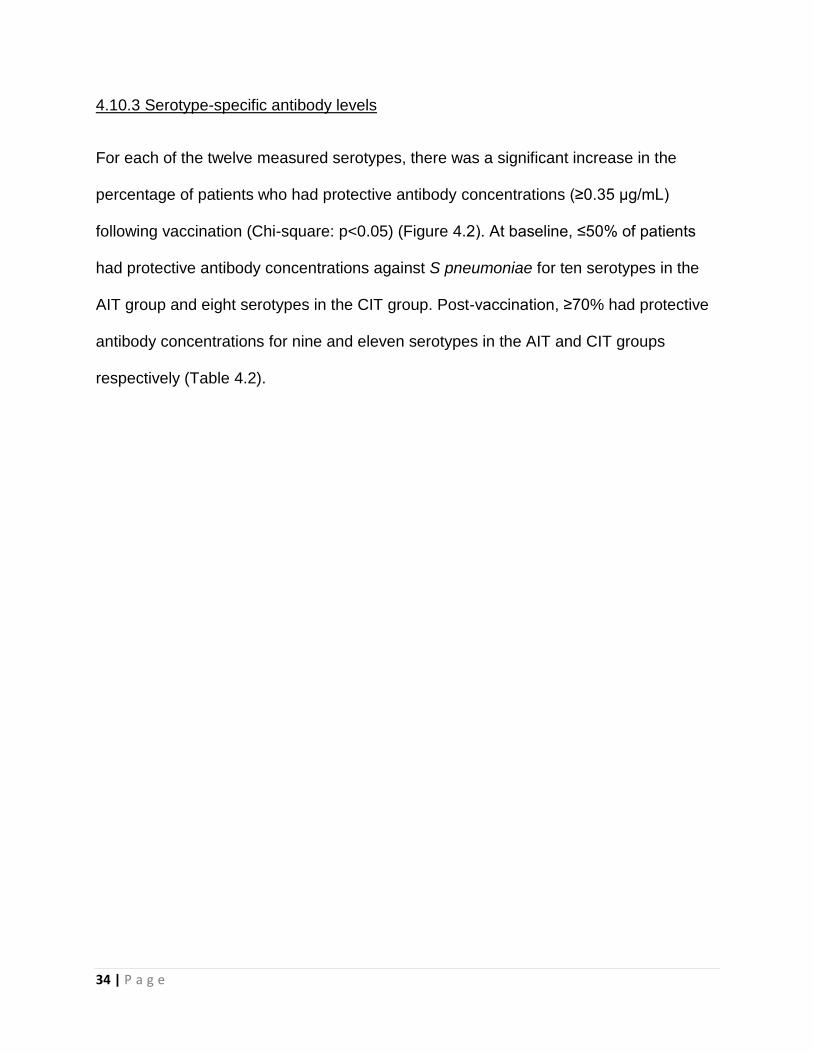
35 | P a g e
Table 4.2 Serological response to single dose 13-valent Pneumococcal Conjugate Vaccine in children (n=82) with cancer.
Pneumococcal
polysaccharide
Serotype
Antibody Concentrations Frequency of Participants with Protective antibody
concentration (≥0.35 µg/mL)
>4-fold concentration rise
Active Immunosuppressive Therapy (AIT)
Completed Immunosuppressive Therapy (CIT)
AIT CIT AIT CIT p-
value
Baseline GMC (95% CI)
Post-vaccination
GMC (95% CI)
Baseline GMC (95% CI)
Post-vaccination GMC (95% CI)
Baseline (%)
Post-vaccination (%)
Baseline (%)
Post-vaccination (%)
4 0.13 (0.09-0.21) 0.64 (0.31-1.31) 0.15 (0.10-0.21) 3.39 (1.46-7.86) 21.7 54.3 18.9 83.3 37.0% 69.4% <0.01
6B 0.31 (0.18-0.54) 3.14 (1.39-7.09) 0.28 (0.15-0.51) 2.35 (0.94-5.92) 47.8 78.3 37.8 63.9 58.7% 55.6% 0.78
9V 0.26 (0.18-0.36) 1.93 (1.02-3.64) 0.37 (0.26-0.51) 3.83 (1.73-8.51) 37.0 71.7 45.9 83.3 56.5% 52.8% 0.74
14 0.33 (0.20-0.54) 2.13 (1.21-3.75) 0.35 (0.21-0.58) 2.97 (1.68-5.25) 45.7 78.3 54.1 83.3 63.0% 58.3% 0.66
18C 0.33 (0.21-0.50) 2.85 (1.44-5.64) 0.31 (0.21-0.45) 4.28 (1.78-10.28) 50.0 78.3 37.8 80.6 56.5% 66.7% 0.35
19F 0.77 (0.47-1.27) 4.24 (2.27-7.92) 0.65 (0.44-0.96) 4.38 (2.35-8.17) 63.0 87.0 62.2 91.7 43.5% 55.6% 0.28
23F 0.31 (0.19-0.49) 2.71 (1.26-5.84) 0.31 (0.18-0.52) 2.49 (1.07-5.80) 41.3 73.9 32.4 77.8 58.7% 55.6% 0.78
1 0.12 (0.08-0.18) 0.51 (0.23-1.09) 0.16 (0.12-0.22) 2.92 (1.64-5.2) 15.2 45.7 24.3 91.7 37.0% 83.3% <0.01
3 0.43 (0.24-0.76) 2.52 (1.27-5) 0.45 (0.25-0.78) 8.6 (5.4-13.68) 50.0 80.4 51.4 100 41.3% 75.0% <0.01
5 0.22 (0.15-0.31) 0.56 (0.29-1.1) 0.27 (0.19-0.37) 2.82 (1.43-5.56) 30.4 52.2 37.8 86.1 21.7% 63.9% <0.01
7F 0.39 (0.25-0.59) 1.65 (0.83-3.25) 0.42 (0.30-0.58) 4.29 (2.42-7.6) 50.0 71.7 56.8 94.4 43.5% 63.9% 0.07
19A 0.48 (0.29-0.80) 1.45 (0.85-2.45) 0.36 (0.23-0.56) 1.59 (0.99-2.56) 54.3 80.4 48.6 86.1 32.6% 55.6% 0.04
36 | P a g e
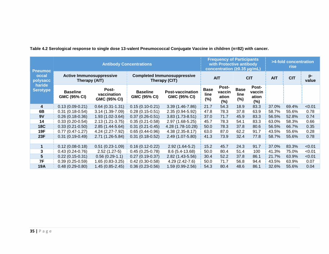
Prior to vaccination, there were no significant differences in the percentages of
participants with protective antibody concentrations (≥0.35 µg/mL) between the AIT and
CIT groups for each of the twelve serotypes measured. Following vaccination, there
were significantly higher percentages of CIT compared to AIT patients with protective
antibody concentrations for serotypes 1 (92% vs. 46%), 3 (100% vs. 80%), 4 (83% vs.
54%), 5 (86% vs. 52%) and 7F (94% vs. 72%) (Chi-square: p<0.05), with serotypes 1,
3, 5 and 7F being PCV13-only serotypes (Figure 4.2).
Figure 4.2 Frequency of children with cancer on Active Immunosuppressive Therapy (AIT) or who have Completed Immunosuppressive Therapy (CIT) with protective serotype-specific antibody concentrations (≥0.35 µg/mL) before and four weeks following single-dose 13-valent Pneumococcal Conjugate Vaccine
37 | P a g e
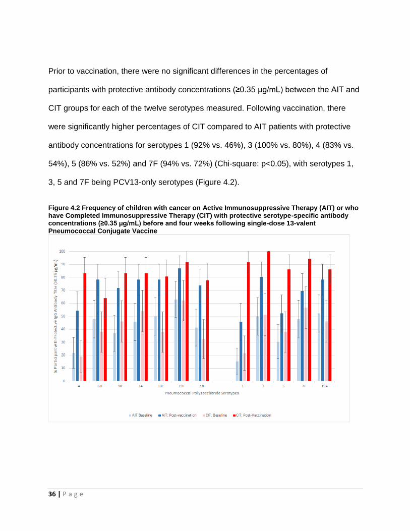
Geometric mean concentrations of serotype-specific antibody levels were compared
pre- and post-vaccination using student t-test (2-sided). The mean and 95% confidence
interval are shown in Figure 4.3.
Figure 4.3 Geometric mean concentrations of serotype-specific pneumococcal antibody in children with cancer on Active Immunosuppressive Therapy (AIT) or who have Completed Immunosuppressive Therapy (CIT) before and four weeks following single-dose 13-valent Pneumococcal Conjugate Vaccine
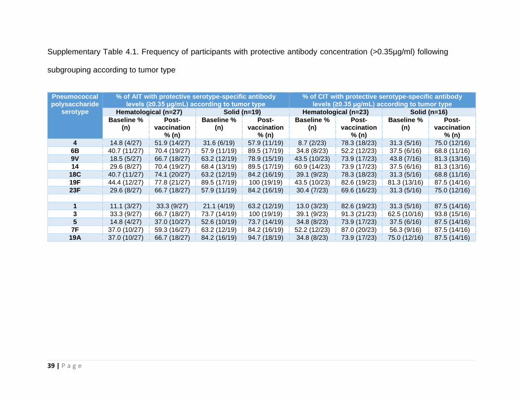
When patients were subgrouped according to tumour type, a lower proportion of
children with hematological malignancy had protective antibody concentrations
compared with solid tumors at baseline. For both subgroups, the numerical increase in
the percentage of participants with protective antibody concentrations for each of the
twelve serotypes measured following vaccination remained (Supplementary Table 4.1).
38 | P a g e
Statistical analysis to detect a difference in vaccine response between the subgroups
was precluded by the small number of patients within each subgroup.
39 | P a g e
Supplementary Table 4.1. Frequency of participants with protective antibody concentration (>0.35µg/ml) following
subgrouping according to tumor type
Pneumococcal polysaccharide
serotype
% of AIT with protective serotype-specific antibody levels (≥0.35 µg/mL) according to tumor type
% of CIT with protective serotype-specific antibody levels (≥0.35 µg/mL) according to tumor type
Hematological (n=27) Solid (n=19) Hematological (n=23) Solid (n=16)
Baseline % (n)
Post-vaccination
% (n)
Baseline % (n)
Post-vaccination
% (n)
Baseline % (n)
Post-vaccination
% (n)
Baseline % (n)
Post-vaccination
% (n)
4 14.8 (4/27) 51.9 (14/27) 31.6 (6/19) 57.9 (11/19) 8.7 (2/23) 78.3 (18/23) 31.3 (5/16) 75.0 (12/16)
6B 40.7 (11/27) 70.4 (19/27) 57.9 (11/19) 89.5 (17/19) 34.8 (8/23) 52.2 (12/23) 37.5 (6/16) 68.8 (11/16)
9V 18.5 (5/27) 66.7 (18/27) 63.2 (12/19) 78.9 (15/19) 43.5 (10/23) 73.9 (17/23) 43.8 (7/16) 81.3 (13/16)
14 29.6 (8/27) 70.4 (19/27) 68.4 (13/19) 89.5 (17/19) 60.9 (14/23) 73.9 (17/23) 37.5 (6/16) 81.3 (13/16)
18C 40.7 (11/27) 74.1 (20/27) 63.2 (12/19) 84.2 (16/19) 39.1 (9/23) 78.3 (18/23) 31.3 (5/16) 68.8 (11/16)
19F 44.4 (12/27) 77.8 (21/27) 89.5 (17/19) 100 (19/19) 43.5 (10/23) 82.6 (19/23) 81.3 (13/16) 87.5 (14/16)
23F 29.6 (8/27) 66.7 (18/27) 57.9 (11/19) 84.2 (16/19) 30.4 (7/23) 69.6 (16/23) 31.3 (5/16) 75.0 (12/16)
1 11.1 (3/27) 33.3 (9/27) 21.1 (4/19) 63.2 (12/19) 13.0 (3/23) 82.6 (19/23) 31.3 (5/16) 87.5 (14/16)
3 33.3 (9/27) 66.7 (18/27) 73.7 (14/19) 100 (19/19) 39.1 (9/23) 91.3 (21/23) 62.5 (10/16) 93.8 (15/16)
5 14.8 (4/27) 37.0 (10/27) 52.6 (10/19) 73.7 (14/19) 34.8 (8/23) 73.9 (17/23) 37.5 (6/16) 87.5 (14/16)
7F 37.0 (10/27) 59.3 (16/27) 63.2 (12/19) 84.2 (16/19) 52.2 (12/23) 87.0 (20/23) 56.3 (9/16) 87.5 (14/16)
19A 37.0 (10/27) 66.7 (18/27) 84.2 (16/19) 94.7 (18/19) 34.8 (8/23) 73.9 (17/23) 75.0 (12/16) 87.5 (14/16)
40 | P a g e
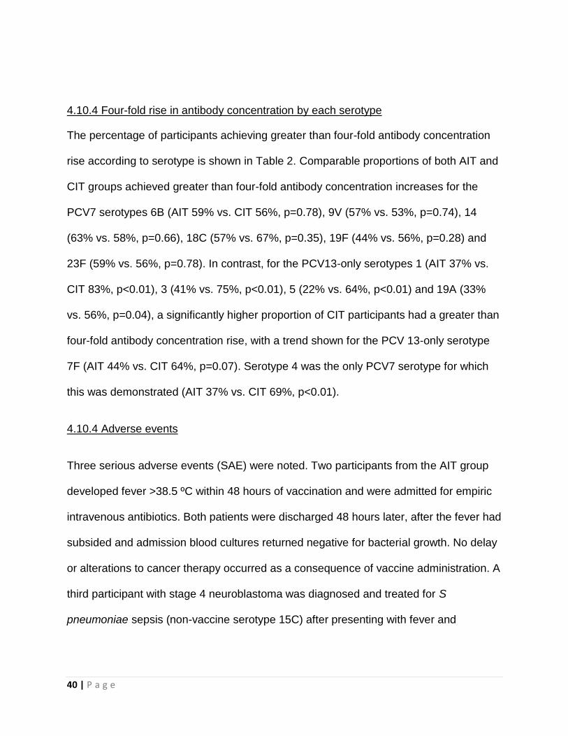
4.10.4 Four-fold rise in antibody concentration by each serotype
The percentage of participants achieving greater than four-fold antibody concentration
rise according to serotype is shown in Table 2. Comparable proportions of both AIT and
CIT groups achieved greater than four-fold antibody concentration increases for the
PCV7 serotypes 6B (AIT 59% vs. CIT 56%, p=0.78), 9V (57% vs. 53%, p=0.74), 14
(63% vs. 58%, p=0.66), 18C (57% vs. 67%, p=0.35), 19F (44% vs. 56%, p=0.28) and
23F (59% vs. 56%, p=0.78). In contrast, for the PCV13-only serotypes 1 (AIT 37% vs.
CIT 83%, p<0.01), 3 (41% vs. 75%, p<0.01), 5 (22% vs. 64%, p<0.01) and 19A (33%
vs. 56%, p=0.04), a significantly higher proportion of CIT participants had a greater than
four-fold antibody concentration rise, with a trend shown for the PCV 13-only serotype
7F (AIT 44% vs. CIT 64%, p=0.07). Serotype 4 was the only PCV7 serotype for which
this was demonstrated (AIT 37% vs. CIT 69%, p<0.01).
4.10.4 Adverse events
Three serious adverse events (SAE) were noted. Two participants from the AIT group
developed fever >38.5 ºC within 48 hours of vaccination and were admitted for empiric
intravenous antibiotics. Both patients were discharged 48 hours later, after the fever had
subsided and admission blood cultures returned negative for bacterial growth. No delay
or alterations to cancer therapy occurred as a consequence of vaccine administration. A
third participant with stage 4 neuroblastoma was diagnosed and treated for S
pneumoniae sepsis (non-vaccine serotype 15C) after presenting with fever and
41 | P a g e
neutropenia 25 days post-vaccination. This was the only case of breakthrough IPD that
occurred during the study.
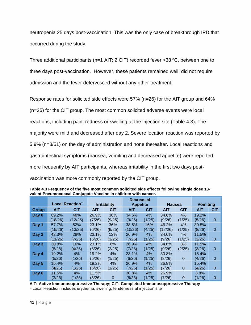
Three additional participants (n=1 AIT; 2 CIT) recorded fever >38 ºC, between one to
three days post-vaccination. However, these patients remained well, did not require
admission and the fever defervesced without any other treatment.
Response rates for solicited side effects were 57% (n=26) for the AIT group and 64%
(n=25) for the CIT group. The most common solicited adverse events were local
reactions, including pain, redness or swelling at the injection site (Table 4.3). The
majority were mild and decreased after day 2. Severe location reaction was reported by
5.9% (n=3/51) on the day of administration and none thereafter. Local reactions and
gastrointestinal symptoms (nausea, vomiting and decreased appetite) were reported
more frequently by AIT participants, whereas irritability in the first two days post-
vaccination was more commonly reported by the CIT group.
Table 4.3 Frequency of the five most common solicited side effects following single dose 13-valent Pneumococcal Conjugate Vaccine in children with cancer.
Local Reaction
∞ Irritability
Decreased Appetite Nausea Vomiting
Group AIT CIT AIT CIT AIT CIT AIT CIT AIT CIT
Day 0 69.2% (18/26)
48% (12/25)
26.9% (7/26)
36% (9/25)
34.6% (9/26)
4% (1/25)
34.6% (9/26)
4% (1/25)
19.2% (5/26) 0
Day 1 57.7% (15/26)
52% (13/25)
23.1% (6/26)
36% (9/25)
38.5% (10/26)
16% (4/25)
46.2% (12/26)
4% (1/25)
30.8% (8/26) 0
Day 2 42.3% (11/26)
28% (7/25)
23.1% (6/26)
12% (3/25)
26.9% (7/26)
4% (1/25)
34.6% (9/26)
4% (1/25)
11.5% (3/26) 0
Day 3 30.8% (8/26)
16% (4/25)
23.1% (6/26)
8% (2/25)
26.9% (7/26)
4% (1/25)
34.6% (9/26)
8% (2/25)
11.5% (3/26) 0
Day 4 19.2% (5/26)
4% (1/25)
19.2% (5/26)
4% (1/25)
23.1% (6/26)
4% (1/25)
30.8% (8/26) 0
15.4% (4/26) 0
Day 5 15.4% (4/26)
4% (1/25)
19.2% (5/26)
4% (1/25)
26.9% (7/26)
4% (1/25)
26.9% (7/26) 0
15.4% (4/26) 0
Day 6 11.5% (3/26)
4% (1/25)
11.5% (3/26) 0
30.8% (8/26)
4% (1/25)
26.9% (7/26) 0
3.8% (1/26) 0
AIT: Active Immunosuppressive Therapy; CIT: Completed Immunosuppressive Therapy ∞Local Reaction includes erythema, swelling, tenderness at injection site
42 | P a g e
4.11 Discussion
As a significant proportion of cancer therapy is delivered in the outpatient or ambulatory
care setting, prevention of community acquired infections such as pneumococcal
disease form an important consideration in the care of children receiving treatment and
following completion of therapy for cancer; particularly given the incidence of S
pneumoniae infection and mortality as a result of IPD is higher in this population
compared to healthy children. There is significant evidence highlighting the benefit of
vaccination in healthy children to prevent pneumococcal disease, however there is a
paucity of literature demonstrating benefit in immunocompromised children with
cancer(58).
This is the first immunogenicity study of PCV13 in children with cancer. At baseline,
there are low pneumococcal antibody levels for all serotypes demonstrating a
vulnerability to IPD during and at completion of cancer therapy. Our data shows that
single-dose PCV13 is immunogenic in both children receiving immunosuppressive
therapy for cancer and those within 12 months from completion of cancer therapy. This
is supported by studies reporting similar benefits of pneumococcal vaccination using the
PCV7 or PCV10 vaccines in children with cancer. Using two doses of PCV7, 86-100%
of children with acute myeloid or lymphoblastic leukemia during maintenance
chemotherapy (n=20) and within 12 months of completing chemotherapy (n=9)
achieved protective antibody levels for all seven serotypes. All children with solid tumors
(n=15) who were within 12 months of completing chemotherapy achieved protective
antibodies against all PCV7 serotypes(38). In a recent study assessing the
43 | P a g e
immunogenicity of PCV10 in children receiving therapy for leukemia, the proportion of
children achieving protective antibody levels ranged from 33.3% to 88.9% for the ten
serotypes(39).
Our study revealed that patients who received previous PCV7 vaccination had similar
rises in antibody concentrations for the seven serotypes when comparing children
receiving immunosuppressive therapy to those who had completed immunosuppressive
therapy. However, responses to PCV13-only serotypes were significantly higher in
patients who had completed immunosuppressive therapy. This suggests that for
children on active immunosuppression, a single dose of PCV13 elicited a “booster
effect” of immune memory from previous PCV7 vaccinations. Despite lower responses,
majority of children on active immunosuppressive treatment had protective
concentrations for the PCV13-only serotypes and thus still benefited from PCV13
vaccination during therapy.
Our study did not demonstrate an increased rate of adverse events in comparison to
previous studies, highlighting the safety and tolerability of the vaccine in children with
cancer. Prior studies with single-dose PCV13 report local reactogenicity ranging from
38% to 70% which is comparable to our local reaction rate of 55% on Day 1(59, 60).
The majority of local reactions were mild with 5.9% reporting severe local reactions.
This is comparable to a study where single-dose PCV13 booster was given to healthy
infants and toddlers previously immunised with PCV7(61). Potential vaccine-related
fever occurred in 6% of our cohort, similar to a study which demonstrated the benefit of
trivalent inactivated influenza vaccine in immunocompromised children with cancer(49).
44 | P a g e
There are several limitations to our study. Although this is the largest cohort examining
PCV immunogenicity in children with cancer, the sample size remains relatively small
due to the rarity of pediatric cancers(62). As a consequence of the rarity of the study
population and the relative infrequency of IPD, we did not evaluate clinical efficacy of
the vaccine, with immunogenicity used as a surrogate of clinical efficacy.
To date, there is no consensus regarding the protective pneumococcal antibody
threshold in immunocompromised hosts(47). In healthy populations, serotype-specific
protective thresholds of >0.15 to 1 µg/mL were utilized in studies prior to 2003. There
has since been international consensus in adopting the World Health Organization
recommended threshold of 0.35 µg/mL in evaluating efficacy of PCV(55). However, this
threshold has been derived from three efficacy studies for PCV7 licensure in healthy
Californian, South African and Gambian infants 1 month post 3rd PCV immunisation and
may not reflect the antibody concentrations required for protection in
immunocompromised children with cancer. The same threshold of 0.35 µg/mL is also
generally used in evaluating the protective efficacy of booster doses of PCV, although it
hasn’t been validated as such or when looking at antibody persistence.
Finally, the subjective method for reporting of solicited adverse events could result in
differences in reporting between individuals and the potential for increased reporting of
symptoms due to concurrent administration of chemotherapy for children receiving
treatment compared to those who have completed therapy.
These limitations could be overcome in future studies by recruiting larger numbers of
patients through international collaborative multi-center trials with clinical surveillance
45 | P a g e
for pneumococcal disease and standardized reporting of adverse events. In addition,
future studies should evaluate functional assessments such as opsonophagocytic
activity, assess the duration of protection and measure humoral responses to test the
benefit of a two-dose regimen.
4.12 Conclusion
In conclusion, our study provides evidence to recommend that all children receiving
therapy for cancer should receive a single dose of PCV13 as soon as possible after
diagnosis, regardless of prior PCV exposure. In addition, our data support current
immunisation guidelines, which recommend an additional dose of PCV13 after
completion of immunosuppressive therapy to provide additional protection against IPD.
46 | P a g e
************************************************************************
CHAPTER 5- NASOPHARYNGEAL COLONISATION OF
STREPTOCOCCUS PNEUMONIAE IN CHILDREN WITH
CANCER
************************************************************************
5.1 Chapter Outline
Streptococcus pneumoniae nasopharyngeal (NP) colonisation was explored as a
clinical endpoint of PCV13 vaccination given to children and adolescent with cancer
during their active immunosuppressive therapy. The methods were described in detail in
Chapter 3, and results are presented in the current chapter.
5.2 Statement of Contribution
Te-Yu Hung: Designed the study, recruited for the clinical aspect of the study, acted as
chief investigator, contributed to interpretation and discussion of the results, writing first
draft, and writing-review and editing. Christopher C. Blyth: Designed the study,
contributed to interpretation and discussion of the results, and writing- review and editing.
Rishi S. Kotecha: Designed the study, designed and recruited for the clinical aspect of
the study, contributed to interpretation and discussion of the results, and writing- review
and editing. Sarah K. Steed: Designed and recruited for the clinical aspect of the study.
Catherine H. Cole: Designed the study, contributed to interpretation and discussion of the
results, and writing–review and editing. Peter C. Richmond: Designed the study, designed
47 | P a g e
the laboratory method, analyzed the results, contributed to interpretation and discussion
of the results, and writing–review and editing.
5.3 Funding
This study was funded by Perth Children’s Hospital Foundation formerly known as the
PMH Foundation. Dr Hung is supported by a Telethon Research Fellowship (2013). Dr
Blyth is supported by a WA Department of Health / Raine Early Career Research
Fellowship (2014-2016) and a NHMRC Career Development Fellowship (APP1111596;
2016-2019). Dr Thornton is supported by a BrightSpark Foundation Fellowship.
5.4 Acknowledgement
We acknowledge Jacinta Bowman, Natalie Stemberger for performing the laboratory
assays; Jan Jones, Lisa Montgomery, Janessa Pickering, other staff at the Vaccine
Trials Group, Telethon Kids Institute and staff on Ward 3B, Princess Margaret Hospital
for Children for assistance with this project.
5.5 Disclosure Statement
Dr. Richmond reports grants from Perth Children’s Hospital Foundation formerly known
as the Princess Margaret Hospital for Children Foundation, during the conduct of the
study, non-financial support and grants from Pfizer, non-financial support and grants
from GlaxoSmithKline, outside the submitted work; Dr. Blyth reports grants from Pfizer
Global, outside the submitted work; Dr. Kotecha reports grants from Pfizer Australia
(Competitive Cancer Research Grant), outside the submitted work; Dr. Hung reports
48 | P a g e
grant from Perth Children's Hospital Foundation, during the conduct of the study; Dr
Cole has nothing to disclose.
5.6 Abstract
5.6.1 Background
There is a paucity of literature describing NP colonisation of S pneumoniae in children
diagnosed with cancer who are shown to have high burden of disease compared to
healthy children. Nasopharyngeal colonisation is essential for Streptococcus
pneumoniae to cause invasive pneumococcal disease (IPD).
5.6.2 Method
Patients aged between 1 and 18 years diagnosed with haematological or solid organ
malignancy at Princess Margaret Hospital for Children, Western Australia who were
receiving active immunosuppressive therapy: (AIT) or completed immunosuppressive
therapy (CIT) in the preceding 12 months were eligible to participate. Children enrolling
in an immunogenicity study had and nasopharyngeal swab (NPS) taken before and
after a single-dose of 13-valent pneumococcal conjugate vaccine (PCV13).
5.6.3 Results
From 85 children, a total of 175 NPS’s were collected pre- and post- vaccination, which
were 28-60 days apart. In the AIT group, 6.8% and 9.3% of children were colonised with
pneumococcus before and after a single-dose of PCV13. This increased to 18.2% 6-
month after cancer therapy was ceased. This is comparable to the CIT group of which
18.9 and 23.3% are colonised with pneumococcus pre- and post-single dose of PCV13
(Table 5.2). In children colonised with S pneumoniae, 10% and 9% were PCV13
serotypes before and after the vaccination. Seventy-four percent of children in the AIT
49 | P a g e
group were receiving regular trimethoprim/sulfamethoxazole for PJP prophylaxis
compared with 13% of children who were recruited after treatment completion (p
<0.001).
5.6.4 Conclusion
We conducted a small nested colonisation study which found pneumococcal NP
carriage to be infrequent during active cancer therapy for children with cancer, despite
the known high risk of IPD. We need a larger study with longer time periods and more
sampling time points to further investigate the epidemiology and rates of colonisation in
this vulnerable cohort. Based on our results, single-dose PCV13 did not influence the
NP colonisation rates of S pneumoniae in children during or after completion of
chemotherapy.
5.7 Introduction
Nasopharyngeal colonisation is essential for Streptococcus pneumoniae to cause IPD.
Early studies with Haemophilus influenza conjugate vaccine and 7-valent pneumococcal
conjugate vaccine (PCV7) show that conjugate vaccines are an effective way of
reducing NP carriage of vaccine-serotypes with a secondary reduction in IPD(63).
However, several studies also show increased colonisation in non-vaccine serotypes, a
phenomenon referred to as “serotype replacement” Klugman et al Lancet ID 2001.
There is a paucity of literature describing NP colonisation of S pneumoniae in children
diagnosed with cancer during and after completion of therapy. Understanding
pneumococcal colonisation in children receiving cancer therapy is important when
considering the various strategies of preventing IPD in this cohort. Children and
adolescents with cancer undergoing immunosuppressive therapy have different
50 | P a g e
epidemiological risk factors for pneumococcal carriage, including increased frequency of
contact with healthcare facilities and healthcare workers, reduced contact with peers
through childcare or school, and more exposure to antibiotics either through prophylaxis
(e.g. TMP/SMX for Pneumocystis jirovecii pneumonia (PJP) prophylaxis) or empirical
broad-spectrum antibiotics. The combination of these person-to-person and
environmental exposure may either increase or decrease total NP carriage of S
pneumoniae and are likely to increased carriage of antibiotic-resistant S pneumoniae.
Utilising an existing prospective PCV13 immunogenicity study in children with or
following treatment with cancer, we explored NP pneumococcal colonisation, serotype
distribution and penicillin susceptibility in this specific population.
5.8 Method
We conducted a prospective study nested within a larger immunogenicity study. Details
of patient recruitment procedures, inclusion and exclusion criteria are described in
Chapter 3 of this thesis. To summarise, children aged between 1 and 18 years
diagnosed with haematological or solid organ malignancy at Princess Margaret Hospital
for Children, Western Australia who were receiving active immunosuppressive therapy:
(AIT) or completed immunosuppressive therapy (CIT) in the preceding 12 months were
eligible to participate. Clinical data including demographic data (sex, age) and previous
pneumococcal vaccination history, underlying malignancy and past history of IPD and
middle ear disease (recurrent acute otitis media, myringotomy, ventilation tube insertion,
adenoidectomy and/or tonsillectomy), immunosuppressive chemotherapeutic and
antimicrobial treatment administered were collected.
51 | P a g e
Participants had baseline nasopharyngeal swab (NPS) nasal swab was taken according
to the method described in the WHO working party report(50). The participants were
given a single dose of intramuscular PCV13 and a symptom diary to record adverse
effects for 7 days. The repeat NPS were taken between 28 to 60 days from vaccination
to assess for NP carriage for S pneumoniae. The third NPS sample for patients in the
AIT group was taken when the patient returned for follow-up at 6 months after the
completion of the cancer therapy.
The NPS’s were taken using a FLOQSwabs™ (Batch 015N37. Lot C9PD00 Copan
Flock Technologies) at enrolment after the consent procedure as described above(50).
The NPS’s were either taken during an outpatient clinic visit or under general
anaesthesia while the patient underwent cancer therapy-related procedure.
Following collection, the NPS was immediately placed in 1.0 ml of skim milk-tryptone-
glucose-glycerin broth (SMTGGB; 3% tryptone soya broth [Sigma-Aldrich, Castle-Hill,
Australia], 0.5% glucose, 2% skimmed milk powder and 10% glycerol in 100 mL distilled
water). All samples are stored in -40C until transfer to -800C freezer within 4 hours of
collection. Further sample storage and processing, including bacterial culture was
undertaken at the state pneumococcal reference laboratory (PathWest Laboratory WA,
QEII Medical Centre, Nedlands WA).
Nasopharyngeal swabs were cultured on blood agar (BA), Colistin, Nalidixic Agar (CNA)
and Bacitracin vancomycin clindamycin chocolate agar (BVCCA). Plates were
incubated at 370C in 5%CO2 and inspected at 24 and 48 hours for signs of bacterial
52 | P a g e
growth. The density of growth of significant isolates were recorded semi quantitatively
as scanty (+), moderate (++) or heavy (+++).
Identification of S. pneumoniae was confirmed by routine phenotypic methods including
colony morphology and optochin susceptibility. Serotyping for S pneumoniae isolate
was undertaken for all cultured isolates using the Quellung reaction(52). For non-
typable isolates, lytA PCR as used as a confirmatory test(53).
Susceptibility testing to penicillin were performed by ETest® (bioMerieux, Australia).
Minimum inhibitory concentration (MIC) for penicillin susceptibility (non-meningitis,
intravenous route) were interpreted according to CLSI (breakpoints: susceptible (≤2
µg/mL), intermediate (4 µg/mL), or resistant (≥8 µg/mL), 2009 Clinical Laboratory
Standards Institute)(54). Data were entered into a Microsoft Access Professional Plus
database (2013) and analyzed using SPSS version 22 (2013). The proportion of
children colonized in treatment groups and at specific time periods were described and
ChiSquare was used to compare the proportion of patients colonized with
pneumococcus before and after immunization. A p-value of <0.05 was considered
statistically significant.
The study was approved by the Child and Adolescent Health Service Ethics Committee
(HREC 2072EP) with delegated authority from the Princess Margaret Hospital for
Children Institutional Review Board, within which the work was undertaken.
5.9 Results
Between March 2013 and December 2014, 85 children were recruited and a total of 175
NPS’s were collected. Eight NPS swabs were excluded from analysis for the following
53 | P a g e
reasons: 1) unsuccessful swab collection, 2) unlabelled sample and 3) delayed storage
of sample into SMTGGB medium or 4) withdrawal of consent (see Table 5.1). Five more
swabs were recorded as “suboptimal collection”, due to patient distress or self-
sampling, however, 60% (3/5) of these swabs were still culture positive for S
pneumoniae.
There was a large difference in the proportion of children receiving regular oral antibiotic
in between the AIT and CIT groups. Seventy-four percent of children in the AIT group
were receiving regular TMP/SMX as prophylaxis for PJP compared with 13% of children
who were recruited after treatment completion (CIT; p = <0.001). Those still receiving
antibiotics following completion received PJP prophylaxis in the initial period of
completing therapy and awaiting marrow recovery. Further exposure to antimicrobial
therapy between visit 1 and visit 2 were also recorded via oral or intravenous routes:
prescribed agents (in order of frequency) included piperacillin/tazobactam, vancomycin,
ceftriaxone, flucloxacillin, tobramycin, cephalexin, amoxicillin/clavulanic acid,
metronidazole. (see Table 5.1).
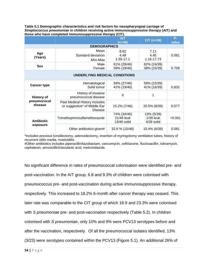
54 | P a g e
Table 5.1 Demographic characteristics and risk factors for nasopharyngeal carriage of Streptococcus pneumoniae in children receiving active immunosuppressive therapy (AIT) and those who have completed immunosuppressive therapy (CIT).
*Includes previous tonsillectomy, adenoidectomy, insertion of myringotomy ventilation tubes, history of recurrent otitis media, mastoiditis #Other antibiotics includes piperacillin/tazobactam, vancomycin, ceftriaxone, flucloxacillin, tobramycin, cephalexin, amoxicillin/clavulanic acid, metronidazole.
No significant difference in rates of pneumococcal colonisation were identified pre- and
post-vaccination. In the AIT group, 6.8 and 9.3% of children were colonised with
pneumococcus pre- and post-vaccination during active immunosuppressive therapy,
respectively. This increased to 18.2% 6-month after cancer therapy was ceased. This
later rate was comparable to the CIT group of which 18.9 and 23.3% were colonised
with S pneumoniae pre- and post-vaccination respectively (Table 5.2). In children
colonised with S pneumoniae, only 10% and 9% were PCV13 serotypes before and
after the vaccination, respectively. Of all the pneumococcal isolates identified, 13%
(3/23) were serotypes contained within the PCV13 (Figure 5.1). An additional 26% of
AIT
(n=46) CIT (n=39)
P- value
DEMOGRAPHICS
Age (Years)
Mean Standard deviation
Min-Max
8.82 4.48
1.55-17.1
7.11 4.45
1.16-17.73 0.081
Sex Male
Female 61% (28/46) 39% (18/46)
62% (24/39) 38% (15/39)
0.708
UNDERLYING MEDICAL CONDITIONS
Cancer type Hematological
Solid tumor 59% (27/46) 41% (19/46)
59% (23/39) 41% (16/39)
0.832
History of pneumococal
disease
History of invasive pneumococcal disease
0 1
Past Medical History includes or suggestive* of Middle Ear
Disease 15.2% (7/46) 20.5% (8/39) 0.577
Antibiotic exposure
Trimethoprim/sulfamethoxazole 74% (34/46) 21/46 leuk 13/46 solid
13% (5/39) 1/39 leuk 4/39 solid
<0.001
Other antibiotics given# 32.6 % (15/46) 15.4% (6/39) 0.081
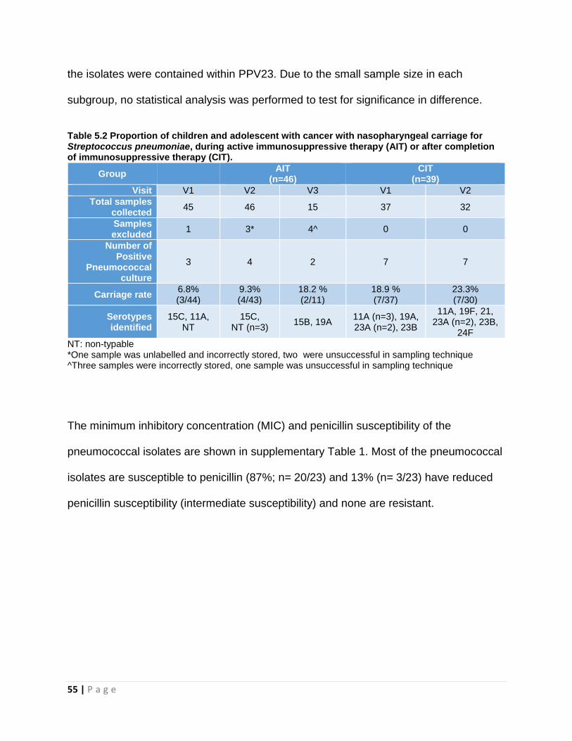
55 | P a g e
the isolates were contained within PPV23. Due to the small sample size in each
subgroup, no statistical analysis was performed to test for significance in difference.
Table 5.2 Proportion of children and adolescent with cancer with nasopharyngeal carriage for Streptococcus pneumoniae, during active immunosuppressive therapy (AIT) or after completion of immunosuppressive therapy (CIT).
Group AIT
(n=46) CIT
(n=39)
Visit V1 V2 V3 V1 V2
Total samples collected
45 46 15 37 32
Samples excluded
1 3* 4^ 0 0
Number of Positive
Pneumococcal culture
3 4 2 7 7
Carriage rate 6.8% (3/44)
9.3% (4/43)
18.2 % (2/11)
18.9 % (7/37)
23.3% (7/30)
Serotypes identified
15C, 11A, NT
15C, NT (n=3)
15B, 19A 11A (n=3), 19A, 23A (n=2), 23B
11A, 19F, 21, 23A (n=2), 23B,
24F
NT: non-typable *One sample was unlabelled and incorrectly stored, two were unsuccessful in sampling technique ^Three samples were incorrectly stored, one sample was unsuccessful in sampling technique
The minimum inhibitory concentration (MIC) and penicillin susceptibility of the
pneumococcal isolates are shown in supplementary Table 1. Most of the pneumococcal
isolates are susceptible to penicillin (87%; n= 20/23) and 13% (n= 3/23) have reduced
penicillin susceptibility (intermediate susceptibility) and none are resistant.
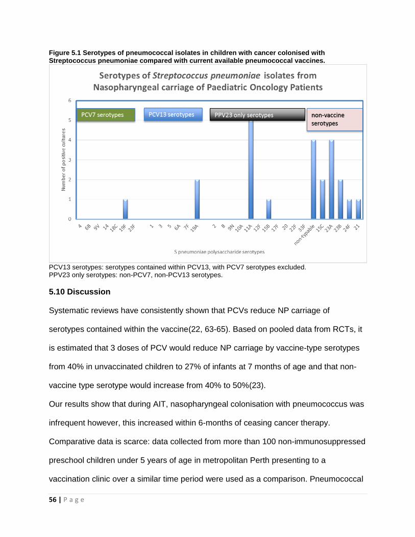
56 | P a g e
Figure 5.1 Serotypes of pneumococcal isolates in children with cancer colonised with Streptococcus pneumoniae compared with current available pneumococcal vaccines.
PCV13 serotypes: serotypes contained within PCV13, with PCV7 serotypes excluded. PPV23 only serotypes: non-PCV7, non-PCV13 serotypes.
5.10 Discussion
Systematic reviews have consistently shown that PCVs reduce NP carriage of
serotypes contained within the vaccine(22, 63-65). Based on pooled data from RCTs, it
is estimated that 3 doses of PCV would reduce NP carriage by vaccine-type serotypes
from 40% in unvaccinated children to 27% of infants at 7 months of age and that non-
vaccine type serotype would increase from 40% to 50%(23).
Our results show that during AIT, nasopharyngeal colonisation with pneumococcus was
infrequent however, this increased within 6-months of ceasing cancer therapy.
Comparative data is scarce: data collected from more than 100 non-immunosuppressed
preschool children under 5 years of age in metropolitan Perth presenting to a
vaccination clinic over a similar time period were used as a comparison. Pneumococcal
57 | P a g e
colonisation was determined using standard bacterial culture from NP specimens. This
project demonstrated that 14.1-30.7% of PCV vaccinated children were colonised
(Rheola Street Pneumococcal Colonisation Project: Blyth, unpublished data). This
suggests that children who are off cancer therapy have comparable pneumococcal
carriage rate to their similar aged peers.
In 277 school-aged children who were diagnosed with haematological or solid
malignancy and receiving maintenance chemotherapy or had completed cancer therapy
within 6 months of the study, significant negative predictors of NP carriage of S
pneumoniae include age, prior PCV7 immunisation and TMP/SMX prophylaxis(14). This
study had higher mean age compared to our cohort, but only 50% had previous receipt
of PCV7 as per the local Immunisation Guidelines, and 80% of the cohort also received
concurrent TMP/SMX prophylaxis. Despite these similarities to our cohort, the
pneumococcal colonisation rate in this population was 18.8%, higher than the
nasopharyngeal colonisation rate detected our AIT group (pre and post-vaccination
colonisation rate of 6.8 and 9.3%)
There are several limitations to this small colonisation study. Our study was a nested
study within a larger immunogenicity study. We did not collected information associated
with high NP pneumococcal carriage, such as child-care attendance, number of
siblings, exposure to cigarette and cooking smoke or indigenous status. Our study
design was not optimally designed to investigate the vaccine effect on NP carriage at 1
month post-vaccination. The timing of NP sampling gave cross-sectional data for
children receiving chemotherapy, the serotype-distribution and frequency of penicillin-
resistant S pneumoniae. A better study design is to have longer time period between
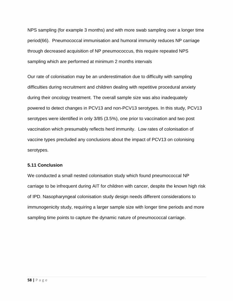
58 | P a g e
NPS sampling (for example 3 months) and with more swab sampling over a longer time
period(66). Pneumococcal immunisation and humoral immunity reduces NP carriage
through decreased acquisition of NP pneumococcus, this require repeated NPS
sampling which are performed at minimum 2 months intervals
Our rate of colonisation may be an underestimation due to difficulty with sampling
difficulties during recruitment and children dealing with repetitive procedural anxiety
during their oncology treatment. The overall sample size was also inadequately
powered to detect changes in PCV13 and non-PCV13 serotypes. In this study, PCV13
serotypes were identified in only 3/85 (3.5%), one prior to vaccination and two post
vaccination which presumably reflects herd immunity. Low rates of colonisation of
vaccine types precluded any conclusions about the impact of PCV13 on colonising
serotypes.
5.11 Conclusion
We conducted a small nested colonisation study which found pneumococcal NP
carriage to be infrequent during AIT for children with cancer, despite the known high risk
of IPD. Nasopharyngeal colonisation study design needs different considerations to
immunogenicity study, requiring a larger sample size with longer time periods and more
sampling time points to capture the dynamic nature of pneumococcal carriage.
59 | P a g e
Table 5.3 Positive nasopharyngeal culture of Streptococcus pneumoniae (S. pn) of children receiving active immunosuppressive therapy (AIT) or 6 months after completion of immunosuppressive therapy (CIT). Data showing relationship between before (V1) and after (V2) single-dose 13-valent pneumococcal conjugate vaccine (PCV13). AIT group had V3 swabs taken 6 months after completion of cancer therapy.
Group
Concomitant Trimethoprim/
sulfamethoxazole
Pre-PCV13 (V1) S. pn serotype
Penicillin MIC µg/mL
(Interpretation) Post-PCV13 (V2) S. pn serotype
Penicillin MIC µg/mL
(Interpretation)
Post-cancer therapy (V3)#
S. pn serotype
Penicillin MIC µg/mL
(Interpretation)
AIT
Yes NT (+) 0.5 (I) NT (+) 1 (I) NG
Yes NG NG 15B (+) 0.03 (S)
No NG NT (+) 0.008 (S) 19A (+) 0.06 (S)
Yes 15C (+)* 0.016 (S) 15C (+)* 0.016 (S) NG
yes 11A (+) 0.25 (I) NT (+) 0.5 (I) NG
CIT
No 19A (+++) 0.016 (S) NG -
Yes 23A (++) 0.016 (S) 19F (++) 0.016 (S) -
Yes 23A (+) 0.06 (S) 23A (+) 0.03 (S) -
No 11A (+) 0.016 (S) NG -
No NG 23B (+) 0.016 (S) -
yes 11A (+) 0.016 (S) 11A (+) 0.016 (S) -
No NG 23A (+) 0.06 (S) -
No 11A (+) 0.016 (S) - -
No 23B (+) 0.016 (S) - -
No NG 24F (+) 0.016 (S) -
No NG 21 (+) 0.016 (S) -
NG: no growth; NT: non typable Group 1: Active Immunosuppressive Therapy (AIT); Group 2: Completed Immunosuppressive Therapy (CIT) Culture growth: (+) scant growth (++) moderate growth (+++) heavy growth Penicillin Minimum Inhibitory Concentration (MIC) and susceptibility interpretation: S susceptible; I intermediate; R resistant. *This 4 year old patient was being treated for Stage 4 neuroblastoma, presented 22 days post-vaccination with fever and neutropaenia. He was diagnosed and successfully treated for S pneumoniae sepsis (serotype 15C). # None of the patients were receiving trimethoprim/cotrimoxazole at V3
60 | P a g e
************************************************************************
CHAPTER 6- GENERAL DISCUSSION
************************************************************************
6.1 Chapter Outline
This chapter presents a summary of the previous chapters and draws together
conclusions from the two included studies. Immunisation practices and clinical
implications of the studies will be described and future research priorities will be
discussed.
6.2 Summary of Findings
6.2.1 Immunogenicity and Safety
A prospective, open-label cohort study recruited 85 children between 1 to 18 years of
age, on active immunosuppressive therapy (AIT) or within 12 months of completing
immunosuppressive therapy (CIT). Serotype-specific IgG antibody concentrations were
measured from serum taken pre- and post-vaccination of PCV13, with concentrations
≥0.35 µg/mL considered protective. At baseline, <50% had protective antibody
concentrations against S pneumoniae for 10 serotypes in the AIT group and 8 serotypes
in the CIT group. Post-vaccination, ≥70% had protective antibody concentrations for 9
and 11 serotypes in the AIT and CIT groups respectively. Both groups had comparable
responses to PCV7 serotypes, whereas significantly higher proportions in the CIT group
achieved protective antibody concentrations to PCV13 serotypes. Our data support the
recommendation that an additional dose of PCV13 after completion of
immunosuppressive therapy provide additional protection against IPD. The optimal
61 | P a g e
timing for this dose remains uncertain. Solicited side-effects recorded in a seven-day
diary following vaccination showed a low rate of serious adverse events (3.5%).
6.2.2 Nasopharyngeal colonisation
Using the same cohort, we investigated the baseline rate of pneumococcal colonisation
in children with cancer and effect PCV13 on NP carriage at 1-month post-vaccination. In
the AIT group, only 6.8% and 9.3% of children were colonised with pneumococcus pre-
and post- PCV13 vaccination during active cancer therapy. The NP colonisation
increased to 18.2% 6-months after cancer therapy is ceased. This is comparable to the
CIT group, with 18.9 and 23.3% are colonised with pneumococcus pre- and post-
PCV13 vaccination. Only 3 of 85 children (3.5 %) were colonised with pneumococcal
serotypes contained within PCV13 (1 prior to vaccination; 2 following vaccination)
precluding any assessment of the impact of PCV13 on NP carriage. Although very
relevant at a population level to assess the impact of vaccination, this study highlights
the difficulty of using pneumococcal colonisation as an endpoint in a small high-risk
cohort.
6.2.3 Limitations of Thesis
There are several limitation to both parts of this project. The study design was optimally
structured to compare pre- and post-vaccination antibody levels to demonstrate
immunogenicity. These timing and frequency of sampling do not allow time for the effect
of vaccination on colonisation (if any) to be detected. It was unexpected that the follow-
up of the AIT group would be so poor, such that no useful data could be gained to
demonstrate persistence of immunity.
62 | P a g e
Due to the logistics and funding arrangements, we were then unable to perform OPA
assays to demonstrate the functional assessment of the antibodies when vaccination
was administered during therapy.
Similarly in the nasopharyngeal colonisation study, antimicrobial susceptibility to
antibiotics such as TMP/SMX and macrolide would have allowed comparisons with
international patterns of antimicrobial resistance in pneumococcus. This was limited by
logistic and funding capacity of the current study.
6.3 Recommendations for Clinical Practice
Our first study shows that the PCV13 is immunogenic and safe in children receiving AIT
and that the period of waiting for marrow recovery prolongs the period of vulnerability to
infection unnecessarily. Given the burden of disease in this vulnerable population, our
data show that a dose of PCV13 given at the commencement of, or early in
immunosuppressive therapy would be beneficial to any person regardless of prior
exposure to PCV’s.
Concern has been raised about the potential for post-vaccine fever to result in
unnecessary hospital admission and therapy given the practice of investigating and
treating undifferentiated fever in immunocompromised children. Our data from Chapter
4 show that this is infrequent and that rate of sustained fever is low. We deduce that the
benefit from the vaccine in achieving protective immunity against IPD is likely to
outweigh the risks.
Since our study was conducted 2 years after PCV13 replaced PCV7 on the NIP, a small
proportion of each group have had PCV13 exposure. With the continuation of the
63 | P a g e
current NIP schedule, the proportion of the population primed with PCV13 will increase
and thus an increasing proportion of the population will be eliciting the “booster dose”
effect of the PCV13 when given at time of commencing immunosuppressive therapy.
Our data suggests that a 2nd dose of PCV13 given within the first 6 months after
completion of immunosuppressive therapy elicits protective humoral response in
antibody levels to most serotypes contained within the vaccine, as the humoral
responses return soon after cessation of therapy. This provides immune memory and
has longer lasting protection against pneumococcal disease, long into patient’s
survivorship from cancer.
As our study did not detect high rates of nasopharyngeal colonization with
pneumococcus, we were also unable to ascertain whether pneumococcal vaccination
had any reduced colonization due to the study design and sample size. Despite these
shortcomings the published incidence of disease and our immunogenicity data in this
high-risk populations compels us to recommend PCV vaccine to children with cancer.
6.4 Future Research Priorities
To understand the effectiveness of PCV vaccine in high risk populations including
children with cancer, large observational studies are required. With the ability to link
national immunisation registry data to hospitalization data, we now have the capacity to
undertake these large scale observational studies(67). To explore efficacy of different
pneumococcal vaccine schedules in high risk populations, large adequately-powered
clinical trials will be required. Having strong immunogenicity data for a single-dose
PCV13, a randomized control trial study to further evaluate the different pneumococcal
64 | P a g e
immunisation to include a two-dose regimen of PCV13, or a regimen including PPV23
schedule are some of the potential pneumococcal regimens to be further explored.
The current study provides pilot study to plan and seek for future studies such as RCT
aimed at answering the post-chemotherapy immunisation question. For example could
have 3 arms with vaccination <1 month, 3-6 months then >6 months. The statistical
power should aim to detect >10% difference in proportion of patient achieving the 0.35
µg/mL antibody threshold.
Designing a nasopharyngeal colonisation study in children undergoing oncology
therapy, could include several NPS samples (i.e. 3 monthly when undergoing general
anaesthesia) throughout 2.5 to 3 years of total duration of oncology treatment plan.
Furthermore, this would allow the study of other colonising organisms and the
propensity of the oncology population to acquiring multi-drug resistant organisms (e.g.
carbapenem-resistant Enterobactericae, vancomycin-resistant Enterococci, or
extended-spectrum beta-lactamases gram-negative bacteria).
Any future studies on vaccination in the high-risk group should also focus on evaluating
the persistence of immunity, when administering PCV13 during AIT phase. The follow-
up rate of our study in AIT patients at 6 months after completion of cancer therapy was
poor (32.6%) and did not allow significant analysis or conclusions to be drawn on
antibody persistence at this time point.
Children’s cancer research has achieved remarkable advances through years of
international multicentred collaborative research. To date, there has been a greater
focus on optimizing chemotherapeutic regimens rather than supportive care. Research
65 | P a g e
into infection prevention strategies or other supportive care therapies could be more
thoroughly explored in this research model. Furthermore, considerations can be made
to include infectious prophylactic interventions to be incorporated into standard
oncology clinical care.
The oncology population receiving immunosuppressive comprises of heterogeneous
groups with different disease pathophysiology which likely affect immune response to
vaccination. A number of monoclonal antibodies or anti-B cell agents are in use in
various cancer protocols. Collaborative research models which are able to achieve
sufficient sample size and allow statistically robust analysis should be promoted and
encouraged.
The functional assay of the humoral antibody response to PCV13 during active
immunosuppression also needs further research. The OPA has become an essential
endpoint evaluated in the licensure evaluation of new pneumococcal vaccines.
To broaden the horizons of our understanding of immune humoral response, the
conjugate-vaccine designs appears to be effective in eliciting a T-cell mediated antibody
response, despite active myelosuppression/ immunosuppression secondary to different
chemotherapeutic agents of different mechanisms. The hypothesis can be tested with
other currently available non-PCV (e.g. Haemophilus influenza type b vaccine or
Meningococcal C vaccine).
Our study attempted to explore the impact of immunosuppression treatment intensity,
however the small sample sizes did not allow detailed quantitative analysis of
immunosuppression or mechanism of action of the various chemotherapeutic agents.
66 | P a g e
Finally, another potential indirect benefit on the horizon, is the role of immunisation in
combating the risk in the immunocompromised populations from the increasing spread
of antibiotic-resistant pneumococcal strains. Although pneumococcal isolates in
Australia remain mostly penicillin susceptible, the trend globally for antimicrobial
resistance is concerning. Close to Australia, Asian Network for Surveillance of Resistant
Pathogens (ANSORP) has reported meningeal isolates of pneumococcus were
resistant to penicillin in 57.5% while 3.9% and 0.7% of pneumococcal non-meningeal
isolates showed intermediate or resistant penicillin susceptibility respectively(68). As
clinicians face greater threats from antimicrobial resistance in managing infections in
their immunocompromised patients with diminishing antimicrobial options, the benefits
of preventative strategies such as PCV13 vaccination would outweigh the risks.
6.5 Conclusion
Advances in the supportive care of paediatric oncology in managing the infectious
complications and co-morbidities is an important field that needs ongoing innovation
and research. Partnership and collaboration between these disciplines improves the
care of children in their journey with cancer therapy through quality of life and long term
survival.
67 | P a g e
REFERENCES
1. Yeoh D, Ryan A, Blyth C. Infectious Prophylaxis in Paediatric Oncology and Stem Cell Transplantation. Current Pediatrics Reports. 2015;3(2):160-9. 2. Tsigrelis C, Ljungman P. Vaccinations in patients with hematological malignancies. Blood Rev. 2015. 3. Fletcher MA, Balmer P, Bonnet E, Dartois N. PCVs in individuals at increased risk of pneumococcal disease: a literature review. Expert Rev Vaccines. 2015;14(7):975-1030. 4. SI Pelton, Jacobs M. Pneumococcal Infections. In: Cherry JD, MD, MSc; Harrison, Gail J., MD; Kaplan, Sheldon L., MD; Steinbach, William J., MD; Hotez, Peter J., MD, PhD, editor. Feigin and Cherry's Textbook of Pediatric Infectious Diseases. Seventh Edition ed: Saunders, Elsevier Inc.; 2014. p. 1198-246. 5. Auranen K, Rinta-Kokko H, Goldblatt D, Nohynek H, O'Brien KL, Satzke C, et al. Colonisation endpoints in Streptococcus pneumoniae vaccine trials. Vaccine. 2013;32(1):153-8. 6. Shigayeva A, Rudnick W, Green K, Chen DK, Demczuk W, Gold WL, et al. Invasive Pneumococcal Disease Among Immunocompromised Persons: Implications for Vaccination Programs. Clin Infect Dis. 2016;62(2):139-47. 7. WONG A, MARRIE TJ, GARG S, KELLNER JD, TYRRELL GJ. Increased risk of invasive pneumococcal disease in haematological and solid-organ malignancies. Epidemiology & Infection. 2010;138(12):1804-10. 8. Kyaw MH, Rose CE, Jr., Fry AM, Singleton JA, Moore Z, Zell ER, et al. The influence of chronic illnesses on the incidence of invasive pneumococcal disease in adults. J Infect Dis. 2005;192(3):377-86. 9. Lehrnbecher T, Schubert R, Behl M, Koenig M, Rose MA, Koehl U, et al. Impaired pneumococcal immunity in children after treatment for acute lymphoblastic leukaemia. Br J Haematol. 2009;147(5):700-5. 10. Patel SR, Bate J, Borrow R, Heath PT. Serotype-specific pneumococcal antibody concentrations in children treated for acute leukaemia. Arch Dis Child. 2012;97(1):46-8. 11. Hjuler T, Wohlfahrt J, Staum Kaltoft M, Koch A, Biggar RJ, Melbye M. Risks of invasive pneumococcal disease in children with underlying chronic diseases. Pediatrics. 2008;122(1):e26-32. 12. Meisel R, Toschke AM, Heiligensetzer C, Dilloo D, Laws HJ, von Kries R. Increased risk for invasive pneumococcal diseases in children with acute lymphoblastic leukaemia. Br J Haematol. 2007;137(5):457-60. 13. Zignol M, Peracchi M, Tridello G, Pillon M, Fregonese F, D'Elia R, et al. Assessment of humoral immunity to poliomyelitis, tetanus, hepatitis B, measles, rubella, and mumps in children after chemotherapy. Cancer. 2004;101(3):635-41. 14. Principi N, Preti V, Gaspari S, Colombini A, Zecca M, Terranova L, et al. Streptococcus pneumoniae pharyngeal colonization in school-age children and adolescents with cancer. Hum Vaccin Immunother. 2016;12(2):301-7. 15. Cardoso NT, Santos BA, Barbosa AV, Superti SV, Teixeira LM, Neves FPG. Serotypes, antimicrobial resistance and genotypes of Streptococcus pneumoniae associated with infections in cancer patients in Brazil. Diagnostic Microbiology and Infectious Disease. 2017;87(3):281-5. 16. de Kluyver R, Toms C, Enhanced Invasive Pneumococcal Disease Surveillance Working Group ftCDNA. Invasive pneumococcal disease surveillance Australia, 1 October to 31 December 2015. Commun Dis Intell Q Rep. 2016;40(1):E185-8. 17. Klugman KP, Black S, Dagan R, Malley R, Whitney CG. Pneumococcal conjugate vaccine and pneumococcal common protein vaccines. In: Stanley A. Plotkin WO, Paul A. Offit, editor. Vaccines. 6th ed 2013.
68 | P a g e
18. Watson M, Roche P, Bayley K, Bell JM, Collignon P, Gilbert GL, et al. Laboratory surveillance of invasive pneumococcal disease in Australia, 2003 predicting the future impact of the universal childhood conjugate vaccine program. Commun Dis Intell Q Rep. 2004;28(4):455-64. 19. Moore CE, Paul J, Foster D, Mahar SA, Griffiths D, Knox K, et al. Reduction of Invasive Pneumococcal Disease 3 Years After the Introduction of the 13-Valent Conjugate Vaccine in the Oxfordshire Region of England. Journal of Infectious Diseases. 2014;210(7):1001-11. 20. Gaviria-Agudelo CL, Jordan-Villegas A, Garcia C, McCracken GH, Jr. The Effect of 13-Valent Pneumococcal Conjugate Vaccine on the Serotype Distribution and Antibiotic Resistance Profiles in Children With Invasive Pneumococcal Disease. J Pediatric Infect Dis Soc. 2016. 21. Jardine A, Menzies RI, McIntyre PB. Reduction in hospitalizations for pneumonia associated with the introduction of a pneumococcal conjugate vaccination schedule without a booster dose in Australia. Pediatr Infect Dis J. 2010;29(7):607-12. 22. Fleming-Dutra KE, Conklin L, Loo JD, Knoll MD, Park DE, Kirk J, et al. Systematic review of the effect of pneumococcal conjugate vaccine dosing schedules on vaccine-type nasopharyngeal carriage. Pediatr Infect Dis J. 2014;33 Suppl 2:S152-60. 23. Nicholls TR, Leach AJ, Morris PS. The short-term impact of each primary dose of pneumococcal conjugate vaccine on nasopharyngeal carriage: Systematic review and meta-analyses of randomised controlled trials. Vaccine. 2016;34(6):703-13. 24. de Kluyver R, Toms C. Invasive pneumococcal disease surveillance Australia, 1 July to 30 September 2015. Commun Dis Intell Q Rep. 2015;39(4):E639-43. 25. The Australian Immunisation Handbook 10th ed (2017 update). 10th ed. Immunisation ATAGo, editor. Canberra: Australian Government Department of Health; 2015. 26. Bruce MG, Singleton R, Bulkow L, Rudolph K, Zulz T, Gounder P, et al. Impact of the 13-valent pneumococcal conjugate vaccine (pcv13) on invasive pneumococcal disease and carriage in Alaska. Vaccine. 2015;33(38):4813-9. 27. Song JH, Dagan R, Klugman KP, Fritzell B. The relationship between pneumococcal serotypes and antibiotic resistance. Vaccine. 2012;30(17):2728-37. 28. Jayasinghe S, Chiu C, Quinn H, Menzies R, Gilmour R, McIntyre P. Effectiveness of 7- and 13-Valent Pneumococcal Conjugate Vaccines in a Schedule Without a Booster Dose: A 10-Year Observational Study. Clin Infect Dis. 2018;67(3):367-74. 29. Alanko S, Pelliniemi T-T, Salmi TT. Recovery of blood B-lymphocytes and serum immunoglobulins after chemotherapy for childhood acute lymphoblastic leukemia. Cancer. 1992;69(6):1481-6. 30. van Tilburg CM, Sanders EA, Rovers MM, Wolfs TF, Bierings MB. Loss of antibodies and response to (re-)vaccination in children after treatment for acute lymphocytic leukemia: a systematic review. Leukemia. 2006;20(10):1717-22. 31. Robertson JD, Nagesh K, Jowitt SN, Dougal M, Anderson H, Mutton K, et al. Immunogenicity of vaccination against influenza, Streptococcus pneumoniae and Haemophilus influenzae type B in patients with multiple myeloma. Br J Cancer. 2000;82(7):1261-5. 32. Addiego JE, Jr., Ammann AJ, Schiffman G, Baehner R, Higgins G, Hammond D. Response to pneumococcal polysaccharide vaccine in patients with untreated Hodgkin's disease. Children's Cancer Study Group Report. Lancet. 1980;2(8192):450-2. 33. Donaldson SS, Vosti KL, Berberich FR, Cox RS, Kaplan HS, Schiffman G. Response to pneumococcal vaccine among children with Hodgkin's disease. Rev Infect Dis. 1981;3 Suppl:S133-43. 34. Sinisalo M, Aittoniemi J, Oivanen P, Kayhty H, Olander RM, Vilpo J. Response to vaccination against different types of antigens in patients with chronic lymphocytic leukaemia. Br J Haematol. 2001;114(1):107-10. 35. Crawford NW, Heath JA, Buttery JP. Immunisation practices of paediatric oncologists: an Australasian survey. J Paediatr Child Health. 2007;43(9):593-6.
69 | P a g e
36. Sinisalo M, Vilpo J, Itala M, Vakevainen M, Taurio J, Aittoniemi J. Antibody response to 7-valent conjugated pneumococcal vaccine in patients with chronic lymphocytic leukaemia. Vaccine. 2007;26(1):82-7. 37. Pasiarski M, Rolinski J, Grywalska E, Stelmach-Goldys A, Korona-Glowniak I, Gozdz S, et al. Antibody and plasmablast response to 13-valent pneumococcal conjugate vaccine in chronic lymphocytic leukemia patients--preliminary report. PLoS One. 2014;9(12):e114966. 38. Cheng FW, Ip M, Chu YY, Lin Z, Lee V, Shing MK, et al. Humoral response to conjugate pneumococcal vaccine in paediatric oncology patients. Arch Dis Child. 2012;97(4):358-60. 39. Crawford NW, Balloch A, Tikkanen L, Merchinaud F, Downie P, Buttery JP. Pneumococcal conjugate vaccine administration during therapy for pediatric leukemia. Pediatr Infect Dis J. 2015;34(1):e9-15. 40. Bate J, Patel SR, Chisholm J, Heath PT, Group SCGotCsCaL. Immunisation practices of paediatric oncology and shared care oncology consultants: a United Kingdom survey. Pediatr Blood Cancer. 2010;54(7):941-6. 41. Nuorti JP, Whitney CG, Centers for Disease C, Prevention. Prevention of pneumococcal disease among infants and children - use of 13-valent pneumococcal conjugate vaccine and 23-valent pneumococcal polysaccharide vaccine - recommendations of the Advisory Committee on Immunization Practices (ACIP). MMWR Recomm Rep. 2010;59(RR-11):1-18. 42. Center for Disease Control. Use of 13-valent pneumococcal conjugate vaccine and 23-valent pneumococcal polysaccharide vaccine among children aged 6-18 years with immunocompromising conditions: recommendations of the Advisory Committee on Immunization Practices (ACIP). MMWR Morb Mortal Wkly Rep. 2013;62(25):521-4. 43. Patel SR, Ortin M, Cohen BJ, Borrow R, Irving D, Sheldon J, et al. Revaccination of children after completion of standard chemotherapy for acute leukemia. Clin Infect Dis. 2007;44(5):635-42. 44. Siber GR, Chang I, Baker S, Fernsten P, O'Brien KL, Santosham M, et al. Estimating the protective concentration of anti-pneumococcal capsular polysaccharide antibodies. Vaccine. 2007;25(19):3816-26. 45. World Health Organization. Pneumococcal vaccines WHO position paper - 2012 - recommendations. Vaccine. 2012;30(32):4717-8. 46. World Health Organization, editor WHO Workshop on Standardization of Pneumococcal opsonophagocytic Assay. WHO Workshop on Standardization of Pneumococcal opsonophagocytic Assay; 2007; Geneva, Switzerland: World Health Organisation; 2007. 47. Cordonnier C, Averbuch D, Maury S, Engelhard D. Pneumococcal immunization in immunocompromised hosts: where do we stand? Expert Rev Vaccines. 2014;13(1):59-74. 48. Romero-Steiner S, Frasch CE, Carlone G, Fleck RA, Goldblatt D, Nahm MH. Use of opsonophagocytosis for serological evaluation of pneumococcal vaccines. Clin Vaccine Immunol. 2006;13(2):165-9. 49. Kotecha RS, Wadia UD, Jacoby P, Ryan AL, Blyth CC, Keil AD, et al. Immunogenicity and clinical effectiveness of the trivalent inactivated influenza vaccine in immunocompromised children undergoing treatment for cancer. Cancer Med. 2016;5(2):285-93. 50. O'Brien KL, Nohynek H, World Health Organization Pneumococcal Vaccine Trials Carriage Working G. Report from a WHO Working Group: standard method for detecting upper respiratory carriage of Streptococcus pneumoniae. Pediatr Infect Dis J. 2003;22(2):e1-11. 51. Corscadden KJ, Kirkham L-AS, Thornton RB, Vijayasekaran S, Coates HL, Richmond PC, et al. High pneumococcal serotype specific IgG, IgG1 and IgG2 levels in serum and the middle ear of children with recurrent acute otitis media receiving ventilation tubes. Vaccine. 2013;31(10):1393-9. 52. Habib M, Porter BD, Satzke C. Capsular serotyping of Streptococcus pneumoniae using the Quellung reaction. J Vis Exp. 2014(84):e51208.
70 | P a g e
53. Resti M, Moriondo M, Cortimiglia M, Indolfi G, Canessa C, Becciolini L, et al. Community-Acquired Bacteremic Pneumococcal Pneumonia in Children: Diagnosis and Serotyping by Real-Time Polymerase Chain Reaction Using Blood Samples. Clinical Infectious Diseases. 2010;51(9):1042-9. 54. Weinstein MP, Klugman KP, Jones RN. Rationale for Revised Penicillin Susceptibility Breakpoints versus Streptococcus pneumoniae: Coping with Antimicrobial Susceptibility in an Era of Resistance. Clinical Infectious Diseases. 2009;48(11):1596-600. 55. World Health Organization. Recommendations to assure the quality, safety and efficacy of pneumococcal conjugate vaccines. 2009. 56. Lehmann D, Willis J, Moore HC, Giele C, Murphy D, Keil AD, et al. The changing epidemiology of invasive pneumococcal disease in aboriginal and non-aboriginal western Australians from 1997 through 2007 and emergence of nonvaccine serotypes. Clin Infect Dis. 2010;50(11):1477-86. 57. Centers for Disease Control and Prevention. Use of 13-valent pneumococcal conjugate vaccine and 23-valent pneumococcal polysaccharide vaccine among children aged 6-18 years with immunocompromising conditions: recommendations of the Advisory Committee on Immunization Practices (ACIP). Morbidity and Mortality Weekly Report. 2013;62(25):521-4. 58. Whitney CG, Farley MM, Hadler J, Harrison LH, Bennett NM, Lynfield R, et al. Decline in invasive pneumococcal disease after the introduction of protein-polysaccharide conjugate vaccine. N Engl J Med. 2003;348(18):1737-46. 59. Frenck R, Jr., Thompson A, Yeh SH, London A, Sidhu MS, Patterson S, et al. Immunogenicity and safety of 13-valent pneumococcal conjugate vaccine in children previously immunized with 7-valent pneumococcal conjugate vaccine. Pediatr Infect Dis J. 2011;30(12):1086-91. 60. Bryant KA, Block SL, Baker SA, Gruber WC, Scott DA, Group PCVIS. Safety and immunogenicity of a 13-valent pneumococcal conjugate vaccine. Pediatrics. 2010;125(5):866-75. 61. Silfverdal SA, Flodmark CE, Rombo L, Tansey SP, Sidhu M, Trammel J, et al. 13-Valent pneumococcal conjugate vaccine (PCV13) in children partially immunized with 7-valent pneumococcal conjugate vaccine (PCV7): a phase 3, open-label trial. Vaccine. 2013;31(9):1284-92. 62. Kotecha RS, Kees UR, Cole CH, Gottardo NG. Rare childhood cancers--an increasing entity requiring the need for global consensus and collaboration. Cancer Med. 2015;4(6):819-24. 63. Klugman KP. Efficacy of pneumococcal conjugate vaccines and their effect on carriage and antimicrobial resistance. Lancet Infect Dis. 2001;1(2):85-91. 64. Leach AJ, Wigger C, Beissbarth J, Woltring D, Andrews R, Chatfield MD, et al. General health, otitis media, nasopharyngeal carriage and middle ear microbiology in Northern Territory Aboriginal children vaccinated during consecutive periods of 10-valent or 13-valent pneumococcal conjugate vaccines. Int J Pediatr Otorhinolaryngol. 2016;86:224-32. 65. Scott P, Herzog SA, Auranen K, Dagan R, Low N, Egger M, et al. Timing of bacterial carriage sampling in vaccine trials: a modelling study. Epidemics. 2014;9:8-17. 66. Auranen K, Rinta-Kokko H, Goldblatt D, Nohynek H, O'Brien KL, Satzke C, et al. Design questions for Streptococcus pneumoniae vaccine trials with a colonisation endpoint. Vaccine. 2013;32(1):159-64. 67. Moore HC, Guiver T, Woollacott A, de Klerk N, Gidding HF. Establishing a process for conducting cross-jurisdictional record linkage in Australia. Aust N Z J Public Health. 2016;40(2):159-64. 68. Kim SH, Song JH, Chung DR, Thamlikitkul V, Yang Y, Wang H, et al. Changing trends in antimicrobial resistance and serotypes of Streptococcus pneumoniae isolates in Asian countries: an Asian Network for Surveillance of Resistant Pathogens (ANSORP) study. Antimicrob Agents Chemother. 2012;56(3):1418-26.