intracellular meth prevents da-induced increase in firing intracellular methamphetamine prevents the...
TRANSCRIPT
Laura Villarroel and Habibeh KhoshboueiRichardson, Landon M. Lin, Brittany Butler, Kaustuv Saha, Danielle Sambo, Ben D. Neuronal Firingthe Dopamine-induced Enhancement of Intracellular Methamphetamine PreventsNeurobiology:
published online June 24, 2014J. Biol. Chem.
10.1074/jbc.M114.563056Access the most updated version of this article at doi:
.JBC Affinity SitesFind articles, minireviews, Reflections and Classics on similar topics on the
Alerts:
When a correction for this article is posted•
When this article is cited•
to choose from all of JBC's e-mail alertsClick here
http://www.jbc.org/content/early/2014/06/24/jbc.M114.563056.full.html#ref-list-1
This article cites 0 references, 0 of which can be accessed free at
at University of Florida on July 7, 2014
http://ww
w.jbc.org/
Dow
nloaded from
at University of Florida on July 7, 2014
http://ww
w.jbc.org/
Dow
nloaded from
Intracellular METH Prevents DA-induced Increase in Firing
1
Intracellular Methamphetamine Prevents the Dopamine-induced Enhancement of Neuronal Firing
Kaustuv Saha, Danielle Sambo, Ben D Richardson, Landon M Lin, Brittany Butler, Laura
Villarroel, Habibeh Khoshbouei
From the Department of Neuroscience and Department of Psychiatry, McKnight Brain Institute,
University of Florida College of Medicine, Gainesville, Florida 32611
*Running title: Intracellular METH Prevents DA-induced Increase in Firing
To whom correspondence should be addressed: Habibeh Khoshbouei, Department of Neuroscience,
Department of Psychiatry, McKnight Brain Institute, University of Florida, 1149 Newell Drive, PO Box
100244, Gainesville, FL 32611-0244, USA, Tel.: (352)-273-8115; Fax: (352)-392-8347; Email:
Keywords: Addiction; Dopamine Transporters; Neurotransmitter Transport; Patch Clamp
Electrophysiology; Amphetamine; Methamphetamine
Background: Behavioral and neurophysiological
correlates of methamphetamine and amphetamine
differ via unknown mechanisms.
Results: While extracellular amphetamine
produces higher increase in neuronal firing and
inward DAT-current, only intracellular
methamphetamine prevents dopamine-induced
neuronal firing and inward current.
Conclusion: Methamphetamine differently
regulates the DAT-mediated conductances and
thus the excitability of dopaminergic neuron.
Significance: Results reveal a new mechanism for
methamphetamine-induced dysregulation of
dopaminergic neurons.
ABSTRACT
The dysregulation of the dopaminergic
system is implicated in multiple neurological
and neuropsychiatric disorders such as
Parkinson’s diseases and drug addiction. The
primary target of psychostimulants such as
amphetamine and methamphetamine is the
dopamine transporter (DAT), the major
regulator of extracellular dopamine levels in
the brain. However, the behavioral and
neurophysiological correlates of
methamphetamine and amphetamine
administration are unique from one another,
thereby suggesting these two compounds
impact dopaminergic neurotransmission
differentially. We further examined the unique
mechanisms by which amphetamine and
methamphetamine regulate DAT function and
dopamine neurotransmission, in the present
study we examined the impact of extracellular
and intracellular amphetamine and
methamphetamine on the spontaneous firing of
cultured midbrain dopaminergic neurons and
isolated DAT-mediated current. In
dopaminergic neurons, the spontaneous firing
rate was enhanced by extracellular application
of amphetamine>dopamine>methamphetamine
and was DAT-dependent.
Amphetamine>methamphetamine similarly
enhanced DAT-mediated inward current,
which was sensitive to isosmotic substitution of
Na+ or Cl
¯ ion. While isosmotic substitution of
extracellular Na+
ions blocked amphetamine
and methamphetamine-induced DAT-mediated
inward current similarly, the removal of
extracellular Cl-
ions preferentially blocked
amphetamine-induced inward current. The
intracellular application of methamphetamine,
but not amphetamine, prevented the dopamine-
induced increase in the spontaneous firing of
dopaminergic neurons and the corresponding
DAT-mediated inward current. The results
reveal a new mechanism for
methamphetamine-induced dysregulation of
dopaminergic neurons.
http://www.jbc.org/cgi/doi/10.1074/jbc.M114.563056The latest version is at JBC Papers in Press. Published on June 24, 2014 as Manuscript M114.563056
Copyright 2014 by The American Society for Biochemistry and Molecular Biology, Inc.
at University of Florida on July 7, 2014
http://ww
w.jbc.org/
Dow
nloaded from
Intracellular METH Prevents DA-induced Increase in Firing
2
The monoamine neurotransmitter,
dopamine (DA), modulates locomotion,
motivation, cognition and reward-associated
functions (1,2). Dysregulation of dopaminergic
neurotransmission has been implicated in various
pathological conditions such as Parkinsonism,
schizophrenia, bipolar disorder, attention deficit
hyperactivity disorder and drug addiction (3-7).
The amplitude, spatial and temporal dimensions of
the dopaminergic responses in the brain are
regulated by the dopamine transporter (DAT)
(8,9). Thus, the elimination of DAT at the synapse
or increased in its expression drastically alters
synaptic dopamine levels, basal locomotion and
psychomotor responses to amphetamine (10,11).
Although amphetamine (AMPH) and
methamphetamine (METH) are structural
congeners with similar pharmacokinetic profiles,
at similar doses, methamphetamine is a more
potent stimulant compared to amphetamine with
longer lasting effects (12,13). While the route of
methamphetamine administration and its
accessibility contributes to the near four fold
greater lifetime nonmedical use of
methamphetamine relative to amphetamine
(11,14-16), recent reports also suggest differences
in the mechanisms that underlie the actions of
these two drugs on DAT.
Like other neurotransmitters, dopamine is
released from synaptic vesicles fused with the
plasma membrane following an action potential
via what is considered the classical vesicle fusion
mechanism (17-21). However, release of
dopamine into the extracellular space can also
occur via DAT-dependent reverse transport
process (dopamine efflux), an action potential-
independent mechanism (16). The
psychostimulants, amphetamine and
methamphetamine, both increase DAT-mediated
dopamine efflux (8,22-26). These compounds
enter dopaminergic neurons either rapidly via
DAT or much more slowly by lipophilic diffusion
(27,28). Our recent in vitro and in vivo studies
reveal methamphetamine is not just a “stronger
amphetamine”; it affects DAT differently.
Recently, we demonstrated at equipotent
concentrations, methamphetamine evokes a
greater dopamine efflux via DAT than
amphetamine, and at a lower membrane potential
(29). This greater effect of methamphetamine on
DAT function was supported by in vivo
chronomicroamperometry experiments in the
nucleus accumbens (29). These findings are
consistent with previous reports suggesting that
methamphetamine-induced release of
[3H]dopamine from pre-loaded COS-7-DAT cells
is significantly higher than that induced by
amphetamine (23). However, the underlying
mechanisms by which methamphetamine regulates
DAT activity both in vivo and in vitro is less
understood.
DAT, like norepinephrine and serotonin
transporters, is a member of the Na+/Cl
- -
dependent co-transporters. Forward transport and
reverse transport of dopamine via DAT is coupled
to Na+ and Cl
- ions and is electrogenic (i.e.
produces measureable current) (30-33). Recent
reports suggest the isolated currents elicited by
DAT promote excitability of dopamine neurons
and thereby may be used as a reliable measure of
DAT function. Here, we tested the possibility that
amphetamine and methamphetamine differentially
influence DAT-mediated conductances and thus
the excitability of midbrain dopaminergic neurons.
Results suggest that, relative to dopamineand
methamphetamine, amphetamine induces a greater
increase in the frequency of the DAT-dependent
spontaneous firing rate of midbrain dopamine
neurons and elicits a larger DAT-dependent
inward current that is uniquely more sensitive to
isosmotic substitution of the external Cl¯ ions.
While isosmotic substitution of extracellular Na+
ions equally decreased amphetamine- and
methamphetamine-mediated inward currents, only
the amphetamine-induced inward current was
inhibited following removal of extracellular Cl-
ions. Furthermore, intracellular amphetamine or
methamphetamine equally but less efficiently
affected the neuronal firing and DAT-mediated
inward current. Surprisingly, only the intracellular
administration of methamphetamine prevented the
DA-induced enhancement of neuronal firing and
selectively inhibited corresponding DA-induced
inward current. Because the nature of drug-
induced dysregualtion of DAT is important in the
etiology of drug addiction, these data provide new
at University of Florida on July 7, 2014
http://ww
w.jbc.org/
Dow
nloaded from
Intracellular METH Prevents DA-induced Increase in Firing
3
information for the exploring treatment of
methamphetamine addiction.
EXPERIMENTAL PROCEDURES
All reagents and drugs were obtained from
Sigma unless otherwise mentioned.
Primary culture of mouse midbrain dopamine
neurons
All animals were housed in animal care
facility of the McKnight Brain Institute Building,
University of Florida, which is accredited by
Association for Assessment and Accreditation of
Laboratory Animal Care International
(AAALAC). All animal procedures were approved
by Institutional Animal Care and Use Committee
of University of Florida. Primary culture of
midbrain dopaminergic neurons was obtained and
used for the electrophysiology recordings
according to our previous reports (34,35). Mice
pups (C57BL/6J), 0 to 3 days of age were
anesthetized using isoflurane. The
ventral midbrain region was isolated and the tissue
was incubated in dissociating media (in mM): 116
NaCl; 5.4 KCl; 26 NaHCO3; 25 glucose; 2
NaH2PO4; 1 MgSO4; 1.3 cysteine 0.5 EDTA; 0.5
kynurenate containing 20 units/ml papain at 34–
36°C under continuous oxygenation for 2 hr. This
was followed by trituration of the tissue in glial
media (in %): 50 minimum essential medium; 38.5
heat-inactivated fetal bovine serum;
7.7 penicillin/streptomycin; 2.9 D-glucose (45%)
and 0.9 glutamine (200mM), using fire polished
Pasteur pipets and then filtration through a 70µm
cell strainer. The subsequent cell suspension was
then centrifuged at 500 RPM at 4⁰C. The acutely
dissociated neuronal suspension was then plated
on Poly-D lysine and laminin treated 12mm round
glass coverslips. One hour after plating, the
medium was changed to neuronal medium (in
%): 2 minimum essential medium, 75 hams-F12
medium, 19 heat-inactivated horse serum, 2 heat-
inactivated fetal bovine serum, 1.56 D-
glucose (45%), 0.04 insulin (0.025g/ml) and 0.4
apotransferrin (50mg/ml).Neuronal medium was
conditioned overnight on cultured glia. The
conditioned neuronal medium was supplemented
with 1 ng/ml glial cell line-derived neurotrophic
factor and 500 μm kynurenate and filter-sterilized,
before it was added to mesencephalic cultures. For
maintaining the cultures half of the media was
replaced every fourth day.
Cell lines
Chinese Hamster Ovary (CHO) and
Human Embryonic Kidney 293 EM4 cells (HEK)
overexpressing human dopamine transporter
(hDAT) tagged with yellow fluorescent protein
(YFP) used in evaluating DAT-mediated currents
were generous gift from Dr. Jonathan Javitch,
Columbia University (23,29). The N-terminus of
synthetic hDAT cDNA was fused with C-terminus
of the coding region of enhanced YFP from
pEYFP-N1 (Clontech) to generate a fusion
construct denoted as YFP-DAT, which was then
stably overexpressed in CHO and HEK cells (36).
Previous experiments from our lab and other
groups suggest the N-terminal YFP tag does not
change substrate-induced DAT activity (36). CHO
YFP-DAT cells were maintained in Ham’s-F12
medium supplemented with 2mM L-glutamine,
10% fetal bovine serum at 37ºC in 5% CO2. The
HEK 293-YFP-DAT cells were maintained in
Dulbecco's Modified Eagle's Medium
supplemented with 2.5% L-glutamine, 2.5%
penicillin/streptomycin mix and 10% fetal bovine
serum at 37ºC and in 5% CO2. The YFP-DAT
expressing cell lines used in this study are well-
characterized and have been frequently used to
study DAT activity and its biophysical properties
(29,36-39). Parental non-expressing cells were
used for control experiments.
Electrophysiology
Whole-cell current-clamp recordings of
DAT substrate-induced effects on neuronal firing
rate of midbrain dopaminergic neurons were
acquired using an Axopatch 200B (Molecular
Devices, Sunnyvale, CA) sampled at 10 kHz with
a low-pass Bessel filter set at 2 kHz and digitized
using a Digidata 1440 (Molecular Devices,
Sunnyvale, CA). Data were analyzed off-line
using Clampfit 10.4 software (Molecular Devices,
Sunnyvale, CA).
Immediately before recording, glass cover
slips containing neurons in primary culture (7-12
DIV) were washed twice standard external
solution before being placed in fresh external
solution which contained: NaCl (146mM), HEPES
at University of Florida on July 7, 2014
http://ww
w.jbc.org/
Dow
nloaded from
Intracellular METH Prevents DA-induced Increase in Firing
4
(5mM), KCl (5mM), Dextrose (30mM),
CaCl2.2H2O (2.5 mM) and MgCl2.6H2O (1.2mM)
with pH 7.4 (0.1N NaOH used to adjust the pH)
and final osmolarity of 290 – 300 mOsm. Patch-
clamp electrodes (3-4 MΩ) were pulled from
borosilicate pipettes on a P-2000 puller (Sutter
Instruments, Novato, CA) and filled with an
internal solution containing: K-gluconate
(138mM), KCl (10mM), HEPES (10mM), EGTA
(1mM), Mg-ATP (4mM), Na-GTP (0.5mM)
CaCl2.2H2O (0.3mM) and MgCl2.6H2O (1mM)
with pH 7.4 adjusted with KOH and a with final
osmolarity of 270-280 mOsm. The junction
potential of was calculated using pClamp 10.2 and
corrected offline. Where indicated, 1% neurobiotin
(Vector Labs), 10µM amphetamine or
methamphetamine was added directly to the
internal solution. All recordings were performed in
the presence of D2 receptor antagonist, Sulpiride
(5µM).
Dopaminergic neurons were identified
both morphologically by their large cell bodies
with broad 2 – 5 first order processes and
electrophysiologically. Based on previous
findings, dopaminergic neurons with
spontaneously firing frequency of 0.3 – 4 Hz were
selected. After each experiment cultures were
subjected to immunolabeling for TH and
neurobiotin (see Immunnolabeling section below).
90% of the selected neurons were immunopositive
for tyrosine hydroxylase (TH) and neuorbiotin.
Only TH positive neurons were included in
analysis.
The frequency of action potential was
measured offline using Clampfit 10.2 software
template search. Firing rate histograms were
calculated using 5 sec bins. For calculation, an
average response over 30 sec prior to drug
application and during the maximum response
over a duration of 5 – 7 min following drug
application were considered. To determine the
effect of extracellular drug on the neuronal firing
rate, the firing rate of each neuron following drug
application was normalized to its own baseline
spontaneous firing activity before drug
application. We determined the effects of
intracellular drug application by normalizing the
response to the baseline spontaneous neuronal
firing rate of each neuron. Similarly, we
determined the effect of extracellular dopamine
after drug dialysis into the neuron. The firing rate
following extracellular dopamine was normalized
to the baseline spontaneous firing. The DAT
blocker GBR12935 (10µM) was applied to block
DAT-mediated depolarizing inward current at the
end of each experiment. Only GBR12935-
sensitive neurons were included in the analysis.
We and others have shown that following whole-
cell patch clamp the pipette solution reaches
equilibrium with the intracellular milieu within 5-
8min when the pipette resistance is ~ 4-5 MΩ
(29,36,40,41). To examine the consequences of
intracellular amphetamine and methamphetamine
on DAT-mediated current or changes in the
neuronal firing rate, the baseline current or voltage
recordings were acquired immediately after
obtaining the whole cell configuration and 8
minutes after the initiation of whole-cell patch
clamp.
Immunostaining
At the end of each recording cultures were
fixed in 3.7% paraformaldehyde in phosphate
buffered saline (PBS) for 15 min at room
temperature. Cultures were then washed three
times for 5 min with PBS and stored at 4ºC. On
the day of immunostaining the coverslips were
washed three times for 5 min with PBS, incubated
with 0.2% TritonX-100 for 15 min at room
temperature then washed three times for 5 min
PBS. Coverslips were blocked with 10% Goat
Serum for 30 min at room temperature. Primary
(Rabbit polyclonal anti-TH, 1:1000, Millipore) or
secondary (Alexa Fluor 633 goat anti-rabbit,
1:500, Life Technologies) anti-body were diluted
in PBS containing 0.25% bovine serum albumin
and 0.05% Triton-X 100. In addition to
immunostaining for TH, DyLight 488 conjugated
Streptavidin (1:500, Vector Labs) was also used to
visualize in neurobiotin filled neurons. A Nikon
A1 confocal microscope with FITC and TRITC
filter cubes were used to visualize TH (633nm)
staining in neurobiotin (488nm) filled neurons,
respectively (Figure 1A).
Measurement of DAT-mediated whole cell
currents
at University of Florida on July 7, 2014
http://ww
w.jbc.org/
Dow
nloaded from
Intracellular METH Prevents DA-induced Increase in Firing
5
Cells were plated at 105 per 35 mm culture
dish. Attached cells were washed three times with
external solution at room temperature. The
external solution contained NaCl (130mM),
HEPES (10mM), Dextrose (34mM), KH2PO4 (1.3
mM), CaCl2.2H2O (1.5 mM) and MgSO4.7H2O
(0.5 mM) with pH 7.35 (adjusted using 0.1N
NaOH) and final osmolarity 290 – 300 mOsm.
The internal solution contained KCl (120 mM),
HEPES (10 mM), Dextrose (30 mM), EGTA (1.1
mM), CaCl2.2H2O (0.1 mM) and MgCl2.6H2O (2
mM) with pH 7.35 (adjusted using 0.1N KOH)
and final osmolarity 270 – 280 mOsm. In ion
substitution experiments, choline was iso-
osmotically substituted for Na+ in the external
solution to completely remove extracellular Na+
and KOH was used to adjust pH. For Cl-
substitution in the external solution, NaNO3 was
iso-osmotically substituted for NaCl and the pH
was adjusted with NaOH. In all experiments,
intracellular Cl- and
Na
+ concentrations were held
constant Cl- = 124.2 mM, and Na
+ = 0 mM. The
pH and osmolarity of internal and external
solutions were kept constant at 270 mOsm and pH
7.35 or 290-300 mOsm and pH 7.35 respectively.
Patch electrodes (4 - 5 MΩ) were pulled
from borosilicate pipettes on a P-2000 puller
(Sutter Instruments, Novato, CA) and filled with
the physiological-like internal solution as
described above. Whole cell currents were
acquired using an Axopatch 200B (Molecular
Devices, Sunnyvale, CA) sampling at 10kHz with
a low-pass Bessel filter set at 2 kHz. Inward
currents were generated using a voltage step (500
msec) protocol at -100 and -80 mV from a holding
potential of -40 mV. Data were recorded and
analyzed off-line using Clampfit 10.2 software
(Molecular Devices, Sunnyvale, CA). The steady-
state current at each voltage was calculated as the
average current during the final 100 ms of each
potential tested. The DAT-mediated whole-cell
current was isolated by subtracting the current
produced in the presence of GBR12935 from the
current produced after bath application of drugs or
after intracellular delivery of drugs into the cell via
the patch pipette. The % inhibition of current was
obtained after Na+ or Cl
- substitution was
calculated by normalizing these currents with a
mean average current with normal Na+ and Cl
- in
the external milieu.
Cell-surface Biotinylation
EM4 cells expressing FLAG-hDAT were
plated in 6-well plates and grown to full
confluency for 4 days. Cells were pretreated with
external solution, 10μM amphetamine or 10μM
methamphetamine for 10 minutes at 37°C in
external solution. Following treatment, cells were
washed with cold excess PBS and the subsequent
steps were performed on ice to prevent further
trafficking. The surface expression of DAT was
determined by reacting surface proteins with 1.0
mg/mL sulfo-NHS-SS-biotin [sulfosuccinimidyl-
2-(biotinamido)-ethyl-1,3-dithiopropionate]
(Thermo Pierce) for 20 minutes. Excess biotin was
quenched by incubating cells with 100
mM glycine in PBS, followed by two additional
washes in 100 mM glycine. Cells were lysed in
radioimmunoprecipitation assay (RIPA) buffer and
soluble protein was isolated. 500 μg of protein was
reacted with neutravidin beads (Thermo Pierce)
overnight to isolate surface protein. Beads were
then pelleted and washed three times in RIPA
buffer. Protein was eluted in laemmli sample
buffer (BioRad) supplemented with 2-
mercaptoethanol. Protein was resolved using SDS-
PAGE and transferred to PVDF membrane.
Western blot analysis was performed using a rat
anti-dopamine transporter antibody (Millipore)
and mouse anti-β-actin antibody (Sigma). Data are
expressed as the optical density (OD) units
normalized to external solution-treated (vehicle)
control. Statistical significance was determined
using one-way –ANOVA followed by Tukey’s
post hoc analysis.
Total Internal Reflection Fluorescence Microscopy
(TIRFM).
For quantitative TIRF imaging of cell
membrane YFP-DAT intensity, CHO-YFP-DAT
cells were grown on (No. 1 thickness) 35 mm
glass-bottom dishes (Mattek, inc.) for 48-72 hours
as described above. Cells were washed twice with
external solution before adding fresh external
solution immediately prior to imaging at 37°C. All
TIRFM imaging of cell surface YFP-tagged DAT
was completed on a Nikon Ti Eclipse inverted
microscope (Nikon Instruments Inc., Melville,
NY, USA). To achieve TIRF, the emission of
at University of Florida on July 7, 2014
http://ww
w.jbc.org/
Dow
nloaded from
Intracellular METH Prevents DA-induced Increase in Firing
6
514nm laser source was guided through a manual
XY translator for angle adjustment then through a
multi-bandpass filter cube containing filters
centered at 514nm (excitation) and 535nm
(emission) before passing through a 60x 1.4 NA
objective and contacting the specimen attached to
the glass-bottom dish. Images were detected
digitally using an attached CoolSNAP HQ2 CCD
camera (1392x1040 pixels, Photometrics, Tucson,
AZ, USA) and stored on a computer hard drive at
5s intervals. Using NIS-Elements software (Nikon
Instruments Inc., Melville, NY, USA), image
exposure time was coupled with YFP stimulation
duration of 200ms and 514nm laser intensity was
maintained at 40% of maximum intensity. For
quantification of fluorescence intensity at the cell
surface, experimenter-defined regions of interest
(ROI) were created for each cell in order to
include the basal surface of only that cell and
exclude overlapping regions from neighboring
cells. ROI shape and size were unchanged
throughout the duration of each experiment.
Background fluorescence was determined using an
ROI of comparable size to those used for
individual cells positioned in an area devoid of
cells. This background intensity was subtracted
from all images and from cell ROI values for
presentation and analysis. Mean intensity (in
arbitrary fluorescent units; AFUs) over time for
each cell’s ROI was recorded continuously for 5
min before and 10n minutes after the application
of vehicle (external solution, 10µM amphetamine
or 10µM methamphetamine in the bathing
solution. All quantification is expressed
normalized to the mean intensity of each cell over
the 1min prior to drug application. If the
normalized fluorescence intensity changed by
more than 5% during the 3 minute baseline
acquisition period before adding drug or vehicle, it
was not included in analysis.
RESULTS
Extracellular amphetamine and
methamphetamine differentially alter the DAT-
mediated firing rate of dopaminergic neurons
The DAT substrates, dopamine,
amphetamine and methamphetamine interact with
the transporter, transported into the neuron and
elicit an excitatory current that is blocked by DAT
antagonist (13,29,42). First we sought to
investigate the influence of the extracellularly
applied (Fig. 1B) dopamine, amphetamine and
methamphetamine on the firing rate of cultured
mice midbrain dopaminergic neurons in the
presence of dopamine receptor antagonist sulpiride
(5µM), which isolate the excitatory effect of these
DAT substrates. The neurons were whole-cell
patch clamped and spontaneous firing rates were
recorded in the current clamp mode. The
dopamine neurons were spontaneously firing at
0.3 – 4 Hz at the resting membrane potential of 53
± 5 mV (n = 15). Similar to previous reports
(26,29,42-45), in the presence of D2 receptor
antagonist (sulpiride, 5µM), the bath application
of dopamine (1µM) increased the firing rate of
dopamine neurons (Fig. 1C). Subsequent
application of the selective DAT blocker,
GBR12935 (1 µM), decreased this dopamine-
induced enhancement of the firing of dopamine
neurons. Consistent with previous reports, the
results suggest that dopamine modulation of the
firing rate of dopaminergic neurons depends on
the activity of the transporter. Subsequently, all of
the current-clamp recordings were performed in
the presence of D2 receptor blocker, sulpiride
(5µM). Since, in cultured mice dopamine neurons,
as well as mice ventral midbrain slices the
dopamine-specific excitatory conductance is
maximal at 1µM of dopamine (42), therefore, we
used this concentration for the neuronal recordings
in this study. Using this experimental
configuration (Fig. 1B), the firing rate of
dopamine neurons significantly increased as
compared to the basal firing rate in response to all
three DAT substrates (Fig. 1 C-E; p < 0.05, one-
way ANOVA followed by Tukey’s post hoc test):
1µM dopamine (105.4 ± 19%; n=5), 1µM
amphetamine (152 ± 45%; n=5) and 1µM
methamphetamine (79.1 ± 27%; n=5). The data
are shown as neuronal firing after extracellular
application of the drug. These values are
normalized to the baseline spontaneous firing. The
baseline spontaneous firing activity is defined as
the firing rate before drug application. The
baseline firing activity of each neuron is set as
100%.While, amphetamine stimulated the greatest
increase in the firing rate of dopamine neurons, the
effect of methamphetamine on the firing rate of
these neurons was significantly smaller than that
of amphetamine (Fig. 1F; p<0.05, one-way
at University of Florida on July 7, 2014
http://ww
w.jbc.org/
Dow
nloaded from
Intracellular METH Prevents DA-induced Increase in Firing
7
ANOVA followed by Tukey’s post hoc test).
Extracellular dopamine produced an intermediate
response (105.4 ± 19%) that under current
experimental conditions was not significantly
different from either amphetamine or
methamphetamine. All three DAT substrates
stimulated GBR12935-sensitive increases in the
firing rate of dopaminergic neurons supporting the
interpretation that the DAT-mediated conductance
affects depolarization of dopaminergic neurons. In
addition, these results reveal an unexpected
difference between the influence of extracellular
amphetamine and methamphetamine on the firing
rate of dopaminergic neurons.
Intracellular methamphetamine
prevents dopamine-induced stimulation of
dopaminergic neurons
Next, we studied the underlying
mechanism of the disparity between the effect of
amphetamine and methamphetamine on the
excitability of dopaminergic neurons. We asked
whether the transport step mediates the differences
in the enhancement of the excitability of the
neurons. Therefore, to bypass the transport step,
amphetamine or methamphetamine was dialyzed
into the neuron as described in the experimental
procedures. Unlike the extracellular applications
of these psychostimulants, when either 1µM
methamphetamine (n=5) or 1µM amphetamine
(n=4) was applied intracellularly, only a modest
and similar increase in the neuronal firing rate was
measured (37.16 ± 26% for amphetamine and
36.43 ± 12 % for methamphetamine; Fig. 2B, C,
D). The firing rate after dialysis of each drug into
the neuron was normalized to the baseline firing
rates. The baseline spontaneous firing activity was
defined as the firing rate before drug dialysis. The
baseline of each neuron is set as 100%.
To determine whether inward transport
was necessary to account for this disparity
between extracellularly and intracellularly applied
psychostimulants, following intracellular dialysis
with methamphetamine (n=5) or amphetamine
(n=4), 1µM dopamine was applied extracellularly
(Fig. 3A). The increase in firing rate in response to
extracellular dopamine with the presence of
intracellular amphetamine (94.2 ± 22.5%) did not
significantly differ from the increase in firing rate
in response to extracellular dopamine alone (105.4
± 19.5%; Fig. 3B, D). However, intracellular
methamphetamine significantly attenuated (44.9 ±
34.7%) the extracellular dopamine-induced
increase in firing rate of dopaminergic neurons
relative to the application of dopamine alone or in
in the presence of intracellular amphetamine Fig. 3
B-D). The firing rates of the neurons following
extracellular application of dopamine are
normalized to the spontaneous firing rate when
physiological-like internal solution, amphetamine
or methamphetamine was dialyzed into the
neurons. These data suggest that, unlike
amphetamine, intracellular methamphetamine
prevents dopamine-induced excitation of
dopaminergic neurons and DAT-mediated inward
depolarizing current.
Methamphetamine induces less DAT-
mediated inward current than amphetamine
The significant contribution of DAT-
mediated inward current on the excitability of
dopamine neurons is widely supported in the
literature (30,42). To determine the underlying
mechanism of the disparity between the effect
amphetamine and methamphetamine on the
excitability of dopaminergic neurons, we
examined their effect on isolated DAT-mediated
inward currents in mammalian expression systems
stably expressing YFP-DAT.
For these experiments we focused on the
substrate- (dopamine, amphetamine, and
methamphetamine) regulation of DAT-mediated
inward currents at -100 mV and -80 mV. The
DAT-mediated currents were measured when the
cells were voltage-clamped at -40mV and then
stepped to -80mV or -100mV for 500ms.
Consistent with the previous reports, bath
application of dopamine (10 µM; n=6),
amphetamine (10µM; n=6) or methamphetamine
(10µM; n=6) induced GBR12935-sensitve inward
currents at -80 and -100mV (10µM). Extracellular
amphetamine consistently stimulated the greatest
GBR12935-senstive inward current, while
methamphetamine produced an intermediate
amplitude of GBR12935-senstive inward currents
(Fig. 5; dopamine: -25.9 ± 2.6 pA at -100mV and -
21.1 ± 5.8 pA at -80mV; methamphetamine: -32.6
± 4.2 pA at -100mV and -23 ±3.1 pA at -80mV;
amphetamine: -40 ± 5.6 pA at -100mV and -32.5 ±
5.3 pA at -80mV). At both voltage steps,
at University of Florida on July 7, 2014
http://ww
w.jbc.org/
Dow
nloaded from
Intracellular METH Prevents DA-induced Increase in Firing
8
amphetamine induced a significantly greater DAT-
mediated inward current than methamphetamine
(p<0.05, one-way ANOVA followed by Tukey’s
post hoc test; Fig. 5B). These data indicate a
positive correlation between the amplitude of
substrate-induced DAT-mediated inward current
and the substrate-induced increase in the firing
rate of dopamine neurons.
Intracellular amphetamine or
methamphetamine produced similar DAT-
mediated inward current
As shown in Fig. 2, intracellular
amphetamine or methamphetamine similarly but
modestly affected the excitability of dopaminergic
neurons. Therefore, due to the correlation
observed with depolarization and isolate DAT-
mediated inward currents, we examined whether a
similar relationship exists between the DAT-
mediated inward current and the excitability of
dopaminergic neurons when these compounds are
dialyzed into the cytoplasm, interacting with the
intracellular face of the transporter. Dialysis of
methamphetamine (10µM; n=5) or amphetamine
(10µM; n=5) via the whole cell pipette induced a
similar but smaller DAT-mediated inward current
which was not significantly different from one
another (p>0.05, t-test; Fig. 6 A, B;
methamphetamine: -18.4± 4.4 pA at -100mV and -
14 ± 8.2 pA at -80mV, amphetamine: -18.2 ± 6.5
pA at -100mV and -11.5 ±.7 pA at -80mV).
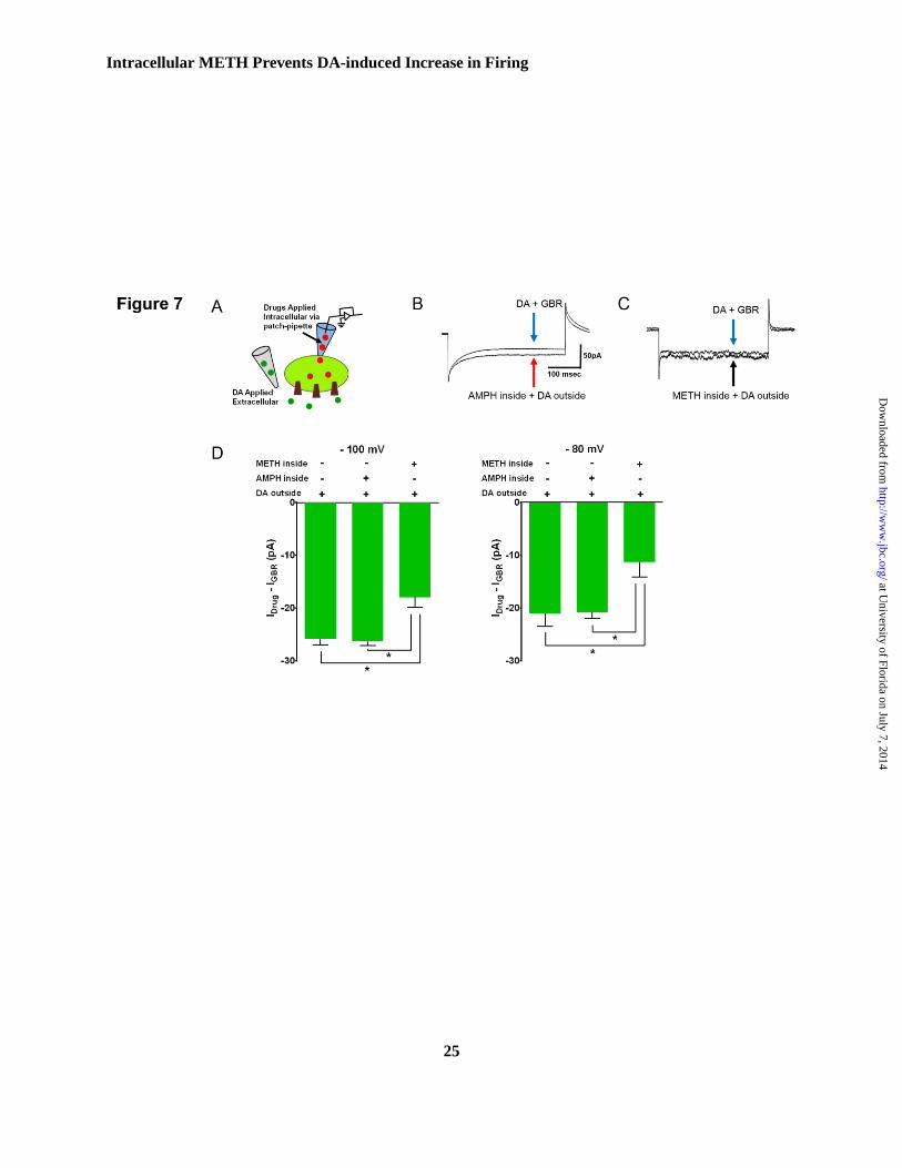
Intracellular methamphetamine, but
not amphetamine prevents the dopamine-
evoked inward current.
While intracellular amphetamine and
methamphetamine similarly affected the
excitability of dopamine neurons and DAT-
mediated inward current (Fig. 2B and 6B), this
similarity did not persist in response to
extracellular dopamine when these drugs exist
intracellularly (Fig. 7A). Therefore, we asked
whether stimulation of forward transport by bath
application of dopamine affects the DAT-mediated
inward current when methamphetamine or
amphetamine is present intracellullarly (Fig. 1A).
The DAT-mediated inward current in response to
extracellular dopamine application alone or in the
presence of intracellular amphetamine was similar,
whereas intracellular methamphetamine
significantly reduced the GBR12935-sensitive,
dopamine-evoked current, by comparison (p<0.05,
one-way ANOVA followed by Tukey’s post hoc
test) (Fig. 7B-E). The dopamine induced inwards
current in presence of methamphetamine inside the
cell at -100 and -80 mV were -18 ± 4.2 pA and -
11.3 ± 6.2 pA respectively, while in presence of
intracellular amphetamine the inward current at -
100 and -80 mV were 26 ± 2 pA and 20 ± 2 pA
respectively. These findings suggest that
amphetamine versus methamphetamine uniquely
regulates the DAT activity.
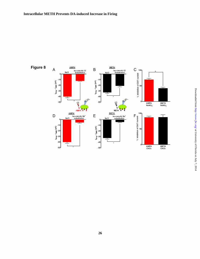
Amphetamine- and methamphetamine-
induced DAT-mediated inward currents are
equally Na+-dependent whereas amphetamine-
induced DAT-mediated inward current is
preferentially Cl--dependent
DAT uses the electrochemical gradients of
the cell to drive transport (13,30,42,46). The DAT-
mediated current is coupled to the translocation of
Na+ and Cl
- ions (30-32,42,46). These results and
the previous reports support the notion that
amphetamine and methamphetamine differentially
regulate the activity of the transporter. Therefore,
we investigated whether the substrate-specific
ionic transport is the underlying mechanism. Na+
or Cl¯
ions were iso-osmotically replaced by
inclusion of choline-Cl or NaNO3 for NaCl in the
external solution, respectively. The internal
solution was kept unchanged. Compared to the
inward current in the presence of normal Na+ ion
concentration, removal of Na+ ion significantly
decreased DAT-mediated inward current in
response to amphetamine by 87.5% (n=5) and
methamphetamine by 87.7% (n=5), (p<0.05; Fig.
8B, D, F). In contrast, removal of Cl¯ ion
preferentially reduced amphetamine-induced
DAT-mediated inward current by 70.16 ± 2%
relative to the 42.44 ± 2.4 % reduction of the
methamphetamine response (Fig. 8A, C, E;
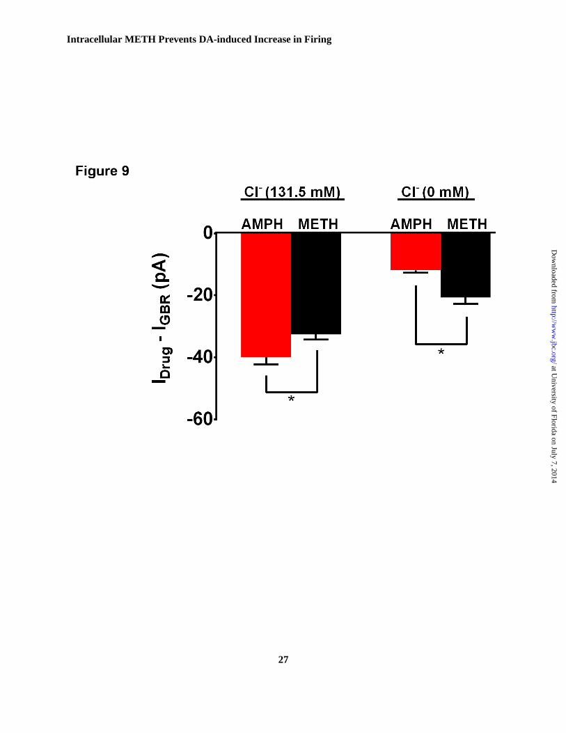
p<0.05, n=2-3). These results suggest that
amphetamine-induced DAT-mediated inward
current is preferentially dependent on external Cl¯
ion. The side by side comparison of amphetamine-
and methamphetamine-induced DAT-mediated
inward current at different Cl¯
concentrations is
depicted in Fig 9.
at University of Florida on July 7, 2014
http://ww
w.jbc.org/
Dow
nloaded from
Intracellular METH Prevents DA-induced Increase in Firing
9
Amphetamine- and methamphetamine-
induced cell surface redistribution of DAT are
not different.
We investigated the possibility that the
higher DAT-mediated inward current induced by
amphetamine compared to methamphetamine,
might be due to methamphetamine causing a much
faster internalization of the transporter. We
therefore, measured surface DAT levels following
10 min of amphetamine or methamphetamine
exposure. Using biotinylation followed by
western-blot analysis we observed that
amphetamine- and methamphetamine-induced cell
surface redistributions of DAT are not
significantly different from each other (Fig 10 A,
B).
These findings were confirmed via live-cell TIRF-
microscopy showing that both psychostimulants
similarly affected the cell surface redistribution of
the transporter within the time frame (10 min)
studied (Fig 10 C, D, E).
DISCUSSION
Long standing evidence supports the idea
that amphetamine and methamphetamine
differentially affect dopamine neurotransmission
in the brain but the underlying mechanism of this
differential effect is less understood. In this study
we examined the effect of these two compounds
on the firing activity of dopaminergic neurons and
DAT-mediated current. In this study we examined
the effect of these two compounds on the firing
activity of dopaminergic neurons and on the DAT-
mediated current. We found, although all three
DAT substrates increase spontaneous firing of
dopaminergic neurons and DAT-mediated inward
current, methamphetamine is the least effective. In
addition, unlike extracellular application of these
drugs, intracellular amphetamine and
methamphetamine are less effective in modulating
the spontaneous firing of dopaminergic neurons.
Importantly, intracellular methamphetamine
prevents the dopamine-induced enhancement of
neuronal firing and inward current. Consistent
with the literature, the isosmotic Na+ substitution
in the external solution eliminated DAT-mediated
inward current elicited by these substrates.
However, to our surprise, the substitution of Cl-
ions in the external solution only partially
inhibited the methamphetamine-induced DAT
current.
Dopamine, amphetamine and
methamphetamine are all DAT substrates. They
interact and are subsequently transported into the
DAT+ neurons in a Na
+/Cl
- dependent mechanism.
The transport process generates current, and
increases the excitability of dopaminergic neurons
that can be measured via classical
electrophysiology experiment. It is not known
whether these compounds influence DAT activity
once they have entered into the neuron, or whether
the presence of intracellular drug can influence the
response to the extracellular dopamine.
Determining DAT activity under these conditions
is important because these drugs compete with
inward transport of dopamine, they enter into the
neuron, while dopamine remains outside.
Therefore, we designed three complementary
experimental configurations to determine the
effect of these compounds on the excitability of
dopaminergic neurons and DAT-mediated inward
current when amphetamine or methamphetamine
exist 1) only outside the neuron, 2) only inside the
neurons or 3) when the drugs were directly
dialyzed into the cell and the native substrate,
dopamine was applied extracellularly. These
experimental configurations provide a unique
approach to mechanistically determine why these
compounds differentially affect DAT activity,
have different central effects and abuse potential.
DAT substrates differentially alter the
DAT-mediate inward current and neuronal firing.
As shown in Fig. 1, we found extracellular
amphetamine is the most effective compound in
increasing the firing rate of dopaminergic neurons.
This was paralleled by a consistently larger
amphetamine-induced DAT-mediated inward
current. The literature supports a direct correlation
between the DAT-mediated substrate induced
inward current and the excitability of
dopaminergic neurons, albeit with less understood
mechanism. (16,42,47). To address the
mechanism, we directly dialyzed each drug into
the neuron bypassing the binding and transport
step. Under this condition neither the excitability
at University of Florida on July 7, 2014
http://ww
w.jbc.org/
Dow
nloaded from
Intracellular METH Prevents DA-induced Increase in Firing
10
of dopaminergic neurons nor DAT-mediated
inward current were different when either
amphetamine or methamphetamine was directly
dialyzed into the neurons or DAT cells. This
suggests that the differential response in the
excitability of dopaminergic neurons might be due
to substrate-specific extracellular binding and
subsequent transport into the neurons.
It is possible that the underlying
mechanism for amphetamine-induced increase in
the excitability of dopaminergic neurons and
DAT-mediated inward current is due to specific
regulation of forward transport step. We tested this
hypothesis by bath application of dopamine while
amphetamine or methamphetamine exists inside
the neuron or DAT cell. Unlike intracellular
methamphetamaine, the presence of amphetamine
inside the neuron did not decrease the dopamine-
induced inward current or its enhancement of
neuronal excitability. This finding is consistent
with an interpretation that intracellular
amphetamine facilitates a conformational state of
the transporter, which supports the interaction of
dopamine with DAT, leading to dopamine-
mediated inward current and neuronal excitability.
This is consistent with a recent report
demonstrating that amphetamine induces a
persistent inward current by increasing the Na+
conductance of the transporter via a molecular
stent mechanism (46).
As shown in Fig. 2 and 6, surprisingly,
when we dialyzed methamphetamine inside the
neuron, it significantly decreased both dopamine-
induced DAT-mediated inward current and
neuronal excitability. Although the current data do
not delineate the exact underlying mechanism, it is
possible that intracellular methamphetamine
induces a state of the transporter that is less
sensitive to the effect of extracellular dopamine on
the activity of the transporter. Studies by Kahlig et
al, 2005 suggest another possible mechanism. The
authors eloquently have shown that extracellular
dopamine decreases the channel-like mode of
DAT activity. Therefore, the presence of
intracellular methamphetamine may further
accentuate the effect of extracellular dopamine.
(35,47).
We considered the possibility that the
differences in the magnitude of inward current
induced by amphetamine versus
methamphetamine is due to their differential
influences on DAT trafficking. However,
biotinylation and live cell TIRF microscopy
experiments suggest that amphetamine induced
reduction of TIRF surface DAT levels was not
different from methamphetamine-induced cell
surface redistribution of DAT. The live-cell TIRF
microscopy experiments support this conclusion
showing that amphetamine and methamphetamine
induce a significant and similar level of cell
surface re-distribution of DAT within the time
frame of these experiments. Thus substrate
induced DAT internalization is not responsible for
the measured differential substrates-induced DAT-
mediated inward current.
It has been shown that Akt through a
Ca2+
/Calmodulin dependent kinase II can regulate
the activity of the transporter following exposure
to psychostimulants. (48-50). It is possible that
these two compounds differentially affect the
activity of Akt in a manner that does not influence
the trafficking of the transporter per se, but it
facilitates a conformational state of the transporter
to regulate the current. This possibility will be the
focus of our future studies.
In an elegant review, Zhu and Reith,
proposed an intriguing model regarding the
substrate regulation of DAT oligomerization (51)
DAT at the cell surface is distributed between
oligomers and monomers, where DAT monomers
are internalized (Zhu and Reith) following
amphetamine (51). Consistent with this idea, data
from Fleckenstein lab suggest the possibility that
methamphetamine-induces oligomerization of the
transporter (52); whereas, a report by Reith group
has shown that amphetamine promotes the
formation of DAT monomers (5). Taken together,
it is possible that the methamphetamine-induced
oligomerization of DAT differentially affects the
DAT-mediated ionic conductance.
The DAT-mediated inward current is
dependent upon transporting of Na+ and Cl
- ions.
This is supported by recently resolved crystal
structure of the Drosophila melanogaster
at University of Florida on July 7, 2014
http://ww
w.jbc.org/
Dow
nloaded from
Intracellular METH Prevents DA-induced Increase in Firing
11
dopamine transporter showing separate binding
sites for Na+ and Cl
- ions (53). Consistent with the
identification of a substrate binding pocket on the
transporter, it is well established that the DAT
substrates amphetamine and methamphetamine
bind to the transporter and mediate GBR12935-
senstive inward current (53). In this study we
found the DAT substrates, amphetamine and
methamphetamine elicit different amount of
inward current and induce profoundly different
magnitude of neuronal firing. Therefore, we
examine the possibility that these two compounds
may be differentially coupled to the co-transport
of Na+ and Cl
- ions. To address this hypothesis,
while keeping the composition of the internal
solution intact, we isosmoticaly substituted either
Na+ or Cl
¯ ions in the external solution. While the
substitution of Na+ ions in the external solution
equally affected amphetamine- and
methamphetamine-induced DAT currents; the
substitution of Cl- ions in the external solution
more effectively blocks the amphetamine-induced
DAT current. The Na+ and Cl
- ionic dependency of
amphetamine-induced current is consistent with
the existing data in the literature that removal of
either Na+, Cl
-, or both ions form the external
solution decreases DAT activity (13). Similarly,
Erreger et al. have elegantly shown that
amphetamine-mediated inward current
significantly decreases following the substitution
of Na+, or Cl
- ions in the external milieu (13).
Furthermore, Ingram et al., demonstrated that Cl-
conductance through the transporter is important
in amphetamine-regulation of excitability of
dopaminergic neurons as well as amphetamine-
mediated inward current through DAT (42). These
reports explain the Na+ and Cl
- ionic dependency
of amphetamine-mediated current, but they do not
explain why Cl- ionic substitution partially inhibits
the methamphetamine-induced DAT current.
Future studies should consider determining the
mechanism responsible for the decreased
sensitivity of DAT to methamphetamine in the
absence of Cl- ion in the external solution.
Nonetheless, these results provide critical
information to further efforts towards designing
effective therapeutic approaches when dopamine
neurotransmission is dysregulated.
at University of Florida on July 7, 2014
http://ww
w.jbc.org/
Dow
nloaded from
Intracellular METH Prevents DA-induced Increase in Firing
12
REFERENCES:
1. Iversen, S. D., and Iversen, L. L. (2007) Dopamine: 50 years in perspective. Trends in
neurosciences 30, 188-193
2. Grace, A. A., Floresco, S. B., Goto, Y., and Lodge, D. J. (2007) Regulation of firing of
dopaminergic neurons and control of goal-directed behaviors. Trends in neurosciences 30, 220-
227
3. Bernheimer, H., Birkmayer, W., Hornykiewicz, O., Jellinger, K., and Seitelberger, F. (1973)
Brain dopamine and the syndromes of Parkinson and Huntington. Clinical, morphological and
neurochemical correlations. Journal of the neurological sciences 20, 415-455
4. Hornykiewicz, O., Kish, S. J., Becker, L. E., Farley, I., and Shannak, K. (1986) Brain
neurotransmitters in dystonia musculorum deformans. The New England journal of medicine 315,
347-353
5. Dawson, T. M., and Dawson, V. L. (2002) Neuroprotective and neurorestorative strategies for
Parkinson's disease. Nat Neurosci 5 Suppl, 1058-1061
6. Wise, C. D., and Stein, L. (1973) Dopamine-beta-hydroxylase deficits in the brains of
schizophrenic patients. Science 181, 344-347
7. Snyder, S. H., and Meyerhoff, J. L. (1973) How amphetamine acts in minimal brain dysfunction.
Annals of the New York Academy of Sciences 205, 310-320
8. Wall, S. C., Gu, H., and Rudnick, G. (1995) Biogenic amine flux mediated by cloned transporters
stably expressed in cultured cell lines: amphetamine specificity for inhibition and efflux.
Molecular pharmacology 47, 544-550
9. Giros, B., Jaber, M., Jones, S. R., Wightman, R. M., and Caron, M. G. (1996) Hyperlocomotion
and indifference to cocaine and amphetamine in mice lacking the dopamine transporter. Nature
379, 606-612
10. Salahpour, A., Ramsey, A. J., Medvedev, I. O., Kile, B., Sotnikova, T. D., Holmstrand, E., Ghisi,
V., Nicholls, P. J., Wong, L., Murphy, K., Sesack, S. R., Wightman, R. M., Gainetdinov, R. R.,
and Caron, M. G. (2008) Increased amphetamine-induced hyperactivity and reward in mice
overexpressing the dopamine transporter. Proceedings of the National Academy of Sciences of
the United States of America 105, 4405-4410
11. Keefe, K. A., Zigmond, M. J., and Abercrombie, E. D. (1992) Extracellular dopamine in striatum:
influence of nerve impulse activity in medial forebrain bundle and local glutamatergic input.
Neuroscience 47, 325-332
12. (2006) National Institute on Drug Abuse in NIDA Research Report
13. Erreger, K., Grewer, C., Javitch, J. A., and Galli, A. (2008) Currents in response to rapid
concentration jumps of amphetamine uncover novel aspects of human dopamine transporter
function. The Journal of neuroscience : the official journal of the Society for Neuroscience 28,
976-989
14. Keefe, K. A., Zigmond, M. J., and Abercrombie, E. D. (1993) In vivo regulation of extracellular
dopamine in the neostriatum: influence of impulse activity and local excitatory amino acids.
Journal of neural transmission. General section 91, 223-240
15. Nieoullon, A., Cheramy, A., and Glowinski, J. (1977) Release of dopamine in vivo from cat
substantia nigra. Nature 266, 375-377
16. Leviel, V. (2011) Dopamine release mediated by the dopamine transporter, facts and
consequences. Journal of neurochemistry 118, 475-489
17. Zhou, Z., and Misler, S. (1995) Action potential-induced quantal secretion of catecholamines
from rat adrenal chromaffin cells. The Journal of biological chemistry 270, 3498-3505
at University of Florida on July 7, 2014
http://ww
w.jbc.org/
Dow
nloaded from
Intracellular METH Prevents DA-induced Increase in Firing
13
18. Bertorello, A. M., Hopfield, J. F., Aperia, A., and Greengard, P. (1990) Inhibition by dopamine of
(Na(+)+K+)ATPase activity in neostriatal neurons through D1 and D2 dopamine receptor
synergism. Nature 347, 386-388
19. Beckstead, M. J., Grandy, D. K., Wickman, K., and Williams, J. T. (2004) Vesicular dopamine
release elicits an inhibitory postsynaptic current in midbrain dopamine neurons. Neuron 42, 939-
946
20. Sara, Y., Virmani, T., Deak, F., Liu, X., and Kavalali, E. T. (2005) An isolated pool of vesicles
recycles at rest and drives spontaneous neurotransmission. Neuron 45, 563-573
21. Rizzoli, S. O., and Betz, W. J. (2005) Synaptic vesicle pools. Nature reviews. Neuroscience 6, 57-
69
22. Fischer, J. F., and Cho, A. K. (1976) Properties of dopamine efflux from rat striatal tissue caused
by amphetamine and p-hydroxyamphetamine. Proceedings of the Western Pharmacology Society
19, 179-182
23. Eshleman, A. J., Henningsen, R. A., Neve, K. A., and Janowsky, A. (1994) Release of dopamine
via the human transporter. Molecular pharmacology 45, 312-316
24. Sitte, H. H., Huck, S., Reither, H., Boehm, S., Singer, E. A., and Pifl, C. (1998) Carrier-mediated
release, transport rates, and charge transfer induced by amphetamine, tyramine, and dopamine in
mammalian cells transfected with the human dopamine transporter. Journal of neurochemistry 71,
1289-1297
25. Jones, S. R., Gainetdinov, R. R., Jaber, M., Giros, B., Wightman, R. M., and Caron, M. G. (1998)
Profound neuronal plasticity in response to inactivation of the dopamine transporter. Proceedings
of the National Academy of Sciences of the United States of America 95, 4029-4034
26. Jones, S. R., Gainetdinov, R. R., Wightman, R. M., and Caron, M. G. (1998) Mechanisms of
amphetamine action revealed in mice lacking the dopamine transporter. J Neurosci 18, 1979-1986
27. Obianwu, H. O., Stitzel, R., and Lundborg, P. (1968) Subcellular distribution of
[3H]amphetamine and [3H]guanethidine and their interaction with adrenergic neurons. The
Journal of pharmacy and pharmacology 20, 585-594
28. Wong, D. T., van Frank, R. M., Horng, J. S., and Fuller, R. W. (1972) Accumulation of
amphetamine and p-chloroamphetamine into synaptosomes of rat brain. The Journal of pharmacy
and pharmacology 24, 171-173
29. Goodwin, J. S., Larson, G. A., Swant, J., Sen, N., Javitch, J. A., Zahniser, N. R., De Felice, L. J.,
and Khoshbouei, H. (2009) Amphetamine and methamphetamine differentially affect dopamine
transporters in vitro and in vivo. The Journal of biological chemistry 284, 2978-2989
30. Sonders, M. S., Zhu, S. J., Zahniser, N. R., Kavanaugh, M. P., and Amara, S. G. (1997) Multiple
ionic conductances of the human dopamine transporter: the actions of dopamine and
psychostimulants. The Journal of neuroscience : the official journal of the Society for
Neuroscience 17, 960-974
31. Krueger, B. K. (1990) Kinetics and block of dopamine uptake in synaptosomes from rat caudate
nucleus. Journal of neurochemistry 55, 260-267
32. McElvain, J. S., and Schenk, J. O. (1992) A multisubstrate mechanism of striatal dopamine
uptake and its inhibition by cocaine. Biochemical pharmacology 43, 2189-2199
33. Gu, H., Wall, S. C., and Rudnick, G. (1994) Stable expression of biogenic amine transporters
reveals differences in inhibitor sensitivity, kinetics, and ion dependence. J Biol Chem 269, 7124-
7130
34. Fog, J. U., Khoshbouei, H., Holy, M., Owens, W. A., Vaegter, C. B., Sen, N., Nikandrova, Y.,
Bowton, E., McMahon, D. G., Colbran, R. J., Daws, L. C., Sitte, H. H., Javitch, J. A., Galli, A.,
and Gether, U. (2006) Calmodulin kinase II interacts with the dopamine transporter C terminus to
regulate amphetamine-induced reverse transport. Neuron 51, 417-429
35. Kahlig, K. M., Binda, F., Khoshbouei, H., Blakely, R. D., McMahon, D. G., Javitch, J. A., and
Galli, A. (2005) Amphetamine induces dopamine efflux through a dopamine transporter channel.
at University of Florida on July 7, 2014
http://ww
w.jbc.org/
Dow
nloaded from
Intracellular METH Prevents DA-induced Increase in Firing
14
Proceedings of the National Academy of Sciences of the United States of America 102, 3495-
3500
36. Khoshbouei, H., Wang, H., Lechleiter, J. D., Javitch, J. A., and Galli, A. (2003) Amphetamine-
induced dopamine efflux. A voltage-sensitive and intracellular Na+-dependent mechanism. The
Journal of biological chemistry 278, 12070-12077
37. Kahlig, K. M., and Galli, A. (2003) Regulation of dopamine transporter function and plasma
membrane expression by dopamine, amphetamine, and cocaine. Eur J Pharmacol 479, 153-158
38. Hastrup, H., Karlin, A., and Javitch, J. A. (2001) Symmetrical dimer of the human dopamine
transporter revealed by cross-linking Cys-306 at the extracellular end of the sixth transmembrane
segment. Proc Natl Acad Sci U S A 98, 10055-10060
39. Hastrup, H., Sen, N., and Javitch, J. A. (2003) The human dopamine transporter forms a tetramer
in the plasma membrane: cross-linking of a cysteine in the fourth transmembrane segment is
sensitive to cocaine analogs. J Biol Chem 278, 45045-45048
40. Swant, J., Goodwin, J. S., North, A., Ali, A. A., Gamble-George, J., Chirwa, S., and Khoshbouei,
H. (2011) alpha-Synuclein stimulates a dopamine transporter-dependent chloride current and
modulates the activity of the transporter. The Journal of biological chemistry 286, 43933-43943
41. Bukauskas, F. F., Kempf, C., and Weingart, R. (1992) Cytoplasmic bridges and gap junctions in
an insect cell line (Aedes albopictus). Experimental physiology 77, 903-911
42. Ingram, S. L., Prasad, B. M., and Amara, S. G. (2002) Dopamine transporter-mediated
conductances increase excitability of midbrain dopamine neurons. Nature neuroscience 5, 971-
978
43. Paladini, C. A., Robinson, S., Morikawa, H., Williams, J. T., and Palmiter, R. D. (2003)
Dopamine controls the firing pattern of dopamine neurons via a network feedback mechanism.
Proceedings of the National Academy of Sciences of the United States of America 100, 2866-
2871
44. Fon, E. A., Pothos, E. N., Sun, B. C., Killeen, N., Sulzer, D., and Edwards, R. H. (1997)
Vesicular transport regulates monoamine storage and release but is not essential for amphetamine
action. Neuron 19, 1271-1283
45. Pierce, R. C., and Kalivas, P. W. (1997) Repeated cocaine modifies the mechanism by which
amphetamine releases dopamine. The Journal of neuroscience : the official journal of the Society
for Neuroscience 17, 3254-3261
46. Rodriguez-Menchaca, A. A., Solis, E., Jr., Cameron, K., and De Felice, L. J. (2012)
S(+)amphetamine induces a persistent leak in the human dopamine transporter: molecular stent
hypothesis. British journal of pharmacology 165, 2749-2757
47. Carvelli, L., McDonald, P. W., Blakely, R. D., and Defelice, L. J. (2004) Dopamine transporters
depolarize neurons by a channel mechanism. Proceedings of the National Academy of Sciences
of the United States of America 101, 16046-16051
48. Wei, Y., Williams, J. M., Dipace, C., Sung, U., Javitch, J. A., Galli, A., and Saunders, C. (2007)
Dopamine transporter activity mediates amphetamine-induced inhibition of Akt through a
Ca2+/calmodulin-dependent kinase II-dependent mechanism. Molecular pharmacology 71, 835-
842
49. Garcia, B. G., Wei, Y., Moron, J. A., Lin, R. Z., Javitch, J. A., and Galli, A. (2005) Akt is
essential for insulin modulation of amphetamine-induced human dopamine transporter cell-
surface redistribution. Molecular pharmacology 68, 102-109
50. Rau, T. F., Kothiwal, A., Zhang, L., Ulatowski, S., Jacobson, S., Brooks, D. M., Cardozo-Pelaez,
F., Chopp, M., and Poulsen, D. J. (2011) Low dose methamphetamine mediates neuroprotection
through a PI3K-AKT pathway. Neuropharmacology 61, 677-686
51. Zhu, J., and Reith, M. E. (2008) Role of the dopamine transporter in the action of
psychostimulants, nicotine, and other drugs of abuse. CNS & neurological disorders drug targets
7, 393-409
at University of Florida on July 7, 2014
http://ww
w.jbc.org/
Dow
nloaded from
Intracellular METH Prevents DA-induced Increase in Firing
15
52. Baucum, A. J., 2nd, Rau, K. S., Riddle, E. L., Hanson, G. R., and Fleckenstein, A. E. (2004)
Methamphetamine increases dopamine transporter higher molecular weight complex formation
via a dopamine- and hyperthermia-associated mechanism. The Journal of neuroscience : the
official journal of the Society for Neuroscience 24, 3436-3443
53. Penmatsa, A., Wang, K. H., and Gouaux, E. (2013) X-ray structure of dopamine transporter
elucidates antidepressant mechanism. Nature 503, 85-90
54. McCutcheon, J. E., Conrad, K. L., Carr, S. B., Ford, K. A., McGehee, D. S., and Marinelli, M.
(2012) Dopamine neurons in the ventral tegmental area fire faster in adolescent rats than in
adults. Journal of neurophysiology 108, 1620-1630
at University of Florida on July 7, 2014
http://ww
w.jbc.org/
Dow
nloaded from
Intracellular METH Prevents DA-induced Increase in Firing
16
Acknowledgements-We thank Dr. Satya Kalra, Dr. Rahul Khanna and Niousha Ahmari or their critical
review of the manuscript.
FOOTNOTES
*This work was supported by DA026947 and NS071122.
To whom correspondence should be addressed: Habibeh Khoshbouei, Department of Neuroscience, and
Department of Psychiatry, McKnight Brain Institute, University of Florida, 1149 Newell Drive, PO Box
100244, Gainesville, FL 32611-0244, USA, Tel.: (352)-273-8115; Fax: (352)-392-8347; Email:
The abbreviations used are: DA, dopamine; AMPH, amphetamine; METH, methamphetamine; DAT,
dopamine transporter; CHO, Chinese hamster ovarian cells; HEK, human embryonic kidney cells; YFP,
yellow fluorescent protein;
FIGURE LEGENDS:
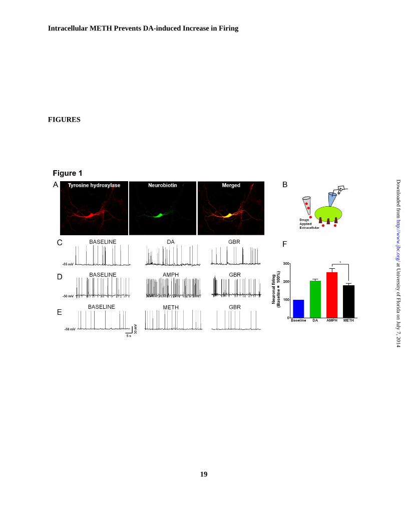
Figure 1. Extracellular amphetamine and methamphetamine differentially affect the DAT-
mediated spontaneous firing of dopaminergic neurons.
A) A representative image of a tyrosine hydroxylase (TH) and neurobiotin immunoreactive dopaminergic
neuron obtained from primary culture of midbrain region of 1-2 days old mice. The neurons were selected
by morphological (bright cell bodies, long processes) and electrophysiological signature (D2-induced
hyperpolarization). The neurons were filled with neurobiotin via the patch pipette during the recording for
subsequent staining for TH, and neurobiotin. The data obtained from TH and neurobiotin immunoreactive
neurons are reported. Neurobiotin, is widely used to identify the neurons from which the recordings were
performed does not affect the electrical properties of neurons (54). The spontaneous firing activity of
midbrain dopaminergic neurons was measured in the current clamp mode when D2 receptor is blocked
(sulpiride, 5µM). B) Cartoon depicts the experimental configuration showing DAT substrates were
applied extracellularly. (C, D, E) Representative traces of current clamp recording of midbrain
dopaminergic neurons showing the basal spontaneous firing activity of dopaminergic neurons at resting
membrane potential in the absence of DAT substrates, dopamine, amphetamine, or methamphetamine.
The extracellular application of DAT substrates, dopamine (1µM; n=5), amphetamine (1µM; n=5) or
methamphetamine (1µM; n=5), increases the firing rate of dopaminergic neurons that is blocked by DAT
antagonist, GBR12935 (1µM). F) Bar-graphs show the spontaneous neuronal firing rate following
extracellular drug application. The data is normalized to the spontaneous firing rate at baseline before
drug application. The reported baseline (100%) is not an interleaved baseline. While the amphetamine-
and dopamine-induced increases in the spontaneous firing rate of dopaminergic neurons were similar, the
methamphetamine-induced changes in the firing rate was significantly lower than amphetamine (*p<0.05,
one-way ANOVA followed by Tukey’s post hoc test)
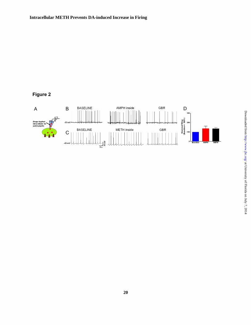
Figure 2. Intracellular amphetamine or methamphetamine less effectively modulates the firing rate
of dopaminergic neurons.
A) Cartoon depicts the experimental configuration showing DAT substrates were applied intracellularly.
B and C) Current clamp recording of midbrain dopaminergic neurons showing the spontaneous firing
activity at the endogenous resting membrane potential when DAT substrates, amphetamine (1µM, n=4) or
methamphetamine (1µM, n=5) were dialyzed into the neuron to bypass the forward transport step. At the
resting membrane potential, the spontaneous firing activity of the neurons immediately after achieving
at University of Florida on July 7, 2014
http://ww
w.jbc.org/
Dow
nloaded from
Intracellular METH Prevents DA-induced Increase in Firing
17
whole-cell configuration reflects the baseline firing activity of the neuron. D) Intracellular amphetamine,
or methamphetamine equally but only modestly affected the firing rate of dopaminergic neurons (black
and red bar). The bar-graphs show the spontaneous firing activity of the neurons when amphetamine or
methamphetamine was dialyzed into the neuron. The data are normalized to the baseline spontaneous
firing rate of each experiment. The change in firing rate was blocked by extracellular application of a
DAT antagonist, GBR12935 (1µM).
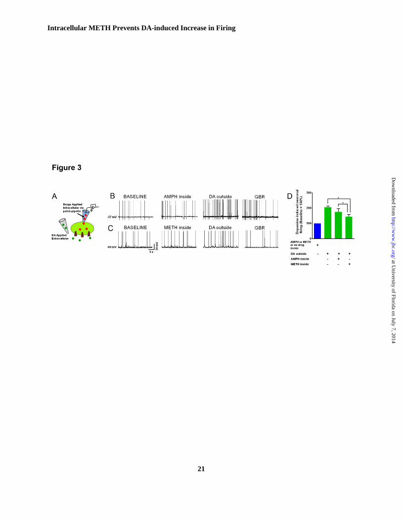
Figure 3. Intracellular methamphetamine attenuates the dopamine-induced increase in the firing
rate of dopaminergic neurons.
A) Cartoon depicts the experimental configuration showing DAT substrates were applied intracellularly
and dopamine was applied extracellularly. B and C) Current clamp recording from midbrain
dopaminergic neurons shows that dopamine (1µM, n=5) increases the spontaneous firing activity of
dopaminergic neurons. Intracellular methamphetamine (1µM, n=5) but not amphetamine (1µM, n=4)
attenuates the influence of dopamine on the spontaneous firing activity of dopaminergic neurons. The
increase in the firing was blocked by DAT blocker GBR12935 (1µM). (D) Bar graph compares the firing
activity of the neurons following extracellular application of dopamine. The data are normalized to the
firing rate when physiological-like internal solution, amphetamine or methamphetamine was dialyzed into
the neurons (one-way ANOVA followed by Tukey’s post hoc test; *p<0.05).
Figure 4. Comparison of the firing activity of dopaminergic neurons across all treatment groups.
Bar graph compares dopamine, amphetamine or methamphetamine regulation of the firing activity of
dopaminergic neurons when the drug is applied extra- or intracellularly.
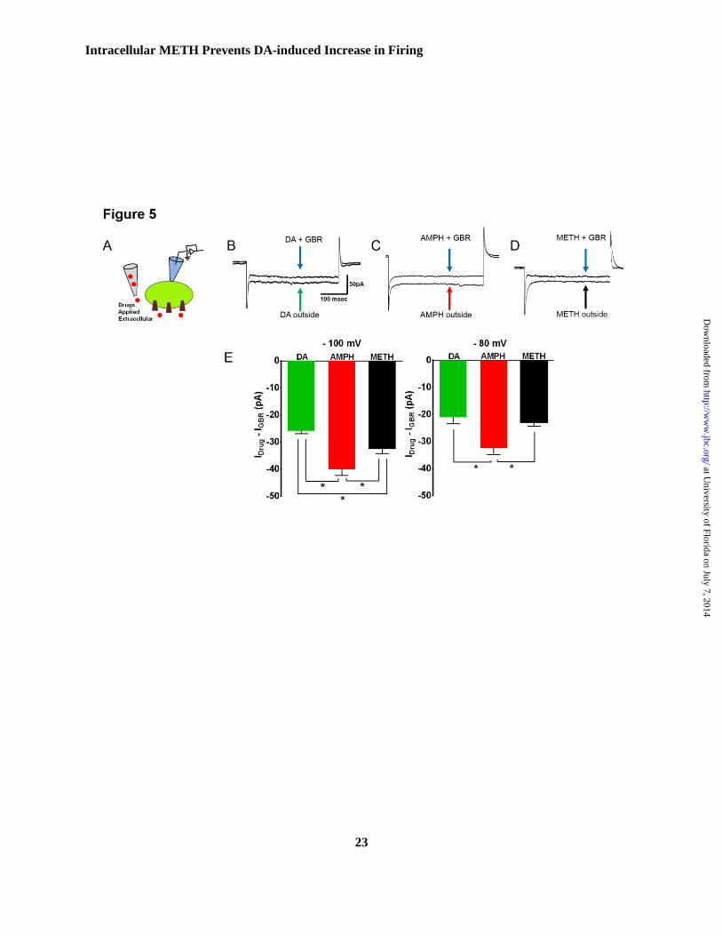
Figure 5. Methamphetamine elicits smaller DAT-mediated inward current compared to
amphetamine.
A) Cartoon depicts the experimental configuration. The DAT-mediated inward current was measured
when drugs are applied extracellularly. The substrate-induced, DAT-mediated current is defined as
GBR12935 subtracted current. The DAT-mediated, amphetamine- or methamphetamine-induced current
was measured when the cells were voltage-clamped in whole-cell configuration. B, C, and D)
Representative traces at -100mV. E) Bar-graph shows the average DAT-mediated current following bath
application of dopamine (10µM; n=6), methamphetamine (10µM; n=6) and amphetamine (10µM; n=6) at
-100 mV and -80mV. The amphetamine-induced, DAT-mediated inward currents were significantly
larger than methamphetamine-induced or dopamine-induced inward currents (*p<0.05, one-way ANOVA
followed by Tukey’s post hoc test).
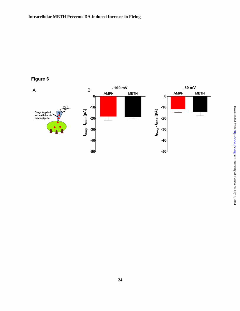
Figure 6. Intracellular amphetamine and methamphetamine elicit similar DAT-mediated current.
A) Carton depicts the experimental configuration. The CHO-YFP-DAT cells were patch clamped in
whole-cell configuration. Amphetamine (10µM; n=5), or methamphetamine (10µM; n=5), was dialyzed
into the cell via the patch pipette. B) Bar graph compares the DAT-mediated inward current at -100mV
and -80mV following intracellular delivery of the drugs. There is no difference between amphetamine- or
methamphetamine-induced DAT-mediated inward currents at the examined membrane potentials.
Figure 7. Intracellular methamphetamine prevents the dopamine-induced inward current.
A) Carton depicts the experimental configuration. The dopamine-induced inward current was measured in
CHO-YFP-DAT cells when internal solution, or internal solution containing amphetamine (10µM; n=4),
or methamphetamine (10µM; n=5), was dialyzed into the cell. Dopamine (10µM) was bath applied. The
experiments were performed as described in Figure 6. B, C) Representative traces at -100mV. D) Bar-
graph compares the dopamine-induced inward current at -100mV and -80mV following intracellular
delivery of methamphetamine (10µM; n=5) or amphetamine (10µM; n=4). Intracellular
at University of Florida on July 7, 2014
http://ww
w.jbc.org/
Dow
nloaded from
Intracellular METH Prevents DA-induced Increase in Firing
18
methamphetamine, but not amphetamine decreases the dopamine-induced, DAT-mediated inward current.
(*p<0.05, one-way ANOVA followed by Tukey’s post hoc test).
Figure 8. Removal of extracellular Cl- preferentially blocks amphetamine-induced DAT-mediated
current relative to methamphetamine.
A and B) Bar graphs show the amphetamine- or methamphetamine-induced DAT current at -100 mV
following removal of Cl- ions in the external solution. C) Substitution of Cl
- ions in the external solution
blocks 70.16 ± 2.0 % of amphetamine-induced DAT current, but only 42.44 ± 2.4 % of
methamphetamine-induced current. D and E) Bar graphs show amphetamine- or methamphetamine-
induced inward current at -100 mV upon removal of Na+
ions in the external solution. F) Substitution of
Na+ ions in the external solution equally blocks the amphetamine- and methamphetamine-induced DAT
currents.
Figure 9. Side by side comparison of the effects of amphetamine versus methamphetamine induced
DAT-mediated inward currents.
Bar-graph showing side by side comparison of effects of amphetamine versus methamphetamine induced
DAT-mediated inward currents at (-100 mV holding potential), at two different concentrations of
extracellular chloride ions (Cl-). We observed that at 0 mM Cl
- ion concentration, the inward current
induced by amphetamine was significantly blocked and to a higher extent compared to methamphetamine.
While, at 131.5mM of extracellular Cl- ion concentration amphetamine produced significantly higher
inward current compared to methamphetamine.
Figure 10. Amphetamine induced reduction in surface DAT levels was not different from
methamphetamine.
FLAG-DAT HEK cells were treated with amphetamine (10μM) or methamphetamine (10μM) for 10 min
followed by treatment with sulfo-NHS-SS-biotin to detected surface DAT. (A) Representative
immunoblots showing surface DAT, total DAT and loading control, β-actin. (B) Bar-graph shows
following amphetamine treatment there was a significant reduction in the surface DAT compared to the
control whereas following 10 min of methamphetamine treatment surface DAT density was not
significantly different from control, or amphetamine. C) Plot of membrane YFP-DAT intensity (AFU)
following exposure to amphetamine (10μM) or methamphetamine (10μM) normalized to the the average
intensity for each cell during the 15sec prior to solution change. The normalized response to vehicle
(external solution) has been accounted for in the normalized values, correcting for fluorophore bleaching.
D) Bar-graph shows average normalized intensity after 10min of drug application. Both psychostimulants
similarly and significantly internalized the dopamine transporter (***p<0.001; *p<0.05; one-way
ANOVA followed by Tukey’s post hoc test). E) Representative images showing amphetamine and
methamphetamine-induced reduction in the surface YFP-DAT intensity.
at University of Florida on July 7, 2014
http://ww
w.jbc.org/
Dow
nloaded from
Intracellular METH Prevents DA-induced Increase in Firing
19
FIGURES
at University of Florida on July 7, 2014
http://ww
w.jbc.org/
Dow
nloaded from
Intracellular METH Prevents DA-induced Increase in Firing
20
at University of Florida on July 7, 2014
http://ww
w.jbc.org/
Dow
nloaded from
Intracellular METH Prevents DA-induced Increase in Firing
21
at University of Florida on July 7, 2014
http://ww
w.jbc.org/
Dow
nloaded from

Intracellular METH Prevents DA-induced Increase in Firing
22
at University of Florida on July 7, 2014
http://ww
w.jbc.org/
Dow
nloaded from
Intracellular METH Prevents DA-induced Increase in Firing
23
at University of Florida on July 7, 2014
http://ww
w.jbc.org/
Dow
nloaded from
Intracellular METH Prevents DA-induced Increase in Firing
24
at University of Florida on July 7, 2014
http://ww
w.jbc.org/
Dow
nloaded from
Intracellular METH Prevents DA-induced Increase in Firing
25
at University of Florida on July 7, 2014
http://ww
w.jbc.org/
Dow
nloaded from
Intracellular METH Prevents DA-induced Increase in Firing
26
at University of Florida on July 7, 2014
http://ww
w.jbc.org/
Dow
nloaded from
Intracellular METH Prevents DA-induced Increase in Firing
27
at University of Florida on July 7, 2014
http://ww
w.jbc.org/
Dow
nloaded from
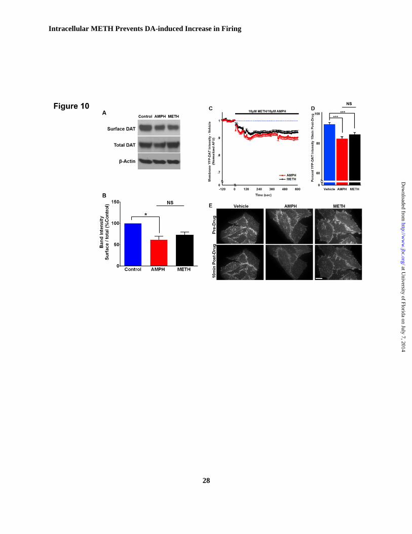
Intracellular METH Prevents DA-induced Increase in Firing
28
at University of Florida on July 7, 2014
http://ww
w.jbc.org/
Dow
nloaded from