international journal of pharmaceutics - permegear
TRANSCRIPT
International Journal of Pharmaceutics 364 (2008) 298–327
Contents lists available at ScienceDirect
International Journal of Pharmaceutics
journa l homepage: www.e lsev ier .com/ locate / i jpharm
Review
Principles of encapsulating hydrophobic drugs in PLA/PLGA microparticles
Christian Wischke1, Steven P. Schwendeman ∗
Department of Pharmaceutical Sciences, University of Michigan, 428 Church Street, Ann Arbor, MI 48109-1065, USA
a r t i c l e i n f o
Article history:Received 17 March 2008Received in revised form 29 April 2008Accepted 29 April 2008Available online 7 May 2008
Keywords:PLGABiopharmaceutical classification systemMicroencapsulationControlled release
a b s t r a c t
Injectable biodegradable and biocompatible copolymers of lactic and glycolic acid (PLGA) are an impor-tant advanced delivery system for week-to-month controlled release of hydrophobic drugs (e.g., frombiopharmaceutical classification system class IV), which often display poor oral bioavailability. The basicprinciples and considerations to develop such microparticle formulations is reviewed here based on acomprehensive study of papers and patents from the beginnings of hydrophobic drug encapsulation inpolylactic acid and PLGA up through the very recent literature. Challenges with the diversity of drugproperties, microencapsulation methods, and organic solvents are evaluated in light of the precedenceof commercialized formulations and with a focus on decreasing the time to lab-scale encapsulation ofwater-insoluble drug candidates in the early stage of drug development. The influence of key formulationvariables on final microparticle characteristics, and how best to avoid undesired microparticle properties,
MicroparticleHydrophobic drug
is analyzed mechanistically. Finally, concepts are developed to manage the common issues of maintainingsink conditions for in vitro drug release assays of hydrophobic compounds. Overall, against the backdropof an increasing number of new, poorly orally available drug entities entering development, microparticle
C
CC
0d
delivery systems may be a viable strategy to rescue an otherwise undeliverable substance.© 2008 Elsevier B.V. All rights reserved.
ontents
1. Introduction . . . . . . . . . . . . . . . . . . . . . . . . . . . . . . . . . . . . . . . . . . . . . . . . . . . . . . . . . . . . . . . . . . . . . . . . . . . . . . . . . . . . . . . . . . . . . . . . . . . . . . . . . . . . . . . . . . . . . . . . . . . . . . . . . . . . . . . . . 2992. Drug properties relevant for microencapsulation and release . . . . . . . . . . . . . . . . . . . . . . . . . . . . . . . . . . . . . . . . . . . . . . . . . . . . . . . . . . . . . . . . . . . . . . . . . . . . . . . . . . . . . 300
2.1. Recent trends in drug discovery and their implications on microencapsulation candidates. . . . . . . . . . . . . . . . . . . . . . . . . . . . . . . . . . . . . . . . . . . . . . . 3002.2. Drug solubility in aqueous and organic media. . . . . . . . . . . . . . . . . . . . . . . . . . . . . . . . . . . . . . . . . . . . . . . . . . . . . . . . . . . . . . . . . . . . . . . . . . . . . . . . . . . . . . . . . . . . . . 3002.3. Drug stability . . . . . . . . . . . . . . . . . . . . . . . . . . . . . . . . . . . . . . . . . . . . . . . . . . . . . . . . . . . . . . . . . . . . . . . . . . . . . . . . . . . . . . . . . . . . . . . . . . . . . . . . . . . . . . . . . . . . . . . . . . . . . . . . 3012.4. Drug–polymer interactions . . . . . . . . . . . . . . . . . . . . . . . . . . . . . . . . . . . . . . . . . . . . . . . . . . . . . . . . . . . . . . . . . . . . . . . . . . . . . . . . . . . . . . . . . . . . . . . . . . . . . . . . . . . . . . . . . 3012.5. Drug solid-state properties . . . . . . . . . . . . . . . . . . . . . . . . . . . . . . . . . . . . . . . . . . . . . . . . . . . . . . . . . . . . . . . . . . . . . . . . . . . . . . . . . . . . . . . . . . . . . . . . . . . . . . . . . . . . . . . . . . 301
3. Microencapsulation techniques for hydrophobic drugs . . . . . . . . . . . . . . . . . . . . . . . . . . . . . . . . . . . . . . . . . . . . . . . . . . . . . . . . . . . . . . . . . . . . . . . . . . . . . . . . . . . . . . . . . . . . 3023.1. o/w emulsion technique . . . . . . . . . . . . . . . . . . . . . . . . . . . . . . . . . . . . . . . . . . . . . . . . . . . . . . . . . . . . . . . . . . . . . . . . . . . . . . . . . . . . . . . . . . . . . . . . . . . . . . . . . . . . . . . . . . . . . 3023.2. s/o/w technique . . . . . . . . . . . . . . . . . . . . . . . . . . . . . . . . . . . . . . . . . . . . . . . . . . . . . . . . . . . . . . . . . . . . . . . . . . . . . . . . . . . . . . . . . . . . . . . . . . . . . . . . . . . . . . . . . . . . . . . . . . . . . 3023.3. o/o method . . . . . . . . . . . . . . . . . . . . . . . . . . . . . . . . . . . . . . . . . . . . . . . . . . . . . . . . . . . . . . . . . . . . . . . . . . . . . . . . . . . . . . . . . . . . . . . . . . . . . . . . . . . . . . . . . . . . . . . . . . . . . . . . . . 3033.4. w/o/w method . . . . . . . . . . . . . . . . . . . . . . . . . . . . . . . . . . . . . . . . . . . . . . . . . . . . . . . . . . . . . . . . . . . . . . . . . . . . . . . . . . . . . . . . . . . . . . . . . . . . . . . . . . . . . . . . . . . . . . . . . . . . . . . 3033.5. In situ forming microparticles . . . . . . . . . . . . . . . . . . . . . . . . . . . . . . . . . . . . . . . . . . . . . . . . . . . . . . . . . . . . . . . . . . . . . . . . . . . . . . . . . . . . . . . . . . . . . . . . . . . . . . . . . . . . . . 3033.6. Salting out/phase separation . . . . . . . . . . . . . . . . . . . . . . . . . . . . . . . . . . . . . . . . . . . . . . . . . . . . . . . . . . . . . . . . . . . . . . . . . . . . . . . . . . . . . . . . . . . . . . . . . . . . . . . . . . . . . . . . 3043.7. Melting techniques . . . . . . . . . . . . . . . . . . . . . . . . . . . . . . . . . . . . . . . . . . . . . . . . . . . . . . . . . . . . . . . . . . . . . . . . . . . . . . . . . . . . . . . . . . . . . . . . . . . . . . . . . . . . . . . . . . . . . . . . . . 304
3.8. Methods using supercritical fluids (SCF) . . . . . . . . . . . . . . . . . . . . . . . . . . .3.9. Spraying techniques . . . . . . . . . . . . . . . . . . . . . . . . . . . . . . . . . . . . . . . . . . . . . . . .3.10. Ammonolysis . . . . . . . . . . . . . . . . . . . . . . . . . . . . . . . . . . . . . . . . . . . . . . . . . . . . . .3.11. Rationale to select an encapsulation technique . . . . . . . . . . . . . . . . . . .∗ Corresponding author. Tel.: +1 734 615 6574; fax: +1 734 615 6162.E-mail address: [email protected] (S.P. Schwendeman).
1 Present address: Center for Biomaterial Development and Berlin-Brandenburgenter for Regenerative Therapies, Institute of Polymer Research, GKSS Researchenter Geesthacht GmbH, Kantstr. 55, 14513 Teltow, Germany.
378-5173/$ – see front matter © 2008 Elsevier B.V. All rights reserved.oi:10.1016/j.ijpharm.2008.04.042
. . . . . . . . . . . . . . . . . . . . . . . . . . . . . . . . . . . . . . . . . . . . . . . . . . . . . . . . . . . . . . . . . . . . . . . . . 304
. . . . . . . . . . . . . . . . . . . . . . . . . . . . . . . . . . . . . . . . . . . . . . . . . . . . . . . . . . . . . . . . . . . . . . . . . 305
. . . . . . . . . . . . . . . . . . . . . . . . . . . . . . . . . . . . . . . . . . . . . . . . . . . . . . . . . . . . . . . . . . . . . . . . . 305
. . . . . . . . . . . . . . . . . . . . . . . . . . . . . . . . . . . . . . . . . . . . . . . . . . . . . . . . . . . . . . . . . . . . . . . . . 305
C. Wischke, S.P. Schwendeman / International Journal of Pharmaceutics 364 (2008) 298–327 299
4. Solvents/cosolvents . . . . . . . . . . . . . . . . . . . . . . . . . . . . . . . . . . . . . . . . . . . . . . . . . . . . . . . . . . . . . . . . . . . . . . . . . . . . . . . . . . . . . . . . . . . . . . . . . . . . . . . . . . . . . . . . . . . . . . . . . . . . . . . . . 3054.1. Dispersed phase solvents . . . . . . . . . . . . . . . . . . . . . . . . . . . . . . . . . . . . . . . . . . . . . . . . . . . . . . . . . . . . . . . . . . . . . . . . . . . . . . . . . . . . . . . . . . . . . . . . . . . . . . . . . . . . . . . . . . . . 305
4.1.1. Solubility of polymer solvent in continuous phase . . . . . . . . . . . . . . . . . . . . . . . . . . . . . . . . . . . . . . . . . . . . . . . . . . . . . . . . . . . . . . . . . . . . . . . . . . . . . . . . 3054.1.2. Solubility of water in the polymer phase . . . . . . . . . . . . . . . . . . . . . . . . . . . . . . . . . . . . . . . . . . . . . . . . . . . . . . . . . . . . . . . . . . . . . . . . . . . . . . . . . . . . . . . . . . 3074.1.3. Solvent removal rate . . . . . . . . . . . . . . . . . . . . . . . . . . . . . . . . . . . . . . . . . . . . . . . . . . . . . . . . . . . . . . . . . . . . . . . . . . . . . . . . . . . . . . . . . . . . . . . . . . . . . . . . . . . . . . . 3074.1.4. Solvent toxicity and regulatory considerations. . . . . . . . . . . . . . . . . . . . . . . . . . . . . . . . . . . . . . . . . . . . . . . . . . . . . . . . . . . . . . . . . . . . . . . . . . . . . . . . . . . . 308
4.2. Cosolvents . . . . . . . . . . . . . . . . . . . . . . . . . . . . . . . . . . . . . . . . . . . . . . . . . . . . . . . . . . . . . . . . . . . . . . . . . . . . . . . . . . . . . . . . . . . . . . . . . . . . . . . . . . . . . . . . . . . . . . . . . . . . . . . . . . . 3095. Polymer types . . . . . . . . . . . . . . . . . . . . . . . . . . . . . . . . . . . . . . . . . . . . . . . . . . . . . . . . . . . . . . . . . . . . . . . . . . . . . . . . . . . . . . . . . . . . . . . . . . . . . . . . . . . . . . . . . . . . . . . . . . . . . . . . . . . . . . . 311
5.1. Criteria for polymer selection . . . . . . . . . . . . . . . . . . . . . . . . . . . . . . . . . . . . . . . . . . . . . . . . . . . . . . . . . . . . . . . . . . . . . . . . . . . . . . . . . . . . . . . . . . . . . . . . . . . . . . . . . . . . . . . . 3115.2. Polymer degradation behavior . . . . . . . . . . . . . . . . . . . . . . . . . . . . . . . . . . . . . . . . . . . . . . . . . . . . . . . . . . . . . . . . . . . . . . . . . . . . . . . . . . . . . . . . . . . . . . . . . . . . . . . . . . . . . . 3115.3. Polymer mixtures and alternative PLGA copolymers . . . . . . . . . . . . . . . . . . . . . . . . . . . . . . . . . . . . . . . . . . . . . . . . . . . . . . . . . . . . . . . . . . . . . . . . . . . . . . . . . . . . . . . 3125.4. Impact of drug properties and preparation procedure on polymer characteristics . . . . . . . . . . . . . . . . . . . . . . . . . . . . . . . . . . . . . . . . . . . . . . . . . . . . . . . . 312
6. Controlling the polymer microparticle size . . . . . . . . . . . . . . . . . . . . . . . . . . . . . . . . . . . . . . . . . . . . . . . . . . . . . . . . . . . . . . . . . . . . . . . . . . . . . . . . . . . . . . . . . . . . . . . . . . . . . . . . . 3126.1. Emulsification procedure . . . . . . . . . . . . . . . . . . . . . . . . . . . . . . . . . . . . . . . . . . . . . . . . . . . . . . . . . . . . . . . . . . . . . . . . . . . . . . . . . . . . . . . . . . . . . . . . . . . . . . . . . . . . . . . . . . . . 3126.2. Formulation parameters . . . . . . . . . . . . . . . . . . . . . . . . . . . . . . . . . . . . . . . . . . . . . . . . . . . . . . . . . . . . . . . . . . . . . . . . . . . . . . . . . . . . . . . . . . . . . . . . . . . . . . . . . . . . . . . . . . . . . 3136.3. Particle size analysis . . . . . . . . . . . . . . . . . . . . . . . . . . . . . . . . . . . . . . . . . . . . . . . . . . . . . . . . . . . . . . . . . . . . . . . . . . . . . . . . . . . . . . . . . . . . . . . . . . . . . . . . . . . . . . . . . . . . . . . . . 313
7. Encapsulation efficiency. . . . . . . . . . . . . . . . . . . . . . . . . . . . . . . . . . . . . . . . . . . . . . . . . . . . . . . . . . . . . . . . . . . . . . . . . . . . . . . . . . . . . . . . . . . . . . . . . . . . . . . . . . . . . . . . . . . . . . . . . . . . . 3147.1. Methods to determine the encapsulation efficiency . . . . . . . . . . . . . . . . . . . . . . . . . . . . . . . . . . . . . . . . . . . . . . . . . . . . . . . . . . . . . . . . . . . . . . . . . . . . . . . . . . . . . . . . 3147.2. Increasing the encapsulation efficiency . . . . . . . . . . . . . . . . . . . . . . . . . . . . . . . . . . . . . . . . . . . . . . . . . . . . . . . . . . . . . . . . . . . . . . . . . . . . . . . . . . . . . . . . . . . . . . . . . . . . . 314
7.2.1. Drug particle size . . . . . . . . . . . . . . . . . . . . . . . . . . . . . . . . . . . . . . . . . . . . . . . . . . . . . . . . . . . . . . . . . . . . . . . . . . . . . . . . . . . . . . . . . . . . . . . . . . . . . . . . . . . . . . . . . . . 3147.2.2. Emulsifiers and drug solubility in the continuous phase . . . . . . . . . . . . . . . . . . . . . . . . . . . . . . . . . . . . . . . . . . . . . . . . . . . . . . . . . . . . . . . . . . . . . . . . . . 3147.2.3. Mass transfer of o-phase solvents and microparticle hardening . . . . . . . . . . . . . . . . . . . . . . . . . . . . . . . . . . . . . . . . . . . . . . . . . . . . . . . . . . . . . . . . . . 315
8. Drug release from the microparticles . . . . . . . . . . . . . . . . . . . . . . . . . . . . . . . . . . . . . . . . . . . . . . . . . . . . . . . . . . . . . . . . . . . . . . . . . . . . . . . . . . . . . . . . . . . . . . . . . . . . . . . . . . . . . . . 3158.1. In vitro assays—rationale for using sink conditions . . . . . . . . . . . . . . . . . . . . . . . . . . . . . . . . . . . . . . . . . . . . . . . . . . . . . . . . . . . . . . . . . . . . . . . . . . . . . . . . . . . . . . . . . 3158.2. In vitro assays—media to maintain sink conditions . . . . . . . . . . . . . . . . . . . . . . . . . . . . . . . . . . . . . . . . . . . . . . . . . . . . . . . . . . . . . . . . . . . . . . . . . . . . . . . . . . . . . . . . . 3158.3. In vitro assays—experimental set-up . . . . . . . . . . . . . . . . . . . . . . . . . . . . . . . . . . . . . . . . . . . . . . . . . . . . . . . . . . . . . . . . . . . . . . . . . . . . . . . . . . . . . . . . . . . . . . . . . . . . . . . . 3168.4. Controlling drug release . . . . . . . . . . . . . . . . . . . . . . . . . . . . . . . . . . . . . . . . . . . . . . . . . . . . . . . . . . . . . . . . . . . . . . . . . . . . . . . . . . . . . . . . . . . . . . . . . . . . . . . . . . . . . . . . . . . . . 317
8.4.1. Typical release pattern from PLGA microparticles . . . . . . . . . . . . . . . . . . . . . . . . . . . . . . . . . . . . . . . . . . . . . . . . . . . . . . . . . . . . . . . . . . . . . . . . . . . . . . . . . 3178.4.2. Burst release . . . . . . . . . . . . . . . . . . . . . . . . . . . . . . . . . . . . . . . . . . . . . . . . . . . . . . . . . . . . . . . . . . . . . . . . . . . . . . . . . . . . . . . . . . . . . . . . . . . . . . . . . . . . . . . . . . . . . . . 3178.4.3. Effect of particle size and porosity on the release rate . . . . . . . . . . . . . . . . . . . . . . . . . . . . . . . . . . . . . . . . . . . . . . . . . . . . . . . . . . . . . . . . . . . . . . . . . . . . 3188.4.4. Drug diffusion through the particle polymer matrix and effect of drug loading on release . . . . . . . . . . . . . . . . . . . . . . . . . . . . . . . . . . . . . . 3188.4.5. Onset of erosion-controlled release and attempts to obtain zero-order profiles . . . . . . . . . . . . . . . . . . . . . . . . . . . . . . . . . . . . . . . . . . . . . . . . . 320
9. Conclusions and future outlook. . . . . . . . . . . . . . . . . . . . . . . . . . . . . . . . . . . . . . . . . . . . . . . . . . . . . . . . . . . . . . . . . . . . . . . . . . . . . . . . . . . . . . . . . . . . . . . . . . . . . . . . . . . . . . . . . . . . . 320Acknowledgments . . . . . . . . . . . . . . . . . . . . . . . . . . . . . . . . . . . . . . . . . . . . . . . . . . . . . . . . . . . . . . . . . . . . . . . . . . . . . . . . . . . . . . . . . . . . . . . . . . . . . . . . . . . . . . . . . . . . . . . . . . . . . . . . . . 320
. . . . . .
1
taftap1EAt1sa11emeohaa
ppl
omDaauesoifcnstttmp
wsre
References . . . . . . . . . . . . . . . . . . . . . . . . . . . . . . . . . . . . . . . . . . . . . . . . . . . . . . . . . . . .
. Introduction
The modern microencapsulation of bioactive substances con-inues to be an important formulation strategy since its inceptionbout 70 years ago. Starting first with the aim to protect vitaminsrom oxidation (Taylor, 1938) it took some decades until polylac-ic acid (PLA) and later its copolymers, e.g., poly(lactic-co-glycoliccid) (PLGA), were evaluated as biodegradable and biocompatibleolymers for drug delivery (Kulkarni et al., 1971; Cutright et al.,971; Brady et al., 1973; Yolles and Sartori, 1980; Laurencin andlgendy, 1994; Ignatius and Cleas, 1996; Anderson and Shive, 1997).lthough already patented (Boswell and Scribner, 1973) and ini-
ially described by others (Nuwayser et al., 1977; Gardner et al.,977), Beck, Tice, and coworkers were among the first to inten-ively study the encapsulation of hydrophobic drugs, i.e., steroids,nd focus on their efficiency in vivo (Beck et al., 1979, 1980, 1981,983a,b; Tice and Lewis, 1983; Hahn et al., 1983; Cowsar et al.,985). Despite the clear significance of these findings, these veryarly papers commonly did not focus in detail on both the experi-ental methods and the underlying concepts and principles of drug
ncapsulation. At the same time, the initial patents and reportsn the delivery of peptide therapeutics, mostly for luteinizingormone-releasing hormone analogs, were also becoming avail-ble (Chang, 1976; Sanders et al., 1984; Redding et al., 1984; Kent et
l., 1986; Okada et al., 1987; Shimamoto, 1987; Ogawa et al., 1988a).In the present literature of polymeric drug delivery devices, mostublications focus on the encapsulation of larger molecules, e.g.,eptides, proteins, and DNA/RNA for potential use as vaccines or as
ong-acting release (LAR®) drug formulations. Importantly, some
achef
. . . . . . . . . . . . . . . . . . . . . . . . . . . . . . . . . . . . . . . . . . . . . . . . . . . . . . . . . . . . . . . . . . . . . . . . 320
f these initiatives led to important pharmaceutical products andost of them are still on the market (e.g., Lupron Depot®, Zoladex®,ecapeptyl®, Eligard®, Enantone®, Trenantone®, Nutropin Depot®,nd Profact®). However, the vast majority of new chemical entitiesre neither peptides nor proteins, but molecules with a low molec-lar weight. Although no precise data are available, it has beenstimated that up to 40% of all new chemical entities show poorolubility (Straub et al., 2005). Particularly with the developmentf BCS class IV drugs with a low solubility and a low permeabil-ty, which exhibit low oral bioavailability, companies are frequentlyaced with the choice to either develop or discard the early stageompound. In order to expedite this decision, the question of alter-ative delivery technologies needs to be discussed in the earlytages of drug development. For certain drugs that (i) have a broadherapeutic window, (ii) require a low daily dose, and (iii) are goingo be used for the long-term treatment of disease, injectable con-rolled release depots such as drug-loaded biodegradable polymer
icroparticles, may provide such an alternative delivery strategy,otentially rescuing an otherwise undeliverable drug.
Despite the literature focussing on the considerable challengesith injectable depots for biomacromolecules (e.g., peptide/protein
tability, high encapsulation efficiency, and undesired initial burstelease; Schwendeman et al., 1996; Sinha and Trehan, 2003; Jiangt al., 2005; Tamber et al., 2005), hydrophobic small molecules are
n extremely significant class of drug substances and pose uniquehallenges in their own right. Therefore, this review focuses onydrophobic drugs and seeks to develop some guiding principles toxamine and solve key issues of their encapsulation in, and releaserom, injectable PLA and PLGA microparticles.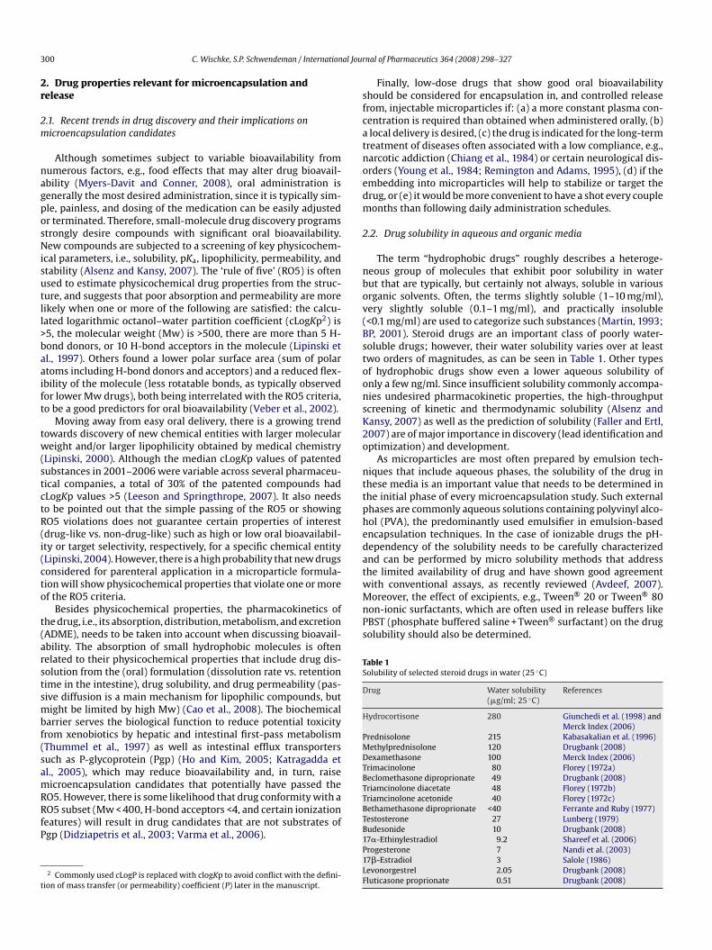
3 al Jour
2r
2m
nagposNisutll>baaift
tw(stctR(i(cto
t(arstsmbf(samRRfP
t
sfcatnoedm
2
nbov(BstoonsK2o
nttphedatwith conventional assays, as recently reviewed (Avdeef, 2007).Moreover, the effect of excipients, e.g., Tween® 20 or Tween® 80non-ionic surfactants, which are often used in release buffers likePBST (phosphate buffered saline + Tween® surfactant) on the drugsolubility should also be determined.
Table 1Solubility of selected steroid drugs in water (25 ◦C)
Drug Water solubility(�g/ml; 25 ◦C)
References
Hydrocortisone 280 Giunchedi et al. (1998) andMerck Index (2006)
Prednisolone 215 Kabasakalian et al. (1996)Methylprednisolone 120 Drugbank (2008)Dexamethasone 100 Merck Index (2006)Trimacinolone 80 Florey (1972a)Beclomethasone diproprionate 49 Drugbank (2008)Triamcinolone diacetate 48 Florey (1972b)
00 C. Wischke, S.P. Schwendeman / Internation
. Drug properties relevant for microencapsulation andelease
.1. Recent trends in drug discovery and their implications onicroencapsulation candidates
Although sometimes subject to variable bioavailability fromumerous factors, e.g., food effects that may alter drug bioavail-bility (Myers-Davit and Conner, 2008), oral administration isenerally the most desired administration, since it is typically sim-le, painless, and dosing of the medication can be easily adjustedr terminated. Therefore, small-molecule drug discovery programstrongly desire compounds with significant oral bioavailability.ew compounds are subjected to a screening of key physicochem-
cal parameters, i.e., solubility, pKa, lipophilicity, permeability, andtability (Alsenz and Kansy, 2007). The ‘rule of five’ (RO5) is oftensed to estimate physicochemical drug properties from the struc-ure, and suggests that poor absorption and permeability are moreikely when one or more of the following are satisfied: the calcu-ated logarithmic octanol–water partition coefficient (cLogKp2) is5, the molecular weight (Mw) is >500, there are more than 5 H-ond donors, or 10 H-bond acceptors in the molecule (Lipinski etl., 1997). Others found a lower polar surface area (sum of polartoms including H-bond donors and acceptors) and a reduced flex-bility of the molecule (less rotatable bonds, as typically observedor lower Mw drugs), both being interrelated with the RO5 criteria,o be a good predictors for oral bioavailability (Veber et al., 2002).
Moving away from easy oral delivery, there is a growing trendowards discovery of new chemical entities with larger moleculareight and/or larger lipophilicity obtained by medical chemistry
Lipinski, 2000). Although the median cLogKp values of patentedubstances in 2001–2006 were variable across several pharmaceu-ical companies, a total of 30% of the patented compounds hadLogKp values >5 (Leeson and Springthrope, 2007). It also needso be pointed out that the simple passing of the RO5 or showingO5 violations does not guarantee certain properties of interestdrug-like vs. non-drug-like) such as high or low oral bioavailabil-ty or target selectivity, respectively, for a specific chemical entityLipinski, 2004). However, there is a high probability that new drugsonsidered for parenteral application in a microparticle formula-ion will show physicochemical properties that violate one or moref the RO5 criteria.
Besides physicochemical properties, the pharmacokinetics ofhe drug, i.e., its absorption, distribution, metabolism, and excretionADME), needs to be taken into account when discussing bioavail-bility. The absorption of small hydrophobic molecules is oftenelated to their physicochemical properties that include drug dis-olution from the (oral) formulation (dissolution rate vs. retentionime in the intestine), drug solubility, and drug permeability (pas-ive diffusion is a main mechanism for lipophilic compounds, butight be limited by high Mw) (Cao et al., 2008). The biochemical
arrier serves the biological function to reduce potential toxicityrom xenobiotics by hepatic and intestinal first-pass metabolismThummel et al., 1997) as well as intestinal efflux transportersuch as P-glycoprotein (Pgp) (Ho and Kim, 2005; Katragadda etl., 2005), which may reduce bioavailability and, in turn, raiseicroencapsulation candidates that potentially have passed the
O5. However, there is some likelihood that drug conformity with aO5 subset (Mw < 400, H-bond acceptors <4, and certain ionization
eatures) will result in drug candidates that are not substrates ofgp (Didziapetris et al., 2003; Varma et al., 2006).
2 Commonly used cLogP is replaced with clogKp to avoid conflict with the defini-ion of mass transfer (or permeability) coefficient (P) later in the manuscript.
TBTB1P1LF
nal of Pharmaceutics 364 (2008) 298–327
Finally, low-dose drugs that show good oral bioavailabilityhould be considered for encapsulation in, and controlled releaserom, injectable microparticles if: (a) a more constant plasma con-entration is required than obtained when administered orally, (b)local delivery is desired, (c) the drug is indicated for the long-term
reatment of diseases often associated with a low compliance, e.g.,arcotic addiction (Chiang et al., 1984) or certain neurological dis-rders (Young et al., 1984; Remington and Adams, 1995), (d) if thembedding into microparticles will help to stabilize or target therug, or (e) it would be more convenient to have a shot every coupleonths than following daily administration schedules.
.2. Drug solubility in aqueous and organic media
The term “hydrophobic drugs” roughly describes a heteroge-eous group of molecules that exhibit poor solubility in waterut that are typically, but certainly not always, soluble in variousrganic solvents. Often, the terms slightly soluble (1–10 mg/ml),ery slightly soluble (0.1–1 mg/ml), and practically insoluble<0.1 mg/ml) are used to categorize such substances (Martin, 1993;P, 2001). Steroid drugs are an important class of poorly water-oluble drugs; however, their water solubility varies over at leastwo orders of magnitudes, as can be seen in Table 1. Other typesf hydrophobic drugs show even a lower aqueous solubility ofnly a few ng/ml. Since insufficient solubility commonly accompa-ies undesired pharmacokinetic properties, the high-throughputcreening of kinetic and thermodynamic solubility (Alsenz andansy, 2007) as well as the prediction of solubility (Faller and Ertl,007) are of major importance in discovery (lead identification andptimization) and development.
As microparticles are most often prepared by emulsion tech-iques that include aqueous phases, the solubility of the drug inhese media is an important value that needs to be determined inhe initial phase of every microencapsulation study. Such externalhases are commonly aqueous solutions containing polyvinyl alco-ol (PVA), the predominantly used emulsifier in emulsion-basedncapsulation techniques. In the case of ionizable drugs the pH-ependency of the solubility needs to be carefully characterizednd can be performed by micro solubility methods that addresshe limited availability of drug and have shown good agreement
riamcinolone acetonide 40 Florey (1972c)ethamethasone diproprionate <40 Ferrante and Ruby (1977)estosterone 27 Lunberg (1979)udesonide 10 Drugbank (2008)7�-Ethinylestradiol 9.2 Shareef et al. (2006)rogesterone 7 Nandi et al. (2003)7�-Estradiol 3 Salole (1986)evonorgestrel 2.05 Drugbank (2008)luticasone proprionate 0.51 Drugbank (2008)
C. Wischke, S.P. Schwendeman / International Journal of Pharmaceutics 364 (2008) 298–327 301
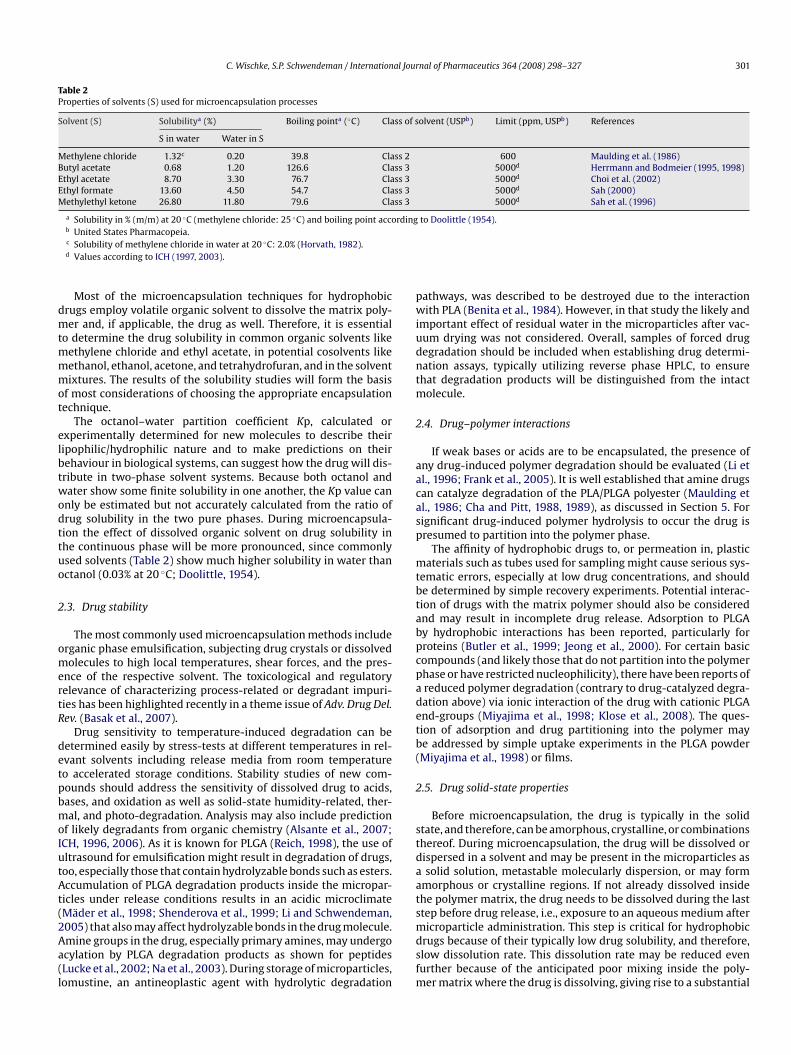
Table 2Properties of solvents (S) used for microencapsulation processes
Solvent (S) Solubilitya (%) Boiling pointa (◦C) Class of solvent (USPb) Limit (ppm, USPb) References
S in water Water in S
Methylene chloride 1.32c 0.20 39.8 Class 2 600 Maulding et al. (1986)Butyl acetate 0.68 1.20 126.6 Class 3 5000d Herrmann and Bodmeier (1995, 1998)Ethyl acetate 8.70 3.30 76.7 Class 3 5000d Choi et al. (2002)Ethyl formate 13.60 4.50 54.7 Class 3 5000d Sah (2000)Methylethyl ketone 26.80 11.80 79.6 Class 3 5000d Sah et al. (1996)
a Solubility in % (m/m) at 20 ◦C (methylene chloride: 25 ◦C) and boiling point according to Doolittle (1954).
dmtmmmot
elbtwodttuo
2
omertR
detpbmoIutAt(2Aa(l
pwiudntm
2
aacasp
mtbtabpcpadetb(
2
stdaats
b United States Pharmacopeia.c Solubility of methylene chloride in water at 20 ◦C: 2.0% (Horvath, 1982).d Values according to ICH (1997, 2003).
Most of the microencapsulation techniques for hydrophobicrugs employ volatile organic solvent to dissolve the matrix poly-er and, if applicable, the drug as well. Therefore, it is essential
o determine the drug solubility in common organic solvents likeethylene chloride and ethyl acetate, in potential cosolvents likeethanol, ethanol, acetone, and tetrahydrofuran, and in the solventixtures. The results of the solubility studies will form the basis
f most considerations of choosing the appropriate encapsulationechnique.
The octanol–water partition coefficient Kp, calculated orxperimentally determined for new molecules to describe theiripophilic/hydrophilic nature and to make predictions on theirehaviour in biological systems, can suggest how the drug will dis-ribute in two-phase solvent systems. Because both octanol andater show some finite solubility in one another, the Kp value cannly be estimated but not accurately calculated from the ratio ofrug solubility in the two pure phases. During microencapsula-ion the effect of dissolved organic solvent on drug solubility inhe continuous phase will be more pronounced, since commonlysed solvents (Table 2) show much higher solubility in water thanctanol (0.03% at 20 ◦C; Doolittle, 1954).
.3. Drug stability
The most commonly used microencapsulation methods includerganic phase emulsification, subjecting drug crystals or dissolvedolecules to high local temperatures, shear forces, and the pres-
nce of the respective solvent. The toxicological and regulatoryelevance of characterizing process-related or degradant impuri-ies has been highlighted recently in a theme issue of Adv. Drug Del.ev. (Basak et al., 2007).
Drug sensitivity to temperature-induced degradation can beetermined easily by stress-tests at different temperatures in rel-vant solvents including release media from room temperatureo accelerated storage conditions. Stability studies of new com-ounds should address the sensitivity of dissolved drug to acids,ases, and oxidation as well as solid-state humidity-related, ther-al, and photo-degradation. Analysis may also include prediction
f likely degradants from organic chemistry (Alsante et al., 2007;CH, 1996, 2006). As it is known for PLGA (Reich, 1998), the use ofltrasound for emulsification might result in degradation of drugs,oo, especially those that contain hydrolyzable bonds such as esters.ccumulation of PLGA degradation products inside the micropar-
icles under release conditions results in an acidic microclimateMäder et al., 1998; Shenderova et al., 1999; Li and Schwendeman,
005) that also may affect hydrolyzable bonds in the drug molecule.mine groups in the drug, especially primary amines, may undergocylation by PLGA degradation products as shown for peptidesLucke et al., 2002; Na et al., 2003). During storage of microparticles,omustine, an antineoplastic agent with hydrolytic degradationmdsfm
athways, was described to be destroyed due to the interactionith PLA (Benita et al., 1984). However, in that study the likely and
mportant effect of residual water in the microparticles after vac-um drying was not considered. Overall, samples of forced drugegradation should be included when establishing drug determi-ation assays, typically utilizing reverse phase HPLC, to ensurehat degradation products will be distinguished from the intact
olecule.
.4. Drug–polymer interactions
If weak bases or acids are to be encapsulated, the presence ofny drug-induced polymer degradation should be evaluated (Li etl., 1996; Frank et al., 2005). It is well established that amine drugsan catalyze degradation of the PLA/PLGA polyester (Maulding etl., 1986; Cha and Pitt, 1988, 1989), as discussed in Section 5. Forignificant drug-induced polymer hydrolysis to occur the drug isresumed to partition into the polymer phase.
The affinity of hydrophobic drugs to, or permeation in, plasticaterials such as tubes used for sampling might cause serious sys-
ematic errors, especially at low drug concentrations, and shoulde determined by simple recovery experiments. Potential interac-ion of drugs with the matrix polymer should also be considerednd may result in incomplete drug release. Adsorption to PLGAy hydrophobic interactions has been reported, particularly forroteins (Butler et al., 1999; Jeong et al., 2000). For certain basicompounds (and likely those that do not partition into the polymerhase or have restricted nucleophilicity), there have been reports ofreduced polymer degradation (contrary to drug-catalyzed degra-ation above) via ionic interaction of the drug with cationic PLGAnd-groups (Miyajima et al., 1998; Klose et al., 2008). The ques-ion of adsorption and drug partitioning into the polymer maye addressed by simple uptake experiments in the PLGA powderMiyajima et al., 1998) or films.
.5. Drug solid-state properties
Before microencapsulation, the drug is typically in the solidtate, and therefore, can be amorphous, crystalline, or combinationshereof. During microencapsulation, the drug will be dissolved orispersed in a solvent and may be present in the microparticles assolid solution, metastable molecularly dispersion, or may form
morphous or crystalline regions. If not already dissolved insidehe polymer matrix, the drug needs to be dissolved during the lasttep before drug release, i.e., exposure to an aqueous medium after
icroparticle administration. This step is critical for hydrophobicrugs because of their typically low drug solubility, and therefore,low dissolution rate. This dissolution rate may be reduced evenurther because of the anticipated poor mixing inside the poly-
er matrix where the drug is dissolving, giving rise to a substantial
3 al Jour
uptbTsarpsoemaib
bmljtmtiRst
3
3
ipsrpciepfauivtooa1tmsaMaDWtBO
(a
1mtpo
laivmhme
3
sc4pcloe1oMrdeceaasv
sDcAitidaealbaH
bt
02 C. Wischke, S.P. Schwendeman / Internation
nstirred boundary layer for diffusion out of the microparticle. Drugroperties that may affect its dissolution in aqueous media fromhe crystalline state include the wettability of a crystal, the sta-ility of the crystal structure (heat of fusion), or the surface area.he initial characteristics of the employed drug material may beubjected to alterations if the drug is dissolved, at least partially,nd precipitates during the encapsulation procedure due to solventemoval. Drug polymorphism may become a serious problem if theolymorphs show strong differences in, e.g., solubility, and conver-ion to another form occurs during microencapsulation, storage,r under release conditions. Therefore, if solid drug is going to bencapsulated, classically the thermodynamically most stable poly-orph is preferred for pharmaceutical development (Tong, 2008),
lthough efforts have been made to engineer crystals by form-ng co-crystals or metastable polymorphs with altered dissolutionehavior (Blagden et al., 2007).
When the drug is going to be encapsulated into the polymery codissolving both substances in an appropriate solvent, the for-ation of true or metastable molecular dispersions is possible for
ow drug loadings. The latter state is undesired as it may be sub-ected to crystallization during storage which might severely affecthe release properties of the formulation. Besides, due to limited
utual miscibility of certain drugs with the polymer, the state ofhe drug may depend on the loading of the particles, as it has beenntensively studied for progesterone (Benoit et al., 1984, 1986a,b;osilio et al., 1991; Benoit, 1996; Hill et al., 1998). Therefore, it istrongly recommended to characterize the state of the drug insidehe formulation by thermal analysis (Dubernet, 1995).
. Microencapsulation techniques for hydrophobic drugs
.1. o/w emulsion technique
As a considerable number of hydrophobic drugs are solublen various water-immiscible organic solvents and, of course, areoorly soluble in water, one of the simplest methods to encapsulateuch drugs is by the oil-in-water (o/w) emulsion/solvent evapo-ation technique. By this method, both drug and biodegradableolymer are first dissolved in a solvent, e.g., generally methylenehloride is most desirable, and then the resulting organic oil phases emulsified in an aqueous solution containing an appropriatemulsifier, the solution of which should have a low dissolvingower for the drug. In general, volatile solvents can be removedrom such emulsions by evaporation to a gas phase (Vranckennd Claeys, 1970a) or in any case by extraction to the contin-ous phase (Vrancken and Claeys, 1970b; Albayrak, 2005). It is
mportant to point out that in the former case, the carrier sol-ent must first dissolve in the continuous phase before evaporationakes place (Wang and Schwendeman, 1999). The o/w method-logy has been applied for the encapsulation of a large numberf drugs, including: the neuroleptics thioridazine (Maulding etl., 1986; Fong et al., 1986), chlorpromazine (Suzuki and Price,985), and bromperidol (Kino et al., 1997), different local anes-hetics (Wakiyama et al., 1981, 1982a,b; Nakano et al., 1984), the
inor tranquilizer diazepam (Bodmeier and McGinity, 1987a), theynthetic opioid l-methadone (Cha and Pitt, 1988), the anticancergents aclarubicin (Wada et al., 1988; Yoshikawa et al., 1989;uranishi et al., 1991), lomustine (Benita et al., 1984; Benoit et
l., 1984; Bissery et al., 1984), and paclitaxel (Burt et al., 1995;
emetrick et al., 1997; Liggins et al., 2000; Liggins and Burt, 2001;ang et al., 1996, 1997; Gupte and Ciftci, 2004; Xie et al., 2007),he gestagens progesterone (Benita et al., 1984; Benoit et al., 1984;odmeier and McGinity, 1987a; Rosilio et al., 1991; Yang andwusu-Ababio, 2000) and, at elevated temperature, levonorgestrel
osacA
nal of Pharmaceutics 364 (2008) 298–327
Beck et al., 1985), and the glucocorticoid dexamethasone (Thote etl., 2005).
For very high drug loading, e.g., 50% progesterone (Hill et al.,998) or 70% testosterone propionate (Tice and Gilley, 1985) in PLAicroparticles, crystallization of the drug has been observed during
he solvent removal, which even may result in perforation of thearticle wall by drug needles. The necessity to characterize the statef the drug inside the polymer has been emphasized in Section 2.3.
However, for drugs that do not show a high solubility in methy-ene chloride, e.g., the estrogen �-estradiol (Birnbaum et al., 2000),n alternative carrier solvent (i.e., solvent for the polymer and,n this case, the drug) may be considered. Table 2 lists some sol-ents that had been suggested to replace methylene chloride inicroencapsulation processes but have not found general use for
ydrophobic drugs. Alternatively, a cosolvent may be added toethylene chloride, or a different encapsulation technique may be
mployed.
.2. s/o/w technique
If the specific drug cannot be dissolved in a carrier solvent orolvent mixture, or extensive drug loss to the continuous phaseannot be avoided when employing cosolvent systems (see Section), then the s/o/w technique is usually used. With this method areliminary formulation should be available for in vivo proof ofoncept studies in an appropriate time and the formulation can beater subjected to optimization. The majority of very early papersn hydrophobic drug encapsulation employed the s/o/w technique,.g., for norethisterone as a contraceptive (Beck et al., 1979, 1980,981, 1983b; Cowsar et al., 1985; Cong and Beck, 1991) and multiplether drugs (Fong et al., 1986; Cavalier et al., 1986; Bodmeier andcGinity, 1987a; Tsakala et al., 1988; Gu et al., 1992). In the more
ecent literature, the s/o/w method was evaluated for hydrophobicrugs like levonorgestrel (Wang et al., 2005), �-estradiol (Birnbaumt al., 2000; Mogi et al., 2000), haloperidol (Kino et al., 1997), orampthecin and its derivatives (Shenderova et al., 1997, 1999; Ertlt al., 1999). Due to a low but distinct solubility of certain activegents in the organic solvent, a certain portion of the drug mightlso be in solution in s/o/w formulations. This is one reason to avoidtorage of drug suspensions in the polymer phase, as crystal growthia Ostwald ripening may occur.
However, the s/o/w method requires a very low drug particleize in order to allow a complete encapsulation of the drug crystals.ue to the limited availability of micronized drug in the preclini-al phases such material might need to prepared on a lab-scale.number of drug properties including the hardness and elastic-
ty of the drug crystals, the melting point, the hygroscopicity, andhe sensitivity to thermal or other decomposition reactions willmpact the selection of a successful method. For non-hygroscopicrugs a simple set-up with a smooth mortar and pestle might beppropriate to grind the drug and cooling the mortar on dry-ice willven increase the efficiency due to a higher brittleness of the drugt lower temperature. With this cryogenic grinding, drug particlesess than 1–10 �m can often be obtained. Hygroscopic drugs shoulde subjected to grinding in a controlled atmosphere, such as insiden air-tight grinding jar of a ball mill with liquid nitrogen cooling.owever, a lower recovery is typical for such mills.
Besides the necessity of small-sized drug material, other draw-acks of the s/o/w technique might be the tendency of the drugo show sedimentation (higher density than suspension medium)
r flotation (caused by adhesion of gas bubbles to the hydrophobicurface due to low wettability) during the encapsulation processnd, in the later stages of the product development, difficultiesan also be expected during scaling up to large-scale manufacture.lterations, which might result from changes in the drug synthe-
al Jour
seomadlrgasc
maiitobhsac
3
soTetdot(1ba1t2b2Prte
3
ofdaaocs1fita
daspciettadsm
3
itptaaFietostsbssis1tPZ
tvpcmtao
aPoJep1tta
C. Wischke, S.P. Schwendeman / Internation
is, e.g., in the drug crystal structure or the wetting behavior, arexpected to affect the release profile from s/o/w particles. More-ver, differences in the release might appear compared to denseicrospheres that were prepared by the o/w technique and showhomogeneous drug distribution. Especially if comparatively largerug material is incorporated, the presence of sparsely encapsu-
ated drug crystals at the microparticle surface can increase burstelease (Birnbaum et al., 2000). Therefore, some authors have sug-ested an extra coating step for s/o/w microparticles by slowlydding a polymer/chloroform solution to preformed microparticlesuspended in 5% aqueous ethanol at 54 ◦C (Cong and Beck, 1991) (foroating procedures see also Section 3.7).
Following the dissolution of crystals in the vicinity of the poly-er surface, large voids in the surface of the microparticles may
ppear, resulting in a faster mass transfer of the dissolution mediumnto the particles. Thus, the medium may access the whole payloadf the drug crystals are not separated and homogeneously dispersedhroughout the microparticles. Also, monolithic particles from, e.g.,/w techniques that have a uniform distribution of hydropho-ic drug in the matrix can be expected to show higher matrixydrophobicity and potentially lower water-uptake compared to/o/w particles with larger crystals at similar drug loading. Bothspects may lead to faster release profiles from s/o/w microparti-les.
.3. o/o method
Although being classified as hydrophobic drugs, substancesuch as hydrocortisone exhibit an appreciable solubility in aque-us media (280 �g/ml water) like the external water phase (seeable 1). Therefore, o/w methods are expected to result in lowncapsulation efficiencies due to a flux of the active agent fromhe dispersed phase to the larger volume of the continuous phaseuring the encapsulation process. In order to overcome this issue,1/o2 emulsion methods can be used. They include the extraction ofhe o1-phase solvent, e.g., acetonitrile, by a solution of an emulsifierHLB typically <8; Jalil and Nixon, 1990a; Herrmann and Bodmeier,998) in oil, e.g., cottonseed oil or mineral oil (acetonitrile solu-ility in cottonseed oil ∼10%; Leach et al., 2005), which should benon-solvent for both the polymer and the drug (Jalil and Nixon,
989, 1990a,b,c; Wada et al., 1990). The s/o/o technique combineshe concepts of s/o/w and o/o methodologies (Janoria and Mitra,007). Also an o1/o2/o3 technique has been described that maye applicable for certain hydrophobic drugs (Herrero-Vanell et al.,000), which are soluble in fluorosilicone oil (o1-phase) but not inLGA solvents like acetone (o2-phase). However, for methods car-ied out in oil the removal of the continuous phase requires a specialreatment, e.g., washing of the particles with hexane or petroleumther.
.4. w/o/w method
Although initially afflicted with low encapsulation efficiencyf hydrophilic molecules unless w1-phase solidification was per-ormed (Okada et al., 1987), the w1/o/w2 method has beenescribed to result in extremely efficient loading of biodegrad-ble microparticles with water-soluble compounds (Yamamoto etl., 1994) and is currently one of the most commonly used meth-ds for peptide and protein encapsulation. Compared to w/o-basedoacervation techniques that were being developed at about the
ame time for hydrophilic drug encapsulation into PLGA (Kent et al.,986), the w/o/w method was described to overcome the issues ofnal oil removal by several washing steps and the tendency to par-icle aggregation during the preparation procedure (Yamamoto etl., 1994). In the literature there are a few cases where hydrophobicwieLe
nal of Pharmaceutics 364 (2008) 298–327 303
rugs are stated to be encapsulated by a w/o/w method. However,closer look at these methods revealed that either the drug was
uspended in the inner water phase in a (s + w)/o/w complex dis-ersed system (Giunchedi et al., 1998) or solution of the activeompounds, e.g., a mixture of ethinylestradiol and levonorgestrel,n an ethanol/water mixture was used as the w1-phase (Dhanarajut al., 2003, 2004, 2006). The addition of some inner water phase inhe (s + w)/o/w technique is expected to increase porosity in the par-icle core, which may influence drug release. However, employingn ethanolic drug solution as the w1-phase is expected to result inrug precipitation in w1 due to mass transfer of ethanol (includingome drug) from w1 through the o-phase to the w2-phase, whichay lower the encapsulation efficiency.
.5. In situ forming microparticles
In situ forming depot systems, first designed as implants, werentended to overcome some drawbacks of conventional formula-ions such as reducing manufacturing costs and complexity andain on injection through larger needles. They should be adminis-ered simply by injecting a polymer/drug solution or suspension inn appropriate solvent that would precipitate and form the implantt the injection site (Dunn et al., 1990, 1994, 2003; Tipton andujita, 1991; Shah et al., 1993), a concept that has been employedn FDA-approved LAR® products (Eligard® with the Atrigel® deliv-ry system). Solvent partitioning into the tissue has been reportedo cause mild transient burning at the injection side in about 20%f the cases (AGL9909 study, 2000). Besides Atrigel®, that usesolutions of PLGA in water-miscible solvents (e.g., NMP, DMSO),here are other methods for in situ precipitating PLGA implantsuch as Alzamer® (using partially water-miscable solvents like ethylenzoate and triacetin) (Brodbeck et al., 2000) and the Saber®
ystem (PLGA dissolved in sucrose acetate isobutyrate, a viscousugar derivative, plus some organic solvent for easier injectabil-ty) (Tipton, 1999; Burns et al., 2000). There are also numeroustrategies that employ matrices other than PLGA (Dittgen et al.,998; Hatefi and Amsden, 2002; Packhaeuser et al., 2004), e.g.,hermosensitive systems such as ReGel® low molecular weightLGA–PEG–PLGA block copolymer micelles (Rathi et al., 2001;entner et al., 2001).
Soon thereafter, the idea of in situ forming depots was appliedo microparticles in order to overcome issues associated with con-entional microparticle formulations such as the cost-intensivereparation, drying, and potentially difficult resuspension. Also, inontrast to in situ implants, in situ microparticles showed loweryotoxicity (Kranz et al., 2001), easier injectability depending on
he type of solvent and suspension medium (Im-Emsap, 2002), andmore reproducible surface area as opposed to the irregular shapebtained from such implants.
Starting with relatively large particles prepared by droppingliquots of drug + PLGA/NMP into aqueous medium (Lambert andeck, 1995), the formulations became injectable when preformed/o emulsions stored until administration (Jain et al., 2000a,b,c;ain, 2000), or two-compartment systems (syringes attached toach other with a syringe connector) with its content being dis-ersed at the bedside to form o/w or o/o emulsions (Bodmeier,998a; Voigt, 2006) were investigated. After injection, the par-itioning of the biocompatible solvent (Royals et al., 1999) intohe tissue causes the hardening of the emulsion droplets in vivond is also responsible for a high burst release typically observed
ith such formulations (Jain et al., 2000c). Most authors havencorporated hydrophilic substances using o/o emulsions (Jaint al., 2000a,b,c; Kranz et al., 2001; Kranz and Bodmeier, 2007;uan and Bodmeier, 2006a,b), and the external oil phase can bexpected to act as a diffusion barrier that decreases the burst
3 al Jour
r2
P(ea2ftaals(eefdat(set2woofa
lepodotcpmps
3
miatipswwm1mct
bf
psrafieshG
3
d(spms1tccgoTtrEo
ti1iaez(bdMlvaassmr
3
teSst
04 C. Wischke, S.P. Schwendeman / Internation
elease depending on its volume and viscosity (Luan and Bodmeier,006a).
However, safety issues will limit the type of oil that can be used.araffin/mineral oils are known to cause severe lipoid pneumoniaPerings et al., 2001; Simmons et al., 2007), and thus their pres-nce in preparations for injection must be excluded. Vegetable oilsre allowed as nonaqueous vehicles by the USP and Ph. Eur. (USP,007; Ph. Eur., 2002a) and peanut oil has been suggested by dif-erent pharmacopoeiae as a standard oil for injections. Althoughhe extraction procedure of Olea Herbaria is designed to reducellergenic protein impurities (Taylor et al., 1981; Ph. Eur., 2002b)nd, e.g., peanut oil is presently used in several injectable formu-ations of steroids and other drugs, it must be recognized that ateadily rising number (>1%) of the children in the US and EuropeSicherer and Sampson, 2007; Savage et al., 2007; Green et al., 2007)xhibit allergic symptoms with numerous peanut proteins (Bernardt al., 2007) and injection of contaminated peanut oil might causeatal anaphylaxis. Since the standard refining procedure, thermalenaturation, might not always eliminate allergenicity (Burks etl., 1992), “peanut products should be treated as allergenic unlesshey have an analytically monitored non-allergenic specification”EMEA, 2004). Also, allergies are well-known for alternative oilsuch as sesame (Gangur et al., 2005), almond, and other oils (Rouxt al., 2003). Medium chain triglycerides (MCT), as derived fromhe kernel of the Coconut Palm and the African Oil Palm (Ph. Eur.,002c) and used in emulsions for parenteral nutrition in mixtureith soybean oil (Discoll, 2006; USP, 2006), might, under exclusion
f allergies of the respective patient, be an alternative nonaqueous2-phase for in situ formulations. However, in situ microparticleormation using MTC was limited to DMSO and propylene carbon-te as o1-phase solvent (Luan, 2006).
For hydrophobic drugs like indomethacin, o/w in situ formu-ations with water-immiscible solvents were reported, and, asxpected, showed burst releases of up to 50% depending on theolymer concentration (Im-Emsap, 2002). Again, higher viscositiesf the continuous water phase should reduce the diffusion of therug during the particle hardening, but will impact the injectabilityf the emulsion. This issue can be overcome by adding substanceso the water phase that change their viscosity when the ambientonditions are altered after injection into the tissue, e.g., by tem-erature or pH-dependent gelation (Bodmeier, 1998b, 2002). Inyotoxicity studies o/w in situ formulations showed the best com-
atibility when ethyl acetate was employed as the drug/polymerolvent (Rungseevijitprapa et al., 2008).
.6. Salting out/phase separation
Salting out is a method to precipitate dissolved polymers, i.e.,ost commonly proteins, by attracting water molecules to the salt
ons and therefore decreasing the number of water molecules avail-ble for solvation of the polymer (simple coacervation). Althoughhe term “salting out” might be misunderstood in case of water-nsoluble polymers, the described method utilizes the controlledrecipitation of PLGA from an organic phase of a water-miscibleolvent while emulsified in a viscous PVA/salt solution. By addingater to the system, the o-phase solvent is slowly extracted,hereas the polymer is unable to follow the solvent and formsicroparticles rather than nanoparticles (Rafler and Jobmann,
997). However, this process seems to require a careful opti-ization of certain process parameters, e.g., the salt type and
oncentration, the type of polymer and solvent, and the ratios ofhese compounds in order to obtain microparticles at all.
Microparticle preparation by organic phase separation cane a temperature-induced process, but has mostly been per-ormed by adding a coacervation agent (ternary system of
tacf(
nal of Pharmaceutics 364 (2008) 298–327
olymer + solvent + nonsolvent or second polymer) to a suspen-ion or emulsion of a drug in a PLGA solution and solidifying theesulting liquid coacervate capsule in a hardening bath (Kent etl., 1986; Thomasin et al., 1998a,b). Some of the steps can be per-ormed at a reduced temperature (Fong, 1979), e.g., the hardeningn hexane cooled to −70 ◦C by a dry-ice/isopropanol mixture (Lapkat al., 1986). Such methods are commonly employed for water-oluble compounds (Nihant et al., 1995; Thomasin et al., 1997), butave been used for hydrophobic drugs, too (Nuwayser et al., 1977;ardner et al., 1977; Leelarasamee et al., 1986).
.7. Melting techniques
Melting techniques represent another strategy to encapsulaterugs into biodegradable polymers which, with some exceptionsYolles et al., 1975; Yolles and Sartori, 1980), avoid the use of organicolvents but require the dispersion or melting of the drug in aolymer melt. In order to form microparticles the hot melt of theatrix polymer 1 can be dispersed in a second, molten, water-
oluble polymer 2, which is immiscible with the matrix polymer. Then, the resulting emulsion will be solidified by cooling, andhe polymer 1 microparticles will be collected after dissolving theontinuous phase polymer 2 in water (Chenite et al., 2002). Moreommonly, the drug/matrix polymer melt is cooled down and thenround (Boswell and Scribner, 1973; Smith and Hunnyball, 1986)r jet-milled (Nykamp et al., 2002) to form non-spherical particles.o allow an easier grinding of otherwise unmanageable lumps ofhe congealed melt, it may be advantageous to extrude the mate-ial before complete solidification, especially for large batch sizes.xtrusion prior to grinding has also been used for the preparationf microparticles from pre-casted films (Gresser et al., 1978).
If spherical particles and a smaller size distribution are desired,he ground melt can be emulsified in a hot solution contain-ng emulsifier (Wichert and Rohdewald, 1990) or a hot gel (Ruiz,992). Another approach to smooth the surface and prolong then vitro release from ground, norethisterone-loaded poly-l-lacticcid microparticles was suggested (Anderson et al., 1976) thatmploys a coating of the particles with poly-d,l-lactic acid in ben-ene, a solvent avoided nowadays because of its carcinogenicityfor coating procedures see also Section 3.2). Also, clear draw-acks of the melting technique are the thermal treatment of therug and the multitude of steps to obtain smooth microparticles.oreover, the fear of residual solvents might be exaggerated, since
yophilization was shown to reduce solvent impurities to a safealue for an emulsion-based preparation technique (Wischke etl., 2006) and numerous commercial microparticle formulationsre prepared with toxic carrier solvents, but have met regulatorytandards. Lastly, it should be pointed out that melt-based encap-ulation methods commonly develop highly nonporous polymeratrices (Zhou et al., 1998), which can lead to undesirably slow
elease profiles especially for hydrophobic drugs.
.8. Methods using supercritical fluids (SCF)
Substances become supercritical fluids (SCF) when placed aboveheir critical point (i.e., T > Tc and p > pc). SCF exhibit the flow prop-rties of a gas (low viscosity) and the dissolving power of a liquid.CF can easily penetrate through materials because they do nothow any surface tension, and their solvent power is related toheir density, which experiences large changes in the vicinity of
he critical point, and can be controlled by altering temperaturend/or pressure (Williams et al., 2002). SCF have a variety of appli-ations including their suitability for the extraction of substancesrom a large variety of materials, e.g., essential oils from plantsPourmortazavi and Hajimirsadeghi, 2007). Most commonly CO2
al Jour
ienet
ef(sitfwpmsp(e1iebpaea
euvapSatdb
3
MahtttSCButvestbt
3
t
dttmoctowaccddumlt
3
umc
fwcpiFewbomp
4
4
tohsluactr
4
ec
C. Wischke, S.P. Schwendeman / Internation
s used for such purposes, as it has a low critical point and is anasily accessible, environment-safe gas. There are at least two tech-iques for the particle design by SCF (Jung and Perrut, 2001; Gintyt al., 2005), which can be roughly differentiated by their concepto dissolve and precipitate the polymer and the drug.
In two common scenarios the drug and matrix polymer might beither dissolved or melted in the SCF and afterwards form particlesollowing the rapid expansion from supercritical solution (RESS)Kim et al., 1996) or precipitate into particles from the gas-saturatedolutions/suspensions (PGSS) (Whitaker et al., 2005) after spray-ng the melt and releasing the gas, respectively. By contrast, otherechniques rely on the antisolvent properties of SCF for PLGA, aact that has limited the usage of RESS and PGSS to low moleculareight PLA. Different antisolvent methods are known, for exam-le, those which differ by the geometry of the nozzle used. Theseethods follow the concept of spraying the organic drug/polymer
olution into a SCF, which extracts the organic solvent. Antisolventrotocols include particle precipitation by compressed antisolventsPCA) (Falk et al., 1997; Martin et al., 2002), the aerosol solventxtraction system (ASES) (Bleich et al., 1994; Bleich and Müller,996), and the solution-enhanced dispersion by supercritical flu-ds (SEDS) (Ghaderi et al., 2000). Recently the supercritical fluidxtraction of emulsion (SFEE), i.e., a classical o/w emulsion, haseen described to reduce the time of solvent removal and polymerrecipitation (Chattapadhyay et al., 2006). Methodological details,pplications, and drawbacks of different SCF techniques for drugncapsulation have been noted in some current reviews (Tewes etl., 2006; Mishima, 2008; Davies et al., 2008).
It is obvious that the methods employing SCF require specialquipment and that these techniques are therefore, not widelysed on the bench scale. Both the fast extraction of the organic sol-ent and the partitioning of the SCF into the polymer beads, whichfterwards expands during the decompression, may induce a highorosity and a faster drug release. Also one should consider thatCF can dissolve some, but not all, hydrophobic drugs (Vatanara etl., 2005) and that the extracted o-phase solvent acts as a cosolventogether with the SCF, so that a reduced encapsulation efficiencyue to drug extraction from the polymer matrix has sometimeseen observed (Bleich and Müller, 1996).
.9. Spraying techniques
The preparation of microparticles by spray-drying (Gander anderkle, 1997) or a cryogenic spray-congealing method (also known
s Alkermes’ ProLease®) (Gombotz et al., 1991; Johnson et al., 1997)as been intensively studied for protein encapsulation in ordero improve the stability of these labile biomacromolecules (e.g.,o obviate protein denaturation at o/w interfaces, during solu-ion state micronization, and/or elevated temperature exposure).pray-drying is also useful for hydrophobic drugs (Bodmeier andhen, 1988; Pavanetto et al., 1993; Wagenaar and Müller, 1994;enelli et al., 1998; Mu and Feng, 2001; Mu et al., 2005), partic-larly for large-scale production of microparticles. For example,his microencapsulation method can overcome the issue of largeolumes of solvent-contaminated water phase that result frommulsion-based encapsulation methods. However, larger batchizes are typically required compared to the emulsion methods andherefore, spray-drying is often more problematic on the economicench scale or if very little drug is available in the early stages ofhe development of an experimental microparticle formulation.
.10. Ammonolysis
Recently, risperidone was encapsulated into PLGA micropar-icles by an o/w emulsion technique that employed methyl
tiMat
nal of Pharmaceutics 364 (2008) 298–327 305
ichloroacetate in place of the traditional volatile carrier solvento dissolve the polymer. By the addition of an ammonia solu-ion the solvent was hydrolyzed into water-miscible products, i.e.,
ethanol and dichloroacetamide, resulting in the precipitationf PLGA (Sah and Lee, 2006). Although the encapsulation effi-iency was almost 100% for risperidone, the flux of methanol fromhe microparticle core to the water phase may result in a lossf methanol-soluble drugs. This theory is supported by a study,here the encapsulation efficiency of progesterone was as much
s 15% lower when ammonolysis with methyl chloroacetate wasompared to a standard encapsulation method with methylenehloride (Kim et al., 2007). In the preliminary studies, methylichloroacetate resulted in particle aggregation during vacuumrying and neither release nor precise gas chromatographic resid-al solvent analysis were provided as methyl dichloroacetate andethyl chloroacetate (Pohanish and Greene, 1996; USCG, 1999),
ike numerous traditionally used solvents for encapsulation, areoxic.
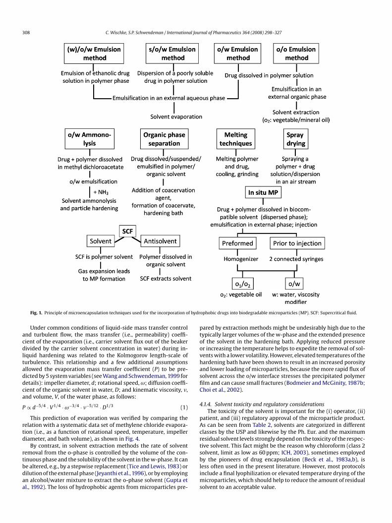
.11. Rationale to select an encapsulation technique
Overall, there are a variety of methods that already have beensed for the encapsulation of hydrophobic drugs in PLA/PLGAicroparticles. A summary of these procedures including their
hallenges is given in Figs. 1 and 2.However, for a pharmaceutical company the approval of the
ormulation for the market might be faster, easier, and cheaper,hen techniques similar to those of already available commer-
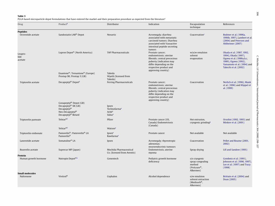
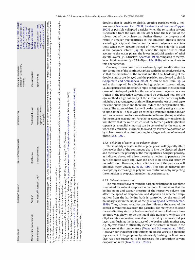
ial products are used. Table 3 shows PLGA-based microparticleroducts and provides information on the expected manufactur-
ng procedure which we have assumed from the patent literature.or the hydrophobic drugs, risperidone and naltrexone, o/w solventvaporation methods were described. Other substances, namelyater-soluble drug salts, peptides, or proteins, were encapsulatedy coacervation, double emulsion, or spraying techniques. As the/w and s/o/w methods are most commonly used for small-scaleicroencapsulation studies, the following sections will focus on
articles derived from these methods.
. Solvents/cosolvents
.1. Dispersed phase solvents
Organic solvents are used in emulsion-based microencapsula-ion techniques to dissolve the matrix polymer and, in the casef the o/w method, also the drug to be encapsulated. Often evenydrophobic drugs do not dissolve very well in the desirable carrierolvent, methylene chloride. Such drugs might be either encapsu-ated by the s/o/w technique or an alternative solvent might besed to prepare PLGA microparticles (Table 2). However, beside thebility of a solvent to dissolve both the polymer and the drug, otherharacteristics of the respective solvent need to be considered, sincehey commonly affect the size, morphology, drug release, or theesidual solvent of the microparticles.
.1.1. Solubility of polymer solvent in continuous phaseThe water solubility of the solvent will impact its initial
xtraction during microparticle preparation. In general, a fast pre-ipitation of the polymer due to the initial efflux of the solvent to
he external phase is considered to be advantageous for achiev-ng high encapsulation efficiencies (Bodmeier and McGinity, 1988;ao et al., 2008). However, if the solvent is too soluble in water,nd/or a large volume of water is used, very fast solidification ofhe polymer may occur, forming a dense polymer shell around the
306C.W
ischke,S.P.Schwendem
an/InternationalJournalofPharm
aceutics364
(2008)298–327
Table 3PLGA based microparticle depot formulations that have entered the market and their preparation procedure as expected from the literaturea
Drug Productb Distributor Indication Encapsulationtechnique
References
PeptidesOctreotide acetate Sandostatin LAR® Depot Novartis Acromegaly; diarrhea
associated with metastaticcarcinoid tumors; Diarrheaassociated with Vasoactiveintestinal peptide secretingtumors
Coacervationc Bodmer et al. (1996a,1996b, 1997), Lambert et al.(2004) and Petersen andAhlheimer (2007)
Leupro-lideacetate
Lupron Depot® (North America) TAP Pharmaceuticals Prostate cancer;endometriosis; uterinefibroids; central precociouspuberty (indication maydiffer depending on therespective product andapproving country)
w/o/w emulsionsolventevaporation
Okada et al. (1987, 1992,1994), Okada (1997),Ogawa et al. (1988a,b,c,1989), Ogawa (1992),Yamamoto et al. (1994) andTakechi et al. (2002)
Enantone®; Trenantone® (Europe) TakedaProstap SR; Prostap 3 (GB) Wyeth (licensed from
Takeda)Triptorelin acetate Decapeptyl® Depotd Ferring Pharmaceuticals Prostate cancer;
endometriosis; uterinefibroids; central precociouspuberty (indication maydiffer depending on therespective product andapproving country)
Coacervation Nerlich et al. (1996), Manket al. (1996) and Klippel etal. (1999)
Gonapeptyl® Depot (GB)Decapeptyl® SR (GB) IpsenDecapeptyl® Technofarmae
Neo-Decapeptyl® Achée
Decapeptyl® Retard Siduse
Triptorelin pamoate Telstar®f Pfizer Prostate cancer (US,Canada) Endometriosis(Canada)
Hot extrusion,cryogenic grindingg
Orsolini (1992, 1993) andMinkov et al. (2001)
Telstar®f Watsone
Triptorelin embonate Pamorelin®; Pamrorelin® LA Ipsene Prostate cancer Not available Not availablePamorelin® Rawfarmae
Lanreotide acetate Somatuline® LA Ipsen Acromegaly; thyreotropicadenomas;neuroendocrinic tumours
Coacervation Pellet and Roume (2001,2002)
Buserelin acetate Suprecur MP (Japan) Mochida PharmaceuticalCo. (licensed from Aventis)
Endometriosis, uterinemyoma
Spray-drying Lill and Sandow (1995)
ProteinHuman growth hormone Nutropin Depot®h Genentech Pediatric growth hormone
deficiencys/o cryogenicspray-congealingmethod(ProLease®,Alkermes)
Gombotz et al. (1991),Johnson et al. (1996, 1997),Lee et al. (1997) and Tracy(1998)
Small moleculesNaltrexone Vivitrol® Cephalon Alcohol dependence o/w emulsion
solvent extraction(Medisorb®,Alkermes)
Brittain et al. (2004) andDean (2005)
C. Wischke, S.P. Schwendeman / International JourR
isp
erid
one
Ris
per
dal
®C
onst
a®Ja
nss
enPh
arm
aceu
tica
lsSc
hiz
oph
ren
iao/
wem
uls
ion
solv
ent
extr
acti
on(M
edis
orb®
,A
lker
mes
)
Ric
key
etal
.(19
98),
Wri
ght
etal
.(20
01),
Mes
ens
etal
.(2
003
),R
amst
ack
etal
.(2
003
)an
dLy
ons
and
Wri
ght
(20
04,2
005
)M
inoc
ycli
ne
hyd
roch
lori
de
Are
stin
®O
raPh
arm
aPe
rid
onta
ldis
ease
s/o
coac
erva
tion
Law
ter
and
Lan
zilo
tti
(199
6)B
rom
ocri
ptin
ePa
rlod
elLA
®i ,
Parl
odel
LAR
®i
San
doz
(Nov
arti
s)Pa
rkin
son
dis
ease
;hy
per
pro
lact
inem
ia;
acro
meg
aly
Spra
y-d
ryin
gZd
enek
and
Kis
sel(
1986
,19
89),
Kis
sele
tal
.(19
91)
and
Mon
tin
iet
al.(
1993
)
aD
rug
app
rova
ldoc
um
ents
asav
aila
ble
onth
eh
omep
age
ofth
eFD
Avi
ah
ttp
://w
ww
.fda.
gov/
cder
/dru
g/d
efau
lt.h
tmd
on
otal
low
con
clu
sion
son
the
pre
par
atio
nte
chn
iqu
e,be
cau
seth
ere
spec
tive
par
agra
ph
sar
ew
ith
hel
d.
As
can
beex
pec
ted
from
the
cite
dre
fere
nce
s,th
est
ated
pre
par
atio
nm
eth
odat
leas
tin
itia
llyh
adbe
enu
sed
top
rod
uce
the
resp
ecti
vep
rod
uct
.How
ever
,in
mos
tca
ses
the
stat
eden
cap
sula
tion
tech
niq
ue
was
not
con
firm
edby
the
dis
trib
uto
ror
oth
erau
thor
itie
s.b
Occ
asio
nal
lyp
rod
uct
sar
eon
lyav
aila
ble
ince
rtai
nco
un
trie
sas
ind
icat
ed.
cO
lder
pat
ents
des
crib
eco
acer
vati
on,d
oubl
eem
uls
ion
,an
dsp
ray-
dry
ing
tech
niq
ues
.Rec
ent
pat
ents
focu
son
the
w/o
/wte
chn
iqu
e,bu
tst
ate
that
sili
con
eoi
lan
dh
epta
ne
are
imp
uri
ties
ofSa
nd
osta
tin
LAR
®,i
nd
icat
ing
the
usa
geof
aco
acer
vati
onte
chn
iqu
ein
the
app
rove
dp
rod
uct
.d
Prod
uct
nam
em
ayd
iffe
rin
cert
ain
cou
ntr
ies,
e.g.
,Dec
apep
tyl®
N3.
75m
gan
dD
ecap
epty
l®G
YN
(Ger
man
y).
ePr
esen
tli
cen
sees
ofD
ebio
ph
arm
(acc
ord
ing
toD
ebio
ph
arm
:h
ttp
://w
ww
.deb
iop
har
m.c
om/d
ecap
epty
l-tr
elst
ar-t
ript
orel
in.h
tml,
Febr
uar
y7t
h,2
008
).f
Ava
ilab
leas
Tels
tarTM
Dep
ot3.
75m
gan
dTe
lsta
rTMLA
11.2
5m
g.g
Stat
emen
tby
Min
kov
etal
.,20
01(“
pre
par
edw
ith
out
orga
nic
solv
ents
”)in
dic
ates
the
use
ofa
hot
extr
usi
onte
chn
iqu
eas
pat
ente
d(O
rsol
ini,
1992
,199
3).
hM
arke
tin
gst
opp
edin
2004
beca
use
ofec
onom
icre
ason
s.i
No
lon
ger
onth
em
arke
t.
dl2isrataaalt
psd(aictomtcvwfwcwbp
4
taappdet
4
ibaob1otpelelHrfe
nal of Pharmaceutics 364 (2008) 298–327 307
roplets that is unable to shrink, creating particles with a hol-ow core (Birnbaum et al., 2000; Birnbaum and Brannon-Peppas,003) or possibly collapsed particles when the remaining solvent
s extracted from the core. On the other hand the fast flux of theolvent out of the o-phase can further disrupt the droplets andesult in smaller microparticles as the emulsion droplets shrinkccordingly, a typical observation for lower polymer concentra-ions when ethyl acetate instead of methylene chloride is useds the polymer solvent (Fig. 3). Beside the higher flux of ethylcetate to the water phase, the lower interfacial tension of ethylcetate–water (� = 6.8 nN/m; Adamson, 1999) compared to methy-ene chloride–water (� = 27.8 nN/m; Sah, 1999) will contribute tohis phenomenon.
One way to overcome the issue of overly rapid solidification is are-saturation of the continuous phase with the respective solvent,o that the extraction of the solvent and the final hardening of theroplet surface are delayed and the particles are allowed to shrinkSoppimath and Aminabhavi, 2002). As can be seen from Fig. 3and e, this step will be effective for high polymer concentrations,.e., fast particle solidification. If rapid precipitation is the suspectedause of misshaped particles, the use of a lower polymer concen-ration in the respective solvent should be evaluated, too. For the/w method a high solubility of the solvent in the hardening bathight be disadvantageous as this will increase the loss of the drug to
he continuous phase and therefore, reduce the encapsulation effi-iency. The extent of drug loss will be decreased by using a smallerolume of the w2-phase with an extended evaporation time and/orith an increased surface area (diameter of beaker) being available
or the solvent evaporation. For ethyl acetate as the carrier solvent itas shown that the microstructure of the formed particles (hollow
apsule vs. monolithic matrix) can be controlled by the o:w ratiohen the emulsion is formed, followed by solvent evaporation or
y solvent extraction after pouring in a larger volume of externalhase (Sah, 1997).
.1.2. Solubility of water in the polymer phaseThe solubility of water in the organic phase will typically affect
he reverse flux of the continuous phase into the dispersed phasend therefore, the porosity of the microparticles. A higher porosity,s indicated above, will allow the release medium to penetrate thearticles more easily and favor the drug to be released faster byore-diffusion. However, a fast solidification of the particles williminish water-uptake (Li et al., 1999). This can be achieved, forxample, by increasing the polymer concentration or by subjectinghe emulsion to evaporation under reduced pressure.
.1.3. Solvent removal rateThe removal of solvent from the hardening bath to the gas phase
s required for solvent evaporation methods. It is obvious that theoiling point and vapour pressure of the respective solvent canffect the speed of evaporation, and depends on whether evap-ration from the hardening bath is controlled by the unstirredoundary layer in the liquid or the gas (Wang and Schwendeman,999). Thus, solvent volatility can also influence the speed of theverall solvent removal from the particles. For methylene chloridehe rate-limiting step in a beaker-method at controlled room tem-erature was shown to be the liquid-side transport, whereas thethyl acetate evaporation was also restricted by the unstirred gasayer, and flushing the headspace of the beaker with another gas,.g., N2, was found to efficiently increase the solvent removal in the
atter case at this temperature (Wang and Schwendeman, 1999).owever, for industrial applications in closed vessels a frequenteplacement of the gas phase by intensively flushing the liquid sur-ace has been suggested to be necessary for appropriate solventvaporation rates (Takechi et al., 2002).
308 C. Wischke, S.P. Schwendeman / International Journal of Pharmaceutics 364 (2008) 298–327
f hydr
acdltaddca
P
rtd
rtbdaa
ptoovhasfiC
4
pAcrts
Fig. 1. Principle of microencapsulation techniques used for the incorporation o
Under common conditions of liquid-side mass transfer controlnd turbulent flow, the mass transfer (i.e., permeability) coeffi-ient of the evaporation (i.e., carrier solvent flux out of the beakerivided by the carrier solvent concentration in water) during in-
iquid hardening was related to the Kolmogorov length-scale ofurbulence. This relationship and a few additional assumptionsllowed the evaporation mass transfer coefficient (P) to be pre-icted by 5 system variables (see Wang and Schwendeman, 1999 foretails): impeller diameter, d; rotational speed, ω; diffusion coeffi-ient of the organic solvent in water, D; and kinematic viscosity, �,nd volume, V, of the water phase, as follows:
∝ d−5/4 · V1/4 · ω−3/4 · �−5/12 · D1/3 (1)
This prediction of evaporation was verified by comparing theelation with a systematic data set of methylene chloride evapora-ion (i.e., as a function of rotational speed, temperature, impelleriameter, and bath volume), as shown in Fig. 4.
By contrast, in solvent extraction methods the rate of solventemoval from the o-phase is controlled by the volume of the con-
inuous phase and the solubility of the solvent in the w-phase. It cane altered, e.g., by a stepwise replacement (Tice and Lewis, 1983) orilution of the external phase (Jeyanthi et al., 1996), or by employingn alcohol/water mixture to extract the o-phase solvent (Gupta etl., 1992). The loss of hydrophobic agents from microparticles pre-blims
ophobic drugs into biodegradable microparticles (MP). SCF: Supercritical fluid.
ared by extraction methods might be undesirably high due to theypically larger volumes of the w-phase and the extended presencef the solvent in the hardening bath. Applying reduced pressurer increasing the temperature helps to expedite the removal of sol-ents with a lower volatility. However, elevated temperatures of theardening bath have been shown to result in an increased porositynd lower loading of microparticles, because the more rapid flux ofolvent across the o/w interface stresses the precipitated polymerlm and can cause small fractures (Bodmeier and McGinity, 1987b;hoi et al., 2002).
.1.4. Solvent toxicity and regulatory considerationsThe toxicity of the solvent is important for the (i) operator, (ii)
atient, and (iii) regulatory approval of the microparticle product.s can be seen from Table 2, solvents are categorized in differentlasses by the USP and likewise by the Ph. Eur. and the maximumesidual solvent levels strongly depend on the toxicity of the respec-ive solvent. This fact might be the reason why chloroform (class 2olvent, limit as low as 60 ppm; ICH, 2003), sometimes employed
y the pioneers of drug encapsulation (Beck et al., 1983a,b), isess often used in the present literature. However, most protocolsnclude a final lyophilization or elevated temperature drying of theicroparticles, which should help to reduce the amount of residualolvent to an acceptable value.
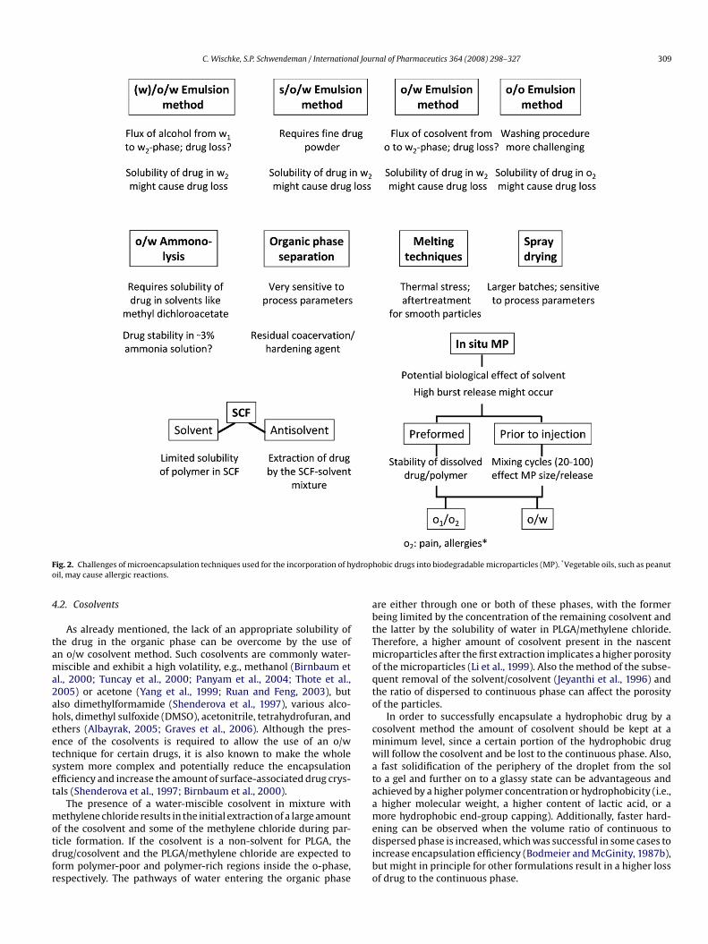
C. Wischke, S.P. Schwendeman / International Journal of Pharmaceutics 364 (2008) 298–327 309
F ydropo
4
tama2aheetset
motdfr
abtTmoqto
cmwataam
ig. 2. Challenges of microencapsulation techniques used for the incorporation of hil, may cause allergic reactions.
.2. Cosolvents
As already mentioned, the lack of an appropriate solubility ofhe drug in the organic phase can be overcome by the use ofn o/w cosolvent method. Such cosolvents are commonly water-iscible and exhibit a high volatility, e.g., methanol (Birnbaum et
l., 2000; Tuncay et al., 2000; Panyam et al., 2004; Thote et al.,005) or acetone (Yang et al., 1999; Ruan and Feng, 2003), butlso dimethylformamide (Shenderova et al., 1997), various alco-ols, dimethyl sulfoxide (DMSO), acetonitrile, tetrahydrofuran, andthers (Albayrak, 2005; Graves et al., 2006). Although the pres-nce of the cosolvents is required to allow the use of an o/wechnique for certain drugs, it is also known to make the wholeystem more complex and potentially reduce the encapsulationfficiency and increase the amount of surface-associated drug crys-als (Shenderova et al., 1997; Birnbaum et al., 2000).
The presence of a water-miscible cosolvent in mixture withethylene chloride results in the initial extraction of a large amount
f the cosolvent and some of the methylene chloride during par-icle formation. If the cosolvent is a non-solvent for PLGA, therug/cosolvent and the PLGA/methylene chloride are expected toorm polymer-poor and polymer-rich regions inside the o-phase,espectively. The pathways of water entering the organic phase
edibo
hobic drugs into biodegradable microparticles (MP). *Vegetable oils, such as peanut
re either through one or both of these phases, with the formereing limited by the concentration of the remaining cosolvent andhe latter by the solubility of water in PLGA/methylene chloride.herefore, a higher amount of cosolvent present in the nascenticroparticles after the first extraction implicates a higher porosity
f the microparticles (Li et al., 1999). Also the method of the subse-uent removal of the solvent/cosolvent (Jeyanthi et al., 1996) andhe ratio of dispersed to continuous phase can affect the porosityf the particles.
In order to successfully encapsulate a hydrophobic drug by aosolvent method the amount of cosolvent should be kept at ainimum level, since a certain portion of the hydrophobic drugill follow the cosolvent and be lost to the continuous phase. Also,fast solidification of the periphery of the droplet from the sol
o a gel and further on to a glassy state can be advantageous andchieved by a higher polymer concentration or hydrophobicity (i.e.,higher molecular weight, a higher content of lactic acid, or aore hydrophobic end-group capping). Additionally, faster hard-
ning can be observed when the volume ratio of continuous toispersed phase is increased, which was successful in some cases to
ncrease encapsulation efficiency (Bodmeier and McGinity, 1987b),ut might in principle for other formulations result in a higher lossf drug to the continuous phase.
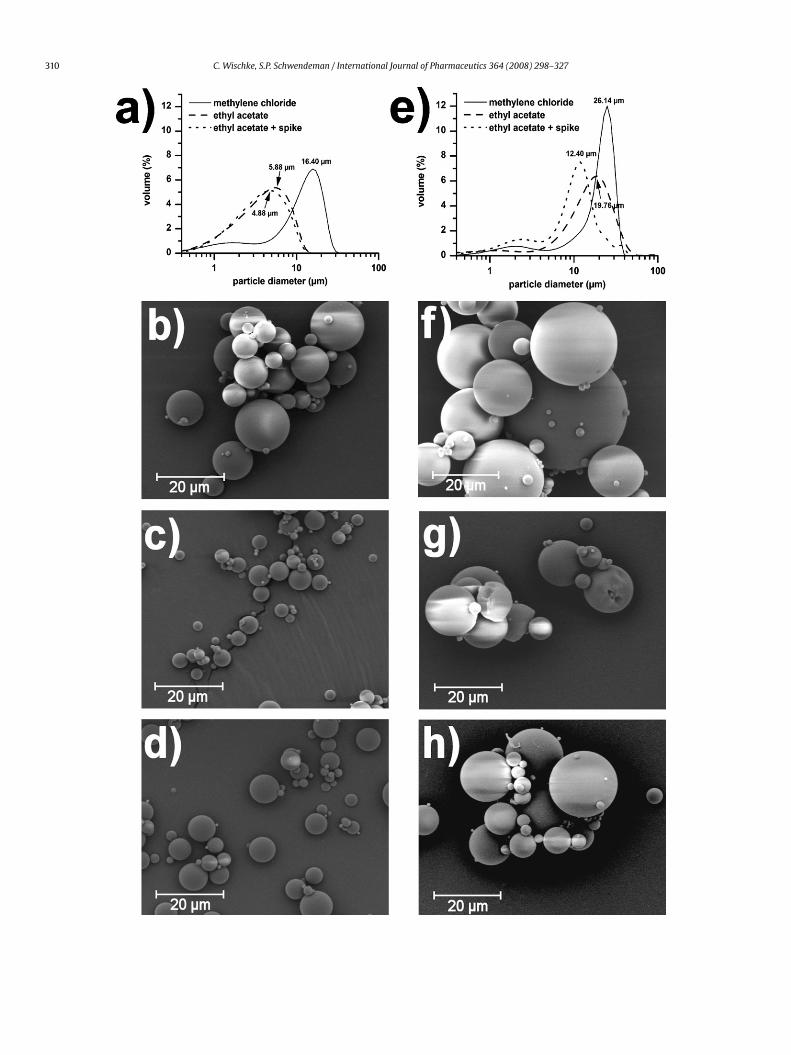
310 C. Wischke, S.P. Schwendeman / International Journal of Pharmaceutics 364 (2008) 298–327
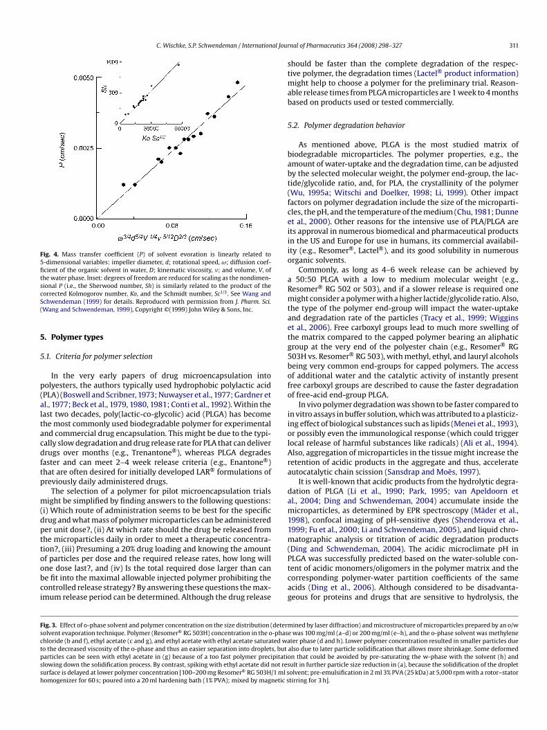
C. Wischke, S.P. Schwendeman / International Jour
Fig. 4. Mass transfer coefficient (P) of solvent evoration is linearly related to5-dimensional variables: impeller diameter, d; rotational speed, ω; diffusion coef-ficient of the organic solvent in water, D; kinematic viscosity, �; and volume, V, ofthe water phase. Inset: degrees of freedom are reduced for scaling as the nondimen-sional P (i.e., the Sherwood number, Sh) is similarly related to the product of thecS(
5
5
p(altacdftp
m(dpttoobci
stmab
5
babt(fceiiio
aRmtaetg5bofo
iiolAra
dam11m(PLGA was successfully predicted based on the water-soluble con-
Fsctpssh
orrected Kolmogorov number, Ko, and the Schmidt number, Sc1/3. See Wang andchwendeman (1999) for details. Reproduced with permission from J. Pharm. Sci.Wang and Schwendeman, 1999), Copyright ©(1999) John Wiley & Sons, Inc.
. Polymer types
.1. Criteria for polymer selection
In the very early papers of drug microencapsulation intoolyesters, the authors typically used hydrophobic polylactic acidPLA) (Boswell and Scribner, 1973; Nuwayser et al., 1977; Gardner etl., 1977; Beck et al., 1979, 1980, 1981; Conti et al., 1992). Within theast two decades, poly(lactic-co-glycolic) acid (PLGA) has becomehe most commonly used biodegradable polymer for experimentalnd commercial drug encapsulation. This might be due to the typi-ally slow degradation and drug release rate for PLA that can deliverrugs over months (e.g., Trenantone®), whereas PLGA degrades
aster and can meet 2–4 week release criteria (e.g., Enantone®)hat are often desired for initially developed LAR® formulations ofreviously daily administered drugs.
The selection of a polymer for pilot microencapsulation trialsight be simplified by finding answers to the following questions:
i) Which route of administration seems to be best for the specificrug and what mass of polymer microparticles can be administereder unit dose?, (ii) At which rate should the drug be released fromhe microparticles daily in order to meet a therapeutic concentra-ion?, (iii) Presuming a 20% drug loading and knowing the amountf particles per dose and the required release rates, how long will
ne dose last?, and (iv) Is the total required dose larger than cane fit into the maximal allowable injected polymer prohibiting theontrolled release strategy? By answering these questions the max-mum release period can be determined. Although the drug releasetcag
ig. 3. Effect of o-phase solvent and polymer concentration on the size distribution (determolvent evaporation technique. Polymer (Resomer® RG 503H) concentration in the o-phashloride (b and f), ethyl acetate (c and g), and ethyl acetate with ethyl acetate saturated wao the decreased viscosity of the o-phase and thus an easier separation into droplets, butarticles can be seen with ethyl acetate in (g) because of a too fast polymer precipitatiolowing down the solidification process. By contrast, spiking with ethyl acetate did not resurface is delayed at lower polymer concentration [100–200 mg Resomer® RG 503H/1 ml somogenizer for 60 s; poured into a 20 ml hardening bath (1% PVA); mixed by magnetic s
nal of Pharmaceutics 364 (2008) 298–327 311
hould be faster than the complete degradation of the respec-ive polymer, the degradation times (Lactel® product information)
ight help to choose a polymer for the preliminary trial. Reason-ble release times from PLGA microparticles are 1 week to 4 monthsased on products used or tested commercially.
.2. Polymer degradation behavior
As mentioned above, PLGA is the most studied matrix ofiodegradable microparticles. The polymer properties, e.g., themount of water-uptake and the degradation time, can be adjustedy the selected molecular weight, the polymer end-group, the lac-ide/glycolide ratio, and, for PLA, the crystallinity of the polymerWu, 1995a; Witschi and Doelker, 1998; Li, 1999). Other impactactors on polymer degradation include the size of the microparti-les, the pH, and the temperature of the medium (Chu, 1981; Dunnet al., 2000). Other reasons for the intensive use of PLA/PLGA arets approval in numerous biomedical and pharmaceutical productsn the US and Europe for use in humans, its commercial availabil-ty (e.g., Resomer®, Lactel®), and its good solubility in numerousrganic solvents.
Commonly, as long as 4–6 week release can be achieved by50:50 PLGA with a low to medium molecular weight (e.g.,
esomer® RG 502 or 503), and if a slower release is required oneight consider a polymer with a higher lactide/glycolide ratio. Also,
he type of the polymer end-group will impact the water-uptakend degradation rate of the particles (Tracy et al., 1999; Wigginst al., 2006). Free carboxyl groups lead to much more swelling ofhe matrix compared to the capped polymer bearing an aliphaticroup at the very end of the polyester chain (e.g., Resomer® RG03H vs. Resomer® RG 503), with methyl, ethyl, and lauryl alcoholseing very common end-groups for capped polymers. The accessf additional water and the catalytic activity of instantly presentree carboxyl groups are described to cause the faster degradationf free-acid end-group PLGA.
In vivo polymer degradation was shown to be faster compared ton vitro assays in buffer solution, which was attributed to a plasticiz-ng effect of biological substances such as lipids (Menei et al., 1993),r possibly even the immunological response (which could triggerocal release of harmful substances like radicals) (Ali et al., 1994).lso, aggregation of microparticles in the tissue might increase theetention of acidic products in the aggregate and thus, accelerateutocatalytic chain scission (Sansdrap and Moës, 1997).
It is well-known that acidic products from the hydrolytic degra-ation of PLGA (Li et al., 1990; Park, 1995; van Apeldoorn etl., 2004; Ding and Schwendeman, 2004) accumulate inside theicroparticles, as determined by EPR spectroscopy (Mäder et al.,
998), confocal imaging of pH-sensitive dyes (Shenderova et al.,999; Fu et al., 2000; Li and Schwendeman, 2005), and liquid chro-atographic analysis or titration of acidic degradation products
Ding and Schwendeman, 2004). The acidic microclimate pH in
ent of acidic monomers/oligomers in the polymer matrix and theorresponding polymer-water partition coefficients of the samecids (Ding et al., 2006). Although considered to be disadvanta-eous for proteins and drugs that are sensitive to hydrolysis, the
ined by laser diffraction) and microstructure of microparticles prepared by an o/we was 100 mg/ml (a–d) or 200 mg/ml (e–h), and the o-phase solvent was methyleneter phase (d and h). Lower polymer concentration resulted in smaller particles due
also due to later particle solidification that allows more shrinkage. Some deformedn that could be avoided by pre-saturating the w-phase with the solvent (h) andult in further particle size reduction in (a), because the solidification of the dropletolvent; pre-emulsification in 2 ml 3% PVA (25 kDa) at 5,000 rpm with a rotor–statortirring for 3 h].
3 al Jour
ab1
5
fpp(gae(ltaladdamagtmmctmPgw1r
5p
iimtdwtooradP5(lnt
otwd
trGaf(
mobtrhshckwro
cnrpcrPs(
6
6
dftisomcwatD1rwtnttar
au
12 C. Wischke, S.P. Schwendeman / Internation
cidic microclimate has been shown to stabilize certain hydropho-ic drugs with maximum stability below pH 4 (Shenderova et al.,999).
.3. Polymer mixtures and alternative PLGA copolymers
Occasionally the release of hydrophobic drugs might be too slowrom 50:50 PLGA copolymers. The use of a PLA–PEG–PLA blockolymer instead of 50:50 PLGA was shown to result in a higherorosity and a faster release for the hydrophobic drug paclitaxelRuan and Feng, 2003). PLGA-glucose star-shaped polymer, withlucose being the initiator molecule for polymerization (Kissel etl., 1991), has successfully entered the market for octreotide deliv-ry (Sandostatin LAR® Depot). Blends of high molecular weightMw) polymer with a small portion of a low Mw polymer, or use ofow Mw polymer altogether, were among the first methods iden-ified to insure continuous release of peptides (Hutchinson, 1986)nd hydrophobic drugs (Bodmeier et al., 1989) by minimizing theag phase to polymer mass loss and release. Such lag phases mayppear after the initial burst release of drug from surface-nearomains in medium to high molecular weight PLA/PLGA, wheniffusion controlled release through the dense matrix is limitednd the generation of matrix microporosity by hydrolytic poly-er degradation takes time depending on polymer type, Mw,
nd matrix geometry (Hutchinson and Furr, 1985). The advanta-eous effect of polymer blends is often rationalized by the facthat low Mw PLGA degrades faster to the critical Mw that ulti-
ately allows their removal from the matrix, forms pores, and thusore transport paths are available for water to access the parti-
le core and drug being released. Also, during polymer degradationhe increasing number of hydrophilic carboxyl group enhances the
atrix hydrophilicity and water-uptake for medium and high MwLA/PLGA and, accordingly, the instant presence of more of suchroups for blends with uncapped, low Mw PLGA will accelerateater-uptake (Hutchinson and Furr, 1989; Witschi and Doelker,
998), being the precondition for matrix degradation and drugelease.
.4. Impact of drug properties and preparation procedure onolymer characteristics
The polymer and microparticle properties are typically stronglynfluenced by the preparation procedure, and in certain cases bynteractions between the drug and polymer. On the one hand this
ay result in faster polymer degradation. For certain drugs, e.g.,hioridazine, an amine-catalyzed hydrolysis of the polymer matrixuring the particle preparation and a faster release was observed,hich was reduced by performing the o/w emulsification at lower
emperatures or erasing the drugs nucleophilicity by the formationf a salt (Maulding et al., 1986). In another study with solid solutionsf different amine drugs in PLA, it was shown that an acceleratedelease correlated well with the ability of the respective drug to cat-lyze the polymer degradation, but not with the Tg reduction, therugs pKa, or the drugs octanol–water partition coefficient (Cha anditt, 1988, 1989). By contrast, the increased catalytic degradation of0:50 PLGA was associated with a loading-dependent plasticizationTg reduction) for microparticles containing a water-soluble acety-ated amino acid, N-acetyl cysteine (Desai et al., 2008). Other drugs,amely haloperidol, changed the type of matrix erosion from bulko surface-erosion (Siegel et al., 2006).
Ultrasound treatment for emulsification can lead to a reductionf polymer molecular weight and therefore, a faster degrada-ion upon exposure to the release medium. This phenomenonas slightly increased in the presence of suspended solids (s/o-ispersion), which act as cavitation nuclei, but was dramatic in
w(Uta
nal of Pharmaceutics 364 (2008) 298–327
he presence of dissolved quinine, again due to amine-catalyzedandom chain cleavage of the polymer’s ester bond (Reich, 1998).amma sterilization of estradiol-loaded microparticles resulted inloss of polymer molecular weight by random chain scission, a
aster release, and the formation of drug degradation productsMohr et al., 1999).
On the other hand, the interaction of hydrophobic drugolecules with the polymer matrix via hydrophobic binding forces
r of amine-groups with the polymer carboxyl groups by ioniconds might cause a trapping of the drug inside the particles andherefore, a slower release. An inverse relationship between theelease rate and the solid-state solubility of the drug in the polymeras been described for dexamethasone, where an increasing solid-tate solubility can be observed with end-group capped polymers,igher lactide content, and lower molecular weight of the end-apped polymer (Panyam et al., 2004). By contrast, another drug,etoprofen, dissolves up to 20% in PLGA, forms hydrogen bondsith PLGA, and acts as a plasticizer, which was hypothesized to
educe polymer chain–chain interactions and thus accelerate itswn release (Blasi et al., 2007).
However, beside the polymer molecular weight, degradationharacteristics, and drug–polymer interactions, there are other sig-ificant factors, like the polymer microstructure, that play a keyole in the properties of microparticles. For instance the release ofrogesterone was found to be faster rather than slower from PLAompared to 85/15 PLGA microparticles. This was attributed to aougher surface and higher porosity of the PLA particles, becauseLA precipitated faster and the microparticles were not able tohrink and form the common smooth surface in this specific caseYang and Owusu-Ababio, 2000).
. Controlling the polymer microparticle size
.1. Emulsification procedure
A large variety of o/w emulsification methods have beenescribed ranging from simple set-ups with a beaker and stirrer to,or instance, methods based on static micromixers, where the par-icle size can be controlled by the flow rates of the o- and w-phasen the micromixer (Schalper et al., 2005; Wischke et al., 2006), orurface liquid spraying, where the o-phase is sprayed on the surfacef the stirred water phase (Tang et al., 2007). Also, a “jet excitationethod” has been described to achieve size-uniform microparti-
les by feeding the drug/polymer solution through a glass nozzle,hich is equipped with an ultrasonic transducer, into a stream of
queous carrier, followed by solvent removal in a standard evapora-ion procedure from the o/w emulsion (Berkland et al., 2001, 2002).roplet formation by Rayleigh-Plateau instability (Lord Rayleigh,879; Eggers, 2006), a process driven by interfacial tension thatesults in axisymmetric undulations of the liquid jet (perturbationsith wavelengths larger than the jet radius) and the break-up of
he cylindrical fluid thread into droplets, is the basic principle ofumerous micromixers. This classic mechanism of droplet forma-ion is superimposed by ultrasonic high-frequency oscillation ofhe nozzle in the “jet excitation method”, which allows predictionnd control of the droplet size by the applied frequency and flowate.
In order to obtain injectable microparticles for long-acting depotpplications, a polydisperse particle size range of 20–100 �m issually desired. Smaller particles, <5–10 �m are necessary, if the
hole microparticles are passively targeted to phagocytic cellsJeffery et al., 1991; Johanson et al., 2000; O’Hagan and Singh, 2004).ltrasound (Muranishi et al., 1991) and high pressure homogeniza-
ion provide a high energy density in the emulsification zone andre expected to produce a high fraction of nanoparticles rather
al Jour
tehmTtpu
tpRooecscto
6
ccptoai
fiaeafkwmscciaiceitwsi(olefimtiors
apmciPsfsfctcmpsaAaAtf2
tflioptctci1emwdava
bwtcptp
6
fiwzc
C. Wischke, S.P. Schwendeman / Internation
han microparticles. However, high-speed rotor–stator homogeniz-rs such as Ultra-Turrax® can produce submicron droplets only at aigh continuous phase viscosity, which is not the case in commonicroencapsulation procedures (Schuchmann and Danner, 2004).
herefore, rotor–stator homogenizers at low stirring speed, vor-exers, or overhead or magnetical stirrers with the o-phase beingoured or injected into the stirred continuous phase are frequentlysed to meet the 20–100 �m goal.
A simple formulation that requires a minimum of instrumen-al equipment and might act as a starting point employs (i) thereparation of the drug containing o-phase (e.g., 15–25% (w/v)esomer® RG503 in methylene chloride), (ii) emulsifying one partf the o-phase in 1–3 parts of a 1–5% PVA (Mw 20–40 kDa, degreef hydrolysis 88%) solution with a vortexer, (iii) pouring the o/wmulsion in 25–100 parts of a hardening bath of a lower PVA con-entration, (iv) stirring the nascent particles for about 3 h to allowolvent evaporation, (v) collecting the particles on a sieve or byentrifugation and washing them with water, and (vi) final dryinghe particle by lyophilisation or under vacuum (desiccator, vacuumven).
.2. Formulation parameters
A large number of formulation parameters may impact the parti-le size and ultrastructure, e.g., the o-phase volume and solvent, theoncentration and type of polymer, the volume of the continuoushase and the type and concentration of stabilizer, the tempera-ure, the stirring speed, and the stirrer type and geometry amongthers (Wu, 1995b; Jain et al., 1998). Discussion of all these vari-bles is beyond the scope of this paper; however, some of the moremportant parameters will be briefly described.
In order to separate the o-phase into individual droplets, shearorces are commonly applied to the system. By increasing thentensity of these forces, e.g., by increasing the stirring speed ofrotor–stator homogenizer, the particle size can be reduced. How-ver, the size distribution often increases simultaneously (Jalilnd Nixon, 1990a,b,c). Due to the increasing interfacial energyor smaller dispersed droplets, i.e., for larger droplet surfaces, noinetically stable reduction in the droplet size will be obtainedithout the presence of emulsifiers that reduce the high poly-er phase/water interfacial tension. In a scenario with dissolved
tabilizer molecules, which is typically the case during microen-apsulation, the speed of the emulsifier diffusion through theontinuous phase to the droplet surface, its adsorption, and finallyts spreading on the o-phase droplets (Gibbs–Marangoni effect) willlso impact the success of the emulsification. Once the emulsions formed, its tendency towards coalescence will depend on effi-iency of stabilizing mechanisms such as (i) static stabilization, i.e.,lectrostatic forces due to the droplets surface charge (by adsorbedons, ionic surfactants, or polymer carboxylate groups introduced inhe water phase) as described in the DLVO (Derjaguin, Landau, Ver-ey, and Overbeek) theory and/or steric repulsion due to adsorbed
urface-active polymers (e.g., PVA) or solid particles that theoret-cally form a mechanic barrier prohibiting droplet approaching,ii) dynamic stabilization, i.e., thermodynamic stabilization (lossf configurational entropy of adsorbed steric stabilizers when over-apping occurs for approaching droplets) and the Gibbs–Marangoniffect (especially relevant for mobile, small-molecule emulsi-ers; when approaching droplets force liquid and some stabilizerolecules out of the gap between them, formation of surface
ension gradient at the interface, stabilizer spreading, subsequentnward flux of medium in gap; voids from random stabilizer des-rption immediately filled), and (iii) other aspects of interfacialheology such as the viscoelasticity of the stabilizer film (for detailsee Heusch, 1983; Tadros, 2005).
tlopt
nal of Pharmaceutics 364 (2008) 298–327 313
Polyvinyl alcohol (PVA), which is a copolymer of vinyl acetatend vinyl alcohol, and is prepared by the controlled hydrolysis ofolyvinyl acetate side groups (Herrmann and Haehnel, 1927), is theost commonly used emulsifier for the preparation of PLGA parti-
les (Beck et al., 1979; Okada et al., 1987) because of its excellentnteraction with PLGA surfaces (Boury et al., 1995). Increasing theVA concentration or its molecular weight is known to result inmaller microparticles (Jobmann and Rafler, 1998). Although theaster droplet surface saturation due to the reduced average diffu-ion length of the stabilizer from the continuous phase to the, soar, uncovered droplet surface at higher PVA concentration mightontribute to these findings, the main factor is expected to reside inhe elevated viscosity of the continuous phase that hinders dropletollisions and coalescence. However, depending on the type ofixer and its efficiency at elevated viscosities of the continuous
hase, an overly high PVA concentration can impede the completeeparation of nascent droplets and result in a larger number ofggregated microparticles during solidification (Yang and Owusu-babio, 2000). An alternative explanation might be a bridging effectt overly high PVA concentration that causes particle aggregation.lso, one should consider that PVA can mediate solubility of cer-
ain drugs in the w-phase and thus, a higher PVA concentration canavor drug loss to the continuous phase (Yang and Owusu-Ababio,000).
Besides the viscosity of the external water phase, the viscosity ofhe disperse polymer phase needs to be considered. In a turbulentow, which is present in most homogenizers, the droplet break-up
s controlled by inertial forces which are dependent on the viscosityf the polymer phase. A higher o-phase viscosity requires a higherower density, i.e., stirring speed and/or exposure time to obtainhe same droplet size (Schuchmann and Danner, 2004). Higher vis-osities of the o-phase are present at increasing concentrations ofhe dissolved matrix polymer or polymer molecular weight andan, depending on the respective emulsification procedure, resultn increased particle sizes (Jeffery et al., 1991; Jobmann and Rafler,998; Choi et al., 2002; Mao et al., 2008). When w/o/w doublemulsion or s/o/w techniques are used, the viscosity of the poly-er phase may also be influenced by the presence of the innerater phase or the amount, crystal size, and shape of dispersedrug, respectively. Also, the type and concentration of polymerlong with the type of solvent and the potential presence of cosol-ents will define the solidification speed and thus the particle sizes already mentioned.
Performing the emulsification in an ice-bath or a cooled jacketeaker, which is required for certain thermo-sensitive drugs andas suggested to reduce amine-drug catalyzed polymer degrada-
ion (Maulding et al., 1986), will change the viscosity of both theontinuous and the disperse phase and therefore presumably thearticle size. However, the solvent diffusion out of the droplets,he hardening speed, and the solubility of the drug in the differenthases is each certain to be altered as well.
.3. Particle size analysis
In order to determine the particle size the operator can chooserom the following widely established methods: (i) microscopyncluding light and electron microscopy, (ii) the Coulter principle,
here particles are passed through an orifice (electrical sensingone) by vacuum from one electrolyte chamber to another andause an increased impedance in an electric field corresponding to
he particle volume by blocking a certain part of the aperture, (iii)aser diffraction, where the light of a laser is diffracted at the surfacef suspended microparticles resulting in a size specific diffractionattern, (iv) dynamic light scattering (photon correlation spec-roscopy) for submicron particles that correlates the fluctuation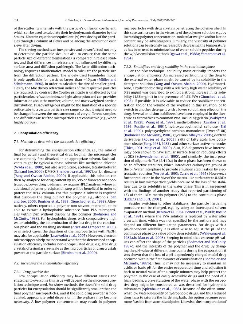
3 al Jour
owScs
tpisnfiScapidobah
7
7
fiav(((ttaptanacMwoommscp
7
7
slpficn
mticsat1
7
etds0b1tsdae1e(ds(iatbttfrlw1(
peeadpc1u1iwoMabph
14 C. Wischke, S.P. Schwendeman / Internation
f the scattering intensity with the particle’s diffusion coefficient,hich can be used to calculate their hydrodynamic diameter by the
tokes–Einstein equation or equivalent, (v) wet sieving of the parti-les through a column of sieves and balancing the amount on eachieve after drying.
The sieving method is an inexpensive and powerful tool not onlyo determine the particle size, but also to ensure that the samearticle size of different formulations is compared in release stud-
es, and that differences in release are not influenced by differingurface area and diffusion pathlength. The laser diffraction tech-ology requires a mathematical model to calculate the particle size
rom the diffraction pattern. The widely used Fraunhofer models only applicable for particles larger than ∼10 �m (Müller andchuhmann, 1996). In order to calculate the size of smaller parti-les by the Mie theory refraction indices of the respective particlesre required. By contrast the Coulter principle is unaffected by thearticle color, refraction index, or light absorption and can provides
nformation about the number, volume, and mass weighted particleistribution. Disadvantageous might be the limitation of a specificrifice tube to a certain particle size range, so that it might need toe changed between the measurements of very different samples,nd difficulties arise if the microparticles are conductive (e.g., whenighly porous).
. Encapsulation efficiency
.1. Methods to determine the encapsulation efficiency
For determining the encapsulation efficiency, i.e., the ratio ofnal (or actual) and theoretical drug loading, the microparticlesre commonly first dissolved in an appropriate solvent. Such sol-ents might be typical o-phase solvents like methylene chlorideWada et al., 1988), but also acetonitrile, acetone, tetrahydrofuranSah and Lee, 2006), DMSO (Shenderova et al., 1997), or 1,4-dioxaneYang and Owusu-Ababio, 2000). If applicable, this solution canhen by analyzed for drug content by UV/VIS or fluorescence spec-roscopy. Lower drug loadings may require HPLC analysis, where andditional polymer precipitation step will be beneficial in order torotect the HPLC column. For this purpose a solvent is requiredhat dissolves the drug but not the polymer, e.g., alcohols (Sahnd Lee, 2006; Buntner et al., 1998; Giunchedi et al., 1998). Alter-atively, others reported a polymer non-solvent, methanol, to beble to extract a hydrophobic substance from PLA microparti-les within 24 h without dissolving the polymer (Bodmeier andcGinity, 1988). For hydrophobic drugs with comparatively highater solubility, the determination of the lost drug in the continu-us phase and the washing medium (Arica and Lamprecht, 2005),r in select cases, the digestion of the microparticles with NaOH,ay also be applicable (Jaraswekin et al., 2007). However, electronicroscopy can help to understand whether the determined encap-
ulation efficiency includes non-encapsulated drug, e.g., free drugrystals of a similar size-scale as the microparticles or drug crystalsresent at the particle surface (Birnbaum et al., 2000).
.2. Increasing the encapsulation efficiency
.2.1. Drug particle sizeLow encapsulation efficiency may have different causes and
trategies to overcome this issue will depend on the microencapsu-
ation technique used. For s/o/w methods, the size of the solid drugarticles for encapsulation should be significantly smaller than thenal polymer microparticles, and if the solid drug powder is floc-ulated, appropriate solid dispersion in the o-phase may becomeecessary. A low polymer concentration may result in polymertswdm
nal of Pharmaceutics 364 (2008) 298–327
icroparticles with drug crystals penetrating the polymer shell. Inhis case, an increase in the viscosity of the polymer solution, e.g., byncreasing polymer concentration, molecular weight, and/or lactideontent may be advantageous. Similarly, the viscosity of polymerolutions can be strongly increased by decreasing the temperature,s has been used to minimize loss of water-soluble peptides duringhe w/o/w emulsion method (Ogawa et al., 1988a; Yamamoto et al.,994).
.2.2. Emulsifiers and drug solubility in the continuous phaseFor the o/w technique, solubility most critically impacts the
ncapsulation efficiency. An increased partitioning of the drug tohe external water phase might be caused by its solubility in theetergent solution (Yang and Owusu-Ababio, 2000). Hydrocorti-one, a hydrophobic drug with a relatively high water solubility of.28 mg/ml was described to exhibit a strong increase in its solu-ility (1.38 mg/ml) in the presence of 1.5% PVA (Giunchedi et al.,998). If possible, it is advisable to reduce the stabilizer concen-ration and/or the volume of the w-phase in this situation, or towitch to another detergent with a lower solubilizing effect on therug. Several emulsion stabilizers have been employed in the liter-ture as alternatives to common PVA, including gelatin (Wakiyamat al., 1982b; Wang et al., 1997), methylcellulose (Cavalier et al.,986; Rosilio et al., 1991), hydroxypropylmethyl cellulose (Ertlt al., 1999), polyoxyethylene sorbitan monooleate (Tween® 80)Bodmeier and McGinity, 1988), glycerine (Albayrak, 2005), dextranerivatives (Rouzes et al., 2003), salts of fatty acids like potas-ium oleate (Fong, 1981, 1983), and other surface-active moleculesThies, 1991; Mogi et al., 2000). Also, PLA oligomers have interest-ngly been shown to have similar surface tension reducing effectss SDS (Schwendeman et al., 1995), and similarly, the incorpora-ion of oligomeric PLA (2.4 kDa) in the o-phase has been shown toe an effective stabilizer, which introduces carboxylate groups athe oil/water interphase to provide emulsion stabilization by elec-rostatic repulsion (Vert et al., 1993; Carrio et al., 1995). However, aurther reduction in the Mw of the matrix-like surfactant to 0.6 kDaesults in low microparticle yields because of the loss of the stabi-izer due to its solubility in the water phase. This is in agreement
ith the findings of another study that reported partitioning of/3 of their 1 kDa matrix polymer (PLA) to the continuous phaseLiggins and Burt, 2001).
Besides switching to other stabilizers, the particle hardeningrocedure can be changed, e.g., by using an interrupted solventvaporation method (Benita et al., 1984; Benoit et al., 1986b; Rosiliot al., 1991), where the PVA solution is replaced by water aftercertain time, which was not specified by the authors and may
epend on different formulation parameters. For drugs with aH-dependent solubility it is often wise to adjust the pH of theontinuous phase to a value of low drug solubility (Wakiyama et al.,982a,b; Mao et al., 2008), keeping in mind that extreme pH val-es can affect the shape of the particles (Bodmeier and McGinity,987c) and the integrity of the polymer and the drug. By chang-ng the pH-value at different time points during the evaporation, itas shown that the loss of a pH-dependently charged model drugccurred within the first minutes of emulsification (Bodmeier andcGinity, 1987b). Thus, it may not be necessary to maintain an
cidic or basic pH for the entire evaporation time and adjusting itack to neutral value after a couple minutes may help protect theolymer. In the case of easily accessible drugs and the need of aigh loading, a pre-saturation of the water phase with the respec-
ive drug might be considered as was described for hydrophilicubstances (Splenhauer et al., 1986). Because of the often some-hat low water-solubility of hydrophobic drugs, and thus reducedrug mass to saturate the hardening bath, this option becomes evenore feasible from a cost stand point. Likewise, the incorporation of
al Jour
cagpa
7h
wmwgtpst
atasstvdb1
itmvd(ttssmic
abev2
8
8
ttHsfvr22c1
rlo3ct
vv1eaalfeirr
8
fimwru1bfpstclsprdap
rwtpFmraa
trimha
C. Wischke, S.P. Schwendeman / Internation
ompounds with pH-dependent solubility that were encapsulatedt neutral pH in order to avoid polymer alteration (disadvanta-eously high solubility of the respective drug in the continuoushase) was beneficial with the pre-saturation concept (Bodmeiernd McGinity, 1987b).
.2.3. Mass transfer of o-phase solvents and microparticleardening
As mentioned in Section 4.1.1, the flux of organic solvent to theater phase will facilitate leaching of dissolved drug from the poly-er particle matrix. This effect may become strongly pronouncedhen water-miscible cosolvents are employed, which partition to a
reat extent into the water phase especially during the emulsifica-ion procedure, and before hardening of the nascent particles takeslace. Therefore, the amount of cosolvent in the solvent mixturehould be kept at the minimum level that is required to dissolvehe drug.
A fast precipitation of the polymer is typically considered to bedvantageous to achieve high encapsulation efficiencies, becausehe solid polymer film acts as a diffusion barrier for the drug. That is,high polymer concentration, hydrophobicity, or molecular weighthould result in a fast transition of the droplet surface from theol to the gel state. Additionally, for drugs with a low solubility inhe detergent solution, the formulation may benefit from a largerolume of the continuous phase with a faster polymer precipitation,ue to a larger portion of the non-water-miscible polymer solventeing initially extracted by the w-phase (Bodmeier and McGinity,987b).
The hardening speed of PLGA microparticles was shown toncrease by the addition of urea or a salt such as NaCl to the con-inuous phase. The faster precipitation of the polymer resulted in a
uch lower porosity of the microparticles prepared with a cosol-ent method and higher encapsulation efficiency for hydrophobicrugs (Thote et al., 2005) and for highly water-soluble drug saltsJaraswekin et al., 2007). However, occasionally w1/o/w2 micropar-icles with larger w1-volumes collapsed in the presence of NaCl dueo fast hardening (Chen et al., 2004). In another o/w microencap-ulation study with a partially water-soluble drug salt, quinidineulfate, the encapsulation efficiency was increased due to the for-ation of new, less soluble drug salts in the o-phase, when counter
ons, not common for this drug (e.g., ClO4−), were introduced in the
ontinuous phase (Al-Maaieh and Flanagan, 2005).As will be discussed in detail in Section 8.1, the diffusion rate of
hydrophobic drug out of the polymer particles can be controlledy its escape from the unstirred boundary layer. Therefore, higherncapsulation efficiency has been suggested by reducing diffusionelocity in the water phase, e.g., by addition of glycerine (Albayrak,005).
. Drug release from the microparticles
.1. In vitro assays—rationale for using sink conditions
While the experimental set-up for release studies is specified inhe pharmacopoeiae (USP, Ph. Eur.) for conventional dosage forms,here is no such specification for long-acting release microparticles.owever, one of the fundamentals of release studies is maintaining
ink conditions and in the literature of particulate sustained releaseormulations only a few knowingly break this rule, e.g., by usingery small volumes (5 ml) (Mittal et al., 2007). Others compared the
elease of a total 2.3 mg nifedipine from microparticles (solubility:.3 mg in 230 ml water) under non-sink conditions (shaking flask;50 ml medium) with sink conditions (flow-through cell; 2500 mlirculating medium) and explained the absence of differences in the4 day release profiles by the drug diffusion through the polymerecsne
nal of Pharmaceutics 364 (2008) 298–327 315
ather than its removal from the boundary layer as being the rate-imiting step (Sansdrap and Moës, 1993). However, the solubilityf nifedipine in the employed phosphate buffer might be higher at7 ◦C than in water, which would at least, if still not fulfilling sinkonditions in the flask assay, increase the concentration gradient athe particle surface.
In addition, theoretical comparison of the release under sinks. non-sink conditions has shown that deviations become rele-ant especially for slowly releasing formulations (Guy and Hadgraft,981). In vivo, at certain injection sites with low perfusion, thexposure to body fluids may not provide perfect sink conditionst all times, especially in the case of very hydrophobic drugs thatre not easily removed from the microparticles’ unstirred boundaryayers. However, it is well-known that the polymer often degradesaster in vivo than under sink conditions in vitro due to the pres-nce of certain endogenous substances (see Section 5). Therefore,t is important to verify that sink conditions are followed duringelease assays for hydrophobic drugs from particulate sustainedelease formulations.
.2. In vitro assays—media to maintain sink conditions
For protein-loaded microparticles release studies are often per-ormed in small volume microcentrifuge tubes and the releases determined from the supernatant after briefly centrifuging the
icrospheres under mild centrifugal forces. For hydrophobic drugsith low water solubility, larger volumes of release medium are
equired in order to maintain sink conditions. To reduce the vol-me of the aqueous media, the use of adsorbents (Wurster and Polli,961) or organic solvent reservoirs (Gibaldi and Feldman, 1967), haseen suggested for release studies from conventional solid dosageorms, although in the latter solubility of organics in the waterhase will likely influence the polymer properties (e.g., particleoftening and fusion, solid-state diffusion, and polymer degrada-ion rate) which may not be preferred for PLGA microparticles. Inertain cases the solubility of the drug may fall below the detectionimit of standard HPLC methods and its determination from theupernatant requires a time-consuming and potentially artefact-rone drug concentrating step to be in the useful concentrationange for analysis. Therefore, the determination of the remainingrug in the microparticles can provide more precise release datand additional information concerning the drug stability inside theolymer matrix.
Another fundamental question concerns the correlation of theesults from a release study to what can be expected in vivo. Itould be beneficial both from an ethical and financial point of view
o reduce animal studies by establishing in vitro assays with goodredictive character for the in vivo release of different formulations.or conventional dosage forms the major pharmacopoeiae specifyethods that do not display the conditions in vivo (e.g., a tablet will
arely encounter 900 ml of phosphate buffer in the stomach), butllow a fast prediction of the properties of a formulation for qualitynd in-process control.
As there is the issue of large volumes and time periods requiredo obtain a release profile for a hydrophobic drug from sustainedelease formulations, some authors followed the concept of speed-ng up the release in order to obtain a fast result that otherwise
ight take months. Such release media typically contain alco-ols, e.g., methanol (Parikh et al., 1993), 27.5% ethanol (Beck etl., 1980), or 25% n-propanol (Yang and Owusu-Ababio, 2000) and
xhibit a higher dissolving power for the drug while also plasti-izing the polymer. However, when 50% methanol was applied byome authors, the maximum release was observed after 24 h ando differentiation was possible between different batches (Zaghloult al., 2005, 2006). Another approach to accelerate the release for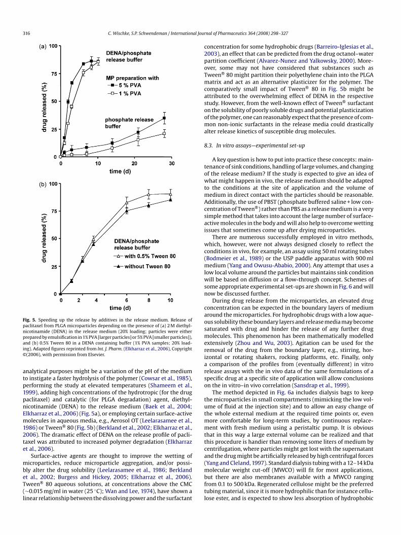
316 C. Wischke, S.P. Schwendeman / International Jour
Fig. 5. Speeding up the release by additives in the release medium. Release ofpaclitaxel from PLGA microparticles depending on the presence of (a) 2 M diethyl-nicotinamide (DENA) in the release medium (20% loading; particles were eitherpai©
atp1pnEm12te
mbeT(l
c2poTmcasooma
8
towtmAcsai
wc(mlwsn
caosmeriarso
tutmmttca(
repared by emulsification in 1% PVA [larger particles] or 5% PVA [smaller particles]),nd (b) 0.5% Tween 80 in a DENA containing buffer (1% PVA samples; 20% load-ng). Adapted figures reprinted from Int. J. Pharm. (Elkharraz et al., 2006), Copyright(2006), with permission from Elsevier.
nalytical purposes might be a variation of the pH of the mediumo instigate a faster hydrolysis of the polymer (Cowsar et al., 1985),erforming the study at elevated temperatures (Shameem et al.,999), adding high concentrations of the hydrotropic (for the drugaclitaxel) and catalytic (for PLGA degradation) agent, diethyl-icotinamide (DENA) to the release medium (Baek et al., 2004;lkharraz et al., 2006) (Fig. 5a), or employing certain surface-activeolecules in aqueous media, e.g., Aerosol OT (Leelarasamee et al.,
986) or Tween® 80 (Fig. 5b) (Berkland et al., 2002; Elkharraz et al.,006). The dramatic effect of DENA on the release profile of pacli-axel was attributed to increased polymer degradation (Elkharrazt al., 2006).
Surface-active agents are thought to improve the wetting oficroparticles, reduce microparticle aggregation, and/or possi-
ly alter the drug solubility (Leelarasamee et al., 1986; Berklandt al., 2002; Burgess and Hickey, 2005; Elkharraz et al., 2006).ween® 80 aqueous solutions, at concentrations above the CMC∼0.015 mg/ml in water (25 ◦C); Wan and Lee, 1974), have shown ainear relationship between the dissolving power and the surfactant
mbftl
nal of Pharmaceutics 364 (2008) 298–327
oncentration for some hydrophobic drugs (Barreiro-Iglesias et al.,003), an effect that can be predicted from the drug octanol–waterartition coefficient (Alvarez-Nunez and Yalkowsky, 2000). More-ver, some may not have considered that substances such asween® 80 might partition their polyethylene chain into the PLGAatrix and act as an alternative plasticizer for the polymer. The
omparatively small impact of Tween® 80 in Fig. 5b might bettributed to the overwhelming effect of DENA in the respectivetudy. However, from the well-known effect of Tween® surfactantn the solubility of poorly soluble drugs and potential plasticizationf the polymer, one can reasonably expect that the presence of com-on non-ionic surfactants in the release media could drastically
lter release kinetics of susceptible drug molecules.
.3. In vitro assays—experimental set-up
A key question is how to put into practice these concepts: main-enance of sink conditions, handling of large volumes, and changingf the release medium? If the study is expected to give an idea ofhat might happen in vivo, the release medium should be adapted
o the conditions at the site of application and the volume ofedium in direct contact with the particles should be reasonable.
dditionally, the use of PBST (phosphate buffered saline + low con-entration of Tween®) rather than PBS as a release medium is a veryimple method that takes into account the large number of surface-ctive molecules in the body and will also help to overcome wettingssues that sometimes come up after drying microparticles.
There are numerous successfully employed in vitro methods,hich, however, were not always designed closely to reflect the
onditions in vivo, for example, an assay using 50 ml rotating tubesBodmeier et al., 1989) or the USP paddle apparatus with 900 ml
edium (Yang and Owusu-Ababio, 2000). Any attempt that uses aow local volume around the particles but maintains sink condition
ill be based on diffusion or a flow-through concept. Schemes ofome appropriate experimental set-ups are shown in Fig. 6 and willow be discussed further.
During drug release from the microparticles, an elevated drugoncentration can be expected in the boundary layers of mediumround the microparticles. For hydrophobic drugs with a low aque-us solubility these boundary layers and release media may becomeaturated with drug and hinder the release of any further drugolecules. This phenomenon has been mathematically modelled
xtensively (Zhou and Wu, 2003). Agitation can be used for theemoval of the drug from the boundary layer, e.g., stirring, hor-zontal or rotating shakers, rocking platforms, etc. Finally, only
comparison of the profiles from (eventually different) in vitroelease assays with the in vivo data of the same formulations of apecific drug at a specific site of application will allow conclusionsn the in vitro–in vivo correlation (Sansdrap et al., 1999).
The method depicted in Fig. 6a includes dialysis bags to keephe microparticles in small compartments (mimicking the low vol-me of fluid at the injection site) and to allow an easy change ofhe whole external medium at the required time points or, even
ore comfortable for long-term studies, by continuous replace-ent with fresh medium using a peristaltic pump. It is obvious
hat in this way a large external volume can be realized and thathis procedure is handier than removing some liters of medium byentrifugation, where particles might get lost with the supernatantnd the drug might be artificially released by high centrifugal forcesYang and Cleland, 1997). Standard dialysis tubing with a 12–14 kDa
olecular weight cut-off (MWCO) will fit for most applications,ut there are also membranes available with a MWCO rangingrom 0.1 to 500 kDa. Regenerated cellulose might be the preferredubing material, since it is more hydrophilic than for instance cellu-ose ester, and is expected to show less absorption of hydrophobic
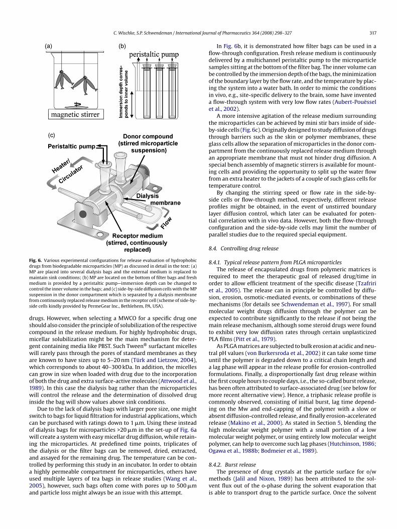
C. Wischke, S.P. Schwendeman / International Jour
Fig. 6. Various experimental configurations for release evaluation of hydrophobicdrugs from biodegradable microparticles (MP) as discussed in detail in the text: (a)MP are placed into several dialysis bags and the external medium is replaced tomaintain sink conditions; (b) MP are located on the bottom of filter bags and freshmedium is provided by a peristaltic pump—immersion depth can be changed tocsfs
dscmgwawco1wi
scowitatau2a
fldsboiiae
tbtgpasift
spltcp
8
8
roesmmemtP
tuafthmciarhmpO
ontrol the inner volume in the bags; and (c) side-by-side diffusion cells with the MPuspension in the donor compartment which is separated by a dialysis membranerom continuously replaced release medium in the receptor cell (scheme of side-by-ide cells kindly provided by PermeGear Inc., Bethlehem, PA, USA).
rugs. However, when selecting a MWCO for a specific drug onehould also consider the principle of solubilization of the respectiveompound in the release medium. For highly hydrophobic drugs,icellar solubilization might be the main mechanism for deter-
ent containing media like PBST. Such Tween® surfactant micellesill rarely pass through the pores of standard membranes as they
re known to have sizes up to 5–20 nm (Türk and Lietzow, 2004),hich corresponds to about 40–300 kDa. In addition, the micelles
an grow in size when loaded with drug due to the incorporationf both the drug and extra surface-active molecules (Attwood et al.,989). In this case the dialysis bag rather than the microparticlesill control the release and the determination of dissolved drug
nside the bag will show values above sink conditions.Due to the lack of dialysis bags with larger pore size, one might
witch to bags for liquid filtration for industrial applications, whichan be purchased with ratings down to 1 �m. Using these insteadf dialysis bags for microparticles >20 �m in the set-up of Fig. 6aill create a system with easy micellar drug diffusion, while retain-
ng the microparticles. At predefined time points, triplicates ofhe dialysis or the filter bags can be removed, dried, extracted,nd assayed for the remaining drug. The temperature can be con-
rolled by performing this study in an incubator. In order to obtainhighly permeable compartment for microparticles, others havesed multiple layers of tea bags in release studies (Wang et al.,005), however, such bags often come with pores up to 500 �mnd particle loss might always be an issue with this attempt.8
mvi
nal of Pharmaceutics 364 (2008) 298–327 317
In Fig. 6b, it is demonstrated how filter bags can be used in aow-through configuration. Fresh release medium is continuouslyelivered by a multichannel peristaltic pump to the microparticleamples sitting at the bottom of the filter bag. The inner volume cane controlled by the immersion depth of the bags, the minimizationf the boundary layer by the flow rate, and the temperature by plac-ng the system into a water bath. In order to mimic the conditionsn vivo, e.g., site-specific delivery to the brain, some have inventedflow-through system with very low flow rates (Aubert-Pouëssel
t al., 2002).A more intensive agitation of the release medium surrounding
he microparticles can be achieved by mini stir bars inside of side-y-side cells (Fig. 6c). Originally designed to study diffusion of drugshrough barriers such as the skin or polymer membranes, theselass cells allow the separation of microparticles in the donor com-artment from the continuously replaced release medium throughn appropriate membrane that must not hinder drug diffusion. Apecial bench assembly of magnetic stirrers is available for mount-ng cells and providing the opportunity to split up the water flowrom an extra heater to the jackets of a couple of such glass cells foremperature control.
By changing the stirring speed or flow rate in the side-by-ide cells or flow-through method, respectively, different releaserofiles might be obtained, in the event of unstirred boundary
ayer diffusion control, which later can be evaluated for poten-ial correlation with in vivo data. However, both the flow-throughonfiguration and the side-by-side cells may limit the number ofarallel studies due to the required special equipment.
.4. Controlling drug release
.4.1. Typical release pattern from PLGA microparticlesThe release of encapsulated drugs from polymeric matrices is
equired to meet the therapeutic goal of released drug/time inrder to allow efficient treatment of the specific disease (Tzafririt al., 2005). The release can in principle be controlled by diffu-ion, erosion, osmotic-mediated events, or combinations of theseechanisms (for details see Schwendeman et al., 1997). For smallolecular weight drugs diffusion through the polymer can be
xpected to contribute significantly to the release if not being theain release mechanism, although some steroid drugs were found
o exhibit very low diffusion rates through certain unplasticizedLA films (Pitt et al., 1979).
As PLGA matrices are subjected to bulk erosion at acidic and neu-ral pH values (von Burkersroda et al., 2002) it can take some timentil the polymer is degraded down to a critical chain length andlag phase will appear in the release profile for erosion-controlled
ormulations. Finally, a disproportionally fast drug release withinhe first couple hours to couple days, i.e., the so-called burst release,as been often attributed to surface-associated drug (see below forore recent alternative view). Hence, a triphasic release profile is
ommonly observed, consisting of initial burst, lag time depend-ng on the Mw and end-capping of the polymer with a slow orbsent diffusion-controlled release, and finally erosion-acceleratedelease (Makino et al., 2000). As stated in Section 5, blending theigh molecular weight polymer with a small portion of a lowolecular weight polymer, or using entirely low molecular weight
olymer, can help to overcome such lag phases (Hutchinson, 1986;gawa et al., 1988b; Bodmeier et al., 1989).
.4.2. Burst releaseThe presence of drug crystals at the particle surface for o/w
ethods (Jalil and Nixon, 1989) has been attributed to the sol-ent flux out of the o-phase during the solvent evaporation thats able to transport drug to the particle surface. Once the solvent
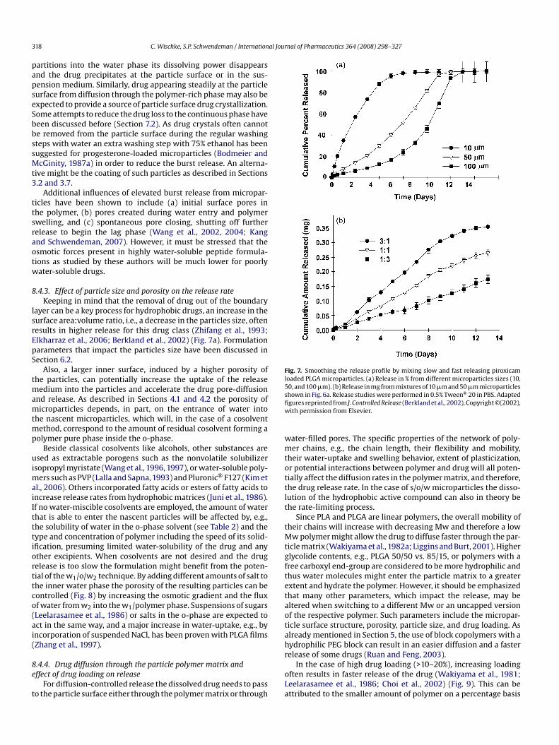
3 al Journal of Pharmaceutics 364 (2008) 298–327
papseSbbssMt3
ttsraotw
8
lsrEpS
tmamtmp
uimaiItttiorttco(ai(
8e
t
Fig. 7. Smoothing the release profile by mixing slow and fast releasing piroxicamloaded PLGA microparticles. (a) Release in % from different microparticles sizes (10,50, and 100 �m). (b) Release in mg from mixtures of 10 �m and 50 �m microparticlessfiw
wmtottlt
tMtgftetaotah
18 C. Wischke, S.P. Schwendeman / Internation
artitions into the water phase its dissolving power disappearsnd the drug precipitates at the particle surface or in the sus-ension medium. Similarly, drug appearing steadily at the particleurface from diffusion through the polymer-rich phase may also bexpected to provide a source of particle surface drug crystallization.ome attempts to reduce the drug loss to the continuous phase haveeen discussed before (Section 7.2). As drug crystals often cannote removed from the particle surface during the regular washingteps with water an extra washing step with 75% ethanol has beenuggested for progesterone-loaded microparticles (Bodmeier andcGinity, 1987a) in order to reduce the burst release. An alterna-
ive might be the coating of such particles as described in Sections.2 and 3.7.
Additional influences of elevated burst release from micropar-icles have been shown to include (a) initial surface pores inhe polymer, (b) pores created during water entry and polymerwelling, and (c) spontaneous pore closing, shutting off furtherelease to begin the lag phase (Wang et al., 2002, 2004; Kangnd Schwendeman, 2007). However, it must be stressed that thesmotic forces present in highly water-soluble peptide formula-ions as studied by these authors will be much lower for poorlyater-soluble drugs.
.4.3. Effect of particle size and porosity on the release rateKeeping in mind that the removal of drug out of the boundary
ayer can be a key process for hydrophobic drugs, an increase in theurface area:volume ratio, i.e., a decrease in the particles size, oftenesults in higher release for this drug class (Zhifang et al., 1993;lkharraz et al., 2006; Berkland et al., 2002) (Fig. 7a). Formulationarameters that impact the particles size have been discussed inection 6.2.
Also, a larger inner surface, induced by a higher porosity ofhe particles, can potentially increase the uptake of the release
edium into the particles and accelerate the drug pore-diffusionnd release. As described in Sections 4.1 and 4.2 the porosity oficroparticles depends, in part, on the entrance of water into
he nascent microparticles, which will, in the case of a cosolventethod, correspond to the amount of residual cosolvent forming a
olymer pure phase inside the o-phase.Beside classical cosolvents like alcohols, other substances are
sed as extractable porogens such as the nonvolatile solubilizersopropyl myristate (Wang et al., 1996, 1997), or water-soluble poly-
ers such as PVP (Lalla and Sapna, 1993) and Pluronic® F127 (Kim etl., 2006). Others incorporated fatty acids or esters of fatty acids toncrease release rates from hydrophobic matrices (Juni et al., 1986).f no water-miscible cosolvents are employed, the amount of waterhat is able to enter the nascent particles will be affected by, e.g.,he solubility of water in the o-phase solvent (see Table 2) and theype and concentration of polymer including the speed of its solid-fication, presuming limited water-solubility of the drug and anyther excipients. When cosolvents are not desired and the drugelease is too slow the formulation might benefit from the poten-ial of the w1/o/w2 technique. By adding different amounts of salt tohe inner water phase the porosity of the resulting particles can beontrolled (Fig. 8) by increasing the osmotic gradient and the fluxf water from w2 into the w1/polymer phase. Suspensions of sugarsLeelarasamee et al., 1986) or salts in the o-phase are expected toct in the same way, and a major increase in water-uptake, e.g., byncorporation of suspended NaCl, has been proven with PLGA filmsZhang et al., 1997).
.4.4. Drug diffusion through the particle polymer matrix andffect of drug loading on release
For diffusion-controlled release the dissolved drug needs to passo the particle surface either through the polymer matrix or through
r
oLa
hown in Fig. 6a. Release studies were performed in 0.5% Tween® 20 in PBS. Adaptedgures reprinted from J. Controlled Release (Berkland et al., 2002), Copyright ©(2002),ith permission from Elsevier.
ater-filled pores. The specific properties of the network of poly-er chains, e.g., the chain length, their flexibility and mobility,
heir water-uptake and swelling behavior, extent of plasticization,r potential interactions between polymer and drug will all poten-ially affect the diffusion rates in the polymer matrix, and therefore,he drug release rate. In the case of s/o/w microparticles the disso-ution of the hydrophobic active compound can also in theory behe rate-limiting process.
Since PLA and PLGA are linear polymers, the overall mobility ofheir chains will increase with decreasing Mw and therefore a low
w polymer might allow the drug to diffuse faster through the par-icle matrix (Wakiyama et al., 1982a; Liggins and Burt, 2001). Higherlycolide contents, e.g., PLGA 50/50 vs. 85/15, or polymers with aree carboxyl end-group are considered to be more hydrophilic andhus water molecules might enter the particle matrix to a greaterxtent and hydrate the polymer. However, it should be emphasizedhat many other parameters, which impact the release, may beltered when switching to a different Mw or an uncapped versionf the respective polymer. Such parameters include the micropar-icle surface structure, porosity, particle size, and drug loading. Aslready mentioned in Section 5, the use of block copolymers with aydrophilic PEG block can result in an easier diffusion and a fasterelease of some drugs (Ruan and Feng, 2003).
In the case of high drug loading (>10–20%), increasing loadingften results in faster release of the drug (Wakiyama et al., 1981;eelarasamee et al., 1986; Choi et al., 2002) (Fig. 9). This can bettributed to the smaller amount of polymer on a percentage basis
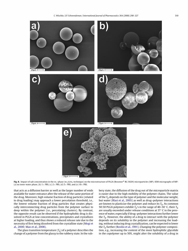
C. Wischke, S.P. Schwendeman / International Journal of Pharmaceutics 364 (2008) 298–327 319
F crostr(
tatttcdtsana
c
bioba5aet
ig. 8. Impact of salt concentration in the w1-phase (w1/o/w2 technique) on the mia) no inner water phase, (b) 1× PBS, (c) 2× PBS, (d) 5× PBS, and (e) 10× PBS.
hat acts as a diffusion barrier as well as the larger number of voidsvailable for water entrance after the release of the same portion ofhe drug. Moreover, high volume fraction of drug particles (relatedo drug loading) may approach a lower percolation threshold, i.e.,he lowest volume fraction of drug particles that creates physi-ally interconnecting drug particles from the polymer surface toeep within the polymer (i.e., percolating clusters). By contrast,he opposite result can be observed if the hydrophobic drug is dis-olved in PLGA at low concentrations, precipitates and crystallizes
t higher loading, and thus shows a reduced release rate due to theecessity of first being dissolved from the crystalline state (Mogi etl., 2000; Mao et al., 2008).The glass transition temperature (Tg) of a polymer describes thehange of a polymer from the glassy to the rubbery state. In the rub-
ditti
ucture of PLGA (Resomer® RG 502H) microparticles (MP). SEM micrographs of MP:
ery state, the diffusion of the drug out of the microparticle matrixs easier due to the high mobility of the polymer chains. The valuef the Tg depends on the type of polymer and the molecular weight,ut water (Blasi et al., 2005) as well as drug–polymer interactionsre known to plasticize the polymer and reduce its Tg. As common0:50 PLGA polymers exhibit Tg’s in the range of 40–50 ◦C, their Tg
re usually exceeded under release conditions at 37 ◦C in the pres-nce of water, especially if drug–polymer interactions further lowerhe Tg. However, the ability of a drug to interact with the polymer
epends on its solubility in the polymer and increasing the load-ng, without inducing drug crystallization, can be expected to lowerhe Tg further (Rosilio et al., 1991). Changing the polymer composi-ion, e.g., increasing the content of the more hydrophilic glycoliden the copolymer up to 50%, might alter the solubility of a drug in
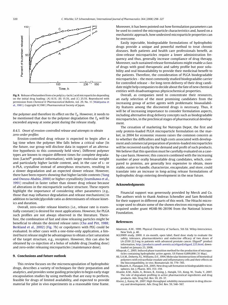
320 C. Wischke, S.P. Schwendeman / International Jour
Fopa
tbe
8z
lttttadata1ohtai
tsfbBeiooa
9
daefm
Mbmb
ddsqMobtmfde
aiiwimm
oktmwWbnpsth
A
TfsaF
R
A
A
A
A
A
ig. 9. Release of butamben from o/w poly(d,l-lactic) acid microparticles dependingn the initial drug loading: (A) 8.1%; (B) 15.3%, and (C) 25.5%. Reproduced withermission from Chemical & Pharmaceutical Bulletin, vol. 29, No. 11 (Wakiyama etl., 1981). Copyright ©(1981) Pharmaceutical Society of Japan.
he polymer and therefore its effect on the Tg. However, it needs toe mentioned that due to the polymer degradation the Tg will bexceeded anyway at some point during the release study.
.4.5. Onset of erosion-controlled release and attempts to obtainero-order profiles
Erosion-controlled drug release is expected to begin after aag time when the polymer Mw falls below a critical value (Inhe future, our group will disclose data in support of an alterna-ive hypothesis to this commonly held view). Different polymerypes are known to require different times for complete degrada-ion (Lactel® product information), with larger molecular weightnd particularly higher lactide content, and, in the case of l- or-PLA, crystalline instead of amorphous structures, resulting inslower degradation and an expected slower release. However,
here have been reports showing that higher lactide contents (Yangnd Owusu-Ababio, 2000) or higher crystallinity (Izumikawa et al.,991) resulted in faster rather than slower drug release becausef alterations in the microparticle surface structure. These reportsighlight the importance of considering other parameters (e.g.,hose that may influence degradation and release mechanisms) inddition to lactide/glycolide ratio as determinants of release kinet-cs and duration.
Overall, zero-order release kinetics (i.e., release rate is essen-ially constant) is desired for most applications. However, for PLGAuch profiles are not always observed in the literature. There-ore, the combination of fast and slow releasing particles might beeneficial to obtain the desired release rates (Cha and Pitt, 1988;erkland et al., 2002) (Fig. 7b) or copolymers with PEG could bevaluated. In other cases with a one-time-only application, a lim-ted burst release might be advantageous to obtain a fast saturationf the target structure, e.g., receptors. However, this can also bebtained by co-injection of a bolus of soluble drug (loading dose)nd zero-order releasing microparticles (maintenance dose).
. Conclusions and future outlook
This review focuses on the microencapsulation of hydrophobic
rugs, describes a variety of techniques for their preparation andnalytics, and provides some guiding principles to begin early stagencapsulation studies by using methods that are easy to perform,easible for drugs of limited availability, and expected to provideaterial for pilot in vivo experiments in a reasonable time frame.
A
A
nal of Pharmaceutics 364 (2008) 298–327
oreover, it has been pointed out how formulation parameters cane used to control the microparticle characteristics and, based on aechanistic approach, how undesired microparticle properties can
e overcome.Easily injectable, biodegradable formulations of hydrophobic
rugs provide a unique and powerful method to treat chroniciseases. Both patients and health care professionals benefit, aslow-release microparticles require a lower administration fre-uency and thus, generally increase compliance of drug therapy.oreover, such sustained release formulations might enable a class
f drugs with good therapeutic and safety profile but poor solu-ility and oral bioavailability to provide their medicinal benefit tohe patients. Therefore, the consideration of PLGA biodegradable
icroparticles – the most commonly studied biodegradable carrieror controlled release – for long-term delivery of their drug candi-ate might help companies to decide about the fate of new chemicalntities with disadvantageous physicochemical properties.
Overall, as companies need to concentrate their resources,n early selection of the most promising candidates from thencreasing group of active agents with problematic bioavailabil-ty features among the discovered drugs is necessary. Thus, it
ill be of increasing importance to consider formulation aspects,ncluding alternative drug delivery concepts such as biodegradable
icroparticles, in the preclinical stages of pharmaceutical develop-ent.The cessation of marketing for Nutropin Depot, the first and
nly protein-loaded PLGA microparticle formulation on the mar-et, in 2004 for economic reasons raises the common concern aso whether the difficulties and high costs associated with develop-
ent and commercial preparation of protein-loaded microparticlesill be recovered easily by the demand and profit of such products.e believe that this question must be carefully evaluated on a case-
y-case basis. However, this concern combined with the increasingumber of poor orally bioavailable drug candidates, which, com-ared to proteins, are generally less expensive to obtain, moretable, easier to handle, characterize, encapsulate, and store, couldranslate into an increase in long-acting release formulations ofydrophobic drugs entering development in the near future.
cknowledgments
Financial support was generously provided by Merck and Co.he authors wish to thank Andreas Schendler and Sam Reinholdor their support in different parts of this work. The Hitachi micro-cope used to obtain some of the shown electron micrographs wascquired under grant #EAR-96-28196 from the National Scienceoundation.
eferences
damson, A.W., 1999. Physical Chemistry of Surfaces, 5th Ed. Wiley-Interscience,New York, p. 40.
GL9909 study, 2000. A six-month, open label, fixed dose study to evaluate thesafety, tolerance, pharmacokinetics and endocrine efficacy of two doses ofLA-2550 22.5 mg in patients with advanced prostate cancer. Eligard® productinformation, http://products.sanofi-aventis.us/eligard/eligard 225.html, down-loaded on January 23, 2008.
lbayrak, C., 2005. Induced phase transition method for the production of micropar-ticles containing hydrophobic active agents. US Patent 6,899,898 (31 May).
li, S.A.M., Doherty, P.J., Williams, D.F., 1994. Molecular biointeractions of biomedicalpolymers with extracellular exudate and inflammatory cells and their effects onthe biocompatibility, in vivo. Biomaterials 10, 779–785.
l-Maaieh, A., Flanagan, D.R., 2005. New drug salt formation in biodegradable micro-
spheres. Int. J. Pharm. 303, 153–159.lsante, K.M., Ando, A., Brown, R., Ensing, J., Hatajik, T.D., Kong, W., Tsuda, Y., 2007.The role of degradant profiling in active pharmaceutical ingredients and drugproducts. Adv. Drug Del. Rev. 59, 29–37.
lsenz, J., Kansy, M., 2007. High throughput solubility measurement in drug discov-ery and development. Adv. Drug Del. Rev. 59, 546–567.
al Jour
A
A
A
A
A
A
A
B
B
B
B
B
B
B
B
B
B
B
B
B
B
B
B
B
B
B
B
B
B
B
B
B
B
B
B
B
B
B
B
B
B
B
B
B
B
B
B
B
B
B
B
B
B
B
B
B
B
C
C
C. Wischke, S.P. Schwendeman / Internation
lvarez-Nunez, F.A., Yalkowsky, S.H., 2000. Relationship between Polysorbat 80 sol-ubilization descriptors and octanol–water partition coefficients of drugs. Int. J.Pharm. 200, 217–222.
nderson, L.C., Wise, D.L., Howes, J.F., 1976. An injectable sustained release fertilitycontrol system. Contraception 13, 375–384.
nderson, J.M., Shive, M.S., 1997. Biodegradation and biocompatibility of PLA andPLGA microspheres. Adv. Drug Del. Rev. 28, 5–24.
rica, B., Lamprecht, R., 2005. In vitro evaluation of Betamethasone-loaded nanopar-ticles. Drug Dev. Ind. Pharm. 31, 19–24.
ttwood, A., Ktistis, G., McVormick, Y., Story, M.J., 1989. Solubilization of indometacinby polysorbat 80 in mixed water-sorbitol solvents. J. Pharm. Sci. 41, 83–86.
ubert-Pouëssel, A., Bibby, D.C., Venier-Julienne, M.-C., Hindré, F., Benoît, J.P., 2002.A novel in vitro delivery system for assessing the biological integrity of proteinupon release from PLGA microspheres. Pharm. Res. 19, 1046–1051.
vdeef, A., 2007. Solubility of sparingly-soluble drugs. Adv. Drug Del. Rev. 59,568–590.
aek, N., Lee, J., Park, K., 2004. Aqueous N,N-diethylnicotinamid (DENA) solution asa medium for accelated release study of paclitaxel. J. Biomater. Sci. Polym. Edn.15, 527–542.
arreiro-Iglesias, R., Alvarez-Lorenzo, C., Concheiro, A., 2003. Controlled release ofestradiol solubilised in carbopol/surfactant aggregates. J. Controlled Release 93,319–330.
asak, A.K., Raw, A.S., Yu, L.X. (Eds.), 2007. Pharmaceutical impurities: analytical,toxicological, and regulatory aspects. Adv. Drug Del. Rev. 59 (Theme issue).
eck, L.R., Cowsar, D.R., Lewis, D.H., Gibson, J.W., Flowers, C.E., 1979. New long-acting injectable microcapsule contraceptive system. Am. J. Obstet. Gynecol. 135,419–426.
eck, L.R., Pope, V.Z., Cowsar, F.R., Lewis, D.H., Tice, T.R., 1980. Evaluation of a newthree-month injectable contraceptive microsphere system in primates (baboon).Contracept. Del. Syst. 1, 79–86.
eck, L.R., Ramos, R.A., Flowers, C.E., Lopez, G.Z., Lewis, D.H., Cowsar, D.R., 1981. Clin-ical evaluation of injectable biodegradable contraceptive system. Am. J. Obstet.Gynecol. 140, 799–806.
eck, L.R., Flowers, C.E., Pope, V.Z., Wilborn, W.H., Tice, T.R., 1983a. Clinical evalua-tion of an improved injectable microcapsule contraceptive system. Am. J. Obstet.Gynecol. 147, 815–821.
eck, L.R., Pope, V.Z., Flowers, C.E., Cowsar, D.R., Tice, T.R., Lewis, D.H., Dunn, L.R.,Moore, A.B., Gilley, R.M., 1983b. Poly(d,l-lactide-co-glycolide)/norethisteronemicrocapsules: an injectable biodegradable contraceptive. Biol. Reprod. 28,186–195.
eck, L.R., Pope, V.Z., Tice, T.R., Gilley, T.R., 1985. Long-acting injectable microsphereformulation for the parenteral administration of levonorgestrel. Adv. Contracept.1, 119–129.
enelli, P., Conti, B., Genta, I., Costantini, M., Montanari, L., 1998. Clonazepammicroencapsulation in poly-d,l-lactide-co-glycolide microspheres. J. Microen-capsul. 15, 431–443.
enita, S., Benoit, J.P., Puisieux, F., Thies, C., 1984. Characterization of drug-loadedpoly(d,l-lactide) microspheres). J. Pharm. Sci. 73, 1721–1724.
enoit, J.P., Benita, S., Puisieux, F., Thies, C., 1984. Stability and release kinetics ofdrugs incorporated within microspheres. In: Davis, S.S., Illum, L., McVie, J.G.,Tomlinson, E. (Eds.), Microspheres and Drug Therapy. Pharmaceutical, Immuno-logical and Medical Aspects. Elsevier, Amsterdam, pp. 91–102.
enoit, J.P., Rosilio, V., Deyme, M., Thies, C., Madekmont, G., 1986a. Aphysico-chemical study of the morphology of progesterone-loaded 85:15poly(d,l-lactide-co-glycolide) microspheres. Proc. Int. Symp. Contr. Rel. Bioact.Mater. 13, 171–172.
enoit, J.P., Courteille, F., Thies, C., 1986b. A physicochemical study of the morphol-ogy of progesterone-loaded poly (d,l-lactide) microspheres. Int. J. Pharm. 29,95–102.
enoit, J.P., 1996. Microsphere morphology. In: Benita, S. (Ed.), Microencapsulation.Methods and Industrial Application. Marcel Dekker, New York, pp. 133–154.
erkland, C., Kim, K., Pack, D.W., 2001. Fabrication of PLG microspheres with preciselycontrolled and monodisperse size distributions. J. Controlled Release 73, 59–74.
erkland, C., King, M., Cox, A., Kim, K., Park, D.W., 2002. Precise control of PLG micro-sphere size provides enhanced control of drug release rate. J. Controlled Release82, 137–147.
ernard, H., Mondoulet, L., Drumare, M.F., Pathy, E., Scheinmann, P., Thai, R., Wal,J.M., 2007. Identification of a new natural Ara h 6 isoform and of its proteolyticproduct as major allergens in peanut. J. Agric. Food Chem. 55, 9663–9669.
irnbaum, D.T., Kosmala, J.D., Henthorn, D.B., Brannon-Peppas, L., 2000. Controlledrelease of �-estradiol from PLAGA microparticles: the effect of organic phasesolvent on encapsulation and release. J. Controlled Release 65, 375–387.
irnbaum, D.T., Brannon-Peppas, L., 2003. Microparticle drug delivery systems. In:Braun, A.M. (Ed.), Drug Delivery Systems in Cancer Therapy. Humana Press,Totowa, pp. 117–135.
issery, M.-C., Valeriote, F., Thies, C., 1984. In vito and in vivo evaluationof CCNU-loaded microspheres prepared from poly((±)-lactide) and poly(�-hydroxybutyrate). In: Davis, S.S., Illum, L., McVie, J.G., Tomlinson, E. (Eds.),Microspheres and Drug Therapy. Pharmaceutical, Immunological and Medical
Aspects. Elsevier, Amsterdam, pp. 217–227.lagden, N., de Matas, M., Gavan, P.T., York, P., 2007. Crystal engineering of activepharmaceutical ingredients to improve solubility and dissolution rates. Adv.Drug. Del. Rev. 59, 617–630.
lasi, P., D’Souza, S.S., Selmin, F., DeLuca, P.P., 2005. Plasticizing effect of water onpoly(lactide-co-glycolide). J. Controlled Release 108, 1–9.
C
C
nal of Pharmaceutics 364 (2008) 298–327 321
lasi, P., Schoubben, A., Giovagnoli, S., Perioli, L., Ricci, M., Rossi, C., 2007. Ketoprofenpoly(lactide-co-glycolide) physical interaction. AAPS PharmSciTech 8 (article 37)http://www.aapspharmscitech.org.
leich, J., Kleinbudde, P., Müller, B.W., 1994. Influence of gas density and pressure onmicroparticles produced with the ASES process. Int. J. Pharm. 106, 77–84.
leich, J., Müller, B.W., 1996. Production of drug loaded microparticles by the use ofsupercritical gases with the aerosol solvent extraction system (ASES) process. J.Microencapsul. 13, 131–139.
odmeier, R., McGinity, J.W., 1987a. The preparation and evaluation of drug-containing poly(d,l-lactide) microspheres formed by the solvent evaporationmethod. Pharm. Res. 4, 465–471.
odmeier, R., McGinity, J.W., 1987b. Poly(lactic acid) microspheres containing quini-dine base and quinidine sulphate prepared by solvent evaporation technique.II. Some process parameters influencing the properties of microspheres. J.Microencapsul. 4, 289–297.
odmeier, R., McGinity, J.W., 1987c. Poly(lactic acid) microspheres containing quini-dine base and quinidine sulphate prepared by solvent evaporation technique. I.Methods and Morphology. J. Microencapsul. 4, 279–288.
odmeier, R., Chen, H., 1988. Preparation of biodegradable poly(±)lactide micropar-ticles using a spray-drying technique. J. Pharm. Pharmacol. 40, 754–757.
odmeier, R., McGinity, J.W., 1988. Solvent selection in the preparation of poly(dl-lactide) microspheres prepared by the solvent evaporation method. Int. J. Pharm.Sci. 43, 179–186.
odmeier, R., Oh, K.H., Chen, H., 1989. The effect of the addition of low molecularweight poly(d,l-lactide) on drug release from biodegradable poly(d,l-lactide)drug delivery systems. Int. J. Pharm. 51, 1–8.
odmeier, R., 1998a. Verfahren zur in-situ Herstellung von Partikeln. German PatentApplication Publication DE 197 24 784 (10 December).
odmeier, R., 1998b. Multiphase system. International Publication WO 98/55100 (10December).
odmeier, R., 2002. Sustained release particle dispersion. International PublicationWO 02/19988 (14 March).
odmer, D., Fong, J.W., Kissel, T., Maulding, H.V., Nagele, O., Pearson, J.E., 1996a.Octreotidepamoat und dessen Verwendung zur Herstellung von pharmazeutis-chen Formulierungen. Swiss Patent CH 686 252 (15 February).
odmer, D., Fong, J.W., Kissel, T., Maulding, H.V., Nagele, O., Pearson, J.E., 1996b.Sustained release formulations of water soluble peptides. US Patent 5,538,739(23 July).
odmer, D., Fong, J.W., Kissel, T., Maulding, H.V., Nagele, O., Pearson, J.E., 1997. Sus-tained release formulations of water soluble peptides. US Patent 5,639,480 (17June).
oswell, G.A., Scribner, R.M., 1973. Polylactide–drug mixtures. US Patent 3,773,919(20 November).
oury, F., Ivanova, T., Panaiotov, I., Proust, J.E., Bois, A., Richou, J., 1995. Dynamicproperties of poly(d,l-lactide) and polyvinyl alcohol monolayers at the air/waterand dichloromethane/water interfaces. J. Colloids Interface Sci. 169, 380–392.
P (British Pharmacopoeia), 2001. General Notices—Solubility. British Pharma-copoeia Commission, distributed by The Stationery Office Bookshop, p. 11.
rady, J.M., Cutright, D.E., Miller, R.A., Battistone, G.C., 1973. Resorption rate, route ofelimination, and ultrastructure of implant site of polylactic acid in the abdominalwall of the rat. J. Biomed. Mater. Res. 7, 155–166.
rittain, H.A., Dickason, D.A., Hotz, J., Lyons, S.L., Ramstack, M.J., Wright, S.G., 2004.Polymorphic forms of naltrexone. International Publication WO 2004/108084(16 December).
rodbeck, K.J., Gaynor-Duarte, A.T., Shen, T.T., 2000. Gel compositions and methods.US Patent 6,130,200 (10 October).
untner, B., Novak, M., Ryba, J., Grieb, P., Walski, M., Dobrzynski, P., Bero, M., 1998. Theapplication of microspheres from the copolymers of lactide and �-caprolactoneto the controlled release of steroids. J. Controlled Release 56, 159–167.
urgess, D.J., Hickey, A.J., 2005. Microsphere: design and manufacturing. In: Burgess,D.J. (Ed.), Injectable Dispersed Systems. Formulation, Processing, and Perfor-mance. Taylor & Francis, Boca Raton, pp. 305–353.
urks, A.W., Williams, L.W., Thresher, W., Connaughton, C., Cockrell, G., Helm, R.M.,1992. Allergenicity of peanut and soybean extracts altered by chemical or ther-mal denaturation in patients with atopic dermatitis and positive food challenge.J. Allergy Clin. Immunol. 90, 889–897.
urns, P.J., Gibson, J.W., Tipton, A.J., 2000. Compositions suitable for controlledrelease of the hormone GNRH and its analogs. US Patent 6,051,558 (18 April).
urt, H.M., Jackson, J.K., Bains, S.K., Liggins, R.T., Oktaba, A.M.C., Arsenault, A.L.,Hunter, W.L., 1995. Controlled delivery of taxol from microspheres composedof a blend of ethylene-vinyl acetate copolymer and poly (d,l-lactic acid). CancerLett. 88, 73–79.
utler, S.M., Tracy, M.A., Tilton, R.D., 1999. Adsorption of serum albumin to thin filmsof poly(lactide-coglycolide). J. Controlled Release 58, 335–347.
ao, X., Yu, L., Sun, D., 2008. Drug absorption principles. In: Krishna, R., Yu, L. (Eds.),Biopharmaceutics Application in Drug Development. Springer, New York, pp.75–100.
arrio, A., Schwach, G., Coudane, J., Vert, M., 1995. Preparation and degra-dation of surfactant-free PLGA microspheres. J. Controlled Release 37,
113–121.avalier, M., Benoit, J.P., Thies, C., 1986. The formation and characterization ofhydrocortisone-loaded poly((±)-lactide microspheres. J. Pharm. Pharmacol. 38,249–253.
ha, Y., Pitt, C.G., 1988. A one-week subdermal delivery system for l-methadonebased on biodegradable microcapsules. J. Controlled Release 8, 69–78.
3 al Jour
C
C
C
C
C
C
C
C
C
C
C
C
D
D
D
D
D
D
D
D
D
D
DD
D
DD
D
D
D
D
E
E
E
E
F
F
F
F
F
F
F
F
F
F
F
F
G
G
G
G
G
G
G
G
G
G
G
G
G
G
G
H
H
22 C. Wischke, S.P. Schwendeman / Internation
ha, Y., Pitt, C.G., 1989. The acceleration of degradation controlled drug delivery frompolyester microspheres. J. Controlled Release 8, 259–265.
hang, T.M.S., 1976. Biodegradable semipermeable microcapsules containingenzymes, hormones, vaccines, and other biologicals. J. Bioeng. 1, 25–32.
hattapadhyay, P., Huff, R., Shekunov, B.Y., 2006. Drug encapsulation using super-critical fluid extraction of emulsions. J. Pharm. Sci. 95, 667–679.
hen, J.-L., Yeh, M.-K., Chiang, C.-H., 2004. The mechanism of surface-indentedprotein-loaded PLGA microparticle formation: the effect of salt (NaCl) on thesolidification process. J. Microencapsul. 21, 877–888.
henite, A., Chaput, C., Selmani, A., 2002. Method for preparation of polymermicroparticles free of organic solvent traces. European Patent EP 934 353 (03July).
hiang, C.N., Hollister, L.E., Kishimoto, A., Barnett, A., 1984. Kinetics of a naltrexonesustained release formulation. Clin. Pharmacol. Ther. 36, 704–708.
hoi, H.S., Seo, S.-A., Khang, G., Rhee, J.M., Lee, H.B., 2002. Preparation and character-ization of fentanyl-loaded microspheres: in vitro release profiles. Int. J. Pharm.234, 195–203.
hu, C.C., 1981. The in-vitro degradation of poly(glycolic acid) sutures—effect of pH.J. Biomed. Mater. Res. 15, 795–804.
ong, H., Beck, L.R., 1991. Preparation and pharmacokinetic evaluation of a mod-ified long-acting injectable norethistherone microsphere. Adv. Contracept. 7,251–256.
onti, B., Pavanetto, F., Genta, I., 1992. Use of polylactic acid for the preparation ofmicroparticlulate drug delivery systems. J. Microencapsul. 9, 153–166.
owsar, D.R., Tice, T.R., Gilley, R.M., English, J.P., 1985. Poly(lactide-co-glycolide)microcapsules for controlled release of steroids. Methods Enzymol. 112,101–116.
utright, D.E., Beasley, J.D., Perez, B., 1971. Histologic comparison of polylactic andpolyglycolic acid sutures. Oral Surg. 32, 165–173.
avies, O.R., Lewis, A.L., Whitaker, M.J., Tai, H., Shakesheff, K.M., Howdle, S.M., 2008.Applications of supercritical CO2 in the fabrication of polymer systems for drugdelivery and tissue engineering. Adv. Drug Del. Rev. 60, 373–387.
ean, R.-L., 2005. The preclinical development of Medisorb® Naltrexone, a once amonth long-acting injection, for the treatment of alcohol dependence. Front.Biosci. 10, 643–655.
emetrick, J.S., Liggins, R.T., Machan, L., Davis, N.L., Burt, H.M., Hunter, W.L., 1997.The development of a novel intraperotoneal tumor-seeding prophylactic. A. J.Surg. 173, 403–406.
esai, K.G.H., Mallery, S.R., Schwendeman, S.P., 2008. Formulation and charac-terization of injectable poly(dl-lactide-co-glycolide) implants loaded withN-acetylcysteine, a MMP inhibitor. Pharm. Res. 25, 586–597.
hanaraju, M.D., Vema, K., Jayakumar, R., Vamsadhara, C., 2003. Preparation andcharacterisation of injectable microspheres of contraceptive hormones. Int. J.Pharm. 268, 23–29.
hanaraju, M.D., Jayakumar, R., Vamsadhara, C., 2004. Influence of manufactoringparameters on development of contraceptive steroid loaded injectable micro-spheres. Chem. Pharm. Bull. 52, 976–979.
hanaraju, M.D., RayKannan, R., Selvaraj, D., Jayakumar, R., Vamsadhara, C., 2006.Biodegradation and biocompatibility of contraceptive-steroid loaded poly(dl-lactide-co-glycolide) injectable microspheres: in vitro and in vivo study.Contraception 74, 148–156.
idziapetris, R., Jaspertas, P., Avdeef, A., Petrauskas, A., 2003. Classification analysisof P-glycoprotein substrate specifity. J. Drug Target 11, 391–406.
ing, A.G., Schwendeman, S.P., 2004. Determination of water-soluble acid distribu-tion in poly(lactide-co-glycolide). J. Pharm. Sci. 93, 322–331.
ing, A.G., Shenderova, A., Schwendeman, S.P., 2006. Prediction of microclimate pHin poly(lactic-co-glycolic acid) films. J. Am. Chem. Soc. 128, 5384–5390.
iscoll, D.F., 2006. Lipid injectable emulsions: 2006. Nutr. Clin. Pract. 21, 381–386.ittgen, M., Friecke, S., Gerecke, H., Möller, I.-P., Völkel, C., 1998. Injizierbares Implan-
tat. German Patent DE 197 01 912 (14 May).oolittle, A.K., 1954. The technology of solvents and plasticizers. In: Properties of
Individual Solvents. Wiley, New York, pp. 492–742 (Chapter 12).rugbank, 2008. http://redpoll.pharmacy.ualberta.ca/drugbank/index.html.ubernet, C., 1995. Thermoanalysis of microspheres. Thermochim. Acta 248,
259–269.unn, L.R., English, J.P., Cowsar, D.R., Vanderbilt, D.P., 1990. Biodegradable in-situ
forming implants and methods of producing the same. US Patent 4,938,763 (3July).
unn, L.R., English, J.P., Cowsar, D.R., Vanderbilt, D.P., 1994. Biodegradable in-situforming implants and methods of producing the same. US Patent 5,278,201 (11January).
unn, L.R., Garrett, J.S., Ravivarapu, H., Chandrashekar, B.L., 2003. Polymeric deliveryformulations of leuprolide with improved efficacy. US Patent 6,565,874 (20 May).
unne, M., Corrigan, O.I., Ramtoola, Z., 2000. Influence of particle size and dis-solution conditions on the degradation properties of polylactide-co-glycolideparticles. Biomaterials 21, 1659–1668.
ggers, J., 2006. A brief history of droplet formation. In: Alart, P., Maisonneuve,O., Rockafellar, R.T. (Eds.), Nonsmooth Mechanics and Analysis. Advances inMechanics and Mathematics, vol. 12. Springer, New York, pp. 163–172.
lkharraz, K., Faisant, N., Guse, C., Siepmann, F., Arica-Yegin, B., Oger, J.M., Gust, R.,Goepferich, A., Benoit, J.P., Siepmann, J., 2006. Paclitaxel-loaded microparticlesand implants for the treatment of brain cancer: preparation and physicochemicalcharacterization. Int. J. Pharm. 314, 127–136.
MEA (European Medicines Agency), 2004. Final position paper on the allergenicpotency of herbal medicinal products containing soya or peanut proteins. Down-
H
H
nal of Pharmaceutics 364 (2008) 298–327
loaded from http://www.emea.europa.eu/pdfs/human/hmpc/003704en.pdf onJanuary 28th, 2008.
rtl, B., Platzer, P., Wirth, M., Gabor, F., 1999. Poly(d,l-lactic-co-glycolic acid) micro-spheres for sustained delivery and stabilization of camptothecin. J. ControlledRelease 61, 305–317.
alk, R., Randolph, T.W., Meyer, J.D., Kelly, R.M., Manning, M.C., 1997. Controlledrelease of ionic compounds from poly (l-lactide) microspheres produced byprecipitation with a compressed antisolvent. J. Controlled Release 44, 77–85.
aller, B., Ertl, P., 2007. Computational approaches to predict solubility. Adv. DrugDel. Rev. 59, 522–545.
errante, M.G., Ruby, B.C., 1977. Betamethasone diproprionate. In: Florey, K. (Ed.),Analytical Profiles of Drug Substances, vol. 6. Academic Press, New York, pp.43–60.
lorey, K., 1972a. Triamcinolone. In: Florey, K. (Ed.), Analytical Profiles of Drug Sub-stances, vol. 1. Academic Press, New York, pp. 367–396.
lorey, K., 1972b. Triamcinolone diacetate. In: Florey, K. (Ed.), Anal Analytical Profilesof Drug Substances, vol. 1. Academic Press, New York, pp. 423–442.
lorey, K., 1972c. Triamcinolone acetonide. In: Florey, K. (Ed.), Analytical Profiles ofDrug Substances, vol. 1. Academic Press, New York, pp. 397–421.
ong, J.W., 1979. Processes for preparation of microspheres. US Patent 4,166,800 (4September).
ong, J.W., 1981. Making microspheres using carboxylic acid salts as emulsifiers. UKPatent Application Publication GB 2 077 693A (23 December).
ong, J.W., 1983. Processes for preparation of microspheres. US Patent 4,384,975 (24May).
ong, J.W., Nazareno, J.P., Pearson, J.E., Maulding, H.V., 1986. Evaluation of biodegrad-able microspheres prepared by a solvent evapoartion process using sodiumoleate as emulsifier. J. Controlled Release 3, 119–130.
rank, A., Rath, S.K., Venkatraman, S.S., 2005. Controlled release from bioerodiblepolymers: effect of drug type and polymer composition. J. Controlled Release102, 333–344.
u, K., Pack, D.W., Klibanov, A.M., Langer, R., 2000. Visual evidence of acidic environ-ment within degrading poly(lactic-co-glycolic acid (PLGA) microspheres. Pharm.Res. 17, 100–106.
ander, B., Merkle, H.-P., 1997. Process for the production of microcapsules. US Patent5,648,096 (15 July).
angur, V., Kelly, C., Navuluri, L., 2005. Sesame allergy: a growing food allergy ofglobal proportions? Ann. Allergy Asthma Immunol. 95, 4–11.
ardner, D.L., Patanus, A.J., Fink, D.J., 1977. Steroid release from microcapsules. In:Gabelnick, H.L. (Ed.), Drug Delivery Systems. US Department of Health, Edu-cation, and Welfare, Washington, DC, pp. 265–278 (USDHEW Pub. No. (NIH)77-1238).
haderi, R., Artursson, P., Carlfors, J., 2000. A new method for preparing biodegrad-able microparticles and entrapment of hydrocortisone in dl-PLG microparticlesusing supercritical fluids. Eur. J. Pharm. Sci. 10, 1–9.
ibaldi, M., Feldman, S., 1967. Establishment of sink conditions in dissolutionrate determinations: theoretical consideration and application to disintegrateddosage forms. J. Pharm. Sci. 56, 1238–1242.
inty, P.J., Whitaker, M.J., Shakesheff, K.M., Howdle, S.M., 2005. Drug delivery goessupercritical. Mater. Today 8, 42–48.
iunchedi, P., Alpar, H.O., Conte, U., 1998. PDLLA microspheres containing steroids:spray drying, o/w and w/o/w emulsification as preparation methods. J. Microen-capsul. 15, 185–195.
ombotz, W.R., Healy, M.S., Brown, L.R., 1991. Very low temperature casting of con-trolled release microspheres. US Patent 5,019,400 (28 May).
raves, R.A., Freeman, T., Pamajula, S., Praetorius, N., Moiseyev, R., Mansal, T.K.,2006. Effect of co-solvents on the characteristics of enkephalin microcapsules.J. Biomater. Sci. Polym. Edn. 17, 709–720.
reen, T.D., LaBelle, V.S., Steele, P.H., Kim, E.H., Lee, L.A., Mankad, V.S., Williams,L.W., Anstrom, K.J., Burks, A.W., 2007. Clinic characteristics of peanut-allergicchildren: recent changes. Pediatrics 120, 1304–1310.
resser, J.D., Wise, D.L., Beck, L.R., Howes, J.F., 1978. Larger animal testing of aninjectable sustained release fertility control system. Contraception 17, 253–266.
u, Z.-W., Ye, W.-P., Li, J.-Y., Chen, X.-L., Zhong, G.W., Feng, X.-D., 1992. Biodegradableblock copolymers matrices for long-acting contraceptives with constant release.J. Controlled Release 22, 3–14.
upta, P.K., Mehta, R.C., Douglas, R.H., DeLuca, P.P., 1992. In vivo evaluation ofbiodegradable microspheres in mares. Pharm. Res. 9, 1502–1506.
upte, A., Ciftci, K., 2004. Formulation and characterization of Paclitaxel, 5-FU andPaclitaxel + 5-FU microspheres. Int. J. Pharm. 276, 93–106.
uy, R.H., Hadgraft, J., 1981. Theoretical comparison of release rates of drugs intosink and nonsink conditions. J. Pharm. Sci. 70, 1243–1245.
atefi, A., Amsden, B., 2002. Biodegradable injectable in situ forming drug deliverysystems. J. Controlled Release 80, 9–28.
ahn, D.W., McGuire, J.L., Cohn, R.M., Beck, L.R., Tice, T.R., Lewis, D.H., 1983. Devel-opment of microencapsulated norgestimate as a long-acting contraceptive. In:Zaptuchni, G.I., Goldsmith, A., Shelton, J.D., Sciarra, J.J. (Eds.), Long Acting Con-traceptive Delivery Systems. Harper & Row, Philadelphia, pp. 96–112 (Chapter8).
errero-Vanell, R., Ramirez, L., Fernandez-Carballido, A., Refolo, M.F., 2000.Biodegradable PLGA microspheres loaded with ganciclovir for intraocularadministration. Encapsulation technique, in vitro release profiles, and steriliza-tion process. Pharm. Res. 17, 1323–1328.
errmann, W.O., Haehnel, W., 1927. Über den Polyvinylalkohol. Ber. Deut. Chem.Ges. 60, 1658–1663.
al Jour
H
H
H
H
H
H
H
H
H
I
I
I
I
I
I
I
J
J
J
J
J
J
J
J
J
J
J
J
J
J
J
J
J
J
J
J
J
K
K
K
K
K
K
K
K
K
K
K
K
K
K
LL
L
L
L
L
L
L
C. Wischke, S.P. Schwendeman / Internation
errmann, J., Bodmeier, R., 1995. Somatostatin containing biodegradable micro-spheres prepared by a modified solvent evaporation method based on w/o/wmultiple emulsions. Int. J. Pharm. 126, 129–138.
errmann, J., Bodmeier, R., 1998. Biodegradable, somatostatin acetate containingmicrospheres prepared by various aqueous and non-aqueous solvent evapora-tion methods. Eur. J. Pharm. Biopharm. 45, 72–85.
eusch, R., 1983. Elementary processes in the production and stabilization of emul-sions. Chem. Ing. Tech. 55, 608–616.
ill, V.L., Paserini, N., Craig, D.Q.M., Vickers, M., Anwar, J., Feely, L.C., 1998. Investiga-tion of progesterone loaded poly(d,l-lactide) microspheres using TMDCS, SEMand PXRD. J. Therm. Anal. 54, 673–685.
o, R.H., Kim, R.B., 2005. Transporters and drug therapy: implications for drug dis-position and disease. Clin. Pharmacol. Ther. 78, 260–277.
orvath, A.L., 1982. Halogenated Hydrocarbones. Marcel Dekker, New York, pp.658–660.
utchinson, F.G., Furr, B.J.A., 1985. Biodegradable polymers for the sustained releaseof peptides. Biochem. Soc. Trans. 13, 520–523.
utchinson, F.G., 1986. Continuous release pharmaceutical compositions. EuropeanPatent EP 58 481 (27 January).
utchinson, F.G., Furr, B.J.A., 1989. Biodegradable polymers for the controlled releaseof peptides and proteins. Horiz. Biochem. Biophys. 9, 111–129.
CH Q1B Guidance (International Conference on Harmonisation), 1996. Guidance forIndustry. Q1B Photostability testing of new drug substances and products. Textavailable from FDA at http://www.fda.gov/cder/Guidance/1318fnl.pdf.
CH draft (International Conference on Harmonisation), 1997. Draft Guidelineon Impurities, Residual Solvents. Published by FDA in: Fed. Regist., 62, pp.24302–24309.
CH Q3C Guidance (International Conference on Harmonisation), 2003. Guid-ance for Industry. Q3C Impurities: Residual solvents. Text and tablesavailable from FDA at http://www.fda.gov/cber/gdlns/q3cresolvent.pdf andhttp://www.fda.gov/cber/gdlns/ichq3ctablist.pdf, respectively.
CH Q3B Guidance (International Conference on Harmonisation), 2006. Guidancefor Industry. Q3B(R2) Impurities in new drug products. Text available from FDAat http://www.fda.gov/cder/guidance/7385fnl.pdf.
gnatius, A.A., Cleas, L.E., 1996. In vitro biocompatibility of bioresorbable polymers:poly(l, dl-lactide) and poly(l-lactide-co-glycolide). Biomaterials 17, 831–839.
m-Emsap, W., 2002. In vitro and in vivo properties of injectable biodegradable in situforming microparticle systems. Ph.D. Thesis. Freie Universität Berlin, Germany.
zumikawa, S., Yoshioka, S., Aso, Y., Takeda, Y., 1991. Preparation of poly (l-lactide) microspheres of different crystalline morphology and effect ofcrystalline morphology on drug release rate. J. Controlled Release 15,133–140.
ain, R., Shah, N.H., Malick, A.W., Rhodes, C.T., 1998. Controlled drug delivery bybiodegradable poly(ester) devices: different preparative approaches. Drug Dev.Ind. Pharm. 24, 703–727.
ain, R.A., Rhodes, C.T., Railkar, A.M., Malick, A.W., Shah, N.H., 2000a. Controlledrelease of drugs from injectable in situ formed biodegradable PLGA micro-spheres: effect of various formulation variables. Eur. J. Pharm. Biopharm. 50,257–262.
ain, R.A., Rhodes, C.T., Railkar, A.M., Malick, A.W., Shah, N.H., 2000b. Controlleddelivery of drugs rom a novel injectable in situ formed biodegradable PLGAmicrosphere system. J. Microencapsul. 17, 343–362.
ain, R.A., Rhodes, C.T., Railkar, A.M., Malick, A.W., Shah, N.H., 2000c. Comparisonof various injectable protein-loaded biodegradable poly(lactide-co-glycolide)(PLGA) devices: in situ-formed implant versus in-situ-formed microspheresversus isolated microspheres. Pharm. Dev. Technol. 5, 201–207.
ain, R.A., 2000. The manufacturing techniques of various drug loadedbiodegradable poly(lactide-co-glycolide) (PLGA) devices. Biomaterials 21,2475–2490.
alil, R., Nixon, J.R., 1989. Microencapsulation using poly(l-lactic acid) I: micro-capsule properties affected by the preparation technique. J. Microencapsul. 6,473–484.
alil, R., Nixon, J.R., 1990a. Microencapsulation using poly(l-lactic acid) II: preparativevariables affecting microcapsule properties. J. Microencapsul. 7, 25–39.
alil, R., Nixon, J.R., 1990b. Microencapsulation using poly(l-lactic acid) III: effect ofthe polymer molecular weight on the microcapsule properties. J. Microencapsul.7, 41–52.
alil, R., Nixon, J.R., 1990c. Microencapsulation using poly(l-lactic acid) IV: releaseproperties of microcapsule containing phenobarbitone. J. Microencapsul. 7,53–66.
anoria, K.G., Mitra, A.K., 2007. Effect of lactide/glycolide ratio on the in vitro releaseof ganciclovir and its lipophilic prodrug (GCV-monobutyrate) from PLGA micro-spheres. Int. J. Pharm. 338, 133–141.
araswekin, S., Prakongpan, S., Bodmeier, R., 2007. Effect of poly(lactide-co-glycolide)molecular weight on the release of dexamethasone sodium phosphate frommicroparticles. J. Microencapsul. 24, 117–128.
eyanthi, R., Thanoo, B.C., Metha, R.C., DeLuca, P.P., 1996. Effect of solvent removaltechnique on the matrix characteristics of polylactide/glycolide microspheresfor peptide delivery. J. Controlled Release 38, 235–244.
effery, H., Davis, S.S., O’Hagan, D.T., 1991. The preparation and characterisation ofpoly(lactide-co-glycolide) microparticles: I: oil-in-water emulsion solvent evap-oration. Int. J. Pharm. 77, 169–175.
eong, J.H., Lim, D.W., Han, D.K., Park, T.G., 2000. Synthesis, characterization and pro-tein adsorption behaviors of PLGA:PEG di-block co-polymer blend films. ColloidsSurf. B 18, 371–379.
L
nal of Pharmaceutics 364 (2008) 298–327 323
iang, W., Gupta, R.K., Deshpande, M.C., Schwendeman, S.P., 2005. Biodegradablepoly(lactic-co-glycolic acid) microparticles for injectable delivery of vaccineantigens. Adv. Drug Del. Rev. 57, 301–410.
obmann, M., Rafler, G., 1998. Controlled release systems of biodegradable poly-mers. 6th communication: control of particle size and size distribution ofmicroparticles produced by the solvent evaporation technique. Pharm. Ind. 60,979–982.
ohanson, P., Men, Y., Merkle, H.P., Gander, B., 2000. Revisiting PLA/PLGA micro-spheres: an analysis of their potential in parenteral vaccination. Eur. J. Pharm.Biopharm. 50, 129–146.
ohnson, O.L., Cleland, J.L., Lee, H.J., Charnis, M., Duenas, E., Jaworowicz, W., Shepard,D., Shahzamani, A., Jones, A.J.S., Putney, S.D., 1996. A month-long effect froma single injection of microencapsulated human growth hormone. Nat. Med. 2,705–709.
ohnson, O.L., Ganmukhi, M.M., Bernstein, H., Auer, H., Khan, M.A., 1997. Compositionfor sustained release of human growth hormone. US Patent 5,654,010 (5 August).
ung, J., Perrut, M., 2001. Particle design using supercritical fluids: literature andpatent survey. J. Supercrit. Fluids 20, 179–219.
uni, K., Nakano, M., Kubota, M., 1986. Controlled release of aclarubicin, an anticancerantibiotic, from poly-�-hydroxybutyric acid microspheres. J. Controlled Release4, 25–32.
abasakalian, P., Britt, E., Yudis, M.D., 1996. Solubility of some steroids in water. J.Pharm. Sci. 55, 642.
ang, J., Schwendeman, S.P., 2007. Pore closing and opening in biodegradable poly-mers and their effect on the controlled release of proteins. Mol. Pharm. 4,104–118.
atragadda, S., Budda, B., Anand, B.S., Mitra, A.K., 2005. Role of efflux pumps andmetabolizing enzymes in drug delivery. Exp. Opin. Drug Deliv. 2, 683–705.
ent, J.S., Sanders, L.M., Lewis, D.H., Tice, T.R., 1986. Microencapsulation of watersoluble polypeptides. European Patent EP 52 510 (27 August).
im, J.-H., Paxton, T.E., Tomasko, D.L., 1996. Microencapsulation of naproxen usingrapid expansion of supercritical solutions. Biotechnol. Prog. 12, 650–661.
im, H.K., Chung, H.J., Park, T.G., 2006. Biodegradable polymeric microparticles with“open/closed” pores for sustained release of human growth hormone. J. Con-trolled Release 112, 167–174.
im, J., Hong, D., Chung, Y., Sah, H., 2007. Ammonolysis-induced solvent removal:a facile approach for solidifying emulsion droplets into PLGA microspheres.Biomacromology 8, 3900–3907.
ino, S., Osajima, T., Minuta, H., 1997. Sustained release microsphere prepara-tion containing antipsychotic drug and production process thereof. US Patent5,656,299 (12 August).
issel, T., Brich, Z., Bantle, S., Lancranjan, I., Nimmerfall, F., Vit, P., 1991. Parenteraldepot-system on the basis of biodegradable polyesters. J. Controlled Release 16,27–42.
lippel, K.F., Winkler, C.J., Jocham, D., Rübben, H., Möser, B., Gulati, A., 1999.Wirksamkeit und Verträglichkeit von 2 Applikationsformen (s.c. und i.m.) vonDecapeptyl® Depot bei Partienten mit fortgeschrittenem Prostatakarzinom.Urologe A 38, 270–275 (Article in German; mentions the switch to a new hard-ening agent, a fact that strongly indicates the use of a coacervation technique).
lose, D., Siepmann, F., Elkharraz, K., Siepmann, J., 2008. PLGA-based drug deliverysystems: importance of the type of drug and device geometry. Int. J. Pharm. 354,95–103.
ranz, H., Brazeau, G.A., Napaporn, J., Martin, R.L., Millard, W., Bodmeier, R., 2001.Myotoxicity studies of injectable biodegradable in-situ forming drug deliverysystems. Int. J. Pharm. 212, 11–18.
ranz, H., Bodmeier, R., 2007. A novel in situ forming drug delivery system for con-trolled parenteral drug delivery. Int. J. Pharm. 332, 107–114.
ulkarni, R.K., Moore, E.G., Hegyelli, A.F., Leonard, F., 1971. Biodegradable poly(lacticacid) polymers. J. Biomed. Mater. Res. 5, 169–181.
actel® product information. http://www.absorbables.com/properties.htm.alla, J.K., Sapna, K., 1993. Biodegradable microspheres of poly(dl-lactic acid) con-
taining piroxicam as a model drug for controlled release via parenteral route. J.Microencapsul. 10, 449–460.
ambert, W.J., Peck, K.D., 1995. Development of an in situ forming biodegradablepoly-lactide-co-glycolide system for the controlled release of proteins. J. Con-trolled Release 33, 189–195.
ambert, O., Ausborn, M., Petersen, H., Löffler, R., Bonny, J.-D., 2004. Pharmaceu-tical composition comprising microparticles. International Patent ApplicationPublication WO 2004/045633 (3 June).
apka, G.G., Mason, N.S., Thies, C., 1986. Process for preparation of microcapsules.US Patent 4,622,244 (11 November).
aurencin, C.T., Elgendy, H., 1994. The biocompatibility and toxicity of degradablepolymeric materials: implications for drug delivery. In: Domb, A.J. (Ed.), Poly-meric Site-specific Pharmacotherapy. Wiley, Chichester, pp. 27–46.
awter, J.R., Lanzilotti, M.G., 1996. Phase separation-microencapsulated pharmaceu-ticals compositions useful for alleviating dental disease. US Patent 5,500,228 (19March).
each, W.T., Simpson, D.T., Val, T.N., Yu, Z., Lim, K.T., Park, E.J., Williams III, R.O.,Johnston, K.P., 2005. Encapsulation of protein nanoparticles into uniform-sized
microspheres formed in a spinning oil film. AAPS PharmSciTech 6 (article 75)http://www.aapspharmscitech.org.ee, H.J., Riley, G., Johnson, O., Cleland, J.L., Kim, N., Charnis, M., Bailey, L., Duenas, E.,Shahzamani, A., Marian, M., Jones, A.J.S., Putney, S.D., 1997. In vivo characteriza-tion of sustained-release formulations of human growth hormone. J. Pharmacol.Exp. Ther. 281, 1431–1439.
3 al Jour
L
L
L
L
L
L
L
L
L
L
L
L
L
L
L
L
L
L
L
L
L
M
M
M
M
MM
M
M
MM
M
M
M
M
M
M
M
M
M
M
M
M
N
N
N
N
N
N
N
O
O
O
O
O
24 C. Wischke, S.P. Schwendeman / Internation
eelarasamee, N., Howard, S.A., Malanga, C.J., Luzzi, L.A., Hogan, T.F., Kandazari, S.J.,Ma, J.K.H., 1986. Kinetics of drug release from polylactic acid-hydrocortisonemicrocapsules. J. Microencapsul. 3, 171–179.
eeson, P.D., Springthrope, B., 2007. The influence of drug-like concepts on decision-making in medicinal chemistry. Nat. Rev. Drug Discov. 6, 881–890.
i, S.M., Garreau, H., Vert, M., 1990. Structure–property relationships in the caseof the degradation of massive aliphatic poly(�-hydroxy acids) in aqueousmedia. Part 1: degradation of lactide-glycolide copolymers: PLA37.5GA25 andPLA75GA25. J. Mater. Sci., Mater. Med. 1, 131–139.
i, S., Girod-Holland, S., Vert, M., 1996. Hydrolytic degradation of poly(dl-lactic acid)in the presence of caffeine base. J. Controlled Release 40, 41–53.
i, W., Anderson, K.W., Mehta, R.C., DeLuca, P.P., 1999. Prediction of solvent removalprofile and effect on properties for peptide-loaded PLGA microspheres pre-pared by solvent extraction/evaporation method. J. Controlled Release 37,199–214.
i, S., 1999. Hydrolytic degradation characteristics of aliphatic polyesters derivedfrom lactic and glycolic acids. J. Biomed. Mater. Res. (Appl. Biomater.) 48,342–353.
i, L., Schwendeman, S.P., 2005. Mapping neutral microclimate pH in PLGA micro-spheres. J. Controlled Release 101, 163–173.
iggins, R.T., D’Amours, S., Demetrick, J.S., Machan, L.S., Burt, H.M., 2000. Paclitaxelloaded poly(l-lactic acid) microspheres for the prevention of intraperitonealcarcinomatosis after a surgical repair and tumor cell spill. Biomaterials 21,1959–1969.
iggins, R.T., Burt, H.M., 2001. Paclitaxel loaded poly(l-lactic acid) microspheres:properties of microspheres made with low molecular weight polymers. Int. J.Pharm. 222, 19–33.
ill, N., Sandow, J., 1995. Langwirkende bioabbaubare Mikropartikel und ein Ver-fahren zur Herstellung. European Patent EP 505 966 (28 June).
ipinski, C.A., Lombardo, F., Dominy, B.W., Feeney, P.J., 1997. Experimental and com-putational approaches to estimate solubility and permeability in drug discoveryand development settings. Adv. Drug Del. Rev. 23, 2–25.
ipinski, C.A., 2000. Drug-like properties and the causes of poor solubility and poorpermeability. J. Pharm. Tox. Meth. 44, 235–249.
ipinski, C.A., 2004. Lead- and drug-like compounds: the rule-of-five revolution.Drug Discov. Today Technol. 1, 337–341.
ord Rayleigh, F.R.S., 1879. On the instability of jets. Proc. Lond. Math. Soc. 10, 4–13(appeared in the volume of 1878).
uan, X., Bodmeier, R., 2006a. In situ forming microparticle systems for controlleddelivery of leuprolide acetate: influence of the formulation and processingparameters. Eur. J. Pharm. Sci. 27, 143–149.
uan, X., Bodmeier, R., 2006b. Influence of the poly(lactide-co-glycolide) type onthe leuprolide release from in situ forming microparticle systems. J. ControlledRelease 110, 266–272.
uan, X., 2006. Biodegradable microparticle and in situ microparticle systems.Ph.D. Thesis. Freie Universität Berlin, Germany. Available at http://www.diss.fu-berlin.de/2006/27/.
ucke, A., Kiermaier, J., Göpferich, A., 2002. Peptide acylation by poly(�-hydroxyesters). Pharm. Res. 19, 175–181.
unberg, B., 1979. Temperature effect on the water solubility and water-octanolpartition of some steroids. Acta Pharm. Suec. 16, 151–159.
yons, S.L., Wright, S.G., 2004. Apparatus and method for preparing microparticles.US Patent 6,713,090 (30 March).
yons, S.L., Wright, S.G., 2005. Method and apparatus for preparing microparticlesusing in-line solvent extraction. US Patent 6,939,033 (6 September).
äder, K., Bittner, B., Li, Y., Wohlauf, W., Kissel, T., 1998. Monitoring micro-viscosity and microacidity of the albumin environment inside degradingmicroparticles from poly(lactide-co-glycolide) (PLG) or ABA-triblock polymerscontaining hydrophobic poly(lactide-co-glycolide) A blocks and hydrophilicpoly(ethyleneoxide) B blocks. Pharm. Res. 15, 787–793.
akino, K., Mogi, T., Ohtake, N., Yoshida, M., Ando, S., Nakajima, T., Ohshima, H., 2000.Pulsatile drug release from poly (lactide-co-glycolide) microspheres: how doesthe composition of the polymer matrices affect the time interval between theinitial burst and the pulsatile release of drugs? Colloids Surf. B 19, 173–179.
ank, R., Gustafsson, J., Hörig, J., Köchling, W., Nerlich, B., 1996. Microencapsulationof water-soluble medicaments. US Patent 5,503,851 (2 April).
ao, S., Shi, Y., Li, L., Xu, J., Schaper, A., Kissel, T., 2008. Effects of process andformulation parameters on characteristics and internal morphology of poly(d,l-lactide-co-glycolide) microspheres formed by solvent evaporation method. Eur.J. Pharm. Biopharm. 68, 214–223.
artin, A., 1993. Physical Pharmacy. Lea & Febiger, Philadelphia, p. 213.artin, T.M., Bandi, N., Shulz, R., Roberts, C.B., Kompella, U.B., 2002. Prepa-
ration of budesonide and budesonide-PLA microparticles using super-critical fluid precipitation technology. AAPS PharmSciTech 3 (article 18)http://www.aapspharmscitech.org.
aulding, H.V., Tice, T.R., Cowar, D.R., Fong, J.W., Pearson, J.E., Nazareno, J.P., 1986.Biodegradable microcapsules: acceleration of polymeric excipient hydrolyticrate by incorporation of a basic medicament. J. Controlled Release 3, 103–117.
enei, P., Daniel, V., Montero-Menei, C., Brouillard, M., Pouplard-Barthelaix, A.,
Benoit, J.P., 1993. Biodegradation and brain tissue reaction to poly(d,l-lactide-co-glycolide) microspheres. Biomaterials 14, 470–478.erck Index, 2006, 14th Edition. Merck Publishing, Rahway.esens, J., Rickey, M.E., Atkins, T.J., 2003. Microencapsulated 3-Piperidinyl-
substituted 1,2-benzisooxazoles and 1,2-benzisothiazoles. US Patent 6,544,559(8 April).
O
O
nal of Pharmaceutics 364 (2008) 298–327
inkov, N.K., Zozikov, B.I., Yaneva, Z., Uldry, P.A., 2001. A phase II trial with new trip-torelin sustained release formulations in prostatic carcinoma. Int. Urol. Nephrol.33, 379–383.
ishima, K., 2008. Biodegradable particle formation for drug and gene delivery usingsupercritical fluid and dense gas. Adv. Drug Del. Rev. 60, 411–432.
ittal, G., Sahana, D.K., Bhardwaj, V., Kumar, M.N.V.R., 2007. Estradiol loaded PLGAnanoparticles for oral administration: effect of polymer molecular weight andcopolymer composition on release behavior in vitro and in vivo. J. ControlledRelease 119, 77–85.
iyajima, M., Koshika, A., Okada, J., Kusai, A., Ikeda, M., 1998. The effect of drugphysicochemical properties on release from copoly(lactic/glycolic acid) matrix.Int. J. Pharm. 169, 255–263.
ogi, T., Ohtake, N., Yoshida, M., Chimura, R., Kamaga, Y., Ando, S., Tsukamoto, T.,Nakajima, T., Uenodan, H., Otsuka, M., Matsuda, Y., Ohshima, H., Makino, K., 2000.Sustained release of 17�-estradiol frompoly(lactide-co-glycolide) microspheresin vitro and in vivo. Colloids Surf. B 17, 153–165.
ohr, D., Wolff, M., Kissel, T., 1999. Gamma irradiation for terminal sterilization of17�-estradiol loaded poly(d,l-lactide-co-glycolide) microparticles. J. ControlledRelease 61, 203–217.
ontini, M., Pedroncelli, A., Tengattini, F., Pagani, M.D., Gianola, D., Cortesi,L., Lancranjan, I., Pagani, G., 1993. Medical applications of intramuscularlyadministered bromocriptin microspheres. In: Rolland, A. (Ed.), PharmaceuticalParticulate Carriers. Marcel Dekker, New York, pp. 227–274.
u, L., Feng, S.-S., 2001. Fabrication, characterization and in vitro release of pacli-taxel (Taxol®) loaded poly(lactic-co-glycolic acid) microspheres prepared byspray drying technique with lipid/cholesterol emulsifiers. J. Controlled Release76, 239–254.
u, L., Teo, M.-M., Ning, H.-Z., Tan, C.-S., Feng, S.-S., 2005. Novel powder formula-tions for controlled delivery of poorly soluble anticancer drug: application andinvestigation of TPGS and PEG in spray-dried particulate system. J. ControlledRelease 103, 565–575.
üller, R.H., Schuhmann, R., 1996. Teilchengrößenmessung in der Laborpraxis. Wis-senschaftliche Verlagsgesellschaft, Stuttgart.
uranishi, S., Ikada, Y., Yoshikawa, H., Gen, S., 1991. Polylactic acid microspheres andprocess for producing the same. US Patent 4,994,281 (19 February).
yers-Davit, B., Conner, D.P., 2008. Food effects on drug bioavailability: impli-cations for new and generic drug development. In: Krishna, R., Yu, L. (Eds.),Biopharmaceutics Application in Drug Development. Springer, New York,pp. 317–335.
a, D.H., Youn, Y.S., Lee, S.D., Son, M.-W., Kim, W.-B., DeLuca, P.P., Lee, K.C., 2003.Monitoring of peptide acylation inside degrading PLGA microspheres by capil-lary electrophoresis and MALDI-TOF mass spectrometry. J. Controlled Release92, 291–299.
akano, M., Wakiyama, N., Kojima, T., Juni, K., Iwaoku, R., Inoue, S., Yoshida, Y.,1984. Biodegradable microspheres for prolonged local anesthesia. In: Davis,S.S., Illum, L., McVie, J.G., Tomlinson, E. (Eds.), Microspheres and Drug Ther-apy. Pharmaceutical, Immunological and Medical Aspects. Elsevier, Amsterdam,pp. 327–335.
andi, I., Bateson, M., Bari, M., Joshi, H.N., 2003. Synergistic effect of PEG-400 andCyclodextrin to enhance solubility of progesterone. AAPS PharmSciTech 4 (arti-cle 1) http://www.aapspharmscitech.org.
erlich, B., Mank, R., Gustafsson, J., Hörig, J., Köchling, W., 1996. Mikroverkapselungwasserlöslicher Wirkstoffe. European Patent EP 579,347 (24 April).
ihant, N., Grandfils, C., Jerome, R., Teyssie, P., 1995. Microencapsulation by coacer-vation of poly(lactide-co-glycolide) IV. Effect of the processing parameters oncoacervation and encapsulation. J. Controlled Release 35, 117–125.
uwayser, E.S., Williams, D.L., Meier, P.M., Wu, T.C., Merchant, S.F., Kerrigan, J.H., 1977.A microcapsule system for drug delivery. In: Gabelnick, H.L. (Ed.), Drug DeliverySystems. US Department of Health, Education, and Welfare, Washington, DC, pp.193–251 (USDHEW Pub. No. (NIH) 77-1238).
ykamp, G., Carstensen, U., Müller, B.W., 2002. Jet milling—a new technique formicroparticle preparation. Int. J. Pharm. 242, 79–86.
gawa, Y., Yakamoto, M., Okada, H., Yashiki, T., Shimamoto, T., 1988a. A new tech-nique to efficiently entrap leuprolide acetate into microcapsules of polylacticacid or Copoly(lactic/glycolic) acid. Chem. Pharm. Bull. 36, 1095–1103.
gawa, Y., Yakamoto, M., Takada, S., Okada, H., Shimamoto, T., 1988b. Controlled-release of leuprolide acetate from polylactic acid or copoly(lactic/glycolic) acidmicrocapsules: influence of molecular weight and copolymer ratio of polymer.Chem. Pharm. Bull. 36, 1502–1507.
gawa, Y., Okada, H., Yakamoto, M., Shimamoto, T., 1988c. In vivo release pro-files of leuprolide acetate from microcapsules prepared with polylactic acids orcopoly(lactic/glycolic) acids and in vivo degradation of these polymers. Chem.Pharm. Bull. 36, 2576–2581.
gawa, Y., Okada, H., Heya, T., Shimamoto, T., 1989. Controlled release ofLHRH agonist, leuprolide acetate, from microcapsules: serum drug levelprofiles and pharmacological effects in animals. J. Pharm. Pharmacol. 41,439–444.
gawa, Y., 1992. Monthly microcapsule-depot form of LHRH agonist, leuprolideacetate (Enantone® Depot): formulation and pharmacokinetics in animals. Eur.
J. Hosp. Pharm. 2, 120–127.’Hagan, D.T., Singh, M., 2004. Microparticles as vaccine adjuvants and delivery sys-tems. In: Kaufmann, S.H.E. (Ed.), Novel Vaccination Strategies. Wiley, Weinheim,pp. 147–172.
kada, H., Ogawa, Y., Yashiki, T., 1987. Prolonged release microcapsule and its pro-duction. US Patent 4,652,441 (24 March).
al Jour
O
O
O
O
O
P
P
P
P
P
P
P
P
P
P
P
P
P
P
P
R
R
R
R
R
R
R
R
R
R
R
R
R
R
S
S
S
S
S
S
S
S
S
S
S
S
S
S
S
S
S
S
S
S
S
S
S
S
S
S
S
C. Wischke, S.P. Schwendeman / Internation
kada, H., Ionoue, Y., Ogawa, Y., Toguchi, H., 1992. Three-month release injectablemicrospheres of leuprolin acetate. Proc. Intern. Symp. Contr. Rel. Bioact. Mater.19, 52–53.
kada, H., Yamamoto, M., Heya, T., Inoue, Y., Kamei, S., Ogawa, Y., Toguchi, H.,1994. Drug delivery using biodegradable microspheres. J. Controlled Release 28,121–129.
kada, H., 1997. One- and three-month release injectable microspheres of the LH-RHsuperagonist leuprolide acetate. Adv. Drug Del. Rev. 28, 43–70.
rsolini, P., 1992. Method for preparing a pharmaceutical composition in the formof microparticles. US Patent 5,134,122 (28 July).
rsolini, P., 1993. Pharmaceutical composition in the form of microparticles. USPatent 5,225,205 (6 July).
ackhaeuser, C.B., Schnieders, J., Oster, C.G., Kissel, T., 2004. In situ forming par-enteral drug delivery systems: an overview. Eur. J. Pharm. Biopharm. 58,445–455.
anyam, J., Williams, D., Dash, A., Leslie-Pelecky, D., Labhasetwar, V., 2004. Solid-state solubility influences encapsulation efficiency and release of hydrophobicdrugs from PLGA/PLA nanoparticles. J. Pharm. Sci. 93, 1804–1814.
arikh, B.V., Upadrashta, S.M., Neau, S.H., Nuessle, N.O., 1993. Oestrone loadedpoly(l-lactic acid microspheres: preparation, evaluation and in vitro releasekinetics. J. Microencapsul. 10, 141–153.
ark, T.G., 1995. Degradation of poly(lactic-co-glycolic acid) microspheres: effect ofcopolymer composition. Biomaterials 16, 1123–1130.
avanetto, F., Genta, I., Giunchedi, P., Conti, B., 1993. Evaluation of spray-drying as amethod for polylactide and polylactide-co-glycolide microsphere preparation.J. Microencapsul. 10, 487–497.
ellet, M., Roume, C., 2001. Sustained-release composition and method for preparingthe same. US Patent 6,217,893 (17 April).
ellet, M., Roume, C., 2002. Sustained release compositions and the process for theirpreparation. US Patent 6,475,507 (5 November).
erings, S.M., Hennersdorf, M., Koch, J.-A., Perings, C., Kelm, M., Heintzen, M.P.,Strauer, B.E., 2001. Lipoid pneumonia following attempted suicide by intra-venous injection of lamp oil. Med. Klin. 96, 685–688 (English abstract, articlein German).
etersen, H., Ahlheimer, M., 2007. Sustained release formulation comprisingoctreotide and two or more polylactide-co-glycolide polymers. InternationalPublication WO 2007/071395 (28 June).
h. Eur. (Pharmacopoeia Europea) 4, 2002a. Parenteral Preparations. European Phar-macopoeia Commission, Strasbourg, pp. 548–550.
h. Eur. (Pharmacopoeia Europea) 4, 2002b. Vegetable Fatty Oils. European Pharma-copoeia Commission, Strasbourg, pp. 530–531.
h. Eur. (Pharmacopoeia Europea) 4, 2002c. Triglycerides, Medium-chain. EuropeanPharmacopoeia Commission, Strasbourg, pp. 2071–2073.
itt, C.G., Jeffcoat, A.R., Zweidinger, R.A., Schindler, A., 1979. Sustained drug deliverysystems: I. The permeability of poly(�-caprolactone), poly(dl-lactic acid), andtheir copolymers. J. Biomed. Mater. Res. 13, 497–507.
ohanish, R.P., Greene, S.A., 1996. Hazardous Material Handbook. ITP, New York, pp.1089–1090.
ourmortazavi, S.M., Hajimirsadeghi, S.S., 2007. Supercritical fluid extraction in plantessential and volatile oil analysis. J. Chromatogr. A 1163, 2–24.
afler, G., Jobmann, M., 1997. Controlled release systems of biodegradable polymers.5th communication: microparticle preparation by a salting out process. Pharm.Ind. 59, 620–627.
amstack, J.M., Riley, M.G., Zale, S.E., Hotz, J.M., Johnson, O.L., 2003. Preparation ofinjectable suspensions having improved injectability. US Patent 6,667,061 (23December).
athi, R., Zentner, G.M., Jeong, B., 2001. Biodegradable low molecular weight tri-block poly(lactid-co-glycolide) poly-ethylene glycol copolymers having reversethermal gelation properties. US Patent 6,201,072 (13 March).
edding, T.W., Schally, A.V., Tice, T.R., Meyers, W.E., 1984. Long-acting delivery sys-tems for peptides: inhibition of rat prostate tumors by controlled release of[d-Trp6] luteinizing hormone releasing hormone from injectable microcapsules.Proc. Natl. Acad. Sci. U.S.A. 81, 5845–5848.
eich, G., 1998. Ultrasound induced degradation of PLA and PLGA during micro-sphere processing: influence of formulation variables. Eur. J. Pharm. Biopharm.45, 165–171.
emington, G.J., Adams, M.E., 1995. Depot neuroleptic therapy: clinical considera-tions. Can. J. Psychiat. 40, S5–S11.
ickey, M.E., Ramstack, J.M., Lewis, D.H., Mesens, J., 1998. Preparation of extendedshelf-life biodegradable, biocompatible microparticles containing a biologicallyactive agent. US Patent 5,792,477 (11 August).
osilio, V., Benoit, J.P., Deyme, M., Thies, C., Madelmont, G., 1991. A physicochemicalstudy of the morphology of progesteron-loaded microspheres fabricated frompoly(d,l-lactide-co-glycolide). J. Biomed. Mater. Res. 25, 667–682.
oux, K.H., Teuber, S.S., Sathe, S.K., 2003. Tree nut allergens. Int. Arch. AllergyImmunol. 131, 234–244.
ouzes, C., Leonard, M., Durand, A., Dellacherie, E., 2003. Influence of polymericsurfactants on the properties of drug-loaded PLA nanospheres. Colloids Surf. B32, 125–135.
oyals, M.A., Fujita, S.M., Yewey, G.L., Rodriguiez, J., Schultheiss, P.C., Dunn, R.L., 1999.Biocompatibility of a biodegradable in situ forming implant system in rhesusmonkeys. Biomed. Mater. Res. 45, 231–239.
uan, G., Feng, S.-S., 2003. Preparation and characterisation of poly(lactic)–poly(ethylene glycol)–poly(lactic acid) (PLA–PEG–PLA) microspheres for con-trolled release of paclitaxel. Biomaterials 24, 5037–5044.
S
S
nal of Pharmaceutics 364 (2008) 298–327 325
uiz, J.-M., 1992. Sustained release particles preparation. UK Patent ApplicationPublication GB 2 246 514 (5 February).
ungseevijitprapa, W., Brazeau, G.A., Simkins, J.W., Bodmeier, R., 2008. Myotoxicitystudies of O/W in situ-forming microparticle systems. Eur. J. Pharm. Biopharm.69, 126–133.
ah, H., Smith, M.S., Chern, R.T., 1996. A novel method of preparing PLGA microcap-sules utilizing methylethyl ketone. Pharm. Res. 13, 360–367.
ah, H., 1997. Microencapsulation techniques using ethyl acetate as a dispersed sol-vent: effect of its extraction rate on the characteristics of PLGA microspheres. J.Controlled Release 47, 233–245.
ah, H., 1999. Protein behaviour at the water methylene chloride interface. J. Pharm.Sci. 88, 1320–1325.
ah, H., 2000. Ethyl formate—alternative dispersed solvent useful in preparing PLGAmicrospheres. Int. J. Pharm. 195, 103–113.
ah, H., Lee, B.-J., 2006. Development of new microencapsulation techniques use-ful for the preparation of PLGA microspheres. Macromol. Rapid Commun. 27,1845–1851.
alole, E.G., 1986. Estradiol. In: Florey, K. (Ed.), Analytical Profiles of Drug Substances,vol. 15. Academic Press, New York, pp. 283–318.
anders, L.M., Kent, J.S., McRae, G.I., Vickery, B.H., Tice, T.R., Lewis, D.H., 1984.Controlled release of a luteinising hormone-releasing hormone analogue frompoly(d,l-lactide-co-glycolide) microspheres. J. Pharm. Sci. 73, 1294–1297.
ansdrap, P., Moës, A.J., 1993. Influence of manufacturing parameters on the sizecharacteristics and the release profiles of nifedipine from poly(dl-lactide-co-glycolide) microspheres. Int. J. Pharm. 98, 157–164.
ansdrap, P., Moës, A.J., 1997. In vitro evaluation of the hydrolytic degradation of dis-persed and aggregated poly(dl-lactide-co-glycolide) microspheres. J. ControlledRelease 43, 47–58.
ansdrap, P., Fontaine, J., Moës, A.J., 1999. Nifedipine-loaded PLGA microspheres: invitro/in vivo comparison of drug release and polymer degradation. S. T. P. Pharm.Sci. 9, 443–446.
avage, J.H., Limb, S.L., Brereton, N.H., Wood, R.A., 2007. The natural history of peanutallergy: extending our knowledge beyond childhood. J. Allergy Clin. Immunol.120, 717–719.
chalper, K., Harnisch, S., Müller, R.H., Hildebrandt, G.E., 2005. Preparation ofmicroparticles by micromixers—characterisation of o/w process and predictionof particle size. Pharm. Res. 22, 276–284.
chuchmann, H.P., Danner, T., 2004. Emulsification: more than just communication.Chem. Ing. Tech. 76, 364–375.
chwendeman, S.P., Gupta, R.K., Siber, G.R., Langer, R., 1995. Pathways of inactivationof tetanus toxoid in the presence of PLA 2000. Presentation at the AAPS meeting,Miami Beach, Florida, USA; abstract reprinted in Pharm. Res., 12, S-80.
chwendeman, S.P., Cardamone, M., Klibanov, A., Langer, R., Brandon, M.R., 1996. Sta-bility of proteins and their delivery from biodegradable polymer microspheres.In: Cohen, S., Bernstein, H. (Eds.), Microparticulate Systems for the Delivery ofProteins and Vaccines. Marcel Decker, New York, pp. 1–49.
chwendeman, S.P., Costantino, H.R., Gupta, R.K., Langer, R., 1997. Progress and chal-lenges for peptide, protein, and vaccine delivery from implantable polymericsystems. In: Park, K. (Ed.), Controlled Drug Delivery: Challenges and Strategies.The American Chemical Society, Washington, DC, pp. 229–267.
hah, N.H., Railkar, A.S., Chen, F.C., Tarantino, R., Kumar, S., Murjani, M., Palmer, D.,Infeld, M.H., Malick, A.W., 1993. A biodegradable injectable implant for deliv-ering micro and macromolecules using poly (lactic-co-glycolic) acid (PLGA)copolymers. J. Controlled Release 27, 139–147.
hameem, M., Lee, H., DeLuca, P.P., 1999. A short-term (accelerated release) approachto evaluate peptide release from PLGA depot formulations. AAPS PharmSci 1(article 7) http://www.aapsj.org/.
hareef, A., Angove, M.J., Wells, J.D., Johnson, B.B., 2006. Aqueous solubility ofestrone, 17�-estradiol, 17�-ethinylestradiol, and bisphenol A. J. Chem. Eng. Data51, 879–881.
henderova, A., Burke, T.G., Schwendeman, S.P., 1997. Stabilisation of 10-Hydroxycamptpthecin in poly(lactide-co-glycolide) microsphere delivery vehi-cles. Pharm. Res. 14, 1406–1414.
henderova, A., Burke, T.G., Schwendeman, S.P., 1999. The acidic microclimate inpoly(lactide-co-glycolide) microspheres stabilizes camptothecins. Pharm. Res.16, 241–248.
himamoto, T., 1987. Pharmaceutical aspects: nasal and depot formulations ofLeuprolide. J. Androl. 8, S14–16.
icherer, S.H., Sampson, H.A., 2007. Peanut allergy: emerging concepts andapproaches for an apparent epidemic. J. Allergy Clin. Immunol. 120,491–503.
iegel, S.J., Kahn, J.B., Metzger, K., Winey, K.I., Werner, K., Dan, N., 2006. Effect ofdrug type on the degradation rate of PLGA matices. Eur. J. Pharm. Biopharm. 64,287–293.
immons, A., Rouf, E., Whittle, J., 2007. Not your typical pneumonia: a case of exoge-nous lipoid pneumonia. J. Gen. Intern. Med. 22, 1613–1616.
inha, V.R., Trehan, A., 2003. Biodegradable microspheres for protein delivery. J.Controlled Release 90, 261–280.
mith, A., Hunnyball, I.M., 1986. Evaluation of poly(lactic acid) as a biodegradabledrug delivery system. Int. J. Pharm. 30, 215–220.
oppimath, K.S., Aminabhavi, T.M., 2002. Ethyl acetate as a dispersing solvent inthe production of poly(dl-lactide-co-glycolide) microspheres: effect of processparameters and polymer type. J. Microencapsul. 19, 281–292.
plenhauer, G., Veillard, M., Benoit, J.-P., 1986. Formation and characterization of cis-platin loaded poly(d,l-lactide) microspheres for chemoembolization. J. Pharm.Sci. 75, 750–755.
3 al Jour
S
S
T
T
T
T
T
T
T
T
T
T
T
T
T
T
T
T
T
T
T
T
T
T
T
T
U
U
U
v
V
V
V
V
V
v
V
V
W
W
W
W
W
W
WW
W
W
W
W
W
W
W
W
W
W
W
W
W
W
26 C. Wischke, S.P. Schwendeman / Internation
traub, J.A., Chickering, D.E., Lovely, J.C., Zhang, H., Shah, B., Waud, W.R., Bernstein,H., 2005. Intravenous hydrophobic drug delivery: a porous particle formulationof paclitaxel (AI-850). Pharm. Res. 22, 347–355.
uzuki, K., Price, J.C., 1985. Microencapsulationand dissolution properties of aneuroleptic in a biodegradable polymer, poly(d,l-lactide). J. Pharm. Sci. 74,21–24.
adros, T.F., 2005. Applications of surfactants in emulsion formation and sta-bilisation. In: Applied Surfactants: Principles and Applications. Wiley-VCH,Weinheim, pp. 115–185 (Chapter 6).
akechi, N., Ohtani, S., Nagai, A., 2002. Production of microspheres. European PatentEP 779 072 (26 June).
amber, H., Johansen, P., Merkle, H.P., Gander, B., 2005. Formulation aspects ofbiodegradable polymeric microspheres for antigen delivery. Adv. Drug Del. Rev.57, 357–376.
ang, H., Xu, N., Meng, J., Wang, C., Nie, S.-F., Pan, W.-S., 2007. Application of a novalapproach to prepare biodegradable polylactic-co-glycolic acid microspheres:surface liquid spraying. Yakugaku Zasshi 127, 1851–1862.
aylor, H.F., 1938. Vitamin preparations and methods of making same. US Patent2,183,053 (12 December).
aylor, S.L., Busse, W.W., Sachs, M.I., Parker, J.L., Yunginger, J.W., 1981. Peanut oilis not allergenic to peanut-sensitive individual. J. Allergy Clin. Immunol. 68,372–375.
ewes, F., Boury, F., Benoit, J.-P., 2006. Biodegradable microspheres: advances inproduction technology. In: Benita, S. (Ed.), Microencapsulation. Methods andIndustrial Applications, 2nd Ed. Taylor & Francis, New York, pp. 1–53.
hies, C., 1991. Formation of biodegradable drug-loaded microparticles by in-liquiddrying processes. In: Donbrow, M. (Ed.), Microcapsules and Nanoparticles inMedicine and Pharmacy. CRC Press, Boca Raton, pp. 47–71.
homasin, C., Merkle, H.P., Gander, B.A., 1997. Physico-chemical parameters govern-ing protein microencapsulation into biodegradable polyesters by coacervation.Int. J. Pharm. 147, 173–186.
homasin, C., Nam-Tran, H., Merkle, H.P., Gander, B., 1998a. Drug Microencapsula-tion by PLA/PLGA coacervation in the light of thermodynamics. 1. Overview andtheroretical considerations. J. Pharm. Sci. 87, 259–268.
homasin, C., Merkle, H.P., Gander, B., 1998b. Drug Microencapsulation by PLA/PLGAcoacervation in the light of thermodynamics. 2. Parameters determining micro-sphere formation. J. Pharm. Sci. 87, 269–275.
hote, A.J., Chappell, J.T., Kumar, R., Gupta, R.B., 2005. Reduction in the initial burstrelease by surface crosslinking of PLGA microparticles containing hydrophilic orhydrophobic drugs. Drug Dev. Ind. Pharm. 31, 43–57.
hummel, K.E., Kunze, K.L., Shen, D.D., 1997. Enzyme-catalyzed processes of first-pass hepatic and intestinal drug. Adv. Drug Del. Rev. 27, 99–127.
ice, T.R., Lewis, D.H., 1983. Microencapsulation process. US Patent 4,389,330 (21June).
ice, T.R., Gilley, R.M., 1985. Preparation of injectable controlled release microcap-sules by a solvent evaporation process. J. Controlled Release 2, 343–352.
ipton, A.J., Fujita, S.M., 1991. A biodegradable, injectable delivery system for nons-teroidal anti-inflammatory drugs. Pharm. Res. 8, S-196.
ipton, A.J., 1999. High viscosity liquid controlled delivery system as a device. USPatent 5,968,542 (19 October).
ong, W.-Q., 2008. Molecular and biochemical aspects impacting oral absorptionof drugs. In: Krishna, R., Yu, L. (Eds.), Biopharmaceutics Application in DrugDevelopment. Springer, New York, pp. 317–335.
racy, M.A., 1998. Development and scale-up of a microsphere protein delivery sys-tem. Biotechnol. Prog. 14, 108–115.
racy, M.A., Ward, K.L., Firouzabadian, L., Wang, Y., Dong, N., Qian, R., Zhang, Y., 1999.Factors affecting the degradation rate of poly(lactide-co-glycolide) microspheresin vivo and in vitro. Biomaterials 20, 1057–1063.
sakala, M., Gillard, J., Roland, M., Chabot, F., Vert, M., 1988. Pyrimethamine sus-tained release systems based on bioresorbable polyesters for chemoprophylaxisof rodent malaria. J. Controlled Release 5, 233–242.
ürk, M., Lietzow, R., 2004. Stabilized nanoparticles of phytosterol by rapid expan-sion from supercritical solution into aqueous solution. AAPS PharmSciTech 5(article 56) http://www.aapspharmscitech.org.
uncay, M., Calis, S., Kas, H.S., Ercan, M.T., Peksoy, I., Hincal, A.A., 2000. Diclofenacsodium incorporated PLGA (50:50) microspheres: formulation considerationsand in vitro:in vivo evaluation. Int. J. Pharm. 195, 179–188.
zafriri, A.R., Lerner, E.I., Flashner-Narak, M., Hinchcliffe, M., Ratner, E., Parnas, H.,2005. Mathematical modeling and optimization of drug delivery from intratu-morally injected microspheres. Clin. Cancer Res. 11, 826–834.
SCG (United States Coast Guard), 1999. Chemical Hazards Response Infor-mation System. Methyl dichloroacetate datasheet. Downloaded fromhttp://www.chrismanual.com/findform.htm on January 25, 2008.
SP (United States Pharmacopoeia)—in-process revision, 2006. Lipid injectable for-mulations. Pharmacopeial forum, 32, pp. 350–352.
SP (United States Pharmacopoeia) 30, 2007. 〈1〉 Injections. US Pharmacopeial Con-vention, Rockville, pp. 33–36.
an Apeldoorn, A.A., van Manen, H.-J., Bezemer, J.M., de Bruijn, J.D., van Blitterswijk,A.C., Otto, C., 2004. Raman imaging of PLGA microsphere degradation inside
macrophages. J. Am. Chem. Soc. 126, 13226–13227.arma, M.V.S., Perumal, O.P., Panchagnula, R., 2006. Functional role of P-glycoproteinin limiting peroral drug absorption: optimizing drug delivery. Curr. Opin. Chem.Biol. 10, 367–373.
atanara, A., Najafabadi, A.R., Khajeh, M., Yamini, Y., 2005. Solubility of some inhaledglucocorticoids in supercritical carbon dioxide. J. Supercrit. Fluid. 33, 21–25.
W
nal of Pharmaceutics 364 (2008) 298–327
eber, D.F., Johnson, S.R., Cheng, H.-Y., Smith, B.R., Ward, K.W., Kopple, K.D., 2002.Molecular properties that influence the oral bioavailability of drug candidates.J. Med. Chem. 45, 2615–2625.
ert, M., Coudane, J., Ustariz, C., Schwach, G., 1993. Bioresorbable polymer micro-spheres free from surface active agents, their preparation and application asdrug. International Publication WO 93/25191 (23 December).
oigt, V., 2006. Biodegradable in situ forming systems and sponge-like implants.Ph.D. Thesis. Freie Universität Berlin, Germany. Available at http://www.diss.fu-berlin.de/2006/284/.
on Burkersroda, F., Schedl, L., Göpferich, A., 2002. Why degradable polymersundergo surface erosion or bulk erosion. Biomaterials 23, 4221–4231.
rancken, M.N., Claeys, D.A., 1970a. Process for encapsulating water and compoundsin aqueous phase by evaporation. US Patent 3,523,906 (11 August).
rancken, M.N., Claeys, D.A., 1970b. Process for encapsulating water and compoundsin aqueous phase by extraction. US Patent 3,523,907 (11 August).
ada, R., Hyon, S.-H., Ikada, Y., Nakao, Y., Yoshikawa, H., Muranishi, S., 1988. Lacticacid oligomer microspheres containing an anticancer agent for selective lym-phatic delivery: I. In vitro studies. J. Bioact. Compat. Polym. 3, 126–136.
ada, R., Hyon, S.-H., Ikada, Y., 1990. Lactic acid oligomer microspheres containinghydrophilic drugs. J. Pharm. Sci. 79, 919–924.
agenaar, B.W., Müller, B.W., 1994. Piroxicam release from spray-dried biodegrad-able microspheres. Biomaterials 15, 49–54.
akiyama, N., Juni, K., Nakano, M., 1981. Preparation and Evaluation in vitro of poly-lactic acid microspheres containing local anesthetics. Chem. Pharm. Bull. 29,3363–3368.
akiyama, N., Juni, K., Nakano, M., 1982a. Influence of physicochemical propertiesof polylactic acid on the characteristics and in vitro release patterns of poly-lactic acid microspheres containing local anesthetics. Chem. Pharm. Bull. 30,2621–2626.
akiyama, N., Juni, K., Nakano, M., 1982b. Preparation and evaluation of in vitro andin vivo of polylactic acid microspheres containing dibucaine. Chem. Pharm. Bull.30, 3719–3727.
an, L.S.C., Lee, P.F.S., 1974. CMC of polysorbates. J. Pharm. Sci. 63, 136–137.ang, Y.M., Sato, H., Adachi, I., Horikoshi, I., 1996. Preparation and characteriza-
tion of poly(lactic-co-glycolic acid) microspheres for targeted delivery of a novelanticancer agent, taxol. Chem. Pharm. Bull. 44, 1935–1940.
ang, Y.M., Sato, H., Horikoshi, I., 1997. In vitro evaluation of taxol release frompoly(lactic-co-glycolic acid) microspheres containing isopropyl myristate anddegradation of microspheres. J. Controlled Release 49, 157–166.
ang, J., Schwendeman, S.P., 1999. Mechanisms of solvent evaporation encap-sulation processes: prediction of solvent evaporation rate. J. Pharm. Sci. 88,1090–1099.
ang, J., Wang, B.M., Schwendeman, S.P., 2002. Characterization of the initial burstrelease of a model peptide from poly(d,l-lactide-co-glycolide) microspheres. J.Controlled Release 82, 289–307.
ang, J., Wang, B.M., Schwendeman, S.P., 2004. Mechanistic evaluation of theglucose-induced reduction in initial burst release of octreotide acetate frompoly(d,l-lactide-co-glycolide). Biomaterials 25, 1919–1927.
ang, S.H., Zhang, L.C., Lin, F., Sa, X.Y., Zuo, J.B., Shao, Q.X., Chen, G.S., Zeng, S.,2005. Controlled release of levonorgestrel from biodegradable poly(d,l-lactide-co-glycolide) microspheres: in vitro and in vivo studies. Int. J. Pharm. 31,217–225.
hitaker, M.J., Hao, J., Davies, O.R., Serhatkulu, G., Stolnik-Trenkic, S., Howdle, S.M.,Shakesheff, K.M., 2005. The production of protein-loaded microparticles bysupercritical fluid enhanced mixing and spraying. J. Controlled Release 101,85–92.
ichert, B., Rohdewald, P., 1990. A new method for the preparation of drug contain-ing polylactic acid microparticles without using organic solvents. J. ControlledRelease 14, 269–282.
iggins, J.S., Hassan, M.K., Mauritz, K.A., Storey, R.F., 2006. Hydrolytic degradation ofpoly(d,l-lactide) as a function of end group: carboxylic acid vs. hydroxyl. Polymer47, 1960–1969.
illiams, J.R., Clifford, A.A., Al-Saidi, S.H.R., 2002. Supercritical fluids and their appli-cations in biotechnology and related areas. Mol. Biotechnol. 22, 263–286.
ischke, C., Lorenzen, D., Zimmermann, J., Borchert, H.-H., 2006. Preparation ofprotein loaded poly(d,l-lactide-co-glycolide) microparticles for the antigendelivery to dendritic cells using a static micromixer. Eur. J. Pharm. Biopharm. 62,247–253.
itschi, C., Doelker, E., 1998. Influence of the microencapsulation method and pep-tide loading on poly(lactic acid) and poly(lactic-co-glycolic acid) degradationduring in vitro testing. J. Controlled Release 51, 327–341.
right, S.G., Rickley, M.E., Ramstack, J.M., Lyons, S.L., Hotz, J.M., 2001. Method forpreparing microparticles having a selected polymer molecular weight. US Patent6,264,987 (11 August).
u, X.S., 1995a. Synthesis and properties of biodegradable lactic/glycolic acid poly-mers. In: Wise, D., Trantolo, D.J., Altobelli, D.E., Yaszems, M.J. (Eds.), EncyclopedicHandbook of Biomaterials and Bioengineering: Materials/Applications. MarcelDekker, New York, pp. 1015–1054 (Chapter 29).
u, X.S., 1995b. Preparation, characterization, and drug delivery application of
microspheres based biodegradable lactic/glycolic acid polymers. In: Wise, D.,Trantolo, D.J., Altobelli, D.E., Yaszems, M.J. (Eds.), Encyclopedic Handbook of Bio-materials and Bioengineering: Materials/Applications. Marcel Dekker, New York,pp. 1151–1200 (Chapter 32).urster, D.E., Polli, G.P., 1961. Investigation of drug release from solids IV: influenceof adsorption on the dissolution rate. J. Pharm. Sci. 50, 403–406.
al Jour
X
Y
Y
Y
Y
Y
Y
Y
Y
Z
Z
Z
Z
Z
Z
Z
C. Wischke, S.P. Schwendeman / Internation
ie, M., Zhou, L., Hu, T., Yao, M., 2007. Intratumoral delivery of paclitaxel-loadedpoly(lactic-co-glycolic acid) microspheres for Hep-2 laryngeal squamous cellcarcinoma xenografts. Anti-Cancer Drugs 18, 459–466.
amamoto, M., Takada, S., Ogawa, Y., 1994. Sustained release microcapsule. US Patent5,330,767 (19 July).
ang, J., Cleland, J.L., 1997. Factors effecting the in vitro release of recombinanthuman interferon-� (rhIFN-�) from PLGA microspheres. J. Pharm. Sci. 86,914–980.
ang, Q., Reams, R., Owusu-Ababio, G., 1999. Effect of solvent composition duringpreparation on the characteristics of enoxacin microparticles. J. Pharm. Pharma-col. 51, 659–665.
ang, Q., Owusu-Ababio, G., 2000. Biodegradable progesterone microsphere deliverysystem for osteoporosis therapy. Drug Dev. Ind. Pharm. 26, 61–70.
olles, S., Leafe, T.D., Woodland, J.H.R., Meyer, F.J., 1975. Long acting delivery sys-tems for narcotic antagonists II: release rates of naltrexone from poly(lactic acid)composites. J. Pharm. Sci. 64, 348–349.
olles, S., Sartori, M.F., 1980. Degradable polymers for sustained drug release. In:Juliano, R.L. (Ed.), Drug Delivery Systems: Characteristics and Biomedical Appli-cations. Oxford University Press, Oxford, pp. 84–111.
oshikawa, H., Nakao, Y., Takada, K., Muranishi, S., Wada, R., Tabata, Y., Hyon,S.-H., Ikada, Y., 1989. Targeted and sustained delivery of aclarubicin to lym-phatics by lactic acid-oligomer microsphere in rat. Chem. Pharm. Bull. 37,802–804.
oung, J.L., Zonana, H.V., Shepler, L., 1984. Medication non-compliance inschizophrenia: codification and update. Bull. Am. Acad. Psych. Law 14, 105–122.
Z
Z
nal of Pharmaceutics 364 (2008) 298–327 327
aghloul, A.-A.A., Mustafa, F., Siddiqu, A., Khan, M., 2005. Biodegradable micropar-ticulates of beta-estradiol: preparation and in vitro characterization. Drug Dev.Ind. Pharm. 31, 803–811.
aghloul, A.-A.A., Mustafa, F., Siddiqu, A., Khan, M., 2006. Response surface method-ology to obtain �-estradiol biodegradable microspheres for long-term therapyof osteoporosis. Pharm. Dev. Technol. 11, 377–387.
denek, B., Kissel, T., 1986. Polyolester, deren Herstellung und Verwendung. SwissPatent CH 656 884 (31 July).
denek, B., Kissel, T., 1989. Polyolester, deren Herstellung und Verwendung. SwissPatent CH 672 133 (31 March).
entner, G.M., Rathi, R., Shih, C., McRea, J.C., Seo, M.H., Oh, H., Rhee, B.G., Mestecky,J., Moldoveanu, Z., Morgan, M., Weitman, S., 2001. Biodegradable block copoly-mers for delivery of proteins and water-insoluble drugs. J. Controlled Release 72,203–215.
hang, Y., Zale, S., Sawyer, L., Bernstein, H., 1997. Effect of metal salts on poly(dl-lactide-co-glycolide) polymer hydrolysis. J. Biomed. Mater. Res. 34,531–538.
hifang, Z., Mingxing, Z., Shenghao, W., Fang, L., Wenzhao, S., 1993. Preparationand evaluation in vitro and in vivo of copoly(lactic/glycoli) acid microspeherescontaining norethisterone. Biomater., Art. Cells Immob. Biotech. 21, 71–84.
hou, T., Lewis, H., Foster, R.E., Schwendeman, S.P., 1998. Development of amultiple-drug delivery implant for intraocular management of proliferative vit-reoretinopathy. J. Controlled Release 55, 281–295.
hou, Y., Wu, X.Y., 2003. Modeling and analysis of dispersed-drug release into a finitemedium from sphere ensembles with a boundary layer. J. Controlled Release 90,23–36.