implementation of vibro-acoustography on a clinical ultrasound system
TRANSCRIPT

Implementation of Vibro-acoustography on a Clinical UltrasoundSystem
Matthew W. Urban1, Carl Chalek2, Randall R. Kinnick1, Thomas M. Kinter1, Bruno Haider2,James F. Greenleaf1, Kai E. Thomenius2, and Mostafa Fatemi11 Department of Physiology and Biomedical Engineering, Mayo Clinic College of Medicine,Rochester, MN USA2 General Electric Global Research, Niskayuna, NY USA
AbstractVibro-acoustography is an ultrasound-based imaging modality that uses two ultrasound beams ofslightly different frequencies to produce images based on the acoustic response due to harmonicultrasound radiation force excitation at the difference frequency between the two ultrasoundfrequencies. Vibro-acoustography has demonstrated feasibility and usefulness in imaging of breastand prostate tissue. However, previous studies have been performed either in controlled water tanksettings or a prototype breast scanner equipped with a water tank. In order to make vibro-acoustography more accessible and relevant to clinical use, we report here on the implementationof vibro-acoustography on a General Electric Vivid 7 ultrasound scanner. In this paper, we willdescribe software and hardware modifications that were performed to make vibro-acoustographyfunctional on this system. We will discuss aperture definition for the two ultrasound beams andbeamforming using a linear array transducer. Experimental results from beam measurements andphantom imaging studies will be shown. The implementation of vibro-acoustography provides astep towards clinical translation of this imaging modality for applications in various organsincluding breast, prostate, thyroid, kidney, and liver.
IntroductionVibro-acoustography (VA) is an imaging modality that uses the dynamic acoustic radiationforce to push on tissue and measure the acoustic response of that stimulation or acousticemission [1, 2]. The formation of this dynamic acoustic radiation force is accomplished byusing two ultrasound beams that are focused to the same spatial location. The ultrasoundfrequencies used are in the range of 1–10 MHz and the frequencies used for the separationof beams typically range from 10–100 kHz. The images produced are free of the speckleusually associated with traditional ultrasound images. Previous studies have shown contrastbetween soft tissue and objects such as calcifications and other abnormalities [1, 3, 4].
Beamforming for vibro-acoustography has been studied in a number of differentconfigurations including amplitude modulation (AM), confocal, X-focal, and with differentarrays such as sector and linear arrays [2, 5, 6]. Silva, et al., specifically studied linear arraybeamforming for vibro-acoustography in three different configurations [6]. Heikkila andHynynen performed a numerical study using linear arrays and beamforming to create astress field for producing harmonic motion with two ultrasound beams of slightly different
Disclosure of Conflict of Interest: Mayo Clinic and some of the authors have a potential financial interest related to the technologyreferenced in this paper.
NIH Public AccessAuthor ManuscriptIEEE Trans Ultrason Ferroelectr Freq Control. Author manuscript; available in PMC 2012June 1.
Published in final edited form as:IEEE Trans Ultrason Ferroelectr Freq Control. 2011 June ; 58(6): 1169–1181. doi:10.1109/TUFFC.2011.1927.
NIH
-PA Author Manuscript
NIH
-PA Author Manuscript
NIH
-PA Author Manuscript
frequencies [7]. The studies performed with these dual beam configurations were donenumerically and have not been validated experimentally.
Vibro-acoustography has been studied in a number of different organs [4, 8] includingintensive studies in arteries [1, 9, 10], breast [11–17], and prostate [18–22]. Only a few ofthe studies mentioned above involve in vivo measurements in animals or humans [10, 13,15]. The in vivo human breast vibro-acoustography images were acquired using astereotactic mammography system combined with a custom-made water tank that housed atwo-element transducer [13, 15]. The transducer was mechanically raster-scanned over a 5.0× 5.0 cm region to create an image in a C-scan orientation. That type of scan took about 7minutes. The in vivo arterial images acquired in sedated pigs were obtained using a waterbath on the hind leg of the pig and a two-element transducer and the scanning procedure wassimilar to the human breast studies [10]. Considering a clinical setting and the need toacquire multiple planes necessitates a faster system.
The aforementioned breast imaging system with the water tank is a prototype that wouldprobably not have wide applicability in a clinical setting. An ideal system would utilizestandard clinical ultrasound scanners and transducers. This would include forming vibro-acoustography beams with linear array transducers. Such a system offers several clearadvantages. First, VA and B-mode ultrasound images could be acquired from the sametissue volume and the images could be compared by a physician. Secondly, there would beno need for a water tank as in the studies by Alizad and colleagues [13, 15]. Simple contacttransducers could be used either on the skin or with a stand-off pad. Third, implementationin a linear array system provides significant time savings due to electronic scanning of thetwo ultrasound beams along the azimuthal direction of the transducer, as well as the abilityto electronically focus the beams at different depths. The transducer would still have to bemechanically translated in the elevation direction to obtain C-scan images at each focaldepth. Implementing VA on an ultrasound system can be an economical way to add a newimaging modality to the widely established available ultrasound technology. Lastly,implementation on a clinical system would allow for a number of clinical applicationsincluding breast, thyroid, prostate, and kidney imaging.
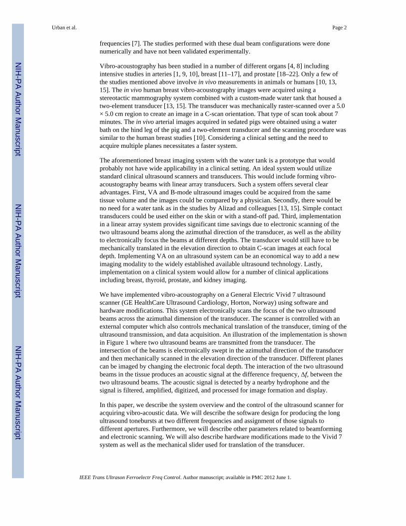
We have implemented vibro-acoustography on a General Electric Vivid 7 ultrasoundscanner (GE HealthCare Ultrasound Cardiology, Horton, Norway) using software andhardware modifications. This system electronically scans the focus of the two ultrasoundbeams across the azimuthal dimension of the transducer. The scanner is controlled with anexternal computer which also controls mechanical translation of the transducer, timing of theultrasound transmission, and data acquisition. An illustration of the implementation is shownin Figure 1 where two ultrasound beams are transmitted from the transducer. Theintersection of the beams is electronically swept in the azimuthal direction of the transducerand then mechanically scanned in the elevation direction of the transducer. Different planescan be imaged by changing the electronic focal depth. The interaction of the two ultrasoundbeams in the tissue produces an acoustic signal at the difference frequency, Δf, between thetwo ultrasound beams. The acoustic signal is detected by a nearby hydrophone and thesignal is filtered, amplified, digitized, and processed for image formation and display.
In this paper, we describe the system overview and the control of the ultrasound scanner foracquiring vibro-acoustic data. We will describe the software design for producing the longultrasound tonebursts at two different frequencies and assignment of those signals todifferent apertures. Furthermore, we will describe other parameters related to beamformingand electronic scanning. We will also describe hardware modifications made to the Vivid 7system as well as the mechanical slider used for translation of the transducer.
Urban et al. Page 2
IEEE Trans Ultrason Ferroelectr Freq Control. Author manuscript; available in PMC 2012 June 1.
NIH
-PA Author Manuscript
NIH
-PA Author Manuscript
NIH
-PA Author Manuscript

We will provide VA beamforming results with linear arrays, and we will show images ofphantoms as examples of the types of images acquired using this system.
MethodsSystem Overview
The four main components of the system are the control computer (PC), ultrasound scannerand transducer, mechanical slider, and hydrophone and associated electronics. The PC,which is external to the ultrasound scanner, uses an interface built using LabVIEW(National Instruments, Austin, TX). Through this LabVIEW interface, which was created byone of our team members at the Mayo Clinic, the control computer communicates with theVivid 7 scanner using a TCP/IP socket-based client-server protocol. The TCP/IP protocol issupported by the Vivid 7, but the specific interface that was used was proprietary to GE.Instructions are sent to the scanner to set and check parameters and initiate scanning. Theparameters include the ultrasound frequencies, the difference frequency, the beam repetitionrate, and the transmit focus depth. Once all the parameters are set for the scan, the scan isstarted. An instruction is sent to the scanner to transmit the beams that make up one rasterline of a C-scan image. To compare with conventional B-mode imaging, transmitting thebeams for one line in a VA C-scan is analogous to transmitting one frame for B-modeimaging. The scanner emits a trigger signal when each beam is transmitted, and that triggersignal is used to synchronize the digitizer in the PC for collecting the acoustic emission datafrom the hydrophone. After all the beams have been transmitted, the PC sends an instructionto the motor controller for the mechanical slider via an RS-232 connection, and the motormoves in the elevation direction of the transducer a specified distance. This process repeatsuntil the end of the scan. The acoustic emission data are collected, processed, and an imageis formed for display. A block diagram of the system is shown in Figure 2.
Software DesignSignal Generation—The transmit signals used to produce B-mode images in a digitalultrasound system such as the GE Vivid 7 typically consist of a short burst of square wavepulses with voltages on the order of ±100 V. These pulses, appropriately delayed, areapplied to the transducer elements where they are converted to sound at the desiredultrasound frequencies. Generally, all of the elements transmit identical waveforms. Forvibro-acoustography, we need to use two slightly different waveforms to produce the desireddifference frequency. Additionally, VA necessitates using long tonebursts on the order ofseveral hundred microseconds, much longer than conventional B-mode pulses. Softwaremodifications were required and made to the Vivid 7 by engineers at GE to make thispossible. These modifications could only be performed by GE engineers because they relateto the GE proprietary software that runs the normal function of this machine as anultrasound scanner.
The transmit circuitry of the Vivid 7 can create somewhat arbitrary pulse trains. This isaccomplished by specifying the duration of each half cycle of the waveform in terms ofperiods of a master clock at frequency Fc. An appropriate number of clock cycles is chosento produce the desired ultrasound frequency emitted by the transducer. We will refer to thisnumber of clock cycles as N. For a normal B-mode waveform, the number of half-cycles tobe transmitted is chosen to optimize the axial resolution or other imaging parameter(s), andthat number is typically very small. For vibro-acoustography, however, we need to transmittwo much longer tonebursts at different frequencies to produce the low frequency differencesignal. The two tonebursts at different frequencies are produced by occasionally lengtheningone or more of the half cycles of one or both of the waveforms relative to the otherwaveform. Doing this causes the frequency spectra of the two signals to shift with respect to
Urban et al. Page 3
IEEE Trans Ultrason Ferroelectr Freq Control. Author manuscript; available in PMC 2012 June 1.
NIH
-PA Author Manuscript
NIH
-PA Author Manuscript
NIH
-PA Author Manuscript

each other, resulting in the low frequency beat signal. On the Vivid 7 we can basicallyinstruct the transmitter circuitry to generate repeating “packets” of tone bursts. As in the B-mode case, the fundamental ultrasound frequency is specified by N (the number of clockcycles in a cycle). The number of cycles in a “packet” is referred to as K. Finally, thenumber of extra clock cycles to be added to each packet is referred to as L. Each of the twowaveforms is specified by a (N, K, L) tuple. In VA mode, the transmitter circuitry is set upto continuously output these packets for each beam for 800 μs, but the final output isamplitude modulated (shaded) by a single cycle of a 3 kHz raised sine wave, as will bediscussed later. The 800 μs value was implemented to provide flexibility for long tonebursts.Figure 3(a) illustrates the definitions of the parameters. The parameter (N1, K1, L1) describesone of the waveforms, and (N2, K2, L2) describes the other waveform.
The nominal ultrasound frequencies of the two waveforms are given as F1 = Fc/N1 and F2 =Fc/N2, where Fc is given in megahertz. The total number of clock cycles in a “packet”defined by (N, K, L) is KN + L, and the average number of clock cycles in a “packet” isgiven by (KN + L)/K. Thus the average periods of the two waveforms are given in units ofmicroseconds by
(1)
(2)
The center frequency of beams 1 and 2, given in megahertz, are f1 = 1/T1,avg and f2 = 1/T2,avg, respectively. The resulting beat frequency, Δf, is given in units of megahertz as
(3)
For example, if we assume Fc = 80 (in MHz) and set (N1, K1, L1) = (16, 6, 1) and (N2, K2,L2) = (16, 6, 0), then using these parameters we find that F1 and F2 are 5 MHz and T1,avg =0.2021 and T2,avg = 0.2000, which are expressed in microseconds, and f1 = 4.9480 MHz andf2 = 5.000 MHz. Using (3) yields Δf = 0.0515464 MHz = 51.5464 kHz. To illustrate thesignals for this case, we simulated the signals, and they are shown in Fig. 3(b) where eachdot represents one cycle of the 80 MHz clock.
The system-calculated beamforming delays are (essentially) independent of frequency, andthus there is no need to modify these calculations for vibro-acoustography. We simplychoose the transducer and an application setup appropriate for the anatomy to be studied,allow the system to scan in normal B-mode, and then issue commands to the system toconfigure itself for VA mode. These commands include the values for (N1, K1, L1), (N2, K2,L2), pulse repetition period, and frequency assignments for the elements. In VA mode thesystem is essentially used as a transmitter only, and the received image displayed on thescreen of the Vivid 7 is ignored.
Beamforming—Vibro-acoustography relies on the intersection of two beams. With lineararray transducers this is accomplished by dividing the active aperture into two subapertures.
Urban et al. Page 4
IEEE Trans Ultrason Ferroelectr Freq Control. Author manuscript; available in PMC 2012 June 1.
NIH
-PA Author Manuscript
NIH
-PA Author Manuscript
NIH
-PA Author Manuscript

The VA software allows arbitrary assignment of the two frequencies to the available 128system channels. We can write the pressure of these two apertures as
(4)
(5)
where Pi(r) and φi(r) (i = 1,2) are the amplitude and phase, respectively, at location r. Vibro-acoustography is based on the radiation force of ultrasound and the force can be written as[2, 23]
(6)
where F is the radiation force, dr is the drag coefficient, S is the surface area of an object, Eis the energy density and ⟨·⟩ is the short-term time average, p(r,t) is the total pressure atlocation r and time t (p(r,t) = p1(r,t) + p2(r,t)), ρ is the density of the medium and c is thesound speed of the medium. The radiation force is proportional to the short-term timeaverage of the squared total pressure which can be written as
(7)
where Δf = f1 − f2.
We will consider one configuration for frequency assignment in this paper. Thisconfiguration splits the active aperture into two equal subapertures that are adjacent, whichwe denote the Split (Spl) configuration. If we assume 128 channels, then 64 are assigned f1and the other 64 are assigned f2.
There are two methods by which we can move the common focal region of the two beams todifferent locations along the azimuthal dimension of the transducer. We can either use astatic active aperture and steer the two ultrasound beams to different locations, or we can fixthe focusing time delay law and translate the active aperture along the full aperture of thetransducer. In most cases during a scan, we perform both of these actions. For instance, theGeneral Electric 7L and 10L transducers have 192 elements, but we only have 128 channelsavailable so we use the multiplexer in the probes to switch the channels that are connected toelements in the transducer. So, in the case that we have 192 beams and 128 active channelsor elements we can translate the active aperture for 64 of these beams, maintaining aconstant focusing time delay law.
In Figure 4 we show a diagram of active aperture for each beam assuming 128 activeelements out of 192 total elements transmitting 192 beams. Therefore, the distance betweenbeams is the same as the pitch of the transducer. For the first 64 beams, the active aperture isstatic and the beams are steered. Then for beams 65–128, the active aperture is translatedacross the transducer one element per beam. The last 64 beams then have a static apertureand the beams are steered. The implication of using steering is that the amplitude of Δfcomponent may be different from beam to beam, leading to nonuniformity in the image. By
Urban et al. Page 5
IEEE Trans Ultrason Ferroelectr Freq Control. Author manuscript; available in PMC 2012 June 1.
NIH
-PA Author Manuscript
NIH
-PA Author Manuscript
NIH
-PA Author Manuscript

translating the active aperture, the variation in beam-to-beam amplitude should beminimized. It would be ideal to use a translating aperture for the entire scan line, but thatwould limit the field of view that we could interrogate, so steering is employed to obtaindata near the edges of the transducer.
Scanning Parameters—There are parameters related to the scanning and beamformingthat are selected by the user. The ultrasound transducer used is designated and informationrelated to the transducer is used for beamforming, particularly the pitch of the elements forfocusing time delay calculation. The practical ultrasound frequencies used are fundamentallylimited by the bandwidth of the transducer as well as the N parameter discussed in thesection on signal generation. The difference frequency is set by the (N, K, L) parameters asdetailed above. The focal depth for the C-scan plane can be set by the user. The pulserepetition period can also be set by the user. This parameter is important for a number ofreasons. It is directly related to maintaining time-averaged intensity values that are safe foruse in humans. It is also important in limiting heating to the transmit board and thetransducer. Lastly, the pulse repetition period must be long enough for the acoustic emissionto propagate to the hydrophone, and to allow any residual reverberating sound to dissipate.This time is typically on the order of a few milliseconds. The total time of a scan isproportional to the product of the number of beams, Nb, the number of elevation planes orlines in the image, Nl, and the pulse repetition period, Trep, or T = Nb·Nl·Trep. A typical scanwith Nb = 192, Nl = 200, and Trep = 2 ms takes 76.8 seconds.
Signal Analysis and Image Display—The Vivid 7, mechanical slider, and digitizer arecontrolled by a LabVIEW (National Instruments, Austin, Texas) program running on a PC.More detail about the hardware components are described in the next section. The frontpanel of a “virtual instrument” provides the interface for the user to specify scanningparameters for the beam formation and subsequent digitization. Once everything is set up,the Vivid 7 is instructed to generate a sequence of beams for an azimuthal scan. With eachbeam, a pulse signal is provided by the Vivid 7 to synchronize and trigger the digitizer fordata collection from the hydrophone. The data generated by this set of beams is a set of timeseries, which is transferred to the computer and stored on the hard drive. This two-dimensional (2D) set is processed to produce one line of the output display image bycalculating a root-mean-square (RMS) value for each beam. This value can be calculatedfrom the whole time-series or from a gated segment that represents an area of interest. Thisone-dimensional line, whose values are represented as intensity, is added to the displayimage. Then the probe motor is instructed to move to the next elevation imaging plane. Asthe process is repeated, the processing steps can be performed in real-time, and a 2D C-scandisplay is built up for display to the user as it is acquired.
Hardware DesignTransmit Signal—Typically the transmitted vibro-acoustic signal is a relatively long,shaded toneburst of greater than 100 μs. Shading of the toneburst is used to prevent transientturn-on/turn-off effects in the transducer from producing acoustic artifacts in the image.Since such a shaded toneburst signal could not be produced internally by the Vivid 7 system,external hardware was added to provide the necessary waveform. Additionally, to obtainadequate power levels for vibro-acoustography to be feasible in humans, an external powersupply was added to the system to replace the internal transmit power supply. Figure 5shows how the supplementary power supply was added. This was accomplished using afunction generator (Model 33120A, Agilent, Santa Clara, CA) driving a two-channel linearpower amplifier (Model LVC-623, AE Techron, Elkhart, IN). The raised sine wave outputsignal from the function generator is first split into inverted and non-inverted forms, whichthen drive the power amplifier inputs. The power amplifier outputs become the positive
Urban et al. Page 6
IEEE Trans Ultrason Ferroelectr Freq Control. Author manuscript; available in PMC 2012 June 1.
NIH
-PA Author Manuscript
NIH
-PA Author Manuscript
NIH
-PA Author Manuscript

(+Vps) and negative (−Vps) power supply inputs to the transmit board in the Vivid 7 system.The power amplifier outputs typically range up to a peak voltage of 30 V. To accommodatethe dynamic power supply signals, it was necessary to modify the transmit board byremoving much of the power supply lines’ on-board capacitance. Because the slew rate forthe transmit board power supply voltages was limited to 0.2 V/μs, due to circuit boardheating effects, the transmit toneburst was shaded using a single cycle of a raised sine waveat frequency 3 kHz, resulting in a toneburst duration of 333 μs.
Acoustic Emission Acquisition—The acoustic emission arising from the ultrasoundstimulation in a scanned object is detected by a sensitive hydrophone (Model 6050C, ITC,Santa Barbara, CA). The hydrophone’s signal is amplified and bandpass filtered (ModelSR650, Stanford Research Systems, Sunnyvale, CA) using a 10 kHz bandwidth and thendigitized with a 12-bit PC card digitizer (Model ATS330, AlazarTech, Montreal, QC,Canada).
Motion Control—Vibro-acoustography is a C-scan modality and to obtain different linesin the elevation direction, we need to mechanically translate the transducer over a specifieddistance. A slider mechanism, shown in Figure 6, was therefore developed which could becomputer-controlled and able to accommodate a variety of ultrasound probes. Themechanical slider consists of a miniature linear servo motor (Model MX80L, Parker Daedal,Irwin, PA) mounted to a plastic frame and driven by a servo controller (Model ViX250IH,Parker Daedal, Irwin, PA). The motor has a maximum travel of 50 mm in the elevationdirection, peak force of 12 N and 5 μm positional resolution. The plastic frame constructioneffectively isolates all electrical connections and exposed metal surfaces from user andpatient contact.
ExperimentsUltrasound Field Measurements
We simulated the pressure signal at the focus of a 10L transducer (GE HealthCareUltrasound Cardiology, Horton, Norway) using the Spl configuration. The simulations wereperformed using custom written code in MATLAB (The Mathworks, Natick, MA). Theultrasound pressure for the simulated case was measured using a needle hydrophone(HGL-0200, Onda Corporation, Sunnyvale, CA) having an active element with 0.200 mmdiameter and a 20 dB preamplifier (AH-2000, Onda Corporation, Sunnyvale, CA). Thehydrophone was placed at the focus of the two ultrasound beams.
To assess the resolution of the system, we evaluated the radiation force component at Δf forthe GE 10L transducer which has an azimuthal field of view of 39 mm and a bandwidth thatranges from 4–10 MHz. The pressure was measured using a needle hydrophone that wasscanned in the azimuthal/axial plane of the transducer. The ultrasound frequencies used were5.00 and 4.95 MHz and the difference frequency was 51.5 kHz. The transducer was focusedat a depth of z = 25 mm. To extract the difference frequency component, the pressure thatwas measured was squared and low-pass filtered as described in (7). The low-pass filter wasapplied to perform the short-term time average. A fast Fourier transform was performed andthe magnitude of the component at 51.5 kHz was extracted for each measurement point.
The need for high power necessitates an evaluation of the safety of this method for patients.We made measurements of the highest pressures and intensities that we could produce withthe Vivid 7 system using the parameters detailed above.
Urban et al. Page 7
IEEE Trans Ultrason Ferroelectr Freq Control. Author manuscript; available in PMC 2012 June 1.
NIH
-PA Author Manuscript
NIH
-PA Author Manuscript
NIH
-PA Author Manuscript

Phantom and Tissue ImagingA urethane breast phantom with distributed lesions (CIRS Model 013, Norfolk, CT) wasplaced in a large water tank and scanned with the 10L transducer. A picture of the phantomis shown in Figure 7(a). Front and side view diagrams of the breast phantom are shown inFigure 7(b) to depict the depth of each simulated lesion. The surface of the phantom wasplaced 10 mm from the transducer surface, where the front surface of the phantom isdepicted as the left edge in the side view of Figure 7(b). The transducer was mechanicallytranslated over the phantom. The data was recorded as discussed above in the section onacoustic emission acquisition. The hydrophone was placed underneath the phantom and outof the way of the ultrasound propagation. Images were acquired using the Spl configuration.
A human cadaveric prostate gland was excised and used for this study. This was incompliance with a protocol approved by the Mayo Clinic Institutional Review Board. Theprostate was fixed in formalin for one hour and then embedded in a gelatin mixture madefrom 300 Bloom gelatin powder (Sigma-Aldrich, St. Louis, MO) and glycerol (Sigma-Aldrich, St. Louis, MO) with concentrations of 10% by volume. A preservative of potassiumsorbate (Sigma-Aldrich, St. Louis, MO) was also added with a concentration of 10 g/L. Theprostate was degassed with gelatin before being put into a mold for setting of the gelatin. Aphotograph of the prostate is shown in Figure 7(c). The prostate was placed in a water tanklike the breast phantom. Images were acquired using the Spl configuration with the GE 7Ltransducer (GE HealthCare Ultrasound Cardiology, Horton, Norway). An x-ray image wasalso taken with a stereotactic mammography system (Mammotest™ System Fischer ImagingInc., Broomfield, CO) with an imaging field of 50.0 × 50.0 mm.
In our work we have found that a deterministic variation in the magnitude of the acousticemission signal along the azimuthal direction arises which we have not been able todefinitively identify its source. When the transducer is mechanically scanned, thesevariations manifest as streaks in the horizontal direction. The streaks are related to thebeamforming because when focal depth or configuration is changed, the streaks change in adeterministic way. We made detailed measurements of the ultrasound intensity field versusbeam position across the azimuthal direction of the transducer, and determined that slightintensity variations versus beam position are causing the streaks in the image. Thisphenomenon has to do with the way the internal beam forming is constructed. We havedevised a streak removal algorithm based on a moving window method that estimates thevertical amplitude variation profile and divides it from the image data. All the images shownhave been corrected for this streak artifact. We removed the background gray level toimprove the contrast, and gamma correction was applied to the images to improve thedynamic range for viewing.
ResultsBeamforming Results
Figure 8(a) shows the simulated pressure signal using the signals in Fig. 3(b) over twocycles of Δf. The signal in Figure 8(a) is expanded over a longer time than that shown inFigure 3(b) so that phase offsets that were used create the modulated ultrasound pressure canbe appreciated. Figure 8(b) shows the experimentally measured signal using the needlehydrophone. The waveforms in Figure 8 show the carrier signals and the modulation createdby the interaction of the two ultrasound beams. The measured signal has been shaded by asingle cycle of a 3 kHz raised sine wave, shown as the dashed line in Figure 8(b).
To evaluate the resolution of the system, the measured azimuthal and axial profiles for fieldfocused at x = 0 mm and z = 25 mm is shown in Figure 9. The values of the azimuthal,
Urban et al. Page 8
IEEE Trans Ultrason Ferroelectr Freq Control. Author manuscript; available in PMC 2012 June 1.
NIH
-PA Author Manuscript
NIH
-PA Author Manuscript
NIH
-PA Author Manuscript

elevational, and axial resolutions at −6 dB for were 0.573, 1.44, and 2.20 mm, respectively.The azimuthal sidelobes were −25.26 dB with respect to the main lobe.
We measured the pressure at the location of the highest pressure, which is shown in Figure10, using the 10L and the Onda hydrophone and found that the spatial-peak pulse-averageintensity Isppa = 51.3 W/cm2 when focused at 25.0 mm focal depth. The spatial-peaktemporal average for a line of VA imaging was Ispta = 0.173 W/cm2 assuming a beamrepetition period of 2 ms, which is lower than the derated Ispta,0.3 Food and DrugAdministration (FDA) regulatory limit of 0.72 W/cm2 [24]. The intensity values givenabove were not derated. The peak negative pressure was measured at 2.15 MPa at 5 MHz. Ifwe do not derate by 0.3 dB/cm/MHz, the mechanical index (MI) is 0.96, and if derating isperformed the MI decreases to 0.62. Both of these values are below the FDA regulatorylimit of 1.9 [24]. Thermal index calculations could be performed with an established methodbased on the work of Chen and colleagues [25].
Phantom and Tissue Imaging ResultsFigure 11 shows the result from imaging the urethane breast phantom. The transducer wasmechanically translated in the horizontal direction (y-direction), and the azimuthal directionof the transducer corresponds to the vertical axis of the image (x-direction). The image hasundergone streak correction. The two large lesions on the bottom of the image were in focusin these images. The brightness of the lesions and the presence of fine detailed edges areways to differentiate which lesions are in focus and which are not. Other lesions can bedetected such as those in the upper corners, which appear darker because they are deeper inthe phantom.
Figure 12 shows the images acquired from scanning the excised human prostate. Thetransducer was mechanically translated in the vertical direction (y-direction) and theazimuthal direction of the transducer corresponds to the horizontal axis of the image (x-direction). An x-ray image is shown in Figure 12(a). The original image and the imageobtained after correction for the streaks are shown in Figure 12(b) and 12(c), respectively.The vibro-acoustic images depict a differentiation of the central region and a region aroundthe edge of the prostate, where the central region appears smooth and the outer regionappears to have a different texture. The clusters of calcifications that appear in the x-rayimage are depicted very clearly in the VA images and the borders of those regions are verydistinct.
DiscussionWe have shown that the Vivid 7 is able to form the ultrasound fields used as an excitationsource for vibro-acoustography imaging and demonstrated good modulation of theultrasound pressure. The azimuthal resolution was sub-millimeter and the axial resolutionwas 2.20 mm which is much less than the 10 mm axial resolution found with confocal VAimaging [23]. The acoustic output results showed that VA as implemented on the Vivid 7 iswithin FDA regulatory limits for the 10L transducer as well as other transducers althoughthese data for other transducers were not shown.
Figure 11 shows the results of an imaging study of a urethane breast phantom. Two lowerlesions in the breast phantom are easily detectable with good edge definition. The lesions inthe top of the image do not appear the same as those in the lower portion of the image,mostly because they are out-of-plane in the axial direction.
Figure 12 shows results from imaging of an excised human prostate. The streaks, in thevertical direction, cause variations that reduce the contrast in the image. The streaks are best
Urban et al. Page 9
IEEE Trans Ultrason Ferroelectr Freq Control. Author manuscript; available in PMC 2012 June 1.
NIH
-PA Author Manuscript
NIH
-PA Author Manuscript
NIH
-PA Author Manuscript

observed outside of the prostate gland in the surrounding gelatin. Figure 12 shows anexcellent correlation between the VA images and the x-ray depicting calcifications. Also,the tissue detail in the different regions of the prostate is interesting. Though the prostatewould not normally be imaged with a linear array transducer with a linear type oftranslation, these images were shown to exhibit the capabilities of the system to differentiatesoft tissue from calcifications as well as different types of tissue within a given organ.
The source of the image contrast is a mixture of different factors including the mechanicalresponse of the tissue or calcifications and factors that affect the radiation force such as thereflectivity/absorption of the tissue. The interplay between these factors and the distributionof the point-spread function is complicated, so at times, the calcifications can have similarimage intensity compared to normal tissue. It should be noted that this prostate was fixed informalin for one hour so that may also have an effect on the imaging results. The differingappearances of the tissue in different regions of the prostate arise from the interactions of thetissue structure and the asymmetric point-spread function in the elevation/azimuthal plane.Also, the interactions of tissue at different axial locations influence the appearance of thetissue in the images. The short-depth-of-field (2.20 mm) compared to previous work with afocused confocal transducer which has a long depth of field (~10 mm), may play a role inthe appearance of the tissue as tissue at different axial depths influence the acoustic emissionsignal [26]. This is an active area of research by our group, and we are trying to betterunderstand the appearance of different types of tissue using these linear array transducers.
The implementation of VA on a clinical scanner allows for integration of both B-mode andVA using the same transducer and that combination of data may provide complementaryinformation. VA can also take advantage of other aspects of the Vivid 7 scanner such astransducer availability. The choice of probe technology is an important factor in the outcomeof vibro-acoustography implementation. In this paper, we focused on linear array probes.Other probe types, such as multi-row probes with larger number elements, may have a betterperformance because such probes allow beam focusing in both the azimuthal and elevationdirections. Further studies are needed to fully explore vibro-acoustography with such probestructures. To date, VA has been implemented with a phased array (M4S), several lineararray transducers (7L, 10L, M12L), a transrectal linear array probe (ERBL), and a custom-made 1.75D transducer array. These various transducers allow for different ultrasoundfrequencies and provide optimized resolution and contrast as well as varied applications forscanning of the thyroid, breast, prostate, kidney, liver, and other organs. Thisimplementation of VA on the Vivid 7 was accomplished primarily through softwaremodifications. VA takes advantage of the transmit beamformer already implemented on theVivid 7 for electronic focusing and scanning of the beam. This is important for saving timethat would previously have been spent in mechanical translation of a transducer in onedirection. It is also important to note that the assignment of the two subapertures for f1 and f2is completely arbitrary. This flexibility of the system allows for implementation of differentconfigurations, which may be useful when approaching different applications of this VAimaging method. In this paper, we mainly focused on split configuration. Alternativebeamforming options include symmetric and interlaced configurations. The symmetricconfiguration consists of separating the aperture into three sections, where the group ofelements in either end section is assigned to one frequency and the elements in the centersection are assigned to the other frequency. Another possible configuration is to divide theelements on the array into M adjacent sections, with sections alternately assigned to one orthe other frequency. One may also assign the two frequencies to elements in alternating orrandom fashion. We have studied the feasibility of these configurations and determinedadvantages and disadvantages of each method. However, the discussion on suchcomparisons is beyond the scope of this paper.
Urban et al. Page 10
IEEE Trans Ultrason Ferroelectr Freq Control. Author manuscript; available in PMC 2012 June 1.
NIH
-PA Author Manuscript
NIH
-PA Author Manuscript
NIH
-PA Author Manuscript

There are some limitations for implementation of VA on this clinical scanner. We did haveto supplement the power supply with an amplifier to achieve power levels adequate for VAimaging. In our study of the GE Vivid 7 system, we determined that a supplementary powersupply was needed to generate the appropriate output power without the drooping effect thatwe observed with original power supply. We evaluated a few different power amplifiersbefore settling on the one described above. This addition of an extra power supply could beavoided in the future either by implementation on a different scanner that has a more robustpower supply or by redesigning the power supply of the existing scanner.
In theory, a 2D image could be acquired in the same plane as the B-mode image by focusingat all points in the VA image. However, VA is primarily a C-scan modality. We choose tokeep a consistent focal plane and calculate the delays needed to focus at points along theazimuth of the transducer. So if a scan uses Nb = 192 beams, only 192 sets of delays need tobe accessed during scanning. The delays are only recalculated if the focal depth plane ischanged. If a 2D image were to be acquired in the B-mode imaging plane, with Nd levels inthe axial direction, the number of sets of time delays that would need to be accessed wouldbe Nb·Nd. This would potentially be too large a number for the Vivid 7 to store in memoryand scanning may be slowed by accessing these delay values. Also, the excitation used tomake each pixel could have a very different point-spread function, especially as the depth isincreased. These variations may have detrimental effects on the image quality.
Because VA is inherently a C-scan method, we need to translate the transducer in thetransducer’s elevation direction. This involves a full motorized assembly to accomplish thistask. We also need the hydrophone to be in contact with the patient during scanning. This isnot a significant issue but placement of the hydrophone may have to be optimized for eachapplication.
In these studies an external PC was used for control, data acquisition, and display of the VAimages. In an actual commercial realization of VA all of these functions could beaccomplished using the scanner’s internal CPU. This would greatly simplify its use in aclinical setting, and allow, for example, simultaneous display of B-mode and VA images.
The implementation of VA on the Vivid 7 provides an avenue for a more transparenttranslation of VA into a clinical setting particularly for breast, prostate, thyroid, kidney, andliver imaging. Optimization of VA in these different applications depends on differentparameters such as the ultrasound frequencies, difference frequency, transducer geometryand hydrophone position. We also have the opportunity to change the apertures for the twoultrasound beams to improve resolution and contrast in the images.
ConclusionWe have described the design and implementation of vibro-acoustography on a clinical GEVivid 7 scanner. We modified the transmit signals to produce a difference frequency in thekilohertz range and demonstrated the beamforming design for VA using a linear arraytransducer. We evaluated the beamforming with experimental measurements with a needlehydrophone. Lastly, we showed imaging results using a breast phantom and excised humanprostate. The implementation of VA on a clinical scanner provides a substantial opportunityfor translation of this imaging modality for clinical imaging of various organs.
AcknowledgmentsThe authors wish to acknowledge Dr. Shigao Chen and the acoustic output measurements that he made. This workwas supported in part by grants CA121579, CA127235, and CA091956 from the National Institutes of Health.
Urban et al. Page 11
IEEE Trans Ultrason Ferroelectr Freq Control. Author manuscript; available in PMC 2012 June 1.
NIH
-PA Author Manuscript
NIH
-PA Author Manuscript
NIH
-PA Author Manuscript

References1. Fatemi M, Greenleaf JF. Ultrasound-stimulated vibro-acoustic spectrography. Science. Apr 3.1998
280:82–5. [PubMed: 9525861]2. Fatemi M, Greenleaf JF. Vibro-acoustography: An imaging modality based on ultrasound-
stimulated acoustic emission. Proc Natl Acad Sci U S A. Jun 8.1999 96:6603–8. [PubMed:10359758]
3. Fatemi M, Manduca A, Greenleaf JF. Imaging elastic properties of biological tissues by low-frequency harmonic vibration. Proc IEEE. Oct.2003 91:1503–1519.
4. Alizad A, Wold LE, Greenleaf JF, Fatemi M. Imaging mass lesions by vibro-acoustography:modeling and experiments. IEEE Trans Med Imaging. Sep.2004 23:1087–93. [PubMed: 15377117]
5. Silva GT, Chen S, Frery AC, Greenleaf JF, Fatemi M. Stress field forming of sector arraytransducers for vibro-acoustography. IEEE Trans Ultrason Ferroelectr Freq Control. Nov.200552:1943–51. [PubMed: 16422406]
6. Silva GT, Greenleaf JF, Fatemi M. Linear arrays for vibro-acoustography: a numerical simulationstudy. Ultrason Imaging. Jan.2004 26:1–17. [PubMed: 15134390]
7. Heikkila J, Hynynen K. Investigation of optimal method for inducing harmonic motion in tissueusing a linear ultrasound phased array - A simulation study. Ultrason Imaging. Apr.2006 28:97–113. [PubMed: 17094690]
8. Alizad A, Fatemi M, Nishimura RA, Kinnick RR, Rambod E, Greenleaf JF. Detection of calciumdeposits on heart valve leaflets by vibro-acoustography: an in vitro study. J Am Soc Echocardiogr.Nov.2002 15:1391–5. [PubMed: 12415234]
9. Alizad A, Fatemi M, Whaley DH, Greenleaf JF. Application of vibro-acoustography for detection ofcalcified arteries in breast tissue. J Ultrasound Med. Feb.2004 23:267–73. [PubMed: 14992365]
10. Pislaru C, Kantor B, Kinnick RR, Anderson JL, Aubry MC, Urban MW, Fatemi M, Greenleaf JF.In vivo vibroacoustography of large peripheral arteries. Invest Radiol. Apr.2008 43:243–252.[PubMed: 18340248]
11. Alizad A, Fatemi M, Wold LE, Greenleaf JF. Performance of vibro-acoustography in detectingmicrocalcifications in excised human breast tissue: a study of 74 tissue samples. IEEE Trans MedImaging. Mar.2004 23:307–12. [PubMed: 15027523]
12. Alizad A, Whaley DH, Greenleaf JF, Fatemi M. Potential applications of vibro-acoustography inbreast imaging. Technol Cancer Res Treat. Apr.2005 4:151–8. [PubMed: 15773784]
13. Alizad A, Whaley DH, Greenleaf JF, Fatemi M. Critical issues in breast imaging by vibro-acoustography. Ultrasonics. Dec 22; 2006 44(Suppl 1):e217–20. [PubMed: 16843513]
14. Hosseini HG, Alizad A, Fatemi M. Integration of vibro-acoustography imaging modality with thetraditional mammography. Int J Biomed Imaging. 2007; 2007:40980. [PubMed: 17710254]
15. Alizad A, Whaley DH, Greenleaf JF, Fatemi M. Image features in medical vibro-acoustography: Invitro and in vivo results. Ultrasonics. 2008; 48:559–562. [PubMed: 18599102]
16. Fatemi M, Wold LE, Alizad A, Greenleaf JF. Vibro-acoustic tissue mammography. IEEE TransMed Imaging. Jan.2002 21:1–8. [PubMed: 11838661]
17. Urban MW, Silva GT, Fatemi M, Greenleaf JF. Multifrequency vibro-acoustography. IEEE TransMed Imaging. Oct.2006 25:1284–95. [PubMed: 17024832]
18. Mitri FG, Trompette P, Chapelon JY. Improving the use of vibro-acoustography for brachytherapymetal seed imaging: a feasibility study. IEEE Trans Med Imaging. Jan.2004 23:1–6. [PubMed:14719682]
19. Mitri FG, Davis BJ, Alizad A, Greenleaf JF, Wilson TM, Mynderse LA, Fatemi M. Prostatecryotherapy monitoring using vibroacoustography: preliminary results of an ex vivo study andtechnical feasibility. IEEE Trans Biomed Eng. Nov.2008 55:2584–2592. [PubMed: 18990628]
20. Mitri FG, Davis BJ, Urban MW, Alizad A, Greenleaf JF, Lischer GH, Wilson TM, Fatemi M.Vibro-acoustography imaging of permanent prostate brachytherapy seeds in an excised humanprostate - Preliminary results and technical feasibility. Ultrasonics. Mar.2009 49:389–394.[PubMed: 19062061]
Urban et al. Page 12
IEEE Trans Ultrason Ferroelectr Freq Control. Author manuscript; available in PMC 2012 June 1.
NIH
-PA Author Manuscript
NIH
-PA Author Manuscript
NIH
-PA Author Manuscript

21. Mitri FG, Davis BJ, Greenleaf JF, Fatemi M. In vitro comparative study of vibro-acoustographyversus pulse-echo ultrasound in imaging permanent prostate brachytherapy seeds. Ultrasonics. Jan.2009 49:31–38. [PubMed: 18538365]
22. Alizad A, Mitri FG, Davis BJ, Sebo T, Kinnick R, Greenleaf JF, Fatemi M. Prostate tissue imagingby vibro-acoustography. IEEE Trans Med Imaging. In press.
23. Chen S, Fatemi M, Kinnick R, Greenleaf JF. Comparison of stress field forming methods forvibro-acoustography. IEEE Trans Ultrason Ferroelectr Freq Control. Mar.2004 51:313–21.[PubMed: 15128218]
24. Herman BA, Harris GR. Models and regulatory considerations for transient temperature rise duringdiagnostic ultrasound pulses. Ultrasound Med Biol. 2002; 28:1217–24. [PubMed: 12401393]
25. Chen S, Aquino W, Alizad A, Urban MW, Kinnick RR, Greenleaf JF, Fatemi M. Thermal safety ofvibro-acoustography using a confocal transducer. Ultrasound Med Biol. 2010; 36
26. Silva GT, Frery AC, Fatemi M. Image formation in vibro-acoustography with depth-of-fieldeffects. Comput Med Imaging Graph. Jul.2006 30:321–7. [PubMed: 16949793]
Urban et al. Page 13
IEEE Trans Ultrason Ferroelectr Freq Control. Author manuscript; available in PMC 2012 June 1.
NIH
-PA Author Manuscript
NIH
-PA Author Manuscript
NIH
-PA Author Manuscript

Figure 1.Block diagram of vibro-acoustography performed with a linear array transducer. Theintersection of the two ultrasound beams is electronically swept in the azimuthal direction ofthe transducer and then mechanically scanned in the elevation direction of the transducer.Different planes can be imaged by changing the electronic focal depth. The interaction of thetwo ultrasound beams in the tissue produces an acoustic signal at the difference frequency,Δf, between the two ultrasound beams. The acoustic signal is detected by a nearbyhydrophone and the signal is filtered, amplified, digitized, and processed for imageformation and display.
Urban et al. Page 14
IEEE Trans Ultrason Ferroelectr Freq Control. Author manuscript; available in PMC 2012 June 1.
NIH
-PA Author Manuscript
NIH
-PA Author Manuscript
NIH
-PA Author Manuscript

Figure 2.Block diagram of vibro-acoustography implementation with the General Electric Vivid 7.The control and analysis computer communicates with the Vivid 7 via TCP/IP protocol.Once an instruction from the computer is sent to the Vivid 7, a synchronization trigger(Sync) was sent to the digitizer and a waveform generator used to shade the poweramplifier’s output. The power amplifier’s output served as a power supply for the transmitboard in the Vivid 7. The control and analysis computer also controlled the motion controlsystem which operated the mechanical slider with the ultrasound transducer.
Urban et al. Page 15
IEEE Trans Ultrason Ferroelectr Freq Control. Author manuscript; available in PMC 2012 June 1.
NIH
-PA Author Manuscript
NIH
-PA Author Manuscript
NIH
-PA Author Manuscript

Figure 3.Waveform generation for vibro-acoustography. (a) The waveforms are defined by theparameters (N1, K1, L1) and (N2, K2, L2). FC represents the transmitter generator systemclock. (b) Simulated signals for (N1, K1, L1) = (16, 6, 1) and (N2, K2, L2) = (16, 6, 0). Eachdot represents one cycle of the 80 MHz clock.
Urban et al. Page 16
IEEE Trans Ultrason Ferroelectr Freq Control. Author manuscript; available in PMC 2012 June 1.
NIH
-PA Author Manuscript
NIH
-PA Author Manuscript
NIH
-PA Author Manuscript

Figure 4.Aperture assignment for 128 active elements in a 192 element transducer. The number ofbeams transmitted is 192. Gray denotes elements assigned a signal with f1, white areelements assigned a signal with f2 and black are not used. For beams 1–64 and 129–192 boththe subapertures are steered and for beams 65–128, the subapertures are translated.
Urban et al. Page 17
IEEE Trans Ultrason Ferroelectr Freq Control. Author manuscript; available in PMC 2012 June 1.
NIH
-PA Author Manuscript
NIH
-PA Author Manuscript
NIH
-PA Author Manuscript

Figure 5.Transmit signal chain. A function generator output is split into inverted and noninvertedsignals and amplified by a two-channel amplifier which provides the power supply voltagesfor the transmit board for the GE Vivid 7 scanner.
Urban et al. Page 18
IEEE Trans Ultrason Ferroelectr Freq Control. Author manuscript; available in PMC 2012 June 1.
NIH
-PA Author Manuscript
NIH
-PA Author Manuscript
NIH
-PA Author Manuscript

Figure 6.Motorized translation stage for moving the linear array transducer. The probe is held in afixture attached to the linear motor stage. The linear motor stage is mounted on a plasticsupport frame.
Urban et al. Page 19
IEEE Trans Ultrason Ferroelectr Freq Control. Author manuscript; available in PMC 2012 June 1.
NIH
-PA Author Manuscript
NIH
-PA Author Manuscript
NIH
-PA Author Manuscript

Figure 7.Photographs of objects used for imaging. (a) Breast phantom, (b) Diagram of breastphantom to scale with front and side views. The front view is scaled to be 40 × 80 mm andthe side view is scaled to be 40 × 43 mm. In the side view, the left edge is the front of thephantom and is closest to the transducer. The vertical dashed line in the side viewillustration depicts the location of the focal plane, about 15 mm from the surface of thephantom. During imaging, the transducer was offset from the front edge by 10 mm. (c)Excised human prostate.
Urban et al. Page 20
IEEE Trans Ultrason Ferroelectr Freq Control. Author manuscript; available in PMC 2012 June 1.
NIH
-PA Author Manuscript
NIH
-PA Author Manuscript
NIH
-PA Author Manuscript

Figure 8.Pressure signals from vibro-acoustography beamforming. (a) Simulated pressure usingsignals from Fig. 3(b). Two cycles of modulated ultrasound with Δf = 51.5464 kHz areshown. (b) Measured pressure with a needle hydrophone for a full toneburst which wasshaded by the raised 3 kHz signal depicted as a dashed line. Note that the time scales aredifferent for each plot.
Urban et al. Page 21
IEEE Trans Ultrason Ferroelectr Freq Control. Author manuscript; available in PMC 2012 June 1.
NIH
-PA Author Manuscript
NIH
-PA Author Manuscript
NIH
-PA Author Manuscript

Figure 9.Experimentally measured azimuthal and axial profiles for the Spl configuration. The field isfocused at x = 0 mm and z = 25 mm. The magnitudes are independently normalized. (a)Azimuthal, (b) Axial.
Urban et al. Page 22
IEEE Trans Ultrason Ferroelectr Freq Control. Author manuscript; available in PMC 2012 June 1.
NIH
-PA Author Manuscript
NIH
-PA Author Manuscript
NIH
-PA Author Manuscript

Figure 10.Experimentally measured pressure at location of spatial peak pressure. The field is focusedat x = 0 mm and z = 25 mm.
Urban et al. Page 23
IEEE Trans Ultrason Ferroelectr Freq Control. Author manuscript; available in PMC 2012 June 1.
NIH
-PA Author Manuscript
NIH
-PA Author Manuscript
NIH
-PA Author Manuscript

Figure 11.Breast phantom images. The azimuthal direction of the transducer corresponds to thevertical axis of the image and the mechanical translation of the transducer in the elevationdirection corresponds to the horizontal axis. The image is 38.4 × 80 mm and streakcorrection has been applied.
Urban et al. Page 24
IEEE Trans Ultrason Ferroelectr Freq Control. Author manuscript; available in PMC 2012 June 1.
NIH
-PA Author Manuscript
NIH
-PA Author Manuscript
NIH
-PA Author Manuscript

Figure 12.Excised human prostate images. (a) X-ray image of prostate gland. Calcium clusters aremarked with the designation Ca in this panel and all other panels. Wire loops in upper rightand lower left corners are fiducial markers. Image is 50.0 × 50.0 mm, (b) Original VAimage, (b) VA image after streak correction. The azimuthal direction of the transducercorresponds to the horizontal axis of the image and the mechanical translation of thetransducer in the elevation direction corresponds to the vertical axis. Images are 46.7 × 60mm.
Urban et al. Page 25
IEEE Trans Ultrason Ferroelectr Freq Control. Author manuscript; available in PMC 2012 June 1.
NIH
-PA Author Manuscript
NIH
-PA Author Manuscript
NIH
-PA Author Manuscript