hiv-1 quasispecies delineation by tag linkage deep sequencing
TRANSCRIPT
HIV-1 Quasispecies Delineation by Tag Linkage DeepSequencingNicholas C. Wu1,2, Justin De La Cruz3, Laith Q. Al-Mawsawi1, C. Anders Olson1, Hangfei Qi1,
Harding H. Luan1, Nguyen Nguyen1, Yushen Du1, Shuai Le4, Ting-TingWu1, Xinmin Li5, Martha J. Lewis6,7,
Otto O. Yang3,6,7,8, Ren Sun1,2,7*
1Department of Molecular and Medical Pharmacology, David Geffen School of Medicine, University of California Los Angeles, Los Angeles, California, United States of
America, 2Molecular Biology Institute, University of California Los Angeles, Los Angeles, California, United States of America, 3Department of Microbiology, Immunology,
and Molecular Genetics, David Geffen School of Medicine, University of California Los Angeles, Los Angeles, California, United States of America, 4Department of
Microbiology, Third Military Medical University, Chongqing, China, 5Department of Pathology and Laboratory Medicine, David Geffen School of Medicine, University of
California Los Angeles, Los Angeles, California, United States of America, 6Division of Infectious Diseases, Department of Medicine, David Geffen School of Medicine,
University of California Los Angeles, Los Angeles, California, United States of America, 7AIDS Institute, University of California Los Angeles, Los Angeles, California, United
States of America, 8AIDS Healthcare Foundation, Los Angeles, California, United States of America
Abstract
Trade-offs between throughput, read length, and error rates in high-throughput sequencing limit certain applications suchas monitoring viral quasispecies. Here, we describe a molecular-based tag linkage method that allows assemblage of shortsequence reads into long DNA fragments. It enables haplotype phasing with high accuracy and sensitivity to interrogateindividual viral sequences in a quasispecies. This approach is demonstrated to deduce ,2000 unique 1.3 kb viral sequencesfrom HIV-1 quasispecies in vivo and after passaging ex vivo with a detection limit of ,0.005% to ,0.001%. Reproducibilityof the method is validated quantitatively and qualitatively by a technical replicate. This approach can improve monitoring ofthe genetic architecture and evolution dynamics in any quasispecies population.
Citation: Wu NC, De La Cruz J, Al-Mawsawi LQ, Olson CA, Qi H, et al. (2014) HIV-1 Quasispecies Delineation by Tag Linkage Deep Sequencing. PLoS ONE 9(5):e97505. doi:10.1371/journal.pone.0097505
Editor: Noam Shomron, Tel Aviv University, Israel, Israel
Received January 20, 2014; Accepted April 17, 2014; Published May 19, 2014
Copyright: � 2014 Wu et al. This is an open-access article distributed under the terms of the Creative Commons Attribution License, which permits unrestricteduse, distribution, and reproduction in any medium, provided the original author and source are credited.
Funding: This work was supported by UCLA Molecular Biology Whitcome Fellowship (N.C.W.), National Institutes of Health R01 AI043203 (O.O.Y). The fundershad no role in study design, data collection and analysis, decision to publish, or preparation of the manuscript.
Competing Interests: The authors have declared that no competing interests exist.
* E-mail: [email protected]
Introduction
Many viruses have such high replication and mutation rates that
they exist as a quasispecies in vivo [1]. A viral quasispecies
population contains a variety of genotypic variants that are
related by similar mutations and exist in varying abundance
depending on their relative fitness within the host environment. In
this report, we refer to viral quasispecies as the whole population of
genotypic variants, whereas viral sequence is defined as the
individual viral variant within quasispecies population. Viral
sequence variation in the quasispecies population can be rapidly
generated by point mutation and/or recombination [1,2].
Mutation rates can be as high as in the order of one per
replication cycle, in which the progeny virus is unlikely to be
identical to its parental template. This diverse array of viral
sequences permits robust adaptation and evolution.
Often, genotypes with a particular set of mutations gain a
significant fitness advantage through synergistic phenotypic effect
among multiple mutations, which is also known as epistasis.
Epistasis has an important role in host adaptation and may drive
evolution towards drug resistance and immune evasion [3–7]. In
many cases, virus drug resistance requires two or more mutations
in concert, especially when multiple drugs are applied simulta-
neously [7–9]. Therefore, monitoring individual viral haplotypes
in the quasispecies populations within patients is important to
estimate the risk of viral rebound and further provide customized
treatment [10]. Characterizing the population structure of viral
quasispecies in the host also helps to understand the evolutionary
landscape and cis-interactions among genetic elements.
Clonal sequencing has been frequently employed to examine
the genetic makeup of individual viruses within a quasispecies
population. However, clonal sequencing has a low throughput and
a high sequencing cost per nucleotide. It limits the number of viral
sequences, hence haplotype variants, being genetically interrogat-
ed. On the other hand, next generation sequencing (NGS)
technology provides enough throughput and sensitivity to detect
very rare viral mutations. Nevertheless, the short read lengths of
NGS pose a challenge in reconstruction of individual viral
sequences within a viral quasispecies. First of all, it is often
difficult to distinguish rare mutations that exist in the quasispecies
population with sequencing errors from NGS. Secondly, haplo-
type phasing is extremely challenging when mutations are sporadic
and are separated by long, highly conserved or even completely
identical regions. These technical challenges make it extremely
difficult to reconstruct viral quasispecies from NGS data.
Existing methods in reconstructing viral quasispecies from NGS
platforms rely heavily on computational tools, including the
development of read graph-based or probabilistic-based algo-
rithms that utilize the information from overlapping reads [11–
20]. Although they provide an approximation of haplotype
PLOS ONE | www.plosone.org 1 May 2014 | Volume 9 | Issue 5 | e97505
information present in a viral quasispecies, the sensitivity and
accuracy vary depending on sequencing error rate and quasis-
pecies diversity. As a result, it is critical to develop a viral
quasispecies recontruction method with higher sensitivity and
accuracy in both mutation calling and haplotype phasing.
In order to genetically define a viral quasispecies population, we
developed a novel analytical technique to assemble short Illumina
amplicon sequence reads derived from individual viral sequences.
In contrast to algorithmic-based methods for quasispecies recon-
struction, tag linkage approach is a molecular-based approach. To
the best of our knowledge, this is the first experimental approach
that specialized in quasispecies reconstruction. The methodology
consists of three key steps: 1) Assigning unique tags to individual
viral sequences to distinguish each variant within the viral
quasispecies, 2) Controlling the complexity of the library during
amplification to ensure sufficient coverage for sampled viral
sequences, and 3) Using a tag linkage strategy to deduce the full-
length templates from non-overlapping amplicons. Here, we
provide a proof-of-concept study by utilizing this approach to
genetically characterize an HIV-1 quasispecies population under
two conditions: an isolated in vivo virus population and the virus
population derived from the same chronically infected HIV-1
patient passaged ex vivo in cell culture. We achieve a detection limit
of ,0.005% to ,0.001%. The reproducibility is validated with a
technical replicate. Overall, this approach enables accurate
haplotype phasing with very high sensitivity.
Results
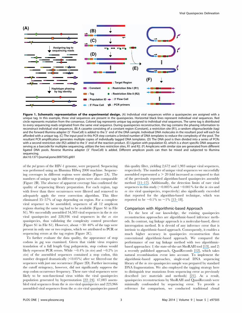
Library Preparation for SequencingThe underlying rationale is to assign a unique tag to individual
viral sequences within the quasispecies and to distribute the tag to
every sequencing read originated from the same viral sequence
(Figure 1A). Individual viral sequences within the quasispecies can
be assembled by grouping sequencing reads that share the same
tag. As a result, the tag linkage approach described in this study
permits reconstruction of individual viral sequences from NGS
reads despite the lack of overlap.
The workflow for sequencing library preparation is summarized
in Figure 1B–F. Briefly, individual DNA molecules are assigned a
unique tag by PCR (Figure 1B). The tag consists of a 13 ‘‘N’’
sequence that allows distinguishing 413 & 70 million molecules.
After tagging individual DNA molecules within the pool, the
complexity of the pool is being controlled. Complexity is defined
as the number of tagged DNA molecules being processed after the
first round of PCR. Thus, the more tagged molecules are being
processed, the higher the complexity becomes. If complexity is too
high, individual tagged molecules will not be covered repeatedly,
leading to a failure in assemble individual DNA molecules (Figure
S1A in File S1). On the other hand, if complexity is too low,
sequencing capacity will be wasted due to redundant sequencing
coverage of individual tagged DNA molecules being processed
(Figure S1B in File S1). Nonetheless, for quasispecies determina-
tion, it is more detrimental if the complexity is too high versus too
low because excessive complexity will abolish the sequence
assembly process (Figure S1 in File S1). In general, the relationship
between complexity and expected coverage for an individual viral
sequence can be calculated with the expected sequencing output:
Coverage
~(Sequencing output)=(Complexity
|Length of region of interest):
In this formula, sequencing capacity and length of region of
interest can be predetermined. Therefore, complexity is estimated
solely based on the desired coverage of each tagged DNA
molecules. For example, if the region of interest is 1 kb and 1 Gb
of sequencing output is expected, then a complexity of 100,000
gives on average 10-fold coverage for individual tagged DNA
molecules being processed. With sufficient coverage for an
individual viral sequence, we can distinguish sequencing error
from true mutation as described previously [21], in addition to
haplotype phasing. Therefore, complexity control represents a
critical step in our experimental design.
After controlling the complexity, a PCR is performed to
generate multiple copies of individually tagged DNA molecules
(Figure 1C). The resultant DNA pool is then divided into a series
of PCRs to generate products with different lengths (Figure 1D).
For every pool, the resultant PCR products contain two different
restriction sites on each ends. Next, restriction enzyme digestions
generate two sticky ends and remove the constant region for PCR
in the earlier step. A self-ligation step follows with the addition of a
short insert (Figure 1E). The short insert can serve as a barcode for
multiplex sequencing. This ligation step circularizes the DNA,
resulting in different sequence regions being proximal to the tag
and further allowing linkage formation between any distal region
with the tag - another key step in our experimental design. In the
final step, a short amplicon (,200 bp) is recovered for NGS
(Figure 1F). Each NGS read, from 59 to 39, will cover a tag for
short read assembly within a quasispecies sample, a barcode for
quasispecies sample identification, and a particular region of
interest on the targeted viral sequence. NGS reads sharing the
same tag belong to the same DNA molecules. Therefore,
haplotypes of individual viral genomes within the quasispecies
population can be interrogated. A more detailed schematic
representation of the key steps in our approach is shown in Figure
S2 in File S1.
Assembly of Two HIV-1 Viral QuasispeciesVirus derived from a chronically infected HIV-1 patient was
analyzed before (in vivo) and after (ex vivo) cell culture passaging for
10 weeks. In vivo virus sample represented the viral quasispecies
within the HIV-1 infected patient. Whereas in ex vivo passaging,
virus from the same patient was passaged serially in primary CD4+
T lymphocytes from an HIV-1-uninfected donor and reflected the
evolution of the viral quasispecies population in the absence of
intra-patient selection pressure. We limited the complexity by
processing roughly 300,000 viral sequences to ensure sufficient
coverage (,50-fold) in all regions for any given viral sequence
(Figure 1B).
50-fold coverage
~(1:8 Gb sequencing output)=(300,000 complexity
|1200 bp region of interest length):
Twelve non-overlapping amplicons, which cover a 1,295
nucleotide stretch and encompass most of the gag and a portion
Viral Quasispecies Delineation
PLOS ONE | www.plosone.org 2 May 2014 | Volume 9 | Issue 5 | e97505
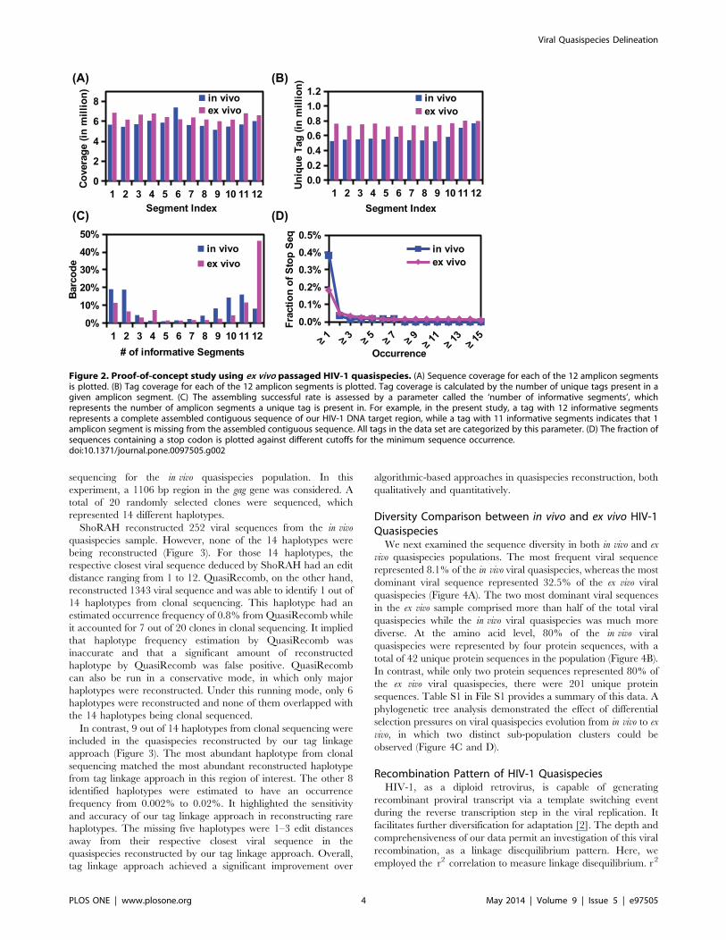
of the pol genes of the HIV-1 genome, were prepared. Sequencing
was performed using an Illumina HiSeq 2000 machine. Sequenc-
ing coverages in different regions were similar (Figure 2A). The
numbers of unique tags in different regions were also comparable
(Figure 2B). The absence of apparent coverage bias confirmed the
quality of sequencing library preparation. For each region, tags
with fewer than three occurrences were filtered and removed to
adequately apply the error correction algorithm. This filter
eliminated 35–57% of tags depending on region. For a complete
viral sequence to be assembled, sequences of all 12 amplicon
regions sharing the same tag had to be available (Figure S1 in File
S1). We successfully assembled 54,583 viral sequences in the in vivo
viral quasispecies and 228,936 viral sequences in the ex vivo
quasispecies, thus validating the complexity control procedure
(Figure S1 in File S1). However, about ,30–40% of the tags were
present in only one or two regions, which we attributed to PCR or
sequencing errors at the tag region (Figure 2C).
To further evaluate the data quality, the appearance of stop
codons in gag was examined. Given that viable virus requires
translation of a full length Gag polyprotein, stop codons would
likely represent PCR errors. While ,0.4% (in vivo) and ,0.2% (ex
vivo) of the assembled sequences contained a stop codon, this
number dropped dramatically (,0.05%) after we filtered-out the
sequences with just one occurrence (Figure 2D). Further increasing
the cutoff stringency, however, did not significantly suppress the
stop codon occurrence frequency. These rare viral sequences were
likely to be non-functional virus within the viral quasispecies
population generated by hypermutation [22–24]. 47,083 assem-
bled viral sequences from the in vivo viral quasispecies and 223,966
assembled viral sequences from the ex vivo viral quasispecies passed
this quality filter, yielding 2,672 and 1,983 unique viral sequences,
respectively. The number of unique viral sequences we successfully
assembled represented a . 20 fold increased as compared to that
of the previously reported algorithm-based quasispecies assembly
method [11–17]. Additionally, the detection limits of rare viral
sequences in this study (,0.005% and,0.001% for the in vivo and
ex vivo viral quasispecies, respectively) also significantly exceeded
that reported for the algorithm-based technique, which was
reported to be ,0.1% to ,1% [15–18].
Comparison with Algorithmic-based ApproachTo the best of our knowledge, the existing quasispecies
reconstruction approaches are algorithmic-based inference meth-
ods. In contrast, tag linkage approach is a molecular-based, direct
interrogation method. It is devoid of any inference error that is
intrinsic to algorithmic-based approach. Consequently, it enables a
much higher accuracy in quasispecies reconstruction than
conventional algorithmic-based approach. We compared the
performance of our tag linkage method with two algorithmic-
based approaches: 1) the state-of-the-art ShoRAH tool [13], and 2)
a recently published approach, QuasiRecomb [12], which takes
natural recombination event into account. To implement the
algorithmic-based approaches, single-read DNA sequencing
library of the in vivo quasispecies sample was prepared by standard
DNA fragmentation. We also employed the tagging strategy here
to distinguish true mutations from sequencing error as previously
described (see materials and methods) [21]. As a result,
quasispecies reconstructions by ShoRAH and QuasiRecomb were
minimally confounded by sequencing error. To provide a
reference for comparison, we conducted traditional clonal
Figure 1. Schematic representation of the experimental design. (A) Individual viral sequences within a quasispecies are assigned with aunique tag. In this example, three viral sequences are present in the quasispecies. Horizontal black lines represent individual viral sequences. Redcircle represents mutation from the consensus. Colored tag represents unique tag assigned to individual viral sequences. The same tag is distributedto every sequencing reads originated from the same viral sequence. During quasispecies reconstruction, the tag contains the phasing information toreconstruct individual viral sequences. (B) A cassette consisting of a constant region (Constant), a restriction site (R1), a random oligonucleotide (tag)and the forward Illumina adapter (59 FlowCell) is added to the 59 end of the DNA sample. Individual DNA molecules in the resultant pool will each beafforded with a unique tag. (C) The input pool in this PCR step contains a limited number of DNA templates to reduce the complexity of the pool. Theresultant PCR amplification generates multiple copies of individually tagged DNA templates. (D) The DNA pool is then divided into a series of PCRswith a second restriction site (R2) added to the 39 end of the reaction product. (E) Ligation with population ID, which is a short specific DNA sequenceserving as a barcode for multiplex sequencing, utilizes the two restriction sites, R1 and R2. (F) Amplicons with similar size are generated from differentligated DNA pools. Reverse Illumina adapter (39 FlowCell) is added. Different amplicon pools can then be mixed and subjected to Illuminasequencing.doi:10.1371/journal.pone.0097505.g001
Viral Quasispecies Delineation
PLOS ONE | www.plosone.org 3 May 2014 | Volume 9 | Issue 5 | e97505
sequencing for the in vivo quasispecies population. In this
experiment, a 1106 bp region in the gag gene was considered. A
total of 20 randomly selected clones were sequenced, which
represented 14 different haplotypes.
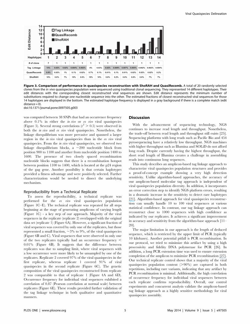
ShoRAH reconstructed 252 viral sequences from the in vivo
quasispecies sample. However, none of the 14 haplotypes were
being reconstructed (Figure 3). For those 14 haplotypes, the
respective closest viral sequence deduced by ShoRAH had an edit
distance ranging from 1 to 12. QuasiRecomb, on the other hand,
reconstructed 1343 viral sequence and was able to identify 1 out of
14 haplotypes from clonal sequencing. This haplotype had an
estimated occurrence frequency of 0.8% from QuasiRecomb while
it accounted for 7 out of 20 clones in clonal sequencing. It implied
that haplotype frequency estimation by QuasiRecomb was
inaccurate and that a significant amount of reconstructed
haplotype by QuasiRecomb was false positive. QuasiRecomb
can also be run in a conservative mode, in which only major
haplotypes were reconstructed. Under this running mode, only 6
haplotypes were reconstructed and none of them overlapped with
the 14 haplotypes being clonal sequenced.
In contrast, 9 out of 14 haplotypes from clonal sequencing were
included in the quasispecies reconstructed by our tag linkage
approach (Figure 3). The most abundant haplotype from clonal
sequencing matched the most abundant reconstructed haplotype
from tag linkage approach in this region of interest. The other 8
identified haplotypes were estimated to have an occurrence
frequency from 0.002% to 0.02%. It highlighted the sensitivity
and accuracy of our tag linkage approach in reconstructing rare
haplotypes. The missing five haplotypes were 1–3 edit distances
away from their respective closest viral sequence in the
quasispecies reconstructed by our tag linkage approach. Overall,
tag linkage approach achieved a significant improvement over
algorithmic-based approaches in quasispecies reconstruction, both
qualitatively and quantitatively.
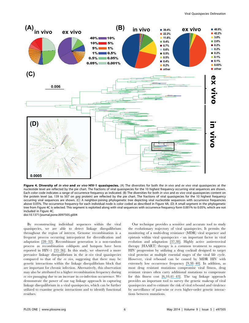
Diversity Comparison between in vivo and ex vivo HIV-1QuasispeciesWe next examined the sequence diversity in both in vivo and ex
vivo quasispecies populations. The most frequent viral sequence
represented 8.1% of the in vivo viral quasispecies, whereas the most
dominant viral sequence represented 32.5% of the ex vivo viral
quasispecies (Figure 4A). The two most dominant viral sequences
in the ex vivo sample comprised more than half of the total viral
quasispecies while the in vivo viral quasispecies was much more
diverse. At the amino acid level, 80% of the in vivo viral
quasispecies were represented by four protein sequences, with a
total of 42 unique protein sequences in the population (Figure 4B).
In contrast, while only two protein sequences represented 80% of
the ex vivo viral quasispecies, there were 201 unique protein
sequences. Table S1 in File S1 provides a summary of this data. A
phylogenetic tree analysis demonstrated the effect of differential
selection pressures on viral quasispecies evolution from in vivo to ex
vivo, in which two distinct sub-population clusters could be
observed (Figure 4C and D).
Recombination Pattern of HIV-1 QuasispeciesHIV-1, as a diploid retrovirus, is capable of generating
recombinant proviral transcript via a template switching event
during the reverse transcription step in the viral replication. It
facilitates further diversification for adaptation [2]. The depth and
comprehensiveness of our data permit an investigation of this viral
recombination, as a linkage disequilibrium pattern. Here, we
employed the r correlation to measure linkage disequilibrium. r2 2
Figure 2. Proof-of-concept study using ex vivo passaged HIV-1 quasispecies. (A) Sequence coverage for each of the 12 amplicon segmentsis plotted. (B) Tag coverage for each of the 12 amplicon segments is plotted. Tag coverage is calculated by the number of unique tags present in agiven amplicon segment. (C) The assembling successful rate is assessed by a parameter called the ‘number of informative segments’, whichrepresents the number of amplicon segments a unique tag is present in. For example, in the present study, a tag with 12 informative segmentsrepresents a complete assembled contiguous sequence of our HIV-1 DNA target region, while a tag with 11 informative segments indicates that 1amplicon segment is missing from the assembled contiguous sequence. All tags in the data set are categorized by this parameter. (D) The fraction ofsequences containing a stop codon is plotted against different cutoffs for the minimum sequence occurrence.doi:10.1371/journal.pone.0097505.g002
Viral Quasispecies Delineation
PLOS ONE | www.plosone.org 4 May 2014 | Volume 9 | Issue 5 | e97505
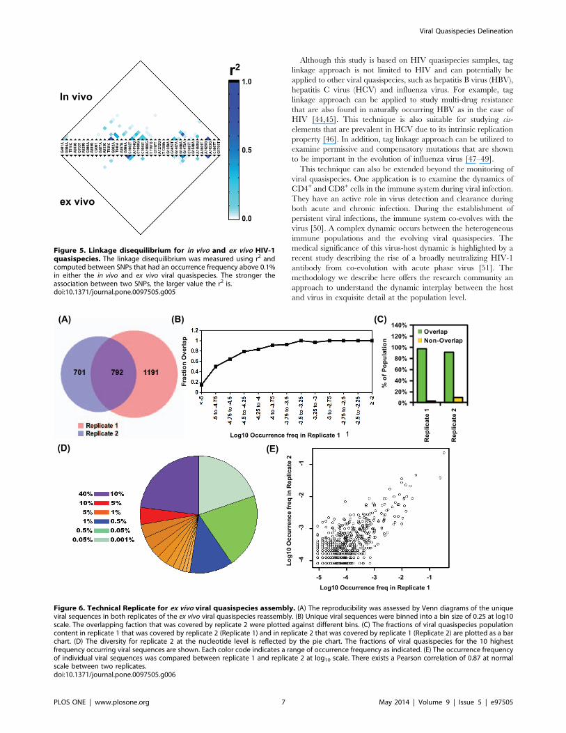
was computed between 38 SNPs that had an occurrence frequency
above 0.1% in either the in vivo or ex vivo viral quasispecies
(Figure 5). Several strong correlations (r2 . 0.5) were observed in
both the in vivo and ex vivo viral quasispecies. Nonetheless, the
linkage disequilibrium was more pervasive and spanned a larger
region in the in vivo viral quasispecies than in the ex vivo viral
quasispecies. From the in vivo viral quasispecies, we observed two
linkage disequilibrium blocks, a ,200 nucleotide block from
position 900 to 1100 and another from nucleotide position 1400 to
1600. The presence of two closely spaced recombination
nucleotide blocks suggests that there is a recombination hotspot
between position 1100 to 1400, which is located at the p24 region
of the gag gene. Another possibility is that certain haplotypes
provided a fitness advantage and were positively selected. Further
characterization would be needed to dissect the underlying
mechanism.
Reproducibility from a Technical ReplicateTo assess the reproducibility, a technical replicate was
performed for the ex vivo viral quasispecies population
(Figure 1C–E). The technical replicate was repeated for all steps
beginning at the stage of generating amplicons of varying length
(Figure 1C) - a key step of our approach. Majority of the viral
sequences in the replicate (replicate 2) overlapped with the original
data set (replicate 1) (Figure 6A). However, a significant fraction of
viral sequences was covered by only one of the replicates, but those
represented a small fraction, ,3% to 9%, of the viral quasispecies
(Figure 6B and C). Viral sequences that were observed in only one
of the two replicates typically had an occurrence frequency ,
0.01% (Figure 6B). It suggests that the difference between
replicates was due to sampling limit, where viral sequences with
a low occurrence were more likely to be unsampled by one of the
replicates. Replicate 2 covered 97% of the viral quasispecies in the
first replicate, whereas replicate 1 covered 91% of viral
quasispecies in the second replicate (Figure 6C). The genetic
composition of the viral quasispecies reconstructed from replicate
2 was comparable to that of replicate 1 (Figure 4A and 6D).
Occurrence frequency for individual viral sequences exhibited a
correlation of 0.87 (Pearson correlation at normal scale) between
replicates (Figure 6E). These results provided further validation of
the tag linkage technique in both qualitative and quantitative
manners.
Discussion
With the advancement of sequencing technology, NGS
continues to increase read length and throughput. Nonetheless,
the trade-off between read length and throughput still exists [25].
Sequencing platforms with long reads such as Pacific Bio and 454
pyrosequencing have a relatively low throughput. NGS machines
with higher throughput such as Illumina and SOLiD do not afford
long reads. Despite currently having the highest throughput, the
short read length of Illumina creates a challenge in assembling
reads into continuous long sequences.
This study describes an amplicon-based tag linkage approach to
characterize viral quasispecies population structures and provides
a proof-of-concept example showing a very high detection
sensitivity. Unlike algorithm-based approaches, the accuracy of
our amplicon-based molecular tag approach is independent of
viral quasispecies population diversity. In addition, it incorporates
an error correction step to identify NGS platform errors, resulting
in a dramatic increase in the sensitivity to detect rare haplotypes
[21]. Algorithm-based approach for viral quasispecies reconstruc-
tion can usually handle 10 to 100 viral sequences at various
statistical confidence. In contrast, our tag linkage approach can
reconstruct close to 1000 sequences with high confidence as
indicated by our replicates. It achieves a significant improvement
in accuracy and sensitivity from the algorithm-base approach [11–
20].
The major limitation in our approach is the length of deduced
sequence, which is restricted by the upper limit of PCR (typically
10 kilobases). Another potential pitfall is PCR recombination. In
our protocol, we tried to minimize this artifact by using a high
processivity and fidelity DNA polymerase for PCR [26]. In
addition, a long PCR extension time was used to ensure extension
completion of the amplicon to minimize PCR recombination [27].
Our technical replicate control shows that a majority of the viral
quasispecies population content (.90%) are captured in both
repetitions, including rare variants, indicating that any artifact by
PCR recombination is minimal. Additionally, the high correlation
of occurrence frequency for individual viral sequences between
each replicate confirms reproducibility. Overall, our control
experiments and concurrent analysis validate the amplicon-based
tag linkage approach as a highly sensitive methodology for viral
quasispecies assembly.
Figure 3. Comparison of performance in quasispecies reconstruction with ShoRAH and QuasiRecomb. A total of 20 randomly selectedclones from the in vivo quasispecies population were sequenced using traditional clonal sequencing. They represented 14 different haplotypes. Theiredit distances with the corresponding closest reconstructed viral sequences are shown. Edit distance represents the minimum number ofsubstitutions required to change one nucleotide sequence into the other. The estimated fractions of closest reconstructed viral sequences for those14 haplotypes are displayed in the bottom. The estimated haplotype frequency is displayed in a gray background if there is a complete match (editdistance= 0).doi:10.1371/journal.pone.0097505.g003
Viral Quasispecies Delineation
PLOS ONE | www.plosone.org 5 May 2014 | Volume 9 | Issue 5 | e97505
By reconstructing individual sequences within the viral
quasispecies, we are able to detect linkage disequilibrium
throughout the region of interest. Genome recombination is a
frequent process occurring intra-patient for diversification and
adaptation [28–32]. Recombinant generation is a non-random
process as recombination coldspots and hotspots have been
reported in HIV-1 [33–36]. In this study, we observed a more
pervasive linkage disequilibrium in the in vivo viral quasispecies
compared to that of the ex vivo, suggesting that there may be
genetic interactions within the linkage disequilibrium block that
are important for chronic infection. Alternatively, this observation
may also be attributed to a higher recombination frequency during
ex vivo passaging due to an increase in co-infection occurrence. We
demonstrate the power of our tag linkage approach in capturing
linkage disequilibrium in a viral quasispecies, which can be further
utilized to examine genetic interactions and to identify functional
residues.
Our technique provides a sensitive and accurate tool to study
the evolutionary trajectory of viral quasispecies. It permits the
monitoring of a multi-drug resistance (MDR) viral sequence and
epistasis within viral quasispecies - an important factor in viral
evolution and adaptation [37,38]. Highly active antiretroviral
therapy (HAART) therapy is a common treatment to suppress
HIV progression by utilizing a drug cocktail designed to target
viral proteins at multiple essential stages of the viral life cycle.
However, viral rebound can be caused by MDR HIV with
extremely low occurrence frequency [9,38–40]. In addition, as
most drug resistant mutations compromise viral fitness, drug
resistant viruses often carry additional mutations to compensate
for this fitness cost [6,10,41–43]. The tag linkage approach
provides an important tool to survey the genetic makeup of viral
quasispecies and to estimate the risk of viral rebound and virulence
by surveillance of pair-wise or even higher-order genetic interac-
tions between mutations.
Figure 4. Diversity of in vivo and ex vivo HIV-1 quasispecies. (A) The diversities for both the in vivo and ex vivo viral quasispecies at thenucleotide level are reflected by the pie chart. The fractions of viral quasispecies for the 10 highest frequency occurring viral sequences are shown.Each color code indicates a range of occurrence frequency as indicated. (B) The diversities for both in vivo and ex vivo viral quasispecies content onthe protein level (aa. 139 to 507 on gag protein) are reflected by the pie chart. The fractions of viral quasispecies for the 10 highest frequencyoccurring viral sequences are shown. (C) A neighbor-joining phylogenetic tree depicting viral nucleotide sequences with occurrence frequenciesabove 0.05%. The occurrence frequency for each individual node is color coded as described in Figure 4A. (D) A small segment in the phylogenetictree from Figure 4C is selected. This segment is replotted along with viral sequences with occurrence frequency form 0.001% to 0.05%, which are notincluded in Figure 4C.doi:10.1371/journal.pone.0097505.g004
Viral Quasispecies Delineation
PLOS ONE | www.plosone.org 6 May 2014 | Volume 9 | Issue 5 | e97505
Although this study is based on HIV quasispecies samples, tag
linkage approach is not limited to HIV and can potentially be
applied to other viral quasispecies, such as hepatitis B virus (HBV),
hepatitis C virus (HCV) and influenza virus. For example, tag
linkage approach can be applied to study multi-drug resistance
that are also found in naturally occurring HBV as in the case of
HIV [44,45]. This technique is also suitable for studying cis-
elements that are prevalent in HCV due to its intrinsic replication
property [46]. In addition, tag linkage approach can be utilized to
examine permissive and compensatory mutations that are shown
to be important in the evolution of influenza virus [47–49].
This technique can also be extended beyond the monitoring of
viral quasispecies. One application is to examine the dynamics of
CD4+ and CD8+ cells in the immune system during viral infection.
They have an active role in virus detection and clearance during
both acute and chronic infection. During the establishment of
persistent viral infections, the immune system co-evolves with the
virus [50]. A complex dynamic occurs between the heterogeneous
immune populations and the evolving viral quasispecies. The
medical significance of this virus-host dynamic is highlighted by a
recent study describing the rise of a broadly neutralizing HIV-1
antibody from co-evolution with acute phase virus [51]. The
methodology we describe here offers the research community an
approach to understand the dynamic interplay between the host
and virus in exquisite detail at the population level.
Figure 5. Linkage disequilibrium for in vivo and ex vivo HIV-1quasispecies. The linkage disequilibrium was measured using r2 andcomputed between SNPs that had an occurrence frequency above 0.1%in either the in vivo and ex vivo viral quasispecies. The stronger theassociation between two SNPs, the larger value the r2 is.doi:10.1371/journal.pone.0097505.g005
Figure 6. Technical Replicate for ex vivo viral quasispecies assembly. (A) The reproducibility was assessed by Venn diagrams of the uniqueviral sequences in both replicates of the ex vivo viral quasispecies reassembly. (B) Unique viral sequences were binned into a bin size of 0.25 at log10scale. The overlapping faction that was covered by replicate 2 were plotted against different bins. (C) The fractions of viral quasispecies populationcontent in replicate 1 that was covered by replicate 2 (Replicate 1) and in replicate 2 that was covered by replicate 1 (Replicate 2) are plotted as a barchart. (D) The diversity for replicate 2 at the nucleotide level is reflected by the pie chart. The fractions of viral quasispecies for the 10 highestfrequency occurring viral sequences are shown. Each color code indicates a range of occurrence frequency as indicated. (E) The occurrence frequencyof individual viral sequences was compared between replicate 1 and replicate 2 at log10 scale. There exists a Pearson correlation of 0.87 at normalscale between two replicates.doi:10.1371/journal.pone.0097505.g006
Viral Quasispecies Delineation
PLOS ONE | www.plosone.org 7 May 2014 | Volume 9 | Issue 5 | e97505
Materials and Methods
Ethics StatementThe study was approved by UCLA IRB. A chronically-infected
HIV-1 patient without undergoing antiretroviral therapy was
recruited from the Los Angeles area and provided written
informed consent.
Subjects and Specimen CollectionTotal peripheral blood mononuclear cells (PBMCs) were
isolated from the patient’s whole blood sample by standard Ficoll
gradient. The plasma viral load at the time of collection was
130,234 viral copies/ml.
Recovery of Virus from PBMCs and Virus PassagingEx vivo passaging was conducted as previously described [52].
Briefly, virus was passaged serially in primary CD4+ T lympho-
cytes from an HIV-1-uninfected donor [53]. After each passage of
,7 days, supernatant virus was collected, titered, and used to
infect fresh cells with an MOI of 1.
DNA Library Preparation for Tag Linkage AssemblyTo extract the viral genomic DNA, cell pellets of 200,000 cells
were resuspended in PBS and genomic DNA was extracted using
the DNeasy Tissue DNA Isolation Kit (Qiagen). DNA was
recovered by PCR using the primer set: 59-GCG GAG GCT AGA
AGG AGA GAG ATG G-39 and 59-CAT CAC CTG CCA TCT
GTT TTC CAT A-39. The forward Illumina sequencing priming
site was added to the 59 end of the DNA sample by PCR using the
primer set: 59-AGA TCG GAA GAG CGT CGT GTA GGG
GCG GAG GCT AGA AGG AGA GAG ATG-39 and 59- GTT
TAA CTT TTG GGC CAT CCA TTC CTG GC-39. Then, the
constant region, a NotI restriction enzyme site and a 13 nucleotide
tag of random ‘N’ sequence was added to the 59 end of the DNA
sample by another PCR using the primer set: 59-ACA TAG ATA
CTA TGC GGC CGC NNN NNN NNN NNN NAG ATC GGA
AGA GCG TCG TGT AGG G-39 and 59- GTT TAA CTT TTG
GGC CAT CCA TTC CTG GC-39. The concentration of the
tagged DNA sample was measured using NanoDrop 1000
spectrophotometer (Thermo Fisher Scientific). This concentration
was used as a reference to calculate the dilution-fold in the
subsequent complexity control step. In the complexity control step,
,300,000 copies of tagged DNA sample were used as the input for
PCR using the primer set: 59-CAC ATA GAT ACT ATG CGG
CCG C-39 and 59-GTT TAA CTT TTG GGC CAT CCA TTC
CTG GC-39. This complexity was calculated based on a ,50-fold
coverage for individual viral sequence with 30 Gb expected
sequencing output per viral quasispecies sample. This was followed
by 12 PCR using the universal forward primer, 59-CAC ATA
GAT ACT ATG CGG CCG C-39, and the reverse primers as
stated in Table S2 in File S1 to add the XhoI restriction enzyme
site on the 39 end using the product of the complexity control step
as template. Consecutive PCR pools should have a different
product size approximately corresponding to the sequencing read
length minus 80 bp (Table S3 and S4 in File S1). From this step
forward, the 12 pools were processed independently until sample
combination at the high-throughput sequencing step. The
products were then subjected to double digestion by NotI and
XhoI. NotI and XhoI were chosen because they were not present
in the consensus sequence of the target DNA template region. A
small insert, which could serve as the population ID, was prepared
by annealing 59-GGC CCG ACG TAA CGA T-39 and 59-TCG
AAT CGT TAC GTC G-39, each with a phosphate group
attached at the 59 end. Ligation was performed using the small
insert to DNA sample at the molar ratio as stated in Table S2 in
File S1. One unit of T4 DNA ligase (Life Technolgies) was used in
each ligation reaction. The reaction condition followed manufac-
turer’s instructions. All ligations were performed overnight at 20uCin 100 uL total reaction volume. The ligated products were used
as the templates for PCR to add the 59 flow cell adapters and the
reverse read Illumina sequencing priming site using the universal
forward primer, 59-AAT GAT ACG GCG ACC ACC GAG ATC
TAC ACT CTT TCC CTA CAC GAC GCT CTT CCG-39, and
the reverse primers as stated in Table S2 in File S1. The 39
Illumina flow cell adapters were then added by PCR using the
primer set: 59-AAT GAT ACG GCG ACC ACC G-39 and 59-
CAA GCA GAA GAC GGC ATA CGA GAT CGG TCT CGG
CAT TCC TGC TGA ACC GCT CTT CCG-39. The resultant
amplicons from all 12 pools were then mixed. High-throughput
sequencing was done by an Illumina HiSeq 2000 machine with an
equivalent of 0.75 lane per sample and 26100 bp paired-end
reads. All PCRs in this study were performed using KOD DNA
polymerase with 1.5 mM MgSO4, 0.2 mM of each dNTP (dATP,
dCTP, dGTP, and dTTP) and 0.4 uM of forward and reverse
primer. PCR extensions were performed with 50 seconds per kb at
68uC. Annealing temperature for a given PCR was 5uC below the
lowest melting temperature of the pair of primers. All primers in
this study were designed to target conserved regions within the
quasispecies which were determined by clonal sequencing of the
sampled viral sequences. This sequencing library preparation
could potentially be adapted to study viral RNA using a reverse
transcription primer tag as decribed by Jabara et al [54]. Raw
sequencing data have been submitted to the NIH Short Read
Archive under accesion number: SRP032753.
Clonal SequencingAfter recovering the DNA by PCR as described above, the
amplicon was inserted into target p83-2 plasmid using In-Fusion
kit (Clontech). Twenty clones were randomly selected and
subjected to capillary sequencing (Laragen).
Data AnalysisSequencing reads were mapped by BWA with 8 mismatches
allowed [55]. Pair-end reads containing two or more short inserts
(barcodes) were discarded. Error-correction was performed as
described previously to distinguish true mutation from sequencing
error [21]. The error-correction step grouped all reads sharing the
same tag and mapped to the same region into a read cluster that
was further conflated into a ‘‘error-free’’ read. As described in
Kinde et al. [21], most reads sharing the same tag should share the
mutation pattern during mapping. In contrast, a sequencing error
would have a low occurrence frequency within a read cluster and
could be distinguished from true mutations. Through this process,
sequencing error would be corrected to generate an ‘‘error-free’’
read. Read cluster with a size of ,3 reads were discarded to
increase the confidence in generating an ‘‘error-free’’ read. Since
intermolecular concatenation at the ligation was observed, a
mutation that existed in 45% of the reads within a conflated read
cluster that also shared the same tag was considered as a true
mutation. The correlation between technical replicates indicated
that intermolecular concatenation did not pose a major barrier in
the accuracy of viral quasispecies assembly. Nonetheless, further
application should adjust the ligation reaction volume to decrease
the intermolecular concatenation during ligation (circularization
step). Next, ‘‘error-free’’ reads that shared the same tag were
assembled into a contiguous sequence, which represented a single
viral sequence. Data processing and analysis were conducted by
custom Python scripts. All scripts are available upon request.
Viral Quasispecies Delineation
PLOS ONE | www.plosone.org 8 May 2014 | Volume 9 | Issue 5 | e97505
Phylogenetic Tree ConstructionClustalX was used to create the neighbor-joining phylogenetic
tree [56]. The phylogenetic tree was mid point-rooted and
displayed by FigTree.
Linkage DisequilibriumWe used the r2 correlation to quantify linkage disequilibrium
between two SNPs. R2 was computed as per convention. Briefly,
r2 = (PAB 2 PA x PB)2/(PA 6 PB 6 (1 PA) 6 (1 PB)), where PAB
represented the occurrence frequency of viral sequences that carry
both SNP A and SNP B; PA represented the occurrence frequency
of viral sequences that carry SNP A; PB represented the
occurrence frequency of viral sequences that carry SNP B.
DNA Library Preparation for Error-free SequencingGag-pol region was PCR amplified using the primer set: 59-
GAC TAG CGG AGG CTA GAA GGA GAG AG-39 and 59-
CAT GTT CTT CTT GGG CCT TAT CTA TTC-39. The
resultant DNA product was sheared to around 200 bp to 600 bp
by sonication using the Sonic Dismembrator Model 100 (Fisher
Scientific). Dismembrator was set to power level four and samples
were pulsed three times for 10 seconds. Samples were kept on ice
for 45 seconds in between pulses. End repair and 39 dA-tailing
were performed respectively by end repair module and dA-tailing
module (New England BioLabs). The DNA product was then
ligated to an Y-shape adaptor carrying a nine-nucleotide tag of
random ‘N’ sequence. As a result, each ligated product contained
an 18-nucleotide tag, nine from each of the 59 and 39 end. Y-shape
adaptor was prepared by annealing two oligonucleotides: 59-CGC
GTA TCC ATG GCA NNN NNN NNN GCC AGA TCG GAA
GAG CGG TTC AGC AGG AAT GCC GAG-39 and 59-ACA
CTC TTT CCC TAC ACG ACG CTC TTC CGA TCT GGC-
39. Then, the annealed product was treated with Klenow
Fragment (New England BioLabs) and digested with BciVI. An
estimated copy of around 10 millions of ligated products were
amplified by primer set: 59-AAT GAT ACG GCG ACC ACC
GAG ATC TAC ACT CTT TCC CTA CAC GAC GCT CTT
CCG-39 and 59-CAA GCA GAA GAC GGC ATA CGA GAT
CGG TCT CGG CAT TCC TGC TGA ACC GCT CTT CCG-
39. The resultant DNA product was submitted for 26100 bp
paired-end sequencing on one lane of Illumina HiSeq 2500
machine.
Quasispecies Reconstruction by ShoRAH andQuasiRecomb‘‘Error-free’’ reads were generated as described above. Here, a
mutation that existed in 95% of the reads within a conflated read
cluster that also shared the same tag was considered as a true
mutation. Reads were mapped by BWA with 8 mismatches
allowed [55]. All reads were treated as single end read. ‘‘Error-
free’’ mapped reads were processed by ShoRAH version 0.6 with a
window size of 40, a window shift of 1 and default settings for
other parameters [13]. Quasispecies reconstruction by QuasiR-
ecomb was performed by default setting [12]. Due to the huge
memory requirement of QuasiRecomb, 500,000 mapped reads
were randomly sampled and processed. Further increase the
number of input reads generated memory error. To limit the false
positive rate, a refinement reconstruction was performed using ‘-
refine’ option. We employed ‘-conservative’ option for high
confidence haplotype reconstruction to identify major haplotypes.
Supporting Information
File S1 Figures S1 and S2 and Tables S1–S4. Figure S1.
Concept of complexity control. In this graphical demonstration,
we employ a simple example with five amplicons and 30 reads
sequenced. A total of nine viral sequences are present in the viral
quasispecies with the genotype being A or B. The colored boxes
represent the tag for distinguishing an individual viral sequence
within the viral quasispecies. Different colors represent different
nucleotide sequences in individual tags. The white boxes represent
individual viral sequences. During the Amplicon generation and
sequencing step, each column of amplicons represents one
genomic region of the viral quasispecies. (A) Complexity is too
high (complexity = 9) where each viral sequence is not sufficiently
covered. (B) Complexity is too low (complexity = 1) where each
viral sequence is excessively covered and therefore, there is a waste
of sequencing capacity. (C) Complexity is well-controlled (com-
plexity = 3) such that individual viral sequences are sufficiently
covered for sequencing error correction and for sequence
assembly. Figure S2. Key step in the experimental design. (A) A
detailed representation that shows the cassette sequence in
Figure 1B. (B) A detailed representation that shows the cassette
sequence after ligation.
(PDF)
Acknowledgments
We would like to thank J. Zhou and J. Yoshizawa for performing the high-
throughput sequencing experiment. This work was supported by UCLA
Center for AIDS research, UCLA Jonsson Comprehensive Cancer Center,
and California HIV/AIDS Research Program.
Author Contributions
Conceived and designed the experiments: NCW RS. Performed the
experiments: NCW NN HHL. Analyzed the data: NCW. Contributed
reagents/materials/analysis tools: JDLC LQA XL MJL OOY. Wrote the
paper: NCW RS. Important intellectual contribution: CAO HQ YD SL
TW.
References
1. Wilke CO, Wang JL, Ofria C, Lenski RE, Adami C (2001) Evolution of digital
organisms at high mutation rates leads to survival of the flattest. Nature 412:
331–333.
2. Worobey M, Holmes EC (1999) Evolutionary aspects of recombination in rna
viruses. J Gen Virol 80 (Pt 10): 2535–2543.
3. Bonhoeffer S, Chappey C, Parkin NT, Whitcomb JM, Petropoulos CJ (2004)
Evidence for positive epistasis in hiv-1. Science 306: 1547–1550.
4. da Silva J, Coetzer M, Nedellec R, Pastore C, Mosier DE (2010) Fitness epistasis
and constraints on adaptation in a human immunodeficiency virus type 1
protein region. Genetics 185: 293–303.
5. Brockman MA, Schneidewind A, Lahaie M, Schmidt A, Miura T, et al. (2007)
Escape and compensation from early hla-b57-mediated cytotoxic t-lymphocyte
pressure on human immunodeficiency virus type 1 gag alter capsid interactions
with cyclophilin a. J Virol 81: 12608–12618.
6. Dam E, Quercia R, Glass B, Descamps D, Launay O, et al. (2009) Gag
mutations strongly contribute to hiv-1 resistance to protease inhibitors in highly
drug-experienced patients besides compensating for fitness loss. PLoS Pathog 5:
e1000345.
7. Zhang J, Hou T, Wang W, Liu JS (2010) Detecting and understanding
combinatorial mutation patterns responsible for hiv drug resistance. Proc Natl
Acad Sci U S A 107: 1321–1326.
8. Sanjun R, Moya A, Elena SF (2004) The contribution of epistasis to the
architecture of fitness in an rna virus. Proc Natl Acad Sci U S A 101: 15376–
15379.
9. Fumero E, Podzamczer D (2003) New patterns of hiv-1 resistance during haart.
Clin Microbiol Infect 9: 1077–1084.
10. Verheyen J, Litau E, Sing T, Dumer M, Balduin M, et al. (2006) Compensatory
mutations at the hiv cleavage sites p7/p1 and p1/p6-gag in therapy-naive and
therapy-experienced patients. Antivir Ther 11: 879–887.
Viral Quasispecies Delineation
PLOS ONE | www.plosone.org 9 May 2014 | Volume 9 | Issue 5 | e97505
11. Beerenwinkel N, Gnthard HF, Roth V, Metzner KJ (2012) Challenges and
opportunities in estimating viral genetic diversity from next-generationsequencing data. Front Microbiol 3: 329.
12. Tpfer A, Zagordi O, Prabhakaran S, Roth V, Halperin E, et al. (2013)
Probabilistic inference of viral quasispecies subject to recombination. J ComputBiol 20: 113–123.
13. Zagordi O, Bhattacharya A, Eriksson N, Beerenwinkel N (2011) Shorah:estimating the genetic diversity of a mixed sample from next-generation
sequencing data. BMC Bioinformatics 12: 119.
14. Zagordi O, Dumer M, Beisel C, Beerenwinkel N (2012) Read length versusdepth of coverage for viral quasispecies reconstruction. PLoS One 7: e47046.
15. Eriksson N, Pachter L, Mitsuya Y, Rhee SY, Wang C, et al. (2008) Viralpopulation estimation using pyrosequencing. PLoS Comput Biol 4: e1000074.
16. Zagordi O, Klein R, Dumer M, Beerenwinkel N (2010) Error correction of next-generation sequencing data and reliable estimation of hiv quasispecies. Nucleic
Acids Res 38: 7400–7409.
17. Astrovskaya I, Tork B, Mangul S, Westbrooks K, Mndoiu I, et al. (2011)Inferring viral quasispecies spectra from 454 pyrosequencing reads. BMC
Bioinformatics 12 Suppl 6: S1.18. Henn MR, Boutwell CL, Charlebois P, Lennon NJ, Power KA, et al. (2012)
Whole genome deep sequencing of hiv-1 reveals the impact of early minor
variants upon immune recognition during acute infection. PLoS Pathog 8:e1002529.
19. Prosperi MCF, Salemi M (2012) Qure: software for viral quasispeciesreconstruction from next-generation sequencing data. Bioinformatics 28: 132–
133.20. Skums P, Mancuso N, Artyomenko A, Tork B, Mandoiu I, et al. (2013)
Reconstruction of viral population structure from next-generation sequencing
data using multicommodity flows. BMC Bioinformatics 14 Suppl 9: S2.21. Kinde I, Wu J, Papadopoulos N, Kinzler KW, Vogelstein B (2011) Detection
and quantification of rare mutations with massively parallel sequencing. ProcNatl Acad Sci U S A 108: 9530–9535.
22. Rose PP, Korber BT (2000) Detecting hypermutations in viral sequences with an
emphasis on g – ¿ a hypermutation. Bioinformatics 16: 400–401.23. Janini M, Rogers M, Birx DR, McCutchan FE (2001) Human immunodefi-
ciency virus type 1 dna sequences genetically damaged by hypermutation areoften abundant in patient peripheral blood mononuclear cells and may be
generated during near-simultaneous infection and activation of cd4(+) t cells.J Virol 75: 7973–7986.
24. Harris RS, Liddament MT (2004) Retroviral restriction by apobec proteins. Nat
Rev Immunol 4: 868–877.25. Loman NJ, Misra RV, Dallman TJ, Constantinidou C, Gharbia SE, et al. (2012)
Performance comparison of benchtop high-throughput sequencing platforms.Nat Biotechnol 30: 434–439.
26. Lahr DJG, Katz LA (2009) Reducing the impact of pcr-mediated recombination
in molecular evolution and environmental studies using a new-generation high-fidelity dna polymerase. Biotechniques 47: 857–866.
27. Judo MS, Wedel AB, Wilson C (1998) Stimulation and suppression of pcr-mediated recombination. Nucleic Acids Res 26: 1819–1825.
28. Jetzt AE, Yu H, Klarmann GJ, Ron Y, Preston BD, et al. (2000) High rate ofrecombination throughout the human immunodeficiency virus type 1 genome.
J Virol 74: 1234–1240.
29. Froissart R, Roze D, Uzest M, Galibert L, Blanc S, et al. (2005) Recombinationevery day: abundant recombination in a virus during a single multi-cellular host
infection. PLoS Biol 3: e89.30. Neher RA, Leitner T (2010) Recombination rate and selection strength in hiv
intra-patient evolution. PLoS Comput Biol 6: e1000660.
31. Charpentier C, Nora T, Tenaillon O, Clavel F, Hance AJ (2006) Extensiverecombination among human immunodeficiency virus type 1 quasispecies makes
an important contribution to viral diversity in individual patients. J Virol 80:2472–2482.
32. Shriner D, Rodrigo AG, Nickle DC, Mullins JI (2004) Pervasive genomic
recombination of hiv-1 in vivo. Genetics 167: 1573–1583.
33. Zhuang J, Jetzt AE, Sun G, Yu H, Klarmann G, et al. (2002) Human
immunodeficiency virus type 1 recombination: rate, fidelity, and putative hotspots. J Virol 76: 11273–11282.
34. Baird HA, Gao Y, Galetto R, Lalonde M, Anthony RM, et al. (2006) Influence
of sequence identity and unique breakpoints on the frequency of intersubtypehiv-1 recombination. Retrovirology 3: 91.
35. Simon-Loriere E, Galetto R, Hamoudi M, Archer J, Lefeuvre P, et al. (2009)Molecular mechanisms of recombination restriction in the envelope gene of the
human immunodeficiency virus. PLoS Pathog 5: e1000418.
36. Levy DN, Aldrovandi GM, Kutsch O, Shaw GM (2004) Dynamics of hiv-1
recombination in its natural target cells. Proc Natl Acad Sci U S A 101: 4204–
4209.
37. Michalakis Y, Roze D (2004) Evolution. epistasis in rna viruses. Science 306:
1492–1493.
38. Clotet B (2004) Strategies for overcoming resistance in hiv-1 infected patients
receiving haart. AIDS Rev 6: 123–130.
39. Palmer S, Boltz V, Maldarelli F, Kearney M, Halvas EK, et al. (2006) Selection
and persistence of non-nucleoside reverse transcriptase inhibitor-resistant hiv-1
in patients starting and stopping non-nucleoside therapy. AIDS 20: 701–710.
40. Liu J, Miller MD, Danovich RM, Vandergrift N, Cai F, et al. (2011) Analysis of
low-frequency mutations associated with drug resistance to raltegravir beforeantiretroviral treatment. Antimicrob Agents Chemother 55: 1114–1119.
41. Martinez-Picado J, Martnez MA (2008) Hiv-1 reverse transcriptase inhibitor
resistance mutations and fitness: a view from the clinic and ex vivo. Virus Res134: 104–123.
42. Piana S, Carloni P, Rothlisberger U (2002) Drug resistance in hiv-1 protease:Flexibility-assisted mechanism of compensatory mutations. Protein Sci 11: 2393–
2402.
43. Johnson JA, Li JF, Morris L, Martinson N, Gray G, et al. (2005) Emergence of
drug-resistant hiv-1 after intrapartum administration of single-dose nevirapine is
substantially underestimated. J Infect Dis 192: 16–23.
44. Yim HJ, Hussain M, Liu Y, Wong SN, Fung SK, et al. (2006) Evolution of multi-
drug resistant hepatitis b virus during sequential therapy. Hepatology 44: 703–712.
45. Delaney WE, Yang H, Westland CE, Das K, Arnold E, et al. (2003) Thehepatitis b virus polymerase mutation rtv173l is selected during lamivudine
therapy and enhances viral replication in vitro. J Virol 77: 11833–11841.
46. Moradpour D, Penin F, Rice CM (2007) Replication of hepatitis c virus. NatRev Microbiol 5: 453–463.
47. Bloom JD, Gong LI, Baltimore D (2010) Permissive secondary mutations enablethe evolution of influenza oseltamivir resistance. Science 328: 1272–1275.
48. Wu NC, Young AP, Dandekar S, Wijersuriya H, Al-Mawsawi LQ, et al. (2013)
Systematic identification of h274y compensatory mutations in influenza a virusneuraminidase by high-throughput screening. J Virol 87: 1193–1199.
49. Gong LI, Suchard MA, Bloom JD (2013) Stability-mediated epistasis constrainsthe evolution of an influenza protein. Elife 2: e00631.
50. Nowak MA, Bangham CR (1996) Population dynamics of immune responses topersistent viruses. Science 272: 74–79.
51. Liao HX, Lynch R, Zhou T, Gao F, Alam SM, et al. (2013) Co-evolution of a
broadly neutralizing hiv-1 antibody and founder virus. Nature 496: 469–476.
52. Lewis MJ, Dagarag M, Khan B, Ali A, Yang OO (2012) Partial escape of hiv-1
from cytotoxic t lymphocytes during chronic infection. J Virol 86: 7459–7463.
53. Wong JT, Colvin RB (1987) Bi-specific monoclonal antibodies: selective binding
and complement fixation to cells that express two different surface antigens.J Immunol 139: 1369–1374.
54. Jabara CB, Jones CD, Roach J, Anderson JA, Swanstrom R (2011) Accurate
sampling and deep sequencing of the hiv-1 protease gene using a primer id. ProcNatl Acad Sci U S A 108: 20166–20171.
55. Li H, Durbin R (2009) Fast and accurate short read alignment with burrows-wheeler transform. Bioinformatics 25: 1754–1760.
56. Larkin MA, Blackshields G, Brown NP, Chenna R, McGettigan PA, et al. (2007)Clustal w and clustal x version 2.0. Bioinformatics 23: 2947–2948.
Viral Quasispecies Delineation
PLOS ONE | www.plosone.org 10 May 2014 | Volume 9 | Issue 5 | e97505