harnessing the antibacterial properties of fluoridated
TRANSCRIPT
�����������������
Citation: Rahayu, D.P.; Draheim, R.;
Lalatsa, A.; Roldo, M. Harnessing the
Antibacterial Properties of
Fluoridated Chitosan Polymers
against Oral Biofilms. Pharmaceutics
2022, 14, 488. https://doi.org/
10.3390/pharmaceutics14030488
Academic Editor: Beom-Jin Lee
Received: 27 January 2022
Accepted: 19 February 2022
Published: 23 February 2022
Publisher’s Note: MDPI stays neutral
with regard to jurisdictional claims in
published maps and institutional affil-
iations.
Copyright: © 2022 by the authors.
Licensee MDPI, Basel, Switzerland.
This article is an open access article
distributed under the terms and
conditions of the Creative Commons
Attribution (CC BY) license (https://
creativecommons.org/licenses/by/
4.0/).
pharmaceutics
Article
Harnessing the Antibacterial Properties of Fluoridated ChitosanPolymers against Oral BiofilmsDien Puji Rahayu 1,2 , Roger Draheim 1 , Aikaterini Lalatsa 1,* and Marta Roldo 1,*
1 School of Pharmacy and Biomedical Sciences, University of Portsmouth, St Michael’s Building,White Swan Road, Portsmouth PO1 2DT, UK; [email protected] (D.P.R.);[email protected] (R.D.)
2 National Research and Innovation Agency of Indonesia (BRIN), Lebak Bulus Raya No. 49,Jakarta 12440, Indonesia
* Correspondence: [email protected] (A.L.); [email protected] (M.R.)
Abstract: Dental caries are a worldwide endemic chronic disease affecting people of all ages. Due tothe limitations of daily used oral hygiene products, there is an unmet need for new, effective, safe,and economic oral products. We have recently demonstrated that N-(2(2,6-diaminohexanamide)-chitosan (CS3H Lys) has enhanced antibacterial properties against Streptococcus mutans, the maincariogenic bacterium, and here we investigated the effect of fluoridation of this polymer (CS3HLys F) on its antibacterial properties and the ability to protect teeth from acid demineralization. Wefurther formulated this polymer into mouthwash preparations and studied their cytocompatibilityand physicochemical stability over 6 months. CS3H Lys F was 1.6-fold more effective than the highesttested oral NaF dose in preventing acid demineralization. CS3H Lys F has a 3- to 5-fold lowerminimum inhibitory concentration value against S. mutants than the values reported for chitosanpolymers and showed negligible cell toxicity. The mouthwashes were stable at both 25 and 40 ◦C.Further work is under way towards other CS3H Lys F oral hygiene products such as a toothpaste.
Keywords: chitosan; fluoride; antimicrobial properties; demineralization protection; dental caries
1. Introduction
Dental caries are a worldwide endemic chronic infection affecting people of all ages [1,2].Almost half of the world’s population is affected by dental caries in permanent teeth [1],with particular high prevalence in young people in developing countries (79% in Thailandand 75% in Malaysia) [3,4]. Dental caries are caused by communities of microorganisms thatpresent on the tooth surface entrapped within the extracellular polymeric substance (EPS)forming the dental biofilm or dental plaque [5,6]. The most prevalent cariogenic bacteria areStreptococcus mutans and Streptococcus sobrinus [7,8]. Enamel, the protective external layer ofteeth composed of hydroxyapatite, is destroyed when the pH drops below a critical level(pH < 5.5) due to the ingestion of acidic food and drinks or the production of acid bybacteria [9]. Dental caries are preventable by (a) reducing sugar in the diet, as when metab-olized by bacteria, sugar generates acid, and (b) good oral hygiene habits such as brushingteeth twice daily, combined with flossing and rinsing with a mouthwash [10]. There areseveral antibacterial agents such as triclosan [11,12], essential oils [13,14], cetyl pyridiniumchloride [15], zinc sulphate [16], chlorhexidine (CHX) [15–17], or a combination of themthat have been shown to control dental biofilm formation and are formulated into tooth-pastes and mouthwashes for ease of use and topical application [11–16,18–21]. Among these,CHX remains the gold standard for reducing plaque and gingival inflammation [22,23].However, CHX causes tooth staining, alters taste perception and promotes calculus ortartar [17,24–27]. Thus, there is need for new, sustainable, effective, safe, and economicsubstances that can be formulated into daily treatment products and particularly mouth-
Pharmaceutics 2022, 14, 488. https://doi.org/10.3390/pharmaceutics14030488 https://www.mdpi.com/journal/pharmaceutics
Pharmaceutics 2022, 14, 488 2 of 15
washes that are easier to use and able to reach the narrow and small spaces in the mouththat brushing cannot.
Fluoride containing products and oral solutions remain a widely used cost-effectivestrategy to prevent dental caries. Fluoride ions are commonly added to oral hygieneproducts in the form of sodium fluoride, as they are known to disturb the growth andmetabolism of cariogenic bacteria by inhibiting enolase, an enzyme involved in gly-colysis, thus decreasing acid production and reducing the EPS formation in bacterialbiofilms [28–31]. Additionally, fluoride ions promote remineralization of weakenedtooth enamel [32,33].
Chitosan, a polysaccharide of a natural origin and a waste product of the fish industry,has been shown to possess an antibacterial activity [34–36], a property that justifies its inclu-sion in oral hygiene products. Commercially available formulations that contain chitosanas an active ingredient are available and include a chitosan-based, non-fluoride toothpaste(Chitodent®, B&F Elektro GmbH, Filsum, Germany), a chitosan (0.5%) toothpaste with1400 ppm fluoride ions (F−) and 3500 ppm tin ions (Sn+2) (Elmex®, GABA International AG,Münchenstein, Switzerland), a chitosan argininamide mouthwash (Synedent®, Prisyna,Claremont, CA, USA), and a chitosan argininamide and sodium fluoride (0.05% w/v equiva-lent to 0.02% w/v F−) (Synedent FLX, Prisyna, Claremont, CA, USA). All products highlightthe natural origin and low environmental impact of chitosan, supporting the wider moveof many consumer health product multinational companies towards the use of naturallyderived excipients and actives.
We have recently shown that N-(2(2,6-diaminohexanamide)-chitosan (CS3H Lys) hasenhanced antibacterial properties against S. aureus and is able to completely inhibit itsgrowth at concentrations as low as 200 µg mL−1 [37]. In this study, we demonstrate,for the first time, the effect of fluoridation of this polymer on its antibacterial propertiesand its ability to protect teeth from acid demineralization. We have also formulated acytocompatible and stable (for 6 months) mouthwash readily commercialisable as anoral product.
2. Materials and Methods2.1. Materials
Chlorhexidine gluconate (0.2%) Minosep® mouthwash (Minorock Mandiri Ltd., De-pok, Indonesia) and Listerine® total care (Johnson and Johnson, Maidenhead, UK) wereused as the control mouthwashes. 3-(4,5-dimethylthiazol-2yl)-2,5-diphenyltetrazoliumbromide (MTT), trypan blue stain, penicillin/streptomycin were purchased from FisherScientific (Longborough, UK). All other chemicals used in this study were of analyticalgrade and were purchased from Sigma Aldrich Inc. (Gillingham, Dorset, UK), unlessotherwise stated. CS3H was synthesized by acid degradation [38] of commercially avail-able low-viscosity chitosan from shrimp shell (CAS 9012-96-4, Lot #BCBQ 3414V, MW:165.3 KDa, acetylation 15.37 ± 0.47% calculated by NMR [37], Sigma-Aldrich Inc. Gilling-ham, Dorset, UK) and had the following properties: molecular weight 4.709 × 10⁴ g/mL,Mn 4.156 × 10⁴, Mw/Mn 1.133, acetylation 14.20 ± 0.17%, and pKa 6.68 ± 0.06. N-(2(2,6-diaminohexanamide)-chitosan (CS3H Lys) was synthesized from CS3H, as previ-ously described (SI Section S1) [37]. The polymer obtained had a molecular weight of3.345 × 104 g/mol (Mn 1.893 × 10⁴ and Mw/Mn 1.768), acetylation 17.56 ± 5.23%, andpKa of 6.56 ± 0.06 [37].
2.2. Fluoridation of Chitosan Polymers and Fluoride Quantification
Chitosan fluoride (CS3H F) was obtained by the dialysis of chitosan (1 g) against 1 Lof sodium fluoride solution containing 362.5, 725, and 1450 mg NaF to obtain CS3H Flow,CS3H Fmedium, and CS3H Fhigh, respectively. Dialysis was carried out at room temperaturewith six changes over 24 h (MWCO: 12–14 KDa, Medicell Membranes Ltd., London, UK).Similarly, N-(2(2,6-diaminohexanamide)-chitosan fluoride (CS3H Lys F) was obtainedby dialysis against 1 L of sodium fluoride (725 mg) solution immediately after CS3H

Pharmaceutics 2022, 14, 488 3 of 15
Lys synthesis (1.54 g scale) [37] without the need for lyophilizing the product first.All dialysates were lyophilized and white polymer products were packaged in sealedpolypropylene containers.
Fluoride ion loading was determined using a fluoride ion-selective electrode (ISE)(Orion Star A214, Thermo Scientific, Indonesia) fitted with a fluoride electrode (Thermo Sci-entific, UK). Distilled water was used for the preparation of samples and standard solutions.A total ionic strength adjustment buffer (TISAB) II with cyclohexylenedinitrilotetraacetate(CDTA) (Thermo Fisher Scientific, Warrington, UK) was used in the potentiometric measure-ments. Measurements were conducted as per the manufacturer’s instructions (Section S1).
2.3. Fourier-Transform Infrared Spectroscopy (FTIR)
FTIR spectra of chitosan derivatives were obtained using a Varian FTIR spectropho-tometer (Agilent Technologies, Stockport, UK). The samples were mounted onto the surfaceof the germanium (Ge) crystal in the attenuated total reflection (ATR) assembly. FTIRspectra were recorded in the middle infrared range (4000–500 cm−1) with a resolution of4 cm−1 in the absorbance mode for 40 scans at room temperature.
2.4. Solubility Studies
The pH dependence of chitosan polymers aqueous solubility was evaluated at roomtemperature by turbidimetry [39]. Each polymer (50 mg) was dissolved in 10 mL of aqueousacetic acid (10% v/v) and stirred for 1 h; the pH level of the solutions was adjusted usingNaOH solution (5 M). Measurements were repeated after each stepwise addition of NaOHuntil reaching pH 12. The transmittance of the solution was recorded on a Nicolet e-100spectrophotometer (Thermo Fisher Scientific, Warrington, UK) using a quartz cell with anoptical path length of 1 cm at λ 600 nm, and the pH was measured using an FE20 pH meter(Mettler Toledo, Greifensee, Switzerland).
2.5. In Vitro Inhibition of Acid Demineralisation
Mineralized surfaces were prepared as follows: hydroxyapatite (HA, 2 g) powderwas suspended in acetone (200 mL), and 60 µL aliquots of the homogenous suspensionwere transferred to each well of a 96 well plate. The plate was shaken (50 rpm, microplatemixer SciQuip, Newtown, UK) at room temperature until the acetone was completelyevaporated [40]. After drying, loose HA was removed by washing with deionized water(5×) and plates were allowed to dry overnight. Any plate showing poor coverage andcracking was discarded, and the coated plates were sealed until further use. Before eachexperiment, the plates were rehydrated with deionized water for 1 h. A phosphate standardcalibration curve was prepared using KH2PO4 solutions with concentrations of phosphorusin the range 10–60 mg L−1 (or 0.32–1.94 mM). Deionized water and sodium fluoridewere used as negative and positive controls, respectively. The positive control (sodiumfluoride) solutions were prepared in deionized water in three different final concentrations:NaFlow (362.5 ppm), NaFmed (725 ppm), and NaFhigh (1450 ppm). The polymer samples(200 µL, 1.0% w/v) were prepared in 0.2% acetic acid and the pH of all sample solutionswas adjusted to 6.0 ± 0.1. Test solutions (200 µL) were added to each well and agitated(50 rpm) at room temperature for 30 min. After exposure to the test solutions, the wellswere rinsed with deionized water (5×), and 200 µL of the erosive solution (0.1 M aqueousacetate buffer, pH 4.0, Alfa Aesar, Heysham, UK) was added to each well and agitated(50 rpm, 60 min). Aliquots (50 µL) were transferred into new microplates and mixed with50 µL vanadomolybdate reagent for 5 min before reading the UV absorbance at 450 nm(SpectraMax i3x, Molecular Devices, Berkshire, UK).
2.6. Preparation of Bacterial Cultures
Staphylococcus aureus (ATCC 25923TM) was stored in Luria Bertani (LB) broth and sterileglycerol 30% (1:1) in a cryovial at −80 ◦C. The loop shalt of Culti-Loops™ Streptococcusmutans (ATCC 25175™) was cut from the handle using sterile scissors and dropped into
Pharmaceutics 2022, 14, 488 4 of 15
warm Brain Heart Infusion (BHI, 0.5 to 1.0 mL, Oxoid, Basingstoke, UK) and incubatedat 37 ◦C in a 5% CO2 atmosphere overnight. Bacteria were then streaked onto blood agar(Oxoid, Basingstoke, UK) plates using sterile cotton swabs and incubated at 37 ◦C in 5%CO2 for 24–48 h. Two to three single colonies were taken from the plate with a sterileloop and dispersed in 5 mL of fresh BHI to be incubated overnight at 37 ◦C in 5% CO2.The bacteria were stored at −80 ◦C by mixing the overnight culture and sterile glycerol30% (1:1) in a cryovial. Before the experiment, the bacteria were transferred from −80 ◦Cinto 5 mL of fresh sterile BHI medium by a sterile tip and incubated at 37 ◦C (in 5% CO2atmosphere for S. mutans) overnight until the medium reached an optical density at 600 nm(OD600) of 0.5.
2.7. Determination of Minimum Inhibitory Concentration (MIC) and Minimum BactericidalConcentration (MBC)
Overnight bacterial suspensions (100 µL) were transferred into 5 mL of fresh sterilemedium and incubated again until OD600 = 0.5. Serial dilutions of the polymer solutionsfrom stock (5 mg mL−1 in 1% acetic acid) were tested at final concentrations 0.1, 0.2, 0.4, 0.8,1.2, 1.6, 2.0, 3.2, and 4.0 mg mL−1 in medium. One hundred microliters of each polymersolution were placed into the well containing 5 µL of the bacterial suspensions. Wellscontaining only culture medium and bacteria were used as a negative control. Turbiditymeasurements were made for all the wells after 24 h of incubation at 37 ◦C for S. aureus andat 37 ◦C under anaerobic condition (5% CO2) for S. mutans. The MIC of each bacteria wasrecorded at 600 nm as the lowest concentration of each polymer that inhibited the bacterialgrowth, as detected by the absence of visual turbidity [41]. The MIC was determined asthe lowest concentration of each polymer that restricted growth to a level below an OD600of ≤ 0.05 [42]. The MBC was defined as the lowest concentration of test compounds thatprevented any visible growth on agar plates. Samples of 20 µL were transferred fromclear wells into LB agar plates, and they were incubated at 37 ◦C for 24 h for S. aureus andinto blood agar plates for 48 h in 5% CO2 for S. mutans. All experiments were performedin triplicate.
2.8. Inhibition of S. mutans Biofilm Formation
One hundred microliters of S. mutans suspension were transferred to 5 mL of pre-warmed fresh BHI medium and incubated at 37 ◦C in a 5% CO2 to OD600 ≈ 0.5. This culture(100 µL) was then dispensed into 48 well plates, and to this biofilm medium (BM, 700 µLfor negative control) or polymer solution (700 µL) in Biofilm Medium (BM) at differentconcentrations (0.1, 0.2, 0.4, 0.8, and 1.2 mg/mL) was added. Controls with 800 µL of BMwithout bacteria were also prepared. Preparation of BM was undertaken as previouslydescribed [43]. Plates were incubated for 24 h at 37 ◦C in a 5% CO2 atmosphere withoutagitation. After overnight incubation, the formation of biofilm was quantified by crystalviolet assay [43].
2.9. Formulation of Chitosan-Based Mouthwash
The chitosan-based mouthwash formulas are summarized in Table 1. Lutrol (polox-amer 407) solution (4% w/v) was prepared in water, while polymers were dissolved in0.5 M acetic acid. Three separate batches were prepared; all the ingredients were solubilizedat room temperature and the pH of the mouthwash was adjusted to 5.5 ± 0.1 with NaOH(1 M).

Pharmaceutics 2022, 14, 488 5 of 15
Table 1. Chitosan-based mouthwash formulation.
IngredientsQuantity
FunctionFormula I Formula II
Glycerin 5 g 5 g HumectantSodium saccharin 450 mg 450 mg Sweetener
Lutrol 4% w/v 50 mL 50 mL SurfactantPolymer 0.2 mg 0.2 mg Anti-biofilm
Acetic acid (0.5 M) 1 mL 1 mL Aciditymodifier/co-solvent
Food blue - 0.2 mL Coloring agentPeppermint oil - 0.25 mL Flavoring agent
Ethanol - 20 mL Co-solventWater to 100 mL to 100 mL Vehicle
2.10. Evaluation of Mouthwash Stability
Samples were stored at 25 or 40 ◦C for six months with eight sampling points (0, 3, 7, 14,21, 30, 90, and 180 days) in 7 mL sample vials made of neutral glass (Type IB) and closed withpolypropylene screw caps (Fisher Scientific, Loughborough, UK). Organoleptic propertiessuch as color, odor, and appearance were monitored. Color intensity was measured byUV spectroscopy (Multiskan Go UV–VIS spectrophotometer, Thermo Fisher Scientific,Paisley, UK) at wavelengths ranging from 300 to 700 nm. Odor was subjectively assessedby the investigator. The pH was measured with a digital pH meter (Accumet AB150,Fisher Scientific, Loughborough, UK). For each sample, three independent measurementswere performed, and data were reported as the mean of the replicates. pH values fromthe stability data were analyzed using a two-way analysis of variance (ANOVA) withTukey’s and Sidak post-test with an a priori level significance of 0.05 to detect statisticallysignificant differences between the time and temperature from the mouthwash formulationsand control. Sedimentation was visually assessed after centrifuging 1 mL of each sampleat 5000 rpm for 5 min [44] at room temperature using a Jouan B4i Centrifuge (HemelHampstead, UK). In vitro anti-biofilm activity was tested as described above, at all time-points of the stability study.
2.11. Biofilm Removal Efficacy
In vitro anti-biofilms activity was performed on overnight S. mutans biofilms on48 well-plates. The S. mutans biofilms were prepared as described in Section 2.9. Afterovernight incubation at 37 ◦C in a 5% CO2 atmosphere without agitation, the plate wasblotted on a paper towel to remove the culture media, and the wells were washed withdistilled water to remove loosely bound cells, and the plate blotted again on a papertowel to remove all liquid. This step was repeated twice, and the plate was air-dried.Mouthwash (100 µL) was added into the well for 30 s with gentle shaking. The fluid wasremoved, washed with deionized water, and the well was air-dried before the additionof 50 µL of 0.1% crystal violet into each well. After 15 min, crystal violet was removed,and the wells were washed with deionized water twice. Acetic acid (33%, 200 µL) wasadded into each well before measuring the absorbance at 575 nm. Chlorhexidine 0.2%mouthwash and Listerine® mouthwash were used as the positive controls and sterile wateras a negative control.
2.12. Cytocompatibility Studies
Cell cytocompatibility was quantitatively measured by MTT assay using primaryhuman gingival fibroblast (HGF-1 ATCC-CRL-2014) cells (P 7-8) derived from adult gingivaltissue (ATCC, Middlesex, UK) [45,46]. Cells were cultured in DMEM (ATCC-30-2002)containing 10% FBS (FBS, ATCC-30-2025) and 1% penicillin/streptomycin (Fisher Scientific,Loughborough, UK). Mouthwash formulations were prepared as described above, butwithout adding the dye to avoid interference. In this experiment, artificial saliva [47] was
Pharmaceutics 2022, 14, 488 6 of 15
used as a negative control; Listerine® total care (a commercially available mouthwash)and 0.2% CHX Minosep® mouthwash (a potent anti-plaque mouthwash) were used as thepositive control. Briefly, fibroblasts were plated at a density of 5 × 10³ cells per well in96-well microtiter plates, and after 24 h of incubation, the cells were exposed to 100 µL oftreatment solution (mouthwash or control) for 30 or 60 s. Afterwards, MTT (5 mg mL−1)was dissolved in PBS, added to a final concentration of 0.5 mg mL−1 to each well, andincubated for further 4 h. After removing the MTT/medium, the purple formazan crystalswere dissolved in 100 µL of DMSO and absorbance was measured at 570 nm and 690 nmas the background reference on a micro-plate reader SpectraMax i3x (Molecular Devices,Berkshire, UK).
2.13. Statistical Analysis
The results were expressed as the mean ± standard deviation (SD) for a triplicate atleast, unless otherwise specified. Statistical significance was tested using GraphPad Prismversion 8.2.1 for Windows (GraphPad Software, San Diego, CA USA, www.graphpad.com(accessed on 21 February 2022)).
3. Results3.1. Preparation and Characterisation of Fluoridated Polymers
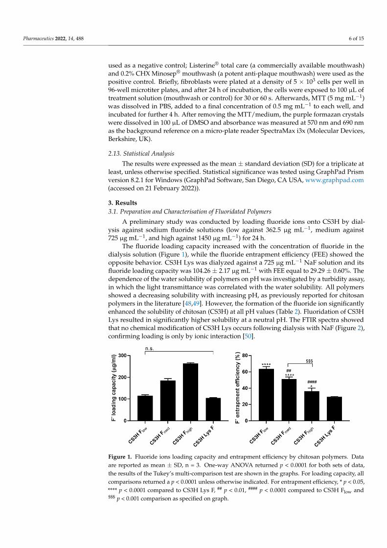
A preliminary study was conducted by loading fluoride ions onto CS3H by dial-ysis against sodium fluoride solutions (low against 362.5 µg mL−1, medium against725 µg mL−1, and high against 1450 µg mL−1) for 24 h.
The fluoride loading capacity increased with the concentration of fluoride in thedialysis solution (Figure 1), while the fluoride entrapment efficiency (FEE) showed theopposite behavior. CS3H Lys was dialyzed against a 725 µg mL−1 NaF solution and itsfluoride loading capacity was 104.26 ± 2.17 µg mL−1 with FEE equal to 29.29 ± 0.60%. Thedependence of the water solubility of polymers on pH was investigated by a turbidity assay,in which the light transmittance was correlated with the water solubility. All polymersshowed a decreasing solubility with increasing pH, as previously reported for chitosanpolymers in the literature [48,49]. However, the formation of the fluoride ion significantlyenhanced the solubility of chitosan (CS3H) at all pH values (Table 2). Fluoridation of CS3HLys resulted in significantly higher solubility at a neutral pH. The FTIR spectra showedthat no chemical modification of CS3H Lys occurs following dialysis with NaF (Figure 2),confirming loading is only by ionic interaction [50].
Pharmaceutics 2022, 14, x 6 of 15
without adding the dye to avoid interference. In this experiment, artificial saliva [47] was used as a negative control; Listerine® total care (a commercially available mouthwash) and 0.2% CHX Minosep® mouthwash (a potent anti-plaque mouthwash) were used as the pos-itive control. Briefly, fibroblasts were plated at a density of 5 × 10³ cells per well in 96-well microtiter plates, and after 24 h of incubation, the cells were exposed to 100 µL of treat-ment solution (mouthwash or control) for 30 or 60 s. Afterwards, MTT (5 mg mL−1) was dissolved in PBS, added to a final concentration of 0.5 mg mL−1 to each well, and incubated for further 4 h. After removing the MTT/medium, the purple formazan crystals were dis-solved in 100 µL of DMSO and absorbance was measured at 570 nm and 690 nm as the background reference on a micro-plate reader SpectraMax i3x (Molecular Devices, Berk-shire, UK).
2.13. Statistical Analysis The results were expressed as the mean ± standard deviation (SD) for a triplicate at least,
unless otherwise specified. Statistical significance was tested using GraphPad Prism version 8.2.1 for Windows (GraphPad Software, San Diego, CA USA, www.graphpad.com (accessed on 21 February 2022)).
3. Results 3.1. Preparation and Characterisation of Fluoridated Polymers
A preliminary study was conducted by loading fluoride ions onto CS3H by dialysis against sodium fluoride solutions (low against 362.5 µg mL−1, medium against 725 µg mL−1, and high against 1450 µg mL−1) for 24 h.
The fluoride loading capacity increased with the concentration of fluoride in the di-alysis solution (Figure 1), while the fluoride entrapment efficiency (FEE) showed the op-posite behavior. CS3H Lys was dialyzed against a 725 µg mL−1 NaF solution and its fluo-ride loading capacity was 104.26 ± 2.17 µg mL−1 with FEE equal to 29.29 ± 0.60%. The de-pendence of the water solubility of polymers on pH was investigated by a turbidity assay, in which the light transmittance was correlated with the water solubility. All polymers showed a decreasing solubility with increasing pH, as previously reported for chitosan polymers in the literature [48,49]. However, the formation of the fluoride ion significantly enhanced the solubility of chitosan (CS3H) at all pH values (Table 2). Fluoridation of CS3H Lys resulted in significantly higher solubility at a neutral pH. The FTIR spectra showed that no chemical modification of CS3H Lys occurs following dialysis with NaF (Figure 2), confirming loading is only by ionic interaction [50].
Figure 1. Fluoride ions loading capacity and entrapment efficiency by chitosan polymers. Data are reported as mean ± SD, n = 3. One-way ANOVA returned p < 0.0001 for both sets of data, the results of the Tukey’s multi-comparison test are shown in the graphs. For loading capacity, all comparisons returned a p < 0.0001 unless otherwise indicated. For entrapment efficiency, * p < 0.05, **** p < 0.0001
Figure 1. Fluoride ions loading capacity and entrapment efficiency by chitosan polymers. Dataare reported as mean ± SD, n = 3. One-way ANOVA returned p < 0.0001 for both sets of data,the results of the Tukey’s multi-comparison test are shown in the graphs. For loading capacity, allcomparisons returned a p < 0.0001 unless otherwise indicated. For entrapment efficiency, * p < 0.05,**** p < 0.0001 compared to CS3H Lys F, ## p < 0.01, #### p < 0.0001 compared to CS3H Flow and$$$ p < 0.001 comparison as specified on graph.

Pharmaceutics 2022, 14, 488 7 of 15
Table 2. pH dependence of aqueous solubility of fluoridated polymers. Data are reported asmean ± SD, n = 3. One-way ANOVA followed by Tukey’s multi-comparison test was performedto assess the effect of fluoridation with different amounts of fluoride ions on the solubility of CS3H(** p < 0.01; *** p < 0.001, and **** p < 0.0001 compared to CS3H; a p < 0.05 and b p < 0.01 comparingpolymers annotated with the same letter). Unpaired, two-tailed t-test was performed to assess theeffect of fluoridation on the solubility of CS3H Lys (# p < 0.05; ## p < 0.01; ### p < 0.001).
PolymersTransmittance (%)
pH 6.00 pH 6.50 pH 7.00 pH 7.25
CS3H 98.04 ± 0.44 97.72 ± 0.13 69.15 ± 3.5 29.06 ± 3.20CS3H Flow 99.34 ± 0.13 ** 99.51 ± 0.04 *** 88.32 ± 4.81 ***a 72.47 ± 5.64 ****
CS3H Fmedium 99.80 ± 0.20 *** 99.34 ± 0.19 *** 97.30 ± 1.46 ****ab 80.21 ± 3.46 ****CS3H Fhigh 99.52 ± 0.25 *** 99.46 ± 0.34 *** 85.80 ± 0.61 ***b 72.78 ± 3.91 ****CS3H Lys 92.88 ± 1.07 92.71 ± 0.26 59.19 ± 7.44 25.22 ± 3.34
CS3H Lys F 88.45 ± 1.89 # 88.67 ± 1.43 86.04 ± 1.16 ## 48.5 ± 2.42 ###
Pharmaceutics 2022, 14, x 7 of 15
compared to CS3H Lys F, ## p < 0.01, #### p < 0.0001 compared to CS3H Flow and $$$ p < 0.001 comparison as specified on graph.
Table 2. pH dependence of aqueous solubility of fluoridated polymers. Data are reported as mean ± SD, n = 3. One-way ANOVA followed by Tukey’s multi-comparison test was performed to assess the effect of fluoridation with different amounts of fluoride ions on the solubility of CS3H (** p < 0.01; *** p < 0.001, and **** p < 0.0001 compared to CS3H; a p < 0.05 and b p < 0.01 comparing polymers annotated with the same letter). Unpaired, two-tailed t-test was performed to assess the effect of fluoridation on the solubility of CS3H Lys (# p < 0.05; ## p < 0.01; ### p < 0.001).
Polymers Transmittance (%)
pH 6.00 pH 6.50 pH 7.00 pH 7.25 CS3H 98.04 ± 0.44 97.72 ± 0.13 69.15 ± 3.5 29.06 ± 3.20
CS3H Flow 99.34 ± 0.13 ** 99.51 ± 0.04 *** 88.32 ± 4.81 ***a 72.47 ± 5.64 **** CS3H Fmedium 99.80 ± 0.20 *** 99.34 ± 0.19 *** 97.30 ± 1.46 ****ab 80.21 ± 3.46 ****
CS3H Fhigh 99.52 ± 0.25 *** 99.46 ± 0.34 *** 85.80 ± 0.61 ***b 72.78 ± 3.91 **** CS3H Lys 92.88 ± 1.07 92.71 ± 0.26 59.19 ± 7.44 25.22 ± 3.34
CS3H Lys F 88.45 ± 1.89 # 88.67 ± 1.43 86.04 ± 1.16 ## 48.5 ± 2.42 ###
Figure 2. FTIR spectra of NaF (black), CS3H (green), CS3H Lys (blue), and CS3H Lys F (red).
3.2. Inhibition of Acid Demineralisation In Vitro Dental erosion of the tooth normally occurs by the action of acid consumed through
food and drink, or the acid products generated by the bacteria present in the mouth; as a result, phosphate ions are released [51]. For this reason, the quantification of phosphate release from hydroxyapatite deposits after acid exposure can be used in vitro to quantify the demineralization of tooth enamel due to acid attack and evaluate the protective action of dental products. The ability of NaF solutions to prevent the acid triggered deminerali-zation of the HA was shown to increase proportionally to the concentration of the ions for the NaF 362.5 and 725 µg mL−1 concentration samples, but the higher concentration of NaF (1450 µg mL−1) showed no further significant increase (Figure 3). All polymers showed significant protection against acid challenge of the hydroxyapatite surfaces, but only CS3H Fhigh presented a significantly higher effect compared to CS3H (Figure 3). CS3H Lys F exhibited the highest activity with 58.57% of inhibition of phosphate release.
5001000150020002500300035004000
50
75
100
Wavenumber (cm-1)
Figure 2. FTIR spectra of NaF (black), CS3H (green), CS3H Lys (blue), and CS3H Lys F (red).
3.2. Inhibition of Acid Demineralisation In Vitro
Dental erosion of the tooth normally occurs by the action of acid consumed throughfood and drink, or the acid products generated by the bacteria present in the mouth; as aresult, phosphate ions are released [51]. For this reason, the quantification of phosphaterelease from hydroxyapatite deposits after acid exposure can be used in vitro to quantifythe demineralization of tooth enamel due to acid attack and evaluate the protective action ofdental products. The ability of NaF solutions to prevent the acid triggered demineralizationof the HA was shown to increase proportionally to the concentration of the ions for theNaF 362.5 and 725 µg mL−1 concentration samples, but the higher concentration of NaF(1450 µg mL−1) showed no further significant increase (Figure 3). All polymers showedsignificant protection against acid challenge of the hydroxyapatite surfaces, but only CS3HFhigh presented a significantly higher effect compared to CS3H (Figure 3). CS3H Lys Fexhibited the highest activity with 58.57% of inhibition of phosphate release.

Pharmaceutics 2022, 14, 488 8 of 15Pharmaceutics 2022, 14, x 8 of 15
Figure 3. Inhibition of phosphate release by NaF, chitosan, and its derivatives. Data are reported as mean ± SD, n = 3. Data were analyzed by one-way ANOVA followed by Tukey’s multiple compar-isons (* p < 0.05, ** p < 0.01, *** p < 0.001 and **** p < 0.0001 compared to deionized water; # p < 0.05, compared to CS3H; $$ p < 0.01, $$$ p < 0.001 compared to CSH3 Lys Fmedium).
3.3. Determination of MIC and MBC The potential antibacterial activity of the modified chitosans was determined against
S. aureus and S. mutans. All chitosan polymers were capable of inhibiting the growth of the microorganisms tested, with no significant difference between samples (Table 3).
Table 3. Values of MIC and MBC for the chitosan and modified chitosan against S. aureus and S. mutans. Data are reported as mean ± SD (n = 4). Data were analyzed by two-way ANOVA followed by Dunnett’s multiple comparisons. No significant difference was observed.
Polymer Staphylococcus aureus Streptococcus mutans
MIC (mg/mL) MBC (mg/mL) MIC (mg/mL) MBC (mg/mL) CS 1.60 ± 0.33 ≥3.0 1.50 ± 0.20 ≥3.0
CS3H 1.10 ± 0.38 ≥3.0 1.30 ± 0.20 ≥3.0 CS3H Lys 1.10 ± 0.60 ≥3.0 1.40 ± 0.23 ≥3.0
CS3H Lys F 1.40 ± 0.23 ≥3.0 1.40 ± 0.23 ≥3.0
3.4. Inhibition of Biofilms Formation Polymers were further studied for their potential to inhibit S. mutans biofilm for-
mation (Figure 4). All polymers were able to completely prevent biofilm formation at the highest concentration tested, with more than 70% efficacy showed at the lowest concen-tration (0.1 mg mL−1). At all concentrations, the modified chitosans were significantly more active than CS3H, with a higher activity demonstrated by the non-fluoridated lysine derivative. CS showed a dose dependent effect with very significant reduction in viability caused by increasing concentrations (p < 0.0001 for all comparisons apart from 800 vs. 1200 that the p value was p < 0.01). CS3H showed a dose dependent behavior (p < 0.001) up to 800 µg mL−1. CS3H Lys was highly effective with no statistical difference between concen-trations above 200 µg mL−1 (p > 0.05), while no more decrease in viability was noticed with CS3H Lys F from 400 µg mL−1 (p > 0.05).
Figure 3. Inhibition of phosphate release by NaF, chitosan, and its derivatives. Data are reportedas mean ± SD, n = 3. Data were analyzed by one-way ANOVA followed by Tukey’s multiplecomparisons (* p < 0.05, ** p < 0.01, *** p < 0.001 and **** p < 0.0001 compared to deionized water;# p < 0.05, compared to CS3H; $$ p < 0.01, $$$ p < 0.001 compared to CSH3 Lys Fmedium).
3.3. Determination of MIC and MBC
The potential antibacterial activity of the modified chitosans was determined againstS. aureus and S. mutans. All chitosan polymers were capable of inhibiting the growth of themicroorganisms tested, with no significant difference between samples (Table 3).
Table 3. Values of MIC and MBC for the chitosan and modified chitosan against S. aureus and S.mutans. Data are reported as mean ± SD (n = 4). Data were analyzed by two-way ANOVA followedby Dunnett’s multiple comparisons. No significant difference was observed.
PolymerStaphylococcus aureus Streptococcus mutans
MIC (mg/mL) MBC (mg/mL) MIC (mg/mL) MBC (mg/mL)
CS 1.60 ± 0.33 ≥3.0 1.50 ± 0.20 ≥3.0CS3H 1.10 ± 0.38 ≥3.0 1.30 ± 0.20 ≥3.0
CS3H Lys 1.10 ± 0.60 ≥3.0 1.40 ± 0.23 ≥3.0CS3H Lys F 1.40 ± 0.23 ≥3.0 1.40 ± 0.23 ≥3.0
3.4. Inhibition of Biofilms Formation
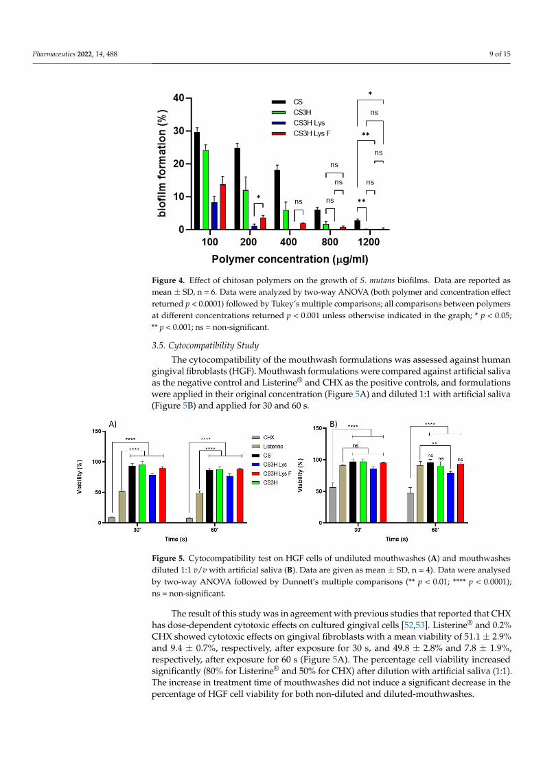
Polymers were further studied for their potential to inhibit S. mutans biofilm formation(Figure 4). All polymers were able to completely prevent biofilm formation at the highestconcentration tested, with more than 70% efficacy showed at the lowest concentration(0.1 mg mL−1). At all concentrations, the modified chitosans were significantly more activethan CS3H, with a higher activity demonstrated by the non-fluoridated lysine derivative.CS showed a dose dependent effect with very significant reduction in viability caused byincreasing concentrations (p < 0.0001 for all comparisons apart from 800 vs. 1200 that the pvalue was p < 0.01). CS3H showed a dose dependent behavior (p < 0.001) up to 800 µg mL−1.CS3H Lys was highly effective with no statistical difference between concentrations above200 µg mL−1 (p > 0.05), while no more decrease in viability was noticed with CS3H Lys Ffrom 400 µg mL−1 (p > 0.05).
Pharmaceutics 2022, 14, 488 9 of 15Pharmaceutics 2022, 14, x 9 of 15
Figure 4. Effect of chitosan polymers on the growth of S. mutans biofilms. Data are reported as mean ± SD, n = 6. Data were analyzed by two-way ANOVA (both polymer and concentration effect re-turned p < 0.0001) followed by Tukey’s multiple comparisons; all comparisons between polymers at different concentrations returned p < 0.001 unless otherwise indicated in the graph; * p < 0.05; ** p < 0.001; ns = non-significant.
3.5. Cytocompatibility Study The cytocompatibility of the mouthwash formulations was assessed against human
gingival fibroblasts (HGF). Mouthwash formulations were compared against artificial sa-liva as the negative control and Listerine® and CHX as the positive controls, and formula-tions were applied in their original concentration (Figure 5A) and diluted 1:1 with artifi-cial saliva (Figure 5B) and applied for 30 and 60 s.
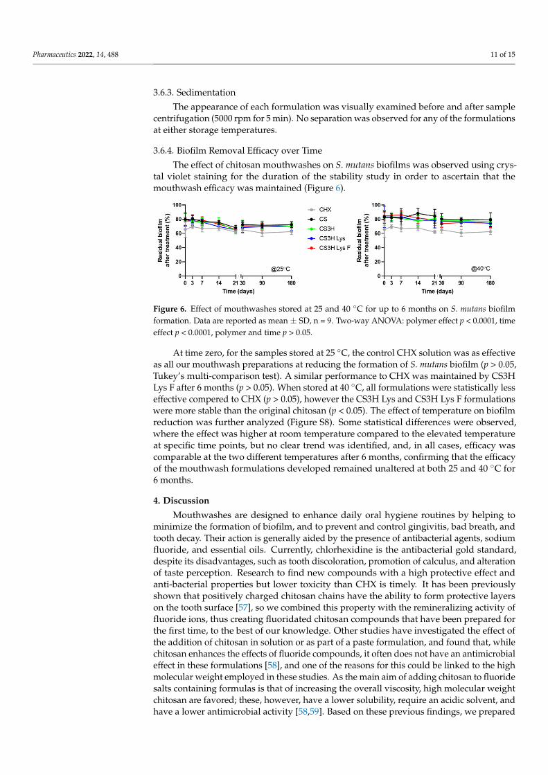
Figure 5. Cytocompatibility test on HGF cells of undiluted mouthwashes (A) and mouthwashes diluted 1:1 v/v with artificial saliva (B). Data are given as mean ± SD, n = 4). Data were analysed by two-way ANOVA followed by Dunnett’s multiple comparisons (** p < 0.01; **** p < 0.0001); ns = non-significant.
The result of this study was in agreement with previous studies that reported that CHX has dose-dependent cytotoxic effects on cultured gingival cells [52,53]. Listerine® and 0.2% CHX showed cytotoxic effects on gingival fibroblasts with a mean viability of 51.1 ± 2.9% and 9.4 ± 0.7%, respectively, after exposure for 30 s, and 49.8 ± 2.8% and 7.8 ± 1.9%, respectively, after exposure for 60 s (Figure 5A). The percentage cell viability in-creased significantly (80% for Listerine® and 50% for CHX) after dilution with artificial saliva (1:1). The increase in treatment time of mouthwashes did not induce a significant decrease in the percentage of HGF cell viability for both non-diluted and diluted-mouth-washes.
Figure 4. Effect of chitosan polymers on the growth of S. mutans biofilms. Data are reported asmean ± SD, n = 6. Data were analyzed by two-way ANOVA (both polymer and concentration effectreturned p < 0.0001) followed by Tukey’s multiple comparisons; all comparisons between polymersat different concentrations returned p < 0.001 unless otherwise indicated in the graph; * p < 0.05;** p < 0.001; ns = non-significant.
3.5. Cytocompatibility Study
The cytocompatibility of the mouthwash formulations was assessed against humangingival fibroblasts (HGF). Mouthwash formulations were compared against artificial salivaas the negative control and Listerine® and CHX as the positive controls, and formulationswere applied in their original concentration (Figure 5A) and diluted 1:1 with artificial saliva(Figure 5B) and applied for 30 and 60 s.
Pharmaceutics 2022, 14, x 9 of 15
Figure 4. Effect of chitosan polymers on the growth of S. mutans biofilms. Data are reported as mean ± SD, n = 6. Data were analyzed by two-way ANOVA (both polymer and concentration effect re-turned p < 0.0001) followed by Tukey’s multiple comparisons; all comparisons between polymers at different concentrations returned p < 0.001 unless otherwise indicated in the graph; * p < 0.05; ** p < 0.001; ns = non-significant.
3.5. Cytocompatibility Study The cytocompatibility of the mouthwash formulations was assessed against human
gingival fibroblasts (HGF). Mouthwash formulations were compared against artificial sa-liva as the negative control and Listerine® and CHX as the positive controls, and formula-tions were applied in their original concentration (Figure 5A) and diluted 1:1 with artifi-cial saliva (Figure 5B) and applied for 30 and 60 s.
Figure 5. Cytocompatibility test on HGF cells of undiluted mouthwashes (A) and mouthwashes diluted 1:1 v/v with artificial saliva (B). Data are given as mean ± SD, n = 4). Data were analysed by two-way ANOVA followed by Dunnett’s multiple comparisons (** p < 0.01; **** p < 0.0001); ns = non-significant.
The result of this study was in agreement with previous studies that reported that CHX has dose-dependent cytotoxic effects on cultured gingival cells [52,53]. Listerine® and 0.2% CHX showed cytotoxic effects on gingival fibroblasts with a mean viability of 51.1 ± 2.9% and 9.4 ± 0.7%, respectively, after exposure for 30 s, and 49.8 ± 2.8% and 7.8 ± 1.9%, respectively, after exposure for 60 s (Figure 5A). The percentage cell viability in-creased significantly (80% for Listerine® and 50% for CHX) after dilution with artificial saliva (1:1). The increase in treatment time of mouthwashes did not induce a significant decrease in the percentage of HGF cell viability for both non-diluted and diluted-mouth-washes.
Figure 5. Cytocompatibility test on HGF cells of undiluted mouthwashes (A) and mouthwashesdiluted 1:1 v/v with artificial saliva (B). Data are given as mean ± SD, n = 4). Data were analysedby two-way ANOVA followed by Dunnett’s multiple comparisons (** p < 0.01; **** p < 0.0001);ns = non-significant.
The result of this study was in agreement with previous studies that reported that CHXhas dose-dependent cytotoxic effects on cultured gingival cells [52,53]. Listerine® and 0.2%CHX showed cytotoxic effects on gingival fibroblasts with a mean viability of 51.1 ± 2.9%and 9.4 ± 0.7%, respectively, after exposure for 30 s, and 49.8 ± 2.8% and 7.8 ± 1.9%,respectively, after exposure for 60 s (Figure 5A). The percentage cell viability increasedsignificantly (80% for Listerine® and 50% for CHX) after dilution with artificial saliva (1:1).The increase in treatment time of mouthwashes did not induce a significant decrease in thepercentage of HGF cell viability for both non-diluted and diluted-mouthwashes.

Pharmaceutics 2022, 14, 488 10 of 15
The MTT test revealed that all chitosan mouthwashes maintained a higher percentageof viable cells compared to the positive control solutions (p ≤ 0.0001) (Figure 5) for bothundiluted and diluted mouthwashes.
3.6. Stability Studies of Mouthwash Formulations
The formulation and preparation of any new pharmaceutical or consumer care productnecessitates adequate physical and chemical stability, as well as a microbiological profileunaltered over the period of time in storage under the influence of a variety of environmen-tal factors, such as temperature, humidity, and light [54–56]. In this study, we evaluatedthe behavior of the formulations at 25 and 40 ◦C for 6 months.
3.6.1. Organoleptic Properties
Visually, both mouthwashes presented a consistent color and clear appearance withno turbidity and a peppermint odor (subjective evaluation) that was unaltered for up to180 days of storage both at 25 and 40 ◦C.
Color stability was also determined by UV measurements and changes to the ab-sorbance values at λmax 639 nm were evaluated. No statistical difference in absorbance wasidentified for samples stored at the two different temperatures (Figures S4–S7 and Table 4).Mouthwashes containing CS3H and CS3H Lys did not undergo any changes in absorbancefor the duration of the experiment, while CS3H Lys F mouthwash showed an initial changethat was reversed in time until the end of the experiment.
Table 4. Results of the statistical evaluation of absorbance values at 639 nm of different mouthwashformulations over time and at different temperatures. Two-way ANOVA was performed, followedby Sidak’s multiple comparisons test to see the effect of the temperature on the absorbance of the dyein the mouthwash formulation. The data were rerun with Dunnett’s multiple comparisons test to seethe effect of the time (ns = not significant or p > 0.05; * p ≤ 0.05; ** p ≤ 0.01).
Polymer 25 ◦C 40 ◦C 25 ◦C vs. 40 ◦C
CS T0 vs. T90 (**)T0 vs. T180 (*) T0 vs. T3 (*) ns
CS3H ns ns nsCS3H Lys ns ns ns
CS3H Lys F T0 vs. T30 (*) ns ns
3.6.2. Evaluation of pH Stability
As part of the stability study, the pH values of the different formulations were recordedover time (Table 5 and Table S2), and the pH remained unaltered by time or temperature.
Table 5. The effect of temperature on the pH of chitosan mouthwash formulation. Data are expressedas mean ± SD, n = 3. Two-way ANOVA followed by Sidak’s test was carried out (* p < 0.05 comparedto T0).
Polymer Temp T0 T30 T90 T180
CS25 ◦C
5.54 ± 0.025.56 ± 0.02 5.54 ± 0.02 5.53 ± 0.02
40 ◦C 5.53 ± 0.02 5.52 ± 0.01 5.52 ± 0.03
CS3H25 ◦C
5.52 ± 0.035.53 ± 0.01 5.52 ± 0.01 5.52 ± 0.01
40 ◦C 5.50 ± 0.01 5.52 ± 0.01 5.50 ± 0.01
CS3H Lys 25 ◦C5.52 ± 0.01
5.52 ± 0.02 5.52 ± 0.02 5.52 ± 0.0240 ◦C 5.48 ± 0.01 * 5.48 ± 0.03 5.48 ± 0.02
CS3H Lys F 25 ◦C5.52 ± 0.02
5.54 ± 0.06 5.54 ± 0.04 5.53 ± 0.0540 ◦C 5.50 ± 0.04 5.52 ± 0.06 5.51 ± 0.03
Pharmaceutics 2022, 14, 488 11 of 15
3.6.3. Sedimentation
The appearance of each formulation was visually examined before and after samplecentrifugation (5000 rpm for 5 min). No separation was observed for any of the formulationsat either storage temperatures.
3.6.4. Biofilm Removal Efficacy over Time
The effect of chitosan mouthwashes on S. mutans biofilms was observed using crys-tal violet staining for the duration of the stability study in order to ascertain that themouthwash efficacy was maintained (Figure 6).
Pharmaceutics 2022, 14, x 11 of 15
3.6.3. Sedimentation The appearance of each formulation was visually examined before and after sample
centrifugation (5000 rpm for 5 min). No separation was observed for any of the formula-tions at either storage temperatures.
3.6.4. Biofilm Removal Efficacy over Time The effect of chitosan mouthwashes on S. mutans biofilms was observed using crystal
violet staining for the duration of the stability study in order to ascertain that the mouth-wash efficacy was maintained (Figure 6).
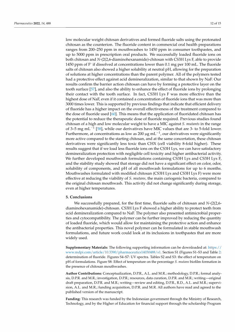
Figure 6. Effect of mouthwashes stored at 25 and 40 °C for up to 6 months on S. mutans biofilm formation. Data are reported as mean ± SD, n = 9. Two-way ANOVA: polymer effect p < 0.0001, time effect p < 0.0001, polymer and time p > 0.05.
At time zero, for the samples stored at 25 °C, the control CHX solution was as effec-tive as all our mouthwash preparations at reducing the formation of S. mutans biofilm (p > 0.05, Tukey’s multi-comparison test). A similar performance to CHX was maintained by CS3H Lys F after 6 months (p > 0.05). When stored at 40 °C, all formulations were statisti-cally less effective compered to CHX (p > 0.05), however the CS3H Lys and CS3H Lys F formulations were more stable than the original chitosan (p < 0.05). The effect of tempera-ture on biofilm reduction was further analyzed (Figure S8). Some statistical differences were observed, where the effect was higher at room temperature compared to the elevated temperature at specific time points, but no clear trend was identified, and, in all cases, efficacy was comparable at the two different temperatures after 6 months, confirming that the efficacy of the mouthwash formulations developed remained unaltered at both 25 and 40 °C for 6 months.
4. Discussion Mouthwashes are designed to enhance daily oral hygiene routines by helping to min-
imize the formation of biofilm, and to prevent and control gingivitis, bad breath, and tooth decay. Their action is generally aided by the presence of antibacterial agents, sodium flu-oride, and essential oils. Currently, chlorhexidine is the antibacterial gold standard, de-spite its disadvantages, such as tooth discoloration, promotion of calculus, and alteration of taste perception. Research to find new compounds with a high protective effect and anti-bacterial properties but lower toxicity than CHX is timely. It has been previously shown that positively charged chitosan chains have the ability to form protective layers on the tooth surface [57], so we combined this property with the remineralizing activity of fluoride ions, thus creating fluoridated chitosan compounds that have been prepared for the first time, to the best of our knowledge. Other studies have investigated the effect of the addition of chitosan in solution or as part of a paste formulation, and found that, while chitosan enhances the effects of fluoride compounds, it often does not have an an-timicrobial effect in these formulations [58], and one of the reasons for this could be linked to the high molecular weight employed in these studies. As the main aim of adding chi-tosan to fluoride salts containing formulas is that of increasing the overall viscosity, high molecular weight chitosan are favored; these, however, have a lower solubility, require
Figure 6. Effect of mouthwashes stored at 25 and 40 ◦C for up to 6 months on S. mutans biofilmformation. Data are reported as mean ± SD, n = 9. Two-way ANOVA: polymer effect p < 0.0001, timeeffect p < 0.0001, polymer and time p > 0.05.
At time zero, for the samples stored at 25 ◦C, the control CHX solution was as effectiveas all our mouthwash preparations at reducing the formation of S. mutans biofilm (p > 0.05,Tukey’s multi-comparison test). A similar performance to CHX was maintained by CS3HLys F after 6 months (p > 0.05). When stored at 40 ◦C, all formulations were statistically lesseffective compered to CHX (p > 0.05), however the CS3H Lys and CS3H Lys F formulationswere more stable than the original chitosan (p < 0.05). The effect of temperature on biofilmreduction was further analyzed (Figure S8). Some statistical differences were observed,where the effect was higher at room temperature compared to the elevated temperatureat specific time points, but no clear trend was identified, and, in all cases, efficacy wascomparable at the two different temperatures after 6 months, confirming that the efficacyof the mouthwash formulations developed remained unaltered at both 25 and 40 ◦C for6 months.
4. Discussion
Mouthwashes are designed to enhance daily oral hygiene routines by helping tominimize the formation of biofilm, and to prevent and control gingivitis, bad breath, andtooth decay. Their action is generally aided by the presence of antibacterial agents, sodiumfluoride, and essential oils. Currently, chlorhexidine is the antibacterial gold standard,despite its disadvantages, such as tooth discoloration, promotion of calculus, and alterationof taste perception. Research to find new compounds with a high protective effect andanti-bacterial properties but lower toxicity than CHX is timely. It has been previouslyshown that positively charged chitosan chains have the ability to form protective layerson the tooth surface [57], so we combined this property with the remineralizing activity offluoride ions, thus creating fluoridated chitosan compounds that have been prepared forthe first time, to the best of our knowledge. Other studies have investigated the effect ofthe addition of chitosan in solution or as part of a paste formulation, and found that, whilechitosan enhances the effects of fluoride compounds, it often does not have an antimicrobialeffect in these formulations [58], and one of the reasons for this could be linked to the highmolecular weight employed in these studies. As the main aim of adding chitosan to fluoridesalts containing formulas is that of increasing the overall viscosity, high molecular weightchitosan are favored; these, however, have a lower solubility, require an acidic solvent, andhave a lower antimicrobial activity [58,59]. Based on these previous findings, we prepared
Pharmaceutics 2022, 14, 488 12 of 15
low molecular weight chitosan derivatives and formed fluoride salts using the protonatedchitosan as the counterion. The fluoride content in commercial oral health preparationsranges from 200–250 ppm in mouthwashes to 1450 ppm in consumer toothpastes, andup to 5000 ppm in prescription oral products. We successfully loaded fluoride ions onboth chitosan and N-(2(2,6-diaminohexanamide)-chitosan with CS3H Lys F, able to provide1450 ppm of F- if dissolved at concentrations lower than 0.1 mg per 100 mL. The fluoridesalts of chitosan also showed a higher solubility at neutral pH, allowing for the preparationof solutions at higher concentrations than the parent polymer. All of the polymers testedhad a protective effect against acid demineralization, similar to that shown by NaF. Ourresults confirm the barrier action chitosan can have by forming a protective layer on thetooth surface [57], and also the ability to enhance the effect of fluoride ions by prolongingtheir contact with the tooth surface. In fact, CS3H Lys F was more effective than thehighest dose of NaF, even if it contained a concentration of fluoride ions that was more than3000 times lower. This is supported by previous findings that indicate that efficient deliveryof fluoride has a higher impact on the overall effectiveness of the treatment compared tothe dose of fluoride used [60]. This means that the application of fluoridated chitosan hasthe potential to reduce the therapeutic dose of fluoride required. Previous studies foundchitosan of a high and low molecular weight to have a MIC against S. mutants in the rangeof 3–5 mg mL−1 [58], while our derivatives have MIC values that are 3- to 5-fold lower.Furthermore, at concentrations as low as 200 µg mL−1, our derivatives were significantlymore active compared to the starting chitosan, and at the same concertation, these chitosanderivatives were significantly less toxic than CHX (cell viability 8-fold higher). Theseresults suggest that if we load less fluoride ions on the CS3H Lys, we can have satisfactorydemineralization protection with negligible cell toxicity and higher antibacterial activity.We further developed mouthwash formulations containing CS3H Lys and CS3H Lys F,and the stability study showed that storage did not have a significant effect on color, odor,solubility of components, and pH of all mouthwash formulations for up to 6 months.Mouthwashes formulated with modified chitosan (CS3H Lys and CS3H Lys F) were moreeffective at reducing the viability of S. mutans, the main cariogenic bacteria, compared tothe original chitosan mouthwash. This activity did not change significantly during storage,even at higher temperatures.
5. Conclusions
We successfully prepared, for the first time, fluoride salts of chitosan and N-(2(2,6-diaminohexanamide)-chitosan. CS3H Lys F showed a higher ability to protect teeth fromacid demineralization compared to NaF. The polymer also presented antimicrobial proper-ties and cytocompatibility. The polymer can be further improved by reducing the quantityof loaded fluoride, which would allow for maintaining the protective action and enhancethe antibacterial properties. This novel polymer can be formulated in stable mouthwashformulations, and future work could look at its inclusions in toothpastes that are morewidely used.
Supplementary Materials: The following supporting information can be downloaded at: https://www.mdpi.com/article/10.3390/pharmaceutics14030488/s1. Section S1 (Figures S1–S3 and Table 2:determination of fluoride. Figures S4–S7: UV spectra. Tables S2 and S3: the effect of temperature onpH of formulations. Figure S8: Effect of temperature on the percentage S. mutans biofilm formation inthe presence of chitosan mouthwashes.
Author Contributions: Conceptualization, D.P.R., A.L. and M.R.; methodology, D.P.R.; formal analy-sis, D.P.R. and M.R.; investigation, D.P.R.; resources, data curation, D.P.R. and M.R.; writing—originaldraft preparation, D.P.R. and M.R.; writing—review and editing, D.P.R., R.D., A.L. and M.R.; supervi-sion, A.L. and M.R.; funding acquisition, D.P.R. and M.R. All authors have read and agreed to thepublished version of the manuscript.
Funding: This research was funded by the Indonesian government through the Ministry of Research,Technology, and by the Higher of Education for financial support through the scholarship Program
Pharmaceutics 2022, 14, 488 13 of 15
for Research and Innovation in Science and Technology (RISET-Pro) World Bank Loan No. 8245-ID.The APC was funded by the University of Portsmouth.
Institutional Review Board Statement: Not applicable.
Informed Consent Statement: Not applicable.
Data Availability Statement: All data relative to this study are included in the manuscript andSupplementary Materials.
Conflicts of Interest: The authors declare no conflict of interest.
References1. WHO Oral Health. Available online: https://www.who.int/news-room/fact-sheets/detail/oral-health (accessed on 11 Jan-
uary 2022).2. Alhabdan, Y.A.; Albeshr, A.G.; Yenugadhati, N.; Jradi, H. Prevalence of dental caries and associated factors among primary school
children: A population-based cross-sectional study in Riyadh, Saudi Arabia. Environ. Health Prev. Med. 2018, 23, 60. [CrossRef][PubMed]
3. Chu, C.H.; Wong, S.S.S.; Suen, R.P.C.; Lo, E.C.M. Oral health and dental care in Hong Kong. Surgeon 2013, 11, 153–157. [CrossRef][PubMed]
4. Duangthip, D.; Gao, S.S.; Lo, E.C.M.; Chu, C.H. Early childhood caries among 5- to 6-year-old children in Southeast Asia. Int.Dent. J. 2017, 67, 98–106. [CrossRef] [PubMed]
5. Chandki, R.; Banthia, P.; Banthia, R. Biofilms: A microbial home. J. Indian Soc. Periodontol. 2011, 15, 111–114. [CrossRef]6. Nazir, R.; Zaffar, M.R.; Amin, I. Chapter 8—Bacterial Biofilms: The Remarkable Heterogeneous Biological Communities and Nitrogen
Fixing Microorganisms in Lakes; Elsevier Inc.: Amsterdam, The Netherlands, 2019; ISBN 978-0-12-817495-1.7. Marsh, P.D. Dental plaque as a biofilm and a microbial community—Implications for health and disease. BMC Oral Health 2006, 6,
1–7. [CrossRef]8. Ahn, S.J.; Ahn, S.J.; Wen, Z.T.; Brady, J.; Burne, R.A. Characteristics of biofilm formation by Streptococcus mutans in the presence
of saliva. Infect. Immun. 2008, 76, 4259–4268. [CrossRef]9. Swamy, M.K.; Akhtar, M.S.; Sinniah, U.R. Antimicrobial properties of plant essential oils against human pathogens and their
mode of action: An updated review. Evid.-Based Complement. Altern. Med. 2016, 2016, 3012462. [CrossRef]10. NHS How to Keep Your Teeth Clean. Available online: https://www.nhs.uk/live-well/healthy-body/how-to-keep-your-teeth-
clean/ (accessed on 11 January 2022).11. Van Loveren, C.; Buijs, J.F.; Ten Cate, J.M. The effect of triclosan toothpaste on enamel demineralization in a bacterial demineral-
ization model. J. Antimicrob. Chemother. 2000, 45, 153–158. [CrossRef]12. Riley, P.; Lamont, T. Triclosan produces statistically significant reduction in plaque, gingivitis and caries but not clinically
important benefit. Evid. Based. Dent. 2014, 15, 6–7. [CrossRef]13. Quintas, V.; Prada-López, I.; Carreira, M.J.; Suárez-Quintanilla, D.; Balsa-Castro, C.; Tomás, I. In situ antibacterial activity of
essential oils with and without alcohol on oral biofilm: A randomized clinical trial. Front. Microbiol. 2017, 8, 2162. [CrossRef]14. Vlachojannis, C.; Winsauer, H.; Chrubasik, S. Effectiveness and safety of a mouthwash containing essential oil ingredients.
Phytother. Res. 2013, 27, 685–691. [CrossRef]15. Sheen, S.; Addy, M. An in vitro evaluation of the availability of cetylpyridinium chloride and chlorhexidine in some commercially
available mouthrinse products. Br. Dent. J. 2003, 194, 16–19. [CrossRef]16. Mehdipour, M.; Taghavi Zenoz, A.; Asvadi Kermani, I.; Hosseinpour, A. A comparison between zinc sulfate and chlorhexidine
gluconate mouthwashes in the prevention of chemotherapy-induced oral mucositis. DARU J. Pharm. Sci. 2011, 19, 71–73.17. Najafi, M.H.; Taheri, M.; Mokhtari, M.R.; Forouzanfar, A.; Farazi, F.; Mirzaee, M.; Ebrahiminik, Z.; Mehrara, R. Comparative
study of 0.2% and 0.12% digluconate chlorhexidine mouth rinses on the level of dental staining and gingival indices. Dent. Res. J.2012, 9, 305–308.
18. Marsh, P.D. Controlling the oral biofilm with antimicrobials. J. Dent. 2010, 38, S11–S15. [CrossRef]19. Davison, J.; Maillard, J. Opinion on Triclosan—Antimicrobial Resistance; European Union: Brussels, Belgium, 2010; ISBN 9789279124846.20. Storehagen, S.; Ose, N.; Midha, S. Dentifrices and Mouthwashes Ingredients and Their Use. Master’s Thesis, University of Oslo,
Oslo, Norway, 2003.21. Sreenivasan, P.; Gaffar, A. Antiplaque biocides and bacterial resistance: A review. J. Clin. Periodontol. 2002, 29, 965–974. [CrossRef]22. Alencar, M.A.S.D.S.; Martinez, E.F.; Figueiredo, F.C.; De Lima e Silva, A.R.; Protazio, J.E.; Bertamoni, M.; Peruzzo, D.C.; Napimoga,
M.H. The Evaluation of Osteoblastic Cell Behavior on Treated Titanium Surface. Open Dent. J. 2019, 14, 1–6. [CrossRef]23. Halboub, E.; Al-Maweri, S.A.; Al-Wesabi, M.; Al-Kamel, A.; Shamala, A.; Al-Sharani, A.; Koppolu, P. Efficacy of propolis-based
mouthwashes on dental plaque and gingival inflammation: A systematic review. BMC Oral Health 2020, 20, 198. [CrossRef]24. Eslami, N.; Ahrari, F.; Rajabi, O.; Zamani, R. The staining effect of different mouthwashes containing nanoparticles on dental
enamel. J. Clin. Exp. Dent. 2015, 7, e457–e461. [CrossRef]

Pharmaceutics 2022, 14, 488 14 of 15
25. Azimi, M.; Jouybari, L.; Moghadam, S.; Ghaemi, E.; Behnampoor, N.; Sanagoo, A.; Hesam, M. Antimicrobial effects of chlorhexi-dine, matrica drop mouthwash (chamomile extract), and normal saline on hospitalized patients with endotracheal tubes. Iran. J.Nurs. Midwifery Res. 2016, 21, 458. [CrossRef]
26. Bahlouli, S.; Aghazadeh, Z.; Aghazadeh, M.; Shojani, S.; Kafil, H.S. Determining the Antibacterial Activity of ChlorhexidineMouthwashes with and without Alcohol against Common Oral Pathogens. J. Adv. Oral Res. 2018, 9, 15–19. [CrossRef]
27. Vranic, E.; Lacevic, A.; Mehmedagic, A.; Uzunovic, A. Formulation ingredients for toothpastes and mouthwashes. Bosn. J. BasicMed. Sci. 2004, 4, 51–58. [CrossRef] [PubMed]
28. Keegan, G.M.; Smart, J.D.; Ingram, M.J.; Barnes, L.M.; Burnett, G.R.; Rees, G.D. Chitosan microparticles for the controlled deliveryof fluoride. J. Dent. 2012, 40, 229–240. [CrossRef] [PubMed]
29. Subramaniam, P.; Nandan, N. Effect of xylitol, sodium fluoride and triclosan containing mouth rinse on Streptococcus mutans.Contemp. Clin. Dent. 2011, 2, 287–290. [CrossRef]
30. O’Mullane, D.M.; Baez, R.J.; Jones, S.; Lennon, M.A.; Petersen, P.E.; Rugg-Gunn, A.J.; Whelton, H.; Whitford, G.M. Fluoride andOral Health. Community Dent. Health. 2016, 33, 69–99.
31. Liao, Y.; Brandt, B.W.; Li, J.; Crielaard, W.; Van Loveren, C.; Mei, D. Fluoride resistance in Streptococcus mutans: A mini review. J.Oral Microbiol. 2017, 9, 1344509. [CrossRef]
32. Pretty, I.A. High Fluoride Concentration Toothpastes for Children and Adolescents. Caries Res. 2016, 50, 9–14. [CrossRef]33. Kavouklis, N.M.; Gaudreault, R.A. Oral Care Formulations with Hydrogen Peroxide and Lycopene. U.S. Patent 11/491,187,
24 July 2006.34. Kasaai, M.R.; Arul, J.; Charlet, G. Fragmentation of Chitosan by Acids. Sci. World J. 2013, 2013, 508540. [CrossRef]35. Sahariah, P.; Gaware, V.S.; Lieder, R.; Jónsdóttir, S.; Hjálmarsdóttir, M.; Sigurjonsson, O.E.; Másson, M. The effect of substituent,
degree of acetylation and positioning of the cationic charge on the antibacterial activity of quaternary chitosan derivatives. Mar.Drugs 2014, 12, 4635–4658. [CrossRef]
36. Islam, S.; Bhuiyan, M.A.R.; Islam, M.N. Chitin and Chitosan: Structure, Properties and Applications in Biomedical Engineering. J.Polym. Environ. 2017, 25, 854–866. [CrossRef]
37. Rahayu, D.P.; De Mori, A.; Draheim, R.R.; Lalatsa, A.; Roldo, M. Enhancing the antibacterial effect of chitosan to combatorthopaedic implant-associated infections. Carbohydr. Polym. 2021. submitted.
38. Lalatsa, A.; Garrett, N.L.; Ferrarelli, T.; Moger, J.; Schätzlein, A.G.; Uchegbu, I.F. Delivery of peptides to the blood and brain afteroral uptake of quaternary ammonium palmitoyl glycol chitosan nanoparticles. Mol. Pharm. 2012, 9, 1764–1774. [CrossRef]
39. Kubota, N.; Tatsumoto, N.; Sano, T.; Toya, K. A simple preparation of half N-acetylated chitosan highly soluble in water andaqueous organic solvents. Carbohydr. Res. 2000, 324, 268–274. [CrossRef]
40. Churchley, D.; Rees, G.D.; Barbu, E.; Nevell, T.G.; Tsibouklis, J. Fluoropolymers as low-surface-energy tooth coatings for oral care.Int. J. Pharm. 2008, 352, 44–49. [CrossRef]
41. Medina-Flores, D.; Ulloa-Urizar, G.; Camere-Colarossi, R.; Caballero-García, S.; Mayta-Tovalino, F.; del Valle-Mendoza, J.Antibacterial activity of Bixa orellana L. (achiote) against Streptococcus mutans and Streptococcus sanguinis. Asian Pac. J. Trop.Biomed. 2016, 6, 400–403. [CrossRef]
42. Pasquantonio, G.; Greco, C.; Prenna, M.; Ripa, C.; Vitali, L.A.; Petrelli, D.; Di Luca, M.C.; Ripa, S. Antibacterial activity andanti-biofilm effect of chitosan against strains of Streptococcus mutans isolated in dental plaque. Int. J. Immunopathol. Pharmacol.2008, 21, 993–997. [CrossRef]
43. Lemos, J.A.; Abranches, J.; Koo, H.; Marquis, R.E.; Burne, R.A. Protocols to study the physiology of oral biofilms. Methods Mol.Biol. 2010, 666, 87–102. [CrossRef]
44. Anshula, D.; Rameshwari, R.; Poonacha, K.; Seema, B.; Monika, K.; Neha, P. Evaluation of the Stability, pH, Density andSedimentation of Green Tea and Green Tea Plus Ginger Mouthwash: A Phytochemical Study. J. Oral Heal. Dent. Sci. 2018, 2.[CrossRef]
45. Depan, D.; Pesacreta, T.C.; Misra, R.D.K. The synergistic effect of a hybrid graphene oxide-chitosan system and biomimeticmineralization on osteoblast functions. Biomater. Sci. 2014, 2, 264–274. [CrossRef]
46. Liu, M.; Xu, H.; Ma, Y.; Cheng, J.; Hua, Z.; Huang, G. Osteoblasts proliferation and differentiation stimulating activities of themain components of Epimedii folium. Pharmacogn. Mag. 2017, 13, 90–94. [CrossRef]
47. Okamoto, H.; Taguchi, H.; Iida, K.; Danjo, K. Development of polymer film dosage forms of lidocaine for buccal administration. J.Control. Release 2001, 77, 253–260. [CrossRef]
48. Panda, P.K.; Yang, J.M.; Chang, Y.H.; Su, W.W. Modification of different molecular weights of chitosan by p-Coumaric acid:Preparation, characterization and effect of molecular weight on its water solubility and antioxidant property. Int. J. Biol. Macromol.2019, 136, 661–667. [CrossRef] [PubMed]
49. Mao, S.; Shuai, X.; Unger, F.; Simon, M.; Bi, D.; Kissel, T. The depolymerization of chitosan: Effect on physicochemical andbiological properties. Int. J. Pharm. 2004, 281, 45–54. [CrossRef] [PubMed]
50. Lin, H.; Chen, L.; Yan, G.; Chen, F.; Huang, L. Preparation of Drug-loaded Chitosan Microspheres and Its Application inPaper-based PVC Wallpaper. IOP Conf. Ser. Mater. Sci. Eng. 2018, 322. [CrossRef]
51. Neel, E.A.A.; Aljabo, A.; Strange, A.; Ibrahim, S.; Coathup, M.; Young, A.M.; Bozec, L.; Mudera, V. Demineralization–remineralization dynamics in teeth and bone. Int. J. Nanomed. 2016, 11, 4743–4763. [CrossRef]

Pharmaceutics 2022, 14, 488 15 of 15
52. Burak, A.K.; Özeroglu, E.; Taspinar, M. The use of methylene blue as mouthwash in periodontology. East. J. Med. 2015, 20,215–221.
53. Verma, U.P.; Gupta, A.; Yadav, R.K.; Tiwari, R.; Sharma, R.; Balapure, A.K. Cytotoxicity of chlorhexidine and neem extract oncultured human gingival fibroblasts through fluorescence-activated cell sorting analysis : An in-vitro study. Eur. J. Dent. 2018, 12,344–349. [CrossRef]
54. Jain, G.; Khar, R.K.; Ahmad, F.J. Theory and Practice of Physical Pharmacy—E-Book; Elsevier: New Delhi, India, 2012; ISBN978-81-312-2824-1.
55. Ojha, S. Formulation and Evaluation of Antibacterial Herbal Mouthwash Against Oral Disorders. Indo Glob. J. Pharm. Sci. 2018, 8,37–40. [CrossRef]
56. Hyunh-Ba, K.; Zahn, M. Understanding ICH Guidelines Applicable to Stability Testing. In Handbook of Stability Testing inPharmaceutical Development: Regulations, Methodologies, and Best Practices; Hyunh-Ba, K., Ed.; Springer: New York, NY, USA, 2008.
57. Pini, N.I.P.; Lima, D.A.N.L.; Luka, B.; Ganss, C.; Schlueter, N. Viscosity of chitosan impacts the efficacy of F/Sn containingtoothpastes against erosive/abrasive wear in enamel. J. Dent. 2020, 92, 103247. [CrossRef]
58. Costa, E.M.; Silva, S.; Pina, C.; Tavaria, F.K.; Pintado, M.M. Evaluation and insights into chitosan antimicrobial activity againstanaerobic oral pathogens. Anaerobe 2012, 18, 305–309. [CrossRef]
59. Sahariah, P.; Cibor, D.; Zielinska, D.; Hjálmarsdóttir, M.; Stawski, D.; Másson, M. The effect of molecular weight on the antibacterialactivity of N,N,N-trimethyl chitosan (TMC). Int. J. Mol. Sci. 2019, 20, 1743. [CrossRef]
60. Chow, L.C.; Takagi, S.; Frukhtbeyn, S.; Sieck, B.A.; Parry, E.E.; Liao, N.S.; Schumacher, G.E.; Markovic, M. Remineralization Effectof a Low-Concentration Fluoride Rinse in an Intraoral Model. Caries Res. 2002, 36, 136–141. [CrossRef]