high added-value compounds with antibacterial properties from ginja cherries by-products
TRANSCRIPT

High Added-Value Compounds with Antibacterial Propertiesfrom Ginja Cherries By-products
C. Piccirillo • S. Demiray • A. R. Franco •
P. M. L. Castro • M. E. Pintado
Abstract
Purpose To test the antimicrobial properties of the
extracts of stems and leaves of Ginja cherry plant. Both
stems and leaves are waste in the production of the cherry
liquor and they could be valorised by extracting valuable
compounds, making the process more environmentally
sustainable.
Methods The ethanol extracts from both stems and leaves
were analysed by LC-ESI/MS to determine the phenolic
composition. They were tested against Gram positive and
Gram negative bacteria (Bacillus subtilis, Staphylococcus
aureus MSSA, Staphylococcus aureus MRSA, Pseudo-
monas sp., Pseudomonas aeruginosa, Flavobacterium sp.,
Escherichia coli, Salmonella), using the disk diffusion
technique and the broth dilution technique.
Results The extracts showed good antibacterial properties
towards Gram positive and Gram negative bacteria. The
values of the Minimum Inhibitory Concentration (MIC)
were lower for Gram positive bacteria (10–15 mg/ml) than
for Gram negative ones (10–100 mg/ml). The values of
Minimum Bactericidal Concentration (MBC) were
between 2 and 4 times higher than the MICs.
Conclusions The waste from Ginja cherry plants can be
successfully employed to extract valuable compounds such
as polyphenols, with antibacterial properties.
Keywords Agricultural waste � Cherry by-products �Polyphenols � Antibacterial properties
Introduction
The production of large quantities of waste is nowadays a
cause of great concern all over the world; considering for
instance the European Union area only, about 2 billions
tonnes of wastes were produced in the year 2006. Fur-
thermore, an increase in the waste production is estimated:
in fact by the year 2020 the production of waste will be
45% higher than it was in 1995. Agricultural waste con-
stitutes about 30% of the total generated waste, therefore
the treatment or valorisation of these products is a very
important sector [1].
Agricultural waste consists of both natural and non-
natural waste; currently there are EU legislations for the
disposal of both kind of waste, depending on the nature of
the substances and on the potential hazard they can cause to
the environment [2].
In recent years, there has been an increasing interest in
the recovery and reuse of some of the materials from the
agricultural waste, especially for natural ones. Natural
wastes are often parts of plants/vegetables not used in the
food production process or by-products, whose disposal is a
cost for the manufacturer. In many cases these by-products
may contain very high value species, showing bioactive
properties; their recovery and reuse in other fields (i.e.
pharmaceutical, food and cosmetic), has been subject of
several studies.
Phenolic compounds are a class of molecules with many
interesting properties; they are in fact reported to have an
antioxidant and anti-inflammatory action [3]; furthermore,
they show anti-allergic, anti-mutagenic, anti-ageing and
antibacterial activity [4–6].
Several studies were reported in literature about the
recovery of phenolic compounds from agricultural waste:
in their review on this topic, Moure et al. [7 and reference
C. Piccirillo � S. Demiray � A. R. Franco �P. M. L. Castro � M. E. Pintado (&)
Escola Superior de Biotecnolgia, Universidade Catolica
Portuguesa, R. Dr. A.B. de Almedia, 4200-072 Porto, Portugal
e-mail: [email protected]

therein] report about many applications of different parts of
plants, including fruit seeds, potato peel waste and fruit
husk. Considering more recent publications, the extraction
of polyphenols from Olive Mill Wastewater and Cake [8,
9], olive leaves [10], waste from the production of fruit
juice [11], from the production of fruit and vegetable [12],
walnut and cashew nut by products [13, 14] were reported.
Ginja cherry (variety Prunus cerasus, L. Rosaceae) is a
kind of cherry native from Portugal; it is mainly used to
produce Ginjinha, a traditional Portuguese liquor. Both the
stems and the leaves of the plants are solid waste products;
they can be considered for the extraction of valuable
compounds such as polyphenols.
Previous work done before on these materials [15]
showed that the extracts of both stems and leaves are rich
in polyphenols; the composition—both qualitative and
quantitative—of the extracts changed depending on the
solvent used for the extraction. These extracts showed
antioxidant properties; these preliminary data confirmed
the importance of these by-products and the potential for
their use.
In this work, we report about the antibacterial properties
of these extracts; in fact, the presence of both antioxidant
and antibacterial properties for the extracted compounds
would make them even more valuable and with more
varied applications, for instance for fields like food additive
or food active packaging components. Ethanol extracts
were chosen, because they were the ones with the highest
antioxidant activity. The antimicrobial properties were
tested on several microorganisms, isolated from different
sources (soil, clinical and food products), both Gram
positive and negative.
Materials and Methods
Samples
Ginja cherry (variety Prunus Cerasus) stems and leaves
were obtained by a Ginjinha liquor producer (Frutobidos,
Obidos, Portugal). They were collected in July 2009; just
after the collection, stems were manually separated from
leaves and then they were both stored at -25�C, until the
start of the extraction process.
Solvent Extraction of the Phenolic Compounds
Prior to extraction, either stems or leaves were dried at
45�C and grinded into fine powder, with a kitchen grinder
(Ciatronic, Germany). The ethanol extracts were obtained
by subjecting powdered samples to maceration in 70%
aqueous ethanol solution (Merck, Darmstatd, Germany) at
room temperature for 20 h under continuous stirring. The
mixture was filtered through Whatman (no. 2) paper under
vacuum and the clarified extract was collected. After fil-
tration, clarified extracts were evaporated to dryness by
rotary evaporator (Buchi, Switzerland) at 50�C. The dried
extracts were kept under nitrogen and stored in freezer at
-25�C for further analysis. The yields of the dried extracts
of stems and leaves, referenced to 100 g of dry samples,
were 17.09 and 17.63% respectively.
LC-ESI/MS Analysis
The chromatographic system consisted of a Prostar 210 LC
pump (Varian, CA, USA) coupled with a Varian 1200
triple quadrupole mass spectrometer (Varian, CA, USA)
with electrospray ionization in positive and negative
modes. A 5 lm C18 column (4.6 mm 9 100 mm, Merck)
was used for the separation at a flow rate of 0.4 ml/min.
For the analysis, a LC/MS/MS method has been developed.
The separation was performed by gradient elution (eluent
A, water with 0.1% formic acid; eluent B, 100% methanol)
in 33 min. For MS/MS fragmentation, Argon atoms were
used (pressure 1.20 mtorr; collision energy of 15 V). Data
were acquired by Varian LC-MS 1200L Workstation.
Antibacterial Activity
The antibacterial activity of the natural extracts was tested
against a total of eight different bacteria. Isolates used were
obtained from environmental, clinical and food samples.
The Gram-positive bacteria tested were: Bacillus subtilis
(isolated from soil, Accession number GU930753),
Staphylococcus aureus (Methicillin Sensitive Staphylo-
coccus aureus NCTC 8532 (MSSA)), Staphylococcus
aureus (Methicillin Resistent Staphylococcus aureus
ATCC 29213 (MRSA)); for the Gram-negative species,
Pseudomonas sp. (isolated from soil, Accession number
GU930780), Pseudomonas aeruginosa (isolated from a
food source, internal collection), Flavobacterium sp. (iso-
lated from soil, Accession number GU930759), Esche-
richia coli (Escherichia coli NCTC 9001), Salmonella
(Salmonella spp. ATCC 3076) were used.
These microorganisms were cultured aerobically in
Agar Mueller Hinton (Sigma, Aldrich, UK), at 37�C, for
24 h. Fully grown cultures were used as inoculum source
for each experiment.
To test the antibacterial activity, the dried ethanolic
stems and leaves extracts were dissolved in 1% (v/v)
solution of ethanol to a final concentration of 0.25 g/ml. A
preliminary screening of the extracts antibacterial proper-
ties was performed using the disk diffusion technique [16].
Briefly, liquid culture of each microorganism was prepared
in 1% KCl solution with an optical density adjusted to 0.2
at k = 610 nm, corresponding to about 108 CFU/ml.

Bacterial cultures were then spread onto Mueller Hinton
Agar and blank sterile disks (6 mm diameter, Oxoid, UK)
were placed on the inoculated agar. Blank discs were
impregnated with 40 or 80 ll of extracts solution. Discs
were allowed to dry at room temperatures and then incu-
bated at 37�C for 24 h. A maximum of two disks were
placed on each plate. The antibacterial activity was deter-
mined considering the inhibition halo on the bacterial
growth. A negative control was performed using a KCl
solution containing no extract but 1% (v/v) ethanol.
Minimum antibacterial concentration (MIC) and mini-
mum bactericidal concentration (MCB) were also assessed.
Bacterial liquid culture was prepared in Mueller Hinton
(MH) broth, with an approximate concentration of 108
CFU/ml. Solutions with 1% of inoculum were used on
variable concentrations of the extracts prepared in MH
broth, with a final volume of 500 ll.
To determine MIC, extract samples with concentration
between 10 and 100 mg/ml were used. For concentration
up to 40 mg/ml, the bacterial growth was followed during
24 h, at 37�C, using a Microplate Optima—BMG Labtech,
measuring the absorbance at k = 610 nm. Negative con-
trols were performed, using a solution of 1% ethanol in MH
broth, and a biotic control were also made, and both were
inoculated with 1% inoculum. For concentration higher
than 40 mg/ml, the bacterial growth was evaluated con-
sidering the cloudiness of the solution. The lowest extract
concentration which inhibited completely the bacteria
growth after 24 h was determined as the MIC value.
Similarly, for the evaluation of the MBC value, solu-
tions with extract concentration equal or higher to the MIC
values were considered, with a maximum concentration of
100 mg/ml. Bacterial cultures were incubated at 37�C for
24 h, then 50 ll for MIC and highest concentrations were
inoculated on Agar Mueller Hinton plates by spread plate
technique, and the plates were incubated again at 37�C, for
24 h. The lowest extracts concentration that showed no
growth on the plate was the MCB value.
All tests were performed in triplicate, with the average
values considered ± the standard deviation.
The growth curves recorded with the Microplate Optima
instrument were used to calculate the bacterial lag time and
growth rate; the latter was calculated considering the slope
of the plot of the natural logarithm of the absorbance versus
time.
Results
The composition of the extracts, determined by LC-ESI/
MS, is reported in Table 1; it can be seen how the total
phenolic composition of stems extracts is higher than the
one of leaves. Table 2 shows the ratios between the
concentrations of some polyphenols detected in the
extracts; a difference between the stems and the leaves can
be observed.
Figure 1 shows as an example the results of the disc
diffusion technique for stems extracts against Flavobacte-
rium: it can be seen the formation of a clear area of inhi-
bition around both discs, and that the area is bigger for the
discs impregnated with higher volume of extracts. Tests for
other bacterial strains for both stems and leaves extracts
gave unclear results; for instance stems extracts gave
positive results for Pseudomonas sp. and Bacillus subtilis,
but not for other strains. However, as previously reported
[17], the absence of an inhibition area does not always
mean that the tested compounds are inactive towards the
bacteria, because the concentration tested in the disk may
be insufficient to induce inhibition or non-polar compounds
may not diffuse into the medium.
To confirm and refine results on the antimicrobial
activity and to determine the MIC and MBC values, the
broth dilution technique experiments was performed. Fig-
ures 2 and 3 report the growth curves for each bacteria as a
function of time, for stems and leaves extracts respectively;
the growth with no extract in solution (c = 0) is compared
with the ones with extracts in variable concentration.
The curves corresponding to the solutions with extracts
not inoculated (not reported in the figures) did not evidence
any change in the absorbance with time; this indicate that
polyphenols do not degrade and/or undergo any reaction
during the experiment, and no contamination was
observed. These curves were considered as baseline and
subtracted from the growth curves corresponding to the
inoculated extract solutions.
Considering stems extract (Fig. 2), it can be seen how
they show antibacterial activity against the majority of the
strains tested: in fact, they inhibit completely the growth of
all tested Gram positive bacteria—Bacillus subtilis,
Staphylococcus aureus MSSA, Staphylococcus aureus
MRSA—and Flavobacterium for c \ 15 mg/ml. For Gram
negative bacteria, a complete inhibition is observed for
Table 1 Composition of the polyphenols extracts (lg/ml) deter-
mined by LC-MS
Compound Stems Leaves
Protocatechuic acid 33.82 ± 0.62 13.51 ± 2.14
p-Coumaric acid 20.20 ± 0.58 21.20 ± 1.50
Ferulic acid 43.27 ± 0.71 ND
Naringenin 28.36 ± 5.59 ND
(?)-Catechin 567.95 ± 7.50 73.37 ± 1.33
Chlorogenic acid 93.94 ± 8.41 23.11 ± 1.32
Quercetin 4.04 ± 0.61 ND
Total phenolic content 791.58 131.19

Pseudomonas sp., even if at higher concentration
(30 mg/ml); for E. coli and Salmonella, on the contrary,
there is only a slight inhibition of the growth for
c [ 30 mg/ml, while no effect was observed towards
Pseudomonas aeruginosa. For leaves extracts, the same
pattern can be observed: in fact the same five bacteria
strains are completely inhibited, E. coli and Salmonella are
only partially inhibited and Pseudomonas aeruginosa is not
affected at all.
Although the growth curves for leaves and stems
extracts are similar for susceptible bacteria, comparing the
values of MIC (Tables 3, 4), it can be seen how a higher
concentration of leaves extracts is necessary to have a
complete inhibition of the bacterial growth; the only
exception is for Pseudomonas sp., where both stems have
leaves extracts showed a MIC of 30 mg/ml.
Regarding the values of the MBC, a similar trend can be
observed; in fact the extracts from the stems showed to be
more active, since they present values 2 or 3 times lower
than the ones corresponding to the leaves. Furthermore
both extracts do not show any bactericidal activity towards
Pseudomonas aeruginosa, E. coli and Salmonella.
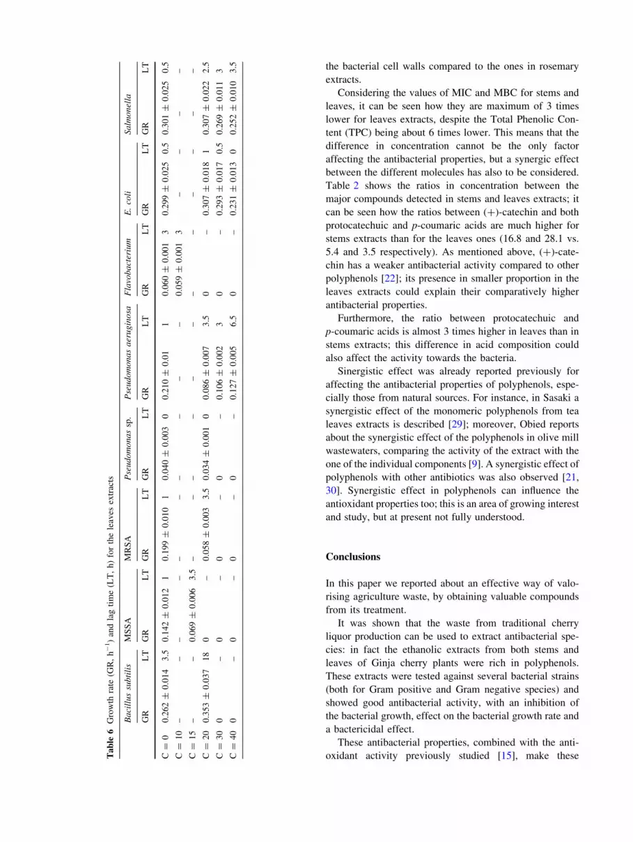
Tables 5 and 6 report the values of the bacterial growth
rate and the lag time for the stem and leaves extracts,
respectively. It can be seen how they are both affected by
the presence of the extracts, even if in a different way,
depending on the nature of the microorganism and the
concentration of the extracts. As expected, in most cases the
values of the growth rate decrease while the ones of the lag
time increase if the extracts are present; there are, however,
some exceptions, for instance Pseudomonas aeruginosa has
a more irregular behaviour with both types of extracts. Also,
the effect on the lag time is more enhanced for the leaves
extracts.
Discussion
Table 1 reports the composition of the ethanolic extracts
from stems and leaves; the conditions and the procedure for
the extraction were chosen considering the instability of
polyphenolic compounds at higher temperatures. For this
reason, different extraction techniques such as Soxhlet
extraction could not be used, because of the degradation of
thermolabile compounds associated with it [18].
Stems extracts show a higher total phenolic content
compared to leaves (Table 1); therefore it is expected that
the stems extracts show higher antimicrobial activity.
Most species detected in the extracts have been reported
to have antibacterial activity, for example ferulic and
p-coumaric acid is effective against E. coli and Staphylo-
coccus aureus [19], protocatechuic acid against Pseudomo-
nas aeruginosa, Bacillus subtilis, Pseudomonas aeruginosa
and E. coli [6, 20], naringenin against several strains of
Staphylococcus aureus [21]. However, the concentration in
the extracts of some of the phenolic compounds is lower than
the MIC value reported in literature. For instance MIC for
protocatechuic acid against E. coli is 2667 lg/ml [6], while
in stems extracts the concentration is only 33.8 lg/ml.
(?)-Catechin is the polyphenol with the highest concentra-
tion in both extracts; however, its antimicrobial activity is
not very high [22]. This can explain why the values of MICs
for these extracts are slightly higher compared to some other
extracts from natural sources [6, 17].
Our extracts, however, show antibacterial properties
towards all studied Gram positive and Gram negative
species; this is not always observed for natural extracts:
extracts from walnut leaves, for instance, do not show any
activity towards Gram negative bacteria [13]. Similar
results are reported about extracts from Cirsium plants
leaves [23].
Table 2 Ratios between some polyphenols concentration for leaves
and stem extracts
Compounds Concentrations ratio
for stems
Concentrations ratio
for leaves
p-Coumaric acid/
protocatechuic acid
0.6 1.6
(?)-Catechin/
protocatechuic acid
16.8 5.4
(?)-Catechin/p-
coumaric acid
28.1 3.5
Fig. 1 Agar diffusion test for stems extracts against Flavobacterium;
extract volume: (a) 40 ll, (b) 80 ll

Compared with other strains, Pseudomonas sp. (isolated
from tannin wastewater wetland) was resistant to higher
concentrations of polyphenols in the extracts. This is in
agreement with literature data: in fact previous study has
demonstrated that Pseudomonas sp. are not only tolerant
but can also degrade some type of polyphenol (as tannic
acid) [24]. Chung suggested that polyphenol antimicrobial
activity could be due to their strong binding capacity to
iron complex [25]. However, the inhibition mechanism
remains unclear and needs to be understood more clearly.
Fig. 2 Growth curves obtained
by absorbance reading at
610 nm, for different bacterial
strains in solutions containing
different concentrations of stem
extracts. a Bacillus subtilis,
b Staphylococcus aureusMSSA, c Staphylococcusaureus MRSA, d Pseudomonassp., e Pseudomonas aeruginosa,
f Flavobacterium, g E. coli,h Salmonella.
All concentrations
are expressed in mg/ml

The effect of natural compounds on the growth rate and
lag time of microorganisms was previously reported
before; for instance Munoz studied the behaviour of
Listeria monocytogenes in the presence of natural essential
oils [26], while Karanika analysed the effect of plant nat-
ural extracts on Yarrovia lipolitica [27]. Both studies
Fig. 3 Growth curves obtained
by absorbance reading at
610 nm, for different bacterial
strains in solutions containing
different concentrations of
leaves extracts. a Bacillussubtilis, b Staphylococcusaureus MSSA,
c Staphylococcus aureusMRSA, d Pseudomonas sp.,
e Pseudomonas aeruginosa,
f Flavobacterium, g E. coli,h Salmonella.
All concentrations are
expressed in mg/ml

showed a clear influence of the natural compounds on the
two parameters, even if the effect was very different
depending on the extracts considered. Karanika for
instance reports that in some cases the growth rate of
Listeria monocytogeneses was higher in the presence of the
extracts; these data are in agreement with what observed
here for Pseudomonas aeruginosa. In the study of Munoz,
the natural compounds did not always have an effect on the
lag time; a similar behaviour was observed here especially
for the stem extracts. These examples show how the
mechanisms of the interactions between microorganisms
and antibacterial species are quite complex and not com-
pletely clear.
From Table 3, it can be seen how the values of MBC for
the stem extracts are at maximum 4 times higher than the
corresponding MIC; the same thing can be observed for the
extracts from leaves. These data show the good bacterici-
dal properties of the analysed samples. Comparing these
values with literature data, other natural extracts show
similar properties (i.e. not a big difference between MIC
and MBC): Furiga in fact reports a ratio MBC/MIC = 2
for some pine bark extracts [28]. In other cases, however,
this ratio is much higher, like for instance for rosemary
extracts [17]. This indicates that the polyphenols present in
stems and leaves extracts are more effective in targeting
Table 3 Values of minimum inhibitory concentration (MIC) and
minimum bactericidal concentration (MBC) for stems extracts
Bacterial strain MIC (mg/ml) MBC (mg/ml)
Bacillus subtilis 15 60
Staphylococcus aureus MSSA 10 40
Staph. aureus MRSA 10 30
Pseudomonas sp. 30 40
Pseudomonas aeruginosa 100 [100
Flavobacterium 10 30
E. coli 100 [100
Salmonella 100 [100
Table 4 Values of minimum inhibitory concentration (MIC) and
minimum bactericidal concentration (MBC) for leaves extracts
Bacterial strain MIC (mg/ml) MBC (mg/ml)
Bacillus subtilis 30 100
Staphylococcus aureus MSSA 20 60
Staph. aureus MRSA 30 100
Pseudomonas sp. 30 60
Pseudomonas aeruginosa 100 [100
Flavobacterium 20 40
E. coli 100 [100
Salmonella 100 [100T
ab
le5
Gro
wth
rate
(GR
,h
-1)
and
lag
tim
e(L
T,
h)
for
the
stem
extr
acts
Ba
cill
us
sub
tili
sM
SS
AM
RS
AP
seu
do
mo
na
ssp
.P
seu
do
mo
na
sa
eru
gin
osa
Fla
vob
act
eriu
mE
.co
liS
alm
on
ella
GR
LT
GR
LT
GR
LT
GR
LT
GR
LT
GR
LT
GR
LT
GR
LT
C=
00
.23
2±
0.0
17
50
.21
3±
0.0
10
10
.18
2±
0.0
08
1.5
0.0
48
±0
.00
50
0.2
84
±0
.01
94
0.0
68
±0
.00
43
.50
.26
1±
0.0
20
1.5
0.3
14
±0
.02
51
C=
10
0.0
70
±0
.00
51
5.5
0–
0–
0.0
63
±0
.00
60
0.0
84
±0
.00
50
0–
0.2
60
±0
.00
80
0.2
86
±0
.03
00
C=
15
0–
0–
0–
––
––
––
––
––
C=
20
0–
0–
0–
0.0
31
±0
.00
50
0.0
44
±0
.00
20
0–
0.1
83
±0
.00
60
0.1
24
±0
.01
10
C=
30
0–
0–
0–
0–
0.0
43
±0
.00
20
0–
0.1
22
±0
.00
50
0.1
19
±0
.00
80
C=
40
0–
0–
0–
0–
0.0
81
±0
.00
37
.50
–0
.06
6±
0.0
03
00
.06
1±
0.0
03
0
the bacterial cell walls compared to the ones in rosemary
extracts.
Considering the values of MIC and MBC for stems and
leaves, it can be seen how they are maximum of 3 times
lower for leaves extracts, despite the Total Phenolic Con-
tent (TPC) being about 6 times lower. This means that the
difference in concentration cannot be the only factor
affecting the antibacterial properties, but a synergic effect
between the different molecules has also to be considered.
Table 2 shows the ratios in concentration between the
major compounds detected in stems and leaves extracts; it
can be seen how the ratios between (?)-catechin and both
protocatechuic and p-coumaric acids are much higher for
stems extracts than for the leaves ones (16.8 and 28.1 vs.
5.4 and 3.5 respectively). As mentioned above, (?)-cate-
chin has a weaker antibacterial activity compared to other
polyphenols [22]; its presence in smaller proportion in the
leaves extracts could explain their comparatively higher
antibacterial properties.
Furthermore, the ratio between protocatechuic and
p-coumaric acids is almost 3 times higher in leaves than in
stems extracts; this difference in acid composition could
also affect the activity towards the bacteria.
Sinergistic effect was already reported previously for
affecting the antibacterial properties of polyphenols, espe-
cially those from natural sources. For instance, in Sasaki a
synergistic effect of the monomeric polyphenols from tea
leaves extracts is described [29]; moreover, Obied reports
about the synergistic effect of the polyphenols in olive mill
wastewaters, comparing the activity of the extract with the
one of the individual components [9]. A synergistic effect of
polyphenols with other antibiotics was also observed [21,
30]. Synergistic effect in polyphenols can influence the
antioxidant properties too; this is an area of growing interest
and study, but at present not fully understood.
Conclusions
In this paper we reported about an effective way of valo-
rising agriculture waste, by obtaining valuable compounds
from its treatment.
It was shown that the waste from traditional cherry
liquor production can be used to extract antibacterial spe-
cies: in fact the ethanolic extracts from both stems and
leaves of Ginja cherry plants were rich in polyphenols.
These extracts were tested against several bacterial strains
(both for Gram positive and Gram negative species) and
showed good antibacterial activity, with an inhibition of
the bacterial growth, effect on the bacterial growth rate and
a bactericidal effect.
These antibacterial properties, combined with the anti-
oxidant activity previously studied [15], make theseTa
ble
6G
row
thra
te(G
R,
h-
1)
and
lag
tim
e(L
T,
h)
for
the
leav
esex
trac
ts
Ba
cill
us
sub
tili
sM
SS
AM
RS
AP
seu
do
mo
na
ssp
.P
seu
do
mo
na
sa
eru
gin
osa
Fla
vob
act
eriu
mE
.co
liS
alm
on
ella
GR
LT
GR
LT
GR
LT
GR
LT
GR
LT
GR
LT
GR
LT
GR
LT
C=
00
.26
2±
0.0
14
3.5
0.1
42
±0
.01
21
0.1
99
±0
.01
01
0.0
40
±0
.00
30
0.2
10
±0
.01
10
.06
0±
0.0
01
30
.29
9±
0.0
25
0.5
0.3
01
±0
.02
50
.5
C=
10
––
––
––
––
––
0.0
59
±0
.00
13
––
––
C=
15
––
0.0
69
±0
.00
63
.5–
––
––
––
––
––
–
C=
20
0.3
53
±0
.03
71
80
–0
.05
8±
0.0
03
3.5
0.0
34
±0
.00
10
0.0
86
±0
.00
73
.50
–0
.30
7±
0.0
18
10
.30
7±
0.0
22
2.5
C=
30
0–
0–
0–
0–
0.1
06
±0
.00
23
0–
0.2
93
±0
.01
70
.50
.26
9±
0.0
11
3
C=
40
0–
0–
0–
0–
0.1
27
±0
.00
56
.50
–0
.23
1±
0.0
13
00
.25
2±
0.0
10
3.5

extracts particularly valuable and suitable for several
applications (i.e. cosmetic and/or food packaging).
This valorisation process makes the cherry liquor pro-
duction more environmentally sustainable; a similar
approach can be applied to other fields of agriculture.
Acknowledgement This work was performed within the InSolEx
network (Innovative Solutions for Extracting High Value Natural
Compounds) funded by the European Union, contract MRT-CT-2006-
036053.
References
1. European Commission: Study on the feasibility of the establish-
ment of a Waste Implementation Agency. http://ec.europa.eu/
environment/waste/index.htm (2010). Accessed 24 Feb 2010
2. European Commission: Directive 2008/98/EC on waste (Waste
Framework Directive). http://ec.europa.eu/environment/waste/
framework/index.htm (2010). Accessed 24 Feb 2010
3. Lule, S.U., Xia, W.: Food phenolics, pros and cons: a review.
Food Rev. Int. 21, 367–388 (2005)
4. Eastwood, M.A.: Interaction of dietary antioxidants in vivo: how
fruit and vegetables prevent disease. QJM 92, 527–530 (1999)
5. Middleton, E.: Effect of plant flavonoids on immune and
inflammatory cell function. Adv. Exp. Med. Biol. 439, 175–182
(1998)
6. Taguri, T., Tanaka, T., Kound, I.: Antibacterial spectrum of
plants polyphenols and extracts depending upon hydroxyphenyl
structure. Biol. Pharm. Bull. 29(11), 2226–2235 (2006)
7. Moure, A., Cruz, M.C., Franco, D., Domingues, J.M., Sineiro, J.,
Dominguez, E., Nunez, M.J., Parajo, J.C.: Natural antioxidant
from residual sources. Food Chem. 72, 145–171 (2001)
8. Lesage-Meessen, L., Navarro, D., Maunier, S., Sigolloit, J.C.,
Lorquin, J., Delattre, M., Simon, J.L., Ashter, M., Labat, M.:
Simple phenolic content in olive oil residues as a function of the
extraction system. Food Chem. 75, 501–507 (2001)
9. Obied, H.K., Bedgood, D.R. Jr, Prenzler, P.D., Robards, K.:
Bioscreening of Australian olive mill waste extracts: biophenol
content, antioxidant, antimicrobial and molluscicidial activities.
Food Chem. Toxicol. 45, 1238–1248 (2007)
10. Sudjana, A.N., D’Orazio, C., Ryan, V., Rasool, N., Ng, J., Islam,
N., Riley, T.V., Hammer, K.A.: Antimicrobial activity of com-
mercial Olea europea (olive) leaf extract. Int. J. Antimicrob.
Agents 33, 461–463 (2009)
11. Peschel, W., Sanchez-Rabaneda, F., Diekmann, W., Plescher, A.,
Gartzia, I., Jimenez, D., Lamuela-Raventos, R., Buxaderas, S.,
Codina, C.: An industrial approach in the search of natural
antioxidants from vegetable and fruit wastes. Food Chem. 97(1),
137–150 (2006)
12. Wijngaard, H.H., Rossle, C., Brunton, N.: A survey of Irish fruit
and vegetable waste and by-products as a source of polyphenols
antioxidants. Food Chem. 116, 202–207 (2009)
13. Pereira, J.A., Oliveira, I., Sousa, A., Valenta, P., Andrade, P.B.,
Ferreira, I.C.F.R., Ferreres, F., Bento, A., Seabra, R., Estevinho,
L.: Walnut (Juglans regia L.) leaves: phenolic compounds,
antibacterial activity and antioxidant potential of different culti-
vars. Food Chem. Toxicol. 45, 2287–2295 (2007)
14. Kumar, P.S., Kumar, A., Sivakumar, R., Kaushik, C.: Experi-
mentation on solvent extracts from natural waste. J. Mater. Sci.
44, 5894–5899 (2009)
15. Demiray S., Pintado M.M., Castro P.M.L.: High value added
compounds with antioxidant activities from Ginja cherries, stems
and leaves. EFFOST conference, Budapest, 11–13 Nov 2009
16. Bauer, A.W., Kirby, W.M., Sherries, J.C., Turck, M.: Antibiotic
susceptibility testing by a standardised single method. Am. J.
Clin. Pathol. 45, 493–496 (1966)
17. Moreno, S., Scheyer, T., Romano, C.S., Vojnov, A.A.: Antioxi-
dant and antimicrobial activity of rosemary extracts linked to
their polyphenolic composition. Free Radic. Res. 40(2), 223–231
(2006)
18. Lao, R.C., Shu, Y.Y., Holmes, J., Chiu, C.: Environmental
sample cleaning and extraction procedures by microwave-assis-
ted process (MAP) technology. Microchem. J. 53(1), 99–108
(1996)
19. Herald, P.J., Davidson, P.M.: Antibacterial activity of selected
hydrocinnamic acid. J. Food Sci. 48(4), 1378–1379 (2006)20. Liu, K., Tsao, S., Yin, M.: In vitro antibacterial activity of roselle
calyx and protocatechuic acid. Phytother. Res. 19(11), 942–945
(2006)
21. Denny, B.J., West, P.W., Mathew, T.C.: Antagonistic interactions
between the flavonoids hesperetin and naringenin and beta-lactam
antibiotics against Staphylococcus aureus. Br. J. Biomed. Sci.
65(3), 145–147 (2008)
22. Kajiya, K., Hojo, H., Suzuki, M., Nanjo, F., Kumazawa, S.,
Nakayama, T.: Relationship between antibacterial activity of
(?)-catechin derivatives and their interaction with a model
membrane. J. Agric. Food Chem. 52, 1514–1519 (2004)
23. Nazaruk, J., Czechowska, S.K., Markiewicz, R., Borawska, M.H.:
Polyphenolic compounds and in vitro antimicrobial and antioxi-
dant activity of aqueous extracts from leaves of some Cirsiumspecies. Nat. Prod. Res. 22(18), 1583–1588 (2008)
24. Franco, A.R., Calheiros, C.S.C., Pacheco, C.C., De Marco, P.,
Manaia, C.M., Castro, P.M.L.: Isolation and characterisation of
polymeric galloyl-ester-degrading bacteria from a tannery dis-
charge place. Microb. Ecol. 50, 550–556 (2005)
25. Chung, K.T., Lu, Z., Chou, M.W.: Mechanism of inhibition of
tannin acid and related compounds on the growth of intestinal
bacteria. Food Chem. Toxicol. 36(12), 104–1060 (1998)
26. Munoz, M., Guevara, L., Palop, A., Tabera, J., Fernandez, P.S.:
Determination of the effect of plant essential oils obtained by
supercritical fluid extraction on the growth and viability of Lis-
teria monocytogenes in broth and food systems using flow
cytometry. LWT Food Sci. Technol. 42, 220–227 (2009)
27. Karanika, M.S., Komaitis, M., Aggelis, G.: Effect of aqueous
extracts of some plants of Lamiaceae family on the growth of
Yarrowia lipolytica. Int. J. Food Microbiol. 64, 175–181 (2001)
28. Furiga, A., Lonvaud-Funel, A., Dorigna, G., Badet, C.: In vitro
anti-bacterial and anti-adherence effects of natural polyphenolic
compounds on oral bacteria. J. Appl. Microbiol. 105, 1470–1476
(2008)
29. Sasaki, H., Matsumoto, M., Tanaka, T., Maeda, M., Nakai, M.,
Hamada, S., Ooshima, T.: Antibacterial activity of polyphenol
components in oolong tea extracts against Streptococcus mutans.
Caries Res. 38(1), 2–8 (2004)
30. Romano, C.S., Anadi, K., Repetto, V., Vojnov, A.A., Moreno, S.:
Synergistic antioxidant and antibacterial activity of rosemary plus
butylated derivatives. Food Chem. 115(2), 456–461 (2009)