nanostructured medical sutures with antibacterial properties
TRANSCRIPT

lable at ScienceDirect
Biomaterials 52 (2015) 291e300
Contents lists avai
Biomaterials
journal homepage: www.elsevier .com/locate/biomater ia ls
Nanostructured medical sutures with antibacterial properties
Cristina Serrano a, 1, Luis García-Fern�andez a, 1, Juan Pedro Fern�andez-Bl�azquez a,Mike Barbeck b, Shahram Ghanaati b, Ron Unger b, James Kirkpatrick b, Eduard Arzt c,Lutz Funk d, Pau Tur�on d, Ar�anzazu del Campo a, *
a Max-Planck-Institut für Polymerforschung, Ackermannweg 10, 55128 Mainz, Germanyb Institute of Pathology, University Medical Center, Johannes Gutenberg University, Mainz, Germanyc INM e Leibniz Institute for New Materials and Saarland University, Saarbrücken, Germanyd B. Braun Surgical, S.A, Rubi, Barcelona, Spain
a r t i c l e i n f o
Article history:Received 19 September 2014Received in revised form2 February 2015Accepted 6 February 2015Available online
Keywords:NanopatterningAntifoulingAntibacterialPlasma treatmentSutures
* Corresponding author. Tel.: þ49 (0)6131 379563;E-mail address: [email protected] (A
1 These two authors have equally contributed to th
http://dx.doi.org/10.1016/j.biomaterials.2015.02.0390142-9612/© 2015 Elsevier Ltd. All rights reserved.
a b s t r a c t
Bacterial repellence in suture materials is a desirable property that can potentially improve the healingprocess by preventing infection. We describe a method for generating nanostructures at the surface ofcommercial sutures of different composition, and their potential for preventing biofilm formation. Weshow how bacteria attachment is altered in the presence of nanosized topographies and identifyoptimum designs for preventing it without compromising biocompatibility and applicability in terms ofnanostructure robustness or tissue friction. These studies open new possibilities for flexible andcost-effective realization of topography-based antibacterial coatings for absorbable biomedical textiles.
© 2015 Elsevier Ltd. All rights reserved.
1. Introduction
Sutures are natural or synthetic textile biomaterials used fortissue approximation during wound closure. For successful tissuefixation suturing materials need to have adequate mechanicalstrength, good tissue compatibility as well as biodegradationkinetics. Ideally the suture material should resist bacterial adher-ence to prevent wound infection. This property depends on thechemical composition of the suture itself and on supplementaryantibacterial modifications [1]. Two major antibacterial strategieshave been applied to sutures and biomaterials in general [2e9]:passive coatings based on cationic biopolymers that preventbacterial attachment [10e13], or active strategies that release activecompounds into the tissue and kill suspended bacteria (mainlysilver [14e17], antimicrobial peptides [18,19] or antibiotics[20e22]). While passive strategies are preferred in terms ofbiocompatibility, active strategies are often more effective.
fax: þ49 (0)6131 379271.. del Campo).is article.
However, toxicity issues and the development of resistant micro-bial strains compromise their application [23].
Particular surface micro- and nanotopographies have alsoshown the capacity of preventing bacterial attachment and biofilmformation [24e30], though the mechanism behind bacterialrepellence and its correlationwith the surface design remains to beunraveled. Reported studies on this topic mostly rely on surfacenanostructures obtained by patterning techniques used in themicroelectronic industry, which can hardly be implemented inbiomaterials or in curved geometries as suture threads. Recently,high aspect ratio silicon nanopillars obtained by reactive ionetching (RIE) have been shown to kill bacteria upon contact [31].Although a topography-based bactericide seems convenientbecause it avoids release of toxic components, silicon finds little usefor low cost manufacture of biomedical devices and biomaterials.Alternative antibacterial strategies allowing cost-effective nano-patterning of established biomaterials in non-planar geometrieswould be desirable for real application.
Nanotopographies on textile fibers have recently been gener-ated by plasma treatment [32]. Plasma treatment involves chemicalchanges, ablation and etching of the fiber surface, and eventuallydeposition of material. The extent of these processes depends onthe plasma treatment conditions and the material itself [33,34].

C. Serrano et al. / Biomaterials 52 (2015) 291e300292
Recent studies have shown that oxidative plasma treatment ofpolymer surfaces of different compositions can lead to controlledetching of the polymer surface and formation of distinct nano-topographies with variable geometrical design and dimensions[35e45]. In this manuscript we apply this method to nanopatterncommercially available suturing threads. We show nanopatternedsutures of different composition, including absorbable andnon-absorbable ones. We demonstrate differential bacterialattachment depending on the suture nanotopographies and weidentify optimum geometries for preventing attachment withoutcompromising biocompatibility and applicability in terms ofnanostructure robustness or tissue friction are identified. Thesestudies open new possibilities for realization of topography-basedantibacterial coatings.
2. Materials and methods
2.1. Materials
Different sutures USP 2/0 from the company B. Braun were used for thesestudies: non-absorbable monofilaments of polypropylene (PP, Premilene®) andpoly(ethylene terephthalate) (PET, Miralene®) and absorbablemonofilament suturesof polydioxanone (PDO, Monoplus®) and modified glycolic acid (Monosyn®).
2.2. Plasma treatment
Oxygen plasma treatment was carried out in a Plasma Activate Mini Flecto®
(Plasma Technology GmbH, Rottenburg, Germany). It consists of a low pressurecapacitive coupled plasma reactor operating at 24 kHz with a circular poweredelectrode of 65 mm diameter (Figure SI-1). The sutures were attached to the centerof the electrode and a small weight (1.2 g) was placed at the free end of the fiber toprevent curling during treatment. Plasma treatment was performed at 0.1 mbaroxygen pressure and 80 W power. These conditions were taken from previouslyreported experiments [41e44,46]. Etching times between 0, 1, 2, 5, 10 and 20 minwere tested. In order to avoid heating of the suture above 40 �C at longer plasmatreatment times, the treatment was performed in pulses with 5 s oxygen plasmaexposure followed by 60 s delay time. This was controlled by the software of theplasma equipment. The “etching time” in seconds is the number of cycles multipliedby 5. The temperature of the chamber did not surpass 40 �C during treatment.
2.3. Analysis of surface nanostructures
The surface structure of the sutures before and after plasma treatment wasevaluated by scanning electron microscopy (SEM). Samples were placed on SEM pinstubs and sputtered coated with a 5 nm thick layer of platinum (Bal-Tec MCS 010sputtering device) prior to SEM imaging. An accelerating voltage between 0.7 and3 KV was used for taking images (LEO 1530 VP, Zeiss).
The top surface area fraction of the structures (the surface in contact with thebacteria) was analyzed from the SEM images using the open source imageprocessing software Fiji (ImageJ 2.0.0-beta-7.5) [47] and a homemade macro.
Atomic force microscopy (AFM) was also used to characterize the topography ofthe treated surfaces. An AFM Bruker Dimension 3100 CL (Santa Barbara, USA) andOlympus non-contact mode cantilevers (OMCL-AC160TS-W2) with a nominal springconstant of 42 N/m were used. The RMS roughness values for the different sampleswere calculated through the following expression:
RMS ¼ffiffiffiffiffiffiffiffiffiffiffiffiffiffiffiffiffiffiffiffiffiffiffiffiffiffiffiffiffiffiffiffiffiffiffiffiPN
i¼1 ðZi � ZaveÞ2N
s
where Zave is the average Z value into the given area, Zi is the current Z value and N isthe total number of points in the area.
2.4. Bacterial assays
E. coli K12 wildtype (DSM A498, ATCC 23716) were grown aerobically in LuriaBertani (LB) medium at 37 �C overnight. The bacteria were diluted with LB mediumto an optical density of 0.1 at 600 nm and shaken at 37 �C at 200 rpm until the mid-logarithmic phase was reached (OD600 ~ 0.5e0.6). The final cell density was adjustedto OD600 ~ 0.5, corresponding approximately to 1.25 � 108 CFU/mL. (CFU ¼ colony-forming unit).
The suture samples (length 2 cm) were placed in a 6-well culture plate and2.5 ml of the bacterial suspension (OD~0.5) were added. The samples were incu-bated at 37 �Cwith shaking at 30 rpm for 2 h. At the end of the incubation period, thesamples were rinsed with PBS in order to eliminate non-adherent bacteria. Theviability of adherent bacteria to the suture was tested by staining with a live/deadfluorescence stain (LIVE/DEAD BacLight Bacterial Viability Kit, Life Technologies) andimaging under the fluorescence microscope. For the quantification of the number ofbacteria attached to the suture surface after incubation samples were vigorously
shacked in tubes containing 1.5 ml of PBS vigorously for 10 min to resuspendattached bacteria. The viable organisms suspended in PBSwere quantified by platingserial dilutions on LB-agar plates. LB-agar plates were incubated at 37 �C, and theCFU were counted using ImageJ software.
Each experiment was repeated threefold using three different plasma-treatedsuture samples. The one-way ANOVA test was used to determine significantdifferences in the results of the bacterial assays.
2.5. Characterization of adherent bacteria with SEM
Sutures incubated with bacteria were washed, immersed in a glutaraldehydesolution (4% in PBS) for fixation and dehydrated by immersion in ascending ethanolsolutions (20%, 40%, 60%, 80% and 100%). Samples were placed on SEM pin stubs andsputter-coated with a 5 nm thick layer of platinum for SEM analysis.
2.6. Tissue drag test
The frictional forces encountered during passage through tissue were estimatedby pulling 20 cm of suture through an artificial skin model (professional skin padfrom Limbs and things). The tissuewas mounted on a tensile testing machine (ZwickRoell Z005 with a maximum load of 50 N) with a homemade holder. Premilene andMonoplus sutures were passed transversally through the skin and attached to theupper jaw of the machine. Pulling tests were performed at an angle of 45� . The forcerequired to pull the sutures through the tissue at constant rate of 400 mm/minversus length was recorded.
2.7. Evaluation of mechanical stability of the surface nanostructures during suturing
The mechanical stability of the surface nanostructures was evaluated by passingthe suture through chicken skin. Monosyn sutures, plasma-treated for 0, 2, 5 and10min, were stitched and pulled through chicken skin at 400mm/min at an angle of45� using a tensile tester (Zwick Roell Z005 with a maximum load of 50 N). Thetopography of the surface was evaluated before and after the test using SEM.
2.8. In vitro degradation studies
The absorbable sutures were treated with plasma for 0, 2 and 10 min andimmersed in LB medium and incubated at 37 �C for 7, 14 and 21 days. After incu-bation, the samples were washed with ethanol and dried at 37 �C. Changes in thesurface morphology of the suture materials during degradation were analyzed bySEM.
2.9. Subcutaneous implantation
All animal experiments were previously authorized by the Committee on theUse of Live Animals in Teaching and Research of the State of Rhineland-Palatinate,Germany.
30 female, 6e8 week-old CD-1 mice (Charles River Laboratories, Germany) wererandomly divided into four study groups. Three study groups were formed with fouranimals (n ¼ 4) which underwent implantation of three different suture materialsfor 15 and 30 days. The fourth groupwith three animals (n¼ 3) served as control andunderwent operation without biomaterial insertion in order to analyze the tissuereaction to the surgical procedure per se.
Subcutaneous implantation was conducted as described in previous publica-tions [48e51]. In brief, the animals were anesthetized and the operation side wasshaved and disinfected. An incision was made down to the subcutaneous tissue ofthe rostral interscapular region. A subcutaneous pocket was built in which thebiomaterials were inserted and the wound was closed with stitches.
Animal housing was conducted at the in vivo Laboratory Animal Unit of theInstitute of Pathology. The animals were kept under standard conditions, includingartificial day/night-cycle as well as water ad libitum and regular rat pellet (Labora-tory Rodent Chow, Altromin, Germany). Pre- and postoperative care was conductedaccording to established protocols. All experimental animals survived the implan-tation procedures without any postoperative complications.
2.10. Explantation and histological study of the explants
The explantation and histological study was conducted as described in previouspublications [48e53]. The animals were euthanized with an overdose of anestheticson day 15 after implantation. The implanted suture materials combined with theirperi-implant tissue or the area of the control incisionwere explanted. The explantedtissue was fixed using a 4% formalin solution for 24 h and cut into three segments ofidentical dimensions including the left margin, the center and the right margin ofthe biomaterial. After dehydration via a series of increasing alcohol concentrationsand xylol exposure, paraffin embedding was conducted. A rotation microtome(Leica, Wetzlar, Germany) was used to cut 3e5 mm thick sections.
The sections were stained with hematoxylin and eosin (H&E), Azan, MovatPentachrome, Tartrate-resistant acid phosphatase (TRAP) and immunohistochemi-cally with F4/80. A control slide was included for qualitative analysis of tissuereactions according to previously described methods [48e53].

C. Serrano et al. / Biomaterials 52 (2015) 291e300 293
2.11. Histological investigation
The histological study was conducted by two investigators (SG andMB) using anEclipse 80i histological microscope (Nikon, Tokyo, Japan) by previously describedmethods [48e53]. Special focus was paid to the evaluation of fibrosis, hemorrhage,necrosis and the presence of different cell types such as granulocytes, lymphocytes,plasma cells, macrophages and multinucleated giant cells. Microphotographs weretaken with a Nikon DS-Fi1 digital camera and a DS-L2 digital sight control unit(Nikon, Tokyo, Japan) connected to the microscope.
3. Results and discussion
3.1. Nanostructured suture threads
Monofilament sutures (non-absorbable Premilene® and Mir-alene® and absorbable Monoplus® and Monosyn®) were plasmatreated at different conditions using a cylindrical plasma chamber[43e45]. Before treatment, the surface of the sutures was flat(Figure SI-2). With increasing plasma treatment, nanostructureswith different geometries appeared at the surface of the sutures(Fig. 1). Lamellae with 100 nm thickness and 500 nm length wereobserved after 1 min plasma treatment. Lamellae were perpen-dicularly aligned with respect to the suture axis and becamethicker, higher and longer with increasing etching times. As theaspect ratio increased, the nanolamellae became mechanicallyunstable and collapsed. After 20 min plasma treatment time,
Fig. 1. SEM images showing the surface topography of Monosyn, Monoplus, Premil
lamellae in absorbable sutures approached 500 nm in thicknessand several microns in length. The lamellae were separated byelongated voids 1e2 mm in width and a few mm in length. Thesevoids had also nanolamellae at their bottom. Lamellae obtained onnon-absorbable sutures had smaller sizes (200 nm thickness and1 mm or 2 mm length in Miralene and Premilene respectively) andwere more closely packed. All these features can be clearly seen inFig. 1. Although the exact height of the structures could not beobtained from the SEM images, lamellae appeared to be higher onthe absorbable sutures.
SEM analysis was performed on suture samples treated indifferent batches in order to ascertain that the observed topog-raphy was uniform along the suture, and reproducible amongdifferent batches. This was the case for Premilene, Miralene andMonosyn sutures. Monoplus sutures showed certain heterogeneityin surface topography (Figure SI-3). Note that PDO has a glasstransition temperature well below room temperature (Table SI-1)and, therefore, the mechanical stability of the nanostructures withincreasing aspect ratio could be compromised during treatment.This could lead to collapse of the etched structures during treat-ment and to the observed higher variability in the nanopatterns.
The differences in the dimensions of the nanolamellae amongthe analyzed sutures reflect the different etching sensitivities of thepolymers. Monoplus and Monosyn are polar polymers with a
ene and Miralene sutures after oxygen plasma treatment for increasing times.

C. Serrano et al. / Biomaterials 52 (2015) 291e300294
higher content of CeO bonds and show higher etching rates than PPor PET [54]. Consequently, etched structures of Monoplus orMonosyn achieved higher aspect ratios and showed more collapsethan those of PP or PET. This results in topographies with thickerlamellae separated by elongated voids of 1e2 mm in width and afew micrometers in length in absorbable sutures, and thinner butmore closely packed lamellae separated by voids of <0.2 mmwidthand 1e2 mm length in non-absorbable ones (see SEM images inFig. 1 corresponding to long etching times).
The surface nanostructures were similar to those reportedpreviously by our group on other polymer materials [43e45]. Theorigin of these structures has been associated with the morphologyof the polymer fiber, i.e. its microcrystalline structure and orien-tation of the polymer chains [43e45]. The lamellae orientedperpendicularly to the fiber direction originate from the crystallinedomains formed by the oriented chains during fiber processing.These show lower etching ratios than the surrounding amorphousregions. This differential etching leads to the 3D topography. Anadditional proof of the relationship between the thermomechanicalpretreatment and the obtained nanostructures on the fibers wasobtained by performing similar plasma treatments on fibers of thesame material but without being drawn during processing. Thenanostructures obtained were randomly oriented (Figure SI-4).
The surface topography of the absorbable nanostructuredsutures was also imaged by AFM. Fig. 2aed shows representativetopography images of Monosyn samples for different plasmatreatment times. The topographical image confirmed the lamellarstructure observed in the SEM images (Fig.1). The rootmean square(RMS) roughness was estimated and represented as a function ofthe plasma treatment time (Fig. 2e). Similar RMS roughness valueswere obtained for Monosyn and Monoplus sutures at the differentplasma treatment times, in agreement with the similarities visiblein the previous SEM images (Fig. 1). Note that the AFM tip isexpected to image only the uppermost aspect of the topography
Fig. 2. AFM images showing the surface topography of Monosyn fibers before (a) and after oMonoplus and Monosyn sutures.
since it cannot effectively penetrate into the narrow and deep voidsbetween lamellae. Therefore, the RMS roughness values in Fig. 2ecan only be taken as orientating.
3.2. Bacterial attachment on the nanostructured sutures
The nanostructured sutures were incubated with E. coli and theamount of adhered bacteria to the suture surface was quantified(see Fig. 3 and experimental section for details). Bacterial attach-ment to absorbable sutures Monosyn and Monoplus significantlydecreased with the plasma treatment. The decrease was almostlinear with the plasma treatment time in Monosyn and wasreduced by an order of magnitude in samples treated for 20 min(Fig. 3A). In Monoplus sutures the decrease was less pronouncedand it was not linear with the plasma treatment time (Fig. 3B). Incontrast, non-absorbable nanostructured sutures (PP, PET) showedsimilar or even higher levels of bacterial attachment than theoriginal samples. It is noteworthy that all nanostructured suturesshowed a lamellar topography. However, lamellae on absorbableand non-absorbable sutures showed significantly different lateraldimensions, indicating a strong correlation between the geometryof the nanotopography and the bacterial response.
Adherent bacteria on the nanostructured sutures were visual-ized by SEM. Figs. 4 and 5 and Figure SI-5 show images corre-sponding to Miralene, Monosyn andMonoplus sutures. The contactbetween the bacteria wall and the nanopattern can be clearlyappreciated in the SEM images of tilted samples in Fig. 5. In all casesbacteria were located at the top part of the lamellar nanostructures.The bacterial membrane was not visibly deformed and contact wasestablished only with the top part of the topographical features.Bacteria were not disrupted, suggesting that the nanotopographywas not harmful. No bacteria were found inside the voids, not evenon absorbable Monosyn or Monoplus samples where the size of thevoids was significantly larger than the size of the bacteria. Bacteria
xygen plasma treatment for (b) 1 min, (c) 5 min, and (d) 20 min. (e) RMS Roughness of

Fig. 3. Inhibition of the bacterial adhesion on commercial sutures modified by different times of plasma treatment (CFU: colony-forming unit). Significant differences: *P < 0.05;**P < 0.01; ***P < 0.001, compared with the control (non-treated fiber).
C. Serrano et al. / Biomaterials 52 (2015) 291e300 295
were randomly oriented through the sample and did not follow thenanopattern direction.
In order to confirm that the surface topography prevents, butdoes not kill bacteria as in previously reported silicon nano-topographies [31], we tested the viability of attached andsuspended bacteria after incubation with Monosyn sutures for 2 hwith a LIVE/DEAD assay. Fluorescence imaging of the attachedbacteria at the suture surface before and after 20 min of plasmatreatment (Figure SI-6a and SI-6c) showed a significant reduction ofbacterial density at the surface of the nanostructured surfaces andthe absence of dead bacteria. We also analyzed bacterial viability inthe medium in which the sutures were immersed. No significantnumber of dead bacteriawas found in suspension, (Figure SI-6b andSI-6d), confirming that the nanotopography prevented bacterialattachment but did not kill bacteria upon contact.
3.3. In vivo tissue response of nanostructured sutures
In order to test if the nanotopography compromised thetissue compatibility of the suturing materials, original and
nanostructured sutures (Monoplus sutures plasma treated for 2and 10 min) were implanted into mice and histopathologicalanalysis of the tissue reaction to the suture material was performedat days 10 and 30 after implantation. Monoplus samples plasmatreated for 2 min showed a wall of cells at the tissueebiomaterialinterface with a few layers at the surface of the material (Fig. 6 A1and A2) at day 15 after implantation. It contained mainly macro-phages and a few eosinophilic granulocytes (Fig. 6 A2 and A3).A large number of macrophages were also visible within theperi-implant connective tissue (CT) (Fig. 6 A3). At day 30 afterimplantation similar features were found and the cell response atthe interface was observed to be more extensive (black arrowheads, Fig. 6 B1 and B2).
Monoplus samples plasma-treated for 10 min showed a thickcell wall with many layers (black arrow heads) at day 15 afterimplantation (Figure SI-7 A1 and A2). The cell wall mainly con-tained mononuclear macrophages (green arrow heads), mono-nuclear eosinophilic granulocytes (red arrow) and multinucleargiant cells (blue arrow head in Figure SI-7 A2). A low number ofmacrophages (green arrows) were also observed within the peri-

Fig. 4. SEM images of E. coli attached to Monosyn sutures after plasma treatment for different times (0, 1, 2, 5, 10 and 20 min).
C. Serrano et al. / Biomaterials 52 (2015) 291e300296
implant connective tissue (Figure SI-7 A3). At day 30 after im-plantation a thicker layer of cells was observed at the suture-tissueinterface and a higher amount of macrophages (green arrows)could be found within the peri-implant connective tissue(Figure SI-7 B3).
For comparison, Figure SI-8 shows the histopathological anal-ysis of the tissue reaction of Monoplus control material. At day 15after implantation a thin layer of cells had formed at the materialsurface (Figure SI-8 A1), mainly containing macrophages (Figure SI-8 A2). The peri-implant connective tissue showed a regular distri-bution of cells and matrix components such as collagen fibers anddid not show any signs of an exaggerated material-related inflam-matory process or granulation tissue (Figure SI-8 A1 and A2). Moremacrophages were also located within the peri-implant tissue(Figure SI-8 A2). At day 30 after implantation the histopathological
Fig. 5. SEM pictures of tilted samples showing the attached bacteria to untre
analyses showed that the interfacial layer of cells was still present(Figure SI-8 B1) and composed mostly of mononuclear cells and afew multinucleated giant cells (Figure SI-8 B2).
The wounds of the control incisions showed a healing processwithout complications. No indications of severe inflammatory re-sponses at the two study time points were detectable at any timepoints of this study. The histological study showed an increasedamount of macrophages, fibroblasts and granulocytes within thewound area and was visibly higher compared to unaffected areas ofsubcutaneous connective tissue (data not shown). At day 30 afterimplantation theamountof cellsdecreasedanda regulardistributionof cells and extracellular matrix was observed. No multinucleatedgiant cells were observed at any of the analyzed study time points.
Altogether, the in vivo results reflect a mild tissue reaction to thenanostructuredmaterials. The formation of a cell demarcation layer
ated and plasma-treated (2 and 10 min) Monosyn and Miralene sutures.

Fig. 6. Tissue reaction to Monoplus treated with O2 Plasma for 2 min (PDO 2) at day 15 (A1e3) and day 30 (B1e3) after implantation within the subcutaneous connective tissue (CT)of the CD-1-mouse. A1) Overview of the implantation bed of the suture material (PDO2) at day 15 after implantation (thin cell capsule ¼ black arrow heads), (Azan-staining, 100�magnification, scale bar ¼ 100 mm). A2) shows the capsule surrounding the PDO2 (black arrow heads) in a higher magnification (mononuclear cells ¼ black arrows; eosinophilicgranulocytes (red arrows)) (Giemsa-staining, 400� magnification, scale bar ¼ 10 mm). A3) Macrophages (green arrow heads) within the cell capsule surrounding the PDO2 andmacrophages (green arrows) within the surrounding connective tissue (CT) (F4/80-immunostaining, 400� magnification, scale bar ¼ 10 mm). B1) Overview of the implantation bedof the suture material (PDO 2) at day 30 after implantation (cell-rich capsule ¼ black arrow heads) (HE-staining, 200� magnification, scale bar ¼ 100 mm). B2) shows the capsule(black arrow heads) in a higher magnification (HE-staining, 400�magnification, scale bar ¼ 10 mm). B3) Macrophages (green arrow heads) within the capsule surrounding the PDO2and macrophages (green arrows) within the surrounding connective tissue (CT) (F4/80-immunostaining, 400� magnification, scale bar ¼ 10 mm). (For interpretation of the ref-erences to color in this figure legend, the reader is referred to the web version of this article.)
C. Serrano et al. / Biomaterials 52 (2015) 291e300 297
at the material surface was comparable to that elicited by theoriginal sutures [55]. The occurrence of multinuclear giant cells onthe Monoplus sutures plasma treated for longer times should behighlighted. Multinucleated giant cells are involved in the tissuereaction to biomaterials [48e51,56,57]. Within the „foreign bodyresponse“ this multinucleated cell type is presumed to be involvedin the regulation of the tissue response by secretion of diverse pro-and anti-inflammatory molecules, and in the degradation of thebiomaterial [57e59].
At the other hand, biomaterials with different surface charac-teristics might not induce macrophage fusion into multinucleatedgiant cells. The former are able to secrete higher amounts ofpro-inflammatory cytokines such as interleukin-1 beta (IL-1b) orinterleukin-6 (IL-6) [57]. In contrast, material-adherent foreignbody giant cells could exhibit differentiation into amore alternativeactivation status with secretion of anti-inflammatory cytokines[57].
In summary, the results of the present in vivo study lead to theassumption that differences in the surface nanotopography lead tovariances in terms of the outcome of the cell and tissue reactions tothe suture materials with special regard to differences in thepattern of inflammation and could possibly give rise to changes inmaterial degradation.
Although in vivo experiments were restricted to Monoplussutures, similar behavior can be expected for Monosyn, since boththe surface chemistry after oxidative plasma treatment and thenanotopography do not differ significantly.
3.4. Evaluation of tissue drag of the nanostructured sutures
Changes in the nanotopography of sutures could result inchanges in the frictional forces with the tissue, which is animportant parameter in the medical application. An increase in the
frictional forces during suturing causes tissue damage and hasnegative consequences for the healing process. We evaluated thefrictional force of the nanostructured sutures with the tissue bymeasuring the pull-through forces of the different sutures in atissue drag test (see experimental information for details).Figure SI-9 shows the force required to pull Monoplus andPremilene sutures through artificial skin (see supporting informa-tion for details on the test and Figure SI-10 for original frictioncurves). A small increase in the friction force was observed insutures with short (<2 min) plasma treatments. Note that thesurface nanostructures were very small and of low aspect ratio, sothat they would hardly be able to mechanically interact with thetissue. We attribute the small increase in tissue friction to thechange in surface chemistry and consequent increased surfaceenergy after the oxidative plasma treatment, rather than to theroughening process. It is important to note that the surface energyincreases already after short plasma treatment times and remainsconstant for longer treatments [46]. Nanostructured sutures forlonger than 2min showed similar friction values as the non-treatedones, despite the increased roughness. Those results confirm thatthe surface nanostructures do not mechanically penetrate the tis-sue and plasma-treated monofilaments are able to smoothly slidethrough tissue as non-treated sutures do. Longer treatment timesdecreased the topmost surface area and, presumably, compensatedfor the friction increase caused by the change in the surfacechemistry and lowered friction values down to the original levels.
3.5. Mechanical stability of surface nanostructures during suturing
From an application point of view themechanical stability of thesurface nanostructures during the suturing process is a relevantissue. We evaluated this property by imaging the surface of thesutures after pulling the thread through chicken skin by SEM. Fig. 7
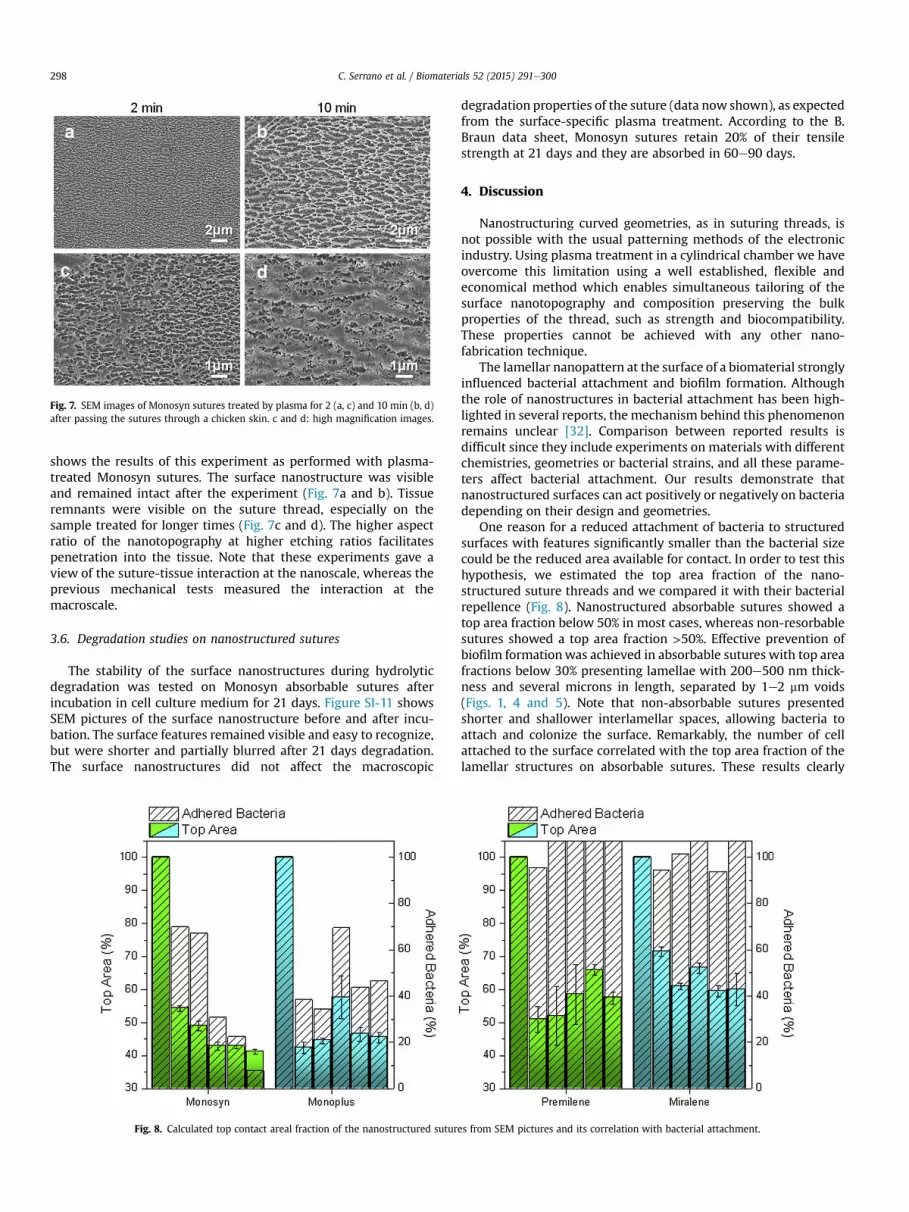
Fig. 7. SEM images of Monosyn sutures treated by plasma for 2 (a, c) and 10 min (b, d)after passing the sutures through a chicken skin. c and d: high magnification images.
C. Serrano et al. / Biomaterials 52 (2015) 291e300298
shows the results of this experiment as performed with plasma-treated Monosyn sutures. The surface nanostructure was visibleand remained intact after the experiment (Fig. 7a and b). Tissueremnants were visible on the suture thread, especially on thesample treated for longer times (Fig. 7c and d). The higher aspectratio of the nanotopography at higher etching ratios facilitatespenetration into the tissue. Note that these experiments gave aview of the suture-tissue interaction at the nanoscale, whereas theprevious mechanical tests measured the interaction at themacroscale.
3.6. Degradation studies on nanostructured sutures
The stability of the surface nanostructures during hydrolyticdegradation was tested on Monosyn absorbable sutures afterincubation in cell culture medium for 21 days. Figure SI-11 showsSEM pictures of the surface nanostructure before and after incu-bation. The surface features remained visible and easy to recognize,but were shorter and partially blurred after 21 days degradation.The surface nanostructures did not affect the macroscopic
Fig. 8. Calculated top contact areal fraction of the nanostructured sutur
degradation properties of the suture (data now shown), as expectedfrom the surface-specific plasma treatment. According to the B.Braun data sheet, Monosyn sutures retain 20% of their tensilestrength at 21 days and they are absorbed in 60e90 days.
4. Discussion
Nanostructuring curved geometries, as in suturing threads, isnot possible with the usual patterning methods of the electronicindustry. Using plasma treatment in a cylindrical chamber we haveovercome this limitation using a well established, flexible andeconomical method which enables simultaneous tailoring of thesurface nanotopography and composition preserving the bulkproperties of the thread, such as strength and biocompatibility.These properties cannot be achieved with any other nano-fabrication technique.
The lamellar nanopattern at the surface of a biomaterial stronglyinfluenced bacterial attachment and biofilm formation. Althoughthe role of nanostructures in bacterial attachment has been high-lighted in several reports, the mechanism behind this phenomenonremains unclear [32]. Comparison between reported results isdifficult since they include experiments on materials with differentchemistries, geometries or bacterial strains, and all these parame-ters affect bacterial attachment. Our results demonstrate thatnanostructured surfaces can act positively or negatively on bacteriadepending on their design and geometries.
One reason for a reduced attachment of bacteria to structuredsurfaces with features significantly smaller than the bacterial sizecould be the reduced area available for contact. In order to test thishypothesis, we estimated the top area fraction of the nano-structured suture threads and we compared it with their bacterialrepellence (Fig. 8). Nanostructured absorbable sutures showed atop area fraction below 50% in most cases, whereas non-resorbablesutures showed a top area fraction >50%. Effective prevention ofbiofilm formationwas achieved in absorbable sutures with top areafractions below 30% presenting lamellae with 200e500 nm thick-ness and several microns in length, separated by 1e2 mm voids(Figs. 1, 4 and 5). Note that non-absorbable sutures presentedshorter and shallower interlamellar spaces, allowing bacteria toattach and colonize the surface. Remarkably, the number of cellattached to the surface correlated with the top area fraction of thelamellar structures on absorbable sutures. These results clearly
es from SEM pictures and its correlation with bacterial attachment.

C. Serrano et al. / Biomaterials 52 (2015) 291e300 299
indicate that bacteria attachment on the lamellae nanostructuresmainly depends on the surface available for contact, antibacterialproperties being achieved for top area fractions <30%. The voidsthemselves also showed lamellae structures at their bottom part, sothat bacteria did not attach there either.
It is important to stress that the etching ratio and the surfacenanostructures may vary when using different etching gases andconditions. This opens up possibilities for achieving antibacterialproperties in the non-absorbable materials. The antibacterialproperties of nanostructured polyglycolic acid sutures (Monosyn)are particularly relevant, since they have shown the highestbacterial adherence among different suture materials in previousstudies [1]. This was also observed in our tests (see bacterialattachment on non plasma treated threads on Fig. 3).
5. Conclusions
Bacterial repellence in suturingmaterials is a desirable property,realized up to date only by loading the suture material with anti-biotics. Given the increasing risk of bacterial resistance, alternativeantibacterial methods are highly desirable. Prevention of biofilmformation by using nanostructured surfaces onmedical textiles anddisposable medical devices is a promising strategy, as it can beeasily realized in low cost manufacture and can be applied todifferent polymer types and material geometries. Beyond theirnormal use in wound closure, medical textiles are also available asscaffolds and meshes in tissue engineering and surgery. Themethod described in this article can easily be extended to all theseother cases.
Acknowledgments
Authors acknowledge financial support from BMBF (FixNaht,FKZ 13EZ1105) and thank Dr. Rüdiger Berger (MPI-P, Mainz) forAFM measurements.
Appendix A. Supplementary data
Supplementary data related to this article can be found at http://dx.doi.org/10.1016/j.biomaterials.2015.02.039.
References
[1] Masini BD, Stinner DJ, Waterman SM, Wenke JC. Bacterial adherence to suturematerials. J Surg Educ 2011;68(2):101e4.
[2] Hasan J, Crawford RJ, Ivanova EP. Antibacterial surfaces: the quest for a newgeneration of biomaterials. Trends Biotechnol 2013;31(5):295e304.
[3] Gao Yuan, Cranston R. Recent advances in antimicrobial treatments of textiles.Text Res J 2008;78(1):60e72.
[4] Glinel K, Thebault P, Humblot V, Pradier CM, Jouenne T. Antibacterial surfacesdeveloped from bio-inspired approaches. Acta Biomater 2012;8(5):1670e84.
[5] Banerjee I, Pangule RC, Kane RS. Antifouling coatings: recent developments inthe design of surfaces that prevent fouling by proteins, bacteria, and Marineorganisms. Adv Mater 2011;23(6):690e718.
[6] Knetsch MLW, Koole LH. New strategies in the development of antimicrobialcoatings: the example of increasing usage of silver and silver nanoparticles.Polymers 2011;3(1):340e66.
[7] Scardino AJ, de Nys R. Mini review: biomimetic models and bioinspiredsurfaces for fouling control. Biofouling 2010;27(1):73e86.
[8] Magin CM, Cooper SP, Brennan AB. Non-toxic antifouling strategies. MaterToday 2010;13(4):36e44.
[9] Ding X, Yang C, Lim TP, Hsu LY, Engler AC, Hedrick JL, et al. Antibacterial andantifouling catheter coatings using surface grafted PEG-b-cationic poly-carbonate diblock copolymers. Biomaterials 2012;33(28):6593e603.
[10] Lin J, Qiu S, Lewis K, Klibanov AM. Mechanism of bactericidal and fungicidalactivities of textiles covalently modified with alkylated polyethylenimine.Biotechnol Bioeng 2003;83(2):168e72.
[11] Tan H, Peng Z, Li Q, Xu X, Guo S, Tang T. The use of quaternised chitosan-loaded PMMA to inhibit biofilm formation and downregulate the virulence-associated gene expression of antibiotic-resistant staphylococcus. Bio-materials 2012;33(2):365e77.
[12] Saxena S, Ray AR, Kapil A, Pavon-Djavid G, Letourneur D, Gupta B, et al.Development of a new polypropylene-based suture: plasma grafting, surfacetreatment, characterization, and biocompatibility studies. Macromol Biosci2011;11(3):373e82.
[13] Shin Y, Yoo DI, Min K. Antimicrobial finishing of polypropylene nonwovenfabric by treatment with chitosan oligomer. J Appl Polym Sci 1999;74(12):2911e6.
[14] Dubas ST, Wacharanad S, Potiyaraj P. Tunning of the antimicrobial activity ofsurgical sutures coated with silver nanoparticles. Colloids Surf PhysicochemEng Asp 2011;380(1e3):25e8.
[15] Blaker JJ, Nazhat SN, Boccaccini AR. Development and characterisation ofsilver-doped bioactive glass-coated sutures for tissue engineering and woundhealing applications. Biomaterials 2004;25(7e8):1319e29.
[16] Niu M, Liu X, Dai J, Hou W, Xu B. UV-induced grafting of organiceinorganicantibacterial membrane on wool fiber. Prog Org Coat 2012;74(3):622e8.
[17] Agarwal A, Weis TL, Schurr MJ, Faith NG, Czuprynski CJ, McAnulty JF, et al.Surfaces modified with nanometer-thick silver-impregnated polymeric filmsthat kill bacteria but support growth of mammalian cells. Biomaterials2010;31(4):680e90.
[18] Kazemzadeh-Narbat M, Lai BFL, Ding C, Kizhakkedathu JN, Hancock REW,Wang R. Multilayered coating on titanium for controlled release of antimi-crobial peptides for the prevention of implant-associated infections. Bio-materials 2013;34(24):5969e77.
[19] Forbes S, McBain AJ, Felton-Smith S, Jowitt TA, Birchenough HL, Dobson CB.Comparative surface antimicrobial properties of synthetic biocides and novelhuman apolipoprotein E derived antimicrobial peptides. Biomaterials2013;34(22):5453e64.
[20] Edmiston CE, Seabrook GR, Goheen MP, Krepel CJ, Johnson CP, Lewis BD, et al.Bacterial adherence to surgical sutures: can antibacterial-coated suturesreduce the risk of microbial contamination? J Am Coll Surg 2006;203(4):481e9.
[21] G�omez-Alonso A, García-Criado FJ, Parre~no-Manchado FC, García-S�anchez JE,García-S�anchez E, Parre~no-Manchado A, et al. Study of the efficacy of coatedVICRYL Plus® antibacterial suture (coated Polyglactin 910 suture with Tri-closan) in two animal models of general surgery. J Infect 2007;54(1):82e8.
[22] Shukla A, Fleming KE, Chuang HF, Chau TM, Loose CR, Stephanopoulos GN,et al. Controlling the release of peptide antimicrobial agents from surfaces.Biomaterials 2010;31(8):2348e57.
[23] Campoccia D, Montanaro L, Speziale P, Arciola CR. Antibiotic-loaded bio-materials and the risks for the spread of antibiotic resistance following theirprophylactic and therapeutic clinical use. Biomaterials 2010;31(25):6363e77.
[24] Ivanova EP, Hasan J, Webb HK, Truong VK, Watson GS, Watson JA, et al.Natural bactericidal surfaces: mechanical rupture of Pseudomonas aeruginosacells by Cicada Wings. Small 2012;8(16):2489e94.
[25] Callow JA, Callow ME. Trends in the development of environmentally friendlyfouling-resistant marine coatings. Nat Commun 2011;2:244.
[26] Díaz C, Schilardi PL, Salvarezza RC, Fern�andez Lorenzo de Mele M. Nano/microscale order affects the early stages of biofilm formation on metal sur-faces. Langmuir 2007;23(22):11206e10.
[27] Ivanova EP, Truong VK, Wang JY, Berndt CC, Jones RT, Yusuf II , et al. Impact ofnanoscale roughness of titanium thin film surfaces on bacterial retention.Langmuir 2009;26(3):1973e82.
[28] Puckett SD, Taylor E, Raimondo T, Webster TJ. The relationship between thenanostructure of titanium surfaces and bacterial attachment. Biomaterials2010;31(4):706e13.
[29] Truong VK, Lapovok R, Estrin YS, Rundell S, Wang JY, Fluke CJ, et al. The in-fluence of nano-scale surface roughness on bacterial adhesion to ultrafine-grained titanium. Biomaterials 2010;31(13):3674e83.
[30] Crawford RJ, Webb HK, Truong VK, Hasan J, Ivanova EP. Surface topographicalfactors influencing bacterial attachment. Adv Colloid Interface Sci2012;179e182(0):142e9.
[31] Ivanova EP, Hasan J, Webb HK, Gervinskas G, Juodkazis S, Truong VK, et al.Bactericidal activity of black silicon. Nat Commun 2013:4.
[32] Bazaka K, Jacob MV, Crawford RJ, Ivanova EP. Plasma-assisted surface modi-fication of organic biopolymers to prevent bacterial attachment. Acta Bio-mater 2011;7(5):2015e28.
[33] Yoshida S, Hagiwara K, Hasebe T, Hotta A. Surface modification of polymers byplasma treatments for the enhancement of biocompatibility and controlleddrug release. Surf Coat Technol.
[34] Oehrlein GS, Phaneuf RJ, Graves DB. Plasma-polymer interactions: a review ofprogress in understanding polymer resist mask durability during plasmaetching for nanoscale fabrication. J Vac Sci Technol B 2011;29(1).
[35] Powell HM, Lannutti JJ. Nanofibrillar surfaces via reactive ion etching. Lang-muir 2003;19(21):9071e8.
[36] Teshima K, Sugimura H, Inoue Y, Takai O, Takano A. Ultra-water-repellentpoly(ethylene terephthalate) substrates. Langmuir 2003;19(25):10624e7.
[37] Di Mundo R, Palumbo F, d'Agostino R. Nanotexturing of polystyrene surface influorocarbon plasmas: from sticky to slippery superhydrophobicity. Langmuir2008;24(9):5044e51.
[38] Tsougeni K, Vourdas N, Tserepi A, Gogolides E, Cardinaud C. Mechanisms ofoxygen plasma nanotexturing of organic polymer surfaces: from stable superhydrophilic to super hydrophobic surfaces. Langmuir 2009;25(19):11748e59.
[39] Milella A, Di Mundo R, Palumbo F, Favia P, Fracassi F, d'Agostino R. Plasmananostructuring of polymers: different routes to superhydrophobicity. PlasmaProcess Polym 2009;6(6e7):460e6.

C. Serrano et al. / Biomaterials 52 (2015) 291e300300
[40] Skarmoutsou A, Charitidis CA, Gnanappa AK, Tserepi A, Gogolides E. Nano-mechanical and nanotribological properties of plasma nanotextured super-hydrophilic and superhydrophobic polymeric surfaces. Nanotechnology2012;23(50):505711.
[41] Wohlfart E, Fern�andez-Bl�azquez JP, Knoche E, Bello A, P�erez E, Arzt E, et al.Nanofibrillar patterns by plasma etching: the influence of polymer crystal-linity and orientation in surface morphology. Macromolecules 2010;43(23):9908e17.
[42] Wohlfart E, Fern�andez-Bl�azquez JP, Arzt E, del Campo A. Nanofibrillar patternson PET: the influence of plasma parameters in surface morphology. PlasmaProcess Polym 2011;8(9):876e84.
[43] Fernandez-Blazquez JP, del Campo A. Templateless nanostructuration ofpolymer surfaces. Soft Matter 2012;8(8):2503e8.
[44] Fern�andez-Bl�azquez JP, Serrano C, Fuentes C, del Campo A. Distinct nano-patterns on dry etched semicrystalline polymer films controlled by mechan-ical orientation. ACS Macro Lett 2012;1(5):627e31.
[45] Fern�andez-Bl�azquez JP, Setzer S, del Campo A. Nanostructured polymer fiberswith enhanced adhesion to epoxy matrices. Plasma Process Polym2013;10(3):207e12.
[46] Fern�andez-Bl�azquez JP, Fell D, Bonaccurso E, Ad Campo. Superhydrophilic andsuperhydrophobic nanostructured surfaces via plasma treatment. J ColloidInterface Sci 2011;357(1):234e8.
[47] Schindelin J, Arganda-Carreras I, Frise E, Kaynig V, Longair M, Pietzsch T, et al.Fiji: an open-source platform for biological-image analysis. Nat Meth2012;9(7):676e82.
[48] Ghanaati S, Barbeck M, Detsch R, Deisinger U, Hilbig U, Rausch V, et al. Thechemical composition of synthetic bone substitutes influences tissue reactionsin vivo: histological and histomorphometrical analysis of the cellular in-flammatory response to hydroxyapatite, beta-tricalcium phosphate andbiphasic calcium phosphate ceramics. Biomed Mater 2012;7(1).
[49] Ghanaati S, Barbeck M, Orth C, Willershausen I, Thimm BW, Hoffmann C, et al.Influence of b-tricalcium phosphate granule size and morphology on tissuereaction in vivo. Acta Biomater 2010;6(12):4476e87.
[50] Ghanaati S, Orth C, Barbeck M, Willershausen I, Thimm BW, Booms P, et al.Histological and histomorphometrical analysis of a silica matrix embeddednanocrystalline hydroxyapatite bone substitute using the subcutaneous im-plantation model in Wistar rats. Biomed Mater 2010;5(3).
[51] Ghanaati S, Orth C, Unger RE, Barbeck M, Webber MJ, Motta A, et al. Fine-tuning scaffolds for tissue regeneration: effects of formic acid processingon tissue reaction to silk fibroin. J Tissue Eng Regen Med 2010;4(6):464e72.
[52] Ghanaati S, Barbeck M, Hilbig U, Hoffmann C, Unger RE, Sader RA, et al. Aninjectable bone substitute composed of beta-tricalcium phosphate granules,methylcellulose and hyaluronic acid inhibits connective tissue influx into itsimplantation bed in vivo. Acta Biomater 2011;7(11):4018e28.
[53] Ghanaati S, Unger RE, Webber MJ, Barbeck M, Orth C, Kirkpatrick JA, et al.Scaffold vascularization in vivo driven by primary human osteoblasts inconcert with host inflammatory cells. Biomaterials 2011;32(32):8150e60.
[54] Gokan H, Esho S, Ohnishi Y. Dry etch resistance of organic materials.J Electrochem Soc 1983;130(1):143e6.
[55] Molea G, Schonauer F, Bifulco G, D'Angelo D. Comparative study on biocom-patibility and absorption times of three absorbable monofilament suturematerials (Polydioxanone, Poliglecaprone 25, Glycomer 631). Br J Plast Surg2000;53(2):137e41.
[56] Ghanaati S, Barbeck M, Lorenz J, Stuebinger S, Seitz O, Landes C, et al.Synthetic bone substitute material comparable with xenogeneic materialfor bone tissue regeneration in oral cancer patients: first and preliminaryhistological, histomorphometrical and clinical results2013 July 1. 2013.p. 126e38.
[57] Anderson JM, Rodriguez A, Chang DT. Foreign body reaction to biomaterials.Semin Immunol 2008;20(2):86e100.
[58] McNally AK, Anderson JM. Multinucleated giant cell formation exhibits fea-tures of phagocytosis with participation of the endoplasmic reticulum. ExpMol Pathol 2005;79(2):126e35.
[59] McNally AK, Anderson JM. Macrophage fusion and multinucleated giant cellsof inflammation. Adv Exp Med Biol 2011;713:97e111.