antibacterial ferroelectric hybrid membranes fabricated via
TRANSCRIPT
membranes
Article
Antibacterial Ferroelectric Hybrid Membranes Fabricated viaElectrospinning for Wound Healing
Ivan V. Lukiev 1,2, Ludmila S. Antipina 3, Semen I. Goreninskii 1,4, Tamara S. Tverdokhlebova 1, DmitryV. Vasilchenko 3 , Anna L. Nemoykina 5, Daria A. Goncharova 6, Valery A. Svetlichnyi 6 , Georgiy T. Dambaev 3,Vyacheslav M. Bouznik 7,8 and Evgeny N. Bolbasov 1,9,*
�����������������
Citation: Lukiev, I.V.; Antipina, L.S.;
Goreninskii, S.I.; Tverdokhlebova,
T.S.; Vasilchenko, D.V.; Nemoykina,
A.L.; Goncharova, D.A.; Svetlichnyi,
V.A.; Dambaev, G.T.; Bouznik, V.M.;
et al. Antibacterial Ferroelectric
Hybrid Membranes Fabricated via
Electrospinning for Wound Healing.
Membranes 2021, 11, 986.
https://doi.org/10.3390/
membranes11120986
Academic Editors: He Li and
Yifei Wang
Received: 11 November 2021
Accepted: 12 December 2021
Published: 17 December 2021
Publisher’s Note: MDPI stays neutral
with regard to jurisdictional claims in
published maps and institutional affil-
iations.
Copyright: © 2021 by the authors.
Licensee MDPI, Basel, Switzerland.
This article is an open access article
distributed under the terms and
conditions of the Creative Commons
Attribution (CC BY) license (https://
creativecommons.org/licenses/by/
4.0/).
1 B.P. Veinberg Research and Educational Centre, Tomsk Polytechnic University, 634050 Tomsk, Russia;[email protected] (I.V.L.); [email protected] (S.I.G.); [email protected] (T.S.T.)
2 Center for Chemical Engineering, ITMO University, 197101 St. Petersburg, Russia3 Department of Hospital Surgery with the Course of Cardiovascular Surgery, Siberian State Medical University,
634050 Tomsk, Russia; [email protected] (L.S.A.); [email protected] (D.V.V.); [email protected] (G.T.D.)4 N.M. Kizhner Research and Educational Centre, Tomsk Polytechnic University, 634050 Tomsk, Russia5 Laboratory of Biopolymers and Biotechnology, Chemical Faculty, Tomsk State University,
634050 Tomsk, Russia; [email protected] Laboratory of Advanced Materials and Technology, Siberian Physical-Technical Institute, Tomsk State
University, 634050 Tomsk, Russia; [email protected] (D.A.G.); [email protected] (V.A.S.)7 Arctic Climate Materials Division, All Russian Scientific Research Institute of Aviation Materials,
105005 Moscow, Russia; [email protected] Department of Inorganic Chemistry, Tomsk State University, 634050 Tomsk, Russia9 Microwave Photonics Laboratory, V.E. Zuev Institute of Atmospheric Optics SB RAS, 634055 Tomsk, Russia* Correspondence: [email protected]
Abstract: In the present study, wound healing ferroelectric membranes doped with zinc oxide nanopar-ticles were fabricated from vinylidene fluoride-tetrafluoroethylene copolymer and polyvinylpyrrolidoneusing the electrospinning technique. Five different ratios of vinylidene fluoride-tetrafluoroethylene topolyvinylpyrrolidone were used to control the properties of the membranes at a constant zinc oxidenanoparticle content. It was found that an increase of polyvinylpyrrolidone content leads to a decreaseof the spinning solution conductivity and viscosity, causing a decrease of the average fiber diameterand reducing their strength and elongation. By means of X-ray diffraction and infrared spectroscopy,it was revealed that increased polyvinylpyrrolidone content leads to difficulty in crystallization of thevinylidene fluoride-tetrafluoroethylene copolymer in the ferroelectric β-phase in membranes. Changingthe ratio of vinylidene fluoride-tetrafluoroethylene copolymer and polyvinylpyrrolidone with a con-stant content of zinc oxide nanoparticles is an effective approach to control the antibacterial propertiesof membranes towards Staphylococcus aureus. After carrying out in vivo experiments, we found thatferroelectric hybrid membranes, containing from five to ten mass percent of PVP, have the greatestwound-healing effect for the healing of purulent wounds.
Keywords: ferroelectrics; electrospinning; nanofibers; wound healing
1. Introduction
Human skin is the organ with the highest area acting as a natural barrier, whichprotects inner organs and tissues from various (physical, chemical, and biological) envi-ronmental factors. Due to its protective function, the skin often undergoes injury and as aconsequence untreated or incorrectly treated wounds may result in developing significantlocal or systemic diseases [1]. The problem is extremely relevant for older and diabeticpatients with chronic wounds [2].
Nowadays, the strategy of skin injury treatment is based on the prevention or elimina-tion of the infection combined with an accelerated healing process for maximum structuraland functional recovery [3]. Semi-permeable electrospun polymer membranes are of great
Membranes 2021, 11, 986. https://doi.org/10.3390/membranes11120986 https://www.mdpi.com/journal/membranes
Membranes 2021, 11, 986 2 of 14
interest as multifunctional materials for wound healing [4–6]. Compared to conventionaldressings, these materials possess the following important characteristics: provide con-trollable release of antibacterial component, may be easily tailored to the wound form,provide long-term gas-exchange level and exudate sorption, and simulate the extracellularmatrix structure thus enhancing wound regeneration [7]. With respect to that, pieso- andferroelectric polymer membranes have gained a special interest of researchers duringrecent years [8,9]. Having the benefits of electrospun polymer membranes, such materialsprovide electrostimulation of the tissue healing process under mechanical, thermal, andelectromagnetic stimuli and they do not require external power sources, thus preventingthe accumulation of electrolysis products in the tissue [10,11].
Vinylidene fluoride-tetrafluoroethylene (VDF-TeFe) copolymer is one of the mostelectrically active polymers demonstrating high residual polarization and piezoelectricconstants [12,13]. At the same time, VDF-TeFe copolymer demonstrates the lowest po-larization switch period [14], high Curie temperature, high strength, thermal stability,tissue compatibility, and anti-adhesive properties [15,16]. The complex of these propertiesmakes VDF-TeFe copolymer one of the most potentially useful materials for the devel-opment of electrospun polymer membranes with ferro- and piezoelectric properties forwound healing.
The disadvantage of VDF-TeFe copolymer semi-permeable membranes is in their ex-treme chemical inertness, low solubility, and the lack of ability to preserve their propertiesunder treatment with concentrated and diluted acidic, basic and neutral electrolytes [17].These factors hinder the loading of pharmacologically active antibacterial and regenerativeagents. Fabrication of composite membranes based on VDF-TeFe and hydrophilic poly-mer, which provide encapsulation and effective delivery of antibacterial and regenerativecomponents in the injury site, may be a satisfactory solution to the problem.
Polyvinylpyrrolidone (PVP), which is a synthetic polymer comprising of 1-vinyl-2-pyrrolidon monomers, may be utilized as hydrophilic polymer. PVP is a non-toxicbiocompatible polymer with good solubility in water and various organic solvents, mediumconductivity and charge transport ability, and substantive to complex hydrophilic andhydrophobic compounds [18]. These properties determine the wide applicability of PVP inthe development of biomaterials and pharmaceutical formulations [19].
ZnO nanoparticles may be used as an antibacterial agent for enhanced wound epithe-lization. This material possesses high antibacterial properties [20], better stability duringthe storage and production process compared to antibiotics [21], with ferro- and piezoelec-tric properties [22], which make it suitable for the development of composite piezoelectricmembranes for wound healing.
The aim of the present work was to reveal the possibility of fabrication of ZnO-loadedVDF-TeFe/PVP hybrid piezoelectric membranes with controllable antibacterial activityvaried by the VDF-TeFe/PVP ratio in the VDF-TeFe/PVP/ZnO membrane. The effects ofthe polymer ratio on the structure, physico-chemical properties, antibacterial properties,and the ability of the membrane to restore skin tissue in the case of abundant contaminationwere also studied.
2. Materials and Methods2.1. Membrane Fabrication
First, the solvent mixture for the preparation of the spinning solution was prepared.To do that, acetone (EKOS-1, Moscow, Russia) and isopropanol (EKOS-1, Moscow, Russia)were mixed in mass ratio 80/20 using a magnetic stirrer (EKOS-1, Moscow, Russia). Thesolvents were mixed at room temperature for 4 h. Then, the suspension of ZnO nanoparti-cles in dimethylformamide (DMFA, EKOS-1, Moscow, Russia) was prepared. 0.45 ± 0.01 gof ZnO nanoparticle powder was placed in a 200 mL glass hermetically sealed reactor and3.0 ± 0.1 g of DMFA were added. The reactor was placed in an ultrasonic bath (Sapphire5M, Saint-Petersburg, Russia) and ultrasonicated for 12 h at a temperature of 50 ◦C. Thenthe reactor was cooled to room temperature. Next, 47.0 ± 0.1 g of the solvent mixture and
Membranes 2021, 11, 986 3 of 14
2.55 ± 0.01 g of the polymers were placed in the reactor, which was further sealed andsubjected to another 12 h of ultrasonication at a temperature of 50 ◦C.
ZnO nanopowder (average nanoparticle size of 18–26 nm) was obtained using zinctarget laser ablation in an air atmosphere as reported previously [23]. Vinylidene fluoride-tetrafluoroethylene (VDF-TeFe) copolymer (GaloPolymer, Moscow, Russia) and polyvinylpyrroli-done (PVP) (Kollidon® 17 PF, BASF, Ludwigshafen am Rhein, Germany) were used as polymercomponents. Five types of the spinning solutions (with PVP content in VDF-TeFE/PVP com-posites of 0, 5, 10, 20, and 40 wt% respectively) were prepared for the experiments. An SV-10viscometer (AND, Tokyo, Japan) was used for the measurements of the spinning solutionviscosity. An InoLab Cond 7319 conductometer with a TetraCon 325 measuring cell (WTW,Weilheim, Germany) was used for the conductivity measurements. Viscosity and conductivityof the spinning solutions were measured at 24 ◦C.
The membranes were produced using a NANON-01A electrospinning setup (MECCCo., Ltd., Fukuoka, Japan). An aluminum cylinder with a diameter of 200 mm and a lengthof 100 mm was used to collect nanofibers. The following parameters were used for themembrane fabrication: applied voltage of 30 kV, injector-to-collector distance of 40 mm,spinning solution feed rate of 4 mL/h, collector rotation rate of 200 rpm. A 22 G needlewas used as injector. The electrospun membranes were exposed to vacuum at a pressure of10−2 Pa and 100 ◦C for 10 h to remove the residual solvent.
2.2. Physico-Chemical Characterization2.2.1. Scanning Electron Microscopy (SEM)
The sample morphology was studied using a JCM-6000 (JEOL, Tokyo, Japan) electronmicroscope. Before microscopy, the samples were coated with thin gold layer in an SC7640magnetron sputtering system (Quorum Technologies Ltd., Laughton, UK). The averagefiber diameter was calculated from the captured SEM images using ImageJ 1.38 software(National Institutes of Health, Bethesda, MD, USA) from not less than 400 measurements.
2.2.2. Energy-Dispersive Spectroscopy (EDS)
Energy-dispersive spectroscopy (EDS) (JED 2300, JEOL, Tokyo, Japan) was used forthe analysis of the chemical composition of the fabricated samples.
2.2.3. Surface Wetting
The water contact angle of the fabricated materials was measured using an EasyDrop-100 (Krüss GmbH, Hamburg, Germany) optical goniometer. The measurement was per-formed 1 min after the 3 µL drop of Milli-Q water had been placed on the sample surface.
2.2.4. Tensile Testing
Tensile testing of the fabricated membranes was conducted according to ISO 9073-3:1989 “Textiles—Test methods for nonwovens—Part 3: Determination of tensile strengthand elongation” using an Instron 3344 (Instron, Buckinghamshire, UK) testing machinewith a 0.10 ± 0.01 N sample pre-load.
2.2.5. Fourier-Transform Infrared Spectroscopy (FTIR)
Chemical structure of the fabricated materials was studied using a Tensor 27 (Bruker,Ettlingen, Germany) FTIR system equipped with PIKE MIRacle (Bruker, Ettlingen, Germany)ZnSe crystal ATR accessory. The spectra were recorded in a range of 600–1800 cm−1 with aresolution of 2 cm−1 and treated using OPUS 3D (Bruker, Ettlingen, Germany) software.
Membranes 2021, 11, 986 4 of 14
2.2.6. X-ray Diffraction Analysis (XRD)
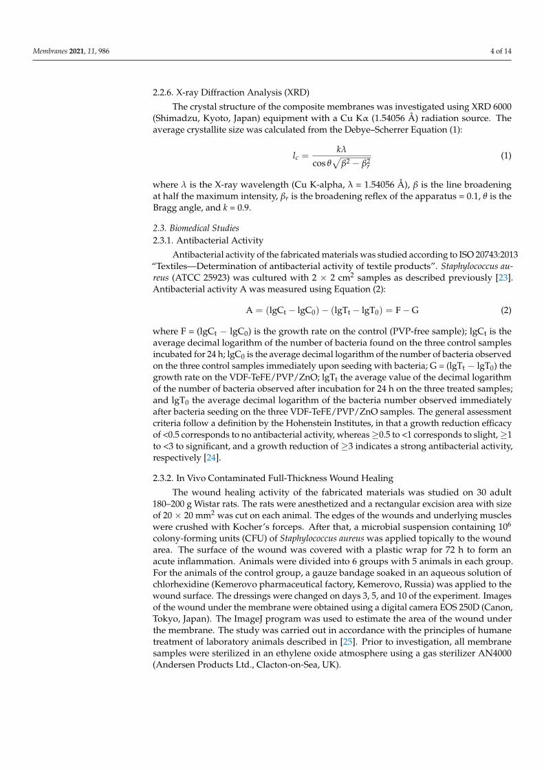
The crystal structure of the composite membranes was investigated using XRD 6000(Shimadzu, Kyoto, Japan) equipment with a Cu Kα (1.54056 Å) radiation source. Theaverage crystallite size was calculated from the Debye–Scherrer Equation (1):
lc =kλ
cos θ√
β2 − β2r
(1)
where λ is the X-ray wavelength (Cu K-alpha, λ = 1.54056 Å), β is the line broadeningat half the maximum intensity, βr is the broadening reflex of the apparatus = 0.1, θ is theBragg angle, and k = 0.9.
2.3. Biomedical Studies2.3.1. Antibacterial Activity
Antibacterial activity of the fabricated materials was studied according to ISO 20743:2013“Textiles—Determination of antibacterial activity of textile products”. Staphylococcus au-reus (ATCC 25923) was cultured with 2 × 2 cm2 samples as described previously [23].Antibacterial activity A was measured using Equation (2):
A = (lgCt − lgC0)− (lgTt − lgT0) = F − G (2)
where F = (lgCt − lgC0) is the growth rate on the control (PVP-free sample); lgCt is theaverage decimal logarithm of the number of bacteria found on the three control samplesincubated for 24 h; lgC0 is the average decimal logarithm of the number of bacteria observedon the three control samples immediately upon seeding with bacteria; G = (lgTt − lgT0) thegrowth rate on the VDF-TeFE/PVP/ZnO; lgTt the average value of the decimal logarithmof the number of bacteria observed after incubation for 24 h on the three treated samples;and lgT0 the average decimal logarithm of the bacteria number observed immediatelyafter bacteria seeding on the three VDF-TeFE/PVP/ZnO samples. The general assessmentcriteria follow a definition by the Hohenstein Institutes, in that a growth reduction efficacyof <0.5 corresponds to no antibacterial activity, whereas ≥0.5 to <1 corresponds to slight, ≥1to <3 to significant, and a growth reduction of ≥3 indicates a strong antibacterial activity,respectively [24].
2.3.2. In Vivo Contaminated Full-Thickness Wound Healing
The wound healing activity of the fabricated materials was studied on 30 adult180–200 g Wistar rats. The rats were anesthetized and a rectangular excision area with sizeof 20 × 20 mm2 was cut on each animal. The edges of the wounds and underlying muscleswere crushed with Kocher’s forceps. After that, a microbial suspension containing 106
colony-forming units (CFU) of Staphylococcus aureus was applied topically to the woundarea. The surface of the wound was covered with a plastic wrap for 72 h to form anacute inflammation. Animals were divided into 6 groups with 5 animals in each group.For the animals of the control group, a gauze bandage soaked in an aqueous solution ofchlorhexidine (Kemerovo pharmaceutical factory, Kemerovo, Russia) was applied to thewound surface. The dressings were changed on days 3, 5, and 10 of the experiment. Imagesof the wound under the membrane were obtained using a digital camera EOS 250D (Canon,Tokyo, Japan). The ImageJ program was used to estimate the area of the wound underthe membrane. The study was carried out in accordance with the principles of humanetreatment of laboratory animals described in [25]. Prior to investigation, all membranesamples were sterilized in an ethylene oxide atmosphere using a gas sterilizer AN4000(Andersen Products Ltd., Clacton-on-Sea, UK).
Membranes 2021, 11, 986 5 of 14
2.4. Statistical Analysis
The data were analyzed with Prism 7 (GraphPad software, San Diego, CA, USA) usingone-way ANOVA with Tukey’s correction for multiple comparisons. Differences wereconsidered significant at p < 0.05.
3. Results and Discussion
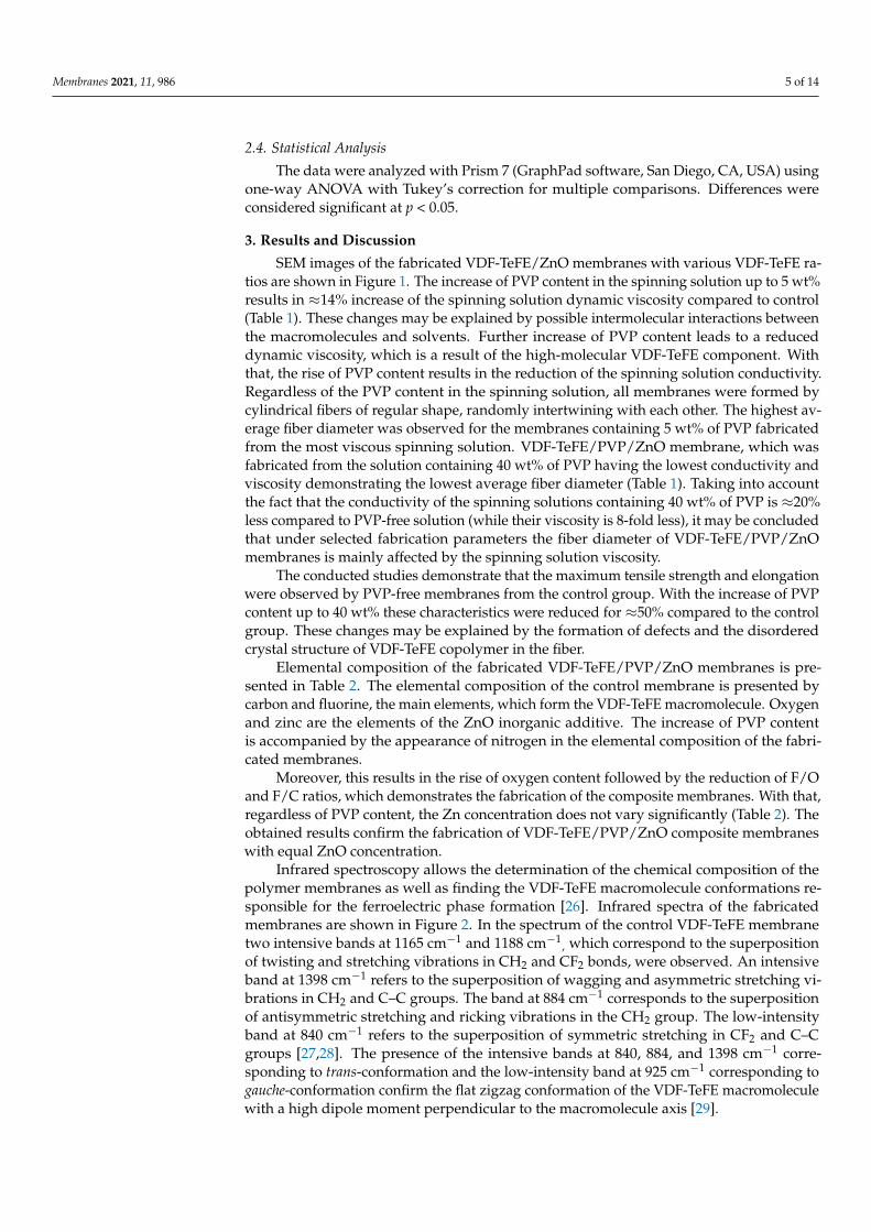
SEM images of the fabricated VDF-TeFE/ZnO membranes with various VDF-TeFE ra-tios are shown in Figure 1. The increase of PVP content in the spinning solution up to 5 wt%results in ≈14% increase of the spinning solution dynamic viscosity compared to control(Table 1). These changes may be explained by possible intermolecular interactions betweenthe macromolecules and solvents. Further increase of PVP content leads to a reduceddynamic viscosity, which is a result of the high-molecular VDF-TeFE component. Withthat, the rise of PVP content results in the reduction of the spinning solution conductivity.Regardless of the PVP content in the spinning solution, all membranes were formed bycylindrical fibers of regular shape, randomly intertwining with each other. The highest av-erage fiber diameter was observed for the membranes containing 5 wt% of PVP fabricatedfrom the most viscous spinning solution. VDF-TeFE/PVP/ZnO membrane, which wasfabricated from the solution containing 40 wt% of PVP having the lowest conductivity andviscosity demonstrating the lowest average fiber diameter (Table 1). Taking into accountthe fact that the conductivity of the spinning solutions containing 40 wt% of PVP is ≈20%less compared to PVP-free solution (while their viscosity is 8-fold less), it may be concludedthat under selected fabrication parameters the fiber diameter of VDF-TeFE/PVP/ZnOmembranes is mainly affected by the spinning solution viscosity.
The conducted studies demonstrate that the maximum tensile strength and elongationwere observed by PVP-free membranes from the control group. With the increase of PVPcontent up to 40 wt% these characteristics were reduced for ≈50% compared to the controlgroup. These changes may be explained by the formation of defects and the disorderedcrystal structure of VDF-TeFE copolymer in the fiber.
Elemental composition of the fabricated VDF-TeFE/PVP/ZnO membranes is pre-sented in Table 2. The elemental composition of the control membrane is presented bycarbon and fluorine, the main elements, which form the VDF-TeFE macromolecule. Oxygenand zinc are the elements of the ZnO inorganic additive. The increase of PVP contentis accompanied by the appearance of nitrogen in the elemental composition of the fabri-cated membranes.
Moreover, this results in the rise of oxygen content followed by the reduction of F/Oand F/C ratios, which demonstrates the fabrication of the composite membranes. With that,regardless of PVP content, the Zn concentration does not vary significantly (Table 2). Theobtained results confirm the fabrication of VDF-TeFE/PVP/ZnO composite membraneswith equal ZnO concentration.
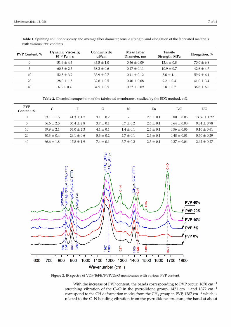
Infrared spectroscopy allows the determination of the chemical composition of thepolymer membranes as well as finding the VDF-TeFE macromolecule conformations re-sponsible for the ferroelectric phase formation [26]. Infrared spectra of the fabricatedmembranes are shown in Figure 2. In the spectrum of the control VDF-TeFE membranetwo intensive bands at 1165 cm−1 and 1188 cm−1
, which correspond to the superpositionof twisting and stretching vibrations in CH2 and CF2 bonds, were observed. An intensiveband at 1398 cm−1 refers to the superposition of wagging and asymmetric stretching vi-brations in CH2 and C–C groups. The band at 884 cm−1 corresponds to the superpositionof antisymmetric stretching and ricking vibrations in the CH2 group. The low-intensityband at 840 cm−1 refers to the superposition of symmetric stretching in CF2 and C–Cgroups [27,28]. The presence of the intensive bands at 840, 884, and 1398 cm−1 corre-sponding to trans-conformation and the low-intensity band at 925 cm−1 corresponding togauche-conformation confirm the flat zigzag conformation of the VDF-TeFE macromoleculewith a high dipole moment perpendicular to the macromolecule axis [29].
Membranes 2021, 11, 986 6 of 14Membranes 2021, 11, x FOR PEER REVIEW 6 of 15
Figure 1. SEM images and water contact angles of VDF-TeFE/ZnO membranes with various PVP
content: (A) 0 wt%; (B) 10 wt%; (C) 40 wt%.
The conducted studies demonstrate that the maximum tensile strength and elonga-
tion were observed by PVP-free membranes from the control group. With the increase of
PVP content up to 40 wt% these characteristics were reduced for ≈50% compared to the
control group. These changes may be explained by the formation of defects and the disor-
dered crystal structure of VDF-TeFE copolymer in the fiber.
Elemental composition of the fabricated VDF-TeFE/PVP/ZnO membranes is pre-
sented in Table 2. The elemental composition of the control membrane is presented by
carbon and fluorine, the main elements, which form the VDF-TeFE macromolecule. Oxy-
gen and zinc are the elements of the ZnO inorganic additive. The increase of PVP content
Figure 1. SEM images and water contact angles of VDF-TeFE/ZnO membranes with various PVPcontent: (A) 0 wt%; (B) 10 wt%; (C) 40 wt%.
Membranes 2021, 11, 986 7 of 14
Table 1. Spinning solution viscosity and average fiber diameter, tensile strength, and elongation of the fabricated materialswith various PVP contents.
PVP Content, % Dynamic Viscosity,10−3 Pa × s
Conductivity,µS/cm
Mean FiberDiameter, µm
TensileStrength, MPa Elongation, %
0 51.9 ± 4.3 43.5 ± 1.0 0.36 ± 0.09 13.4 ± 0.8 70.0 ± 6.8
5 60.3 ± 2.5 38.2 ± 0.6 0.47 ± 0.11 10.9 ± 0.7 42.6 ± 4.7
10 52.8 ± 3.9 33.9 ± 0.7 0.41 ± 0.12 8.6 ± 1.1 59.9 ± 6.4
20 28.0 ± 1.5 32.8 ± 0.5 0.40 ± 0.08 9.2 ± 0.4 41.0 ± 3.4
40 6.3 ± 0.4 34.5 ± 0.5 0.32 ± 0.09 6.8 ± 0.7 36.8 ± 6.6
Table 2. Chemical composition of the fabricated membranes, studied by the EDX method, at%.
PVPContent, % C F O N Zn F/C F/O
0 53.1 ± 1.5 41.3 ± 1.7 3.1 ± 0.2 - 2.6 ± 0.1 0.80 ± 0.05 13.56 ± 1.22
5 56.6 ± 2.5 36.4 ± 2.8 3.7 ± 0.1 0.7 ± 0.2 2.6 ± 0.1 0.64 ± 0.08 9.84 ± 0.98
10 59.9 ± 2.1 33.0 ± 2.3 4.1 ± 0.1 1.4 ± 0.1 2.5 ± 0.1 0.56 ± 0.06 8.10 ± 0.61
20 60.3 ± 0.4 29.1 ± 0.6 5.3 ± 0.2 2.7 ± 0.1 2.5 ± 0.1 0.48 ± 0.01 5.50 ± 0.29
40 66.6 ± 1.8 17.8 ± 1.9 7.4 ± 0.1 5.7 ± 0.2 2.5 ± 0.1 0.27 ± 0.04 2.42 ± 0.27Membranes 2021, 11, x FOR PEER REVIEW 8 of 15
Figure 2. IR spectra of VDF-TeFE/PVP/ZnO membranes with various PVP content.
With the increase of PVP content, the bands corresponding to PVP occur: 1650 cm−1
stretching vibration of the C=O in the pyrrolidone group, 1421 cm−1 and 1372 cm−1 corre-
spond to the CH deformation modes from the CH2 group in PVP, 1287 cm−1 which is re-
lated to the C–N bending vibration from the pyrrolidone structure, the band at about
1495 cm−1 refers to the characteristic vibration of C=N (pyridine ring) [30,31]. The pres-
ence of bands at 840, 884, and 1398 cm−1 and the absence of shifts regardless of PVP content
is evidence for the preservation of the trans-conformation of VDF-TeFE molecules with
high dipole moment. The presence of the bands corresponding both to PVP and VDF-
TeFE confirms the fabrication of composite membranes as well as the results of EDX stud-
ies. It should be noted that the addition of hydrophilic PVP in the range of 0 to 40 wt%
had no effect on the surface properties of the fabricated membranes, which is demon-
strated by the insignificant changes in water contact angles (122° for the control membrane
and 110° for the membranes containing 40 wt% of PVP) (Figure 1).
The XRD patterns of the fabricated membranes are presented in Figure 3.
Figure 2. IR spectra of VDF-TeFE/PVP/ZnO membranes with various PVP content.
With the increase of PVP content, the bands corresponding to PVP occur: 1650 cm−1
stretching vibration of the C=O in the pyrrolidone group, 1421 cm−1 and 1372 cm−1
correspond to the CH deformation modes from the CH2 group in PVP, 1287 cm−1 which isrelated to the C–N bending vibration from the pyrrolidone structure, the band at about
Membranes 2021, 11, 986 8 of 14
1495 cm−1 refers to the characteristic vibration of C=N (pyridine ring) [30,31]. The presenceof bands at 840, 884, and 1398 cm−1 and the absence of shifts regardless of PVP contentis evidence for the preservation of the trans-conformation of VDF-TeFE molecules withhigh dipole moment. The presence of the bands corresponding both to PVP and VDF-TeFEconfirms the fabrication of composite membranes as well as the results of EDX studies. Itshould be noted that the addition of hydrophilic PVP in the range of 0 to 40 wt% had noeffect on the surface properties of the fabricated membranes, which is demonstrated by theinsignificant changes in water contact angles (122◦ for the control membrane and 110◦ forthe membranes containing 40 wt% of PVP) (Figure 1).
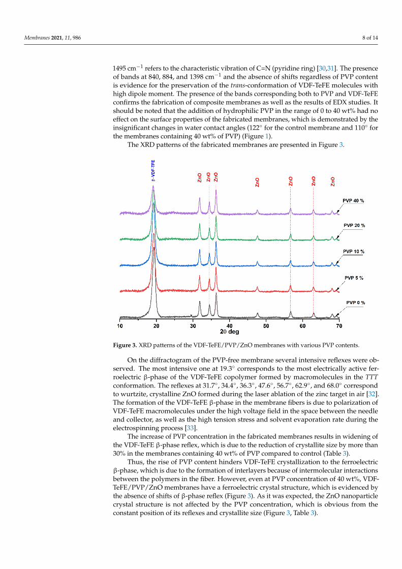
The XRD patterns of the fabricated membranes are presented in Figure 3.
Membranes 2021, 11, x FOR PEER REVIEW 9 of 15
Figure 3. XRD patterns of the VDF-TeFE/PVP/ZnO membranes with various PVP contents.
On the diffractogram of the PVP-free membrane several intensive reflexes were ob-
served. The most intensive one at 19.3° corresponds to the most electrically active ferroe-
lectric β-phase of the VDF-TeFE copolymer formed by macromolecules in the TTT confor-
mation. The reflexes at 31.7°, 34.4°, 36.3°, 47.6°, 56.7°, 62.9°, and 68.0° correspond to wurtz-
ite, crystalline ZnO formed during the laser ablation of the zinc target in air [32]. The for-
mation of the VDF-TeFE β-phase in the membrane fibers is due to polarization of VDF-
TeFE macromolecules under the high voltage field in the space between the needle and
collector, as well as the high tension stress and solvent evaporation rate during the elec-
trospinning process [33].
The increase of PVP concentration in the fabricated membranes results in widening
of the VDF-TeFE β-phase reflex, which is due to the reduction of crystallite size by more
than 30% in the membranes containing 40 wt% of PVP compared to control (Table 3).
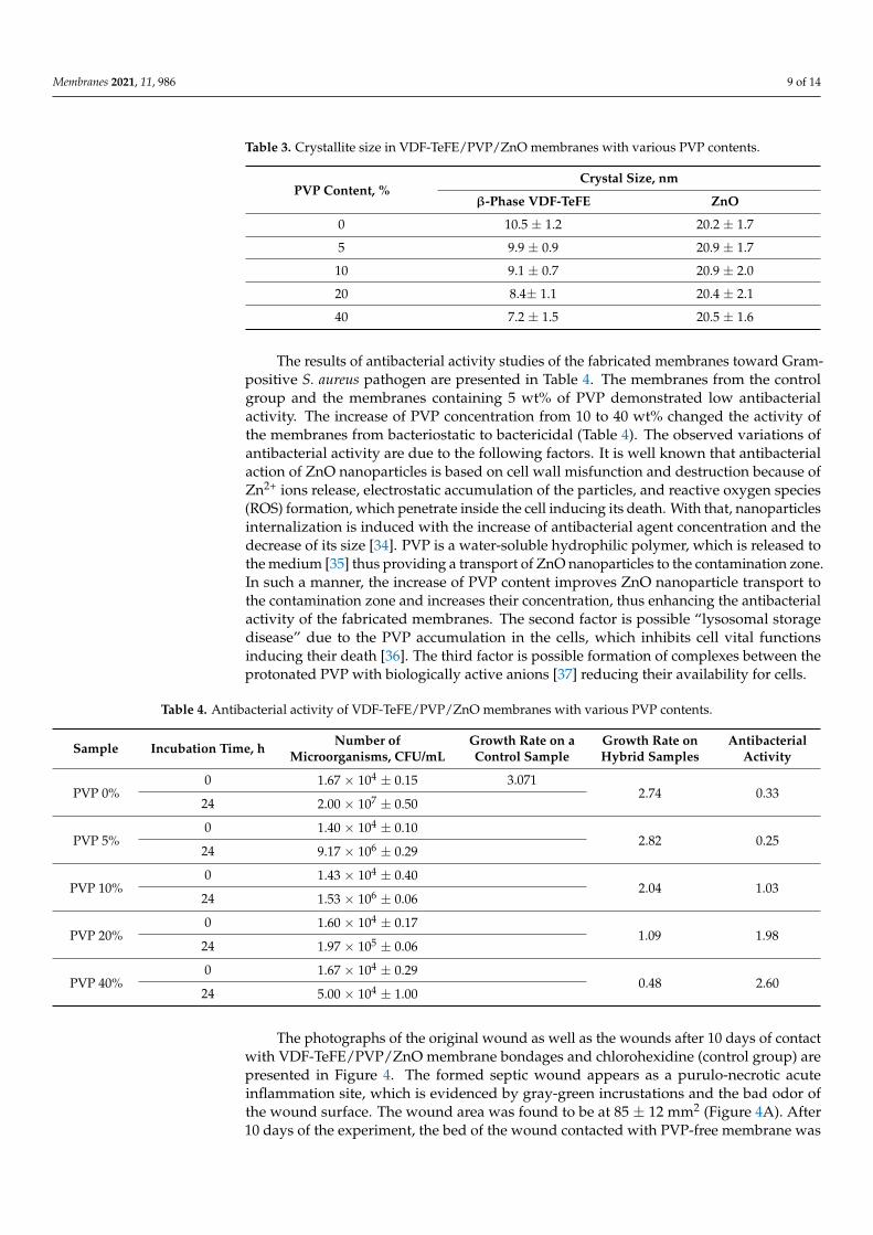
Table 3. Crystallite size in VDF-TeFE/PVP/ZnO membranes with various PVP contents.
PVP Content, % Crystal Size, nm
β-Phase VDF-TеFE ZnO
0 10.5 ± 1.2 20.2 ± 1.7
5 9.9 ± 0.9 20.9 ± 1.7
10 9.1 ± 0.7 20.9 ± 2.0
20 8.4± 1.1 20.4 ± 2.1
40 7.2 ± 1.5 20.5 ± 1.6
Thus, the rise of PVP content hinders VDF-TeFE crystallization to the ferroelectric
β- phase, which is due to the formation of interlayers because of intermolecular interac-
tions between the polymers in the fiber. However, even at PVP concentration of 40 wt%,
Figure 3. XRD patterns of the VDF-TeFE/PVP/ZnO membranes with various PVP contents.
On the diffractogram of the PVP-free membrane several intensive reflexes were ob-served. The most intensive one at 19.3◦ corresponds to the most electrically active fer-roelectric β-phase of the VDF-TeFE copolymer formed by macromolecules in the TTTconformation. The reflexes at 31.7◦, 34.4◦, 36.3◦, 47.6◦, 56.7◦, 62.9◦, and 68.0◦ correspondto wurtzite, crystalline ZnO formed during the laser ablation of the zinc target in air [32].The formation of the VDF-TeFE β-phase in the membrane fibers is due to polarization ofVDF-TeFE macromolecules under the high voltage field in the space between the needleand collector, as well as the high tension stress and solvent evaporation rate during theelectrospinning process [33].
The increase of PVP concentration in the fabricated membranes results in widening ofthe VDF-TeFE β-phase reflex, which is due to the reduction of crystallite size by more than30% in the membranes containing 40 wt% of PVP compared to control (Table 3).
Thus, the rise of PVP content hinders VDF-TeFE crystallization to the ferroelectricβ-phase, which is due to the formation of interlayers because of intermolecular interactionsbetween the polymers in the fiber. However, even at PVP concentration of 40 wt%, VDF-TeFE/PVP/ZnO membranes have a ferroelectric crystal structure, which is evidenced bythe absence of shifts of β-phase reflex (Figure 3). As it was expected, the ZnO nanoparticlecrystal structure is not affected by the PVP concentration, which is obvious from theconstant position of its reflexes and crystallite size (Figure 3, Table 3).
Membranes 2021, 11, 986 9 of 14
Table 3. Crystallite size in VDF-TeFE/PVP/ZnO membranes with various PVP contents.
PVP Content, %Crystal Size, nm
β-Phase VDF-TeFE ZnO
0 10.5 ± 1.2 20.2 ± 1.7
5 9.9 ± 0.9 20.9 ± 1.7
10 9.1 ± 0.7 20.9 ± 2.0
20 8.4± 1.1 20.4 ± 2.1
40 7.2 ± 1.5 20.5 ± 1.6
The results of antibacterial activity studies of the fabricated membranes toward Gram-positive S. aureus pathogen are presented in Table 4. The membranes from the controlgroup and the membranes containing 5 wt% of PVP demonstrated low antibacterialactivity. The increase of PVP concentration from 10 to 40 wt% changed the activity ofthe membranes from bacteriostatic to bactericidal (Table 4). The observed variations ofantibacterial activity are due to the following factors. It is well known that antibacterialaction of ZnO nanoparticles is based on cell wall misfunction and destruction because ofZn2+ ions release, electrostatic accumulation of the particles, and reactive oxygen species(ROS) formation, which penetrate inside the cell inducing its death. With that, nanoparticlesinternalization is induced with the increase of antibacterial agent concentration and thedecrease of its size [34]. PVP is a water-soluble hydrophilic polymer, which is released tothe medium [35] thus providing a transport of ZnO nanoparticles to the contamination zone.In such a manner, the increase of PVP content improves ZnO nanoparticle transport tothe contamination zone and increases their concentration, thus enhancing the antibacterialactivity of the fabricated membranes. The second factor is possible “lysosomal storagedisease” due to the PVP accumulation in the cells, which inhibits cell vital functionsinducing their death [36]. The third factor is possible formation of complexes between theprotonated PVP with biologically active anions [37] reducing their availability for cells.
Table 4. Antibacterial activity of VDF-TeFE/PVP/ZnO membranes with various PVP contents.
Sample Incubation Time, h Number ofMicroorganisms, CFU/mL
Growth Rate on aControl Sample
Growth Rate onHybrid Samples
AntibacterialActivity
PVP 0%0 1.67 × 10⁴ ± 0.15 3.071
2.74 0.3324 2.00 × 10⁷ ± 0.50
PVP 5%0 1.40 × 10⁴ ± 0.10
2.82 0.2524 9.17 × 10⁶ ± 0.29
PVP 10%0 1.43 × 10⁴ ± 0.40
2.04 1.0324 1.53 × 10⁶ ± 0.06
PVP 20%0 1.60 × 10⁴ ± 0.17
1.09 1.9824 1.97 × 10⁵ ± 0.06
PVP 40%0 1.67 × 10⁴ ± 0.29
0.48 2.6024 5.00 × 10⁴ ± 1.00
The photographs of the original wound as well as the wounds after 10 days of contactwith VDF-TeFE/PVP/ZnO membrane bondages and chlorohexidine (control group) arepresented in Figure 4. The formed septic wound appears as a purulo-necrotic acuteinflammation site, which is evidenced by gray-green incrustations and the bad odor ofthe wound surface. The wound area was found to be at 85 ± 12 mm2 (Figure 4A). After10 days of the experiment, the bed of the wound contacted with PVP-free membrane was
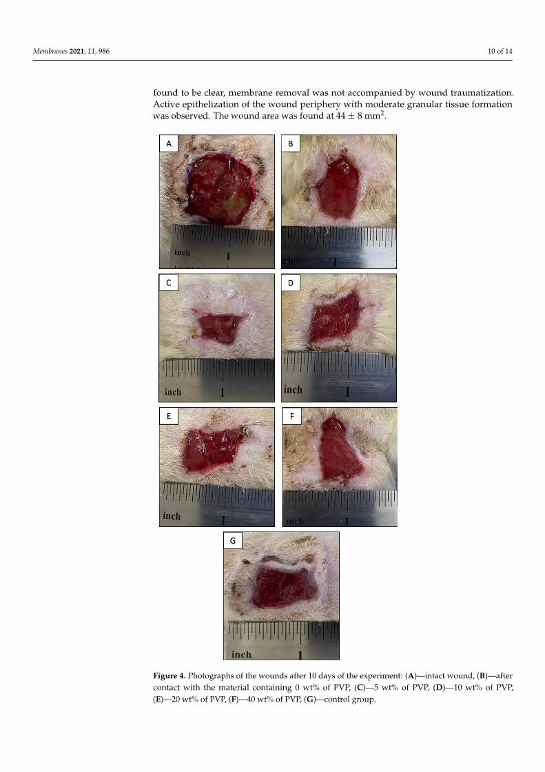
Membranes 2021, 11, 986 10 of 14
found to be clear, membrane removal was not accompanied by wound traumatization.Active epithelization of the wound periphery with moderate granular tissue formationwas observed. The wound area was found at 44 ± 8 mm2.
Membranes 2021, 11, x FOR PEER REVIEW 11 of 15
to be clear, membrane removal was not accompanied by wound traumatization. Active
epithelization of the wound periphery with moderate granular tissue formation was ob-
served. The wound area was found at 44 ± 8 mm2.
Figure 4. Photographs of the wounds after 10 days of the experiment: (A)—intact wound, (B)—after
contact with the material containing 0 wt% of PVP, (C)—5 wt% of PVP, (D)—10 wt% of PVP, (E)—
20 wt% of PVP, (F)—40 wt% of PVP, (G)—control group.
Figure 4. Photographs of the wounds after 10 days of the experiment: (A)—intact wound, (B)—aftercontact with the material containing 0 wt% of PVP, (C)—5 wt% of PVP, (D)—10 wt% of PVP,(E)—20 wt% of PVP, (F)—40 wt% of PVP, (G)—control group.
Membranes 2021, 11, 986 11 of 14
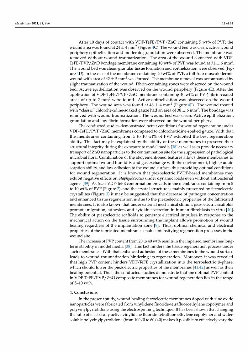
After 10 days of contact with VDF-TeFE/PVP/ZnO containing 5 wt% of PVP, thewound area was found at 24 ± 4 mm2 (Figure 4C). The wound bed was clean, active woundperiphery epithelization and moderate granulation were observed. The membrane wasremoved without wound traumatization. The area of the wound contacted with VDF-TeFE/PVP/ZnO bondage membrane containing 10 wt% of PVP was found at 31 ± 6 mm2.The wound bed was clean, granular tissue formation and epithelization were observed (Fig-ure 4D). In the case of the membrane containing 20 wt% of PVP, a full-frap musculodermicwound with area of 42 ± 5 mm2 was formed. The membrane removal was accompanied byslight traumatization of the wound. Fibrin-containing zones were observed on the woundbed. Active epithelization was observed on the wound periphery (Figure 4E). After theapplication of VDF-TeFE/PVP/ZnO membrane containing 40 wt% of PVP, fibrin-coatedareas of up to 2 mm2 were found. Active epithelization was observed on the woundperiphery. The wound area was found at 46 ± 4 mm2 (Figure 4F). The wound treatedwith “classic” chlorohexidine-soaked gauze had an area of 38 ± 6 mm2. The bondage wasremoved with wound traumatization. The wound bed was clean. Active epithelization,granulation and low fibrin formation were observed on the wound periphery.
The conducted studies demonstrated better conditions for wound regeneration underVDF-TeFE/PVP/ZnO membranes compared to chlorohexidine-soaked gauze. With that,the membranes containing from 5 to 10 wt% of PVP exhibited the best regenerationability. This fact may be explained by the ability of these membranes to preserve theirstructural integrity during the exposure to model media [38] as well as to provide necessarytransport of ZnO nanoparticles to the contamination site for the suppression of pathologicalmicrobial flora. Combination of the abovementioned features allows these membranes tosupport optimal wound humidity and gas exchange with the environment, high exudatesorption ability, and low adhesion to the wound surface, thus providing optimal conditionsfor wound regeneration. It is known that piezoelectric PVDF-based membranes mayexhibit negative effects on Staphylococcus under dynamic loads even without antibacterialagents [39]. As trans VDF-TeFE conformation prevails in the membranes containing from 5to 10 wt% of PVP (Figure 2), and the crystal structure is mainly presented by ferroelectriccrystallites (Figure 3) it may be suggested that the decrease of pathogen concentrationand enhanced tissue regeneration is due to the piezoelectric properties of the fabricatedmembranes. It is also known that under external mechanical stimuli, piezoelectric scaffoldspromote migration, adhesion, and cytokine secretion in human fibroblasts in vitro [40].The ability of piezoelectric scaffolds to generate electrical impulses in response to themechanical action on the tissue surrounding the implant allows promotion of woundhealing regardless of the implantation zone [9]. Thus, optimal chemical and electricalproperties of the fabricated membranes enable intensifying regeneration processes in thewound site.
The increase of PVP content from 20 to 40 wt% results in the impaired membranes long-term stability in model media [38]. This fact hinders the tissue regeneration process undersuch membranes. With that, enhanced adhesion of these membranes to the wound surfaceleads to wound traumatization hindering its regeneration. Moreover, it was revealedthat high PVP content hinders VDF-TeFE crystallization into the ferroelectric β-phase,which should lower the piezoelectric properties of the membranes [41,42] as well as theirhealing potential. Thus, the conducted studies demonstrate that the optimal PVP contentin VDF-TeFE/PVP/ZnO composite membranes for wound regeneration lies in the rangeof 5–10 wt%.
4. Conclusions
In the present study, wound healing ferroelectric membranes doped with zinc oxidenanoparticles were fabricated from vinylidene fluoride-tetrafluoroethylene copolymer andpolyvinylpyrrolidone using the electrospinning technique. It has been shown that changingthe ratio of electrically active vinylidene fluoride-tetrafluoroethylene copolymer and water-soluble polyvinylpyrrolidone (from 100/0 to 60/40) makes it possible to effectively vary the
Membranes 2021, 11, 986 12 of 14
physicochemical and biomedical parameters of the obtained materials. Polyvinylpyrroli-done hindered the crystallization of vinylidene fluoride-tetrafluoroethylene copolymer,which led to a deterioration in the mechanical characteristics of the membranes (nearlytwo-fold decrease in tensile strength and elongation). The surface of the obtained mate-rials had an extremely hydrophobic character (water contact angle ≈120◦) regardless ofthe polyvinylpyrrolidone content. The polyvinylpyrrolidone addition made it possibleto change the antibacterial activity of the formed materials against Staphylococcus aureusfrom bacteriostatic to bactericidal. In vivo studies using the rat model showed a positiveeffect of the formed membranes on the healing of wounds with abundant infection. It wasshown that the optimal concentration of polyvinylpyrrolidone lies between five to ten masspercent based on the combination of physicochemical and biomedical characteristics.
Author Contributions: Conceptualization, E.N.B.; methodology, A.L.N., I.V.L., L.S.A.; software,A.L.N., I.V.L., L.S.A., T.S.T., D.V.V., A.L.N., D.A.G.; validation, A.L.N., I.V.L., L.S.A., T.S.T., D.V.V.,A.L.N., D.A.G.; formal analysis, S.I.G.; investigation, A.L.N., I.V.L., L.S.A., T.S.T., D.V.V., A.L.N.,D.A.G.; resources, V.A.S.; data curation, A.L.N., I.V.L., L.S.A., T.S.T., D.V.V., A.L.N., D.A.G., G.T.D.;writing—original draft preparation, E.N.B., S.I.G.; writing—review and editing, S.I.G.; visualization,E.N.B.; supervision, V.M.B., V.A.S.; project administration, E.N.B.; funding acquisition, E.N.B. Allauthors have read and agreed to the published version of the manuscript.
Funding: Membrane fabrication and physicochemical studies were funded by RFBR, project number20-33-90159. Biomedical studies were funded by RFBR, project number 20-03-00171.
Institutional Review Board Statement: The study was conducted according to the guidelines of theDeclaration of Helsinki, and approved by the Ethics Committee of Siberian State Medical University(protocol code # 6897 date of approval 6 November 2019).
Informed Consent Statement: Not applicable.
Data Availability Statement: Not applicable.
Conflicts of Interest: The authors declare no conflict of interest.
References1. Brocke, T.; Barr, J. The History of Wound Healing. Surg. Clin. N. Am. 2020, 100, 787–806. [CrossRef] [PubMed]2. Han, G.; Ceilley, R. Chronic Wound Healing: A Review of Current Management and Treatments. Adv. Ther. 2017, 34, 599–610.
[CrossRef] [PubMed]3. Naskar, A.; Kim, K.-S. Recent Advances in Nanomaterial-Based Wound-Healing Therapeutics. Pharmaceutics 2020, 12, 499.
[CrossRef]4. Mousavi, S.; Zarei, M.; Hashemi, S.; Ramakrishna, S.; Chiang, W.-H.; Lai, C.; Gholami, A.; Omidifar, N.; Shokripour, M.
Asymmetric Membranes: A Potential Scaffold for Wound Healing Applications. Symmetry 2020, 12, 1100. [CrossRef]5. Liu, X.; Xu, H.; Zhang, M.; Yu, D.-G. Electrospun Medicated Nanofibers for Wound Healing: Review. Membranes 2021, 11, 770.
[CrossRef] [PubMed]6. Bombin, A.D.J.; Dunne, N.J.; McCarthy, H.O. Electrospinning of natural polymers for the production of nanofibres for wound
healing applications. Mater. Sci. Eng. C Mater. Biol. Appl. 2020, 114, 110994. [CrossRef] [PubMed]7. Afsharian, Y.P.; Rahimnejad, M. Bioactive electrospun scaffolds for wound healing applications: A comprehensive review. Polym.
Test. 2021, 93, 106952. [CrossRef]8. Mokhtari, F.; Azimi, B.; Salehi, M.; Hashemikia, S.; Danti, S. Recent advances of polymer-based piezoelectric composites for
biomedical applications. J. Mech. Behav. Biomed. Mater. 2021, 122, 104669. [CrossRef] [PubMed]9. Wang, A.; Liu, Z.; Hu, M.; Wang, C.; Zhang, X.; Shi, B.; Fan, Y.; Cui, Y.; Li, Z.; Ren, K. Piezoelectric nanofibrous scaffolds as in vivo
energy harvesters for modifying fibroblast alignment and proliferation in wound healing. Nano Energy 2018, 43, 63–71. [CrossRef]10. Tandon, B.; Magaz, A.; Balint, R.; Blaker, J.; Cartmell, S.H. Electroactive biomaterials: Vehicles for controlled delivery of therapeutic
agents for drug delivery and tissue regeneration. Adv. Drug Deliv. Rev. 2018, 129, 148–168. [CrossRef] [PubMed]11. Ning, C.; Zhou, Z.; Tan, G.; Zhu, Y.; Mao, C. Electroactive polymers for tissue regeneration: Developments and perspectives. Prog.
Polym. Sci. 2018, 81, 144–162. [CrossRef]12. Nakhmanson, S.M.; Nardelli, M.B.; Bernholc, J. Ab InitioStudies of Polarization and Piezoelectricity in Vinylidene Fluoride and
BN-Based Polymers. Phys. Rev. Lett. 2004, 92, 115504. [CrossRef] [PubMed]13. Nakhmanson, S.M.; Nardelli, M.B.; Bernholc, J. Collective polarization effects inβ-polyvinylidene fluoride and its copolymers
with tri- and tetrafluoroethylene. Phys. Rev. B 2005, 72, 115210. [CrossRef]
Membranes 2021, 11, 986 13 of 14
14. Nakagawa, Y.; Hashizume, Y.; Nakajima, T.; Okamura, S. Polarization switching characteristics of vinylidene fluo-ride/tefrafluoroethylene copolymer thin films. Jpn. J. Appl. Phys. 2015, 54, 10NA09. [CrossRef]
15. Ameduri, B. From Vinylidene Fluoride (VDF) to the Applications of VDF-Containing Polymers and Copolymers: RecentDevelopments and Future Trends. Chem. Rev. 2009, 109, 6632–6686. [CrossRef] [PubMed]
16. Bolbasov, E.N.; Popkov, D.; Kononovich, N.A.; Gorbach, E.N.; Khlusov, I.A.; Golovkin, A.S.; Stankevich, K.S.; Ignatov, V.P.;Bouznik, V.M.; Anissimov, Y.; et al. Flexible intramedullary nails for limb lengthening: A comprehensive comparative study ofthree nails types. Biomed. Mater. 2019, 14, 025005. [CrossRef]
17. Cui, Z.; Drioli, E.; Lee, Y.M. Recent progress in fluoropolymers for membranes. Prog. Polym. Sci. 2014, 39, 164–198. [CrossRef]18. Teodorescu, M.; Bercea, M. Poly(vinylpyrrolidone)—A Versatile Polymer for Biomedical and Beyond Medical Applications.
Polym. Technol. Eng. 2015, 54, 923–943. [CrossRef]19. Franco, P.; De Marco, I. The Use of Poly(N-vinyl pyrrolidone) in the Delivery of Drugs: A Review. Polymers 2020, 12, 1114.
[CrossRef]20. Ann, L.C.; Mahmud, S.; Bakhori, S.K.M.; Sirelkhatim, A.; Mohamad, D.; Hasan, H.; Seeni, A.; Rahman, R.A. Antibacterial
responses of zinc oxide structures against Staphylococcus aureus, Pseudomonas aeruginosa and Streptococcus pyogenes. Ceram. Int.2014, 40, 2993–3001. [CrossRef]
21. Singh, T.A.; Sharma, A.; Tejwan, N.; Ghosh, N.; Das, J.; Sil, P.C. A state of the art review on the synthesis, antibacterial, antioxidant,antidiabetic and tissue regeneration activities of zinc oxide nanoparticles. Adv. Colloid Interface Sci. 2021, 295, 102495. [CrossRef][PubMed]
22. Goel, S.; Kumar, B. A review on piezo-/ferro-electric properties of morphologically diverse ZnO nanostructures. J. Alloys Compd.2020, 816, 152491. [CrossRef]
23. Gavrilenko, E.A.; Goncharova, D.A.; Lapin, I.N.; Nemoykina, A.L.; Svetlichnyi, V.A.; Aljulaih, A.A.; Mintcheva, N.; Kulinich, S.A.Comparative Study of Physicochemical and Antibacterial Properties of ZnO Nanoparticles Prepared by Laser Ablation of ZnTarget in Water and Air. Materials 2019, 12, 186. [CrossRef] [PubMed]
24. Hoefer, D.; Hammer, T.R. Antimicrobial Active Clothes Display No Adverse Effects on the Ecological Balance of the HealthyHuman Skin Microflora. ISRN Dermatol. 2011, 2011, 369603. [CrossRef] [PubMed]
25. Janet, C.G.; Barbee, R.W.; Bielitzki, J.T.; Clayton, L.A.; Donovan, J.C.; Hendriksen, C.F.M.; Kohn, D.F.; Lipman, N.S.; Locke,P.A.; Melcher, J.; et al. Guide for the Care and Use of Laboratory Animals; National Academies Press: Washington, DC, USA, 2011;ISBN 0-919087-18-3.
26. Cui, Z.; Hassankiadeh, N.T.; Zhuang, Y.; Drioli, E.; Lee, Y.M. Crystalline polymorphism in poly(vinylidenefluoride) membranes.Prog. Polym. Sci. 2015, 51, 94–126. [CrossRef]
27. Kobayashi, M.; Tashiro, K.; Tadokoro, H. Molecular Vibrations of Three Crystal Forms of Poly(vinylidene fluoride). Macromolecules1975, 8, 158–171. [CrossRef]
28. Tashiro, K.; Abe, Y.; Kobayashi, M. Computer simulation of structure and ferroelectric phase transition of vinylidene fluoridecopolymers (1) vdf content dependence of the crystal structure. Ferroelectrics 1995, 171, 281–297. [CrossRef]
29. Kochervinskii, V.V. The properties and applications of fluorine-containing polymer films with piezo- and pyro-activity. Russ.Chem. Rev. 1994, 63, 367–371. [CrossRef]
30. Safo, I.A.; Werheid, M.; Dosche, C.; Oezaslan, M. The role of polyvinylpyrrolidone (PVP) as a capping and structure-directingagent in the formation of Pt nanocubes. Nanoscale Adv. 2019, 1, 3095–3106. [CrossRef]
31. Abdelghany, A.; Abdelrazek, E.M.; Badr, S.I.; Morsi, M.A. Effect of gamma-irradiation on (PEO/PVP)/Au nanocomposite:Materials for electrochemical and optical applications. Mater. Des. 2016, 97, 532–543. [CrossRef]
32. Goncharova, D.A.; Bolbasov, E.N.; Nemoykina, A.L.; Aljulaih, A.A.; Tverdokhlebova, T.S.; Kulinich, S.A.; Svetlichnyi, V.A.Structure and Properties of Biodegradable PLLA/ZnO Composite Membrane Produced via Electrospinning. Materials 2020, 14, 2.[CrossRef]
33. Teo, W.E.; Ramakrishna, S. A review on electrospinning design and nanofibre assemblies. Nanotechnology 2006, 17, R89–R106.[CrossRef]
34. Sirelkhatim, A.; Mahmud, S.; Seeni, A.; Kaus, N.H.M.; Ann, L.C.; Bakhori, S.K.M.; Hasan, H.; Mohamad, D. Review on ZincOxide Nanoparticles: Antibacterial Activity and Toxicity Mechanism. Nano-Micro Lett. 2015, 7, 219–242. [CrossRef] [PubMed]
35. Kim, G.-M.; Le, K.H.T.; Giannitelli, S.M.; Lee, Y.J.; Rainer, A.; Trombetta, M. Electrospinning of PCL/PVP blends for tissueengineering scaffolds. J. Mater. Sci. Mater. Med. 2013, 24, 1425–1442. [CrossRef] [PubMed]
36. Duncan, R.; Richardson, S.C.W. Endocytosis and Intracellular Trafficking as Gateways for Nanomedicine Delivery: Opportunitiesand Challenges. Mol. Pharm. 2012, 9, 2380–2402. [CrossRef]
37. Burnett, C.L. PVP (Polyvinylpyrrolidone). Int. J. Toxicol. 2017, 36, 50S–51S. [CrossRef]38. Tverdokhlebova, T.S.; Bolbasov, E.N.; Bouznik, V.M. Composition Polymeric Membranes Based on the VDF-TeFE Copolymer
Formed by Electrospinning. IOP Conf. Ser. Mater. Sci. Eng. 2020, 731, 012022. [CrossRef]39. Carvalho, E.O.; Fernandes, M.M.; Padrão, J.; Nicolau, A.; Marqués-Marchán, J.; Asenjo, A.; Gama, F.M.; Ribeiro, C.; Lanceros-
Mendez, S. Tailoring Bacteria Response by Piezoelectric Stimulation. ACS Appl. Mater. Interfaces 2019, 11, 27297–27305. [CrossRef][PubMed]
40. Guo, H.-F.; Li, Z.-S.; Dong, S.-W.; Chen, W.-J.; Deng, L.; Wang, Y.-F.; Ying, D.-J. Piezoelectric PU/PVDF electrospun scaffolds forwound healing applications. Colloids Surf. B Biointerfaces 2012, 96, 29–36. [CrossRef] [PubMed]
Membranes 2021, 11, 986 14 of 14
41. Tasaka, S.; Miyata, S. Effects of crystal structure on piezoelectric and ferroelectric properties of copoly(vinylidenefluoride-tetrafluoroethylene). J. Appl. Phys. 1985, 57, 906–910. [CrossRef]
42. Hicks, J.C.; Jones, T.E.; Logan, J.C. Ferroelectric properties of poly(vinylidene fluoride-tetrafluoroethylene). J. Appl. Phys. 1978, 49,6092–6096. [CrossRef]