ray assisted synthesis of silver nanoparticles in chitosan solution and the antibacterial properties
TRANSCRIPT
�a
NAa
b
c
d
e
a
ARRA
KNE�CS
1
rTemmH
m[pruwbto[f
L
1d
Chemical Engineering Journal 155 (2009) 499–507
Contents lists available at ScienceDirect
Chemical Engineering Journal
journa l homepage: www.e lsev ier .com/ locate /ce j
-Ray assisted synthesis of silver nanoparticles in chitosan solutionnd the antibacterial properties
.M. Huang a,∗, S. Radiman b, H.N. Lim c, P.S. Khiew d, W.S. Chiu d, K.H. Lee e,
. Syahida e, R. Hashim a, C.H. Chia b
Faculty of Science, University of Malaya, 50603 Kuala Lumpur, MalaysiaSchool of Applied Physics, Faculty of Science and Technology, Universiti Kebangsaan Malaysia, 43600 Bangi, Selangor Darul Ehsan, MalaysiaChemistry Department, Faculty of Science, Universiti Putra Malaysia, 43400 UPM Serdang, Selangor Darul Ehsan, MalaysiaFaculty of Engineering and Computer Science, Nottingham University, Jalan Broga, 43500 Semenyih, Selangor Darul Ehsan, MalaysiaDepartment of Biochemistry, Faculty of Biotechnology and Biomolecular Sciences, Universiti Putra Malaysia, 43400 UPM Serdang, Selangor Darul Ehsan, Malaysia
r t i c l e i n f o
rticle history:eceived 30 April 2009eceived in revised form 9 July 2009
a b s t r a c t
In the present study, chitosan had been utilized as a “green” stabilizing agent for the synthesis of sphericalsilver nanoparticles in the range of 5–30 nm depending on the percentage of chitosan used (0.1, 0.5, 1.0and 2.0 wt%) under �-irradiation. X-ray diffractometer identified the nanoparticles as pure silver hav-
ccepted 17 July 2009
eywords:anostructureslectron microscopy
ing face-centered cubic phase. Ultraviolet–visible spectra exhibited the influence of �-irradiation totalabsorbed dose and chitosan concentration on the yield of silver nanoparticles. The antibacterial propertiesof the silver nanoparticles were tested against Methicillin-resistant Staphylococcus aureus (MRSA) (gram-positive) and Aeromonas hydrophila (gram-negative) bacteria. This work provides a simple and “green”method for the synthesis of highly stable silver nanoparticles in aqueous solution with good antibacterial
-Ray irradiationhitosanilver
property.
. Introduction
Silver nanoparticles are of great interest amongst theesearchers due to their exquisite properties in nanometer size.hese particles exhibit potential applications in catalysis, surfacenhanced Raman scattering, nanoelectronics, data saving, electro-agnetic coating, efficient antibacterial activities [1–6] and theost recent finding reported on the inhibition of the growth ofIV-1 virus [7].
Bacterial pollution on water is a major threat to health. Asicroorganisms become more resistant to antimicrobial agents
8], there is an increasing need to improve bacteria eradicationrocedures. The antibacterial properties of silver ions have beenecognized for a long time and silver ions have been extensivelysed as bacteria eradicator in catheter, burn wounds and dentalork [9]. Researchers also encourage the use of silver as effective
acteria eradicator for wastes generated from hospitals, which con-
ain highly infectious microorganisms [10,11]. However, remnantsf silver ions in treated water might cause adverse effects to health12]. The emergence of nanoscience and nanotechology in the pastew decades has opened the doors of opportunities to study the∗ Corresponding author at: Faculty of Science, University of Malaya, 50603 Kualaumpur, Malaysia. Tel.: +60 12 2091008; fax: +60 3 58911088.
E-mail address: [email protected] (N.M. Huang).
385-8947/$ – see front matter © 2009 Elsevier B.V. All rights reserved.oi:10.1016/j.cej.2009.07.040
© 2009 Elsevier B.V. All rights reserved.
effect of bacteria eradication using nanoparticles. The antibacterialeffect of metal nanoparticles is due to fine metal size and broadsurface area to volume, which allows nanoparticles to have inti-mate contact with membranes of the bacteria and not just basedon solubilization of metal ions in solutions [13].
For the last decade, the increased awareness towards the envi-ronment has encouraged nanomaterial scientists to look into“greener” methods. The use of non-toxic chemicals, environmental-friendly solvents and renewable resources are important issues in“green” synthesis strategies [14]. Nanomaterials significantly affectthe fields of physics, chemistry, electronics, optics, materials scienceand biomedical science. Even though nanomaterials exhibit specialproperties due to their size-related effects, their synthesis methodsreflect negatively on the environment. Although milder reductantshave been introduced for the synthesis of silver nanoparticles suchas glucose [15], sodium citrate [16] and polyvinyl pyrrolidone (PVP)[17], most synthesis methods were still highly dependent on the useof toxic chemicals such as sodium borohidride [18], hydrazine [19],formamide [20] and N,N-dimethylformide [21]. These reductantsare very reactive, causing environmental and biological risks.
Various methods have been reported for the synthesis of
nanosized silver particles e.g. chemical reduction [22–24], pho-tochemical reduction (UV [25], microwave [26,27], electron beam[28] and �-irradiation [29–32]), micelle [33], reverse micelle [34],microemulsion [35], lamellar liquid crystal [36], aerosol spray-ing technique [37] and capping agent method [38–40]. Chitosan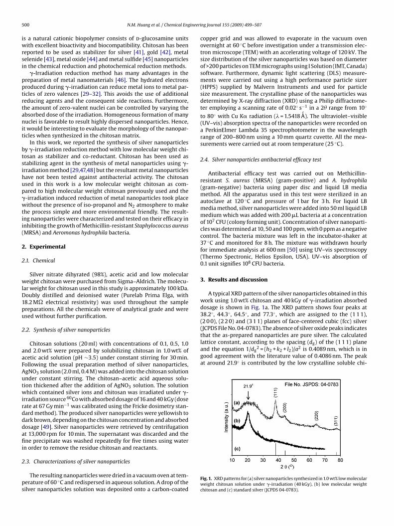
5 ineeri
iwrsi
pptrtanit
btsihup�wtii(
2
2
wlD1pu
2
aaFAutwirdddafii
2
ps
lattice constant, according to the spacing (dg) of the (1 1 1) planeand the equation 1/dg
2 = (h2 + k2 + l2)/a2 is 0.4089 nm, which is ingood agreement with the literature value of 0.4086 nm. The peakat around 21.9◦ is contributed by the low crystalline soluble chi-
00 N.M. Huang et al. / Chemical Eng
s a natural cationic biopolymer consists of d-glucosamine unitsith excellent bioactivity and biocompatibility. Chitosan has been
eported to be used as stabilizer for silver [41], gold [42], metalelenide [43], metal oxide [44] and metal sulfide [45] nanoparticlesn the chemical reduction and photochemical reduction methods.
�-Irradiation reduction method has many advantages in thereparation of metal nanomaterials [46]. The hydrated electronsroduced during �-irradiation can reduce metal ions to metal par-icles of zero valences [29–32]. This avoids the use of additionaleducing agents and the consequent side reactions. Furthermore,he amount of zero-valent nuclei can be controlled by varying thebsorbed dose of the irradiation. Homogeneous formation of manyuclei is favorable to result highly dispersed nanoparticles. Hence,
t would be interesting to evaluate the morphology of the nanopar-icles when synthesized in the chitosan matrix.
In this work, we reported the synthesis of silver nanoparticlesy �-irradiation reduction method with low molecular weight chi-osan as stabilizer and co-reductant. Chitosan has been used astabilizing agent in the synthesis of metal nanoparticles using �-rradiation method [29,47,48] but the resultant metal nanoparticlesave not been tested against antibacterial activity. The chitosansed in this work is a low molecular weight chitosan as com-ared to high molecular weight chitosan previously used and the-irradiation induced reduction of metal nanoparticles took placeithout the presence of iso-propanol and N2 atmosphere to make
he process simple and more environmental friendly. The result-ng nanoparticles were characterized and tested on their efficacy innhibiting the growth of Methicillin-resistant Staphylococcus aureusMRSA) and Aeromonas hydrophila bacteria.
. Experimental
.1. Chemical
Silver nitrate dihyrated (98%), acetic acid and low moleculareight chitosan were purchased from Sigma–Aldrich. The molecu-
ar weight for chitosan used in this study is approximately 100 kDa.oubly distilled and deionised water (Purelab Prima Elga, with8.2 M� electrical resistivity) was used throughout the samplereparations. All the chemicals were of analytical grade and weresed without further purification.
.2. Synthesis of silver nanoparticles
Chitosan solutions (20 ml) with concentrations of 0.1, 0.5, 1.0nd 2.0 wt% were prepared by solubilizing chitosan in 1.0 wt% ofcetic acid solution (pH ∼3.5) under constant stirring for 30 min.ollowing the usual preparation method of silver nanoparticles,gNO3 solution (2.0 ml, 0.4 M) was added into the chitosan solutionnder constant stirring. The chitosan–acetic acid aqueous solu-ion thickened after the addition of AgNO3 solution. The solutionhich contained silver ions and chitosan was irradiated under �-
rradiation source 60Co with absorbed dosage of 16 and 40 kGy (doseate at 67 Gy min−1 was calibrated using the Fricke dosimetry stan-ard method). The produced silver nanoparticles were yellowish toark brown, depending on the chitosan concentration and absorbedosage [49]. Silver nanoparticles were retrieved by centrifugationt 13,000 rpm for 10 min. The supernatant was discarded and thene precipitate was washed repeatedly for five times using water
n order to remove the residue chitosan and reactants.
.3. Characterizations of silver nanoparticles
The resulting nanoparticles were dried in a vacuum oven at tem-erature of 60 ◦C and redispersed in aqueous solution. A drop of theilver nanoparticles solution was deposited onto a carbon-coated
ng Journal 155 (2009) 499–507
copper grid and was allowed to evaporate in the vacuum ovenovernight at 60 ◦C before investigation under a transmission elec-tron microscope (TEM) with an accelerating voltage of 120 kV. Thesize distribution of the silver nanoparticles was based on diameterof >200 particles on TEM micrographs using I Solution (IMT, Canada)software. Furthermore, dynamic light scattering (DLS) measure-ments were carried out using a high performance particle sizer(HPPS) supplied by Malvern Instruments and used for particlesize measurement. The crystalline phase of the nanoparticles wasdetermined by X-ray diffraction (XRD) using a Philip diffractome-ter employing a scanning rate of 0.02◦ s−1 in a 2� range from 10◦
to 80◦ with Cu K� radiation (� = 1.5418 ´̊A). The ultraviolet–visible(UV–vis) absorption spectra of the nanoparticles were recorded ona PerkinElmer Lambda 35 spectrophotometer in the wavelengthrange of 200–800 nm using a 10 mm quartz cuvette. All the mea-surements were carried out at room temperature (25 ◦C).
2.4. Silver nanoparticles antibacterial efficacy test
Antibacterial efficacy test was carried out on Methicillin-resistant S. aureus (MRSA) (gram-positive) and A. hydrophila(gram-negative) bacteria using paper disc and liquid LB mediamethod. All the apparatus used in this test were sterilized in anautoclave at 120 ◦C and pressure of 1 bar for 3 h. For liquid LBmedia method, silver nanoparticles were added into 50 ml liquid LBmedium which was added with 200 �L bacteria at a concentrationof 107 CFU (colony forming unit). Concentration of silver nanoparti-cles was determined at 10, 50 and 100 ppm, with 0 ppm as a negativecontrol. The bacteria mixture was left in the incubator-shaker at37 ◦C and monitored for 8 h. The mixture was withdrawn hourlyfor immediate analysis at 600 nm [50] using UV–vis spectroscopy(Thermo Spectronic, Helios Epsilon, USA). UV–vis absorption of0.1 unit signifies 108 CFU bacteria.
3. Results and discussion
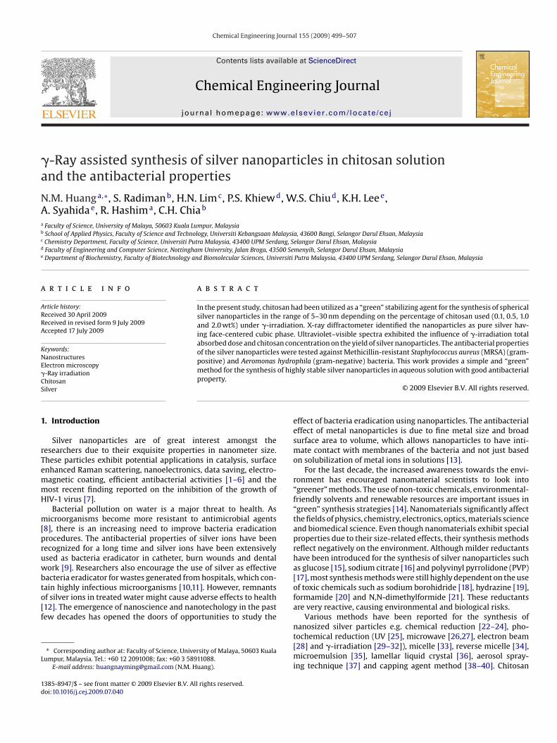
A typical XRD pattern of the silver nanoparticles obtained in thiswork using 1.0 wt% chitosan and 40 kGy of �-irradiation absorbeddosage is shown in Fig. 1a. The XRD pattern shows four peaks at38.2◦, 44.3◦, 64.5◦, and 77.3◦, which are assigned to the (1 1 1),(2 0 0), (2 2 0) and (3 1 1) planes of face-centered cubic (fcc) silver(JCPDS File No. 04-0783). The absence of silver oxide peaks indicatesthat the as-prepared nanoparticles are pure silver. The calculated
Fig. 1. XRD patterns for (a) silver nanoparticles synthesized in 1.0 wt% low molecularweight chitosan solution under �-irradiation (40 kGy), (b) low molecular weightchitosan and (c) standard silver (JCPDS 04-0783).
N.M. Huang et al. / Chemical Engineering Journal 155 (2009) 499–507 501
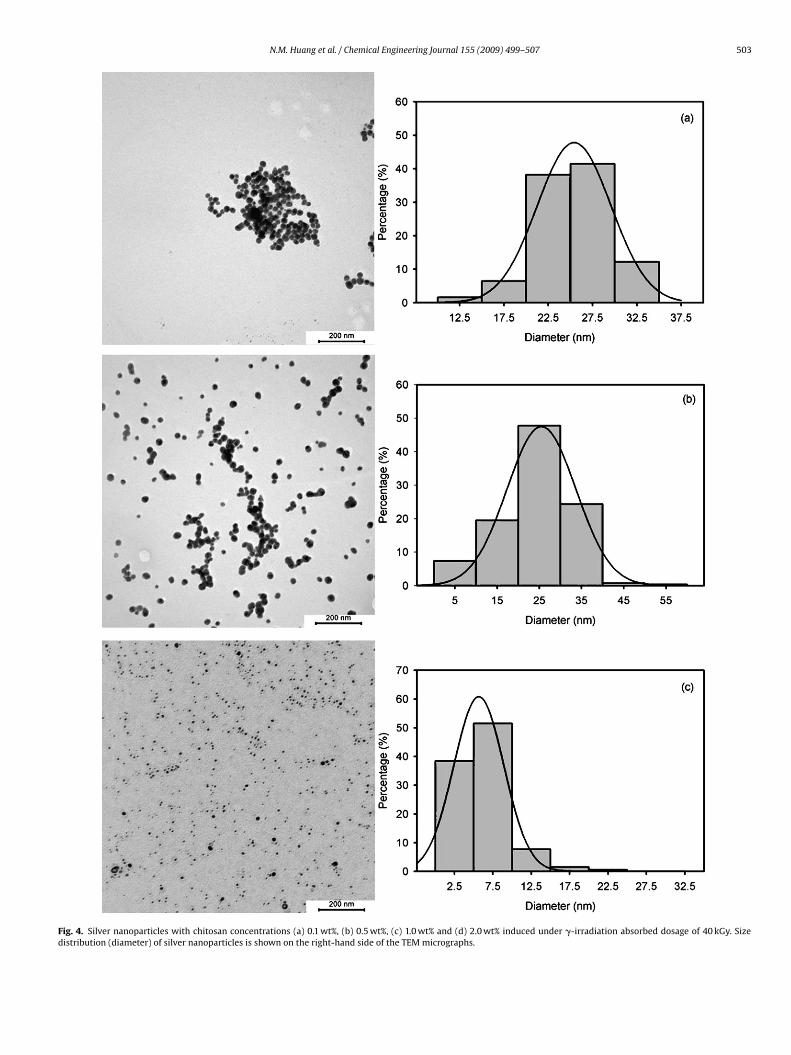
Fig. 2. Silver nanoparticles with chitosan concentrations of (a) 0.1 wt%, (b) 0.5 wt%, (c) 1.0 wt% and (d) 2.0 wt% induced under �-irradiation absorbed dosage of 16 kGy. Sizedistribution (diameter) of silver nanoparticles is shown on the right-hand side of the TEM micrographs.
502 N.M. Huang et al. / Chemical Engineering Journal 155 (2009) 499–507
(Conti
ttedeo
as
Fig. 2.
osan. When comparing this peak with the pure chitosan in Fig. 1b,he pure chitosan has a narrower XRD diffraction peak. The broad-ning of the chitosan peak in the silver nanoparticles is due to theefragmentation of chitosan polymer under �-irradiation. Thus, it isvident that the silver nanoparticles are encapsulated by fragments
f chitosan.Silver nanoparticles prepared by �-irradiation with a totalbsorbed dose of 16 kGy using different concentrations of chitosanolutions are shown in Fig. 2. From the TEM micrographs, it was
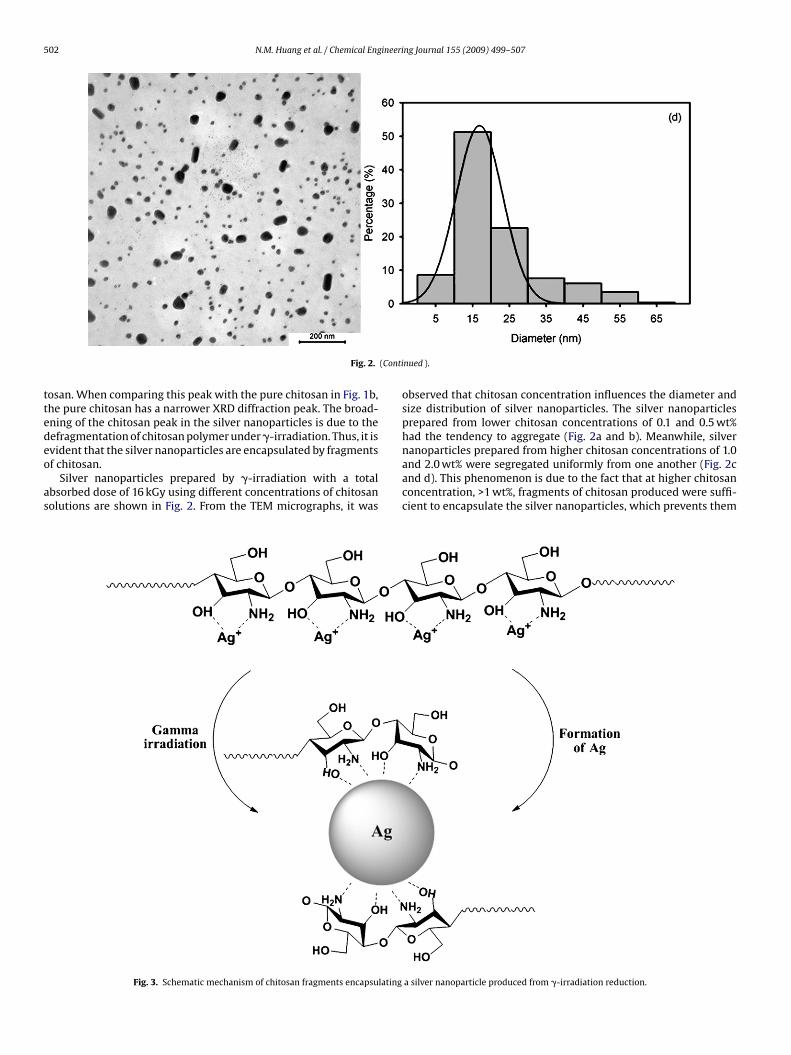
Fig. 3. Schematic mechanism of chitosan fragments encapsulating
nued ).
observed that chitosan concentration influences the diameter andsize distribution of silver nanoparticles. The silver nanoparticlesprepared from lower chitosan concentrations of 0.1 and 0.5 wt%had the tendency to aggregate (Fig. 2a and b). Meanwhile, silvernanoparticles prepared from higher chitosan concentrations of 1.0
and 2.0 wt% were segregated uniformly from one another (Fig. 2cand d). This phenomenon is due to the fact that at higher chitosanconcentration, >1 wt%, fragments of chitosan produced were suffi-cient to encapsulate the silver nanoparticles, which prevents thema silver nanoparticle produced from �-irradiation reduction.
N.M. Huang et al. / Chemical Engineering Journal 155 (2009) 499–507 503
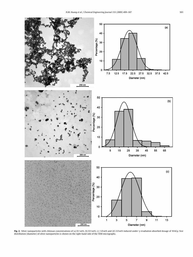
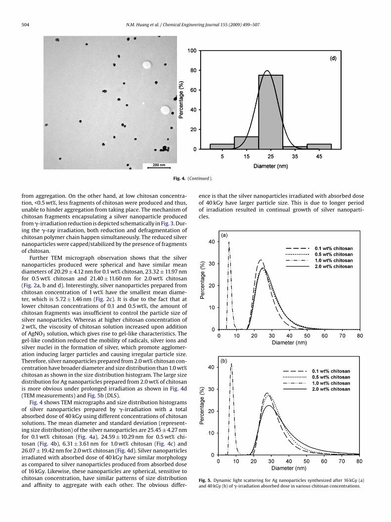
Fig. 4. Silver nanoparticles with chitosan concentrations (a) 0.1 wt%, (b) 0.5 wt%, (c) 1.0 wt% and (d) 2.0 wt% induced under �-irradiation absorbed dosage of 40 kGy. Sizedistribution (diameter) of silver nanoparticles is shown on the right-hand side of the TEM micrographs.
504 N.M. Huang et al. / Chemical Engineering Journal 155 (2009) 499–507
(Conti
ftucficno
ndf(ctlcs2ogsaTccdi(
oasift2iaoca
ence is that the silver nanoparticles irradiated with absorbed doseof 40 kGy have larger particle size. This is due to longer periodof irradiation resulted in continual growth of silver nanoparti-cles.
Fig. 4.
rom aggregation. On the other hand, at low chitosan concentra-ion, <0.5 wt%, less fragments of chitosan were produced and thus,nable to hinder aggregation from taking place. The mechanism ofhitosan fragments encapsulating a silver nanoparticle producedrom �-irradiation reduction is depicted schematically in Fig. 3. Dur-ng the �-ray irradiation, both reduction and defragmentation ofhitosan polymer chain happen simultaneously. The reduced silveranoparticles were capped/stabilized by the presence of fragmentsf chitosan.
Further TEM micrograph observation shows that the silveranoparticles produced were spherical and have similar meaniameters of 20.29 ± 4.12 nm for 0.1 wt% chitosan, 23.32 ± 11.97 nm
or 0.5 wt% chitosan and 21.40 ± 11.60 nm for 2.0 wt% chitosanFig. 2a, b and d). Interestingly, silver nanoparticles prepared fromhitosan concentration of 1 wt% have the smallest mean diame-er, which is 5.72 ± 1.46 nm (Fig. 2c). It is due to the fact that atower chitosan concentrations of 0.1 and 0.5 wt%, the amount ofhitosan fragments was insufficient to control the particle size ofilver nanoparticles. Whereas at higher chitosan concentration ofwt%, the viscosity of chitosan solution increased upon additionf AgNO3 solution, which gives rise to gel-like characteristics. Theel-like condition reduced the mobility of radicals, silver ions andilver nuclei in the formation of silver, which promote agglomer-tion inducing larger particles and causing irregular particle size.herefore, silver nanoparticles prepared from 2.0 wt% chitosan con-entration have broader diameter and size distribution than 1.0 wt%hitosan as shown in the size distribution histogram. The large sizeistribution for Ag nanoparticles prepared from 2.0 wt% of chitosan
s more obvious under prolonged irradiation as shown in Fig. 4dTEM measurements) and Fig. 5b (DLS).
Fig. 4 shows TEM micrographs and size distribution histogramsf silver nanoparticles prepared by �-irradiation with a totalbsorbed dose of 40 kGy using different concentrations of chitosanolutions. The mean diameter and standard deviation (represent-ng size distribution) of the silver nanoparticles are 25.45 ± 4.27 nmor 0.1 wt% chitosan (Fig. 4a), 24.59 ± 10.29 nm for 0.5 wt% chi-osan (Fig. 4b), 6.31 ± 3.61 nm for 1.0 wt% chitosan (Fig. 4c) and6.07 ± 19.42 nm for 2.0 wt% chitosan (Fig. 4d). Silver nanoparticles
rradiated with absorbed dose of 40 kGy have similar morphologys compared to silver nanoparticles produced from absorbed dosef 16 kGy. Likewise, these nanoparticles are spherical, sensitive tohitosan concentration, have similar patterns of size distributionnd affinity to aggregate with each other. The obvious differ-
nued ).
Fig. 5. Dynamic light scattering for Ag nanoparticles synthesized after 16 kGy (a)and 40 kGy (b) of �-irradiation absorbed dose in various chitosan concentrations.
ineering Journal 155 (2009) 499–507 505
fiasn4oa(n2rcbni
tfepnsthm
Fc
Table 1The mean absorbance and yield of the silver nanoparticles synthesized in 20 ml ofchitosan solution under 40 kGy of �-irradiation.
N.M. Huang et al. / Chemical Eng
The size of the Ag nanoparticles produced was further con-rmed using dynamic light scattering as shown in Fig. 5. The resultsre in accord with the TEM observation with Ag nanoparticlesynthesized in 1.0 wt% chitosan having the smallest diameter andarrowest size distribution for both 16 kGy (6.02 ± 0.85 nm) and0 kGy (7.55 ± 1.11 nm) of absorbed dosage. For 0.1, 0.5 and 2.0 wt%f chitosan under 16 kGy of irradiation absorbed dosage, the aver-ge diameter were 25.08 ± 4.82, 26.88 ± 6.35 and 26.91 ± 5.94 nmFig. 5a). Prolonging the irradiation to 40 kGy resulted in Aganoparticles with diameter of 29.67 ± 7.55, 28.6 ± 7.62 and9.71 ± 11.21 nm for 0.1, 0.5 and 2.0 wt% of chitosan concentrations,espectively (Fig. 5b). The DLS measured size is slightly bigger asompared to the particle size measured from the TEM micrographsecause dynamic light scattering method measures the hydrody-amic radius which takes the chitosan coating on the surface of Ag
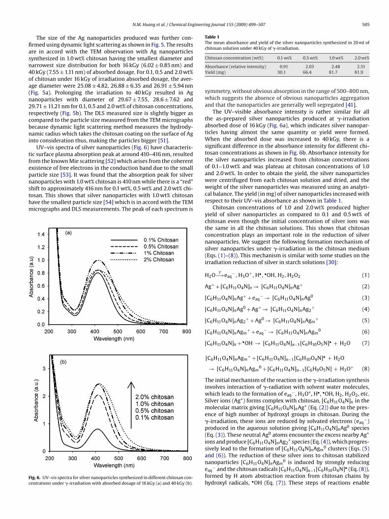
nto consideration thus, making the particles bigger [51].UV–vis spectra of silver nanoparticles (Fig. 6) have characteris-
ic surface plasma absorption peak at around 410–416 nm, resultedrom the known Mie scattering [52] which arises from the coherentxistence of free electrons in the conduction band due to the smallarticle size [53]. It was found that the absorption peak for silveranoparticles with 1.0 wt% chitosan is 410 nm while there is a “red”
hift to approximately 416 nm for 0.1 wt%, 0.5 wt% and 2.0 wt% chi-osan. This shows that silver nanoparticles with 1.0 wt% chitosanave the smallest particle size [54] which is in accord with the TEMicrographs and DLS measurements. The peak of each spectrum isig. 6. UV–vis spectra for silver nanoparticles synthesized in different chitosan con-entrations under �-irradiation with absorbed dosage of 16 kGy (a) and 40 kGy (b).
Chitosan concentration (wt%) 0.1 wt% 0.5 wt% 1.0 wt% 2.0 wt%
Absorbance (relative intensity) 0.91 2.03 2.48 2.51Yield (mg) 30.1 66.4 81.7 81.9
symmetry, without obvious absorption in the range of 500–800 nm,which suggests the absence of obvious nanoparticles aggregationand that the nanoparticles are generally well segregated [41].
The UV–visible absorbance intensity is rather similar for allthe as-prepared silver nanoparticles produced at �-irradiationabsorbed dose of 16 kGy (Fig. 6a), which indicates silver nanopar-ticles having almost the same quantity or yield were formed.When the absorbed dose was increased to 40 kGy, there is asignificant difference in the absorbance intensity for different chi-tosan concentrations as shown in Fig. 6b. Absorbance intensity forthe silver nanoparticles increased from chitosan concentrationsof 0.1–1.0 wt% and was plateau at chitosan concentrations of 1.0and 2.0 wt%. In order to obtain the yield, the silver nanoparticleswere centrifuged from each chitosan solution and dried, and theweight of the silver nanoparticles was measured using an analyti-cal balance. The yield (in mg) of silver nanoparticles increased withrespect to their UV–vis absorbance as shown in Table 1.
Chitosan concentrations of 1.0 and 2.0 wt% produced higheryield of silver nanoparticles as compared to 0.1 and 0.5 wt% ofchitosan even though the initial concentration of silver ions wasthe same in all the chitosan solutions. This shows that chitosanconcentration plays an important role in the reduction of silvernanoparticles. We suggest the following formation mechanism ofsilver nanoparticles under �-irradiation in the chitosan medium(Eqs. (1)–(8)). This mechanism is similar with some studies on theirradiation reduction of silver in starch solutions [30]:
H2O�−→eaq
−, H3O+, H•, •OH, H2, H2O2 (1)
Ag+ + [C6H11O4N]n → [C6H11O4N]nAg+ (2)
[C6H11O4N]nAg+ + eaq− → [C6H11O4N]nAg0 (3)
[C6H11O4N]nAg0 + Ag+ → [C6H11O4N]nAg2+ (4)
[C6H11O4N]nAg2+ + Ag0 → [C6H11O4N]nAgm
+ (5)
[C6H11O4N]nAgm+ + eaq
− → [C6H11O4N]nAgm0 (6)
[C6H11O4N]n + •OH → [C6H11O4N]n−1[C6H10O5N]• + H2O (7)
[C6H11O4N]nAgm+ + [C6H11O4N]n−1[C6H10O4N]• + H2O
→ [C6H11O4N]nAgm0 + [C6H11O4N]n−1[C6H9O5N] + H3O+ (8)
The initial mechanism of the reaction in the �-irradiation synthesisinvolves interaction of �-radiation with solvent water molecules,which leads to the formation of eaq
−, H3O+, H•, •OH, H2, H2O2, etc.Silver ions (Ag+) forms complex with chitosan, [C6H11O4N]n in themolecular matrix giving [C6H11O4N]nAg+ (Eq. (2)) due to the pres-ence of high number of hydroxyl groups in chitosan. During the�-irradiation, these ions are reduced by solvated electrons (eaq
−)produced in the aqueous solution giving [C6H11O4N]nAg0 species(Eq. (3)). These neutral Ag0 atoms encounter the excess nearby Ag+
ions and produce [C6H11O4N]nAg2+ species (Eq. (4)), which progres-
sively lead to the formation of [C6H11O4N]nAgm0 clusters (Eqs. (5)
and (6)). The reduction of these silver ions to chitosan stabilizednanoparticles [C6H11O4N]nAgm
0 is induced by strongly reducingeaq
− and the chitosan radicals [C6H11O4N]n−1[C6H10O4N]• (Eq. (8)),formed by H atom abstraction reaction from chitosan chains byhydroxyl radicals, •OH (Eq. (7)). These steps of reactions enable
506 N.M. Huang et al. / Chemical Engineering Journal 155 (2009) 499–507
Fnt
cthif
sewatDvnat
hotigdovsAteiieakctTpnAg<e
ig. 7. UV–vis spectra for (a) as-synthesized silver nanoparticles and (b) silveranoparticles after 6 months of storage in laboratory condition (chitosan concen-ration of 1 wt% and �-irradiation absorbed dosage of 40 kGy).
hitosan to reduce Ag+ ions, coat and stabilize silver nanopar-icles while inhibiting their excessive aggregations. Thus, underigher chitosan concentration, the yield of the silver nanoparticles
s higher due to the relatively higher amount of chitosan radicals’ormed.
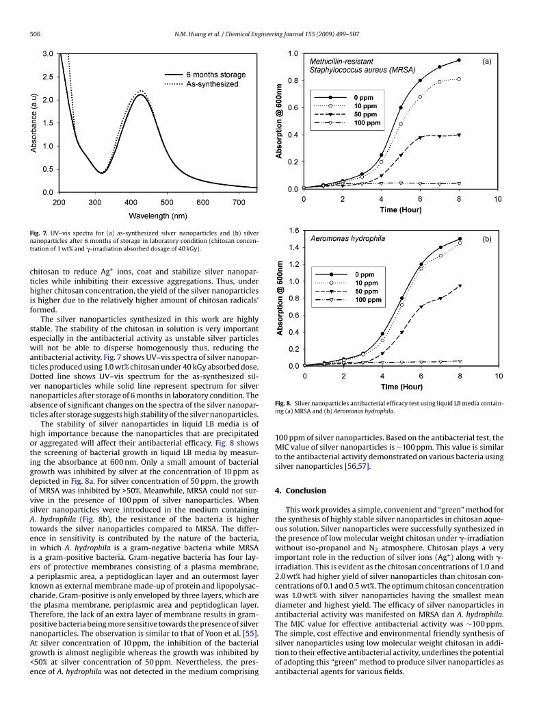
The silver nanoparticles synthesized in this work are highlytable. The stability of the chitosan in solution is very importantspecially in the antibacterial activity as unstable silver particlesill not be able to disperse homogenously thus, reducing the
ntibacterial activity. Fig. 7 shows UV–vis spectra of silver nanopar-icles produced using 1.0 wt% chitosan under 40 kGy absorbed dose.otted line shows UV–vis spectrum for the as-synthesized sil-er nanoparticles while solid line represent spectrum for silveranoparticles after storage of 6 months in laboratory condition. Thebsence of significant changes on the spectra of the silver nanopar-icles after storage suggests high stability of the silver nanoparticles.
The stability of silver nanoparticles in liquid LB media is ofigh importance because the nanoparticles that are precipitatedr aggregated will affect their antibacterial efficacy. Fig. 8 showshe screening of bacterial growth in liquid LB media by measur-ng the absorbance at 600 nm. Only a small amount of bacterialrowth was inhibited by silver at the concentration of 10 ppm asepicted in Fig. 8a. For silver concentration of 50 ppm, the growthf MRSA was inhibited by >50%. Meanwhile, MRSA could not sur-ive in the presence of 100 ppm of silver nanoparticles. Whenilver nanoparticles were introduced in the medium containing. hydrophila (Fig. 8b), the resistance of the bacteria is higher
owards the silver nanoparticles compared to MRSA. The differ-nce in sensitivity is contributed by the nature of the bacteria,n which A. hydrophila is a gram-negative bacteria while MRSAs a gram-positive bacteria. Gram-negative bacteria has four lay-rs of protective membranes consisting of a plasma membrane,periplasmic area, a peptidoglican layer and an outermost layer
nown as external membrane made-up of protein and lipopolysac-haride. Gram-positive is only enveloped by three layers, which arehe plasma membrane, periplasmic area and peptidoglican layer.herefore, the lack of an extra layer of membrane results in gram-ositive bacteria being more sensitive towards the presence of silver
anoparticles. The observation is similar to that of Yoon et al. [55].t silver concentration of 10 ppm, the inhibition of the bacterialrowth is almost negligible whereas the growth was inhibited by50% at silver concentration of 50 ppm. Nevertheless, the pres-nce of A. hydrophila was not detected in the medium comprisingFig. 8. Silver nanoparticles antibacterial efficacy test using liquid LB media contain-ing (a) MRSA and (b) Aeromonas hydrophila.
100 ppm of silver nanoparticles. Based on the antibacterial test, theMIC value of silver nanoparticles is ∼100 ppm. This value is similarto the antibacterial activity demonstrated on various bacteria usingsilver nanoparticles [56,57].
4. Conclusion
This work provides a simple, convenient and “green” method forthe synthesis of highly stable silver nanoparticles in chitosan aque-ous solution. Silver nanoparticles were successfully synthesized inthe presence of low molecular weight chitosan under �-irradiationwithout iso-propanol and N2 atmosphere. Chitosan plays a veryimportant role in the reduction of silver ions (Ag+) along with �-irradiation. This is evident as the chitosan concentrations of 1.0 and2.0 wt% had higher yield of silver nanoparticles than chitosan con-centrations of 0.1 and 0.5 wt%. The optimum chitosan concentrationwas 1.0 wt% with silver nanoparticles having the smallest meandiameter and highest yield. The efficacy of silver nanoparticles inantibacterial activity was manifested on MRSA dan A. hydrophila.The MIC value for effective antibacterial activity was ∼100 ppm.
The simple, cost effective and environmental friendly synthesis ofsilver nanoparticles using low molecular weight chitosan in addi-tion to their effective antibacterial activity, underlines the potentialof adopting this “green” method to produce silver nanoparticles asantibacterial agents for various fields.
ineeri
A
tl
R
[[[[
[[[[
[
[[[[
[
[[
[[[
[[[
[[[
[[[[[
[[[[
[
[
[[[[
[
[[
N.M. Huang et al. / Chemical Eng
cknowledgements
This work is supported by Universiti Kebangsaan Malaysiahrough grant no: UKM-OUP-NBT-27-138/2008. The author wouldike to thank MOSTI for the NSF scholarship.
eferences
[1] X.Q. Zou, E.B. Ying, S.J. Dong, J. Colloids Interf. Sci. 306 (2007) 307.[2] L.C. Courrol, F.R. Silva, L. Gomes, Colloids Surf. A 305 (2007) 54.[3] Y.M. Mohan, K. Lee, T. Premkumar, K.E. Geckeler, Polymer 48 (2007) 158.[4] X.L. Tian, W.H. Wang, G.Y. Cao, Mater. Lett. 61 (2007) 130.[5] M.H. Ullah, K. Il, C.S. Ha, Mater. Lett. 60 (2006) 1496.[6] A. Kameo, T. Yoshimura, K. Esumi, Colloids Surf. A 215 (2003) 181.[7] J.L. Elechiguerra, J.L. Burt, J.R. Morones, A. Camacho-Bragado, X.X. Gao, H.H. Lara,
M.J. Yacaman, J. Nanobiotech. 3 (2005) 6.[8] M. Kolar, K. Urbanek, T. Latal, Int. J. Antimicrob. Agents 17 (2001) 357.[9] J.S. Kim, E. Kuk, K. Yu, J. Kim, S. Park, H. Lee, S. Kim, Y. Park, Y. Park, C. Hwang,
Nanomed. Nanotech. Bio. Med. 3 (2007) 95.10] Y.E. Lin, R.D. Vidic, J.E. Stout, C.A. McCartney, V.L. Yu, Water Res. 32 (1998) 1997.11] Y.E. Lin, R.D.S. Vidic, V.L. Yu, Water Res. 30 (1996) 1905.12] D.S. Blanc, P. Carrara, G. Zanetti, P. Francioli, J. Hosp. Infect. 60 (2005) 69.13] J.R. Morones, J.L. Elechiguerra, A. Camacho, K. Holt, J.B. Kouri, J.T. Ramirez, M.J.
Yacaman, Nanotechnology 16 (2005) 2346–2353.14] V.K. Sharma, R.A. Yngard, Y. Lin, Adv. Colloids Interf. Sci. 145 (2009) 83.15] P. Raveendran, J. Fu, S.L. Wallen, J. Am. Chem. Soc. 125 (2003) 13940.16] P.C. Lee, D. Meisel, J. Phys. Chem. 86 (1982) 3391.17] H.S. Wang, X.L. Qiao, J.G. Chen, X.J. Wang, S.Y. Ding, Mater. Chem. Phys. 94 (2005)
449.18] P.V. Adhyapak, P. Karandikar, K. Vijayamohanan, A.A. Athawale, A.J. Chandwad-
kar, Mater. Lett. 58 (2004) 1168.19] W. Zhang, X. Qiao, J. Chen, H. Wang, J. Colloids Interf. Sci. 302 (2006) 370.20] C.R.K. Rao, D.C. Trivedi, Synth. Met. 155 (2005) 324.21] I. Pastoriza-Santos, L.M. Liz-Marzan, Langmuir 15 (1999) 948.
22] P.K. Khanna, N. Singh, S. Charan, V.V.V.S. Subbarao, R. Gokhale, U.P. Mulik, Mater.Chem. Phys. 93 (2005) 117.23] B.H. Ryu, Y. Choi, H.S. Park, J.H. Byun, K. Kong, J.O. Lee, H. Chang, Colloids Surf.
A 270–271 (2005) 345.24] H.S. Wang, X.L. Qiao, J.G. Chen, S.Y. Ding, Colloids Surf. A 256 (2005) 111–115.25] S.K. Ghosh, S. Kunda, T. Pal, Bull. Mater. Sci. 25 (2002) 581.
[[[[
[
ng Journal 155 (2009) 499–507 507
26] H. Yin, T. Yamamoto, Y. Wada, S. Yanagida, Mater. Chem. Phys. 83 (2004) 66–70.27] J. Chen, J. Wang, X. Zhang, Y.L. Jin, Mater. Chem. Phys. 108 (2008) 421.28] Y. Li, Y.N. Kim, E.J. Lee, W.P. Cai, S.O. Cho, Nucl. Inst. Meth. Phys. Res. B 251 (2006)
425.29] P. Chen, L. Song, Y. Liu, Y. Fang, Radiat. Phys. Chem. 76 (2007) 1165.30] M. Kumar, L. Varshney, S. Francis, Radiat. Phys. Chem. 73 (2005) 21.31] W.T. Wu, Y. Wang, L. Shi, Q. Zhu, W. Pang, G. Xu, F. Lu, Nanotechnology 16 (2005)
3017.32] M.K. Temgire, S.S. Joshi, Radiat. Phys. Chem. 71 (2004) 1039.33] C.J. Murphy, N.R. Jana, Adv. Mater. 14 (2002) 80.34] M.C. McLeod, R.S. McHenry, E.J. Beckman, C.B. Roberts, J. Phys. Chem. B 107
(2003) 2693.35] W.Z. Zhang, X.L. Qiao, J.G. Chen, Chem. Phys. 300 (2006) 495.36] L.M. Qi, Y.Y. Gao, J.M. Ma, Colloids Surf. A 157 (1999) 285–294.37] L.P. Jiang, A.W. Wang, Inorg. Chem. Commun. 7 (2004) 506.38] S. Chen, D.L. Carroll, Nano Lett. 2 (2002) 1003.39] A.S. Reddy, C.Y. Chen, S.C. Baker, C.C. Chen, J.S. Jean, C.W. Fan, H.R. Chen, J.C.
Wang, Mater. Lett. 63 (2009) 1227–1230.40] S.D. Bunge, T.J. Boyle, T.J. Headley, Nano Lett. 3 (2003) 901.41] H.Z. Huang, X.R. Yang, Carbohydr. Res. 339 (2004) 2627.42] Y.K. Twu, Y.W. Chen, C.M. Shih, Powder Technol. 185 (2008) 251.43] N.M. Huang, S. Radiman, H.N. Lim, S.K. Yeong, P.S. Khiew, W.S. Chiu, G.H.
Mohamed Saeed, K. Nadarajah, Chem. Eng. J. 147 (2009) 399–404.44] B. Feng, R.Y. Hong, Y.J. Wu, G.H. Liu, L.H. Zhong, Y. Zheng, J.M. Ding, D.G. Wei, J.
Alloys Compd. 473 (2009) 356–362.45] H.Y. Ru Jiang, L. Xiao, Y.H. Chang, Y.J. Guan, X.D. Li, G.M. Zeng, J. Hazard. Mater.,
in press, doi:10.1016/j.jhazmat.2009.04.037.46] T.H. Li, H.G. Park, S.H. Choi, Mater. Chem. Phys. 105 (2007) 325.47] D. Long, G. Wu, S. Chen, Radiat. Phys. Chem. 76 (2007) 1126.48] D.M. Cheng, X.D. Zhou, H.B. Xia, H.S.O. Chan, Chem. Mater. 17 (2005) 3578.49] P. Barnickel, A. Wokaun, W. Sager, H.F. Eicke, J. Colloids Interf. Sci. 148 (1992)
80.50] S. Shrivastava, T. Bera, A. Roy, G. Singh, P. Ramachandrarao, D. Dash, Nanotech-
nology 18 (2007) 225103.51] L. D’Souza, A. Suchopar, R.M. Richards, J. Colloids Interf. Sci. 279 (2004) 458.52] T. Hasell, J. Yang, W. Wang, P.D. Brown, S.M. Howdle, Mater. Lett. 61 (2007) 4906.
53] C. Burda, X. Chen, R. Narayanan, M.A. El-Sayed, Chem. Rev. 105 (2005) 1025.54] Y. Xia, N.J. Halas, Mater. Res. Soc. Bull. 30 (2005) 338–343.55] K. Yoon, J.H. Byeon, J. Park, J. Hwang, Sci. Total Environ. 373 (2007) 572.56] J.P. Ruparelia, A.K. Chatterjee, S.P. Duttagupta, S. Mukherji, Acta Biomater. 4(2008) 707.57] I. Sondi, B. Salopek-Sondi, J. Colloids Interf. Sci. 275 (2004) 177.