harnessing the therapeutic potential of th17 cells
TRANSCRIPT

Review ArticleHarnessing the Therapeutic Potential of Th17 Cells
Jonas Bystrom,1 Taher E. Taher,1 M. Sherwan Muhyaddin,2 Felix I. Clanchy,3
Pamela Mangat,4 Ali S. Jawad,5 Richard O. Williams,3 and Rizgar A. Mageed1
1Bone and Joint Research Unit, William Harvey Research Institute, Queen Mary University of London,London EC1M 6BQ, UK2College of Pharmacy, Hawler Medical University, Erbil, Iraq3Kennedy Institute of Rheumatology, University of Oxford, Oxford OX3 7FY, UK4Department of Rheumatology, Royal Free Hospital, London NW3 2GQ, UK5Department of Rheumatology, The Royal London Hospital, London E1 4DG, UK
Correspondence should be addressed to Jonas Bystrom; [email protected]
Received 6 March 2015; Accepted 5 May 2015
Academic Editor: Jianfei Yang
Copyright © 2015 Jonas Bystrom et al. This is an open access article distributed under the Creative Commons Attribution License,which permits unrestricted use, distribution, and reproduction in any medium, provided the original work is properly cited.
Th17 cells provide protective immunity to infections by fungi and extracellular bacteria as well as cancer but are also involvedin chronic inflammation. The cells were first identified by their ability to produce interleukin 17A (IL-17A) and, subsequently,associated with chronic inflammation and autoimmunity. Th17 cells have some gene profile similarity with stem cells and canremain dormant in mucosal tissues for long periods. Indeed, recent studies suggest that functionally distinct subsets of pro-and anti-inflammatory Th17 cells can interchange phenotype and functions. For development, Th17 cells require activation of thetranscription factors STAT3 and ROR𝛾t while RUNX1, c-Maf, and Aiolos are involved in changes of phenotype/functions. Attemptsto harnessTh17 cells against pathogens and cancer using vaccination strategies are being explored.The cells gain protective abilitieswhen induced to produce interferon 𝛾 (IFN𝛾). In addition, treatment with antibodies to IL-17 is effective in treating patients withpsoriasis, psoriatic arthritis, and refectory rheumatoid arthritis. Moreover, since ROR𝛾t is a nuclear receptor, it is likely to be apotential future drug target for modulatingTh17 functions.This review explores pathways through whichTh17 subsets are induced,the molecular basis of their plasticity, and potential therapeutic strategies for their modulation in diseases.
1. Introduction
Different subsets of helper T cells (Th) have been identifiedbased, primarily, on the pattern of cytokines they produce.The Th1 subset is induced to differentiate in response tointracellular pathogens and viruses to produce IFN𝛾 andTNF𝛼 and initiate cellular immunity. The Th2 subset, incontrast, produces interleukin 4 (IL-4), IL-5, and IL-13 andmediates immunity to helminths and parasites as well asinitiating humoral immunity. Regulatory T cells, which werediscovered subsequently, dampen inflammatory responsesagainst foreign and self-antigens through cell-cell interac-tions and produce IL-10 and TGF𝛽 [1]. The most recentaddition to effector Th subsets is Th17 cells that were iden-tified in 2006 based on their ability to produce IL-17A [2].Although the latest to be discovered, subsequent evolutionary
studies have established that the Th17 subset is the mostancient one. Hence, immune cells equipped with a nascentT cell receptor (TCR) from the primitive fish lamprey, whoselineage diverged from that of humans 500 million years ago,produce IL-17 but none of the cytokines associated withthe other T cell lineages [3]. In mammals, at homeostasiscommensal bacteria in the gut induce IL-1𝛽 production tomaintain a basal level of Th17 cells in the lamina propria [4].However, in response to pathogenic extracellular bacterialand fungal infections at mucocutaneous surfaces in theintestine, the respiratory tract, and the skin, large numbers ofnaiveTh cells differentiate toTh17 cells under the influence ofIL-1𝛽, IL-6, IL-23, and/or TGF𝛽 [5]. In addition to producingIL-17A,Th17 cells can produce IL-17F, IL-21, IL-22, IFN𝛾, andGM-CSF [6, 7]. IL-17A, referred to as IL-17 in this review,has pleiotropic properties after binding IL-17 receptors on
Hindawi Publishing CorporationMediators of InflammationVolume 2015, Article ID 205156, 11 pageshttp://dx.doi.org/10.1155/2015/205156

2 Mediators of Inflammation
haematopoietic and nonhaematopoietic cells such as epithe-lial and endothelial cells [8].The binding of IL-17 to its recep-tors triggers intracellular signalling that induces the pro-duction of proinflammatory cytokines such as IL-6, C-X-Cchemokines such as chemokine 8 (CXCL8), CXCL9, CXCL10,and CXCL11, and beta-defensin-2 [9–11]. During acute infec-tions, Th17 cells recruit neutrophils and, thereby, mediateinitial protection frompathogens [8]. Furthermore, IL-21 andIL-22 produced byTh17 cells protect mucosal membranes byinducing the production of antimicrobial proteins, RegIII𝛽,and RegIII𝛾 and by stimulating B cells [6, 7]. IL-17 is, byitself, a weak activator of other immune cells and studies haveshown that the presence of other cytokines, such as TNF𝛼 orIL-1𝛽, is required formaximumeffects of the cytokine [12, 13].
After their activation, effector and memory Th17 cellscan remain dormant in the mucosa for extended periodsof time [14]. A number of recent studies have revealed thatTh17 cells show a great degree of functional and phenotypicplasticity. Thus, there is evidence that Th17 cells can changeto Th1-like cells or acquire the ability to produce IL-10which can be beneficial during certain types of infections.For example, Candida albicans induces IFN𝛾 production byTh17 cells while Staphylococcus aureus induces IL-10 [15].With regard to phenotype, all Th17 cells express CCR6and most also express CD161 [16]. Th17 cells that onlyproduce IL-17 express CCR4 while IFN𝛾-producing Th17cells express CXCR3 [17]. In addition to providing protectiveimmunity and driving chronic inflammation, Th17 cells havebeen suggested to play a dual role in tumour development.Thus, Th17 cells have been implicated in promoting tumourthrough producing angiogenic factors but, paradoxically, alsoshown to counteract tumour development by producing IL-17 and IFN𝛾 [18]. Treatment of patients with the epidermalskin disorder psoriasis with antibodies to IL-17 or withits soluble recombinant receptor leads to remission [19,20]. Furthermore, patients with rheumatoid arthritis (RA),psoriatic arthritis, and ankylosing spondylitis have beenreported to benefit from treatment with biologic inhibitorsof IL-17 [21–23]. However, treatment of patients with Crohn’sdisease with inhibitors of IL-17 worsens disease, perhaps,highlighting some protective functions for Th17 cells inthe gastrointestinal tract [24]. Interestingly, there is someevidence to indicate that the ability of Th17 cells to promotepathology in autoimmune diseases is acquired when the cellsgain the ability to produce IFN𝛾. In animal models of disease,these cells were shown to express receptors for either IL-23 orIL-1𝛽 [25, 26].
This reviewwill explore potential strategies to harness theuse ofTh17 cells for therapeutic purposes. First, wewill reviewavailable evidence on the signals that promote the develop-ment ofTh17 cells and mechanisms that underpin changes totheir phenotype. These involve TCR- and cytokine-mediatedsignals, transcription factors, and epigenetic modifications.Second, studies aimed at employingTh17 cells for vaccinationagainst various organisms and for protection from cancerswill be reviewed. We will also discuss advantages and pitfallsof reported experimental strategies and contemplate whetherit would be beneficial to alter the phenotype of Th17 cells inhuman diseases.
2. Th17 Cell Development, TranscriptionalRegulation, and Functional Plasticity
The available evidence indicates that Th17 cell progenitors,identified byCD161 expression, are present atmucocutaneoussites and in peripheral and cord blood [16]. These cells areinduced to differentiate into effector Th17 cells by cytokinesthat activate a highly regulated transcriptional networkinvolving at least five transcription factors and through epige-netic modifications. Cytokines IL-1𝛽, IL-6, TGF𝛽, and IL-23and the lipid mediator PGE
2have all been variably reported
to be involved in Th17 cell differentiation [27]. An initialmedium level of T cell receptor (TCR) engagement activatesthe nuclear factor kappa B (NF𝜅B) which, in turn, activatesthe interferon regulatory factor 4 transcription factor (IRF4).In contrast, high level TCR engagement preferentially pro-motes Th1 cell differentiation. The two transcription factorstogetherwith another transcription factor called basic leucinezipper transcription factor ATF-like (BATF), binds multiplesites throughout the chromatin [28–30]. IL-1𝛽 increasesthe expression of IRF4 [31] while IL-6 and IL-23 inducethe phosphorylation of signal transducer and activator oftranscription 3 (STAT3). This leads to the disassociationof STAT3 from the receptor-bound Janus kinase 2 (JAK2).Phosphorylated STAT3 then transmigrates to the nucleus andpopulates many DNase sensitive chromosomal sites, madeaccessible by TGF𝛽, and stabilizes some of the BATF/IRF4interactions [28]. IL-23 also induces the expression of afourth transcription factor, runt-related transcription factor1 (RUNX1) [32]. RUNX1/3 promotes Th17 differentiation byenhancing expression of the transcription factor ROR𝛾t andincreasing its stability at the Il17 locus [33]. The transcriptionfactor ROR𝛾t is a signature transcription factor forTh17 cellsas it binds a number of specific DNA loci critical for thedifferentiation of the cells [28].
Numerous studies have highlighted the plasticity of Th17cells [32, 34–36]. Although key Th17-specific loci (e.g., Il17a,Il17f, and Rorc) are known to be accessible in all Th17cells, they have also been reported to be easily repressed.TGF𝛽 has been shown to stabilize the open state of theseloci but in its absence both IL-23 and IL-12 suppress IL-17production while instead enhancing IFN𝛾 production in aSTAT4- and T-box transcription factor- (T-bet-) dependentmanner [34]. Furthermore, the Ifng locus was shown tobe semiactivated in Th17 cells and to rapidly acquire anadditional permissive state in response to IL-12 [35]. IL-12 induces T-bet expression and repressive histone marksin the Rorc locus [35]. T-bet then interacts with RUNX1to disrupt RUNX1/ROR𝛾t interaction and activity [36]. Inthe presence of IL-12, RUNX1 was also reported to bind tothe Ifng promoter [32]. T-bet and RUNX1/3 activation isrequired for maximal IFN𝛾 production in “ex-Th17” cells. Atlow RUNX1 levels, however, and in the presence of Th17-promoting cytokines, the Th17 cell phenotype is retained.Depending on the level of RUNX1 activation and whichcytokines are present, the formation of the RUNX1/T-betcomplex in Th17 cells leads to the development of IFN𝛾+IL-17+ T cells independent of ROR𝛾t expression [32]. IFN𝛾+Th17 cells have been shown to have the ATP-dependent
Mediators of Inflammation 3
Th17
Th1/Th17
IL-17
rTh17/Th2
IL-6 IL-23
IL-17 IL-10
Ectonucleotidases
GM-CSF
MDR1
RUNX1/3T-bet
IL-22
Th22
c-MafAiolosFoxP3
IL-17 IL-4
GATA3AhR
Th2/Th17
IL-17
IL-12/IL-23ICOS
IL-6
TGF𝛽/IL-23
Anti-TNF𝛼
IL-4
INF𝛾
IL-1𝛽
ROR𝛾t
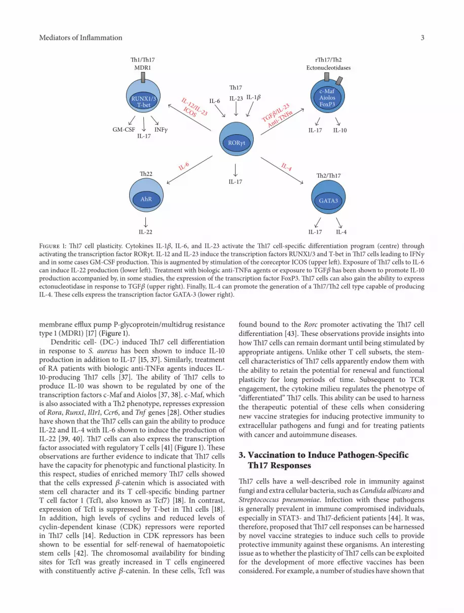
Figure 1: Th17 cell plasticity. Cytokines IL-1𝛽, IL-6, and IL-23 activate the Th17 cell-specific differentiation program (centre) throughactivating the transcription factor ROR𝛾t. IL-12 and IL-23 induce the transcription factors RUNX1/3 and T-bet inTh17 cells leading to IFN𝛾and in some cases GM-CSF production. This is augmented by stimulation of the coreceptor ICOS (upper left). Exposure ofTh17 cells to IL-6can induce IL-22 production (lower left). Treatment with biologic anti-TNF𝛼 agents or exposure to TGF𝛽 has been shown to promote IL-10production accompanied by, in some studies, the expression of the transcription factor FoxP3. Th17 cells can also gain the ability to expressectonucleotidase in response to TGF𝛽 (upper right). Finally, IL-4 can promote the generation of a Th17/Th2 cell type capable of producingIL-4. These cells express the transcription factor GATA-3 (lower right).
membrane efflux pump P-glycoprotein/multidrug resistancetype 1 (MDR1) [17] (Figure 1).
Dendritic cell- (DC-) induced Th17 cell differentiationin response to S. aureus has been shown to induce IL-10production in addition to IL-17 [15, 37]. Similarly, treatmentof RA patients with biologic anti-TNF𝛼 agents induces IL-10-producing Th17 cells [37]. The ability of Th17 cells toproduce IL-10 was shown to be regulated by one of thetranscription factors c-Maf and Aiolos [37, 38]. c-Maf, whichis also associated with aTh2 phenotype, represses expressionof Rora, Runx1, Il1r1, Ccr6, and Tnf genes [28]. Other studieshave shown that theTh17 cells can gain the ability to produceIL-22 and IL-4 with IL-6 shown to induce the production ofIL-22 [39, 40]. Th17 cells can also express the transcriptionfactor associated with regulatory T cells [41] (Figure 1).Theseobservations are further evidence to indicate that Th17 cellshave the capacity for phenotypic and functional plasticity. Inthis respect, studies of enriched memory Th17 cells showedthat the cells expressed 𝛽-catenin which is associated withstem cell character and its T cell-specific binding partnerT cell factor 1 (Tcf1, also known as Tcf7) [18]. In contrast,expression of Tcf1 is suppressed by T-bet in Th1 cells [18].In addition, high levels of cyclins and reduced levels ofcyclin-dependent kinase (CDK) repressors were reportedin Th17 cells [14]. Reduction in CDK repressors has beenshown to be essential for self-renewal of haematopoieticstem cells [42]. The chromosomal availability for bindingsites for Tcf1 was greatly increased in T cells engineeredwith constituently active 𝛽-catenin. In these cells, Tcf1 was
found bound to the Rorc promoter activating the Th17 celldifferentiation [43]. These observations provide insights intohowTh17 cells can remain dormant until being stimulated byappropriate antigens. Unlike other T cell subsets, the stem-cell characteristics of Th17 cells apparently endow them withthe ability to retain the potential for renewal and functionalplasticity for long periods of time. Subsequent to TCRengagement, the cytokine milieu regulates the phenotype of“differentiated” Th17 cells. This ability can be used to harnessthe therapeutic potential of these cells when consideringnew vaccine strategies for inducing protective immunity toextracellular pathogens and fungi and for treating patientswith cancer and autoimmune diseases.
3. Vaccination to Induce Pathogen-SpecificTh17 Responses
Th17 cells have a well-described role in immunity againstfungi and extra cellular bacteria, such asCandida albicans andStreptococcus pneumoniae. Infection with these pathogensis generally prevalent in immune compromised individuals,especially in STAT3- andTh17-deficient patients [44]. It was,therefore, proposed thatTh17 cell responses can be harnessedby novel vaccine strategies to induce such cells to provideprotective immunity against these organisms. An interestingissue as to whether the plasticity ofTh17 cells can be exploitedfor the development of more effective vaccines has beenconsidered. For example, a number of studies have shown that

4 Mediators of Inflammation
the advantage of a vaccine that relies on inducing Th17 cell-dependent responses would be that the protective immunity,unlike the B cell-mediated immunity, will be independentof pathogen serotype [45]. A further possible advantage ofTh17-inducing vaccines would be that infants and immunecompromised individuals that do not develop a good anti-body response will benefit from long-lived memory Th17cells [18, 45, 46]. To enhance Th17 responses by vaccination,the use of various adjuvants has been assessed. The bacterialcomponents, muramyl dipeptide (MDP), lipopolysaccharide(LPS), and CpG, augmentedTh17 responses [45, 47, 48].
Strategies to develop vaccines that specifically induceTh17 cells in immune compromised individuals have alsobeen actively considered. These efforts were based on keyobservations regarding the role of Th17 immunity at sitesmost susceptible to infections in immune compromisedindividuals. Thus, Th17-mediated immunity to C. albicans isimportant for infections of the upper respiratory tract andthe skin [44, 49]. During such infections, fungal antigensactivate Dectin-1 and toll-like receptor 2 (TLR2) on dendriticcells and this leads to the production of IL-23 and IL-1𝛽[50]. Th17 cells generated in response to C. albicans, in turn,induce the production of IFN𝛾 and, thus, further augmentcellular immunity [15]. A vaccine consisting of a recombinantvirulence factor used by Candida which has a similar shapeto a virulence factor in S. aureus (N-terminus of Als3p)with aluminium hydroxide as adjuvant induced protectiveimmunity dominated by Th17 cells that produced both IL-17and IFN𝛾 and recruited neutrophils [51].
In addition to fungal infections in which Th17 cell-mediated immunity plays a critical protective role, thesecells are also important in immunity to the gram-positivebacterium, Streptococcus pneumoniae. This bacterium causeslife-threatening infections of the respiratory tract in immunecompromised individuals and, in addition, leads to systemicinfection and septic arthritis [52]. There are over 90 differentserotypes of S. pneumonia and antibiotic resistance caneasily develop [53]. In children, natural protection against S.pneumoniae is dependent on Th17 cells and this immunitydevelops before antibody-mediated immunity [46]. Duringthe course of an infection, activated monocytes first recruitTh17 cells which, in turn, recruit neutrophils to kill thebacteria [54–56]. Furthermore, intranasal vaccination withcommon cell wall polysaccharides, which bind MHCII,has been shown to induce a Th17-dependent, antibody-independent protective immunity [57, 58].
The gram-negative Pseudomonas aeruginosa is anotherpathogen known to induce a Th17 response. This pathogencan cause similar infections as C. albicans and S. pneumonia,that is, sepsis and respiratory and gastrointestinal tractinfections, especially in immune compromised individuals[59]. IL-17 production was noted in response to intranasalvaccination with live attenuated P. aeruginosa, or a libraryof P. aeruginosa proteins. The resulting immunity which wasmostly dependent on rapid neutrophil recruitment conferredprotection to several strains of the bacterium [59, 60].
The role of Th17-mediated protective immunity to therespiratory tract is further highlighted by studies showing
immunity to conserved outer membrane proteins from sev-eral serotypes of the gram-negative bacterium, Klebsiellapneumonia [45]. Vaccination with this bacterium with LPSused as adjuvant induced an MHCII-dependent Th17 cell-mediated immunity [45].The resulting immune responsewasserotype-independent and specific to conserved outer mem-brane proteins. The response was also antibody-independentand lasted for at least four weeks. In contrast, serotype-specific immunity to polysaccharide capsular antigens fromK. pneumonia induced a transient B cell response [45].
Protection from Mycobacterium tuberculosis is alsoknown to involve Th17-mediated immunity. Thus, a studynoted that vaccination with M. tuberculosis induced pro-tection associated with an IL-17-mediated response [11]. Inthis study, IL-23 was shown to be essential for the acceler-ated immune response to prevent bacterial growth and theinduction of Th17 cells in the lung. The recall Th17 responseoccurred concurrently with the expression of chemokinesCXCL9, CXCL10, and CXCL11 that, in turn, recruited otherCD4+ cells that produced IFN𝛾 in the lung [11].
Another study reported that immunization with thegram-negative Bordetella pertussis, which causes whoopingcough, induced immunity that was associatedwith the induc-tion of Th17 cells. Thus, intraperitoneal immunization with awhole B. pertussis vaccine twice, four weeks apart followed bychallenge with aerosol inoculation two weeks later, induceda toll-like receptor 4- (TLR4-) dependent production of IL-23 from DCs [61]. This augmented Th1 and Th17 responsesand led to protective cellular immunity that involved bacterialkilling by activated macrophages.
Although often associated with a Th1 immune response,immunity to the respiratory syncytial virus (RSV) has alsobeen shown to induce aTh17 response in the respiratory tract.A protectiveTh17-dependent response developed in a mousemodel concomitant with allergic asthma and also in infectedinfants [62, 63]. Interestingly, a Pertussis vaccine inducedprotection from RSV infections in neonatal mice when givenintranasally [64]. This vaccination led to modification ofthe immune response by enhancing mucosal resistance toRSV infection during adulthood. In this setting, IL-17 wasproduced by Th17 and NK cells and led to the recruitmentof neutrophils. In addition, IL-17+IFN𝛾+ T cells were shownto contribute significantly to the protection [64].
For certain bacteria, fungi, and at least one virus, vac-cination that initiates Th17 responses can, therefore, confereffective immune protection. Such vaccines often induceimmunity at mucosal surfaces that are dependent eitheron a switch from IL-17-producing T cells to IL-17- andIFN𝛾-producing cells, or a Th17-dependent recruitment ofIFN𝛾 producing Th1 cells (Figure 2(a)). In most situationsduring recall responses, key cells that are eventually recruitedappear to be neutrophils, which facilitate the eradicationof pathogens [51, 55, 59, 64]. Issues to consider duringdevelopment of a vaccine inducingTh17 protective immunityare the role of adjuvants and whether specific augmentationof theTh17 cell phenotype can increase a favourable immuneresponse.

Mediators of Inflammation 5
IL-17IL-23
IL-8
CXCL9
CXCL10
CXCL11
Th1
PMN
Th17
DC
PMN
IL-1𝛽INF𝛾
INF𝛾
(a)
IL-17IL-17
VEGFCXCL9
CXCL10
Th1
Th17 Th17
IL-17RA INF𝛾TGF𝛽
(b)
IL-6MMPs
RANKL
IL-17
IL-17
IL-17
IL-17
Th17 Blood brainbarrier
Th17
SC
S100A8IL-8
?
Blood vessels
RANKL
INF𝛾
INF𝛾
OCOB
(c)
Figure 2: The heterogeneity of Th17 cell phenotype and functions. (a) In immunity to pathogens, (b) in tumour pathology, and (c) inpromotion of autoimmune diseases. (a) Dendritic cells (DCs) are stimulated by pathogens to present antigens to induce the differentiationof naıve T-cells to Th17 cells. DCs produce IL-23 and IL-1𝛽 that facilitate the differentiation to and expansion of activated Th17 cells.Pathogen-specific Th17 cells produce IL-17 and, in some cases, IFN𝛾. IL-17 production induces IL-8 production by endothelial cells leadingto neutrophil recruitment and pathogen eradication. Th17 cells can also recruit effector T cells for pathogen eradication. (b) Tumourmicroenvironments influence the phenotype ofTh17 cells. TGF𝛽 inducesTh17 cells to produce vascular endothelia growth factor (VEGF) thatinduces angiogenesis. However, whilst IL-17 can promote tumour development, IFN𝛾 produced byTh17 cells suppresses tumour developmentthrough recruitment of other immune cells. (c) IL-17 can promote chronic inflammation and autoimmune diseases. For example, Th17 cellsinfiltrate the blood brain barrier in patients with MS. In addition, IL-17 induces inflammation in dermal cells in patients with psoriasis.Furthermore, IL-17 can induce angiogenesis and the production of other cytokines-, proteases-, and the receptor activator of nuclear factorkappa-B ligand (RANK-L) from synoviocytes (SC) and osteoblasts (OB) in the synovium of RA patients activating osteoclasts (OC).
4. Harnessing Th17-MediatedProtection from Cancer
Theability to augment the protective potential ofTh17 cells byvaccination has actively been considered for treating cancer.Several studies have identified Th17 cells in tumour masses[65–67].However, the role ofTh17 cells in immunity to canceris somewhat controversial. As Th17 cells are characterised byfunctional/phenotypic plasticity, they are likely, therefore, to
be differentially influenced by the complex nature of tumourmicroenvironments (Figure 2(b)). Gastrointestinal tumourdevelopment is, for example, driven by an inflammatoryenvironment due to chronic disease and disrupted barriersor by the bacterial flora [68–70]. Th17 cells present inthese malignancies have been shown to promote tumourdevelopment. One study observed that Th17 cells defined byexpression of the transcription factor BATF and IL-23 recep-tor weremore prevalent in the lamina propria of patients with

6 Mediators of Inflammation
colitis-associated colon cancer than in healthy individuals[69]. Studies in animal models revealed that Th17 cells wereinduced to proliferate by IL-23 from adjacent antigen pre-senting cells (APCs) [69]. Perhaps consistent with the stemcell-like genotype of Th17 cells, 𝛽-catenin produced by Th17cells in the colon gradually increases comparing patients withulcerative colitis and colon cancer [43]. In an animal model,colitis-induced cancer was strongly linked to stabilization of𝛽-catenin in T cells. 𝛽-catenin’s binding partner Tcf1 wasshown bound to the promoter of ROR𝛾t, thus, leading toincreased Th17 cell proliferation and cancer development[43]. The proposition that microorganisms influence theinflammatory tumour microenvironment through stimulat-ing Th17 cells is supported by the outcome of three studies.One study revealed that, early during the development ofcolorectal cancer, impaired mucus production leads to theloss of barrier functions [68].This resulted inmicrobial prod-ucts accessing tumour sites and leading to the production ofIL-23 by myeloid cells which, in turn, increased size of thetumour through inducing Th17 cells [68]. The second studyobserved that the bacterium enterotoxigenic Bacteroidesfragilis in the colon caused inflammation and proliferation ofcolonic epithelia leading to the recruitment ofTh17 cells. Theresulting combination of hyperproliferation of the epithelia,Th17 recruitment, and expansion led to chronic inflammationand colon carcinogenesis [70]. The third study observedthat released enteropathogenic bacteria-secreted particlesstimulated the intestinal epithelium to produce exosome-likenanoparticles that promoted colon cancer.The nanoparticles,intestinal mucosa-derived exosome-like nanoparticles, con-tained sphingosine-1-phosphate, CCL20, and PGE
2. CCL20
recruited T cells from circulation while PGE2facilitatedTh17
cell differentiation leading to the development of colon cancer[27]. Although IL-17 can mediate tumours through causingor enhancing chronic inflammation, the cytokine can alsoact directly on tumour cells. One study reported that theIL-17 receptor A (IL-17RA) was expressed on transformedepithelial cells and these developed into colorectal tumours.These epithelial cells where the main site for protumorigenicactivity by IL-17 (Figure 2(b)) [71]. Also the inflammatoryenvironment at other sites than the gastrointestinal tract caninduce tumour promoting Th17 cells. Two studies showedthat the inflammatory environments in the skin induced bytumour cells or by tumour-derived fibroblasts led to suchTh17 recruitment and IL-17-dependent tumour development[72, 73].
Some studies have revealed that Th17 cells can promotecancer if they are the only immune cells found in thetumour but have protective functions in the presence ofother immune cells [74]. CD39 and CD73 ectonucleotidase-expressing Th17 cells, induced by IL-6 and TGF-𝛽, werereported to be immunosuppressive in several mouse tumours[75]. The ectonucleotidases-degraded ATP led to adenosinerelease and, subsequently, suppression of helper CD4+ andcytotoxic CD8+ T cell effector functions. TGF𝛽 which iscommonly found in tumour microenvironments has, there-fore, been suggested to augment the immune suppressiveand tumour-promoting functions of Th17 cells [75, 76].Th17 cells have also been reported to promote cancer by
virtue of their ability to induce angiogenesis [65]. Zhangand colleagues noted that increased numbers of intratumoralIL-17-producing cells correlated with microvessel density inthe tumours and with poor survival of patients with hepa-tocellular carcinoma [67]. Another study reported that highIL-17 levels in patients with colorectal carcinoma correlatedwith bad prognosis. In these tumours, Th17 cells facilitateangiogenesis through their ability to induce the production ofvascular endothelial growth factor (VEGF) from cancer cells[77]. Indeed, Wang and colleagues reported on the depen-dence of angiogenesis and experimental melanoma tumourgrowth on IL-17 [78]. Interestingly, however, angiogenesispromoted by IL-17 was reduced considerably when IFN𝛾+cells were present in the tumour. This observation could betaken as support for the notion that the presence of otherimmune cells within tumours promotes a protective role byTh17 cells [78]. The notion that coexpression of IFN𝛾 andIL-17 is favourable for tumour immunity was supported byanother study of ovarian cancers [66]. In this study, theimportance ofTh17 cells was suggested by the low number oftumour-infiltratingTh17 cells and the low level of IL-17 in theascites in patients with more advanced disease. Through thesynergistic action between IL-17 and IFN𝛾,Th17 cells inducedthe production of CXCL9 and CXCL10 to recruit effector Tcells [66].Muranski and colleagues also showed that adoptivetransfer of Th17 cells led to the killing of established tumoursmore efficiently than the transfer of other effector T cellsubsets [18].The authors noted that IL-17+IFN𝛾+ T cells weremore efficient in cancer eradication than IFN𝛾+ T cells. Thiswas believed to be because the cells were not terminallydifferentiated and less prone to apoptosis than terminally dif-ferentiated IFN𝛾+Th1 cells [18].The potential involvement ofIFN𝛾+Th17 cells in providing protective immunity to cancerwas highlighted in two further studies. In an animal modelinvolving transplanted solid tumour, bacterial DNA (CpG-)stimulated plasmacytoid DCs (pDCs) presented tumourantigens to Th17 cells. The ability to shrink tumours wasdependent on this antigen presentation to Th17 cells and thecells also gaining the ability to produce IFN𝛾 [48]. Immunecells including cytotoxic CD8+ T cells were subsequentlyrecruited by Th17 cells, mediating protective antitumourimmunity [48].The inducible costimulatory receptor (ICOS)is expressed on Th17 cells and other immune cells. Interest-ingly, stimulation of ICOS onTh17 cells in vitro induced IFN𝛾production (Figure 1). In contrast, anti-CD28 costimulationfavoured an IL-17 phenotype only [79] Furthermore, IFN𝛾+Th17 cells developed through costimulationwith ICOS ligandwere superior to cells costimulated with anti-CD28 in regres-sion of human tumour engrafted into mice [79].
In conclusion, it is evident that Th17 cells can haveparadoxical roles in cancer (Figure 2). It is not currentlyestablished if it would be possible to pharmacologicallymodulateTh17 cells for suppressing cancer. Inhibition ofTh17cells by anti-IL17 therapy or through inhibition of ROR𝛾tcould be favourable in situations whereTh17 have angiogenicactivity [80–83]. Although anti-IL17 therapy has been shownto worsen Crohn’s disease, a study using an animal modelof colonic tumour revealed that the therapy can be effica-cious as a neoadjuvant in combination with chemotherapy

Mediators of Inflammation 7
when gastric inflammation is localized to the tumour area[71]. In settings where the tumour microenvironment altersTh17 function to one that augments tumour development,therapeutic strategies could be considered to harness thecells to a protective phenotype such as inducing the cells tocoproduce IFN𝛾. This suggestion is based on observationscited above that protective immunity to tumours by Th17-like cells is more likely to be achieved if the tumour containsT cells that produce IFN𝛾 [74]. Specific stimulation of thecoreceptor ICOS was reported to be capable of skewingTh17 cells to become IFN𝛾/IL-17-producing cells which havebeen revealed to be beneficial for tumour suppression [18,79]. Furthermore, vaccination with tumour antigens usingpeptidoglycan or CpG as adjuvants for the activation of DCsor pDCs, respectively, has also been shown to augment afavourableTh17 response [47, 48]. Based on the observations,novel pharmacological agents and adjuvants that can alter thephenotype of Th17 cells should be tested in animal models ofcancer. ICOS engagement has, however, also been associatedwith promotion of autoimmune diseases as its stimulationincreases the number of effector Th17 cells [84]. Each agentdeveloped for the purpose of inducing cancer-suppressingTh17 cells should, therefore, be tested both for antitumourproperties and the ability to promote autoimmunity.
5. Therapeutic Considerations Involving Th17Cells in Autoimmune Diseases
In addition to their critical role in providing effective immu-nity to pathogens, especially on mucocutaneous membranesand possible involvement in tumour eradication, Th17 cellshave been implicated in autoimmune diseases [19–23, 85]. Forexample, there is good evidence for multiple roles for Th17cells and for IL-17 in plaque growth in patients with psoriasis,synovial inflammation, increased angiogenesis and bonedegradation in patient with RA and ankylosing spondylitis.The cells have also been suggested to have a role in disruptionof the blood brain barrier in multiple sclerosis (Figure 2(c))[19, 20, 85]. The beneficial therapeutic effect of the anti-IL-17 antibody ixekizumab and the anti-IL-17 receptor anti-body brodalumab in psoriasis, psoriatic arthritis, ankylosingspondylitis, and refractory RA is further evidence for theinvolvement of Th17 cells in these disorders [19–23]. Novelstrategies are under development for selective targeting ofTh17 cells through inhibiting the nuclear receptor ROR𝛾t[80–83].
As cited above, the available evidence suggests that IFN𝛾-producing Th17 cells are beneficial when considering thedevelopment of vaccines for infectious diseases and in somecancers [18, 48, 51, 79]. However, several studies have impli-cated IFN𝛾+ Th17 cells in the pathogenicity of autoimmunediseases. For example, experimental colitis in mice is drivenby IL-23-induced IFN𝛾+IL17+ T cells that express the IL-23 receptor [86]. Importantly, the same cells were found inthe lamina propria of the intestine in patients with Crohn’sdisease [86, 87]. In juvenile arthritis, Th17 cells change toa Th1 phenotype while migrating from the circulation toinflamed joints. This conversion can be facilitated in vitro by
incubating Th17 cells with TGF𝛽 and high IL-12 levels [88].Highly enriched Th17 cells from diabetic BDC2.5 transgenicmice adoptively transferred diabetes to NOD/SCID recipi-ents, conferred pathology when the cells were modified toa Th1 phenotype [89]. Furthermore, circulating Th17 cellsfrom relapsing MS patients show an increased propensityto change to IFN𝛾-producing Th17 cells. These cells werealso found in brain tissues of patients with multiple scle-rosis (MS) [90]. In the animal model of MS, experimentalallergic encephalomyelitis (EAE), IFN𝛾-producingTh17 cellspreferentially crossed the blood brain barrier to accumulatein the central nervous system. The IL-17+IFN𝛾+ T cellsdisplayed better migratory potential than IFN𝛾-producingTh1 cells or non-IFN𝛾-producingTh17 cells [90]. However, anumber of other studies disagree with the notion that IFN𝛾-producingTh17 cells are pathogenic in autoimmune diseases.For example, in mice with EAE neither IL-17 nor IFN𝛾deficiency inhibited disease development and symptoms [91,92]. Instead, it was noted that Th17 cells conferred pathologywhen the cells gained the ability to produceGM-CSF [93, 94].Furthermore, two recent studies revealed that, in EAE and inpatients with MS, GM-CSF is produced by a separate T cellsubset than byTh17 cells [95, 96]. In addition, overexpressionof T-bet in T cells during collagen induced arthritis (CIA)wasfound to reduce bone erosion [97]. Studies in our laboratorieshave shown that patients with RA who do not respond totreatmentwith biologic anti-TNF𝛼 agents produce high levelsof IL-17 [98]. Although IL-17 induces bone resorption inpatients with RA, IFN𝛾, perhaps paradoxically, is associatedwith less bone erosion indicating that the development of IL-17+IFN𝛾+ phenotype could associate with a better outcomeat least for bone loss in the disease [99]. Taken together, theseobservations suggest that althoughTh17 cells have pathogenicroles in autoimmune diseases there is also evidence to suggestthat not all Th17 cell subsets confer pathology. Furthermore,as cited above, the presence of IFN𝛾-producing Th17 cellscould have beneficial protective roles in infections and cancer[18, 51].There is also evidence that someTh17 cells in patientswith RA treated with biologic anti-TNF𝛼 agents gained afunctionally distinct phenotype and produced IL-10 [37].Clearly, therefore, there is more to discover regarding howTh17 cells and how their plasticity contribute to protectiveimmunity and to inflammatory diseases.
Although augmenting Th17-mediated immunity by vac-cination holds promise for infectious diseases and, possibly,for cancer, one caveat is that the approach will not be withoutrisks of promoting autoimmune inflammatory diseases. Itis, therefore, of great importance to determine whetherdifferent antigens have different propensities to induce Th17-mediated autoimmunity and whether some individuals aremore predisposed genetically to such responses [45, 100].ICOS engagement might be one such example as activationof pathway associated with the coreceptor has been shownto induce Th17 cells capable of ameliorating cancer but alsoTh1 and Th17 cells with ability to induce arthritis [79, 84].Whether or not induced Th17 cells could mediate or triggerautoimmune diseases in susceptible individuals should beevaluated in experimental models of diseases such in CIAand in EAE. So far, however, no study has reported on the
8 Mediators of Inflammation
development of autoimmune symptoms through inducingTh17-mediated responses to infectious pathogens. In humans,a polymorphism in the DECTIN1 gene, leading to reducedability to stimulate IL-17 production, was not associated withsusceptibility to or severity of RA [101]. It is also possiblethat theTh17 subset(s) that is protective in infectious diseasescould be distinct from Th17 cells that promote autoimmunedisease pathology as has been suggested [25]. Indeed, there issome evidence to show that patients with RA have Th17 cellsthat are unable to eradicate C. albicans suggesting that therole of these cells in pathology may not directly be associatedwith their ability to produce IL-17 [102]. In this respect, IL-17 is known to confer protection of inflamed gastrointestinaltracts from fungal infections suggesting that future treatmentoptions could benefit from avoiding simple blockade of thecytokine [24]. If manipulation of Th17 is safe, it would be ofinterest to knowwhether augmenting the transcription factorRUNX1 for IL-17+IFN𝛾+ T cell phenotype induction will bebeneficial in treating patients with cancer and autoimmunediseases.
6. Concluding Remarks
There is a steady increase in knowledge about Th17 cellsand their role in different clinical settings since they werefirst discovered in 2006. As these cells are involved both inimmunity against pathogens and in promoting autoimmunediseases and perhaps some cancers, the development oftherapeutic strategies to harness Th17-mediated responsesis already an area of huge interest. Augmentation of Th17cell-dependent but antibody-independent vaccination forcertain pathogens is under active development. Such anapproach would have the benefit of triggering protectiveimmunity that is serotype-independent and suitable forchildren, immune compromised individuals, and individualswith impaired B cell-mediated immunity. However, the factthat at least some Th17 subsets could play pathogenic rolesin some autoimmune diseases and, possibly, in some cancers,targeted inhibition or modulation of the function of thesecells could also be immensely beneficial. Strategies thatrely on developing novel specific molecules to modulateTh17 cells are already underway. Perhaps what complicatesthe development of such strategies is the plasticity of thephenotype and functions of Th17 cells which are, clearly,noted to be more than other T cell subsets. This plasticity islikely to hamper the development of strategies to modulateTh17 cells in cancer as the tumour microenvironment andpresence of other immune cells could influence the outcomeof such treatments. Specific augmentation, for example, byinducing IFN𝛾 production by Th17 cells, has been shown tobe beneficial inmany infectious diseases and in some cancers.Although IFN𝛾-producing Th17 cells have been suggestedto be associated with autoimmune diseases in some studies,there is currently limited evidence that such cells can actuallylead to autoimmune disease development. Future treatmentoptions for vaccination for infectious diseases, cancers, andautoimmune diseases might involve augmentation of distinctTh17 cell subsets for beneficial outcomes. Th17 cells arearguably the T cell subset that has coevolved with us for
the longest time. Increased knowledge of these cells in healthand in disease could be of immense future benefit.
Conflict of Interests
The authors declare no conflict of interests.
Acknowledgment
The authors would like to thank Dr. Helene Rosenberg(NIAID, NIH) for taking the time to critically review thispaper.
References
[1] F. Broere, S. G. Apasov, M. V. Sitkovsky, and W. van Eden,“T cell subsets and T cell mediated immunity,” in Principles ofImmunopharmacology, F. P. Nijkamp and M. J. Parnham, Eds.,Birkhauser, Basel, Switzerland, 2011.
[2] C. Sutton, C. Brereton, B. Keogh, K. H. G. Mills, and E. C.Lavelle, “A crucial role for interleukin (IL)-1 in the induc-tion of IL-17-producing T cells that mediate autoimmuneencephalomyelitis,” The Journal of Experimental Medicine, vol.203, no. 7, pp. 1685–1691, 2006.
[3] P. Guo, M. Hirano, B. R. Herrin et al., “Dual nature of theadaptive immune system in lampreys,” Nature, vol. 459, no.7248, pp. 796–801, 2009.
[4] M.H. Shaw,N.Kamada, Y.-G.Kim, andG.Nunez, “Microbiota-induced IL-1𝛽, but not IL-6, is critical for the development ofsteady-state T
𝐻17 cells in the intestine,” Journal of Experimental
Medicine, vol. 209, no. 2, pp. 251–258, 2012.[5] W. Ouyang, J. K. Kolls, and Y. Zheng, “The biological functions
of T helper 17 cell effector cytokines in inflammation,” Immu-nity, vol. 28, no. 4, pp. 454–467, 2008.
[6] Y. Zheng, P. A. Valdez, D. M. Danilenko et al., “Interleukin-22 mediates early host defense against attaching and effacingbacterial pathogens,” Nature Medicine, vol. 14, no. 3, pp. 282–289, 2008.
[7] M. Mitsdoerffer, Y. Lee, A. Jager et al., “Proinflammatory Thelper type 17 cells are effective B-cell helpers,” Proceedings ofthe National Academy of Sciences of the United States of America,vol. 107, no. 32, pp. 14292–14297, 2010.
[8] S. L. Gaffen, “Structure and signalling in the IL-17 receptorfamily,” Nature Reviews Immunology, vol. 9, no. 8, pp. 556–567,2009.
[9] C. Y. Kao, Y. Chen, P. Thai et al., “IL-17 markedly up-regulates𝛽-defensin-2 expression in human airway epithelium via JAKand NF-kappaB signaling pathways,” Journal of Immunology,vol. 173, no. 5, pp. 3482–3491, 2004.
[10] C. A. Hitchon, P. Alex, L. B. Erdile et al., “A distinct multicy-tokine profile is associated with anti-cyclical citrullinated pep-tide antibodies in patients with early untreated inflammatoryarthritis,”The Journal of Rheumatology, vol. 31, no. 12, pp. 2336–2346, 2004.
[11] S. A. Khader, G. K. Bell, J. E. Pearl et al., “IL-23 and IL-17 in theestablishment of protective pulmonary CD4+ T cell responsesafter vaccination and during Mycobacterium tuberculosis chal-lenge,” Nature Immunology, vol. 8, no. 4, pp. 369–377, 2007.
[12] T. Yago, Y. Nanke, N. Ichikawa et al., “IL-17 induces osteo-clastogenesis from human monocytes alone in the absence

Mediators of Inflammation 9
of osteoblasts, which is potently inhibited by anti-TNF-alphaantibody: a novel mechanism of osteoclastogenesis by IL-17,”Journal of Cellular Biochemistry, vol. 108, no. 4, pp. 947–955,2009.
[13] H. Fujie, K. Niu, M. Ohba et al., “A distinct regulatory role ofTh17 cytokines IL-17A and IL-17F in chemokine secretion fromlung microvascular endothelial cells,” Inflammation, vol. 35, no.3, pp. 1119–1131, 2012.
[14] I. Kryczek, E. Zhao, Y. Liu et al., “Human TH17 cells are long-lived effectormemory cells,” Science TranslationalMedicine, vol.3, no. 104, Article ID 104ra100, 2011.
[15] C. E. Zielinski, F. Mele, D. Aschenbrenner et al., “Pathogen-induced human TH17 cells produce IFN-gamma or IL-10 andare regulated by IL-1beta,” Nature, vol. 484, no. 7395, pp. 514–518, 2012.
[16] L. Cosmi, R. De Palma, V. Santarlasci et al., “Human interleukin17-producing cells originate from a CD161+CD4+ T cell precur-sor,” The Journal of Experimental Medicine, vol. 205, no. 8, pp.1903–1916, 2008.
[17] R. Ramesh, L. Kozhaya, K. McKevitt et al., “Pro-inflammatoryhuman Th17 cells selectively express P-glycoprotein and arerefractory to glucocorticoids,” The Journal of ExperimentalMedicine, vol. 211, no. 1, pp. 89–104, 2014.
[18] P.Muranski, Z. A. Borman, S. P. Kerkar et al., “Th17 cells are longlived and retain a stem cell-likemolecular signature,” Immunity,vol. 35, no. 6, pp. 972–985, 2011.
[19] C. Leonardi, R.Matheson, C. Zachariae et al., “Anti-interleukin-17 monoclonal antibody ixekizumab in chronic plaque psoria-sis,” The New England Journal of Medicine, vol. 366, no. 13, pp.1190–1191, 2012.
[20] K. A. Papp, C. Leonardi, A. Menter et al., “Brodalumab, ananti-interleukin-17-receptor antibody for psoriasis,” The NewEngland Journal of Medicine, vol. 366, no. 13, pp. 1181–1189, 2012.
[21] I. B. McInnes, J. Sieper, J. Braun et al., “Efficacy and safety ofsecukinumab, a fully human anti-interleukin-17A monoclonalantibody, in patients with moderate-to-severe psoriatic arthri-tis: a 24-week, randomised, double-blind, placebo-controlled,phase II proof-of-concept trial,” Annals of the Rheumatic Dis-eases, vol. 73, no. 2, pp. 349–356, 2014.
[22] M. C. Genovese, M. Greenwald, C. Cho et al., “A phaseII randomized study of subcutaneous ixekizumab, an anti-interleukin-17 monoclonal antibody, in rheumatoid arthritispatients who were naive to biologic agents or had an inadequateresponse to tumor necrosis factor inhibitors,” Arthritis &Rheumatology, vol. 66, no. 7, pp. 1693–1704, 2014.
[23] D. Baeten, X. Baraliakos, J. Braun et al., “Anti-interleukin-17Amonoclonal antibody secukinumab in treatment of ankylosingspondylitis: a randomised, double-blind, placebo-controlledtrial,”The Lancet, vol. 382, no. 9906, pp. 1705–1713, 2013.
[24] W. Hueber, B. E. Sands, S. Lewitzky et al., “Secukinumab,a human anti-IL-17A monoclonal antibody, for moderate tosevere Crohn’s disease: unexpected results of a randomised,double-blindplacebo-controlled trial,” Gut, vol. 61, no. 12, pp.1693–1700, 2012.
[25] K. Hirota, J. H. Duarte, M. Veldhoen et al., “Fate mappingof IL-17-producing T cells in inflammatory responses,” NatureImmunology, vol. 12, no. 3, pp. 255–263, 2011.
[26] Y. Lee, A. Awasthi, N. Yosef et al., “Induction and molecularsignature of pathogenic TH17 cells,”Nature Immunology, vol. 13,no. 10, pp. 991–999, 2012.
[27] Z. Deng, J. Mu, M. Tseng et al., “Enterobacteria-secreted par-ticles induce production of exosome-like S1P-containing parti-cles by intestinal epithelium to drive Th17-mediated tumorige-nesis,” Nature Communications, vol. 6, article 6956, 2015.
[28] M. Ciofani, A. Madar, C. Galan et al., “A validated regulatorynetwork forTh17 cell specification,” Cell, vol. 151, no. 2, pp. 289–303, 2012.
[29] B. U. Schraml, K. Hildner, W. Ise et al., “The AP-1 transcriptionfactor Batf controls TH17 differentiation,” Nature, vol. 460, no.7253, pp. 405–409, 2009.
[30] H. A. Purvis, J. N. Stoop, J. Mann et al., “Low-strength T-cellactivation promotes Th17 responses,” Blood, vol. 116, no. 23, pp.4829–4837, 2010.
[31] Y. Chung, S. H. Chang, G. J. Martinez et al., “Critical regulationof early Th17 cell differentiation by interleukin-1 signaling,”Immunity, vol. 30, no. 4, pp. 576–587, 2009.
[32] Y.Wang, J. Godec, K. Ben-Aissa et al., “The transcription factorsT-bet and runx are required for the ontogeny of pathogenicinterferon-gamma-producing T helper 17 cells,” Immunity, vol.40, no. 3, pp. 355–366, 2014.
[33] F. Zhang, G. Meng, and W. Strober, “Interactions amongthe transcription factors Runx1, ROR𝛾t and Foxp3 regulatethe differentiation of interleukin 17-producing T cells,” NatureImmunology, vol. 9, no. 11, pp. 1297–1306, 2008.
[34] Y. K. Lee, H. Turner, C. L. Maynard et al., “Late developmentalplasticity in the T helper 17 lineage,” Immunity, vol. 30, no. 1, pp.92–107, 2009.
[35] R. Mukasa, A. Balasubramani, Y. K. Lee et al., “Epigenetic insta-bility of cytokine and transcription factor gene loci underliesplasticity of the T helper 17 cell lineage,” Immunity, vol. 32, no.5, pp. 616–627, 2010.
[36] V. Lazarevic, X. Chen, J.-H. Shim et al., “T-bet represses TH 17differentiation by preventing Runx1-mediated activation of thegene encoding RORgammat,”Nature Immunology, vol. 12, no. 1,pp. 96–104, 2011.
[37] H. G. Evans, U. Roostalu, G. J. Walter et al., “TNF-𝛼 blockadeinduces IL-10 expression in human CD4+ T cells,” NatureCommunications, vol. 5, article 3199, 2014.
[38] J. Xu, Y. Yang, G. Qiu et al., “c-Maf regulates IL-10 expressionduring Th17 polarization,” Journal of Immunology, vol. 182, no.10, pp. 6226–6236, 2009.
[39] S. C. Liang, X.-Y. Tan, D. P. Luxenberg et al., “Interleukin (IL)-22 and IL-17 are coexpressed by Th17 cells and cooperativelyenhance expression of antimicrobial peptides,” The Journal ofExperimental Medicine, vol. 203, no. 10, pp. 2271–2279, 2006.
[40] L. Cosmi, L. Maggi, V. Santarlasci et al., “Identification of anovel subset of human circulating memory CD4+ T cells thatproduce both IL-17A and IL-4,” The Journal of Allergy andClinical Immunology, vol. 125, no. 1–3, pp. 222–230.e4, 2010.
[41] K. S. Voo, Y.-H. Wang, F. R. Santori et al., “Identificationof IL-17-producing FOXP3+ regulatory T cells in humans,”Proceedings of the National Academy of Sciences of the UnitedStates of America, vol. 106, no. 12, pp. 4793–4798, 2009.
[42] A. P. Bracken, D. Kleine-Kohlbrecher, N. Dietrich et al., “ThePolycomb group proteins bind throughout the INK4A-ARFlocus and are disassociated in senescent cells,” Genes andDevelopment, vol. 21, no. 5, pp. 525–530, 2007.
[43] S. Keerthivasan, K. Aghajani, M. Dose et al., “𝛽-cateninpromotes colitis and colon cancer through imprinting ofproinflammatory properties in T cells,” Science TranslationalMedicine, vol. 6, no. 225, Article ID 225ra28, 2014.
10 Mediators of Inflammation
[44] J. D. Milner, J. M. Brenchley, A. Laurence et al., “Impaired T𝐻17
cell differentiation in subjects with autosomal dominant hyper-IgE syndrome,” Nature, vol. 452, no. 7188, pp. 773–776, 2008.
[45] K. Chen, J. P. McAleer, Y. Lin et al., “Th17 cells mediate clade-specific, serotype-independent mucosal immunity,” Immunity,vol. 35, no. 6, pp. 997–1009, 2011.
[46] M. Lipsitch, C.G.Whitney, E. Zell, T. Kaijalainen, R.Dagan, andR. Malley, “Are anticapsular antibodies the primary mechanismof protection against invasive pneumococcal disease?” PLoSMedicine, vol. 2, no. 1, article e15, 2005.
[47] A. J. van Beelen, Z. Zelinkova, E. W. Taanman-Kueter etal., “Stimulation of the intracellular bacterial sensor NOD2programs dendritic cells to promote interleukin-17 productionin human memory T cells,” Immunity, vol. 27, no. 4, pp. 660–669, 2007.
[48] L. Guery, J. Dubrot, C. Lippens et al., “Ag-presenting CpG-activated pDCs primeTh17 cells that induce tumor regression,”Cancer Research, vol. 74, no. 22, pp. 6430–6440, 2014.
[49] S. L. Gaffen, N. Hernandez-Santos, and A. C. Peterson, “IL-17signaling in host defense against Candida albicans,” Immuno-logic Research, vol. 50, no. 2-3, pp. 181–187, 2011.
[50] H. R. Conti and S. L. Gaffen, “Host responses to Candidaalbicans: Th17 cells and mucosal candidiasis,” Microbes andInfection, vol. 12, no. 7, pp. 518–527, 2010.
[51] L. Lin, A. S. Ibrahim, X. Xu et al., “Th1-Th17 cells mediateprotective adaptive immunity against Staphylococcus aureus andCandida albicans infection in mice,” PLoS Pathogens, vol. 5, no.12, Article ID e1000703, 2009.
[52] J. Raad and J. E. Peacock Jr., “Septic arthritis in the adult causedby Streptococcus pneumoniae: a report of 4 cases and review ofthe literature,” Seminars in Arthritis and Rheumatism, vol. 34,no. 2, pp. 559–569, 2004.
[53] B. G. Spratt and B. M. Greenwood, “Prevention of pneumococ-cal disease by vaccination: does serotype replacement matter?”The Lancet, vol. 356, no. 9237, pp. 1210–1211, 2000.
[54] K. L. Moffitt, T. M. Gierahn, Y.-J. Lu et al., “T𝐻17-based
vaccine design for prevention of Streptococcus pneumoniaecolonization,” Cell Host and Microbe, vol. 9, no. 2, pp. 158–165,2011.
[55] Z. Zhang, T. B. Clarke, and J.N.Weiser, “Cellular effectorsmedi-ating Th17-dependent clearance of pneumococcal colonizationin mice,”The Journal of Clinical Investigation, vol. 119, no. 7, pp.1899–1909, 2009.
[56] Y.-J. Lu, J. Gross, D. Bogaert et al., “Interleukin-17A medi-ates acquired immunity to pneumococcal colonization,” PLoSPathogens, vol. 4, no. 9, Article ID e1000159, 2008.
[57] R. Malley, A. Srivastava, M. Lipsitch et al., “Antibody-independent, interleukin-17A-mediated, cross-serotype immu-nity to pneumococci in mice immunized intranasally with thecell wall polysaccharide,” Infection and Immunity, vol. 74, no. 4,pp. 2187–2195, 2006.
[58] A. Lundgren, T. R. Bhuiyan, D. Novak et al., “Characterizationof Th17 responses to Streptococcus pneumoniae in humans:comparisons between adults and children in a developed anda developing country,” Vaccine, vol. 30, no. 26, pp. 3897–3907,2012.
[59] G. P. Priebe, R. L. Walsh, T. A. Cederroth et al., “IL-17 isa critical component of vaccine-induced protection againstlung infection by lipopolysaccharide-heterologous strains ofPseudomonas aeruginosa,” Journal of Immunology, vol. 181, no.7, pp. 4965–4975, 2008.
[60] W. Wu, J. Huang, B. Duan et al., “Th17-stimulating proteinvaccines confer protection against Pseudomonas aeruginosapneumonia,” American Journal of Respiratory and Critical CareMedicine, vol. 186, no. 5, pp. 420–427, 2012.
[61] S. C. Higgins, A. G. Jarnicki, E. C. Lavelle, and K. H. G. Mills,“TLR4 mediates vaccine-induced protective cellular immunityto Bordetella pertussis: role of IL-17-producing T cells,” Journalof Immunology, vol. 177, no. 11, pp. 7980–7989, 2006.
[62] D. C. Newcomb, M. G. Boswell, W. Zhou et al., “Human TH17cells express a functional IL-13 receptor and IL-13 attenuates IL-17A production,” The Journal of Allergy and Clinical Immunol-ogy, vol. 127, no. 4, pp. 1006–1013, 2011.
[63] T. E. Faber, H. Groen, M.Welfing, K. J. G. Jansen, and L. J. Bont,“Specific increase in local IL-17 production during recoveryfrom primary RSV bronchiolitis,” Journal of Medical Virology,vol. 84, no. 7, pp. 1084–1088, 2012.
[64] C. Schnoeller, X. Roux, D. Sawant et al., “Attenuated Bordetellapertussis vaccine protects against respiratory syncytial virusdisease via an IL-17-dependent mechanism,” American Journalof Respiratory andCritical CareMedicine, vol. 189, no. 2, pp. 194–202, 2014.
[65] M. Numasaki, J. I. Fukushi, M. Ono et al., “Interleukin-17promotes angiogenesis and tumor growth,” Blood, vol. 101, no.7, pp. 2620–2627, 2003.
[66] I. Kryczek, M. Banerjee, P. Cheng et al., “Phenotype, distribu-tion, generation, and functional and clinical relevance of Th17cells in the human tumor environments,” Blood, vol. 114, no. 6,pp. 1141–1149, 2009.
[67] J.-P. Zhang, J. Yan, J. Xu et al., “Increased intratumoral IL-17-producing cells correlate with poor survival in hepatocellularcarcinoma patients,” Journal of Hepatology, vol. 50, no. 5, pp.980–989, 2009.
[68] S. I. Grivennikov, K. Wang, D. Mucida et al., “Adenoma-linked barrier defects andmicrobial products drive IL-23/IL-17-mediated tumour growth,” Nature, vol. 491, no. 7423, pp. 254–258, 2012.
[69] E. Punkenburg, T. Vogler, M. Buttner et al., “Batf-dependentTh17 cells critically regulate IL-23 driven colitis-associatedcolon cancer,” Gut, 2015.
[70] S. Wu, K.-J. Rhee, E. Albesiano et al., “A human coloniccommensal promotes colon tumorigenesis via activation of Thelper type 17 T cell responses,” Nature Medicine, vol. 15, no. 9,pp. 1016–1022, 2009.
[71] K. Wang, M. Kim, G. Di Caro et al., “Interleukin-17 receptor asignaling in transformed enterocytes promotes early colorectaltumorigenesis,” Immunity, vol. 41, no. 6, pp. 1052–1063, 2014.
[72] D. He, H. Li, N. Yusuf et al., “IL-17 mediated inflammationpromotes tumor growth and progression in the skin,” PLoSONE, vol. 7, no. 2, Article ID e32126, 2012.
[73] X. Su, J. Ye, E. C. Hsueh, Y. Zhang, D. F. Hoft, and G.Peng, “Tumor microenvironments direct the recruitment andexpansion of humanTh17 cells,” Journal of Immunology, vol. 184,no. 3, pp. 1630–1641, 2010.
[74] N. Martin-Orozco and C. Dong, “The IL-17/IL-23 axis ofinflammation in cancer: friend or foe?” Current Opinion inInvestigational Drugs, vol. 10, no. 6, pp. 543–549, 2009.
[75] F. Chalmin, G. Mignot, M. Bruchard et al., “Stat3 and Gfi-1 transcription factors control Th17 cell immunosuppressiveactivity via the regulation of ectonucleotidase expression,”Immunity, vol. 36, no. 3, pp. 362–373, 2012.

Mediators of Inflammation 11
[76] B. Bierie and H. L. Moses, “Transforming growth factor beta(TGF-𝛽) and inflammation in cancer,” Cytokine & GrowthFactor Reviews, vol. 21, no. 1, pp. 49–59, 2010.
[77] J. Liu, Y. Duan, X. Cheng et al., “IL-17 is associated with poorprognosis and promotes angiogenesis via stimulating VEGFproduction of cancer cells in colorectal carcinoma,” Biochemicaland Biophysical Research Communications, vol. 407, no. 2, pp.348–354, 2011.
[78] L. Wang, T. Yi, M. Kortylewski, D. M. Pardoll, D. Zeng, andH. Yu, “IL-17 can promote tumor growth through an IL-6-Stat3signaling pathway,” The Journal of Experimental Medicine, vol.206, no. 7, pp. 1457–1464, 2009.
[79] C. M. Paulos, C. Carpenito, G. Plesa et al., “The induciblecostimulator (ICOS) is critical for the development of humanT(H)17 cells,” Science Translational Medicine, vol. 2, no. 55,Article ID 55ra78, 2010.
[80] J. R. Huh, M. W. L. Leung, P. Huang et al., “Digoxin and itsderivatives suppress T H17 cell differentiation by antagonizingROR𝛾3t activity,” Nature, vol. 472, no. 7344, pp. 486–490, 2011.
[81] S. Xiao, N. Yosef, J. Yang et al., “Small-molecule RORgammatantagonists inhibit T helper 17 cell transcriptional network bydivergent mechanisms,” Immunity, vol. 40, no. 4, pp. 477–489,2014.
[82] L. A. Solt, N. Kumar, P. Nuhant et al., “Suppression of T𝐻17
differentiation and autoimmunity by a synthetic ROR ligand,”Nature, vol. 472, no. 7344, pp. 491–494, 2011.
[83] T. Xu, X. Wang, B. Zhong, R. I. Nurieva, S. Ding, and C. Dong,“Ursolic acid suppresses interleukin-17 (IL-17) production byselectively antagonizing the function of ROR𝛾t protein,” TheJournal of Biological Chemistry, vol. 286, no. 26, pp. 22707–22710, 2011.
[84] O. Frey, J. Meisel, A. Hutloff et al., “Inducible costimulator(ICOS) blockade inhibits accumulation of polyfunctional Thelper 1/T helper 17 cells and mitigates autoimmune arthritis,”Annals of the Rheumatic Diseases, vol. 69, no. 8, pp. 1495–1501,2010.
[85] H. Kebir, K. Kreymborg, I. Ifergan et al., “Human TH17 lym-phocytes promote blood-brain barrier disruption and centralnervous system inflammation,” Nature Medicine, vol. 13, no. 10,pp. 1173–1175, 2007.
[86] P. P. Ahern, C. Schiering, S. Buonocore et al., “Interleukin-23 drives intestinal inflammation through direct activity on Tcells,” Immunity, vol. 33, no. 2, pp. 279–288, 2010.
[87] F. Annunziato, L. Cosmi, V. Santarlasci et al., “Phenotypicand functional features of human Th17 cells,” The Journal ofExperimental Medicine, vol. 204, no. 8, pp. 1849–1861, 2007.
[88] K. Nistala, S. Adams, H. Cambrook et al., “Th17 plasticity inhuman autoimmune arthritis is driven by the inflammatoryenvironment,” Proceedings of the National Academy of Sciencesof the United States of America, vol. 107, no. 33, pp. 14751–14756,2010.
[89] D. Bending, H. de la Pena, M. Veldhoen et al., “Highlypurified Th17 cells from BDC2.5NOD mice convert into Th1-like cells in NOD/SCID recipient mice,” The Journal of ClinicalInvestigation, vol. 119, no. 3, pp. 565–572, 2009.
[90] H. Kebir, I. Ifergan, J. I. Alvarez et al., “Preferential recruitmentof interferon-𝛾-expressing TH17 cells in multiple sclerosis,”Annals of Neurology, vol. 66, no. 3, pp. 390–402, 2009.
[91] C.-Q. Chu, S. Wittmer, and D. K. Dalton, “Failure to sup-press the expansion of the activated CD4 T cell populationin interferon gamma-deficient mice leads to exacerbation of
experimental autoimmune encephalomyelitis,” The Journal ofExperimental Medicine, vol. 192, no. 1, pp. 123–128, 2000.
[92] S. Haak, A. L. Croxford, K. Kreymborg et al., “IL-17A and IL-17Fdo not contribute vitally to autoimmune neuro-inflammationin mice,”The Journal of Clinical Investigation, vol. 119, no. 1, pp.61–69, 2009.
[93] L. Codarri, G. Gyulveszii, V. Tosevski et al., “ROR𝛾3t drivesproduction of the cytokine GM-CSF in helper T cells, which isessential for the effector phase of autoimmune neuroinflamma-tion,” Nature Immunology, vol. 12, no. 6, pp. 560–567, 2011.
[94] M. El-Behi, B. Ciric, H. Dai et al., “The encephalitogenicity ofTH17 cells is dependent on IL-1- and IL-23-induced productionof the cytokine GM-CSF,”Nature Immunology, vol. 12, no. 6, pp.568–575, 2011.
[95] W. Sheng, F. Yang, Y. Zhou et al., “STAT5 programs a distinctsubset of GM-CSF-producing T helper cells that is essential forautoimmune neuroinflammation,” Cell Research, vol. 24, no. 12,pp. 1387–1402, 2014.
[96] R. Noster, R. Riedel, M. Mashreghi et al., “IL-17 and GM-CSFexpression are antagonistically regulated by human T helpercells,” Science Translational Medicine, vol. 6, no. 241, Article ID241ra80, 2014.
[97] Y. Kondo, M. Iizuka, E. Wakamatsu et al., “Overexpression ofT-bet gene regulates murine autoimmune arthritis,” Arthritis &Rheumatism, vol. 64, no. 1, pp. 162–172, 2012.
[98] S. Alzabin, S. M. Abraham, T. E. Taher et al., “Incompleteresponse of inflammatory arthritis to TNFalpha blockade isassociated with the Th17 pathway,” Annals of the RheumaticDiseases, vol. 71, no. 10, pp. 1741–1748, 2012.
[99] H. Takayanagi, K. Ogasawara, S. Hida et al., “T-cell-mediatedregulation of osteoclastogenesis by signalling cross-talkbetween RANKL and IFN-𝛾,” Nature, vol. 408, no. 6812, pp.600–605, 2000.
[100] Y. Okada, D. Wu, G. Trynka et al., “Genetics of rheumatoidarthritis contributes to biology and drug discovery,”Nature, vol.506, no. 7488, pp. 376–381, 2014.
[101] T. S. Plantinga, J. Fransen, N. Takahashi et al., “Functionalconsequences of DECTIN-1 early stop codon polymorphismY238X in rheumatoid arthritis,” Arthritis Research & Therapy,vol. 12, no. 1, article R26, 2010.
[102] S. Bishu, E. W. Su, E. R. Wilkerson et al., “Rheumatoidarthritis patients exhibit impaired Candida albicans-specificTh17 responses,” Arthritis Research and Therapy, vol. 16, articleR50, 2014.

Submit your manuscripts athttp://www.hindawi.com
Stem CellsInternational
Hindawi Publishing Corporationhttp://www.hindawi.com Volume 2014
Hindawi Publishing Corporationhttp://www.hindawi.com Volume 2014
MEDIATORSINFLAMMATION
of
Hindawi Publishing Corporationhttp://www.hindawi.com Volume 2014
Behavioural Neurology
EndocrinologyInternational Journal of
Hindawi Publishing Corporationhttp://www.hindawi.com Volume 2014
Hindawi Publishing Corporationhttp://www.hindawi.com Volume 2014
Disease Markers
Hindawi Publishing Corporationhttp://www.hindawi.com Volume 2014
BioMed Research International
OncologyJournal of
Hindawi Publishing Corporationhttp://www.hindawi.com Volume 2014
Hindawi Publishing Corporationhttp://www.hindawi.com Volume 2014
Oxidative Medicine and Cellular Longevity
Hindawi Publishing Corporationhttp://www.hindawi.com Volume 2014
PPAR Research
The Scientific World JournalHindawi Publishing Corporation http://www.hindawi.com Volume 2014
Immunology ResearchHindawi Publishing Corporationhttp://www.hindawi.com Volume 2014
Journal of
ObesityJournal of
Hindawi Publishing Corporationhttp://www.hindawi.com Volume 2014
Hindawi Publishing Corporationhttp://www.hindawi.com Volume 2014
Computational and Mathematical Methods in Medicine
OphthalmologyJournal of
Hindawi Publishing Corporationhttp://www.hindawi.com Volume 2014
Diabetes ResearchJournal of
Hindawi Publishing Corporationhttp://www.hindawi.com Volume 2014
Hindawi Publishing Corporationhttp://www.hindawi.com Volume 2014
Research and TreatmentAIDS
Hindawi Publishing Corporationhttp://www.hindawi.com Volume 2014
Gastroenterology Research and Practice
Hindawi Publishing Corporationhttp://www.hindawi.com Volume 2014
Parkinson’s Disease
Evidence-Based Complementary and Alternative Medicine
Volume 2014Hindawi Publishing Corporationhttp://www.hindawi.com