ercc4 (xpf) encodes a human nucleotide excision repair protein with eukaryotic recombination...
TRANSCRIPT

MOLECULAR AND CELLULAR BIOLOGY, Nov. 1996, p. 6553–6562 Vol. 16, No. 110270-7306/96/$04.0010
ERCC4 (XPF) Encodes a Human Nucleotide Excision Repair Proteinwith Eukaryotic Recombination Homologs
KERRY W. BROOKMAN,1 JANE E. LAMERDIN,1 MICHAEL P. THELEN,1 MONA HWANG,1
JOYCE T. REARDON,2 AZIZ SANCAR,2 ZI-QIANG ZHOU,3 CHRISTI A. WALTER,3
CHRISTOPHER N. PARRIS,1 AND LARRY H. THOMPSON1*
Biology and Biotechnology Research Program, Lawrence Livermore National Laboratory, Livermore, California 94551-08081;Department of Biochemistry and Biophysics, University of North Carolina School of Medicine, Chapel Hill,
North Carolina 27599-72602; and Department of Cellular & Structural Biology, The University ofTexas Health Science Center at San Antonio, 7703 Floyd Curl Drive,
San Antonio, Texas 78284-77623
Received 28 June 1996/Returned for modification 30 July 1996/Accepted 7 August 1996
ERCC4 is an essential human gene in the nucleotide excision repair (NER) pathway, which is responsible forremoving UV-C photoproducts and bulky adducts from DNA. Among the NER genes, ERCC4 and ERCC1 arealso uniquely involved in removing DNA interstrand cross-linking damage. The ERCC1-ERCC4 heterodimer,like the homologous Rad10-Rad1 complex, was recently found to possess an endonucleolytic activity thatincises on the 5* side of damage. The ERCC4 gene, assigned to chromosome 16p13.1-p13.2, was previouslyisolated by using a chromosome 16 cosmid library. It corrects the defect in Chinese hamster ovary (CHO)mutants of NER complementation group 4 and is implicated in complementation group F of the humandisorder xeroderma pigmentosum. We describe the ERCC4 gene structure and functional cDNA sequenceencoding a 916-amino-acid protein (104 kDa), which has substantial homology with the eukaryotic DNA repairand recombination proteins MEI-9 (Drosophila melanogaster), Rad16 (Schizosaccharomyces pombe), and Rad1(Saccharomyces cerevisiae). ERCC4 cDNA efficiently corrected mutants in rodent NER complementation groups4 and 11, showing the equivalence of these groups, and ERCC4 protein levels were reduced in mutants of bothgroups. In cells of an XP-F patient, the ERCC4 protein level was reduced to less than 5%, consistent with XPFbeing the ERCC4 gene. The considerable identity (40%) between ERCC4 and MEI-9 suggests a possibleinvolvement of ERCC4 in meiosis. In baboon tissues, ERCC4 was expressed weakly and was not significantlyhigher in testis than in nonmeiotic tissues.
Nucleotide excision repair (NER) is a universal DNA repairpathway that acts on most bulky chemical adducts and themajor photoproducts (cyclobutane and [6-4] pyrimidinedimers) produced by UV radiation (19, 25, 52, 68). In xero-derma pigmentosum (XP) patients, who have a very high riskof skin cancer due to solar exposure, there is a partial orcomplete deficiency in NER (15). This observation provided acritical link between deficiency of an error-free repair processand enhanced mutagenesis and carcinogenesis in humans (15,37). Genetic analysis of XP cell lines identified seven comple-mentation groups of incision-defective XP patients (30, 79)and a common variant form of XP that is excision proficient(14). Parallel studies with rodent cell mutants isolated in cul-ture demonstrated 11 complementation groups that are phe-notypically similar to the XP lines (51). The rodent mutantsproved well suited for cloning NER genes on the basis offunctional complementation because of efficient DNA trans-formation (74, 76, 81, 83), and considerable overlap was foundbetween the XP and rodent groups (68). Thus, the genes XPA(66), XPB (ERCC3) (83), XPC (34), XPD (ERCC2) (18, 81),and XPG (ERCC5) (36, 57) have been characterized, as has theERCC1 NER gene (76), which is not involved in any knownhuman disorder (77).Recently the NER process has been reconstituted by using
recombinant proteins and highly purified factors (1, 43, 44).
With the reconstituted systems, insights into the biochemicalmechanism of NER have been obtained. Damage recognitionlikely involves XPA (2, 29), replication protein RPA (10, 24),and possibly other proteins. The biochemical overlap betweentranscription and NER is embodied in the transcription initi-ation factor TFIIH, which contains the XPB 39-to-59 helicase(16, 56) and the XPD 59-to-39 helicase (16, 55) as well asapproximately six other proteins (17). These helicase activitiesare thought to participate in creating a preincision complexthat contains a single-stranded region encompassing the DNAlesion. Asymmetrical dual incisions are made (27), first on the39 side of damage by the XPG endonuclease (45) and then onthe 59 side by the ERCC1-ERCC4 endonuclease (40, 41, 43).In the yeast Saccharomyces cerevisiae, the 59 incision is per-formed by the homologous Rad10-Rad1 endonuclease com-plex (5). Following the excision of oligonucleotides predomi-nantly 27 to 29 bases in length (27, 43, 65), synthesis andligation complete the repair process (61).Of the genes that are essential for the excision step of NER
in the reconstituted systems (other than those encoding certaincomponents of the TFIIH complex), all have been character-ized at the level of the cDNA except for ERCC4. The humanERCC4 genomic sequence was previously cloned by functionalcomplementation by transfection of a chromosome-specificcosmid library (70). In this article we report characterization ofthe gene and functional ERCC4 cDNA sequences, which cor-rect the UV sensitivity of the CHOmutant UVS1 (22) assignedto group 11 (51) as well as that of the prototype mutant(UV41) in group 4 (71). These results imply that the twogroups are genetically equivalent. Our results also support the
* Corresponding author. Mailing address: Biology & BiotechnologyResearch Program, L452, Lawrence Livermore National Laboratory,P.O. Box 808, Livermore, CA 94551-0808. Phone: (510) 422-5658. Fax:(510) 422-2282. Electronic mail address: [email protected].
6553
conclusion that ERCC4 is equivalent to XPF, on the basis ofbiochemical complementation studies (47, 49) and microinjec-tion experiments combined with sequence analysis (62).
MATERIALS AND METHODS
Cells and culture conditions. CHO cell lines AA8 (wild type), UV41 (NERgroup 4), UV135 (group 5), UVS1 (group 11) (22, 51), and transformants weregrown at 378C in suspension or monolayer culture in a-modified minimumessential medium with 10% fetal bovine serum and antibiotics, without selectionagents, as described previously (81). Population doubling times were determinedin exponential growth by counting concentrations of cell suspension on a CoulterMultisizer II counter (Coulter Corporation). HeLa S3 and mouse L60WT4 cellswere grown in suspension culture under conditions used for CHO cells. Humandiploid fibroblasts XP3YO (XP-F, GM03542), immortalized XP2YOSV (XP-F,GM08437A), and WI38VA13 (simian virus 40 immortalized WI38) cells weremaintained in monolayer culture in 15 to 20% fetal bovine serum.cDNA library screening. A HeLa cDNA library in vector pEBS7 (48) was
screened under conditions reported earlier (69). Replicates of 14 filters, eachwith 1.2 3 105 cells, were hybridized with ;18 kb of DNA derived from ERCC4cosmid clone pER4-5 (70). Repetitive elements were blocked by an initial asso-ciation between 1 mg of sonicated human placental DNA and 10 to 100 ng ofgenomic probe, and hybridized filters were rinsed at 668C to a final stringency of0.13 SSC (13 SSC is 0.15 M NaCl plus 0.15 M sodium citrate).cDNA extension and construction of expression plasmids. To retrieve missing
cDNA sequences, gene-specific PCR primers were derived from library clonesequences and used in reverse-transcribed PCR (RT-PCR) amplification ofHeLa poly(A)1RNA (20). PCR amplifications were conducted with high-fidelityDNA polymerase PfuI (Stratagene) for 20 to 30 cycles at temperatures ;58Cabove primer-annealing temperatures. Primary products were expanded by usingnested gene-specific and anchoring oligonucleotides in secondary and tertiaryamplifications of 20 to 30 cycles. Products generated from cosmid pER4-6 (70)were obtained by a single course of 20 to 30 cycles. Intermediate and finalconstructs of the ERCC4 cDNA were assembled in mammalian expression vectorpcDNA3 (Invitrogen) containing a human cytomegalovirus (CMV) promoter,bovine growth hormone polyadenylation signal, and a neo gene for Geneticinselection. Sequence determination of cDNA fragments and full-length constructcER4-40, each subcloned into pBluescript II KS1 (Stratagene), was performedby methods described below for cosmid sequencing.Northern (RNA) blot analysis. A human and mouse RNA blot was generated
as described earlier (69). Poly(A)1 RNAs from HeLa S3 and mouse L60WT4cells were electrophoresed on a 1% agarose–2.2 M formaldehyde gel, hybridizedwith a 1.5-kb mid-ERCC4 cDNA PCR fragment, and rinsed with 23 SSC–1%sodium dodecyl sulfate (SDS) at 608C. Baboon tissue poly(A)1 RNAs wereisolated from a 14-year-old male and separated on a 1% agarose–2.2 M form-aldehyde gel as described previously (80, 87). The blot was probed first with a1.4-kb SacII-EcoRV ERCC4 cDNA fragment and rinsed at 558C to a finalstringency of 0.13 SSC in 0.1% SDS. After removal of ERCC4 sequences, theblot was probed with abundantly expressed glyceraldehyde-3-phosphate dehy-drogenase (GAPDH) by using a 0.8-kb PstI-XbaI fragment of cDNA insert fromvector pHcGAP (American Type Culture Collection).Transfection and transformant selection. After electroporation of lines UV41
and UVS1 at 300 V/1,600 mF with 5 mg of DNA in a volume of 1 ml, cultureswere grown for 28 to 44 h for expression and plated in 20 nM mitomycin (UV41only) or were given 5 to 7 J of UV-C per m2 and then grown in 1.7 mg ofGeneticin (Gibco BRL Life Technologies) per ml. Ten UV41-derived transfor-mant colonies (41.cER4.30 through -34 and 41.cER4.40 through -44) and threeUVS1-derived transformants (UVS1.cER4.1, -2, and -3) were isolated and grownto mass culture. For UV survival measurements, three 10-cm-diameter disheswere exposed at each fluence (71).The XP lines XP3YO and XP2YOSV were electroporated at 0.3 3 107 to
1.03 107 cells per ml with 6 to 10 mg of linearized or supercoiled DNA. For assayof expression of stably integrated DNA, XP2YOSV cells were grown for 48 h andthen plated at 0.83 105 or 83 105/100-mm-diameter dish in 0.8 mg of Geneticinper ml. Eight to 10 days after electroporation, surviving cells received 10 J ofUV-C light per m2, and seven colonies surviving double selection were isolatedafter an additional 7 days of growth. For assay of transient gene expression,electroporated XP cells were grown for 16 h and then trypsinized and plated into6-well trays at 1 3 105 to 2 3 105 cells per well and irradiated after 2 h ofincubation for attachment. Growth continued for 4 to 21 days, depending on UVfluence and cell line.Excision nuclease assay. Cell extracts (38), excision substrate preparation (40),
and repair assay conditions (46) were described earlier. An aliquot of 50 mg ofcell extract (or 25 mg each for a mixture) was incubated for 60 min at 308C with6 fmol of substrate DNA, a double-stranded 140-mer containing a centrallypositioned cholesterol adduct and a 32P label at the sixth phosphodiester bond 59to the adduct. Products were resolved on a 10% denaturing polyacrylamide geland, after autoradiography, excision products were quantified by scanning driedgels with an AMBIS Systems Scanner.Immunoblot analysis. To produce anti-ERCC4 antibody, an N-terminal 260-
amino-acid (aa) polypeptide of ERCC4 was overexpressed in bacteria by using
an NcoI-BamHI fragment of the ERCC4 cDNA inserted into the same sites inthe polylinker of the pET26b expression plasmid (Novagen). After inductionusing isopropyl-1-thio-b-D-galactopyranoside, cells were lysed, and protein waspurified in 0.05% Sarkosyl on a column of Ni-nitrilotriacetic acid agarose (Qia-gen). Mouse antiserum was raised against this antigen, and ascites fluid was usedfor the final collection of polyclonal antibody. Extracts from cell lines wereprepared either by sonication of harvested cells in 50 mM Tris (pH 8.0)–1 mMEDTA–1 mM dithiothreitol–10% glycerol or by methods earlier described (38).Protein samples of 50 mg were electrophoresed in a 7.5% SDS-polyacrylamidegel, transferred to nitrocellulose membrane, and probed with anti-ERCC4 anti-body by the enhanced chemiluminescence detection method as described by themanufacturer (Amersham).Cosmid subcloning and sequencing. Human cosmid pER4-6 containing the
ERCC4 gene was sonicated, end repaired with Klenow and T4 polymerases, sizeselected, and cloned into M13mp18 as described earlier (32, 39). DNA templateswere prepared using 96-well-format M13 kits (Qiagen). Single-stranded tem-plates were sequenced on a Catalyst 800 Molecular Biology Labstation (AppliedBiosystems Division [ABD]; Perkin Elmer) by using a fluorescently labeleduniversal 221m13 primer and Taq cycle sequencing (ABD). The resulting se-quencing ladders were loaded onto 4.75% polyacrylamide gels, and data werecollected on ABD 373A DNA Sequencers. Vector, ambiguous sequences, andclones containing Alu repetitive elements were identified prior to assembly byusing a prescreening feature of the GENeration sequence assembly package(IntelliGenetics). Ambiguities in the assembled sequence were resolved by visualinspection of the chromatograms or resequencing with Sequenase Dye DeoxyTerminators (ABD). Areas containing compressions or other mobility artifactswere resequenced by using Taq Dye Deoxy Terminators with dITP. CosmidpER4-6 sequence was obtained at a level of sevenfold redundancy and 90%coverage of both strands. PCR primers designed from cosmid sequences specif-ically for validation were tested with cosmid and human genomic DNA to verifythe presence of a fragment of a predicted size in the human genome. Of 21primer pairs tested, 14 gave products with both the cosmid and human genomicDNA, 5 gave a product with only the cosmid genomic DNA, and 2 gave noproducts. The last results most likely represent complications of the PCR ratherthan inaccurate sequence.Sequence homology analysis. The intron-exon structure of ERCC4 was deter-
mined by comparison of cDNA sequences with the genomic sequence by usingALIGN (IntelliGenetics) and assessment of the coding region predictions madeby XGRAIL versions 1.2 and 1.3 (75). By using ALIGN, repetitive elements wereidentified by comparison to a subset of known human repeats: Alu, L1, transpo-son-like human element, and long terminal repeat. Additional searches wereperformed against the Genome Sequence Data Base (GSDB) (daily update),dbEST (daily update), and SWISSPROT (release 32; December 1995). Proteinalignment was generated by GeneWorks software (IntelliGenetics).Nucleotide sequence accession numbers.GSDB designations: cosmid pER4-6,
L76568; cDNA cER4-40, L77890.
RESULTS
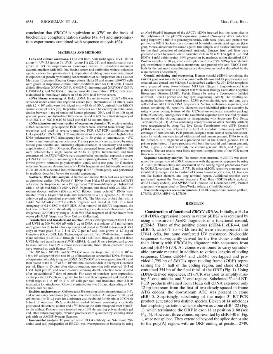
Construction of functional ERCC4 cDNAs. Initially, a HeLacell cDNA expression library in vector pEBS7 was screened byusing a mixture of EcoRI fragments of a functional cosmid,pER4-5. Three of five positive clones (cER4-1, cER4-4, andcER4-5, with 0.7- to ;2-kb inserts) were electroporated intoUV41 cells, but none conferred UV resistance. Nucleotidesequences subsequently derived for the five clones confirmedtheir identity with ERCC4 by alignment with sequences fromcosmid pER4-6 (70). All clones were found to carry consider-able intronic material in addition to various lengths of codingsequence. Clones cER4-4 and cER4-5 overlapped and pro-vided 1,795 bp of ERCC4 open reading frame (ORF) repre-senting the 59 half of the coding region, and clone cER4-2contained 354 bp of the final third of the ORF (Fig. 1). UsingcDNA-derived sequences, RT-PCR was used to amplify miss-ing 59-end, middle, and 39-end regions. Subcloned 59-end RT-PCR products obtained from HeLa cell cDNA extended only12 bp upstream from the first of two closely spaced in-frameATG codons; the downstream ATG was present in clonecER4-5. Surprisingly, subcloning of the major 39 RT-PCRproduct generated two distinct species. Eleven of 14 subcloneshad a splicing variation, which is shown as clone cER4-22 (Fig.1), which terminated the ORF in exon 11 at position 3180 (seeFig. 6). However, three clones, represented by cER4-40 in Fig.1, were not spliced. They extended beyond the splice donor siteto the poly(A) region, with an ORF ending at position 2749.
6554 BROOKMAN ET AL. MOL. CELL. BIOL.
Constructs cER4-22 (spliced) and cER4-34 and cER4-40 (un-spliced) were assembled in the pcDNA3 CMV expression vec-tor from a combination of library and PCR-derived sequences.To test for the minimum functional transcript, clone
cER4-34 was constructed by using only 30 bp downstream ofthe stop codon. Both cER4-34 and cER4-40 have the same2.75-kb ORF, which conferred UV resistance to the group 4mutant UV41 (see below). Clone cER4-22, with a 2.58-kbORF representing the predominantly amplified transcript, wasnot functional. A Northern blot of human and mouse poly(A)1
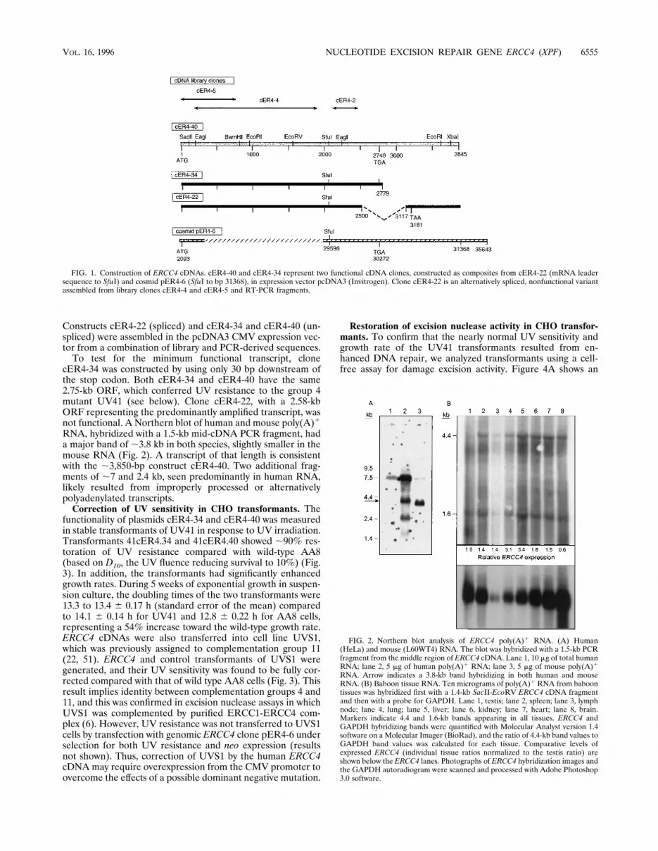
RNA, hybridized with a 1.5-kb mid-cDNA PCR fragment, hada major band of ;3.8 kb in both species, slightly smaller in themouse RNA (Fig. 2). A transcript of that length is consistentwith the ;3,850-bp construct cER4-40. Two additional frag-ments of ;7 and 2.4 kb, seen predominantly in human RNA,likely resulted from improperly processed or alternativelypolyadenylated transcripts.Correction of UV sensitivity in CHO transformants. The
functionality of plasmids cER4-34 and cER4-40 was measuredin stable transformants of UV41 in response to UV irradiation.Transformants 41cER4.34 and 41cER4.40 showed ;90% res-toration of UV resistance compared with wild-type AA8(based on D10, the UV fluence reducing survival to 10%) (Fig.3). In addition, the transformants had significantly enhancedgrowth rates. During 5 weeks of exponential growth in suspen-sion culture, the doubling times of the two transformants were13.3 to 13.4 6 0.17 h (standard error of the mean) comparedto 14.1 6 0.14 h for UV41 and 12.8 6 0.22 h for AA8 cells,representing a 54% increase toward the wild-type growth rate.ERCC4 cDNAs were also transferred into cell line UVS1,which was previously assigned to complementation group 11(22, 51). ERCC4 and control transformants of UVS1 weregenerated, and their UV sensitivity was found to be fully cor-rected compared with that of wild type AA8 cells (Fig. 3). Thisresult implies identity between complementation groups 4 and11, and this was confirmed in excision nuclease assays in whichUVS1 was complemented by purified ERCC1-ERCC4 com-plex (6). However, UV resistance was not transferred to UVS1cells by transfection with genomic ERCC4 clone pER4-6 underselection for both UV resistance and neo expression (resultsnot shown). Thus, correction of UVS1 by the human ERCC4cDNAmay require overexpression from the CMV promoter toovercome the effects of a possible dominant negative mutation.
Restoration of excision nuclease activity in CHO transfor-mants. To confirm that the nearly normal UV sensitivity andgrowth rate of the UV41 transformants resulted from en-hanced DNA repair, we analyzed transformants using a cell-free assay for damage excision activity. Figure 4A shows an
FIG. 1. Construction of ERCC4 cDNAs. cER4-40 and cER4-34 represent two functional cDNA clones, constructed as composites from cER4-22 (mRNA leadersequence to SfuI) and cosmid pER4-6 (SfuI to bp 31368), in expression vector pcDNA3 (Invitrogen). Clone cER4-22 is an alternatively spliced, nonfunctional variantassembled from library clones cER4-4 and cER4-5 and RT-PCR fragments.
FIG. 2. Northern blot analysis of ERCC4 poly(A)1 RNA. (A) Human(HeLa) and mouse (L60WT4) RNA. The blot was hybridized with a 1.5-kb PCRfragment from the middle region of ERCC4 cDNA. Lane 1, 10 mg of total humanRNA; lane 2, 5 mg of human poly(A)1 RNA; lane 3, 5 mg of mouse poly(A)1
RNA. Arrow indicates a 3.8-kb band hybridizing in both human and mouseRNA. (B) Baboon tissue RNA. Ten micrograms of poly(A)1 RNA from baboontissues was hybridized first with a 1.4-kb SacII-EcoRV ERCC4 cDNA fragmentand then with a probe for GAPDH. Lane 1, testis; lane 2, spleen; lane 3, lymphnode; lane 4, lung; lane 5, liver; lane 6, kidney; lane 7, heart; lane 8, brain.Markers indicate 4.4 and 1.6-kb bands appearing in all tissues. ERCC4 andGAPDH hybridizing bands were quantified with Molecular Analyst version 1.4software on a Molecular Imager (BioRad), and the ratio of 4.4-kb band values toGAPDH band values was calculated for each tissue. Comparative levels ofexpressed ERCC4 (individual tissue ratios normalized to the testis ratio) areshown below the ERCC4 lanes. Photographs of ERCC4 hybridization images andthe GAPDH autoradiogram were scanned and processed with Adobe Photoshop3.0 software.
VOL. 16, 1996 NUCLEOTIDE EXCISION REPAIR GENE ERCC4 (XPF) 6555
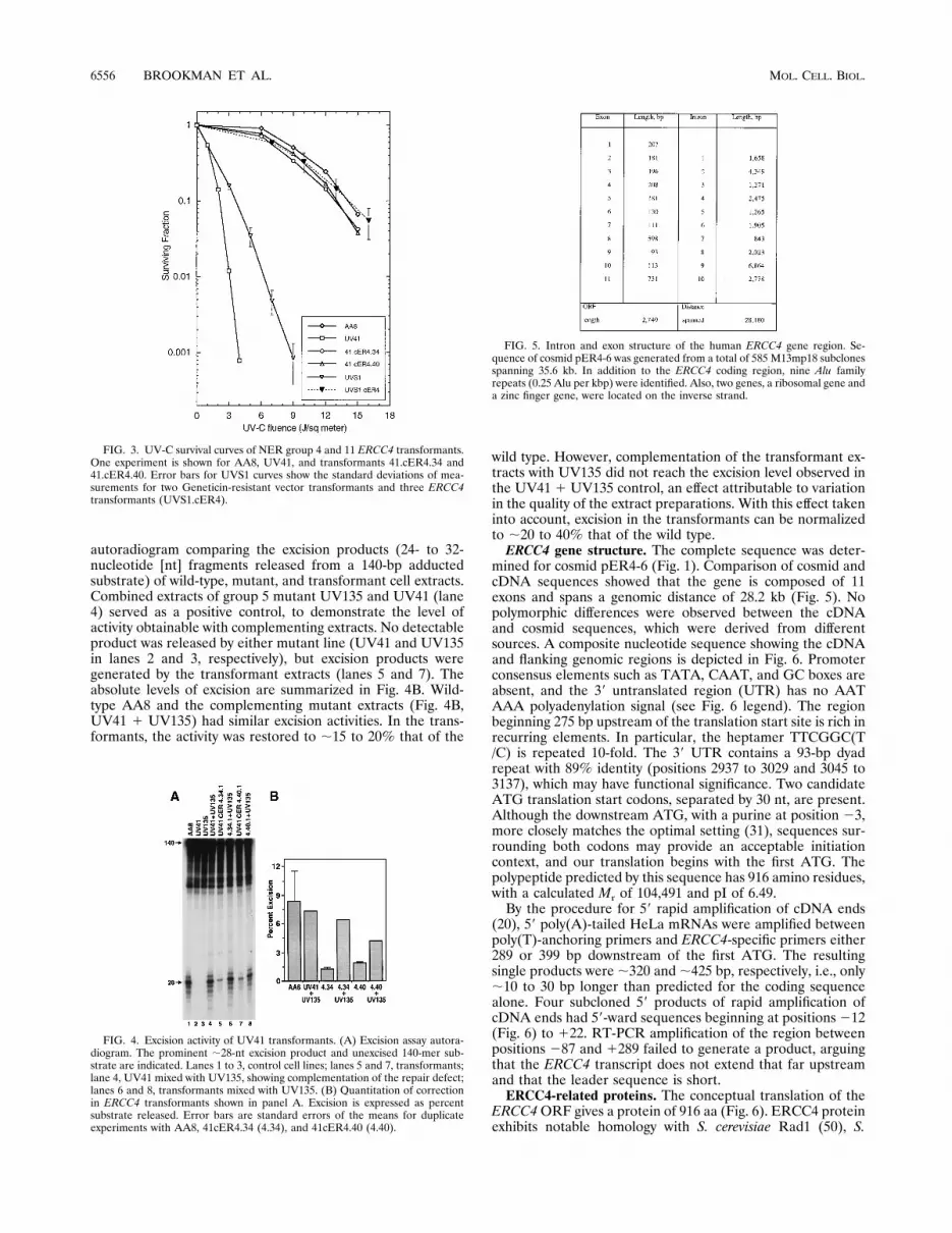
autoradiogram comparing the excision products (24- to 32-nucleotide [nt] fragments released from a 140-bp adductedsubstrate) of wild-type, mutant, and transformant cell extracts.Combined extracts of group 5 mutant UV135 and UV41 (lane4) served as a positive control, to demonstrate the level ofactivity obtainable with complementing extracts. No detectableproduct was released by either mutant line (UV41 and UV135in lanes 2 and 3, respectively), but excision products weregenerated by the transformant extracts (lanes 5 and 7). Theabsolute levels of excision are summarized in Fig. 4B. Wild-type AA8 and the complementing mutant extracts (Fig. 4B,UV41 1 UV135) had similar excision activities. In the trans-formants, the activity was restored to ;15 to 20% that of the
wild type. However, complementation of the transformant ex-tracts with UV135 did not reach the excision level observed inthe UV41 1 UV135 control, an effect attributable to variationin the quality of the extract preparations. With this effect takeninto account, excision in the transformants can be normalizedto ;20 to 40% that of the wild type.ERCC4 gene structure. The complete sequence was deter-
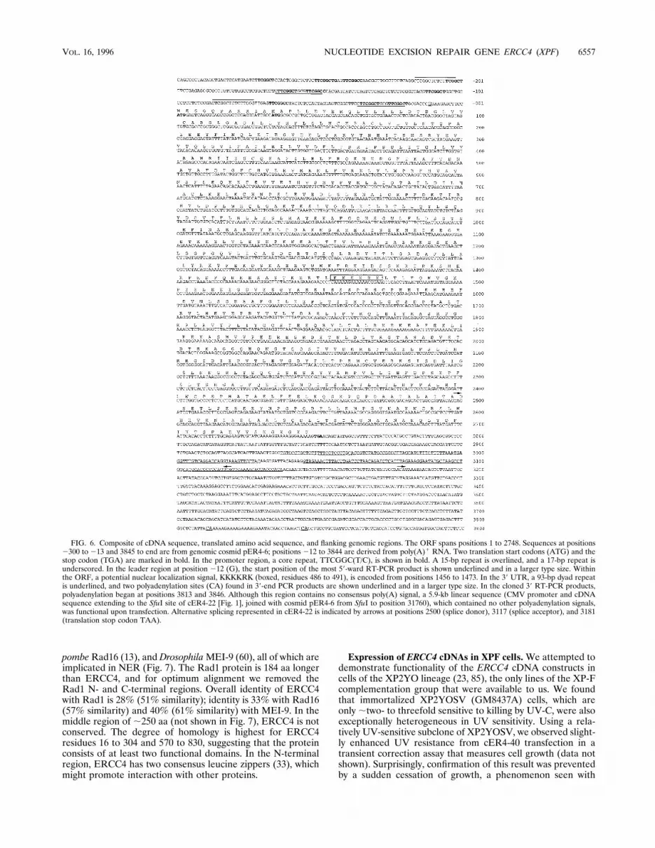
mined for cosmid pER4-6 (Fig. 1). Comparison of cosmid andcDNA sequences showed that the gene is composed of 11exons and spans a genomic distance of 28.2 kb (Fig. 5). Nopolymorphic differences were observed between the cDNAand cosmid sequences, which were derived from differentsources. A composite nucleotide sequence showing the cDNAand flanking genomic regions is depicted in Fig. 6. Promoterconsensus elements such as TATA, CAAT, and GC boxes areabsent, and the 39 untranslated region (UTR) has no AATAAA polyadenylation signal (see Fig. 6 legend). The regionbeginning 275 bp upstream of the translation start site is rich inrecurring elements. In particular, the heptamer TTCGGC(T/C) is repeated 10-fold. The 39 UTR contains a 93-bp dyadrepeat with 89% identity (positions 2937 to 3029 and 3045 to3137), which may have functional significance. Two candidateATG translation start codons, separated by 30 nt, are present.Although the downstream ATG, with a purine at position 23,more closely matches the optimal setting (31), sequences sur-rounding both codons may provide an acceptable initiationcontext, and our translation begins with the first ATG. Thepolypeptide predicted by this sequence has 916 amino residues,with a calculated Mr of 104,491 and pI of 6.49.By the procedure for 59 rapid amplification of cDNA ends
(20), 59 poly(A)-tailed HeLa mRNAs were amplified betweenpoly(T)-anchoring primers and ERCC4-specific primers either289 or 399 bp downstream of the first ATG. The resultingsingle products were ;320 and ;425 bp, respectively, i.e., only;10 to 30 bp longer than predicted for the coding sequencealone. Four subcloned 59 products of rapid amplification ofcDNA ends had 59-ward sequences beginning at positions 212(Fig. 6) to 122. RT-PCR amplification of the region betweenpositions 287 and 1289 failed to generate a product, arguingthat the ERCC4 transcript does not extend that far upstreamand that the leader sequence is short.ERCC4-related proteins. The conceptual translation of the
ERCC4ORF gives a protein of 916 aa (Fig. 6). ERCC4 proteinexhibits notable homology with S. cerevisiae Rad1 (50), S.
FIG. 3. UV-C survival curves of NER group 4 and 11 ERCC4 transformants.One experiment is shown for AA8, UV41, and transformants 41.cER4.34 and41.cER4.40. Error bars for UVS1 curves show the standard deviations of mea-surements for two Geneticin-resistant vector transformants and three ERCC4transformants (UVS1.cER4).
FIG. 4. Excision activity of UV41 transformants. (A) Excision assay autora-diogram. The prominent ;28-nt excision product and unexcised 140-mer sub-strate are indicated. Lanes 1 to 3, control cell lines; lanes 5 and 7, transformants;lane 4, UV41 mixed with UV135, showing complementation of the repair defect;lanes 6 and 8, transformants mixed with UV135. (B) Quantitation of correctionin ERCC4 transformants shown in panel A. Excision is expressed as percentsubstrate released. Error bars are standard errors of the means for duplicateexperiments with AA8, 41cER4.34 (4.34), and 41cER4.40 (4.40).
FIG. 5. Intron and exon structure of the human ERCC4 gene region. Se-quence of cosmid pER4-6 was generated from a total of 585 M13mp18 subclonesspanning 35.6 kb. In addition to the ERCC4 coding region, nine Alu familyrepeats (0.25 Alu per kbp) were identified. Also, two genes, a ribosomal gene anda zinc finger gene, were located on the inverse strand.
6556 BROOKMAN ET AL. MOL. CELL. BIOL.
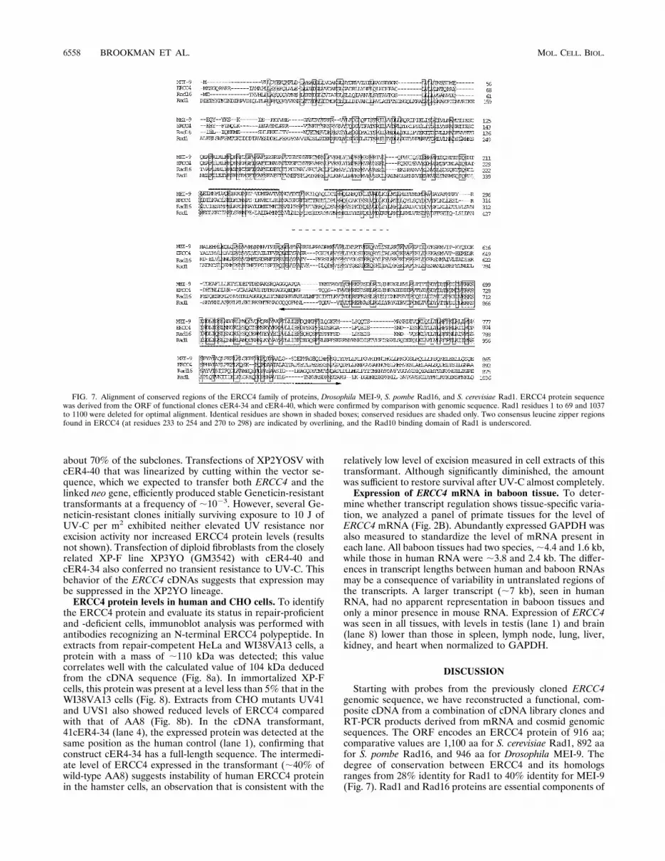
pombeRad16 (13), and DrosophilaMEI-9 (60), all of which areimplicated in NER (Fig. 7). The Rad1 protein is 184 aa longerthan ERCC4, and for optimum alignment we removed theRad1 N- and C-terminal regions. Overall identity of ERCC4with Rad1 is 28% (51% similarity); identity is 33% with Rad16(57% similarity) and 40% (61% similarity) with MEI-9. In themiddle region of ;250 aa (not shown in Fig. 7), ERCC4 is notconserved. The degree of homology is highest for ERCC4residues 16 to 304 and 570 to 830, suggesting that the proteinconsists of at least two functional domains. In the N-terminalregion, ERCC4 has two consensus leucine zippers (33), whichmight promote interaction with other proteins.
Expression of ERCC4 cDNAs in XPF cells.We attempted todemonstrate functionality of the ERCC4 cDNA constructs incells of the XP2YO lineage (23, 85), the only lines of the XP-Fcomplementation group that were available to us. We foundthat immortalized XP2YOSV (GM8437A) cells, which areonly ;two- to threefold sensitive to killing by UV-C, were alsoexceptionally heterogeneous in UV sensitivity. Using a rela-tively UV-sensitive subclone of XP2YOSV, we observed slight-ly enhanced UV resistance from cER4-40 transfection in atransient correction assay that measures cell growth (data notshown). Surprisingly, confirmation of this result was preventedby a sudden cessation of growth, a phenomenon seen with
FIG. 6. Composite of cDNA sequence, translated amino acid sequence, and flanking genomic regions. The ORF spans positions 1 to 2748. Sequences at positions2300 to 213 and 3845 to end are from genomic cosmid pER4-6; positions 212 to 3844 are derived from poly(A)1 RNA. Two translation start codons (ATG) and thestop codon (TGA) are marked in bold. In the promoter region, a core repeat, TTCGGC(T/C), is shown in bold. A 15-bp repeat is overlined, and a 17-bp repeat isunderscored. In the leader region at position 212 (G), the start position of the most 59-ward RT-PCR product is shown underlined and in a larger type size. Withinthe ORF, a potential nuclear localization signal, KKKKRK (boxed, residues 486 to 491), is encoded from positions 1456 to 1473. In the 39 UTR, a 93-bp dyad repeatis underlined, and two polyadenylation sites (CA) found in 39-end PCR products are shown underlined and in a larger type size. In the cloned 39 RT-PCR products,polyadenylation began at positions 3813 and 3846. Although this region contains no consensus poly(A) signal, a 5.9-kb linear sequence (CMV promoter and cDNAsequence extending to the SfuI site of cER4-22 [Fig. 1], joined with cosmid pER4-6 from SfuI to position 31760), which contained no other polyadenylation signals,was functional upon transfection. Alternative splicing represented in cER4-22 is indicated by arrows at positions 2500 (splice donor), 3117 (splice acceptor), and 3181(translation stop codon TAA).
VOL. 16, 1996 NUCLEOTIDE EXCISION REPAIR GENE ERCC4 (XPF) 6557
about 70% of the subclones. Transfections of XP2YOSV withcER4-40 that was linearized by cutting within the vector se-quence, which we expected to transfer both ERCC4 and thelinked neo gene, efficiently produced stable Geneticin-resistanttransformants at a frequency of ;1023. However, several Ge-neticin-resistant clones initially surviving exposure to 10 J ofUV-C per m2 exhibited neither elevated UV resistance norexcision activity nor increased ERCC4 protein levels (resultsnot shown). Transfection of diploid fibroblasts from the closelyrelated XP-F line XP3YO (GM3542) with cER4-40 andcER4-34 also conferred no transient resistance to UV-C. Thisbehavior of the ERCC4 cDNAs suggests that expression maybe suppressed in the XP2YO lineage.ERCC4 protein levels in human and CHO cells. To identify
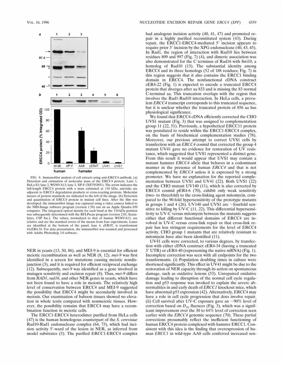
the ERCC4 protein and evaluate its status in repair-proficientand -deficient cells, immunoblot analysis was performed withantibodies recognizing an N-terminal ERCC4 polypeptide. Inextracts from repair-competent HeLa and WI38VA13 cells, aprotein with a mass of ;110 kDa was detected; this valuecorrelates well with the calculated value of 104 kDa deducedfrom the cDNA sequence (Fig. 8a). In immortalized XP-Fcells, this protein was present at a level less than 5% that in theWI38VA13 cells (Fig. 8). Extracts from CHO mutants UV41and UVS1 also showed reduced levels of ERCC4 comparedwith that of AA8 (Fig. 8b). In the cDNA transformant,41cER4-34 (lane 4), the expressed protein was detected at thesame position as the human control (lane 1), confirming thatconstruct cER4-34 has a full-length sequence. The intermedi-ate level of ERCC4 expressed in the transformant (;40% ofwild-type AA8) suggests instability of human ERCC4 proteinin the hamster cells, an observation that is consistent with the
relatively low level of excision measured in cell extracts of thistransformant. Although significantly diminished, the amountwas sufficient to restore survival after UV-C almost completely.Expression of ERCC4 mRNA in baboon tissue. To deter-
mine whether transcript regulation shows tissue-specific varia-tion, we analyzed a panel of primate tissues for the level ofERCC4 mRNA (Fig. 2B). Abundantly expressed GAPDH wasalso measured to standardize the level of mRNA present ineach lane. All baboon tissues had two species,;4.4 and 1.6 kb,while those in human RNA were ;3.8 and 2.4 kb. The differ-ences in transcript lengths between human and baboon RNAsmay be a consequence of variability in untranslated regions ofthe transcripts. A larger transcript (;7 kb), seen in humanRNA, had no apparent representation in baboon tissues andonly a minor presence in mouse RNA. Expression of ERCC4was seen in all tissues, with levels in testis (lane 1) and brain(lane 8) lower than those in spleen, lymph node, lung, liver,kidney, and heart when normalized to GAPDH.
DISCUSSION
Starting with probes from the previously cloned ERCC4genomic sequence, we have reconstructed a functional, com-posite cDNA from a combination of cDNA library clones andRT-PCR products derived from mRNA and cosmid genomicsequences. The ORF encodes an ERCC4 protein of 916 aa;comparative values are 1,100 aa for S. cerevisiae Rad1, 892 aafor S. pombe Rad16, and 946 aa for Drosophila MEI-9. Thedegree of conservation between ERCC4 and its homologsranges from 28% identity for Rad1 to 40% identity for MEI-9(Fig. 7). Rad1 and Rad16 proteins are essential components of
FIG. 7. Alignment of conserved regions of the ERCC4 family of proteins, Drosophila MEI-9, S. pombe Rad16, and S. cerevisiae Rad1. ERCC4 protein sequencewas derived from the ORF of functional clones cER4-34 and cER4-40, which were confirmed by comparison with genomic sequence. Rad1 residues 1 to 69 and 1037to 1100 were deleted for optimal alignment. Identical residues are shown in shaded boxes; conserved residues are shaded only. Two consensus leucine zipper regionsfound in ERCC4 (at residues 233 to 254 and 270 to 298) are indicated by overlining, and the Rad10 binding domain of Rad1 is underscored.
6558 BROOKMAN ET AL. MOL. CELL. BIOL.
NER in yeasts (13, 50, 86), and MEI-9 is essential for efficientmeiotic recombination as well as NER (8, 12). mei-9 was firstidentified in a screen for mutations causing meiotic nondis-junction (3), and it is required for efficient reciprocal exchange(12). Subsequently, mei-9 was identified as a gene involved inmutagen sensitivity and excision repair (8). Thus, mei-9 differsfrom RAD1, rad16, and other NER genes in yeasts, which havenot been found to have a role in meiosis. The relatively highlevel of conservation between ERCC4 and MEI-9 suggestedthe possibility that ERCC4 might be secondarily involved inmeiosis. Our examination of baboon tissues showed no eleva-tion in whole testis compared with nonmeiotic tissues. How-ever, the possibility remains that ERCC4 may have a recom-bination function in meiotic cells.The ERCC1-ERCC4 heterodimer purified from HeLa cells
(47) is the human homologous counterpart of the S. cerevisiaeRad10-Rad1 endonuclease complex (64, 73), which had inci-sion activity 59-ward of the lesion in NER, as inferred frommodel substrates (5). The purified ERCC1-ERCC4 complex
had analogous incision activity (40, 41, 47) and promoted re-pair in a highly purified reconstituted system (43). Duringrepair, the ERCC1-ERCC4-mediated 59 incision appears torequire prior 39 incision by the XPG endonuclease (40, 43, 45).In Rad1, the region of interaction with Rad10 lies betweenresidues 809 and 997 (Fig. 7) (4), and dimeric association wasalso demonstrated for the C terminus of Rad16 with Swi10, ahomolog of Rad10 (13). The substantial identity amongERCC4 and its three homologs (52 of 188 residues; Fig. 7) inthis region suggests that it also contains the ERCC1 bindingdomain in ERCC4. The nonfunctional cDNA constructcER4-22 (Fig. 1) is expected to encode a truncated ERCC4protein that diverges after aa 833 and is missing the 83 normalC-terminal aa. This truncation overlaps with the region thatinvolves the Rad1-Rad10 interaction. In HeLa cells, a preva-lent ERCC4 transcript corresponds to this truncated sequence,but it is unclear whether the truncated protein of 856 aa hasphysiological significance.We found that ERCC4 cDNA efficiently corrected the CHO
UVS1 mutant (Fig. 3) that was assigned to complementationgroup 11 (22, 51). Previously, a hypothetical ERCC11 proteinwas postulated to reside within the ERCC1-ERCC4 complex,on the basis of biochemical complementation studies (78).Moreover, our previous attempt to correct UVS1 cells bytransfection with an ERCC4 cosmid that corrected the group 4mutant UV41 gave no evidence for restoration of UV resis-tance, which suggested that UVS1 represented a distinct gene.From this result it would appear that UVS1 may contain amutant hamster ERCC4 allele that behaves in a codominantmanner in the presence of human ERCC4 and that is notcomplemented by ERCC4 unless it is expressed by a strongpromoter. We have no explanation for the reported comple-mentation between UVS1 and UV41 (22). Both UVS1 (22)and the CHO mutant UV140 (11), which is also corrected byERCC4 cosmid pER4-6 (70), exhibit only weak sensitivity(two- to threefold) to the cross-linking agent mitomycin, com-pared to the 90-fold hypersensitivity of the prototype mutantsin groups 1 and 4 (26). UV140 and UVS1 are ;fourfold sen-sitive to killing by UV-C (11, 22). This differential hypersensi-tivity to UV-C versus mitomycin between the mutants suggestseither that different functional domains of ERCC4 are in-volved in UV-C versus cross-link repair or that cross-link re-pair has less stringent requirements for the level of ERCC4activity. CHO group 1 mutants that are relatively resistant tomitomycin have also been identified (11).UV41 cells were corrected, to various degrees, by transfec-
tion with either cDNA construct cER4-34 (having a truncated39UTR) or cER4-40 (representing the native mRNA) (Fig. 1).Incomplete correction was seen with all endpoints for the twotransformants. (i) Population doubling times in culture wereshortened significantly. This effect in UV41 might be due to therestoration of NER capacity through its action on spontaneousdamage, such as oxidative lesions (53). Unrepaired oxidativedamage leading to disruption of the normal cell cycle regula-tion and p53 response was invoked to explain the severe ab-normalities in and early death of ERCC1 knockout mice, whichhave abnormal p53 expression (42). Alternatively, ERCC4 mayhave a role in cell cycle progression that does involve repair.(ii) Cell survival after UV-C exposure gave an ;90% level ofcorrection based on D10 fluences (Fig. 3), which was a signif-icant improvement over the 30 to 64% level of correction seenearlier with the ERCC4 genomic sequence (70). These partialcorrections presumably reflect the inefficient functioning ofhuman ERCC4 protein complexed with hamster ERCC1. Con-sistent with this idea is the finding that overexpression of hu-man ERCC1 in wild-type AA8 cells conferred increased sen-
FIG. 8. Immunoblot analysis of cell extracts using anti-ERCC4 antibody. (a)Detection and estimation of molecular mass of the ERCC4 protein. Lane 1,HeLa S3; lane 2, WI38VA13; lane 3, XP-F (XP2YOSV). The arrow indicates thefull-length ERCC4 protein with a mass estimated at 110 kDa; asterisks areadjacent to ERCC4 degradation products or cross-reacting proteins. Molecularmasses of protein standards are indicated to the left of the image. (b) Detectionand quantitation of ERCC4 protein in mutant cell lines. After the film wasdeveloped, the immunoblot image was captured using a video camera linked tothe NIH-Image software program (version 1.58) run on an Apple Quadra 650computer. The integrated optical density for each band at the 110-kDa positionwas subsequently determined with the RFLPscan program (version 2.01; Scana-lytics, CSP Inc.). The values, normalized to that of human WI38VA13, arerelative and are the standard errors of the means from four experiments. Lanesare identified at the bottom of the panel; lane 4, cER4T, is transformant41cER4.34. For data presentation, the immunoblot was scanned and processedwith Adobe Photoshop 3.0 software.
VOL. 16, 1996 NUCLEOTIDE EXCISION REPAIR GENE ERCC4 (XPF) 6559
sitivity to cross-linking agents (9). (iii) Excision nucleaseactivity, which was undetectable in UV41 extracts, was presentin transformants at,40% of the AA8 level (Fig. 4), suggestingthat the heterologous human protein may function even lessefficiently in vitro than in vivo. A similar partial level of cor-rection was seen in excision nuclease assays with ERCC4genomic transformants (70). (iv) The hamster ERCC4 proteinlevel in UV41 was very low, while the transformants had totalERCC4 levels that were ;40% of the AA8 level, or lower ifthe antibody recognized human ERCC4 more efficiently thanhamster ERCC4 (Fig. 8 and results not shown). ERCC4 ap-pears to be unstable in hamster cells in the absence of over-expressed ERCC1.It was previously suggested on the basis of biochemical
complementation studies that ERCC4 might be equivalent toXPF (7, 49, 78). The purified complex containing ERCC1 andthe 112-kDa protein was shown to complement UV41 andXP-F cell extracts (47). More recently, anti-ERCC4 antibodydetected the 112-kDa protein that copurifies with ERCC1, andrecombinant ERCC1-ERCC4 complex isolated from the bac-ulovirus system corrected the repair deficiency in XP-F cellsusing the excision nuclease assay (6). In the latter assay, theERCC4 protein was encoded by the sequence from cDNAplasmid cER4-34 (Fig. 1). Mutations were also identified inERCC4 in XP-F cells, providing genetic evidence that ERCC4equals XPF (62).Using our cDNA constructs, we were unable to convincingly
demonstrate correction of the related XP-F cell linesXP2YOSV and XP3YO in both transient and stable transfec-tions. We attribute these negative results in part to the rela-tively mild UV-C sensitivity and partial repair capacity (21) ofthe immortalized XP-F cells combined with unpredictablegrowth upon subcloning and also to an apparent lack of stableexpression of the ERCC4 cDNA. The latter inference is sug-gested by the uncorrected UV sensitivity of six stable neotransformants of XP2YOSV resulting from cER4-40 transfec-tion. Joint integration and coexpression of neo and ERCC4were expected for at least some transformants using linearizedmolecules. The failure of the overexpressing ERCC4 cDNA toconfer UV resistance was consistent with results of our attemptto transfer the ERCC4 gene using cosmid pER4-7, which alsogave no clear correction of Geneticin-resistant XP2YOSVcells. Thus, ERCC4 expression controlled either by the CMVpromoter or by its native promoter did not restore repaircapability. Aberrant XP-F regulation or a dominant negativeeffect of a mutant allele might block expression of ERCC4transgenes in cells of the XP2YO lineage.In an earlier study, somatic cell hybrids between diploid
XP3YO and UV41 cells were produced by selecting forcomplementation of mitomycin sensitivity (72). Since signifi-cant restoration of both mitomycin and UV resistance wasobserved, it was concluded that the UV41 and XP-F mutationswere in different genes. However, in retrospect we realize thisinterpretation was incorrect, since the results can be explainedby the partial repair capacity of the XP-F mutant allele(s) (21).In a study involving microcell-mediated chromosome transfer,it was concluded that human chromosome 15 gave partialcorrection to UV resistance in XP2YOSV cells (54). This con-clusion is at odds with the localization of ERCC4 to chromo-some 16p13.13-p13.2 (35) and can be attributed to the extremepopulation heterogeneity we observed for UV sensitivity withXP2YOSV.Among the NER genes, ERCC1 and ERCC4 are uniquely
involved in interstrand cross-link repair, as evidenced by theextreme sensitivity of a subset of the CHO mutants in groups1 and 4 to cross-linking agents (26, 63, 84). Mutants in NER
groups 2, 3, and 5 are apparently relatively proficient in cross-link repair (but not the repair of monoadducts accompanyingcross-links), since they are much less sensitive to cross-linkingagents. Thus, ERCC1 and ERCC4 proteins must possess afunction, which is likely involved in homologous recombina-tion, that assists in removing cross-links. A unique role for theRad10 and Rad1 proteins in mitotic recombination (28, 58, 59)parallels the putatively unique roles of ERCC1 and ERCC4 incross-link repair (see Discussion in reference 67). The clonedERCC4 cDNA and recombinant protein should help to eluci-date ERCC4’s putative recombinational repair function as wellas help with further investigation into its possible role in re-combination during meiosis.
ACKNOWLEDGMENTS
We thank Aaron Adamson andMelissa Ramirez for expert technicalassistance with DNA sequencing.This work was performed under the auspices of the U.S. DOE by
LLNL under contract no. W-7405-ENG-48, and portions were doneunder NIH grant no. GM32833 (A.S.), ES05798 (C.A.W.), andCA61335 (C.A.W.).
ADDENDUM IN PROOF
Fresh preparations of baboon poly(A)1 RNA and humanERCC4 cDNA probe were made and used in two additionalNorthern blots like those shown in Fig. 2B. In these newlong-exposure blots, 7.5-kb hybridizing bands were detected inall of the tissues, which included ovary tissue; however, the7.5-kb band was slightly more abundant in the lane containingRNA from testes. In addition, hybridizing bands of 4.4, 3.7(most intense), and 2.0 kb were detected only in the testes.
REFERENCES
1. Aboussekhra, A., M. Biggerstaff, M. K. K. Shivji, J. A. Vilpo, V. Moncollin,V. N. Podust, M. Protic, U. Hubscher, J. M. Egly, and R. D. Wood. 1995.Mammalian DNA nucleotide excision repair reconstituted with purified pro-tein components. Cell 80:859–868.
2. Asahina, H., I. Kuraoka, M. Shirakawa, E. H. Morita, N. Miura, I. Miy-amoto, E. Ohtsuka, Y. Okada, and K. Tanaka. 1994. The XPA protein is azinc metalloprotein with an ability to recognize various kinds of DNA dam-age. Mutat. Res. 315:229–237.
3. Baker, B. S., and A. T. C. Carpenter. 1972. Genetic analysis of sex chromo-somal meiotic mutants in Drosophila melanogaster. Genetics 71:255–286.
4. Bardwell, A. J., L. Bardwell, D. K. Johnson, and E. C. Friedberg. 1993. YeastDNA recombination and repair proteins Rad1 and Rad10 constitute a com-plex in vivo mediated by localized hydrophobic domains. Mol. Microbiol.8:1177–1188.
5. Bardwell, A. J., L. Bardwell, A. E. Tomkinson, and E. C. Friedberg. 1994.Specific cleavage of model recombination and repair intermediates by theyeast Rad1-Rad10 endonuclease. Science 265:2082–2085.
6. Bessho, T., M. P. Thelen, and A. Sancar. Unpublished results.7. Biggerstaff, M., D. E. Szymkowski, and R. D. Wood. 1993. Co-correction ofthe ERCC1, ERCC4 and xeroderma pigmentosum group F DNA repairdefects in vitro. EMBO J. 12:3685–3692.
8. Boyd, J. B., M. D. Golino, and R. B. Setlow. 1976. The mei-9a mutant ofDrosophila melanogaster increases mutagen sensitivity and decreases excisionrepair. Genetics 84:527–544.
9. Bramson, J., and L. C. Panasci. 1993. Effect of ERCC-1 overexpression onsensitivity of Chinese hamster ovary cells to DNA damaging agents. CancerRes. 53:3237–3240.
10. Burns, J. L., S. N. Guzder, P. Sung, S. Prakash, and L. Prakash. 1996. Anaffinity of human replication protein A for ultraviolet-damagedDNA. J. Biol. Chem. 271:11607–11610.
11. Busch, D. B., H. van Vuuren, J. de Wit, A. Collins, M. Zdzienicka, D. L.Mitchell, K. W. Brookman, M. Stefanini, R. Ribioni, L. H. Thompson, R. B.Albert, A. van Gool, and J. Hoeijmakers. Phenotypic heterogeneity inFAECB nucleotide excision repair mutants of rodent complementationgroups 1 and 4. Submitted for publication.
12. Carpenter, A. T. 1982. Mismatch repair, gene conversion, and crossing-overin two recombination-defective mutants of Drosophila melanogaster. Proc.Natl. Acad. Sci. USA 79:5961–5965.
13. Carr, A. M., H. Schmidt, S. Kirchhoff, W. J. Muriel, K. S. Sheldrick, D. J.Griffiths, C. N. Basmacioglu, S. Subramani, M. Clegg, A. Nasim, and A. R.
6560 BROOKMAN ET AL. MOL. CELL. BIOL.
Lehmann. 1994. The rad16 gene of Schizosaccharomyces pombe: a homologof the RAD1 gene of Saccharomyces cerevisiae. Mol. Cell. Biol. 14:2029–2040.
14. Cleaver, J. E. 1972. Xeroderma pigmentosum: variants with normal DNArepair and normal sensitivity to ultraviolet light. J. Invest. Dermatol. 58:124–128.
15. Cleaver, J. E., and K. H. Kraemer. 1995. Xeroderma pigmentosum andCockayne syndrome, p. 4393–4419. In C. R. Scriver, A. L. Beudet, W. S. Sly,and D. Valle (ed.), The metabolic and molecular bases of inherited disease,7th ed. vol. III. McGraw-Hill, New York.
16. Drapkin, R., J. Reardon, A. Ansari, J. C. Huang, L. Zawel, K. Ahn, A.Sancar, and D. Reinberg. 1994. TFIIH, a link between RNA polymerase IItranscription and DNA excision repair. Nature (London) 368:769–772.
17. Drapkin, R., and D. Reinberg. 1994. The multifunctional TFIIH complexand transcriptional control. Trends Biochem. Sci. 19:504–508.
18. Flejter, W. L., L. D. McDaniel, D. Johns, E. C. Friedberg, and R. A. Schultz.1992. Correction of xeroderma pigmentosum complementation group Dmutant cell phenotypes by chromosome and gene transfer: involvement ofthe human ERCC2 DNA repair gene. Proc. Natl. Acad. Sci. USA 89:261–265.
19. Friedberg, E. C., G. C. Walker, and W. Siede. 1995. DNA repair and mu-tagenesis. American Society for Microbiology, Washington, D.C.
20. Frohman, M. A. 1990. RACE: rapid amplification of cDNA ends, p. 28–38.In M. A. Innes (ed.), PCR protocols: a guide to methods and applications.Academic Press, San Diego.
21. Galloway, A. M., M. Liuzzi, and M. C. Paterson. 1994. Metabolic processingof cyclobutyl pyrimidine dimers and (6-4) photoproducts in UV-treatedhuman cells. Evidence for distinct excision-repair pathways. J. Biol. Chem.269:974–980.
22. Hata, H., M. Numata, H. Tohda, A. Yasui, and A. Oikawa. 1991. Isolation oftwo chloroethylnitrosourea-sensitive Chinese hamster cell lines. Cancer Res.51:195–198.
23. Hayakawa, H., K. Ishizaki, M. Inoue, T. Yagi, M. Sekiguchi, and H. Takebe.1981. Repair of ultraviolet radiation damage in xeroderma pigmentosumcells belonging to complementation group F. Mutat. Res. 80:381–388.
24. He, Z., L. A. Henricksen, M. S. Wold, and C. J. Ingles. 1995. RPA involve-ment in the damage-recognition and incision steps of nucleotide excisionrepair. Nature (London) 374:566–569.
25. Hoeijmakers, J. H. J. 1994. Human nucleotide excision repair syndromes:molecular clues to unexpected intricacies. Eur. J. Cancer 30A:1912–1921.
26. Hoy, C. A., L. H. Thompson, C. L. Mooney, and E. P. Salazar. 1985. Defec-tive DNA cross-link removal in Chinese hamster cell mutants hypersensitiveto bifunctional alkylating agents. Cancer Res. 45:1737–1743.
27. Huang, J. C., D. L. Svoboda, J. T. Reardon, and A. Sancar. 1992. Humannucleotide excision nuclease removes thymine dimers from DNA by incisingthe 22nd phosphodiester bond 59 and the 6th phosphodiester bond 39 to thephotodimer. Proc. Natl. Acad. Sci. USA 89:3664–3668.
28. Ivanov, E. L., and J. E. Haber. 1995. RAD1 and RAD10, but not otherexcision repair genes, are required for double-strand break-induced recom-bination in Saccharomyces cerevisiae. Mol. Cell. Biol. 15:2245–2251.
29. Jones, C. J., and R. D. Wood. 1993. Preferential binding of the xerodermapigmentosum group A complementing protein to damaged DNA. Biochem-istry 32:12096–12116.
30. Keijzer, W., N. G. J. Jaspers, P. J. Abrahams, A. M. R. Taylor, C. F. Arlett,B. Zelle, H. Takebe, P. D. S. Kinmont, and D. Bootsma. 1979. A seventhcomplementation group in excision-deficient xeroderma pigmentosum. Mu-tat. Res. 62:183–190.
31. Kozak, M. 1995. Adherence to the first-AUG rule when a second AUGcodon follows closely upon the first. Proc. Natl. Acad. Sci. USA 92:2662–2666.
32. Lamerdin, J. E., M. A. Montgomery, S. A. Stilwagen, L. K. Scheidecker, R. S.Tebbs, K. W. Brookman, L. H. Thompson, and A. V. Carrano. 1995.Genomic sequence comparison of the human and mouse XRCC1 DNArepair gene regions. Genomics 25:547–554.
33. Landschulz, W. H., P. F. Johnson, and S. L. McKnight. 1988. The leucinezipper: a hypothetical structure common to a class of DNA binding proteins.Science 240:1759–1763.
34. Legerski, R., and C. Peterson. 1992. Expression cloning of a human DNArepair gene involved in xeroderma pigmentosum group C. Nature (London)359:70–73.
35. Liu, P., J. Siciliano, B. White, R. Legerski, D. Callen, S. Reeders, M. J.Siciliano, and L. H. Thompson. 1992. Regional mapping of human DNAexcision repair gene ERCC4 to chromosome 16p13.13-p13.2. Mutagenesis8:199–205.
36. MacInnes, M. A., J. A. Dickson, R. R. Hernandez, D. Learmonth, G. Y. Lin,J. S. Mudgett, M. S. Park, S. Schauer, R. J. Reynolds, G. F. Strniste, andJ. Y. Yu. 1993. Human ERCC5 cDNA-cosmid complementation for excisionrepair and bipartite amino acid domains conserved with RAD proteins ofSaccharomyces cerevisiae and Schizosaccharomyces pombe. Mol. Cell. Biol.13:6393–6402.
37. Maher, V. M., D. J. Dorney, A. L. Mendrala, B. Konze-Thomas, and J. J.McCormick. 1979. DNA excision-repair processes in human cells can elim-
inate the cytotoxic and mutagenic consequences of ultraviolet radiation.Mutat. Res. 62:311–323.
38. Manley, J. L., A. Fire, A. Cano, P. A. Sharp, and M. L. Gefter. 1980.DNA-dependent transcription of adenovirus genes in a soluble whole-cellextract. Proc. Natl. Acad. Sci. USA 77:3855–3859.
39. Martin-Gallardo, A., J. Lamerdin, and A. V. Carrano. 1993. Shotgun se-quencing, p. 37–41. In M. Adams, C. Fields, and J. C. Ventner (ed.), Auto-mated DNA sequencing and analysis. Academic Press, London.
40. Matsunaga, T., D. Mu, C. H. Park, J. T. Reardon, and A. Sancar. 1995.Human DNA repair excision nuclease. J. Biol. Chem. 270:20862–20869.
41. Matsunaga, T., C. H. Park, T. Bessho, D. Mu, and A. Sancar. 1996. Repli-cation protein A confers structure-specific endonuclease activities to theXPF-ERCC1 and XPG subunits of human DNA repair excision nuclease.J. Biol. Chem. 271:11047–11050.
42. McWhir, J., J. Selfridge, D. J. Harrison, S. Squires, and D. W. Melton. 1993.Mice with DNA repair gene (ERCC-1) deficiency have elevated levels of p53,liver nuclear abnormalities and die before weaning. Nature Genet. 5:217–224.
43. Mu, D., D. S. Hsu, and A. Sancar. 1996. Reaction mechanism of humanDNA repair excision nuclease. J. Biol. Chem. 271:8285–8294.
44. Mu, D., C. H. Park, T. Matsunaga, D. S. Hsu, J. T. Reardon, and A. Sancar.1995. Reconstitution of human DNA repair excision nuclease in a highlydefined system. J. Biol. Chem. 270:2415–2418.
45. O’Donovan, A., A. A. Davies, J. G. Moggs, S. C. West, and R. D. Wood. 1994.XPG endonuclease makes the 39 incision in human DNA nucleotide excisionrepair. Nature (London) 371:432–435.
46. Pan, Z. Q., J. T. Reardon, L. Li, H. Fores-Rozas, R. Legerski, A. Sancar, andJ. Hurwitz. 1995. Inhibition of nucleotide excision repair by the cyclin-dependent kinase inhibitor p21. J. Biol. Chem. 270:22008–22016.
47. Park, C. H., T. Bessho, T. Matsunaga, and A. Sancar. 1995. Purification andcharacterization of the XPF-ERCC1 complex of human DNA repair excisionnuclease. J. Biol. Chem. 270:22657–22660.
48. Peterson, C., and R. Legerski. 1991. High-frequency transformation of hu-man repair-deficient cell lines by an Epstein-Barr virus-based cDNA expres-sion vector. Gene 107:279–284.
49. Reardon, J. T., L. H. Thompson, and A. Sancar. 1993. Excision repair in manand the molecular basis of xeroderma pigmentosum syndrome. Cold SpringHarbor Symp. Quant. Biol. 58:605–617.
50. Reynolds, P., L. Prakash, and S. Prakash. 1987. Nucleotide sequence andfunctional analysis of the RAD1 gene of Saccharomyces cerevisiae. Mol. Cell.Biol. 7:1012–1020.
51. Riboni, R., E. Botta, M. Stefanini, M. Numata, and A. Yasui. 1992. Identi-fication of the eleventh complementation group of UV-sensitive excisionrepair-defective rodent mutants. Cancer Res. 52:6690–6691.
52. Sancar, A. 1996. DNA excision repair. Annu. Rev. Biochem. 65:43–81.53. Satoh, M. S., C. J. Jones, R. D. Wood, and T. Lindahl. 1993. DNA excision-
repair defect of xeroderma pigmentosum prevents removal of a class ofoxygen free radical-induced base lesions. Proc. Natl. Acad. Sci. USA 90:6335–6339.
54. Saxon, P. J., R. A. Schultz, E. J. Stanbridge, and E. C. Friedberg. 1989.Human chromosome 15 confers partial complementation of phenotypes toxeroderma pigmentosum group F cells. Am. J. Hum. Genet. 44:474–485.
55. Schaeffer, L., V. Moncollin, R. Roy, A. Staub, M. Mezzina, A. Sarasin, G.Weeda, J. H. Hoeijmakers, and J. M. Egly. 1994. The ERCC2/DNA repairprotein is associated with the class II BTF2/TFIIH transcription factor.EMBO J. 13:2388–2392.
56. Schaeffer, L., R. Roy, S. Humbert, V. Moncollin, W. Vermeulen, J. H. Hoei-jmakers, P. Chambon, and J. M. Egly. 1993. DNA repair helicase: a com-ponent of BTF2 (TFIIH) basic transcription factor. Science 260:58–63.
57. Scherly, D., T. Nouspikel, J. Corlet, C. Ucla, A. Bairoch, and S. G. Clarkson.1993. Complementation of the DNA repair defect in xeroderma pigmento-sum group G cells by a human cDNA related to yeast RAD2. Nature (Lon-don) 363:182–185.
58. Schiestl, R. H., and S. Prakash. 1988. RAD1, an excision repair gene ofSaccharomyces cerevisiae, is also involved in recombination. Mol. Cell. Biol.8:3619–3626.
59. Schiestl, R. H., and S. Prakash. 1990. RAD10, an excision repair gene ofSaccharomyces cerevisiae, is involved in the RAD1 pathway of mitotic recom-bination. Mol. Cell. Biol. 10:2485–2491.
60. Sekelsky, J. J., K. S. McKim, G. M. Chin, and R. S. Hawley. 1995. TheDrosophila meiotic recombination gene mei-9 encodes a homologue of theyeast excision repair protein Rad1. Genetics 141:619–627.
61. Shivji, M. K., V. N. Podust, U. Hubscher, and R. D. Wood. 1995. Nucleotideexcision repair DNA synthesis by DNA polymerase epsilon in the presenceof PCNA, RFC, and RPA. Biochemistry 34:5011–5017.
62. Sijbers, A. M., W. L. de Laat, R. R. Ariza, M. Biggerstaff, Y. F. Wei, J. G.Moggs, K. C. Carter, B. K. Shell, E. Evans, M. C. de Jong, S. Rademakers,J. de Rooij, N. G. J. Jaspers, J. H. J. Hoeijmakers, and R. D. Wood. 1996.Xeroderma pigmentosum group F caused by a defect in a structure-specificDNA repair endonuclease. Cell 86:811–822.
63. Sorenson, C. M., and A. Eastman. 1988. Mechanism of cis-diamminedichlo-roplatinum(II)-induced cytotoxicity: role of G2 arrest and DNA double-
VOL. 16, 1996 NUCLEOTIDE EXCISION REPAIR GENE ERCC4 (XPF) 6561
strand breaks. Cancer Res. 48:4484–4488.64. Sung, P., P. Reynolds, L. Prakash, and S. Prakash. 1993. Purification and
characterization of the Saccharomyces cerevisiae RAD1/RAD10 endonucle-ase. J. Biol. Chem. 268:26391–26399.
65. Svoboda, D. L., J. S. Taylor, J. E. Hearst, and A. Sancar. 1993. DNA repairby eukaryotic nucleotide excision nuclease. Removal of thymine dimer andpsoralen monoadduct by HeLa cell-free extract and of thymine dimer byXenopus laevis oocytes. J. Biol. Chem. 268:1931–1936.
66. Tanaka, K., N. Miura, I. Satokata, I. Miyamoto, M. C. Yoshida, Y. Satoh, S.Kondo, A. Yasui, H. Okayama, and Y. Okada. 1990. Analysis of a humanDNA excision repair gene involved in group A xeroderma pigmentosum andcontaining a zinc-finger domain. Nature (London) 348:73–76.
67. Thompson, L. H. 1996. Evidence that mammalian cells possess homologousrecombinational repair pathways. Mutat. Res. 363:77–88.
68. Thompson, L. H.Nucleotide excision repair: its relation to human disease. InJ. A. Nickoloff and M. Hoekstra (ed.), DNA damage and repair—biochem-istry, genetics, and cell biology. Humana Press, in press.
69. Thompson, L. H., K. W. Brookman, N. J. Jones, S. A. Allen, and A. V.Carrano. 1990. Molecular cloning of the human XRCC1 gene, which correctsdefective DNA strand break repair and sister chromatid exchange. Mol. Cell.Biol. 10:6160–6171.
70. Thompson, L. H., K. W. Brookman, C. A. Weber, E. P. Salazar, J. T.Reardon, A. Sancar, Z. Deng, and M. J. Siciliano. 1994. Molecular cloningof the human nucleotide-excision-repair gene ERCC4. Proc. Natl. Acad. Sci.USA 91:6855–6859.
71. Thompson, L. H., D. B. Busch, K. W. Brookman, C. L. Mooney, and D. A.Glaser. 1981. Genetic diversity of UV-sensitive DNA repair mutants ofChinese hamster ovary cells. Proc. Natl. Acad. Sci. USA 78:3734–3737.
72. Thompson, L. H., C. L. Mooney, and K. W. Brookman. 1985. Geneticcomplementation between UV-sensitive CHO mutants and xeroderma pig-mentosum fibroblasts. Mutat. Res. 150:423–429.
73. Tomkinson, A. E., A. J. Bardwell, L. Bardwell, N. J. Tappe, and E. C.Friedberg. 1993. Yeast DNA repair and recombination proteins Rad1 andRad10 constitute a single-stranded-DNA endonuclease. Nature (London)362:860–862.
74. Troelstra, C., A. van Gool, J. de Wit, W. Vermeulen, D. Bootsma, and J. H.Hoeijmakers. 1992. ERCC6, a member of a subfamily of putative helicases,is involved in Cockayne’s syndrome and preferential repair of active genes.Cell 71:939–953.
75. Uberbacher, E. C., and R. J. Mural. 1991. Locating protein-coding regions inhuman DNA sequences by a multiple sensor-neural network approach. Proc.Natl. Acad. Sci. USA 88:11261–11265.
76. van Duin, M., J. de Wit, H. Odijk, A. Westerveld, A. Yasui, M. H. M. Koken,J. Hoeijmakers, and D. Bootsma. 1986. Molecular characterization of thehuman excision repair gene ERCC-1: cDNA cloning and amino acid homol-ogy with the yeast DNA repair gene RAD10. Cell 44:913–923.
77. van Duin, M., G. Vredeveldt, L. V. Mayne, H. Odijk, W. Vermeulen, B. Klein,G. Weeda, J. H. Hoeijmakers, D. Bootsma, and A. Westerveld. 1989. Thecloned human DNA excision repair gene ERCC-1 fails to correct xerodermapigmentosum complementation groups A through I. Mutat. Res. 217:83–92.
78. van Vuuren, A. J., E. Appeldoorn, H. Odijk, A. Yasui, N. G. Jaspers, D.Bootsma, and J. H. Hoeijmakers. 1993. Evidence for a repair enzyme com-plex involving ERCC1 and complementing activities of ERCC4, ERCC11and xeroderma pigmentosum group F. EMBO J. 12:3693–3701.
79. Vermeulen, W., M. Stefanini, S. Giliani, J. H. Hoeijmakers, and D. Bootsma.1991. Xeroderma pigmentosum complementation group H falls into comple-mentation group D. Mutat. Res. 255:201–208.
80. Walter, C. A., J. Lu, M. Bhakta, Z. Q. Zhou, L. H. Thompson, and J. R.McCarrey. 1994. Testis and somatic Xrcc-1 DNA repair gene expression.Somatic Cell Mol. Genet. 20:451–461.
81. Weber, C. A., E. P. Salazar, S. A. Stewart, and L. H. Thompson. 1988.Molecular cloning and biological characterization of a human gene, ERCC2,that corrects the nucleotide excision repair defect in CHO UV5 cells. Mol.Cell. Biol. 8:1137–1146.
82. Weber, C. A., E. P. Salazar, S. A. Stewart, and L. H. Thompson. 1990.ERCC2: cDNA cloning and molecular characterization of a human nucleo-tide excision repair gene with high homology to yeast RAD3. EMBO J.9:1437–1447.
83. Weeda, G., R. C. A. van Ham, W. Vermeulen, D. Bootsma, A. J. van der Eb,and J. H. J. Hoeijmakers. 1990. A presumed DNA helicase encoded byERCC-3 is involved in the human repair disorders xeroderma pigmentosumand Cockayne’s syndrome. Cell 62:777–791.
84. Wu, Z. N., C. L. Chan, A. Eastman, and E. Bresnick. 1992. Expression ofhuman O6-methylguanine-DNA methyltransferase in a DNA excision re-pair-deficient Chinese hamster ovary cell line and its response to certainalkylating agents. Cancer Res. 52:32–35.
85. Yagi, T., and H. Takebe. 1983. Establishment by SV40 transformation andcharacteristics of a cell line of xeroderma pigmentosum belonging to comple-mentation group F. Mutat. Res. 112:59–66.
86. Yang, E., and E. C. Friedberg. 1984. Molecular cloning and nucleotidesequence analysis of the Saccharomyces cerevisiae RAD1 gene. Mol. Cell.Biol. 4:2161–2169.
87. Zhou, Z. Q., and C. A. Walter. 1995. Expression of the DNA repair geneXRCC1 in baboon tissues. Mutat. Res. 348:111–116.
6562 BROOKMAN ET AL. MOL. CELL. BIOL.