kaposi's sarcoma-associated herpesvirus encodes a functional cyclin
TRANSCRIPT
JOURNAL OF VIROLOGY,0022-538X/97/$04.0010
Mar. 1997, p. 1984–1991 Vol. 71, No. 3
Copyright q 1997, American Society for Microbiology
Kaposi’s Sarcoma-Associated Herpesvirus Encodes aFunctional Cyclin
MENGTAO LI,1 HEUIRAN LEE,1 DUK-WON YOON,1 JENS-CHRISTIAN ALBRECHT,2
BERNHARD FLECKENSTEIN,2 FRANK NEIPEL,2* AND JAE U. JUNG1*
Microbiology and Molecular Genetics, New England Regional Primate Research Center, Harvard Medical School,Southborough, Massachusetts 01772,1 and Institut fur Klinische und Molekulare Virologie,
Universitat Erlangen-Nurnberg, D-91054 Erlangen, Germany2
Received 11 October 1996/Accepted 26 November 1996
Kaposi’s sarcoma-associated herpesvirus (KSHV) (also called human herpesvirus 8) is consistently found inKaposi’s sarcoma lesions and in body-cavity-based lymphomas. A 17-kb KSHV lambda clone was obtaineddirectly from a Kaposi’s sarcoma lesion. DNA sequence analysis of this clone identified an open reading framewhich has 32% amino acid identity and 53% similarity to the virus-encoded cyclin (v-cyclin) of herpesvirussaimiri (HVS) and 31% identity and 53% similarity to human cellular cyclin D2. This KSHV open readingframe was shown to encode a 29- to 30-kDa protein with the properties of a v-cyclin. KSHV v-cyclin protein wasfound to associate predominantly with cdk6, a cellular cyclin-dependent kinase known to interact with cellulartype D cyclins and HVS v-cyclin. The KSHV v-cyclin was also found to associate weakly with cdk4. KSHVv-cyclin–cdk6 complexes strongly phosphorylated glutathione S-transferase–Rb fusion protein and histone H1as substrates in vitro. Thus, KSHV v-cyclin resembles the v-cyclin of the T-lymphocyte-transforming HVS inits specificity for association with cdk6 and in its ability to strongly activate cdk6 protein kinase activity.
The decision to enter the eukaryotic cell cycle is made in G1,during which cells respond to both positive and negativegrowth signals (19). The ultimate recipients of these signals arecyclin-dependent protein kinases (cdks), which interact withregulatory subunits called cyclins (19, 39). cdk-cyclin com-plexes regulate passage through sequential cell cycle transi-tions (40). Eukaryotes from clams to humans express twoclasses of mitotic cyclins, A and B (19). These mitotic cyclinsregulate the G2/M transition. The G1 cyclins C, D, and Eregulate the G1/S transition (11, 22–24, 26, 28–30, 34, 47). TypeD cyclins, including D1, D2, and D3, were shown to associatewith three different cdks, i.e., cdk2, cdk4, and cdk5, along withproliferating cell nuclear antigen and a kinase-inhibitory pro-tein, p21 (28, 48, 49). Direct binding of cyclin D to the Rbprotein and Rb phosphorylation by the cyclin D-dependentkinase cdk4 have been reported (10, 13, 22, 29). More recently,cdk6, the PLSTIRE gene product, was demonstrated to con-tain cell cycle-dependent kinase activity and to have 71% iden-tity with cdk4 (2, 33). cdk6 protein was found to associate withcyclins D1, D2, and D3 in human cells, and cdk6 kinase activitywith an Rb fusion protein was activated by coexpression ofD-type cyclins (33).Deregulated expression of cellular cyclins may be involved in
at least some types of abnormal cell proliferation. In a humanhepatocellular carcinoma, the cyclin A gene was identified asthe site of clonal integration of hepatitis B virus (45). In ade-novirus-transformed cells, cyclin A is associated with the viraltransforming protein E1A (16, 36). Overexpression of cyclinD1 protein as a consequence of genetic rearrangement and
deletion or mutation of the p16INK4 gene have been demon-strated in a large variety of human cancers, including parathy-roid adenoma (34), centrocytic lymphoma (38), breast and asquamous cell carcinoma (25), and esophageal carcinoma (20).Additionally, cyclin D1 was recently shown to contribute totransforming activity by complementing a defective adenovirusE1A oncogene (18).Kaposi’s sarcoma (KS) has previously occurred rarely in
elderly individuals of Mediterranean or eastern European or-igin and in central Africa. However, with the global pandemicof infection with human immunodeficiency virus (HIV), KShas surfaced as a major complication of AIDS (15). The lesionof KS is histologically complex, with many features that areatypical for a classical malignancy. It often occurs in a multi-focal manner, and the spindle cells of individual nodules havebeen shown to be of clonal origin (15). Cultured KS cellssecrete a host of cytokines, including basic fibroblast growthfactor, interleukin-6, and platelet-derived growth factor (12).Many lines of epidemiological evidence have suggested an
infectious etiology for KS. Recently, DNA sequences of anovel member of the herpesvirus group, called KS-associatedherpesvirus (KSHV) or human herpesvirus 8 (HHV-8), havebeen widely identified in KS tumors from HIV-positive andHIV-negative patients (4, 5, 31, 50). This agent has been pro-posed as a possible etiologic factor for KS (4, 5, 31, 46, 50).Viral sequences of KSHV show greatest homology to herpes-virus saimiri (HVS), a member of the gamma-2 subfamily ofherpesviruses.HVS naturally infects squirrel monkeys (Saimiri sciureus), a
common primate species of the South American rain forest,without any apparent disease association. Infection of marmo-sets, owl monkeys and other species of New World primatesresults in rapidly progressing, malignant lymphomas, leuke-mias, and lymphosarcomas (14). Furthermore, HVS can trans-form peripheral blood lymphocytes from monkey and humanorigins to continuous growth (3, 9). The complete 113-kb DNAsequence of the HVS genome has recently been published (1),
* Corresponding author. Mailing address for Jae U. Jung: New En-gland Regional Primate Research Center, 1 Pine Hill Dr., Southbor-ough, MA 01772. Phone: (508) 624-8083. Fax: (508) 624-8190. E-mail:[email protected]. Mailing address for Frank Neipel: In-stitut fur Klinische und Molekulare Virologie, Friedrich-AlexanderUniversitat Erlangen-Nurnberg, Schlossgarten 4, D-91054 Erlangen,Germany. Phone: 49-9131-85-6483. Fax: 49-9131-85-6599. E-mail:[email protected].
1984
on Decem
ber 3, 2014 by guesthttp://jvi.asm
.org/D
ownloaded from
and one open reading frame, called eclf2, displays 23% aminoacid identity and 46% similarity to the human cyclin D1 (35).The eclf2 gene product of HVS was shown to be a 29-kDaphosphoprotein that associates with cdk6, resulting in strongprotein kinase activity toward Rb fusion proteins (21). This wasthe first virus-encoded cyclin (v-cyclin) to be identified.While this report was in preparation, Chang et al. reported
a partial sequence of a KSHV open reading frame which hassimilarity with the cyclin box of HVS v-cyclin and cellular typeD2 cyclin (6). They also showed that expression of this openreading frame induced phosphorylation of cellular Rb, which isa hallmark of Rb inactivation (6). In this report, we describethe complete sequence of a KSHV cyclin-related gene, itssimilarity to other cyclin sequences, and the properties of theKSHV protein encoded by this gene.
MATERIALS AND METHODS
Cell culture and transfection. COS-1 cells were grown in Dulbecco’s modifiedEagle’s medium supplemented with 10% fetal calf serum. Sf9 cells were main-tained at 278C in Grace’s medium containing 10% fetal calf serum, yeastolate,and lactalbumin hydrolysate. A DEAE-dextran transfection procedure was usedfor transient expression in COS-1 cells (7).Cloning of KSHV from KS. A single KS biopsy specimen from a male AIDS
patient was digested in melting buffer and extracted with phenol-chloroformtwice. DNA was partially digested with restriction endonuclease Sau3A andligated with BamHI-digested arms of the bacteriophage-lambda vector DASH2(Stratagene Inc., La Jolla, Calif.). The ligation reaction product was in vitropackaged and amplified once. A DNA corresponding to the ORF75 homolog ofKSHV (5) was synthesized by PCR with primers H8-75-1 and H8-75-2. With thisPCR product as a probe for plaque hybridization, the KSHV-specific phagelambda clone SY3-2 with an insert of about 17 kb was identified.Shotgun cloning and DNA sequencing. The complete DNA sequence of the
17-kb insert of phage lambda clone SY3-2 was determined by a shotgun ap-proach. Briefly, purified insert DNA of SY3-2 was sonicated (maximum output,70% cycle, 60 s), and ends were filled in with Klenow and T4 DNA polymerases(New England Biolabs). DNA fragments ranging from 1 to 4 kb were preparedfrom agarose gels and ligated into the SmaI-digested vector BluescriptKSII-minus (Stratagene Inc.). DNA from the shotgun plasmids was sequenced on anABI377 automated DNA sequencer by using the dye-terminator cycle sequenc-ing chemistry according to the instructions of the manufacturer (Perkin-ElmerInc., Foster City, Calif.). Sequence assembly and analysis of the contiguoussequence were performed with the suite of sequence analysis tools from theGenetics Computer Group, Madison, Wis.Expression and purification of GST fusion proteins.Glutathione S-transferase
(GST)–Rb (positions 379 to 928) fusion protein expression and purification wereperformed essentially as described by Smith and Johnson (41). For fusion proteinrecovery with glutathione-Sepharose, bacterial cell pellets were frozen once,resuspended with 1/10 volume lysis buffer (1% Triton X-100 and 0.1% sarcosi-nate in phosphate-buffered saline [PBS]) containing protease inhibitors, anddisrupted by sonication. After centrifugation to remove cell debris, supernatantfluids were mixed with preequilibrated glutathione-Sepharose for 30 min at 48C.The beads were then washed three times with PBS and once with buffer (10 mMMgCl2, 1 mM dithiothreitol, 20 mM Tris [pH 7.0]). A flag monoclonal antibodyrecognizing DYKDDDDK sequence was purchased from Eastman Kodak (NewHaven, Conn.). Antisera against cdk2 and cdk5 were kindly supplied by GiulioDraetta, Michele Pagano, Tony Hunter, and David Beach. cdk6 and cdk3 anti-bodies were also kindly provided by Matthew Meyerson, Ed Harlow, andMichele Pagano. Polyclonal antibodies to cdc2, cdk2, cdk4, and cdk6 and mono-clonal antibodies to PSTAIRE were purchased from Santa Cruz Biotech (SantaCruz, Calif.).Plasmid constructions. DNA containing the KSHV v-cyclin open reading
frame was amplified from lambda DNA by PCR with primers containing BglIIand EcoRI recognition sequences at the ends. Amplified DNA was ligated intothe BamHI and EcoRI cloning sites of the pcDNA3-flag vector for a flag tag atthe amino terminus. For transient expression in COS-1 cells, flag-tagged KSHVv-cyclin DNA was amplified by PCR and subcloned into the pFJ vector (43).KSHV v-cyclin and flag-tagged KSHV v-cyclin DNAs were completely se-quenced to verify 100% agreement with the original sequence. cdk2, cdk3, cdk4,and cdk5 genes were subcloned into the pFJ vector. The cdk6 expression vectorwas obtained from Matthew Meyerson and Ed Harlow, and the myc-tagged cdk7expression vector was obtained from Robert Weinberg.Metabolic labeling, immunoprecipitation, and immunoblotting. COS-1 cells
at 80 to 90% confluence in a 25-cm2 dish were rinsed three times with PBS,washed once with labeling medium (minimum essential medium without methi-onine and cysteine plus 10% dialyzed fetal calf serum), and then incubated with2 ml of the same medium containing 200 mCi of methionine and cysteine (NewEngland Nuclear, Boston, Mass.) for 5 to 7 h. In all cases, cells were incubated
in labeling medium for 30 min prior to the addition of the radioisotopes. Cellswere harvested and lysed with lysis buffer (0.3 M NaCl, 0.1% Nonidet P-40, and50 mM HEPES buffer [pH 8.0]) or radioimmunoprecipitation assay buffer (0.15M NaCl, 1% Nonidet P-40, 0.5% sodium deoxycholate, 0.1% sodium dodecylsulfate [SDS], 50 mM Tris [pH 7.5]) containing 0.1 mM Na2VO3 and proteaseinhibitors (leupeptin, aprotinin, phenylmethylsulfonyl fluoride, and bestatin).Immunoprecipitated proteins from cleared cell lysates were separated by SDS-polyacrylamide gel electrophoresis (SDS-PAGE) and detected by autoradiogra-phy of the dried gel slabs (21). For protein immunoblots, polypeptides in celllysates corresponding to 105 cells were resolved by SDS-PAGE and transferredto nitrocellulose membrane filters. Immunoblot detection was performed with a1:3,000 dilution of primary antibody as described previously (21).Construction of recombinant baculoviruses. EcoRI-XhoI fragments contain-
ing flag-tagged KSHV v-cyclin genes were inserted into the EcoRI and XhoI sitesof the baculovirus transfer vector pAcSG1 (Pharmingen, San Diego, Calif.).Vector plasmids were cotransfected into Sf9 cells with linearized baculovirusDNA. Four days later, virus-containing supernatants were harvested. The re-combinant baculovirus was amplified to obtain a high-titer stock solution. Thecdk4, cdk6, and v-cyclin baculoviruses have been described previously (21). Sf9cells infected with baculovirus were assayed for expression of recombinant pro-tein by labeling with [35S]methionine or by immunoblotting. For routine produc-tion of recombinant proteins, 106 cells were infected with 0.2 ml of each bacu-lovirus supernatant and lysed at 48 h postinfection with lysis buffer, and clearedcell lysates were used for immunoprecipitations.In vitro kinase assays. For in vitro protein kinase assays, complexes prepared
as described above were washed once more with kinase buffer and resuspendedwith 10 ml of the same buffer containing 5 mCi of [g-32P]ATP (6,000 Ci/mmol;NEN) for 15 min at room temperature. For some experiments, 5 mg of histoneH1 protein and GST-Rb were added as substrates.Nucleotide sequence accession number. The GenBank accession number for
KSHV v-cyclin is U79416.
RESULTS
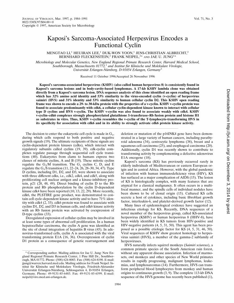
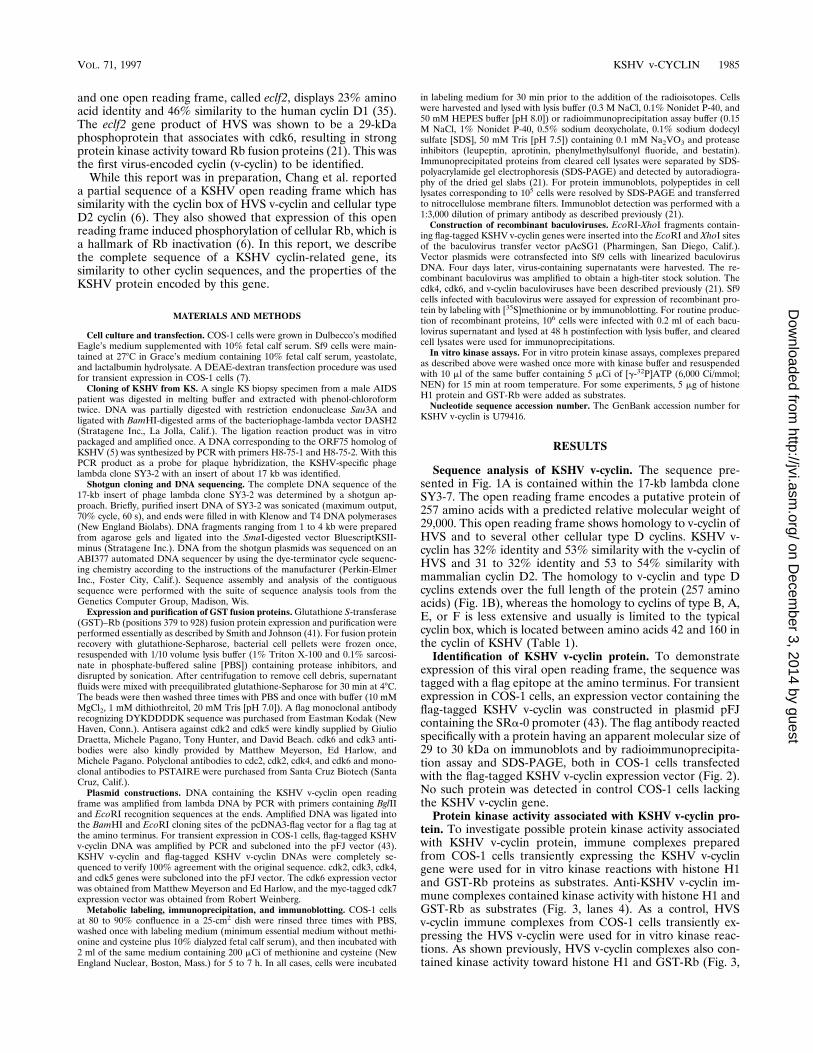
Sequence analysis of KSHV v-cyclin. The sequence pre-sented in Fig. 1A is contained within the 17-kb lambda cloneSY3-7. The open reading frame encodes a putative protein of257 amino acids with a predicted relative molecular weight of29,000. This open reading frame shows homology to v-cyclin ofHVS and to several other cellular type D cyclins. KSHV v-cyclin has 32% identity and 53% similarity with the v-cyclin ofHVS and 31 to 32% identity and 53 to 54% similarity withmammalian cyclin D2. The homology to v-cyclin and type Dcyclins extends over the full length of the protein (257 aminoacids) (Fig. 1B), whereas the homology to cyclins of type B, A,E, or F is less extensive and usually is limited to the typicalcyclin box, which is located between amino acids 42 and 160 inthe cyclin of KSHV (Table 1).Identification of KSHV v-cyclin protein. To demonstrate
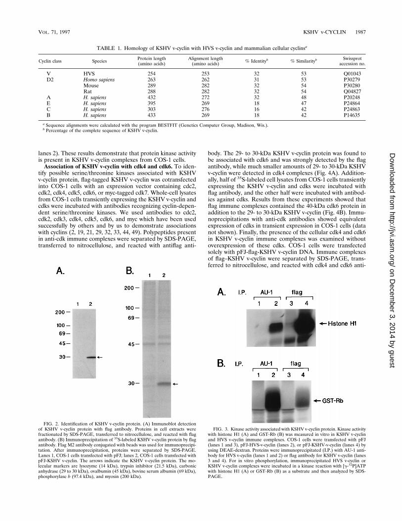
expression of this viral open reading frame, the sequence wastagged with a flag epitope at the amino terminus. For transientexpression in COS-1 cells, an expression vector containing theflag-tagged KSHV v-cyclin was constructed in plasmid pFJcontaining the SRa-0 promoter (43). The flag antibody reactedspecifically with a protein having an apparent molecular size of29 to 30 kDa on immunoblots and by radioimmunoprecipita-tion assay and SDS-PAGE, both in COS-1 cells transfectedwith the flag-tagged KSHV v-cyclin expression vector (Fig. 2).No such protein was detected in control COS-1 cells lackingthe KSHV v-cyclin gene.Protein kinase activity associated with KSHV v-cyclin pro-
tein. To investigate possible protein kinase activity associatedwith KSHV v-cyclin protein, immune complexes preparedfrom COS-1 cells transiently expressing the KSHV v-cyclingene were used for in vitro kinase reactions with histone H1and GST-Rb proteins as substrates. Anti-KSHV v-cyclin im-mune complexes contained kinase activity with histone H1 andGST-Rb as substrates (Fig. 3, lanes 4). As a control, HVSv-cyclin immune complexes from COS-1 cells transiently ex-pressing the HVS v-cyclin were used for in vitro kinase reac-tions. As shown previously, HVS v-cyclin complexes also con-tained kinase activity toward histone H1 and GST-Rb (Fig. 3,
VOL. 71, 1997 KSHV v-CYCLIN 1985
on Decem
ber 3, 2014 by guesthttp://jvi.asm
.org/D
ownloaded from
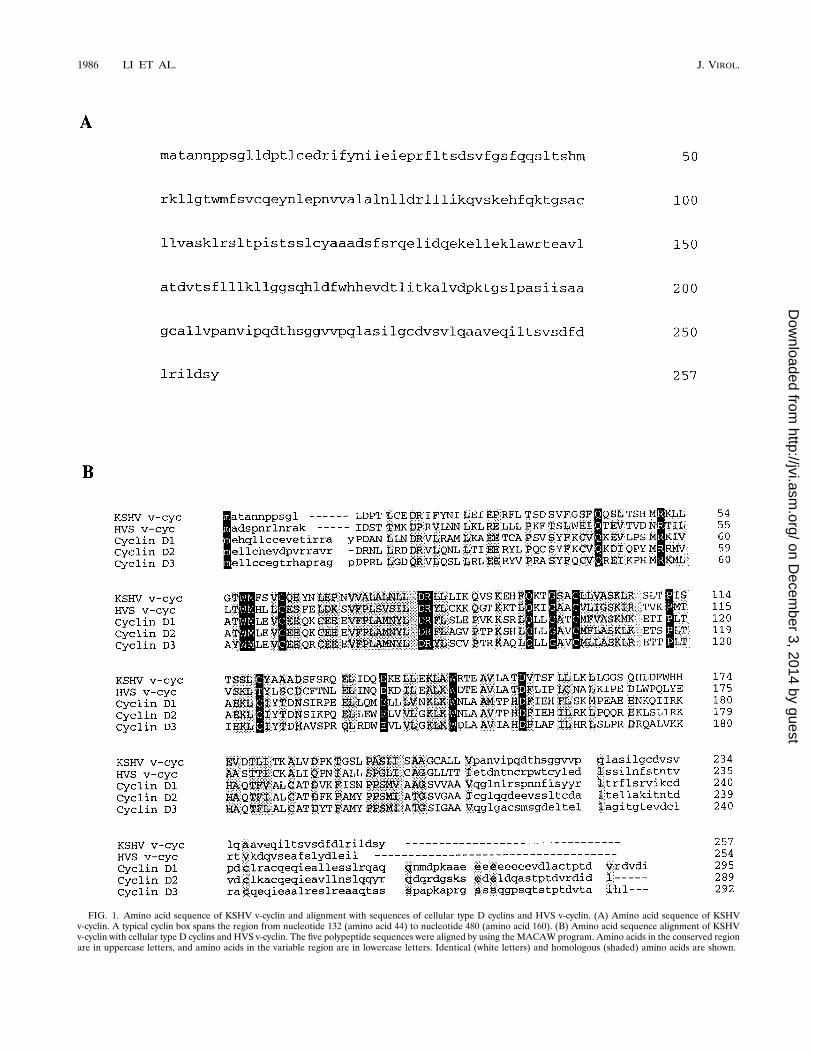
FIG. 1. Amino acid sequence of KSHV v-cyclin and alignment with sequences of cellular type D cyclins and HVS v-cyclin. (A) Amino acid sequence of KSHVv-cyclin. A typical cyclin box spans the region from nucleotide 132 (amino acid 44) to nucleotide 480 (amino acid 160). (B) Amino acid sequence alignment of KSHVv-cyclin with cellular type D cyclins and HVS v-cyclin. The five polypeptide sequences were aligned by using the MACAW program. Amino acids in the conserved regionare in uppercase letters, and amino acids in the variable region are in lowercase letters. Identical (white letters) and homologous (shaded) amino acids are shown.
1986 LI ET AL. J. VIROL.
on Decem
ber 3, 2014 by guesthttp://jvi.asm
.org/D
ownloaded from
lanes 2). These results demonstrate that protein kinase activityis present in KSHV v-cyclin complexes from COS-1 cells.Association of KSHV v-cyclin with cdk4 and cdk6. To iden-
tify possible serine/threonine kinases associated with KSHVv-cyclin protein, flag-tagged KSHV v-cyclin was cotransfectedinto COS-1 cells with an expression vector containing cdc2,cdk2, cdk4, cdk5, cdk6, or myc-tagged cdk7. Whole-cell lysatesfrom COS-1 cells transiently expressing the KSHV v-cyclin andcdks were incubated with antibodies recognizing cyclin-depen-dent serine/threonine kinases. We used antibodies to cdc2,cdk2, cdk3, cdk4, cdk5, cdk6, and myc which have been usedsuccessfully by others and by us to demonstrate associationswith cyclins (2, 19, 21, 29, 32, 33, 44, 49). Polypeptides presentin anti-cdk immune complexes were separated by SDS-PAGE,transferred to nitrocellulose, and reacted with antiflag anti-
body. The 29- to 30-kDa KSHV v-cyclin protein was found tobe associated with cdk6 and was strongly detected by the flagantibody, while much smaller amounts of 29- to 30-kDa KSHVv-cyclin were detected in cdk4 complexes (Fig. 4A). Addition-ally, half of 35S-labeled cell lysates from COS-1 cells transientlyexpressing the KSHV v-cyclin and cdks were incubated withflag antibody, and the other half were incubated with antibod-ies against cdks. Results from these experiments showed thatflag immune complexes contained the 40-kDa cdk6 protein inaddition to the 29- to 30-kDa KSHV v-cyclin (Fig. 4B). Immu-noprecipitations with anti-cdk antibodies showed equivalentexpression of cdks in transient expression in COS-1 cells (datanot shown). Finally, the presence of the cellular cdk4 and cdk6in KSHV v-cyclin immune complexes was examined withoutoverexpression of these cdks. COS-1 cells were transfectedsolely with pFJ-flag-KSHV v-cyclin DNA. Immune complexesof flag–KSHV v-cyclin were separated by SDS-PAGE, trans-ferred to nitrocellulose, and reacted with cdk4 and cdk6 anti-
TABLE 1. Homology of KSHV v-cyclin with HVS v-cyclin and mammalian cellular cyclinsa
Cyclin class Species Protein length(amino acids)
Alignment length(amino acids) % Identityb % Similarityb Swissprot
accession no.
V HVS 254 253 32 53 Q01043D2 Homo sapiens 263 262 31 53 P30279
Mouse 289 282 32 54 P30280Rat 288 282 32 54 Q04827
A H. sapiens 432 272 32 48 P20248E H. sapiens 395 269 18 47 P24864C H. sapiens 303 276 16 42 P24863B H. sapiens 433 269 18 42 P14635
a Sequence alignments were calculated with the program BESTFIT (Genetics Computer Group, Madison, Wis.).b Percentage of the complete sequence of KSHV v-cyclin.
FIG. 2. Identification of KSHV v-cyclin protein. (A) Immunoblot detectionof KSHV v-cyclin protein with flag antibody. Proteins in cell extracts werefractionated by SDS-PAGE, transferred to nitrocellulose, and reacted with flagantibody. (B) Immunoprecipitation of 35S-labeled KSHV v-cyclin protein by flagantibody. Flag M2 antibody conjugated with beads was used for immunoprecipi-tation. After immunoprecipitation, proteins were separated by SDS-PAGE.Lanes 1, COS-1 cells transfected with pFJ; lanes 2, COS-1 cells transfected withpFJ-KSHV v-cyclin. The arrows indicate the KSHV v-cyclin protein. The mo-lecular markers are lysozyme (14 kDa), trypsin inhibitor (21.5 kDa), carbonicanhydrase (29 to 30 kDa), ovalbumin (45 kDa), bovine serum albumin (69 kDa),phosphorylase b (97.4 kDa), and myosin (200 kDa).
FIG. 3. Kinase activity associated with KSHV v-cyclin protein. Kinase activitywith histone H1 (A) and GST-Rb (B) was measured in vitro in KSHV v-cyclinand HVS v-cyclin immune complexes. COS-1 cells were transfected with pFJ(lanes 1 and 3), pFJ-HVS-v-cyclin (lanes 2), or pFJ-KSHV-v-cyclin (lanes 4) byusing DEAE-dextran. Proteins were immunoprecipitated (I.P.) with AU-1 anti-body for HVS v-cyclin (lanes 1 and 2) or flag antibody for KSHV v-cyclin (lanes3 and 4). For in vitro phosphorylation, immunoprecipitated HVS v-cyclin orKSHV v-cyclin complexes were incubated in a kinase reaction with [g-32P]ATPwith histone H1 (A) or GST-Rb (B) as a substrate and then analyzed by SDS-PAGE.
VOL. 71, 1997 KSHV v-CYCLIN 1987
on Decem
ber 3, 2014 by guesthttp://jvi.asm
.org/D
ownloaded from
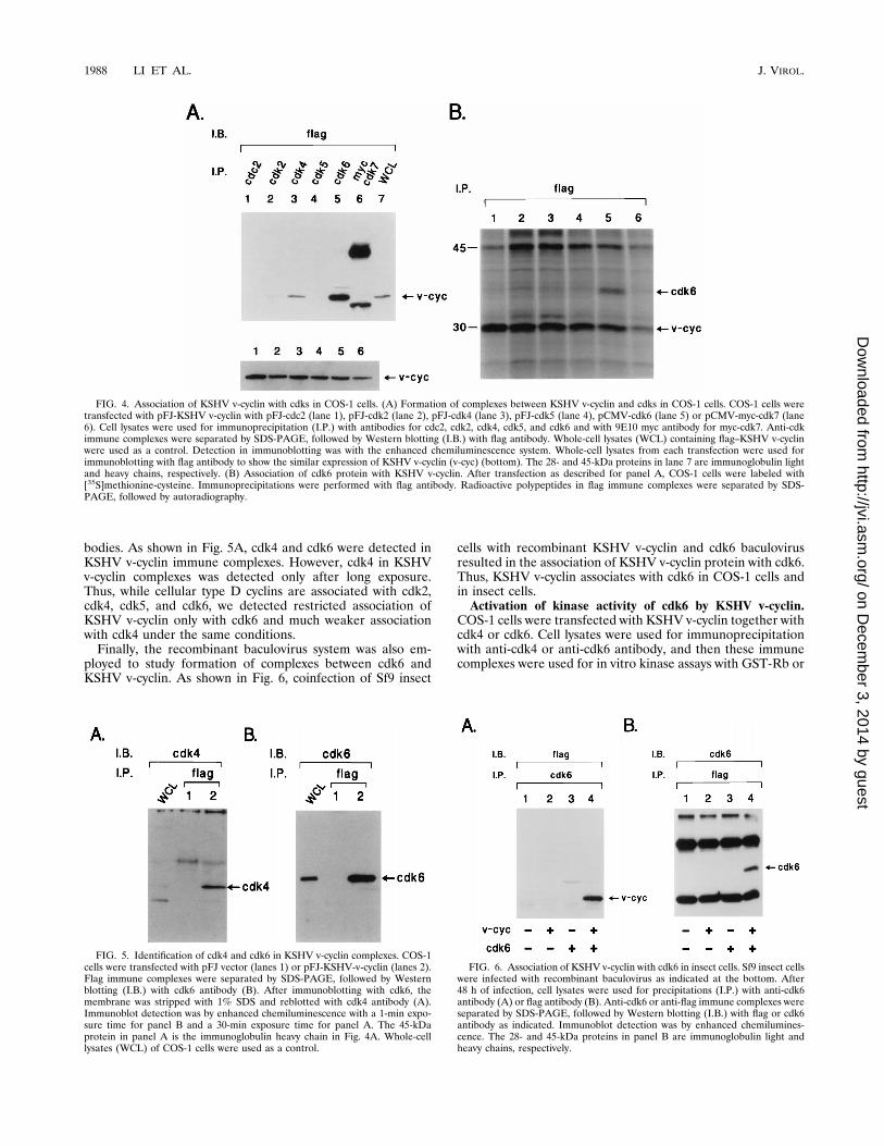
bodies. As shown in Fig. 5A, cdk4 and cdk6 were detected inKSHV v-cyclin immune complexes. However, cdk4 in KSHVv-cyclin complexes was detected only after long exposure.Thus, while cellular type D cyclins are associated with cdk2,cdk4, cdk5, and cdk6, we detected restricted association ofKSHV v-cyclin only with cdk6 and much weaker associationwith cdk4 under the same conditions.Finally, the recombinant baculovirus system was also em-
ployed to study formation of complexes between cdk6 andKSHV v-cyclin. As shown in Fig. 6, coinfection of Sf9 insect
cells with recombinant KSHV v-cyclin and cdk6 baculovirusresulted in the association of KSHV v-cyclin protein with cdk6.Thus, KSHV v-cyclin associates with cdk6 in COS-1 cells andin insect cells.Activation of kinase activity of cdk6 by KSHV v-cyclin.
COS-1 cells were transfected with KSHV v-cyclin together withcdk4 or cdk6. Cell lysates were used for immunoprecipitationwith anti-cdk4 or anti-cdk6 antibody, and then these immunecomplexes were used for in vitro kinase assays with GST-Rb or
FIG. 4. Association of KSHV v-cyclin with cdks in COS-1 cells. (A) Formation of complexes between KSHV v-cyclin and cdks in COS-1 cells. COS-1 cells weretransfected with pFJ-KSHV v-cyclin with pFJ-cdc2 (lane 1), pFJ-cdk2 (lane 2), pFJ-cdk4 (lane 3), pFJ-cdk5 (lane 4), pCMV-cdk6 (lane 5) or pCMV-myc-cdk7 (lane6). Cell lysates were used for immunoprecipitation (I.P.) with antibodies for cdc2, cdk2, cdk4, cdk5, and cdk6 and with 9E10 myc antibody for myc-cdk7. Anti-cdkimmune complexes were separated by SDS-PAGE, followed by Western blotting (I.B.) with flag antibody. Whole-cell lysates (WCL) containing flag–KSHV v-cyclinwere used as a control. Detection in immunoblotting was with the enhanced chemiluminescence system. Whole-cell lysates from each transfection were used forimmunoblotting with flag antibody to show the similar expression of KSHV v-cyclin (v-cyc) (bottom). The 28- and 45-kDa proteins in lane 7 are immunoglobulin lightand heavy chains, respectively. (B) Association of cdk6 protein with KSHV v-cyclin. After transfection as described for panel A, COS-1 cells were labeled with[35S]methionine-cysteine. Immunoprecipitations were performed with flag antibody. Radioactive polypeptides in flag immune complexes were separated by SDS-PAGE, followed by autoradiography.
FIG. 5. Identification of cdk4 and cdk6 in KSHV v-cyclin complexes. COS-1cells were transfected with pFJ vector (lanes 1) or pFJ-KSHV-v-cyclin (lanes 2).Flag immune complexes were separated by SDS-PAGE, followed by Westernblotting (I.B.) with cdk6 antibody (B). After immunoblotting with cdk6, themembrane was stripped with 1% SDS and reblotted with cdk4 antibody (A).Immunoblot detection was by enhanced chemiluminescence with a 1-min expo-sure time for panel B and a 30-min exposure time for panel A. The 45-kDaprotein in panel A is the immunoglobulin heavy chain in Fig. 4A. Whole-celllysates (WCL) of COS-1 cells were used as a control.
FIG. 6. Association of KSHV v-cyclin with cdk6 in insect cells. Sf9 insect cellswere infected with recombinant baculovirus as indicated at the bottom. After48 h of infection, cell lysates were used for precipitations (I.P.) with anti-cdk6antibody (A) or flag antibody (B). Anti-cdk6 or anti-flag immune complexes wereseparated by SDS-PAGE, followed by Western blotting (I.B.) with flag or cdk6antibody as indicated. Immunoblot detection was by enhanced chemilumines-cence. The 28- and 45-kDa proteins in panel B are immunoglobulin light andheavy chains, respectively.
1988 LI ET AL. J. VIROL.
on Decem
ber 3, 2014 by guesthttp://jvi.asm
.org/D
ownloaded from
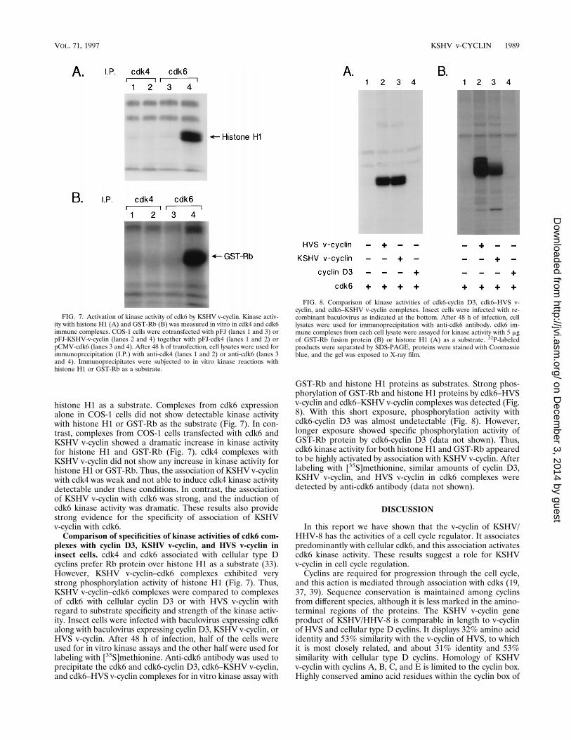
histone H1 as a substrate. Complexes from cdk6 expressionalone in COS-1 cells did not show detectable kinase activitywith histone H1 or GST-Rb as the substrate (Fig. 7). In con-trast, complexes from COS-1 cells transfected with cdk6 andKSHV v-cyclin showed a dramatic increase in kinase activityfor histone H1 and GST-Rb (Fig. 7). cdk4 complexes withKSHV v-cyclin did not show any increase in kinase activity forhistone H1 or GST-Rb. Thus, the association of KSHV v-cyclinwith cdk4 was weak and not able to induce cdk4 kinase activitydetectable under these conditions. In contrast, the associationof KSHV v-cyclin with cdk6 was strong, and the induction ofcdk6 kinase activity was dramatic. These results also providestrong evidence for the specificity of association of KSHVv-cyclin with cdk6.Comparison of specificities of kinase activities of cdk6 com-
plexes with cyclin D3, KSHV v-cyclin, and HVS v-cyclin ininsect cells. cdk4 and cdk6 associated with cellular type Dcyclins prefer Rb protein over histone H1 as a substrate (33).However, KSHV v-cyclin–cdk6 complexes exhibited verystrong phosphorylation activity of histone H1 (Fig. 7). Thus,KSHV v-cyclin–cdk6 complexes were compared to complexesof cdk6 with cellular cyclin D3 or with HVS v-cyclin withregard to substrate specificity and strength of the kinase activ-ity. Insect cells were infected with baculovirus expressing cdk6along with baculovirus expressing cyclin D3, KSHV v-cyclin, orHVS v-cyclin. After 48 h of infection, half of the cells wereused for in vitro kinase assays and the other half were used forlabeling with [35S]methionine. Anti-cdk6 antibody was used toprecipitate the cdk6 and cdk6-cyclin D3, cdk6–KSHV v-cyclin,and cdk6–HVS v-cyclin complexes for in vitro kinase assay with
GST-Rb and histone H1 proteins as substrates. Strong phos-phorylation of GST-Rb and histone H1 proteins by cdk6–HVSv-cyclin and cdk6–KSHV v-cyclin complexes was detected (Fig.8). With this short exposure, phosphorylation activity withcdk6-cyclin D3 was almost undetectable (Fig. 8). However,longer exposure showed specific phosphorylation activity ofGST-Rb protein by cdk6-cyclin D3 (data not shown). Thus,cdk6 kinase activity for both histone H1 and GST-Rb appearedto be highly activated by association with KSHV v-cyclin. Afterlabeling with [35S]methionine, similar amounts of cyclin D3,KSHV v-cyclin, and HVS v-cyclin in cdk6 complexes weredetected by anti-cdk6 antibody (data not shown).
DISCUSSION
In this report we have shown that the v-cyclin of KSHV/HHV-8 has the activities of a cell cycle regulator. It associatespredominantly with cellular cdk6, and this association activatescdk6 kinase activity. These results suggest a role for KSHVv-cyclin in cell cycle regulation.Cyclins are required for progression through the cell cycle,
and this action is mediated through association with cdks (19,37, 39). Sequence conservation is maintained among cyclinsfrom different species, although it is less marked in the amino-terminal regions of the proteins. The KSHV v-cyclin geneproduct of KSHV/HHV-8 is comparable in length to v-cyclinof HVS and cellular type D cyclins. It displays 32% amino acididentity and 53% similarity with the v-cyclin of HVS, to whichit is most closely related, and about 31% identity and 53%similarity with cellular type D cyclins. Homology of KSHVv-cyclin with cyclins A, B, C, and E is limited to the cyclin box.Highly conserved amino acid residues within the cyclin box of
FIG. 7. Activation of kinase activity of cdk6 by KSHV v-cyclin. Kinase activ-ity with histone H1 (A) and GST-Rb (B) was measured in vitro in cdk4 and cdk6immune complexes. COS-1 cells were cotransfected with pFJ (lanes 1 and 3) orpFJ-KSHV-v-cyclin (lanes 2 and 4) together with pFJ-cdk4 (lanes 1 and 2) orpCMV-cdk6 (lanes 3 and 4). After 48 h of transfection, cell lysates were used forimmunoprecipitation (I.P.) with anti-cdk4 (lanes 1 and 2) or anti-cdk6 (lanes 3and 4). Immunoprecipitates were subjected to in vitro kinase reactions withhistone H1 or GST-Rb as a substrate.
FIG. 8. Comparison of kinase activities of cdk6-cyclin D3, cdk6–HVS v-cyclin, and cdk6–KSHV v-cyclin complexes. Insect cells were infected with re-combinant baculovirus as indicated at the bottom. After 48 h of infection, celllysates were used for immunoprecipitation with anti-cdk6 antibody. cdk6 im-mune complexes from each cell lysate were assayed for kinase activity with 5 mgof GST-Rb fusion protein (B) or histone H1 (A) as a substrate. 32P-labeledproducts were separated by SDS-PAGE, proteins were stained with Coomassieblue, and the gel was exposed to X-ray film.
VOL. 71, 1997 KSHV v-CYCLIN 1989
on Decem
ber 3, 2014 by guesthttp://jvi.asm
.org/D
ownloaded from
cyclin A have been shown to represent a contact region forinteraction with cdc2 and a functional domain for activation ofcdc2 kinase activity (27). Our previous mutational analyseshave shown that corresponding amino acids in HVS v-cyclinare also important for the association with cdk6 and activationof cdk6 kinase activity (21). KSHV v-cyclin also has a highdegree of conservation in amino acids residues within the cy-clin box, suggesting that this conserved region is likely to be atarget site for binding to cdks (6). Notably, KSHV and HVSv-cyclins and cellular type D cyclins have substantial N-termi-nal truncations relative to other cellular cyclins.Cellular cyclins are found in association with cdks, and their
activity and function in regulating cell cycle progression areabsolutely dependent on this interaction. Cyclins D1, D2, andD3 were previously found to be associated with four differentcdks, i.e., cdk2, cdk4, cdk5, and cdk6, along with proliferatingcell nuclear antigen and p21 or p16 kinase-inhibitory protein(17, 29, 48, 49). Also, recent reports showed that the mamma-lian cdc37 gene product associates with cyclin D-cdk4 com-plexes and that this association stabilizes cyclin D-cdk4 com-plexes (8, 42). Similarly to v-cyclin of HVS, which associatesexclusively with cdk6, KSHV v-cyclin also associates predom-inantly with cdk6. Additionally, interaction with cdk4 wasweakly detected in transient expression in COS-1 cells. Thecatalytic subunit for type D cyclins in macrophages and fibro-blasts is mainly cdk4, while cdk6 as the catalytic subunit fortype D cyclins appears to predominate in lymphoid cells (32,33). Since T lymphocytes harbor HVS persistently in vivo andare the target of HVS transformation, a primary associationbetween cdk6 and v-cyclin seems physiologically relevant.KSHV/HHV-8 has been localized to lymphocytes and endo-thelial cells in KS patients (5) and to the transformed B cells inbody-cavity-based lymphomas (4) and primary effusion lym-phomas (4). The specificity of KSHV v-cyclin for cdk6 is thusconsistent with its presence in lymphocytes; the relevance ofthis association for endothelial cells and the spindle cells in KSremains to be explored.Although the virus-encoded cyclins HVS v-cyclin and KSHV
v-cyclin have sequence homology to type D cyclins and activatecellular cdk kinase activity, these cyclins differ from cellulartype D cyclins in their specificity for cdk binding and their levelof activation of cdk kinase activity. These virus-encoded cyclinswere found to associate principally with cdk6, while cellulartype D cyclins have been found to associate with four differentcdks. Also, the kinase activity of cdk6 is much more greatlyinduced by association with v-cyclin than by association withthe cellular D cyclin. Therefore, the virus-encoded cyclins ex-hibit a marked discrimination for the catalytic subunit withwhich they associate and a high level of kinase activation. Ahigh level of activation of cdk kinase activity by these viralcyclins may result in much greater activation of cell cycle pro-cesses. The similarities in the activities of KSHV and HVSv-cyclins suggest that v-cyclin is not responsible for differencesin the cell type specificities of these viruses or for differences inthe diseases they induce. Whether v-cyclin in the context ofeither virus is important for growth transformation of one ormore cell types remains to be determined.
ACKNOWLEDGMENTS
We thank R. Weinberg, C. Sherr, M. Pagano, E. Harlow, M. Mey-erson, S. Dowdy, P. Hinds, D. Beach, T. Hunter, D. Morgan, S.Elledge, G. Draetta, A. Arnold, T. Kanda, and M. Kaelin for providingDNAs and antisera. We especially thank Ronald Desrosiers for dis-cussions, encouragement, and manuscript preparation. We also thankJ. Newton and T. Connors for manuscript preparation.This work was supported by U.S. Public Health Service grants
CA31363 and RR00168 and by the Mildred Scheel foundation forcancer research (grant W134/94/FL2).
REFERENCES1. Albrecht, J.-C., J. Nicholas, D. Biller, K. R. Cameron, B. Biesinger, C.Newman, S. Wittman, M. Craxton, H. Coleman, B. Fleckenstein, and R. W.Honness. 1992. Primary structure of the herpesvirus saimiri genome. J. Virol.66:5047–5058.
2. Bates, S., L. Bonetta, D. MacAllan, D. Parry, A. Holder, C. Dickson, and G.Peters. 1994. CDK6 (PLSTIRE) and CDK4 (PSK-J3) are a distinct subset ofthe cyclin-dependent kinases that associate with cyclin D1. Oncogene 9:71–79.
3. Biesinger, B., I. Muller-Fleckenstein, B. Simmer, G. Lang, S. Wittmann, E.Platzer, R. C. Desrosiers, and B. Fleckenstein. 1992. Stable growth trans-formation of human T lymphocytes by herpesvirus saimiri. Proc. Natl. Acad.Sci. USA 89:3116–3119.
4. Cesarman, E., Y. Chang, P. S. Moore, J. W. Said, and D. M. Knowles. 1996.Kaposi’s sarcoma-associated herpesvirus-like DNA sequences in AIDS-re-lated body-cavity-based lymphomas. N. Engl. J. Med. 332:1186–1191.
5. Chang, Y., E. Cesarman, M. S. Pessin, F. Lee, J. Culpepper, D. M. Knowles,and P. S. Moore. 1994. Identification of herpesvirus-like DNA sequences inAIDS-associated Kaposi’s sarcoma. Science 266:1865–1869.
6. Chang, Y., P. S. Moore, S. I. Talbot, C. H. Boshoff, T. Zarkowska, D.Godden-Kent, H. Paterson, R. A. Weiss, and S. Mittnacht. 1996. Cyclinencoded by KS herpesvirus. Nature 382:410.
7. Cullen, B. R. 1987. Use of eukaryotic expression technology in the functionalanalysis of cloned genes. Methods Enzymol. 152:684–703.
8. Dai, K., R. Kobayashi, and D. Beach. 1996. Physical interaction of mamma-lian CDC37 with CDK4. J. Biol. Chem. 271:22030–22634.
9. Desrosiers, R. C., D. Silva, L. M. Waldron, and N. L. Letvin. 1986. Nonon-cogenic deletion mutants of herpesvirus saimiri are defective for in vitroimmortalization. J. Virol. 57:701–705.
10. Dowdy, S. F., P. W. Hinds, K. Louie, S. I. Reed, A. Arnold, and R. A.Weinberg. 1993. Physical interaction of the retinoblastoma protein withhuman D cyclins. Cell 73:499–511.
11. Dulic, V., E. Lees, and S. I. Reed. 1992. Association of human cyclin E witha periodic G1-S phase protein kinase. Science 257:1958–1961.
12. Ensoli, B., R. Gendelman, P. Markham, V. Fiorelli, S. Colombini, M.Raffeld, A. Cafaro, H.-K. Chang, J. N. Brady, and R. C. Gallo. 1994. Synergybetween basic fibroblast growth factor and HIV-1 tat protein in induction ofKaposi’s sarcoma. Nature 371:674–680.
13. Ewin, M. E., H. K. Sluss, C. J. Sherr, H. Matsushine, J.-Y. Kato, and D. M.Livingston. 1993. Functional interactions of the retinoblastoma protein withmammalian D-type cyclins. Cell 73:487–497.
14. Fleckenstein, B., and R. C. Desrosiers. 1982. Herpesvirus saimiri and her-pesvirus ateles, p. 253–332. In B. Roizman (ed.), The herpesviruses. PlenumPublishing Corporation, New York, N.Y.
15. Ganem, D. 1995. Viruses, cytokines and Kaposi’s sarcoma. Curr. Biol. 5:469–471.
16. Giordano, A., A. W. Murray, and M. W. Kirschner. 1991. A 60 kd cdc2-associated polypeptide complexes with the E1A proteins in adenovirus-infected cells. Cell 58:981–990.
17. Harper, J. W., G. R. Adami, N. Wei, K. Keyomarsi, and S. J. Elledge. 1993.The p21 cdk-interacting protein cip1 is a potent inhibitor of G1 cyclin-dependent kinases. Cell 75:805–816.
18. Hinds, P. W., S. F. Dowdy, E. N. Eaton, A. Arnold, and R. A. Weinberg. 1994.Function of a human cyclin gene as an oncogene. Proc. Natl. Acad. Sci. USA91:709–713.
19. Hunter, T., and J. Pines. 1991. Cyclins and cancer. Cell 66:1071–1074.20. Jiang, W., S. Kahan, N. Tomita, Y. Zhang, S. Lu, and B. Weinstein. 1992.
Amplification and expression of the human cyclin D gene in esophagealcancer. Cancer Res. 52:2980–2983.
21. Jung, J. U., M. Stager, and R. C. Desrosiers. 1994. Virus-encoded cyclin.Mol. Cell Biol. 14:7235–7244.
22. Kato, J.-Y., H. Matsushime, S. W. Hiebert, M. E. Ewen, and C. J. Sherr.1993. Direct binding of cyclin D to the retinoblastoma gene product (pRb)and pRb phosphorylation by the cyclin D-dependent kinase CDK4. GenesDev. 7:331–342.
23. Koff, A., F. Cross, A. Fisher, J. Schumacher, K. Leugellec, M. Philippe, andJ. M. Roberts. 1991. Human cyclin E, a new cyclin that interacts with twomembers of the CDC2 gene family. Cell 66:1217–1228.
24. Koff, A., A. Giordano, D. Desai, K. Yamashita, J. W. Harper, S. Elledge, T.Nishomoto, D. O. Morgan, B. R. Franza, and J. M. Roberts. 1992. Formationand activation of cyclin E-cdk2 complex during the G1 phase of the humancell cycle. Science 257:1689–1694.
25. Lammie, G., V. Fantl, R. Smith, E. Schuuring, S. Brookes, R. Michalides, C.Dickson, A. Arnold, and G. Peters. 1991. D11S287, a putative oncogene onchromosome 11q13, is amplified and expressed in squamous cell and mam-mary carcinomas and linked to BCL-1. Oncogene 6:439–444.
26. Lees, E., B. Faha, V. Dulic, S. I. Reed, and E. Harlow. 1993. Cyclin E/cdk2and cyclin A/cdk2 kinase associate with p107 and E2F in a temporally distinctmanner. Genes Dev. 5:1874–1885.
1990 LI ET AL. J. VIROL.
on Decem
ber 3, 2014 by guesthttp://jvi.asm
.org/D
ownloaded from
27. Lees, E., and E. Harlow. 1993. Sequences within the conserved cyclin box ofhuman cyclin A are sufficient for binding to and activation of cdc2 kinase.Mol. Cell. Biol. 13:1194–1201.
28. Lew, D. J., V. Dulic, and S. I. Reed. 1991. Isolation of three novel humancyclins by rescue of G1 cyclin (cln) function in yeast. Cell 66:1197–1206.
29. Matsushime, H., M. E. Ewen, D. K. Strom, J.-Y. Kato, S. K. Hanks, M. F.Roussel, and C. J. Sherr. 1992. Identification and properties of an atypicalcatalytic subunit (p34PSK-J3/cdk4) for mammalian D type cyclin. Cell 71:323–334.
30. Matsushime, H., M. F. Roussel, R. A. Ashmun, and C. J. Sherr. 1991.Colony-stimulating factor 1 regulates novel cyclins during the G1 phase ofthe cell cycle. Cell 65:701–713.
31. Mesri, E. A., E. Cesarman, L. Arvanitakis, S. Rafii, M. A. S. Moore, D. N.Posnett, D. M. Knowles, and A. S. Asch. 1996. Human herpesvirus-8/Kapo-si’s sarcoma-associated herpesvirus is a new transmissible virus that infects Bcells. J. Exp. Med. 183:2385–2389.
32. Meyerson, M., G. H. Enders, C.-L. Wu, L.-K. Su, C. Corka, C. Nelson, E.Harlow, and L.-H. Tsai. 1992. A family of human cdc2-related proteinkinases. EMBO J. 11:2909–2917.
33. Meyerson, M., and E. Harlow. 1994. Identification of G1 kinase activity forcdk6, a novel cyclin D partner. Mol. Cell. Biol. 14:2077–2086.
34. Motokura, T., T. Bloom, G. Kim, H. Juppner, J. V. Ruderman, H. M.Kronenberg, and A. Arnold. 1991. A novel cyclin encoded by a bcl1 linkedcandidate oncogene. Nature 350:512–515.
35. Nicholas, J., K. R. Cameron, and R. W. Honess. 1992. Herpesvirus saimiriencodes homologues of G protein-coupled receptors and cyclins. Nature355:362–365.
36. Pines, J., and T. Hunter. 1990. Human cyclin A is adenovirus E1-A-associ-ated protein p60, and behaves differently from cyclin B. Nature 346:760–763.
37. Reed, S. I. 1992. The role of p34 kinases in the G1 to S-phase transition.Annu. Rev. Cell Biol. 8:529–561.
38. Rosenberg, C., H. Kim, T. Shows, H. M. Kronenberg, and A. Arnold. 1991.Rearrangement and overexpression of D11S287E, a candidate oncogene onchromosome 11q13 in benign parathyroid tumors. Oncogene 6:449–453.
39. Sherr, C. J. 1993. Mammalian G1 cyclins. Cell 73:1059–1065.
40. Sherr, C. J., and J. M. Roberts. 1995. Inhibitors of mammalian G1 cyclin-dependent kinases. Genes Dev. 9:1149–1163.
41. Smith, D. B., and K. S. Johnson. 1988. Single-step purification of polypep-tides expressed in Escherichia coli as fusion with glutathione S-transferase.Gene 67:31–40.
42. Stepanova, L., X. Leng, S. B. Parker, and J. W. Harper. 1996. Mammalianp50Cdc37 is a protein kinase-targeting subunit of Hsp90 that binds and sta-bilizes Cdk4. Genes Dev. 10:1491–1502.
43. Takebe, Y., M. Seiki, J.-I. Fujisawa, P. Hoy, K. Yokota, K.-I. Arai, M.Yoshida, and N. Arai. 1988. SRa promoter: an efficient and versatile mam-malian cDNA expression system composed of the simian virus 40 earlypromoter and the R-U5 segment of human T-cell leukemia virus type 1 longterminal repeat. Mol. Cell. Biol. 8:466–472.
44. Tsai, L.-H., E. Harlow, and M. Meyerson. 1991. Isolation of the human cdk2gene that encodes the cyclin A-and adenovirus E1A-associated p33 kinase.Nature 353:174–177.
45. Wang, J., X. Chenivesse, B. Henglein, and C. Brechot. 1993. Hepatitis B virusintegration in a cyclin A gene in a hepatocellular carcinoma. Nature 343:555–557.
46. Whitby, D., M. R. Howard, M. Tenant-Flowers, N. S. Brink, A. Copas, C.Boshoff, T. Hatzioannou, F. E. A. Suggett, D. M. Aldam, A. S. Denton, R. F.Miller, I. V. D. Weller, R. A. Weiss, R. S. Tedder, and T. F. Schulz. 1995.Detection of Kaposi sarcoma associated herpesvirus in peripheral blood ofHIV-infected individuals and progression to Kaposi’s sarcoma. Lancet 346:799–802.
47. Xiong, Y., T. Connolly, B. Futcher, and D. Beach. 1991. Human D-typecyclin. Cell 65:691–699.
48. Xiong, Y., G. J. Hannon, H. Zhang, D. Casso, R. Kobayashi, and D. Beach.1993. p21 is a universal inhibitor of cyclin kinases. Nature 366:701–704.
49. Xiong, Y., H. Zhang, and D. Beach. 1992. D-type cyclins associate withmultiple protein kinases and the DNA replication and repair factor PCNA.Cell 71:505–514.
50. Zhong, W., H. Wang, B. Herndier, and D. Ganem. 1996. Restricted expres-sion of Kaposi sarcoma-associated herpesvirus (human herpesvirus 8) genesin Kaposi sarcoma. Proc. Natl. Acad. Sci. USA 93:6641–6646.
VOL. 71, 1997 KSHV v-CYCLIN 1991
on Decem
ber 3, 2014 by guesthttp://jvi.asm
.org/D
ownloaded from