decreased nucleotide excision repair in steatotic livers associates with...
TRANSCRIPT

Dm
MMWa
b
c
d
e
a
ARRAA
KNMNCO
1
c
p(ms
Nb
vG
0d
Mutation Research 736 (2012) 75– 81
Contents lists available at SciVerse ScienceDirect
Mutation Research/Fundamental and MolecularMechanisms of Mutagenesis
jo ur n al hom ep a ge: www.elsev ier .com/ locate /molmutC om mun i ty a ddress : www.elsev ier .com/ locate /mutres
ecreased nucleotide excision repair in steatotic livers associates withyeloperoxidase-immunoreactivity�
arten A. Schultsa, Peter W. Naglea, Sander S. Rensenb, Roger W. Godschalka, Armelle Munniac,arco Pelusoc, Sandra M. Claessend, Jan W. Greveb, Ann Driessene, Froukje J. Verdamb,im A. Buurmanb, Frederik J. van Schootena, Roland K. Chiua,∗
Department of Toxicology, NUTRIM-School for Nutrition, Toxicology and Metabolism, Maastricht University Medical Centre, PO Box 616, 6200 MD Maastricht, The NetherlandsDepartment of Surgery, NUTRIM-School for Nutrition, Toxicology and Metabolism, Maastricht University Medical Centre, PO Box 616, 6200 MD Maastricht, The NetherlandsCancer Risk Factor Branch, ISPO Cancer Prevention and Research Institute, Via Cosimo il Vecchio 2, 50139 Florence, ItalyDepartment of Toxicogenomics, GROW-School for Oncology and Developmental Biology, Maastricht University Medical Centre, PO Box 616, 6200 MD Maastricht, The NetherlandsDepartment of Pathology, NUTRIM-School for Nutrition, Toxicology and Metabolism, Maastricht University Medical Centre, PO Box 616, 6200 MD Maastricht, The Netherlands
r t i c l e i n f o
rticle history:eceived 4 May 2011eceived in revised form 17 October 2011ccepted 1 November 2011vailable online 7 November 2011
eywords:ucleotide excision repairyeloperoxidaseonalcoholic steatohepatitishronic inflammationxidative stress
a b s t r a c t
Chronic inflammation is characterized by the influx of neutrophils and is associated with an increasedproduction of reactive oxygen species that can damage DNA. Oxidative DNA damage is generally thoughtto be involved in the increased risk of cancer in inflamed tissues. We previously demonstrated thatactivated neutrophil mediated oxidative stress results in a reduction in nucleotide excision repair (NER)capacity, which could further enhance mutagenesis. Inflammation and oxidative stress are critical factorsin the progression of nonalcoholic fatty liver disease that is linked with enhanced liver cancer risk. In thisreport, we therefore evaluated the role of neutrophils and the associated oxidative stress in damagerecognition and DNA repair in steatotic livers of 35 severely obese subjects with either nonalcoholicsteatohepatitis (NASH) (n = 17) or steatosis alone (n = 18). The neutrophilic influx in liver was assessed bymyeloperoxidase (MPO) staining and the amount of oxidative DNA damage by measuring M1dG adducts.No differences in M1dG adduct levels were observed between patients with or without NASH and also notbetween individuals with high or low MPO immunoreactivity. However, we found that high expressionof MPO in the liver, irrespective of disease status, reduced the damage recognition capacity as determined
by staining for histone 2AX phosphorylation (�H2AX). This reduction in �H2AX formation in individualswith high MPO immunoreactivity was paralleled by a significant decrease in NER capacity as assessed bya functional repair assay, and was not related to cell proliferation. Thus, the observed reduction in NERcapacity upon hepatic inflammation is associated with and may be a consequence of reduced damagegs suisease
recognition. These findinnonalcoholic fatty liver d
. Introduction
Hepatic inflammation plays a fundamental role in liver can-er development [1] and is increasingly observed in parallel with
Abbreviations: �H2AX, histone 2AX phosphorylation; H2O2, hydrogeneroxide; HCC, hepatocellular carcinoma; HOCl, hypochlorous acid; M1dG, 3-2-deoxy-�-d-erythro-pentofuranosyl)pyrimido [1,2-�]purin-10(3H)-one; MPO,
yeloperoxidase; NAFLD, nonalcoholic fatty liver disease; NASH, nonalcoholicteatohepatitis; NER, nucleotide excision repair; ROS, reactive oxygen species.� This work was supported by the Province of Limburg, The Netherlands, by Senterovem Innovatiegerichte Onderzoeks Programma’s Genomics grant IGE05012, andy Associazione Italiana per la Ricerca sul Cancro, Milan, Italy.∗ Corresponding author. Current address: Department of Radiation Oncology, Uni-
ersity Medical Center Groningen, University of Groningen, P.O. Box 30001, 9700 RBroningen, The Netherlands. Tel.: +31 43 388 1968; fax: +31 43 388 4146.
E-mail address: [email protected] (R.K. Chiu).
027-5107/$ – see front matter © 2011 Elsevier B.V. All rights reserved.oi:10.1016/j.mrfmmm.2011.11.001
ggest a novel mechanism of liver cancer development in patients with.
© 2011 Elsevier B.V. All rights reserved.
the high prevalence of nonalcoholic fatty liver disease (NAFLD).NAFLD is characterized by the accumulation of fat within the liver.Increased levels of hepatic free fatty acids are thought to ini-tiate an inflammatory response culminating in the recruitmentof neutrophils and other leukocytes [2,3] and eventually in theprogression of the benign steatotic stage of NAFLD to the moreadvanced stages of nonalcoholic steatohepatitis (NASH), fibrosis,and cirrhosis [4,5]. Importantly, there is a link between NASHand the development of hepatocellular carcinoma (HCC), whichaccounts for more than 80% of primary liver cancer cases [6].Several groups have suggested that HCC could in fact be the end-point of NASH [7,8]. Although the mechanisms by which NASHcan lead to the development of HCC are not fully understood, it
is known that development of HCC occurs in a significant propor-tion of people with cirrhosis [6]. Oxidative stress caused by reactiveoxygen species (ROS) is an important characteristic of cancer devel-opment [9]. Furthermore, many animal studies have revealed a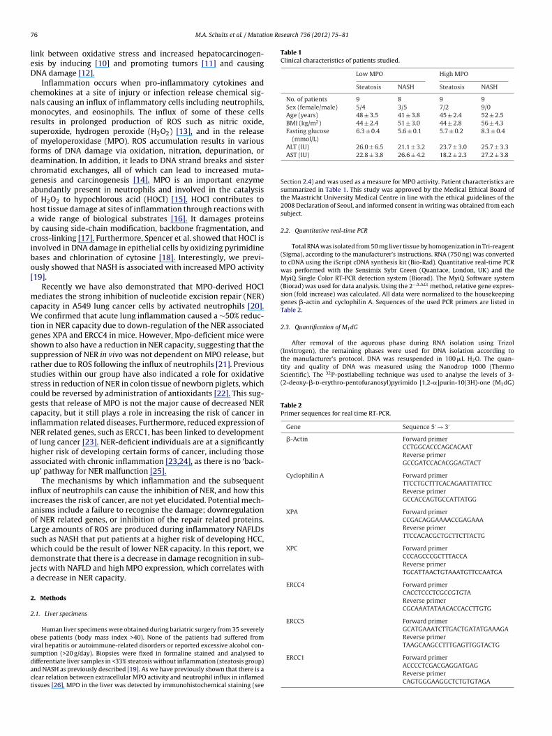
7 ion Research 736 (2012) 75– 81
leD
cnmrsofdcgaohabcibo[
mcWtgssrsscgciNohau
iiaoLswdja
2
2
ovsdact
Table 1Clinical characteristics of patients studied.
Low MPO High MPO
Steatosis NASH Steatosis NASH
No. of patients 9 8 9 9Sex (female/male) 5/4 3/5 7/2 9/0Age (years) 48 ± 3.5 41 ± 3.8 45 ± 2.4 52 ± 2.5BMI (kg/m2) 44 ± 2.4 51 ± 3.0 44 ± 2.8 56 ± 4.3Fasting glucose
(mmol/L)6.3 ± 0.4 5.6 ± 0.1 5.7 ± 0.2 8.3 ± 0.4
the manufacturer’s protocol. DNA was resuspended in 100 �L H2O. The quan-tity and quality of DNA was measured using the Nanodrop 1000 (ThermoScientific). The 32P-postlabelling technique was used to analyse the levels of 3-(2-deoxy-�-d-erythro-pentofuranosyl)pyrimido [1,2-�]purin-10(3H)-one (M1dG)
Table 2Primer sequences for real time RT-PCR.
Gene Sequence 5′ → 3′
�-Actin Forward primerCCTGGCACCCAGCACAATReverse primerGCCGATCCACACGGAGTACT
Cyclophilin A Forward primerTTCCTGCTTTCACAGAATTATTCCReverse primerGCCACCAGTGCCATTATGG
XPA Forward primerCCGACAGGAAAACCGAGAAAReverse primerTTCCACACGCTGCTTCTTACTG
XPC Forward primerCCCAGCCCGCTTTACCAReverse primerTGCATTAACTGTAAATGTTCCAATGA
ERCC4 Forward primerCACCTCCCTCGCCGTGTAReverse primerCGCAAATATAACACCACCTTGTG
ERCC5 Forward primerGCATGAAATCTTGACTGATATGAAAGAReverse primerTAAGCAAGCCTTTGAGTTGGTACTG
6 M.A. Schults et al. / Mutat
ink between oxidative stress and increased hepatocarcinogen-sis by inducing [10] and promoting tumors [11] and causingNA damage [12].
Inflammation occurs when pro-inflammatory cytokines andhemokines at a site of injury or infection release chemical sig-als causing an influx of inflammatory cells including neutrophils,onocytes, and eosinophils. The influx of some of these cells
esults in prolonged production of ROS such as nitric oxide,uperoxide, hydrogen peroxide (H2O2) [13], and in the releasef myeloperoxidase (MPO). ROS accumulation results in variousorms of DNA damage via oxidation, nitration, depurination, oreamination. In addition, it leads to DNA strand breaks and sisterhromatid exchanges, all of which can lead to increased muta-enesis and carcinogenesis [14]. MPO is an important enzymebundantly present in neutrophils and involved in the catalysisf H2O2 to hypochlorous acid (HOCl) [15]. HOCl contributes toost tissue damage at sites of inflammation through reactions with
wide range of biological substrates [16]. It damages proteinsy causing side-chain modification, backbone fragmentation, andross-linking [17]. Furthermore, Spencer et al. showed that HOCl isnvolved in DNA damage in epithelial cells by oxidizing pyrimidineases and chlorination of cytosine [18]. Interestingly, we previ-usly showed that NASH is associated with increased MPO activity19].
Recently we have also demonstrated that MPO-derived HOClediates the strong inhibition of nucleotide excision repair (NER)
apacity in A549 lung cancer cells by activated neutrophils [20].e confirmed that acute lung inflammation caused a ∼50% reduc-
ion in NER capacity due to down-regulation of the NER associatedenes XPA and ERCC4 in mice. However, Mpo-deficient mice werehown to also have a reduction in NER capacity, suggesting that theuppression of NER in vivo was not dependent on MPO release, butather due to ROS following the influx of neutrophils [21]. Previoustudies within our group have also indicated a role for oxidativetress in reduction of NER in colon tissue of newborn piglets, whichould be reversed by administration of antioxidants [22]. This sug-ests that release of MPO is not the major cause of decreased NERapacity, but it still plays a role in increasing the risk of cancer innflammation related diseases. Furthermore, reduced expression ofER related genes, such as ERCC1, has been linked to developmentf lung cancer [23]. NER-deficient individuals are at a significantlyigher risk of developing certain forms of cancer, including thosessociated with chronic inflammation [23,24], as there is no ‘back-p’ pathway for NER malfunction [25].
The mechanisms by which inflammation and the subsequentnflux of neutrophils can cause the inhibition of NER, and how thisncreases the risk of cancer, are not yet elucidated. Potential mech-nisms include a failure to recognise the damage; downregulationf NER related genes, or inhibition of the repair related proteins.arge amounts of ROS are produced during inflammatory NAFLDsuch as NASH that put patients at a higher risk of developing HCC,hich could be the result of lower NER capacity. In this report, weemonstrate that there is a decrease in damage recognition in sub-
ects with NAFLD and high MPO expression, which correlates with decrease in NER capacity.
. Methods
.1. Liver specimens
Human liver specimens were obtained during bariatric surgery from 35 severelybese patients (body mass index >40). None of the patients had suffered fromiral hepatitis or autoimmune-related disorders or reported excessive alcohol con-
umption (>20 g/day). Biopsies were fixed in formaline stained and analysed toifferentiate liver samples in <33% steatosis without inflammation (steatosis group)nd NASH as previously described [19]. As we have previously shown that there is alear relation between extracellular MPO activity and neutrophil influx in inflamedissues [26], MPO in the liver was detected by immunohistochemical staining (seeALT (IU) 26.0 ± 6.5 21.1 ± 3.2 23.7 ± 3.0 25.7 ± 3.3AST (IU) 22.8 ± 3.8 26.6 ± 4.2 18.2 ± 2.3 27.2 ± 3.8
Section 2.4) and was used as a measure for MPO activity. Patient characteristics aresummarized in Table 1. This study was approved by the Medical Ethical Board ofthe Maastricht University Medical Centre in line with the ethical guidelines of the2008 Declaration of Seoul, and informed consent in writing was obtained from eachsubject.
2.2. Quantitative real-time PCR
Total RNA was isolated from 50 mg liver tissue by homogenization in Tri-reagent(Sigma), according to the manufacturer’s instructions. RNA (750 ng) was convertedto cDNA using the iScript cDNA synthesis kit (Bio-Rad). Quantitative real-time PCRwas performed with the Sensimix Sybr Green (Quantace, London, UK) and theMyiQ Single Color RT-PCR detection system (Biorad). The MyiQ Software system(Biorad) was used for data analysis. Using the 2−��Ct method, relative gene expres-sion (fold increase) was calculated. All data were normalized to the housekeepinggenes �-actin and cyclophilin A. Sequences of the used PCR primers are listed inTable 2.
2.3. Quantification of M1dG
After removal of the aqueous phase during RNA isolation using Trizol(Invitrogen), the remaining phases were used for DNA isolation according to
ERCC1 Forward primerACCCCTCGACGAGGATGAGReverse primerCAGTGGGAAGGCTCTGTGTAGA

ion Re
aPtpwlnw
2
caouc�af32bspaPsSc75isswtm
2
ieBcssma(ttwfs0aAts4wntwai5(tNii
2
Ta
of the same liver samples stained for �H2AX showed no differencebetween the four groups (Fig. 3). This suggests that the differencesfound for the �H2AX staining were unlikely to be the direct resultof differences in proliferation status.
M.A. Schults et al. / Mutat
dducts, as previously reported [27]. M1dG adduct separation was carried out byEI-cellulose TLC chromatography according to published conditions [28]. Detec-ion and quantification of M1dG adducts and total nucleotides were obtained byhosphor imaging technology (Typhoon 9210, Amersham) and ImageQuant soft-are (Molecular Dynamics, Sunnyvale, CA). After background subtraction, the
evels of DNA adducts were expressed as relative adduct labelling (RAL = adducteducleotides/total nucleotides). Standard MDA modified [29] and unmodified DNAere routinely processed in the analysis as controls.
.4. Immunohistochemistry
MPO abundance was detected by immunohistochemical staining using poly-lonal rabbit anti-human MPO antiserum (dilution 1:1000; Dako) as a primaryntibody and semi-quantitative evaluation of MPO positive cells was performedn a four-point scale by two independent observers [19]. In the present study wesed the patients with hardly any MPO positive cells (category 1) versus those whoontain a large amount of MPO positive cells in their liver (category 4). For theH2AX and Ki-67 staining, paraffin embedded tissue sections were deparaffinizednd rehydrated 3 times for 10 min in xylol, twice for 5 min in 100% EtOH, twiceor 5 min in 96% EtOH, 5 min in 70% EtOH and 5 min in PBS. Slides were heated
times for 5 min in 0.01 M citrate buffer (pH 6.0) followed by treatment with% H2O2 in methanol for 30 min at room temperature. Nonspecific binding waslocked with 3% BSA/PBS for 30 min at room temperature and slides were sub-equently incubated for 1 h at room temperature with a 1:1000 dilution of therimary antibody, mouse anti-�H2AX JBW301 (Upstate, Billerica, MA) or mousenti-human Ki-67 (Dako). The secondary antibody, biotinylated Rabbit anti Mouse0161 (Dako) was diluted in 1% BSA/PBS for 1 h at 37 ◦C followed by BrightVi-ion+ (Immunologic, The Netherlands) according to the manufacturer’s instructions.lides were incubated with DAB (0.7 mg/ml) and urea (2 mg/ml) for 7 min andounterstained with haematoxylin for 1 min. Slides were dehydrated for 5 min in0% EtOH after washing with tap water, twice for 5 min in 96% EtOH, twice for
min in 100% EtOH and twice for 10 min in xylol. Finally the slides were mountedn Entellan (Merck). At least 100 randomly selected cells per slide were micro-copically scored blindly. For the �H2AX staining, cells were graded on a gradualcale in which 0 was considered no staining (only counterstaining visible) and 4as considered highly positive in which over 80% of the nucleus was brown. For
he Ki-67 staining, the percentage of positive cells (brown nucleus) was deter-ined.
.5. Measurement of NER capacity
To measure NER capacity in the liver samples, we applied a previously val-dated modified comet assay [30] which measures the ability of NER-relatednzymes that are present in “test” extracts, to incise substrate DNA containingPDE-DNA adducts. The substrate nucleoids were prepared from untreated A549ells (human epithelial lung carcinoma cells) obtained from the American Tis-ue Culture Collection (ATCC, Rockville, MD). The cells were cultured in DMEMupplemented with 10% heat-inactivated FCS and 1% penicillin/streptomycin andaintained at 37 ◦C in a 5% CO2 atmosphere. A549 cells were embedded in LMP
garose on glass microscope slides and subsequently lysed overnight in cold4 ◦C) lysis buffer (2.5 M NaCl, 0.1 M EDTA, 0.01 M Tris, 0.25 M NaOH plus 1% Tri-on X-100 and 10% DMSO). The resulting nucleoids were then either exposedo 1 �M BPDE (NCI Chemical Carcinogen Reference Standard Repository, Mid-est Research Institute, Kansas City, MO) in PBS or vehicle control (DMSO, 0.5%)
or 30 min at 4 ◦C. To prepare protein/enzyme extracts, ground frozen liver tis-ues were thawed and resuspended in 45 mM HEPES, 0.4 M KCl, 1 mM EDTA,.1 mM dithiothreitol, 10% glycerol, adjusted to pH 7.8 using KOH. Resultingliquots were snap-frozen, thawed again and 30 �l of 1% Triton X-100 in buffer
per 100 �l of extract was added. Protein concentrations were determined byhe BioRAD DC protein assay using bovine serum albumin as a standard. Tis-ue extracts were diluted to a concentration of 1 mg/ml. Next, 4 volumes of5 mM HEPES, 0.25 mM EDTA, 2% glycerol, 0.3 mg/ml BSA, adjusted to pH 7.8ith KOH were added and 50 �l of the mixture was added to the gel-embeddeducleoids and incubated for 20 min at 37 ◦C. Alkaline treatment (40 min) and elec-rophoresis (30 min) were conducted as in the standard comet assay [31]. Cometsere visualized using a Zeiss Axioskop fluorescence microscope and quantified
s tail moment and percentage DNA in tail (tail intensity). Samples were testedn two independent incubations within each single experiment. On every slide,0 cells were analysed randomly using the Comet assay III software programPerspective Instruments, Haverhill, UK). The increased DNA strand breakage inhe BPDE-modified nucleoids vs. the DMSO-treated nucleoids is indicative of theER capacity of the cell extracts. The final repair capacity was calculated accord-
ng to the method previously described [30] using both tail moment as well tailntensity.
.6. Statistical analysis
Results are expressed as means ± S.E. SPSS 15.0 was used for statistical analysis.he two-way ANOVA was used to determine the influence of MPO and NASH. Tossess differences between four groups, a one-way analysis of variance test with
search 736 (2012) 75– 81 77
Dunnet’s post hoc multiple comparison correction was used. Differences were con-sidered to be statistically significant when p < 0.05.
3. Results
3.1. Inflammation in steatotic livers did not increase M1dGadduct levels
To characterize the effect of NASH and/or MPO abundance, onthe amount of ROS-induced DNA damage in the liver and M1dGadduct levels were determined. We were able to isolate DNA from22 patients with steatosis. MPO expression in the liver tissue wasdetermined and patients were divided into groups of low versushigh MPO staining or steatosis alone versus NASH. There were nodifferences between the four groups regarding M1dG adduct lev-els (Fig. 1). This suggests that inflammation, as determined by thepresence of MPO had no effect on hepatic DNA damage in NAFLD.
3.2. Decreased phosphorylated H2AX under high MPO expressionin the liver
Next, we stained for histone 2AX phosphorylation (�H2AX).Of 18 patients we were able to obtain liver samples for staining.Patients without NASH and low MPO expression had the highest�H2AX levels. A ∼52% decrease in semi-quantitative �H2AX levelswas observed in the liver samples of patients with NASH and lowMPO expression. Patients with high MPO expression had a furtherdecrease towards ∼26% and ∼28%, of the semi-quantitative �H2AXlevels observed in patients with steatosis and NASH, respectively(Fig. 2). The patients with high MPO immunoreactivity showed astatistically significant decrease of ∼59% compared to the patientswith low MPO staining, while patients with NASH had ∼19% lowersemi-quantitative �H2AX levels compared to patients withoutNASH.
3.3. Proliferation is unaltered in steatotic livers uponinflammation
One possible explanation for the differences found in phospho-rylation of H2AX is changes in proliferation of cells. Ki-67 staining
Fig. 1. Inflammation has no effect on M1dG DNA adduct levels. M1dG DNA adductslevels in human livers with low MPO levels and steatosis (n = 6) or NASH (n = 6)and high MPO levels and steatosis (n = 6) or NASH (n = 4) were determined by32P-postlabeling and data was presented as amount of M1dG adducts per 108
nucleotides. Values are mean ± S.E.

78 M.A. Schults et al. / Mutation Research 736 (2012) 75– 81
Fig. 2. �-H2AX is decreased under inflammatory conditions. Phosphorylation of H2AX in human livers with low MPO levels and steatosis (n = 3) or NASH (n = 6) and high MPOl is preV or �Hi
3h
s(3
TR
Dd
evels and steatosis (n = 6) or NASH (n = 3) was microscopically evaluated. (A) Dataalues are mean ± S.E. **p < 0.01. (B) Representative images within the four groups f
ncrease contrast for the brown �H2AX staining.
.4. Decreased NER gene expression in the liver of patients withigh MPO immunoreactivity
To investigate potential differences in DNA repair gene expres-
ion, the effect of MPO and NASH on the mRNA levels of 5 NER genesXPA, XPC, ERCC4, ERCC5, and ERCC1) was determined (Table 3). Of2 patients, mRNA levels could be determined. For both XPA andable 3elative NER gene expression in human livers.
XPA XPC
Low MPO Steatosis 1.00 ± 0.10 1.00 ± 0.09NASH 0.84 ± 0.06 1.05 ± 0.07
High MPO Steatosis 0.80 ± 0.05 0.93 ± 0.07NASH 0.81 ± 0.07 0.84 ± 0.10
ata are presented as mean ± SE. Differences between low MPO levels and steatosis (n =etermined.
* p < 0.05.
sented as fold increase compared to patients with low MPO levels without NASH.2AX staining (magnification 400×) were equally post-microscopically enhanced to
ERCC4 gene expression, the highest mRNA levels were found inpatients with low MPO staining without NASH. The relative amountof mRNA of these NER related genes decreased in patients withNASH and high MPO immunoreactivity; however the differences
between the groups was only statistically significant for ERCC4.Patients with low MPO immunoreactivity were observed to havegenerally higher NER gene expression compared to patients withERCC4 ERCC5 ERCC1
1.00 ± 0.05 1.00 ± 0.06 1.00 ± 0.08 0.91 ± 0.09 1.24 ± 0.08 1.14 ± 0.09
0.88 ± 0.06 1.11 ± 0.06 1.15 ± 0.11 0.73 ± 0.05* 0.84 ± 0.09 0.91 ± 0.12
8) or NASH (n = 8) and high MPO levels and steatosis (n = 7) or NASH (n = 9) were

M.A. Schults et al. / Mutation Research 736 (2012) 75– 81 79
Fig. 3. No changes in proliferation under inflammatory conditions. Human livershp
hedNl
3
hiNphwtrhwMolvapwdec
4
onoDuhbeatod
Fig. 4. NER capacity is decreased under inflammatory conditions. Repair capacity(A: calculated by using tail moment; B: calculated by using tail intensity) of cell
amples of patients with low MPO levels and steatosis (n = 3) or NASH (n = 6) andigh MPO levels and steatosis (n = 6) or NASH (n = 3) were stained for Ki-67 andercentage of proliferating cells was determined.
igh MPO immunoreactivity. ERCC4 mRNA relative expression lev-ls were significantly lower with ∼17%. Patients with NASH alsoisplayed a lower gene expression compared to patients withoutASH. However, the difference in ERCC4 mRNA levels was not as
arge as compared to the differences in expression related to MPO.
.5. Decreased NER activity correlates with high MPO in the liver
To investigate if the decreased �H2AX levels in patients withigh MPO staining translated into a reduced functional NER capac-
ty, we next studied NER capacity using a modified comet assay.ER capacity could be measured in liver samples isolated from 33atients. Patients with low MPO expression without NASH had theighest repair capacity. A ∼26% and ∼30% lower repair capacityas observed in the liver of patients with low MPO immunoreac-
ivity and NASH, when using tail moment and tail intensity values,espectively. The repair capacity further decreased in patients withigh MPO expression to only ∼58% of the repair capacity of patientsithout NASH and low MPO staining (Fig. 4). The group with highPO immunoreactivity showed a statistically significant decrease
f ∼33% (p = 0.02) and ∼30% (p = 0.05) compared to the patients withow MPO expression, when using tail moment and tail intensityalues, respectively. On the other hand, patients with NASH had
∼14% (p = 0.31) and ∼20% (p = 0.20) lower repair capacity com-ared to patients without NASH (irrespective of MPO abundance),hen using tail moment and tail intensity values, respectively. Thisifference was not statistically significant, suggesting that the pres-nce of high levels of MPO-immunoreactive cells had the largestontribution to the observed lower levels of DNA repair.
. Discussion
Chronic inflammation has been shown to play a role in the devel-pment of cancer in various organs, in part due to the recruitment ofeutrophils at the site of inflammation resulting in enhanced levelsf oxidative stress [14,32]. This oxidative stress may result in moreNA damage, a subsequent induction of mutation prone cells andltimately increase in development in HCC [7,8,33–35]. Indeed, weave previously shown that NER activity was significantly impairedy neutrophil activation in lung cells [20]. There is circumstantialvidence that individuals with a reduced NER pathway capacity are
t greater risk to develop cancer [23,24]. Therefore, we proposedhat NER inhibition upon neutrophil activation and accumulationf mutations over time due to oxidative stress may play a role in theevelopment of hepatocellular carcinomas associated with NASH.extracts isolated from human livers was measured and differences between lowMPO levels and steatosis (n = 8) or NASH (n = 8) and high MPO levels and steatosis(n = 9) or NASH (n = 8) were determined. Values are mean ± S.E. *p < 0.05.
To determine the amount of ROS induced DNA damage, M1dGadducts, a marker for oxidative damage, were measured. M1dGadducts result from ROS reacting with deoxyribose as well asmalondialdehyde, a naturally occurring product of lipid peroxi-dation reacting with 2-deoxy-guanosine resulting in mutagenicand carcinogenic DNA adducts [36]. NER is the primary pathwaythat removes the damage caused by DNA helix distorting lesions,including M1dG adducts [37,38]. When DNA damage is not effec-tively removed, these lesions can be highly mutagenic [25,39,40]. Ingeneral, increased oxidative stress is seen as a trigger for the devel-opment of NASH from steatosis. However, several studies haveobserved similarly elevated levels of oxidative stress in steatosisand NASH [41–43]. Indeed, in this study, no significant differencesin M1dG adduct levels were observed in relation to the presence ofNASH neither to the inflammation as determined by MPO expres-sion.
The phosphorylation of histone H2AX by the PI-3 kinases ATM,ATR and DNA-PK, is a well described marker for DNA damage.This occurs rapidly in response to double strand breaks (DSBs)[44], V(D)J recombination [45], replication fork blockage [46] andNER dependent DNA damage [47]. In liver cells, the observation of�H2AX is not likely linked to somatic recombination events. Wetherefore suggest that the reduced �H2AX is related to impairmentof the recognition of DNA damage rather than absolute amountof damage, as the tissues showed similar DNA damage levels asinferred from the M1dG adducts (Fig. 1). Furthermore, �H2AX is
also observed in S phase cells most likely due to DSBs or endoge-nous damage leading to stalled replication forks [48]. To determinewhether the decrease in �H2AX that we observed with increasedMPO (Fig. 2) resulted simply from changes in proliferation rate, we
8 ion Re
sid
NXchlMrw
tNtciarhwbwtNwcNpaiitbaarda
ottfpsioreNpaiw
C
R
[
[
[
[
[
[
[
[
[
[
[
[
[
[
[
[
[
[
[
0 M.A. Schults et al. / Mutat
tained for Ki-67. As all groups showed similar levels of Ki-67 stain-ng (Fig. 3), proliferation rates are unlikely to be the source of theifferences in �H2AX staining.
To investigate whether MPO immunoreactivity in the liver or theASH phenotype altered NER gene expression, the mRNA levels ofPA, XPC, ERCC1, ERCC4 and ERCC5, were determined. There was aonsistent trend that repair genes were decreased in patients withigh MPO immunoreactivity, but only ERCC4 showed significantly
ower levels of expression in combination with conditions of highPO expression. This is in agreement with previous studies on the
egulation of NER genes in response to inflammation, where ERCC4as down regulated in the presence of high MPO levels [21].
As DNA repair is often regulated in multiple manners in additiono transcriptional control, we sought to determine whether eitherASH and/or higher MPO immunoreactivity impair overall func-
ional NER. To this end, we used a previously validated modifiedomet assay, which predominantly assesses the cellular capacityn the recognition and incision phase of NER to remove bulky DNAdducts [30] and used both tail moment and to intensity to assessepair capacity. Steatotic liver samples with higher MPO expressionad a significant decrease in the activity of this part of the NER path-ay compared to the livers with lower expression of MPO using
oth the tail moment and tail intensity. Although the repair capacityas further reduced in the liver samples taken from NASH patients,
he contribution of MPO appears to dominate. A small decrease inER capacity was also observed in patients with NASH comparedith subjects with steatosis alone; however this was not statisti-
ally significant. These results suggest that in human liver samples,ER is inhibited by increased MPO in vivo, similar to what we havereviously described for lung cells in vitro [20]. The level of DNAdducts is the result of a balance between damage induction andts repair. Therefore, one would expect that the lower NER activ-ty in patients with high MPO is accompanied by higher levels ofhe M1dG adducts. However, in this study we observed compara-le levels between the four groups. Human data of both in vivond in vitro models have demonstrated that cell death, particularlypoptosis, is increased in NAFLD and NASH patients [49]. If DNAepair is decreased in these patients as observed in our study, celleath of highly damaged cells could prevent DNA adduct levels toccumulate.
In this study, we have demonstrated that increased expressionf MPO in the liver correlates with decreased NER capacity withinhe liver. Although the amount of inflammatory cells is much lowerhan the structural liver cells, a limitation of the study is that the dif-erences found in DNA repair may partly be due to differences in theroportion of inflammatory cells that contribute to the tissue lysate,ince whole tissue was used for the analysis. The observed decreasen �H2AX appears to be related to impairment of the recognitionf DNA damage when MPO expression is high, as no significanteduction of cell proliferation and only a small change in NER genexpression transcription, was observed. Although in patients withASH there is a decrease in both NER capacity and H2AX phos-horylation, the major component determining the repair capacityppears to be MPO-immunoreactivity. This suggests that increasednflux of neutrophils and their activation, which is often associated
ith NASH, may play an important role in the development of HCC.
onflict of interest
The authors declare that there are no conflicts of interest.
eferences
[1] P.A. Farazi, R.A. DePinho, Hepatocellular carcinoma pathogenesis: from genesto environment, Nat. Rev. Cancer 6 (2006) 674–687.
[
search 736 (2012) 75– 81
[2] G. Boden, Obesity and free fatty acids, Endocrinol. Metab. Clin. North Am. 37(2008) 635–646, viii–ix.
[3] K. Yamaguchi, L. Yang, S. McCall, J. Huang, X.X. Yu, S.K. Pandey, S. Bhanot,B.P. Monia, Y.X. Li, A.M. Diehl, Inhibiting triglyceride synthesis improves hep-atic steatosis but exacerbates liver damage and fibrosis in obese mice withnonalcoholic steatohepatitis, Hepatology 45 (2007) 1366–1374.
[4] J.J. Maher, P. Leon, J.C. Ryan, Beyond insulin resistance: innate immunity innonalcoholic steatohepatitis, Hepatology 48 (2008) 670–678.
[5] H. Tilg, G.S. Hotamisligil, Nonalcoholic fatty liver disease: cytokine-adipokineinterplay and regulation of insulin resistance, Gastroenterology 131 (2006)934–945.
[6] E. Bugianesi, Non-alcoholic steatohepatitis and cancer, Clin. Liver Dis. 11 (2007)191–207, x–xi.
[7] E. Bugianesi, N. Leone, E. Vanni, G. Marchesini, F. Brunello, P. Carucci, A. Musso,P. De Paolis, L. Capussotti, M. Salizzoni, M. Rizzetto, Expanding the naturalhistory of nonalcoholic steatohepatitis: from cryptogenic cirrhosis to hepato-cellular carcinoma, Gastroenterology 123 (2002) 134–140.
[8] J.P. Ong, Z.M. Younossi, Is hepatocellular carcinoma part of the natural historyof nonalcoholic steatohepatitis? Gastroenterology 123 (2002) 375–378.
[9] S. Toyokuni, K. Okamoto, J. Yodoi, H. Hiai, Persistent oxidative stress in cancer,FEBS Lett. 358 (1995) 1–3.
10] Y. Sanchez-Perez, C. Carrasco-Legleu, C. Garcia-Cuellar, J. Perez-Carreon, S.Hernandez-Garcia, M. Salcido-Neyoy, L. Aleman-Lazarini, S. Villa-Trevino,Oxidative stress in carcinogenesis. Correlation between lipid peroxidation andinduction of preneoplastic lesions in rat hepatocarcinogenesis, Cancer Lett. 217(2005) 25–32.
11] Y. Dewa, J. Nishimura, M. Muguruma, M. Jin, M. Kawai, Y. Saegusa, T. Okamura,T. Umemura, K. Mitsumori, Involvement of oxidative stress in hepatocellu-lar tumor-promoting activity of oxfendazole in rats, Arch. Toxicol. 83 (2009)503–511.
12] J. Nishimura, Y. Dewa, M. Muguruma, Y. Kuroiwa, H. Yasuno, T. Shima, M. Jin, M.Takahashi, T. Umemura, K. Mitsumori, Effect of fenofibrate on oxidative DNAdamage and on gene expression related to cell proliferation and apoptosis inrats, Toxicol. Sci. 97 (2007) 44–54.
13] H. Maeda, T. Akaike, Nitric oxide and oxygen radicals in infection, inflammation,and cancer, Biochemistry (Mosc). 63 (1998) 854–865.
14] A.M. Knaapen, N. Gungor, R.P. Schins, P.J. Borm, F.J. Van Schooten, Neutrophilsand respiratory tract DNA damage and mutagenesis: a review, Mutagenesis 21(2006) 225–236.
15] S.J. Klebanoff, Myeloperoxidase: friend and foe, J. Leukoc. Biol. 77 (2005)598–625.
16] M.J. Davies, C.L. Hawkins, D.I. Pattison, M.D. Rees, Mammalian heme perox-idases: from molecular mechanisms to health implications, Antioxid. RedoxSignal. 10 (2008) 1199–1234.
17] D.I. Pattison, M.J. Davies, Absolute rate constants for the reaction of hypochlor-ous acid with protein side chains and peptide bonds, Chem. Res. Toxicol. 14(2001) 1453–1464.
18] J.P. Spencer, M. Whiteman, A. Jenner, B. Halliwell, Nitrite-induced deaminationand hypochlorite-induced oxidation of DNA in intact human respiratory tractepithelial cells, Free Radic. Biol. Med. 28 (2000) 1039–1050.
19] S.S. Rensen, Y. Slaats, J. Nijhuis, A. Jans, V. Bieghs, A. Driessen, E. Malle, J.W.Greve, W.A. Buurman, Increased hepatic myeloperoxidase activity in obesesubjects with nonalcoholic steatohepatitis, Am. J. Pathol. 175 (2009) 1473–1482.
20] N. Gungor, R.W. Godschalk, D.M. Pachen, F.J. Van Schooten, A.M. Knaapen,Activated neutrophils inhibit nucleotide excision repair in human pulmonaryepithelial cells: role of myeloperoxidase, FASEB J. 21 (2007) 2359–2367.
21] N. Gungor, A. Haegens, A.M. Knaapen, R.W. Godschalk, R.K. Chiu, E.F. Wouters,F.J. van Schooten, Lung inflammation is associated with reduced pulmonarynucleotide excision repair in vivo, Mutagenesis 25 (2010) 77–82.
22] S.A. Langie, P. Kowalczyk, B. Tudek, R. Zabielski, T. Dziaman, R. Olinski, F.J. vanSchooten, R.W. Godschalk, The effect of oxidative stress on nucleotide-excisionrepair in colon tissue of newborn piglets, Mutat. Res. 695 (2010) 75–80.
23] L. Cheng, M.R. Spitz, W.K. Hong, Q. Wei, Reduced expression levels of nucleotideexcision repair genes in lung cancer: a case–control analysis, Carcinogenesis 21(2000) 1527–1530.
24] J.J. Latimer, J.M. Johnson, C.M. Kelly, T.D. Miles, K.A. Beaudry-Rodgers, N.A.Lalanne, V.G. Vogel, A. Kanbour-Shakir, J.L. Kelley, R.R. Johnson, S.G. Grant,Nucleotide excision repair deficiency is intrinsic in sporadic stage I breastcancer, Proc. Natl. Acad. Sci. U. S. A. 107 (50) (2010) 21725–21730.
25] T. Lindahl, R.D. Wood, Quality control by DNA repair, Science 286 (1999)1897–1905.
26] F.J. Van Schooten, A.W. Boots, A.M. Knaapen, R.W. Godschalk, L.M. Maas, P.J.Borm, M. Drent, J.A. Jacobs, Myeloperoxidase (MPO) -463G->A reduces MPOactivity and DNA adduct levels in bronchoalveolar lavages of smokers, CancerEpidemiol. Biomarkers Prev. 13 (2004) 828–833.
27] A. Munnia, F. Saletta, A. Allione, S. Piro, M. Confortini, G. Matullo, M. Peluso,32P-post-labelling method improvements for aromatic compound-relatedmolecular epidemiology studies, Mutagenesis 22 (2007) 381–385.
28] A. Munnia, S. Bonassi, A. Verna, R. Quaglia, D. Pelucco, M. Ceppi, M. Neri, M.Buratti, E. Taioli, S. Garte, M. Peluso, Bronchial malondialdehyde DNA adducts,
tobacco smoking, and lung cancer, Free Radic. Biol. Med. 41 (2006) 1499–1505.29] H. Seto, T. Seto, T. Takesue, T. Ikemura, Reaction of malonaldehyde with nucleicacid. III. Studies of the fluorescent substances released by enzymatic digestionof nucleic acids modified with malonaldehyde, Chem. Pharm. Bull. (Tokyo) 34(1986) 5079–5085.

ion Re
[
[
[
[
[
[
[
[
[
[
[
[
[
[
[
[
[
[
M.A. Schults et al. / Mutat
30] S.A. Langie, A.M. Knaapen, K.J. Brauers, D. van Berlo, F.J. van Schooten, R.W.Godschalk, Development and validation of a modified comet assay to pheno-typically assess nucleotide excision repair, Mutagenesis 21 (2006) 153–158.
31] A.M. Knaapen, R.P. Schins, P.J. Borm, F.J. van Schooten, Nitrite enhancesneutrophil-induced DNA strand breakage in pulmonary epithelial cells by inhi-bition of myeloperoxidase, Carcinogenesis 26 (2005) 1642–1648.
32] L.M. Coussens, Z. Werb, Inflammation and cancer, Nature 420 (2002) 860–867.
33] E. Bugianesi, E. Vanni, G. Marchesini, NASH and the risk of cirrhosis and hepa-tocellular carcinoma in type 2 diabetes, Curr. Diab. Rep. 7 (2007) 175–180.
34] E.E. Powell, W.G. Cooksley, R. Hanson, J. Searle, J.W. Halliday, L.W. Powell, Thenatural history of nonalcoholic steatohepatitis: a follow-up study of forty-twopatients for up to 21 years, Hepatology 11 (1990) 74–80.
35] Y. Zen, K. Katayanagi, K. Tsuneyama, K. Harada, I. Araki, Y. Nakanuma, Hep-atocellular carcinoma arising in non-alcoholic steatohepatitis, Pathol. Int. 51(2001) 127–131.
36] L.J. Marnett, Lipid peroxidation-DNA damage by malondialdehyde, Mutat. Res.424 (1999) 83–95.
37] L.A. VanderVeen, M.F. Hashim, Y. Shyr, L.J. Marnett, Induction of frameshiftand base pair substitution mutations by the major DNA adduct of the endoge-nous carcinogen malondialdehyde, Proc. Natl. Acad. Sci. U. S. A. 100 (2003)14247–14252.
38] K.A. Johnson, S.P. Fink, L.J. Marnett, Repair of propanodeoxyguanosine bynucleotide excision repair in vivo and in vitro, J. Biol. Chem. 272 (1997)11434–11438.
39] L.C. Gillet, O.D. Scharer, Molecular mechanisms of mammalian global genomenucleotide excision repair, Chem. Rev. 106 (2006) 253–276.
40] A. Sancar, L.A. Lindsey-Boltz, K. Unsal-Kacmaz, S. Linn, Molecular mechanismsof mammalian DNA repair and the DNA damage checkpoints, Annu. Rev.Biochem 73 (2004) 39–85.
[
[
search 736 (2012) 75– 81 81
41] H. Kojima, S. Sakurai, M. Uemura, H. Fukui, H. Morimoto, Y. Tamagawa, Mito-chondrial abnormality and oxidative stress in nonalcoholic steatohepatitis,Alcohol. Clin. Exp. Res. 31 (2007) S61–S66.
42] C. Loguercio, V. De Girolamo, I. de Sio, C. Tuccillo, A. Ascione, F. Baldi, G. Budillon,L. Cimino, A. Di Carlo, M.P. Di Marino, F. Morisco, F. Picciotto, L. Terracciano, R.Vecchione, V. Verde, C. Del Vecchio Blanco, Non-alcoholic fatty liver diseasein an area of southern Italy: main clinical, histological, and pathophysiologicalaspects, J. Hepatol. 35 (2001) 568–574.
43] S. Seki, T. Kitada, T. Yamada, H. Sakaguchi, K. Nakatani, K. Wakasa, In situ detec-tion of lipid peroxidation and oxidative DNA damage in non-alcoholic fatty liverdiseases, J. Hepatol. 37 (2002) 56–62.
44] E.P. Rogakou, D.R. Pilch, A.H. Orr, V.S. Ivanova, W.M. Bonner, DNA double-stranded breaks induce histone H2AX phosphorylation on serine 139, J. Biol.Chem. 273 (1998) 5858–5868.
45] H.T. Chen, A. Bhandoola, M.J. Difilippantonio, J. Zhu, M.J. Brown, X. Tai, E.P.Rogakou, T.M. Brotz, W.M. Bonner, T. Ried, A. Nussenzweig, Response toRAG-mediated VDJ cleavage by NBS1 and gamma-H2AX, Science 290 (2000)1962–1965.
46] M.E. Gagou, P. Zuazua-Villar, M. Meuth, Enhanced H2AX phosphorylation, DNAreplication fork arrest, and cell death in the absence of Chk1, Mol. Biol. Cell 21(2010) 739–752.
47] T.M Marti, E. Hefner, L. Feeney, V. Natale, J.E. Cleaver, H2AX phosphorylationwithin the G1 phase after UV irradiation depends on nucleotide excision repairand not DNA double-strand breaks, Proc. Natl. Acad. Sci. U. S. A. 103 (2006)9891–9896.
48] I.M. Ward, J. Chen, Histone H2AX is phosphorylated in an ATR-dependent man-ner in response to replicational stress, J. Biol. Chem. 276 (2001) 47759–47762.
49] M.V. Machado, H. Cortez-Pinto, Cell death and nonalcoholic steatohepatitis:where is ballooning relevant? Expert Rev. Gastroenterol. Hepatol. 5 (2011)213–222.