measuring myeloperoxidase activity in biological samples
TRANSCRIPT
Measuring Myeloperoxidase Activity in BiologicalSamplesBenjamin Pulli1,2., Muhammad Ali1., Reza Forghani3, Stefan Schob1, Kevin L. C. Hsieh1,4,
Gregory Wojtkiewicz1, Jenny J. Linnoila1, John W. Chen1,2*
1Center for Systems Biology, Massachusetts General Hospital and Harvard Medical School, Boston, Massachusetts, United States of America, 2Department of Radiology,
Massachusetts General Hospital, Boston, Massachusetts, United States of America, 3Department of Radiology, Jewish General Hospital and McGill University, Montreal,
Canada, 4Department of Medical Imaging, Far-Eastern Memorial Hospital, Taipei, Taiwan
Abstract
Background: Enzymatic activity measurements of the highly oxidative enzyme myeloperoxidase (MPO), which is implicatedin many diseases, are widely used in the literature, but often suffer from nonspecificity and lack of uniformity. Thus,validation and standardization are needed to establish a robust method that is highly specific, sensitive, and reproduciblefor assaying MPO activity in biological samples.
Principal findings: We found conflicting results between in vivo molecular MR imaging of MPO, which measuresextracellular activity, and commonly used in vitro MPO activity assays. Thus, we established and validated a protocol toobtain extra- and intracellular MPO from murine organs. To validate the MPO activity assays, three different classes of MPOactivity assays were used in spike and recovery experiments. However, these assay methods yielded inconsistent results,likely because of interfering substances and other peroxidases present in tissue extracts. To circumvent this, we firstcaptured MPO with an antibody. The MPO activity of the resultant samples was assessed by ADHP and validated againstsamples from MPO-knockout mice in murine disease models of multiple sclerosis, steatohepatitis, and myocardial infarction.We found the measurements performed using this protocol to be highly specific and reproducible, and when performedusing ADHP, to be highly sensitive over a broad range. In addition, we found that intracellular MPO activity correlated wellwith tissue neutrophil content, and can be used as a marker to assess neutrophil infiltration in the tissue.
Conclusion: We validated a highly specific and sensitive assay protocol that should be used as the standard method for allMPO activity assays in biological samples. We also established a method to obtain extra- and intracellular MPO from murineorgans. Extracellular MPO activity gives an estimate of the oxidative stress in inflammatory diseases, while intracellular MPOactivity correlates well with tissue neutrophil content. A detailed step-by-step protocol is provided.
Citation: Pulli B, Ali M, Forghani R, Schob S, Hsieh KLC, et al. (2013) Measuring Myeloperoxidase Activity in Biological Samples. PLoS ONE 8(7): e67976.doi:10.1371/journal.pone.0067976
Editor: Rajasingh Johnson, University of Kansas Medical Center, United States of America
Received March 7, 2013; Accepted May 23, 2013; Published July 5, 2013
Copyright: � 2013 Pulli et al. This is an open-access article distributed under the terms of the Creative Commons Attribution License, which permits unrestricteduse, distribution, and reproduction in any medium, provided the original author and source are credited.
Funding: This study was supported in part by the NIH (R01-NS070835 and R01-NS072167 to JWC). Additional funds have come from a fellowship from the ErnstSchering Foundation in Berlin, Germany, and a grant from the National Natural Science Foundation, Beijing, China, awarded to BP. The funders had no role instudy design, data collection and analysis, decision to publish, or preparation of the manuscript.
Competing Interests: The authors have declared that no competing interests exist.
* E-mail: [email protected]
. These authors contributed equally to this work.
Introduction
Myeloperoxidase (MPO) is the most abundant proinflammatory
enzyme stored in the azurophilic granules of neutrophilic
granulocytes, accounting for approximately 5% of their dry mass
[1]. It catalyzes the formation of hypochlorous acid from hydrogen
peroxide, generates other highly reactive molecules such as tyrosyl
radicals, and cross-links proteins [2]. Recently, MPO has been
found to be implicated in a multitude of diseases, including
atherosclerosis [3], myocardial infarction [4,5], atrial fibrillation
[6], multiple sclerosis [7], Alzheimer’s disease [8], lung cancer [9],
and transplant rejection [10]. Scientific research on MPO has
steadily increased over the last 2 decades, with approximately
1000 manuscripts published in 2012 alone.
While MPO expression or protein level measurements can
provide some information regarding the abundance of the MPO
molecule, the enzymatic activity of MPO can vary considerably
between individuals even if the amount of MPO present is similar
[11]. Besides effects such as age and gender, multiple polymor-
phisms have been identified both with decreased [12] and
increased [11] MPO activity. Furthermore, as MPO can be
inhibited by endogenous inhibitors, MPO activity does not always
correspond to MPO protein or expression levels [13,14].
Evaluating MPO activity is crucial to understanding its effects in
inflammation, and it is not surprising that MPO activity assays are
widely used in the literature for this purpose. However, no
consensus has been reached on which of the many available assays
to use. This is further complicated by the fact that most available
probes (e.g. TMB, o-dianisidine, guaiacol) are general peroxidase
substrates, lacking specificity towards MPO. Moreover, tissue
inhibitors of MPO can interfere with assays [15], and peroxidase
PLOS ONE | www.plosone.org 1 July 2013 | Volume 8 | Issue 7 | e67976
activity of myoglobin and hemoglobin can also alter assay results
[16]. More importantly, different assays have not yet been
compared to each other, and validation and standardization are
much needed to verify results from different studies.
In this work we compared different peroxidation and chlorina-
tion assays of MPO for their specificity and utility in evaluating
biological samples. We established a method to isolate extracel-
lular as well as intracellular MPO, and measured activity with high
specificity and reproducibility using an antibody capture activity
assay. We provide a detailed protocol (Protocol S1 in File S2)
validated against MPO-knockout mice on multiple murine organs
and different disease models, and propose this assay protocol be
used as the standard method to measure MPO activity in
biological samples.
Materials and Methods
All protocols for animal experiments were approved by the
Institutional Animal Care and Use Committee (IACUC) at the
Massachusetts General Hospital, Boston, MA.
Literature SearchTo evaluate the choice of MPO activity assays over the past
several years, scientific manuscripts were searched for in PubMed
using the search string ‘myeloperoxidase activity’ OR ‘MPO
activity’. Work between January 1, 2011 and December 31, 2012
was considered. Manuscripts were then evaluated for the use of
MPO activity assays and the specific tissues and methods used.
Animal ModelsAll procedures were performed under anesthesia with 1–3%
isoflurane (Forane, Baxter, Deerfield, IL). C57Bl/6J wildtype
(WT) and MPO knockout (KO) mice (Jackson laboratories, Bar
Harbor, ME) were used.
The permanent coronary ligation model was used to induce
myocardial infarction (MI) in age-matched male WT and MPO-
KO mice as described previously [17]. Briefly, anesthetized mice
were intubated, and mechanically ventilated with a single animal
pressure/volume controlled ventilator (Harvard Apparatus, Hol-
liston, MA). A thoracotomy was performed in the left fourth
intercostal space, and the left coronary artery was permanently
ligated. At day 2 post surgery, plasma and heart were extracted for
further tissue processing.
Experimental autoimmune encephalomyelitis (EAE) was in-
duced in age-matched female WT and MPO-KO mice 6–10
weeks old with myelin oligodendrocyte glycoprotein (MOG35–55,
Neo Bioscience, Cambridge, MA) [18]. Briefly, each mouse was
injected subcutaneously at 4 sites (bilateral inguinal and axillary)
with a suspension containing 800 mg M. tuberculosis H37RA (BD
Difco, Franklin Lakes, NJ), 200 mg MOG35–55 in one part
complete Freund’s adjuvant (CFA, Sigma, St. Louis, MO) mixed
with one part phosphate buffered saline (PBS). Within two hours
of induction and on day 2, mice received 0.25 mg pertussis toxin
(Sigma) intravenously. For experiments involving in-vivo MR
imaging with MPO-Gd, female SJL mice (NCI, Frederick, MD)
were induced with 100 mg proteolipid protein (PLP139–151) in one
part CFA mixed with one part PBS containing 400 mg M.
tuberculosis H37RA. Within two hours of induction and on day 2,
SJL mice received 0.1 mg pertussis toxin (Sigma) intravenously.
Animals were sacrificed at the peak of disease (days 14–18 post
induction for C57BL/6J and MPO-knockout mice, and days 10–
13 post induction for SJL mice), and brains were harvested for
analysis.
For induction of non-alcoholic steatohepatitis (NASH), age-
matched female WT and MPO-KO were fed a diet deficient in
methionine and choline (MCD diet, Harlan, Indianapolis, IN) for
4 weeks. Dietary methionine and choline are required for hepatic
beta-oxidation and VLDL-secretion, and their absence triggers
steatohepatitis with increased oxidative stress, proinflammatory
cytokines, and histological findings resembling human NASH
[19].
In vivo MPO-Gd Molecular MR ImagingTo confirm the presence of MPO activity in EAE in vivo and to
evaluate the effects of the specific MPO inhibitor ABAH [20] (4-
Aminobenzoic acid hydrazide, Sigma) noninvasively, we per-
formed MPO-Gd (bis-5-hydroxytryptamide-diethylenetriamine-
pentaacetate-gadolinium) molecular MR imaging in mice with
EAE. MPO-Gd is an activatable MR imaging agent that reports
extracellular MPO activity in vivo with high specificity and sensitivity
[21,22]. It was synthesized in our laboratory as previously
described [22]. MR imaging was performed by using an animal
4.7-T MR imaging unit (Bruker, Billerica, MA) before and after
intravenous administration of MPO-Gd (0.3 mmol/kg). In vivo
MPO activation was determined by calculating the activation ratio
(ratio of contrast-to-noise ratios [CNRs] of lesions at delayed [60
minutes] and early [15 minutes] time points) [23]. Early
enhancement represents mostly leakage through blood-brain
barrier breakdown, whereas delayed enhancement is derived
mostly from agent retention caused by MPO activation.
Tissue Processing and Protein PrecipitationAnesthetized mice were transcardially perfused with 20 mL
PBS to clear the intravascular compartment of blood cells. For
analysis of whole tissue MPO activity, brains were harvested and
homogenized by a mechanical homogenizer (Tissuemiser, Fisher
Scientific, Waltham, MA) in 500 ml CTAB buffer (50 mM
cetyltrimethylammonium bromide [CTAB, Sigma] in 50 mM
potassium phosphate buffer at pH=6), sonicated, and centrifuged
at 15,000 g for 20 min. The supernatant was used for protein
analysis with a BCA protein assay kit (Thermo Scientific,
Waltham, MA) and for MPO activity assays. For separation of
extra- (ECF) and intracellular protein fractions (ICF) we modified
a method initially described for mouse brains [24]. We washed
harvested organs (kidney, brain, liver, heart, spleen, and lungs)
three times in PBS and incubated them for 2 hours in extraction
buffer (0.32 M sucrose [Sigma], 1 mM CaCl2 [Sigma], 10 U/ml
Heparin [APP Pharmaceuticals, Schaumburg, IL] in Hanks
Balanced Salt Solution [HBSS]). Then, organs were removed
from the solution and processed in the same way as for whole
tissue MPO activity to obtain the ICF. The extraction buffer
containing the ECF was then centrifuged at 1000 g for 5 min to
pellet any cellular debris, and the supernatant underwent protein
precipitation by slow mixing with 4 parts ice-cold acetone (Fisher
Scientific). This was performed in order to concentrate the very
dilute extracellular fraction. The acetone-protein mixture was then
incubated for 1 hour at 220uC, and proteins were precipitated by
centrifugation at 3500 g for 15 min at 4uC. The supernatant was
discarded, and the protein pellet was air-dried and resuspended in
PBS for BCA and MPO activity assays.
Optimal protein precipitation conditions for MPO were tested
by using purified human MPO (1.7 mg/ml; Lee Biosolutions, St.
Louis, MO). 0.24, 0.12, 0.06 and 0.03 pmol MPO were
precipitated with either acetone or ammonium sulfate as
previously described [25]. Recovery of MPO after precipitation
was compared to unprecipitated MPO activity using 10-acetyl-3,7-
Measuring MPO Activity
PLOS ONE | www.plosone.org 2 July 2013 | Volume 8 | Issue 7 | e67976
dihydroxyphenoxazine (ADHP, AAT Bioquest, Sunnyvale, CA) as
described below.
MPO Activity AssaysAll activity assays were performed in duplicates or triplicates on
96 well microtiter plates and analyzed with a Safire 2 microplate
reader (Tecan, Durham, NC).
Peroxidase activity with 3,39,5,59-Tetramethylbenzidine (TMB,
Sigma) was measured as described before [26]. Briefly, 10 mlsample were combined with 80 ml 0.75 mM H2O2 (Sigma) and
110 ml TMB solution (2.9 mM TMB in 14.5% DMSO [Sigma]
and 150 mM sodium phosphate buffer at pH 5.4), and the plate
was incubated at 37uC for 5 min. The reaction was stopped by
adding 50 ml 2 M H2SO4 (Sigma), and absorption was measured
at 450 nm to estimate MPO activity.
Peroxidase activity was also assessed with 10-acetyl-3,7-
dihydroxyphenoxazine (ADHP, AAT Bioquest, Sunnyvale, CA)
at an excitation wavelength of 535 nm and an emission
wavelength of 590 nm. Briefly, 10 ml sample were combined with
39 ml PBS and 1 ml of 1:100 diluted 3% hydrogen peroxide
(H2O2), then 50 ml of 200 mM ADHP solution was added. The
assays were performed immediately and data was collected from a
total of 50 kinetic cycles (Fig. S1 in File S1). Enzyme activity was
defined as the slope of the data.
Chlorination activity of MPO was evaluated with 39-(p-
aminophenyl) fluorescein (APF) and 39-(p-hydroxyphenyl) fluores-
cein (HPF, both from Cayman Chemicals, Ann Arbor, MI) [27].
20 ml sample were combined with 40 ml 2 mM H2O2 and 40 ml20 mM APF or HPF (in 1% methyl acetate). Fluorescence was
measured at 499 nm excitation and 515 nm emission after 30
minutes. HPF fluorescence values were subtracted from APF in
order to calculate MPO chlorination activity.
Bromide-dependent chemiluminescence with luminol (Sigma)
was measured as previously described [28]. Briefly, 10 ml sample
were combined with 30 ml of 33.3 mM H2O2 and 40 ml of 25 mMluminol, both in 0.1 M sodium acetate buffer (pH=5.0).
Chemiluminescence was then measured for 3 minutes, after
which 20 ml 20 mM potassium bromide (Br, Sigma) was added to
each well, and chemiluminescence was measured for 3 minutes.
MPO activity was measured by subtraction of Br-dependent signal
from the first 3 minutes of readout. At the acidic pH of 5, this has
been suggested to result in specific MPO activity detection [29].
To specifically capture MPO, samples were incubated in MPO
ELISA dilution buffer (Hycult, Plymouth Meeting, PA) on anti-
MPO antibody coated plates (Hycult) for 1 hour at room
temperature. Assay wells were then washed 4 times with washing
buffer (PBS with 0.05% Tween 20), and once with PBS only.
MPO activity of antibody-captured MPO was assessed with
ADHP as above.
Spike and Recovery AssayTo test recovery of MPO from different organ extracts, pure
human MPO from neutrophil extracts (Lee Biosolutions) was used
as positive control at either 0.12 pmol or 0.05 pmol, and added to
ICF and ECF of isolated organs. MPO activity assays (with
ADHP, luminol, and APF/HPF, as described above) were then
performed to investigate recovery of MPO activity from organ
supernatants, and normalized as a percentage of MPO positive
control.
LDH AssayTo confirm successful isolation of ECF and ICF, we conducted
an LDH assay (Cayman Chemicals) as per manufacturer’s
instructions. LDH is a strictly intracellular enzyme, and its
presence outside of cells indicates cell death. Briefly, samples were
diluted in assay buffer and combined with NAD+, lactic acid,
tetrazolium salt, and diaphorase, and absorption was read at
490 nm. LDH activity in mU was normalized to BCA protein
assay concentration, and the ratio of intra- to extracellular LDH
was reported.
Neutrophil IsolationNeutrophils were isolated using an established protocol [30].
Briefly, femur and tibia bone shafts were flushed with staining
buffer (Dulbecco’s phosphate buffered saline [DPBS] with 0.5%
bovine serum albumin [BSA] and 1% fetal bovine serum [FBS])
with a 25 G needle and a 10 ml syringe, filtered through a 40 mmcell strainer (BD Biosciences, San Jose, CA), and centrifuged at
400 g for 7 minutes. Red blood cells were lysed using RBC lysis
buffer (Biolegend, San Diego, CA) according to manufacturer’s
instructions. Cells were then washed and centrifuged on a 0–62%
discontinuous percoll (GE Healthcare, Little Chalfont, UK)
gradient for 30 min at 1000 g. The neutrophil-enriched pellet
was harvested, and the cells were washed and homogenized by a
Tissuemiser homogenizer (Fisher Scientific, Waltham, MA) and
sonicated. Cell homogenates underwent three freeze-thaw cycles
in liquid nitrogen, and after centrifugation (15,000 g, 20 min)
supernatant was used for subsequent experiments as the murine
MPO positive control. For the intra-assay reproducibility exper-
iment, 1.2 million cells were serially diluted and loaded in
triplicates to simultaneously run a MPO standard curve by using
ADHP as explained above. For inter-assay reproducibility, three
additional standard curves were run at three different points by
using the same samples and dilution factors; each curve was run at
least 1 hour apart.
Flow CytometryOrgans were harvested as described above. Half of each organ
was processed for intra- and extracellular protein fractions, while
the other half was used for flow cytometric evaluation of tissue
neutrophil content. All organs except heart were dounce
homogenized and filtered through a 40 mm cell strainer. Spleen
cells were then incubated in RBC lysis buffer as per manufactur-
er’s instructions. To remove parenchymal liver cells, the cell
suspension was centrifuged twice at 50 g for 5 min, and the cell
pellet was discarded. The supernatant was then spun at 350 g for
10 min, and liver leukocytes were enriched over a 35% percoll
gradient [32]. Brain leukocytes were enriched over a discontinuous
30–70% percoll gradient [33]. Heart tissue was digested with
450 U/ml collagenase I, 125 U/ml collagenase XI, 60 U/ml
DNase I and 60 U/ml hyaluronidase [17] (all from Sigma) shaken
at 37uC for 1 hour, and then filtered through a 100 mm cell
strainer. Cells from all organs were then counted using a
hematocytometer, and the cell number of different cell populations
was calculated as total cells multiplied by percentage within the
respective cell population gate. The following antibodies (all from
BD Bioscience, Franklin-Lakes, NJ) were used: anti-Ly-6G, IA8;
anti-CD11b, M1/70; and anti-Ly-6C, AL-21. Viable cells were
determined by adding 1 mg/ml 49,6-diamidino-2-phenylindole
(DAPI, Invitrogen, Carlsbad, CA) to the cell suspension immedi-
ately before analysis, and DAPI-positive cells were excluded.
Neutrophils were identified as CD11bhigh, Ly-6Ghigh, and Ly-
6Cint.
Statistical AnalysisStatistical analysis of the data was performed using GraphPad
Prism software (GraphPad Software, Inc., La Jolla, CA). Results
were expressed as mean 6 SD. Statistical tests included Student’s
Measuring MPO Activity
PLOS ONE | www.plosone.org 3 July 2013 | Volume 8 | Issue 7 | e67976
t-test using Welch’s correction for unequal variances, and Mann-
Whitney U test for non-normal distributed data as determined by
the D’Agostino and Pearson omnibus normality test. A P-value
equal or less than 0.05 was considered statistically significant.
Results
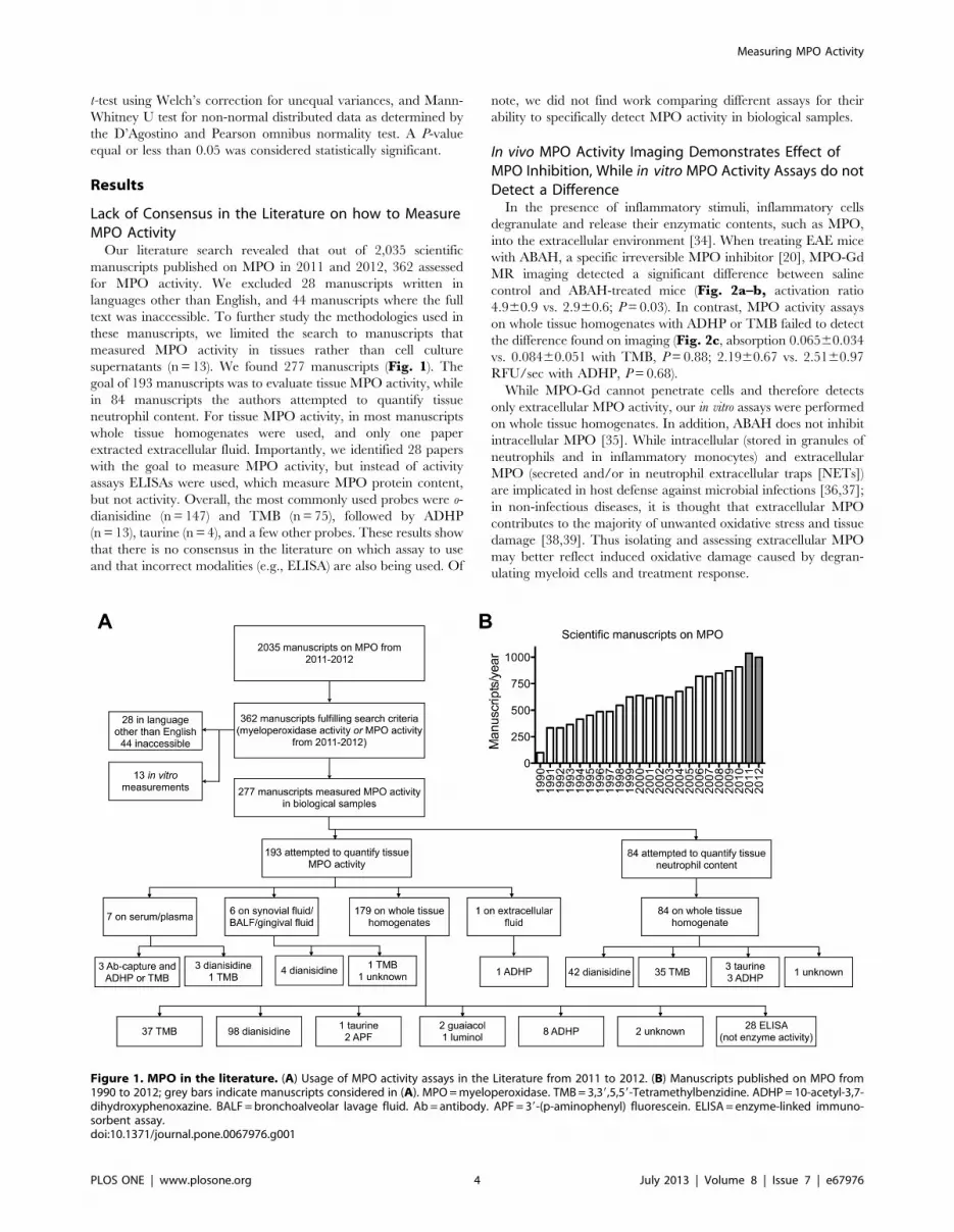
Lack of Consensus in the Literature on how to MeasureMPO ActivityOur literature search revealed that out of 2,035 scientific
manuscripts published on MPO in 2011 and 2012, 362 assessed
for MPO activity. We excluded 28 manuscripts written in
languages other than English, and 44 manuscripts where the full
text was inaccessible. To further study the methodologies used in
these manuscripts, we limited the search to manuscripts that
measured MPO activity in tissues rather than cell culture
supernatants (n = 13). We found 277 manuscripts (Fig. 1). Thegoal of 193 manuscripts was to evaluate tissue MPO activity, while
in 84 manuscripts the authors attempted to quantify tissue
neutrophil content. For tissue MPO activity, in most manuscripts
whole tissue homogenates were used, and only one paper
extracted extracellular fluid. Importantly, we identified 28 papers
with the goal to measure MPO activity, but instead of activity
assays ELISAs were used, which measure MPO protein content,
but not activity. Overall, the most commonly used probes were o-
dianisidine (n = 147) and TMB (n= 75), followed by ADHP
(n= 13), taurine (n = 4), and a few other probes. These results show
that there is no consensus in the literature on which assay to use
and that incorrect modalities (e.g., ELISA) are also being used. Of
note, we did not find work comparing different assays for their
ability to specifically detect MPO activity in biological samples.
In vivo MPO Activity Imaging Demonstrates Effect ofMPO Inhibition, While in vitro MPO Activity Assays do notDetect a DifferenceIn the presence of inflammatory stimuli, inflammatory cells
degranulate and release their enzymatic contents, such as MPO,
into the extracellular environment [34]. When treating EAE mice
with ABAH, a specific irreversible MPO inhibitor [20], MPO-Gd
MR imaging detected a significant difference between saline
control and ABAH-treated mice (Fig. 2a–b, activation ratio
4.960.9 vs. 2.960.6; P=0.03). In contrast, MPO activity assays
on whole tissue homogenates with ADHP or TMB failed to detect
the difference found on imaging (Fig. 2c, absorption 0.06560.034
vs. 0.08460.051 with TMB, P=0.88; 2.1960.67 vs. 2.5160.97
RFU/sec with ADHP, P=0.68).
While MPO-Gd cannot penetrate cells and therefore detects
only extracellular MPO activity, our in vitro assays were performed
on whole tissue homogenates. In addition, ABAH does not inhibit
intracellular MPO [35]. While intracellular (stored in granules of
neutrophils and in inflammatory monocytes) and extracellular
MPO (secreted and/or in neutrophil extracellular traps [NETs])
are implicated in host defense against microbial infections [36,37];
in non-infectious diseases, it is thought that extracellular MPO
contributes to the majority of unwanted oxidative stress and tissue
damage [38,39]. Thus isolating and assessing extracellular MPO
may better reflect induced oxidative damage caused by degran-
ulating myeloid cells and treatment response.
Figure 1. MPO in the literature. (A) Usage of MPO activity assays in the Literature from 2011 to 2012. (B) Manuscripts published on MPO from1990 to 2012; grey bars indicate manuscripts considered in (A). MPO=myeloperoxidase. TMB=3,39,5,59-Tetramethylbenzidine. ADHP= 10-acetyl-3,7-dihydroxyphenoxazine. BALF =bronchoalveolar lavage fluid. Ab= antibody. APF= 39-(p-aminophenyl) fluorescein. ELISA= enzyme-linked immuno-sorbent assay.doi:10.1371/journal.pone.0067976.g001
Measuring MPO Activity
PLOS ONE | www.plosone.org 4 July 2013 | Volume 8 | Issue 7 | e67976

Validation of ECF Protein Extraction and MPO RetrievalAfter Protein PrecipitationTo validate that our method to isolate ECF and ICF works on
various organs, we measured the activity of LDH, a strictly
intracellular enzyme, on extracts from different organs. The ICF
of kidney, brain, lungs, spleen, liver, and heart contained 198.7,
215.6, 47.7, 58.9, 83.9, and 31.3 mU LDH/mg BCA protein,
respectively. ECF contained 0.65, 1.48, 0.52, 0.19, 0.52, and
0.30 mU LDH/mg BCA protein, respectively (Fig. 3a). The ratio
of intra- over extracellular LDH activity normalized to BCA
protein was between 91 and 301 (Fig. 3a). Taken together, these
results suggest that there is no significant contamination from
intracellular proteins in our extracellular protein isolation method.
Because of the relatively high volume of extraction buffer
needed and the subsequently low protein concentration of the
extracellular fluid, it was necessary to concentrate proteins before
further use. We tested two methods of protein precipitation:
acetone and ammonium sulfate. With ammonium sulfate, the
recovered MPO had lost most of its activity (Fig. S2 in File S1).In contrast, acetone preserved MPO activity, and over three
different concentrations of MPO, we were able to recover
9665.2% of activity (Fig. 3b). This validated acetone protein
precipitation as a feasible method to concentrate samples that
contain MPO.
ECF and ICF Protein Extracts Contain Substances thatInterfere with MPO Activity MeasurementsTo test if our assays could efficiently recover MPO from
biological samples, we performed a spike and recovery experi-
ment, where a known amount of human MPO was added to both
ECF and ICF extracts from several organs, and MPO activity was
measured thereafter. We selected three different MPO assay
methods from the literature, which have all been reported to be
sensitive and specific to MPO: 1) Bromide dependent chemilumi-
nescence with luminol at acidic pH [29], 2) peroxidase activity
with ADHP, and 3) chlorination activity with APF and HPF,
where the subtraction of HPF signal from APF signal is thought to
represent specific MPO activity [40]. Results were normalized as
percentage activity of pure enzyme (Fig. 4a–b). For both ECF
and ICF, MPO recovery was variable and dependent on the assay
and organ used, without a clear recognizable trend. Of note, a
large range of MPO activity levels was found, which suggested that
peroxidases other than MPO and/or other interfering substances
were likely affecting the three assay methods. The nonspecificity of
these assays was further confirmed by assaying different concen-
trations of hemoglobin, which has peroxidase activity [41], with
these probes. ADHP and luminol showed a dose-dependent signal
increase in these circumstances (Fig. S3 in File S1). Based on
these findings we conclude that it is necessary to utilize a more
specific method for MPO activity detection and hypothesized that
antibody-specific binding or extraction of MPO from biological
samples before measuring enzyme activity would likely circumvent
these issues.
Figure 2. In vivo imaging and in vitro MPO activity assays demonstrate markedly different findings. (A) MPO-Gd molecular MR imagingreveals MPO inhibition in vivo in mice with experimental autoimmune encephalomyelitis that were treated with ABAH. MPO activity maps are shownin 3D from two angles (left), as well as overlays of MPO activity maps over T1 images (right). (B) Quantification of imaging reveals significantdifference in MPO activity in vivo (P= 0.03, n = 8 per group). (C) In vitro assays on whole tissue homogenates using ADHP or TMB do not confirm thein vivo imaging finding (P=0.68 and 0.88, respectively, n = 4 per group). *: P,0.05, n.s. = not statistically significant. MPO=myeloperoxidase.TMB= 3,39,5,59-Tetramethylbenzidine. ADHP=10-acetyl-3,7-dihydroxyphenoxazine. ABAH= 4-aminobenzoic acid hydrazide. Activation ratio = con-trast-to-noise ratio 60 minutes over 15 minutes post MPO-Gd injection.doi:10.1371/journal.pone.0067976.g002
Measuring MPO Activity
PLOS ONE | www.plosone.org 5 July 2013 | Volume 8 | Issue 7 | e67976

MPO Antibody Capture Assay is Highly ReproducibleTo remove interfering substances from the biological samples
being tested, we used an antibody capture assay [42]. First, to
establish the reproducibility and linear range of this assay, we
loaded homogenized murine neutrophils into anti-MPO antibody-
covered wells, and after binding washed away any interfering
substances. We then measured MPO activity with ADHP, chosen
for its high sensitivity and assay range (Fig. S4 in File S1).
For intra-assay reproducibility, triplicates were run simulta-
neously (Fig. S5a in File S1), while for inter-assay reproducibil-
ity, each standard curve was run at least 1 hour apart (Fig. S5b inFile S1). The range of the linear part of the curve was found to be
from 598 to 1.2 million neutrophils with coefficients of determi-
nation (R2) of 0.98 and 0.88 for intra-assay (Fig. S5a in File S1,P,0.0001) and inter-assay (Fig. S5b in File S1, P,0.0001)
reproducibility, respectively.
Figure 3. Validation of Extracellular Protein Isolation and MPO Protein Precipitation. (A) LDH assay of intra- and extracellular proteinfractions of different organs shows that the extracellular fraction only contains very low levels of LDH activity, while the intracellular fraction containsthe majority of the LDH activity (left). LDH ratio shows a 90 or higher fold level of ICF LDH over ECF LDH activity (right). (B) Protein precipitation ofMPO with acetone has no effect on its activity, as evaluated with ADHP (n = 2 per group). LDH= lactate dehydrogenase. BCA=bicinchoninic acid.MPO=myeloperoxidase.doi:10.1371/journal.pone.0067976.g003
Figure 4. Spike and recovery assay: tissue homogenates and extracellular fluid contain interfering substances. (A) Extracellularprotein fraction from different organs contains substances interfering with ADHP, luminol, and APF assays (n = 2 per group). (B) Intracellular proteinfractions also contain interfering substances (n = 2 per group). MPO=myeloperoxidase. ADHP= 10-acetyl-3,7-dihydroxyphenoxazine. APF= 39-(p-aminophenyl) fluorescein. HPF= 39-(p-hydroxyphenyl) fluorescein.doi:10.1371/journal.pone.0067976.g004
Measuring MPO Activity
PLOS ONE | www.plosone.org 6 July 2013 | Volume 8 | Issue 7 | e67976
This experiment also allowed us to evaluate the sensitivity of this
assay, which can detect MPO from as few as 500 neutrophils (Fig.S5 in File S1).
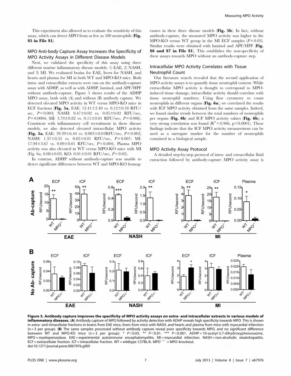
MPO Anti-body Capture Assay Increases the Specificity ofMPO Activity Assays in Different Disease ModelsNext, we validated the specificity of this assay using three
different murine inflammatory disease models: 1) EAE, 2) NASH,
and 3) MI. We evaluated brains for EAE, livers for NASH, and
hearts and plasma for MI in both WT and MPO-KO mice. Both
intra- and extracellular extracts were run on the antibody-capture
assay with ADHP, as well as with ADHP, luminol, and APF/HPF
without antibody-capture. Figure 5 shows results of the ADHP
MPO assay, both with (A) and without (B) antibody capture. We
detected elevated MPO activity in WT versus MPO-KO mice in
ECF fractions (Fig. 5a, EAE: 12.4162.40 vs. 0.1260.18 RFU/
sec, P=0.003; NASH: 0.4760.02 vs. 0.0560.02 RFU/sec,
P=0.0004; MI: 3.7960.82 vs. 0.1160.01 RFU/sec, P=0.006).
Consistent with inflammatory cell recruitment in these disease
models, we also detected elevated intracellular MPO activity
(Fig. 5a, EAE: 39.3966.44 vs. 0.00360.01RFU/sec, P=0.002;
NASH: 1.3760.35 vs. 0.0260.01 RFU/sec, P=0.007; MI:
17.9463.67 vs. 0.0960.01 RFU/sec, P=0.004). Plasma MPO
activity was also elevated in WT versus MPO-KO mice with MI
(Fig. 6a, 0.0860.03, KO: 0.0160.01 RFU/sec, P=0.02).
In contrast, ADHP without antibody-capture was unable to
detect significant differences between WT and MPO-KO homog-
enates in these three disease models (Fig. 5b). In fact, without
antibody-capture, the measured MPO activity was higher in the
MPO-KO versus WT group in the MI ECF samples (P=0.03).
Similar results were obtained with luminol and APF/HPF (Fig.S6 and S7 in File S1). This establishes the non-specificity of
these assays towards MPO without an antibody-capture step.
Intracellular MPO Activity Correlates with TissueNeutrophil CountOur literature search revealed that the second application of
MPO activity assays is to quantify tissue neutrophil content. While
extracellular MPO activity is thought to correspond to MPO-
induced tissue damage, intracellular activity should correlate with
tissue neutrophil numbers. Using flow cytometry to count
neutrophils in different organs (Fig. 6a), we correlated the results
with ICF MPO activity obtained from the same samples. Indeed,
we found similar trends between the total numbers of neutrophils
per organs (Fig. 6b) and ICF MPO activity values (Fig. 6b); avery strong correlation was found (R2= 0.966, p,0.0001). These
findings indicate that the ICF MPO activity measurement can be
used as a surrogate marker for the number of neutrophils
contained in a biological sample.
MPO Activity Assay ProtocolA detailed step-by-step protocol of intra- and extracellular fluid
extraction followed by antibody-capture MPO activity assay is
Figure 5. Antibody capture improves the specificity of MPO activity assays on extra- and intracellular extracts in various models ofinflammatory diseases. (A) Antibody capture of MPO followed by activity detection with ADHP reveals high specificity towards MPO. This is shownin extra- and intracellular fractions in brains from EAE mice, livers from mice with NASH, and hearts and plasma from mice with myocardial infarction(n = 3 per group). (B) The same samples processed without antibody capture reveal poor specificity towards MPO, and no significant differencebetween WT and MPO-KO mice (n = 3 per group). * P,0.05. ** P,0.01. *** P,0.001. ADHP = 10-acetyl-3,7-dihydroxyphenoxazine.MPO=myeloperoxidase. EAE = experimental autoimmune encephalomyelitis. MI =myocardial infarction. NASH=non-alcoholic steatohepatitis.ECF = extracellular fraction. ICF = intracellular fraction. WT=wildtype C57BL/6. MPO2/2=MPO knockout.doi:10.1371/journal.pone.0067976.g005
Measuring MPO Activity
PLOS ONE | www.plosone.org 7 July 2013 | Volume 8 | Issue 7 | e67976

given in File S2. Table S1 in File S2 provides troubleshooting
for the assay.
Discussion
Our study showed that the proposed method to isolate intra-
and extracellular protein fractions of biological samples is feasible.
We found that antibody-capture of MPO is necessary before
assessing its activity, due to the non-specificity of available probes
and the presence of interfering substances. This method to assay
MPO activity was validated in different mouse models of
inflammatory conditions and against MPO-KO mice, and a
detailed assay protocol is provided.
We selected three probes: ADHP, APF/HPF, and luminol to
represent major classes of MPO activity assay probes. ADHP,
TMB, o-dianisidine, and guaiacol are peroxidase substrates (note
that TMB, o-dianisidine, and guaiacol are less resistant to
autoxidation, are less sensitive, and have a narrower assay range
than ADHP [43,44]; in addition, o-dianisidine is carcinogenic
[45]). In contrast to these peroxidase probes, APF/HPF can detect
the chlorination activity of MPO, and subtracting HPF from APF
signal has been suggested to specifically measure HOCl, a highly
specific product of MPO [27]. Bromide-dependent chemilumi-
nescence with luminol at low pH has also been shown to be
specific towards MPO, and was applied successfully to estimate
tumor neutrophil content in a mouse model [28]. Unfortunately,
when we tested these assays using samples from multiple different
murine organs, we could not adequately recover the spiked MPO
activity. Moreover, we did not detect signals in WT mice samples
which were substantially greater than those from MPO-KO mice
using three different diseases that are known to trigger high tissue
levels of MPO. We conclude that interfering substances (e.g. other
peroxidases) and tissue inhibitors account for these findings
[15,16].
To circumvent this, we validated an antibody capture assay. In
this assay, MPO is bound to the wells of an ELISA plate by means
of a monoclonal anti-MPO antibody, which guarantees high
specificity. After washing away the unbound substances, enzyme
activity can then be detected with a suitable substrate. The capture
assay provides researchers with a platform to evaluate both
peroxidation and chlorination activity by using different probes
without concerns about non-specificity. By using mouse models of
myocardial infarction, multiple sclerosis, and steatohepatitis
(diseases in which MPO has been implicated in humans), we
validated the high specificity and sensitivity of this assay.
In addition to biochemical assays, advanced imaging methods
for in vivo MPO activity detection are available. These include
MPO-Gd, an activatable, ‘smart’ MR imaging probe [22], and the
bioluminescent agent luminol [46]. Although both agents have
been shown to be highly sensitive and specific in vivo, imaging has
relatively slow throughput. Thus, a high throughput assay to be
used in vitro on extracts from biological tissues would be highly
desirable to complement the in vivo probes.
Another measurement of MPO activity that is widely used is 3-
chlorotyrosine, a highly specific product of MPO that can be
measured with stable isotope dilution gas chromatography/mass
spectrometry [47,48]. Although specific, 3-chlorotyrosine levels
are only a surrogate marker and as such give an estimate of MPO
activity in the past, but not necessarily current MPO activity. 3-
chlorotyrosine can also be quickly degraded in an inflammatory
environment [49], and reactions other than chlorination might be
preferentially induced [50]. Thus, its absence does not definitively
Figure 6. Intracellular MPO activity correlates well with tissue neutrophil content. (A) Flow cytometry demonstrates different neutrophilcounts in brain, heart, liver, spleen, and bone marrow, as quantified in (B) (n = 2 per group). (C) Intracellular MPO activity was measured with theantibody-capture assay using ADHP, and shows a similar trend to neutrophil content per organ (n = 2 per group). (D) A close correlation was foundbetween neutrophil content and intracellular MPO activity in these organs. MPO=myeloperoxidase. ADHP=10-acetyl-3,7-dihydroxyphenoxazine.doi:10.1371/journal.pone.0067976.g006
Measuring MPO Activity
PLOS ONE | www.plosone.org 8 July 2013 | Volume 8 | Issue 7 | e67976

prove lack of MPO activity. Furthermore, 3-chlorotyrosine levels
can be markedly reduced by thiocyanate ions, which are elevated
in smokers [51]. All of these findings suggest that 3-chlorotyrosine
levels are dependent on the tissue microenvironment, and that
direct measurements of MPO activity should be performed
whenever possible.
ConclusionsIn summary, we validated a robust protocol to isolate and
measure intra- and extracellular MPO activity with high sensitivity
and specificity. We validated this assay in three different mouse
disease models and in MPO-KO mice. This protocol should be
established as the standard method for measuring MPO activity in
biological samples. For standardization purposes, we propose the
use of ADHP after the antibody capture, due to its wider assay
range and higher sensitivity.
Supporting Information
File S1 This file contains Figure S1–S7. Figure S1,
Ammonium sulfate protein precipitation decreases recovery of
MPO activity. Figure S2, ADHP and luminol assays are sensitive
to unspecific peroxidase activity. Figure S3, ADHP is more
sensitive and has a wider assay range than APF, TMB, and
luminol. Figure S4, Reproducibility of the MPO capture assay.
Figure S5, Luminol is not specific for MPO activity in EAE,
NASH, or MI. Figure S6, APF/HPF is not specific to MPO in
EAE, NASH, or MI. Figure S7, Relative fluorescent units plotted
over time with different samples using the antibody-capture MPO
activity assay with ADHP.
(PDF)
File S2 This file contains two items. Protocol S1, Step-by-Step Protocol. Table S1, Troubleshooting.
(PDF)
Author Contributions
Conceived and designed the experiments: BP MA RF JWC. Performed the
experiments: BP MA SS GW. Analyzed the data: BP MA RF SS KLCH
GW JL JWC. Wrote the paper: BP MA JL JWC.
References
1. Schultz J, Kaminker K (1962) Myeloperoxidase of the leucocyte of normal
human blood. I. Content and localization. Arch Biochem Biophys 96: 465–467.
2. Heinecke JW (1999) Mechanisms of oxidative damage by myeloperoxidase inatherosclerosis and other inflammatory disorders. J Lab Clin Med 133: 321–325.
3. Zhang R, Brennan ML, Fu X, Aviles RJ, Pearce GL, et al. (2001) Association
between myeloperoxidase levels and risk of coronary artery disease. JAMA 286:
2136–2142.
4. Nahrendorf M, Sosnovik D, Chen JW, Panizzi P, Figueiredo JL, et al. (2008)Activatable magnetic resonance imaging agent reports myeloperoxidase activity
in healing infarcts and noninvasively detects the antiinflammatory effects ofatorvastatin on ischemia-reperfusion injury. Circulation 117: 1153–1160.
5. Brennan ML, Penn MS, Van Lente F, Nambi V, Shishehbor MH, et al. (2003)
Prognostic value of myeloperoxidase in patients with chest pain. N Engl J Med
349: 1595–1604.
6. Rudolph V, Andrie RP, Rudolph TK, Friedrichs K, Klinke A, et al. (2010)Myeloperoxidase acts as a profibrotic mediator of atrial fibrillation. Nat Med 16:
470–474.
7. Gray E, Thomas TL, Betmouni S, Scolding N, Love S (2008) Elevated
myeloperoxidase activity in white matter in multiple sclerosis. Neurosci Lett 444:195–198.
8. Reynolds WF, Rhees J, Maciejewski D, Paladino T, Sieburg H, et al. (1999)
Myeloperoxidase polymorphism is associated with gender specific risk forAlzheimer’s disease. Exp Neurol 155: 31–41.
9. Feyler A, Voho A, Bouchardy C, Kuokkanen K, Dayer P, et al. (2002) Point:
myeloperoxidase -463G –. a polymorphism and lung cancer risk. Cancer
Epidemiol Biomarkers Prev 11: 1550–1554.
10. Swirski FK, Wildgruber M, Ueno T, Figueiredo JL, Panizzi P, et al. (2010)Myeloperoxidase-rich Ly-6C+ myeloid cells infiltrate allografts and contribute to
an imaging signature of organ rejection in mice. J Clin Invest 120: 2627–2634.
11. Chevrier I, Tregouet DA, Massonnet-Castel S, Beaune P, Loriot MA (2006)
Myeloperoxidase genetic polymorphisms modulate human neutrophil enzymeactivity: genetic determinants for atherosclerosis? Atherosclerosis 188: 150–154.
12. Piedrafita FJ, Molander RB, Vansant G, Orlova EA, Pfahl M, et al. (1996) An
Alu element in the myeloperoxidase promoter contains a composite SP1-thyroidhormone-retinoic acid response element. J Biol Chem 271: 14412–14420.
13. Chapman AL, Mocatta TJ, Shiva S, Seidel A, Chen B, et al. (2013)
Ceruloplasmin is an endogenous inhibitor of myeloperoxidase. J Biol Chem.
14. Segelmark M, Persson B, Hellmark T, Wieslander J (1997) Binding and
inhibition of myeloperoxidase (MPO): a major function of ceruloplasmin? ClinExp Immunol 108: 167–174.
15. Ormrod DJ, Harrison GL, Miller TE (1987) Inhibition of neutrophil
myeloperoxidase activity by selected tissues. J Pharmacol Methods 18: 137–142.
16. Xia Y, Zweier JL (1997) Measurement of myeloperoxidase in leukocyte-
containing tissues. Anal Biochem 245: 93–96.
17. Swirski FK, Nahrendorf M, Etzrodt M, Wildgruber M, Cortez-Retamozo V, etal. (2009) Identification of splenic reservoir monocytes and their deployment to
inflammatory sites. Science 325: 612–616.
18. Miller SD, Karpus WJ, Davidson TS (2010) Experimental autoimmune
encephalomyelitis in the mouse. Curr Protoc Immunol Chapter 15: Unit 15 11.
19. Sahai A, Malladi P, Pan X, Paul R, Melin-Aldana H, et al. (2004) Obese anddiabetic db/db mice develop marked liver fibrosis in a model of nonalcoholic
steatohepatitis: role of short-form leptin receptors and osteopontin. Am J PhysiolGastrointest Liver Physiol 287: G1035–1043.
20. Kettle AJ, Gedye CA, Hampton MB, Winterbourn CC (1995) Inhibition of
myeloperoxidase by benzoic acid hydrazides. Biochem J 308 (Pt 2): 559–563.
21. Chen JW, Breckwoldt MO, Aikawa E, Chiang G, Weissleder R (2008)
Myeloperoxidase-targeted imaging of active inflammatory lesions in murine
experimental autoimmune encephalomyelitis. Brain 131: 1123–1133.
22. Chen JW, Querol Sans M, Bogdanov A, Jr., Weissleder R (2006) Imaging of
myeloperoxidase in mice by using novel amplifiable paramagnetic substrates.
Radiology 240: 473–481.
23. Breckwoldt MO, Chen JW, Stangenberg L, Aikawa E, Rodriguez E, et al. (2008)
Tracking the inflammatory response in stroke in vivo by sensing the enzyme
myeloperoxidase. Proc Natl Acad Sci U S A 105: 18584–18589.
24. Hofstein R, Hesse G, Shashoua VE (1983) Proteins of the extracellular fluid of
mouse brain: extraction and partial characterization. J Neurochem 40: 1448–
1455.
25. Wingfield P (2001) Protein precipitation using ammonium sulfate. Curr Protoc
Protein Sci Appendix 3: Appendix 3F.
26. Suzuki K, Ota H, Sasagawa S, Sakatani T, Fujikura T (1983) Assay method for
myeloperoxidase in human polymorphonuclear leukocytes. Anal Biochem 132:
345–352.
27. Setsukinai K, Urano Y, Kakinuma K, Majima HJ, Nagano T (2003)
Development of novel fluorescence probes that can reliably detect reactive
oxygen species and distinguish specific species. J Biol Chem 278: 3170–3175.
28. Haqqani AS, Sandhu JK, Birnboim HC (1999) A myeloperoxidase-specific assay
based upon bromide-dependent chemiluminescence of luminol. Anal Biochem
273: 126–132.
29. Haqqani AS, Sandhu JK, Birnboim HC (1999) A Myeloperoxidase-Specific
Assay Based upon Bromide-Dependent Chemiluminescence of Luminol.
Analytical Biochemistry 273: 126–132.
30. Preparation of mouse bone marrow neutrophils. Available: http://medicine.
ucsf.edu/labs/brown/protocols_03_2005/Murine_BMN_Prep.pdf. Accessed
2013 Mar 05.
31. O’Driscoll S, Height SE, Dick MC, Rees DC (2008) Serum lactate
dehydrogenase activity as a biomarker in children with sickle cell disease.
British Journal of Haematology 140: 206–209.
32. Jahng A, Maricic I, Aguilera C, Cardell S, Halder RC, et al. (2004) Prevention
of autoimmunity by targeting a distinct, noninvariant CD1d-reactive T cell
population reactive to sulfatide. J Exp Med 199: 947–957.
33. Pino PA, Cardona AE (2011) Isolation of brain and spinal cord mononuclear
cells using percoll gradients. J Vis Exp.
34. Bradley PP, Christensen RD, Rothstein G (1982) Cellular and extracellular
myeloperoxidase in pyogenic inflammation. Blood 60: 618–622.
35. Forghani R, Wojtkiewicz GR, Zhang Y, Seeburg D, Bautz BR, et al. (2012)
Demyelinating diseases: myeloperoxidase as an imaging biomarker and
therapeutic target. Radiology 263: 451–460.
36. Metzler KD, Fuchs TA, Nauseef WM, Reumaux D, Roesler J, et al. (2011)
Myeloperoxidase is required for neutrophil extracellular trap formation:
implications for innate immunity. Blood 117: 953–959.
37. Parker H, Albrett AM, Kettle AJ, Winterbourn CC (2012) Myeloperoxidase
associated with neutrophil extracellular traps is active and mediates bacterial
killing in the presence of hydrogen peroxide. J Leukoc Biol 91: 369–376.
38. Lefkowitz DL, Mone J, Lefkowitz SS (2010) Myeloperoxidase: The Good, the
Bad, and the Ugly. Current Immunology Reviews 6: 123–129.
Measuring MPO Activity
PLOS ONE | www.plosone.org 9 July 2013 | Volume 8 | Issue 7 | e67976

39. Klebanoff SJ (2005) Myeloperoxidase: friend and foe. J Leukoc Biol 77: 598–
625.40. Setsukinai K-i, Urano Y, Kakinuma K, Majima HJ, Nagano T (2003)
Development of Novel Fluorescence Probes That Can Reliably Detect Reactive
Oxygen Species and Distinguish Specific Species. Journal of BiologicalChemistry 278: 3170–3175.
41. Kapralov A, Vlasova, II, Feng W, Maeda A, Walson K, et al. (2009) Peroxidaseactivity of hemoglobin-haptoglobin complexes: covalent aggregation and
oxidative stress in plasma and macrophages. J Biol Chem 284: 30395–30407.
42. Franck T, Kohnen S, Boudjeltia KZ, Van Antwerpen P, Bosseloir A, et al.(2009) A new easy method for specific measurement of active myeloperoxidase in
human biological fluids and tissue extracts. Talanta 80: 723–729.43. Zhou M, Diwu Z, Panchuk-Voloshina N, Haugland RP (1997) A stable
nonfluorescent derivative of resorufin for the fluorometric determination of tracehydrogen peroxide: applications in detecting the activity of phagocyte NADPH
oxidase and other oxidases. Anal Biochem 253: 162–168.
44. Meng Y, High K, Antonello J, Washabaugh MW, Zhao Q (2005) Enhancedsensitivity and precision in an enzyme-linked immunosorbent assay with
fluorogenic substrates compared with commonly used chromogenic substrates.Anal Biochem 345: 227–236.
45. Health Hazard Alert– Benzidine-, o-Tolidine-, and o-Dianisidine- Based Dyes.
Available: http://www.cdc.gov/niosh/docs/81-106/. Accessed 2013 Mar 05.
46. Gross S, Gammon ST, Moss BL, Rauch D, Harding J, et al. (2009)
Bioluminescence imaging of myeloperoxidase activity in vivo. Nat Med 15:
455–461.
47. Brennan ML, Wu W, Fu X, Shen Z, Song W, et al. (2002) A tale of two
controversies: defining both the role of peroxidases in nitrotyrosine formation
in vivo using eosinophil peroxidase and myeloperoxidase-deficient mice, and the
nature of peroxidase-generated reactive nitrogen species. J Biol Chem 277:
17415–17427.
48. Wu W, Samoszuk MK, Comhair SA, Thomassen MJ, Farver CF, et al. (2000)
Eosinophils generate brominating oxidants in allergen-induced asthma. J Clin
Invest 105: 1455–1463.
49. Whiteman M, Spencer JP (2008) Loss of 3-chlorotyrosine by inflammatory
oxidants: implications for the use of 3-chlorotyrosine as a bio-marker in vivo.
Biochem Biophys Res Commun 371: 50–53.
50. Holzer M, Zangger K, El-Gamal D, Binder V, Curcic S, et al. (2012)
Myeloperoxidase-derived chlorinating species induce protein carbamylation
through decomposition of thiocyanate and urea: novel pathways generating
dysfunctional high-density lipoprotein. Antioxid Redox Signal 17: 1043–1052.
51. Talib J, Pattison DI, Harmer JA, Celermajer DS, Davies MJ (2012) High plasma
thiocyanate levels modulate protein damage induced by myeloperoxidase and
perturb measurement of 3-chlorotyrosine. Free Radic Biol Med 53: 20–29.
Measuring MPO Activity
PLOS ONE | www.plosone.org 10 July 2013 | Volume 8 | Issue 7 | e67976