cyclin-b homologs in saccharomyces cerevisiae function in s phase and in g2
TRANSCRIPT
10.1101/gad.6.11.2021Access the most recent version at doi: 1992 6: 2021-2034Genes Dev.
H Richardson, D J Lew, M Henze, et al. phase and in G2.Cyclin-B homologs in Saccharomyces cerevisiae function in S
References
http://genesdev.cshlp.org/content/6/11/2021#related-urlsArticle cited in:
http://genesdev.cshlp.org/content/6/11/2021.refs.htmlThis article cites 64 articles, 20 of which can be accessed free at:
serviceEmail alerting
click heretop right corner of the article orReceive free email alerts when new articles cite this article - sign up in the box at the
http://genesdev.cshlp.org/subscriptions go to: Genes & DevelopmentTo subscribe to
Copyright © Cold Spring Harbor Laboratory Press
Cold Spring Harbor Laboratory Press on July 10, 2011 - Published by genesdev.cshlp.orgDownloaded from
Cyclin-B h0mologs !n Saccharomyc.es cerevisiae function in S phase and m G2 Helena Richardson, 1'2 Danie l J. Lew, 1 Martha Henze , 1 Katsunori Sug imoto , 1'3 and Steven I. Reed 1'4
1Department of Molecular Biology, The Scripps Research Institute, La Jolla, California 92037 USA; 2Department of Biochemistry, University of Adelaide, Adelaide, South Australia 5001
We have cloned four cyclin-B homologs from Saccharomyces cerevisiae, CLB1-CLB4, using the polymerase chain reaction and low stringency hybridization approaches. These genes form two classes based on sequence relatedness: CLB1 and CLB2 show highest homology to the Schizosaccharomyces pombe cyclin-B homolog cdcl3 involved in the initiation of mitosis, whereas CLB3 and CLB4 are more highly related to the S. pombe cyclin-B homolog cigl, which appears to play a role in G1 or S phase. CLB1 and CLB2 mRNA levels peak late in the cell cycle, whereas CLB3 and CLB4 are expressed earlier in the cell cycle but peak later than the Gl-specific cyclin, CLN1. Analysis of null mutations suggested that the CLB genes exhibit some degree of redundancy, but clbl,2 and clb2,3 cells were inviable. Using clbl,2,3,4 cells rescued by conditional overproduction of CLB1, we showed that the CLB genes perform an essential role at the G2/M-phase transition, and also a role in S phase. CLB genes also appear to share a role in the assembly and maintenance of the mitotic spindle. Taken together, these analyses suggest that CLB1 and CLB2 are crucial for mitotic induction, whereas CLB3 and CLB4 might participate additionally in DNA replication and spindle assembly.
[Key Words: Yeast; cyclin-B homologs; S-phase; mitosis; Cdc28 kinase]
Received June 8, 1992; revised version accepted August 11, 1992.
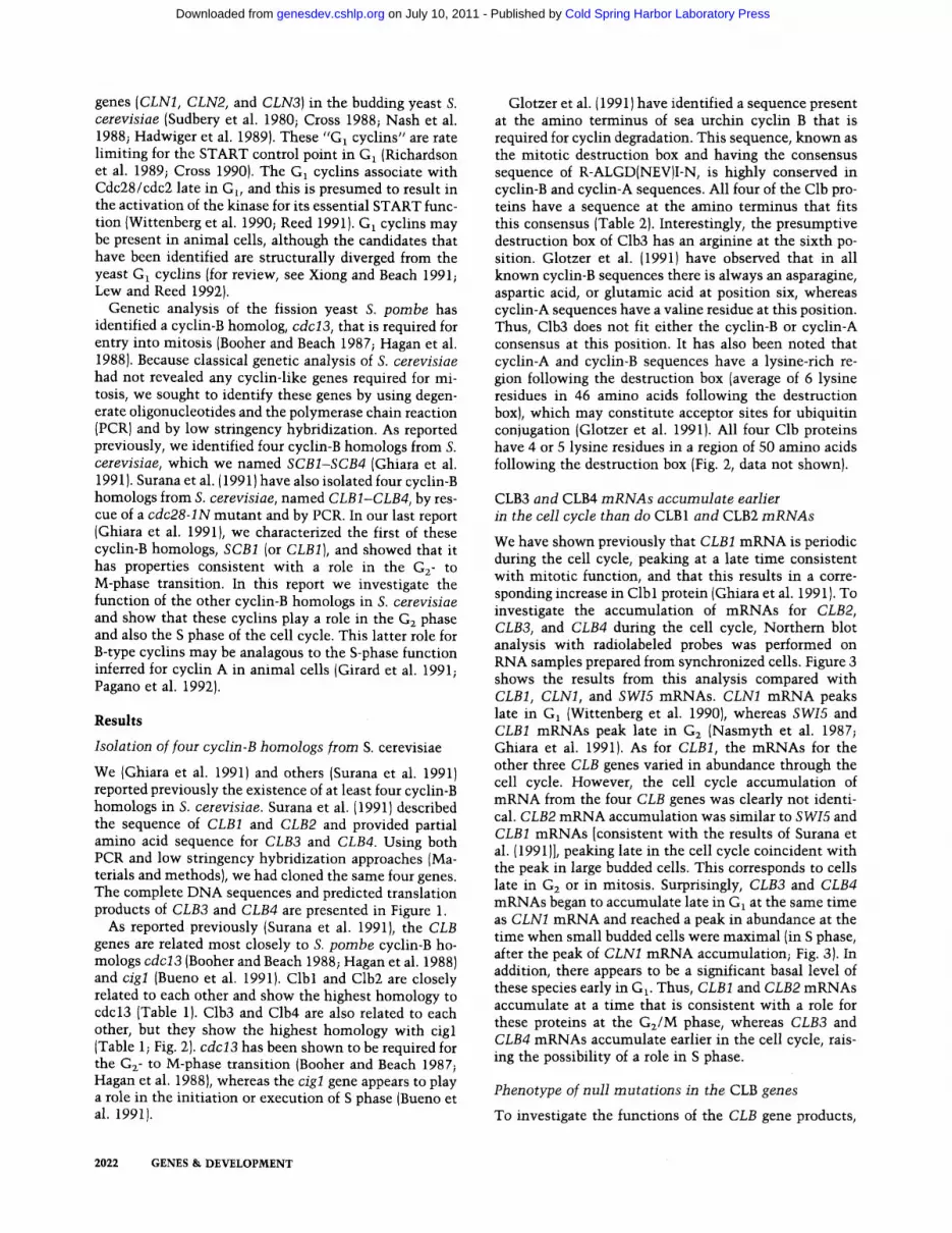
The eukaryotic cell cycle is highly conserved in both mechanism and components from yeast to man (for re- cent reviews, see Cross et al. 1989; Murray and Kirsch- her 1989a; Lewin 1990; Nurse 1990; Pines and Hunter 1990a; Maller 1991; Reed 1991). Studies carried out in the budding yeast Saccharomyces cerevisiae and the fis- sion yeast Schizosaccharomyces pombe have revealed that a 34-kD serine-threonine protein kinase, known as Cdc28 or cdc2, plays a critical role in at least two key regulatory events. The first is at a point late in G1 known as START, at which environmental factors, such as nu- trient availability and the presence of mating phero- mones, act to determine whether a cell will enter the cell cycle (Hartwell et al. 1974; Nurse and Bissett 1981). Sec- ond, the Cdc28/cdc2 protein kinase functions at the ini- tiation of mitosis at a point late in Gz (Nurse and Bissett 1981; Piggott et al. 1982; Reed and Wittenberg 1990; Surana et al. 1991). Studies in Xenopus have revealed that Cdc28/cdc2 is an essential component of a factor [M-phase-promoting factor (MPF)] required for the G2- to M-phase transition (for review, see Hunt 1989; Maller 1991). Furthermore, recent studies in mammalian cells have provided functional evidence that Cdc28/cdc2 is required for the initiation of mitosis (Riabowol et al. 1989; Th'ng et al. 1990), and a newly discovered cdc2- like kinase [cdk2 (Elledge and Spottswood 1991; Ni-
~Present address: Department of Molecular Biology, Nagoya University, Nagoya 464-01, Japan. 4Corresponding author.
nomiya-Tsuji et al. 1991; Paris et al. 1991; Tsai et al. 1991)] is required for the initiation of S phase (Fang and Newport 1991).
In its active form, the Cdc28/cdc2 protein kinase is a multimeric complex with at least one other protein of 45-60 kD, known as cyclin (for review, see Hunt 1989; Murray and Kirschner 1989a; Mailer 1991; Reed 1991; Xiong and Beach 1991; Lew and Reed 1992). Cyclins were initially discovered in the oocytes and embryos of marine invertebrates, as proteins that peaked in abun- dance just before the onset of mitosis and were then rapidly degraded during mitosis (Evans et al. 1983; Hunt et al. 1992). These "mitotic cyclins" are activators of Cdc28/cdc2 for the G2- to M-phase transition (for re- view, see Hunt 1989; Murray and Kirschner 1989a; Nurse 1990; Mailer 1991). Mitotic cyclins have been di- vided into two classes, A-type and B-type, on the basis of sequence relatedness; but they also appear to differ in their kinetics of accumulation, intracellular distribu- tion, and association with different forms of Cdc28/cdc2 (Minshull et al. 1990; Pines and Hunter 1990a,b; Hunt et al. 1992). Cyclin A and B both appear to be capable of promoting entry into mitosis when assayed in the Xeno- pus oocyte system (Swenson et al. 1986; Minshull et al. 1989; Murray and Kirschner 1989b; Murray et al. 1989); however these cyclins do not perform redundant roles in Drosophila (Lehner and O'Farrell 1990). In somatic ani- mal cells, cyclin A plays a role at the initiation of S phase (Girard et al. 1991; Pagano et al. 1992).
Another group of cyclins has been defined by three
GENES & DEVELOPMENT 6:2021-2034 © 1992 by Cold Spring Harbor Laboratory Press ISSN 0890-9369/92 $3.00 2021
Cold Spring Harbor Laboratory Press on July 10, 2011 - Published by genesdev.cshlp.orgDownloaded from
genes (CLN1, CLN2, and CLN3) in the budding yeast S. cerevisiae (Sudbery et al. 1980; Cross 1988; Nash et al. 1988; Hadwiger et al. 1989). These "G~ cyclins" are rate limiting for the START control point in G~ (Richardson et al. 1989; Cross 1990). The G1 cyclins associate with Cdc28/cdc2 late in G~, and this is presumed to result in the activation of the kinase for its essential START func- tion (Wittenberg et al. 1990; Reed 1991). G~ cyclins may be present in animal cells, although the candidates that have been identified are structurally diverged from the yeast G~ cyclins (for review, see Xiong and Beach 1991; Lew and Reed 1992).
Genetic analysis of the fission yeast S. pombe has identified a cyclin-B homolog, cdc13, that is required for entry into mitosis (Booher and Beach 1987; Hagan et al. 1988). Because classical genetic analysis of S. cerevisiae had not revealed any cyclin-like genes required for mi- tosis, we sought to identify these genes by using degen- erate oligonucleotides and the polymerase chain reaction (PCR) and by low stringency hybridization. As reported previously, we identified four cyclin-B homologs from S. cerevisiae, which we named SCB1-SCB4 (Ghiara et al. 1991). Surana et al. (1991) have also isolated four cyclin-B homologs from S. cerevisiae, named CLB1-CLB4, by res- cue of a cdc28-1N mutant and by PCR. In our last report (Ghiara et al. 1991), we characterized the first of these cyclin-B homologs, SCB1 (or CLB1), and showed that it has properties consistent with a role in the G2- to M-phase transition. In this report we investigate the function of the other cyclin-B homologs in S. cerevisiae and show that these cyclins play a role in the G2 phase and also the S phase of the cell cycle. This latter role for B-type cyclins may be analagous to the S-phase function inferred for cyclin A in animal cells (Girard et al. 1991; Pagano et al. 1992).
R e s u l t s
Isolation of four cyclin-B homologs from S. cerevisiae
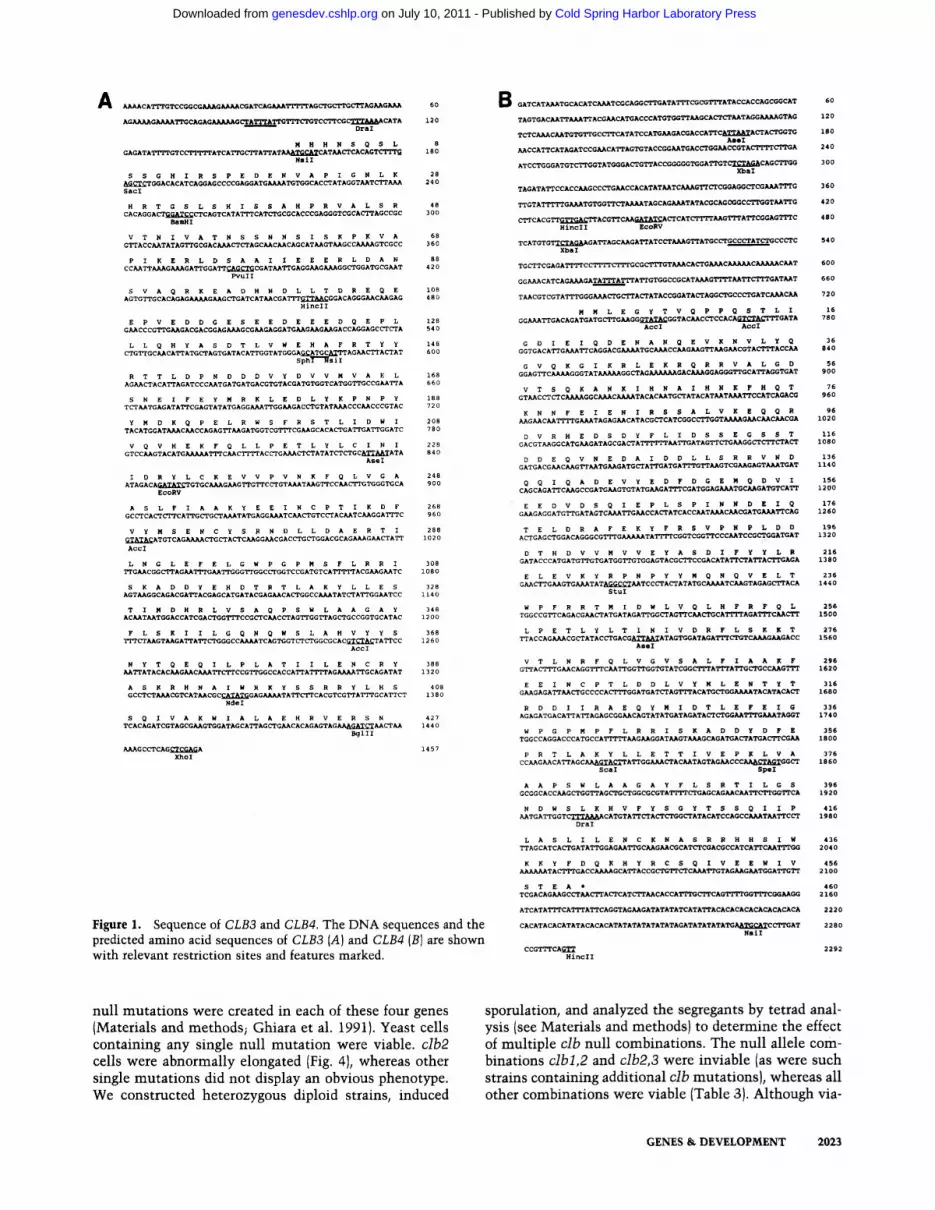
We (Ghiara et al. 1991) and others (Surana et al. 1991) reported previously the existence of at least four cyclin-B homologs in S. cerevisiae. Surana et al. (1991) described the sequence of CLB1 and CLB2 and provided partial amino acid sequence for CLB3 and CLB4. Using both PCR and low stringency hybridization approaches (Ma- terials and methods), we had cloned the same four genes. The complete DNA sequences and predicted translation products of CLB3 and CLB4 are presented in Figure 1.
As reported previously (Surana et al. 1991), the CLB genes are related most closely to S. pombe cyclin-B ho- mologs cdcl3 (Booher and Beach 1988; Hagan et al. 1988) and cigl (Bueno et al. 1991). Clbl and Clb2 are closely related to each other and show the highest homology to cdcl3 (Table 1). Clb3 and Clb4 are also related to each other, but they show the highest homology with cigl (Table 1; Fig. 2). cdcl3 has been shown to be required for the G z- to M-phase transition {Booher and Beach 1987; Hagan et al. 1988), whereas the cigl gene appears to play a role in the initiation or execution of S phase (Bueno et al. 1991).
Glotzer et al. ( 1991 ) have identified a sequence present at the amino terminus of sea urchin cyclin B that is required for cyclin degradation. This sequence, known as the mitotic destruction box and having the consensus sequence of R-ALGD{NEV)I-N, is highly conserved in cyclin-B and cyclin-A sequences. All four of the Clb pro- teins have a sequence at the amino terminus that fits this consensus (Table 2). Interestingly, the presumptive destruction box of Clb3 has an arginine at the sixth po- sition. Glotzer et al. (1991) have observed that in all known cyclin-B sequences there is always an asparagine, aspartic acid, or glutamic acid at position six, whereas cyclin-A sequences have a valine residue at this position. Thus, Clb3 does not fit either the cyclin-B or cyclin-A consensus at this position. It has also been noted that cyclin-A and cyclin-B sequences have a lysine-rich re- gion following the destruction box (average of 6 lysine residues in 46 amino acids following the destruction box), which may constitute acceptor sites for ubiquitin conjugation {Glotzer et al. 1991). All four Clb proteins have 4 or 5 lysine residues in a region of 50 amino acids following the destruction box (Fig. 2, data not shown).
CLB3 and CLB4 mRNAs accumulate earlier m the cell cycle than do CLB1 and CLB2 mRNAs
We have shown previously that CLB1 mRNA is periodic during the cell cycle, peaking at a late time consistent with mitotic function, and that this results in a corre- sponding increase in Clb 1 protein (Ghiara et al. 1991). To investigate the accumulation of mRNAs for CLB2, CLB3, and CLB4 during the cell cycle, Northern blot analysis with radiolabeled probes was performed on RNA samples prepared from synchronized cells. Figure 3 shows the results from this analysis compared with CLB1, CLN1, and SWI5 mRNAs. CLN1 mRNA peaks late in G1 (Wittenberg et al. 1990), whereas SWI5 and CLB1 mRNAs peak late in G2 (Nasmyth et al. 1987; Ghiara et al. 1991). As for CLB1, the mRNAs for the other three CLB genes varied in abundance through the cell cycle. However, the cell cycle accumulation of mRNA from the four CLB genes was clearly not identi- cal. CLB2 mRNA accumulation was similar to SWI5 and CLB1 mRNAs [consistent with the results of Surana et al. (1991)], peaking late in the cell cycle coincident with the peak in large budded cells. This corresponds to cells late in G2 or in mitosis. Surprisingly, CLB3 and CLB4 mRNAs began to accumulate late in G1 at the same time as CLN1 mRNA and reached a peak in abundance at the time when small budded cells were maximal (in S phase, after the peak of CLN1 mRNA accumulation; Fig. 3). In addition, there appears to be a significant basal level of these species early in G1. Thus, CLB1 and CLB2 mRNAs accumulate at a time that is consistent with a role for these proteins at the Gz/M phase, whereas CLB3 and CLB4 mRNAs accumulate earlier in the cell cycle, rais- ing the possibility of a role in S phase.
Phenotype of null mutations in the CLB genes
To investigate the functions of the CLB gene products,
2022 GENES & DEVELOPMENT
Cold Spring Harbor Laboratory Press on July 10, 2011 - Published by genesdev.cshlp.orgDownloaded from
A AAAACATTTGTCCGGCGAAAGAAAACGATCAG~z-L~T~'AG~CTTG~AG~G~ 60
AGAAAAGAAAA~G~GAGAAAAAG~A~A~TC~CTTTAAAA~TA 120 DraI
M H H N S Q S L 8 GAGATATTTTGTCt-FFIT~'ATCA~A~AT~TG~TCAT~CACAGTCTTT~ 180
NslI
S S G H I R S P E D E N V A P I G N L K 28 AG~GGACACATCAGGAGCCCCGA~ATGA~TGT~CAC~ATAGGT~T~ 240 SacI
H R T G S L S H I S S A H P R V A L S R 48 CACAGGA~GGATCC~CAGTCATA~CAT~CGCACCCGAGGGTCGCA~AGCCGC 300
Bali
V T N I V A T N S S N N S I S K P K V A 68 G~ACC~TATAG~GCGAC~AGC~C~CAGCAT~GT~GCCA/~GTCGCC 360
P I K E R L D S A A I I E E E R L D A N 88 CC~TT~G~GA~GGA~CAG~CGAT~GAGG~G~GG~GGATGCG~T 420
~uII
S V A Q R K E A D H N D L L T D R E Q E 108 AGTG~GCACAGAGAAAAG~G~GATCAT~CGA~G~CGGACAGGG~C~GAG 480
HincII
E P V E D D G E S E E D E E E D Q E P L 128 G~CCCG~GACGACGGAG~GCG~GAGGATG~G~G~GACCA~AGC~A 540
L L Q H Y A S D T L V W E H A F R T Y Y 148 CTGTTGC~CA~ATG~AGTGATACA~GGTATGGGAATG~Q~.G~TTAG~A~AT 600
Sphl/~siI
R T T L D P N D D D V Y D V V M V A E L 168 AG~ACA~AGATCCC~TGATGATGACG~TACGATGT~TCATGG~GCCG~A 660
S N E I F E Y M R K L E D L Y K P N P ¥ 188 T~TGAGATA~CGAGTATATGAGG~GG~GAC~GTAT~CCC~CCCGTAC 720
Y M D K Q P E L R W S F R S T L I D W I 208 TACATGGAT~C~CCAGAG~GATGGTCG~CG~GCACA~GA~GA~GGATC 780
V Q V H E K F Q L L P E T L Y L C I N I 2 2 8 GTCC~GTACATGAAAAA~C~CTTTTAC~G~ATAT~GCA~TATA 840
AseI
I D R Y L C K E V V P V N K F Q L V G A 248 ATAGACAGATAT~TGC~G~G~G~C~GT~T~G~CC~GTGGGTGCA 900
EcoRV
A S L F I A A K Y E E I N C P T I K D F 268 GC~CA~CA~G~G~TATGAGG~TC~GTC~AC~TC~GGA~C 960
V Y M S E N C Y S R N D L L D A E R T I 288 GTATACATGTCAGAAAA~G~A~C~GG~CGAC~G~GGACGCAG~G~A~ 1020 AccI
L N G L E F E L G W P G P M S F L R R I 308 ~G~CGG~AG~TTTG~G~GGC~GGTCCGATGTCA'~Ti~ACG~G~TC 1080
S K A D D Y E H D T R T L A K Y L L E S 328 AGT~GGCAGACGA~ACGAGCATGATACGAG~CA~GGCC~TAT~A~GG~TCC 1140
T I M D H R L V S A Q P S W L A A G A Y 348 AC~T~TGGACCATCGA~G~CCG~C~C~AG~GG~AG~GCCGGTGCATAC 1200
F L S K I I L G Q N Q W S L A H V Y Y S 368 ~GT~GA~A~CCAAAATCAGT~T~GGCGCACGT~A~A~CC 1260
AccI
N Y T Q E Q I L P L A T I I L E N C R Y 388 ~ATACAC~G~C~CCG~GCCACCA~ATTI~FAGA~GCAGATAT 1320
A S K R H N A I W R K Y S S R R Y L H S 408 GC~CGTCAT~CGCCATATGGAGA~TA~CACGTCG~A~GCA~CT 1380
NdeI
S Q I V A K W I A L A E H R V E R S N 427 TCACAGATCGTAGCG~GTGGATAGCA~AG~G~CACAGAGTAG~GAT~ 1440
~lII
~GC~CAG~CGAGA 1457 XhoI
Figure 1. Sequence of CLB3 and CLB4. The DNA sequences and the predicted amino acid sequences of CLB3 (A) and CLB4 (BI are shown with relevant restriction sites and features marked.
B GATCATAAATGCACATCAAATCGCAGGCTTGATATTTCG~TTTATAC~CCAGCGGCAT 60
TAGTGAC~AAA~ACG~CATGACCCATGT~GCA~TAGGAAAAGTAG 120
T~CAAAC~GCCTTCATATCCATG~GACGACCA~CA~TA~A~ 180 AseI
~CCA~CATAGATCCG~CA~AG~TACCGG~TGAC~CCGTAL~i-A-i-~CTTGA 240
ATC~GGGA~TCTT~TATGGGA~G~ACCG~TGGATTGT~AGACAGCTTGG 300 XbaI
TAGATA~CCACC~GCC~G~CCACATAT~TCAAAG~CGGA~CGAAATTTG 360
TTGTATTTTTGAAATGTGG~AAAATAGCAGAAATATA~CAGC~,GCCTTGGT~G 420
C~CACG~GTTGACTTACG~C~GATATCA~CAT~'~"Iq~GTTTA~C~AGTTTC 480 HincII EcoRV
TCATGTG~AG~GA~AGC~GA~ATC~AAAG~ATGC~GCC~ATCTGCC~C 540 XbaI
TGCTTCGAGATTTTCCTTTTCTTTGCGCTTTGTAAACA~GAAACAAAAACAAAAAC~T 600
GGAAACATCAGAAAGATATTTATTTA~GTGGCCGCATAAAGTTTT~CTTTGAT~T 660
T~CGTCGTA~TGGGAAACTGCTTA~ATACCGGATA~AGGCTGCC~GATCAAAC~ 720
M M L E G Y T V Q P P Q S T L I 16 GGAAA~GACAGATGATGCTTG~GGGTATACGGTAC~C~CCACA~GATA 780
AccI ACCI
G D I E I Q D E N A N Q E V K N V L Y Q 36 GGTGACA~GAAA~CAGGACGAAAATGCAAACC~G~G~G~CGTACTTTACC~ 840
G V Q K G I K R L E K R Q R R V A L G D 56 GGAG~CAAAAGGGTATAAAAAGG~AGAAAAAAGACAAAGGAGGGTTGCA~AGGTGAT 900
V T S Q K A N K I H N A I H N K F H Q T 76 GT~C~CAAAAGGCAAACAAAATACAC~TG~ATACAT~TAAA~C~TCAGACG 960
K N N F E I E N I R S S A L V K E Q Q R 96 ~G~C~TTTTGAAATAGAG~CATACG~CATCGGCCTTGGTAAAAG~C~C~CGA 1020
D V R H E D S D Y F L I D S S E G S S T 116 GACGT~GGCATG~GATAGCGA~ATTTTTT~TTGATAG~CTG~GG~CTT~A~ 1080
D D E Q V N E D A I D D L L S R R V N D 136 GATGACG~C~G~TG~GATG~A~GATGATTTG~GTCG~GAGTAAATGAT 1140
Q Q I Q A D E V Y E D F D G E M Q D V I 156 CAGCAGA~C~GCCGATG~GTGTATG~GATTTCGAT~AGAAATGC~GA~TCA~ 1200
E E D V D S Q I E P L S P I N N D E I Q 176 G~GAGGATG~GATAGTCAAA~G~CCA~ATCACC~TAAAC~ATGAAA~CAG 1260
T E L D R A F E K Y F R S V P M P L D D 196 ACTGAG~GGACAGGGCGTTTGAAAAATATTTTC~TCGG~CCC~TCCG~ATGAT 1320
D T H D V V M V V E Y A S D I F Y Y L R 216 GATACCCATGATG~GTGATGGTTGTGGAGTACGCTTCCGACATA~A~ACTTGAGA 1380
E L E V K Y R P N P Y Y M Q N Q V E L T 236 G~C~G~GTGAAATATAGGC~TCC~A~ATA~CAAAATC~GTAGAGCTTACA 1440
StuI
W P F R R T M I D W L V Q L H F R F Q L 256 TGGCCG~CAGACG~ATGATAGATTGG~AG~C~CTGCATTTTAGATTTC~CTT 1500
L P E T L Y L T I N I V D R F L S K K T 276 ~ACCAGAAACG~ATAC~GACGA~TATAGTGGATAGATTTCTGTCAAAG~GACC 1560
AseI
V T L N R F Q L V G V S A L F I A A K F 296 G~AC~G~CAGGI~TC~GG~TATCGGC~AT~A~GC'fGCC~G~rT 1620
E E I N C P T L D D L V Y M L E N T Y T 316 G~GAGA~GCCCCACTTT~ATGAT~AGTTTACA~AAAATACATA~ 1680
R D D I I R A E Q Y M I D T L E F E I G 336 AGAGATGACA~A~AGAGCGG~CAGTATATGATAGATA~CTGG~TTTGAAATA~T 1740
W P G P M P F L R R I S K A D D Y D F E 356 TGGCCAGGACCCATGCCA'FFFi-i'~G~GGAT~GTAAAGCAGA~A~A~ACTTCG~ 1800
P R T L A K Y L L E T T I V E P K L V A 3 7 6 CC~G~CA~AGCAAAGTACTTA~GGAAA~AC~TAGTAG~CCCAAA~AGTGG~ 1860
ScaI SpeI
A A P S W L A A G A Y F L S R T I L G S 396 GCGGCACC~G~GG~AG~G~GGCGCGTATTTT~GAGCAG~C~CTT~CA 1920
N D W S L K H V F Y S G Y T S S Q I I P 416 ~TGA~GGTCTTT~%AACATGTA~A~ATACATCCAGCCAAAT~C~ 1980
DraI
L A S L I L E N C K N A S R R H H S I W 436 TTAGCATCACTGATATTGGAG~TTGC~G~CAT~CGACGC~TCA~C~TTTGG 2040
K K Y F D Q K H Y R C S Q I V E E W I V 456 AAAAAATACTTTGACCAAAAGCA~ACCGCTG~CAAATTGTAG~G~T~A~G~ 2100
S T E A * 460 TCGACAG~GC~CTTA~CATCTT~CACCATTTGCq~rCAGTTTT~TTTC~ 2160
ATCATATTTCATTTA~CAGGTAG~GATATATATCATA~ACACACACACACA~CACA 2220
CACATACACATATACACACATATATATATATATAGATATATATA~TCCTTGAT 2280 NsiI
CCGTTTCAGTT 2292 HincII
null mutations were created in each of these four genes {Materials and methods; Ghiara et al. 1991). Yeast cells containing any single null mutation were viable, clb2 cells were abnormally elongated {Fig. 4), whereas other single mutations did not display an obvious phenotype. We constructed heterozygous diploid strains, induced
sporulation, and analyzed the segregants by tetrad anal- ysis {see Materials and methods} to determine the effect of multiple clb null combinations. The null allele com- binations cibl,2 and clb2,3 were inviable {as were such strains containing additional clb mutations), whereas all other combinations were viable (Table 3). Although via-
GENES & DEVELOPMENT 2023
Cold Spring Harbor Laboratory Press on July 10, 2011 - Published by genesdev.cshlp.orgDownloaded from

Table 1. Comparison of S. cerevisiae Clb proteins to other cyclins
Clb proteins (%) Cyclins (%)a
Clb2 Clb3 Clb4 cdcl3 cigl HsB1 HsA HsC HsD HsE Clnl Cln3 pucl
Clbl 78 43.6 40.7 51.6 39.9 37 30.8 17.2 23,1 24.9 17.9 20.5 20.9 Clb2 - - 42.5 39.9 51.6 41.4 38.5 33.3 16.1 23.4 28.2 19 24.9 22.3 Clb3 - - - - 62.3 43.5 46.4 36.2 29.7 16.7 23.2 25.7 21.4 19.2 25 Clb4 - - - - - - 43.1 45.3 36.2 30.4 17 18.5 21.7 17.4 20.7 22.8
Sequences were aligned using the GAP program (Needleman and Wunsch 1970), with a GAP weight of 3.0 and a GAP length of 0.1. A region of 276 residues at the carboxyl terminus of each cyclin containing the cyclin box was used for the comparisons. Scores indicate percent identical residues within this region. aSources of cyclin sequences: cdcl3 (Booher and Beach 19881; cigl (Bueno et al. 19911; HsB1 (Pines and Hunter 1989); HsA (Wang et al. 1990); HsC, HsD, HsE (Lew et al. 1991); Clnl (Hadwiger et al. 1989); Cln3 (Cross 1988); pucl (Forsburg and Nurse 1991).
ble, clb3,4 and clbl,3,4 cells were slightly enlarged (Fig. 4C, D). The fact that clbl,3,4 cells were viable indicates that Clb2 can perform all essential Clb functions, sug- gesting that Clb2 is the dominant cyclin-B homolog in the cell during mi to t ic growth. The fact that muta t ion of clb4 did not affect the viabili ty of any other clb mutan t combinat ion suggests that Clb4 is the cyclin-B homolog of least import in the cell during mitot ic growth.
Viable clb combinat ions were analyzed by flow cytom- etry to determine whether they caused any perturbation in cell cycle distribution. Homozygous diploid strains were used because diploid cells contain twice as much
DNA as haploids, and clearer results can therefore be obtained. Flow cytometric analysis of propidium iodide- stained cib2 cells revealed that there was a dramatic in- crease in cells with a 4N DNA content, consistent wi th a delay at the G2- to M-phase transi t ion (Fig. 5B). In con- trast, clb3,4 or clbl,3,4 cells did not exhibit any obvious perturbation of the cell cycle distr ibution in exponen- tially growing cultures (Fig. 5C, D).
Analysis of viable clb deletion strains suggested that CLB genes share at least one essential role in the cell cycle, wi th Clb2 making the greatest contr ibut ion and Clb4 the smallest. However, it is not clear from these
SPCIG1
CLB4
CLB3
SPCIG1
CLB4
CLB3
SPCIG1
CLB4
CLB3
SPCIG1
CLB4
CLB3
SPCIG1
CLB4
CLB3
SPCIG1
CLB4
CLB3
Figure 2. Alignment of the CLB3 and SPCIG1
CLB4 amino acid sequences with S. pombe cigl. Identical amino acids be- CL~ tween Clb3 and Clb4 are indicated by ver- cL~ tical lines. Conservative substitutions are indicated by two dots. cigl, a B-type cyclin spc~G1 form S. pombe with G1- or S-phase func- cL~ tions (Bueno et al. 1991), is also compared. CLm
MDVSTQTRHATYF QDENQL Q K . 2 1 I I I I I I
MMLEGYTVQPPQSTLIGDIE IQDENANQEVKNVLYQGVQKGIKRLEKRQRRVALGDVTSQKANKo64 I I I I I . . . . I I I 1 : I t I MHHNSQSLSSGHIRSP EDENV A P I G N L K H R T G S L s H i S S A H P R V A L S R V T N I V A T N . 5 6
D H I Y V K K K S H I K L N T G V P A P F K A V D N I N Q Q D E P T L I E G N N E S S I S S S T G D T F E E D F A Y Q D . 8 1 I I I I I I : 1 1 I I I I : : I :1
I H N A I H N K F H Q T K N N F E I E N I R S S A L V K E Q Q R D V R H E D S D Y F L I O S S E G S S T D D E Q M N E D A I D D , 1 2 8 I I : I I I I : : : : : :
S S N N S I S K P K V A P I K E R L D S A A I I EEER LOANSVAQo92
K V E I E E R S I R S T P K S l G D D D L E N R E G S F D A P E G I L T H G K H R L P T I P E W T K E D L A A L S E A A A R L Q . 1 4 5 I I : I : :1 I I I ] I
LLSRRVNDQQ IQADEVYEDFDGEMQDvI EEDVDSQI E P L S P I N N D E I O T E L D R A F E K Y F , 1 8 7 I I : - I : : :1 I I I : i l l : J J I : RKEADHN DLLTDR EQEEPVEDDGESEEDEEEDQ EPLLLQHYASDTLVWEHAFRTYY, 148
ANPSPEDIET DPSMVPDYOPEIFHYMQSLERKLAPPPNYMSVQQEIDWVTRHMLVDWIVQVQo207 : I I I :1 I I I :1 : : I I [ : I I I I I I I I I : t I : : I 1 : 1 1 :
RSMPNPLDDDTHDVVMVVEYASD , FYYLRELEVKYRPNPYYMQNQMELTWPFRRTMiDWLMQLH-251 I :1 I I I I I I I r I 1 1 I I II f l l l l l l l II I l r I : 1 1 1 : 1 1 : 1 RTTLDPNDDDVYDVVMVAELSNEIFEYMRKLEDLYKPNPYYMDKQPELRWSFRSTLIDWtVQVH.212
I H F R L L P E T L F L A V N L I D R F L S l K V V S L Q K M Q L V G L S A L L I A C K Y E E I H P P S l Y N F A H V V Q G I F . 2 7 1 :1 I l l l l l : l : : l : : l l l l l I ! : 1 : : I I I 1 : 1 1 1 I I I : 1 1 1 I : : : : : : :
F R F Q L L P E T L Y L T I N I M D R F L S K K T V T L N R F Q L V G V S A L F I A A K F E E I N C P T L D D L V Y M L E N T Y . 3 1 5 : 1 1 1 1 1 1 1 1 1 1 ] 1 1 : 1 1 : 1 I I : : 1 : t 1 1 1 1 : : 1 1 1 1 1 1 : 1 ] 1 1 1 1 1 : I i l l I I I
E K F Q L L P E T L Y L C I N I I D R Y L C K E V M P V N K F Q L V G A A S L F I A A K Y E E I N C P T I K D F V Y M S E N C Y . 2 7 6
T V D E I I R A E R Y M L M L L D F D I S W P G P M S F L R R I S R A H S Y D H D I R M L A K Y L Q E V T L M D E I F I G A H I * 3 3 5 I I : 1 1 1 1 1 I I I : 1 : 1 : 1 1 1 1 1 I t l l t l : l I I : 1 I I I I I I I : : : : : 1 T R D D I I R A E Q Y M I D T L E F E I G W P G P M P F L R R I S K A D D Y D F E P R T L A K Y L L E T T I V E P K L V A A A P . 3 7 9 : 1 : 1 : : I ] : : : : l l l l : l l l l t l : l l l l l l l l l l l : : : l t l l l l l l l : l l : : : l l : l I SRNDLLDAERTILNGLEFELGWPGPMSFLRRISKADDYEHDTRTLAKYLLEST IMDHRLMSAQP-340
SF IAATAYYLSMQMLGHLDWTPCHVYYSGYTARQLKPCANI IWECLVDAPNHHNAIYRKYSENR-349 t t l I I I I I I I 1 : I I : l t t l t : I ] I I I : 1 J : l : : l J : : : S W L A A G A Y F L S R T I L G S N D W S L K H V F Y S G Y T S S Q I I P L A S L I L E N C K N A S R R H H S I W K K Y F D O K . 4 4 3 I I I I I I I 1 ~ 1 1 : I I I I : 1 1 1 I 1 : 1 1 I t I I ; ; I I 1 : I I I I 1 : I 1 : 1 1 : 1 1 : 1 1 : SWLAAGAY LSKI LGQNQWSLAHVYYSNYTQEQILPLAT, LENCRYASKRHNAIWRKYSSRR.404
MKRVSAFAHNWVLSVI -415 I I I : 1
H Y R C S Q I V E E W I V S T E A , 4 6 0 • l l l l I I :1
Y L H D D Q I V A K W I A L A E H R V E R S N . 4 2 7
2 0 2 4 GENES & DEVELOPMENT
Cold Spring Harbor Laboratory Press on July 10, 2011 - Published by genesdev.cshlp.orgDownloaded from

Table 2. Clb destruction box sequences
Destruction box consensus cyclin B cyclin A
Clbl 35 Clb2 25 Clb3 51 Clb4 43
1 2 3 4 5 6 7 8 9 R X A L G DNE I X N
V R T I L G N V T N R L A L N N V T N R V A L S R V T N R V A L G D V T S
experiments whether the different CLB genes normally perform overlapping or distinct functions: It is possible (for example) that Clb3 and Clb4 normally perform a specialized function in S phase but that in their absence Clbl and Clb2 can substitute, so that clb3,4 cells do not display an obvious phenotype.
Conditional removal of Clb function
The Ga/M delay seen in clb2 cells (Fig. 5B) suggested that these cyclins play a role in Gz/M phase, which is consistent wi th previous studies wi th cyclin ]3 in other systems (Booher and Beach 1987, 1988; Hagan et al. 1988; Murray and Kirschner 1989b). To investigate the
A
80
--~ 60
O
4O
20
0
m
0 20 40 60 80 100 120 140 160 180 200 220
Time (Min)
B c,e -
CLB2--
CLB3-- ~
C L N I ~
LEU2--SWI5-- ....
Figure 3. Timing of expression of CLB mRNAs during the cell cycle. (A) Cells were synchronized by mating pheromone arrest/ release as described by Ghiara et al. (1991). (m)Unbudded cells (G1)~ (C)) ceils with small buds (S phase}; (½) cells with large buds (GJM phase). (B) Parallel Northern blot analyses were performed on RNA samples prepared at the indicated times fol- lowing release from pheromone arrest using probes specific for the Clbl, Clb2, Clb3, Clb4, Clnl, Swi5, and Leu2 mRNAs.
~,~!~!,~ ~o ~ • :i~ ~'
Figure 4. Morphology of clb mutant strains. Differential inter- ference contrast microscopy was performed on wild-type (A); clb2 (B); clb3,4 (C); clbi,3,4 (D); clbl,2,3 GALI::CLB1 growing on YEPGal (E); clbl,2,3 GALI::CLB1 after addition of glucose for 2 hr (F); clbl,2,3 GALI::CLBI growing on YEPGal (G); and clbl,2,3,4 GALl ::CLB1 after the addition of glucose for 2 hr (H).
GENES & DEVELOPMENT 2025
Cold Spring Harbor Laboratory Press on July 10, 2011 - Published by genesdev.cshlp.orgDownloaded from
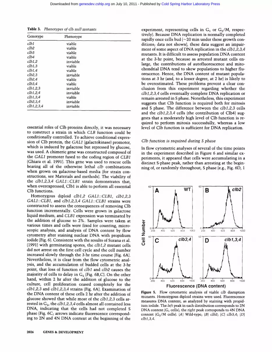
Table 3. Phenotypes of clb null mutants
Genotype Phenotype
clbl viable clb2 viable clb3 viable clb4 viable clbl,2 inviable clbl,3 viable clbl,4 viable clb2,3 inviable clb2,4 viable clb3,4 viable clbl,2,3 inviable clbl,2,4 inviable clbl,3,4 viable clb2,3,4 inviable clbl,2,3,4 inviable
essential roles of Clb proteins directly, it was necessary to construct a strain in which CLB function could be conditionally controlled. To achieve conditional expres- sion of Clb protein, the GALl (galactokinase) promoter, which is induced by galactose but repressed by glucose, was used. A chimeric gene was constructed composed of the GALl promoter fused to the coding region of CLB1 (Ghiara et al. 1991). This gene was used to rescue cells bearing all of the otherwise lethal clb combinations when grown on galactose-based media (for strain con- structions, see Materials and methods). The viability of the clbl,2,3,4 GALI::CLB1 strain demonstrates that, when overexpressed, Clb 1 is able to perform all essential Clb functions.
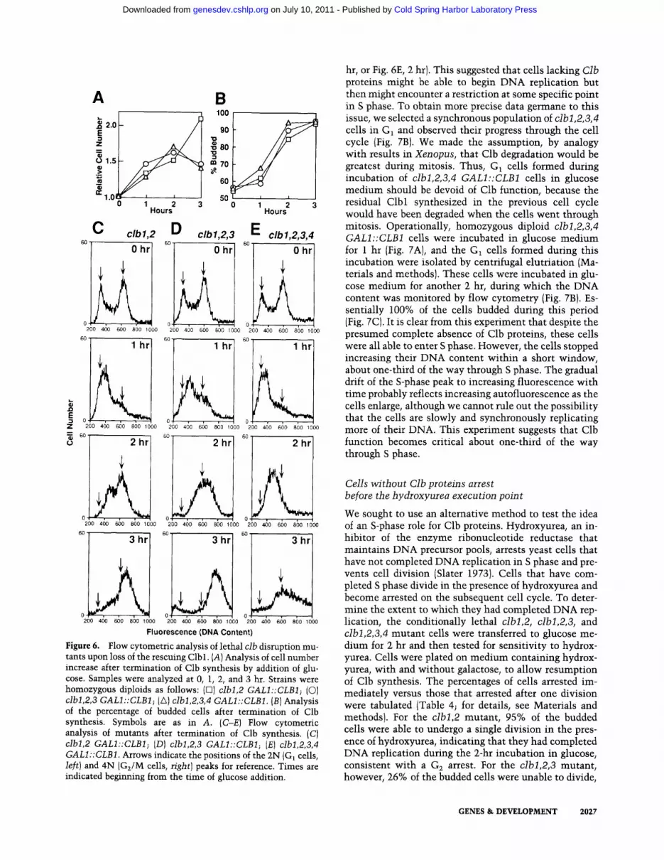
Homozygous diploid clbl,2 GALI::CLB1, clbl,2,3 GALI::CLB1, and clbl,2,3,4 GALI::CLB1 strains were constructed to assess the consequences of removing Clb function incrementally. Cells were grown in galactose liquid medium, and CLB1 expression was terminated by the addition of glucose to 2%. Samples were taken at various times and cells were fixed for counting, micro- scopic analysis, and analysis of DNA content by flow cytometry after staining nuclear DNA with propidium iodide (Fig. 6). Consistent with the results of Surana et al. (1991) with germinating spores, the clbl,2 mutant cells did not arrest on the first cell cycle and the cell number increased slowly through the 3-hr time course (Fig. 6A). Nevertheless, it is clear from the flow cytometric anal- ysis, and the accumulation of budded cells at the 3-hr point, that loss of function of clbl and clb2 causes the majority of cells to delay in G~ (Fig. 6B, C). On the other hand, within 2 hr after the addition of glucose to the culture, cell proliferation ceased completely for the clbl,2,3 and clbl,2,3,4 strains (Fig. 6A}. Examination of the DNA content of these cells 2 hr after the addition of glucose showed that while most of the clbl,2,3 cells ar- rested in G~, the clbl,2,3,4 cells almost all contained less DNA, indicating that the cells had not completed S phase (Fig. 6C; arrows indicate fluorescence correspond- ing to 2N and 4N DNA content at the beginning of the
experiment, representing cells in G1 or G2/M, respec- tively). Because DNA replication is normally completed rapidly once cells bud ( -20 rain under these growth con- ditions; data not shown), these data suggest an impair- ment of some aspect of DNA replication in the clbl,2,3,4 mutants. It is difficult to assess population DNA content at the 3-hr point, because as arrested mutant cells en- large, the contributions of autofluorescence and mito- chondrial DNA tend to skew populations to higher flu- orescence. Hence, the DNA content of mutant popula- tions at 3 hr (and, to a lesser degree, at 2 hr) is likely to be overestimated. These problems prevent a clear con- clusion from this experiment regarding whether the clbl,2,3,4 cells eventually complete DNA replication or remain arrested in S phase. Nonetheless, this experiment suggests that Clb function is required both for mitosis and S phase. The difference between the clbl,2,3 cells and the clbl,2,3,4 cells (the contribution of Clb4) sug- gests that a moderately high level of Clb function is re- quired to perform mitosis successfully, whereas a low level of Clb function is sufficient for DNA replication.
Clb function is required during S phase
In flow cytometric analyses of several of the time points in the experiment described in Figure 6 and similar ex- periments, it appeared that cells were accumulating in a distinct S-phase peak, rather than arresting at the begin- ning of, or randomly throughout, S phase (e.g., Fig. 6D, 1
VT A
~: 0 0 - :~, 200 400 600 800 1000 200 400 600 800 000
z 6O
D clb 1, 3, 4
0-~ " 0 200 ~o G~o 8~o ~ooo 200 ~o 6~o 8~o ,ooo
Fluorescence (DNA content) Figure 5. Flow cytometric analysis of viable clb disruption mutants. Homozygous diploid strains were used. Fluorescence measures DNA content, as analyzed by staining with propid- ium iodide. The left peak in each distribution corresponds to 2N DNA content {G~ cells); the right peak corresponds to 4N DNA content (G2/M cells). (A} Wild-type; (B) clb2; (C) clb3,4; (D) cibl,3,4.
60 60
2026 GENES & DEVELOPMENT
Cold Spring Harbor Laboratory Press on July 10, 2011 - Published by genesdev.cshlp.orgDownloaded from
.O E
• ~ 60 0
A
,•2.0 E
Z n
o 1.5
3 n- 1.0 I
0 1 2 3 H o u r s
B 1 ]0
)0
3o g~ to
~o
~o o
C clbl,2 D clbl,2,3 6O
Ohr
200 400 600 800 1000
I
2 3 Hours
E 1,2,3,4 0 hr I 60 0hr
0200 400 600 84]0 1000 200 400 6(~0 8;0 100<)
60 l h r 60 - l h r l l 60 ~ l h r
0 , , , 0 0
200 400 600 800 1000 200 400 600 800 1000 200 400 600 8(~0 1000
2hr
200 400 600 800 1000
60" 3 h r
0200 4{30 600 8;0 1000
2hr 2 h r
200 400 600 800 1000 200 400 600 800 1000
60 -
3 hr I 3 hr
0 0 , 200 4;0 660 8;0 lOOO 200 do 660 800 looo
Fluorescence (DNA Content)
Figure 6. Flow cytometric analysis of lethal clb disruption mu- tants upon loss of the rescuing Clbl. (A) Analysis of cell number increase after termination of Clb synthesis by addition of glu- cose. Samples were analyzed at 0, 1, 2, and 3 hr. Strains were homozygous diploids as follows: (El) clbl,2 GALI::CLB1; (C)) clbl,2,3 GALI::CLB1; (A) clbl,2,3,4 GALI::CLB1. (B) Analysis of the percentage of budded cells after termination of Clb synthesis. Symbols are as in A. (C-E) Flow cytometric analysis of mutants after termination of Clb synthesis. (C) clbl,2 GALI::CLB1; (D) clbl,2,3 GALI::CLB1; (E) clbl,2,3,4 GALI::CLB1. Arrows indicate the positions of the 2N (Gt cells, left) and 4N (G~/M cells, right) peaks for reference. Times are indicated beginning from the time of glucose addition.
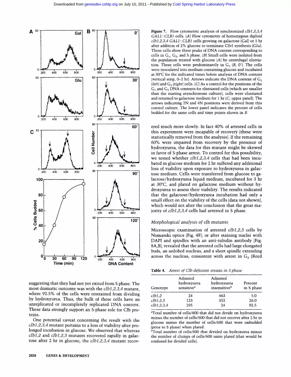
hr, or Fig. 6E, 2 hr). This suggested that cells lacking Clb proteins might be able to begin DNA replication but then might encounter a restriction at some specific point in S phase. To obtain more precise data germane to this issue, we selected a synchronous population of clbl,2,3,4 cells in G1 and observed their progress through the cell cycle (Fig. 7B). We made the assumption, by analogy with results in Xenopus, that Clb degradation would be greatest during mitosis. Thus, G1 cells formed during incubation of clbl,2,3,4 GALI::CLB1 cells in glucose medium should be devoid of Clb function, because the residual Clbl synthesized in the previous cell cycle would have been degraded when the cells went through mitosis. Operationally, homozygous diploid clbl,2,3,4 GALI::CLB1 cells were incubated in glucose medium for 1 hr (Fig. 7A), and the G1 cells formed during this incubation were isolated by centrifugal elutriation (Ma- terials and methods). These cells were incubated in glu- cose medium for another 2 hr, during which the DNA content was monitored by flow cytometry (Fig. 7B). Es- sentially 100% of the cells budded during this period (Fig. 7CI. It is clear from this experiment that despite the presumed complete absence of Clb proteins, these cells were all able to enter S phase. However, the cells stopped increasing their DNA content within a short window, about one-third of the way through S phase. The gradual drift of the S-phase peak to increasing fluorescence with time probably reflects increasing autofluorescence as the cells enlarge, although we cannot rule out the possibility that the cells are slowly and synchronously replicating more of their DNA. This experiment suggests that Clb function becomes critical about one-third of the way through S phase.
Cells without Clb proteins arrest before the hydroxyurea execution point
We sought to use an alternative method to test the idea of an S-phase role for Clb proteins. Hydroxyurea, an in- hibitor of the enzyme ribonucleotide reductase that maintains DNA precursor pools, arrests yeast cells that have not completed DNA replication in S phase and pre- vents cell division (Slater 1973). Cells that have com- pleted S phase divide in the presence of hydroxyurea and become arrested on the subsequent cell cycle. To deter- mine the extent to which they had completed DNA rep- lication, the conditionally lethal clbl,2, clbl,2,3, and clbl,2,3,4 mutant cells were transferred to glucose me- dium for 2 hr and then tested for sensitivity to hydrox- yurea. Cells were plated on medium containing hydrox o yurea, with and without galactose, to allow resumption of Clb synthesis. The percentages of cells arrested im- mediately versus those that arrested after one division were tabulated (Table 4; for details, see Materials and methods). For the clbl,2 mutant, 95% of the budded cells were able to undergo a single division in the pres- ence of hydroxyurea, indicating that they had completed DNA replication during the 2-hr incubation in glucose, consistent with a G2 arrest. For the clbl,2,3 mutant, however, 26% of the budded cells were unable to divide,
GENES & DEVELOPMENT 2027
Cold Spring Harbor Laboratory Press on July 10, 2011 - Published by genesdev.cshlp.orgDownloaded from
A so Gal
C so
200 400 600 800
Glu
2oo 46O 66o 86o
260 46o 66o 86o
100
8O
"10 m= 60
° 40
20
I I I 30 60 90 120 Time (min)
B 810 0'
0 200 400 600 800
8o ~ 30'
o , ~o 4~ 6~ 8&
8o
60'
~~11 | | I 90'
26o 46o 66o 86o
120'
0 , 200 a4o 660 860
DNA Content
suggesting that they had not yet exited from S phase. The most dramatic outcome was with the clbl,2,3,4 mutant , where 92.5% of the cells were restrained from dividing by hydroxyurea. Thus, the bulk of these cells have an unreplicated or incompletely replicated D N A content. These data strongly support an S-phase role for Clb pro- teins.
One potential caveat concerning the result with the clbl,2,3,4 mutan t pertains to a loss of viability after pro- longed incubation in glucose. We observed that whereas clbl,2 and clbl,2,3 mutants recovered rapidly in galac- tose after 2 hr in glucose, the clbl,2,3,4 mutan t recov-
Figure 7. Flow cytometric analysis of synchronized clbi,2,3,4 GALI::CLB1 cells. (A)Flow cytometry of homozygous diploid clbl,2,3,4 GALI::CLB1 cells growing on galactose (Gal) or 1 hr after addition of 2% glucose to terminate Clbl synthesis (Glu). These cells show three peaks of DNA content corresponding to cells in Gl, G~, and S phase. (B) Small cells were isolated from the population treated with glucose (A) by centrifugal elutria- tion. These cells were predominantly in GI (B, 0'). The cells were inoculated into medium containing glucose and incubated at 30°C for the indicated times before analysis of DNA content (vertical strip, 0--2 hr). Arrows indicate the DNA content of G1 (left) and G2 (right) cells. (C) As a control for the positions of the G] and G 2 DNA contents for elutriated cells (which are smaller than the starting asynchronous culture), cells were elutriated and returned to galactose medium for 1 hr (C, upper panel). The arrows indicating 2N and 4N positions were derived from this control culture. The lower panel indicates the percent of cells budded for the same cells and time points shown in B.
ered much more slowly. In fact 40% of arrested cells in this experiment were incapable of recovery (these were statistically removed from the analysis). If the remaining 60% were impaired from recovery by the presence of hydroxyurea, the data for this mu tan t might be skewed in favor of S-phase arrest. To control for this possibility, we tested whether clbl,2,3,4 cells that had been incu- bated in glucose medium for 2 hr suffered any additional loss of viability upon exposure to hydroxyurea in galac- tose medium. Cells were transferred from glucose to ga- lactose/hydroxyurea liquid medium, incubated for 3 hr at 30°C, and plated on galactose med ium wi thout hy- droxyurea to assess their viability. The results indicated that the galactose/hydroxyurea incubation had only a small effect on the viability of the cells (data not shown), which would not alter the conclusion that the great ma- jority of clbl,2,3,4 cells had arrested in S phase.
Morphological analysis of clb mutants
Microscopic examinat ion of arrested clbl,2,3 cells by Nomarsk i optics (Fig. 4F), or after staining nuclei wi th DAPI and spindles with an anti- tubulin antibody {Fig. 8A, B), revealed that the arrested cells had large elongated buds, an unlobed nucleus, and a short spindle extending across the nucleus, consistent wi th arrest in G2 (Reed
Table 4. Arrest of Clb-deficient strains in S phase
Adjusted Adjusted hydroxyurea hydroxyurea Percent
Genotype sensitive a insensitive b in S phase
clbl,2 24 463 5.0 clbl,2,3 123 352 26.0 clbl,2,3,4 295 24 92.5
aTotal number of cells/600 that did not divide on hydroxyurea minus the number of cells/600 that did not recover after 2 hr in glucose minus the number of cells/600 that were unbudded (prior to S phase) when plated. bTotal number of cells/600 that divided on hydroxurea minus the number of clumps of cells/600 units plated (that would be confused for divided cells).
2028 GENES & DEVELOPMENT
Cold Spring Harbor Laboratory Press on July 10, 2011 - Published by genesdev.cshlp.orgDownloaded from
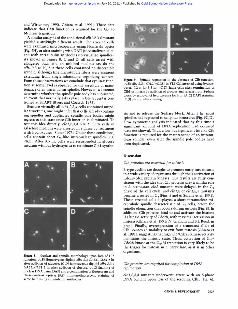
and Wittenberg 1990; Ghiara et al. 1991). These data indicate that CLB function is required for the G2- to M-phase transition.
A similar analysis of the conditional clbl,2,3,4 mutant yielded a strikingly different result. The arrested cells were examined microscopically using Nomarski optics (Fig. 4H), or after staining with DAPI (to visualize nuclei) and with anti-tubulin antibodies (to visualize spindles). As shown in Figure 8, C and D, all cells arrest with elongated buds and an unlobed nucleus (as do the clbl,2,3 cells), but these cells contained no discernible spindle, although fine microtubule fibers were apparent extending from single-microtuble organizing centers. From these observations we conclude that cyclin-B func- tion at some level is required for the assembly or main- tenance of an intranuclear spindle. However, we cannot determine whether the spindle pole body has duplicated, an event that normally takes place in late G1 and is con- trolled at START (Byers and Goetsch 1975).
Because virtually all clbl,2,3,4 cells contained unipo- lar structures, one might infer that cells already contain- ing spindles and duplicated spindle pole bodies might regress to this state once Clb function is eliminated. To test this idea directly, clbl,2,3,4 GALI::CLB1 cells in galactose medium were arrested in S phase by treatment with hydroxyurea (Slater 1973). Under these conditions, cells contain short G2-1ike intranuclear spindles (Fig. 9A,B). After 3.5 hr, cells were resuspended in glucose medium without hydroxyurea to terminate Clb 1 synthe-
Figure 8. Nuclear and spindle morphology upon loss of Clb function. (A,B) Homozygous diploid clbl,2,3 GALl::CLB1 2 hr after addition of glucose; (C,D) homozygous diploid clbl,2,3,4 GALI::CLBI 2 hr after addition of glucose. (A,C) Staining of nuclear DNA using DAPI and a combination of fluorescent and phase-contrast optics; (B,D) immunofluorescent staining of same field using anti-tubulin antibodies.
Figure 9. Spindle regression in the absence of Clb function. {A,B1 clbl,2,3,4 GALl ::CLB1 in YEP Gal arrested using hydrox- yurea (0.2 M for 3.5 hr). (C,D) Same cells after termination of Clbl synthesis by addition of glucose and release from S-phase block by removal of hydroxyurea for 3 hr. (A, C) DAPI staining; {B,D) anti-tubulin staining.
sis and to release the S-phase block. After 3 hr, most spindles had regressed to unipolar structures (Fig. 9C,D). Flow cytometric analysis indicated that by this time a significant amount of DNA replication had occurred (data not shown). Thus, a low but significant level of Clb function is required for the maintanence of an intranu- clear spindle, even after the spindle pole bodies have been duplicated.
D i s c u s s i o n
Clb proteins are essential for mitosis
B-type cyclins are thought to promote entry into mitosis in a wide variety of organisms through their activation of Cdc28/cdc2 protein kinases. Our results are fully con- sistent with the idea that Glb proteins play a similar role in S. cerevisiae, clb2 mutants were delayed in the G2 phase of the cell cycle, and clbl,2 or clbl,2,3 mutants became arrested in G2 (Figs. 5 and 6; Surana et al. 1991). These arrested cells displayed a short intranuclear mi- crotubule spindle characteristic of G2 cells, before the spindle elongation that occurs during mitosis (Fig. 8). In addition, Clb proteins bind to and activate the histone H1 kinase activity of Cdc28, with maximal activation in mitosis (Ghiara et al. 1991; N. Grandin and ST Reed, in prep.). Finally, overexpression of a truncated allele of Clbl causes an inability to exit from mitosis (Ghiara et al. 1991), suggesting that high Clb/Cdc28 kinase activity maintains the mitotic state. Thus, activation of Clb/ Cdc28 kinase at the G2/M transition is very likely to be the trigger for mitosis in S. cerevisiae, as it is in other organisms.
Clb proteins are required for completion of DNA replication
clbl,2,3,4 mutants underwent arrest with an S-phase DNA content upon loss of the rescuing Clbl (Fig. 6).
GENES & DEVELOPMENT 2029
Cold Spring Harbor Laboratory Press on July 10, 2011 - Published by genesdev.cshlp.orgDownloaded from
Furthermore, when the arrested cells were returned to growth on media that reinduced Clbl, it could be seen that they were still sensitive to hydroxyurea (Table 4). Both of these observations suggest that elimination of Clb function prevents completion of DNA replication. Analysis of synchronized cells lacking Clb proteins showed that these cells all entered S phase and arrested uniformly after replicating approximately one-third of their nuclear DNA (Fig. 7). This surprising result sug- gests that Clb function may not be required for the ini- tiation or elongation phases of DNA replication but, rather, for a distinct function that only becomes essen- tial for DNA replication once a significant part of the genome has been replicated. Possible functions might include initiation of DNA replication at late origins or activation of topoisomerase required to relieve DNA su- percoiling generated by replication. In this regard, it is interesting to note that an additional B-type cyclin, Clb5, has recently been identified in S. cerevisiae based on its ability to function as a G~ cyclin (Epstein and Cross 1992; ST Reed, unpubl.). Although the functional rela- tionship of Clb5 to the other Clb proteins is not yet clear, it appears that Clb5 is required for efficient progression through S phase (Epstein and Cross 1992). It is possible that Clb5 and the other Clb proteins perform overlapping functions in S phase and that the DNA replication seen in the clbl,2,3,4 mutant was the result of Clb5 activity. However, different Clb proteins might perform distinct specialized roles in S phase. Recently, cyclin A has been shown to be required for initiation of, or progression through, S phase in mammalian somatic cells (Girard et al. 1991; Pagano et al. 1992). Although Clb3, Clb4, and Clb5 do not share strong structural homology with cy- clin A, they may perform analogous functions in yeast.
CIb proteins play a role in the assembly and maintenance of the intranuclear spindle
In S. cerevisiae, the spindle pole bodies (the major mi- crotubule-organizing centers in these cells) are dupli- cated near the Gx/S transition (Byers and Goetsch 1975). During S phase, the duplicated spindle pole bodies sep- arate and a short intranuclear spindle forms between them, which is maintained through Gz and elongates during mitosis {Byers and Goetsch 1975). Most S. cere- visiae S-phase and G2-arresting cdc mutations confer a terminal phenotype that consists, in part, of a short, thick, intranuclear spindle (Byers and Goetsch 1975; Ghiara et al. 1991). It was therefore rather surprising that the clbl,2,3,4 mutants deprived of the rescuing Clbl ar- rested uniformly without a discernible intranuclear spin- dle (Fig. 8). Moreover, the loss of Clbl in this strain re- sulted in the disappearance of spindles that had already formed during a hydroxyurea arrest (Fig. 9). Thus, Clb proteins are required for the maintenance (and possibly assembly) of the short G2 spindle, as well as for the elon- gation of this spindle during mitosis (see above).
The finding that both DNA replication and spindle assembly were defective in clbl,2,3,4 mutants raised the possibility that one of these phenotypes was indirectly
caused by the other (i.e., that DNA replication was blocked because of the inability to assemble a spindle or that spindle assembly was blocked because of the inabil- ity to complete DNA replication). We consider this pos- sibility unlikely because previous studies have shown that DNA replication proceeds unhindered even when microtubule structures are disrupted by nocodazole or ~-tubulin mutations (Huffaker et al. 1988; Jacobs et al. 1988). Similarly, spindle assembly proceeds normally even when DNA replication is blocked by hydroxyurea or various cdc mutations (Byers 1981; Pringle and Hart- well 1981). Thus, we consider it more likely that Clb proteins directly regulate both DNA replication and spindle assembly.
Another surprising finding was that clbl,2,3,4 mu- tants suffered a dramatic loss of viability 3-4 hr after withdrawal of the rescuing Clbl. In contrast, clbl,2,3 mutants did not show a significant loss of viability dur- ing this period. This difference suggests that the loss of viability was a result of the defects observed uniquely with the clbl,2,3,4 mutant, namely impaired DNA rep- lication and spindle assembly. Possibly, the unipolar mi- crotubule structure of this mutant corresponds to an ir- reversible state, blocking further cell cycle progress. Al- ternatively, the defect in DNA replication may lead to lesions that cannot be repaired efficiently.
Which Clb protein does what?
The genetic data reported here demonstrate that Clb pro- teins perform several critical functions during the cell cycle (above), with Clb2 making the strongest contribu- tion to these functions and Clb4 the weakest. Our ob- servations with different lethal clb mutant combina- tions suggest that a low level of Clb function is required for DNA replication and a high level of Clb function is required for chromosome segregation. Similarly, a low level of Clb function suffices for assembly and mainte- nance of a short interphase spindle while a higher level is required for spindle elongation in mitosis. All four Clb proteins are able to provide the DNA replication and spindle assembly functions, and at least Clbl, Clb2, and Clb3 can contribute to the mitotic functions. However, these data do not reveal whether different Clb proteins are normally responsible for the separate functions (but can substitute for each other when necessary) or whether all Clb proteins participate redundantly in all functions.
Although not revealed by the genetic experiments, there are strong reasons to believe that Clb proteins fall into two functional classes, with Clb3 and Clb4 perform- ing the S-phase and early spindle assembly roles and Clbl and Clb2 performing the mitotic roles. First, se- quence analysis shows that Clb3/Clb4 and Clbl/Clb2 form pairs that are related much more closely to each other than they are to members of the other pair. Fur- thermore, Clb3 and Clb4 are related most closely to the S. pombe cyclin cigl (Fig. 2), which appears to play a role in G1 or S phase [Bueno et al. 1991), whereas Clbl and Clb2 show highest homology to the S. pombe cyclin cdcl3 involved in the initiation of mitosis. Second,
2030 GENES & DEVELOPMENT
Cold Spring Harbor Laboratory Press on July 10, 2011 - Published by genesdev.cshlp.orgDownloaded from
mRNAs for Clb3 and Clb4 are expressed [and corre- sponding kinases are activated (N. Grandin and S.I. Reed, in prep.)] earlier in the cell cycle than those for Clbl and Clb2 (Fig. 3). Thus, we propose that in the normal cell cycle Clb3 and Clb4 function in S phase to promote D N A replication and spindle assembly, while Clbl and Clb2 (possibly aided by Clb3 and Clb4) are mainly re- sponsible for mitot ic induction. This model, however, requires a certain plasticity of Clb function, such that Clbl and/or Clb2 can fulfill early cell cycle functions if Clb3 and Clb4 are not present.
Substrates for the Clb / Cdc28 protein k inase
Given the broad range of precedents for cyclin B in other eukaryotic cells and our previous results with Clbl (Ghi- ara et al. 1991), it is very likely that Clb proteins exert their effects through activation of the Cdc28 protein ki- nase and consequent phosphorylat ion of appropriate sub- strates. The Clb functions identified in this report pro- vide clues to the possible identi ty of these substrates. The D N A replication function may reflect a need to phosphorylate proteins that form part of the replication complexes, whereas spindle-associated motor proteins are attractive candidates for substrates involved in spin- dle elongation. The role uncovered here in spindle main- tenance might also reflect a requirement to phosphory- late kinesin-like motors shown recently to be crucial for spindle pole body separation (Hoyt et al. 1992; Roof et al. 1992). Alternatively, perhaps nucleat ion of microtubules on the nuclear side of the spindle pole body requires phosphorylat ion of spindle pole body components by the Clb/Cdc28 kinase. Consistent wi th this idea, the col- lapse of already assembled spindles in the absence of Clb function is reminiscent of the return of spindle pole bod- ies to a side-by-side configuration in cells treated with microtubule-depolymerizing drugs such as benomyl and nocodazole (Jacobs et al. 1988).
M a t e r i a l s a nd m e t h o d s
Cloning the CLB2, CLB3, and CLB4 genes
The S. cerevisiae cyclin-B homolog CLB1 (SCB1) was isolated by using PCR and degenerate oligonucleotides designed to the AA(S}KYEE and AKYL(F}M(VI)E motifs {Ghiara et al. 1991). To isolate other cyclin-B homologs from S. cerevisiae using the PCR technique (Saiki et al. 1988) the following degenerate oli- gonucleotides were used: 5' primer, 5'-CCGGATCCMRNYT- NCARYTNGTNGG-3' corresponding to the amino acid se- quence KLQLVG; 3' primer, 5'-CGAATTCYTCNAYNAR- RTAYTTNGC-3' corresponding to the amino acid sequence AKYL(F)M(VI)E (Y = C + T, R = A + G, M = C + A, N = A + G + C + T). The BamHI and EcoRI restriction sites used for cloning are underlined. The PCR products were di- gested with BamHI and EcoRI restriction enzymes, gel purified, and cloned into the BamHI and EcoRI sites of pT7T319U (Phar- macia).
Using PCR and the KLQLVG and AKYL(F)M(VI)E primers, a second cyclin-B homolog was isolated from S. cerevisiae. The derived amino acid sequence of this clone was identical to the partial amino acid sequence of the CLB3 gene, as presented by
Surana et al. (1991). To isolate the complete genomic clone, a radiolabeled probe made from the PCR clone of CLB3 was used to screen a S. cerevisiae genomic library in the vector h Dash (obtained from Merl Hoekstra, Salk Institute, San Diego, CA). DNA sequence analysis revealed an open reading frame of 1278 bp, predicted to encode a protein of 426 amino acids (Fig. 1A).
The CLB2 gene was obtained by screening the h Dash S. cer- evisiae genomic library under low stringency conditions (as de- scribed below) with a CLB1 probe (0.5-kb EcoRV fragment) con- taining the cyclin box region. The sequence identified had sev- eral discrepancies relative to that reported by Surana et al. (1991): At position -108, they have TATAACCCC, whereas we have TATAAAAAC, which fits the consensus sequence for a TATA box; and at position 1510, they have TGA, whereas we have TAA.
The CLB4 gene was isolated by screening the h Dash S. cer- evisiae genomic library under low stringency conditions (as de- scribed below) with a CLB3 probe specific for the cyclin box (the CLB3 PCR clone). The derived amino acid sequence of this gene was similar to the partial amino acid sequence of CLB4 pre- sented by Surana et al. (1991).
Radiolabeled probes were made using the random primer DNA labeling kit (Boehringer Mannheim) according to the man- ufacturer's instructions. For Southern blot analysis, DNA was transferred to nitrocellulose filters (Schleicher & Schuell).
Construction of clb null mutations
The CLB2 null allele (clb2::LEU2) was constructed by utilizing a 2.8-kb EcoRI subclone derived from a phage clone of CLB2. This clone enabled the 0.38-kb EcoRV-BglII fragment from the cyclin box region to be replaced with a 2.5-kb PvuII-BamHI fragment containing LEU2 (derived from a clone of LEU2 in pUC19) The construct was then excised with NsiI and used to transform BF264-15DUa. Transformants were selected on min- imal plates supplemented with amino acids but lacking leucine.
The CLB3 null allele (clb3:: TRP1) was constructed as follows. A 1.5-kb BglII-HincII fragment containing the TRP1 gene (from the plasmid YRp7) was used to replace the 0.58-kb BglII-EcoRV fragment containing the cyclin box region of CLB3. The target for insertion was a 2.7-kb ClaI-SalI clone of CLB3 in pT7T3 19U. The resulting disruption construct was then excised with SalI and ClaI and used to transform BF264-15DUa. Transfor- mants were selected on minimal plates supplemented with amino acids but lacking tryptophan.
The CLB4 null allele (clb4::HIS2) was constructed by the re- placement of a 1.8-kb EcoRV-NsiI fragment containing the CLB4 gene with a 1.9-kb PstI-EcoRI fragment containing HIS2 (derived from a clone of HIS2 in pUC118). This insertion was carried out in the 3.8-kb EcoRI clone of CLB4. The construct was excised with AseI and EcoRI and used to transform YS102. Transformants were selected on minimal plates supplemented with amino acids but lacking histidine.
In all cases, transformants were screened for the gene disrup- tion by Southern blot analysis of genomic DNA.
Yeast strains, genetic procedures, media, and growth conditions
All yeast strains used in this study were derivatives of BF264 o 15DU: MATa adel his2 leu2-3,112 trpl-1 a ura3Dns (Richardson et al. 1989). The relevant genotypes of strains used in this study are shown in Table 5.
Standard genetic procedures for yeast were used (Sherman et al. 1982). Yeast transformations were carried out by the alkali
GENES & DEVELOPMENT 2031
Cold Spring Harbor Laboratory Press on July 10, 2011 - Published by genesdev.cshlp.orgDownloaded from
Table 5. Strain list
Strain Relevant genotype
YH134 MA Ta YH135 MA Ta YS101 MATa YS102 MA Ta YS104 MA Ta YS105 MA Ta YS106 MA Te~ YS107 MATa YS108 MATa YS112 MATa YS109 MA Ta YSll4 MA Ta YSll5 MATa YSll8 MA Ta YS201 MA Ta/a YS202 MA Ta/a YS203 MA Ta/~ DLY005 MA Ta/~ DLY373 MATa/~ DLY378 MA Ta/c, DLY379 MATa/~ DLY380 MA Ta/c~ DLY382 MATa/a DLY384
clbl :: URA3 clb2::LEU2 clb3::TRP1 c lb l : :URA3 clb3::TRP1 clbI::URA3 clb3::TRP1 clb4::HIS2 GALl :: CLB1 (LE U2) clbl :: URA3 clb3:: TRP1 clb4::HIS2 clb2::LE U2 GALl ::CLBI (LEU2) clbl :: URA3 clb2::LEU2 clb3::TRP1 GALl ::CLBI (LEU2) clbl :: URA3 clb2::LEU2 clb3::TRP1 clb4::HIS2 clb4::HIS2 GALl ::CLB1 (LEU2) clbl :: URA3 clb2::LEU2 clbl :: URA3 clb4::HIS2 clb2::LEU2 clb4::HIS2 clb3::TRP1 clb4::HIS2
clbl ::URA3/CLBI clb3::TRP1/CLB3 c lb l : :URA3/CLBI clb3::TRP1/CLB3 clb2::LEU2/CLB2 GALl :: CLB 1 (LE U2) clb I :: URA3/CLB I clb3:: TRP1/CLB3 clb2::LE U2/CLB2 clb4::HIS2/CLB4
clb2::LEU2/clb2::LEU2 clb3::TRP1/clb3::TRP1 clb4::HIS2/clb4::HIS2 clbl :: URA3/clbl :: URA3 clb3:: TRP1/clb3:: TRP1 clb4::HIS2/clb4::HIS2 GA L1 :: CLBI (LE U2) clb 1 :: URA3/clbl :: URA3 clb2::LE U2/clb2::LE U2 GALl ::CLBI (LEU2) clbl :: URA3/clbl :: URA3 clb2::LEU2/clb2::LEU2 clb3::TRP1/clb3::TRP1
MATa/eL GALl :: CLBI (LEU2) clbl :: URA3/clbl :: URA3 clb2::LEU2/clb2::LE U2 clb3::TRPI/clb3::TRP1 clb4 : : HI S2/ clb4 : : HI S2
cation method (Ito et al. 1983). Genomic sequence replacements were performed as described by Rothstein (1983).
Yeast cultures were grown at 30°C in YEP (1% yeast extract, 2% Bacto-peptone, 0.005% adenine, 0.005% uracil) supple- mented with glucose (2%) or galactose (2%). Conditional Clbl synthesis was terminated (GALI:CLB1 strains) by adding 50% glucose to cultures growing in YEP/galactose medium to a final concentration of 2%. Cultures were arrested in S phase (Fig. 8) by growth in YEP/galactose supplemented with 0.2 M hydrox- yurea for 3.5 hr and released from the block by resuspension in YEP/glucose.
Hydroxyurea execution point experiments (Table 4} were per- formed by growing GALI::CLB1 clbl,2, GALI::CLB1 clbl,2,3, and GALI::CLB1 clbl,2,3,4 mutant strains in glucose-supple- mented medium for 2 hr, after which cells were subjected to a short pulse of sonic disruption and plated on each of three types of solid medium: (1) minimal synthetic medium containing 2% glucose, (2) YEP/galactose, and (3) YEP/galactose supplemented with 0.4 M hydroxyurea. The first plate, which was immediately stored at 4°C, served as a record of the composition of the plated cell population. The second plate served as viability control for the nutritional shift from glucose to galactose and was main- tained at 30°C for 12 hr. The third plate, kept at 30°C for 20 hr, was used to determine whether individual cells could undergo a round of division in the presence of the replication inhibitor hydroxyurea. Cells were observed directly on the agar surface, using a low power objective and a stage modified to accommo- date 100-mm petri dishes. The plating control (minimal plate) was scored by assigning cells to three catagories: unbudded cells, singly budded cells, and units composed of multiple buds or cells. The viability control was scored by assigning cells to two catagories: single cells and microcolonies. Single cells were assumed to be inviable. The experimental plate was scored by assigning cells to two catagories: undivided (singly budded) cells
and divided (two side-by-side budded) cells. To calculate the percentage of S-phase (hydroxyurea sensitive) cells, the number of inviable cells (viability control) and unbudded cells (plating control) was subtracted from the number of cells that did not divide on hydroxyurea to give an adjusted hydroxyurea-sensi- tire cell number. The number of multiple cells (plating control) was subtracted from the number of cells that divided on hy- droxyurea to give an adjusted hydroxyurea-insensitive cell number. The percentage of S-phase cells was then calculated by dividing the adjusted hydroxyurea-sensitive cell number by the sum of the adjusted hydroxyurea-sensitive and -insensitive cell numbers. The calculations were based on scoring 600 indepen- dent structures (cells, pairs of cells, or microcolonies) on each plate.
Photomicroscopy, immunofluorescence staining, cell counting, f low cytometry, and cell synchrony
Yeast cells were photographed live, using differential interfer- ence contrast (Nomarski) optics with a 100 x objective. Fluores- cent photomicroscopy on fixed and stained cells was performed using a Zeiss Axiophot photomicroscope with a 100 x objective. DAPI and anti-tubulin antibody staining was performed as de- scribed by Ghiara et al. (1991).
Cell numbers and budding percentages were determined us- ing a hemacytometer and a Leitz SM-Lux phase-contrast micro- scope with a 40x objective.
Cell cultures were analyzed f6r DNA content using flow cy- tometry by the method of Hutter and Eipel (1979). Yeast cells were fixed in 70% ethanol, stained with propidium iodide, and analyzed for fluorescence using a Becton-Dickinson FACScan analyzer (Lew et al. 1992).
Cell synchrony using mating pheromone was as described by
2032 GENES & DEVELOPMENT
Cold Spring Harbor Laboratory Press on July 10, 2011 - Published by genesdev.cshlp.orgDownloaded from
Ghiara et al. (1991). Centrifugal elutriation was performed as described by Lew et al. (1992).
Acknowledgments
We are very grateful to Pat O'Farrell and Rob Saint for supplying laboratory space, equipment, and materials to H.E.R. for part of this work. Thanks go to C. Epstein and F. Cross for communi- cation of results before publication. We also thank Dalia Resnitzky for help with various aspects of this work and Nath- alie Grandin for critical reading of this manuscript. H.E.R was supported for part of this work by an Australian Research Coun- cil QEII fellowship. D.J.L. was supported by a Damon Runyon- Walter Winchell Cancer Research Fund Fellowship (DRG- 1078). This work was supported by U.S. Public Health Service grant GM38328 to S.I.R.
The publication costs of this article were defrayed in part by payment of page charges. This article must therefore be hereby marked "advertisement" in accordance with 18 USC section 1734 solely to indicate this fact.
Note added in proof
Sequence data described in this paper have been submitted to the GenBank data library.
References
Booher, R. and D. Beach. 1987. Interaction between cdcl3 + and cdc2 + in the control of mitosis in fission yeast; dissociation of the G1 and G2 roles of the cdc2 + protein kinase. EMBO J. 6: 3441-3447.
1988. Involvement of cdcl3 + in mitotic control in Schizosaccharomyces pombe: Possible interaction of the gene product with microtubeules. EMBO J. 7: 2321-2327.
Bueno, A., H. Richardson, S.I. Reed, and P. Russell. 1991. A fission yeast B-type cyclin functioning early in the cell cycle. Cell 66: 149-159.
Byers, B. 1981. Cytology of the yeast life cycle. In The molecular biology of the yeast Saccharomyces: Life cycle and inheri- tance (ed. J.N. Strathern, E.W. Jones, and J.R. Broach), pp. 59-97. Cold Spring Harbor Laboratory, Cold Spring Harbor, New York.
Byers, B. and L. Goetsch. 1975. Behavior of spindles and spindle plaques in the cell cycle and conjugation of Saccharomyces cerevisiae. J. Bacteriol. 124: 511-523.
Cross, F. 1988. DAF1, a mutant gene affecting size control, pher- omone arrest, and cell cycle kinetics of Saccharomyces cer- evisiae. Mol. Cell. Biol. 8: 4675-4684.
- - . 1990. Cell cycle arrest caused by CLN gene deficiency in Saccharomyces cerevisiae resembles START-arrest and is independent of the mating-pheromone signalling pathway. Mol. Cell. Biol. 10: 6482-6490.
Cross, F., J. Roberts, and H. Weintraub. 1989. Simple and com- plex cell cycles. Annu. Rev. Cell Biol. 5: 341-395.
Elledge, S.J. and M.R. Spottswood. 1991. A new human p34 protein kinase, CDK2, identified by complementation of a cdc28 mutation in Saccharomyces cerevisiae, is a homolog of Xenopus Egl. EMBO J. 10: 2653-2659.
Epstein, C.B. and F.R. Cross. 1992. CLB5: A novel B cyclin from budding yeast with a role in S phase. Genes & Dev. 6: 1695- 1706.
Evans, T., E.T. Rosenthal, J. Youngblom, D. Distel, and T. Hunt. 1983. Cyclin: A protein specified by maternal mRNA in sea urchin eggs that is destroyed at each cleavage division. Cell
33: 389-396. Fang, F. and J.W. Newport. 1991. Evidence that the G1-S and
G2-M transitions are controlled by different cdc2 proteins in higher eukaryotes. Cell 66: 731,742.
Forsburg, S.L. and P. Nurse. 1991. Identification of a Gl-type cyclin puc + in the fission yeast Schizosaccharomyces pombe. Nature 351: 245-248.
Ghiara, J.B., H.E. Richardson, K. Sugimoto, M. Henze, D.J. Lew, C. Wittenberg, and S.I. Reed. 1991. A cyclin B homolog in S. cerevisiae: Chronic activation of the Cdc28 protein kinase by cyclin prevents exit from mitosis. Cell 65: 163-174.
Girard, F., U. Strausfeld, A. Fernandez, and N.J.C. Lamb. 1991. Cyclin A is required for the onset of DNA replication in mammalian fibroblasts. Ceil 67:1169-1179.
Glotzer, M., A.W. Murray, and M.W. Kirschner. 1991. Cyclin is degraded by the ubiquitin pathway. Nature 349: 132-138.
Hadwiger, J.A., C. Wittenberg, H.E. Richardson, M. de Barros Lopes, and S.I. Reed. 1989). A novel family of cyclin ho- mologs that control G1 in yeast. Proc. Natl. Acad. Sci. 86: 6255-6259.
Hagan, I.M., J. Hayles, and P. Nurse. 1988. Cloning and sequenc- ing of the cyclin related cdcl3 + gene and a cytological study of its role in fission yeast mitosis. J. Ceil Sci. 91: 587-595.
Hartwell, L.H., J. Culotti, J.R. Pringle, and B.J. Reid. 1974. Ge- netic control of the cell division cycle in yeast. Science 183: 46--51.
Hoyt, M.A., L. He, K.K. Loo, and W.S. Saunders. 1992. Two Saccharomyces cerevisiae kinesin-related gene products re- quired for mitotic spindle assembly. J. Cell Biol. 118: 109- 120.
Huffaker, T.C., J.H. Thomas, and D. Botstein. 1988. Diverse effects of J~-tubulin mutations on microtubule formation and function. ]. Cell Biol. 106: 1997-2010.
Hunt, T. 1989. Maturation promoting factor, cyclin and the control of M-phase. Curr. Opin. Cell Biol. 1: 268-274.
Hunt, T., F.C. Luca, and J.V. Ruderman. 1992. The requirements for protein synthesis and degradation, and the control of de- struction of cyclins A and B in the meiotic and mitotic cell cycles of the clam embryo. J. Cell Biol. 116: 707-724.
Hutter, K.-J. and H.E. Eipel. 1979. DNA determination of yeast by flow cytometry. 1. Gen. Microbiol. 113: 369-375.
Ito, H., Y. Fukuda, K. Murata, and A. Kimura. 1983. Transfor- mation of intact yeast cells treated with alkali cations. J. Bacteriol. 153: 163-168.
Jacobs, C.W., A.E.M. Adams, P.J. Szaniszlo, and J.R. Pringle. 1988. Functions of microtubules in the Saccharomyces cer- evisiae cell cycle. J. Cell Biol. 107: 1409-1426.
Lehner, C.F. and P.H. O'Farrell. 1990. The roles of Drosophila cyclins A and B in mitotic control. Cell 61: 535-547.
Lew, D.J. and S.I. Reed. 1992. A proliferation of cyclins. Trends Ceil Biol. 2: 77-81.
Lew, D.J., V. Dulic, and S.I. Reed. 1991. Isolation of three novel human cyclins by rescue of G1 cyclin (Cln) function in yeast. Cell 66:1197-1206.
Lew, D.J., N.M. Marini, and S.I. Reed. 1992. Different G1 cy- clins control the timing of cell cycle commitment in mother and daughter cells of the budding yeast S. cerevisiae. Cell 69: 317-327.
Lewin, B. 1990. Driving the cell cycle: M phase kinase, its part- ners, and substrates. Cell 61: 535-547.
Maller, J. 1991. Mitotic control. Curr. Opin. Cell Biol. 3: 269- 275.
Minshull, J., J.J. Blow, and T. Hunt. 1989. Translation of cyclin mRNA is necessary for extracts of activated Xenopus eggs to enter mitosis. Cell 56: 947-956.
Minshull, J., R. Golsteyn, C.S. Hill, and T. Hunt. 1990. The A-
GENES & DEVELOPMENT 2033
Cold Spring Harbor Laboratory Press on July 10, 2011 - Published by genesdev.cshlp.orgDownloaded from
and B-type cyclin-associated cdc2 kinases in Xenopus turn on and off at different times in the cell cycle. EMBO J. 9: 2865-2875.
Murray, A.W. and M.W. Kirschner. 1989a. Dominoes and clocks: The union of two views of the cell cycle. Science 246: 614-621.
~ . 1989b. Cyclin synthesis drives the early embryonic cell cycle. Nature 339: 275-280.
Murray, A.W., M.J. Solomon, and M.W. Kirschner. 1989. The role of cyclin synthesis in the control of maturation promot- ing factor activity. Nature 339: 280-286.
Nash, R., G. Tokiwa, S. Anand, K. Erickson, and A.B. Futcher. 1988. The WHI1 + gene of Saccharomyces cerevisiae tethers cell division to cell size and is a cyclin homolog. EMBO J. 7: 4335--4346.
Nasmyth, K., A. Seddon, and G. Ammerer. 1987. Cell cycle regulation of SWI5 is required for mother-cell-specific HO transcription in yeast. Cell 49: 549-558.
Needleman, S.B. and C.D. Wunsch. 1970. A general method applicable to the search for similarities in the amino acid sequence of two proteins. J. Mol. Biol. 48: 443-453.
Ninomiya-Tsuji, J., S. Nomoto, H. Yasuda, S.I. Reed, and K. Matsumoto. 1991. Cloning of a human eDNA encoding a CDC2-related kinase by complementation of a budding yeast cdc28 mutation. Proc. Natl. Acad. Sci. 88: 9006-9010.
Nurse, P. 1990. Universal control mechanism regulating onset of M-phase. Nature 344: 503-508.
Nurse, P. and Y. Bissett. 1981. Gene required in G1 for com- mitment to cell cycle and in G2 for control of mitosis in fission yeast. Nature 292: 448-460.
Pagano, M., R. Pepperkok, F. Verde, W. Ansorje, and G. Draetta. 1992. Cyclin A is required at two points in the human cell cycle. EMBO J. 11: 961-971.
Paris, J., R. Le Guellec, A. Couturier, K. Le Guellec, F. Omilli, J. Camonis, S. MacNeill, and M. Philippe. 1991. Cloning by differential screening of a Xenopus cDNA coding for a pro- tein highly homologous to cdc2. Proc Natl. Acad. Sci. 88: 1039-1043.
Piggott, J.R., R. Rai, and B.L.A. Carter. 1982. A bifunctional gene product involved in two phases of the yeast cell cycle. Na- ture 298: 391-393.
Pines, J. and T. Hunter. 1989. Isolation of a human cyclin cDNA: Evidence for cyclin mRNA and protein regulation in the cell cycle and for interaction with p34 cat2. Cell 58: 833- 846.
~ . 1990a. p34cd~2: The S and M kinase? N e w Biol. 2: 389- 401.
1990b. Human cyclin A is adenovirus E1A-associated protein p60 and behaves differently from cyclin B. Nature 346: 760-763.
Pringle, J.R. and L.H. Hartwell. 1981. The Saccharomyces cer- evisiae cell cycle. In The molecular biology of the yeast Saccharomyces: Life cycle and inheritance (ed. J.N. Strath- ern, E.W. Jones, and J.R. Broach), pp. 97-142. Cold Spring Harbor Laboratory, Cold Spring Harbor, New York.
Reed, S.I. 1991. Gl-specific cyclins: In search of an S-phase- promoting factor. Trends Genet. 7: 95-99.
Reed, ST and C. Wittenberg. 1990. A mitotic role for the Cdc28 protein kinase of S. cerevisiae. Proc Natl. Acad. Sci. 87: 5697-5701.
Richardson, H.E., C. Wittenberg, F. Cross, and S.I. Reed. 1989. An essential G1 function for cyclin-like proteins in yeast. Cell 59: 1127-1133.
Riabowol, K., G. Draetta, L. Brizuela, D. Vandre, and D. Beach. 1989. The cdc2 kinase is a nuclear protein that is essential for mitosis in mammalian cells. Cell 57: 393-401.
Roof, D.M., P.B. Meluh, and M.D. Rose. 1992. Kinesin-related proteins required for assembly of the mitotic spindle. J. Cell Biol. 118: 95-108.
Rothstein, R.J. 1983. One-step gene disruption in yeast. Meth- ods Enzymol. 101: 202-211.
Saiki, R.K., D.H. Gelfand, S. Stoffel, S.J. Scharf, R. Higuchi, G.T. Horn, K.B. Mullis, and H.A. Erlich. 1988. Primer-directed enzymatic amplification of DNA with a thermostable DNA polymerase. Science 239: 487-491.
Sherman, F., G. Fink, and J.B. Hicks. 1982. Methods in yeast genetics. Cold Spring Harbor Laboratory, Cold Spring Har- bor, New York.
Slater, M.L. 1973. Effect of reversible inhibition of deoxyribo- nucleic acid synthesis on the yeast cell cycle. J. Bacteriol. 113: 263-270.
Sudbery, P., A.R. Goodey, and B.L.A. Carter. 1980. Genes which control cell proliferation in the yeast Saccharomyces cere- visiae. Nature 288: 401-404.
Surana, U., H. Robitsch, C. Price, T. Schuster, I. Fitch, A.B. Futcher, and K. Nasmyth. 1991. The role of CDC28 and cyclins during mitosis in the budding yeast S. cerevisiae. Cell 65: 145-161.
Swenson, K.I., K.M. Farrell, and J.V. Ruderman. 1986. The clam embryo protein cyclin A induces entry into M-phase and the resumption of meiosis in Xenopus oocytes. Cell 47: 861- 870.
Th'ng, J.P.H., P.S. Wright, J. Hamaguchi, M.G. Lee, C.J. Nor- bury, P. Nurse, E.M. Bradbury. 1990. The FT210 cell line is a mouse G2 phase mutant with a temperature-sensitive CDC2 gene product. Cell 63: 313-324.
Tsai, L.-H., E. Harlow, and M. Meyerson. 1991. Isolation of the human cdk2 gene that encodes the cyclin A- and adenovirus E 1A-associated p33 kinase. Nature 353:174-177.
Wang, 1., X. Chenivesse, B. Henglein, and C. Brechot. 1990. Hepatitis B virus integration in a cyclin A gene in a hepato- cellular carcinoma. Nature 343: 555-557.
Wittenberg, C., K. Sugimoto, and S.I. Reed. 1990. Gl-specific cyclins of S. cerevisiae: Cell cycle periodicity, regulation by mating pheromone, and association with the p34 cDc28 pro- tein kinase. Cell 62: 225-237.
Xiong, Y. and D. Beach. 1991. Population explosion in the cyclin family. Curr. Biol. 1: 362-364.
2034 GENES & DEVELOPMENT
Cold Spring Harbor Laboratory Press on July 10, 2011 - Published by genesdev.cshlp.orgDownloaded from