endocannabinoid signaling in alzheimers disease: current knowledge and future directions
TRANSCRIPT

JOURNAL OF BIOLOGICAL REGULATORS & HOMEOSTATIC AGENTS
0393-974X (2013)Copyright © by BIOLIFE, s.a.s.
This publication and/or article is for individual use only and may not be furtherreproduced without written permission from the copyright holder.
Unauthorized reproduction may result in financial and other penaltiesDISCLOSURE: ALL AUTHORS REPORT NO CONFLICTS OF
INTEREST RELEVANT TO THIS ARTICLE.61 (S)
Key words: Alzheimer’s disease, endocannabinoid, inflammation, lipoxygenase, neuroinflammation
Vol. 27, no. 2 (S), 61-73 (2013)
*Corresponding authorProf. Mauro Maccarrone,Center of Integrated Research,Campus Bio-Medico University of Rome, Via Alvaro del Portillo 21, 00128 Rome.Tel: +39 06 2254 19169; Fax: +39 06 2254 1456;e-mail: [email protected].
The importance of the endocannabinoid system (ECS) in the modulation functions of the central nervous system has been extensively investigated during the last few years. In particular, accumulated evidence has implicated ECS in the pathophysiology of Alzheimer’s disease (AD), that is a progressive, degenerative, and irreversible disorder characterized by the accumulation in the brain of β-amyloid fragments forming insoluble plaques, and of intracellular neurofibrillary tangles (NTFs) associated with synaptic and neuronal loss. In all the processes involved in the formation of both plaques and NFTs, the key-role played by the ECS has been documented. Here, we review current knowledge and future directions of ECS modulation both in animal models of AD and in human tissues, underlying the role of endocannabinoid signaling in the development of AD hallmarks. Overall, the available data suggest that next generation therapeutics might target distinct ECS elements, for instance CB2 receptor or fatty acid amide hydrolase, as a promising approach to halt or at least to slow down disease progression.
ENDOCANNABINOID SIGNALING IN ALZHEIMER’S DISEASE: CURRENT KNOWLEDGE AND FUTURE DIRECTIONS
C. D’ADDARIO1§, A. DI FRANCESCO1§, L. TRABACE2, A. FINAZZI AGRÒ3#, V. CUOMO4#
and M. MACCARRONE3,5#*
1Department of Biomedical Sciences, University of Teramo, Teramo, Italy; 2Department of Biomedical Sciences, University of Foggia, Foggia, Italy; 3Center of Integrated Research, Campus Bio-Medico University of Rome,
Rome, Italy; 4Department of Physiology and Pharmacology “Vittorio Erspamer”, Sapienza University of Rome, Rome, Italy; 5European Center for Brain Research (CERC)/Santa Lucia Foundation, Rome, Italy
§ Equally contributing authors. #Equally senior authors.
Alzheimer’s disease (AD) affects over 26 million people worldwide and it has been predicted that 1 in 85 persons will be living with the disease by 2050 (1). Thus, a better understanding of this debilitating disease and the identification of new targets to protect the brain and to slow down the AD development appear of utmost importance.
AD is a progressive, degenerative, and irreversible neurological disorder, which by impairing all the critical metabolic processes that keep the neurons healthy, causes the death of those cells responsible for the disease’s fea-tures such as memory failure, personality changes and problems in carrying out daily activities. AD is character-ized by the accumulation of β-amyloid (Aβ) fragments in the brain forming insoluble plaques and of intracellular neurofibrillary tangles (NFTs), both responsible for dam-age to synapses.
Inflammation, oxidative stress, mitochondrial dysfunction, brain cholesterol dynamics are all processes
involved in the formation of plaques and NFTs (2). In all these events recent studies have pointed out the key role played by the endocannabinoid system (ECS) (3-5). This evidence, along with recent in vitro and in vivo studies, has implicated ECS in AD pathophysiology, pointing to a possible ECS-oriented intervention for halting or slowing down the disease (6,7). The present review aims at covering current knowledge and future directions of ECS involvement in AD.
THE ENDOCANNABINOID SYSTEM
The ECS includes cannabinoid receptors, their endogenous ligands and the enzymes responsible for their synthesis and degradation (8). It modulates neurotransmission at inhibitory and excitatory synapses which control different processes, such as motor behavior, nociception, appetite, cognition and reinforcement/reward (9-14). In the last decade, many efforts have been made to

62 (S) C. D’ADDARIO ET AL.
are mainly inactivated through hydrolysis, increasing evidence indicates that these compounds are also subject to most of the oxidative metabolic pathways that lead to eicosanoid biosynthesis. Both AEA and 2-AG, possibly under conditions in which the activity of FAAH and/or MAGL is suppressed, become substrates for cyclooxygenases (COX) and lipoxygenases (LOX), giving rise to the corresponding hydroperoxy derivatives (49). These metabolites show different activity at CB1/CB2 or at other ECS elements (50), or appear to act at new binding sites with distinct biological effects (51,52).
REGULATION OF ECS COMPONENTS IN AD
Cannabinoid functions in cognitive processes have been observed in the hippocampus (53), a brain region rich in CB1 receptors (16), especially in the CA2 and CA3 subregions (54). Instead, CB2 receptors are mainly present in the brainstem (21), cerebellum (55) and microglia (56). Alterations of ECS in AD have been recently reviewed (57), and are listed in Tab. 1. The major implications of dysregulated endocannabinoid signaling in AD are briefly discussed below.
CB receptors have been shown to be unaffected in AD (58-61), though one report documented the decrease of their levels in human brain tissues (62). In the same study, the authors claimed that discrepancies with previous find-ings (58) could be due to the different region under in-vestigation, i.e. frontal versus parahippocampal cortex. However, no changes in CB receptors were reported in a later investigation even in the frontal cortex of human AD subjects (60).
More recently, a significant reduction in CB1 levels was observed also in the hippocampus of double transgenic (dtg) APPswe/PS1ΔE9 mice (an animal model of AD), compared to non-transgenic animals (54). CB1 reduction occurred mainly in CA1 region, particularly susceptible to neurodegeneration in AD (63).
CB2 were found to be overexpressed in senile plaques, and so was FAAH (59). FAAH expression appeared to be restricted to reactive astrocytes, and CB2 receptors were expressed only in activated microglial cells (59). Consis-tently with these results, the same authors found an in-crease of both CB2 and FAAH expression in glial cells surrounding Aβ plaques in tissues from subjects with the Down’ syndrome (64). The latter is considered a natural model of AD, because patients at an age ≥ 40 develop neuropathological symptoms of AD (65,66). Further-more, in the APPswe/PS1dE9 mouse model of AD an in-crease in CB2 receptors binding was observed in brain ar-eas with Aβ amyloid plaque deposition, measured in vivo by positron emission tomography (PET) (67). Moreover, in peripheral blood of AD subjects, an increase in CB2
identify distinct elements of ECS, and to better understand their involvement in human health and disease (15).
To date, two 7-transmembrane G protein-coupled cannabinoid (CB) receptor subtypes have been characterized, namely CB1 (16) and CB2 (17). CB1, localized to pre-synaptic terminals, is one of the most abundant G protein-coupled receptors in brain. It is also expressed peripherally, though at lower levels (16,18). CB2 shows only 44% overall identity to CB1, and it was found particularly abundant in peripheral organs (17-20). More recent studies have shown that CB2 is expressed in both normal (21-24) and diseased brain cells (25-27). The possible presence of other CB receptors has also been proposed, such as the purported “CB3” (or GPR55) receptor and the transient receptor potential vanilloid 1 (TRPV1) channel (28). TRPV1 is expressed in several CNS nuclei (29) and the importance of endocannabinoid/endovanilloid activity in the control of brain function has been documented (30,31). Additional evidence has demonstrated the interaction of endocannabinoids also with peroxisome proliferator-activated receptors (PPARs) α and γ, although at high concentrations (32), with significant implications for gene expression regulation (33).
The two most studied and best characterized endocannabinoids, the endogenous agonists of CB receptors that mimic the effect of Cannabis sativa extracts, are: N-arachidonoylethanolamine, also called anandamide (AEA), and 2-arachidonoylglycerol (2-AG). Unlike most neurotransmitters that are mobilized from membrane-delimited storage vesicles in a bioactive form, endocannabinoids are not stored in secretory vesicles, but are released in response to different (patho)physiological stimuli through cleavage of membrane phospholipid precursors. AEA was the first endocannabinoid to be described in neurons (34), but afterwards 2-AG was found to be more abundant in the CNS (35) and to act as a full agonist of both CB1 and CB2 receptors (36). AEA is synthesized by N-acyl phosphatidylethanolamine-specific phospholipase D (NAPE-PLD) (37,38), whereas 2-AG is formed by hydrolysis of membrane phospholipids by diacylglycerol lipase (DAGL). The latter enzyme has been found in neuronal dendritic spines (39), and has been also shown to be inducible in reactive astrocytes (40).
So far, two degradating enzymes for endocannabinoids have been described: fatty acid amide hydrolase (FAAH) and monoacylglycerol lipase (MAGL). FAAH, widely expressed throughout the CNS as an integral membrane protein (34,41-43), shows complementary expression with CB1 (44,45). It hydrolyzes both AEA and 2-AG at similar rates in vitro (46). MAGL acts preferentially on 2-AG (47) and is responsible for ~85 % of its hydrolysis in the brain (48). Furthermore, though endocannabinoids
63 (S)Journal of Biological Regulators & Homeostatic Agents
of AD patients (72), and consistently an increase of both enzymes in human AD brains has been recently documented (61).
ENDOCANNABINOIDS AND AD HALLMARKS
AD can be distinguished from other dementias for the presence of two neuropathological hallmarks in brain regions responsible for memory: amyloid plaques and NFTs, associated with synaptic and neuronal loss.
The senile plaques are essentially composed of Aβ peptides, that are fragments of the β-amyloid precursor protein (APP), often surrounded by activated microglia and astrocytes (65). Activated microglia clusters at senile plaques seem to be responsible for the ongoing inflammatory process of the disease (73,74). Aβ accumulation can be responsible for oxidative stress,
mRNA expression was observed in patients with lower Mini Mental State Examination (MMSE) scores (68). Another report showed increased CB2 receptor levels in severe AD, when compared with age-matched controls or subjects with moderate AD (69).
Also a reduction in the levels of AEA and of its precursor, NArPE, but not of 2-AG, has been observed in the cortex of AD patients (70). Yet, no differences in AEA or 2-AG concentrations in plasma from patients with AD and healthy controls have been detected (71). Moreover, Jung and colleagues (70) found that AEA levels correlate with the cognitive impairment of the patients, whereas Koppel and coworkers (71) did not find cognitive performance correlation with circulating endocannabinoids in subjects at risk for AD.
An early report also found an enhanced enzymatic activity of both DAGL and MAGL in the hippocampus
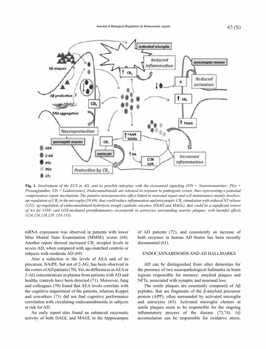
Fig. 1. Involvement of the ECS in AD, and its possible interplay with the eicosanoid signaling (NTs = Neurotransmitter; PGs = Prostaglandins; LTs = Leukotrienes). Endocannabinoids are released in response to pathogenic events, thus representing a potential compensatory repair mechanism. The putative neuroprotective effect linked to neuronal repair and cell maintenance mainly involves: up-regulation of CB2 in the microglia (59,69), that could reduce inflammation and presynaptic CB1 stimulation with reduced NT release (132); up-regulation of endocannabinoid hydrolysis trough catabolic enzymes (FAAH and MAGL), that could be a significant source of AA for COX- and LOX-mediated proinflammatory eicosanoids in astrocytes surrounding neuritic plaques, with harmful effects (124,124,128,129, 133-135).

64 (S)
Aβ levels (79). In this scenario, the identification of soluble Aβ peptides as highly bioactive assemblies has furthered interest in detecting and analysing their mechanistic properties, as well as their diagnostic and therapeutic potential. Yet, we still lack insight into the in vivo relevance of the soluble forms of Aβ in the brain during the development of AD-related diseases.
At present, no treatment is available that stops the degenerative process of AD, and current treatments can offer only modest symptomatic benefits. Therapeutic strategies are now unable to delay disease progression by more than one year, likely because AD is diagnosed when the pathology is already permanently advanced (80).
The identification of potential new therapeutic targets is then a major need, since development of novel therapeutic approaches is inhibited by the poor knowledge of the early
inflammation and neurotoxicity, and hence it might trigger the pathogenic cascade that ultimately leads to apoptosis and impairment of neurotransmission networks (2).
Soluble Aβ. After several years of research on the role of Aβ in its insoluble, aggregated form, recent findings have changed the perspective of Aβ meaning. Indeed, accumulating evidence suggests that pre-fibrillar, diffusible assemblies of Aβ are also deleterious. There is now persuasive evidence that the cognitive and behavioral alterations in AD arise, at least in part, from impaired synaptic function due to soluble forms of Aβ (75). It has been shown that these forms potently alter synaptic structure and functions (76,77). In particular, soluble Aβ interferes with synaptic function that subserves higher-order neural network activity (78). Moreover, learning and memory can be improved by reducing brain soluble
36
Table 1. Modifications of ECS elements in AD
ECSelement
Productanalysis
Change Tissue/Cell Reference
CB1 Protein ↔ Human brain 58-61
↓ Human brain 62
↓ Mice brain 54
mRNA ↑ Whole blood 68
↔ Human brain 58
CB2 Protein ↑
Human brain 59,69
↔ Human brain 58
mRNA ↔ Human brain 58
2-AG Endogenous levels
↔
Human plasma 71
↔
Human brain 70
↑ Human brain 61
AEA Endogenous levels
↔
Human plasma 71
↓ Human brain 70
FAAH Protein ↑
Human brain,
Human PBMCs
59,124
mRNA ↑ Human PBMCs 124
DAGL Protein ↑ Human brain 61
MAGL Protein ↑ Human brain 61
Table I. Modifications of ECS elements in AD.
C. D’ADDARIO ET AL.
65 (S)Journal of Biological Regulators & Homeostatic Agents
brain (89). Another study showed that endocannabinoids are protective in the Aβ-induced increase in DNA fragmentation and caspase-3 activation, both hallmarks of apoptosis, in primary cerebral cortical neurons (90). It was also documented that endocannabinoids can keep the cell alive by stabilizing lysosomes, that may be permeabilized by Aβ (90). Moreover, earlier studies have shown that CB2 receptors and FAAH expression increase in immune cells surrounding senile plaques in AD subjects (59).
It was also observed that Δ9-tetrahydrocannabinol (THC), the psychoactive principle of Cannabis sativa extracts, reduces Aβ aggregation through competitive in-hibition of acetylcholinesterase (AChE), the enzyme re-sponsible for the degradation of acetylcholine (91), which is generally associated with amyloid plaque deposits (92). AChE accelerates the formation of amyloid fibrils in the brain by generating stable complexes with Aβ (93) and, not surprisingly, most drugs licensed for AD treatment are AChE inhibitors. It is of noteworthy that THC appears more effective than any other drug in reducing AChE-induced Aβ deposition (91).
In animal models of Aβ-induced toxicity, many reports confirmed the beneficial effects of cannabinoids in reducing neuroinflammation; indeed, both cannabinoid agonists and phytocannabinoids like cannabidiol are able to reduce Aβ-triggered microglial activation (62,94,95). In particular, Ramírez and colleagues observed that cannabinoid administration can attenuate loss of neuronal markers and reduce cognitive deficits occurring in Aβ-treated rats, thus preventing microglial activation (62). Moreover, they showed that CB1-positive neurons were reduced when compared to control areas of microglia activation (62). In mouse hippocampus injected with human Aβ42 peptide, cannabidiol inhibited glial fibrillary acidic protein, as well as nitric oxide synthase and IL-1β protein expression and release (94).
Consistently, the AEA reuptake inhibitor VDM-11, which causes an elevation of the endocannabinoid tone, reversed hippocampal damage and loss of memory reten-tion in rodents treated with Aβ42 peptide (96). Moreover, CB1 modulation can protect against Aβ-induced amnesia in hippocampal learning tasks (97,98). Rimonabant, a se-lective CB1 antagonist/inverse agonist, improves memory deficit induced by β-amyloid fragments, probably through an increase of hippocampal acetylcholine release, or by acting directly on cannabinoid neuronal circuits involved in memory (97). Micale and coworkers focused the at-tention on the interaction between endocannabinoids and the dopaminergic system, in particular on dopamine D3 receptor (D3R) involvement in the neurotoxicity and am-nesia induced by Aβ. They found that neurotoxin adminis-tration induced a less pronounced cognitive impairment in the passive-avoidance paradigm performance in wild-type
stages of the disease, prior to Aβ plaque deposition, massive neuronal and neurotransmitter loss with irreversible neuronal damage and cognitive dysfunctions. In this context, recent studies have suggested that ECS plays a crucial role in neuroprotection, and the enhancement of the endocannabinoid tone is now considered an attractive approach for future therapeutic exploitation (81). Remarkable changes of endocannabinoid levels and receptor concentrations found in patient brains and in animal models have further strengthened the hypothesis that ECS is considerably altered in AD (82). For instance, it has been demonstrated that the enhancement of brain endocannabinoid tone, through the early endocannabinoid reuptake blockade, is able to reverse memory impairment and neurotoxic effects triggered by soluble Aβ in murine models of AD. In the same study, the authors reported that, when the treatment was done in a late phase, cognitive deficit was dramatically aggravated (83). Moreover, a novel player in brain endocannabinoid signaling has been recently identified. Indeed, it has been reported that the anti-inflammatory lipid lipoxin A4, detected in brain tissues, enhances the affinity of AEA for CB1 receptors, thereby potentiating the effects of this endocannabinoid. Additionally, Aβ-induced impairment of spatial memory formation was prevented by co-injection of lipoxin A4, showing that lipoxin A4-induced neuroprotection depends on CB1 receptors (84). Nonetheless, it must be emphasized that the relevance of ECS for human medical care of amyloid-related diseases is still in its infancy (82). Taken together, the actual role of endocannabinoids in AD remains elusive, yet chances are that elevation of endocannabinoid levels is part of a neuroprotective mechanism that aims at counteracting Aβ-related neurotoxicity, rather than part of the pathological process.
Aβ plaques. The first study of endocannabinoids as inhibitors of Aβ toxicity was carried out on a human neuronal cell line, and showed the neuroprotective role of AEA and noladin (a putative endogenous cannabinoid with agonist activity at CB1 receptors) through a CB1-dependent, mitogen activated protein kinase (MAPK)-mediated mechanism (85). A previous study had already shown that the production of nitric oxide (a proinflammatory mediator) by microglial activation, is prevented by exogenous cannabinoids (86). More recently, it has also been observed that cannabinoids can counteract the Aβ-induced microglial activation via CB2 (62), and promote microglia migration allowing clearance of the Aβ peptide (87). Consistently, the CB2 agonist JWH-015 inhibits Aβ-induced production of proinflammatory cytokines (88). It has been observed that in microglial cells activated by interferon-γ JWH-015 suppresses the expression of CD40 (88), a glycoprotein belonging to the tumor necrosis factor receptor that is highly expressed in senile plaques in AD
66 (S)
with dementia (105). In line with this, in a case report study the synthetic cannabinoid receptor agonist nabilone significantly ameliorated dementia-related restlessness (106).
CB2 agonist JWH-015 was found to increase the ability of cultured human macrophages to remove Aβ deposits from human tissues of AD patients, as well as from synthetic Aβ fibrils in vitro (107). As already mentioned, by targeting the different ECS components, many studies have reported the potential neuroprotective ECS abilities in various models of AD (62,85,96-98), and highlighted disease-related alterations of the ECS occurring over time (58,59,62).
Chronic infusion of lipopolysaccharide (LPS) into the fourth ventricle of rats can induce many of the patho-physiological changes observed in neurodegenerative dis-eases, as well as the activation of microglia (108). The latter process is indirectly prevented by WIN-55212-2, a CB1/CB2 agonist (109).
The non-psychoactive component of cannabis, cannabidiol, has a number of additional characteristics that highlight the potential benefits of using cannabinoid-based therapeutics for the treatment of AD (110). Indeed, it has been observed that cannabidiol can scavenge reactive oxygen species (111), reverse Tau hyperphosphorylation (100), and reduce activation of the inflammatory transcription target nuclear factor-κB (112). Again, it has to be taken under consideration that not all animal studies agree on the possible beneficial effects of endocannabinoids for AD therapy. For example, in two animal studies using Morris water maze to measure cognitive impairment, one report documented that cannabidiol and WIN-55,212-2 could prevent memory impairment in Aβ-treated rats (87). In contrast, another report showed that HU210, a potent synthetic cannabinoid, did not improve water maze performance nor a contextual fear conditioning task in an APP23/PS45 double transgenic mouse model of AD (113). These conflicting results are likely to depend on differences in experimental sets, as well as on the drugs used to modulate ECS (e.g., phyto-, endo-, or syntho-cannabinoids), their administration routes, concentrations and timing of application (90,96).
INFLAMMATION IN AD: THE ENDOCANNABINOID-EICOSANOID CONNECTION
Inflammation coupled with oxidative stress plays a major role in AD, and its suppression has been shown to reduce AD pathological hallmarks as well as cognitive and behavioral deficits in AD models (114). It is well known that inflammation involves arachidonic acid (AA)-derived lipid mediators biosynthesized by pathways dependent on COX and LOX activity. Non-steroidal
compared to D3R knockout mice (98). Since the latter ani-mals exhibited higher level of endovanilloid and endocan-nabinoid signaling, the authors suggested a potential role for enhanced CB1 tone in worsening memory retention (98).
NFTs. These structures result from hyperphosphorylation of microtubule-associated Tau protein, leading first to the dissociation of Tau from the microtubule, then to microtubule destabilization and Tau oligomerization within the cell, and finally to cell death (99).
Only few studies investigated the role of ECS in NFTs formation. It has been observed that cannabidiol reverses Tau hyperphosphorylation by reducing phosphorylation of glycogen synthase kinase-3β, a key kinase for both physiological and pathological tau phosphorylation in AD (100). More recently, it has been reported that MAGL ex-pression levels are increased in neurons with hyperphos-phorylated Tau (61), confirming 2-AG contribution to synapse silencing in AD. Another study showed that the reduction of AEA levels in brain tissues from AD patients correlates with patients cognitive impairment, but not with Tau hyperphosphorylation nor with amyloid plaques formation (70).
Neuronal loss of function. The third main feature of AD is the loss of neuronal ability to rapidly communi-cate and process signals across synapses. Synaptic func-tion is controlled through different mechanisms, such as neurotransmitter release acting at specific pre- and post-synaptic receptors. Among the presynaptic receptors, an important role in the regulation of brain synapses is played again by those activated by endocannabinoids (101). 2-AG has been found to mediate synaptic commu-nication in the hippocampus (102), and indeed a reduction of pre- and post-synaptic 2-AG degradation, along with an increase of its synthesis, has been demonstrated in AD, suggesting that an endocannabinoid hypertone might ag-gravate synapse impairment by disrupting retrograde sig-naling (61). However, further investigations are needed to better clarify the role of ECS in synaptic plasticity follow-ing Aβ plaque formation.
TREATMENTS OF AD THAT TARGET ECS
The relevance of the ECS for AD treatment has been debated for many years. The first report suggesting ECS modulation as potential therapy of AD showed that dronabinol, a synthetic form of THC, was able to improve the behavior in AD subjects and also to stimulate appetite (103). Indeed, it is known that patients with dementia start refusing food during the course of the disease (104). More recently, the same compound was found to reduce also nocturnal restlessness that frequently occurs in patients
C. D’ADDARIO ET AL.

67 (S)Journal of Biological Regulators & Homeostatic Agents
in LOAD subjects, that was paralleled by reduced DNA methylation at gene promoter, increased 5-LOX protein and increased plasma levels of the 5-LOX end-product, LTB4. We thus hypothesized that increased AEA hydrolysis by FAAH could contribute to the inflammatory process that occurs in AD, for instance by releasing the AA pool for neuroinflammatory LTB4. We also provided evidence that ALOX5 and FAAH genes share common epigenetic signatures. According to this, we found a direct correlation between DNA methylation at FAAH and ALOX5 gene promoters. Moreover, LTB4 levels were directly correlated to FAAH mRNA levels, and inversely correlated to FAAH DNA methylation, suggesting that a parallel increase of FAAH and 5-LOX expression in AD patients could evoke a sustained inflammatory condition, thus reinforcing neurodegeneration. Overall, our data highlighted the contribution of epigenetic mechanisms to the control of genes involved in AA metabolism upon AD development (126).
The possible interplay between endocannabinoid and eicosanoid signaling has been also explored in animal models of neuroinflammation. Nomura and colleagues documented the relevance of MAGL in the control of AA release for the production of proinflammatory eicosanoids in the brain of rodents (127). This observation was corroborated by the finding that mice deficient in the MAGL-encoding gene, and mice treated with the MAGL-selective inhibitor JZL184 showed elevated brain levels of 2-AG and reduced levels of AA and AA-derived prostaglandins (127). Moreover, two independent studies have documented a deregulated endocannabinoid-eicosanoid network in mouse models of AD, whereby endocannabinoid hydrolysis was recognized as the main source of AA for COX-mediated eicosanoid production in the diseased brain (128,129). In these studies, MAGL blockade was found to substantially reduce neuroinflammation and to attenuate amyloidosis, likely by suppressing pro-inflammatory eicosanoids production, thus pointing to MAGL inhibitors as an attractive therapeutic strategy for the treatment of AD.
Very recently, a functional connection between CB2 and 5-LOX has also been documented in a zebrafish mod-el of inflammation (130), where both a CB2 agonist and a 5-LOX inhibitor reduced leukocyte migration in response to acute injury.
CONCLUDING REMARKS
Drugs currently used for the treatment of AD, such as AChE inhibitors (e.g., donepezil and rivastigmine) or antagonists for the N-methyl-D-aspartate glutamatergic receptors (like memantine), produce limited clinical benefit and do not correct the underlying molecular defects
anti-inflammatory drugs (NSAIDs), that exert their effects through COX inhibition, have well-documented protective effects in AD when administered at an early stage and taken over prolonged periods of time (115). It was reported that patients who suffer from inflammatory diseases (e.g., arthritis) or take anti-inflammatory drugs (e.g., anti-inflammatory COX inhibitors like aspirin and indomethacin), have a reduced risk of developing AD (116). However, an issue with NSAIDs has been their lack of efficacy in AD clinical studies (117). Also selective COX-2 inhibitors have been investigated and tested clinically as potentially better therapeutics for AD patients; yet, they failed to confirm their efficacy for actual therapy (118). Moreover, the gastrointestinal and cardiovascular toxicity of COX inhibitors limited their use for neuroinflammatory syndromes. Novel and safer anti-inflammatory strategies are thus required, not only to gain a deeper understanding of the role that inflammation plays in AD progression, but also to investigate the therapeutic potential of anti-inflammatory drugs to combat this disease. Growing evidence suggests a role for LOX, and in particular for 5-LOX, as potential target in AD (119-121). In line with this, also leukotrienes (LT), that are end-products of 5-LOX pathway, have been found to be involved in brain inflammation associated with age-related dementia, as well as with neurodegenerative diseases (122,123).
To date, the eicosanoid and endocannabinoid signaling systems have been investigated independently of each other, and indeed one is likely to operate in the absence of the other and vice versa. However, both endocannabinoids and eicosanoids are derivatives of AA, therefore a potential intersection between their signaling systems can be expected (49). In this context, it seems noteworthy that the lipases that initiate both pathways are responsive to common second messengers (e.g., elevation in intracellular Ca2+), supporting the view that in cells where the enzymatic machinery for both pathways is present, endocannabinoid and eicosanoid signaling might cooperate. Such an interaction is further complicated by the ability of metabolic enzymes of eicosanoids to metabolize also (and even better) endocannabinoids (51).
We recently reported the epigenetic regulation of ECS components, and of LOX isoforms, in peripheral blood mononuclear cells (PBMCs) of subjects with late-onset AD (LOAD) and age-matched controls. Our data showed that FAAH is up-regulated in PBMCs of LOAD subjects compared to healthy individuals, without changes in the mRNA levels of any other ECS elements (124). Consistently, we also demonstrated in LOAD subjects an increase in FAAH protein levels and enzymatic activity, due to an epigenetic regulation of gene trascription. Moreover, in an independent investigation (125) we observed a significant increase of 5-LOX gene (ALOX5) expression

68 (S)
G, Cascio MG, Hermann H, Tang J, Hofmann C, Ziegl-gänsberger W, Di Marzo V, Lutz B. The endogenous can-nabinoid system controls extinction of aversive memories. Nature 2002; 418:530-4.
5. Szoke E, Czéh G, Szolcsányi J, Seress L. Neonatal anan-damide treatment results in prolonged mitochondrial dam-age in the vanilloid receptor type 1-immunoreactive B-type neurons of the rat trigeminal ganglion. Neuroscience 2002; 115:805-14.
6. Pazos MR, Núñez E, Benito C, Tolón RM, Romero J. Role of the endocannabinoid system in Alzheimer’s disease: new perspectives. Life Sci 2004; 75:1907-15.
7. Benito C, Núñez E, Pazos MR, Tolón RM, Romero J. The endocannabinoid system and Alzheimer’s disease. Mol Neurobiol 2007; 36:75-81.
8. Pertwee RG, Howlett AC, Abood ME, Alexander SP et al and Ross RA. International Union of Basic and Clinical Pharmacology. LXXIX. Cannabinoid receptors and their ligands: beyond CB₁ and CB₁. Pharmacol Rev 2010; 62:588-631.
9. Viveros MP, Marco EM, File SE. Endocannabinoid system and stress and anxiety responses. Pharmacol Biochem Be-hav 2005; 81:331-42.
10. Wotjak CT. Role of endogenous cannabinoids in cognition and emotionality. Mini Rev Med Chem 2005; 5:659-70.
11. Moreira FA, Lutz B. The endocannabinoid system: emotion, learning and addiction. Addict Biol 2008; 13:196-212.
12. Guindon J, Hohmann AG. The endocannabinoid system and pain. CNS Neurol Disord Drug Targets 2009; 8:403-21.
13. Finn DP. Endocannabinoid-mediated modulation of stress responses: physiological and pathophysiological signifi-cance. Immunobiology 2010; 215:629-46.
14. Moreira FA, Wotjak CT. Cannabinoids and anxiety. Curr Top Behav Neurosci 2010; 2:429-50.
15. Maccarrone M, Gasperi V, Catani MV, Diep TA, Dainese E, Hansen HS, Avigliano L. The endocannabinoid sys-tem and its relevance for nutrition. Annu Rev Nutr 2010; 30:423-40.
16. Herkenham M, Lynn AB, Johnson MR, Melvin LS, de Costa BR, Rice KC. Characterization and localization of cannabinoid receptors in rat brain: a quantitative in vitro autoradiographic study. J Neurosci. 1991; 11:563-83.
17. Munro S, Thomas KL, Abu-Shaar M. Molecular charac-terization of a peripheral receptor for cannabinoids. Nature 1993; 2:61-5.
18. Galiègue S, Mary S, Marchand J, Dussossoy D, Carrière D, Carayon P, Bouaboula M, Shire D, Le Fur G, Casellas P. Expression of central and peripheral cannabinoid receptors
(131). The prevalence of AD is expected to triple over the next 50 years, creating an urgency to develop effective disease-modifying therapies to reduce the economic burden of this devastating disorder. One of the main areas of therapeutic focus has been an anti-inflammatory strategy originated from epidemiological evidence that long-term exposure to NSAIDs protected against the development of AD. Unfortunately, subsequent large-scale double-blind placebo-controlled clinical trials failed to support the use of NSAIDs for the treatment of AD. In search for novel strategies able to halt or slow down the course of AD, and to improve the patient quality of life, here we have reviewed recent investigations that suggest a key-role for distinct ECS components in both normal and altered neuronal circuits. Therefore, ECS modulation (e.g., by targeting CB2 and FAAH) might provide a promising arena for next generation therapeutics (132-134). In addition, evidence is mounting to support the hypothesis that endocannabinoid tone can modulate eicosanoid levels, and vice versa (see figure 1 for a schematic representation of ECS involvement in AD). This scenario is particularly likely under conditions of inflammation, that would lead to increased expression of 5-LOX. Indeed, reduction in levels of anti-inflammatory endocannabinoids may be one mechanism by which 5-LOX exerts its pro-inflammatory effects. Although the beneficial effects produced by FAAH or 5-LOX inhibition against neuropathology of AD remain to be determined, it could be anticipated that these enzymes might become a promising therapeutic target for the prevention and treatment of AD.
ACKNOWLEDGEMENTS
This investigation was partly supported by the Italian Ministero dell’Istruzione, dell’Università e della Ricerca (grant PRIN 2010–2011 to MM, and Futuro in Ricerca RBFR12DELS_001 to CDA), and by Fondazione TERCAS (grant 2009–2012) to MM. ADF was supported by a “Fondazione TERCAS-Progetto Speciale Assegni di Ricerca 2011–2013” fellowship.
REFERENCES
1. Brookmeyer R, Johnson E, Ziegler-Graham K, Arrighi HM. Forecasting the global burden of Alzheimer’s disease. Alzheimer’s and Dementia 2007; 3:186–91.
2. Koudinova NV, Kontush A, Berezov TT, Koudinov A. Amyloid beta, neural lipids, cholesterol and Alzheimer’s disease. Neurobiology of Lipids 2003; 1:27-33.
3. Walter L, Stella N. Cannabinoids and neuroinflammation. Br J Pharmacol 2004; 141:775-85.
4. Marsicano G, Wotjak CT, Azad SC, Bisogno T, Rammes
C. D’ADDARIO ET AL.
69 (S)Journal of Biological Regulators & Homeostatic Agents
rone M, Bernardi G, Mercuri NB. Presynaptic facilitation of glutamatergic synapses to dopaminergic neurons of the rat substantia nigra by endogenous stimulation of vanilloid receptors. J Neurosci 2003; 23:3136-44.
30. Lastres-Becker I, de Miguel R, De Petrocellis L, Makri-yannis A, Di Marzo V, Fernández-Ruiz J. Compounds act-ing at the endocannabinoid and/or endovanilloid systems reduce hyperkinesia in a rat model of Huntington’s disease. J Neurochem 2003; 84:1097-1109.
31. Maccarrone M, Rossi S, Bari M, De Chiara V, Rapino C, Musella A, Bernardi G, Bagni C, Centonze D. Anandamide inhibits metabolism andphysiological actions of 2-arachi-donoylglycerol in the striatum. Nature Neurosci 2008; 11:152-9.
32. O’Sullivan SE. Cannabinoids go nuclear: evidence for activation of peroxisome proliferatoractivated receptors. Br J Pharmacol 2007; 152, 576–82.
33. Pistis M and Melis M. From surface to nuclear receptors: the endocannabinoid family extends its assets. Curr Med Chem 2010; 17, 1450–67.
34. Di Marzo V. Endocannabinoids: synthesis and degrada-tion. Rev Physiol Biochem Pharmacol 2008; 160:1-24.
35. Sugiura T, Kondo S, Sukagawa A, Nakane S et al and Waku K. 2-Arachidonoylglycerol: a possible endogenous cannabinoid receptor ligand in brain. Biochem Biophys Res Commun. 1995; 215:89-97.
36. Sugiura T, Kobayashi Y, Oka S, Waku K. Biosynthesis and degradation of anandamide and 2-arachidonoylglycerol and their possible physiological significance. Prostaglan-dins Leukot Essent Fatty Acids 2002; 66:173-92.
37. Di Marzo V, Fontana A, Cadas H, Schinelli S, Cimino G, Schwartz JC, Piomelli D. Formation and inactivation of endogenous cannabinoid anandamide in central neurons. Nature. 1994; 372:686-91.
38. Cadas H, Schinelli S, Piomelli D.Membrane localization of N-acylphosphatidylethanolamine in central neurons: studies with exogenous phospholipases. J Lipid Mediat Cell Signal 1996; 14:63-70.
39. Bisogno T, Howell F, Williams G, Minassi A, Cascio MG, Ligresti A, Matias I, Schiano-Moriello A, Paul P, Williams EJ, Gangadharan U, Hobbs C, Di Marzo V, Doherty P. Cloning of the first sn1-DAG lipases points to the spatial and temporal regulation of endocannabinoid signaling in the brain. J Cell Biol 2003; 163:463-8.
40. Garcia-Ovejero D, Arevalo-Martin A, Petrosino S, Docagne F, Hagen C, Bisogno T, Watanabe M, Guaza C, Di Marzo V, Molina-Holgado E. The endocannabinoid sys-tem is modulated in response to spinal cord injury in rats. Neurobiol Dis 2009; 33:57-71.
in human immune tissues and leukocyte subpopulations. Eur J Biochem 1995; 232:54-61.
19. Liu QR, Pan CH, Hishimoto A, Li CY, Xi ZX, Llorente-Berzal A, Viveros MP, Ishiguro H, Arinami T, Onaivi ES, Uhl GR. Species differences in cannabinoid receptor 2 (CNR2 gene): identification of novel human and rodent CB2 isoforms, differential tissue expression and regulation by cannabinoid receptor ligands. Genes Brain Behav 2009; 8:519-30.
20. Roche R, Hoareau L, Bes-Houtmann S, Gonthier MP, Laborde C, Baron JF, Haffaf Y, Cesari M, Festy F. Pres-ence of the cannabinoid receptors, CB1 and CB2, in human omental and subcutaneous adipocytes. Histochem Cell Biol 2006; 126:177-87.
21. Van Sickle MD, Duncan M, Kingsley PJ, Mouihate A, Ur-bani P, Mackie K, Stella N, Makriyannis A, Piomelli D, Da-vison JS, Marnett LJ, Di Marzo V, Pittman QJ, Patel KD, Sharkey KA. Identification and functional characterization of brainstem cannabinoid CB2 receptors. Science 2005; 310:329-32.
22. Gong JP, Onaivi ES, Ishiguro H, Liu QR, Tagliaferro PA, Brusco A, Uhl GR. Cannabinoid CB2 receptors: immuno-histochemical localization in rat brain. Brain Res 1984; 1071:10-23.
23. Onaivi ES. Neuropsychobiological evidence for the func-tional presence and expression of cannabinoid CB2 recep-tors in the brain. Neuropsychobiology. 2006; 54:231-46.
24. García-Gutiérrez MS, Pérez-Ortiz JM, Gutiérrez-Adán A, Manzanares J. Depression-resistant endophenotype in mice overexpressing cannabinoid CB(2) receptors. Br J Pharma-col 2010; 160:1773-84.
25. Sánchez C, de Ceballos ML, Gomez del Pulgar T, Rueda D, Corbacho C, Velasco G, Galve-Roperh I, Huffman JW, Ramón y Cajal S, Guzmán M. Inhibition of glioma growth in vivo by selective activation of the CB(2) cannabinoid receptor. Cancer Res 2001; 61:5784-9.
26. Ellert-Miklaszewska A, Grajkowska W, Gabrusiewicz K, Kaminska B, Konarska L. Distinctive pattern of cannabi-noid receptor type II (CB2) expression in adult and pediat-ric brain tumors. Brain Res 2007; 1137:161-9.
27. Viscomi MT, Oddi S, Latini L, Pasquariello N, Florenzano F, Bernardi G, Molinari M, Maccarrone M. Selective CB2 receptor agonism protects central neurons from remote axotomy-induced apoptosis through the PI3K/Akt path-way. J Neurosci 2009; 29:4564-70.
28. Starowicz K, Nigam S, Di Marzo V. Biochemistry and pharmacology of endovanilloids. Pharmacol Ther 2007; 114:13-33.
29. Marinelli S, Di Marzo V, Berretta N, Matias I, Maccar-

70 (S)
patterns of cannabinoid CB1 receptors in the hippocampus of APPswe/PS1ΔE9 double transgenic mice. Brain Res 2011; 1376:94-100.
55. Ashton JC, Friberg D, Darlington CL, Smith PF. Expression of the cannabinoid CB2 receptor in the rat cerebellum: an immunohistochemical study. Neurosci Lett 2006; 396:113-6.
56. Núñez E, Benito C, Pazos MR, Barbachano A, Fajardo O, González S, Tolón RM, Romero J. Cannabinoid CB2 re-ceptors are expressed by perivascular microglial cells in the human brain: an immunohistochemical study. Synapse 2004; 53:208-13.
57. Bisogno T, Di Marzo V. The role of the endocannabinoid system in Alzheimer’s disease: facts and hypotheses. Curr Pharm Des 2008; 14:2299-3305.
58. Westlake TM, Howlett AC, Bonner TI, Matsuda LA, Herkenham M. Cannabinoid receptor binding and messen-ger RNA expression in human brain: an in vitro receptor autoradiography and in situ hybridization histochemistry study of normal aged and Alzheimer’s brains. Neurosci-ence 1994; 63:637-52.
59. Benito C, Núñez E, Tolón RM, Carrier EJ, Rábano A, Hillard CJ, Romero J. Cannabinoid CB2 receptors and fatty acid amide hydrolase are selectively overexpressed in neuritic plaque-associated glia in Alzheimer’s disease brains. J Neurosci 2003; 23:11136-41.
60. Lee JH, Agacinski G, Williams JH, Wilcock GK, Esiri MM, Francis PT, Wong PT, Chen CP, Lai MK.. Intact can-nabinoid CB1 receptors in the Alzheimer’s disease cortex. Neurochem Int 2010; 57:985-9.
61. Mulder J, Zilberter M, Pasquaré SJ, Alpár A, Schulte G, Ferreira SG, Köfalvi A, Martín-Moreno AM, Keimpema E, Tanila H, Watanabe M, Mackie K, Hortobágyi T, de Ce-ballos ML, Harkany T. Molecular reorganization of endo-cannabinoid signalling in Alzheimer’s disease. Brain 2011; 134:1041-60.
62. Ramírez BG, Blázquez C, Gómez del Pulgar T, Guzmán M, de Ceballos ML. Prevention of Alzheimer’s disease pathology by cannabinoids: neuroprotection mediated by blockade of microglial activation. J Neurosci 2005; 25:1904-13.
63. Van Hoesen GW, Hyman BT, Damasio AR (1991) Entorhi-nal cortex pathology in Alzheimer’s disease. Hippocampus 1:1-8.
64. Núñez E, Benito C, Tolón RM, Hillard CJ, Griffin WS, Romero J. Glial expression of cannabinoid CB(2) recep-tors and fatty acid amide hydrolase are beta amyloid-linked events in Down’s syndrome. Neuroscience 2008; 151:104-10.
41. Basavarajappa BS. Critical enzymes involved in endocannabinoid metabolism. Protein Pept Lett 2007; 14:237-46.
42. Vandevoorde S, Lambert DM. The multiple pathways of endocannabinoid metabolism: a zoom out. Chem Biodiv-ers 2007; 4:1858-81.
43. Fezza F, De Simone C, Amadio D, Maccarrone M. Fatty acid amide hydrolase: a gate-keeper of the endocannabi-noid system. Subcell Biochem 2008; 49:101-32.
44. Egertová M, Giang DK, Cravatt BF, Elphick MR. A new perspective on cannabinoid signalling: complementary lo-calization of fatty acid amide hydrolase and the CB1 recep-tor in rat brain. Proc Biol Sci. 1998; 265:2081-5.
45. Gulyas AI, Cravatt BF, Bracey MH, Dinh TP, Piomelli D, Boscia F, Freund TF. Segregation of two endocannabinoid-hydrolyzing enzymes into pre- and postsynaptic compart-ments in the rat hippocampus, cerebellum and amygdala. Eur J Neurosci 2004; 20:441-58.
46. Goparaju SK, Ueda N, Yamaguchi H, Yamamoto S. Anan-damide amidohydrolase reacting with 2-arachidonoylglyc-erol, another cannabinoid receptorligand. FEBS Lett 1998; 422:69-73.
47. Dinh TP, Freund TF, Piomelli D. A role for monoglyceride lipase in 2-arachidonoylglycerol inactivation. Chem Phys Lipids 2002; 121:149-58..
48. Blankman JL, Simon GM, Cravatt BF. A comprehensive profile of brain enzymes that hydrolyze the endocannabinoid 2-arachidonoylglycerol. Chem Biol 2007; 14:1347-56.
49. Rouzer CA, Marnett LJ. Endocannabinoid oxygenation by cyclooxygenases, lipoxygenases, and cytochromes P450: cross-talk between the eicosanoid and endocannabinoid signaling pathways. Chem Rev 2011; 111, 5899–921.
50. Van der Stelt M, van Kuik JA, Bari M, van Zadelhoff G, Leeflang BR, Veldink GA, Finazzi-Agro A, Vliegenthart JF, Maccarrone M. Oxygenated metabolites of anandamide and 2- arachidonoylglycerol: conformational analysis and interaction with cannabinoid receptors, membrane transporter, and fatty acid amide hydrolase. J Med Chem 2002; 45, 3709–20.
51. Rouzer CA, Marnett LJ. Non-redundant functions of cyclooxygenases: oxygenation of endocannabinoids. J Biol Chem 2008; 283:8065–9.
52. Woodward DF, Liang Y, Krauss AH. Prostamides (prostaglandin-ethanolamides) and their pharmacology. Br J Pharmacol 2008; 153, 410–9.
53. Riedel G, Davies SN. Cannabinoid function inlearn-ing, memory and plasticity. Handb Exp Pharmacol 2005; 168:445-77.
54. Kalifa S, Polston EK, Allard JS, Manaye KF. Distribution
C. D’ADDARIO ET AL.

71 (S)Journal of Biological Regulators & Homeostatic Agents
2010; 159:1704-15.77. Li S, Hong S, Shepardson NE, Walsh DM, Shankar GM,
Selkoe D. Soluble oligomers of amyloid Beta protein facilitate hippocampal long-term depression by disrupting neuronal glutamate uptake. Neuron 2009;62:788-801.
78. Palop JJ, Mucke L. Amyloid-beta-induced neuronal dysfunction in Alzheimer’s disease: from synapses toward neural networks. Nat Neurosci 2010; 13:812-8.
79. Fukumoto H, Takahashi H, Tarui N, Matsui J, Tomita T, Hirode M, Sagayama M, Maeda R, Kawamoto M, Hirai K, Terauchi J, Sakura Y, Kakihana M, Kato K,Iwatsubo T, Miyamoto M, A noncompetitive BACE1 inhibitor TAK-070 ameliorates Abeta pathology and behavioral deficits in a mouse model of Alzheimer’s disease. J Neurosci 2010; 30:11157-66.
80. Goutagny R, Krantic S, Hippocampal oscillatory activity in Alzheimer’s disease: toward the identification of early biomarkers? Aging Dis. 2013; 4:134-40.
81. Hwang J, Adamson C, Butler D, Janero DR, Makriyannis A, Bahr BA. Enhancement of endocannabinoid signaling by fatty acid amide hydrolase inhibition: a neuroprotective therapeutic modality. Life Sci 2010; 86:615-23.
82. Ruiz-Valdepeñas L, Benito C, Tolón RM, Martínez Orgado JA, Romero J. The endocannabinoid system and amyloid-related diseases. Exp Neurol 2010; 224:66-73.
83. Van der Stelt M, Mazzola C, Esposito G, Matias I, Petrosi-no S, De Filippis D, Micale V, Steardo L, Drago F, Iuvone T, Di Marzo V. Endocannabinoids and beta-amyloid-in-duced neurotoxicity in vivo: effect of pharmacological el-evation of endocannabinoid levels. Cell Mol Life Sci 2006; 63:1410-24.
84. Pamplona FA, Ferreira J, Menezes de Lima O Jr, Duarte FS, Bento AF, Forner S, Villarinho JG, Bellocchio L, Wotjak CT, Lerner R, Monory K, Lutz B, Canetti C, Matias I, Calixto JB, Marsicano G, Guimarães MZ, Takahashi RN. Anti-inflammatory lipoxin A4 is an endogenous allosteric enhancer of CB1 cannabinoid receptor. Proc Natl Acad Sci U S A 2012; 109:21134-9.
85. Milton NG. Anandamide and noladin ether prevent neuro-toxicity of the human amyloid-beta peptide. Neurosci Lett 2002; 332:127-30.
86. Waksman Y, Olson JM, Carlisle SJ, Cabral GA. The cen-tral cannabinoid receptor (CB1) mediates inhibition of ni-tric oxide production by rat microglial cells. J Pharmacol Exp Ther 1999; 288:1357-66.
87. Martín-Moreno AM, Reigada D, Ramírez BG, Mechoulam R, Innamorato N, Cuadrado A, de Ceballos ML. Cannabi-Cannabi-diol and other cannabinoids reduce microglial activation in vitro and in vivo: relevance to Alzheimer’s disease. Mol
65. Glenner GG, Wong CW. Alzheimer’s disease and Down’s syndrome: sharing of a unique cerebrovascular amy-loid fibril protein. Biochem Biophys Res Commun 1984; 122:1131-5.
66. Wisniewski KE, Dalton AJ, McLachlan C, Wen GY, Wis-niewski HM. Alzheimer’s disease in Down’s syndrome: clinicopathologic studies. Neurology 1985; 35:957-61.
67. Horti AG, Gao Y, Ravert HT, Finley P et al and Dannals RF. Synthesis and biodistribution of (11C)A-836339, a newpo-tential radioligand for PET imaging of cannabinoid type 2 receptors (CB2). Bioorg Med Chem 2010; 18:5202-7.
68. Grünblatt E, Bartl J, Zehetmayer S, Ringel TM, Bauer P, Riederer P, Jacob CP. Gene expression as peripheral bio-markers for sporadic Alzheimer’s disease. J Alzheimers Dis 2009; 16: 627-34.
69. Halleskog C, Mulder J, Dahlström J, Mackie K, Hortobágyi T, Tanila H, Kumar Puli L, Färber K, Harkany T, Schulte G. WNT signaling in activated microglia is proinflammatory. Glia 2011; 59:119-31.
70. Jung KM, Astarita G, Yasar S, Vasilevko V, Cribbs DH, Head E, Cotman CW, Piomelli D. An amyloid β(42)-dependent deficit in anandamide mobilization is associated with cognitive dysfunction in Alzheimer’s disease. Neuro-biol Aging 2011; 33:1522-32.
71. Koppel J, Bradshaw H, Goldberg TE, Khalili H, Maram-baud P, Walker MJ, Pazos M, Gordon ML, Christen E, Da-vies P. Endocannabinoids in Alzheimer’s disease and their impact on normative cognitive performance: a case-control and cohort study. Lipids Health Dis 2009; 8:2.
72. Farooqui AA, Liss L, Horrocks LA. Stimulation of lipo-lytic enzymes in Alzheimer’s disease. Ann Neurol 1988; 23:306-8.
73. McGeer PL, Itagaki S, Tago H, McGeer EG. Reactive mi-croglia in patients with senile dementia of the Alzheimer type are positive for the histocompatibility glycoprotein HLA-DR. Neurosci Lett 1987; 79:195-200.
74. Dickson DW, Farlo J, Davies P, Crystal H, Fuld P, Yen SH. Alzheimer’s disease. A double-labeling immunohis-tochemical study of senile plaques. Am J Pathol. 1988; 132:86-101.
75. Cramer PE, Cirrito JR, Wesson DW, Lee CY, Karlo JC, Zinn AE, Casali BT, Restivo JL, Goebel WD, James MJ, Brunden KR, Wilson DA, Landreth GE. ApoE-directed therapeutics rapidly clear β-amyloid and reverse deficits in AD mouse models. Science 2012; 335:1503-6.
76. Colaianna M, Tucci P, Zotti M, Morgese MG, Schiavone S, Govoni S, Cuomo V, Trabace L. Soluble beta amyloid(1-42): a critical player in producing behavioural and biochemical changes evoking depressive-related state? Br J Pharmacol.

72 (S)
T. The marijuana component cannabidiol inhibits beta-am-yloid-induced tau protein hyperphosphorylation through Wnt/beta-catenin pathway rescue in PC12 cells. J Mol Med (Berl) 2006a; 84:253-8.
101. Heifets BD, Castillo PE Endocannabinoid signaling and long-term synaptic plasticity. Annu Rev Physiol 2009; 71:283–306.
102. Tanimura A, Kawata S, Hashimoto K, Kano M. Not glu-tamate but endocannabinoids mediate retrograde suppres-sion of cerebellar parallel fiber to Purkinje cell synaptic transmission in young adult rodents. Neuropharmacology 2010; 57:157-63.
103. Volicer L, Stelly M, Morris J, McLaughlin J, Volicer BJ. Effects of dronabinol on anorexia and disturbed behavior in patients with Alzheimer’s disease. Int J Geriatr Psychia-try 1997; 2:188-95.
104. Volicer L, Seltzer B, Rheaume Y, Karner J, Glennon M, Ri-ley ME, Crino P. Eating difficulties in patients with prob-able dementia of the Alzheimer type. J Geriatr Psychiatry Neurol 1989; 2:188-95.
105. Walther S, Mahlberg R, Eichmann U, Kunz D (2006) Del-ta-9-tetrahydrocannabinol for nighttime agitation in severe dementia. Psychopharmacology (Berl). 185:524-528.
106. Passmore MJ. The cannabinoid receptor agonist nabilone for the treatment of dementia-related agitation. Int J Geri-atr Psychiatry 2008; 23:116-7.
107. Tolón RM, Núñez E, Pazos MR, Benito C, Castillo AI, Martínez-Orgado JA, Romero J. The activation of canna-binoid CB2 receptors stimulates in situ and in vitro beta-amyloid removal by human macrophages. Brain Res 2009; 1283:148-54.
108. Hauss-Wegrzyniak B, Lukovic L, Bigaud M, Stoeckel ME. Brain inflammatory response induced by intracerebroven-tricular infusion of lipopolysaccharide: an immunohisto-chemical study. Brain Res 1998; 794:211-24.
109. Marchalant Y, Rosi S, Wenk GL. Anti-inflammatory prop-erty of the cannabinoid agonist WIN-55212-2 in a rodent model of chronic brain inflammation. Neuroscience 2007; 144:1516-22.
110. Iuvone T, Esposito G, De Filippis D, Scuderi C, Steardo L. Cannabidiol: a promising drug for neurodegenerative disorders? CNS Neurosci Ther 2009; 15:65-75.
111. Iuvone T, Esposito G, Esposito R, Santamaria R, Di Rosa M, Izzo AA. Neuroprotective effect of cannabidiol, a non-psychoactive component from Cannabis sativa, on beta-amyloid-induced toxicity in PC12 cells. J Neurochem 2004; 89:134-41.
112. Esposito G, De Filippis D, Maiuri MC, De Stefano D, Car-nuccio R, Iuvone T. Cannabidiol inhibits inducible nitric
Pharmacol. 2011; 79:964-73.88. Ehrhart J, Obregon D, Mori T, Hou H, Sun N, Bai Y, Klein
T, Fernandez F, Tan J, Shytle RD. J Neuroinflammation 2005;2:29. Stimulation of cannabinoid receptor 2 (CB2) suppresses microglial activation.
89. Togo T, Akiyama H, Kondo H, Ikeda K, Kato M, Iseki E, Kosaka K Expression of CD40 in the brain of Alzheimer’s disease and other neurological diseases. Brain Res 2000; 885:117-21.
90. Noonan J, Tanveer R, Klompas A, Gowran A, McKiernan J, Campbell VA. Endocannabinoids prevent beta-amyloid-mediated lysosomal destabilization in cultured neurons. J Biol Chem 2010; 285:38543-54.
91. Eubanks LM, Rogers CJ, Beuscher AE 4th, Koob GF, Ol-son AJ, Dickerson TJ, Janda KD. A molecular link between the active component of marijuana and Alzheimer’s dis-ease pathology. Mol Pharm 2006; 3:773-7.
92. Ulrich J, Meier-Ruge W, Probst A, Meier E, Ipsen S. Senile plaques: staining for acetylcholinesterase and A4 protein: a comparative study in the hippocampus and entorhinal cor-tex. Acta Neuropathol 1990; 80:624-8.
93. Inestrosa NC, Alvarez A, Pérez CA, Moreno RD et al and Garrido J. Acetylcholinesterase accelerates assembly of amyloid-beta-peptides into Alzheimer’s fibrils: possible role of the peripheral site of the enzyme. Neuron 1996; 16:881-91.
94. Esposito G, Scuderi C, Savani C, Steardo L Jr, et al and Steardo L. Cannabidiol in vivo blunts beta-amyloid in-duced neuroinflammation by suppressing IL-1beta and iNOS expression. Br J Pharmacol 2007; 151: 1272-9.
95. Booz GW. Cannabidiol as an emergent therapeutic strategy for lessening the impact of inflammation on oxidative stress. Free Radic Biol Med 2011; 51:1054-61.
96. Van der Stelt M, Di Marzo V. Cannabinoid receptors and their role in neuroprotection. Neuromolecular Med 2005; 7:37-50.
97. Mazzola C, Micale V, Drago F. Amnesia induced by beta-amyloid fragments is counteracted by cannabinoid CB1 receptor blockade. Eur J Pharmacol 2003; 477:219-25.
98. Micale V, Cristino L, Tamburella A, Petrosino S, Leg-gio GM, Di Marzo V, Drago F. Enhanced cognitive per-formance of dopamine D3 receptor “knock-out” mice in thestep-through passive-avoidance test: assessing the role of the endocannabinoid/endovanilloid systems. Pharmacol Res 2010; 61:531-6.
99. Mi K, Johnson GV. The role of tau phosphorylation in the pathogenesis of Alzheimer’s disease. Curr Alzheimer Res 2006; 3:449-63.
100. Esposito G, De Filippis D, Carnuccio R, Izzo AA, Iuvone
C. D’ADDARIO ET AL.

73 (S)Journal of Biological Regulators & Homeostatic Agents
124. D’Addario C, Di Francesco A, Arosio B, Gussago C, Dell’Osso B, Bari M, Galimberti D, Scarpini E, Altamura AC, Mari D, Maccarrone M. Epigenetic regulation of fatty acid amide hydrolase in Alzheimer disease. PLoS One. 2012;7:e39186.
125. Di Francesco A, Arosio B, Gussago C, Dainese E, Mari D, D’Addario C, Maccarrone M. Involvement of 5-Lipoxygenase in Alzheimer’s Disease: A Role for DNA Methylation. J Alzheimers Dis 2013; doi: 10.3233/JAD-130506.
126. D’Addario C, Di Francesco A, Pucci M, Finazzi Agro A, Maccarrone M. Epigenetic mechanisms and endocannabinoid signalling. FEBS J 2013; 280:1905-17.
127. Nomura DK, Morrison BE, Blankman JL, Long JZ, Kinsey SG, Marcondes MC, Ward AM, Hahn YK, Lichtman AH, Conti B, Cravatt BF. Endocannabinoid hydrolysis generates brain prostaglandins that promote neuroinflammation. Science 2011; 334:809-13.
128. Chen R, Zhang J, Wu Y, Wang D, Feng G, Tang YP, Teng Z, Chen C. Monoacylglycerol lipase is a therapeutic target for Alzheimer’s disease. Cell Rep 2012; 2:1329-39.
129. Piro JR, Benjamin DI, Duerr JM, Pi Y, Gonzales C, Wood KM, Schwartz JW, Nomura DK, Samad TA. A dysregulated endocannabinoid-eicosanoid network supports pathogenesis in a mouse model of Alzheimer’s disease. Cell Rep 2012; 1:617-23.
130. Liu YJ, Fan HB, Jin Y, Ren CG, Jia XE, Wang L, Chen Y, Dong M, Zhu KY, Dong ZW, Ye BX, Zhong Z, Deng M, Liu TX, Ren R. Cannabinoid Receptor 2 Suppresses Leukocyte Inflammatory Migration by Modulating the JNK/c-Jun/Alox5 Pathway. J Biol Chem 2013; 288:13551-62.
131. Lleó A, Greenberg SM, Growdon JH. Current pharma-cotherapy for Alzheimer’s disease. Annu Rev Med 2006; 57:513-33.
132. Bahr BA, Karanian DA, Makanji SS, Makriyannis A. Targeting the endocannabinoid system in treating brain disorders. Expert Opin Investig Drugs 2006; 15:351-65.
133. Maccarrone M, Dainese E, Oddi S. Intracellular trafficking of anandamide: new concepts for signaling. Trends Biochem Sci 2010; 35:601-8.
134. Bisogno T, Maccarrone M. Latest advances in the discovery of fatty acid amide hydrolase inhibitors. Expert Opin Drug Discov 2013; 8:509-22.
135. Joshi YB, Chu J, Pratico D. Knockout of 5-lipoxygenase prevents dexamethasone-induced tau pathology in 3xTg mice. Aging Cell 2013; doi: 10.1111/acel.12096.
oxide synthase protein expression and nitric oxide produc-tion in beta-amyloid stimulated PC12 neurons through p38 MAP kinase and NF-kappaB involvement. Neurosci Lett 2006b; 15:399.
113. Chen B, Bromley-Brits K, He G, Cai F, Zhang X, Song W. Effect of synthetic cannabinoid HU210 on memory deficits and neuropathology in Alzheimer’s disease mouse model. Curr Alzheimer Res 2010; 7:255-61.
114. Glass CK, Saijo K, Winner B, Marchetto MC, Gage FH. Mechanisms underlying inflammation in neurodegeneration. Cell 2010;140:918-34.
115. Breitner JC, Baker LD, Montine TJ, Meinert CL, Lyketsos CG, Ashe KH, Brandt J, Craft S, Evans DE, Green RC, Ismail MS, Martin BK, Mullan MJ, Sabbagh M, Tariot PN; ADAPT Research Group. Extended results of the Alzheimer’s disease anti-inflammatory prevention trial. Alzheimers Dement 2011; 7:402-11.
116. McGeer PL, Schulzer M, McGeer EG. Arthritis and anti-inflammatory agents as possible protective factors for Alzheimer’s disease: a review of 17 epidemiologic studies (see comments). Neurology 1196; 47:425-32.
117. Szekely CA, Zandi PP. Non-steroidal anti-inflammatory drugs and Alzheimer’s disease: the epidemiological evidence. CNS Neurol Disord Drug Targets 2010; 9:132-9.
118. Reines SA, Block GA, Morris JC, Liu G, Nessly ML, Lines CR, Norman BA, Baranak CC. Rofecoxib Protocol 091 Study Group. Rofecoxib: no effect on Alzheimer’s disease in a 1-year randomized, blinded, controlled study. Neurology 2004; 13:66–71.
119. Sugaya K, Uz T, Kumar V, Manev H. New anti-inflammatory treatment strategy in Alzheimer’s disease. Jpn J Pharmacol 2000; 82: 85-94.
120. Klegeris A, McGeer PL. Cyclooxygenase and 5-lipoxygenase inhibitors protect against mononuclear phagocyte neurotoxicity. Neurobiol Aging 2002; 23:787-94.
121. Chu J, Li JG, Ceballos-Diaz C, Golde T, Pratico D. The Influence of 5-Lipoxygenase on Alzheimer’s Disease-Related Tau Pathology: In Vivo and In Vitro Evidence. Biol Psychiatry 2013; doi: 10.1016/j.biopsych.2012.12.012.
122. Phillis JW, Horrocks LA, Farooqui AA. Cyclooxygenases, lipoxygenases, and epoxygenases in cns: Their role and involvement in neurological disorders. Brain Res Rev 2006; 52:201-43.
123. Manev H, Chen H, Dzitoyeva S, Manev R. Cyclooxygenases and 5-lipoxygenase in Alzheimer’s disease. Prog Neuropsychopharmacol Biol Psychiatry 2011; 35:315-19.