proteomic analysis of alzheimers disease cerebrospinal fluid from neuropathologically diagnosed...
TRANSCRIPT

Proteomic Analysis of Alzheimer’s Disease Cerebrospinal Fluidfrom Neuropathologically Diagnosed Subjects
Chera L. Maarouf1, Tracy M. Andacht2, Tyler A. Kokjohn1,3, Eduardo M. Castaño4, Lucia I.Sue5, Thomas G. Beach5, and Alex E. Roher1,*1 The Longtine Center for Molecular Biology and Genetics, Sun Health Research Institute, Sun City,Arizona 853512 Department of Chemistry, University of Georgia, Athens, GA 306023 Department of Microbiology, Midwestern University, Glendale, AZ 853084 Fundacion Instituto Leloir, Buenos Aires, Argentina C1405BWE5 W. H. Civin Laboratory for Neuropathology, Sun Health Research Institute, Sun City, Arizona85351, USA
AbstractA crucial need exists for reliable Alzheimer’s disease (AD) diagnostic and prognostic tests. Givenits intimate communication with the brain, the cerebrospinal fluid (CSF) has been surveyedintensively for reliable AD biomarkers. The heterogeneity of AD pathology and the unavoidabledifficulties associated with the clinical diagnosis and differentiation of this dementia from otherpathologies have confounded biomarker studies in antemortem CSF samples. Using postmortemventricular CSF (V-CSF) pools, two-dimensional difference gel electrophoresis (2D DIGE) analysesrevealed a set of proteins that showed significant differences between neuropathologically-diagnosedAD and elderly non-demented controls (NDC), as well as subjects with non-AD dementias. The 2DDIGE system identified a set of 21 different protein biomarkers. This panel of proteins probablyreflects fundamental pathological changes that are divergent from both normal aging and non-ADdementias.
KeywordsAlzheimer’s disease; cerebrospinal fluid; proteomics; DIGE; biomarkers
INTRODUCTIONAlzheimer’s disease (AD) affects 20 million people worldwide and over 5 million individualsin the USA alone and these numbers are projected to increase due to enhanced longevity. Ithas been estimated that by the year 2050 the number of AD cases will triple [1]. Therefore,there is a crucial need for a diagnostic test that will detect AD before it causes irreversibledisability. Such a test would need to distinguish AD not only from normal elderly non-demented controls (NDC), but also from a number of other, less common diseases that havenearly identical clinical characteristics, but belong to the non-AD dementia (non-ADD) group.Additionally, an ideal test would predict the eventual development of AD, have the sensitivity
*Address correspondence to this author at the Sun Health Research Institute, 10515 W. Santa Fe Dr., Sun City, AZ 85351 USA; Tel:(623)-876-5465; Fax: (623)-876-5698; [email protected].
NIH Public AccessAuthor ManuscriptCurr Alzheimer Res. Author manuscript; available in PMC 2010 August 1.
Published in final edited form as:Curr Alzheimer Res. 2009 August ; 6(4): 399–406.
NIH
-PA Author Manuscript
NIH
-PA Author Manuscript
NIH
-PA Author Manuscript
and specificity for the diagnosis of AD and also provide an index of disease severity, which isuseful in assessing the efficacy of prospective therapeutic agents. It is expected that acombination of protein markers will be required to accurately diagnose AD due to the highdegree of human variability embodied by the genetic heterogeneity of AD and the interventionof environmental, lifestyle and intrinsic aging factors that dictate an individual’s distinctiveaging [2]. Thus far, the CSF levels of Aβ42, total tau and phospho-tau 181 have had somesuccess in diagnosing AD [3–8], but these studies have largely been based on clinically definedAD.
We hypothesize that the pathologic changes of AD will be associated with specific ventricularcerebrospinal fluid (V-CSF) protein profiles and that these patterns will be useful for predictingAD risk, diagnosis, prognosis and monitoring of the clinical course of disease. In the presentinvestigation, we examined postmortem V-CSF from neuropathologically diagnosed AD,NDC and non-ADD individuals to investigate a group of potential AD biomarkers. The mostimportant advantage of utilizing postmortem V-CSF is the neuropathological verification ofdiagnosis. Earlier studies have largely used CSF only from living individuals. However, thesestudies may be confounded due to the inherent difficulty of diagnosing dementia type basedsolely on clinical findings [9,10].
Pools of V-CSF from a large number of cases involving the three patient categories under study(AD, NDC and non-ADD), in which each individual was equally represented in terms of totalprotein content, were prepared and the most abundant proteins separated by antibody affinitychromatography, thus permitting an evaluation of the less plentiful proteins. The pools of V-CSF were analyzed by two-dimensional difference gel electrophoresis (2D DIGE). Theseparated proteins were submitted to computerized scanning densitometry. Those proteins thatsignificantly differed between two sets of samples were digested with trypsin and the resultingpeptides submitted to mass spectrometry for identification.
MATERIALS AND METHODSCerebrospinal Fluid Samples
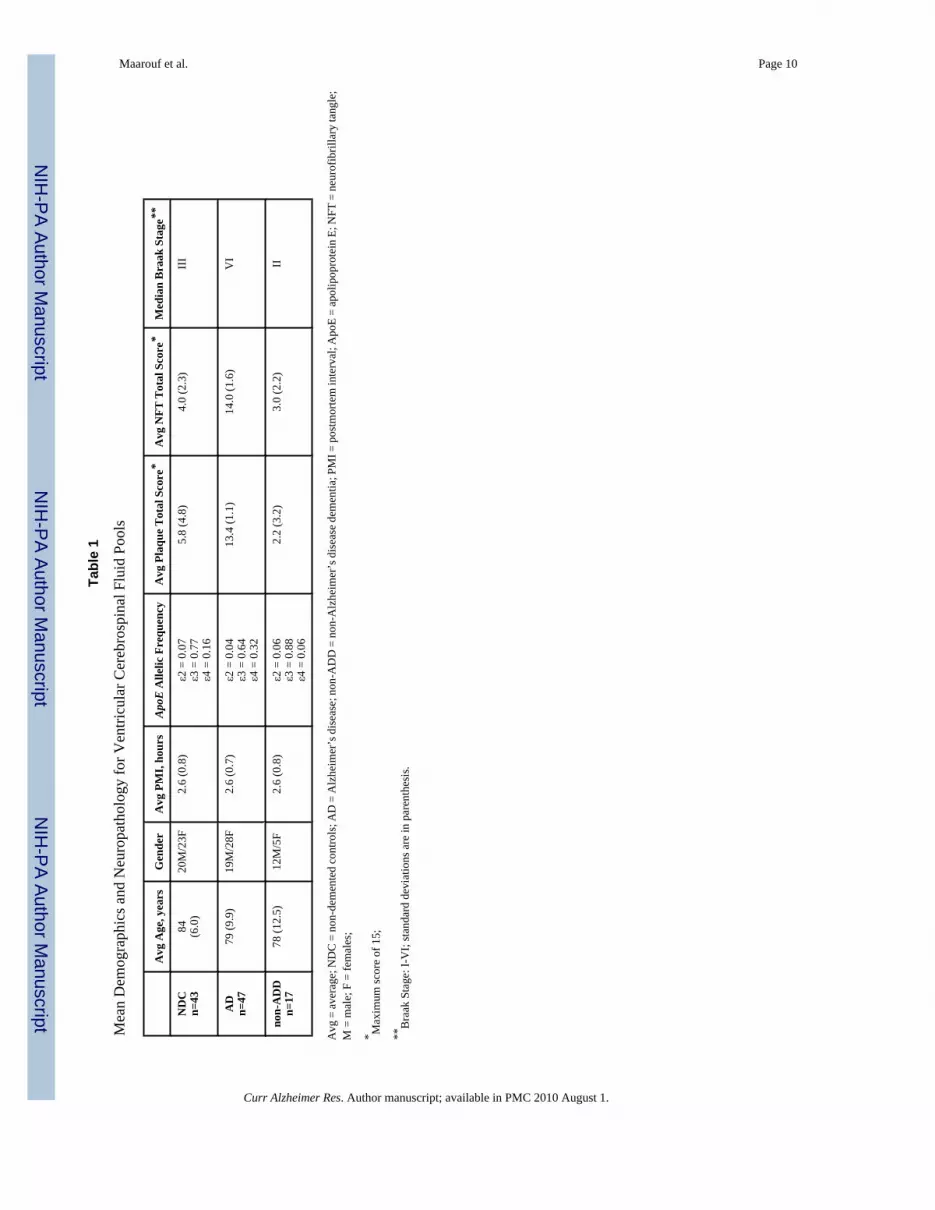
Cerebrospinal fluid samples were collected in the immediate postmortem period by syringeaspiration from the lateral ventricles, centrifuged at 1600 × rpm for 10 min and the supernatantstored at −80°C until analysis. The total protein concentration of each individual’s V-CSF wasmeasured using the Micro BCA protein assay kit (Pierce, Rockford, IL). The average amountof protein in the AD, NDC and non-ADD cases amounted to 0.87 mg/ml (range: 1.79 – 0.52mg/ml, SD = 0.24), 0.88 mg/ml (range 1.74 – 0.46 mg/ml, SD = 0.33) and 1.20 mg/ml (range:1.81–0.81, SD = 0.27), respectively. A description of the three populations under investigationis given in Table 1 which includes average age, gender, postmortem interval (PMI),apolipoprotein E (ApoE) allelic frequency and the neuropathological scores for amyloidplaques, neurofibrillary tangles and Braak stage. The plaque and neurofibrillary tangle (NFT)total scores were derived by adding the scores from 5 different areas of the brain: frontal,parietal, occipital, temporal lobes and hippocampus. Each area is classified as 0 = none, 1 =mild, 2 = moderate or 3 = severe. All cases were carefully selected and only those with a PMIof less than 4 h were included. With respect to the NDC cohort, all individuals had no historyof dementia or other neurological conditions and did not meet the neuropathological criteriafor AD. In contrast, all subjects in the AD group met the neuropathological criteria as definedby the CERAD (“definite” or “probable”) [11] and the NIA-Reagan Institute (“high” or“intermediate” probability of AD) [12]. The AD cases were carefully selected to the extent thatnone of them had any other neuropathological conditions, such as non-ADD, and likewise noneof the non-ADD cases had a concurrent neuropathological diagnosis of AD. The pathologicdistribution in the non-ADD cohort was as follows: 4 with progressive supranuclear palsy anddementia, 3 with hippocampal sclerosis dementia, 4 with non-specific sporadic tauopathy and/
Maarouf et al. Page 2
Curr Alzheimer Res. Author manuscript; available in PMC 2010 August 1.
NIH
-PA Author Manuscript
NIH
-PA Author Manuscript
NIH
-PA Author Manuscript
or argyrophilic grains, 2 with frontotemporal lobar degeneration with ubiquitinated inclusionsand progranulin gene mutations, 1 with frontotemporal dementia with tau gene mutation, 1with normal pressure hydrocephalus, 1 with corticobasal degeneration and 1 with dementialacking distinctive histology. Due to low number of individuals with diverse non-ADD in ourCSF bank, we were compelled to pool the non-ADD specimens into one category in order tohave sufficient statistical power. Apolipoprotein E ε4 still remains the major risk factor insporadic AD hence, all 107 individuals involved in the present study were Apo E genotypedaccording to a previously published protocol [13]. The allelic frequency shown in Table 1demonstrated as expected, an increase of ApoE ε4 in the AD population compared to the NDCand non-ADD cohort.
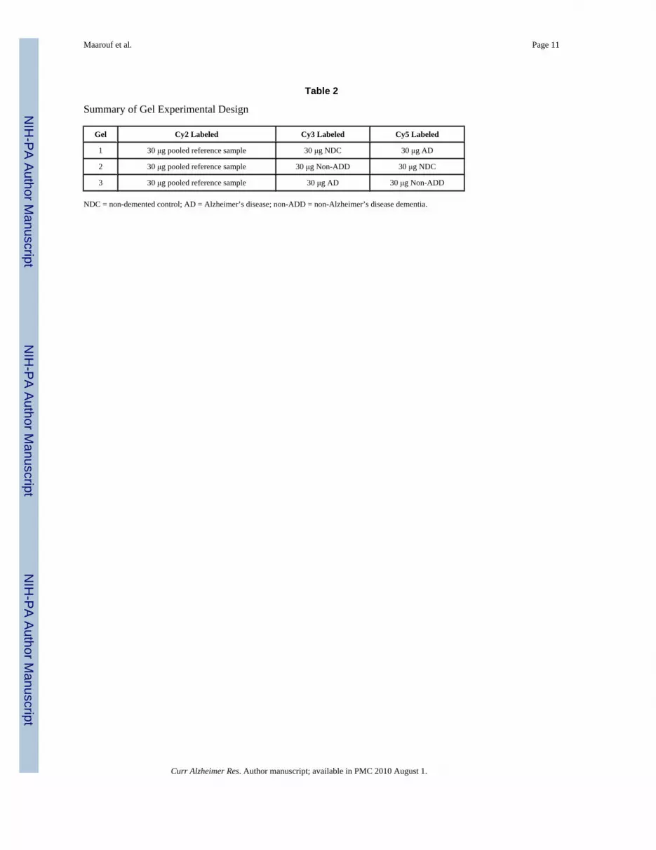
2D-DIGEEqual amounts of total protein were pooled from each of the 47 AD, 43 NDC and 17 non-ADDcases. Albumin and IgG were selectively removed using an antibody affinity resin kit followingthe technique described by the manufacturer (GE Healthcare, Piscataway, NJ) and elsewhere[14]. The proteins were concentrated by acetone/trichloroacetic acid (TCA) precipitation.Briefly, samples were mixed with 3 volumes of cold 13.3% TCA in acetone (stored at −20°C)and incubated at −20°C overnight. Samples were centrifuged at 15,000 × g for 30 min and thesupernatant was removed. The pellet was washed with the same volume of cold acetone, vortex-mixed, and incubated at −20°C for 30 min. Samples were centrifuged at 15,000 × g for 15 minand the supernatant was removed. Pellets were allowed to air dry for about 5 min, and thenwere suspended in 7 M urea, 2 M thiourea, 2% 3-[(3-Cholamidopropyl)dimethylammonio]-1-propanesulfonate hydrate (CHAPS), 2% 3-[N,N-Dimethyl(3-myristoylaminopropyl)ammonio]propanesulfonate (ASB-14), 15 mM Tris, pH 8.3. A pooled reference sample, loadedon every analytical gel, was created by combining 30 μg from each sample group, and allsamples were diluted to 2 mg/ml using the above buffer. Thirty μg of each individual samplegroup was labeled with 200 pmol minimal Cy3 and Cy5 NHS ester (GE Healthcare,Piscataway, NJ), and the pooled reference sample was labeled in bulk with minimal Cy2 NHSester (at a ratio of 200 pmol dye:30 μg sample) at 4°C for 30 min. The labeling reaction wasquenched with 10 nmol of lysine. Labeled protein extracts were combined and proteins wereseparated on 18 cm pH 4–7 IPG strips for 32,000 Vhr using the IPGphor (GE Healthcare,Piscataway, NJ) in 7 M urea, 2 M thiourea, 2% CHAPS, 2% ASB-14, 0.5% IPG buffer, 18.2mM dithiothreitol (DTT) and 0.002% bromophenol blue using active rehydration at 30V. Afterisoelectric focusing, IPG strips were equilibrated in 6 M urea, 2% SDS, 65 mM DTT, 30%glycerol, 50 mM Tris, pH 8.8, and 0.002% bromophenol blue for 15 min at room temperature(RT). IPG strips were then equilibrated in the above buffer, replacing DTT with 135 mMiodoacetamide, for 15 min at RT. Proteins were then separated on 20 × 26 cm 8–15% SDSpolyacrylamide gels using the Dalt II (GE Healthcare, Piscataway, NJ). Gels were fixed in 30%ethanol and 7.5% acetic acid overnight at RT. Gels were imaged using the Typhoon 9400 (GEHealthcare, Piscataway, NJ), optimizing the photomultiplier tube voltage for each laser toachieve the broadest dynamic range, and analyzed for significant differences by ANOVA basedon the log2 standardized volume using DeCyder 6.5 software (GE Healthcare, Piscataway,NJ). In summary, three analytical gels were completed in total, running 30 μg of pooledreference sample labeled with Cy2, along with two samples (30 μg each), one labeled withCy3 and the other labeled with Cy5. The final gel experimental design resulted in threeanalytical gels with two technical replicates (Table 2).
Gels selected for picking were stained with LavaPurple (Fluorotechnics, Guelph, Ontario) in100 mM sodium borate, pH 10.5, for 2 hrs at RT, washed in 15% ethanol for 30 min at RT,and acidified in 15% ethanol/7.5% acetic acid for 30 min at RT. Spots were matched acrossgels by matching the pooled reference sample images to each other using DeCyder 6.5.Quantitation of protein abundance was determined based on pixel intensity using DeCyder
Maarouf et al. Page 3
Curr Alzheimer Res. Author manuscript; available in PMC 2010 August 1.
NIH
-PA Author Manuscript
NIH
-PA Author Manuscript
NIH
-PA Author Manuscript

software and calculating the pixel intensity volume for each spot. Statistical analyses for eachspot were performed using the log2 standardized volume. The standardized volume iscalculated by normalizing (ie. dividing) the volume determined for every Cy3 and Cy5 labeledspot to the Cy2 labeled spot volume for each gel (Table 2). Approximately 1100 spots werematched across all three analytical gels. All three analytical gels were picked using anautomated robotic system, the Spot Handling Workstation (GE Healthcare). The pick list wascreated based on the LavaPurple image. Two mm gel plugs were picked, washed, reduced andalkylated, digested with trypsin, and the resulting peptides were extracted and spotted usingthe Spot Handling Workstation (GE Healthcare, Piscataway, NJ). Briefly, plugs were washedtwice with 50 mM ammonium bicarbonate/50% methanol for 20 min at RT and then with 75%acetonitrile for 20 min at RT and dried at 40°C for 10 min. Plugs were incubated in 10 mMDTT/20 mM ammonium bicarbonate at 37°C for 1 hr. The DTT solution was removed andimmediately replaced with 100 mM iodoacetamide/20 mM ammonium bicarbonate andincubated at RT in the dark for 30 min. Plugs were washed as above, and then incubated with200 ng sequencing grade trypsin (Promega, Madison, WI) at 37°C for 2 hrs. Peptides wereextracted twice with 50% acetonitrile/0.1% trifluoroacetic acid for 20 min at RT andconcentrated by SpeedVac (Jouan).
Mass SpectrometryApproximately 25% of the resulting peptides were spotted with partially saturated α-cyano-4-hydroxy-cinnamic acid (Sigma, St. Louis, MO). Mass spectrometry (MS) and MS/MS datawere acquired on the 4700 Proteomics Analyzer (Applied Biosystems, Framingham, MA)using standard acquisition methods. MS spectra were calibrated using two trypsin autolysispeaks (1045.56 and 2211.096 m/z) and MS/MS spectra were calibrated using the instrumentdefault calibration. The most recent version of NCBInr database was downloaded from NCBI(http://www.ncbi.nlm.nih.gov/en-trez/) and was incorporated into a licensed copy of Mascot1.9.05 (http://www.matrixscience.com/). Mass lists were submitted to the various sub-databases (NCBInr-Mammals, and NCBInr-Homo sapiens) considering fixed cysteinecarbamidomethylation and partial methionine oxidation modifications, 1 missed trypticcleavage, and 25 ppm mass accuracy. Identifications were cross-examined using massaccuracy, molecular weight, and pI.
RESULTSFigs. (1B, 1C and 1D) show the 2D DIGE gels of AD, NDC and non-ADD V-CSF pools,respectively. Two replicates of each pooled sample were run, labeling one replicate with Cy3and one replicate with Cy5, resulting in three analytical gels (Table 2). The 2D DIGEcomparative analysis of the three diagnosis groups revealed 22 spots with statisticallysignificant (p ≥ 0.05) differences. These spots are shown in (Fig. 1A) encircled in red andreported in Tables 3 and 4. The blue encircled spots represent additional spots that were alsoinvestigated by MS (Fig. 1A). The protein identities of the 22 statistically significant spots areshown in Table 3, which also includes the 1-ANOVA analysis, National Center forBioinformatics (NCBI) gi accession number, predicted molecular weight (MW), the predictedpI, number of matching peptides, protein MASCOT score and protein MASCOT confidence.With the exception of spots 13, 18 and 20, which show MASCOT scores of 67, 60 an 65, allMASCOT scores were greater than 72 (p = 0.05) and had a MASCOT confidence of 99.6–100(Table 3). Only one highly confident protein assignment resulted from the MS analyses. Asthe NCBInr database is not completely non-redundant, many of the searches did return severalprotein variants of the same functional protein. However, the next ranking protein was alwaysbelow the 95% confidence limit.
Maarouf et al. Page 4
Curr Alzheimer Res. Author manuscript; available in PMC 2010 August 1.
NIH
-PA Author Manuscript
NIH
-PA Author Manuscript
NIH
-PA Author Manuscript
The Student’s t-test and average ratio of intensity are shown in Table 4. Column I shows 7proteins were increased and 12 were decreased in the NDC when compared to the non-ADDgroup, with the biggest difference being in transthyretin (TTR), which was on average 3Xhigher in the non-ADD gels. Of the 14 spots found to be statistically different when comparingNDC and AD gels (Table 4, column II), 7 were increased and 7 were decreased in the NDCgroup. Once again TTR had the most significant change in average ratio (−2.76). As seen inTable 4, column III, 6 proteins were increased and 11 decreased in AD gels compared to thenon-ADD gels. The largest difference in average ratio was ε1-antitrypsin which was 2X higherin the non-ADD group. We included the pigment epithelium derived factor (PEDF; spot 17)because in the comparison between NDC/AD the p value was borderline (0.07). In addition, aprevious biomarker assessment performed in our laboratory [14], in which pools of NDC andAD V-CSF were investigated, two isoforms of PEDF, were significantly increased in the ADpool (p = 0.027 and p = 0.014).
DISCUSSIONA justification for the use of postmortem V-CSF is the ability to use the neuropathologicaldiagnosis as a gold standard. While existing clinical diagnostic methods have a high sensitivityfor AD, about 85%, they have a low specificity, about 45%, as determined by a comprehensivestudy of results from the National Institute on Aging Alzheimer’s disease centers [10].Furthermore, in many instances the clinical diagnosis of AD is complicated by concurrentpathologies such as vascular dementia, Lewy body disease, Parkinson dementia, progressivesupranuclear palsy and other non-AD dementias. Another advantage is that the V-CSF is veryseldom contaminated by blood. However, it must be recognized that the diagnostic value ofV-CSF biomarkers needs to be correlated with measurements obtained from living individualsusing lumbar CSF. In a small proteomics study, quantitative deviations between ventricularand lumbar CSF were observed [15]. Although the authors attributed the differences to PMI,these disparities may also be due to protein gradient differences that exist between the twoanatomical sites (lumbar versus V-CSF). Consideration should be given to the fact that someproteins that are generated by the brain have higher concentrations in the V-CSF. On the otherhand, those proteins derived from the blood are more concentrated in the lumbar CSF [16]. Inthe best of the situations, antemortem CSF collection of clinically diagnosed individuals shouldbe followed by postmortem neuropathological confirmation. These conditions are difficult toachieve given that, in most cases, relatively few individuals are autopsied. Other considerationsfor CSF biomarker studies include long PMI, which should be avoided, due to proteindegradation. Additionally, lumbar Aβ CSF protein values are unreliable to a certain degreebecause of fluctuations that occur at different times of the day as recently demonstrated byBateman and colleagues [17]. An important caveat in dealing with lumbar or V-CSF is the longduration of AD and its pre-symptomatic period, which altogether can last for several decades.It is likely that different protein profiles would be obtained at different disease stages if thereis not a selective stratification of patients, an issue that may confound the utility of the results.In an ideal study, lumbar CSF could be taken at different time points along the disease courseto monitor how protein profiles change during AD progression, but problems with patientattrition, the slow rate of death and limited funding make this difficult to accomplish.Notwithstanding these disadvantages, the CSF remains the most promising venue to discovera set of reliable biomarkers, since it is in close contact with the brain and is a natural basin forsome interstitial brain molecules.
Our failure to detect increases in CSF tau (reviewed in [3,8]) could be due to postmortemchanges in the phosphorylation of these molecules. In rat models, the kinetics ofphosphorylation/dephosphorylation of tau in the immediate postmortem results in a net loss ofphosphate groups, being a relatively fast and site specific event [18]. In a second report [19],tau levels remained steady up to 8 h postmortem, although the electrophoretic mobility of some
Maarouf et al. Page 5
Curr Alzheimer Res. Author manuscript; available in PMC 2010 August 1.
NIH
-PA Author Manuscript
NIH
-PA Author Manuscript
NIH
-PA Author Manuscript
tau isoforms was modified. Similar cytoskeletal alterations were observed in fresh postmortemand surgically removed tissues.
Another limitation of biomarker studies is the use of small numbers of cases due to the expenseand technical difficulties of proteomic experiments. Instead of performing 107 individual 2DDIGE tests, we decided to investigate pools of V-CSF obtained from our rapid autopsy programwith short PMI. Our strategy was to individually measure the amount of V-CSF total proteinand then to pool individual aliquots, with an equal protein representation from each case, withthe aim of finding the most prominent deviations shared by a cohort of demented individualsand compare them with an equally represented NDC group. We are aware that such studies arepreliminary and need to be confirmed or rejected by individual assessments using immunoassayapproaches.
Presently, the literature on AD CSF biomarkers is extensive with more than 500 publicationsrecorded by the National Library of Medicine (USA) as of September 2008. Consequently, itis beyond the scope of this paper to discuss this vast body of information. However, it isnoteworthy that most of the proteins identified in our study have already been implicated,directly or indirectly, in AD pathophysiology and/or Aβ metabolism or have been found alteredin the CSF of AD patients. These include α-2-macroglobulin [20,21], α1-antichymotrypsin[22–24], α1-antitrypsin [24–27], complement proteins [28], heat shock proteins [29], fibulin-1[30], gelsolin [31,32], hemopexin [14], tubulin [33,34], PEDF [14,35], TTR [14,36,37],cathepsin D [14,38–40], enolase [41–43] and creatine kinase [44]. In addition, in a previouspilot CSF proteomic study from our laboratory using 2D-gel electrophoresis [14], 3 out of 5proteins identified in that study were confirmed in the present investigation by DIGE (PEDF,TTR, cathepsin D). This panel of proteins is likely reflective of fundamental pathologicalpathways in the AD brain that are divergent from normal aging or non-ADD. Additional workwith a larger number of cases assessed individually, with quantitative reliable methods, suchas ELISA, is needed to determine if the present array of CSF proteins would provide thesensitivity and specificity, useful for the accurate diagnosis of sporadic AD.
In conclusion, our results indicate that protein separation techniques based on physicochemicalprinciples are promising in providing significant differences between AD, NDC and non-ADdementias.
AcknowledgmentsThis study was supported by a grant from the National Alzheimer’s Coordinating Center (NACC) 5U01-AG-016976.We are grateful to the Sun Health Research Institute Brain Donation Program of Sun City, Arizona, for the provisionof human CSF. The Brain Donation Program is Supported by The National Institute on Aging (P30 AG19610 ArizonaAlzheimer’s Disease Core Center), the Arizona Biomedical Commission (contracts 4001, 0011 and 05-901 to theArizona Parkinson’s Disease Consortium) and the Prescott Family Initiative of Michael J. Fox Foundation forParkinson’s Research. We are indebted to Dr. Douglas Walker for ApoE genotyping.
References1. Alzheimer’s Association. Alzheimer’s disease facts and figures. Alzheimers Dement 2008;4:110–133.
[PubMed: 18631956]2. Vijg J, Campisi J. Puzzles, promises and a cure for ageing. Nature 2008;454:1065–1071. [PubMed:
18756247]3. Marksteiner, J.; Hinterhuber, H.; Humpel, C. Drugs Today. Vol. 43. Barc: 2007. Cerebrospinal fluid
biomarkers for diagnosis of Alzheimer’s disease: beta-amyloid(1–42), tau, phospho-tau-181 and totalprotein; p. 423-431.
4. Hampel H, Burger K, Teipel SJ, Bokde AL, Zetterberg H, Blennow K. Core candidate neurochemicaland imaging biomarkers of Alzheimer’s disease. Alzheimers Dement 2008;4:38–48. [PubMed:18631949]
Maarouf et al. Page 6
Curr Alzheimer Res. Author manuscript; available in PMC 2010 August 1.
NIH
-PA Author Manuscript
NIH
-PA Author Manuscript
NIH
-PA Author Manuscript
5. Nordlund A, Rolstad S, Klang O, Lind K, Pedersen M, Blennow K, et al. Episodic memory and speed/attention deficits are associated with Alzheimer-typical CSF abnormalities in MCI. J Int NeuropsycholSoc 2008;14:582–590. [PubMed: 18577287]
6. Lewczuk P, Wiltfang J. Neurochemical dementia diagnostics: State of the art and research perspectives.Proteomics 2008;8:1292–1301. [PubMed: 18271068]
7. Hansson O, Zetterberg H, Buchhave P, Londos E, Blennow K, Minthon L. Association between CSFbiomarkers and incipient Alzheimer’s disease in patients with mild cognitive impairment: a follow-up study. Lancet Neurol 2006;5:228–234. [PubMed: 16488378]
8. Frankfort SV, Tulner LR, van Campen JP, Verbeek MM, Jansen RW, Beijnen JH. Amyloid beta proteinand tau in cerebrospinal fluid and plasma as biomarkers for dementia: a review of recent literature.Curr Clin Pharmacol 2008;3:123–131. [PubMed: 18700307]
9. Dubois B, Feldman HH, Jacova C, DeKosky ST, Barberger-Gateau P, Cummings J, et al. Researchcriteria for the diagnosis of Alzheimer’s disease: revising the NINCDS-ADRDA criteria. LancetNeurol 2007;6:734–746. [PubMed: 17616482]
10. Mayeux R, Saunders AM, Shea S, Mirra S, Evans D, Roses AD, et al. Utility of the apolipoproteinE genotype in the diagnosis of Alzheimer’s disease. Alzheimer’s Disease Centers Consortium onApolipoprotein E and Alzheimer’s Disease. N Engl J Med 1998;338:506–511. [PubMed: 9468467]
11. Mirra SS, Heyman A, McKeel D, Sumi SM, Crain BJ, Brownlee LM, et al. The Consortium toEstablish a Registry for Alzheimer’s Disease (CERAD). Part II. Standardization of theneuropathologic assessment of Alzheimer’s disease. Neurology 1991;41:479–486. [PubMed:2011243]
12. Hyman BT, Trojanowski JQ. Consensus recommendations for the postmortem diagnosis of Alzheimerdisease from the National Institute on Aging and the Reagan Institute Working Group on diagnosticcriteria for the neuropathological assessment of Alzheimer disease. J Neuropathol Exp Neurol1997;56:1095–1097. [PubMed: 9329452]
13. Hixson JE, Vernier DT. Restriction isotyping of human apolipoprotein E by gene amplification andcleavage with HhaI. J Lipid Res 1990;31:545–548. [PubMed: 2341813]
14. Castaño EM, Roher AE, Esh CL, Kokjohn TA, Beach T. Comparative proteomics of cerebrospinalfluid in neuropathologically-confirmed Alzheimer’s disease and non-demented elderly subjects.Neurol Res 2006;28:155–163. [PubMed: 16551433]
15. Finehout EJ, Franck Z, Relkin N, Lee KH. Proteomic analysis of cerebrospinal fluid changes relatedto postmortem interval. Clin Chem 2006;52:1906–1913. [PubMed: 16887899]
16. Reiber H. Dynamics of brain-derived proteins in cerebrospinal fluid. Clin Chim Acta 2001;310:173–186. [PubMed: 11498083]
17. Bateman RJ, Wen G, Morris JC, Holtzman DM. Fluctuations of CSF amyloid-beta levels: implicationsfor a diagnostic and therapeutic biomarker. Neurology 2007;68:666–669. [PubMed: 17325273]
18. Gartner U, Janke C, Holzer M, Vanmechelen E, Arendt T. Postmortem changes in the phosphorylationstate of tau-protein in the rat brain. Neurobiol Aging 1998;19:535–543. [PubMed: 10192212]
19. Schwab C, Bondada V, Sparks DL, Cahan LD, Geddes JW. Postmortem changes in the levels andlocalization of microtubule-associated proteins (tau, MAP2 and MAP1B) in the rat and humanhippocampus. Hippocampus 1994;4:210–225. [PubMed: 7951696]
20. Kovacs DM. alpha2-macroglobulin in late-onset Alzheimer’s disease. Exp Gerontol 2000;35:473–479. [PubMed: 10959035]
21. McGeer PL, McGeer EG. Polymorphisms in inflammatory genes and the risk of Alzheimer disease.Arch Neurol 2001;58:1790–1792. [PubMed: 11708985]
22. Zhang S, Janciauskiene S. Multi-functional capability of proteins: alpha1-antichymotrypsin and thecorrelation with Alzheimer’s disease. J Alzheimers Dis 2002;4:115–122. [PubMed: 12214135]
23. Harigaya Y, Shoji M, Nakamura T, Matsubara E, Hosoda K, Hirai S. Alpha 1-antichymotrypsin levelin cerebrospinal fluid is closely associated with late onset Alzheimer’s disease. Intern Med1995;34:481–484. [PubMed: 7549128]
24. Nielsen HM, Minthon L, Londos E, Blennow K, Miranda E, Perez J, et al. Plasma and CSF serpinsin Alzheimer disease and dementia with Lewy bodies. Neurology 2007;69:1569–1579. [PubMed:17761554]
Maarouf et al. Page 7
Curr Alzheimer Res. Author manuscript; available in PMC 2010 August 1.
NIH
-PA Author Manuscript
NIH
-PA Author Manuscript
NIH
-PA Author Manuscript
25. Bohrmann B, Tjernberg L, Kuner P, Poli S, Levet-Trafit B, Naslund J, et al. Endogenous proteinscontrolling amyloid beta-peptide polymerization. Possible implications for beta-amyloid formationin the central nervous system and in peripheral tissues. J Biol Chem 1999;274:15990–15995.[PubMed: 10347147]
26. Elliott PR, Lomas DA, Carrell RW, Abrahams JP. Inhibitory conformation of the reactive loop ofalpha 1-antitrypsin. Nat Struct Biol 1996;3:676–681. [PubMed: 8756325]
27. Puchades M, Hansson SF, Nilsson CL andreasen N, Blennow K, Davidsson P. Proteomic studies ofpotential cerebrospinal fluid protein markers for Alzheimer’s disease. Brain Res Mol Brain Res2003;118:140–146. [PubMed: 14559363]
28. Bonifati DM, Kishore U. Role of complement in neurodegeneration and neuroinflammation. MolImmunol 2007;44:999–1010. [PubMed: 16698083]
29. Smith RC, Rosen KM, Pola R, Magrane J. Stress proteins in Alzheimer’s disease. Int J Hyperthermia2005;21:421–431. [PubMed: 16048839]
30. Ohsawa I, Takamura C, Kohsaka S. Fibulin-1 binds the aminoterminal head of beta-amyloid precursorprotein and modulates its physiological function. J Neurochem 2001;76:1411–1420. [PubMed:11238726]
31. Chauhan VP, Ray I, Chauhan A, Wisniewski HM. Binding of gelsolin, a secretory protein, to amyloidbeta-protein. Biochem Biophys Res Commun 1999;258:241–246. [PubMed: 10329371]
32. Ray I, Chauhan A, Wegiel J, Chauhan VP. Gelsolin inhibits the fibrillization of amyloid beta-proteinand also defibrillizes its pre-formed fibrils. Brain Res 2000;853:344–351. [PubMed: 10640633]
33. Iqbal K, Alonso AC, Gong CX, Khatoon S, Pei JJ, Wang JZ, et al. Mechanisms of neurofibrillarydegeneration and the formation of neurofibrillary tangles. J Neural Transm Suppl 1998;53:169–180.[PubMed: 9700655]
34. Li B, Chohan MO, Grundke-Iqbal I, Iqbal K. Disruption of microtubule network by Alzheimerabnormally hyperphosphorylated tau. Acta Neuropathol 2007;113:501–511. [PubMed: 17372746]
35. Yamagishi S, Inagaki Y, Takeuchi M, Sasaki N. Is pigment epithelium-derived factor level incerebrospinal fluid a promising biomarker for early diagnosis of Alzheimer’s disease? MedHypotheses 2004;63:115–117. [PubMed: 15193361]
36. Gloeckner SF, Meyne F, Wagner F, Heinemann U, Krasnianski A, Meissner B, et al. Quantitativeanalysis of transthyretin, tau and amyloid-beta in patients with dementia. J Alzheimers Dis2008;14:17–25. [PubMed: 18525124]
37. Sousa JC, Cardoso I, Marques F, Saraiva MJ, Palha JA. Transthyretin and Alzheimer’s disease: wherein the brain? Neurobiol Aging 2007;28:713–718. [PubMed: 16698124]
38. Riemenschneider M, Blennow K, Wagenpfeil S, Andreasen N, Prince JA, Laws SM, et al. Thecathepsin D rs17571 polymorphism: effects on CSF tau concentrations in Alzheimer disease. HumMutat 2006;27:532–537. [PubMed: 16652347]
39. Papassotiropoulos A, Lewis HD, Bagli M, Jessen F, Ptok U, Schulte A, et al. Cerebrospinal fluidlevels of beta-amyloid(42) in patients with Alzheimer’s disease are related to the exon 2polymorphism of the cathepsin D gene. Neuroreport 2002;13:1291–1294. [PubMed: 12151789]
40. Schwagerl AL, Mohan PS, Cataldo AM, Vonsattel JP, Kowall NW, Nixon RA. Elevated levels ofthe endosomallysosomal proteinase cathepsin D in cerebrospinal fluid in Alzheimer disease. JNeurochem 1995;64:443–446. [PubMed: 7798944]
41. Palumbo B, Siepi D, Sabalich I, Tranfaglia C, Parnetti L. Cerebrospinal fluid neuron-specific enolase:a further marker of Alzheimer’s disease? Funct Neurol 2008;23:93–96. [PubMed: 18671910]
42. Parnetti L, Palumbo B, Cardinali L, Loreti F, Chionne F, Cecchetti R, et al. Cerebrospinal fluid neuron-specific enolase in Alzheimer’s disease and vascular dementia. Neurosci Lett 1995;183:43–45.[PubMed: 7746484]
43. Blennow K, Wallin A, Ekman R. Neuron specific enolase in cerebrospinal fluid: a biochemical markerfor neuronal degeneration in dementia disorders? J Neural Transm Park Dis Dement Sect 1994;8:183–191. [PubMed: 7748462]
44. Li X, Burklen T, Yuan X, Schlattner U, Desiderio DM, Wallimann T, et al. Stabilization of ubiquitousmitochondrial creatine kinase preprotein by APP family proteins. Mol Cell Neurosci 2006;31:263–272. [PubMed: 16260146]
Maarouf et al. Page 8
Curr Alzheimer Res. Author manuscript; available in PMC 2010 August 1.
NIH
-PA Author Manuscript
NIH
-PA Author Manuscript
NIH
-PA Author Manuscript
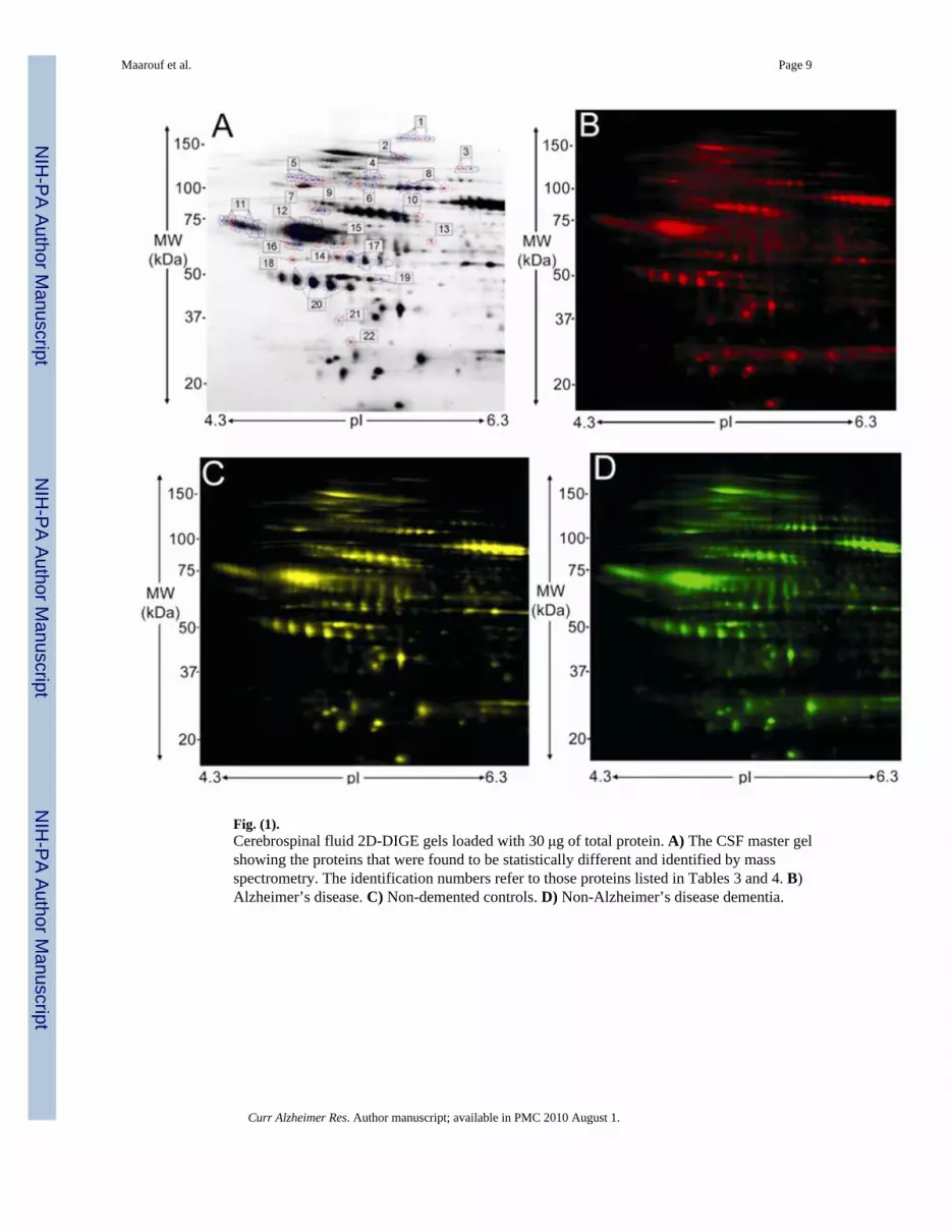
Fig. (1).Cerebrospinal fluid 2D-DIGE gels loaded with 30 μg of total protein. A) The CSF master gelshowing the proteins that were found to be statistically different and identified by massspectrometry. The identification numbers refer to those proteins listed in Tables 3 and 4. B)Alzheimer’s disease. C) Non-demented controls. D) Non-Alzheimer’s disease dementia.
Maarouf et al. Page 9
Curr Alzheimer Res. Author manuscript; available in PMC 2010 August 1.
NIH
-PA Author Manuscript
NIH
-PA Author Manuscript
NIH
-PA Author Manuscript
NIH
-PA Author Manuscript
NIH
-PA Author Manuscript
NIH
-PA Author Manuscript
Maarouf et al. Page 10
Tabl
e 1
Mea
n D
emog
raph
ics a
nd N
euro
path
olog
y fo
r Ven
tricu
lar C
ereb
rosp
inal
Flu
id P
ools
Avg
Age
, yea
rsG
ende
rA
vg P
MI,
hour
sAp
oE A
llelic
Fre
quen
cyA
vg P
laqu
e T
otal
Sco
re*
Avg
NFT
Tot
al S
core
*M
edia
n B
raak
Sta
ge**
ND
Cn=
4384 (6.0
)20
M/2
3F2.
6 (0
.8)
ε2 =
0.0
7ε3
= 0
.77
ε4 =
0.1
6
5.8
(4.8
)4.
0 (2
.3)
III
AD
n=47
79 (9
.9)
19M
/28F
2.6
(0.7
)ε2
= 0
.04
ε3 =
0.6
4ε4
= 0
.32
13.4
(1.1
)14
.0 (1
.6)
VI
non-
AD
Dn=
1778
(12.
5)12
M/5
F2.
6 (0
.8)
ε2 =
0.0
6ε3
= 0
.88
ε4 =
0.0
6
2.2
(3.2
)3.
0 (2
.2)
II
Avg
= a
vera
ge; N
DC
= n
on-d
emen
ted
cont
rols
; AD
= A
lzhe
imer
’s d
isea
se; n
on-A
DD
= n
on-A
lzhe
imer
’s d
isea
se d
emen
tia; P
MI =
pos
tmor
tem
inte
rval
; Apo
E =
apol
ipop
rote
in E
; NFT
= n
euro
fibril
lary
tang
le;
M =
mal
e; F
= fe
mal
es;
* Max
imum
scor
e of
15;
**B
raak
Sta
ge: I
-VI;
stan
dard
dev
iatio
ns a
re in
par
enth
esis
.
Curr Alzheimer Res. Author manuscript; available in PMC 2010 August 1.
NIH
-PA Author Manuscript
NIH
-PA Author Manuscript
NIH
-PA Author Manuscript
Maarouf et al. Page 11
Table 2
Summary of Gel Experimental Design
Gel Cy2 Labeled Cy3 Labeled Cy5 Labeled
1 30 μg pooled reference sample 30 μg NDC 30 μg AD
2 30 μg pooled reference sample 30 μg Non-ADD 30 μg NDC
3 30 μg pooled reference sample 30 μg AD 30 μg Non-ADD
NDC = non-demented control; AD = Alzheimer’s disease; non-ADD = non-Alzheimer’s disease dementia.
Curr Alzheimer Res. Author manuscript; available in PMC 2010 August 1.

NIH
-PA Author Manuscript
NIH
-PA Author Manuscript
NIH
-PA Author Manuscript
Maarouf et al. Page 12
Tabl
e 3
Mas
s Spe
ctro
met
ry P
rote
in Id
entif
icat
ion
of 2
D D
IGE
Spot
s of I
nter
est a
nd S
tatis
tical
Ana
lyse
s Usi
ng 1
-AN
OV
A C
ompa
ring
Non
-Dem
ente
d C
ontro
ls,
Non
-Alz
heim
er’s
Dis
ease
Dem
entia
and
Alz
heim
er’s
Dis
ease
Gel
s
Spot
Prot
ein
ID1-
AN
OV
A (p
)N
CB
I ac-
ces
sion
(gi)
Pred
icte
d M
W (D
a)Pr
edic
ted
pI#
of p
eptid
esM
ASC
OT
scor
eM
ASC
OT
con
fiden
ce
1α2
-mac
rogl
obul
in0.
0003
822
4053
162,
072
5.95
1785
99.8
3
2α2
-mac
rogl
obul
in0.
005
2240
5316
2,07
25.
9517
102
99.9
9
3C
LIC
6 pr
otei
n0.
0096
3478
3345
44,5
384.
628
8699
.86
4C
ompl
C4-
B p
recu
rsor
0.00
5481
1751
6719
4,21
26.
7318
8699
.86
5Fi
bulin
10.
0014
1849
0682
78,4
795.
110
8299
.62
6C
4B1
0.00
4740
7373
1348
,007
5.92
1112
310
0
7H
SP90
AA
1 pr
otei
n0.
018
6291
4009
74,1
795.
0820
209
100
8G
elso
lin is
ofor
m0.
0016
4504
165
86,0
435.
919
182
100
9C
ompl
90.
0006
122
5812
861
,728
5.42
1398
99.9
9
10H
emop
exin
0.00
3711
3215
6152
,385
6.55
1217
410
0
11α1
-ant
ichy
mot
ryps
in6.
40E-
0513
4014
249
,986
5.5
1515
010
0
12α1
-ant
itryp
sin
0.01
519
4262
944
,280
5.37
2120
610
0
13β2
-gly
copr
otei
n0.
0016
6435
718
36,6
668.
265
6788
.35
14α-
tubu
lin0.
0027
3400
2150
,804
4.94
885
99.9
4
15A
ngio
tens
inog
en0.
0001
162
0891
2454
,029
5.97
815
710
0
16Tu
bulin
, β 2
C0.
0076
1196
0877
549
,250
4.88
1421
510
0
17PE
DF
0.07
339
7259
3446
,454
5.97
1218
910
0
18γ
enol
ase
0.00
1818
2118
44,5
684.
945
6078
.67
19C
reat
ine
kina
se0.
0089
1805
7042
,876
5.34
1110
599
.99
20H
P pr
otei
n0.
0021
3478
5974
25,7
276.
065
6582
.37
21TT
R d
imer
0.00
455
6695
7512
,836
5.33
614
910
0
22C
athe
psin
D0.
0082
4942
9626
,457
5.31
511
610
0
NC
BI =
Nat
iona
l Cen
ter f
or B
ioin
form
atic
s; M
W =
mol
ecul
ar w
eigh
t; D
a =
Dal
tons
; Com
pl =
com
plem
ent;
HSP
= h
eat s
hock
pro
tein
; PED
F =
pigm
ent e
pith
eliu
m d
eriv
ed fa
ctor
; TTR
= tr
anst
hyre
tin.
Curr Alzheimer Res. Author manuscript; available in PMC 2010 August 1.

NIH
-PA Author Manuscript
NIH
-PA Author Manuscript
NIH
-PA Author Manuscript
Maarouf et al. Page 13
Tabl
e 4
Stat
istic
al A
naly
ses o
f 2D
DIG
E V
entri
cula
r Cer
ebro
spin
al F
luid
Pro
tein
s Usi
ng S
tude
nt’s
t-Te
st
Spot
Prot
ein
ID
I. N
DC
/non
-AD
DII
. ND
C/A
DII
I. A
D/n
on-A
DD
t-tes
t (p)
Avg
Rat
iot-t
est (
p)A
vg R
atio
t-tes
t (p)
Avg
Rat
io
1α2
-mac
rogl
obul
in0.
002
1.21
0.00
611.
140.
007
1.07
2α2
-mac
rogl
obul
in0.
021
1.21
0.01
61.
28
3C
LIC
6 pr
otei
n0.
021
1.32
0.02
61.
35
4C
ompl
C4-
B p
recu
rsor
0.00
41−1
.46
0.04
5−1
.23
5Fi
bulin
10.
0062
−2.0
50.
024
−1.3
70.
0053
−1.4
9
6C
4B1
0.01
2−1
.36
0.00
91−1
.27
7H
SP90
AA
1 pr
otei
n0.
017
1.22
0.03
1−1
.25
8G
elso
lin is
ofor
m0.
0049
−1.2
50.
026
1.25
0.00
57−1
.55
9C
ompl
. 90.
0026
−1.5
30.
0044
−1.5
3
10H
emop
exin
0.00
231.
350.
024
1.22
11α1
-ant
ichy
mot
ryps
in0.
0008
4−1
.65
0.01
11.
190.
0005
−1.9
7
12α1
-ant
itryp
sin
0.03
7−1
.83
0.02
1−2
.04
13β2
-gly
copr
otei
n0.
0051
−1.6
70.
019
−1.3
60.
012
−1.2
3
14α-
tubu
lin0.
011
−1.4
20.
011
−1.4
7
15A
ngio
tens
inog
en0.
0013
−1.4
50.
012
−1.0
40.
0015
−1.3
9
16Tu
bulin
, β 2
C0.
0022
−1.8
0.02
7−1
.69
17PE
DF
0.07
−1.1
7
18γ
enol
ase
0.01
1.22
0.00
841.
26
19C
reat
ine
kina
se0.
016
1.67
0.03
81.
24
20H
P pr
otei
n0.
0056
−1.1
70.
012
1.15
21TT
R d
imer
0.01
5−3
.07
0.00
06−2
.76
22C
athe
psin
D0.
018
1.23
0.04
1.12
0.04
91.
1
ND
C =
non
-dem
ente
d co
ntro
ls; n
on-A
DD
= n
on-A
lzhe
imer
’s d
isea
se d
emen
tia; A
D =
Alz
heim
er’s
dis
ease
; Avg
= a
vera
ge; C
ompl
= c
ompl
emen
t; H
SP =
hea
t sho
ck p
rote
in; P
EDF
= pi
gmen
t epi
thel
ium
deriv
ed fa
ctor
; TTR
= tr
anst
hyre
tin.
Curr Alzheimer Res. Author manuscript; available in PMC 2010 August 1.