fetal death in goats in khartoum state diagnosed by
TRANSCRIPT

Sudan University of Science and Technology
College of Graduate Studies
Fetal Death in Goats in Khartoum State Diagnosed by Ultrasonography
and Treated by PGF2α and Oxytetracycline
بواسطة الموجات فوق الصوتية وعالجها في والية الخرطوم الميتة في الماعز األجنةتشخيص
وكسي تيتراسايكلينبواسطة البروستاغالندين واأل
A Thesis Submitted for Fulfillment of the Requirements for Master
Degree in Veterinary Medicine (Reproduction and Obstetrics)
By
Abram Samuel Aban Deng
B.V.M. (2011), Upper Nile University
Supervisor
Dr. Rihab Mohamed Abdelghafar Osman
Co-supervisor
Dr. Majdi Elnaim Badawi
January-2017

II
Dedication
To the soul of my father
To my beloved mother Jachinta Nyakwic
To my siblings
To my small family Jully and Gieth

III
Acknowledgments
Firstly, I would like to thank my Almighty Allah for giving me health and
strength to complete this work.
I would like to express my sincere gratitude to my supervisor Dr. Rihab
Abdelghafar for the continuous support of my master study and related research, for her
patience, motivation and immense knowledge. Her guidance helped me all the time.
I wish to express my sincere thanks to my co-supervisor Dr. Majdi Elnaim
Badawi for his endless help and thorough guidance.
I am using this opportunity to express my gratitude to professor Mohamed
Abdelsalam Abdalla, professor Abdelhamid Ahmed Mohamed Elfadil, professor
Mohamed Tagelddin Ibrahim and Dr. Naglaa Abd Elhakeem Abbas who supported me
throughout this study. I am thankful for their aspiring guidance, invaluably constructive
criticism and friendly advice during my study. I would like to thank my colleague Dr.
Areeg Mohamed Almubarak for her diligence since started of this study till end.
I would like to thank my friends for accepting nothing less than excellence from
me. Last but not the least; I would like to thank my family: my parents and my brothers
and sisters for supporting me spiritually throughout the study period. Without their help
and guidance this study wouldn’t be complete.
I would also like to thank the animal’s owners; without their passionate
participation and input, the study could not have been successfully conducted.
My great thanks also go to the Upper Nile University, College of Veterinary
Medicine for providing me the scholarship and to Sudan University of Science and
Technology for offering me the good environment.
IV
Table of Contents
Subject Page
Dedication
Acknowledgments
Table of contents
List of tables
List of figures
Appendix
English Abstract
Arabic Abstract
INTRODUCTION
Introduction
Objectives
CHAPTER ONE
LITERATURE REVIEW
1.1. Goat
1.1.1. Reproductive properties of goats
1.1.1.1. Puberty
1.1.1.2. Estrus cycle
1.1.1.3. Gestational length and placentation
1.1.1.4. Fecundity
1.1.1.5. Fertility (Conception rate)
1.1.1.6. Prolificacy
1.2. Pathological conditions of the uterus diagnosed by ultrasound technique
1.2.1. Pseudopregnancy
1.2.2. Pyometra
1.2.3. Metritis and endometritis
1.2.4. Fetal/embryonic death
1.2.4.1. Causes of fetal/embryonic death
1.2.4.1.1. Non-infectious causes
1.2.4.1.1.1. Genetic or chromosomal causes
1.2.4.1.1.2. Hormonal factors
1.2.4.1.1.3. Nutritional factors
I
II
III
VII
VIII
IX
X
XI
1
2
3
3
3
3
4
4
4
4
4
4
5
5
6
7
7
7
7
7
V
1.2.4.1.1.4. Toxic plants and pharmaceuticals
1.2.4.1.1.5. Stress
1.2.4.1.2. Infectious causes
1.2.4.1.2.1. Toxoplasmosis
1.2.4.1.2.2. Brucellosis
1.2.4.1.2.3. Vibriosis (Campylobacter fetus or jejuni)
1.2.4.1.2.4. Salmonellosis
1.2.4.1.2.5. Listeriosis
1.2.4.1.2.6. Enzootic abortion of ewe (Chlamydia psittaci)
1.2.4.1.2.7. Rift Valley Fever
1.2.5. Outcome of fetal death
1.2.5.1. Mummification
1.2.5.2. Maceration
1.2.5.3. Prolonged gestational period
1.2.5.4. Still birth/still born
1.2.5.5. Abortion
1.2.6. Treatment of fetal death
1.2.6.1. PGF2α
1.2.6.2. Dexamethazone
1.2.6.3. Estradiol
1.2.7. Subsequent fertility
1.2.8. Prevalence of fetal death worldwide and possible risk factors
1.2.9. Ultrasonography
1.2.9.1. Sound waves, definition and spectrum
1.2.9.2. The transducer
1.2.9.3. Principal echo display modes
1.2.9.3.1. Amplitude modulation (A-mode)
1.2.9.3.2. Brightness modulation (B-mode)
1.2.9.3.3. Motion modulation (M-mode)
1.2.9.4. Sonographic echotexure
1.2.9.5. Different ultrasound techniques in goats
1.2.9.6. Ultrasound bioeffects and hazards
1.2.9.6.1. Thermal effect
1.2.9.6.2. Non- thermal effects (cavitation)
7
8
8
8
8
8
9
9
9
9
10
10
10
11
11
11
11
11
12
12
12
12
13
13
13
14
14
14
14
15
15
16
16
16
VI
CHAPTER TWO
MATERIALS AND METHODS
2.1. Materials
2.1.1. Animals
2.1.2. Study design, location and duration
2.1.3. Sample size determination
2.2.4. Ultrasound machine and image recorder
2.2. Methods
2.2.1. Ultrasound scanning
2.2.1.1. Animal preparation
2.2.1.2. Animal positioning
2.2.1.3. Ultrasound technique
2.2.2. Questionnaire
2.2.3. Clinical examination and age determination
2.2.4. Treatment
2.2.4.1. Medical treatment
2.2.4.2. Caesarean section
2.2.5. Subsequent fertility
2.2.6. Statistical analysis
CHAPTER THREE
RESULTS
3.1. Sonographic pregnancy diagnosis
3.2. Prevalence of fetal death and associated risk factors
3.2.1. Season of the year
3.2.2. Locality
3.2.3. Breed of the dam
3.2.4. Age of the dam
3.2.5. Parity number
3.2.6. Feeding type
3.2.7. Breed of the inseminating buck
3.3. Clinical examination
3.4. Treatment
18
18
18
18
20
20
20
20
20
20
20
22
22
22
22
22
23
25
25
25
25
25
25
26
26
26
26

VII
3.5. Subsequent reproductive performance
CHAPTER FOUR
DISCUSSION, CONCLSION AND RECOMMENDATIONS
Discussion
Conclusion
Recommendations
References
Appendix
27
29
30
31
32-39
40-41

VIII
List of Tables
No Subject Page
3.1.
3.2.
Frequency table of 439 pregnant goats examined in Khartoum
State during 2015-2016
Univariate analysis of associated risk factors with fetal death in
439 pregnant goats using Chi-square test
23
28
IX
List of Figures
No Subject Page
Figure 2.1
Figure 2.2
Figure 3.1
Figure 3.2
Figure 3.3
Figure 3.4
Figure 3.5
Figure 3.6
Figure 3.7
Ultrasound machine and video graphic printer
Ultrasound table
Pregnant goat
Single dead fetus
Dead twin fetuses
Pyometra
Pseudopregnancy (hydrometra)
Pseudopregnancy (mucometra)
Full-term dead fetus removed by cesarean section.
19
21
24
24
24
24
24
24
27

X
Appendix
No Subject Page
Appendix
Questionnaire form
40-41
XI
ABSTRACT
A cross sectional study was performed from October, 2015 – October, 2016 on
962 female goats to diagnose fetal mortality, find the prevalence of fetal death and
investigate the associated risk factors using ultrasound technique. Treatment of dead
fetuses was also done and the subsequent fertility was registered. Transabdominal
ultrasound was performed using a real-time scanner (Pie Medical, Easote, the
Netherlands) equipped with dual frequency (3.5-5) MHz convex probe while the goats
were on their backs on a comfortable table. Results of the current study revealed that out
of 962 examined goats, 431 were found non-pregnant (44.8%), 88 (9.14%) were
pseudopregnant and 4 (0.42%) were diagnosed as having pyometra. 439 (45.63%) were
diagnosed as pregnant. Out of 439 pregnant goats, 36 were diagnosed as bearing dead
foetuses. The prevalence of fetal death in Khartoum state, Sudan was found to be 8.2%.
Nine risk factors were investigated to found their association with fetal death. All
factors; season of the year (χ2 = 4.873, P-value = 0.087), locality (χ2 = 4.398, P- value
= 0.494), breed of the dam (χ2 = 7.147, P- value = 0.067), age of the dam (χ2 = 2.566,
P-value = 0.277), parity number (χ2 = 3.024, P-value = 0.221), feeding type (χ2 =
0.728, P-value = 0.501) and breed of the inseminating buck (χ2 = 0.560, P-value =
0.756) were not significantly associated with fetal death. Out of 36 goats diagnosed as
bearing dead fetuses; 13 goats were subjected to medical treatment using 0.5 ml single
intramuscular injection of 250 µg cloprostenol and 5% oxytetracycline with a dose of 5
ml for five consecutive days. Out of 13 treated goats, 11 responded successfully to the
treatment and dead fetuses were expelled within 48-96 hours. Follow up of the treated
goats was performed using ultrasonography. Two full term goats with fetal death did
not respond to PGF2α treatment and thus referred to surgery. Cesarean section and
hysterectomy was done for three goats. It is concluded that fetal death is a major herd
problem in goats in Khartoum state and real-time ultrasonography is a rapid, accurate
and non-hazardous method for detecting fetal mortality. In addition, PGF2α in
conjunction with oxytetracycline was found an effective treatment for fetal death in
goats.

XII
1
INTRODUCTION
Goat’s population is estimated to be approximately 100,560 million heads
worldwide (FAO, 2015). In the Sudan there are about 31 million head of goats (MOAR,
2015) classified into four main groups distributed throughout the country; the Nubian,
the Desert, the Nilotic and the Dwarf (Ibrahim, 2000). Goats are very valuable in
poverty alleviation and used as an easily cashable source of investment in rural areas
(Abdel Aziz, 2010; Mahgoub et al., 2012).
Goat is a domesticated, sociable and intellectual species, which has been used
for leather, milk and meat (Morand-Fehr and Lebbie, 2004; Miranda-de la Lama and
Mattiello, 2010). Goats of the world can be grouped into meat breeds (indigenous goat
breeds, Boer), milk breeds (Saanen, Alpine, Toggenburg) and Angora goat for mohair
(FAO, 2007).
Embryonic and fetal mortality contribute to large considerable economic loss
due to the loss of offspring and prolonged open periods (Jonker, 2004; Dixon et al.,
2007). The high percentage of embryonic loss in domestic species during early
pregnancy will lead to false positive diagnosis (Moraes et al., 2009). The causes of fetal
death are numerous and can be divided into infectious and non-infectious (Jonker,
2004). Non-infectious causes of fetal death include malnutrition, stress, hormonal
disturbances, and ambient temperature (Jonker, 2004; Marai et al., 2007; Kara et al.,
2010). Several infectious agents that cause fetal death are zoonotic (Jonker, 2004). Fetal
fluids and placentomes can be used as a technique for identifying dead or retarded
fetuses using ultrasound technique (Jonker, 2004; Schatten and Constantinescu, 2007).
Viable fetus can be identified by documenting fetal heartbeats and movements
(Matthews, 2009). Appropriate, sonolucent amniotic fluid, normal posture of the fetus
and measurements of fetal parameters equivalent to predicted gestational age are also
signs of a viable fetus (Jonker, 2004; Matthews, 2009).
Ultrasound is one of the most widely used imaging technique in human and
veterinary medicine; it is a rapid, trustworthy, non-hazardous and real-time
(Hangiandreou, 2003; Aiello and Moses, 2012).
Early pregnancy identification, determination of fetal number and estimation of
gestational age are crucial for effective reproductive performance in goats (Yotov, 2005;
Oral et al., 2007; Anwar et al., 2008; Suguna et al., 2008). Ultrasound technique

2
represents a dramatical invent which has revolutionized knowledge of reproductive
biology (Medan and Abd El-Aty, 2010; Gonzalez-Bulnes et al., 2010). This imaging
technique has become an important tool in veterinary medicine for imaging the fetus in
utero (Serin et al., 2010). In goats, the transabdominal and transrectal approaches are
most frequently used without need for sedation (DesCoteaux et al., 2010).
In the Sudan, there is scarcity in literature regarding usage of ultrasound
technique for diagnosis of fetal death in goats and few case reports were published
(Abdelghafar, 2010, 2015); however treatment of fetal death, follow up of subsequent
fertility of the treated dams was not reported. In addition, the prevalence and associated
risk factors of fetal death in Khartoum State, Sudan was not documented. Thus the
objectives of the present study were to:
1. Diagnose fetal death in goats in Khartoum State using ultrasound technique.
2. Report the prevalence and investigate the associated risk factors of fetal death
in goats in Khartoum State.
3. Treat the affected goats with PGF2α in conjunction with oxytetracycline and
follow up of the consequent reproductive performance.
3
CHAPTER ONE
LITERATURE REVIEW
1.2. Goat
Goats belong to the family bovidae in the suborder ruminantia of the order
artiodactyla and genera Capra (Salami et al., 2011). Goats play a significant socio-
economic role in the augmentation of human civilization around the world (Mahgoub et
al., 2012). They are a major source of meat, milk, and leather in many classical societies
and considered as a source of poverty alleviation (Lebbie, 2004; Vries, 2008; Mahgoub
et al., 2012).
Goats are mostly kept under traditional, extensive and semi-intensive systems
around the world. There are four major goat production systems in the developing
countries: rural landless, extensive, crop-based and rangeland-based (Devandra, 2010).
Goats utilize poor quality nutrients to produce food. They are adapted to harsh
environment (Pollott and Wilson, 2009).
1.2.1. Reproductive properties of goats
1.2.1.1. Puberty
As general, puberty occurs at 5-7 months in goats. Angora goats reach puberty at
18-20 months while pygmy goats reach puberty as early as 3-4 months (Hafez and
Hafez, 2006; Youngquist and Threlfall, 2007; Noakes et al., 2009).
1.2.1.2. Estrous cycle
Goats are seasonally polyestrous and young are born during the spring (Hafez
and Hafez, 2006). The estrus length is 21 days and estrus phase is 18-36 hours. Onset
and length of the breeding season depends on many factors such as climate, latitude,
breed and breeding system (Hafez and Hafez, 2006; Noakes et al., 2009). In temperate
regions most breed of goats are anestrous during spring and summer but start cycling
with decreased daylight during fall. In the tropics indigenous goats tends to breed
throughout the year. And they gradually lose their seasonality and get adapted to the
new environment (Hafez and Hafez, 2006).
4
1.2.1.3.Gestational length and placentation
The gestational length for goats is about 150 days. The length differ between
breeds and individuals by up to 13 days (Hafez and Hafez, 2006; Solaiman, 2010). The
placentomes (functional unit) is made up of fetal cotyledons and maternal caruncles
and they said to be cotyledonary (Hafez and Hafez, 2006; Noakes et al., 2009).
1.2.1.4. Fecundity
Fecundity is defined as a number of off-spring born in reproductive age per year
(ILCA, 1995; Taye et al., 2013).
1.2.1.5. Fertility (Conception rate)
It is defined as proportion of animals mated that get pregnant (Hafez and Hafez,
2006).
1.2.1.6. Prolificacy
Prolificacy or litter size is defined as the number of progenies born per
parturition. Other definitions which specify that the kids need to be alive at birth may
not be appropriate (ILCA, 1995). In goats, the average prolificacy is 1.77 (Mellado et
al., 1991). Litter size of Saanen breed reared under Sudan conditions was found to be
1.65 (Abdelghafar, 2011).
1.3. Pathological conditions of the uterus diagnosed by ultrasound technique
Almost all pathological conditions of the uterus such as pseudopregnancy,
pyometra, endometritis can be diagnosed using ultrasound techniques. In addition to
that abnormalities during pregnancy such as hydramnion, hydrallantois, fetal
mummification and maceration can also be diagnosed efficiently (Buckrell 1988; Kahn,
2004).
1.3.1. Pseudopregnancy
Pseudopregnancy or hydrometra which is regarded as synonymous is
accumulation of a septic fluid in the uterus with persistent corpus luteum (CL) and
failure of the doe to return to estrus (Hesselink and Taverne, 1994; Noakes et al., 2009;
Sridevi et al., 2011). It is very important pathological condition that lead to
impermanent infertility in dairy goats (Kornalijnslijper et al., 1997a; Gonzalez-Bulnes
5
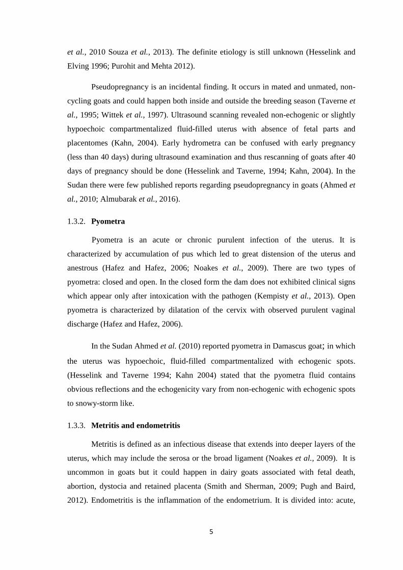
et al., 2010 Souza et al., 2013). The definite etiology is still unknown (Hesselink and
Elving 1996; Purohit and Mehta 2012).
Pseudopregnancy is an incidental finding. It occurs in mated and unmated, non-
cycling goats and could happen both inside and outside the breeding season (Taverne et
al., 1995; Wittek et al., 1997). Ultrasound scanning revealed non-echogenic or slightly
hypoechoic compartmentalized fluid-filled uterus with absence of fetal parts and
placentomes (Kahn, 2004). Early hydrometra can be confused with early pregnancy
(less than 40 days) during ultrasound examination and thus rescanning of goats after 40
days of pregnancy should be done (Hesselink and Taverne, 1994; Kahn, 2004). In the
Sudan there were few published reports regarding pseudopregnancy in goats (Ahmed et
al., 2010; Almubarak et al., 2016).
1.3.2. Pyometra
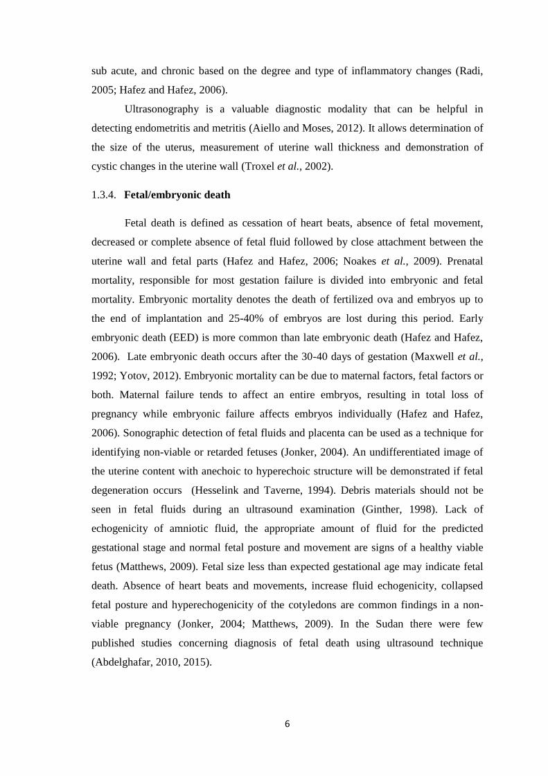
Pyometra is an acute or chronic purulent infection of the uterus. It is
characterized by accumulation of pus which led to great distension of the uterus and
anestrous (Hafez and Hafez, 2006; Noakes et al., 2009). There are two types of
pyometra: closed and open. In the closed form the dam does not exhibited clinical signs
which appear only after intoxication with the pathogen (Kempisty et al., 2013). Open
pyometra is characterized by dilatation of the cervix with observed purulent vaginal
discharge (Hafez and Hafez, 2006).
In the Sudan Ahmed et al. (2010) reported pyometra in Damascus goat; in which
the uterus was hypoechoic, fluid-filled compartmentalized with echogenic spots.
(Hesselink and Taverne 1994; Kahn 2004) stated that the pyometra fluid contains
obvious reflections and the echogenicity vary from non-echogenic with echogenic spots
to snowy-storm like.
1.3.3. Metritis and endometritis
Metritis is defined as an infectious disease that extends into deeper layers of the
uterus, which may include the serosa or the broad ligament (Noakes et al., 2009). It is
uncommon in goats but it could happen in dairy goats associated with fetal death,
abortion, dystocia and retained placenta (Smith and Sherman, 2009; Pugh and Baird,
2012). Endometritis is the inflammation of the endometrium. It is divided into: acute,
6
sub acute, and chronic based on the degree and type of inflammatory changes (Radi,
2005; Hafez and Hafez, 2006).
Ultrasonography is a valuable diagnostic modality that can be helpful in
detecting endometritis and metritis (Aiello and Moses, 2012). It allows determination of
the size of the uterus, measurement of uterine wall thickness and demonstration of
cystic changes in the uterine wall (Troxel et al., 2002).
1.3.4. Fetal/embryonic death
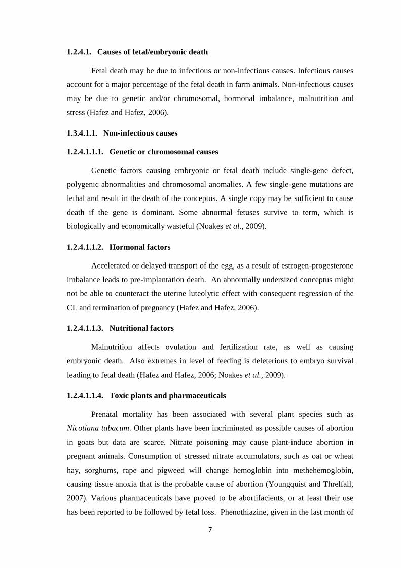
Fetal death is defined as cessation of heart beats, absence of fetal movement,
decreased or complete absence of fetal fluid followed by close attachment between the
uterine wall and fetal parts (Hafez and Hafez, 2006; Noakes et al., 2009). Prenatal
mortality, responsible for most gestation failure is divided into embryonic and fetal
mortality. Embryonic mortality denotes the death of fertilized ova and embryos up to
the end of implantation and 25-40% of embryos are lost during this period. Early
embryonic death (EED) is more common than late embryonic death (Hafez and Hafez,
2006). Late embryonic death occurs after the 30-40 days of gestation (Maxwell et al.,
1992; Yotov, 2012). Embryonic mortality can be due to maternal factors, fetal factors or
both. Maternal failure tends to affect an entire embryos, resulting in total loss of
pregnancy while embryonic failure affects embryos individually (Hafez and Hafez,
2006). Sonographic detection of fetal fluids and placenta can be used as a technique for
identifying non-viable or retarded fetuses (Jonker, 2004). An undifferentiated image of
the uterine content with anechoic to hyperechoic structure will be demonstrated if fetal
degeneration occurs (Hesselink and Taverne, 1994). Debris materials should not be
seen in fetal fluids during an ultrasound examination (Ginther, 1998). Lack of
echogenicity of amniotic fluid, the appropriate amount of fluid for the predicted
gestational stage and normal fetal posture and movement are signs of a healthy viable
fetus (Matthews, 2009). Fetal size less than expected gestational age may indicate fetal
death. Absence of heart beats and movements, increase fluid echogenicity, collapsed
fetal posture and hyperechogenicity of the cotyledons are common findings in a non-
viable pregnancy (Jonker, 2004; Matthews, 2009). In the Sudan there were few
published studies concerning diagnosis of fetal death using ultrasound technique
(Abdelghafar, 2010, 2015).
7
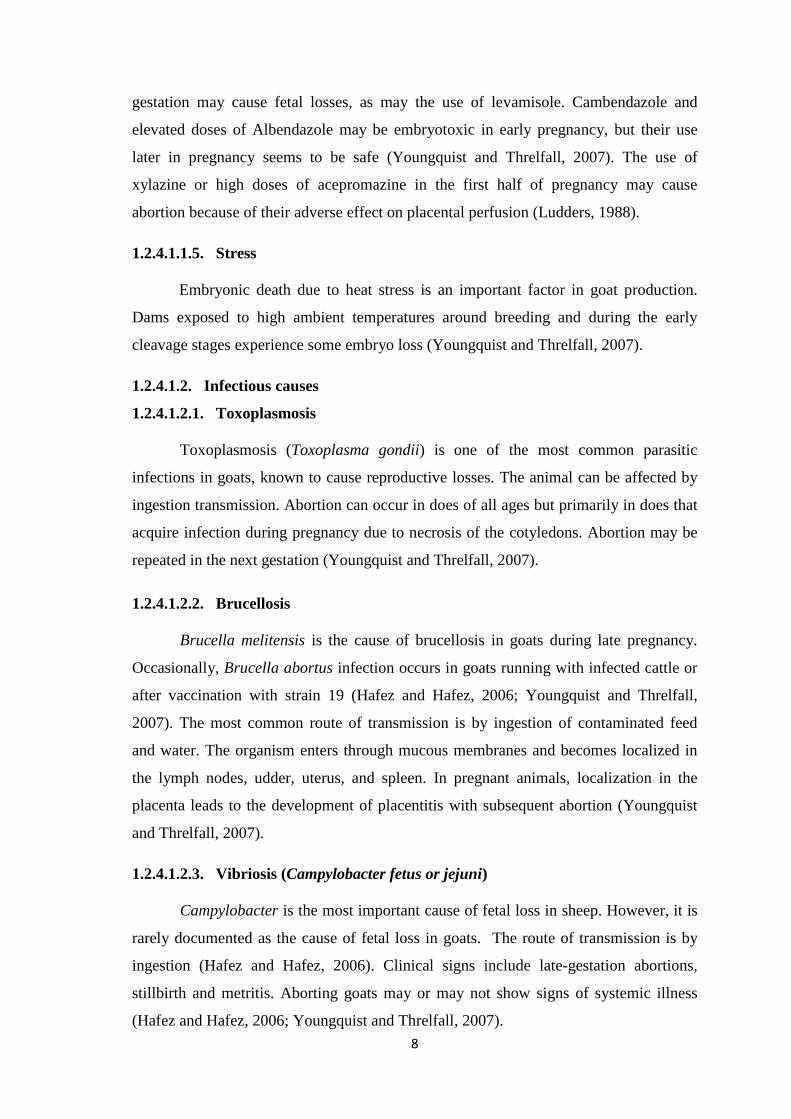
1.2.4.1. Causes of fetal/embryonic death
Fetal death may be due to infectious or non-infectious causes. Infectious causes
account for a major percentage of the fetal death in farm animals. Non-infectious causes
may be due to genetic and/or chromosomal, hormonal imbalance, malnutrition and
stress (Hafez and Hafez, 2006).
1.3.4.1.1. Non-infectious causes
1.2.4.1.1.1. Genetic or chromosomal causes
Genetic factors causing embryonic or fetal death include single-gene defect,
polygenic abnormalities and chromosomal anomalies. A few single-gene mutations are
lethal and result in the death of the conceptus. A single copy may be sufficient to cause
death if the gene is dominant. Some abnormal fetuses survive to term, which is
biologically and economically wasteful (Noakes et al., 2009).
1.2.4.1.1.2. Hormonal factors
Accelerated or delayed transport of the egg, as a result of estrogen-progesterone
imbalance leads to pre-implantation death. An abnormally undersized conceptus might
not be able to counteract the uterine luteolytic effect with consequent regression of the
CL and termination of pregnancy (Hafez and Hafez, 2006).
1.2.4.1.1.3. Nutritional factors
Malnutrition affects ovulation and fertilization rate, as well as causing
embryonic death. Also extremes in level of feeding is deleterious to embryo survival
leading to fetal death (Hafez and Hafez, 2006; Noakes et al., 2009).
1.2.4.1.1.4. Toxic plants and pharmaceuticals
Prenatal mortality has been associated with several plant species such as
Nicotiana tabacum. Other plants have been incriminated as possible causes of abortion
in goats but data are scarce. Nitrate poisoning may cause plant-induce abortion in
pregnant animals. Consumption of stressed nitrate accumulators, such as oat or wheat
hay, sorghums, rape and pigweed will change hemoglobin into methehemoglobin,
causing tissue anoxia that is the probable cause of abortion (Youngquist and Threlfall,
2007). Various pharmaceuticals have proved to be abortifacients, or at least their use
has been reported to be followed by fetal loss. Phenothiazine, given in the last month of
8
gestation may cause fetal losses, as may the use of levamisole. Cambendazole and
elevated doses of Albendazole may be embryotoxic in early pregnancy, but their use
later in pregnancy seems to be safe (Youngquist and Threlfall, 2007). The use of
xylazine or high doses of acepromazine in the first half of pregnancy may cause
abortion because of their adverse effect on placental perfusion (Ludders, 1988).
1.2.4.1.1.5. Stress
Embryonic death due to heat stress is an important factor in goat production.
Dams exposed to high ambient temperatures around breeding and during the early
cleavage stages experience some embryo loss (Youngquist and Threlfall, 2007).
1.2.4.1.2. Infectious causes
1.2.4.1.2.1. Toxoplasmosis
Toxoplasmosis (Toxoplasma gondii) is one of the most common parasitic
infections in goats, known to cause reproductive losses. The animal can be affected by
ingestion transmission. Abortion can occur in does of all ages but primarily in does that
acquire infection during pregnancy due to necrosis of the cotyledons. Abortion may be
repeated in the next gestation (Youngquist and Threlfall, 2007).
1.2.4.1.2.2. Brucellosis
Brucella melitensis is the cause of brucellosis in goats during late pregnancy.
Occasionally, Brucella abortus infection occurs in goats running with infected cattle or
after vaccination with strain 19 (Hafez and Hafez, 2006; Youngquist and Threlfall,
2007). The most common route of transmission is by ingestion of contaminated feed
and water. The organism enters through mucous membranes and becomes localized in
the lymph nodes, udder, uterus, and spleen. In pregnant animals, localization in the
placenta leads to the development of placentitis with subsequent abortion (Youngquist
and Threlfall, 2007).
1.2.4.1.2.3. Vibriosis (Campylobacter fetus or jejuni)
Campylobacter is the most important cause of fetal loss in sheep. However, it is
rarely documented as the cause of fetal loss in goats. The route of transmission is by
ingestion (Hafez and Hafez, 2006). Clinical signs include late-gestation abortions,
stillbirth and metritis. Aborting goats may or may not show signs of systemic illness
(Hafez and Hafez, 2006; Youngquist and Threlfall, 2007).
9
1.2.4.1.2.4. Salmonellosis
Salmonella which transmitted through ingestion causes abortion, metritis, and
systemic illness in does. Salmonella abortus-ovis is first implicated as a cause of fetal
death in late stages and neonatal mortality. The organism has been isolated from goat
fetuses and placentas in France (Hafez and Hafez, 2006; Youngquist and Threlfall,
2007).
1.2.4.1.2.5. Listeriosis
Listeriosis most commonly causes meningioencephalitis but can also cause
pregnancy loss in goats at early gestation. Later, infection results in stillbirth or weak
kids. Listeria may be found in soil, water, plant litter, silage, and the digestive tract.
Abortions have been reported on farms on which the goats were fed hay with the
addition of concentrate (Hafez and Hafez, 2006; Youngquist and Threlfall, 2007).
1.2.4.1.2.6. Enzootic abortion of ewe (Chlamydia psittaci)
Chlamydiosis is the most common cause of infectious pregnancy wastage in late
gestation in goats and it is transmitted by ingestion (Hafez and Hafez, 2006; Youngquist
and Threlfall, 2007). This disease must be differentiated from vibriosis. Diagnosis of
Chlamydiosis depends on finding elementary bodies of the infectious agent in smear
from the cotyledon or placenta exudates (Hafez and Hafez, 2006).
1.2.4.1.2.7. Rift Valley Fever
Rift Valley fever is a per acute or acute, mosquito-borne, zoonotic disease of
domestic and wild ruminants in Africa. This virus belongs to the genus phlebovirus and
is a typical Bunya virus which transmitted by insects (Hafez and Hafez, 2006). Large
outbreaks of clinical disease are usually associated with heavy rainfall and localized
flooding. During epidemics, the occurrence of abortions in livestock and deaths among
young animals, particularly lambs, together with an influenza-like disease in human, is
characteristic. However, infections are frequently subclinical or mild (Aiello and Moses,
2012).
10
1.2.5. Outcome of fetal death
The exact outcome of early fetal mortality is unpredictable and is influenced by
several factors, including the cause, differences in pregnancy between species, stage of
gestation at fetal death and number of fetuses (Lefebvre, 2015).
Fetal death may be followed by mummification, maceration or abortion. In cases
of abortion and fetal maceration, the hormonal support of pregnancy is lost (Aiello and
Moses, 2012).
1.2.5.1. Mummification
Fetal mummification is characterized by fetal death, failure of abortion,
resorption of placental fluids, dehydration of the fetus and its membranes and involution
of the uterus (Hafez and Hafez, 2006). If fetal death occurs after ossification has begun
fetal resorption cannot take place (Hafez and Hafez, 2006; Noakes et al., 2009).
Fetal mummification is occasionally diagnosed in almost all domestic species,
including the goat with the highest prevalence occurring in the swine. It is most often
found in polytocous species but it can also occur in monotocous ones (Lefebvre, 2015).
Diagnosis of mummified fetuses is straight forward and can be done by transrectal
palpation and ultrasound. Using ultrasound technique the mummified fetus appear as a
compact, firm and immobile mass without fetal fluids or placentomes with the general
health of the dam being an affected (Lefebvre et al., 2009).
1.2.5.2. Maceration
Fetal maceration defined as disintegration of a fetus that occurs at any stage of
gestation in all animal species (Deori et al., 2015). Fetal maceration is uncommon in
goats (Rautela et al., 2016). This generally occurs in the event of fetal death after
formation of the fetal bones beyond 100 days of gestation in such animals failed to abort
or parturition (Givens and Marley, 2008; Purohit et al., 2011). Bacteria enter the uterus
through the dilated cervix and the soft tissues are digested by combination of
putrefaction and autolysis leaving a mass of bones in the uterus (Noakes et al., 2009;
Deori et al., 2015; Rautela et al., 2016). Ultrasonographic examination of the uterus
reveals hyperechoic bone shadows, absence of fluid contrast and loss of fetal
architecture (Matthews, 2009).
11
1.2.5.3. Prolonged gestational period
Prolonged gestational periods occur as a result of genetic and non genetic
factors. The gestation is prolonged for periods ranging from three weeks to three
months. Dystocia usually occurs and the fetuses should be delivered by caesarean
section (Hafez and Hafez, 2006).
1.2.5.4. Still birth/still born
Still birth is a fetus that has matured in utero but is born dead. It is of infectious
or non-infectious origin. Infectious agents affect fetuses; usually die before the end of
gestation and around the prenatal period. Most common non-infectious causes include
hypoxia secondary to umbilical rupture or impairment of umbilical blood flow, high
atmospheric carbon monoxide concentration, high temperatures, and prolonged
parturition (Youngquist and Threlfall, 2007).
1.2.5.5. Abortion
Abortion is termination of pregnancy with the expulsion of immature fetus,
either live or dead, before the completion of gestational period as a result of the failure
of the mechanisms that control pregnancy (Youngquist and Threlfall, 2007; Noakes et
al., 2009). Abortion may be spontaneous or induced, infectious or non-infectious (Hafez
and Hafez, 2006).
Major infectious agents of abortion in goats are Chlamydia, Toxoplasma,
Leptospira, Brucella, Coxiella burnetii, and Listeria. Non infectious causes of abortion
may be genetic, chromosomal, hormonal and nutritional. Nutritional factors include
plant toxins, such as broom weed or loco weed poisoning; dietary deficiencies of
copper, selenium, vitamin A and magnesium. Certain drugs such as estrogen,
glucocorticoids, phenothiazine, carbon tetrachloride and levamisole also cause abortion
(Hafez and Hafez, 2006; Aiello and Moses, 2012).
1.2.6. Treatment of fetal death
1.2.6.1. PGF2α
Prostaglandins are secreted by almost all body tissues. PGF2α is the natural
luteolytic agent that terminates the luteal phase of the estrous cycle and allows for the
launch of a new one in the absence of pregnancy (Hafez and Hafez, 2006).
12
Administration of PGF2α and its analogs is an efficient treatment of fetal death in
goats (Purohit et al., 2011; Ali, 2011). It is a drug of choice for induction of abortion
resulting in more number of live births when parturition is near. It is efficient in
terminating pregnancy throughout gestational period in goats (Purohit et al., 2011).
PGF2α causes contractions of the uterus (Hafez and Hafez, 2006; Palliser et al., 2006).
The goat depends on corpus-luteum-derived progesterone throughout pregnancy and is
thus susceptible to luteolytic agents, including prostaglandins, throughout the whole
period of pregnancy (Matthews, 2009). In goats, administration of synthetic
prostaglandins will usually cause luteolysis and termination of a pregnancy within 24-
48 hours from day 5 of gestation till term (Purohit et al., 2011). In small ruminants, Ali
(2011) reported that administration of PGF2a intramuscularly will cause cervical
dilatation within 72 hours.
1.2.6.2. Dexamethazone
In domestic animals, pregnancy can be terminated at any stages using single or
repeated injections of dexamethasone (Purohit et al., 2011).
1.2.6.3. Estradiol
In domestic species, estradiol can be administered to relax the cervix and expel
the dead fetus (Ghosh et al., 1992; Wu et al., 2004; Lefebvre, 2015).
1.2.7. Subsequent fertility
In farm animals, the subsequent fertility of fetal death has not been reported
except for very few studies (Lefebvre et al., 2009). In small ruminants, Brounts et al.
(2004) reported that animals that were pregnant with fetal death and delivered using
caesarean section became pregnant after rebreeding. In cattle, Drost, (2007) stated that
the prognosis of induced abortion in case of macerated fetuses is always poor because of
the severe endometrial damage. Kumaresan et al. (2013) stated that PGF2α in
conjunction with estradiol for treatment of mummified fetuses in cattle would not affect
future fertility.
1.2.8. Prevalence of fetal death worldwide and possible risk factors
Engeland et al. (1998) reported a prevalence of fetal death (11.1%) in dairy
goats in Norway. They reported that, fetal loss associated significantly with many risk
13
factors including increased age, building design and combined feeding. Other diseases
and routine flock management procedures were not found to be significant. In abattoir
study, Bokko (2011) stated a prevalence of 17.88 % fetal death in goats in the Sahel
region of Nigeria. According to their study, higher frequencies of fetal death occurred in
the dry season. The slaughter mediated fetal loss incidence was highest in the first
trimester. Idahor (2013) reported a prevalence of 50% of fetal death in does in
Nasarawa State in Nigeria. He observed a higher rate of fetal death in West African
Dwarf breed than Red Sokoto breed. Samir et al. (2015) found a prevalence of 41.28%
embryonic and fetal losses in goats.
1.2.9. Ultrasonography
1.2.9.1. Sound waves, definition and spectrum
Sound is a mechanical energy transmitted by pressure waves in a medium. It
propagates through a material medium; it cannot travel through a vacuum (Zagzebski,
1996). Acoustic waves are classified according to their frequency. 20 Hz-20 KHz is an
audible sound and most human beings can hear sound within this range. A sound wave
whose frequency is greater than 20 KHz is termed ultrasound; while mechanical
vibrations whose frequencies are below the audible range are termed infrasound
(Zagzebski, 1996).
1.2.9.2. The transducer
A transducer is a device that converts one form of energy to another; in medical
ultrasound, the transducer converts electrical energy to mechanical energy and vice-
versa (Hendee and Ritenour, 2002). Ultrasound transducers use piezoelectricity
principle discovered by Curie and Curie (1880). Piezoelectric materials have the unique
ability to respond to the action of an electric field by changing shape. They also have
the property of generating electrical potentials when compressed. Changing the polarity
of a voltage applied to the transducer changes the transducer thickness, which expands
and contracts as the polarity changes. This results in the generation of mechanical
pressure waves that can transmit into the body (Rumack et al., 2011). The current
technology uses a transducer composed of multiple elements, usually produced by
precise slicing of a piece of piezoelectric material into numerous small units, each with
its own electrodes. Such transducer arrays may be form into various configurations; i.e.
linear, curved, phased or annular arrays (Fulton, 2014). The individual elements that are

14
arranged in a linear fashion producing a rectangular image-shaped display which is well
suited for scanning of small parts, vascular and obstetrics applications (Fulton, 2014).
Linear arrays have been configured into convex curves to produce an image that
combines a relatively large surface field of view with a sector display format i.e.
trapezoid-image shaped field (Zagzebski, 1996). They are used for a variety of
applications and the larger one serving for general abdominal, obstetric and
transabdominal pelvic scanning. The small, high frequency curved array scanners are
used for transvaginal and transrectal probes for pediatric imaging. In contrast to
mechanical sector scanners, phased array has no moving parts. A sector field of view is
produced by multiple transducer elements fired in precise sequence under electric
control. These transducers are particularly useful for intercostal scanning to evaluate the
heart, liver and spleen or in other areas where access is limited (Fulton, 2014). It
produces a triangular-image shaped (Zagzebski, 1996).
1.2.9.3. Principal echo display modes
1.2.9.3.1. Amplitude modulation (A-mode)
Amplitude modulation is indicated by the height of the vertical deflection
displayed on the oscilloscope. With A-mode ultrasound, only the position and strength
of a reflecting structure are recorded (Rumack et al., 2011). It is used where accurate
measurements are needed as in ophthalmological studies (Zagzebski, 1996).
1.2.9.3.2. Brightness modulation (B-mode)
The mainstay of imaging with ultrasound is provided by real-time gray-scale, B-
mode display, in which variation in display intensity or brightness are used to indicate
reflected signals of different amplitude (Hagen-Ansert, 2012). To generate a two-
dimension (2-D) image, multiple ultrasound pulses are sent down a series of successive
scan lines, building a 2-D representation of echoes arising from the object being
scanned (Hangiandreou, 2003; Abu-Zidan et al., 2011; Shriki, 2014).
1.2.9.3.3. Motion modulation (M-mode)
It displays echo amplitude and shows the position of moving reflectors. M-mode
imaging uses the brightness of the display to indicate the intensity of the reflected
signal. The major application of M-mode is evaluation of the rapid motion of cardiac
valves, chambers and vessels wall (Rumack et al., 2011).
15
1.2.9.4. Sonographic echo texture
Echo texture is defined as a reflection that returns from the tissue to the
transducer. Display of specular interface is highly dependent on the angle of insonation
(exposure to ultrasound waves). Specular reflectors return echoes to the transducer only
if the sound beam is perpendicular to the interface. If interface is not at a 90º to the
sound beam, it will be reflected away from the transducer, and echo will not be detected
(Rumack et al., 2011).
Most echoes in the body do not arise from specular reflectors but it is rather
arise from interfaces within solid organs. If large and relatively smooth, the interface
reflects sound much as a mirror reflects light. Thus, sound striking reflect directly back
to the transducer resulting in a strong echo and a portion of the waves is reflected by
tissue interfaces, while other parts of the waves are propagated (Zagzebski, 1996;
Rumack et al., 2011). Images are displayed as two-dimensional maps of grey-scale
based on location and strength of the echoes returning from the tissue interfaces. Grey-
scale images are composed of thousands of picture elements, known as pixels
(Zagzebski, 1996).
The terms anechoic, hypoechoic, hyperechoic and echogenic are related to the
assessment of tissue echogenicity. Echogenic refers to the ability to reflect ultrasound
waves in the context of surrounding tissues. Anechoic is a reflection that produces from
the lacking structures such as a cyst containing fluids. Hypoechoic is defined as a less
echogenic tissues such as smooth muscle with the rectal wall and structures frequently
contain fluids. Hyperechoic is a more reflection which produced from tissues such as fat
or fascia (Cochlin et al., 1994).
1.2.9.5. Different ultrasound techniques in goats
Three types of transducers are available which include transabdominal,
transrectal and transvaginal (Ayres et al., 1999; Hafez and Hafez, 2006; Youngquist and
Threlfall, 2007; Noakes et al., 2009). Transabdominal scanning is applied to the skin
surface of the abdomen just cranial to the udder (Ayres et al., 1999). This is generally
used for examinations made more than 35 days after breeding and can be also used
between 80-100 days to differentiate between single and multiple pregnancy, although
the accuracy in identifying the precise number of fetuses is poor (Youngquist and
Threlfall, 2007; Noakes et al., 2009). Transrectal scanning is done by inserting the
lubricated probe into the rectal and slowly rotating it from side to side. This is usually
16
reserved for diagnosis at less than 30-35 days after breeding. A poor quality image or no
image is usually occurs due to feces trapped under the probe. Transvaginal approach
using a cylindrical sectorial probe with variable angle head can be used to diagnose
pregnancy from the cranial vagina (Ayres et al., 1999).
1.2.9.6. Ultrasound bioeffects and hazards
Ultrasound has provided a wealth of knowledge in diagnostic medicine and has
greatly impacted medical practice, particularly obstetrics. It remains one of the fastest-
growing imaging modalities because it is cheap, real-time, portable with no apparent
adverse effects (Rumack et al., 2011). At present, the U.S. Food and Drug
Administration (FDA) regulate the maximum output of ultrasound devices to an
established level. The physical effects of sound can be divided into two types: thermal
and non-thermal. The thermal effects caused by ultrasound are similar to those of any
localized heat source (Rumack et al., 2011).
1.2.9.6.1. Thermal effect
There is no evidence that diagnostic ultrasound produces harm in dams or the
developing fetus when used properly. Up to 8 weeks of pregnancy is the period when
cell damage might lead to developmental changes or anomalies. Increased
mineralization of developing bones will increase potential for heating of the brain and
spinal cord (Nelson et al., 2009). Bones have high attenuation of ultrasound beam.
When gestation is progressing there is increased degree of bone mineralization, thereby
increasing risk of localized heating as in measurement of the Bioparietal diameter of the
skull (Rumack et al., 2011).
1.2.9.6.2. Non- thermal effects (cavitation)
Cavitation is defined as interaction of ultrasound with gas bodies. Acoustic
cavitation inception is demarcated by a specific threshold value: the minimum acoustic
pressure necessary to initiate the growth of a cavity in a fluid during the rarefaction
phase of the cycle. This threshold is affected by many factors including cavitation
nucleus size, acoustic pulse characteristics, ambient hydrostatic pressure and host fluid
parameters (Rumack, et al., 2011).
Non-thermal bioeffects are recently defined as those resulting from exposures in
which the temperature rises by less than 1° C above normal physiological levels

17
(Stratmeyer et al., 2008). These effects arise either directly from the interaction with the
sound field or as a consequence of this interaction. The action of an ultrasound wave on
a bubble will give rise to small-scale circulation in the fluid surrounding it, i.e. micro
streaming. Induced localized shear stresses can lead to changes in the cell membrane if
a bubble is close to it. Collapsing bubbles have a highly destructive effect on cells
nearby. Shock waves may be produced, leading to high stresses locally, which can
distort and lyses cells (Haar, 2010). Histological effects on mammalian and embryo
tissues may occur after irradiation which may lead to damage in the walls of blood
vessels (Hill et al., 2004).
18
CHAPTER TWO
MATERIALS AND METHODS
2.1. Materials
2.1.1. Animals
962 female goats of different breeds (Saanen, Sannen crosses, Damascus and
local breeds) were involved in the present study.
2.1.2. Study design, location and duration
A cross sectional study was performed on goats that were presented to the
Veterinary clinic, College of Veterinary Medicine, Sudan University of Science and
Technology (SUST) from different localities of Khartoum State (Khartoum, Um Bada,
Bahri, Sharq El Nile, Karari and Um Durman) from October, 2015 – October, 2016.
Khartoum state lies in the central Sudan in the semi-arid zone between longitudes 31.5-
34º east and latitude 15-16º north. The Veterinary clinic received goats from almost all
localities of Khartoum State; hence it was selected for the conduction of the present
study.
2.1.3. Sample size determination
The sample size was calculated using the formula 4PQ/L2 given by (Martin et
al., 1988) where:
P = Prevalence
Q = 1-P
L2 = allowable error, with 41.28% prevalence reported by (Samir et al., 2015) and 5%
allowable error. Accordingly the sample size was determined to be 388.
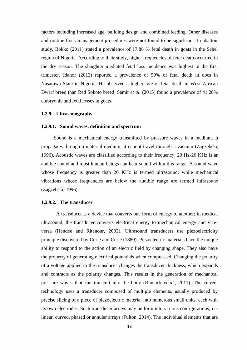
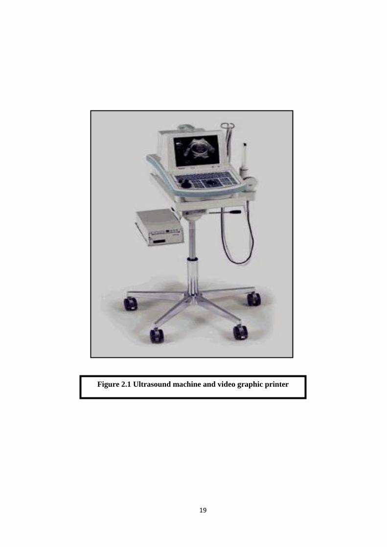
2.1.4. Ultrasound machine and image recorder
A real-time ultrasound scanner (Pie-medical easote, Aquila, Holland) equipped
with switchable frequency (3.5-5) MHz convex transducer was used in the current study
and images were printed in thermal papers using video graphic printer UP-895EC,
Sony-Japan (figure 2.1).
19
Figure 2.1 Ultrasound machine and video graphic printer
20
2.2. Methods
2.2.1. Ultrasound scanning
2.2.1.1. Animal preparation
Area of scanning which extend from one side of the udder to the other and 15
cm cranial to the udder (Goddard, 1996) was clipped and shaved appropriately.
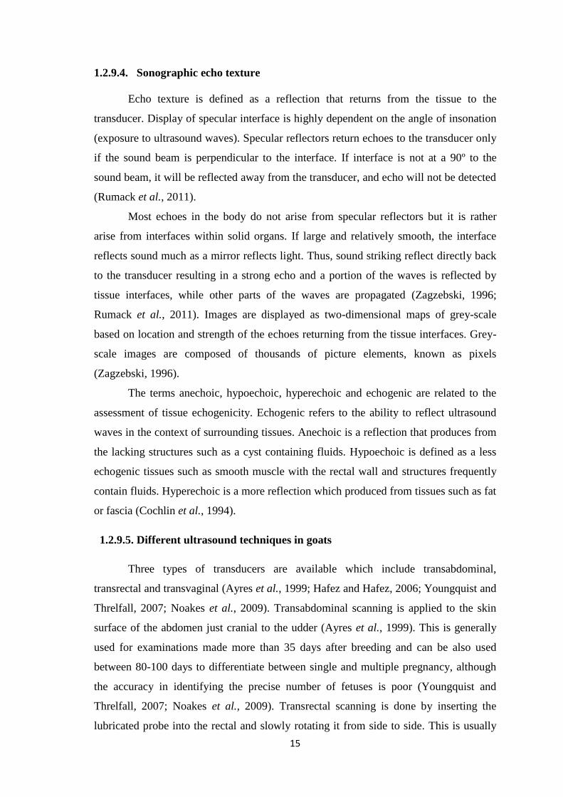
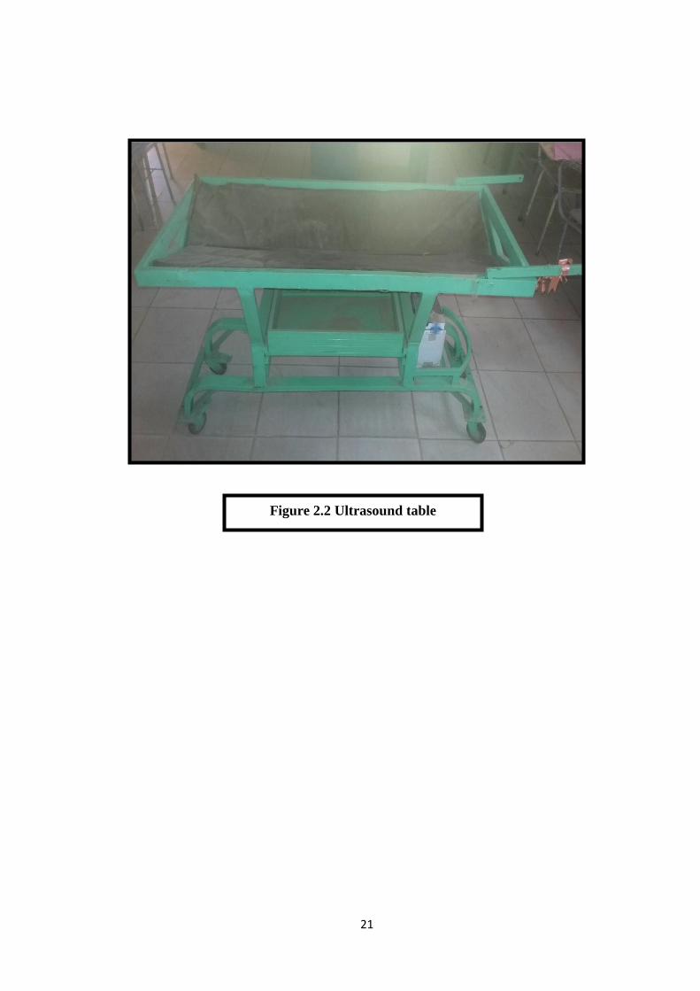
2.2.1.2. Animal positioning
Animals were put on a dorsal recumbency on especially designed table (Figure
2.2). Copious amount of water soluble, hypoallergenic gel (Aquasonic-Turkey) was
administered to the area prior to the scanning.
2.2.1.3. Ultrasound technique
Carful transabdominal scanning was done and all possible sections were taken
to ensure accurate diagnosis.
2.2.2. Questionnaire
A well prepared questionnaire was constructed for collection of all data seems to
have a relationship to the subject under investigation. Season of the year, locality, breed
of the dam, age of the dam, rearing system, parity number, feeding type, oestrous type
and breed of the inseminating buck were all registered as assumed risk factors for fetal
death according to previous studies. All data concerning the owners of the goats under
investigation were also recorded.
2.2.3. Clinical examination and age determination
A full clinical examination was carried out. Vital parameters such as
temperature, respiratory rate and heart rate were all taken. The os cervix was also
examined. Determination of the age of the goats was done (Ebert and Solaiman, 2010).
21
Figure 2.2 Ultrasound table
22
2.2.4. Treatment
Out of 36 goats with dead fetuses; only 14 goats were treated. Animals which do
not respond to the medical treatment were referred for surgical intervention.
2.2.4.1. Medical treatment
Goats were treated using 0.5 ml single intramuscular injection of 250 µg
cloprostenol (Estrumate, Schering-Plough Animal Health, Germany) and 5 ml 5%
oxytetracycline for five consecutive days (Purohit et al., 2011)
2.2.4.2. Caesarean section
Caesarean section and hysterectomy were performed for goats that did not
respond to medical treatment according to (Hichman et al., 1995).
2.2.5. Subsequent fertility
Follow up of the treated animals was made. Expulsion of dead fetuses and return
to estrus, as well as pregnancy diagnosis and delivery of the treated dams were also
followed and registered.
2.2.6. Statistical analysis
Collected data were analyzed using Statistical Packages for Social Sciences
(SPSS) version 22.0 (SPSS Inc, Chicago, USA). Descriptive statistics of the examined
variables were obtained. Univariate analysis using chi square test (χ2) was done. A level
of ≤ 5% was considered significant.
23
CHAPTER THREE
RESULTS
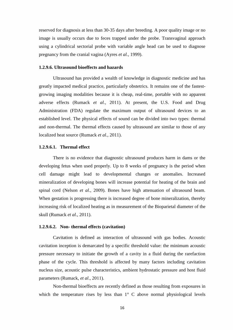
3.1. Sonographic pregnancy diagnosis
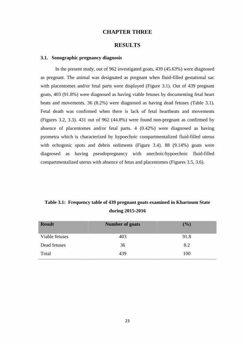
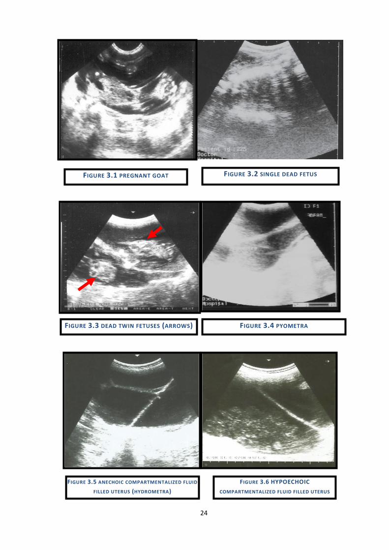
In the present study, out of 962 investigated goats, 439 (45.63%) were diagnosed
as pregnant. The animal was designated as pregnant when fluid-filled gestational sac
with placentomes and/or fetal parts were displayed (Figure 3.1). Out of 439 pregnant
goats, 403 (91.8%) were diagnosed as having viable fetuses by documenting fetal heart
beats and movements. 36 (8.2%) were diagnosed as having dead fetuses (Table 3.1).
Fetal death was confirmed when there is lack of fetal heartbeats and movements
(Figures 3.2, 3.3). 431 out of 962 (44.8%) were found non-pregnant as confirmed by
absence of placentomes and/or fetal parts. 4 (0.42%) were diagnosed as having
pyometra which is characterized by hypoechoic compartmentalized fluid-filled uterus
with echogenic spots and debris sediments (Figure 3.4). 88 (9.14%) goats were
diagnosed as having pseudopregnancy with anechoic/hypoechoic fluid-filled
compartmentalized uterus with absence of fetus and placentomes (Figures 3.5, 3.6).
Table 3.1: Frequency table of 439 pregnant goats examined in Khartoum State
during 2015-2016
Result Number of goats (%)
Viable fetuses 403 91.8
Dead fetuses 36 8.2
Total 439 100
24
FIGURE 3.4 PYOMETRA FIGURE 3.3 DEAD TWIN FETUSES (ARROWS)
FIGURE 3.6 HYPOECHOIC
COMPARTMENTALIZED FLUID FILLED UTERUS
(MUCOMETRA)
FIGURE 3.5 ANECHOIC COMPARTMENTALIZED FLUID
FILLED UTERUS (HYDROMETRA)
FIGURE 3.2 SINGLE DEAD FETUS FIGURE 3.1 PREGNANT GOAT
25
3.2. Prevalence of fetal death and associated risk factors
The prevalence of fetal death in goats in Khartoum State was found to be 8.2%
(Table 3.1). Out of nine examined risk factors; two factors (rearing system and type of
estrus) were not statistically analyzed because they were constant.
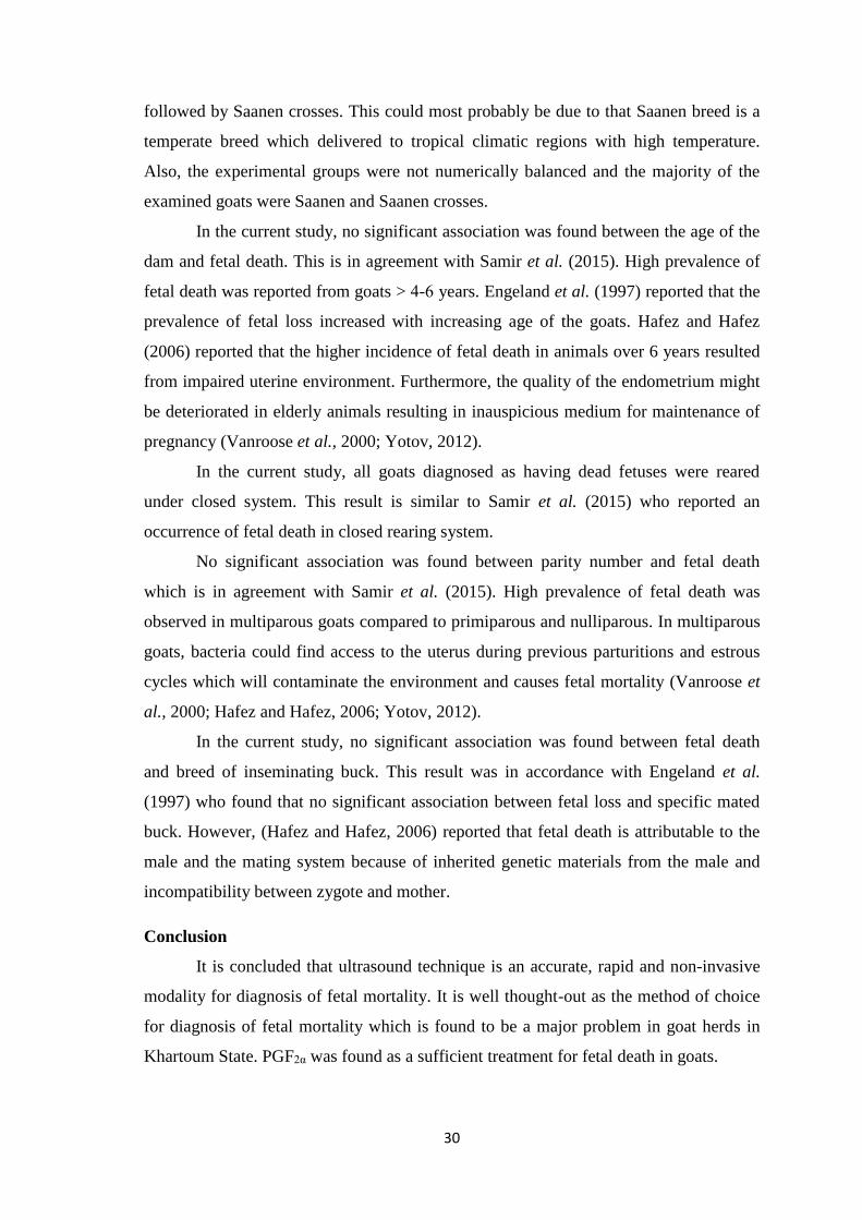
3.2.1. Season of the year
No significant association (χ2 = 4.873, P-value = 0.087) was found between
season of the year and fetal death. High prevalence (13%) of fetal death was reported
from dry summer season followed by winter (6.7%) and wet summer (6.2%) seasons
(Table 3.2).
3.2.2. Locality
In the present study, no significant association (χ2 = 4.398, P-value = 0.494) was
found between locality and fetal death. High prevalence (13.3%) of fetal death was
recorded from Um Durman (Table 3.2).
3.2.3. Breed of the dam
In the present study, no significant association (χ2 = 7.147, P-value = 0.067) was
found between breed of the dam and fetal death. High occurrence of fetal death was
reported from Saanen (11%) followed by local breeds (Table 3.2).
3.2.4. Age of the dam
No significant association (χ2 = 2.566, P-value = 0.277) was found between age
of the dam and occurrence of fetal death. Higher prevalence (11.8%) of fetal death was
reported from goats aged > 4-6 years (Table 3.2).
3.2.5. Parity number
No significant association (χ2 = 3.024, P-value = 0.221) was found between
parity number and fetal death. High prevalence (10.3%) of fetal death was reported
from multiparous goats followed by nulliparous goats (6.7%). However, the least
occurrence (3.8%) of fetal death was recorded from primiparous goats (Table 3.2).
26
3.2.6. Feeding type
No significant association (χ2 = 0.728, P-value = 0.50) was found between type
of feeding and the occurrence of fetal death. The occurrence of fetal death (8.4%) was
only observed in animals that were fed on roughages and concentrates (Table 3.2).
3.2.7. Breed of the inseminating buck
No significant association (χ2 = 0.560, P-value = 0.756) was found between
breed of the inseminating buck and fetal death. High occurrence (9%) of fetal death was
reported from goats that were mated with Saanen buck followed by Damascus (6.1%)
and local breed (5.6%) bucks as shown in Table 3.2.
3.3. Clinical examination
Temperature, respiratory rate and fetal heartbeats were found to be within the
normal physiological range in 438 goats. One goat had elevated temperature (41.8 C˚),
increased heart rate (103 bpm) and respiratory rate (44 cycle/min). The Os cervix was
found closed in all goats except the one with abnormal vital parameters.
3.4. Treatment
Thirteen goats were subjected to medical treatment using 0.5 ml intramuscular
injection of 250 µg cloprostenol (Estrumate, Schering-Plough animals Health,
Germany) and 5 ml 5% oxytetracycline for five consecutive days. After treatment, 11
goats responded successfully to the treatment as judged by dilatation of the cervix and
abortion of fetuses after 48-96 hours. The animals returned to estrus and the animal’s
owners were advised to breed their goats at the next coming estrous cycle. Two full
term goats with fetal death did not respond to the treatment and the cervix was not
dilated. Another dose of PGF2α was administered. The goats did not respond to the
treatment for the second time and hence were referred to surgery. Regarding the 14th
goat which had dilated cervix, more than a few attempts were made to deliver the goat
but all attempts were unsuccessful. Supportive treatment and broad spectrum antibiotic
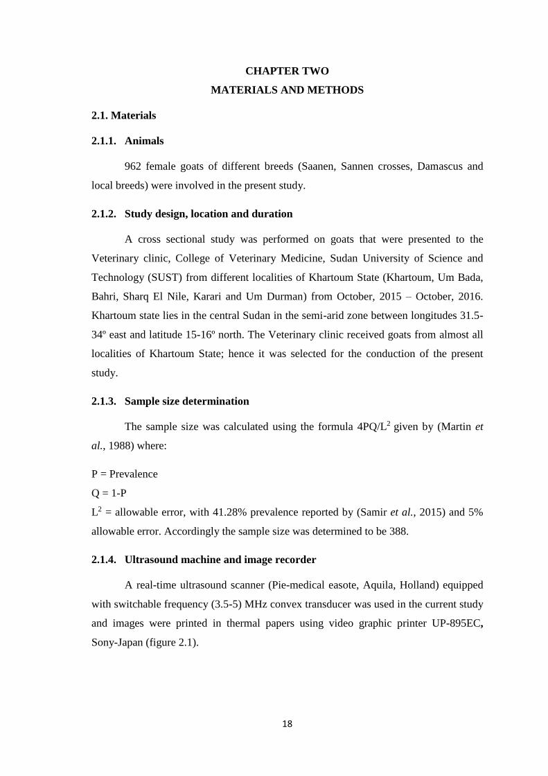
injection was administered to the goat and also referred to surgery. Cesarean section and
hysterectomy were performed for the three full term goats and fetuses were removed
(Figure 3.7).
27
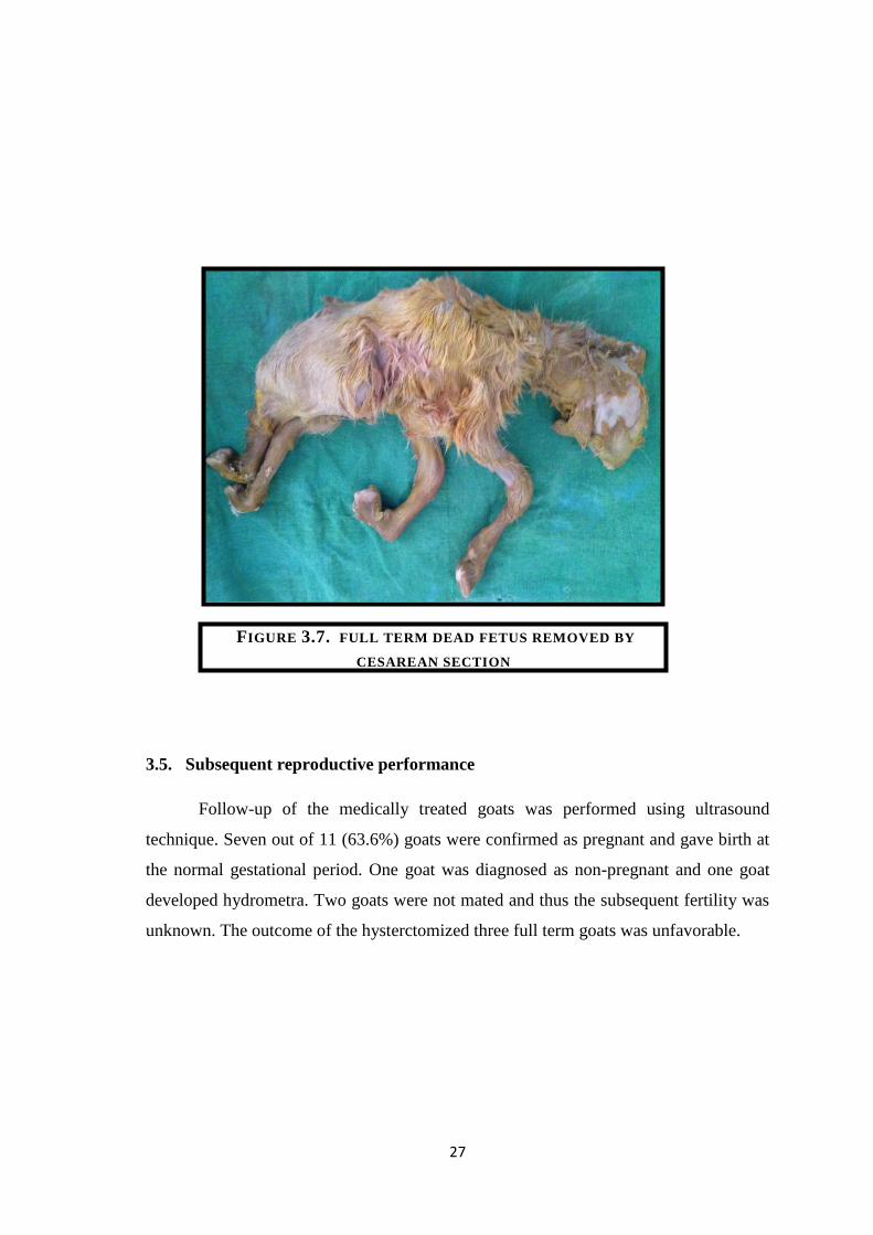
3.5. Subsequent reproductive performance
Follow-up of the medically treated goats was performed using ultrasound
technique. Seven out of 11 (63.6%) goats were confirmed as pregnant and gave birth at
the normal gestational period. One goat was diagnosed as non-pregnant and one goat
developed hydrometra. Two goats were not mated and thus the subsequent fertility was
unknown. The outcome of the hysterctomized three full term goats was unfavorable.
FIGURE 3.7. FULL TERM DEAD FETUS REMOVED BY
CESAREAN SECTION
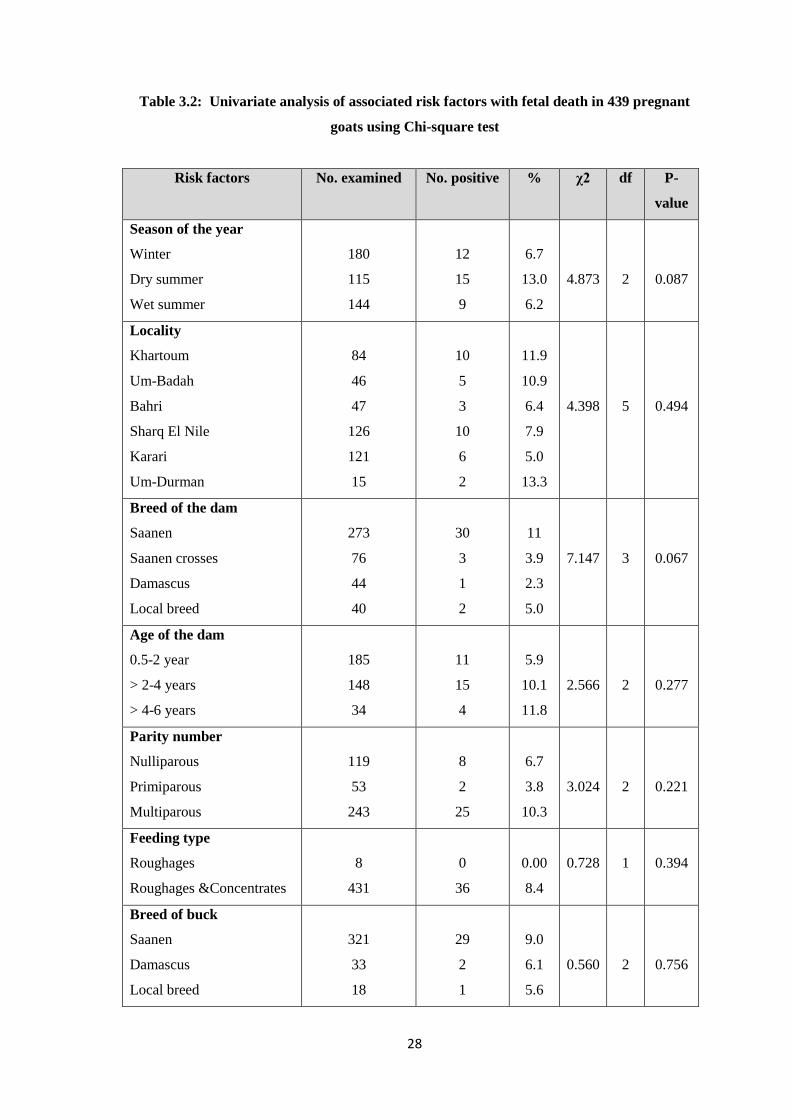
28
Table 3.2: Univariate analysis of associated risk factors with fetal death in 439 pregnant
goats using Chi-square test
Risk factors No. examined No. positive % χ2 df P-
value
Season of the year
Winter
Dry summer
Wet summer
180
115
144
12
15
9
6.7
13.0
6.2
4.873
2
0.087
Locality
Khartoum
Um-Badah
Bahri
Sharq El Nile
Karari
Um-Durman
84
46
47
126
121
15
10
5
3
10
6
2
11.9
10.9
6.4
7.9
5.0
13.3
4.398
5
0.494
Breed of the dam
Saanen
Saanen crosses
Damascus
Local breed
273
76
44
40
30
3
1
2
11
3.9
2.3
5.0
7.147
3
0.067
Age of the dam
0.5-2 year
> 2-4 years
> 4-6 years
185
148
34
11
15
4
5.9
10.1
11.8
2.566
2
0.277
Parity number
Nulliparous
Primiparous
Multiparous
119
53
243
8
2
25
6.7
3.8
10.3
3.024
2
0.221
Feeding type
Roughages
Roughages &Concentrates
8
431
0
36
0.00
8.4
0.728
1
0.394
Breed of buck
Saanen
Damascus
Local breed
321
33
18
29
2
1
9.0
6.1
5.6
0.560
2
0.756
29
CHAPTER FOUR
DISCUSSION, CONCLSION AND RECOMMENDATIONS
B-mode real-time ultrasonography is considered a method of choice for
diagnosis of fetal death in goats. It is non-invasive, non-time consuming and unfailing
method for detecting fetal death throughout gestational period. Transabdominal
approach was used for diagnosis of fetal death in the present study and ultrasound
scanning was performed only once to confirm fetal mortality. Yotov (2012) reported
that single ultrasound scanning could be successfully used for diagnosis of fetal death.
Absence of fetal movements and heartbeats were the first signs of fetal death in the
current study. Jonker (2004) and Samir et al. (2015) reported that lack of fetal
movement and heartbeats were basic signs of the dead fetus monitored by
ultrasonographic technique. In the present study, no clinical signs were observed in
goats that carried dead fetuses except for one goat with dilated cervix. Engeland et al.
(1998) stated that goats with dead fetuses don’t exhibit any clinical symptoms.
In the present study, the prevalence of fetal death in goats in Khartoum State
was found to be 8.2 %. Our result was lower than Samir et al. (2015) who identified
embryonic and fetal death (41.28%) using both transrectal and transabdominal
approaches during different stages of pregnancy. In the current study, transabdominal
ultrasonography was used and only fetal death was documented. The higher rate of
embryonic loss might be due to malfunction of the placenta in the embryonic stage of
pregnancy compared with the fetal period (Inskeep, 2004).
Nine associated risk factors were investigated in the current study. No
significant association was found between season of the year and prevalence of fetal
death. However a high prevalence of fetal death was reported from dry summer season
compared with winter and wet summer seasons and this may probably be due to high
temperature and low feed intake during dry summer season. Our results agree with
Bokko (2011) who reported higher prevalence of pregnancy wastage in dry season due
to forage scarce.
No significant association was found between locality and fetal death and this
may probably be due to the same environmental conditions.
No association was found between the breed of the dam and prevalence of fetal
death. However, the high occurrence of fetal death was recorded from Saanen breed
30
followed by Saanen crosses. This could most probably be due to that Saanen breed is a
temperate breed which delivered to tropical climatic regions with high temperature.
Also, the experimental groups were not numerically balanced and the majority of the
examined goats were Saanen and Saanen crosses.
In the current study, no significant association was found between the age of the
dam and fetal death. This is in agreement with Samir et al. (2015). High prevalence of
fetal death was reported from goats > 4-6 years. Engeland et al. (1997) reported that the
prevalence of fetal loss increased with increasing age of the goats. Hafez and Hafez
(2006) reported that the higher incidence of fetal death in animals over 6 years resulted
from impaired uterine environment. Furthermore, the quality of the endometrium might
be deteriorated in elderly animals resulting in inauspicious medium for maintenance of
pregnancy (Vanroose et al., 2000; Yotov, 2012).
In the current study, all goats diagnosed as having dead fetuses were reared
under closed system. This result is similar to Samir et al. (2015) who reported an
occurrence of fetal death in closed rearing system.
No significant association was found between parity number and fetal death
which is in agreement with Samir et al. (2015). High prevalence of fetal death was
observed in multiparous goats compared to primiparous and nulliparous. In multiparous
goats, bacteria could find access to the uterus during previous parturitions and estrous
cycles which will contaminate the environment and causes fetal mortality (Vanroose et
al., 2000; Hafez and Hafez, 2006; Yotov, 2012).
In the current study, no significant association was found between fetal death
and breed of inseminating buck. This result was in accordance with Engeland et al.
(1997) who found that no significant association between fetal loss and specific mated
buck. However, (Hafez and Hafez, 2006) reported that fetal death is attributable to the
male and the mating system because of inherited genetic materials from the male and
incompatibility between zygote and mother.
Conclusion
It is concluded that ultrasound technique is an accurate, rapid and non-invasive
modality for diagnosis of fetal mortality. It is well thought-out as the method of choice
for diagnosis of fetal mortality which is found to be a major problem in goat herds in
Khartoum State. PGF2α was found as a sufficient treatment for fetal death in goats.

31
Recommendations
It is recommended that:
1. Ultrasound technique should be introduced as a routine diagnostic modality in
veterinary clinics and teaching hospitals.
2. Further studies should be performed to find the specific causes of fetal death and to
investigate other risk factors which might be associated with it.
3. Awareness of owners of the goats should be raised and risk factors of fetal death
should be put into consideration.
32
REFERNCES
Abdel Aziz, A. (2010). Present status of the world goat populations and their
productivity. Lohmann Information, 45 (2), 42-52.
Abdelghafar, R. M. (2010). Ultrasonography as a diagnostic tool for fetal mortality
in goats (case report), Assiut Veterinary Medical Journal, 26 (127): 316-
322.
Abdelghafar, R. M. (2011). Transabdominal ultrasonographic fetometry in Saanen
goats. PhD Thesis, Sudan University of Science and Technology, 100
pages.
Abdelghafar, R. M. (2015). Fetal death in Saanen goats: Case reports: Ruminant
Science, 4 (2): 231-233.
Abu-Zidan, F., Hefny, A. and Corr, P. (2011). Clinical ultrasound physics. Journal
of Emergencies, 4 (4): 501-503.
Ahmed, B., Hamad, R. and Abdelghafar, R. (2010). Ultrasonography for diagnosis
of hydrometra and pyometra in goats (two case reports). Assiut
Veterinary Medical Journal, 56: 316-322.
Aiello, S. and Moses, M. (2012). The Merck Veterinary Manual, 11th edition.
Kenilworth, USA, 3366 pages.
Ali, A. (2011). Causes and management of dystocia in small ruminants in Saudia
Arabia. Journal of Agricultural and Veterinary Sciences, 4 (2): 95-108.
Almubarak, A., Abdelghafar, R. and Badawi, M. (2016). Hydrometra in a goat:
diagnosis, treatment and subsequent fertility. International Journal of
Livestock Research, 6 (4): 114-118.
Anwar, M., Riaz, A., Ullah, N. and Rafig, M. (2008). Use of ultrasonography for
pregnancy diagnosis in Balkhi sheep. Pakistan Veterinary Journal, 28
(3): 144-146.
Ayres, S., Nims, S. and Porter, C. (1999). Early pregnancy diagnosis in goats using
transvaginal ultrasound. Proceeding of the annual conference of the
society for theriogenology, Nashville, Tenn, p7.
Bokko, P. (2011). Pregnancy wastage in sheep and goats in the Sahel region of
Nigeria. Nigerian Veterinary Journal, 32 (2): 120-126.
Brounts, S., Hawkins, J., Baird, A. and Glickman, L. (2004). Outcome and
subsequent fertility of sheep and goats undergoing cesarean section
33
because of dysticia: 110 cases (1981-2001). Journal of the American
Veterinary Medical Association, 224 (2): 275-279.
Buckrell, B. (1988). Applications of ultrasonography in reproduction in sheep and
goats. Theriogenology, 29: 71-84.
Cochlin, D., Dubbins, P., Goldberg, B. and Alexander, A. ( 1994). Urogenital
Ultrasound: A Text Atlas. Martin Dunitz, UK, 417 pages.
Curie, J. and Curie, P. (1880). Development via compression of electric
polarization in hemihedral crystals with inclined faces. Bulletin de la
Societe de Minerologique de France, 3: 90-93.
Deori, S., Bhuyan, D., Kalitaand, D. and Saema, D. (2015). Management of foetal
maceration in a doe carried twin. Indian Journal of Animal
Reproduction, 33 (2): 109-110.
DesCoteaux, L., Colloton, J. and Gnemmi, G. (2010). Sheep and goats. Practical
Atlas of Ruminant and Camelid Reproductive Ultrasonography, 1st
edition. Willey-Blackwell, Iowa, USA, 244 pages.
Devandra, C. (2010). Concluding synthesis and the future for sustainable goat
production. Small Ruminant Research, 89: 125-130.
Dixon, A., Knight, M., Winkler, J., Marsh, D., Pate, J., Wilson, M., Dailey, R.,
Seidel, G. and Inskeep, E. (2007). Patterns of late embryonic and fetal
mortality and association with several factors in sheep. Journal of
Animal Sciences, 85: 1274-1287.
Drost, M. (2007). Complications during gestation in the cow. Theriogenology, 68:
487-491.
Ebert, R. and Solaiman, S. (2010). Animal evaluation. In: Solaiman, S. G. Goat
Science and Production. Wiley-Blackwell, USA pp 193- 216.
Engeland, I., Waldeland, H., Andresen, O., Loken, T., Bjorkman, C. and Bjerkas,
I. (1998). Foetal loss in dairy goats: An epidemiological study in 22
herds. Small Ruminant Research, 30: 37-48.
Engeland, I., Waldeland, H., Andersen, O. and Tverdal, A. (1997). Foetal loss in
dairy goats: an epidemiological study in 515 individual goats. Animal
Reproduction Science, 49 (1): 45-53.
FAO (2007). Food and Agriculture Organization. The State of the World’s Animal
Genetic Resources for Food and Agriculture, Rome.
34
FAO (2015). Food and Agriculture Organization. FAO Statistical Pocketbook,
Rome.
Fulton, R. (2014). Focused-Basics ultrasound principles and artifacts. In:
Lisciandro, G. R. Focused Ultrasound Techniques for the Small Animal
Practitioner, 1st edition. John Wiley & Sons, Texas, USA, 360 pages.
Ghosh, A., Yeasmina, F. and Alam, M. G. (1992). Studies of ringwomb in black
Bengal goats. Theriogenology, 37: 527-532.
Ginther, O. (1998). Pregnancy loss. Ultrasonic Imaging and Reproduction in cattle,
3rd edition. Equiservices, Cross Plains, USA, pp. 209-228.
Givens, M. and Marley, M. (2008). Infectious causes of embryonic and fetal
mortality. Theriogenology, 4: 1-16.
Goddard, P. (1996). Veterinary Ultrasonography. Cab International, Wallingford,
Oxon. UK. Pp 329.
Gonzales-Bulnes, A., Pallares, P. and Vazquez, M. (2010). Ultrasonographic
imaging in small ruminant reproduction. Reproduction in Domestic
Animals, 45: 9-20.
Haar, G. (2010). Ultrasound bioeffects and safety. Journal of Engineering in
Medicine, 224: 363-373.
Hafez, B. and Hafez, E. (2006). Reproduction in Farm Animals, 7th edition.
Blackwell, India: 509 pages.
Hagen-Ansert, S. (2012). Foundations of sonography. Textbook of Diagnostic
Sonograhy. 7th edition. St. Louis, New York, 1552 pages.
Hangiandreou, N. (2003). B-mode ultrasound: Basic concepts and new technology.
Radiographics, 23:1019-1033.
Hendee, W. and Ritenour, E. (2002). Ultrasound transducers. Medical Imaging
Physics, 4th edition, Wiley-Liss, New York, 511 pages.
Hesselink, J. and Elving, L. (1996). Pedigree analysis in a herd of dairy goats with
respect to the incidence of hydrometra. Veterinary Quarterly,18 (1): 24-
25.
Hesselink, J. and Taverne, M. (1994). Ultrasonography of the uterus of the goat.
Veterinary Quarterly, 16 (1): 41-45.
Hickman, J., Houlton, J. and Edwards, B. (1995). Surgery of the genito-urinary
system. An Atlas of Veterinary Surgery, 3rd edition. Blackwell Science,
London, UK, 275 pages.
35
Hill, C., Bamber, J. and Haar, G. (2004). Ultrasonic biophysics. Physical Principles
of Medical Ultrasonics, 2nd edition. John Willy and Sons, West Sussex,
England, 528 pages.
Ibrahim, M. (2000). Studies On Some Reproductive Parameters of Nubian Goats
and their Sannen Cross Breeds Under Local Environmental Conditions.
PhD Thesis, Faculty of Animal Production, University of Khartoum,
Sudan.
Idahor, K. (2013). Sheep and goats slaughtered at Keffi abattoir: health status,
carcass yield and foetal deaths. Journal of Animal Science Advances, 3:
276-283.
ILCA (1995). International Livestock Centre for Africa. Reproductive losses in
small ruminants in Sub-Saharan Africa: A review. Addis Ababa,
Ethiopia.
Inskeep, E. (2004). Preovulatory, postovulatory, and postmaternal recognition
effects of concentrations of progesterone on embryonic survival in the
cow. Journal of Animals Science, 82: 24-39.
Jonker, F. (2004). Fetal death: Comparative aspects in large domestic animals.
Animal Reproduction Science, 82–83: 415-430.
Kahn, W. (2004). Veterinary Reproductive Ultrasonography, Schlutersche,
Germany, 256 pages.
Kara, C., Orman, A., Topal, E. and Çarkungöz, E. (2010). Effects of supplementary
nutrition in Awassi ewes on sexual behaviors and reproductive traits.
Journal of Biology and Environment Sciences, 4 (10): 15-21.
Kempisty, B., Bukowska, D., Wozna, M., Piotrowska, K., Jackowska, M., Zuraw,
A., Ciesiolka, K., Antosik, P., Maryniak, H., Ociepa, E., Porowski, Sz.,
Brussow, K., Jaskowski, J., and Nowicki, M. (2013). Endometritis and
pyometra in bitches. Veterinary Medicine, 58(6): 289-297.
Kornalalijnslijper, J., Bevers, M., Van Oord, H., Taverne, M. (1997a). Induction of
hydrometra in goats by means of immunization against prostaglandin
F2α
. Animal Reproduction Science, 46: 109-122.
Kumaresan, A., Chand, S., Suresh, S., Mohanty, T., Prasad, S., Layek, S. and
Behera, K. (2013). Effect of estradiol and cloprostenol combination
therapy on expulsion of mummified fetus and subsequent fertility in four
crossbred cows. Veterinary Research Forum, 4: 85-89.
36
Lebbie, S. (2004). Goats under household conditions. Small Ruminant Research,
51: 131-136.
Lefebvre, R. (2015). Fetal mummification in the major domestic species: current
perspective on causes and management. Veterinary Medicine: Research
and Reports. 6: 233-244.
Lefebvre, R., Saint-Hilaire, E. and Morin, I. (2009). Retrospective case study of
fetal mummification in cows that did not respond to prostaglandin F2α
treatment. Canadian Veterinary Journal, 50: 71-76.
Ludders, J. (1988). Anesthesia for the pregnant patient. Proceedings of the Annual
Meeting of the Society of Theriogenology. Orlando, Sept 16-17, p 36.
Mahgoub, O., Kadim, I., and Webb, E. (2012). Goat Meat Production and Quality.
CAB International, Nosworthy Way, UK, 360 pages.
Marai, I., El-Darawany, A., Fadiel, A. and Abdel-Hafez, M. (2007). Physiological
traits as affected by heat stress in sheep: A review: Small Ruminant
Research, 71: 1-12.
Martin, S., Meek, A., Willeberg, P. (1988). Veterinary epidemiology, principles and
methods. Iowa State University Press, USA, p. 343.
Matthews, J. (2009). Female Infertility. Diseases of the Goat, 3rd edition, Wiley
Blackwell, Chelmeford, UK, 448 pages.
Maxwell, W., Quintana-Casares, P. and Setchell, B. (1992). Ovulation rate, fertility,
and embryonic mortality in ewes mated to rams from two different
stains. Proceeding of the Australian Society of Animal Production, 19:
192-194.
Medan, M. and Abd El-Aty, A. (2010). Advances in ultrasonography and its
applications in domestic ruminants and other farm animals reproduction.
Journal of Advanced Research, 1: 123-128.
Mellado, M., Foote, R. and Gomez, A. (1991). Reproductive efficiency of Nubian
goats throughout the year in Northern Mexico. Small Ruminant
Research, 6: 151-157.
Miranda-de la Lama, G. and Mattiello, S. (2010). The importance of social
behavior for goat welfare in livestock farming, Small Ruminant
Research, 90: 1-10.
MOAR (2015). Ministry of Animal Resources. Estimate of Livestock Population
by State. Information Center, Khartoum, Sudan.
37
Moraes, E., Freitas Neto, L., Aguiar Filho, C., Bezerra, F., Santos, M., Neves, J.,
Lima, P., Oliveira, M. (2009). Mortality determination and gender
identification of conceptuses in pregnancies of Santa Ines ovine by
ultrasound, South African Journal of Animal Science, 39 (4): 307-312.
Morand-Fehr., P. and Lebbie, S. (2004). Proposal for improving the research
efficiency in goat. Small Ruminant Research, 51: 145-153.
Nelson, T., Fowlkes, J., Abramowicz, J. and Church, C. (2009). Ultrasound
biosafety considerations for the practicing sonographer and sonologist.
Journal of Ultrasound in Medicine, 28 (2): 139-150.
Noakes, D., Parkison, T. and England, G. (2009). Veterinary Reproduction and
Obstetrics, 9th edition, Saunders, London, UK, 950 pages.
Oral, H., Pancarci, S., Gungor, O. and Kacar, C. (2007). Determination of
gestational age by measuring fetal heart diameter with transrectal
ultrasonography in sheep. Medycyna Wet, 63: 1558-1560.
Palliser, H., Hirst, J., Rice, G., Ooi, G., Dellios, N., Escalona, R. and Young, I.
(2006). Labor-associated regulation of prostaglandin E and F synthesis
and action in the ovine amnion and cervix. Journal of Society for
Gynecologic Investigation, 13: 19-24.
Pollott, G. and Wilson, R. (2009). Sheep and goats for diverse products and profits.
FAO diversification booklet. Rural Infrastructure and Agro-Industries
Division Food and Agriculture Organization of the United Nations,
Rome.
Pugh, D. and Baird, A. (2012). Theriogenology of sheep and goats. Sheep and Goat
Medicine, 2nd edition. Elsevier Saunders, Maryland, 640 pages.
Purohit, G. and Mehta, J. (2012). Hydrometra in goats (Capra hircus): Clinical
analysis of 26 cases, Ruminant Science, 1 (2): 117-119.
Purohit, G., Shekher, C., Kumar, P. and Solanki, K. (2011). Induced termination of
pregnancy in domestic farm animals. Iranian Journal of Applied Animal
Science, 2: 1-12.
Radi, Z. (2005). Endometritis and cystic endometrial hyperplasia in a goat. Journal
of Veterinary Diagnostic Investigation, 17: 393–395.
Rautela, R., Yadav, D., Katiyar, R. Singh, S., Das, G. and Kumar, H. (2016). Fetal
maceration in goat: a case report. International Journal of Science,
Environment and Technology, 5: 2323-2326.

38
Rumack, C., Wilson, S. R., Charborneau, J. and Levine, D. (2011). Diagnostic
Ultrasound, 4th edition. Mosby, Elsevier, St. Louis, USA, 2192 pages.
Salami, S., Ibe, C., Umosen, A., Ajayi, I. and Maidawa, S. (2011). Comparative
oesteomeric study of long bone in Yankase sheep and Red Sokoto goat.
International Journal of Morphology, 29: 100-104.
Samir, H., Karen, A., Ashmawy, T., Abo-Ahmed, M., El-Sayed, M. and Watanabe,
G. (2015). Monitoring of embryonic and fetal losses in different breeds
of goats using real-time B-mode ultrasonography,
http://dx.doi.org/10.1016/j.theriogenology.2015.09.039.
Schatten, H. and Constantinescu, G. (2007). Ultrasonography in Small Ruminant
Reproduction. Comparative Reproductive Biology, 1st edition.
Blackwell, Iowa, USA, 355 pages.
Serin, G., Gokdal, O., Tarimcilar, T. and Atay, O. (2010). Umbilical artery Doppler
sonography in Saanen goat fetuses during singleton and multiple
pregnancies. Theriogenology, 74: 1082-1087.
Shriki, J. (2014). Ultrasound physics. Critical Care Clinics, 30 (1): 1-24.
Smith, M. and Sherman, D. (2009). Goat Medicine, 2nd edition. Wiley-Blackwell,
USA, 888 pages.
Solaiman, S. (2010). Applied reproductive physiology. Goat Science and
Production, 1st edition. Blackwell, Iowa, USA, 446 pages.
Souza, J., Maia, A., Brandao, F., Vilela, C., Oba, E., Bruschi, J. and Fonseca, J.
(2013). Hormonal treatment of dairy goats affected by hydrometra
associated or not with ovarian follicular cyst. Small Ruminant Research,
111: 104-109.
Sridevi, P., Kulasekar, K., Devanathan, T., Rangasamy, S. (2011). Ultrasonographic
diagnosis and management of hydrometra in goats. Indian Journal of
Animal Reproduction, 32 (1): 76-77.
Stratmeyer, M., Greenleaf, J., Dalecki, D. and Salvesen, K. (2008). Fetal ultrasound:
mechanical effects. Journal of Ultrasound in Medicine, 27: 597–605.
Suguna, K., Mehrotra, S., Hoque, M., Singh, S., Shanker, U. and Sarath, T. (2008).
Early pregnancy diagnosis and embryonic and fetal death development
using real time b-mode ultrasound in goats. Small Ruminant Research,
80: 80-86.
39
Taverne, M., Hesselink, J., Bevers, M., Van Oord, H. and Kornalijnslijper, J.
(1995). Etiology and endocrinology of pseudopregnancy in the goat.
Reproduction in Domestic Animals, 30: 228-230.
Taye, M., Deribe, B. and Melekot, M. (2013). Reproductive performance of central
highland goats under traditional management in Sekota district, Ethiopia.
Asian Jounrnal of Biological Sciences, 6: 271-276.
Troxel, M., Cornetta, A. and Pastor, K. (2002). Severe hematometra in a dog with
cystic endometrial hyperplasia/pyometra complex. Journal of the
American Animal Hospital Association, 38: 85–89.
Vanroose, G., De Kruif, A. and Van Soom, A. (2000). Embryonic mortality and
embryo-pathogen interactions. Animal Reproduction Science, 60-61:
131-143.
Vries, J. (2008). Goat for the poor: Some key to successful promotion of goat
production among the poor. Small Ruminant Research, 77: 221-224.
Wittek, T., Richter, A., Erices, J. and Elze, K. (1997). Incidence, diagnosis, therapy
and subsequent fertility in goats with hydrometra. Tierarztl Prax, 25:
576-582.
Wu, W., Xiao, M., Coksaygan, T., Chakrabarty, K., Collins, K., Rose, J. and
Nathanielsz, P. (2004). Prostaglandin mediates premature delivery in
pregnant sheep induced by estradiol at 121 days of gestational age.
Endocrinology, 145: 1444-1452.
Yotov, S. (2005). Diagnostics of early pregnancy in Stara Zagora dairy sheep breed.
Bulgarian Journal of Veterinary Medicine, 8 (1): 41-45.
Yotov, S. (2012). Ultrasound diagnostics of late embryonic and foetal death in three
sheep breeds. Journal of Veterinary Advances, 2 (3): 120-125.
Youngquist, R. and Threlfall, W. (2007). Current Therapy in Large Animal
Theriogenology, 2nd edition. Saunders, USA, 1068 pages.
Zagzebski, J. (1996). Essentials of Ultrasound Physics. St. Louis, Mosby, USA 220
pages.

40
Appendix: Questionnaire form
Date: Serial No:
Owner’s name:
Phone No:
Address (locality):
Animal ID
No Risk factors
1 Breed of the dam
2 Age of the dam
3 Rearing system
1. Closed
2. Opened
4 Parity number
1. Nulliparous
2. Primiparous
3. Multiparous
5 Estrous type
1. Natural
2. Induced
6 Feeding type:
1. Roughages:
2. Roughages and concentrates:
7
Breed of the insemination buck
1. Saanen
2. Damascus
3. Local breed

41
Ultrasound result
Empty
Pregnant
1. Dead fetus
2. Viable fetus
Physiological parameters
Heartbeats
Respiration
Temperature
Treatment
……………………………………………………………………………………………
……………………………………………………………………………………………
……………………………………………………………………………………………
……………………………………………………………………………………………
Follow-up
……………………………………………………………………………………………
……………………………………………………………………………………………
……………………………………………………………………………………………
……………………………………………………………………………………………
Comments:
……………………………………………………………………………………………
……………………………………………………………………………………………
……………………………………………………………………………………………
……………………………………………………………………………………………