dissertation - uj ir
TRANSCRIPT

COPYRIGHT AND CITATION CONSIDERATIONS FOR THIS THESIS/ DISSERTATION
o Attribution — You must give appropriate credit, provide a link to the license, and indicate ifchanges were made. You may do so in any reasonable manner, but not in any way thatsuggests the licensor endorses you or your use.
o NonCommercial — You may not use the material for commercial purposes.
o ShareAlike — If you remix, transform, or build upon the material, you must distribute yourcontributions under the same license as the original.
How to cite this thesis
Surname, Initial(s). (2012) Title of the thesis or dissertation. PhD. (Chemistry)/ M.Sc. (Physics)/ M.A. (Philosophy)/M.Com. (Finance) etc. [Unpublished]: University of Johannesburg. Retrieved from: https://ujcontent.uj.ac.za/vital/access/manager/Index?site_name=Research%20Output (Accessed: Date).
0
SYNTHESIS AND CHARACTERIZATION OF CHOLINE BASED IONIC LIQUIDS
AND THEIR UTILIZATION IN THE RECOVERY OF BASE METALS FROM BCL
SLAG ASSISTED BY GOLD 1 MINE BACTERIAL ISOLATES A Dissertation submitted to the Faculty of Science, University of Johannesburg In
partial fulfilment of the requirement for the award of a Master’s Degree in
Technology: Biotechnology
January 2017
By
LETLHABILE THAPELO MOYAHA
STUDENT NUMBER: 200803857
Supervisor : Dr. V. Mavumengwana
Co-supervisor : Dr. S. Sekar
Co-Supervisor : Prof. A. Mulaba-Bafubiandi
I
ABSTRACT
Due to the depletion of rich ore bodies, the fact that conventional extractions become
obsolete and uneconomical and additional stringency on environment related regulations,
there is a call for cheaper and greener metal extraction methods. Through research and
innovation, new developments and improvements of processes and products are required in
both metallurgical and biological areas. As a result, research interests in the use of microbes
in the optimization of metal recovery from low grade sulphide ores have grown increasingly
popular. The involvement of microbes in the recovery process is thus dependent on the
growth of the microbes, which is influenced by their physicochemical parameters. The
availability of nutrient salts is thus essential for maintaining optimal growth, furthermore is
the dependence of metal dissolution with nutrient quantities on substrate accessibility.
Considering the “green chemistry” of some ionic liquids the important “technological”,
“toxicological ” and “eco-toxicological” assessment of the risks related with ionic liquid
design; choline based ionic liquids were selected by virtue of their low toxicity, low
environmental persistence and readily biodegradable nature. As a result of the selective
solubility strength of metal oxides in ionic liquids, choline based ionic liquids were
considered novel media for obtaining target metals and improving bioleaching kinetics. The
study thus sought to develop an economic and eco-friendly alternative technology, employing
choline based ionic liquids as biocompatible substrates for the optimal recovery of base
metals, improving bioleaching kinetics.
Biological and molecular characterization of ultra-deep mine isolates (Gold 1 Mine East
Rand, Springs Johannesburg, South Africa) revealed a halophilic community of gram positive
and negative bacteria namely Raoultella ornithinolytica, Bacillus sp., Bacillus thuringiensis
and Pseudomonas moraviensis. The halophilic bacterial strains indicated high
biocompatibility in choline lactate, choline chlorite, choline dihydrogen phosphate, choline
citrate and choline levulinate with poor biocompatibility noted in choline citrate and choline
tartarate. According to literature, the biocompatibility of the leading choline based ionic
liquids (ILs) was accredited to the quaternary- ammonium cation integrating a polar hydroxyl
and the limited branching of the side chain. Prominent logarithmic growth in choline lactate
was indicative of the metabolic advantage of choline lactate over conventional glucose
sources. Furthermore, the bioleaching of BCL slag by Bacillus species and Bacillus
II
thuringiensis employing choline lactate as a substrate was found to effect 97 % iron (Fe)
recoveries from the major iron fayalite phase in BCL slag. Producing moderate growth rates,
choline dihydrogen phosphate employing Bacillus species and Bacillus thuringiensis was
found to effect 31 % recovery of the zinc (Zn) element corroborated by the liberation of a
new zinc silicate phase ( Zn2SiO4) in the slag residue. The studies were thus suggestive of the
selective nature of choline lactate and choline dihydrogen phosphate ILs for the recovery of
iron (Fe) and zinc (Zn) base metals.
In contrast to the chemical leaching control, the use of halophilic bacteria in choline lactate
was found to accelerate bacterial growth, producing competitive iron recoveries; thus
alluding to the potential of these ionic liquid based reactions as an alternative to conventional
hydrometallurgy methods.
Keywords: Bioleaching kinetics, choline based ILs, biocompatibility, halophilic, recoveries

III
DECLARATION
I, Letlhabile Thapelo Moyaha hereby declare that this study entitled “Synthesis and
characterization of choline based ionic liquids and their utilization in the recovery of base
metals from BCL slag assisted by Gold 1 Mine bacterial isolates” is my work, conducted
under the supervision of Dr. Vuyo Mavumengwana, Dr. Sekar Sudharshan and Prof Antoine
Mulaba-Bafubiandi. This study represents my original work and has not been submitted for
any degree or examination in any other university. All other sources used, have been duly
cited in text and acknowledged by complete references.
XLetlhabile Thapelo Moyaha

IV
DEDICATION
I dedicate this work to my Heavenly Farther and maker for He’s abundant favour upon my
life and to my guardian angels Mrs E.B Moyaha and Mr N.N Moyaha for their continual
support. To God almighty be all the glory.

V
ACKNOWLEDGEMENTS
Trust in the Lord with all your heart and lean not on your own understanding; in all your
ways acknowledge him, and he will make your paths straight (Proverbs 3:5-6). Blessed is the
man who finds wisdom, the man who gains understanding, for she is more profitable than
silver and yields better returns than gold (Proverbs 3:13-14) .Glorifying God in all that he has
done and been for me during this remarkable journey.
My sincere gratitude to my supervisors Dr. Vuyo Mavumengwana, Dr. Sekar Sudharshan and
Prof Antoine Mulaba-Bafubiandi for their generous contribution to the success of this project.
I thank you for your mentorship, asking me insightful questions and offering invaluable
guidance towards improving and enhancing the quality of this work. Above all I thank you
for allowing me the creative space and freedom to learn.
A heartfelt appreciation goes to my family, my mother Mrs E.B Moyaha and father Mr N.N
Moyaha, my loving siblings Karabo Moyaha, Tlou Moyaha, Kibi Sebotja, Nomfundo
Morutse, and Nonhlanhla Maphanga, my grandmother Mrs P. Huma and late grandmother
Mrs A. Moyaha. I thank you for supporting me in my goals, always helping me to put my
best foot forward and spurring me on with my favourite guilty pleasures when fatigue has set
in. I thank you for you for your invaluable love, support and always dreaming with me. You
are one in a million.
My sincere gratitude to my laboratory colleagues, my partners in crime Bongeka Mbambo
and Evonia Kanyane Nchabeleng for the stimulating conversations and for all the laughs we
had that brightened up the late nights in the lab. I would also like to acknowledge my mentor
Ms Daphney Mogano and my friends Mapula Pale, Matshidiso Tlhapane, Ayanda Timothy
and Nondumiso Shongwe for their unwavering support.
My infinite gratitude to Mr. E Malenga Ntumba and Ms. N. P Baloyi for their insight,
expertise and mentorship in the field of Extraction Metallurgy, you always went beyond the
call of duty and for that I am eternally grateful. I also sincerely thank Professor Taddese
Wondimu Godetto, Nombuzo Mabuza, Harold Hussein Shiri and Bienvenue Gael Mbanga-
Fouda from the Analytical Chemistry Department. A special thank you goes to Mr. E.Van
Zyl, Dr. N. Niemann, Dr. Derek Dinteh, for their generous assistance and support.
For the financial endorsement of this study, I sincerely thank the University of Johannesburg,
through the National Research Foundation (NRF).
VI
PRESENTATIONS AND ARTICLES PUBLISHED OR WRITTEN FOR
PUBLICATION
Presentations:
Moyaha, L., Mavumengwana, V., Sidu, S., Mulaba-Bafubiandi, A., (2015).The recovery of
base metals from slag using bacterial isolates in the presence of biocompatible ionic liquids
as substrate(s).Poster presentation. DST: Howard University-IT woman in stem conference.
Johannesburg, Hyatt Regency hotel in Rosebank.
Publications:
Sekar, S., Moyaha, L., Mavumengwana, V., Sidu, S., Mulaba-Bafubiandi, A., (2016)
Metagenomics: DNA sequencing of environmental samples from Gold 1 Mine East Rand,
Springs Johannesburg, South Africa. New Biotechnology, 33, 179.
Moyaha, L., Sekar, S., Mavumengwana, V., Sidu, S., Mulaba-Bafubiandi, A. The
valorisation of slag from BCL using biocompatible choline based ionic liquids as a support.
VII
TABLE OF CONTENTS
ABSTRACT................................................................................................................................I
DECLARATION ..................................................................................................................... III
DEDICATION ......................................................................................................................... IV
ACKNOWLEDGEMENTS ...................................................................................................... V
PRESENTATIONS AND ARTICLES PUBLISHED OR WRITTEN FOR PUBLICATION
.................................................................................................................................................. VI
TABLE OF CONTENTS........................................................................................................VII
LIST OF FIGURES .............................................................................................................. XIII
LIST OF TABLES ............................................................................................................... XVII
LIST OF ABBREVIATIONS ............................................................................................... XIX
LIST OF UNITS .................................................................................................................. XXII
DISSERTATION OUTLINE............................................................................................. XXIV
CHAPTER ONE ........................................................................................................................ 1
1.0 GENERAL INTRODUCTION ................................................................................... 1
1.1 Background ................................................................................................................. 1
1.2 Problem statement ....................................................................................................... 2
1.3 Hypothesis ................................................................................................................... 3
1.4 Aim and Objectives of study ....................................................................................... 3
1.4.1 Aim of the study................................................................................................... 3
1.4.2 Objectives of the study......................................................................................... 3
CHAPTER TWO ....................................................................................................................... 5
2.0 LITTERATURE REVIEW ......................................................................................... 5
2.1 Introduction ................................................................................................................. 5
2.2 Historical perspective .................................................................................................. 5
2.3 Biomining motivation ................................................................................................. 8
2.4 Bioleaching microorganisms ....................................................................................... 8
VIII
2.4.1 Mesophiles ......................................................................................................... 10
2.4.2 Moderate thermophiles ...................................................................................... 11
2.4.3 Extreme thermophiles ........................................................................................ 11
2.5 Halophilic bacteria .................................................................................................... 15
2.6 Principle of microbial leaching ................................................................................. 15
2.6.1 Metal sulphide dissolution mechanism .............................................................. 16
2.7 Leaching technique ................................................................................................... 18
2.7.1 In situ leaching ................................................................................................... 18
2.7.2 Dump leaching ................................................................................................... 19
2.7.3 Heap leaching..................................................................................................... 19
2.7.4 Vat leaching ....................................................................................................... 20
2.8 Factors affecting bioleaching .................................................................................... 22
2.8.1 Physiochemical parameters................................................................................ 23
2.8.2 Microbiological parameters ............................................................................... 27
2.9 Recovery of base metals from slag ........................................................................... 30
2.10 Ionic liquids as an alternative medium...................................................................... 31
2.10.1 Properties of Ionic Liquids (ILs)........................................................................ 31
2.10.2 Synthesis of Ionic Liquids (ILs) ........................................................................ 33
2.10.3 Types of Ionic Liquids (ILs) .............................................................................. 34
2.10.4 Biodegradable designer solvents........................................................................ 35
2.10.5 Biological Activity of Ionic Liquids (ILs) ......................................................... 36
2.11 Metal recovery potential of Ionic Liquids (ILs) ........................................................ 37
2.12 Choline/cholinium derived Ionic Liquids (ILs) ........................................................ 38
2.13 Concluding remarks .................................................................................................. 39
REFERENCES..................................................................................................................... 40
CHAPTER THREE ................................................................................................................. 51
IX
CHARACTERIZATION OF HALOPHILIC BACTERIA FROM GOLD1 MINE EAST
RAND, SPRINGS JOHANNESBURG, SOUTH AFRICA FOR MICROBIAL MINING
APPLICATIONS ..................................................................................................................... 51
Abstract ................................................................................................................................ 51
3.1 Introduction ............................................................................................................... 52
3.2 Methodology ............................................................................................................. 53
3.2.1 Study area of interest for sampling .................................................................... 53
3.2.2 Collection of samples......................................................................................... 54
3.2.3 Isolation and enrichment of moderately halophilic bacteria .............................. 54
3.2.4 Phenotypic classification of bacteria: Microbiological identification of bacteria .
............................................................................................................................ 54
3.2.5 Biochemical characterization of bacteria API 20E® strips ................................ 54
3.2.6 Genotypic classification of bacteria: 16S rRNA sequencing ............................. 55
3.2.7 Phylogenetic tree................................................................................................ 55
3.3 Results and Discussion.............................................................................................. 55
3.3.1 Microbiological characterization of bacterial isolates ....................................... 55
3.3.2 Biochemical characterization of bacterial isolates............................................. 57
3.3.3 Genotypic characterization of bacterial isolates ................................................ 58
3.3.4 Phylogenetic study of bacterial strains .............................................................. 60
3.4 Conclusion................................................................................................................. 62
Acknowledgement................................................................................................................ 62
References ............................................................................................................................ 63
CHAPTER FOUR.................................................................................................................... 67
SYNTHESIS AND CHARACTERIZATION OF BIOCOMPATIBLE CHOLINE DERIVED
IONIC LIQUIDS FOR BIOTECHNOLOGY APPLICATIONS ............................................ 67
Abstract ................................................................................................................................ 67
4.1 Introduction ............................................................................................................... 68
4.2 Methodology ............................................................................................................. 69
X
4.2.1 Choline hydroxide properties............................................................................. 69
4.2.2 Acids .................................................................................................................. 70
4.2.3 Ionic liquid synthesis ......................................................................................... 71
4.2.4 Evaporation process ........................................................................................... 71
4.2.5 Analytical Spectral study/Characterization........................................................ 71
4.2.5.1 1H NMR Spectra............................................................................................. 72
4.2.5.2 13C NMR Spectra ........................................................................................... 72
4.2.5.3 FT-IR .............................................................................................................. 72
4.2.6 Preliminary growth studies of halophilic bacteria in the presence of choline
based ILs ........................................................................................................................... 72
4.2.6.1 Inoculum preparation for halotolerant bacteria strains .................................. 72
4.2.6.2 Media preparation for growth plate studies (solid media) ............................. 73
4.2.6.3 Direct colony count ........................................................................................ 73
4.2.6.4 Media preparation for shaker growth studies (liquid media) ......................... 73
4.2.6.5 Indirect colony count...................................................................................... 73
4.3 Results and Discussion.............................................................................................. 74
4.3.1 NMR................................................................................................................... 74
4.3.1.1 Characterization of Choline Lactate 1H NMR ............................................... 74
4.3.1.2 Characterization of Choline Lactate 13C NMR .............................................. 75
4.3.1.3 Characterization of Choline Dihydrogen Phosphate 1H NMR....................... 76
4.3.1.4 Characterization of Choline Dihydrogen Phosphate 13C NMR ..................... 77
4.3.1.5 Characterization of Choline Citrate 1H NMR ................................................ 78
4.3.1.6 Characterization of Choline Citrate 13C NMR ............................................... 79
4.3.1.7 Characterization of Choline Tartarate 1H NMR............................................. 80
4.3.1.8 Characterization of Choline Tartarate 13C NMR............................................ 81
4.3.1.9 Characterization of Choline Chloride 1H NMR ............................................. 82
4.3.1.10 Characterization of Choline Chloride 13C NMR ........................................ 83
4.3.1.11 Characterization of Choline Levulinate 1H NMR ...................................... 84
4.3.1.12 Characterization of Choline Levulinate 13C NMR ..................................... 85
4.3.2 FTIR ................................................................................................................... 85
4.3.2.1 FTIR: 2-Hydroxy-N, N, N-trimethylethanaminium 2-hydroxypropanoate ... 86
XI
4.3.2.2 FTIR: 2-Hydroxy-N, N, N-trimethylethanaminium dihydrogen phosphate .. 87
4.3.2.3 FTIR:2-Hydroxy-N,N,N-trimethylethanaminium 3-carboxy-2-
(carboxymethyl)-2-hydroxypropanoate ........................................................................ 87
4.3.2.4 FTIR:2-Hydroxy-N,N,N-trimethylethanaminium3-carboxy-2,
3dihydroxypropanoate. ................................................................................................. 88
4.3.2.5 FTIR: 2-Hydroxy-N, N, N-trimethylethanaminium chloride......................... 89
4.3.2.6 FTIR: 2-Hydroxy-N, N, N-trimethylethanaminium 4-oxopentanoate ........... 89
4.3.3 Preliminary growth studies: ............................................................................... 90
4.3.3.1 Growth Plate studies....................................................................................... 90
4.3.4 Shaker growth studies ........................................................................................ 96
4.4 Conclusion............................................................................................................... 101
Acknowledgement.............................................................................................................. 102
References .......................................................................................................................... 103
CHAPTER FIVE ................................................................................................................... 105
THE VALORISATION OF SLAG FROM BCL USING BIOCOMPATIBLE CHOLINE
BASED IONIC LIQUIDS AS A SUPPORT ......................................................................... 105
Abstract .............................................................................................................................. 105
5.1 Introduction ............................................................................................................. 106
5.2 Methodology ........................................................................................................... 108
5.2.1 Slag processing ................................................................................................ 108
5.2.2 Particle size distribution................................................................................... 108
5.2.3 Scanning electron microscopy (SEM) ............................................................. 108
5.2.4 Slag prep -Microwave digestion ...................................................................... 109
5.2.5 Inductively coupled plasma optical emission spectroscopy (ICP-OES).......... 110
5.2.6 X-ray fluorescence (XRF) analysis.................................................................. 111
5.2.7 X-ray Powder Diffraction (XRD) analysis ...................................................... 112
5.2.8 Inductively coupled plasma mass spectrometry (ICP-MS) ............................. 112
5.2.9 Standard solution preparation: ......................................................................... 114
5.2.9.1 Internal standard ........................................................................................... 114
XII
5.2.9.2 Calibration standards .................................................................................... 114
5.2.10 Atomic absorption spectroscopy (AAS) .......................................................... 114
5.2.11 Bioleaching experiment of mineral sample (BCL slag) .................................. 115
5.3 Results and Discussion............................................................................................ 116
5.3.1 Particle size distribution................................................................................... 116
5.3.2 Slag morphology .............................................................................................. 116
5.3.3 XRF of BCL slag before and after leaching .................................................... 119
5.3.4 ICP-OES slag elemental analysis..................................................................... 122
5.3.5 XRD analysis of BCL slag before and after leaching ...................................... 123
5.3.6 Bacterial growth studies................................................................................... 127
5.3.7 pH studies......................................................................................................... 132
5.3.8 Bioleaching Studies.......................................................................................... 135
5.4 Conclusion............................................................................................................... 141
Acknowledgement.............................................................................................................. 141
References .......................................................................................................................... 142
CHAPTER SIX ...................................................................................................................... 145
6.0 GENERAL DISCUSSION AND CONCLUSION ................................................. 145
6.1 GENERAL DISCUSSION...................................................................................... 145
6.2 CONCLUSION ....................................................................................................... 147
REFERENCES ...................................................................................................................... 148
APPENDICES ....................................................................................................................... 150
XIII
LIST OF FIGURES
Figure 2-1: Extreme thermophile isolates located in hot springs A) Yellow State National
Park United States B) Hot coal dumps in Witbank South Africa (Sourced from Neale, 2006).
.................................................................................................................................................. 14
Figure 2-2: Bacteria classified according to optimal growth temperatures namely Extreme
thermophiles (left), spherical shaped with a dimension of 1-2µm. Moderate thermophiles
(centre) with comparable phenotypic characteristics to Mesophiles. Mesophiles (right) known
to be rod shaped microorganisms with an estimated dimension of 0.5X2.0µm (Sourced from
Neale, 2006). ............................................................................................................................ 14
Figure 2-3: Summery diagram of a) thiosulfate mechanisms and b) polysulfide mechanism
(Sourced from Mishra et al., 2005).......................................................................................... 18
Figure 2-4: Operating flow for In situ Leaching (Sourced from Thosar et al., 2014) ............. 19
Figure 2-5: Operating flow for Heap Leaching (Sourced from Bauer, n.d.) ........................... 20
Figure 2-6: Operating process for Vat Leaching (Sourced from Thosar et al., 2014)............. 21
Figure 2-7: Annual growth rates of ionic liquid published articles (Sourced from Park et al.,
2014) ........................................................................................................................................ 32
Figure 2-8: The application of ILs (Sourced from Pham et al., 2010) .................................... 32
Figure 2-9: Synthesis procedure of an ionic liquid (Sourced from Park et al, 2014) .............. 34
Figure 2-10: Metabolic activity of choline lactate by Stapylococcus lentus (Sourced from
Sekar et al., 2013). ................................................................................................................... 39
Figure 3-1: Gold 1 mine East Rand mine sites under study NRW Ramp water, W2E19,
N2W2, UK9 and E2 Reef drive south ..................................................................................... 53
Figure 3-2:Streak plates of Springs Gold1 mine bacterial isolates after 24hrs incubation at
37°C to attain pure cultures for further characterization studies: A) Strain 2KL, B) Strain
9KL, C) Strain 12KL, D) Strain 16KL and E)Strain 19KL. ................................................... 55
Figure 3-3: API (Analytical Profile index) Colour change: A) 9KL before incubation at 37 °C
for 24 hrs and B) 9KL after incubation (addition of TDA, James, VP1, VP2 Reagents) C)
19KL before incubation at 37 °C for 24hrs and B) 19KL after incubation (addition of TDA,
James, VP1, VP2 Reagents)..................................................................................................... 57
Figure 3-4: Phylogenetic tree of consensus sequence 2KL ..................................................... 60
Figure 3-5: Phylogenetic tree of consensus sequence 9KL ..................................................... 61
Figure 3-6: Phylogenetic tree of consensus sequence 12KL ................................................... 61
Figure 3-7: Phylogenetic tree of consensus sequence 16KL ................................................... 61
XIV
Figure 3-8: Phylogenetic tree of consensus sequence 19KL ................................................... 61
Figure 4-1: Structure of choline hydroxide.............................................................................. 69
Figure 4-2: Synthesised ionic liquids namely choline levulinate, choline chloride, choline
citrate, choline lactate, choline tartarate and choline dihydrogen phosphate ........................... 71
Figure 4-3: 1H NMR of choline lactate .................................................................................... 74
Figure 4-4: 13C NMR of choline lactate................................................................................... 75
Figure 4-5: 1H NMR of choline dihydrogen phosphate ........................................................... 76
Figure 4-6: 13C NMR of choline dihydrogen phosphate.......................................................... 77
Figure 4-7: 1H NMR of choline citrate .................................................................................... 78
Figure 4-8: 13C NMR of choline citrate ................................................................................... 79
Figure 4-9: 1H NMR of choline tartarate ................................................................................. 80
Figure 4-10: 13C NMR of choline tartarate .............................................................................. 81
Figure 4-11: 1H NMR of choline chloride ............................................................................... 82
Figure 4-12: 13C NMR of choline chloride .............................................................................. 83
Figure 4-13: 1H NMR of choline levulinate ............................................................................ 84
Figure 4-14: 13C NMR of choline levulinate ........................................................................... 85
Figure 4-15: FTIR of choline lactate........................................................................................ 86
Figure 4-16: FTIR of Choline dihydrogen phosphate.............................................................. 87
Figure 4-17: FTIR of choline citrate ........................................................................................ 88
Figure 4-18: FTIR of choline tartarate ..................................................................................... 88
Figure 4-19: FTIR of choline chloride ..................................................................................... 89
Figure 4-20: FTIR of choline levulinate .................................................................................. 90
Figure 4-21: Shaker growth studies of Bacillus aryabhattai in choline based ionic ILs vs.
glucose as an alternative carbon source at a speed of 120 rmp and temperature of 37 °C
(5days)...................................................................................................................................... 96
Figure 4-22: Growth curve of Raoultella ornithinolytica in choline ILs vs. glucose as an
alternative carbon source at speed of 120 rpm and temperature of 37 °C ............................... 97
Figure 4-23: Growth curve of Bacillus sp. in choline based ILs vs. glucose as an alternative
carbon source at speed of 120 rpm and temperature of 37 °C. ................................................ 98
Figure 4-24: Growth curve of Bacillus thuringiensis in choline based ILs vs. glucose as an
alternative carbon source at speed of 120 rpm and temperature of 37 °C. .............................. 99
Figure 4-25: Growth curve of Pseudomonas moraviensis in choline based ionic liquids vs.
glucose as an alternative carbon source at speed of 120 rpm and temperature of 37 °C. ...... 100
XV
Figure 4-26: Growth curve in the absence of bacteria in choline based ionic liquids vs.
glucose as an alternative carbon source at speed of 120 rpm and temperature of 37 °C ....... 101
Figure 5-1: Schematic of BCL slag (Adapted from Malema & Legg, 2006) ........................ 108
Figure 5-2: Particle size distribution of BCL slag before bioleaching process ..................... 116
Figure 5-3: SEM of raw BCL slag (Before bioleaching process) indicative of an amorphous
structure.................................................................................................................................. 116
Figure 5-4: EDS spectrum of BCL slag as directed in Figure 5.3 ......................................... 117
Figure 5-5: Scanning electron photomicrograph of BCL slag after bioleaching in mixed
culture..................................................................................................................................... 118
Figure 5-6: Scanning electron photomicrograph of BCL slag after bioleaching in mixed
culture..................................................................................................................................... 118
Figure 5-7: Raw BCL slag sample before bioleaching process (feed) .................................. 123
Figure 5-8: BCL slag subsequent to bioleaching with Bacillus aryabhattai (2) in choline
based ILs vs. glucose ............................................................................................................. 124
Figure 5-9: BCL slag subsequent to bioleaching with Raoultella ornithinolytica (9) in choline
based ILs vs. glucose ............................................................................................................. 124
Figure 5-10: BCL slag subsequent to bioleaching with Bacillus sp. (12) in choline based ILs
vs. glucose .............................................................................................................................. 125
Figure 5-11: BCL slag subsequent to bioleaching with Bacillus thuringiensis (16) in ILs vs.
glucose ................................................................................................................................... 125
Figure 5-12: BCL slag subsequent to bioleaching with Pseudomonas moraviensis (19) in
choline based ILs vs. glucose................................................................................................. 126
Figure 5-13: BCL slag subsequent to bioleaching with a mixed culture (All) of bacteria in
choline based ILs vs. glucose................................................................................................. 126
Figure 5-14: BCL slag subsequent to leaching in the absence (0) of bacteria in choline based
ILs vs. glucose........................................................................................................................ 127
Figure 5-15: Growth curve of Bacillus aryabhattai in choline based ILs vs. glucose for the
recovery of base metals at a speed of 120 rmp and temperature of 37 °C (3 weeks) ............ 128
Figure 5-16: Growth curve of Raoultella ornithinolytica in choline based ILs vs. glucose for
the recovery of base metals at a speed of 120 rmp and temperature of 37 ⁰C (3 weeks) ...... 128
Figure 5-17: Growth curve of Bacillus sp. in choline based ILs vs. glucose for the recovery of
base metals at a speed of 120 rmp and temperature of 37 °C (3 weeks) ............................... 129
Figure 5-18: Growth curve of Bacillus thuringiensis in choline based ILs vs. glucose for the
recovery of base metals at a speed of 120 rmp and temperature of 37 °C (3 weeks) ............ 129
XVI
Figure 5-19: Growth curve of Pseudomonas moraviensis in choline based ILs vs. glucose for
the recovery of base metals at a speed of 120 rmp and temperature of 37 °C (3 weeks) ...... 130
Figure 5-20: Growth curve of a mixed culture of bacteria in choline based ILs vs. glucose for
the recovery of base metals at a speed of 120 rmp and temperature of 37 °C (3 weeks) ...... 130
Figure 5-21: Growth curve in the absence of bacteria in choline based ILs vs. glucose for the
recovery of base metals at a speed of 120 rmp and temperature of 37 °C (3 weeks) ............ 131
Figure 5-22: The pH profile of BCL slag bioleaching by Bacillus aryabhattai in choline
based ILs vs. glucose ............................................................................................................. 132
Figure 5-23: The pH profile of BCL slag bioleaching by Raoultella ornithinolytica in choline
based ILs vs. glucose ............................................................................................................. 132
Figure 5-24: The pH profile of BCL slag bioleaching by Bacillus sp. in choline based ILs vs.
glucose ................................................................................................................................... 133
Figure 5-25: The pH profile of BCL slag bioleaching by Bacillus thuringiensis in choline
based ILs vs. glucose ............................................................................................................. 133
Figure 5-26: The pH profile of BCL slag bioleaching by Pseudomonas moraviensis in choline
based ILs vs. glucose ............................................................................................................. 134
Figure 5-27: The pH profile of BCL slag bioleaching by a mixed culture in choline based ILs
vs. glucose .............................................................................................................................. 134
Figure 5-28: The pH profile of BCL slag bioleaching without bacteria in choline ILs vs.
glucose ................................................................................................................................... 135
Figure 5-29: The recovery rates of base metals from BCL slag using Bacillus aryabhattai in
choline ILs vs. glucose........................................................................................................... 136
Figure 5-30: The recovery rates of base metals from BCL slag using Raoultella
ornithinolytica in choline ILs vs. glucose .............................................................................. 136
Figure 5-31: The recovery rates of base metals from BCL slag using Bacillus sp. in choline
based ILs vs. glucose ............................................................................................................. 137
Figure 5-32: The recovery rates of base metals from BCL slag using Bacillus thuringiensis in
choline based ILs vs. glucose................................................................................................. 137
Figure 5-33: The recovery rates of base metals from BCL slag using Pseudomonas
moraviensis in choline based ILs vs. glucose ........................................................................ 138
Figure 5-34: The recovery rates of base metals from BCL slag using a mixed culture in
choline based ILs vs. glucose................................................................................................. 138
Figure 5-35: The recovery rates of base metals from BCL slag in choline based ILs vs.
glucose (absence of bacteria) ................................................................................................. 139
XVII
LIST OF TABLES
Table 2-1: Historic timeline of bio mining; Nearly 2000 years of documentation to the
identification of Acidithiobacillus ferrooxidans (formerly Thiobacillus ferrooxidans)
(Sourced from Watling et al., 2016) .......................................................................................... 6
Table 2-2: Industrial copper bio heap leaching plants (Sourced from Mishra, 2005) ............... 7
Table 2-3: Development of Bioxidation Processes-Commercial plants for pre-treatment of
gold concentrates (Sourced from Mishra, 2005)........................................................................ 8
Table 2-4: Categories of Bioleaching microorganisms based on optimal growth temperatures
(Sourced from Gentina & Acevedo, 2013) .............................................................................. 10
Table 2-5: Microbial inoculation used in a ‘top down’ approach to evaluate mesophilic and
moderately thermophilic mineral leaching Consortia (Sourced from Rawlings & Johnson,
2007) ........................................................................................................................................ 13
Table 2-6: Bio-mining technologies (Sourced from Johnson, 2013)....................................... 21
Table 2-7: Factors affecting the bioleaching kinetics (Sourced from Celik, 2008 & Riekkola-
Vanhanen, 2010) ...................................................................................................................... 22
Table 2-8: Acidithiobacillus pH Profile (Sourced from Hamidian et al., 2009)...................... 23
Table 2-9: pH effects on metal recovery from bauxite ore by T. ferrooxidans (Sourced from
Shaikh et al., 2010) .................................................................................................................. 24
Table 2-10: Medium formulations ........................................................................................... 26
Table 2-11: Formulation sheet of nutrient medium used in bioleaching and bioxidation
research (Sourced from Deveci et al, 2015) ............................................................................ 26
Table 2-12: Medium formulation studies for bioleaching of heavy metals from ores
(Sourced from Mulligan et al., 2004) ...................................................................................... 26
Table 2-13 : Defined growth media for Thiobacillus ferrooxidans (Sourced from Acevedo et
al., 1989) .................................................................................................................................. 27
Table 2-14: Bioleaching potential of microorganism for metal liberation (Sourced from
Thosar et al., 2014) .................................................................................................................. 29
Table 2-15: Bioleaching investigations using an array of minerals......................................... 31
Table 2-16: Unique properties of ionic liquids (Sourced from Park et al., 2014) ................... 33
Table 2-17: Metal studies employing ionic liquids.................................................................. 38
Table 3-1: Characteristics of five bacterial isolates obtained from mine water samples ......... 56
Table 3-2: Biochemical characterisation of bacterial isolates ................................................. 57
Table 3-3: Identification of bacterial sequence........................................................................ 58
Table 4-1: Properties of Choline Hydroxide Solution ............................................................. 69
XVIII
Table 4-2: Ionic Liquids Reagents ........................................................................................... 70
Table 4-3: Nicolet iS10 FT-IR instrument specifications ........................................................ 72
Table 4-4: Growth plate studies of Bacillus aryabhattai (CFU) in Ionic liquids .................... 90
Table 4-5: Plate growth studies of Raoultella ornithinolytica (CFU) in Ionic Liquids ........... 91
Table 4-6: Growth plate studies of Bacillus sp. (CFU) in Ionic Liquids ................................. 92
Table 4-7: Growth plate studies of Bacillus thuringiensis (CFU) in ionic liquids ................ 93
Table 4-8: Plate growth studies of Pseudomonas moraviensis................................................ 94
Table 4-9: Plate growth studies -Control ................................................................................. 95
Table 5-1: SEM Tescan EDX measurement specification..................................................... 109
Table 5-2: Microwave digestion specification....................................................................... 109
Table 5-3: ICP-OES measurement specifications for slag analysis ....................................... 110
Table 5-4: ZXS Primus II measurement specification........................................................... 111
Table 5-5: XRD RigakuUltimalV measurement specification .............................................. 112
Table 5-6:Perkin- Elmer SCIEX-ELAN 6100 ICP-MS measurement specifications ........... 113
Table 5-7: Elements of interest for ICP-MS analysis ............................................................ 113
Table 5-8: Calibration standard preparation specification for ICP-MS ................................. 114
Table 5-9: Formulation of mineral salt medium .................................................................... 115
Table 5-10: XRF of slag before and after leaching with Bacillus aryabhattai in ILs vs.
Glucose................................................................................................................................... 119
Table 5-11: XRF of slag before and after leaching with Raoultella ornithinolytica in ILs vs.
Glucose................................................................................................................................... 120
Table 5-12: XRF of BCL slag after leaching with Bacillus sp. in ILs vs. Glucose ............... 120
Table 5-13: XRF of BCL slag before and after leaching with Bacillus thuringiensis in ILs vs.
Glucose................................................................................................................................... 120
Table 5-14: XRF of BCL slag before and after leaching with Pseudomonas moraviensis in
ILs vs. Glucose....................................................................................................................... 121
Table 5-15: XRF of BCL slag before and after leaching with a mixed culture in ILs vs.
Glucose................................................................................................................................... 121
Table 5-16: XRF of BCL slag before and after leaching with no bacteria in ILs vs. Glucose
................................................................................................................................................ 122
Table 5-17: ICP-OES Characterization of BCL slag ............................................................. 122
XIX
LIST OF ABBREVIATIONS
ABC Adapted bacteria culture
Al Aluminium
API Analytical profile index
AD Anno Domini
As Arsenic
AAS Atomic absorption spectrophotometer
BCL Bangwato Concessions Ltd Mine
BC Before Christ
Bio Biological
Bi Bismuth
Ca Calcium
C Carbon
C Chalcopyrite
Cl Chlorine
CCh Choline chloride
CC Choline citrate
CDP Choline dihydrogen phosphate
CLa Choline lactate
Cle Choline levulinate
CT Choline tartarate
Cr Chromium
Co Cobalt
CFU Colony forming units
Cu Copper
DNA Deoxyribonucleic acid
D2O Deuterated water
DF Dilution factor
EDS Energy Dispersive X-ray Spectroscopy
F Fayalite
FTIR Fourier Transform Infrared Spectroscopy
Ga Gallium
XX
G Glucose
Au Gold
H Hydrogen
OH Hydroxide
IND Indole
ICP-MS Inductively coupled mass spectrometry
ICP-OES Inductively coupled plasma optical emission spectrometry
IL/s Ionic Liquid/s
Fe Iron
Pb Lead
Lu Luthinium
Mg Magnesium
Mn Manganese
CH3 Methyl
Mo Molybdenum
NCBI National centre for biotechnology
Ni Nickel
NC Not countable
NMR Nuclear magnetic resonance
No Number
O Olivine
O Oxygen
PBS Phosphate buffered saline
PCR Polymerase chain reaction
K Potassium
P Pyrite
P Pyrrhotite
Q Quartz
r RNA Ribosomal ribonucleic acid
Rb Rubidium
SEM Scanning electron microscopy
Si Silicone
Ag Silver
XXI
sp. Specie
S Sulphur
TMS Tetramethylsilan
Ti Titanium
NC Too much to count
UBC Unadapted bacteria culture
U Uranium
V Vaesite
V Vanadium
VP Voges-Proskauer
XRD X-ray diffraction
XRF X-ray fluorescence
Zn Zinc
Z Zinc silicate
XXII
LIST OF UNITS
cP Centipoises
Cps Counts per second
cm3/dm3 Cubic centimetre per cubic decimetre
°C Degree
G Gram
g/l Gram per litre
g/ml Gram per millilitre
Hrs Hours
kA Kiloamps
Kg/m3 Kilograms per cubic metre
kV Kilovolts
kW Kilowatt-hour
L Litres
L/min Litre per minute
MHz Megahertz
µA Microamps
µm Micrometre
mA Milliamp
Mg Milligram
Ml Millilitre
Mm Millimetre
mM Millimolar
Ms Millisecond
mS/cm Milli siemen per centimetre
Nm Nanometre
Ns Nanosecond
N Newton
OD Optical density
Ppb Parts per billion
Ppm Parts per million
Pa Pascal
XXIII
% Percentage
pH Potential of hydrogen
N Refractive index
Rpm Revolutions per minute
Scm2/mol Siemens square centimetre per mol
V Volts
V Volume
W Watt
D Wavelength
Wt Weight
w/v weight per volume
XXIV
DISSERTATION OUTLINE
A synopsis of the chapters presented in this dissertation is as follows:
Chapter One: Introduction
This chapter presents the research topic, providing pertinent information on the context of the
study and a brief explanation of the research problem. The chapter ends with the hypothesis,
aim and objectives.
Chapter Two: Literature review
This chapter presents a comprehensive literature review on the focus of this research,
describing bioleaching and its implications. The use of novel microorganism and alternative
media as technologies for the development of bioleaching kinetics is also thoroughly
reviewed.
Chapter Three: Characterization of halophilic bacteria from Gold 1 Mine, East Rand,
Springs Johannesburg, South Africa for microbial mining applications
This chapter describes the microbial diversity of Gold 1 Mine aquifers, East Rand, Springs
Johannesburg, South Africa employing microbial and molecular characterization techniques.
Chapter Four: Synthesis and characterization of biocompatible choline derived ILs for
biotechnology applications
This chapter describes the synthesis of choline derived ILs relative to glucose as a carbon
source and the biocompatibility studies of these ILs.
Chapter Five: The valorisation of slag from BCL using biocompatible choline based
ILs as a support
This chapter describes the bioleaching application of these ILs for base metal recovery from
Bamangwato Concessions Ltd Mine (BCL) slag.
Chapter Six: General discussion and conclusion
This is the concluding chapter of this study and presents the general discussions and
conclusions made from the studies undertaken from Chapters three to Chapter five. The
chapter also presents closing remarks and recommendations for future work.
1
CHAPTER ONE
1.0 GENERAL INTRODUCTION
1.1 Background
Biomining describes the use of micro-organisms in facilitating the recovery of the
metallurgical value from sulphide/iron bearing ores (Çelik, 2008). Biomining includes two
similar processes, namely bioleaching and biooxidation. Bioleaching is a solubilizing process
occurring in natural environments where optimal conditions exist, allowing the development
of ubiquitous bioleaching microbes. Then there is biooxidation, an oxidation process initiated
by microorganisms which maintains the precious metals or the related minerals in the solid
state (Celik, 2008; Schippers et al., 2013). Due to the exhaustion of high grade ores and
stringent antipollution legislations, bioleaching is growing increasingly popular as an
alternative technology to conventional hydrometallurgy methods.
While bioleaching is being researched for its ability to recover precious metals from unique
ore bodies, thus far industrial applications is restricted mainly to lower grade ores. The main
problem facing the industrialization of this process is the result of the slow bioleaching
kinetics affecting the dissolution of metals from sulphide ores (Das et al., 1999). As a result
improvement is required in both technical and biological areas affecting bioleaching
(Bosecher Klaus, 1997; Mulligan et al., 2004). Investigations have demonstrated the selective
solubility strength of metal oxides in Ionic liquids, thus providing a novel technique for
obtaining target metals in ionic liquids advancing recovery and separation processes. Crucial
factors that had to be observed in enhancing the green properties of biological processes were
namely the mineral salt employed to increase microbial populations, which had to be non-
toxic to the atmosphere; and biomass production, which could not be a pollutant.
Considering these factors and the biodegradable nature of choline based ionic liquid
(Vijayaraghavan et al., 2010) one could conclude that choline based Ionic liquids offer a
green alternative to traditional organic solvents and can potentially be used as media in
leaching of low grade ores and refractory oxide ores ( Guo-cai et al.,2010).
2
1.2 Problem statement
Owing to the exhaustion of the earth’s reserves of high grade ores, the recovery of base metal
values from low grade sulphide ores has grown significantly over the last decade. The poor
solubilisation of these ores in traditional leaching solvents, their complex mineralogical
structure reducing the concentrations of accessible metal values and the demand of ores to be
processed, has restricted the use of conventional hydrometallurgy processes (Olubambi,
Ndlovu & Potgieter, 2008). Furthermore are the heavy implications of the toxic, volatile
solvents employed in traditional hydrometallurgy methods that instigate ozone destruction.
Notwithstanding the slow bioleaching kinetics that obstruct further commercialization,
biohydrometallurgy presents an alternative method for processing ores and recovering metal
values, proving to be an eco-friendly technology (Bosecher, 1997; Rawlings et al., 1995). As
a result further research is required in improving bioleaching kinetics, thus increasing the
economic viability of this process.
Ionic liquids (ILs) are regarded as potential alternatives to traditional organic solvents due to
their minimal toxicity and by virtue of being environmentally benign (Guo-cai et al., 2010).
As traditional organic solvents are toxic, volatile and highly flammable resulting in oxidant
smog and ozone destruction, ionic liquids (ILs) are thus becoming increasingly popular as
replacements of conventional solvents in chemical and biological processes (Lee et al., 2005;
Silva et al., 2014).
Despite the awareness of improved leaching rates of “mixed cultures” to “pure cultures” the
effects of ore “mineralogy” which are a key element in the leaching kinetics of Mesophiles,
have not been thoroughly investigated (Olubambi, Ndlovu & Potgieter, 2008). In addition,
minimal quantitative studies have been executed on the solvability of metal salts in the
presence of ILs (Guo-cai et al., 2010). The selective design of non-toxic, biodegradable and
biocompatible ionic liquid that promote microbial growth (Hou et al., 2013); and the
continued selection of microbes that catalyse mineral destruction (Rawlings et al., 2007) have
also been nominal or entirely absent in bioleaching processes. However, due to the
characteristic nature of ILs as “designer solvents” cation and anion combinations can be
manipulated to synthesis unique ionic liquids with characteristics suitable for green leaching
(Docherty et al., 2007). Considering the factors impeding the further commercialization of
bioleaching processes, the choice of an eco-friendly and affordable energy source was a
crucial aspect for developing this technology and ensuring a process that produces optimal
3
profitable yields. Mineralogical studies were also crucial in determining the leaching
response and behavioural pattern of complex ores/slags in choline derived media, due to the
unique effects of microbial-mineral interactions of individual minerals and microorganisms in
different media.
1.3 Hypothesis
It was hypothesized in the study that:
Biocompatible choline derived ionic liquids are a reliable alternative of nutrients for the
optimal growth of bacteria for base metal recovery as they are a cheaper, easily synthesized,
and an environmentally friendly alternative.
1.4 Aim and Objectives of study
1.4.1 Aim of the study
The aim of the study was to uncover the understanding of significant pathways in
microorganisms activated by base metal extraction utilizing choline ILs relative to glucose
as carbon sources for optimizing bioleaching processes. Their synthesis, characterization
and base metal adsorption mechanisms onto such IL systems would be elucidated.
1.4.2 Objectives of the study
To achieve the aim specified in section 1.4, the subsequent objectives were met:
o Isolation and identification (sequencing) of bacterial isolates from Gold 1 Mine East
Rand, Springs Johannesburg
o Synthesis of choline based ILs: (1) Choline lactate, (2) Choline dihydrogen phosphate,
(3) Choline citrate, (4) Choline levulinate , (5) Choline tartarate and (6) Choline
chloride
o Characterization of choline based ILs using NMR and FTIR
o Biocompatibility determination:
Preliminary growth studies of bacteria in the presence of choline based ILs by
means of direct plate counts and spectrophotometry
Finally establishing growth factors and physiochemical parameters pH, temperature,
incubation time, carbon and nitrogen source for optimizing growth conditions
o Characterization of Bamangwato Concessions Ltd (BCL) mine slag using SEM, XRD ,
XRF and ICP-OES

4
o Bioleaching shaker studies in the presence of a) Bacteria and BCL slag in the absence
of choline based ILs and b) Bacteria, BCL slag and choline based ILs, investigating
bioleaching factors and the effects of choline based ILs on optimizing bioleaching
processes etc.
5
CHAPTER TWO
2.0 LITERATURE REVIEW
2.1 Introduction
The leading drawbacks in bioleaching applications are the slow paced reaction kinetics
affecting the extraction of metals from sulphide minerals. Hence significant improvement is
required in manipulating operating parameters granting the process commercially feasible
and efficient. These challenges thus call for a pragmatic approach towards optimizing
bioleaching processes using green technology
2.2 Historical perspective
The ability of microorganisms to facilitate the dissolution of minerals was not established up
until the late 1900s. As such, the only historical information available on biological leaching
dates back to 100-200BC, a prehistoric Chines practice involving natural copper (Cu)
recovery from ores. The 2nd century also revealed Europe and Asia Minor practicing similar
processes (Mishra et al., 2005). Gaius Plinius Secundus, a roman author (23-19 A.D) was one
of the first to report possible mobilization of metal species due to leaching; from 1494 to
1555 Georgius Agricola a German physician and mineralogist referred to his findings
involving copper leaching from ores and metal rich leachates from mines. Though the
commercialisation of biological mining was initiated in Tharsis Spain ten years before,
including Sweden, Germany and China (Brandl et al., 2008; Mishra et al., 2005), the Rio
Tinto mines in Spain are regarded as the “cradle of bio-hydrometallurgy.” Dating back to the
pre- Roman period the Rio Tinto mines yielded precious metals values such as copper (Cu),
gold (Au) and silver (Ag). In the 1890s attempts were made at the Rio Tinto mines in
establishing biological leaching involving heaps of low grade ores (Mishra et al., 2005).The
run-of-mine low grade copper ores were arranged in waste dumps to heights of 100m and
recovered using iron (III) acidic fluids for economic copper extraction at the Bingham mines
of Kennecott. Heap and in situ mining by means of native microorganisms was later
established.
In 1947 Acidithiobacillus ferrooxidans were established as one of the microbial communities
discovered in acid mine drainages (Brandl et al., 2008). However it was only in 1961 upon
the discovery of Acidithiobacillus ferrooxidans (Thiobacillus ferrooxidans) in the leachate
that bacteria were recognised for their role in solubilizing metals. Parameters contributing to
6
biological leaching such as heap heights, particle dimension, acid pre-treatment, temperature,
water volumes etc. were later reported. The 1880s revealed the oxidation of reduced sulphur
and sulphur minerals leading to the production of sulphuric acids. However it was only in
1992 upon the analysis of the dissolution of zinc from zinc sulphide metal values, that the
redox reaction of metal sulphides was scientifically elucidated. The reaction was also
revealed to be facilitated by natural microbial processes. The 1980s saw the
commercialization of bio-hydrometallurgy processes involving copper recovery from heaps.
Since then multiple heap bioleaching projects have been commissioned (Watling et al.,
2016), as indicated in Table 2.1 to 2.3:
Table 2-1: Historic timeline of bio mining; Nearly 2000 years of documentation to the identification of Acidithiobacillus ferrooxidans (formerly Thiobacillus ferrooxidans) (Sourced from Watling et al., 2016)
Timeline Records
25–220
A.D.
The Roman Gaius Plinius Secundus (Pliny the Elder, 23–79 A.D.) documented
that chrysocolla is also synthetically formed water gently flowing through the mine over the winter season up until June; there after the water evaporates
throughout the months of June and July
Records in ancient Chinese manuscripts based on rock leaching including the
production of gall (CuSO4) springs / copper (Cu+) extraction was by cementation on iron
The researcher Galen explained in-situ leaching at a mining site around Cyprus
1086 A.D. The Gall (CuSO4):Copper development is defined: Cu+ leaching and extraction by
cementation on iron as a commercialized operation; the Cu+ was employed for coinage
1500’s In-situ Cu+ leaching of specific ores (in situ) and mine sites (Spain, Hungary, Germany) and Cu+ recovery by using cementation on iron. A woodcut image in
Agricola’s De re metallica symbolizes the labour-intensive gathering of Cu+
solutions in woody vats and transferral to evaporation pools for concentrating the solutions
1600’s Copper recovery from mining water samples (Peru) stated by Alvaro Alonso
Barba de Garfias (priest and metallurgist); His patent was approved for Cu+
recovery from mine water
1800’s Upscale Cu+ heap leaching (Spain): Stacks of low grade ores are deserted for 1to
3 years to decompose naturally
1900–1920 Commercialized Cu+ processing using leaching techniques, including Cu+
cementation (Butte, MT, USA); in-place leaching (Cananea, Sonora, Mexico); extraction of Cu+ from below ground mine waters (Bisbee, AZ, USA)
1921– 1940 The oxidizing principal of sulphur by ground microbes defined; Acidithiobacillus ferrooxidans (formerly Thiobacillus) isolated and characterised
The theory relating to the biosphere wherein live organisms formed the
environment (biogeochemistry) developed by Vladimir Vernadskii (1863–1945).
Reports on the biotic manufacturing of organic acids
First report on the bio-oxidation of pyrite and recommendations for the commercialization of bio-hydrometallurgical processing of zinc-sulphide
Large-scale dump leaching of open mine excess rock by recirculating the leaching solution (Bingham Canyon, UT, USA)
7
The role of microbes in rock weathering process is documented
1941–1951 The isolation of Acidithiobacillus ferrooxidans from coal mine acid effluent and
its part in the oxidation of ferrous and reduced in-organic sulphur complexes (RISC) in acid mine effluents (AMD) is explained
1960’s Dump and heap leaching for copper, uranium lean ores – in-situ is leaching.
1965 Iron (Fe) – and sulphur (S) oxidizing archaea
1977 First international biohydrometallurgy conference in Braunschweig, Germany
1980 Lo Aguirre, first heap bioleaching plant
1985 In-situ uranium (U) heap leaching using intermittent flooding and forced aeration
1986 Fairview S.Africa: first commercial refractory gold biooxidation plant (many gold
bioreactors to follow)
1987 Paques anaerobic systems for effluent treatment
1993 Forced aeration on heap bioleach systems
1995 Bioleaching of chalcopyrite concentrate developed and evaluated on commercial
scale
1997 BioNic ® and BioZinc ® for nickel and zinc bioleaching
1999 Cobalt bioreactor plant, Uganda
2000 Commercial scale applications of archaea
2002 Penoles, Mintek, BacTech Chalcopyrite concentrate bioleach pilot plant, Mexico
2002 BHP Billiton / Alliance Copper commercial demonstration plant for copper- enargite concentrate
2002 GEOCOAT Thermophilic Bioleachng of Chalcopyrite Concentrates (Field Trials
2003 Exhibition. BioCopTM, BHP. Billiton(chalcopyrite)
2006 High temperature heap bioleaching, transitional primary / secondary copper ore (Mintek)
Where AD indicates Anno Domini. Information dating between 1960s-2006 was sourced from Van Staden
et al. (2009)
Table 2-2: Industrial copper bio heap leaching plants (Sourced from Mishra, 2005)
Plant and Location Processed quantities
(tonnes/day)
Years’ operating
Lo Aguirre, Chile 16000 1980-1996
Gunpowder’s Mammoth Mine, Australia
In situ- 1.2 million tonnes 1991-Current
Mt.Leyshon,Australia 1370 1992-1997
Cerro Colorado ,Chile 16000 1993-Current
Girilambone, Australia 2000 1993-Current
Ivan-Zar,Chile 1500 1994-Current
Quebrada Blanca,Chile 17300 1994-Current
Andacollo, Chile 10000 1996-Current
Dos Amigos, hile 3000 1996-urrent
Cerro Verde, Peru 15000 1996-Current
Zaldivar, Chile 20000 1998-Current
S&K Copper Project ,Myanmar 15000 1998-Current
8
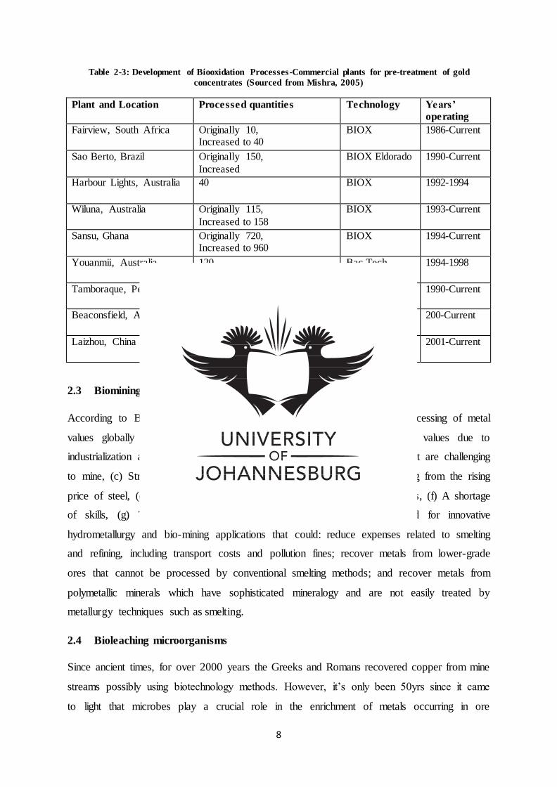
Table 2-3: Development of Biooxidation Processes-Commercial plants for pre-treatment of gold
concentrates (Sourced from Mishra, 2005)
Plant and Location Processed quantities Technology Years’
operating
Fairview, South Africa Originally 10, Increased to 40
BIOX 1986-Current
Sao Berto, Brazil Originally 150,
Increased
BIOX Eldorado 1990-Current
Harbour Lights, Australia 40 BIOX 1992-1994
Wiluna, Australia Originally 115,
Increased to 158
BIOX 1993-Current
Sansu, Ghana Originally 720, Increased to 960
BIOX 1994-Current
Youanmii, Australia 120 Bac Tech 1994-1998
Tamboraque, Peru 60 BIOX 1990-Current
Beaconsfield, Australia 70 Bac Tech 200-Current
Laizhou, China 100 Bac Tech 2001-Current
2.3 Biomining motivation
According to Brierly (2008), in the past century factors impacting the processing of metal
values globally have namely been: (a) The call for an array of metal values due to
industrialization and urbanization in China, (b) Discovery of ore minerals that are challenging
to mine, (c) Stringent environmental laws, (d) Increasing capital cost resulting from the rising
price of steel, (e) Increasing operation cost resulting from hiking energy prices, (f) A shortage
of skills, (g) Technology challenges. As such, these needs necessitated for innovative
hydrometallurgy and bio-mining applications that could: reduce expenses related to smelting
and refining, including transport costs and pollution fines; recover metals from lower-grade
ores that cannot be processed by conventional smelting methods; and recover metals from
polymetallic minerals which have sophisticated mineralogy and are not easily treated by
metallurgy techniques such as smelting.
2.4 Bioleaching microorganisms
Since ancient times, for over 2000 years the Greeks and Romans recovered copper from mine
streams possibly using biotechnology methods. However, it’s only been 50yrs since it came
to light that microbes play a crucial role in the enrichment of metals occurring in ore
9
precipitates and mine effluents (Bosecher, 1997). Since then bio-mining also known as bio-
hydrometallurgy has become a popular biotechnology process for the treatment of ores in
the mining sector (Schippers et al., 2013).With the global depletion of high grades ore
deposits increasing metal demands, conventional methods such as pyro metallurgy, chemical
processes etc. are becoming unfeasible. Microorganisms are more advantageous due to their
economic feasibility and their green technology properties (Das et al., 1999; Siddiqui et al.,
2009). Tailings from biological mining processes present minimal chemical activity and
sustain nominal biological life due to the degree of biological leaching, resulting in the
mitigation of environmental threats. In contrast, physiochemical methods exposed to the
atmosphere can undergo microbial leaching; releasing toxins into the environment (Celik,
2008). Microorganisms like bacteria and fungi transform metals into their soluble form
functioning as biological catalysts in leaching reactions. In addition microbial leaching can be
implemented in the recovery of valuable metals from industry waste. This process is gaining
increasing popularity in recovering valuable minerals including copper, gold, iron and
uranium (Siddiqui et al., 2009).
The microorganisms generally employed in biological leaching applications are known as
chemolithotrophs (Nakade, 2013). Chemolithotrophs are mainly acidophilic autotrophs that
play a role in the release of precious minerals from sulphide ores. These microbes develop in
inorganic media of low pH. They are able to withstand environments of elevated metal-ion
content. These microbes are mostly CO2 fixing chemolithoautotrophs and play a critical role
in oxidising the reaction of iron (II) to iron (III) and sulphur to sulphuric acid, which are
responsible for controlling the reaction kinetics (Das et al., 1999; Schippers et al., 2013).
Comprehension of the reaction kinetics is dependent on bacterial characterization and optimal
oxidation environments (Das et al., 1999).
The microbes responsible for the oxidation of iron (II) to iron (III) and sulphur to sulphuric
acid are namely Acidithiobacillus (Thiobacillus), Leptospirillium, Acidianus and Sulfolobus.
Acidithiobacillus (Thiobacillus) are a class of gram negative, non-sporing rods growing in
aerobic environments, which are mesophiles with the exception of Acidithiobacilli
(Thiobacilli) categorized as thermophiles which are capable of growing at greater
temperatures. Leptospirillium are gram negative, non-sporing, spiral structured bacteria.
Acidianus bacteria are sphere-shaped having saucer structured lobes. Sulfobacillus are gram
positive, rod structured bacteria having curved or narrowing ends and are capable of growing
at great temperatures (Bosecher, 1997; Das et al., 1999; Schippers et al., 2013). Even though
10
heap and tank bioleaching processes mostly operate at temperatures less than 40 °C, reactions
occurring at elevated temperatures are favoured promising greater bioleaching rates
(Schippers et al., 2013). Based on their optimal temperature ranges as shown in Table 2.4,
acidophilic microbes can be classified into three groups (Celik et al., 2008).
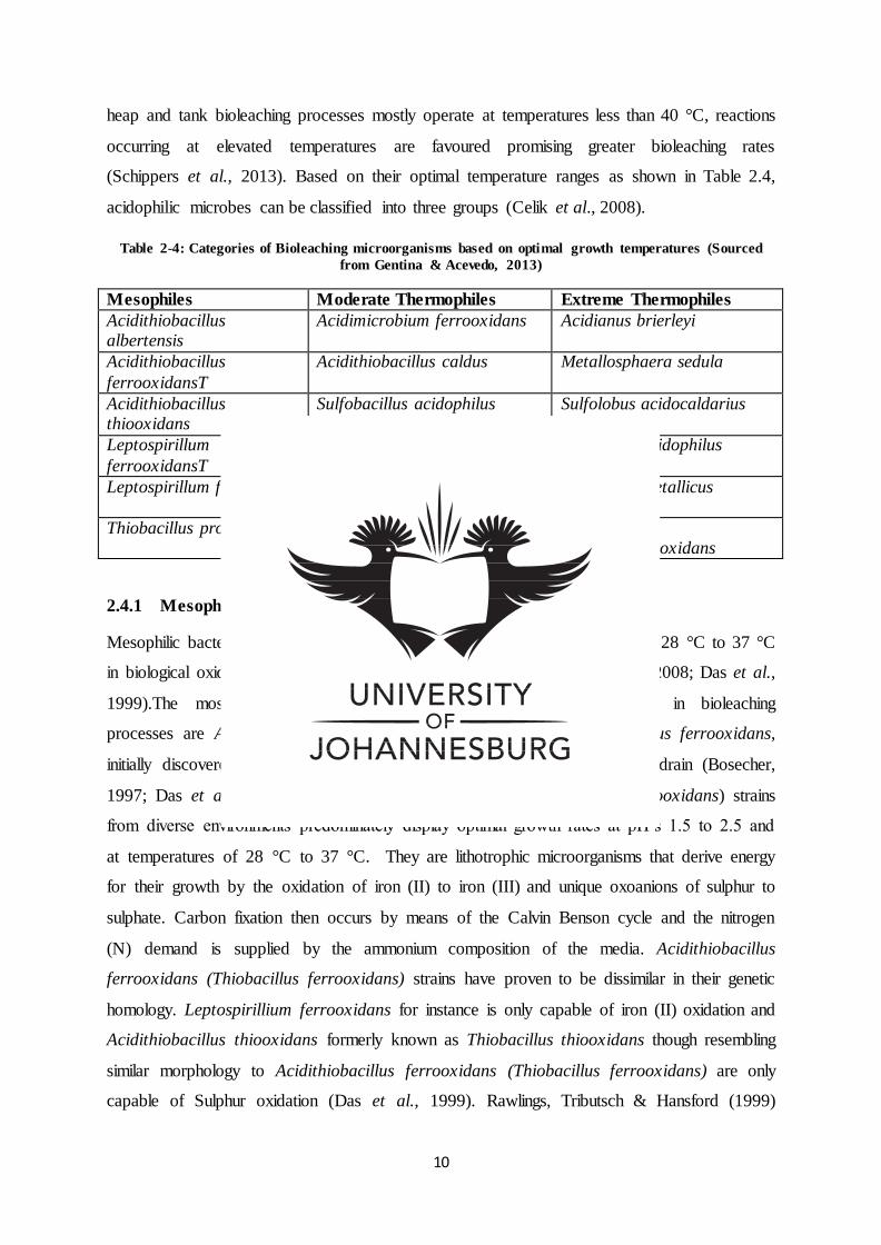
Table 2-4: Categories of Bioleaching microorganisms based on optimal growth temperatures (Sourced
from Gentina & Acevedo, 2013)
Mesophiles Moderate Thermophiles Extreme Thermophiles
Acidithiobacillus albertensis
Acidimicrobium ferrooxidans Acidianus brierleyi
Acidithiobacillus
ferrooxidansT
Acidithiobacillus caldus Metallosphaera sedula
Acidithiobacillus thiooxidans
Sulfobacillus acidophilus Sulfolobus acidocaldarius
Leptospirillum
ferrooxidansT
Sulfobacillus
thermosulfidooxidans
Sulfolobus acidophilus
Leptospirillum ferriphilum Sulfobacillus thermotolerans Sulfolobus metallicus
Thiobacillus prosperus Ferroplasma acidiphilum Sulfolobus thermosulfidooxidans
2.4.1 Mesophiles
Mesophilic bacteria are known to grow optimally at room temperatures, from 28 °C to 37 °C
in biological oxidation reactions occurring at 40 °C and lower (Celik et al., 2008; Das et al.,
1999).The most prevalent and commonly applied mesophilic bacteria in bioleaching
processes are Acidithiobacillus ferrooxidans formerly known as Thiobacillus ferrooxidans,
initially discovered in 1947 by Colmer and Hickle at an acid coal mine drain (Bosecher,
1997; Das et al., 1999). Acidithiobacillus ferrooxidans (Thiobacillus ferrooxidans) strains
from diverse environments predominately display optimal growth rates at pH’s 1.5 to 2.5 and
at temperatures of 28 °C to 37 °C. They are lithotrophic microorganisms that derive energy
for their growth by the oxidation of iron (II) to iron (III) and unique oxoanions of sulphur to
sulphate. Carbon fixation then occurs by means of the Calvin Benson cycle and the nitrogen
(N) demand is supplied by the ammonium composition of the media. Acidithiobacillus
ferrooxidans (Thiobacillus ferrooxidans) strains have proven to be dissimilar in their genetic
homology. Leptospirillium ferrooxidans for instance is only capable of iron (II) oxidation and
Acidithiobacillus thiooxidans formerly known as Thiobacillus thiooxidans though resembling
similar morphology to Acidithiobacillus ferrooxidans (Thiobacillus ferrooxidans) are only
capable of Sulphur oxidation (Das et al., 1999). Rawlings, Tributsch & Hansford (1999)
11
stated that in stirred-tanks the fixed Fe (III) concentrations are predominantly elevated, in
which case Acidithiobacillus ferrooxidans (Thiobacillus ferrooxidans) are regarded of
secondary importance to combinations of Leptospirillium ferrooxidans and Acidithiobacillus
thiooxidans (Thiobacillus thiooxidan ) or Acidithiobacillus caldus .These combinations have
also been noted to be dominant in copper heap leaching occurring at acidic pH ranges of pH
0.7 (Celik et al., 2008).
2.4.2 Moderate thermophiles
Moderate thermophiles are known to grow optimally at temperatures close to 50 °C,
oxidising Fe (II) and sulphur minerals at temperatures 45 °C to 65 °C (Celik et al., 2008; Das
et al., 1999). Compared to the study of mesophiles, minimal research has been conducted on
moderate thermophiles and as a result, the characterization and nomenclature of moderate
thermophiles has yet to be well established. Moderate thermophiles Sulfobacillus
thermosulfidooxidans are vital in bioleaching processes as sulphur and Fe (II) oxidizing
bacteria (Das et al., 1999). Other moderate thermophiles include Sulfobacillus acidophilus,
Acidophilus ferrooxidans, and Thiobacillus caldus (Celik et al., 2008). Moderate
thermophiles function at elevated temperatures exhibiting greater reaction rates in biological
leaching processes than mesophiles (Das et al., 1999). In cases where the temperature
exceeds a maximum of 45 °C, mesophiles are compromised and their protein is denatured
leading to cell death whiles moderate thermophiles thrive. Okibe et al. (2003) noted
Thiobacillus caldus as the most prevalent sulphur oxidizing bacteria in biological reactors
processing arsenopyrite/copper (Celik et al., 2008).
2.4.3 Extreme thermophiles
Extreme thermophiles are found growing optimally at extraordinary temperatures ranging
from 60 °C to 80 °C (Das et al., 1999).They fall under the domain Arcahae rather than
Bacteria. The genus Sulfolobus are among the most popular extreme thermophiles namely
Sulfolobus acidocaldarius, Sulfolobus sofataricus, Sulfolobus brierley, and Sulfolobus
ambioalous. They are capable of growing anaerobically whiles reducing sulphur elements,
growing aerobically whiles oxidizing sulphur and are capable of both Fe (II) and sulphur
oxidation. The application of extreme thermophiles in bioleaching processes is still under
development in contrast to popular processes that involve bacteria growing at optimum
temperatures below or at 50 °C (Celik, 2008; Das et al., 1999). In comparison to mesophiles
and moderate thermophiles, extreme thermophiles exhibit greater biological leaching

12
kinetics. As a result, heat exchangers in bioleaching reactions involving these extremophilic
thermophiles (Figure 2.1) might not be necessary in controlling bioleaching temperatures
(Das et al., 1999).
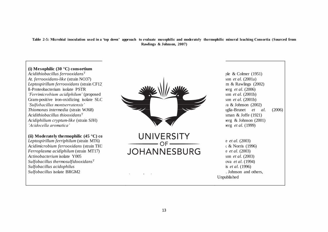
13
Table 2-5: Microbial inoculation used in a ‘top down’ approach to evaluate mesophilic and moderately thermophilic mineral leaching Consortia (Sourced from
Rawlings & Johnson, 2007)
(i) Mesophilic (30 °C) consortium
Acidithiobacillus ferrooxidansT
At. ferrooxidans-like (strain NO37) Leptospirillum ferrooxidans (strain CF12)
ß-Proteobacterium isolate PSTR ‘Ferrimicrobium acidiphilum’ (proposed strain)
Gram-positive iron-oxidizing isolate SLC66
‘Sulfobacillus montserratensis’ Thiomonas intermedia (strain WJ68)
Acidithiobacillus thiooxidansT
Acidiphilium cryptum-like (strain SJH) ‘Acidocella aromatica’
(ii) Moderately thermophilic (45 °C) consortium Leptospirillum ferriphilum (strain MT6)
Acidimicrobium ferrooxidans (strain TH3) Ferroplasma acidiphilum (strain MT17)
Actinobacterium isolate Y005
Sulfobacillus thermosulfidooxidansT Sulfobacillus acidophilus
Sulfobacillus isolate BRGM2
Autotrophic Fe2+/S-oxidizer, Fe3+-reducer
Autotrophic Fe2+/S-oxidizer, Fe3+-reducer Autotrophic Fe2+ -oxidizer
Autotrophic Fe2+-oxidizer Heterotrophic Fe2+-oxidizer, Fe3+-reducer
Heterotrophic Fe2+-oxidizer
Mixotrophic Fe2+/S-oxidizer, Fe3+-reducer Mixotrophic Fe2+/S
Autotrophic S-oxidizer
Heterotrophic Fe3+-reducer Heterotrophic Fe3+-reducer
Autotrophic Fe2+-oxidizer
Mixotrophic Fe2+-oxidizer, Fe3+-reducer Heterotrophic Fe2+-oxidizer, Fe3+-reducer
Heterotrophic Fe2+-oxidizer, Fe3+-reducer
Mixotrophic Fe2+/S-oxidizer, Fe3+-reducer Mixotrophic Fe2+/S-oxidizer, Fe3+-reducer
(Mixotrophic) Fe2+/S-oxidizer
Temple & Colmer (1951)
Johnson et al. (2001a) Coram & Rawlings (2002)
Hallberg et al. (2006) Johnson et al. (2001b)
Johnson et al. (2001b)
Yahya & Johnson (2002) Battaglia-Brunet et al. (2006)
Waksman & Joffe (1921)
Hallberg & Johnson (2001) Hallberg et al. (1999)
Okibe et al. (2003)
Clark & Norris (1996) Okibe et al. (2003)
Johnson et al. (2003)
Tourova et al. (1994) Norris et al. (1996)
D. B. Johnson and others,
Unpublished
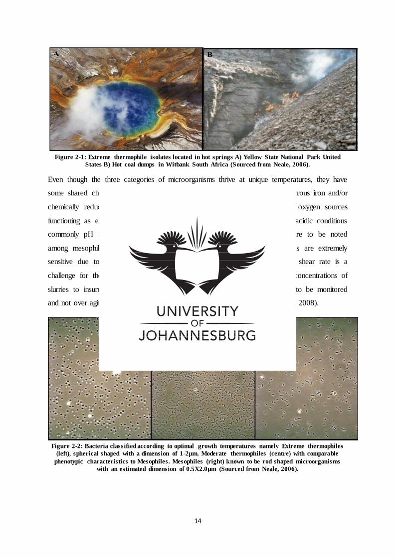
14
Figure 2-1: Extreme thermophile isolates located in hot springs A) Yellow State National Park United
States B) Hot coal dumps in Witbank South Africa (Sourced from Neale, 2006).
Even though the three categories of microorganisms thrive at unique temperatures, they have
some shared characteristics (Figure 2.2). They all attain their energy from ferrous iron and/or
chemically reduced sulphur complexes functioning as electron donors and oxygen sources
functioning as electron acceptors. The microbes are acidophiles, growing in acidic conditions
commonly pH 1.4-1.6 for bioleaching processes. Important dissimilarities are to be noted
among mesophiles and thermophiles. In contrast to mesophiles, thermophiles are extremely
sensitive due to the absence of a cell wall in their structure. As a result shear rate is a
challenge for thermophiles and lower agitation rates are essential with low concentrations of
slurries to insure their survival. Having a short life cycle thermophiles have to be monitored
and not over agitated without a continuous supply of concentrate (Celik et al., 2008).
Figure 2-2: Bacteria classified according to optimal growth temperatures namely Extreme thermophiles
(left), spherical shaped with a dimension of 1-2µm. Moderate thermophiles (centre) with comparable
phenotypic characteristics to Mesophiles. Mesophiles (right) known to be rod shaped microorganisms
with an estimated dimension of 0.5X2.0µm (Sourced from Neale, 2006).
15
Microbial heterotrophs such as bacteria and fungi also form a significant portion of the
bioleaching community. These microorganisms make use of extracellular metabolic products
and cellular lysates from autotrophic microbes as carbon sources thereof removing any
hindering effects of excessive carbon, and encouraging the development and iron-oxidation
of heterotrophs. Additionally heterotrophic microorganism also participate in solubilizing
metals by extracting organic acids known as citrates, gluconates, oxalates and succinates
(Brandl et al., 2008).
2.5 Halophilic bacteria
Biological leaching employing autotrophs though highly economical, can be ineffective for
specific elements (Zn, As). To overcome this shortfall, bioleaching using heterotrophs has
been established using organic matter for instance, yeast extracts or glucose which functions
as a nutrient source (Wang et al., 2015). Aerobic spore forming halophiles are a taxonomy of
gram positive rods that have been recovered from salt environments namely saltern
environments, estuarine-water, salt-lakes, salt foods, sea-ice, and deep sea hydro-thermal
vents. Halophiles are quickly growing popularity, due to their biotechnological prospective as
producers of harmonious solutes and hydrolysing enzymes. It has been indicated from 16s
rRNA gene sequencing that Bacillus species contain six distinctive phylogenetic groups with
numerous alkaliphilic or halophilic bacillus belonging to the rRNA Bacillus group (Lim et
al., 2006). Acidithiobacillus ferrooxidans halophiles isolated from saline-soil were discovered
to recover 72 % of Cu from ores (Nakade, 2013). Halophilic Bacillus species isolated by
Nithya & Pandian (2010) from the oceans depths were able to tolerate high metal volumes of
As.
2.6 Principle of microbial leaching
The principals that govern the mineralytic effect of bacteria / fungi on minerals include
“acidolysis”, “complexolysis” and “redoxolysis”. The mobilization of metals is facilitated by
the microbes through a) the development of organic / inorganic acids (protons), b) oxidation-
reduction reactions and c) elimination of coordination compounds. Sulphuric acids (H2SO4)
are common inorganic acids in leaching reactions and are the product of sulphur oxidising
microbes, Thiobacillus. The metabolic activity of theses bacteria /fungi produces chains of
organic acids causing organic acidolysis, complexolysis and chelating (Brandl, 2008).
16
2.6.1 Metal sulphide dissolution mechanism
Direct vs. Indirect mechanisms of bioleaching for describing microbial metal solubilisation of
sulphide minerals, have been one of the main discussions of past literature. Direct leaching
describes the ability of the microorganism to oxidize metal sulphides by the direct transferal
of electrons from reduced metal sulphides to the microbial cells bound to the minerals surface
(Shippers et al., 2013). Indirect leaching occurs via reduced metals facilitated by ferric (III)
ion which is produced through the microbial oxidation of ferrous (II) ions found in the
minerals. Ferric (III) ion thus functions as oxidizing agents, oxidizing metal sulphides
resulting in its reduction to ferrous (II) iron, which can then undergo microbial oxidation.
Nonetheless, the binding of microbes to the metal surface is not a clear suggestion of the
presence of a direct mechanism (Shippers et al., 2013). More especially, because of the
absence of direct electron transferal by enzymes and the lack of information around the
attraction of metal sulphides to the bound cell, the direct mechanism remains a theory.
Contrary, the bound cells demonstrate an effective “EPS-filled reaction area” supportive of
indirect leaching facilitated by ferric (III). Data has also been presented establishing indirect
leaching with none to support direct enzymatic processes (Brandl, 2008; Shippers et al.,
2013). As a result the labels “ direct-leaching” and “ indirect-leaching” have been replaced
by “contact-leaching” and “non-contact leaching” ,indicating the pertinence of microbial
attachment to the surface of the mineral (Mishra et al., 2005). Nonetheless the “direct-
leaching” and “indirect-leaching” models are still under review. Instead, an alternative model
has been proposed which is independent of the distinction between direct and indirect
leaching mechanisms. The model reviews all former knowledge formulating a comprehensive
leaching mechanism, characteristic of the following (Brandl, 2008; Mishra et al., 2005):
a) Cells have to be attached to the minerals and in physical contact with the surface;
b) Cells form and excrete exopolymers;
c) These exopolymeric cell envelopes contain ferric iron compounds that are complexed to
glucouronic residues. These are regarded as part of primary attack;
d) Thiosulfate is formed as intermediate during the oxidation of sulphur compounds;
e) Sulphur particles accumulate in the periplasmic-space alternatively occurring in the cell-
envelope;
Metal sulphide dissolution occurs through two unique reaction mechanism namely the
thiosulfate pathway and the polysulfate pathway. Dissolution generally couples proton attack
17
and oxidation reactions. The reaction mechanism depends on the mineral species (Brandl,
2008). According to Mishra et al. (2005) observations, this is because different metal
sulphides produce different intermediates in their reactions. As a result metal sulphides are
characterised according to their ability to solubilize in acids. This includes the (Mishra et al.,
2005; Siddiqui et al., 2009):
a) Thiosulfate pathway proposed for acid-insoluble metal sulphides such as pyrite(FeS2),
molybdenite (MoS2) and tungstenite(WS2);
b) Polysulfate mechanisms for acid-soluble metal sulphides spalerite (ZnS), chalcopyrite
(CuFeS2), galena (PbS), arsenopyrite (FeAsS) and hauerite (MnS2).
Microbes in bioleaching reactions attain energy by facilitating the breakdown of minerals into
their principal /basic elements. In the Thiosulfate mechanism, dissolution occurs through the
oxidation of acid-insoluble metal sulphide facilitated by electron extraction by ferric (III)
ions. The reaction outputs include a thiosulfate intermediate and a sulphate end product
(Siddiqui et al., 2009). The name Thiosulfate thus originates from the first liberated sulphur
product (Mishra et al., 2005).
The equation below illustrates the Thiosulfate mechanisms using pyrite (Siddiqui et al., 2009):
FeS2 + 6Fe3+ +3H2O S2O3
2- + 7Fe2+ + 6H+ (1)
S2O32- + 8 Fe3 + 5 H2O 2SO4
2- + 8Fe2+ + 10H+ (2)
In the Polysulfide mechanisms, dissolution occurs through a combination of electron
extraction facilitated by ferric (III) ions and proton attack (Siddiqui et al., 2009). The metal
sulphide chemical bond is broken by the binding of two protons to the sulphide moiety of the
molecule producing hydrogen sulphide (H2S). The sulphide cation then undergoes
spontaneous dimerization forming disulphide (H2S2) which then undergoes additional
oxidation by polysulfide/s producing intermediate product elemental sulphur, hence the name
“polysulfide mechanism” (Mishra et al., 2005). Though elemental sulphur is fairly stable,
sulphur oxidizing microorganisms can be employed to further oxidize elemental sulphur to
sulphate (Siddiqui et al., 2009).
18
The equation below illustrates the polysulfide mechanisms (Siddiqui et al., 2009):
MS + Fe3++ H+ M2+ + 0:5H2Sn + Fe 2+ (n ≥2) (3)
0:5 H 2Sn + Fe3+ 0:125S8+ Fe2+ + H+ (4)
0:125 S8 + 1.5O2+ H2O SO42- + 2H+ (5)
Figure 2-3: Summery diagram of a) thiosulfate mechanisms and b) polysulfide mechanism (Sourced from
Mishra et al., 2005).
2.7 Leaching technique
Biological mining is based on two principal methods know as percolation and agitation. The
percolation process consists of a “lixiviant” liquid medium percolating through a stationery
bed, while the agitation process consists of fine sized particles agitated in a “lixiviant” media.
Commercial bio-mining processes occur at a large scale and thus favour percolation methods.
The primary techniques applied commercially are in situ, dump, heap and vat leaching
(Siddiqui et al., 2009).
2.7.1 In situ leaching
This leaching process is applicable for mineral and metal recovery of ores found below the
earth’s surface. It’s characterised as solution-mining involving the extraction of ground
minerals through leaching and fluid-recovery. A pump action and gravity-flow facilitate the
movement of the solution (Figure 2.4). The extraction process produces a leachate labelled a
“pregnant solution” and the fluid returning to the extraction process is labelled a “barren
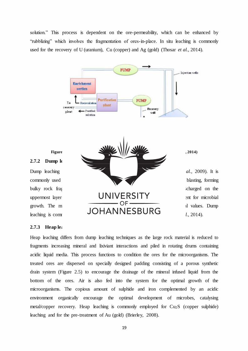
19
solution.” This process is dependent on the ore-permeability, which can be enhanced by
“rubblizing” which involves the fragmentation of ores-in-place. In situ leaching is commonly
used for the recovery of U (uranium), Cu (copper) and Ag (gold) (Thosar et al., 2014).
Figure 2-4: Operating flow for In situ Leaching (Sourced from Thosar et al., 2014)
2.7.2 Dump leaching
Dump leaching requires the piling up of uncrushed waste rock (Siddiqui et al., 2009). It is
commonly used for recovering low grade ores. Large rocks are ruptured by blasting, forming
bulky rock fragments for dump leaching. An acidic liquid solution is discharged on the
uppermost layer and filters through the dump creating a favourable environment for microbial
growth. The microbes initiate mineral oxidation for the dissolution of metal values. Dump
leaching is commonly employed for Cu2S (copper sulphide) ores (Thosar et al., 2014).
2.7.3 Heap leaching
Heap leaching differs from dump leaching techniques as the large rock material is reduced to
fragments increasing mineral and lixiviant interactions and piled in rotating drums containing
acidic liquid media. This process functions to condition the ores for the microorganisms. The
treated ores are dispersed on specially designed padding consisting of a porous synthetic
drain system (Figure 2.5) to encourage the drainage of the mineral infused liquid from the
bottom of the ores. Air is also fed into the system for the optimal growth of the
microorganisms. The copious amount of sulphide and iron complemented by an acidic
environment organically encourage the optimal development of microbes, catalysing
metal/copper recovery. Heap leaching is commonly employed for Cu2S (copper sulphide)
leaching and for the pre-treatment of Au (gold) (Brierley, 2008).
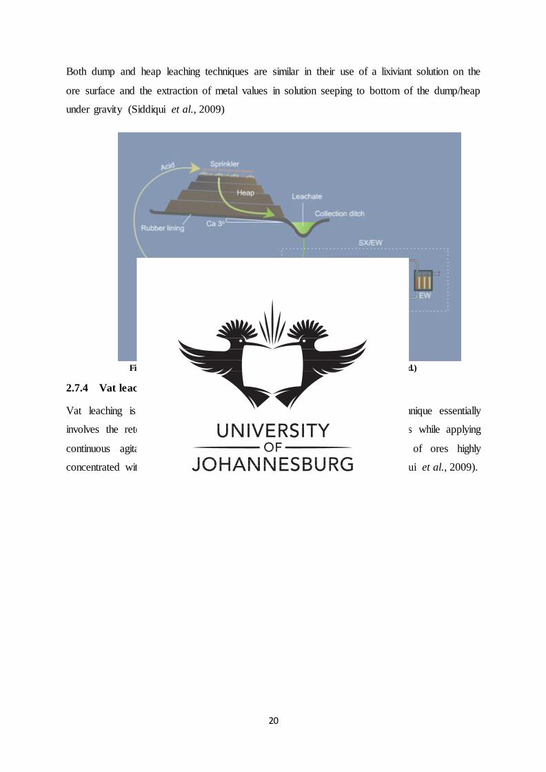
20
Both dump and heap leaching techniques are similar in their use of a lixiviant solution on the
ore surface and the extraction of metal values in solution seeping to bottom of the dump/heap
under gravity (Siddiqui et al., 2009)
Figure 2-5: Operating flow for Heap Leaching (Sourced from Bauer, n.d.)
2.7.4 Vat leaching
Vat leaching is operated for mineral extraction from oxide-ores. The technique essentially
involves the retention of ore-slurry and solvent for numerous hours in tanks while applying
continuous agitation (Figure 2.6). It is employed for the cyanidation of ores highly
concentrated with gold and to recover other precious metals from ores (Siddiqui et al., 2009).
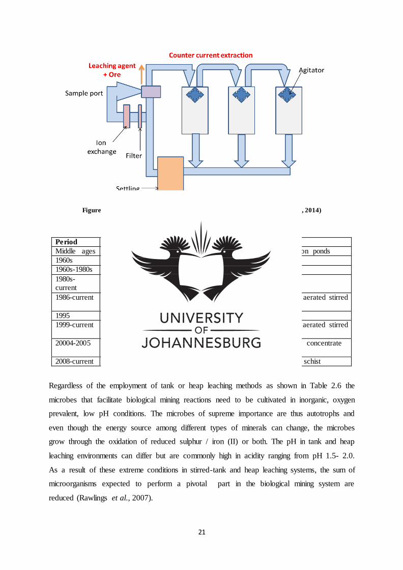
21
Figure 2-6: Operating process for Vat Leaching (Sourced from Thosar et al., 2014)
Table 2-6: Biomining technologies (Sourced from Johnson, 2013)
Period Location Technique
Middle ages China, Spain, UK Leaching of copper ores using precipitation ponds
1960s Utah, USA Copper dump leaching implemented
1960s-1980s Canada In situ mining of uranium
1980s-current
Chile Heap leaching of copper
1986-current South Africa Bio-oxidation of refractory gold ores in aerated stirred tanks
1995 Nevada, USA Bio heap leaching of gold ores
1999-current Kasese, Uganda Bioleaching of cobalt ,ferrous pyrite in aerated stirred tanks
20004-2005 Chuquicamata, Chile
Thermophilic bioleaching of chalcopyrite concentrate
2008-current Talvivaara, Finland Bio heap leaching of polymetallic black schist
Regardless of the employment of tank or heap leaching methods as shown in Table 2.6 the
microbes that facilitate biological mining reactions need to be cultivated in inorganic, oxygen
prevalent, low pH conditions. The microbes of supreme importance are thus autotrophs and
even though the energy source among different types of minerals can change, the microbes
grow through the oxidation of reduced sulphur / iron (II) or both. The pH in tank and heap
leaching environments can differ but are commonly high in acidity ranging from pH 1.5- 2.0.
As a result of these extreme conditions in stirred-tank and heap leaching systems, the sum of
microorganisms expected to perform a pivotal part in the biological mining system are
reduced (Rawlings et al., 2007).
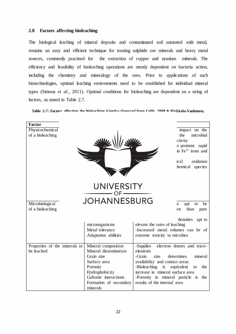
22
2.8 Factors affecting bioleaching
The biological leaching of mineral deposits and contaminated soil saturated with metal,
remains an easy and efficient technique for treating sulphide ore minerals and heavy metal
sources, commonly practised for the extraction of copper and uranium minerals. The
efficiency and feasibility of bioleaching operations are mostly dependent on bacteria action,
including the chemistry and mineralogy of the ores. Prior to applications of such
biotechnologies, optimal leaching environments need to be established for individual mineral
types (Simona et al., 2011). Optimal conditions for bioleaching are dependent on a string of
factors, as stated in Table 2.7.
Table 2-7: Factors affecting the bioleaching kinetics (Sourced from Celik, 2008 & Riekkola-Vanhanen,
2010)
Factor Parameters Outcome
Physicochemical parameters
of a bioleaching environment
Temperature
pH
Redox potential Water potential
Oxygen content and
availability Carbon dioxide content
Mass transfer Nutrient availability
Iron(III) concentration
Light Pressure
Surface tension
Presence of inhibitors
-Temperature has an impact on the
leaching rates , the microbial
organization and productivity -Low pH is required to promote rapid
leaching and to maintain Fe3+ irons and
metals in solution -In biological/chemical oxidation
electron accepting chemical species are required
Microbiological parameters
of a bioleaching environment
Microbial diversity
Population density Microbial activities
Spatial distribution of microorganisms
Metal tolerance
Adaptation abilities
-Bacterial consortiums apt to be
resilient and effective than pure cultures
-Increased population densities apt to elevate the rates of leaching
-Increased metal volumes can be of
extreme toxicity to microbes
Properties of the minerals to be leached
Mineral composition Mineral dissemination
Grain size
Surface area Porosity
Hydrophobicity Galvanic interactions
Formation of secondary
minerals
-Supplies electron donors and trace-elements
-Grain size determines mineral
availability and contact areas -Bioleaching is equivalent to the
increase in mineral surface area -Porosity in mineral particle is the
results of the internal area
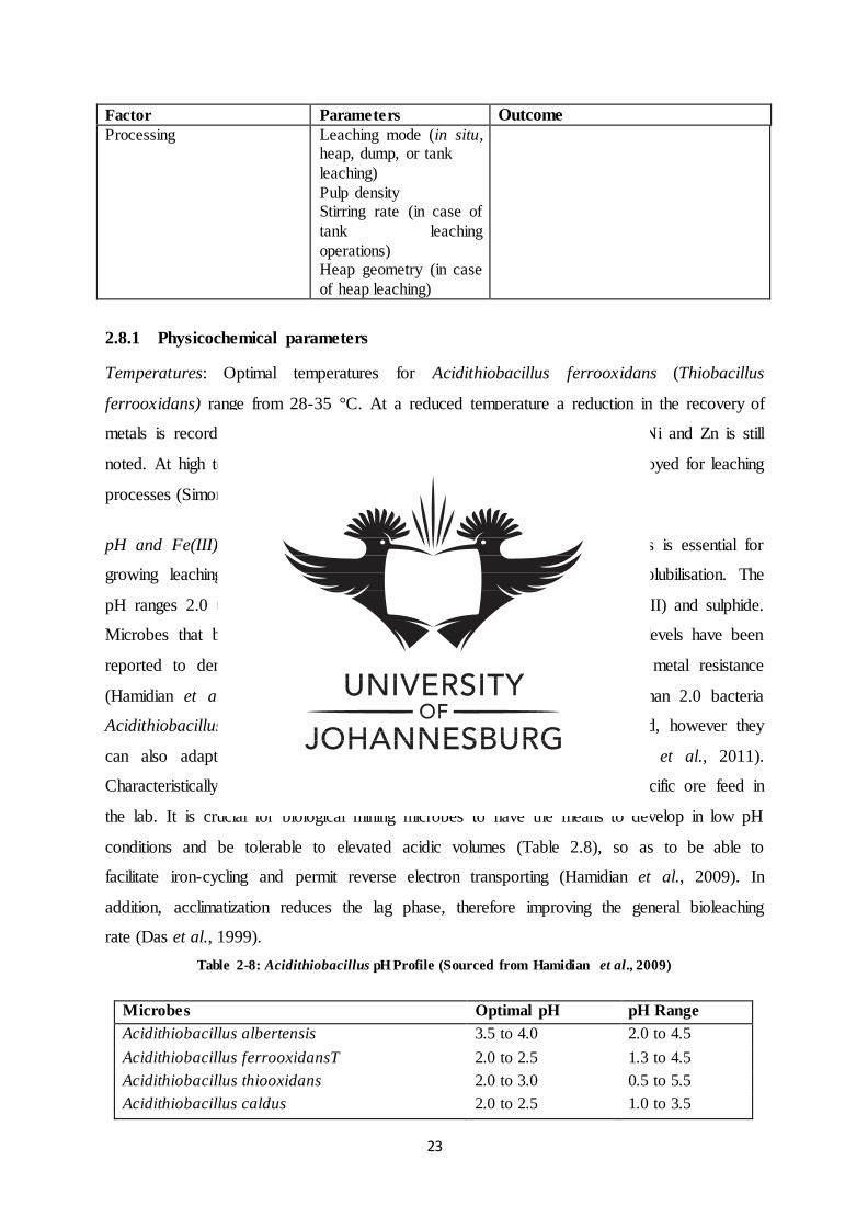
23
Factor Parameters Outcome
Processing Leaching mode (in situ, heap, dump, or tank
leaching)
Pulp density Stirring rate (in case of
tank leaching
operations) Heap geometry (in case
of heap leaching)
2.8.1 Physicochemical parameters
Temperatures: Optimal temperatures for Acidithiobacillus ferrooxidans (Thiobacillus
ferrooxidans) range from 28-35 °C. At a reduced temperature a reduction in the recovery of
metals is recorded, nevertheless at 4 °C bacteria solubilisation of Cu+, Co, Ni and Zn is still
noted. At high temperature ranges 50 °C to 80 °C thermophiles can be employed for leaching
processes (Simona et al., 2011).
pH and Fe(III) Concentrations: The modification of pH to the right values is essential for
growing leaching microorganisms and is a determining factor for metal solubilisation. The
pH ranges 2.0 to 2.5 offer optimal conditions for microbes to oxidise iron (II) and sulphide.
Microbes that biologically oxidize sulphide mineral deposits at reduced pH levels have been
reported to demonstrate resilience to acid prone environments and heavy metal resistance
(Hamidian et al., 2009; Simona et al., 2011). In pH environments less than 2.0 bacteria
Acidithiobacillus ferrooxidans (Thiobacillus ferrooxidans) can be inhibited, however they
can also adapt to reduced pH when sulphuric acid is added (Simona et al., 2011).
Characteristically, microorganisms are initially grown and acclimatized to specific ore feed in
the lab. It is crucial for biological mining microbes to have the means to develop in low pH
conditions and be tolerable to elevated acidic volumes (Table 2.8), so as to be able to
facilitate iron-cycling and permit reverse electron transporting (Hamidian et al., 2009). In
addition, acclimatization reduces the lag phase, therefore improving the general bioleaching
rate (Das et al., 1999).
Table 2-8: Acidithiobacillus pH Profile (Sourced from Hamidian et al., 2009)
Microbes Optimal pH pH Range
Acidithiobacillus albertensis 3.5 to 4.0 2.0 to 4.5
Acidithiobacillus ferrooxidansT 2.0 to 2.5 1.3 to 4.5
Acidithiobacillus thiooxidans 2.0 to 3.0 0.5 to 5.5
Acidithiobacillus caldus 2.0 to 2.5 1.0 to 3.5
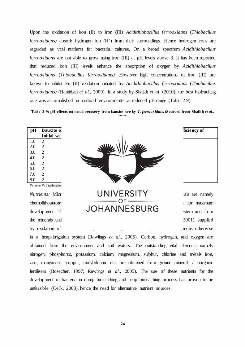
24
Upon the oxidation of iron (II) to iron (III) Acidithiobacillus ferrooxidans (Thiobacillus
ferrooxidans) absorb hydrogen ion (H+) from their surroundings. Hence hydrogen irons are
regarded as vital nutrients for bacterial cultures. On a broad spectrum Acidithiobacillus
ferrooxidans are not able to grow using iron (III) at pH levels above 3. It has been reported
that reduced iron (III) levels enhance the absorption of oxygen by Acidithiobacillus
ferrooxidans (Thiobacillus ferrooxidans). However high concentrations of iron (III) are
known to inhibit Fe (II) oxidation initiated by Acidithiobacillus ferrooxidans (Thiobacillus
ferrooxidans) (Hamidian et al., 2009). In a study by Shaikh et al. (2010), the best bioleaching
rate was accomplished in oxidized environments at reduced pH range (Table 2.9).
Table 2-9: pH effects on metal recovery from bauxite ore by T. ferrooxidans (Sourced from Shaikh et al.,
2010)
Where Wt indicates weight of the ores
Nutrients: Microbes utilised for the recovery of metals from sulphide minerals are namely
chemolithoautotrophs which are bacteria that require inorganic components for maximum
development. The nutrients are generally sourced from the surrounding ecosystem and from
the minerals undergoing leaching processes (Bosecher, 1997; Waites et al., 2001), supplied
by oxidation of iron and/or sulphur containing minerals suspended in an aqueous otherwise
in a heap-irrigation system (Rawlings et al., 2005). Carbon, hydrogen, and oxygen are
obtained from the environment and soil waters. The outstanding vital elements namely
nitrogen, phosphorus, potassium, calcium, magnesium, sulphur, chlorine and metals iron,
zinc, manganese, copper, molybdenum etc. are obtained from ground minerals / inorganic
fertilisers (Bosecher, 1997; Rawlings et al., 2005). The use of these nutrients for the
development of bacteria in dump bioleaching and heap bioleaching process has proven to be
unfeasible (Celik, 2008), hence the need for alternative nutrient sources.
pH Bauxite ore(g) Metal recovered (%) Bio extraction efficiency of
Al and Fe (%) Initial wt Final wt Al Fe
1.0 2 2.789 66.20 80.01 70.49 2.0 2 2.896 81.13 84.82 82.28
3.0 2 2.846 77.59 80.64 78.54
4.0 2 2.856 68.95 75.20 70.89
5.0 2 2.734 63.87 65.92 64.51 6.0 2 2.72 55.67 59.88 56.98
7.0 2 2.679 52.74 54.47 53.28
8.0 2 2.523 40.12 49.39 43.56
25
Various nutrient media (Table 2.10-2.12) have been suggested for bioleaching as derived
from the cited nutrients in variable quantities, such as the much quoted 9K media. High
recovery of zinc, copper and iron metal concentration were reported in 9K media than in
Norris media. The salt concentrations in the additional media formulations were
suggestively lower (Deveci et al., 2015). The main uncertainty surrounding the application
of 9K media is the possible sedimentation of phosphate, potassium and ammonium in the
form of jarosite compounds as a result of their great concentrations in the media (Deveci et
al., 2015).
Metal elements such as calcium and magnesium are found in adequate concentrations from
degrading rocks in acid leachates, whereas nitrogen and potassium can be scarce and need to
be added in the leaching reaction. In a study by Sarcheshmehpour et al. (2009)
characterization of low sulphide ores established that calcium ,magnesium and sulphur
quantities were abundant for supporting microbial growth ,whiles nitrogen, phosphorous and
potassium were lacking. Thus the addition of nitrogen, phosphate and potassium as an
affordable fertiliser revealed that, the addition of potassium has optimal results on the
biological leaching of copper ores whiles nitrate and chloride inhibit bacterial growth. On the
contrary, it was reported by Celik (2008) that the addition of phosphate and ammonium
should be avoided due to the expenses of such chemicals and the danger of the precipitation
of these chemicals in the form of jarosites and ferric phosphates. The addition of phosphate
and ammonium nutrients takes place in stirred-tank bioleaching processes and the occasional
addition of ammonium takes place in heap leaching processes. Potassium addition in tank
bioleaching process acts as hydroxides, sulphates and sometimes phosphates (Celik, 2008).
For optimization of metal recovery Bosecker (1997) and Simonan et al. (2011) reported that
iron and sulphur components can be augmented along with NH4, PO43 and Mg salt. While
Acevedo et al. (1989) reported that Thiobacillus ferrooxidans are able to grow in defined
media (Table 2.13) and obtain energy from oxidizing reduced sulphur, elemental sulphur and
ferrous (Acevedo et al., 1989).
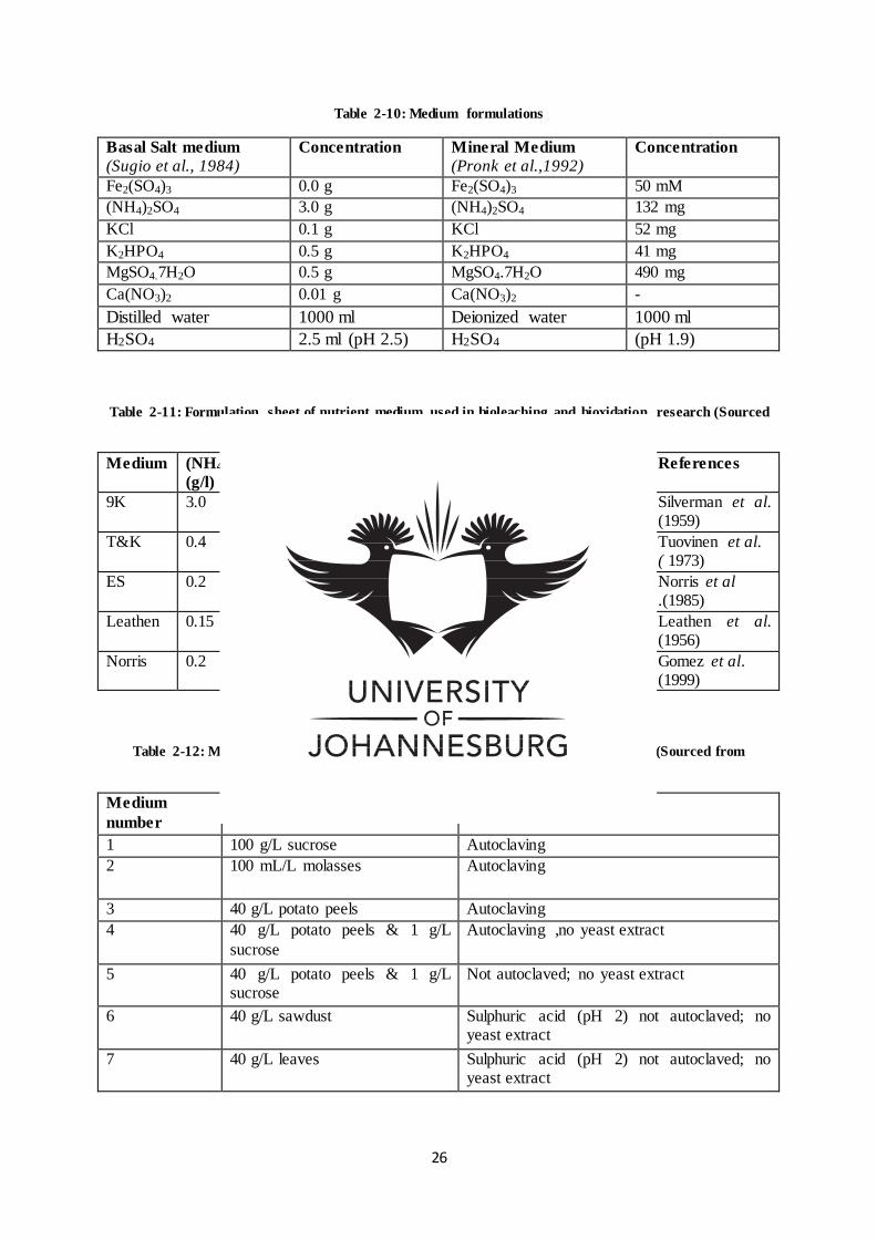
26
Table 2-10: Medium formulations
Basal Salt medium
(Sugio et al., 1984) Concentration Mineral Medium
(Pronk et al.,1992) Concentration
Fe2(SO4)3 0.0 g Fe2(SO4)3 50 mM
(NH4)2SO4 3.0 g (NH4)2SO4 132 mg
KCl 0.1 g KCl 52 mg
K2HPO4 0.5 g K2HPO4 41 mg
MgSO4.7H2O 0.5 g MgSO4.7H2O 490 mg
Ca(NO3)2 0.01 g Ca(NO3)2 -
Distilled water 1000 ml Deionized water 1000 ml
H2SO4 2.5 ml (pH 2.5) H2SO4 (pH 1.9)
Table 2-11: Formulation sheet of nutrient medium used in bioleaching and bioxidation research (Sourced
from Deveci et al., 2015)
Medium (NH4)2SO4
(g/l)
MgSO4.7H2O
(g/l)
KH2PO4
(g/l)
KCl
(g/l)
Ca(NO3)2.H2O
(g/l)
References
9K 3.0 0.5 0.5 0.1 0.01 Silverman et al. (1959)
T&K 0.4 0.4 0.4 - - Tuovinen et al. ( 1973)
ES 0.2 0.4 0.1 0.1 - Norris et al .(1985)
Leathen 0.15 0.5 0.01 0.05 0.05 Leathen et al. (1956)
Norris 0.2 0.2 0.2 - - Gomez et al. (1999)
Table 2-12: Medium formulation studies for bioleaching of heavy metals from ores (Sourced from
Mulligan et al., 2004)
Medium
number
Substrates Preliminary treatment
1 100 g/L sucrose Autoclaving
2 100 mL/L molasses Autoclaving
3 40 g/L potato peels Autoclaving
4 40 g/L potato peels & 1 g/L
sucrose
Autoclaving ,no yeast extract
5 40 g/L potato peels & 1 g/L sucrose
Not autoclaved; no yeast extract
6 40 g/L sawdust Sulphuric acid (pH 2) not autoclaved; no yeast extract
7 40 g/L leaves Sulphuric acid (pH 2) not autoclaved; no yeast extract
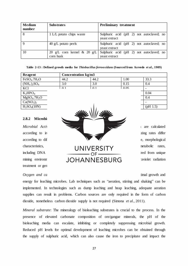
27
Medium
number
Substrates Preliminary treatment
8 1 L/L potato chips waste Sulphuric acid (pH 2) not autoclaved; no yeast extract
9 40 g/L potato peels Sulphuric acid (pH 2) not autoclaved; no
yeast extract
10 20 g/L corn kernel & 20 g/L corn husk
Sulphuric acid (pH 2) not autoclaved; no yeast extract
Table 2-13 : Defined growth media for Thiobacillus ferrooxidans (Sourced from Acevedo et al., 1989)
Reagent Concentration kg/m3
FeSO4.7H2O 44.2 44.2 1.00 33.3
(NH4 )2SO4 3.0 3.0 0.15 0.4
KCl 0.1 0.1 0.05 -
K2HPO4 0.5 0.5 0.05 0.04
MgSO4.7H2O 0.5 0.5 0.50 0.4
Ca(NO3)2 0.01 0.01 0.01 -
H2SO4(10N) 1.0cm3/dm3 1.0cm3/dm3 (pH 3.5) (pH 1.5)
2.8.2 Microbiological parameters
Microbial Activity: The activity of Thiobacillus and acidophilic bacteria are calculated
according to iron and sulphur oxidizing rates. The iron and sulphur oxidizing rates differ
according to different strains. The oxidation rates differ based on aspects like, morphological
characteristics, lipopolysaccharide cell envelope chemistry, nutrient metabolic rates,
including DNA nucleobase ratios. High activity microorganisms can be acquired from unique
mining environments and microbial activity can be multiplied through ultraviolet radiation
treatment or genetic engineering processes (Das et al., 1999).
Oxygen and carbon dioxide: Sufficient excess to oxygen is required for optimal growth and
energy for leaching microbes. Lab techniques such as “aeration, stirring and shaking” can be
implemented. In technologies such as dump leaching and heap leaching, adequate aeration
supplies can result in problems. Carbon sources are only required in the form of carbon
dioxide, nonetheless carbon dioxide supply is not required (Simona et al., 2011).
Mineral substrate: The mineralogy of bioleaching substrates is crucial to the process. In the
presence of elevated carbonate composition of ore/gangue minerals, the pH of the
bioleaching media can escalate, inhibiting or completely suppressing microbial growth.
Reduced pH levels for optimal development of leaching microbes can be obtained through
the supply of sulphuric acid, which can also cause the iron to precipitate and impact the
28
costing of the operation. The bioleaching rates are dependent on substrate surface area as
well. A reduction in the particle magnitude implies increased surface area and thus increased
metal yield short of changes in total particle mass. Particle sizes with a range of 42 µm are
considered as optimal. An increase in the particle surface area is achieved by increasing pulp
density. The increasing of pulp density can elevate metal recovery (Simona et al., 2011). It
has been reported that in the mineralogy of low grade sulphide ores, decreasing particles sizes
expressed larger quantities of Pb, Fe, S and Si whiles Zn and Cu volumes reduced (Olubambi
et al., 2008). Olubami et al. (2007) reported significant recoveries acquired at particle sizes in
the range of 75 µm whiles 106 µm particles expressed minimal recovery.
Heavy metals: Bioleaching of metal sulphides is associated with increased metal
concentrations in the leaching solution. Generally bioleaching microbes more importantly
Acidithiobacillus /Thiobacillus (Table 2.14) are tolerant of heavy metals and other
microorganisms can have a tolerance for e.g. 50 g/L Ni, 55 g/L Cu or 112 g/L Zn. Unique
genus of the same species can display dissimilar heavy metal sensitivity. Single bacteria
species can also be adapted to high metal conditions and substrate through gradual increase of
metal/substrate concentration (Simona et al., 2011).
Water: Water makes up 80 % to 90 % of total cell mass and performs a pivotal function in
solubilizing nutrients, required for transport and hydrolysis reaction. Water activity
parameters quantify its accessibility. In nutrients water is associated with other compounds
e.g. salt, protein and is inaccessible for microbes requiring isolated water for growth (Simona
et al., 2011).
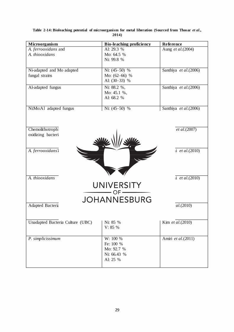
29
Table 2-14: Bioleaching potential of microorganism for metal liberation (Sourced from Thosar et al.,
2014)
Microorganism Bio-leaching proficiency Reference
A. ferrooxidans and
A. thiooxidans
Al: 29.3 %
Mo: 64.5 % Ni: 99.8 %
Aung et al.(2004)
Ni-adapted and Mo adapted
fungal strains
Ni: (45–50) %
Mo: (62–66) % Al: (30–33) %
Santhiya et al.(2006)
Al-adapted fungus Ni: 88.2 %,
Mo: 45.1 %, Al: 68.2 %
Santhiya et al.(2006)
Ni:Mo:Al adapted fungus Ni: (45–50) %
Mo: 82.3 %
Al: 65.2 %
Santhiya et al.(2006)
Chemolithotrophic sulphur
oxidizing bacteria
Ni: 89.5 %
V: 95.8 %
Mo: 21.5 %
Mishra et al.(2007)
A. ferrooxidansT Al: 63 % Co: 96 %
Mo: 84 %
Ni: 99 %
Gholami et al.(2010)
A. thiooxidans
Al: 2.4 %
Co: 83 %
Mo: 95 % Ni: 16 %
Gholami et al.(2010)
Adapted Bacteria Culture (ABC)
Ni: 95 %
V: 95 %
Kim et al.(2010)
Unadapted Bacteria Culture (UBC)
Ni: 85 % V: 85 %
Kim et al.(2010)
P. simplicissimum W: 100 %
Fe: 100 % Mo: 92.7 %
Ni: 66.43 %
Al: 25 %
Amiri et al.(2011)
30
2.9 Recovery of base metals from slag
The recovery of Cu, Ni and additional nonferrous minerals by means of smelting processes
is associated with the accumulation of large volumes of slag. With reference to copper ore, 1 t
of ore gives rise to 22 t of slag (Kalinkin et al., 2012). Slag recovered from nonferrous
smelting processes can often contain significant volumes of metal values such as cobalt,
copper and nickel which can be recovered by pyrometallurgy and hydrometallurgy processes
(Kalinkin et al., 2012; Maweja et al., 2010; Wang et al., 2015). Nonferrous slag or cleaned
nonferrous slag containing low quantities of metal values can also be applied in the
manufacturing of a tools, paving, concrete, tiles, glass and roofing (Yang et al., 2010). Slag is
often designed for optimizing the removal of gangue elements including the oxides produced
by ore smelters. The content of slag is thus determined by raw material (Table 2.15) and the
smelting process employed. The quantities of metal are automatically drawn into the slag in
the course of tapping, resulting in slag material containing base metals (Maweja et al., 2010).
Hydrometallurgy methods for recovering valuable metals have been extensively investigated,
such as the recovery of Zn, Co, Cu from copper ores and slag, with one study focusing on
atmospheric leaching using acid, base and salt as solvents; and high pressure oxidation acidic
leaching. The applications of roasting methods and additional processing for slag have also
been reported. Then again, this approach has proven to be costly and poorly selective .Slag
waste containing base metals (Co, Ni, Cu) has also been reported to pollute the atmosphere
(Kalinkin et al., 2012;Yang et al., 2010). Environmental contamination can result through
numerous systems e.g. the suspension of metals from slag dumps in acid rain water as well as
the air driven corrosion and transport of slag dust (Maweja et al., 2010). Taking into
consideration the environmental and financial implications of contamination, there is a need
for efficient and economic solutions for the recovery of these metal values and disposal of
slag waste material (Yang et al., 2010).

31
Table 2-15: Bioleaching investigations using an array of minerals
Solid material Reference
Fly ash Burgstaller et al. (1993); Bosshard et al. (1996); Brombacher et al. (1998)
Sewage sludge Jenkins et al. (1981); Tyagi et al. (1991); Chartier et al. ( (1997)
Spent batteries Cerruti et al. (1998)
Electronic scrap material
Brandl et al. (2001); Ilyas et al. (2007); Ilyas et al. (2010)
Ores Mousavi et al. (2005); Mousavi et al. (2006a); Mousavi et al. (2006b);
Mousavi et al. (2007); Mousavi et al. (2008); Soleimani, et al. (2009);
Soleimani et al. (2011)
Spent catalysts Mafi et al. (2010); Mafi et al. (2011); Amiri et al. (2011a); Amiri et al.
(2011b); Amiri et al. (2011c); Amiri et al. (2012)
2.10 Ionic liquids as an alternative medium
Reduced enzyme turnover rates have also been observed in organic media sparking an
interest in alternative, environmentally friendly solvents and reaction media (Silva et al.,
2014). Inorganic nutrients are necessary for the development of chemolithotrophs applied
in metal recovery from sulphide mineral deposits. Other bioleaching microbes make use of
critical nutrients such as C, H, O found in the environment and ground water. The
outstanding nutrients N, P, K, Ca, Mg, SO32
, Cl including heavy metal Fe, Zn, Mn, and Cu
etc. are sourced from ground mineral deposits/inorganic fertilizer. Such nutrients are
provided in the lab in the form of a media. The establishment of such media for the
development of bacteria for the bioleaching of heaps and dumps on an industrial scale is
costly. As a result of the cost and accessibility of these nutrients, optional compounds are
required. It’s imperative that individual elements initiate elevated microbial growth and not
hinder microbial activity (Sarcheshmehpour et al., 2009).
2.10.1 Properties of ionic liquids (ILs)
Ionic liquids gained significant popularity in 1914 when Paul Walden produced ethyl
ammonium nitrate (room temperature salts), since then from the year 2000 the quantity of
ionic liquid based articles published has mushroomed (Figure 2.7) more especially between
1998 and 2013.
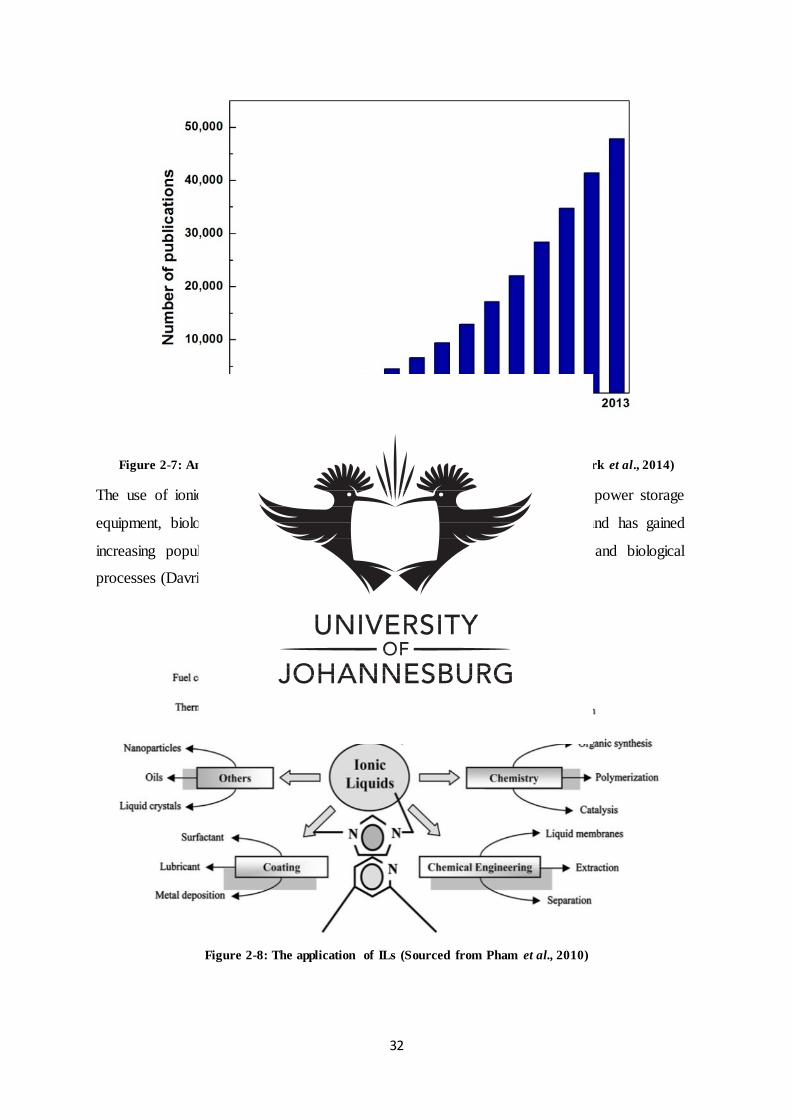
32
Figure 2-7: Annual growth rates of ionic liquid published articles (Sourced from Park et al., 2014)
The use of ionic liquids has expanded to catalytic reactions, electrolytes for power storage
equipment, biological sensors, and extraction/recovery agents (Figure 2.8); and has gained
increasing popularity as replacements of conventional solvents in chemical and biological
processes (Davris et al., 2014; Lee et al., 2005; Park et al., 2014).
Figure 2-8: The application of ILs (Sourced from Pham et al., 2010)
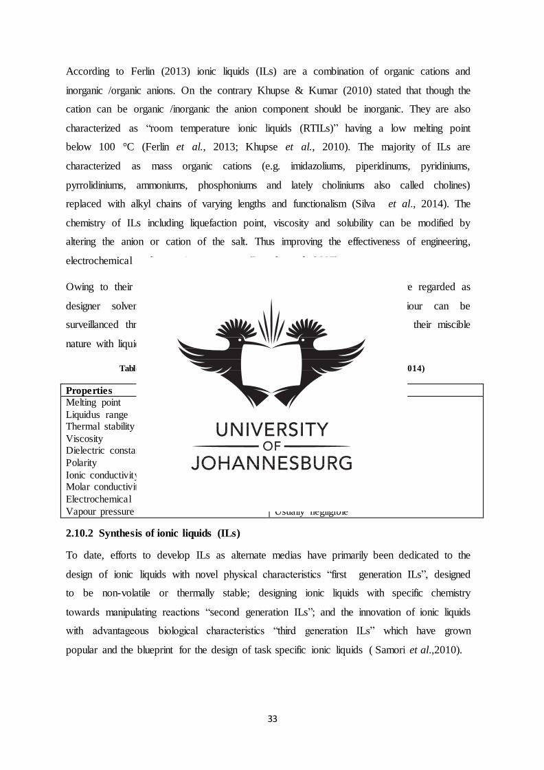
33
According to Ferlin (2013) ionic liquids (ILs) are a combination of organic cations and
inorganic /organic anions. On the contrary Khupse & Kumar (2010) stated that though the
cation can be organic /inorganic the anion component should be inorganic. They are also
characterized as “room temperature ionic liquids (RTILs)” having a low melting point
below 100 °C (Ferlin et al., 2013; Khupse et al., 2010). The majority of ILs are
characterized as mass organic cations (e.g. imidazoliums, piperidinums, pyridiniums,
pyrrolidiniums, ammoniums, phosphoniums and lately choliniums also called cholines)
replaced with alkyl chains of varying lengths and functionalism (Silva et al., 2014). The
chemistry of ILs including liquefaction point, viscosity and solubility can be modified by
altering the anion or cation of the salt. Thus improving the effectiveness of engineering,
electrochemical and extraction processes (Pernak et al., 2007).
Owing to their capabilities to adapt their physicochemical conditions ILs are regarded as
designer solvents (Table 2.16). Hence hydrophobic/hydrophilic behaviour can be
surveillanced through the modification of the cation chain lengths, including their miscible
nature with liquid by employing unique anions (Davris et al., 2014).
Table 2-16: Unique properties of ionic liquids (Sourced from Park et al., 2014)
Properties Values Melting point
Liquidus range Thermal stability
Viscosity Dielectric constant
Polarity
Ionic conductivity Molar conductivity
Electrochemical window
Vapour pressure
Preferably below 100 °C
Often > 200 °C Usually high
Normally < 100 cP, workable Implied < 30
Moderate
Usually < 10 mS/cm <10 Scm2/mol
Often > 4 V
Usually negligible
2.10.2 Synthesis of ionic liquids (ILs)
To date, efforts to develop ILs as alternate medias have primarily been dedicated to the
design of ionic liquids with novel physical characteristics “first generation ILs”, designed
to be non-volatile or thermally stable; designing ionic liquids with specific chemistry
towards manipulating reactions “second generation ILs”; and the innovation of ionic liquids
with advantageous biological characteristics “third generation ILs” which have grown
popular and the blueprint for the design of task specific ionic liquids ( Samori et al.,2010).
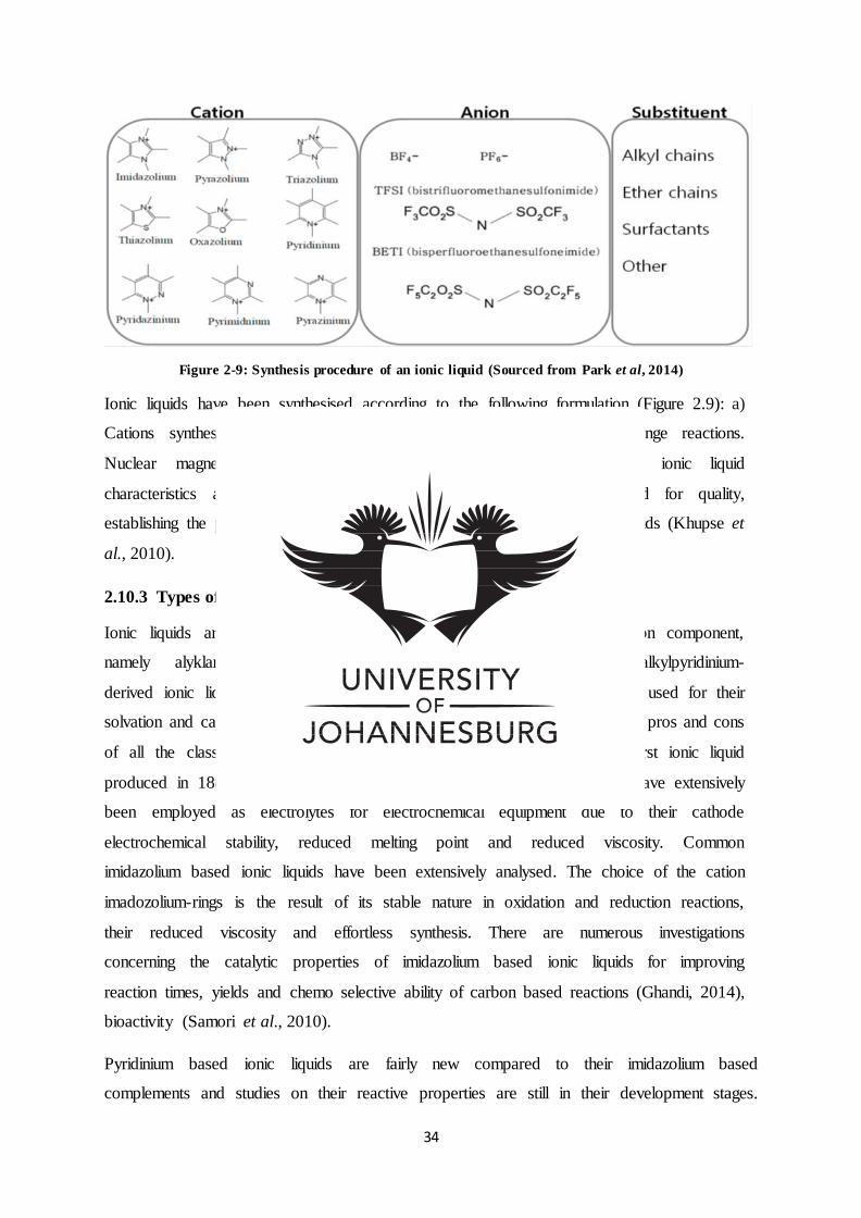
34
Figure 2-9: Synthesis procedure of an ionic liquid (Sourced from Park et al, 2014)
Ionic liquids have been synthesised according to the following formulation (Figure 2.9): a)
Cations synthesised using quaternization reactions and, b) Anion exchange reactions.
Nuclear magnetic resonance is then employed for determining the ionic liquid
characteristics and purity. Karl –Fischer coulometer can be employed for quality,
establishing the presence of water from the purified and desiccated ionic liquids (Khupse et
al., 2010).
2.10.3 Types of ionic liquids (ILs)
Ionic liquids are classified under four categories according to their cation component,
namely alyklammonium-, dialkylimidazolium-, phosphonium and N-alkylpyridinium-
derived ionic liquids. Even though these ionic liquids have effectively been used for their
solvation and catalytic properties, they do pose certain restrictions. Hence the pros and cons
of all the classes will be discussed. Ethanolammonium nitrate was the first ionic liquid
produced in 1888 by Gabriel. Since then, ammonium based ionic liquids have extensively
been employed as electrolytes for electrochemical equipment due to their cathode
electrochemical stability, reduced melting point and reduced viscosity. Common
imidazolium based ionic liquids have been extensively analysed. The choice of the cation
imadozolium-rings is the result of its stable nature in oxidation and reduction reactions,
their reduced viscosity and effortless synthesis. There are numerous investigations
concerning the catalytic properties of imidazolium based ionic liquids for improving
reaction times, yields and chemo selective ability of carbon based reactions (Ghandi, 2014),
bioactivity (Samori et al., 2010).
Pyridinium based ionic liquids are fairly new compared to their imidazolium based
complements and studies on their reactive properties are still in their development stages.
35
Even though they have demonstrated low region-selectivity in palladium catalysed
telomerisation of butadiene and methanol; and proved to have adverse effects on the rates of
certain Diels Alder reactions, the application of pyridinium has been effective in the Friedel
Crafts and Grignard reactions. In their capacity as catalysts, pyridinum based ionic liquids
have proven extraordinary in synthesising pharmacological products namely 1, 4-
dihydropyridine (Ghandi, 2014).
Phosphonium based ionic liquids have more novelty than imidazolium and pyridinium based
ionic liquids; and exhibit greater thermal stability compared to ammonium and imidazolium.
Their extraordinary properties make them appropriate for processes executed at temperatures
above 100 °C. Phosphonium based ionic liquids are commonly employed as catalysts and
solvents in hydroformylation (Ghandi, 2014).
2.10.4 Biodegradable designer solvents
Ionic liquids were initially labelled as “green solvents”, sparking great interest in ILs as
alternatives to volatile solvents in the chemical manufacturing industry (Khupse et al.,
2010; Laus et al., 2005). This was due to their physicochemical properties such as
negligible vapour pressure (minimal risk as air pollutants), high thermal and
electrochemical stability; and high solvation power of an array of compounds. However,
the latest research has revealed that though ILs are alleged to be “green” due to their low
vapour pressure resulting in minimal atmospheric pollution, ILs are incorrectly branded as
green solvents. This is because of their varying solubility in water facilitating the flow of
ILs in aquatic environments. Several research studies have also established that some ionic
liquids used thus far are highly toxic and non-biodegradable (Laus et al., 2005; Lopes et al.,
2013; Ventura et al., 2014). Investigations on ecotoxicology and environmental safety of
ionic liquids have revealed that toxicity of ILs differs across organisms and trophic levels
(Ventura, 2014). The overall toxicology of ILs is dependent on both anion and cation, but
the length of the cation alkyl chain has the greatest impact (Lopes et al., 2013; Samori et
al., 2010).
The ionic liquid anion component has also been known to add to the IL toxicity, though its
role has been disregarded (Hou et al., 2013). Hence the selection of the cation and anion
component that constitute an ionic liquid has a great impact on the physical factors like
viscosity, conductivity and polarity. The generation of non-toxic and biodegradable ILs
makes use of cation and anion biological molecules derived from natural amino acids, lactic
36
acid, fructose and choline (Lopes et al., 2013). As a result, information regarding
developments in ILs toxicity, biodegradation potential and pathways are imperative for
designing green chemistry ILs (Docherty et al., 2007).
The features of designing a biodegradable ionic liquid as developed for biodegradables
surface-active agents were identified as; a) the existence of a probable site for enzyme
hydrolysis such as an ester or amide group, b) the institution of oxygen in the forms of
hydroxyls, aldehydes and carboxyl acids, including c) the existence of un-substituted
straight chain alkyls (particularly ±44 carbon groups) and phenyls, representing potential
target areas for oxgenase enzymatic activity (Gathergood, Garcia & Scammells, 2004).
2.10.5 Biological activity of ionic liquids (ILs)
Reduced enzyme turnover rates observed in organic media have ignited interest in ILs as
alternative media. However, there is not enough evidence to predict enzyme activity and
stability in ILs. However in one study bio-catalysis of microbial lipases did reveal catalytic
activity in ILs (1-alkyl-3lmethylimidazolium and 1-alkyl pyridinium) mixed with anion
tetrafluoroborate, hexafluorophosphate and bisimide. Drawbacks in the study were the
result of contaminations during IL preparations. As a result, purification procedures are
highly suggested (Rantwijk et al., 2003).
Denaturing gradient gel electrophoresis (DGGE) studies have also suggested that specific
microbes are enhanced by ILs utilized as a carbon supplies (Docherty et al., 2007).
Research based on baker’s yeast, Rhodococcus R312 and Escherichia coli (E. coli)
indicated that their activity was maintained in ILs with none/minimal water. Thus
indicating ionic liquids as having lower toxicity than traditional organic solvents to
microbial cell membranes. Various enzymes tolerating traditional organic solvents were
active in ILs. Enzyme performance was reported to be similar if not superior in ILs than in
traditional organic solvents (Rantwijk et al., 2003).
In another study Martins et al. (2013) attempted to comprehend the effect of ionic liquid
stress on the biochemistry and cells of fungi. The investigation revealed the growth of
Ascomycota in media containing elevated volumes of IL, leading to altered metabolism.
Mycelial proteins were monitored in Apergillusnidulans and Neurosporacrassain in ILs
cholinium chloride. Information obtained indicated the alteration of stress response proteins
and biological pathways as a result of the ionic liquids (ILs). Hulle cells responsible for the
production of osmolytes were identified, which have a critical part in stress response. The
37
ILs analysed produced high levels of proteins responsible for the biological synthesis of
novel metabolic compounds (Martins et al., 2013).
2.11 Metal recovery potential of ionic liquids (ILs)
Pilot research has revealed the possibility of ionic liquids as solvents and electrolytes for
recovering metals. Ionic liquids have shown to be promising solvents in extracting gold and
silver from mineral matrices, recovering uranium and plutonium from used nuclear power,
metal electrodeposition and electroextraction specifically lithium, aluminium, magnesium,
titanium etc. Investigations have revealed that ionic liquids can reserve 30% to 50% energy
use in contrast to conventional methods, receiving great support in industrial technology
(Guo-cai et al., 2010).
Investigations have demonstrated the discriminatory solubility potential of metal oxides in
ionic liquids, thus providing a novel technique for obtaining target metals in ionic liquids
advancing recovery and separation processes (Table 2.17). Ionic liquids could potentially be
used in the green leaching of low grade ores and refractory oxide ores. The 2nd generational
ILs consisting of 1-ethyl and/or butly-3methylimidazolium cation and distinct inorganic
anion components are gaining importance for their use in metal leaching (Davris et al., 2014).
In conventional methods, metallic cations remain in the liquid phase of the mixture. Hence
for metal removal from the liquid state into the hydrophilic ionic liquid, chelating or ligand
agents are required to produce a complex that will increase the metals hydrophobic nature.
The thermodynamics of the solubility power of the crown ether complex in ionic liquids is
more favoured in ILs compared to traditional solvents, which is one of the main advantages
of employing ionic liquids for mineral extraction (Zhao et al., 2005).
Factors affecting optimum recovery proficiency (Revised from Zhao et al., 2005):
a) Manipulating the ionic liquid arrangement in order to alter the hydrophobic properties has
the potential to advance the partition coefficient of the metal/s
b) The forms of extractant e.g. crown-ether can be adapted to accomplish optimum selection
for particular applications
c) The recovery effectiveness is also dependent on pH conditions
38
Table 2-17: Metal studies employing ionic liquids
Ionic liquid Metal studied Reference
Imidazolium chloroaluminate melt UO3 and V2O5 Davris et al., 2014
methyltrioctylammonium chloride, [MTOA+][Cl-],
Zn2+, Cd2+ , Fe3 and Cu2+,
Hernandez et al., 2010
1-methyl-3-octylimidazolium tetrafluoroborate [omim+][BF4-],
Zn2+, Cd2+ Hernandez et al., 2010
2.12 Choline/cholinium derived ionic liquids (ILs)
Synthesis of choline ILs has been a common theme in several investigations. Choline
chloride (2-hydroxyethyl trimethyl ammonium chloride also known as vitamin B4, an
inexpensive organic nutrient salt commonly used as a supplement in animal food) melts at
elevated temperature from 298 °C-204 °C (Lopes et al., 2013). In addition, choline chloride
derived ILs have been reported to be easily synthesised, demonstrative of high water/air
stability and affordable for up scaling (Haerens et al., 2009).
Eutectic solvents synthesized from choline acetate/choline chloride glycerol have been
reported as excellent biodegradables complemented by industrially immobilized Candida
antartic lipase B (Lopes et al., 2013). Choline is imperative for effective cell function and
has systems in place that regulate biosynthesis and hydrolysis reactions. Choline hydroxide
has reportedly been used as a catalyst in aldol condensation reactions. Eutectic
combinations of choline chloride as well as zinc chloride have been utilized as substitutes
for alkyl imidazolium and aluminium chloride Ionic Liquids facilitating O-acetylation on
monosaccharide’s and cellulose (Pernak et al., 2007).
More recently bioenergetics studies based on choline lactate nutrient source for the
development of Staphylococcus lentus have been reported to increase bacterial growth rates
in comparison to traditional carbon nutrients. Furthermore, the metabolic pathway of lactate
was directed by the consumption of this IL used as an energy source by the developing
bacteria. Heat production as a result of lactate utilization and oxygen compared to that of
glucose clearly indicated the preference of choline lactate by Stapylococcus lentus over
glucose (Sekar et al., 2013).
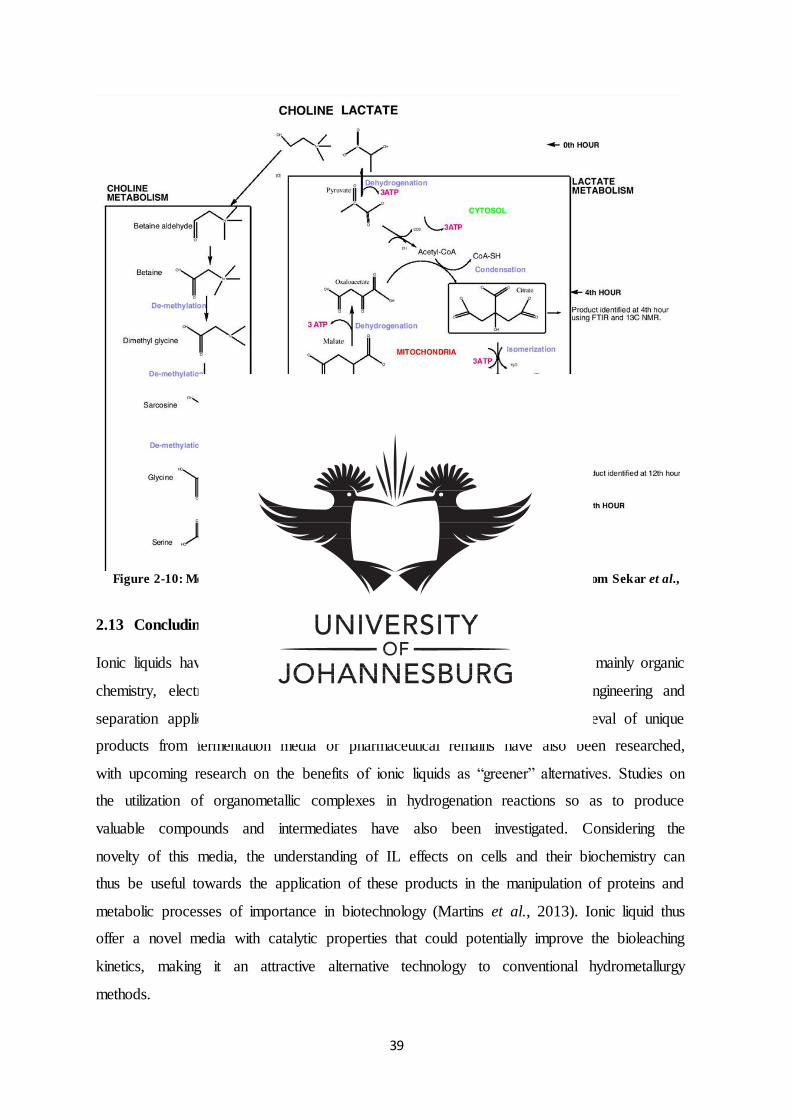
39
Figure 2-10: Metabolic activity of choline lactate by Stapylococcus lentus (Sourced from Sekar et al.,
2013).
2.13 Concluding remarks
Ionic liquids have proven to be the focal points in numerous research areas, mainly organic
chemistry, electrochemistry, nanotechnology, biotechnology, and process engineering and
separation applications. Specific applications in the extraction and the retrieval of unique
products from fermentation media or pharmaceutical remains have also been researched,
with upcoming research on the benefits of ionic liquids as “greener” alternatives. Studies on
the utilization of organometallic complexes in hydrogenation reactions so as to produce
valuable compounds and intermediates have also been investigated. Considering the
novelty of this media, the understanding of IL effects on cells and their biochemistry can
thus be useful towards the application of these products in the manipulation of proteins and
metabolic processes of importance in biotechnology (Martins et al., 2013). Ionic liquid thus
offer a novel media with catalytic properties that could potentially improve the bioleaching
kinetics, making it an attractive alternative technology to conventional hydrometallurgy
methods.
40
REFERENCES
Acevedo, F. & Valparaiso, J. (1989). Process engineering aspects of bioleaching of copper
ores. Bioprocess Engineering, 4, 223-229.
Amiri, F., Yaghmaei, S. & Mousavi, S. (2011a). Bioleaching of tungsten-rich spent
hydrocracking catalyst using Penicillium simplicissimum. Bioresource Technology, 102,
1567–1573.
Amiri, F., Mousavi, S. & Yaghmaei, S. (2011b). Enhancement of bioleaching of a spent
Ni/Mo hydroprocessing catalyst by Penicillium simplicissimum. Separation Purification
and Technology, 80, 566–576.
Amiri, F., Yaghmaei, S., Mousavi, S. & Sheibani, S. (2011c). Recovery of metals from spent
refinery hydrocracking catalyst using adapted Aspergillus niger. Hydrometallurgy, 109, 65–
71.
Amiri, F., Yaghmaei, S., Mousavi, S. & Barati, M. (2012). Bioleaching kinetics of a spent
refinery catalyst using Aspergillus niger at optimal conditions. Biochemical Engineering
Journal, 67, 208–21.
Aung, K. & Ting, Y. (2004). Bioleaching of metals from spent catalysts for metal
removal/recovery. A Thesis submitted for the degree of Master of Engineering Department of
Chemical and Biomolecular Engineering National University of Singapore.
Battaglia-Brunet, F., Joulian, C., Garrido, F., Dictor, M., Morin, D., Coupland, K., Johnson,
D., Hallberg, K. & Baranger, P. (2006). Oxidation of arsenite by Thiomonas strains and
characterization of Thiomonas arsenivorans sp. nov. Antonie van Leeuwenhoek, 89, 99–108.
Bauer, A. [n.d.]. Heap leaching followed by solvent extraction and electrowinning (SX/EW).
Available from:
http://wiki.biomine.skelleftea.se/wiki/images/thumb/a/a2/HeapLeaching.png/500pxHeapLeac
hing.png (Accessed, 08/10/2016).
Bosecher, K. (1997). Bioleaching: metal solubilisation by microorganisms. Federation of
European Microbiological Societies Microbiology, 20, 591-604.
41
Bosshard, P., Bachofen, R. & Brandl, H. (1996). Metal leaching of fly ash from municipal
waste incineration by Aspergillus niger. Environmental Science and Technology, 30, 3066–
3070.
Brandl, H. (2008). Microbial Leaching of Metals. 2nd edition. Switzerland: Biotechnology Set
191-224.
Brandl, H., Bosshard, R. & Wegmann, M. (2001). Computer-munching microbes: metal
leaching from electronic scrap by bacteria and fungi. Hydrometallurgy, 59, 319–326.
Brierley, C. (2008). How will biomining be applied in future? Transaction of Nonferrous
Metals Society of China, 18, 1302-1310.
Brombacher, C., Bachofen, R. & Brandl, H. (1998). Development of a laboratory-scale
leaching plant for metal extraction from fly ash by Thiobacillus strains. Applied and
Environmental Microbiology, 64, 1237–1241.
Burgstaller, W. & Schinner, F. (1993). Mini review: leaching of metals with fungi. Journal of
Biotechnology, 27, 91–116.
Celik, H. (2008). Biologically assisted extraction of metals from ores concentrates, and
present commercial applications. The Journal of ore dressing,10, 19.
Cerruti, C., Curutchet, G. & Donati, E. (1998). Bio-dissolution of spent nickel-cadmium
batteries using Thiobacillus ferrooxidans. Journal of Biotechnology, 62, 209–219.
Chartier, M. & Couillard, D. (1997). Biological processes: the effects of initial pH,
percentage inoculum and nutrient enrichment on the solubilisation of sediment bound metals.
Water Air and Soil Pollution, 96, 249–267.
Clark, D. & Norris, P. (1996). Acidimicrobium ferrooxidans gen. nov., sp. nov. mixed culture
ferrous iron oxidation with Sulfobacillus species. Microbiology, 141, 785–790.
Coram, N. & Rawlings, D. (2002). Molecular relationship between two groups of the genus
Leptospirillum and the finding that Leptospirillum ferriphilum sp. nov. dominates South
African commercial biooxidation tanks that operate at 40 °C. Applied and Environmental
Microbiology, 68, 838–845.
Das, T., Ayyappan, S. & Chaudhury, G. (1999). Factors affecting bioleaching kinetics of
sulfide ores using acidophilic micro-organisms. Biology of Metals, 12, 1–10.
42
Davris, P., Balomenos, E., Panias, D. & Paspaliaris, L. (2014). Leaching of rare earths from
bauxite residues using imidazolium based ionic liquids. ERES2014: 1st European Rare Earth
Resources Conference. Available from:
http://www.eurare.eu/docs/eres2014/fifthsession/panagiotisdavris.pdf(Accessed, 15/10/2016).
Deveci, H., Akcil, A. & Alp, I. (2015). Parameters for Control and Optimization of
Bioleaching of Sulfide Minerals. Material Science and Technology, 9.
Docherty, K., Dixon, J. & Kulpa, C. (2007). Biodegradability of imidazolium and pyridinium
ionic liquids by an activated sludge microbial community. Biodegradation, 18, 481–493.
Ferlin, N., Courty, M., Gatard, S., Spulak, M., Quilty, B., Beadham, I., Ghavre, M., Haiß, A.,
K€ummerer, K., Gathergood, N. & Bouquillon, S. (2013). Biomass derived ionic liquids:
synthesis from natural organic acids, characterization, toxicity, biodegradation and use as
solvents for catalytic hydrogenation processes. Tetrahedron, 69, 6150-6161.
Gathergood, N., Garcia, M. & Scammells, J. (2004). Biodegradable ionic liquids: Part I.
Concept, preliminary targets and evaluation. Green Chemistry, 6, 166-175.
Gentina, J. & Acevedo, F. (2013). Application of bioleaching to copper mining in Chile.
Electronic Journal of Biotechnology, 6(3).
Ghandi, K. (2014). A Review of Ionic Liquids. Their Limits and applications. Green and
Sustainable Chemistry, 4, 44-53.
Gholami, R.M., Borghei S.M. & Mousavi S.M. (2010). Heavy metals recovery from spent
catalyst using Acidithiobacillus ferrooxidans and Acidithiobacillus thiooxidans. International
Conference on Chemistry and Chemical Engineering (ICCCE).
Gomez, C., Blazquez, M. & Ballester, A. (1999). Bioleaching of a Spanish Complex
Sulphide Ore-Bulk Concentrate. Minerals Engineering, 12 (1), 93-106.
Guo-cai, T., Jian, L. & Yi-xin, H. (2010). Application of ionic liquids in hydrometallurgy of
nonferrous metals.Transactions of Nonferrous Metals Society of China, 20, 513-5120.
Haerens, K., Matthijs, E., Chmielarz, A. & Bruggen, B. (2009). The use of ionic liquids based
on choline chloride for metal deposition: A green alternative. Journal of environmental
management, 90, 3245-3252.
43
Hallberg, K., Johnson, D. & Williams, P. (1999). A novel metabolic phenotype among
acidophilic bacteria: aromatic degradation and the potential use of these microorganisms for
the treatment of wastewater containing organic and inorganic pollutants. Biohydrometallurgy
and the Environment. Toward the Mining of the 21st Century, Process Metallurgy, 9, 719–
728.
Hallberg, K. & Johnson, D. (2001). Biodiversity of acidophilic prokaryotes. Advances in
Applied Microbiology, 49, 37–84.
Hallberg, K., Coupland, K., Kimura, S. & Johnson, D. (2006). Macroscopic ‘‘acid streamer’’
growths in acidic, metal-rich mine waters in north Wales consist of novel and remarkably
simple bacterial communities. Applied and Environmental Microbiology, 72, 2022–2030.
Hamidian, H., Rezai, B., Milani, F., Vahabzades, F. & Shafaie, S. (2009). Microbial
leaching of Uranium Ore. Asian Journal of Chemistry, 21(8), 5808-5820.
Hernandez, F., Perez de los Rios, A., Ginesta, A., Sanchez, S., Lozano, L., Moreno, J. &
Godinez, C. (2010). Use of ionic liquids as ‘green’ solvents for extraction of Zn2+, Cd2+,
Fe3+ and Cu2+ from aqueous solutions. Chemical Engineering Transactions, 21, 631-636.
Hou, X., Liu, Q., Smith, T., Li, N. & Zong, M. (2013). Evaluation of Toxicity and
Biodegradability of Cholinium amino Acids Ionic Liquids. Public Library of Science, 8, 3.
Ilyas, S., Anwar, M., Niazi, S. & Ghauri, M. (2007). Bioleaching of metals from electronic
scrap by moderately thermophilic acidophilic bacteria. Hydrometallurgy, 88, 180–188.
Ilyas, S., Ruan, C., Bhatti, H. Ghauri, M. & Anwar, M. (2010). Column bioleaching of
metals from electronic scrap. Hydrometallurgy, 101, 135–140.
Jenkins, R., Benjamin, T., Marvin, L., Rodger, B., Lo, M. & Huang, R. (1981). Metal
removal and recovery from municipal sludge. Journal of Water Pollution Control
Federation, 25–32.
Johnson, D., Rolfe, S., Hallberg, K. & Iversen, E. (2001a). Isolation and phylogenetic
characterisation of acidophilic microorganisms indigenous to acidic drainage waters at an
abandoned Norwegian copper mine. Environmental Microbiology, 3, 630–637.
Johnson, D., Bacelar-Nicolau, P., Okibe, N., Yahya, A. & Hallberg, K. (2001b). Role of pure
and mixed cultures of Gram-positive eubacteria in mineral leaching.In Biohydrometallurgy:
Fundamentals, Technology and Sustainable Development. Process Metallurgy, 11, 461–470.
44
Johnson, D., Okibe, N. & Roberto, F. (2003). Novel thermoacidophiles isolated from
geothermal sites in Yellowstone National Park: physiological and phylogenetic
characteristics. Archives of Microbiology, 180, 60–68.
Johnson, D. (2013). Development and application of biotechnologies in the metal mining
industry. Environmental Science and Pollution Research, 20, 7768–7776.
Kalinkin, A., Kumar, S., Gurevich, B., Alex, T., Kalinkina, E., Tyukavkina, V., Kalinnikov,
V. & Kumar, R. (2012). Geopolymerization behaviour of Cu–Ni slag mechanically activated
in air and in CO2 atmosphere. International Journal of Mineral Processing. 112–113, 101–
106.
Khupse, N. & Kumar, A. (2010). Ionic Liquids: New materials with wide applications.
Indian Journal of Chemistry, 49, 635-648.
Kim, D., Pradhan, D., Ahn, J. & Lee S.W. (2010). Enhancement of metals dissolution from
spent refinery catalysts using adapted bacteria culture—Effects of pH and Fe (II).
Hydrometallurgy, 103, 136–143.
Laus, G., Bentivoglio, G., Schottenberger, H., Kahlenberg, V., Kopacka, H., Roder, T. &
Sixta, H. (2005). Ionic liquids: Current developments, potential and drawbacks for industrial
applications. Lenzinger Berichte, 84, 71-85.
Leathen, W., Kinsel, N. & Braley, I. (1956). Ferrobacillus ferrooxidans: A Chemosynthetic
Autotrophic Bacterium. Bacteriology, 72, 700-704.
Lee, S., Chang, W., Choi, A. & Koo, Y. (2005). Influence of Ionic Liquids on the Growth
of Escherichia coli. Korean Journal of Chemical Engineering, 22(5), 687-690.
Lim, J., Jeon, C., Lee, S., Lee, J., Xua, L., Jiang, C. & Kim, C. (2006). Bacillus salarius sp.
nov., a halophilic, sporeforming bacterium isolated from a salt lake in China. International
Journal of Systematic and Evolutionary Microbiology, 56, 373–377.
Lopes, J., Paninho, A., Molho, M., Nunes, A., Roch, A., Lourenco, N. & Najdanovic-Visak,
V. (2013). Biocompatible choline based ionic salts: Solubility in short-chain alcohols.
Journal of Chemical Thermodynamics, 67, 99–105.
45
Mafi Gholami, R., Borghei, S. & Mousavi, S. (2010). Heavy metals recovery from spent
catalyst using Acidithiobacillus ferrooxidans and Acidithiobacillus thiooxidans. International
Conference on Chemistry and Chemical Engineering (ICCCE). Available from:
http://ieeexplore.ieee.org/stamp/stamp.jsp?tp=&arnumber=5560422&isnumber=5560343
(Accessed, 5/06/2016).
Mafi Gholami, R., Borghei, S. & Mousavi, S. (2011). Process optimization and modelling of
heavy metals extraction from a molybdenum rich spent catalyst by Aspergillus niger using
response surface methodology. Journal of Industrial Engineering and Chemistry, 18, 218–
224.
Martins, I., Hartmann, D., Alves, P., Planchon, S., Renaut, J., Leitão, M., Rebelo, L. &
Pereira, C. (2013). Proteomic alterations induced by ionic liquids in Aspergillusnidulans
and Neurosporacrassa. Journal of Proteomics, 94 (26), 2 – 2 7 8.
Maweja, K., Mukongo, T., Mbaya, R. & Mochube, E. (2010). Effect of annealing treatment
on the crystallisation and leaching of dumped base metal smelter slags. Journal of
Hazardous Materials, 183, 294–300.
Mishra, D., Kim, D., Ahn, J. & Rhee, Y. (2005). Bioleaching: A microbial Process of Metal
Recovery. Metals and Material International, 11 (3), 249-256.
Mishra D., Kim D.J., Ralph D.E., Ahn J.G. & Rhee Y.H. (2007). Bioleaching of vanadium
rich spent refinery catalysts using sulfur oxidizing lithotrophs. Hydrometallurgy, 88, 202–
209.
Mousavi, S., Yaghmaei, S., Vossoughi, M., Jafari, A. & Hoseini, S. (2005). Comparison of
bioleaching ability of two native mesophilic and thermophilic bacteria on copper recovery
from chalcopyrite concentrate in an airlift bioreactor. Hydrometallurgy, 80, 139–144.
Mousavi, S., Vossoughi, M., & Yaghmaei, S. (2006a).Copper recovery from chalcopyrite
concentrate by an indigenous Acidithiobacillus ferrooxidans in an air-lift bioreactor. Iranian
Journal of Chemistry and Chemical Engineering, 25 (3).
Mousavi, S., Jafari, A., Yaghmaei, S., Vossoughi, M. & Roostaazad, R. (2006b).
Bioleaching of low-grade sphalerite using a column reactor. Hydrometallurgy, 82, 75–82.
46
Mousavi, S., Yaghmaei, S., Vossoughi, M., Jafari, A., Roostaazad, R. & Turunen, I. (2007).
Bacterial leaching of low-grade ZnS concentrate using indigenous mesophilic and
thermophilic strains. Hydrometallurgy, 85, 59–65.
Mousavi, S., Yaghmaei, S., Vossoughi, M., Roostaazad, R., Jafari, A., Ebrahimi, M.,
Habibollahnia Chabok, O. & Turunen, I. (2008).The effects of Fe (II) and Fe (III)
concentration and initial pH on microbial leaching of low-grade sphalerite ore in a column
reactor. Bioresource and Technology, 99, 2840–2845.
Mulligan, C., Kamali, M. & Gibbs, B. (2004). Bioleaching of heavy metals from a low-grade
mining ore using Asperigillus niger. Journal of Hazardous Materials, 110, 77-84.
Nakade, D. (2013). Bioleaching of Copper from Low Grade Ore Bornite Using Halophilic
Thiobacillus Ferroxidans. Research Journal of Recent Sciences, 2, 162-166.
Neale, J. (2006). Bioleaching technology in minerals processing. Mintek, Biotechnology
Division. Available from:
http://wiki.biomine.skelleftea.se/biomine/hyper/start_files/bioleachingtechnologyinmineralsp
rocessing_38.pdf (Accessed, 20/08/2016).
Nithya, C. & Pandian, K. (2010). Isolation of heterotrophic bacteria from Palk Bay sediments
showing heavy metal tolerance and antibiotic production. Microbiological Research, 165,
578—593.
Norris, P. & Barr, D. (1985). Growth and Iron Oxidation by Acidophilic Thermophiles.
Federation of European Microbiological Societies Microbiology Letters, 28, 221-224.
Norris, P., Clark, D., Owen, J. & Waterhouse, S. (1996). Characteristics of Sulfobacillus
acidophilus sp. nov. and other moderately thermophilic mineral-sulphide-oxidizing bacteria.
Microbiology, 141, 775–783.
Okibe, N., Gericke, M., Hallberg, K. & Johnson, D. (2003). Enumeration and
characterization of acidophilic microorganisms isolated from a pilot plant stirred tank
bioleaching operation. Applied Environmental Microbiology, 69, 1936–1943
Olubambi, P., Ndlovu, S., Potgieter, J. & Borode, J. (2007). Effects of ore mineralogy on the
microbial leaching of low grade complex sulphides ores. Hydrometallurgy, 86, 96–104.
47
Olubambi, P., Ndlovu, S. & Potgieter, J. (2008). Role of ore mineralogy in optimizing
conditions for bioleaching low-grad complex sulphide ores. Transactions of Nonferrous
Metals Society of China, 18, 1234-1246.
Park, J., Jung, Y., Kusumah, P., Lee, J., Kwon, K. & Lee, C. (2014). Application of Ionic
Liquids in Hydrometallurgy. International Journal of Molecular Science, 15, 15320-15343.
Pernak, J., Syguda, A., Mirska, L., Pernak, A., Nawrot, J., Pra, A., Ska, D., Griffin, S. &
Rogers, R. (2007). Choline-Derivative-Based Ionic Liquids. Chemistry European Journal,
13, 6817 – 6827.
Pham, T., Cho, C. & Yun, Y. (2010). Environmental fate and toxicity of ionic liquids: A
review. Water Research, 4 4, 5 2 – 3 7 2.
Pronk, J., Bruyn, J., Bos, P. & Kuenen, J. (1992). Anaerobic Growth of Thiobacillus
ferrooxidans. Applied and Environmental Microbiology, 58(7), 2227-2230.
Rantwijk, F., Lau, R. & Sheldon, R. (2003). Biocatalytic transformations in ionic liquids.
Tends in Biotechnology, 21, 3.
Rawlings, D. & Silver, S. (1995). Mining with Microbes. Biotechnology, 13, 773-778.
Rawlings, D., Tributsch, H. & Hansford, G. (1999). Reasons why ‘Leptospirillum’-like
species rather than Thiobacillus ferrooxidans are the dominant iron-oxidizing bacteria in
many commercial processes for the biooxidation of pyrite and related ores. Microbiology,
145, 5–13.
Rawlings, D. & Johnson, D. (2005). Review: Characteristics and adaptability of iron- and
sulfur-oxidizing microorganisms used for the recovery of metals from minerals and their
concentrates, Microbial Cell Factories, 4,13.
Rawlings, D. & Johnson, D. (2007). The microbiology of biomining: development and
optimization of mineral-oxidizing microbial consortia, Microbiology, 153, 315–324.
Riekkola-Vanhanen, M. (2010). Bioheapleaching. Talivaara Mining Company PlC.
Samori, C., Malferrari, D., Valbonesi, P., Montecavalli, A., Moretti, F., Galletti, P., Sartor,
G., Tagliavini, E., Fabbri, E. & Pasteris, A. (2010). Introduction of oxygenated side chain
into imidazolium ionic liquids: Evaluation of the effects of different biological organization
levels. Ecotoxicology and Environmental Safety, 73, 1456-1464.
48
Santhiya, D. & Ting Y.P. (2006). Use of adapted Aspergillus niger in the bioleaching of spent
refinery processing catalyst. Journal of Biotechnology, 121, 62–74.
Sarcheshmehpour, Z., Lakzian, A., Fotovat, A., Berenji, A., Haghnia, G. & Bagheri, S.
(2009). Possibility of using chemical fertilizers instead of 9K medium in bioleaching process
of low-grade sulphide copper ores. Hydrometallurgy, 96, 264–267.
Schippers, A., Hedrich, S., Vasters, J., Drobe, M., Sand, W. & Willscher, S. (2013).
Biomining: Metal Recovery from Ores with Microorganisms. Advances in Biochemical
Engineering/ Biotechnology, 141, 1–47.
Sekar, S., Mahadevan, S., Deepa, P., Shanmugam, B., Kumar, B. & Mandal, A. (2013). The
Metabolic Advantage of Choline Lactate in Growth Media: An Experimental Analysis with
Staphylococcus lentus. Applied Biochemistry and Biotechnology, 169, 380-392.
Shaikh, S., Khan, Z. & Ade, A. (2010). Effect of pH on Metal Extraction from Bauxite Ore
by Thiobacillus Ferrooxidans. Journal of Scientific Research, 2 (2), 403-406.
Siddiqui, M., Kumar, A., Kesari, K. & Arif, J. (2009). Biomining - A Useful Approach
toward Metal Extraction. American-Eurasian Journal of Agronomy, 2 (2), 84-88.
Silva, F., Siopa, F., Figueiredo, B., Gonçalves, A., Pereira, J., Gonçalves, F., Coutinho, J.,
Afonso, C. & Ventura, S. (2014). Sustainable design for environment-friendly mono and
dicationiccholinium-based ionic liquids. Ecotoxicology and Environmental Safety, 108, 302–
310.
Silverman, M. & Lundgren, D. (1959). Studies on the Chemolithotrophic Iron Bacterium
Ferrobacillus ferrooxidans: An Improved Medium and Harvesting Procedure for Securing
High Cell Yields. J. Bacteriology, 77, 642-677.
Simona, C. & Micle, V. (2011). Consideration concerning factors influencing bioleaching
processes. ProEnvironment, 4, 76 – 79.
Soleimani, M., Hosseini,S., Roostaazad,R., Petersen,J., Mousavi,S. & Kazemi Vasiri,
A.(2009). Microbial leaching of a low-grade sphalerite ore using a draft tube fluidized bed
bioreactor.Hydrometallurgy, 99,131–136.
Soleimani, M., Petersen, J., Roostaazad, R., Hosseini, S., Mousavi, S., Najafi, A. & Kazemi
Vasiri, A. (2011). Leaching of a zinc ore and concentrate using the Geocoat TM Technology.
Minerals Engineering, 24, 64–69.
49
Sugio, T., Domatsu, C., Tano, T. & Imai, K. (1984). Role of Ferrous Ions in Synthetic
Cobaltous Sulfide Leaching of Thiobacillus ferrooxidans. Applied and environmental
microbiology ,48(3), 461-467.
Temple, K. & Colmer, A. (1951). The autotrophic oxidation of iron by a new bacterium:
Thiobacillus ferrooxidans. Journal of Bacteriology, 62, 605–611.
Thosar, A., Satpathy, P. Mathiya, T. & Rajan, P. (2014). Bio-mining: a revolutionizing
technology for a safer and greener environment. International Journal of Recent Scientific
Research, 5, 9, 1624-1632.
Tourova, T., Poltoraus, A., Lebedeva, I., Tsaplina, I., Bogdanova, T. & Karavaiko, G. (1994).
16S ribosomal RNA (rDNA) sequence analysis and phylogenetic position of Sulfobacillus
thermosulfidooxidans. Systematic and Applied Microbiology, 17, 509–512.
Tuovinen, O. & Kelly,D. (1973). Studies on the Growth of Thiobacillus ferrooxidans: I. Use
ofMembraine Filters and Ferrous Iron Agar to Determine Viable Numbers and Comparison
with CO2 Fixation and Iron Oxidation as Measure of Growth, Arch. Microbiology,88,285-
298.
Tyagi, R. & Couillard, D. (1991). An innovative biological process for heavy metals removal
from municipal sludge. New York: A.M. Martin (Ed.) Biological Degradation of Wastes.,
Elsevier Applied Science, 307–322.
Van Staden, P., Robertson, S., Gericke, M., Neale, J.W. & Seyedbagheri, A. (2009).
Maximizing the value derived from laboratory test work towards heap leaching design. Base
Metals 2009: The Fifth Southern African Base Metals Conference. Johannesburg, The
Southern African Institute of Mining and Metallurgy.
Ventura, S., Silva, F., Gonçalves, A., Pereira, J., Gonçalves, F. & Coutinho, J. (2014).
Ecotoxicity analysis of cholinium-based ionic liquids to Vibrio fischeri marine bacteria.
Ecotoxicology and Environmental Safety, 102, 48–54.
Vijayaraghan, R., Izgordin, A., Ganesh, V., Surianaryanan, M. & MacFarlane, D. (2010).
Long term structural and chemical stability of DNA in hydrated ionic liquid. Angewandte
Chemie International Edition, 49, 1631-163.3
50
Waiters, M., Morgan, N., Rockey, J. & Hington, G. (2001). Industrial Microbiology: An
Introduction. London: Blackwell Science.
Waksman, S. & Joffe, J. (1921). Acid production by a new sulfur-oxidizing bacterium.
Science, 53, 216.
Wang, J., Huang, Q., Li, T., Xin, B., Chen, S., Guo, X., Liu, C. & Li, Y. (2015). Bioleaching
mechanism of Zn, Pb, In, Ag, Cd and As from Pb/Zn smelting slag by autotrophic bacteria.
Journal of Environmental Management, 159, 11-17.
Watling, H. (2016). Review: Microbiological Advances in Biohydrometallurgy. Minerals, 6,
49.
Yahya, A. & Johnson, D. (2002). Bioleaching of pyrite at low pH and low redox potentials by
novel mesophilic Gram-positive bacteria. Hydrometallurgy, 63, 181–188.
Yang, Z., Rui-lin, M., Wang-dong, Ni. & Hui, W. (2010). Selective leaching of base metals
from copper smelter slag. Hydrometallurgy, 103, 25–29.
Zhao, H., Xia, S. & Ma, P. (2005). Use of ionic liquids as ‘green’ solvents for extractions.
Journal of Chemical Technology and Biotechnology, 80, 1089–1096.
51
CHAPTER THREE
CHARACTERIZATION OF HALOPHILIC BACTERIA FROM GOLD1 MINE
EAST RAND, SPRINGS JOHANNESBURG, SOUTH AFRICA FOR
MICROBIAL MINING APPLICATIONS
Letlhabile Moyaha1, Sudharshan sekar1, Rufaro Archibald Bhero1, Sibusiso
Sidu2, Antoine F. Mulaba-Bafubiandi3, Vuyo Mavumengwana1
1Department of Biotechnology and Food Technology, University of Johannesburg,
Johannesburg, South Africa – 2094
2Gold One International Limited, Springs, South Africa
3Mineral Processing and Technology Research Center, Department of Metallurgy, School of
Mining, Metallurgy and Chemical Engineering University of Johannesburg, South Africa,
Johannesburg, P.O. Box 17011, Doornfontein 2028, Gauteng, South Africa
*Corresponding author:[email protected]; Tel: +27115596915
Abstract
The South African gold mines represent extreme environments harbouring an ecosystem of
diverse microorganisms. The unique gold mine conditions attributed to extreme pH, pressure
temperature and/or salinity and elevated concentrations of heavy metals have created an
ecological niche for potential novel gene sequences that could be of biotechnological
importance. The microbial diversity of Gold 1 Mine (aquifer), East Rand, Springs
Johannesburg, South Africa was studied to investigate potential microorganisms that could be
applicable in the optimization of bioleaching processes. Sampling was conducted at five
mines sites namely: NRW Ramp, W2E19, N2W2, UK9 Raw and E2 Reef drive south. A sum
of six halophilic bacteria were isolated and characterized according to colony pigmentation,
form, margin, elevation, opacity, texture, gram nature and shape. The majority of bacterial
strains (60 %) were established to be gram positive bacillus and the remaining (30 %) were
gram negative bacillus. The bacteria were subsequently characterized by employing
biochemical tests, revealing their biological diversity. Due to the limitations of conventional
phenotypic characterization in identifying bacterial isolates, 16S rRNA was employed using
the conserved region in species of the same genus and thus used for identifying bacteria at
species level. The identification of the bacterial isolates at species level was conducted by
employing 16S rRNA sequencing and the GenBank database. The 16S rRNA gene fragments
of the bacterial isolates were amplified by PCR. The 27F and1492 universal primers were
52
subsequently employed to amplify the 16S target region. The nucleotide BLAST was then
conducted and the phylogenetic tree constructed using the determined sequences. The
molecular studies identified the two bacterial strains to be, Raoultella ornithinolytica and
Pseudomonas moraviensis. The remaining three strains 2KL, 12KL and 16KL were
discovered to belong to the Bacillus genus. The phylogenetic tree confirmed 12KL and 16KL
to be closely related and members of the Bacillus cereus group. According to research strain
2KL identified as Bacillus aryabhattai are characterised as salt, metal and UV radiation
tolerant bacteria suggesting their potential in metal dissolution. Raoultella sp. were found to
be characterized as ecological bacteria commonly identified in soil and water samples but
with no accounts of Raoultella ornithinolytica isolated from mine water samples. With the
exception of Bacillus sp. to one’s knowledge there have been no accounts of the application
of bacterial isolates Raoultella ornithinolytica and Pseudomonas moraviensis in bioleaching
process.
Keywords: Optimization, bioleaching processes, bacteria, characterized, halophilic bacteria, 16S rRNA, phylogenetic tree
3.1 Introduction
In the mid-1940s bacterial isolates Acidithiobacillus ferrooxidans (formerly Thiobacillus
ferrooxidans) were unearthed and by 1958 the Kennecott mine company had licenced the
usage of Acidithiobacillus ferrooxidans for the purpose of copper (Cu+) recovery and bio-
hydrometallurgy processes from run-of-mines /low grade copper ores located in the Bingham
Canyon Mines, USA (Córdoba et al., 2008). Since then microbial leaching of ore minerals
has grown significantly. Unlike conventional hydrometallurgy and pyrometallurgy practices,
biological leaching presents more advantageous properties for treating low grade ores,
including the extraction of minerals from acid leachate waste and flotation sites (Córdoba et
al., 2008). Microorganisms like bacteria and fungi perform a principal role in solubilizing,
transporting and depositing minerals in nature, functioning as biological catalysts of leaching
reactions (Brandl et al., 2008; Brierley et al., 2008).
Due to the depletion of high-grade mineral reserves, power price hikes and the negative
environmental impact of traditional technologies, biotechnologies offer an economic
advantage for current mine, mineral and waste water treatment projects (Baba et al., 2011).
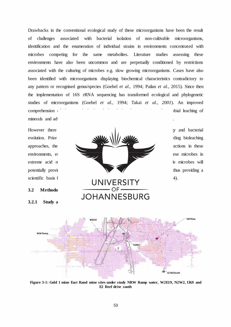
53
Drawbacks in the conventional ecological study of these microorganisms have been the result
of challenges associated with bacterial isolation of non-cultivable microorganisms,
identification and the enumeration of individual strains in environments concentrated with
microbes competing for the same metabolites. Literature studies assessing these
environments have also been uncommon and are perpetually conditioned by restrictions
associated with the culturing of microbes e.g. slow growing microorganisms. Cases have also
been identified with microorganisms displaying biochemical characteristics contradictory to
any pattern or recognised genus/species (Goebel et al., 1994; Pailan et al., 2015). Since then
the implementation of 16S rRNA sequencing has transformed ecological and phylogenetic
studies of microorganisms (Goebel et al., 1994; Takai et al., 2001). An improved
comprehension of these methods has furthered the characterization of microbial leaching of
minerals and advanced biotechnologies for metal recovery (Baba et al., 2011).
However there is a necessity for improved insight into the mine ecology and bacterial
evolution. Prior to future developments and implementation strategies regarding bioleaching
approaches, the responsible microbes need to be identified and their interactions in these
environments, establishing the conditions that activate the development of these microbes in
extreme acid mine drainage systems. Increasing the quantity of responsible microbes will
potentially provide additional knowledge on the geobiology of these areas, thus providing a
scientific basis for concrete and operative remedial action (Ramesh et al., 2014).
3.2 Methodology
3.2.1 Study area of interest for sampling
Figure 3-1: Gold 1 mine East Rand mine sites under study NRW Ramp water, W2E19, N2W2, UK9 and
E2 Reef drive south
54
Water samples were aseptically collected from Gold 1 Mine East Rand, Springs
Johannesburg, South Africa, (26°15’17’’28°26’34’’E) at five points in the gold mine process
namely NRW Ramp water,W2E19,N2W2,UK9 and E2 Reef drive south. The pH, DO and
conductivity of the samples were examined at the respective sampling sites.
3.2.2 Collection of samples
Water samples were aseptically collected in triplicates from the 5 mine sites in sterile blue
cap bottles and marked accordingly. The samples were then stored at 4 ºC and transported to
the University of Johannesburg Biotechnology and Food technology laboratory for further
analysis.
3.2.3 Isolation and enrichment of moderately halophilic bacteria
Bacteria were isolated from mine water samples by employing a membrane filtration
technique. Membranes with a pore size of 0.22 µm were employed and enriched with
Himedia nutrient broth. One millilitre of bacterial culture from individual enrichment broth
flasks was inoculated into 9 ml of sterile phosphate buffered saline (PBS) and subsequently
spread onto moderate halophilic agar media (15 % of w/v). Once successful bacterial growth
was established the cultures were distinguished according to morphology characteristics and
sub cultured to attain pure colonies. The pure cultures were then preserved in 70 % glycerol
stock at -80 ºC
3.2.4 Phenotypic classification of bacteria: Microbiological identification of bacteria
Colony and cell morphology studies were conducted utilising phenotypic data of known
organisms described in Bergy’s Manual of Determinative Bacteriology (Bergey, 1934). The
macroscopic characterization of bacterial colonies was conducted by observing the nature of
the colony. Microscopic characterization of bacterial isolates was then conducted by Gram
staining to determine the cell wall morphology and observed under a high power Olympus
CX21 light microscope.
3.2.5 Biochemical characterization of bacteria API 20E® strips
Biochemical profiles for isolates were generated using API 20E® strips (bioMérieux Inc.,
Durham, NC). API 20E® strips included enzymatic tests for fermentation or oxidation of
glucose, mannitol, inositol, sorbitol, rhamnose, saccharose, melibiose, amygdalin, and
arabinose, along with nitrate reduction to nitrite and nitrate reduction to nitrogen gas. API
20E® strips also tested for the presence of b-galactosidase, arginine dihydrolase, lysine

55
decarboxylase, ornithine decarboxylase, citrate utilization, H2S production, urease,
tryptophan deaminase, indole production, acetoin production (Voges - Proskauer), and
gelatinase. API 20E® tests were performed according to the manufacturer’s instructions. The
number and types of positive tests were then tabulated for the isolates and used to construct
biochemical phenotype profiles of the cultures which were compared amongst the isolates.
3.2.6 Genotypic classification of bacteria: 16S rRNA sequencing
The ZR Fungal/ Bacterial DNA KITS TM were used to extract DNA from the pure bacterial
cultures. Universal primers 27F: AGAGTTTGATCMTGGCTCAG and 1492R:
CGGTTACCTTGTTACGACTT were used to amplify the 16S target region. The PCR
products were gel extracted, purified and sequenced using the ABI PRISM TM 3500xl Genetic
Analyser. The CLC Main Workbench 7 was then employed to analyse the sequences
followed by a BLAST search assisted by NCBI.
3.2.7 Phylogenetic tree
Using the 16S rRNA gene sequence data the MEGA-6 tool was employed to determine the
evolutionary relationship of the bacterial strains, which was used to construct a phylogenetic
tree.
3.3 Results and Discussion
3.3.1 Microbiological characterization of bacterial isolates
Figure 3-2:Streak plates of Springs Gold1 mine bacterial isolates after 24hrs incubation at 37°C to attain
pure cultures for further characterization studies: A) Strain 2KL, B) Strain 9KL, C) Strain 12KL, D)
Strain 16KL and E)Strain 19KL.
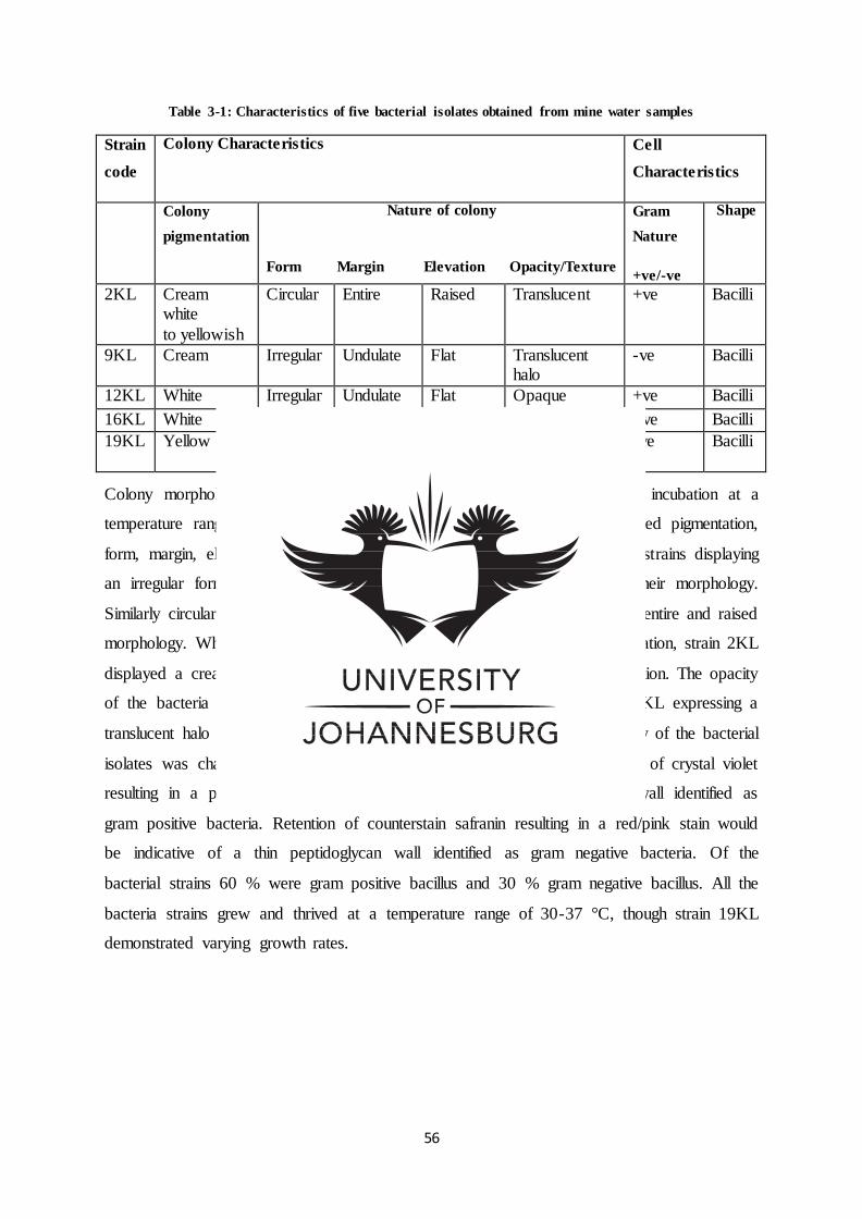
56
Table 3-1: Characteristics of five bacterial isolates obtained from mine water samples
Strain
code
Colony Characteristics
Cell
Characteristics
Colony
pigmentation
Nature of colony
Form Margin Elevation Opacity/Texture
Gram
Nature
+ve/-ve
Shape
2KL Cream white
to yellowish
Circular Entire Raised Translucent +ve Bacilli
9KL Cream Irregular Undulate Flat Translucent halo
-ve Bacilli
12KL White Irregular Undulate Flat Opaque +ve Bacilli
16KL White Irregular Undulate Flat Opaque +ve Bacilli
19KL Yellow Circular Entire Raised Translucent
mucoid
-ve Bacilli
Colony morphology of the bacterial isolates was conducted after 24 hr. incubation at a
temperature range of 37 °C (Table 3.1). The studied characteristics included pigmentation,
form, margin, elevation, opacity, texture etc. It was observed that bacteria strains displaying
an irregular form (9KL, 12KL and 16 KL) were undulate and flat in their morphology.
Similarly circular bacteria (2KL and 19KL) displayed a common pattern of entire and raised
morphology. While most of the bacteria displayed a cream or white pigmentation, strain 2KL
displayed a cream to yellowish colour and 19KL expressed yellow pigmentation. The opacity
of the bacteria was predominantly opaque or translucent with one strain, 9KL expressing a
translucent halo and 19KL expressing a mucoid texture. The cell morphology of the bacterial
isolates was characterized using gram staining. Upon gram staining, retention of crystal violet
resulting in a purple stain would be indicative of a thick peptidoglycan wall identified as
gram positive bacteria. Retention of counterstain safranin resulting in a red/pink stain would
be indicative of a thin peptidoglycan wall identified as gram negative bacteria. Of the
bacterial strains 60 % were gram positive bacillus and 30 % gram negative bacillus. All the
bacteria strains grew and thrived at a temperature range of 30-37 °C, though strain 19KL
demonstrated varying growth rates.
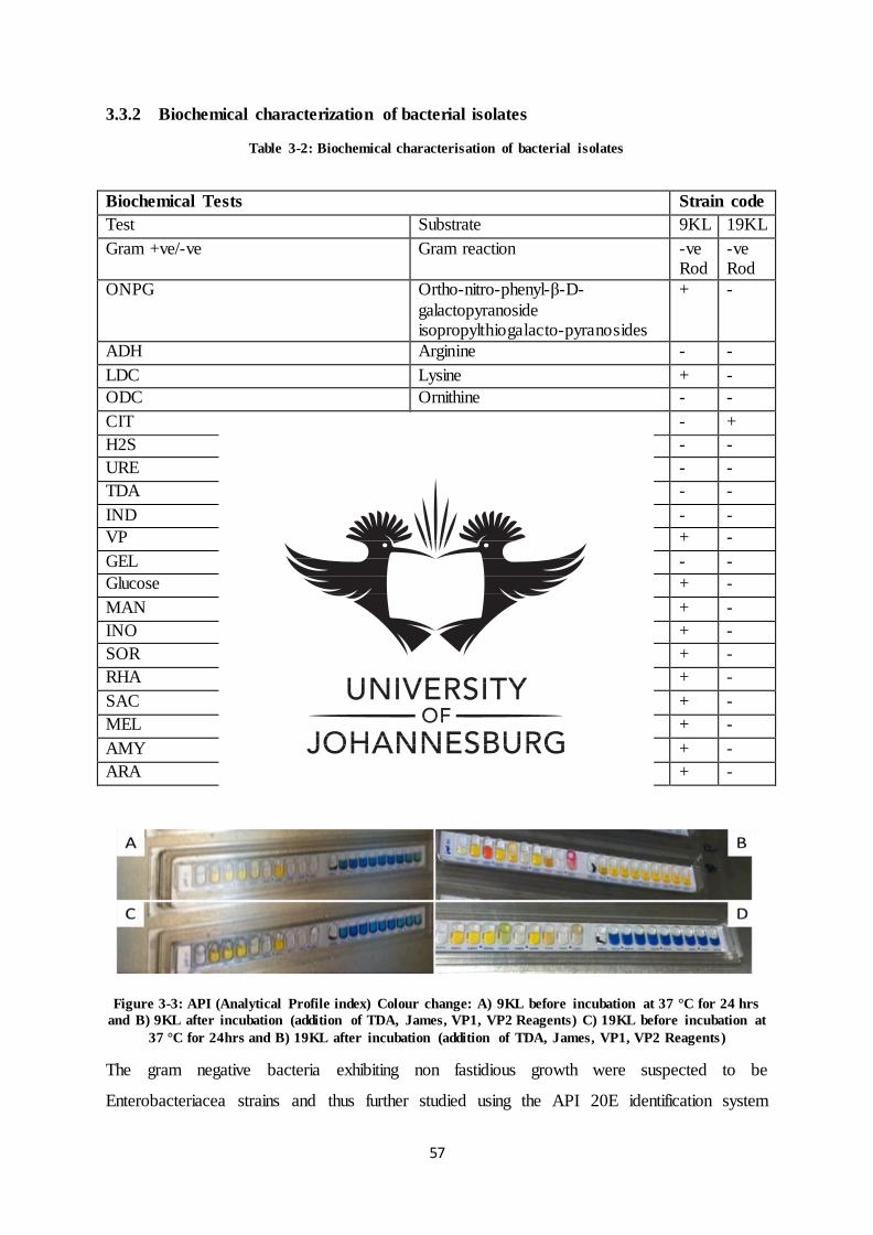
57
3.3.2 Biochemical characterization of bacterial isolates
Table 3-2: Biochemical characterisation of bacterial isolates
Figure 3-3: API (Analytical Profile index) Colour change: A) 9KL before incubation at 37 °C for 24 hrs
and B) 9KL after incubation (addition of TDA, James, VP1, VP2 Reagents) C) 19KL before incubation at
37 °C for 24hrs and B) 19KL after incubation (addition of TDA, James, VP1, VP2 Reagents)
The gram negative bacteria exhibiting non fastidious growth were suspected to be
Enterobacteriacea strains and thus further studied using the API 20E identification system
Biochemical Tests Strain code
Test Substrate 9KL 19KL
Gram +ve/-ve Gram reaction -ve Rod
-ve Rod
ONPG Ortho-nitro-phenyl-β-D-
galactopyranoside isopropylthiogalacto-pyranosides
+ -
ADH Arginine - -
LDC Lysine + -
ODC Ornithine - -
CIT Sodium citrate - +
H2S Sodium thiosulfate - -
URE Urea - -
TDA Tryptophan - -
IND Tryptophan - -
VP Creatine sodium pyruvate + -
GEL Kohns gelatin - -
Glucose Glucose + -
MAN Mannitol + -
INO Inositol + -
SOR Sorbitol + -
RHA Rhamnose + -
SAC Sucrose + -
MEL Melibiose + -
AMY Amygdalin + -
ARA Arabinose + -
58
(Table 3.2). Subsequent to the incubation of the API 20E strips at 37 °C for 24 hrs and the
addition of reagents TDA to TDA test, James reagent to IND test and VP1/VP2 reagent to the
VP test, the colour changes (Figure 3.3) (indicative of a reaction taking place) were read and
interpenetrated according to the reading table. The API 20E tests suggested strain 9KL to be
members of bacteria family Enterobacteriacea and 19KL not to be Enterobacteriace. The API
20E test indicated that unknown strain19KL has the ability to utilise citrate as its sole source
of carbon.
3.3.3 Genotypic characterization of bacterial isolates
Table 3-3: Identification of bacterial sequence
Sample Band I.D GenBank
no.
Blast forecast
of closest
relative
%
Similarity
Phylogenetic
affiliation
2KL KYX3YDEC01R KM675970.1 Bacillus aryabhattai
99 Firmicutes
9KL KYZ4WZAD01R KP192770.1 Raoultella
ornithinolytica
99 Proteobacteria
12KL MPMUGDGP014 HM566903.1 Bacillus sp. 99 Firmicutes
16KL KYZ4WZAD01R KP192770.1 Bacillus thuringiensis
99 Firmicutes
19KL KYZ4WZAD01R KP192770.1 Pseudomonas moraviensis
99 Proteobacteria
The NCBI BLAST of strain 2KL revealed a 16S rRNA gene sequence similarity of 99 % to
Bacillus aryabhattai 16S ribosomal RNA gene sequence, which were reported to be
culturable bacteria isolated from tobacco rhizospheric soil. They are scientifically classified
under the Domain: Bacteria; Pylum: Firmicutes; Class: Bacilli; Order: Bacillales; Family:
Bacillaceae; Genus: Bacillus and species name B. aryabhattai (You & Zhang, 2014). Bacillus
aryabhattai (named after a distinguished Indian astrophysicist of the 5th century AD)were
initially isolated from stratosphere air samples (Shivaji et al., 2009) and successively
identified in diverse environments including desolate lands, the rhizospheres of halophytic
plants and in South China ocean water samples (Lee et al., 2012; Ray et al., 2012). They are
characterised as promoters in plant development and as salt, metal and UV radiation tolerant
bacteria (Pailan et al., 2015), which explains the presence of these microorganisms in
extreme mine water conditions.
Thus far there have been accounts of B. aryabhattai strains in mine water samples. Bacillus
colonies were reported to be entire, circular and plane (Shivaji et al., 2009). The Bacillus
aryabhattai mine bacteria colonies in this study proved to be raised, expressing a rather
59
cream to yellowish pigmentation which could be attributed to the production of intermediate
products between primary and secondary metabolites; or delayed secondary products
resulting from primary metabolism. Contrary to these finding Ray et al. (2012) reported
colonies to be brown later producing a peach pigmentation attributing this loss of
pigmentation to continual sub-culturing on nutrient-agar.
The NCBI BLAST of strain 9KL revealed a 16S rRNA gene sequence similarity of 99 % to
Raoultella ornithinolytica ribosomal RNA gene sequence. Raoultella species (named after
Didier Raou a French bacteriologist) are interrelated with Klebsiella. They are described as
gram-negative, capsule forming, non-motile bacillus scientifically classified under Domain:
Bacteria; Phylum: Proteobacteria; Class: Gammaproteobacteria; Order: Enterobacteriales;
Family: Enterobacteriaceae; Genus Raoultella; Species: R. ornithinolytica (Aziz et al., 2014;
Bhatt et al., 2015). The NCBI blast confirmed the API 20E test results of strain 9KL.
Raoultella species were initially reported to be ecological bacteria commonly identified in
soil and water samples Bhatt et al. (2015), which explain their presence in mine water
samples. However, though uncommon causes of human disease R. ornithinolytica, R.
planticola and R. terrigena have been associated with soft-tissue and blood-stream infection.
As a result, research on Raoultella ornithinolytica has predominantly been focused on the
isolation and characterisation of theses bacteria from clinical specimens. Of recent, genus
Raoultella species were distinguished from Klebsiella using molecular techniques. Pending
the 1990s Raoultella strains were incorrectly classified under genus Klebsiella (Bhatt et al.,
2015). This was due to the discovery that biochemical analysis commonly utilised in medical
laboratories were incapable of differentiating between Raoultella spp. and Klebsiella spp., as
a result there has been insufficient literature on the phenotypic classification of Raoultella
species (Park et al., 2011). As far as ones research, this is one of the first accounts describing
the colony morphology of Raoultella species from gold mine water samples, specifically
Raoultella ornithinolytica which were found to be cream coloured, irregular shaped, flat
colonies expressing a translucent halo.
The NCBI BLAST of strain 12KL revealed a 16S rRNA gene sequence similarity of 99 % to
Bacillus sp. 16S ribosomal RNA gene sequence. Bacillus species are gram positive rod
shaped bacteria and members of Phylum: Firmicutes; Class: Bacilli; Order: Bacillales;
Family: Bacillaceae and Genus: Bacillus (Yu et al., 2011). Bacillus isolates have been
reported from soil samples from the Elmadau savannahs (Aslim et al., 2000).The species

60
have also been identified in polluted acid drainage water and soil, displaying potential as a
bio-sorbent for bioremediation metal removal (Zondo et al., n.d.). Thus explaining the
presence of Bacillus sp. in mine water samples and also eluding to its potential metal
tolerance and applications in metal recovery processes.
The NCBI BLAST of strain 16KL revealed a 16S rRNA gene sequence similarity of 99 % to
Bacillus thuringiensis 16S ribosomal RNA gene sequence. They are scientifically classified
under the Phylum: Firmicutes, Class: Bacilli, Order: Bacillales, Family: Bacillaceae, Genus:
Bacillus, Species: B. thuringiensis (Liu et al., 2014). Bacillus thuringiensis are described as
aerobic, gram positive, soil bacteria and are reported to synthesize parasporal toxic proteins
in the course of sporulation, which are employed as bioinsecticides (Hofte et al., 1989). A
study was conducted on the bio-sorption of copper and manganese from Sarcheshme copper-
mine waste water employing locally isolated bacterium B. thuringiensis. The bacterium was
reported to be of high resistance and biocompatibility to toxic metals .Thus making it a model
microorganism for bioleaching applications (Marandi et al., 2011).
The NCBI BLAST of strain 19KL revealed a 16S rRNA gene sequence similarity of 99 % to
Pseudomonas moraviensis 16S ribosomal RNA gene sequence. They are scientifically
classified under Domain: Bacteria, Phylum: Proteobacteria, Class: Gammma Proteobacteria,
Order: Pseudomonadales, Family: Pseudomonadaceae, Genus: Pseudomonas, Species: P.
moraviensis (Xia et al., 2015). Pseudomonas moraviensis (moraviensis referring to Moravia
of the Czech Republic, the region microorganism 1B4T was located) was initially recovered
from soil. They have been reported to be gram-negative, non- spore forming bacilli
demonstrating spherical colony morphology (Tvrzova et al., 2006), supporting the
investigation findings.
3.3.4 Phylogenetic study of bacterial strains
Figure 3-4: Phylogenetic tree of consensus sequence 2KL
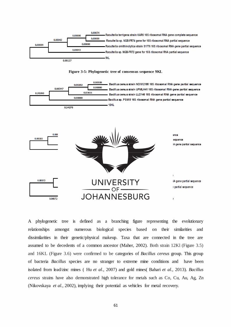
61
Figure 3-5: Phylogenetic tree of consensus sequence 9KL
Figure 3-6: Phylogenetic tree of consensus sequence 12KL
Figure 3-7: Phylogenetic tree of consensus sequence 16KL
Figure 3-8: Phylogenetic tree of consensus sequence 19KL
A phylogenetic tree is defined as a branching figure representing the evolutionary
relationships amongst numerous biological species based on their similarities and
dissimilarities in their genetic/physical makeup. Taxa that are connected in the tree are
assumed to be decedents of a common ancestor (Maher, 2002). Both strain 12Kl (Figure 3.5)
and 16KL (Figure 3.6) were confirmed to be categories of Bacillus cereus group. This group
of bacteria Bacillus species are no stranger to extreme mine conditions and have been
isolated from lead/zinc mines ( Hu et al., 2007) and gold mines( Bahari et al., 2013). Bacillus
cereus strains have also demonstrated high tolerance for metals such as Co, Cu, Au, Ag, Zn
(Nikovskaya et al., 2002), implying their potential as vehicles for metal recovery.
62
3.4 Conclusion
It can be concluded from the study that five bacteria strains (2KL, 9KL, 12KL, 16KL, 19KL)
were isolated from Gold 1 mine East Rand mine sites NRW Ramp water, W2E19, N2W2,
UK9 and E2 Reef drive south. The microorganisms were characterized and their identity was
further confirmed by analytical profile indexing, 16S rRNA sequencing, and phylogenetic
studies. The bacteria were identified to be Bacillus aryabhattai, Raoultella ornithinolytica,
Pseudomonas moraviensis and Bacillus cereus group (Bacillus sp., Bacillus thuringiensis).
The Gold1 mine East Rand, Springs Johannesburg, South Africa has proven to be an
ecosystem harbouring a diversity of microorganisms. Further research is required to
authenticate the novelty of microorganisms in the South African mine waters, potentially
yielding novel genomic data.
Acknowledgement
This work was supported by a research grant sponsored by the National Research
Foundation.
63
References
Aslim, B., Salum, N. & Beyatli, Y. (2000). Determination of Some Properties of Bacillus
Isolated from Soil. Turkish Journal of Biology, 26, 41-48.
Aziz, Z., Al- Muhanna, A., Salman, A. & Alzuhairi, M. (2014). Klebsiella and Raoultella
biotyping and probability of identification by Vitek-2 system . International Journal of
Innovative Research in Science, Engineering and Technology, 3(4).
Baba, A., Ezekafor, E., Adekola, F., Ahmed, R. & Panda, S. (2011). Bio-oxidation of a low
grade chalcopyrite ore by mixed culture of acidophilic bacteria. Journal of Ecobiotechnology,
3(12), 1-6.
Bahari, Z., Altowayti, W., Ibrahim, Z., Jaafar, J. & Shahir, S. (2013). Biosorption of As (III)
by Non-living Biomass of an Arsenic-Hypertolerant Bacillus cereus Strain SZ2 Isolated from
a Gold Mining Environment: Equilibrium and Kinetic Study. Applied Biochemistry and
Biotechnology, 171, 2247–2261.
Bergey, D. (1934). Bergey's manual of determinative bacterioloy. The American Journal of
the Medical Sciences, 188(2), 282.
Bhatt, P., Tandel, K., Das, K. & Rathi, C. (2015). New Delhi metallo-β-lactamase producing
extensively drug-resistant Raoultella ornithinolytica isolated from drain fluid following
Whipple's pancreaticoduodenectomy. Medical journal armed forces India, 1, 3.
Brandl, H. (2008). Microbial Leaching of metals. 2nd edition. Switzerland: Biotechnology
Set, 191-224.
Brierley, C. (2008). How will biomining be applied in future? Transaction of Nonferrous
Metals Society of China, 181, 302-310.
Córdoba, E., Muñoz, J., Blázquez, M., González, F. & Ballester, A. (2008). Leaching of
chalcopyrite with ferric ion. Part IV: The role of redox potential in the presence of mesophilic
and thermophilic bacteria. Hydrometallurgy, 93, 106–115.
Goebel, B. & Stackebrandt, E. (1994). Cultural and Phylogenetic Analysis of Mixed
Microbial Populations Found in Natural and Commercial Bioleaching Environments. Applied
and Environmental Microbiology, 1614-1621.
64
Hofte, H. & Whiteley, H. (1989). Society for Microbiology insecticidal Crystal Proteins of
Bacilllus thuringiensis. Microbiological Reviews, 242-255.
Hu, Q., Qi, H., Bai, Z., Dou, M., Zeng, J., Zhang, F. & Zhang, F. (2007). Biosorption of
cadmium by a Cd2+-hyperresistant Bacillus cereus strain HQ-1 newly isolated from a lead
and zinc mine. World Journal of Microbiology and Biotechnology, 23, 971-976.
Lee, S., Ka, J. & Song, H. (2012). Growth Promotion of Xanthium italicum by Application of
Rhizobacterial Isolates of Bacillus aryabhattai in Microcosm Soil. The Journal of
Microbiology, 50(1), 45-49.
Liu, G. (2011). Fujian Academy of Agricultural Sciences. Agricultural Bioresource Research
Institute. Unpublished raw data.
Liu, Y., Lai, Q. & Shao, Z. (2014). Phylogenetic Diversity of the Bacillus cereus Group and
the Marine Ecotype Revealed by Multilocus Sequence Typing. Unpublished raw data.
Marandi, R. (2011). Bioextraction of Cu (II) Ions from Acid Mine Drainage by Bacillus
Thuringiensis. International Journal of Biological Engineering, 1(1), 11-17.
Nikovskaya, G., Ul’berg, Z. & Strizhak, N. (2002). Colloidal Regularities of the Interaction
between Uranium(VI) and the Cells of Metal-Resistant Bacillus cereus AUMC 4368
Bacterial Culture. Colloid Journal, 64(2), 172–177.
Pailan, S., Gupta, D., Apte, S., Krishnamurthi, S. & Saha, P. (2015). Degradation of
organophosphate insecticide by a novel Bacillus aryabhattai strain SanPS1, isolated from soil
of agricultural field in Burdwan, West Bengal, India. International Biodeterioration &
Biodegradation, 103, 191-195.
Park, J., Hong, K., Lee, H., Choi, S., Song, S., Song, K., Kim, H., Park, K., Song, J. & Kim,
E. ( 2011). Evaluation of three phenotypic identification systems for clinical isolates of
Raoultella ornithinolytica. Journal of Medical Microbiology,60, 492–499.
Pascual, J., Garcı́ a-Lo´pez, M., Bills, G. & Genilloud, O. (2015). Pseudomonas granadensis
sp. nov., a new bacterial species isolated from the Tejeda, Almijara and Alhama Natural Park,
Granada, Spain. International Journal of Systematic and Evolutionary Microbiology, 65,
625–632.
65
Ramesh, A. Sharma, S. Sharma, M. Yadav, N. & Joshi, O. (2014). Inoculation of zinc
solubilizing Bacillus aryabhattai strains for improved growth, mobilization and
biofortification of zinc in soybeanand wheat cultivated in Vertisols of central India. Applied
Soil Ecology, 73, 87– 96.
Ray, S., Datta, R., Bhadra, P., Chaudhuri, B. & Mitra, A. (2012). From space to earth:
Bacillus Aryabhattai found in the Indian Sub-Continent. Bioscience Discovery, 3(1), 138-145.
Shivaji, S., Chaturvedi, P., Begum, Z., Pindi, P., Manorama, R., Padmanaban, D., Shouche,
Y., Pawar, S., Vaishampayan, P., Dutt, C., Datta, G. Manchanda, R., Rao, U., Bhargava, P. &
Narlikar, J. (2009). Janibacter hoylei sp. nov., Bacillus isronensis sp. nov. and Bacillus
aryabhattai sp. nov., isolated from cryotubes used for collecting air from the upper
atmosphere. International Journal of Systematic and Evolutionary Microbiology, 59, 2977–
2986.
Takai, K., Moser, D., Onstott, T., Spoelstra, N., Pfiffner, S., Dohnalkova, A. & Fredrickson,
J. (2001). Alkaliphilus transvaalensis gen. nov., sp. nov., an extremely alkaliphilic bacterium
isolated from a deep South African gold mine. International Journal of Systematic and
Evolutionary Microbiology, 51, 1245–1256.
Tvrzova, L., Schumann, P., Sproer, C., Sedlacek, I., Pacova, Z., Sedo, O., Zdrahal, Z.,
Steffen, M. & Lang, E. (2006). Pseudomonas moraviensis sp. nov. and Pseudomonas
vranovensis sp. nov., soil bacteria isolated on nitroaromatic compounds, and emended
description of Pseudomonas asplenii. International Journal of Systematic and Evolutionary
Microbiology, 56, 2657–2663.
Xia, Y., Xin, Y., Lv, C., Hou, N., Liu, H. & Xun, L. (2015). Heterotrophic bacteria use an
energy-inefficient pathway for H2S oxidation and sulphur conservation. Unpublished Raw
Data.
You, C. & Zhang, C. (2014). Culturable bacteria isolated from tobacco rhizospheric soil.
Available from:
https://www.ncbi.nlm.nih.gov/nucleotide/746770333?report=genbank&log$=nucltop&blast_
rank=2&RID=1HU858G401R (Accessed, 25/05/2016).
66
Yu, L., Hong, W., Wu, Z. & Luo, J. (2011). Screening, Identification and Degradation
Characteristics of a Phosphate Soluble Microorganisms. Available from:
https://www.ncbi.nlm.nih.gov/nucleotide/355469233?report=genbank&log$=nucltop&blast_
rank=3&RID=1HW2EEW6014 (Accessed, 2/12/2016).
Zhang, Y. (2014). School of Environmental Science and Engineering. Tongji University.
Unpublished raw data.
Zondo, N., Maphutha, T. & Kondiah, K.A. [n.d.].Culture-dependent investigation of heavy-
metal resistant bacteria in acid mine drainage with the potential for bioremediation. Available
from: http://www.ewisa.co.za/literature/files/ID147%20Paper149%20Kondia(Accessed,
22/12/2016).
67
CHAPTER FOUR
SYNTHESIS AND CHARACTERIZATION OF BIOCOMPATIBLE CHOLINE
DERIVED IONIC LIQUIDS FOR BIOTECHNOLOGY APPLICATIONS
Letlhabile Moyaha1, Sudharshan sekar1, Rufaro Archibald Bhero1, Sibusiso Sidu2,
Antoine F. Mulaba-Bafubiandi3, Vuyo Mavumengwana1
1Department of Biotechnology and Food Technology, University of Johannesburg,
Johannesburg, South Africa – 2094
2Gold One International Limited, Springs, South Africa
3Mineral Processing and Technology Research Center, Department of Metallurgy, School of
Mining, Metallurgy and Chemical Engineering University of Johannesburg, South Africa,
Johannesburg, P.O. Box 17011, Doornfontein 2028,Gauteng, South Africa
*Corresponding author:[email protected]; Tel: 27115596915
Abstract
Traditional organic solvents are toxic, volatile and highly flammable resulting in oxidant
smog and ozone destruction. This has sparked an interest in alternative, environmentally
friendly solvents and reaction media. Ionic liquids are regarded as potential contenders due to
their minimal toxicity and environmentally friendly potential. A great advantage of ILs is
their characteristic as “designer solvents”. As a result cation and anion combinations can be
manipulated to synthesis greener Ionic liquids with unique characteristics suitable for specific
processes. Cholinium/choline is a potential cation contender due to quaternary ammonium
incorporated with a polar hydroxyl functional group resulting in decreased levels of toxicity.
Choline is also a naturally prevalent and vital trace element that has the potential to break
down in aerobic environments.
The current study suggests that choline (cation) based ionic liquids can serve as an alternative
carbon source to conventional glucose sources, catalysing the growth of microorganisms for
further biotechnology applications. Keeping the cation component constant six ionic liquids
were synthesised employing a neutralization reaction and distillation processes. The ionic
liquids included choline lactate, choline dihydrogen phosphate, choline citrate, choline
tartarate, choline chloride, and choline levulinate. The ionic liquids were then characterized
by employing 1H NMR, 13C NMR and FT-IR techniques. Preliminary growth studies of
bacteria in the presence of ionic liquids were conducted by employing direct and indirect
colony counts. Shaker growth studies proved 80% of the bacterial strains to have a greater
68
preference for ionic liquids than the conventional glucose source, with the exception of
Bacillus aryabhattai. Choline chloride, choline lactate, choline dihydrogen phosphate and
choline levulinate were proven to be the most biocompatible ionic liquids across bacterial
strains Raoultella ornithinolytica, Bacillus sp., Bacillus thuringiensis and Pseudomonas
moraviensis.
Keywords: Ionic liquids (ILs), choline, choline lactate, choline dihydrogen phosphate,
choline citrate, choline tartarate, choline chloride, choline levulinate, 1H-NMR, 13C NMR,
and FT-IR
4.1 Introduction
Throughout the history of humankind biotransformations have been of economic and social
significance. Biocatalysis has been the most effective method of generating valuable
chemical products. To date, numerous chemicals including pharmaceuticals, esters, amino
acids, vitamins, saccharides and polysaccharides have been developed by enzymatic bio-
transformations (Habulin et al., 2011). A lot of research has been done on the benefits of
ionic liquids as “greener” alternatives. Studies on the utilization of organometallic
complexes in hydrogenation reactions so as to produce valuable compounds and
intermediates have also been investigated. Recent studies have shown the cation
component, more importantly the head group to be a major player in the toxicity of an ionic
liquid (IL). As an example, ILs with quaternary ammonium and alicyclic cations
demonstrate minimal toxicity compared to aromatic cations like imidazolium and
pyrimidinum. The inclusion of a polar hydroxyl, ether and a nitrile in the alkyl chain has the
potential to lower toxicity levels. As a result, cholinium is a potential cation IL contender
due to quaternary ammonium incorporated with a polar hydroxyl functional group resulting
in decreased levels of toxicity. Choline is a naturally prevalent and vital trace element that
has the potential to break down in aerobic environments. In recent times a selection of
choline based ionic liquids have been produced and reportedly showed minimal toxicity.
The ionic liquids synthesized also proved to be readily biodegradable. The ionic liquid
anion component has been known to add to the IL toxicity, though its role has been
disregarded (Guo-cai et al., 2010; Hou et al., 2013; Silva et al., 2014). Investigations
involving catalytic processes concerning metal complexes in ionic liquid have been noted,
with an average of 300 IL screened. Reduced enzyme turnover rates observed in organic
media have also ignited interest in ILs as alternative media (Rantwijk et al., 2003).
However minimal research has been done on the characterization of choline based ionic
69
liquids including their biodegradation and bioaccumulation (Ferlin et al., 2013). The
understanding of choline based IL effects on cells and their biochemistry can be useful
towards the application of these products in the manipulation of proteins and metabolic
processes of importance in biotechnology (Martins et al., 2013). The present paper thus
aims to uncover the understanding of choline derived ionic liquids relative to glucose as
carbon sources for catalysing microbial growth.
4.2 Methodology
4.2.1 Choline hydroxide properties
For the synthesis of choline based ionic liquids, choline hydroxide was employed as the
cation component of the ionic liquid.
N
CH3
H3C CH3
OHOH
Figure 4-1: Structure of choline hydroxide
Choline hydroxide solution was sourced from a reputable manufacturer, Sigma-Aldrich.
The properties of the solution are stated as,
Table 4-1: Properties of choline hydroxide solution
Stabilizer 0.5 % Paraformaldehyde as stabilizer
Concentration 45 wt. % in methanol
Refractive index n20/D 1.4186
Density 0.945 g/ml at 25 °C
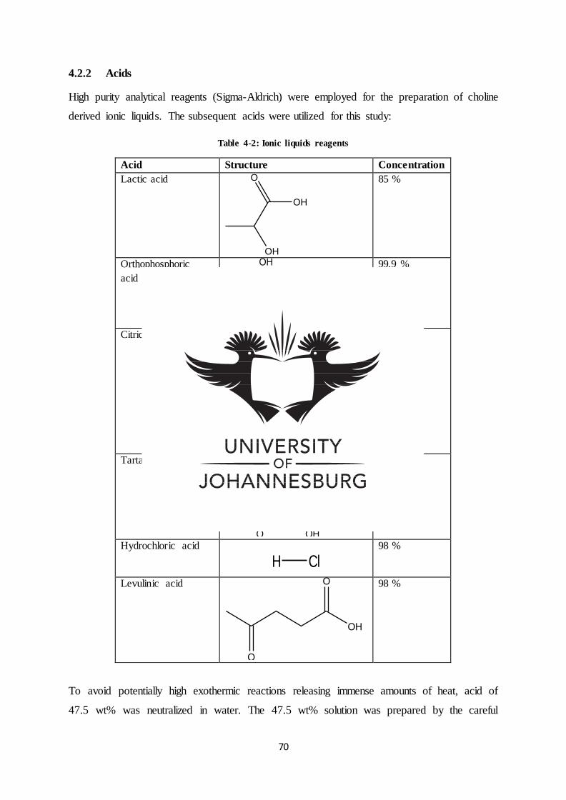
70
4.2.2 Acids
High purity analytical reagents (Sigma-Aldrich) were employed for the preparation of choline
derived ionic liquids. The subsequent acids were utilized for this study:
Table 4-2: Ionic liquids reagents
Acid Structure Concentration
Lactic acid
OH
O
OH
85 %
Orthophosphoric
acid
P
O
HO
OH
OH
99.9 %
Citric acid
HO
O
OH
O OH
O
HO
99.5 %
Tartaric acid OH
OHO
HO
O
OH
99.5 %
Hydrochloric acid
H Cl 98 %
Levulinic acid
O
O
OH
98 %
To avoid potentially high exothermic reactions releasing immense amounts of heat, acid of
47.5 wt% was neutralized in water. The 47.5 wt% solution was prepared by the careful
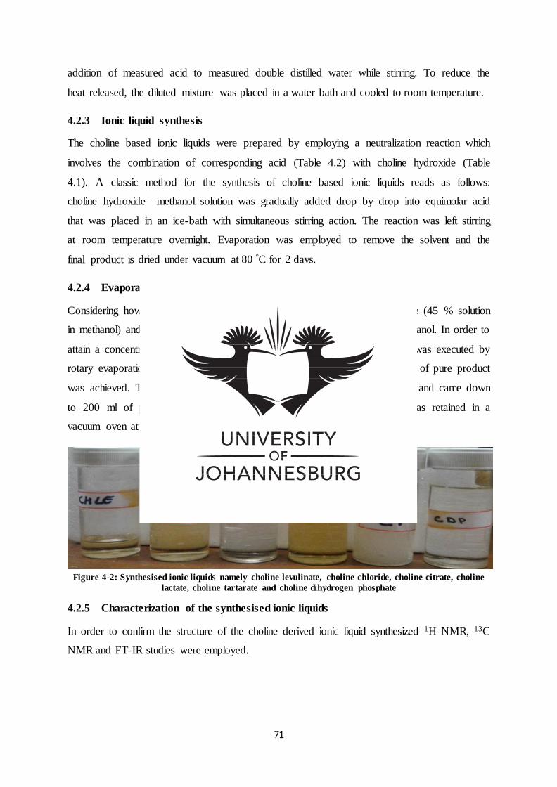
71
addition of measured acid to measured double distilled water while stirring. To reduce the
heat released, the diluted mixture was placed in a water bath and cooled to room temperature.
4.2.3 Ionic liquid synthesis
The choline based ionic liquids were prepared by employing a neutralization reaction which
involves the combination of corresponding acid (Table 4.2) with choline hydroxide (Table
4.1). A classic method for the synthesis of choline based ionic liquids reads as follows:
choline hydroxide– methanol solution was gradually added drop by drop into equimolar acid
that was placed in an ice-bath with simultaneous stirring action. The reaction was left stirring
at room temperature overnight. Evaporation was employed to remove the solvent and the
final product is dried under vacuum at 80 °C for 2 days.
4.2.4 Evaporation process
Considering how dilute the chemistry of the end product is: choline hydroxide (45 % solution
in methanol) and acid (47.5 % solution in water) as a result of water and methanol. In order to
attain a concentrated product water and methanol have to be removed. This was executed by
rotary evaporation. Rotary evaporation was applied until the expected weight of pure product
was achieved. The volume of the diluted product after synthesis was 600 ml and came down
to 200 ml of pure product, after rotary evaporation. The final product was retained in a
vacuum oven at 80 °C for two days to ensure the complete removal of water.
Figure 4-2: Synthesised ionic liquids namely choline levulinate, choline chloride, choline citrate, choline
lactate, choline tartarate and choline dihydrogen phosphate
4.2.5 Characterization of the synthesised ionic liquids
In order to confirm the structure of the choline derived ionic liquid synthesized 1H NMR, 13C
NMR and FT-IR studies were employed.
72
4.2.5.1 1H NMR Spectra
High-resolution 1H-NMR spectra were recorded on a Bruker 400 MHz FT-NMR
spectrometer at room temperature using 10-15 % (w/v) solutions of deuterated water (D2O).
Tetramethylsilane (TMS) was used as the internal reference.
4.2.5.2 13C NMR Spectra
High-resolution 13C-NMR spectra were recorded on a Bruker 100 MHz FT-NMR
spectrometer at room temperature using 10-15 % (w/v) solutions of deuterated water (D2O).
Tetramethylsilane (TMS) was used as the internal reference.
4.2.5.3 FT-IR
Infrared spectra were recorded on a Nicolet iS10 FT-IR spectrophotometer by the standard
KBr disc method in a range of 4000 cm-1 to 650 cm-1.
Table 4-3: Nicolet iS10 FT-IR instrument specifications
Parameter Condition
Detector DTGS KBr
Beam splitter KBr
Source IR
Window Diamond
Recommended range
Max range limit
Min range limit
4000-525
4000
650
Gain 2
Optical velocity 0.4747
4.2.6 Preliminary growth studies of halophilic bacteria in the presence of choline
based ILs
The study was based on using five choline based ionic liquids as alternative carbon sources to
conventional glucose namely: Choline lactate, choline dihydrogen phosphate, choline citrate,
choline tartarate, choline chloride and choline levulinate
4.2.6.1 Inoculum preparation for halotolerant bacteria strains
For individual bacterial strains, the nutrient broth was inoculated with optimized seed culture
of 1 % (v/v).
73
4.2.6.2 Media preparation for growth plate studies (solid media)
Himedia nutrient agar media was employed in the study. The pH of the media was then
adjusted to pH 7.0 using 0.1 N HCL and 0.1 N Na OH. The media was inoculated with 5 g/L
of the respective ionic liquid before autoclaving at a temperature of 121°C for 15 minutes. A
1/10 serial dilution of the cultivated bacterial strains (Bacillus aryabhattai, Raoultella
ornithinolytica, Bacillus species, Bacillus thuringiensis and Pseudomonas moraviensis) was
prepared, employing the pour plate technique. The cultures were then incubated at 37 °C
followed by daily sampling conducted from day one.
4.2.6.3 Direct colony count
Using the Astor 20 Colony counter the number of colony (CFUs) forming units on agar plates
(solid media) were recorded.
4.2.6.4 Media preparation for shaker growth studies (liquid media)
Himedia nutrient broth media was employed in the study. The pH of the media was adjusted
to pH 7.0 using 0.1 N HCL and 0.1 N Na OH solution. The media was inoculated with 5g/L
of the respective ionic liquid and sterilized by autoclaving at a temperature of 121 °C for 15
minutes. A batch culture format was used in all shake flask studies and the cultures were
placed in the IncoShake rotary shaker incubator at 37 °C with a constant agitation of 120
rpm. Daily sampling was conducted, starting from day one.
4.2.6.5 Indirect colony count
An indirect colony count study was then conducted on the liquid media(shaker flasks) by
employing the Thermo Spectronic BioMate 3UV-Visable spectrophotometer at a
wavelength of 600 nm, as a measure of optical density(OD)/ the developing turbidity (an
index of increasing cell mass which is assumed to correlate with a rise in the cell
population).
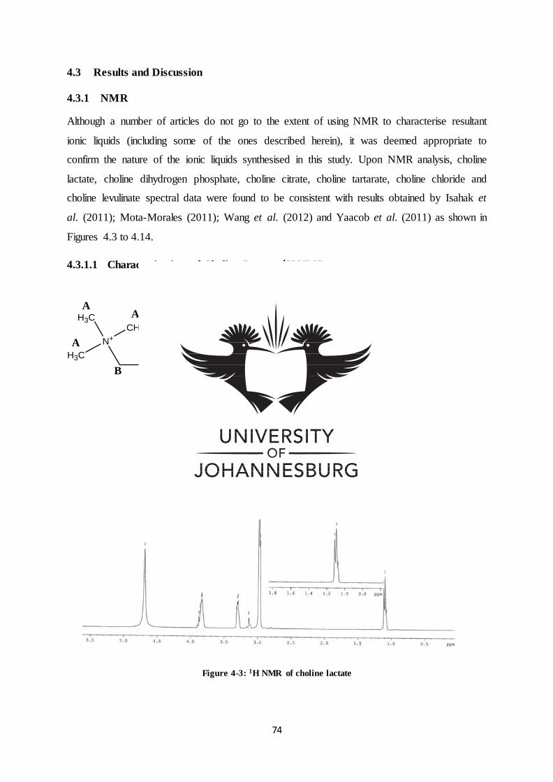
74
4.3 Results and Discussion
4.3.1 NMR
Although a number of articles do not go to the extent of using NMR to characterise resultant
ionic liquids (including some of the ones described herein), it was deemed appropriate to
confirm the nature of the ionic liquids synthesised in this study. Upon NMR analysis, choline
lactate, choline dihydrogen phosphate, choline citrate, choline tartarate, choline chloride and
choline levulinate spectral data were found to be consistent with results obtained by Isahak et
al. (2011); Mota-Morales (2011); Wang et al. (2012) and Yaacob et al. (2011) as shown in
Figures 4.3 to 4.14.
4.3.1.1 Characterization of Choline Lactate 1H NMR
Figure 4-3: 1H NMR of choline lactate
OHN+
CH3
H3C
H3C
C
OH
CH3
O
-O
EA
A
B
A
C
D
F
D
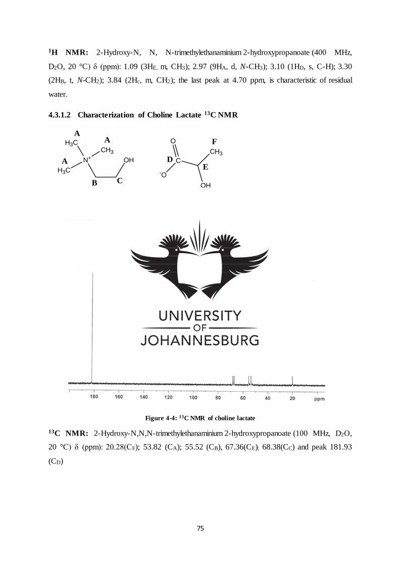
75
1H NMR: 2-Hydroxy-N, N, N-trimethylethanaminium 2-hydroxypropanoate (400 MHz,
D2O, 20 °C) δ (ppm): 1.09 (3HE, m, CH3); 2.97 (9HA, d, N-CH3); 3.10 (1HD, s, C-H); 3.30
(2HB, t, N-CH2); 3.84 (2Hc, m, CH2); the last peak at 4.70 ppm, is characteristic of residual
water.
4.3.1.2 Characterization of Choline Lactate 13C NMR
OHN+
CH3
H3C
H3C
C
OH
CH3
O
-OE
A
B C
D
F
A
A
Figure 4-4: 13C NMR of choline lactate
13C NMR: 2-Hydroxy-N,N,N-trimethylethanaminium 2-hydroxypropanoate (100 MHz, D2O,
20 °C) δ (ppm): 20.28(CF); 53.82 (CA); 55.52 (CB), 67.36(CE); 68.38(CC) and peak 181.93
(CD)
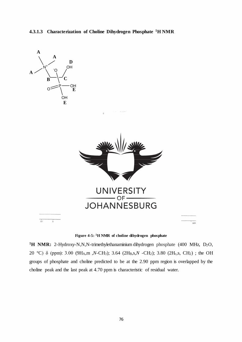
76
4.3.1.3 Characterization of Choline Dihydrogen Phosphate 1H NMR
OHN+
PO
OH
OH
-O
A
A
A
B C
D
E
E
Figure 4-5: 1H NMR of choline dihydrogen phosphate
1H NMR: 2-Hydroxy-N,N,N-trimethylethanaminium dihydrogen phosphate (400 MHz, D2O,
20 °C) δ (ppm): 3.00 (9HA,m ,N-CH3); 3.64 (2HB,s,N -CH2); 3.80 (2Hc,s, CH2) ; the OH
groups of phosphate and choline predicted to be at the 2.90 ppm region is overlapped by the
choline peak and the last peak at 4.70 ppm is characteristic of residual water.
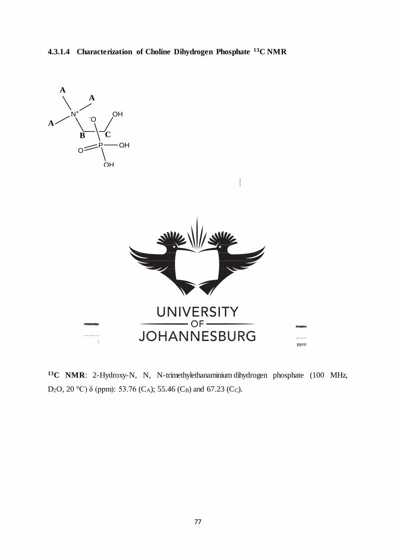
77
4.3.1.4 Characterization of Choline Dihydrogen Phosphate 13C NMR
OHN+
PO
OH
OH
-O
A
A
A
B C
Figure 4-6: 13C NMR of choline dihydrogen phosphate
13C NMR: 2-Hydroxy-N, N, N-trimethylethanaminium dihydrogen phosphate (100 MHz,
D2O, 20 °C) δ (ppm): 53.76 (CA); 55.46 (CB) and 67.23 (CC).
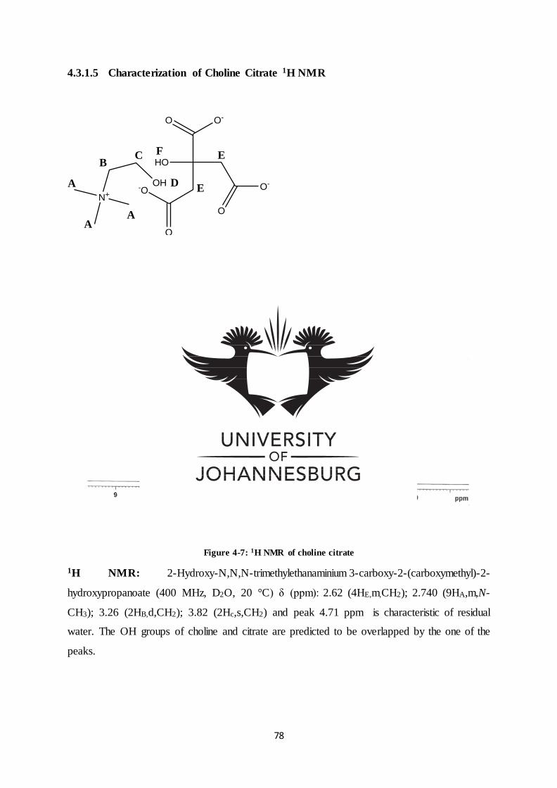
78
4.3.1.5 Characterization of Choline Citrate 1H NMR
OH
N+
HO
O
O-
O O-
O
-O E
E
A
AA
BC
D
F
Figure 4-7: 1H NMR of choline citrate
1H NMR: 2-Hydroxy-N,N,N-trimethylethanaminium 3-carboxy-2-(carboxymethyl)-2-
hydroxypropanoate (400 MHz, D2O, 20 °C) δ (ppm): 2.62 (4HE,m,CH2); 2.740 (9HA,m,N-
CH3); 3.26 (2HB,d,CH2); 3.82 (2Hc,s,CH2) and peak 4.71 ppm is characteristic of residual
water. The OH groups of choline and citrate are predicted to be overlapped by the one of the
peaks.
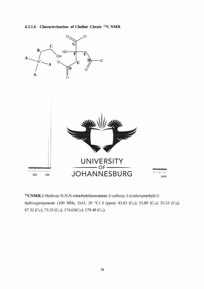
79
4.3.1.6 Characterization of Choline Citrate 13C NMR
OH
N+
HO
O
O-
O O-
O
-O E
A
A
B
A
C
D
F E
G
D
Figure 4-8: 13C NMR of choline citrate
13CNMR:2-Hydroxy-N,N,N-trimethylethanaminium 3-carboxy-2-(carboxymethyl)-2-
hydroxypropanoate (100 MHz, D2O, 20 °C) δ (ppm): 43.63 (CE); 53.80 (CA); 55.53 (CB);
67.32 (CF); 73.33 (CC); 174.63(CD); 178.48 (CG).
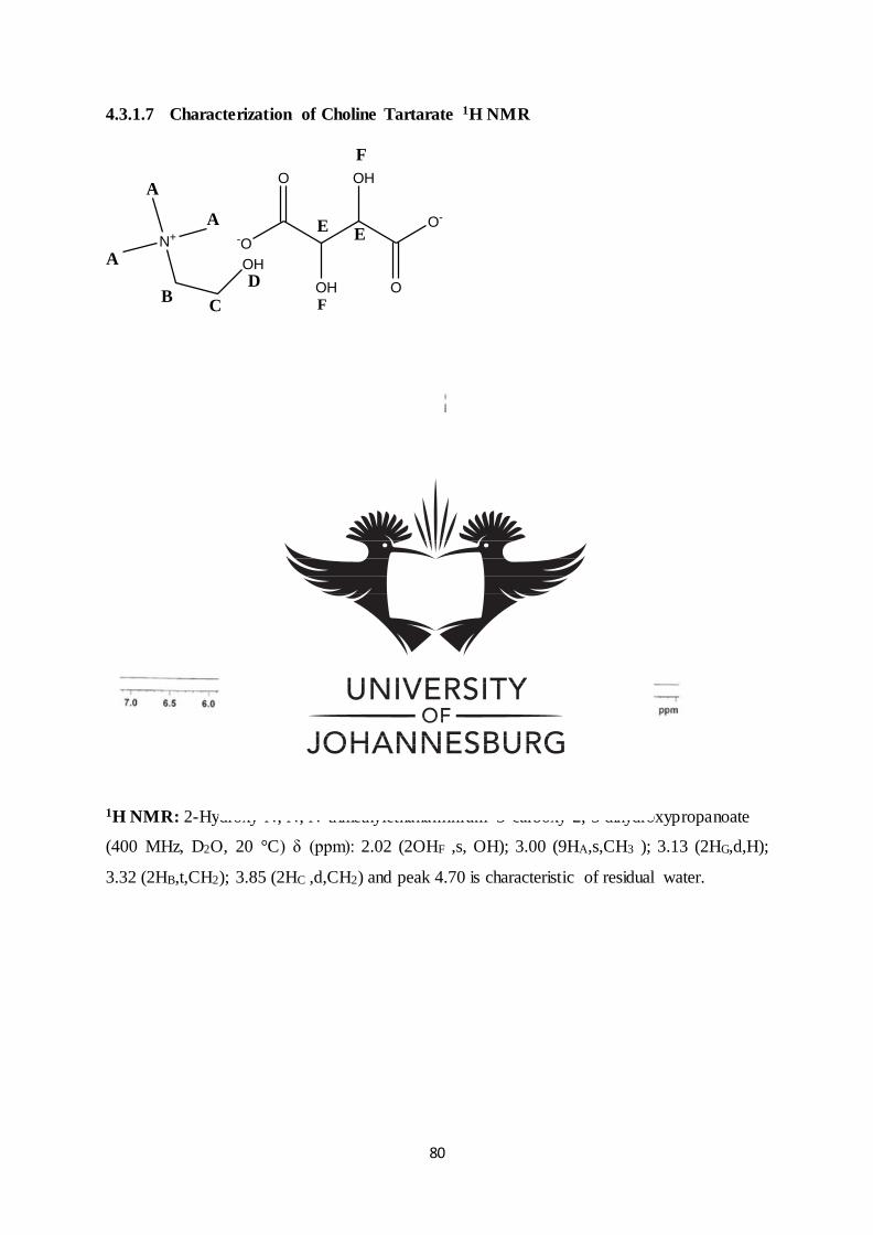
80
4.3.1.7 Characterization of Choline Tartarate 1H NMR
OH
N+
OH
OHO
-O
O
O-
A
A
A
BC
D
F
E
F
E
Figure 4-9: 1H NMR of choline tartarate
1H NMR: 2-Hydroxy-N, N, N-trimethylethanaminium 3-carboxy-2, 3 dihydroxypropanoate
(400 MHz, D2O, 20 °C) δ (ppm): 2.02 (2OHF ,s, OH); 3.00 (9HA,s,CH3 ); 3.13 (2HG,d,H);
3.32 (2HB,t,CH2); 3.85 (2HC ,d,CH2) and peak 4.70 is characteristic of residual water.
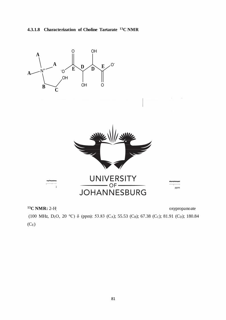
81
4.3.1.8 Characterization of Choline Tartarate 13C NMR
OH
N+
OH
OHO
-O
O
O-
A
A
B
A
C
D DEE
Figure 4-10: 13C NMR of choline tartarate
13C NMR: 2-Hydroxy-N, N, N-trimethylethanaminium 3-carboxy-2, 3-dihydroxypropanoate
(100 MHz, D2O, 20 °C) δ (ppm): 53.83 (CA); 55.53 (CB); 67.38 (CC); 81.91 (CD); 180.84
(CE)
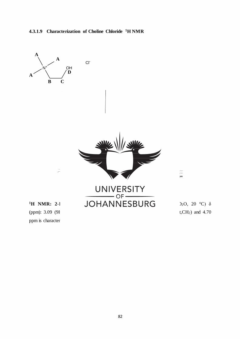
82
4.3.1.9 Characterization of Choline Chloride 1H NMR
OHN+Cl-
A
A
A
B C
D
Figure 4-11: 1H NMR of choline chloride
1H NMR: 2-Hydroxy-N,N,N-trimethylethanaminium chloride (400 MHz, D2O, 20 °C) δ
(ppm): 3.09 (9HA,s,CH3); 3.23 (1HD,s,OH); 3.41 (2HB,m,CH2); 3.94 (2HC,t,CH2) and 4.70
ppm is characteristic of residual water.
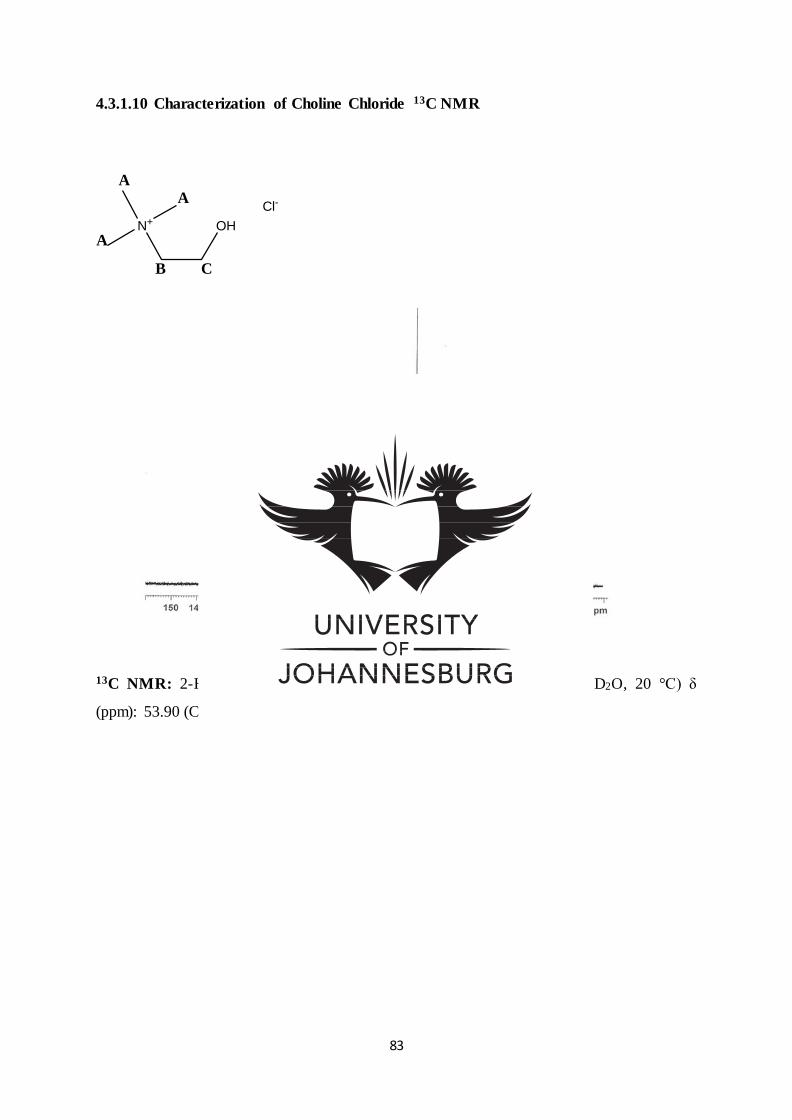
83
4.3.1.10 Characterization of Choline Chloride 13C NMR
OHN+
Cl-
A
A
A
B C
Figure 4-12: 13C NMR of choline chloride
13C NMR: 2-Hydroxy-N, N, N-trimethylethanaminium chloride (100 MHz, D2O, 20 °C) δ
(ppm): 53.90 (CA); 55.70 (CB); 67.50 (CC).
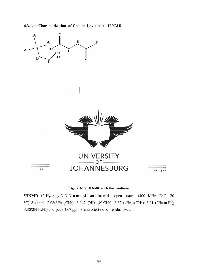
84
4.3.1.11 Characterization of Choline Levulinate 1H NMR
OH
N+
O
O
-O
E
E
A
A
A
BC
D
F
Figure 4-13: 1H NMR of choline levulinate
1HNMR :2-Hydroxy-N,N,N-trimethylethanaminium 4-oxopentanoate (400 MHz, D2O, 20
°C) δ (ppm): 2.08(3HF,s,CH3); 3.047 (9HA,s,N-CH3); 3.37 (4HE,m,CH2); 3.91 (2HB,m,H2);
4.36(2HC,s,H2) and peak 4.67 ppm is characteristic of residual water.
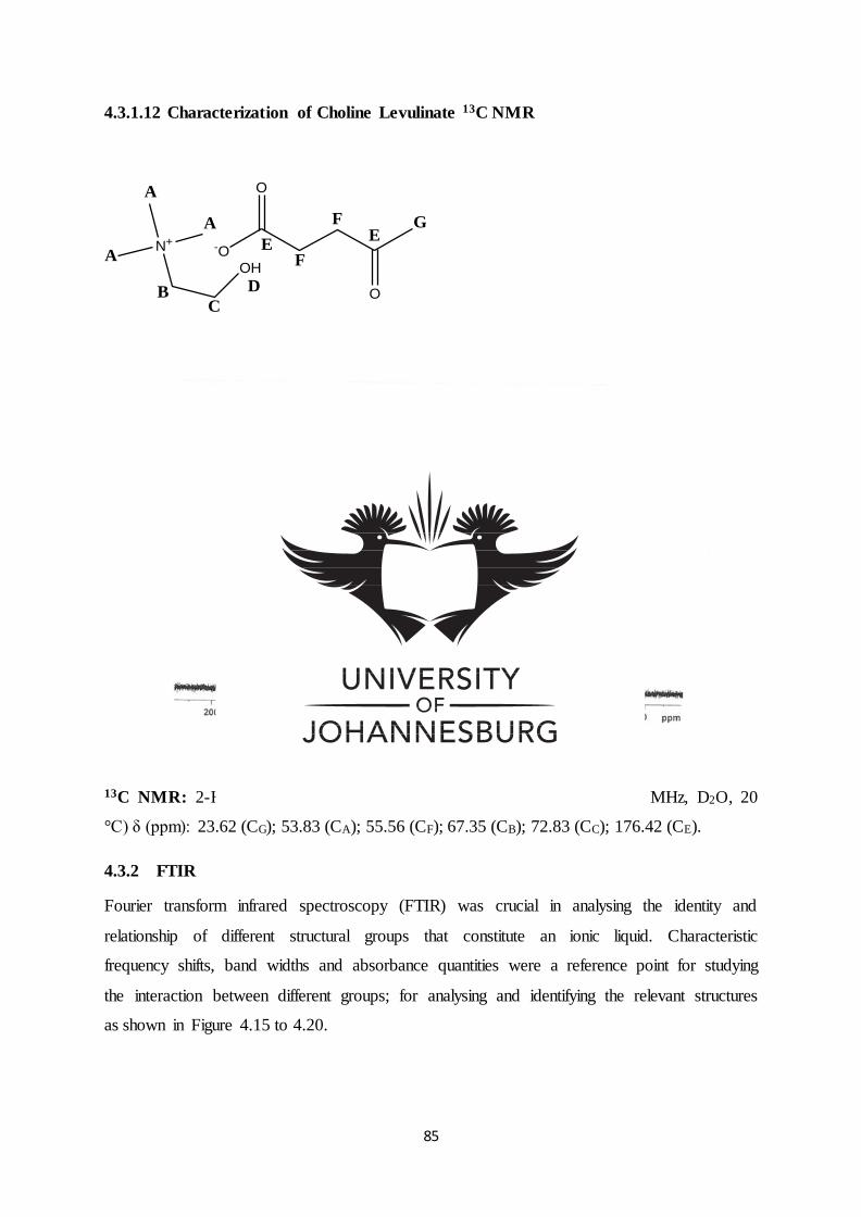
85
4.3.1.12 Characterization of Choline Levulinate 13C NMR
OH
N+
O
O
-O
A
A
A
BC
D
G
EF
FE
Figure 4-14: 13C NMR of choline levulinate
13C NMR: 2-Hydroxy-N N, N-trimethylethanaminium 4-oxopentanoate (100 MHz, D2O, 20
°C) δ (ppm): 23.62 (CG); 53.83 (CA); 55.56 (CF); 67.35 (CB); 72.83 (CC); 176.42 (CE).
4.3.2 FTIR
Fourier transform infrared spectroscopy (FTIR) was crucial in analysing the identity and
relationship of different structural groups that constitute an ionic liquid. Characteristic
frequency shifts, band widths and absorbance quantities were a reference point for studying
the interaction between different groups; for analysing and identifying the relevant structures
as shown in Figure 4.15 to 4.20.

86
4.3.2.1 FTIR: 2-Hydroxy-N, N, N-trimethylethanaminium 2-hydroxypropanoate
Characterization using FTIR (Figure 4.15) confirmed the functional groups occurring in
region 4000 cm-1 to 600 cm-1: The absorptions in the region 1478 cm-1 (C-H Scissoring
vibration), 1350 cm-1(C-H methyl rock vibration), and 2961cm-1 (C-H stretch vibration) were
the result of the methyl group appearing in choline and lactate. The OH group observed in the
region 3278 cm-1 and C-O in region 1086 cm-1 appeared in choline and lactate, and 1119
cm-1 C-N in choline. The absorptions’ in the region 1588 cm-1 and 1406 cm-1 were the result
of the COO- group appearing in Lactate.
Figure 4-15: FTIR of choline lactate
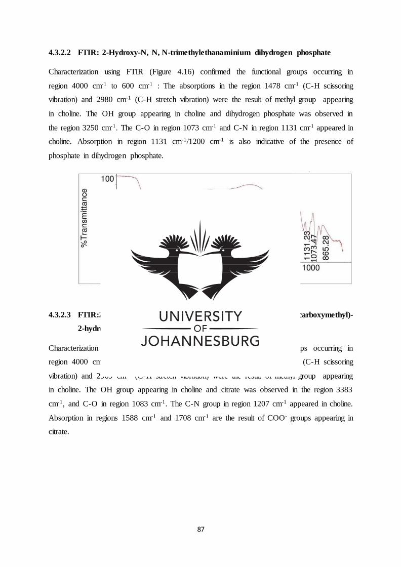
87
4.3.2.2 FTIR: 2-Hydroxy-N, N, N-trimethylethanaminium dihydrogen phosphate
Characterization using FTIR (Figure 4.16) confirmed the functional groups occurring in
region 4000 cm-1 to 600 cm-1 : The absorptions in the region 1478 cm-1 (C-H scissoring
vibration) and 2980 cm-1 (C-H stretch vibration) were the result of methyl group appearing
in choline. The OH group appearing in choline and dihydrogen phosphate was observed in
the region 3250 cm-1. The C-O in region 1073 cm-1 and C-N in region 1131 cm-1 appeared in
choline. Absorption in region 1131 cm-1/1200 cm-1 is also indicative of the presence of
phosphate in dihydrogen phosphate.
Figure 4-16: FTIR of Choline dihydrogen phosphate
4.3.2.3 FTIR:2-Hydroxy-N,N,N-trimethylethanaminium 3-carboxy-2-(carboxymethyl)-
2-hydroxypropanoate
Characterization using FTIR (Figure 4.17) confirmed the functional groups occurring in
region 4000 cm-1 to 600 cm-1 : The absorptions in the region 1477 cm-1 (C-H scissoring
vibration) and 2969 cm-1 (C-H stretch vibration) were the result of methyl group appearing
in choline. The OH group appearing in choline and citrate was observed in the region 3383
cm-1, and C-O in region 1083 cm-1. The C-N group in region 1207 cm-1 appeared in choline.
Absorption in regions 1588 cm-1 and 1708 cm-1 are the result of COO- groups appearing in
citrate.
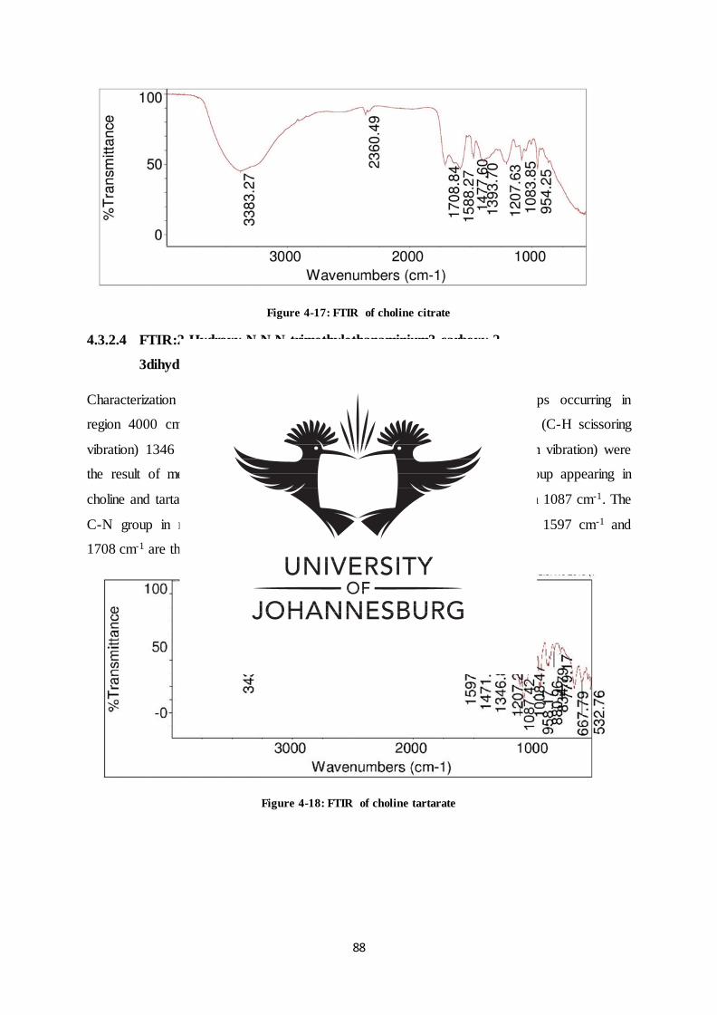
88
Figure 4-17: FTIR of choline citrate
4.3.2.4 FTIR:2-Hydroxy-N,N,N-trimethylethanaminium3-carboxy-2,
3dihydroxypropanoate.
Characterization using FTIR (Figure 4.18) confirmed the functional groups occurring in
region 4000 cm-1 to 600 cm-1 : The absorptions in the region 1471 cm-1 (C-H scissoring
vibration) 1346 cm-1(C-H methyl rock vibration) and 2969 cm-1 (C-H stretch vibration) were
the result of methyl group appearing in choline and tartarate. The OH group appearing in
choline and tartarate was observed in the region 3433 cm-1, and C-O in region 1087 cm-1. The
C-N group in region 1207 cm-1 appeared in choline. Absorption in regions 1597 cm-1 and
1708 cm-1 are the result of COO groups appearing in choline and tartarte.
Figure 4-18: FTIR of choline tartarate
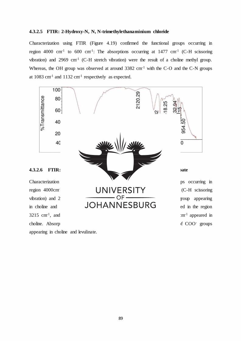
89
4.3.2.5 FTIR: 2-Hydroxy-N, N, N-trimethylethanaminium chloride
Characterization using FTIR (Figure 4.19) confirmed the functional groups occurring in
region 4000 cm-1 to 600 cm-1: The absorptions occurring at 1477 cm-1 (C-H scissoring
vibration) and 2969 cm-1 (C-H stretch vibration) were the result of a choline methyl group.
Whereas, the OH group was observed at around 3382 cm-1 with the C-O and the C-N groups
at 1083 cm-1 and 1132 cm-1 respectively as expected.
Figure 4-19: FTIR of choline chloride
4.3.2.6 FTIR: 2-Hydroxy-N, N, N-trimethylethanaminium 4-oxopentanoate
Characterization using FTIR (Figure 4.20) confirmed the functional groups occurring in
region 4000cm-1 to 600cm-1 : The absorptions in the region 1479 cm-1 (C-H scissoring
vibration) and 2969 cm-1 (C-H stretch vibration) were the result of methyl group appearing
in choline and levulinate. The OH group appearing in choline was observed in the region
3215 cm-1, and C-O in region 1088 cm-1. The C-N group in region 1132 cm-1 appeared in
choline. Absorption in regions 1574 cm-1 and 1705 cm-1 are the result of COO- groups
appearing in choline and levulinate.
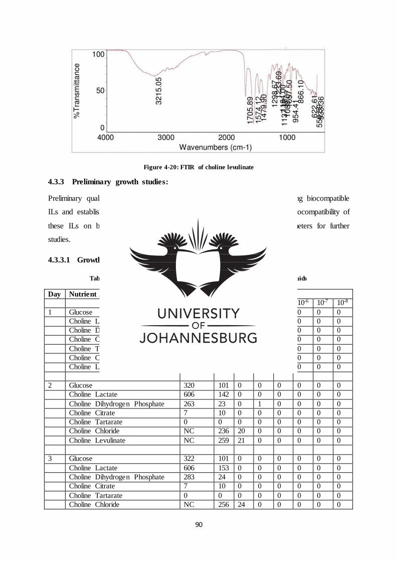
90
Figure 4-20: FTIR of choline levulinate
4.3.3 Preliminary growth studies:
Preliminary qualitative studies (Table 4.4 to 4.9) were essential for screening biocompatible
ILs and establishing quantitative data (Figure 4.21 to 4.26) related to the biocompatibility of
these ILs on bacterial growth and in addition, refining the process parameters for further
studies.
4.3.3.1 Growth plate studies
Table 4-4: Growth plate studies of Bacillus aryabhattai (CFU) in ionic liquids
Day Nutrient Agar Supplement Dilution factor (DF)
10-1 10-2 10-3 10-4 10-5 10-6 10-7 10-8
1 Glucose 301 76 0 0 0 0 0 0
Choline Lactate 556 128 0 0 0 0 0 0
Choline Dihydrogen Phosphate 249 13 0 0 0 0 0 0
Choline Citrate 0 0 0 0 0 0 0 0
Choline Tartarate 0 0 0 0 0 0 0 0
Choline Chloride 800 230 15 0 0 0 0 0
Choline Levulinate 800 219 21 0 0 0 0 0
2 Glucose 320 101 0 0 0 0 0 0
Choline Lactate 606 142 0 0 0 0 0 0
Choline Dihydrogen Phosphate 263 23 0 1 0 0 0 0
Choline Citrate 7 10 0 0 0 0 0 0
Choline Tartarate 0 0 0 0 0 0 0 0
Choline Chloride NC 236 20 0 0 0 0 0
Choline Levulinate NC 259 21 0 0 0 0 0
3 Glucose 322 101 0 0 0 0 0 0
Choline Lactate 606 153 0 0 0 0 0 0
Choline Dihydrogen Phosphate 283 24 0 0 0 0 0 0
Choline Citrate 7 10 0 0 0 0 0 0
Choline Tartarate 0 0 0 0 0 0 0 0
Choline Chloride NC 256 24 0 0 0 0 0
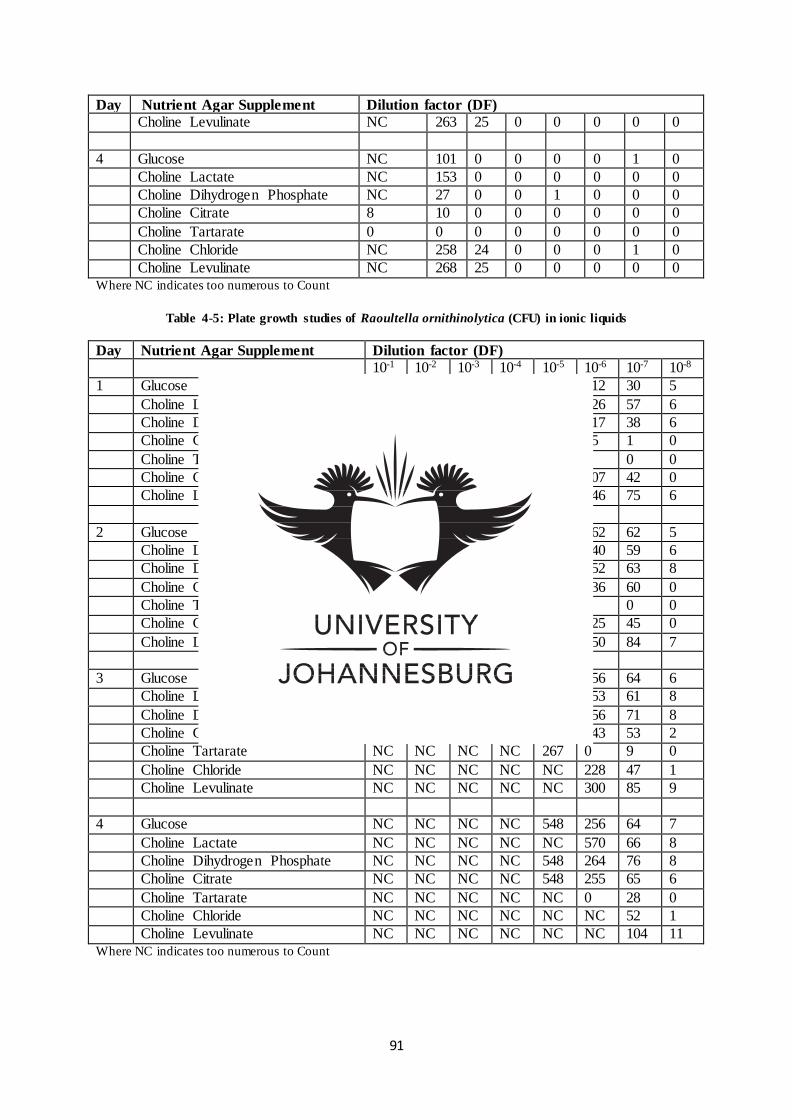
91
Where NC indicates too numerous to Count
Table 4-5: Plate growth studies of Raoultella ornithinolytica (CFU) in ionic liquids
Where NC indicates too numerous to Count
Day Nutrient Agar Supplement Dilution factor (DF)
Choline Levulinate NC 263 25 0 0 0 0 0
4 Glucose NC 101 0 0 0 0 1 0
Choline Lactate NC 153 0 0 0 0 0 0
Choline Dihydrogen Phosphate NC 27 0 0 1 0 0 0
Choline Citrate 8 10 0 0 0 0 0 0
Choline Tartarate 0 0 0 0 0 0 0 0
Choline Chloride NC 258 24 0 0 0 1 0
Choline Levulinate NC 268 25 0 0 0 0 0
Day Nutrient Agar Supplement Dilution factor (DF)
10-1 10-2 10-3 10-4 10-5 10-6 10-7 10-8
1 Glucose NC NC 515 600 273 112 30 5
Choline Lactate NC NC NC NC 526 226 57 6
Choline Dihydrogen Phosphate NC 500 515 352 273 117 38 6
Choline Citrate NC NC 515 340 143 55 1 0
Choline Tartarate NC NC NC NC 0 0 0 0
Choline Chloride NC NC NC NC 465 207 42 0
Choline Levulinate NC NC NC NC 520 246 75 6
2 Glucose NC NC NC NC 346 162 62 5
Choline Lactate NC NC NC NC 546 240 59 6
Choline Dihydrogen Phosphate NC 600 NC 600 346 152 63 8
Choline Citrate NC NC NC 400 220 136 60 0
Choline Tartarate NC NC NC NC 218 0 0 0
Choline Chloride NC NC NC NC NC 225 45 0
Choline Levulinate NC NC NC NC NC 250 84 7
3 Glucose NC NC NC NC 548 256 64 6
Choline Lactate NC NC NC NC 550 253 61 8
Choline Dihydrogen Phosphate NC 632 NC NC 526 256 71 8
Choline Citrate NC NC NC NC 524 243 53 2
Choline Tartarate NC NC NC NC 267 0 9 0
Choline Chloride NC NC NC NC NC 228 47 1
Choline Levulinate NC NC NC NC NC 300 85 9
4 Glucose NC NC NC NC 548 256 64 7
Choline Lactate NC NC NC NC NC 570 66 8
Choline Dihydrogen Phosphate NC NC NC NC 548 264 76 8
Choline Citrate NC NC NC NC 548 255 65 6
Choline Tartarate NC NC NC NC NC 0 28 0
Choline Chloride NC NC NC NC NC NC 52 1
Choline Levulinate NC NC NC NC NC NC 104 11
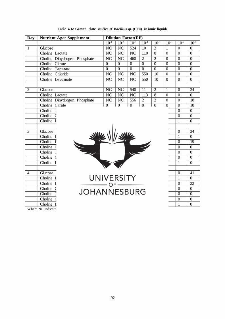
92
Table 4-6: Growth plate studies of Bacillus sp. (CFU) in ionic liquids
Where NC indicates too numerous to Count
Day Nutrient Agar Supplement Dilution Factor(DF)
10-1 10-2 10-3 10-4 10-5 10-6 10-7 10-8
1 Glucose NC NC 524 10 2 1 0 0
Choline Lactate NC NC NC 110 8 0 0 0
Choline Dihydrogen Phosphate NC NC 460 2 2 0 0 0
Choline Citrate 0 0 0 0 0 0 0 0
Choline Tartarate 0 0 0 0 0 0 0 0
Choline Chloride NC NC NC 550 10 0 0 0
Choline Levulinate NC NC NC 550 10 0 0 0
2 Glucose NC NC 540 11 2 1 0 24
Choline Lactate NC NC NC 113 8 0 0 0
Choline Dihydrogen Phosphate NC NC 556 2 2 0 0 18
Choline Citrate 0 0 0 0 0 0 0 18
Choline Tartarate 0 0 0 0 0 0 0 0
Choline Chloride NC NC NC NC 11 0 0 0
Choline Levulinate NC NC NC NC 10 0 1 0
3 Glucose NC NC NC 11 2 1 0 34
Choline Lactate NC NC NC 113 8 0 1 0
Choline Dihydrogen Phosphate NC NC NC 2 2 0 0 19
Choline Citrate 0 0 0 0 0 0 0 0
Choline Tartarate 0 0 0 0 0 0 0 0
Choline Chloride NC NC NC NC 11 0 0 0
Choline Levulinate NC NC NC NC 12 0 1 0
4 Glucose NC NC NC 15 3 1 0 41
Choline Lactate NC NC NC 118 8 0 1 0
Choline Dihydrogen Phosphate NC NC NC 3 2 0 0 22
Choline Citrate 0 0 0 0 0 0 0 0
Choline Tartarate 0 0 0 0 0 0 0 0
Choline Chloride NC NC NC NC 12 0 0 0
Choline Levulinate NC NC NC NC 12 0 1 0
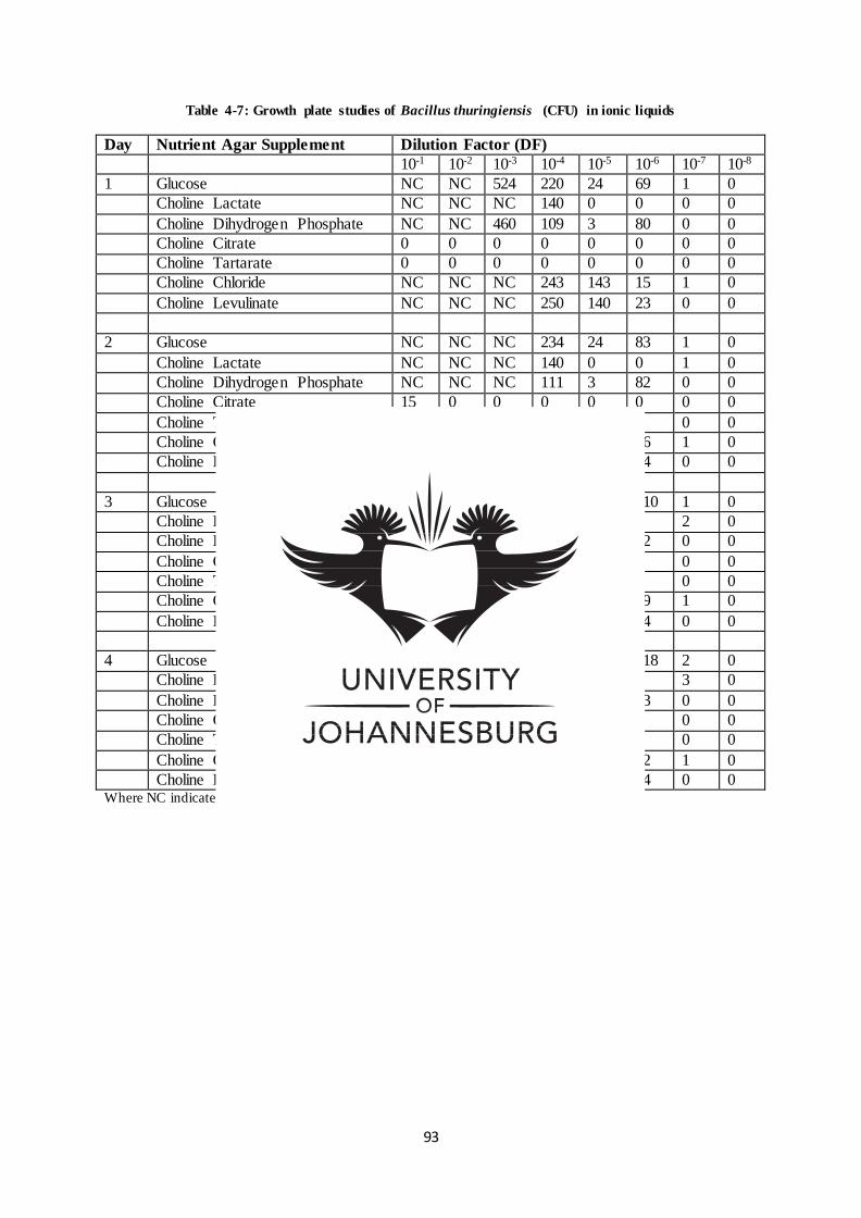
93
Table 4-7: Growth plate studies of Bacillus thuringiensis (CFU) in ionic liquids
Where NC indicates too numerous to Count
Day Nutrient Agar Supplement Dilution Factor (DF)
10-1 10-2 10-3 10-4 10-5 10-6 10-7 10-8
1 Glucose NC NC 524 220 24 69 1 0
Choline Lactate NC NC NC 140 0 0 0 0
Choline Dihydrogen Phosphate NC NC 460 109 3 80 0 0
Choline Citrate 0 0 0 0 0 0 0 0
Choline Tartarate 0 0 0 0 0 0 0 0
Choline Chloride NC NC NC 243 143 15 1 0
Choline Levulinate NC NC NC 250 140 23 0 0
2 Glucose NC NC NC 234 24 83 1 0
Choline Lactate NC NC NC 140 0 0 1 0
Choline Dihydrogen Phosphate NC NC NC 111 3 82 0 0
Choline Citrate 15 0 0 0 0 0 0 0
Choline Tartarate 0 0 0 0 0 0 0 0
Choline Chloride NC NC NC NC 144 16 1 0
Choline Levulinate NC NC NC NC NC 24 0 0
3 Glucose NC NC NC 255 32 110 1 0
Choline Lactate NC NC NC 140 0 0 2 0
Choline Dihydrogen Phosphate NC NC NC 86 4 82 0 0
Choline Citrate 15 0 0 0 0 0 0 0
Choline Tartarate 0 0 0 0 0 0 0 0
Choline Chloride NC NC NC NC NC 19 1 0
Choline Levulinate NC NC NC NC NC 24 0 0
4 Glucose NC NC NC 259 32 118 2 0
Choline Lactate NC NC NC 141 0 0 3 0
Choline Dihydrogen Phosphate NC NC NC 101 7 93 0 0
Choline Citrate 108 0 0 0 0 0 0 0
Choline Tartarate 0 0 0 0 0 0 0 0
Choline Chloride NC NC NC NC NC 22 1 0
Choline Levulinate NC NC NC NC NC 24 0 0
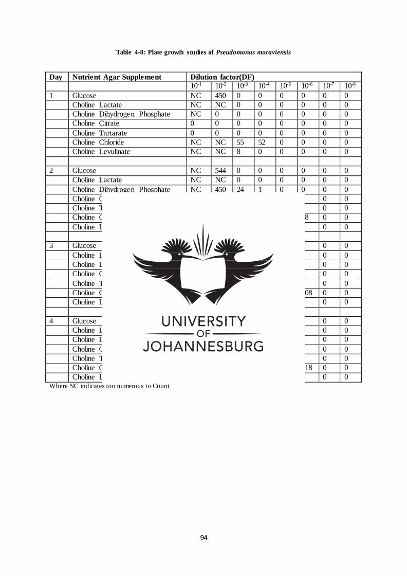
94
Table 4-8: Plate growth studies of Pseudomonas moraviensis
Where NC indicates too numerous to Count
Day Nutrient Agar Supplement Dilution factor(DF)
10-1 10-2 10-3 10-4 10-5 10-6 10-7 10-8
1 Glucose NC 450 0 0 0 0 0 0
Choline Lactate NC NC 0 0 0 0 0 0
Choline Dihydrogen Phosphate NC 0 0 0 0 0 0 0
Choline Citrate 0 0 0 0 0 0 0 0
Choline Tartarate 0 0 0 0 0 0 0 0
Choline Chloride NC NC 55 52 0 0 0 0
Choline Levulinate NC NC 8 0 0 0 0 0
2 Glucose NC 544 0 0 0 0 0 0
Choline Lactate NC NC 0 0 0 0 0 0
Choline Dihydrogen Phosphate NC 450 24 1 0 0 0 0
Choline Citrate 0 0 0 0 0 0 0 0
Choline Tartarate 0 0 0 0 0 0 0 0
Choline Chloride NC NC NC 600 213 18 0 0
Choline Levulinate NC NC 150 24 0 0 0 0
3 Glucose NC NC 0 0 0 0 0 0
Choline Lactate NC NC 0 0 0 0 0 0
Choline Dihydrogen Phosphate NC 455 435 1 0 0 0 0
Choline Citrate 108 0 0 0 0 0 0 0
Choline Tartarate 0 0 0 0 0 0 0 0
Choline Chloride NC NC NC 620 250 108 0 0
Choline Levulinate NC NC NC 263 104 0 0 0
4 Glucose NC NC 0 0 0 0 0 0
Choline Lactate NC NC 0 0 0 0 0 0
Choline Dihydrogen Phosphate NC NC 446 70 0 0 0 0
Choline Citrate 108 0 0 0 0 0 0 0
Choline Tartarate 0 0 0 0 0 0 0 0
Choline Chloride NC NC NC NC 623 218 0 0
Choline Levulinate NC NC NC 292 105 0 0 0
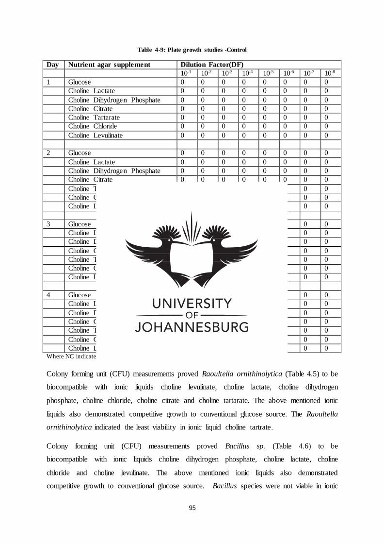
95
Table 4-9: Plate growth studies -Control
Day Nutrient agar supplement Dilution Factor(DF)
10-1 10-2 10-3 10-4 10-5 10-6 10-7 10-8
1 Glucose 0 0 0 0 0 0 0 0
Choline Lactate 0 0 0 0 0 0 0 0
Choline Dihydrogen Phosphate 0 0 0 0 0 0 0 0
Choline Citrate 0 0 0 0 0 0 0 0
Choline Tartarate 0 0 0 0 0 0 0 0
Choline Chloride 0 0 0 0 0 0 0 0
Choline Levulinate 0 0 0 0 0 0 0 0
2 Glucose 0 0 0 0 0 0 0 0
Choline Lactate 0 0 0 0 0 0 0 0
Choline Dihydrogen Phosphate 0 0 0 0 0 0 0 0
Choline Citrate 0 0 0 0 0 0 0 0
Choline Tartarate 0 0 0 0 0 0 0 0
Choline Chloride 0 0 0 0 0 0 0 0
Choline Levulinate 0 0 0 0 0 0 0 0
3 Glucose 0 0 0 0 0 0 0 0
Choline Lactate 0 0 0 0 0 0 0 0
Choline Dihydrogen Phosphate 0 0 0 0 0 0 0 0
Choline Citrate 0 0 0 0 0 0 0 0
Choline Tartarate 0 0 0 0 0 0 0 0
Choline Chloride 0 0 0 0 0 0 0 0
Choline Levulinate 0 0 0 0 0 0 0 0
4 Glucose 0 0 0 0 0 0 0 0
Choline Lactate 0 0 0 0 0 0 0 0
Choline Dihydrogen Phosphate 0 0 0 0 0 0 0 0
Choline Citrate 0 0 0 0 0 0 0 0
Choline Tartarate 0 0 0 0 0 0 0 0
Choline Chloride 0 0 0 0 0 0 0 0
Choline Levulinate 0 0 0 0 0 0 0 0 Where NC indicates too numerous to Count
Colony forming unit (CFU) measurements proved Raoultella ornithinolytica (Table 4.5) to be
biocompatible with ionic liquids choline levulinate, choline lactate, choline dihydrogen
phosphate, choline chloride, choline citrate and choline tartarate. The above mentioned ionic
liquids also demonstrated competitive growth to conventional glucose source. The Raoultella
ornithinolytica indicated the least viability in ionic liquid choline tartrate.
Colony forming unit (CFU) measurements proved Bacillus sp. (Table 4.6) to be
biocompatible with ionic liquids choline dihydrogen phosphate, choline lactate, choline
chloride and choline levulinate. The above mentioned ionic liquids also demonstrated
competitive growth to conventional glucose source. Bacillus species were not viable in ionic
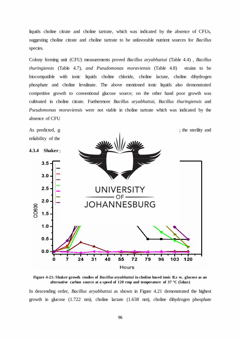
96
liquids choline citrate and choline tartrate, which was indicated by the absence of CFUs,
suggesting choline citrate and choline tartrate to be unfavorable nutrient sources for Bacillus
species.
Colony forming unit (CFU) measurements proved Bacillus aryabhattai (Table 4.4) , Bacillus
thuringiensis (Table 4.7), and Pseudomonas moraviensis (Table 4.8) strains to be
biocompatible with ionic liquids choline chloride, choline lactate, choline dihydrogen
phosphate and choline levulinate. The above mentioned ionic liquids also demonstrated
competitive growth to conventional glucose source; on the other hand poor growth was
cultivated in choline citrate. Furthermore Bacillus aryabhattai, Bacillus thuringiensis and
Pseudomonas moraviensis were not viable in choline tartrate which was indicated by the
absence of CFUs, suggesting unfavorable nutrient sources.
As predicted, growth was entirely absent in the control (Table 4.9), verifying the sterility and
reliability of the experiment.
4.3.4 Shaker growth studies
Figure 4-21: Shaker growth studies of Bacillus aryabhattai in choline based ionic ILs vs. glucose as an
alternative carbon source at a speed of 120 rmp and temperature of 37 °C (5days)
In descending order, Bacillus aryabhattai as shown in Figure 4.21 demonstrated the highest
growth in glucose (1.722 nm), choline lactate (1.638 nm), choline dihydrogen phosphate
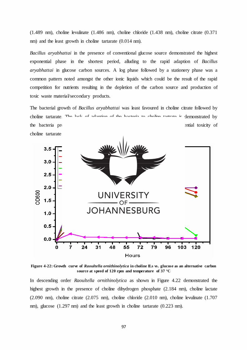
97
(1.489 nm), choline levulinate (1.486 nm), choline chloride (1.438 nm), choline citrate (0.371
nm) and the least growth in choline tartarate (0.014 nm).
Bacillus aryabhattai in the presence of conventional glucose source demonstrated the highest
exponential phase in the shortest period, alluding to the rapid adaption of Bacillus
aryabhattai in glucose carbon sources. A log phase followed by a stationery phase was a
common pattern noted amongst the other ionic liquids which could be the result of the rapid
competition for nutrients resulting in the depletion of the carbon source and production of
toxic waste material/secondary products.
The bacterial growth of Bacillus aryabhattai was least favoured in choline citrate followed by
choline tartarate. The lack of adaption of the bacteria to choline tartrate is demonstrated by
the bacteria predominantly remaining in the lag phase alluding to the potential toxicity of
choline tartarate to Bacillus aryabhattai.
Figure 4-22: Growth curve of Raoultella ornithinolytica in choline ILs vs. glucose as an alternative carbon
source at speed of 120 rpm and temperature of 37 °C
In descending order Raoultella ornithinolytica as shown in Figure 4.22 demonstrated the
highest growth in the presence of choline dihydrogen phosphate (2.184 nm), choline lactate
(2.090 nm), choline citrate (2.075 nm), choline chloride (2.010 nm), choline levulinate (1.707
nm), glucose (1.297 nm) and the least growth in choline tartarate (0.223 nm).
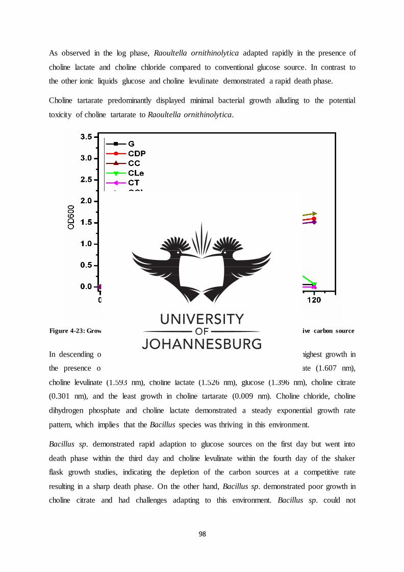
98
As observed in the log phase, Raoultella ornithinolytica adapted rapidly in the presence of
choline lactate and choline chloride compared to conventional glucose source. In contrast to
the other ionic liquids glucose and choline levulinate demonstrated a rapid death phase.
Choline tartarate predominantly displayed minimal bacterial growth alluding to the potential
toxicity of choline tartarate to Raoultella ornithinolytica.
Figure 4-23: Growth curve of Bacillus sp. in choline based ILs vs. glucose as an alternative carbon source
at speed of 120 rpm and temperature of 37 °C.
In descending order Bacillus sp. as shown in Figure 4.23 demonstrated the highest growth in
the presence of, choline chloride (1.715 nm), choline dihydrogen phosphate (1.607 nm),
choline levulinate (1.593 nm), choline lactate (1.526 nm), glucose (1.396 nm), choline citrate
(0.301 nm), and the least growth in choline tartarate (0.009 nm). Choline chloride, choline
dihydrogen phosphate and choline lactate demonstrated a steady exponential growth rate
pattern, which implies that the Bacillus species was thriving in this environment.
Bacillus sp. demonstrated rapid adaption to glucose sources on the first day but went into
death phase within the third day and choline levulinate within the fourth day of the shaker
flask growth studies, indicating the depletion of the carbon sources at a competitive rate
resulting in a sharp death phase. On the other hand, Bacillus sp. demonstrated poor growth in
choline citrate and had challenges adapting to this environment. Bacillus sp. could not
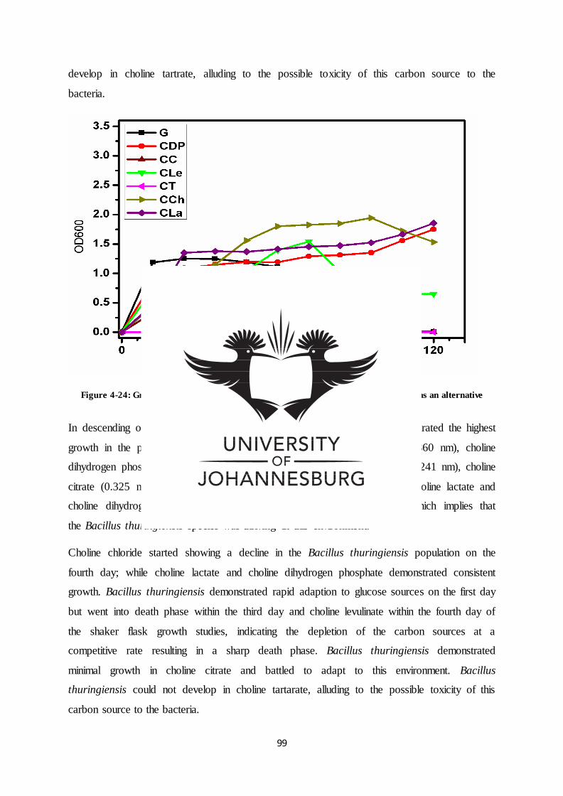
99
develop in choline tartrate, alluding to the possible toxicity of this carbon source to the
bacteria.
Figure 4-24: Growth curve of Bacillus thuringiensis in choline based ILs vs. glucose as an alternative
carbon source at speed of 120 rpm and temperature of 37 °C.
In descending order Bacillus thuringiensis as shown in Figure 4.24 demonstrated the highest
growth in the presence of choline chloride (1.943 nm), choline lactate (1.860 nm), choline
dihydrogen phosphate (1.761 nm), choline levulinate (1.550 nm), glucose (1.241 nm), choline
citrate (0.325 nm) and the least growth in choline tartarate (0.029 nm). Choline lactate and
choline dihydrogen phosphate demonstrated steady exponential growth, which implies that
the Bacillus thuringiensis species was thriving in this environment.
Choline chloride started showing a decline in the Bacillus thuringiensis population on the
fourth day; while choline lactate and choline dihydrogen phosphate demonstrated consistent
growth. Bacillus thuringiensis demonstrated rapid adaption to glucose sources on the first day
but went into death phase within the third day and choline levulinate within the fourth day of
the shaker flask growth studies, indicating the depletion of the carbon sources at a
competitive rate resulting in a sharp death phase. Bacillus thuringiensis demonstrated
minimal growth in choline citrate and battled to adapt to this environment. Bacillus
thuringiensis could not develop in choline tartarate, alluding to the possible toxicity of this
carbon source to the bacteria.
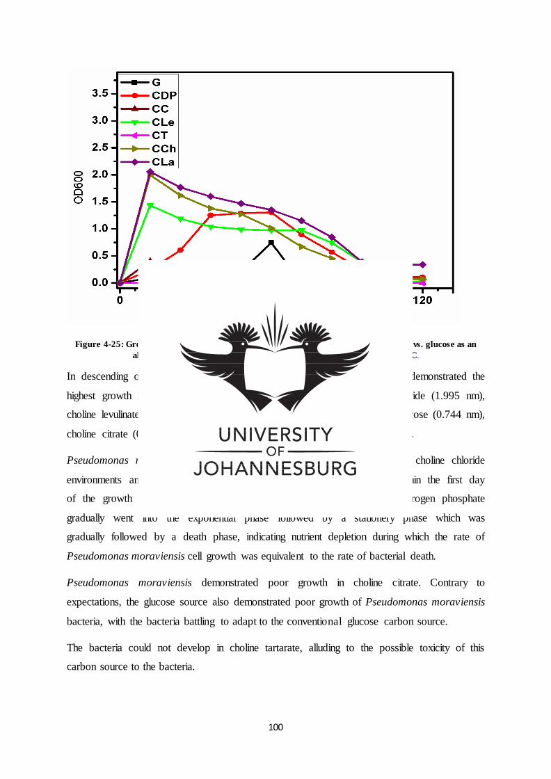
100
Figure 4-25: Growth curve of Pseudomonas moraviensis in choline based ionic liquids vs. glucose as an
alternative carbon source at speed of 120 rpm and temperature of 37 °C.
In descending order Pseudomonas moraviensis as shown in Figure 4.25 demonstrated the
highest growth in the presence of choline lactate (2.058 nm), choline chloride (1.995 nm),
choline levulinate (1.438 nm), choline dihydrogen phosphate (1.304 nm), glucose (0.744 nm),
choline citrate (0.401 nm) and the least growth in choline tartarate (0.019 nm).
Pseudomonas moraviensis demonstrated rapid adaption in choline lactate choline chloride
environments and choline levulinate, reaching rapid exponential growth within the first day
of the growth studies followed by a gradual death phase. Choline dihydrogen phosphate
gradually went into the exponential phase followed by a stationery phase which was
gradually followed by a death phase, indicating nutrient depletion during which the rate of
Pseudomonas moraviensis cell growth was equivalent to the rate of bacterial death.
Pseudomonas moraviensis demonstrated poor growth in choline citrate. Contrary to
expectations, the glucose source also demonstrated poor growth of Pseudomonas moraviensis
bacteria, with the bacteria battling to adapt to the conventional glucose carbon source.
The bacteria could not develop in choline tartarate, alluding to the possible toxicity of this
carbon source to the bacteria.
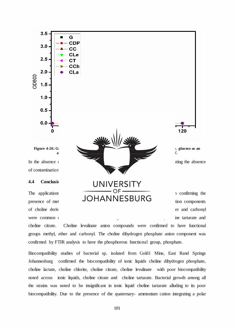
101
Figure 4-26: Growth curve in the absence of bacteria in choline based ionic liquids vs. glucose as an
alternative carbon source at speed of 120 rpm and temperature of 37 °C
In the absence of bacteria as shown in Table 4.26 there was no growth, indicating the absence
of contamination and proving the sterilization technique effective.
4.4 Conclusion
The applications of 13C NMR, 1H NMR and FT-IR were successful in confirming the
presence of methyl groups, alcohol and amine functional groups in all the cation components
of choline derived ionic liquids. The functional groups methyl, hydroxyl, ether and carbonyl
were common denominators in the anion component of choline lactate, choline tartarate and
choline citrate. Choline levulinate anion compounds were confirmed to have functional
groups methyl, ether and carbonyl. The choline dihydrogen phosphate anion component was
confirmed by FTIR analysis to have the phosphorous functional group, phosphate.
Biocompatibility studies of bacterial sp. isolated from Gold1 Mine, East Rand Springs
Johannesburg confirmed the biocompatibility of ionic liquids choline dihydrogen phosphate,
choline lactate, choline chlorite, choline citrate, choline levulinate with poor biocompatibility
noted across ionic liquids, choline citrate and choline tartarate. Bacterial growth among all
the strains was noted to be insignificant in ionic liquid choline tartarate alluding to its poor
biocompatibility. Due to the presence of the quaternary- ammonium cation integrating a polar
102
OH it was projected that all choline based ILs would not be of a toxic nature, however
choline tartarate indicated poor biocompatibility indicating the influence of the anion
component on the total toxicity.
Bacillus sp. and Bacillus thuringiensis demonstrated a similar growth curve and
biocompatibility pattern across the ionic liquids, confirming their phylogenetic relationship.
The ionic liquids choline chloride, choline lactate, choline dihydrogen phosphate and choline
levulinate were the preferred source of carbon across all five bacteria strains. Choline lactate,
choline chloride, choline dihydrogen phosphate and choline levulinate in agreement with both
plate and shaker flask studies were concluded to be the most biocompatible ionic liquids
across all five bacterial strains, and selected for further studies.
Acknowledgement
This work was supported by a research grant sponsored by the National Research
Foundation.
103
References
Ferlin, N., Courty, M., Gatard, S., Spulak, M., Quilty, B., Beadham, I., Ghavre, M., Haiß, A.,
Kummerer, K.,Gathergood, N. & Bouquillon, S. (2013). Biomass derived ionic liquids:
synthesis from natural organic acids, characterization, toxicity, biodegradation and use as
solvents for catalytic hydrogenation processes. Tetrahedron, 69, 6150 - 6161.
Guo-cai, T., Jian, L. & Yi-xin, H. (2010). Application of ionic liquids in hydrometallurgy of
nonferrous metals. Transactions of Nonferrous Metals of Society of China, 20, 513-5120.
Habulin, M., Primožič, M. & Knez, Z. (2011). Application of Ionic Liquids in
Biocatalysis.Ionic.Liquids: Applications and Perspectives.Prof.Alexander Kokorin (Ed.).
InTech. Available from: http://www.intechopen.com/books/ionic-liquids-applications-and-
perspectives/application-ofionic- (Accessed, 21/06/2016).
Hou, X., Liu, Q., Smith, T., Li, N. & Zong, M. (2013). Evaluation of Toxicity and
Biodegradability of Cholinium amino Acids Ionic Liquids. Primary Library of Science, 8, 3.
Isahak, W., Ismail, M., Mohd Jahim, J., Salimon, J. & Yarmo, M. (2011). Transesterification
of Palm Oil by using Ionic Liquids as a New Potential Catalyst. Trends in Applied Sciences
Research. doi: http://dx.doi.org/10.3923/tasr.2011.
Martins, I., Hartmann, D., Alves, P., Planchon, S., Renaut, J., Leitão, M., Rebelo, L. &
Pereira, C. (2013). Proteomic alterations induced by ionic liquids in Aspergillusnidulans and
Neurosporacrassa. Journal of Proteomics, 9 4, 2 6 2 – 2 7 8.
Mota-Morales, J., Gutiérrez, M., Sanchez, I., Luna-Bárcenas, G. & Monte, F. (2011). Made
Deep-Eutectic Solvents Suitable for Frontal Polymerization. The Royal Society of Chemistry.
Rantwijk, F., Lau, R. & Sheldon, R. (2003). Biocatalytic transformations in ionic liquids.
Tends in Biotechnology, 21, 3.
Silva, F., Siopa, F., Figueiredo, B., Gonçalves, A., Pereira, J., Gonçalves, F., Coutinho, J.,
Afonso, C. & Ventura, S. (2014). Sustainable design for environment-friendly mono and
dicationiccholinium-based ionic liquids. Ecotoxicology and Environmental Safety, 108, 302 –
310.
Wang, H., Jla, Y., Wang, X., Ma, J. & Jing, Y. (2012). Physico-chemical Properties of
Magnesium Ionic Liquid Analogous. Journal of Chilean Chemical Society, 57, 3.

104
Yaacob, Z., Nordin, N. & Yarmo, M. (2011). Ionic Liquid Supported Acid-catalysed
Esterification of Lauric Acid. The Malaysian Journal of Analytical Sciences, 15(1), 46-53.
105
CHAPTER FIVE
THE VALORISATION OF SLAG FROM BCL USING BIOCOMPATIBLE
CHOLINE BASED IONIC LIQUIDS AS A SUPPORT
Letlhabile Moyaha1, Sudharshan sekar1, Antoine F. Mulaba-Bafubiandi3, Sibusiso
Sidu2, Vuyo Mavumengwana1
1Department of Biotechnology and Food Technology, University of Johannesburg,
Johannesburg, South Africa – 2094
2Gold One International Limited, Springs, South Africa
3Mineral Processing and Technology Research Center, Department of Metallurgy, School of
Mining, Metallurgy and Chemical Engineering University of Johannesburg, South Africa,
Johannesburg, P.O. Box 17011, Doornfontein 2028,Gauteng, South Africa
*Corresponding author:[email protected]; Tel: 0712839617
Abstract
Slags often contain significant volumes of base metals and can potentially be exploited as
reliable secondary resources if treated in an environmentally friendly fashion or potential
pollutants if not processed correctly. Considering the environmental and economic
implications of these pollutants, it was imperative to establish an efficient and feasible
method for the optimal recovery of these metal values and slag waste disposal. As a result a
novel bioleaching method was developed for the valorisation of slag from BCL using choline
based ionic liquids as a support. The study was indicative of choline ILs, specifically choline
lactate having a metabolic advantage over glucose carbon sources, which was validated by
the optimal leaching kinetics represented by significant iron recoveries ranging from 65 % to
97% by Bacillus aryabhattai, Raoultella ornithinolytica and mixed cultures; with the highest
dissolution efficiency facilitated by Bacillus species and Bacillus thuringiensis. Moderate
zinc recoveries ranging from 26% to 31% were also indicated in IL choline dihydrogen
phosphate. Thus, suggesting the selective bioleaching of base metals by ILs choline lactate
and choline dihydrogen phosphate, used as a substrate for the valorisation of slag from BCL.
Keywords: Slags, valorisation, bioleaching, choline, ILs, base metals
106
5.1 Introduction
Slags are formed in bulk quantities as by-products of ferrous / non-ferrous smelting processes
comprising of metal oxides which can either be recovered or re-used. Slags also contain base
metals namely Co, Ni, Zn etc. that can potentially pollute the atmosphere if not recovered
accordingly (Yang et al., 2010). Slag minerals can thus be valorised by the value of their
minor constituents; by the iron-oxides and silicates occurring in large concentrations
(Kalinkin et al., 2012; Maweja et al., 2010; Wang et al., 2015). Investigations on
hydrometallurgy methods for the treatment of slag have been extensively conducted. Such as
investigations on the dissolution rate of 95 % cadmium, nickel and zinc from zinc plant
residues, by employing 1.22 mol/L of sulphuric acid at a temperature of 25 °C for 60
minutes, though with insignificant extraction of Lead (Kai-qi et al., 2012). However
hydrometallurgy processes have indicated limitations, favouring the recovery of metals
occurring in high concentrations and have also been discovered to produce contaminants in
the form of excess hazardous leaching agents. As a result, bioleaching processes provide an
alternative approach for slag wastes with low quantities of metal values (Hocheng et al.,
2014).
While thorough investigations have been conducted into the architecture and engineering-
technology of biological mining processes, the microbiology aspect has been neglected,
lacking critical studies and clear parameters (Kai-qi et al., 2012; Kaksonen et al., 2011;
Rawlings et al., 2007). The main challenge presented in the commercialization of bioleaching
processes lies in the slow bacterial growth rates impeding the leaching kinetics; as a result
further research is required for improving the growth kinetics of these microorganisms.
Contrary to common industrial companies that employ microorganisms such as fermenting
technology industries, biopharmaceuticals etc. the collection, regulation and supervision of
microbes in bio-mining has been minimum or completely absent. This poses a question
whether commercially used microbes are the most competent consortium of microbial species
for diverse mineral processing. Supposing a supreme microbial consortium was discovered in
lab studies, there remains the question, as to how the population of mineral oxidizers can be
improved and monitored, specifically in heap-leaching processes. Furthermore there are
significant debates referring to how optimized microbial consortiums used in the bio-
oxidation of specific minerals in fixed operating conditions can be developed e.g.
temperature/pH. The debates also argue whether there are advantages in the transfer of
107
bacterial consortiums from foreign locations in comparison to the selection of native
microbes identified within the vicinity of the deposits (Rawlings et al., 2007).
To date ionic liquids have grown popular in different fields namely organic synthesis,
catalysis , bio-catalysis and biomass pre-treatment , which is owed to their thermal stability,
diverse solubility and their properties as designer solvents (Guo-cai, Jian & Yi-xin, 2010;
Samori et al., 2010). Furthermore choline chloride derived ionic liquids (ILs) were reported
to be advantageous due to their simple prep, good water/air stability and inexpensive nature,
thus qualifying their application at an industrial scale (Haerens et al., 2009).
Choline is a crucial component for healthy cellular function with an existing mechanism for
regulating its biological synthesis and hydrolysis. Recently an investigation on the efficiency
of choline lactate for the growth of Staphylococcus lentus evaluating the fate of the cation
and anion component was conducted, establishing favourable biochemistry and bioenergetics
in choline lactate medium (Sekar et al., 2013). Choline hydroxide has also been employed for
the catalysis reaction of aldol condensation (Pernak et al., 2007). Even though numerous
papers have been published on the subject of ionic liquids; pertinent investigations in the
hydrometallurgical field have proven unsuccessful. As a result, the application of ionic
liquids in this field is still in its infancy and requires great attention (Pernak et al., 2007).
Furthermore is the research gap in the use of these ionic liquids as catalysts for biological
processes.
With the aim of minimising the loss of valued product from bio-mining operations and
reducing the impact of process waste on the environment, an investigation was conducted to
determine optimal base metal recovery from BCL slag employing choline based ionic liquids
as an alternative substrate to conventional glucose sources. The ionic liquids of interest
included choline levulinate (CLe), choline lactate (CLa), choline dihydrogen phosphate
(CDP) and choline chloride (CCh) vs. conventional glucose (G) sources.
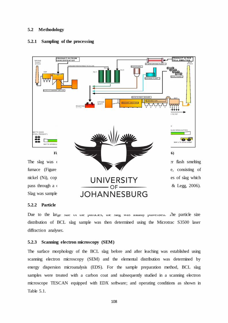
108
5.2 Methodology
5.2.1 Sampling of the processing
Figure 5-1: Schematic of BCL slag (Adapted from Malema & Legg, 2006)
The slag was obtained from BCL Limited which operates a nickel copper flash smelting
furnace (Figure 5.1) at Selebi-Phikwe yielding high-grade sulphide matte, consisting of
nickel (Ni), copper (Cu) and cobalt (Co) for further refining; and large volumes of slag which
pass through a cleaning furnace before being crushed and discarded (Malema & Legg, 2006).
Slag was sampled in triplicates and stored at room temperature.
5.2.2 Particle size distribution
Due to the large size of the particles, the slag was initially pulverised. The particle size
distribution of BCL slag sample was then determined using the Microtrac S3500 laser
diffraction analyser.
5.2.3 Scanning electron microscopy (SEM)
The surface morphology of the BCL slag before and after leaching was established using
scanning electron microscopy (SEM) and the elemental distribution was determined by
energy dispersion microanalysis (EDS). For the sample preparation method, BCL slag
samples were treated with a carbon coat and subsequently studied in a scanning electron
microscope TESCAN equipped with EDX software; and operating conditions as shown in
Table 5.1.
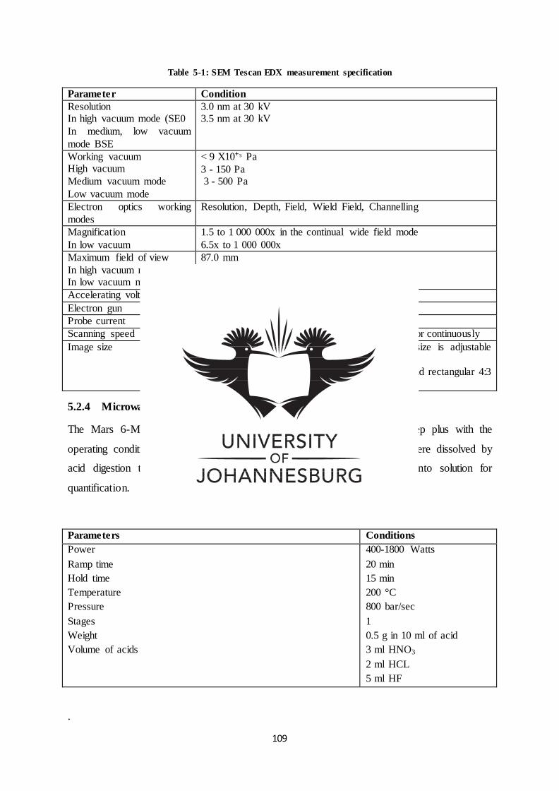
109
Table 5-1: SEM Tescan EDX measurement specification
Parameter Condition
Resolution In high vacuum mode (SE0
In medium, low vacuum
mode BSE
3.0 nm at 30 kV 3.5 nm at 30 kV
Working vacuum High vacuum
Medium vacuum mode
Low vacuum mode
< 9 X10⁺ᶟ Pa
3 - 150 Pa 3 - 500 Pa
Electron optics working
modes
Resolution, Depth, Field, Wield Field, Channelling
Magnification
In low vacuum
1.5 to 1 000 000x in the continual wide field mode
6.5x to 1 000 000x
Maximum field of view
In high vacuum mode (SE0 In low vacuum mode
87.0 mm
19.9 mm
Accelerating voltage 200 V to 30 kV
Electron gun Tungsten heated cathode
Probe current 1 Pa to 2 uA
Scanning speed From 20 ns to 10 ms per pixel adjustable in steps or continuously
Image size Up to 8.192 x 8 192 pixel in 16-bit quality, size is adjustable separately for live image (in 3 steps)
and for saved images (in 10 steps), for square and rectangular 4:3
or 2:1 image shapes
5.2.4 Microwave digestion of raw slag for ICP-OES analysis
The Mars 6-Microwave digestion system was employed using the EasyPrep plus with the
operating conditions as stated in Table 5.2. The slag samples (triplicates) were dissolved by
acid digestion thus increasing the sample solubility, releasing the metals into solution for
quantification.
Table 5-2: Microwave digestion specification
Parameters Conditions
Power
Ramp time
Hold time
Temperature
Pressure
Stages
Weight
Volume of acids
400-1800 Watts
20 min
15 min
200 °C
800 bar/sec
1
0.5 g in 10 ml of acid
3 ml HNO3
2 ml HCL
5 ml HF
.
110
5.2.5 Inductively coupled plasma optical emission spectroscopy (ICP-OES) analysis
The ICP-OES (Spectro Acros) spectrometer was used to characterize and quantify the
chemical composition of the raw slag material before leaching; and the default operating
conditions as stated in Table 5.3. A concentration of 500 ppb, 100 ppb and 100 ppm multi-
element standards were prepared and made up to 50 ml in a volumetric flask using 1 % ACE
HNO3. The 500 ppb multi-element standard was the used to prepare standards 0.5 ppm, 100
ppb; which were then used to prepare 0.1 ppm and 100 ppm; which was finally used to
prepare 1 ppm, 5 ppm, 10 ppm, 20 ppm and 40 ppm. The blank was prepared using the acids
employed for digesting.
Table 5-3: ICP-OES measurement specifications for slag analysis
Parameters Conditions
Generator parameters Plasma power
Pump speed
Coolant gas flow Auxiliary gas flow
Nebulizer gas flow
Global preflush parameters Fast Speed
Total
Normal speed
Rinse parameters
Rinse time Pump speed
Elements analysed
Analyte and wavelength
1400 Watts
30 Rpm
13 L/ min 2 L/min
0.95 L/min
63 Rpm
45 s
30 Rpm
10 s 30 s
Cu 224.700 Ni 227.021
Zn 202.613
Co 230.786 Fe 259.941
Al 176.641 Mg 285.213
Ca 315.887
Cr 205.618 Mn 257.611
Si 212.412
Ti 323.452
111
5.2.6 X-ray fluorescence (XRF) analysis
The chemical composition of the solid slag before and after bioleaching (residue) was
determined using X-ray fluorescence spectrometer (XRF) Rigaku ZXS Primus II with semi-
quantitative (SQX) analysis software by Scatter fundamental parameter (FP) method (Table
5.4).
Table 5-4: ZXS Primus II measurement specification
X-ray source X-ray tube End window type Rh Target 4kW or 3kW
High Voltage Generator High Frequency Inverter type Maximum Rating
4 kW, 60 kV-150 mA
Optics Maximum
sample Size
Ø51 mmx30 mm (H)
Primary Beam Filter 4 Filters (Al,Al-2,Cu,Zr)
Diaphragm 6 position Automatic Exchanger
(Ø 35, 30, 20, 10, 1, 0.5mm)
Crystal
Exchanger
10 position Automatic Exchanger
Counting Detector Heavy Elements: SC
Light Elements: F-PC
Components
analysed
Elements Na2O
MgO
Al2O3
Cr2O3
Fe2O3
Co2O3
SiO2
P2O5
K2O
TiO2
NiO
CuO
CaO
Zn
Mn
Pb
Rb
Cl
V
S
112
Sample preparation and analysis:
10g of the sample was pulverised to -38µm and mixed with a binder (30% Sasol wax)
The mixture was placed in an aluminium cup
Then placed on the pelletiser and pressed to a pressure of 20kpa
The pellet was placed in the oven for 1hr at 100°C to dry off prior to analysis
The calibration of the instrument was confirmed using certified machinery standards
The sample was then collected and loaded for analysis
5.2.7 X-ray powder diffraction (XRD) analysis
The mineralogical phase/s of the slag before and after bioleaching was determined using X-
ray Powder Diffraction spectrometer (XRD) RigakuUltimalV equipped with PDXL analysis
software. The specification used to run the experiment is displayed in Table 5.5.
Table 5-5: XRD RigakuUltimalV measurement specification
X-Ray 40 kV , 30 mA Scan speed /
Duration time
2.0000 deg./min.
Goniometer Step width 0.0200 deg.
Attachment - Scan axis 2theta/theta
Filter K-beta filter Scan range 3.0000 - 90.0000 deg.
CBO selection slit - Incident slit Open
Diffrected beam
mono.
Fixed Monochro.(U4) Length limiting slit -
Detector Scintillation counter Receiving slit #1 2/3deg.
Scan mode CONTINUOUS Receiving slit #2 Open
Sample preparation analysis:
The calibration of the instrument was confirmed using certified machinery standards
The sample was pulverised to -38µm
The sample was placed on the aluminium sample holder and flattened using a glass
slide
The sample was then mounted on the goniometer for analysis
The collected spectrum was analysed using the PDXL software loaded with ICDD
(International Centre for Diffraction Data) pdf card
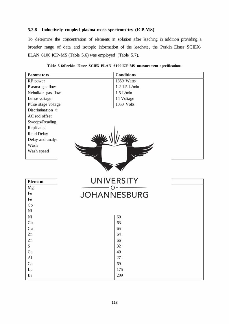
113
5.2.8 Inductively coupled plasma mass spectrometry (ICP-MS)
To determine the concentration of elements in solution after leaching in addition providing a
broader range of data and isotopic information of the leachate, the Perkin Elmer SCIEX-
ELAN 6100 ICP-MS (Table 5.6) was employed (Table 5.7).
Table 5-6:Perkin- Elmer SCIEX-ELAN 6100 ICP-MS measurement specifications
Parameters Conditions
RF power
Plasma gas flow
Nebulizer gas flow
Lense voltage
Pulse stage voltage
Discrimination threshold
AC rod offset
Sweeps/Reading
Replicates
Read Delay
Delay and analysis speed
Wash
Wash speed
1350 Watts
1.2-1.5 L/min
1.5 L/min
14 Voltage
1050 Volts
30m Volts
-12 Volts
20
3
15 sec
-24 (± rpm)
40 (sec)
-48 (± rpm)
Table 5-7: Elements of interest for ICP-MS analysis
Element Isotope mass
Mg
Fe
Fe
Co
Ni
Ni
Cu
Cu
Zn
Zn
S
Ca
Al
Ga
Lu
Bi
24
54
56
59
58
60
63
65
64
66
32
40
27
69
175
209
114
5.2.9 Standard solution preparation:
5.2.9.1 Internal standard
To minimize instrumental and signal intensity drifts internal standards were prepared. One
had to validate that elements used for the internal standard preparation were not naturally
occurring in the sample matrices. Subsequently, a concentration of 1000 ppm 69Ga, 175Lu and
209Bi elements was used to prepare 10 ppm internal standards in 1 % nitric acid. Considering
that internal standard elements should be as close as possible to the masses of the analytes of
interest, and that the ionization potential should be similar to that of the analytes of interest;
gallium (Ga69) corrections were used.
5.2.9.2 Calibration standards
Calibration standards were necessary for constructing a multipoint standard curve that would
cover the concentration scope of the analytes expected in the samples. Intermediate standards
were prepared by using 1000 ppm standard solution to prepare 1 ppm/1000 ppb intermediate
standards in 1 % nitric acid. Calibration standards (Table 5.8) were then conducted by
preparing dilutions of the intermediate standard with 1% nitric acid and 0.5 ml internal
standard to produce a final concentration of internal standards (0 ppb, 1 ppb, 5 ppb, 10 ppb,
20 ppb, and 40 ppb).
Table 5-8: Calibration standard preparation specification for ICP-MS
Standard St0 St1 St2 St3 St4 St5
Concentration 0 1 ppb 5 ppb 10 ppb 20 ppb 40 ppb
Intermediate Standard 0 0.05 ml 0.25 ml 0.5 ml 1 ml 2 ml
Internal Standard 0.5 ml 0.5 ml 0.5 ml 0.5 ml 0.5 ml 0.5 ml
HNO3 49.5 ml 49.45 ml 49.25 ml 49 ml 48.5 ml 47.5 ml
Total 50 ml 50 ml 50 ml 50 ml 50 ml 50 ml
5.2.10 Atomic absorption spectroscopy (AAS)
As an alternative to the ICP-MS method, due to polyatomic interferences of different isotopes
produced by oxygen, argon and calcium on the iron signal the Thermo Scientific ICE 3300
Series Atomic Absorption Spectrometer was employed to determine the concentration of the
iron elements in solution(leachate). Acetylene gas was used as a fuel while the oxygen
present in air was blown using a volumetric pump to produce the flame for atomisation. For
the analysis in order to accommodate the iron concentrations, standards were prepared from
1.25 ppm to 5 ppm for low concentrations and 62.5 ppm to 1000 ppm for high concentrated
115
solutions. The wavelength used for analysis was Fe 238.2047 while the calibration was the
normal linear least squares with the fitting set to R2 of 0.995.The standards used were of high
purity and supplied by associated chemical enterprises (ACE).
5.2.11 Bioleaching experiment of mineral sample (BCL slag)
A pre-growth approach was implemented, which entailed the cultivation of microbial cultures
in mineral salt media in the absence of choline based ionic liquids / glucose and slag material,
enabling the bacterial cultures to initiate and sustain optimum leaching conditions. A standard
method was employed where mineral salt media was prepared as per Table 5.9 with 5 g/l of
the respective ionic liquid and adjusted to pH 2 (0.1N HCL/0.1N NaOH) before sterilizing at
121 °C for 15 minutes. Employing 250 ml scotts bottles, a concentration of 9 g/l of sterilized
BCL slag was prepared in 150 ml of mineral salt media. The cooled media was then
inoculated with 10 % v/v of the corresponding pre-grown culture, with the exception of the
control which comprised of mineral salt media without bacteria. The flasks were incubated in
an orbital shaker at temperature of 37 °C and agitation speed of 120 rpm, maintaining a
homogenous mixture. Sampling was conducted daily using disposable sterilized pipettes.
Throughout the experiment pH and absorbance at 600 nm (Thermo Spectronic BioMate
3UV-Visable spectrophotometer) was measured. The pH range monitored was from pH 2 to
pH10. Upon the conclusion of the experiment the quantity of metal release/bioleached in the
culture medium was quantified by ICP-MS and AAS. The experiment was concluded on the
onset of the cell death phase. The solid residue before and after bioleaching was characterised
by SEM, XRF and XRD. The study was conducted in triplicates to obtain representative
figures of the samples.
Table 5-9: Formulation of mineral salt medium
Reagent Formula Quantity(g/l)
Dipotassium hydrogen phosphate
Potassium dihydrogen phosphate
Magnesium sulphate heptahydrate
Sodium chloride
Ferrous sulphate heptahydrate
Ammonium nitrate
Calcium chloride
Choline lactate
K2HPO4
KH2PO4
MgSO4· 7H2O
NaCl
FeSO4 · 7H2O
NH4NO3
CaCl2 · 2H2O
C5H14NO(+) + C3H5O3(-)
1.73
0.68
0.1
4
0.03
1
0.02
2
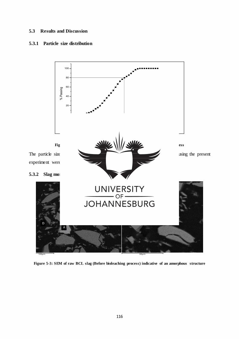
116
5.3 Results and Discussion
5.3.1 Particle size distribution
10 100 1000
0
20
40
60
80
100
% P
ass
ing
Size (Um)
Figure 5-2: Particle size distribution of BCL slag before bioleaching process
The particle size distribution curve revealed that 80 % passing particle size using the present
experiment were 100 μm.
5.3.2 Slag morphology
Figure 5-3: SEM of raw BCL slag (Before bioleaching process) indicative of an amorphous structure
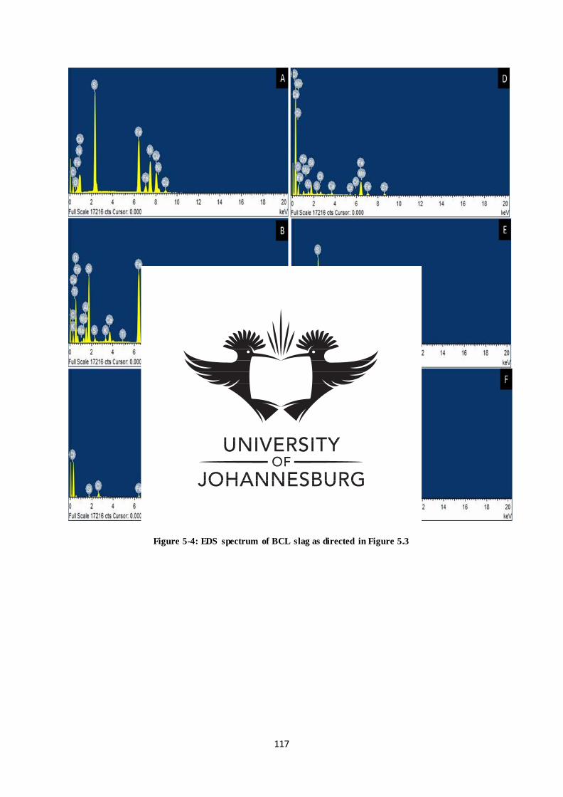
117
Figure 5-4: EDS spectrum of BCL slag as directed in Figure 5.3
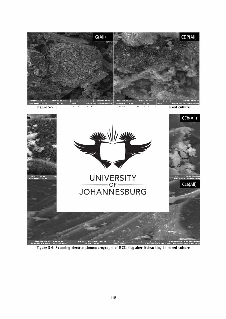
118
Figure 5-5: Scanning electron photomicrograph of BCL slag after bioleaching in mixed culture
Figure 5-6: Scanning electron photomicrograph of BCL slag after bioleaching in mixed culture
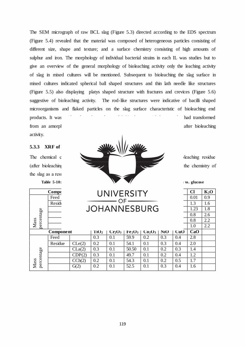
119
The SEM micrograph of raw BCL slag (Figure 5.3) directed according to the EDS spectrum
(Figure 5.4) revealed that the material was composed of heterogeneous particles consisting of
different size, shape and texture; and a surface chemistry consisting of high amounts of
sulphur and iron. The morphology of individual bacterial strains in each IL was studies but to
give an overview of the general morphology of bioleaching activity only the leaching activity
of slag in mixed cultures will be mentioned. Subsequent to bioleaching the slag surface in
mixed cultures indicated spherical ball shaped structures and thin lath needle like structures
(Figure 5.5) also displaying platys shaped structure with fractures and crevices (Figure 5.6)
suggestive of bioleaching activity. The rod-like structures were indicative of bacilli shaped
microorganisms and flaked particles on the slag surface characteristic of bioleaching end
products. It was evident from the surfaces of BCL slag material that samples had transformed
from an amorphous structure prior to bioleaching to a crystalline structure after bioleaching
activity.
5.3.3 XRF of BCL slag before and after leaching
The chemical composition of raw BCL slag (before bioleaching) and bioleaching residue
(after bioleaching) was studied (Tables 5.10 -5.16) to determine changes in the chemistry of
the slag as a result of bioleaching processes.
Table 5-10: XRF of slag before and after leaching with Bacillus aryabhattai in ILs vs. glucose
Component Na2O MgO Al2O3 SiO2 P2O5 S Cl K2O
Mass
perc
en
tag
e
Feed 0.8 1.9 6.2 24.0 0.04 2.0 0.01 0.9
Residue CLe(2) 3.1 0.8 5.0 14.3 15.1 1.5 1.3 1.6
CLa(2) 1.0 0.4 6.8 13.8 20.0 2.1 1.23 1.8
CDP(2) 1.0 0.3 5.3 8.7 27.7 1.5 0.8 2.6
CCh(2) 1.3 0.7 5.2 11.9 19.4 1.4 0.8 2.2
G(2) 1.3 0.4 5.8 11.3 21.3 1.5 1.0 2.2
Component TiO2 Cr2O3 Fe2O3 Co2O3 NiO CuO CaO
Mass
perc
en
tag
e
Feed 0.3 0.1 59.9 0.2 0.3 0.4 2.8
Residue CLe(2) 0.2 0.1 54.1 0.1 0.3 0.4 2.0
CLa(2) 0.3 0.1 50.50 0.1 0.2 0.3 1.4
CDP(2) 0.3 0.1 49.7 0.1 0.2 0.4 1.2
CCh(2) 0.2 0.1 54.3 0.1 0.2 0.5 1.7
G(2) 0.2 0.1 52.5 0.1 0.3 0.4 1.6
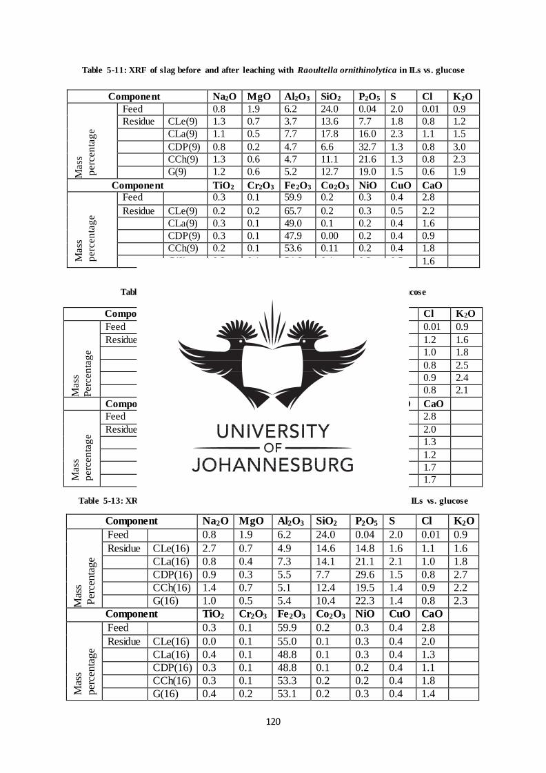
120
Table 5-11: XRF of slag before and after leaching with Raoultella ornithinolytica in ILs vs. glucose
Table 5-12: XRF of BCL slag after leaching with Bacillus sp. in ILs vs. glucose
Component Na2O MgO Al2O3 SiO2 P2O5 S Cl K2O
Mass
Perc
en
tag
e
Feed 0.8 1.9 6.2 24.0 0.04 2.0 0.01 0.9
Residue CLe(12) 2.9 0.8 5.0 14.3 15.2 1.5 1.2 1.6
CLa(12) 0.9 0.4 7.2 13.3 21.2 2.4 1.0 1.8
CDP(12) 1.0 0.3 5.5 9.0 28.0 1.6 0.8 2.5
CCh(12) 1.2 0.6 4.9 11.5 21.0 1.2 0.9 2.4
G(12) 1.2 0.6 5.6 12.9 19.8 1.5 0.8 2.1
Component TiO2 Cr2O3 Fe2O3 Co2O3 NiO CuO CaO
Mass
perc
en
tag
e
Feed 0.3 0.1 59.9 0.2 0.3 0.4 2.8
Residue CLe(12) 0.2 0.1 55.0 0.1 0.2 0.4 2.0
CLa(12) 0.2 0.1 49.1 0.1 0.2 0.4 1.3
CDP(12) 0.2 0.1 49.0 0.1 0.2 0.4 1.2
CCh(12) 0.2 0.1 53.6 0.1 0.2 0.4 1.7
G(12) 0.3 0.1 52.7 0.1 0.2 0.4 1.7
Table 5-13: XRF of BCL slag before and after leaching with Bacillus thuringiensis in ILs vs. glucose
Component Na2O MgO Al2O3 SiO2 P2O5 S Cl K2O
Mas
s
Per
centa
ge
Feed 0.8 1.9 6.2 24.0 0.04 2.0 0.01 0.9
Residue CLe(16) 2.7 0.7 4.9 14.6 14.8 1.6 1.1 1.6
CLa(16) 0.8 0.4 7.3 14.1 21.1 2.1 1.0 1.8
CDP(16) 0.9 0.3 5.5 7.7 29.6 1.5 0.8 2.7
CCh(16) 1.4 0.7 5.1 12.4 19.5 1.4 0.9 2.2
G(16) 1.0 0.5 5.4 10.4 22.3 1.4 0.8 2.3
Component TiO2 Cr2O3 Fe2O3 Co2O3 NiO CuO CaO
Mas
s
per
centa
ge
Feed 0.3 0.1 59.9 0.2 0.3 0.4 2.8
Residue CLe(16) 0.0 0.1 55.0 0.1 0.3 0.4 2.0
CLa(16) 0.4 0.1 48.8 0.1 0.3 0.4 1.3
CDP(16) 0.3 0.1 48.8 0.1 0.2 0.4 1.1
CCh(16) 0.3 0.1 53.3 0.2 0.2 0.4 1.8
G(16) 0.4 0.2 53.1 0.2 0.3 0.4 1.4
Component Na2O MgO Al2O3 SiO2 P2O5 S Cl K2O
Mass
perc
en
tag
e
Feed 0.8 1.9 6.2 24.0 0.04 2.0 0.01 0.9
Residue CLe(9) 1.3 0.7 3.7 13.6 7.7 1.8 0.8 1.2
CLa(9) 1.1 0.5 7.7 17.8 16.0 2.3 1.1 1.5
CDP(9) 0.8 0.2 4.7 6.6 32.7 1.3 0.8 3.0
CCh(9) 1.3 0.6 4.7 11.1 21.6 1.3 0.8 2.3
G(9) 1.2 0.6 5.2 12.7 19.0 1.5 0.6 1.9
Component TiO2 Cr2O3 Fe2O3 Co2O3 NiO CuO CaO
Mass
perc
en
tag
e
Feed 0.3 0.1 59.9 0.2 0.3 0.4 2.8
Residue CLe(9) 0.2 0.2 65.7 0.2 0.3 0.5 2.2
CLa(9) 0.3 0.1 49.0 0.1 0.2 0.4 1.6
CDP(9) 0.3 0.1 47.9 0.00 0.2 0.4 0.9
CCh(9) 0.2 0.1 53.6 0.11 0.2 0.4 1.8
G(9) 0.2 0.1 54.6 0.1 0.3 0.5 1.6
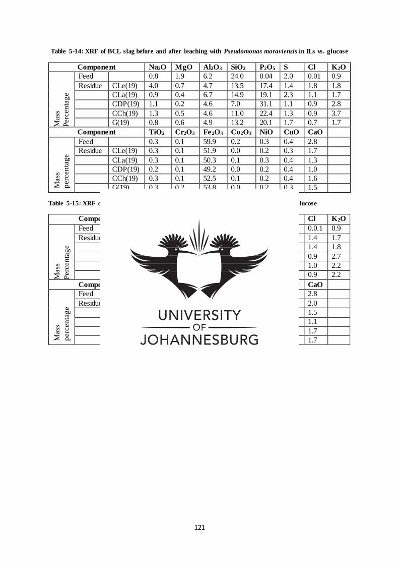
121
Table 5-14: XRF of BCL slag before and after leaching with Pseudomonas moraviensis in ILs vs. glucose
Component Na2O MgO Al2O3 SiO2 P2O5 S Cl K2O
Mass
Perc
en
tag
e
Feed 0.8 1.9 6.2 24.0 0.04 2.0 0.01 0.9
Residue CLe(19) 4.0 0.7 4.7 13.5 17.4 1.4 1.8 1.8
CLa(19) 0.9 0.4 6.7 14.9 19.1 2.3 1.1 1.7
CDP(19) 1.1 0.2 4.6 7.0 31.1 1.1 0.9 2.8
CCh(19) 1.3 0.5 4.6 11.0 22.4 1.3 0.9 3.7
G(19) 0.8 0.6 4.9 13.2 20.1 1.7 0.7 1.7
Component TiO2 Cr2O3 Fe2O3 Co2O3 NiO CuO CaO
Mass
perc
en
tag
e
Feed 0.3 0.1 59.9 0.2 0.3 0.4 2.8
Residue CLe(19) 0.3 0.1 51.9 0.0 0.2 0.3 1.7
CLa(19) 0.3 0.1 50.3 0.1 0.3 0.4 1.3
CDP(19) 0.2 0.1 49.2 0.0 0.2 0.4 1.0
CCh(19) 0.3 0.1 52.5 0.1 0.2 0.4 1.6
G(19) 0.3 0.2 53.8 0.0 0.2 0.3 1.5
Table 5-15: XRF of BCL slag before and after leaching with a mixed culture in ILs vs. glucose
Component Na2O MgO Al2O3 SiO2 P2O5 S Cl K2O
Mass
Perc
en
tag
e
Feed 0.8 1.9 6.2 24.0 0.04 2.0 0.0.1 0.9
Residue CLe(All) 3.2 0.7 4.6 12.9 16.7 1.4 1.4 1.7
CLa(All) 1.1 0.5 7.7 14.7 19.9 2.1 1.4 1.8
CDP(All) 1.1 0.2 5.1 7.7 29.8 1.3 0.9 2.7
CCh(All) 1.2 0.7 5.0 11.9 19.9 1.5 1.0 2.2
G(All) 1.3 0.5 5.2 10.7 21.0 1.4 0.9 2.2
Component TiO2 Cr2O3 Fe2O3 Co2O3 NiO CuO CaO
Mass
perc
en
tag
e
Feed 0.3 0.1 59.9 0.2 0.3 0.4 2.8
Residue CLe(All) 0.3 0.1 54.3 0.1 0.2 0.4 2.0
CLa(All) 0.3 0.1 48.1 0.1 0.3 0.4 1.5
CDP(All) 0.0 0.1 49.1 0.0 0.2 0.4 1.1
CCh(All) 0.0 0.1 53.9 0.1 0.3 0.4 1.7
G(All) 0.2 0.1 54.1 0.1 0.3 0.4 1.7
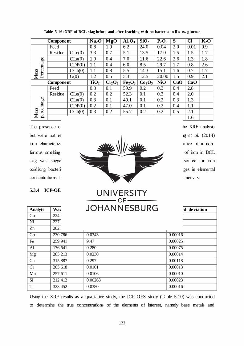
122
Table 5-16: XRF of BCL slag before and after leaching with no bacteria in ILs vs. glucose
Component Na2O MgO Al2O3 SiO2 P2O5 S Cl K2O M
ass
Per
centa
ge
Feed 0.8 1.9 6.2 24.0 0.04 2.0 0.01 0.9
Residue CLe(0) 3.3 0.7 5.1 13.5 17.0 1.5 1.5 1.7
CLa(0) 1.0 0.4 7.0 11.6 22.6 2.6 1.3 1.8
CDP(0) 1.1 0.4 6.0 8.5 29.7 1.7 0.8 2.6
CCh(0) 1.1 0.8 5.5 14.3 15.1 1.6 0.7 1.7
G(0) 1.2 0.5 5.3 12.5 20.00 1.5 0.9 2.1
Component TiO2 Cr2O3 Fe2O3 Co2O3 NiO CuO CaO
Mas
s
per
centa
ge
Feed 0.3 0.1 59.9 0.2 0.3 0.4 2.8
Residue CLe(0) 0.2 0.2 52.3 0.1 0.3 0.4 2.0
CLa(0) 0.3 0.1 49.1 0.1 0.2 0.3 1.3
CDP(0) 0.2 0.1 47.0 0.1 0.2 0.4 1.1
CCh(0) 0.3 0.2 55.7 0.2 0.2 0.5 2.1
G(0) 0.2 0.1 53.0 0.2 0.3 0.4 1.6
The presence of metals Zn, Mn, V, Pb, Rb elements were also detected in the XRF analysis
but were not reported due to low concentrations. In agreement with Hocheng et al. (2014)
iron characterized as Fe2SiO3 was a major component of BCL slag, indicative of a non-
ferrous smelting process producing an iron-silicate based slag. The profusion of iron in BCL
slag was suggestive of the potential of BCL slag to function as energy source for iron
oxidizing bacteria in the bioleaching process (Isildar et al., 2016). The changes in elemental
concentrations before and after bioleaching were also indicative of bioleaching activity.
5.3.4 ICP-OES raw slag elemental analysis
Table 5-17: ICP-OES Characterization of raw BCL slag
Analyte Wavelength Average concentration (mass %) Standard deviation
Cu 224.700 0.0985 0.00041
Ni 227.021 0.0598 0.00019
Zn 202.613 0.00601 0.00001
Co 230.786 0.0343 0.00016
Fe 259.941 9.47 0.00025
Al 176.641 0.280 0.00075
Mg 285.213 0.0230 0.00014
Ca 315.887 0.297 0.00118
Cr 205.618 0.0101 0.00013
Mn 257.611 0.0106 0.00010
Si 212.412 0.00263 0.00023
Ti 323.452 0.0380 0.00016
Using the XRF results as a qualitative study, the ICP-OES study (Table 5.10) was conducted
to determine the true concentrations of the elements of interest, namely base metals and
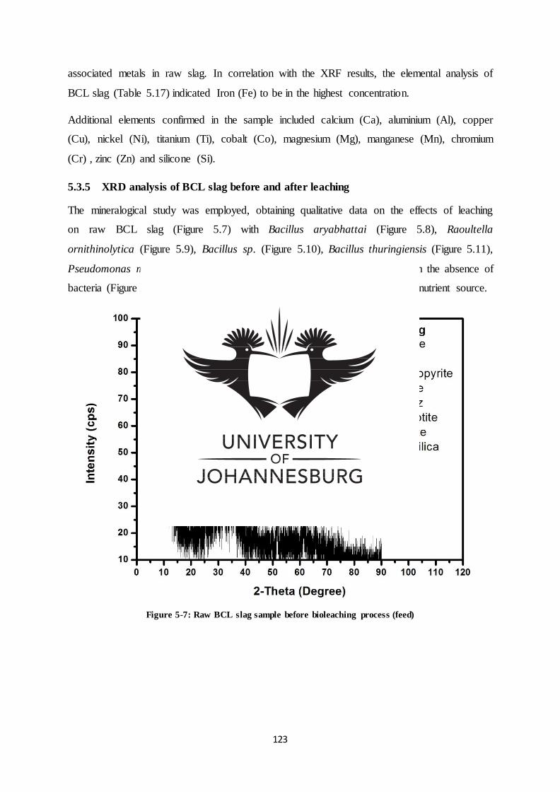
123
associated metals in raw slag. In correlation with the XRF results, the elemental analysis of
BCL slag (Table 5.17) indicated Iron (Fe) to be in the highest concentration.
Additional elements confirmed in the sample included calcium (Ca), aluminium (Al), copper
(Cu), nickel (Ni), titanium (Ti), cobalt (Co), magnesium (Mg), manganese (Mn), chromium
(Cr) , zinc (Zn) and silicone (Si).
5.3.5 XRD analysis of BCL slag before and after leaching
The mineralogical study was employed, obtaining qualitative data on the effects of leaching
on raw BCL slag (Figure 5.7) with Bacillus aryabhattai (Figure 5.8), Raoultella
ornithinolytica (Figure 5.9), Bacillus sp. (Figure 5.10), Bacillus thuringiensis (Figure 5.11),
Pseudomonas moraviensis (figure 5.12), mixed cultures (Figure 5.13) and in the absence of
bacteria (Figure 5.14) in choline based ionic liquids vs. conventional glucose nutrient source.
Figure 5-7: Raw BCL slag sample before bioleaching process (feed)
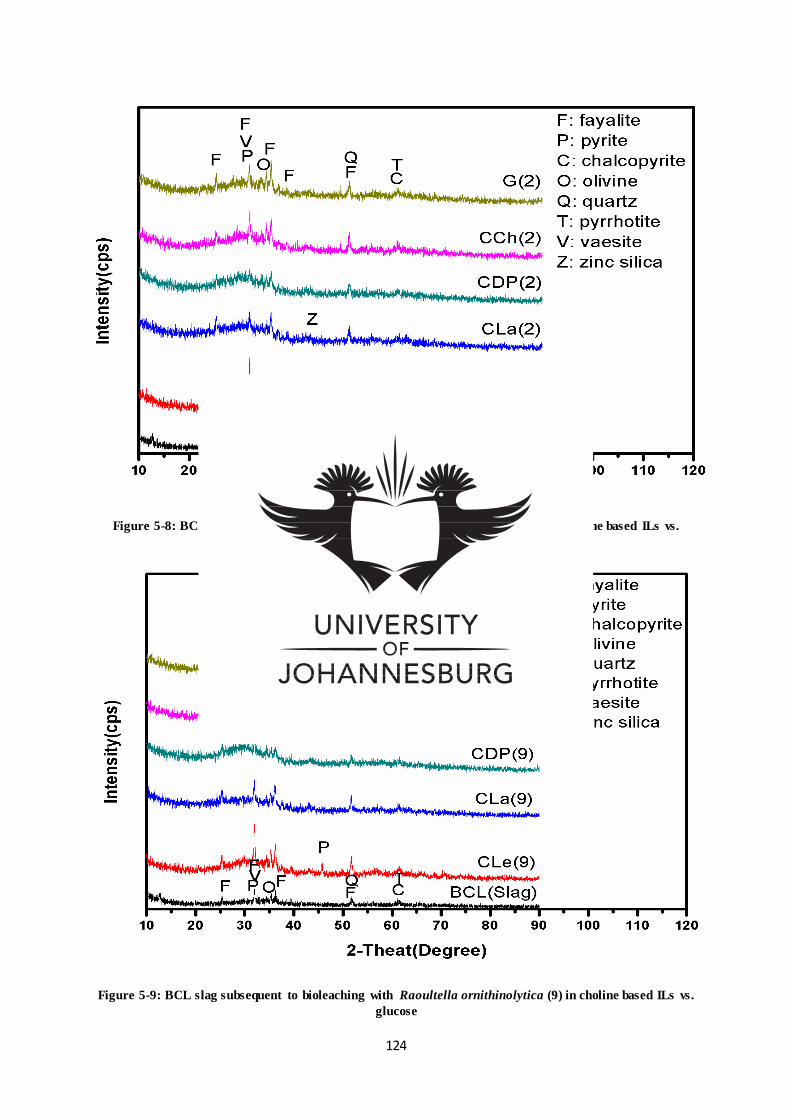
124
Figure 5-8: BCL slag subsequent to bioleaching with Bacillus aryabhattai (2) in choline based ILs vs.
glucose
Figure 5-9: BCL slag subsequent to bioleaching with Raoultella ornithinolytica (9) in choline based ILs vs.
glucose
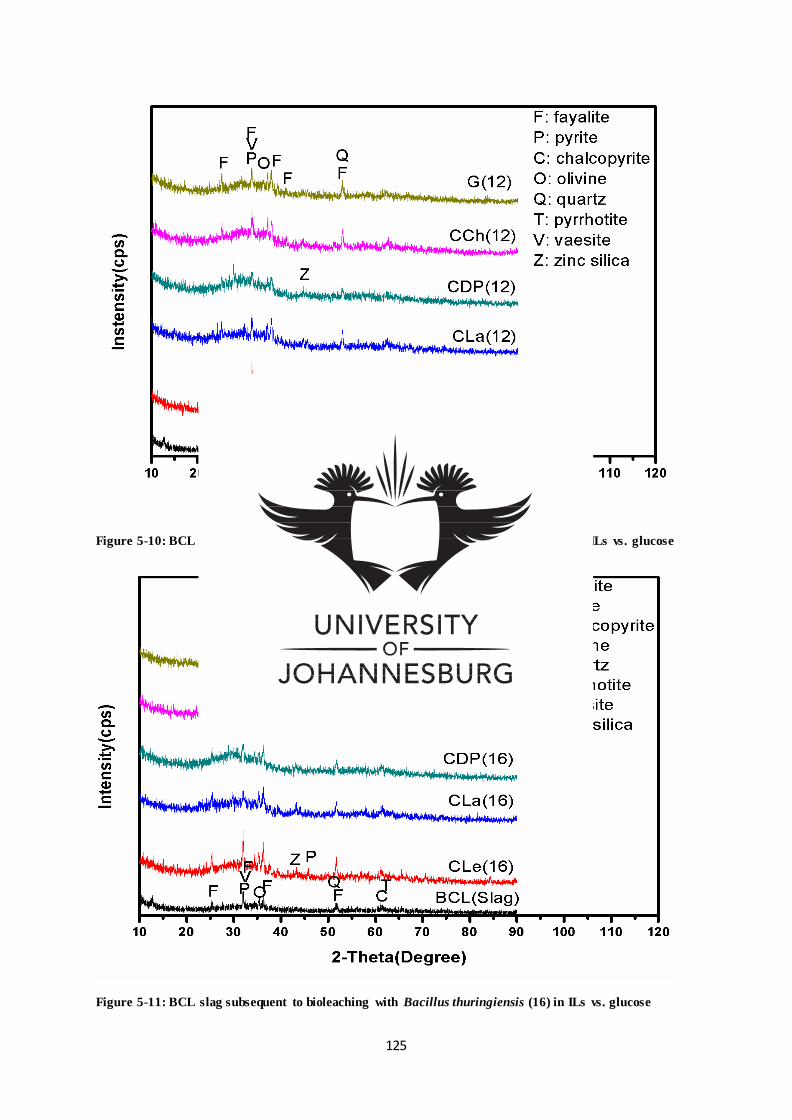
125
Figure 5-10: BCL slag subsequent to bioleaching with Bacillus sp. (12) in choline based ILs vs. glucose
Figure 5-11: BCL slag subsequent to bioleaching with Bacillus thuringiensis (16) in ILs vs. glucose
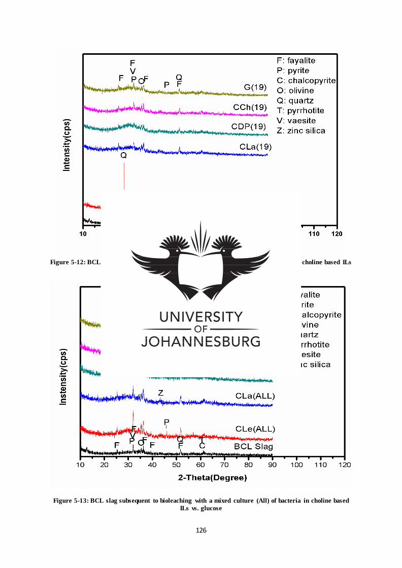
126
Figure 5-12: BCL slag subsequent to bioleaching with Pseudomonas moraviensis (19) in choline based ILs
vs. glucose
Figure 5-13: BCL slag subsequent to bioleaching with a mixed culture (All) of bacteria in choline based
ILs vs. glucose
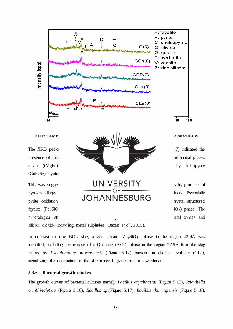
127
Figure 5-14: BCL slag subsequent to leaching in the absence (0) of bacteria in choline based ILs vs.
glucose
The XRD peaks of raw BCL slag before the bioleaching process (Figure 5.7) indicated the
presence of mineralogical phase fayalite (Fe2SiO4) a major iron carrier and additional phases
olivine ((MgFe)2SiO4) and quartz (SiO2) with sulphide minerals specified by chalcopyrite
(CuFeS2), pyrite(FeS2), pyrrhotite (FeS) and vaesite (NiS2).
This was suggestive of a nonferrous slag, described by Yang et al. (2010) as by-products of
pyro-metallurgy copper processing for concentrated copper sulphide products. Essentially
pyrite oxidation gives rise to iron-oxide occurring in silica, producing a crystal structured
fayalite (Fe2SiO4) slag occasionally containing a small amorphous silica (SiO2) phase. The
mineralogical studies were conclusive of slag material, characteristic of metal oxides and
silicon dioxide including metal sulphides (Bazan et al., 2015).
In contrast to raw BCL slag, a zinc silicate (Zn2SiO4) phase in the region 42.9Å was
identified, including the release of a Q-quartz (SiO2) phase in the region 27.9Å from the slag
matrix by Pseudomonas moraviensis (Figure 5.12) bacteria in choline levulinate (CLe),
signalizing the destruction of the slag mineral giving rise to new phases.
5.3.6 Bacterial growth studies
The growth curves of bacterial cultures namely Bacillus aryabhattai (Figure 5.15), Raoultella
ornithinolytica (Figure 5.16), Bacillus sp.(Figure 5.17), Bacillus thuringiensis (Figure 5.18),
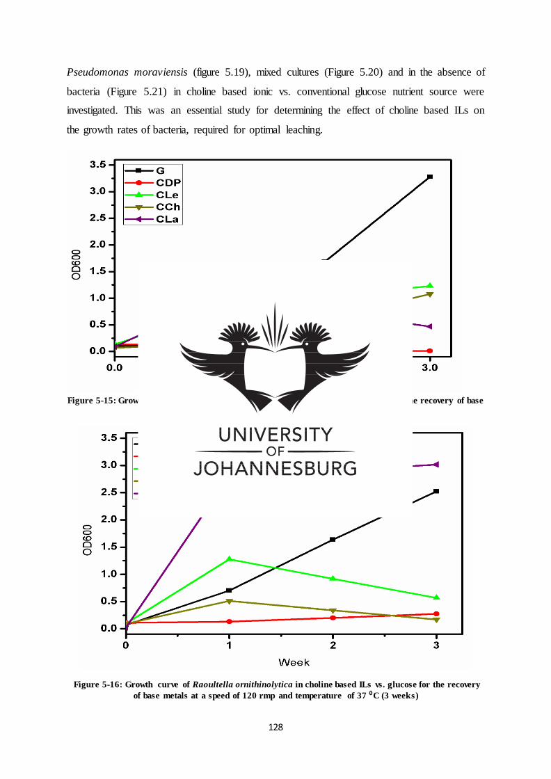
128
Pseudomonas moraviensis (figure 5.19), mixed cultures (Figure 5.20) and in the absence of
bacteria (Figure 5.21) in choline based ionic vs. conventional glucose nutrient source were
investigated. This was an essential study for determining the effect of choline based ILs on
the growth rates of bacteria, required for optimal leaching.
Figure 5-15: Growth curve of Bacillus aryabhattai in choline based ILs vs. glucose for the recovery of base
metals at a speed of 120 rmp and temperature of 37 °C (3 weeks)
Figure 5-16: Growth curve of Raoultella ornithinolytica in choline based ILs vs. glucose for the recovery
of base metals at a speed of 120 rmp and temperature of 37 ⁰C (3 weeks)
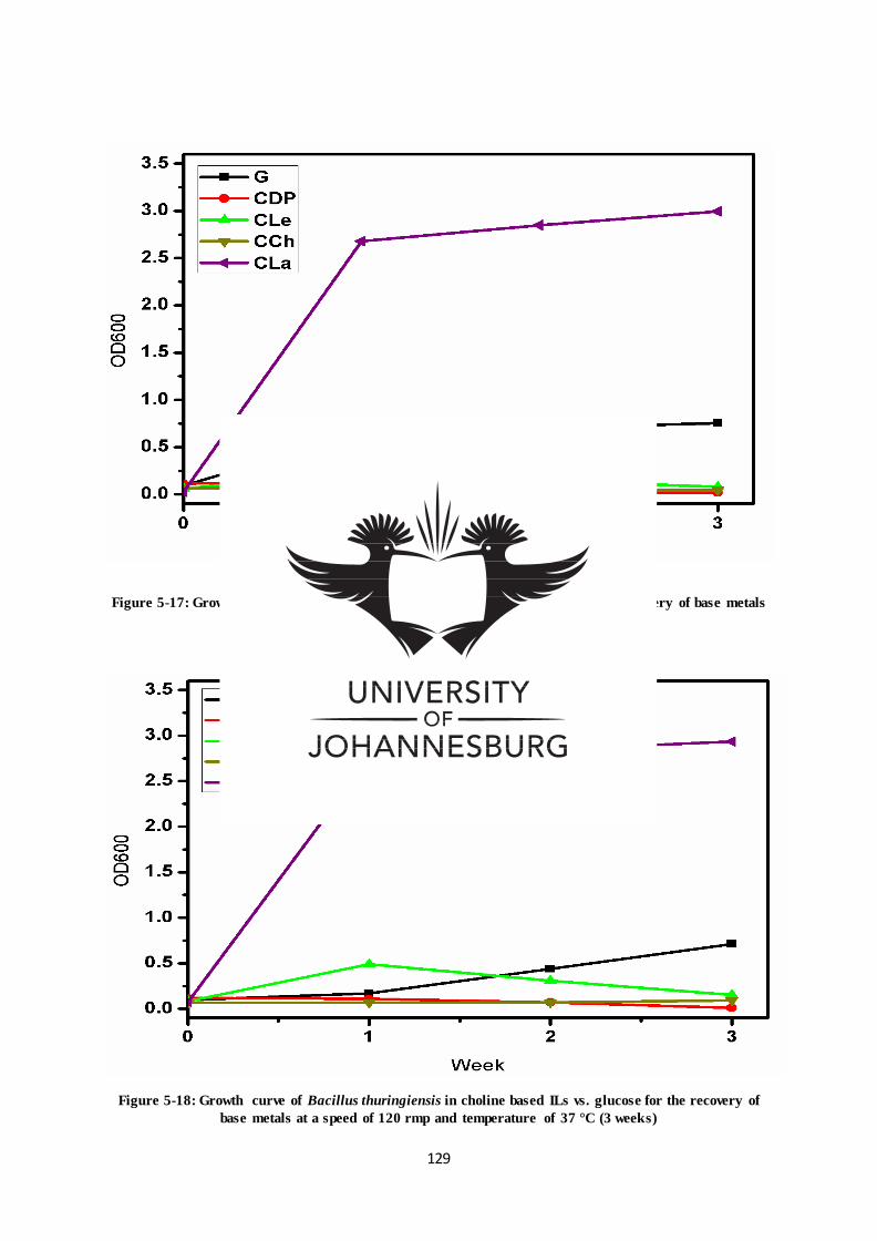
129
Figure 5-17: Growth curve of Bacillus sp. in choline based ILs vs. glucose for the recovery of base metals
at a speed of 120 rmp and temperature of 37 °C (3 weeks)
Figure 5-18: Growth curve of Bacillus thuringiensis in choline based ILs vs. glucose for the recovery of
base metals at a speed of 120 rmp and temperature of 37 °C (3 weeks)
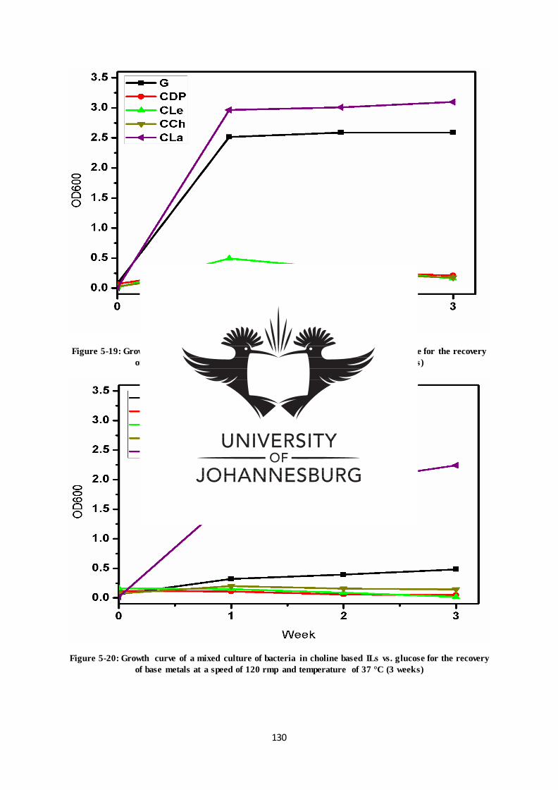
130
Figure 5-19: Growth curve of Pseudomonas moraviensis in choline based ILs vs. glucose for the recovery
of base metals at a speed of 120 rmp and temperature of 37 °C (3 weeks)
Figure 5-20: Growth curve of a mixed culture of bacteria in choline based ILs vs. glucose for the recovery
of base metals at a speed of 120 rmp and temperature of 37 °C (3 weeks)
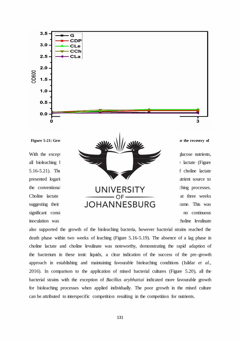
131
Figure 5-21: Growth curve in the absence of bacteria in choline based ILs vs. glucose for the recovery of
base metals at a speed of 120 rmp and temperature of 37 °C (3 weeks)
With the exception of Bacillus aryabhattai (Table 5.15) which thrived in glucose nutrients,
all bioleaching bacteria indicated optimal growth in the presence of choline lactate (Figure
5.16-5.21). The growth pattern of bioleaching bacteria in the presence of choline lactate
presented logarithmic growth, thus indicating choline lactate as a preferred nutrient source to
the conventional glucose source for optimal bacterial growth during bioleaching processes.
Choline lactate and glucose based bacteria were still growing optimally at three weeks
suggesting their potential for further growth past the three week time frame. This was
significant considering the study was based on a closed system where no continuous
inoculation was performed. Trailing behind choline lactate, ionic liquid choline levulinate
also supported the growth of the bioleaching bacteria, however bacterial strains reached the
death phase within two weeks of leaching (Figure 5.16-5.19). The absence of a lag phase in
choline lactate and choline levulinate was noteworthy, demonstrating the rapid adaption of
the bacterium in these ionic liquids, a clear indication of the success of the pre-growth
approach in establishing and maintaining favourable bioleaching conditions (Isildar et al.,
2016). In comparison to the application of mixed bacterial cultures (Figure 5.20), all the
bacterial strains with the exception of Bacillus arybhattai indicated more favourable growth
for bioleaching processes when applied individually. The poor growth in the mixed culture
can be attributed to interspecific competition resulting in the competition for nutrients.
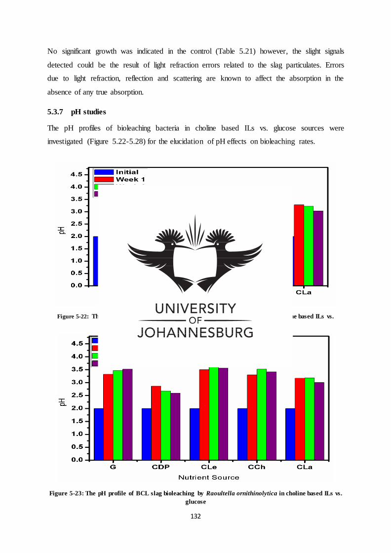
132
No significant growth was indicated in the control (Table 5.21) however, the slight signals
detected could be the result of light refraction errors related to the slag particulates. Errors
due to light refraction, reflection and scattering are known to affect the absorption in the
absence of any true absorption.
5.3.7 pH studies
The pH profiles of bioleaching bacteria in choline based ILs vs. glucose sources were
investigated (Figure 5.22-5.28) for the elucidation of pH effects on bioleaching rates.
Figure 5-22: The pH profile of BCL slag bioleaching by Bacillus aryabhattai in choline based ILs vs.
glucose
Figure 5-23: The pH profile of BCL slag bioleaching by Raoultella ornithinolytica in choline based ILs vs.
glucose
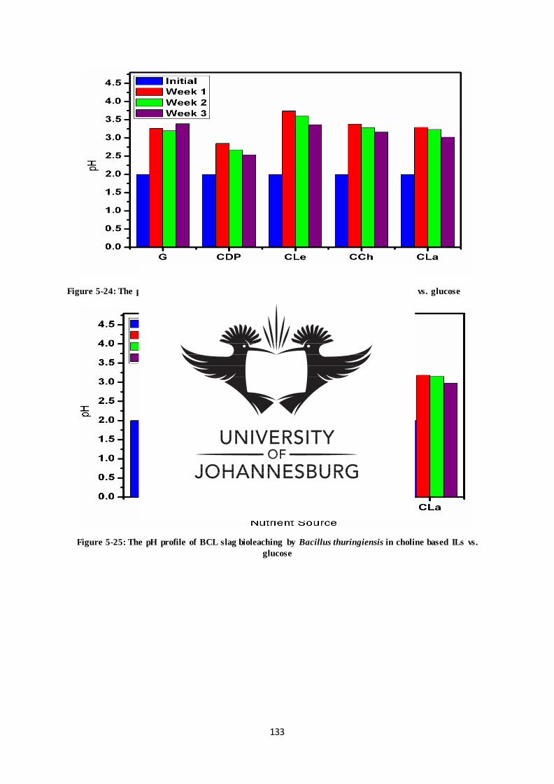
133
Figure 5-24: The pH profile of BCL slag bioleaching by Bacillus sp. in choline based ILs vs. glucose
Figure 5-25: The pH profile of BCL slag bioleaching by Bacillus thuringiensis in choline based ILs vs.
glucose
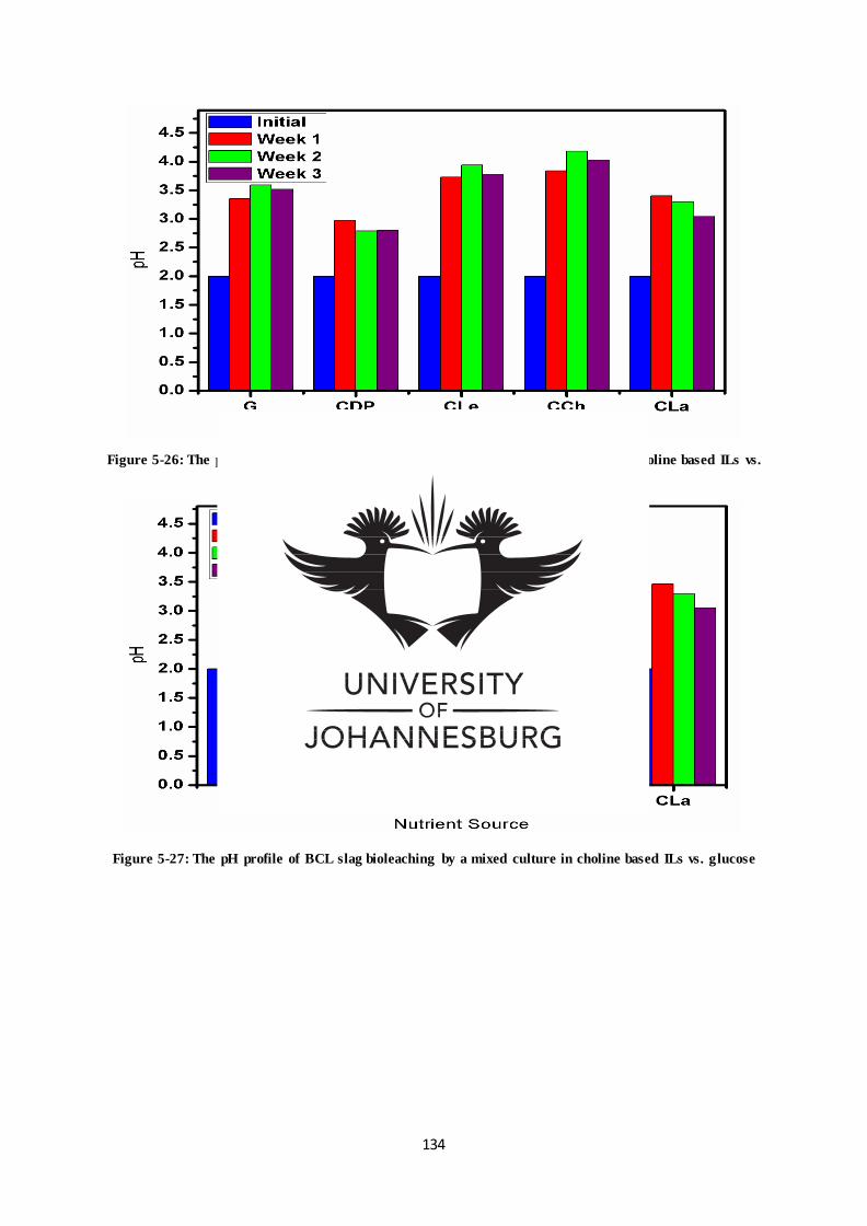
134
Figure 5-26: The pH profile of BCL slag bioleaching by Pseudomonas moraviensis in choline based ILs vs.
glucose
Figure 5-27: The pH profile of BCL slag bioleaching by a mixed culture in choline based ILs vs. glucose
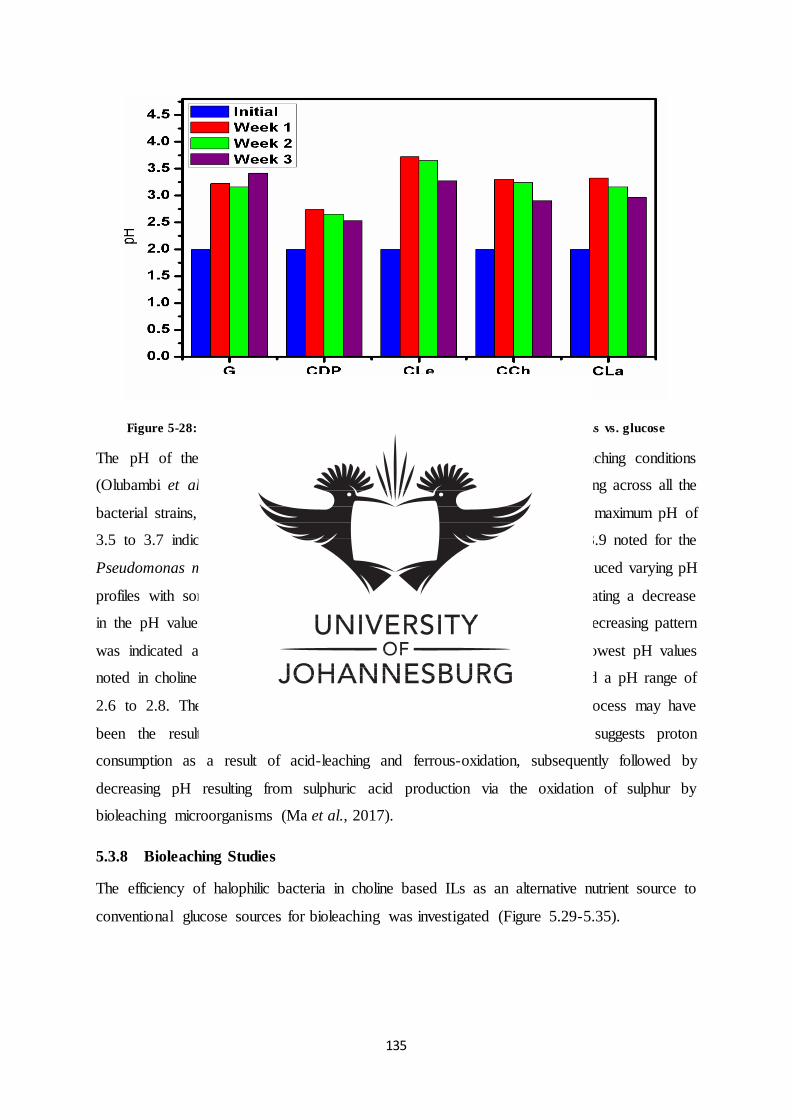
135
Figure 5-28: The pH profile of BCL slag bioleaching without bacteria in choline ILs vs. glucose
The pH of the solution was initially adjusted to pH 2 for optimal bioleaching conditions
(Olubambi et al., 2008; Shaikh et al., 2010). Within the first week of leaching across all the
bacterial strains, the samples indicated an increase in their pH values, with a maximum pH of
3.5 to 3.7 indicated in all choline levulinate (CLe) nutrient sources and pH 3.9 noted for the
Pseudomonas moraviensis strain in choline chloride (CCh). Week two introduced varying pH
profiles with some samples indicating further increase in pH and others initiating a decrease
in the pH value. With the exception of glucose (G), towards week three a decreasing pattern
was indicated across the pH profiles of all the bacterial strains, with the lowest pH values
noted in choline dihydrogen phosphate (CDP) nutrient sources, which indicated a pH range of
2.6 to 2.8. The increasing pH at the commencement of the bioleaching process may have
been the result of the alkalinity of the added slag material but also suggests proton
consumption as a result of acid-leaching and ferrous-oxidation, subsequently followed by
decreasing pH resulting from sulphuric acid production via the oxidation of sulphur by
bioleaching microorganisms (Ma et al., 2017).
5.3.8 Bioleaching Studies
The efficiency of halophilic bacteria in choline based ILs as an alternative nutrient source to
conventional glucose sources for bioleaching was investigated (Figure 5.29-5.35).
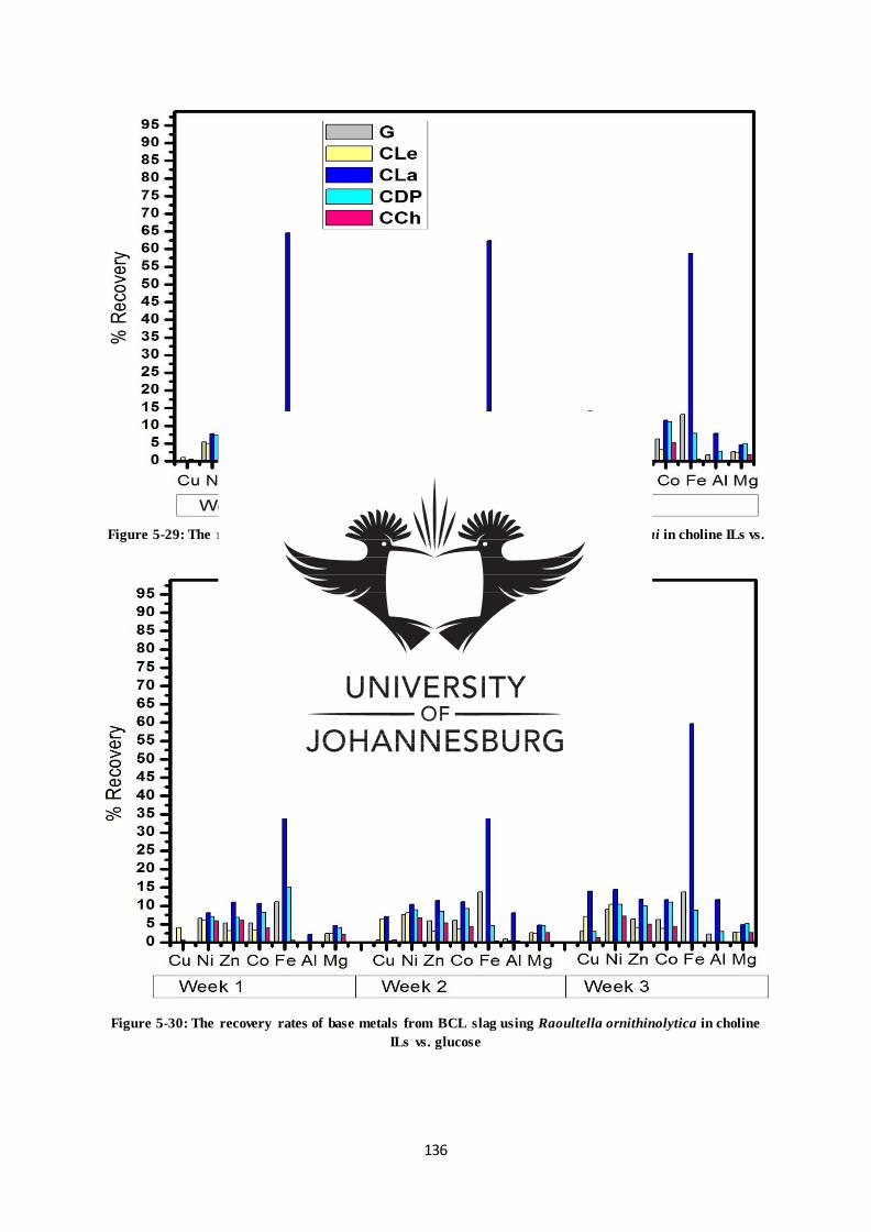
136
Figure 5-29: The recovery rates of base metals from BCL slag using Bacillus aryabhattai in choline ILs vs.
glucose
Figure 5-30: The recovery rates of base metals from BCL slag using Raoultella ornithinolytica in choline
ILs vs. glucose
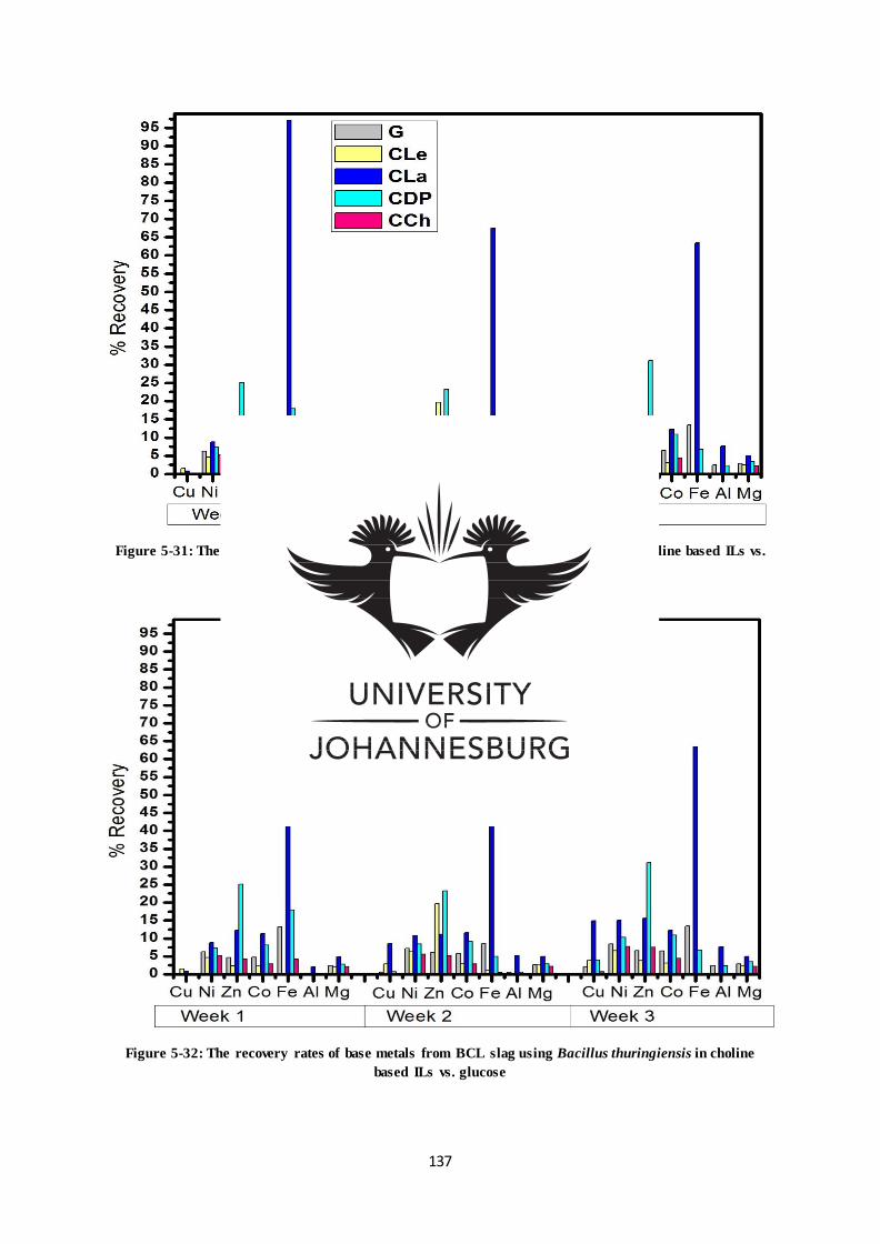
137
Figure 5-31: The recovery rates of base metals from BCL slag using Bacillus sp. in choline based ILs vs.
glucose
Figure 5-32: The recovery rates of base metals from BCL slag using Bacillus thuringiensis in choline
based ILs vs. glucose
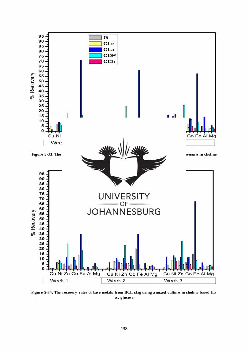
138
Figure 5-33: The recovery rates of base metals from BCL slag using Pseudomonas moraviensis in choline
based ILs vs. glucose
Figure 5-34: The recovery rates of base metals from BCL slag using a mixed culture in choline based ILs
vs. glucose
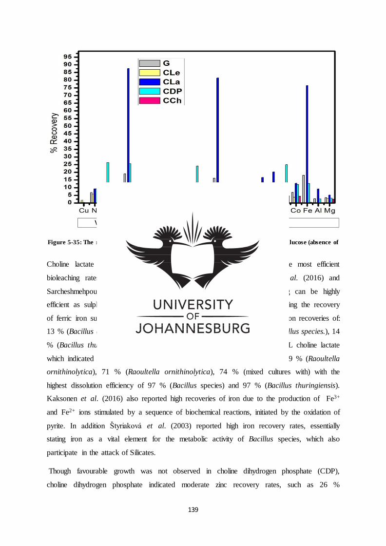
139
Figure 5-35: The recovery rates of base metals from BCL slag in choline based ILs vs. glucose (absence of
bacteria)
Choline lactate produced the most optimal base metal recovery, with the most efficient
bioleaching rates occurring at initial pH 2. According to Kaksonen et al. (2016) and
Sarcheshmehpour et al. (2009), the biochemical activity of acid on slag can be highly
efficient as sulphides can occur at insignificant levels in slag minerals, initiating the recovery
of ferric iron sulphates in solution. Conventional glucose sources produced iron recoveries of:
13 % (Bacillus aryabhattai), 13 % ( Raoultella ornithinolytica), 13 % (Bacillus species.), 14
% (Bacillus thuringiensis) and 20 % (mixed cultures) in comparison with IL choline lactate
which indicated significant iron recoveries of: 65 % (Bacillus aryabhattai), 69 % (Raoultella
ornithinolytica), 71 % (Raoultella ornithinolytica), 74 % (mixed cultures with) with the
highest dissolution efficiency of 97 % (Bacillus species) and 97 % (Bacillus thuringiensis).
Kaksonen et al. (2016) also reported high recoveries of iron due to the production of Fe3+
and Fe2+ ions stimulated by a sequence of biochemical reactions, initiated by the oxidation of
pyrite. In addition Štyriaková et al. (2003) reported high iron recovery rates, essentially
stating iron as a vital element for the metabolic activity of Bacillus species, which also
participate in the attack of Silicates.
Though favourable growth was not observed in choline dihydrogen phosphate (CDP),
choline dihydrogen phosphate indicated moderate zinc recovery rates, such as 26 %
140
(Pseudomonas moraviensis), 28 % (mixed cultures), 31 % (Bacillus species) and 31 %
(Bacillus thuringiensis). Ionic liquid choline levulinate also produced moderate zinc (Zn)
recoveries in Bacillus sp. and Bacillus thuringiensis. The zinc recoveries were validated by
the appearance of the zinc silica phase after bioleaching processes (Table 5.8-5.14).
In accord with Kaksonen et al. (2016) the initial dissolving of acid in solution at room
temperature increased the solubility of fayalite (Fe2SiO4) additional silica and metal oxide
phases. This is was due to the yield of ferrous (Fe2+) by fayalite which oxidised the oxygen in
the acid solution and thereafter was accelerated by iron oxidizing microbes. The fayalite
(Fe2SiO4) phase normally comprises of additional elements, which are dissolved in
association with fayalite (Kaksonen et al., 2016). The decrease in iron dissolution rates
towards the third week was due to ferric (Fe3+) ion precipitation (Abdollahi et al., 2014;
Córdoba et al., 2008).
The zinc recovery rates in the absence of optimal bacterial growth suggest the chemical
leaching of the zinc base metal by choline dihydrogen phosphate.
The low dissolution rates of the other base metal elements may be attributed to the
unavailability of sulphur in slag material, as slag minerals are inadequate as substrate material
for microorganisms thus requiring additional sulphur and ferrous (Kaksonen et al.,2016 ), and
the reduction of oxidizable ferrous (Fe2+). In addition Cordeba et al. (2008) suggested that
cumulative iron concentrations can cause minor drops in biological leaching kinetics related
to nucleating and precipitating jarosites. Hence the high iron dissolution rates caused
precipitating irons (ferric) in solution, ultimately leading to the passivation of the slag
minerals and affecting the dissolution rates of other elements. Alternatively solubilised
Silicates associated with fayalite (Fe2SiO4) and zinc silicate (Zn2SiO4), indicated by the
release of the quartz phase may have increased the leachate viscosity, affecting bioleaching
rates.
In comparison to the control (Table 5.35) representative of hydrometallurgical leaching,
metal recoveries increased slightly in some cases in the presence of bioleaching bacteria in
choline based ILs ;overall indicating competitive bioleaching rates. Furthermore, contrary to
common findings pure cultures indicated higher dissolution rates compared to mixed cultures.
The dissolution rates did not adhere to the conventional pattern of decreasing pH associated
with increasing dissolution rates, thus suggesting major extraction due to Fe3+ ions and other
contributing factors due to by-products formed by the metabolic break down of these ionic
141
liquids, possibly affecting bioleaching parameters namely pH associated with bio-
dissolution.
5.4 Conclusion
The BCL slag was confirmed to be an Iron-silicate-based slag typical of metal oxides and
silicon dioxides. Bioleaching activity by Raoultella ornithinolytica, Bacillus, Bacillus
thuringiensis, Pseudomonas moraviensis and mixed cultures was evident in the release of
quartz and structural changes confirmed by SEM analysis. With optimal bacterial growth and
pH conditions maintained at acidic levels, bacterial growth was catalysed in the presence of
choline ILs dramatically increasing the leaching efficiency of the major Iron element. In
comparison to the chemical leaching control, bioleaching in choline ILs indicated competitive
recoveries offering a novel, environmentally and economical friendly technology from Cu-Ni
smelter slag.
Acknowledgement
This work was supported by a research grant sponsored by the National Research
Foundation.
142
References
Abdollahi, H., Shafaei, S, Noaparast, M., Manafi, Z., Niemelä, S. & Tuovinen, O. (2014).
Mesophilic and thermophilic bioleaching of copper from a chalcopyrite-containing
molybdenite concentrate. International Journal of Mineral Processing, 128, 25–32.
Bazan, V., Brandaleze, E., Valentini, M. & Hidalgo, N. (2015). Characterization of Slags
Produced During Gold Melting Process. Procedia Materials Science, 8 , 851 – 860.
Córdoba, E., Muñoz, J., Blázquez, M., González, F. & Ballester, A. (2008). Leaching of
chalcopyrite with ferric ion. Part IV: The role of redox potential in the presence of mesophilic
and thermophilic bacteria. Hydrometallurgy, 93, 106–115.
Guo-cai, T., Jian, L. & Yi-xin, H. (2010). Application of ionic liquids in hydrometallurgy of
nonferrous metals. Transactions of Nonferrous Metals Society of China, 20,513-5120.
Haerens, K., Matthijs, E., Chmielarz, A. & Van der Bruggen, B. ( 2009). The use of ionic
liquids based on choline chloride for metal deposition: A green alternative. Journal of
Environmental Management, 90, 3245–3252.
Hocheng, H., Su, C., Umesh, U. & Jadhav, U. (2014). Bioleaching of metals from steel slag
by Acidithiobacillus thiooxidans culture supernatant. Chemosphere, 117,652-657.
Isildar, A., Vossenberg, J., Rene, E., Hullebusch, E. & Lens, P. (2016). Two-step
bioleaching of copper and gold from discarded printed circuitboards (PCB). Waste
Management, 57, 149–157.
Kai-qi, J., Zhao-hui, G., Xi-yuan, X. & Xiao-ying, W. (2012). Effect of moderately
thermophilic bacteria on metal extraction and electrochemical characteristics for zinc
smelting slag in bioleaching system. Transactions of Nonferrous Metals Society of China, 22,
3120−3125.
Kaksonen, A., Lavonen, L., Kuusenaho, M., Kolli, A., Närhi, H., Vestola, E., Puhakka, J. &
Tuovinen, O. (2011). Bioleaching and recovery of metals from final slag waste of the copper
smelting industry. Minerals Engineering, 24, 1113-1121.
Kaksonen, A., Särkijärvi, S., Puhakka, J., Peuraniemi, E., Junnikkala, S. & Tuovinen, O.
(2016). Chemical and bacterial leaching of metals from a smelter slag in acid solutions.
Hydrometallurgy, 159, 46–53.
143
Kalinkin, A., Kumar, S., Gurevich, B., Alex, T., Kalinkina, E., Tyukavkina,V., Kalinnikov,
V. & Kumar, R. (2012). Geopolymerization behavior of Cu–Ni slag mechanically activated
in air and in CO2 atmosphere. International Journal of Mineral Processing, 112–113, 101–
106.
Khupse, N. & Kumar, A. (2010). Ionic Liquids: New materials with wide applications.
Indian Journal of Chemistry, 49, 635-648.
Ma, L., Wang, X., Feng, X., Liang, Y., Xiao, Y., Hao, X., Yin, H., Liu, H. & Liu, X. (2017).
Co-culture microorganisms with different initial proportions reveal the mechanism of
chalcopyrite bioleaching coupling with microbial community succession. Bioresource
Technology, 223, 121–130.
Malema, M. & Legg, A. (2006). Recent Improvements at the BCL Smelter. Southern African
Pyrometallurgy.
Maweja, K., Mukongo, T., Mbaya, R. & Mochube, E. (2010). Effect of annealing treatment
on the crystallisation and leaching of dumped base metal smelter slags. Journal of Hazardous
Materials, 183, 294–300.
Olubambi, P., Ndlovu, S. & Potgieter, J. (2008). Role of ore mineralogy in optimizing
conditions for bioleaching low-grad complex sulphide ores. Transactions of Nonferrous
Metals Society of China, 18, 1234-1246.
Park, J., Jung, Y., Kusumah, P., Lee, J., Kwon, K. & Lee, C. (2014). Application of Ionic
Liquids in Hydrometallurgy. International Journal of Molecular science, 15, 15320-15343.
Pernak, J., Syguda, A., Mirska, L., Pernak, A., Nawrot, J., Pra, A.¸Ska, D., Griffin, S. &
Rogers, R. (2007). Choline-Derivative-Based Ionic Liquids. Chemistry European Journal,
13, 6817 – 6827.
Rawlings, D. & Johnson, D. (2007). The microbiology of biomining: development and
optimization of mineral-oxidizing microbial consortia. Microbiology, 153, 315–324.
Samori, C., Malferrari, D., Valbonesi, P., Montecavalli, A., Moretti, F., Galletti, P., Sartor,
G., Tagliavini, E., Fabbri, E. & Pasteris, A. (2010). Introduction of oxygenated side chain
into imidazolium ionic liquids :Evaluation of the effects of different biological organization
levels. Ecotoxicology and Environmental Safety ,73,1456-1464.
144
Sarcheshmehpour, Z., Lakzian A., Fotovat , A., Berenji , A.,Haghnia , G. & Bagheri, S.
(2009). Possibility of using chemical fertilizers instead of 9K medium in bioleaching process
of low-grade sulphide copper ores. Hydrometallurgy, 96, 264–267.
Sekar, S., Mahadevan, S., Deepa, P., Shanmugam, B., Kumar, B. & Manda, A. (2013).
Bioenergetics for the growth of Staphylococcus lentus in biocompatible choline salts. Applied
Microbiology and Biotechnology, 97(4), 1767–177.
Shaikh, S., Khan, Z. & Ade, A. (2010). Effect of pH on Metal Extraction from Bauxite Ore
by Thiobacillus Ferrooxidans. Journal of Scientific Research, 2 (2), 403-406
Štyriaková, I., Štyriak, I., Nandakumar, M. & Mattiasson, B. (2003). Bacterial destruction of
mica during bioleaching of kaolin and quartz sands by Bacillus cereus. World Journal of
Microbiology & Biotechnology,19, 583–590.
Wang, J., Huang, Q., Li, T., Xin, B., Chen, S., Guo, X., Liu, C. & Li, Y. (2015). Bioleaching
mechanism of Zn, Pb, In, Ag, Cd and As from Pb/Zn smelting slag by autotrophic bacteria.
Journal of Environmental Management, 159, 11-17.
Yang, Z., Rui-lin, M., Wang-dong, N. & Hui, W. (2010). Selective leaching of base metals
from copper smelter slag. Hydrometallurgy, 103, 25–29.
145
CHAPTER SIX
6.0 GENERAL DISCUSSION AND CONCLUSION
6.1 GENERAL DISCUSSION
The volumes of metal value in slag are generally low and regarded to be uneconomical for
reprocessing using conventional extraction processes, hence the need for innovative
extraction technologies. The benefits of such methods lie in the prospective yields of metal
values of which the natural stockpiles are increasingly becoming depleted (Guezennec et al.,
2014). In contrast to traditional metal extraction methods, the commercialization of
bioleaching has progressed significantly, attributable to favourable processing economics and
reduced environmental challenges (Guezennec et al., 2014). As a result, the employment of
microorganisms for the recovery of metals has been researched as an eco-friendly and
economical alternate (Rawlings et al., 2007). One of the shortcomings impeding further
commercialization of these processes is the slow bioleaching kinetics related to metal
solubilisation. Thus, considerable research is required in improving the bioleaching kinetics
(Das, Ayyappan and Chaudhury, 1999). Considering the properties of ILs as designer
solvents and their applications as reaction media in catalytic applications, electrolytes for
metal extraction and bio-catalysis, ILs are regarded as potential contenders for improving the
slow bioleaching kinetics (Hou et al., 2013).
The selection of halophilic microorganisms was based on the unique biology of these
halophiles and their ability to produce enzymes that make them tolerant and adaptive to
heavy metal conditions, that would otherwise be toxic (Bahari et al., 2013; Hu et al., 2007;
Marandi et al., 2011; Pailan et al., 2015). Employing molecular biology techniques the study
undertaken to analyse the identification and characteristics of Gold 1 mine East Rand mine
sites revealed a microbial community of gram positive and gram negative bacillus
microorganisms, which were confirmed to be Bacillus aryabhattai, Raoultella
ornithinolytica, Pseudomonas moraviensis and Bacillus cereus group (Bacillus sp., Bacillus
thuringiensis). With the exception of Bacillus sp. as far as ones research there have been no
accounts of the application of bacterial isolates Raoultella ornithinolytica and Pseudomonas
moraviensis in bioleaching processes. Thus attesting to the fact that extreme environments
and/or waste sites do indeed harbour a diversity of microbes with unique properties.
The applications of 1H NMR ,13C NMR, and FT-IR techniques were vital for confirming the
chemistry of the synthesized choline derived ILs, which was required for establishing the
146
relationship between the structures and biological properties of these ILs. The study
confirmed the presence of methyl groups, alcohol and amine functional groups in all the
cation components of choline derived ionic liquids, confirming the presence of carbon and
nitrogen required for the biosynthesis of intermediate products for cell functioning in
developing microbial colonies (Pernak et al., 2007). Biocompatibility studies employing
these halophilic strains were indicative of exponential growth in ILs choline dihydrogen
phosphate, choline lactate, choline citrate, choline levulinate with poor growth noted across
choline citrate and choline tartarate. This was apparent in the lag phase of the growth curve in
choline tartarate impeding bacterial activity, thus alluding to its toxic nature. The anion
component and extended branching of the side chain of choline tartarate were found to have
contributed significantly to its toxicity thus producing poor biocompatibility (Gathergood,
Garcia & Scammels, 2014; Hou et al., 2013). However, with the exception of Bacillus
aryabhattai, halophilic bacterial strains Bacillus aryabhattai, Raoultella ornithinolytica,
Pseudomonas moraviensis and Bacillus sp. in choline based media were found to have a
metabolic advantage over conventional glucose. Ionic liquids choline dihydrogen phosphate,
choline lactate, choline citrate and choline levulinate were thus selected for further studies.
Slag characterization was valuable in producing information on the properties of the slag
material which was confirmed to mainly consist of metal oxides, silicon dioxides and metal
sulphides, further advancing the comprehension of the behavioural pattern of BCL slag under
bioleaching conditions. In addition chemical and phase analyses of BCL slag revealed the
heterogeneous nature of BCL slag material with the major element confirmed to be iron in
the form of fayalite (Fe2SiO4), olivine ((MgFe)2SiO4), chalcopyrite (CuFeS2), pyrite (FeS2)
and pyrrhotite (FeS).
The structural transformation of BCL slag was confirmed by the development of crystalline
particles typical of a quartz phase. The optimal growth of bioleaching bacteria, namely
Raoultella ornithinolytica, Bacillus species, Bacillus thuringiensis, Pseudomonas
moraviensis and mixed cultures suggested that choline lactate as carbon and nitrogen source
provides a metabolic advantage to conventional glucose sources, with the pre-growth
approach also contributing to maintaining optimal bioleaching environments and cell
viability. As a result, with optimal bacterial growth and pH conditions maintained at acidic
levels, 97% of iron recovery rates proved to be the highest recoveries in BCL slag which was
leached by Bacillus species and Bacillus thuringiensis catalysed by choline lactate as a
substrate. However, although optimal bacterial growth was not observed in choline
147
dihydrogen phosphates nutrients, its zinc recoveries were fairly significant. Thus alluding to
the selective nature of choline lactate and choline dihydrogen phosphate ILs for the recovery
of Fe and Zn metals. In comparison to the chemical leaching control, bioleaching in choline
ILs indicated competitive recoveries alluding to the potential of these IL based reactions as an
alternative to conventional hydrometallurgy methods.
6.2 CONCLUSION
The extraction of microbes from extreme environments presented a wealth of halophilic
microorganisms with unique properties and microbial activity. Choline base ionic liquid
choline dihydrogen phosphate, choline lactate, choline citrate and choline levulinate were
confirmed to be biocompatible substrates for the development of halophilic microorganisms;
with choline lactate substrates having a metabolic advantage over glucose carbon source.
Furthermore, the logarithmic growth and metabolic activity of bacteria species, namely
Raoultella ornithinolytica, Bacillus sp., Bacillus thuringiensis, Pseudomonas moraviensis
and mixed cultures in IL choline lactate was discovered to affect the bio-dissolution of Iron
atoms from the fayalite phase in BCL slag. The characterization of ILs for biocompatibility
studies was thus imperative for identifying process deficiencies which would assist in
optimizing bioleaching processes. It was thus considered key in designs of quaternary
ammonium based ILs for bioleaching processes, that limited branching of the side chain be
adhered to, promoting the attachment of liner alkyls to the amides . Furthermore, the addition
of an amide was suggested to increase the biocompatibility of the ILs.
Overall, the study could potentially contribute to the understanding of cell and biochemical
response to choline based ionic liquids, which could provide further direction and content on
the employment of these salts in the manipulation of proteins and metabolic pathways,
offering an economical and eco-friendly alternative for the optimal recovery of metal
values/base metals from Cu-Ni slags. Further studies to elucidate the leaching mechanism
should be conducted via the speciation of dissolved irons and characterization of by-products
from the metabolic breakdown of these ILs. Additional studies on the leaching parameters
such as contact time, temperature, particle size etc. could further contribute to the
optimization of this technology. Bioenergetics studies would provide vital data on the
biomass growth, substrate consumption (ILs), oxygen consumption and power time profiles
that would give insight into the metabolic action of the bacterial isolates in these ILs during
bioleaching processes.
148
REFERENCES
Bahari, Z., Altowayti, W., Ibrahim, Z., Jaafar, J. & Shahir, S. (2013). Biosorption of As (III)
by Non-living Biomass of an Arsenic-Hypertolerant Bacillus cereus Strain SZ2 Isolated from
a Gold Mining Environment: Equilibrium and Kinetic Study. Applied Biochemistry and
Biotechnology, 171, 2247–2261.
Das, T., Ayyappan, S. & Chaudhury, G. (1999). Factors affecting bioleaching kinetics of
sulfide ores using acidophilic micro-organisms. BioMetal ,12, 1–10.
Gathergood, N., Garcia, M. & Scammels, P. (2014). Biodegradable ionic liquids: Part I.
Concept, preliminary targets and evaluation. Green Chemistry, 6, 1 6 6 – 1 7 5.
Guezennec, A., Hanke, M., Chmielarz, A., Joulian, C. & Menard, Y. (2014). Bio-
hydrometallurgy: an alternative to pyrometallurgy for copper recovery in a polymineral
concentrate. Hydrometallurgy, 10. Available from: https://hal-brgm.archives-ouvertes.fr/hal-
00988744 (Accessed, 21/01/17).
Hou, X., Liu, Q., Smith, T., Li, N. & Zong, M. (2013). Evaluation of Toxicity and
Biodegradability of Cholinium amino Acids Ionic Liquids. Public Library of science, 8, 3.
Hu, Q., Qi, H., Bai, Z., Dou, M., Zeng, J., Zhang, F. & Zhang, F. (2007). Biosorption of
cadmium by a Cd2+-hyperresistant Bacillus cereus strain HQ-1 newly isolated from a lead
and zinc mine. World Journal of Microbiology/ Biotechnology, 23,971-976.
Marandi, R. (2011). Bioextraction of Cu (II) Ions from Acid Mine Drainage by Bacillus
Thuringiensis.International Journal of Biological Engineering, 1(1), 11-17.
Pailan, S., Gupta, D., Apte, S., Krishnamurthi, S. & Saha, P. (2015). Degradation of
organophosphate insecticide by a novel Bacillus aryabhattai strain SanPS1, isolated from soil
of agricultural field in Burdwan, West Bengal, India. International Biodeterioration &
Biodegradation, 103, 191-195.
Pernak, J., Syguda, A., Mirska, L., Pernak, A., Nawrot, J., Pra, A., Ska, D., Griffin, S. &
Rogers, R. (2007). Choline-Derivative-Based Ionic Liquids. Chemistry European Journal,
13, 6817 – 6827.
Potysz, A., Lens, P., Vossenberg, J., Rene, E., Grybos, M., Guibaud, G., Kierczak, J. &
Hullebusch, E. (2016). Comparison of Cu, Zn and Fe bioleaching from Cu-metallurgical

149
slags in the presence of Pseudomonas fluorescens and Acidithiobacillus thiooxidans. Applied
Geochemistry, 68, 39-52.
Ramesh, M., Anbusaravanan, N. & Loganathan, A. (2014). Isolation, Identification And
Characterization of Bacteria In Godavarikhani Open Cast – III Coal Mine Soil of the
Singareni Collieries In Andhra Pradesh. Journal of Pharmacy and Biological Sciences, 9, 6.
38-43.
Rawlings, D. & Johnson, D. (2007). The microbiology of biomining: development and
optimization of mineral-oxidizing microbial consortia. Microbiology, 153, 315–324.
150
APPENDICES
Appendix 1: Bacillus aryabhattai strain 16S ribosomal RNA gene sequence
GTCGAGCGAACTGATTAGAAGCTTGCTTCTATGACGTTAGCGGCGGACGGGTGAGTAACACGTGGGCAACC
TGCCTGTAAGACTGGGATAACTTCGGGAAACCGAAGCTAATACCGGATAGGATCTTCTCCTTCATGGGAGA
TGATTGAAAGATGGTTTCGGCTATCACTTACAGATGGGCCCGCGGTGCATTAGCTAGTTGGTGAGGTAACG
GCTCACCAAGGCAACGATGCATAGCCGACCTGAGAGGGTGATCGGCCACACTGGGACTGAGACACGGCCCA
GACTCCTACGGGAGGCAGCAGTAGGGAATCTTCCGCAATGGACGAAAGTCTGACGGAGCAACGCCGCGTGA
GTGATGAAGGCTTTCGGGTCGTAAAACTCTGTTGTTAGGGAAGAACAAGTACGAGAGTAACTGCTCGTACC
TTGACGGTACCTAACCAGAAAGCCACGGCTAACTACGTGCCAGCAGCCGCGGTAATACGTAGGTGGCAAGC
GTTATCCGGAATTATTGGGCGTAAAGCGCGCGCAGGCGGTTTCTTAAGTCTGATGTGAAAGCCCACGGCTC
AACCGTGGAGGGTCATTGGAAACTGGGGAACTTGAGTGCAGAAGAGAAAAGCGGAATTCCACGTGTAGCGG
TGAAATGCGTAGAGATGTGGAGGAACACCAGTGGCGAAGGCGGCTTTTTGGTCTGTAACTGACGCTGAGGC
GCGAAAGCGTGGGGAGCAAACAGGATTAGATACCCTGGTAGTCCACGCCGTAAACGATGAGTGCTAAGTGT
TAGAGGGTTTCCGCCCTTTAGTGCTGCAGCTAACGCATTAAGCACTCCGCCTGGGGAGTACGGTCGCAAGA
CTGAAACTCAAAGGAATTGACGGGGGCCCGCACAAGCGGTGGAGCATGTGGTTTAATTCGAAGCAACGCGA
AGAACCTTACCAGGTCTTGACATCCTCTGACAACTCTAGAGATAGAGCGTTCCCCTTCGGGGGACAGAGTG
ACAGGTGGTGCATGGTTGTCGTCAGCTCGTGTCGTGAGATGTTGGGTTAAGTCCCGCAACGAGCGCAACCC
TTTGATCTTAGTTGCCAGCATTTAGTTGGGCACTCTAAGGTGACTGCCGGTGACAAACCGGAGGAAGGTGG
GGATGACGTCAAATCATCATGCCCCTTATGACCTGGGCTACACACGTGCTACAATGGATGGTACAAAGGGC
TGCAAGACCGCGAGGTCAAGCCAATCCCATAAAACCATTCTCAGTTCGGATTGTAGGCTGCAACTCGCCTA
CATGAAGCTGGAATCGCTAGTAATCGCGGATCAGCATGCCGCGGTGAATACGTTCCCGGGCCTTGTACACA
CCGCCCGKCACACCACGAGAGTTTGTAACACCCGAAGTCGGTGGAGTAACCGTAAGGAGCTAGCCGCGTAA
G
Appendix 2: Raoultella ornithinolytica strain 16S ribosomal RNA gene sequence
GGGTGACGAGCGGCGGACGGGTGTAGTAATGTCTGGGAAACTGCCTGATGGAGGGGGATAACTACTGGAAA
CGGTAGCTAATACCGCATAACGTCGCAAGACCAAAGTGGGGGACCTTCGGGCCTCATGCCATCAGATGTGC
CCAGATGGGATTAGCTAGTAGGTGAGGTAATGGCTCACCTAGGCGACGATCCCTAGCTGGTCTGAGAGGAT
GACCAGCCACACTGGAACTGAGACACGGTCCAGACTCCTACGGGAGGCAGCAGTGGGGAATATTGCACAAT
GGGCGCAAGCCTGATGCAGCCATGCCGCGTGTATGAAGAAGGCCTTCGGGTTGTAAAGTACTTTCAGCGAG
GAGGAAGGCTTTAAGGTTAATAACCCTTGKYGATTGACGTTACTCGCAGAAGAAGCACCGGCTAACTCCGT
GCCAGCAGCCGCGGTAATACGGAGGGTGCAAGCGTTAATCGGAATTACTGGGCGTAAAGCGCACGCAGGCG
GTCTGTCAAGTCGGATGTGAAATCCCCGGGCTCAACCTGGGAACTGCATCCGAAACTGGCAGGCTAGAGTC
TTGTAGAGGGGGGTAGAATTCCAGGTGTAGCGGTGAAATGCGTAGAGATCTGGAGGAATACCGGTGGCGAA
GGCGGCCCCCTGGACAAAGACTGACGCTCAGGTGCGAAAGCGTGGGGAGCAAACAGGATTAGATACCCTGG
TAGTCCACGCTGTAAACGATGTCGACTTGGAGGTTGTTCCCTTGAGGAGTGGCTTCCGGAGCTAACGCGTT
AAGTCGACCGCCTGGGGAGTACGGCCGCAAGGTTAAAACTCAAATGAATTGACGGGGGCCCGCACAAGCGG
TGGAGCATGTGGTTTAATTCGATGCAACGCGAAGAACCTTACCTACTCTTGACATCCAGAGAACTTAGCAG
AGATGCTTTGGTGCCTTCGGGAACTCTGAGACAGGTGCTGCATGGCTGTCGTCAGCTCGTGTTGTGAAATG
TTGGGTTAAGTCCCGCAACGAGCGCAACCCTTATCCTTTGTTGCCAGCGGTTCGGCCGGGAACTCAAAGGA
GACTGCCAGTGATAAACTGGAGGAAGGTGGGGATGACGTCAAGTCATCATGGCCCTTACGAGTAGGGCTAC
ACACGTGCTACAATGGCATATACAAAGAGAAGCGACCTCGCGAGAGCAAGCGGACCTCATAAAGTATGTCG
TAGTCCGGATCGGAGTCTGCAACTCGACTCCGTGAAGTCGGAATCGCTAGTAATCGTGGATCAGAATGCCA
CGGTGAATACGTTCCCGGGCCTTGTACACACCGCCSGTCACACCATGGGAGTGGGYTGCAAAAGAAGTAGG
TAGCT
Appendix 3: Bacillus sp. 16S ribosomal RNA gene sequence
CGAGCGAATGGATTGAGAGCTTGCTCTCAAGAAGTTAGCGGCGGACGGGTGAGTAACACGTGGGTAACCTGCCCA
TAAGACTGGGATAACTCCGGGAAACCGGGGCTAATACCGGATAACATTTTGAACTGCATGGTTCGAAATTGAAAG
GCGGCTTCGGCTGTCACTTATGGATGGACCCGCGTCGCATTAGCTAGTTGGTGAGGTAACGGCTCACCAAGGCAA
CGATGCGTAGCCGACCTGAGAGGGTGATCGGCCACACTGGGACTGAGACACGGCCCAGACTCCTACGGGAGGCAG
CAGTAGGGAATCTTCCGCAATGGACGAAAGTCTGACGGAGCAACGCCGCGTGAGTGATGAARGCTTTCSGGTCGT
151
AAAACTCTGTTGTTAGGGAAGAACAAGTGCTAGTTGACCCCACCGACTTCGGGTGTTACAAACTCTCGTGGTGTG
ACGGGAGGTGTGTACAAGGCCCGGGAACGTATTCACCGCGGCATGCTGATCCGCGATTACTAGCGATTCCAGCTT
CATGTAGGCGAGTTGCAGCCTACAATCCGAACTGAGAACGGTTTTATGAGATTAGCTCCACCTCGCGGTCTTGCA
GCTCTTTGTACCGTCCATTGTAGCACGTGTGTAGCCCAGGTCATAAGGGGCATGATGATTTGACGTCATCCCCAC
CTTCCTCCGGTTTGTCACCGGCAGTCACCTTAGAGTGCCCAACTTAATGATGGCAACTAAGATCAAGGGTTGCGC
TCGTTGCGGGACTTAACCCAACATCTCACGACACGAGCTGACGACAACCATGCACCACCTGTCTCTCTGCTCCCG
AAGGAGAAGCCCTATCTCTAGGGTTTTCAGAGGATGTCAAGACCTGSTAAGGTTCTTCGCGTTGCTTCGAATTAA
ACCACATGCTCCACCGCTTGTGCGGGCCCCCGTCAATTC
Appendix 4: Bacillus thuringiensis strain 16S ribosomal RNA gene sequence
AGTCGAGCGATGGATTGAGAGCTTGCTCTCAWGAAGTTAGCGGCGGACGGGTGAGTAACACGTGGGTAACC
TGCCCATAAGACTGGGATAACTCCGGGAAACCGGGGCTAATACCGGATAACATTTTGAACTGCATGGTTCG
AAATTGAAAGGCGGCTTCGGCTGTCACTTATGGATGGACCCGCGTCGCATTAGCTAGTTGGTGAGGTAACG
GCTCACCAAGGCAACGATGCGTAGCCGACCTGAGAGGGTGATCGGCCACACTGGGACTGAGACACGGCCCA
GACTCCTACGGGAGGCAGCAGTAGGGAATCTTCCGCAATGGACGAAAGTCTGACGGAGCAACGCCGCGTGA
GTGATGAAGGCTTTCGGGTCGTAAAACTCTGTTGTTAGGGAAGAACAAGTGCTAGTTGAATAAGCTGGCAC
CTTGACGGTACCTAACCAGAAAGCCACGGCTAACTACGTGCCAGCAGCCGCGGTAATAGTAGGTGGCAAGC
GTTATCCGGAATTATTGGGCGTAAAGCGCGCGCAGGTGGTTTCTTAAGTCTGATGTGAAAGCCCACGGCTC
AACCGTGGAGGGTCATTGGAAACTGGGAGACTTGRRTGCMARAGAGGAAAGGGGAATTCCATGTGTAGCGG
TGAAATGCGTAGAGATATGGAGGAACACCAGTGGCGAAGGCGACTTTCTGGTCTGTAACTGACACTGAGGC
GCGAAAGCGTGGGGAGCAAACAGGATTAGATACCCTGGTAGTCCACGCCGTAAACGATGAGTGCTAAGTGT
TAGAGGGTTTCCGCCCTTTAGTGCTGAAGTTAMSSCATTAAGCACTCCGCCTGGGGAGTACGGCCGCAAGG
CTGAAACTCAAAGGAATTGACGGGGGCCCGCACAAGCGGTGGAGCTTGTGGTTTAATTCGAAGCAACGCGA
AGAACCTTACCAGGTCTTGACASTCCTCGAAAACCCTAGAGATAGGGCTTCTCCTTCGGGAGCAGAGTGAC
AGGTGGTGCATGGTTGTCGTCAGCTCGTGTCGTGAGATGTTGGGTTAAGTCCCGCAACGAGCGCAACCCTT
GATCTTAGTTGCCATCATTAAGTTGGGCACTCTAAGGTGACTGCCGGTGACAAACCGGAGGAAGGTGGGGA
TGACGTCAAATCATCATGCCCCTTATGACCTGGGCTACACACGTGCTACAATGGACGGTACAAAGAGCTGC
AAGACCGCGAGGTGGAGCTAATCTCATAAAACCGTTCTCAGTTCGGATTGTAGGCTGCAACTCGCCTACAT
GAAGCTGGAATCGCTAGTAATCGCGGATCAGCATGCCGCGGTGAATACGTTCCCGGGCCTTGTACACACCG
CCCGTCACACCACGAGAGTTTGTAACACCCGAAGTCGGTGGGGTAACCTTTTTGGAGCCAGCC
Appendix 5: Pseudomonas moraviensis strain 16S ribosomal RNA gene, partial sequence
GTCGAGCGGATGAAGGAGCTTGCTCCTGGATTCAGCGGCGGACGGGTGAGTMATGCCTAGGAATCTGCCTGGTAG
TGGGGGACAACGTTTCGAAAGGAACGCTAATACCGCATACGTCCTACGGGAGAAAGCAGGGGACCTTCGGGCCTT
GCGCTATCAGATGAGCCTAGGTCGGATTAGCTAGTTGGTGAGGTAATGGCTCACCAAGGCGACGATCCGTAACTG
GTCTGAGAGGATGATCAGTCACACTGGAACTGAGACACGGTCCAGACTCCTACGGGAGGCAGCAGTGGGGAATAT
TGGACAATGGGCGAAAGCCTGATCCAGCCATGCCGCGTGTGTGAAGAAGGTCTTCGGATTGTAAAGCACTTTAAG
TTGGGAGGAAGGGTTGTAGATTAATACTCTGCAATTTTGACGTTACCGACAGAATAAGCACCCGGCTAACTCTGT
GCCAGCAGCCGCGGTAATACAGAGGGTGCAAGCGTTAATCGGAATTACTGGGCGTAAAGCGCGCGTAGGTGGTTT
GTTAAGTTGGATGTGAAATCCCCGGGCTCAACCTGGGAACTGCATTCAAAACTGACAAGCTAGAGTATGGTAGAG
GGTGGTGGAATTTCCTGTGTAGCGGTGAAATGCGTAGATATAGGAAGGAACACCAGTGGCGAAGGCGACCACCTG
GACTGATACTGACACTGAGGTGCGAAAGCGTGGGGAGCAAACAGGATTAGATACCCTGGTAGTCCACGCCGTAAA
CGATGTCAACTAGCCGTTGGGAGCCTTGAGCTCTTAGTGGCGCAGCTAACGCATTAAGTTGACCGCCTGGGGAGT
ACGGCCGCAAGGTTAAAACTCAAATGAATTGACGGGGGCCCGCACAAGCGGTGGAGCATGTGGTTTAATTCGAAG
CAACGCGAAGAACCTTACCAGGCCTTGACATCCAATGAACTTTCCAGAGATGGATTGGTGCCTTCGGGAACATTG
AGACAGGTGCTGCATGGCTGTCGTCAGCTCGTGTCGTGAGATGTTGGGTTAAGTCCCGTAACGAGCGCAACCCTT
GTCCTTAGTTACCAGCACGTAATGGTGGGCACTCTAAGGAGACTGCCGGTGACAAACCGGAGGAAGGTGGGGATG
ACGTCAAGTCATCATGGCCCTTACGGCCTGGGCTACACACGTGCTACAATGGTCGGTACAAAGGGTTGCCAAGCC
GCGAGGTGGAGCTAATCCCATAAAACCGATCGTAGTCCGGATCGCAGTCTGCAACTCGACTGCGTGAAGTCGGAA
TCGCTAGTAATCGCGAATCAGAATGTCGCGGTGAATACGTTCCCGGGCCTTGTACACACCGCCSGTCACACCATG
GGAGTGGGTTGCACCAGAAGTAGCTAGTCTAACCTT
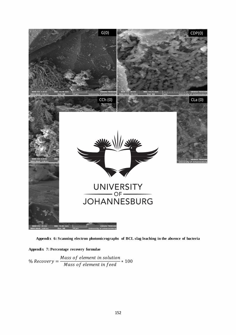
152
Appendix 6: Scanning electron photomicrographs of BCL slag leaching in the absence of bacteria
Appendix 7: Percentage recovery formulae
% 𝑅𝑒𝑐𝑜𝑣𝑒𝑟𝑦 =𝑀𝑎𝑠𝑠 𝑜𝑓 𝑒𝑙𝑒𝑚𝑒𝑛𝑡 𝑖𝑛 𝑠𝑜𝑙𝑢𝑡𝑖𝑜𝑛
𝑀𝑎𝑠𝑠 𝑜𝑓 𝑒𝑙𝑒𝑚𝑒𝑛𝑡 𝑖𝑛 𝑓𝑒𝑒𝑑∗ 100
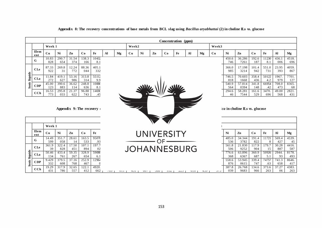
153
Appendix 8: The recovery concentrations of base metals from BCL slag using Bacillus aryabhattai (2) in choline ILs vs. glucose
Concentration (ppm)
Week 1 Week2 Week3
Element
Cu Ni Zn Co Fe Al Mg Cu Ni Zn Co Fe Al Mg Cu Ni Zn Co Fe Al Mg
G 10.83
828 290.7
654 31.54
374 138.3
166 10432
8.1 8.449
906 3629.
308 25.32
999 362.1
073 31.62
584 163.9
898 67120
.77 42.86
204 4052.
823 130.85
9611 450.6
746 36.286
7261 192.6
187 11230
8.1 436.1
006 4518.
696
Ion
ic l
iqu
ids
CLe 87.33
922 269.8
31 12.24
772 88.36
044 405.1
332
-3.743
58
3842.762
153.8576
302.7797
14.06825
94.74136
301.1155
-6.187
15
4025.742
500.699533
366.0985
17.1983214
101.4942
551.0731
23.95393
4059.867
CLa 11.84
272 419.1
627 53.16
986 313.8
314 55112
9.9 257.3
752 6833.
558 628.4
582 542.3
376 61.43
304 340.9
876 53254
0.9 1318.
224 7330.
97 1246.9
5403 746.5
818 70.603
1668 358.4
436 50122
4.2 1967.
979 7701.
127
CDP 45.00
123 390.6
883 36.62
114 248.8
636 10886
8.1 10.11
974 6917.
054 67.27
213 467.5
366 45.21
59 293.9
861 35337
.86 206.0
54 7874.
401 346.39
3701 540.9
564 57.814
0394 341.8
148 66954
.42 708.0
473 8365.
68
CCh 16.53
773 295.8
033 21.37
622 96.88
743 14690
.45 13.13
638 3528.
191 20.71
927 323.5
419 26.24
979 112.5
24 4011.
405 12.64
985 3480.
132 87.790
5552 294.6
46 58.281
7544 161.6
529 3470.
696 49.00
568 2821.
431
Appendix 9: The recovery concentrations of base metals from BCL slag using Raoultella ornithinolytica in choline ILs vs. glucose
Concentration (ppm)
Week 1 Week2 Week3
Element
Cu Ni Zn Co Fe Al Mg Cu Ni Zn Co Fe Al Mg Cu Ni Zn Co Fe Al Mg
G 14.49
599 351.7
058 28.61
14 163.5
533 95078
.93 11.15
451 4062.
514 61.08
908 407.5
206 32.04
805 185.1
024 11702
6.9 233.4
086 4454.
093 279.67
4922 485.0
536 34.344
3782 191.4
822 11721
8 569.4
427 4539.
45
Ion
ic l
iqu
ids
CLe 361.9
39 322.4
828 17.50
451 107.1
894 197.7
02 45.58
401 4265.
36 569.9
179 435.9
572 17.02
081 111.2
96 195.1
385 69.92
652 4233.
737 626.59
3983 561.8
506 21.830
9252 117.9
904 179.7
15 30.28
887 4416.
587
CLa 50.46
134
433.4
761
59.35
267
328.9
654
59088
6.1
537.4
744
7746.
005
621.6
417
561.1
669
61.94
432
343.1
203
52935
9.6
2038.
477
7938.
367
1239.7
392
776.6
368
63.896
6367
360.9
687
50926
5.1
2944.
93
8178.
493
CDP 9.429
332 379.5
608 37.16
768 252.9
467 12964
0 0.994
124 6866.
216 39.75
927 477.1
115 46.00
985 286.7
519 38668
.34 85.21
12 7483.
143 262.52
7864 558.6
876 53.945
0615 339.4
747 74757
.63 741.3
658 8646.
417
CCh 19.29
431 317.9
786 32.65
557 122.1
412 4920.
662 7.785
769 3616.
011 62.95
431 362.9
607 28.53
568 133.7
932 2678.
225 32.18
616 4385.
439 107.88
573 387.8
039 26.768
9683 134.6
966 373.6
263 37.27
06 4383.
263
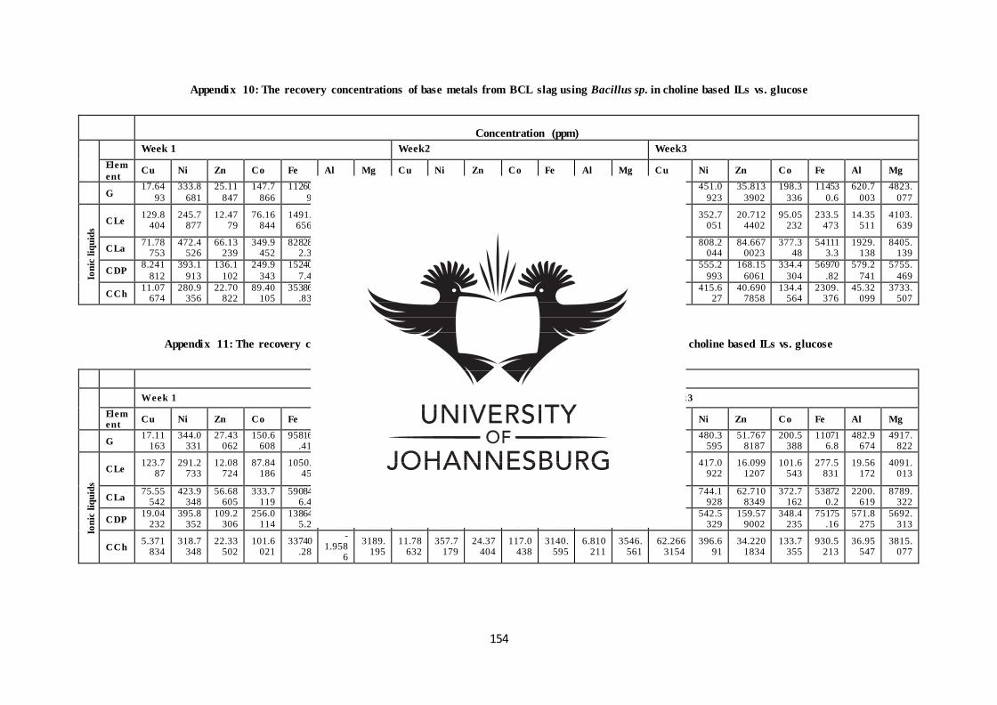
154
Appendix 10: The recovery concentrations of base metals from BCL slag using Bacillus sp. in choline based ILs vs. glucose
Concentration (ppm)
Week 1 Week2 Week3
Elem
ent Cu Ni Zn Co Fe Al Mg Cu Ni Zn Co Fe Al Mg Cu Ni Zn Co Fe Al Mg
G 17.64
93
333.8
681
25.11
847
147.7
866
11260
9
14.04
415
4025.
315
49.39
447
383.1
085
33.18
437
177.8
238
73073
.88
134.1
907
4532.
82
179.53
543
451.0
923
35.813
3902
198.3
336
11453
0.6
620.7
003
4823.
077
Ion
ic l
iqu
ids
CLe 129.8
404 245.7
877 12.47
79 76.16
844 1491.
656
-7.744
06
3468.628
255.4494
333.3456
106.5499
93.12924
10206.14
3.765083
4188.736
347.573412
352.7051
20.7124402
95.05232
233.5473
14.35511
4103.639
CLa 71.78
753 472.4
526 66.13
239 349.9
452 82828
2.3 508.2
05 8102.
525 754.0
288 576.7
279 60.26
258 358.6
555 57531
9.3 1321.
65 8226.
034 1319.9
4519 808.2
044 84.667
0023 377.3
48 54111
3.3 1929.
138 8405.
139
CDP 8.241
812
393.1
913
136.1
102
249.9
343
15240
7.4
12.64
38
4590.
445
63.16
659
454.2
968
125.6
19
282.6
782 42537
132.4
563
4759.
25
351.30
8704
555.2
993
168.15
6061
334.4
304
56970
.82
579.2
741
5755.
469
CCh 11.07
674 280.9
356 22.70
822 89.40
105 35386
.83 7.516
386 3558.
666 17.66
848 301.3
45 27.98
267 90.54
164 3837.
269 16.51
119 3755.
984 60.013
8603 415.6
27 40.690
7858 134.4
564 2309.
376 45.32
099 3733.
507
Appendix 11: The recovery concentrations of base metals from BCL slag using Bacillus thuringiensis in choline based ILs vs. glucose
Concentration (ppm)
Week 1 Week2 Week3
Element
Cu Ni Zn Co Fe Al Mg Cu Ni Zn Co Fe Al Mg Cu Ni Zn Co Fe Al Mg
G 17.11
163 344.0
331 27.43
062 150.6
608 95816
.41 6.980
769 4070.
313 34.46
322 409.6
679 32.40
468 178.1
239 45735
.91 88.32
524 4653.
581 138.53
2246 480.3
595 51.767
8187 200.5
388 11071
6.8 482.9
674 4917.
822
Ion
ic l
iqu
ids
CLe 123.7
87 291.2
733 12.08
724 87.84
186 1050.
45
-7.325
39
4030.734
134.8947
345.7214
15.28939
96.34774
492.7643
-3.382
74
4141.689
446.15131
417.0922
16.0991207
101.6543
277.5831
19.56172
4091.013
CLa 75.55
542 423.9
348 56.68
605 333.7
119 59084
6.4 256.0
679 8456.
775 682.6
889 553.5
076 63.22
845 358.2
446 55769
2.3 1436.
537 8508.
492 1248.5
6651 744.1
928 62.710
8349 372.7
162 53872
0.2 2200.
619 8789.
322
CDP 19.04
232 395.8
352 109.2
306 256.0
114 13864
5.2 13.62
661 4505.
515 84.49
115 469.7
209 170.0
673 305.1
275 36869
.35 142.4
82 4905.
331 393.96
7176 542.5
329 159.57
9002 348.4
235 75175
.16 571.8
275 5692.
313
CCh 5.371
834 318.7
348 22.33
502 101.6
021 33740
.28
-1.958
6
3189.195
11.78632
357.7179
24.37404
117.0438
3140.595
6.810211
3546.561
62.2663154
396.691
34.2201834
133.7355
930.5213
36.95547
3815.077
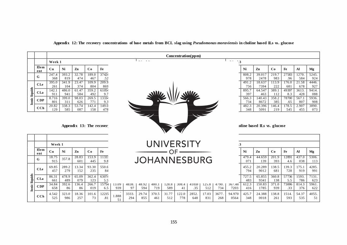
155
Appendix 12: The recovery concentrations of base metals from BCL slag using Pseudomonas moraviensis in choline based ILs vs. glucose
Concentration(ppm)
Week 1 Week2 Week3
Element
Cu Ni Zn Co Fe Al Mg Cu Ni Zn Co Fe Al Mg Cu Ni Zn Co Fe Al Mg
G 247.4
368 393.2
819 32.78
474 189.0
467 37424
.52 801.6
565 4822.
154 694.7
463 626.2
301 43.36
164 211.1
972 25563
.56 1276.
806 5053.
27 844.21
9981 808.2
978 39.017
2478 219.7
983 27583
.96 1270.
584 5245.
924
Ion
ic l
iqu
ids
CLe 395.0
261
341.9
104
23.47
374
109.9
804
209.9
869
27.78
6
4425.
009
457.9
746
422.6
359
14.46
737
109.7
68
171.2
316
25.07
176
4371.
528
471.52
1608
491.2
756
18.637
7394
113.9
222
176.0
681
21.58
678
4446.
927
CLa 142.1
911 486.0
941 61.47
584 359.2
492 61054
9.7 1379.
226 8971.
574 802.5
054 624.8
518 62.93
84 366.6
257 52100
1.3 2588.
909 9173.
676 1452.2
2444 895.7
397 64.547
463 389.1
112 49397
8.3 3613.
428 9414.
088
CDP 8.716
801 399.0
311 98.03
626 269.5
771 13334
9.3 20.67
209 4653.
135 63.68
603 476.9
917 135.6
429 308.3
523 32526
.78 157.3
522 4824.
773 328.96
3637 566.3
734 140.43
8672 358.2
385 78700
.65 567.1
807 5936.
908
CCh 20.82
129 338.3
585 53.74
087 142.4
158 149.0
478 10.06
153 3912.
555 107.3
502 409.9
31 20.51
015 143.0
955 166.5
42 5.418
66 3928.
566 184.53
5823 482.3
348 20.396
5091 146.4
219 178.5
545 2.907
455 3890.
073
Appendix 13: The recovery concentrations of base metals from BCL slag using a mixed culture in choline based ILs vs. glucose
Concentration(ppm)
Week 1 Week2 Week3
Element
Cu Ni Zn Co Fe Al Mg Cu Ni Zn Co Fe Al Mg Cu Ni Zn Co Fe Al Mg
G 18.75
915 357.8
28.83
601
153.9
445
11333
9.8
24.92
913
4431.
833
36.02
206
416.8
954
29.20
886
173.1
085
17310
8.5
51.44
099
4916.
873
130.13
8653
479.4
071
44.659
139
201.9
393
12891
4.6
437.0
038
5306.
113
Ion
ic l
iqu
ids
CLe 69.85
457 289.2
279 13.34
152 93.30
235 550.6
84
-
4.4334
4125.553
166.3606
357.9736
17.64822
106.3799
249.377
30.65518
4257.705
377.172701
455.2794
20.2899012
138.5681
139.3728
175.1919
4285.991
CLa 86.31
661 478.9
489 65.09
079 362.4
123 63074
5.5 505.9
221 9473.
809 588.8
711 595.6
059 57.87
189 391.4
618 60509
1.8 1443.
563 6401.
743 1090.3
9648 727.5
483 65.855
9341 360.8
138 57736
5.5 1593.
786 7131.
623
CDP 34.84
658 392.6
86 136.4
86 266.7
019 15754
6.5 13.09
939 4838.
97 48.92
594 480.1
719 128.8
589 308.4
41 41059
.35 125.8
512 4790.
734 367.48
7203 612.3
416 150.83
1785 371.0
939 73006
.33 814.3
376 5961.
622
CCh 4.542
525
323.0
986
18.36
257
101.6
73
12215
.81
-
1.88851
3333.
294
29.74
855
370.5
461
31.77
512
122.0
778
2852.
648
17.03
831
3677.
268
94.970
0564
425.7
348
24.388
0018
138.8
261
1514.
593
54.37
535
4055.
51
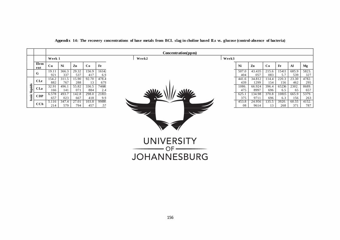
156
Appendix 14: The recovery concentrations of base metals from BCL slag in choline based ILs vs. glucose (control-absence of bacteria)
Concentration(ppm)
Week 1 Week2 Week3
Elem
ent Cu Ni Zn Co Fe Al Mg Cu Ni Zn Co Fe Al Mg Cu Ni Zn Co Fe Al Mg
G 19.11
921 366.3
337 29.32
537 156.9
417 16142
6.9 26.30
206 4753.
899 34.08
419 426.1
487 51.55
099 181.1
999 13770
3.7 124.4
944 5423.
362 273.28
8454 507.0
404 43.435
057 215.6
083 15411
5.7 685.9
539 5823.
327
Ion
ic l
iqu
ids
CLe 154.2
882 311.5
767 15.90
288 92.70
13 478.4
679 4.815
693 4236.
783 222.5
924 383.1
338 21.58
135 100.1
766 529.7
849 0.540
785 4512.
537 332.10
535 441.6
439 34.812
1299 114.4
154 220.3
156 23.30
462 4782.
295
CLa 32.91
166 496.1
141 55.82
071 336.5
884 74680
2.4 499.6
034 7792.
182 839.7
615 696.1
564 62.87
546 368.2
287 69553
9.6 1451.
136 8397.
651 1479.1
4093 1086.
475 66.924
8997 396.4
696 65236
6.5 2302.
65 8689.
657
CDP 6.578
657
493.7
023
142.8
667
298.0
418
21811
9.9
23.28
216
5222.
868
37.94
386
552.4
185
130.4
424
324.5
97
99693
.72
146.2
233
5091.
044
268.15
6331
625.1
375
134.98
9711
370.8
696
10815
6.3
665.9
156
5379.
263
CCh 5.116
214
347.4
579
27.01
794
103.8
457
99899
.57
3.121
313
3506.
384
12.59
105
410.4
505
23.54
184
118.9
665
29124
.44
7.609
691
3844.
683
70.636
6986
453.8
08
24.956
9614
135.5
13
3920.
268
60.55
371
4152.
787