correlations between severity of coronary atherosclerosis and persistent elevation of circulating...
TRANSCRIPT

Revista Română de Medicină de Laborator Vol. 22, Nr. 1, Martie, 2014 49
DOI: 10.2478/rrlm-2014-0005
Correlations between severity of coronary atherosclerosis and persistent elevation of circulating C-reactive protein
levels 30 days after an acute myocardial infarction
Corelaţii între severitatea aterosclerozei coronariene şi persistenţa nivelelor serice crescute ale proteinei C reactive la 30 de zile după un infarct
miocardic acut
Theodora Benedek1*, Beata Jako1, Zsuzsa Suciu2, Imre Benedek1
1. University of Medicine and Pharmacy Tirgu Mures, Department of Internal Medicine 32. Cardiomed Medical Center
AbstractIntroduction: We aimed to assess the relationships between the persistence of elevated circulating levels of
hs-CRP, a powerful inflammatory marker, determined at 30 days after an acute myocardial infarction (AMI), and the characteristics of the pre-existing coronary lesions. Material and methods: The study included 83 consecutive patients 30 days post AMI, who were subjected to coronary angiography and primary PCI. The patients were di-vided into two groups according to their hsCRP levels at 30 days after AMI: group 1 included 35 low-risk patients, with hsCRP levels <2 mg/l, and group 2 included 48 high-risk patients, with hsCRP levels >2 mg/l. Results: Angio-graphic analysis revealed the presence of a multivascular disease in 48.5% of the patients in group 1 versus 72.9% of the patients in group 2 (p=0.037). The Syntax scores for groups 1 and 2 were 22.2 +/- 6.6 and 27.07+/-0.94, respectively (p=0.001), and these values were significantly correlated with the hsCRP values (r=0.56, p<0.0001). LAD culprit lesions were found in 47.9% of the patients in group 1 and 20% of the patients in group 2 (p=0.01), and 42.8% of the group 1 patients and 83.3% of the group 2 patients had at least one significant stenosis in the LAD (p=0.0002). The ejection fraction at 30 days was significantly lower in the patients with elevated levels of hsCRP (52.91+/-4.03 vs 49.04+/-5.74, p=0.001), showing an inverse correlation with hsCRP levels (r=-0.52, p<0.0001).Conclusions: A more severe coronary artery disease was associated with am increased inflammatory status in the postinfarction phase, as evidenced by the high levels of circulating hsCRP. hsCRP can help for risk stratification in post AMI patients by identifying the subsets of patients who are at risk based on persistent elevated circulating levels of hsCRP at 30 days after infarction.
Key words: hsCRP, inflammation, acute myocardial infarction
*Corresponding author: Theodora Benedek, MD, PhD, University of Medicine and Pharmacy Tîrgu Mureș, Clinic of Cardiology, phone: +40722560549, e-mail: [email protected]
Research article
- 10.2478/rrlm-2014-0005Downloaded from PubFactory at 09/03/2016 03:07:54PM
via free access

Revista Română de Medicină de Laborator Vol. 22, Nr. 1, Martie, 201450
RezumatIntroducere: Scopul studiului a fost evaluarea corelaţiei dintre severitatea afectării coronariene şi persistenţa
unor nivele serice crescute ale hs-CRP, un puternic marker inflamator, determinate la 30 de zile post Infarct Miocardic Acut (IMA). Material şi metodă: Studiul a inclus 83 pacienţi consecutivi cu IMA, la care s-a efectuat coronarografie şi angioplastie primară, împărţiţi în două grupuri în funcţie de nivelul hsCRP la 30 de zile postinfarct: grupa 1 – 35 pacienţi cu risc redus, cu nivele de hsCRP<2 mg/l, respectiv grupa 2 – 48 pacienţi cu risc înalt, cu nivele de hsCRP>2 mg/l. Rezultate: Analiza angiografică a relevat prezenţa unei afectări multivasculare la 48.5% din pacienţii grupei 1 versus 72.9% din pacienţii grupei 2 (p=0.037). Scorul Syntax a fost de 22.2 +/- 6.6 la grupa 1 vs 27.07+/-0.94 la grupa 2 (p=0.001), prezentând o corelaţie semnificativă cu valorile hsCRP (r=0.56, p<0.0001). Numărul mediu de coronare afectate a fost de 1.6+/- 0.69 vs 1.97+/-0.73 (p=0.019). Localizarea leziunilor ţintă la nivelul ADA a fost întâlnită la 47.9% din pacienţii grupei 1 vs 20% la grupa 2 (p=0.01) iar prezenţa de stenoze semnificative la nivelul ADA la 42.8% din pacienţii grupei 1 vs 83.3% din pacienţii grupei 2 (p=0.0002). Fracţia de ejecţie la 30 de zile a fost semnificativ mai mică la grupa 2 (52.91+/-4.03 vs 49.04+/-5.74, p=0.001), prezentând o corelaţie inversă cu nivelele hsCRP (r=-0.52, p<0.0001). Concluzii: Persistenţa unui sta-tus inflamator în faza postinfarct, evidenţiată de nivelele crescute ale hsCRPcirculant, se asociază cu o severitate mai crescută a afecţiunii coronariene şi o evoluţie mai severă. HsCRP poate contribui la stratificarea riscului postIMA, identificând subsetul de pacienţi la risc pe baza persistenţei nivelelor circulante crescute ale hsCRP la 30 de zile postinfarct.
Cuvinte cheie: hsCRP, inflamaţie, infarct miocardic acutReceived: 9th July 2013; Accepted: 9th February 2014; Published:2nd March 2014.
Introduction
Atherosclerosis is characterized nowadays as a generalized inflammatory disease that involves all vascular beds, most commonly localised at the level of the coronary, carotid and peripheral arteries (1). The most devastating manifestation of atherosclerosis is acute myocardial infarction (AMI), in which inflammation has been demon-strated to play a pivotal role; many studies have proven that an elevated inflammatory status is associated with the development of acute coro-nary syndromes (2).
In acute myocardial infarction, the rupture of an intracoronary plaque leads to the abrupt occlusion of the coronary artery and subsequent myocardial necrosis in the territory supplied by the occluded artery. Several days following the infarction, the myocardial healing process leads to the development of a scar at the site of necro-sis. The inflammatory process is involved in dif-ferent phases of acute myocardial infarction that include plaque formation, plaque rupture and
also in the myocardial healing process that is fol-lowed by scar formation at the site of the dam-aged myocardium (3). During the postinfarction phase, myocardial ischaemia caused by coronary occlusion leads to a systemic humoral and a lo-cal cellular inflammatory response, which act as triggers of a myocardial inflammatory reaction that aims to promote myocardial healing. Simul-taneously, the systemic activation of the humor-al system following myocardial necrosis leads to an additional significant increase in the regional inflammatory response (4,5).
The systemic inflammatory reaction in post acute myocardial infarction phase consists mainly in a humoral response that is represent-ed by the release of cytokines and complement systems, and a cell-mediated response, repre-sented by the migration and accumulation of inflammatory cells (i.e. neutrophils, monocytes/macrophages and mast cells) at the site of the ischaemic myocardium. Several inflammatory markers that can be easily measured in periph-eral blood, have been proposed as indices of the
- 10.2478/rrlm-2014-0005Downloaded from PubFactory at 09/03/2016 03:07:54PM
via free access

Revista Română de Medicină de Laborator Vol. 22, Nr. 1, Martie, 2014 51
systemic inflammatory reaction that is associat-ed with the development of an acute myocardial infarction. One such marker, C-reactive protein has been shown to be associated with increased early and late cardiovascular morbidity and mortality in patients with STsegment elevation myocardial infarction, and has been proposed to represent one of the most important markers for characterizing the systemic inflammatory pro-cess in post AMI patients (6,7).
The high-sensitivity C-reactive protein (hs-CRP) is currently considered to represent a marker of future cardiovascular events inde-pendent from the traditional cardiovascular risk factors such as elevated cholesterol, smoking, obesity or hypertension. Several studies have proven that high levels of hs-CRP are able to identify patients who are at risk for significant endothelial dysfunction and deleterious ventric-ular remodeling in the postinfarction phase (8,9).
However, all the studies published so far evaluated the correlation between high levels of hs-CRP collected upon hospital admission, during the acute phase of AMI, and cardiovas-cular events, without taking into consideration the risk associated with the persistence of high hsCRP levels in the postAMI phase.
In this angiographically-controlled study, we aimed to assess the relationships between the pre-existing coronary lesions (the angiographic severity of coronary artery diseases expressed by the Syntax scores, the locations of the culprit le-sions, the involvement of different coronary ar-teries, the presence of multivascular disease and the total number of coronary arteries with sig-nificant stenosis) and the persistence of elevated circulating levels of hs-CRP as determined at 30 days after acute myocardial infarction.
Material and methods
The study included 83 consecutive patients who presented with acute myocardial infarction
30 days prior to the inclusion in the study and who were subjected to coronary angiography and primary PCI at the time of the infarction, performed within 12 hours of the onset of symp-toms.
All patients received optimum medical ther-apy following the infarction, including aspirin (75 mg), clopidogrel (a 300 mg loading dose fol-lowed by 150 mg daily for 7 days, and then by 75 mg daily), ACE inhibitors and statins (80 mg atorvastatin for the first 30 days after the infarc-tion as by local routine practice).
The demographic data, history and risk fac-tors (i.e., age, gender, smoking status, presence of diabetes, hyperlipidemia, obesity, hyperten-sion and blood tests) were recorded for every patient.
In all patients hs CRP levels were determined at 30 days postinfarction and the patients were divided into 2 groups according to their hsCRP levels: group 1 consisted in 35 low risk patients, with hsCRP levels below 2 mg/l, and group 2 consisted in 48 high risk patients, with hsCRP levels above 2 mg/l.
Measurements of hsCRP levels were per-formed utilizing lateral flow immunometric methodology, which enables hsCRP testing from fingerstick or venous whole blood using the Cholestech LDX System (Cholestech-LDX Analyzer, Biosite Incorporated, san Diego, CA, USA), which is a desk-top analyzer that utilizes dry chemistry cassettes and reflectance photom-etry to quantify substances in blood, based on the conversion of the reflectance reading (% R) to hs-CRP concentration in mg/L.
Retrospective assessment of coronary angi-ographies that were performed within the first 12 hours after the onset of the infarction included calculation of the Syntax score, the number of coronary arteries with significant stenoses, the number of coronary artery stenosis in each of the 3 major coronary arteries and the location of the culprit lesion. A significant stenosis was
- 10.2478/rrlm-2014-0005Downloaded from PubFactory at 09/03/2016 03:07:54PM
via free access

Revista Română de Medicină de Laborator Vol. 22, Nr. 1, Martie, 201452
defined as a stenosis >75% in a major epicardial coronary vessel. A culprit lesion was defined as a lesion that was responsible for an acute isch-aemic event, as identified by ECG, echocardi-ography and angiography based on location in the coronary artery that supplied the myocardial territory with a contractility defect (as assessed with echocardiography) or with ST/T changes (as assessed by ECG), together with a correspon-dent angiographic picture (thrombus or unstable plaque).
Echocardiographic assessment included de-termination of the left ventricular end-diastolic and end-systolic volumes and calculation of left ventricular ejection at 30 day postinfarction. All echocardiographic acquisitions were made using a PhilipsSonos 7500 equipment. All acquired images were transferred to a workstation (QLab, Philips) for data processing, measurements and interpretation. Calculation of left ventricular ejection fraction was based on determination of left ventricular volumes on bi-dimensional as-sessment (in 4-chambers and 2-chambers apical views) using the modified Simpson technique.
All patients gave written informed consent for the study, and the study protocol was ap-proved by the ethics committee of the medical center in which the study was conducted.
The primary objective of the study was to demonstrate the association between the sever-ity of coronary atherosclerosis (as expressed by the Syntax score, the presence of multivascular disease and the total number of coronary arter-ies with significant stenoses) and increased lev-els of hsCRP 30 days after an acute myocardial infarction.
The secondary objectives of the study were to demonstrate the association between in-creased levels of hsCRP 30 days after an acute myocardial infarction and:
a. angiographic characteristics (i.e., loca-tion of the culprit lesion in different coro-nary arteries, the involvement of different
coronary arteries in the atherosclerotic process, and postprocedural TIMI flow)
b. left ventricular function, as expressed by the ejection fraction immediate after in-tervention and at 30 days
c. other baseline characteristics that reflect metabolic status
We performed a multivariate analysis of fac-tors that could predict a depressed ventricular function at 30 days postinfarction, including in this model the postprocedural TIMI flow, the to-tal ischemic time, the immediate postprocedural left ventricular ejection fraction, the hsCRP val-ues, the angiographic Syntax score, and the val-ues of troponin I and CK-MB.
Statistical analysisAll statistical analysis were performed using
the InStat Graph Pad software. We used the Fish-er’s exact test (or the Student’s t-test for age) to compare the baseline characteristics of patients between the low-risk and high-risk patient pop-ulation. Continuous values are expressed as the mean and standard deviation, and statistical sig-nificance was determined using the Mann-Whit-ney test. Categorical variables are expressed as percentages. Linear regression was used to as-sess the correlation between EF and hsCRP val-ues. Statistical significance was considered for a p value <0.05, and all p values were 2-sided.
Results
The clinical baseline characteristics of the study population showed no significant differ-ences between the low-risk and the high-risk group in respect to age (p=0.6), gender (p=0.06), the presence of diabetes (p=0.5), hypertension (p=0.4), hyperlipidemia (p=1), obesity (p=0.7) or smoking status (p=0.4) (Table I). However, metabolic syndrome (defined as the presence
- 10.2478/rrlm-2014-0005Downloaded from PubFactory at 09/03/2016 03:07:54PM
via free access
Revista Română de Medicină de Laborator Vol. 22, Nr. 1, Martie, 2014 53
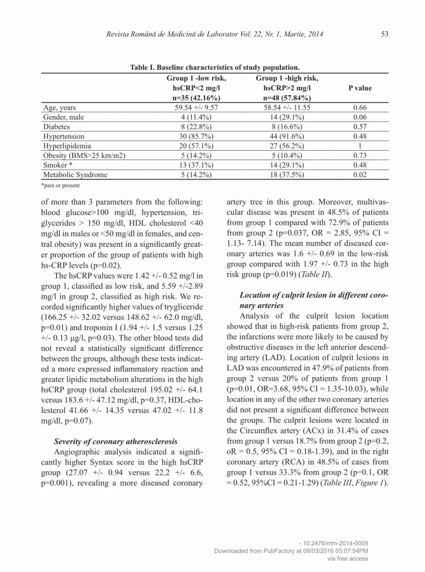
of more than 3 parameters from the following: blood glucose>100 mg/dl, hypertension, tri-glycerides > 150 mg/dl, HDL cholesterol <40 mg/dl in males or <50 mg/dl in females, and cen-tral obesity) was present in a significantly great-er proportion of the group of patients with high hs-CRP levels (p=0.02).
The hsCRP values were 1.42 +/- 0.52 mg/l in group 1, classified as low risk, and 5.59 +/-2.89 mg/l in group 2, classified as high risk. We re-corded significantly higher values of trygliceride (166.25 +/- 32.02 versus 148.62 +/- 62.0 mg/dl, p=0.01) and troponin I (1.94 +/- 1.5 versus 1.25 +/- 0.13 μg/l, p=0.03). The other blood tests did not reveal a statistically significant difference between the groups, although these tests indicat-ed a more expressed inflammatory reaction and greater lipidic metabolism alterations in the high hsCRP group (total cholesterol 195.02 +/- 64.1 versus 183.6 +/- 47.12 mg/dl, p=0.37, HDL-cho-lesterol 41.66 +/- 14.35 versus 47.02 +/- 11.8 mg/dl, p=0.07).
Severity of coronary atherosclerosis Angiographic analysis indicated a signifi-
cantly higher Syntax score in the high hsCRP group (27.07 +/- 0.94 versus 22.2 +/- 6.6, p=0.001), revealing a more diseased coronary
artery tree in this group. Moreover, multivas-cular disease was present in 48.5% of patients from group 1 compared with 72.9% of patients from group 2 (p=0.037, OR = 2.85, 95% CI = 1.13- 7.14). The mean number of diseased cor-onary arteries was 1.6 +/- 0.69 in the low-risk group compared with 1.97 +/- 0.73 in the high risk group (p=0.019) (Table II).
Location of culprit lesion in different coro-nary arteries Analysis of the culprit lesion location
showed that in high-risk patients from group 2, the infarctions were more likely to be caused by obstructive diseases in the left anterior descend-ing artery (LAD). Location of culprit lesions in LAD was encountered in 47.9% of patients from group 2 versus 20% of patients from group 1 (p=0.01, OR=3.68, 95% CI = 1.35-10.03), while location in any of the other two coronary arteries did not present a significant difference between the groups. The culprit lesions were located in the Circumflex artery (ACx) in 31.4% of cases from group 1 versus 18.7% from group 2 (p=0.2, oR = 0.5, 95% CI = 0.18-1.39), and in the right coronary artery (RCA) in 48.5% of cases from group 1 versus 33.3% from group 2 (p=0.1, OR = 0.52, 95%CI = 0.21-1.29) (Table III, Figure 1).
Table I. Baseline characteristics of study population.Group 1 -low risk,
hsCRP<2 mg/ln=35 (42.16%)
Group 1 -high risk, hsCRP>2 mg/ln=48 (57.84%)
P value
Age, years 59.54 +/- 9.57 58.54 +/- 11.55 0.66Gender, male 4 (11.4%) 14 (29.1%) 0.06Diabetes 8 (22.8%) 8 (16.6%) 0.57Hypertension 30 (85.7%) 44 (91.6%) 0.48Hyperlipidemia 20 (57.1%) 27 (56.2%) 1Obesity (BMS>25 km/m2) 5 (14.2%) 5 (10.4%) 0.73Smoker * 13 (37.1%) 14 (29.1%) 0.48Metabolic Syndrome 5 (14.2%) 18 (37.5%) 0.02
*past or present
- 10.2478/rrlm-2014-0005Downloaded from PubFactory at 09/03/2016 03:07:54PM
via free access

Revista Română de Medicină de Laborator Vol. 22, Nr. 1, Martie, 201454
Table II. Comparison of blood tests, angiographic data and EF in patients with high versus low risk.Group 1- low risk,
hsCRP<2mg/lGroup 2- high hsCRP>2 mg/l
p value
Blood TEsTshsCRP (mg/l) <0.0001Mean +/- SD 1.428 +/- 0.529 5.59 +/- 2.8995% confidence interval 1.24 – 1.61 4.75 – 6.44Cholesterol (mg/dl) 0.37Mean +/- SD 183.6 +/- 47.12 195.02 +/- 64.195% confidence interval 167.4 - 199.8 176.39 - 213.65Hdl-cholesterol (mg/dl) 0.07Mean +/- SD 47.02 +/- 11.8 41.66 +/- 14.3595% confidence interval 42.96 - 51.09 37.49 - 45.83Triglyceride (mg/dl) 0.01Mean +/- SD 148.62 +/- 62.0 166.25 +/- 32.0295% confidence interval 139.17 - 158.09 156.94 - 175.56WBC (*103) 0.07Mean +/- SD 7.47 +/- 1.69 7.97 +/- 9.7295% confidence interval 6.85 - 8.02 7.69 - 8.25Thrombocyte count (*103) 0.6Mean +/- SD 240.85 +/- 69.91 232.89 +/- 71.1495% confidence interval 216.83 - 264.89 212.22 - 253.58cTnI (μg/l) 0.03Mean +/- SD 1.25 +/- 0.13 1.94 +/- 1.595% confidence interval 0.79 - 1.71 1.49 - 2.39Peak CK-MB (μg/l) 0.06Mean +/- SD 19.45 +/- 5.77 21.83 +/- 5.4895% confidence interval 17.47 - 21.44 20.23 - 23.43AnGIoGRAPHIC dATAsyntax score 0.001Mean +/- SD 22.2 +/- 6.6 27.07 +/- 0.9495% confidence interval 19.93 - 24.47 25.13 - 28.94number of diseased coronary arteries 0.019Mean +/- SD 1.6 +/- 0.69 1.97 +/- 0.7395% confidence interval 1.36 – 1.84 1.76 – 2.19Ischemic time (min) 0.9Mean +/- SD 304 +/- 132 301 +/- 109.5995% confidence interval 258.74 - 349.61 269.15 - 332.85Postprocedural TIMI flow 0.3Mean +/- SD 2.8 +/- 0.47 2.75 +/- 0.4895% confidence interval 2.63 - 2.96 2.6 - 2.89EjECTIon FRACTIonEjection fraction at baseline (%) 0.07Mean +/- SD 47.74 +/- 3.8 45.79 +/- 5.4595% confidence interval 46.43 - 49.05 44.2 - 47.37Ejection fraction at 30 days (%) 0.001Mean +/- SD 52.91 +/- 4.03 49.04 +/- 5.7495% confidence interval 51.52 – 54.3 47.37 – 50.71
- 10.2478/rrlm-2014-0005Downloaded from PubFactory at 09/03/2016 03:07:54PM
via free access

Revista Română de Medicină de Laborator Vol. 22, Nr. 1, Martie, 2014 55
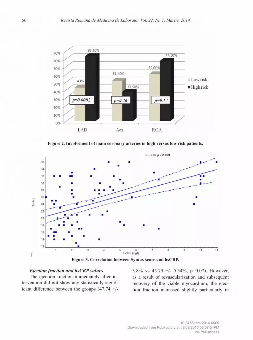
Involvement of different coronary arteries in the atherosclerotic process The presence of at least one significant ste-
nosis in the LAD was recorded in 42.8% of pa-tients from group 1 compared with 83.3% of pa-tients from group 2 (p=0.0002, OR=6.66, 95% CI = 2.42-18.34), while the presence of at least one significant stenosis in ACx or RCA did not present any statistically significant difference between the groups (51.4% versus 37.5%, p=0.2
for ACx, 60% versus 70.1%, p= 0.1 for RCA) (Figure 2).
Linear regression analysis indicated a good correlation between the levels of Syntax score and the values of hsCRP at 30 days postinfarc-tion (r=0.56, p<0.0001) (Figure 3), indicating that a higher severity of coronary lesions cor-relates with higher values of inflammatory mark-ers at 30 days postinfarction.
Table III. Angiographic characteristics of the study population.Group 1 -low risk,
hsCRP<2 mg/ln=35 (42.16%)
Group 1 -high risk, hsCRP>2 mg/ln=48 (57.84%)
P value OR 95% CI
Culprit lesionLAD 7 (20%) 23 (47.9%) 0.01 3.68 1.35 – 10.03ACx 11 (31.4%) 9 (18.7%) 0.2 0.5 0.18 – 1.39RCA 17 (48.5%) 16 (33.33%) 0.18 0.52 0.21 – 1.29
Presence of at least 1 significant stenosisLAD 15 (42.8%) 40 (83.3%) 0.0002 6.66 2.42 – 18.34ACx 18 (51.4%) 18 (37.5%) 0.26 0.56 0.23 – 1.37RCA 21 (60%) 37 (77.1%) 0.14 2.24 0.86 – 5.82
Multivascular disease 17 (48.5%) 35 (72.9%) 0.03 2.85 1.13 – 7.14
Figure 1. location of culprit lesion in high versus low risk patients.
- 10.2478/rrlm-2014-0005Downloaded from PubFactory at 09/03/2016 03:07:54PM
via free access
Revista Română de Medicină de Laborator Vol. 22, Nr. 1, Martie, 201456
Ejection fraction and hsCRP valuesThe ejection fraction immediately after in-
tervention did not show any statistically signif-icant difference between the groups (47.74 +/-
3.8% vs 45.79 +/- 5.54%, p=0.07). However, as a result of revascularization and subsequent recovery of the viable myocardium, the ejec-tion fraction increased slightly particularly in
Figure 2. Involvement of main coronary arteries in high versus low risk patients.
Figure 3. Correlation between syntax score and hsCRP.
4
- 10.2478/rrlm-2014-0005Downloaded from PubFactory at 09/03/2016 03:07:54PM
via free access

Revista Română de Medicină de Laborator Vol. 22, Nr. 1, Martie, 2014 57
the first group, and the difference between the groups became statistically significant at 30 days , when mean EF was 52.91 +/- 4.03, 95% CI 51.52 – 54.3 in group 1, and 49.04 +/- 5.74, 95% CI 47.37 – 50.71 in group 2, p=0.001. We also found a good correlation between the ejection fraction and the high levels of hsCRP (r=-0.52, p<0.0001) (Figure 4).
Predictors for low EF at 30 days postinfarction Multivariate analysis identified the fol-
lowing parameters as significant predictors of low ejection fraction at 30 days postinfarction: postprocedural TIMI flow (Odds Ratio 3.06, p=0.05), low (<45%) immediate postprocedur-al ejection fraction (Odds Ratio 3.9, p=0.006)
and angiographic Syntax score (Odds Ratio 3.9, p=0.01) (Table IV).
discussion
The excessive release of inflammation me-diators during the acute phase of a myocardial infarction may lead to the exacerbation of tissue damage and the development of severe compli-cations (10). During the acute phase of AMI, serum C-reactive protein has been suggested to represent not only a sensible inflammatory marker, but also a direct inflammatory promoter (11) with pro-atherogenic (12) and pro-throm-botic properties (13). According to a study pub-lished by Bonvini et al, inflammatory response following an acute myocardial infarction should
Figure 4. Correlation between ejection fraction and hsCRP.
Table IV. Multivariate predictors of 30-days EF.odds Ratio (95% CI) p value
Ischemic time 2.35 (0.88 - 6.29) 0.1Post-procedural TIMI flow (<3) 3.06 (1.02 - 9.16) 0.05Immediate post-procedural Left Ventricular Ejection Fraction (<45%)
3.9 (1.46 - 10.55) 0.006
High hs-CRP value (>2 mg/dl) 2.4 (0.8 - 6.6) 0.09Angiographic Syntax score 3.9 (1.29 - 11.9) 0.01TnI 1.6 (0.65 - 3.96) 0.3CK-MB 1.24 (0.51 - 3.03) 0.65
- 10.2478/rrlm-2014-0005Downloaded from PubFactory at 09/03/2016 03:07:54PM
via free access

Revista Română de Medicină de Laborator Vol. 22, Nr. 1, Martie, 201458
represent a new therapeutic target for postinfarc-tion patients (5).
CRP is an acute-phase protein synthetized by hepatocytes under the control of inflamma-tory cytokines and particular intrerleukin 6, and is released into the circulation in response to in-flammation and tissue damage. Given that the circulating level of CRP, considered currently considered to represent a “golden marker for in-flammation”, reflects the severity of inflamma-tory response and plays a significant role in the development of atherosclerosis, many studies have attempted to predict the future cardiovas-cular events and the response to therapy based on CRP-based risk classification (14,15). It has been proved that high levels of circulating CRP upon admission correlates with the extent of the infarction, the development of postinfarction heart failure and the presence of severe lesion by angiography, while in patients undergoing primary PCI, high CRP level before the proce-dure predicted the rate of early complications (16,17). In a previous study of Pietila et al, high serum C-reactive protein concentrations in acute myocardial infarction patients were found to predict an increased mortality up to 6 months following the infarction (18). Moreover, Cagh et al recently demonstrated that hs-CRP is an in-dependent predictor of ST resolution following primary PCI for AMI, showing also that a level of hs-CRP higher than 0.88 mg/dl predicted poor myocardial blush following PCI with 73% sen-sitivity and 31% specificity (19).
While all these studies have focused on the levels of CRP or hsCRP upon admission, during the acute phase of the infarction, our study eval-uated the correlation between the persistence of the inflammatory response, as expressed by persistent elevation o the hsCRP levels at 30 days postinfarction, the clinical and angiograph-ic characteristics and the evolution of these pa-tients.
Furthermore, this is the first study to cor-relate the high levels of hsCRP with the severity of the coronary artery disease and the location of the culprit lesion, lesion that is responsible for the acute myocardial infarction in patients with multiple coronary lesions. As the site of the cul-prit lesion indicates the location of infarction (in the territory supplied by the coronary artery in which culprit lesion is located), our study indi-cates that there is an association between high hsCRP levels and certain locations of infarction. We found that an anterior infarction, produced by a culprit lesion in the LAD, is associated with higher levels of inflammation than infarctions located in other coronary territories.
Our study found that culprit lesions located in LAD are more likely to trigger an exacerbat-ed inflammatory response, as the LAD was the infarct related artery in a significantly higher ex-tent in the high-risk group patients than in the low-risk group patients (47.9% from the patients in the high risk group having the culprit lesion located in the LAD, compared with 20% from patients in the low-risk group, p=0.01), while for ACx and RCA there were no statistically signif-icant differences between the groups. Interest-ingly, the involvement of the LAD in the athero-sclerotic process was also associated with higher hsCRP levels (83.3% in the high hsCRP group compared with 42.8% in the low hsCRP group), independent of the nature of LAD lesions (cul-prit or non-culprit). Therefore, we can postulate that stenoses in the LAD, whether acute (cul-prit) or chronic (non-culprit) are associated with higher levels of hsCRP and therefore these ste-noses represent higher risk lesions than those in the ACx and RCA. A possible explanation for this observation is that LAD, compared to the other arteries, irrigates a larger portion of the left ventricle, represented by the anterior wall, the septum and the apex. Therefore, occlusions of the LAD usually result in larger myocardial infarction, with larger areas of myocardial ne-
- 10.2478/rrlm-2014-0005Downloaded from PubFactory at 09/03/2016 03:07:54PM
via free access

Revista Română de Medicină de Laborator Vol. 22, Nr. 1, Martie, 2014 59
crosis and more marked systemic inflammatory responses.
As the areas supplied by the LAD are the most important contributors to left ventricular contractility, it is not surprising that in this study, the left ventricular function was significantly depressed in patients with elevated hsCRP lev-els at 30 days postinfarction, as indicated by the inverse correlation between EF and hsCRP val-ues and also by the significantly lower EFs in the group with high hsCRP levels (49.04 +/-5.74 versus 52.91 +/- 4.03 in group 1, p=0.001).
In a study published by Swiatkiewicz et al, multivariate analysis demonstrated CRP concen-tration at discharge to be an independent marker of early postinfarction left ventricular dysfunc-tion (odds ratio of 1.38, 95 % confidence interval 1.01–1.87; p < 0.04), which proves that higher levels of CRP upon admission identifies patients with worse outcomes and suggests that high hsCRP levels may identify patients who will have poorer results after reperfusion, or may be more likely to have ventricular remodeling and enlargement after hospital discharge (20). Simi-larly, in a recently published study by Perlas et al, high levels of hsCRP upon admission were found to correlate with an increase in cardiovas-cular risk on short term (21). Our study goes a step forward and shows that persistence of in-flammation at 30 days postinfarction was also associated with a poorer outcome as reflected by the lower ejection fraction in patients with persistently elevated hsCRP levels. This finding may be due to mydriad factors that contribute to the impairment of left ventricular function, in-cluding the severity of the coronary artery dis-ease, the postintervention TIMI flow and ejec-tion fraction, the involvement of the LAD, the anterior location of the infarction, the presence of a multivascular disease with coexisting signif-icant stenoses in other coronary territories, and also the marked elevation in the inflammatory
status, which classifies these patients at high-risk for future cardiovascular events.
Another parameter which significantly cor-related with higher hsCRP values was the pres-ence of a multivascular coronary artery disease, which was defined as the involvement of at least two coronary arteries in the atheromatous pro-cess. In our study, the presence of a significant stenosis in at least 2 coronary arteries was as-sociated with higher hsCRP levels compared to the presence of a significant stenosis in only one main coronary artery (multivascular disease in 48.5% of patients from group 1 compared with 72.9% of patients from group 2, p=0.03). As the presence of multiple stenoses is a marker of a more generalized inflammation, it is reasonable that the presence of a multivascular disease re-flects a more expressed inflammatory status than a disease localized in only one vessel. Therefore the increased levels of hsCRP in patients with multivascular disease may be attributable to such elevation in the global inflammatory status. As this status was evaluated at 30 days postinfarc-tion, it is reasonable to believe that it represents not only an infarct-related overexpression of the inflammatory response related to infarction, per-sisting at 30 days postinfarction, but also a fac-tor involved in the pathophysiology of the acute coronary syndrome. However, the exact contri-bution of these two mechanisms to the marked inflammation persistent at 30 days postinfarction remains to be established.
Multivariate analysis showed that a postpro-cedural TIMI flow <2, a low (<45%) immediate postprocedural ejection fraction, an angiograph-ic Syntax score higher than 22 and persistence of high levels of hsCRP were the strongest pre-dictors of low ejection fraction at 30 days post-infarction.
In comparison with other studies, we ex-tended the time window for assessing the hsCRP values to 30 days postinfarction, which allowed
- 10.2478/rrlm-2014-0005Downloaded from PubFactory at 09/03/2016 03:07:54PM
via free access
Revista Română de Medicină de Laborator Vol. 22, Nr. 1, Martie, 201460
us to identify the patients with persistence of inflammatory response, who are more exposed to cardiovascular risk than those with normal values of inflammatory markers. Our study in-dicates that the persistent elevated serum hsCRP levels at 30 days postinfarction indicates a more severe coronary artery disease and should classi-fy the patient as being at risk for future cardio-vascular events, triggering the initiation of ap-propriate therapeutic strategies in these patients.
Conclusions
Our study showed that the severity of the cor-onary artery disease is correlated with a marked inflammation at 30 days postinfarction, particu-larly in patients with coronary artery disease that involves the LAD or with culprit lesions that are located within the LAD. However, multivariate analysis indicated that the most powerful con-tributors to a low ejection fraction postinfarction were the postprocedural TIMI flow, the imme-diate postprocedural ejection fraction and the severity of coronary artery disease as expressed by the Syntax score. Moreover, patients with persistently high levels of hsCRP at 30 days postinfarction exhibited poorer outcome as re-flected by lower ejection fraction following the infarction. Therefore, hsCRP is an inflammato-ry marker which can aid the risk stratification in post myocardial infarction patients, identifying subsets of patients at risk based on persistently elevated levels of circulating hsCRP at 30 days postinfarction.
Acknowledgements
This paper is partly supported by the Secto-rial Operation al Programme Human Resources Development (SOP HRD), financed from the European Social Fund and by the Romanian Government under the contract number POSD-RU 80641.
list of abbreviations
ACx – Circumflex arteryAMI – Acute Myocardial InfarctionCRP – C-Reactive Proteinhs-CRP – High Sensitivity C- Reactive ProteinLAD – Left Anterior Descending ArteryEF – Ejection FractionRCA – Right coronary artery
disclosures
There are no disclosures related to this man-uscript.
References1. Frangogiannis NG, Smith CW, Entman ML. The in-
flammatory response in myocardial infarction.Cardio-vasc Res. 2002 Jan;53(1):31-47. DOI: 10.1016/S0008-6363(01)00434-5
2. Lindahl B, Toss H, Siegbahn A, Venge P, Wallentin L. Markers of myocardial damage and inflammation in relation to long-term mortality in unstable coro-nary artery disease. FRISC Study Group. Fragmin during Instability in Coronary Artery Disease. N Engl J Med. 2000 Oct;343(16):1139-47. DOI: 10.1056/NEJM200010193431602
3. Anzai T, Yoshikawa T, Shiraki H, Asakura Y, Akaishi M, Mitamura H, et al. C-reactive protein as a predic-tor of infarct expansion and cardiac rupture after a first Q-wave acute myocardial infarction. Circulation. 1997 Aug;96(3):778-84. DOI: 10.1161/01.CIR.96.3.778
4. Ridker PM, Thuren T, Zalewski A, Libby P.Interleu-kin-1β inhibition and the prevention of recurrent car-diovascular events: rationale and design of the Canak-inumab Anti-inflammatory Thrombosis Outcomes Study (CANTOS).Am Heart J. 2011 Oct;162(4):597-605. DOI: 10.1016/j.ahj.2011.06.012
5. Bonvini RF, Hendiri T, Camenzind E. Inflammatory response post-myocardial infarction and reperfusion: a new therapeutic target? Eur Heart J Suppl.2005 Oct;7(-suppl I):I27-I36. DOI: 10.1093/eurheartj/sui077
6. Nian M, Lee P, Khaper N, Liu P. Inflammatory cyto-kines and postmyocardial infarction remodeling. Cir-cRes. 2004 Jun;94(12):1543-53. DOI: 10.1161/01.RES.0000130526.20854.fa
7. Rioufol G, Zeller M, Dentan G, Laurent Y, L’Huillier I, Ravisy J, et al. Predictors and prognosis for complex coronary lesions in patients with acute myocardial in-farction: data from RICO survey. Am Heart J. 2007
- 10.2478/rrlm-2014-0005Downloaded from PubFactory at 09/03/2016 03:07:54PM
via free access

Revista Română de Medicină de Laborator Vol. 22, Nr. 1, Martie, 2014 61
Aug;154(2):330-5. DOI: 10.1016/j.ahj.2007.04.0138. Makrygiannis SS, Ampartzidou OS, Zairis MN, Pat-
sourakos NG, Pitsavos C, Tousoulis D, et al. Prognostic usefulness of serial C-reactive protein measurements in ST-elevation acute myocardial infarction.Am J Car-diol. 2013 Jan 1;111(1):26-30. DOI: 10.1016/j.amj-card.2012.08.041
9. Raposeiras-Roubín S, Barreiro Pardal C, Rodi-o Janeiro B, Abu-Assi E, García-Acu-a JM, González-Juanatey JR. High-sensitivity C-reactive protein is a predictor of in-hospital cardiac events in acute myocardial infarction independently of GRACE risk score.Angiology. 2012 Jan;63(1):30-4. DOI: 10.1177/0003319711406502
10. Ridker PM. Moving beyond JUPITER: will inhibiting inflammation reduce vascular event rates? CurrAthero-scler Rep. 2013 Jan;15(1):295. DOI: 10.1007/s11883-012-0295-3
11. Brodie BR, Stuckey TD, Hansen C, VerSteeg DS, Mun-cy DB, Moore S, et al. Relation between electrocardio-graphic ST-segment resolution and early and late out-comes after primary percutaneous coronary interven-tion for acute myocardial infarction. Am J Cardiol. 2005 Feb;95(3):343-8. DOI: 10.1016/j.amjcard.2004.09.031
12. Gibson CM, Cannon CP, Murphy SA, Marble SJ, Barron HV, Braunwald E, et al. Relationship of the TIMI myo-cardial perfusion grades, flow grades, frame count, and percutaneous coronary intervention to long-term out-comes after thrombolytic administration in acute myo-cardial infarction. Circulation. 2002 Apr;105(16):1909-13. DOI: 10.1161/01.CIR.0000014683.52177.B5
13. Gibson CM, Kirtane AJ, Morrow DA, Palabrica TM, Murphy SA, Stone PH, et al. Association between thrombolysis in myocardial infarction myocardial per-fusion grade, biomarkers, and clinical outcomes among patients with moderate- to high-risk acute coronary syn-dromes: observations from the randomized trial to eval-uate the relative PROTECTion against post-PCI mi-crovascular dysfunction and post-PCI ischemia among antiplatelet and antithrombotic agents-Thrombolysis In Myocardial Infarction 30 (PROTECT-TIMI 30). Am Heart J. 2006 Oct;152(4):756-61. DOI: 10.1016/j.ahj.2006.04.016
14. Raposeiras Roubín S, Barreiro Pardal C, Roubín-Ca-mi-a F, Ocaranza Sanchez R, Alvarez Castro E, Parad-
elaDobarro B, et al. High-sensitivity C-reactive protein predicts adverse outcomes after non-ST-segment ele-vation acute coronary syndrome regardless of GRACE risk score, but not after ST-segment elevation myocar-dial infarction.Rev Port Cardiol. 2013 Feb;32(2):117-22. DOI: 10.1016/j.repc.2012.05.026
15. Suleiman M, Khatib R, Agmon Y, Mahamid R, Boulos M, Kapeliovich M, et al. Early inflammation and risk of longterm development of heart failure and mortali-ty in survivors of acute myocardial infarction predic-tive role of C-reactive protein. J Am CollCardiol. 2006 Mar;47(5):962-8. DOI: 10.1016/j.jacc.2005.10.055
16. Herrmann J, Lennon RJ, Barsness GW, Sandhu GS, Gulati R, Best PJ, et al. High sensitivity C-reactive protein and outcomes following percutaneous coronary intervention in contemporary practice. CircCardiovas-cInterv. 2012 Dec;5(6):783-90. DOI: 10.1161/CIRCIN-TERVENTIONS.112.972182
17. Magadle R, Hertz I, Merlon H, Weiner P, Mohammedi I, Robert D. The relation between preprocedural C-re-active protein levels and early and late complications in patients with acute myocardial infarction undergoing interventional coronary angioplasty.ClinCardiol. 2004 Mar;27(3):163-8. DOI: 10.1002/clc.4960270314
18. Pietilä KO, Harmoinen AP, Jokiniitty J, Pasternack AI. Serum - C-reactive protein concentration in acute myocardial infarction and its relationship to mortality during 24 months of follow-up in patients under throm-bolytic treatment. Eur Heart J. 1996 Sep;17(9):1345-9. DOI: 10.1093/oxfordjournals.eurheartj.a015068
19. Cağli KE, Topaloğlu S, Aras D, Günel EN, Ozlü MF, Uygur B, et al. The significance of admission hs-CRP in patients undergoing primary percutaneous interven-tion for acute myocardial infarction. Turk KardiyolDer-nArs. 2009 Jan;37(1):19-25.
20. Swiatkiewicz I, Kozinski M, MagielskiP. Usefulness of C-reactive protein as a marker of early post-infarct left ventricular systolic dysfunction. Inflamm Res.2012 July;61(7):725–734. DOI: 10.1007/s00011-012-0466-2
21. Perlas TR., Te CG, Punzalan FE., Uy CC., Medldrick G. High Sensitivity CRP and Short-Term Cardiovas-cular Risk among Patients with Acute Myocardial Infarction: A Two-center Study. ActaMedPhilipp. 2012;46(3):64-68.
- 10.2478/rrlm-2014-0005Downloaded from PubFactory at 09/03/2016 03:07:54PM
via free access