coleg meddygaeth - -orca
TRANSCRIPT
The In Vitro Development and
Characterisation of two Clinically
Important Biofilms
Thesis submitted in fulfilment of the requirements of the degree of
Doctor of Philosophy, Cardiff University.
June 2008
coleg meddygaeth
1 iCD “ ic/> ccollege of medicine
Sladjana Malic
School of Dentistry,
Cardiff University,
Cardiff.
i
UMI Number: U584289
All rights reserved
INFORMATION TO ALL USERS The quality of this reproduction is dependent upon the quality of the copy submitted.
In the unlikely event that the author did not send a complete manuscript and there are missing pages, these will be noted. Also, if material had to be removed,
a note will indicate the deletion.
Dissertation Publishing
UMI U584289Published by ProQuest LLC 2013. Copyright in the Dissertation held by the Author.
Microform Edition © ProQuest LLC.All rights reserved. This work is protected against
unauthorized copying under Title 17, United States Code.
ProQuest LLC 789 East Eisenhower Parkway
P.O. Box 1346 Ann Arbor, Ml 48106-1346

Declaration
This work has not previously been accepted in substance for any degree and is not concurrently submitted in candidature for any degree.
Signed. i \ .(candidate) Date. ^Icefcr
Statement 1This thesis is being submitted in partial fulfilment of the requirements for the degree of PhD.
(candidate) Date.
Statement 2This thesis is the result of my own independent work/investigation, except where otherwise stated. Other sources are acknowledged by explicit references.
Signed.. ....(candidate) Date...
Statement 3I hereby give consent for my thesis, if accepted, to be available for photocopying and for interlibrary loan, and for the title and summary to be made available to outside organisations.
S i g n e d . ... (\jd s f.. (candidate) Date... ity O n fo i.
Acknowledgements
Firstly, i would like to express my sincere thanks to my project supervisors,
Dr David Williams and Dr Katja Hill, who provided constant support, advice,
patience and encouragement during the course of this study. I could not
have asked for anything more from my supervisors. I am also deeply
indebted to Prof Dave Thomas and Prof Mike Lewis for their scientific advice
throughout my thesis and for providing financial support.
I am extremely grateful to Dr. Anthony Hayes (Cardiff University, Bioscience)
for his advice, guidance and assistance with confocal laser scanning
microscopy. My thanks also go to Dr. Nick Saunders and Miss Deqa
Mohamed (Health Protection Agency, Colindale, London) for their help in the
field of microarray analysis. I would also like to thank Dr. Rebecca Playle for
her advice and help with the statistical analysis.
I am grateful to my colleague Dr Samuel Hooper, for his advice,
encouragement and assistance provided to me throughout my period of
study.
Thanks also go to all my colleagues on the 4th floor at the Dental School,
Cardiff for making my time an enjoyable one. Their support and wise words
in how to survive thesis writing were invaluable. Although not all can be
mentioned individually, I would like to thank Kath Allsopp for processing the
biofilm sections used in this work and Wendy Godfrey for her advice and
assistance in general microbiology.
Finally, my special thanks have to go to my partner Nikolaos Angelou for
helping me achieve my goal by being more than patient, supportive and
understanding, and my parents whose total encouragement made it possible
for me to undertake this work.
TABLE OF CONTENTS
PageTITLE iDECLARATION AND STATEMENTS jjACKNOWLEDGEMENTS ijjTABLE OF CONTENTS ivLIST OF TABLES xLIST OF FIGURES xiiLIST OF APPENDICES xviLIST OF UNITS AND ABBREVIATIONS xviiPREFACE xixABSTRACT xxOVERVIEW OF THE THESIS STRUCTURE xxi
CHAPTER 1 REVIEW OF THE LITERATURE
1.1 INTRODUCTION 21.1.1 The concept of biofilm growth 21.1.2 Prevalence of biofilms 2
1.2 CHARACTERISTICS OF BIOFILMS 51.2.1 Biofilm formation 51.2.1.1 Initial adhesion 51.2.1.2 Secondary bacterial adhesion 81.2.2 Biofilm structure 91.2.3 Physiological properties of biofilms 12
1.3 MICROBIAL INTERACTIONS WITHIN BIOFILMS 121.3.1. Microbial cell coaggregation 121.3.2 Competitive interactions 131.3.3 Synergistic interactions 141.3.4 Quorum-sensing 141.3.4.1 Quorum-sensing by Pseudomonas aeruginosa 161.3.4.2 Quorum-sensing by Staphylococcus aureus 18
1.4 BIOFILM STABILITY 211.4.1 Resistance of biofilms against host defence mechanisms 211.4.2 Resistance of biofilms against antimicrobial agents 211.4.2.1 Reduced penetration of an antibiotic into the biofilm matrix 241.4.2.2 Growth rate of bacteria in biofilms 251.4.2.3 Induction of a biofilm phenotype to resist antimicrobials 25
1.5 GENE EXPRESSION ASSOCIATED WITH BIOFILM DEVELOPMENT 261.5.1 Gene expression in S. aureus biofilms 281.5.2 Gene expression in P. aeruginosa biofilms 29
1.6 ROLE OF BIOFILMS IN NON ORAL HUMAN INFECTION 321.6.1 Native Valve Endocarditis 321.6.2 Otitis Media 321.6.3 Cystic Fibrosis 33
1.7 INDWELLING MEDICAL DEVICE RELATED INFECTIONS 341.7.1 Urinary Catheters 361.7.2 Ventilator associated pneumonia 371.7.3 Contact lens infections 371.7.4 Denture based infections 37
1.8 ORAL BIOFILMS AND THEIR ROLE IN INFECTION 381.8.1 The oral microflora 381.8.2 Composition of the oral microflora 381.8.3 Dental plaque 381.8.4 Dental plaque and caries 411.8.5 Dental plaque and periodontal disease 411.8.6 Oral candidosis 421.8.6.1 Pseudomembranous candidosis 42
iv
1.8.6.2 Acute erythematous candidosis (acute atrophic candidosis) 441.8.6.3 Chronic erythematous candidosis (chronic atrophic candidosis) 441.8.6.4 Chronic hyperplastic candidosis (CHC) 441.8.6.5 Angular cheilitis 451.8.6.6 Median rhomboid glossits 451.8.7 Predisposing host factors to oral candidosis 451.8.7.1 Damage to the oral mucosa 451.8.7.2 Effect of saliva 471.8.7.3 Commensal microflora 471.8.7.4 Diabetes mellitus 471.8.7.5 Nutritional status 48
1.9 CHRONIC WOUNDS AND THE ROLE OF BACTERIA 481.9.1 Course of normal wound healing 501.9.2 The significance of bacteria in chronic wounds 521.9.3 Biofilms and wounds 53
1.10 MANAGEMENT OF BIOFILM INFECTIONS 541.11 RESEARCH INTO BIOFILMS 56
1.11.1 Microtitre plate assays 561.11.2 Colorimetric method using XTT 571.11.3 Flow cells 571.11.4 Constant Depth Film Fermenter (CDFF) 571.11.5 Confocal laser scanning microscopy (CLSM) 581.11.6 Fluorescence in situ hybridisation (FISH) 61
1.12 SUMMARY 611.13 AIMS AND OBJECTIVES OF STUDY 62
CHAPTER 2 BIOFILM FORMATION AND ORAL EPITHELIUM INFECTION BY CANDIDA ALBICANS
2.1 INTRODUCTION 652.1.1 Non-oral infection 652.1.1.1 Candida and implantable medical device associated infection 652.11.2 Central venous catheters 672.1.1.3 Voice box prostheses 672.1.2 Candida and oral candidosis 672.1.2.1 Pseudomembranous candidosis 672.1.2.2 Acute erythematous candidosis 682.1.2.3 Chronic erythematous candidosis 682.1.2.4 Chronic hyperplastic candidosis (CHC) 692.1.3 Candida virulence factors associated with oral candidosis 692.1.3.1 Adherence 692.1.3.2 Morphology 712.1.3.3 Phenotypic switching 742.1.3.4 Production of hydrolytic enzymes 742.1.4 Models to assess Candida biofilm formation 752.1.4.1 Reconstituted human oral epithelium (RHE) 752.1.5 Methods of analysing the developed Candida biofilms 782.1.6 Microscopical analysis 79
2.2 AIMS 802.3 METHODS 82
2.3.1 Identification of test isolates 822.3.2 In vitro RHE infection model 822.3.2.1 Preparation of Candida albicans 822.3.2.2 Preparation of RHE 822.3.2.3 Culture of infected RHE 842.3.2.4 RHE processing for confocal laser scanning microscopy (CLSM) 842.3.2.41 Fresh tissue 842.3.2.4.2 Frozen fixed tissue 852.3.2.4.3 Formalin fixed tissue 85
v
2.3.2.4.4 Immunohistochemistry on fixed tissue 852.3.2.5. Confocal laser scanning microscopy (CLSM) 862.3.2.6 Criteria of biofilm parameters examined 86
2.4 RESULTS 882.4.1 Infection of the RHE model 882.4.2 Comparison of planktonic morphology and biofilm morphology 882.4.3 In vitro RHE infection 882.4.3.1 Extent of colonisation of the surface of the RHE 882.4.3.2 Invasion of the biofilm in the RHE 902.4.3.3 Pattern of tissue invasion 96
2.5 DISCUSSION 1002.6 SUMMARY 103
CHAPTER 3 EXPRESSION OF CANDIDA ALBICANS VIRULENCE GENES IN AN IN VITRO TISSUE MODEL
3.1 INTRODUCTION 1053.1.1 Candida virulence factors and associated gene families 1063.1.1.1 Secreted aspartyl proteinase (SAP) production 1063.1.1.2 Phospholipase (PL) expression 1083.1.1.3 Agglutinin-like-sequence (ALS) gene expression 1093.1.1.4 Morphology controlling genes 1113.1.2 Reconstituted human epithelium 111
3.2 AIMS 1123.3 METHODS 113
3.3.1 Preparation of C. albicans for infection of RHE 1133.3.2 Preparation of C. albicans for control gene
expression comparison (without RHE)113
3.3.3 RNA extraction protocols for C. albicans 1133.3.3.1 Ultraspec RNA extraction 1143.3.3.2 RNeasy RNA extraction 1143.3.4 Processing of infected RHE tissue for RNA extraction 1163.3.5 cDNA synthesis from extracted RNA 1163.3.6 PCR on cDNA samples 1163.3.7 Gel electrophoresis of PCR products 122
3.4 RESULTS 1233.4.1 RNA extraction-comparison of the methods 1233.4.2 Control gene expression 1233.4.2.1 SAP gene expression by C. albicans cultures without
RHE (maintenance medium alone)123
3.4.2.2 PLs gene expression by C. albicans cultures without RHE (maintenance medium alone)
123
3.4.2.3 ALS gene expression by C. albicans cultures without RHE (maintenance medium alone)
127
3.4.3 Gene expression within RHE model 1273.4.3.1 SAP gene expression within RHE model 1273.4.3.2 PL gene expression within RHE model 1273.4.3.3 ALS gene expression within RHE model 1323.4.4 Semi-quantitative comparisons of gene expression
using RT-PCR132
3.5 DISCUSSION 1383.6 SUMMARY 141
CHAPTER 4 DEVELOPMENT OF IN VITRO BIOFILM MODELS FOR WOUND BACTERIA
4.1 INTRODUCTION4.1.1 Chronic wounds 1444.1.2 Bacterial infection of chronic wounds 1454.1.3 Biofilm formation in chronic wounds 145
4.24.3
4.4
4.54.6
4.1.44.1.54.1.5.14.1.5.2
AIMSMETHODS
4.3.14.3.24.3.34.3.44.3.54.3.64.3.7
4.3.84.3.9
4.3.104.3.11
4.3.12
4.3.12.1
4.3.134.3.144.3.154.3.15.14.3.16
RESULTS4.4.14.4.2
4.4.3
4.4.44.4.54.4.5.1
DISCUSSIONSUMMARY
Treatment of chronic wounds Analysis of bacteria biofilms Microtitre plate assay system Constant Depth Film Fermenter (CDFF)
Preparation of bacterial species DNA extraction from test bacterial isolates Amplification of 16S rRNA genes from bacterial isolates Sequencing of 16S rRNAIdentification of bacterial isolates by 16S sequence analysis In vitro biofilm formation by chronic wound bacteria Preparation of wound cultures for an in vitro microtitre plate assayMicrotitre plate biofilm assay Semi-quantification of microtitre plate biofilms by crystal violet stainingStatistical analysis of the crystal violet microtitre plate assay Assessment of biofilm formation by single and mixed bacterial isolates using the microtitre plate assay and viable count estimationUse of the CDFF to develop biofilms using chronic wound bacteriaInoculation of chronic wound bacteria for the in vitro biofilm model using the CDFF and preparation of planktonic Recovery of biofilms established in the CDFF PVP1 susceptibility of CDFF generated biofilms Biofilm recovery from the CDFF during PVP1 testing Live/dead staining of biofilms treated with PVP1 Planktonic cell susceptibility to PVP1
16S rRNA sequence identification of test bacterial isolates Comparison of biofilm formation by test species using the microtitre plate assay (crystal violet)Comparision of biofilm formation by test species using the CDFFPlanktonic test species’ susceptibility to PVP1 Susceptibility of CDFF generated biofilms to PVP1 Live/dead stain of CDFF biofilms treated with PVP1
146147 147149150151 151 151 151153154155 155
155156
156156
157
157cells
158158159159160 162 162 162
174
177182182185193
CHAPTER 5 STRUCTURAL ANALYSIS OF BIOFILMS GENERATED BY WOUND BACTERIA
5.1 INTRODUCTION5.1.15.1.25.1.35.1.4
1 *5
5.2 AIMS5.3 METHODS
5.3.15.3.25.3.3
5.3.3.1
Bacterial detection and distribution in a biofilm community Bacterial structure within a community Fluorescence in situ hybridisation (FISH)Peptide Nucleic Acid Probes Applications of FISH
Universal DNA EUB338 probe Peptide nucleic acid probes (PNA)Development of fluorescence in situ hybridisation (FISH) for planktonically grown bacteria Protocol A: Detection of fixed planktonic cells using the universal DNA EUB338 probe
195195196197 199199200 201 201 201 203
203
vii
5.3.3.2 Protocol B: Detection of fixed planktonic bacteria using the PNA probes
204
5.3.4 Development of FISH for bacteria in biofilms 2055.3.4.1 Production of biofilms in the CDFF 2055.3.4.2 Fixation of CDFF generated biofilms 2065.3.4.3 Preparation of bacteria for infection of an in vitro
Reconstituted Human Epithelium (RHE) model206
5.3.4.4 Culture of infected RHE 2065.3.4.5 Processing of infected RHE 2075.3.4.6 Processing of biopsies from non-infected venous leg ulcers 2075.3.4.7 Preparation of biofilms (CDFF and RHE) and biopsies 2075.3.4.8 Hybridisation of RHE derived biofilms with the universal
EUB338 DNA probe207
5.3.4.9 Hybridisation of PNA probes to bacteria in RHE and CDFF biofilms and wound biopsies with PNA probes
208
5.3.4.10 Staining of RHE keratinocytes 2085.3.4.11 Haemotoxylin and eosin 2085.3.4.12 Gram stain 2095.3.5 Confocal Laser Scanning Microscoy (CLSM) 209
RESULTS 2115.4.1 Analysis of fixed planktonic cell detection and RHE
biofilms using universal EUB338 probe211
5.4.2 Bacterial specificity of PNA probes 2115.4.3 Analysis of CDFF generated biofilms using PNA FISH 2145.4.4 Analysis of RHE biofilms using PNA FISH 2215.4.5 Analysis of unfixed RHE biofilms using PNA FISH 2215.4.6 Analysis of fixed RHE biofilms using PNA FISH 2215.4.7 Analysis of wound biopsies using PNA FISH 229
DISCUSSION 234SUMMARY 240
CHAPTER 6 TARGETED GENE EXPRESSION IN BACTERIAL BIOFILMS
INTRODUCTION 2426.1.1 The bacterial microflora of chronic wounds 2436.1.2 Genotypic differences between planktonic and biofim
planktonic populations243
6.1.3 Gene expression in P. aeruginosa and S. aureus biofilms
244
6.1.3.1 Periplasmic glucans in P. aeruginosa 2446.1.3.2 Alginate genes of P. aeruginosa 2466.1.3.3 Accessory gene regulator (agr) of S. aureus 2496.1.3.4 Intercellular polysaccharide adhesion (/ca) of S. aureus 2496.1.4 Reverse Transcription Polymerase Chain Reaction
(RT-PCR)250
6.1.5 Microarray study 252AIMS 254MATERIALS AND METHODS 255
6.3.1 Processing of CDFF biofilms 2556.3.2 Preparation of bacteria for planktonic growth 2556.3.3 RNA extraction 2556.3.4 cDNA synthesis 2566.3.5 PCR on cDNA samples 2576.3.6 Confirmation of PCR products by gel electrophoresis 2626.3.7 Microarray study for S. aureus gene expression 2626.3.7.1 CDFF biofilm samples for microarray study 2636.3.7.2 Preparation of planktonic growth for microarray analysis 2636.3.7.3 DNA extraction for comparative genome hybridisation 2636.3.7.3.1 Indirect labelling of DNA for comparative genome hybridisation 263
viii
6.3.7.4 RNA extraction 2646.3.7.4.1 Aminoallyl cDNA synthesis protocol for total RNA 264
extraction6 .3.7.4.2 Aminoallyl cDNA purifying 2646. 3.7.4.3 Conjugation of CyDye NHS Ester to Aminoallyl cDNA 2656 .3.7.4.4 Purifying CyDye labelled cDNA 2656.3.7.5 Printing 2666.3.7.6 Slide processing 2666.3.7.7 Hybridisation 2666.3.7.8 Scanning and Data analysis 266
6.4 Results 2676.4.1 RT-PCR 2676.4.2 Expression of genes encoding outer membrane 267
surface proteins of P. aeruginosa6.4.3 Expression of alginate genes by P. aeruginosa 2676.4.4 Expression of accessory gene regulator (agr) in 271
S. aureus6.4.5 Expression of the intercellular adhesion (/ca) genes 271
in S. aureus6.4.6 Semi-quantitative comparison of gene expression 271
using RT-PCR6.4.7 Microarray study for S. aureus gene expression 2776.4.7.1 Comparative genome hybridisation 2776.4.7.2 Staphylococcus aureus gene expression as determined 277
using the microarray6.5 DISCUSSION 2846.6 SUMMARY 291CHAPTER 7 GENERAL DISCUSSION 292REFERENCES 304
APPENDIX I 341APPENDIX II 342APPENDIX III 351APPENDIX IV 367
ix
List of TablesTable number
1.11.21.3
1.41.51.61.71.8
2.1
2.22.32.42.52.6
2.7
3.1a
3.1b
3.1c
3.23.3
3.4
3.5a
3.5b
4.14.2
4.3
4.4a
4.4b
Chapter 1Comparison of biofilm and planktonic lifestyles Clinically significant examples of biofilm infections Examples of adhesins knowns to be involved in biofilm formationQuorum sensing systems and the factors they regulate Typical components of a biofilm matrix Genes involved in biofilm formation Biofilms on medical devicesHost related factors associated with the development of oral candidosis Chapter 2Body locations for Candida infection and specific candidosesVirulence factors associated with Candida albicans Adhesion factors associated with Candida albicans Models and methods to assess Candida biofilm formation Candida albicans isolates and source Excitation and Emission and spectra for the 3 fluorochromes used for CLSM Candida albicans invasion of reconstituted human oral epithelium (RHE) as determined by confocal laser scanning microscopy (CLSM)Chapter 3Primers used to amplify hydrolytic enzyme (SAPs, PLs and ALS) gene sequences Primers used to amplify hydrolytic enzyme (PLs & controls) gene sequencesPrimers used to amplify hydrolytic enzyme (ALS) gene sequencesRT-PCR conditions used for the gene targets Putative virulence gene expression {SAP, PLs, ALS) for C. albicans cultured in tissue maintenance medium alone Putative virulence gene expression {SAP, PLs, ALS) for C. albicans cultured during RHE infection Detection of gene families for C. albicans cultured in maintenance medium alone using RT-PCR Detection of gene families for C. albicans during RHE infection using RT-PCR Chapter 4Methods of analysing bacterial biofilmsOligonucleotide primers used for sequencing of bacterialDNA extracted from wound isolates16S rRNA gene sequencing: Identification of woundisolatesAnalysis of a) single biofilms by total numbers in the microtitre plate assay in BHI medium for 24h and 48h Analysis of b) mixed biofilms by total numbers in the microtitre plate assay in BHI medium for 24h and 48h
Page
4610
2022273546
66
7072768387
89
118
119
120
121127
128
136
137
148152
163
175
175
x
Table number Chapter 5 Page
5.1 Probes used and their conditions 2025.2 Scan parameters used for the simultaneous excitation
and detection of the four probes used for CSLM Chapter 6
210
6.1 Genes involved in biofilm formation 2456.2 Primers used to amplify virulence factors (ica, agr genes)
of S. aureus.258
6.3 Primers used to amplify virulence factors and control genes of P. aeruginosa
260
6.4 PCR conditions used for amplification of each of the primer pairs (virulence factors) in this study
261
6.5 Detection of gene families associated with P. aeruginosa biofilm formation
268
6.6 Detection of gene families associated with S. aureus biofilm formation
272
6.7 Detection of gene families associated with pathogenesis of P. aeruginosa showing semi-quantification of RT-PCR products produced
275
6.8 Detection of gene families associated with S.aureus biofilms showing semi-quantification of RT-PCR products produced
276
xi
List of FiguresFigure number
1.1
1.2
1.3a
1.3b
1.4
1.5
1.61.7
1.8
1.91.10 1.11 1.12 1.13
2.1
2.2a2.2b2.3
2.4
2.5
2.6
2.7
2.8
2.9
2.10
3.1
PageChapter 1Image of a mixed biofim generated in the CDFF, day 3. 3Scanning electron microscopy image (SEM)Schematic diagram showing the various stages of 7biofilm formation consisting of attachment, colonisation and growth phasesA 3-dimensional structure of a biofilm including 11formation of distinct water channelsMixed biofilm generated in the Constant Depth Film 11Fermenter, stained with universal bacterial peptide nucleic acid probe (PNA, Bac-Uni1CY3-red)Bacterial quorum sensing molecules a) Homoserine 15lactones of P. aeruginosa and b) cyclic peptides of S. aureusHierarchical organisation of P. aeruginosa quorum 17sensing systemQuorum sensing in S. aureus 19A schematic drawing of mechanisms that can contribute 23to the resistance of biofilm-grown bacteria to antimicrobial agentsAn oral plaque biofilm on teeth A) unstained and stained 40with iodine B)Oral candidosis infections 43Examples of Chronic Venous Leg Ulcers (CVLU). 49Course of normal wound healing 51The Constant Depth Film Fermenter (CDFF) 59Confocal Laser Scanning Microscopy 60Chapter 2Candida albicans stained with calcofluor white 73illustrating the different morphological forms including yeast (a) and hyphae (b)Reconstituted human oral epithelium 77Oral epithelium ex vivo 77Surface colonisation of two strains of C. albicans on 91RHE tissueInfection of RHE by C. albicans high invader strain 92(135BM2/94) and visualised using CLSM Infection of RHE by C. albicans high invader strain 93(PTR/94) and visualised using CLSMInfection of RHE by C. albicans low invader strain 94(705/93) and visualised using CLSMCandida albicans infected oral epithelium showing the 95pattern of hyphal invasionTracking of invading C. albicans hyphae a) strain 97135BM2/94 and b) strain PTR/94 during infection ofRHEPattern of Candida albicans hyphal invasion of infected 98RHENuclear penetration by hyphae 99Chapter 3Correlation of SAP gene expression with function in C. 107albicans pathogenicity
XII
Figure number
3.23.3
3.4
3.5
3.6
3.7
3.8
3.9
3.10
4.1
4.2
4.3
4.4
4.5
4.6
4.7
4.8
4.9
Mini-BeadBeater-8Agarose gel showing typical RT-PCR products of EFB1 Candida control gene expressed during RHE infection Agarose gel showing typical RT-PCR products of S14 human control gene expressed during RHE infection Agarose gels showing typical RT-PCR products of candidal virulence genes (SAP1 and SAP2) expressed during RHE infectionAgarose gels showing typical RT-PCR products of candidal virulence genes (SAP5, and SAP6) expressed during RHE infectionAgarose gels showing typical RT-PCR products of candidal virulence genes (PLB2, and PLC) expressed during RHE infectionAgarose gels showing typical RT-PCR products of candidal virulence genes (ALS6, and ALS7) expressed during RHE infectionAgarose gels showing typical RT-PCR products of candidal virulence genes (ALS1, and ALS2) expressed during RHE infectionAgarose gels showing typical RT-PCR products of candidal virulence genes (ALS1) expressed during RHE infection Chapter 4Relative biofilm formation for different inocula levels ofindividually grown wound bacteria using the microtitreplate assay and BHI mediumRelative biofilm formation for different inocula levels ofindividually grown wound bacteria using the microtitreplate assay and TSB mediumRelative biofilm formation for different inocula levels ofindividually grown wound bacteria using the microtitreplate assay and BM mediumRelative biofilm development for wound bacteria in themicrotitre plate assay for 24h a) BHI and b) TSBmediumRelative biofilm development for individual and mixed wound bacteria in the microtitre plate assay for 24h in BHI mediumRelative biofilm development for individual and mixed wound bacteria in the microtitre plate assay for 24h in BHI mediumRelative biofilm formation in the CDFF for 7 days in single species biofilmsSuspension of biofilms of chronic wound flora generated in the CDFFRelative biofilm formation of four bacterial species in the CDFF for 7 days when cultured together as a biofilm; M. luteus not detected
Page
117124
125
129
130
131
133
134
135
164
166
168
171
172
173
176
178
179
xiii
Figure number
4.10
4.11
4.12
4.13
5.1
5.2
5.3
5.4
5.5
5.6
5.75.8
5.9
5.10
5.11
5.12
5.13
5.14
5.15
Relative growth of four bacterial species in mixed planktonic culture; M. luteus not detected Minimum inhibitory concentration (MIC) of planktonic P. aeruginosa to PVP1 in BM medium using a microtitre plate assayEffect of PVP1 on a mixed four member wound bacteria consortium grown in BMLive/Dead® staining of a PVP1 treated biofilm smear using the BacLight bacterial viability stain Chapter 5A schematic diagram summarising the typical steps of a FISH protocolDetection of bacteria by universal EUB338 probe on (a) on fixed planktonic bacteria (b) on a CDFF biofilm smear (c) on a RHE section; x 100 magnification Detection of mixed planktonic bacteria using three PNA probesPNA FISH on mixed bacterial species biofilms prepared in the CDFF, hybridised with 3 PNA probes PNA FISH on mixed CDFF biofilms using all 3 PNA probesPNA FISH on mixed CDFF biofilms using all 3 PNA probesPNA FISH on mixed CDFF biofilms using 2 PNA probes PNA FISH on a P. aeruginosa CDFF biofilm using P. aeruginosa specific probe (PsaerFITC-green)A 3D reconstruction of a mixed biofilm generated on the CDFF and stained with universal PNA probe ((Bac- Uni1CY3Gram stain of RHE infected with two bacterial species (P. aeruginosa and S. aureus)Detection of a mixed population of bacteria in the infected RHE model (unfixed)Detection of mixed population of bacteria in infected RHE model (unfixed)Fixed RHE samples infected with S. aureus (D76) and stained with a) Hoechst, b) universal bacterial PNA probe (Bac-Uni1CY3-red) and c) overlay of both channelsFixed RHE samples infected with Streptococcus oralis (B52) and stained with a) Hoechst, b) universal bacterial PNA probe (Bac-Uni1CY3-red) and c) overlay of both channelsRHE infected with Micrococcus luteus (B81) and stained with a) Hoechst, b) universal bacterial PNA probe (Bac-Uni1CY3-red) and c) overlay of both channels
Page
180
181
183
184
198
212
213
215
216
217
218219
220
222
223
223
224
225
226
xiv
Figure number
5.16
5.17
5.18
5.19
5.20
5.21
6.1
6.26.36.4
6.5
6.6
6.7
6.8
6.9
6.10
6.11
Scratch wounded RHE tissue infected with Streptococcus oralis (B52), Micrococcus luteus (B81),P. aeruginosa (D40) and S. aureus (D76) and stained with a) Hoechst, b) universal bacterial PNA probe (Bac- Uni1CY3-red) and c) overlay of both channels Scratch wounded RHE specimen infected with Streptococcus oralis (B52), Micrococcus luteus (B81),P. aeruginosa (D40) and S. aureus (D76) and stained with Hoechst (nuclei-blue), universal bacterial PNA probe (Bac-Uni1CY3-red) and P. aeruginosa specific probe (PsaerFITC-green)Gram stain of Biopsy sections from non-infected chronic venous leg ulcersSections from a biopsy sample from a non-infected chronic venous leg ulcer patient (female, 64 year old) stained with the universal bacterial PNA probe (Bac- Uni1CY3-red) and the Hoechst stain Sections from a Biopsy sample from a 73 year old female, neurodiabetic with lower limb ischaemia and PVDSections from a Biopsy sample from a 71 year old female, diabetic with lower limb ischaemia Chapter 6Pathway leading to alginate production in Pseudomonas aeruginosa Schematic of the ica locusSchematic procedure involved in microarray analysis Agarose gel showing typical RT-PCR products during RHE infection of P. aeruginosa housekeeping (rplU) and virulence (algU) genesAgarose gel showing typical RT-PCR products during RHE infection of P. aeruginosa housekeeping (rplU) and virulence (algU) genesAgarose gel showing typical RT-PCR products during RHE infection of S.aureus housekeeping (pta) and virulence (icaA) genesAgarose gel showing typical RT-PCR products during RHE infection of S. aureus housekeeping {pta) and virulence (/caD) genesExpression of genes in biofilm against exponential planktonic phase of S. aureus D76 Expression of genes in biofilms against stationary planktonic phase of S. aureus D76 Expression of genes of interest in biofilm against exponential growth phase of S. aureus D76 Expression of genes of interest in biofilm against stationary growth phase of S. aureus D76
Page
227
228
230
231
232
233
248
251253269
270
273
273
280
281
282
283
xv
LIST OF APPENDICES
APPENDIX I Bacterial media
APPENDIX IISequences of Streptococcus oralis (B52),
Staphylococcus aureus (D76), Pseudomonas aeruginosa (D40), Micrococcus luteus (B81)
APPENDIX III Microarray gene list
APPENDIX IVGenes present by S. aureus D76 as analysed by comparative genome hybridisation (CGH)
LIST OF UNITS AND ABBREVIATIONS
Aap accumulation associated proteinAFM atomic force microscopeAgr accessory gene regulatorAHL acylated homoserine lactonesAIP autoinducing peptideALS agglutinin like sequence°C degree CentigradeCF cystic fibrosisMl microlitreBHI brain heart infusionBEC buccal epithelial cellsbp base pairsBSA bovine serum albumincfu colony forming unitsCAC chronic atrophic candidosisCFTR cystic fibrosis transmembrane conductance regulatorCGH comparative genome hybridisationCHC chronic hyperplasitc candidosisCLSI clinical laboratory standards instituteCLSM confocal laser scanning microscopycm centimetreCDFF constant depth film fermenterConA concanavalin ACrc catabolite repression controlCup chaperon usherCVLU chronic venous leg ulcersddNTPs dideoxynucleoside triphosphateDEPC diethyl-pyrocarbonateDGGE denaturing gradient gel electrophoresisDMSO dimethyl sulphoxideDNA deoxynucleosidetriphosphatesdNTPS deoxynucleosidetriphosphatesEPS extracellular polysaccharideFimH fimbrial component HFISH fluorescence in situ hybridisationFITC fluorescein isothiocyanateh hoursHCI hydrochloric acidH and E haematoxylin and eosinHIV human immunodeficiency virusHSL homoserine lactoneK2HP04 dipotassium hydrogen orthophosphateKCL potassium chloride1 litreLPS lipopolysaccharideM molarm minutesMDR multidrugml millilitremg milligramMgS04 magnesium sulphateMIC minimum inhibitory concentration
mm millimetreMRSA methicillin resistant staphylococcus aureusMSCRAMMs microbial surface components recognising adhesive matrix
molecules n numberNBTE non-bacterial thrombotic endocarditisNCCLS national committee for clinical laboratory standardsNVE native valve endocarditisOM otitis mediaPAS periodic acid - SchiffPBS phosphate buffered salinePCR polymerase chain reactionPEI polyethyleneiminePel pellicle formationPIA polysaccharide intercellular adhesionPLs phsopholipasesPis polysaccharide synthesis locusPMC pseudomembranous candidosisPMLs polymorphonuclear leukocytesPMNs polymorphonuclear neutrophilsPNA peptide nucleic acidPS/A polysaccharide/adhesinpta phosphate acetyltransferasePTFE polytetrafluoroethylenePVP1 polyvinylpyrrolidone iodineQS quorum sensingRHE reconstituted human epitheliumRNA ribonucleic acidrpoS stationary sigma factors secondsSAP secreted aspartyl proteinasesSar staphylococcal accessory gene regulatorSCC squamous cell carcinomaSD standard deviationSDA sabourad’s dextrose agarSDS sodium dodecyl sulphateSEM scanning electron microscopySPSS statistical Package for the Social SciencesTCA trichloracetic acidTE Tris EDTAVAP ventilator associated pneumoniav/v volume per unit volumew/v weight per unit volumeYCB yeast carbon baseYEPD Yeast extract peptone dextroseXTT tetrazolium salt 2,3-bis(2-methoxy-4-nitro-5-sulfophenyl)-5-
[(phenylamino)carbonyl]-2H-tetrazolium hydroxide

PREFACE
The work presented in this thesis was undertaken at the School of Dentistry, Cardiff University, United Kingdom.
Chapter 2 and Chapter 3 were partly published in:
S. Malic, K. E. Hill, J. R. Ralphs, A. Hayes, D. W. Thomas, A. J. Potts, D. W. Williams. 2007. Characterization of Candida albicans infection of an in vitro oral epithelial model using confocal laser scanning microscopy. Oral Microbiol Immunology: 22: 188-194
xix
Abstract
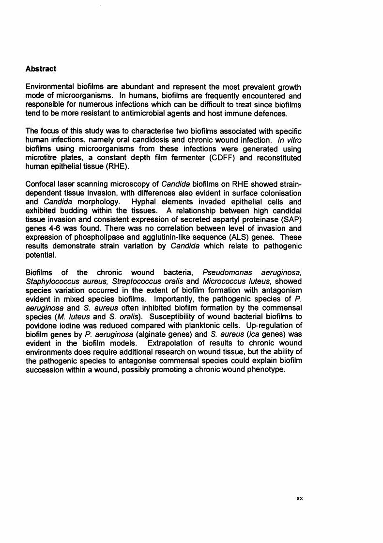
Environmental biofilms are abundant and represent the most prevalent growth mode of microorganisms. In humans, biofilms are frequently encountered and responsible for numerous infections which can be difficult to treat since biofilms tend to be more resistant to antimicrobial agents and host immune defences.
The focus of this study was to characterise two biofilms associated with specific human infections, namely oral candidosis and chronic wound infection. In vitro biofilms using microorganisms from these infections were generated using microtitre plates, a constant depth film fermenter (CDFF) and reconstituted human epithelial tissue (RHE).
Confocal laser scanning microscopy of Candida biofilms on RHE showed strain- dependent tissue invasion, with differences also evident in surface colonisation and Candida morphology. Hyphal elements invaded epithelial cells andexhibited budding within the tissues. A relationship between high candidal tissue invasion and consistent expression of secreted aspartyl proteinase (SAP) genes 4-6 was found. There was no correlation between level of invasion and expression of phospholipase and agglutinin-like sequence (ALS) genes. These results demonstrate strain variation by Candida which relate to pathogenic potential.
Biofilms of the chronic wound bacteria, Pseudomonas aeruginosa, Staphylococcus aureus, Streptococcus oralis and Micrococcus luteus, showed species variation occurred in the extent of biofilm formation with antagonism evident in mixed species biofilms. Importantly, the pathogenic species of P. aeruginosa and S. aureus often inhibited biofilm formation by the commensal species (M. luteus and S. oralis). Susceptibility of wound bacterial biofilms to povidone iodine was reduced compared with planktonic cells. Up-regulation of biofilm genes by P. aeruginosa (alginate genes) and S. aureus (ica genes) was evident in the biofilm models. Extrapolation of results to chronic wound environments does require additional research on wound tissue, but the ability of the pathogenic species to antagonise commensal species could explain biofilm succession within a wound, possibly promoting a chronic wound phenotype.
XX
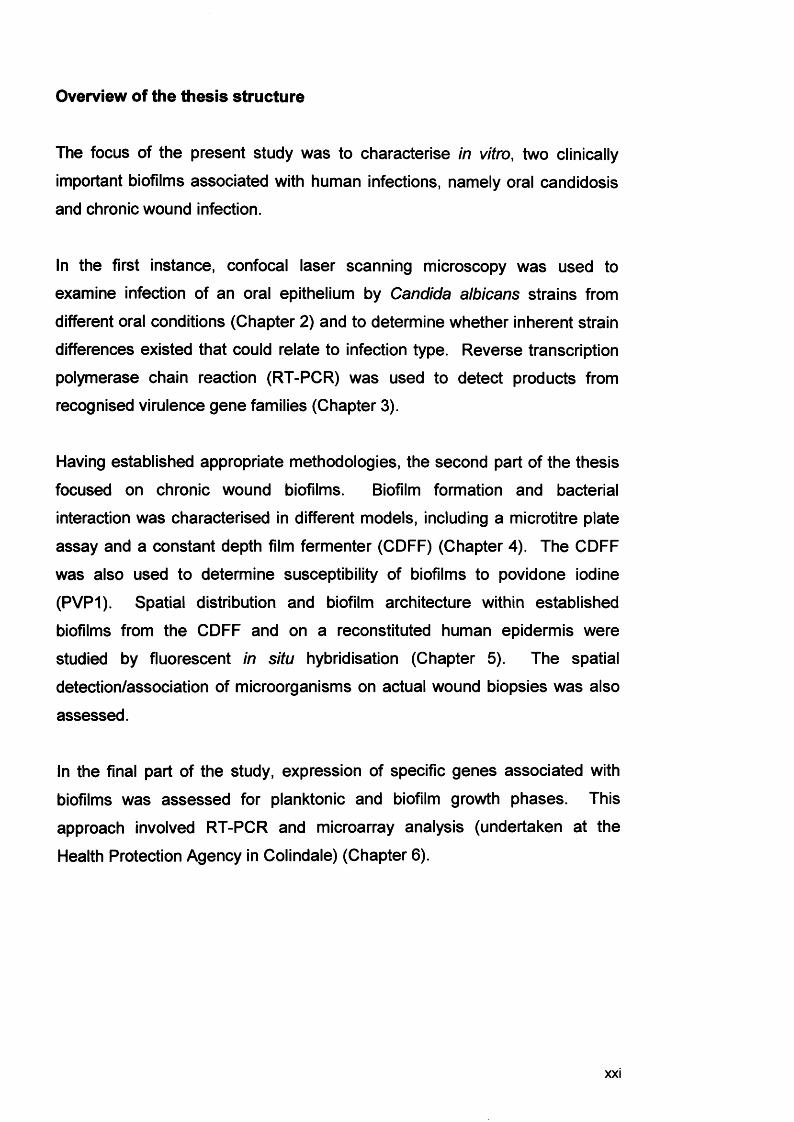
Overview of the thesis structure
The focus of the present study was to characterise in vitro, two clinically
important biofilms associated with human infections, namely oral candidosis
and chronic wound infection.
In the first instance, confocal laser scanning microscopy was used to
examine infection of an oral epithelium by Candida albicans strains from
different oral conditions (Chapter 2) and to determine whether inherent strain
differences existed that could relate to infection type. Reverse transcription
polymerase chain reaction (RT-PCR) was used to detect products from
recognised virulence gene families (Chapter 3).
Having established appropriate methodologies, the second part of the thesis
focused on chronic wound biofilms. Biofilm formation and bacterial
interaction was characterised in different models, including a microtitre plate
assay and a constant depth film fermenter (CDFF) (Chapter 4). The CDFF
was also used to determine susceptibility of biofilms to povidone iodine
(PVP1). Spatial distribution and biofilm architecture within established
biofilms from the CDFF and on a reconstituted human epidermis were
studied by fluorescent in situ hybridisation (Chapter 5). The spatial
detection/association of microorganisms on actual wound biopsies was also
assessed.
In the final part of the study, expression of specific genes associated with
biofilms was assessed for planktonic and biofilm growth phases. This
approach involved RT-PCR and microarray analysis (undertaken at the
Health Protection Agency in Col indale) (Chapter 6).
xxi

Chapter 1 Literature Review
1.1 INTRODUCTION
1.1.1 The concept of biofilm growth
Microbiology based research has in the past primarily focused on the in vitro
study of monotypic organisms grown in liquid media. The study of such ’free
growing* or planktonic organisms has undoubtedly been of value in
expanding our knowledge on the physiology and biochemistry of
microorganisms. However, in recent years, the concept of microbial growth
occurring as a biofilm rather than a planktonic growth phase has been
recognised and become the focus of much research (Percival and Bowler,
2004; Wilson, 2001).
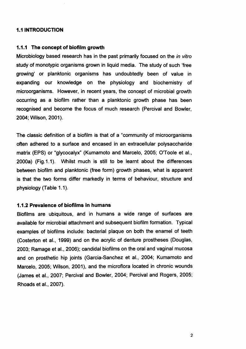
The classic definition of a biofilm is that of a “community of microorganisms
often adhered to a surface and encased in an extracellular polysaccharide
matrix (EPS) or “glycocalyx” (Kumamoto and Marcelo, 2005; O'Toole et al.,
2000a) (Fig.1.1). Whilst much is still to be learnt about the differences
between biofilm and planktonic (free form) growth phases, what is apparent
is that the two forms differ markedly in terms of behaviour, structure and
physiology (Table 1.1).
1.1.2 Prevalence of biofilms in humansBiofilms are ubiquitous, and in humans a wide range of surfaces are
available for microbial attachment and subsequent biofilm formation. Typical
examples of biofilms include: bacterial plaque on both the enamel of teeth
(Costerton et al., 1999) and on the acrylic of denture prostheses (Douglas,
2003; Ramage et al., 2006); candidal biofilms on the oral and vaginal mucosa
and on prosthetic hip joints (Garcia-Sanchez et al., 2004; Kumamoto and
Marcelo, 2005; Wilson, 2001), and the microflora located in chronic wounds
(James et al., 2007; Percival and Bowler, 2004; Percival and Rogers, 2005;
Rhoads et al., 2007).
2
Coccus;Staphyloccus aureus
Bacillus;Pseudomonasaeruginosa
Fig. 1.1 Im age of a mixed biofim generated in the CDFF, day 3. Scanning electron microscopy image (SEM ).
3
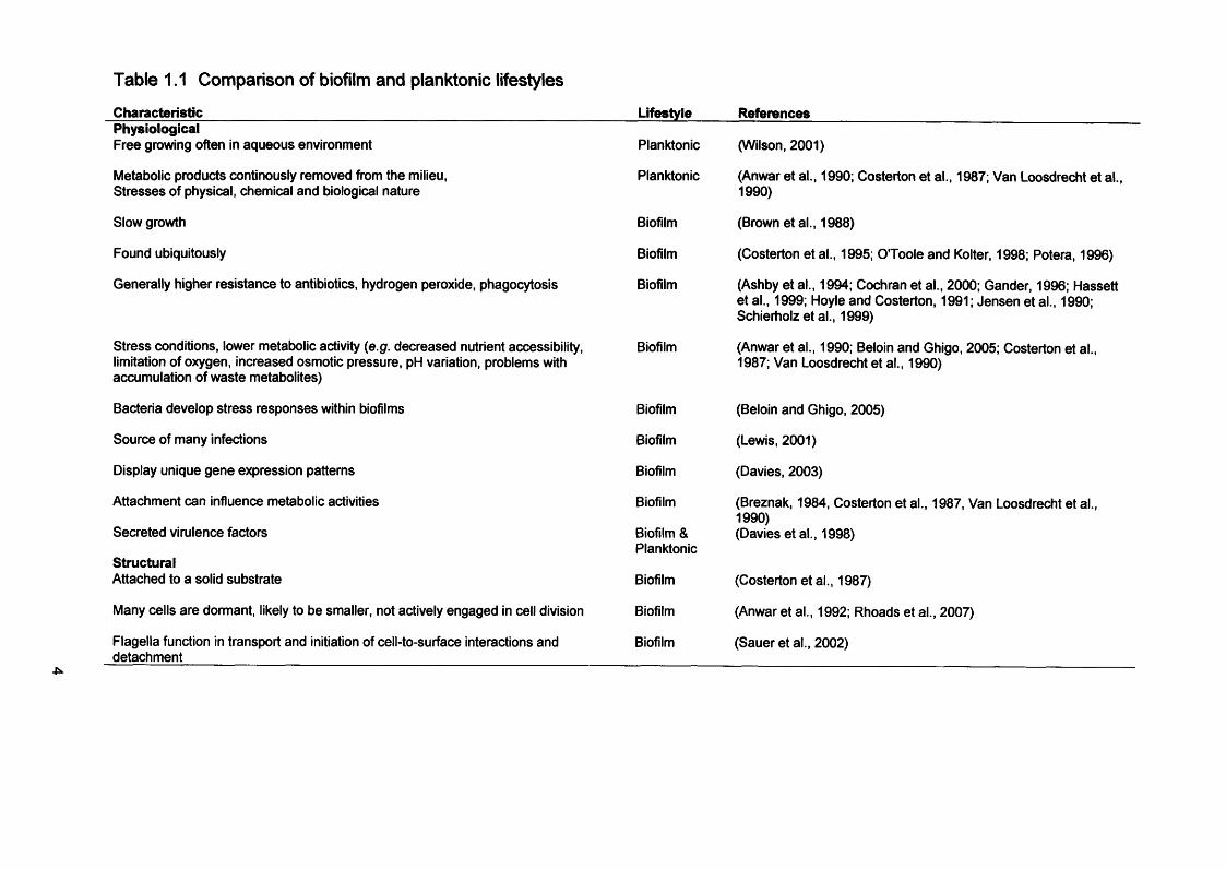
Table 1.1 Comparison of biofilm and planktonic lifestyles
Characteristic_______________________________________________PhysiologicalFree growing often in aqueous environment
Metabolic products continously removed from the milieu,Stresses of physical, chemical and biological nature
Slow growth
Found ubiquitously
Generally higher resistance to antibiotics, hydrogen peroxide, phagocytosis
Stress conditions, lower metabolic activity (e.g. decreased nutrient accessibility, limitation of oxygen, increased osmotic pressure, pH variation, problems with accumulation of waste metabolites)
Bacteria develop stress responses within biofilms
Source of many infections
Display unique gene expression patterns
Attachment can influence metabolic activities
Secreted virulence factors
StructuralAttached to a solid substrate
Many cells are dormant, likely to be smaller, not actively engaged in cell division
Flagella function in transport and initiation of cell-to-surface interactions and detachment
Lifestyle References
Planktonic
Planktonic
Biofilm
Biofilm
Biofilm
Biofilm
Biofilm
Biofilm
Biofilm
Biofilm
Biofilm & Planktonic
Biofilm
Biofilm
Biofilm
(Wilson, 2001)
(Anwar et al., 1990; Costerton et al., 1987; Van Loosdrecht et al., 1990)
(Brown et al., 1988)
(Costerton et al., 1995; O'Toole and Kolter, 1998; Potera, 1996)
(Ashby et al., 1994; Cochran et al., 2000; Gander, 1996; Hassett et al., 1999; Hoyle and Costerton, 1991; Jensen et al., 1990; Schierholz et al., 1999)
(Anwar et al., 1990; Beloin and Ghigo, 2005; Costerton et al., 1987; Van Loosdrecht et al., 1990)
(Beloin and Ghigo, 2005)
(Lewis, 2001)
(Davies, 2003)
(Breznak, 1984, Costerton et al., 1987, Van Loosdrecht et al., 1990)(Davies et al., 1998)
(Costerton etal., 1987)
(Anwar et al., 1992; Rhoads et al., 2007)
(Sauer et al., 2002)
Importantly, all these biofilms can act as reservoirs of infection and lead to
human disease. Indeed, biofilm related infections are extremely prevalent
and it has been estimated that over 65% of all hospital infections have a
biofilm origin (Costerton et al., 1999; Donlan, 2001; Donlan and Costerton,
2002; Douglas, 2003; Ramage et al., 2006). Table 1.2 outlines some
clinically significant examples of biofilms and these are discussed in more
detail later in this Literature Review (section 1.6). Of additional significance,
is that once established, biofilm originating infections can be very difficult to
combat due to the resilience of this growth phase against removal by host
defence mechanisms and antimicrobial agents. A greater understanding of
the biofilm lifestyle could enhance our knowledge of pathogenic processes
and possibly lead to novel and effective control strategies designed to
improve the management of patients with biofilm infections (Donlan, 2002).
1.2 CHARACTERISTICS OF BIOFILMS
1.2.1. Biofilm formation
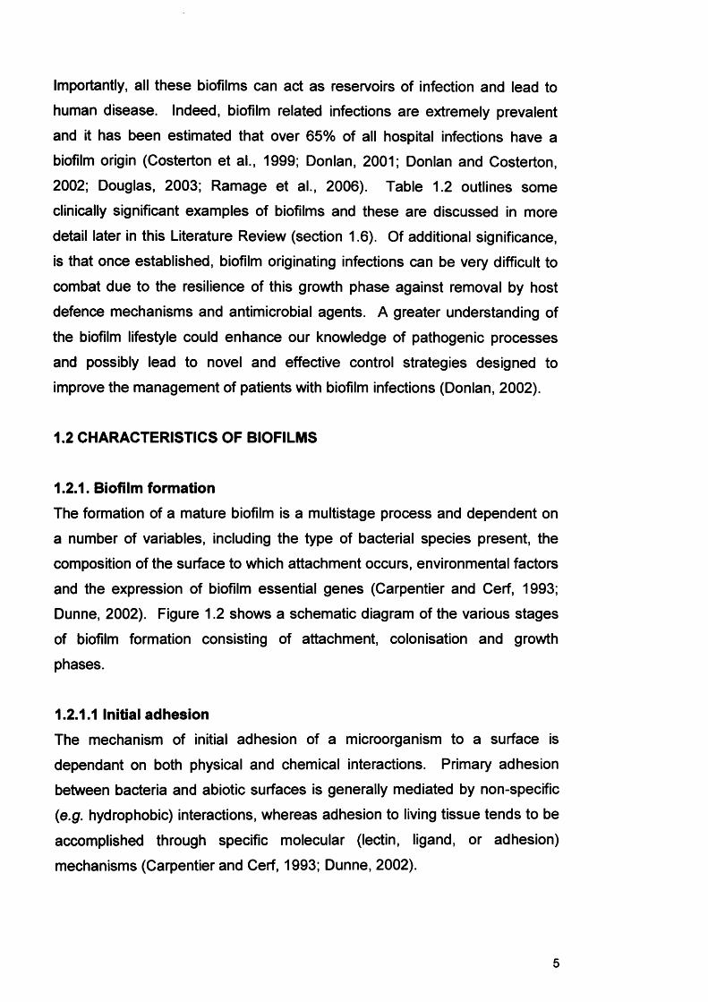
The formation of a mature biofilm is a multistage process and dependent on
a number of variables, including the type of bacterial species present, the
composition of the surface to which attachment occurs, environmental factors
and the expression of biofilm essential genes (Carpentier and Cerf, 1993;
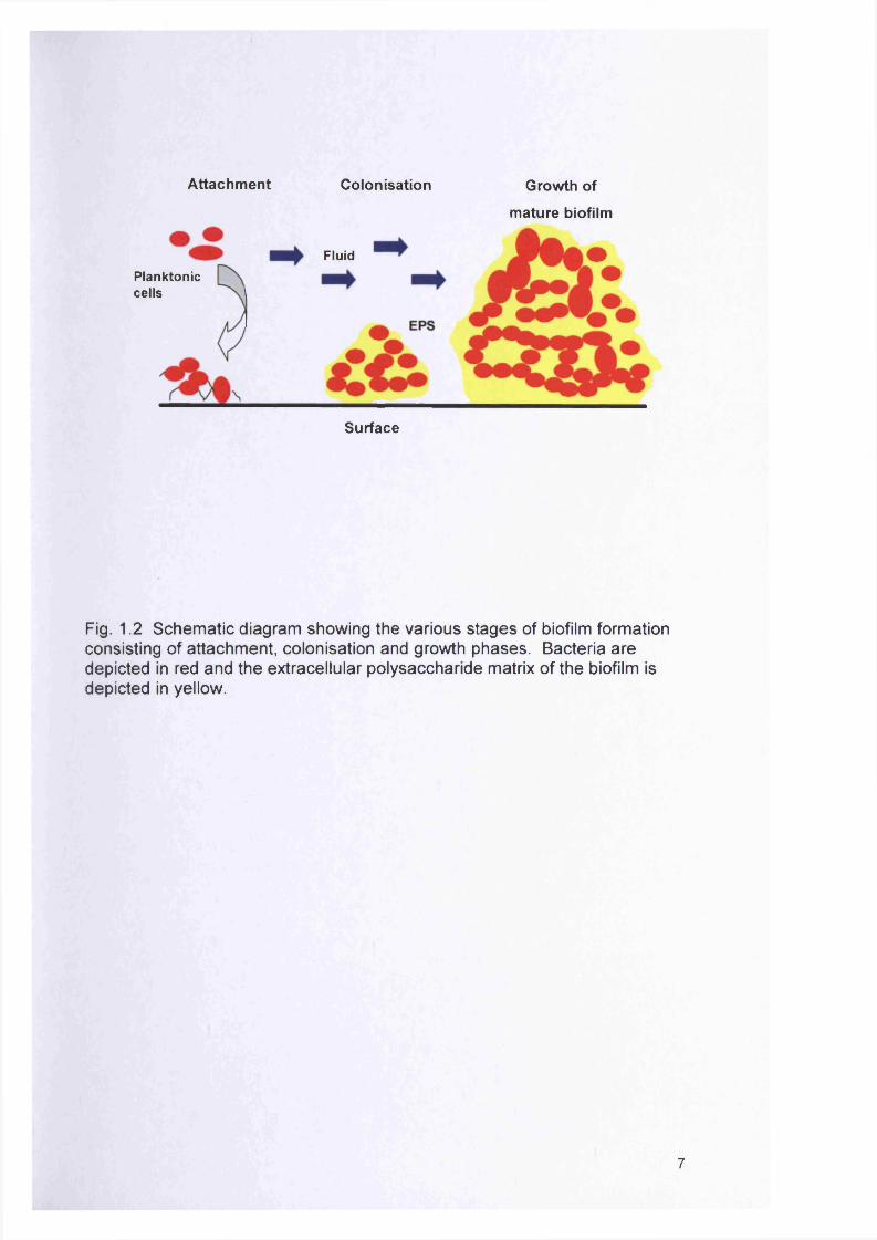
Dunne, 2002). Figure 1.2 shows a schematic diagram of the various stages
of biofilm formation consisting of attachment, colonisation and growth
phases.
1.2.1.1 Initial adhesionThe mechanism of initial adhesion of a microorganism to a surface is
dependant on both physical and chemical interactions. Primary adhesion
between bacteria and abiotic surfaces is generally mediated by non-specific
(e.g. hydrophobic) interactions, whereas adhesion to living tissue tends to be
accomplished through specific molecular (lectin, ligand, or adhesion)
mechanisms (Carpentier and Cerf, 1993; Dunne, 2002).
5
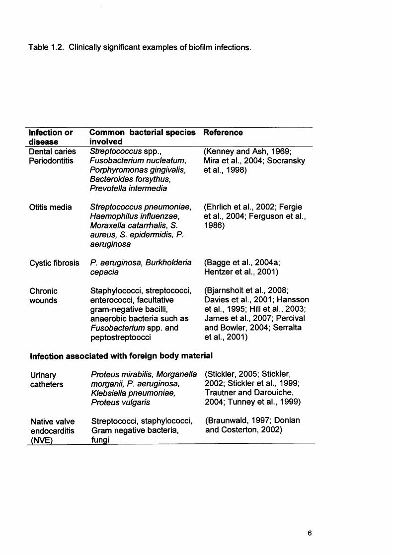
Table 1.2. Clinically significant examples of biofilm infections.
Infection or disease
Common bacterial species Reference involved
Dental caries Periodontitis
Otitis media
Streptococcus spp., Fusobacterium nucleatum, Porphyromonas gingivalis, Bacteroides forsythus, Prevotella intermedia
Streptococcus pneumoniae, Haemophilus influenzae, Moraxella catarrhalis, S. aureus, S. epidermidis, P. aeruginosa
Cystic fibrosis P. aeruginosa, Burkholderia cepacia
Chronicwounds
Staphylococci, streptococci, enterococci, facultative gram-negative bacilli, anaerobic bacteria such as Fusobacterium spp. and peptostreptoocci
(Kenney and Ash, 1969; Mira et al., 2004; Socransky etal., 1998)
(Ehrlich et al., 2002; Fergie et al., 2004; Ferguson et al., 1986)
(Bagge et al., 2004a;Hentzer et al., 2001)
(Bjarnsholt et al., 2008; Davies et al., 2001; Hansson etal., 1995; Hill et al., 2003; James et al., 2007; Percival and Bowler, 2004; Serralta et al., 2001)
Infection associated with foreign body material
Urinarycatheters
Native valve endocarditis (NVE)
Proteus mirabilis, Morganella (Stickler, 2005; Stickler,morganii, P. aeruginosa, 2002; Stickler et al., 1999;Klebsiella pneumoniae, Trautner and Darouiche,Proteus vulgaris 2004; Tunney et al., 1999)
Streptococci, staphylococci, (Braunwald, 1997; DonlanGram negative bacteria, and Costerton, 2002)fungi______________
6
Attachment Colonisation Growth of
m ature biofilm
FluidPlanktoniccells
Surface
Fig. 1.2 Schem atic diagram showing the various stages of biofilm formation consisting of attachment, colonisation and growth phases. Bacteria are depicted in red and the extracellular polysaccharide matrix of the biofilm is depicted in yellow.
7
Primary adhesion is reversible and in biological systems occurs between a
conditioned surface and a planktonic microorganism. In the case of a
biomaterial, conditioning represents the modification of the native surface
through the adsorption of water, proteins, lipids, extracellular matrix
molecules, complement, fibronectin and inorganic salts. This conditioning
film may change the affinity of an organism to attach to the surface (Dunne,
2002).
In the initial stages of the adherence process, the organism needs to be in
close proximity to the surface, either through random transportation in a
stream of fluid or in a direct way via chemotaxis and motility (Dunne, 2002).
In the case of P. aeruginosa biofilms, flagella motility is required to promote
this primary adhesion (Dunne, 2002; O'Toole and Kolter, 1998a). Once in
the vicinity of the host surface, bacterial attachment is mediated by multiple
adherence factors (known collectively as adhesins), which can overcome any
net repulsion between the two surfaces that may be present due to electrical
charges (Boland et al., 2000; Carpentier and Cerf, 1993). The final
determination of the initial adhesion depends on electrostatic and
hydrophobic interactions, van der Waals forces, temperature and the
hydrodynamic forces between the two surfaces (An et al., 2000; Carpentier
and Cerf, 1993; Dunne, 2002).
Attachment is a complex progress regulated by diverse variables. Cell
surface structures such as fimbriae, other proteins, lipopolysaccharide (LPS),
EPS and flagella all play an important role. Attachment to hydrophobic
substrata seems to be associated with cell surface polymer structures such
as fimbriae, other proteins and cell wall components of certain Gram-
negative bacteria (mycolic acids), while EPS and LPS are more important in
attachment to hydrophilic materials (Donlan, 2002).
1.2.1.2 Secondary bacterial adhesionSecondary bacterial adhesion involves covalent interaction between specific
adhesins and receptors on cell surfaces (An et al., 2000). Bacteria and host
surfaces generally express multiple adhesins and receptors respectively
8
(Table 1.3). The levels and activities of these components are modulated in
response to different environmental and external conditions, nutrient sources
available, static conditions or fluid flow (Dunne, 2002; Jenkinson et al., 2003).
Certain adhesins are regulated at the transcriptional level, permitting
organisms to switch from sessile to planktonic forms under different
environmental influences (An et al., 2000; Ziebuhr et al., 1999).
Staphylococcus epidermidis and S. aureus produce a polysaccharide
intercellular adhesion (PIA) which is associated with cell-to-ce!l adhesion and
subsequent biofilm formation (Cramton et al., 1999; Heilmann et al., 1996;
Mack et al., 1996; Mack et al., 1994). Pseudomonas aeruginosa exhibits a
surface-associated motion known as “twitching motility”, where monolayer
cells “walk” via twitching motility to form cellular aggregates on surfaces that
eventually differentiate into microcolonies (Dunne, 2002). It has been shown
that mutants deficient in type IV pili form monolayers but not cellular
aggregates (Dunne, 2002).
1.2.2 Biofilm structure
As already stated, bacteria that form biofilms exist in two forms, planktonic or
sessile. Those bacteria that are sessile attach to a surface, aggregate and
form a matrix to produce a biofilm (Costerton et al., 1999). Attached bacteria
multiply and encase their colonies with the generated ‘slimy’ matrix. The
matrix surrounding the biofilm primarily consists of exopolysaccharides often
collectively referred to as the glycocalyx.
Biofilms are heterogeneous environments and the consensus is that they
comprise of aggregates of microbial cells within the glycocalyx, with
interstitial voids and channels separating the microcolonies (Fig. 1.3a)
(Sutherland, 2001). The planktonic population exists in the liquid state
surrounding the biofilm. Importantly, sessile and planktonic bacteria of the
same species have been shown to have different phenotypes.
9
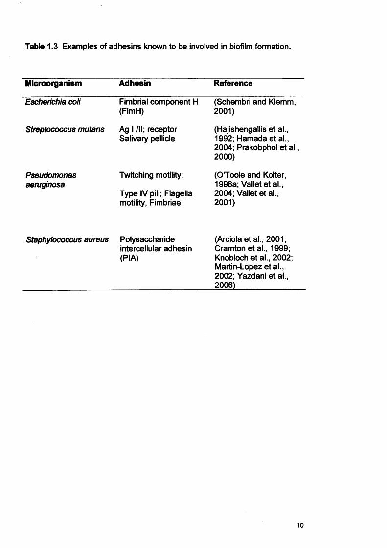
Table 1.3 Examples of adhesins known to be involved in biofilm formation.
Microorganism Adhesin Reference
Escherichia coli Fimbrial component H (FimH)
(Schembri and Klemm, 2001)
Streptococcus mutans Ag I /II; receptor Salivary pellicle
(Hajishengallisetal., 1992; Hamadaetal., 2004; Prakobphol et al., 2000)
Pseudomonasaeruginosa
Twitching motility:
Type IV pili; Flagella motility, Fimbriae
(O'Toole and Kolter, 1998a; Valletetal., 2004; Vallet et al., 2001)
Staphylococcus aureus Polysaccharide intercellular adhesin (PIA)
(Arciola et al., 2001; Cramton etal., 1999; Knobloch et al., 2002; Martin-Lopez et al., 2002; Yazdani et al., 2006)
10
1 BULK FLUID
STREAMERCELL CLUSTER
CHANNEL
>•: 19W5 CENTER FOR BIOflLM ENGINEERING MONTANA STATE UNIVERSITY BOZEMAN
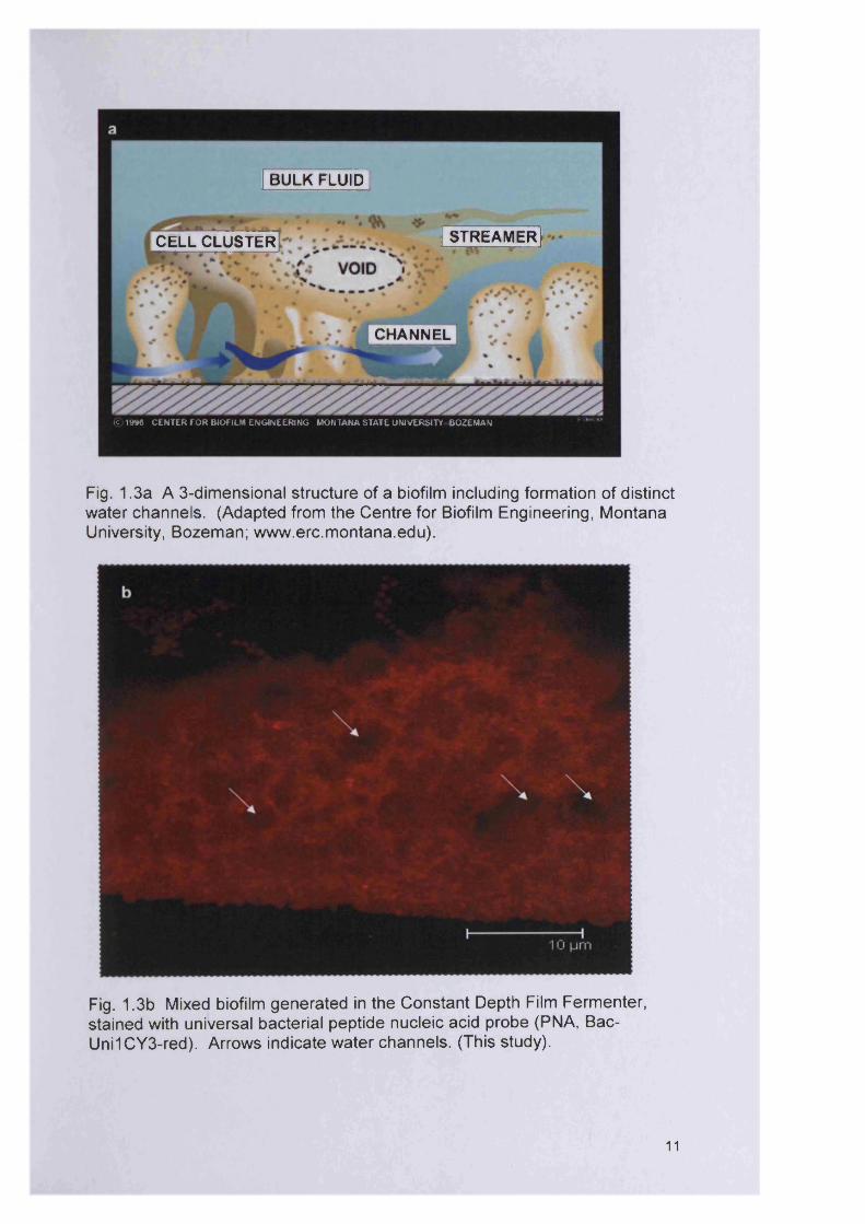
Fig. 1 .3a A 3-dimensional structure of a biofilm including formation of distinct water channels. (Adapted from the Centre for Biofilm Engineering, Montana University, Bozeman; www.erc.m ontana.edu).
Fig. 1.3b Mixed biofilm generated in the Constant Depth Film Fermenter, stained with universal bacterial peptide nucleic acid probe (PNA, Bac- Uni1CY3-red). Arrows indicate water channels. (This study).
11
Figure 1.3b shows a mixed biofilm consisting of P. aeruginosa, S. aureus,
Micrococcus luteus and Streptococcus oralis generated in the laboratory
using the constant depth film fermenter (Chapter 3) over a five day incubation
period. This image clearly depicts the water channels (spaces devoid of
bacteria) which allow diffusion of nutrients and gases. This biofilm structure
is typical of other descriptions found in the literature (Davey and O'Toole,
2000; Donlan, 2002).
1.2.3 Physiological properties of biofilms
The physiology of biofilm cells is extremely complex and profoundly different
from those organisms grown planktonically (Costerton et al., 1987). The
physiological status of biofilm cells is heterogeneous and determined by the
location of each individual cell within the multiple layers of cells that form the
biofilm (Anwar et al., 1992a). Surface biofilm cells are metabolically active
and tend to be large in size since these have easy access to nutrients and
oxygen, and have fewer problems with the discharge of metabolic waste
products (Anwar et al., 1992a). The surface-associated biofilm cells are
most likely to have properties similar to planktonically cultured cells.
Sternberg et al (1999) has shown that cells at the base of the biofilm have
reduced metabolic rates compared with cells at or near the surface, and
highlights the importance of nutrient availability as a crucial factor influencing
metabolic activity (Davies, 2003).
1.3 MICROBIAL INTERACTIONS WITHIN BIOFILMS
1.3.1 Microbial cell coaggregation
Biofilms can be comprised of single or mixed species populations. During
the adhesion process, planktonic bacteria can form aggregates, thereby
'sticking* to neighbouring cells in a process known as coaggregation (Dunne,
2002).
Coaggregation is defined as the specific cell-to-cell recognition that occurs
between both intra and inter species (Kolenbrander et al., 2006). This
12
mechanism of adhesion is highly specific and considered to have a role in the
development of multi-species biofilms in many different environments.
Coaggregation has been shown to be highly specific in dental plaque
bacteria and mediated by growth phase-dependent adhesion-receptor
interactions (Rickard et al., 2003a; Rickard et al., 2002; Rickard et al.,
2003b).
Away from the oral cavity, coaggregation has also recently been reported for
bacterial members of the urogenital flora (Reid et al., 1988), lactobacilli from
the crops of chickens (Vandevoorde et al., 1992) and in aquatic biofilm-
forming bacteria (Buswell et al., 1997; Rickard et al., 2000). The
phenomenon has been attributed with enabling the survival of anaerobic
species in an oxygenated environment by ensuring close association with
certain aerobic species (Bradshaw et al., 1998).
Coaggregation may provide additional benefits to the interacting species
beyond promoting adherence and facilitating bacterial succession (Bradshaw
et al., 1998). It seems to promote a nutritionally beneficial, communal
relationship, where metabolic cooperation results in the liberation of
additional nutrients, which may help maintain the characteristic diversity of
many biofilm communities (Rickard et al., 2003a).
1.3.2 Competitive interactions
Microorganisms in a biofilm are in close proximity to one another and
therefore interact as a consequence. These interactions can be beneficial to
one or more of the interacting populations, whilst others can be antagonistic
(James et al., 1995; Stewart et al., 1997).
Donlan (2002) suggested that P. aeruginosa is competitive in mixed biofilms
despite its slower growth rate compared with other bacteria because it is able
to rapidly colonise the surface of the substratum. Pseudomonas aeruginosa
therefore could provide the base layer of biofilms with other more rapidly
growing bacteria producing surface microcolonies (Donlan, 2002).
13
1.3.3 Synergistic interactions
It is thought that synergistic mechanisms play a role in mixed species biofilms
(Hofstad, 1992). Synergistic effects include oxygen consumption by aerobes
thereby increasing the redox potential and allowing anaerobes to proliferate,
as well as nutrient production and impairment of host immune response.
Polymicrobial interactions may well play a crucial role for biofilms in delayed
wound healing. For example, less invasive microorganisms can act
synergistically with more virulent ones (Bowler, 2003).
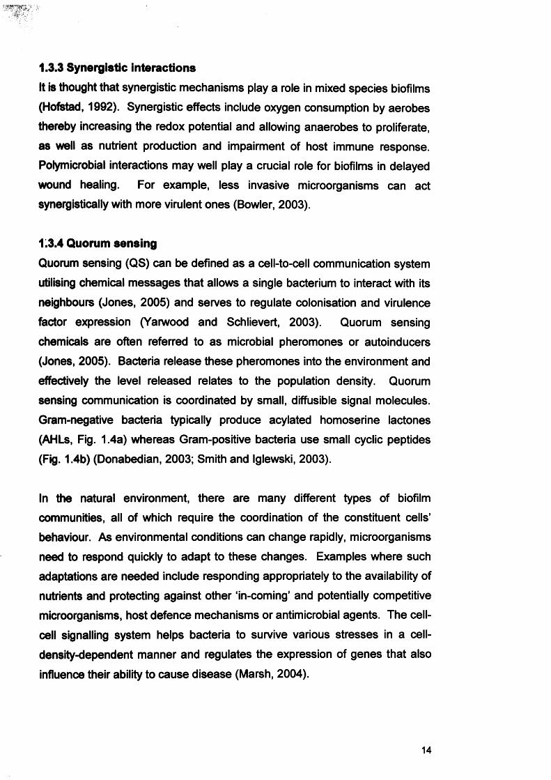
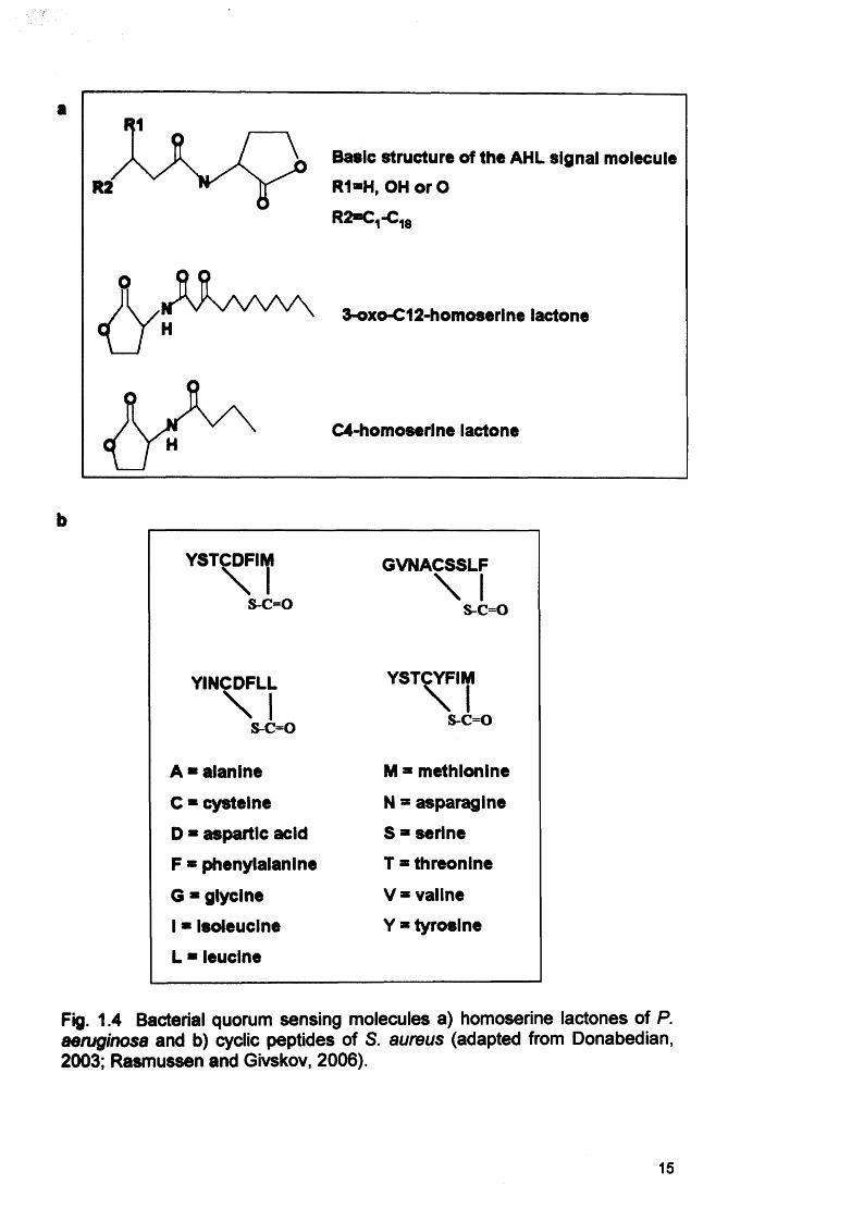
113.4 Quorum sensing
Quorum sensing (QS) can be defined as a cell-to-cell communication system
utilising chemical messages that allows a single bacterium to interact with its
neighbours (Jones, 2005) and serves to regulate colonisation and virulence
factor expression (Yarwood and Schlievert, 2003). Quorum sensing
chemicals are often referred to as microbial pheromones or autoinducers
(Jones, 2005). Bacteria release these pheromones into the environment and
effectively the level released relates to the population density. Quorum
sensing communication is coordinated by small, diffusible signal molecules.
Gram-negative bacteria typically produce acylated homoserine lactones
(AHLs, Fig. 1.4a) whereas Gram-positive bacteria use small cyclic peptides
(Fig. 1.4b) (Donabedian, 2003; Smith and Iglewski, 2003).
In the natural environment, there are many different types of biofilm
communities, all of which require the coordination of the constituent cells’
behaviour. As environmental conditions can change rapidly, microorganisms
need to respond quickly to adapt to these changes. Examples where such
adaptations are needed include responding appropriately to the availability of
nutrients and protecting against other ‘in-coming’ and potentially competitive
microorganisms, host defence mechanisms or antimicrobial agents. The cell
cell signalling system helps bacteria to survive various stresses in a cell-
density-dependent manner and regulates the expression of genes that also
influence their ability to cause disease (Marsh, 2004).
14
a
Basic structure of the AHL signal molecule
R1-H, OH orO
R2=C,-C-fiR2
3-oxo-C12-homoseri ne lactone
C4-homoserine lactone
b
YST^DFIIjl
s-c=o
GVNACSSLF\ 1s^c=o
YINCDFLL\ Is-c=o
YSTCYFIM\ 1s-c=o
A * alanine M = methionine
C ■ cysteine N a asparagine
D * aspartic acid S = serine
F * phenylalanine I = threonine
G * glycine V = valine
1 * isoleucine Y * tyrosine
L * leucine
Fig. 1.4 Bacterial quorum sensing molecules a) homoserine lactones of P. aeruginosa and b) cyclic peptides of S. aureus (adapted from Donabedian, 2003; Rasmussen and Givskov, 2006).
15
For pathogenic bacteria to cause infection, coordination of virulence factors
may be required to avoid associated host immune responses.
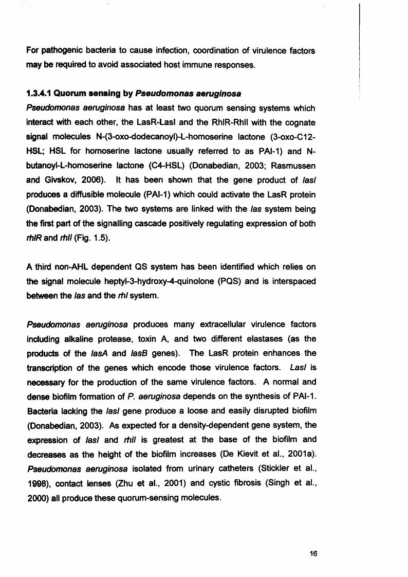
1.3.4.1 Quorum sensing by Pseudomonas aeruginosa
Pseudomonas aeruginosa has at least two quorum sensing systems which
interact with each other, the LasR-LasI and the RhIR-Rhll with the cognate
signal molecules N-(3-oxo-dodecanoyl)-L-homoserine lactone (3-oxo-C12-
HSL; HSL for homoserine lactone usually referred to as PAI-1) and N-
butanoyl-L-homoserine lactone (C4-HSL) (Donabedian, 2003; Rasmussen
and Givskov, 2006). It has been shown that the gene product of Iasi
produces a diffusible molecule (PAI-1) which could activate the LasR protein
(Donabedian, 2003). The two systems are linked with the las system being
the first part of the signalling cascade positively regulating expression of both
rhIR and rhll (Fig. 1.6).
A third non-AHL dependent QS system has been identified which relies on
the signal molecule heptyl-3-hydroxy-4-quinolone (PQS) and is interspaced
between the las and the rhl system.
Pseudomonas aeruginosa produces many extracellular virulence factors
including alkaline protease, toxin A, and two different elastases (as the
products of the lasA and lasB genes). The LasR protein enhances the
transcription of the genes which encode those virulence factors. Lasl is
necessary for the production of the same virulence factors. A normal and
dense biofilm formation of P. aeruginosa depends on the synthesis of PAI-1.
Bacteria lacking the lasl gene produce a loose and easily disrupted biofilm
(Donabedian, 2003). As expected for a density-dependent gene system, the
expression of lasl and rhll is greatest at the base of the biofilm and
decreases as the height of the biofilm increases (De Kievit et al., 2001a).
Pseudomonas aeruginosa isolated from urinary catheters (Stickler et al.,
1998), contact lenses (Zhu et al., 2001) and cystic fibrosis (Singh et al.,
2000) all produce these quorum-sensing molecules.
16
The las regulonOther regulators
I J[ /LasR Lasl —
• Exoenzymes: elastase, alkaline, proteases
• Exotoxin A Multiple gene . Biofilms * — - z regulation
• Haemolysin *• Siderophore
• Secretion apparatus (Xcp)
LasR 3-oxo-C12-HSL(PAI-1)
+
(PAI-2)
The rhl regulon
^ • Exoenzymes: elastase
Multiple gene . Lectins
regulation ̂ . Hydrogen cyanide
• Rhamnolipids• Siderophore
• Secretion apparatus (Xcp)
Fig. 1.5 Hierarchical organisation of P. aeruginosa quorum sensing system. LasR activates the expression of lasl to generate 3-oxo-C 12-H C L (also known as PAI-1). The LasR /3-oxo-C 12-H SL complex drives the expression of multiple target genes. The RhIR protein drives the expression of rhll, and hence C 4-H S L (also known as PAI-2) and the R hlR /C 4-H SL complex is responsible for controlling multiple target genes. LasR /3-oxo-C 12-H SL also regulates the expression of rhll.
17
A study using the burn mouse model showed that QS plays a significant role
in wound infections (Rasmussen and Givskov, 2006). However, if the mice
were infected with QS mutants (deletion of synthase genes in P. aeruginosa,
e.g. lasl deletion mutant, lasl rhll deletion mutant), the immune response
appeared to develop quicker, the polymorphonuclear leukocytes (PMLs)
responded with the development of stronger oxidative bursts and antibodies
accumulated faster at the infected lung site (Bjarnsholt et al., 2005; Smith et
al., 2002; Wu et al., 2001). In addition to the role of QS in the virulence and
pathogenesis of P. aeruginosa, other pathogenic organisms, including
Serratia liquefaciens and Chromobacterium violaceum (both of which are
human pathogens), and Agrobacterium tumefaciens and Erwinia carotovora
(both of which are plant pathogens) have also been shown to encode QS
signalling pathways (Rasmussen and Givskov, 2006; Sheng and Citovsky,
1996; Whitehead et al., 2002).
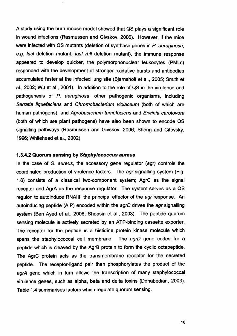
1.3.4.2 Quorum sensing by Staphylococcus aureus
In the case of S. aureus, the accessory gene regulator (agr) controls the
coordinated production of virulence factors. The agr signalling system (Fig.
1.6) consists of a classical two-component system; AgrC as the signal
receptor and AgrA as the response regulator. The system serves as a QS
regulon to autoinduce RNAIII, the principal effector of the agr response. An
autoinducing peptide (AIP) encoded within the agrD drives the agr signalling
system (Ben Ayed et al., 2006; Shopsin et al., 2003). The peptide quorum
sensing molecule is actively secreted by an ATP-binding cassette exporter.
The receptor for the peptide is a histidine protein kinase molecule which
spans the staphylococcal cell membrane. The agrD gene codes for a
peptide which is cleaved by the AgrB protein to form the cyclic octapeptide.
The AgrC protein acts as the transmembrane receptor for the secreted
peptide. The receptor-ligand pair then phosphorylates the product of the
agrA gene which in turn allows the transcription of many staphylococcal
virulence genes, such as alpha, beta and delta toxins (Donabedian, 2003).
Table 1.4 summarises factors which regulate quorum sensing.
18
|ATPA grC
QS peptide<ADPA grAAgrD
A grA
P 2 P3RNAIII
agrA \ agrC agrD r agrB
Effector RNASignal detection Signal synthesis
Regulation of target genes
Cytoplasmicmembrane
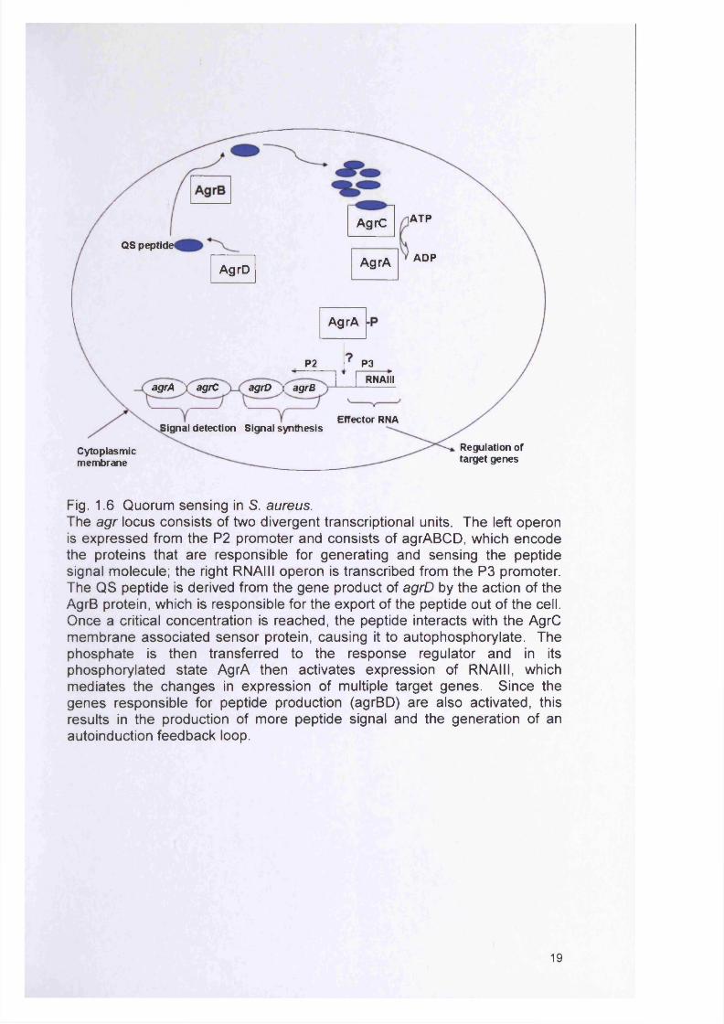
Fig. 1.6 Quorum sensing in S. aureus.The agr locus consists of two divergent transcriptional units. The left operon is expressed from the P2 promoter and consists of agrABCD, which encode the proteins that are responsible for generating and sensing the peptide signal molecule; the right RNAIII operon is transcribed from the P3 promoter. The Q S peptide is derived from the gene product of agrD by the action of the AgrB protein, which is responsible for the export of the peptide out of the cell. Once a critical concentration is reached, the peptide interacts with the AgrC m em brane associated sensor protein, causing it to autophosphorylate. The phosphate is then transferred to the response regulator and in its phosphorylated state AgrA then activates expression of RNAIII, which m ediates the changes in expression of multiple target genes. Since the genes responsible for peptide production (agrBD) are also activated, this results in the production of more peptide signal and the generation of an autoinduction feedback loop.
19
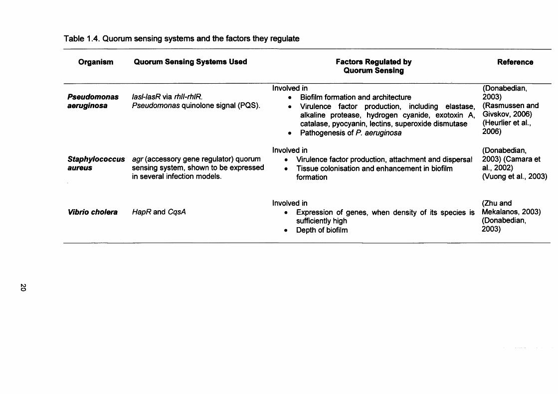
Table 1.4. Quorum sensing systems and the factors they regulate
Organism Quorum Sensing Systems Used Factors Regulated by Quorum Sensing
Reference
Pseudomonasaeruginosa
lasl-lasR via rhll-rhlR.Pseudomonas quinolone signal (PQS).
Involved in• Biofilm formation and architecture• Virulence factor production, including elastase,
alkaline protease, hydrogen cyanide, exotoxin A, catalase, pyocyanin, lectins, superoxide dismutase
• Pathogenesis of P. aeruginosa
(Donabedian,2003)(Rasmussen and Givskov, 2006) (Heurlier et al., 2006)
Staphyiococcusaureus
agr (accessory gene regulator) quorum sensing system, shown to be expressed in several infection models.
Involved in• Virulence factor production, attachment and dispersal• Tissue colonisation and enhancement in biofilm
formation
(Donabedian,2003) (Camara et al., 2002)(Vuong et al., 2003)
Vibrio cholera HapR and CqsAInvolved in
• Expression of genes, when density of its species is sufficiently high
• Depth of biofilm
(Zhu and Mekalanos, 2003) (Donabedian, 2003)
1.4 BIOFILM STABILITY
1.4.1 Resistance of biofilms against host defence mechanisms
Bacteria within a biofilm generate a protected microenvironment through the
production of an extracellular biofilm matrix. This matrix is composed of a
mixture of components, including EPS, proteins and nucleic acids. The
biofilm matrix is a complex structure encompassing of water channels that
permit transfer of nutrients and waste products (Fig. 1.3a). The most widely
studied of these components is EPS (Table 1.5). Bacterial generated EPS
would appear to be the key component in affording biofilm protection against
host defences (Davey and O'Toole, 2000). The fact that biofilm-based
infections are rarely resolved, even in individuals with competent innate and
adaptive immune responses, highlights the high degree of resistance
exhibited (Stewart and Costerton, 2001). It has been shown that biofilms are
resistant against the host cellular defence system (the phagocytic and
immune responses) (Todar, 2005) and the adaptive and innate immunity
(lysozyme, lactoferrin, Toll-like receptors) (Hiemstra, 2001).
1.4.2 Resistance of biofilms against antimicrobial agents
The nature of the biofilm structure and the physiological attributes of
microorganisms within the biofilm create an intrinsic resistance to
antimicrobial agents (antibiotics, disinfectants, germicides or antifungals).
Mechanisms responsible for resistance may be by one or more of the
following: a) reduced penetration of the antimicrobial agent into the biofilm
matrix, b) altered growth rate of biofilm organisms and c) other physiological
changes due to the biofilm mode of growth (Fig.1.7); (Donlan and Costerton,
2002).
21
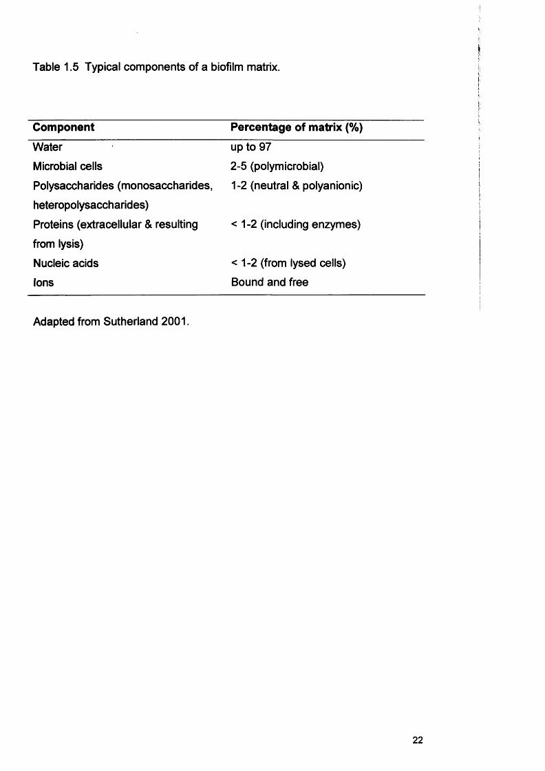
Table 1.5 Typical components of a biofilm matrix.
Component Percentage of matrix (%)
Water up to 97
Microbial cells 2-5 (polymicrobial)
Polysaccharides (monosaccharides, 1-2 (neutral & polyanionic)
heteropolysaccharides)
Proteins (extracellular & resulting <1-2 (including enzymes)
from lysis)
Nucleic acids <1-2 (from lysed cells)
Ions Bound and free
Adapted from Sutherland 2001.
22
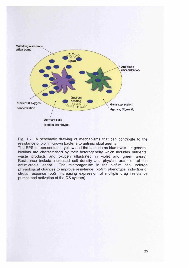
Multidrug resistanceefflux pump
Antibioticconcentration
Quorumsensing
Nutrient & oxygen
concentrationGene expression: Agr, lea, Sigma B,
Dormant cells
(biofilm phenotype)
Fig. 1.7 A schematic drawing of mechanisms that can contribute to the resistance of biofilm-grown bacteria to antimicrobial agents.The EPS is represented in yellow and the bacteria as blue ovals. In general, biofilms are characterised by their heterogeneity which includes nutrients, waste products and oxygen (illustrated in violet and green areas). Resistance include increased cell density and physical exclusion of the antimicrobial agent. The microorganism in the biofilm can undergo physiological changes to improve resistance (biofilm phenotype, induction of stress response rpoS, increasing expression of multiple drug resistance pumps and activation of the Q S system).
23
Biofilm-grown bacteria can be up to 1000 times more resistant to antibiotics
than their planktonic equivalents (Hoyle and Costerton, 1991; Mah and
O'Toole, 2001). Whilst biofilm cells themselves employ a variety of
mechanisms to resist the action of antimicrobial agents (Mah et al., 2003)
studies have also shown that a number of other factors could contribute to
the antimicrobial resistance properties of biofilms (Percival and Bowler,
2004). Some details of these mechanisms are discussed below.
1.4.2.1 Reduced penetration of an antibiotic into the biofilm matrix
Enhanced antimicrobial resistance by biofilms could relate to a reduced
ability of the antimicrobial to gain access to the microorganisms located
within the matrix of the biofilm. A reduced penetration of the agent through
the matrix could arise through chemical interaction with extracellular biofilm
components or adsorption to anionic polysaccharides. It has been shown
that P-lactamase producing organisms increase enzyme production in
response to antibiotic exposure (Bagge et al., 2004a). (3 -lactamase also
accumulates in the biofilm matrix due to secretion or cell lysis, deactivating 13-
lactam antibiotics in the surface layers more rapidly than the rate they diffuse
into the biofilm (Anderl et al., 2003). A recent study identified that biofilm
penetration of positively charged aminoglycosides is retarded through binding
to negatively charged matrices, such as alginate produced by P. aeruginosa
biofilms (Walters III et al., 2003). It has been suggested that this retardation
could provide more time for bacteria to conduct adaptive stress responses
(Fux et al., 2005). Another study showed that extracellular slime derived from
coagulase-negative staphylococci reduced the effect of glycopeptide
antibiotics, even in planktonic cultures (Fux et al., 2005; Konig et al., 2001).
In addition, Mah et al (2003) showed that bacteria within a biofilm may
actively employ distinct mechanisms such as sequestering antibiotics, such
as tobramycin, in the periplasm to prevent them from reaching their target
sites. Whilst this may occur, it is known that the biofilm matrix does not form
a completely impenetrable barrier to antimicrobial agents (Percival and
Bowler, 2004).
24
1.4.2.2 Growth rate of bacteria in biofilms
Antibiotics are generally more effective in killing actively reproducing cells.
Hence, reduced activity of microorganisms within a biofilm could render these
cells less susceptible to antimicrobial agents. It has been known for
sometime that non-dividing bacteria escape the killing effects of antibiotics
targeted against growth-specific factors (Davies, 2003). Antibiotics, such as
ampicillin and penicillin, which target cell-wall synthesis, are not able to kill
non-dividing cells (Costerton et al., 1999; Stewart and Costerton, 2001).
Whilst cephalosporins and fluoroquinolones, are cidal to non-growing cells,
they do have greater activity against rapidly growing bacteria. Therefore, the
presence of bacteria that are growing slowly in a biofilm state may contribute
to their reduced susceptibility to antibiotics (Costerton et al., 1999). The
likely location of slow-growing bacteria in a biofilm is in the lower region of
the biofilm (Davies, 2003). Cells deep within the biofilm are more likely to be
metabolically inactive, because of reduced access to essential nutrients and
gaseous exchange. Embedded biofilm cells are at the stage of dormancy
and these cells are phenotypically equipped to persist even in the most
hostile environments (Anwar et al., 1992b; Rhoads et al., 2007). These
‘persister cells’ are defined as a small and slow-growing subpopulation of the
biofilm which have differentiated into an inactive, but highly protected state
(Roberts and Stewart, 2005). It has been estimated that these
subpopulations represent 0.1-10% of all cells in the biofilm and it has been
hypothesised that they can survive an enormous antimicrobial challenge,
subsequently allowing this subpopulation to reseed the biofilm once use of
antibiotics is ceased (Harrison et al., 2005; Roberts and Stewart, 2005).
1.4.2.3 Induction of a biofilm phenotype to resist antimicrobials
Once microorganisms attach a surface they may express a general and more
virulent biofilm phenotype compared to planktonically grown identical cells
(Mah and O'Toole, 2001; Saye, 2007). Gilbert et al (1997) suggested that a
biofilm-specific phenotype may be induced in a subpopulation of the biofilm.
These subpopulations have been shown to express active mechanisms to
reduce the efficacy of antibiotics, such as the expression of bacterial
25
periplasmic glucans to bind to and physically sequester antibiotics (Gilbert et
al., 1997; Maira-Litran et al., 2000; Percival and Bowler, 2004). Once
microorganisms attach a surface some genes are turned on whilst others are
turned off (Stewart and Costerton, 2001).
It has been documented that altered gene expression by organisms within a
biofilm or a general stress response of a biofilm could reduce susceptibility to
antimicrobial agents (Brown and Barker, 1999). In theory, the bacterial cell
has a small number of target sites for antibiotics. It could therefore be that
biofilm cells use specific genes to phenotypically alter these target sites to
protect themselves (Saye, 2007). Exact details about the physiological
changes that occur during the transition from a planktonic to a biofilm state is
not known. It has been suggested that multidrug resistance efflux pumps
(MDR) may substantially contribute to the resistance of biofilms (De Kievit et
al., 2001b; Mah and O'Toole, 2001). However, studies have shown that
expression of well characterised multidrug efflux (MDR) pumps does not
increase in P. aeruginosa biofilms (De Kievit et al., 2001b).
1.5 GENE EXPRESSION ASSOCIATED WITH BIOFILM DEVELOPMENT
For the initiation of biofilm formation, microorganisms must be able to attach
to and move on surfaces, sense their cell density and generate a 3-
dimensional structure of cells encased in exopolysaccharides (O'Toole et al.,
2000a). These processes are all thought to be controlled via differential gene
expression (Table 1.6), which differs markedly from that of planktonic
microorganisms (Beloin and Ghigo, 2005). A pioneering study showed that
compared to planktonic cultures maintained in steady state, P. aeruginosa
biofilms (6 day old biofilm) differed in gene expression by 1% (Stanley et al.,
2003; Whiteleyetal., 2001).
26
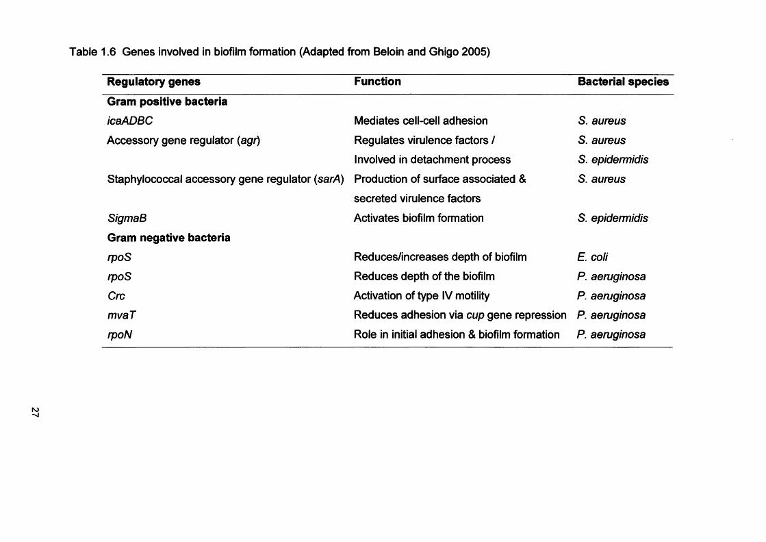
Table 1.6 Genes involved in biofilm formation (Adapted from Beloin and Ghigo 2005)
Regulatory genes Function Bacterial species
Gram positive bacteria
icaADBC Mediates cell-cell adhesion S. aureus
Accessory gene regulator (agr) Regulates virulence factors / S. aureus
Involved in detachment process S. epidermidis
Staphylococcal accessory gene regulator (sarA) Production of surface associated & S. aureus
secreted virulence factors
SigmaB Activates biofilm formation S. epidermidis
Gram negative bacteria
rpoS Reduces/increases depth of biofilm E. coli
rpoS Reduces depth of the biofilm P. aeruginosa
Cm Activation of type IV motility P. aeruginosa
mvaT Reduces adhesion via cup gene repression P. aeruginosa
rpoN Role in initial adhesion & biofilm formation P. aeruginosa
1.5.1 Gene expression in S. aureus biofilms
Staphylococcus aureus is a significant bacterial pathogen of humans
responsible for a wide variety of infections and a major source of patient
morbidity (Beenken et al., 2004; Yazdani et al., 2006). Biofilm formation by
staphylococci develops in two stages: primary attachment followed by cell
cell adhesion and accumulation (Gotz, 2002). The second phase of biofilm
formation requires the polysaccharide intercellular adhesin (PIA) (Vuong et
al., 2003).
Staphylococcus aureus adheres to surfaces through the production of a
capsular polysaccharide/adhesin (PS/A). A multilayer biofilm is formed
following S. aureus multiplication, which also results in the production of a
PIA (Beenken et al., 2004; Rohde et al., 2005; Yazdani et al., 2006). PIA is a
homoglycan consisting of p-1,6-linked polymeric N-acetylglucosamine
residues (Beenken et al., 2004; Mack et al., 1996). Control of PIA production
is dependent upon the genes within the icaADBC operon (Heilmann et al.,
1996; Yazdani et al., 2006) which is also known as a key virulence
determinant for S. epidermidis (Arciola et al., 2001; Conlon et al., 2002a;
Conlon et al., 2002b).
Whilst there are strain dependent differences (Beenken et al., 2004; Cramton
et al., 1999; Rohde et al., 2001), most S. aureus strains appear to contain the
entire ica operon (Cramton et al., 1999; Fowler et al., 2001; Rohde et al.,
2001). Vandecasteele et al. (2003) showed that ica expression was
associated with the initial colonisation of S. epidermidis in an infection model
but not with long term persistence. Frebourg et al. (2000) have indicated the
possible value of targeting the ica locus as a diagnostic marker to distinguish
between invasive and contaminating isolates of S. epidermidis. Regulation of
ica expression and the ability to form a biofim also involves other regulatory
elements. Among these additional regulatory loci are the accessory gene
regulator (agr, section 1.3.4.2) and the staphylococcal regulator (sarA)
(Beenken et al., 2004; Patti et al., 1994). It has been suggested that sarA
modulates biofilm production. Studies have confirmed that sarA mutants
28
have a reduced capacity to form a biofilm. Hence, sarA would presumably
be required for production of an activator of biofilm formation or a required
effector molecule (Beenken et al., 2004; Patti et al., 1994).
In S. aureus proteolysis also plays a pivotal role in its pathogenicity (Rohde
et al., 2005). The expression of staphylococcal proteases is under
environmental control of quorum sensing circuits like agr and sar (Lindsay
and Foster, 1999; Rohde et al., 2005), potentially linking Aap (accumulation
associated protein) processing with these global regulators of staphylococcal
virulence. The differential expression of protease activity contributes to the
adaptation of the pathogen to a changing environment by altering the
adhesive properties to extracellular matrix proteins (Karlsson et al., 2001;
McAleese et al., 2001; McGavin et al., 1997; Rohde et al., 2005; Savolainen
etal.,2001).
Staphylococcus aureus adherence is also promoted by cell wall adhesins
(Fluckiger et al., 2005). More specifically, a group of surface-exposed
proteins collectively referred to as MSCRAMMs (microbial surface
components recognising adhesive matrix molecules) interact with a number
of host proteins, such as fibronectin, fibrinogen, collagen, vitronectin and
laminin (Beenken et al., 2004; Harris et al., 2002; Patti et al., 1994).
1.5.2 Gene expression in P. aeruginosa biofilms
Pseudomonas aeruginosa is a commensal of humans and a recognised
opportunistic pathogen with great adaptability and metabolic versatility in
individuals whose defence systems are impaired (Bagge et al., 2004b;
Wilson, 2001). This organism is particularly associated with morbidity and
mortality in patients with cystic fibrosis (CF). Alginate expression by P.
aeruginosa is considered as an important virulence factor of this bacterium,
as it is thought to have a protective function against an environment that is
high in oxidative stress and against attack by immune system components
(Krieg et al., 1988; Simpson et al., 1988; Simpson et al., 1989). Possible
roles of alginate could therefore be to serve as a barrier to phagocytosis and
29
as a substance that neutralizes oxygen radicals (Edwards and Saunders,
2001). Alginate is a viscous exopolysaccharide, consisting of D-mannuronic
and L-guluronic acids (Hershberger et al., 1995). Studies have shown that P.
aeruginosa strains isolated from the lungs of CF patients, have a mucoid
phenotype and exhibit production of the exopolysaccharide alginate
(Edwards and Saunders, 2001).
Pseudomonas aeruginosa can also express genes coding for extracellular
appendages, such as flagella and type IV pili, which play a role in biofilm
formation (section 1.2.1.1), in bacterial conjugation for plasmid transfer and
other pathogenic adaptations (Finelli et al., 2003; Goodman and Lory, 2004;
O'Toole and Kolter, 1998a; Vallet et al., 2004). Flagellar motility appears to
be required for approaching surfaces and counteracting repulsive forces.
Twitching motility is mediated by type IV pili and is required for subsequent
spreading over a surface (Vallet et al., 2004). It has been shown that type IV
pili play a role in microcolony formation (O'Toole and Kolter, 1998a) and in a
recent study 3 genes from different chromosomal regions involved in type IV
pilus production (pilB, pilY2 and pilP) were up-regulated in 8h developing
biofilms (Waite et al., 2005). Using infected animal models such,
appendages are directly implicated in virulence and serve as pattern
recognition molecules for host defences (Mah et al., 2003; Van Bambeke et
al., 2003).
Biofilm formation is controlled by a number of different regulators. Vallet et
al. (2004) identified gene clusters, namely the cup (chaperone-usher
pathways), consisting of cupA, cupB and cupC, which may be involved in the
assembly of novel P. aeruginosa fimbrial structures (Croft et al., 2000; Vallet
et al., 2001). The cupA gene cluster is required for adhesion to inert surfaces
and likely to be important in the initiation of biofilm formation. The two
additional gene clusters cupB and cupC have no obvious influence on
surface attachment (Vallet et al., 2004; Vallet et al., 2001). Another
regulatory factor, Crc (catabolite repression control) protein which plays a
role in the regulation of carbon metabolism is necessary for biofilm formation
30
(O'Toole et al., 2000b). It has been suggested that Crc controls the
transcription of genes required for type IV pilus biogenesis. Therefore, Crc
may be part of a signal transduction pathway; it can sense and respond to
nutritional signals and may play a role in the organisms’ transition from
planktonic to biofilm growth. However, additional work is needed to
determine the precise mechanism by which Crc is regulated (Finelli et al.,
2003; O'Toole et al., 2000b).
Friedman and Kolter (2004) identified the pel (pellicle formation) gene cluster
and revealed its involvement in the formation of biofilms by demonstrating
that pel mutants were unable to form a robust and shear force resistant
biofilm. It was hypothesised that the pel genes are involved in the production
of a novel glucose rich polysaccharide component that contributes to the
formation of a matrix material that encases the cells and holds them together
in a resistant biofilm. Another study by Vasseur et al. (2005) showed that the
pel genes probably influence the initiation of the biofilm formation process
and the irreversible attachment on abiotic surfaces.
The stationary sigma factor rpoS (Table 1.6) is thought to play a central role
in development of starvation-mediated general resistance in Escherichia coli
(Lange and Hengge-Aronis, 1994; Xu et al., 2001). It is believed that it could
also contribute to P. aeruginosa biofilm resistance (Fig. 1.7); (Jorgensen et
al., 1999; Xu et al., 2001).
It is important to highlight that bacteria are subject to an array of stresses
within their natural environment and it has been demonstrated that stationary
phase (starving) cells survive better than their exponential phase
counterparts (Xu et al., 2001). Bacteria in a biofilm and planktonic state are
capable of turning on stress-response genes and switching to more
environmental tolerant phenotypes upon thereby resisting environments poor
in nutritional quality, high cell density, and extremes in temperature, pH or
osmolarity (Novick, 2003).
31
It is thought that the control of key biofilm gene patterns present promising
targets to characterise the biofilm mode of growth and offer excellent
opportunities to overcome antibiotic tolerance in biofilm-related infections.
To compare gene expression patterns in biofilms with those in planktonic
cultures, a multitude of strategies have been used, such as microarray
studies and real time PCR (Fux et al., 2005). Such strategies are discussed
in more detail in Chapter 6.
1.6 ROLE OF BIOFILMS IN NON ORAL INFECTION
1.6.1 Native Valve Endocarditis (NVE)
Native valve endocarditis is a condition that results from the interaction of
bacteria, the vascular endothelium and pulmonic valves of the heart. The
causes of NVE are varied, but the condition is frequently associated with
streptococci, staphylococci, Gram-negative bacteria and also fungi
(Braunwald, 1997). These microorganisms gain access to the blood and the
heart via the oropharynx, gastrointestinal and urinary tract. Once the intact
endothelium is damaged, microorganisms adhere to it and non-bacterial
thrombotic endocarditis (NBTE) develops at the injury. At the point of injury,
the thrombus develops, which is an accumulation of platelets, fibrin and red
blood cells (Donlan and Costerton, 2002). Treatment is less effective due to
a combination of mass transfer limitations and inherent resistance of biofilm
organisms (Donlan and Costerton, 2002).
1.6.2 Otitis Media
Otitis media (OM) is an infection or inflammation of the middle ear. This
inflammation often begins when infections that cause sore throats, colds, or
other respiratory or breathing problems spread to the middle ear. OM
infections can be of viral or bacterial origin and involve the inflammation of
the mucoperiosteal lining. The bacteria that cause OM include
Streptococcus pneumoniae, Haemophilus influenza, Moraxella catarrhalis,
group A p-haemolytic streptococci, enteric bacteria, S. aureus, S. epidermidis
32
and P. aeruginosa (Ferguson et al., 1986). Although most effusions from
acute OM are culture-positive for bacteria, the majority of chronic effusions
are culture-negative and resistant to antibiotic treatment (Ehrlich et al., 2002).
Tympanostomy tubes are implanted to alleviate pressure build-up and
hearing loss. Unfortunately, these too are subject to contamination and
biofilms build up (Donlan and Costerton, 2002).
1.6.3 Cystic Fibrosis
Cystic Fibrosis (CF) is a relatively common disease caused by mutations in
the gene encoding the cystic fibrosis transmembrane conductance regulator
(CFTR), a phosphorylation-regulated Cl' channel in the apical membrane of
involved epithelia (Hilman, 1997; Mathee et al., 1999; Sferra and Collins,
1993). In CF patients, the constant flow of mucus which removes debris and
bacteria is excessively thick and sticky. It is an ideal environment for
bacterial growth, resulting in frequent bacterial chest infection and
pneumonia (BUPA, 2006).
The bacterium most frequently responsible for the morbidity and mortality
seen in CF patients, is P. aeruginosa, although other bacterial species have
also been implicated e.g. Burkholderia cepacia (Bagge et al., 2004b;
Hershberger et al., 1995; Mathee et al., 1999; Stapper et al., 2004). In CF,
bacteria colonise and infect the chronic airway, which progressively destroys
the lung and often leads to respiratory failure (Mathee et al., 1999). The
colonisation of the CF airways occurs early in the lifetime of the individual
(Hoiby, 1974). The environment in the CF lungs causes P. aeruginosa to
switch to a mucoidy phenotype (Doggett, 1969; Hentzer et al., 2001; lacocca
et al., 1963). Pseudomonas aeruginosa adapts to the inflammatory defence
system of CF patients and occupies specific niches, forming mucoid biofilms
or microcolonies that reflect particular conditions.
The mucoid phenotype of P. aeruginosa is caused by an overproduction of
alginate, which is an exopolysaccharide consisting of mannuronic acid and
guluronic acid monomers (Hentzer et al., 2001). Alginate has a protective
33
function and the switch to mucoidy is also thought to promote persistence of
P. aeruginosa (Hentzer et al., 2001; Krieg et al., 1988; Simpson et al., 1988;
Simpson et al., 1989). Evidence to support the case that CF is a biofilm
disease include its innate resistance to antibiotics, demonstration of
aggregates of bacteria embedded in EPS and, also that CF sputum contains
the two principal P. aeruginosa quorum sensing signal molecules, 30C12-
HSL and C4-HSL (Fergie et al., 2004; Singh et al., 2000). It has been shown
that P. aeruginosa in CF patients also carry mutations in the mucA gene
which directs bacteria to produce alginate, which is an oxygen scavenger.
The phenotype change protects P. aeruginosa against phagocytosis and
other inflammatory host defence mechanisms (Stapper et al., 2004).
1.7 INDWELLING MEDICAL DEVICE RELATED INFECTIONS
Microbial biofilms often develop on, or within indwelling medical devices e.g.
contact lenses, central venous catheters, mechanical heart valves,
pacemakers, peritoneal dialysis catheters, prosthetic joints, urinary catheters
and voice prostheses (Table 1.7) (Donlan, 2001). There are a number of key
organisms that cause biofilms on these surfaces. Staphylococcus spp. (S.
aureus, S. epidemnidis) which are frequent members of the commensal
microflora of the skin, can form biofilms on implantable medical devices, such
as intravenous catheters, and hip and knee joint prostheses (Bayston, 1999;
Habash and Reid, 1999; Khardori and Yassien, 1995). Staphylococcus
epidermidis, S. aureus, Streptococcus spp., Escherichia coli, Proteus
mirabilis, Enterococcus faecalis, P. aeruginosa, K. pneumoniae, other Gram-
negative bacteria and Candida spp. are the primary organisms responsible
for central venous catheter, mechanical heart valve or urinary catheter
biofilms. These organisms may originate from a patient’s normal skin
microflora, from the environment, from an invasive procedure such as dental
work or other indwelling devices.
Microorganisms usually gain access to the catheter or medical device by
migration externally from the skin along the exterior catheter surface or
internally from the catheter hub or port (Maki, 1994).
34
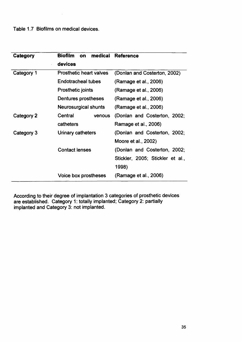
Table 1.7 Biofilms on medical devices.
Category Biofilm on medical
devices
Reference
Category 1 Prosthetic heart valves (Donlan and Costerton, 2002)
Endotracheal tubes (Ramage etal., 2006)
Prosthetic joints (Ramage et al., 2006)
Dentures prostheses (Ramage et al., 2006)
Neurosurgical shunts (Ramage et al., 2006)
Category 2 Central venous (Donlan and Costerton, 2002;
catheters Ramage et al., 2006)
Category 3 Urinary catheters (Donlan and Costerton, 2002;
Moore et al., 2002)
Contact lenses (Donlan and Costerton, 2002;
Stickler, 2005; Stickler et al.,
1998)
Voice box prostheses (Ramage et al., 2006)
According to their degree of implantation 3 categories of prosthetic devices are established. Category 1: totally implanted; Category 2: partially implanted and Category 3: not implanted.
35
Implantation of mechanical heart valves causes tissue damage, and
circulating platelets and fibrin tend to accumulate where the valve has been
attached. Once the tissue is damaged, it facilitates a greater tendency for
microorganisms to colonise these locations (Anwar et al., 1992a). The
resulting biofilms develop on the heart tissue (Carrel et al., 1998; Illingworth
et al., 1999) surrounding the prosthesis or the sewing cuff fabric used to
attach the device to the tissue (Donlan, 2001; Karchmer and Gibbons, 1994).
1.7.1 Urinary Catheters
Urinary catheters are used to remove and monitor urine production from
impaired patients. Urinary catheters also facilitate repair of the urethra after
surgical procedures and are used to manage urinary retention and
incontinence in the elderly and disabled patients (Moore and Lindsay, 2001;
Stickler, 2005). Biofilms associated with urinary catheters are particularly
important because they cause infections in 10-50% of patients who undergo
catheterisation (Mulhall et al., 1988; Stickler, 2002). Proteus mirabilis,
Morganella morganii, P. aeruginosa, K. pneumoniae and Proteus vulgaris,
are commonly found in urinary catheter biofilms. A number of these bacteria
(e.g. P. mirabilis) express urease, an enzyme which hydrolyses urea found in
the urine, resulting in the production of ammonia. Ammonia causes an
increase in the pH of the urine, which in turn allows mineral precipitation
including that of calcium and magnesium phosphates, leading to blockage of
the catheter and infection (Stickler et al., 1999; Tunney et al., 1999). The
problem is compounded because these organisms tend to be antibiotic
resistant and as a biofilm forms within the catheter, they are less susceptible
to host defences and antimicrobial treatments (Trautner and Darouiche,
2004).
Unfortunately, urinary catheters also provide a passageway for bacteria from
a heavily contaminated external skin site to a vulnerable body cavity.
Polymicrobial communities develop as time goes by, but the initial infections
are usually by single bacterial species (Stickler, 2005).
36
1.7.2 Ventilator associated pneumonia
Patients requiring airway management can develop ventilator-associated
pneumonia (VAP). VAP involves infection of the lower respiratory tract and
can cause morbidity and mortality in intensive care settings (Brennan et al.,
2004). Biofilms occur on the endotracheal tubes which are an important
factor promoting the occurrence of VAP (Iregui and Kollef, 2002).
Microorganisms associated with VAP are Gram negative organisms including
P. aeruginosa, Enterobacteriaceae, Haemophilus, and Acinetobacter spp.
Gram-positive organisms include S. aureus, Streptococcus spp.,
Streptococcus pneumoniae and methicillin resistant S. aureus (MRSA)
strains. It has been suggested that VAP associated pathogens have differing
levels of virulence, with the highest mortality associated with P. aeruginosa
and MRSA (Brennan et al., 2004).
1.7.3 Contact lens infections
Bacterial biofilms on contact lenses and storage cases may be a risk factor
for corneal infection (Dyavaiah et al., 2007; McLaughlin et al., 1998). In
several studies, such biofilms contained multiple species of bacteria and
fungi including P. aeruginosa, S. aureus, S. epidermidis, E. coli, Proteus,
Candida and Fusarium spp. (Donlan and Costerton, 2002; Dyavaiah et al.,
2007). It has been suggested that improper lens cleaning regimens are
contributing factors for corneal infections (Dyavaiah et al., 2007; Ifejika et al.,
2000).
1.7.4 Denture-based infectionsCandida associated denture stomatitis is a common and multifactorial
disease (Ramage et al., 2006), associated with poor denture fitting, denture
cleanliness, trauma, oral bacteria, immune defects and the presence of C.
albicans. Attachment and colonisation of C. albicans is supported by the
roughness of the surface and by cracks within the acrylic (Ramage et al.,
2006).
37
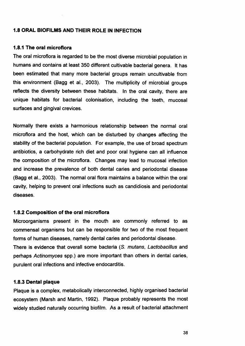
1.8 ORAL BIOFILMS AND THEIR ROLE IN INFECTION
1.8.1 The oral microflora
The oral microflora is regarded to be the most diverse microbial population in
humans and contains at least 350 different cultivable bacterial genera. It has
been estimated that many more bacterial groups remain uncultivable from
this environment (Bagg et al., 2003). The multiplicity of microbial groups
reflects the diversity between these habitats. In the oral cavity, there are
unique habitats for bacterial colonisation, including the teeth, mucosal
surfaces and gingival crevices.
Normally there exists a harmonious relationship between the normal oral
microflora and the host, which can be disturbed by changes affecting the
stability of the bacterial population. For example, the use of broad spectrum
antibiotics, a carbohydrate rich diet and poor oral hygiene can all influence
the composition of the microflora. Changes may lead to mucosal infection
and increase the prevalence of both dental caries and periodontal disease
(Bagg et al., 2003). The normal oral flora maintains a balance within the oral
cavity, helping to prevent oral infections such as candidiosis and periodontal
diseases.
1.8.2 Composition of the oral microflora
Microorganisms present in the mouth are commonly referred to as
commensal organisms but can be responsible for two of the most frequent
forms of human diseases, namely dental caries and periodontal disease.
There is evidence that overall some bacteria (S. mutans, Lactobacillus and
perhaps Actinomyces spp.) are more important than others in dental caries,
purulent oral infections and infective endocarditis.
1.8.3 Dental plaquePlaque is a complex, metabolically interconnected, highly organised bacterial
ecosystem (Marsh and Martin, 1992). Plaque probably represents the most
widely studied naturally occurring biofilm. As a result of bacterial attachment
38
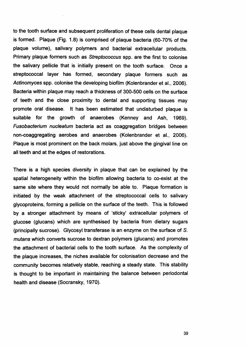
to the tooth surface and subsequent proliferation of these cells dental plaque
is formed. Plaque (Fig. 1.8) is comprised of plaque bacteria (60-70% of the
plaque volume), salivary polymers and bacterial extracellular products.
Primary plaque formers such as Streptococcus spp. are the first to colonise
the salivary pellicle that is initially present on the tooth surface. Once a
streptococcal layer has formed, secondary plaque formers such as
Actinomyces spp. colonise the developing biofilm (Kolenbrander et al., 2006).
Bacteria within plaque may reach a thickness of 300-500 cells on the surface
of teeth and the close proximity to dental and supporting tissues may
promote oral disease. It has been estimated that undisturbed plaque is
suitable for the growth of anaerobes (Kenney and Ash, 1969).
Fusobacterium nucleatum bacteria act as coaggregation bridges between
non-coaggregating aerobes and anaerobes (Kolenbrander et al., 2006).
Plaque is most prominent on the back molars, just above the gingival line on
all teeth and at the edges of restorations.
There is a high species diversity in plaque that can be explained by the
spatial heterogeneity within the biofilm allowing bacteria to co-exist at the
same site where they would not normally be able to. Plaque formation is
initiated by the weak attachment of the streptococcal cells to salivary
glycoproteins, forming a pellicle on the surface of the teeth. This is followed
by a stronger attachment by means of ‘sticky’ extracellular polymers of
glucose (glucans) which are synthesised by bacteria from dietary sugars
(principally sucrose). Glycosyl transferase is an enzyme on the surface of S.
mutans which converts sucrose to dextran polymers (glucans) and promotes
the attachment of bacterial cells to the tooth surface. As the complexity of
the plaque increases, the niches available for colonisation decrease and the
community becomes relatively stable, reaching a steady state. This stability
is thought to be important in maintaining the balance between periodontal
health and disease (Socransky, 1970).
39
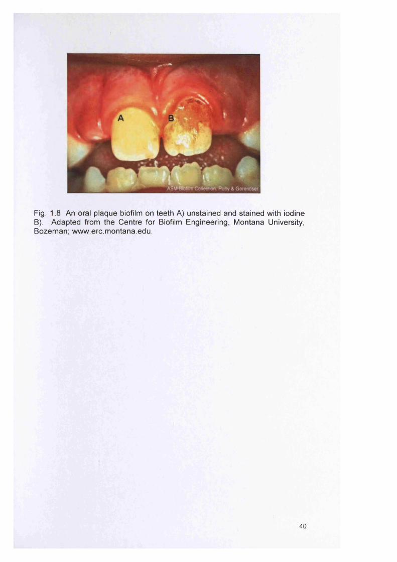
Fig. 1.8 An oral plaque biofilm on teeth A) unstained and stained with iodine B). Adapted from the Centre for Biofilm Engineering, Montana University, Bozeman; w ww.erc.m ontana.edu.
40
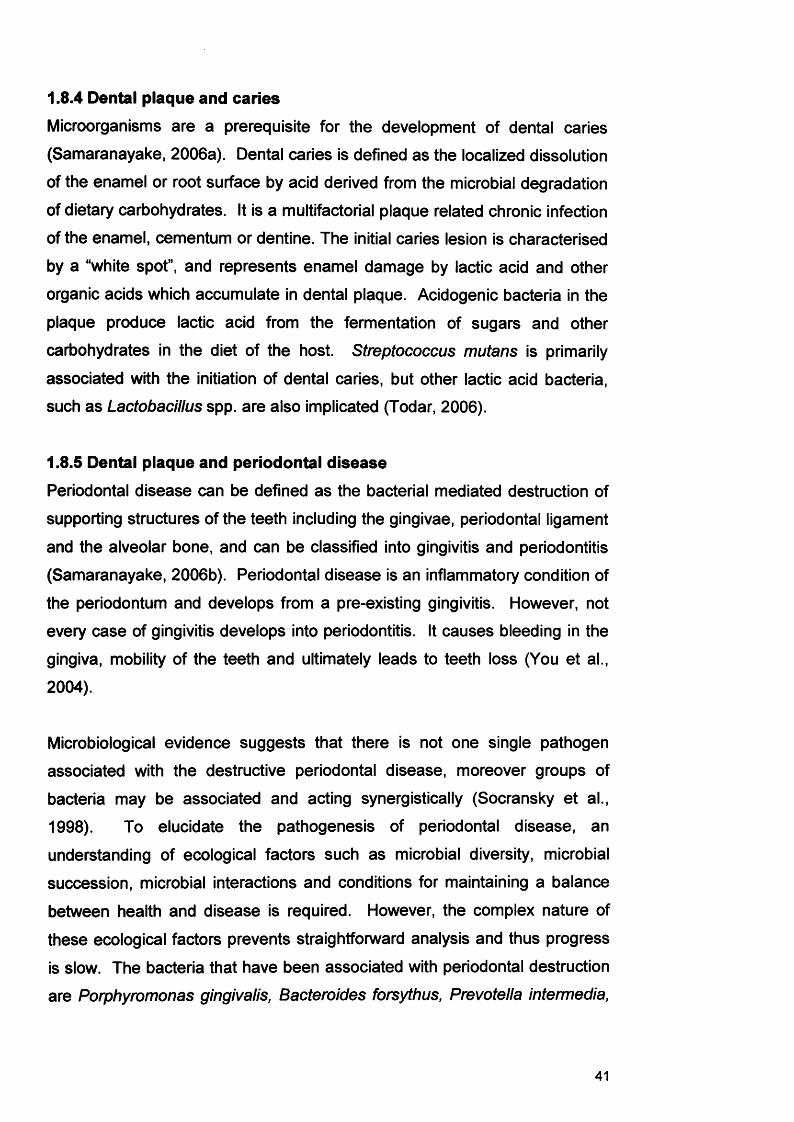
1.8.4 Dental plaque and caries
Microorganisms are a prerequisite for the development of dental caries
(Samaranayake, 2006a). Dental caries is defined as the localized dissolution
of the enamel or root surface by acid derived from the microbial degradation
of dietary carbohydrates. It is a multifactorial plaque related chronic infection
of the enamel, cementum or dentine. The initial caries lesion is characterised
by a “white spot”, and represents enamel damage by lactic acid and other
organic acids which accumulate in dental plaque. Acidogenic bacteria in the
plaque produce lactic acid from the fermentation of sugars and other
carbohydrates in the diet of the host. Streptococcus mutans is primarily
associated with the initiation of dental caries, but other lactic acid bacteria,
such as Lactobacillus spp. are also implicated (Todar, 2006).
1.8.5 Dental plaque and periodontal disease
Periodontal disease can be defined as the bacterial mediated destruction of
supporting structures of the teeth including the gingivae, periodontal ligament
and the alveolar bone, and can be classified into gingivitis and periodontitis
(Samaranayake, 2006b). Periodontal disease is an inflammatory condition of
the periodontum and develops from a pre-existing gingivitis. However, not
every case of gingivitis develops into periodontitis. It causes bleeding in the
gingiva, mobility of the teeth and ultimately leads to teeth loss (You et al.,
2004).
Microbiological evidence suggests that there is not one single pathogen
associated with the destructive periodontal disease, moreover groups of
bacteria may be associated and acting synergistically (Socransky et al.,
1998). To elucidate the pathogenesis of periodontal disease, an
understanding of ecological factors such as microbial diversity, microbial
succession, microbial interactions and conditions for maintaining a balance
between health and disease is required. However, the complex nature of
these ecological factors prevents straightforward analysis and thus progress
is slow. The bacteria that have been associated with periodontal destruction
are Porphyromonas gingivalis, Bacteroides forsythus, Prevotella intermedia,
41
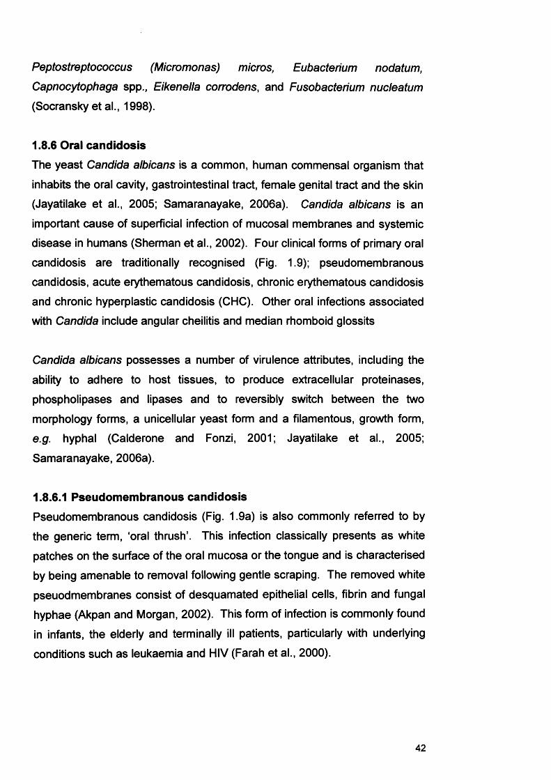
Peptostreptococcus (Micromonas) micros, Eubacterium nodatum,
Capnocytophaga spp., Eikenella corrodens, and Fusobacterium nucleatum
(Socransky et al., 1998).
1.8.6 Oral candidosis
The yeast Candida albicans is a common, human commensal organism that
inhabits the oral cavity, gastrointestinal tract, female genital tract and the skin
(Jayatilake et al., 2005; Samaranayake, 2006a). Candida albicans is an
important cause of superficial infection of mucosal membranes and systemic
disease in humans (Sherman et al., 2002). Four clinical forms of primary oral
candidosis are traditionally recognised (Fig. 1.9); pseudomembranous
candidosis, acute erythematous candidosis, chronic erythematous candidosis
and chronic hyperplastic candidosis (CHC). Other oral infections associated
with Candida include angular cheilitis and median rhomboid glossits
Candida albicans possesses a number of virulence attributes, including the
ability to adhere to host tissues, to produce extracellular proteinases,
phospholipases and lipases and to reversibly switch between the two
morphology forms, a unicellular yeast form and a filamentous, growth form,
e.g. hyphal (Calderone and Fonzi, 2001; Jayatilake et al., 2005;
Samaranayake, 2006a).
1.8.6.1 Pseudomembranous candidosisPseudomembranous candidosis (Fig. 1.9a) is also commonly referred to by
the generic term, ‘oral thrush’. This infection classically presents as white
patches on the surface of the oral mucosa or the tongue and is characterised
by being amenable to removal following gentle scraping. The removed white
pseuodmembranes consist of desquamated epithelial cells, fibrin and fungal
hyphae (Akpan and Morgan, 2002). This form of infection is commonly found
in infants, the elderly and terminally ill patients, particularly with underlying
conditions such as leukaemia and HIV (Farah et al., 2000).
42
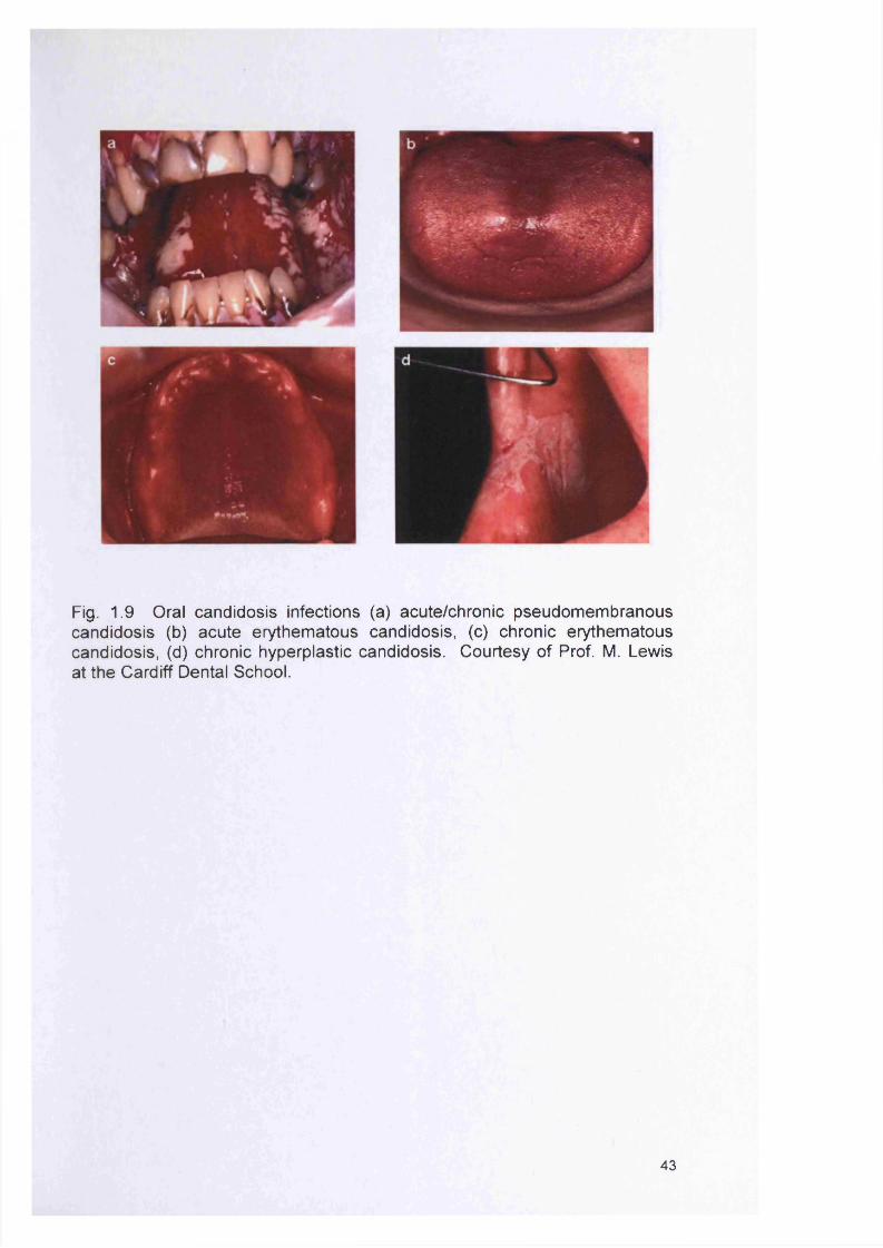
Fig. 1.9 Oral candidosis infections (a) acute/chronic pseudomembranous candidosis (b) acute erythematous candidosis, (c) chronic erythematous candidosis, (d) chronic hyperplastic candidosis. Courtesy of Prof. M. Lewis at the Cardiff Dental School.
43
1.8.6.2 Acute erythematous candidosis (acute atrophic candidosis)
Acute erythematous candidosis (Fig. 1.9b) is associated with the use of
broad spectrum antibiotics or steroid therapy. Clinically it presents as a
reddening of the oral mucosa similar to that seen in patients with low serum
levels of vitamin B12, low folate and low ferritin (Akpan and Morgan, 2002).
It is believed that the antimicrobial therapy alters the normal oral bacterial
community and allows C. albicans numbers to increase (Farah et al., 2000).
1.8.6.3 Chronic erythematous candidosis (chronic atrophic candidosis)
Chronic erythematous candidosis (Fig. 1.9c) is associated with the wearing of
denture prostheses and results in mucosal edema of the palate. In this
infection, other factors such as bacterial accumulation reduced salivary
protection and mechanical irritation may be implicated. A poor fitting denture
and poor oral hygiene is associated with this type of infection (Akpan and
Morgan, 2002). The patient usually does not have any symptoms but may
complain of slight soreness (Farah et al., 2000). It presents in approximately
50% of all denture wearers (Farah et al., 2000).
1.8.6.4 Chronic hyperplastic candidosis (CHC)
CHC (Fig. 1.9d) presents as white lesions on the oral mucosa. It is distinct
from the other forms of oral candidoses as it is characterised by hyphal
penetration of the oral epithelium (Sitheeque and Samaranayake, 2003) and
the presence of an associated inflammatory cell infiltrate (Williams et al.,
1997).
Importantly, CHC is also implicated in the development of squamous cell
carcinoma (SCC) at the lesional site (Banoczy, 1977; Krogh et al., 1987;
Williams et al., 2001). The host factors implicated in CHC include tobacco
smoking, denture wearing, and regular alcohol intake (Arendorf et al., 1983).
However, it is also possible that Candida express specific virulence attributes
that promote this infection. Since hyphal invasion of the epithelium is a
distinguishing feature of CHC, expression of certain C. albicans virulence
factors may be enhanced for CHC strains compared with others.
44
1.8.6.5 Angular cheilitis
Angular cheilitis is a clinical term used to describe painful erythematous
lesions affecting the angles of the mouth. The condition seems to be
associated with the wearing of removable dentures (Samaranayake and
MacFarlane, 1990a), and is often accompanied by the presence of chronic
erythematous candidosis (Farah et al., 2000). Other microorganisms are
frequently present in this infection, such as Staphylococcus aureus and
Streptococcus spp. (Akpan and Morgan, 2002).
1.8.6.6 Median rhomboid glossits
Median rhomboid glossits presents on the tongue as a chronic symmetrical
shaped area consisting of atrophic filiform papillae. Recovery of Candida
from this area is high. The exact cause of this infection is not known,
although hormonal links, tobacco smoking and the use of inhaled steroids
have been suggested (Samaranayake and MacFarlane, 1990b).
1.8.7 Predisposing host factors to oral candidosis
Oral candidosis has frequently been described as a ‘disease of the diseased’.
Whilst the incidence of normal commensal carriage of Candida in the mouth
is high, such individuals will not suffer infection unless otherwise debilitated.
The transition of Candida from a harmless commensal member of the
microflora to a disease causing organism is complex, involving a variety of
both local and systemic host factors. Several of these factors are discussed
below, and presented in Table 1.8.
1.8.7.1 Damage to the oral mucosalIn the mouth the oral mucosa provides a primary host defence mechanism by
acting as a mechanical barrier to Candida. However, damage to the oral
mucosa can occur through denture wearing, which has an association with
chronic erythematous candidosis and CHC (Arendorf et al., 1983). It has
also been shown that the wearing of dentures produces an anaerobic
microenvironment beneficial to the growth of Candida due to its associated
low oxygen levels and low pH (Ruby and Barbeau, 2002).
45
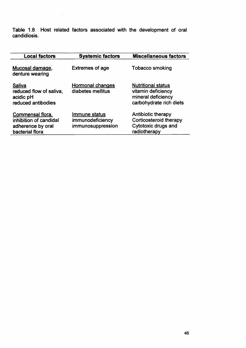
Table 1.8 Host related factors associated with the development of oral candidiosis.
Local factors Systemic factors Miscellaneous factors
Mucosal damaae! Extremes of age Tobacco smokingdenture wearing
Saliva Hormonal chanaes Nutritional statusreduced flow of saliva, diabetes mellitus vitamin deficiencyacidic pH mineral deficiencyreduced antibodies carbohydrate rich diets
Commensal flora. Immune status Antibiotic therapyinhibition of candidal immunodeficiency Corticosteroid therapyadherence by oral immunosuppression Cytotoxic drugs andbacterial flora radiotherapy
46
Furthermore oral hygiene levels in many denture wearers can be poor, due to
inadequate cleaning regimes, particularly for individuals who wear their
dentures through the night (Eppstein, 1990; Guida, 1988).
1.8.7.2 Effect of saliva
The constant flushing action of saliva can remove Candida cells prior to their
attachment to oral surfaces. Furthermore, antimicrobial proteins in saliva
such as lactoferrin, sialoperoxidase, lysozyme, histidine-rich polypeptides,
and specific anti-Candida antibodies, can contribute to the killing and removal
of organisms from the mucosa and prevent overgrowth of Candida.
Therefore, drugs that reduce salivary secretions or radiotherapy of the head
and neck can lead to an increased risk of oral candidosis (Akpan and
Morgan, 2002). Sjogren’s syndrome is one of the most common systemic
conditions associated with xerostomia and salivary dysfunction (Turner and
Ship, 2007). It has been shown that this condition favours increased oral
carriage of Candida (Sitheeque and Samaranayake, 2003). Colonisation of
the denture acrylic surface by Candida is also favoured by the relatively low
saliva flow under the surfaces of the denture fittings which limits removal of
otherwise poorly attached yeast (Eppstein, 1990; Guida, 1988).
1.8.7.3 Commensal microfloraIt is a well-documented problem that patients receiving broad spectrum
antibiotics are highly likely to develop oral candidosis (Farah et al., 2000). A
reduction in the bacterial component of the oral microflora reduces the
competition for nutrients and adherence sites. Generally, the infection acute
erythematous candidosis is the form of oral candidosis that follows (section
1.8.6 .2).
1.8.7.4 Diabetes mellitusDiabetes mellitus has been associated with the occurrence of high levels of
candidal colonisation and oral candidosis (Manfredi et al., 2002; Sitheeque
and Samaranayake, 2003). Several reasons for the high oral candidal
carriage in diabetics have been suggested. Firstly, high salivary glucose
47
levels in diabetics may favour the growth of yeasts (Sitheeque and
Samaranayake, 2003). Furthermore, adherence of Candida to buccal
epithelial cells (BEC) from diabetics has been demonstrated to be greater
than to cells originating from non-diabetic controls (Sitheeque and
Samaranayake, 2003). This could be due to intrinsic qualitative changes in
the cell surface receptors of diabetics that modulate yeast adhesion
(Sitheeque and Samaranayake, 2003).
1.8.7.5 Nutritional status
Oral candidosis is also associated with mineral and vitamin deficiencies, and
carbohydrate rich diets of infected patients (Sitheeque and Samaranayake,
2003). On the basis of animal models, deficiencies in vitamins A, B1 and B2
are generally implicated with predisposition to oral candidosis (Akpan and
Morgan, 2002). Iron deficiency can cause epithelial abnormalities such as
hyperkeratonisis and atrophy, through alterations in the kinetics of the rapidly
dividing cells of the oral mucosa, which in turn, result from an impairment of
iron-dependent enzymes (Sitheeque and Samaranayake, 2003). Iron-
deficiency could also influence oral carriage of Candida through the
impairment of iron-dependent host enzymes, depression of host cell-
mediated immunity (CMI) or by reducing antibody production (Sitheeque and
Samaranayake, 2003).
1.9 CHRONIC WOUNDS AND THE ROLE OF BACTERIA
Chronic wounds are defined as those wounds that do not heal within an
expected time frame. These chronic wounds are a serious health concern
with substantial morbidity and are a source of frustration to both physician
and patient due to lengthy treatments, treatment failures and long periods of
patient compliance.
Chronic wounds include chronic venous leg ulcers (CVLUs; Fig. 1.10),
diabetic foot ulcers and pressure sores. CVLUs are non-healing wounds that
occur due to a breakdown in the underlying physiology of the leg.
48
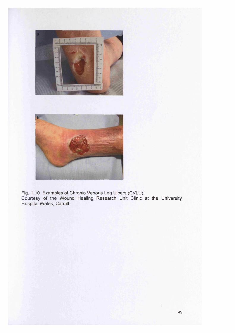
Fig. 1.10 Examples of Chronic Venous Leg Ulcers (CVLU).Courtesy of the Wound Healing Research Unit Clinic at the University Hospital W ales, Cardiff.
49
762494
The breakdown is associated with venous, arterial or metabolic factors
(Bowler et al., 2001; Howell-Jones et al., 2005; Thomas and Harding, 2002).
Treatment costs are almost £1 billion each year to the NHS, which will only
increase with the increasingly aged population of the UK. Chronic wounds
are characterised by oedema, severe pain, erythema and strong odour and
affect more than 1% of the UK population (Thomas and Harding, 2002).
These wounds tend to occur in individuals with an increased risk of bacterial
invasion as a result of poor local factors (arterial insufficiency, venous
hypertension, trauma) or systemic disease such as diabetes mellitus and
rheumatoid arthritis (Falanga, 1993).
1.9.1 Course of normal wound healing
In healthy individuals the normal wound healing process involves a complex
series of events including cell proliferation, migration, differentiation and
remodelling that can be summarised into overlapping phases. However in
chronic wounds, factors such as systemic illness, poor nutrition, vascular
insufficiency and bacterial interference influence the normal wound healing
process (Jones et al., 2004; Rhoads et al., 2007). The time of healing in
chronic wounds is extended as shown in Fig. 1.11. The inflammatory phase
begins at time of injury and serves to cleanse the wound of bacteria and
debris; this phase can take up to 25 days.
The exposed extracellular matrix triggers the coagulation cascade,
plasminogen cascade and others. These cascades along with the exposed
extracellular matrix and microorganisms that find their way back into the
wound; stimulate the innate immune system. Polymorphonuclear cells arrive
first with preprogrammed apoptosis. Macrophages follow and finalise the
cleanup of pathogens, remove devitalized tissue and clean host agents such
as polymorphonuclear cells by phagocytosis and down-regulate acute
inflammatory signals (Rhoads et al., 2007; Schultz et al., 2004). In the
proliferative phase, a process of deposition between angiogenesis,
fibroblasts, fibronectin and collagen takes place and can take up to 25 days
to complete.
50
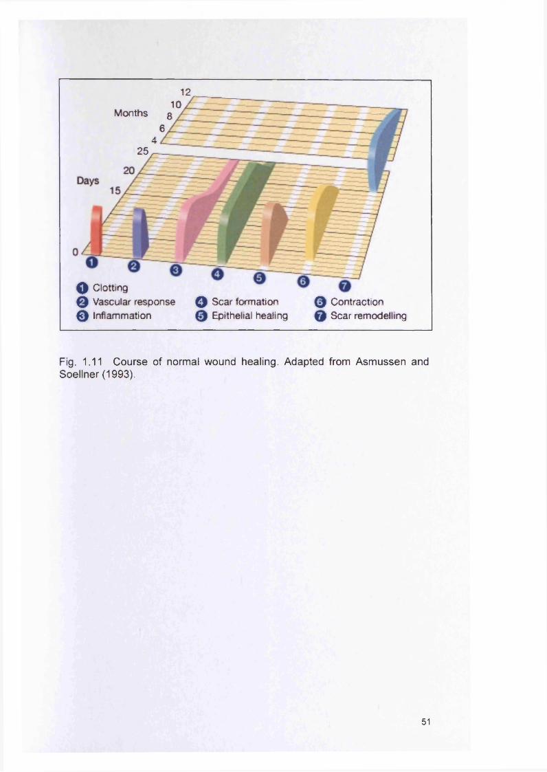
12 10
Months 3 6
425
0 Contraction 0 Scar remodelling
o Clotting 0 Vascular response 0 Inflammation
Q Scar formation 0 Epithelial healing
Fig. 1.11 Course of normal wound healing. Adapted from Asmussen and Soellner (1993).
51
Cytokines are controlling factors for granulation tissue formation. These
small molecules direct the endothelial cell neovascularisation of the wound
bed and also direct fibroblasts to deposit the new extracellular matrix
materials. The remodelling phase which succeeds the proliferation phase,
can take up to 12 months (Rhoads et al., 2007).
1.9.2 The significance of bacteria in chronic wounds
Open skin wounds are often colonised with microorganisms, having lost
many of the protective defence mechanisms of intact skin (Edwards and
Harding, 2004). Chronic wounds harbour a diverse, often dense and complex
polymicrobial microflora of potentially pathogenic organisms (both aerobic
and anaerobic species). The wound environment provides support for
bacterial survival as it is both moist and warm plus there is a good nutrient
supply from exuded wound fluid. As a result wounds are susceptible to
microbial contamination from both exogenous and endogenous sources
including the nose, skin, mouth and the gut (Percival and Bowler, 2004).
There is evidence that microorganisms in chronic wound biofilms contribute
to the non-healing phenotype (Hill et al., 2003). Cultural analyses have
documented that pseudomonads, staphylococci (present in >70% of wounds)
and anaerobic species such as Peptostreptococcus spp. are the bacterial
groups most frequently recovered from chronic wounds (Hansson et al.,
1995; Hill et al., 2003). Polymicrobial interactions may play a crucial role in
delayed wound healing. Moreover, less invasive microorganisms may act
synergistically with more virulent ones (Bowler, 2003). However, although
significant in inhibiting the wound healing process, the importance of
individual species or microbial density in relation to healing is unclear.
Nevertheless, bacteria play an important role in wounds and possess indirect
and direct mechanisms to impair wound healing (Stephens et al., 2003).
It has been shown that all wounds are colonised by bacteria, although not all
wounds are infected (Bowler, 2003). One of the most difficult clinical
judgements facing the clinician is determining whether a wound is purely
52
colonised by bacteria or whether there are signs of infection. However, often
the lines between colonisation and the beginnings of infection are blurred and
correct diagnosis is wholly dependent upon the experience and skill of the
clinician.
To reduce the bioburden (the bacterial load in chronic wounds) and improve
the wound healing process, the use of antimicrobial therapies are necessary.
It is important to note that many patients with chronic wounds are often
elderly and hence may also have underlying conditions which make them
immunocompromised. Howell-Jones et al. (2006; 2005) showed that over
prescribing of antibiotics seems typical in this particular group of patients.
Therefore, an understanding of the physiology and interactions within multi
species biofilms may aid the development of more effective methods of
treating infected and poorly healing wounds (Edwards and Harding, 2004).
1.9.3 Biofilms and wounds
There is now evidence to show that bacteria are able to form biofilms within
the wound environment (James et al., 2007; Mertz, 2003; Percival and
Bowler, 2004). It has recently been suggested that both acute partial
thickness wounds (Serralta et al., 2001) and chronic human wounds (Bello et
al., 2001) may harbour bacterial biofilms growing on the wound surface. In
acute wounds, a study involving a pig model inoculated with a laboratory
strain of P. aeruginosa showed that two distinct bacterial populations were
present in the wound (Serralta et al., 2001). The first population was loosely
associated with the wound and was readily washed off. In contrast, the
second population was more firmly anchored and could only be removed by
significant scrubbing with detergent. The authors suggest that the loosely
associated wound population were planktonically growing cells, whereas
those that could only be removed by mechanical and chemical means were
biofilm associated.
A second study showed that in vitro biofilms could easily be grown using
wound isolates. For example, P. aeruginosa from a human burn wound
53
formed mature biofilms within 10h as visualised by light microscopy
(Harrison-Balestra et al., 2003). Scanning electron micrograph (SEM)
images of S. aureus biofilms in acute wounds have also been produced
(Welsh et al., 2005). In contrast, fewer studies have been done with chronic
wounds. EPS has been visualised by epifluoresence and light microscopy on
chronic wound smears (Bello et al., 2001; Ricotti et al., 2003) and a biofilm
has been demonstrated on a dermal model (Mertz, 2003). Moreover,
Akiyama et al. (2002) demonstrated that S. aureus can assemble a
glycocalyx on the surface of traumatic wounds in mice and that the biofilm
could be visualised using confocal laser scanning microscopy.
From the above studies it is clear that bacteria can persist within a wound
and establish a biofilm community which can overcome the host’s natural
defences and antimicrobial therapy. Whilst the impaired immune system
tries to prepare the wound for proliferation of healing, the body cannot
successfully complete the inflammatory phase. Therefore, the wound
remains in the inflammatory phase, unless successful therapeutic strategies
can be applied to eliminate the infection (Rhoads et al., 2007; Schultz et al.,
2004).
1.10 MANAGEMENT OF BIOFILM INFECTIONS
Viewing bacteria from the perspective of multicellular behaviour is altering
our view of microbiology and of Koch’s postulates. It is evident that 99.9% of
organisms prefer attachment, and that bacterial cells have the ability to
aggregate into particular three-dimensional assemblages (Davey and
O'Toole, 2000). Biofilms have been recognised as being important in human
disease and the number of biofilm-associated diseases seems to be
increasing (Davies, 2003). It is important to understand the characteristics of
the biofilm mode of growth and the various aspects of biofilm formation. To
successfully treat biofilm infections, knowledge of the phenotype of the
bacterial population is required. This is important as antibiotic treatments
may not be totally effective if more than one phenotype exists. Some cells
might, remain intact which are able to re-colonise a wound and debilitate the
54
host once the antimicrobial treatment has finished (Brooun et al., 2000;
Davies, 2003; Spoering and Lewis, 2001).
A key factor to combating biofilm infections is to understand the physiology of
biofilm development. Davies (2003) suggested that chemotherapeutic
agents could be developed to promote or prevent transition from one stage of
biofilm maturation to the next by targeting unique biofilm regulatory or
signalling molecules. Specific agents might be discovered or developed
which will interfere with the production of virulence factors, or promote or
inhibit the shedding of biofilm bacteria (Davies, 2003).
As mentioned before (section 1.4.2), biofilm resistance depends on
aggregation of bacteria into multicellular communities. Therefore, one
antimicrobial strategy might be to develop therapies to disrupt the
multicellular structure of the biofilm. It could be that host defences might be
able to resolve the infection once the multicellularity of the biofilm is reduced,
and then the effectiveness of antibiotics might be restored (Stewart and
Costerton, 2001). Other potential therapies include enzymes that dissolve
the matrix polymers of the biofilm, chemical reactions that block biofilm matrix
synthesis, and analogues of microbial signalling molecules that interfere with
cell-to-cell communication, required for normal biofilm formation (Nemoto et
al., 2000; Parsek and Greenberg, 2000; Yasuda et al., 1993). Already, a
number of QS inhibitors have been identified such as the inhibitory peptide
RNAIII, which inhibits the agr system of Gram-positive bacteria (Rhoads et
al., 2007). In P. aeruginosa, furanones derived from plants have been
demonstrated to block AHL pathways (Heurlier et al., 2006).
For in vivo indwelling device-associated infections, effective, preventive and
therapeutic strategies still need to be developed. One such therapy could be
the production of materials with anti-adhesive surfaces, for example heparin
(Tenke et al., 2004). Tenke et al. (2004) showed that on heparin-coated
catheter stents, no biofilm formation was evident between 6-8 weeks,
whereas uncoated tubes got obstructed within 2-3 weeks. Heparin coating
55
seems one possible solution, but further development of materials resisting
bacterial colonisation is needed (Tenke et al., 2004).
Progress has already been made, but the future of biofilm research and
management relies upon collaborative efforts to fully explore these complex
systems of the microbial world.
1.11 RESEARCH INTO BIOFILMS
Biofims have a great importance for public health because of their significant
role in certain infectious diseases and in medical device-related infections. A
greater understanding of biofilm processes will hopefully lead to novel and
effective strategies for biofilm control resulting in improvement in patient
management (Donlan, 2002). To enable easier study of bacterial attachment
and colonisation, a variety of direct and indirect experimental observation
methods have been developed. Direct methods mainly consist of microscopy
techniques, which permit visualisation of the biofilms, while indirect methods
estimate the numbers of adherent microbes in situ (Djordjevic et al., 2002; Jin
et al., 2003). Standard plate counts, roll techniques and sonication are
indirect methods that first detach the microorganisms from the surface prior
to enumeration. Other indirect methods (radio-labelled bacteria, enzyme-
linked immunosorbent assays (ELISAs), biologic assays, stained bacterial
films and microtitre plate procedures) estimate the number of attached
organisms in situ by measuring some attribute of the attached organism (An
and Friedman, 2000; Djordjevic et al., 2002).
1.11.1 Microtitre plate assaysA microtitre plate assay is typically an indirect method for the estimation of
bacteria in vitro and can be modified for various biofilm formation procedures
(An and Friedman, 2000). Typically, this semi-quantitative method for
measuring biofilm formation involves growing bacterial cultures in individual
wells of a 96-well polystyrene plate before fixing and staining with 1 % crystal
violet solution. It is a simple and sensitive assay, which permits the
simultaneous quantification of an organism. It is also widely used for
56
susceptibility testing of biofilms (Casey et al., 2004; McBride et al., 2005).
1.11.2 Colorimetric methods using XTT
In the XTT assay, a microtitre plate assay is often used and the biofilm
quantified by reduction of the tetrazolium salt 2,3-bis(2-methoxy-4-nitro-5-
sulfophenyl)-5-[(phenylamino)carbonyl]-2H-tetrazolium hydroxide (XTT).
XTT is reduced to the XTT formazan product by the mitochondrial
dehydrogenases of metabolically active cells. The colorimetric change
produced is proportional to the number of living cells which therefore can be
indirectly quantified (Hawser and Douglas, 1994; Jin et al., 2003; Kuhn et al.,
2002a).
1.11.3 Flow cells
Flow cells are small continuous-flow systems with a viewing port that allows
direct observation of the biofilm using microscopy and image analysis without
disrupting the community (Davey and O'Toole, 2000). Biofilms are usually
grown on microscope glass coverslips in three or more channel chambers.
The inoculum is added to each channel and the cells are allowed to attach to
the substratum for a specific time before a fluid is injected covering the
surface (Hancock and Klemm, 2006). Fresh medium enters the system,
passes through the cell and is collected as waste. The medium is not
recycled through the flow cell. Slides can be removed without stopping the
flow, thus, diminishing the possibility of contamination and disturbance of the
biofilm (Battin et al., 2003; Caiazza and O'Toole, 2003; Davey and O'Toole,
2000; Davies et al., 1998; Pereira et al., 2000; Sauer et al., 2002).
1.11.4 Constant Depth Film Fermenter (CDFF)
In recent years, complex instruments have been developed to generate
biofilms in vitro. The constant depth film fermenter (CDFF; Fig. 1.12) was
originally developed to model dental plaque biofilms (McBain et al., 2003;
Wilson, 1999) and has since been used to model water system bacteria
(Allan et al., 2002).
57
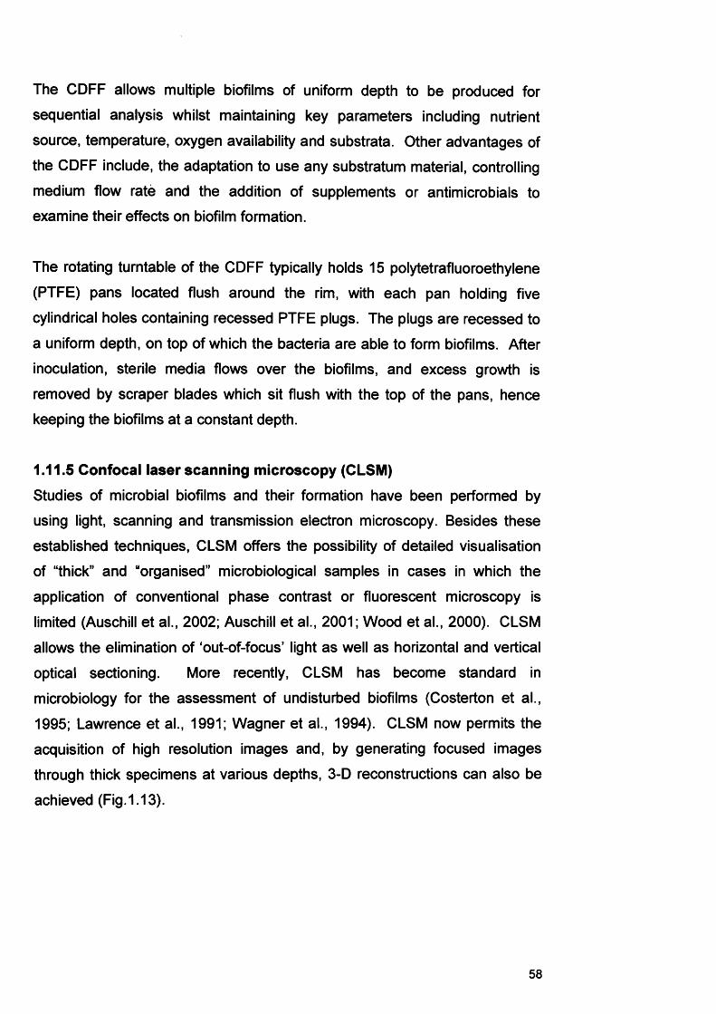
The CDFF allows multiple biofilms of uniform depth to be produced for
sequential analysis whilst maintaining key parameters including nutrient
source, temperature, oxygen availability and substrata. Other advantages of
the CDFF include, the adaptation to use any substratum material, controlling
medium flow rate and the addition of supplements or antimicrobials to
examine their effects on biofilm formation.
The rotating turntable of the CDFF typically holds 15 polytetrafluoroethylene
(PTFE) pans located flush around the rim, with each pan holding five
cylindrical holes containing recessed PTFE plugs. The plugs are recessed to
a uniform depth, on top of which the bacteria are able to form biofilms. After
inoculation, sterile media flows over the biofilms, and excess growth is
removed by scraper blades which sit flush with the top of the pans, hence
keeping the biofilms at a constant depth.
1.11.5 Confocal laser scanning microscopy (CLSM)
Studies of microbial biofilms and their formation have been performed by
using light, scanning and transmission electron microscopy. Besides these
established techniques, CLSM offers the possibility of detailed visualisation
of “thick” and “organised” microbiological samples in cases in which the
application of conventional phase contrast or fluorescent microscopy is
limited (Auschill et al., 2002; Auschill et al., 2001; Wood et al., 2000). CLSM
allows the elimination of ‘out-of-focus’ light as well as horizontal and vertical
optical sectioning. More recently, CLSM has become standard in
microbiology for the assessment of undisturbed biofilms (Costerton et al.,
1995; Lawrence et al., 1991; Wagner et al., 1994). CLSM now permits the
acquisition of high resolution images and, by generating focused images
through thick specimens at various depths, 3-D reconstructions can also be
achieved (Fig. 1.13).
58

a b
Fig. 1.12 a) The Constant Depth Film Ferm enter (CDFF); b) close up of the rotating turntable; c) PTFE pan with 5 recessed plugs; d) close-up of the scrapper blade
59
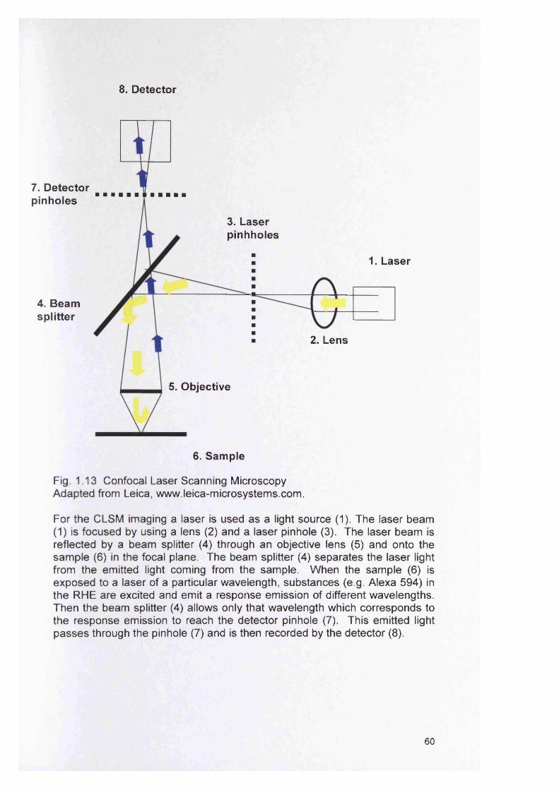
8. Detector
7. Detector pinholes
4. Beam splitter
3. Laser pinhholes
1. Laser
2. Lens
5. Objective
6. Sample
Fig. 1.13 Confocal Laser Scanning Microscopy Adapted from Leica, www.leica-microsystems.com.
For the CLSM imaging a laser is used as a light source (1). The laser beam (1) is focused by using a lens (2) and a laser pinhole (3). The laser beam is reflected by a beam splitter (4) through an objective lens (5) and onto the sam ple (6) in the focal plane. The beam splitter (4) separates the laser light from the emitted light coming from the sample. W hen the sample (6) is exposed to a laser of a particular wavelength, substances (e.g. Alexa 594) in the R H E are excited and emit a response emission of different wavelengths. Then the beam splitter (4) allows only that wavelength which corresponds to the response emission to reach the detector pinhole (7). This emitted light passes through the pinhole (7) and is then recorded by the detector (8).
60
1.11.6 Fluorescence in situ hybridisation (FISH)
FISH is a frequently used and culture - independent approach for detecting
and visualising microorganisms. Bacteria and Archaea contain 5S, 16S and
23S ribosomal Ribonucleic acid (rRNAs) with lengths of approximately 120,
1500 and 3000 nucleotides. In the vast majority of applications FISH probes
have been designed to specifically target 16S rRNA (Amann, 2001). A
typical FISH protocol includes fixation and permeabilisation of the sample,
hybridisation of the probe, a washing step to remove unbound probe and the
detection of labelled cells by microscopy (Amann, 2001).
Peptide nucleic acid (PNA) molecules are pseudopeptides that obey Watson-
Crick base-pairing rules for hybridisation to complementary nucleic acid
targets (Egholm et al., 1993; Nielsen and Haaima, 1997). PNA probes are
extremely efficient at hybridisation (having high specificities, strong affinities,
rapid kinetics) due to their uncharged, neutral backbones (Oliveira et al.,
2002). PNA probes penetrate the hydrophobic cell wall of bacteria to a
greater extent than that of DNA oligonucleotides, because of their relatively
hydrophobic character. The FISH technique and application is discussed in
more details in Chapter 5.
1.12 SUMMARY
Microorganisms are capable of growing in both a free form (planktonic) or as
biofilms attached to solid surfaces. Biofilms are defined as communities of
microorganisms, often adhered to a surface and encased within an
extracellular polysaccharide matrix (Kumamoto and Marcelo, 2005).
Examples of surfaces supporting biofilm growth include inanimate
environmental materials, biomaterials interfacing with host tissue and
systems, or the host tissue itself. The behaviour and phenotype of microbes
existing in either planktonic and biofilm states is known to differ significantly
and this is perhaps best exemplified by studies on antimicrobial efficacy
against the different growth phases (Hill et al., 2003).
61
In the oral environment candidal biofilms on prostheses and the oral mucosa
have been associated with infection (Kumamoto and Marcelo, 2005). Intra
oral biofilms can develop on the tooth enamel, oral mucosa or on introduced
oral prostheses and these can all provide a reservoir of potentially
pathogenic organisms promoting dental caries and periodontal disease.
Furthermore, in the case of oral tissue, certain microorganisms such as yeast
of the genus Candida have been shown to actually invade the tissue,
invoking a pathogenic effect. Similarly, within a chronic wound, it may well
be the case that the occurrence of a biofilm results in clinical problems due to
existence of the microbes in a more persistent state. Examples of clinically,
important biofilms include Proteus mirabilis on urinary tract catheters
(Stickler, 2005), Candida spp. on denture surfaces and bacteria
(polymicrobial) within chronic wounds (Hill et al., 2003). Significantly, when
biofilms are present on the surface of medical devices, a failure of the device
can occur, as encountered in the blocking of urinary catheters and the
obstruction of airways within artificial voice box prostheses (Douglas, 2003;
Van der Mei et al., 2000). Furthermore, biofilms tend to exhibit heightened
resistance to antibiotics, possibly by diffusion limitation or the presence of
biochemically inert microbes within the biofilm (Donlan and Costerton, 2002).
The clinical significance of biofilms provides the basis for this PhD and
consequently the aim was to characterise specific biofilms that are involved
in human infection, specifically those biofilms associated with chronic wounds
and oral candidosis.
1.13 AIMS AND OBJECTIVES OF STUDYThe aims of this study were to examine the in vitro properties of C. albicans
biofilms and also biofilms of bacteria recovered from the chronic wound
environment. The underlying hypothesis of this work was that clinically
important biofilms associated with infections have characteristics and
features that promote their virulence and resistance to both host defences
and administered antimicrobial agents. Specifically the aims were:
62
1. To determine the structural properties of C. albicans biofilm generated
in an in vitro model of oral candidosis.
2. To analyse C. albicans virulence gene expression within developed
biofilms.
3. To examine the relative in vitro biofilm-forming ability of selected
bacterial strains originating from chronic wound environments.
4. To investigate the spatial distribution and structure of in vitro chronic
wound biofilms.
5. To look at gene expression of chronic wound biofilm organisms.
63
Chapter 2
Biofilm formation and oral epithelium infection by Candida
albicans
64
2.1 Introduction
The genus Candida comprises of over 150 species of ‘yeast-Hke’ fungi,
several of which can exist as either harmless commensals or as opportunistic
pathogens of humans. Candida infection (candidosis, sing; candidoses, pi.),
has been reported at most body sites, with superficial infections of the oral
and vaginal mucosa the most frequently encountered. Although, Candida
albicans is generally isolated from 80% of all oral candidoses, this organism
is also prevalent as a harmless member of the oral microflora. It is estimated
that commensal carriage rate in humans is between 35 and 55%. Whilst C.
albicans is the most frequent cause of human fungal infection resulting in a
mortality rate of 30-50% in immunocompromised patients with systemic
infection, the pathogenesis of this organism remains unclear (Mukherjee et
al., 2003; Odds, 1988).
2.1.1 Non-oral infection
Candidoses are primarily superficial infections, occurring on moist and warm
mucosal membranes such as in the mouth and vagina. In women using oral
contraceptives, candidal vulvovaginitis is common. Vulvovaginitis is
accompanied by a thick discharge, vaginal itching and discomfort
(Samaranayake, 2006). Candidosis can also develop at a variety of other
body locations including the skin, in the gut, bone joints, nails and urinary
tract (Table 2.1).
2.1.1.1 Candida and implantable medical device associated infection
Introduction of artificial materials at various body locations has been
accompanied by the ability of microorganisms, including Candida spp. to
colonise and form biofilms (Ramage et al., 2006). Devices such as shunts,
artificial prostheses, stents, implants, endotracheal tubes, pacemakers and
various catheter types have been shown to support Candida colonisation and
biofilm formation (Ramage et al., 2006).
65
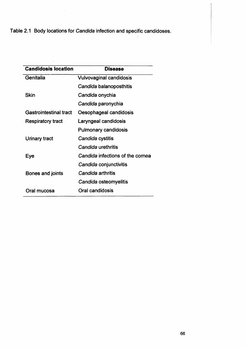
Table 2.1 Body locations for Candida infection and specific candidoses.
Candidosis location Disease
Genitalia Vulvovaginal candidosis
Candida balanoposthitis
Skin Candida onychia
Candida paronychia
Gastrointestinal tract Oesophageal candidosis
Respiratory tract Laryngeal candidosis
Pulmonary candidosis
Urinary tract Candida cystitis
Candida urethritis
Eye Candida infections of the cornea
Candida conjunctivitis
Bones and joints Candida arthritis
Candida osteomyelitis
Oral mucosa Oral candidosis
66
2.1.1.2 Central venous catheters
Yeasts, mainly Candida, are the third leading cause of intravascular catheter-
related infections, with the second highest colonisation to infection rate and
the overall highest crude mortality rate in the USA and Western Europe
(Ramage et al., 2006). Studies have shown that biofilms are present in all
central venous catheters, on both the inner lumen and outside the catheter
(Raad, 1998).
2.1.1.3 Voice box prostheses
A voice box prosthesis is a device fitted following a total laryngectomy. A
total laryngectomy involves the removal of the larynx from the throat and is
performed as part of the surgical treatment for laryngeal cancer. The
procedure leads to loss of natural speech, is very traumatic for the patient
and presents a lifelong challenge for communication (Dijk et al., 2000).
Silicone rubber voice prostheses fitted in patients are subject to rapid
microbial colonisation and biofilm formation, mainly by Candida spp. The
problem is exacerbated due to the lack of salivary flow to remove loosely
attached organisms. The biofilm causes malfunction, increased airflow
resistance and leakage of the valves of the prosthesis, which then needs to
be surgically replaced (Ell, 1996; Neu et al., 1994; Ramage et al., 2006).
2.1.2 Candida and oral candidosisAs already mentioned, C. albicans is an important cause of superficial
infection of mucosal membranes and systemic disease in humans (Sherman
et al., 2002). Four clinical forms of primary oral candidosis are recognised;
acute/chronic pseudomembranous candidosis, acute erythematous
candidosis, chronic erythematous candidosis, and chronic hyperplastic
candidosis (CHC; Chapter 1, Fig. 1.9, section 1.8.6) (Axell et al., 1997).
2.1.2.1 Pseudomembranous candidosis
Pseudomembranous candidosis (PMC) is also synonymous with the term
“oral thrush” and is traditionally regarded as an acute infection characterised
by the presence of non-adherent white pseudomembranes on the oral
67
mucosa. Primarily, PMC occurs with extremes of age e.g. in the new-born
and elderly, or in patients suffering from a systemic debilitating disease. The
persistence of PMC in HIV-infected or otherwise debilitated individuals has
led to the occurrence of chronic PMC. The lesions of PMC can be scraped
off with a swab to expose the underlying erythematous mucosa. The
removed white pseudomembranes consist of desquamated epithelial cells,
fibrin and fungal hyphae. Diagnosis is usually straightforward and can be
confirmed microbiologically either by staining a smear from the affected area
or by culturing a swab or an oral rinse (Akpan and Morgan, 2002).
2.1.2.2 Acute erythematous candidosis
Acute erythematous candidosis is most frequently seen on the dorsum of the
tongue in patients receiving prolonged antibiotic therapy. It presents as a
red, painful area of the mucosa and may also be seen with patients in
conjunction with low serum vitamin B12, low folate and low ferritin
concentrations (Akpan and Morgan, 2002). It is believed that broad spectrum
antibiotic therapy reduces the normal numbers of the oral bacterial
community and allows C. albicans to overgrow in the mouth (Farah et al.,
2000). Diagnosis of acute erythematous candidosis can be confirmed
through use of a swab from the tongue or buccal mucosa and confirmation of
Candida presence by culture and identification (Akpan and Morgan, 2002).
2.1.2.3 Chronic erythematous candidosisChronic erythematous candidosis (denture stomatitis) is the most frequently
occurring form of oral candidosis and is present in up to 65% of denture
wearers (Akpan and Morgan, 2002). This form of oral candidosis is believed
to be promoted through the occurrence of tissue damage caused by frictional
irritation of the palatal mucosa by the denture. A poor fitting denture and
poor oral hygiene is associated with this infection. Diagnosis requires
isolation and culture of Candida from the denture surface (Akpan and
Morgan, 2002).
68
2.1.2.4 Chronic hyperplastic candidiosis
Chronic hyperplastic candidiosis (CHC) is a distinct form of oral candidosis
and is characterised by hyphal penetration of the oral epithelium (Sitheeque
and Samaranayake, 2003) and the presence of an inflammatory cell infiltrate
(Williams et al., 1997). Of particular concern with CHC is the potential risk of
squamous cell carcinoma (SCC) development at the lesional site (Banoczy,
1977; Krogh et al., 1987a; Williams et al., 2001). The presence of Candida is
associated with an increased incidence of malignant transformation. The
exact role of Candida in the development of oral cancer is unclear but might
be related to the organism’s ability to generate carcinogenic nitrosamines
from salivary precursor molecules (Krogh et al., 1987b; Sitheeque and
Samaranayake, 2003). In addition, specific C. albicans strains are known to
promote neoplastic change, which highlights the potential role of these
organisms in malignant transformation (Krogh et al., 1987a). It is also
important to note that the lifestyle for both promotion of Candida carriage and
oral cancer are similar which makes establishing an association difficult
(Sitheeque and Samaranayake, 2003).
2.1.3 Candida virulence factors associated with oral candidosisIn addition to host factors (Chapter 1, section 1.8.7) Candida itself expresses
specific virulence attributes that promote oral infection. No single
predominant virulence factor has been associated with Candida, although
many factors have been implicated in its pathogenicity (Table 2.2).
2.1.3.1 AdherenceAdherence of Candida to the oral mucosa is an important initial step in the
establishment of infection and therefore, candidal adhesion to buccal
epithelial cells (BECs) (Nair and Samaranayake, 1996; Tsang and
Samaranayake, 1999; Verran et al., 2007), to denture acrylic (Bulad et al.,
2004; Verran and Maryan, 1997) and silicone rubber (Price et al., 2002; Price
et al., 2005) have been extensively studied.
69
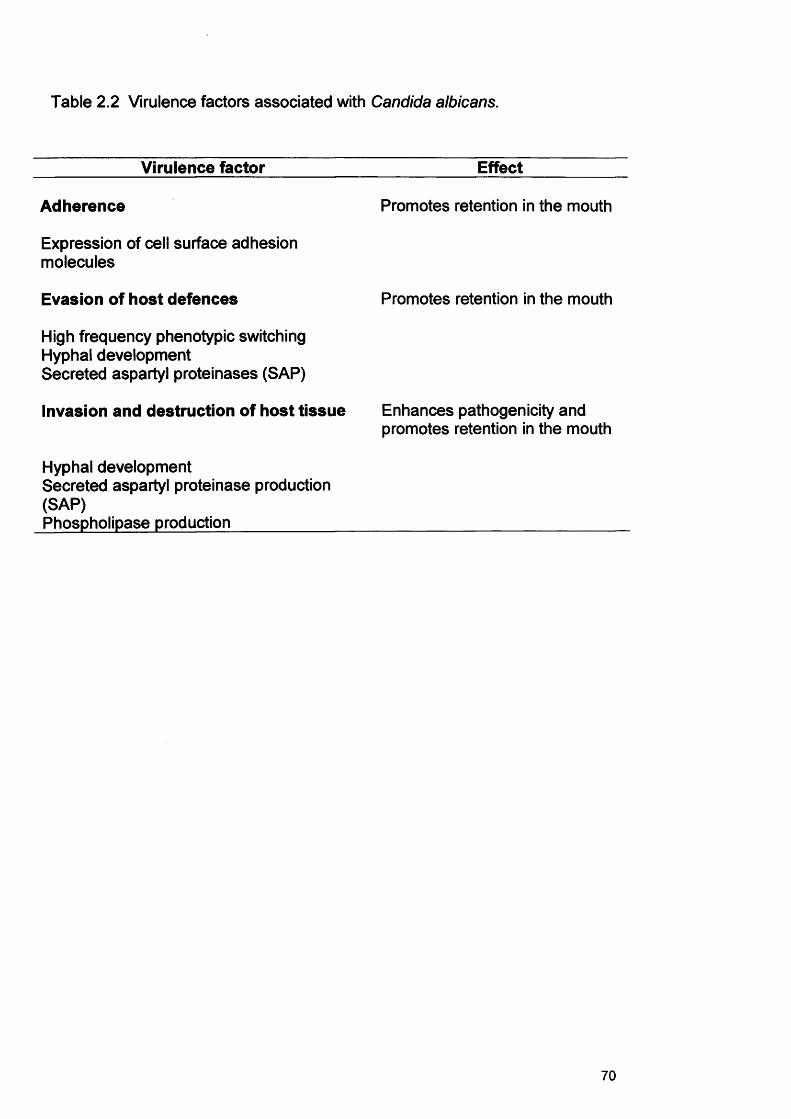
Table 2.2 Virulence factors associated with Candida albicans.
Virulence factor Effect
Adherence Promotes retention in the mouth
Expression of cell surface adhesion molecules
Evasion of host defences Promotes retention in the mouth
High frequency phenotypic switchingHyphal developmentSecreted aspartyl proteinases (SAP)
Invasion and destruction of host tissue Enhances pathogenicity andpromotes retention in the mouth
Hyphal developmentSecreted aspartyl proteinase production(SAP)Phospholipase production_____________________________________________
70
Candida albicans must first adhere to the epithelium to colonise, infect or
invade tissues (Vargas et al., 1994). Attachment of Candida cells can be
mediated by both non-specific and specific factors (Table 2.3). Non specific
factors include relative cell surface hydrophobicity and electrostatic forces
(Ramage et al., 2005). Specific factors include adhesins, such as serum
proteins (fibrinogen and fibronectin) and salivary factors (Ramage et al.,
2005). Hence, specific C. albicans adherence is mediated by both protein
and carbohydrate factors and is a complex phenomenon, with strain variation
evident (Ramage et al., 2005).
2.1.3.2 MorphologyCandida albicans has the ability to grow in different morphological forms
including budding yeast cells, pseudohyphae and true filamentous hyphae
(Fig. 2.1). Once C. albicans attaches to host surfaces, it can switch from its
yeast morphology to a filamentous form which may promote adhesion and
penetration of the epithelium and increased resistance to phagocytosis by
host immune cells. It has also been suggested that filamentous forms of
Candida secrete higher quantities of hydrolytic enzymes (Senet, 1998)
(section 2.1.3.4). It is generally believed that the budding yeast form is
predominant in commensal carriage whilst the hyphal form predominates in
tissue penetration. Although others have demonstrated that both forms can
be associated with commensalism and disease (Soil, 2002). It has been
observed that hyphae grow by tip extension and generate significant
pressure for tissue invasion (Gow et al., 2002).
71
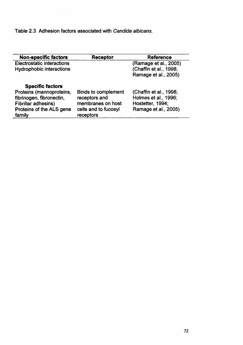
Table 2.3 Adhesion factors associated with Candida albicans.
Non-specific factors Receptor ReferenceElectrostatic interactions Hydrophobic interactions
Specific factorsProteins (mannoproteins, fibrinogen, fibronectin, Fibrillar adhesins) Proteins of the ALS gene family________________
Binds to complement receptors and membranes on host cells and to fucosyl receptors__________
(Ramage et al., 2005) (Chaffin et al., 1998; Ramage et al., 2005)
(Chaffin et al., 1998; Holmes et al., 1996; Hostetter, 1994; Ramage et al., 2005)
72
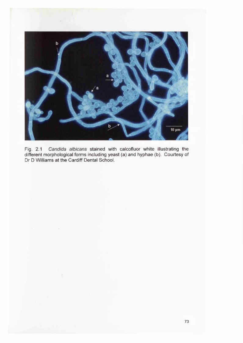
Fig. 2.1 Candida albicans stained with calcofluor white illustrating the different morphological forms including yeast (a) and hyphae (b). Courtesy of Dr D Williams at the Cardiff Dental School.
73
2.1.3.3 Phenotypic switching
Candida albicans has the ability to rapidly and reversibly switch between the
yeast budding form and a hyphal growth form (Gow, 1997; Soil, 1986).
Various studies have demonstrated that high frequency switching affects a
variety of virulence features, including the bud-hypha transition (Anderson et
al., 1989), antigenicity (Anderson et al., 1990), adhesion (Kennedy et al.,
1988; Vargas et al., 1994), sensitivity to neutrophils and oxidants (Kolotila
and Diamond, 1990), secretion of proteinase (Soli et al., 1989; Vargas et al.,
2000) and drug susceptibility (Soil et al., 1989; Vargas et al., 2000). The
morphogenic transition between yeast and hyphal forms is often stimulated
by growth in the host, correlated with invasion and associated with
pathogenesis (Gow et al., 2002). In vitro high frequency switching can be
detected by colony morphologies of strains grown on amino acid-rich Lee’s
medium supplemented with arginine and a limiting concentration of zinc (Soli,
1992).
2.1.3.4 Production of hydrolytic enzymes
Destruction of host tissues by C. albicans is believed to occur through the
activity of hydrolytic enzymes produced by the organism. Most of these
enzymes are extracellularly secreted by C. albicans. The most frequently
studied of these enzymes are secreted aspartyl proteinases (SAPs) (Hube
and Naglik, 2001; Naglik et al., 2004). Hydrolytic enzymes contribute to host
tissue invasion by digesting or destroying cell membranes and by degrading
host surface molecules. There is some evidence that hydrolytic enzymes are
able to damage cells and molecules of the host immune system thereby
avoiding or resisting antimicrobial activity (Schaller et al., 2005a).
In addition to SAPs, phospholipases (PLs) contribute to the pathogenicity of
C. albicans by damaging host-cell membranes, which aids the fungus by
facilitating host tissue invasion (Borst and Fluit, 2003). Details on Candida
hydrolytic enzymes and associated genes are discussed in detail in Chapter
3, section 3.1.1.
74
2.1.4 Models to assess Candida biofilm formation
In this present study the primary aim was to compare the ability of different C.
albicans strains to grow as biofilms and determine whether any observed
differences with respect to the biofilm growth related to their origin of
infection. A suitable model for this had therefore to be adopted.
In recent years a large number of experimental model systems have been
designed to study Candida grown in the biofilm state (Table 2.4). Many of
these have already been highlighted in the Literature Review (section 1.11)
of this thesis. The availability of well-characterised, reproducible biofilm
models is essential to understand the nature of Candida biofilms and to
perform studies of biofilm formation and antifungal drug resistance (Chandra
et al., 2001a). Many of the models outlined in Table 2.4 are sufficient for
device infection studies but not for tissue infections. However, the recent use
of a reconstituted human oral epithelium system is adequate to study tissue
infections and closely resembles the healthy human epithelium (Jayatilake et
al., 2005).
2.1.4.1 Reconstituted human oral epithelium (RHE)
The human oral mucosa is composed of an outermost layer of stratified
squamous epithelium similar to the stratified squamous epithelia found in the
rest of the body. It has a mitotically active basal cell layer, a number of
differentiating intermediate layers and a superficial layer, where cells are
shed from the surface of the epithelium (Giannola et al., 2007). SkinEthic
Laboratories cultivate a 0.5 cm2 reconstituted human epithelium (RHE) in
vitro on a polycarbonate filter at the air liquid interface in chemically defined
medium lacking antibiotics for 5 days (Fig. 2.2). The transformed human
keratinocytes of the cell line TR146, from a squamous cell carcinoma of the
buccal mucosa form an epithelial tissue devoid of stratum corneum, yet
histologically resembling the mucosa of the oral cavity.
75
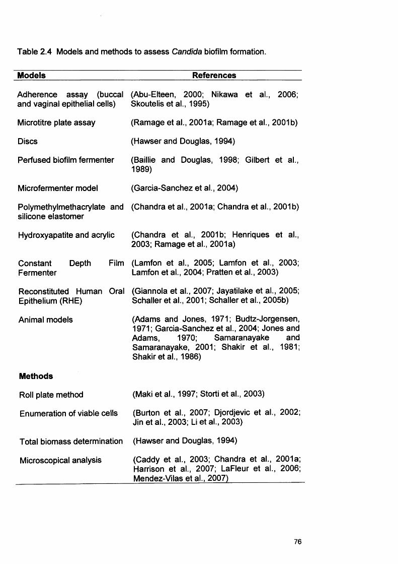
Table 2.4 Models and methods to assess Candida biofilm formation.
Models References
Adherence assay (buccal and vaginal epithelial cells)
(Abu-Elteen, 2000; Nikawa et al., 2006; Skoutelis et al., 1995)
Microtitre plate assay (Ramage et al., 2001a; Ramage et al., 2001b)
Discs (Hawser and Douglas, 1994)
Perfused biofilm fermenter (Baillie and Douglas, 1998; Gilbert et al., 1989)
Microfermenter model (Garcia-Sanchez et al., 2004)
Polymethylmethacrylate and silicone elastomer
(Chandra et al., 2001a; Chandra et al., 2001b)
Hydroxyapatite and acrylic (Chandra et al., 2001b; Henriques et al., 2003; Ramage et al., 2001a)
Constant Depth Fermenter
Film (Lamfon et al., 2005; Lamfon et al., 2003; Lamfon et al., 2004; Pratten et al., 2003)
Reconstituted Human Epithelium (RHE)
Oral (Giannola et al., 2007; Jayatilake et al., 2005; Schaller et al., 2001; Schaller et al., 2005b)
Animal models (Adams and Jones, 1971; Budtz-Jorgensen, 1971; Garcia-Sanchez et al., 2004; Jones and Adams, 1970; Samaranayake and Samaranayake, 2001; Shakir et al., 1981; Shakir etal., 1986)
Methods
Roll plate method (Maki et al., 1997; Storti et al., 2003)
Enumeration of viable cells (Burton et al., 2007; Djordjevic et al., 2002; Jin et al., 2003; Li et al., 2003)
Total biomass determination (Hawser and Douglas, 1994)
Microscopical analysis (Caddy et al., 2003; Chandra et al., 2001a; Harrison et al., 2007; LaFleur et al., 2006; Mendez-Vilas et al., 2007)
76
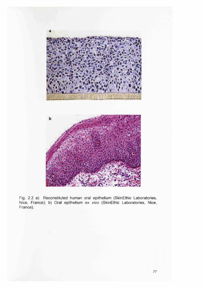
a^gj^iis
Fig. 2.2 a) Reconstituted human oral epithelium (SkinEthic Laboratories, Nice, France); b) Oral epithelium ex vivo (SkinEthic Laboratories, Nice, France).
77
In recent years, this RHE (SkinEthic Laboratories, Nice, France) has
successfully been used to investigate mechanisms of tissue degradation by
Candida (Jayatilake et al., 2005; Schaller et al., 2001; Schaller et al., 1999;
Schaller et al., 1998) and examine the response of tissue to infection
(Schaller et al., 2005b). Indeed, previous studies using Periodic Acid Schiff
(PAS) staining to profile C. albicans RHE infection have suggested that strain
differences exist between C. albicans isolates with respect to tissue invasion
(Bartie et al., 2004).
In general, this approach provides a number of advantages compared to
other in vitro models. The stratified cultured TR146 cell layers are analogous
to normal human buccal epithelium with the cells are able to grow on the
polycarbonate permeable inserts to form cell layers resembling the stratified
human buccal tissue. So overall, the model has similar morphology, ultra
structure and permeability barrier properties to intact buccal mucosa
(Jacobsen et al., 1999). The RHE has also been used to perform
permeability studies of drugs in a well-defined environment (Giannola et al.,
2007; Jacobsen et al., 1999). Importantly, when using any model system,
one has to be aware of its limitations e.g. in the case of the RHE, the
epithelium cannot recover after destruction nor can it elicit a full immune
response.
2.1.5 Methods of analysing the developed Candida biofilms
Once a biofilm has been generated, it can be characterised using a variety of
approaches including the assessment of cell number either through counting
the total number of cells (live or dead) or by viable count estimation. Other
characteristics that can be measured include the analysis of biofilm
associated gene expression (Chapter 3), or investigating the actual
architecture of the biofilm that develops. In this present Chapter, the aim was
the latter and therefore a microscopical analysis was selected. Several
options were available as outlined below.
78
2.1.6 Microscopical analysis
Scanning electron microscopy (SEM) techniques have been used to observe
biofilms (or more often adherent cells) because of the high magnification and
image contrast that can be achieved. However, sample preparation
(dehydration of the samples) may cause problems leading to false artifactual
observations (Caddy et al., 2003). Another disadvantage of SEM is that
detail on the exact architecture of biofilms is difficult to obtain.
Inverted light microscopy has also been used, e.g. to measure adherence of
C. albicans to acrylic (Williams et al., 1998). Recently, Harrison et al. (2007)
used the inverted light microscope to examine C. albicans and C. tropicalis
biofilms cultivated at the bottom of microtitre plate wells (Harrison et al.,
2007). The benefits of such an approach are that visualisation of the biofilm
in a liquid environment can be undertaken without disturbing the architecture.
The atomic force microscope (AFM) has become an important tool for
investigating biological materials such as tissues, yeast, bacteria and their
biological constituents. It can provide real-time in situ quantitative
morphological information, as well as measure the interaction forces between
a sharp tip and the surface of interest. The probe of interest should be
positioned on the stage of the AFM to be scanned and has to be fixed by
chemical treatment so force applied by the AFM tip does not induce
movement of the sample. Surface modifications include polyethyleneimine
(PEI), silanisation or poly-L-lysine treatment to immobilise microbes onto the
surface (Mendez-Vilas et al., 2007; Schar-Zammaretti and Ubbink, 2003;
Velegol and Logan, 2002). However, one of the limitations of the AFM is that
all these methods can alter the surface physiochemical properties of the cells
(Mendez-Vilas et al., 2007).
Fluorescent microscopy can be used to evaluate the morphology of biofilms
(Chandra et al., 2001a; Kuhn et al., 2002b), and this is of particular value for
live/dead determination of biofilm cells and the specific detection of individual
group members in biofilm consortia (LaFleur et al., 2006). Furthermore, cells
79
can be detected when present on an opaque surface. The disadvantage of
standard fluorescent microscopy is that it cannot generate focused images
through very thick specimens.
Confocal laser scanning microscopy is a microscopy method that permits the
acquisition of high resolution images and 3-D reconstructions. This
technique has the advantages of fluorescence microscopy but significantly
can also generate focused images through thick specimens at various
depths. The CLSM procedure involves images being taken ‘point-by-point’
and then reconstructed using a computer, rather than projected through an
eyepiece. Particulars of this method are discussed in detail in Chapter 1,
section 1.11.5. Due to the significant advantages of CLSM over other
microscopical methods, it was chosen to study the Candida biofilms
generated in this study.
2.2 Aims
With regards to oral candidosis, the question that remains to be answered is
why individuals present with such different infections, which are all seemingly
caused by the same fungus. It is evident that C. albicans is an extremely
heterogeneous species whose strains differ phenotypically and genotypically.
Strain variation could determine which type of infection develops and whether
the host actually manages to combat the colonising strain or whether it is
retained as a commensal strain.
The hypothesis of this study was that different strains of C. albicans develop
specific biofilm growth forms on a Reconstituted Human Oral Epithelium
which are directly associated with their origin of infection. For example, if
isolates originated from an invasive candidosis such as CHC, the resulting
biofilm/infection would differ compared to another strain from a more
superficial mucosal infection such as PMC. To test this hypothesis, the
specific aims were:
80
1. To develop a suitable in vitro biofilm model for C. albicans infection using
the RHE.
2. To infect this model with a range of C. albicans strains isolated from
various forms of oral candidosis.
3. To compare the following parameters of RHE infection: extent of RHE
colonisation; type of candidal morphology involved in the infection; degree of
RHE tissue invasion exhibited and the pattern of tissue invasion.
81
2.3 Materials and Methods
2.3.1 Identification of test isolates
Nineteen isolates of C. albicans from four distinct clinical conditions (normal
oral mucosa, n=4; non-CHC oral candidoses, n=4; CHC, n=8; SCC
(squamous cell carcinoma), n=3) were obtained either by imprint culture from
patients with defined lesions or concentrated rinse culture for isolates from
non-defined infection sites. The strains were collected between 1993 until
2001 as part of the routine microbiological service at the Cardiff Dental
School. Details of these isolates are presented in Table 2.5. Identification of
C. albicans was based on germ-tube formation and biochemical profiling
using the API 32C system (bioMerieux, Basingstoke, UK).
2.3.2 In vitro RHE infection model
2.3.2.1 Preparation of Candida albicans
Candida albicans strains were cultured on Sabouraud Dextrose Agar (SDA;
LAB M, Bury, UK) at 37°C for 24 h. The resulting growth was used to
inoculate 10 ml of Yeast Nitrogen Base (YNB; BD Diagnostics, Cowley, UK)
medium supplemented with 0.5% glucose (w/v). This medium was then
incubated for 12 h at 37°C. Yeast cells were harvested by centrifugation and
washed (*3) with phosphate buffered saline (PBS). The resulting yeast cells
were enumerated using a haemocytometer and a total of either 4x10⁷ yeast
cells or 2x10⁶ yeast cells was added to the RHE (SkinEthic Laboratories,
Nice, France; section 2.3.2.2). The initial morphology of the inoculum was
determined using a light microscope at original magnifications of x400 and
x600 (Carl Zeiss Ltd., Welwyn Garden City, UK).
2.3.2.2 Preparation of RHEThe RHE was supplied by SkinEthic Laboratories (Nice, France) and was
generated by culturing transformed human keratinocytes derived from the
cell line TR146 originally obtained from a squamous cell carcinoma of the
buccal mucosa.
82
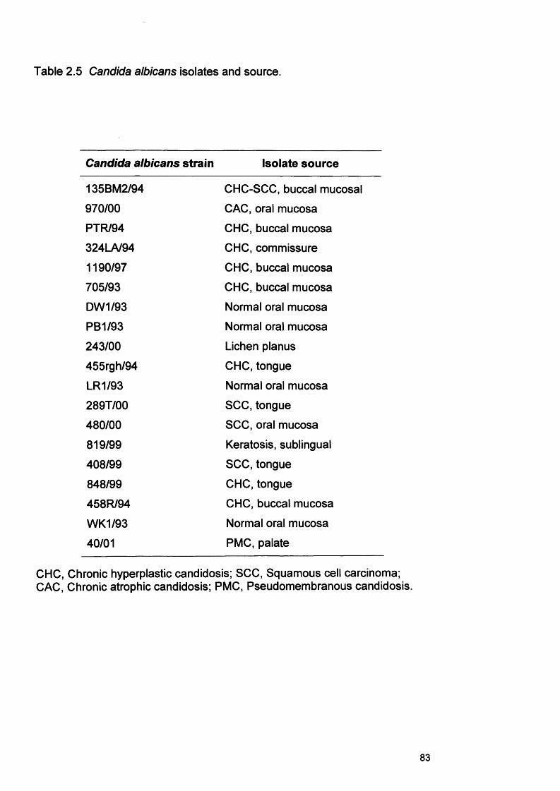
Table 2.5 Candida albicans isolates and source.
Candida albicans strain Isolate source
135BM2/94 CHC-SCC, buccal mucosal
970/00 CAC, oral mucosa
PTR/94 CHC, buccal mucosa
324LA/94 CHC, commissure
1190/97 CHC, buccal mucosa
705/93 CHC, buccal mucosa
DW1/93 Normal oral mucosa
PB1/93 Normal oral mucosa
243/00 Lichen planus
455rgh/94 CHC, tongue
LR1/93 Normal oral mucosa
289T/00 SCC, tongue
480/00 SCC, oral mucosa
819/99 Keratosis, sublingual
408/99 SCC, tongue
848/99 CHC, tongue
458R/94 CHC, buccal mucosa
WK1/93 Normal oral mucosa
40/01 PMC, palate
CHC, Chronic hyperplastic candidosis; SCC, Squamous cell carcinoma; CAC, Chronic atrophic candidosis; PMC, Pseudomembranous candidosis.
83
Cultures were incubated in serum-free conditions in a chemically defined
MCDB-153 medium containing 5 |jg ml'1 insulin and 1.5 mM calcium chloride
on a 0.5 cm2 microporous polycarbonate filter for 5 days. The MCDB-153
medium comprised of essential amino acids, vitamins, inorganic salts,
organic compounds and trace elements. The RHE in vitro model and all
culture media were prepared for this study without antibiotics and
antimycotics.
Immediately after receipt of the RHE from the supplier, the tissue was
removed from the agarose-nutrient solution in the shipping multiwell plate
and placed in a fresh 6-well tissue culture plate previously filled with 100 pi of
SkinEthic maintenance medium.
2.3.2.3 Culture of infected RHE
Infected RHE was incubated for 24 h (for 4x10⁷ yeast cells) or 12 h (for 2x10⁶
yeast cells) at 37°C in a humidified atmosphere enriched with 5% C 02. After
incubation, the tissue was bisected; one portion was used for CLSM analysis
and the other for RT-PCR (Chapter 3, section 3.3.4). A non-infected control
was included for comparison and in all cases, the experiments were repeated
on a minimum of two occasions.
2.3.2.4 RHE processing for Confocal Laser Scanning Microscopy
(CLSM)Three processing protocols were assessed in this study and included the
analysis of fresh (unprocessed) tissue, frozen fixed tissue and formalin fixed
tissue.
2.3.2.4.1 Fresh tissueThe fresh tissue portion used for CLSM was placed on a microscope slide,
and overlaid with 20 pi concanavalin A lectin (0.025% w/v in PBS)
conjugated with the fluorescent tag, Alexa 594 (Sigma, Poole, UK). After 30
min incubation at room temperature, the lectin solution was removed and the
tissue washed once using distilled H20. Finally, the tissue was overlaid with
84
5 pi of vectashield (Vector Laboratories Ltd, Peterborough, UK). A coverslip
was placed on top of the preparation and sealed with nail varnish.
2.3.2.4.2 Frozen fixed tissue
Specimens of infected tissue were also frozen using isopentane (100%)
cooled in liquid nitrogen. Fresh specimens were placed in aluminium foil and
lowered into the isopentane solution for 30 s until frozen. The tissue in the
foil was then stored at -80°C in cryotubes until required for staining and
CLSM analysis. To visualise the infecting C. albicans, sections were directly
labelled with 20 pi of Alexa 594-conjugated concanavalin A lectin (Molecular
Probes) at 0.025% w/v in PBS for 20 min at room temperature.
2.3.2.4.3 Formalin fixed tissue
Infected tissue was fixed in 2% (v/v in FfeO) paraformaldehyde solution for 24
h and embedded in paraffin wax using standard histological techniques.
Sections (20 pm) were cut and placed on to Histobond+ coated microscope
slides (Raymond A Lamb, East Sussex, UK), de-waxed and processed
through xylene and ethanol to water before fluorescent labelling.
2.3.2.4.4 Immunohistochemistry on fixed tissue
To identify epithelial cells within the RHE, sections were also labelled with a
pan-cytokeratin antibody using an indirect immunofluorescent labelling
procedure. Sections were first heated in citrate buffer for 10 min at 95°C,
washed in PBS and blocked with normal goat serum (Sigma-Aldrich, Dorset,
UK) using a 1:20 dilution in PBS for 30 min, prior to primary antibody
staining. Sections were then incubated overnight at 4°C with a mouse
monoclonal pan-cytokeratin primary antibody (Santa Cruz Biotechnology, Inc;
Heidelberg, Germany) used at a 1:50 dilution in PBS. After washing, sections
were fluorescently labelled with Alexa 488-conjugated goat anti-mouse
secondary antibody (5 pg ml"1; Invitrogen) for 30 min at room temperature.
85
For nuclear context, the sections were counterstained with Hoechst 33258
(Sigma) 1|jg ml'1 for 20 min, before washing in PBS. Tissues were then
mounted using vectashield fade-retarding mountant.
2.3.2.5 Confocal Laser Scanning Microscopy (CLSM)
Stained sections were viewed and analysed by CLSM using a Leica TCS
SP2 AOBS spectral confocal microscope (Leica, Wetzlar, Germany). The
sections were scanned through the full depth of the tissue using appropriate
settings for single, double or triple channel fluorescence recordings of Alexa
594 alone; Alexa 594 and Hoechst 33258; or Alexa 594, Hoechst 33258 and
Alexa 488 as detailed in Table 2.6. For multi-channel recordings,
fluorochromes were scanned sequentially to eliminate spectral overlap
between probes. Selected images were presented either as single confocal
optical sections or maximum intensity type reconstructions.
2.3.2.6 Criteria of biofilm parameters examined
The parameters of the biofillms examined were I) the level of C. albicans
colonisation on the RHE, II) the morphology of the infecting C. albicans and,
III) the extent and pattern of any tissue invasion by the organism. “High
invaders” were defined as those strains that readily penetrated the RHE
within the incubation time period. These were in contrast to “low invaders”
where only superficial invasion on the RHE was noted. “Non-invaders” did
not display any evidence of tissue penetration.
86
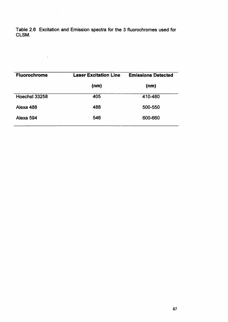
Table 2.6 Excitation and Emission spectra for the 3 fluorochromes used for CLSM.
Fluorochrome Laser Excitation Line Emissions Detected
(nm) (nm)
Hoechst 33258 405 410-480
Alexa 488 488 500-550
Alexa 594 546 600-660
87
2.4 Results
2.4.1 Infection of the RHE model
Preliminary studies with the two inoculum levels and incubation times
showed that a high inoculum (4x10⁷) and longer incubation time (24 h)
caused an excessive level of tissue destruction with many of the tested
strains. As a consequence, the lower inoculum (2x10⁶) level and shorter
incubation time (12 h) proved more suitable for strain comparisons with
respect to the chosen measured parameters. The following results are
based on these optimal infection conditions. Furthermore, detection of
Candida was achieved with all three processing methods with no apparent
differences in staining quality or alterations in tissue morphology. Formalin
fixed tissue was however easiest to handle and to store, and so the reported
results are also based on this tissue processing approach.
2.4.2 Comparison of planktonic morphology and biofilm morphology
For the majority of strains tested, the morphological form of the Candida
strain was the same regardless of whether culture occurred in the presence
(biofilm form) or absence (planktonic form) of the RHE. Six strains
(324LA/94, PB1/93, 455rgh/94, LR1/93, 819/99 and 40/01) out of 19
exhibited a different morphology between the two different culture conditions
(Table 2.7).
2.4.3 In vitro RHE infection
2.4.3.1 Extent of colonisation of the surface of the RHE
The majority of C. albicans strains (n=17/19) invaded the RHE (Table 2.7)
and CLSM showed the presence of both yeast and hyphae on the RHE
surface. The results were consistent between the three separately repeated
experiments.
88
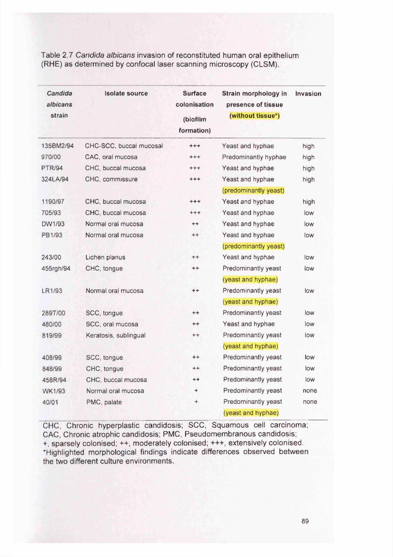
Table 2.7 Candida albicans invasion of reconstituted human oral epithelium (RHE) as determined by confocal laser scanning microscopy (CLSM ).
Candida
albicans
strain
Isolate source Surface
colonisation
(biofilm
formation)
Strain morphology in
presence of tissue
(without tissue*)
Invasion
135BM2/94 CHC-SCC, buccal mucosal +++ Yeast and hyphae high
970/00 CAC, oral mucosa +++ Predominantly hyphae high
PTR/94 CHC, buccal mucosa +++ Yeast and hyphae high
324LA/94 CHC, commissure +++ Yeast and hyphae
(predominantly yeast)
high
1190/97 CHC, buccal mucosa +++ Yeast and hyphae high
705/93 CHC, buccal mucosa +++ Yeast and hyphae low
DW1/93 Normal oral mucosa ++ Yeast and hyphae low
PB1/93 Normal oral mucosa ++ Yeast and hyphae
(predominantly yeast)
low
243/00 Lichen planus ++ Yeast and hyphae low
455rgh/94 CHC, tongue ++ Predominantly yeast
(yeast and hyphae)
low
LR1/93 Normal oral mucosa ++ Predominantly yeast
(yeast and hyphae)
low
289T/00 SCC, tongue ++ Predominantly yeast low
480/00 SCC, oral mucosa ++ Yeast and hyphae low
819/99 Keratosis, sublingual ++ Predominantly yeast
(yeast and hyphae)
low
408/99 SCC, tongue ++ Predominantly yeast low
848/99 CHC, tongue ++ Predominantly yeast low
458R/94 CHC, buccal mucosa ++ Predominantly yeast low
WK1/93 Normal oral mucosa + Predominantly yeast none
40/01 PMC, palate + Predominantly yeast
(yeast and hyphae)
none
CHC, Chronic hyperplastic candidosis; SCC, Squamous cell carcinoma; CAC, Chronic atrophic candidosis; PMC, Pseudomembranous candidosis;+, sparsely colonised; ++, moderately colonised; +++, extensively colonised, highlighted morphological findings indicate differences observed between the two different culture environments.
89
Figure 2.3a shows a more extensive surface colonisation with more hyphal
development and Figure 2.3b shows an example of sparse surface
colonisation of the RHE.
In some cases, hyphal and yeast growth degraded the epithelial layers of the
RHE and, therefore, the cell numbers and the incubation time was reduced
as highlighted earlier.
2.4.3.2 Invasion of the biofilm in the RHE
By decreasing the yeast inoculum level and reducing the incubation time, a
better analysis of the direction of hyphal penetration and determination of any
budding occurring within the tissue was possible. Figures 2.4a-2.6a illustrate
the different forms of Candida infection (24h and a high inoculum 4x10⁷) of
the RHE and depict variation in the extent of both tissue invasion and the
actual pattern of hyphal invasion within the tissue.
The 19 C. albicans strains were categorised as either “high invaders”, “low
invaders” or “non-invaders” (Table 2.7). CLSM showed the presence of both
yeast budding and hyphae on the RHE surface and within the tissue. Hyphal
growth forms were most frequently detected within the tissues. Figures 2.4a-
2.6a illustrate infection of RHE (24h, with a high inoculum 4x10⁷ level) and
Fig. 2.4b-2.10 illustrate infection of the RHE (12h, with a lower inoculum
2x10⁶ level) and the pattern of hyphal invasion. Five strains out of 19 were
‘high invaders’, where extensive surface colonisation and large numbers of
hyphae occurred within the tissue (Fig. 2.4b and 2.5b). This contrasted with
‘low invader’ or ‘none invader’, where yeast predominated for 10 of the 14
isolates and little or no invasion was evident (Fig. 2.6b and 2.7a and b). The
RHE was approximately 50-60 pm thick and the ‘high invaders’ penetrated
through the whole thickness of the tissue. The extent of RHE invasion was
therefore isolate-dependent and consistent over repeat experiments for each
given isolate (Table 2.7).
90
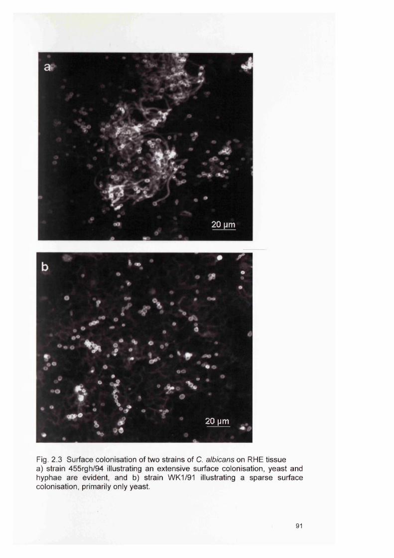
■ •
20 nm
i .• /
o0
V *•o
»■{,-. ***.v • _ ■ V . v> A d
fc *- J*
20 |jm * *
Fig. 2.3 Surface colonisation of two strains of C. albicans on RHE tissue a) strain 455rgh/94 illustrating an extensive surface colonisation, yeast and hyphae are evident, and b) strain W K1/91 illustrating a sparse surface colonisation, primarily only yeast.
91
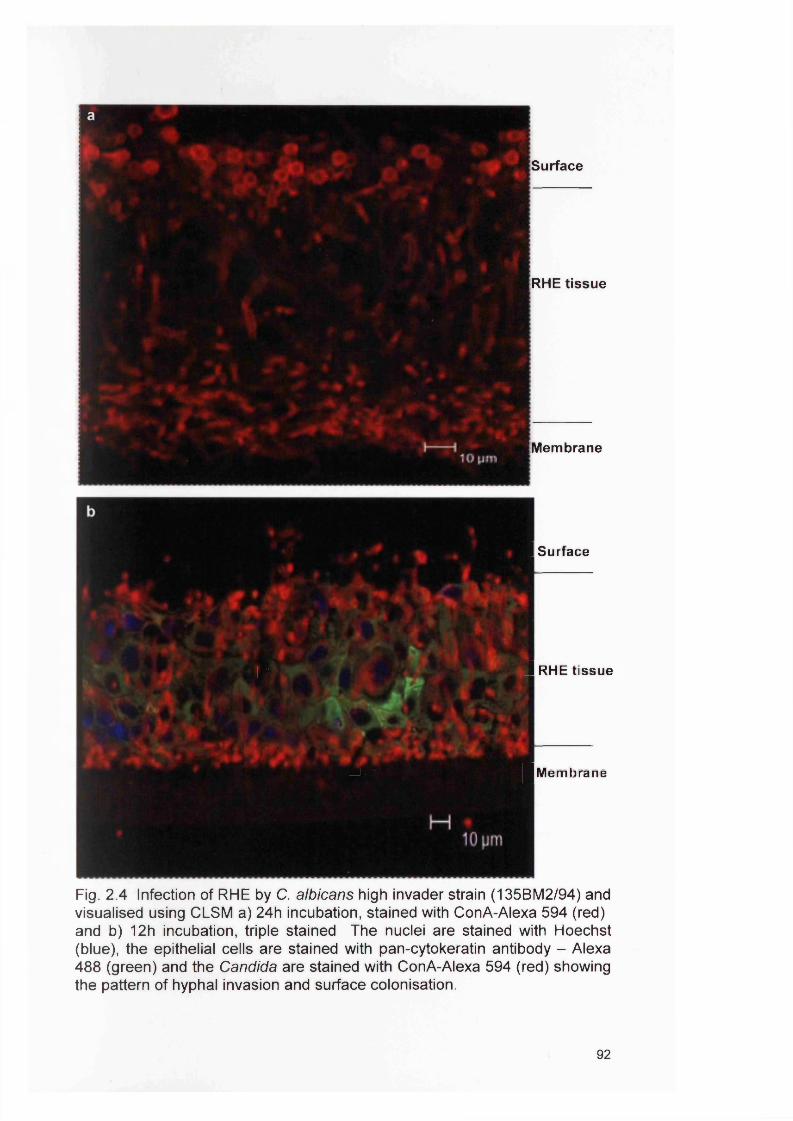
a
Surface
|RHE tissue
Membrane
- 1 Surface
/■ 1 RHE tissue
v ../; | Mem lira no
Fig. 2 .4 Infection of RHE by C. albicans high invader strain (135B M 2/94) and visualised using CLSM a) 24h incubation, stained with ConA-Alexa 594 (red) and b) 12h incubation, triple stained The nuclei are stained with Hoechst (blue), the epithelial cells are stained with pan-cytokeratin antibody - Alexa 488 (green) and the Candida are stained with ConA-Alexa 594 (red) showing the pattern of hyphal invasion and surface colonisation.
92
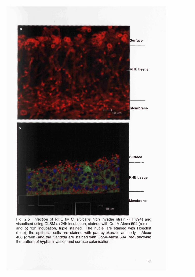
1■Surface
■RHE tissue
y Membrane■SurfaceismRHE tissue
■1 HI Membrane
! ■ ■s (MlFig. 2 .5 Infection of RHE by C. albicans high invader strain (P TR /94) and visualised using CLSM a) 24h incubation, stained with ConA-Alexa 594 (red) and b) 12h incubation, triple stained The nuclei are stained with Hoechst (blue), the epithelial cells are stained with pan-cytokeratin antibody - Alexa 488 (green) and the Candida are stained with ConA-Alexa 594 (red) showing the pattern of hyphal invasion and surface colonisation.
93
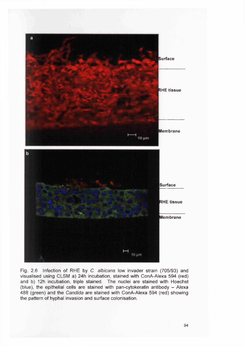
B s u rfa c e
SSrHE tissue
■ M em brane
b
Surface
RHE tissue
Membrane
I—110 pm
Fig. 2 .6 Infection of RHE by C. albicans low invader strain (705/93) and visualised using CLSM a) 24h incubation, stained with ConA-Alexa 594 (red) and b) 12h incubation, triple stained. The nuclei are stained with Hoechst (blue), the epithelial cells are stained with pan-cytokeratin antibody - Alexa 488 (green) and the Candida are stained with ConA-Alexa 594 (red) showing the pattern of hyphal invasion and surface colonisation.
94
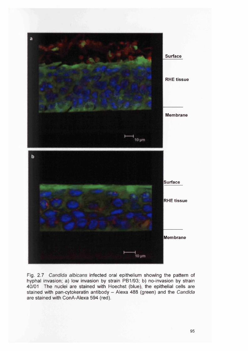
a
Surface
RHE tissue
Membrane
RHE tissue
Surface
Membrane
Fig. 2.7 Candida albicans infected oral epithelium showing the pattern of hyphal invasion; a) low invasion by strain PB1/93; b) no-invasion by strain 40/01 The nuclei are stained with Hoechst (blue), the epithelial cells are stained with pan-cytokeratin antibody - Alexa 488 (green) and the Candida are stained with ConA-Alexa 594 (red).
95
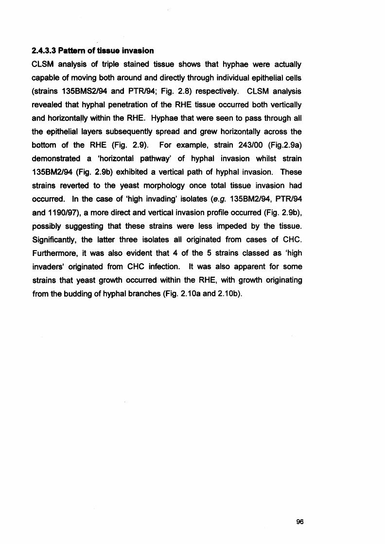
2.4.3.3 Pattern of tissue invasion
CLSM analysis of triple stained tissue shows that hyphae were actually
capable of moving both around and directly through individual epithelial cells
(strains 135BMS2/94 and PTR/94; Fig. 2.8) respectively. CLSM analysis
revealed that hyphal penetration of the RHE tissue occurred both vertically
and horizontally within the RHE. Hyphae that were seen to pass through all
the epithelial layers subsequently spread and grew horizontally across the
bottom of the RHE (Fig. 2.9). For example, strain 243/00 (Fig.2.9a)
demonstrated a ‘horizontal pathway’ of hyphal invasion whilst strain
135BM2/94 (Fig. 2.9b) exhibited a vertical path of hyphal invasion. These
strains reverted to the yeast morphology once total tissue invasion had
occurred. In the case of ‘high invading’ isolates (e.g. 135BM2/94, PTR/94
and 1190/97), a more direct and vertical invasion profile occurred (Fig. 2.9b),
possibly suggesting that these strains were less impeded by the tissue.
Significantly, the latter three isolates all originated from cases of CHC.
Furthermore, it was also evident that 4 of the 5 strains classed as ‘high
invaders’ originated from CHC infection. It was also apparent for some
strains that yeast growth occurred within the RHE, with growth originating
from the budding of hyphal branches (Fig. 2.10a and 2.10b).
96
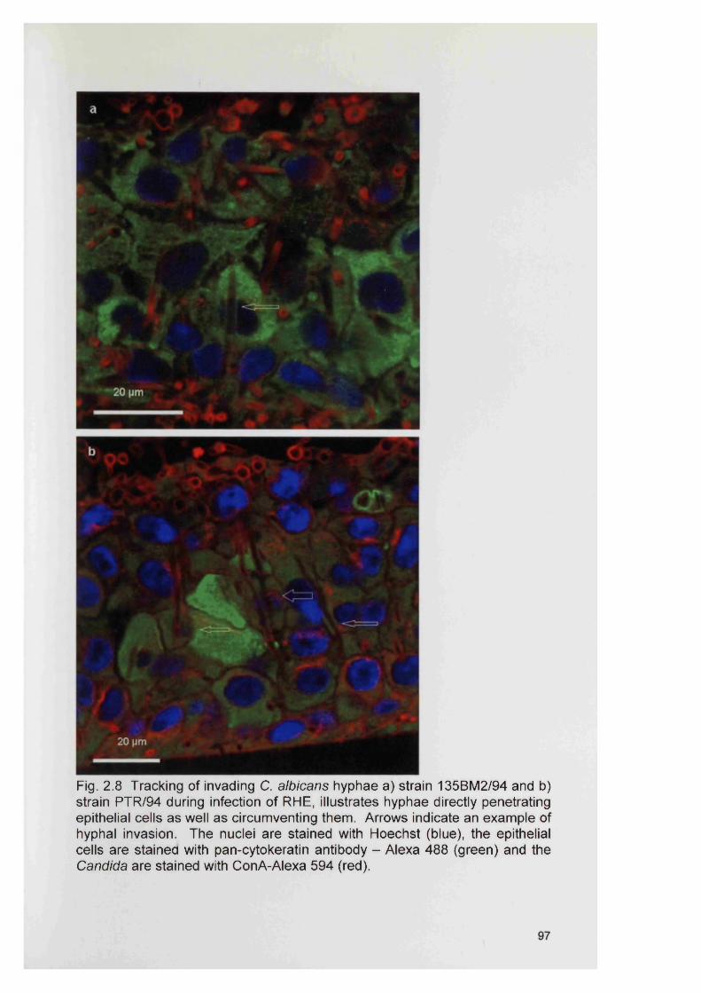
Fig. 2.8 Tracking of invading C. albicans hyphae a) strain 135BM 2/94 and b) strain PTR /94 during infection of RHE, illustrates hyphae directly penetrating epithelial cells as well as circumventing them. Arrows indicate an example of hyphal invasion. The nuclei are stained with Hoechst (blue), the epithelial cells are stained with pan-cytokeratin antibody - Alexa 488 (green) and the Candida are stained with ConA-Alexa 594 (red).
__ *320 pm
Fig. 2 .9 Pattern of Candida albicans hyphal invasion of infected RHE a) heterogenous hyphal network during C. albicans infection by strain 243/00; b) vertical hyphal invasion during C. albicans infection by strain 135BM 2/94.
98

ii4&%
Fig. 2 .10 (a) Nuclear penetration by hyphae of strain 1190/97, (b) nuclear penetration and budding of yeast from hyphal branch by strain 135BM 2/94. Arrows indicate the example of hyphal invasion type; * indicates budding.
99
2.5 Discussion
The pathogenic behaviour of opportunistic Candida species is dependent on
expression of certain virulence factors (e.g. hydrolytic enzymes) (Schaller et
al., 2002). Recently, the organisms ability to form biofilms has been
recognised as one of these factors (Ramage et al., 2005; Richard et al.,
2005). Candida albicans is dimorphic, (i.e. it is capable of reversible
transition between a yeast and hyphal form) and the morphogenic transition
between these forms is often stimulated by growth in the host and associated
with host tissue invasion (Gow et al., 2002). It has been suggested that
hyphae grow by tip extension and can generate significant tip pressures for
penetration (Gow et al., 2002). The underlying hypothesis for this present
study was that different strains of C. albicans from specific types of oral
infection would produce distinct characteristics of RHE infection and biofilm
formation. To compare biofilm formation and subsequent tissue invasion by
C. albicans, the use of the RHE model together with CLSM was chosen.
Previous studies using the multilayered RHE model have shown its
histological similarity with healthy oral epithelium and also described the
biofilm-like infection of the tissue by C. albicans (Jayatilake et al., 2005).
Compared with standard light microscopy, CLSM provides much more
detailed information on C. albicans tissue invasion. Previous use of CLSM to
assess C. albicans interaction with oral epithelium has been limited. Studies
involving infection of the FaDu oropharyngeal epithelial cell line (isolated from
a pharyngeal squamous cell carcinoma, American Type Culture Collection)
with C. albicans have previously described the use of CLSM in determining
fungal-epithelial cell interaction mechanisms (Park et al., 2005). Additional
work examining endothelial infection by C. albicans using CLSM has
elegantly revealed a self-induced endocytotic mechanism exhibited by
candidal hyphae as a means of tissue invasion (Phan et al., 2005).
The commercially available RHE employed in this present study has been
used with success as a basis for in vitro Candida infection models. A
possible limitation of the RHE system is the lack of certain host immune
100
responses. It has however been shown that pro-inflammatory cytokine
responses by in vitro epithelial cell lines including RHE infected with Candida,
appear to be maintained, and have actually been shown to vary with C.
albicans of different invasive potential and virulence (Schaller et al., 2002;
Villar et al., 2005). This present Chapter describes for the first time the
CLSM based characterisation of actual tissue infection and biofilm formation
by Candida.
Using conventional microscopy procedures, it has previously been difficult to
ascertain the manner by which hyphae migrate through tissue. The CLSM
approach used in this study, conclusively demonstrated that, in addition to
being able to move between epithelial cell junctions, hyphae can track
directly through keratinocytes. Epithelial cell penetration was also more
strongly associated with strains that demonstrated the vertical invasion profile
depicted in Fig. 2.9b compared with the less direct invasion profile seen in
Fig. 2.9a. The exact mechanism of hyphal penetration remains unclear. It
may occur either through pressure exerted from the growing tip of the hyphae
or as mentioned earlier by inducing an endocytosis process, alternatively,
invasion may be facilitated by extracellular hydrolytic enzymes promoting the
passage of hyphae.
It is also important to mention that biofilm structure and the proportions of
yeast- and hyphal- cells are strongly influenced by parameters such as
medium composition, temperature and the nature of the substratum
(Kumamoto, 2002). The key component of invasive growth is hyphal
penetration. The RHE model used in this study showed strain differences
between the 19 strains and was highly suitable for characterisation.
The levels of candidal invasion reported in this present study were largely
comparable to those previously found by the research group at the Cardiff
Dental School using standard light microscopy and PAS histological staining
analysis (Bartie et al., 2004). This was reassuring, particularly as the
methods of assessment between the two studies were different and were
101
performed by separate operators. All isolates previously deemed to be ‘high
invaders’ (Bartie et al., 2004) were also identified as such in this present
study. In addition, three of the four isolates originating from normal healthy
oral mucosa exhibited low RHE invasion and two of these were also
previously considered to be low invaders (Bartie et al., 2004). This latter
finding for commensal strains could indicate a limited ability of such isolates
to produce the necessary virulence attributes required for invasion. This was
also reflected by the relatively low level of RHE colonisation by these
isolates, together with an apparent reduced ability to produce hyphal forms.
It is, however, worth noting that invasion, albeit at a low level, did occur with
three of the four commensal isolates and it is therefore possible that given an
appropriate oral environment, these 'commensal' isolates could instigate
infection. Overall, the ability of C. albicans to invade appeared to correlate
with the isolates’ capacity to exhibit yeast/hyphal transition.
Interestingly, in all but one case, the ‘high invaders’ revealed a predominance
of hyphae within the tissue. This was not surprising, as movement of yeast
from the surface of the RHE would have been physically difficult, as
traditionally, yeast are considered to lack the migratory ability possessed by
hyphal elements (Gow et al., 2002). For a number of tissues, yeast
predominated on the surface of the RHE, whilst hyphae predominated within
and below the tissue. This would suggest that either the epithelium provided
a trigger for morphological transition following contact or that only hyphae
penetrated the tissue and thus predominated due to this selective process.
To support the latter, previous work using CLSM to study endothelium
invasion by C. albicans has shown that hyphae can induce their own
endocytosis, an ability not possessed by yeast (Phan et al., 2005). For most
strains used in the present study, the same morphological type was evident
in either the presence or absence of tissue (Table 2.7) and would suggest
that hyphal morphology was not induced through contact with the epithelium
and supports the view that hyphae are the principle invasive forms of C.
albicans.
102
Four of the five strains classed in this study as ‘high invaders’ (135BM2/94,
PTR/94, 324LA/94, 1190/97) originated from CHC and this might suggest an
inherently greater ability of these strains to invade the oral epithelium. A
particular pathogenic feature of C. albicans that is essential to the
development of CHC is the ability to invade the oral mucosa. Furthermore,
this feature of mucosal invasion is notably distinct from other forms of oral
candidosis and indeed serves as a diagnostic marker of CHC (see section
2.1.2.4). The fact that four other CHC strains were ‘low invaders’ again
highlights the undoubted importance of additional host factors in the
development of CHC infection. Indeed, studies performed by Soil (2002)
emphasise the importance of candidal adaptation to host environments as
being critical in determining whether commensalism or virulence results.
Evidence from complex DNA profiling of C. albicans populations currently
suggests the absence of true commensal and pathogenic strains, and
advocates rapid phenotypic adaptation as a key component in promoting a
particular oral status (Soil, 2002). The advantage of the current model is that
strain differences can be ascertained under the standardised conditions
used. An ability to associate invasive features with the clinical origin of the
isolates would be a significant advantage, as it would enable the early
identification of strains with the potential to cause CHC, and possibly
influence patient management.
2.6 SummaryA suitable method of assessing biofilm formation and tissue infection by C.
albicans using an in vitro tissue model using CLSM has been developed.
Using this RHE model we have shown that strain differences exist and may
relate to the clinical origin of Candida. The differences evident related to
adhesion, invasion and hyphal formation. Candida hyphae were seen to
develop as a complex network of filaments within the tissue and were seen to
physically penetrate cells and exhibit budding within the tissue. In summary:
1) A suitable in vitro biofilm model for C. albicans infection was established
using the RHE.
103
2) Seventeen out of 19 C. albicans strains invaded the RHE. Using this
model it has been shown that strain differences exist and may relate to the
clinical origin of the Candida. Factors associated with C. albicans in addition
to those of the host could therefore be important in the progression of oral
infections such as CHC.
3) Strain variation was evident based on the level of adherence, tissue
penetration and pattern of invasion. Heterogeneous tissue invasion was
evident. Candida hyphae develop as a complex network of filaments within
the tissue and are seen to physically penetrate the cells (nuclei) and exhibit
budding within the tissue.
104
Chapter 3
Expression of Candida albicans
virulence genes in an in vitro
tissue model
104
3.1 IntroductionCandida albicans is an opportunistic fungal pathogen of humans and as a
result causes infection only in suitably debilitated individuals. It is not
surprising therefore that no single predominant virulence factor is currently
recognised for this normally harmless organism. However, a number of
putative virulence factors have been proposed.
Candida albicans produces a variety of secreted hydrolytic enzymes (Hube et
al., 1994; Hube et al., 2000; Ibrahim et al., 1995; Monod et al., 1998) that
may enable it to persist and cause disease within a suitably compromised
host (Schofield et al., 2003). Differences in such gene expression may be
key to the pathogenic potential of a given strain. Furthermore, even for
individual strains, gene expression can give rise to phenotypic differences as
seen between biofilm and planktonic cells. Candida albicans infections (see
Chapter 2, section 2.1.2) are common, debilitating and an important clinical
problem (Naglik et al., 2004). In the case of oral infections, Candida biofilm
formation is considered to be a key factor in the cause of the infection, e.g.
denture stomatitis is associated with candidal biofilms on the acrylic surfaces
of the denture (Douglas, 2003). The adherence of Candida to oral mucosal
surfaces is critical for the persistence of the organism in the mouth
(Sitheeque and Samaranayake, 2003). Pseudomembranous candidosis
could be considered to be a complex biofilm, although this concept has still
not been well recognized (Jin et al., 2003).
It remains unclear whether ‘biofilm-like’ growth imposes a significant change
in C. albicans physiology and whether this change is reflected in a particular
pattern of gene expression, although some differential properties of biofilms
have been observed and several gene expression changes have been
reported. Genome-wide approaches are now available that allow this
question to be more fully investigated (Berman and Sudbery, 2002; Garcia-
Sanchez et al., 2004). Previous studies, using microarray technology,
assessed C. albicans biofilm transcriptional profiles to identify which genes
were involved in the biofilm development process (Cao et al., 2005; Garcia-
105
Sanchez et al., 2004; Murillo et al., 2005). The data from this previous study
suggest that the sulphur-amino acid biosynthesis pathway was an important
feature of C. albicans biofilms (Garcia-Sanchez et al., 2004).
3.1.1 Candida virulence factors and associated gene families
3.1.1.1 Secreted aspartyl proteinase (SAP) productionProteinases are classified on the basis of their catalytic mechanism and at
present four classes are recognised: I) serine, II) cysteine, III) metallo and,
IV) aspartyl proteinases.
Aspartyl proteinases are ubiquitous in nature and are involved in a myriad of
biochemical processes. Aspartyl proteinases include HIV aspartyl
proteinase, and pepsin and rennin in humans (Naglik et al., 2004). It is these
aspartyl proteinases that are associated with C. albicans virulence.
Candidal-proteinases all have a broad spectrum of activity, and salivary
proteins such as mucin, lactoferrin and secretory immunoglobulin A (slgA)
are all degraded by these enzymes. Based on current data, the main roles of
candidal aspartyl proteinases are to provide nutrition for the cells, to aid
penetration and invasion and to evade immune responses (Naglik et al.,
2003). MacDonald and Odds (1983) first demonstrated the relationship
between proteinase production and pathogenicity using chemically induced
mutants of C. albicans with decreased proteolytic activity. These mutants
were shown to be less virulent in murine models compared with the wild type
strains. Figure 3.1 shows a hypothetical model for the correlation of SAP
gene expression with function in C. albicans pathogenicity.
In recent years there has been a relatively large amount of work aimed at
studying SAPs of C. albicans. At present, 10 different genes have been
identified that encode for C. albicans SAPs (Hube, 1996; Hube et al., 1997;
Sanglard et al., 1997). One explanation for the existence of the 10 different
SAP genes may be the necessity for specific and optimised proteinase
production during different stages of an infection (Hube and Naglik, 2001).
106
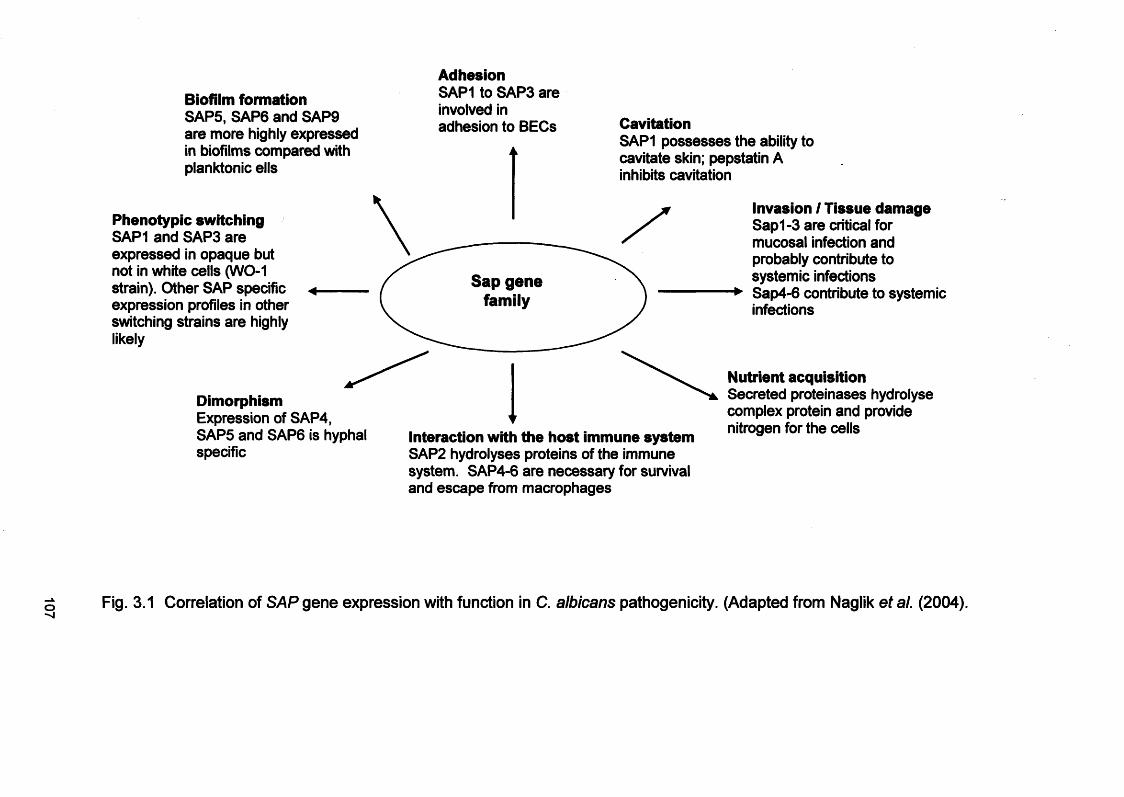
Biofilm formationSAP5, SAP6 and SAP9 are more highly expressed in biofilms compared with planktonic ells
AdhesionSAP1 to SAP3 are involved in adhesion to BECs
Phenotypic switchingSAP1 and SAP3 are expressed in opaque but not in white cells (WO-1 strain). Other SAP specific expression profiles in other switching strains are highly likely
CavitationSAP1 possesses the ability to cavitate skin; pepstatin A inhibits cavitation
Sap gene family
DimorphismExpression of SAP4, SAP5 and SAP6 is hyphal specific
Interaction with the host immune systemSAP2 hydrolyses proteins of the immune system. SAP4-6 are necessary for survival and escape from macrophages
Invasion / Tissue damageSap1-3 are critical for mucosal infection and probably contribute to systemic infections Sap4-6 contribute to systemic infections
Nutrient acquisitionSecreted proteinases hydrolyse complex protein and provide nitrogen for the cells
Fig. 3.1 Correlation of SAP gene expression with function in C. albicans pathogenicity. (Adapted from Naglik et al. (2004).
The SAPs are essential for growth when protein is the sole nitrogen source
(Hube et al., 1994; Naglik et al., 2004). SAPs are characterised by
functioning in an acidic environment and being inhibited by pepstatin A, a
hexapeptide from Streptomyces (Hoegl et al., 1996; Watts et al., 1998). It
has been reported that pepstatin A blocks invasion of host tissue by hyphal
cells at neutral pH and reduces the adherence of yeast cells to various cell
types (Watts et al., 1998). Candida albicans is not the only Candida species
known to produce extracellular proteinases. Candida tropicalis is thought to
possess four SAP genes (Zaugg et al., 2001), whereas C. parapsilosis
possesses at least two SAP genes (De Viragh et al., 1993). Proteolytic
activity has also been found in vitro by Candida dubliniensis (Monod and
Borg-von, 2002).
One approach to study the role and expression of SAPs involves the
proteolytic degradation of bovine serum albumin (BSA) which is
spectrophotometrically assessed by determining the generation of
trichloroacetic acid (TCA) soluble BSA fragments (Hube et al., 1997;
Kuriyama et al., 2003). Reverse-transcriptase polymerase chain reaction
(RT-PCR) has also been used to detect mRNA products from SAP genes
(section 3.3.6).
3.1.1.2 Phospholipase (PL) expression
Phospholipases represent a heterogeneous group of enzymes with the ability
to hydrolyse phospholipids into fatty acids and other lipophilic products.
Besides C. albicans, PLs are also considered to be virulence factors for other
fungi, e.g. Aspergillus fumigatus or Cryptococcus neoformans and for many
pathogenic bacteria, e.g. Pseudomonas species, Staphylococcus aureus etc.
(Schaller et al., 2005).
Candida albicans can exhibit lipolytic activity through the production of four
classes of phospholipases termed A, B, C and D (Barrett-Bee et al., 1985).
As these enzymes degrade phospholipids to release free fatty acids, damage
to host cell membranes and membrane bound vesicles can therefore
theoretically occur, which could promote cell lysis or expose receptors to
108
facilitate adherence. Samaranayake et al (1984) examined 41 oral isolates
of Candida species, and found that 79% of C. albicans isolates produced
extracellular phospholipase, which was not detectable in isolates of C.
tropicalis, C. glabrata or C. parapsilosis. However, subsequent studies have
shown that C. glabrata, C. parapsilosis, C. tropicalis, C. lusitaniae and C.
/cruse/can all secrete phospholipases (Schaller et al., 2005).
It is important to note that phospholipase activity is inversely proportional to
the glucose or galactose concentration in the growth medium
(Samaranayake et al., 1984) and appears to be greatest at the tip region of
hyphae (Pugh and Cawson, 1975). In addition, high phospholipase activity
positively correlates with enhanced adherence of C. albicans buccal epithelial
cells (BEC) and increased virulence in mice (Barrett-Bee et al., 1985).
Moreover, it has been demonstrated that blood isolates of C. albicans from
patients with systemic candidosis produce significantly more phospholipase
than commensal strains and are more invasive in mouse models (Ibrahim et
al., 1995).
A study by Price et al (1982) demonstrated extracellular phospholipase
production by C. albicans isolated from urine, blood and wounds, involving a
simple egg-yolk agar plate assay. In this method, Candida were cultured on
egg yolk agar, incubated for 48h at 37°C and any hydrolysis of lipid
substrates present in egg-yolk resulted in the formation of a calcium complex
with fatty acids released by the action of the secreted enzymes (Price et al.,
1982). Furthermore, molecular techniques (such as RT-PCR, Northern- and
Western- blot analysis) can also be used to detect phospholipase gene
expression in C. albicans.
3.1.1.3 Agglutinin-like-sequence (ALS) gene expressionCandida albicans, like many fungi, contain a family of cell-wall glycoproteins,
known as adhesins that confer unique adhesion properties (Guar and SA,
1997; Hoyer, 2001; Verstrepen et al., 2004). Eight Candida ALS genes
(ALS1 to ALS7 and ALS9) encoding large glycoproteins have been shown to
promote candidal adhesion to host surfaces (Guar and SA, 1997; Hoyer,
109
2001). Although these proteins share a similar three-domain structure,
sequence differences between the ALS proteins can be large, suggesting
that the proteins may have different functions (Hoyer, 2001). The gene
family is called ALS due to the resemblance of domains of its encoded
proteins to a-agglutinin, a cell-surface adhesion glycoprotein in
Saccharomyces cerevisiae (Hoyer et al., 1995). In S. cerevisiae,
heterologous expression of ALS confers an adherence phenotype,
suggesting ALS proteins function in adhesion to host surfaces, which could
correlate with Candida pathogenesis (Calderone and Braun, 1991; Fu et al.,
1998; Guar and SA, 1997). Similar to the SAP gene family, ALS genes are
differentially expressed under various conditions and growth phases
including morphological switching, growth medium composition and strain
type of C. albicans (Hoyer et al., 1998a; Hoyer et al., 1998b; Hube et al.,
1994).
It has been suggested that the ALS gene family is important in C. albicans
pathogenesis (Hoyer, 2001). Green et al. (2004) used an RT-PCR assay for
ALS genes to analyse C. albicans RNA expression from in vitro models of
oral candidosis and denture and catheter biofilms. In the study by Green et
al. (2004) an infected RHE model was used and consistent expression of
ALS1, ALS2, ALS3 and ALS4 was found as destruction of the RHE
progressed in contrast to the more sporadic detection of ALS6 and ALS7
gene. Another study focused on human clinical vaginal specimens and two
models of vaginal candidosis (Cheng et al., 2005). In this work ALS1, ALS2,
ALS3 and ALS9 were detected most frequently and expression of ALS4 and
ALS5 were detected least frequently (Cheng et al., 2005). It has been
proposed that Candida virulence properties may act synergistically to
enhance the virulence properties of C. albicans (Felk et al., 2002; Korting et
al., 2003; Schroppel et al., 2000; Schweizer et al., 2000; Srikantha et al.,
2000; Staib et al., 2002).
3.1.1.4 Morphology controlling genesThe morphological flexibility of C. albicans is regulated by a number of
possible cross-talking signal transduction pathways (Ernst, 2000; Felk et al.,
110
2002). Efg1 and Cph1 are key transcriptional factors that regulate the yeast-
to-hypha transition and are therefore considered to be important virulence
attributes (Felk et al., 2002; Lo et al., 1997). It is also believed that these
factors, and especially Efg1, regulate the expression of hyphal-associated
genes, e.g. SAP4-SAP6. The relevance of these genes for Candida
infections was studied using strains which lacked functional Efg1, Cph1 or
both factors during interaction of the oral epithelium and polymorphonuclear
neutrophils (PMNs). These studies confirmed that Efg1 was a key regulator
for virulence during interaction with human epithelial cells and PMNs by
regulating dimorphism and by influencing the expression of hyphal-
dependent and hyphal-independent aspartyl proteinase genes during
experimental oral infection (Korting et al., 2003).
3.1.2 Reconstituted human epitheliumIn the present Chapter, a human in vitro tissue model was used to investigate
the expression of SAPs, PL and ALS by C. albicans strains. The RHE used
is a 0.5 cm2 epithelium reconstituted by airlifted culture of transformed human
keratinocytes derived from a squamous cell carcinoma (SCC) of the buccal
mucosa (as previously described in Chapter 2). Previous investigations have
used in vitro models of oral candidosis based on the RHE model. SAP gene
expression has previously been examined in this commercially available RHE
to assess the role of the genes in tissue invasion (Schaller et al., 1998).
These experiments showed temporal expression of SAP genes (SAP1,
SAP3, SAP6, SAP8, and SAP2) for C. albicans SC5314. Although the
hydrolytic enzymes in vivo have not been well-defined, possible roles for
them include the acquisition of nutrients (Hube et al., 1994), promotion of
adherence (Watts et al., 1998) and degradation of gastric mucosa (Colina et
al., 1996; Mukherjee et al., 2001). SAPs have also been implicated in both
the evasion and destruction of host immune system molecules (Hube, 1996).
The RHE model has previously proved to be very effective for the direct
observation of C. albicans virulence gene expression but nevertheless the
data has to be interpreted carefully. The model does not wholly represent
the in vivo situation in human patients. Parameters such as pH changes,
111
nutrient limitation, competition with the oral flora, and cell-mediated and
cytokine responses are likely to have a profound effect on the ability of C.
albicans to grow unchallenged on the mucosal surface (as in the models) and
also to provide strong local pressures that may influence the differential
expression of the SAP gene family (Naglik et al., 2003). However, studies
have shown that proinflammatory cytokine responses by in vitro epithelial cell
lines, including RHE infected with Candida, appear to be maintained and
actually have been shown to vary with C. albicans strains with different
invasive potential and virulence (Schaller et al., 2002; Villar et al., 2005).
3.2 AIMSThe RHE provides an in vitro model that allows study of the organism in an
environment closer to that of the in vivo situation than previously used
approaches. Culture of Candida on the RHE results in a “biofilm”-like
appearance of growth as evident in Chapter 2 of this thesis and by other
studies (Schaller et al., 1999; Schaller et al., 1998). The hypothesis of this
study was that mRNA transcripts of specific virulence genes correlated with
the type of infection and colonisation profiles of C. albicans in the RHE model
as described in Chapter 2. To test this hypothesis, the specific aims were:
1. To investigate whether candidal biofilms on the RHE exhibited a different
profile of virulence gene expression compared with equivalent cultures
without the RHE.
2. To determine whether the presence of mRNA from the gene families
correlated with the profile of infection and biofilm formation for the different
strains.
3. To compare a range of C. albicans strains from different oral conditions, to
ascertain whether inherent strain differences exist with respect to gene
expression.
112
3.3 Materials and Methods
3.3.1 Preparation of C. albicans for infection of RHECandida albicans strains (n=19) were cultured on Sabouraud Dextrose Agar
(SDA; LAB M, Bury, UK) at 37°C for 24 h. Subcultures were used to
inoculate 10 ml of Yeast Nitrogen Base (YNB; BD Diagnostics, Cowley, UK)
medium supplemented with 0.5% glucose (w/v). This medium was then
incubated for 12 h at 37°C. Yeast cells were harvested by centrifugation and
washed (*3) with phosphate buffered saline (PBS). The resulting yeast cells
were enumerated using a haemocytometer and a total of 4x10⁷ yeast cells in
maintenance medium were added to a microtitre plate well containing RHE in
a CO2 humidified atmosphere and incubated at 37°C for 24 h. The
preparation of RHE for use in these experiments was as previously described
in Chapter 2 (sections 2.3.2.1; 2.3.2.3 and 2.3.2.4).
3.3.2. Preparation of C. albicans for control gene expression
comparison (without RHE)In addition to C. albicans infecting the RHE, gene expression was also
assessed for strains of C. albicans cultured in the tissue maintenance
medium under an identical culture environment, but in the absence of the
RHE tissue.
3.3.3 RNA extraction protocols for C. albicansTwo basic approaches were initially evaluated for RNA extraction from C.
albicans. These were the use of Ultraspec (Biotecx Laboratories, Texas,
USA) reagent and also the use of the RNeasy mini kit (Qiagen, West Sussex,
UK) with physical disruption using a bead-beater. To assess these RNA
extraction protocols a standardised preparation of C. albicans was prepared
that would induce mRNA expression of SAP, PL and ALS genes. Briefly,
these cells were prepared as follows.
113
All C. albicans isolates (n=19) were cultured on Sabouraud Dextrose Agar
(SDA) at 37°C overnight, then subcultured into yeast extract peptone
dextrose (YEPD) broth (1% w/v yeast extract, Oxoid; 2% w/v peptone, Oxoid;
2% w/v dextrose) at 37°C for 24 h. A 200-pl volume of the suspension was
inoculated into 5 ml of yeast carbon base-bovine serum albumin (YCB -
BSA) medium (1.17% w/v yeast carbon base; Becton Dickinson, Oxford, UK;
1% w/v glucose; 0.5% w/v bovine serum albumin; Sigma, Poole, UK; pH 5.6).
Cultures were incubated at 37°C for 24 h.
For all RNA extraction protocols diethyl-pyrocarbonate (DEPC) treated water
was used which was prepared by treating 1 L of distilled H2O, with 2 ml of
DEPC and incubating overnight at 37°C. After incubation the DEPC H20
was autoclaved at 126°C for 30 min, to remove the DEPC.
3.3.3.1 Ultraspec RNA extraction
A 1.5-ml volume from the overnight YCB-BSA culture was centrifuged, the
broth was removed and 1 ml of Ultraspec (Biotecx Laboratories Inc.,
Houston, USA) and 1.5-ml volume of sterile glass beads (0.5 mm diameter;
Sigma, UK) were added and vortexed for 30 min at 4°C. The supernatant
was transferred to a fresh 1.5 ml Eppendorf tube containing chloroform (200
pi) and the mixture was shaken vigorously. The preparation was placed on
ice for 5 min, centrifuged at 12,000 g for 3 min and the RNA (top aqueous
layer) was removed into a fresh Eppendorf tube. An equal volume of
isopropanol (Sigma, UK) was added and the preparation centrifuged at
12,000 g for 10 min. The supernatant was decanted and the RNA pellet was
washed twice with 75 % ethanol and air-dried in an incubator. The RNA
pellet was suspended in 30 pi of DEPC treated water.
3.3.3.2 RNeasy RNA extractionFor this protocol, sterile glass beads were initially placed in a small bottle to
which 100 ml DEPC H20 was added and incubated overnight at 37°C. After
incubation, the water was removed and the beads dried in an oven at 150°C.
114
Subsequently RLT extraction medium (Qiagen, Crawley, UK) and prepared
glass beads (approx. 500 pi) were added to the prepared C. albicans and
the mix was homogenised twice for 30 s using a Mini-Bead Beater-8 (Stratech
Scientific Ltd., Soham, UK; Fig. 3.2). After cell disruption, the RNeasy Mini
Kit (Qiagen) was used to complete total RNA extraction according to the
manufacturers’ recommended protocol.
3.3.4 Processing of infected RHE tissue for RNA extraction
After 24 h infection the RHE for RT-PCR was stored in a 2 ml microtube with
RNAIater® (Ambion Europe Ltd, Huntington, UK) solution before RNA
extraction and was maintained at -80 °C, until required. RNA extraction was
then undertaken using one of the two protocols outlined above.
3.3.5 cDNA synthesis from extracted RNA
Potential DNA contamination was first removed from samples using RNase-
free DNase I treatment following the manufacturer’s instructions (Qiagen).
cDNA synthesis involved the use of an oligo dT primer (250 ng, 5 pi per
reaction; Promega, Southampton, UK), with Moloney Murine Leukemia Virus-
Reverse Transcriptase (400 U, 2 pi per reaction; MMLV-RT, Promega). After
incubation at 42°C for 1 h, the cDNA-RNA hybrid was denatured at 95°C for
5 min.
3.3.6 PCR on cDNA samplesPCR targeted a number of virulence gene families including secreted aspartyl
proteinase (SAPs), phospholipases (PLs), and agglutinin-like sequence
(ALS) genes. For each gene a specific primer pair was used to yield an
amplicon of distinct size (Table 3.1a-c). The reaction conditions (Table 3.2)
were as previously described for SAPs (Monod et al., 1998; Monod et al.,
1994), PLs (Niewerth and Korting, 2001; Sugiyama et al., 1999), and ALS
genes (Green et al., 2004). Control S14 (Cairns et al., 1997) primers were
also used to detect human rRNA and additional primers were employed for
the amplification of the candidal elongation factor 1(3 (EFB1) RNA (Schofield
et al., 2003). The S14 control was used to confirm RNA recovery from the
115
tissue, whilst EFB1 provided an ideal control gene for candidal RT-PCR as it
is believed to be universally expressed by all forms of C. albicans (Schofield
et al., 2003).
116
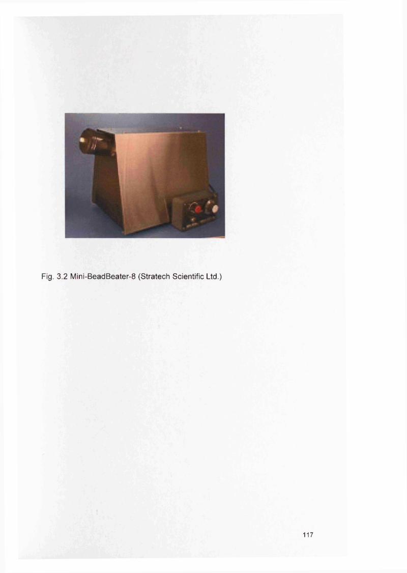
Fig. 3.2 Mini-BeadBeater-8 (Stratech Scientific Ltd.)
117
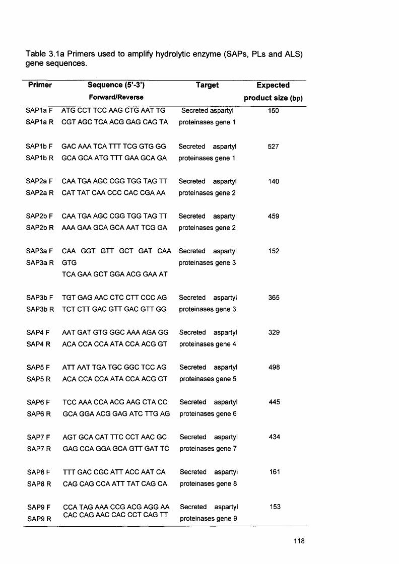
Table 3.1a Primers used to amplify hydrolytic enzyme (SAPs, PLs and ALS)gene sequences.
Primer Sequence (5’-3’) Forward/Reverse
Target Expected
product size (bp)
SAP1a F SAP 1a R
ATG CCT TCC AAG CTG AAT TG CGT AGC TCA ACG GAG CAG TA
Secreted aspartyl proteinases gene 1
150
SAP1b F SAP1b R
GAC AAA TCA TTT TCG GTG GG GCA GCA ATG TTT GAA GCA GA
Secreted aspartyl proteinases gene 1
527
SAP2a F
SAP2a RCAA TGA AGC CGG TGG TAG TT
CAT TAT CAA CCC CAC CGA AASecreted aspartyl proteinases gene 2
140
SAP2b F SAP2b R
CAA TGA AGC CGG TGG TAG TT AAA GAA GCA GCA AAT TCG GA
Secreted aspartyl proteinases gene 2
459
SAP3a F
SAP3aRCAA GGT GTT GCT GAT CAA
GTGTCA GAA GCT GGA ACG GAA AT
Secreted aspartyl proteinases gene 3
152
SAP3bF SAP3bR
TGT GAG AAC CTC CTT CCC AG TCT CTT GAC GTT GAC GTT GG
Secreted aspartyl proteinases gene 3
365
SAP4F SAP4 R
AAT GAT GTG GGC AAA AGA GG ACA CCA CCA ATA CCA ACG GT
Secreted aspartyl proteinases gene 4
329
SAP5FSAP5R
ATT AAT TGA TGC GGC TCC AG
ACA CCA CCA ATA CCA ACG GTSecreted aspartyl proteinases gene 5
498
SAP6FSAP6R
TCC AAA CCA ACG AAG CTA CC GCA GGA ACG GAG ATC TTG AG
Secreted aspartyl proteinases gene 6
445
SAP7 F SAP7R
AGT GCA CAT TTC CCT AAC GC GAG CCA GGA GCA GTT GAT TC
Secreted aspartyl proteinases gene 7
434
SAP8FSAP8R
TTT GAC CGC ATT ACC AAT CA
CAG CAG CCA ATT TAT CAG CASecreted aspartyl proteinases gene 8
161
SAP9FSAP9R
CCA TAG AAA CCG ACG AGG AA CAC CAG AAC CAC CCT CAG TT
Secreted aspartyl proteinases gene 9
153
118

Table 3.1b Primers used to amplify hydrolytic enzyme (PLs & controls) genesequences.
Primer Sequence (5’-3’)
Forward/ReverseTarget Expected
product
size
(bp)PLB1 F PLB1 R
CAA CGA AGC GGT GTT GTC TA TTG CTG CCA GAA CTT TTG AA
Phospholipase gene B1
150
PLB2FPLB2R
GGC CAG ATG GAT CAG CTT TA
AAG TTC TGG GCA TCA CAT CCPhospholipase gene B2
154
PLCFPLCR
GGT TCA ATT CGG CTG GTG AT
TTG TTC ACC GGA ATG TCA AAPhospholipase gene C
150
PLDFPLDR
GAC AAA GCA GCG CCT AAA TC
CCC TTT TCC TGG TTT TGG ATPhospholipase gene D
148
EFB1 F EFB1 R
GAA CGA ATT CTT GGC TGA C CAT CAG AAC CGA ACA AGT C
C. albicans elongation factor
1P
242
S14FS14R
CAG GTC CAG GGG TCT TGG TCC GGC AGA CCG AGA TGA ATC CTC A
Human control ribosomal RNA
143
119
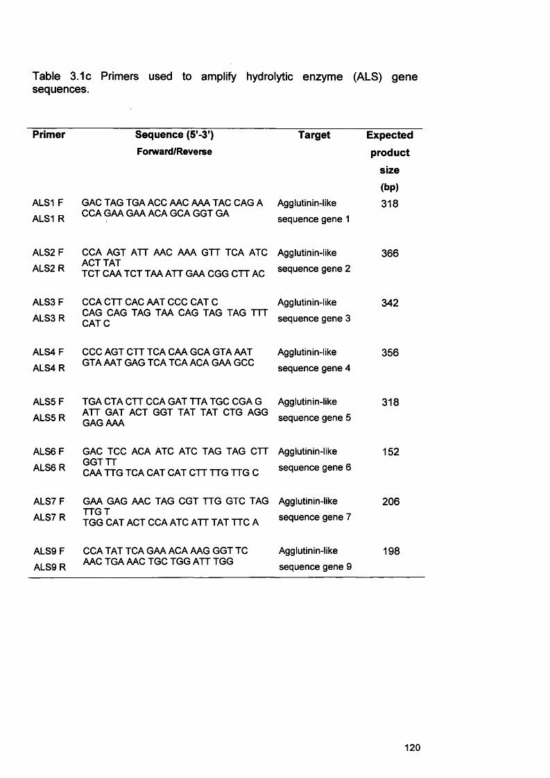
Table 3.1c Primers used to amplify hydrolytic enzyme (ALS) genesequences.
Primer Sequence (5’-3’)
Forward/ReverseTarget Expected
product
size
(bp)ALS1 F GAC TAG TGA ACC AAC AAA TAC CAG A Agglutinin-like 318ALS1 R CCA GAA GAA ACA GCA GGT GA sequence gene 1
ALS2F CCA AGT ATT AAC AAA GTT TCA ATC Agglutinin-like 366ALS2R ACT TAT
TCT CAA TCT TAA ATT GAA CGG CTT AC sequence gene 2
ALS3F CCA CTT CAC AAT CCC CAT C Agglutinin-like 342ALS3R CAG CAG TAG TAA CAG TAG TAG TTT
CATC sequence gene 3
ALS4F CCC AGT CTT TCA CAA GCA GTA AAT Agglutinin-like 356ALS4R GTA AAT GAG TCA TCA ACA GAA GCC sequence gene 4
ALS5F TGA CTA CTT CCA GAT TTA TGC CGA G Agglutinin-like 318ALS5R ATT GAT ACT GGT TAT TAT CTG AGG
GAG AAA sequence gene 5
ALS6F GAC TCC ACA ATC ATC TAG TAG CTT Agglutinin-like 152ALS6R GGTTT
CAA TTG TCA CAT CAT CTT TTG TTG C sequence gene 6
ALS7F GAA GAG AAC TAG CGT TTG GTC TAG Agglutinin-like 206ALS7R TTG T
TGG CAT ACT CCA ATC ATT TAT TTC A sequence gene 7
ALS9F CCA TAT TCA GAA ACA AAG GGT TC Agglutinin-like 198ALS9R AAC TGA AAC TGC TGG ATT TGG sequence gene 9
120
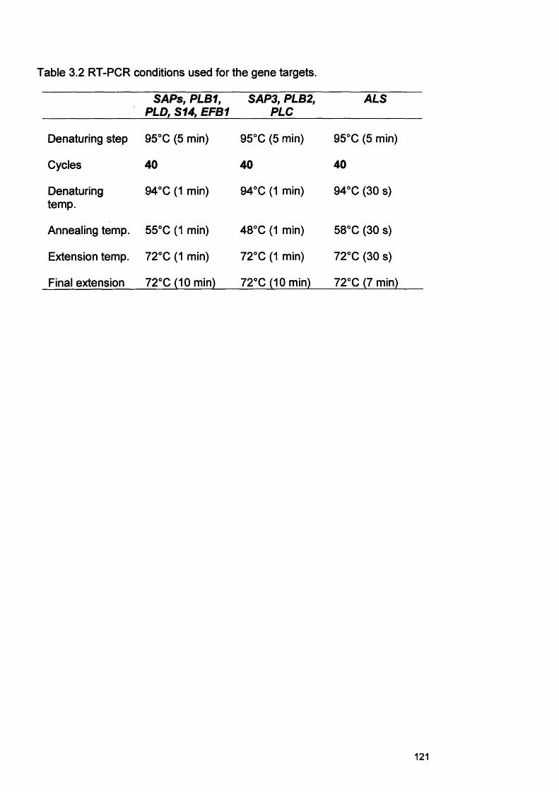
Table 3.2 RT-PCR conditions used for the gene targets.
SAPs, PLB1, PLD, S14, EFB1
SAP3, PLB2, PLC
ALS
Denaturing step 95°C (5 min) 95°C (5 min) 95°C (5 min)
Cycles 40 40 40
Denaturingtemp.
94°C (1 min) 94°C (1 min) 94°C (30 s)
Annealing temp. 55°C (1 min) 48°C (1 min) 58°C (30 s)
Extension temp. 72°C (1 min) 72°C (1 min) 72°C (30 s)
Final extension 72°C (10 min) 72°C (10 min) 72°C (7 min)
121
Controls for each reaction included tissue extracts not treated with reverse
transcriptase (to establish that genomic DNA amplification was not occurring)
and a water template control.
3.3.7 Gel electrophoresis of PCR productsPCR products (10 pi) were visualised following standard gel electrophoresis
in a 0.75 % agarose gel (Sigma, UK) with ethidium bromide staining (10 pg
pi'1). Gel images were obtained using a Gel-Doc 2000 (Bio-Rad, Hemel-
Hempstead, UK) and the average band densities for each PCR product were
measured using the Discovery Series™ Quantity One® (Bio-Rad) software.
Estimation of semi-quantitative values involved defining lanes and bands on
the gel image following the Quantity One user guide. After defining lanes, a
lane-based background subtraction was performed with bands identified
using the Detect Bands command and then the values of a standard (a
quantitative MassRuler DNA Ladder; low range, Fermentas, York, UK) being
entered. The values of the experimental bands were then determined using
this standard. The DNA concentration of each PCR product was then
expressed as a percentage relative to that of the matched housekeeping
gene (EFB1) amplified for each strain.
122
3.4 Results
3.4.1 RNA extraction-comparison of the methods
It was not possible to extract RNA from C. albicans using the ultraspec
method. For this study RNA extraction was most consistently obtained using
physical cell disruption and the RNeasy mini kit method (section 3.3.3).
Subsequent results are therefore based on the latter RNA extraction protocol.
3.4.2 Control gene expression
For all RT-PCRs, control gene expression was routinely detected by EFB1
and S14 primers (Fig. 3.3 - 3.4).
3.4.2.1 SAP gene expression by C. albicans cultures without RHE
(maintenance medium alone)Sixteen out of 19 strains expressed SAP1-3. The non-invasive strains
(WK1/93; 40/01) did not express SAP1-3. SAP4-6 was expressed by 10 out
of 19 strains. The non-invasive strains (WK1/93; 40/01) did not express
SAP4-6. None of the tested strains expressed SAP7. SAP8 was expressed
by all strains except non invading strain WK1/93. SAP9 was expressed by 7
strains out of 19 (Table 3.3).
3.4.2.2 PLs gene expression by C. albicans cultures without RHE
(maintenance medium alone)PLs gene expression (Table 3.3) appeared to be more frequently detected in
Candida isolates without the RHE. Six strains out of 19 strains expressed
PLB1, PLB2 and PLD, whilst 6 expressed PLC. The non-invasive strains
(WK1/93 and 40/01) did not express PLB2 and PLC. When cultured without
the RHE, 44 PL amplicons were evident (with multiple genes expressed by
individual strains).
123
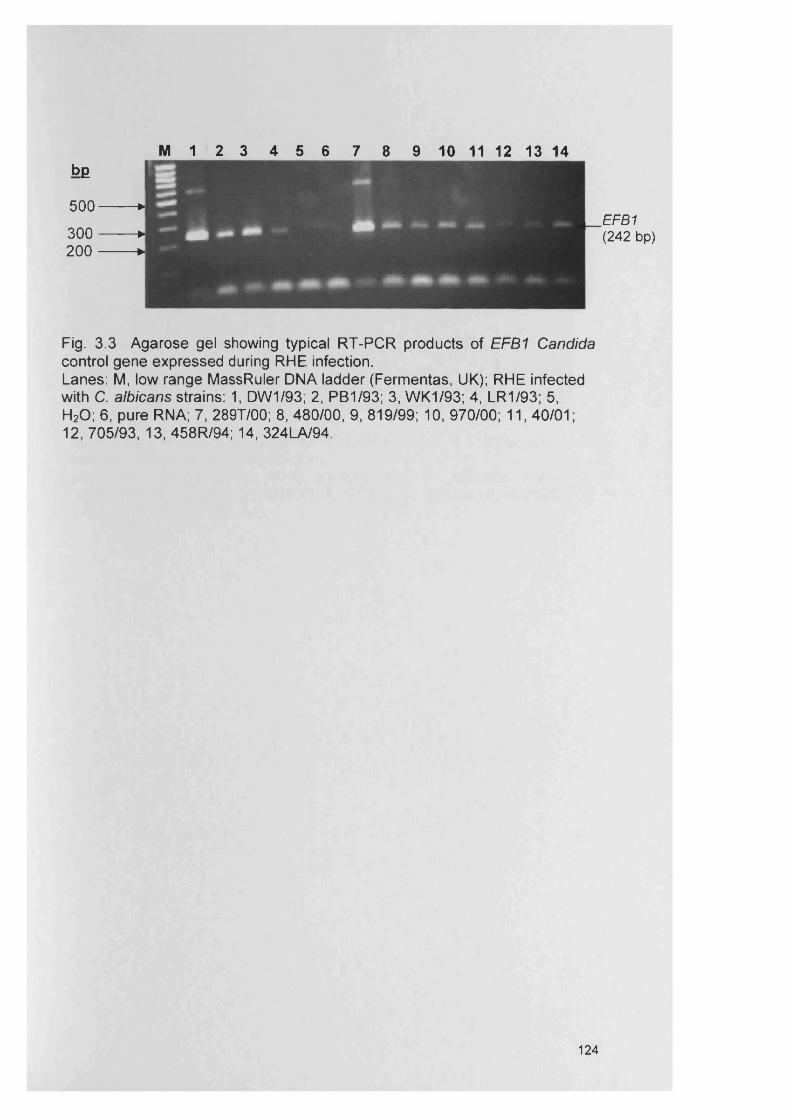
M 1 2 3 4 5 6 7 8 9 10 11 12 13 14b£
5 0 0 ---------J3 0 0 ---------J200 H
Fig. 3.3 Agarose gel showing typical R T-PC R products of EFB1 Candida control gene expressed during RHE infection.Lanes: M, low range MassRuler DNA ladder (Fermentas, UK); RHE infected with C. albicans strains: 1, DW 1/93; 2, PB1/93; 3, W K1/93; 4, LR1/93; 5,H20 ; 6, pure RNA; 7, 289T/00; 8, 480/00, 9, 819/99; 10, 970/00; 11, 40/01;12, 705/93, 13, 458R/94; 14, 324LA/94.
EFB1 (242 bp)
124
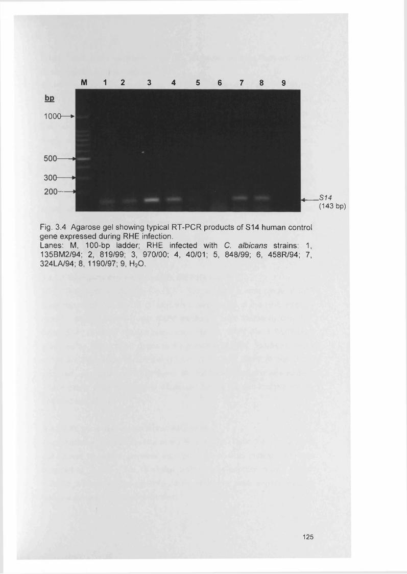
M 1 2 3 4 5 6 7 8 9
1000— ►
Fig. 3.4 Agarose gel showing typical R T-PC R products of S14 human control gene expressed during RHE infection.Lanes: M, 100-bp ladder; RHE infected with C. albicans strains: 1, 135BM 2/94; 2, 819/99; 3, 970/00; 4, 40/01; 5, 848/99; 6, 458R /94; 7, 324LA/94; 8 ,1 1 90 /9 7 ; 9, H20 .
.S14 (143 bp)
125
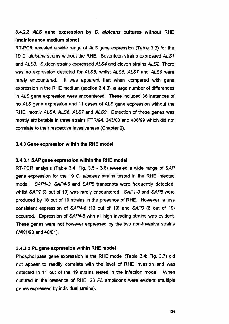
3.4.2.3 ALS gene expression by C. albicans cultures without RHE
(maintenance medium alone)
RT-PCR revealed a wide range of ALS gene expression (Table 3.3) for the
19 C. albicans strains without the RHE. Seventeen strains expressed ALS1
and ALS3. Sixteen strains expressed ALS4 and eleven strains ALS2. There
was no expression detected for ALS5, whilst ALS6, ALS7 and ALS9 were
rarely encountered. It was apparent that when compared with gene
expression in the RHE medium (section 3.4.3), a large number of differences
in ALS gene expression were encountered. These included 36 instances of
no ALS gene expression and 11 cases of ALS gene expression without the
RHE, mostly ALS4, ALS6, ALS7 and ALS9. Detection of these genes was
mostly attributable in three strains PTR/94, 243/00 and 408/99 which did not
correlate to their respective invasiveness (Chapter 2).
3.4.3 Gene expression within the RHE model
3.4.3.1 SAP gene expression within the RHE modelRT-PCR analysis (Table 3.4; Fig. 3.5 - 3.6) revealed a wide range of SAP
gene expression for the 19 C. albicans strains tested in the RHE infected
model. SAP1-3, SAP4-6 and SAP8 transcripts were frequently detected,
whilst SAP7 (3 out of 19) was rarely encountered. SAP1-3 and SAP8 were
produced by 18 out of 19 strains in the presence of RHE. However, a less
consistent expression of SAP4-6 (13 out of 19) and SAP9 (6 out of 19)
occurred. Expression of SAP4-6 with all high invading strains was evident.
These genes were not however expressed by the two non-invasive strains
(WK1/93 and 40/01).
3.4.3.2 PL gene expression within RHE modelPhospholipase gene expression in the RHE model (Table 3.4; Fig. 3.7) did
not appear to readily correlate with the level of RHE invasion and was
detected in 11 out of the 19 strains tested in the infection model. When
cultured in the presence of RHE, 23 PL amplicons were evident (multiple
genes expressed by individual strains).
126
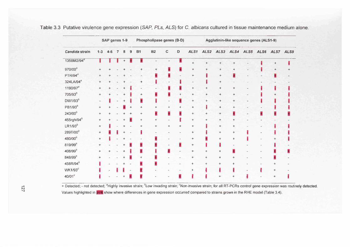
Table 3.3 Putative virulence gene expression (SAP, PLs, ALS) for C. albicans cultured in tissue maintenance medium alone.
SAP genes 1-9 Phospholipase genes (B-D) Agglutinin-like sequence genes (ALS1-9)
Candida strain 1-3 4-6 7 8 9 B1 B2 c D ALS1 ALS2 ALS3 ALS4 ALS5 ALS6 ALS7 ALS9
135BM2/94a l 1 1 + l 1 - B + + + + - 1 + 1970/00a + + - + - + + 1 1 + + + + - 1 + +PTR/94a + + - + - - 1 1 - + 1 + 1 - - 1 -324LA/94a + + - + - + 1 - 1 - 1 + - - - - -1190/97a + + - + I - - 1 1 + + + + - 1 1 I705/936 + + - + I + 1 1 + + + + + - 1 + 1DW1/93b - 1 - + 1 i 1 - ■ + + + + - 1 l 1PB1/936 + + - 1 + + + + + + 1 + + - - l 1243/006 + + - + + + 1 1 ■ + + + 1 - 1 I 1455rgh/94b + + - + 1 + + - i + + + + - - - -LR1/936 + 1 - + - + + + + + i + + - - I I289T/006 + 1 1 + - i - - - + i + + 1 - l l480/00b + 1 - + - - 1 - - + i + + l - + l819/99b + - - + 1 1 1 - 1 + i 1 - - - 1 -408/99b + + - + 1 1 1 1 - + + + 1 - - 1 1848/99b + - - + 1 + 1 - - + + + + - - 1 -458R/94b i - - + - i 1 - - + + + + - - + -WK1/93C I - 1 1 - i - - - + I 1 1 - I + -40/01° i - - + 1 i - - 1 1 i + + 1 - + 1
+ Detected; - not detected; aHighly invasive strain; bLow invading strain; cNon-invasive strain; for all RT-PCRs control gene expression was routinely detected. Values highlighted in M l show where differences in gene expression occurred compared to strains grown in the RHE model (Table 3.4).
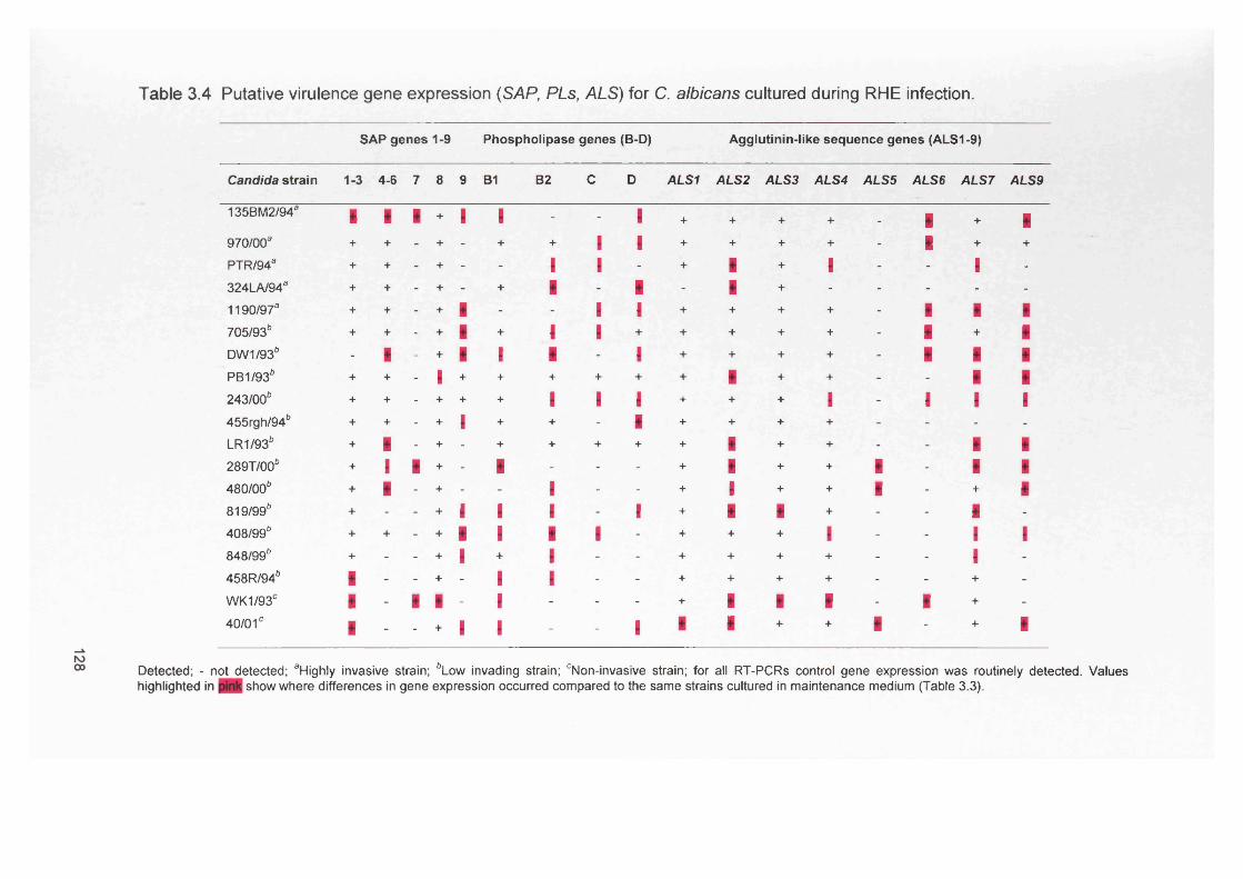
Table 3.4 Putative virulence gene expression (SAP, PLs, ALS) for C. albicans cultured during RHE infection.
SAP genes 1-9 Phospholipase genes (B-D) Agglutinin-like sequence genes (ALS1-9)
Candida strain 1-3 4-6 7 8 9 B1 B2 c D ALS1 ALS2 ALS3 ALS4 ALS5 ALS6 ALS7 ALS9
135BM2/943 i 1 1 + 1 1 - - 1 + + + + - 1 + 1970/003 + + - + - + + 1 1 + + + + - 1 + +
PTR/943 + + - + - - 1 1 - + i + 1 - - 1 -324LA/943 + + - + - + 1 - 1 - i + - - - - -1190/973 + + - + 1 - - 1 1 + + + + - 1 1 1705/93b + + - + 1 + 1 1 + + + + + - 1 + 1DW1/93b - 1 - + 1 i 1 - 1 + + + + - 1 1 1PB1/93b + + - I + + + + + + i + + - - 1 1243/00b + + - + + + 1 1 1 + + + l - 1 1 1455rgh/94b + + - + 1 + + - 1 + + + + - - - -LR1/93b + 1 - + - + + + + + 1 + + - - 1 1289T/006 + 1 i + - i - - - + 1 + + I - 1 1480/00b + 1 - + - - 1 - - + 1 + + i - + 1819/99b + - - + 1 1 1 - I + 1 1 + - - 1 -408/99b + + - + 1 1 1 1 - + + + l - - 1 I848/99b + - - + 1 + 1 - - + + + + - - 1 -458R/946 i - - + - I 1 - - + + + + - - + -WK1/930 i - i i - i - - - + 1 i l - I + -40/01° i - - + I I - - 1 1 1 + + 1 - + i
Detected; - not detected; aHighly invasive strain; bLow invading strain; cNon-invasive strain; for all RT-PCRs control gene expression was routinely detected. Values highlighted in show where differences in gene expression occurred compared to the same strains cultured in maintenance medium (Table 3.3).
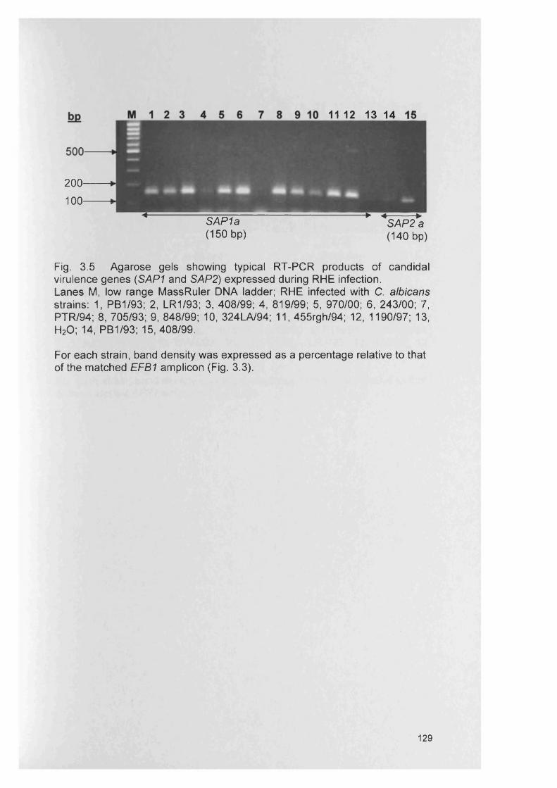
11 12 13 14
200100
SAP 1a (150 bp)
SAP2 a (140 bp)
Fig. 3.5 Agarose gels showing typical RT-PCR products of candidal virulence genes (SAP1 and SAP2) expressed during RHE infection.Lanes M, low range MassRuler DNA ladder; RHE infected with C. albicans strains: 1, PB1/93; 2, LR1/93; 3, 408/99; 4, 819/99; 5, 970/00; 6, 243/00; 7, PTR/94; 8, 705/93; 9, 848/99; 10, 324LA/94; 11, 455rgh/94; 12, 1190/97; 13, H20; 14, PB1/93; 15, 408/99.
For each strain, band density was expressed as a percentage relative to that of the matched EFB1 amplicon (Fig. 3.3).
129
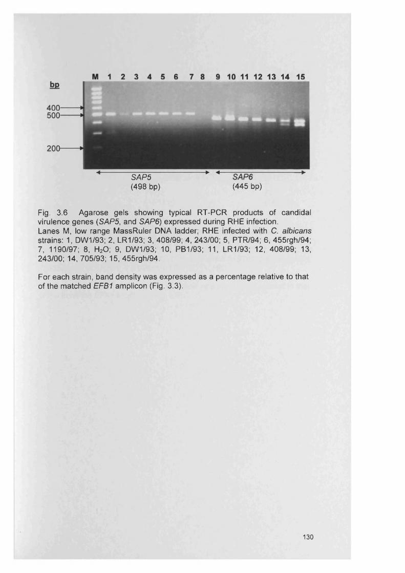
SAP5 *" ■*(498 bp) (445 bp)
Fig. 3.6 Agarose gels showing typical RT-PCR products of candidal virulence genes (SAP5, and SAP6) expressed during RHE infection.Lanes M, low range MassRuler DNA ladder; RHE infected with C. albicans strains: 1, DW1/93; 2, LR1/93; 3, 408/99; 4, 243/00; 5, PTR/94; 6, 455rgh/94; 7, 1190/97; 8, H20; 9, DW1/93; 10, PB1/93; 11, LR1/93; 12, 408/99; 13, 243/00; 14, 705/93; 15, 455rgh/94.
For each strain, band density was expressed as a percentage relative to that of the matched EFB1 amplicon (Fig. 3.3).
130
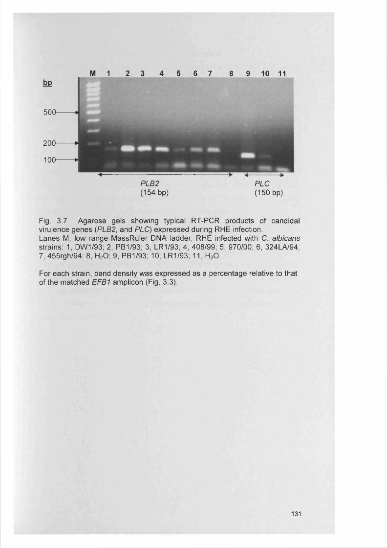
bg
500
200
100
Fig. 3.7 Agarose gels showing typical RT-PCR products of candidal virulence genes (PLB2, and PLC) expressed during RHE infection.Lanes M, low range MassRuler DNA ladder; RHE infected with C. albicans strains: 1, DW1/93; 2, PB1/93; 3, LR1/93; 4, 408/99; 5, 970/00; 6, 324LA/94; 7, 455rgh/94; 8, H20; 9, PB1/93; 10, LR1/93; 11, H20.
For each strain, band density was expressed as a percentage relative to that of the matched EFB1 amplicon (Fig. 3.3).
M 1 2 3 4 5 6 7 8 9 10 11
PLB2 PLC(154 bp) (150 bp)
131
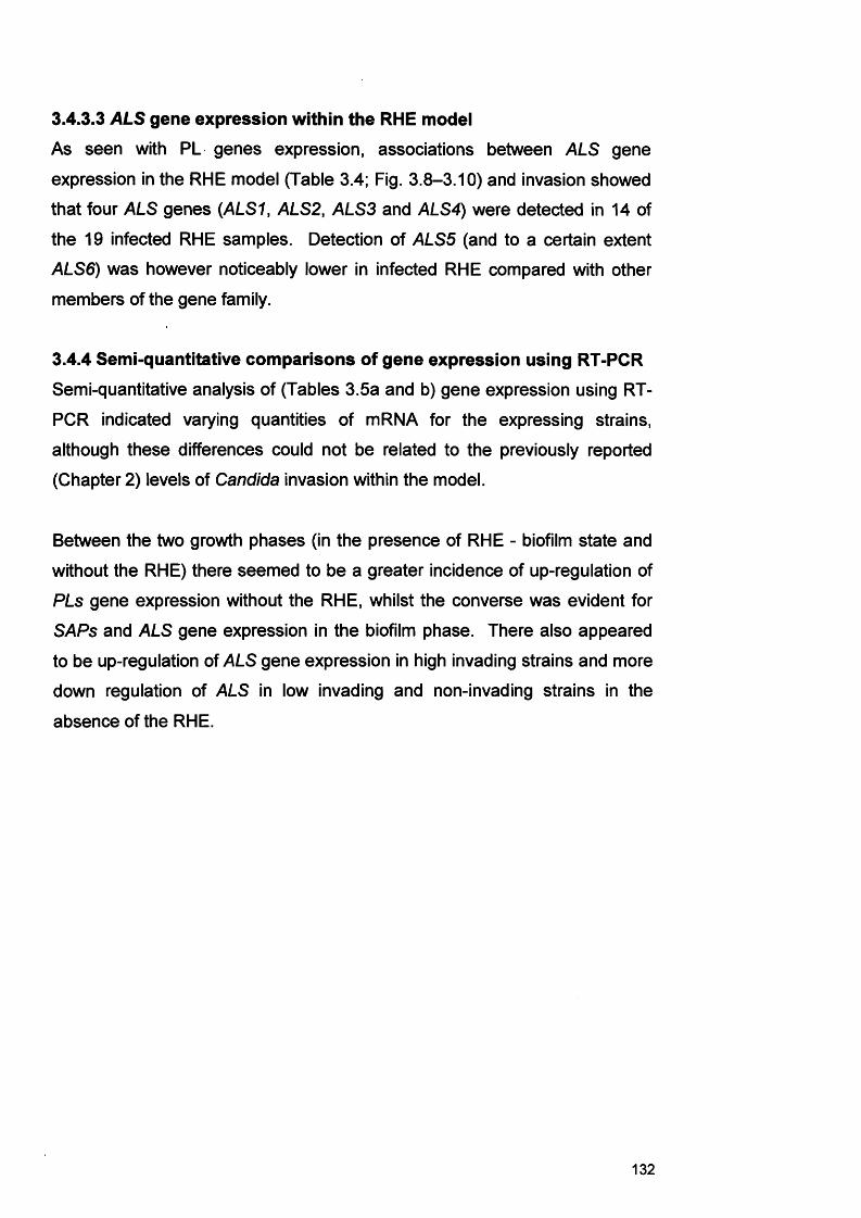
3.4.3.3 ALS gene expression within the RHE model
As seen with PL genes expression, associations between ALS gene
expression in the RHE model (Table 3.4; Fig. 3.8-3.10) and invasion showed
that four ALS genes (ALS1, ALS2, ALS3 and ALS4) were detected in 14 of
the 19 infected RHE samples. Detection of ALS5 (and to a certain extent
ALS6) was however noticeably lower in infected RHE compared with other
members of the gene family.
3.4.4 Semi-quantitative comparisons of gene expression using RT-PCRSemi-quantitative analysis of (Tables 3.5a and b) gene expression using RT-
PCR indicated varying quantities of mRNA for the expressing strains,
although these differences could not be related to the previously reported
(Chapter 2) levels of Candida invasion within the model.
Between the two growth phases (in the presence of RHE - biofilm state and
without the RHE) there seemed to be a greater incidence of up-regulation of
PLs gene expression without the RHE, whilst the converse was evident for
SAPs and ALS gene expression in the biofilm phase. There also appeared
to be up-regulation of ALS gene expression in high invading strains and more
down regulation of ALS in low invading and non-invading strains in the
absence of the RHE.
132
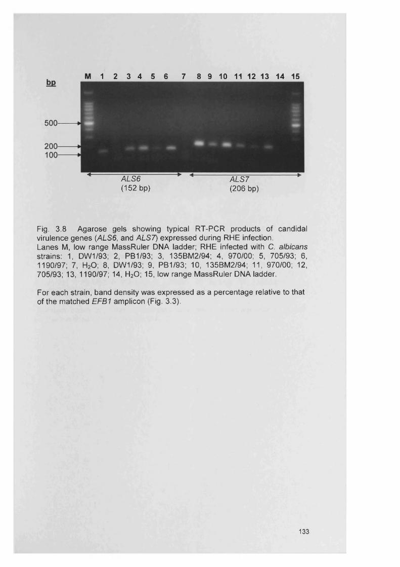
M 1 2 3 4 5 6 7 8 9 10 11 12 13 14 15
ALS6 (152 bp)
ALS7 (206 bp)
Fig. 3.8 Agarose gels showing typical RT-PCR products of candidal virulence genes (ALS6, and ALS7) expressed during RHE infection.Lanes M, low range MassRuler DNA ladder; RHE infected with C. albicans strains: 1, DW1/93; 2, PB1/93; 3, 135BM2/94; 4, 970/00; 5, 705/93; 6, 1190/97; 7, H20; 8, DW1/93; 9, PB1/93; 10, 135BM2/94; 11, 970/00; 12, 705/93; 13, 1190/97; 14, H20; 15, low range MassRuler DNA ladder.
For each strain, band density was expressed as a percentage relative to that of the matched EFB1 amplicon (Fig. 3.3).
133
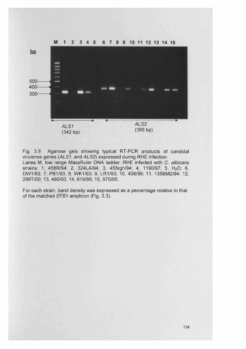
M 1 2 3 4 5 6 7 8 9 10 11 12 13 14 15
ALS1 (342 bp)
ALS2 (366 bp)
Fig. 3.9 Agarose gels showing typical RT-PCR products of candidal virulence genes (ALS1, and ALS2) expressed during RHE infection.Lanes M, low range MassRuler DNA ladder; RHE infected with C. albicans strains: 1, 458R/94; 2, 324LA/94; 3, 455rgh/94; 4, 1190/97; 5, H20; 6, DW1/93; 7, PB1/93; 8, WK1/93; 9, LR1/93; 10, 408/99; 11, 135BM2/94; 12, 289T/00; 13, 480/00; 14, 819/99; 15, 970/00.
For each strain, band density was expressed as a percentage relative to that of the matched EFB1 amplicon (Fig. 3.3).
134
Fig. 3.10 Agarose gels showing typical RT-PCR products of candidal virulence genes (ALS1) expressed during RHE infection.Lanes M, low range MassRuler DNA ladder; RHE infected with C. albicans strains: 1, DW1/93; 2, PB1/93; 3, WK1/93; 4, LR1/93; 5, 408/99; 6, 135BM2/94; 7, 289T/00; 8, 480/00; 9, 819/99; 10, 970/00; 11, 40/01; 12, 243/00; 13, PTR/94; 14, 705/93; 15, 848/99.
For each strain, band density was expressed as a percentage relative to that of the matched EFB1 amplicon (Fig. 3.3).
135
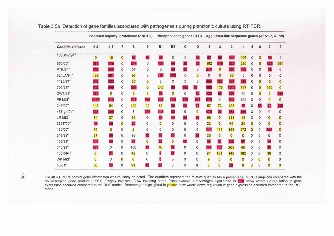
Table 3.5a Detection of gene families associated with pathogenesis during planktonic culture using RT-PCR.
Secreted aspartyl proteinase (SAPI-9) Phospholipase genes (B-D) Agglutinin- like sequence genes (ALS1-7, ALS9)
Candida albicans 1-3 4-6 7 8 9 B1 B2 C D 1 2 3 4 5 6 7 9
135BM2/94* 0 19 0 80 ■ 48 0 0 48 87 88 ■ 107 0 0 ■ 0970/00a 351 m 0 m 0 150 44 26 27 192 148 328 230 0 0 422 264PTR/94* m 243 0 57 0 0 48 80 0 m 0 406 178 0 0 16 0324LA/94* 162 m 0 96 0 154 m 0 0 0 0 60 0 0 0 0 01190/97* 542 u 0 85 0 0 0 0 0 664 136 467 312 0 0 0 0705/93* 842 348 0 504 0 245 88 605 85 480 178 1040 137 0 0 120 0DW1/93* 188 0 0 5 0 32 0 0 68 ■ ez 388 75 0 0 0 422PB1/93* iW 414 0 m m 282 tie 378 387 878 0 926 359 0 0 0 0243/00* 143 84 0 102 49 85 38 84 27 67 72 105 m 0 56 29 125455rgh/94* 828 320 0 8 278 m 82 0 0 176 89 363 164 0 0 0 0LR1/93* 81 27 0 66 0 m m 24 ■ 56 0 111 14 0 0 0 0289T/00* m 58 0 20 0 0 0 0 0 23 0 69 24 0 0 0 0480/00* 56 0 0 2 0 0 0 0 0 m 113 199 172 0 0 ■ 0819/99* 67 55 0 96 36 76 30 0 38 30 0 0 0 0 0 0 0408/99* m m 0 m 0 25 0 ■ 0 68 60 125 87 0 0 M 0848/99* ■ 0 0 180 m 76 59 0 0 m 103 265 45 0 0 30 0458R/94* 0 21 0 42 0 1 » 0 0 91 101 145 182 0 0 38 0WK1/930 0 0 0 0 0 8 0 0 0 9 0 0 0 0 0 0 040/01c 56 49 0 91 ■ 25 0 0 8 0 0 52 0 0 0 0 0
For all RT-PCRs control gene expression was routinely detected. The numbers represent the relative quantity (as a percentage) of PCR amplicon compared with the housekeeping gene product (EFB1). aHighly invasive. *Low invading strain. cNon-invasive. Percentages highlighted in |>in| show where up-regulation in gene expression occurred compared to the RHE model. Percentages highlighted in yellow show where down regulation in gene expression occurred compared to the RHE model.
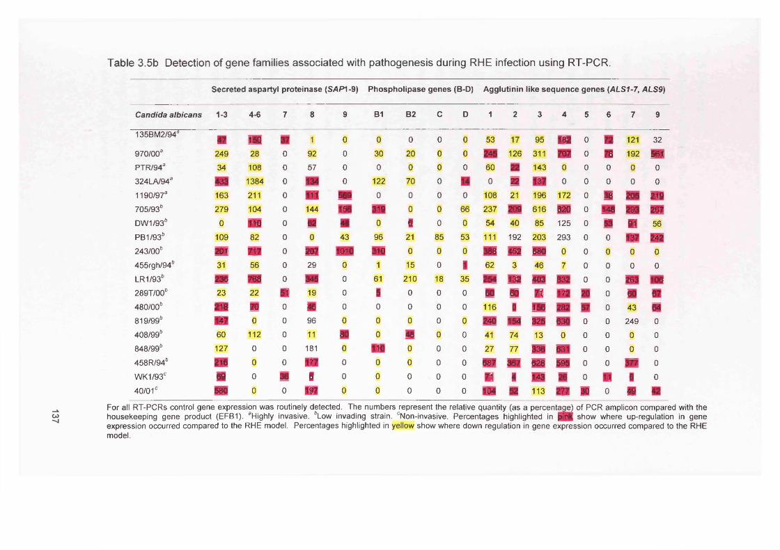
Table 3.5b Detection of gene families associated with pathogenesis during RHE infection using RT-PCR.
Secreted aspartyl proteinase (SAP1-9) Phospholipase genes (B-D) Agglutinin like sequence genes (ALS1-7, ALS9)
Candida albicans 1-3 4-6 7 8 9 B1 B2 c D 1 2 3 4 5 6 7 9
135BM2/94aF m 180 m 1 0 0 0 0 0 53 17 95 ■ 0 m 121 32970/00a 249 28 0 92 0 30 20 0 0 246 126 311 m 0 m 192 561PTR/94a 34 108 0 57 0 0 0 0 0 60 n 143 0 0 0 0 0324LA/943 433 1384 0 m 0 122 70 0 ■ 0 22 m 0 0 0 0 01190/973 163 211 0 m 589 0 0 0 0 108 21 196 172 0 38 205 219705/93* 279 104 0 144 188 319 0 0 66 237 209 616 820 0 148 293 267DW1/93* 0 m 0 82 ■ 0 1 0 0 54 40 85 125 0 53 m 56PB1/93* 109 82 0 0 43 96 21 85 53 111 192 203 293 0 0 m 242243/00* m m 0 207 1010 310 0 0 0 388 462 680 0 0 0 0 0455rgh/94* 31 56 0 29 0 1 15 0 1 62 3 46 7 0 0 0 0LR1/93* 236 788 0 340 0 61 210 18 35 254 132 403 832 0 0 263 m289T/00* 23 22 ■ 19 0 8 0 0 0 60 00 m m 20 0 60 m480/00* m m 0 « 0 0 0 0 0 116 1 156 282 57 0 43 64819/99* M 0 0 96 0 0 0 0 0 240 m 325 630 0 0 249 0408/99* 60 112 0 11 80 0 48 0 0 41 74 13 0 0 0 0 0848/99* 127 0 0 181 0 ■ 0 0 0 27 77 336 631 0 0 0 0458R/94* 216 0 0 m 0 0 0 0 0 687 387 028 595 0 0 377 0WK1/93C 69 0 38 ■ 0 0 0 0 0 m 1 V® 26 0 ■ 1 040/01c 580 0 0 m 0 0 0 0 0 m 82 113 m 30 0 ■ ■
For all RT-PCRs control gene expression was routinely detected. The numbers represent the relative quantity (as a percentage) of PCR amplicon compared with the housekeeping gene product (EFB1). aHighly invasive. *Low invading strain. cNon-invasive. Percentages highlighted in |)in | show where up-regulation in gene expression occurred compared to the RHE model. Percentages highlighted in yellow show where down regulation in gene expression occurred compared to the RHE model.
3.5 DiscussionCompared to bacteria, there has been little work looking at C. albicans gene
expression, or, indeed, at gene expression in fungal biofilms generally
(Douglas, 2003). However, it has been shown that Candida albicans
expresses several virulence factors that contribute to pathogenesis
(Calderone and Fonzi, 2001). Genes associated with virulence factors
include those linked with adhesion to the host tissue (e.g. ALS genes),
morphogenesis, and hydrolytic enzymes, such as SAPs, and PLs (Jayatilake
et al., 2005; Schaller et al., 2005). The main role of the hydrolytic enzymes is
to provide nutrition for the cells, to aid penetration and invasion and to evade
immune responses (Naglik et al., 2003) by degrading cellular substrates.
In various Candida infections, these gene families may be differentially
regulated and prevention and control of expression might be achieved by
pharmacological or immunological tools specifically designed to modulate or
inhibit these virulence factors (Schaller et al., 2005). This present Chapter
compared the expression of specific candidal virulence factors in an in vitro
RHE model and, as a control, devoid of tissue.
It was apparent in this study that those strains previously categorised as ‘high
invaders’ (Chapter 2) consistently expressed SAPs4-6 despite the level of
expression being strain variable. It is important to note that 4 “high invading”
strains (135BM2/94, PTR/94, 324LA/94, 1190/97) and 4 “low-invading”
(705/93, 455rhg/94, 848/99 and 458R/94) that originated from CHC infections
all expressed SAPs1-3, and six of them expressed SAPs4-6 (135BM2/94,
PTR/94, 324LA/94, 1190/97, 705/93 and 455rhg/94). These findings may
suggest that SAPs play a role in oral Candida infections, such as CHC.
However, some of the “low invaders” also expressed SAP1-3 and SAP4-6
which may suggest that host factors, including tobacco smoking, denture
wearing and regular alcohol intake, also play an important role in the
development of CHC (Bartie et al., 2004). Furthermore, SAP genes 4-6 were
not detected for the two non-invading strains (WK1/93 and 40/01). SAP
genes 4-6 are believed to be important in controlling the yeast to hyphal
138
transition (Naglik et al., 2003) and so an indirect role for these enzymes in
tissue invasion can readily be envisaged. These results might be considered
to contrast with those of Korting et al (2003), who used efg1/cph1 mutants of
C. albicans SC5314 (which are known to exhibit reduced expression of the
hyphal-associated genes SAP4-SAP6) to demonstrate that SAP1 and SAP3
were important causes of epithelial cell damage in the same RHE model. In
those studies, epithelial cell injury was determined by lactate dehydrogenase
(LD) release and this correlated with the presence of SAP1 and SAP3
transcripts (Korting et al., 2003). Unfortunately, the model of RHE invasion
used in this study did not incorporate the LDH release assay, and so any
associations between SAP expression, invasion and tissue damage could not
be confirmed. Since SAP1-3 transcripts were detected in the majority of
strains, it may be the case that tissue damage was still occurring, even for
strains where limited RHE invasion was detected. In addition, gene
expression was also assessed for C. albicans strains cultured in the tissue
maintenance medium in an identical culture environment but in absence of
the RHE tissue. A comparison of gene expression revealed a largely
consistent expression of SAPs between the two growth environments.
However, there were some notable differences, with an up-regulation of
some SAPs evident in the presence of the RHE, which may suggest that
external factors such as the growth environment play an important role.
A relationship between RHE invasion and agglutinin-like sequence (ALS)
gene expression was not evident. However, there may have been up-
regulation of these genes for some C. albicans strains in the presence of the
RHE; in particularly this was seen with ALS2, ALS6, ALS7 and ALS9. The
ALS gene family designated ALS1-ALS7 and ALS9 has recently been
implicated with facilitating candidal adherence to host tissues (Green et al.,
2004; Hoyer, 2001). Garcia-Sanchez et al (2004) also looked at differences
in ALS gene expression between biofilm and planktonic populations and
contrary to this present work ALS1 was dramatically over-expressed in all the
biofilm populations and ALS7 was under-expressed compared to planktonic
ones. However, those differences could be explained by different culture
139

conditions and these investigators were not looking at gene expression in
conjunction with the RHE model. However, the expression of ALS genes in
this study, agreed in part, with those reported previously by Green et al.
(2004), who also used a similar RHE infection model. Green et al. (2004)
found consistent expression of ALS1, ALS2, ALS3 and ALS4, in contrast to
the more sporadic detection of ALS6 and ALS7 genes. In this present
investigation, a lower incidence of ALS5 and ALS6 was found (3/19 and 6/19
strains respectively). However, only four C. albicans strains were used by
Green et al. (2004) as opposed to the 19 strains from specifically-defined oral
infections used in this study, which may account for these apparent
differences.
Phospholipase expression similarly did not appear to correlate with RHE
invasion (Table 3.4), as exemplified by the fact that three highly invasive
strains did not produce mRNA for any PLs, whilst the poorly invasive C.
albicans PB1/93 and C. albicans LR1/93 were positive for expression of all
four PL genes. Furthermore, PL gene expression actually seemed to be
higher in the absence of the RHE which might suggest the possible induction
of PL genes through a starvation trigger as opposed to contact with tissue.
Damage of the RHE during infection may negate this ‘starvation’ trigger
through the release of nutrients for the infecting organism.
Phenotypic differences between biofilm and planktonic organisms are often
regarded as important pathogenic determinants. These phenotypic changes
may manifest through differential gene control arising from environmental
triggers. For example, adherent organisms within a biofilm may express a
pattern of genes different from that of their planktonic counterparts (Becker et
al., 2001). It has been demonstrated with Escherichia coli that levels of gene
expression between biofilm and planktonic populations differ significantly
(Becker et al., 2001). Understanding gene expression patterns might
therefore be useful for determining when certain proteins are generated in C.
albicans and enhance our understanding on the pathogenicity of this
organism (Green et al., 2004). This study showed that some of the strains
140
which are from conditions such as CHC express SAPs 4-6 (important for
hyphal development). Potentially, screening of clinical isolates for these or
other expressed virulence genes could maybe indicate the patient’s possible
risk of CHC and other invasive diseases.
Previous studies have assessed C. albicans biofilm transcriptional profiles to
identify genes that were involved in the biofilm development process (Yeater
et al., 2007). Garcia-Sanchez etal. (2004) used microarray transcript profiling
to investigate a gene expression pattern specific to C. albicans biofilms.
Genes involved in protein synthesis were over expressed in biofilms as well
as genes that encoded proteins with secretion signals and adhesion-like
domains (Garcia-Sanchez et al., 2004). Another study identified genes that
were differentially expressed during biofilm development using microarray
analysis (Yeater et al., 2007). Yeater et al. (2007) showed that genes were
differentially expressed in the biofilm state compared to planktonic cells
independent of the strain of C. albicans used or the substrate on which the
biofilm was grown. Candida albicans biofilms expressed changes in gene
expression by the expression of specific subsets of genes at different
developmental stages as a result of adjusting to diverse requirements during
development (Yeater et al., 2007).
A combination of real-time PCR and microarray analysis could be used to
verify the findings in this present study. It would be also interesting to look at
C. albicans strains from different body locations and to detect any relevant
gene expression pattern compared to oral strains. This could give a link to
pathogenicity of C. albicans strains in general.
3.6 Summary
In summary, this study and previous work on the in vitro RHE model
illustrated that sufficient material can be obtained for the molecular analysis
of gene expression (Schaller et al., 1998). Our findings support the
hypothesis that C. albicans has a number of virulence factors associated with
141
its pathogenicity and that tissue infection is a result of the interaction of
physical, mechanical and enzymatic processes (Jayatilake et al., 2005).
1) The in vitro expression of C. albicans virulence factors was largely
consistent between the two growth phases (without the RHE and RHE biofilm
state). Importantly there were some notable differences, e.g. up-regulation of
certain SAPs and ALS expression in the presence of the tissue, and up-
regulation of PLs expression without the RHE; implying that the culture
environment was an important factor in phenotype determination. In the case
of PLs, a starvation response might be occurring in the presence of
maintenance medium alone. However, in the case of SAPs, a starvation
response was not evident. The interaction with the RHE may trigger SAP
gene expression which could induce hyphae production.
2) It was evident though, that a heterogeneous behaviour with regards to
gene expression existed for isolates of C. albicans, from a variety of clinical
origins. All strains associated with CHC expressed SAPs 1-3 (important for
tissue damage) and some of them expressed SAPs 4-6 (important for
hyphae development).
3) Some strain types (high invaders) were consistent producers of SAPs 4-6
and may possibly be equipped to have enhanced adherence and invasion. It
is appropriate to conclude that isolate-specific differences exist in the
invasive ability of C. albicans and the expression of certain putative virulence
genes.
142
Chapter 4
Development of in vitro biofilm
models for chronic woundbacteria
143
4.1 Introduction
4.1.1 Chronic wounds
Chronic wounds can be defined as wounds that do not heal within the normal
expected time-frame. These wounds are considered to be a worldwide
health problem with an estimated incidence of 1% of the world’s population
suffering from them (James et al., 2007). Chronic wounds are often an
unrecognised cause of disease and disability of the elderly population despite
having a great impact on the quality of life of the patients affected (Davies et
al., 2007). Chronic wounds are also a source of frustration to both physician
and patient due to requirements for lengthy treatment, a high incidence of
treatment failure and long periods of patient compliance.
Recently, much attention has been focused on the ability of bacteria within
chronic wounds to form and exist in a biofilm (James et al., 2007). Moreover,
the wound environment is capable of supporting, and indeed, favours the
growth of biofilms, in that such wounds tend to be both moist and warm.
There is evidence to show that bacteria are able to form biofilms within the
wound environment (Mertz, 2003; Percival and Rogers, 2005). Microscopic
studies of chronic wound specimens have revealed the presence of densely
aggregated colonies of bacteria often surrounded by an extracellular matrix
(James et al., 2007).
It is important to recognise that bacteria in chronic wounds exist within a
polymicrobial environment. Therefore, treatment regimes need to be
targeted against all the microorganisms present, not just to individual species
or genera of bacteria when the wound is critically colonised (Percival and
Bowler, 2004; Percival et al., 2005) or infected (Hansson et al., 1995). To
study biofilms effectively, a thorough characterisation of the individual
bacteria in these wound environments is required.
144
4.1.2 Bacterial infection of chronic wounds
All skin wounds are colonised by bacteria. The key factors that determine
whether skin wounds colonised with bacteria become infected are, as yet,
still not completely understood (Edwards and Harding, 2004). The definition
of bacterial infection is the process by which microorganisms bind to tissue,
multiply and then invade viable tissue and elicit a marked host response
(Bowler et al., 2001). In patients with chronic wounds, the risk of infection is
increased due to underlying systemic conditions such as possible receipt of
steroid therapy, old age and poor vascularisation; all of which reduce immune
function and hence increase the risk of bacterial invasion (Bowler et al.,
2001; Davies et al., 2001). Actual wound infection is defined as the presence
of replicating organisms within a wound with subsequent induction of host
injury (Jones et al., 2004a).
4.1.3 Biofilm formation in chronic woundsMoist environments such as those found in chronic wounds are optimal for
the formation of biofilms. In chronic wounds, it is apparent that the biofilm is
not eradicated through host defence mechanisms nor are they necessarily
removed following administration of systemic or topical antibiotics.
Serralta et al. (2001) investigated the ability of P. aeruginosa to assemble a
biofilm in an experimental pig dermal wound model. Their data showed that
two bacterial populations were present; a population of bacteria adherent to
the wound that required excessive scrubbing to be removed and a planktonic
population that was easily removed. Akiyama et al. (2002) also showed that
S. aureus could produce a glycocalyx on the surface of traumatic wounds in
mice and this biofilm could be visualised by Confocal Laser Scanning
Microscopy (CLSM). Davis et al. (2008) used a porcine model to create a
biofilm associated wound. Partial thickness wounds were inoculated with a
S. aureus wound isolate and treated with topical antimicrobial agents. They
showed aggregates of microcolonies of bacteria embedded within and
attached to the wound bed and increased antimicrobial resistance of these
biofilm communities was also demonstrated (Davis et al., 2008). Another
145
investigation revealed by microscopic analysis aggregated colonies of
bacteria in chronic wounds which seemed to be coated with EPS.
Polymicrobial communities and anaerobic bacteria were also identified by
denaturing gradient gel electrophoresis (DGGE) (James et al., 2007).
4.1.4 Treatment of chronic wounds
The aetiology of a chronic wound is likely to be multi-factorial. Many of the
patients who have chronic wounds are elderly, have contemporaneous
medical problems and other factors such as poor diet, poor vascular supply
or other systemic factors which play a role in poor wound healing (Hill et al.,
2003; Jones et al., 2004b). The clinical approach to treatment of chronic
wounds needs to consider all of these factors for the best outcome. It is
vitally important to establish an accurate diagnosis, as clinically distinct
wound types (e.g. venous, arterial, pressure) are treated in different ways.
Typical treatments involve decreasing the infection risk (wide variety of
dressings are available) and stimulating an acute wound healing response.
For instance, debridement reduces the bacterial bioburden along with any
secreted toxins and also clears devitalized tissue, reducing the source of
nutrients for remaining bacteria (Bowler et al., 2001; McGuckin et al., 2003).
The formation of new blood vessels (angiogenesis) to supply nutrients and
oxygen is also essential to allow growth of new tissue (Li et al., 2007). A
wound presenting with a clinical infection (characterised by erythema,
bleeding tissue, pain, oedema, and odour) may require both topical
antimicrobial and systemic antibiotic therapy.
Iodine (polyvinylpyrrolidone iodine complex, PVP1) is a bacteriostatic
antimicrobial that is commonly used as a topical agent in both the treatment
and prophylaxis of wound infection (Mertz et al., 1999). Early iodine-based
products caused allergic responses manifested in the form of pain, irritation
and skin discolouration. However, the development of iodophores (povidone-
iodine and cadexomer iodine) post 1949, has yielded safer and less irritating
formulations, lodophors are iodine-carrying complexes in which iodine is
solubilised by surface active agents whilst retaining its bactericidal activity
146
and losing its undesirable properties (Russell, 2003). Overall observations
from animal models have indicated a degree of cytotoxicity by iodine (at a
concentration of approximately 1%) against granulocytes and monocytes
(Van den Broek et al., 1982), keratinocytes (Lineaweaver et al., 1985) and
fibroblasts (Lineaweaver et al., 1985). However, no studies have looked at
the PVP1 levels required to kill bacteria when in the biofilm state.
4.1.5 Analysis of bacterial biofilms
There are different methods available to analyse wound biofilms (Table 4.1).
In this study, an in vitro microtitre plate assay and the use of the Constant
Depth Film Fermenter (CDFF) were evaluated (section 4.1.5.1 and section
4.1.5.2). A simple model of bacterial attachment was used in our preliminary
studies using a microtitre plate assay. To overcome some recognised
limitations of the microtitre plate assay, a more established and complex
laboratory model, the CDFF was also used.
4.1.5.1 Microtitre plate assay systemThe microtitre plate assay is one of the most frequently employed in vitro
methods to measure the biofilm forming ability of bacterial and fungal isolates
(Kennedy and O'Gara, 2004; Ramage et al., 2005; Ramage et al., 2001).
This semi-quantitative method for measuring biofilm formation involves
growing microbial cultures in individual wells of 96-well polystyrene plates
before fixing the developed biofilm and staining with 1% (w/v) crystal violet
solution. The extent of biofilm formation can then be measured indirectly by
the degree of crystal violet staining. It is a simple and sensitive assay, which
permits the semi-quantification of biofilm formation and a high throughput of
samples is possible. The major disadvantage of the crystal violet staining is
that it is an indirect indication of the level of biofilm produced and it is a
closed batch model (Djordjevic et al., 2002).
147
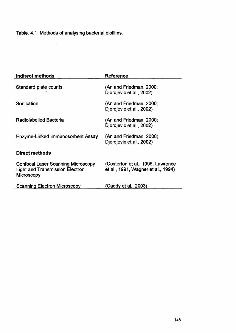
Table. 4.1 Methods of analysing bacterial biofilms.
Indirect methods Reference
Standard plate counts (An and Friedman, 2000; Djordjevic et al., 2002)
Sonication (An and Friedman, 2000; Djordjevic et al., 2002)
Radiolabelled Bacteria (An and Friedman, 2000; Djordjevic et al., 2002)
Enzyme-Linked Immunosorbent Assay
Direct methods
(An and Friedman, 2000; Djordjevic et al., 2002)
Confocal Laser Scanning Microscopy (Costerton et al., 1995, LawrenceLight and Transmission Electron Microscopy
etal., 1991, Wagner et al., 1994)
Scanning Electron Microscopy (Caddy et al., 2003)
148
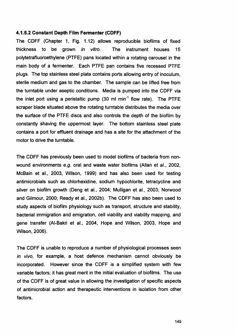
4.1.5.2 Constant Depth Film Fermenter (CDFF)
The CDFF (Chapter 1, Fig. 1.12) allows reproducible biofilms of fixed
thickness to be grown in vitro. The instrument houses 15
polytetrafluoroethylene (PTFE) pans located within a rotating carousel in the
main body of a fermenter. Each PTFE pan contains five recessed PTFE
plugs. The top stainless steel plate contains ports allowing entry of inoculum,
sterile medium and gas to the chamber. The sample can be lifted free from
the turntable under aseptic conditions. Media is pumped into the CDFF via
the inlet port using a peristaltic pump (30 ml min'1 flow rate). The PTFE
scraper blade situated above the rotating turntable distributes the media over
the surface of the PTFE discs and also controls the depth of the biofilm by
constantly shaving the uppermost layer. The bottom stainless steel plate
contains a port for effluent drainage and has a site for the attachment of the
motor to drive the turntable.
The CDFF has previously been used to model biofilms of bacteria from non
wound environments e.g. oral and waste water biofilms (Allan et al., 2002,
McBain et al., 2003, Wilson, 1999) and has also been used for testing
antimicrobials such as chlorhexidine, sodium hypochlorite, tetracycline and
silver on biofilm growth (Deng et al., 2004; Mulligan et al., 2003; Norwood
and Gilmour, 2000; Ready et al., 2002b). The CDFF has also been used to
study aspects of biofilm physiology such as transport, structure and stability,
bacterial immigration and emigration, cell viability and viability mapping, and
gene transfer (Al-Bakri et al., 2004, Hope and Wilson, 2003, Hope and
Wilson, 2006).
The CDFF is unable to reproduce a number of physiological processes seen
in vivo, for example, a host defence mechanism cannot obviously be
incorporated. However since the CDFF is a simplified system with few
variable factors; it has great merit in the initial evaluation of biofilms. The use
of the CDFF is of great value in allowing the investigation of specific aspects
of antimicrobial action and therapeutic interventions in isolation from other
factors.
149
Initial experiments in this present study using the CDFF were designed to
determine whether biofilms could be produced and maintained using
bacterial isolates from chronic wound patients. Once biofilms were
successfully produced, analysis of an antimicrobial wound therapeutic agent
was tested in vitro. The antimicrobial examined was povidone-iodine (PVP1),
commonly prescribed for chronic wound patients. Iodine is one of the most
frequently employed therapeutic constituents of wound dressings, e.g.
Inadine (Johnson & Johnson) and is also used in antiseptic pre-surgical
scrubs and other antiseptics.
4.2 AimsThe aims of this study were:
1. To confirm the species identity of chronic wound isolates to be used in
these studies using 16S rRNA gene sequencing.
2. To develop in vitro models of biofilm formation for the identified wound
bacterial isolates.
3. To compare the ability of these wound bacteria to produce biofilms and
determine whether synergism or antagonism occurred in mixed biofilm
populations.
4. To assess the susceptibility of the wound bacterial biofilms to PVP1.
150
4.3 Methods
4.3.1 Preparation of bacterial species
Bacterial species were selected from a previous prospective study of 70
patients with chronic venous leg ulcers (Davies et al., 2007, Davies et al.,
2004) and included representation of four species most frequently
encountered and with distinctive colony morphologies namely:
Staphylococcus aureus, Pseudomonas aeruginosa, Micrococcus luteus and
Streptococcus oralis. Isolates were routinely grown on blood agar (BA)
supplemented with 5% (v/v) sheep blood. BA plates were incubated
aerobically at 37°C for 24 h. Confirmation of identification of these isolates
in this present study was through 16S rRNA gene sequencing (section 4.3.2).
4.3.2 DNA extraction from test bacterial isolates
16S rRNA gene sequencing was used to confirm the identification of the four
isolates selected for this study. The previous presumptive identification of
these isolates, which had been based upon biochemical profiling using API
ID strips (BioMerieux sa, Marcy L’Etoile, France), was S. aureus, P.
aeruginosa, M. luteus and S. oralis. DNA was extracted from an overnight
culture in Brain Heart Infusion (BHI) broth of each pure bacterial isolate using
a Puregene® DNA isolation kit (Gentra Systems, Minneapolis, USA) by
following the “DNA Isolation From 1 ml Gram-positive Bacteria Culture
Medium” protocol (http://www.gentra.com/technical_assistance
/protocols, asp).
4.3.3 Amplification of 16S rRNA genes from bacterial isolatesExtracted DNA was quantified using the Nanodrop ND-1000
Spectrophotometer (NanoDrop Technologies, Fisher, UK) and stock DNA
concentrations of 200 pg ml'1 were prepared. PCR was used to amplify
targeted sequences of the 16S rRNA using the universal bacterial primer pair
of 27F and 1492R (0.5 pM of each; oligonucleotides synthesized by
Invitrogen Ltd. Paisley, UK), both sequences of which were specific for the
domain Bacteria (Dymock et al 1996; Table 4.2).
151
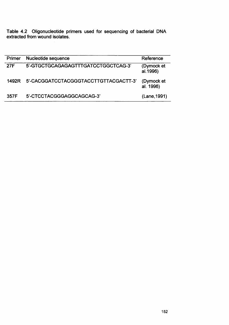
Table 4.2 Oligonucleotide primers used for sequencing of bacterial DNA extracted from wound isolates.
Primer Nucleotide sequence Reference27F 5’-GT GCT GC AGAG AGTTT GAT CCT GGCT C AG-3’ (Dymock et
al.1996)
1492R 5’-C ACGG AT CCTACGGGTACCTT GTTACG ACTT-3’ (Dymock etal. 1996)
357F 5’-CTCCTACGGGAGGCAGCAG-3’ (Lane,1991)
152
PCR was performed in a total reaction volume of 50 pi and used 1.5 U of Taq
DNA polymerase, 200 pM of each deoxynucleotide, 0.5 pM of each primer,
and the working concentration of appropriate magnesium-free buffer
(Promega, Southampton, UK). Additionally, 2.0 mM of MgCI2 (Promega) was
used, with 5 pi of DNA extract as template.
A touchdown PCR protocol was used, whereby, in the first cycle,
denaturation at 95°C for 1 min was followed by primer annealing at 65°C for
1 min, and primer extension at 72°C for 2 min. This cycle was then followed
by denaturation 1 min with the annealing temperature subsequently
decreased by 2°C for each cycle (11 cycles), after which 25 cycles were
performed under the same primer annealing conditions (45°C). In the final
cycle, primer extension was performed for 12 min. PCR was undertaken
using a PTC-200 thermal cycler (MJ Research Ltd., GRI, Braintree, UK).
PCR products were purified by precipitation and washed with pure ethanol.
Firstly, 15 pi of 5 M NaCI (Sigma-Aldrich, Steinheim, Germany) and 15 pi of
40% polyethylene glycol (Mol. Wt. 8000; Sigma) were added to each PCR
tube. This mix was centrifuged (16,000 g for 15 min) and the supernatant
aspirated and replaced with 200 pi of 70% ethanol (v/v in water; Fisher
Scientific, Loughborough, UK). Centrifugation, aspiration and ethanol
washing steps were repeated (3* in total). Following another centrifugation
step, the PCR products were dried under a fume hood (EDF1200, Envair,
Rossendale, UK) overnight, and resuspended with DNA-free and nuclease-
free water (30 pi). Products were then stored at -20°C until required.
4.3.4 Sequencing of 16S rRNA genesPurified and cleaned PCR products were sequenced using ABI Prism BigDye
terminator cycle sequencing ready reaction kits (Applied Biosystems,
Warrington, UK). A total reaction volume of 20 pi was used and contained
the working concentrations of BigDye Sequencing Buffer and Ready
Reaction Premix (Applied Biosystems), 0.5 pM of an oligonucleotide primer
(Table 4.2, synthesized by Invitrogen), and 6 pi of purified PCR-product
153
(approx. 5-20 ng). For all isolates, sequencing was performed using the
universal bacterial primer 357F (Lane, 1991; Table 4.2). Reactions were
performed in a PTC-200 thermal cycler (MJ Research Ltd.) and comprised of
1 min of denaturation at 94°C, followed by 30 cycles of denaturation at 94°C
(15 s), annealing at 50°C (30 s) and extension at 60°C (5 min).
Extension products were purified, firstly by precipitating via the addition of 1
pi sodium acetate (3M; Sigma) and 1 pi ethylenediaminetetraacetic acid
(EDTA, pH 8.0, 0.5 M; Sigma), and then by adding 80 pi of chilled absolute
ethanol, to each reaction mix. The supernatant was aspirated following
centrifugation (16,000 g, 15 min), and replaced with chilled 70% ethanol (200
pi). This was repeated once and after a final centrifugation and drying under
vacuum, products were re-suspended in 100% (v/v) formamide (30 pi) and
run on an automated DNA sequencer (ABI PRISM 3100 Genetic Analyser;
Applied Biosystems). This procedure was expected to generate reliable
sequences of greater than 500 base pairs (bp) that could be compared to
sequences in the Genbank and EMBL public databases, which is sufficient
for species level identification (Clarridge, 2004).
4.3.5 Identification of bacterial isolates by 16S sequence analysisSequences obtained in this study were identified by comparison to the
GenBank DNA sequence database EMBL (Benson et al 2004) using the
FASTA sequence homology search program
(http://www.ebi.ac.uk/services/index.html: Pearson, 1990).
A >99% homology to the 16S rRNA gene sequence of the type strain, or
other suitable reference strain, was the criterion used to identify an isolate to
the species level. Where more than one reference species exhibited >99%
sequence homology, the match with the greatest homology was taken as the
identity, wherever the sequence was shown to be reproducible and reliable.
154
4.3.6 In vitro biofilm formation by chronic wound bacteria
Preliminary studies were undertaken to identify a culture medium and
incubation time that allowed growth and biofilm formation by all isolates to be
studied. A total of four different culture media were evaluated. These
included Brain Heart Infusion broth (BHI; Oxoid, Hampshire, UK), Tryptone
Soya Broth (TSB; Oxoid), BM medium (Appendix I) (McKee et al., 1985) and
a simulated wound fluid (Appendix I) previously described by Addison et al
(2004). The latter medium was modified by adding 2 g I'1 glucose to provide
an additional carbon source.
4.3.7 Preparation of wound cultures for an in vitro microtitre plate assayMicrococcus luteus (B81), S. oralis (B52), P. aeruginosa (D40) and S. aureus
(D76) were cultured at 37°C for 24 h in 10 ml of BHI, TSB, BM or simulated
wound fluid medium.
Standardised bacterial preparations were then made by adjusting the culture
concentration to an optical density of 0.4 (absorbance measured at 600 nm)
in PBS. After the required dilution for this OD measurement was identified
using PBS, fresh medium was added to the overnight cultures, to adjust the
bacteria to the required level.
4.3.8 Microtitre plate biofilm assayMicrotitre plate biofilm formation involved growing bacterial cultures in
individual wells of 96-well microtitre plates (flat bottomed, Greiner, UK). A
range of culture concentrations were tested in separate plates by performing
two-fold serial dilutions (in appropriate culture medium) of the standardised
bacterial preparation. A 100-pi volume of bacterial culture was then added to
the wells of the microtitre plate. The microtitre plates were then sealed with
parafilm to prevent dehydration (American National Can Group, USA) and
incubated at 37°C for 24 h, 48 h, 72 h and 96 h.
Biofilm formation was subsequently assessed by one of 2 methods: a) crystal
violet staining or b) total viable count estimation.
155
4.3.9 Semi quantification of microtitre plate biofilms by crystal violet staining
After the appropriate incubation period, culture medium was removed and the
microtitre plate wells washed three times with sterile distilled water to remove
loosely associated bacteria. Plates were dried at 56°C for 45 min and each
well was stained with 150 pi of 1% v/v crystal violet solution (in water) for 45
min. After staining, plates were again washed three times with sterile distilled
water. At this point, biofilms were visible by purple staining formed on the
side of each well. The quantitative analysis of biofilm production was then
performed by adding 200 pi of 95% ethanol to destain the cells. After 5 min,
100 pi from each well was transferred in a new microtitre plate and the level
of the crystal violet present in the de-stain solution was measured at 540nm.
This method has previously been described by Djordjevic et al. (2002).
4.3.10 Statistical analysis of the crystal violet microtitre plate assayStatistical analyses were done on the data obtained from the crystal violet
microtitre plate assays. The Friedman, Wilcoxon and Kruskal-Wallis tests
are all non-parametric statistical tests. The Friedman test and the Wilcoxon
Signed Rank test were used to measure any differences in biofilm formation
when different starting inocula were used. The Kruskal-Wallis test is an
extension of the Wilcoxon rank sum test. All statistical analysis was done
using the Statistical Package for the Social Sciences (SPSS) software.
4.3.11 Assessment of biofilm formation by single and mixed bacterial
isolates using the microtitre plate assay and viable count estimation
The ability of the test isolates to produce biofilms in both single species and
mixed species population was also determined using the microtitre plate
assay. Using the identified optimal growth conditions, single and mixed
species biofilm experiments were undertaken to determine whether any
synergy or antagonism between species occurred with regards to biofilm
formation.
156
Briefly, in the case of mixed species biofilms, equal volumes of isolate
combinations (total final volume of 100 pi) were mixed, added to the wells of
the microtitre plate and incubated for 24 and 48 h in BHI medium.
After incubation the planktonic phase of growth was carefully removed from
the microtitre plates. A 100-pl volume of PBS was added to each well to re
suspend the biofilms. The biofilms of 6 wells were then pooled and serially
diluted in PBS. Dilutions (100 pi) were plated on to BA plates which were
incubated aerobically at 37°C for 24 h. Enumeration of each species was
confirmed by visual inspection of the plates and representative colonies of
each species recorded at 24-48 h.
4.3.12 Use of the CDFF to develop biofilms using chronic wound
bacteria
In the present study, the CDFF was fully assembled with the PTFE plugs
recessed within the pans to a depth of 400 pm using a gauged recessing
tool. The CDFF was then autoclaved at 121°C for 45 min, and allowed to
cool. For the duration of each experiment, the CDFF was maintained in an
incubator at 37°C and ambient air was allowed to diffuse into the CDFF via
sterile air filters. For all CDFF experiments the turntable speed was operated
at 20 revs min'1. Initial experiments in the CDFF were performed to
determine whether a stable biofilm wound model could be produced using
bacterial isolates from chronic wound patients.
4.3.12.1 Inoculation of chronic wound bacteria for the in vitro biofilm
model using the CDFF and preparation of planktonic cellsChronic wound bacteria were routinely grown on BA and incubated
aerobically at 37°C, as described previously (section 4.3.1). Isolates were
then subcultured in 10 ml BHI broth for 24 h at 37°C in a shaking incubator.
Isolates were tested for their ability to form biofilms in both single and mixed
inocula. Five ml of each wound bacterial culture (section 4.3.1) was added to
1 L of BHI or BM broth and this preparation was recirculated through the
CDFF for 24 h to ‘seed’ the inoculum in the system. After this time, the
157
inoculum was disconnected and fresh media was fed into the CDFF without
recirculation and the CDFF waste was collected in an effluent bottle. Medium
was delivered at a flow rate of 30 ml min-1 using a peristaltic pump (Watson-
Marlow, Falmouth, UK). Purity plates were performed at the time of
inoculation to ensure that no contamination occurred in the CDFF inoculum.
Planktonic mixes of the bacteria were also assessed in parallel to the
biofilms. For planktonic cell preparation 100 pi from the overnight culture of
each organism was added to fresh 19.9 ml BHI medium. Seven replicate
broths were incubated with shaking at 37°C for 7 d. Each day the cells were
centrifuged, the supernatant removed and new media was added. At each
24 h time point, 100 pi of one of the replicates suspensions was serial diluted
in PBS and plated onto BA plates for viable counts of planktonic growth. The
remainder of the suspension was centrifuged and the pelleted cells were
stored in RNAIater® (Ambion, UK) for subsequent investigation of gene
expression profiles (see Chapter 6).
4.3.13 Recovery of biofilms established in the CDFFA single pan holding 5 plugs was aseptically removed at each sampling time
point from the CDFF for microbial analysis of the wound microflora. The
samples were lifted from the turntable with a dedicated tool under aseptic
conditions every 24 h for 7 d. Individual plugs were then removed aseptically
from the pan. The 5 plugs were each placed in 2.5 ml of PBS and serial
decimally diluted. Dilutions (100 pi) were plated on to BA plates and the
plates incubated aerobically at 37°C for 24 h.
4.3.14 PVP1 susceptibility of CDFF generated biofilmsAntimicrobial testing of the biocide, povidone-iodine (PVP1) in the CDFF was
performed against a mixed biofilm consortia. Bacteria used for the inoculum
were S. aureus (D76), P. aeruginosa (D40), M. luteus (B81) and S. oralis
(B52) cultured for 24 h in BM medium at 37°C. In addition to these aerobic
species, Bacteroides fragilis (B11) and Peptostreptococcus anaerobius (B12)
were prepared in fastidious anaerobe broth (FAB; LabM Ltd., UK) for 72 h in
158

an anaerobic cabinet (10% v/v C 02, 20% v/v H2j 70v/v N2; Don Whitley
Scientific Ltd., Shipley, UK) at 37°C. The inoculum for the biofilm consisted
of 5 ml of each of these bacterial species, which were added to the growth
medium and used to inoculate the CDFF as previously described (4.3.12.1).
PVP1 (final concentration of 1% w/v in BM) was added to the medium of the
CDFF once a fully mature biofilm had been established (after 5 d incubation).
The PVP1 concentration of 1 % used was thought to be representative of the
concentration of iodine that is effectively available to the wound bed in
commonly used commercially available dressings which contain 10% PVP1
(http ://www.d ressi ngs. org/d ressi ngs/i nad ine. htm I).
4.3.15 Biofilm recovery from the CDFF during PVP1 testing
The CDFF was sampled every 48 h over a total experimental run of 29 d. A
single pan containing 5 plugs was again removed from the CDFF at each
sampling point. Two sets of two plugs were taken from each pan and
separately vortexed in 5 ml PBS for 2 min to disperse the biofilm. Serial
dilutions were then made of each of the resulting bacterial suspensions to a
dilution factor of 107 fold. Then, 100 pi of each dilution was plated onto BA
and FAA plates which were incubated at 37°C under aerobic and anaerobic
conditions, respectively. Colony counts of each species on the plates after
24-48 h incubation were then recorded.
Resulting growth from the ‘biofilm’ cultures were also subsequently tested for
susceptibility to PVP1 using a broth culture minimum bactericidal
concentration (MBC) test (see section 4.3.16). This enabled an assessment
to be made on possible acquired resistance development of these isolates
during prolonged PVP1 exposure in the CDFF.
4.3.15.1 Live/dead staining of biofilms treated with PVP1In addition to total viable count estimation, a smear of the biofilm was also
stained with the Live/Dead® BacLight™ stain (Live/Dead® BacLight™ Bacterial
Viability Kit, Invitrogen Ltd). The live/dead stain consisted of two nucleic acid
stains: the green fluorescent SYT09 and the red-fluorescent propidium
159
iodide stains are used in combination to discriminate live and dead bacteria
in a mixed population. Bacterial cells with a compromised membrane that
are considered to be dead or dying stain red, whereas cells with an intact
membrane appear green.
Three pi of each stain was added to 300 pi PBS, mixed and 20 pi added to
the biofilm smear. The preparation was left for 1 min and then viewed by
standard fluorescence microscopy (Olympus Provis AX70 Digital Microscope;
Olympus U.K. Ltd., Middlesex, U.K.), at a total magnification of x600.
4.3.16 Planktonic cell susceptibility to PVP1In order to determine whether an elevated resistance to PVP1 occurred in
chronic wound biofilms it was also necessary to establish the susceptibility of
planktonic equivalent cells. These experiments were undertaken using a
microtitre plate assay susceptibility test based on the CLSI (Clinical
Laboratory Standards Institute; previously known as the National Committee
for Clinical Laboratory Standards; NCCLS) guidelines for broth microdilution
testing with some modifications. Tests were undertaken using cation-
adjusted Mueller-Hinton broth (CAMHB; Lab114, LabM, Bury, England) or
BM medium. PVP1 was diluted in the culture medium and serial two-fold
dilutions of PVP1 ranging from 2% w/v to 0.004% w/v were used.
Bacterial isolates were grown on BA plates at 37°C overnight and then used
to inoculate 10 ml BM broth which was incubated at 37°C overnight. The
bacterial suspension was then washed by centrifugation. The cell pellet was
re-suspended in PBS and the turbidity was adjusted to a 0.5 McFarland
standard (approximately 108 CFU ml'1). Based on the required dilution to
establish the 0.5 MacFarland standard, an equivalent dilution preparation
was made in the appropriate growth medium for study. A proportion of the
standardised suspension was diluted 1:100 (106 CFU ml'1) with BM broth or
Mueller-Hinton broth. One hundred pi of the bacterial suspension was added
to a microtitre plate well already containing 100 pi of diluted PVP1. A volume
(100 pi) of the inoculum was plated on to BA to check for purity and inoculum
160
density. Broth not containing PVP1 was inoculated as a control for organism
viability, and a further control of broth without inoculum was also included.
The microtitre plate was sealed in parafilm and incubated at 37°C for 24 h, 48
h, 72 h and 96 h before minimum bactericidal concentrations (MBCs) were
determined.
The lowest concentration of PVP1 found to completely inhibit visible growth
of the organism was recorded as the MBC at all time points. This was
confirmed by the total absence of growth on BA plates after sub-culturing a
loopful of broth from the identified MBC dilution.
161
4.4 Results
4.4.1 16S rRNA gene sequence identification of test bacterial isolates
Results for the highest sequence match from 16S rRNA gene sequencing of
bacterial wound isolates used in this study are shown in Table 4.3. All isolate
sequences showed a high homology (> 99.4%) to at least one database
sequence (actual sequences, Appendix I).
4.4.2 Comparison of biofilm formation by test species using the
microtitre plate assay (crystal violet)
Figures 4.1 - 4.3 demonstrate the levels of biofilm formed by the test species
in BHI, TSB and BM media respectively. In simulated wound fluid (with or
without glucose) the bacteria were not found to produce biofilms. There were
no statistical differences for each species in the biofilm level generated at
each time point when different starting inocula were used (Fig. 4.1 - 4.3).
The level of biofilm formation by the test bacterial species was, however,
found to vary with different growth media (BHI and TSB) over a time period of
24-96 h.
162
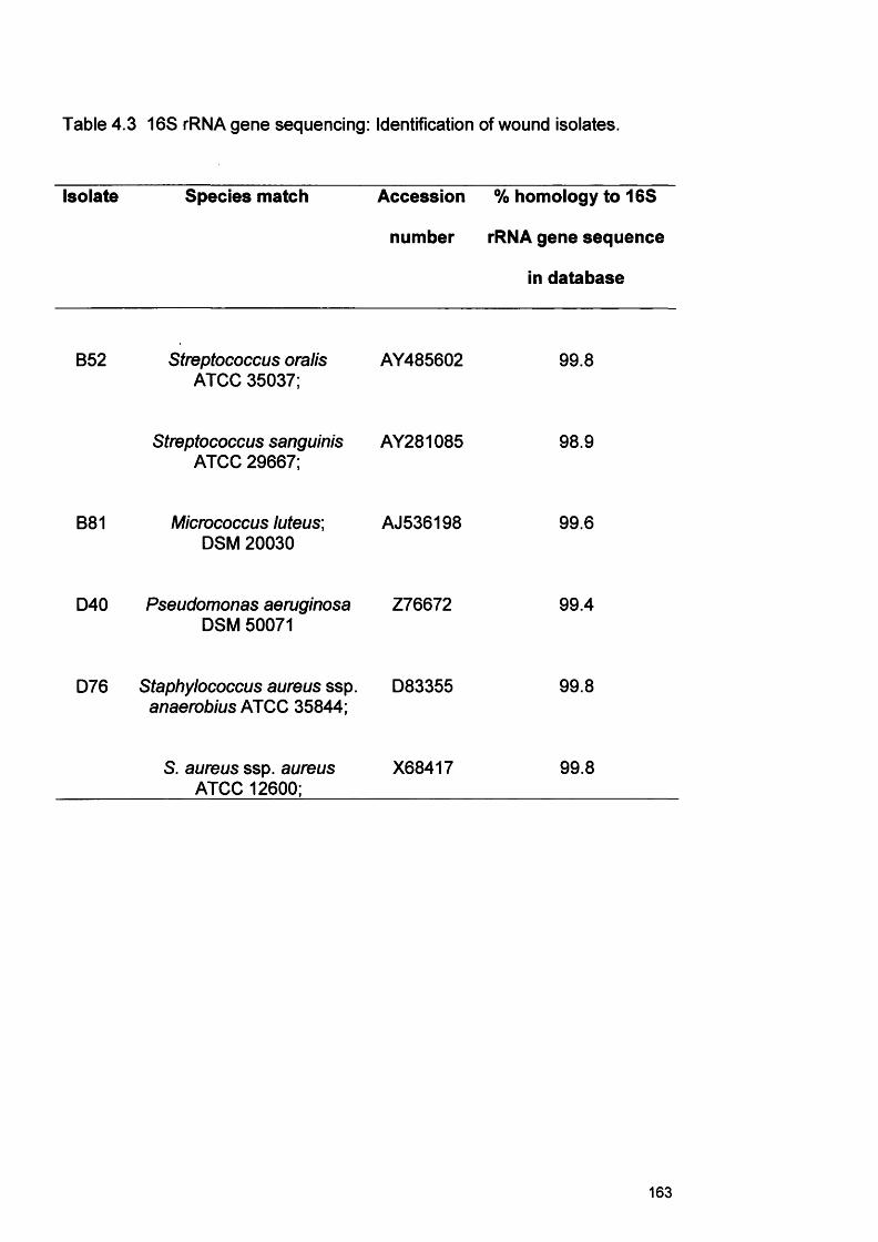
Table 4.3 16S rRNA gene sequencing: Identification of wound isolates.
Isolate Species match Accession
number
% homology to 16S
rRNA gene sequence
in database
B52 Streptococcus oralis ATCC 35037;
AY485602 99.8
Streptococcus sanguinis ATCC 29667;
AY281085 98.9
B81 Micrococcus luteus; DSM 20030
AJ536198 99.6
D40 Pseudomonas aeruginosa DSM 50071
Z76672 99.4
D76 Staphylococcus aureus ssp. anaerobius ATCC 35844;
D83355 99.8
S. aureus ssp. aureus ATCC 12600;
X68417 99.8
163
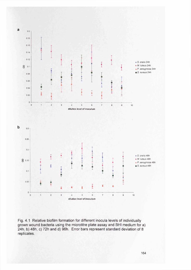
0.2
0.18
0.16
0.14
0.12
8 ii0.08
0.06
0.04
0.02 f 1
♦ S. oralis 24h ■ M. luteus 24h a P. aeruginosa 24h u S. aureus 24h
1 2 4 5 6
dilution level of inoculum10
0.2
0.15
0.1
0.05
{ I
f f
f i
♦ S. oralis 48h ■ M. luteus 48h a P. aeruginosa 48h m S. aureus 48h
4 5 6
dilution level of inoculum
Fig. 4.1 Relative biofilm formation for different inocula levels of individually grown wound bacteria using the microtitre plate assay and BHI medium for a) 24h, b) 48h, c) 72h and d) 96h. Error bars represent standard deviation of 8 replicates.
164
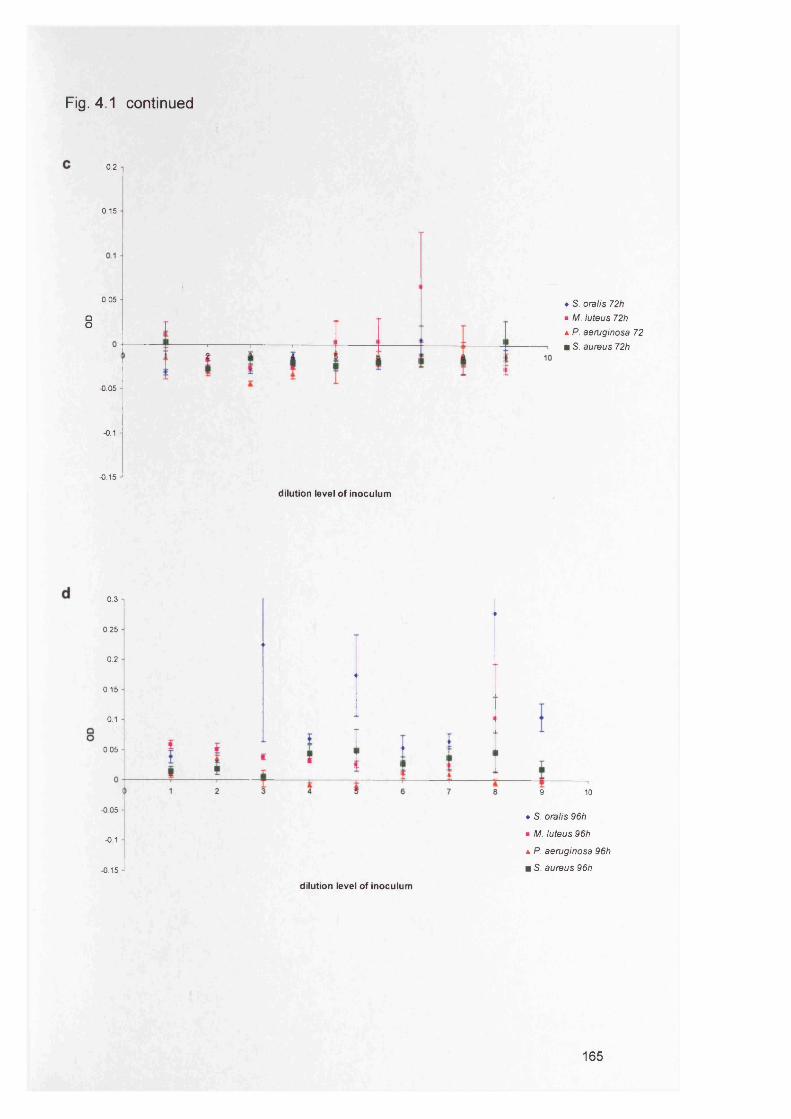
Fig. 4.1 continued
0.2
0.15
0.05
QO
-0.05
•0.1
-0.15
dilution level of inoculum
♦ S. oralis 72h ■ M. luteus 72h a P. aeruginosa 72 m S. aureus 72h
0.3
0.25
0.2
0.15
0.1
0.05
-0.05
-0.1
0.15
9 10
♦ S. oralis 96h
■ M. luteus 96h
a P. aeruginosa 96h
m S. aureus 96h
dilution level of inoculum
165
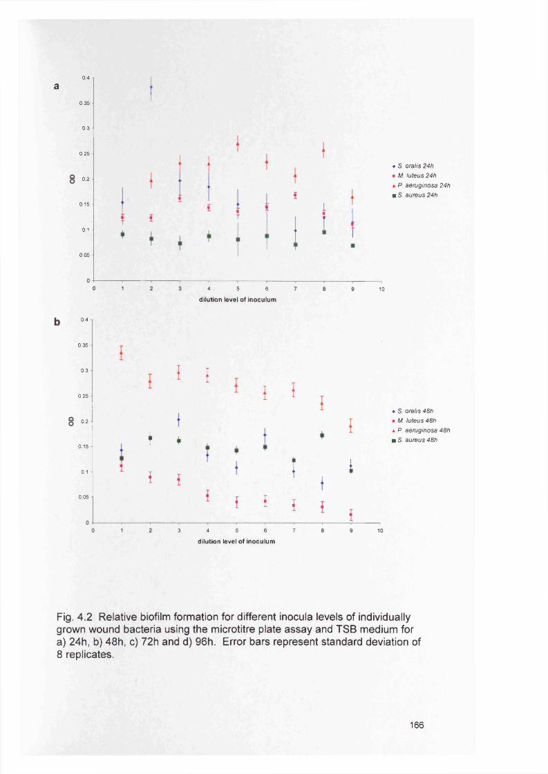
0.4
a0.35
0.3
0.25
0.2
0.15
0.1
0.05
♦ S. oralis 24h ■ M. luteus 24h h P. aeruginosa 24h m S. aureus 24h
4 5 6
dilution level of inoculum10
0.4
0.35
0.3
0.25
0.2
0.15
0.1
0.05
I i
+ *
I
{ I
I , *I * {
♦ S. oralis 48h ■ M. luteus 48h a P. aeruginosa 48h m S. aureus 48h
3 4 5 6 7
dilution level of inoculum
Fig. 4.2 Relative biofilm formation for different inocula levels of individually grown wound bacteria using the microtitre plate assay and TSB medium for a) 24h, b) 48h, c) 72h and d) 96h. Error bars represent standard deviation of 8 replicates.
166
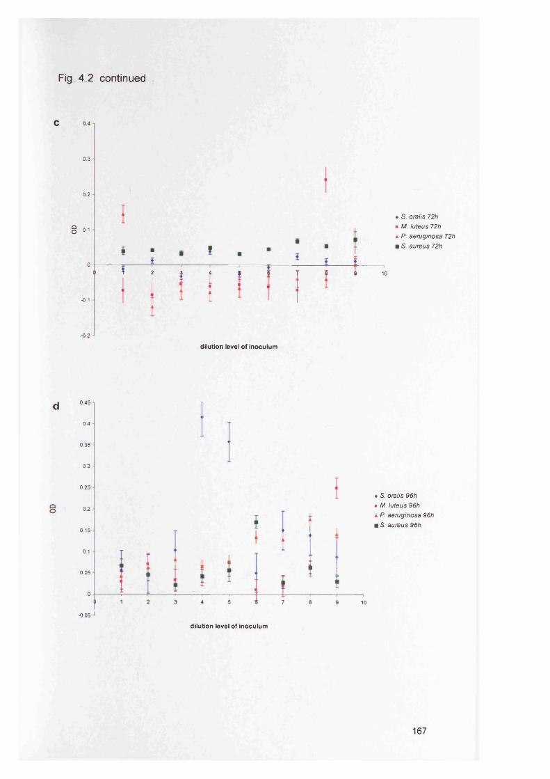
Fig. 4.2 continued
0.3
0.2
8 0.1
0
-0.1
-0.2
d 0450.4
0.35
0.3
0.25
§ 02
0.15
0.1
0.05
0
-0.05
♦ S. oralis 72h ■ M. luteus 72h a P. aeruginosa 72h m S. aureus 72h
10
dilution level of inoculum
♦ S. oralis 96h• M. luteus 96ha P. aeruginosa 96h m S. aureus 96h
10
dilution level of inoculum
167
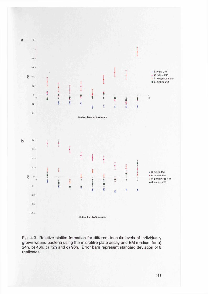
1.2
0.8
0.6
0.4
0.2
-0.2
-0.4
6 i£ £
• S. oralis 24h ■ M. luteus 24h a P. aeruginosa 24h m S. aureus 24h
10
dilution level of inoculum
0.4
0.3
0.2
0.1
-0.1
-0.2
•0.3
i I
I I
( t f f
• S. oralis 48h ■ M. luteus 48h a P. aeruginosa 48h m S. aureus 48h
-0.4
dilution level of inoculum
Fig. 4.3 Relative biofilm formation for different inocula levels of individually grown wound bacteria using the microtitre plate assay and BM medium for a) 24h, b) 48h, c) 72h and d) 96h. Error bars represent standard deviation of 8 replicates.
168
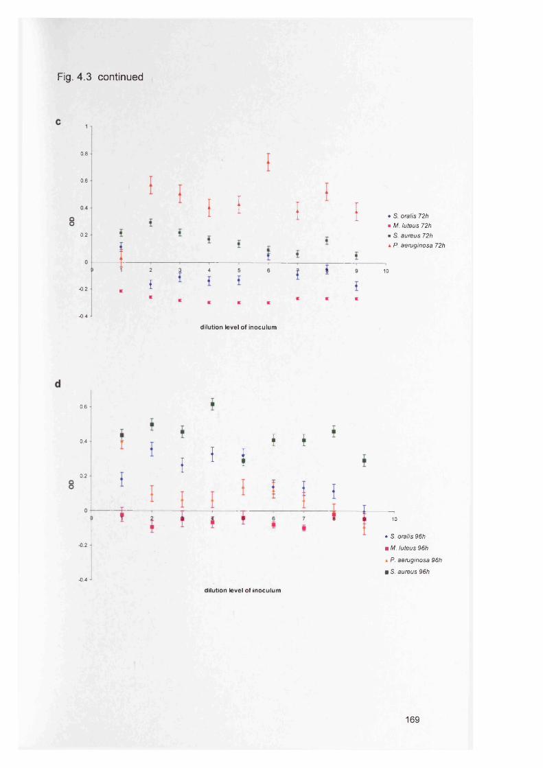
Fig. 4.3 continued
0.8
0.6
0.2
I f I I • S. oralis 72h■ M. luteus 72h■ S. aureus 72ha P. aeruginosa 72h
-0.2
2 3 4
{10
-0.4
dilution level of inoculum
0.6
0.4
0.2
-0.2
-0.4
I i ii f
10
• S. oralis 96h
m M. luteus 96h
* P. aeruginosa 96h
■ S. aureus 96h
dilution level of inoculum
169
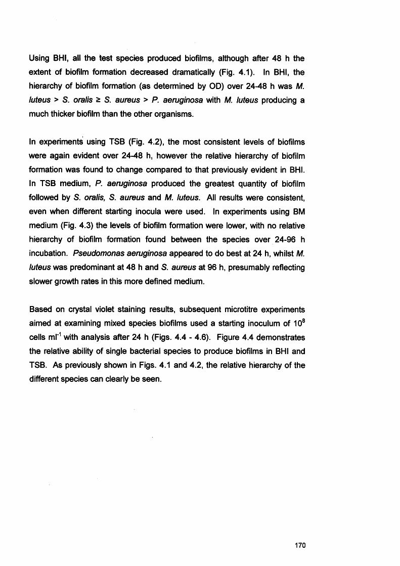
Using BHI, all the test species produced biofilms, although after 48 h the
extent of biofilm formation decreased dramatically (Fig. 4.1). In BHI, the
hierarchy of biofilm formation (as determined by OD) over 24-48 h was M.
luteus > S. oralis £ S. aureus > P. aeruginosa with M. luteus producing a
much thicker biofilm than the other organisms.
In experiments using TSB (Fig. 4.2), the most consistent levels of biofilms
were again evident over 24-48 h, however the relative hierarchy of biofilm
formation was found to change compared to that previously evident in BHI.
In TSB medium, P. aeruginosa produced the greatest quantity of biofilm
followed by S. oralis, S. aureus and M. luteus. All results were consistent,
even when different starting inocula were used. In experiments using BM
medium (Fig. 4.3) the levels of biofilm formation were lower, with no relative
hierarchy of biofilm formation found between the species over 24-96 h
incubation. Pseudomonas aeruginosa appeared to do best at 24 h, whilst M.
luteus was predominant at 48 h and S. aureus at 96 h, presumably reflecting
slower growth rates in this more defined medium.
Based on crystal violet staining results, subsequent microtitre experiments
aimed at examining mixed species biofilms used a starting inoculum of 108
cells ml'1 with analysis after 24 h (Figs. 4.4 - 4.6). Figure 4.4 demonstrates
the relative ability of single bacterial species to produce biofilms in BHI and
TSB. As previously shown in Figs. 4.1 and 4.2, the relative hierarchy of the
different species can clearly be seen.
170
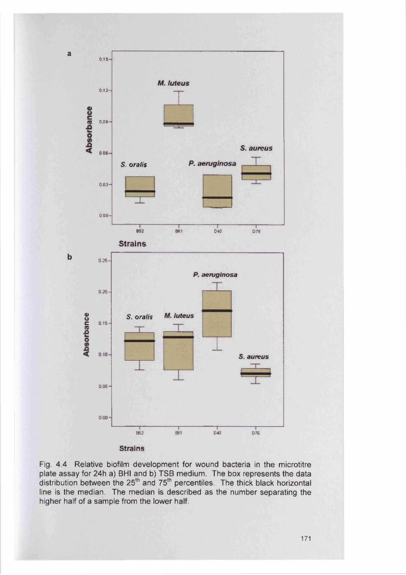
a0 1 5 -
M . tu teus0 12 -
m 0 09 -
S. aureu s
P. aeru g in o saS. o ratis
0 0 3 -
0 0 0 -
B81 040 D76
Strains
0»oCwJQOn
0 25 -
0 2 0 -
0 15-
0 0 5 -
000-
S. oralis M. luteus
P. aeruginosa
S. aureus
B62 ffll 040 076
Strains
Fig. 4 .4 Relative biofilm development for wound bacteria in the microtitre plate assay for 24h a) BHI and b) TSB medium. The box represents the data distribution between the 25th and 75th percentiles. The thick black horizontal line is the median. The median is described as the number separating the higher half of a sample from the lower half.
171
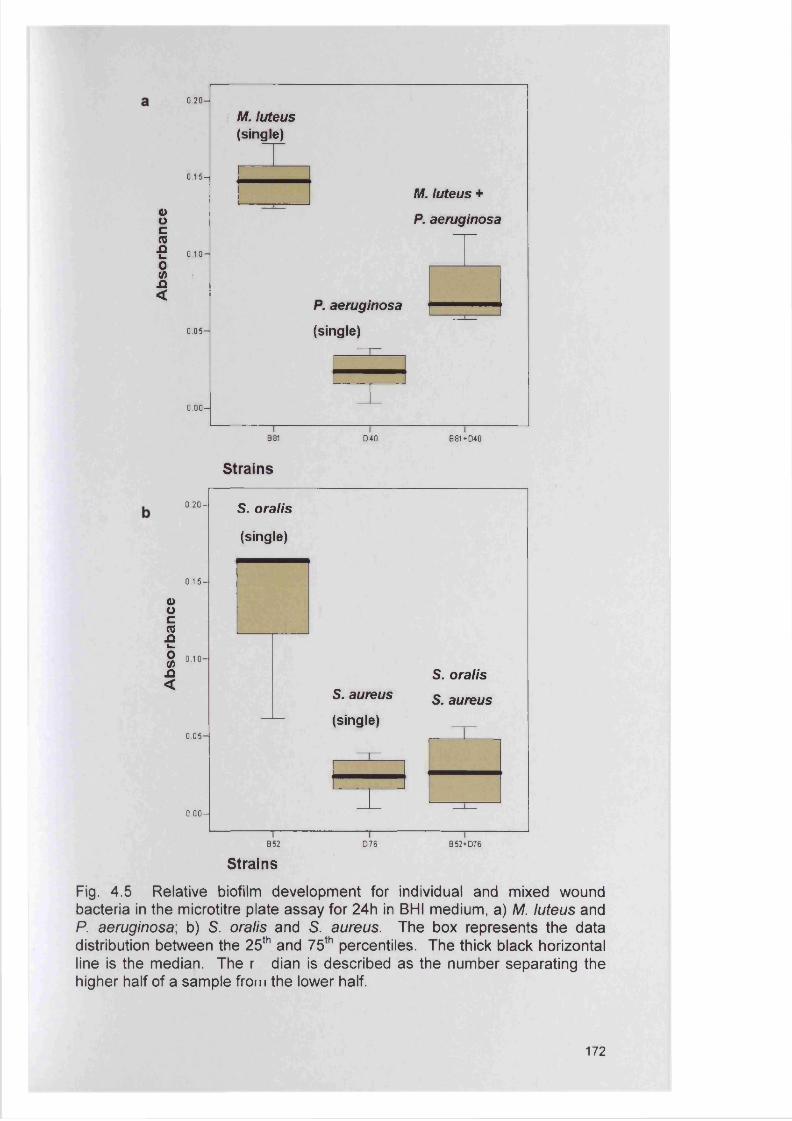
ooc(012i_o<A.Q<
0 .2 0 -
0.15-
0 .1 0 -
0.05-
0 .0 0 -
M. luteus (single)
P. aeruginosa
(single)
M. luteus +
P. aeruginosa
B81 D40 B81 + D40
Strains
ooc(0ai*.o(Aa<
0 .2 0 -
0.15-
0 .1 0 -
0.05-
0 .0 0 -
S. oralis
(single)
S. aureus
(single)
S. oralis
S. aureus
B52
StrainsD76 B52+D76
Fig. 4.5 Relative biofilm development for individual and mixed wound bacteria in the microtitre plate assay for 24h in BHI medium, a) M. luteus and P. aeruginosa; b) S. oralis and S. aureus. The box represents the data distribution between the 25th and 75th percentiles. The thick black horizontal line is the median. The r dian is described as the number separating the higher half of a sample from the lower half.
172
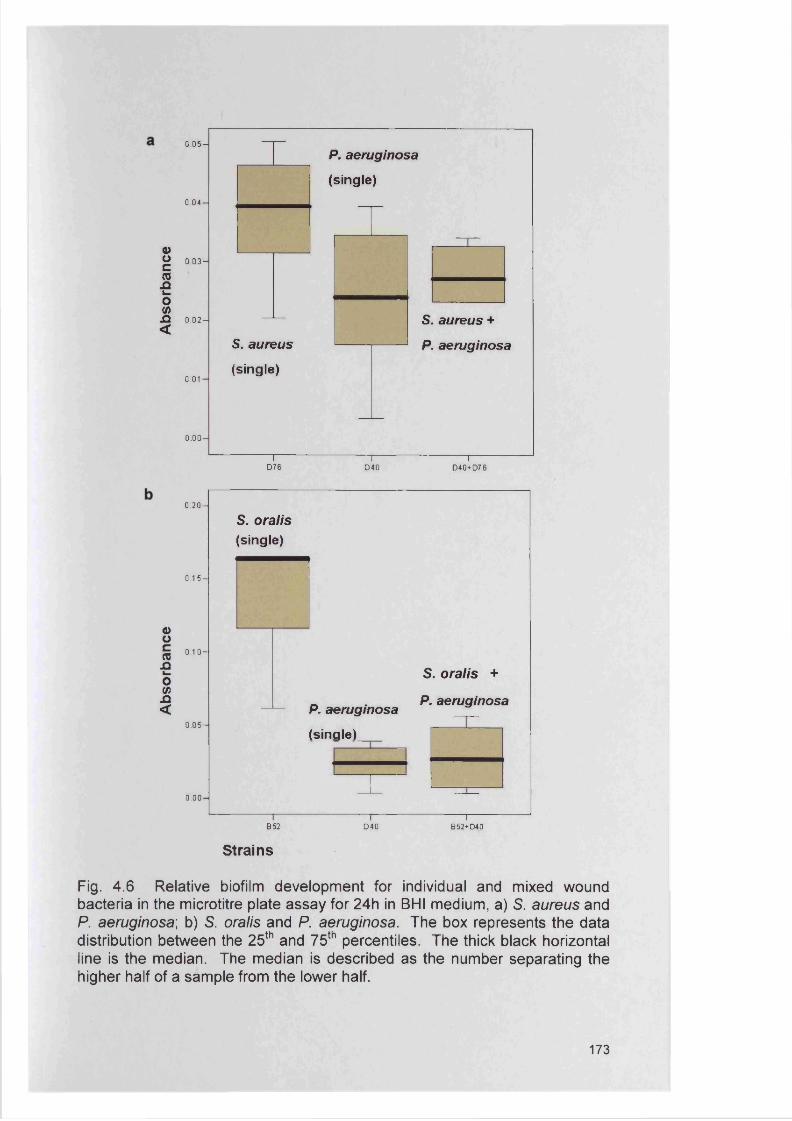
0.05—
0.04-
a>g 0.03̂(0 JQCo<A
JQ 0.02- <
0 .01 -
0 .0 0 -
P. aeruginosa
(single)
S. aureus
(single)
S. aureus +
P. aeruginosa
TD76
i rD40 D40+D76
0 .2 0 -
0.15-
Ooc(QnL _
o(A-Q<
0 .1 0 -
0.05-
0 .0 0 -
S. oralis (single)
P. aeruginosa
(single)
S. o ra lis +
P. aeruginosa
B52
Strains
040I
B52+D40
Fig. 4 .6 Relative biofilm development for individual and mixed wound bacteria in the microtitre plate assay for 24h in BHI medium, a) S. aureus and P. aeruginosa; b) S. oralis and P. aeruginosa. The box represents the data distribution between the 25th and 75th percentiles. The thick black horizontal line is the median. The median is described as the number separating the higher half of a sample from the lower half.
173
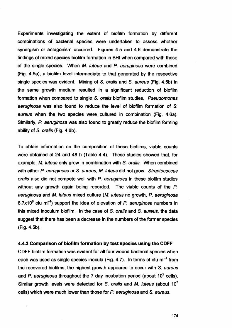
Experiments investigating the extent of biofilm formation by different
combinations of bacterial species were undertaken to assess whether
synergism or antagonism occurred. Figures 4.5 and 4.6 demonstrate the
findings of mixed species biofilm formation in BHI when compared with those
of the single species. When M. luteus and P. aeruginosa were combined
(Fig. 4.5a), a biofilm level intermediate to that generated by the respective
single species was evident. Mixing of S. oralis and S. aureus (Fig. 4.5b) in
the same growth medium resulted in a significant reduction of biofilm
formation when compared to single S. oralis biofilm studies. Pseudomonas
aeruginosa was also found to reduce the level of biofilm formation of S.
aureus when the two species were cultured in combination (Fig. 4.6a).
Similarly, P. aeruginosa was also found to greatly reduce the biofilm forming
ability of S. oralis (Fig. 4.6b).
To obtain information on the composition of these biofilms, viable counts
were obtained at 24 and 48 h (Table 4.4). These studies showed that, for
example, M. luteus only grew in combination with S. oralis. When combined
with either P. aeruginosa or S. aureus, M. luteus did not grow. Streptococcus
oralis also did not compete well with P. aeruginosa in these biofilm studies
without any growth again being recorded. The viable counts of the P.
aeruginosa and M. luteus mixed culture (M. luteus no growth, P. aeruginosa
8.7x10® cfu ml'1) support the idea of elevation of P. aeruginosa numbers in
this mixed inoculum biofilm. In the case of S. oralis and S. aureus, the data
suggest that there has been a decrease in the numbers of the former species
(Fig. 4.5b).
4.4.3 Comparison of biofilm formation by test species using the CDFF
CDFF biofilm formation was evident for all four wound bacterial species when
each was used as single species inocula (Fig. 4.7). In terms of cfu ml'1 from
the recovered biofilms, the highest growth appeared to occur with S. aureus
and P. aeruginosa throughout the 7 day incubation period (about 109 cells).
Similar growth levels were detected for S. oralis and M. luteus (about 107
cells) which were much lower than those for P. aeruginosa and S. aureus.
174
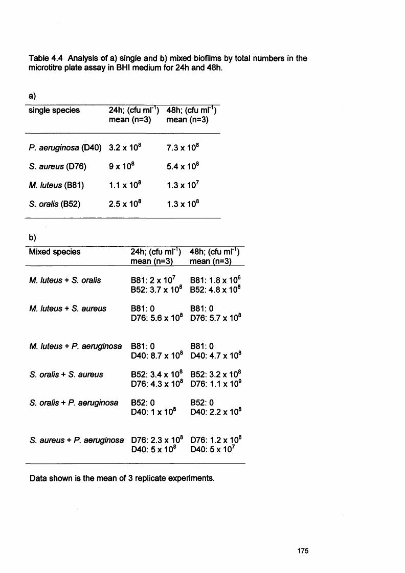
Table 4.4 Analysis of a) single and b) mixed biofilms by total numbers in the microtitre plate assay in BHI medium for 24h and 48h.
a)
single species 24h; (cfu ml'1) mean (n=3)
48h; (cfu ml"1) mean (n=3)
P. aeruginosa (D40) 3.2 x10⁸ 7.3x10®
S. aureus (D76) 9x10® 5.4x10®
M. luteus (B81) 1.1 x 10® 1.3 x10⁷
S. oralis (B52) 2.5x10® 1.3x10®
b)Mixed species 24h; (cfu ml"1)
mean (n=3)48h; (cfu ml'1) mean (n=3)
M. luteus + S. oralis B81: 2 x 10⁷ B52: 3.7 x10⁸
B81:1.8x10® B52: 4.8x10®
M. luteus + S. aureus B81: 0D76: 5.6 x10⁸
B81: 0D76: 5.7x10®
M. luteus + P. aeruginosa B81: 0D40: 8.7 x10⁸
B81: 0D40: 4.7x10®
S. oralis + S. aureus B52: 3.4 x10⁸ D76: 4.3 x10⁸
B52: 3.2x10® D76: 1.1 x 10⁹
S. oralis + P. aeruginosa B52: 0 D40: 1 x 10⁸
B52: 0D40: 2.2x10®
S. aureus + P. aeruginosa D76: 2.3 x 10⁸ D40: 5 x 10⁸
D76:1.2x10® D40: 5 x 10⁷
Data shown is the mean of 3 replicate experiments.
175
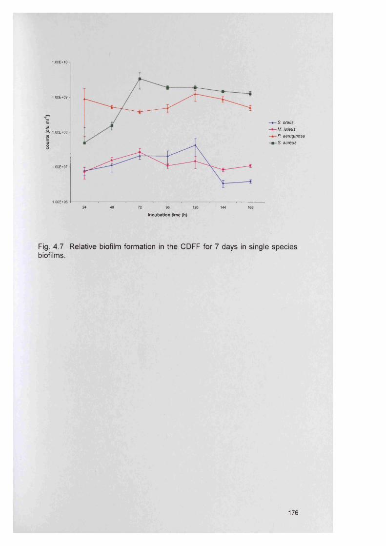
coun
ts
[cfu
ml'
]
1.00E+10
1.00E+09
S. oralis M. luteus P. aeruginosa S. aureus
1.00E+08
1.00E+07
1.00E+06
incubation time (h)
Fig. 4 .7 Relative biofilm formation in the CDFF for 7 days in single species biofilms.
176
Figure 4.8 illustrates the ability to differentially enumerate the recovered
bacterial species from CDFF generated biofilms. It was also noted that
subcultures of P. aeruginosa from biofilms (Fig. 4.8) appeared to have
different colony morphology compared to their planktonic equivalents (Fig.
not shown). Using mixed species biofilms (Fig. 4.9), similar numbers of both
S. aureus and P. aeruginosa were detected as previously reported in the
single species experiments (108-109 cfu ml'1). However, there was an
apparent increase in the numbers of S. oralis compared with its single culture
equivalent. In contrast, no growth of M. luteus in these mixed species
biofilms was again detected throughout the experimental period.
Interestingly, a similar loss of M. luteus was also noted in control mixed
planktonic cultures of these isolates over 7 d incubation (Fig. 4.10). The
viable count numbers for S. aureus and S. oralis were similar in both biofilm
and planktonic mixed cultures, whereas counts for P. aeruginosa were at
least one order of magnitude lower when grown in the planktonic state.
4.4.4 Planktonic test species’ susceptibility to PVP1The MBCs of planktonically cultured isolates against PVP1 were determined
as the total inhibition of visible growth and the absence of viable bacteria
growth. The latter was determined by taking a loopful of test solution and
plating it onto BA. An example of a P. aeruginosa MBC using a microtitre
plate assay is shown in Fig. 4.11. The MBC of PVP1 against P. aeruginosa
and S. aureus was 1%, against S. oralis and M. luteus it was 0.25 % and
0.03%, respectively.
177
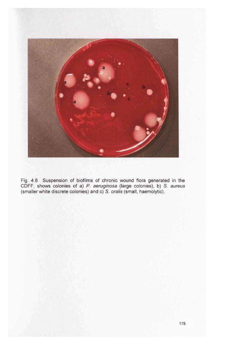
Fig. 4 .8 Suspension of biofilms of chronic wound flora generated in the CDFF, shows colonies of a) P. aeruginosa (large colonies), b) S. aureus (smaller white discrete colonies) and c) S. oralis (small, haemolytic).
178
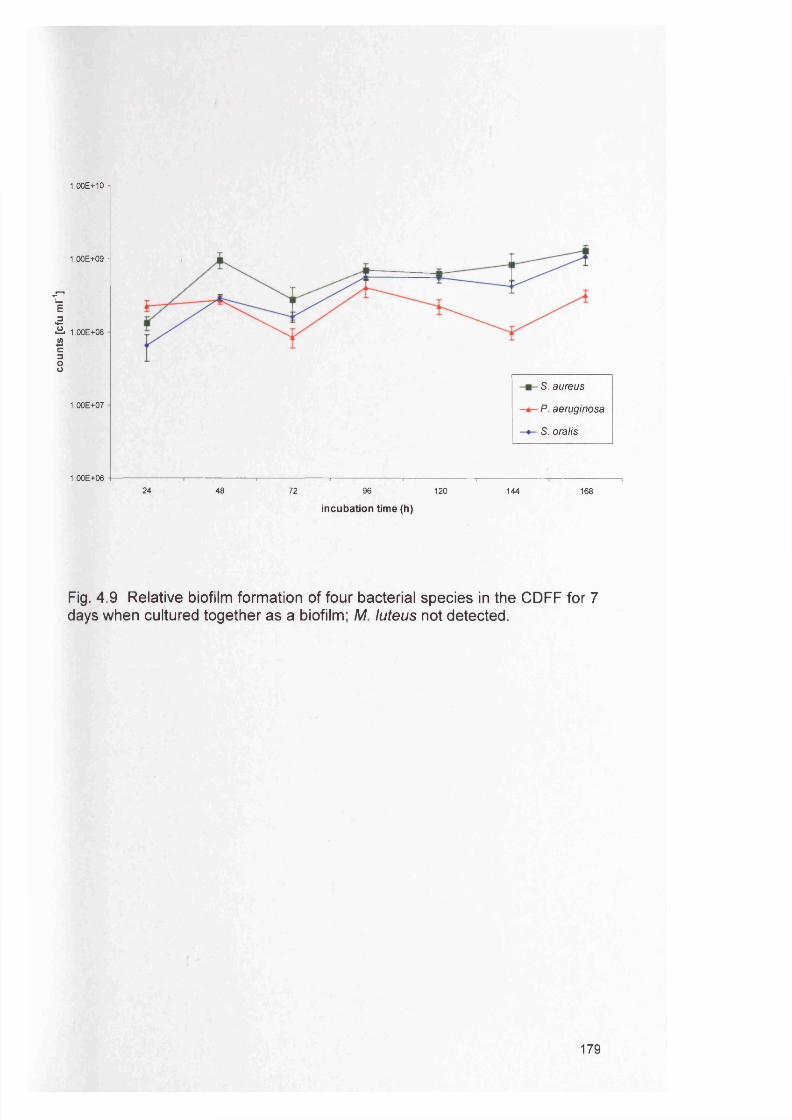
coun
ts [cfu
m
l'1]
1.00E+10
1.00E+09 -
1.00E+08
1.00E+07
S. a u re u s
P . a e r u g in o s a
■ S . o r a l is
1.00E+0624 48 72 96 120 144 168
incubation time (h)
Fig. 4.9 Relative biofilm formation of four bacterial species in the CDFF for 7 days when cultured together as a biofilm; M. luteus not detected.
179
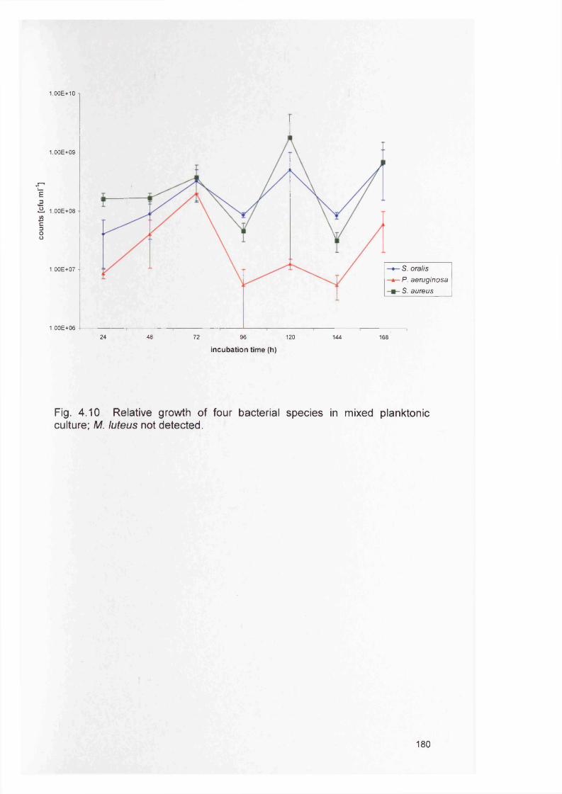
coun
ts [cfu
m
l'1]
1.00E+10
1.00E+09 -
1.00E+08
S. o r a l is
P . a e r u g in o s a
S . a u r e u s
1.00E+07 -
1.00E+064824 72 96 120 144 168
incubation time (h)
Fig. 4.10 Relative growth of four bacterial species in mixed planktonic culture; M. luteus not detected.
180
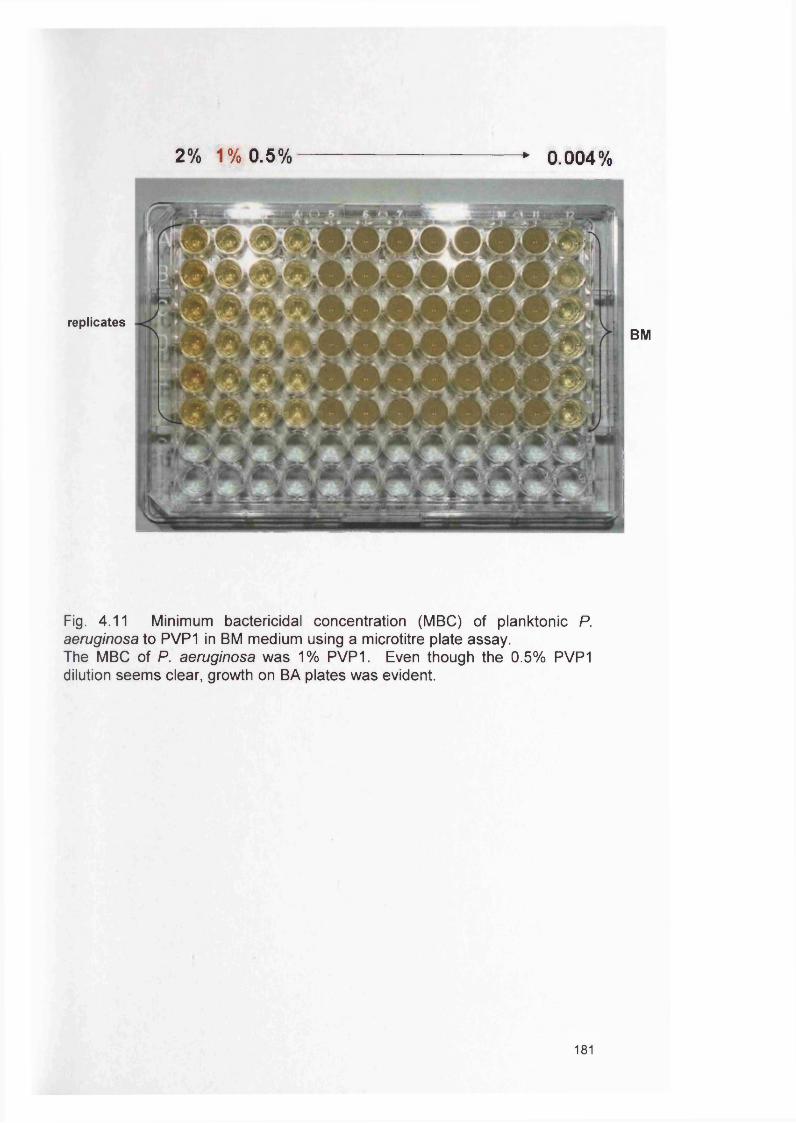
2% 1% 0.5% * 0.004%
replicatesBM
Fig. 4.11 Minimum bactericidal concentration (M BC) of planktonic P. aeruginosa to PVP1 in BM medium using a microtitre plate assay.The MBC of P. aeruginosa was 1% PVP1. Even though the 0.5% PVP1 dilution seems clear, growth on BA plates was evident.
181
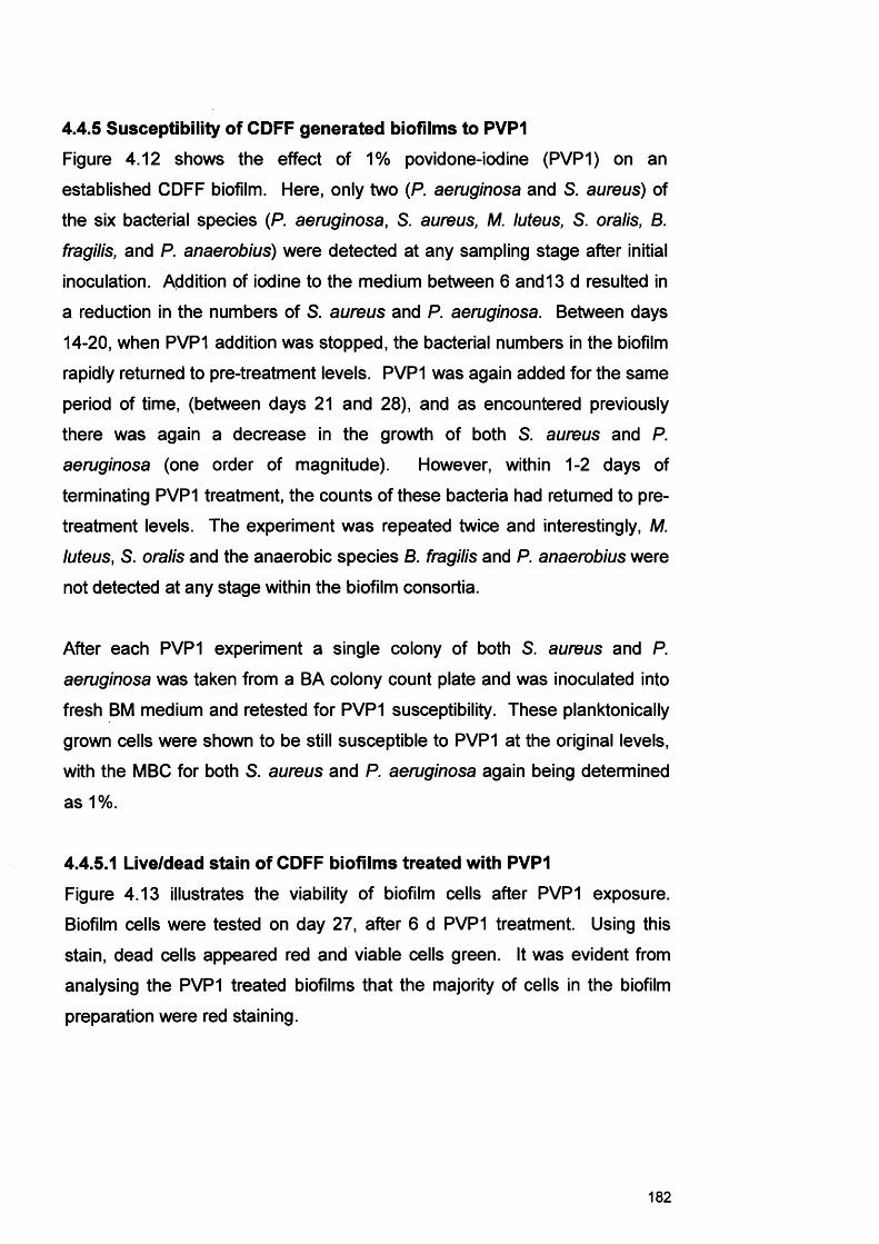
4.4.5 Susceptibility of CDFF generated biofilms to PVP1Figure 4.12 shows the effect of 1% povidone-iodine (PVP1) on an
established CDFF biofilm. Here, only two (P. aeruginosa and S. aureus) of
the six bacterial species (P. aeruginosa, S. aureus, M. luteus, S. oralis, B.
fragilis, and P. anaerobius) were detected at any sampling stage after initial
inoculation. Addition of iodine to the medium between 6 and 13 d resulted in
a reduction in the numbers of S. aureus and P. aeruginosa. Between days
14-20, when PVP1 addition was stopped, the bacterial numbers in the biofilm
rapidly returned to pre-treatment levels. PVP1 was again added for the same
period of time, (between days 21 and 28), and as encountered previously
there was again a decrease in the growth of both S. aureus and P.
aeruginosa (one order of magnitude). However, within 1-2 days of
terminating PVP1 treatment, the counts of these bacteria had returned to pre
treatment levels. The experiment was repeated twice and interestingly, M.
luteus, S. oralis and the anaerobic species B. fragilis and P. anaerobius were
not detected at any stage within the biofilm consortia.
After each PVP1 experiment a single colony of both S. aureus and P.
aeruginosa was taken from a BA colony count plate and was inoculated into
fresh BM medium and retested for PVP1 susceptibility. These planktonically
grown cells were shown to be still susceptible to PVP1 at the original levels,
with the MBC for both S. aureus and P. aeruginosa again being determined
as 1%.
4.4.5.1 Live/dead stain of CDFF biofilms treated with PVP1Figure 4.13 illustrates the viability of biofilm cells after PVP1 exposure.
Biofilm cells were tested on day 27, after 6 d PVP1 treatment. Using this
stain, dead cells appeared red and viable cells green. It was evident from
analysing the PVP1 treated biofilms that the majority of cells in the biofilm
preparation were red staining.
182
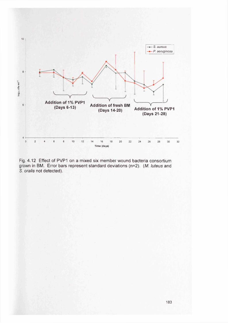
logi
o cfu
m
l
10
S. a u r e u s
P . a e r u g in o s a
Addition of1%PVP1 (Days 6-13) Addition of fresh BM
(Days 14-20) Addition of 1% PVP1 (Days 21-28)
4 ------------1------------1------------.------------ .----------------1----------------- 1---,--------- ,---------------,------------,------------ ,--------------- ,------- , ,------------------ ,------- ,0 2 4 6 8 10 12 14 16 18 20 22 24 26 28 30 32
Time (days)
Fig. 4 .12 Effect of PVP1 on a mixed six member wound bacteria consortiumgrown in BM. Error bars represent standard deviations (n=2). (M. luteus andS. oralis not detected).
183
Fig. 4.13 Live/Dead® staining of a PVP1 treated biofilm smear using the BacLight™ bacterial viability stain. Live cells appear green and dead cells appear red.
184
4.5 Discussion
A biofilm is often described as a community of microorganisms attached to a
surface and encased within an extracellular polysaccharide matrix (EPS). In
the laboratory, the microtitre plate assay is one of the most frequently used
methods to measure the biofilm-forming ability of bacteria (Kennedy and
O'Gara, 2004). In this present study, the microtitre plate assay was used to
examine the in vitro biofilm formation of chronic wound bacterial species and
in particular, to directly compare biofilm development of single (Fig. 4.4a and
b) and mixed wound bacterial species (Fig. 4.5-4.6a and b).
The benefits of using the microtitre plate assay were that it enabled the rapid
analysis of biofilm formation of multiple bacterial strains in a way that was
inexpensive and relatively simple to use. The microtitre plate biofilm model
does however have limitations. Principal amongst these is that it is a closed
model (batch system) which ultimately limits the level of biofilm formation
through depletion of nutrients and accumulation of waste products. However,
this could partially be overcome in any future experiments by replenishing the
growth medium at specific time intervals. The microtitre plate assay was also
labour intensive to prepare, particularly when many isolates were being
tested. Furthermore, inclusion of more than one species per plate can
promote contamination problems between neighbouring wells.
Throughout the study, species differences in the level of biofilm generated in
the microtitre plate assay were evident, and consistent for any given medium.
The hierarchy of biofilm formation altered with the medium and this highlights
the importance of the environmental milieu on biofilm development, which is
consistent with ideas presented by Kennedy et al. (2004). These differences
in the biofilm forming ability between the species could either be a result of
the different growth rates of the species in the chosen media, or a reflection
of the adherence properties of the species to the microtitre plate surface.
Previous work has indeed demonstrated that bacteria adhere more
favourably to some materials than others (Simoes et al., 2007a).
Furthermore, differences in the adherence capability to polystyrene microtitre
185
plates has been found between strains of some species, including S. aureus
(Stepanovic et al., 2000).
For all subsequent experiments using mixed species analysis, BHI, was used
as the growth medium, since this was found to be the most consistent of the
tested media in supporting biofilm formation for all the bacterial species. BHI
has also been widely used and validated in previous work (Fitzpatrick et al.,
2005; Kennedy and O'Gara, 2004; Parsek and Singh, 2003).
It was interesting to note that in mixed species biofilms, both P. aeruginosa
and S. aureus appeared to antagonise the biofilm forming ability of S. oralis
and M. luteus. Such an effect could occur for a variety of reasons including
occurrence of inter-species competition for surface attachment sites,
competition for nutrients within the growth medium or by production of a less
stable biofilm architecture. Furthermore, a motile organism like P.
aeruginosa can readily be envisaged as having an advantage over non-
motile counterparts within a biofilm environment. As the biofilm matures it is
inevitable that areas will develop that are restricted with respect to nutrient
and gaseous availability. In theory, a motile organism could migrate to areas
of the biofilm that are more suitable for its growth.
Simoes et al. (2007b) also reported antagonism between bacterial species
isolated from drinking water in biofilms generated within 96 well microtitre
plates. The microbial interactions were compared between single and dual
biofilms. Weak biofilm production was found between Sphingomonas
capsulate and Mycobacterium mucogenicum, S. capsulate and Acinetobacter
calcoaceticus and between M. mucogenicum and Staphylococcus spp.
(Simoes et al., 2007b). Kreth et al. (2005) also showed that S. mutans and
S. sanguinis engage in antagonistic interactions in dental biofilms.
The predominant microorganisms in chronic wounds have been determined
as P. aeruginosa and Staphylococcus spp. found in 34.8% (surface), 31.8%
(tissue), and 71.2% (both surface and tissue) of patients (Davies et al.,
186
2007). However, it is still unclear whether all four of the microorganisms
used in this study occur simultaneously within the same wound and therefore
whether the relative proportions of these species within a wound environment
reflects that found in these in vitro studies. Further clinical research of
chronic wounds is required to understand the relationship between biofilm
communities and the wound healing process. Extrapolating the results in the
present in vitro study to wounds might suggest that P. aeruginosa would be
expected to predominate over organisms such as M. iuteus. Chronic wounds
do show considerable diversity in the species of bacteria present, although S.
aureus and P. aeruginosa do appear to be the predominant organisms
encountered (James et al., 2007; Jones et al., 2004b).
Generally a lack of synergy between the bacterial species in the microtitre
plate assay and in the CDFF was observed with the exception of P.
aeruginosa which appeared to perform much better in the presence of M.
luteus (although this was to the detriment of the Micrococcus species; Fig.
4.5a). The ability of strains to compete in this in vitro environment could
theoretically be related to the relative bacterial numbers encountered in a
wound environment. Davies et al. (2007) in a study examining the
relationship between bacterial load and wound status, quantified both S.
aureus and P. aeruginosa from the same chronic wounds and showed that
the relative proportions of these two species was variable depending on
patients (results not shown). Equivalent results for Micrococcus species
were not however available.
The inability to extrapolate the in vitro findings of this present study to those
obtained from the work of Davies et al (2007) highlight the role of other
factors that are present in the wound, including the presence of other
microorganisms and host defence processes. In all microbial communities, a
degree of competition between species is expected despite the fact that a
stable community exists.
187
It could be hypothesised that M. luteus represents a wound contaminant from
the skin with only a transient occurrence in chronic wounds, whilst the
presence of P. aeruginosa and/or S. aureus could be indicative of the
pathogenic state. However, Micrococcus spp. were found to be present in
64% of patients (Davies et al., 2007) suggesting that this species is a true
coloniser of chronic wounds. In this regard, treatment options targeting P.
aeruginosa would be advocated. Further work is still needed on clinical
samples to confirm this hypothesis.
Due to the previously mentioned limitations of the microtitre plate assay the
Constant Depth Film Fermenter (CDFF) was also used to compare wound
bacterial biofilms. The advantages of the CDFF over the microtitre plate
system included, 1) the medium could be constantly replenished and the
waste components of the medium removed; 2) many replicates of equivalent
biofilms could be obtained; 3) the flow rate of the medium and any
supplements could be introduced as required; 4) almost any material could
theoretically be incorporated into the CDFF and serve as the biofilm
substratum. The CDFF model also has the potential to be adapted to
simulate the wound environment further, for example by the addition of fixed
volumes of collagen or fibronectin. However, the model also has limitations,
in that it is not able to simulate the effects of host factors, it is an abiotic
surface and lacks the presence of components of the host immune system
(Chapter 5 describes the use of a skin equivalent system that addresses
some of these limitations). Additionally, it is important to be aware of the
potential risk of contamination from airborne bacteria being introduced when
opening up the system daily to take samples. Nevertheless, it remains
important to gain an understanding of biofilm physiology by studying various
aspects of a biofilm in isolation, and the CDFF is an ideal model for such
analysis.
Results from the CDFF again showed that all four bacterial species grown
individually in the CDFF were able to form biofilms and that these biofilms
were readily maintained (Fig. 4.7). Interestingly, the relative species
188
hierarchy of biofilm formation using BHI medium was different between the
microtitre plate assay and the CDFF. In the microtitre plate assay, the
hierarchy of biofilm formation was M. luteus > S. oralis > S. aureus > P.
aeruginosa. In the CDFF, S. aureus produced the highest level of biofilm
followed by P. aeruginosa, S. oralis and M. luteus. The explanation for these
differences in biofilm hierarchy could be that the CDFF provided an open
system where fresh media was being constantly replenished and this may
have been of greater benefit to those species with higher growth rates. In
addition, the actual surface for attachment of the bacteria was different in the
the two models which could influence initial adherence and subsequently the
relative growth of the respective species.
Whilst it was evident that multispecies biofilms of chronic wound bacterial
species were able to be established and maintained within the CDFF model,
it was apparent that anaerobes could not be detected when included in mixed
species biofilm consortia. This could be because the CDFF did not provide a
truly anaerobic environment since anaerobic gas was not infused into the
fermenter for these experiments. Bradshaw et al. (1998) did however find
that anaerobes can flourish in a primarily aerobic environment as they utilise
specific anaerobic pockets within the biofilm. For this to occur however, the
anaerobic niches have to develop prior to loss of the anaerobic bacteria
within the system. Furthermore, it is also possible that in this present study
the anaerobes required a richer growth medium to be supported instead of
Brain Heart Infusion (BHI) medium. BHI had been selected based on the
previous microtitre plate experiments which did not include anaerobic
bacterial species.
In this present study, all bacterial species were added simultaneously and it
is important to note that under such situations the anaerobes would have had
to compete with aerobic organisms in an entirely aerobic environment. It has
been suggested that in a wound environment, the early contaminants are
most likely to be skin flora (e.g. Staphylococcus epidermidis and Micrococcus
species), which adhere to the wound, proliferate and synthesise EPS
189
(Percival and Bowler, 2004). These pioneer colonisers are then followed by
Gram-negative bacilli which populate the biofilm, utilise oxygen and provide
growth factors for anaerobes to establish themselves within the biofilm
(Percival and Bowler, 2004). Future work could therefore attempt to create
such a succession of colonisation of the biofilm through a later introduction of
the anaerobic bacteria into the system.
In the CDFF experiments that combined all four bacterial species, M. luteus
appeared to be ‘out-competed’ by the other organisms present (Fig. 4.10).
These results would therefore confirm those earlier findings of the microtitre
plate assay (section 4.4.2). The analysis of viable counts from biofilm
cultures also showed that M. luteus did not grow in combination with both S.
aureus and P. aeruginosa (Table 4.6 b). Since M. luteus was able to form
biofilms in single culture it is possible that this organism can rapidly colonise
the substratum used and thus as a non motile organism provide the base of
the maturing biofilm. Given such a location in the biofilm structure and the
associated limitations in terms of nutrients and oxygen at this site it is
possible that a dormant state or even a completer loss of viability might occur
for these organisms (Hill et al., 2003). However, the fact that M. luteus also
appeared to be ‘out-competed’ within a planktonic (Fig. 4.11) mixed culture
does support the likelihood of a true competition for nutrients with the other
bacteria as opposed to a strictly biofilm related factor.
It is also worth noting that subcultures of P. aeruginosa from the mixed
biofilms generated in the CDFF appeared to have a different colony
morphology compared to those of planktonic origin, suggesting that there
were phenotypic variations occurring for the P. aeruginosa in the two different
growth phases (Fig. 4.8). This phenotypic switch not only highlights the
successful biofilm development in the CDFF but also validates the
importance of subsequent investigations into any associated gene
expression changes within these biofilms (Chapter 6). It has been suggested
that under stress conditions, bacteria might possess diverse mechanisms
that may lead to an increase in genetic and phenotypic variability (Webb et
190
al., 2004). These mechanisms include adaptive mutation (Bjedov et al.,
2003; Oliver et al., 2000; Taddei et al., 1995), phase variation (Drenkard and
Ausubel, 2002) and enhanced gene transfer (Hausner and Wuertz, 1999;
Molin and Tolker-Nielsen, 2003).
In the present study, the CDFF was also used to model the sensitivity of a
biofilm comprising chronic wound bacteria to PVP1. PVP1 is a bacteriostatic
antimicrobial which is commonly used as a topical agent in the treatment of
wound infections (Mertz et al., 1999). Similar studies with antimicrobials
have been done previously using the CDFF. Ready et al. (2002a) used the
CDFF to examine the effect of tetracycline on the composition and antibiotic
resistance profile of dental plaque. However, that study examined the effect
of a single pulse of antibiotic rather than continuous administration (Ready et
al., 2002a). Also, their group did not compare the sensitivities of bacteria
recovered from the biofilm to the antibiotic when subsequently grown in the
planktonic state
Initial experiments with PVP1 in the present study, showed that planktonic
organisms were susceptible to a 1% concentration of PVP1. This is the
concentration that is believed to be available to the wound surface from
PVP1 impregnated dressings such as Inadine (Kumar et al., 2004). In
contrast, results showed that 1% PVP1 had only a slight effect on CDFF
generated bacterial biofilms. However, when colonies from S. aureus and P.
aeruginosa were recovered from the CDFF and then grown planktonically,
they remained susceptible to this 1% PVP1 concentration. This suggests
that the PVP1 resistance of the CDFF biofilms was associated with the
biofilm phenotype rather than being the result of an acquired resistance by
individual organisms exposed to the agent. The data presented in this study
concurs with that of Anderl et al. (2003) who suggested that planktonic cells
in stationary phase rapidly regain antibiotic susceptibility upon dilution in
fresh medium, which provides nutrients and also might dilute protective cell
cell signalling pathways (Anderl et al., 2003). This reversibility in
191
antimicrobial sensitivity indicates that biofilm tolerance of antimicrobials
represents a phenotypic rather than a genotypic alteration (Fux et al., 2005).
One mechanism of biofilm resistance to antimicrobials could be explained by
the ‘persister cell’ theory. As previously mentioned, bacteria embedded deep
within a biofilm are likely to be less metabolically active because of poor
access to essential nutrients, including oxygen, and may have problems with
the accumulation of waste metabolites in their surroundings. This reduced
activity has been linked to the finding that surface biofilm cells are normally
large in size whilst the biofilm embedded cells are often smaller (Anwar et al.,
1992). Embedded cells, or ‘persister cells’ are at a stage of ‘dormancy’
thereby making them phenotypically better equipped to persist in hostile
environments (Anwar et al., 1992; Rhoads et al., 2007). It was interesting to
note that live/dead staining of PVP1 biofilms indicated a majority of ‘dead’
cells. This could be indicative of ‘persister cells’ existing in the established
biofilm, which are dormant but culturable as seen by the viable counts.
Another reason could be that PVP1 does not actually kill the bacteria but
might only damage the cell membrane which is then reflected as the bacteria
staining ‘dead’ by the live/dead stain. It is important to note that on
withdrawal of treatment with PVP1, the level of bacterial numbers within the
biofilm rapidly reverted to those seen before treatment. This observation
might have important clinical implications in terms of the duration of treatment
with topical PVP1.
The antimicrobial activity of an agent on a biofilm can occur by two
mechanisms. Firstly, there is the direct effect of the agent on the microbial
cells themselves. PVP1 clearly demonstrates effective killing against the
cells used in this study as seen from the planktonic cell inhibition work. It has
also been proposed that topical antimicrobials enhance the recruitment of
polymorphonuclear neutrophils (PMNs) to the site of biofilm occurrence
(Akiyama et al., 2003). For example, treatment of S. aureus skin infections
with 2% fusidic acid ointment resulted in 100 times lower numbers of bacteria
in normal mice than in neutropenic mice 12 h after treatment (Akiyama et al.,
192
2002). As a consequence, whilst 1 % PVP1 appears to have limited value in
combating ‘wound biofilms’ in vitro, the in vivo effects may be higher through
synergism with patient host defence mechanisms.
This present study showed that the CDFF was an effective and reliable
model to generate in vitro sustainable growth of multispecies biofilms from
chronic wound bacteria, as evident by viable count determination (and later
by CLSM images of the biofilms; see Chapter 5). The PVP1 experiments
highlight the value of the CDFF for future in vitro experiments examining both
conventional and novel therapeutic interventions. The CDFF model also has
great potential to be adapted to allow investigation of a wide range of other
medically important biofilm-related infections, with wide reaching implications.
4.6 Summary1. This study demonstrated the development of in vitro biofilm models by
identified wound bacterial species.
2. Wound bacteria were able to produce biofilms in both a microtitre plate
assay and in the CDFF. The CDFF allowed multiple, identical and
reproducible biofilms to be grown simultaneously. Species differences in
terms of level of biofilm formation were evident for any given growth
medium and species antagonism occurred in mixed species biofilms for
certain bacterial combinations.
3. Susceptibility of the wound bacterial biofilms generated in the CDFF to
PVP1 was limited compared with planktonic cell susceptibility and any
effects observed rapidly returned to pre-treatment levels once PVP1
treatment was stopped.
193
Chapter 5
Structural analysis of biofilms generated by wound bacteria
194
5.1 INTRODUCTIONPrevious chapters of this thesis have described the use of in vitro models to
construct biofilms using oral (Candida albicans) and wound (bacterial
isolates) microorganisms. Although a lot of research has been done in the
study of oral plaque biofilms (Gu et al., 2005), there have been limited
studies undertaken on wound biofilms despite the recent proposed role of
biofilm involvement in delaying wound healing (Davies et al., 2007; Mertz,
2003).
Sophisticated molecular techniques have been applied in many fields of
microbiology. However, in the application of molecular techniques to
biofilms, little information is generally obtained regarding biofilm morphology
or architecture. For example, in the case of DNA/RNA extraction, cells are
often pooled and any tissue/biofilm destroyed in the extraction process.
Traditional microscopical analyses and bacterial stains, like the Gram or
Ziehl-Neelsen stain (Moter and Gobel, 2000) are useful, but limited, as they
often rely on prior cultivation of the bacteria. Whilst Gram staining provides
information on the type of bacterial cell wall it does not allow for species
identification (Moter and Gobel, 2000). It is important to highlight that
microbial communities are fundamental in the functioning of all ecosystems
(Tyson, 2004). Since cultivation techniques tend to work only for a minority
of species (only 2-3% of bacteria are thought to be cultivable using currently
available media), the development of alternative methods for the
identification and visualisation of bacteria has become a necessity (Amann,
2001; Moter and Gobel, 2000).
5.1.1 Bacterial detection and distribution in a biofilm communityBiofilm formation is a dynamic process involving continuous changes in
structure and composition through microbial attachment, detachment, growth
of different species and the production of extracellular polysaccharides (EPS)
(Gu et al., 2005). An ambitious goal in microbiology is to have rapid and
accurate identification of pathogenic bacteria within biofilm communities.
Effective monitoring of the organisms within a biofilm is a crucial step in
195
assessing the possible risk or the status of a disease (Gu et al., 2005).
Marsh et al. (2003) established a connection between the proportions of
certain bacterial species in the oral biofilm (plaque) and the absence or
presence of dental caries. Marsh et al. (2003) suggested that changes in the
environment (e.g. nutrition, oxygen, pH, long term use of medication) alter
the pattern of gene expression of oral bacteria, so that the competitiveness of
the species associated with disease is increased, while that of organisms that
normally prevail in health is reduced (ecological plaque hypothesis). Dental
caries is associated with an increase in the proportion of acidogenic and
aciduric bacteria (e.g. Streptococcus mutans, S. sobhnus and lactobacilli)
which demineralise the enamel, and in periodontal diseases anaerobic
bacteria including Gram-negative proteolytic species are increased. These
plaque mediated diseases could be prevented by interfering with the
environmental factors as well as by inhibiting the putative pathogens (Marsh,
2003). The work on dental plaque therefore highlights the potential
importance in analysing the diversity and distribution pattern of pathogenic
organisms which reside within chronic wounds or any other biofilm-related
infections.
5.1.2 Biofilm structure within a community
In the majority of natural habitats, microorganisms exist as biofilms, which
usually comprise (although not exclusively) of a variety of species and
genera. Biofilms are formed after attachment and growth of microorganisms
on a broad range of surfaces (Thurnheer et al., 2004). Biofilms are
comprised of water, cells, cell aggregates and microcolonies embedded in an
EPS matrix (Costerton et al., 1995; Costerton et al., 1999). The biofilm
architecture and the physiological status within this community is the main
interest for clinical, industrial and environmental microbiology (Thurnheer et
al., 2004). Further detail on biofilm structure is provided in the Literature
Review (section 1.2.2).
196
5.1.3 Fluorescence in situ hybridisationFluorescence in situ hybridisation (FISH) is a relatively recent and now
frequently used culture-independent approach for detecting and visualising
microorganisms. This technique detects nucleic acid sequences within cells
using fluorescently labelled probes that hybridize specifically to their
complementary target sequences within intact cells (Moter and Gobel, 2000).
FISH therefore combines the precision of molecular genetics with the visual
imaging of fluorescent microscopy. Hence it permits visualisation and
identification of individual cells within their natural habitat or diseased tissue
(Moter and Gobel, 2000). The FISH procedure includes the following steps
(Fig. 5.1): a) fixation of the specimen; b) preparation of the sample, including
specific pre-treatment steps if required; c) hybridisation with probes for
detecting the respective target sequences; d) washing steps to remove
unbound probes; e) mounting and visualisation (Moter and Gobel, 2000).
More often, probes are used that target ribosomal RNA molecules as these
are functionally conserved, abundant and present in all cells. The small
subunit (16S and sometimes 23S) rRNA molecules are ideal targets for
bacterial detection because they are stable and have high a copy number of
molecules per cell (usually more than 10,000), which means that probing can
be highly sensitive (Perry-O'Keefe et al., 2001b). As 16S rRNA sequence
data is readily available for a wide variety of species, probes can be designed
either to target signature sequences of specific species or chosen groups, or
to hybridise with sequences conserved in all known bacteria (Amann, 2001).
The probes used in FISH are typically 15 to 30 nucleotides in length and
covalently linked at the 5’ end to a single fluorescent dye molecule (Wagner
et al., 2003b). Suitable fluorophors include carbocyanide dyes,
tetramethylrhodamine and fluorescein (Amann, 2001).
One of the major disadvantages of FISH when using oligonucleotide probes
is that sometimes insufficient penetration of probes into bacteria occurs due
to their thick cell wall, e.g. the walls of Gram-positive bacteria.
197
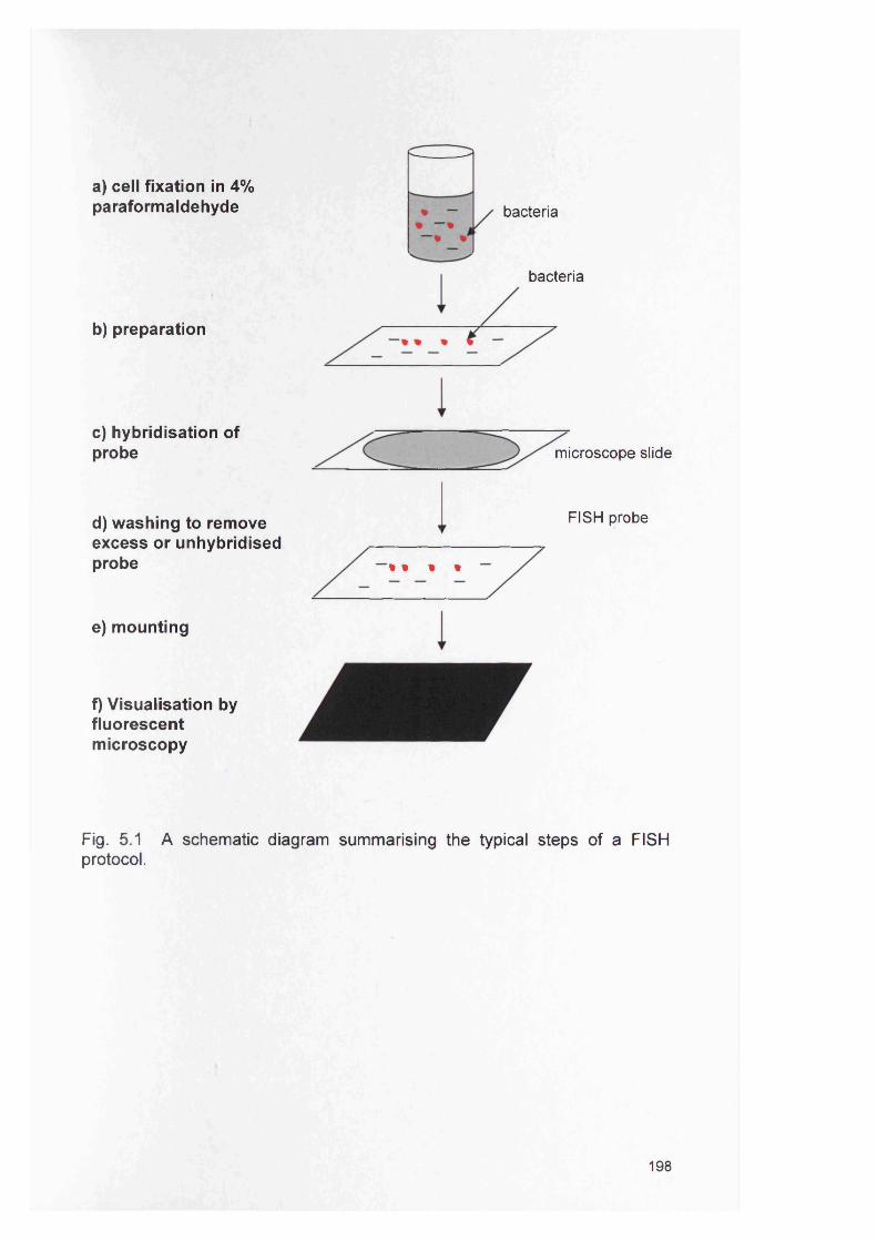
a) cell fixation in 4% paraformaldehyde
b) preparation
bacteria
bacteria
c) hybridisation of probe
d) washing to remove excess or unhybridised probe
e) mounting
f) Visualisation byfluorescentmicroscopy
microscope slide
FISH probe
i t % %
Fig. 5.1 A schematic diagram summarising the typical steps of a FISH protocol.
198
5.1.4 Peptide Nucleic Acid (PNA) probes
Recently, PNA probes have been incorporated into FISH applications. PNA
probes are DNA-analogues (pseudopeptides) with an uncharged polyamide
backbone instead of conventional sugar phosphates. PNA probes hybridise
to complementary nucleic acid targets obeying the Watson-Crick base pairing
rules (Moter and Gobel, 2000; Perry-O'Keefe et al., 2001b). Due to their
uncharged backbone, PNA probes have superior hybridisation
characteristics, including higher specificity and more rapid hybridisation
kinetics. PNA probes also hybridise independently of the salt concentration
and readily diffuse through hydrophobic cell walls, thus allowing detection
without any pre-treatments (Moter and Gobel, 2000; Perry-O'Keefe et al.,
2001b). As hybridisation with PNA probes can be performed in a low salt
buffer, a decrease in the stability of the rRNA secondary structure can be
induced, thereby allowing PNA probes to hybridise to otherwise less
accessible targets (Perry-O'Keefe et al., 2001b). Hence PNA probes have
considerable advantages over DNA probes, but unfortunately are significantly
more expensive to synthesize.
5.1.5 Applications of FISHFISH targeted towards 16S rRNA sequences has been applied to many
diverse bacterial systems, often in conjunction with other molecular-based
profiling approaches such as denaturing gradient gel electrophoresis (DGGE)
or gene sequencing. The first uses of the FISH technique were in the
detection and identification of bacteria in environmental specimens, including
activated sludge granules (Sekiguchi et al., 1999) and marine sediments
(Amann, 2001). In clinical samples, FISH has been applied to the study of
potentially medically important bacteria, such as Escherichia coli in human
faecal samples (Jansen et al., 1999; Vaahtovuo et al., 2005) and
polymicrobial specimens from the oral cavity (Thurnheer et al., 2004). In the
case of oral bacteria, FISH has been employed in the detection of organisms
within samples of human carious dentine (Banerjee et al., 2002), dental
plaque (Banerjee et al., 2002; Thurnheer et al., 2004; Thurnheer et al., 2001;
Wecke et al., 2000), periapical endodontic lesions (Sunde et al., 2003) and
199
buccal epithelial cells (Rudney et al., 2001; Rudney et al., 2005). PNA FISH
has been less extensively used, although in recent years the approach has
been applied for fast visualisation and identification of clinically relevant
mycobacteria in clinical specimens and tissue (Lefmann et al., 2006).
Examples of bacteria detected by PNA probes include clinically relevant
Enterococcus species (Wellinghausen et al., 2007), Helicobacter pylori in
gastric biopsy specimens (Guimaraes et al., 2007) and the identification of S.
aureus directly from positive blood culture bottles (Oliveira et al., 2002).
5.2 AIMSThe aims of the present study were:
1. To establish a reliable FISH methodology using both DNA and PNA
probes to detect specific wound bacterial species.
2. To evaluate FISH and CLSM in examining the spatial organisation of
bacteria in biofilm models and wound tissue.
200
5.3 Methods
To establish a reliable FISH methodology two separate FISH protocols were
initially evaluated against planktonic bacteria and after validating these
methods were extended to determine the spatial organisation in mixed
species biofilm modes of growth. The two protocols were: A) detection of
fixed bacteria using a universal DNA probe- EUB338 and B) PNA probe
detection of bacteria (PNA FISH). The probes and their conditions are
summarised in Table 5.1.
5.3.1 Universal DNA EUB338 probe
The universal bacterial DNA probe EUB338 is complementary to a portion of
the 16S rRNA gene conserved in the domain Bacteria, and has been used to
visualise the entire bacterial population of biofilms (Amann et al., 1990;
Banerjee et al., 2002; Sunde et al., 2003). The probe EUB338, was
synthesised commercially (MWG Biotech, Ebersberg, Germany) and at the 5’
end-labelled with fluorescein isothiocyanate (FITC).
The NON338 probe (Amann et al., 1990) which is complementary to the
universal bacterial probe EUB338 was used in this study as a negative
control for non-specificity. As an additional negative control, RNase treated
cells were also probed to confirm that the probes specifically targeted RNA.
5.3.2 Peptide nucleic acid probes (PNA)Initially, PNA probe sequences to target two bacterial species (P. aeruginosa,
S. aureus) and the Bac-Uni1 universal bacterial PNA probe were used (Table
5.1). The sequences of these probes were partially adapted from Perry-
O’Keefe et al. (2001b). The probes were manufactured by Boston Probes
(Foster City, USA) for Applied Biosystems (Warrington, UK) and were
labelled either with FITC, cyanine 5 (CY5) or cyanine 3 (CY3) and designed
to target the 16S rRNA. All probes were pre-validated using the PNA probe
designer software (Applied Biosystems) before synthesis.
201

Table 5.1 Probes used and their conditions.
Probes Target Nucleotide sequence Hybridisation conditions: temp. [°C]
formamide [%]
Fluorescentlabel
WorkingCone.[nM]
Excitationwavelengths
[nm]
References
EUB338 Universalprobe
(Eubacteria)
5’-GCTGCCTCCCGTAGGAGT -3’ 46°C no formamide
FITC 0.2-0.5 nM 488 (Amann et al., 1990)
NonEUB338
Non-senseEUB338
5’-CG ACGG AGGGC AT CCT C A-3’ 46°C no formamide
FITC 0.2-0.5 pM 488 (Amann et al., 1990)
Psaer PNAP.
aeruginosa
5’-OOAACTT GCT GAACCAC-3’ 55°C no formamide
FITC 300 488 (Coull and Hyldig- Nielsen, 2003)
Sta16S03
PNAS. aureus
5’-OOGCTT CT CGT CCGTTC-3’ 55°C 30% formamide
CY5 500 633 (Perry- O'Keefe et al., 2001)
Bac-Uni1
PNAUniversal
probe(Eubacteria)
5’00CTGCCTCCCGTAGGA-3’ 55°C no formamide
CY3 150 561 (Perry- O'Keefe et al., 2001)
5.3.3 Development of fluorescence in situ hybridisation (FISH) for planktonically grown bacteria
5.3.3.1 Protocol A: Detection of fixed planktonic bacteria using the
universal bacterial DNA EUB338 probe
Bacteria were prepared and detected by EUB338 using two methods. The
first of these involved culturing bacteria overnight (in BHI broth) at 37°C. The
growth was pelleted by centrifugation (7000 g, 5 min at 4°C) and
resuspended in 0.85% (w/v) saline. This procedure was repeated to produce
a washed pellet. A portion of the suspension was taken (100 pi) and three
volumes of 4% paraformaldehyde added (giving a final concentration of
paraformaldehyde of 3%). The cell suspension was then vortexed and
stored overnight at 4°C. The cells were then centrifuged (7000 g, 5 min at
4°C) and the bacterial pellet was again resuspended twice in 0.85% saline to
its original volume, before dilution with paraformaldehyde. Subsequently, the
suspension was incubated in 50 mM Tris-EDTA (pH 8; TE, Sigma) buffer
containing lysozyme (10 mg ml'1; Sigma) for 20 min at 37°C (60 pi). The cell
suspension was centrifuged and resuspended in half its original volume (50
pi). An equal volume of 96% ethanol was then added. The suspension was
vortexed for 30 s and stored at -20°C. Prior to staining with the DNA probe a
portion of these fixed cells (10 pi) was deposited on to a microscope slide
and air dried.
Pre-warmed hybridisation buffer (300 pi) containing 0.5 pM (in H2O) of
oligonucleotide probe EUB338 (FITC-labelled), was carefully applied to the
slide. Following incubation for 3.5 h in a dark humid chamber at 46°C, slides
were washed with distilled water, air-dried in the dark and subsequently
mounted with FluoroSave™ (Calbiochem, Merck Biosciences Ltd.,
Nottingham, UK).
The second method of preparing and detecting bacteria involved the
resuspension of a colony of the bacteria from an agar plate into PBS.
203
A smear was then made on a microscope slide that was air dried, flamed
briefly to fix and then immersed in 4% paraformaldehyde overnight at 4°C.
Slides were then washed with distilled water twice and dried at 37°C. To
permeabilise the cells for entry of the oligonucleotide probe, slides were
incubated in 50mM Tris-EDTA (pH 8) buffer containing lysozyme (10mg ml-1)
for 20 min at 37°C. Slides were washed with distilled water, air-dried and
incubated in 50mM TE buffer (pH 8) containing proteinase K (7pg ml'1) at
37°C for 20 min. Slides were then washed again with distilled water, air-dried
and pre-incubated at 48°C for 20 min in a hybridisation buffer (300 pi)
containing 0.9 M sodium chloride (Sigma, Poole, UK), 20mM Tris-HCI (pH
7.4, Sigma), and 0.5% SDS (Sigma). EUB338 hybridisation was then
performed as described above.
5.3.3.2 Protocol B: Detection of fixed planktonic bacteria using the PNA
probes
The first step was to confirm the specificity of the PNA probes on planktonic
bacteria in a mixed fixed population using the three PNA probes; the
universal probe (Bac-Uni1CY3), the P. aeruginosa specific probe
(PsaerFITC) and the S. aureus specific probe (Sta16S03).
Cell fixation and PNA FISH was performed as described by Perry-O’Keefe et
al. (2001a). Overnight bacterial cultures were pelleted by centrifugation
(16,000 g, 5 min) and resuspended in PBS (7 mM Na2HP04, 7 mM
NaH2P0 4 , 130 mM NaCI). Cell suspensions were centrifuged again (16,000
g, 5 min), resuspended in PBS with 4% (w/v) paraformaldehyde (Sigma) and
fixed for 1 h. The fixed cells were rinsed in PBS, resuspended in 50% (v/v)
ethanol and incubated for at least 30 min at -20°C prior to use.
A 100-pl volume of fixed cells was pelleted by centrifugation, the cell pellet
was rinsed with PBS and resuspended in 100 pi of hybridisation buffer (25
mM Tris-HCI, pH 9.0; 100mM NaCI; 0.5% (w/v) SDS) containing 150-500 nM
fluorescently labelled PNA probe. The cells were incubated at 55°C for 30
204
min, centrifuged at 16,000 g for 5 min and resuspended in 500 pi of wash
solution (10 mM Tris pH 9.0, 1 mM EDTA). After a further incubation at 55°C
for 10 min the cells were pelleted by centrifugation. This was repeated twice
for a total of three washes. Following the last wash, cells were resuspended
in 100 pi wash solution and 2 pi of this cell suspension was spread on a
clean microscope slide and allowed to air dry. Vectashield mounting medium
was applied to the dried cells which were overlaid with a coverslip (Perry-
O'Keefeetal., 2001a).
5.3.4 Development of biofilms for fluorescence in situ hybridisation
(FISH) detectionPrior to bacterial detection and determination of bacterial spatial organisation
in the biofilm mode of growth using FISH, biofilms had to be established
using different models (CDFF, RHE). The preliminary steps for this are
described below. The chronic wound biopsies were also processed as
described below prior to FISH imaging.
5.3.4.1 Production of biofilms in the CDFFPrior to FISH detection, biofilms were grown in the constant depth film
fermenter (CDFF; Chapter 4, section 4.3.12.1). The temperature of the
fermenter was maintained at 37°C by housing it in an incubator. The plugs
were recessed to a depth of 400 pm, and the turntable speed of the CDFF
was at 20 revs min'1. Wound bacteria (Staphylococcus aureus D76,
Streptococcus oralis B52, Micrococcus luteus B81, and Pseudomonas
aeruginosa D40) were grown overnight at 37°C in 10 ml BHI, and five ml of
each wound isolate was added to 1 L of BHI medium. This inoculum was
recirculated through the CDFF for 24 h to seed the system. After this time,
the inoculum was disconnected and fresh media fed into the CDFF. Biofilms
were grown in the CDFF for 7 d. Fresh medium was delivered at a rate of 30
ml min'1, using a peristaltic pump (Watson-Marlow, Falmouth, UK) and
biofilm samples were taken for analysis over a period of 7 days every 24 h.
205
5.3.4.2 Fixation of CDFF generated biofilmsBiofilm samples (including the PTFE pin) generated in the CDFF were
placed in molten agarose (2% w/v; Sigma) for 24h; fixed in 2%
paraformaldehyde solution for 24 h and then embedded in paraffin wax using
standard histological techniques.
5.3.4.3 Preparation of bacteria for infection of an in vitro Reconstituted
Human Epidermidis (RHE) modelSkinEthic Laboratories cultivate a 0.5 cm2 reconstituted human epidermis in
vitro on a polycarbonate filter at the air liquid interface in chemically defined
medium lacking antibiotics for 17 days. The RHE consists of normal human
keratinocytes (human foreskin derived) with a well differentiated epidermis
consisting of basal, spinous, granular layers and a stratum corneum. At least
four viable cell layers are present. This RHE was different to the oral
epithelium used previously in Chapter 2.
Bacterial wound species (S. aureus D76, S. oralis B52, M. luteus B81, and P.
aeruginosa D40) were cultured on blood agar (LAB M, Bury, UK) at 37°C
overnight. The resulting growth was used to inoculate 10 ml of BHI and
incubated at 37°C for 24 h. Bacterial cells were then harvested by
centrifugation and washed (*3) with PBS. The pelleted bacterial cells were
resuspended in RHE tissue maintenance medium (supplied by SkinEthic
Laboratories, Nice, France; chemically defined medium MCDB 153
containing 5 pg ml'1 insulin and 1.5 mM calcium chloride with no antibiotic)
and 200 \x\ of the resulting bacterial suspension added to the RHE.
Additionally to simulate a skin wound, the surface of the RHE model was on
occasion scratched with a sterile pipette tip prior to infection with bacteria.
5.3.4.4 Culture of infected RHEInfected RHE was incubated for 24-28 h at 37°C in a humidified atmosphere,
enriched with 5% C02as described previously (Chapter 2, section 2.3.2.4).
206
5.3.4.5 Processing of infected RHEAfter incubation, the whole RHE was fixed in 10% (v/v) formalin for 24 h and
then embedded in paraffin wax using standard histological techniques (as
previously described (Chapter 2, section 2.3.2.4.3).
5.3.4.6 Processing of biopsies from non- infected venous leg ulcersWound biopsies had previously been obtained with ethical approval and
patient informed written consent from non-infected venous leg ulcer patients
attending the Wound Healing Research Unit Clinic at the University Hospital
of Wales, Cardiff between 1999 until 2002 and stored at -20°C until required
(Davies et al., 2007). Biopsies were fixed in 10% (v/v) formalin for 24 h and
then embedded in paraffin wax using standard histological techniques.
5.3.4.7 Preparation of biofilms (CDFF and RHE) and biopsiesPrior to FISH, all processed biofilm sections (20 pm; see above) were placed
on to Histobond+ coated microscope slides (Raymond A Lamb, East Sussex,
England), de-waxed and processed through xylene and ethanol to water
before probe hybridisation. To hybridise the PNA/DNA probes to the fixed
bacteria, sections were directly treated with 100 pi lysozyme (10 mg ml'1;
Sigma) and incubated at 37°C for 30 min. Sections were then briefly washed
in pre-warmed wash solution (10 mM Tris pH 9.0, 1 mM EDTA) prior to probe
hybridisation.
5.3.4.8 Hybridisation of RHE derived biofilms with the universal EUB338
DNA probeSections of RHE developed biofilms (RHE, 20 pM) were pre-incubated for 20
min at 48°C in hybridisation buffer (300 pi) containing 0.9 M sodium chloride
(Sigma), 20mM Tris-HCI (pH 7.4, Sigma), and 0.5% SDS (Sigma). Pre
warmed hybridisation buffer (300 pi) containing 0.2-0.5 pM of FITC-labelled
EUB338 oligonucleotide probe, (MWG Biotech, Ebersberg, Germany) was
carefully applied to the RHE section. Following incubation for 3.5 h in a dark
humid chamber at 46 °C, slides were washed with dH20, air-dried in the
dark.
207
5.3.4.9 Hybridisation of PNA probes to bacteria in RHE and CDFF
biofilms and wound biopsies
One hundred and fifty pi of pre-warmed hybridisation buffer (25 mM Tris-HCI,
pH 9.0; 100mM NaCI; 0.5% (w/v) SDS) containing 150-400 nM of
fluorescently labelled PNA probe (see Table 5.1), was added to each CDFF
biofilm/RHE tissue and wound biopsy section (20 pM), which was placed in a
dark, humidified chamber and incubated at 55°C for 90 min. The stringency
was adjusted by adding 30% (v/v) formamide to the hybridisation buffer for
the S. aureus specific probe (Sta16S03). After incubation, each slide was
washed with pre-warmed wash solution using a magnetic stirrer for 30 min.
5.3.4.10 Staining of RHE keratinocytesFor nuclear context, sections were counterstained with Hoechst 33258
(Sigma) 2 pg ml-1 for 20 min, before washing in wash-solution and mounted
using Vectashield fade-retarding mountant (Vector Laboratories Ltd.,
Peterborough, UK).
5.3.4.11 Haematoxylin and eosinAdditionally, biofilm and RHE sections were stained by the routine
haematoxylin and eosin (H and E) procedure (Cross, 2004). Sections were
processed using a Shandon Linistain machine (Shandon, Runcorn, UK) in
the following order. Sections were first de-waxed, rinsed in 100% ethanol and
then rinsed in H2O. After the washing steps, sections were stained with
Harris’ haemotoxylin (Sigma) for 5-6 min, rinsed briefly in 1% acidic alcohol,
followed by a brief rinse in Scott’s tap water substitute (blue, Sigma, UK) and
counterstained with 1% aqueous eosin for 30 s to 1 min. Sections were
dehydrated in alcohol, cleared in saline and mounted with DPX mounting
medium (Raymond A Lamb Ltd., Eastbourne, UK).
5.3.4.12 Gram stain
Sections were first de-waxed, rinsed in 100% ethanol and then rinsed in H20.
After the washing steps, sections were stained with Crystal violet (0.5% w/v)
in 25% alcohol for 3 min, rinsed briefly in H20 and then stained with Gram’s
208
iodine for 3 min. Sections were rinsed briefly in H2O, acetone decolourised
for few seconds and again briefly rinsed in H2O. Subsequently sections were
stained with neutral red for 3 min, dehydrated in alcohol, rinsed briefly in
saline and mounted with DPX mounting medium.
5.3.5 Confocal Laser Scanning Microscopy (CLSM)Stained sections were viewed and analysed by CLSM using a Leica TCS
SP2 AOBS spectral confocal microscope (Leica, Wetzlar, Germany). The
sections were scanned through the full depth using appropriate settings for
single, double or triple channel fluorescence recordings of FITC, CY5, CY3 or
Hoechst 33258 as detailed in Table 5.2. For multi-channel recordings,
fluorochromes were scanned sequentially to eliminate spectral overlap
between probes. Selected images were presented either as single confocal
optical sections or maximum intensity type reconstructions.
209
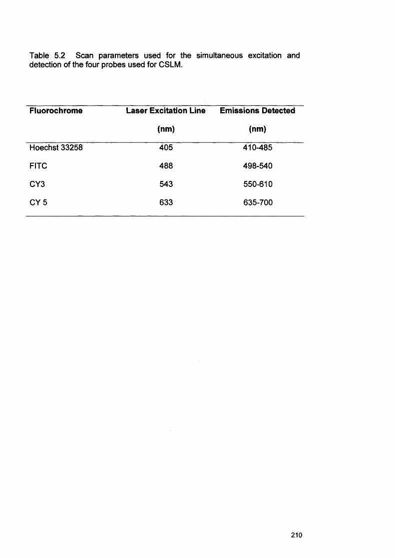
Table 5.2 Scan parameters used for the simultaneous excitation and detection of the four probes used for CSLM.
Fluorochrome Laser Excitation Line
(nm)
Emissions Detected
(nm)
Hoechst 33258 405 410-485
FITC 488 498-540
CY3 543 550-610
CY 5 633 635-700
210
5.4 RESULTS
5.4.1 Analysis of planktonic cell detection and RHE biofilms using the
universal EUB338 probe
To establish a reliable FISH protocol the universal EUB338 probe was initially
evaluated on planktonic and biofilm cells. Using the EUB338 probe, all the
bacteria species could be detected (Fig. 5.2). However, strong
autofluorescence of planktonic cells and the tissue and weak hybridisation
signals were evident on RHE sections (Fig. 5.2c).
5.4.2 Bacterial specificity of PNA probesThe FISH protocol using PNA probes was established and widely used in this
study on fixed and non-fixed bacteria. Figure 5.3 is a composite of three
micrographs that show detection of all four test bacteria (S. aureus D76, S.
oralis B52, M. luteus B81, and P. aeruginosa D40). Figure 5.3a illustrates
detection of all bacteria using the PNA universal probe (Bac-Uni1CY3) with
all cells visualised red. Figure 5.3b shows detection of P. aeruginosa only
with the PsaerFITC. In this figure only a proportion of the population stain
green, identifying P. aeruginosa. Cells which did not stain with the
PsaerFITC also clearly had a distinct coccus type morphology. Figure 5.3c is
an overlay of all the CLSM channels and shows detection of all bacteria
stained with the three different fluorescent markers. Pseudomonas
aeruginosa cells appear yellow in this overlay as a result of hybridising with
both the Bac-Uni1CY3 and with the PsaerFITC probe. Blue/purple cells were
S. aureus, as a result of hybridising with both the Bac-Uni1CY3 and the
Sta16S03 probes. Red staining cells were either M. luteus or S. oralis as
these hybridised only with the Bac-Uni1CY3 probe. Multiplex PNA staining
demonstrated that these combined probe preparations remained effective for
species identification.
211
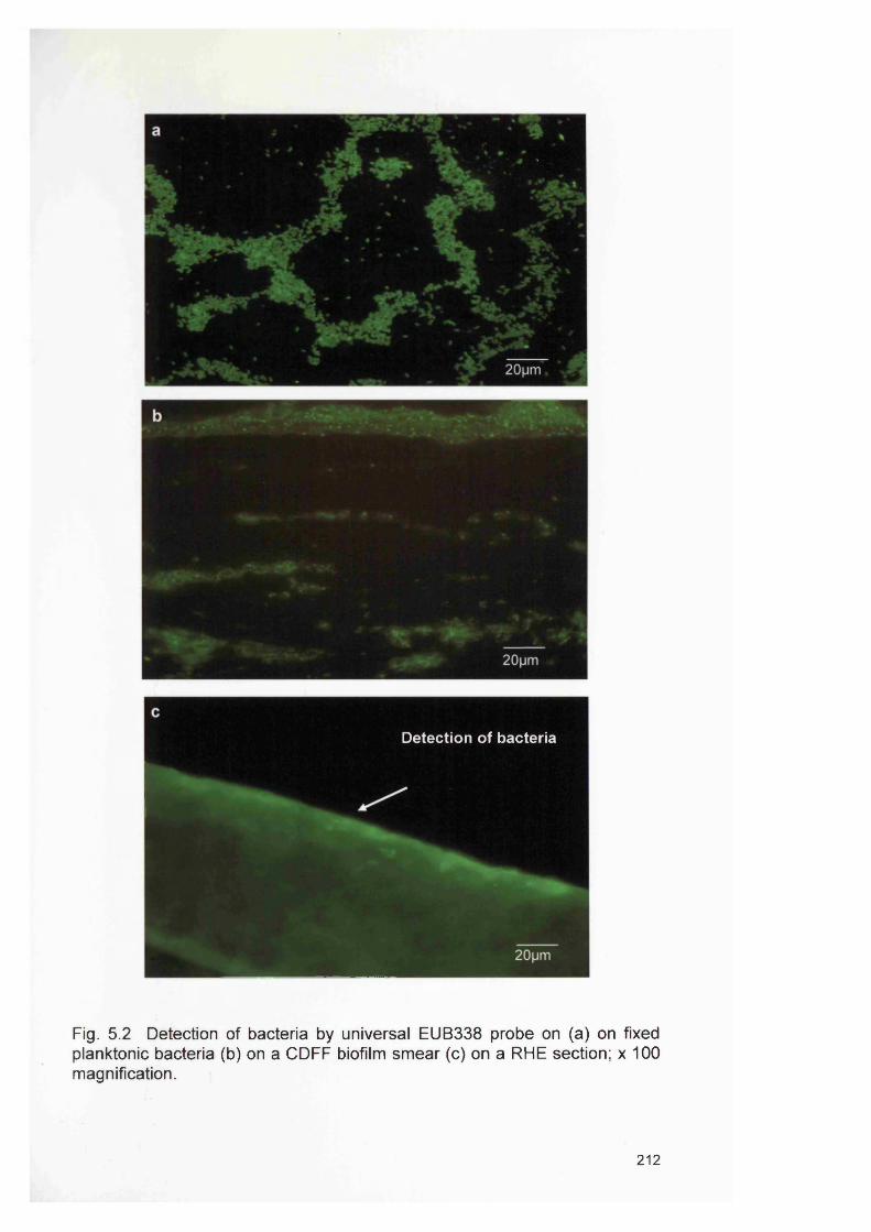
Detection of bacteria
Fig. 5.2 Detection of bacteria by universal EUB338 probe on (a) on fixed planktonic bacteria (b) on a C D FF biofilm sm ear (c) on a RHE section; x 100 magnification.
212
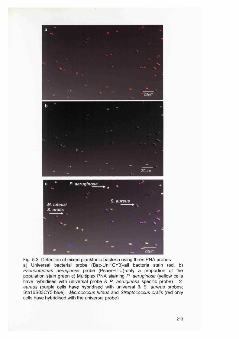
Fig. 5.3 Detection of mixed planktonic bacteria using three PNA probes, a) Universal bacterial probe (Bac-Uni1CY3)-all bacteria stain red, b) Pseudomonas aeruginosa probe (PsaerFITC)-only a proportion of the population stain green c) Multiplex PNA staining P. aeruginosa (yellow cells have hybridised with universal probe & P. aeruginosa specific probe). S. aureus (purple cells have hybridised with universal & S. aureus probes; Sta16S03CY5-blue). Micrococcus luteus and Streptococcus oralis (red only cells have hybridised with the universal probe).
5.4.3 Analysis of CDFF generated biofilms using PNA FISH
The PNA FISH technique was applied to mixed species biofilm growth of the
target bacteria. Biofilms generated in the CDFF (single and mixed) were
successfully processed and stained with the PNA FISH probes. Using all
three PNA probes, target bacteria could be detected in appropriate sections
of the biofilms examined. Examples of biofilm sections prepared in the CDFF
containing fluorescently-labelled bacteria are shown in Fig. 5.4.-5.7. In a
mixed species biofilm hybridised with all three PNA probes, distinct zones of
each bacterial population were observed, and Fig. 5.4 confirms that the
mixed biofilm is heterogeneous with respect to the species composition.
Within these mixed biofilms P. aeruginosa seemed to be the predominant
organism (Fig. 5.4-5.7). The biofilm mass was principally of bacillus-shaped
bacteria (P. aeruginosa), with cocci found in isolated pockets (Fig.5.4-5.7).
Pseudomonas aeruginosa was detected throughout the biofilm whilst S.
aureus was concentrated towards the surface of the biofilm (Fig. 5.4c). The
other bacteria (M. luteus and S. oralis) used in this system appeared to
concentrate in the middle and lower sections of the biofilm (Fig. 5.4d). A
mixed biofilm section showing staining with the P. aeruginosa specific probe
and S. aureus specific probe reveals that the biofilm is mostly comprised of
P. aeruginosa which encircle the S. aureus cells (Fig. 5.7). Figure 5.8
illustrates spaces devoid of bacteria within the biofilm, suggestive of being
water channels.
Analysis of biofilms over 7 days (day 3, day 5 and day 7) show how the
mixed biofilms altered over time (Fig. 5.9). Figure 5.9a shows a 3 day old
biofilm, where some of the bacteria are loose and the morphology of the
community is clear. Figure 5.9b shows a 5 day old biofilm, which is very
dense, especially on the top and on the base of the biofilm. Figure 5.9c
shows a 7 day old biofilm which is very dense and features spaces devoid of
bacteria characteristic of water channels.
214
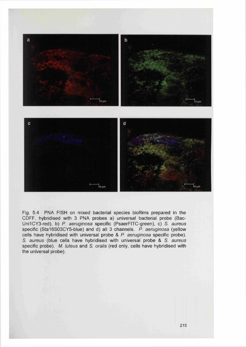
> i-?
1^.
10 pm10 pm
Fig. 5.4 PNA FISH on mixed bacterial species biofilms prepared in the CDFF, hybridised with 3 PNA probes a) universal bacterial probe (Bac- Uni1CY3-red), b) P. aeruginosa specific (PsaerFITC-green), c) S. aureus specific (Sta16S03CY5-blue) and d) all 3 channels. P. aeruginosa (yellow cells have hybridised with universal probe & P. aeruginosa specific probe). S. aureus (blue cells have hybridised with universal probe & S. aureus specific probe). M. luteus and S. oralis (red only, cells have hybridised with the universal probe).
215
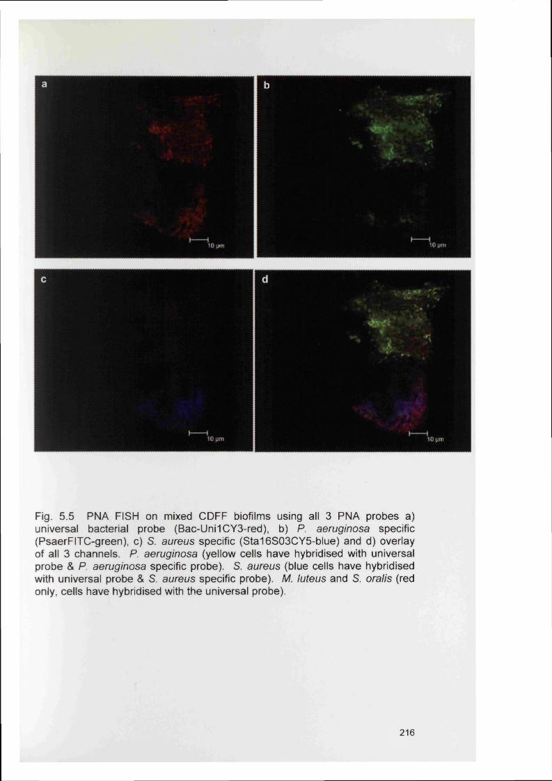
a b
10 |jm10 pm
10 pm 10 pm
Fig. 5.5 PNA FISH on mixed C D FF biofilms using all 3 PNA probes a) universal bacterial probe (Bac-Uni1CY3-red), b) P. aeruginosa specific (PsaerFITC-green), c) S. aureus specific (S ta16S03C Y5-blue) and d) overlay of all 3 channels. P. aeruginosa (yellow cells have hybridised with universal probe & P. aeruginosa specific probe). S. aureus (blue cells have hybridised with universal probe & S. aureus specific probe). M. luteus and S. oralis (red only, cells have hybridised with the universal probe).
216
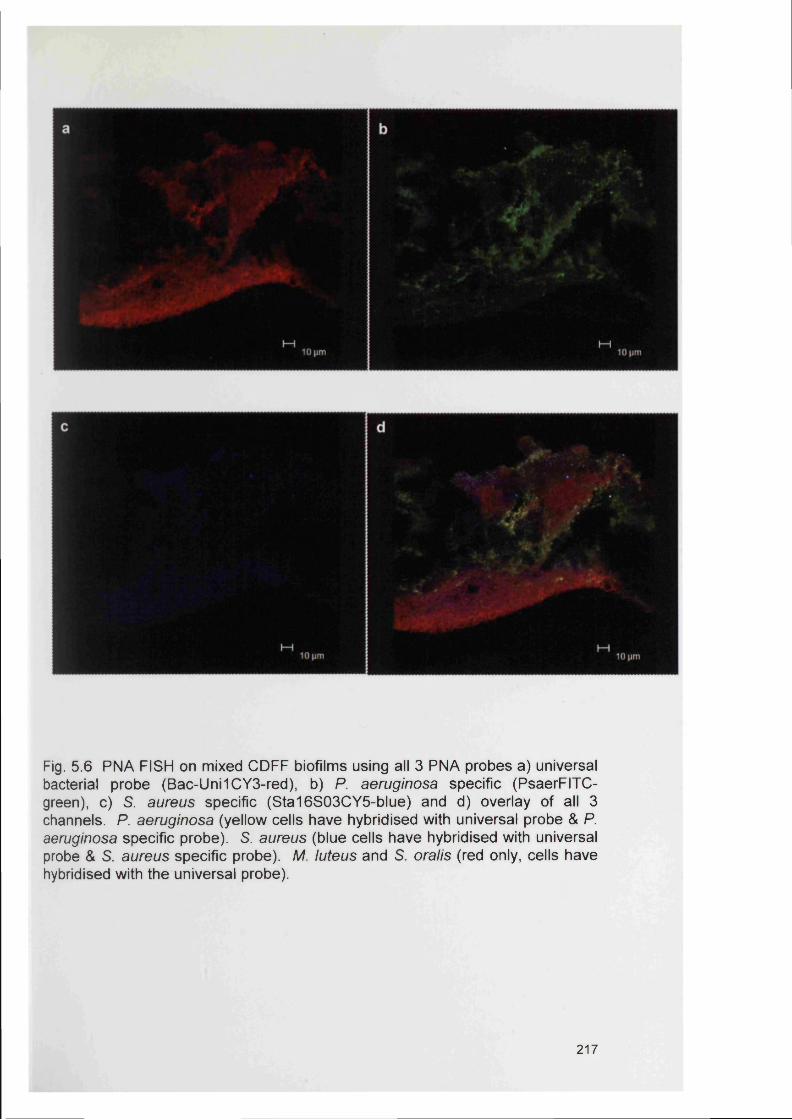
Fig. 5.6 PNA FISH on mixed C D FF biofilms using all 3 PNA probes a) universal bacterial probe (Bac-Uni1CY3-red), b) P. aeruginosa specific (PsaerFITC- green), c) S. aureus specific (S ta16S03C Y5-blue) and d) overlay of all 3 channels. P. aeruginosa (yellow cells have hybridised with universal probe & P. aeruginosa specific probe). S. aureus (blue cells have hybridised with universal probe & S. aureus specific probe). M. luteus and S. oralis (red only, cells have hybridised with the universal probe).
217
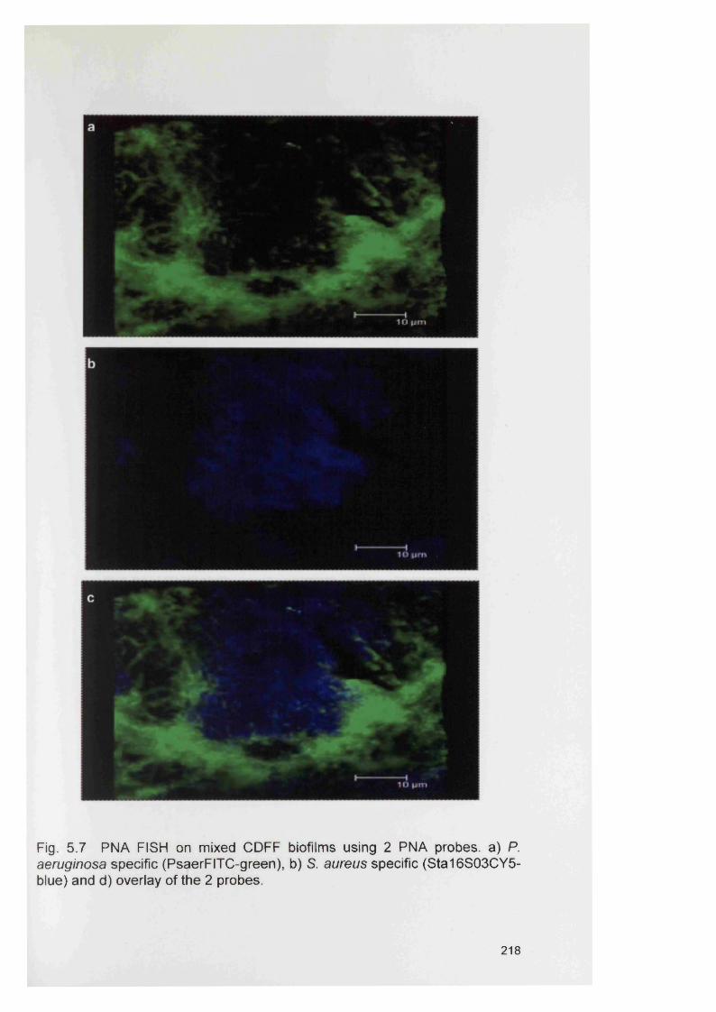
Fig. 5 .7 PNA FISH on mixed C D FF biofilms using 2 PNA probes, a) P. aeruginosa specific (PsaerFITC-green), b) S. aureus specific (S ta16S 03C Y5- blue) and d) overlay of the 2 probes.
218
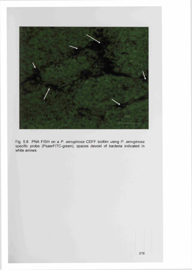
Fig. 5.8 PNA FISH on a P. aeruginosa C D FF biofilm using P. aeruginosa specific probe (PsaerFITC-green), spaces devoid of bacteria indicated in white arrows.
219
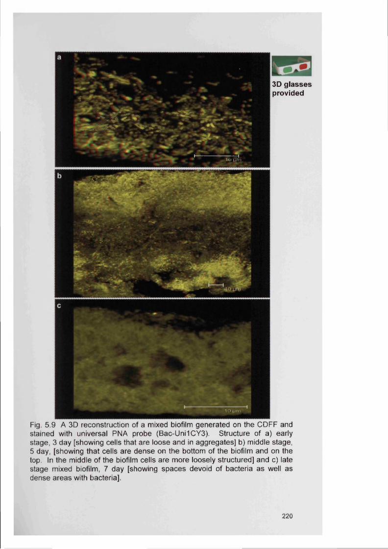
f -
ajBSfegjBBglB- - * v i------------— T
. VO ^jrnVO p m
3D glasses provided
1 0 p m
Fig. 5 .9 A 3D reconstruction of a mixed biofilm generated on the C D FF and stained with universal PNA probe (Bac-Uni1CY3). Structure of a) early stage, 3 day [showing cells that are loose and in aggregates] b) middle stage, 5 day, [showing that cells are dense on the bottom of the biofilm and on the top. In the middle of the biofilm cells are more loosely structured] and c) late stage mixed biofilm, 7 day [showing spaces devoid of bacteria as well as dense areas with bacteria].
220
5.4.4 Analysis of RHE biofilms using PNA FISH
Biofilms generated on the RHE were successfully processed and stained by
both standard Gram staining (Fig. 5.10) and with the PNA FISH probes (Fig.
5.11-5.17). The Hoechst stain, which was used to stain the nuclei of the
RHE model, also stained the DNA of the bacterial organisms that were not
detected by any of the species-specific PNA probes (i.e. non-S. aureus or P.
aeruginosa organisms). Figure 5.10b shows a scratched RHE with limited
bacterial invasion into the deep tissue.
5.4.5 Analysis of unfixed RHE biofilms using PNA FISH
In the infected (mixed population), unfixed RHE, bacteria were only detected
on the tissue surface and not within the RHE. The detected bacteria tended
to be present in clusters or aggregates on the surface of the RHE with no
evidence of RHE invasion (Fig. 5.11-5.12). However, when a 3D motion
video was created from the CLSM data, bacteria were seen as a multilayered
community of cells deeper within the tissue (Fig. 5.12).
5.4.6 Analysis of fixed RHE biofiims using PNA FISH
Formalin fixed and paraffin-embedded RHE sections were also successfully
processed and stained with the PNA FISH probes although only a few
bacteria were found to be present (Fig. 5.13-5.15). These sections were
infected with single species inocula. It was noticeable that bacterial
colonisation was restricted to the surface of the RHE tissue with no bacterial
invasion evident. Figures 5.16-5.17 confirm that bacteria actually invade the
tissue when the tissue surface is damaged. It seemed that bacteria within
the scratch wound tissue model were more abundant at the site of tissue
damage compared with intact tissue. These results suggest that once the
integrity is destroyed bacteria are able to move freely around and invade the
tissue.
221
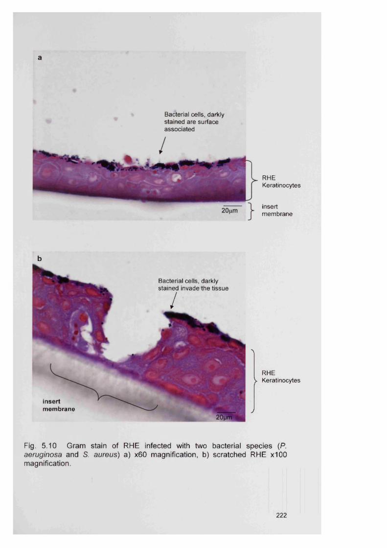
a
Bacterial cells, darkly stained are surface associated
RHEKeratinocytes
insertmembrane
b
RHE V Keratinocytes
Fig. 5 .10 Gram stain of RHE infected with two bacterial species (P. aeruginosa and S. aureus) a) x60 magnification, b) scratched RHE x100 magnification.
Bacterial cells, darkly stained invade the tissue
/
insertmembrane
222
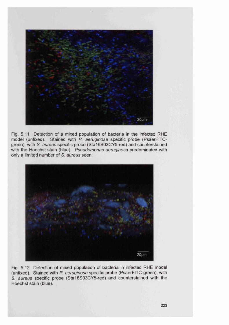
Fig. 5.11 Detection of a mixed population of bacteria in the infected RHE model (unfixed). Stained with P. aeruginosa specific probe (PsaerFITC - green), with S. aureus specific probe (S ta16S 03C Y5-red) and counterstained with the Hoechst stain (blue). Pseudomonas aeruginosa predominated with only a limited number of S. aureus seen.
20|jm
Fig. 5 .12 Detection of mixed population of bacteria in infected RHE model (unfixed). Stained with P. aeruginosa specific probe (PsaerFITC-green), with S. aureus specific probe (S ta16S 03C Y5-red) and counterstained with the Hoechst stain (blue).
223
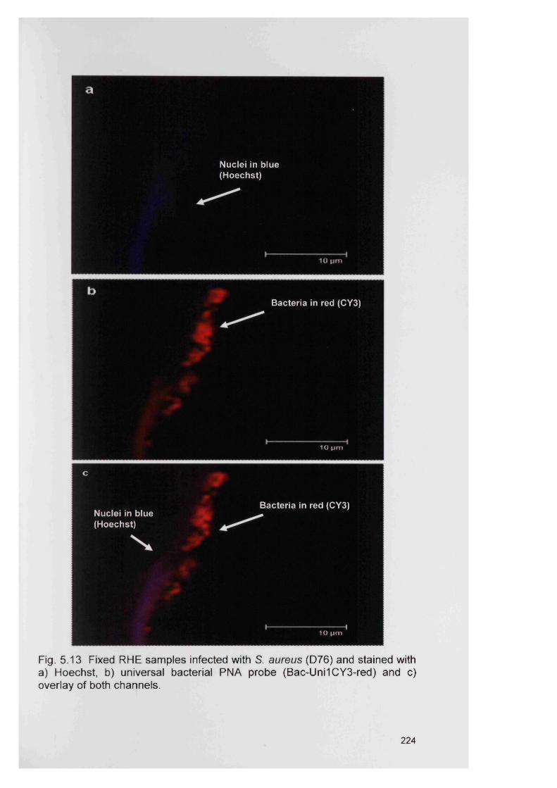
Nuclei in blue (Hoechst)
10 pm
Bacteria in red (CY3)
lO p m
Bacteria in red (CY3)Nuclei in blue (Hoechst)
10 pm
Fig. 5.13 Fixed RHE samples infected with S. aureus (D76) and stained witha) Hoechst, b) universal bacterial PNA probe (Bac-Uni1CY3-red) and c)overlay of both channels.
224
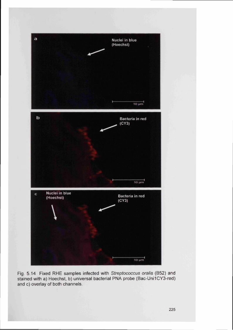
a Nuclei in blue(Hoechst)
Bacteria in red (CY3)
1 0 ( im
Nuclei in blue (Hoechst) Bacteria in red
(CY3)
10 pm
Fig. 5.14 Fixed RHE samples infected with Streptococcus oralis (B52) andstained with a) Hoechst, b) universal bacterial PNA probe (Bac-Uni1CY3-red)and c) overlay of both channels.
225
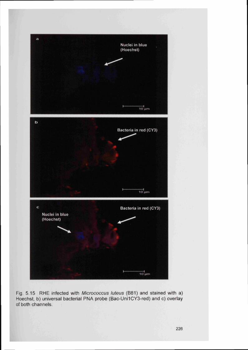
aNuclei in blue(Hoechst)
10 pm
Bacteria in red (CY3)
10 pm
Bacteria in red (CY3)
Nuclei in blue (Hoechst)
10 pm
Fig. 5.15 RHE infected with Micrococcus luteus (B81) and stained with a)Hoechst, b) universal bacterial PNA probe (Bac-Uni1CY3-red) and c) overlayof both channels.
226
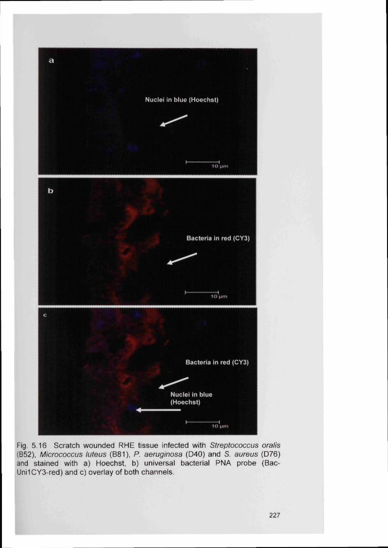
a
Nuclei in blue (Hoechst)
10 pm
Fig. 5 .16 Scratch wounded RHE tissue infected with Streptococcus oralis (B52), Micrococcus luteus (B81), P. aeruginosa (D 40) and S. aureus (D 76) and stained with a) Hoechst, b) universal bacterial PNA probe (Bac- Uni1CY3-red) and c) overlay of both channels.
227
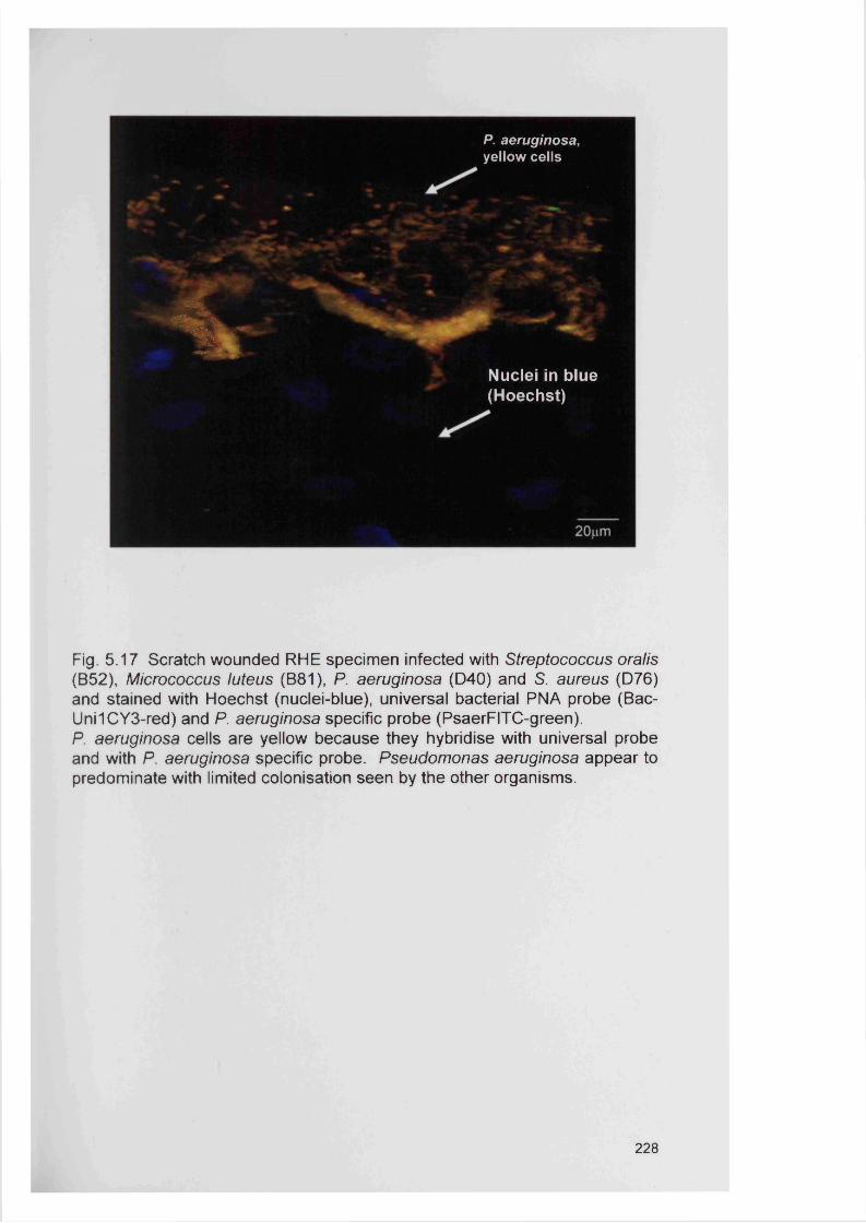
P. aeruginosa, yellow cells
%Hb*
Nuclei in blue (Hoechst)
Fig. 5 .17 Scratch wounded RHE specimen infected with Streptococcus oralis (B52), Micrococcus luteus (B81), P. aeruginosa (D 40) and S. aureus (D 76) and stained with Hoechst (nuclei-blue), universal bacterial PNA probe (Bac- Uni1CY3-red) and P. aeruginosa specific probe (PsaerFITC-green).P. aeruginosa cells are yellow because they hybridise with universal probe and with P. aeruginosa specific probe. Pseudomonas aeruginosa appear to predominate with limited colonisation seen by the other organisms.
228
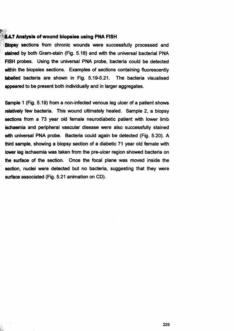
4A.7 Analysis of wound biopsies using PNA FISH
Biopsy sections from chronic wounds were successfully processed and
stained by both Qram-stain (Fig. 5.18) and with the universal bacterial PNA
FISH probes. Using the universal PNA probe, bacteria could be detected
within the biopsies sections. Examples of sections containing fluorescently
labelled bacteria are shown in Fig. 5.19-5.21. The bacteria visualised
appeared to be present both individually and in larger aggregates.
Sample 1 (Fig. 5.19) from a non-infected venous leg ulcer of a patient shows
relatively few bacteria. This wound ultimately healed. Sample 2, a biopsy
sections from a 73 year old female neurodiabetic patient with lower limb
ischaemia and peripheral vascular disease were also successfully stained
with universal PNA probe. Bacteria could again be detected (Fig. 5.20). A
third sample, showing a biopsy section of a diabetic 71 year old female with
lower leg ischaemia was taken from the pre-ulcer region showed bacteria on
the surface of the section. Once the focal plane was moved inside the
section, nuclei were detected but no bacteria, suggesting that they were
surface associated (Fig. 5.21 animation on CD).
229
■
X
«
Jp'! “ ■ %
50um.
MkT
* •
50um’ > *
5%<af
\ * ' &w
/ A4
J 50um■
R 4 jFig. 5 .18 Gram stain of Biopsy sections from non-infected chronic venous leg ulcers, a) x100 magnification; b-c) x 60 magnification; Bacteria are indicated by black arrows.
230
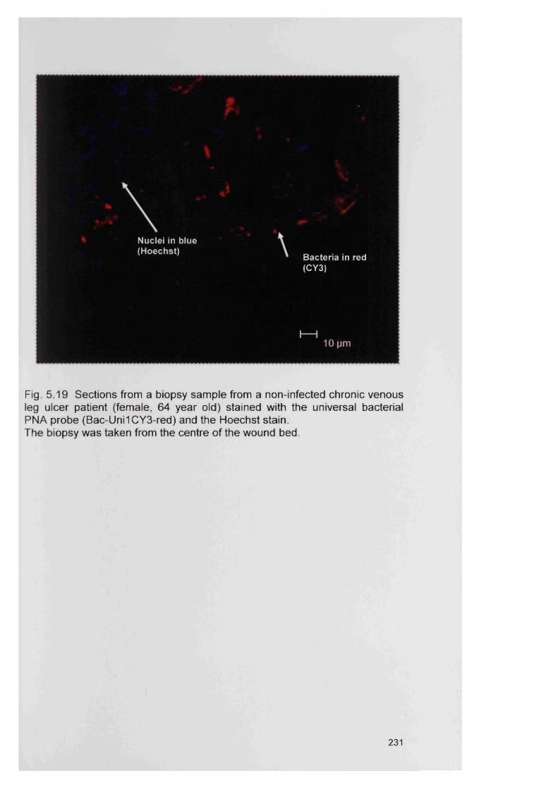
Bacteria in red (CY3)
I 110 pm
Nuclei in blue (Hoechst)
Fig. 5 .19 Sections from a biopsy sam ple from a non-infected chronic venous leg ulcer patient (female, 64 year old) stained with the universal bacterial PNA probe (Bac-Uni1CY3-red) and the Hoechst stain.The biopsy was taken from the centre of the wound bed.
231
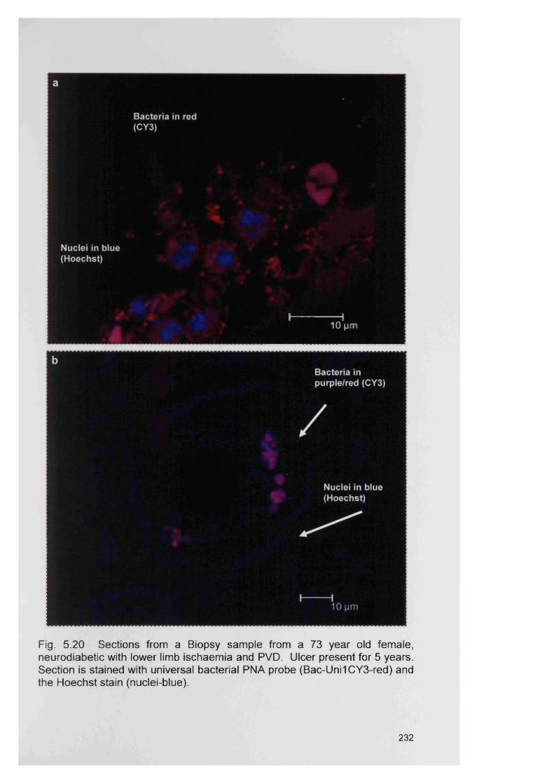
^ ^ 9
lillpH■Fig. 5 .20 Sections from a Biopsy sample from a 73 year old female, neurodiabetic with lower limb ischaemia and PVD. Ulcer present for 5 years. Section is stained with universal bacterial PNA probe (Bac-Uni1CY3-red) and the Hoechst stain (nuclei-blue).
232
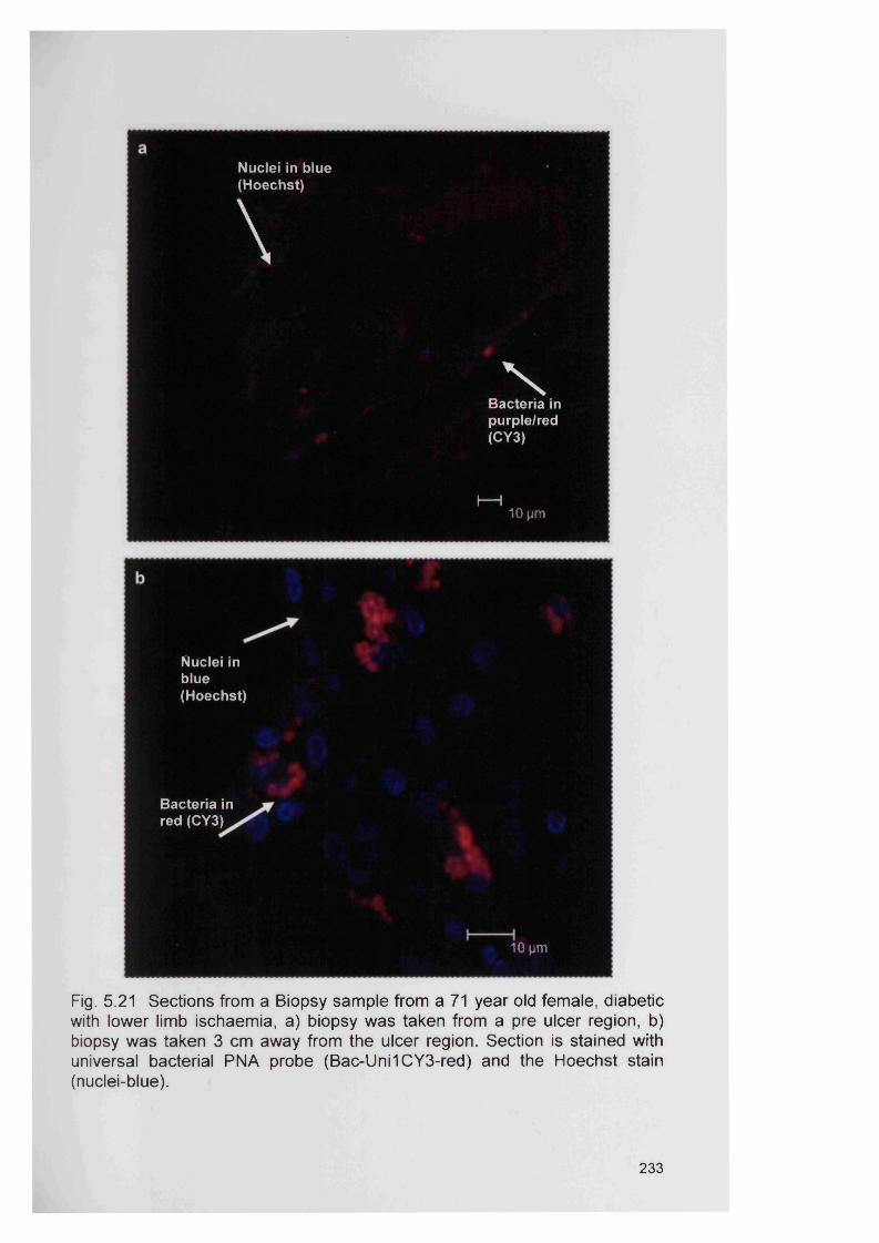
Nuclei in blue(Hoechst)
\N
Bacteria in purple/red (CY3)
I— I10 pm
Nuclei in blue(Hoechst)
Bacteria in red (CY3)
10 pm
Fig. 5.21 Sections from a Biopsy sample from a 71 year old female, diabetic with lower limb ischaemia, a) biopsy was taken from a pre ulcer region, b) biopsy was taken 3 cm away from the ulcer region. Section is stained with universal bacterial PNA probe (Bac-Uni1CY3-red) and the Hoechst stain (nuclei-blue).
233
f
5.5 Discussion
The impact of microorganisms on wound healing is poorly understood but
there is strong evidence to suggest that bacteria within chronic wounds delay
the wound healing process (Edwards and Harding, 2004; Percival and
Bowler, 2004; Percival and Rogers, 2005). The production of destructive
enzymes and toxins by bacteria results in tissue damage and indirectly
promotes an inflammatory state (Percival and Rogers, 2005; Stephens et al.,
2003). Bacteria, such as P. aeruginosa release proteinases (e.g. matrix
metalloproteinases) which are known to affect growth factors and many other
tissue proteins necessary for wound healing (Kim et al., 2000; Percival and
Rogers, 2005; Steed et al., 1996; Travis et al., 1995; Vaalamo et al., 1997).
Removal of bacteria by host defence mechanisms and antimicrobial therapy
is difficult in wound environments where biofilms have been established.
In this study, FISH was used to identify the presence of specific bacteria and
to examine their spatial organisation within “in vitro” biofilms established
using the CDFF and RHE models, as well as “in vivo” in actual chronic
wound biopsy sections.
In the preliminary study, a universal eubacterial DNA probe, EUB338 was
used to detect bacteria in planktonic suspension and in biofilms. One
problem of using the EUB338 probe was its relatively low signal intensity,
possibly as a consequence of insufficient penetration of the probe into the
bacterial cells (Moter and Gobel, 2000). The enzymatic treatment of the
sections in this study, with both lysozyme and proteinase K was employed in
an effort to counter this problem. However, even after enzymatic treatment
poor fluorescence of the target bacteria was evident.
In contrast, much stronger fluorescence of bacteria occurred when the PNA
probes were used. Due to the neutral backbone of PNA probes they are able
to diffuse readily through hydrophobic cell walls and membranes thereby
allowing detection of bacteria without the need for any pretreatment to
enhance cell permeabilisation. The high copy number of 16S rRNA in
234
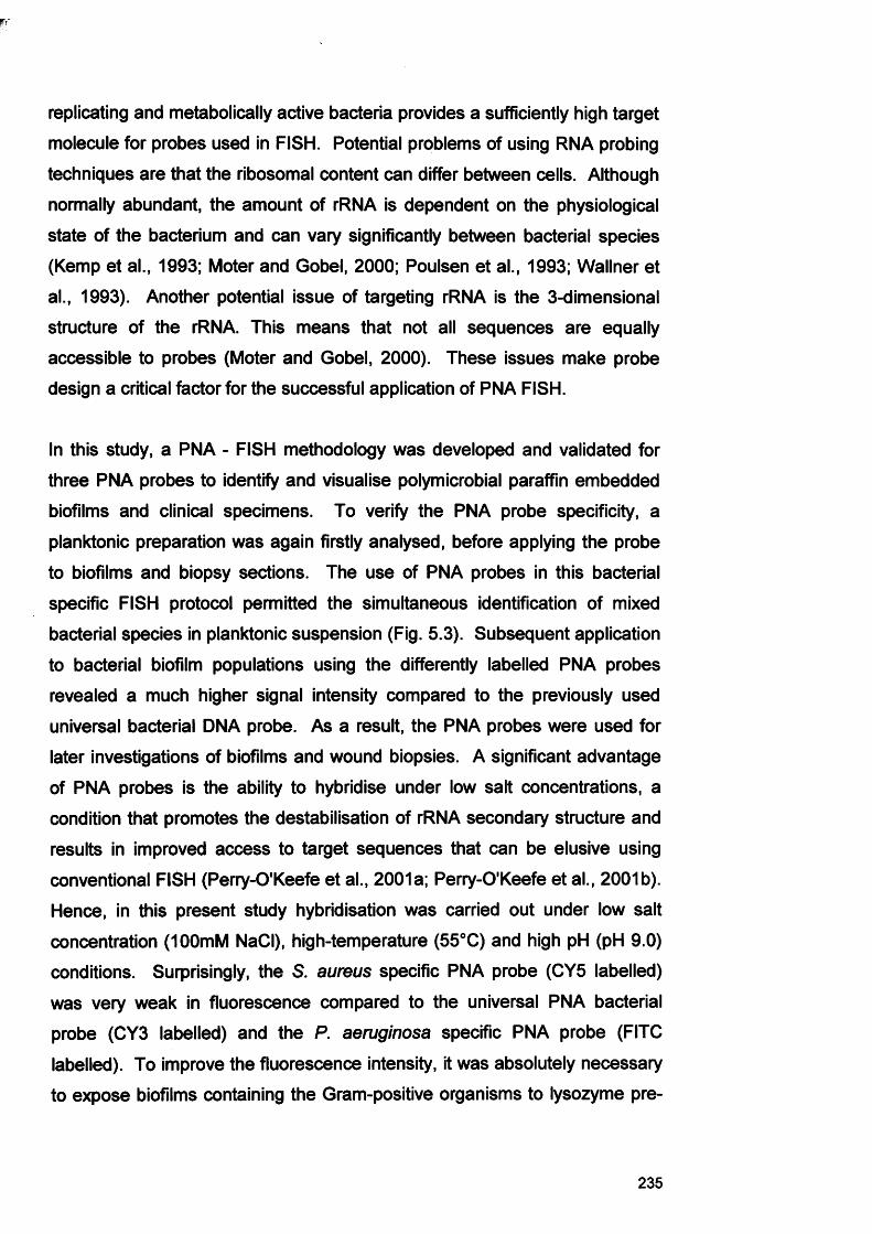
w
replicating and metabolically active bacteria provides a sufficiently high target
molecule for probes used in FISH. Potential problems of using RNA probing
techniques are that the ribosomal content can differ between cells. Although
normally abundant, the amount of rRNA is dependent on the physiological
state of the bacterium and can vary significantly between bacterial species
(Kemp et al., 1993; Moter and Gobel, 2000; Poulsen et al., 1993; Wallner et
al., 1993). Another potential issue of targeting rRNA is the 3-dimensional
structure of the rRNA. This means that not all sequences are equally
accessible to probes (Moter and Gobel, 2000). These issues make probe
design a critical factor for the successful application of PNA FISH.
In this study, a PNA - FISH methodology was developed and validated for
three PNA probes to identify and visualise polymicrobial paraffin embedded
biofilms and clinical specimens. To verify the PNA probe specificity, a
planktonic preparation was again firstly analysed, before applying the probe
to biofilms and biopsy sections. The use of PNA probes in this bacterial
specific FISH protocol permitted the simultaneous identification of mixed
bacterial species in planktonic suspension (Fig. 5.3). Subsequent application
to bacterial biofilm populations using the differently labelled PNA probes
revealed a much higher signal intensity compared to the previously used
universal bacterial DNA probe. As a result, the PNA probes were used for
later investigations of biofilms and wound biopsies. A significant advantage
of PNA probes is the ability to hybridise under low salt concentrations, a
condition that promotes the destabilisation of rRNA secondary structure and
results in improved access to target sequences that can be elusive using
conventional FISH (Perry-O'Keefe et al., 2001a; Perry-O'Keefe et al., 2001b).
Hence, in this present study hybridisation was carried out under low salt
concentration (100mM NaCI), high-temperature (55°C) and high pH (pH 9.0)
conditions. Surprisingly, the S. aureus specific PNA probe (CY5 labelled)
was very weak in fluorescence compared to the universal PNA bacterial
probe (CY3 labelled) and the P. aeruginosa specific PNA probe (FITC
labelled). To improve the fluorescence intensity, it was absolutely necessary
to expose biofilms containing the Gram-positive organisms to lysozyme pre
235
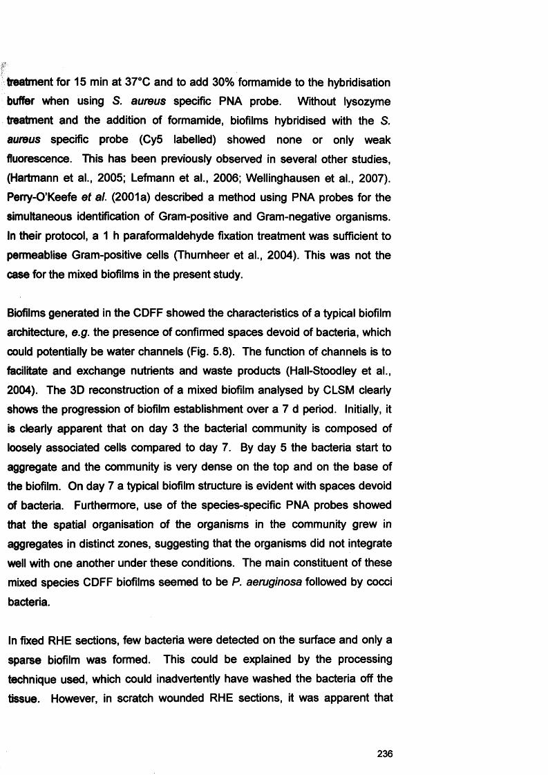
treatment for 15 min at 37°C and to add 30% formamide to the hybridisation
buffer when using S. aureus specific PNA probe. Without lysozyme
treatment and the addition of formamide, biofilms hybridised with the S.
aureus specific probe (Cy5 labelled) showed none or only weak
fluorescence. This has been previously observed in several other studies,
(Hartmann et al., 2005; Lefmann et al., 2006; Wellinghausen et al., 2007).
Perry-O’Keefe et al. (2001a) described a method using PNA probes for the
simultaneous identification of Gram-positive and Gram-negative organisms.
In their protocol, a 1 h paraformaldehyde fixation treatment was sufficient to
permeablise Gram-positive cells (Thurnheer et al., 2004). This was not the
case for the mixed biofilms in the present study.
Biofilms generated in the CDFF showed the characteristics of a typical biofilm
architecture, e.g. the presence of confirmed spaces devoid of bacteria, which
could potentially be water channels (Fig. 5.8). The function of channels is to
facilitate and exchange nutrients and waste products (Hall-Stoodley et al.,
2004). The 3D reconstruction of a mixed biofilm analysed by CLSM clearly
shows the progression of biofilm establishment over a 7 d period. Initially, it
is clearly apparent that on day 3 the bacterial community is composed of
loosely associated cells compared to day 7. By day 5 the bacteria start to
aggregate and the community is very dense on the top and on the base of
the biofilm. On day 7 a typical biofilm structure is evident with spaces devoid
of bacteria. Furthermore, use of the species-specific PNA probes showed
that the spatial organisation of the organisms in the community grew in
aggregates in distinct zones, suggesting that the organisms did not integrate
well with one another under these conditions. The main constituent of these
mixed species CDFF biofilms seemed to be P. aeruginosa followed by cocci
bacteria.
In fixed RHE sections, few bacteria were detected on the surface and only a
sparse biofilm was formed. This could be explained by the processing
technique used, which could inadvertently have washed the bacteria off the
tissue. However, in scratch wounded RHE sections, it was apparent that
236
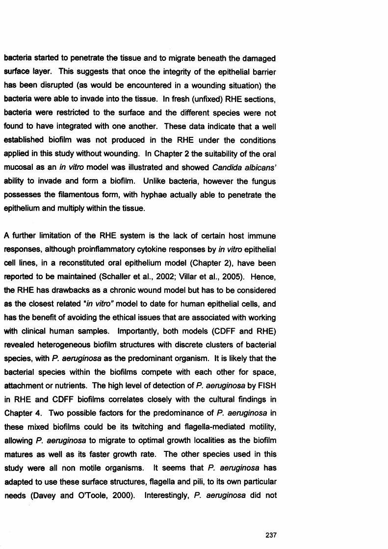
bacteria started to penetrate the tissue and to migrate beneath the damaged
surface layer. This suggests that once the integrity of the epithelial barrier
has been disrupted (as would be encountered in a wounding situation) the
bacteria were able to invade into the tissue. In fresh (unfixed) RHE sections,
bacteria were restricted to the surface and the different species were not
found to have integrated with one another. These data indicate that a well
established biofilm was not produced in the RHE under the conditions
applied in this study without wounding. In Chapter 2 the suitability of the oral
mucosal as an in vitro model was illustrated and showed Candida albicans’
ability to invade and form a biofilm. Unlike bacteria, however the fungus
possesses the filamentous form, with hyphae actually able to penetrate the
epithelium and multiply within the tissue.
A further limitation of the RHE system is the lack of certain host immune
responses, although proinflammatory cytokine responses by in vitro epithelial
cell lines, in a reconstituted oral epithelium model (Chapter 2), have been
reported to be maintained (Schaller et al., 2002; Villar et al., 2005). Hence,
the RHE has drawbacks as a chronic wound model but has to be considered
as the closest related “in vitro” model to date for human epithelial cells, and
has the benefit of avoiding the ethical issues that are associated with working
with clinical human samples. Importantly, both models (CDFF and RHE)
revealed heterogeneous biofilm structures with discrete clusters of bacterial
species, with P. aeruginosa as the predominant organism. It is likely that the
bacterial species within the biofilms compete with each other for space,
attachment or nutrients. The high level of detection of P. aeruginosa by FISH
in RHE and CDFF biofilms correlates closely with the cultural findings in
Chapter 4. Two possible factors for the predominance of P. aeruginosa in
these mixed biofilms could be its twitching and flagella-mediated motility,
allowing P. aeruginosa to migrate to optimal growth localities as the biofilm
matures as well as its faster growth rate. The other species used in this
study were all non motile organisms. It seems that P. aeruginosa has
adapted to use these surface structures, flagella and pili, to its own particular
needs (Davey and O'Toole, 2000). Interestingly, P. aeruginosa did not
237
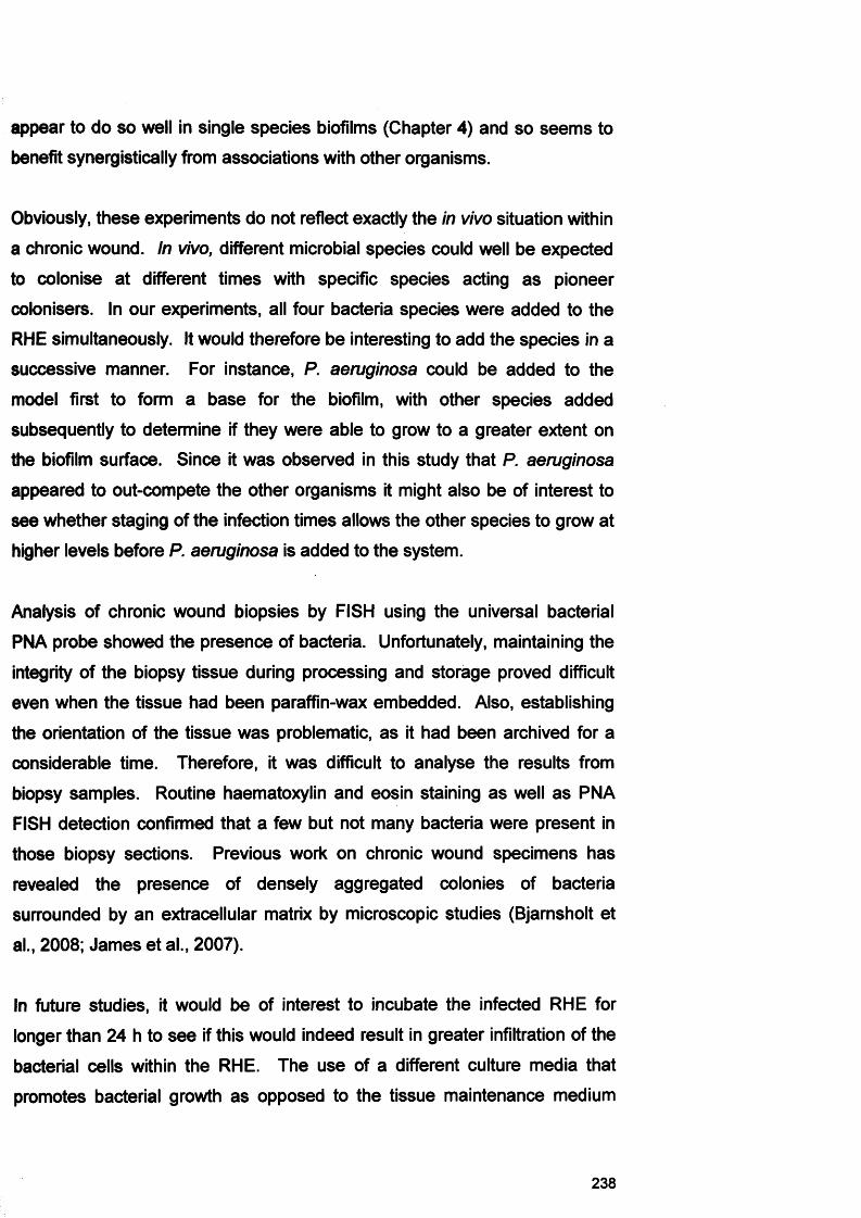
appear to do so well in single species biofilms (Chapter 4) and so seems to
benefit synergistically from associations with other organisms.
Obviously, these experiments do not reflect exactly the in vivo situation within
a chronic wound. In vivo, different microbial species could well be expected
to colonise at different times with specific species acting as pioneer
colonisers. In our experiments, all four bacteria species were added to the
RHE simultaneously. It would therefore be interesting to add the species in a
successive manner. For instance, P. aeruginosa could be added to the
model first to form a base for the biofilm, with other species added
subsequently to determine if they were able to grow to a greater extent on
the biofilm surface. Since it was observed in this study that P. aeruginosa
appeared to out-compete the other organisms it might also be of interest to
see whether staging of the infection times allows the other species to grow at
higher levels before P. aeruginosa is added to the system.
Analysis of chronic wound biopsies by FISH using the universal bacterial
PNA probe showed the presence of bacteria. Unfortunately, maintaining the
integrity of the biopsy tissue during processing and storage proved difficult
even when the tissue had been paraffin-wax embedded. Also, establishing
the orientation of the tissue was problematic, as it had been archived for a
considerable time. Therefore, it was difficult to analyse the results from
biopsy samples. Routine haematoxylin and eosin staining as well as PNA
FISH detection confirmed that a few but not many bacteria were present in
those biopsy sections. Previous work on chronic wound specimens has
revealed the presence of densely aggregated colonies of bacteria
surrounded by an extracellular matrix by microscopic studies (Bjarnsholt et
al., 2008; James et al., 2007).
In future studies, it would be of interest to incubate the infected RHE for
longer than 24 h to see if this would indeed result in greater infiltration of the
bacterial cells within the RHE. The use of a different culture media that
promotes bacterial growth as opposed to the tissue maintenance medium
238
could also be used. In addition, it would be interesting to detect the presence
of any EPS matrix generated by the chronic wound bacterial biofilms. Due to
time constraints EPS analysis was not pursued but it would be interesting to
counterstain the EPS using for example, calcofluor white, after completion of
FISH probing. Thurnheer et al. (2004) showed detection of organisms in a
multispecies biofilm of supragingival plaque using FISH and combined this
with EPS staining using calcofluor white.
Now that a basic protocol for using PNA FISH to examine biopsy sections
has been established, the technique could be used in future studies on new
‘fresh’ chronic wound tissue. This could provide important information on the
various species present in the chronic wound tissue and how they are
spatially arranged on and within the tissue. Ideally, this approach could also
be used to compare the species present within chronic wounds with those in
healthy control tissue, either from the same patients, or from other, age-
matched patients without chronic wounds.
PNA - FISH was shown to be a rapid and versatile tool for both research
purposes and potentially for clinical microbiology diagnostics when used in
conjunction with CLSM. CLSM has been established as a valuable tool for
obtaining high-resolution images and three-dimensional reconstructions of
fluorescently labelled biofilms and biological samples (Lopez et al., 2005;
Sunde et al., 2003; Thurnheer et al., 2004; Wagner et al., 2003a). The future
application of this technique to clinical biofilms from infected tissues or
indwelling medical devices could facilitate the identification and estimation of
the relative proportions of bacteria within a biofilm without the need for
bacterial culture. This approach could be used to assess biofilm
management strategies or evaluate the effectiveness of antimicrobials
against members of the biofilm consortium. Importantly, this technique could
potentially be applied to clinical samples for identifying significant bacterial
infections.
239
6.6 Summary
1. This study showed that it was possible to simultaneously detect and
identify several members of a four-species biofilm consortium, comprised of
both Gram-positive and Gram-negative bacteria.
2. The spatial organisation of bacteria in biofilm models and wound tissue
was assessed by PNA FISH and CLSM. This modified FISH technique
enabled the visualisation of the arrangement of the microbial population as
well as the typical architecture of the biofilms depicting spaces devoid of
bacteria which could potentially be water channels. The different species did
not appear to integrate well with each other, with distinct, discrete clusters of
each species evident. The main constituent of the established biofilms was
P. aeruginosa, which supports previous culture evaluations (Chapter 4).
240

Chapter 6
Gene expression in bacterialbiofilms
6.1 Introduction
The aetiology of chronic wounds is poorly understood, but evidence suggests
that chronic wounds support a diverse microbial flora, which may contribute
both indirectly and directly to the failure of the normal wound healing process.
Bacteria can express numerous virulence factors that contribute to
pathogenesis and host invasion. Typical examples of bacterial virulence
factors include hydrolytic enzymes such as proteinases, DNAses, and
phospholipases. In the past, evaluating the potential role of such agents in
pathogenicity has involved detection of the protein or its activity in clinical
specimens. However, with the advent of high technology molecular analysis
it has become possible to detect expression of bacterial virulence factors
through analysis of specific messenger RNAs.
The roles of bacterial virulence factors in promoting the chronic wound
phenotype have, as yet, not been elucidated. Examples of P. aeruginosa
virulence factors include the possession of pili, flagella, lipopolysaccharide,
proteases, quorum sensing molecules, exotoxin A, exoenzymes and alginate,
which all contribute to its pathogenic importance (Bjarnsholt et al., 2008;
Lyczak et al., 2000). It has been suggested that proteases promote infection
and dissemination, whilst elastase degrades collagen and non-collagen host
proteins and disrupt the integrity of the host basement membrane (Lyczak et
al., 2000). It can be readily envisaged how the expression of these enzymes
that either degrade host tissue or destroy the protective immune cells or their
products could contribute to delayed wound healing.
In recent years it has been postulated that bacterial biofilms may play a role
in the prevention of the normal wound healing process (James et al., 2007).
Biofilm bacteria are reported to be less susceptible to the immune system
and also to administered antimicrobial agents and therefore a biofilm
associated wound infection may persevere and progress from an acute to a
chronic infection state (Percival and Rogers, 2005).
242
It has been hypothesised that the phenotypic changes observed in
microorganisms as they attach to a surface are due to the differential
expression of genes within biofilms (O'Toole et al., 2000a). However, it
appears that biofilm development occurs differently according to nutritional
and environmental conditions and consequently there may be many
pathways and stages involved in biofilm development (Klausen et al., 2003).
6.1.1 The bacterial microflora of chronic wounds
The significance of microorganisms in chronic wounds has been extensively
studied and different approaches used to elicit their possible role in non
healing wounds. Studies have included assessing the impact of microbial
populations on clinical outcome (Madsen et al., 1996) and observations of
the occurrence of particular species (Schraibman, 1990) or groups of
organisms within specific wounds (Davies et al., 2007; Wall et al., 2002).
Previous studies using molecular (Davies et al., 2004; Davies et al., 2001;
Hill et al., 2003; James et al., 2007) and culture-based approaches (Bowler
and Davies, 1999) have indicated the presence of polymicrobial bacterial
communities in chronic wounds. The most frequent isolates include
staphylococci, streptococci and Pseudomonas sp. (Davies et al., 2001).
Cultural analysis has also shown Acinetobacter sp. and Proteus sp. to be
prevalent in chronic wounds (Hill et al., 2003) and have also documented that
staphylococci, streptococci, enterococci and facultative Gram-negative bacilli
are the principle bacterial groups that are recovered from chronic venous leg
ulcers (CVLU) (Davies et al., 2007; Hansson et al., 1995). In addition to
these aerobic bacteria, anaerobic organisms have also been identified in
chronic wounds, such as Peptostreptococcus spp., Bacteroides spp. (Davies
et al., 2007) and pigmented and non-pigmented Prevotella and
Porphyromonas spp. (Bowler and Davies, 1999; James et al., 2007).
6.1.2 Genotypic differences between planktonic and biofilm populations
It is believed that microorganisms present in chronic wounds contribute to the
non-healing phenotype and may exist within wounds as part of a biofilm
community (Davies et al., 2007; Mertz, 2003; Serralta et al., 2001). Biofilm
243
associated infections are typically persistent; they develop slowly, are rarely
resolved by immune defences and respond transiently to antimicrobial
therapy (James et al., 2007; Parsek and Singh, 2003). For example, Gram-
negative bacterial components such as colonic acid (in Escherichia coli),
alginate, glucose and mannose-rich Pel (product of the pellicle formation
gene) and Pis (product of the polysaccharide synthesis locus) matrix
components (in P. aeruginosa), cellulose and p-1-6-GlcNac polymer (in
Salmonella and E. coli), have been reported to play important roles in biofilm
formation (Beloin and Ghigo, 2005; Friedman and Kolter, 2004; Matsukawa
and Greenberg, 2004).
As described previously (see section 6.1) and in the Literature Review
(Chapter 1, section 1.9.2), P. aeruginosa and S. aureus are recognised key
organisms in chronic wounds and consequently provide a major focus for this
chapter.
6.1.3 Gene expression in P. aeruginosa and S. aureus biofilms
There are many genes whose expression has been associated with the
virulence of P. aeruginosa and S. aureus (Table 6.1). Examples of genes
important in biofilm formation are described below.
6.1.3.1 Periplasmic glucans synthesis in P. aeruginosa
Periplasmic glucans may contribute to antibiotic resistance by reducing the
diffusion of antibiotics into the cell (extracellular sequestering) thereby
allowing the bacteria additional time to adapt to an antibiotic challenge (Mah
et al., 2003; Whiteley et al., 2001).
244
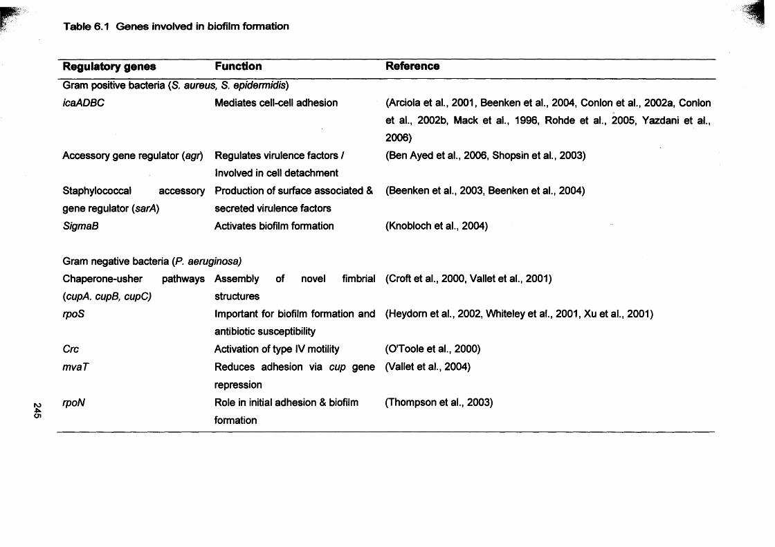
245
Table 6.1 Genes involved in biofilm formation
Regulatory genes Function Reference
Gram positive bacteria (S. aureus, S. epidermidis)icaADBC Mediates cell-cell adhesion (Arciola et al., 2001, Beenken et al., 2004, Conlon et al., 2002a, Conlon
et al., 2002b, Mack et al., 1996, Rohde et al., 2005, Yazdani et al.,2006)
Accessory gene regulator (agt) Regulates virulence factors / (Ben Ayed et al., 2006, Shopsin et al., 2003)Involved in cell detachment
Staphylococcal accessory Production of surface associated & (Beenken et al., 2003, Beenken et al., 2004)gene regulator (sarA) secreted virulence factorsSigmaB Activates biofilm formation (Knobloch et al., 2004)
Gram negative bacteria (P. aeruginosa)Chaperone-usher pathways Assembly of novel fimbrial (Croft et al., 2000, Vallet et al., 2001)(cupA. cupB, cupC) structuresrpoS Important for biofilm formation and (Heydom et al., 2002, Whiteley et al., 2001, Xu et al., 2001)
antibiotic susceptibilityCrc Activation of type IV motility (O'Toole et al., 2000)mvaT Reduces adhesion via cup gene (Vallet et al., 2004)
repressionrpoN Role in initial adhesion & biofilm (Thompson et al., 2003)
formation
ps?W
The extracytoplasmic mode of antibiotic sequestration might be expected to
interfere with the passage of antibiotics through the periplasmic space to their
site of action in the cytoplasm. For the synthesis of periplasmic glucans
ndvB is required. The ndvB gene encodes for a glucosyltransferase that is
required for the synthesis of cyclic-P-(1, 3)-glucans (Mah et al., 2003;
Whiteley et al., 2001). Mah et al. (2003) also predicted that ndvB is
preferentially expressed in biofilm grown cells.
Cyclic glucans are circular polymers of glucose, which play a role in
adaptation to low osmotic media, flagella-mediated motility and plant-microbe
interactions. These glucans are located in the periplasm and also secreted
into the extracellular medium (Breedveld and Miller, 1994; Mah et al., 2003).
It has been suggested that the glucose polymers may prevent antibiotics
from reaching their sites of action by sequestering antimicrobial agents in the
periplasm (Breedveld and Miller, 1994; Mah et al., 2003).
The impermeability of bacteria to antibiotics involves several factors,
including the tolA gene product (Rivera et al., 1988) and terminal electron
transport proteins (Bryan and Kwan, 1983; Bryan et al., 1980; Whiteley et al.,
2001). The tolA gene product affects the LPS structure, resulting in
decreased aminoglycoside affinity for the outer membrane. Evidence
indicates that tolA is an essential P. aeruginosa gene, but mutants that
under-produce tolA are hypersensitive to aminoglycoside antibiotics (Rivera
et al., 1988). Therefore, the tolA gene could contribute to biofilm resistance
to aminoglycosides.
6.1.3.2 Alginate genes of P. aeruginosa
Alginate is a viscous exopolysaccharide, consisting of D-mannuronic and L-
guluronic acids. Alginate production by P. aeruginosa is regarded as a
virulence factor that appears to promote biofilm formation, thus enhancing
host colonisation. Furthermore, alginate induced biofilms have also been
shown to confer resistance to antibiotics (Hershberger et al., 1995), promote
bacterial adherence, sen/e as a barrier to phagocytosis and provide a
246
mechanism to neutralise oxygen free radicals (Edwards and Saunders,
2001).
It has been suggested that alginates play an important role in P. aeruginosa
infection of cystic fibrosis (CF) patients, urinary tract infections, the fouling of
man-made materials and in biofilm formation on abiotic surfaces in the
natural environment (Davies et al., 1993). Strains of P. aeruginosa isolated
from the lungs of CF patients have been observed to have a mucoid
phenotype with production of exopolysaccharide alginate (Edwards and
Saunders, 2001). Alginate is thought to have a protective function in a
relatively harsh environment in which the bacteria are continually subjected
to oxidative stress and challenge by the immune system (Krieg et al., 1988;
Simpson et al., 1988; Simpson et al., 1989).
Even though most P. aeruginosa strains have the ability to produce alginate,
the majority of strains exhibit a non-mucoid phenotypes (Hershberger et al.,
1995). The switch to the mucoid phenotype is also thought to promote
persistence of P. aeruginosa (Hentzer et al., 2001). Mucoid production has
further been shown to aid bacterial survival in nutrient poor environments
(Hershberger et al., 1995). In a biofilm state or in a condition such as cystic
fibrosis, P. aeruginosa must adapt to its environment and this is manifested
by overproduction of alginate.
Alginate production is regulated by at least two mechanisms. Firstly, by a
response regulator involved in a bacterial signal transduction system which is
controlled by environmental factors. Secondly, by a gene cluster containing
the sigma factor algU (Fig. 6.1) (Edwards and Saunders, 2001). The algU
gene is autoregulated and has two negative regulators encoded by mucA
and mucB. Inactivation of either mucA or mucB results in activation of algD
through enhanced expression of algU, and so leads to overproduction of
alginate (Edwards and Saunders, 2001). Although alginate production may
not be required for biofilm formation, it undeniably affects the biofilm
architecture (Stapper et al., 2004).
247
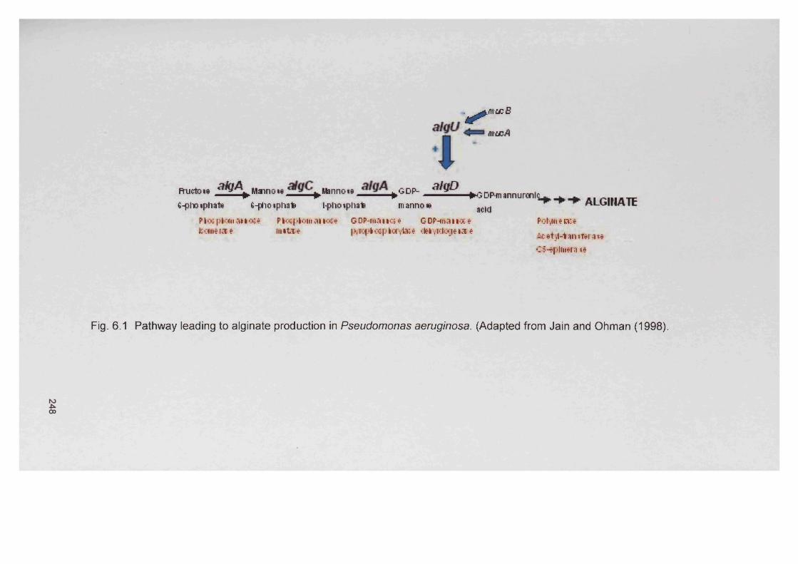
Lrtru:Bv * '
Hmjctoie dkjA Hanno»e Mannoie fcGDP- dlQD fcrnrmannuronltP P P — P^DPfnannuronhj- - a i m iA T F6-pho»phat& G-phoiphate l-phoiphafc mannow acjcl f t L l i m A l c
Plo&pftanaiiote Pfeosplom ai»«e GDP-manot* GDP-rnanoce Polya ewekaaerat m itee p^ap*otplcfvt»e cttyTdogeiat Acatyl-tan.ftrai*
C5-tplm*ni ie
Fig. 6.1 Pathway leading to alginate production in Pseudomonas aeruginosa. (Adapted from Jain and Ohman (1998).
6.1.3.3 Accessory gene regulator (agr) of S. aureusThe agr system regulates the coordinated production of several virulence
factors (Shopsin et al., 2003). The agr operon encodes a two-component
signalling system (Chapter 1, Fig. 1.6; agrC is the signal receptor and agrA is
the response regulator) which is driven by a quorum sensing autoinducing
peptide, encoded by the agrD gene (Ben Ayed et al., 2006).
Staphylococcus aureus isolates can be classified into four (l-IV) specific agr
groups on the basis of the autoinducing peptide specificity for its signal
receptor (agrC) (Ben Ayed et al., 2006; Shopsin et al., 2003). It has been
suggested that strains of one group can cross-activate the agr system of
other strains in the same group to produce a response, e.g. an up regulation
of virulence factors in other strains of the same group. Conversely, inhibition
of the agr response of members of other groups (Camara et al., 2002) could
also occur. It is important to note that these genotypes can also be
correlated with specific phenotypes, e.g. most menstrual toxic shock
syndrome strains belong to agr group III (Fischetti, 2006). The agr system
coordinates the production of virulence factors during different phases of the
cell cycle, which help with the organisms’ ability to invade and its
pathogenicity, e.g. it can down-regulate the production of cell wall-associated
proteins and up-regulate secreted proteins at late to stationary growth
phases in vitro (Camara et al., 2002; Fischetti, 2006).
6.1.3.4 Intercellular polysaccharide adhesin (ica) of S. aureusOver 90% of clinical S. aureus strains possess capsular polysaccharides.
Capsule production is believed to decrease phagocytosis in vitro, and has
been shown to enhance S. aureus virulence in a mouse bacteriaemia model
(Harris et al., 2002).
Biofilm formation can be described as a two step process that first requires
adhesion of bacteria to a surface followed by cell-cell adhesion, forming
multiple layers of the biofilm. This process in S. aureus is associated with the
polysaccharide intercellular adhesin (PIA), which is composed of linear P-1,
249
6-linked glucosaminylglycans. The ica locus is thought to mediate cell-cell
adhesion and PIA production (Cramton et al., 1999). The locus itself is
comprised of four intercellular adhesion genes (icaA, icaB, icaC and icaD)
and one regulator gene (/caP) which appears to function as a repressor
(Cafiso et al., 2004; Conlon et al., 2002a; Gotz, 2002; Mack et al., 1996;
Ziebuhr et al., 1999). IcaA and icaD play a significant role in S. aureus and
S. epidermidis biofilm formation (Yazdani et al., 2006). The product of icaA is
a N-acetylglucosamine transferase, icaC and icaD encode membrane
proteins and icaB encodes a signal peptide that is secreted into the medium
(Fig. 6.2) (Gelosia et al., 2001).
6.1.4 Reverse Transcriptase Polymerase Chain Reaction (RT-PCR)
Previous studies have examined gene expression in bacterial biofilms
including those produced by S. aureus and P. aeruginosa (Arciola et al.,
2001; Conlon et al., 2002a; Cramton et al., 1999; Gelosia et al., 2001; Mah et
al., 2003; Sakoulas et al., 2002). For example, Cafiso et al. (2004) used this
approach to determine the expression of the icaADBC operon in S.
epidermidis strains isolated from catheter related and nosocomial infections
and correlated expression to biofilm production. Mah et al. (2003) used RT-
PCR to identify expression of the ndvB locus which is required for the
synthesis of periplasmic glucans in P. aeruginosa. RT-PCR is a sensitive
method but is at best only semi-quantitative. To overcome this limitation a
quantitative real-time PCR can be employed which allows monitoring the
formation of the product in real time. The monitoring of product is
accomplished using various types of fluorescent dyes.
250
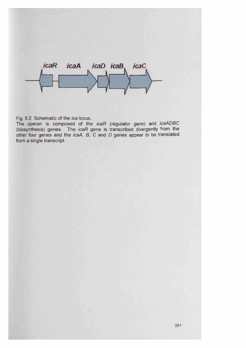
icaR icaA ic
Fig. 6.2 Schematic of the ica locus.The operon is composed of the icaR (regulator gene) and icaADBC (biosynthesis) genes. The icaR gene is transcribed divergently from the other four genes and the icaA, B, C and D genes appear to be translated from a single transcript.
251
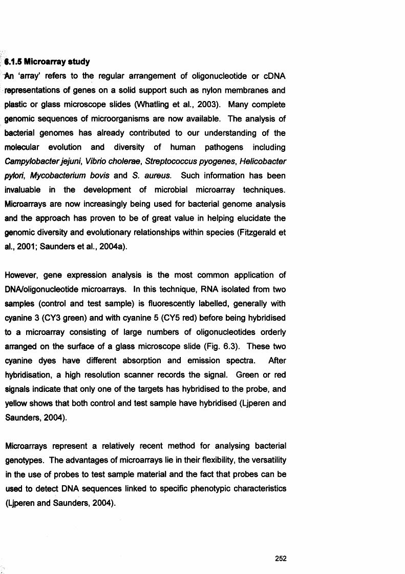
6.1.5 Microarray study
‘array’ refers to the regular arrangement of oligonucleotide or cDNA
representations of genes on a solid support such as nylon membranes and
plastic or glass microscope slides (Whatling et al., 2003). Many complete
genomic sequences of microorganisms are now available. The analysis of
bacterial genomes has already contributed to our understanding of the
molecular evolution and diversity of human pathogens including
Campylobacter jejuni, Vibrio cholerae, Streptococcus pyogenes, Helicobacter
pylori, Mycobacterium bovis and S. aureus. Such information has been
invaluable in the development of microbial microarray techniques.
Microarrays are now increasingly being used for bacterial genome analysis
and the approach has proven to be of great value in helping elucidate the
genomic diversity and evolutionary relationships within species (Fitzgerald et
al., 2001; Saunders et al., 2004a).
However, gene expression analysis is the most common application of
DNA/oligonucleotide microarrays. In this technique, RNA isolated from two
samples (control and test sample) is fluorescently labelled, generally with
cyanine 3 (CY3 green) and with cyanine 5 (CY5 red) before being hybridised
to a microarray consisting of large numbers of oligonucleotides orderly
arranged on the surface of a glass microscope slide (Fig. 6.3). These two
cyanine dyes have different absorption and emission spectra. After
hybridisation, a high resolution scanner records the signal. Green or red
signals indicate that only one of the targets has hybridised to the probe, and
yellow shows that both control and test sample have hybridised (Ljperen and
Saunders, 2004).
Microarrays represent a relatively recent method for analysing bacterial
genotypes. The advantages of microarrays lie in their flexibility, the versatility
in the use of probes to test sample material and the fact that probes can be
used to detect DNA sequences linked to specific phenotypic characteristics
(Ljperen and Saunders, 2004).
252
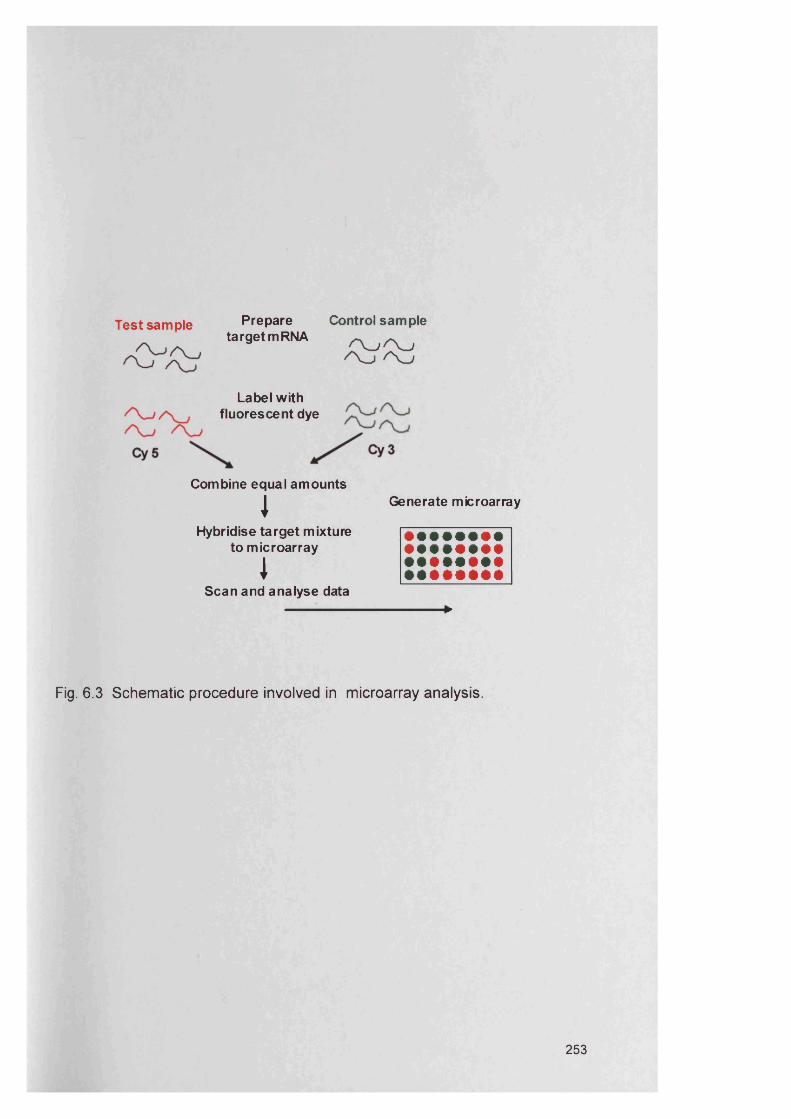
Test sample Prepare Control sampletarget mRNA
Label with fluorescent dye
Combine equal amounts
IHybridise target mixture
to microarray
IScan and analyse data
Generate microarray
Fig. 6.3 Schematic procedure involved in microarray analysis.
253
- Microarray studies have been used to identify global gene expression profiles
of biofilms (Lazazzera, 2005). Whiteley et al. (2001) performed the first DNA
microarray study to analyse gene expression in P. aeruginosa planktonic and
biofilm cells. This initial microarray showed that approximately 1 % of genes
were differentially expressed between biofilm and planktonic growth phases
and that rpoS gene expression was important in both biofilm formation and
resistance to the antibiotic tobramycin (Whiteley et al., 2001). Resch et al.
(2005) carried out a microarray analysis to determine which genes were up-
regulated in S. aureus biofilms. Their data reported up-regulation of the ica
genes in early biofilms (6-8 h of growth) compared to planktonic cells,
suggesting that ica up-regulation was not needed after attachment and
biofilm formation had begun (Resch et al., 2005). Resch et al. (2005) found
that a range of virulence factors (e.g. proteases) and toxins were up-
regulated in planktonic cells, which could be useful to cause acute infection
and to invade cells. It has been suggested that toxins and proteases are not
important for biofilm persistence once established (Resch et al., 2005).
Printed arrays can be used for either Comparative Genome Hybridisation
(CGH) or expression profiling work. The S. aureus microarray used in this
study consists of 720 oligonucleotide probes representing the virulence and
housekeeping genes of S. aureus. Data derived from the array will allow
comparative analysis of gene expression by isolates from S. aureus biofilms
generated on the CDFF and from planktonic S. aureus in exponential and
stationary phase.
6.2 AIMSThe aims of this chapter were:
To use both RT-PCR and microarray analysis to examine the in vitro biofilm
gene expression of two chronic wound bacterial species, namely S. aureus
and P. aeruginosa. Biofilms were generated for this study using the Constant
Depth Film Fermenter as described in Chapter 4.
254
6.3 Methods
6.3.1 Processing of CDFF biofilms
Biofilms were prepared using the CDFF as described previously (Chapter 4,
section 4.3.12). CDFF biofilms for RT-PCR were recovered and stored in a 2
ml microtube with 1.5 ml RNAIater® (Ambion Europe Ltd, Huntington, UK)
solution before RNA extraction (section 6.3.3) and were maintained at -80°C
until required.
6.3.2 Preparation of bacteria for planktonic growth
In addition to growth in a biofilm state (section 6.3.1), gene expression was
also assessed for planktonic strains of P. aeruginosa D40 and S. aureus D76
cultured in BHI medium at 37°C.
As described previously (Chapter 4, section 4.3.12.1), overnight cultures
were initially prepared to generate inocula for planktonic growth experiments.
This overnight culture was diluted two hundred-fold in 20 ml BHI (an identical
dilution factor as used for the CDFF inoculum) and 7 broths for each bacterial
species were incubated with shaking at 37°C for 7 d. Each day, the cells
were centrifuged and the supernatant removed and new medium was added.
At each 24 h time point, the cells from one of the broths were harvested by
centrifugation and stored in a 2 ml microtube with 1.5 ml RNAIater® (Ambion,
Europe Ltd, Huntington, UK) solution prior to RNA extraction. The pelleted
cells in RNAIater were stored at -80 °C until processed.
6.3.3 RNA extractionRNA was extracted using two different methods, employing either a RiboPure
Bacteria Kit (Ambion Ltd., Warrington, UK) or a Qiagen RNeasy Kit (Qiagen,
West Sussex, UK). RNA samples were kept on ice throughout the RNA
extraction whenever possible. For the RiboPure Bacteria Kit, biofilm and
planktonic cells from the RNAIater were collected by centrifugation for 5 min
(16,000 g). The supernatant was decanted and 350 pi RNAwiz (Ambion,
RiboPure Bacteria Kit) added to the bacteria which were resuspended by
255
vortexing vigorously for 15 s. The bacteria were transferred to a tube
containing 250 |jl of sterile zirconia beads and then homogenised for 2-5 min
using a Mini-BeadBeater-8 (Stratech Scientific Ltd., Soham, UK). After
disruption, cells were centrifuged for 5 min at 4°C at 16,000 g and the
bacterial lysate transferred to a fresh 1.5 ml tube. The lysate volume was
estimated (200 - 250 pi) and 0.2x volumes of chloroform (Sigma, Poole, UK)
added and shaken vigorously for 30 s prior to incubation for 10 min at room
temperature. After centrifugation (5 min, 4°C, 16,000 g) the aqueous phase
containing the partially purified RNA (150 - 200 pi) was transferred to a fresh
1.5 ml eppendorf tube and 0.5x volumes of absolute ethanol added. The
lysate/ethanol mixture was transferred to a filter cartridge, which was placed
into a 2 ml collection tube, and centrifuged for 1 min. The flow-through was
discarded and the RNA washed according to the manufacturer’s
recommended protocol.
For the Qiagen RNeasy Kit, 200 pi of biofilm cells or planktonic cells from the
RNAIater were collected by centrifugation for 5 min at 4°C (16,000 g). The
supernatant was decanted and the cells re-suspended in 200 pi lysis buffer,
containing 2x TE buffer, 1.2% Triton X-100, 0.3 mg ml'1 lysozyme, 0.03 mg
ml'1 lysostaphin and nuclease free water. The mixture was incubated at
37°C for 45 min. To the lysate, 350 pi RLT extraction medium (Qiagen,
Crawley, UK) was added and mixed thoroughly by vortexing vigorously. The
RNeasy Mini Kit (Qiagen) was used to complete total RNA extraction
according to the manufacturer’s recommended protocol. Potential DNA
contamination of all samples was then removed using RNase-Free DNase I
(Qiagen) treatment following the manufacturer’s instructions.
6.3.4 cDNA synthesis
50 ng pi'1 of total RNA starting material or a nuclease free water control was
placed into 0.2 ml thin-walled eppendorf tubes. To each sample, the
following components were added: 5 pi (x10) thermophilic DNA polymerase
buffer (100 mM Tris-HCI, 500 mM KCI and 1% Triton X-100), 5 pi 10 mM
dNTPs, 10 Ml (25 mM) MgCI, 4 pi (10 U pi'1) RNase inhibitor, 2.5 pi (500 ng
256
ml'1) random hexamers and 1 pi (200 U pI"1) MMLV reverse transcriptase
(Promega, Southampton, UK). The final volume was made up to 50 pi with
nuclease free diethyl-pyrocarbonate (DEPC) treated water. Addition of
nuclease free water in replacing the constituents described above provided a
non-RT control. All samples were vortexed, centrifuged for 1 min at 8,000 g
and incubated at 25°C for 10 min, 42°C for 60 min and 95°C for 5 min to
generate cDNA. The samples were subsequently cooled to 4°C and stored
at -20°C until required for PCR.
6.3.5 PCR on cDNA samplesPCR targeted a number of virulence gene families of P. aeruginosa and S.
aureus including outer membrane protein genes, alginate genes, the
intercellular adhesion locus (ica) and the accessory gene regulator (agr). For
each gene, a specific primer pair was used to yield an amplicon of distinct
size (Tables 6.2-6.3). A variety of ica primers were examined, which were
previously described (Cafiso et al., 2004; Knobloch et al., 2002). In the case
of icaA, two primer sets were used, S. aureus specific and those previously
used for S. epidermidis. Additionally ica primers for icaA, icaB, icaC, icaD
and icaR were designed in this study using the software program Primer3
(Rozen and Skaletsky, 2000).
The primers and reaction conditions (Table 6.4) were as previously described
for agr (Ben Ayed et al., 2006; Shopsin et al., 2003), alginate (Edwards and
Saunders, 2001) and for outer membrane proteins (Mah et al., 2003). The
constitutively expressed gene encoding the 50S ribosomal protein L21 (rplil)
was used as a control for P. aeruginosa (Mah et al., 2003) and the phosphate
acetyltransferase (pta) house-keeping gene of S. aureus was also amplified
as a RT-PCR control (Enright et al., 2000). Additional controls for each
reaction included, genomic DNA, DNAse I treated RNA not treated with
reverse transcriptase (to establish that genomic DNA amplification was not
present) and a water template control. A typical reaction mix contained 5 pi
of cDNA template, together with 1.5 mM MgCfe, 2.5 U of Taq polymerase,
0.25 mM of each primer, 5x Taq buffer and 0.2 mM dNTP mix.
257
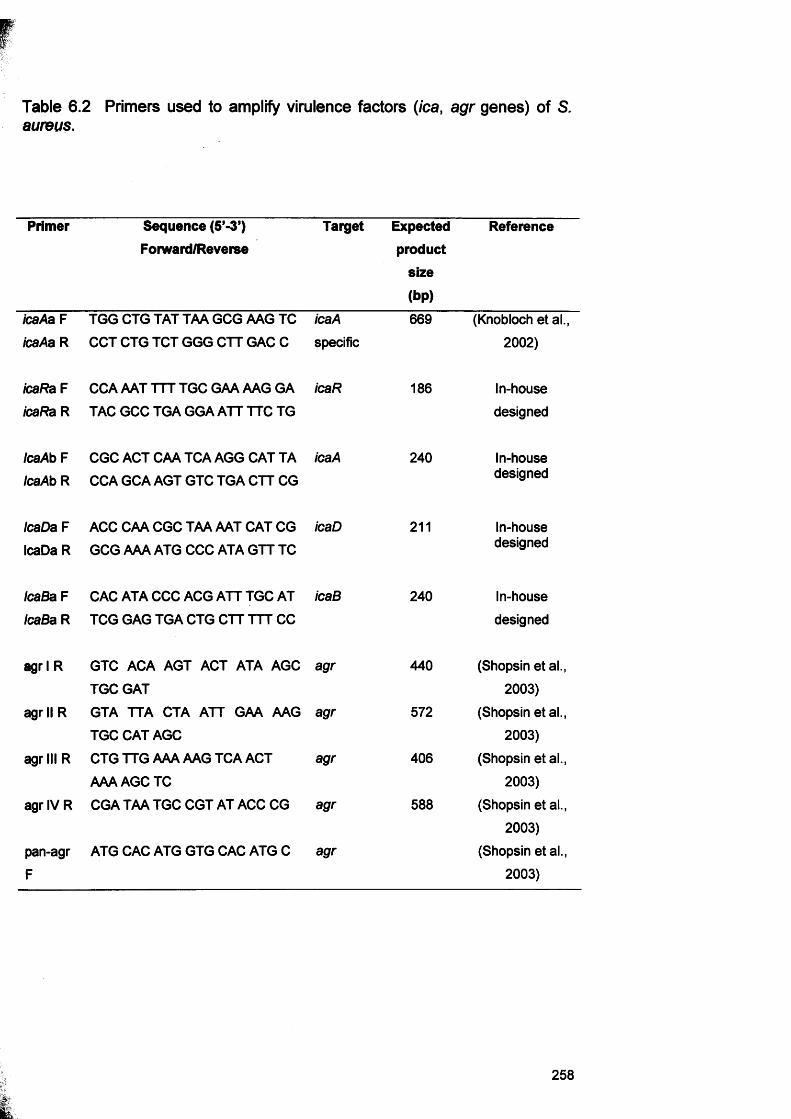
Table 6.2 Primers used to amplify virulence factors (ica, agr genes) of S.aureus.
Primer Sequence (5f-3’) Forward/Reverse
Target Expectedproduct
size
(bp)
Reference
icaAa F TGG CTG TAT TAA GCG AAG TC icaA 669 (Knobloch et al.,icaAa R CCT CTG TCT GGG CTT GAC C specific 2002)
icaRa F icaRa R
CCA AAT TTT TGC GAA AAG GA TAC GCC TGA GGA ATT TTC TG
icaR 186 In-housedesigned
IcaAb F IcaAb R
CGC ACT CAA TCA AGG CAT TA CCA GCA AGT GTC TGA CTT CG
icaA 240 In-housedesigned
IcaDa F IcaDa R
ACC CAA CGC TAA AAT CAT CG GCG AAA ATG CCC ATA GTT TC
icaD 211 In-housedesigned
IcaBa F IcaBa R
CAC ATA CCC ACG ATT TGC AT TCG GAG TGA CTG CTT TTT CC
icaB 240 In-housedesigned
agr 1 R GTC ACA AGT ACT ATA AGC TGC GAT
agr 440 (Shopsin et al., 2003)
agr II R GTA TTA CTA ATT GAA AAG
TGC CAT AGCagr 572 (Shopsin et al.,
2003)agr III R CTG TTG AAA AAG TCA ACT
AAA AGC TCagr 406 (Shopsin etai.,
2003)agr IV R CGA TAA TGC CGT AT ACC CG agr 588 (Shopsin et al.,
2003)pan-agrF
ATG CAC ATG GTG CAC ATG C agr (Shopsin et al., 2003)
258
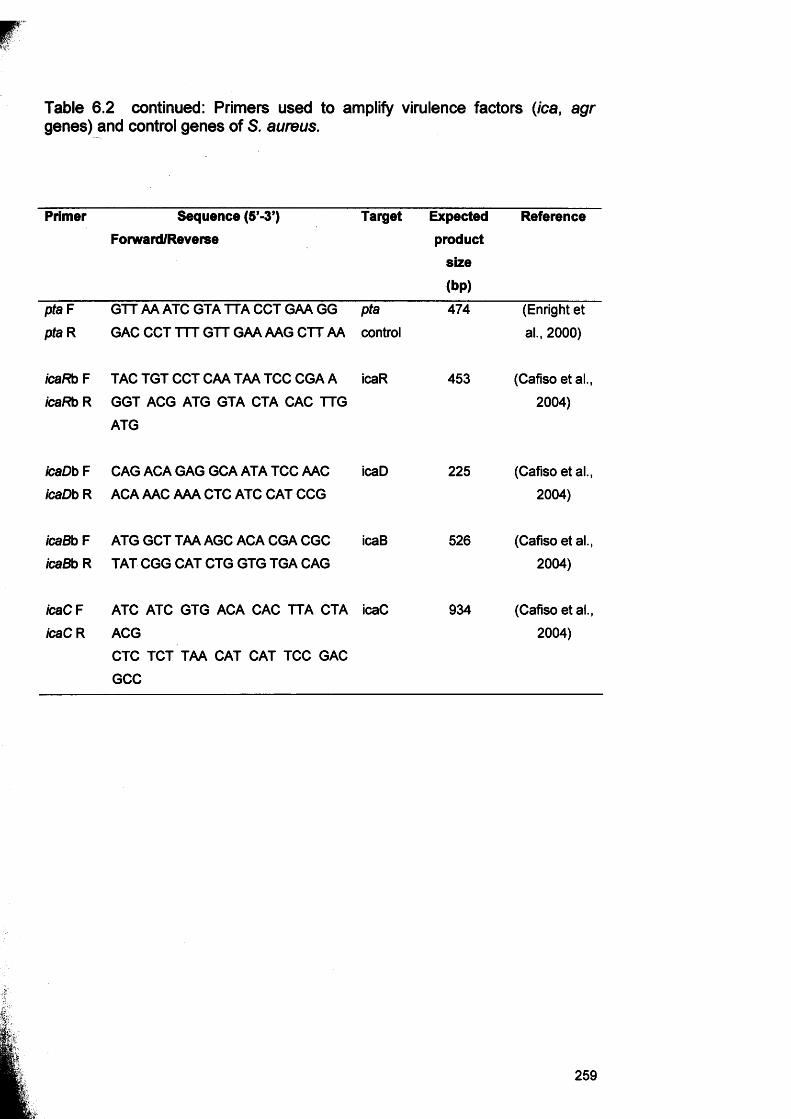
Table 6.2 continued: Primers used to amplify virulence factors (ica, agrgenes) and control genes of S. aureus.
Primer Sequence (5’-3’) Forward/Reverse
Target Expectedproduct
size
(bp)
Reference
pta F
pta RGTT AA ATC GTA TTA CCT GAA GG GAC CCT TTT GTT GAA AAG CTT AA
ptacontrol
474 (Enright et al., 2000)
icaRb F
icaRb RTAC TGT CCT CAA TAA TCC CGA A GGT ACG ATG GTA CTA CAC TTG
ATG
icaR 453 (Cafiso et al., 2004)
icaDb F
icaDb RCAG ACA GAG GCA ATA TCC AAC ACA AAC AAA CTC ATC CAT CCG
icaD 225 (Cafiso et al., 2004)
icaBb F icaBb R
ATG GCT TAA AGC ACA CGA CGC
TAT CGG CAT CTG GTG TGA CAGicaB 526 (Cafiso et al.,
2004)
icaC F icaC R
ATC ATC GTG ACA CAC TTA CTA
ACGCTC TCT TAA CAT CAT TCC GAC
GCC
icaC 934 (Cafiso et al., 2004)
259
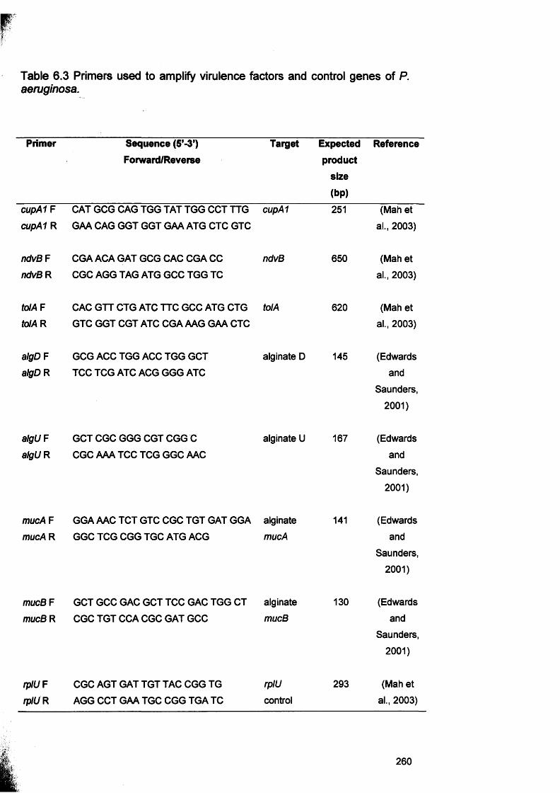
Table 6.3 Primers used to amplify virulence factors and control genes of P. aeruginosa.
Primer Sequence (5’-3’) Forward/Reverse
Target Expectedproduct
size
(bp)
Reference
cupA1 F cupA1 R
CAT GCG CAG TGG TAT TGG CCT TTG GAA CAG GGT GGT GAA ATG CTC GTC
cupA1 251 (Mah et al., 2003)
ndvB F
ndvB RCGA ACA GAT GCG CAC CGA CC
CGC AGG TAG ATG GCC TGG TCndvB 650 (Mah et
al., 2003)
tolA F to!A R
CAC GTT CTG ATC TTC GCC ATG CTG GTC GGT CGT ATC CGA AAG GAA CTC
tolA 620 (Mah et al., 2003)
algD F
algD RGCG ACC TGG ACC TGG GCT
TCC TCG ATC ACG GGG ATCalginate D 145 (Edwards
andSaunders,
2001)
algUFalgUR
GCT CGC GGG CGT CGG C CGC AAA TCC TCG GGC AAC
alginate U 167 (Edwardsand
Saunders,2001)
mucA F mucA R
GGA AAC TCT GTC CGC TGT GAT GGA
GGC TCG CGG TGC ATG ACGalginatemucA
141 (Edwardsand
Saunders,2001)
mucB F mucB R
GCT GCC GAC GCT TCC GAC TGG CT
CGC TGT CCA CGC GAT GCCalginatemucB
130 (Edwardsand
Saunders,2001)
rplU F
rplURCGC AGT GAT TGT TAC CGG TG AGG CCT GAA TGC CGG TGA TC
rplUcontrol
293 (Mah et al., 2003)
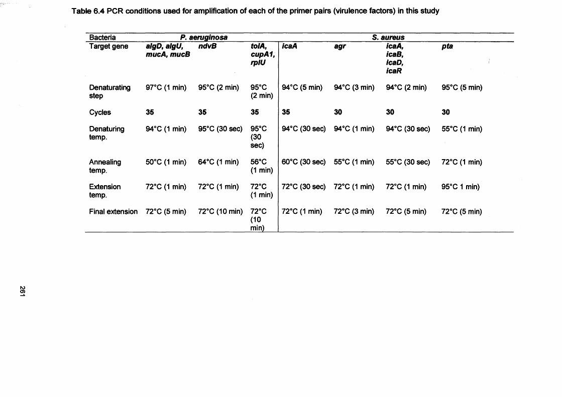
Table 6.4 PCR conditions used for amplification of each of the primer pairs (virulence factors) in this study
Bacteria P. aeruginosa S. aureusTarget gene algD, algU,
mucA, mucBndvB tolA,
cupA1,rplU
icaA agr icaA,icaB,icaD,icaR
pta
Denaturatingstep
97°C (1 min) 95°C (2 min) 95°C (2 min)
94°C (5 min) 94°C (3 min) 94°C (2 min) 95°C (5 min)
Cycles 35 35 35 35 30 30 30
Denaturingtemp.
94°C (1 min) 95° C (30 sec) 95°C(30sec)
94°C (30 sec) 94°C (1 min) 94°C (30 sec) 55°C (1 min)
Annealingtemp.
50°C (1 min) 64°C (1 min) 56°C (1 min)
60°C (30 sec) 55°C (1 min) 55°C (30 sec) 72°C (1 min)
Extensiontemp.
72°C (1 min) 72°C (1 min) 72°C (1 min)
72°C (30 sec) 72°C (1 min) 72°C (1 min) 95° C 1 min)
Final extension 72°C (5 min) 72°C (10 min) 72°C(10min)
72°C (1 min) 72°C (3 min) 72°C (5 min) 72°C (5 min)
6.3.6 Confirmation of PCR products by gel electrophoresis
PCR products (10 pi) were visualised following standard gel electrophoresis
in a 0.75 % agarose gel (Sigma, UK) with ethidium bromide staining (10 pg
pi'1). Gel images were obtained using a Gel-Doc 2000 (Bio-Rad, Hemel-
Hempstead, UK) and the average band densities for each PCR product were
measured using the Discovery Series™ Quantity One® (Bio-Rad) software.
Estimation of semi-quantitative values involved defining lanes and bands on
the gel image following the Quantity One user guide. After defining lanes, a
lane-based background subtraction was performed with bands identified
using the Detect Bands command and then the values of a standard (a
quantitative MassRuler DNA Ladder; low range, Fermentas, York, UK) being
entered. The quantity of PCR product in the experimental bands was
determined using this standard.
To obtain additional confirmation of the PCR product size and sequence,
PCR products were cleaned using the QIAquick PCR Purification Kit
(Qiagen) and sequenced as described previously in Chapter 4 (section
4.3.2).
6.3.7 Microarray study for S. aureus gene expression
The microarray study was done in collaboration with Dr Nick Saunders at the
Health Protection Agency in Colindale, London, UK. The keywords used for
virulence searches in the original design of this array were toxin, leukocidin,
coagulase, adhesion, protease, nuclease, lipoprotein, lipase, capsular
polysaccharide, binding regulatory peptidase, staphylococcal cassette
chromosome (SCC) and iron (Saunders et al., 2004a). This original array
(Saunders et al., 2004a) has since been adapted to accommodate an
additional 337 S. aureus virulence associated genes whose sequences have
been obtained. A further 30 E. coli gene sequences were also added to the
array as negative controls. Therefore the revised array comprised of probes
for 750 genes in total (Appendix III). The microarray described here was
originally used to provide an insight into the pathogenicity of the S. aureus
strains. In this present study this microarray was used in the comparative
analysis of virulence gene expression between S. aureus biofilms and S.
aureus planktonic cultures (exponential and stationary growth phases). RNA
was extracted from the S. aureus biofilms and planktonic growth as
described in section 6.3.3.
6.3.7.1 CDFF biofilm samples for microarray study
Staphylococcus aureus biofilms were prepared using the CDFF as described
previously (Chapter 4, section 4.3.12). The only difference was that the
biofilms were recovered only after 7 d incubation period instead of every 24 h
for 7 d.
6.3.7.2 Preparation of planktonic growth for microarray analysis
In addition to the biofilm study, gene expression for S. aureus’ (D76) was
also assessed for planktonic cultures. The planktonic samples were
harvested at various points along the growth curve, namely in exponential
phase (3 h) and in stationary phase (10 h). Overnight cultures were used to
set up planktonic exponential and planktonic stationary phase and diluted
1:500. An aliquot (1 ml) was added to wells of a 24 microtitre well plate and
placed in the plate reader (Fluostar Optima, BMG Labtech, UK). The
absorbance was measured at 600 nm for 48h (at 37°C, with agitation).
6.3.7.3 DNA extraction for comparative genome hybridisation
DNA was extracted from an overnight culture in BHI broth of each pure
bacterial isolate using a Puregene® DNA isolation kit (Gentra Systems,
Minneapolis, USA) by following the “Gram-positive extraction” protocol
(http://www.aentra.com/technical assistance/protocols.aspV
6.3.7.3.1 Indirect labelling of DNA for comparative genome hybridisation
DNA (1 M9) was mixed with 2 pi of random nonamers (Invitrogen, Paisley,
UK) and the volume was adjusted with water to 16 pi in total. The mixture
was heated to 95°C for 30 s in a PCR thermal cycler then cooled on ice and
4 pi of the following master mix added: 2 pi exo-Klenow buffer (10x; New
England BioLabs, Ipswich, UK) 1 pi dNTP mix (20x 1:1 aa-dUTP mix;
263
Invitrogen) 1 pi exo-Klenow (5 units). The preparation was incubated at
room temperature for 30 min followed by subsequent incubation at 37°C for
60 min. For the cleanup step, the ‘Minelute Reaction Cleanup1 (Qiagen) kit
was used and the manufacturer’s instructions followed before proceeding to
hybridisation (see section 6.3.7.7).
6.3.7.4 RNA extractionRNA from S. aureus biofilms and planktonic growth was extracted using the
Qiagen RNeasy Kit protocol and the maufacturer’s recommended protocol
(6.3.3).
6.3.7.4.1 Aminoallyl cDNA synthesis protocol for total RNA extraction
For all steps, the ChipShot indirect labelling kit was used (Promega,
Southampton, UK).
For each indirect synthesis reaction, the following mix was prepared, 1 pg of
total RNA, 1 pi of random primers (3 pg pi'1), and nuclease free water to a
volume of 20 pi. The RNA/primer solution was incubated at 70°C for 10 min
and then placed on ice. Twenty pi of extension mix containing 8 pi ChipShot
Reverse Transcriptase (5x) reaction buffer, 4.8 pi magnesium chloride (25
mM), 4 pi aminoallyl-dNTP mix and 3.2 pi ChipShot Reverse Transcriptase
was added to the RNA/primer solution, vortexed, spun briefly and incubated
at 22-25°C for 10 min, followed by incubation at 42°C for 2 h. To each cDNA
synthesis reaction 1 pi RNase H and 0.35 pi RNase solution was added,
mixed gently and incubated at 37°C in a thermal cycler for 15 min.
6 .3.7.4.2 Aminoallyl cDNA purification
To 40 pi of the synthesised cDNA, 4 pi sodium acetate (3M; pH 5.2) and 225
pi of binding solution (Promega Kit, proprietary contents) was added and
vortexed gently for 5-10 min. The solution was applied to a ChipShot
membrane on a spin column, which was placed into a collection tube. The
column was incubated for 5 min at RT, then spun down at 10,000 g for 1 min
and the flow-through discarded. The column was washed 3 times with 500 pi
264
80% ethanol, centrifuged at 10,000 g for 1 min. To remove traces of ethanol,
the column was centrifuged again at 10,000 g for 1 min. After the washing
steps, the column was placed in a clean collection tube and purified cDNA
eluted with 65 pi of 100 mM sodium bicarbonate (pH 9.0). The column was
incubated at RT for 1 min and centrifuged at 10,000 g for 1 min. Sixty pi of
cDNA was recovered after centrifugation. The cDNA was then labelled and
purified for microarray analysis as described below.
6. 3.7.4.3 Conjugation of CyDye NHS Ester to Aminoallyl cDNA
To prevent photo bleaching, the reactive dyes were protected from light at all
times. To each dry aliquot of CyDye NHS ester (Amersham, UK) 5 pi of
dimethyl sulphoxide (DMSO) was added and mixed gently to ensure that the
CyDye NHS ester was thoroughly resuspended. Two pi of CyDye solution
was added to each eluted 60 pi aminoallyl cDNA, vortexed gently and
incubated at 23°C in a thermal cycler for 1 h. Planktonic samples were
stained with CY5 and biofilm samples were stained with CY3 dyes.
6. 3.7.4.4 Purifying CyDye labelled cDNA
To the 60 pi of the synthesised and labelled cDNA, 8 pi of sodium acetate
(3M, pH 5.2) and 440 pi of binding solution (ChipShot indirect labelling kit,
Promega) was added. The mixture was vortexed gently for 5-10 s and
applied to a ChipShot membrane column (ChipShot indirect labelling kit,
Promega) which was placed into a collection tube. The column was
incubated at room temperature for 5 min and then centrifuged at 10,000 g for
1 min. The flow-through was discarded and the column was washed (x3)
with 500 pi 80% v/v ethanol, centrifuged again at 10,000 g for 1 min and
followed by a final centrifuge step to remove traces of ethanol. The column
was placed in a clean collection tube and 30 pi elution buffer (Promega Kit,
proprietary contents) was added, incubated at RT for 1 min and centrifuged
at 10,000 g for 1 min. The elution step was repeated with another 30 pi
elution buffer as described above. Eluted cDNA was stored in a light proof
container at 4°C until required for the microarray study.
265
6.3.7.5 Printing
Slides were printed using a Genetix Qarray Mini platform. The
oligonucleotides probes were printed in replicates of four, arranged in a
random pattern using Telechem Arraylt Stealth pins (Genetix Limited,
Hampshire, UK).
6.3.7.6 Slide processingPrinted microarray slides were placed in a chamber at 42°C with 50-75%
relative humidity provided by a saturated solution of NaCI for 16-18 h.
Excess spotting solution was washed off with 0.2% SDS by agitation in a
slide washer for 2 min at 20-25°C. The slides were then washed, with
shaking, three times in sterile distilled water for 1 min each wash. The
microarrays were then incubated for 20 min at 50°C in distilled water, dried
by centrifugation (10,000 g for 5 min) and stored in the dark at room
temperature (Saunders et al., 2004a).
6.3.7.7 HybridisationHybridisation was performed using the Lucidea SlidePro Hybridisation
Instrument (GE Healthcare, Chalfont St Giles, UK). The software was
programmed to perform a series of washes to the hybridisation chambers
and their respective tubing on dummy slides before inserting the arrays and
initiating the pre-hybridisation process. The hybridisation mix was then
injected and hybridisation allowed to take place for up to 7 h. Any unbound
probe mix was washed away with isopropanol and the arrays were air dried
ready for analysis (Saunders et al., 2004a).
6.3.7.8 Scanning and Data analysisThe arrays were scanned using the Gentix aQuire Scanner. Fluorescent
signals for the differentially labelled samples were detected at wavelengths of
532 nm for Cy3 and 639 nm for Cy5. The images were analysed using
QScan software (Genetix). The software enables superimposing of two
images, subtraction of background and calculation of the ratio of the two
colour signals for comparative analysis.
266
6.4 ResultsFor RNA extraction, two different methods were employed, the RiboPure
Bacteria Kit and the Qiagen RNeasy Kit. Results obtained with the Qiagen
RNeasy Kit were more consistent and the RNA yield was generally higher
than obtained with the RiboPure Bacteria Kit. Whenever possible the Qiagen
RNeasy Kit was used to generate RNA.
6.4.1 RT-PCRThe housekeeping genes rplU and pta were successfully amplified from P.
aeruginosa and S. aureus, respectively, indicating that gene expression
could be routinely and consistently detected (Fig. 6.4-6.7).
6.4.2 Expression of genes encoding outer membrane surface proteins
of P. aeruginosa
Messenger RNA from the tolA gene was detected from biofilm samples after
day 3, whilst cupA1 was expressed from day 1 to day 6 and not expressed
on day 7. In contrast, no expression of ndvB was observed in any of the
biofilm samples over the 7 d of the experiment.
Inconsistent expression of the outer membrane proteins was observed in the
planktonic samples. TolA was only expressed on day 1 and day 3 and was
not expressed on any of days 2, 4, 5, 6 and 7. The cupA1 gene was only
expressed on day 5, day 6 and day 7. Again no expression of ndvB was
observed (Table 6.5).
6.4.3 Expression of alginate genes by P. aeruginosaRT-PCR was performed on biofilm and planktonic samples from day 1 to day
7 of incubation. The target genes algD, mucA and mucB were expressed in
all biofilm samples throughout the 7 d analysis. AlgU expression in biofilm
samples was only detected after day 3. In comparison, analysis of planktonic
cultures also demonstrated expression of these target genes, although for
each gene, expression was inconsistent over the 7 d study period (Table 6.5;
Fig. 6.4-6.5).
267
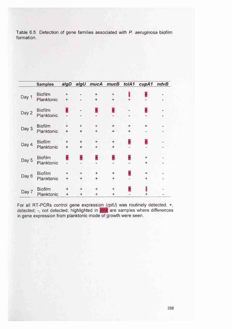
Table 6.5 Detection of gene families associated with P. aeruginosa biofilmformation.
Samples algD a lg il mucA mucB tolA1 cupA1 ndvB
Day 1
Day 2
Day 3
Day 4
Day 5
Day 6
Day 7
Biofilm + - + + I I -
Planktonic + - + + + -
Biofilm i _ i i _ i _
Planktonic - - - - - - -
Biofilm + + + + + + _
Planktonic + + + + + - -
Biofilm + + + + i i _
Planktonic + + + + - - -
Biofilm i i i i i + _
Planktonic - - - - - + -
Biofilm + + + + i + _
Planktonic + + + + - + -
Biofilm + + + + i I -
Planktonic + + + + - + -
For all RT-PCR s control gene expression (rplU) was routinely detected. +, detected; not detected; highlighted in pink are samples where differences in gene expression from planktonic mode of growth were seen.
268
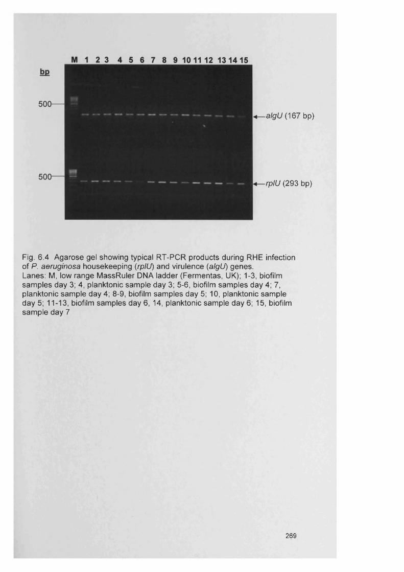
alg il (167 bp)
rplU (293 bp)
Fig. 6 .4 Agarose gel showing typical R T-PC R products during RHE infection of P. aeruginosa housekeeping (rplU) and virulence (algU) genes.Lanes: M, low range MassRuler DNA ladder (Fermentas, UK); 1-3, biofilm samples day 3; 4, planktonic sample day 3; 5-6, biofilm samples day 4; 7, planktonic sample day 4; 8-9, biofilm samples day 5; 10, planktonic sample day 5; 11-13, biofilm samples day 6 , 14, planktonic sample day 6; 15, biofilm sample day 7
269
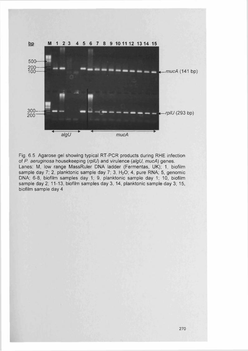
b£ M 1 2 3 4 5 6 7 8 9 10 11 12 13 14 15
500-200 -
100-
300-200 ' ■ M M j
-mucA (141 bp)
-rplU (293 bp)
< rrr ►algU mucA
Fig. 6 .5 Agarose gel showing typical R T-PC R products during RHE infection of P. aeruginosa housekeeping (rplU) and virulence (algU, mucA) genes. Lanes: M, low range MassRuler DNA ladder (Fermentas, UK); 1, biofilm sample day 7; 2, planktonic sample day 7; 3, H20 ; 4, pure RNA; 5, genomic DNA; 6-8, biofilm samples day 1; 9, planktonic sample day 1; 10, biofilm sample day 2; 11-13, biofilm samples day 3, 14, planktonic sample day 3; 15, biofilm sample day 4
270
6.4.4 Expression of accessory gene regulator {agr) in S. aureus
All biofilm and planktonic samples expressed the agr gene for all 7 days
(Table 6.6).
6.4.5 Expression of the intercellular adhesion (ica) genes in S. aureus
RT-PCR using three sets of primers was undertaken. RT-PCR products
were generated using icaAa primers (Knobloch et al., 2002) targeting the
entire ica locus of S. aureus, in biofilm samples from day 3 to day 7 (Fig. 6.6-
6.7). However, no expression of the icaAa gene was evident in any of the
planktonic samples. In contrast, RT-PCR products were not detected using
alternative primers {icaBb, icaCb, icaDb and icaRb) specific to the ica operon
of S. epidermidis (Cafiso et al., 2004).
Using a third set of ‘in-house’ prepared primers, S. aureus ica genes were
found to generate RT-PCR products for all biofilm samples from 2 d onwards,
with icaDa expressed only in biofilm samples from day 3. Using these
primers on planktonic samples, icaAb expression was evident on days 4-6,
icaBa expression occurred on days 6 and 7 and icaDa and icaRa were
expressed on days 3-6 (Table 6.6).
6.4.6 Semi-quantitative comparison of gene expression using RT-PCR
Semi-quantitative analysis using RT-PCR for all the target genes indicated
varying quantities of mRNA for the expressing strains.
The majority of the biofilm specific genes in P. aeruginosa were upregulated
in the biofilm state (27 out of 42). However, algU, mucA and mucB appeared
to be down regulated in the biofilm state from day 5 onwards (Table 6.7).
Between the two growth phases, there appeared to be greater up regulation
of ica gene expression in the biofilm state, with only three samples
demonstrating an apparent down regulation compared to the planktonic
state. The majority of agr genes were upregulated in the biofilm state, 6 out
of 7 (Table 6.8).
271
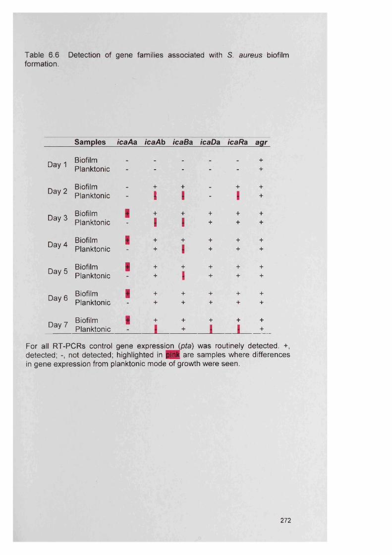
Table 6.6 Detection of gene families associated with S. aureus biofilmformation.
Sam ples icaAa icaAb icaBa icaDa icaRa agr
Biofilm +Day 1
Planktonic - - - - +
Day 2Biofilm + + + +Planktonic I I - I +
Day 3Biofilm | + + + + +Planktonic I I + + +
Day 4Biofilm + + + + +Planktonic + I + + +
Day 5Biofilm + + + + +Planktonic + I + + +
Day 6 Biofilm + + + + +Planktonic + + + + +
Day 7Biofilm | + + + + +Planktonic l + I I +
For all RT-PCR s control gene expression (pta) was routinely detected. +, detected; not detected; highlighted in pink are samples where differences in gene expression from planktonic mode of growth were seen.
272
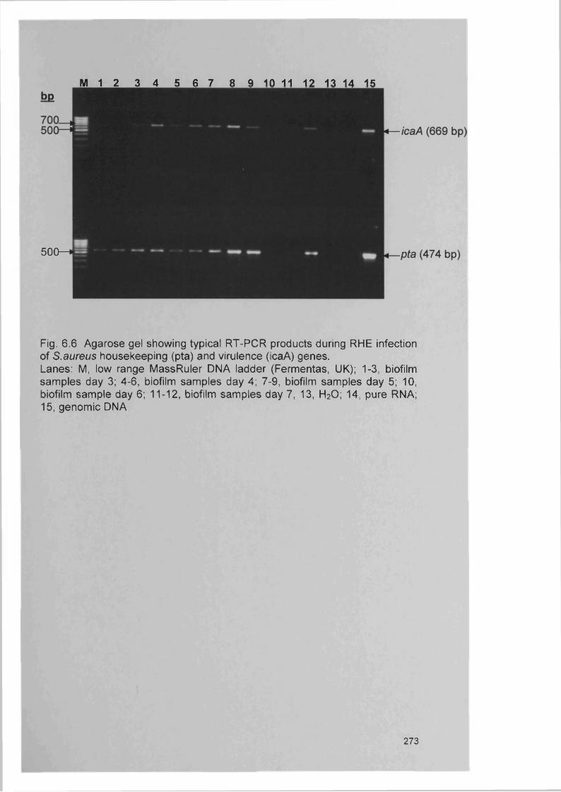
M 1 2 3 4 5 6 7 8 9 10 11 12 13 14 15
500— H
Fig. 6.6 Agarose gel showing typical R T-PC R products during RHE infection of S.aureus housekeeping (pta) and virulence (icaA) genes.Lanes: M, low range MassRuler DNA ladder (Fermentas, UK); 1-3, biofilm samples day 3; 4-6, biofilm samples day 4; 7-9, biofilm samples day 5; 10, biofilm sample day 6; 11-12, biofilm samples day 7, 13, H20 ; 14, pure RNA; 15, genomic DNA
273
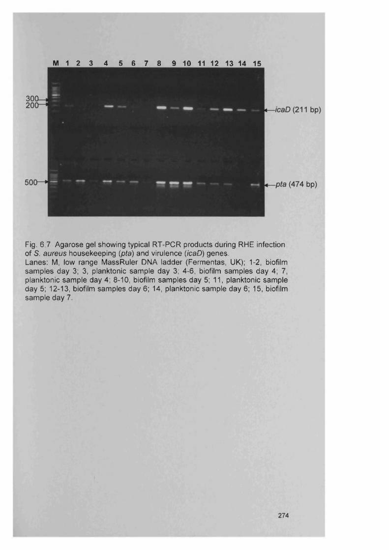
M 1 2 3 4 5 6 7 8 9 10 11 12 13 14 15
Fig. 6 .7 Agarose gel showing typical R T-PC R products during RHE infection of S. aureus housekeeping (pta) and virulence (icaD) genes.Lanes: M, low range MassRuler DNA ladder (Fermentas, UK); 1-2, biofilm samples day 3; 3, planktonic sample day 3; 4-6, biofilm samples day 4; 7, planktonic sample day 4; 8-10, biofilm samples day 5; 11, planktonic sample day 5; 12-13, biofilm samples day 6; 14, planktonic sample day 6; 15, biofilm sample day 7.
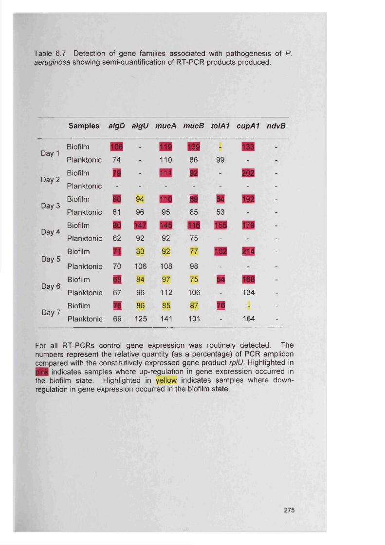
Table 6.7 Detection of gene families associated with pathogenesis of P.aeruginosa showing semi-quantification of RT-PCR products produced.
S am ples algD algU mucA mucB tolA1 cupA1 ndvB
Day 1Biofilm - ■ - -
Planktonic 74 - 110 86 99 - -
Day 2Biofilm 7® - 111 92 - 202 -
Planktonic - - - - - - -
Day 3Biofilm m 94 ■ 89 04 192 -
Planktonic 61 96 95 85 53 - -
Day 4Biofilm 80 m 145 110 160 17® -
Planktonic 62 92 92 75 - - -
Biofilm n 83 92 77 102 214 -
Day 5Planktonic 70 106 108 98 - - -
Biofilm 68 84 97 75 54 _
Dav 6Planktonic 67 96 112 106 - 134 -
Biofilm 76 86 85 87 76 _ -
Dav 7Planktonic 69 125 141 101 - 164 “
For all RT-PCR s control gene expression was routinely detected. The numbers represent the relative quantity (as a percentage) of PCR amplicon compared with the constitutively expressed gene product rplU. Highlighted in ■ I indicates samples where up-regulation in gene expression occurred in the biofilm state. Highlighted in yellow indicates samples where down- regulation in gene expression occurred in the biofilm state.
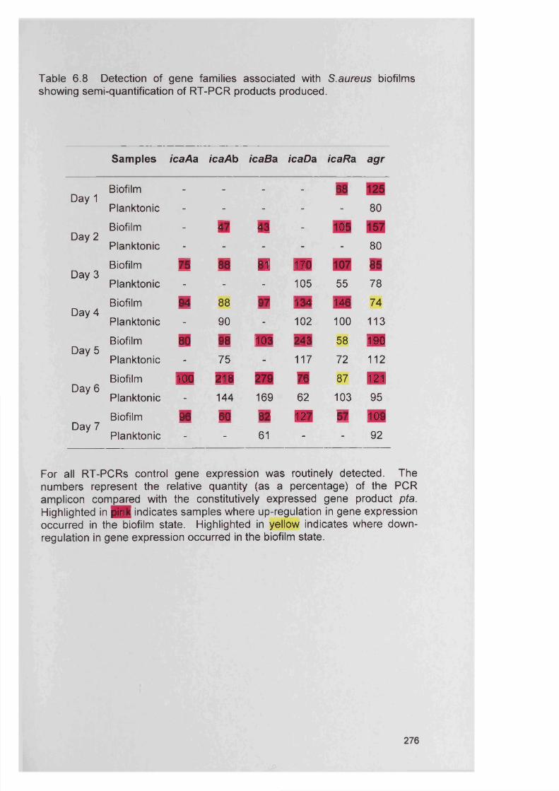
Table 6.8 Detection of gene families associated with S.aureus biofilmsshowing semi-quantification of RT-PCR products produced.
Samples icaAa icaAb icaBa icaDa icaRa agr
Day 1Biofilm - - - - 68Planktonic - - - - - 80
Day 2Biofilm - 41 43 - 106Planktonic - - - 80
Day 3Biofilm 75 88 ■ 170 81Planktonic - - - 105 55 78
Day 4Biofilm 94 88 97 134 146 74
Planktonic - 90 - 102 100 113
Day 5Biofilm 80 98 103 243 58 190Planktonic - 75 - 117 72 112
Day 6Biofilm 100 218 m 87 121
Planktonic - 144 169 62 103 95
Day 7Biofilm 96 60 82 ■ 6? 109
Planktonic - - 61 - - 92
For all RT-PCR s control gene expression was routinely detected. The numbers represent the relative quantity (as a percentage) of the PCR amplicon compared with the constitutively expressed gene product pta. Highlighted in pink indicates samples where up-regulation in gene expression occurred in the biofilm state. Highlighted in yellow indicates where down- regulation in gene expression occurred in the biofilm state.
6.4.7 Microarray study for S. aureus gene expression
6.4.7.1 Comparative genome hybridisationIn comparative genome hybridisation experiments, two genomic DNA
samples one from wound isolate S. aureus D76 and the other from S. aureus
MW2 (Baba et al., 2002) were simultaneously hybridised to the microarray
and detected using different fluorochromes. In this study the presence and
absence of the genes on the array for the genome of the test strains was
confirmed. Hybridisation with the S. aureus D76 strain was found to occur
with signals evident for 486 genes and with S. aureus MW2, 550 genes were
present. The majority of these genes present in S. aureus D76 were
hypothetical proteins related to the ATP-binding protein, exotoxins and
capsular polysaccharide synthesis enzymes. The intercellular adhesion
proteins icaA, icaB, icaD and the regulator icaR were also present as well as
the staphylococcal accessory regulator A (sarA), which is implicated in
biofilm formation (Appendix IV). Virulence factors such as proteases, zinc
metalloproteinase, thermonuclease, adhesins, alpha-, beta-, gamma-
haemolysin were also present. It seems that the D76 strain carries drug
resistance in various forms including a hypothetical protein, which is similar
to tetracycline resistance proteins as well as multidrug resistance proteins.
Interestingly, phage related proteins, such as staphylococcal phage phi11,
phage integrase family, hypothetical protein which is similar to Enterococcus
faecalis plasmid pPD1 and a lytic regulatory protein truncated with Tn554
were also present in the S. aureus D76 strain which could promote gene
transfer (Appendix IV).
6.4.7.2 Staphylococcus aureus gene expression as determined using
the microarrayComparative gene expression between S. aureus D76 biofilm samples after
7 d of incubation in the CDFF was compared against both stationary and
exponential phase planktonic growth. Microarray analysis revealed that both
growth phases exhibited common expression of the majority of genes,
although some differences were found. Figure 6.8 (biofilm against
277
exponential phase) and Fig. 6.9 (biofilm against stationary phase) illustrate
the up-regulation of genes between the two growth phases. The genes
which are distributed around the line at 1 are non changeable. The genes
above this line are up-regulated in the biofilm phase and the genes beneath
the line are up-regulated in the planktonic phase (Appendix III, for detailed
gene names).
Figure 6.10 shows expression of genes of interest in biofilm phase against
exponential planktonic phase. For example, the regulatory genes such as
the accessory gene regulator (agrC1 - groupl) and the sarA were more
highly expressed in the biofilm state than in the exponential planktonic phase
as well as few hypothetical proteins such as SA1999 (hypothetical protein,
similar to the regulatory protein SIR2 family), MW0596 (hypothetical protein,
similar to iron dependent repressor) and MW2499 (hypothetical protein,
similar to short chain oxireductase). However, several hypothetical genes
seemed to be up-regulated in the exponential planktonic growth phase
compared to biofilm growth mode such as MW0595 (hypothetical protein,
similar to the ABC transporter ATP-binding protein), MW0339 (hypothetical
protein, similar to GTP-binding protein), MW0283 (hypothetical protein,
similar to branched chain amino acid uptake carrier), MW0384 (Exotoxin 18,
Genomic island nu Sa alpha 2) and MW2217 (secretory antigen precursor).
Figure 6.11 shows expression of genes of interest in the biofilm phase
against the stationary planktonic phase. Genes which are up-regulated in the
biofilm phase compared to the stationary planktonic phase included MW1834
(hypothetical protein, similar to ferritin), MW0396 (hypothetical protein),
MW1041 (hypothetical protein, similar to fibrinogen binding protein), SA0857
(hypothetical protein, similar to negative regulator of genetic competence
mecA), SAV0151 Cap8c (capsular polysaccharide synthesis enzyme Cap8c),
SA1174 (SOS regulatory LexA protein) and MW2129 hysA (hyaluronate
lysate precursor).
278
In comparison hypothetical proteins such as MW0757, MW1744, MW0579
(similar to esterase/lipase), MW1508 (similar to ABC transporter ATP-binding
protein) and SA2272 were up-regulated in the stationary planktonic growth
phase.
279
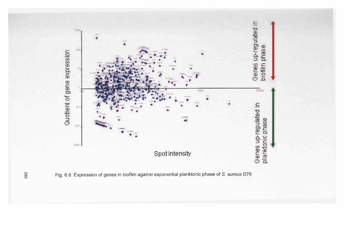
doCOCO2CLXCDCDcCDOto“E<DO=5o
Spot intensity
oo Fig. 6.8 Expression of genes in biofilm against exponential planktonic phase of S. aureus D76
Genes
up-re
gulated
in I
Genes
up-regulated
in
planktonic phase
biofilm
ph
ase
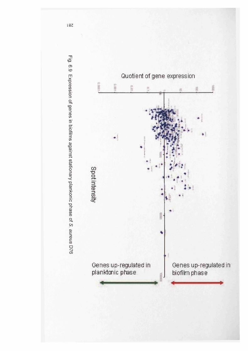
Fig. 6.9 Expression
of genes
in biofilm
s against
stationary plankonic
phase of
S. aureus D
76
1.83
Quotient of gene expression
Genes up-regulated in Genes up-regulated in planktonic phase J biofilm phase
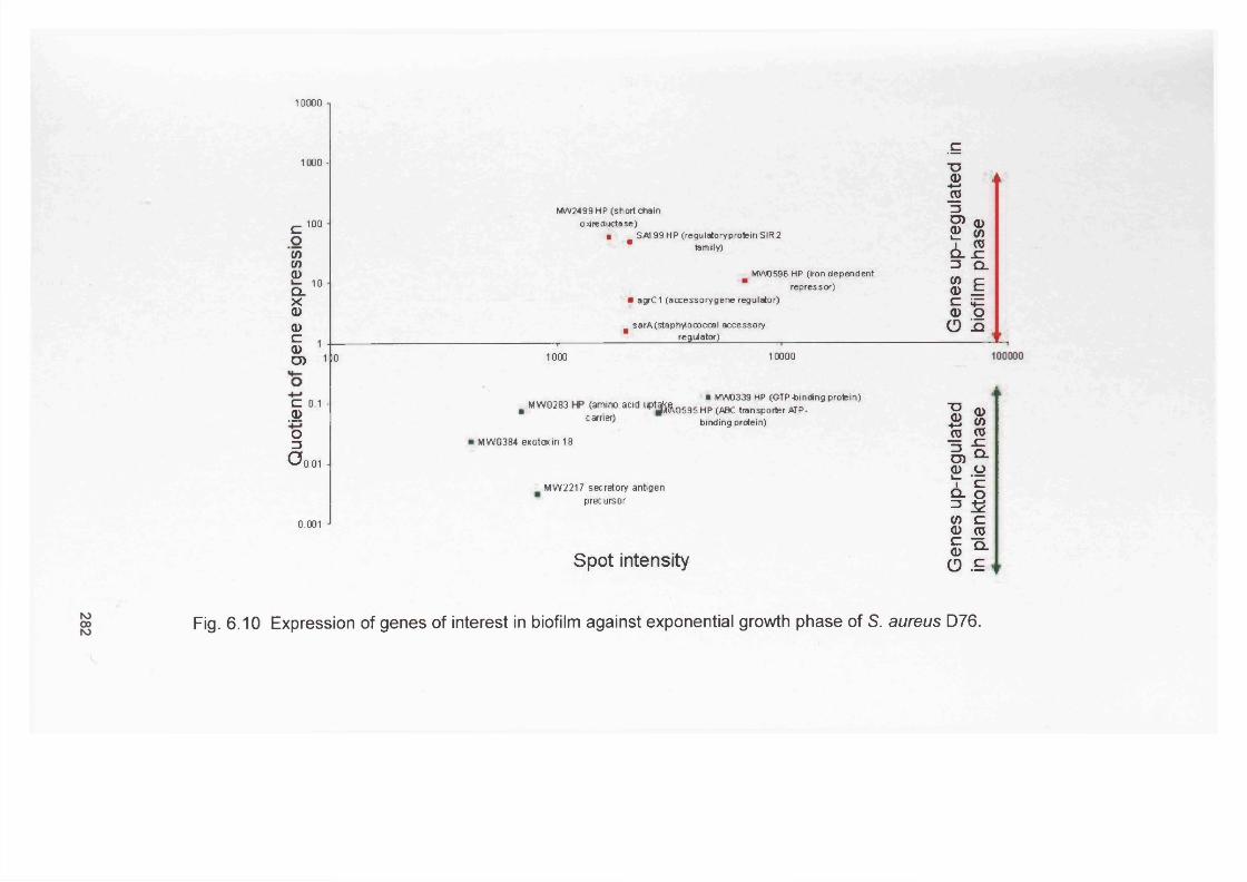
10000
1000 -
co(0C/)<DQ_XQ)a>c=a>D)
100
10
M W 2499HP (short chain o>dreductase)
■ SM 99 HP (regulatoryprotein S IR 2* family)
MW0S96 HP (iron dependent repressor)
• agrC1 (accessorygene regulator)
B sarA (staphylococcal accessory __________________________ regulator)_____________ _____________________
11 0 1000 10000
C 0.1 -.s>o13
CJq.01 A
, . . . , • MWD339 HP (GTP-binding protein). M W 0283 <aml" ° aCld ,* j t o D S 9 S H P (ABC transporter ATP-
carner' binding protein)
MW0384 exotaxin 18
0.001
MW2217 secretory antigen precursor
Spot intensity
Fig. 6.10 Expression of genes of interest in biofilm against exponential growth phase of S. aureus D76.
Gene
s up
-regu
late
d Ge
nes
up-re
gula
ted
inin
plan
kton
ic ph
ase
biofilm
ph
ase
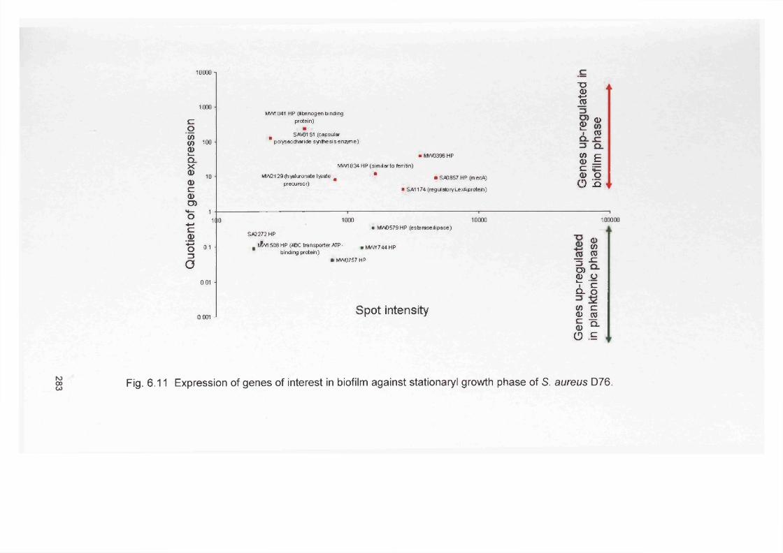
Quo
tient
of
gene
ex
pres
sion
10000 1
1000 -
100 ■
10 ■
I1 ■ —
mo
0.1 ■
0.01 ■
0.001 •
■O2TO
MW1041 HP (Ibri nogen bindingprotein) £ <J>
m SAS0151 (capsular I <0polysaccharide syrt-iesis engine)
■ MW0396 HP (0 C<1) ^MWI834 HP (similarto feiritin) £ j =
MW2129 (hyduronate lysate & * • S40857 HP (mecA) ^ Qprecursor) r ) jQ
■ SA1174 (regulatoryLexAprotein)
1000 10000 100000 ■ MIA0579 HP (esterasejlipase)
SA2272 HP
. I^VI 508 HP (fiBC transporter ATP- .M W I 744 HP 8)binding protein) f l j
• MW0757 HP ■ = _ C
O)<D OS_'§
Spot intensity $ ^s ^O .£
Fig. 6.11 Expression of genes of interest in biofilm against stationaryl growth phase of S. aureus D76.
r
6.5 DiscussionIn the past, research has largely focused on the in vitro study of organisms
grown in liquid media. However, in recent years it has been recognized that
in their natural settings bacterial cells are most often found in close
association with surfaces and interfaces, in the form of aggregates referred to
as biofilms (Branda et al., 2005). It has been suggested that adherent and
biofilm bacteria behave differently (Literature Review, section 1.1.1, Table
1.1) and hence may express a different pattern of genes compared with their
planktonic counterparts (Becker et al., 2001; Beloin and Ghigo, 2005).
Whiteley et al. (2001) showed that gene expression in P. aeruginosa biofilm
cells was remarkably similar to their planktonic counterparts under similar
environment conditions but that there were a small number of significant
differences. Such differences in gene expression could perhaps explain
important phenotypic traits of biofilms including the high antibiotic resistance
of biofilm cells, or even aid in the development of rapid screens for agents
that block biofilm maintenance (Whiteley et al., 2001).
The present study focused on comparing specific gene expression for both P.
aeruginosa and S. aureus when cultured in biofilms and in planktonic states.
The significance of this work was that the detection, albeit in vitro, of an up-
regulated biofilm gene product could be important if also manifested in a
chronic wound biofilm state. Differential expression between the two growth
states could theoretically provide evidence of virulence mechanisms or
strategies promoting the survival of the bacteria in biofilms. This knowledge
could in the future be exploited in the development of management and
treatment regimes (Beloin and Ghigo, 2005).
The selected bacterial species were studied as these organisms have
previously been shown to be frequently encountered in chronic wounds
(Davies et al., 2007; Davies et al., 2004) and in chronic infections (Parsek
and Singh, 2003). In addition, previous chapters reported in this thesis
(Chapter 4 and Chapter 5) have shown that these organisms predominated
over other bacterial species in biofilm models.
284
it.
The specific aims of this Chapter were to examine the expression of
previously proposed ‘biofilm specific genes’ and to compare their expression
in planktonic and biofilm growth phases. It addition exploratory studies were
also performed using a microarray analysis.
Bacteria growing in biofilms possess characteristics distinct from their
planktonic counterparts, e.g. biofilm bacteria are more resistant to
antimicrobial treatments (Whiteley et al., 2001). The outer membrane
surface proteins are of particular interest in P. aeruginosa due to their
involvement in the transport of antibiotics (Stover et al., 2000). Mah et al.
(2003) proposed a mechanism of biofilm-specific antibiotic resistance
wherein periplasmic glucans bind to, and sequester antibiotics. It has been
suggested that the gene, ndvB is required for the synthesis of periplasmic
glucans (Mah et al., 2003). However, in this study ndvB was not found to be
expressed in P. aeruginosa biofilms. This contrasts with the results of Mah et
al. (2003) who predicted and showed that ndvB was exclusively expressed in
biofilm grown cells using a microtitre plate assay for biofilm formation.
Although Mah et al. (2003) data supports an involvement for ndvB in biofilm
antibiotic resistance, it may be that some strains of P. aeruginosa have
compensatory mechanisms for ndvB mutations or because of strain
variations.
Previous studies have examined how alginate overproduction influences P.
aeruginosa biofilm development and function, using a non-mucoid P.
aeruginosa wild type and a mucoid alginate overproducing mutant strain
(Hentzer et al., 2001). These studies showed that in cystic fibrosis (CF)
patients, conversion to the mucoid phenotype resulted in an altered biofilm
structure and increased antimicrobial resistance. RT-PCR of alginate gene
expression in P. aeruginosa in the present study did not show a direct
correlation with biofilm formation. The majority of planktonic samples were
also found to express algD, algil, mucA and mucB. These data might
suggest that some bacteria in the broth cultures had biofilm phenotypes or
that expression of these genes is not an exclusive property of biofilms. In
285
fact Lazazzera et al. (2005) suggests that subsets of genes expressed in
biofilms are also expressed under different planktonic conditions.
The expression of S. aureus virulence factors is controlled by the quorum
dependent accessory gene regulator (<agr) and can be classified into 4 (l-IV)
specific agr groups (Shopsin et al., 2003; Yarwood et al., 2002). Group I and
II have been linked with enterotoxin mediated and invasive disease, while
groups III and IV have been associated with non-invasive and exfoliating-
producing disease (Jarraud et al., 2002). The S. aureus wound isolate D76
used in this study expressed agr I in all biofilm and planktonic samples. It
may therefore be possible that this S. aureus strain plays a role in impaired
healing by the production of this toxin which then attacks the patient epithelial
cells.
Studies have shown that PIA, a homoglycan consisting of (3-1,6-linked N-
acetylglucosamine which is encoded by the ica operon, is required for biofilm
formation (Conlon et al., 2002b; Mack et al., 1996). Mack et al (1999; 1996)
suggested that PIA mediates intercellular adhesion and that bacteria have no
direct contact to the surface following the accumulative phase but remain in
the biofilm via mechanisms mediating cell-to-cell adhesion (Rohde et al.,
2005). Staphylococcus epidermidis, a commensal of human skin and
mucous membranes, is also a predominant cause of foreign-body-associated
infections. PIA is a significant virulence factor and essential for S.
epidermidis and S. aureus cell accumulation (Knobloch et al., 2004).
However, investigations into the relationship between the ica genotype and
biofilm formation of S. aureus has in the past led to contradictory results by
different investigators (Knobloch et al., 2002). Cramton et al. (1999)
characterised S. aureus isolates that were ica positive and showed that
deletion of the ica genes eliminated the ability to produce polysaccharide
intercellular adhesion (PIA) and form a biofilm in vitro. In contrast, Yazdani et
al. (2006) evaluated the biofilm forming capacity and the presence of icaAD
(associated with slime production) among S. aureus isolated from wound
infections. All fifty strains identified, were found to carry the icaAD genes,
286

with only 26 of the strains found to produce a biofilm in vitro. Hence, this
study indicated that whilst there was a high prevalence of the icaAD genes
amongst the S. aureus isolates, their presence was not always associated
with biofilm formation in vitro (Yazdani et al., 2006). Despite the importance
of ica genes, not much is known about the regulation of the ica locus itself
(Jefferson et al., 2003). Numerous recent publications report on factors
influencing PIA or biofilm production in vitro. Such factors include the level of
glucose and other sugars in the culture medium, extent of osmolarity and
anaerobiosis, low levels of ethanol, and iron restriction (Cramton et al., 2001;
Fluckiger et al., 2005; Jefferson et al., 2003; Rachid et al., 2000a; Rachid et
al., 2000b).
The observation that biofilms are not wholly dependent upon expression of
the ica operon is also supported by the findings of Muller et al. (1993) who
found that a few S. epidermidis strains were able to form a biofilm with no
apparent production of polysaccharide (Cramton et al., 2001). Importantly,
the majority of clinical S. aureus strains contain the ica operon (Arciola et al.,
2001; Cramton et al., 1999; Fitzpatrick et al., 2005; Rohde et al., 2005), and
expression of the ica operon and biofilm production is controlled under in
vitro conditions (Cramton et al., 2001; Fitzpatrick et al., 2005; O'Gara, 2007).
In the present investigation, RT-PCR detection of expressed genes of the S.
aureus ica operon used primers previously validated by Knobloch et al.
(2002), primers designed by Cafiso et al. (2004) and also ‘in-house’ designed
primers. The failure of the Cafiso et al. (2004) primers to yield PCR products
with any of the specimens likely relates to sequence variations between S.
epidermidis and S. aureus, as the former species was used as the basis of
the primer design by Cafiso et al. (2004).
It was evident from the results (Table 6.6) that ica genes were expressed in
both biofilm and planktonic samples. As mentioned previously for P.
aeruginosa strains, a biofilm occurrence within the planktonic samples could
be envisaged due to the extended incubation time of the culture (7 days)
287
even though the broth was replenished at 24 h intervals. A possible
improvement could be to generate continuous planktonic growth within a
dedicated fermenter (e.g. a chemostat).
After 48 h, all biofilm samples were found to express icaA, icaB and icaR
whilst icaD was expressed only after 72h. This data suggests that using the
CDFF for biofilm formation, 48 h incubation was necessary to get an
established biofilm of S. aureus.
Semi-quantification of the RT-PCR products indicated that for the majority of
targeted genes, expression was up-regulated in the biofilm samples
compared to the planktonic ones. The up-regulation of the ica genes in the
biofilm mode of growth is consistent with previous work (Resch et al., 2005).
The data therefore suggests that whilst there is differential gene expression
between biofilm and planktonic cultures, expression does vary with other
factors including culture age and for the studied genes, expression did not
appear exclusive to a particular mode of growth. Previous studies also
highlight that environmental signals such as temperature, osmolarity, pH, iron
and oxygen play a role in biofilm development and therefore alter gene
expression (Costerton et al., 1995). It also highlights the uniqueness of
biofilms and that a universal biofilm gene-expression pattern still remains a
wide-open area of investigation (Beloin and Ghigo, 2005).
The detection of biofilm specific genes in this present study seemed to be
more complex than expected and it also appeared to be very difficult to
maintain for 7 d planktonic cultures. Further studies are still required to fully
elucidate the genetics of bacterial growth (Waite et al., 2005). Considering
these contrasting results, the findings could be indicative that a wide range of
genes influence biofilm development under specific conditions. More work
remains to be done, e.g. the discovery of other genes which are critical for
biofilm development. Improved understanding of which genes and proteins
are differentially expressed under biofilm and planktonic growth conditions
and how the morphology and physiology of biofilm cells differs from those of
288
planktonic cells is needed to understand the high persistence and
antimicrobial resistance of biofilm cells (Resch et al., 2005). It is also
important to highlight how widespread biofilms are with more than 99.9% of
bacteria found to grow in biofilms (Donlan and Costerton, 2002) and that
biofilm research encompasses studies of multispecies communities (Lewis,
2001).
In this present study, exploratory experiments using the microarray technique
at the Health Protection Agency in Colindale were employed. The microarray
used in this study was designed to look for potential virulence associations
(section 6.3.8.7) and not only for biofilm-specific genes. Therefore a direct
comparison of the microarray data with the RT-PCR results was difficult. Of
the genes targeted by RT-PCR, only agr and ica genes were housed on the
microarray. Comparative genome hybridisation was first used to confirm if
the genes on the array were present or absent within the S. aureus D76
genome. The 486 genes present included the ica proteins, the sarA gene,
virulence factors, phage related proteins which are involved with gene
transfer events and multidrug resistance proteins.
The microarray expression work showed that several regulatory genes
including agrC1 (accessory gene regulator, group 1) and SA0573 sarA
(staphylococcal accessory regulator A) had a higher expression in biofilm
samples compared with planktonic exponential samples. Significantly, agr
expression was also confirmed previously by RT-PCR in all biofilm and
planktonic samples. By RT-PCR the majority of agr genes were up-regulated
in the biofilm state, 6 out of 7. It is important to note that a network of
regulatory genes including agr and sar operons control the expression of S.
aureus virulence factors (Saunders et al., 2004b). The microarray data
provided detailed information concerning gene carriage and increase or
decrease in the level of gene between planktonic and biofilm mode of growth
in the S. aureus D76 wound isolate. In this microarray study there was no
evidence for the expression of the ica genes. This correlates with previous
findings reported by Resch et al (2005) who suggested that these genes
289
were not needed after initial attachment and start of biofilm formation as the
biofilm samples used for this study were 7 d old. Transcriptomic analysis
opens the door for future work and provides a powerful tool to assess
differential gene expression patterns under biofilm and planktonic growth
conditions (Resch et al., 2005) as well as in other systems. However, more
work still needs to be done to fully understand the genetics of bacterial
biofilm growth (Waite et al., 2005).
In future studies, it would be interesting to examine gene expression from a
mixed biofilm population and compare it with a mixed planktonic population.
In this present study, mixed biofilms prepared in the CDFF and mixed
planktonic preparations were generated but due to time constraints, the RT-
PCR process was not pursued with these populations. It would certainly be
valuable to extend the current study to apply a real-time PCR to detect,
identify and quantify the relevant genes of interest over time between the
planktonic and biofilm modes of growth to confirm the apparent up-regulation
of the genes identified in this study. The real-time technique would allow
more accurate monitoring of gene expression over time.
In general, microarray technology whilst providing a significant amount of
information does suffer from the disadvantage of relating the findings to the
role of the gene products, simply due to our currently limited knowledge
concerning the hypothetical proteins and their relevant role in biofilm
formation. Therefore, it would be helpful to actually identify first biofilm
related genes and then to monitor the expression in samples. So far neither
the microarray nor the RT-PCR study provided any direct indications related
to genes associated with chronic wound biofilms. In future, and
accompanying the development of these techniques it would be interesting to
look at bacterial gene expression within actual clinical wound samples, and
maybe compare specimens from both the normally healing phenotype and
those that fail to heal.
290
6.6 Summary
In summary, this present study showed:
1. That the majority of the examined P. aeruginosa and S. aureus genes
previously associated with biofilm formation were expressed in biofilm
samples. Some of these genes were also expressed in planktonic samples
suggesting that there might be a biofilm phenotype present or that the
expression of the genes was not exclusive to specific growth modes.
2. Semi-quantification of the RT-PCR products from these genes did indicate
that the majority were up-regulated in the biofilm samples compared to
planktonic ones.
3. Altered gene expression between biofilm and planktonic samples was also
apparent from exploratory microarray studies of S. aureus. In particular,
confirmation of up-regulation of agrC1, sarA and the capsular polysaccharide
synthesis enzyme Cap8c in biofilm grown cells was evident using the array
method.
291

General Discussion
The significance of biofilm related infections is becoming increasingly evident
and it has been suggested that over 65% of all hospital infections have their
origin in biofilms (Costerton et al., 1999; Donlan, 2001; Donlan and
Costerton, 2002; Douglas, 2003; Ramage et al., 2006). As a consequence, it
is not surprising that much research in recent years has been focussed at
improving our understanding of biofilms and the options available to combat
them. The debate surrounding what a biofilm actually is remains an ongoing
one. It is always difficult to define a biofilm and to compare biofilms with
other studies due to the lack of standardised systems and protocols (Parsek
and Singh, 2003). In this study, a biofilm was classed as being formed once
the microorganisms (in this work C. albicans or chronic wound bacteria) were
attached to a surface and produced multilayered structures.
Biofilms are ubiquitous throughout natural, industrial and clinical
environments (Parsek and Singh, 2003). A distinguishing feature of these
biofilms is the presence of aggregated microcolonies of cells that are
attached to a surface (Hall-Stoodley et al., 2004). In the clinical environment,
biofilms provide a reservoir of potentially pathogenic organisms that are often
resistant to standard antibiotic concentrations. Elevated antibiotic resistance
might be affected by phenotypic changes in the organisms which make up a
biofilm. These changes include slower growth rate and expression of new
multi-drug-resistant pumps (Anwar et al., 1992; Hoyle and Costerton, 1991;
O'Toole et al., 2000). Another clinical problem of biofilms is that they can
cause device failure when adhered to biomaterials used in medical devices
(Lewis, 2001; Mah and O'Toole, 2001). Indeed, once a biofilm is established,
the removal of bacteria by host defence mechanisms is also very difficult,
and in the case of medical devices, often the only treatment option is the
removal and replacement of the medical device.
Candida albicans is a commensal fungus of humans, commonly found in the
oral cavity, gastrointestinal tract, female genital tract and occasionally on the
skin (Hube and Naglik, 2001; Watts et al., 1998). Although considered as a
commensal of healthy individuals, this fungus frequently causes superficial
293
infections when the normal microbial flora is unbalanced or there is a
debilitation in the hosts’ defence mechanisms. Hence, in
immunocompromised patients, C. albicans may invade the tissue, penetrate
the blood vessels and cause life threatening systemic infections (Felk et al.,
2002).
In this study, it was shown that C. albicans strains isolated from different oral
conditions were capable of forming biofilms and invading a reconstituted
human oral epithelium (RHE) model. Using CLSM a heterogeneous invasion
pattern of the tissue was evident with the strains of C. albicans, with the
surface mainly colonised by the yeast form and a complex network of
filaments occurring within the tissue. These filamentous hyphae were seen
to physically penetrate the cells and exhibited budding within the tissue.
Hyphae are indeed considered to be the principle invasive forms of C.
albicans and are frequently identified in infected tissue (Felk et al., 2002). At
the time of writing no other CLSM investigations regarding C. albicans biofilm
formation ability and invasion in an in vitro model have been reported. In this
present study, the ability of C. albicans to invade the RHE appeared to
correlate with the isolates’ capacity to exhibit a yeast/hyphal transition and in
all but one case, isolates categorised as the ‘high invaders’ revealed a
predominance of the hyphal morphology within the tissue. It was also evident
that four of the five strains identified as high invaders had previously been
isolated from chronic hyperplastic candidosis conditions. This could suggest
that there is a particular strain type associated with this form of oral
candidosis. It was clear from the pattern of RHE infection that C. albicans is
heterogenous with respect to tissue invasion by strains, and whilst the host
condition obviously plays an important role in the occurrence of infection, this
study suggests that inherent strain virulence could also contribute.
Destruction of host tissues by Candida may be facilitated by the release of
hydrolytic enzymes into the environment. The most widely studied hydrolytic
enzymes of C. albicans are the secreted aspartyl proteinases (SAPs) and
phospholipases (PLs). At present, 10 different genes have been identified
294
that encode for candidal SAPs (Hube, 1996; Hube et al., 1997; Sanglard et
al., 1997). The exact role of these enzymes in virulence remains unclear,
however their ability to degrade host extracellular matrix proteins, cell
membranes, host surface molecules and their involvement in host tissue
invasion are obvious pathogenic factors (Naglik et al., 2003). The production
of phospholipases could contribute to host cell membrane damage which
could promote cell lysis or expose receptors to facilitate adherence
(Ghannoum, 2000; Ibrahim etal., 1995).
In addition to hydrolytic enzymes, adherence factors of C. albicans are also
regarded as being important in its virulence. Recently, a gene family
collectively referred to as ‘agglutinin-like-sequences’ (ALS) genes have been
studied with respect to their role in promoting Candida infection. Eight
Candida ALS genes encoding large glycoproteins have been shown to
promote candidal adhesion to host surfaces (Guar and Klotz, 1997; Hoyer et
al., 2001).
In this present study, the expression of these ALS genes together with those
encoding for hydrolytic enzymes were analysed to determine whether
expression correlated with biofilm formation and pattern of RHE infection. As
reported in Chapter 3, C. albicans strains that had previously been
categorised as ‘high invaders’ (Chapter 2) were also found to be consistent
producers of SAPs 4-6 despite the level of expression of these genes being
strain dependent. SAP genes 4-6 are believed to be important in controlling
yeast to hyphal transition (Naglik et al. 2003) and therefore might have had
an indirect link with invasion through promoting hyphal development. Whilst
there was a largely consistent expression pattern observed between the
planktonic and the biofilm growth phases of the Candida strains examined,
there were some notable differences. These included up-regulation of
certain SAPs and ALS expression in the presence of the tissue and up-
regulation of PLs expression in the planktonic state. The findings would
appear to imply that culture environment was an important factor in inducing
expression of these genes. The fact that phospholipase genes were up-
295
regulated in the absence of RHE could indicate that expression results from a
‘starvation trigger’ that may not be present when the tissue is available as a
contributing source of additional nutrients. The up-regulation of both ALS
and SAP genes in environments containing the tissue could, in contrast,
relate to adhesion based triggers. Further work is required to investigate and
validate these hypotheses.
An additional area of work using this RHE model could be in the investigation
of non-C. albicans Candida (NCAC) species. Several studies have shown an
increase in the incidence of candidaemia and a shift from the involvement of
NCAC species, namely Candida parapsilosis, C. tropicalis, C. guilliermondii,
C. glabrata, C. krusei, C. lusitaniae, C. kefyr and C. dubliniensis (Bassetti et
al., 2006; Nucci and Colombo, 2007; Pfallerand Diekema, 2002; Weinberger
et al., 2005). C. tropicalis has emerged as the second most frequent species
of all candidemias after C. albicans (Nucci and Colombo, 2007; Weinberger
et al., 2005). Other NCAC species frequently isolated from blood include C.
glabrata and C. parapsilosis (Bassetti et al., 2006). It has been observed that
the prevalence of NCAC species from the oral cavities of patients with
advanced cancer is also increasing. An explanation for this could be the
increased use of antifungal drugs (e.g. fluconazole) or the advancements in
methods for identifying the recovered yeast species (Bassetti et al., 2006;
Davies et al., 2002). This recent recognition of the importance of NCAC
species in human infection would support studies examining the biofilms
produced by these species and assessing their roles in infection. Therefore,
analysis of clinical NCAC species biofilms within the RHE model could aid in
clarifying their role in both candidemia and oral candidosis.
With regards to chronic wound biofilms (produced by P. aeruginosa, S.
aureus, M. luteus and S. oralis) differences in biofilm formation were again
evident between the species examined. Several in vitro models were used to
study these biofilms, including a microtitre plate assay, the constant depth
film fermenter (CDFF) and the RHE system. Chronic wound bacteria were
able to form biofilms (both individually and in mixed culture) in all these
296
models, although in the case of the RHE, invasion only occurred once the
integrity of the epithelium barrier was disrupted. It is also important to
highlight that biofilm formation in the CDFF was observed over a period of 7
d, in contrast to the formation on the RHE, which was only observed between
24-28 h. It may be that greater levels of biofilm might have occurred on the
RHE if a prolonged incubation time had been utilised, and again this is an
area for potential future development.
To assess whether synergism or antagonism occurred between the studied
bacterial species in the microtitre plate assay, different combinations of
bacteria in mixed biofilms were analysed. In these mixed species biofilms,
both P. aeruginosa and S. aureus appeared to antagonise the biofilm forming
ability of S. oralis and M. luteus. In the case of the CDFF, all four bacterial
species were added simultaneously and M. luteus appeared to be ‘out-
competed’ in these situations by the other organisms present and S. oralis
was often at the limits of detection. Micrococcus luteus is not generally
considered to be a human pathogen, whereas both P. aeruginosa and S.
aureus are. It is tempting to speculate that in chronic wound situation the
introduction of either P. aeruginosa or S. aureus to the environment may
cause replacement of harmless skin commensals such as M. luteus, thus
causing an ecological shift to the infected state. Such a situation has indeed
previously been postulated to occur in plaque mediated diseases (Marsh,
2003). To confirm this hypothesis, there is a need for more in depth cultural
analysis of the chronic wound environment, and identification of the bacterial
species involved. Potentially, if this situation did occur, then there may be a
possible role for probiotic therapy in chronic wounds that could reverse the
microbial ecology to a healthy state. The CDFF would provide an ideal
system in which to model such interactions.
In this present study, an attempt was made to add anaerobic bacteria
simultaneously with aerobic bacteria to form a ‘wound’ biofilm consortium in
the CDFF. However, the anaerobic species were not subsequently detected
when the mixed species biofilm was cultured and therefore these organisms
297
were not included in further studies. The reason for this ‘loss’ of the
anaerobic bacteria could be that the CDFF did not provide a truly anaerobic
environment, as anaerobic gas was not infused into the fermenter.
Alternatively, it may have been that the anaerobic species needed a richer
growth medium than that provided by the BHI broth used. It is known from
previous research that it is sometimes difficult to maintain anaerobic culture
within such environments. Denaturing Gradient Gel Electrophoresis (DGGE)
is a technique that could be used on DNA recovered from the biofilms to
confirm the presence of the anaerobic organisms as low number constituents
within the biofilm. DGGE is a more sensitive technique than traditional
culture techniques, whereby total population DNA is amplified by PCR,
enabling detection of organisms even when present in very low numbers
(Spratt, 2004).
To test the statement that biofilms are inherently more resistant to
antimicrobial and bactericidal agents, povidone-iodine (PVP1) was used.
PVP1 is a bacteriostatic and antimicrobial agent which is commonly used as
a topical agent in the treatment of wound infections (Mertz et al., 1999).
Initial experiments with PVP1 showed that planktonic organisms were
susceptible to 1% PVP1 which is the concentration that is believed to be
available to the wound surface from PVP1 integrated wound dressings such
as Inadine (Johnson & Johnson). Against CDFF generated biofilms however,
1% PVP1 had little long term effect on the number of viable cells. It was
interesting to see that after live/dead staining of PVP1 biofilms, a majority of
‘dead’ cells was evident. ‘Persister cells’ may be in existence in the
established biofilm, which, whilst dormant are less susceptible to
antimicrobial activity but are able to effect re-colonisation of the biofilm after
treatment is stopped. Another possible explanation for this staining pattern
could be that PVP1 did not actually kill the bacteria and only damaged the
cell membrane which is reflected as appearing ‘dead’ by the live/dead stain.
However a reduction in viable cell numbers was observed following culture,
which lends more support to the ‘persister’ cell theory. It would have been
interesting to test other antimicrobial agents on CDFF generated biofilms.
298
For example, Rasmussen and Givskov (2006) have suggested that quorum
sensing inhibitors (QSI) may have value as potentially new antimicrobial
agents to eradicate in vitro biofilms and such molecules could readily be
tested in the future using the CDFF model. Garlic extract, carrots, chilli,
water lily, and patulin and penicillic acid (both compounds produced by some
members of the fungal genus Penicillium) have all been found to inhibit QS
and could be tested in this way (Ramsey and Wozniak, 2005; Rasmussen et
al., 2005; Rasmussen and Givskov, 2006).
Whilst much is known of the physiology of organisms in biofilms little is
known about biofilm architecture. An important aim of this study was to
examine the spatial distribution and structure of chronic wound bacterial
biofilms in both in vitro models and in actual wound tissue. Chronic wounds
are known to be colonised by a diverse microflora, including anaerobic
bacteria, and it has been speculated that bacteria exist in these highly
persistent biofilm communities (James et al., 2007). Fluorescent in situ
hybridisation (FISH) with peptide nucleic acid probes (PNA) was used to
assess biofilm architecture. The data showed that in bacterial biofilms
established in the CDFF, the organisms grew as aggregates and in species
specific zones, suggesting that the bacterial species did not integrate well
with one another under these conditions. Pseudomonas aeruginosa was the
predominant constituent of these CDFF generated biofilms as demonstrated
by FISH and indeed previously by viable count estimation. The distribution of
mixed organisms confirmed the previous cultural findings, suggesting that
species antagonism occurred within the community. Bjarnsholt et al. (2008)
analysed sections from chronic wounds by PNA FISH using a P. aeruginosa
specific (Texas red labelled) and a universal bacterial (FITC labelled) probe
and also found distinct microcolonies of P. aeruginosa and cocci shaped
bacteria when viewed by CLSM. Another study using a porcine model,
inoculated partial thickness wounds with a S. aureus wound isolate strain and
showed aggregates of microcolonies of bacteria embedded within and
attached to the wound bed (Davis et al., 2008). James et al. (2007) also
observed Gram-positive cocci in large aggregates in all acute and chronic
299
wounds. Unfortunately in this study, the presence of any EPS matrix
generated by the chronic wound bacterial biofilms was not established.
Unlike the findings seen with C. albicans, none of the bacterial species were
able to invade the tissue model and these bacterial biofilms were found to be
limited using an undamaged RHE surface. Subsequent analysis of chronic
wound biopsies showed the presence of bacteria, but it was difficult to
confirm whether an actual biofilm was present due to the condition of the
archived biopsy tissue analysed. These samples were from chronic wounds
which clinically were not regarded as infected. Certainly, in the case of
clinically infected wounds, biofilms are evident on the surface of many patient
wounds particularly when P. aeruginosa is involved, as it has a characteristic
green colouration and strong odour.
Bacteria possess adhesins which are thought to be involved in biofilm
formation. For example, Love et al (1997) showed that antigen I/ll
polypeptides are involved in the attachment of oral streptococci to collagen
and that their activity may be crucial for penetration of human root dentinal
tubules and infection of the tooth root canal system and pulp. Pseudomonas
aeruginosa also promotes attachment and biofilm formation through its
twitching and flagella mediated motility. Furthermore, some bacteria have
the ability to actually invade the tissues, as evident with spirochetes,
Salmonella, Escherichia, Shigella and Yersinia spp. (Barocchi et al., 2002).
There are, however, no specific bacterial morphological forms that actually
promote epithelium invasion as seen with the hyphae of C. albicans.
Prokaryotic (bacteria) as well as eukaryotic organisms (fungi) produce
destructive enzymes, such as proteinases, phospholipases and DNases,
which damage the epithelial cells and tissues, and indirectly promote an
inflammatory response (Percival and Rogers, 2005). In addition, recent
studies have also identified numerous bacterial genes or factors as being
‘essential’ or ‘required’ for biofilm formation (Caiazza and O'Toole, 2003;
Cramton et al., 1999; Hall-Stoodley et al., 2004; Valle et al., 2003). These
300
genes regulate or express surface-adhesion proteins, appendages such as
pili, flagella and the EPS matrix (Hall-Stoodley et al., 2004). Bacterial surface
proteins contribute significantly to adhesion and include the autolysin AtlE of
Staphylococcus epidermidis, the biofilm associated protein (Bap) and the
accumulation associated protein (AAP) (Hall-Stoodley et al., 2004).
Staphylococcus aureus and P. aeruginosa biofilms generated in the CDFF
were used to detect biofilm and virulence related genes by RT-PCR.
Staphylococcus aureus gene expression was further analysed by microarray
technology at the Health Protection Agency in Colindale, London, UK. The in
vitro study showed that the majority of the examined genes, previously
associated with biofilm formation by S. aureus and P. aeruginosa were
expressed in biofilm samples. Some of these genes were also expressed in
planktonic samples, suggesting that there might be some organisms with a
biofilm phenotype present, or that the expression of the genes was not
exclusive to the biofilm mode of growth. Furthermore, semi-quantification of
the RT-PCR products from these genes indicated that the majority were ‘up
regulated’ in the biofilm samples compared to the planktonic ones. Altered
gene expression for S. aureus between biofilm and planktonic samples was
also apparent from the exploratory microarray data. The expression of S.
aureus virulence associated factors is controlled by a network of regulatory
genes including agr and the sar operon (Saunders et al., 2004).
Confirmation of up-regulation of agr and sarA was evident using the
microarray method in this present study. Beenken et al. (2004) similarly
investigated differential gene expression in a mature S. aureus biofilm and
revealed that genes which were part of the sarA regulon were also
differentially expressed in biofilms.
Following study of these chronic wound isolates, several areas for further
investigation have been identified. Additional molecular analysis e.g.
quantitative real time PCR (qRT-PCR) for gene quantification would prove
valuable in elucidation the role of individual genes in a biofilm state. qRT-
PCR would provide more accurate quantitative data on specific gene
301
expression compared with the semi-quantitative RT-PCR used here. qRT-
PCR allows quantification of the gene product and could also be used to
validate the microarray data. Biofilm related gene expression could be
further assessed with mixed species biofilm samples and with biofilm
samples that have been treated with PVP1 or other antimicrobial agents.
FISH and CLSM analysis on fresh or well preserved clinical biopsies (CHC
and chronic wounds) could be used to highlight the location of causative
organisms within the tissue. This could actually aid in the determination of
which species act as primary colonisers of the infection and which organisms
actually invade the tissue and cause harm. The FISH technique could
facilitate estimation of the relative proportions of organisms within the biopsy
and could be valuable in assessing biofilm management strategies or
evaluating the effectiveness of antimicrobials against the biofilm consortium
in vivo. The application of such tests in the clinical setting in the not too
distant future seems highly probable. It is still not clear to what extent
microorganisms in a wound are surface colonisers or how deep they
penetrate the wound tissue. This could affect the outcome for the patient and
the strategy for treatment. For instance, whether prescription of systemic
antibiotics is necessary or whether antimicrobial wound dressings are
sufficient.
Succession studies for biofilm formation represents an area that appears to
be neglected by researchers and warrants future investigation. Even in this
present study, all species were added simultaneously to the CDFF. Without
time constraints, it would have been interesting to add the species of interest
in a successive manner, e.g. i) first add suspect commensal species and
then the pathogenic organisms or, ii) aerobic organisms and then in a mature
biofilm add anaerobic ones. This could give a better understanding of how
biofilms form in vivo.
The characterisation of biofilms as shown in this study is extremely complex.
The RHE used is by far, the closest model to the in vivo situation for studying
302
biofilm formation. Only with further knowledge can our understanding of
‘biofilms’ be improved. Such research is warranted, as it may facilitate the
development of new diagnostic tools and techniques which could be applied
to any biofilm related infections.
Biofilms are currently a subject of intense research interest and a major
public health issue in the emergence of antimicrobial resistance. This study
attempted to characterise biofilms associated with infections and their feature
that promote virulence and resistance to antimicrobial agents. In conclusion,
principal findings of this study showed
• that strain and species variation in terms of biofilm formation, infection
type and invasion of the RHE tissue was evident, particularly for
Candida albicans.
• Bacterial interaction studies highlighted that P. aeruginosa was the
predominant organism in mixed species biofilms using in vitro studies
which could have importance when extrapolated to the clinical
situation.
• The limited long term effect of PVP1 on biofilms has potential
significance in how chronic wounds are therapeutically managed.
• However, it is still difficult to determine, whether the presence of either
S. aureus or P. aeruginosa in chronic wounds reflects microbial
infection or colonisation. If the former, then the occurrence of these
organisms might be valuable indicators of the likely occurrence of a
non-healing wound phenotype.
• Moreover, the appreciation that biofilms are involved in chronic
wounds reinforces the need for continued study of this subject with
particular emphasis directed at the involvement of both S. aureus and
P. aeruginosa.
303

References
Abu-Elteen, K.H. 2000. Effects of date extract on adhesion of Candida species to human buccal epithelial cells in vitro. Journal of Oral Pathology & Medicine. 29:200-205.
Adams, D., and J.H. Jones. 1971. Life history of experimentally induced acute oral candidiasis in the rat. Journal of Dental Research. 50:643-644.
Addison, D., T.J. Rennison, and S. Norris. 2004. A new antimicrobial alginate dressing for moderate to heavily exuding, infected and critically colonised wounds In Wound Repair and Regeneration. Vol. 12, ETRS meeting, Paris.
Akiyama, H., T. Hamada, W.K. Huh, O. Yamasaki, T. Oono, W. Fujimoto, and K. Iwatsuki. 2003. Confocal laser scanning microscopic observation of glycocalyx production by Staphylococcus aureus in skin lesions of bullous impetigo, atopic dermatitis and pemphigus foliaceus. British Journal of Dermatology. 148:526-532.
Akiyama, H., W.K. Huh, O. Yamasaki, T. Oono, and K. Iwatsuki. 2002. Confocal laser scanning microscopic observation of glycocalyx production by Staphylococcus aureus in mouse skin: does S. aureus generally produce a biofilm on damaged skin? British Journal of Dermatology. 147:879-885.
Akpan, A., and R. Morgan. 2002. Oral candidiasis. Postgraduate Medical Journal 78:455-459.
Allan, I., H. Newman, and M. Wilson. 2002. Particulate bioglass reduces the viability of bacterial biofilms formed on its surface in an in vitro model. Clinical Oral Implants Research. 13:53-58.
Amann, R., Fuchs, B. M., Behrens, S. 2001. The identification of microorganisms by fluorescence in situ hybridisation. Current Opinion in Environmental biotechnology. 12:231-236.
Amann, R.I., B.J. Binder, R.J. Olson, S.W. Chisholm, R. Devereux, and D.A. Stahl. 1990. Combination of 16S rRNA-targeted oligonucleotide probes with flow cytometry for analyzing mixed microbial populations. Applied and Environmental Microbiology. 56:1919-1925.
An, Y.H., R.B. Dickinson, and R.J. Doyle. 2000. Mechanisms of bacterial adhesion and pathogenesis of implant and tissue infections. In Y.H. An and R. J. Friedman (ed), Handbook of bacterial adhesion: principles, methods, and applications. Humana Press, Totowa, N. J. 1-27.
An, Y.H., and R.J. Friedman. 2000. Handbook of bacterial adhesion: principles, methods, and applications. Humana Press, Totowa, N.J.
Anderl, J.N., J. Zahller, F. Roe, and P.S. Stewart. 2003. Role of nutrient limitation and stationary- phase existence in Klebsiella pneumoniae biofilm resistance to ampicillin and ciprofloxacin. Antimicrobial Agents and Chemotherapy. 47:1251-1256.
Anderson, J., L. Cundiff, B. Schnars, M.X. Gao, I. Mackenzie, and D.R. Soli. 1989. Hypha formation in the white-opaque transition of Candida albicans. Infection and Immunity. 57:458- 467.
305
Anderson, J., R. Mihalik, and D.R. Soli. 1990. Ultrastructure and antigenicity of the unique cell wall pimple of the Candida opaque phenotype. Journal of Bacteriology. 172:224-235.
Anwar, H., M.K. Dasgupta, and J.W. Costerton. 1990. Testing the susceptibility of bacteria in biofilms to antibacterial agents. Antimicrobial Agents and Chemotherapy. 34:2043-2046.
Anwar, H., J.L. Strap, K. Chen, and J.W. Costerton. 1992. Dynamic interactions of biofilms of mucoid Pseudomonas aeruginosa with tobramycin and piperacillin. Antimicrobial Agents and Chemotherapy. 36:1208-1214.
Anwar, H., J.L. Strap, and J.W. Costerton. 1992. Establishment of aging biofilms: Possible mechanism of bacterial resistance to antimicrobial therapy. Antimicrobial Agents and Chemotherapy. 36:1347-1351.
Arciola, C.R., L. Baldassarri, and L. Montanaro. 2001. Presence of icaA and icaD genes and slime production in a collection of staphylococcal strains from catheter-associated infections. Journal of Clinical Microbiology. 39:2151 -2156.
Arendorf, T.M., D.M. Walker, R.J. Kingdom, J.R.S. Roll, and , and R.G. Newcombe. 1983. Tobacco smoking and denture wearing in oral candidal leukoplakia. British Dental Journal. 155:340-343.
Ashby, M.J., J.E. Neale, S.J. Knott, and I.A. Critchley. 1994. Effect of antibiotics on non-growing planktonic cells and biofilms of Escherichia coli. Journal of Antimicrobial Chemotherapy. 33:443- 452.
Asmussen, P., and B. Soellner. 1993. Principles of wound healing.
Auschill, T.M., N.B. Arweiler, M. Brecx, E. Reich, A. Sculean, and L. Netuschil. 2002. The effect of dental restorative materials on dental biofilm. European Journal of Oral Sciences. 110:48-53.
Auschill, T.M., N.B. Arweiler, L. Netuschil, M. Brecx, E. Reich, and A. Sculean. 2001. Spatial distribution of vital and dead microorganisms in dental biofilms. Archives of Oral Biology. 46:471- 476.
Axell, T., L.P. Samaranayake, P.A.a. Reichart, and I. Olsen. 1997. A proposal for reclassification of oral candidosis. Oral Surgery, Oral Medicine, Oral Pathology, Oral Radiology & Endodontology. 84:111 -112.
Baba, T., F. Takeuchi, M. Kuroda, H. Yuzawa, K.-i. Aoki, A. Oguchi, Y. Nagai, N. Iwama, K. Asano, T. Naimi, H. Kuroda, L. Cui, K. Yamamoto, and K. Hiramatsu. 2002. Genome and virulence determinants of high virulence community-acquired MRSA. The Lancet. 359:1819- 1827.
Bagg, J., T. MacFarlane, I.R. Poxton, C.H. Miller, and A.J. Smith. 2003. The oral microflora and dental plaque. In Essentials of Microbiology for Dental Students. Oxford.
Bagge, N., M. Hentzer, J.B. Andersen, O. Ciofu, M. Givskov, and N. Hoiby. 2004a. Dynamics and spatial distribution of beta-lactamase expression in Pseudomonas aeruginosa biofilms. Antimicrobial Agents and Chemotherapy. 48:1168-1174.
306
Bagge, N., M. Schuster, M. Hentzer, O. Ciofu, M. Givskov, E.P. Greenberg, and N. Hoiby. 2004b. Pseudomonas aeruginosa biofilms exposed to imipenem exhibit changes in global gene expression and beta-lactamase and alginate production. Antimicrobial Agents and Chemotherapy. 48:1175-1187.
Baillie, G.S., and L.J. Douglas. 1998. Effect of growth rate on resistance of Candida albicans biofilms to antifungal agents. Antimicrobial Agents and Chemotherapy. 42:1900-1905.
Banerjee, A., M. Yasseri, and M. Munson. 2002. A method for the detection and quantification of bacteria in human carious dentine using fluorescent in situ hybridisation. Journal of Dentistry. 30:359-363.
Banoczy, J. 1977. Follow-up studies in oral leukoplakia. Journal of Maxillofacial Surgery 5:69-75.
Barocchi, M.A., A.I. Ko, M.G. Reis, K.L. McDonald, and L.W. Riley. 2002. Rapid translocation of polarized MDCK cell monolayers by Leptospira interrogans, an invasive but nonintracellular pathogen. Infection and Immunity. 70:6926-6932.
Barrett-Bee, K., Y. Hayes, R. Wilson, and J. Ryley. 1985. A comparison of phospholipase activity, cellular adherence and pathogenicity of yeasts, journal of General Microbiology. 131:1217-1221.
Bartie, K., D. Williams, M. Wilson, A. Potts, and M. Lewis. 2004. Differential invasion of Candida albicans isolates in an in vitro model of oral candidosis. Oral Microbiology Immunology. 19:293- 296.
Bassetti, M., E. Righi, A. Costa, R. Fasce, M.P. Molinari, R. Rosso, F.B. Pallavicini, and C. Viscoli. 2006. Epidemiological trends in nosocomial candidemia in intensive care. BMC Infectious Diseases. 6:21-26.
Battin, T.J., L.A. Kaplan, J. Denis Newbold, and C.M. Hansen. 2003. Contributions of microbial biofilms to ecosystem processes in stream mesocosms. Nature. 426:439-442.
Bayston, R. 1999. Medical problems due to biofilms: Clinical impact, aetiology, molecular pathogenesis, treatment and prevention. In Dental plaque revisited: Oral biofilms in health and disease. W.M.e. Newman HN, editor, Cardiff, UK: BioLine,. 111-124.
Becker, P., W. Hufnagle, G. Peters, and M. Herrmann. 2001. Detection of differential gene expression in biofilm-forming versus planktonic populations of Staphylococcus aureus using micro-representational-difference analysis. Applied and Environmental Microbiology. 67:2958- 2965.
Beenken, K.E., J.S. Blevins, and M.S. Smeltzer. 2003. Mutation of sarA in Staphylococcus aureus limits biofilm formation. Infection and Immunity. 71:4206-4211.
Beenken, K.E., P.M. Dunman, F. McAleese, D. Macapagal, E. Murphy, S.J. Projan, J.S. Blevins, and M.S. Smeltzer. 2004. Global gene expression in Staphylococcus aureus biofilm. Journal of Bacteriology. 186:4665-4684.
Bello, Y.M., A.F. Falabella, and A.L. Cazzaniga. 2001. Are biofilms present in human chronic wounds? In Symposium on Advanced Wound Care and Medical Research Forum on Wound Repair, Las Vegas, NV.
307
Beloin, C., and J.M. Ghigo. 2005. Finding gene-expression patterns in bacterial biofilms. TRENDS in Microbiology. 13:16-19.
Ben Ayed, S., I. Boutiba-Ben Boubaker, E. Samir, and S. Ben Redjeb. 2006. Prevalence of agr specificity groups among methicilin resistant Staphylococcus aureus circulating at Charles Nicolle hospital of Tunis. Pathologie Biologie. 54:435-438.
Berman, J., and P.E. Sudbery. 2002. Candida albicans: a molecular revolution built on lessons from budding yeast. Nat Rev Genet. 3:918-30.
Bjarnsholt, T., P.O. Jensen, M. Burmolle, M. Hentzer, J.A. Haagensen, H.P. Hougen, H. Calum, K.G. Madsen, C. Moser, S. Molin, N. Hoiby, and M. Givskov. 2005. Pseudomonas aeruginosa tolerance to tobramycin, hydrogen peroxide and polymorphonuclear leukocytes is quorum- sensing dependent. Microbiology. 151:373-383.
Bjarnsholt, T., K. Kirketerp-Moller, P.O. Jensen, K.G. Madsen, R. Phipps, K. Krogfelt, N. Hoiby, and M. Givskov. 2008. Why chronic wounds will not heal: a novel hypothesis. Wound Repair and Regeneration. 16:2-10.
Bjedov, I., O. Tenaillon, B. Gerard, V. Souza, E. Denamur, M. Radman, F. Taddei, and I. Matic.2003. Stress-induced mutagenesis in bacteria. Science. 300:1404-1409.
Boland, T., R.A. Latour, and F.J. Sutzenberger. 2000. Molecular basis of bacterial adhesion In Y.H. An and R. J. Friedman (ed), Handbook of bacterial adhesion: principles, methods, and applications, 1sted., Humana Press, Totowa, N. J : 29-41.
Borst, A., and A.C. Fluit. 2003. High levels of hydrolytic enzymes secreted by Candida albicans isolates involved in respiratory infections. Journal of Medical Microbiology. 52:971-974.
Bowler, P.G. 2003. The 10(5) bacterial growth guideline: reassessing its clinical relevance in wound healing. Ostomy Wound Managment. 49:44-53.
Bowler, P.G., and B. Davies. 1999. The microbiology of infected and noninfected leg ulcers. International Journal of Dermatology. 38:573-578.
Bowler, P.G., B.l. Duerden, and D.G. Armstrong. 2001. Wound microbiology and associated approaches to wound management. Clinical Microbiology Reviews. 14:244-269.
Bradshaw, D.J., P.D. Marsh, G.K. Watson, and C. Allison. 1998. Role of Fusobacterium nucleatum and coaggregation in anaerobe survival in planktonic and biofilm oral microbial communities during aeration. Infection and Immunity. 66:4729-4732.
Branda, S.S., A. Vik, L. Friedman, and R. Kolter. 2005. Biofilms: The Matrix Revisited. TRENDS in Microbiology. 13.
Braunwald, E. 1997. Valvular heart disease. In Heart disease. Vol. 2. E. Braunwald, editor. W. B. Saunders Co, Philadelphia.
Breedveld, M.W., and K.J. Miller. 1994. Cyclic beta-glucans of members of the family Rhizobiaceae. Microbiology Reviews 58:145-161.
Brennan, M.T., F. Bahrani-Mougeot, P.C. Fox, T.P. Kennedy, S. Hopkins, R.C. Boucher, and P.B. Lockhart. 2004. The role of oral microbial colonization in ventilator-associated pneumonia. Oral Surgery, Oral Medicine, Oral Pathology, Oral Radiology, and Endodontology. 98:665-672.
308
Breznak, J.A. 1984. Activity on surface: group report, Dahlem conference, in K. C. Marshall (ed.), Microbial adhesion and aggregation. Springer-Verlag, Berlin:203-221.
Brooun, A., S. Liu, and K. Lewis. 2000. A dose-response study of antibiotic resistance in Pseudomonas aeruginosa biofilms. Antimicrobial Agents and Chemotherapy. 44:640-646.
Brown, M.R.W., D.G. Allison, and P. Gilbert. 1988. Resistance of bacterial biofilms to antibiotics a growth-rate related effect? Journal of Antimicrobial Chemotherapy. 22:777-780.
Brown, M.R.W., and J. Barker. 1999. Unexplored reservoirs of pathogenic bacteria: protozoa and biofilms. Trends in Microbiology. 7:46-50.
Bryan, L.E., and S. Kwan. 1983. Roles of ribosomal binding, membrane potential, and electron transport in bacterial uptake of streptomycin and gentamicin. Antimicrobial Agents and Chemotherapy. 23:835-845.
Bryan, L.E., T. Nicas, B.W. Holloway, and C. Crowther. 1980. Aminoglycoside-resistant mutation of Pseudomonas aeruginosa defective in cytochrome c552 and nitrate reductase. Antimicrobial Agents and Chemotherapy. 17:71-79.
Budtz-Jorgensen, E. 1971. Denture stomatitis IV. An experimental model in monkeys. Acta Odontologica Scandinavica 29:513-526.
Bulad, K., R.L. Taylor, J. Verran, and J.F. McCord. 2004. Colonization and penetration of denture soft lining materials by Candida albicans. Dental Materials. 20:167-175.
BUPA. 2006. Cystic fibrosis. BUPA's health information team.
Burton, E., N. Yakandawala, K. LoVetri, and M.S. Madhyastha. 2007. A microplate spectrofluorometric assay for bacterial biofilms. Journal of Industrial Microbiology and Biotechnology. 34:1-4.
Buswell, C.M., Y.M. Herlihy, P.D. Marsh, C.W. Keevil, and S.A. Leach. 1997. Coaggregation amongst aquatic biofilm bacteria. Journal of Applied Microbiology. 83:477-484.
Caddy, D.L., F. Earley, and C.W. Keevil. 2003. The structure and viability of MRSA biofilms determined using episcopic differential interference contrast microscopy. In Biofilm Communities: Order from Chaos? Vol. 6. D.A. A. McBain, M. Brading, A. Rickard, J. Verran, J. Walker, editor. BioLine, Cardiff.
Cafiso, V., T. Bertuccio, M. Santagati, F. Campanile, G. Amicosante, M.G. Perilli, L. Selan, M. Artini, G. Nicoletti, and S. Stefani. 2004. Presence of the ica operon in clinical isolates of Staphylococcus epidermidis and its role in biofilm production. Clinical Microbiology and Infection. 10:1081-1088.
Caiazza, N.C., and G.A. O'Toole. 2003. Alpha-toxin is required for biofilm formation by Staphylococcus aureus. Journal of Bacteriology. 185:3214-3217.
Cairns, M.T., S. Church, P.G. Johnston, K. Phenix, and J. Marley. 1997. Paraffin-embedded tissue as a source of RNA for gene expression analysis in oral malignancy. Oral Diseases. 3:157-161.
Calderone, R.A., and P.C. Braun. 1991. Adherence and receptor relationships of Candida albicans. Microbiol. Mol. Biol. Rev. 55:1-20.
309
Calderone, R.A., and W.A. Fonzi. 2001. Virulence factors of Candida albicans. Trends in Microbiology. 9:327-335.
Camara, M., P. Williams, and A. Hardman. 2002. Controlling infection by tuning in and turning down the volume of bacterial small-talk. The Lancet Infectious Diseases. 2:667-676.
Cao, Y.-Y., Y.-B. Cao, Z. Xu, K. Ying, Y. Li, Y. Xie, Z.-Y. Zhu, W.-S. Chen, and Y.-Y. Jiang.2005. cDNA microarray analysis of differential gene expression in Candida albicans biofilm exposed to farnesol. Antimicrob. Agents Chemother. 49:584-589.
Carpentier, B., and O. Cerf. 1993. Biofilms and their consequences, with particular reference to hygiene in the food industry. Journal of Applied Bacteriology. 75:499-511.
Carrel, T , T. Nguyen, B. Kipfer, and U. Althaus. 1998. Definitive cure of recurrent prosthetic endocarditis using silver-coated St. Jude medical heart valves: A preliminary case report. Journal of Heart Valve Disease 7(5)::531-533.
Casey, J.T., C. O'Cleirigh, P.K. Walsh, and D.G. O'Shea. 2004. Development of a robust microtiter plate-based assay method for assessment of bioactivity. Journal of Microbiological Methods. 58:327-334.
Chaffin, W.L., J.L. Lopez-Ribot, M. Casanova, D. Gozalbo, and J.P. Martinez. 1998. Cell wall and secreted proteins of Candida albicans: Identification, function, and expression. Microbiology and Molecular Biology Reviews. 62:130-180.
Chandra, J., D.M. Kuhn, P.K. Mukherjee, L.L. Hoyer, T. McCormick, and M.A. Ghannoum. 2001a. Biofilm formation by the fungal pathogen Candida albicans: Development, architecture, and drug resistance. Journal of Bacteriology. 183:5385-5394.
Chandra, J., P.K. Mukherjee, S.D. Leidich, F.F. Faddoul, L.L. Hoyer, L.J. Douglas, and M.A. Ghannoum. 2001b. Antifungal resistance of candidal biofilms formed on denture acrylic in vitro. Journal of Dental Research. 80:903-908.
Cheng, G., K. Wozniak, M.A. Wallig, P.L. Fidel, Jr., , S.R. Trupin, and L.L. Hoyer. 2005. Comparison between Candida albicans agglutinin-like sequence gene expression patterns in human clinical specimens and models of vaginal candidiasis. Infection and Immunity. 73:1656- 1663.
Clarridge, J.E., III. 2004. Impact of 16S rRNA Gene Sequence Analysis for Identification of Bacteria on Clinical Microbiology and Infectious Diseases. Clin. Microbiol. Rev. 17:840-862.
Cochran, W.L., S.J. Suh, G.A. McFeters, and P.S. Stewart. 2000. Role of RpoS and AlgT in Pseudomonas aeruginosa biofilm resistance to hydrogen peroxide and monochloramine. Journal of Applied Microbiology. 88:546-553.
Colina, A.R., F. Aumont, N. Deslauriers, P. Belhumeur, and L. de Repentigny. 1996. Evidence for degradation of gastrointestinal mucin by Candida albicans secretory aspartyl proteinase. Infect. Immun. 64:4514-4519.
Conlon, K.M., H. Humphreys, and J.P. O'Gara. 2002a. IcaR encodes a transcriptional repressor involved in environmental regulation of ica operon expression and biofilm formation in Staphylococcus epidermidis. Journal of Bacteriology. 184:4400-4408.
310
Conlon, K.M., H. Humphreys, and J.P. O'Gara. 2002b. Regulation of icaR gene expression in Staphylococcus epidermidis. FEMS Microbiology Letters. 216:171-177.
Costerton, J.W., K.J. Cheng, G.G. Geesey, T.l. Ladd, J.C. Nickel, M. Dasgupta, and T.J. Marrie. 1987. Bacterial biofilms in nature and disease. Annual Review of Microbiology. 41:435-464.
Costerton, J.W., Z. Lewandowski, D.E. Caldwell, D.R. Korber, and H.M. Lappin-Scott. 1995. Microbial Biofilms. Annual Review of Microbiology. 49:711-745.
Costerton, J.W., P.S. Stewart, and E.P. Greenberg. 1999. Bacterial Biofilms: A common cause of persistent infections. Science. 284:1318-1322.
Coull, J.J., and J.J. Hyldig-Nielsen. 2003. PNA probes, probe sets, methods and kits pertaining to the detection of microorganisms. P. Storm, editor.
Cramton, S.E., C. Gerke, N.F. Schnell, W.W. Nichols, and F. Gotz. 1999. The intercellular adhesion (ica) locus is present in Staphylococcus aureus and is required for biofilm formation. Infection and Immunity. 67:5427-5433.
Cramton, S.E., M. Ulrich, F. Gotz, and G. Doring. 2001. Anaerobic conditions induce expression of polysaccharide intercellular adhesin in Staphylococcus aureus and Staphylococcus epidermidis. Infection and Immunity. 69:4079-4085.
Croft, L., S.A. Beatson, C.B. Whitchurch, B. Huang, R.L. Blakeley, and J.S. Mattick. 2000. An interactive web-based Pseudomonas aeruginosa genome database: discovery of new genes, pathways and structures. Microbiology. 146:2351-2364.
Cross, S.S. 2004. Diagnostic pathology in clinical practice. In General and Systematic Pathology. J.C.E. Underwood, editor. Churchill Livingstone, London, U.K. 57-70.
Davey, M.E., and A. O'Toole. 2000. Microbial biofilms: from ecology to molecular genetics. Microbiology and Molecular Biology Reviews. 64:847-867.
Davies, A.N., S. Brailsford, K. Broadley, and D. Beighton. 2002. Oral yeast carriage in patients with advanced cancer. Oral Microbiology and Immunology. 17:79-84.
Davies, C.E., K.E. Hill, R.G. Newcombe, P. Stephens, M.J. Wilson, K.G. Harding, and D.W. Thomas. 2007. A prospective study of the microbiology of chronic venous leg ulcers to reevaluate the clinical predictive value of tissue biopsies and swabs. Wound Repair and Regeneration. 15:17-22.
Davies, C.E., K.E. Hill, M. Wilson, P. Stephens, C.M. Hill, K.G. Harding, and D.W. Thomas.2004. Use of 16S ribosomal DNA PCR and denaturing gradient gel electrophoresis for analysis of the microfloras of healing and nonhealing chronic venous leg ulcers. Journal of Clinical Microbiology. 42:3549-3557.
Davies, C.E., M. Wilson, K.E. Hill, P. Stephens, C.M. Hill, K.G. Harding, and D.W. Thomas. 2001. Use of molecular techniques to study microbial diversity in the skin: Chronic wounds reevaluated. Wound Repair and Regeneration. 9:332-340.
Davies, D. 2003. Understanding biofilm resistance to antibacterial agents. Nature Reviews Drug Discovery 2:114-122.
311
Davies, D.G., A.M. Chakrabarty, and G.G. Geesey. 1993. Exopolysaccharide production in biofilms: substratum activation of alginate gene expression by Pseudomonas aeruginosa. Applied and Environmental Microbiology. 59:1181 -1186.
Davies, D.G., M.R. Parsek, J.P. Pearson, B.H. Iglewski, J.W. Costerton, and E.P. Greenberg. 1998. The involvement of cell-to-cell signals in the development of a bacterial biofilm. Science. 280:295-298.
Davis, S.C., C. Ricotti, A. Cazzaniga, E. Welsh, W.H. Eaglstein, and P.M. Mertz. 2008. Microscopic and physiologic evidence for biofilm-associated wound colonization in vivo. Wound Repair and Regeneration. 16:23-29.
De Kievit, T.R., R. Gillis, S. Marx, C. Brown, and B.H. Iglewski. 2001a. Quorum-sensing genes in Pseudomonas aeruginosa biofilms: Their role and expression patterns. Applied and Environmental Microbiology. 67:1865-1873.
De Kievit, T.R., M.D. Parkins, R.J. Gillis, R. Srikumar, H. Ceri, K. Poole, B.H. Iglewski, and D.G. Storey. 2001b. Multidrug efflux pumps: Expression patterns and contribution to antibiotic resistance in Pseudomonas aeruginosa biofilms. Antimicrobial Agents and Chemotherapy. 45:1761-1770.
De Viragh, P., D. Sanglard, G. Togni, R. Falchetto, and M. Monod. 1993. Cloning and sequencing of two Candida parapsilosis genes encoding acid proteases. Journal of General Microbiology. 139:335-342.
Deng, D.M., M.J. Buijs, and J.M. ten Cate. 2004. The effects of substratum on the pH response of Streptococcus mutans biofilms and on the susceptibility to 0.2% chlorhexidine. European Journal of Oral Sciences. 112:42-47.
Dijk, F., M. .Westerhof, H.J. Busscher, M.J.A. Van Luyn, and H.C. Van der Mei. 2000. In vitro formation of oropharyngeal biofilms on silicone rubber treated with a palladium/tin salt mixture. Journal of Biomedical Materials Research. 51:408-412.
Djordjevic, D., M. Wiedmann, and L.A. McLandsborough. 2002. Microtiter plate assay for assessment of Listeria monocytogenes biofilm formation. Applied and Environmental Microbiology. 68:2950-2958.
Doggett, R.G. 1969. Incidence of mucoid Pseudomonas aeruginosa from clinical sources. Applied Microbiology. 18:936-937.
Donabedian, H. 2003. Quorum sensing and its relevance to infectious diseases. Journal of Infection. 46:207-214.
Donlan, R. 2001. Biofilms and device-associated infections. Emerging Infectious Diseases. 7:277-281.
Donlan, R. 2002. Biofilms: Microbial life on surfaces. Emerging Infectious Diseases. 8:881-890.
Donlan, R., and J.W. Costerton. 2002. Biofilms: Survival mechanisms of clinically relevant microorganisms. Clinical Microbiology Reviews. 15:167-193.
Douglas, L.J. 2003. Candida biofilms and their role in infection. Trends in Microbiology. 11:30- 36.
312
Drenkard, E., and F.M. Ausubel. 2002. Pseudomonas biofilm formation and antibiotic resistance are linked to phenotypic variation. Nature. 416:740-743.
Dunne, W.M., Jr. 2002. Bacterial adhesion: Seen any good biofilms lately? Clinical Microbiology Reviews. 15:155-166.
Dyavaiah, M., R. Ramani, D. Chu, D. Ritterband, M. Shah, W. Samsonoff, S. Chaturvedi, and V. Chaturvedi. 2007. Molecular characterization, biofilm analysis and experimental biofouling study of Fusarium isolates from recent cases of fungal keratitis in New York State. BMC Ophthalmology 7:1 -9.
Edwards, K.J., and N.A. Saunders. 2001. Real-time PCR used to measure stress-induced changes in the expression of the genes of the alginate pathway of Pseudomonas aeruginosa. Journal of Applied Microbiology. 91:29-37.
Edwards, R., and K.G. Harding. 2004. Bacteria and wound healing. Current Opinion in Infectious Diseases. 17:91-96.
Egholm, M., O. Buchardt, L. Christensen, C. Behrens, S.M. Freier, D.A. Driver, R.H. Berg, S.K. Kim, B. Norden, and P.E. Nielsen. 1993. PNA hybridizes to complementary oligonucleotides obeying the Watson-Crick hydrogen-bonding rules. Nature. 365:566-568.
Ehrlich, G.D., R. Veeh, X. Wang, J.W. Costerton, J.D. Hayes, F.Z. Hu, B.J. Daigle, M.D. Ehrlich, and J.C. Post. 2002. Mucosal biofilm formation on middle-ear mucosa in the chinchilla model of otitis media. Journal of the American Medical Association. 287:1710-1715.
Ell, S.R. 1996. Candida 'the cancer of silastic'. The Journal of Laryngology & Otology 110:240-2.
Enright, M.C., N.P.J. Day, C.E. Davies, S.J. Peacock, and B.G. Spratt. 2000. Multilocus sequence typing for characterization of methicillin-resistant and methicillin-susceptible clones of Staphylococcus aureus. Journal of Clinical Microbiology. 38:1008-1015.
Eppstein, J. 1990. Antifungal therapy in oropharyngeal mycotic infections. Oral Sur Oral Med Oral Pathol. 69:32-41.
Ernst, J.F. 2000. Transcription factors in Candida albicans - environmental control of morphogenesis. Microbiology. 146:1763-1774.
Falanga, V. 1993. Chronic Wounds: Pathophysiologic and experimental considerations. Journal of Investigative Dermatology 100:721-725.
Farah, C.S., R.B. Ashman, and S.J. Challacombe. 2000. Oral candidosis. Clinics in Dermatology. 18:553-562.
Felk, A., M. Kretschmar, A. Albrecht, M. Schaller, S. Beinhauer, T. Nichterlein, D. Sanglard, H.C. Korting, W. Schaefer, and B. Hube. 2002. Candida albicans hyphal formation and the expression of the Efg1-regulated proteinases Sap4 to Sap6 are required for the invasion of parenchymal organs. Infection and Immunity. 70:3689-3700.
Fergie, N., R. Bayston, J.P. Pearson, and J.P. Birchall. 2004. Is otitis media with effusion a biofilm infection? Clinical Otolaryngology. 29:38-46.
313
Ferguson, D.J., A.A. McColm, D.M. Ryan, and P. Acred. 1986. A morphological study of experimental staphylococca\ endocarditis and aortitis. II. Inter-relationship of bacteria, vegetation and cardiovasculature in established infections. British Journal of Experimental Pathology 76:679-686.
Finelli, A., C.V. Gallant, K. Jarvi, and L.L. Burrows. 2003. Use of in-biofilm expression technology to identify genes involved in Pseudomonas aeruginosa biofilm development. Journal of Bacteriology. 185:2700-2710.
Fischetti, V.A. 2006. Gram-positive pathogens. ASM Press.
Fitzgerald, J.R., D.E. Sturdevant, S.M. Mackie, S.R. Gill, and J.M. Musser. 2001. Evolutionary genomics of Staphylococcus aureus: Insights into the origin of methicillin-resistant strains and the toxic shock syndrome epidemic. Proceedings of the National Academy of Sciences of the United States of America. 98:8821-8826.
Fitzpatrick, F., H. Humphreys, and J.P. O'Gara. 2005. Evidence for /'caADBC-independent biofilm development mechanism in methicillin-resistant Staphylococcus aureus clinical isolates. Journal of Clinical Microbiology. 43:1973-1976.
Fluckiger, U., M. Ulrich, A. Steinhuber, G. Doring, D. Mack, R. Landmann, C. Goerke, and C. Wolz. 2005. Biofilm formation, icaADBC transcription, and polysaccharide intercellular adhesin synthesis by staphylococci in a device-related infection model. Infection and Immunity. 73:1811- 1819.
Fowler, V.G., Jr.,, P.D. Fey, L.B. Reller, A.L. Chamis, G.R. Corey, and M.E. Rupp. 2001. The intercellular adhesion locus ica is present in clinical isolates of Staphylococcus aureus from bacteremic patients with infected and uninfected prosthetic joints. Medical Microbiology and Immunology. 189:127-131.
Frebourg, N.B., S. Lefebvre, S. Baert, and J.-F. Lemeland. 2000. PCR-based assay for discrimination between invasive and contaminating Staphylococcus epidermidis strains. Journal of Clinical Microbiology. 38:877-880.
Friedman, L., and R. Kolter. 2004. Genes involved in matrix formation in Pseudomonas aeruginosa PA14 biofilms. Molecular Microbiology. 51:675-690.
Fu, Y., G. Rieg, W.A. Fonzi, P.H. Belanger, J.E. Edwards, Jr., and S.G. Filler. 1998. Expression of the Candida albicans gene ALS1 in Saccharomyces cerevisiae induces adherence to endothelial and epithelial cells. Infection and Immunity. 66:1783-1786.
Fux, C.A., J.W. Costerton, P.S. Stewart, and P. Stoodley. 2005. Survival strategies of infectious biofilms. TRENDS in Microbiology. 13:34-40.
Gander, S. 1996. Bacterial biofilms: Resistance to antimicrobial agents. Journal of Antimicrobial Chemotherapy. 37:1047-1050.
Garcia-Sanchez, S., S. Aubert, I. Iraqui, G. Janbon, J.-M. Ghigo, and C. D'Enfert. 2004. Candida albicans biofilms: A developmental state associated with specific and stable gene expression patterns. Eukaryotic Cell. 3:536-545.
Gelosia, A., L. Baldassari, M. Deighton, and T.V. Nguyen. 2001. Phenotypic and genotypic markers of Staphylococcus epidermidis virulence. Clinical Microbiology and Infection. 7:193-199.
314
Ghannoum, M.A. 2000. Potential role of phospholipases in virulence and fungal pathogenesis. Clinical Microbiology Reviews. 13:122-143.
Giannola, L.I., V. De Caro, G. Giandalia, M.G. Siragusa, G. Campisi, A.M. Florena, and T. Ciach. 2007. Diffusion of naltrexone across reconstituted human oral epithelium and histomorphological features. European Journal of Pharmaceutics and Biopharmaceutics. 65:238-246.
Gilbert, P., D.G. Allison, D.J. Evans, P.S. Handley, and M.R. Brown. 1989. Growth rate control of adherent bacterial populations. Applied and Environmental Microbiology. 55:1308-1311.
Gilbert, P., J. Das, and I. Foley. 1997. Biofilm susceptibility to antimicrobials. Advances in Dental Researach. 11:160-167.
Goodman, A.L., and S. Lory. 2004. Analysis of regulatory networks in Pseudomonas aeruginosa by genomewide transcriptional profiling. Current Opinion in Microbiology. 7:39-44.
Gotz, F. 2002. Staphylococcus and biofilms. Molecular Microbiology. 43:1367-1378.
Gow, N. 1997. Germ tube growth of Candida albicans. Current Topics in Medical Mycology. 8:43-55.
Gow, N.A.R., A.J.P. Brown, and F.C. Odds. 2002. Fungal morphogenesis and host invasion. Current Opinion in Microbiology. 5:366-371.
Green, C.B., G. Cheng, J. Chandra, P. Mukherjee, M.A.a. Ghannoum, and L.L. Hoyer. 2004. RT-PCR detection of Candida albicans ALS gene expression in the reconstituted human epithelium (RHE) model of oral candidiasis and in model biofilm. Microbiology. 150:267-275.
Gu, F., R. Lux, L. Du-Thumm, I. Stokes, J. Kreth, M.H. Anderson, D.T. Wong, L. Wolinsky, R. Sullivan, and W. Shi. 2005. In situ and non-invasive detection of specific bacterial species in oral biofilms using fluorescently labelled monoclonal antibodies. Journal of Microbiological Methods. 62:145-160.
Guar, N., and S. Klotz. 1997. Expression, cloning and characterization of a Candida alibcans gene, ALA1, that confers adherence properties upon Saccharomyces cerevisiae for extracellular matrix proteins. Infection and Immunity. 65:5289-5294.
Guida, R. 1988. Candidiasis of the oropharynx and oesophagus. Ear Nose Throat J. 67.
Guimaraes, N., N.F. Azevedo, C. Figueiredo, C.W. Keevil, and M.J. Vieira. 2007. Development and application of a novel peptide nucleic acid probe for the specific detection of Helicobacter pylori in gastric biopsy specimens. Journal of Clinical Microbiology. 45:3089-3094.
Habash, M., and G. Reid. 1999. Microbial biofilms: Their development and significance for medical device-related infections. Journal of Clinical Pharmacology. 39:887-898.
Hajishengallis, G., E. Nikolova, and M.W. Russell. 1992. Inhibition of Streptococcus mutans adherence to saliva-coated hydroxyapatite by human secretory immunoglobulin A (S-lgA) antibodies to cell surface protein antigen I/ll: reversal by lgA1 protease cleavage. Infection and Immunity. 60:5057-5064.
Hall-Stood ley, L., J.W. Costerton, and P. Stoodley. 2004. Bacterial biofilms: from the natural environment to infectious diseases. Nature Reviews Microbiology. 2:95-108.
315
Hamada, T., M. Kawashima, H. Watanabe, J. Tagami, and H. Senpuku. 2004. Molecular interactions of surface protein peptides of Streptococcus gordonii with human salivary components. Infection and Immunity. 72:4819-4826.
Hancock, V., and P. Klemm. 2006. Global gene expression profiling of asymptomatic bacteriuria Escherichia coli during biofilm growth in human urine. Infection and Immunity. 75:966-976.
Hansson, C., J. Hoborn, A. Moller, and G. Swanbeck. 1995. The microbial flora in venous leg ulcers without clinical signs of infection. Acta Dermato-Venereologica. 75:24-30.
Harris, L.G., S.J. Foster, and R.G. Richards. 2002. An introduction to Staphylococcus aureus, and techniques for identifying and quantifying S. aureus adhesins in relation to adhesion to biomaterials: Review. European Cells and Materials. 4:39-60.
Harrison, J.J., H. Ceri, J. Yerly, M. Rabiei, Y. Hu, R. Martinuzzi, and R.J. Turner. 2007. Metal ions may suppress or enhance cellular differentiation in Candida albicans and Candida tropicalis biofilms. Applied and Environmental Microbiology. 73:4940-4949.
Harrison, J.J., R.J. Turner, and H. Ceri. 2005. Persister cells, the biofilm matrix and tolerance to metal cations in biofilm and planktonic Pseudomonas aeruginosa. Environmental Microbiology. 7:981-994.
Harrison-Balestra, C., A.L. Cazzaniga, S.C. Davis, and P.M. Mertz. 2003. A wound-isolated Pseudomonas aeruginosa grows a biofilm in vitro within 10 hours and is visualized by light microscopy. Dermatologic Surgery. 29:631-635.
Hartmann, H., H. Stender, A. Schaefer, I.B. Autenrieth, and V.A.J. Kempf. 2005. Rapid identification of Staphylococcus aureus in blood cultures by a combination of fluorescence in situ hybridisation using peptide nucleic acid probes and flow cytometry. Journal of Clinical Microbiology. 43:4855-4857.
Hassett, D.J., J.G. Elkins, J.F. Ma, and T.R. McDermott. 1999. Pseudomonas aeruginosa biofilm sensitivity to biocides: use of hydrogen peroxide as model antimicrobial agent for examinig resistance mechanisms. Methods in Enzymology. 310:599-608.
Hausner, M., and S. Wuertz. 1999. High rates of conjugation in bacterial biofilms as determined by quantitative in situ analysis. Applied and Environmental Microbiology. 65:3710-3713.
Hawser, S.P., and L.J. Douglas. 1994. Biofilm formation by Candida species on the surface of catheter materials in vitro. Infection and Immunity. 62:915-921.
Heilmann, C., O. Schweitzer, C. Gerke, N. Vanittanakom, D. Mack, and F. Goetz. 1996. Molecular basis of intercellular adhesion in the biofilm-forming Staphylococcus epidermidis. Molecular Microbiology. 20:1083-1091.
Henriques, M., J. Azeredo, and R. Oliveira. 2003. Adhesion of Candida albicans and Candida dubliniensis to oral surfaces. In Biofilm Communities: Order from Chaos? Vol. 6. D.A. A. McBain, M. Brading, A. Rickard, J. Verran, J. Walker, editor. BioLine, Cardiff. 51-60.
Hentzer, M., G.M. Teitzel, G.J. Balzer, A. Heydorn, S. Molin, M. Givskov, and M.R. Parsek. 2001. Alginate overproduction affects Pseudomonas aeruginosa biofilm structure and function. Journal of Bacteriology. 183:5395-5401.
316
Hershberger, C.D., R.W. Ye, M.R. Parsek, Z. Xie, and A.M. Chakrabarty. 1995. The a lg T (algU) gene of Pseudomonas aeruginosa, a key regulator involved in alginate biosynthesis, encodes an alternative sigma factor (sigmaE). Proceedings of the National Academy of Sciences of the United States of America. 92:7941-7945.
Heurlier, K., V. Denervaud, and D. Haas. 2006. Impact of quorum sensing on fitness of Pseudomonas aeruginosa. International Journal of Medical Microbiology. 296:93-102.
Heydorn, A., B. Ersboll, J. Kato, M. Hentzer, M.R. Parsek, T. Tolker-Nielsen, M. Givskov, and S. Molin. 2002. Statistical analysis of Pseudomonas aeruginosa biofilm development: Impact of mutations in genes involved in twitching motility, cell-to-cell signaling, and stationary-phase sigma factor expression. Applied Environmental Microbiology. 68:2008-2017.
Hiemstra, P.S. 2001. Epithelial antimicrobial peptides and proteins: Their role in host defence and inflammation. Paediatric Respiratory Reviews 2:306-10.
Hill, K.E., C.E. Davies, M. Wilson, P. Stephens, K.G. Harding, and D.W. Thomas. 2003. Molecular analysis of the microflora in chronic venous leg ulceration. Journal of Medical Microbiology. 52:365-369.
Hilman, B.C. 1997. Genetic and immunologic aspects of cystic fibrosis. Annals of Allergy, Asthma and Immunology. 79:379-390.
Hoegl, L., M. Ollert, and H. Korting. 1996. The role of Candida albicans secreted aspartic proteinase in the development of candidoses. Journal of Molecular Medicine. 74:135-142.
Hofstad, T. 1992. Virulence factors in anaerobic bacteria. European Journal of Clinical Microbiology & Infectious Diseases. 11:1044-1048.
Hoiby, N. 1974. Epidemiological investigations of the respiratory tract bacteriology in patients with cystic fibrosis. Acta Pathologica, Microbiologica et Immunologies Scandinavica. Sect B 82:541-550.
Holmes, A.R., R. McNab, and H.F. Jenkinson. 1996. Candida albicans binding to the oral bacterium Streptococcus gordonii involves multiple adhesin-receptor interactions. Infection and Immunity. 64:4680-4685.
Hostetter, M.K. 1994. Adhesins and ligands involved in the interaction of Candida spp. with epithelial and endothelial surfaces. Clinical Microbiology Reviews. 7:29-42.
Howell-Jones, R.S., P.E. Price, A.J. Howard, and D.W. Thomas. 2006. Antibiotic prescribing for chronic skin wounds in primary care. Wound Repair Regeneration. 14:387-393.
Howell-Jones, R.S., M.J. Wilson, K.E. Hill, A.J. Howard, P.E. Price, and D.W. Thomas. 2005. A review of the microbiology, antibiotic usage and resistance in chronic skin wounds. Journal of Antimicrobial Chemotherapy. 55:143-149.
Hoyer, L.L. 2001. The ALS gene family of Candida albicans. TRENDS in Microbiology. 9:176-80.
Hoyer, L.L., R. Fundyga, J.E. Hecht, J.C. Kapteyn, F.M. Klis, and J. Arnold. 2001. Characterization of agglutinin-like sequence genes from non-albicans Candida and phylogenetic analysis of the ALS family. Genetics. 157:1555-1567.
IF
Hoyer, L.L., T.L. Payne, M. Bell, A.M. Myers, and S. Scherer. 1998a. Candida albicans ALS3 and insights into the nature of the ALS gene family. Current Genetics. 33:451-459.
Hoyer, L.L., T.L. Payne, and J.E. Hecht. 1998b. Identification of Candida albicans ALS2 and ALS4 and localization of Als proteins to the fungal cell surface. Journal of Bacteriology. 180:5334-5343.
Hoyer, L.L., S. Scherer, A.R. Shatzman, and G.P. Livi. 1995. Candida albicans ALS1: domains related to a Saccharomyces cerevisiae sexual agglutinin separated by a repeating motif. Molecular Microbiology. 15:39-54.
Hoyle, B.D., and J.W. Costerton. 1991. Bacterial resistance to antibiotics: The role of biofilms. Progress in Drug Research. 37:91-105.
Hube, B. 1996. Candida albicans secreted aspartyl proteinases. Current Topics in Medical Mycology. 7:55-69.
Hube, B., M. Monod, D. Schofield, A. Brown, and N. Gow. 1994. Expression of seven members of the gene family encoding secretory aspartyl proteinases in Candida albicans. Molecular Microbiology. 14:87-99.
Hube, B., and J. Naglik. 2001. Candida albicans proteinases: Resolving the mystery of a gene family. Microbiology. 147:1997-2005.
Hube, B., D. Sanglard, F.C. Odds, D. Hess, M. Monod, W. Schafer, A.J. Brown, and N.A. Gow. 1997. Disruption of each of the secreted aspartyl proteinase genes SAP1, SAP2, and SAP3 of Candida albicans attenuates virulence. Infection and Immunity. 65:3529-3538.
Hube, B., F. Stehr, M. Bossenz, A. Mazur, M. Kretschmar, and W. Schafer. 2000. Secreted lipases of Candida albicans: Cloning, characterisation and expression analysis of a new gene family with at least ten members. Archives of Microbiology. 174:362-374.
lacocca, V.F., M.S. Sibinga, and G.J. Barbero. 1963. Respiratory tract microbiology in cystic fibrosis. American Journal of Diseases of Children. 106:316-324.
Ibrahim, A.S., F. Mirbod, S.G. Filler, Y. Banno, G.T. Cole, Y. Kitajima, J.E. Edwards, Jr., Y. Nozawa, and M.A. Ghannoum. 1995. Evidence implicating phospholipase as a virulence factor of Candida albicans. Infection and Immunity. 63:1993-1998.
Ifejika, C.P., L. McLaughlin-Borlace, V.J. Lucas, A.D.G. Roberts, and J.T. Walker. 2000. Efficacy of a contact lens cleaning device and its enhancement of the performance of contact lens care products. British Journal of Ophthalmology. 84:539-541.
Illingworth, B., K. Tweden, R. Schroeder, and J. Cameron. 1999. In vivo efficacy of silver-coated (silzone) infection-resistant polyester fabric against a biofilm-producing bacteria, Staphylococcus epidermidis. Journal of heart Valve Disease. 7:524-530.
Iregui, M., and M.H. Kollef. 2002. Prevention of ventilator-associated pneumonia : Selecting interventions that make a difference. Chest. 121:679-681.
Jacobsen, J., E.B. Nielsen, K. Brondum-Nielsen, M.E. Christensen, H.-B.D. Olin, N. Tommerup, and M.R. Rassing. 1999. Filter-grown TR146 cells as an in vitro model of human buccal epithelial permeability. European Journal of Oral Sciences. 107:138-146.
318
Jain, S., and D.E. Ohman. 1998. Deletion of algK in mucoid Pseudomonas aeruginosa blocks alginate polymer formation and results in uronic acid secretion. Journal of Bacteriology. 180:634- 641.
James, G.A., L. Beaudette, and J.W. Costerton. 1995. Interspecies bacterial interactions in biofilms. Journal of Industrial Microbiology and Biotechnology. 15:257-262.
James, G.A., E. Swogger, R. Wolcott, E.D. Pulcini, P. Secor, J. Sestrich, J.W. Costerton, and P.S. Stewart. 2007. Biofilms in chronic wounds. Wound Repair Regeneration. 16:37-44.
Jansen, G.J., A.C.M. Wildeboer-Veloo, R.H.J. Tonk, A.H. Franks, and G.W. Welling. 1999. Development and validation of an automated, microscopy-based method for enumeration of groups of intestinal bacteria. Journal of Microbiological Methods. 37:215-221.
Jarraud, S., C. Mougel, J. Thioulouse, G. Lina, H. Meugnier, F. Forey, X. Nesme, J. Etienne, and F. Vandenesch. 2002. Relationships between Staphylococcus aureus genetic background, virulence factors, agr groups (alleles), and human disease. Infection and Immunity. 70:631-641.
Jayatilake, J.A.M.S., Y.H. Samaranayake, and L.P. Samaranayake. 2005. An ultrastructural and a cytochemical study of candidal invasion of reconstituted human oral epithelium. Journal of Oral Pathology & Medicine. 34:240-246.
Jefferson, K.K., S.E. Cramton, F. Gotz, and G.B. Pier. 2003. Identification of a 5-nucleotide sequence that controls expression of the ica locus in Staphylococcus aureus and characterization of the DNA-binding properties of IcaR. Molecular Microbiology. 48:889-899.
Jenkinson, H.F., A.H. Nobbs, and N.S. Jakubovics. 2003. Adhesin-regulated biofilm communities. Biofilm Club; Biofilm Communities. BBC 6:97-103.
Jensen, E.T., A. Kharazmi, K. Lam, J.W. Costerton, and N. Hoiby. 1990. Human polymorphonuclear leukocyte response to Pseudomonas aeruginosa grown in biofilms. Infection and Immunity. 58:2383-2385.
Jin, Y., H.K. Yip, Y.H. Samaranayake, J.Y. Yau, and L.P. Samaranayake. 2003. Biofilm-forming ability of Candida albicans is unlikely to contribute to high levels of oral yeast carriage in cases of human immunodeficiency virus infection. Journal of Clinical Microbiology. 41:2961-2967.
Jones, F. 2005. Quorum sensing. Microbiolgoy 7oday:34-35.
Jones, J.H., and D. Adams. 1970. Experimentally induced acute oral candidosis in the rat. British Journal of Dermatology 83:670-673.
Jones, S.G., R. Edwards, and D.W. Thomas. 2004. Inflammation and wound healing: The role of bacteria in the immuno-regulation of wound healing. International Journal of Lower Extremity Wounds. 3:201-208.
Jorgensen, F., M. Bally, V. Chapon-Herve, G. Michel, A. Lazdunski, P. Williams, and G.S. Stewart. 1999. RpoS-dependent stress tolerance in Pseudomonas aeruginosa. Microbiology. 145:835-844.
Karchmer, A., and G. Gibbons. 1994. Infections of prosthetic heart valves and vascular grafts. In Bisno AL, Waldovogel FA, editors. Infections associated with indwelling medical devices. 2nd ed. Washington: American Society for Microbiology:213-249.
319

Karlsson, A., P. Saravia-Otten, K. Tegmark, E. Morfeldt, and S. Arvidson. 2001. Decreased amounts of cell wall-associated protein A and fibronectin-binding proteins in Staphylococcus aureus sarA mutants due to up-regulation of extracellular proteases. Infection and Immunity. 69:4742-4748.
Kemp, P.F., S. Lee, and J. Laroche. 1993. Estimating the growth rate of slowly growing marine bacteria from RNA content. Applied and Environmental Microbiology. 59:2594-2601.
Kennedy, C.A., and J.P. O'Gara. 2004. Contribution of culture media and chemical properties of polystyrene tissue culture plates to biofilm development by Staphylococcus aureus. Journal of Medical Microbiology. 53:1171-1173.
Kennedy, M.J., A.L. Rogers, L.R. Hanselmen, D.R. Soli, and R.J. Yancey. 1988. Variation in adhesion and cell surface hydrophobicity in Candida albicans white and opaque phenotypes. Mycopathologia. 102:149-156.
Kenney, E.B., and M.M. Ash, Jr. 1969. Oxidation reduction potential of developing plaque, periodontal pockets and gingival sulci. Journal of Periodontology 40:630-633.
Khardori, N., and M. Yassien. 1995. Biofilms in device-related infections. Journal of Industrial Microbiology and Biotechnology. 15:141 -147.
Kim, J.S., J.C. Kim, B.K. Na, J.M. Jeong, and C.Y. Song. 2000. Amniotic membrane patching promotes healing and inhibits proteinase activity on wound healing following acute corneal alkali burn. Experimental Eye Research. 70:329-337.
Klausen, M., A. Heydorn, P. Ragas, L. Lambertsen, A. Aaes-Jorgensen, S. Molin, and T. Tolker- Nielsen. 2003. Biofilm formation by Pseudomonas aeruginosa wild type, flagella and type IV pili mutants. Molecular Microbiology. 48:1511-1524.
Knobloch, J., K. M., M. Horstkotte, A. , H. Rohde, and D. Mack. 2002. Evaluation of different detection methods of biofilm formation in Staphylococcus aureus. Medical Microbiology and Immunology. V191:101-106.
Knobloch, J.K.M., S. Jager, M.A. Horstkotte, H. Rohde, and D. Mack. 2004. RsbU-dependent regulation of Staphylococcus epidermidis biofilm formation is mediated via the alternative sigma factor sigmaB by repression of the negative regulator gene icaR. Infection and Immunity. 72:3838-3848.
Kolenbrander, P.E., R.J. Palmer, A.H. Rickard, N.S. Jakubovics, N.I. Chalmers, and P.l. Diaz.2006. Bacterial interactions and successions during plaque development. Periodontology 2000. 42:47-79.
Kolotila, M.P., and R.D. Diamond. 1990. Effects of neutrophils and in vitro oxidants on survival and phenotypic switching of Candida albicans W O -1. Infection and Immunity. 58:1174-1179.
Konig, C., S. Schwank, and J. Blaser. 2001. Factors compromising antibiotic activity against biofilms of Staphylococcus epidermidis. European Journal of Clinical Microbiology & Infectious Diseases. 20:20-26.
Korting, H., B. Hube, S. Oberbauer, E. Januschke, G. Hamm, A. Albrecht, C. Borelli, and M. Schaller. 2003. Reduced expression of the hyphal-independent Candida albicans proteinase genes SAP1 and SAP3 in the efgl mutant is associated with attenuated virulence during infection of oral epithelium. Journal of Medical Microbiology. 52:623-632.
320
Kreth, J., J. Merritt, W. Shi, and F. Qi. 2005. Competition and coexistence between Streptococcus mutans and Streptococcus sanguinis in the dental biofilm. Journal of Bacteriology. 187:7193-7203.
Krieg, D.P., R.J. Helmke, V.F.a. German, and J.A. Mangos. 1988. Resistance of mucoid Pseudomonas aeruginosa to nonopsonic phagocytosis by alveolar macrophages in vitro. Infection and Immunity. 56:3173-3179.
Krogh, P., B. Hald, and P. Holmstrup. 1987a. Possible mycological etiology of oral mucosal cancer: Catalytic potential of infecting Candida albicans and other yeast in production of N- nitrosobenzylmethylamine. Carcinogenesis. 8:1543-1548.
Krogh, P., P. Holmstrup, J.J. Thorn, P. Vedtofte, and J.J. Pindborg. 1987b. Yeast species and biotypes associated with oral leukoplakia and lichen planus. Oral Surg Oral Med Oral Pathol. 63:48-54.
Kuhn, D.M., J. Chandra, P.K. Mukherjee, and M.A. Ghannoum. 2002a. Comparison of biofilms formed by Candida albicans and Candida parapsilosis on bioprosthetic surfaces. Infection and Immunity. 70:878-888.
Kuhn, D.M., T. George, J. Chandra, P.K. Mukherjee, and M.A. Ghannoum. 2002b. Antifungal susceptibility of Candida biofilms: Unique efficacy of amphotericin B lipid formulations and echinocandins. Antimicrobial Agents and Chemotherapy. 46:1773-1780.
Kumamoto, C. 2002. Candida biofilms. Current Opinion in Microbiology. 5:608-611.
Kumamoto, C.A., and D.V. Marcelo. 2005. Alternative Candida albicans lifestyles: Growth on surfaces. Annual Reviews Microbiology. 59:113-133.
Kumar, S., P.F. Wong, and D.J. Leaper. 2004. What is new in wound healing? Turkish Journal of Medical Sciences 34:147-160.
Kuriyama, T., D. Williams, and M. Lewis. 2003. In vitro secreted aspartyl proteinase activity of Candida albicans isolated from oral diseases and healthy oral cavities. Oral Microbiology Immunology. 18:405-407.
LaFleur, M.D., C.A. Kumamoto, and K. Lewis. 2006. Candida albicans biofilms produce antifungal-tolerant persister cells. Antimicrobial Agents and Chemotherapy. 50:3839-3846.
Lamfon, H., Z. Al-Karaawi, M. McCullough, S.R. Porter, and J. Pratten. 2005. Composition of in vitro denture plaque biofilms and susceptibility to antifungals. FEMS Microbiology Letters. 242:345-351.
Lamfon, H., S.R. Porter, M. McCullough, and J. Pratten. 2003. Formation of Candida albicans biofilms on non-shedding oral surfaces. European Journal of Oral Sciences. 111:465-471.
Lamfon, H., S.R. Porter, M. McCullough, and J. Pratten. 2004. Susceptibility of Candida albicans biofilms grown in a constant depth film fermentor to chlorhexidine, fluconazole and miconazole: A longitudinal study. Journal of Antimicrobial Chemotherapy. 53:383-385.
Lange, R., and R. Hengge-Aronis. 1994. The cellular concentration of the sigma S subunit of RNA polymerase in Escherichia coli is controlled at the levels of transcription, translation, and protein stability. Genes and Development 8:1600-1612.
321
Lawrence, J.R., D.R. Korber, B.D. Hoyle, J.W. Costerton, and D.E. Caldwell. 1991. Optical sectioning of microbial biofilms. Journal of Bacteriology. 173:6558-6567.
Lazazzera, B.A. 2005. Lessons from DNA microarray analysis: the gene expression profile of biofilms. Current Opinion in Microbiology. 8:222-227.
Lefmann, M., S. B., P. Buchholz, U.B. Goebel, T. Ulrichs, P. Seiler, T. D., and A. Moter. 2006. Evaluation of peptide nucleic acid-fluorescence in situ hybridization for identification of clinically relevant mycobacteria in clinical specimens and tissue sections. Journal of Clinical Microbiology. 44:3760-3767.
Lewis, K. 2001. Riddle of Biofilm Resistance. Antimicrobial Agents and Chemotherapy. 45:999- 1007.
Li, J., J. Chen, and R. Kirsner. 2007. Pathophysiology of acute wound healing. Clinics in Dermatology. 25:9-18.
Li, X., Z. Yan, and J. Xu. 2003. Quantitative variation of biofilms among strains in natural populations of Candida albicans. Microbiology. 149:353-362.
Lindsay, J.A., and S.J. Foster. 1999. Interactive regulatory pathways control virulence determinant production and stability in response to environmental conditions in Staphylococcus aureus. Molecular and General Genetics. 262:323-331.
Lineaweaver, W., S. McMorris, D. Soucy, and R. Howard. 1985. Cellular and bacterial toxicities of topical antimicrobials. Plastic and Reconstructive Surgery. 75:394-396.
Ljperen, C., and N.A. Saunders. 2004. Microarrays for Bacterial Typing. In Genomics, Proteomics, and Clinical Bacteriology. Vol. 266. Humana Press. 213-227.
Lo, H.J., J.R. Koehler, B. DiDomenico, D. Loebenberg, A. Cacciapuoti, and G.R. Fink. 1997. Nonfilamentous C. albicans mutants are avirulent. Cell. 90:9399-949.
Lopez, C., M.N. Pons, and E. Morgenroth. 2005. Evaluation of microscopic techniques (epifluorescence microscopy, CLSM, TPE-LSM) as a basis for the quantitative image analysis of activated sludge. Water Research. 39:456-468.
Love, R.M., M.D. McMillan, and H.F. Jenkinson. 1997. Invasion of dentinal tubules by oral streptococci is associated with collagen recognition mediated by the antigen I/ll family of polypeptides. Infection and Immunity. 65:5157-5164.
Lyczak, J.B., C.L. Cannon, and G.B. Pier. 2000. Establishment of Pseudomonas aeruginosa infection: Lessons from a versatile opportunist. Microbes and Infection. 2:1051-1060.
MacDonald, F., and F.C. Odds. 1983. Virulence for mice of a proteinase-secreting strain of Candida albicans and a proteinase-deficient mutant. Journal of General Microbiology. 129:431- 438.
Mack, D. 1999. Molecular mechanisms of Staphylococcus epidermidis biofilm formation. Journal of Hospital Infection. 43 Suppl:113-125.
322
Mack, D., W. Fischer, A. Krokotsch, K. Leopold, R. Hartmann, H. Egge, and R. Laufs. 1996. The intercellular adhesin involved in biofilm accumulation of Staphylococcus epidermidis is a linear beta-1,6-linked glucosaminoglycan: purification and structural analysis. Journal of Bacteriology. 178:175-183.
Mack, D., M. Nedelmann, A. Krokotsch, A. Schwarzkopf, J. Heesemann, and R. Laufs. 1994. Characterization of transposon mutants of biofilm-producing Staphylococcus epidermidis impaired in the accumulative phase of biofilm production: genetic identification of a hexosamine- containing polysaccharide intercellular adhesin. Infection and Immunity. 62:3244-3253.
Madsen, S.M., H. Westh, L. Danielson, and V.T. Rosdahl. 1996. Bacterial colonization and healing of venous leg ulcers. Acta Pathologica, Microbiologica et Immunologica Scandinavica. 104:895-899.
Mah, T., and G.A. O'Toole. 2001. Mechanisms of biofilm resistance to antimicrobial agents. Trends in Microbiology. 9:34-39.
Mah, T., B. Pitts, B. Pellock, G.C. Walker, P.S. Stewart, and G.A. O'Toole. 2003. A genetic basis for Pseudomonas aeruginosa biofilm antibiotic resistance. Nature. 426:306-310.
Maira-Litran, T., D.G. Allison, and P. Gilbert. 2000. An evaluation of the potential of the multiple antibiotic resistance operon (mar) and the multidrug efflux pump acrAB to moderate resistance towards ciprofloxacin in Escherichia coli biofilms. Journal of Antimicrobial Chemotherapy. 45:789-795.
Maki, D. 1994. Infections caused by intravascular devices used for infusion therapy: Pathogenesis, prevention, and management. In Infections associated with indwelling medical devices. 2nd ed. Washington: American Society for Microbiology. A. Bisno and F. Waldovogel, editors. 155-212.
Maki, D.G., S.M. Stolz, S. Wheeler, and L.A. Mermel. 1997. Prevention of central venous catheter-related bloodstream infection by use of an antiseptic-impregnated catheter. A randomized, controlled trial. Annals of Internal Medicine. 127:257-266.
Manfredi, M., M.J. McCullough, Z.M. Al-Karaawi, S.J. Hurel, and S.R. Porter. 2002. The isolation, identification and molecular analysis of Candida spp. isolated from the oral cavities of patients with diabetes mellitus. Oral Microbiology and Immunology. 17:181-185.
Marsh, P., and M. Martin. 1992. Oral Microbiology, Third Edition. L.U.C.a. Hall, editor.
Marsh, P.D. 2003. Are dental diseases examples of ecological catastrophes? Microbiology. 149:279-294.
Marsh, P.D. 2004. Dental plaque as a microbial biofilm. Caries Research. 38:204-211.
Martin-Lopez, J.V., E. Perez-Roth, F. Claverie-Martin, O. Diez Gil, N. Batista, M. Morales, and S. Mendez-Alvarez. 2002. Detection of Staphylococcus aureus clinical isolates harboring the ica gene cluster needed for biofilm establishment. Journal of Clinical Microbiology. 40:1569-1570.
Mathee, K., O. Ciofu, C. Sternberg, P.W. Lindum, J.I.A. Campbell, P. Jensen, A. Johnsen, M.M. Givskov, D.E. Ohman, S. Molin, N. Hoiby, and A. Kharazmi. 1999. Mucoid conversion of Pseudomonas aeruginosa by hydrogen peroxide: a mechanism for virulence activation in the cystic fibrosis lung. Microbiology. 145:1349-1357.
323
Matsukawa, M., and E.P. Greenberg. 2004. Putative exopolysaccharide synthesis genes influence Pseudomonas aemginosa biofilm development. Journal of Bacteriology. 186:4449- 4456.
McAleese, F.M., E.J. Walsh, M. Sieprawska, J. Potempa, and T.J. Foster. 2001. Loss of clumping factor B fibrinogen binding activity by Staphylococcus aureus involves cessation of transcription, shedding and cleavage by metalloprotease. Journal of Biological Chemistry. 276:29969-29978.
McBain, A.J., R.G. Bartolo, C.E. Catrenich, D. Charbonneau, R.G. Ledder, A.H. Rickard, S.A. Symmons, and P. Gilbert. 2003. Microbial characterization of biofilms in domestic drains and the establishment of stable biofilm microcosms. Applied and Environmental Microbiology. 69:177- 185.
McBride, J., P.R. Ingram, F.L. Henriquez, and C.W. Roberts. 2005. Development of colorimetric microtiter plate assay for assessment of antimicrobials against Acanthamoeba. Journal of Clinical Microbiology. 43:629-634.
McGavin, M.J., C. Zahradka, K. Rice, and J.E. Scott. 1997. Modification of the Staphylococcus aureus fibronectin binding phenotype by V8 protease. Infection and Immunity. 65:2621-2628.
McGuckin, M., R. Goldman, L. Bolton, and R. Salcido. 2003. The clinical relevance of microbiology in acute and chronic wounds. Advances in Skin & Wound Care. 16:12-23.
McKee, A., A. McDermid, D. Ellwood, and P. Marsh. 1985. The establishment of reproducible, complex communities of oral bacteria in the chemostat using defined inocula. Journal of Applied Bacteriology. 59:263-275.
McLaughlin, B., Stapleton, Matheson, and Dart. 1998. Bacterial biofilm on contact lenses and lens storage cases in wearers with microbial keratitis. Journal of Applied Microbiology. 84:827- 838.
Mendez-Vilas, A., A.M. Gallardo-Moreno, and M.L. Gonzalez-Martin. 2007. Atomic force microscopy of mechanically trapped bacterial cells. Microscopy and Microanalysis 13:55-64.
Mertz, P.M. 2003. Cutaneous Biofilms: Friend or Foe? Wounds. 15:129-132.
Mertz, P.M., M.F. Oliveira-Gandia, and S.C. Davis. 1999. The evaluation of a cadexomer iodine wound dressing on methicillin resistant Staphylococcus aureus (MRSA) in acute wounds. Dermatologic Surgery. 25:89-93.
Mira, A., R. Pushker, B. Legault, D. Moreira, and F. Rodriguez-Valera. 2004. Evolutionary relationships of Fusobacterium nucleatum based on phylogenetic analysis and comparative genomics. BMC Evolutionary Biology. 4:50.
Molin, S., and T. Tolker-Nielsen. 2003. Gene transfer occurs with enhanced efficiency in biofilms and induces enhanced stabilisation of the biofilm structure. Current Opinion in Biotechnology. 14:255-261.
Monod, M., and Z. Borg-von. 2002. Secreted aspartic proteases as virulence factors of Candida species. Biological Chemistry.383:1087-1093
324
Monod, M., B. Hube, D. Hess, and D. Sanglard. 1998. Differential regulation of SAP8 and SAP9, which encode two new members of the secreted aspartic proteinase family in Candida albicans. Microbiology. 144:2731 -2771.
Monod, M., G. Togni, B. Hube, and D. Sanglard. 1994. Multiplicity of genes encoding secreted aspartic proteinases in Candida species. Molecular Microbiology. 13:357-368.
Moore, K.N., R.A. Day, and M. Albers. 2002. Pathogenesis of urinary tract infections: A review. Journal of Clinical Nursing 11:568-574.
Moore, P.C.L., and J.A. Lindsay. 2001. Genetic variation among hospital isolates of methicillin- sensitive Staphylococcus aureus: Evidence for horizontal transfer of virulence genes. Journal of Clinical Microbiology. 39:2760-2767.
Moter, A., and U.B. Gobel. 2000. Fluorescence in situ hybridization (FISH) for direct visualization of microorganisms. Journal of Microbiological Methods. 41:85-112.
Mukherjee, P.K., J. Chandra, D.M. Kuhn, and M.A. Ghannoum. 2003. Differential expression of Candida albicans phospholipase B (PLB1) under various environmental and physiological conditions. Microbiology. 149:261-267.
Mukherjee, P.K., K.R. Seshan, S.D. Leidich, J. Chandra, G.T. Cole, and M.A. Ghannoum. 2001. Reintroduction of the PLB1 gene into Candida albicans restores virulence in vivo. Microbiology. 147:2585-2597.
Mulhall,. A.B., R.G. Chapman, and R.A. Crow. 1988. Bacteriuria during indwelling urethral catheterization. Journal of Hospital Infection. 11:253-262.
Muller, E., S. Takeda, H. Shiro, D. Goldmann, and G.B. Pier. 1993. Occurrence of capsular polysaccharide/adhesin among clinical isolates of coagulase-negative staphylococci. Journal of Infectious Diseases. 168:1211-1218.
Mulligan, A.M., M. Wilson, and J.C. Knowles. 2003. Effect of increasing silver content in phosphate-based glasses on biofilms of Streptococcus sanguis. Journal of Biomedical Materials Research Part A. 67A:401-412.
Murillo, L.A., G. Newport, C.-Y. Lan, S. Habelitz, J. Dungan, and N.M. Agabian. 2005. Genome- wide transcription profiling of the early phase of biofilm formation by Candida albicans. Eukaryotic Cell. 4:1562-1573.
Naglik, J., A. Albrecht, O. Bader, and B. Hube. 2004. Candida albicans proteinases and host/pathogen interactions. Celluar Microbiology. 6:915-926.
Naglik, J., S.J. Challacombe, and B. Hube. 2003. Candida albicans secreted aspartyl proteinases in virulence and pathogenesis. Microbiology and Molecular Biology Reviews. 67:400-428.
Nair, R.G., and L.P. Samaranayake. 1996. The effect of oral commensal bacteria on candidal adhesion to human buccal epithelial cells in vitro. Journal of Medical Microbiology. 45:179-185.
Nemoto, K., K. Hirota, T. Ono, K. Murakami, K. Murakami, D. Nagao, and Y. Miyake. 2000. Effect of Varidase (streptokinase) on biofilm formed by Staphylococcus aureus. Chemotherapy. 46:111-115.
325
Neu, T.R., G.J. Verkerke, I.F. Herrmann, H.K. Schutte, H.C. Van der Mei, and H.J. Busscher. 1994. Microflora on explanted silicone rubber voice prostheses: taxonomy, hydrophobicity and electrophoretic mobility. Journal of Applied Bacteriology. 76:521-528.
Nielsen, P.E., and G. Haaima. 1997. Peptide nucleic acid (PNA). A DNA mimic with a pseudopeptide backbone. Chemical Society Reviews. 26:73-78.
Niewerth, M., and H.C. Korting. 2001. Phospholipases of Candida albicans. Mycoses. 44:361- 367.
Nikawa, H., H. Egusa, S. Makihira, T. Okamoto, H. Kurihara, H. Shiba, H. Amano, T. Murayama, H. Yatani, and T. Hamada. 2006. An in vitro evaluation of the adhesion of Candida species to oral and lung tissue cells. Mycoses. 49:14-17.
Norwood, D.E., and A. Gilmour. 2000. The growth and resistance to sodium hypochlorite of Listeria monocytogenes in a steady-state multispecies biofilm. Journal of Applied Microbiology. 88:512-20.
Novick, R.P. 2003. Autoinduction and signal transduction in the regulation of staphylococcal virulence. Molecular Microbiology. 48:1429-1449.
Nucci, M., and A.L. Colombo. 2007. Candidemia due to Candida tropicalis: clinical, epidemiologic, and microbiologic characteristics of 188 episodes occurring in tertiary care hospitals. Diagnostic Microbiology and Infectious Disease. 58:77-82.
Odds, F.C. 1988. Superficial Candidoses - general aspects. In: Odds FC, ed. Candida and candidosis. A review and bibliography. London: Bailliere Tindall.
O'Gara, J.P. 2007. lea and beyond: Biofilm mechanisms and regulation in Staphylococcus epidermidis and Staphylococcus aureus. FEMS Microbiology Letters. 270:179-188.
Oliveira, K., G.W. Procop, D. Wilson, J. Coull, and H. Stender. 2002. Rapid identification of Staphylococcus aureus directly from blood cultures by fluorescence in situ hybridization with peptide nucleic acid probes. Journal of Clinical Microbiology. 40:247-251.
Oliver, A., R. Canton, P. Campo, F. Baquero, and J. Btezquez. 2000. High frequency of hypermutable Pseudomonas aeruginosa in cystic fibrosis lung infection. Science. 288:1251- 1253.
O'Toole, G., H.B. Kaplan, and R. Kolter. 2000a. Biofilm formation as microbial development. Annual Review of Microbiology. 54:49-79.
O'Toole, G.A., K.A. Gibbs, P.W. Hager, P.V. Phibbs, Jr., and R. Kolter. 2000b. The global carbon metabolism regulator Crc is a component of a signal transduction pathway required for biofilm development by Pseudomonas aeruginosa. Journal of Bacteriology. 182:425-431.
O'Toole, G.A., and R. Kolter. 1998a. Flagellar and twitching motility are necessary for Pseudomonas aeruginosa biofilm development. Molecular Microbiology. 30:295-304.
O'Toole, G.A., and R. Kolter. 1998b. Initiation of biofilm formation in Pseudomonas fluorescens WCS365 proceeds via multiple, convergent signalling pathways: A genetic analysis. Molecular Microbiology. 28:449-461.
326
Park, H., C.L. Myers, D.C. Sheppard, Q.T. Phan, A.A. Sanchez, J. E. Edwards, and S.G. Filler.2005. Role of the fungal Ras-protein kinase A pathway in governing epithelial cell interactions during oropharyngeal candidiasis. Cellular Microbiology. 7:499-510.
Parsek, M.R., and E.P. Greenberg. 2000. Acyl-homoserine lactone quorum sensing in Gram- negative bacteria: A signaling mechanism involved in associations with higher organisms. Proceedings of the National Academy of Sciences of the United States of America. 97:8789- 8793.
Parsek, M.R., and P.K. Singh. 2003. Bacterial biofilms: An emerging link to disease pathogenesis. Annual Review of Microbiology. 57:677-701.
Patti, J.M., B.L. Allen, M.J. McGavin, and M. Hook. 1994. MSCRAMM-mediated adherence of microorganisms to host tissues. Annual Review of Microbiology. 48:585-617.
Percival, S.L., and P.G. Bowler. 2004. Biofilms and their potential role in wound healing. Wounds. 16:234-240.
Percival, S.L., P.G. Bowler, and D. Russell. 2005. Bacterial resistance to silver in wound care. Journal of Hospital Infection. 60:1 -7.
Percival, S.L., and A.A. Rogers. 2005. The significance and role of biofilms in chronic wounds. In Biofilms: Persistence and ubiquity. Vol. 7. A. Macbain, A. Allison, J. Pratten, D. Spratt, M. Upton, and J. Verran, editors, The Biofilm Club, University of Manchester.
Pereira, M.O., M.J. Vieira, and L.F. Melo. 2000. A simple flow cell for monitoring biofilm formation in laboratory and industrial conditions. In World Congress of the International Water Association, Paris.
Perry-O'Keefe, H., S. Rigby, K. Oliveira, D. Sorensen, H. Stender, J. Coull, and J.J. Hyldig- Nielsen. 2001a. Identification of indicator microorganisms using a standardized PNA FISH method. Journal of Microbiological Methods. 47:281-292.
Perry-O'Keefe, H., H. Stender, A. Broomer, K. Oliveira, J. Coull, and J.J. Hyldig-Nielsen. 2001b. Filter-based PNA in situ hybridization for rapid detection, identification and enumeration of specific micro-organisms. Journal of Applied Microbiology. 90:180-189.
Pfaller, M.A., and D.J. Diekema. 2002. Role of sentinel surveillance of Candidemia: Trends in species distribution and antifungal susceptibility. Journal of Clinical Microbiology. 40:3551-3557.
Phan, Q.T., R.A. Fratti, N.V. Prasadarao, J.E. Edwards, Jr., and S.G. Filler. 2005. N-cadherin mediates endocytosis of Candida albicans by endothelial cells. Journal of Biological Chemistry. 280:10455-10461.
Potera, C. 1996. Biofilms invade microbiology. Science 273:1795 - 1797.
Poulsen, L.K., G. Ballard, and D.A. Stahl. 1993. Use of rRNA fluorescence in situ hybridization for measuring the activity of single cells in young and established biofilms. Applied and Environmental Microbiology. 59:1354-60.
327
Prakobphol, A., F. Xu, V.M. Hoang, T. Larsson, J. Bergstrom, I. Johansson, L. Frangsmyr, U. Holmskov, H. Leffler, C. Nilsson, T. Boren, J.R. Wright, N. Stromberg, and S.J. Fisher. 2000. Salivary agglutinin, which binds Streptococcus mutans and Helicobacter pylori, is the lung scavenger receptor cysteine-rich protein gp-340. Journal of Biological Chemistry. 275:39860- 39866.
Pratten, J., H. Lamfon, S.R. Porter, and M. McCullough. 2003. Formation and susceptibility of Candida albicans biofilms. In Biofilm Communities: Order from Chaos? Vol. 6. D.A. A. McBain, M. Brading, A. Rickard, J. Verran, J. Walker, editor. BioLine, Cardiff. 299-308.
Price, C., M.G. Waters, D.W. Williams, M.A. Lewis, and D. Stickler. 2002. Surface modification of an experimental silicone rubber aimed at reducing initial candidal adhesion. The Journal of Biomedical Materials Research 63:122-128.
Price, C.L., D.W. Williams, M.G.J. Waters, L. Coulthwaite, J. Verran, R.L. Taylor, D. Stickler, and M.A.O. Lewis. 2005. Reduced adherence of Candida to silane-treated silicone rubber. Journal of Biomedical Materials Research Part B: Applied Biomaterials. 74B:481-487.
Price, M., I. Wilkinson, and L. Gentry. 1982. Plate method for detection of phospholipase activity in Candida albicans. Medical Mycology. 20:7-14.
Pugh, D., and R.A. Cawson. 1975. The cytochemical localization of phospholipase A and lysophospholipase in Candida albicans. Medical Mycology. 13:110-115.
Raad, I. 1998. Intravascular-catheter-related infections. Lancet. 351:893-898.
Rachid, S., S. Cho, K. Ohlsen, J. Hacker, and W. Ziebuhr. 2000a. Induction of Staphylococcus epidermidis biofilm formation by environmental factors: The possible involvement of the alternative transcription factor sigB. Advances in Experimental Medicine and Biology 485:159- 166.
Rachid, S., K. Ohlsen, W. Witte, J. Hacker, and W. Ziebuhr. 2000b. Effect of subinhibitory antibiotic concentrations on polysaccharide intercellular adhesin expression in biofilm-forming Staphylococcus epidermidis. Antimicrobial Agents and Chemotherapy. 44:3357-3363.
Ramage, G., J.P. Martinez, and J.L. Lopez-Ribot. 2006. Candida biofilms on implanted biomaterials: A clinically significant problem. FEMS Yeast Research. 6:979-986.
Ramage, G., S.P. Saville, D.P. Thomas, and J.L. Lopez-Ribot. 2005. Candida Biofilms: An update. Eukaryotic Cell. 4:633-638.
Ramage, G., K. Vande Walle, B.L. Wickes, and J.L. Lopez-Ribot. 2001a. Standardized method for in vitro antifungal susceptibility testing of Candida albicans biofilms. Antimicrobial Agents and Chemotherapy. 45:2475-2479.
Ramage, G., K. Vandewalle, B.L. Wickes, and J.L. Lopez-Ribot. 2001b. Characteristics of biofilm formation by Candida albicans. Revista Iberoamericana de Micologia. 18:163-170.
Ramsey, D.M., and D.J. Wozniak. 2005. Understanding the control of Pseudomonas aeruginosa alginate synthesis and the prospects for management of chronic infections in cystic fibrosis. Molecular Microbiology. 56:309-322.
328
Rasmussen, T.B., T. Bjarnsholt, M.E. Skindersoe, M. Hentzer, P. Kristoffersen, M. Kote, J. Nielsen, L. Eberl, and M. Givskov. 2005. Screening for quorum-sensing inhibitors (QSI) by use of a novel genetic system, the QSI selector. Journal of Bacteriology. 187:1799-1814.
Rasmussen, T.B., and M. Givskov. 2006. Quorum-sensing inhibitors as anti-pathogenic drugs. International Journal of Medical Microbiology. 296:149-161.
Ready, D., A.P. Roberts, J. Pratten, D.A. Spratt, M. Wilson, and P. Mullany. 2002. Composition and antibiotic resistance profile of microcosm dental plaques before and after exposure to tetracycline. Journal of Antimicrobial Chemotherapy. 49:769-775.
Reid, G., J. McGroarty, R. Angotti, and R. Cook. 1988. Lactobacillus inhibitor production against Escherichia coli and coaggregation ability with uropathogens. Canadian Journal of Microbiology 34:344-351.
Resch, A., R. Rosenstein, C. Nerz, and F. Gotz. 2005. Differential gene expression profiling of Staphylococcus aureus cultivated under biofilm and planktonic conditions. Applied and Environmental Microbiology. 71:2663-2676.
Rhoads, D.D., R.W. Wolcott, K.F. Cutting, and S.L. Percival. 2007. Evidence of biofilms in wounds and potential ramifications. In Biofilms: Coming of age. Vol. 8. P. Gilbert, D. Allison, M. Brading, J. Pratten, D. Spratt, and M. Upton, editors. The biofilm club. 131-143.
Richard, M.L., C.J. Nobile, V.M. Bruno, and A.P. Mitchell. 2005. Candida albicans biofilm- defective mutants. Eukaryotic Cell. 4:1493-1502.
Rickard, A.H., P. Gilbert, N.J. High, P.E. Kolenbrander, and P.S. Handley. 2003a. Bacterial coaggregation: An integral process in the development of multi-species biofilms. Trends in Microbiology. 11:94-100.
Rickard, A.H., S.A. Leach, C.M. Buswell, N.J. High, and P.S. Handley. 2000. Coaggregation between aquatic bacteria is mediated by specific-growth-phase-dependent lectin-saccharide interactions. Applied and Environmental Microbiology. 66:431-434.
Rickard, A.H., S.A. Leach, L.S. Hall, C.M. Buswell, N.J. High, and P.S. Handley. 2002. Phylogenetic relationships and coaggregation ability of freshwater biofilm bacteria. Applied and Environmental Microbiology. 68:3644-3650.
Rickard, A.H., A.J. McBain, R.G. Ledder, P.S. Handley, and P. Gilbert. 2003b. Coaggregation between freshwater bacteria within biofilm and planktonic communities. FEMS Microbiology Letters. 220:133-140.
Ricotti, C.A., A. Cazzaniga, and A.M. Feiner. 2003. Epifluorescent microscopic visualization of an in-vitro biofilm formed by a Pseudomonas aeruginosa wound isolate and of an in vivo polymicrobial obtained from an infected wound. In Symposium on Advanced Wound Care and Medical Research Forum on Wound Repair Las Vegas, NV.
Rivera, M., R.E. Hancock, J.G. Sawyer, A. Haug, and E.J. McGroarty. 1988. Enhanced binding of polycationic antibiotics to lipopolysaccharide from an aminoglycoside-supersusceptible, tolA mutant strain of Pseudomonas aeruginosa. Antimicrobial Agents and Chemotherapy. 32:649- 655.
329
Rohde, H., C. Burdelski, K. Bartscht, M. Hussain, F. Buck, M.A. Horstkotte, J.K.M. Knobloch, C. Heilmann, M. Herrmann, and D. Mack. 2005. Induction of Staphylococcus epidermidis biofilm formation via proteolytic processing of the accumulation-associated protein by staphylococcal and host proteases. Molecular Microbiology. 55:1883-1895.
Rohde, H., J.K.M. Knobloch, M.A. Horstkotte, and D. Mack. 2001. Correlation of biofilm expression types of Staphylococcus epidermidis with polysaccharide intercellular adhesin synthesis: Evidence for involvement of icaADBC genotype-independent factors. Medical Microbiology and Immunology. 190:105-112.
Rozen, S., and H.J. Skaletsky. 2000. Primer3 on the WWW for general users and for biologist programmers. In Bioinformatics Methods and Protocols: Methods in Molecular Biology. M.S. Krawetz S, editor. Humana Press, Totowa, NJ. 365-386.
Ruby, J., and J. Barbeau. 2002. The buccale puzzle: The symbiotic nature of endogenous infections of the oral cavity. Can J Infect Dis. 13:34-41.
Rudney, J.D., R. Chen, and G.J. Sedgewick. 2001. Intracellular Actinobacillus actinomycetemcomitans and Porphyromonas gingivalis in buccal epithelial cells collected from human subjects. Infection and Immunity. 69:2700-2707.
Rudney, J.D., R. Chen, and G.J. Sedgewick. 2005. Actinobacillus actinomycetemcomitans, Porphyromonas gingivalis, and Tannerella forsythensis are components of a polymicrobial intracellular flora within human buccal cells. Journal of Dental Research. 84:59-63.
Sakoulas, G., G.M. Eliopoulos, R.C. Moellering, Jr., C. Wennersten, L. Venkataraman, R.P. Novick, and H.S. Gold. 2002. Accessory gene regulator (agi) locus in geographically diverse Staphylococcus aureus isolates with reduced susceptibility to vancomycin. Antimicrobial Agents and Chemotherapy. 46:1492-1502.
Samaranayake, L., J. Raeside, and T. MacFarlane. 1984. Factors affecting the phospholipase activity of Candida species in vitro. Medical Mycology. 22:201-207.
Samaranayake, L.P. 2006a. Microbiology of dental caries. In Essential Microbiology for Dentistry. M. Parkinson, editor. Churchill Livingstone Elsevier.
Samaranayake, L.P. 2006b. Microbiology of periodontal disease. In Essential Microbiology for Dentistry. M. Parkinson, editor. Churchill Livingstone Elsevier. 275.
Samaranayake, L.P., and T. MacFarlane. 1990a. Candida-associated denture stomatitis and angular cheilitis. In Oral candidiosis. L.P. Samaranayake and T. MacFarlane, editors.
Samaranayake, L.P., and T. MacFarlane. 1990b. Candidal leukoplakia, chronic multifocal candidosis and mediam rhomboid glossitis. In Oral candidosis. L.P. Samaranayake and T. MacFarlane, editors.
Samaranayake, Y.H., and L.P. Samaranayake. 2001. Experimental oral candidiasis in animal models. Clinical Microbiology Reviews. 14:398-429.
Sanglard, D., B. Hube, M. Monod, F.C. Odds, and N.A.R. Gow. 1997. A triple deletion in SAP4, SAP5, and SAP6 secretory aspartyl proteinase genes of Candida albicans causes attenuated virulence. Infection and Immunity. 65:3539-3546.
330
Sauer, K., A.K. Camper, G.D. Ehrlich, J.W. Costerton, and D.G. Davies. 2002. Pseudomonas aeruginosa displays multiple phenotypes during development as a biofilm. Journal of Bacteriology. 184:1140-1154.
Saunders, N.A., A. Underwood, A.M. Kearns, and G. Hallas. 2004. A virulence-associated gene microarray: a tool for investigation of the evolution and pathogenic potential of Staphylococcus aureus. Microbiology. 150:3763-3771.
Savolainen, K., L. Paulin, B. Westerlund-Wikstrom, T.J. Foster, T.K. Korhonen, and P. Kuusela.2001. Expression of p/s, a gene closely associated with the mecA gene of methicillin-resistant Staphylococcus aureus, prevents bacterial adhesion in vitro. Infection and Immunity. 69:3013- 3020.
Saye, D.E. 2007. Recurring and antimicrobial-resistant infections:considering the potential role of biofilms in clinical practice. Ostomy Wound Care Management 53:46-48.
Schaller, M., C. Borelli, H.C. Korting, and B. Hube. 2005a. Hydrolytic enzymes as virulence factors of Candida albicans. Mycoses. 48:365-377.
Schaller, M., E. Januschke, C. Schackert, B. Woerle, and H.C. Korting. 2001. Different isoforms of secreted aspartyl proteinases (Sap) are expressed by Candida albicans during oral and cutaneous candidosis in vivo. Journal of Medical Microbiology. 50:743-747.
Schaller, M., H.C. Korting, C. Borelli, G. Hamm, and B. Hube. 2005b. Candida albicans-secreted aspartic proteinases modify the epithelial cytokine response in an in vitro model of vaginal candidiasis. Infection and Immunity. 73:2758-2765.
Schaller, M., H.C. Korting, W. Schaefer, J. Bastert, W.a. Chen, and B. Hube. 1999. Secreted aspartic proteinase (Sap) activity contributes to tissue damage in a model of human oral candidosis. Molecular Microbiology. 34:169-180.
Schaller, M., R. Mailhammer, G. Grassl, C.A. Sander, B. Hube, and H.C. Korting. 2002. Infection of human oral epithelia with Candida species induces cytokine expression correlated to the degree of virulence. Journal of Investigative Dermatology. 118:652-657.
Schaller, M., W. Schaefer, H.C. Korting, and B. Hube. 1998. Differential expression of secreted aspartyl proteinases in a model of human oral candidosis and in patient samples from the oral cavity. Molecular Biology. 29:605-615.
Schar-Zammaretti, P., and J. Ubbink. 2003. The cell wall of lactic acid bacteria: Surface constituents and macromolecular conformations. Biophysical Journal. 85:4076-4092.
Schembri, M.A., and P. Klemm. 2001. Biofilm Formation in a Hydrodynamic Environment by Novel FimH Variants and Ramifications for Virulence. Infection and Immunity. 69:1322-1328.
Schierholz, J.M., J. Beuth, D. Konig, A. Nurnberger, and G. Pulverer. 1999. Antimicrobial substances and effects on sessile bacteria. International Journal of Medical Microbiology. 289.
Schofield, D., C. Westwater, T. Warner, P. Nicholas, E. Paulling, and E. Balish. 2003. Hydrolytic gene expression during oroesophageal and gastric candidiasis in immunocompetent and immunodeficient gnotobiotic mice. Journal of Infectious Diseases. 188:591-599.
Schraibman, I. 1990. The significance of beta-haemolytic streptococci in chronic leg ulcers. Annals of the Royal College of Surgeons of England. 72:123-124.
331
Schroppel, K., K. Sprosser, M. Whiteway, D.Y. Thomas, M. Rollinghoff, and C. Csank. 2000. Repression of hyphal proteinase expression by the mitogen-activated protein (MAP) kinase phosphatase Cpplp of Candida albicans is independent of the MAP kinase Ceklp. Infection and Immunity. 68:7159-7161.
Schultz, G.S., D.J. Barillo, D.W. Mozingo, and G.A. Chin. 2004. Wound bed preparation and a brief history of TIME. International Wound Journal. 1:19-32.
Schweizer, A., S. Rupp, B.N. Taylor, M. Rollinghoff, and K. Schroppel. 2000. The TEA/ATTS transcription factor CaTeclp regulates hyphal development and virulence in Candida albicans. Molecular Microbiology. 38:435-445.
Sekiguchi, Y., Y. Kamagata, K. Nakamura, A. Ohashi, and H. Harada. 1999. Fluorescence in situ hybridization using 16S rRNA-targeted oligonucleotides reveals localization of methanogens and selected uncultured bacteria in mesophilic and thermophilic sludge granules. Applied and Environmental Microbiology. 65:1280-1288.
Serralta, V.W., C.H. Balestra, A.L. Cazzaniga, S.C. Davis, and P.M. Mertz. 2001. Lifestyles of bacteria in wounds: Presence of biofilms? Wounds. 13:29-34.
Sferra, T.J., and F.S. Collins. 1993. The molecular biology of cystic fibrosis. Annual Review of Medicine 44:133-144.
Shakir, B.S., M.V. Martin, and C.J. Smith. 1981. Induced palatal candidosis in the wistar rat. Archives of Oral Biology. 26:787-793.
Shakir, B.S., M.V. Martin, and C.J. Smith. 1986. Effect on experimental palatal candidosis in the Wistar rat of removal and re-insertion of acrylic appliances. Archives of Oral Biology. 31:617- 621.
Sherman, R.G., L. Prusinski, M.C. Ravenel, and R.A. Joralmon. 2002. Oral candidosis. Quintessence international. 33:521-532.
Shopsin, B., B. Mathema, P. Alcabes, B. Said-Salim, G. Lina, A. Matsuka, J. Martinez, and B.N. Kreiswirth. 2003. Prevalence of agr specificity groups among Staphylococcus aureus strains colonizing children and their guardians. Journal of Clinical Microbiology. 41:456-459.
Simdes, L., M. Simoes, R. Oliveira, and M. Vieira. 2007. Potential of the adhesion of bacteria isolated from drinking water to materials. Journal of Basic Microbiology. 47:174-183.
Simoes, L.C., M. Simoes, and M.J. Vieira. 2007. Biofilm interactions between distinct bacterial genera isolated from drinking water. Applied and Environmental Microbiology. 73:6192-6200.
Simpson, J.A., S.E.a. Smith, and R.T. Dean. 1988. Alginate inhibiton of the uptake of Pseudomonas aeruginosa by macrophages. Journal of General Microbiology 134:29-36.
Simpson, J.A., S.E.a. Smith, and R.T. Dean. 1989. Scavenging by alginate of free radicals released by macrophages. Journal of Free Radical Biology & Medicine 6:347-353.
Singh, P.K., A.L. Schaefer, M.R. Parsek, T.O. Moninger, M.J. Welsh, and E.P. Greenberg. 2000. Quorum-sensing signals indicate that cystic fibrosis lungs are infected with bacterial biofilms. Nature. 407:762-764.
332
Sitheeque, M.A.M., and L.P. Samaranayake. 2003. Chronic hyperplastic candidosis/candidiasis (candidal leukoplakia). Critical Reviews in Oral Biology & Medicine. 14:253-267.
Skoutelis, A., P. Lianou, O. Marselou, J. Papavassiliou, and H. Bassaris. 1995. Differences in adherence to buccal epithelial cells, in phagocytosis and in killing by neutrophils between human and non-human strains of Candida albicans. Journal of Infection. 30:17-21.
Smith, R.S., S.G. Harris, R. Phipps, and B. Iglewski. 2002. The Pseudomonas aeruginosa quorum-aensing molecule N-(3-oxododecanoyl)homoserine lactone contributes to virulence and induces inflammation in vivo. Journal of Bacteriology. 184:1132-1139.
Smith, R.S., and B.H. Iglewski. 2003. P. aeruginosa quorum-sensing systems and virulence. Current Opinion in Microbiology. 6:56-60.
Socransky, S.S. 1970. Relationship of bacteria to the etiology of periodontal disease. Journal of Dental Research. 49:203-222.
Socransky, S.S., A.D. Haffajee, M.A. Cugini, C. Smith, and R.L. Kent. 1998. Microbial complexes in subgingival plaque. Journal of Clinical Periodontology. 25:134-144.
Soil, D.R. 1986. The regulation of cellular differentiation in the dimorphic yeast Candida albicans. In Bioessays, vol. 5. Cambridge University Press, Cambridge, UK: 5-11.
Soil, D.R. 1992. High-frequency switching in Candida albicans. Clinical Microbiology Reviews. 5:183-203.
Soli, D.R. 2002. Candida commensalism and virulence: the evolution of phenotypic plasticity. Acta Tropica. 81:101-110.
Soli, D.R., R. Galask, S. Isley, T.V. Rao, D. Stone, J. Hicks, J. Schmid, K. Mac, and C. Hanna. 1989. Switching of Candida albicans during successive episodes of recurrent vaginitis. Journal of Clinical Microbiology. 27:681-690.
Spoering, A.L., and K. Lewis. 2001. Biofilms and planktonic cells of Pseudomonas aeruginosa have similar resistance to killing by antimicrobials. Journal of Bacteriology. 183:6746-6751.
Spratt, D.A. 2004. Significance of bacterial identification by molecular biology methods. Endodontic Topics. 9:5-14.
Srikantha, T., L.K. Tsai, K. Daniels, and D.R. Soli. 2000. EFG1 null mutants of Candida albicans switch but cannot express the complete phenotype of white-phase budding cells. Journal of Bacteriology. 182:1580-1591.
Staib, P., M. Kretschmar, T. Nichterlein, H. Hof, and J. Morschhauser. 2002. Transcriptional regulators Cphlp and Efglp mediate activation of the Candida albicans virulence gene SAP5 during infection. Infection and Immunity. 70:921-927.
Stanley, N.R., R.A. Britton, A.D. Grossman, and B.A. Lazazzera. 2003. Identification of catabolite repression as a physiological regulator of biofilm formation by Bacillus subtilis by use of DNA microarrays. Journal of Bacteriology. 185:1951-1957.
333

Stapper, A.P., G. Narasimhan, D.E. Ohman, J. Barakat, M. Hentzer, S. Molin, A. Kharazmi, N. Hoiby, and K. Mathee. 2004. Alginate production affects Pseudomonas aeruginosa biofilm development and architecture, but is not essential for biofilm formation. Journal of Medical Microbiology. 53:679-690.
Steed, D.L., D. Donohoe, M.W. Webster, and L. Lindsley. 1996. Effect of extensive debridement and treatment on the healing of diabetic foot ulcers. Diabetic Ulcer Study Group. Journal of the American College of Surgeons 183:61-64.
Stepanovic, S., D. Vukovic, I. Dakic, B. Savic, and M. Svabic-Vlahovic. 2000. A modified microtiter-plate test for quantification of staphylococcal biofilm formation. Journal of Microbiological Methods. 40:175-179.
Stephens, P., I.B. Wall, M. Wilson, K.E. Hill, C.E. Davies, C.M. Hill, K.G. Harding, and D.W. Thomas. 2003. Anaerobic cocci populating the deep tissues of chronic wound impair cellular wound healing responses in vitro. British Journal of Dermatology. 148:456-466.
Sternberg, C., B.B. Christensen, T. Johansen, A. Toftgaard Nielsen, J.B. Andersen, M. Givskov, and S. Molin. 1999. Distribution of bacterial growth activity in flow-chamber biofilms. Applied and Environmental Microbiology. 65:4108-4117.
Stewart, P.S., A.K. Camper, S.D. Handran, C.T. Huang, and M. Warnecke. 1997. Spatial distribution and coexistence of Klebsiella pneumoniae and Pseudomonas aeruginosa in biofilms. Microbial Ecology. 33:2-10.
Stewart, P.S., and J.W. Costerton. 2001. Antibiotic resistance of bacteria in biofilms. Lancet. 358:135-8.
Stickler, D. 2005. Urinary catheters: Ideal sites for the development of biofilm communities. Microbiology today:22-25.
Stickler, D.J. 2002. Susceptibility of antibiotic-resistant Gram-negative bacteria to biocides: A perspective from the study of catheter biofilms. Journal of Applied Microbiology. 92:163S-170S.
Stickler, D.J., N.S. Morris, R.J.C. McLean, and C. Fuqua. 1998. Biofilms on indwelling urethral catheters produce quorum-sensing signal molecules in situ and in vitro. Applied and Environmental Microbiology. 64:3486-3490.
Stickler, D.J., N.S. Morris, and C. Winters. 1999. Simple physical model to study formation and physiology of biofilms on urethral catheters. Methods in Enzymology. 310:494-501.
Storti, A., A.C. Pizzolitto, and E.L. Pizzolitto. 2003. Biofilm detected on central venous catheter tips by means of scanning electron microscopy and a roll-plate method. In Biofilm Communities: Order from Chaos? Vol. 6. D.A. A. McBain, M. Brading, A. Rickard, J. Verran, J. Walker, editor. BioLine, Cardiff. 89.
Stover, C.K., X.Q. Pham, A.L. Erwin, S.D. Mizoguchi, P. Warrener, M.J. Hickey, F.S.L. Brinkman, W.O. Hufnagle, D.J. Kowalik, M. Lagrou, R.L. Garber, L. Goltry, E. Tolentino, S. Westbrock-Wadman, Y. Yuan, L.L. Brody, S.N. Coulter, K.R. Folger, A. Kas, K. Larbig, R. Lim, K. Smith, D. Spencer, G.K.S. Wong, Z. Wu, I.T. Paulsen, J. Reizer, M.H. Saier, R.E.W. Hancock, S. Lory, and M.V. Olson. 2000. Complete genome sequence of Pseudomonas aeruginosa PA01, an opportunistic pathogen. Nature. 406:959-964.
334
Sugiyama, Y., S. Nakashima, F. Mirbod, H. Kanoh, Y. Kitajima, M.A. Ghannoum, and Y. Nozawa. 1999. Molecular cloning of a second phospholipase B gene, caPLB2 from Candida albicans Medical Mycology. 37:61-67.
Sunde, P.T., I. Olsen, U.B.I. Goebe, D. Theegarten, S. Winter, G.J. Debelian, L. Tronstad, and A. Moter. 2003. Fluorescence in situ hybridization (FISH) for direct visualization of bacteria in periapical lesion of asymptomatic root-filled teeth. Microbiology. 149:1095-1102.
Sutherland, I.W. 2001. The biofilm matrix--an immobilized but dynamic microbial environment. Trends in Microbiology. 9:222-227.
Taddei, F., I. Matic, and M. Radman. 1995. cAMP-dependent sos induction and mutagenesis in resting bacterial populations. Proceedings of the National Academy of Sciences. 92:11736- 11740.
Tenke, P., C.R. Riedl, G.L. Jones, G.J. Williams, D. Stickler, and E. Nagy. 2004. Bacterial biofilm formation on urologic devices and heparin coating as preventive strategy. International Journal of Antimicrobial Agents. 23:67-74.
Thomas, D.W., and K.G. Harding. 2002. Wound healing. British Journal of Surgery. 89:1203- 1205.
Thompson, L.S., J.S. Webb, S.A. Rice, and S. Kjelleberg. 2003. The alternative sigma factor RpoN regulates the quorum sensing gene rhll in Pseudomonas aeruginosa: Fimbrial cup gene clusters are controlled by the transcriptional regulator MvaT. Journal of Bacteriology. 186:2880- 2890.
Thurnheer, T., R. Gmuer, and B. Guggenheim. 2004. Multiplex FISH Analysis of a Six-Species Bacterial Biofilm. Journal of Microbiological Methods. 56:37-47.
Thurnheer, T., R. Gmur, E. Giertsen, and B. Guggenheim. 2001. Automated fluorescent in situ hybridization for the specific detection and quantification of oral streptococci in dental plaque. Journal of Microbiological Methods. 44:39-47.
Todar, K. 2005. Mechanisms of bacterial pathogenicity: Bacterial resistance to phagocytosis. In Kenneth Todar University of Wisconsin-Madison Department of Bacteriology.
Todar, K. 2006. The microbial world: Microbes and dental disease (in progress). In Kenneth Todar University of Wisconsin-Madison Department of Bacteriology.
Trautner, B.W., and R.O. Darouiche. 2004. Role of biofilm in catheter-associated urinary tract infection. American Journal of Infection Control. 32:177-183.
Travis, J., J. Potempa, and H. Maeda. 1995. Are bacterial proteinases pathogenic factors? Trends in Microbiology. 3:405-407.
Tsang, C.S.P., and L.P. Samaranayake. 1999. Factors affecting the adherence of Candida albicans to human buccal epithelial cells in human immunodeficiency virus infection. British Journal of Dermatology. 141:852-858.
Tunney, M.M., D.S. Jones, and S.P. Gorman. 1999. Biofilm and biofilm-related encrustation of urinary tract devices. Methods in Enzymology. 310:558-566.
335
Turner, M.D., and J.A. Ship. 2007. Dry Mouth and Its Effects on the Oral Health of Elderly People. Journal of the American Dental Association. 138:15S-20.
Tyson, G.W. 2004. Community structure and metabolism through reconstruction of microbial genomes from the environment. Nature. 428:37-43.
Vaahtovuo, J., M. Korkeamaki, E. Munukka, M.K. Viljanen, and P. Toivanen. 2005. Quantification of bacteria in human feces using 16S rRNA-hybridization, DNA-staining and flow cytometry. Journal of Microbiological Methods. 63:276-286.
Vaalamo, M., L. Mattila, N. Johansson, A.-L. Kariniemi, M.-L. Karjalainen-Lindsberg, V.-M. Kahari, and U. Saarialho-Kere. 1997. Distinct populations of stromal cells express collagenase-3 (MMP-13) and collagenase-1 (MMP-1) in chronic ulcers but not in normally healing wounds. Journal of Investigative Dermatology. 109:96-101.
Valle, J., A. Toledo-Arana, C. Berasain, J.-M. Ghigo, B. Amorena, J.R. Penades, and I. Lasa. 2003. SarA and not sigmaB is essential for biofilm development by Staphylococcus aureus. Molecular Microbiology. 48:1075-1087.
Vallet, I., S.P. Diggle, R.E. Stacey, M. Camara, I. Ventre, S. Lory, A. Lazdunski, P. Williams, and A. Filloux. 2004. Biofilm formation in Pseudomonas aeruginosa: Fimbrial cup gene clusters are controlled by the transcriptional regulator MvaT. Journal of Bacteriology. 186:2880-2890.
Vallet, I., J.W. Olson, S. Lory, A. Lazdunski, and A. Filloux. 2001. The chaperone/usher pathways of Pseudomonas aeruginosa: Identification of fimbrial gene clusters (cup) and their involvement in biofilm formation. Proceedings of the National Academy of Sciences of the United States of America. 98:6911-6916.
Van Bambeke, F., Y. Glupczynski, P. Plesiat, J.C. Pechere, and P.M. Tulkens. 2003. Antibiotic efflux pumps in prokaryotic cells: occurrence, impact on resistance and strategies for the future of antimicrobial therapy. Journal of Antimicrobial Chemotherapy. 51:1055-1065.
Van de Belt, H., D. Neut, W. Schenk, J.R. van Horn, H.C. van Der Mei, and H.J. Busscher. 2001. Staphylococcus aureus biofilm formation on different gentamicin-loaded polymethylmethacrylate bone cements. Biomaterials. 22:1607-11.
Van den Broek, P.J., L.F. Buys, and R. Van Furth. 1982. Interaction of povidone-iodine compounds, phagocytic cells, and microorganisms. Antimicrobial Agents and Chemotherapy. 22:593-597.
Van der Mei, H.C., R.H. Free, G.J. Elving, R. Van Weissenbruch, F.W. Albers, and H.J. Busscher. 2000. Effect of probiotic bacteria on prevalence of yeasts in oropharyngeal biofilms on silicone rubber voice prostheses in vitro. Journal of Medical Microbiology. 49:713-718.
Van Loosdrecht, M.C., J. Lyklema, W. Norde, and A.J. Zehnder. 1990. Influence of interfaces on microbial activity. Microbiology and Molecular Biology Reviews. 54:75-87.
Vandecasteele, S.J., W.E. Peetermans, R. Merckx, and J. Van Eldere. 2003. Expression of biofilm-associated genes in Staphylococcus epidermidis during in vitro and in vivo foreign body infections. Journal of Infectious Diseases. 188:730-737.
Vandevoorde, L., H. Christiaens, and W. Verstraete. 1992. Prevalence of coaggregation reactions among chicken lactobacilli. Journal of Applied Bacteriology. 72:214-219.
336
Vargas, K., S.A. Messer, M. Pfaller, S.R. Lockhart, J.T. Stapleton, J. Hellstein, and D.R. Soil. 2000. Elevated phenotypic switching and drug resistance of Candida albicans from human immunodeficiency virus-positive individuals prior to first thrush episode. Journal of Clinical Microbiology. 38:3595-3607.
Vargas, K., P.W. Wertz, D. Drake, B. Morrow, and D.R. Soli. 1994. Differences in adhesion of Candida albicans 3153A cells exhibiting switch phenotypes to buccal epithelium and stratum corneum. Infection and Immunity. 62:1328-1335.
Vasseur, P., I. Vallet-Gely, C. Soscia, S. Genin, and A. Filloux. 2005. The pel genes of the Pseudomonas aeruginosa PAK strain are involved at early and late stages of biofilm formation. Microbiology. 151:985-997.
Velegol, S.B., and B.E. Logan. 2002. Contributions of bacterial surface polymers, electrostatics, and cell elasticity to the shape of AFM force curves. Langmuir. 18:5256-5262.
Verran, J., and C.J. Maryan. 1997. Retention of Candida albicans on acrylic resin and silicone of different surface topography. Journal Prosthetic Dentistry. 77:535-539.
Verran, J., J. Melvin, and L. Coulthwaite. 2007. Adhesion of Candida spp. from denture plaque to epithelial cells. In British Society Dental Research, Durham.
Verstrepen, K.J., T.B. Reynolds, and G.R. Fink. 2004. Origins of variation in the fungal cell surface. Nature Reviews Microbiology. 2:533-540.
Villar, C.C., H. Kashleva, A.P. Mitchell, and A. Dongari-Bagtzoglou. 2005. Invasive phenotype of Candida albicans affects the host proinflammatory response to infection. Infection and Immunity. 73:4588-4595.
Vuong, C., C. Gerke, G.A. Somerville, E.R. Fischer, and M. Otto. 2003. Quorum-sensing control of biofilm factors in Staphylococcus epidermidis. Journal of Infectious Diseases. 188:706-718.
Wagner, M., B. Assmus, A. Hartmann, P. Hutzler, and R. Amann. 1994. In situ analysis of microbial consortia in activated sludge using fluorescently labelled, rRNA-targeted oligonucleotide probes and confocal scanning laser microscopy. Journal of Microscopy 176:181- 187.
Wagner, M., M. Horn, and H. Daims. 2003a. Fluorescence in situ hybridisation for the identification and characterisation of prokaryotes. Current Opinion in Microbiology 6:302-309.
Wagner, V.E., D. Bushnell, L. Passador, A.I. Brooks, and B.H. Iglewski. 2003b. Microarray analysis of Pseudomonas aeruginosa quorum-sensing regulons: Effects of growth phase and environment. Journal of Bacteriology. 185:2080-2095.
Waite, R.D., A. Papakonstantinopoulou, E. Littler, and M.A. Curtis. 2005. Transcriptome analysis of Pseudomonas aeruginosa growth: Comparison of gene expression in planktonic cultures and developing and mature biofilms. Journal of Bacteriology. 187:6571-6576.
Wall, I.B., C.E. Davies, K.E. Hill, M. Wilson, P. Stephens, K.G. Harding, and D.W. Thomas.2002. Potential role of anaerobic cocci in impaired human wound healing. Wound Repair and Regeneration. 10:346-353.
337
Wallner, G., R. Amann, and W. Beisker. 1993. Optimizing fluorescent in situ hybridization with rRNA-targeted oligonucleotide probes for flow cytometric identification of microorganisms. Cytometry. 14:136-143.
Walters III, M.C., F. Roe, A. Bugnicourt, M.J. Franklin, and P.S. Stewart. 2003. Contributions of antibiotic penetration, oxygen limitation, and low metabolic activity to tolerance of Pseudomonas aeruginosa biofilms to ciprofloxacin and tobramycin. Antimicrobial Agents and Chemotherapy. 47:317-323.
Watts, H.J., F.S.H. Cheah, B. Hube, D. Sanglard, and N.A.R. Gow. 1998. Altered adherence in strains of Candida albicans harbouring null mutations in secreted aspartic proteinase genes. FEMS Microbiology Letters. 159:129-135.
Webb, J.S., M. Lau, and S. Kjelleberg. 2004. Bacteriophage and phenotypic variation in Pseudomonas aeruginosa biofilm development. Journal of Bacteriology. 186:8066-8073.
Wecke, J., T. Kersten, K. Madela, A. Moter, U.B. Gobel, A. Friedmann, and J.-P. Bernimoulin.2000. A novel technique for monitoring the development of bacterial biofilms in human periodontal pockets. FEMS Microbiology Letters. 191:95-101.
Weinberger, M., L. Leibovici, S. Perez, Z. Samra, I. Ostfeld, I. Levi, E. Bash, D. Turner, A. Goldschmied-Reouven, G. Regev-Yochay, S.D. Pitlik, and N. Keller. 2005. Characteristics of candidaemia with Candida-albicans compared with non-albicans Candida species and predictors of mortality. Journal of Hospital Infection. 61:146-154.
Wellinghausen, N., M. Bartel, A. Essig, and S. Poppert. 2007. Rapid identification of clinically relevant Enterococcus species by fluorescence in situ hybridization. Journal of Clinical Microbiology. 45:3424-3426.
Welsh, E., S. Davis, P. Mertz, and W. Eaglstein. 2005. Enzymatic removal of Staphylococcus aureus biofilms. Journal of the American Academy of Dermatology. 52:210.
Whatling, C., S. Ye, and P. Eriksson. 2003. Expression Microarrays. In Microarrays & Microplates: Applications in Biomedical Sciences. O. BIOS Scientific Publishers Ltd, editor. Shu Ye, Ian N. M. Day, Oxford.
Whitehead, N., J. Byers, P. Commander, M. Corbett, S. Coulthurst, L. Everson, A. Harris, C. Pemberton, N. Simpson, H. Slater, D. Smith, M. Welch, N. Williamson, and G. Salmond. 2002. The regulation of virulence in phytopathogenic Erwinia species: Quorum sensing, antibiotics and ecological considerations. Antonie van Leeuwenhoek. 81:223-231.
Whiteley, M., M.G. Bangera, R.E. Bumgarner, M.R. Parsek, G.M. Teitzel, S. Lory, and E.P. Greenberg. 2001. Gene expression in Pseudomonas aeruginosa biofilms. Nature. 413:860-864.
Williams, D., K. Bartie, A. Potts, M. Wilson, M. Fardy, and M. Lewis. 2001. Strain persistence of invasive Candida albicans in chronic hyperplastic candidosis that underwent malignant change. The Gerodontology Association. 18:73-78.
Williams, D., A. Potts, M. Wilson, J. Matthews, and L. MA. 1997. Characterisation of the inflammatory cell infiltrate in chronic hyperplastic candidosis of the oral mucosa. Journal of Oral Pathology & Medicine. 26:83-89.
338
Williams, D.W., M.G.J. Waters, and M.A.O. Lewis. 1998. A novel technique for assessment of adherence of Candida albicans to solid surfaces. Journal of Clinical Pathology. 51:390-391.
Wilson, M. 1999. Use of the constant depth film fermenter in studies of biofilms of oral bacteria. . Methods in Enzymology. 310:264-296.
Wilson, M. 2001. Bacterial biofilms and human disease. Science Progress. 84:235-254.
Wood, S.R., J. Kirkham, P.D. Marsh, R.C. Shore, B. Nattress, and C. Robinson. 2000. Architecture of intact natural human plaque biofilms studied by confocal laser scanning microscopy. Journal of Dental Research. 79:21-27.
Wu, H., Z. Song, M. Givskov, G. Doring, D. Worlitzsch, K. Mathee, J. Rygaard, and N. Hoiby.2001. Pseudomonas aeruginosa mutations in Iasi and rhll quorum sensing systems result in milder chronic lung infection. Microbiology. 147:1105-1113.
Xu, K.D., M.J. Franklin, C.-H. Park, G.A. McFeters, and P.S. Stewart. 2001. Gene expression and protein levels of the stationary phase sigma factor, RpoS, in continuously-fed Pseudomonas aeruginosa biofilms. FEMS Microbiology Letters. 199:67-71.
Yarwood, J.M., J.K. McCormick, M.L. Paustian, V. Kapur, and P.M. Schlievert. 2002. Repression of the Staphylococcus aureus accessory gene regulator in serum and in vivo. Journal of Bacteriology. 184:1095-1101.
Yarwood, J.M., and P.M. Schlievert. 2003. Quorum sensing in Staphylococcus infections. Journal of Clinical Investigation. 112:1620-1625.
Yasuda, H., Y. Ajiki, T. Koga, H. Kawada, and T. Yokota. 1993. Interaction between biofilms formed by Pseudomonas aeruginosa and clarithromycin. Antimicrobial Agents and Chemotherapy. 37:1749-1755.
Yazdani, R., M. Oshaghi, A. Havayi, E. Pishva, R. Salehi, M. Sadeghizadeh, and H. Foroohesh.2006. Detection of icaAD gene and biofilm formation in Staphylococcus aureus isolates from wound infections. Iranian Journal of Public Health. 35:25-28.
Yeater, K.M., J. Chandra, G. Cheng, P.K. Mukherjee, X. Zhao, S.L. Rodriguez-Zas, K.E. Kwast, M.A. Ghannoum, and L.L. Hoyer. 2007. Temporal analysis of Candida albicans gene expression during biofilm development. Microbiology. 153:2373-2385.
You, Y.-O., H.-Y. Shin, H.-H. Yu, S.-J. Yoo, S.-H. Kim, Y.-K. Kim, S.-H. Hong, T.-Y. Shin, and H.-M. Kim. 2004. Effect of powerdental on caries-inducing properties of Streptococcus mutans and TNF-alpha secretion from HMC-1 cells. Journal of Ethnopharmacology. 92:331-335.
Zaugg, C., M. Borg-von Zepelin, U. Reichard, D. Sanglard, and M. Monod. 2001. Secreted aspartic proteinase family of Candida tropicalis. Infection and Immunity. 69:405-412.
Zhu, H., S.J. Thuruthyil, and M.D.P. Willcox. 2001. Production of N-acyl homoserine lactones by Gram-negative bacteria isolated from contact lens wears. Clinical and Experimental Ophthalmology. 91:150-152.
Zhu, J., and J.J. Mekalanos. 2003. Quorum sensing-dependent biofilms enhance colonization in Vibrio cholerae. Developmental Cell. 5:647-656.
339
Ziebuhr, W., V. Krimmer, S. Rachid, I. ner, F. tz, J. Hacker, and rg. 1999. A novel mechanism of phase variation of virulence in Staphylococcus epidermidis: Evidence for control of the polysaccharide intercellular adhesin synthesis by alternating insertion and excision of the insertion sequence element IS256. Molecular Microbiology. 32:345-356.
340
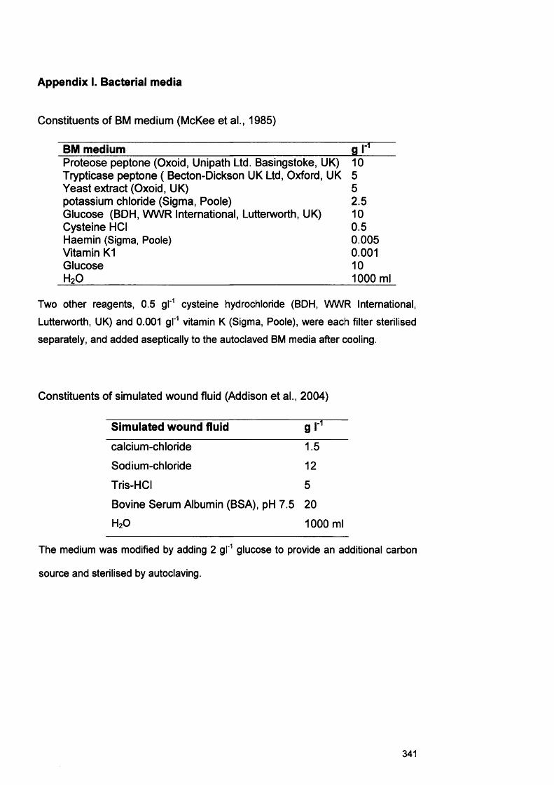
Appendix I. Bacterial media
Constituents of BM medium (McKee et al., 1985)
BM medium g rProteose peptone (Oxoid, Unipath Ltd. Basingstoke, UK) 10Trypticase peptone ( Becton-Dickson UK Ltd, Oxford, UK 5Yeast extract (Oxoid, UK) 5potassium chloride (Sigma, Poole) 2.5Glucose (BDH, WWR International, Lutterworth, UK) 10Cysteine HCI 0.5Haemin (Sigma, Poole) 0.005Vitamin K1 0.001Glucose 10H20 1000 ml
Two other reagents, 0.5 gl'1 cysteine hydrochloride (BDH, WWR International, Lutterworth, UK) and 0.001 gl'1 vitamin K (Sigma, Poole), were each filter sterilised separately, and added aseptically to the autoclaved BM media after cooling.
Constituents of simulated wound fluid (Addison et al., 2004)
Simulated wound fluid g r 1calcium-chloride 1.5
Sodium-chloride 12
Tris-HCI 5
Bovine Serum Albumin (BSA), pH 7.5 20H20 1000 ml
The medium was modified by adding 2 gl'1 glucose to provide an additional carbon
source and sterilised by autoclaving.
341
Appendix II
Sequences of Streptococcus oralis (B52),
Staphylococcus aureus (D76), Pseudomonas aeruginosa (D40),
Micrococcus luteus (B81)
342
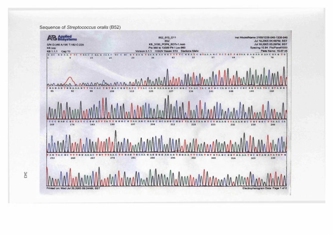
343
Sequence of Streptococcus oralis (B52)
S/N G 346 A 199 T182 C229 KB.bcpKB 1.1.1 Cap:13
B52_013_G11B52
KB_3100_POP6_BDTv1 .mob Pts 960 to 13500 Pk1 Loc:960
Version 5.1.1 HiSQV Bases: 813 Sladiana Malic
Inst Model/Name 3100/1338-040-1338-040 Jill 19.2005 04:46PM. BST Jut 19.2005 05:29PM. BST
Spadng:15.94 Pts/Panel1000 Plate Name: 15.07.05
G A C G G A A T T C T G A C C G A G C A A C 0 C C OC G T G A G T O A A G A A G G T T T T C G G A T C G T A A A G C T C T O T T G T A A G A G A A G A A C G A G T G 1 8 15 22 29 3 6 43 50 5 7 64 71 78
a . a / W L A /w A ^ a N\A/V\ Aa/WI
Printed on: Wed Jul 20.2005 09:24AM, BST Electropherogram Data Page 1 of 2
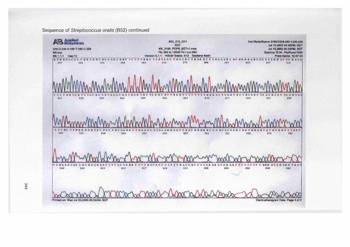
344
Sequence of Streptococcus oralis (B52) continued
AfcttSm,S/N G:346 A:199 T:182 C:229 KB.bcpKB 1.1.1 C«p:13
B52_013_G11B52
KB_3100_POP6_BDTv1. mob PtS 960 to 13500 Pk1 Loc:960
Version 5.1.1 HiSQV Bases: 813 Sladiana Malic
Inst Model/Name 3100/1338-040-1338-040 Jul 19,2005 04:46PM, BST Jul 19.2005 05:29PM, BST
Spadng:15.94 Pts/Panel1000 Plate Name: 15.07.05
3 79365 372 393 400 407337 344 351 358 414
449 456 463 470 484421 428 4 35 442 477 491 498
505 512 526 533 540 554 561 568 575 582
617 6 31 666589 5 9 6 603 610 624
Printed on: Wed Jul 20,2005 09:24AM, BST Electropherogram Data Page 2 of 2
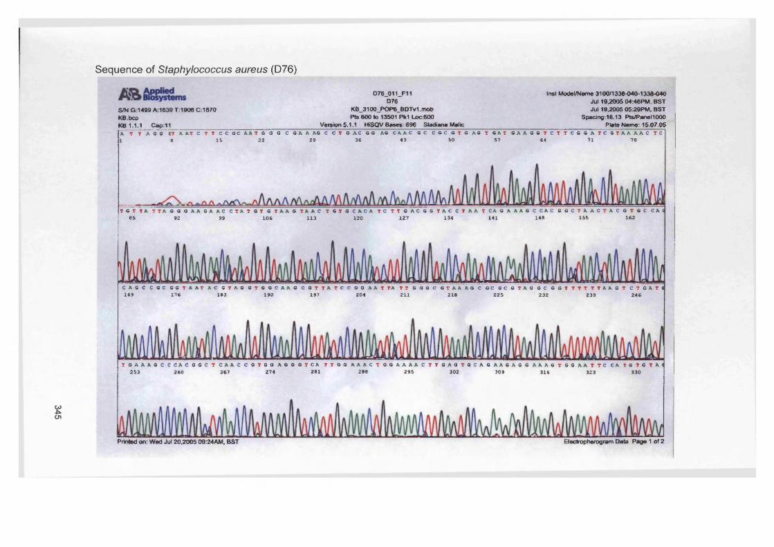
Sequence of Staphylococcus aureus (D76)
D76_011_F11 Inst Model/Name 3100/1338-040-1338-040D76 Jul 19,2005 04:46PM, BST
KB_3100_POP6_BDTv1 .mob Jul 19,2005 05:29PM, BSTPts 600 to 13501 Pk1 Loc:600 Spacing: 16 13 Pts/Panel1000
Version 5.1.1 HiSQV Bases: 696 Sladiana Malic Plate Name: 15.07.05A T T A S 6 ET A A T C T T C C O C A A T G G G C G A A A G C C T S A C S S A S C A A C S C C S C G T S A S T S A T S A A S S T C T T C G S A T C G T A A A A C T C 1 e I S 22 29 36 43 SO 57 64 71 78
T O T T A T T A S S O A A S A A C C T A T O T 8 T A A O T A A C T S T S C A C A T C T T S A C S S T A C C T A A T C A S A A A S C C A C S S C T A A C T A C S T S C C X I 8S 92 99 106 113 120 127 134 141 148 155 162
C A G C C G C G G T A A T A C G T A G G T S G C A A G C O T T A T C C G G A A T T A T T G G G C G T A A A G C G C S C G T A G G C G 6 T T T T T T A A 6 T C T G A T 169 176 183 1 9 0 197 204 211 218 2 2 5 232 239 246
T G A A A G C C C A C G G C T C A A C C G T G G A G G G T C A T T G G A A A C T G G A A A A C T T 6 A G T 6 C A G A A 6 A G G A A A G T 6 G A A T T C C A T O T G T A <2 5 3 2 6 0 267 2 7 4 281 288 2 9 5 302 309 316 323 330
S/N G:1499 A1639 T:1908 C:1870 KB.bcpKB 1.1.1 Cap:11
Printed on: Wed Jul 20,2005 09:24AM, BST Electropherogram Data Page 1 of 2
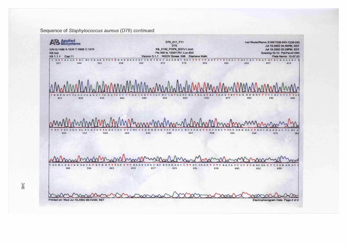
346
Sequence of Staphylococcus aureus (D76) continued
Inst Model/Name 3100/1338-040-1338-040 Jul 19,2005 04:46PM, BST Jul 19,2005 05:29PM, BST
Spadng:16.13 PWPanellOOO _________________ Plato Mama: 15.07.05
C G G T S A A A T 6 C G C A G A G A T A T G G A G G A A C A C C A G T G G C G A A G G C G A C T T T C T G G T C T G T A A C T O A C G C T G A T G T S C 6 A A A G C C 3 37 344 351 358 365 372 3 7 9 3 8 6 393 4 0 0 407 414
T G G 8 G A T C A A A C A G G A T T A G A T A C C C T O G T A O T C C A C G C C G T A A A C G A T G A G T O C T A A G T G T T A G G G G G T T T C C G C C C C T T A C4 2 1 4 2 8 4 3 5 4 4 2 4 4 9 456 4 6 3 4 7 0 4 7 7 484 491 498
T G C T G C A G C T A A C G C A T T A A G C A C T C C G C C T G G G G A G T A C G A C C G C A A G G T T G A A A C T C A A A G G A A T T G A C G G G G A C C C G C A5 0 5 5 1 2 5 1 9 5 2 6 5 3 3 5 4 0 547 SS4 S61 568 575 581
C A A G C G G T G G A G C A T G T G G T T T A A T T C G A A G C A A C G C G A A G A A C C T T A C C A A A T C T T G A C A T C C T T T G A C A A C T C T A G A G A T J 5 8 9 5 9 6 6 0 3 6 1 0 617 624 631 6 3 8 6 4 5 652 659
MR t t f S S SystemsS/N G1499 A: 1639 T:1906 C:1870 KB.bcpKB 1.1.1 Cap:11
D76_011_F11D76
KB_3100_POP6_BDTv1 .mob Pts 600 to 13501 Pk1 Loc:600
Version 5.1.1 HiSQV Bases: 696 Sladiana Malic
Printed on: Wed Jul 20,2005 09:24AM, BST Electropherogram Data Page 2 of 2
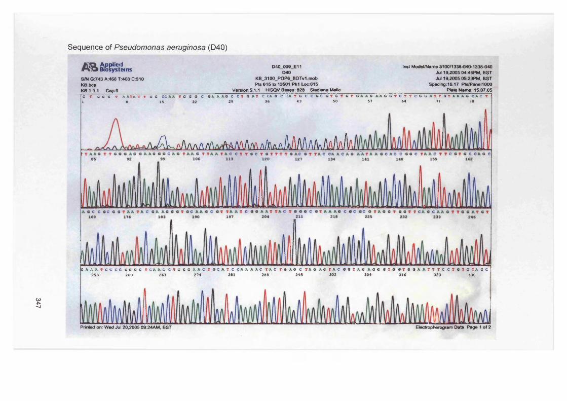
Sequence of Pseudomonas aeruginosa (D40)
PiR t t g S L -S/N G:743 A:468 T:403 C 510 KB.bcpKB 1.1.1 Cap:9
D40_009_E11 D40
KB_3100_POP6_BDTv1 .mob Pts 615 to 13501 Pk1 Loc:615
Version 5.1.1 HiSQV Bases: 826 Sladiana Malic
Inst Model/Name 3100/1338-040-1338-040 Jut 19,2005 04:46PM, BST Jul 19,2005 05:29PM, BST
Spacing:16.17 Pis/Panel 1000 Plate Name: 15.07.05
G T G G G T A A T A T T G G C C A A T G G G C G A A A O C C T O A T C CA G C C A T G C C G C O T G T O T G A A G A A G G T C T T C G G A T T G T A A A G C A C T 1 6 15 22 2 9 36 4 3 50 57 64 71 7B
a/^ aAamM̂&aMA/vi a/)(\aaMAA A/V\ AaAaAa
G A A A T C C C C G G G C T C A A C C T G G G A A C T G C A T C C A A A A C T A C T G A G C T A G A G T A C G G T A G A G G G T G G T G G A A T T T C C T G T G T A G C 2 5 3 2 6 0 2 6 7 274 2 * 1 2 8 8 2 9 5 302 3 0 9 3 1 6 3 2 3 330
Printed on: Wed Juf 20,2005 09:24AM, BST Electropherogram Data Page 1 of 2
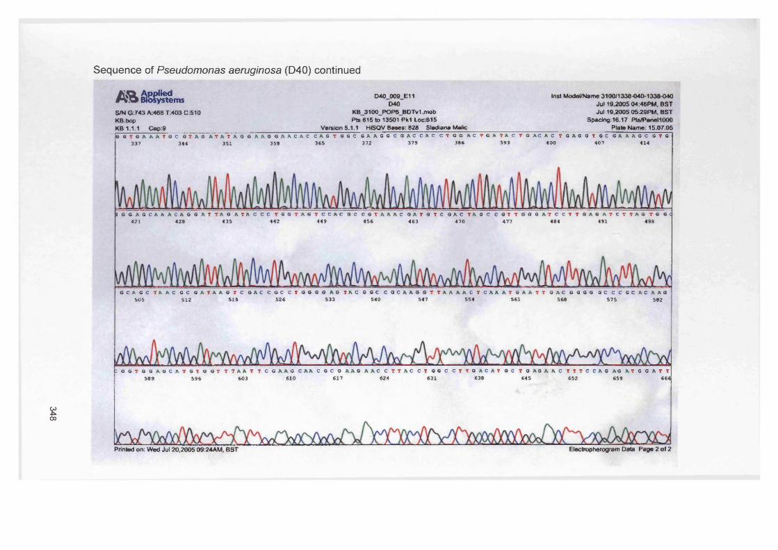
Sequence of Pseudomonas aeruginosa (D40) continued
GJ00
AfcgSSj&lmsS/N G:743 A468 T:403 C:510 KB.bcpKB 1.1.1 Cap:9
D40_009_E11 D40
KB_3100_POP6_BDTv 1 mob Pts 615 to 13501 Pk1 Loc 615
Version 5.1.1 HiSQV Bases: 828 Sladiana Malic
Inst Model/Name 3100/1338-040-1338-040 Jul 19.2005 04:46PM, BST Jul 19.2005 05:29PM, BST
Spadng:16.17 Pts/Panel1000 Plate Name: 15.07.05
G G T G A A A T G C G T A 6 A T A T A G G A A G G A A C A C C A G T G G C G A A G G C G A C C A C C T G G A C T G A T A C T G A C A C T G A G G T 6 C G A A A G C G T G 3 37 344 351 358 3 6 5 3 72 3 79 3 8 6 3 93 4 0 0 407 414
5 G G A G C A A A C A G G A T T A G A T A C C C T G G T A G T C C A C G C C G T A A A C G A T G T C G A C T A G C C G T T G G G A T C C T T 6 A G A T C T T A G T G G C 421 428 4 3 5 4 4 2 4 4 9 4 5 6 4 6 3 4 7 0 477 4 84 491 498
G C A G C T A A C G C G A T A A G T C G A C C G C C T G G G G A G T A C G G C C G C A A G G T T A A A A C T C A A A T G A A T T G A C G G G G G C C C G C A C A A C 5 0 5 5 1 2 5 19 5 2 6 5 33 540 547 554 561 568 5 75 582
I G G T G G A G C A T G T G G T T T A A T T C G A A G C A A C G C G A A G A A C C T T A C C T G G C C T T G A C A T G C T G A G A A C T T T C C A G A G A T G G A T T 589 5 9 6 6 0 3 6 10 6 1 7 624 6 31 638 6 4 5 652 659 66€
Printed on: Wed Jul 20,2005 09:24AM, BST Electropherogram Data Page 2 of 2
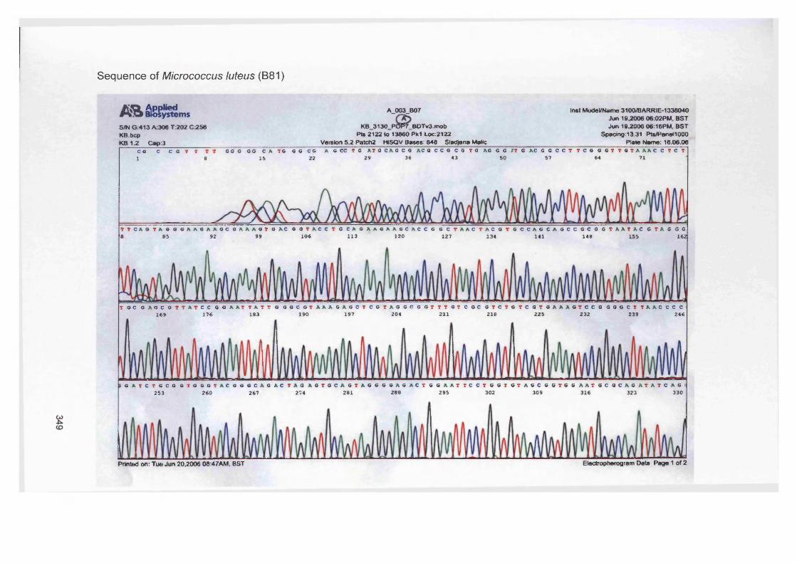
Sequence of Micrococcus luteus (B81)
A_003_B07 Inst Model/Name 3100/BARR IE-1338040Jun 19,2006 06:02PM, BST
KB_3130_POP7_BDTv3.mob Jun 19,2006 06:16PM, BSTPts 2122 to 13660 Pk1 Loc:2122 Spacing:13.31 Pts/Penel 1000
Version 5.2 Patch2 HiSQV Bases: 848 Sladjana Malic Plato Name: 16.06.06~C~G C C G T T G G G GG C A T G G G C G A G C C T C A I G C A G C G A C G C C G C O T O A G G G f T G A C G G C C T T C G G G T T O T A A A C C T C T1 8 15 22 2 9 36 43 50 57 64 71
T T C A G T A G G G A A G A A G C G A A A G T G A C G G T A C C T G C A G A A G A A G C A C C G G C T A A C T A C G T G C C A G C A G C C G C G G T A A T A C G T A G G G8 85 92 99 106 1 1 3 1 20 1 27 134 141 1 48 155 162
T G C G A G C G T T A T C C G G A A T T A T T G G G C G T A A A G A G C T C G T A G G C G G T T T G T C G C G T C T G T C G T G A A A G T C C G G G G C T T A A C C C C169 1 7 6 183 190 197 2 04 2 1 1 2 1 8 2 2 5 2 3 2 2 39 2 4 6
g a t c t g c g g t g g g t a c g g g c a g a c t a g a s t g c a g t a g g g g a g a c t g g a a t t c c t g g t g t a g c g g t g g a a t g c g c a g a t a t c a g
2 5 3 2 6 0 2 6 7 2 7 4 2 8 1 2 88 2 9 5 302 309 3 1 6 323 330
A&M&nsS/N G:413 A306 T:202 C 256 KB.bcpKB 1.2 Cap:3
Printed on: Tue Jun 20,2006 08:47AM, BST Electropherogram Data Page 1 of 2
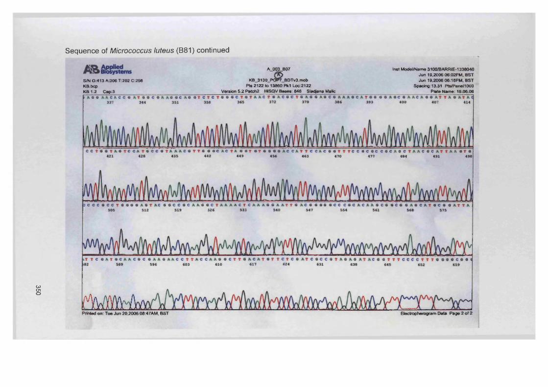
COOl
Sequence of Micrococcus luteus (B81) continued
S/N G:413 A 306 T:202 C:256 KB.bcpKB 1.2 Cap:3
A_003 B07
KB_3130_PdP7_BDTv3.mob Pts 2122 to 13860 Pk1 Loc:2122
Version 5.2 Patch2 HjSQV Bases: 848 Sladiana Malic
Inst Model/Name 3100/BARRIE-1338040 Jun 19,2006 06:02PM, BST Jun 19,2006 06:16PM, BST
Spacing:13.31 Pts/Panel1000 Plate Name: 16.06.06
<lumwwPrinted on: Tue Jun 20,2006 08:47AM, BST Electropherogram Data Page 2 of 2
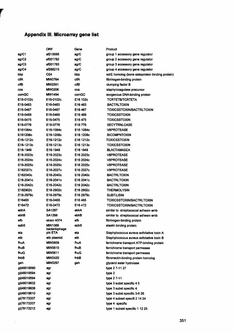
Appendix III. Microarray gene list
ORF GeneagrC1 af210055 agrCagrC2 af001782 agrCagrC3 af001783 agrCagrC4 af288215 agrCbbp 024 bbpclfA MW0764 clfAclfB MW2551 clfBcoa MW0206 coacomGC MW1494 comGCE16-0102c E16-0102c E16 102cE16-0463 E16-0463 E16 463E16-0467 E16-0467 E16 467E16-0469 E16-0469 E16 469E16-0475 E16-0475 E16 475E16-0776 E16-0776 E16 776E161084c E16-1084c E16 1084cE161208c E16-1208c E16 1208cE16-1212c E16-1212c E16 1212cE16-1213c E16-1213C E16 1213cE16-1949 E16-1949 E16 1949E16-2023C E16-2023C E16 2023cE16-2024C E16-2024C E16 2024cE16-2025C E16-2025C E16 2025cE162027c E16-2027C E16 2027cE162040c E16-2040C E16 2040cE16-2041 c E16-2041 c E16 2041cE16-2042C E16-2042C E16 2042cE162902c E16-2902C E16 2902cE16-2979C E16-2979C E16 2979cE16465 E16-0465 E16465E16472 E16-0472 E16 472ebhA SA1267 ebhAebhB SA1268 ebhBefb strain 4074 efbepbS MW1369
bacteriophageepbS
eta phi ETA etaetb etb plasmid etbfhuA MW0609 fhuAfhuB MW0610 fhuBfhuG MW0611 fhuGfnbB MW2420 fnbBgeh MW0297 gehgi|46019590 agrgi|46019594 agrgi|46019594 agrgi|46019602 agrgi|46019606 agrgi|46019610 agrgi|78172207 agrgi|78172207 agrgi|78172212 agr
Productgroup 1 accessory gene regulatorgroup 2 accessory gene regulatorgroup 3 accessory gene regulatorgroup 4 accessory gene regulatorsdrE homolog (bone sialoprotein binding protein)fibrinogen-binding proteinclumping factor Bstaphylcoagulase precursorexogenous DNA-binding proteinTCRTETB/TCRTETABACTRLTOXINTOXICSSTOXIN/BACTRLTOXINTOXICSSTOXINTOXICSSTOXINSECYTRNLCASEV8PROTEASEBICOMPNTOXIN
TOXICSSTOXINTOXICSSTOXINBLACTAMASEAV8PROTEASEV8PROTEASEV8PROTEASEV8PROTEASEBACTRLTOXINBACTRLTOXINBACTRLTOXINTHERMOLYSINSUBTILISINTOXICSSTOXIN/BACTRLTOXIN TOXICSSTOXIN/BACTRLTOXIN similar to streptococcal adhesin emb similar to streptococcal adhesin emb fibrinogen-binding protein elastin binding protein
Staphylococcus aureus exfoliative toxin A Staphylococcus aureus exfoliative toxin B ferrichrome transport ATP-binding protein ferrichrome transport permease ferrichrome transport permease fibronectin-binding protein homolog glycerol ester hydrolase type 2 7-11 27 type 2 type 2 7-11type 3 subst specific 4 5 type 3 subst specific 4 type 3 subst specific 3-6 26 type 4 subset specifi 214 24 type 4 specific type 1 subset specific 1 12 25
351
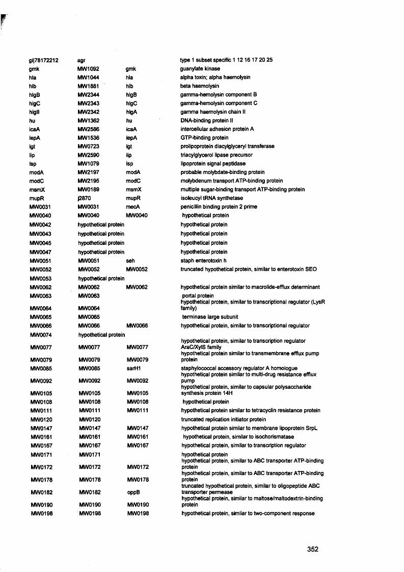
gi|78172212 agrgmk MW1092 gmkhla MW1044 hlahlb MW1881 hlbhlgB MW2344 hlgBhlgC MW2343 hlgChlgll MW2342 hlgAhu MW1362 huicaA MW2586 icaAlepA MW 1536 lepAigt MW0723 igtlip MW2590 lipIsp MW1079 IspmodA MW2197 modAmodC MW2195 modCmsmX MW0189 msmXmupR j2870 mupRMW0031 MW0031 mecAMW0040 MW0040 MW0040MW0042 hypothetical proteinMW0043 hypothetical proteinMW0045 hypothetical proteinMW0047 hypothetical proteinMW0051 MW0051 sehMW0052 MW0052 MW0052MW0053 hypothetical proteinMW0062 MW0062 MW0062MW0063 MW0063
MW0064 MW0064MW0065 MW0065MW0066 MW0066 MW0066MW0074 hypothetical protein
MW0077 MW0077 MW0077
MW0079 MW0079 MW0079MW0085 MW0085 sarH1
MW0092 MW0092 MW0092
MW0105 MW0105 MW0105MW0108 MW0108 MW0108MW0111 MW0111 MW0111MW0120 MW0120MW0147 MW0147 MW0147MW0161 MW0161 MW0161MW0167 MW0167 MW0167MW0171 MW0171
MW0172 MW0172 MW0172
MW0178 MW0178 MW0178
MW0182 MW0182 oppB
MW0190 MW0190 MW0190MW0198 MW0198 MW0198
type 1 subset specific 1 12 16 17 20 25guanylate kinasealpha toxin; alpha haemolysinbeta haemolysingamma-hemolysin component Bgamma-hemolysin component Cgamma haemolysin chain IIDNA-binding protein IIintercellular adhesion protein AGTP-binding proteinprolipoprotein diacylglyceryl transferasetriacylgiycerol lipase precursorlipoprotein signal peptidaseprobable molybdate-binding proteinmolybdenum transport ATP-binding proteinmultiple sugar-binding transport ATP-binding proteinisoleucyl tRNA synthetasepenicillin binding protein 2 primehypothetical proteinhypothetical proteinhypothetical proteinhypothetical proteinhypothetical proteinstaph enterotoxin htruncated hypothetical protein, similar to enterotoxin SEO
hypothetical protein similar to macrolide-efflux determinant portal proteinhypothetical protein, similar to transcriptional regulator (LysR family)terminase large subunithypothetical protein, similar to transcriptional regulator
hypothetical protein, similar to transcription regulator AraC/XylS familyhypothetical protein similar to transmembrane efflux pump proteinstaphylococcal accessory regulator A homologue hypothetical protein similar to multi-drug resistance efflux pumphypothetical protein, similar to capsular polysaccharide synthesis protein 14Hhypothetical proteinhypothetical protein similar to tetracyclin resistance protein truncated replication initiator protein hypothetical protein similar to membrane lipoprotein SrpL hypothetical protein, similar to isochorismatase hypothetical protein, similar to transcription regulator hypothetical proteinhypothetical protein, similar to ABC transporter ATP-binding proteinhypothetical protein, similar to ABC transporter ATP-binding proteintruncated hypothetical protein, similar to oligopeptide ABC transporter permeasehypothetical protein, similar to maltose/maltodextrin-binding proteinhypothetical protein, similar to two-component response
352
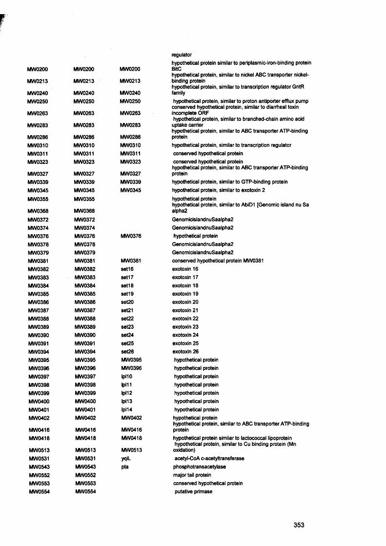
MW0200 MW0200 MW0200
MW0213 MW0213 MW0213
MW0240 MW0240 MW0240MW0250 MW0250 MW0250
MW0263 MW0263 MW0263
MW0283 MW0283 MW0283
MW0286 MW0286 MW0286MW0310 MW0310 MW0310MW0311 MW0311 MW0311MW0323 MW0323 MW0323
MW0327 MW0327 MW0327MW0339 MW0339 MW0339MW0345 MW0345 MW0345MW0355 MW0355
MW0368 MW0368MW0372 MW0372MW0374 MW0374MW0376 MW0376 MW0376MW0378 MW0378MW0379 MW0379MW0381 MW0381 MW0381MW0382 MW0382 set16MW0383 MW0383 set17MW0384 MW0384 set18MW0385 MW0385 set19MW0386 MW0386 set20MW0387 MW0387 set21MW0388 MW0388 set22MW0389 MW0389 set23MW0390 MW0390 set24MW0391 MW0391 set25MW0394 MW0394 set26MW0395 MW0395 MW0395MW0396 MW0396 MW0396MW0397 MW0397 IpMOMW0398 MW0398 Ipl11MW0399 MW0399 Ipl12MW0400 MW0400 Ipl13MW0401 MW0401 Ipl14MW0402 MW0402 MW0402
MW0416 MW0416 MW0416MW0418 MW0418 MW0418
MW0513 MW0513 MW0513MW0531 MW0531 yqiiMW0543 MW0543 ptaMW0552 MW0552MW0553 MW0553MW0554 MW0554
regulatorhypothetical protein similar to periplasmic-iron-binding protein BitChypothetical protein, similar to nickel ABC transporter nickel- binding proteinhypothetical protein, similar to transcription regulator GntR familyhypothetical protein, similar to proton antiporter efflux pump conserved hypothetical protein, similar to diarrheal toxin incomplete ORFhypothetical protein, similar to branched-chain amino acid uptake carrierhypothetical protein, similar to ABC transporter ATP-binding proteinhypothetical protein, similar to transcription regulator conserved hypothetical protein conserved hypothetical proteinhypothetical protein, similar to ABC transporter ATP-binding proteinhypothetical protein, similar to GTP-binding protein hypothetical protein, similar to exotoxin 2 hypothetical proteinhypothetical protein, similar to AbiD1 [Genomic island nu Sa alpha2GenomicislandnuSaalpha2 GenomicislandnuSaalpha2 hypothetical protein GenomicislandnuSaalpha2 Genomicis!andnuSaalpha2 conserved hypothetical protein MW0381 exotoxin 16 exotoxin 17 exotoxin 18 exotoxin 19 exotoxin 20 exotoxin 21 exotoxin 22 exotoxin 23 exotoxin 24 exotoxin 25 exotoxin 26 hypothetical protein hypothetical protein hypothetical protein hypothetical protein hypothetical protein hypothetical protein hypothetical protein hypothetical proteinhypothetical protein, similar to ABC transporter ATP-binding proteinhypothetical protein similar to lactococcal lipoprotein hypothetical protein, similar to Cu binding protein (Mn oxidation)acetyl-CoA c-acetyltransferase phosphotransacetylase major tail protein conserved hypothetical protein putative primase
353

MW0558 MW0558 conserved hypothetical proteinMW0559 MW0559 conserved hypothetical proteinMW0560 MW0560 hypothetical proteinMW0573 MW0573 MW0573 hypothetical protein similar to iron-binding protein
hypothetical protein similar to iron(lll) ABC transporterMW0574 MW0574 MW0574 permease proteinMW0579 MW0579 MW0579 hypothetical protein similar to esterase/lipaseMW0593 MW0593 MW0593 lipoprotein, Streptococcal adhesin PsaA homologueMW0594 MW0594 MW0594 conserved hypothetical protein
hypothetical protein, similar to ABC transporter ATP-bindingMW0595 MW0595 MW0595 proteinMW0596 MW0596 MW0596 hypothetical protein similar to iron dependent repressorMW0617 MW0617 MW0617 hypothetical protein similar to lipase LipAMW0623 MW0623 vraF ABC transporter ATP-binding proteinMW0651 MW0651 MW0651 hypothetical proteinMW0681 MW0681 MW0681 hypothetical protein, similar to anion-binding protein
hypothetical protein, similar to ABC transporter ATP-bindingMW0682 MW0682 MW0682 protein
hypothetical protein, similar to choline transport ATP-bindingMW0684 MW0684 MW0684 protein
hypothetical protein similar to ferrichrome ABC transporterMW0695 MW0695 MW0695 permease
hypothetical protein similar to ferrichrome ABC transporterMW0696 MW0696 MW0696 permease
hypothetical protein similar to ferrichrome ABC transporterMW0697 MW0697 MW0697 ATP-binding proteinMW0698 MW0698 MW0698 lipoprotein, similar to ferrichrome ABC transporterMW0736 MW0736 tpi triosephosphate isomeraseMW0746 MW0746 hypothetical protein (Genomic island)MW0751 MW0751 hypothetical protein (Genomic island)MW0754 MW0754 hypothetical protein (Genomic island)MW0755 MW0755 hypothetical protein (Genomic island)MW0756 MW0756 hypothetical protein (Genomic island)MW0757 MW0757 hypothetical protein (Genomic island)MW0758 MW0758 ear Ear (Genomic island)MW0760 MWQ760 sel2 extracellular enterotoxin IMW0765 MW0765 MW0765 truncated secreted von Willebrand factor-binding proteinMW0766 MW0766 MW0766 truncated secreted von Willebrand factor-binding proteinMW0767 MW0767 ssp extracellular ECM and plasma binding proteinMW0790 MW0790 MW0790 ABC transporter ATP-binding protein homologueMW0795 MW0795 MW0795 ABC transporter ATP-binding protein homologue
oligopeptide transport system ATP-binding protein OppDMW0870 MW0870 oppD homologueMW0871 MW0871 oppF oligopeptide transport ATP-binding proteinMW0872 MW0872 MW0872 hypothetical protein, similar to peptide binding protein OppA
hypothetical protein, similar to oligopeptide ABC transporterMW0873 MW0873 MW0873 oligopeptide-binding proteinMW0874 MW0874 MW0874 oligopeptide ABC transporter ATP-binding protein homologue
oligopeptide transport system ATP-binding protein AppFMW0875 MW0875 appF homologue
hypothetical protein, similar to multidrug resistance protein-MW0897 MW0897 MW0897 related protein
hypothetical protein, similar to UDP-glucose:polyglycerolMW0919 MW0919 phosphate glucosyltransferaseMW0921 MW0921 MW0921 hypothetical protein similar to ferrichrome ABC transporterMW0962 MW0962 MW0962 hypothetical protein
spermidine/putrescine ABC transporter, ATP-binding proteinMW0982 MW0982 potA homologMW0985 MW0985 MW0985 spermidine/putrescine-binding protein precursor homoiogMW0986 MW0986 MW0986 conserved hypothetical proteinMW1015 MW1015 MW1015 hypothetical protein similar to ferrichrome ABC transporter
354
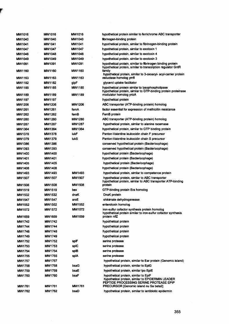
MW1016 MW1016 MW1016 hypothetical protein similar to ferrichrome ABC transporterMW1040 MW1040 MW1040 fibrinogen-binding proteinMW1041 MW1041 MW1041 hypothetical protein, similar to fibrinogen-binding proteinMW 1047 MW1047 MW1047 hypothetical protein, similar to exotoxin 1MW1048 MW1048 MW1048 hypothetical protein, similar to exotoxin 4MW1049 MW1049 MW1049 hypothetical protein, similar to exotoxin 3MW1091 MW1091 MW1091 hypothetical protein, similar to fibrinogen binding protein
hypothetical protein, similar to transcription regulator GntRMW1160 MW1160 MW1160 family
hypothetical protein, similar to 3-oxoacyl- acyl-carrier proteinMW1163 MW1163 MW 1163 reductase homolog ymflMW1182 MW1182 gipF glycerol uptake facilitatorMW1185 MW1185 MW1185 hypothetical protein similar to lysophospholipase
hypothetical protein, similar to GTP-binding protein proteinaseMW1189 MW1189 MW1189 modulator homolog ynbAMW1197 MW1197 hypothetical proteinMW1206 MW1206 MW1206 ABC transporter (ATP-binding protein) homologMW1261 MW1261 femA factor essential for expression of methicillin resistanceMW1262 MW1262 femB FemB proteinMW1280 MW1280 MW 1280 ABC transporter (ATP-binding protein) homologMW1287 MW1287 MW1287 hypothetical protein, similar to alanine racemaseMW 1364 MW1364 MW1364 hypothetical protein, similar to GTP binding proteinMW1378 MW1378 lukF Panton-Valentine leukocidin chain F precursorMW1379 MW1379 lukS Panton-Valentine leukocidin chain S precursorMW1386 MW1386 conserved hypothetical protein (Bacterioophage)MW1393 MW1393 conserved hypothetical protein (Bacterioophage)MW1400 MW1400 hypothetical protein (Bacterioophage)MW1401 MW1401 hypothetical protein (Bacterioophage)MW1405 MW1405 hypothetical protein (Bacterioophage)MW 1408 MW1408 hypothetical protein (Bacterioophage)MW1493 MW1493 MW1493 hypothetical protein, similar to competence proteinMW1507 MW1507 MW1507 hypothetical protein, similar to ABC transporter
hypothetical protein, similar to ABC transporter ATP-bindingMW 1508 MW1508 MW1508 proteinMW1519 MW1519 bex GTP-binding protain Era homologMW1532 MW1532 dnaK DnaK proteinMW1547 MW1547 aroE shikimate dehydrogeneaseMW1552 MW1552 MW1552 enterotoxin homologMW1572 MW1572 MW1572 iron-sulfur cofactor synthesis protein homolog
hypothetical protein similar to iron-sulfur cofactor synthesisMW1659 MW1659 MW1659 protein nifZMW1742 MW1742 hypothetical proteinMW1744 MW1744 hypothetical proteinMW1748 MW1748 hypothetical proteinMW1749 MW1749 hypothetical proteinMW1752 MW1752 splF serine proteaseMW1753 MW1753 spIC serine proteaseMW1754 MW1754 splB serine proteaseMW1755 MW1755 splA serine proteaseMW1757 MW1757 hypothetical protein, similar to Ear protein (Genomic island)MW1758 MW1758 bsaG hypothetical protein, similar to EpiGMW1759 MW1759 bsaE hypothetical protein, similar tpo EpiEMW1760 MW1760 bsaF hypothetical protein, similar to EpiF
hypothetical protein, similar to EPIDERMIN LEADERPEPTIDE PROCESSING SERINE PROTEASE EPIP
MW1761 MW1761 MW1761 PRECURSOR [Genomic island nu Sa beta2]MW1762 MW1762 bsaD hypothetical protein, similar to lantibiotic epidermin
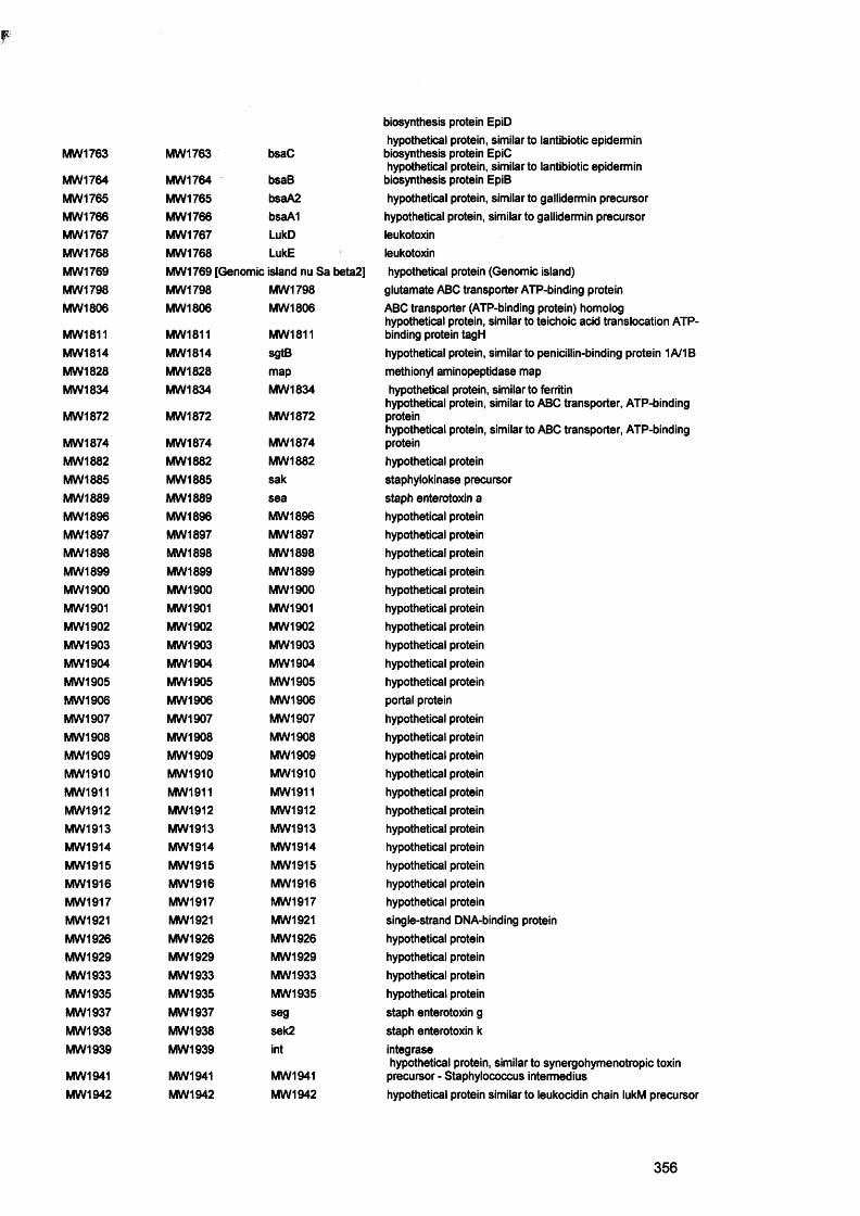
p
biosynthesis protein EpiD hypothetical protein, similar to lantibiotic epidermin
MW1763 MW1763 bsaC biosynthesis protein EpiChypothetical protein, similar to lantibiotic epidermin
MW1764 MW1764 bsaB biosynthesis protein EpiBMW 1765 MW1765 bsaA2 hypothetical protein, similar to gallidermin precursorMW1766 MW1766 bsaA1 hypothetical protein, similar to gallidermin precursorMW1767 MW1767 LukD leukotoxinMW1768 MW1768 LukE leukotoxinMW1769 MW1769 [Genomic island nu Sa beta2] hypothetical protein (Genomic island)MW1798 MW1798 MW1798 glutamate ABC transporter ATP-binding proteinMW1806 MW1806 MW1806 ABC transporter (ATP-binding protein) homolog
hypothetical protein, similar to teichoic acid translocation ATP-MW1811 MW1811 MW1811 binding protein tagHMW1814 MW1814 sgtB hypothetical protein, similar to penicillin-binding protein 1A/1BMW1828 MW1828 map methionyl aminopeptidase mapMW1834 MW1834 MW1834 hypothetical protein, similar to ferritin
hypothetical protein, similar to ABC transporter, ATP-bindingMW1872 MW1872 MW1872 protein
hypothetical protein, similar to ABC transporter, ATP-bindingMW1874 MW1874 MW1874 proteinMW 1882 MW 1882 MW1882 hypothetical proteinMW1885 MW1885 sak staphylokinase precursorMW 1889 MW 1889 sea staph enterotoxin aMW 1896 MW1896 MW1896 hypothetical proteinMW1897 MW 1897 MW1897 hypothetical proteinMW 1898 MW1898 MW1898 hypothetical proteinMW1899 MW1899 MW1899 hypothetical proteinMW1900 MW1900 MW 1900 hypothetical proteinMW1901 MW 1901 MW1901 hypothetical proteinMW1902 MW1902 MW1902 hypothetical proteinMW1903 MW1903 MW1903 hypothetical proteinMW 1904 MW1904 MW1904 hypothetical proteinMW1905 MW 1905 MW1905 hypothetical proteinMW1906 MW1906 MW1906 portal proteinMW1907 MW1907 MW1907 hypothetical proteinMW1908 MW1908 MW1908 hypothetical proteinMW1909 MW1909 MW1909 hypothetical proteinMW1910 MW1910 MW1910 hypothetical proteinMW1911 MW1911 MW1911 hypothetical proteinMW1912 MW1912 MW1912 hypothetical proteinMW1913 MW1913 MW1913 hypothetical proteinMW1914 MW1914 MW1914 hypothetical proteinMW1915 MW1915 MW1915 hypothetical proteinMW1916 MW1916 MW1916 hypothetical proteinMW1917 MW1917 MW1917 hypothetical proteinMW1921 MW1921 MW1921 single-strand DNA-binding proteinMW1926 MW1926 MW1926 hypothetical proteinMW 1929 MW1929 MW1929 hypothetical proteinMW1933 MW1933 MW1933 hypothetical proteinMW1935 MW1935 MW1935 hypothetical proteinMW1937 MW1937 seg staph enterotoxin gMW1938 MW1938 sek2 staph enterotoxin kMW1939 MW1939 int integrase
hypothetical protein, similar to synergohymenotropic toxinMW1941 MW1941 MW1941 precursor - Staphylococcus intermediusMW1942 MW1942 MW1942 hypothetical protein similar to leukocidin chain iukM precursor
356
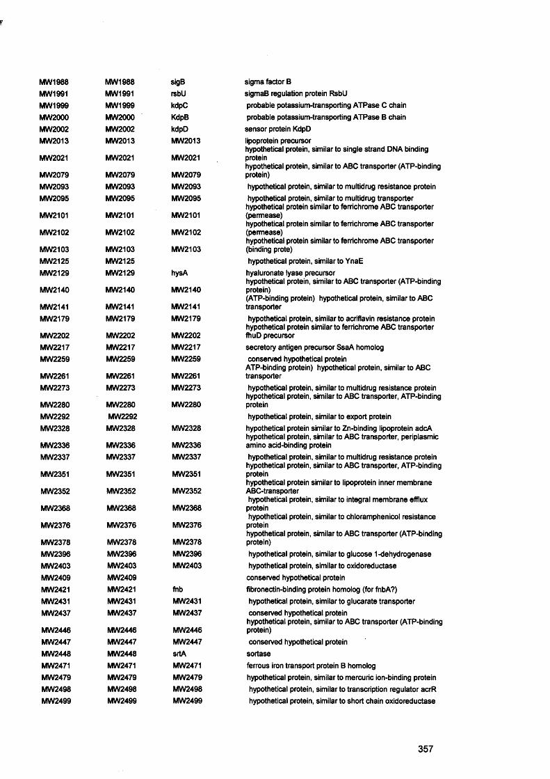
MW1988 MW 1988 sigB sigma factor BMW1991 MW1991 rsbl) sigmaB regulation protein RsbUMW1999 MW1999 kdpC probable potassium-transporting ATPase C chainMW2000 MW2000 KdpB probable potassium-transporting ATPase B chainMW2002 MW2002 kdpD sensor protein KdpDMW2013 MW2013 MW2013 lipoprotein precursor
hypothetical protein, similar to single strand DNA bindingMW2021 MW2021 MW2021 protein
hypothetical protein, similar to ABC transporter (ATP-bindingMW2079 MW2079 MW2079 protein)MW2093 MW2093 MW2093 hypothetical protein, similar to multidrug resistance proteinMW2095 MW2095 MW2095 hypothetical protein, similar to multidrug transporter
hypothetical protein similar to ferrichrome ABC transporterMW2101 MW2101 MW2101 (permease)
hypothetical protein similar to ferrichrome ABC transporterMW2102 MW2102 MW2102 (permease)
hypothetical protein similar to ferrichrome ABC transporterMW2103 MW2103 MW2103 (binding prote)MW2125 MW2125 hypothetical protein, similar to YnaEMW2129 MW2129 hysA hyaluronate lyase precursor
hypothetical protein, similar to ABC transporter (ATP-bindingMW2140 MW2140 MW2140 protein)
(ATP-binding protein) hypothetical protein, similar to ABCMW2141 MW2141 MW2141 transporterMW2179 MW2179 MW2179 hypothetical protein, similar to acriflavin resistance protein
hypothetical protein similar to ferrichrome ABC transporterMW2202 MW2202 MW2202 fhuD precursorMW2217 MW2217 MW2217 secretory antigen precursor SsaA homoiogMW2259 MW2259 MW2259 conserved hypothetical protein
ATP-binding protein) hypothetical protein, similar to ABCMW2261 MW2261 MW2261 transporterMW2273 MW2273 MW2273 hypothetical protein, similar to multidrug resistance protein
hypothetical protein, similar to ABC transporter, ATP-bindingMW2280 MW2280 MW2280 proteinMW2292 MW2292 hypothetical protein, similar to export proteinMW2328 MW2328 MW2328 hypothetical protein similar to Zn-binding lipoprotein adcA
hypothetical protein, similar to ABC transporter, periplasmicMW2336 MW2336 MW2336 amino acid-binding proteinMW2337 MW2337 MW2337 hypothetical protein, similar to multidrug resistance protein
hypothetical protein, similar to ABC transporter, ATP-bindingMW2351 MW2351 MW2351 protein
hypothetical protein similar to lipoprotein inner membraneMW2352 MW2352 MW2352 ABC-transporter
hypothetical protein, similar to integral membrane effluxMW2368 MW2368 MW2368 protein
hypothetical protein, similar to chloramphenicol resistanceMW2376 MW2376 MW2376 protein
hypothetical protein, similar to ABC transporter (ATP-bindingMW2378 MW2378 MW2378 protein)MW2396 MW2396 MW2396 hypothetical protein, similar to glucose 1-dehydrogenaseMW2403 MW2403 MW2403 hypothetical protein, similar to oxidoreductaseMW2409 MW2409 conserved hypothetical proteinMW2421 MW2421 fnb fibronectin-binding protein homolog (for fnbA?)MW2431 MW2431 MW2431 hypothetical protein, similar to glucarate transporterMW2437 MW2437 MW2437 conserved hypothetical protein
hypothetical protein, similar to ABC transporter (ATP-bindingMW2446 MW2446 MW2446 protein)MW2447 MW2447 MW2447 conserved hypothetical proteinMW2448 MW2448 srtA sortaseMW2471 MW2471 MW2471 ferrous iron transport protein B homologMW2479 MW2479 MW2479 hypothetical protein, similar to mercuric ion-binding proteinMW2498 MW2498 MW2498 hypothetical protein, similar to transcription regulator acrRMW2499 MW2499 MW2499 hypothetical protein, similar to short chain oxidoreductase
357
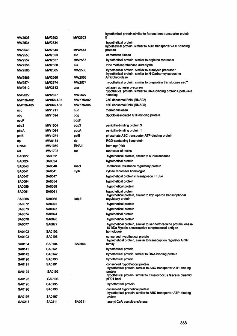
MW2503 MW2503 MW2503MW2534 MW2534
MW2543 MW2543 MW2543MW2553 MW2553 arcMW2557 MW2557 MW2557MW2558 MW2558 aurMW2565 MW2565 MW2565
MW2566 MW2566 MW2566MW2574 MW2574 MW2674MW2612 MW2612 cna
MW2627 MW2627 MW2627MWrRNA02 MWrRNA02 MWrRNA02MWrRNA05 MWrRNA05 MWrRNA05nuc MW1211 nucobg MW1594 obgoppF oppFpbp3 MW 1504 pbp3pbpA MW1064 pbpApstB MW1274 pstBrip MW0184 ripRNAIII MW1959 RNAIIIrot MW1705 rotSA0022 SA0022SA0024 SA0024SA0040 SA0040 meclSA0041 SA0041 xylRSA0047 SA0047SA0054 SA0054SA0059 SA0059SA0061 SA0061
SA0066 SA0066 kdpESA0072 SA0072SA0073 SA0073SA0074 SA0074SA0076 SA0076SA0077 SA0077
SA0102 SA0102SA0103 SA0103
SA0104 SA0104 SA0104SA0141 SA0141SA0142 SA0142SA0190 S A0190SA0191 SA0191
SA0192 SA0192
SA0193 SA0193SA0195 SA0195SA0196 SA0196
SA0197 SA0197SA0211 SA0211 SA0211
hypothetical protein similar to ferrous iron transporter protein Bhypothetical proteinhypothetical protein, similar to ABC transporter (ATP-binding protein)carbamate kinasehypothetical protein, similar to arginine repressorzinc metalloproteinase aureolysinhypothetical protein, similar to autolysin precursor hypothetical protein, similar to N-Carbamoylsarcosine Amidohydrolasehypothetical protein, similar to preprotein translocase secY collagen adhesin precursorhypothetical protein, similar to DNA-binding protein SpoOJ-like homolog23S ribosomal RNA (RNA02)16S ribosomal RNA (RNA05) thermonucleaseSpoOB-associated GTP-binding protein
penicillin-binding protein 3penicillin-binding protein 1phosphate ABC transporter ATP-binding proteinRGD-containing lipoproteinfrom agr (hid)repressor of toxinshypothetical protein, similar to 5'-nucleotidase hypothetical proteinmethicillin resistance regulatory protein xylose repressor homologue hypothetical protein in transposon Tn554 hypothetical protein hypothetical protein hypothetical proteinhypothetical protein, similar to kdp operon transcriptional regulatory proteinhypothetical proteinhypothetical proteinhypothetical proteinhypothetical proteinhypothetical protein, similar to serine/threonine protein kinase 67 kDa Myosin-crossreactive streptococcal antigen homologueconserved hypothetical proteinhypothetical protein, similar to transcription regulator GntR
familyhypothetical proteinhypothetical protein, similar to DNA-binding protein hypothetical protein conserved hypothetical proteinhypothetical protein, similar to ABC transporter ATP-binding proteinhypothetical protein, similar to Enterococcus faecalis plasmid pPD1 badhypothetical proteinconserved hypothetical proteinhypothetical protein, similar to ABC transporter ATP-binding proteinacetyl-CoA acetyltransferase
358
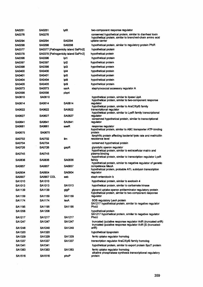
SA0251 SA02S1 lytR two-component response regulatorSA0276 SA0276 conserved hypothetical protein, similar to diarrheal toxin
hypothetical protein, similar to branched-chain amino acidSA0294 SA0294 SA0294 uptake carrierSA0298 SA0298 SA0298 hypothetical protein, similar to regulatory protein PfoRSA0377 SA0377 [Pathogenicity island SaPln2] hypothetical proteinSA0378 SA0378 [Pathogenicity island SaPln2] hypothetical proteinSA0396 SA0396 Ipl1 hypothetical proteinSA0397 SA0397 Ipl2 hypothetical proteinSA0398 SA0398 Ipl3 hypothetical proteinSA0400 SA0400 Ipl4 hypothetical proteinSA0401 SA0401 Ipl5 hypothetical proteinSA0404 SA0404 Ipl8 hypothetical proteinSA0405 SA0405 Ipl9 hypothetical proteinSA0573 SA0573 sarA staphylococcal accessory regulator ASA0598 SA0598 pbp4SA0610 SA0610 hypothetical protein, similar to lipase LipA
hypothetical protein, similar to two-component responseSA0614 SA0614 SA0614 regulator
hypothetical protein, similar to AraC/XylS familySA0622 SA0622 SA0622 transcriptional regulator
hypothetical protein, similar to LysR family transcriptionalSA0627 SA0627 SA0627 regulator
conserved hypothetical protein, similar to transcriptionalSA0641 SA0641 SA0641 regulatorSA0661 SA0661 saeR response regulator
hypothetical protein, similar to ABC transporter ATP-bindingSA0675 SA0675 protein
lipophilic protein affecting bacterial lysis rate and methicillinSA0702 SA0702 llm resistance levelSA0704 SA0704 conserved hypothetical proteinSA0726 SA0726 gapR glycolytic operon regulator
hypothetical protein, similar to extracellular matrix andSA0745 SA0745 plasma binding
hypothetical protein, similar to transcription regulator LysRSA0836 SA0836 SA0836 family
hypothetical protein, similar to negative regulator of geneticSA0857 SA0857 SA0857 competence MecA
hypothetical protein, probable ATL autolysin transcriptionSA0904 SA0904 SA0904 regulatorSA0907 SA0907 COL seb staph enterotoxin bSA1010 SA1010 hypothetical protein, similar to exotoxin 4SA1013 SA1013 SA1013 hypothetical protein, similar to carbamate kinaseSA1139 SA1139 gigp glycerol uptake operon antiterminator regulatory protein
hypothetical protein, similar to two-component responseSA1159 SA1159 SA1159 regulatorSA1174 SA1174 lexA SOS regulatory LexA protein
SA1217 hypothetical protein, similar to negative regulatorSA1195 SA1195 SA1195 PhoUSA1208 SA1208 hypothetical protein
SA1217 hypothetical protein, similar to negative regulatorSA1217 SA1217 SA1217 PhoUSA1247 SA1247 SA1247 truncated (putative response regulator ArIR (truncated-arIR)
truncated (putative response regulator ArIR [S (truncated-SA1248 SA1248 SA1248 arlR)SA1320 SA1320 hypothetical lipoproteinSA1329 SA1329 SA1329 ferric uptake regulator homologSA1337 SA1337 SA1337 transcription regulator AraC/XylS family homologSA1341 SA1341 hypothetical protein, similar to export protein SpcT proteinSA1383 SA1383 SA1383 ferric uptake regulator homolog
alkaline phosphatase synthesis transcriptional regulatorySA1516 SA1516 phoP protein
359
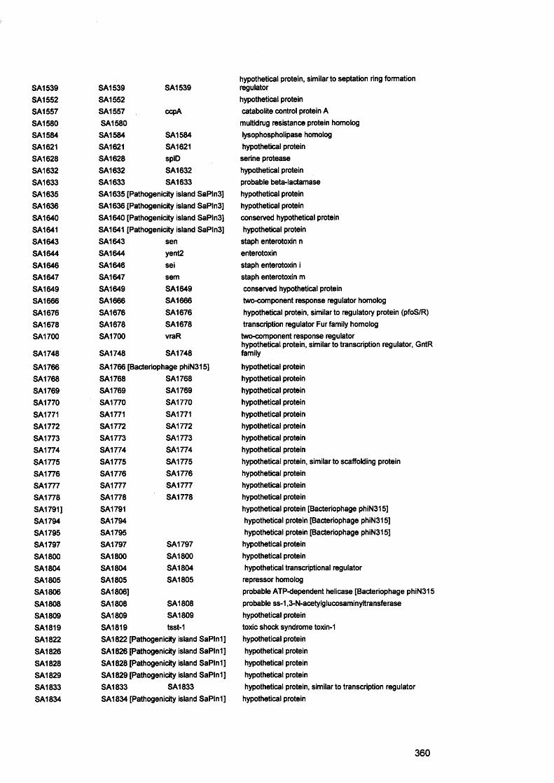
SA1539 SA1539 SA1539SA1552 SA1652SA1557 SA1557 ccpASA1580 SA1580SA1584 SA1584 SA1584SA1621 SA1621 SA1621SA1628 SA1628 spIDSA1632 SA1632 SA1632SA1633 SA1633 SA1633SA1635 SA1635 [Pathogenicity island SaPln3]SA1636 SA1636 [Pathogenicity island SaPln3]SA1640 SA1640 [Pathogenicity island SaPln3]SA1641 SA1641 [Pathogenicity island SaPln3]SA1643 SA1643 senSA1644 SA1644 yent2SA1646 SA1646 seiSA1647 SA1647 semSA1649 SA1649 SA1649SA1666 SA1666 SA1666SA1676 SA1676 SA1676SA1678 SA1678 SA1678SA1700 SA1700 vraR
SA1748 SA1748 SA1748SA1766 SA1766 [Bacteriophage phiN315]SA1768 SA1768 SA1768SA1769 SA1769 SA1769SA1770 SA1770 SA1770SA1771 SA1771 SA1771SA1772 SA1772 SA1772SA1773 SA1773 SA1773SA1774 SA1774 SA1774SA1775 SA1775 SA1775SA1776 SA1776 SA1776SA1777 SA1777 SA1777SA1778 SA1778 SA1778SA1791] SA1791SA1794 SA1794SA1795 SA1795SA1797 SA1797 SA1797SA1800 SA1800 SA1800SA1804 SA1804 SA1804SA1805 SA1805 SA1805SA1806 SA1806]SA1808 SA1808 SA1808SA1809 SA1809 SA1809SA1819 SA1819 tsst-1SA1822 SA1822 [Pathogenicity island SaPlnl]SA1826 SA1826 [Pathogenicity island SaPlnl]SA1828 SA1828 [Pathogenicity island SaPlnl]SA1829 SA1829 [Pathogenicity island SaPlnl]SA1833 SA1833 SA1833SA1834 SA1834 [Pathogenicity island SaPlnl]
hypothetical protein, similar to septation ring formation regulatorhypothetical proteincatabolite control protein Amultidrug resistance protein homologlysophospholipase homologhypothetical proteinserine proteasehypothetical proteinprobable beta-lactamasehypothetical proteinhypothetical proteinconserved hypothetical proteinhypothetical proteinstaph enterotoxin nenterotoxinstaph enterotoxin istaph enterotoxin mconserved hypothetical proteintwo-component response regulator homologhypothetical protein, similar to regulatory protein (pfoS/R)transcription regulator Fur family homologtwo-component response regulatorhypothetical protein, similar to transcription regulator, GntRfamilyhypothetical protein hypothetical protein hypothetical protein hypothetical protein hypothetical protein hypothetical protein hypothetical protein hypothetical proteinhypothetical protein, similar to scaffolding protein hypothetical protein hypothetical protein hypothetical proteinhypothetical protein [Bacteriophage phiN315] hypothetical protein [Bacteriophage phiN315] hypothetical protein [Bacteriophage phiN315] hypothetical protein hypothetical protein hypothetical transcriptional regulator repressor homologprobable ATP-dependent helicase [Bacteriophage phiN315probable ss-1,3-N-acetylglucosaminyltransferasehypothetical proteintoxic shock syndrome toxin-1hypothetical proteinhypothetical proteinhypothetical proteinhypothetical proteinhypothetical protein, similar to transcription regulator hypothetical protein
360
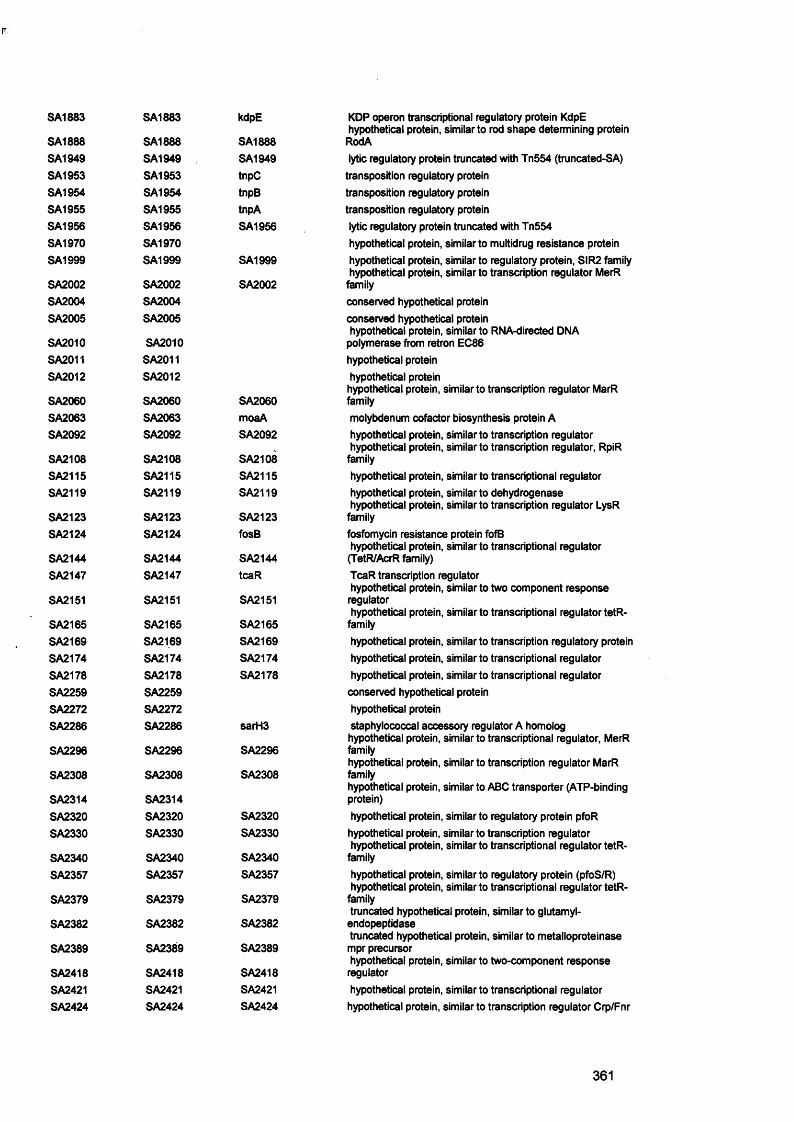
SA1883 SA1883 kdpE KDP operon transcriptional regulatory protein KdpE hypothetical protein, similar to rod shape determining protein
SA1888 SA1888 SA1888 RodASA1949 SA1949 SA1949 lytic regulatory protein truncated with Tn554 (truncated-SA)SA1953 SA1953 tnpC transposition regulatory proteinSA1954 SA1954 tnpB transposition regulatory proteinSA1955 SA1955 tnpA transposition regulatory proteinSA1956 SA1956 SA1956 lytic regulatory protein truncated with Tn554SA1970 SA1970 hypothetical protein, similar to multidrug resistance proteinSA1999 SA1999 SA1999 hypothetical protein, similar to regulatory protein, SIR2 family
hypothetical protein, similar to transcription regulator MerRSA2002 SA2002 SA2002 familySA2004 SA2004 conserved hypothetical proteinSA2005 SA2005 conserved hypothetical protein
hypothetical protein, similar to RNA-directed DIMASA2010 SA2010 polymerase from retron EC86SA2011 SA2011 hypothetical proteinSA2012 SA2012 hypothetical protein
hypothetical protein, similar to transcription regulator MarRSA2060 SA2060 SA2060 familySA2063 SA2063 moaA molybdenum cofactor biosynthesis protein ASA2092 SA2092 SA2092 hypothetical protein, similar to transcription regulator
hypothetical protein, similar to transcription regulator, RpiRSA2108 SA2108 SA2108 familySA2115 SA2115 SA2115 hypothetical protein, similar to transcriptional regulatorSA2119 SA2119 SA2119 hypothetical protein, similar to dehydrogenase
hypothetical protein, similar to transcription regulator LysRSA2123 SA2123 SA2123 familySA2124 SA2124 fosB fosfomycin resistance protein fofB
hypothetical protein, similar to transcriptional regulatorSA2144 SA2144 SA2144 (TetR/AcrR family)SA2147 SA2147 tcaR TcaR transcription regulator
hypothetical protein, similar to two component responseSA2151 SA2151 SA2151 regulator
hypothetical protein, similar to transcriptional regulator tetR-SA2165 SA2165 SA2165 familySA2169 SA2169 SA2169 hypothetical protein, similar to transcription regulatory proteinSA2174 SA2174 SA2174 hypothetical protein, similar to transcriptional regulatorSA2178 SA2178 SA2178 hypothetical protein, similar to transcriptional regulatorSA2259 SA2259 conserved hypothetical proteinSA2272 SA2272 hypothetical proteinSA2286 SA2286 sarH3 staphylococcal accessory regulator A homolog
hypothetical protein, similar to transcriptional regulator, MerRSA2296 SA2296 SA2296 family
hypothetical protein, similar to transcription regulator MarRSA2308 SA2308 SA2308 family
hypothetical protein, similar to ABC transporter (ATP-bindingSA2314 SA2314 protein)SA2320 SA2320 SA2320 hypothetical protein, similar to regulatory protein pfoRSA2330 SA2330 SA2330 hypothetical protein, similar to transcription regulator
hypothetical protein, similar to transcriptional regulator tetR-SA2340 SA2340 SA2340 familySA2357 SA2357 SA2357 hypothetical protein, similar to regulatory protein (pfoS/R)
hypothetical protein, similar to transcriptional regulator tetR-SA2379 SA2379 SA2379 family
truncated hypothetical protein, similar to giutamyl-SA2382 SA2382 SA2382 endopeptidase
truncated hypothetical protein, similar to metalloproteinaseSA2389 SA2389 SA2389 mpr precursor
hypothetical protein, similar to two-component responseSA2418 SA2418 SA2418 regulatorSA2421 SA2421 SA2421 hypothetical protein, similar to transcriptional regulatorSA2424 SA2424 SA2424 hypothetical protein, similar to transcription regulator Crp/Fnr
361
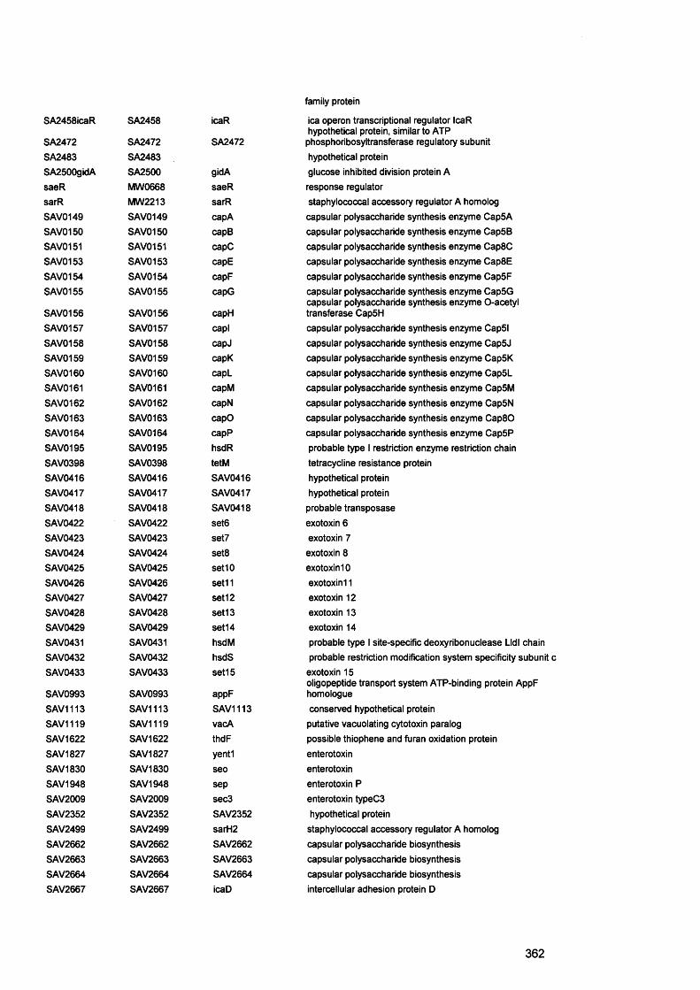
SA2458icaR SA2458 icaR
SA2472 SA2472 SA2472SA2483 SA2483SA2500gidA SA2500 gidAsaeR MW0668 saeRsarR MW2213 sarRSAV0149 SAN/0149 capASAV0150 SAN/0150 capBSAV0151 SAV0151 capCSAV0153 SAVO153 capESAV0154 SAN/0154 capFSAV0155 SAN/0155 capG
SAV0156 SAN/0156 capHSAV0157 SAN/0157 caplSAV0158 SAN/0158 capJSAV0159 SAV0159 capKSAV0160 SAV0160 capLSAV0161 SAV0161 capMSAV0162 SAV0162 capNSAN/0163 SAV0163 capOSAN/0164 SAV0164 capPSAN/0195 SAV0195 hsdRSAN/0398 SAV0398 tetMSAN/0416 SAV0416 SAV0416SAN/0417 SAV0417 SAV0417SAN/0418 SAV0418 SAV0418SAN/0422 SAV0422 set6SAN/0423 SAV0423 set7SAN/0424 SAV0424 set8SAN/0425 SAV0425 set10SAV0426 SAV0426 set11SAN/0427 SAV0427 set12SAV0428 SAV0428 set13SAN/0429 SAV0429 set14SAN/0431 SAV0431 hsdMSAN/0432 SAV0432 hsdSSAN/0433 SAV0433 set 15
SAN/0993 SAV0993 appFSAN/1113 SAV1113 SAV1113SAN/1119 SAV1119 vacASAN/1622 SAV1622 thdFSAN/1827 SAV1827 yentlSAN/1830 SAV1830 seoSAN/1948 SAV1948 sepSAV2009 SAV2009 sec3SAN/2352 SAV2352 SAV2352SAN/2499 SAV2499 sarH2SAN/2662 SAV2662 SAV2662SAN/2663 SAV2663 SAV2663SAN/2664 SAV2664 SAV2664SAV2667 SAV2667 icaD
family protein
ica operon transcriptional regulator IcaR hypothetical protein, similar to ATP phosphoribosyltransferase regulatory subunithypothetical proteinglucose inhibited division protein Aresponse regulatorstaphylococcal accessory regulator A homologcapsular polysaccharide synthesis enzyme Cap5Acapsular polysaccharide synthesis enzyme Cap5Bcapsular polysaccharide synthesis enzyme Cap8Ccapsular polysaccharide synthesis enzyme Cap8Ecapsular polysaccharide synthesis enzyme Cap5Fcapsular polysaccharide synthesis enzyme Cap5G capsular polysaccharide synthesis enzyme O-acetyl transferase Cap5Hcapsular polysaccharide synthesis enzyme Cap5lcapsular polysaccharide synthesis enzyme Cap5Jcapsular polysaccharide synthesis enzyme Cap5Kcapsular polysaccharide synthesis enzyme Cap5Lcapsular polysaccharide synthesis enzyme Cap5Mcapsular polysaccharide synthesis enzyme Cap5Ncapsular polysaccharide synthesis enzyme Cap80capsular polysaccharide synthesis enzyme Cap5Pprobable type I restriction enzyme restriction chaintetracycline resistance proteinhypothetical proteinhypothetical proteinprobable transposaseexotoxin 6exotoxin 7exotoxin 8exotoxin 10exotoxin 11exotoxin 12exotoxin 13exotoxin 14probable type I site-specific deoxyribonuclease Lid I chain probable restriction modification system specificity subunit c exotoxin 15oligopeptide transport system ATP-binding protein AppF homologueconserved hypothetical protein putative vacuolating cytotoxin paralog possible thiophene and furan oxidation protein enterotoxin enterotoxin enterotoxin P enterotoxin typeC3 hypothetical proteinstaphylococcal accessory regulator A homolog capsular polysaccharide biosynthesis capsular polysaccharide biosynthesis capsular polysaccharide biosynthesis intercellular adhesion protein D
362
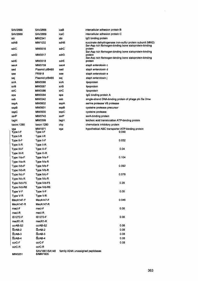
SAV2668 SAV2668 icaBSAV2669 SAV2669 icaCsbi MW2341 sbisdhB MW 1032 sdhB
sdrC MW0516 sdrC
sdrD MW0517 sdrD
sdrE MW0518 sdrEsec4 MW0759 sec4sed Plasmid plB485 sedsee FRI918 seesej Plasmid plB485 sejsirA MW0088 sirAsirB MW0087 sirBsirC MW0086 sirCspa MW0084 spassb MW0342 ssbsspA MW0932 sspAsspB MW0931 sspBsspC MW0930 sspCssrP MW0743 ssrPtagH MW0599 tagHtaxon:1280 taxon:1280 chpvga MW1971 vgaType l-F Type l-FType l-R Type l-RType I l-F Type I l-FType I l-R Type ll-RType lll-F Type lll-FType lll-R Type lll-RType IVa-F Type IVa-FType IVa-R Type IVa-RType IVb-F Type IVb-FType IVb-R Type IVb-RType IVc-F Type IVc-FType IVc-R Type IVc-RType IVd-F5 Type IVd-F5Type IVd-R6 Type IVd-R6Type V-F TypeV-FType V-R Type V-RMecA147-F MecA147-FMecA147-R MecA147-Rmecl-F mecl-Fmecl-R mecl-RIS1272-F IS1272-FmecR1-R mecR1-RccrAB-62 ccrAB-82ccrAB-2 ccrAB-2ccrAB-3 ccrAB-3ccrAB-4 ccrAB-4ccrC-F ccrC-FccrC-R ccrC-R
intercellular adhesion protein B intercellular adhesion protein C IgG binding proteinsuccinate dehydrogenase iron-sulfur protein subunit (MW2) Ser-Asp rich fibrinogen-binding bone sialoprotein-binding proteinSer-Asp rich fibrinogen-binding bone sialoprotein-binding proteinSer-Asp rich fibrinogen-binding bone sialoprotein-binding proteinstaph enterotoxin c staph enterotoxin d staph enterotoxin e staph enterotoxin j lipoprotein lipoprotein lipoproteinIgG binding protein Asingle-strand DNA-binding protein of phage phi Sa 2mwserine protease V8 proteasecysteine protease precursorcysteine proteasessrA-binding proteinteichoic acid translocation ATP-binding proteinchemotaxis inhibitory proteinhypothetical ABC transporter ATP-binding protein
0.048
0.032
0.04
0.104
0.092
0.078
0.28
0.06
0.046
0.08
0.08
0.080.080.080.080.08
SAV1661 /SA148 family A24A unassigned peptidases MW0251 6/MW1605
363
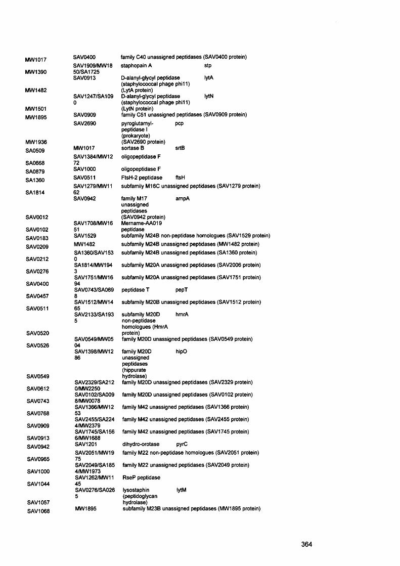
MW1017
MW 1390
MW1482
MW1501MW1895
MW1936SA0509
SA0668SA0879SA1360
SA1814
SAV0012
SAV0102SAV0183SAV0209
SAV0212
SAV0276
SAV0400
SAV0457
SAV0511
SAV0520
SAV0526
SAV0549
SAV0612
SAV0743
SAV0768
SAV0909
SAV0913SAV0942
SAV0965
SAV1000
SAV1044
SAV1057SAV1068
SAV0400SAV1909/MW18 50/SA1725 SAV0913
SAV1247/SA109 0
SAV0909SAV2690
MW1017SAV1384/MW12 72SAV1000SAV0511SAV1279/MW1162SAV0942
SAV1708/MW16 51SAV1529MW1482SA1360/SAV153 0SA1814/MW1943SAV1751/MW1694SAV0743/SA0698SAV1512/MW1465SAV2133/SA193 5
SAV0549/MW0504SAV1398/MW12 86
SAV2329/SA2120/MW2250SAV0102/SA0098/MW0078SAV1366/MW1253SAV2455/SA2244/MW2379SAV1745/SA1566/MW1688SAV1201SAV2051/MW1975SAV2049/SA185 4/MW1973 SAV1262/MW11 45SAV0276/SA0265
MW1895
srtB
ftsH
family C40 unassigned peptidases (SAV0400 protein) staphopain A stp
D-aianyl-giycyl peptidase lytA(staphylococcal phage phi11)(LytA protein)D-aianyl-glycyl peptidase lytN(staphylococcal phage phi11)(LytN protein)family C51 unassigned peptidases (SAV0909 protein)pyroglutamyl- peppeptidase I (prokaryote)(SAV2690 protein) sortase Boligopeptidase F
oligopeptidase F FtsH-2 peptidase subfamily M16C unassigned peptidases (SAV1279 protein)
family M17 ampAunassignedpeptidases(SAV0942 protein)Mername-AA019 peptidasesubfamily M24B non-peptidase homologues (SAV1529 protein) subfamily M24B unassigned peptidases (MW1482 protein) subfamily M24B unassigned peptidases (SA1360 protein)
subfamily M20A unassigned peptidases (SAV2006 protein)
subfamily M20A unassigned peptidases (SAV1751 protein)
peptidase T pepT
subfamily M20B unassigned peptidases (SAV1512 protein)
subfamily M20D hmrA non-peptidase homologues (HmrA protein)family M20D unassigned peptidases (SAV0549 protein)
family M20D hipOunassignedpeptidases(hippuratehydrolase)family M20D unassigned peptidases (SAV2329 protein)
family M20D unassigned peptidases (SAV0102 protein)
family M42 unassigned peptidases (SAV1366 protein)
family M42 unassigned peptidases (SAV2455 protein)
family M42 unassigned peptidases (SAV1745 protein)
dihydro-orotase pyrCfamily M22 non-peptidase homologues (SAV2051 protein)
family M22 unassigned peptidases (SAV2049 protein)
RseP peptidase
lysostaphin lytM(peptidoglycanhydrolase)subfamily M23B unassigned peptidases (MW1895 protein)
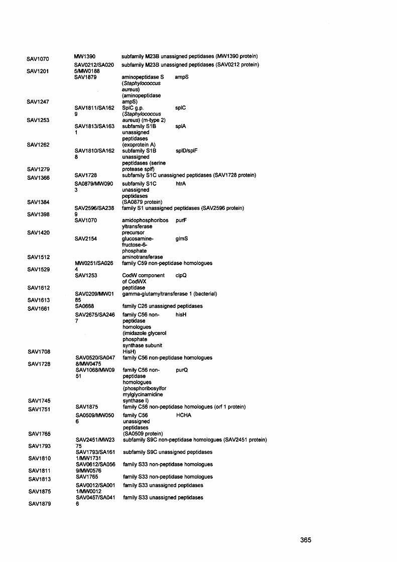
SAV1070
SAV1201
SAV1247
SAV1253
SAV1262
SAV1279SAV1366
SAV1384
SAV1398
SAV1420
SAV1512
SAV1529
SAV1612
SAV1613SAV1661
SAV1708
SAV1728
SAV1745SAV1751
SAV1765
SAV1793
SAV1810
SAV1811SAV1813
SAV1875
SAV1879
MW 1390SAV0212/SA0205/MW0188SAV1879
SAV1811/SA1629
SAV1813/SA1631
SAV1810/SA1628
SAV1728SA0879/MW0903
SAV2596/SA2389SAV1070
SAV2154
MW0251/SA0264SAV1253
SAV0209/MW01 85SA0668S AV2675/SA246 7
SAV0520/SA047 8/MW0475 SAV1068/MW09 51
SAV1875SA0509/MW0506
SAV2451/MW2375SAV1793/SA161 1/MW1731 SAV0612/SA056 9/MW0576 SAV1765SAV0012/SA001 1/MW0012 SAV0457/SA041 6
subfamily M23B unassigned peptidases (MW1390 protein) subfamily M23B unassigned peptidases (SAV0212 protein)
aminopeptidase S ampS( S ta p h y lo c o c c u s
a u re u s )
(aminopeptidaseampS)SpIC g.p. spIC( S ta p h y lo c o c c u s
a u re u s ) (m-type 2)subfamily StB splAunassignedpeptidases(exoprotein A)subfamily S1B splD/splFunassigned peptidases (serine protease splf)subfamily S1C unassigned peptidases (SAV1728 protein)subfamily S1C htrAunassigned peptidases (SA0879 protein)family S1 unassigned peptidases (SAV2596 protein)
amidophosphoribos purFyltransferaseprecursorglucosamine- glmSfructose-6-phosphateaminotransferasefamily C59 non-peptidase homologues
CodW component cIpQof CodWXpeptidasegamma-glutamyltransferase 1 (bacterial)
family C26 unassigned peptidasesfamily C56 non- hisHpeptidasehomologues(imidazole glycerolphosphatesynthase subunitHisH)family C56 non-peptidase homologues
family C56 non- purQpeptidasehomologues(phosphoribosylformylglycinamidinesynthase I)family C56 non-peptidase homologues (orf 1 protein)family C56 HCHAunassignedpeptidases(SA0509 protein)subfamily S9C non-peptidase homologues (SAV2451 protein)
subfamily S9C unassigned peptidases
family S33 non-peptidase homologues
family S33 non-peptidase homologues family S33 unassigned peptidases
family S33 unassigned peptidases
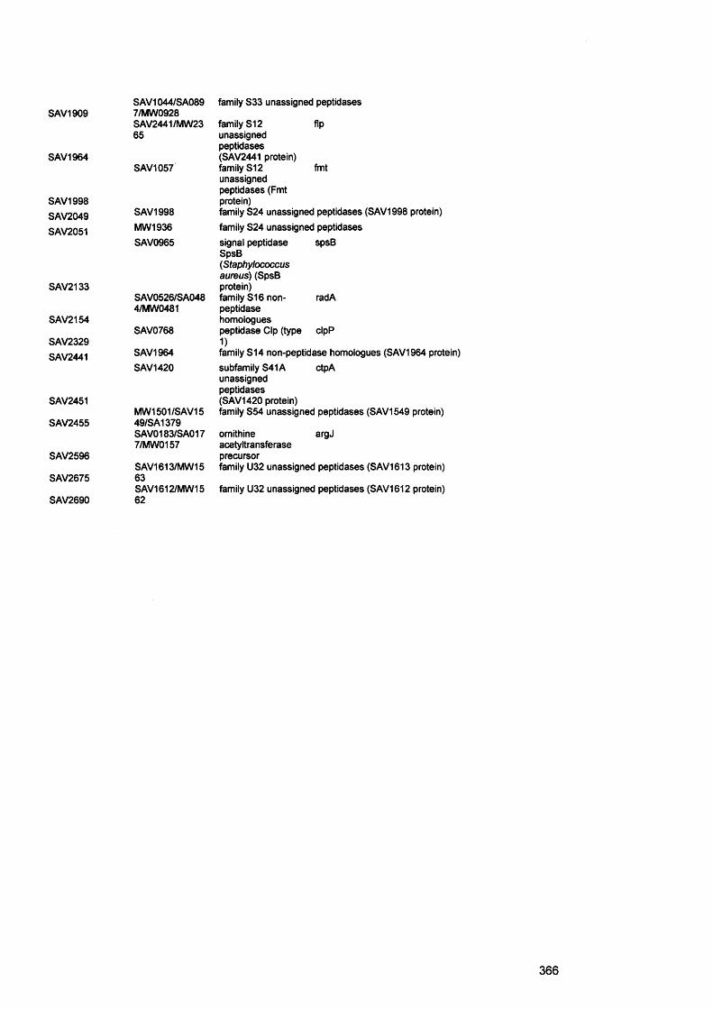
SAV1909
SAV1964
SAV1998SAV2049SAV2051
SAV2133
SAV2154
SAV2329SAV2441
SAV2451
SAV2455
SAV2596
SAV2675
SAV2690
SAV1044/SA089 7/MW0928 SAV2441/MW23 65
SAV1057
SAV1998MW1936SAV0965
SAV0526/SA0484/MW0481
SAV0768
SAV1964SAV1420
MW1501/SAV15 49/SA1379 SAV0183/SA017 7/MW0157
SAV1613/MW1563SAV1612/MW1562
family S33 unassigned peptidases
family S12 flpunassigned peptidases (SAV2441 protein) family S12 fmtunassigned peptidases (Fmt protein)family S24 unassigned peptidases (SAV1998 protein)family S24 unassigned peptidasessignal peptidase spsB SpsB( S ta p h y lo c o c c u s
a u r e u s ) (SpsB protein)family S16 non- radApeptidasehomologuespeptidase Clp (type cIpP 1)family S14 non-peptidase homologues (SAV1964 protein)subfamily S41A ctpAunassigned peptidases (SAV1420 protein)family S54 unassigned peptidases (SAV1549 protein)
ornithine argJacetyltransferaseprecursorfamily U32 unassigned peptidases (SAV1613 protein)
family U32 unassigned peptidases (SAV1612 protein)
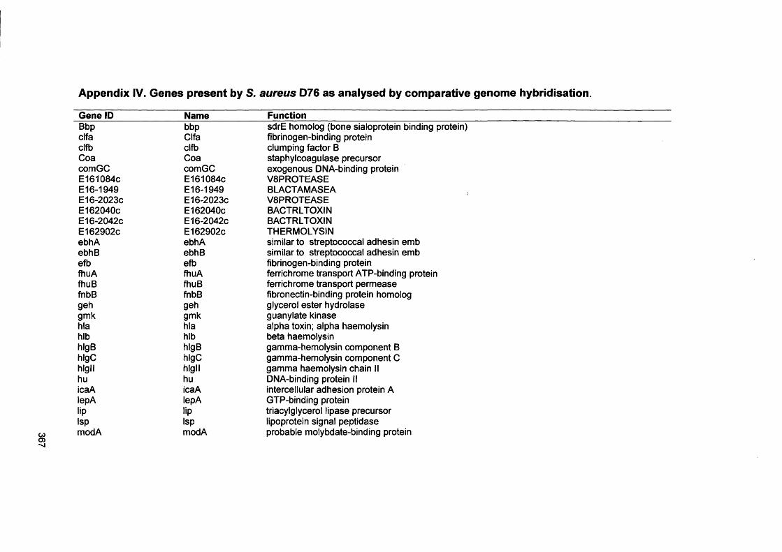
367
Appendix IV. Genes present by S. aureus D76 as analysed by comparative genome hybridisation.
Gene ID______________ Name____________Function_________________________________Bbp bbp sdrE homolog (bone sialoprotein binding protein)clfa Clfa fibrinogen-binding proteinclfb clfb clumping factor BCoa Coa staphylcoagulase precursorcomGC comGC exogenous DNA-binding proteinE161084C E161084C V8PROTEASEE16-1949 E16-1949 BLACTAMASEAE16-2023C E16-2023C V8PROTEASEE162040c E162040c BACTRLTOXINE16-2042C E16-2042C BACTRLTOXINE162902c E162902c THERMOLYSINebhA ebhA similar to streptococcal adhesin embebhB ebhB similar to streptococcal adhesin embefb efb fibrinogen-binding proteinfhuA fhuA ferrichrome transport ATP-binding proteinfhuB fhuB ferrichrome transport permeasefnbB fnbB fibronectin-binding protein homologgeh geh glycerol ester hydrolasegmk gmk guanylate kinasehla hla alpha toxin; alpha haemolysinhlb hlb beta haemolysinhlgB hlgB gamma-hemolysin component BhlgC hlgC gamma-hemolysin component Chlgll hlgll gamma haemolysin chain IIhu hu DNA-binding protein IIicaA icaA intercellular adhesion protein AlepA lepA GTP-binding proteinlip lip triacylglycerol lipase precursorIsp Isp lipoprotein signal peptidasemodA modA probable molybdate-binding protein
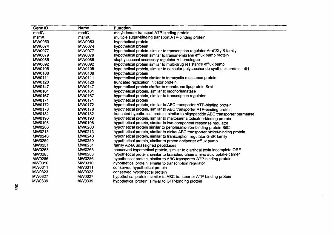
Gene IP Name FunctionmodC modC molybdenum transport ATP-binding proteinmsmX msmX multiple sugar-binding transport ATP-binding proteinMW 0053 MW0053 hypothetical proteinMW0074 MW0074 hypothetical proteinMW 0077 MW0077 hypothetical protein, similar to transcription regulator AraC/XylS familyMW0079 MW0079 hypothetical protein similar to transmembrane efflux pump proteinMW0085 MW0085 staphylococcal accessory regulator A homologueMW0092 MW0092 hypothetical protein similar to multi-drug resistance efflux pumpMW0105 MW 0105 hypothetical protein, similar to capsular polysaccharide synthesis protein 14HMW0108 MW0108 hypothetical proteinMW0111 MW0111 hypothetical protein similar to tetracyclin resistance proteinMW0120 MW 0120 truncated replication initiator proteinMW0147 MW0147 hypothetical protein similar to membrane lipoprotein SrpLMW0161 MW0161 hypothetical protein, similar to isochorismataseMW0167 MW0167 hypothetical protein, similar to transcription regulatorMW0171 MW0171 hypothetical proteinMW0172 MW0172 hypothetical protein, similar to ABC transporter ATP-binding proteinMW0178 MW0178 hypothetical protein, similar to ABC transporter ATP-binding proteinMW0182 MW0182 truncated hypothetical protein, similar to oligopeptide ABC transporter permeaseMW0190 MW 0190 hypothetical protein, similar to maltose/maltodextrin-binding proteinMW0198 M W 0 198 hypothetical protein, similar to two-component response regulatorMW0200 MW0200 hypothetical protein similar to periplasmic-iron-binding protein BitCMW0213 MW0213 hypothetical protein, similar to nickel ABC transporter nickel-binding proteinMW 0240 MW0240 hypothetical protein, similar to transcription regulator GntR familyMW0250 MW0250 hypothetical protein, similar to proton antiporter efflux pumpMW0251 MW0251 family A24A unassigned peptidasesMW 0263 MW 0263 conserved hypothetical protein, similar to diarrheal toxin incomplete ORFMW0283 MW0283 hypothetical protein, similar to branched-chain amino acid uptake carrierMW0286 MW0286 hypothetical protein, similar to ABC transporter ATP-binding proteinMW 0310 MW0310 hypothetical protein, similar to transcription regulatorMW0311 MW0311 conserved hypothetical proteinMW0323 MW0323 conserved hypothetical proteinMW0327 MW0327 hypothetical protein, similar to ABC transporter ATP-binding proteinMW0339 MW0339 hypothetical protein, similar to GTP-binding protein
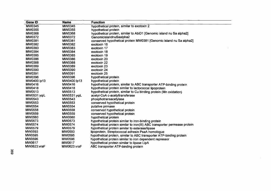
369
Gene ID Name FunctionMW0345 MW0345 hypothetical protein, similar to exotoxin 2MW0355 MW0355 hypothetical proteinMW0368 MW0368 hypothetical protein, similar to AbiD1 [Genomic island nu Sa alpha2]MW0372 MW0372 GenomicislandnuSaalpha2MW0381 MW0381 conserved hypothetical protein MW0381 [Genomic island nu Sa alpha2]MW0382 MW0382 exotoxin 16MW0383 MW0383 exotoxin 17MW0384 MW0384 exotoxin 18MW0385 MW0385 exotoxin 19MW0386 MW0386 exotoxin 20MW0388 MW0388 exotoxin 22MW0389 MW0389 exotoxin 23MW0390 MW0390 exotoxin 24MW0391 MW0391 exotoxin 25MW 0396 MW0396 hypothetical proteinMW0400:lp13 MW0400:lp13 hypothetical proteinMW0416 MW0416 hypothetical protein, similar to ABC transporter ATP-binding proteinMW0418 MW0418 hypothetical protein similar to lactococcal lipoproteinMW0513 MW0513 hypothetical protein, similar to Cu binding protein (Mn oxidation)MW0531:yqiL MW0531:yqiL acetyl-CoA c-acetyltransferaseMW0543 MW0543 phosphotransacetylaseMW0553 MW0553 conserved hypothetical proteinMW0554 MW0554 putative primaseMW0558 MW0558 conserved hypothetical proteinMW0559 MW0559 conserved hypothetical proteinMW0560 MW0560 hypothetical proteinMW0573 MW0573 hypothetical protein similar to iron-binding proteinMW0574 MW0574 hypothetical protein similar to iron(lll) ABC transporter permease proteinMW 0579 MW0579 hypothetical protein similar to esterase/lipaseMW0593 MW0593 lipoprotein, Streptococcal adhesin PsaA homologueMW0595 MW0595 hypothetical protein, similar to ABC transporter ATP-binding proteinMW0596 MW0596 hypothetical protein similar to iron dependent repressorMW 0617 MW0617 hypothetical protein similar to lipase LipAMW0623:vraF MW0623:vraF ABC transporter ATP-binding protein
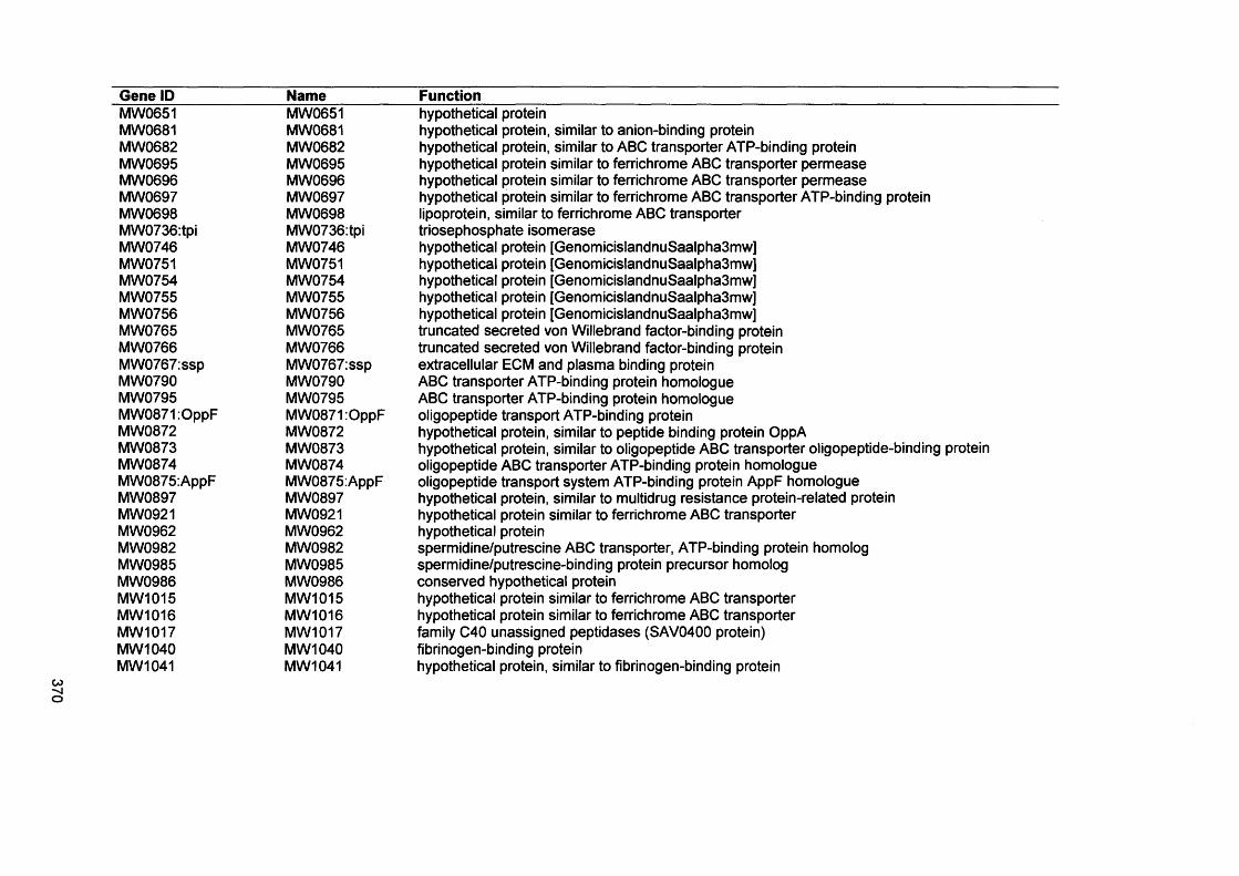
Gene ID Name FunctionMW0651 MW0651 hypothetical proteinMW0681 MW0681 hypothetical protein, similar to anion-binding proteinMW0682 MW0682 hypothetical protein, similar to ABC transporter ATP-binding proteinMW0695 MW0695 hypothetical protein similar to ferrichrome ABC transporter permeaseMW0696 MW0696 hypothetical protein similar to ferrichrome ABC transporter permeaseMW0697 MW0697 hypothetical protein similar to ferrichrome ABC transporter ATP-binding proteinMW0698 MW 0698 lipoprotein, similar to ferrichrome ABC transporterMW0736:tpi MW0736:tpi triosephosphate isomeraseMW0746 MW 0746 hypothetical protein [GenomicislandnuSaalpha3mw]MW0751 MW0751 hypothetical protein [GenomicislandnuSaalpha3mw]MW0754 MW 0754 hypothetical protein [GenomicislandnuSaalpha3mw]MW0755 MW0755 hypothetical protein [GenomicislandnuSaalpha3mwjMW0756 MW 0756 hypothetical protein [GenomicislandnuSaalpha3mw]MW0765 MW0765 truncated secreted von Willebrand factor-binding proteinMW0766 MW0766 truncated secreted von Willebrand factor-binding proteinMW0767:ssp MW0767:ssp extracellular ECM and plasma binding proteinMW0790 MW0790 ABC transporter ATP-binding protein homologueMW0795 MW0795 ABC transporter ATP-binding protein homologueMW0871:OppF MW0871:OppF oligopeptide transport ATP-binding proteinMW0872 MW0872 hypothetical protein, similar to peptide binding protein OppAMW0873 MW0873 hypothetical protein, similar to oligopeptide ABC transporter oligopeptide-binding proteinMW0874 MW0874 oligopeptide ABC transporter ATP-binding protein homologueMW0875:AppF MW0875:AppF oligopeptide transport system ATP-binding protein AppF homologueMW0897 MW0897 hypothetical protein, similar to multidrug resistance protein-related proteinMW0921 MW0921 hypothetical protein similar to ferrichrome ABC transporterMW0962 MW0962 hypothetical proteinMW0982 MW0982 spermidine/putrescine ABC transporter, ATP-binding protein homologMW0985 MW0985 spermidine/putrescine-binding protein precursor homologMW0986 MW 0986 conserved hypothetical proteinMW1015 MW 1015 hypothetical protein similar to ferrichrome ABC transporterMW1016 MW1016 hypothetical protein similar to ferrichrome ABC transporterMW1017 MW1017 family C40 unassigned peptidases (SAV0400 protein)MW1040 MW1040 fibrinogen-binding proteinMW1041 MW1041 hypothetical protein, similar to fibrinogen-binding protein

Gene ID Name FunctionMW1047 MW1047 hypothetical protein, similar to exotoxin 1MW1048 MW1048 hypothetical protein, similar to exotoxin 4MW1091 MW1091 hypothetical protein, similar to fibrinogen binding proteinMW1160 MW1160 hypothetical protein, similar to transcription regulator GntR familyMW1163 MW1163 hypothetical protein, similar to 3-oxoacyl- acyl-carrier protein reductase homolog ymflMW1182:glpF MW1182:glpF glycerol uptake facilitatorMW1185 MW1185 hypothetical protein similar to lysophospholipaseMW1189 MW1189 hypothetical protein, similar to GTP-binding protein proteinase modulator homolog ynbAMW1206 MW1206 ABC transporter (ATP-binding protein) homologMW1261:femA MW1261:femA factor essential for expression of methicillin resistanceMW1262:femB MW1262:femB FemB proteinMW1280 MW1280 ABC transporter (ATP-binding protein) homologMW1287 MW1287 hypothetical protein, similar to alanine racemaseMW1364 MW 1364 hypothetical protein, similar to GTP binding proteinMW1386 MW1386 conserved hypothetical protein [BacteriophagephiSa2mw]MW1390:stp MW1390:stp stp staphopain AMW1393 MW1393 conserved hypothetical protein [BacteriophagephiSa2mw]MW1400 MW1400 hypothetical protein [BacteriophagephiSa2mw]MW1401 MW1401 hypothetical protein [BacteriophagephiSa2mw]MW1405 MW1405 hypothetical protein [BacteriophagephiSa2mw]MW1408 MW1408 hypothetical protein [BacteriophagephiSa2mw]MW1482: lytA MW1482: lytA D-alanyl-glycyl peptidase (staphylococcal phage phi11) (LytA protein)MW1493 MW1493 hypothetical protein, similar to competence proteinMW1501:lytN M W 1501: lytN D-alanyl-glycyl peptidase (staphylococcal phage phi11) (LytN protein)MW1508 MW1508 hypothetical protein, similar to ABC transporter ATP-binding proteinMW1519:bex MW1519:bex GTP-binding protain Era homologMW1532:dnaK MW1532:dnaK DnaK proteinMW1547:aroE MW1547:aroE shikimate dehydrogeneaseMW1552 MW1552 enterotoxin homologMW1572 MW1572 iron-sulfur cofactor synthesis protein homologMW1659 MW1659 hypothetical protein similar to iron-sulfur cofactor synthesis protein nifZMW1752:splF MW1752:splF serine proteaseMW1753:splC MW1753:splC serine proteaseMW1754:splB MW1754:splB serine protease
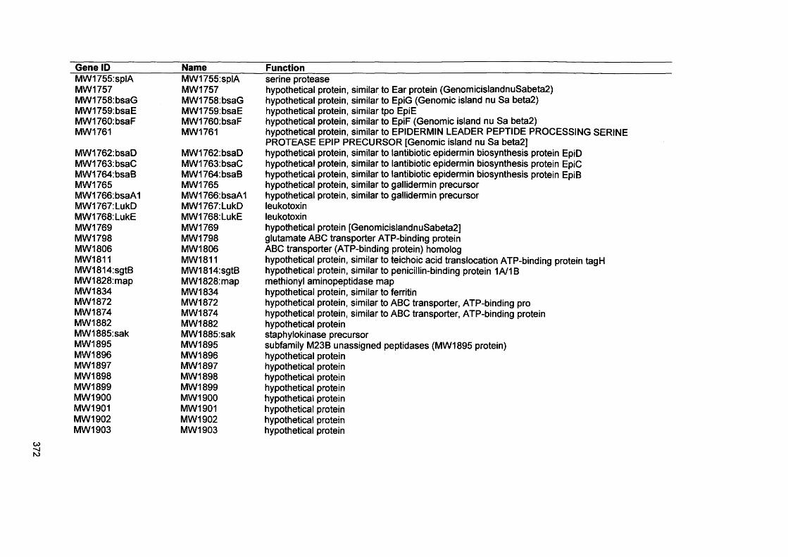
372
Gene ID______MW1755:splAMW1757MW1758:bsaGMW1759:bsaEMW1760:bsaFMW1761
MW1762:MW1763:MW1764:MW 1765MW1766:MW1767:MW1768:MW1769MW1798MW1806MW1811MW1814:MW1828:MW1834MW1872MW1874MW1882MW1885:MW1895MW1896MW1897MW 1898MW 1899MW1900MW1901MW1902MW1903
bsaDbsaCbsaB
bsaA1LukDLukE
sgtBmap
sak
Name____________Function__________________________________________________________________MW1755:splA serine proteaseMW 1757 hypothetical protein, similar to Ear protein (GenomicislandnuSabeta2)MW1758:bsaG hypothetical protein, similar to EpiG (Genomic island nu Sa beta2)MW1759:bsaE hypothetical protein, similar tpo EpiEMW1760:bsaF hypothetical protein, similar to EpiF (Genomic island nu Sa beta2)MW1761 hypothetical protein, similar to EPIDERMIN LEADER PEPTIDE PROCESSING SERINE
PROTEASE EPIP PRECURSOR [Genomic island nu Sa beta2]MW1762:bsaD hypothetical protein, similar to lantibiotic epidermin biosynthesis protein EpiDMW1763:bsaC hypothetical protein, similar to lantibiotic epidermin biosynthesis protein EpiCMW1764:bsaB hypothetical protein, similar to lantibiotic epidermin biosynthesis protein EpiBMW1765 hypothetical protein, similar to gallidermin precursorMW1766:bsaA1 hypothetical protein, similar to gallidermin precursorMW1767:LukD leukotoxin M W 1768: LukE leukotoxinMW 1769 hypothetical protein [GenomicislandnuSabeta2]M W 1798 glutamate ABC transporter ATP-binding proteinMW1806 ABC transporter (ATP-binding protein) homologM W 1811 hypothetical protein, similar to teichoic acid translocation ATP-binding protein tagHMW1814:sgtB hypothetical protein, similar to penicillin-binding protein 1A/1BMW1828:map methionyl aminopeptidase mapM W 1834 hypothetical protein, similar to ferritinMW1872 hypothetical protein, similar to ABC transporter, ATP-binding proMW 1874 hypothetical protein, similar to ABC transporter, ATP-binding proteinMW1882 hypothetical proteinMW1885:sak staphylokinase precursorMW1895 subfamily M23B unassigned peptidases (MW 1895 protein)MW 1896 hypothetical proteinMW 1897 hypothetical proteinMW 1898 hypothetical proteinMW1899 hypothetical proteinMW 1900 hypothetical proteinMW1901 hypothetical proteinMW1902 hypothetical proteinMW1903 hypothetical protein
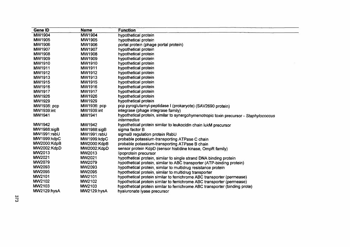
Gene ID Name FunctionMW1904 MW1904 hypothetical proteinMW1905 MW 1905 hypothetical proteinMW1906 MW1906 portal protein (phage portal protein)MW1907 MW 1907 hypothetical proteinMW1908 MW1908 hypothetical proteinMW1909 MW 1909 hypothetical proteinMW1910 MW1910 hypothetical proteinMW1911 MW1911 hypothetical proteinMW1912 MW1912 hypothetical proteinMW1913 MW1913 hypothetical proteinMW1915 MW1915 hypothetical proteinMW1916 MW1916 hypothetical proteinMW1917 MW1917 hypothetical proteinMW1926 MW1926 hypothetical proteinMW1929 MW1929 hypothetical proteinMW1936: pep MW1936: pep pep pyroglutamyl-peptidase I (prokaryote) (SAV2690 protein)MW1939:int MW1939:int integrase (phage integrase family)MW1941 MW1941 hypothetical protein, similar to synergohymenotropic toxin precursor - Staphylococcus
intermediusMW1942 MW1942 hypothetical protein similar to leukocidin chain lukM precursorMW1988:sigB MW1988:sigB sigma factor BMW1991:rsbU MW1991:rsbU sigmaB regulation protein RsbUMW1999:kdpC MW1999:kdpC probable potassium-transporting ATPase C chainMW2000:KdpB MW2000:KdpB probable potassium-transporting ATPase B chainMW2002:KdpD MW2002:KdpD sensor protein KdpD (sensor histidine kinase, OmpR family)MW2013 MW2013 lipoprotein precursorMW2021 MW2021 hypothetical protein, similar to single strand DNA binding proteinMW2079 MW2079 hypothetical protein, similar to ABC transporter (ATP-binding protein)MW2093 MW 2093 hypothetical protein, similar to multidrug resistance proteinMW2095 MW 2095 hypothetical protein, similar to multidrug transporterMW2101 MW2101 hypothetical protein similar to ferrichrome ABC transporter (permease)MW2102 MW2102 hypothetical protein similar to ferrichrome ABC transporter (permease)MW2103 MW2103 hypothetical protein similar to ferrichrome ABC transporter (binding prote)MW2129:hysA MW2129:hysA hyaluronate lyase precursor
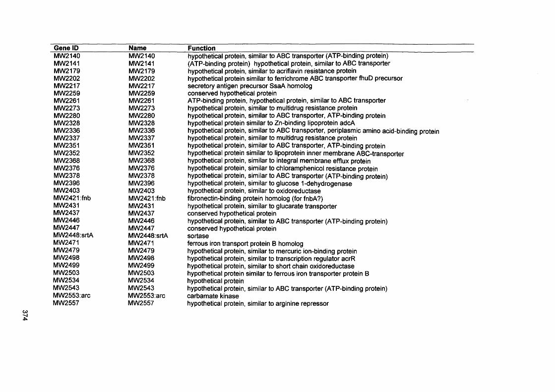
374
Gene ID Name FunctionMW2140 MW2140 hypothetical protein, similar to ABC transporter (ATP-binding protein)MW2141 MW2141 (ATP-binding protein) hypothetical protein, similar to ABC transporterMW2179 MW2179 hypothetical protein, similar to acriflavin resistance proteinMW2202 MW2202 hypothetical protein similar to ferrichrome ABC transporter fhuD precursorMW2217 MW2217 secretory antigen precursor SsaA homologMW2259 MW2259 conserved hypothetical proteinMW2261 MW2261 ATP-binding protein, hypothetical protein, similar to ABC transporterMW2273 MW2273 hypothetical protein, similar to multidrug resistance proteinMW2280 MW2280 hypothetical protein, similar to ABC transporter, ATP-binding proteinMW2328 MW2328 hypothetical protein similar to Zn-binding lipoprotein adcAMW2336 MW2336 hypothetical protein, similar to ABC transporter, periplasmic amino acid-binding proteinMW2337 MW2337 hypothetical protein, similar to multidrug resistance proteinMW2351 MW2351 hypothetical protein, similar to ABC transporter, ATP-binding proteinMW2352 MW2352 hypothetical protein similar to lipoprotein inner membrane ABC-transporterMW2368 MW2368 hypothetical protein, similar to integral membrane efflux proteinMW2376 MW2376 hypothetical protein, similar to chloramphenicol resistance proteinMW2378 MW2378 hypothetical protein, similar to ABC transporter (ATP-binding protein)MW2396 MW2396 hypothetical protein, similar to glucose 1-dehydrogenaseMW2403 MW2403 hypothetical protein, similar to oxidoreductaseMW2421:fnb MW2421:fnb fibronectin-binding protein homolog (for fnbA?)MW2431 MW2431 hypothetical protein, similar to glucarate transporterMW2437 MW2437 conserved hypothetical proteinMW2446 MW2446 hypothetical protein, similar to ABC transporter (ATP-binding protein)MW 2447 MW2447 conserved hypothetical proteinMW2448:srtA MW2448:srtA sortaseMW2471 MW2471 ferrous iron transport protein B homologMW2479 MW2479 hypothetical protein, similar to mercuric ion-binding proteinMW2498 MW2498 hypothetical protein, similar to transcription regulator acrRMW2499 MW2499 hypothetical protein, similar to short chain oxidoreductaseMW2503 MW2503 hypothetical protein similar to ferrous iron transporter protein BMW2534 MW2534 hypothetical proteinMW2543 MW2543 hypothetical protein, similar to ABC transporter (ATP-binding protein)MW2553:arc MW2553:arc carbamate kinaseMW2557 MW2557 hypothetical protein, similar to arginine repressor

375
Gene ID Name FunctionMW2558:aur MW2558:aur zinc metalloproteinase aureolysinMW2566 MW2566 hypothetical protein, similar to N-Carbamoylsarcosine AmidohydrolaseMW2574 MW2574 hypothetical protein, similar to preprotein translocase secYMWrRNA02 MWrRNA02 23S ribosomal RNA (RNA02)MWrRNA05 MWrRNA05 16S ribosomal RNA (RNA05)nuc nuc thermonucleaseobg obg SpoOB-associated GTP-binding proteinpbp3 pbp3 penicillin-binding protein 3pbpA pbpA penicillin-binding protein 1pstB pstB phosphate ABC transporter ATP-binding proteinrip rip RGD-containing lipoproteinrot rot repressor of toxinsSA0074 SA0074 hypothetical proteinSA0102 SA0102 67 kDa Myosin-crossreactive streptococcal antigen homologueSA0103 SA0103 conserved hypothetical proteinSA0104 SA0104 hypothetical protein, similar to transcription regulator GntR familySA0190 SA0190 hypothetical proteinSA0191 SA0191 conserved hypothetical proteinSA0192 SA0192 hypothetical protein, similar to ABC transporter ATP-binding proteinSA0193 SA0193 hypothetical protein, similar to Enterococcus faecalis plasmid pPD1 badSA0195 SA0195 hypothetical proteinSA0196 SA0196 conserved hypothetical proteinSA0197 SA0197 hypothetical protein, similar to ABC transporter ATP-binding proteinSA0211 SA0211 acetyl-CoA acetyltransferaseSA0251:lytR SA0251:lytR two-component response regulatorSA0276 SA0276 conserved hypothetical protein, similar to diarrheal toxinSA0294 SA0294 hypothetical protein, similar to branched-chain amino acid uptake carrierSA0298 SA0298 hypothetical protein, similar to regulatory protein PfoRSA0509 SA0509:srtB srtB sortase BSA0573:sarA SA0573:sarA staphylococcal accessory regulator ASA0598:pbp4 SA0598:pbp4SA0610 SA0610 hypothetical protein, similar to lipase LipASA0614 SA0614 hypothetical protein, similar to two-component response regulatorSA0622 SA0622 hypothetical protein, similar to AraC/XylS family transcriptional regulator
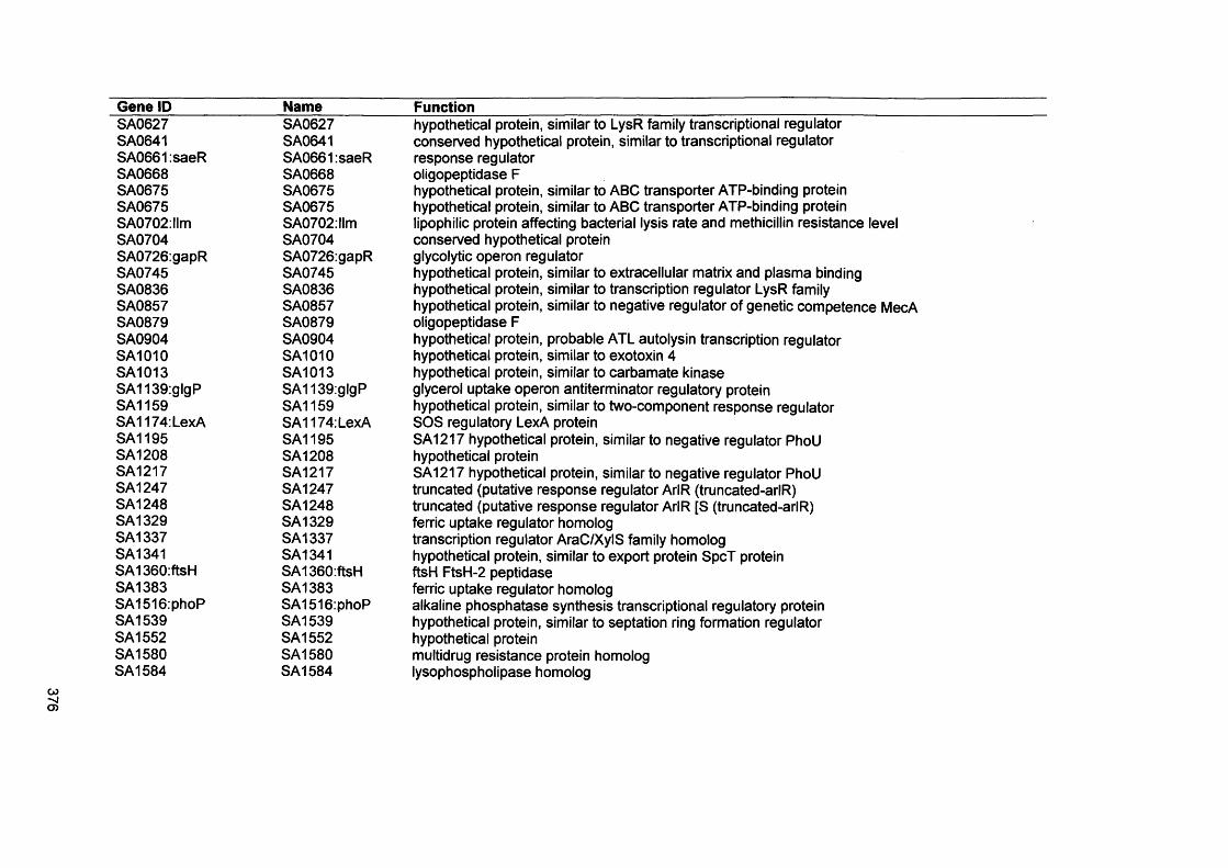
376
Gene ID NameSA0627SA0641SA0661:saeRSA0668SA0675SA0675SA0702:llmSA0704SA0726:gapRSA0745SA0836SA0857SA0879SA0904SA1010SA1013SA1139:glgPSA1159SA1174: LexASA1195SA1208SA1217SA1247SA1248SA1329SA1337SA1341SA1360:ftsHSA1383SA1516:phoPSA1539SA1552SA1580SA1584
SA0627SA0641SA0661:saeRSA0668SA0675SA0675SA0702:llmSA0704SA0726:gapRSA0745SA0836SA0857SA0879SA0904SA1010SA1013SA1139:glgPSA1159SA1174: LexASA1195SA1208SA1217SA1247SA1248SA1329SA1337SA1341SA1360:ftsHSA1383SA1516:phoPSA1539SA1552SA1580SA1584
Functionhypothetical protein, similar to LysR family transcriptional regulator conserved hypothetical protein, similar to transcriptional regulator response regulator oligopeptidase Fhypothetical protein, similar to ABC transporter ATP-binding protein hypothetical protein, similar to ABC transporter ATP-binding protein lipophilic protein affecting bacterial lysis rate and methicillin resistance level conserved hypothetical protein glycolytic operon regulatorhypothetical protein, similar to extracellular matrix and plasma binding hypothetical protein, similar to transcription regulator LysR family hypothetical protein, similar to negative regulator of genetic competence MecA oligopeptidase Fhypothetical protein, probable ATL autolysin transcription regulator hypothetical protein, similar to exotoxin 4 hypothetical protein, similar to carbamate kinase glycerol uptake operon antiterminator regulatory protein hypothetical protein, similar to two-component response regulator SOS regulatory LexA proteinSA1217 hypothetical protein, similar to negative regulator PhoU hypothetical proteinSA1217 hypothetical protein, similar to negative regulator PhoU truncated (putative response regulator ArIR (truncated-arIR) truncated (putative response regulator ArIR [S (truncated-arIR) ferric uptake regulator homolog transcription regulator AraC/XylS family homolog hypothetical protein, similar to export protein SpcT protein ftsH FtsH-2 peptidase ferric uptake regulator homologalkaline phosphatase synthesis transcriptional regulatory protein hypothetical protein, similar to septation ring formation regulator hypothetical protein multidrug resistance protein homolog lysophospholipase homolog
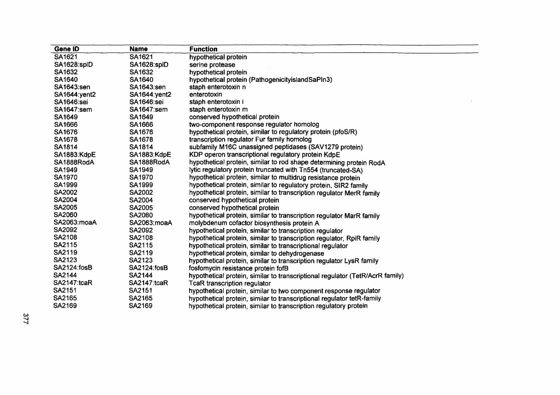
Gene ID Name FunctionSA1621 SA1621 hypothetical proteinSA1628:splD SA1628:splD serine proteaseSA1632 SA1632 hypothetical proteinSA1640 SA1640 hypothetical protein (PathogenicityislandSaPln3)SA1643:sen SA1643:sen staph enterotoxin nSA1644:yent2 SA1644:yent2 enterotoxinSA1646:sei SA1646:sei staph enterotoxin iSA1647:sem SA1647:sem staph enterotoxin mSA1649 SA1649 conserved hypothetical proteinSA1666 SA1666 two-component response regulator homologSA1676 SA1676 hypothetical protein, similar to regulatory protein (pfoS/R)SA1678 SA1678 transcription regulator Fur family homologSA1814 SA1814 subfamily M16C unassigned peptidases (SAV1279 protein)SA1883: KdpE SA1883: KdpE KDP operon transcriptional regulatory protein KdpESA1888RodA SA1888RodA hypothetical protein, similar to rod shape determining protein RodASA1949 SA1949 lytic regulatory protein truncated with Tn554 (truncated-SA)SA1970 SA1970 hypothetical protein, similar to multidrug resistance proteinSA1999 SA1999 hypothetical protein, similar to regulatory protein, SIR2 familySA2002 SA2002 hypothetical protein, similar to transcription regulator MerR familySA2004 SA2004 conserved hypothetical proteinSA2005 SA2005 conserved hypothetical proteinSA2060 SA2060 hypothetical protein, similar to transcription regulator MarR familySA2063:moaA SA2063:moaA molybdenum cofactor biosynthesis protein ASA2092 SA2092 hypothetical protein, similar to transcription regulatorSA2108 SA2108 hypothetical protein, similar to transcription regulator, RpiR familySA2115 SA2115 hypothetical protein, similar to transcriptional regulatorSA2119 SA2119 hypothetical protein, similar to dehydrogenaseSA2123 SA2123 hypothetical protein, similar to transcription regulator LysR familySA2124:fosB SA2124:fosB fosfomycin resistance protein fofBSA2144 SA2144 hypothetical protein, similar to transcriptional regulator (TetR/AcrR family)SA2147:tcaR SA2147:tcaR TcaR transcription regulatorSA2151 SA2151 hypothetical protein, similar to two component response regulatorSA2165 SA2165 hypothetical protein, similar to transcriptional regulator tetR-familySA2169 SA2169 hypothetical protein, similar to transcription regulatory protein
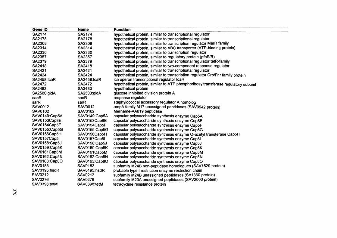
Gene ID Name FunctionSA2174 SA2174 hypothetical protein, similar to transcriptional regulatorSA2178 SA2178 hypothetical protein, similar to transcriptional regulatorSA2308 SA2308 hypothetical protein, similar to transcription regulator MarR familySA2314 SA2314 hypothetical protein, similar to ABC transporter (ATP-binding protein)SA2330 SA2330 hypothetical protein, similar to transcription regulatorSA2357 SA2357 hypothetical protein, similar to regulatory protein (pfoS/R)SA2379 SA2379 hypothetical protein, similar to transcriptional regulator tetR-familySA2418 SA2418 hypothetical protein, similar to two-component response regulatorSA2421 SA2421 hypothetical protein, similar to transcriptional regulatorSA2424 SA2424 hypothetical protein, similar to transcription regulator Crp/Fnr family proteinSA2458:lcaR SA2458:lcaR ica operon transcriptional regulator IcaRSA2472 SA2472 hypothetical protein, similar to ATP phosphoribosyltransferase regulatory subunitSA2483 SA2483 hypothetical proteinSA2500:gidA SA2500:gidA glucose inhibited division protein AsaeR saeR response regulatorsarR sarR staphylococcal accessory regulator A homologSAV0012 SAV0012 ampA family M17 unassigned peptidases (SAV0942 protein)SAV0102 SAV0102 Mername-AA019 peptidaseSAV0149:Cap5A SAV0149:Cap5A capsular polysaccharide synthesis enzyme Cap5ASAV0153Cap8E SAV0153Cap8E capsular polysaccharide synthesis enzyme Cap8ESAV0154Cap5F SAV0154Cap5F capsular polysaccharide synthesis enzyme Cap5FSAV0155:Cap5G SAV0155:Cap5G capsular polysaccharide synthesis enzyme Cap5GSAV0156Cap5H SAV0156Cap5H capsular polysaccharide synthesis enzyme O-acetyl transferase Cap5HSAV0157Cap5l SAV0157Cap5l capsular polysaccharide synthesis enzyme Cap5lSAV0158:Cap5J SAV0158:Cap5J capsular polysaccharide synthesis enzyme Cap5JSAV0159:Cap5K SAV0159:Cap5K capsular polysaccharide synthesis enzyme Cap5KSAV0161Cap5M SAV0161Cap5M capsular polysaccharide synthesis enzyme Cap5MSAV0162:Cap5N SAV0162:Cap5N capsular polysaccharide synthesis enzyme Cap5NSAV0163:Cap8O SAV0163:Cap8O capsular polysaccharide synthesis enzyme C ap80SAV0183 SAV0183 subfamily M24B non-peptidase homologues (SAV1529 protein)SAV0195:hsdR SAV0195:hsdR probable type I restriction enzyme restriction chainSAV0212 SAV0212 subfamily M24B unassigned peptidases (SA1360 protein)SAV0276 SAV0276 subfamily M20A unassigned peptidases (SAV2006 protein)SAV0398:tetM SAV0398:tetM tetracycline resistance protein
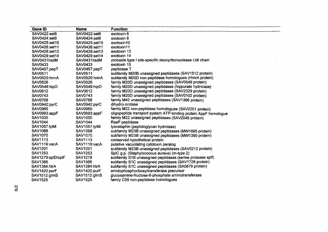
Gene ID NameSAV0422:set6SAV0424:set8SAV0425:set10SAV0426:set11SAV0428:set13SAV0429:set14SAV0431hsdMSAV0433SAV0457:pepTSAV0511SAV0520:hmrASAV0526SAV0549:hipOSAV0612SAV0743SAV0768SAV0942:pyrCSAV0965SAV0993:appFSAV1000SAV1044SAV1057:lytMSAV1068SAV1070SAV1113SAV1119:vacASAV1201SAV1253SAV1279:splD/splFSAV1366SAV1384:htrASAV1420:purFSAV1512:glmSSAV1529
SAV0422:set6SAV0424:set8SAV0425:set10SAV0426:set11SAV0428:set13SAV0429:set14SAV0431hsdMSAV0433SAV0457:pepTSAV0511SAV0520:hmrASAV0526SAV0549:hipOSAV0612SAV0743SAV0768SAV0942:pyrCSAV0965SAV0993:appFSAV1000SAV1044SAV1057:lytMSAV1068SAV1070SAV1113SAV1119:vacASAV1201SAV1253SAV1279SAV1366SAV1384:htrASAV1420:purFSAV1512:glmSSAV1529
Functionexotoxin 6 exotoxin 8 exotoxin 10 exotoxin 11 exotoxin 13 exotoxin 14probable type I site-specific deoxyribonuclease Lldl chain exotoxin 15 peptidase Tsubfamily M20B unassigned peptidases (SAV1512 protein) subfamily M20D non-peptidase homologues (HmrA protein) family M20D unassigned peptidases (SAV0549 protein) family M20D unassigned peptidases (hippurate hydrolase) family M20D unassigned peptidases (SAV2329 protein) family M20D unassigned peptidases (SAV0102 protein) family M42 unassigned peptidases (SAV1366 protein) dihydro-orotasefamily M22 non-peptidase homologues (SAV2051 protein) oligopeptide transport system ATP-binding protein AppF homologue family M22 unassigned peptidases (SAV2049 protein)RseP peptidaselysostaphin (peptidoglycan hydrolase)subfamily M23B unassigned peptidases (MW 1895 protein)subfamily M23B unassigned peptidases (MW 1390 protein)conserved hypothetical proteinputative vacuolating cytotoxin paralogsubfamily M23B unassigned peptidases (SAV0212 protein)SpIC g.p. (Staphylococcus aureus) (m-type 2) subfamily S1B unassigned peptidases (serine protease splf) subfamily S1C unassigned peptidases (SAV1728 protein) subfamily S1C unassigned peptidases (SA0879 protein) amidophosphoribosyltransferase precursor glucosamine-fructose-6-phosphate aminotransferase family C59 non-peptidase homologues
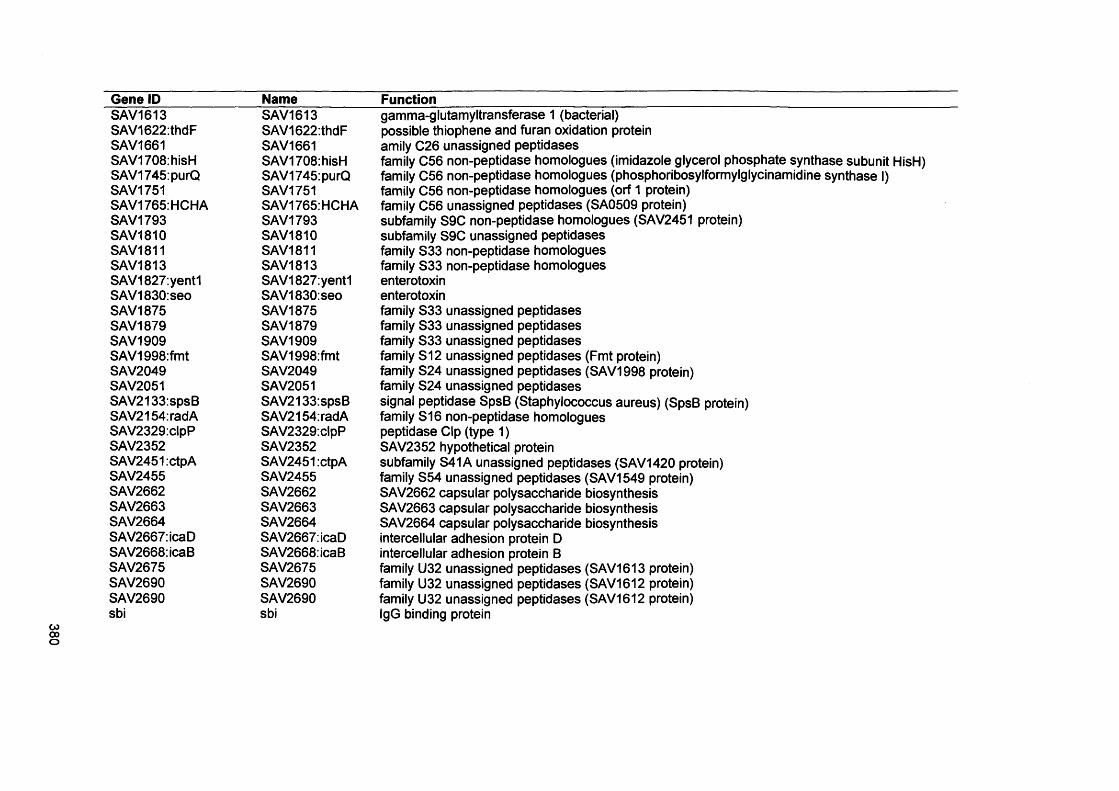
380
Gene ID Name FunctionSAV1613 SAV1613 gamma-glutamyltransferase 1 (bacterial)SAV1622:thdF SAV1622:thdF possible thiophene and furan oxidation proteinSAV1661 SAV1661 amily C26 unassigned peptidasesSAV1708:hisH SAV1708:hisH family C56 non-peptidase homologues (imidazole glycerol phosphate synthase subunit HisH)SAV1745:purQ SAV1745:purQ family C56 non-peptidase homologues (phosphoribosylformylglycinamidine synthase I)SAV1751 SAV1751 family C56 non-peptidase homologues (orf 1 protein)SAV1765:HCHA SAV1765:HCHA family C56 unassigned peptidases (SA0509 protein)SAV1793 SAV1793 subfamily S9C non-peptidase homologues (SAV2451 protein)SAV1810 SAV1810 subfamily S9C unassigned peptidasesSAV1811 SAV1811 family S33 non-peptidase homologuesSAV1813 SAV1813 family S33 non-peptidase homologuesSAV1827:yent1 SAV1827:yent1 enterotoxinSAV1830:seo SAV1830:seo enterotoxinSAV1875 SAV1875 family S33 unassigned peptidasesSAV1879 SAV1879 family S33 unassigned peptidasesSAV1909 SAV1909 family S33 unassigned peptidasesSAV1998:fmt SAV1998:fmt family S12 unassigned peptidases (Fmt protein)SAV2049 SAV2049 family S24 unassigned peptidases (SAV1998 protein)SAV2051 SAV2051 family S24 unassigned peptidasesSAV2133:spsB SAV2133:spsB signal peptidase SpsB (Staphylococcus aureus) (SpsB protein)SAV2154:radA SAV2154:radA family S16 non-peptidase homologuesSAV2329:clpP SAV2329:clpP peptidase Clp (type 1)SAV2352 SAV2352 SAV2352 hypothetical proteinSAV2451 :ctpA SAV2451 :ctpA subfamily S41A unassigned peptidases (SAV1420 protein)SAV2455 SAV2455 family S54 unassigned peptidases (SAV1549 protein)SAV2662 SAV2662 SAV2662 capsular polysaccharide biosynthesisSAV2663 SAV2663 SAV2663 capsular polysaccharide biosynthesisSAV2664 SAV2664 SAV2664 capsular polysaccharide biosynthesisSAV2667:icaD SAV2667:icaD intercellular adhesion protein DSAV2668:icaB SAV2668:icaB intercellular adhesion protein BSAV2675 SAV2675 family U32 unassigned peptidases (SAV1613 protein)SAV2690 SAV2690 family U32 unassigned peptidases (SAV1612 protein)SAV2690 SAV2690 family U32 unassigned peptidases (SAV1612 protein)sbi sbi IgG binding protein
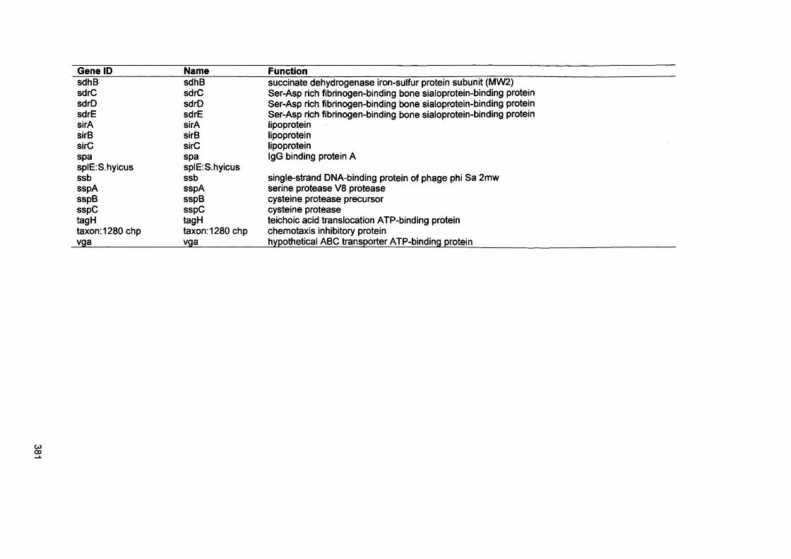
Gene ID Name FunctionsdhB sdhB succinate dehydrogenase iron-sulfur protein subunit (MW 2)sdrC sdrC Ser-Asp rich fibrinogen-binding bone sialoprotein-binding proteinsdrD sdrD Ser-Asp rich fibrinogen-binding bone sialoprotein-binding proteinsdrE sdrE Ser-Asp rich fibrinogen-binding bone sialoprotein-binding proteinsirA sirA lipoproteinsirB sirB lipoproteinsirC sirC lipoproteinspa spa IgG binding protein AsplE:S.hyicus splE.S.hyicusssb ssb single-strand DNA-binding protein of phage phi Sa 2mwsspA sspA serine protease V8 proteasesspB sspB cysteine protease precursorsspC sspC cysteine proteasetagH tagH teichoic acid translocation ATP-binding proteintaxon:1280 chp taxon:1280 chp chemotaxis inhibitory proteinvga yga . hypothetical ABC transporter ATP-binding protein