cell culture models in microfluidic systems
TRANSCRIPT

ANRV362-AC01-14 ARI 13 May 2008 9:0
Cell Culture Modelsin Microfluidic SystemsIvar Meyvantsson1 and David J. Beebe2
1Bellbrook Labs, LLC, Madison, Wisconsin 53711;email: [email protected] of Biomedical Engineering, University of Wisconsin at Madison,Madison, Wisconsin 53706; email: [email protected]
Annu. Rev. Anal. Chem. 2008. 1:423–49
First published online as a Review in Advance onMarch 4, 2008
The Annual Review of Analytical Chemistry is onlineat anchem.annualreviews.org
This article’s doi:10.1146/annurev.anchem.1.031207.113042
Copyright c© 2008 by Annual Reviews.All rights reserved
1936-1327/08/0719-0423$20.00
Key Words
microenvironment, extracellular matrix, microfabrication
AbstractMicrofluidic technology holds great promise for the creation of ad-vanced cell culture models. In this review, we discuss the character-ization of cell culture in microfluidic systems, describe importantbiochemical and physical features of the cell microenvironment, andreview studies of microfluidic cell manipulation in the context ofthese features. Finally, we consider the integration of analytical ele-ments, ways to achieve high throughput, and the design constraintsimposed by cell biology applications.
423
Click here for quick links to Annual Reviews content online, including:
• Other articles in this volume• Top cited articles• Top downloaded articles• Our comprehensive search
FurtherANNUALREVIEWS
Ann
ual R
evie
w o
f A
naly
tical
Che
mis
try
2008
.1:4
23-4
49. D
ownl
oade
d fr
om a
rjou
rnal
s.an
nual
revi
ews.
org
by U
nive
rsity
of
Wis
cons
in -
Mad
ison
on
01/1
5/10
. For
per
sona
l use
onl
y.
ANRV362-AC01-14 ARI 13 May 2008 9:0
1. INTRODUCTION
We continuously strive toward a better understanding of human biology and disease.Because direct observation of humans is only possible in epidemiological studies, ex-perimental work must rely on biological models. Models are, to varying degrees, ableto manipulate and analyze biological systems. They can be categorized in order ofbiological relevance as follows: (a) biochemical models using purified biomolecules,(b) cell lines, (c) cultured primary cells, (d ) model organisms such as yeast and fruitflies expressing endogenous or human genes, (e) human tissue explants, and ( f ) ani-mals (often rodents or nonhuman primates). However, increasing biological relevancegoes hand in hand with increases in cost, labor, and experiment duration. Conse-quently, the simplest model that sufficiently represents the system of interest is usuallychosen so that the largest possible parameter space can be explored using availableresources.
Mammalian cell culture can in many cases provide both the desired biological rel-evance and throughput, and it represents a large fraction of human biology research.The year 2007 marked the one-hundredth anniversary of in vitro cell culture. Al-though our understanding of molecular and cell biology has increased tremendouslyover the past 100 years, the methods used today are surprisingly similar to thoseemployed by Harrison in his 1907 study of frog neurons cultured in hanging dropsof clotted frog lymph on a depression slide (1). We still rely on undefined biologicalmaterial (e.g., fetal bovine serum) and simple containers such as dishes, bottles, andflasks. Currently, studies of model organisms and animal models are more biologicallyrelevant than those using cultured human cells, but this may change in the future withthe introduction of improved cell culture systems.
Various influences determine the phenotype of cells in vivo, including interac-tions with neighboring cells, interactions with the extracellular matrix (ECM), andsystemic factors. Ease of use and low price notwithstanding, dishes and flasks al-low no control over the spatial distribution of the cells and biomolecules needed tomodel many chemical and physical influences cells experience in vivo. Approachesthat increase the biological relevance of cell culture models while maintaining or in-creasing the throughput of current methods are of great interest to the life sciencescommunity.
Microfluidic systems represent a new kind of cell culture vessel that expands ourability to control the local cellular microenvironment (2, 3). Microfluidic systemsenable patterning of molecules and cells (4) as well as both passive (5) and active(6) cell handling and environmental control. Temporal and spatial control on themicrometer scale (0.1–100 μm) have been used in fundamental studies from thesubcellular (7) to the organismal (8) level, for instance in studies of cell division axisorientation (9) and geometric influence on cell survival (10).
Applications of soft lithography in cell biology have been reviewed (11), as havemethods of engineering cellular interactions via microtechnology (4). Microfabri-cated cell cultures were reviewed by Voldman et al. (2), Park and Shuler (12), andmore recently by El-Ali et al. (3). This review explicitly focuses upon cell biologyand the manner in which microfluidic structures have been or may be employed
424 Meyvantsson · Beebe
Ann
ual R
evie
w o
f A
naly
tical
Che
mis
try
2008
.1:4
23-4
49. D
ownl
oade
d fr
om a
rjou
rnal
s.an
nual
revi
ews.
org
by U
nive
rsity
of
Wis
cons
in -
Mad
ison
on
01/1
5/10
. For
per
sona
l use
onl
y.
ANRV362-AC01-14 ARI 13 May 2008 9:0
to manipulate the cellular microenvironment in order to better understand cellbiology or to build cellular models and assays. We also discuss analytical meth-ods used with microfluidic cell culture, ways in which throughput can be in-creased, and the constraints imposed upon microfluidic system design by cell biologyapplications.
2. MICROFLUIDIC CELL CULTURE
The cell culture methods in use today have been founded on over a century of work.It is important to put microfluidic cell culture in context with this work to deter-mine which assumptions hold true and which do not when methods are scaled downto microchannels. Although this characterization is just beginning, several researchgroups have already contributed to a better understanding of the multiple aspects ofmicrofluidic environments.
The physical design of microfluidic devices affects the cell microenvironment ofcultured cells. Design considerations for useful application of microfluidic devicesin cell biology were described by Walker et al. (13), who introduced the concept ofeffective culture volume as an indicator of cellular control over the microenviron-ment in the culture device. The complex but predictable patterns formed by growthfactors and other solutes have been described as secondary interfaces (14), the shapesof which are affected by perfusion of the cell culture medium. A comprehensive re-view of perfusion culture system design, material choices, and operation was recentlypresented by Kim et al. (15), whose discussion of both the engineering aspects andbiological application considerations of these factors provided a practical overview ofdesign, fabrication, sterilization, culture, and analysis. In this section, we describe rel-evant microfluidic cell culture work to date both in two dimensions (i.e., suspensionand monolayer) and in three dimensions (i.e., cells in a polymer matrix).
2.1. Cell Culture in Suspension and Monolayers
Fluid suspension is the natural environment of several cell types, including yeast cellsand mammalian blood cells. Other cell types retain important phenotypic character-istics in monolayer culture. This is true for cell-cell attachments that are generallyconserved in monolayer culture of epithelial cells. Suspension cell culture is tradi-tionally performed in roller bottles or spinner flasks, whereas monolayer cell cultureis done in Petri dishes, multiwell plates, or culture flasks.
One of the first studies of adherent cell culture in microfluidic channels was per-formed by Tilles et al. (16). They constructed a microchannel cell culture systemfrom polycarbonate and glass with channels 85–500 μm in height and a cell culturearea 25 mm wide × 75 mm long. Primary rat hepatocytes were seeded in coculturewith 3T3-J2 fibroblasts in channels that had either a gas exchange membrane top ora polycarbonate top. A significant difference in cell viability and hepatocyte functionwas seen after only 8 h when the devices with and without gas exchange membranewere compared. Monitoring albumin and urea production (markers of hepatocyte
www.annualreviews.org • Cell Culture Models in Microfluidic Systems 425
Ann
ual R
evie
w o
f A
naly
tical
Che
mis
try
2008
.1:4
23-4
49. D
ownl
oade
d fr
om a
rjou
rnal
s.an
nual
revi
ews.
org
by U
nive
rsity
of
Wis
cons
in -
Mad
ison
on
01/1
5/10
. For
per
sona
l use
onl
y.
ANRV362-AC01-14 ARI 13 May 2008 9:0
function) in devices with a gas exchange membrane over time for different flow rates,the authors found that increasing flow rate led to diminished viability and function.At low flow rates, hepatocyte function remained stable for 10 d.
As the liver is a highly perfused organ, perfusion culture is an appropriate way tomodel the mass transport aspects of liver biology in vitro. The culture of a human hep-atocarcinoma cell line (Hep G2) in a perfusion culture device was reported by Leclercet al. (17). They built a network of 270-μm-high channels constructed from twolayers of poly(dimethylsiloxane) (PDMS). Leclerc et al. used 4-(2-hydroxyethyl)-1-piperazineethanesulfonic acid (HEPES)–buffered media (i.e., independent of carbondioxide) and reported successful attachment, spreading, and growth on the PDMSsurface; they also reported that gas transport through PDMS was sufficient to main-tain a viable culture over days. They further compared perfusion and static conditions.Glucose consumption was similar in both cases for 3 d, after which the cells in staticculture started to die. In perfusion culture, the cells continued to grow and consumeglucose at an increasing rate until confluence was reached around day 7. The authorsalso monitored albumin expression, which is an indicator of normal phenotype of theHep G2 cell line. Albumin production was comparable for the first 3–4 d in perfusionand static culture, after which it dropped sharply in static culture, consistent with theobserved cell death. Although nutrient requirements and rate of waste productionwere shown to vary widely for different cell types, this study demonstrates that thereis a time window wherein these needs are met in static culture.
Yu et al. studied the effects of microchannel dimensions on the proliferation ofsuspended insect cells (Sf 9) (18) as well as the effects of cell density, exogenous growthfactors, and media change frequency on the growth rate of normal murine mammarygland cells (NMuMG) (19). They found that the growth rate of Sf 9 cells decreasedwith increasing cell density and decreasing channel height, whereas channel widthand length did not affect cell proliferation. Interestingly, the authors showed thatNMuMG cells proliferated more rapidly in microchannels than in 96-well plates, allelse being equal. The difference between microfluidic channels and wells was de-creased when the cell culture medium was changed frequently (either every 1 h orevery 4 h). The frequent media change also reduced the growth advantage providedby fetal bovine serum supplementation over epidermal growth factor (EGF) sup-plementation only. Consistent with the effective culture volume concept discussedabove (13), the authors hypothesized that the secreted growth factor accumulationfacilitated by the diffusion-dominated environment of microfluidic channels was re-sponsible for this effect.
Other indications of the environmental differences between microfluidic systemsand conventional culture come from embryo studies. Raty et al. cultured murineembryos in microfluidic devices and observed a greater proliferation rate with dailymedia changes in the microfluidic device compared to conventional methods (20).
2.2. Three-Dimensional Cell Culture
In vivo tissue organization and three-dimensional cell culture play an important role incells’ behavior. Three-dimensional cell culture can increase the biological relevance of
426 Meyvantsson · Beebe
Ann
ual R
evie
w o
f A
naly
tical
Che
mis
try
2008
.1:4
23-4
49. D
ownl
oade
d fr
om a
rjou
rnal
s.an
nual
revi
ews.
org
by U
nive
rsity
of
Wis
cons
in -
Mad
ison
on
01/1
5/10
. For
per
sona
l use
onl
y.
ANRV362-AC01-14 ARI 13 May 2008 9:0
cell-based models beyond that achievable in monolayer culture. Dolberg’s and Bissell’sstudy of Rous sarcoma virus (RSV) infection is a striking example of the powerfulinfluence exerted by complex three-dimensional environments in vivo (21). RSV isknown to cause neoplastic transformation of chick tissues and cells derived from chickembryos, but when Dolberg and Bissell infected cells of the early chick embryo, theactivity of the highly potent oncogene possessed by the virus was completely inhibited,even in the fully grown organism. When removed from the animal the cells expresseda transformed phenotype after only 24 h in culture, demonstrating that the ability ofin vivo three-dimensional architecture to control structure and function is lost whenthe cells are removed from the animal. These aspects of cell behavior can only bemodeled in three-dimensional culture (22).
Several approaches to seeding cells in gels inside microchannels have been re-ported. Toh et al. produced a device with a middle channel for cell seeding in a gelflanked on each side by aqueous channels for perfusion. The perfusion channels wereseparated from the culture channel by rows of micropillars (23). The authors cul-tured several cell types in the device and, over several days, analyzed cell functionsincluding albumin secretion of primary hepatocytes and differentiation competenceof mesenchymal stem cells. Laminar flow was used to pattern a microchannel with ahydrophobic coating, which led to the formation of virtual walls along the channel( J.P. Puccinelli & D.J. Beebe, manuscript in preparation). Subsequently, laminar flowwas again used to pattern two separate cell types in collagen gel along the hydrophiliccenter line of the channel, leaving the hydrophobic edges of the channel unwetted.The unwetted area was subsequently filled with culture media, thereby providing anaqueous interface to the cell culture.
Kim et al. created another device that employed laminar flow to create perfusionchannels on either side of a gel compartment (24). They cultured hepatocellular car-cinoma cells in the device and compared their albumin secretion when cultured incollagen, PuraMatrixTM (a synthetic peptide provisional matrix), or polylactic acid(PLA). They found that albumin secretion was initially lower in PuraMatrixTM thaneither collagen or PLA, but became higher by day three and remained high through-out the eight-day duration of the study. Cells cultured in collagen and PLA showedsimilar rates of albumin secretion.
Paguirigan and Beebe molded channels from gelatin by crosslinking with theenzyme transglutaminase (25). This enzyme occurs naturally; thus, the device con-tained no synthetic crosslinking or photoinitiating agents. The authors reported thatthe morphology of cells grown on the gelatin surface of these channels was signif-icantly different from that of cells in monolayer culture on tissue culture–treatedpolystyrene. The former showed nuclear staining results, indicating cell entry intothe three-dimensional crosslinked collagen matrix.
3. CELL MICROENVIRONMENT
The cell microenvironment comprises a complex of biochemical and physicalinfluences (which may be synergistic or antagonistic), the frequency (26) andtime sequence (27) of which can be remembered by cells. However, the cell
www.annualreviews.org • Cell Culture Models in Microfluidic Systems 427
Ann
ual R
evie
w o
f A
naly
tical
Che
mis
try
2008
.1:4
23-4
49. D
ownl
oade
d fr
om a
rjou
rnal
s.an
nual
revi
ews.
org
by U
nive
rsity
of
Wis
cons
in -
Mad
ison
on
01/1
5/10
. For
per
sona
l use
onl
y.
ANRV362-AC01-14 ARI 13 May 2008 9:0
microenvironment and the part it plays in homeostasis are still poorly understood(28). With regard to the soil nematode Caenorhabditis elegans, for example, every sin-gle cell division leading to the formation of the hatched larvae has been mapped (29),the entire genome of the organism has been sequenced (29), and numerous publishedreports have described various details of C. elegans cell biology. Still, we cannot take acell or tissue from the nematode and maintain its natural phenotype in culture becausethe biochemical and physical influences necessary to reproduce the cell microenvi-ronment that maintains that phenotype remain unknown. Microfluidics provides aset of tools that may enable us to define specific features of in vitro cell cultureenvironments.
3.1. Biochemical Environment
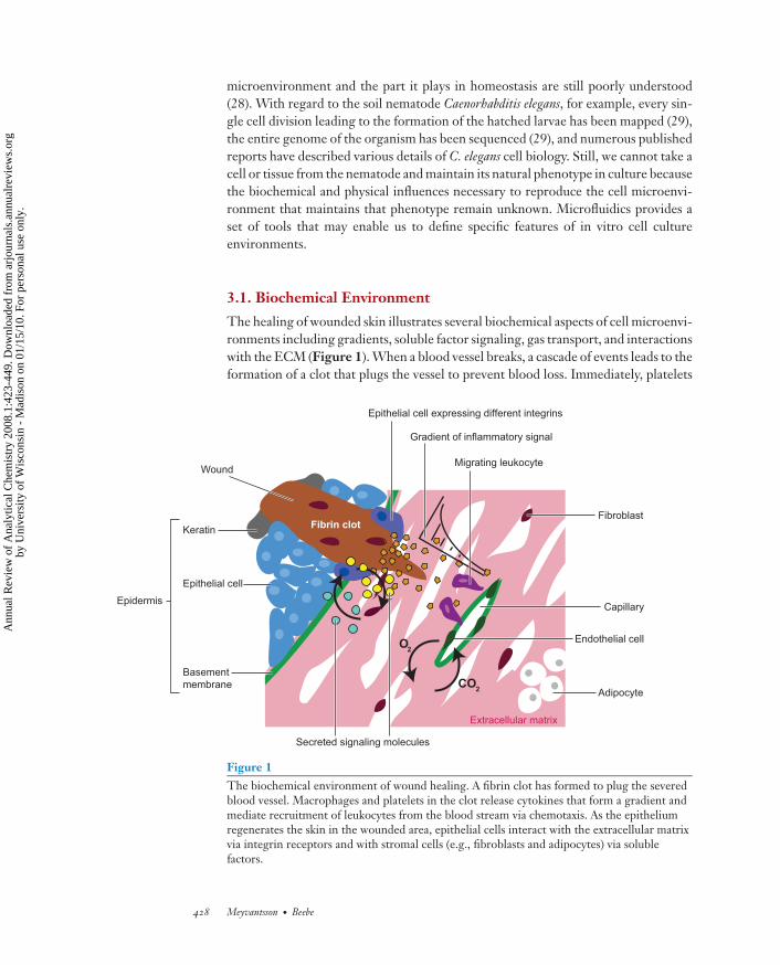
The healing of wounded skin illustrates several biochemical aspects of cell microenvi-ronments including gradients, soluble factor signaling, gas transport, and interactionswith the ECM (Figure 1). When a blood vessel breaks, a cascade of events leads to theformation of a clot that plugs the vessel to prevent blood loss. Immediately, platelets
O2
CO2
Capillary
Basement membrane
Endothelial cell
Secreted signaling molecules
Fibrin clot
Wound
Keratin
Fibroblast
Epithelial cell
Epidermis
Migrating leukocyte
Gradient of inflammatory signal
Epithelial cell expressing different integrins
Adipocyte
Extracellular matrix
Figure 1The biochemical environment of wound healing. A fibrin clot has formed to plug the severedblood vessel. Macrophages and platelets in the clot release cytokines that form a gradient andmediate recruitment of leukocytes from the blood stream via chemotaxis. As the epitheliumregenerates the skin in the wounded area, epithelial cells interact with the extracellular matrixvia integrin receptors and with stromal cells (e.g., fibroblasts and adipocytes) via solublefactors.
428 Meyvantsson · Beebe
Ann
ual R
evie
w o
f A
naly
tical
Che
mis
try
2008
.1:4
23-4
49. D
ownl
oade
d fr
om a
rjou
rnal
s.an
nual
revi
ews.
org
by U
nive
rsity
of
Wis
cons
in -
Mad
ison
on
01/1
5/10
. For
per
sona
l use
onl
y.
ANRV362-AC01-14 ARI 13 May 2008 9:0
and resident macrophages begin to release soluble factors, known as cytokines, thatmediate inflammation. As cytokine molecules diffuse away from the wound, theyform a gradient. When the gradient reaches the closest intact blood vessel, the cy-tokine signal is sensed by local endothelial cells and soon by leukocytes in the bloodstream. This signal prompts endothelial cells to mediate leukocyte entry into tissueand leukocytes to enter the tissue (30). Leukocytes move up the cytokine gradient, abehavior known as chemotaxis.
Later in the healing process, skin epithelial cells proceed to regenerate the epi-dermis (31). The cells of the dermal connective tissue, including fibroblasts andadipocytes (collectively known as stromal cells), regulate epithelial proliferation anddifferentiation via soluble factors (32). Integrin receptors allow epithelial cells to senseand bind to the ECM and are specific for a given set of ECM proteins. In normalskin, epithelial cells lie on the basement membrane, a layer of ECM rich in laminin.During regeneration, the epithelial cells express new integrin receptors in order tointeract with the fibrin clot and the dermis and re-epithelialize the wounded area (31).Another important biochemical influence is gas concentration: As the wound contin-ues to heal, new blood vessels are formed to provide gas exchange to the regeneratedtissue. Oxygen plays important roles in metabolism, in the regulation of angiogenesis(discussed in detail below in the context of physical environmental influences), andin the zonal differentiation in the liver (33). These biochemical aspects of the cellmicroenvironment and the ways in which they have been explored with microfluidicsystems are described in this section.
3.1.1. Soluble factors. Various biomolecules exist dissolved intracellularly and inthe interstitial space. Many actions in the body are regulated, at least partially, bysoluble factors including angiogenesis (34) and embryonic morphogenesis (35). Amolecule secreted from a cell is transported away from the source via diffusion, theconvective flow of the blood stream, and the continuous flow from capillaries intolymphatic vessels. The spatial and temporal distribution of soluble factors is furtheraffected by the lifetime of the factor dictated by the molecule’s inherent stabilityor enzymatic degradation, sequestration by the ECM, and binding endocytosis byother cells. Microfluidic systems have been employed to perform spatially definedtreatment. For instance, Sawano et al. used laminar streams to expose only a smallpart of a cell to EGF. They subsequently observed the intracellular propagation of cellsignaling (7). Similarly, Blake et al. demonstrated the use of laminar flow for treatmentof specific regions of perfused rat brain slices (36). They measured electrical activityrelated to respiratory motor function and showed that this activity could be suppressedin a specific area of the slice by applying the appropriate solution.
Spatially defined treatment is often impossible to achieve with conventional means.Microfluidic systems provide new opportunities in achieving spatially defined treat-ment by facilitating readout (see, e.g., Reference 7) and in studies of spatial hetero-geneity of responses and spatially defined signals (see, e.g., Reference 36).
3.1.2. Gradients. Many soluble factor signals, in particular those associated withchemotaxis, exist as gradients in vivo. Using microfluidic networks with two
www.annualreviews.org • Cell Culture Models in Microfluidic Systems 429
Ann
ual R
evie
w o
f A
naly
tical
Che
mis
try
2008
.1:4
23-4
49. D
ownl
oade
d fr
om a
rjou
rnal
s.an
nual
revi
ews.
org
by U
nive
rsity
of
Wis
cons
in -
Mad
ison
on
01/1
5/10
. For
per
sona
l use
onl
y.
ANRV362-AC01-14 ARI 13 May 2008 9:0
inlets, Jeon et al. generated gradients via successive flow splitting and diffusive mixing(37). Under continuous flow the concentration distribution can be maintained stablyover time. The authors studied the migration of primary neutrophils in gradients ofinterleukin-8 (IL-8) and observed the differential sensitivity of neutrophils to a sharpversus gradual spatial drop in concentration. Wang et al. demonstrated that althoughMDA-MB-231 cells did not show a chemotactic response to linear gradients of EGF,they did show significant directional migration when exposed to polynomial gradients(38).
In another exploration of the temporal aspects of neutrophil migration, Irimiaet al. employed a gradient-switching device to study the response of neutrophils totime-dependent step-up, step-down, and reversal of IL-8 gradients (39). Interest-ingly, the authors observed a period of depolarization followed by repolarizationwhen stepping down the gradient to half of the initial concentration, thereby re-vealing dynamics that had not been reported before. Employing an extremely lowflow rate (314 nl/min), Mao et al. quantified the migration of Escherichia coli bac-teria (40). In contrast to previous work with mammalian cells, the E. coli bacteriawere suspended during the assay and were collected in different channels down-stream depending on their position within the laminar flow. In the absence of gra-dients the bacteria were distributed symmetrically around the center channel. Theauthors successfully characterized the responses of wild-type and chemotactic mu-tant strains to several gradients, yielding results consistent with the known behav-ior of the strains. Flow-based gradients have also been used to explore the concen-tration dependence of proliferation and differentiation of human neural stem cells(41).
All of the above-mentioned approaches to gradient generation rely on continu-ous flow to maintain the gradient. Walker et al. found that although total migrationdistance was not influenced by flow rate, the direction of migration was biased inthe direction of flow in a flow rate–dependent manner (42). An additional concernfor flow-based gradients is the dissipation of endogenous signaling molecules. Cellsgenerally do not act alone, but rather orchestrate their concerted action throughsoluble factor signals. Under flow conditions, even relatively slow flow, these sig-naling molecules are transported away from the cells that would otherwise respond(43). Thus, flow-free gradient devices facilitate the inclusion of cell-cell signaling inmicrofluidic cell culture models.
Several methods have been reported to produce gradients in microfluidic deviceswithout flow. Abhyankar et al. characterized a device employing a 0.2-μm pore-sized membrane to limit flow in a narrow channel between a large volume sourceand a sink (44). Using a hydrogel sandwiched between two layers of PDMS, oneof which contained a channel network, Wu et al. demonstrated the formation of agradient (45). The gradient varied linearly in one dimension across the hydrogel anddifferent concentration profiles were achieved along the length of the microchannelsby producing channels that traced a variety of curves across the plane of the hydrogel.
Generation of three-dimensional gradients in gels has also been shown. Rosoffet al. demonstrated the formation of arbitrary gradients via micropump printing onthe top face of a collagen gel (46). Using three parallel channels molded in an agarose
430 Meyvantsson · Beebe
Ann
ual R
evie
w o
f A
naly
tical
Che
mis
try
2008
.1:4
23-4
49. D
ownl
oade
d fr
om a
rjou
rnal
s.an
nual
revi
ews.
org
by U
nive
rsity
of
Wis
cons
in -
Mad
ison
on
01/1
5/10
. For
per
sona
l use
onl
y.
ANRV362-AC01-14 ARI 13 May 2008 9:0
gel, Cheng et al. formed a gradient in the center channel by using the two outer-most channels as chemoattractant source and sink (47). They studied the behavior ofE. coli as well as HL-60 cells exposed to appropriate gradient stimuli.
A variety of gradient generation tools have been reported, providing researcherswith a choice of several approaches, including continuous-flow devices for fast switch-ing and no-flow devices for inclusion of cell-cell signaling. Microfluidic gradientdevices are a great improvement over pipette-based methods, Dunn and Boydenchambers, and transwell plates in terms of precision and variety of gradient shapes.However, the ease of use and throughput of microfluidic gradient devices can still beimproved.
3.1.3. The extracellular matrix. The coupling of secreted factor distribution withflow in gel culture is considered in a recent review by Griffith and Swartz (48). Aidedby computational models, the authors demonstrate the complex patterns formed byconvective transport of secreted factors that interact with the ECM to enzymaticallyrelease secondary sequestered factors. These are some of the many functions of ECMin cell biology. As mentioned above, cell attachments to the ECM are not merelymechanical anchors, but receptors that sense the biochemistry and mechanics of themicroenvironment. The use of synthetic biomaterials containing bioactive ligands toguide tissue morphogenesis in vitro was recently reviewed (49).
A surface-bound gradient of the ECM protein laminin was produced using aflow-based gradient (50) in the manner described above [see, e.g., Jeon et al. (37)].Rat hippocampal neurons were grown on this gradient and the orientation of axongrowth was observed. The orientation was found to be in the direction of increasinglaminin concentration. Also, Tan and Desai produced multilayer cocultures with thegoal of incorporating the cellular heterogeneity of blood vessels (51). They seededeach cell type in a gel and relied on the inherent contraction of the gel to form achannel on top of the gel for the next layer. Frisk et al. also took advantage of gelshrinkage to form an aqueous channel for perfusion of three-dimensional culture andrelied on an array of micropillars to hold the gel in place (52). They also demonstratedrapid switching between perfusion solutions. The duration from the point at which10% relative concentration of the new solution was reached to the point at which90% relative concentration was reached (the rise time) was only 2 min. The authorsfurther demonstrated viability of fetal monkey kidney (COS 7) cells after 72 h inculture. Evans et al. employed micropatterned three-dimensional ECM to study thedirection of spiral ganglion neurite outgrowth by laminin and fibronectin (53). Theyobserved outgrowth patterns consistent with the hypothesized role of ECM patternsin neurite guidance.
Tsang et al. produced three-dimensional hepatic tissues by photopatterning ofpoly(ethylene glycol) (PEG) hydrogels containing cells (54). The researchers incor-porated cell-adhesive peptides, representing specific ECM proteins, in the hydrogelsto support hepatocyte survival. From the same research group, Underhill et al. studiedthe function of liver cells embedded in PEG hydrogels (55) and found that albuminsecretion of mouse embryonic liver cells was significantly influenced by the peptidesequence incorporated in the PEG hydrogel.
www.annualreviews.org • Cell Culture Models in Microfluidic Systems 431
Ann
ual R
evie
w o
f A
naly
tical
Che
mis
try
2008
.1:4
23-4
49. D
ownl
oade
d fr
om a
rjou
rnal
s.an
nual
revi
ews.
org
by U
nive
rsity
of
Wis
cons
in -
Mad
ison
on
01/1
5/10
. For
per
sona
l use
onl
y.
ANRV362-AC01-14 ARI 13 May 2008 9:0
As described above, several methods have been reported to seed cells in three-dimensional gels. The results discussed in this section clearly show the exciting op-portunities that exist for combining microfluidic patterning of biological matricesand rapid solution-switching with three-dimensional cell culture. These methodsare important in the effort to establish appropriate spatial and temporal patterns ofbiochemical influences.
3.1.4. Gas concentration. Mammalian cell metabolism depends upon a regulatedoxygen supply and the removal of carbon dioxide. To perform these processes inmicrofluidic cell culture systems, we must design the systems such that supply anduse are well balanced. Supply depends on the device geometry, material, and perfusionconditions, whereas use depends on cell type and cell density. Oxygen concentrationin microfluidic bioreactors has been measured during cell culture by means of anoxygen-sensitive ruthenium dye (56) and was found to decrease with increasing celldensity.
A perfusion bioreactor system was demonstrated to form steady-state oxygen gra-dients in cell culture (57). The bioreactor was fabricated by machining from polycar-bonate, which is relatively impermeable to oxygen. The reactor was perfused with anoxygenated cell culture medium and the flow rate was adjusted to achieve the desiredgradient between the input and output due to cell consumption. It was shown thatthe pattern of oxygen tension–related enzyme expression (cytochrome P450 2B) inprimary rat hepatocyte cultures is consistent with that shown in vivo. This model wasfurther extended by adding fibroblasts in coculture with hepatocytes (58), and wasrecently applied to study the expression profile of hypoxic primary hepatocytes (59).
Multicompartment devices (cell culture analogs) have been constructed for toxi-cology studies (12). In some cases these devices have a gas-permeable area intended tomodel gas exchange in the lungs. An alternate way to regulate oxygen concentrationwas presented by Park et al., who employed water electrolysis to control the oxygentension in a separate microfluidic chamber (60). Arbitrary spatial concentration pro-files can be created by varying electrode geometry. Hyperoxic apoptosis of C2C12myoblasts was also demonstrated.
A range of oxygen levels can be established in conventional cell culture, but thisrequires external gas regulation or culture media level adjustment. Microfluidic sys-tems offer faster response times than external gas regulation; also, they do not dependupon cell culture media volume. Furthermore, perfusion of oxygenated media intogas-impermeable microfluidic devices with active culture has been shown to allowthe formation of gradients that promote zonation in liver cell culture (57).
3.2. Physical Environment
Physical influences such as force and temperature can be sensed directly by cells, andother physical factors—including geometry—affect cells indirectly. Many physicalinfluences exist in the vascular system. As discussed in the previous section, cellsdepend on constant transport and exchange of oxygen and carbon dioxide. Whenthe geometry of the capillary network is structured such that transport in a certain
432 Meyvantsson · Beebe
Ann
ual R
evie
w o
f A
naly
tical
Che
mis
try
2008
.1:4
23-4
49. D
ownl
oade
d fr
om a
rjou
rnal
s.an
nual
revi
ews.
org
by U
nive
rsity
of
Wis
cons
in -
Mad
ison
on
01/1
5/10
. For
per
sona
l use
onl
y.
ANRV362-AC01-14 ARI 13 May 2008 9:0
D2
D1
Mechanoreceptors
Rise inbloodpressure
c
Newly formedcapillary
Endothelial cells
Fibroblast
Smooth muscle
Elastic lamina
baCapillary networkArtery
Sparse capillarieslead to production of angiogenic signal
Gradient of angiogenic signal
Parabolic flow profile
Extracellularmatrix
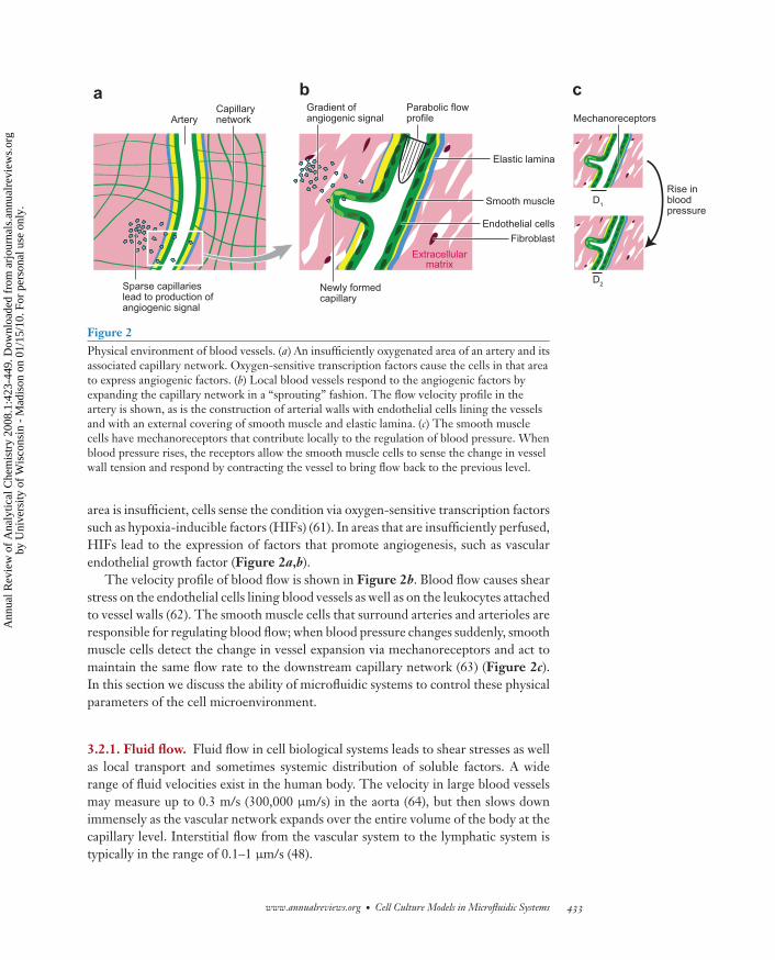
Figure 2Physical environment of blood vessels. (a) An insufficiently oxygenated area of an artery and itsassociated capillary network. Oxygen-sensitive transcription factors cause the cells in that areato express angiogenic factors. (b) Local blood vessels respond to the angiogenic factors byexpanding the capillary network in a “sprouting” fashion. The flow velocity profile in theartery is shown, as is the construction of arterial walls with endothelial cells lining the vesselsand with an external covering of smooth muscle and elastic lamina. (c) The smooth musclecells have mechanoreceptors that contribute locally to the regulation of blood pressure. Whenblood pressure rises, the receptors allow the smooth muscle cells to sense the change in vesselwall tension and respond by contracting the vessel to bring flow back to the previous level.
area is insufficient, cells sense the condition via oxygen-sensitive transcription factorssuch as hypoxia-inducible factors (HIFs) (61). In areas that are insufficiently perfused,HIFs lead to the expression of factors that promote angiogenesis, such as vascularendothelial growth factor (Figure 2a,b).
The velocity profile of blood flow is shown in Figure 2b. Blood flow causes shearstress on the endothelial cells lining blood vessels as well as on the leukocytes attachedto vessel walls (62). The smooth muscle cells that surround arteries and arterioles areresponsible for regulating blood flow; when blood pressure changes suddenly, smoothmuscle cells detect the change in vessel expansion via mechanoreceptors and act tomaintain the same flow rate to the downstream capillary network (63) (Figure 2c).In this section we discuss the ability of microfluidic systems to control these physicalparameters of the cell microenvironment.
3.2.1. Fluid flow. Fluid flow in cell biological systems leads to shear stresses as wellas local transport and sometimes systemic distribution of soluble factors. A widerange of fluid velocities exist in the human body. The velocity in large blood vesselsmay measure up to 0.3 m/s (300,000 μm/s) in the aorta (64), but then slows downimmensely as the vascular network expands over the entire volume of the body at thecapillary level. Interstitial flow from the vascular system to the lymphatic system istypically in the range of 0.1–1 μm/s (48).
www.annualreviews.org • Cell Culture Models in Microfluidic Systems 433
Ann
ual R
evie
w o
f A
naly
tical
Che
mis
try
2008
.1:4
23-4
49. D
ownl
oade
d fr
om a
rjou
rnal
s.an
nual
revi
ews.
org
by U
nive
rsity
of
Wis
cons
in -
Mad
ison
on
01/1
5/10
. For
per
sona
l use
onl
y.
ANRV362-AC01-14 ARI 13 May 2008 9:0
Bone tissue responds to mechanical signals, including shear stress in culture. Theactivity of the enzyme alkaline phosphatase (ALP) is a marker of bone cell function.Leclerc et al. studied the ALP activity of mouse calvarial osteoblastic (MC3T3-E1)cells in a PDMS microfluidic network (65). The authors found that although highflow rates led to loss of activity and eventually cell death, lower flow rates increasedALP activity up to threefold after 13 d in culture compared to static culture withmedia changes each day.
Using an array of 12 microfluidic bioreactors, Figallo et al. explored the differenti-ation of human embryonic stem cells to endothelial cells as determined by expressionof α–smooth muscle actin (66). By comparing two different culture chamber de-signs, the authors showed that cultures in chambers producing higher shear stressdeveloped a greater fraction of differentiated cells. For both chamber designs, lowerseeding density also resulted in a greater fraction of differentiated cells.
Schaff et al. constructed a device to monitor leukocyte interaction with a biologicalsubstrate under flow conditions (67). The authors validated the device using primaryhuman neutrophils and mouse fibroblasts transfected with human E-selectin. Thecapture, rolling, and deceleration to arrest were monitored under flow conditions.The cells were found to exhibit the expected response to activation by IL-8.
Microfluidic systems provide a variety of methods to control flow rate and modu-late shear stress. These methods have been used to model physical influences on cellsincluding endothelial cells, leukocytes, and osteoblasts.
3.2.2. Tissue mechanics. The mechanical properties of the cell microenvironmentare an important influence on cell behavior (68). The cell-surface molecules thatare responsible for anchoring cells to two-dimensional substrates, three-dimensionalmatrices, and neighboring cells act as receptors as well as anchors. For example, eachmember of the integrin family binds a specific set of ECM proteins (69). Integrin re-ceptor complexes are coupled to the cytoskeleton. Ingber has described the processesof mechanical and chemical signal integration on the whole-cell level in the contextof tensegrity models (70, 71). Such models present a view of sensing on the whole-cell level rather than on the individual mechanoreceptor level. In three-dimensionalcell culture, for instance, the density of the ECM has been shown to influence celldifferentiation (72). Similarly, McBeath et al. found that cell shape as dictated by theadhesive area available to cells in two-dimensional culture influenced lineage commit-ment of human mesenchymal stem cells (73). The authors showed that commitmentto adipose or osteoblastic lineages was signaled through the cytoskeletal regulationprotein RhoA. Micropatterning has also been used to elucidate various aspects of cellmechanical and spatial sensing (9, 10, 74, 75).
3.2.3. Geometry. The formation of branched duct structures (branching morpho-genesis) in the breast may be guided by a geometrical influence analogous to thevascular remodeling discussed above. Nelson et al. used three-dimensional micropat-terning to define mammary epithelial cell colonies in a collagen gel (76) and foundthat the synthetic tubules branched out at positions determined by the geometry of
434 Meyvantsson · Beebe
Ann
ual R
evie
w o
f A
naly
tical
Che
mis
try
2008
.1:4
23-4
49. D
ownl
oade
d fr
om a
rjou
rnal
s.an
nual
revi
ews.
org
by U
nive
rsity
of
Wis
cons
in -
Mad
ison
on
01/1
5/10
. For
per
sona
l use
onl
y.
ANRV362-AC01-14 ARI 13 May 2008 9:0
the tubule. The observed branching pattern was consistent with the pattern of anunknown diffusible inhibitor.
Embryoid bodies (EBs) are spherical aggregates of embryonic stem cells (ES cells)and are commonly formed as a first step in ES cell experiments. Torisawa et al.demonstrated synchronized formation of uniformly sized EBs using a microfluidicdevice (77). They showed that the size of EBs could be controlled by channel cross-sectional geometry. Similarly, Karp et al. employed PEG microwells to control EBshape (78).
In an effort to reproduce aspects of liver tissue structure, Powers et al. constructedthree-dimensional culture scaffolds via deep reactive ion etching in silicon (79). Theyselectively deposited a cell-adhesive coating on the inside walls of the culture cham-bers, as the top surface of the device and the bottom surface of each chamber didnot support cell attachment. Interestingly, the authors found that cells preculturedas spheroids fared better in the devices than those seeded from a single-cell suspen-sion. This suggests the importance of the ECM in three-dimensional culture: Duringspheroid formation cells will synthesize ECM, which then becomes an integral partof the spheroid. Similarly, Gottwald et al. produced polymer microcontainer arraysfor three-dimensional cell culture, and analyzed gene expression of hepatocellularcarcinoma cells cultured in the device (80). They identified several genes that wereupregulated in cells cultured in the device rather than in cells in conventional mono-layer culture.
Using differently sized patches of clot-inducing tissue factor, Kastrup et al. studiedthe threshold response of blood clotting (81). They compared the threshold responsesof platelet-rich and platelet-poor plasma as well as the importance of specific coagu-lation cascade factors.
Recently, Hui and Bhatia described a micromechanical device that allowed tem-poral control of cell-cell interaction (82). Their system was based on a structure madeof microfabricated silicon combs, two of which could be separated and brought intoclose contact or brought into proximity without contact. Using this system, Hui andBhatia analyzed the communication of hepatocytes and supporting stromal cells andfound that a short period of contact followed by soluble factor communication issufficient to maintain hepatocellular phenotype.
Tao et al. compared the fate of retinal progenitor cells (RPCs) cultured on porousand nonporous poly(methylmethacrylate) scaffolds after implantation into the sub-retinal space of mice (83). Although RPCs grew equally well on both substrates invitro, cell survival, differentiation, and integration into host tissue in vivo was sig-nificantly greater for porous substrates than nonporous. Upon integration with themicroenvironment provided by the mouse eye, the RPCs differentiated and expressedneuronal, glial, and retina-specific markers.
Precise microfluidic geometries have been utilized to model important influencesin the body, ranging from mammary morphogenesis to blood-clotting threshold re-sponse. As evident from the studies discussed above, geometric effects are often theresult of biochemical influences. Microfluidics enables the study of unknown bio-chemical influences [see, e.g., Nelson et al. (76)], as well as influences with complexkinetics [see, e.g., Kastrup et al. (81)].
www.annualreviews.org • Cell Culture Models in Microfluidic Systems 435
Ann
ual R
evie
w o
f A
naly
tical
Che
mis
try
2008
.1:4
23-4
49. D
ownl
oade
d fr
om a
rjou
rnal
s.an
nual
revi
ews.
org
by U
nive
rsity
of
Wis
cons
in -
Mad
ison
on
01/1
5/10
. For
per
sona
l use
onl
y.
ANRV362-AC01-14 ARI 13 May 2008 9:0
3.2.4. Temperature. Cell behavior is influenced to a large extent by the tempera-ture of the cells’ environment. Given the small thermal mass and the large surfacearea:volume ratio of microfluidic systems, temperature is a particularly importantconsideration for microfluidic cell culture devices. Along with gene expression (84),diffusion and biochemical reaction kinetics change as a function of temperature. Mi-crofluidic devices have been used to provide temperature-controlled environmentsfor biological studies.
By placing a Drosophila embryo in a microfluidic device straddling the interfacebetween two streams at different temperatures, Lucchetta et al. (8) observed thedynamics of embryonic patterning arising from the bicoid morphogen. The authorsdetected lower cell density and slower patterning in the cooler half of the embryo’sbody, followed by compensation that evened out the pattern across the entire body.
Differential flow control from two inputs at different temperatures was used byPearce et al. to control the temperature in a cell culture chamber (85). The chamberhad an electrode array on the bottom surface to monitor the electrical activity of neu-rons. The firing rates of dorsal root ganglion neurons were measured to demonstratethe operation of the system.
Microfluidic systems have proven useful for high-resolution control of tempera-ture in biological studies. These would be difficult to do in conventional culture.
3.3. Compartmentalization
Two of the advantages of PDMS are its elasticity and its ability to conform to surfaces.These properties have been exploited for valving (86) and loading of bacteria intominiature culture chambers (87). For example, valves were employed by Balagaddeet al. to construct small-volume bioreactors (88). The authors used the bioreactorsto study quorum sensing and reported that population density via broadcasting andsensing of bacteria population was autonomously regulated, resulting in populationoscillation. The small isolated volume of the reactor enabled monitoring at single-cellresolution. Compartmentalization has also been employed to specifically manipulateor analyze different parts of a single cell or individual cells that are biochemicallyconnected to other cells via gap junctions.
Neural cell bodies and axons have distinct properties and functions. Taylor et al.employed a two-compartment microfluidic system to culture polarized neurons withcell bodies and axons in separate compartments (89). The authors studied axon-specific gene expression and regeneration after physical axotomy.
One symptom common to several diseases of the nervous system is distal-to-proximal axonal degeneration. Recently, Ravula et al. demonstrated the division ofthe environment surrounding a neuron into two distinct fluid compartments, onecontaining the cell body and the other containing the axon (90). Using the compart-mentalized culture device, Ravula et al. settled a long-standing debate regarding thesource of this degeneration and showed that a chemical insult to the axon, but notthe cell body, led to the death of the axons.
In a similar experiment, Klauke et al. placed cardiomyocytes in microfluidic de-vices with two individually addressable compartments (91). The authors harvested
436 Meyvantsson · Beebe
Ann
ual R
evie
w o
f A
naly
tical
Che
mis
try
2008
.1:4
23-4
49. D
ownl
oade
d fr
om a
rjou
rnal
s.an
nual
revi
ews.
org
by U
nive
rsity
of
Wis
cons
in -
Mad
ison
on
01/1
5/10
. For
per
sona
l use
onl
y.
ANRV362-AC01-14 ARI 13 May 2008 9:0
pairs of rabbit cardiomyocytes maintaining end-to-end connections, used microma-nipulators to place the pair such that the cell-cell junction was located between twodam structures, and surrounded the junction region with mineral oil to separate thetwo compartments. Pulled-glass pipette-tip perfusion and integrated electrodes wereused to explore the mechanical and electrical coupling of the cells as well as thetransmission of Ca+2 waves under a variety of physiological and nonphysiologicalconditions.
The ability to contain cell populations or specific parts of a cell provides opportu-nities to focus biochemical influences on specific biological elements and to study thecausal relationships among elements. Although a certain level of compartmentaliza-tion can be achieved using pipettes, and although pipettes remain a superior methodfor electrical isolation of different compartments (i.e., patch clamping), microflu-idic systems have greatly increased the number of available compartmentalizationmethods.
4. SYSTEMS FOR CELL-BASED MODELS
4.1. Integrated Analysis
Every cell-based experiment requires a reliable method of extracting information.Thus far, we have considered the ways in which microfluidic systems serve to con-struct environments that yield improved cellular models. Analytical methods mayalso be integrated with microfluidic systems to produce information from cell mod-els. Analyses of single mammalian cells in microfluidic systems were recently reviewedby Sims and Allbritton (92), and analyses of cell cultures were described in reviewsby Anderson and van den Berg (6) and El-Ali et al. (3).
Many microfluidic cell culture studies have relied on traditional microscopy tech-niques including phase contrast imaging, histological stains, fluorescent dyes, andimmunocytochemistry. Yu et al. employed a plate reader to quantify cell number(93). Thompson et al. studied dynamic gene expression profiles using green fluores-cent protein reporters, and King et al. (94) studied the same for multiple genes andstimuli in a multiplexed microfluidic system.
Additionally, several methods rely on PCR. Wilding et al. reported an applicationof PCR to analyze cells captured in a microfabricated filter (95). Easley et al. presenteda microfluidic system integrating all the components necessary to go from a complexbiological sample to a final PCR result (96). Marcy et al. demonstrated genomicanalysis of complex populations of microbes at the organism level using a microfluidicgenome amplification system (97). This integrated microfluidic system contained nineamplification units and allowed selection of individual cells, lysis, sample preparation,and amplification via PCR. The results from this study demonstrate the heterogeneityof biological samples and the future utility of highly parallel microfluidic systems foranalysis.
Capillary electrophoresis was one of the first systems to employ microfluidic chan-nels (98), and it has continued to develop high-performance tools for use in variousapplications, including sequencing (99). Electrophoresis has also been combined with
www.annualreviews.org • Cell Culture Models in Microfluidic Systems 437
Ann
ual R
evie
w o
f A
naly
tical
Che
mis
try
2008
.1:4
23-4
49. D
ownl
oade
d fr
om a
rjou
rnal
s.an
nual
revi
ews.
org
by U
nive
rsity
of
Wis
cons
in -
Mad
ison
on
01/1
5/10
. For
per
sona
l use
onl
y.
ANRV362-AC01-14 ARI 13 May 2008 9:0
cell culture systems to identify and quantify the activities of intracellular enzymes(100).
Biosensors have also been employed to study cells in culture. Bratten et al. mea-sured purine release from cardiomyocytes under conditions mimicking cardiac is-chemia (101). The authors used a cascade of three enzymes to convert the analyteinto hydrogen peroxide that could be detected by the sensor. Chen et al. employedamperometric sensors to detect quantal secretion of catecholamines from individualcells in microfabricated wells (102).
Other demonstrations of integrated analytical tools include cell culture on can-tilevers coupled with an atomic force microscopy detection apparatus used to studymyotube function (103), application of cylindrical optics to count proteins in single-cell lysates (104), and integrated spectrometry for fluorescence analysis (105).
4.2. Throughput
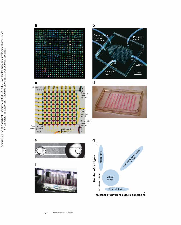
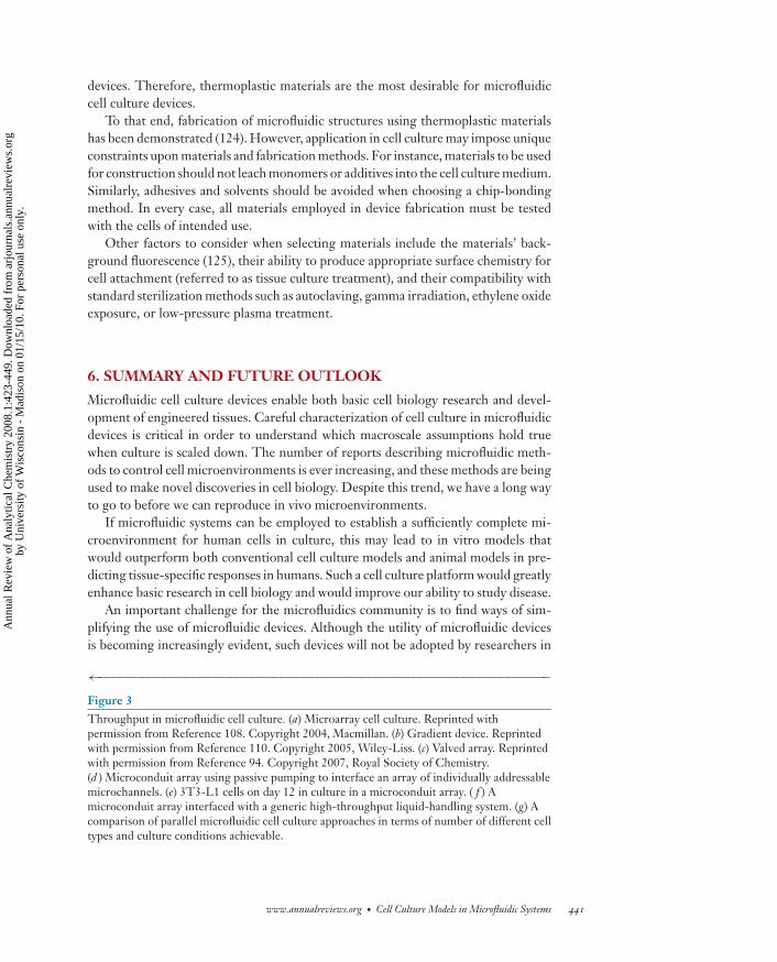
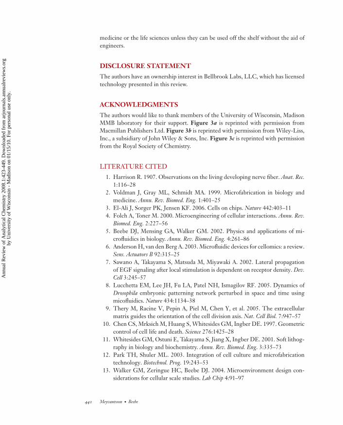
Microfluidic systems have the potential to increase the biological relevance of cellculture models and to contribute to improved assay content. For large-scale biologyresearch applications (106), as well as for industry applications such as in drug dis-covery, throughput is an important consideration. Various methods have been usedto increase microfluidic system throughput, some of which were discussed in a re-cent review by Dittrich and Manz (107). The approaches that have been applied tocell culture can be divided into four general categories: (a) microarrays, (b) gradientdevices, (c) valved arrays, and (d ) individually addressable channel arrays.
Several tools exist for spotting arrays that can be employed to spot different ma-trices for cell culture or possibly the cells themselves. Microarrays provide a highspatial density of different surface coatings and/or different cell types (78, 108,109), which represents a great advantage for screening growth surfaces, et cetera.In microarrays, however, all of the cells will be exposed to the same fluid environ-ment. Therefore, they are not suitable for screening soluble factors, including drugcompounds.
Gradient devices can provide different concentrations of a single compound for asingle cell type. Hung et al. reported the use of a microfluidic gradient generator toaddress an array of 10 × 10 cell culture chambers (110). All 10 columns were seededwith the same cell type, and a gradient generator was used to produce a linearlyvarying concentration across the rows. A similar design was reported by Thompsonet al. (111), who used the same inputs for cell seeding and perfusion. A scalable lineargradient generator for cell assays was presented by Walker et al. (112).
Valved arrays enable simultaneous testing of multiple cell types and multiple com-pounds. With the use of valves, Wang et al. demonstrated multiplexed cytotoxicitytesting in a high-density microfluidic array with six distinct cell inputs and 12 distinctcompound inputs (113). After 24 h in culture, three different cell lines were exposedto a panel of five toxins. Cell morphology and viability were comparable to those of96-well control cultures. Using a similar approach for selecting between seeding andperfusion mode, King et al. studied dynamic responses to soluble factor stimuli in an8 × 8 array (94).
438 Meyvantsson · Beebe
Ann
ual R
evie
w o
f A
naly
tical
Che
mis
try
2008
.1:4
23-4
49. D
ownl
oade
d fr
om a
rjou
rnal
s.an
nual
revi
ews.
org
by U
nive
rsity
of
Wis
cons
in -
Mad
ison
on
01/1
5/10
. For
per
sona
l use
onl
y.
ANRV362-AC01-14 ARI 13 May 2008 9:0
Meyvantsson and Beebe demonstrated cell culture in arrays of 192 individuallyaddressable microchannels (114, 115). Droplet-based passive pumping (116) was em-ployed to seed cells, change media, and treat and stain cells. This approach requires nophysical connections between external instruments and the microfluidic device, andis compatible with hand pipettes as well as generic liquid-handling robotics. Warricket al. analyzed the fluid replacement in these arrays (117) and Berthier et al. presentedan analytical model (118) that indicates that perfusion culture will be feasible usingpassive pumping.
A comparison of different approaches to high-throughput microfluidic cell assaysis presented in Figure 3. A wide range of flow velocities can be applied in perfu-sion culture using microarrays, gradient devices, and valved arrays. In channel arraysoperated by passive pumping, however, the range is smaller. As mentioned above,the natural perfusion velocity for most cells is very slow and falls within the rangeavailable via passive pumping. Whereas microarray cell culture allows screening of alarge number of surface bound elements, soluble factors, if any, are uniform acrossthe entire array. The opposite is true for gradient devices, where several differentconcentrations are possible but where all the cells and other surface-bound elementsare identical. Valved arrays represent a compromise between these two methods, andallow several cell types and several culture environments to be assayed simultane-ously. However, the constraints imposed by the physical connections required forvalved arrays severely limit throughput. In contrast, individually addressable chan-nels can have a large number of access ports (384 ports are shown in Figure 3d,and we have demonstrated up to 1536 access ports). When using passive pump-ing, the density of assay chambers is limited only by the precision of the liquid-handling equipment in its ability to control evaporation in small volumes. Thus, anumber of different approaches are being pursued, each of which has inherent advan-tages and disadvantages that must be taken into account with regard to the intendedapplication.
5. FABRICATION OF MICROFLUIDIC CELLCULTURE DEVICES
Currently, there are several fabrication challenges facing microfluidics researchers.A wide variety of macro-to-micro interfaces exist (119) that need to be standardized(120). Two important aspects of microfluidic interfacing are automation (3) and easeof handling (107), both of which must be considered when designing microfluidicsystems.
Also related to interfacing is choice of materials. Many research devices have beenmade from PDMS (121), and some interfacing strategies have relied on its elasticproperties. However, the material properties of PDMS may be problematic for cellculture models (122, 123). Because several molecules involved in cell microenviron-ments are small and hydrophobic, it is necessary to characterize the effect of PDMSabsorption of such molecules upon cell culture in microfluidic devices. Althoughglass is very common for chemical analysis devices, the cost is too high for disposable
www.annualreviews.org • Cell Culture Models in Microfluidic Systems 439
Ann
ual R
evie
w o
f A
naly
tical
Che
mis
try
2008
.1:4
23-4
49. D
ownl
oade
d fr
om a
rjou
rnal
s.an
nual
revi
ews.
org
by U
nive
rsity
of
Wis
cons
in -
Mad
ison
on
01/1
5/10
. For
per
sona
l use
onl
y.
ANRV362-AC01-14 ARI 13 May 2008 9:0
a b
c
Number of different culture conditions
Nu
mb
er o
f ce
ll ty
pes
Valvedarrays
Gradient devices
Uni
form
cul
ture
Mic
roar
rays
Indiv
iduall
y add
ress
able
arra
ys
g
d
f
e
Perfusion inlet
Seedingvalvecontrol
Cellseedingoutlet
Stimulationvalvecontrol
Stimulationoutlets
Reporter cellseeding inlets
Stimulationinlets
Waste
5 mm
Perfusionoutlet
Concentration gradient generator
2 mm
440 Meyvantsson · Beebe
Ann
ual R
evie
w o
f A
naly
tical
Che
mis
try
2008
.1:4
23-4
49. D
ownl
oade
d fr
om a
rjou
rnal
s.an
nual
revi
ews.
org
by U
nive
rsity
of
Wis
cons
in -
Mad
ison
on
01/1
5/10
. For
per
sona
l use
onl
y.
ANRV362-AC01-14 ARI 13 May 2008 9:0
devices. Therefore, thermoplastic materials are the most desirable for microfluidiccell culture devices.
To that end, fabrication of microfluidic structures using thermoplastic materialshas been demonstrated (124). However, application in cell culture may impose uniqueconstraints upon materials and fabrication methods. For instance, materials to be usedfor construction should not leach monomers or additives into the cell culture medium.Similarly, adhesives and solvents should be avoided when choosing a chip-bondingmethod. In every case, all materials employed in device fabrication must be testedwith the cells of intended use.
Other factors to consider when selecting materials include the materials’ back-ground fluorescence (125), their ability to produce appropriate surface chemistry forcell attachment (referred to as tissue culture treatment), and their compatibility withstandard sterilization methods such as autoclaving, gamma irradiation, ethylene oxideexposure, or low-pressure plasma treatment.
6. SUMMARY AND FUTURE OUTLOOK
Microfluidic cell culture devices enable both basic cell biology research and devel-opment of engineered tissues. Careful characterization of cell culture in microfluidicdevices is critical in order to understand which macroscale assumptions hold truewhen culture is scaled down. The number of reports describing microfluidic meth-ods to control cell microenvironments is ever increasing, and these methods are beingused to make novel discoveries in cell biology. Despite this trend, we have a long wayto go to before we can reproduce in vivo microenvironments.
If microfluidic systems can be employed to establish a sufficiently complete mi-croenvironment for human cells in culture, this may lead to in vitro models thatwould outperform both conventional cell culture models and animal models in pre-dicting tissue-specific responses in humans. Such a cell culture platform would greatlyenhance basic research in cell biology and would improve our ability to study disease.
An important challenge for the microfluidics community is to find ways of sim-plifying the use of microfluidic devices. Although the utility of microfluidic devicesis becoming increasingly evident, such devices will not be adopted by researchers in
←−−−−−−−−−−−−−−−−−−−−−−−−−−−−−−−−−−−−−−−−−−−−−−−−−−−−−−−−−−−−−−Figure 3Throughput in microfluidic cell culture. (a) Microarray cell culture. Reprinted withpermission from Reference 108. Copyright 2004, Macmillan. (b) Gradient device. Reprintedwith permission from Reference 110. Copyright 2005, Wiley-Liss. (c) Valved array. Reprintedwith permission from Reference 94. Copyright 2007, Royal Society of Chemistry.(d ) Microconduit array using passive pumping to interface an array of individually addressablemicrochannels. (e) 3T3-L1 cells on day 12 in culture in a microconduit array. ( f ) Amicroconduit array interfaced with a generic high-throughput liquid-handling system. (g) Acomparison of parallel microfluidic cell culture approaches in terms of number of different celltypes and culture conditions achievable.
www.annualreviews.org • Cell Culture Models in Microfluidic Systems 441
Ann
ual R
evie
w o
f A
naly
tical
Che
mis
try
2008
.1:4
23-4
49. D
ownl
oade
d fr
om a
rjou
rnal
s.an
nual
revi
ews.
org
by U
nive
rsity
of
Wis
cons
in -
Mad
ison
on
01/1
5/10
. For
per
sona
l use
onl
y.
ANRV362-AC01-14 ARI 13 May 2008 9:0
medicine or the life sciences unless they can be used off the shelf without the aid ofengineers.
DISCLOSURE STATEMENT
The authors have an ownership interest in Bellbrook Labs, LLC, which has licensedtechnology presented in this review.
ACKNOWLEDGMENTS
The authors would like to thank members of the University of Wisconsin, MadisonMMB laboratory for their support. Figure 3a is reprinted with permission fromMacmillan Publishers Ltd. Figure 3b is reprinted with permission from Wiley-Liss,Inc., a subsidiary of John Wiley & Sons, Inc. Figure 3c is reprinted with permissionfrom the Royal Society of Chemistry.
LITERATURE CITED
1. Harrison R. 1907. Observations on the living developing nerve fiber. Anat. Rec.1:116–28
2. Voldman J, Gray ML, Schmidt MA. 1999. Microfabrication in biology andmedicine. Annu. Rev. Biomed. Eng. 1:401–25
3. El-Ali J, Sorger PK, Jensen KF. 2006. Cells on chips. Nature 442:403–114. Folch A, Toner M. 2000. Microengineering of cellular interactions. Annu. Rev.
Biomed. Eng. 2:227–565. Beebe DJ, Mensing GA, Walker GM. 2002. Physics and applications of mi-
crofluidics in biology. Annu. Rev. Biomed. Eng. 4:261–866. Anderson H, van den Berg A. 2003. Microfluidic devices for cellomics: a review.
Sens. Actuators B 92:315–257. Sawano A, Takayama S, Matsuda M, Miyawaki A. 2002. Lateral propagation
of EGF signaling after local stimulation is dependent on receptor density. Dev.Cell 3:245–57
8. Lucchetta EM, Lee JH, Fu LA, Patel NH, Ismagilov RF. 2005. Dynamics ofDrosophila embryonic patterning network perturbed in space and time usingmicofluidics. Nature 434:1134–38
9. Thery M, Racine V, Pepin A, Piel M, Chen Y, et al. 2005. The extracellularmatrix guides the orientation of the cell division axis. Nat. Cell Biol. 7:947–57
10. Chen CS, Mrksich M, Huang S, Whitesides GM, Ingber DE. 1997. Geometriccontrol of cell life and death. Science 276:1425–28
11. Whitesides GM, Ostuni E, Takayama S, Jiang X, Ingber DE. 2001. Soft lithog-raphy in biology and biochemistry. Annu. Rev. Biomed. Eng. 3:335–73
12. Park TH, Shuler ML. 2003. Integration of cell culture and microfabricationtechnology. Biotechnol. Prog. 19:243–53
13. Walker GM, Zeringue HC, Beebe DJ. 2004. Microenvironment design con-siderations for cellular scale studies. Lab Chip 4:91–97
442 Meyvantsson · Beebe
Ann
ual R
evie
w o
f A
naly
tical
Che
mis
try
2008
.1:4
23-4
49. D
ownl
oade
d fr
om a
rjou
rnal
s.an
nual
revi
ews.
org
by U
nive
rsity
of
Wis
cons
in -
Mad
ison
on
01/1
5/10
. For
per
sona
l use
onl
y.
ANRV362-AC01-14 ARI 13 May 2008 9:0
14. Atencia J, Beebe DJ. 2005. Controlled microfluidic interfaces. Nature 437:648–55
15. Kim L, Toh Y, Voldman J, Yu H. 2007. A practical guide to microfluidic perfu-sion culture of adherent mammalian cells. Lab Chip 7:681–94
16. Tilles AW, Baskaran H, Roy P, Yarmush ML, Toner M. 2001. Effects of oxy-genation and flow on the viability and function of rat hepatocytes cocultured ina microchannel flat-plate bioreactor. Biotechnol. Bioeng. 73:379–89
17. Leclerc E, Sakai Y, Fujii T. 2003. Cell culture in a three-dimensional networkof PDMS (polydimethylsiloxane) microchannels. Biomed. Microdev. 5:109–14
18. Yu H, Meyvantsson I, Shkel IA, Beebe DJ. 2005. Diffusion dependent cellbehavior in microenvironments. Lab Chip 5:1089–95
19. Yu H, Alexander CM, Beebe DJ. 2007. Understanding microchannel culture:parameters involved in soluble factor signaling. Lab Chip 7:726–30
20. Raty S, Walters EM, Davis J, Zeringue H, Beebe DJ, et al. 2004. Embryonic de-velopment in the mouse is enhanced via microchannel culture. Lab Chip 4:186–90
21. Dolberg DS, Bissell MJ. 1984. Inability of Rous sarcoma virus to cause sarcomasin the avian embryo. Nature 309:552–56
22. Abbott A. 2003. Cell culture: biology’s new dimension. Nature 424:870–7223. Toh YC, Zhang C, Zhang J, Khong YM, Chang S, et al. 2007. A novel 3D
mammalian cell perfusion–culture system in microfluidic channels. Lab Chip7:302–9
24. Kim MS, Yeon JH, Park JK. 2007. A microfluidic platform for 3-dimensionalcell culture and cell-based assays. Biomed. Microdev. 9:25–34
25. Paguirigan A, Beebe DJ. 2006. Gelatin-based microfluidic devices for cell cul-ture. Lab Chip 6:407–13
26. Udy GB, Towers RP, Snell RG, Wilkins RJ, Park SH, et al. 1997. Require-ment of STAT5b for sexual dimorphism of body growth rates and liver geneexpression. Proc. Natl. Acad. Sci. USA 94:7239–44
27. Yan Y, Yang D, Zarnowska ED, Du Z, Werbel B, et al. 2005. Directed differen-tiation of dopaminergic neuronal subtypes from human embryonic stem cells.Stem Cells 23:781–90
28. Davenport RJ. 2005. What controls organ regeneration? Science 309:8429. Blaxter M. 1998. Caenorhabditis elegans is a nematode. Science 282:2041–4630. Wu D. 2005. Signaling mechanisms for regulation of chemotaxis. Cell Res.
15:52–5631. Martin P. 1997. Wound healing: aiming for perfect skin regeneration. Science
276:75–8132. Mackenzie IC, Fusenig NE. 1983. Regeneration of organized epithelial struc-
ture. J. Invest. Dermatol. 81:189–9433. Jungermann K, Kietzmann T. 2000. Oxygen: modulator of metabolic zonation
and disease of the liver. Hepatology 31:255–6034. Montesano R, Pepper MS, Orci L. 1993. Paracrine induction of angiogenesis
in vitro by Swiss 3T3 fibroblasts. J. Cell Sci. 105:1013–2435. Tabata T, Takei Y. 2004. Morphogens, their identification and regulation. De-
velopment 131:703–12
www.annualreviews.org • Cell Culture Models in Microfluidic Systems 443
Ann
ual R
evie
w o
f A
naly
tical
Che
mis
try
2008
.1:4
23-4
49. D
ownl
oade
d fr
om a
rjou
rnal
s.an
nual
revi
ews.
org
by U
nive
rsity
of
Wis
cons
in -
Mad
ison
on
01/1
5/10
. For
per
sona
l use
onl
y.
ANRV362-AC01-14 ARI 13 May 2008 9:0
36. Blake AJ, Pearce TM, Rao NS, Johnson SM, Williams JC. 2007. MultilayerPDMS microfluidic chamber for controlling brain slice microenvironment. LabChip 7:842–49
37. Jeon NL, Baskaran H, Dertinger SKW, Whitesides GM, Van de Water L, TonerM. 2002. Neutrophil chemotaxis in linear and complex gradients of interleukin-8 formed in a microfabricated device. Nat. Biotechnol. 20:826–30
38. Wang S-J, Saadi W, Lin F, Nguyen CM-C, Jeon NL. 2004. Differential effect ofEGF gradient profiles on breast cancer cell chemotaxis. Exp. Cell Res. 300:180–89
39. Irimia D, Liu SY, Tharp WG, Samadani A, Toner M, Poznansky MC. 2006. Mi-crofluidic system for measuring neutrophil migratory responses to fast switchesof chemical gradients. Lab Chip 6:191–98
40. Mao H, Cremer PS, Manson MD. 2003. A sensitive, versatile microfluidic assayfor bacterial chemotaxis. Proc. Natl. Acad. Sci. USA 100:5449–54
41. Chung BG, Flanagan LA, Rhee SW, Schwartz PH, Lee AP, et al. 2005. Humanneural stem cell growth and differentiation in a gradient-generating microfluidicdevice. Lab Chip 5:401–6
42. Walker GM, Sai J, Richmond A, Stremler M, Chung CY, Wikswo JP. 2005.Effects of flow and diffusion on chemotaxis studies in a microfabricated gradientgenerator. Lab Chip 5:611–18
43. Berthier ES, Warrick JW, Yu H, Beebe DJ. 2008. Managing evaporation formore robust microscale assays. Part 2: Characterization of convection and dif-fusion for cell biology. Lab Chip. doi: 10.1039/b717423c
44. Abhyankar VV, Lokuta MA, Huttenlocher A, Beebe DJ. 2006. Characterizationof a membrane-based gradient generator for use in cell-signaling studies. LabChip 6:389–93
45. Wu HK, Huang B, Zare RN. 2006. Generation of complex, static solutiongradients in microfluidic channels. J. Am. Chem. Soc. 128:4194–95
46. Rosoff WJ, McAllister R, Esrick MA, Goodhill GJ, Urbach JS. 2005. Genera-ting controlled molecular gradients in 3D gels. Biotechnol. Bioeng. 91:754–59
47. Cheng S, Heilman S, Wasserman M, Archer S, Shuler ML, Wu M. 2007. Ahydrogel-based microfluidic device for the studies of directed cell migration.Lab Chip 7:763–69
48. Griffith LG, Swartz MA. 2006. Capturing complex 3D tissue physiology invitro. Nat. Rev. Mol. Cell Biol. 7:211–24
49. Lutolf MP, Hubbell JA. 2005. Synthetic biomaterials as instructive extracellularmicroenvironments for morphogenesis in tissue engineering. Nat. Biotechnol.23:47–55
50. Dertinger SKW, Jiang X, Li Z, Murthy VN, Whitesides GM. 2002. Gradientsof substrate-bound laminin orient axonal specification of neurons. Proc. Natl.Acad. Sci. USA 99:12542–47
51. Tan W, Desai TA. 2004. Microscale multilayer cocultures for biomimetic bloodvessels. J. Biomed. Mater. Res. 72A:146–60
52. Frisk T, Rydholm S, Andersson H, Stemme G, Brismar H. 2005. A concept forminiaturized 3-D cell culture using an extracellular matrix gel. Electrophoresis26:4751–58
444 Meyvantsson · Beebe
Ann
ual R
evie
w o
f A
naly
tical
Che
mis
try
2008
.1:4
23-4
49. D
ownl
oade
d fr
om a
rjou
rnal
s.an
nual
revi
ews.
org
by U
nive
rsity
of
Wis
cons
in -
Mad
ison
on
01/1
5/10
. For
per
sona
l use
onl
y.
ANRV362-AC01-14 ARI 13 May 2008 9:0
53. Evans AR, Euteneuer S, Chavez E, Mullen LM, Hui EE, et al. 2007. Lamininand fibronectin modulate inner ear spiral ganglion neurite outgrowth in an invitro alternate choice assay. Dev. Neurobiol. 67:1721–30
54. Tsang VL, Chen AA, Cho LM, Jadin KD, Sah RL, et al. 2007. Fabrication of3D hepatic tissues by additive photopatterning of cellular hydrogels. FASEB J.21:790–801
55. Underhill GH, Chen AA, Albrecht DR, Bhatia SN. 2007. Assessment of hepa-tocellular function within PEG hydrogels. Biomaterials 28:256–70
56. Sud D, Mehta G, Mehta K, Linderman J, Takayama S, Mycek MA. 2006. Opticalimaging in microfluidic bioreactors enables oxygen monitoring for continuouscell culture. J. Biomed. Opt. 11:050504
57. Allen JW, Bhatia SN. 2003. Formation of steady-state oxygen gradients in vitro:application to liver zonation. Biotechnol. Bioeng. 82:253–62
58. Allen JW, Khetani SR, Bhatia SN. 2005. In vitro zonation and toxicity in ahepatocyte bioreactor. Toxicol. Sci. 84:110–19
59. Allen JW, Khetani SR, Johnson RS, Bhatia SN. 2006. In vitro liver tissue modelestablished from transgenic mice: role of HIF-1α on hypoxic gene expression.Tissue Eng. 12:3135–47
60. Park J, Bansal T, Pinelis M, Maharbiz MM. 2006. A microsystem for sensingand patterning oxidative microgradients during cell culture. Lab Chip 6:611–22
61. Pugh CW, Radcliffe PJ. 2003. Regulation of angiogenesis by hypoxia: role ofthe HIF system. Nat. Med. 9:677–84
62. Li YS, Haga JH, Chien S. 2005. Molecular basis of the effects of shear stress onvascular endothelial cells. J. Biomech. 38:1949–71
63. Davis MJ, Hill MA. 1999. Signaling mechanisms underlying the vascular myo-genic response. Physiol. Rev. 79:387–423
64. Widmaier EP, Raff H, Strang KT. 2004. Human Physiology, ed. SI Fox,pp. 375–466. New York: McGraw-Hill. 9th ed.
65. Leclerc E, David B, Griscom L, Lepioufle B, Fujii T, et al. 2006. Study ofosteoblastic cells in a microfluidic environment. Biomaterials 27:586–95
66. Figallo E, Cannizzaro C, Gerecht S, Burdick JA, Langer R, et al. 2007. Micro-bioreactor array for controlling cellular microenvironments. Lab Chip 7:710–19
67. Schaff UY, Xing MM, Lin KK, Pan N, Jeon NL, Simon SI. 2007. Vascularmimetics based on microfluidics for imaging the leukocyte–endothelial inflam-matory response. Lab Chip 7:448–56
68. Discher DE, Janmey P, Wang YL. 2005. Tissue cells feel and respond to thestiffness of their substrate. Science 310:1139–43
69. Li S, Guan JL, Chien S. 2005. Biochemistry and biomechanics of cell motility.Annu. Rev. Biomed. Eng. 7:105–50
70. Ingber DE. 2003. Tensegrity I. Cell structure and hierarchical systems biology.J. Cell Sci. 116:1157–73
71. Ingber DE. 2003. Tensegrity II. How structural networks influence cellularinformation processing networks. J. Cell Sci. 116:1397–408
72. Wozniak MA, Desai R, Solski PA, Der CJ, Keely PJ. 2003. ROCK-generatedcontractility regulates breast epithelial cell differentiation in response to the
www.annualreviews.org • Cell Culture Models in Microfluidic Systems 445
Ann
ual R
evie
w o
f A
naly
tical
Che
mis
try
2008
.1:4
23-4
49. D
ownl
oade
d fr
om a
rjou
rnal
s.an
nual
revi
ews.
org
by U
nive
rsity
of
Wis
cons
in -
Mad
ison
on
01/1
5/10
. For
per
sona
l use
onl
y.
ANRV362-AC01-14 ARI 13 May 2008 9:0
physical properties of a three-dimensional collagen matrix. J. Cell Biol. 163:583–95
73. McBeath R, Pirone DM, Nelson CM, Bhadriraju K, Chen CS. 2004. Cell shape,cytoskeletal tension, and RhoA regulate stem cell lineage commitment. Dev. Cell6:483–95
74. Balaban NQ, Schwarz US, Riveline D, Goichberg P, Tzur G, et al. 2001. Forceand focal adhesion assembly: a close relationship studied using elastic micropat-terned substrates. Nat. Cell Biol. 3:466–72
75. Tan JL, Tien J, Pirone DM, Gray DS, Bhadriraju K, Chen CS. 2003. Cells lyingon a bed of microneedles: an approach to isolate mechanical force. Proc. Natl.Acad. Sci. USA 100:1484–89
76. Nelson CM, Van Duijn MM, Inman JL, Fletcher DA, Bissell MJ. 2006. Tissuegeometry determines sites of mammary branching morphogenesis in organ-otypic cultures. Science 314:298–300
77. Torisawa Y, Chueh BH, Huh D, Ramamurthy P, Roth TM, et al. 2007. Effi-cient formation of uniform-sized embryoid bodies using a compartmentalizedmicrochannel device. Lab Chip 7:770–76
78. Karp JM, Yeh J, Eng G, Fukuda J, Blumling J, et al. 2007. Controlling size, shapeand homogeneity of embryoid bodies using poly(ethylene glycol) microwells.Lab Chip 7:786–94
79. Powers MJ, Domansky K, Kaazempur-Mofrad MR, Kalezi A, Capitano A, et al.2002. A microfabricated array bioreactor for perfused 3D liver culture. Biotech-nol. Bioeng. 78:257–69
80. Gottwald E, Giselbrecht S, Augspurger C, Lahni B, Dambrowsky N, et al. 2007.A chip-based platform for the in vitro generation of tissues in three-dimensionalorganization. Lab Chip 7:777–85
81. Kastrup CJ, Shen F, Runyon MK, Ismagilov RF. 2007. Characterization of thethreshold response of initiation of blood clotting to stimulus patch size. Biophys.J. 93:2969–77
82. Hui EE, Bhatia SN. 2007. Micromechanical control of cell-cell interactions.Proc. Natl. Acad. Sci. USA 104:5722–26
83. Tao S, Young C, Redenti S, Zhang Y, Klassen H, et al. 2007. Survival, migrationand differentiation of retinal progenitor cells transplanted on micro-machinedpoly(methyl methacrylate) scaffolds to the subretinal space. Lab Chip 7:695–701
84. Sonna LA, Fujita J, Gaffin SL, Lilly CM. 2002. Invited review: Effects of heatand cold stress on mammalian gene expression. J. Appl. Physiol. 92:1725–42
85. Pearce TM, Wilson JA, Oakes SG, Chiu SY, Williams JC. 2005. Integrated mi-croelectrode array and microfluidics for temperature clamp of sensory neuronsin culture. Lab Chip 5:97–101
86. Thorsen T, Maerkl SJ, Quake SR. 2002. Microfluidic large-scale integration.Science 298:580–84
87. Groisman A, Lobo C, Cho H, Campbell JK, Dufour YS, et al. 2005. A mi-crofluidic chemostat for experiments with bacterial and yeast cells. Nat. Methods2:685–49
446 Meyvantsson · Beebe
Ann
ual R
evie
w o
f A
naly
tical
Che
mis
try
2008
.1:4
23-4
49. D
ownl
oade
d fr
om a
rjou
rnal
s.an
nual
revi
ews.
org
by U
nive
rsity
of
Wis
cons
in -
Mad
ison
on
01/1
5/10
. For
per
sona
l use
onl
y.
ANRV362-AC01-14 ARI 13 May 2008 9:0
88. Balagadde FK, You L, Hansen CL, Arnold FH, Quake SR. 2005. Long-termmonitoring of bacteria undergoing programmed population control in a mi-crochemostat. Science 309:137–40
89. Taylor AM, Blurton-Jones M, Rhee SW, Cribbs DH, Cotman CW, Jeon NL.2005. A microfluidic culture platform for CNS axonal injury, regeneration andtransport. Nat. Methods 2:599–605
90. Ravula SK, Wang MS, McClain MA, Asress SA, Frazier B, Glass JD. 2007. Spa-tiotemporal localization of injury potentials in DRG neurons during vincristine-induced axonal degeneration. Neurosci. Lett. 415:34–39
91. Klauke N, Smith G, Cooper JM. 2007. Microfluidic systems to examine inter-cellular coupling of pairs of cardiac myocytes. Lab Chip 7:731–39
92. Sims CE, Allbritton NL. 2007. Analysis of single mammalian cells on-chip. LabChip 7:423–40
93. Yu H, Alexander CM, Beebe DJ. 2007. A platereader-compatible microchannelarray for cell biology assays. Lab Chip 7:388–91
94. King KR, Wang S, Irimia D, Jayaraman A, Toner M, Yarmush ML. 2007. Ahigh-throughput microfluidic real-time gene expression living cell array. LabChip 7:77–85
95. Wilding P, Kricka LJ, Cheng J, Hvichia G, Shoffner MA, Fortina P. 1998.Integrated cell isolation and polymerase chain reaction analysis using siliconmicrofilter chambers. Anal. Biochem. 257:95–100
96. Easley CJ, Karlinsey JM, Bienvenue JM, Legendre LA, Roper MG, et al. 2006. Afully integrated microfluidic genetic analysis system with sample-in-answer-outcapability. Proc. Natl. Acad. Sci. USA 103:19272–77
97. Marcy Y, Ouverney C, Bik EM, Losekann T, Ivanova N, et al. 2007. Dissectingbiological “dark matter” with single-cell genetic analysis of rare and uncultivatedTM7 microbes from the human mouth. Proc. Natl. Acad. Sci. USA 104:11889–94
98. Harrison DJ, Fluri K, Seiler K, Fan Z, Effenhauser CS, Manz A. 1993. Mi-cromachining a miniaturized capillary electrophoresis–based chemical analysissystem on a chip. Science 261:895–97
99. Aborn JH, El-Difrawy SA, Novotny M, Gismondi EA, Lam R, et al. 2005. A768-lane microfabricated system for high-throughput DNA sequencing. LabChip 5:669–74
100. Meredith GD, Sims CE, Soughayer JS, Allbritton NL. 2000. Measure-ment of kinase activation in single mammalian cells. Nat. Biotechnol. 18:309–12
101. Bratten CDT, Cobbold PH, Cooper JM. 1998. Single-cell measurements ofpurine release using a micromachined electroanalytical sensor. Anal. Chem.70:1164–70
102. Chen P, Xu B, Tokranova N, Feng X, Castracane J, Gillis KD. 2003. Amper-ometric detection of quantal catecholamine secretion from individual cells onmicromachined silicon chips. Anal. Chem. 75:518–24
103. Wilson K, Molnar P, Hickman J. 2007. Integration of functional myotubes witha Bio-MEMS device for non-invasive interrogation. Integration of functionalmyotubes with a Bio-MEMS device for non-invasive interrogation. Lab Chip7:920–22
www.annualreviews.org • Cell Culture Models in Microfluidic Systems 447
Ann
ual R
evie
w o
f A
naly
tical
Che
mis
try
2008
.1:4
23-4
49. D
ownl
oade
d fr
om a
rjou
rnal
s.an
nual
revi
ews.
org
by U
nive
rsity
of
Wis
cons
in -
Mad
ison
on
01/1
5/10
. For
per
sona
l use
onl
y.
ANRV362-AC01-14 ARI 13 May 2008 9:0
104. Huang B, Wu H, Bhaya D, Grossman A, Granier S, et al. 2007. Countinglow-copy number proteins in a single cell. Science 315:81–84
105. Schmidt O, Bassler M, Kiesel P, Knollenberg C, Johnson N. 2007. Fluorescencespectrometer-on-a-fluidic-chip. Lab Chip 7:626–29
106. Abraham VC, Taylor DL, Haskins JR. 2004. High content screening applied tolarge-scale cell biology. Trends Biotechnol. 22:15–22
107. Dittrich PS, Manz A. 2006. Lab-on-a-chip: microfluidics in drug discovery.Nat. Rev. Drug Discov. 5:210–18
108. Anderson DG, Levenberg S, Langer R. 2004. Nanoliter-scale synthesis of ar-rayed biomaterials and application to human embryonic stem cells. Nat. Biotech-nol. 22:863–66
109. Flaim CJ, Chien S, Bhatia SN. 2005. An extracellular matrix microarray forprobing cellular differentiation. Nat. Methods 2:119–25
110. Hung PJ, Lee PJ, Sabounchi P, Lin R, Lee LP. 2005. Continuous perfusionmicrofluidic cell culture array for high-throughput cell-based assays. Biotechnol.Bioeng. 89:1–8
111. Thompson DM, King KR, Wieder KJ, Toner M, Yarmush ML, Jayaraman A.2004. Dynamic gene expression profiling using a microfabricated living cellarray. Anal. Chem. 76:4098–103
112. Walker GM, Monteiro-Riviere N, Rouse J, O’Neill AT. 2007. A linear dilutionmicrofluidic device for cytotoxicity assays. Lab Chip 7:226–32
113. Wang Z, Kin M, Marquez M, Thorsen T. 2007. High-density microfluidicarrays for cell cytotoxicity analysis. Lab Chip 7:740–45
114. Meyvantsson I, Beebe DJ. 2005. High throughput microfluidic system for het-erogeneous assays. Proc. Annu. Int. IEEE/EMBS Conf. Microtech. Med. Biol., 3rd,Oahu, pp. 42–44. Piscataway: IEEE
115. Meyvantsson I, Warrick JW, Hayes S, Skoien A, Beebe DJ. 2008. Auto-mated cell culture in high density tubeless microfluidic device arrays. Lab Chip.doi: 10.1039/b71537a
116. Walker GM, Beebe DJ. 2002. A passive pumping method for microfluidic de-vices. Lab Chip 2:131–34
117. Warrick JW, Meyvantsson I, Ju J, Beebe DJ. 2007. High-throughput microflu-idics: improved sample treatment and washing over standard wells. Lab Chip7:316–21
118. Berthier ES, Berthier JC, Beebe DJ. 2007. Flow rate analysis of a surface tensiondriven micropump. Lab Chip 7:1475–78
119. Fredrickson CK, Fan ZH. 2004. Macro-to-micro interfaces for microfluidicdevices. Lab Chip 4:526–33
120. Whitesides GM. 2006. The origins and the future of microfluidics. Nature442:368–73
121. Duffy D, McDonald J, Schueller O, Whitesides GM. 1998. Rapid prototypingof microfluidic systems in poly(dimethylsiloxane). Anal. Chem. 70:4974–84
122. Toepke MW, Beebe DJ. 2006. PDMS absorption of small molecules and con-sequences in microfluidic applications. Lab Chip 6:1484–86
448 Meyvantsson · Beebe
Ann
ual R
evie
w o
f A
naly
tical
Che
mis
try
2008
.1:4
23-4
49. D
ownl
oade
d fr
om a
rjou
rnal
s.an
nual
revi
ews.
org
by U
nive
rsity
of
Wis
cons
in -
Mad
ison
on
01/1
5/10
. For
per
sona
l use
onl
y.
ANRV362-AC01-14 ARI 13 May 2008 9:0
123. Mukhopadhyay R. 2007. When PDMS isn’t the best: What are its weaknesses,and which other polymers can researchers add to their toolboxes? Anal. Chem.79:3248–53
124. Becker H, Gartner C. 2000. Polymer microfabrication methods for microfluidicanalytical applications. Electrophoresis 21:12–26
125. Hawkins KR, Yager P. 2003. Nonlinear decrease of background fluorescencein polymer thin-films: a survey of materials and how they can complicate fluo-rescence detection in microTAS. Lab Chip 3:248–52
www.annualreviews.org • Cell Culture Models in Microfluidic Systems 449
Ann
ual R
evie
w o
f A
naly
tical
Che
mis
try
2008
.1:4
23-4
49. D
ownl
oade
d fr
om a
rjou
rnal
s.an
nual
revi
ews.
org
by U
nive
rsity
of
Wis
cons
in -
Mad
ison
on
01/1
5/10
. For
per
sona
l use
onl
y.
AR362-FM ARI 13 May 2008 9:33
Annual Review ofAnalyticalChemistry
Volume 1, 2008Contents
A Personal Journey of Discovery: Developing Technology andChanging BiologyLee Hood � � � � � � � � � � � � � � � � � � � � � � � � � � � � � � � � � � � � � � � � � � � � � � � � � � � � � � � � � � � � � � � � � � � � � � � � � � � � � � � � � � � � �1
Spectroscopic and Statistical Techniques for Information Recoveryin Metabonomics and MetabolomicsJohn C. Lindon and Jeremy K. Nicholson � � � � � � � � � � � � � � � � � � � � � � � � � � � � � � � � � � � � � � � � � � � � � � � � 45
Mass Spectrometry for Rapid Characterization of MicroorganismsPlamen A. Demirev and Catherine Fenselau � � � � � � � � � � � � � � � � � � � � � � � � � � � � � � � � � � � � � � � � � � � � 71
Scanning Electrochemical MicroscopyShigeru Amemiya, Allen J. Bard, Fu-Ren F. Fan, Michael V. Mirkin,and Patrick R. Unwin � � � � � � � � � � � � � � � � � � � � � � � � � � � � � � � � � � � � � � � � � � � � � � � � � � � � � � � � � � � � � � � � � � � � � 95
Novel Detection Schemes of Nuclear Magnetic Resonance andMagnetic Resonance Imaging: Applications from AnalyticalChemistry to Molecular SensorsElad Harel, Leif Schröder, and Shoujun Xu � � � � � � � � � � � � � � � � � � � � � � � � � � � � � � � � � � � � � � � � � � � �133
Chemical Cytometry: Fluorescence-Based Single-Cell AnalysisDaniella Cohen, Jane A. Dickerson, Colin D. Whitmore, Emily H. Turner,Monica M. Palcic, Ole Hindsgaul, and Norman J. Dovichi � � � � � � � � � � � � � � � � � � � � � � � � � � �165
Chemical Analysis of Single CellsLaura M. Borland, Sumith Kottegoda, K. Scott Phillips,and Nancy L. Allbritton � � � � � � � � � � � � � � � � � � � � � � � � � � � � � � � � � � � � � � � � � � � � � � � � � � � � � � � � � � � � � � � � � �191
Ion Chemistry in the Interstellar MediumTheodore P. Snow and Veronica M. Bierbaum � � � � � � � � � � � � � � � � � � � � � � � � � � � � � � � � � � � � � � � � � �229
Plasma Diagnostics for Unraveling Process ChemistryJoshua M. Stillahn, Kristina J. Trevino, and Ellen R. Fisher � � � � � � � � � � � � � � � � � � � � � � � � �261
Biomolecule Analysis by Ion Mobility SpectrometryBrian C. Bohrer, Samuel I. Merenbloom, Stormy L. Koeniger,Amy E. Hilderbrand, and David E. Clemmer � � � � � � � � � � � � � � � � � � � � � � � � � � � � � � � � � � � � � � � � � �293
In Vitro Electrochemistry of Biological SystemsKelly L. Adams, Maja Puchades, and Andrew G. Ewing � � � � � � � � � � � � � � � � � � � � � � � � � � � � � �329
vii
Ann
ual R
evie
w o
f A
naly
tical
Che
mis
try
2008
.1:4
23-4
49. D
ownl
oade
d fr
om a
rjou
rnal
s.an
nual
revi
ews.
org
by U
nive
rsity
of
Wis
cons
in -
Mad
ison
on
01/1
5/10
. For
per
sona
l use
onl
y.
AR362-FM ARI 13 May 2008 9:33
Current Applications of Liquid Chromatography/Mass Spectrometryin Pharmaceutical Discovery After a Decade of InnovationBradley L. Ackermann, Michael J. Berna, James A. Eckstein, Lee W. Ott,and Ajai K. Chaudhary � � � � � � � � � � � � � � � � � � � � � � � � � � � � � � � � � � � � � � � � � � � � � � � � � � � � � � � � � � � � � � � � � �357
Optical Probes for Molecular Processes in Live CellsYoshio Umezawa � � � � � � � � � � � � � � � � � � � � � � � � � � � � � � � � � � � � � � � � � � � � � � � � � � � � � � � � � � � � � � � � � � � � � � � � � �397
Cell Culture Models in Microfluidic SystemsIvar Meyvantsson and David J. Beebe � � � � � � � � � � � � � � � � � � � � � � � � � � � � � � � � � � � � � � � � � � � � � � � � � �423
Peptides in the Brain: Mass Spectrometry–Based MeasurementApproaches and ChallengesLing jun Li and Jonathan V. Sweedler � � � � � � � � � � � � � � � � � � � � � � � � � � � � � � � � � � � � � � � � � � � � � � � � � �451
Analysis of Atmospheric AerosolsKimberly A. Prather, Courtney D. Hatch, and Vicki H. Grassian � � � � � � � � � � � � � � � � � � � �485
Multiplexed Spectroscopic DetectionsKyle D. Bake and David R. Walt � � � � � � � � � � � � � � � � � � � � � � � � � � � � � � � � � � � � � � � � � � � � � � � � � � � � � � � �515
Terrestrial Analysis of the Organic Component of Comet DustScott A. Sandford � � � � � � � � � � � � � � � � � � � � � � � � � � � � � � � � � � � � � � � � � � � � � � � � � � � � � � � � � � � � � � � � � � � � � � � � �549
High-Resolution Mass SpectrometersAlan G. Marshall and Christopher L. Hendrickson � � � � � � � � � � � � � � � � � � � � � � � � � � � � � � � � � � � �579
Surface-Enhanced Raman SpectroscopyPaul L. Stiles, Jon A. Dieringer, Nilam C. Shah, and Richard P. Van Duyne � � � � � � �601
Time-Resolved Microdialysis for In Vivo NeurochemicalMeasurements and Other ApplicationsKristin N. Schultz and Robert T. Kennedy � � � � � � � � � � � � � � � � � � � � � � � � � � � � � � � � � � � � � � � � � � � � � �627
Applications of Ultrafast Lasers for Optical Measurementsin Combusting Flows
James R. Gord, Terrence R. Meyer, and Sukesh Roy � � � � � � � � � � � � � � � � � � � � � � � � � � � � � � � � � � �663
Matrix-Assisted Laser Desorption/Ionization Imaging MassSpectrometry for the Investigation of Proteins and PeptidesKristin E. Burnum, Sara L. Frappier, and Richard M. Caprioli � � � � � � � � � � � � � � � � � � � � � �689
Formation and Characterization of Organic Monolayerson Semiconductor Surfaces
Robert J. Hamers � � � � � � � � � � � � � � � � � � � � � � � � � � � � � � � � � � � � � � � � � � � � � � � � � � � � � � � � � � � � � � � � � � � � � � � � �707
Nanoscopic Porous SensorsJohn J. Kasianowicz, Joseph W.F. Robertson, Elaine R. Chan, Joseph E. Reiner,and Vincent M. Stanford � � � � � � � � � � � � � � � � � � � � � � � � � � � � � � � � � � � � � � � � � � � � � � � � � � � � � � � � � � � � � � � � �737
viii Contents
Ann
ual R
evie
w o
f A
naly
tical
Che
mis
try
2008
.1:4
23-4
49. D
ownl
oade
d fr
om a
rjou
rnal
s.an
nual
revi
ews.
org
by U
nive
rsity
of
Wis
cons
in -
Mad
ison
on
01/1
5/10
. For
per
sona
l use
onl
y.
AR362-FM ARI 13 May 2008 9:33
Combining Self-Assembled Monolayers and Mass Spectrometryfor Applications in BiochipsZachary A. Gurard-Levin and Milan Mrksich � � � � � � � � � � � � � � � � � � � � � � � � � � � � � � � � � � � � � � � � �767
Liposomes: Technologies and Analytical ApplicationsAldo Jesorka and Owe Orwar � � � � � � � � � � � � � � � � � � � � � � � � � � � � � � � � � � � � � � � � � � � � � � � � � � � � � � � � � � �801
Fundamentals of Protein Separations: 50 Years of Nanotechnology,and Growing
David A. Egas and Mary J. Wirth � � � � � � � � � � � � � � � � � � � � � � � � � � � � � � � � � � � � � � � � � � � � � � � � � � � � �833
Functional and Spectroscopic Measurements with Scanning TunnelingMicroscopyAmanda M. Moore and Paul S. Weiss � � � � � � � � � � � � � � � � � � � � � � � � � � � � � � � � � � � � � � � � � � � � � � � � � �857
Coherent Anti-Stokes Raman Scattering Microscopy: ChemicalImaging for Biology and MedicineConor L. Evans and X. Sunney Xie � � � � � � � � � � � � � � � � � � � � � � � � � � � � � � � � � � � � � � � � � � � � � � � � � � � � �883
Contents ix
Ann
ual R
evie
w o
f A
naly
tical
Che
mis
try
2008
.1:4
23-4
49. D
ownl
oade
d fr
om a
rjou
rnal
s.an
nual
revi
ews.
org
by U
nive
rsity
of
Wis
cons
in -
Mad
ison
on
01/1
5/10
. For
per
sona
l use
onl
y.