microfluidic screening of circulating tumor biomarkers
TRANSCRIPT

Full Terms & Conditions of access and use can be found athttp://www.tandfonline.com/action/journalInformation?journalCode=lspr20
Separation & Purification Reviews
ISSN: 1542-2119 (Print) 1542-2127 (Online) Journal homepage: http://www.tandfonline.com/loi/lspr20
Microfluidic Screening of Circulating TumorBiomarkers toward Liquid Biopsy
Nanjing Hao & John X.J. Zhang
To cite this article: Nanjing Hao & John X.J. Zhang (2018) Microfluidic Screening of CirculatingTumor Biomarkers toward Liquid Biopsy, Separation & Purification Reviews, 47:1, 19-48, DOI:10.1080/15422119.2017.1320763
To link to this article: https://doi.org/10.1080/15422119.2017.1320763
Accepted author version posted online: 28Apr 2017.Published online: 22 May 2017.
Submit your article to this journal
Article views: 274
View Crossmark data
Citing articles: 5 View citing articles

Microfluidic Screening of Circulating TumorBiomarkers toward Liquid Biopsy
Nanjing Hao and John X.J. ZhangThayer School of Engineering, Dartmouth College, Hanover, New Hampshire, USA
The development of early and personalized diagnostic protocol with rapid response and highaccuracy is considered the most promising avenue to advance point-of-care testing for tumordiagnosis and therapy. Given the growing awareness of the limitations of conventional tissuebiopsy for gathering tumor information, considerable interest has recently been aroused inliquid biopsy. Among a myriad of analytical approaches proposed for liquid biopsy, micro-fluidics-based separation and purification techniques possess merits of high throughput, lowsamples consumption, high flexibility, low cost, high sensitivity, automation capability andenhanced spatio-temporal control. These characteristics endow microfluidics to serve as anemerging and promising tool in tumor diagnosis and prognosis by identifying specificcirculating tumor biomarkers. In this review, we will put our focus on three key categoriesof circulating tumor biomarkers, namely, circulating tumor cells (CTCs), circulating exo-somes, and circulating nucleic acids (cNAs), and discuss the significant roles of microfluidicsin the separation and analysis of circulating tumor biomarkers. Recent advances in micro-fluidic separation and analysis of CTCs, exosomes, and cNAs will be highlighted andtabulated. Finally, the current challenges and future niches of using microfluidic techniquesin the separation and analysis of circulating tumor biomarkers will be discussed.
Keywords: Microfluidic, separation, purification, tumor biomarker, liquid biopsy
INTRODUCTION
Although great progress has been made in the diagnosis andtreatment, cancer is still the leading cause of death world-wide. Cancer metastasis, which occurs in a multistep pro-cess including migration and invasion from primary tumor,intravasation and survival in the circulation system, extra-vasation into distant tissues, and establishing growth inseeded locus, makes it more difficult to successfully treatthe disease (1). Therefore, early diagnosis, real-time mon-itoring and accurate prediction are the most critical issues incancers. In contrast to conventional tissue biopsy that couldtake time to process and analyze and be costly, painful, anddifficult to obtain, liquid biopsy offers a new and unique
opportunity to identify the potential tumor biomarkers forpredicting tumor progression and thus have attracted moreattentions in recent years (2).
Since then, great efforts have been devoted to the separa-tion and analysis of circulating tumor biomarkers throughliquid biopsy (3). Gained from the advances of micro-/nano-fabrication approaches, microfluidics-based separation andanalysis techniques offer tremendous opportunities in point-of-care disease examination and state-of-art personalizedhealthcare devices. Typically, microfluidics possesses meritsof high throughput, low samples consumption, high flex-ibility, low cost, high sensitivity, automation capability andenhanced spatio-temporal control (4). These characteristicsendow them to serve as a promising tool in tumor diagnosisand prognosis by identifying specific circulating tumorbiomarkers.
This review will focus on how the advantages of micro-fluidics-based separation and purification techniques havebeen exploited to enhance liquid biopsy analysis. We willemphasize several typical circulating tumor biomarkers
Received 14 October 2016, Accepted 2 April 2017.Address correspondence to John X.J. Zhang, Thayer School of Engineering,
Dartmouth College, 14 Engineering Drive, Hanover, New Hampshire 03755,USA. E-mail: [email protected]
Color versions of one or more of the figures in the article can be foundonline at www.tandfonline.com/lspr.
Separation & Purification Reviews, 47: 19–48, 2018Copyright © Taylor & Francis Group, LLCISSN: 1542-2119 print / 1542-2127 onlineDOI: https://doi.org/10.1080/15422119.2017.1320763

from liquid biopsy, including circulating tumor cells(CTCs), circulating exosomes and circulating tumor nucleicacids (ctNAs). The roles of microfluidics in the liquidbiopsy will be described. Recent advances of microfluidicsin the separation and analysis of circulating tumor biomar-kers will be highlighted. Finally, the current challenges andfuture niches of using microfluidic systems in the separationand analysis of circulating tumor biomarkers will be sum-marized and discussed accordingly.
CIRCULATING TUMOR BIOMARKERS FOR THELIQUID BIOPSY
Background of the Liquid Biopsy
Cancer still represents the leading cause of death worldwide,and the major cause of cancer-related death is metastasis.Metastasis is a multistep process in which tumor cellsescape from the primary tumor site, enter into the blood-stream and then form to secondary tumor colonies(Figure 1). This metastasis process generally occurs in par-allel to the development of the primary tumor, and oftenbefore that tumor can be initially detected. Therefore, to beable to effectively improve cancer patient survival, bothearly real-time diagnosis and the frequent monitoring ofpatient response to treatment should be carefully and timelyperformed.
Conventional clinical protocols for cancer diagnosis andtreatment are usually based on tissue biopsy, which can besurgical biopsy, radiologically guided biopsy, or endoscopicbiopsy. Of these, surgical biopsy is the most commonly usedexamination technique, which consists of sampling tissuesand cells from human body by puncturing organs with aspecially designed needle. This technique is invasive,expensive and may introduce clinical risks to the patient.In addition, it also takes time, needs to be consistentlyevaluated by expert pathologists and thus constitutes a sig-nificant barrier for easy and frequent monitoring of cancerprogressions. Comparatively, radiologically guided biopsyand endoscopic biopsy techniques have a relatively betteroperation performance, but both of them make patient sufferfrom discomfort, and also need expensive equipment andexpert pathologists. Therefore, there is an urgent clinicalneed for the research and development of alternative tech-niques that can tell cancer information in a simpler andmore convenient way compared with tissue biopsy.
The use of biological fluids as a source of noninvasivetumor biomarkers has recently raised a great deal of interest.This so-called liquid biopsy holds great clinical promise, astheir noninvasive feature can allow for rapid, facile, eco-nomical and repeat sampling. In addition, regarding theincreasing awareness of the tumor genetic heterogeneity,liquid biopsy also provides great hope to capture the entireprofile of tumor genetic information, which may be prob-ably missed by tissue biopsy. Consequently, liquid biopsy is
FIGURE 1 Schematic illustration showing cancer progression (A), metastasis (B) and cancer-derived circulating biomarkers presented in the blood of cancerpatient (C).
20 N. HAO AND J.X.J. ZHANG
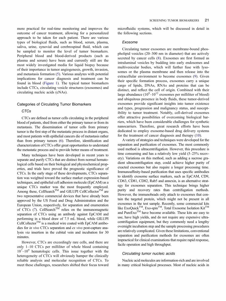
more practical for real-time monitoring and improves theoutcome of cancer treatment, allowing for a personalizedapproach to be taken for each patient. There are varioustypes of biological fluids, such as blood, serum, plasma,saliva, urine, synovial and cerebrospinal fluid, which canbe sampled to monitor the level of tumor biomarkers.Peripheral blood and blood-derived products (such asplasma and serum) have been and currently still are themost widely investigated media for liquid biopsy becauseof their importance in tumor angiogenesis, growth, invasion,and metastasis formation (5). Various analytes with potentialimplications for cancer diagnosis and treatment can befound in blood (Figure 1). The typical tumor biomarkersinclude CTCs, circulating vesicle structures (exosomes) andcirculating nucleic acids (cNAs).
Categories of Circulating Tumor Biomarkers
CTCs
CTCs are defined as tumor cells circulating in the peripheralblood of patients, shed from either the primary tumor or from itsmetastasis. The dissemination of tumor cells from primarytumor is the first step of the metastatic process in distant organs,and most patients with epithelial cancers die of metastasis ratherthan from primary tumors (6). Therefore, identification andcharacterization of CTCs offer great opportunities to understandthe metastatic process and to provide better means of treatment.
Many techniques have been developed in recent years toseparate and purify CTCs that are distinct from normal hemato-logical cells based on their biological and physiochemical prop-erties, and trials have proved the prognostic significance ofCTCs. In the early stage of these developments, CTCs separa-tion was weighted toward the surface marker expression-basedtechniques, and epithelial cell adhesion molecule (EpCAM) as aunique CTCs marker was the most frequently employed.Among these, CellSearchTM and GILUPI CellCollectorTM aretwo representative commercial devices that have already beenapproved by the US Food and Drug Administration and theEuropean Union, respectively, for separation and enumerationof CTCs (7). CellSearchTM relies on the immunomagneticseparation of CTCs using an antibody against EpCAM andperforming in a blood draw of 7.5 mL blood, while GILUPICellCollectorTM is a medical wire coated with EpCAM antibo-dies for in vivo CTCs separation and ex vivo post-capture ana-lysis via insertion in the cubital vein and incubation for 30minutes.
However, CTCs are exceedingly rare cells, and there areonly 1–10 CTCs per milliliter of whole blood containing107–109 hematologic cells. This issue together with theheterogeneity of CTCs will obviously hamper the clinicallyreliable analysis and molecular recognition of CTCs. Tomeet these challenges, researchers shifted their focus toward
microfluidic systems, which will be discussed in detail inthe following sections.
Exosome
Circulating tumor exosomes are membrane-bound phos-pholipid vesicles (20–300 nm in diameter) that are activelysecreted by cancer cells (8). Exosomes are first formed asintraluminal vesicles by budding into early endosomes andmultivesicular bodies, which will further fuse with lyso-somes or the plasma membrane and then release into theextracellular environment to become exosomes (9). Giventheir specific formation process, exosomes carry a uniquecargo of lipids, DNAs, RNAs and proteins that can bedistinct, and reflect the cell of origin. Combined with theirlarge abundance (109–1012 exosomes per milliliter of blood)and ubiquitous presence in body fluids, these tumor-derivedexosomes provide significant insights into tumor existenceand types, progression and malignancy status, and suscept-ibility to tumor treatment. Notably, cell-derived exosomesoffer attractive possibilities of overcoming biological bar-riers, which have been considerable challenges for syntheticnanocarriers. Therefore, great research efforts have beendedicated to employ exosome-based drug delivery systemsfor the treatment of cancer diagnosis and therapy (10).
Avariety of strategies and techniques were developed for theseparation and purification of exosomes. The most commonlyused method is ultracentrifugation. However, this procedure istime consuming and has a relatively low yield (5–25% recov-ery). Variations on this method, such as adding a sucrose gra-dient ultracentrifugation step, could achieve higher purity ofexacted exosomes but also require a longer processing time.Immunoaffinity-based purification that uses specific antibodiesto identify exosome surface markers, such as EpCAM, CD9,CD63, CD81, CD82, Rab5 and annexin, is an alternative strat-egy for exosomes separation. This technique brings higherpurity and recovery rates than centrifugation methods.However, the immunobeads only attach to exosomes that con-tain the targeted protein, which might not be present in allexosomes in the test sample. Recently, some commercial kitslike ExoQuickTM, Exo-spinTM, Total Exosome Isolation KitTM
and PureExoTM have become available. These kits are easy touse, have high yields, and do not require any expensive ultra-centrifugation equipment, but they commonly need a lengthyovernight incubation step and the sample processing proceduresare relatively complicated. Given these limitations, conventionalseparation and purification methods for exosomes are oftenimpractical for clinical examinations that require rapid response,facile operation and high throughput.
Circulating tumor nucleic acids
Nucleic acidmolecules are information-rich and are involvedin many critical biological processes. Most of nucleic acids in
SCREENING TUMOR BIOMARKERS 21

the body are located within cells, but a small amount of nucleicacids can also be found circulating freely in the blood plasma orserum. The term “Circulating Nucleic Acids” (cNAs) refers tocell-free segments of DNA or RNA found in the bloodstream.The discovery of cNAs in the blood was first reported byMandel and Metais in 1948, but was initially not widely recog-nized (11). Evidence that tumor cells can release their nucleicacids into the blood was provided in 1990s (12). cNAs can beused to noninvasively determine the tumor status by decodingthe contained genetic and epigenetic information, emerging as apromising liquid biopsy biomarker for cancer early diagnosisand therapy efficacy assessment (13). In particular, cNAsreleased from tumor cells have recently attracted great attentionbecause they can become detectable in the blood of bladder,breast and colorectal cancer patients before the appearance ofother circulating tumor biomarkers, such as CTCs (14).
Circulating DNA was first demonstrated in human bloodwith concentrations depending on cancer type and progres-sion stage (11). Most cell-free DNA (cfDNA) fragmentsgenerating from cell apoptosis are 100–200 base pairs inlength, whereas longer fragments (up to 10,000 base pairs)are generated from cell necrosis (15). The concentrations ofcfDNA in cancer patient are generally higher than those inhealthy individuals (3, 16), indicating that the level ofcfDNA can be used directly for cancer screening. Manymutation sites, such as KRAS and EGFR, have been dis-covered in circulating DNA of cancer patients. MessengerRNA (mRNA) is well-known central to gene expression,which in turn plays a significant role in cellular physiology.Accordingly, cancer disease can often involve changes tothe expression of certain genes, which can thus be analyzedthrough mRNA detection and quantification (17). Sincetheir discovery in the early 1990s (18), micro RNA(miRNA) also holds great promise as distinctive and non-invasive cancer biomarkers (19). miRNAs are small (18–24nucleotides long), single-stranded, endogenous and nonpro-tein-coding RNA molecules that regulate gene expression atthe posttranscriptional level. Circulating miRNAs play keyroles in tumor development and progression, and more than79 miRNAs have been reported as plasma or serum biomar-kers of tumors, such as breast, colon, prostate, melanoma,lung, ovarian, esophageal and gastric cancer (19).
Circulating tumor nucleic acids (ctNAs), especiallyDNA, mRNA and miRNA, have already been extensivelydemonstrated their throughout tumor management in theassessment of disease progression, treatment response andprognosis. However, the clinical analysis of ctNAs stillfaces several key challenges, including relatively low con-centration level, great background noise and small size.Conventional methods such as polymerase chain reaction(PCR)-based approaches usually suffer from many draw-backs, including high false-positive rate, low throughputand the lack of reproducibility (17, 20, 21). These chal-lenges set the stage where novel platforms such as micro-fluidics can play a revolutionary role.
ROLES OF MICROFLUIDICS IN THE LIQUIDBIOPSY
Microfluidics is the science and engineering of systemsthat can precisely manipulate and control small amountsof fluids, usually in the range of microliters (10−6 L) topicoliter (10−12 L), which are geometrically constrained inthe networks of channels with dimensions from a fewmicrometers up to a millimeter (22). It adopts an inte-grated, multidisciplinary and intellectual strategy withcontributions from physics, engineering, chemistry, mate-rials and biotechnology. Microfluidic systems typicallyfeature high analytical performance, great sensitivity,short analysis time, small volume of analytes, low cost,high system integration, improved potential for automa-tion and control, and disposability (23). This enablingtechnology holds great promise to open new ways inmodern analytical chemistry, pharmaceutics, cell biology,genetics and many other research areas.
Microfluidics normally consists of a set of operationunits that allow different analytes to be examined in aneasy and flexible manner. These microfluidic components,such as mixer, actuator, reactor, separator, sensor, valve andpump, can be designed and optimized separately for trans-port processes and fluid control (24). Manufacturing meth-ods of microfluidic devices were developed in thesemiconductor and microelectromechanical systems(MEMS) industry; therefore, substrates such as silicon,glass and other various types of polymers are commonlyused in their production (25). Silicon is appealing as it is anextensively used material in semiconductor technology withwell-known properties (such as chemically and thermallystable) and fabrication methods (such as routine etching andphotolithography procedures), which are highly standar-dized. Glass as an early replacement for silicon is lessexpensive, negatively charged, chemically stable and trans-parent, which is especially attractive when optical detectiontechniques are used. Polymers offer many interesting prop-erties for microfluidic devices, such as inexpensive, dispo-sable, good structural rigidity and strength, facile channelsformation via soft lithography process, and easy sealing ofdiscrete parts by thermal or adhesive treatment. The choiceof polymeric materials is commonly restricted to solvent-resistant materials, such as poly(dimethylsiloxane) (PDMS),poly(methylmethacrylate) (PMMA), thermoset polyesters,polyimide, SU-8 (negative photoresist) and teflon (24, 25).
Inherently, microfluidics could lead them to achievespatial and temporal control over analytes, which makesit an effective means to interrogate the constituents ofbiological fluids for disease diagnosis and management.When it comes to exploring the circulating tumor biomar-kers in liquid biopsy, all these appealing features wouldallow microfluidics to revolutionize many aspects of can-cer study, such as cancer diagnosis and treatment, perso-nalized medicine and point-of-care devices.
22 N. HAO AND J.X.J. ZHANG

MICROFLUIDICS-BASED SEPARATION ANDANALYSIS OF CIRCULATING TUMOR
BIOMARKERS
Separation and Analysis of CTCs
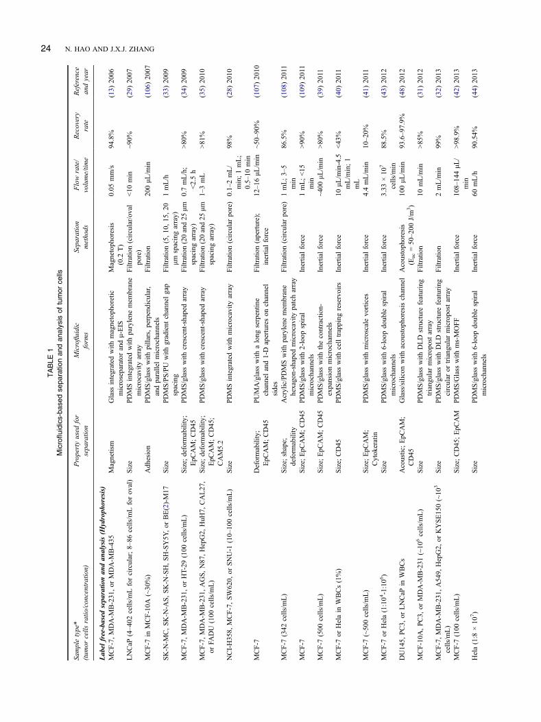
CTCs are shed from primary tumors to flow through theblood stream and may migrate to distant organs to formmetastases, which ultimately cause the death of mostpatients with cancer. Identification and characterization ofCTCs provide an effective means to monitor tumor burdenand progression (26). Separation and purification of CTCsare generally challenging because they are rare in the bloodand possess very heterogeneous features. Microfluidic sys-tems offer many advantages for the separation and analysisof CTCs, especially their high level of system integration,design flexibility, material versatility and advanced degreeof automation. For these reasons, a variety of microfluidicdevices have been developed to separate and analyze CTCsfrom a liquid biopsy. These techniques can be simply clas-sified into two main categories: label free–based techniquesand immunoaffinity-based techniques (Figure 2). The for-mer is established on the basis of the physical properties ofthe target cells, such as their size, density, shape, deform-ability and dielectric properties, whereas the latter utilizesthe biochemical properties of the target cells, mainlythrough protein biomarkers expressed on the cell surface(7, 26).
Label free-based separation and analysis
Many microfluidic techniques for the label-free separa-tion and analysis of cells have been developed (Table 1).According to the physical properties differences, these label-free techniques can be further divided into two subcate-gories: hydrophoresis (based on the cell size, density,
shape, and deformability properties) and dielectrophoresis(based on the cell dielectric property) (27).
Hydrophoresis. Differences in cell size, density,shape and deformability can be exploited for hydrophoresispurpose to separate and purify cells without the need toapply any external force. Hydrophoresis-based methods forlabel-free capture of cells in microfluidic settings are greatlydependent on the interactions between the species of interestand the microfluidic channel substrates. The cell-microchan-nel interactions in hydrophoresis systems can be driven byeither the continuous channels or the discrete obstacles. Theformer has a flat microchannel surface, and the flow of cellfractionation in microfluidic channel is mainly controlled bypressure. The latter generally has a periodic array of micro-meter-scale posts or ridges, and the critical size cut-off forcell separation can be well controlled. In most cases, hydro-phoresis-based label-free cell separation is mediated by thedifference in cell size (Figure 3A).
Filtration is a commonly used label-free hydrophoresistechnique because it is a relatively straightforward approachfor cells separation mainly based on their size property.Microfilters are generally designed with well-defined porechannels to restrict passage of cells above a critical size.Microcavity array represents a typical technique for size-based capture of CTCs (Figure 3B). The size of microcav-ities is usually less than 10 μm, and because of the largersize of tumor cells than red blood cells (RBCs) (Figure 3A),the blood cells can be filtered out while tumor cells are leftbehind. The pores of microcavity array can be also designedas many shapes, such as circular (28), oval (29) and rectan-gular (30). Because each trap is likely to hold only onesingle cell, the trapped cells can be thus easily detectedand counted. Micropost array, different from microcavityarray, is an interesting size-based filtration system to
FIGURE 2 Categories of the separation and purification methods for CTCs in microfluidic systems.
SCREENING TUMOR BIOMARKERS 23
TABLE
1Microfluidics-bas
edse
paratio
nan
dan
alys
isof
tumor
cells
Sampletype*
(tum
orcells
ratio
/concentratio
n)Propertyused
for
separatio
nMicrofluidic
form
sSeparatio
nmethods
Flowrate/
volume/tim
eRecovery
rate
Reference
andyear
Label
free-based
separatio
nan
dan
alysis(H
ydroph
oresis)
MCF-7,MDA-M
B-231,or
MDA-M
B-435
Magnetism
Glass
integrated
with
magnetophoretic
microseparatorandμ-EIS
Magnetoph
oresis
(0.2
T)
0.05
mm/s
94.8%
(13)
2006
LNCaP
(4–402
cells/m
Lforcircular;8–86
cells/m
Lforoval)
Size
PDMSintegrated
with
parylene
mem
brane
microcavity
array
Filtratio
n(circular/oval
pore)
<10
min
~90%
(29)
2007
MCF-7
inMCF-10A
(~30%)
Adhesion
PDMS/glass
with
pillars,perpendicular,
andparallelmicrochannels
Filtratio
n20
0μL
/min
(106)20
07
SK-N
-MC,SK-N
-AS,SK-N
-SH,SH-SY5Y
,or
BE(2)-M17
Size
PDMS/PS/PU
with
gradient
channelgap
spacing
Filtratio
n(5,10
,15
,20
μmspacingarray)
1mL/h
(33)
2009
MCF-7,MDA-M
B-231,or
HT-29
(100
cells/m
L)
Size;
deform
ability;
EpC
AM;CD45
PDMS/glass
with
crescent-shapedarray
Filtratio
n(20and25
μmspacingarray)
0.7mL/h;
<2.5h
>80
%(34)
2009
MCF-7,MDA-M
B-231
,AGS,N87
,HepG2,
HuH
7,CAL27,
orFA
DU
(100
cells/m
L)
Size;
deform
ability;
EpC
AM;CD45;
CAM5.2
PDMS/glass
with
crescent-shapedarray
Filtratio
n(20and25
μmspacingarray)
1–3mL
>81
%(35)
2010
NCI-H358,
MCF-7,SW620,
orSNU-1
(10–
100cells/m
L)
Size
PDMSintegrated
with
microcavity
array
Filtratio
n(circularpore)
0.1–2mL/
min;1mL;
0.5–10
min
98%
(28)
2010
MCF-7
Deformability;
EpC
AM;CD45
PUMA/glass
with
along
serpentin
echanneland1-D
apertureson
channel
sides
Filtratio
n(aperture);
inertialforce
12–16μL
/min
~50–90
%(107)20
10
MCF-7
(342
cells/m
L)
Size;
shape;
deform
ability
Acrylic/PDMSwith
parylene
mem
brane
hexago
n-shaped
microcavity
patcharray
Filtratio
n(circularpo
re)
1mL;3–5
min
86.5%
(108)20
11
MCF-7
Size;
EpC
AM;CD45
PDMS/glass
with
2-loop
spiral
microchannels
Inertialforce
1mL;<15
min
>90
%(109)20
11
MCF-7
(500
cells/m
L)
Size;
EpC
AM;CD45
PDMS/glass
with
thecontraction-
expansionmicrochannels
Inertialforce
~400
μL/m
in>80
%(39)
2011
MCF-7
orHelain
WBCs(1%)
Size;
CD45
PDMS/glass
with
celltrapping
reservoirs
Inertialforce
10μL
/min-4.5
mL/m
in;1
mL
<43
%(40)
2011
MCF-7
(~50
0cells/m
L)
Size;
EpC
AM;
Cytokeratin
PDMS/glass
with
microscalevortices
Inertialforce
4.4mL/m
in10–20%
(41)
2011
MCF-7
orHela(1:104-1:106)
Size
PDMS/glass
with
6-loop
double
spiral
microchannels
Inertialforce
3.33
×10
7
cells/m
in88
.5%
(43)
2012
DU14
5,PC3,
orLNCaP
inWBCs
Acoustic;EpC
AM;
CD45
Glass/siliconwith
acoustophoresischannel
Acoustophoresis
(Eac=50–200
J/m
3)
100μL
/min
93.6–97.9%
(48)
2012
MCF-10A
,PC3,
orMDA-M
B-231
(~10
6cells/m
L)
Size
PDMS/glass
with
DLD
structurefeaturing
triangular
micropostarray
Filtratio
n10
mL/m
in>85
%(31)
2012
MCF-7,MDA-M
B-231
,A54
9,HepG2,
orKYSE150(~10
3
cells/m
L)
Size
PDMS/glass
with
DLD
structurefeaturing
circular
ortriangular
micropostarray
Filtratio
n2mL/m
in99
%(32)
2013
MCF-7
(100
cells/m
L)
Size;
CD45;EpC
AM
PDMS/Glass
with
ms-MOFF
Inertialforce
108–14
4μL
/min
>98
.9%
(42)
2013
Hela(1:8
×10
7)
Size
PDMS/glass
with
6-loop
double
spiral
microchannels
Inertialforce
60mL/h
90.54%
(44)
2013
24 N. HAO AND J.X.J. ZHANG

Hela;
who
lebloo
dfrom
liver
cancer
patient
(3–20cells/3
mL
bloo
d)Size;
CD45;EpC
AM;
Cytokeratin
PDMS/glass
with
gradient
micropillar
channelgapspacing
Filtratio
n0.5–2mL/h
>90
%(36)
2013
MCF-7
inPBMCs(~10
5/m
L);who
lebloo
dfrom
metastatic
lung
cancer
(5–88cells/m
L)
Size;
EpC
AM;CD45
;CD133
PDMS/glass
with
2-loop
spiral
microchannels
Inertialforce
3mL/h
>85%
(46)
2013
NCI-H69
orNIC-H
82(100
0cells/m
L);who
lebloo
dof
SCLC
patient
(0.3–7
2.7cells/m
L)
Size
PDMSintegrated
with
microcavity
array
Filtratio
n(rectangular/
circular
pore)
200μL
/min;
1.0–7.5mL
>80
%(30)
2013
Who
lebloo
dfrom
NSCLCor
SCLCpatient
(0–291
cells/7.5
mL)
Size;
CD45
CellSearchT
Msystem
;PDMSintegrated
with
microcavity
array
Filtratio
n(circularpo
re)
200μL
/min;
3.0–7.5mL
>68
%(110
)20
13
MCF-7,T24
,or
MDA-M
B-231
(500
cells/7.5
mL);who
lebloodfrom
metastatic
breastandlung
cancer
patient
(3–125
CTCs/mL)
Size;
CD45
;Cytokeratin,CD44;
C24
PDMSwith
8-loop
sing
lespiral
microchannel
Inertialforce
7.5mL;<8
min
>80
%(45)
2014
MCF-7
inWBCs(~10
%)
Acoustic;EpC
AM;
CD45
PDMSintegrated
with
tiltedinterdigitated
transducers
Acoustophoresis(~30
dBm)
1.1mL
71%
(49)
2014
MCF-7,HeL
a,LNCaP,or
UACC903M
-GFPin
WBCs
(1:6×10
3-1:105)
Acoustic;CD45;
Cytokeratin
PDMSintegrated
with
tiltedinterdigitated
transducers
Acoustophoresis(~37
.5dB
m)
1.2mL/h
>83
%(50)
2015
Label
free-based
separatio
nan
dan
alysis(D
ielectroph
oresis)
HL-60(2:3)
Dielectric
Goldpo
lyno
mialelectrod
earrayon
glass
slidewith
rotatin
gelectrical
field
generator
DEP(5
Vpp,20
–200
kHz)
30μL
80%
(111)19
94
MDA-231
(1:3)
Dielectric;
size
Goldpo
lyno
mialelectrod
earrayon
glass
slidewith
rotatin
gelectrical
field
generator
DEP(5
Vpp,20
–200
kHz)
30μL
>95
%(112
)19
95
HL-60in
PBMCs
Dielectric
DEP-FFFwith
microelectrodearray
DEP(0.88Vrm
s,25
kHz)
250μL
(64)
1997
MDA-231
(1:3-1:3×10
5)
Dielectric;
size;shape
Goldpo
lyno
mialelectrod
earrayon
glass
slidewith
rotatin
gelectrical
field
generator
DEP(2
Vrm
s,50
kHz)
5μL
/min
>95
%(113
)19
97
HeL
ain
PBMCs
Dielectric
Silicon/glasswith
circular
platinum
electrodes
DEP(6
Vpp,30
kHz)
200μL
/min;
~3min
(58)
1998
MDA-435
inCD34
+stem
cells
(2:3)
Dielectric
DEP-FFFwith
interdigitatedelectrode
array
DEP(4
Vpp,10
kHz)
50μL
;<12
min
>99
%(for
CD34
+
fractio
ns)
(53)
1999
MDA-435
(2:3)
Dielectric;
density
DEP/G-FFFwith
interdigitatedelectrode
array
DEP(1.4
Vrm
s,5kH
z)10
μL;5min
>98
%(54)
1999
MDA-435
inCD34
+stem
cells
(1:1);MDA-435
inT-lymph
ocytes
(2:3)
Dielectric
DEP-FFFwith
interdigitatedelectrode
array
DEP(4
Vpp,15
–40
kHz)
50μL
;<15
min
~70%
(55)
2000
HTBin
SH-SY5Y
(10–20%)
Dielectric
PMMAwith
circular
platinum
electrode
array
DEP(8
Vpp,40
0kH
z)~5
00–15,000
cells
>47
%(59)
2002
K56
2in
RBCs
Dielectric
Cylinder-shaped
DEPcage
with
aprinted
circuitbo
arddevice
DEP(6
Vpp,10
0kH
z)10
0%(60)
2003
MDA-231
,MDA-435
,MDA-468
,HL-60,
SW-756,Jurkat,
andSKI
Dielectric
Goldelectrod
earrayon
electrosmearglass
slidewith
multifrequency
signal
generator
DEP(0–5
Vpp,0–12
00kH
z)20
μL(114
)20
05
P19
inRBCs(1:1)
Dielectric
Pyrex
with
fan-shaped
3D-asymmetric
microelectrod
esDEP(8
Vpp,5MHz)
300μm
/s81
.5%
(61)
2005
A549
Dielectric
PDMS/glass
with
aserpentin
e-shape
pneumatic
micropumpdevice
DEP(15Vpp,16
MHz)
3μL
/min;10
0μL
~80%
(115
)20
07
(Con
tinued)
SCREENING TUMOR BIOMARKERS 25

TABLE
1(C
ontin
ued)
Sampletype*
(tum
orcells
ratio
/concentratio
n)Propertyused
for
separatio
nMicrofluidic
form
sSeparatio
nmethods
Flowrate/
volume/tim
eRecovery
rate
Reference
andyear
MDA-M
B-231
Dielectric;
size;cell-
cycleph
ase
Polyimidewith
DACSyncdevice
DEP(20Vpp,80
0kH
z)20
0–40
0μL
/h96
%(G
1
phase)
(116
)20
07
MDA-M
B-435
from
tumor
xenograftsin
PBMCs(20%
)Dielectric
Electrosm
ears-based
gold
microelectrode
array
DEP(3.3
Vpp,10–920
kHz)
100μL
/min
~75%
(117
)20
08
MCF-7
(100%,size-based
DEPseparatio
n)Dielectric;
size
PDMSwith
rectangularandtriangular
hurdle
channels
DEP(0–180
V)
~100
%(118
)20
08
MCF-7
inMCF-10A
Dielectric
Glass
with
DACShaving
fan-shaped
electrod
eDEP(8
Vpp,48
MHz)
290μm
/s86
.67%
(62)
2009
MDA-M
B-435
,MDA-M
B-468
,MDA-M
B-231
inPBMCs
(1:103–1:105)
Dielectric;
size
DEP-FFFcham
berwith
aninterdigitated
gold-on-copper
electrode
DEP(2.8
Vpp,60
kHz);
Inertialforce
1.5–12
mL/
min;<15
min
>90
%(56)
2009
THP-1,MCF-7,andMCF-10A
Dielectric
PDMS/glass
with
acontactless
DEPdevice
DEP(250
Vrm
s,85
kHz)
10–15μL
/min
(66)
2009
HCT116in
HEK
293andE.coli
Dielectric
Plastic
with
awedge
microfluidicchip
DEP(16Vpp,10
0kH
z)0.1μL
/min
~90%
(63)
2010
B16F10
clones
Dielectric
Castellatedgold
electrodearray
DEP(5
Vpp,20
0–40
0kH
z)50
0cells
per
frequency
<70
%(57)
2010
Jurkat
andHeL
aDielectric
Siliconwith
aguided
DEPmicrochannel
DEP(4
Vrm
s,1MHz)
0.45
μL/m
in;
50μL
(119
)20
10
MCF-7
inRBCsandWBCs(0.1%)
Dielectric;
EpC
AM;
CD45
PDMS/glass
with
p-MOFFandDEP
microchannels
DEP(10Vpp,90
0kH
z)12
6μL
/min
75.81%
(65)
2011
MDA-M
B-231
inMCF-7
andMCF-10A
Dielectric
PDMS/glass
with
acontactless
DEPdevice
DEP(30Vrm
s,16
4kH
z)0.02
mL/h
>90
%(67)
2011
MDA231(1:104–1:106)
Dielectric
PDMS/quartswith
interdigitatedcomb-lik
eelectrodes
DEP(20Vpp,10
–50
kHz)
0.1mL/h;<1
h95–98%
(120)20
11
SKOV3or
MDA-M
B-231
inPBMCs(~1:10
3–1
:104)
Dielectric
Apo
Stream
TMhaving
apo
lyim
idefilm
sheetwith
copper
andgold
electrodes
DEP(2–4.5
Vpp,90
0kH
z)18–25μL
/min;60
min
~75%
(68)
2012
PC-3
orOEC-M
1in
leukocyte(10%
)Dielectric;
EpC
AM;
CD45
PDMS/glass
with
anoptically-induced
DEPdevice
DEP(2–7
Vpp,5
0–15
00kH
z)0.1μL
/min;1
μL~6
0–80
%(121)20
13
Immuno
affinity-based
separatio
nan
dan
alysis(Positive
selection)
NCI-H1650
(50-50,000
cells/m
L);whole
bloodfrom
metastatic
lung,prostate,pancreatic,breast,andcolon
cancer
patient
(5–128
1cells/m
L)
EpC
AM;CD45;
Cytokeratin
Siliconwafer
with
circular
micropo
starray
Antibodymicropost
1–2mL/h
NCI-H16
50:
>60
%;
Patient:99
%
(74)
2007
MCF-7
(10–25
0cells/m
L)
EpC
AM
PMMAwith
thesinu
soidally
shaped
capturechannelsandan
integrated
conductiv
itysensor
Antibodymicrochannel;
Inertialforce
>1mL;<37
min
>97
%(72)
2008
MCF-7
(5–125
0cells/m
L)
EpC
AM
Siliconwafer
with
nanopillararray
Antibodynano
pillar
1mL
>40
%(76)
2009
CCL-119
inPBS(5
×10
5–1
×10
6cells/m
L)
Aptam
ersgc8
PDMS/glass
with
aptamers-im
mobilized
microfluidicchannel
Aptam
ermicrochannel
200nL
/s;48
μL>80
%(80)
2009
CCL-119
,CRL-159
6,andCRL-263
1in
PBS(1
×10
6cells/
mL)
Aptam
ersgc8;TD05
;Sgd5
PDMS/glass
with
aptamers-im
mobilized
serpentin
emicrofluidicchannel
Aptam
ermicrochannel
300nL
/s96%
(81)
2009
MCF-7
inJurkat
(4:6)
5D10
mAb;
Fibronectin
PDMS/glass
with
patterned
immunom
agnetic
beadsarray
Antibodymicrochannel
10nL
/s85%
(78)
2010
PC3(500–100
0cells/m
L);Whole
bloo
dfrom
prostate
cancer
patient
(12–31
67cells/m
L)
EpC
AM;CD45;PSA
PDMS/glass
with
microvortex-generating
herringb
onechip
Antibodymicrochannel
1.5–2.5mL/h
~93%
(73)
2010
26 N. HAO AND J.X.J. ZHANG

SW62
0or
HT29
EpC
AM
PMMAwith
thesinu
soidally
shaped
capturechannelsandan
integrated
cond
uctiv
itysensor
Antibodymicrochannel;
Inertialforce
2mm/s;1
mL;<40
min
96%
(71)
2011
PC-9
orMCF-7
EpC
AM;CD45
MACSTMMS-colum
ncoup
ledwith
flow
cytometer
Immun
omagnetic
beads
forlabelin
g1mL
95.8%
(122
)20
11
MCF-7
(50–10
00cells/m
L)
EpC
AM;CD45;
Cytok
eratin
PDMS/Siliconwith
aserpentin
echaotic
mixingchannel
Antibod
ynano
pillar
0.5–7mL/h
>95
%(70)
2011
COLO205or
SKBR3(200
cells/2.5
mL)
EpC
AM;CD45;
Cytok
eratin
PDMS/glass
with
parallel-arrangem
ent
magnetarray
Immun
omagnetic
beads
forlabelin
g10
mL/h
>86
%(84)
2011
KG1a
(1:1);who
lebloo
dfrom
breast,prostate,lung
,and
ovariancancer
patient
(20–704cells/3.75mL)
EpC
AM;Selectin
;PSA
Micro-Renathane
microtube
Antibodymicrochannel
4.8mL/h
~50%
(69)
2012
PC3in
WBCs(1:250
)EpC
AM
PDMSwith
afluid-perm
eablepo
rous
polycarbon
atemem
brane
Antibodymicrochannel
6mL/h
~70%
(123
)20
12
M6C
(2–80cells/m
L)
EpC
AM;CD45;
Cytok
eratin
PDMS/glass
with
micropillararraychannel
Immunom
agnetic
beads
forlabelin
g20
μL/m
in;5
min
90%
(77)
2012
MCF-7
orMDA-M
B-231
(100
cells/m
L)
EpC
AM;Size
Silicon/glasswith
MOA
filter
Immun
obeads
forsize
amplificatio
n20
μL/m
in;1
mL
92%
(51)
2012
MCF-7
orCKBr-3;
wholebloodfrom
metastatic
breastcancer
(11–
105cells/7.5
mL)
EpC
AM;Her2;
Cytok
eratin,CD44;
CD24
PDMS/glass
with
eDAR-based
microfluidicchip
Fluorescent-activated
cellsorter
10–80μL
/min
>93
%(124
)20
12
MCF-7
(0–100
0cells/m
L)
EpC
AM
PDMS/glass
with
packed
bedmicrofluidic
channelcontaining
aweir
Immun
obeads
for
retention
0.2mL/h
30–70%
(125
)20
12
MCF-7
orDMS-79(100
cells/m
L)
EpC
AM;size;CD45
;Cytok
eratin
Silicon/glasswith
parallelmicrofluidic
channel
Immun
obeads
forsize
amplificatio
n;Filtratio
n
1mL
>89
%(95)
2012
MCF-7
inJurkat
(1:107)
EpC
AM
PMMAwith
disk-based
microchannelsand
multistage
concentric-circularmagnet
Immun
omagnetic
beads
forlabelin
g;Inertial
force
~30min
80%
(92)
2012
MCF-7
(1-10cells/m
L)
EpC
AM
PDMS/glass
with
DLD
structurefeaturing
triangular
micropostarrayandfishbo
nestructurecham
ber
Antibodymicrochannel
9.6mL/m
in90%
(75)
2013
PC3-9,
SKBR3,
MDA-M
B-231
,MCF10
A-LBX1;
who
lebloodfrom
prostate,lung,pancreas,breast,andmelanom
acancer
patient
(>0.5cell/mL)
EpC
AM;Her2;
Estrogenreceptor;
Cytok
eratin;size
PDMS/glass
integrated
with
CTC-iChips
Immunom
agnetic
beads
forlabelin
g;DLD;
Inertialforce
6–12
mL
>77
.8%
(93)
2013
MCF-7
EpC
AM;CD45;
Cytok
eratin
Silicon/glasswith
TRABmicrofilter
Immun
obeads
forsize
amplificatio
n10
0μL
/min
93%
(97)
2013
MCF-7
orMDA-M
B-231
(125–2000cells/m
L)
EpC
AM
PDMS/glass
with
avortex
micromixer
and
consecutivewavyducts
Immun
obeads
forsize
amplificatio
n10
0–60
0μL
/min
MCF-7:
95.8%;
MDA-M
B-
231:
15%
(96)
2013
SkB
r3,P
C3,
orColo2
05(200
cells/2.5
mL);who
lebloo
dfrom
breast,prostate,andlung
cancer
patient
(>1cells/5
mL)
EpC
AM;CD45;
Cytok
eratin
PDMS/glass
with
inverted
channeland
parallel-arrangem
entmagnetarray
Immun
omagnetic
beads
forlabelin
g2.5mL/h
>94
%(85)
2013
SK-BR-3
(10–10
4cells/0.2
mL);Who
lebloo
dfrom
breastand
lung
cancer
patient
(2–41cells/0.2
mL)
EpC
AM;CD45;
Cytok
eratin
Glass
with
aferrom
agnetic
wirearray
Immun
omagnetic
beads
forlabelin
g5mL/h
90%
(94)
2013
COLO
205,
SK-BR-3,or
A-431
(~10
0–200cells/2.5
mL)
EpC
AM;Cytokeratin;
HER2;
EGFR
PDMS/glass
with
inverted
channeland
parallel-arrangem
entmagnetarray
Immun
omagnetic
beads
forlabelin
g2.5mL/h
~45–93
%(91)
2013
(Contin
ued)
SCREENING TUMOR BIOMARKERS 27

TABLE
1(C
ontin
ued)
Sampletype*
(tum
orcells
ratio
/concentratio
n)Propertyused
for
separatio
nMicrofluidic
form
sSeparatio
nmethods
Flowrate/
volume/tim
eRecovery
rate
Reference
andyear
COLO
205(~15
0cells/2.5
mL)
EpC
AM;CD45;
Cytokeratin
PDMS/glass
with
inverted
channeland
micromagnetarray
Immun
omagnetic
beads
forlabelin
g2.5mL/h
98%
(90)
2015
MCF-7,PC3,
SK-BR-3,or
COLO
205(200
cells/10μL
)EpC
AM;CD45;
Cytokeratin
PDMS/glass
with
micromagnetarray
Immun
omagnetic
beads
forlabelin
g2.5mL/h
97%
(87)
2015
COLO
205(~15
0cells/2.5
mL)
EpC
AM;CD45;
Cytokeratin
PDMS/glass
with
inverted
channel,
micromagnetarray,
andspacered
magnets
Immun
omagnetic
beads
forlabelin
g2.5mL/h
95.6%
(88)
2016
Immuno
affinity-based
separatio
nan
dan
alysis(N
egativeselection)
MCF-7
inPBMCs(~47
:106)
CD45;Cytokeratin
PMMAwith
disk-based
microchannelsand
multistage
concentric-circularmagnet
Immun
omagnetic
beads
forlabelin
g;Inertial
force
~30min
60%
(101)20
11
MCF-7
orMDA-M
B-231
inJurkat;Whole
bloo
dfrom
metastatic
breast,lung,andgastriccancer
patients(1–5
1cells/m
L)
CD45;Cytokeratin
PDMS/glass
with
GASIchip
having
asym
metricherringb
onemicrochannels
Antibodymicrochannel
10–40μL
/min
3.92–100
%(99)
2013
PC3-9,
SKBR3,
MDA-M
B-231,MCF10A-LBX1;
whole
bloodfrom
prostate,lung,pancreas,breast,andmelanom
acancer
patient
(>0.5cell/mL)
CD45;Cytokeratin;
CD15;size
PDMS/glass
integrated
with
CTC-iChips
Immun
omagnetic
beads
forlabelin
g;DLD;
Inertialforce
6–12
mL
>77
.8%
(93)
2013
WM16
4,MB23
1,PC9,
PC3-9,
SKBR3,
orMCF10A-LBX1
(1000cells/m
L);whole
bloodfrom
breastandpancreatic
cancer
patient
CD45;CD66b;
Size
PDMS/glass
integrated
with
CTC-iChips
Immun
omagnetic
beads
forlabelin
g;DLD;
Inertialforce
8mL/h
97%
(105)20
14
MCF-7
orNCI-H1975
(5–50cells/m
L)
CD45;Cytokeratin
PMMA
integrated
with
microslit
mem
branemesh
Immun
omagnetic
beads
forlabelin
g;filtration
~60min
>90
%(103)20
14
MCF-7
inWBCs
CD45
PDMS/glass
with
serpentin
emicrofluidic
channels
Rough
ened
antib
ody
microchannel
2μL
/min;20
0μL
~50%
(100)20
15
HCT116in
PBMCs(3–250
cells/2
mLbloo
d);who
lebloo
dfrom
colorectal
cancer
patient
CD45;Cytokeratin;
EpC
AM;CD44;
CD47
Magnetic
cellseparatorcoupledwith
flow
cytometer
Immun
omagnetic
beads
forlabelin
g>61
%(102)20
15
NCI-H1975,SW48,PC3,
MCF-7,or
Jurkat
(1–25cells/m
L)
CD45;Cytokeratin
PMMA
integrated
with
perm
anentmagnet
arrayandmicroslitmem
branemesh
Immun
omagnetic
beads
forlabelin
g;filtration
500μL
/min;2
mL
>80
%(104)20
16
*Celllin
ewas
used
tospikeinto
whole
bloodthat
was
collected
from
healthybody,unless
specially
indicated.
DACS:Dielectrophoresis-activated
cellsorter;DACSync:
Dielectrophoresis-activated
cellsynchronizer;DEP/G-FFF:Dielectrophoretic/gravitatio
nalfield-flow
fractio
natio
n;DEP:Dielectrophoresis;
DEP-FFF:Dielectrophoretic
field-flow
fractio
natio
n;DLD:Deterministic
lateraldisplacement;Eac:Acoustic
energy
density
;eD
AR:Ensem
ble-decision
aliquo
tranking;
EpC
AM:Epithelialcelladhesion
molecule;
GASI:Geometrically
activ
ated
surfaceinteraction;
mAb:
Monoclonalantib
odies;MOA:Multi-obstacle
architecture;
ms-MOFF:Multi-stagemulti-orifice
flow
fractio
natio
n;NSCLC:Nonsm
all
celllung
cancer;p-MOFF:Parallelmulti-orifice
flow
fractio
natio
n;PBMCs:
Peripheralbloodmononuclear
cells;PBS:Phosphate
buffered
salin
e;PDMS:Polydim
ethylsilo
xane;PMMA:Poly(methyl
methacrylate);PS:P
olystyrene;PSA:P
rostate-specificantig
en;PU:P
olyurethane;PUMA:Polyurethane-methacrylate;RBCs:Red
bloodcells;S
CLC:S
mallcelllung
cancer;taSSAW:T
ilted-anglestanding
surfaceacoustic
waves;TRAB:Trachealcarina-inspiredbifurcated;Vpp:Peak-to-peakvoltage;Vrm
s:Rootmeansquare
voltage;WBCs:White
bloodcells.
28 N. HAO AND J.X.J. ZHANG

separate cells using a principal known as deterministic lat-eral displacement (DLD), as the displacement of cells per-pendicular to primary flow is determined by the pattern ofthe array (31, 32). Cells below a critical size follow stream-lines through the array gaps with no net displacement fromthe original streamline, whereas cells above the critical sizeare “bumped” laterally to cross sequential streamlines ineach row at an angle predetermined by the post offsetdistance. The critical cell size for fractionation depends onthe gap between posts and offset of posts, which also meansthat DLD micropost shape is an important parameter fordetermining the separation and purification efficiency (32).Compared to the microcavity array and micropost array, thegradient gap spacing microchannel array-based filtration is amuch more appealing technique, which can be used for theseparation of various blood cell types due to their inherentsize differences (33–36). This kind of microfluidic systemwith successively narrower gap widths between the columnscan retain increasingly smaller cells; therefore, tumor cells,white blood cells (WBCs), RBCs and cell fractions can bespecially separated.
Inertial forces generated by the flow in microfluidicchannels can be applied as a rapid and label-free strategy
to separate and purify CTCs. There are two kinds of micro-channel forms for inertial force-based separation: straightchannels and curved channels (37, 38). In a straight channel,fluid shear generates lateral forces, which cause transverseinertial migration of cells. In a curved channel, centrifugeforces overlap with the inertial forces and generate a doublerecirculation in the transversal section of the microchannels(Dean drag) (27). Inertial migration in a straight channel forcell separation generally consists of different kinds of trap-ping reservoirs (39–42). When cells are flowing through themicrofluidic channel, they need to reach equilibrium at awell-defined distance between the center of the cell and themicrochannel wall due to the inertial lateral forces. Once thecells reach the reservoir’s area, larger-sized cells are pushedtoward a vortex generated in the reservoir and thus trappedwithin, while smaller-sized cells can be flushed toward theoutlet. Inertial force–based cell separation in a curved chan-nel is often provided by using spiral microfluidic system(Figure 3C) (43–46). In this case, when cells are flowingthrough the microchannel, the dominant inertial force and asecondary rotational flow field perpendicular to the originalflow direction (Dean flow) will lead the smaller-sized parti-cles to migrate in the direction of the outer half of the
FIGURE 3 Hydrophoresis-based microfluidic systems for cell analysis. (A) Size comparison of CTCs with hematologic white blood cells (WBCs) and redblood cells (RBCs). Adapted from Ref. (51); (B) Size-selective microfiltration array-based CTC recovery device. (a) Schematic image of CTC recovery deviceusing the size-selective microfiltration array. (b) and (c) Photographs of the microfiltration array and CTC recovery device equipped with the array, respectively.(d) SEM image of MCF-7 cells trapped on the microfiltration array. The microcavities are 9 μm in size with a 60-μm pitch. Adapted from Ref. (28); (C) Inertialforce–based size separation of CTCs in spiral microfluidic channels. (a) Schematics of the microfluidic cell sorter containing 6-loop double spiralmicrochannels with one inlet and three outlets for cell separation. (b) Illustration of the two counter-rotating Dean vortices forming in the top and bottomhalves of the channel. The black arrows represent the velocity field in the cross section. Adapted from Ref. (43); (D) Magnetophoresis-based breast cancer cells(BCCs) separation from RBCs. (a) Top view of the fabricated paramagnetic capture mode (PMC) microseparator. Fluorescently probed BCCs passing throughthe microchannel of the PMC microseparator at (b) an average flow velocity of 0.05 mm/s with an external magnetic flux of 0.2T, and (c) an average flowvelocity of 0.05 mm/s without the external magnetic flux. Adapted from Ref. (13); (E) Illustration of taSSAW-based acoustophoretic cell separation. Adaptedfrom Ref. (50).
SCREENING TUMOR BIOMARKERS 29

channel, while the larger-sized particles can migrate towardthe inner channel wall. Therefore, this so-called Dean flowfield fractionation technique can be applied to separate cellswith short acting time and high selectivity.
Magnetophoresis and acoustophoresis, in addition to fil-tration and inertial force, are another two appealing label-free techniques for cells separation based on their intrinsicmagnetic moment and acoustic properties, respectively.Magnetophoresis-based separation involves the manipula-tion of cells in a fluid medium under the influence of anexternal magnetic field. Magnetophoresis provides an alter-native means for gentle cells separation in their naturalmilieu that could be highly specific and highly sensitivewithout the need of any complicated or expensive equip-ment (47). This kind of microfluidic separation systemsemploys the intrinsic paramagnetic properties of deoxyhe-moglobin that was found only in erythrocytes. In contrast,other cell types, such as WBCs and tumor cells, are gen-erally considered with diamagnetic properties. Therefore,RBCs and other biological components will be moving inopposite directions due to the force created by the externalmagnetic field (Figure 3D) (13). Similarly, acoustophoresisin a flow channel is achieved by establishing a standingacoustic field, which will push cells toward regions withminimal acoustic radiation pressure (pressure nodes). Cellswith different size, shape and other physical properties willexperience different acoustic radiation forces and requiredifferent times to migrate to the pressure nodes, thus pro-viding clear identifiers for separation. The standing surface
acoustic waves for acoustophoresis in microfluidic systemscan be controlled at a position parallel to the fluid flowdirection (48), or with certain angle (49, 50). The lattersystem (so-called tilted-angle standing surface acousticwaves, taSSAW) could make the cells in a fluid mediumexperience both the acoustic radiation force and the laminardrag force, and thus lead to better separation sensitivity(Figure 3E) (49,50).
Dielectrophoresis. Dielectrophoresis (DEP)-basedcells separation is a technique by which cells are inducedto move by the application of a nonuniform electric fielddue to the interactions of the cells’ dipole and spatial gra-dient of the electric field (Table 1) (52). When electric fieldsare applied to cells, they become polarized. This inducedpolarization can then interact with the applied field, result-ing in each kind of cells experiencing a unique net electricalforce. The magnitude of this net force depends on manyfactors, such as cell membrane property, cell size, cytoplas-mic property, the frequency and strength of the electric field,and the fluid medium property. If cells are more polarizablethan the suspending medium, they will be attracted towardthe regions of higher electric fields and retained at theelectrode surface; this motion is called positive DEP(pDEP) (Figure 4A). Conversely, if cells are less polarizablethan the suspending medium, they will move to the regionsof lower electric fields and then be eluted by the flow; thismotion is called negative DEP (nDEP) (Figure 4A). In thecase of CTCs separation from blood, electrophoretic
FIGURE 4 Dielectrophoresis-based microfluidic systems for cell analysis. (A) Dielectrophoresis (DEP) separation can be positive (pDEP) (a) or negative(nDEP) (b), which affects the positioning of cells within a field. (c) Examples of DEP utilized in microfluidic systems in a variety of arrangements. Adaptedfrom Ref. (38); (B) A continuous flow DEP microfluidic cell-sorting device. Adapted from Reference (68).
30 N. HAO AND J.X.J. ZHANG

mobility generally distinguishes tumor cells with attractiontoward the electrode, and normal blood cells migrate in theelectric field into the eluent.
Metallic microelectrodes with various shapes, such as inter-digitation (53–56), castellate (57), circular (58, 59), cylinder(60), fan (61, 62) and wedge (63), can be easily patterned on amicrofluidic wafer using conventional lithography techniquesfor DEP-based cells separation. Therefore, a number of DEP-based microfluidic devices have been employed to separatetumor cells (Table 1). Among these, field flow fractionation(FFF) and multi-orifice flow fractionation (MOFF)-integratedDEP techniques bring many new features for CTCs separation.FFF is an analytical technique to achieve cell fractionation thatuses the velocity gradient of a hydrodynamic flow profile.Cells in a long and narrow microfluidic channel are forcedtoward the bottom wall of the chamber when a force fieldnormal to the flow direction is applied. In this so-calledDEP-FFF system, which is the combination of dielectrophore-tic levitation of cells with FFF technique, cells can reachequilibrium positions at different heights depending on theirelectric properties, achieving high cell separation efficiencyand throughput (53–56, 64). The termed MOFF techniqueinvolves the lateral movement of cells according to their sizedue to hydrodynamic inertial force. This integrated DEP-MOFF system that combines both hydrodynamic and dielec-trophoretic separation techniques can result in fast (withrespect to flow rate) and efficient performance (65).
Compared to conventional metallic microelectrodes, thecontactless DEP (cDEP) that utilizes fluidic electrode chan-nels with filled high-conductivity solution is also a promis-ing technique for label-free cells separation. This techniqueeliminates cell-electrode contact, minimizes the contamina-tion of the biological samples, and reduces the fabricationsteps and costs. cDEP has successfully been proven toselectively separate cells (66, 67). In addition, it is notedthat a commercial DEP-based microfluidic system for CTCsseparation, ApoStreamTM, has already been launched (68).In this system, the sample is introduced through a portlocated in the floor of the flow chamber at the sameupstream end as the elution buffer, and cancer cells can becollected through another port located downstream from thesample inlet port (Figure 4B). When cells encounter theDEP field, the DEP force will pull cancer cells toward thechamber floor and repel other cells as they traverse theelectrode. Therefore, cancer cells traveling close to thechamber floor will be withdrawn through the collectionport, while other blood cells traveling at greater heightswill be carried beyond this port and exit the chamber tothe waste container via a second outlet port (Figure 4B)(68).
Immunoaffinity-based separation and analysis
CTCs can also be distinguished from background cellsbased on their surface markers. Many techniques have thus
been developed for the separation and analysis CTCs(Table 1). These immunoaffinity-based techniques can befurther classified into two categories: positive selection(based on the capture of target tumor cells and the elutionof nontarget cells) and negative selection (based on thecapture of nontarget cells and the elution of target tumorcells).
Positive Selection. Positive selection, which is estab-lished by using the specific epitopes expressed on the tumorcell surface, has been extensively developed (Table 1).EpCAM is the most commonly used epitope for all immu-noaffinity-based positive selection methods. According tothe binding site differences, positive selection can be rea-lized by either modifying microchannel substrate surfacewith antibodies or manipulating antibodies-conjugatedmicrometer-sized magnetic beads.
EpCAM antibody-coated microfluidic channel systemshave attracted a great interest in recent years. When CTCsflow across the microchannels, the interactions betweenthese binding ligands and CTCs surface epitopes can cap-ture and retain the tumor cells, and the remaining bloodcomponents can be carried away by the flow. The capturedtumor cells on the microfluidic channel surface can then bedislodged and collected for further analysis. The biggestchallenge in conventional straight flat microfluidic channelis its limited surface area for anchoring ligands (69). Giventhe larger surface area in microfluidic channel will certainlyprovide more possible interaction sites to increase thechances of CTCs capture, different kinds of curved and/orcoarse channels have been specially designed, such as ser-pentine-shaped channel (70–72), herringbone array (73),micropost array (74, 75), nanopillar array (70, 76, 77) andimmunobeads array (78, 79). All these strategies, comparedto conventional straight flat microfluidic systems, couldgenerate higher surface area inside the microchannels forbinding more target ligands and thus show better captureefficiency of tumor cells (Figure 5A). It is noted that besidesEpCAM antibody, aptamers that are single-strand nucleicacid oligomers for binding target proteins, peptides andamino acids can also endow aptamers-coated microchannelswith high specificity and affinity for tumor cell separationand enrichment (80, 81).
Immunomagnetic-based cell separation, in which micro-meter-sized magnetic beads are selectively attached to thetumor cells, is also a common technique for CTCs enrich-ment due to its high sensitivity and ability to handle a largerange of volumes without the need of surface modificationinside of microfluidic channels (82, 83). Various immuno-magnetic microfluidic devices were developed in our labora-tory, and the magnetic field design from the integratedmagnets in microfluidic systems was also optimized forcontrolling capture of tumor cells (Figure 5B) (84–91).There are generally two kinds of roles for magnetic beadsin immunomagnetic-based cell separation. One is to label
SCREENING TUMOR BIOMARKERS 31
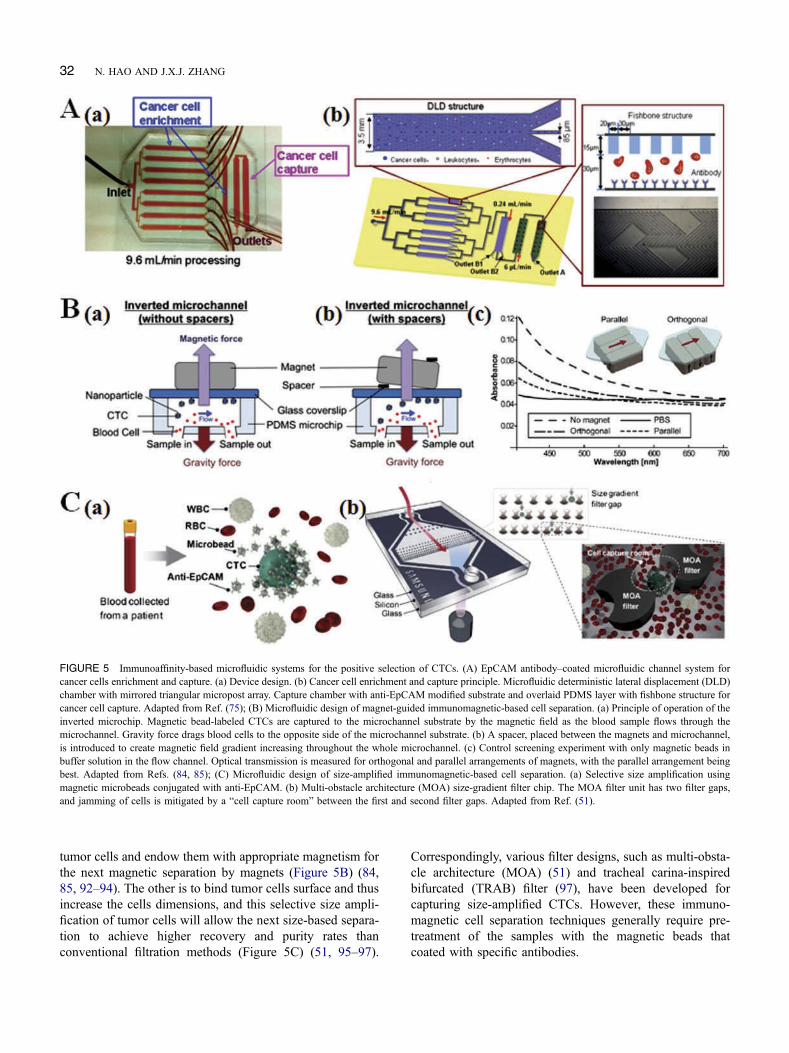
tumor cells and endow them with appropriate magnetism forthe next magnetic separation by magnets (Figure 5B) (84,85, 92–94). The other is to bind tumor cells surface and thusincrease the cells dimensions, and this selective size ampli-fication of tumor cells will allow the next size-based separa-tion to achieve higher recovery and purity rates thanconventional filtration methods (Figure 5C) (51, 95–97).
Correspondingly, various filter designs, such as multi-obsta-cle architecture (MOA) (51) and tracheal carina-inspiredbifurcated (TRAB) filter (97), have been developed forcapturing size-amplified CTCs. However, these immuno-magnetic cell separation techniques generally require pre-treatment of the samples with the magnetic beads thatcoated with specific antibodies.
FIGURE 5 Immunoaffinity-based microfluidic systems for the positive selection of CTCs. (A) EpCAM antibody–coated microfluidic channel system forcancer cells enrichment and capture. (a) Device design. (b) Cancer cell enrichment and capture principle. Microfluidic deterministic lateral displacement (DLD)chamber with mirrored triangular micropost array. Capture chamber with anti-EpCAM modified substrate and overlaid PDMS layer with fishbone structure forcancer cell capture. Adapted from Ref. (75); (B) Microfluidic design of magnet-guided immunomagnetic-based cell separation. (a) Principle of operation of theinverted microchip. Magnetic bead-labeled CTCs are captured to the microchannel substrate by the magnetic field as the blood sample flows through themicrochannel. Gravity force drags blood cells to the opposite side of the microchannel substrate. (b) A spacer, placed between the magnets and microchannel,is introduced to create magnetic field gradient increasing throughout the whole microchannel. (c) Control screening experiment with only magnetic beads inbuffer solution in the flow channel. Optical transmission is measured for orthogonal and parallel arrangements of magnets, with the parallel arrangement beingbest. Adapted from Refs. (84, 85); (C) Microfluidic design of size-amplified immunomagnetic-based cell separation. (a) Selective size amplification usingmagnetic microbeads conjugated with anti-EpCAM. (b) Multi-obstacle architecture (MOA) size-gradient filter chip. The MOA filter unit has two filter gaps,and jamming of cells is mitigated by a “cell capture room” between the first and second filter gaps. Adapted from Ref. (51).
32 N. HAO AND J.X.J. ZHANG

Negative Selection. Although positive selectionmethods can be used to separate CTCs at a high purity,these kinds of methods may have potentially significantlimitations, one of which is that CTCs, heterogeneous bynature, do not all express the same or the same level ofspecific antigens. Even with the same origin cancer celllines, the surface densities of epitopes are quite distinctfrom each other. The biological characteristics of thetumor cells, after antibody-antigen reactions via positiveselection methods, may also be affected. In addition, posi-tive selection of CTCs requires an assumption about theunknown nature of CTCs in the blood sample. For thesereasons, negative selection methods, in which the bloodsample is depleted of leukocytes using antibodies againstCD45 and other leukocyte antigens (which are notexpressed on the tumor cells surface), are growing in popu-larity for the collection of CTCs (98).
Same as the positive selection, negative selection can bealso realized by either modifying microchannel substratesurface with antibodies or manipulating antibodies-conju-gated micrometer-sized magnetic beads (Table 1). The for-mer type of negative selection methods is commonlyperformed by CD45 antibodies-immobilized microfluidicchannels. To increase the surface interactions between thenontarget leukocytes and the channel surface, the roughenedmicrochannel systems are generally created by conventionallithography techniques (99) or using strong acid (Figure 6A)(100). The latter type of immunomagnetic beads–basednegative selection methods can be performed in manyforms for collecting CTCs, such as multistage concentric-circular magnet on microfluidic disk (101), fluorescent-acti-vated cell sorting (102), and microslit membrane (103, 104).
The most promising one is the so-called CTC-iChip tech-nology using two-stage magnetophoresis and depletion anti-bodies against leukocytes (Figure 6B). This CTC-iChip thatintegrates DLD, inertial focusing and magnetophoresis cansort up to 107 cells/s and successfully collect CTCs from thewhole blood of both epithelial and nonepithelial cancerpatients including lung, prostate, pancreas, breast and mel-anoma (93, 105). However, it should be noted that not allCD45-negative cells in the blood are tumor cells (for exam-ple, the circulating endothelial cells) (26). Therefore, sub-sequent characterization and detection steps are of utmostimportance to increase the specificity and accuracy of thesenegative selection-based microfluidic systems.
Separation and Analysis of Exosome
Exosomes, carrying cell-specific cargos (proteins, lipids andnucleic acids) and distributing ubiquitously in body fluids,can be harnessed as a minimally invasive means to probethe tumor origin and progression status. Although a numberof recent studies have highlighted the potential clinical roleof exosomes in disease diagnosis and therapy (10), routineexosome analysis is still a challenging task. Compared toconventional exosome separation techniques, such as ultra-centrifugation, density gradient and physical force (126),microfluidics provide great opportunities for exosome ana-lysis in terms of reagent volumes, automation and integra-tion capabilities, separation time, product integrity andpurity, and recovery rate (127). Generally, vesicle size(20–300 nm) and surface biomarkers (such as EpCAM,CD9, CD63 and CD81) are two typical characters to iden-tify exosomes. Therefore, current reported microfluidic
FIGURE 6 Immunoaffinity-based microfluidic systems for the negative selection of CTCs. (A) Experimental design of CD45 antibodies–immobilizedmicrofluidic system: (1, 2) blood pre-samples with spiked cancer cells are injected through the depletion device functionalized with anti-CD45 antibody tospecifically bind to white blood cells (WBCs). Eluted cells comprising the target CD45-cells and false-positive WBCs are collected and characterized usingeither imaging flow cytometry (3a) or optical microscopy after plating in standard tissue culture wells (3b). Adapted from Ref. (100); (B) Immunomagnetic-based CTC-iChip schematic. The CTC-iChip is composed of two separate microfluidic devices that house three different microfluidic components engineeredfor inline operation: DLD to remove nucleated cells from whole blood by size-based deflection by using a specially designed array of posts performed in CTC-iChip1, inertial focusing to line up cells to prepare for precise magnetic separation and magnetophoresis for sensitive separation of immunobeads-labeledWBCs and unlabeled CTCs, which are performed in CTC-iChip2. Adapted from Reference (105).
SCREENING TUMOR BIOMARKERS 33

systems for exosome separation and analysis can be simplyclassified into two categories: size-based analysis andimmunoaffinity-based analysis (Table 2).
Size-based separation and analysis
Size-based microfluidic systems for exosome analysis aregenerally established on the basis of nanoporous structure(128–131). One typical example is the so-called ciliatedmicropillar structure array that is formed by the electrolesslyetching on the silver eletrodeposited sidewalls of micropil-lars (Figure 7A) (128). The inner-nanowire spacing can betuned within a size range of 30–200 nm to create a highdensity of interstitial sites, which allows physical trap ofexosomes. The micropillars not only provide walls foranchoring the nanowires but also filter larger-sized samplecomponents and function as the structural supports for themicrofluidic channel. This nanowire-on-micropillar hier-archical structure showed fast trapping rate, specific selec-tion and high retention rate toward exosome vesicles.Similar nanoporous structures or arrays can be also seen inother microfluidic filtration system to separate exosomeswith tunable size cut-off (129–131). However, these physi-cal trapping approaches may be restricted by the saturationlimit. Alternatively, an acoustic nanofilter microfluidic sys-tem was developed for separating the exosomes in a size-specific, continuous and contact-free manner (Figure 7B)(132). The separation uses ultrasound standing waves toexert differential acoustic force on exosomes according totheir size and density. By optimizing the design of theultrasound transducers and underlying electronics, a highseparation yield and resolution can be achieved. And the“filter size cutoff” can be controlled electronically in situ,which enables versatile extracellular vesicles-size selection(Figure 7B). Another interesting approach for the separationand titration of exosomes is the colorimetric nanoplasmonicassay, which can achieve naked eye readout and femtomolardetection (133). Table 2 summarizes the separation andanalysis of size-based microfluidic systems for exosomes.It is noted that although size is the most acceptable criterionfor exosome identification, it is not a strict feature of exo-somes. Therefore, it is crucial to design novel microfluidicmethods that can combine multiple features for separatingand characterizing vesicle types and allowing precise ana-lysis of respective exosome functions.
Immunoaffinity-based separation and analysis
Immunoaffinity-based microfluidic systems for exosomeanalysis can be implemented by either modifying micro-channel substrate surface with antibodies (8, 134–140) ormanipulating antibodies-conjugated micrometer-sized beads(141–143). The former type of microfluidic systems wasfirst reported in 2010 using an anti-CD63 functionalizedmicrochannel surface for immunocapture of exosomes(136). Since then, different kinds of microfluidic devices
modifying with specific antibodies toward exosome surfacebiomarkers were designed (Table 2). Among these, a typicalexample is the nanoplasmonic exosome (nPLEX) microflui-dic system, which is based on transmission surface plasmonresonance through periodic nanohole arrays (Figure 8A) (8).Each array surface is functionalized with affinity antibodiesfor different exosome protein markers, such as CD9, CD63,HSP70, HSP90, Flotillin 1, Flotillin 2, CD24 and EpCAM.With target-specific exosome binding, the nPLEX microflui-dic system displays spectral shifts or intensity changes pro-portional to target marker protein levels. Therefore, thisnPLEX technology enables high-sensitive and high-throughput monitoring of exosome binding in real-timeand with single-exosome resolution (Figure 8A).
Although various microchannel surface modificationapproaches were configured to enhance the capture effi-ciency of exosomes, the capture capacity was still limitedby available surface area for antibodies immobilization.Therefore, micrometer-sized immunobeads were generallyintroduced into microfluidic systems to enhance the capturecapacity of exosomes (Table 2). One kind of immunobeadsreported in previous studies was based on polystyrene par-ticles (142). There, inertial lift forces push the polystyrenebeads toward the center of microfluidic channel with multi-ple outlets, transferring them into a wash buffer and allow-ing their selective separation. Comparatively, magneticparticles-based immunobeads bring many superior features,especially their easy manipulation by an external magneticforce (82, 83, 141, 143). Specifically, when the immuno-magnetic beads were mixed with exosomes, the boundcomplex can be retained as a tight aggregate in the down-stream microfluidic chamber by magnetic force. The amountof the retained beads was proportional to the injectionvolume of exosomes, thus allowing for quantitative separa-tion and analysis (Figure 8B). Compared to the microchan-nel substrate surface modification-based immunoaffinityanalysis, the immunobeads method allows for rapid enrich-ment of captured exosomes and convenient sample prepara-tion for following characterization in addition to highercapture efficiency and analysis sensitivity due to the largersurface area. However, premixing and incubation of immu-nobeads with exosome samples are needed.
Separation and Analysis of cNAs
Nucleic acid analysis could enhance the understanding ofbiological processes and disease progression, elucidate theassociation of genetic variants and disease, and lead to thepoint-of-care design and implementation of new treatmentstrategies (21). Conventional methods for separation andanalysis of cNAs in liquid biopsy have some limitations inthat they are labor-intensive and high cost, and exhibitunavoidable sample loss in the multistep treatment. Thus,microfluidic systems can provide researchers with a promis-ing alternative tool to validate these circulating biomarkers.
34 N. HAO AND J.X.J. ZHANG
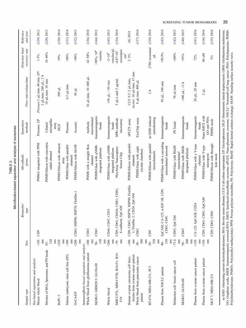
TABLE
2Microfluidics-bas
edse
paratio
nan
dan
alysis
ofex
osom
es
Sampletype
Size
Biomarker
type
Microfluidic
form
sSeparatio
nmethods
Flowrate/volum
e/tim
eDetectio
nlim
it/recovery
rate
Reference
andyear
Size-based
separatio
nan
dan
alysis
Mouse
who
lebloo
d~1
50 nmCD9
PMMA
integrated
with
PPM
Pressure;
EP
Pressure:1μL
/min;4
0min.E
P:
2μL
/min;24
0μL
;2h
1.5%
(129)20
12
Mixture
ofBSA,liposom
es,and
PSbeads
83– 120
nm
PDMS/Siliconwith
concentric
radial
channel
Ciliated
micropillar
array
10μL
/min;10
min
10–60%
(128)20
13
BxP
C-3
<22
0nm
PDMS/Siliconwith
obstacle
array
DLD
200μL
39%
(130)20
14
Murineem
bryonicstem
celllin
e(D
3)~1
00 nmPDMS/Glass
with
parallel
channels
Pressure
6.5μL
/min
>80
%(131)20
14
OvC
A42
9<20
0nm
CD63;HSP90
;HSP70
;Flotillin
-1PDMS/Siliconwith
SSAW
Acoustic
50μL
>90
%(132)20
15
Immunoa
ffinity-based
separatio
nandanalysis
Whole
bloo
dfrom
glioblastomapatient
~100 nm
CD63
PDMSwith
astraight
flow
channel
Antibod
ymicrochannel
16μL
/min;10
–400
μL42–94%
(136)20
10
SKMG3;
GBM20
/3;GLI36vIII
50-
150
nm
CD63
PDMS/Glass
with
μNMR-
integrated
platform
Immunom
agnetic
beads
>90
%;10
4
vesicles
(144)20
12
Whole
bloo
d<20
0nm
CD44;CD47;CD55
PDMS/Glass
with
μNMR-
integrated
platform
Immunom
agnetic
beads
150μL
;<30
min
~2×10
6
vesicles/μL
(145)20
13
MHCC97
L;MHCC97
H;B16
-F1;
B16-
F10
~70 nm
CD9;
CD63;CD41
b;CD81;CD82;
E-cadherin;
EpC
AM
PlexA
rray
Nanocapture
SensorChip
SPRiantib
ody
microarray
5μL
/sand5μg
/mL
~4.87×
107
exosom
es/
cm2
(134)20
14
Hum
anov
ariancarcinom
acelllin
es;
Ascitesfrom
ovariancancer
patient
~100 nm
CD9;
CD63;HSP70
;HSP90
;Flotillin
1;Flotillin
2;CD24;EpC
AM
PDMS/Siliconwith
parallel
channels
nPLEX
assay
CCS:
0.2–2μL
/min;
Ascites:10
μL/m
in;15
min
2–3%
(8)20
14
Whole
bloo
dfrom
pancreatic
cancer
patient
30– 300
nm
CD63
PDMS/Glass
with
eight
circular
cham
bers
Exo
Chipassay
8μL
/min;40
0μL
(137)20
14
BT-47
4;MDA-M
B-231
;PC3
30– 350
nm
CD9
PDMS/Siliconwith
parallel
microchannels
ac-EHD
indu
ced
nanoshearing
2h
2760
exosom
es/
μL(139)20
14
Plasm
afrom
NSCLCpatient
40– 150
nm
EpC
AM;CA-125;α-IG
F-1R;CD9;
CD63
;CD81
PDMS/Glass
with
acascading
microchannelnetwork
Immunom
agnetic
beads
30μL
;10
0min
>99
.9%
(143)20
14
Melanom
acell;
breastcancer
cell
~75.4
nmCD63;EpC
AM
PDMS/Glass
with
RInSE
microchannel
PSbeads
70μL
/min
~100
%(142)20
15
SKMG3;
GLI36vIII
50– 200
nm
CD63
PDMS/Glass
with
iMER-
integrated
platform
Immunom
agnetic
beads
~100
μL;<2h
>93
%(140)20
15
Plasm
afrom
ovariancancer
patient
<15
0nm
CA-125
;EpC
AM;CD24
PDMS/Glass
with
aY-type
channel
Immunom
agnetic
beads
20μL
;20
min
72%
(141)20
16
Plasm
afrom
ovariancancer
patient
<15
0nm
CD9;
CD63;CD81
;EpC
AM
PDMS/Glass
with
Y-type
micropo
sts
Nano-IM
EX
with
GO
andPDA
2μL
80aM
(138)20
16
MCF-7;MDA-M
B-231
CD9;
CD63;CD81
PDMS/Siliconwith
through-
hole
mesh
PDMSmesh
3%(135)20
16
ac-EHD:A
lternatingcurrentelectrohydrodynam
ic;B
SA:B
ovineserumalbumin;C
CS:C
ellculturesupernatant;EP:E
lectrophoresis;D
LD:D
eterministic
lateraldisplacem
ent;GBM:G
lioblastomamultiforme;
GO:Grapheneoxide;iM
ER:Im
munom
agnetic
exosom
alRNA;IM
EX:Interfaced
microfluidicexosom
e;nP
LEX:Nano-plasmon
icexosom
e;NSCLC:Non
small-celllung
cancer;PDA:Polydop
amine;PDMS:
Polydim
ethylsilo
xane;PMMA:P
oly(methylm
ethacrylate);PPM:P
orouspolymer
monolith
s;PS:P
olystyrene;R
InSE:R
apid
inertialsolutionexchange;S
SAW:S
tand
ingsurfaceacou
sticwave.
SCREENING TUMOR BIOMARKERS 35
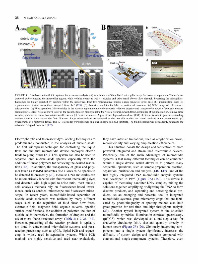
Electrophoretic and fluorescent dyes labeling techniques arepredominantly conducted in the analysis of nucleic acids.The first widespread technique for controlling the liquidflow and the first microfluidic device employed electricfields to pump fluids (23). This system can also be used toseparate ionic nucleic acids species, especially with theaddition of linear polymers for achieving the desired resolu-tion (146). In addition, the transparency of glass and poly-mer (such as PDMS) substrates also allows cNAs species tobe detected fluorescently (20). Because DNA molecules canbe ratiometrically labeled with fluorescent intercalating dyesand detected with high signal-to-noise ratio, most nucleicacid analysis methods rely on fluorescence-based instru-ments, such as confocal microscope and fluorescent micro-scope. In recent years, microfluidic-based separation ofnucleic acids molecules was realized by many differentways, such as the regulation of fluid shear flow force,electronic field, magnetic field, organic solvents, specificsurface modifications, the addition of polymer, the size ofnucleic acids themselves, the formation of droplets and theuse of micro-/nano-structured arrays (Table 3) (17, 21, 147).However, processing of the reaction products is typicallynot done in conventional microfluidic systems, and post-reaction processing, such as qPCR, digital PCR and sequen-cing, is widely used in separation systems. While PCRmethods are highly sensitive and used near exclusively,
they have intrinsic limitations, such as amplification errors,reproducibility and varying amplification efficiencies.
This situation boosts the design and fabrication of morepowerful integrated and streamlined microfluidic devices.Practically, one of the main advantages of microfluidicsystems is that many different techniques can be combinedwithin a single device, which allows us to perform manysequential operations, such as sample preparation, reaction,separation, purification and analysis (148, 149). One of thefirst highly integrated DNA microfluidic analysis systemswas developed in 1998 (Figure 9A) (150). This device iscapable of measuring nanoliter DNA samples, mixing thesolutions together, amplifying or digesting the DNA to formdiscrete products, and separating and detecting those pro-ducts. As an emerging and powerful tool in integratedmicrofluidic systems, gene microarray chips that are fabri-cated by photolithography or spotting method also holdgreat promise for real-time and high-throughput analysis(23). Another typical integrated system is the so-calledmicrofluidic cylindrical illumination confocal spectroscopy(µCICS), which was developed as a one-step assay foranalyzing circulating DNA size and quantity directly inhuman serum (Figure 9B) (20). Obviously, integrating com-ponents into a single system significantly increases thedifficulty of system design and operation compared withconventional single-component systems. Therefore, even
FIGURE 7 Size-based microfluidic systems for exosome analysis. (A) A schematic of the ciliated micropillar array for exosome separation. The cells aredepleted before entering the micropillar region, while cellular debris as well as proteins and other small objects flow through, bypassing the micropillars.Exosomes are highly enriched by trapping within the nanowires. Inset (a): representative porous silicon nanowire forest. Inset (b): micropillars. Inset (c):representative ciliated micropillars. Adapted from Ref. (128); (B) Acoustic nanofilter for label separation of exosomes. (a) SEM image of cell releasedmicrovesicles. (b) Filter operation. Microvesicles in the acoustic region are under the acoustic radiation pressure and transported to nodes of acoustic pressureregion (inset). Larger vesicles move faster as the acoustic force is proportional to the vesicle volume. Sheath flows, positioned at the node region, remove largevesicles, whereas the center flow retains small vesicles. (c) Device schematic. A pair of interdigitated transducer (IDT) electrodes is used to generate a standingsurface acoustic wave across the flow direction. Large microvesicles are collected at the two side outlets, and small vesicles at the center outlet. (d)Micrographs of a prototype device. The IDT electrodes were patterned on a piezoelectric (LiNO3) substrate. The fluidic channel was permanently bonded to thesubstrate. Adapted from Ref. (132).
36 N. HAO AND J.X.J. ZHANG

FIGURE 8 Immunoaffinity-based microfluidic systems for exosome analysis. (A) Surface modification of antibodies on plasmonic substrate for microfluidic-based exosome analysis. (a) Cancer cells secrete an abundance of exosomes through fusion of a multivesicular body (MVB) with the cellular plasmamembrane. The inset TEM image shows exosomes from CaOV3 cell. (b) Finite-difference time-domain simulation shows the enhanced electromagnetic fieldstightly confined near a periodic nanohole surface. The field distribution overlaps with the size of exosomes captured onto the sensing surface, maximizingexosome detection sensitivity. (c) A SEM image of the periodic nanoholes in the nanoplasmonic exosome (nPLEX) sensor. (d) A prototype miniaturizednPLEX imaging system developed for multiplexed and high-throughput analyses of exosomes. (e) A representative schematic of changes in transmissionspectra showing exosome detection with nPLEX. The gold surface is pre-functionalized by a layer of polyethylene glycol (PEG), and antibody conjugation andspecific exosome binding were monitored by transmission spectral shifts as measured by nPLEX. (f) SEM image shows exosome capture by functionalizednPLEX. Adapted from Ref. (8); (B) Surface modification of antibodies on micrometer-sized beads for microfluidic-based exosome analysis. (a) Workflow ofthe ExoSearch chip for continuous mixing, separation and in situ, multiplexed detection of circulating exosomes. (b, c) Bright-field microscope images ofimmunomagnetic beads manipulated in the microfluidic channel for mixing and separation of exosomes. (d) Exosome-bound immunomagnetic beadsaggregated in the microchamber with an on/off switchable magnet for continuous collection and release of exosomes. (e) TEM image of an exosome-bound immunomagnetic bead in a cross-sectional view. Adapted from Ref. (141).
SCREENING TUMOR BIOMARKERS 37

though such devices are theoretically possible, the commer-cialization and widespread development of these fully inte-grated and automated systems are still largely demanded inthe future. However, the availability of robust microfabrica-tion techniques and high unit demand should facilitate thedevelopment of “Sample-In, Answer-Out” point-of-carenucleic acids analysis platform and thus utilize them tobenefit our humankinds. Examples of microfluidics-basedseparation and analysis of nucleic acids are summarized inTable 3.
CURRENT CHALLENGES AND FUTUREPERSPECTIVES
CTCs
The rare and heterogeneous properties of CTCs are stilltwo significant facts to be drawn from the liquid biopsyapproaches. Given the significance of CTCs in the meta-static process and the potential roles of using these cellsfor noninvasive cancer theranostics, great efforts havebeen made to develop robust CTCs separation and ana-lysis approaches. Many emerging and scalable microflui-dic systems, including our own, demonstrated feasibilityunder clinical settings for separating CTCs at increas-ingly higher yields and facilitate downstream analyses.The current progress of these microfluidic systems forCTCs separation and detection is summarized, and theworking principles and experimental results of keymicrofluidic technologies are tabulated above (Figure 2
and Table 1). These microfluidic systems are generallybased on label-free techniques (including hydrophoresisand dielectrophoresis strategies) and immunoaffinitytechniques (including positive selection and negativeselection strategies) (Figure 2). Despite the recent tech-nological advances, the development of a single portabledevice capable of simultaneously achieving short proces-sing time, high CTCs recovery and high throughputremains challenging.
For label free-based microfluidic techniques, bothhydrophoresis and dielectrophoresis strategies relyingon the physical properties of cells can have a highthroughput as these strategies are well compatible withhigh cell flow rates. Hydrophoresis strategies can alsoenable high capture rates of CTCs due to the actual sizeand deformability differences between CTCs and hema-tological cells. However, these hydrophoresis strategies,especially filtration methods, may suffer from the cellviability issue resulting from potential damage incurredwhen the cells pass through the obstacle arrays in anarrow passage. In addition, size overlapping betweenCTCs and WBCs also indicates that any size-basedseparation strategies may lead to a loss of relativelysmall-sized CTCs (Figure 3A). Dielectrophoresis strate-gies, which can leverage differences in the size andelectric properties of cells, hold a great promise to leadto high CTCs capture efficiency compared to the hydro-phoresis strategies that are generally based on the cellssize differences only. However, because of the limitedelectric differences between target cells and background
FIGURE 9 Integrated microfluidic devices for nucleic acids analysis. (A) An integrated nanoliter DNA microfluidic analysis device. (a) Schematic ofintegrated device with two liquid samples and electrophoresis gel present. Color code: blue, liquid sample (ready for metering); green, hydrophobic surfaces;purple, polyacrylamide gel. (b) Optical micrograph of the device from above. Wire bonds to the printed circuit board can be seen along the top edge of thedevice. Adapted from Ref. (150); (B) An integrated microfluidic cylindrical illumination confocal spectroscopy (µCICS) system for circulating nucleic acidsdetection. (a) Schematic illustration of the one-step, PCR-free, single-molecule DNA sizing assay. (b) Optical component diagram of µCICS platform. (c) CCDimage of the µCICS illumination sheet focused into the microfluidic analysis constriction. The channel boundaries are demarcated by dashed yellow lines,while the projection of the confocal aperture into sample space is shown in red. Adapted from Ref. (20).
38 N. HAO AND J.X.J. ZHANG

TABLE
3Microfluidics-bas
edse
paratio
nan
dan
alysis
ofnu
cleicac
ids(N
As)
NAstype
Sampletype
Microfluidicform
sSeparatio
nmethods
Volume/tim
eAna
lysistools
Detectio
nrange/
limit/efficiency
Reference
andyear
DNA
Mycobacterium
tuberculosis
Silicon/glasswith
Y-typechannel
EP
120nL
;15
min
PAGE
0.12
mg/mL
(150
)19
98
DNA
λ-phage
Glass/Siliconwith
tapershapearray
DLA
(Stretching)
Confocalmicroscope
120μg
/mL
(151
)20
04DNA
λ-ph
age;
T2-phage;
Who
lebloo
dPDMS/Siliconwith
parallelmillim
eters
channels
EP
Microliter
AILIIM
25μg
/mL
(152
)20
04
DNA
λ-phage
PCBwith
multilayer
setof
coils
Magnetic
droplet
manipulationsystem
~10μL
PCR;AGE
1.25
μg/m
L(153
)20
06
miRNA
IDT
Microfluidiccapillary
LNA-D
NA
1fL
Confocalmicroscope
500fM
(154
)20
06mRNA
NIH
/3T3cells
PDMS/glass
with
multilayer
channels
Magnetic
Dyn
abeads
TM
with
oligo(dT
) 25
4nL
qRT-PCR
0.3–10
0pg
(155
)20
06
DNA
B.subtilis;
S.aureus;E.coli
96-w
ellSPRIreactorplatewith
LAMIs
Gradientmicropillararray
~1μL
AGE
~10ng
(156
)20
08miRNA
Plasm
afrom
prostate
cancer
patient
TaqM
anTMmiRNA
microarray
mirVana™
PARIS™
Kit
2μL
qRT-PCR
100%
sensitivity
(157
)20
08
DNA
λ-ph
age;
T2-phage
PDMS/Glass
with
parallelchannels
Oscillatoryshearflow
(Stretching)
AILIIM
100%
sensitivity
(158
)20
09
mRNA
C.pa
rvum
oocysts
PMMA/Glass
with
asawtoothchannel
Magnetic
Dyn
abeads
TM
with
oligo(dT
) 25
3.5μL
Fluorescent
microscop
eFrom
5oo
cysts
(159
)20
09
DNA
IDT
PDMS/Glass
with
multilayer
channels
MB
0.1μL
;<30
min
Confocalmicroscope
5pM
(160
)20
09
DNA
Serum
from
lung
cancer
patient
PDMSwith
Y-typechannel
Non
e<10
pL;10
sµCICS
100%
massdetection
efficiency
(20)
2010
DNA
λ-phage
PMMA/Siliconwith
micropillararray
None
3μL
EOM;SEM
1×10
−18M
(161
)20
11DNA
Celllin
esbearingKRASalleles
PDMS/Siliconwith
droplets-forming
channels
None
Picoliter
Confocalmicroscope
1/200000
(162
)20
11
DNA;RNA
IDT
PDMS/Glass
with
aring-shape
channel
None
Picoliter
Confocalmicroscope
100pM
(163
)20
11DNA;rRNA
Whole
bloodfrom
ovariancancer
patient
PDMS/Siliconwith
aV-shape
droplets-
form
ingchannels
SPE
5μL
AGE
(164
)20
11
DNA
BAC
Glass/Siliconwith
micropillararrayand
parallelchannels
Gradientmicropillararray
(Stretching)
Fluorescent
microscop
e5μg
/mL
(165
)20
12
DNA
λ-phage
PDMS/glass
with
crossslots
PEG-N
aCl(Stretching)
Fluorescent
microscope
1pM
(166
)20
12DNA
λ-phage
PeT
microchip
bydirect-printing
EPwith
polymer
1.5nL
;4min
Bioanalyzer
TM21
0025
μg/m
L(146
)20
12miRNA
Who
lebloo
dfrom
breastcancer
patient
Geniom
TMBiochip
miRNA
microarray
miRNeasy
TMkit
Bioanalyzer
TM21
0085
.6%
detection
accuracy
(167
)20
12
miRNA
HEK293cells
Goldmicroelectrodes
array
Electrochem
istry
Picoliter
Potentio
metry
20pM
(168
)20
12RNA
IDT
Glass
with
droplets-formingcrossed-
channel
Agarose
Picoliter
Flow
cytometry;
Fluorescent
microscop
e10
0%sensitivity
(169
)20
12
rRNA
E.coli
PDMS/Siliconwith
droplets-forming
channels
PNA
Picoliter
Confocalmicroscope
1.56
nM(170
)20
12
DNA
Who
lebloo
dfrom
CLLpatient
100sitesNanoC
hipT
Mwith
checkerboard
pattern
DEP
20μL
Fluorescent
microscop
e8–16
ng/m
L(171
)20
13
mRNA
C.pa
rvum
oocysts
PMMAwith
parallelchannels
PAMAM
dend
rimer
NASBA;LFA
From
30oo
cysts
(172
)20
14DNA
saliv
a/plasmafrom
lung
cancer
patient
GeneFluidicsT
Mchip
with
16bare
gold
electrode
Electrochem
istry
20–4
0μL
;<10
min
FISH
1/10
00(173
)20
14
(Con
tinued)
SCREENING TUMOR BIOMARKERS 39

TABLE
3(C
ontin
ued)
NAstype
Sampletype
Microfluidicform
sSeparatio
nmethods
Volume/tim
eAna
lysistools
Detectio
nrange/
limit/efficiency
Reference
andyear
DNA
λ-ph
age;
Whole
bloo
d10
0sitesNanoC
hipT
Mwith
checkerboard
pattern
DEP
15min
Epifluo
rescentmicroscop
e20
0pg
/mL
(174
)20
14
mRNA
GBM
celllin
es;Whole
bloo
dfrom
GBM
patient
PDMS/Glass
with
iMER-integrated
platform
Glass
beadsfilter
~100
μL;2h
qRT-PCR
90%
(140
)20
15
miRNA
PANC1cells
Plastic/LiNbO
3with
Ti/A
lelectrode
SAW
~100
μL;1.5
hIon-exchange
nanomem
brane
2pM
(175
)20
15
DNA
Who
lebloo
dPDMS/PMMAwith
aU-shape
QIA
ampT
Mkit
1μL
Magnetoresistivesensor
0.6μM
(176
)20
16
µCICS:Microfluidiccylin
drical
illum
inationconfocal
spectroscopy;AGE:Agarose
gelelectrophoresis;AILIIM:Argon
ionlaser-illum
inated
inverted
microscope;
BAC:Bacterial
artifi
cial
chromosom
e;CLL:
Chron
iclymph
ocyticleukem
ia;D
EP:Dielectroph
oresis;DLA:D
irectlinearanalysis;EOM:Epifluorescence
opticalmicroscopy;
EP:E
lectrophoresis;F
ISH:F
luorescencein
situ
hybridization;
GBM:G
lioblastoma
multiforme;
IDT:Integrated
DNA
technology;iM
ER:Im
munom
agnetic
exosom
alRNA;LAMIs:Large
area
moldinserts;LFA
:Lateral
flow
assay;
LNA:Lockednu
cleicacid;MB:Molecular
beacon;miRNA:
MicroRNA;mRNA:Messenger
RNA;NASBA:Nucleic
acid
sequence-based
amplificatio
n;PA
GE:Polyacrylam
idegelelectrophoresis;PA
MAM:Polyamidoamine;
PCB:Printed
circuitboard;
PCR:Polym
erase
chainreactio
n;PDMS:Polydim
ethylsilo
xane;PEG:Polyethyleneglycol;PeT:Polyester-toner;PMMA:Poly(methylmethacrylate);PNA:Peptid
enucleicacid;qR
T-PCR:Quantitativ
ereversetranscription
polymerasechainreactio
n;rRNA:Ribosom
alRNA;SAW:Surface
acou
stic
wave;
SEM:Scann
ingelectron
microscop
y;SPE:Solid
phaseextractio
n;SPRI:Solid-phase
reversible
immobilizatio
n.
40 N. HAO AND J.X.J. ZHANG
cells, these dielectrophoresis strategies are still not get-ting as high yield performance as expected.
For immunoaffinity-based microfluidic techniques, thereare two types of approaches for capturing and collectingCTCs, i.e., positive selections and negative selections. Bothof them can be generally realized by either modifyingmicrochannel substrate surface with antibodies or manipu-lating antibodies-conjugated micrometer-sized magneticbeads. A significant challenge in the antibodies-immobilizedmicrochannel methods for both positive selections andnegative selections is their relatively low processingthroughput. This situation is caused by a limited numberof interaction sites between the surface-bound ligands andthe target cells. The possibility of nonspecific capture alsomakes it more complicated. Specifically, cell flow rates needto be slow enough to ensure the maximum target cellsretention, but reasonably fast enough to generate shearforce to prevent nonspecific attachment of nontarget cellsto the channel surface. Therefore, although various micro-channel structures have been developed to show increasedperformance, microfluidic devices with high CTCs recoveryrate, purity and throughput still remain to be achieved.Regarding the immunomagnetic beads methods for CTCsseparation, the heterogeneous property of tumor cells thatmay not express the same or the same level of specificantigens makes the negative selections much more appeal-ing than the positive selections. Negative selections not onlyenable the simultaneous separation of various types of CTCsbut also allow the target cells to be collected in an intactform.
Despite great efforts have been achieved, many works stillneed to be done for further improving the separation efficiencyand analysis sensitivity of CTCs in microfluidic systems. Thewell-developed versatile microfabrication techniques can pro-vide new ideas in the design and construction of microfluidicchannels, which will be greatly helpful for target cells capturedinside microchannels. Current advances of nanobiotechnologycan also be used in this field, especially for immunomagneticbeads-based cells separation. For example, many studies shedlight on the important roles of shape and surface chemistry ofmicro-/nano-particles on biological effects, such as cell bindingkinetics, margination in laminar flows, surface functionalizationdensity of ligands and even cell toxicity (177, 178). These willbring new insights for the design of immunomagnetic beads toachieve short processing time and high CTCs recovery rate.Another approach that utilizes a combination of multiple cellcapture techniques may provide tremendous opportunities forimproving the CTCs capture performance of microfluidic sys-tems. We can envision a multi-module microfluidic system forCTCs capture in which some modules perform high throughputconcentration and purification of tumor cells, while some othersenable the selective capture of specific tumor cells. The proto-types of such kinds of devices can be found in recent studies,which may guide the focus of future research (51, 75, 105). In
addition, it is noteworthy that most of current tests in micro-fluidic systems were carried out using spiked samples (spikingtumor cells into PBS, cell culture medium or whole blood fromhealthy individuals) rather than real clinical samples from cancerpatients (Table 1). Because of the complexity of the peripheralblood and tumormicroenvironment of cancer patients, such testsmay not properly reflect the effectiveness of microfluidicdevices, and may thus hinder their research and development.
Exosome
Tumor-derived exosome as a promising alternative in liquidbiopsy offers new clinical opportunities for minimally inva-sive diagnostics and monitoring of cancer diseases. Theseparation and analysis capabilities of exosomes in micro-fluidic systems are summarized in Table 2, which showstheir superior performance compared to that of conventionaltechniques, such as ultracentrifugation and commercial kits.Such improvements in terms of response time, sensitivityand selectivity from microfluidic systems will greatlyenhance the specific role of exosome in existing circulatingtumor biomarkers and address current hurdles in liquidbiopsy of cancers. However, exosome secretion is adynamic process, producing diverse populations with largedifferences in size and concentration. Exosomes, during thetreatment process, also suffer from the stability issue (179).Therefore, precise measurement and analysis of exosomesare still quite challenging.
Current established microfluidic systems for exosomesseparation are generally based on either size or immunoaffinitytechniques. The performance of size-based exosome separationtechniques will certainly be affected by the population sizedifferences as mentioned above. In this situation, it is importantto determine the design of microfluidic devices in order to avoidpassage clogging and exosome vesicle trapping issues. In addi-tion, because the sizes of exosomes are in the nanometer rangeand thus much smaller than the sizes in conventional microfab-rication technologies, more advanced technologies are stillrequired to achieve high yield and purity of exosomes forclinical translation. To this end, a novel multidimensional hier-archical structure (termed “ciliated nanowire-on-micropillar”)with a combination of silicon microfabrication processes, elec-troplating and electroless metal-assisted nanowire etching tech-niques was developed in our laboratory (128). Comparatively,immunoaffinity-based exosome separation techniques can leadto relatively high specificity and rapid analysis. However, sig-nificant challenges still exist in this field, for both antibody-immobilized microchannel methods and immunobeads meth-ods. For example, there is currently no “ubiquitous” biomarkerto distinguish subsets of exosomes from each other, due to thefact that exosomes also have a heterogeneous nature similar toCTCs. Therefore, there are very limited biomarkers that canaccurately distinguish cancer-specific exosomes from normalexosomes.
SCREENING TUMOR BIOMARKERS 41

To date, efficient and reliable microfluidic systems for theseparation of circulating tumor exosomes are still lacking, whichlead to the exosomes-related research far lagging behind thanCTCs research. The fact that the definition and characterizationof exosome types are not yet firmly established makes thissituation even worse (9). In order to qualitatively and quantita-tively evaluate exosomes with cells origins, and thus boost thedevelopment of corresponding microfluidic systems, morenovel technologies are urgently needed for comprehensivelycharacterizing the surface and intravesicular compositions ofexosomes.
cNAs
Gene expression profiles have shown dramatic correla-tions with tumor development and progression. Geneticchanges can thus be utilized as biomarkers for the detec-tion and diagnosis of cancer. Recently developed micro-fluidic systems for the separation and analysis of nucleicacids are summarized in Table 3, demonstrating thepotential to develop point-of-care microfluidic devicescapable of being used as liquid biopsies. By integratingand miniaturizing these progresses into microfluidic sys-tems, many advantages including low cost, automation,fast processing time, high throughput, and reduced con-tamination are realized compared to their conventionalmacroscale counterparts. Examination of nucleic acidsmolecules with microfluidic operations has also enabledthe emergence of new analysis platforms, especially forclinical translations (180).
Although the development of microfluidic techniquesfor nucleic acids analysis has advanced significantly,there is still great room for the techniques themselvesand the integration capabilities with other processes. Forexample, nucleic acids are generally separated throughextraction process, which can compromise their integrityand result in analysis errors. The ability to achieve com-plete extraction efficiency from complex samples hasalso yet to be realized. These issues limit the utility ofnucleic acids as biomarkers and are further troublesomefor their clinical applications. Therefore, faster and gen-tler extraction protocols that help to maintain sampleintegrity and maximize yield efficiency will be crucialto realize the full potential of genetic mapping and sin-gle-molecule sequencing technologies. Standardizingsample collection and processing protocols is also impor-tant for the development of more reliable and efficientmicrofluidic devices. In addition, since the separation ofnucleic acids is quite difficult and less efficient, an alter-native based on separation-free techniques was devel-oped for nucleic acids analysis. Few of these integratedmicrofluidic devices that are summarized in Table 3 canbe able to handle raw samples and guide them throughdirect detection apparatus. This will be an appealing
technique toward developing truly point-of-care micro-fluidic devices. Furthermore, it is noteworthy that theprofiles of genetic mutations in different tumors aremuch more complicated than we can imagine, and theheterogeneity features make it even more difficult toidentify key genetic information of cancer that couldprovide sufficient diagnostic utility. This calls for moreefficient integrated genetic and microfluidic techniques interms of performance and precision.
CONCLUSIONS
Cancer-derived cells, exosomes and nucleic acids representtypical circulating biomarkers that can be used as noninvasiveliquid biopsies for the diagnosis and treatment of cancer-related diseases. Microfluidic technologies endow uniqueopportunities to achieve the unprecedented spatio-temporalcontrol of these circulating tumor biomarkers. Although theresearch and development of microfluidic systems for cancertheranostics are relatively recent, a number of studies havealready demonstrated their superior performance in separatingand identifying these biomarkers from various sources. Thenext few years will certainly witness even more intensedevelopment of microfluidic systems for biomarkers separa-tion, characterization and detection.
However, many challenges still remain and demand persis-tent attention before clinical translation is feasible. Firstly, thecirculating tumor biomarkers are relatively rare and extremelyheterogeneous, making their analysis inherently difficult.Overcoming this challenge requires the development of com-binatorial techniques that can take advantages of multipleunique physical and biochemical properties of tumor biomar-kers. Secondly, through highly precise fluid control and auto-mation, significant advances have beenmade in the burgeoningfield of microfluidic biomarker separation and analysis, butmany of such systems remain in the proof-of-concept stage.
To fully explore the clinical potential of microfluidicsin liquid biopsy, tremendous efforts are still needed toimprove the adaptability of the microfluidic technologiesto clinical settings and promote the commercialization ofthe microfluidic systems. Thirdly, the relationshipbetween circulating tumor biomarkers and microfluidicsis still not well established. To further advance thedevelopment of microfluidics-based clinical biomarkersassays, validation studies to establish the specificity,sensitivity and reproducibility of such tests should besystematically conducted in large cohorts of cancerpatients and for multiple cancers of interest. Finally,but not lastly, our understanding of tumor biology isstill in its infancy. As discoveries continue to be madeon the roles of various circulating biomarkers in tumorburden and progression, more robust and efficient micro-fluidic systems will be developed to benefit cancer
42 N. HAO AND J.X.J. ZHANG

patients. We anticipate that in the near future, microflui-dic technologies will play an increasingly prominent rolein the liquid biopsy of cancer, personalized disease diag-nosis and beyond.
FUNDING
The authors are grateful for the financial support from theNational Institute of Health (NIH) Director’s TransformativeResearch Award (R01HL137157), NSF ECCS-1509369,and Norris Cotton Cancer Center Developmental Funds(Pilot Projects).
REFERENCES
1. Wan, L., Pantel, K., and Kang, Y. (2013) Tumor metastasis: movingnew biological insights into the clinic. Nat. Med., 19: 1450–1464.
2. Chi, K.R. (2016) The tumour trail left in blood. Nature, 532: 269–271.3. Hyun, K.A., Kim, J., Gwak, H., and Jung, H.I. (2016) Isolation and
enrichment of circulating biomarkers for cancer screening, detection,and diagnostics. Analyst. 141: 382–92.
4. Kailasa, S.K. and Kang, S.H. (2009) Microchip-based capillary elec-trophoresis for dna analysis in modern biotechnology: a review. Sep.Purif. Rev., 38: 242–288.
5. Warburg, O. (1956) On the origin of cancer cells. Science,123: 309–314.6. Hynes, R.O. (2011) Metastatic cells will take any help they can get.
Cancer Cell, 20: 689–690.7. Myung, J.H. and Hong, S. (2015) Microfluidic devices to enrich and
isolate circulating tumor cells. Lab Chip, 15:4500–4511.8. Im, H., Shao, H., Park, Y.I., Peterson, V.M., Castro, C.M., Weissleder, R.,
and Lee, H. (2014) Label-free detection and molecular profiling of exo-somes with a nano-plasmonic sensor. Nat. Biotechnol., 32: 490–495.
9. Kowal, J., Tkach, M., and Théry, C. (2014) Biogenesis and secretionof exosomes. Curr. Opin. Cell Biol., 29: 116–125.
10. Ren, J., He, W., Zheng, L., and Duan, H. (2016) From structures tofunctions: insights into exosomes as promising drug delivery vehicles.Biomater. Sci., 4: 910–921.
11. Mandel, P. and Metais, P. (1948) Les acides nucléiques du plasma sanguinchez l’homme. C. R. Seances Soc. Biol. Ses Fil., 142: 241–243.
12. Stroun, M., Anker, P., Maurice, P., Lyautey, J., Lederrey, C., andBeljanski, M. (1989) Neoplastic characteristics of the dna found inthe plasma of cancer patients. Oncology, 46: 318–322.
13. Han, K-H., Han, A., and Frazier, A.B. (2006) Microsystems forisolation and electrophysiological analysis of breast cancer cellsfrom blood. Biosens. Bioelectron., 21: 1907–1914.
14. Bettegowda, C., Sausen, M., Leary, R.J., Kinde, I., Wang, Y., Agrawal, N.,Bartlett, B.R., Wang, H., Luber, B., Alani, R.M., Antonarakis, E.S., Azad,N.S., Bardelli, A., Brem, H., Cameron, J.L., Lee, C.C., Fecher, L.A.,Gallia, G.L., Gibbs, P., Le, D., Giuntoli, R.L., Goggins, M., Hogarty, M.D., Holdhoff, M., Hong, S.-M., Jiao, Y., Juhl, H.H., Kim, J.J., Siravegna,G., Laheru, D.A., Lauricella, C., Lim, M., Lipson, E.J., Marie, S.K.N.,Netto, G.J., Oliner, K.S., Olivi, A., Olsson, L., Riggins, G.J., Sartore-Bianchi, A., Schmidt, K., Shih, L.-M., Oba-Shinjo, S.M., Siena, S.,Theodorescu, D., Tie, J., Harkins, T.T., Veronese, S., Wang, T.-L.,Weingart, J.D., Wolfgang, C.L., Wood, L.D., Xing, D., Hruban, R.H.,Wu, J., Allen, P.J., Schmidt, C.M., Choti, M.A., Velculescu, V.E.,Kinzler, K.W., Vogelstein, B., Papadopoulos, N., and Diaz, L.A. (2014)
Detection of circulating tumor dna in early- and late-stage human malig-nancies. Sci. Transl. Med., 6: 224ra24.
15. Siravegna, G. and Bardelli, A. (2016) Blood circulating tumor dna for non-invasive genotyping of colon cancer patients.Mol. Oncol., 10: 475–480.
16. Bellassai, N. and Spoto, G. (2016) Biosensors for liquid biopsy:circulating nucleic acids to diagnose and treat cancer. Anal. Bioanal.Chem., 408: 7255–7264.
17. Nahavandi, S., Baratchi, S., Soffe, R., Tang, S-Y., Nahavandi, S.,Mitchell, A., and Khoshmanesh, K. (2014) Microfluidic platformsfor biomarker analysis. Lab Chip, 14: 1496–1514.
18. Lee, R.C., Feinbaum, R.L., and Ambros, V. (1993) The c. elegansheterochronic gene lin-4 encodes small rnas with antisense comple-mentarity to lin-14. Cell, 75: 843–854.
19. Allegra, A., Alonci, A., Campo, S., Penna, G., Petrungaro, A.,Gerace, D., and Musolino, C. (2012) Circulating micrornas: newbiomarkers in diagnosis, prognosis and treatment of cancer (review).Int. J. Oncol., 41: 1897–1912.
20. Liu, K.J., Brock, M.V., Shih, I., andWang, T. (2010) Decoding circulatingnucleic acids in human serum using microfluidic single molecule spectro-scopy. J. Am. Chem. Soc., 132: 5793–5798.
21. Friedrich, S.M., Zec, H.C., and Wang, T-H. (2016) Analysis of singlenucleic acid molecules in micro- and nano-fluidics. Lab Chip, 16:790–811.
22. Haeberle, S. and Zengerle, R. (2007) Microfluidic platforms for lab-on-a-chip applications. Lab Chip, 7: 1094–1110.
23. Livak-Dahl, E., Sinn, I., and Burns, M. (2011) Microfluidic chemicalanalysis systems. Annu. Rev. Chem. Biomol. Eng., 2: 325–353.
24. Lisowski, P. and Zarzycki, P.K. (2013)Microfluidic paper-based analyticaldevices (μpads) and micro total analysis systems (μtas): development,applications and future trends. Chromatographia, 76: 1201–1214.
25. Faustino, V., Catarino, S.O., Lima, R., and Minas, G. (2015)Biomedical microfluidic devices by using low-cost fabrication techni-ques: a review. J. Biomech., 49: 2280–2292.
26. Alix-Panabières, C. and Pantel, K. (2014) Challenges in circulatingtumour cell research. Nat. Rev. Cancer, 14: 623–631.
27. Cima, I., Wen Yee, C., Iliescu, F.S., Min Phyo, W., Hon Lim, K.,Iliescu, C., and Han Tan, M. (2013) Label-free isolation of circulatingtumor cells in microfluidic devices: current research and perspectives.Biomicrofluidics, 7: 011810.
28. Hosokawa, M., Hayata, T., Fukuda, Y., Arakaki, A., Yoshino, T.,Tanaka, T., and Matsunaga, T. (2010) Size-selective microcavityarray for rapid and efficient detection of circulating tumor cells.Anal. Chem., 82: 6629–6635.
29. Zheng, S., Lin, H., Liu, J-Q., Balic, M., Datar, R., Cote, R. J., and Tai,Y.-C. (2007) Membrane microfilter device for selective capture, elec-trolysis and genomic analysis of human circulating tumor cells. J.Chromatogr. A, 1162: 154–161.
30. Hosokawa, M., Yoshikawa, T., Negishi, R., Yoshino, T., Koh, Y.,Kenmotsu, H., Naito, T., Takahashi, T., Yamamoto, N., Kikuhara,Y., Kanbara, H., Tanaka, T., Yamaguchi, K., and Matsunaga, T.(2013) Microcavity array system for size-based enrichment of circu-lating tumor cells from the blood of patients with small-cell lungcancer. Anal. Chem., 85: 5692–5698.
31. Loutherback, K., D’Silva, J., Liu, L., Wu, A., Austin, R.H., andSturm, J.C. (2012) Deterministic separation of cancer cells fromblood at 10 ml/min. AIP Adv., 2: 42107.
32. Liu, Z., Huang, F., Du, J., Shu, W., Feng, H., Xu, X., and Chen, Y.(2013) Rapid isolation of cancer cells using microfluidic deterministiclateral displacement structure. Biomicrofluidics, 7: 011801.
33. Mohamed, H., Murray, M., Turner, J.N., and Caggana, M. (2009)Isolation of tumor cells using size and deformation. J. Chromatogr. A,1216: 8289–8295.
SCREENING TUMOR BIOMARKERS 43

34. Tan, S.J., Yobas, L., Lee, G.Y.H., Ong, C.N., and Lim, C.T. (2009)Microdevice for the isolation and enumeration of cancer cells fromblood. Biomed. Microdevices, 11: 883–892.
35. Tan, S.J., Lakshmi, R.L., Chen, P., Lim, W-T., Yobas, L., and Lim, C.T. (2010) Versatile label free biochip for the detection of circulatingtumor cells from peripheral blood in cancer patients. Biosens.Bioelectron., 26: 1701–1705.
36. Lv, P., Tang, Z., Liang, X., Guo, M., and Han, R.P.S. (2013) Spatiallygradated segregation and recovery of circulating tumor cells fromperipheral blood of cancer patients. Biomicrofluidics, 7: 034109.
37. Di Carlo, D. (2009) Inertial microfluidics. Lab Chip, 9: 3038–3046.38. Gossett, D.R., Weaver, W.M., Mach, A.J., Hur, S.C., Tse, H.T.K., Lee, W.,
Amini, H., andDi Carlo, D. (2010) Label-free cell separation and sorting inmicrofluidic systems. Anal. Bioanal. Chem., 397: 3249–3267.
39. Bhagat, A.A.S., Hou, H.W., Li, L.D., Lim, C.T., and Han, J. (2011)Pinched flow coupled shear-modulated inertial microfluidics for high-throughput rare blood cell separation. Lab Chip, 11: 1870–1878.
40. Hur, S.C., Mach, A.J., and Di Carlo, D. (2011) High-throughput size-based rare cell enrichment using microscale vortices. Biomicrofluidics,5: 022206.
41. Mach, A.J., Kim, J.H., Arshi, A., Hur, S.C., and Di Carlo, D. (2011)Automated cellular sample preparation using a centrifuge-on-a-chip.Lab Chip, 11: 2827–2834.
42. Moon, H-S., Kwon, K., Hyun, K-A., Sim, T.S., Park, J.C., Lee, J.G.,and Jung, H.I. (2013) Continual collection and re-separation of circu-lating tumor cells from blood using multi-stage multi-orifice flowfractionation. Biomicrofluidics, 7: 014105.
43. Sun, J., Li, M., Liu, C., Zhang, Y., Liu, D., Liu, W., Hu, G., and Jiang,X. (2012) Double spiral microchannel for label-free tumor cell separa-tion and enrichment. Lab Chip, 12: 3952.
44. Sun, J., Liu, C., Li, M., Wang, J., Xianyu, Y, Hu, G., and Jiang, X.(2013) Size-based hydrodynamic rare tumor cell separation in curvedmicrofluidic channels. Biomicrofluidics, 7: 011802.
45. Warkiani, M.E., Guan, G., Luan, K.B., Lee, W.C., Bhagat, A.A.S.,Kant Chaudhuri, P., Tan, D.S.-W., Lim, W.T., Lee, S.C., Chen, P.C.Y.,Lim, C.T., and Han, J. (2014) Slanted spiral microfluidics for theultra-fast, label-free isolation of circulating tumor cells. Lab Chip,14: 128–137.
46. Hou, H.W., Warkiani, M.E., Khoo, B.L., Li, Z.R., Soo, R.A., Tan, D.S.-W., Lim, W.-T., Han, J., Bhagat, A.A.S., and Lim, C.T. (2013)Isolation and retrieval of circulating tumor cells using centrifugalforces. Sci. Rep., 3: 1259.
47. Zborowski, M. and Chamers, J.J. (2011) Rare cell separation andanalysis by magnetic sorting. Anal. Chem., 83: 8050–8056.
48. Augustsson, P., Magnusson, C., Nordin, M., Lilja, H., and Laurell, T.(2012) Microfluidic, label-free enrichment of prostate cancer cells inblood based on acoustophoresis. Anal. Chem., 84: 7954–7962.
49. Ding, X., Peng, Z., Lin, S.C.S., Geri, M., Li, S., Li, P., Chen, Y., Dao,M., Suresh, S., and Huang, T.J. (2014) Cell separation using tilted-angle standing surface acoustic waves. Proc. Natl. Acad. Sci. U.S.A.,111: 12992–12997.
50. Li, P., Mao, Z., Peng, Z., Zhou, L., Chen, Y., Huang, P.-H., Truica, C.I., Drabick, J.J., El-Deiry, W.S., Dao, M., Suresh, S., and Huang, T.J.(2015) Acoustic separation of circulating tumor cells. Proc. Natl.Acad. Sci. U.S.A., 112: 4970–4975.
51. Kim, M.S., Sim, T.S., Kim, Y.J., Kim, S.S., Jeong, H., Park, J.-M.,Moon, H.-S., Kim, S.I., Gurel, O., Lee, S.S., Lee, J.-G., and Park, J.C.(2012) Ssa-moa: a novel ctc isolation platform using selective sizeamplification (ssa) and a multi-obstacle architecture (moa) filter. LabChip, 12: 2874–2880.
52. Pethig, R. (2010) Dielectrophoresis: status of the theory, technology,and applications. Biomicrofluidics, 4: 022811.
53. Huang, Y., Yang, J., Wang, X-B., Becker, F.F., and Gascoyne, P.R.C.(1999) The removal of human breast cancer cells from hematopoieticcd34 + stem cells by dielectrophoretic field-flow-fractionation. J.Hematother. Stem Cell Res., 8: 481–490.
54. Yang, J., Huang, Y., Wang, X.B., Becker, F.F., and Gascoyne, P.R.(1999) Cell separation on microfabricated electrodes using dielectro-phoretic/gravitational field-flow fractionation. Anal. Chem., 71: 911–918.
55. Wang, X-B., Yang, J., Huang, Y., Vykoukal, J., Becker, F.F., andGascoyne, P.R.C. (2000) Cell separation by dielectrophoretic field-flow-fractionation. Anal. Chem., 72: 832–839.
56. Gascoyne, P.R.C., Noshari, J., Anderson, T.J., and Becker, F.F. (2009)Isolation of rare cells from cell mixtures by dielectrophoresis.Electrophoresis, 30: 1388–1398.
57. Sabuncu, A.C., Liu, J.A., Beebe, S.J., and Beskok, A. (2010)Dielectrophoretic separation of mouse melanoma clones.Biomicrofluidics, 4: 021101.
58. Cheng, J., Sheldon, E.L., Wu, L., Heller, M.J., and O’Connell, J.P.(1998) Isolation of cultured cervical carcinoma cells mixed withperipheral blood cells on a bioelectronic chip. Anal. Chem., 70:2321–2326.
59. Huang, Y., Joo, S., Duhon, M., Heller, M., Wallace, B., and Xu, X.(2002) Dielectrophoretic cell separation and gene expression profilingon microelectronic chip arrays. Anal. Chem., 74: 3362–3371.
60. Altomare, L., Borgatti, M., Medoro, G., Manaresi, N., Tartagni, M.,Guerrieri, R., and Gambari, R. (2003) Levitation and movement ofhuman tumor cells using a printed circuit board device based on soft-ware-controlled dielectrophoresis. Biotechnol. Bioeng., 82: 474–479.
61. Park, J., Kim, B., Choi, S.K., Hong, S., Lee, S.H., and Lee, K-I.(2005) An efficient cell separation system using 3d-asymmetricmicroelectrodes. Lab Chip, 5: 1264–1270.
62. An, J., Lee, J., Lee, S.H., Park, J., and Kim, B. (2009) Separation ofmalignant human breast cancer epithelial cells from healthy epithelialcells using an advanced dielectrophoresis-activated cell sorter (dacs).Anal. Bioanal. Chem., 394: 801–809.
63. Yang, F., Yang, X., Jiang, H., Bulkhaults, P., Wood, P., Hrushesky, W.,and Wang, G. (2010) Dielectrophoretic separation of colorectal cancercells. Biomicrofluidics, 4: 013204.
64. Huang, Y., Wang, X.B., Becker, F.F., and Gascoyne, P.R. (1997)Introducing dielectrophoresis as a new force field for field-flow frac-tionation. Biophys. J., 73: 1118–1129.
65. Moon, H-S., Kwon, K., Kim, S-I., Han, H., Sohn. J., Lee, S., andJung, H.-I. (2011) Continuous separation of breast cancer cells fromblood samples using multi-orifice flow fractionation (moff) and die-lectrophoresis (dep). Lab Chip, 11: 1118–1125.
66. Shafiee, H., Caldwell, J.L., Sano, M.B., and Davalos, R.V. (2009)Contactless dielectrophoresis: a new technique for cell manipulation.Biomed. Microdevices, 11: 997–1006.
67. Henslee, E.A., Sano, M.B., Rojas, A.D., Schmelz, E.M., and Davalos,R.V. (2011) Selective concentration of human cancer cells usingcontactless dielectrophoresis. Electrophoresis, 32: 2523–2529.
68. Gupta, V., Jafferji, I., Garza, M., Melnikova, V.O., Hasegawa, D.K.,Pethig, R., and Davis, D.W. (2012) ApostreamTM, a new dielectro-phoretic device for antibody independent isolation and recovery ofviable cancer cells from blood. Biomicrofluidics, 6: 024133.
69. Hughes, A.D., Mattison, J., Western, L.T., Powderly, J.D., Greene, B.T., and King, M.R. (2012) Microtube device for selectin-mediatedcapture of viable circulating tumor cells from blood. Clin. Chem., 58:846–853.
70. Wang, S., Liu, K., Liu, J., Yu, Z.T.F., Xu, X., Zhao, L., Lee, T., Lee,E.K., Reiss, J., Lee, Y.K., Chung, L.W.K., Huang, J., Rettig, M.,Seligson, D., Duraiswamy, K.N., Shen, C.K.F., and Tseng, H.R.
44 N. HAO AND J.X.J. ZHANG

(2011) Highly efficient capture of circulating tumor cells by usingnanostructured silicon substrates with integrated chaotic micromixers.Angew. Chem. Int. Ed., 50: 3084–3088.
71. Dharmasiri, U., Njoroge, S.K., Witek, M.A., Adebiyi, M.G.,Kamande, J.W., Hupert, M.L., Barany, F., and Soper, S.A. (2011)High-throughput selection, enumeration, electrokinetic manipulation,and molecular profiling of low-abundance circulating tumor cellsusing a microfluidic system. Anal. Chem., 83: 2301–2309.
72. Adams, A.A., Okagbare, P.I., Feng, J., Hupert, M.L., Patterson, D.,Gottert, J., McCarley, R. L., Nikitopoulos, D., Murphy, M.C., andSoper, S.A. (2008) Highly efficient circulating tumor cell isolationfrom whole blood and label-free enumeration using polymer-basedmicrofluidics with an integrated conductivity sensor. J. Am. Chem.Soc., 130: 8633–8641.
73. Stott, S.L., Hsu, C-H., Tsukrov, D.I., Yu, M., Miyamoto, D.T.,Waltman, B.A., Rothenberg, S.M., Shah, A.M., Smas, M.E., Korir,G.K., Floyd, F.P., Gilman, A.J., Lord, J.B., Winokur, D., Springer, S.,Irimia, D., Nagrath, S., Sequist, L.V., Lee, R.J., Isselbacher, K.J.,Maheswaran, S., Haber, D.A., and Toner, M. (2010) Isolation ofcirculating tumor cells using a microvortex-generating herringbone-chip. Proc. Natl. Acad. Sci. U.S.A., 107: 18392–18397.
74. Nagrath, S., Sequist, L.V., Maheswaran, S., Bell, D.W., Irimia, D.,Ulkus, L., Smith, M.R., Kwak, E.L., Digumarthy, S., Muzikansky, A.,Ryan, P., Balis, U.J., Tompkins, R.G., Haber, D.A., and Toner, M.(2007) Isolation of rare circulating tumour cells in cancer patients bymicrochip technology. Nature, 450: 1235–1239.
75. Liu, Z., Zhang, W., Huang, F., Feng, H., Shu, W., Xu, X., and Chen,Y. (2013) High throughput capture of circulating tumor cells using anintegrated microfluidic system. Biosens. Bioelectron., 47:113–119.
76. Wang, S., Wang, H., Jiao, J., Chen, K.J., Owens, G.E., Kamei, K.I.,Sun, J., Sherman, D.J., Behrenbruch, C.P., Wu, H., and Tseng, H.R.(2009) Three-dimensional nanostructured substrates toward efficientcapture of circulating tumor cells. Angew. Chem. Int. Ed., 48: 8970–8973.
77. Kang, J.H., Krause, S., Tobin, H., Mammoto, A., Kanapathipillai, M.,and Ingber, D.E. (2012) A combined micromagnetic-microfluidicdevice for rapid capture and culture of rare circulating tumor cells.Lab Chip, 12: 2175–2181.
78. Sivagnanam, V., Song, B., Vandevyver, C., Bünzli, J.C.G., and Gijs,M.A.M. (2010) Selective breast cancer cell capture, culture, andimmunocytochemical analysis using self-assembled magnetic beadpatterns in a microfluidic chip. Langmuir, 26: 6091–6096.
79. Fais, S., O’Driscoll, L., Borras, F.E., Buzas, E., Camussi, G.,Cappello, F., Carvalho, J., Cordeiro da Silva, A., Del Portillo, H., ElAndaloussi, S., Ficko Trček, T., Furlan, R., Hendrix, A., Gursel, I.,Kralj-Iglic, V., Kaeffer, B., Kosanovic, M., Lekka, M.E., Lipps, G.,Logozzi, M., Marcilla, A., Sammar, M., Llorente, A., Nazarenko, I.,Oliveira, C., Pocsfalvi, G., Rajendran, L., Raposo, G., Rohde, E.,Siljander, P., van Niel, G., Vasconcelos, M.H., Yáñez-Mó, M.,Yliperttula, M.L., Zarovni, N., Zavec, A.B., and Giebel, B. (2016)Evidence-based clinical use of nanoscale extracellular vesicles innanomedicine. ACS Nano, 10: 3886–3899.
80. Phillips, J.A., Xu, Y., Xia, Z., Fan, Z.H., and Tan, W. (2009)Enrichment of cancer cells using aptamers immobilized on a micro-fluidic channel. Anal. Chem., 81: 1033–1039.
81. Xu, Y., Phillips, J.A., Yan, J., Li, Q., Fan, Z.H., and Tan, W. (2009)Aptamer-based microfluidic device for enrichment, sorting, and detec-tion of multiple cancer cells. Anal. Chem., 81: 7436–7442.
82. Zhu, Y., Kekalo, K., Ndong, C., Huang, Y., Shubitidze, F., Griswold,K.E., Baker, I., and Zhang, J.X.J. (2016) Magnetic-nanoparticle-basedimmunoassays-on-chip: materials synthesis, surface functionalization,and cancer cell screening. Adv. Funct. Mater., 26: 3953–3972.
83. Chen, P., Huang, Y., Hoshino, K., and Zhang, J.X.J. (2014) Multiscaleimmunomagnetic enrichment of circulating tumor cells: from tubes tomicrochips. Lab Chip, 14: 446–458.
84. Hoshino, K., Huang, Y-Y., Lane, N., Huebschman, M., Uhr, J.W.,Frenkel, E.P., and Zhang, J.X.J. (2011) Microchip-based immunomag-netic detection of circulating tumor cells. Lab Chip, 11: 3449–3457.
85. Huang, Y.Y., Hoshino, K., Chen, P., Wu, C.H., Lane, N.,Huebschman, M., Liu, H., Sokolov, K., Uhr, J.W., Frenkel, E.P., andZhang, J.X.J. (2013) Immunomagnetic nanoscreening of circulatingtumor cells with a motion controlled microfluidic system. Biomed.Microdevices, 15: 673–681.
86. Hoshino, K., Chen, P., Huang, Y.Y., and Zhang, J.X.J. (2012)Computational analysis of microfluidic immunomagnetic rare cellseparation from a particulate blood flow. Anal. Chem., 84: 4292–4299.
87. Huang, Y., Chen, P., Wu, C., Hoshino, K., Sokolov, K., Lane, N., Liu,H., Huebschman, M., Frenkel, E., and Zhang, J.X.J. (2015) Screeningand molecular analysis of single circulating tumor cells using micro-magnet array. Sci. Rep., 5: 16047.
88. Chen, P., Huang, Y.Y., Bhave, G., Hoshino, K., and Zhang, J.X.J.(2016) Inkjet-print micromagnet array on glass slides for immuno-magnetic enrichment of circulating tumor cells. Ann. Biomed. Eng.,44: 1710–1720.
89. Ng, E., Hoshino, K., and Zhang, J.X.J. (2013) Microfluidic immuno-detection of cancer cells via site-specific microcontact printing ofantibodies on nanoporous surface. Methods, 63: 266–275.
90. Chen, P., Huang, Y-Y., Hoshino, K., and Zhang, J.X.J. (2015)Microscale magnetic field modulation for enhanced capture and dis-tribution of rare circulating tumor cells. Sci. Rep., 5: 8745.
91. Wu, C.H., Huang, Y.Y., Chen, P., Hoshino, K., Liu, H., Frenkel, E.P.,Zhang, J.X.J., and Sokolov, K.V. (2013) Versatile immunomagneticnanocarrier platform for capturing cancer cells. ACS Nano, 7: 8816–8823.
92. Chen, K.C., Pan, Y.C., Chen, C.L., Lin, C.H., Huang, C.S., and Wo,A.M. (2012) Enumeration and viability of rare cells in a microfluidicdisk via positive selection approach. Anal. Biochem., 429: 116–123.
93. Ozkumur, E., Shah, A.M., Ciciliano, J.C., Emmink, B.L., Miyamoto,D.T., Brachtel, E., Yu, M., Chen, P., Morgan, B., Trautwein, J.,Kimura, A., Sengupta, S., Stott, S.L., Karabacak, N.M., Barber, T.A., Walsh, J.R., Smith, K., Spuhler, P.S., Sullivan, J.P., Lee, R.J.,Ting, D.T., Luo, X., Shaw, A.T., Bardia, A., Sequist, L.V, Louis, D.N.,Maheswaran, S., Kapur, R., Haber, D.A., and Toner, M. (2013)Inertial focusing for tumor antigen-dependent and -independent sort-ing of rare circulating tumor cells. Sci. Transl. Med., 5: 179ra47.
94. Kim, S., Han, S-I., Park, M-J., Jeon, C-W., Joo, Y-D., Choi, I., andHan, K. (2013) Circulating tumor cell microseparator based on lateralmagnetophoresis and immunomagnetic nanobeads. Anal. Chem., 85:2779–2786.
95. Park, J.M., Lee, J.Y., Lee, J.G., Jeong, H., Oh, J.M., Kim, Y.J., Park,D., Kim, M.S., Lee, H.J., Oh, J.H., Lee, S.S., Lee, W.Y., and Huh, N.(2012) Highly efficient assay of circulating tumor cells by selectivesedimentation with a density gradient medium and microfiltrationfrom whole blood. Anal. Chem., 84: 7400–7407.
96. Lin, M.X, Hyun, K.A., Moon, H.S., Sim, T.S., Lee, J.G., Park, J.C.,Lee, S.S., and Jung, H.I. (2013) Continuous labeling of circulatingtumor cells with microbeads using a vortex micromixer for highlyselective isolation. Biosens. Bioelectron., 40: 63–67.
97. Kim, M.S., Kim, J., Lee, W., Cho, S.J., Oh, J.M., Lee, J.Y., Baek, S.,Kim, Y.J., Sim, T.S., Lee, H.J., Jung, G.E., Kim, S.I., Park, J.M., Oh,J.H., Gurel, O., Lee, S.S., and Lee, J.G. (2013) A trachea-inspiredbifurcated microfilter capturing viable circulating tumor cells viaaltered biophysical properties as measured by atomic force micro-scopy. Small, 9: 3103–3110.
98. Liu, Z., Fusi, A., Klopocki, E., Schmittel, A., Tinhofer, I.,Nonnenmacher, A., and Keilholz, U. (2011) Negative enrichment byimmunomagnetic nanobeads for unbiased characterization of circulat-ing tumor cells from peripheral blood of cancer patients. J. Transl.Med., 9: 70.
SCREENING TUMOR BIOMARKERS 45
99. Hyun, K.A., Lee, T.Y., and Jung, H.I. (2013) Negative enrichment ofcirculating tumor cells using a geometrically activated surface inter-action chip. Anal. Chem., 85: 4439–4445.
100. Diéguez, L., Winter, M.A., Pocock, K.J., Bremmell, K.E., andThierry, B. (2015) Efficient microfluidic negative enrichment of cir-culating tumor cells in blood using roughened pdms. Analyst, 140:3565–3572.
101. Chen, C-L., Chen, K-C., Pan, Y-C., Lee, T-P., Hsiung, L-C., Lin, C.-M., Chen, C.-Y., Lin, C.-H., Chiang, B.-L., and Wo, A.M. (2011)Separation and detection of rare cells in a microfluidic disk vianegative selection. Lab Chip, 11: 474–483.
102. Lu, Y., Liang, H., Yu, T., Xie, J., Chen, S., Dong, H., Sinko, P.J., Lian,S., Xu, J., Wang, J., Yu, S., Shao, J., Yuan, B., Wang, L., and Jia, L.(2015) Isolation and characterization of living circulating tumor cellsin patients by immunomagnetic negative enrichment coupled withflow cytometry. Cancer, 121: 3036–3045.
103. Sajay, B.N.G., Chang, C.P., Ahmad, H., Khuntontong, P., Wong, C.C.,Wang, Z., Puiu, P. D., Soo, R., and Rahman, A.R.A. (2014)Microfluidic platform for negative enrichment of circulating tumorcells. Biomed. Microdevices, 16: 537–548.
104. Bhuvanendran Nair Gourikutty, S., Chang, C-P., and Poenar, D.P.(2016) An integrated on-chip platform for negative enrichment oftumour cells. J. Chromatogr. B, 1028: 153–164.
105. Karabacak, N.M., Spuhler, P.S., Fachin, F., Lim, E.J., Pai, V.,Ozkumur, E., Martel, J.M., Kojic, N., Smith, K., Chen, P., Yang, J.,Hwang, H., Morgan, B., Trautwein, J., Barber, T.A., Stott, S.L.,Maheswaran, S., Kapur, R., Haber, D.A., and Toner, M. (2014)Microfluidic, marker-free isolation of circulating tumor cells fromblood samples. Nat. Protoc., 9: 694–710.
106. Kwon, K.W., Choi, S.S., Lee, S.H., Kim, B., Lee, S.N., Park, M.C.,Kim, P., Hwang, S.Y., and Suh, K.Y. (2007) Label-free, microfluidicseparation and enrichment of human breast cancer cells by adhesiondifference. Lab Chip, 7: 1461–1468.
107. Kuo, J.S., Zhao, Y., Schiro, P.G., Ng, L., Lim, D.S.W., Shelby, J.P.,and Chiu, D.T. (2010) Deformability considerations in filtration ofbiological cells. Lab Chip, 10: 837–842.
108. Zheng, S., Lin, H.K., Lu, B., Williams, A., Datar, R., Cote, R.J., andTai, Y.C. (2011) 3d microfilter device for viable circulating tumor cell(ctc) enrichment from blood. Biomed. Microdevices, 13: 203–213.
109. Bhagat, A.A.S., Hou,H.W., Li, L.D., Lim, C.T., andHan, J.H. (2011)Deanflow fractionation (DFF) isolation of circulating tumor cells (CTCs) fromblood. 15th International Conference on Miniaturized Systems forChemistry and Life Sciences. Seattle, Washington, USA, 524–526.
110. Hosokawa, M., Kenmotsu, H., Koh, Y., Yoshino, T., Yoshikawa, T., Naito,T., Takahashi, T., Murakami, H., Nakamura, Y., Tsuya, A., Shukuya, T.,Ono, A., Akamatsu, H., Watanabe, R., Ono, S., Mori, K., Kanbara, H.,Yamaguchi, K., Tanaka, T.,Matsunaga, T., andYamamoto, N. (2013) Size-based isolation of circulating tumor cells in lung cancer patients using amicrocavity array system. PLoS One, 8: e67466.
111. Becker, F.F., Wang, X.B., Huang, Y., Pethig, R., Vykoukal, J., andGascoyne, P.R.C. (1994) The removal of human leukaemia cells fromblood using interdigitated microelectrodes. J. Phys. D Appl. Phys., 27:2659–2662.
112. Becker, F.F., Wang, X.B., Huang, Y., Pethig, R., Vykoukal, J., andGascoyne, P.R. (1995) Separation of human breast cancer cells fromblood by differential dielectric affinity. Proc. Natl. Acad. Sci. U.S.A.,92: 860–864.
113. Gascoyne, P.R.C., Wang, X.B., Huang, Y., and Becker, R.F. (1997)Dielectrophoretic separation of cancer cells from blood. IEEE T. Ind.Appl., 33: 670–678.
114. Das, C.M., Becker, F., Vernon, S., Noshari, J., Joyce, C., and Gascoyne, P.R.C. (2005) Dielectrophoretic segregation of different human cell types onmicroscope slides dielectrophoretic segregation of different human celltypes on microscope slides. Anal. Chem., 77: 2708–2719.
115. Tai, C.H., Hsiung, S.K., Chen, C.Y., Tsai, M.L., and Lee, G.B. (2007)Automatic microfluidic platform for cell separation and nucleus col-lection. Biomed. Microdevices, 9: 533–543.
116. Kim, U., Shu, C-W., Dane, K.Y., Daugherty, P.S., Wang, J.Y.J., andSoh, H.T. (2007) Selection of mammalian cells based on their cell-cycle phase using dielectrophoresis. Proc. Natl. Acad. Sci. U.S.A.,104: 20708–20712.
117. Cristofanilli, M., Krishnamurthy, S., Das, C.M., Reuben, J.M., Spohn,W., Noshari, J., Becker, F., and Gascoyne, P.R. (2008) Dielectric cellseparation of fine needle aspirates from tumor xenografts. J. Sep. Sci.,31: 3732–3739.
118. Kang, Y., Li, D., Kalams, S.A., and Eid, J.E. (2008) Dc-dielectro-phoretic separation of biological cells by size. Biomed. Microdevices,10: 243–249.
119. Kostner, S., van den Driesche, S., Witarski, W., Pastorekova, S., andVellekoop, M.J. (2010) Guided dielectrophoresis: a robust method forcontinuous particle and cell separation. IEEE Sens. J., 10: 1440–1446.
120. Alazzam, A., Stiharu, I., Bhat, R., and Meguerditchian, A.N. (2011)Interdigitated comb-like electrodes for continuous separation ofmalignant cells from blood using dielectrophoresis. Electrophoresis.32: 1327–1336.
121. Huang, S-B., Wu M-H., Lin, Y-H., Hsieh, C-H., Yang, C-L., Lin, H.-C.,Tseng, C.-P., and Lee, G.-B. (2013) High-purity and label-free isolation ofcirculating tumor cells (ctcs) in a microfluidic platform by using optically-induced-dielectrophoretic (odep) force. Lab Chip, 13: 1371–1383.
122. Takao, M. and Takeda, K. (2011) Enumeration, characterization, andcollection of intact circulating tumor cells by cross contamination-freeflow cytometry. Cytom. A, 79: 107–117.
123. Mittal, S., Wong, I.Y., Deen, W.M., and Toner, M. (2012) Antibody-functionalized fluid-permeable surfaces for rolling cell capture at highflow rates. Biophys. J., 102: 721–730.
124. Schiro, P.G., Zhao, M., Kuo, J.S., Koehler, K.M., Sabath, D.E., andChiu, D.T. (2012) Sensitive and high-throughput isolation of rare cellsfrom peripheral blood with ensemble-decision aliquot ranking. Angew.Chem. Int. Ed., 51: 4618–4622.
125. Kralj, J.G., Arya, C., Tona, A., Forbes, T.P., Munson, M.S., Sorbara,L., Srivastava, S., and Forry, S.P. (2012) A simple packed bed devicefor antibody labelled rare cell capture from whole blood. Lab Chip,12: 4972–4975.
126. Ko, J., Carpenter, E., and Issadore, D. (2015) Detection and isolationof circulating exosomes and microvesicles for cancer monitoring anddiagnostics using micro-/nano-based devices. Analyst, 141:450–460.
127. He, M. and Zeng, Y. (2016) Microfluidic exosome analysis towardliquid biopsy for cancer. J. Lab. Autom., 21: 599–608.
128. Wang, Z., Wu, H., Fine, D., Schmulen, J., Hu, Y., Godin, B., Zhang, J.X.J., and Liu, X. (2013) Ciliated micropillars for the microfluidic-based isolation of nanoscale lipid vesicles. Lab Chip, 13: 2879–2882.
129. Davies, R.T., Kim, J., Jang, S.C., Choi, E-J., Gho, Y.S., and Park, J.(2012) Microfluidic filtration system to isolate extracellular vesiclesfrom blood. Lab Chip, 12: 5202–5210.
130. Santana, S.M., Antonyak, M.A., Cerione, R.A., and Kirby, B.J. (2014)Microfluidic isolation of cancer-cell-derived microvesicles from heterge-neous extracellular shed vesicle populations. Biomed. Microdevices, 16:869–877.
131. Jo, W., Jeong, D., Kim, J., Cho, S., Jang, S.C., Han, C., Kang, J.Y.,Gho, Y.S., and Park, J. (2014) Microfluidic fabrication of cell-derivednanovesicles as endogenous rna carriers. Lab Chip, 14: 1261–1269.
132. Lee, K., Shao, H., Weissleder, R., and Lee, H. (2015) Acousticpurification of extracellular microvesicles. ACS Nano, 9: 2321–2327.
133. Maiolo, D., Paolini, L., Di Noto, G., Zendrini, A., Berti, D., Bergese, P.,and Ricotta, D. (2015) Colorimetric nanoplasmonic assay to determinepurity and titrate extracellular vesicles. Anal. Chem., 87: 4168–4176.
134. Zhu, L., Wang, K., Cui, J., Liu, H., Bu, X., Ma, H., Wang, W., Gong,H., Lausted, C., Hood, L., Yang, G., and Hu, Z. (2014) Label-free
46 N. HAO AND J.X.J. ZHANG

quantitative detection of tumor-derived exosomes through surfaceplasmon resonance imaging. Anal. Chem., 86: 8857–8864.
135. Chiu, Y., Cai, W., Shih, Y.V., Lian, I., and Lo, Y. (2016) A single-cellassay for time lapse studies of exosome secretion and cell behaviors.Small, 12: 3658–3666.
136. Chen, C., Skog, J., Hsu, C., Lessard, R.T., Balaj, L., Wurdinger, T.,Carter, B.S., Breakefield, X.O., Toner, M., and Irimia, D. (2010)Microfluidic isolation and transcriptome analysis of serum microve-sicles. Lab Chip, 10: 505–511.
137. Kanwar, S.S., Dunlay, C.J., Simeone, D.M., and Nagrath, S. (2014)Microfluidic device (exochip) for on-chip isolation, quantification andcharacterization of circulating exosomes. Lab Chip, 14: 1891–1900.
138. Zhang, P., He, M., and Zeng, Y. (2016) Ultrasensitive microfluidicanalysis of circulating exosomes using a nanostructured grapheneoxide/polydopamine coating. Lab Chip, 16: 3033–3042.
139. Vaidyanathan, R., Naghibosadat, M., Rauf, S., Korbie, D., Carrascosa,L.G., Shiddiky, M.J.A., and Trau, M. (2014) Detecting exosomesspecifically: a multiplexed device based on alternating current electro-hydrodynamic induced nanoshearing. Anal. Chem., 86: 11125–11132.
140. Shao, H., Chung, J., Lee, K., Balaj, L., Min, C., Carter, B.S.,Hochberg, F.H., Breakefield, X.O., Lee, H., and Weissleder, R.(2015) Chip-based analysis of exosomal mrna mediating drug resis-tance in glioblastoma. Nat. Commun., 6: 6999.
141. Zhao, Z., Yang, Y., Zeng, Y., and He, M. (2016) A microfluidicexosearch chip for multiplexed exosome detection towards blood-based ovarian cancer diagnosis. Lab Chip, 16: 489–496.
142. Dudani, J.S., Gossett, D.R., Tse, H.T.K., Lamm, R.J., Kulkarni, R.P.,and Carlo, D.D. (2015) Rapid inertial solution exchange for enrich-ment and flow cytometric detection of microvesicles.Biomicrofluidics, 9: 014112.
143. He, M., Crow, J., Roth, M., Zeng, Y., and Godwin, A.K. (2014)Integrated immunoisolation and protein analysis of circulating exo-somes using microfluidic technology. Lab Chip, 14: 3773–3780.
144. Shao, H., Chung, J., Balaj, L., Charest, A., Bigner, D.D., Carter, B.S.,Hochberg, F.H., Breakefield, X.O., Weissleder, R., and Lee, H. (2012)Protein typing of circulating microvesicles allows real-time monitor-ing of glioblastoma therapy. Nat. Med., 18: 1835–1840.
145. Rho, J., Chung, J., Im, H., Liong, M., Shao, H., Castro, C.M., Weissleder,R., and Lee, H. (2013) Magnetic nanosensor for detection and profiling oferythrocyte-derived microvesicles. ACS Nano, 7: 11227–11233.
146. Duarte, G.R.M., Coltro, W.K.T., Borba, J.C., Price, C.W., Landers, J.P., and Carrilho, E. (2012) Disposable polyester-toner electrophoresismicrochips for dna analysis. Analyst, 137: 2692–2698.
147. Reinholt, S.J. and Baeumner, A.J. (2014) Microfluidic isolation ofnucleic acids. Angew. Chem. Int. Ed., 53: 13988–13981.
148. Ahmad, H., Sutherland, A., Shin, Y.S., Hwang, K., Qin, L., Krom, R.-J., and Heath, J.R. (2011) A robotics platform for automated batchfabrication of high density, microfluidics-based dna microarrays, withapplications to single cell, multiplex assays of secreted proteins. Rev.Sci. Instrum., 82: 94301.
149. Yu, J., Zhou, J., Sutherland, A.,Wei,W., Shin, Y.S., Xue,M., and Heath, J.R. (2014) Microfluidics-based single-cell functional proteomics for funda-mental and applied biomedical applications. Annu. Rev. Anal. Chem., 7:275–295.
150. Burns, M.A., Johnson, B.N., Brahmasandra, S.N., Handique, K.,Webster, J.R., Krishnan, M., Sammarco, T.S., Man, P.M., Jones, D.,Heldsinger, D., Mastrangelo, C.H., and Burke, D.T. 1998. An inte-grated nanoliter dna analysis device. Science, 282: 484–487.
151. Chan, E.Y., Goncalves, N.M., Haeusler, R.A., Hatch, A.J., Larson, J.W., Maletta, A.M., Yantz, G.R., Carstea, E.D., Fuchs, M., Wong, G.G., Gullans, S.R., and Gilmanshin, R. (2004) Dna mapping usingmicrofluidic stretching and single-molecule detection of fluorescentsite-specific tags. Genome Res., 14: 1137–1146.
152. Dimalanta, E.T., Lim, A., Runnheim, R., Lamers, C., Churas, C.,Forrest, D.K., De Pablo, J.J., Graham, M.D., Coppersmith, S.N.,
Goldstein, S., and Schwartz, D.C. (2004) A microfluidic system forlarge dna molecule arrays. Anal. Chem., 76: 5293–5301.
153. Lehmann, U., Vandevyver, C., Parashar, V.K., and Gijs, M.A.M.(2006) Droplet-based dna purification in a magnetic lab-on-a-chip.Angew. Chem. Int. Ed., 45: 3062–3067.
154. Neely, L.A., Patel, S., Garver, J., Gallo, M., Hackett, M., McLaughlin,S., Nadel, M., Harris, J., Gullans, S., and Rooke, J. (2006) A single-molecule method for the quantitation of microrna gene expression.Nat. Methods, 3: 41–46.
155. Marcus, J.S., Anderson, W.F., and Quake, S.R. (2006) Microfluidicsingle-cell mrna isolation and analysis. Anal. Chem., 78: 3084–3089.
156. Park, D.S.W., Hupert, M.L., Witek, M.A., You, B.H., Datta, P., Guy, J.,Lee, J.B., Soper, S.A., Nikitopoulos, D.E., and Murphy, M.C. (2008) Atiter plate-based polymermicrofluidic platform for high throughput nucleicacid purification. Biomed. Microdevices, 10: 21–33.
157. Mitchell, P.S., Parkin, R.K., Kroh, E.M., Fritz, B.R., Wyman, S.K.,Pogosova-Agadjanyan, E.L., Peterson, A., Noteboom, J., O’Briant,K.C., Allen, A., Lin, D.W., Urban, N., Drescher, C.W., Knudsen,B.S., Stirewalt, D.L., Gentleman, R., Vessella, R.L., Nelson, P.S.,Martin, D.B., and Tewari, M. (2008) Circulating micrornas asstable blood-based markers for cancer detection. Proc. Natl.Acad. Sci. U.S.A., 105: 10513–10518.
158. Jo, K., Chen, Y.L., de Pablo, J.J., and Schwartz, D.C. (2009)Elongation and migration of single dna molecules in microchannelsusing oscillatory shear flows. Lab Chip, 9: 2348–2355.
159. Nugen, S.R., Asiello, P.J., and Baeumner, A.J. (2009) Design and fabrica-tion of a microfluidic device for near-single cell mrna isolation using acopper hot embossing master.Microsyst. Technol., 15: 477–483.
160. Puleo, C.M. and Wang, T-H. (2009) Microfluidic means of achievingattomolar detection limits with molecular beacon probes. Lab Chip, 9:1065–1072.
161. De, A.F., Gentile, F., Mecarini, F., Das, G., Moretti, M., Candeloro, P.,Coluccio, M.L., Cojoc, G., Accardo, A., Liberale, C., Zaccaria, R.P.,Perozziello, G., Tirinato, L., Toma, A., Cuda, G., Cingolani, R., andDi Fabrizio, E. (2011) Breaking the diffusion limit with super-hydro-phobic delivery of molecules to plasmonic nanofocusing sers struc-tures. Nat. Photon., 5: 682–687.
162. Pekin, D., Skhiri, Y., Baret, J-C., Corre, D.L., Mazutis, L., Salem, C.Ben, Millot, F., Harrak, A. El, Hutchison, J. B., Larson, J. W., Link,D. R., Laurent-Puig, P., Griffiths, A. D., and Taly, V. (2011)Quantitative and sensitive detection of rare mutations using droplet-based microfluidics. Lab Chip, 11: 2156–2166.
163. Kim, S., Streets, A.M., Lin, R.R., Quake, S.R., Weiss, S., andMajumdar, D.S. (2011) High-throughput single-molecule optofluidicanalysis. Nat. Methods, 8: 242–245.
164. Zhang, Y., Park, S., Liu, K., Tsuan, J., Yang, S., and Wang, T-H. (2011) Asurface topography assisted droplet manipulation platform for biomarkerdetection and pathogen identification. Lab Chip, 11: 398–406.
165. Lam, E.T., Hastie, A., Lin, C., Ehrlich, D., Das, S.K., Austin, M.D.,Deshpande, P., Cao, H., Nagarajan, N., Xiao, M., and Kwok, P.Y.(2012) Genome mapping on nanochannel arrays for structural varia-tion analysis and sequence assembly. Nat. Biotechnol., 30: 771–776.
166. Xu, W. and Muller, S.J. (2012) Polymer-monovalent salt-induced dnacompaction studied via single-molecule microfluidic trapping. LabChip. 12: 647–651.
167. Schrauder, M.G., Strick, R., Schulz-Wendtland, R., Strissel, P.L.,Kahmann, L., Loehberg, C.R., Lux, M.P., Jud, S. M., Hartmann, A.,Hein, A., Bayer, C.M., Bani, M.R., Richter, S., Adamietz, B.R.,Wenkel, E., Rauh, C., Beckmann, M.W., and Fasching, P.A. (2012)Circulating micro-rnas as potential blood-based markers for earlystage breast cancer detection. PLoS One, 7: e29770.
168. Goda, T., Masuno, K., Nishida, J., Kosaka, N., Ochiya, T.,Matsumoto, A., and Miyahara, Y. (2012) A label-free electrical detec-tion of exosomal micrornas using microelectrode array. Chem.Commun., 48: 11942–11944.
SCREENING TUMOR BIOMARKERS 47

169. Zhang, H., Jenkins, G., Zou, Y., Zhu, Z., and Yang, C.J. (2012)Massively parallel single-molecule and single-cell emulsion reversetranscription polymerase chain reaction using agarose droplet micro-fluidics. Anal. Chem., 84: 3599–3606.
170. Rane, T.D., Zec, H.C., Puleo, C., Lee, A.P., and Wang, T-H. (2012)Droplet microfluidics for amplification-free genetic detection of singlecells. Lab Chip, 12: 3341–3347.
171. Sonnenberg, A., Marciniak, J.Y., Mccanna, J., Krishnan, R., Rassenti,L., Kipps, T.J., and Heller, M.J. (2013) Dielectrophoretic isolation anddetection of cfc-dna nanoparticulate biomarkers and virus from blood.Electrophoresis, 34: 1076–1084.
172. Reinholt, S.J., Behrent, A., Greene, C., Kalfe, A., and Baeumner, A.J.(2014) Isolation and amplification of mrna within a simple microflui-dic lab on a chip. Anal. Chem., 86: 849–856.
173. Wei, F., Lin, C-C, Joon, A., Feng, Z., Troche, G., Lira, M.E., Chia, D.,Mao, M., Ho, C.-L., Su, W.-C., and Wong, D.T.W. (2014) Noninvasivesaliva-based egfr gene mutation detection in patients with lung cancer. Am.J .Respir. Crit. Care Med., 190: 1117–1126.
174. Mccanna, J.P., Sonnenberg, A., and Heller, M.J. (2014) Low levelepifluorescent detection of nanoparticles and dna on dielectrophore-tic microarrays. J. Biophotonics., 7: 863–873.
175. Taller, D., Richards, K., Slouka, Z., Senapati, S., Hill, R., Go, D.B., and Chang, H.-C. (2015) On-chip surface acoustic wave lysis
and ion-exchange nanomembrane detection of exosomal rna forpancreatic cancer study and diagnosis. Lab Chip., 15: 1656–1666.
176. Dias, T.M., Cardoso, F.A., Martins, S.A.M., Martins, V.C.,Cardoso, S., Gaspar, J.F., Monteiro, G., and Freitas, P.P. (2016)Implementing a strategy for on-chip detection of cell-free dnafragments using gmr sensors: a translational application in cancerdiagnostics using alu elements. Anal. Methods., 8: 119–128.
177. Hao, N.J., Li, L., and Tang, F. (2016) Shape matters when engi-neering mesoporous silica-based nanomedicines. Biomater. Sci., 4:575–591.
178. Hao, N.J., Li, L., and Tang, F. (2017) Roles of particle size, shapeand surface chemistry of mesoporous silica nanomaterials onbiological systems. Int. Mater. Rev., 62: 57–77.
179. Oosthuyzen, W., Sime, N.E.L., Ivy, J.R., Turtle, E.J., Street, J.M.,Pound, J., Bath, L.E., Webb, D.J., Gregory, C.D., Bailey, M.A.,and Dear, J.W. (2013) Quantification of human urinary exosomesby nanoparticle tracking analysis. J. Physiol., 591: 5833–5842.
180. Fakhoury, J.R., Sisson, J.C., and Zhang, J.X.J. (2009)Microsystems for controlled genetic perturbation of live droso-phila embryos: rna interference, development robustness and drugscreening. Microfluid. Nanofluid., 6: 299–313.
48 N. HAO AND J.X.J. ZHANG