biochemical analysis on microfluidic chips
TRANSCRIPT

Biochemical analysis on microfluidic chipsJing Wu a, Ziyi He b, Qiushui Chen b, Jin-Ming Lin b,*a School of Science, China University of Geosciences (Beijing), Beijing 100083, Chinab Department of Chemistry, Beijing Key Laboratory of Microanalytical Methods and Instrumentation, Tsinghua University, Beijing 100084, China
A R T I C L E I N F O
Keywords:Biochemical analysisMicrofluidic chipOptical detectorElectronic methodsAcoustic waveMagnetic operationChip-MS platformBiomimeticsBiomolecular analysiscell analysis
A B S T R A C T
Biochemical analysis is crucial in understanding mechanism of life activities and giving biological in-sights into life process. With inherent merits in flexible design, microscale of operation, good incorporationwith other techniques for manipulation and detection, microfluidic chip has introduced new paradigmsin biochemical analysis field. Considering the explosive development of microfluidics over the past decades,in this review, we summarized recent advances in biochemical analysis on microfluidic platforms. High-light was put on the integrated technologies such as optical, electrical, acoustic and magnetic techniques.Focus also was given on relative applications in biomimetics, drug screening, biomolecular detection, singlecell and stem cell analysis.
© 2016 Elsevier B.V. All rights reserved.
Contents
1. Introduction ........................................................................................................................................................................................................................................................ 2132. Biochemical analysis techniques on chips ............................................................................................................................................................................................... 214
2.1. Optical detector .................................................................................................................................................................................................................................... 2142.2. Electronic methods ............................................................................................................................................................................................................................. 2152.3. Acoustic wave ....................................................................................................................................................................................................................................... 2162.4. Magnetic operation ............................................................................................................................................................................................................................ 2162.5. Chip-MS platform ................................................................................................................................................................................................................................ 218
3. Applications ........................................................................................................................................................................................................................................................ 2193.1. Biomimetics ........................................................................................................................................................................................................................................... 2193.2. Drug discovery ..................................................................................................................................................................................................................................... 2213.3. Biomolecular analysis ........................................................................................................................................................................................................................ 221
3.3.1. Genetic analysis .................................................................................................................................................................................................................. 2213.3.2. Protein analysis ................................................................................................................................................................................................................... 223
3.4. Cell analysis ........................................................................................................................................................................................................................................... 2243.4.1. Single cells ............................................................................................................................................................................................................................ 2243.4.2. Stem cells .............................................................................................................................................................................................................................. 225
4. Conclusions and outlook ................................................................................................................................................................................................................................ 227Acknowledgements .......................................................................................................................................................................................................................................... 227References ............................................................................................................................................................................................................................................................ 227
1. Introduction
In native cellular microenvironment, cells are subject to complexbiochemical cues that vary in temporal and spatial scales [1]. These
cues can be divided into soluble and insoluble signaling mol-ecules, including chemokines [2], gradients of cytokines [3], growthfactor secretion from neighboring cells [4], and even biophysical in-teractions with the extracellular matrix (ECM) [5]. The autocrine andparacrine signals are sensed by cells in different ways and regu-late cell physiology temporally and spatially. Meanwhile, cells exertsome factors to alter their surrounding microenvironment [6,7].Probing the biochemical processes could govern cell behavior and
* Corresponding author. Tel.: +86 10 62792343; Fax: +86 10 62792343.E-mail address: [email protected] (J.-M. Lin).
http://dx.doi.org/10.1016/j.trac.2016.03.0130165-9936/© 2016 Elsevier B.V. All rights reserved.
Trends in Analytical Chemistry 80 (2016) 213–231
Contents lists available at ScienceDirect
Trends in Analytical Chemistry
journal homepage: www.elsevier.com/ locate / t rac
show great current and potential impact on the bio-analytical chem-istry researches. However, most in vitro cell-based biochemicalexperiments are performed in two-dimensional (2D) manner thatcells are cultured onto plastic surfaces and treated by coating themwith various solutions [8,9]. The standard 2D conditions poorlymimicthe cellular microenvironment and are absent of three-dimensional(3D) cues.
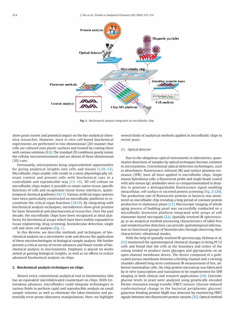
Fortunately, microsystems bring unprecedented opportunitiesfor giving analytical insights into cells and tissues [1,10–12].Microfluidic chips enable cells reside in a more physiologically rel-evant context and present cells with biochemical cues in acontrollable and reproducible way [13–15]. 3D cell culture onmicrofluidic chips makes it possible to retain native tissue-specificfunctions of cells and recapitulate tissue-tissue interfaces, spatio-temporal chemical gradients [16,17]. Various artificial organ systemshave been particularly constructed on microfluidic platforms to re-constitute the critical organ functions [18,19]. By integrating withbiochemical analysis techniques, microdevices show great promisefor basic biomedical and pharmaceutical researches. Over the pastdecade, the microfluidic chips have been recognized as ideal plat-forms for biochemical assays which have been widely expanded totissue engineering, drug screening, biomolecular detection, singlecell and stem cell analysis (Fig. 1).
In this Review, we describe methods and techniques of bio-chemical analysis on a micrometer scale and discuss the applicationof these microtechnologies in biological sample analysis. We furtherpresent a critical survey of recent advances and future trends of bio-chemical analysis in microsystems. Emphasis is placed on worksaimed at gaining biological insights, as well as on efforts to realizeadvanced biochemical analysis on chips.
2. Biochemical analysis techniques on chips
Almost every conventional analytical tool in biochemistry labshas an equivalent microfabricated counterpart on chips. With tre-mendous advances, microfluidics could integrate technologies invarious fields to perform rapid and reproducible analysis on smallsample volumes as well as eliminate the labor-intensive and po-tentially error-prone laboratory manipulations. Here, we highlight
several kinds of analytical methods applied in microfluidic chips inrecent years.
2.1. Optical detector
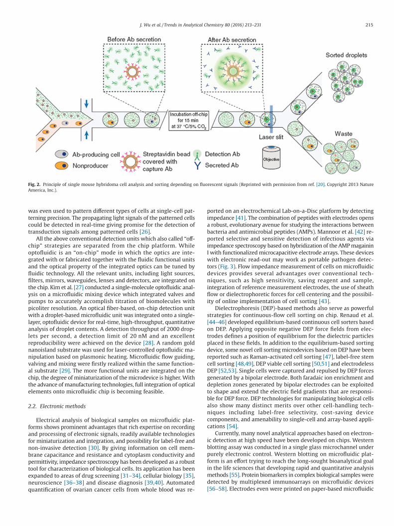
Due to the ubiquitous optical instruments in laboratories, quan-titative detections of samples by optical techniques become commonin microsystems. Conventional optical detection technologies, suchas absorbance, fluorescence, infrared (IR) and surface plasmon res-onance (SPR), have all been applied in microfluidic chips. Singlemouse hybridoma cells, a fluorescent probe and single beads coatedwith anti-mouse IgG antibodies were co-compartmentalized in drop-lets to generate a distinguishable fluorescence signal enablingintracellular, cell-surface or secreted protein screening (Fig. 2) [20].The production rate of fluorescent proteins in bacteria was moni-tored onmicrofluidic chip revealing a long period of constant proteinproduction in stationary phase [21]. Microscopic imaging of wholeaging process of budding yeast was successfully conducted on amicrofluidic dissection platform integrated with arrays of softelastomer-based micropads [22]. Spatially resolved IR spectrosco-py as an analytical method possessing characteristics of label-freeand nondestructive detection can provide spatiotemporal informa-tion on functional groups of biomolecules through observing theircharacteristic vibrational modes.
With the help of spatially resolved IR spectroscopy, Holman et al.[23] monitored the spatiotemporal chemical changes in living PC12cells and found that the cells at the boundary and center of thecolony tended to produce more glycogen and glycoprotein on anopen-channel membrane device. The device composed of a gold-coated porous membrane between a feeding channel and a viewingchamber allowed long-term continuous IR measurement of live, ad-herent mammalian cells. On-chip protein microarray was fabricatedby in vitro transcription and translation to be implemented for SPRimaging in both clinical and research applications [24]. Cytosolicglucose levels in yeast were analyzed using genetically encodedFörster resonance energy transfer (FRET) sensors. Glucose-inducedconformational change in the bacterial periplasmic glucose/galactose binding protein MglB was detected by monitoring FRETsignals between two fluorescent protein variants [25]. Optical method
Fig. 1. Biochemical analysis integrated on microfluidic chip.
214 J. Wu et al. / Trends in Analytical Chemistry 80 (2016) 213–231
was even used to pattern different types of cells at single-cell pat-terning precision. The propagating light signals of the patterned cellscould be detected in real-time giving promise for the detection oftransduction signals among patterned cells [26].
All the above conventional detection units which also called “off-chip” strategies are separated from the chip platform. Whileoptofluidic is an “on-chip” mode in which the optics are inte-grated with or fabricated together with the fluidic functional unitsand the optical property of the integrated optics can be tuned byfluidic technology. All the relevant units, including light sources,filters, mirrors, waveguides, lenses and detectors, are integrated onthe chip. Kim et al. [27] conducted a single-molecule optofluidic anal-ysis on a microfluidic mixing device which integrated valves andpumps to accurately accomplish titration of biomolecules withpicoliter resolution. An optical fiber-based, on-chip detection unitwith a droplet-based microfluidic unit was integrated onto a single-layer, optofluidic device for real-time, high-throughput, quantitativeanalysis of droplet contents. A detection throughput of 2000 drop-lets per second, a detection limit of 20 nM and an excellentreproducibility were achieved on the device [28]. A random goldnanoisland substrate was used for laser-controlled optofluidic ma-nipulation based on plasmonic heating. Microfluidic flow guiding,valving and mixing were firstly realized within the same function-al substrate [29]. The more functional units are integrated on thechip, the degree of miniaturization of themicrodevice is higher.Withthe advance of manufacturing technologies, full integration of opticalelements onto microfluidic chip is becoming feasible.
2.2. Electronic methods
Electrical analysis of biological samples on microfluidic plat-forms shows prominent advantages that rich expertise on recordingand processing of electronic signals, readily available technologiesfor miniaturization and integration, and possibility for label-free andnon-invasive detection [30]. By giving information on cell mem-brane capacitance and resistance and cytoplasm conductivity andpermittivity, impedance spectroscopy has been developed as a robusttool for characterization of biological cells. Its application has beenexpanded to areas of drug screening [31–34], cellular biology [35],neuroscience [36–38] and disease diagnosis [39,40]. Automatedquantification of ovarian cancer cells from whole blood was re-
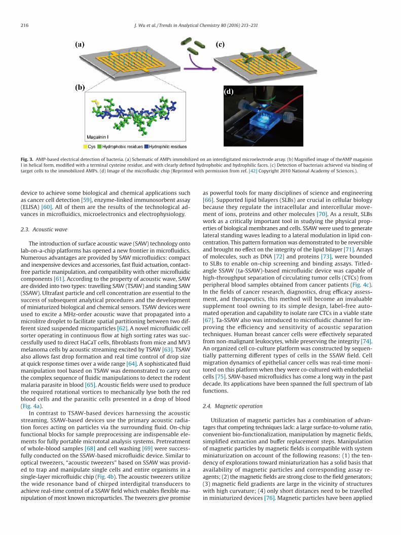
ported on an electrochemical Lab-on-a-Disc platform by detectingimpedance [41]. The combination of peptides with electrodes opensa robust, evolutionary avenue for studying the interactions betweenbacteria and antimicrobial peptides (AMPs). Mannoor et al. [42] re-ported selective and sensitive detection of infectious agents viaimpedance spectroscopy based on hybridization of the AMPmagaininI with functionalizedmicrocapacitive electrode arrays. These deviceswith electronic read-out may work as portable pathogen detec-tors (Fig. 3). Flow impedance measurement of cells on microfluidicdevices provides several advantages over conventional tech-niques, such as high sensitivity, saving reagent and sample,integration of reference measurement electrodes, the use of sheathflow or dielectrophoretic forces for cell centering and the possibil-ity of online implementation of cell sorting [43].
Dielectrophoresis (DEP)-based methods also serve as powerfulstrategies for continuous-flow cell sorting on chip. Renaud et al.[44–46] developed equilibrium-based continuous cell sorters basedon DEP. Applying opposite negative DEP force fields from elec-trodes defines a position of equilibrium for the dielectric particlesplaced in these fields. In addition to the equilibrium-based sortingdevice, some novel cell sortingmicrodevices based on DEP have beenreported such as Raman-activated cell sorting [47], label-free stemcell sorting [48,49], DEP viable cell sorting [50,51] and electrodelessDEP [52,53]. Single cells were captured and repulsed by DEP forcesgenerated by a bipolar electrode. Both faradaic ion enrichment anddepletion zones generated by bipolar electrodes can be exploitedto shape and extend the electric field gradients that are responsi-ble for DEP force. DEP technologies for manipulating biological cellsalso show many distinct merits over other cell-handling tech-niques including label-free selectivity, cost-saving devicecomponents, and amenability to single-cell and array-based appli-cations [54].
Currently, many novel analytical approaches based on electron-ic detection at high speed have been developed on chips. Westernblotting assay was conducted in a single glass microchannel underpurely electronic control. Western blotting on microfluidic plat-form is an effort trying to reach the long-sought bioanalytical goalin the life sciences that developing rapid and quantitative analysismethods [55]. Protein biomarkers in complex biological samplesweredetected by multiplexed immunoarrays on microfluidic devices[56–58]. Electrodes even were printed on paper-based microfluidic
Fig. 2. Principle of single mouse hybridoma cell analysis and sorting depending on fluorescent signals (Reprinted with permission from ref. [20]. Copyright 2013 NatureAmerica, Inc.).
215J. Wu et al. / Trends in Analytical Chemistry 80 (2016) 213–231
device to achieve some biological and chemical applications suchas cancer cell detection [59], enzyme-linked immunosorbent assay(ELISA) [60]. All of them are the results of the technological ad-vances in microfluidics, microelectronics and electrophysiology.
2.3. Acoustic wave
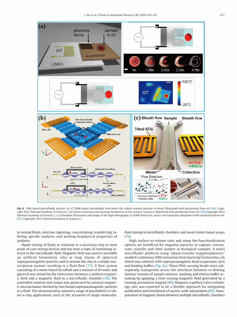
The introduction of surface acoustic wave (SAW) technology ontolab-on-a-chip platforms has opened a new frontier in microfluidics.Numerous advantages are provided by SAWmicrofluidics: compactand inexpensive devices and accessories, fast fluid actuation, contact-free particle manipulation, and compatibility with other microfluidiccomponents [61]. According to the property of acoustic wave, SAWare divided into two types: travelling SAW (TSAW) and standing SAW(SSAW). Ultrafast particle and cell concentration are essential to thesuccess of subsequent analytical procedures and the developmentof miniaturized biological and chemical sensors. TSAW devices wereused to excite a MHz-order acoustic wave that propagated into amicrolitre droplet to facilitate spatial partitioning between two dif-ferent sized suspendedmicroparticles [62]. A novel microfluidic cellsorter operating in continuous flow at high sorting rates was suc-cessfully used to direct HaCaT cells, fibroblasts frommice and MV3melanoma cells by acoustic streaming excited by TSAW [63]. TSAWalso allows fast drop formation and real time control of drop sizeat quick response times over a wide range [64]. A sophisticated fluidmanipulation tool based on TSAW was demonstrated to carry outthe complex sequence of fluidic manipulations to detect the rodentmalaria parasite in blood [65]. Acoustic fields were used to producethe required rotational vortices to mechanically lyse both the redblood cells and the parasitic cells presented in a drop of blood(Fig. 4a).
In contrast to TSAW-based devices harnessing the acousticstreaming, SSAW-based devices use the primary acoustic radia-tion forces acting on particles via the surrounding fluid. On-chipfunctional blocks for sample preprocessing are indispensable ele-ments for fully portable micrototal analysis systems. Pretreatmentof whole-blood samples [68] and cell washing [69] were success-fully conducted on the SSAW-based microfluidic device. Similar tooptical tweezers, “acoustic tweezers” based on SSAW was provid-ed to trap and manipulate single cells and entire organisms in asingle-layer microfluidic chip (Fig. 4b). The acoustic tweezers utilizethe wide resonance band of chirped interdigital transducers toachieve real-time control of a SSAW field which enables flexible ma-nipulation of most knownmicroparticles. The tweezers give promise
as powerful tools for many disciplines of science and engineering[66]. Supported lipid bilayers (SLBs) are crucial in cellular biologybecause they regulate the intracellular and intercellular move-ment of ions, proteins and other molecules [70]. As a result, SLBswork as a critically important tool in studying the physical prop-erties of biological membranes and cells. SSAWwere used to generatelateral standing waves leading to a lateral modulation in lipid con-centration. This pattern formationwas demonstrated to be reversibleand brought no effect on the integrity of the lipid bilayer [71]. Arraysof molecules, such as DNA [72] and proteins [73], were boundedto SLBs to enable on-chip screening and binding assays. Titled-angle SSAW (ta-SSAW)-based microfluidic device was capable ofhigh-throughput separation of circulating tumor cells (CTCs) fromperipheral blood samples obtained from cancer patients (Fig. 4c).In the fields of cancer research, diagnostics, drug efficacy assess-ment, and therapeutics, this method will become an invaluablesupplement tool owning to its simple design, label-free auto-mated operation and capability to isolate rare CTCs in a viable state[67]. Ta-SSAW also was introduced to microfluidic channel for im-proving the efficiency and sensitivity of acoustic separationtechniques. Human breast cancer cells were effectively separatedfrom non-malignant leukocytes, while preserving the integrity [74].An organized cell co-culture platform was constructed by sequen-tially patterning different types of cells in the SSAW field. Cellmigration dynamics of epithelial cancer cells was real-time moni-tored on this platformwhen they were co-cultured with endothelialcells [75]. SAW-based microfluidics has come a long way in the pastdecade. Its applications have been spanned the full spectrum of labfunctions.
2.4. Magnetic operation
Utilization of magnetic particles has a combination of advan-tages that competing techniques lack: a large surface-to-volume ratio,convenient bio-functionalization, manipulation by magnetic fields,simplified extraction and buffer replacement steps. Manipulationof magnetic particles by magnetic fields is compatible with systemminiaturization on account of the following reasons: (1) the ten-dency of explorations toward miniaturization has a solid basis thatavailability of magnetic particles and corresponding assay re-agents; (2) themagnetic fields are strong close to the field generators;(3) magnetic field gradients are large in the vicinity of structureswith high curvature; (4) only short distances need to be travelledin miniaturized devices [76]. Magnetic particles have been applied
Fig. 3. AMP-based electrical detection of bacteria. (a) Schematic of AMPs immobilized on an interdigitated microelectrode array. (b) Magnified image of theAMP magaininI in helical form, modified with a terminal cysteine residue, and with clearly defined hydrophobic and hydrophilic faces. (c) Detection of bacteriais achieved via binding oftarget cells to the immobilized AMPs. (d) Image of the microfluidic chip (Reprinted with permission from ref. [42] Copyright 2010 National Academy of Sciences.).
216 J. Wu et al. / Trends in Analytical Chemistry 80 (2016) 213–231
in mixing fluids, selective capturing, concentrating, transferring, la-beling specific analytes, and probing biophysical properties ofanalytes.
Rapid mixing of fluids or solutions is a necessary step in mostpoint-of-care testing devices and has been a topic of everlasting in-terest in the microfluidic field. Magnetic field was used to assemblean artificial biomimetic cilia as long chains of sphericalsuperparamagnetic particles and to actuate the cilia in a simple non-reciprocal manner resulting in a fluid flow [77]. A flow systemconsisting of a water-based ferrofluid and a mixture of DI water andglycerol was mixed by the interaction between a uniform magnet-ic field and a magnetic fluid in a microfluidic chamber [78]. Thecontrolled rotation and torque was generated by uniaxial magnet-ic microactuators formed by two bound superparamagnetic particlesin a fluid. This demonstration opened a range of possibilities in lab-on-a-chip applications, such as the actuation of single molecules,
fluidmixing inmicrofluidic chambers and novel cluster-based assays[79].
High surface-to-volume ratio and many bio-functionalizationoptions are beneficial for magnetic particles to capture, concen-trate, transfer and label analyte in biological samples. A novelmicrofluidic platform using “phase-transfer magnetophoresis”enabled continuous DNA extraction from bacterial Escherichia coliwhich was cultured with superparamagnetic bead suspension, lysisand binding buffers (Fig. 5a). These DNA carrying beads were sub-sequently transported across the interfaces between co-flowinglaminar streams of sample mixture, washing and elution buffer ac-tuating by applying a time-varying magnetic field generated by arotating permanent magnet [80]. Magneto-capillary valve technol-ogy also was reported to be a flexible approach for integratingpurification and enrichment of nucleic acids and proteins [83]. Trans-portation of magnetic beads betweenmultiplemicrofluidic chambers
Fig. 4. SAW-based microfluidic devices. (a) A TSAW-based microfluidic tool detect the rodent malaria parasite in blood (Reprinted with permission from ref. [65]. Copy-right 2012 National Academy of Sciences.). (b) Device structure and working mechanism of the acoustic tweezers (Reprinted with permission from ref. [66].Copyright 2012National Academy of Sciences.). (c) Schematic illustration and image of the high-throughput ta-SSAW device for cancer cell separation (Reprinted with permission from ref.[67]. Copyright 2015 National Academy of Sciences.).
217J. Wu et al. / Trends in Analytical Chemistry 80 (2016) 213–231
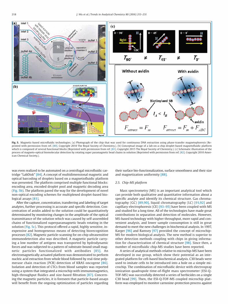
was even realized to be automated on a centrifugal microfluidic car-tridge “LabDisk” [84]. A concept of multidimensional magnetic andoptical barcoding of droplets based on a magnetiofluidic platformwas presented. The platform comprised multiple functional blocks:encoding area, encoded droplet pool and magnetic decoding area(Fig. 5b). The platform paved the way for the development of novelnon-optical encoding schemes for multiplexed droplet-based bio-logical assays [81].
After the capture, concentration, transferring and labeling of targetanalytes, further processing is accurate and specific detection. Con-centration of avidin added to the solution could be quantitativelydeterminated by monitoring changes in the amplitude of the opticaltransmittance of the solution which was caused by self-assembledchains of functionalized superparamagnetic beads rotating in thesolution (Fig. 5c). This protocol offered a rapid, highly sensitive, in-expensive and homogeneous means of detecting biorecognitionprocesses [82]. Magnetic particle-scanning for on-chip ultrasensitiveimmunodetection also was described. A magnetic particle carry-ing a low number of antigens was transported by hydrodynamicforces and was subjected to a pattern of substrate-bound small mag-netic particles functionalized with antibodies [85]. Anelectromagnetically actuated platformwas demonstrated to performnucleic acid extraction fromwhole blood followed by real-time poly-merase chain reaction (PCR) detection of KRAS oncogene [86].Isolation and detection of CTCs from blood samples was reportedusing a system that integrated a microchip with immunomagnetics,high-throughput fluidics and size-based filtration [87]. Concern-ing the magnetic particles, it is foreseen that particle-based assayswill benefit from the ongoing optimization of particles regarding
their surface bio-functionalization, surface smoothness and their sizeand magnetization uniformity [88].
2.5. Chip-MS platform
Mass spectrometry (MS) is an important analytical tool whichcan provide both qualitative and quantitative information about aspecific analyte and identify its chemical structure. Gas chroma-tography (GC) [89,90], liquid chromatography (LC) [91,92] andcapillary electrophoresis (CE) [93–95] have been coupled with MSand studied for a long time. All of the technologies have made greatcontributions in separation and detection of molecules. However,MS-based technology with higher throughput, more rapid and con-venient analysis, and lower sample consumption is still in greatdemand tomeet the new challenges in biochemical analysis. In 1997,Karger [96] and Ramsey [97] provided the concept of microchip-MS for modern biological analysis. The new method is superior toother detection methods coupling with chips in giving informa-tion for characterization of chemical structure [98]. Since then, anumber of microfluidic chip-MS studies have been reported.
A series of analytical methods relative tomicrochip-MS have beendeveloped in our group, which show their potential as an inte-grated platform for cell-based biochemical analysis. C30 beads wereused to imitate cells to be introduced into a hole on a simple mi-crochip. The combination of microfluidic device with electrosprayionization quadrupole time-of-flight mass spectrometer (ESI-Q-TOF-MS) was successfully detected a series of herbicides on a singleC30 bead [99]. Then, the ESI-Q-TOF-MS coupled microchip plat-formwas employed tomonitor carnosine-protection process against
Fig. 5. Magnetic-based microfluidic technologies. (a) Photograph of the chip that was used for continuous DNA extraction using phase-transfer magnetophoresis (Re-printed with permission from ref. [80]. Copyright 2010 The Royal Society of Chemistry.). (b) Conceptual image of a lab-on-a-chip droplet-based magnetofluidic platformwhich is composed of several functional blocks (Reprinted with permission from ref. [81]. Copyright 2015 The Royal Society of Chemistry.). (c) Schematic illustration of theprocess of magneto-optical biomolecular detection by rotating super paramagnetic bead chains in solution (Reprinted with permission from ref. [82]. Copyright 2010 Amer-ican Chemical Society.).
218 J. Wu et al. / Trends in Analytical Chemistry 80 (2016) 213–231
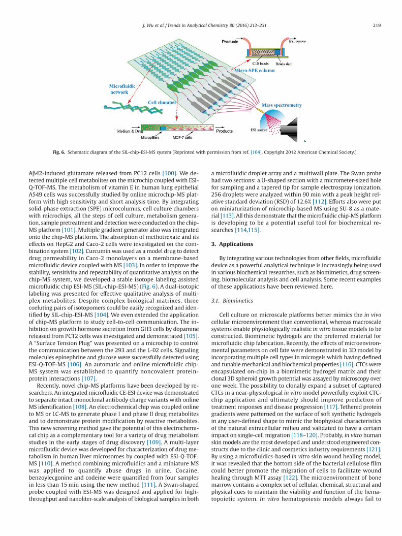
Aβ42-induced glutamate released from PC12 cells [100]. We de-tected multiple cell metabolites on the microchip coupled with ESI-Q-TOF-MS. The metabolism of vitamin E in human lung epithelialA549 cells was successfully studied by online microchip-MS plat-form with high sensitivity and short analysis time. By integratingsolid-phase extraction (SPE) microcolumns, cell culture chamberswith microchips, all the steps of cell culture, metabolism genera-tion, sample pretreatment and detectionwere conducted on the chip-MS platform [101]. Multiple gradient generator also was integratedonto the chip-MS platform. The absorption of methotrexate and itseffects on HepG2 and Caco-2 cells were investigated on the com-bination system [102]. Curcumin was used as a model drug to detectdrug permeability in Caco-2 monolayers on a membrane-basedmicrofluidic device coupled with MS [103]. In order to improve thestability, sensitivity and repeatability of quantitative analysis on thechip-MS system, we developed a stable isotope labeling assistedmicrofluidic chip ESI-MS (SIL-chip-ESI-MS) (Fig. 6). A dual-isotopiclabeling was presented for effective qualitative analysis of multi-plex metabolites. Despite complex biological matrixes, threecoeluting pairs of isotopomers could be easily recognized and iden-tified by SIL-chip-ESI-MS [104]. We even extended the applicationof chip-MS platform to study cell-to-cell communication. The in-hibition on growth hormone secretion from GH3 cells by dopaminereleased from PC12 cells was investigated and demonstrated [105].A “Surface Tension Plug” was presented on a microchip to controlthe communication between the 293 and the L-02 cells. Signalingmolecules epinephrine and glucose were successfully detected usingESI-Q-TOF-MS [106]. An automatic and online microfluidic chip-MS system was established to quantify noncovalent protein-protein interactions [107].
Recently, novel chip-MS platforms have been developed by re-searchers. An integratedmicrofluidic CE-ESI devicewas demonstratedto separate intact monoclonal antibody charge variants with onlineMS identification [108]. An electrochemical chip was coupled onlineto MS or LC-MS to generate phase I and phase II drug metabolitesand to demonstrate protein modification by reactive metabolites.This new screening method gave the potential of this electrochemi-cal chip as a complementary tool for a variety of drug metabolismstudies in the early stages of drug discovery [109]. A multi-layermicrofluidic device was developed for characterization of drug me-tabolism in human liver microsomes by coupled with ESI-Q-TOF-MS [110]. A method combining microfluidics and a miniature MSwas applied to quantify abuse drugs in urine. Cocaine,benzoylecgonine and codeine were quantified from four samplesin less than 15 min using the new method [111]. A Swan-shapedprobe coupled with ESI-MS was designed and applied for high-throughput and nanoliter-scale analysis of biological samples in both
a microfluidic droplet array and a multiwall plate. The Swan probehad two sections: a U-shaped section with a micrometer-sized holefor sampling and a tapered tip for sample electrospray ionization.256 droplets were analyzed within 90 min with a peak height rel-ative standard deviation (RSD) of 12.6% [112]. Efforts also were puton miniaturization of microchip-based MS using SU-8 as a mate-rial [113]. All this demonstrate that themicrofluidic chip-MS platformis developing to be a potential useful tool for biochemical re-searches [114,115].
3. Applications
By integrating various technologies from other fields, microfluidicdevice as a powerful analytical technique is increasingly being usedin various biochemical researches, such as biomimetics, drug screen-ing, biomolecular analysis and cell analysis. Some recent examplesof these applications have been reviewed here.
3.1. Biomimetics
Cell culture on microscale platforms better mimics the in vivocellular microenvironment than conventional, whereas macroscalesystems enable physiologically realistic in vitro tissue models to beconstructed. Biomimetic hydrogels are the preferred material formicrofluidic chip fabrication. Recently, the effects of microenviron-mental parameters on cell fate were demonstrated in 3D model byincorporating multiple cell types in microgels which having definedand tunablemechanical and biochemical properties [116]. CTCs wereencapsulated on-chip in a biomimetic hydrogel matrix and theirclonal 3D spheroid growth potential was assayed bymicroscopy overone week. The possibility to clonally expand a subset of capturedCTCs in a near-physiological in vitromodel powerfully exploit CTC-chip application and ultimately should improve prediction oftreatment responses and disease progression [117]. Tethered proteingradients were patterned on the surface of soft synthetic hydrogelsin any user-defined shape to mimic the biophysical characteristicsof the natural extracellular milieu and validated to have a certainimpact on single-cell migration [118–120]. Probably, in vitro humanskinmodels are themost developed and understood engineered con-structs due to the clinic and cosmetics industry requirements [121].By using a microfluidics-based in vitro skin wound healing model,it was revealed that the bottom side of the bacterial cellulose filmcould better promote the migration of cells to facilitate woundhealing through MTT assay [122]. The microenvironment of bonemarrow contains a complex set of cellular, chemical, structural andphysical cues to maintain the viability and function of the hema-topoietic system. In vitro hematopoiesis models always fail to
Fig. 6. Schematic diagram of the SIL-chip-ESI-MS system (Reprinted with permission from ref. [104]. Copyright 2012 American Chemical Society.).
219J. Wu et al. / Trends in Analytical Chemistry 80 (2016) 213–231
demonstrate the complex microenvironment and functions of livingbone marrow. A method for fabricating “bone marrow-on-a-chip”allowed in vitro culturing of live marrow with a functional hema-topoietic niche. Hematopoietic stem and progenitor cells could beretained in the engineering bone marrow for at least 1 week [123].We developed a microfluidic 3D culture device to imitate the dif-fusion process between blood vessels and tissues. On thismicrofluidicdevice, QD cytotoxicity was evaluated by determining cell apopto-sis, intracellular reactive oxygen species and glutathionewith specificfluorescence probes. The degree of QD cytotoxicity was demon-strated to be relative to the diffusion distance [124].
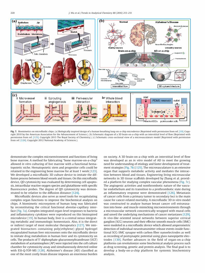
Microfluidic devices also serve as novel tools for recapitulatingcomplex organ functions to improve the biochemical analysis onchips. A biomimetic microsystem of human lung was fabricatedby reconstituting the critical functional alveolar-capillary inter-face (Fig. 7a). Complex integrated organ-level responses to bacteriaand inflammatory cytokines were reproduced on this bioinspiredmicrodevice [19]. In human body, liver is a central nexus integrat-ing metabolic and immunologic homeostasis. Also, it is the director indirect target of most molecular therapeutics [127]. We inte-grated bioreactors containing poly(ethylene) glycol hydrogelencapsulated human liver microsomes onto the microfluidic deviceto imitate drug metabolism in human liver and its cytotoxicity oncells. The products of 5′-diphosphate-glucuronosyltransferase (UGT)metabolism of acetaminophen (AP) were injected into the cell culturechamber for cytotoxicity assay and simultaneously detected onlinewith ESI-Q-TOF-MS [128]. Alzheimer’s disease (AD) classified asone of the most costly brain disease imposes an enormous burden
on society. A 3D brain-on-a-chip with an interstitial level of flowwas developed as an in vitro model of AD to meet the growingneed for understanding of etiology and faster development of treat-ment strategies (Fig. 7b) [125]. The microvasculature is an extensiveorgan that supports metabolic activity and mediates the interac-tion between blood and tissues. Engineering living microvascularnetworks in 3D tissue scaffolds developed by Zhang et al. provid-ed a platform for studying complex vascular phenomena (Fig. 7c).The angiogenic activities and nonthrombotic nature of the vascu-lar endothelium and its transition to a prothrombotic state duringan inflammatory response were demonstrated [126]. Metastasisof cancer cells from a primary tumor to secondary loci is the maincause for cancer-related mortality. A microfluidic 3D in vitromodelwas constructed to analyze human breast cancer cell extravasa-tion into bone- and muscle-mimicking microenvironments througha microvascular network concentrically wrapped with mural cellsand unveil the underlying mechanisms of cancer metastases [129].In vivo-like oriented neural networks between superior cervicalganglion (SCG) neurons and their effector smoothmuscle cells (SMC)were modeled in a microfluidic device which allowed amperometricdetection of individual neurotransmitter release events inside func-tional SCG-SMC synapse with carbon fiber nanoelectrodes as wellas recording of postsynaptic potential using glass nanopipette elec-trodes [130]. Further advances in the field of organ-on-a-chipplatforms can revolutionize some biochemical analysis process suchas drug screening, genetic and protein analysis. The final goal is todevelop a body-on-a-chip platform for systemic biochemistryanalysis.
Fig. 7. Biomimetics on microfluidic chips. (a) Biologically inspired design of a human breathing lung-on-a-chip microdevice (Reprinted with permission from ref. [19]. Copy-right 2010 by the American Association for the Advancement of Science.). (b) Schematic diagram of a 3D brain-on-a-chip with an interstitial level of flow (Reprinted withpermission from ref. [125]. Copyright 2015 The Royal Society of Chemistry.). (c) Schematic cross-sectional view of a microvasculature model (Reprinted with permissionfrom ref. [126]. Copyright 2012 National Academy of Sciences.).
220 J. Wu et al. / Trends in Analytical Chemistry 80 (2016) 213–231
3.2. Drug discovery
Bringing a new drug to market is a complex, lengthy and ex-pensive process. An adequate cell-based assay to efficiently screenand validate potential drug candidates is crucial in the initial stageof drug discovery [131]. Microfluidic platforms provide severalunique advantages for drug screening. Physical and chemical prop-erties of drug carriers can be easily and effectivelymodified by tuningthe flow rate and geometries of microchannels on microfluidicdevices. Batches of carriers can be fabricated with minimal effortand with little variation. Tissue or organ models can be mimickedin microfluidic systems to be used as in vitro drug screening tools[132].
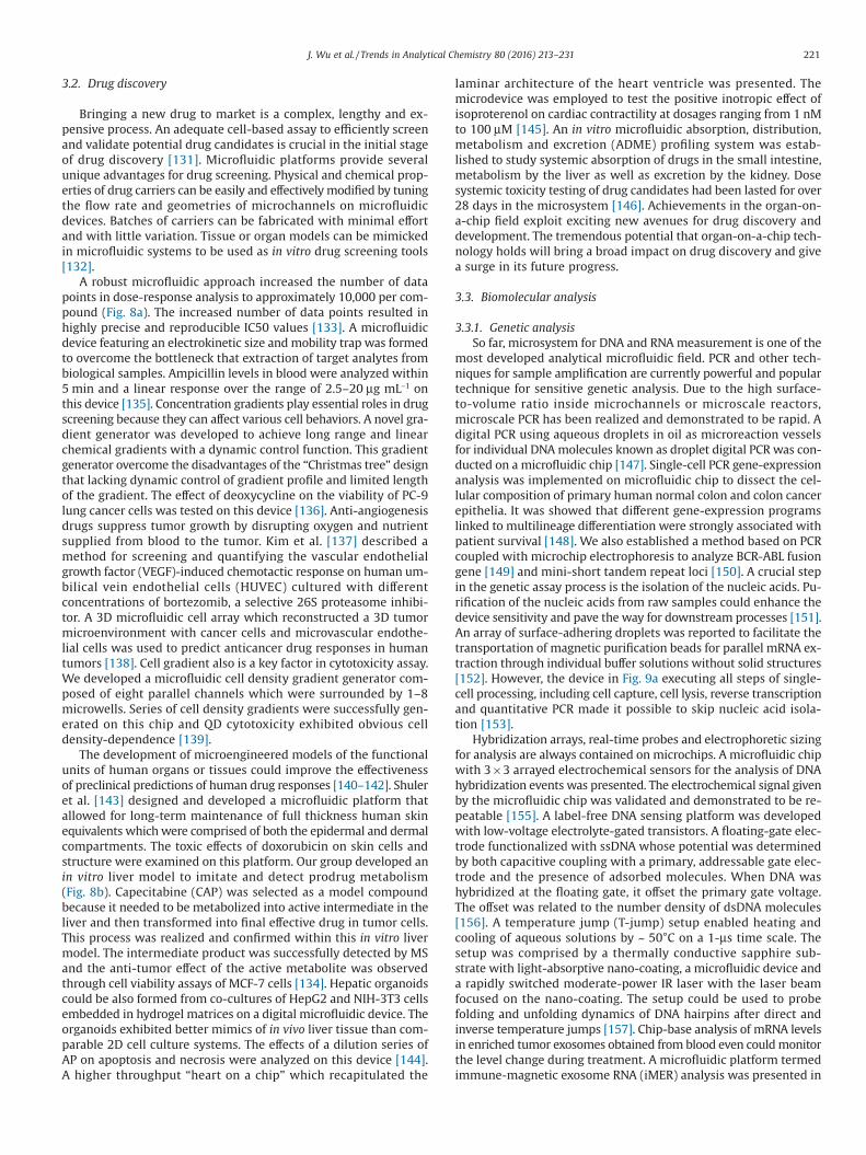
A robust microfluidic approach increased the number of datapoints in dose-response analysis to approximately 10,000 per com-pound (Fig. 8a). The increased number of data points resulted inhighly precise and reproducible IC50 values [133]. A microfluidicdevice featuring an electrokinetic size andmobility trap was formedto overcome the bottleneck that extraction of target analytes frombiological samples. Ampicillin levels in blood were analyzed within5 min and a linear response over the range of 2.5–20 μg mL−1 onthis device [135]. Concentration gradients play essential roles in drugscreening because they can affect various cell behaviors. A novel gra-dient generator was developed to achieve long range and linearchemical gradients with a dynamic control function. This gradientgenerator overcome the disadvantages of the “Christmas tree” designthat lacking dynamic control of gradient profile and limited lengthof the gradient. The effect of deoxycycline on the viability of PC-9lung cancer cells was tested on this device [136]. Anti-angiogenesisdrugs suppress tumor growth by disrupting oxygen and nutrientsupplied from blood to the tumor. Kim et al. [137] described amethod for screening and quantifying the vascular endothelialgrowth factor (VEGF)-induced chemotactic response on human um-bilical vein endothelial cells (HUVEC) cultured with differentconcentrations of bortezomib, a selective 26S proteasome inhibi-tor. A 3D microfluidic cell array which reconstructed a 3D tumormicroenvironment with cancer cells and microvascular endothe-lial cells was used to predict anticancer drug responses in humantumors [138]. Cell gradient also is a key factor in cytotoxicity assay.We developed a microfluidic cell density gradient generator com-posed of eight parallel channels which were surrounded by 1–8microwells. Series of cell density gradients were successfully gen-erated on this chip and QD cytotoxicity exhibited obvious celldensity-dependence [139].
The development of microengineered models of the functionalunits of human organs or tissues could improve the effectivenessof preclinical predictions of human drug responses [140–142]. Shuleret al. [143] designed and developed a microfluidic platform thatallowed for long-term maintenance of full thickness human skinequivalents whichwere comprised of both the epidermal and dermalcompartments. The toxic effects of doxorubicin on skin cells andstructure were examined on this platform. Our group developed anin vitro liver model to imitate and detect prodrug metabolism(Fig. 8b). Capecitabine (CAP) was selected as a model compoundbecause it needed to be metabolized into active intermediate in theliver and then transformed into final effective drug in tumor cells.This process was realized and confirmed within this in vitro livermodel. The intermediate product was successfully detected by MSand the anti-tumor effect of the active metabolite was observedthrough cell viability assays of MCF-7 cells [134]. Hepatic organoidscould be also formed from co-cultures of HepG2 and NIH-3T3 cellsembedded in hydrogel matrices on a digital microfluidic device. Theorganoids exhibited better mimics of in vivo liver tissue than com-parable 2D cell culture systems. The effects of a dilution series ofAP on apoptosis and necrosis were analyzed on this device [144].A higher throughput “heart on a chip” which recapitulated the
laminar architecture of the heart ventricle was presented. Themicrodevice was employed to test the positive inotropic effect ofisoproterenol on cardiac contractility at dosages ranging from 1 nMto 100 μM [145]. An in vitro microfluidic absorption, distribution,metabolism and excretion (ADME) profiling system was estab-lished to study systemic absorption of drugs in the small intestine,metabolism by the liver as well as excretion by the kidney. Dosesystemic toxicity testing of drug candidates had been lasted for over28 days in the microsystem [146]. Achievements in the organ-on-a-chip field exploit exciting new avenues for drug discovery anddevelopment. The tremendous potential that organ-on-a-chip tech-nology holds will bring a broad impact on drug discovery and givea surge in its future progress.
3.3. Biomolecular analysis
3.3.1. Genetic analysisSo far, microsystem for DNA and RNAmeasurement is one of the
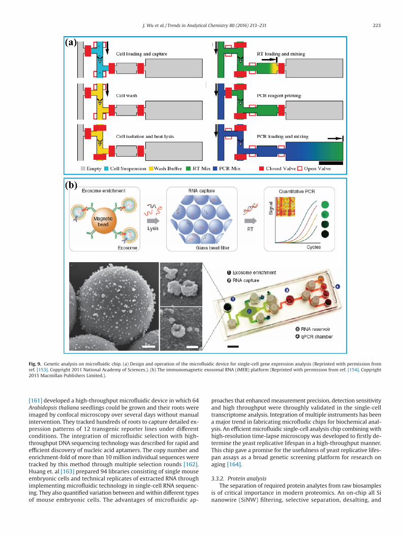
most developed analytical microfluidic field. PCR and other tech-niques for sample amplification are currently powerful and populartechnique for sensitive genetic analysis. Due to the high surface-to-volume ratio inside microchannels or microscale reactors,microscale PCR has been realized and demonstrated to be rapid. Adigital PCR using aqueous droplets in oil as microreaction vesselsfor individual DNAmolecules known as droplet digital PCR was con-ducted on a microfluidic chip [147]. Single-cell PCR gene-expressionanalysis was implemented on microfluidic chip to dissect the cel-lular composition of primary human normal colon and colon cancerepithelia. It was showed that different gene-expression programslinked to multilineage differentiation were strongly associated withpatient survival [148]. We also established a method based on PCRcoupled with microchip electrophoresis to analyze BCR-ABL fusiongene [149] and mini-short tandem repeat loci [150]. A crucial stepin the genetic assay process is the isolation of the nucleic acids. Pu-rification of the nucleic acids from raw samples could enhance thedevice sensitivity and pave the way for downstream processes [151].An array of surface-adhering droplets was reported to facilitate thetransportation of magnetic purification beads for parallel mRNA ex-traction through individual buffer solutions without solid structures[152]. However, the device in Fig. 9a executing all steps of single-cell processing, including cell capture, cell lysis, reverse transcriptionand quantitative PCR made it possible to skip nucleic acid isola-tion [153].
Hybridization arrays, real-time probes and electrophoretic sizingfor analysis are always contained on microchips. A microfluidic chipwith 3 × 3 arrayed electrochemical sensors for the analysis of DNAhybridization events was presented. The electrochemical signal givenby the microfluidic chip was validated and demonstrated to be re-peatable [155]. A label-free DNA sensing platform was developedwith low-voltage electrolyte-gated transistors. A floating-gate elec-trode functionalized with ssDNA whose potential was determinedby both capacitive coupling with a primary, addressable gate elec-trode and the presence of adsorbed molecules. When DNA washybridized at the floating gate, it offset the primary gate voltage.The offset was related to the number density of dsDNA molecules[156]. A temperature jump (T-jump) setup enabled heating andcooling of aqueous solutions by ~ 50°C on a 1-μs time scale. Thesetup was comprised by a thermally conductive sapphire sub-strate with light-absorptive nano-coating, a microfluidic device anda rapidly switched moderate-power IR laser with the laser beamfocused on the nano-coating. The setup could be used to probefolding and unfolding dynamics of DNA hairpins after direct andinverse temperature jumps [157]. Chip-base analysis of mRNA levelsin enriched tumor exosomes obtained from blood even couldmonitorthe level change during treatment. A microfluidic platform termedimmune-magnetic exosome RNA (iMER) analysis was presented in
221J. Wu et al. / Trends in Analytical Chemistry 80 (2016) 213–231
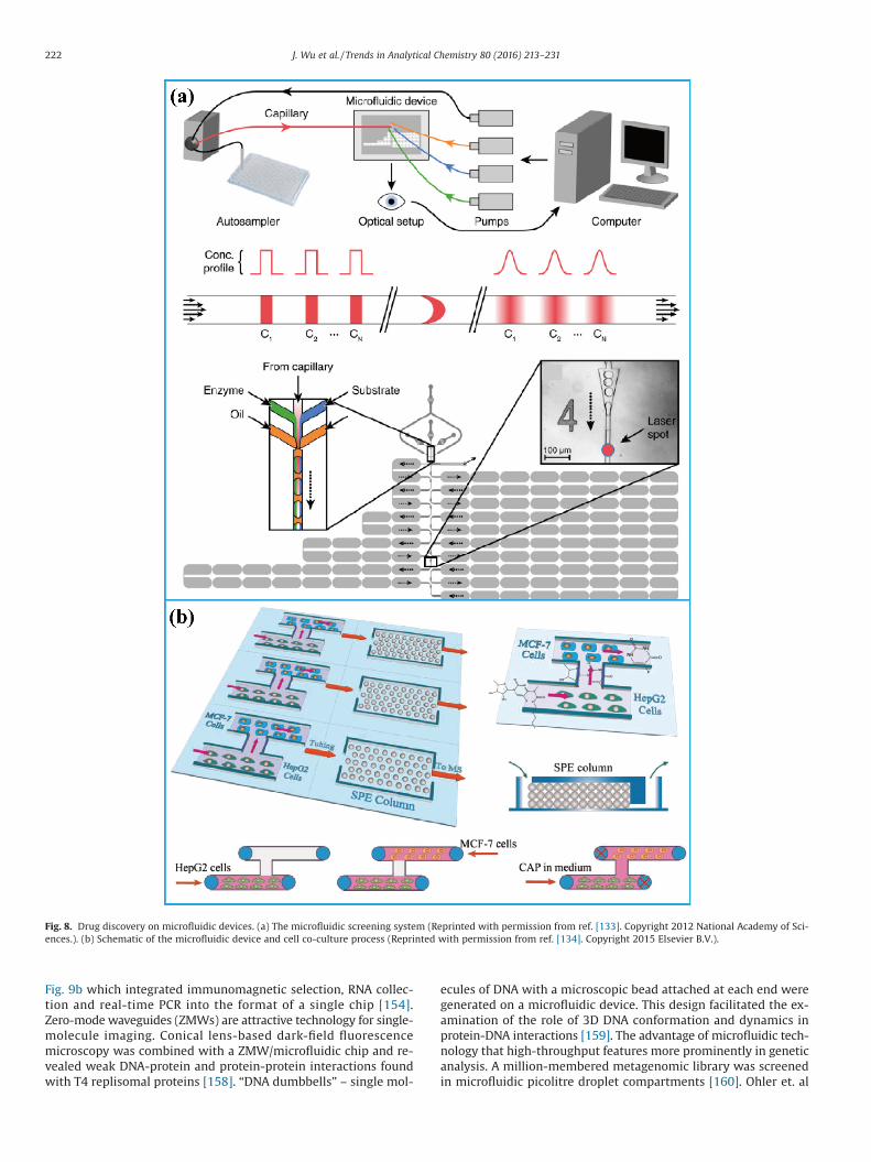
Fig. 9b which integrated immunomagnetic selection, RNA collec-tion and real-time PCR into the format of a single chip [154].Zero-mode waveguides (ZMWs) are attractive technology for single-molecule imaging. Conical lens-based dark-field fluorescencemicroscopy was combined with a ZMW/microfluidic chip and re-vealed weak DNA-protein and protein-protein interactions foundwith T4 replisomal proteins [158]. “DNA dumbbells” – single mol-
ecules of DNA with a microscopic bead attached at each end weregenerated on a microfluidic device. This design facilitated the ex-amination of the role of 3D DNA conformation and dynamics inprotein-DNA interactions [159]. The advantage of microfluidic tech-nology that high-throughput features more prominently in geneticanalysis. A million-membered metagenomic library was screenedin microfluidic picolitre droplet compartments [160]. Ohler et. al
Fig. 8. Drug discovery on microfluidic devices. (a) The microfluidic screening system (Reprinted with permission from ref. [133]. Copyright 2012 National Academy of Sci-ences.). (b) Schematic of the microfluidic device and cell co-culture process (Reprinted with permission from ref. [134]. Copyright 2015 Elsevier B.V.).
222 J. Wu et al. / Trends in Analytical Chemistry 80 (2016) 213–231
[161] developed a high-throughput microfluidic device in which 64Arabidopsis thaliana seedlings could be grown and their roots wereimaged by confocal microscopy over several days without manualintervention. They tracked hundreds of roots to capture detailed ex-pression patterns of 12 transgenic reporter lines under differentconditions. The integration of microfluidic selection with high-throughput DNA sequencing technology was described for rapid andefficient discovery of nucleic acid aptamers. The copy number andenrichment-fold of more than 10 million individual sequences weretracked by this method through multiple selection rounds [162].Huang et. al [163] prepared 94 libraries consisting of single mouseembryonic cells and technical replicates of extracted RNA throughimplementing microfluidic technology in single-cell RNA sequenc-ing. They also quantified variation between andwithin different typesof mouse embryonic cells. The advantages of microfluidic ap-
proaches that enhancedmeasurement precision, detection sensitivityand high throughput were throughly validated in the single-celltranscriptome analysis. Integration of multiple instruments has beena major trend in fabricating microfludic chips for biochemical anal-ysis. An efficientmicrofluidic single-cell analysis chip combiningwithhigh-resolution time-lapse microscopy was developed to firstly de-termine the yeast replicative lifespan in a high-throughput manner.This chip gave a promise for the usefulness of yeast replicative lifes-pan assays as a broad genetic screening platform for research onaging [164].
3.3.2. Protein analysisThe separation of required protein analytes from raw biosamples
is of critical importance in modern proteomics. An on-chip all Sinanowire (SiNW) filtering, selective separation, desalting, and
Fig. 9. Genetic analysis on microfluidic chip. (a) Design and operation of the microfluidic device for single-cell gene expression analysis (Reprinted with permission fromref. [153]. Copyright 2011 National Academy of Sciences.). (b) The immunomagnetic exosomal RNA (iMER) platform (Reprinted with permission from ref. [154]. Copyright2015 Macmillan Publishers Limited.).
223J. Wu et al. / Trends in Analytical Chemistry 80 (2016) 213–231
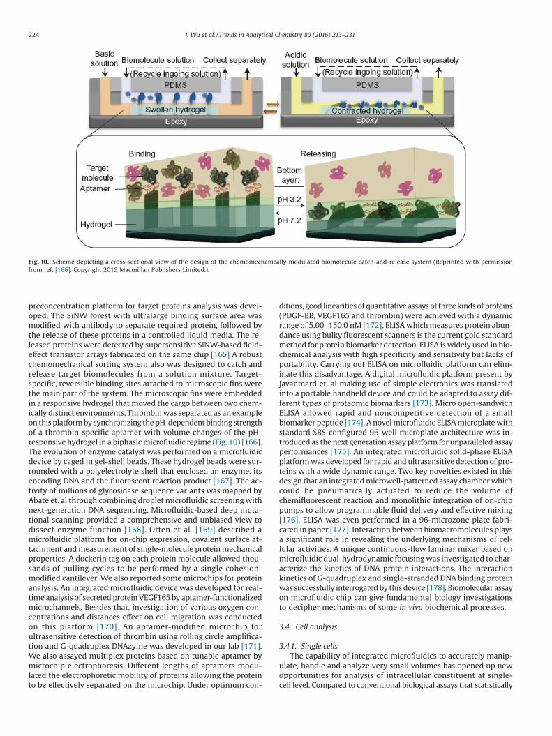
preconcentration platform for target proteins analysis was devel-oped. The SiNW forest with ultralarge binding surface area wasmodified with antibody to separate required protein, followed bythe release of these proteins in a controlled liquid media. The re-leased proteins were detected by supersensitive SiNW-based field-effect transistor arrays fabricated on the same chip [165] A robustchemomechanical sorting system also was designed to catch andrelease target biomolecules from a solution mixture. Target-specific, reversible binding sites attached to microscopic fins werethe main part of the system. The microscopic fins were embeddedin a responsive hydrogel that moved the cargo between two chem-ically distinct environments. Thrombinwas separated as an exampleon this platformby synchronizing thepH-dependentbinding strengthof a thrombin-specific aptamer with volume changes of the pH-responsive hydrogel in a biphasicmicrofluidic regime (Fig. 10) [166].The evolution of enzyme catalyst was performed on a microfluidicdevice by caged in gel-shell beads. These hydrogel beads were sur-rounded with a polyelectrolyte shell that enclosed an enzyme, itsencoding DNA and the fluorescent reaction product [167]. The ac-tivity of millions of glycosidase sequence variants was mapped byAbate et. al through combining droplet microfluidic screeningwithnext-generation DNA sequencing. Microfluidic-based deep muta-tional scanning provided a comprehensive and unbiased view todissect enzyme function [168]. Otten et al. [169] described amicrofluidic platform for on-chip expression, covalent surface at-tachment andmeasurement of single-molecule proteinmechanicalproperties. A dockerin tag on each protein molecule allowed thou-sands of pulling cycles to be performed by a single cohesion-modified cantilever. We also reported some microchips for proteinanalysis. An integrated microfluidic device was developed for real-time analysis of secretedproteinVEGF165by aptamer-functionalizedmicrochannels. Besides that, investigation of various oxygen con-centrations and distances effect on cell migration was conductedon this platform [170]. An aptamer-modified microchip forultrasensitive detection of thrombin using rolling circle amplifica-tion and G-quadruplex DNAzyme was developed in our lab [171].We also assayed multiplex proteins based on tunable aptamer bymicrochip electrophoresis. Different lengths of aptamers modu-lated the electrophoretic mobility of proteins allowing the proteinto be effectively separated on the microchip. Under optimum con-
ditions, good linearitiesof quantitativeassaysof threekindsof proteins(PDGF-BB, VEGF165 and thrombin) were achieved with a dynamicrange of 5.00–150.0 nM [172]. ELISAwhichmeasures protein abun-dance using bulky fluorescent scanners is the current gold standardmethod for protein biomarker detection. ELISA iswidely used in bio-chemical analysis with high specificity and sensitivity but lacks ofportability. Carrying out ELISA on microfluidic platform can elim-inate this disadvantage. A digital microfluidic platform present byJavanmard et. al making use of simple electronics was translatedinto a portable handheld device and could be adapted to assay dif-ferent types of proteomic biomarkers [173]. Micro open-sandwichELISA allowed rapid and noncompetitive detection of a smallbiomarker peptide [174]. A novelmicrofluidic ELISAmicroplatewithstandard SBS-configured 96-well microplate architecture was in-troduced as thenext generation assayplatform for unparalleled assayperformances [175]. An integrated microfluidic solid-phase ELISAplatformwas developed for rapid andultrasensitive detection of pro-teins with a wide dynamic range. Two key novelties existed in thisdesign that an integratedmicrowell-patterned assay chamberwhichcould be pneumatically actuated to reduce the volume ofchemifluorescent reaction and monolithic integration of on-chippumps to allow programmable fluid delivery and effective mixing[176]. ELISA was even performed in a 96-microzone plate fabri-cated in paper [177]. Interaction between biomacromolecules playsa significant role in revealing the underlying mechanisms of cel-lular activities. A unique continuous-flow laminar mixer based onmicrofluidic dual-hydrodynamic focusingwas investigated to char-acterize the kinetics of DNA-protein interactions. The interactionkinetics of G-quadruplex and single-stranded DNA binding proteinwas successfully interrogatedby this device [178]. Biomolecular assayon microfluidic chip can give fundamental biology investigationsto decipher mechanisms of some in vivo biochemical processes.
3.4. Cell analysis
3.4.1. Single cellsThe capability of integrated microfluidics to accurately manip-
ulate, handle and analyze very small volumes has opened up newopportunities for analysis of intracellular constituent at single-cell level. Compared to conventional biological assays that statistically
Fig. 10. Scheme depicting a cross-sectional view of the design of the chemomechanically modulated biomolecule catch-and-release system (Reprinted with permissionfrom ref. [166]. Copyright 2015 Macmillan Publishers Limited.).
224 J. Wu et al. / Trends in Analytical Chemistry 80 (2016) 213–231
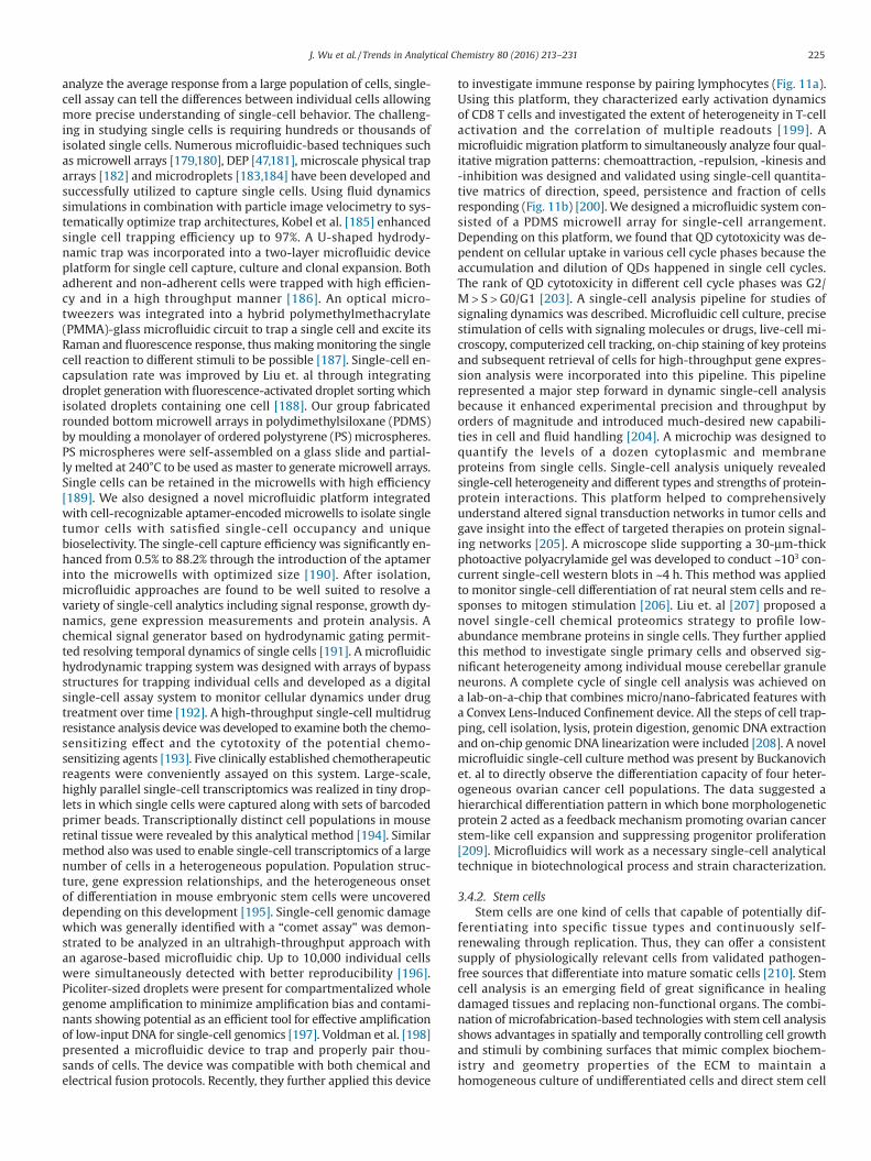
analyze the average response from a large population of cells, single-cell assay can tell the differences between individual cells allowingmore precise understanding of single-cell behavior. The challeng-ing in studying single cells is requiring hundreds or thousands ofisolated single cells. Numerous microfluidic-based techniques suchas microwell arrays [179,180], DEP [47,181], microscale physical traparrays [182] and microdroplets [183,184] have been developed andsuccessfully utilized to capture single cells. Using fluid dynamicssimulations in combination with particle image velocimetry to sys-tematically optimize trap architectures, Kobel et al. [185] enhancedsingle cell trapping efficiency up to 97%. A U-shaped hydrody-namic trap was incorporated into a two-layer microfluidic deviceplatform for single cell capture, culture and clonal expansion. Bothadherent and non-adherent cells were trapped with high efficien-cy and in a high throughput manner [186]. An optical micro-tweezers was integrated into a hybrid polymethylmethacrylate(PMMA)-glass microfluidic circuit to trap a single cell and excite itsRaman and fluorescence response, thusmakingmonitoring the singlecell reaction to different stimuli to be possible [187]. Single-cell en-capsulation rate was improved by Liu et. al through integratingdroplet generationwith fluorescence-activated droplet sorting whichisolated droplets containing one cell [188]. Our group fabricatedrounded bottommicrowell arrays in polydimethylsiloxane (PDMS)by moulding a monolayer of ordered polystyrene (PS) microspheres.PS microspheres were self-assembled on a glass slide and partial-ly melted at 240°C to be used asmaster to generatemicrowell arrays.Single cells can be retained in the microwells with high efficiency[189]. We also designed a novel microfluidic platform integratedwith cell-recognizable aptamer-encodedmicrowells to isolate singletumor cells with satisfied single-cell occupancy and uniquebioselectivity. The single-cell capture efficiency was significantly en-hanced from 0.5% to 88.2% through the introduction of the aptamerinto the microwells with optimized size [190]. After isolation,microfluidic approaches are found to be well suited to resolve avariety of single-cell analytics including signal response, growth dy-namics, gene expression measurements and protein analysis. Achemical signal generator based on hydrodynamic gating permit-ted resolving temporal dynamics of single cells [191]. A microfluidichydrodynamic trapping system was designed with arrays of bypassstructures for trapping individual cells and developed as a digitalsingle-cell assay system to monitor cellular dynamics under drugtreatment over time [192]. A high-throughput single-cell multidrugresistance analysis devicewas developed to examine both the chemo-sensitizing effect and the cytotoxity of the potential chemo-sensitizing agents [193]. Five clinically established chemotherapeuticreagents were conveniently assayed on this system. Large-scale,highly parallel single-cell transcriptomics was realized in tiny drop-lets in which single cells were captured along with sets of barcodedprimer beads. Transcriptionally distinct cell populations in mouseretinal tissue were revealed by this analytical method [194]. Similarmethod also was used to enable single-cell transcriptomics of a largenumber of cells in a heterogeneous population. Population struc-ture, gene expression relationships, and the heterogeneous onsetof differentiation in mouse embryonic stem cells were uncovereddepending on this development [195]. Single-cell genomic damagewhich was generally identified with a “comet assay” was demon-strated to be analyzed in an ultrahigh-throughput approach withan agarose-based microfluidic chip. Up to 10,000 individual cellswere simultaneously detected with better reproducibility [196].Picoliter-sized droplets were present for compartmentalized wholegenome amplification to minimize amplification bias and contami-nants showing potential as an efficient tool for effective amplificationof low-input DNA for single-cell genomics [197]. Voldman et al. [198]presented a microfluidic device to trap and properly pair thou-sands of cells. The device was compatible with both chemical andelectrical fusion protocols. Recently, they further applied this device
to investigate immune response by pairing lymphocytes (Fig. 11a).Using this platform, they characterized early activation dynamicsof CD8 T cells and investigated the extent of heterogeneity in T-cellactivation and the correlation of multiple readouts [199]. Amicrofluidic migration platform to simultaneously analyze four qual-itative migration patterns: chemoattraction, -repulsion, -kinesis and-inhibition was designed and validated using single-cell quantita-tive matrics of direction, speed, persistence and fraction of cellsresponding (Fig. 11b) [200]. We designed a microfluidic system con-sisted of a PDMS microwell array for single-cell arrangement.Depending on this platform, we found that QD cytotoxicity was de-pendent on cellular uptake in various cell cycle phases because theaccumulation and dilution of QDs happened in single cell cycles.The rank of QD cytotoxicity in different cell cycle phases was G2/M > S > G0/G1 [203]. A single-cell analysis pipeline for studies ofsignaling dynamics was described. Microfluidic cell culture, precisestimulation of cells with signaling molecules or drugs, live-cell mi-croscopy, computerized cell tracking, on-chip staining of key proteinsand subsequent retrieval of cells for high-throughput gene expres-sion analysis were incorporated into this pipeline. This pipelinerepresented a major step forward in dynamic single-cell analysisbecause it enhanced experimental precision and throughput byorders of magnitude and introduced much-desired new capabili-ties in cell and fluid handling [204]. A microchip was designed toquantify the levels of a dozen cytoplasmic and membraneproteins from single cells. Single-cell analysis uniquely revealedsingle-cell heterogeneity and different types and strengths of protein-protein interactions. This platform helped to comprehensivelyunderstand altered signal transduction networks in tumor cells andgave insight into the effect of targeted therapies on protein signal-ing networks [205]. A microscope slide supporting a 30-μm-thickphotoactive polyacrylamide gel was developed to conduct ~103 con-current single-cell western blots in ~4 h. This method was appliedto monitor single-cell differentiation of rat neural stem cells and re-sponses to mitogen stimulation [206]. Liu et. al [207] proposed anovel single-cell chemical proteomics strategy to profile low-abundance membrane proteins in single cells. They further appliedthis method to investigate single primary cells and observed sig-nificant heterogeneity among individual mouse cerebellar granuleneurons. A complete cycle of single cell analysis was achieved ona lab-on-a-chip that combines micro/nano-fabricated features witha Convex Lens-Induced Confinement device. All the steps of cell trap-ping, cell isolation, lysis, protein digestion, genomic DNA extractionand on-chip genomic DNA linearization were included [208]. A novelmicrofluidic single-cell culture method was present by Buckanovichet. al to directly observe the differentiation capacity of four heter-ogeneous ovarian cancer cell populations. The data suggested ahierarchical differentiation pattern in which bone morphologeneticprotein 2 acted as a feedback mechanism promoting ovarian cancerstem-like cell expansion and suppressing progenitor proliferation[209]. Microfluidics will work as a necessary single-cell analyticaltechnique in biotechnological process and strain characterization.
3.4.2. Stem cellsStem cells are one kind of cells that capable of potentially dif-
ferentiating into specific tissue types and continuously self-renewaling through replication. Thus, they can offer a consistentsupply of physiologically relevant cells from validated pathogen-free sources that differentiate into mature somatic cells [210]. Stemcell analysis is an emerging field of great significance in healingdamaged tissues and replacing non-functional organs. The combi-nation of microfabrication-based technologies with stem cell analysisshows advantages in spatially and temporally controlling cell growthand stimuli by combining surfaces that mimic complex biochem-istry and geometry properties of the ECM to maintain ahomogeneous culture of undifferentiated cells and direct stem cell
225J. Wu et al. / Trends in Analytical Chemistry 80 (2016) 213–231
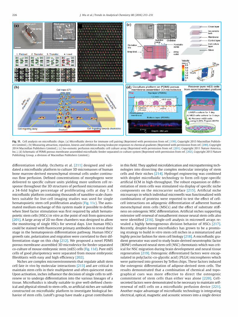
differentiation reliably. Occhetta et al. [211] designed and vali-dated a microfluidic platform to culture 3D micromasses of humanbone marrow-derived mesenchymal stromal cells under continu-ous flow perfusion. Defined concentrations of morphogens weredelivered to specific culture units yielding more uniform cell re-sponse throughout the 3D structures of perfused micromasses anda 34-fold higher percentage of proliferating cells at day 7. Amicrofluidic platform containing thousands of nanoliter-scale cham-bers suitable for live-cell imaging studies was used for singlehematopoietic stem cell proliferation analysis (Fig. 11c). The auto-mated medium exchange of this system made it possible to definewhen Steel factor stimulation was first required by adult hemato-poietic stem cells (HSCs) in vitro as the point of exit from quiescence[201]. A large array of 2D no-flow chambers was designed to allowthe monitoring of single HSCs for several days. Live human HSCscould be stained with fluorescent primary antibodies to reveal theirstage in the hematopoiesis differentiation pathway. Human HSCs’growth rate, polarization andmigration were correlated to their dif-ferentiation stage on this chip [212]. We proposed a novel PDMSporous membrane-assembled 3D microdevice for feeder-separatedco-culture of mouse embryonic stem (mES) cells (Fig. 11d). PuremEScells of good pluripotency were separated from mouse embryonicfibroblasts with easy and high efficiency [202].
Niches are complex microenvironments that regulate adult stemcell fate in vivo by molecular interactions [213] and are critical tomaintain stem cells in their multipotent and often quiescent state.Upon activation, inches influence the decision of single cells to self-renew or to undergo differentiation into the various lineages of atissue. Microfluidics is ideally suitable to give well-defined chem-ical and physical stimuli to stem cells, so artificial niches are suitableconstructed on microfluidic platform to investigate biological be-havior of stem cells. Lutolf’s group have made a great contribution
in this field. They appliedmicrofabrication andmicropatterning tech-nologies into dissecting the complex molecular interplay of stemcells and their niches [214]. Hydrogel engineering was combinedwith droplet microfluidic technology to form cell-type-specificartificial ECM in high-throughput. The robust expansion or differ-entiation of stem cells was stimulated via display of specific nichecomponents on the microcarrier surface [215]. Artificial nichemicroarrays in which individual microwells was functionalized withcombinations of proteins were reported to test the effect of cell-cell interactions on adipogenic differentiation of adherent humanmesenchymal stem cells (MSCs) and the effect of substrate stiff-ness on osteogenic MSC differentiation. Artificial niches supportingextensive self-renewal of nonadherent mouse neural stem cells alsowere identified [216]. Single-cell analysis in microwell arrays re-vealed a highly heterogeneous division behavior of HSCs [217].Recently, droplet-based microfluidics has grown to be a promis-ing strategy to build in vitro stem cell niches in a miniaturized andhighly precise fashion for stem cell biology [218]. A microfluidic gra-dient generator was used to study brain-derived neurotrophic factor(BDNF)-enhanced neural stem cell (NSC) chemotaxis which was crit-ical for NSC migration during brain development and neural tissueregeneration [219]. Osteogenic differentiated factors were encap-sulated in poly(lactic-co-glycolic acid) (PLGA) microspheres whichwere patterned into grooves by Teflon chips. These factors inducedthe osteogenic differentiation of adipose-derived stem cells. Theresults demonstrated that a combination of chemical and topo-graphical cues was more effective to direct the osteogeniccommitment of stem cells than either was alone [220]. Cell-secreted factors were demonstrated to be necessary tomaintain self-renewal of mES cells on a microfluidic perfusion device [221].Another major advantage of microfluidic technology is integratingelectrical, optical, magnetic and acoustic sensors into a single device
Fig. 11. Cell analysis on microfluidic chips. (a) Microfluidic device for immune cell pairing (Reprinted with permission from ref. [199]. Copyright 2015 Macmillan Publish-ers Limited.). (b) Measuring attraction, repulsion, kinesis and inhibition during leukocyte responses to chemical gradients (Reprinted with permission from ref. [200]. Copyright2014 Macmillan Publishers Limited.). (c) Iso-osmotic perfusion microfluidic cell culture array (Reprinted with permission from ref. [201]. Copyright 2011 Nature America,Inc.). (d) Schematic of PDMS porous membrane-assembled microfluidic feeder-separated co-culture system (Reprinted with permission from ref. [202]. Copyright 2013 NaturePublishing Group, a division of Macmillan Publishers Limited.).
226 J. Wu et al. / Trends in Analytical Chemistry 80 (2016) 213–231
to give observation on stem cells. Kang et al. [222] presented amicrofluidic device that coupled on-chip culture of adherent cellsand transfection by localized electroporation. Differentiation of NSCsand transfection of postmitotic neurons with a green fluorescentprotein plasmid were demonstrated on this device. A radio fre-quency identification-based sensor platform was designed byintegrating sensitive elements onto glass substrate comprised of twocomb-shaped interdigitated gold electrodes covering an area of1.8 mm × 2 mm. The platform was adopted to characterize cultiva-tion and differentiation of human bonemarrow-derivedmultipotentstem cells over periods of up to several days and weeks [223]. Os-teogenic differentiation of MSCs was evaluated on a SPR-basedsystem. A high correlation between the duration of osteogenic in-duction and the difference in refractive angle shift with coefficientwas demonstrated [224]. In the light of an increasing demand ofstem cells for cytotoxicity assay, disease modeling, drug screeningand cell-based therapies, there is an increasing necessity for ad-vanced in vitro stem cell analysis systems. Advances in biomaterialsand microtechnology for stem cell analysis even make it possibleto offer mechanistic insight into organogenesis and unveil power-ful new models for drug discovery, as well as strategies for tissueregeneration in the clinic [225–227].
4. Conclusions and outlook
Owning to the inherent advances in device design, fabricationand flexible integration with other microscale techniques,microfluidics has experienced an explosive growth during the pastdecades and become a major analytical technology with enlargingapplication fields. As a technique enabling precise manipulation offluid at the micrometer scale, microfluidics has paved a revolution-ary way for time-saving and cost-saving detection in sophisticatedbiochemical analysis. Not only traditional biochemical operationswere miniaturized onto the microfluidic device, but also new plat-forms for generating unconventional strategies to overcome thechallenges in biochemical analysis were provided.
For improving microfluidic applications in biochemical analy-sis, there are still some challenges have to be addressed: (1) Highlyintegrated microdevices coupled with large and multifunctional in-struments such as MS, nuclear magnetic resonance spectrometerhave to be designed. Although we have explored in the field of chipcoupled with MS, fully-integrated chip-MS platforms need to befurther developed. Besides that, how to miniaturize these large in-struments to make them adjustable to be coupled with chip stilldemand prompt development. (2) Automated manipulation ofmicrofluidic chips desire more convenient sample pretreatment andhigh-throughput analysis. This promotion will advancemicrofluidicsto the upper level in biochemical analysis. (3) Real-time analysisof multiple targets of in vivo biochemical process in situ is a crucialarea in microfluidics. With the development of online microfluidicchip-based biochemical analysis, it is hoped that portable and in-expensive microfluidic devices will play a great role in point-of-care diagnosis and drug discovery in the future.
Acknowledgements
This work was supported by the National Natural Science Foun-dation of China (No. 21405143, 21435002, 21227006) and theFundamental Research Funds for the Central Universities (No.2-9-2014-023).
References
[1] J. El-Ali, P.K. Sorger, K.F. Jensen, Cells on chips, Nature 442 (2006) 403–411.[2] M.M. Rosenkilde, T.W. Schwartz, The chemokine system – a major regulator
of angiogenesis in health and disease, APMIS 112 (2004) 481–495.
[3] L. Liu, B.D. Ratner, E.H. Sage, S. Jiang, Endothelial cell migration on surface-density gradients of fibronectin, VEGF, or both proteins, Langmuir 23 (2007)11168–11173.
[4] A.S. Belloni, G. Albertin, M.L. Forneris, G.G. Nussdorfer, Proadrenomedullin-derived peptides as autocrine-paracrine regulators of cell growth, Histol.Histopathol. 16 (2001) 1263–1274.
[5] F. Rosso, A. Giordano, M. Barbarisi, A. Barbarisi, From cell-ECM interactionsto tissue engineering, J. Cell. Physiol. 199 (2004) 174–180.
[6] C. Jamora, E. Fuchs, Intercellular adhesion, signalling and the cytoskeleton,Nat. Cell Biol. 4 (2002) E101–E108.
[7] B. Geiger, A. Bershadsky, R. Pankov, K.M. Yamada, Transmembrane extracellularmatrix-cytoskeleton crosstalk, Nat. Rev. Mol. Cell Biol. 2 (2001) 793–805.
[8] Y. Li, Y. Zhou, H. Wang, S. Perrett, Y. Zhao, Z. Tang, et al., Chirality of glutathionesurface coating affects the cytotoxicity of quantum dots, Angew. Chem. Int.Edit. 50 (2011) 5860–5864.
[9] A. Hoshino, K. Fujioka, T. Oku, M. Suga, Y.F. Sasaki, T. Ohta, et al.,Physicochemical properties and cellular toxicity of nanocrystal quantum dotsdepend on their surface modification, Nano Lett. 4 (2004) 2163–2169.
[10] A. Arora, G. Simone, G.B. Salieb-Beugelaar, J.T. Kim, A. Manz, Latestdevelopments in micro total analysis systems, Anal. Chem. 82 (2010) 4830–4847.
[11] G.B. Salieb-Beugelaar, G. Simone, A. Arora, A. Philippi, A. Manz, Latestdevelopments in microfluidic cell biology and analysis systems, Anal. Chem.82 (2010) 4848–4864.
[12] D. Mark, S. Haeberle, G. Roth, F. von Stetten, R. Zengerle, Microfluidic lab-on-a-chip platforms: requirements, characteristics and applications, Chem. Soc.Rev. 39 (2010) 1153–1182.
[13] I. Meyvantsson, D.J. Beebe, Cell culture models in microfluidic systems, Annu.Rev. Anal. Chem. (Palo Alto Calif) 1 (2008) 423–449.
[14] A. Khademhosseini, R. Langer, J. Borenstein, J.P. Vacanti, Microscale technologiesfor tissue engineering and biology, Proc. Natl. Acad. Sci. U.S.A. 103 (2006)2480–2487.
[15] D. Gao, H. Liu, Y. Jiang, J.-M. Lin, Recent developments in microfluidic devicesfor in vitro cell culture for cell-biology research, TrAC – Trends Anal. Chem.35 (2012) 150–164.
[16] M.C. Cushing, K.S. Anseth, Hydrogel cell cultures, Science 316 (2007) 1133–1134.
[17] D. Huh, G.A. Hamilton, D.E. Ingber, From 3D cell culture to organs-on-chips,Trends Cell Biol. 21 (2011) 745–754.
[18] Y. Imura, K. Sato, E. Yoshimura, Micro total bioassay system for ingestedsubstances: assessment of intestinal absorption, hepatic metabolism, andbioactivity, Anal. Chem. 82 (2010) 9983–9988.
[19] D. Huh, B.D. Matthews, A. Mammoto, M. Montoya-Zavala, H.Y. Hsin, D.E. Ingber,Reconstituting organ-level lung functions on a chip, Science 328 (2010)1662–1668.
[20] L. Mazutis, J. Gilbert, W.L. Ung, D.A. Weitz, A.D. Griffiths, J.A. Heyman,Single-cell analysis and sorting using droplet-based microfluidics, Nat. Protoc.8 (2013) 870–891.
[21] O. Gefen, O. Fridman, I. Ronin, N.Q. Balaban, Direct observation of singlestationary-phase bacteria reveals a surprisingly long period of constant proteinproduction activity, Proc. Natl. Acad. Sci. U.S.A. 111 (2014) 556–561.
[22] S.S. Lee, I. Avalos Vizcarra, D.H.E.W. Huberts, L.P. Lee, M. Heinemann, Wholelifespanmicroscopic observation of budding yeast aging through amicrofluidicdissection platform, Proc. Natl. Acad. Sci. U.S.A. 109 (2012) 4916–4920.
[23] K. Loutherback, L. Chen, H.N. Holman, Open-channel microfluidic membranedevice for long-term FT-IR spectromicroscopy of live adherent cells, Anal.Chem. 87 (2015) 4601–4606.
[24] T.H. Seefeld, A.R. Halpern, R.M. Corn, On-Chip synthesis of protein microarraysfrom DNA microarrays via coupled in vitro transcription and translation forsurface plasmon resonance imaging biosensor applications, J. Am. Chem. Soc.134 (2012) 12358–12361.
[25] C. Bermejo, F. Haerizadeh, H. Takanaga, D. Chermak, W.B. Frommer, Opticalsensors for measuring dynamic changes of cytosolic metabolite levels in yeast,Nat. Protoc. 6 (2011) 1806–1817.
[26] H. Xin, Y. Li, B. Li, Controllable patterning of different cells via opticalassembly of 1D periodic cell structures, Adv. Funct. Mater. 25 (2015) 2816–2823.
[27] S. Kim, A.M. Streets, R.R. Lin, S.R. Quake, S. Weiss, D.S. Majumdar, High-throughput single-molecule optofluidic analysis, Nat. Methods 8 (2011)242–245.
[28] F. Guo, M.I. Lapsley, A.A. Nawaz, Y. Zhao, S.S. Lin, Y. Chen, et al., A droplet-based,optofluidic device for high-throughput, quantitative bioanalysis, Anal. Chem.84 (2012) 10745–10749.
[29] J. Chen, Z. Kang, G. Wang, J.F.C. Loo, S.K. Kong, H. Ho, Optofluidic guiding,valving, switching and mixing based on plasmonic heating in a random goldnanoisland substrate, Lab Chip 15 (2015) 2504–2512.
[30] A. Valero, T. Braschler, P. Renaud, A unified approach to dielectric single cellanalysis: impedance and dielectrophoretic force spectroscopy, Lab Chip 10(2010) 2216–2225.
[31] T.B. Tran, S. Cho, J. Min, Hydrogel-based diffusion chip with Electric Cell-substrate Impedance Sensing (ECIS) integration for cell viability assay and drugtoxicity screening, Biosens. Bioelectron. 50 (2013) 453–459.
[32] Y. Xu, Y. Lv, L. Wang, W. Xing, J. Cheng, A microfluidic device with passiveair-bubble valves for real-time measurement of dose-dependent drugcytotoxicity through impedance sensing, Biosens. Bioelectron. 32 (2012)300–304.
227J. Wu et al. / Trends in Analytical Chemistry 80 (2016) 213–231
[33] K. Zor, A. Heiskanen, C. Caviglia, M. Vergani, E. Landini, F. Shah, et al., A compactmultifunctional microfluidic platform for exploring cellular dynamics inreal-time using electrochemical detection, RSC Adv. 4 (2014) 63761–63771.
[34] R. Meissner, B. Eker, H. Kasi, A. Bertsch, P. Renaud, Distinguishing drug-inducedminor morphological changes from major cellular damage via label-freeimpedimetric toxicity screening, Lab Chip 11 (2011) 2352–2361.
[35] A.C. Sabuncu, J. Zhuang, J.F. Kolb, A. Beskok, Microfluidic impedancespectroscopy as a tool for quantitative biology and biotechnology,Biomicrofluidics 6 (2012) 34103.
[36] B.J. Dworak, B.C. Wheeler, Novel MEA platform with PDMS microtunnelsenables the detection of action potential propagation from isolated axons inculture, Lab Chip 9 (2009) 404–410.
[37] A. Hai, J. Shappir, M.E. Spira, In-cell recordings by extracellular microelectrodes,Nat. Methods 7 (2010) 200–202.
[38] G. Cellot, E. Cilia, S. Cipollone, V. Rancic, A. Sucapane, S. Giordani, et al., Carbonnanotubes might improve neuronal performance by favouring electricalshortcuts, Nat. Nanotechnol. 4 (2009) 126–133.
[39] S.Y. Ng, J. Reboud, K.Y.P. Wang, K.C. Tang, L. Zhang, P. Wong, et al., Label-freeimpedance detection of low levels of circulating endothelial progenitor cellsfor point-of-care diagnosis, Biosens. Bioelectron. 25 (2010) 1095–1101.
[40] E. Du, S. Ha, M. Diez-Silva, M. Dao, S. Suresh, A.P. Chandrakasan, Electricimpedance microflow cytometry for characterization of cell disease states,Lab Chip 13 (2013) 3903–3909.
[41] C.E. Nwankire, A. Venkatanarayanan, T. Glennon, T.E. Keyes, R.J. Forster, J.Ducrée, Label-free impedance detection of cancer cells from whole blood onan integrated centrifugal microfluidic platform, Biosens. Bioelectron. 68 (2015)382–389.
[42] M.S. Mannoor, S. Zhang, A.J. Link, M.C. McAlpine, Electrical detection ofpathogenic bacteria via immobilized antimicrobial peptides, Proc. Natl. Acad.Sci. U.S.A. 107 (2010) 19207–19212.
[43] S. Gawad, K. Cheung, U. Seger, A. Bertsch, P. Renaud, Dielectric spectroscopyin a micromachined flow cytometer: theoretical and practical considerations,Lab Chip 4 (2004) 241–251.
[44] T. Braschler, N. Demierre, E. Nascimento, T. Silva, A.G. Oliva, P. Renaud,Continuous separation of cells by balanced dielectrophoretic forces at multiplefrequencies, Lab Chip 8 (2008) 280–286.
[45] N. Demierre, T. Braschler, P. Linderholm, U. Seger, H. van Lintel, P. Renaud,Characterization and optimization of liquid electrodes for lateraldielectrophoresis, Lab Chip 7 (2007) 355–365.
[46] N. Demierre, T. Braschler, R. Muller, P. Renaud, Focusing and continuousseparation of cells in a microfluidic device using lateral dielectrophoresis, Sens.Actuator. B-Chem. 132 (2008) 388–396.
[47] P. Zhang, L. Ren, X. Zhang, Y. Shan, Y. Wang, Y. Ji, et al., Raman-activated cellsorting based on dielectrophoretic single-cell trap and release, Anal. Chem.87 (2015) 2282–2289.
[48] M.G. Simon, Y. Li, J. Arulmoli, L.P. McDonnell, A. Akil, J.L. Nourse, et al.,Increasing label-free stem cell sorting capacity to reach transplantation-scalethroughput, Biomicrofluidics 8 (2014) 64106.
[49] H. Song, J.M. Rosano, Y. Wang, C.J. Garson, B. Prabhakarpandian, K. Pant, et al.,Continuous-flow sorting of stem cells and differentiation products based ondielectrophoresis, Lab Chip 15 (2015) 1320–1328.
[50] G. Lee, H. Wu, P. Yang, J.D. Mai, Optically induced dielectropheresis sortingwith automatedmedium exchange in an integrated optofluidic device resultingin higher cell viability, Lab Chip 14 (2014) 2837–2843.
[51] Z. Wei, X. Li, D. Zhao, H. Yan, Z. Hu, Z. Liang, et al., Flow-through cellelectroporationmicrochip integrating dielectrophoretic viable cell sorting, Anal.Chem. 86 (2014) 10215–10222.
[52] J. DuBose, X. Lu, S. Patel, S. Qian, S.W. Joo, X. Xuan, Microfluidic electrical sortingof particles based on shape in a spiral microchannel, Biomicrofluidics 8 (2014)14101.
[53] J. Zhu, X. Xuan, Curvature-induced dielectrophoresis for continuous separationof particles by charge in spiral microchannels, Biomicrofluidics 5 (2011) 24111.
[54] R.K. Anand, E.S. Johnson, D.T. Chiu, Negative dielectrophoretic capture andrepulsion of single cells at a bipolar electrode: the impact of faradaic ionenrichment and depletion, J. Am. Chem. Soc. 137 (2015) 776–783.
[55] A.J. Hughes, A.E. Herr, Microfluidic western blotting, Proc. Natl. Acad. Sci. U.S.A.109 (2012) 21450–21455.
[56] P. Batalla, A. Martin, M. Angel Lopez, M. Cristina Gonzalez, A. Escarpa,Enzyme-based microfluidic chip coupled to graphene electrodes for thedetection of D-Amino acid enantiomer-biomarkers, Anal. Chem. 87 (2015)5074–5078.
[57] K. Kadimisetty, S. Malla, N.P. Sardesai, A.A. Joshi, R.C. Faria, N.H. Lee, et al.,Automated multiplexed ECL immunoarrays for cancer biomarker proteins,Anal. Chem. 87 (2015) 4472–4478.
[58] R. Vaidyanathan, L.M. van Leeuwen, S. Rauf, M.J.A. Shiddiky, M. Trau, Amultiplexed device based on tunable nanoshearing for specific detection ofmultiple protein biomarkers in serum, Sci. Rep. 5 (2015) 9756.
[59] L. Wu, C. Ma, L. Ge, Q. Kong, M. Yan, S. Ge, et al., Paper-basedelectrochemiluminescence origami cyto-device for multiple cancer cellsdetection using porous AuPd alloy as catalytically promoted nanolabels,Biosens. Bioelectron. 63 (2015) 450–457.
[60] A.C. Glavan, D.C. Christodouleas, B. Mosadegh, H.D. Yu, B.S. Smith, J. Lessing,et al., Folding analytical devices for electrochemical ELISA in hydrophobic R-Hpaper, Anal. Chem. 86 (2014) 11999–12007.
[61] X. Ding, P. Li, S.S. Lin, Z.S. Stratton, N. Nama, F. Guo, et al., Surface acousticwave microfluidics, Lab Chip 13 (2013) 3626–3649.
[62] P.R. Rogers, J.R. Friend, L.Y. Yeo, Exploitation of surface acoustic waves to drivesize-dependent microparticle concentration within a droplet, Lab Chip 10(2010) 2979–2985.
[63] T. Franke, S. Braunmuller, L. Schmid, A. Wixforth, D.A. Weitz, Surface acousticwave actuated cell sorting (SAWACS), Lab Chip 10 (2010) 789–794.
[64] L. Schmid, T. Franke, SAW-controlled drop size for flow focusing, Lab Chip 13(2013) 1691–1694.
[65] J. Reboud, Y. Bourquin, R. Wilson, G.S. Pall, M. Jiwaji, A.R. Pitt, et al., Shapingacoustic fields as a toolset for microfluidic manipulations in diagnostictechnologies, Proc. Natl. Acad. Sci. U.S.A. 109 (2012) 15162–15167.
[66] X. Ding, S.S. Lin, B. Kiraly, H. Yue, S. Li, I. Chiang, et al., On-chip manipulationof single microparticles, cells, and organisms using surface acoustic waves,Proc. Natl. Acad. Sci. U.S.A. 109 (2012) 11105–11109.
[67] P. Li, Z. Mao, Z. Peng, L. Zhou, Y. Chen, P. Huang, et al., Acoustic separation ofcirculating tumor cells, Proc. Natl. Acad. Sci. U.S.A. 112 (2015) 4970–4975.
[68] M. Travagliati, R.J. Shilton, M. Pagliazzi, I. Tonazzini, F. Beltram, M. Cecchini,Acoustofluidics and whole-blood Manipulation in Surface Acoustic WaveCounterflow Devices, Anal. Chem. 86 (2014) 10633–10638.
[69] S. Li, X. Ding, Z. Mao, Y. Chen, N. Nama, F. Guo, et al., Standing surface acousticwave (SSAW)-based cell washing, Lab Chip 15 (2015) 331–338.
[70] R.P. Richter, R. Bérat, A.R. Brisson, Formation of solid-supported Lipid Bilayers:an Integrated View, Langmuir 22 (2006) 3497–3505.
[71] M. Hennig, J. Neumann, A. Wixforth, J.O. Radler, M.F. Schneider, Dynamicpatterns in a supported lipid bilayer driven by standing surface acoustic waves,Lab Chip 9 (2009) 3050–3053.
[72] M. Hennig, M. Wolff, J. Neumann, A. Wixforth, M.F. Schneider, J.O. Rädler, DNAconcentration modulation on supported lipid bilayers switched by surfaceacoustic waves, Langmuir 27 (2011) 14721–14725.
[73] J. Neumann, M. Hennig, A. Wixforth, S. Manus, J.O. Rädler, M.F. Schneider,Transport, separation, and accumulation of proteins on supported lipid bilayers,Nano Lett. 10 (2010) 2903–2908.
[74] X. Ding, Z. Peng, S.S. Lin, M. Geri, S. Li, P. Li, et al., Cell separation usingtilted-angle standing surface acoustic waves, Proc. Natl. Acad. Sci. U.S.A. 111(2014) 12992–12997.
[75] S. Li, F. Guo, Y. Chen, X. Ding, P. Li, L. Wang, et al., Standing surface acousticwave based cell coculture, Anal. Chem. 86 (2014) 9853–9859.
[76] A. van Reenen, A.M. de Jong, J.M.J. den Toonder, M.W.J. Prins, Integratedlab-on-chip biosensing systems based on magnetic particle actuation – acomprehensive review, Lab Chip 14 (2014) 1966–1986.
[77] M. Vilfan, A. Potocnik, B. Kavcic, N. Osterman, I. Poberaj, A. Vilfan, et al.,Self-assembled artificial cilia, Proc. Natl. Acad. Sci. U.S.A. 107 (2010) 1844–1847.
[78] G. Zhu, N. Nguyen, Rapid magnetofluidic mixing in a uniform magnetic field,Lab Chip 12 (2012) 4772–4780.
[79] A. Ranzoni, X.J.A. Janssen, M. Ovsyanko, L.J. van IJzendoorn, M.W.J. Prins,Magnetically controlled rotation and torque of uniaxial microactuators forlab-on-a-chip applications, Lab Chip 10 (2010) 179–188.
[80] M. Karle, J. Miwa, G. Czilwik, V. Auwarter, G. Roth, R. Zengerle, et al., Continuousmicrofluidic DNA extraction using phase-transfer magnetophoresis, Lab Chip10 (2010) 3284–3290.
[81] G. Lin, D. Makarov, M. Medina-Sanchez, M. Guix, L. Baraban, G. Cuniberti, et al.,Magnetofluidic platform for multidimensional magnetic and optical barcodingof droplets, Lab Chip 15 (2015) 216–224.
[82] S.Y. Park, H. Handa, A. Sandhu, Magneto-optical biosensing platform basedon light scattering from self-assembled chains of functionalized rotatingmagnetic beads, Nano Lett. 10 (2010) 446–451.
[83] R.C. den Dulk, K.A. Schmidt, G. Sabatte, S. Liebana, M.W.J. Prins, Magneto-capillary valve for integrated purification and enrichment of nucleic acids andproteins, Lab Chip 13 (2013) 106–118.
[84] O. Strohmeier, A. Emperle, G. Roth, D. Mark, R. Zengerle, F. von Stetten,Centrifugal gas-phase transition magnetophoresis (GTM) – a generic methodfor automation of magnetic bead based assays on the centrifugal microfluidicplatform and application to DNA purification, Lab Chip 13 (2013) 146–155.
[85] M. Cornaglia, R. Trouillon, H.C. Tekin, T. Lehnert, M.A.M. Gijs, Magneticparticle-scanning for ultrasensitive immunodetection On-Chip, Anal. Chem.86 (2014) 8213–8223.
[86] C. Chiou, D. Jin Shin, Y. Zhang, T. Wang, Topography-assisted electromagneticplatform for blood-to-PCR in a droplet, Biosens. Bioelectron. 50 (2013) 91–99.
[87] C. Chang, W. Huang, S.I. Jalal, B. Chan, A. Mahmood, S. Shahda, et al., Circulatingtumor cell detection using a parallel flow micro-aperture chip system, LabChip 15 (2015) 1677–1688.
[88] A. van Reenen, Y. Gao, A.H. Bos, A.M. de Jong, M.A. Hulsen, J.M.J. den Toonder,et al., Accurate quantification of magnetic particle properties by intra-pairmagnetophoresis for nanobiotechnology, Appl. Phys. Lett. 103 (2013) 43704.
[89] F. Hernández, T. Portolés, E. Pitarch, F.J. López, Gas chromatography coupledto high-resolution time-of-flight mass spectrometry to analyze trace-levelorganic compounds in the environment, food safety and toxicology, TrAC –Trends Anal. Chem. 30 (2011) 388–400.
[90] G. Wei, E.Y. Zeng, Gas chromatography-mass spectrometry and high-performance liquid chromatography-tandemmass spectrometry in quantifyingfatty acids, TrAC – Trends Anal. Chem. 30 (2011) 1429–1436.
[91] T. Cajka, O. Fiehn, Comprehensive analysis of lipids in biological systems byliquid chromatography-mass spectrometry, TrAC – Trends Anal. Chem. 61(2014) 192–206.
[92] P.D. Rainville, G. Theodoridis, R.S. Plumb, I.D. Wilson, Advances in liquidchromatography coupled to mass spectrometry for metabolic phenotyping,TrAC – Trends Anal. Chem. 61 (2014) 181–191.
228 J. Wu et al. / Trends in Analytical Chemistry 80 (2016) 213–231
[93] A. Hirayama, M. Wakayama, T. Soga, Metabolome analysis based on capillaryelectrophoresis-mass spectrometry, TrAC – Trends Anal. Chem. 61 (2014)215–222.
[94] C. Seger, S. Sturm, H. Stuppner, Mass spectrometry and NMR spectroscopy:modern high-end detectors for high resolution separation techniques – stateof the art in natural product HPLC-MS, HPLC-NMR, and CE-MS hyphenations,Nat. Prod. Rep. 30 (2013) 970–987.
[95] A.R. Timerbaev, Capillary electrophoresis coupled to mass spectrometry forbiospeciation analysis: critical evaluation, TrAC – Trends Anal. Chem. 28 (2009)416–425.
[96] Q.F. Xue, F. Foret, Y.M. Dunayevskiy, P.M. Zavracky, N.E. McGruer, B.L. Karger,Multichannel microchip electrospray mass spectrometry, Anal. Chem. 69(1997) 426–430.
[97] R.S. Ramsey, J.M. Ramsey, Generating electrospray from microchip devicesusing electroosmotic pumping, Anal. Chem. 69 (1997) 1174–1178.
[98] X. Feng, B. Liu, J. Li, X. Liu, Advances in coupling microfluidic chips to massspectrometry, Mass Spectrom. Rev. 34 (2015) 535–557.
[99] H. Wei, H. Li, J.-M. Lin, Analysis of herbicides on a single C30 bead via amicrofluidic device combined with electrospray ionization quadrupoletime-of-flight mass spectrometer, J. Chromatogr. A 1216 (2009) 9134–9142.
[100] H. Wei, H. Li, D. Gao, J.-M. Lin, Multi-channel microfluidic devices combinedwith electrospray ionization quadrupole time-of-flight mass spectrometryapplied to the monitoring of glutamate release from neuronal cells, Analyst135 (2010) 2043–2050.
[101] D. Gao, H. Wei, G.S. Guo, J.-M. Lin, Microfluidic cell culture and metabolismdetection with electrospray ionization quadrupole time-of-flight massspectrometer, Anal. Chem. 82 (2010) 5679–5685.
[102] D. Gao, H. Li, N. Wang, J.-M. Lin, Evaluation of the absorption of methotrexateon cells and its cytotoxicity assay by using an integrated microfluidic devicecoupled to a mass spectrometer, Anal. Chem. 84 (2012) 9230–9237.
[103] D. Gao, H. Liu, J.-M. Lin, Y. Wang, Y. Jiang, Characterization of drug permeabilityin Caco-2 monolayers by mass spectrometry on a membrane-basedmicrofluidic device, Lab Chip 13 (2013) 978–985.
[104] Q. Chen, J. Wu, Y. Zhang, J.-M. Lin, Qualitative and quantitative analysis of tumorcell metabolism via stable isotope labeling assisted microfluidic chipelectrospray ionization mass spectrometry, Anal. Chem. 84 (2012) 1695–1701.
[105] H. Wei, H. Li, S. Mao, J.-M. Lin, Cell signaling analysis by mass spectrometryunder coculture conditions on an integrated microfluidic device, Anal. Chem.83 (2011) 9306–9313.
[106] S. Mao, J. Zhang, H. Li, J.-M. Lin, Strategy for signaling molecule detection byusing an integrated microfluidic device coupled with mass spectrometry tostudy Cell-to-Cell communication, Anal. Chem. 85 (2013) 868–876.
[107] W. Liu, Q. Chen, X. Lin, J.-M. Lin, Online multi-channel microfluidic chip-massspectrometry and its application for quantifying noncovalent protein-proteininteractions, Analyst 140 (2015) 1551–1554.
[108] E.A. Redman, N.G. Batz, J.S. Mellors, J.M. Ramsey, Integrated microfluidiccapillary electrophoresis-electrospray ionization devices with online MSdetection for the separation and characterization of intact monoclonal antibodyvariants, Anal. Chem. 87 (2015) 2264–2272.
[109] F.T.G. van den Brink, L. Büter, M. Odijk, W. Olthuis, U. Karst, A. van den Berg,Mass spectrometric detection of short-lived drug metabolites generated inan electrochemical microfluidic chip, Anal. Chem. 87 (2015) 1527–1535.
[110] Q. Wu, D. Gao, J. Wei, F. Jin, W. Xie, Y. Jiang, et al., Development of a novelmulti-layer microfluidic device towards characterization of drugmetabolism and cytotoxicity for drug screening, Chem. Commun. 50 (2014)2762–2764.
[111] A.E. Kirby, N.M. Lafrenière, B. Seale, P.I. Hendricks, R.G. Cooks, A.R. Wheeler,Analysis on the go: quantitation of drugs of abuse in dried urine with digitalmicrofluidics and miniature mass spectrometry, Anal. Chem. 86 (2014)6121–6129.
[112] D. Jin, Y. Zhu, Q. Fang, Swan probe: a nanoliter-scale and high-throughputsampling interface for coupling electrospray ionizationmass spectrometry withmicrofluidic droplet array and multiwell plate, Anal. Chem. 86 (2014) 10796–10803.
[113] S. Arscott, SU-8 as a material for lab-on-a-chip-based mass spectrometry, LabChip 14 (2014) 3668–3689.
[114] D. Gao, H. Liu, Y. Jiang, J.-M. Lin, Recent advances in microfluidics combinedwith mass spectrometry: technologies and applications, Lab Chip 13 (2013)3309–3322.
[115] X. He, Q. Chen, Y. Zhang, J.-M. Lin, Recent advances in microchip-massspectrometry for biological analysis, TrAC – Trends Anal. Chem. 53 (2014)84–97.
[116] S. Allazetta, L. Kolb, S. Zerbib, J. Bardy, M.P. Lutolf, Cell-instructive microgelswith Tailor-made physicochemical properties, Small 11 (2015) 5647–5656.
[117] C.A. Bichsel, S. Gobaa, S. Kobel, C. Secondini, G.N. Thalmann, M.G. Cecchini,et al., Diagnostic microchip to assay 3D colony-growth potential of capturedcirculating tumor cells, Lab Chip 12 (2012) 2313–2316.
[118] S. Allazetta, S. Cosson, M.P. Lutolf, Programmable microfluidic patterning ofprotein gradients on hydrogels, Chem. Commun. 47 (2011) 191–193.
[119] S. Cosson, S. Allazetta, M.P. Lutolf, Patterning of cell-instructive hydrogels byhydrodynamic flow focusing, Lab Chip 13 (2013) 2099–2105.
[120] S. Cosson, S.A. Kobel, M.P. Lutolf, Capturing complex protein gradients onbiomimetic hydrogels for cell-based assays, Adv. Funct. Mater. 19 (2009)3411–3419.
[121] S.H. Mathes, H. Ruffner, U. Graf-Hausner, The use of skin models in drugdevelopment, Adv. Drug Deliv. Rev. 69–70 (2014) 81–102.
[122] Y. Li, S. Wang, R. Huang, Z. Huang, B. Hu, W. Zheng, et al., Evaluation of theeffect of the structure of bacterial cellulose on full thickness skin wound repairon a microfluidic chip, Biomacromolecules 16 (2015) 780–789.
[123] Y. Torisawa, C.S. Spina, T. Mammoto, A. Mammoto, J.C. Weaver, T. Tat, et al.,Bone marrow-on-a-chip replicates hematopoietic niche physiology in vitro,Nat. Methods 11 (2014) 663–669.
[124] J. Wu, Q. Chen, W. Liu, Y. Zhang, J.-M. Lin, Cytotoxicity of quantum dots assayon a microfluidic 3D-culture device based on modeling diffusion processbetween blood vessels and tissues, Lab Chip 12 (2012) 3474–3480.
[125] J. Park, B.K. Lee, G.S. Jeong, J.K. Hyun, C.J. Lee, S. Lee, Three-dimensionalbrain-on-a-chip with an interstitial level of flow and its application as an invitro model of Alzheimer’s disease, Lab Chip 15 (2015) 141–150.
[126] Y. Zheng, J. Chen, M. Craven, N.W. Choi, S. Totorica, A. Diaz-Santana, et al., Invitro microvessels for the study of angiogenesis and thrombosis, Proc. Natl.Acad. Sci. U.S.A. 109 (2012) 9342–9347.
[127] M.R. Ebrahimkhani, J.A.S. Neiman, M.S.B. Raredon, D.J. Hughes, L.G. Griffith,Bioreactor technologies to support liver function in vitro, Adv. Drug Deliv. Rev.69–70 (2014) 132–157.
[128] S. Mao, D. Gao,W. Liu, H.Wei, J.-M. Lin, Imitation of drugmetabolism in humanliver and cytotoxicity assay using a microfluidic device coupled to massspectrometric detection, Lab Chip 12 (2012) 219–226.
[129] J.S. Jeon, S. Bersini, M. Gilardi, G. Dubini, J.L. Charest, M. Moretti, et al., Human3D vascularized organotypic microfluidic assays to study breast cancer cellextravasation, Proc. Natl. Acad. Sci. U.S.A. 112 (2015) 214–219.
[130] Y. Li, S. Zhang, X. Wang, X. Zhang, A.I. Oleinick, I. Svir, et al., Real-timemonitoring of discrete synaptic release events and excitatory potentials withinself-reconstructed neuromuscular junctions, Angew. Chem. Int. Edit. 54 (2015)9313–9318.
[131] M. Wu, S. Huang, G. Lee, Microfluidic cell culture systems for drug research,Lab Chip 10 (2010) 939–956.
[132] J.H. Tsui, W. Lee, S.H. Pun, J. Kim, D. Kim, Microfluidics-assisted in vitro drugscreening and carrier production, Adv. Drug Deliv. Rev. 65 (2013) 1575–1588.
[133] O.J. Miller, A. El Harrak, T. Mangeat, J. Baret, L. Frenz, B. El Debs, et al.,High-resolution dose-response screening using droplet-based microfluidics,Proc. Natl. Acad. Sci. U.S.A. 109 (2012) 378–383.
[134] J. Zhang, J. Wu, H. Li, Q. Chen, J.-M. Lin, An in vitro liver model on microfluidicdevice for analysis of capecitabine metabolite using mass spectrometer asdetector, Biosens. Bioelectron. 68 (2015) 322–328.
[135] A.I. Shallan, R.M. Guijt, M.C. Breadmore, Electrokinetic size and mobility trapsfor on-site therapeutic drug monitoring, Angew. Chem. Int. Edit. 54 (2015)7359–7362.
[136] H. Wang, C. Chen, Z. Xiang, M. Wang, C. Lee, A convection-driven long-rangelinear gradient generator with dynamic control, Lab Chip 15 (2015) 1445–1450.
[137] C. Kim, J. Kasuya, J. Jeon, S. Chung, R.D. Kamm, A quantitative microfluidicangiogenesis screen for studying anti-angiogenic therapeutic drugs, Lab Chip15 (2015) 301–310.
[138] Z. Dereli-Korkut, H.D. Akaydin, A.H.R. Ahmed, X. Jiang, S. Wang, Threedimensional microfluidic cell arrays for ex vivo drug screening with mimickedvascular flow, Anal. Chem. 86 (2014) 2997–3004.
[139] J. Wu, Q. Chen, W. Liu, J.-M. Lin, A simple and versatile microfluidic cell densitygradient generator for quantum dot cytotoxicity assay, Lab Chip 13 (2013)1948–1954.
[140] E.W. Esch, A. Bahinski, D. Huh, Organs-on-chips at the frontiers of drugdiscovery, Nat. Rev. Drug Discov. 14 (2015) 248–260.
[141] P. Neuži, S. Giselbrecht, K. Länge, T.J. Huang, A. Manz, Revisiting lab-on-a-chiptechnology for drug discovery, Nat. Rev. Drug Discov. 11 (2012) 620–632.
[142] C.Y. Chan, P. Huang, F. Guo, X. Ding, V. Kapur, J.D. Mai, et al., Accelerating drugdiscovery via organs-on-chips, Lab Chip 13 (2013) 4697–4710.
[143] H.E. Abaci, K. Gledhill, Z. Guo, A.M. Christiano, M.L. Shuler, Pumplessmicrofluidic platform for drug testing on human skin equivalents, Lab Chip15 (2015) 882–888.
[144] S.H. Au, M.D. Chamberlain, S. Mahesh, M.V. Sefton, A.R. Wheeler, Hepaticorganoids for microfluidic drug screening, Lab Chip 14 (2014) 3290–3299.
[145] A. Agarwal, J.A. Goss, A. Cho, M.L. McCain, K.K. Parker, Microfluidic heart ona chip for higher throughput pharmacological studies, Lab Chip 13 (2013)3599–3608.
[146] I. Maschmeyer, A.K. Lorenz, K. Schimek, T. Hasenberg, A.P. Ramme, J. Hubner,et al., A four-organ-chip for interconnected long-term co-culture of humanintestine, liver, skin and kidney equivalents, Lab Chip 15 (2015) 2688–2699.
[147] H. Tanaka, S. Yamamoto, A. Nakamura, Y. Nakashoji, N. Okura, N. Nakamoto,et al., Hands-Off preparation of monodisperse emulsion droplets using apoly(dimethylsiloxane) microfluidic chip for droplet digital PCR, Anal. Chem.87 (2015) 4134–4143.
[148] P. Dalerba, T. Kalisky, D. Sahoo, P.S. Rajendran, M.E. Rothenberg, A.A. Leyrat,et al., Single-cell dissection of transcriptional heterogeneity in human colontumors, Nat. Biotechnol. 29 (2011) 1120–1127.
[149] X. Lin, J. Wu, W. Liu, H. Li, Z. Wang, J.-M. Lin, Detection of BCR-ABL using onestep reverse transcriptase- polymerase chain reaction and microchipelectrophoresis, Methods 64 (2013) 250–254.
[150] X. Lin, J. Wu, H. Li, Z. Wang, J.-M. Lin, Determination of mini-short tandemrepeat (miniSTR) loci by using the combination of polymerase chain reaction(PCR) and microchip electrophoresis, Talanta 114 (2013) 131–137.
[151] S.J. Reinholt, A.J. Baeumner, Microfluidic isolation of nucleic acids, Angew.Chem. Int. Edit. 53 (2014) 13988–14001.
[152] X. Shi, C. Chen, W. Gao, S. Chao, D.R. Meldrum, Parallel RNA extraction usingmagnetic beads and a droplet array, Lab Chip 15 (2015) 1059–1065.
229J. Wu et al. / Trends in Analytical Chemistry 80 (2016) 213–231
[153] A.K. White, M. VanInsberghe, O.I. Petriv, M. Hamidi, D. Sikorski, M.A. Marra,et al., High-throughput microfluidic single-cell RT-qPCR, Proc. Natl. Acad. Sci.U.S.A. 108 (2011) 13999–14004.
[154] H. Shao, J. Chung, K. Lee, L. Balaj, C. Min, B.S. Carter, et al., Chip-based analysisof exosomal mRNAmediating drug resistance in glioblastoma, Nat. Commun.6 (2015) 6999.
[155] H. Ben-Yoav, P.H. Dykstra, W.E. Bentley, R. Ghodssi, A controlled microfluidicelectrochemical lab-on-a-chip for label-free diffusion-restricted DNAhybridization analysis, Biosens. Bioelectron. 64 (2015) 579–585.
[156] S.P. White, K.D. Dorfman, C.D. Frisbie, Label-Free DNA sensing platform withLow-Voltage Electrolyte-Gated transistors, Anal. Chem. 87 (2015) 1861–1866.
[157] M.E. Polinkovsky, Y. Gambin, P.R. Banerjee, M.J. Erickstad, A. Groisman, A.A.Deniz, Ultrafast cooling reveals microsecond-scale biomolecular dynamics, Nat.Commun. 5 (2014) 5737.
[158] Y. Zhao, D. Chen, H. Yue, M.M. Spiering, C. Zhao, S.J. Benkovic, et al., Dark-fieldillumination on zero-mode waveguide/microfluidic hybrid chip reveals T4replisomal protein interactions, Nano Lett. 14 (2014) 1952–1960.
[159] A.L. Forget, C.C. Dombrowski, I. Amitani, S.C. Kowalczykowski, Exploringprotein-DNA interactions in 3D using in situ construction, manipulation andvisualization of individual DNA dumbbells with optical traps, microfluidicsand fluorescence microscopy, Nat. Protoc. 8 (2013) 525–538.
[160] P. Colin, B. Kintses, F. Gielen, C.M. Miton, G. Fischer, M.F. Mohamed, et al.,Ultrahigh-throughput discovery of promiscuous enzymes by picodropletfunctional metagenomics, Nat. Commun. 6 (2015) 10008.
[161] W. Busch, B.T. Moore, B. Martsberger, D.L. Mace, R.W. Twigg, J. Jung, et al., Amicrofluidic device and computational platform for high-throughput liveimaging of gene expression, Nat. Methods 9 (2012) 1101–1106.
[162] M. Cho, Y. Xiao, J. Nie, R. Stewart, A.T. Csordas, S.S. Oh, et al., Quantitativeselection of DNA aptamers throughmicrofluidic selection and high-throughputsequencing, Proc. Natl. Acad. Sci. U.S.A. 107 (2010) 15373–15378.
[163] A.M. Streets, X. Zhang, C. Cao, Y. Pang, X. Wu, L. Xiong, et al., Microfluidicsingle-cell whole-transcriptome sequencing, Proc. Natl. Acad. Sci. U.S.A. 111(2014) 7048–7053.
[164] M.C. Jo, W. Liu, L. Gu, W. Dang, L. Qin, High-throughput analysis of yeastreplicative aging using a microfluidic system, Proc. Natl. Acad. Sci. U.S.A. 112(2015) 9364–9369.
[165] V. Krivitsky, L. Hsiung, A. Lichtenstein, B. Brudnik, R. Kantaev, R. Elnathan, et al.,Si nanowires forest-based on-chip biomolecular filtering, separation andpreconcentration devices: nanowires do it all, Nano Lett. 12 (2012)4748–4756.
[166] A. Shastri, L.M. McGregor, Y. Liu, V. Harris, H. Nan, M. Mujica, et al., Anaptamer-functionalized chemomechanically modulated biomolecule catch-and-release system, Nat. Chem. 7 (2015) 447–454.
[167] M. Fischlechner, Y. Schaerli, M.F. Mohamed, S. Patil, C. Abell, F. Hollfelder,Evolution of enzyme catalysts caged in biomimetic gel-shell beads, Nat. Chem.6 (2014) 791–796.
[168] P.A. Romero, T.M. Tran, A.R. Abate, Dissecting enzyme function withmicrofluidic-based deep mutational scanning, Proc. Natl. Acad. Sci. U.S.A. 112(2015) 7159–7164.
[169] M. Otten, W. Ott, M.A. Jobst, L.F. Milles, T. Verdorfer, D.A. Pippig, et al., Fromgenes to protein mechanics on a chip, Nat. Methods 11 (2014) 1127–1130.
[170] X. Lin, Q. Chen, W. Liu, J. Zhang, S. Wang, Z. Lin, et al., Oxygen-induced cellmigration and on-line monitoring biomarkers modulation of cervical cancerson a microfluidic system, Sci. Rep. 5 (2015) 9643.
[171] X. Lin, Q. Chen, W. Liu, H. Li, J.-M. Lin, A portable microchip for ultrasensitiveand high-throughput assay of thrombin by rolling circle amplification andhemin/G-quadruplex system, Biosens. Bioelectron. 56 (2014) 71–76.
[172] X. Lin, Q. Chen, W. Liu, L. Yi, H. Li, Z. Wang, et al., Assay of multiplex proteinsfrom cell metabolism based on tunable aptamer andmicrochip electrophoresis,Biosens. Bioelectron. 63 (2015) 105–111.
[173] J. Mok, M.N. Mindrinos, R.W. Davis, M. Javanmard, Digital microfluidic assayfor protein detection, Proc. Natl. Acad. Sci. U.S.A. 111 (2014) 2110–2115.
[174] M. Ihara, A. Yoshikawa, Y. Wu, H. Takahashi, K. Mawatari, K. Shimura, et al.,Micro OS-ELISA: rapid noncompetitive detection of a small biomarker peptideby open-sandwich enzyme-linked immunosorbent assay (OS-ELISA) integratedinto microfluidic device, Lab Chip 10 (2010) 92–100.
[175] J. Kai, A. Puntambekar, N. Santiago, S.H. Lee, D.W. Sehy, V. Moore, et al., A novelmicrofluidic microplate as the next generation assay platform for enzymelinked immunoassays (ELISA), Lab Chip 12 (2012) 4257–4262.
[176] T. Wang, M. Zhang, D.D. Dreher, Y. Zeng, Ultrasensitive microfluidic solid-phaseELISA using an actuatable microwell-patterned PDMS chip, Lab Chip 13 (2013)4190–4197.
[177] C. Cheng, A.W. Martinez, J. Gong, C.R. Mace, S.T. Phillips, E. Carrilho, et al.,Paper-based ELISA, Angew. Chem. Int. Edit. 49 (2010) 4771–4774.
[178] Y. Li, F. Xu, C. Liu, Y. Xu, X. Feng, B. Liu, A novel microfluidic mixer based ondual-hydrodynamic focusing for interrogating the kinetics of DNA-proteininteraction, Analyst 138 (2013) 4475–4482.
[179] C. Han, Q. Zhang, R. Ma, L. Xie, T. Qiu, L. Wang, et al., Integration of single oocytetrapping, in vitrofertilization and embryo culture in a microwell-structuredmicrofluidic device, Lab Chip 10 (2010) 2848–2854.
[180] M.C. Park, J.Y. Hur, H.S. Cho, S. Park, K.Y. Suh, High-throughput single-cellquantification using simple microwell-based cell docking and programmabletime-course live-cell imaging, Lab Chip 11 (2011) 79–86.
[181] N. Chen, C. Chen, M. Chen, L. Jang, M. Wang, Single-cell trapping andimpedance measurement utilizing dielectrophoresis in a parallel-platemicrofluidic device, Sens. Actuator. B-Chem. 190 (2014) 570–577.
[182] D. Wlodkowic, S. Faley, M. Zagnoni, J.P. Wikswo, J.M. Cooper, Microfluidicsingle-cell array cytometry for the analysis of tumor apoptosis, Anal. Chem.81 (2009) 5517–5523.
[183] S. Koster, F.E. Angile, H. Duan, J.J. Agresti, A. Wintner, C. Schmitz, et al.,Drop-based microfluidic devices for encapsulation of single cells, Lab Chip 8(2008) 1110–1115.
[184] J. Clausell-Tormos, D. Lieber, J. Baret, A. El-Harrak, O.J. Miller, L. Frenz, et al.,Droplet-based microfluidic platforms for the encapsulation and screening ofmammalian cells andmulticellular organisms, Chem. Biol. 15 (2008) 427–437.
[185] S. Kobel, A. Valero, J. Latt, P. Renaud, M. Lutolf, Optimization of microfluidicsingle cell trapping for long-term on-chip culture, Lab Chip 10 (2010) 857–863.
[186] H. Chen, J. Sun, E. Wolvetang, J. Cooper-White, High-throughput, deterministicsingle cell trapping and long-term clonal cell culture in microfluidic devices,Lab Chip 15 (2015) 1072–1083.
[187] C. Liberale, G. Cojoc, F. Bragheri, P. Minzioni, G. Perozziello, R. La Rocca, et al.,Integrated microfluidic device for single-cell trapping and spectroscopy, Sci.Rep. 3 (2013) 1258.
[188] L. Wu, P. Chen, Y. Dong, X. Feng, B. Liu, Encapsulation of single cells on amicrofluidic device integrating droplet generation with fluorescence-activateddroplet sorting, Biomed. Microdevices 15 (2013) 553–560.
[189] C. Liu, J. Liu, D. Gao, M. Ding, J.-M. Lin, Fabrication of microwell arrays basedon two-dimensional ordered polystyrene microspheres for high-throughputsingle-cell analysis, Anal. Chem. 82 (2010) 9418–9424.
[190] Q. Chen, J. Wu, Y. Zhang, Z. Lin, J.-M. Lin, Targeted isolation and analysis ofsingle tumor cells with aptamer-encoded microwell array on microfluidicdevice, Lab Chip 12 (2012) 5180–5185.
[191] J. Sun, J. Wang, P. Chen, X. Feng, W. Du, B. Liu, A chemical signal generatorfor resolving temporal dynamics of single cells, Anal. Bioanal. Chem. 400 (2011)2973–2981.
[192] Y. Wang, X. Tang, X. Feng, C. Liu, P. Chen, D. Chen, et al., A microfluidic digitalsingle-cell assay for the evaluation of anticancer drugs, Anal. Bioanal. Chem.407 (2015) 1139–1148.
[193] Y. Li, D. Chen, Y. Zhang, C. Liu, P. Chen, Y. Wang, et al., High-throughput singlecell multidrug resistance analysis with multifunctional gradients-customizingmicrofluidic device, Sens. Actuator. B-Chem. 225 (2016) 563–571.
[194] E.Z. Macosko, A. Basu, R. Satija, J. Nemesh, K. Shekhar, M. Goldman, et al., Highlyparallel genome-wide expression profiling of individual cells using nanoliterdroplets, Cell 161 (2015) 1202–1214.
[195] A.M. Klein, L. Mazutis, I. Akartuna, N. Tallapragada, A. Veres, V. Li, et al., Dropletbarcoding for single-cell transcriptomics applied to embryonic stem cells, Cell161 (2015) 1187–1201.
[196] Y. Li, X. Feng, W. Du, Y. Li, B. Liu, Ultrahigh-throughput approach for analyzingsingle-cell genomic damage with an agarose-based microfluidic comet array,Anal. Chem. 85 (2013) 4066–4073.
[197] Y. Nishikawa, M. Hosokawa, T. Maruyama, K. Yamagishi, T. Mori, H. Takeyama,Monodisperse picoliter droplets for Low-Bias and Contamination-Free reactionsin Single-Cell whole genome amplification, PLoS ONE 10 (2015) e138733.
[198] A.M. Skelley, O. Kirak, H. Suh, R. Jaenisch, J. Voldman, Microfluidic control ofcell pairing and fusion, Nat. Methods 6 (2009) 147–152.
[199] B. Dura, S.K. Dougan, M. Barisa, M.M. Hoehl, C.T. Lo, H.L. Ploegh, et al., Profilinglymphocyte interactions at the single-cell level by microfluidic cell pairing,Nat. Commun. 6 (2015) 5940.
[200] L. Boneschansker, J. Yan, E. Wong, D.M. Briscoe, D. Irimia, Microfluidic platformfor the quantitative analysis of leukocyte migration signatures, Nat. Commun.5 (2014) 4787.
[201] V. Lecault, M. VanInsberghe, S. Sekulovic, D.J.H.F. Knapp, S. Wohrer, W. Bowden,et al., High-throughput analysis of single hematopoietic stem cell proliferationin microfluidic cell culture arrays, Nat. Methods 8 (2011) 581–586.
[202] Q. Chen, J. Wu, Q. Zhuang, X. Lin, J. Zhang, J.-M. Lin, Microfluidic isolation ofhighly pure embryonic stem cells using feeder-separated co-culture system,Sci. Rep. 3 (2013) 2433.
[203] J. Wu, H. Li, Q. Chen, X. Lin, W. Liu, J.-M. Lin, Statistical single-cell analysis ofcell cycle-dependent quantum dot cytotoxicity and cellular uptake using amicrofluidic system, RSC Adv. 4 (2014) 24929–24934.
[204] R.A. Kellogg, R. Gómez-Sjöberg, A.A. Leyrat, S. Tay, High-throughputmicrofluidic single-cell analysis pipeline for studies of signaling dynamics, Nat.Protoc. 9 (2014) 1713–1726.
[205] Q. Shi, L. Qin, W. Wei, F. Geng, R. Fan, Y.S. Shin, et al., Single-cell proteomicchip for profiling intracellular signaling pathways in single tumor cells, Proc.Natl. Acad. Sci. U.S.A. 109 (2012) 419–424.
[206] A.J. Hughes, D.P. Spelke, Z. Xu, C. Kang, D.V. Schaffer, A.E. Herr, Single-cellwestern blotting, Nat. Methods 11 (2014) 749–755.
[207] F. Xu, H. Zhao, X. Feng, L. Chen, D. Chen, Y. Zhang, et al., Single-cell chemicalproteomics with an activity-based probe: identification of low-copymembraneproteins on primary neurons, Angew. Chem. Int. Edit. 53 (2014) 6730–6733.
[208] S. Mahshid, M.J. Ahamed, D. Berard, S. Amin, R. Sladek, S.R. Leslie, et al.,Development of a platform for single cell genomics using convex lens-inducedconfinement, Lab Chip 15 (2015) 3013–3020.
[209] Y. Choi, P.N. Ingram, K. Yang, L. Coffman, M. Iyengar, S. Bai, et al., Identifyingan ovarian cancer cell hierarchy regulated by bone morphogenetic protein 2,Proc. Natl. Acad. Sci. U.S.A. 112 (2015) E6882–E6888.
[210] P. Ertl, D. Sticker, V. Charwat, C. Kasper, G. Lepperdinger, Lab-on-a-chiptechnologies for stem cell analysis, Trends Biotechnol. 32 (2014) 245–253.
[211] P. Occhetta, M. Centola, B. Tonnarelli, A. Redaelli, I. Martin, M. Rasponi,High-throughput microfluidic platform for 3D cultures of mesenchymal stemcells, towards engineering developmental processes, Sci. Rep. 5 (2015) 10288.
230 J. Wu et al. / Trends in Analytical Chemistry 80 (2016) 213–231
[212] T. Cambier, T. Honegger, V. Vanneaux, J. Berthier, D. Peyrade, L. Blanchoin, et al.,Design of a 2D no-flow chamber to monitor hematopoietic stem cells, Lab Chip15 (2015) 77–85.
[213] S.J. Morrison, A.C. Spradling, Stem cells and niches: mechanisms that promotestem cell maintenance throughout life, Cell 132 (2008) 598–611.
[214] S. Kobel, M.P. Lutolf, High-throughput methods to define complex stem cellniches, Biotechniques 48 (2010) IX–XXII.
[215] S. Allazetta, T.C. Hausherr, M.P. Lutolf, Microfluidic synthesis of cell-type-specific artificial extracellular matrix hydrogels, Biomacromolecules 14 (2013)1122–1131.
[216] S. Gobaa, S. Hoehnel, M. Roccio, A. Negro, S. Kobel, M.P. Lutolf, Artificial nichemicroarrays for probing single stem cell fate in high throughput, Nat. Methods8 (2011) 949–955.
[217] A. Roch, V. Trachsel, M.P. Lutolf, Brief Report: single-cell analysis reveals celldivision-independent emergence of megakaryocytes from phenotypichematopoietic stem cells, Stem Cells 33 (2015) 3152–3157.
[218] S. Allazetta, M.P. Lutolf, Stem cell niche engineering through dropletmicrofluidics, Curr. Opin. Biotech. 35 (2015) 86–93.
[219] H. Xu, S.C. Heilshorn, Microfluidic investigation of BDNF-enhanced neural stemcell chemotaxis in CXCL12 gradients, Small 9 (2013) 585–595.
[220] X. Shi, S. Chen, J. Zhou, H. Yu, L. Li, H. Wu, Directing osteogenesis of stem cellswith drug-laden, polymer-microsphere-based micropatterns generated byteflon microfluidic chips, Adv. Funct. Mater. 22 (2012) 3799–3807.
[221] L.M. Przybyla, J. Voldman, Attenuation of extrinsic signaling reveals theimportance of matrix remodeling on maintenance of embryonic stem cellself-renewal, Proc. Natl. Acad. Sci. U.S.A. 109 (2012) 835–840.
[222] W. Kang, J.P. Giraldo-Vela, S.S.P. Nathamgari, T. McGuire, R.L. McNaughton, J.A.Kessler, et al., Microfluidic device for stem cell differentiation and localizedelectroporation of postmitotic neurons, Lab Chip 14 (2014) 4486–4495.
[223] S. Reitinger, J. Wissenwasser, W. Kapferer, R. Heer, G. Lepperdinger, Electricimpedance sensing in cell-substrates for rapid and selective multipotentialdifferentiation capacitymonitoring of humanmesenchymal stem cells, Biosens.Bioelectron. 34 (2012) 63–69.
[224] Y. Kuo, J.H. Ho, T. Yen, H. Chen, O.K. Lee, Development of a surface plasmonresonance biosensor for real-time detection of osteogenic differentiation inlive mesenchymal stem cells, PLoS ONE 6 (2011) e22382.
[225] A. Ranga, N. Gjorevski, M.P. Lutolf, Drug discovery through stem cell-basedorganoid models, Adv. Drug Deliv. Rev. 69–70 (2014) 19–28.
[226] S. Kobel, M.P. Lutolf, Biomaterials meet microfluidics: building the nextgeneration of artificial niches, Curr. Opin. Biotech. 22 (2011) 690–697.
[227] N. Gjorevski, A. Ranga, M.P. Lutolf, Bioengineering approaches to guide stemcell-based organogenesis, Development 141 (2014) 1794–1804.
231J. Wu et al. / Trends in Analytical Chemistry 80 (2016) 213–231